/


Author: Anderson Burt Friedman Herman Bendinelli Mauro
Tags: microbiology terrorism microorganisms immunology
ISBN: 0-387-28156-8
Year: 2006


Text
Microorganisms
and Bioterrorism
INFECTIOUS AGENTS AND PATHOGENESIS
Series Editors:
Mauro Bendinelli, University of Pisa
Herman Friedman, University of South Florida
College of Medicine
Recent volumes in this series:
CHLAMYDIA PNEUMONIAE
Infection and Disease
Edited by Herman Friedman, Yoshimasa Yamamoto, and Mauro Bendinelli
DNA TUMOR VIRUSES
Oncogenic Mechanisms
Edited by Giuseppe Barbanti-Brodano, Mauro Bendinelli,
and Herman Friedman
ENTERIC INFECTIONS AND IMMUNITY
Edited by Lois J. Paradise, Mauro Bendinelli, and Herman Friedman
HELICOBACTER PYLORI INFECTION AND IMMUNITY
Edited by Yoshimasa Yamamoto, Herman Friedman, and Paul S. Hoffman
HERPESVIRUSES AND IMMUNITY
Edited by Peter G. Medveczky, Herman Friedman, and Mauro Bendinelli
HUMAN RETROVIRAL INFECTIONS
Immunological and Therapeutic Control
Edited by Kenneth E. Ugen, Mauro Bendinelli,
and Herman Friedman
INFECTIOUS DISEASES AND SUBSTANCE ABUSE
Edited by Herman Friedman, Catherine Newton, and Thomas W. Klein
IN VIVO MODELS OF HIV DISEASE AND CONTROL
Edited by Herman Friedman, Steven Specter, and Mauro Bendinelli
MICROORGANISMS AND AUTOIMMUNE DISEASES
Edited by Herman Friedman, Noel R. Rose, and Mauro Bendinelli
MICROORGANISMS AND BIOTERRORISM
Edited by Burt Anderson, Herman Friedman, and Mauro Bendinelli
OPPORTUNISTIC INTRACELLULAR BACTERIA AND IMMUNITY
Edited by Lois J. Paradise, Herman Friedman, and Mauro Bendinelli
PULMONARY INFECTIONS AND IMMUNITY
Edited by Herman Chmel, Mauro Bendinelli, and Herman Friedman
RAPID DETECTION OF INFECTIOUS AGENTS
Edited by Steven Specter, Mauro Bendinelli, and Herman Friedman
A Continuation Order Plan is available for this series. A continuation order will bring delivery of
each new volume immediately upon publication. Volumes are billed only upon actual shipment.
For further information please contact the publisher.
Microorganisms
and Bioterrorism
Edited by
Burt Anderson
University of South Florida
Tampa, Florida, USA
Herman Friedman
University of South Florida
Tampa, Florida, USA
and
Mauro Bendinelli
University of Pisa
Pisa, Italy
Herman Friedman
University of South Florida College
of Medicine
Tampa, Florida 3612-4799
USA
hfriedma@hsc.usf.edu
Burt Anderson
University of South Florida College
of Medicine
Tampa, Florida 33612-4799
USA
banderso@hsc.usf.edu
Mauro Bendinelli
Universita di Pisa
Via San Zeno 37
I- 56127 Pisa
Italy
bendinelli@biomed.unipi.it
Library of Congress Control Number: 2005930722
ISBN-10: 0-387-28156-8
ISBN-13: 978-0387-28156-8
䉷2006 Springer Science⫹Business Media, Inc.
All rights reserved. This work may not be translated or copied in whole or in part without the
written permission of the publisher (Springer Science⫹Business Media, Inc., 233 Spring Street,
New York, NY 10013, USA), except for brief excerpts in connection with reviews or scholarly
analysis. Use in connection with any form of information storage and retrieval, electronic
adaptation, computer software, or by similar or dissimilar methodology now known or hereafter
developed is forbidden. The use in this publication of trade names, trademarks, service marks and
similar terms, even if they are not identified as such, is not to be taken as an expression of opinion
as to whether or not they are subject to proprietary rights.
Printed in the United States of America
9
8
7
6
5
4
springeronline.com
3
2 1
(BS/DH)
Contributors
KEN ALIBEK • The National Center for Biodefense, George Mason University, Manassas, VA 20910
PHILIP AMUSO • Center for Biological Defense, University of South
Florida, Tampa, FL
BURT ANDERSON • Department of Medical Microbiology and Immunology, University of South Florida College of Medicine, Tampa, FL
THOMAS E. BLANK • Headquarters, U.S. Army Medical Research Institute
of Infectious Diseases, Ft. Detrick, Frederick, MD 21702
DONALD H. BOUYER • Department of Pathology and Center for
Biodefense and Emerging Infectious Diseases, The University of Texas
Medical Branch at Galveston, Galveston, TX 77555-0609
JOEL A. BOZUE • Bacteriology Division, U.S. Army Medical Research Institute of Infectious Diseases, Ft. Detrick, Frederick, MD 21702
ANDREW CANNONS • Center for Biological Defense, University of South
Florida, Tampa, FL
DONALD J. CHABOT • Headquarters, U.S. Army Medical Research Institute of Infectious Diseases, Ft. Detrick, Frederick, MD 21702
CHRISTOPHER K. COTE • Headquarters, U.S. Army Medical Research Institute of Infectious Diseases, Ft. Detrick, Frederick, MD 21702
WILLIAM A. DAY • Headquarters, U.S. Army Medical Research Institute of
Infectious Diseases, Ft. Detrick, Frederick, MD 21702
DAVID L. ERICKSON • Laboratory of Human Bacterial Pathogenesis,
Rocky Mountain Laboratories, National Institute of Allergy and Infectious
Diseases, National Institutes of Health, Hamilton, MT
SANDRA G. GOMPF • Department of Infectious and Tropical Medicine and
Allergy/Immunology Section Chief, James A. Haley Veterans Hospital,
Tampa, FL
KATE F. GRIFFIN • Defence Science and Technology Laboratory, Porton
Down, Salisbury, Wiltshire, SP4 0JQ, United Kingdom
v
vi
CONTRIBUTORS
LAURA R. HENDRIX • Department of Medical Microbiology and Immunology, Texas A&M University System Health Science Center, College Station,
TX 77843
B. JOSEPH HINNEBUSCH • Laboratory of Human Bacterial Pathogenesis,
Rocky Mountain Laboratories, National Institute of Allergy and Infectious
Diseases, National Institutes of Health, Hamilton, MT
MICHELLE KUTZLER • Department of Pathology and Laboratory
Medicine, University of Pennsylvania School of Medicine, Philadelphia,
PA
JORDAN LEWIS • Florida Infectious Disease Institute and University of
South Florida Infectious and Tropical Medicine Division, Tampa, FL
CATHERINE LOBANOVA • The National Center for Biodefense, George
Mason University, Manassas, VA 20910
STEPHEN L. MICHELL • Defence Science and Technology Laboratory,
Porton Down, Salisbury, Wiltshire, SP4 0JQ, United Kingdom
J. D. MILLER • Viral and Rickettsial Zoonoses Branch, Division of Viral and
Rickettsial Diseases, Center for Disease Control, Atlanta, GA
STEPHEN A. MORSE • Bioterrorism Preparedness and Response Program,
Centers for Disease Control and Prevention, Atlanta, GA
DAVID B. MORTON • Centre for Biomedical Ethics, University of Birmingham, B152TT, United Kingdom
EKNATH NAIK • Department Global Health, Epidemiology and Statistics,
University of South Florida, Tampa, FL
R. MARTIN ROOP II • Department of Microbiology and Immunology, E.
Carolina University School of Medicine, Greenville, NC 27858-4354
KASI RUSSELL • Department of Medical Microbiology and Immunology,
Texas A&M University System Health Science Center, College Station, TX
77843
JAMES E. SAMUEL • Department of Medical Microbiology and Immunology, Texas A&M University System Health Science Center, College Station,
TX 77843
ANGELO SCORPIO • Headquarters, U.S. Army Medical Research Institute
of Infectious Diseases, Ft. Detrick, Frederick, MD 21702
E. I. SHAW • Viral and Rickettsial Zooroses Branch, Division of Viral and
Rickettsial Diseases, Center for Disease Control, Atlanta, GA
KALEY TASH • Harvard University, Boston, MA
H. A. THOMPSON • Viral and Rickettsial Zoonoses Branch, Division of Viral
and Rickettsial Diseases, Center for Disease Control, Atlanta, GA
RICHARD W. TITBALL • Defence Science and Technology Laboratory, Porton Down, Salisbury, Wiltshire, SP4 0JQ, United Kingdom
KENNETH E. UGEN • Department Medical Microbiology and Immunology, University of South Florida College of Medicine, Tampa, FL
CONTRIBUTORS
vii
MICHELLE WRIGHT VALDERAS • Department of Microbiology and Immunology, E. Carolina University School of Medicine, Greenville, NC
27858-4354
DAVID H. WALKER • Department of Pathology and Center for Biodefense
and Emerging Infectious Diseases, The University of Texas Medical Branch
at Galveston, Galveston, TX 77555-0609
DAVID B. WEINER • Department of Pathology and Laboratory Medicine,
University of Pennsylvania School of Medicine, Philadelphia, PA
SUSAN L. WELKOS • Headquarters, U.S. Army Medical Research Institute
of Infectious Diseases, Ft. Detrick, Frederick, MD 21702
GUOQUAN ZHANG • Department of Medical Microbiology and Immunology, Texas A&M University System Health Science Center, College Station,
TX 77843
Preface to the Series
The mechanisms of disease production by infectious agents are presently the
focus of an unprecedented flowering of studies. The field has undoubtedly
received impetus from the considerable advances recently made in the understanding of the structure, biochemistry, and biology of viruses, bacteria, fungi,
and other parasites. Another contributing factor is our improved knowledge
of immune responses and other adaptive or constitutive mechanisms by which
hosts react to infection. Furthermore, recombinant DNA technology, monoclonal antibodies, and other newer methodologies have provided the technical
tools for examining questions previously considered too complex to be successfully tackled. The most important incentive is probably the regenerated idea
that infection might be the initiating event in many clinical entities, presently
classified as idiopathic or of uncertain origin.
Infectious pathogenesis research holds great promise. As more information is uncovered, it is becoming increasingly apparent that our present knowledge of the pathogenic potential of infectious agents is often limited to the
most noticeable effects, which sometimes represents only the tip of the iceberg. For example, it is now well appreciated that pathologic processes caused
by infectious agents may emerge clinically after an incubation of decades, and
may result from genetic, immunologic, and other indirect routes more than
from the infecting agent itself. Thus, there is a general expectation that continued investigation will lead to the isolation of new agents of infection, the
identification of hitherto unsuspected etiologic correlations, and, eventually,
more effective approaches to prevention and therapy.
Studies on the mechanisms of disease caused by infectious agents demand
a breadth of understanding across many specialized areas, as well as much cooperation between clinicians and experimentalists. The series Infectious Agents
and Pathogenesis is intended not only to document the state of the art in this
ix
x
PREFACE TO THE SERIES
fascinating and challenging field, but also to help lay bridges among diverse
areas and people.
M. Bendinelli
H. Friedman
Introduction
The threat of bioterrorism has become a major challenge for the twenty-first
century. However, the potentials of infectious agents as bioweapons have been
recognized for centuries. Throughout history there have been attempts to initiate infectious disease outbreaks and epidemics during warfare. In the last
decade the attention of the biomedical community, as well as governments
and the United Nations, has increasingly focused on the threat of bioterrorism, especially the use of biological and/or chemical weapons against military
and civilian populations. As an example, there is now much interest concerning microbial infection and bioterrorism in the medical microbiology and immunology communities. This volume addresses such concerns and emphasizes
both basic and clinical concepts, as well as problematic implications of infection
by various microbes now recognized as potential bioterrorism agents.
The first chapter by Drs. Andrew Canons, Philip Amuso, and Burt Anderson from the University of South Florida is an overview of the biotechnology of
bioterrorism both in the public health response to possible acts of bioterrorism,
as well as for the concerns about the misuse of biotechnology. The second chapter is a historical perspective of microbial bioterrorism by Dr. Steven Morse,
Director of the Bioterrorism Division at the Center for Disease Control and
Prevention in Atlanta, GA. This chapter describes in detail historical aspects
concerning the early use of biological agents in warfare, development and
international conventions to prohibit the use of such weapons, and a brief description of important incidents of infectious agents as bioterrorist agents and
use during the last few centuries. The next chapter by Dr. Sandra Gompf from
the University of South Florida discusses the role of public health physicians
and infectious diseases specialists in the control of microbial bioterrorism.
Dr. Ken Alibek and associates from the National Center for Biodefense
at George Mason University then present a detailed chapter concerning the
role of innate immunity in protection against biological weapons, especially
antimicrobial agents and vaccines to such agents. This chapter describes in
xi
xii
INTRODUCTION
detail various components of the immune response system that has a role in
host defense, especially of the pulmonary system, which would be the first system involved in an aerosol bioterrorist attack. This chapter also discusses in
detail the possibility that the immune system may be nonspecifically stimulated
in an antigen-independent manner by nonspecific immune modulators, both
synthetic and natural, to enhance resistance to microbial bioterrorist agents.
This chapter also discusses the possibility of immunotherapeutics to neutralize
or lyse pathogens by interfering with microbial gene expression and growth by
enhancing and activating immune cells or a combination of both. The possible
use of cytokines, including interferons and interferon inducers to stimulate resistance against infectious diseases, which could be used as biological weapons,
is discussed. The possibility of inhalation administration of cytokines to ameliorate aerosol spread of a bioterrorist agent is also discussed.
The next chapter by biomedical scientists from the University of Pennsylvania and University of South Florida discusses host responses relevant to vaccineinduced immunity and therapeutic strategies to treat bioterrorist agents, especially viruses. Dr. Chris Coté and colleagues from Frederick, MD, then discuss in
detail Bacillus anthracis as a possible bioterrorist agent, especially pathogenicity and infection caused by this organism. Drs. Richard Titball and Steven
Michell from the United Kingdom then describe pathogenesis and immunity
to Tularemia, an organism considered a high priority as a possible bioterrorist
agent. Drs. Valderas and Roop from E. Carolina University then discuss Brucella as a possible bioterrorism agent, including pathogenicity and immunity
involved in infection by this organism.
Drs. Joseph Hinnebush and David Ericson from Hamilton, MT, discuss
Pasteurella pestis, an important organism causing pneumonic plague, as a
possible bioterrorist agent. Drs. J. Miller, E. Shaw, and H. Thompson from the
Centers for Disease Control in Atlanta discuss Q fever and Coxciella burnetti as
possible bioterrorist agents. Dr. Laura Hendrix and colleagues from University
of Texas at College Station discuss further Q fever as a possible bioterrorist
agent from the viewpoint of genomic and proteomic approaches. Drs. David
Walker and D. Bouyer from the University of Texas Medical Branch in Galveston
discuss Spotted Fever Rickettsia as a potential bioterrorist agent.
Because it is assumed that bioterrorist microbes may cause epidemic disease and panic among civilian populations during a possible terrorist attack or
even warfare, there is widespread concern on this issue by various governments
and organizations. Thus it is widely acknowledged and recognized that much
emphasis is now being focused on laboratory and clinical studies concerning basic research as well as clinical and epidemiologic investigations about microbes
that can be considered possible bioterrorist weapons. Although there have
been several documented probable bioterrorist attacks in the United States in
the past decade, these are considered to be due to individuals rather than governments or terrorist organizations. Nevertheless, there is now much concern
about the possibility that microbial agents will be used in attacks against civilian
INTRODUCTION
xiii
populations, or the environment, by terrorists. The editors of this volume, as
well as the authors of individual chapters, are encouraged by recent advances
and new knowledge about microorganisms that have been till now considered
mainly esoteric until the threat of bioterrorism became a national concern and
encouraged new and precise information being accumulated.
There is little doubt that the rapidly emerging interest in understanding the nature and pathogenic mechanisms of agents that may be used as
bioweapons provides increasing impetus for further research of the infectious
diseases that they may induce. This has resulted in further detailed understanding of the nature and mechanisms whereby opportunistic pathogens that may
be used as bioweapons cause disease. The chapters in this book by experts in
the field will be of value, the editors believe, for laboratory investigators and
biomedical scientists in general, as well as for clinicians and health professionals.
The editors wish to express their gratitude to Ms. Ilona Friedman who
served as an outstanding editorial assistant for this volume as well as for all the
books in this series.
Burt Anderson
Herman Friedman
Mauro Bendinelli
Contents
1. Biotechnology and the Public Health Response to Bioterrorism.........
1
ANDREW CANNONS, PHILIP AMUSO, and BURT ANDERSON
1. Introduction ............................................................................
2. US Public Health Service Infrastructure—the Laboratory Response
Network ..................................................................................
2.1. Threat Agents by Category ...................................................
3. Detection, Monitoring, and Identification of BT Agents ...................
4. The Potential for Misuse of Biotechology ......................................
5. Biotechnology, Public Health Interest and the Exchange of Scientific
Information .............................................................................
5.1. Public Perception of Biodefense Research ..............................
Summary .................................................................................
References ...............................................................................
1
2
3
5
8
10
11
12
12
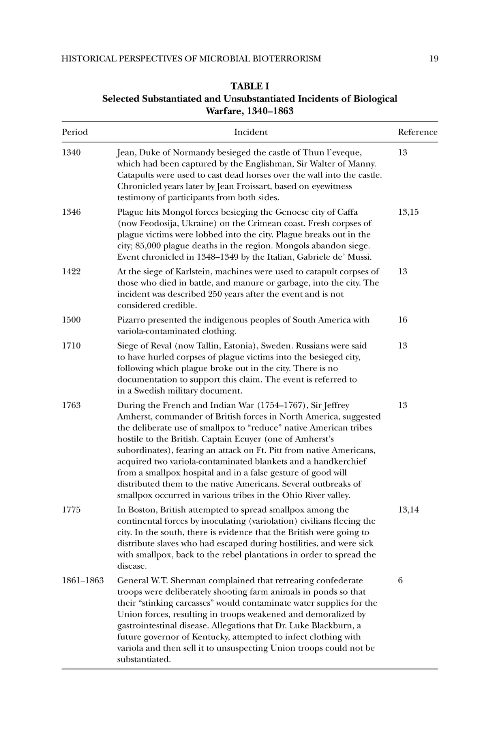
2. Historical Perspectives of Microbial Bioterrorism............................. 15
STEPHEN A. MORSE
1. Introduction ............................................................................
1.1. Definitions ........................................................................
1.2. Development and Prohibition of Biological Weapons ................
2. Early use of Biological Agents in Warfare ......................................
2.1. Early Theories of Infectious Disease .......................................
2.2. Selected Incidents from the Fourteenth to the Nineteenth
Centuries ..........................................................................
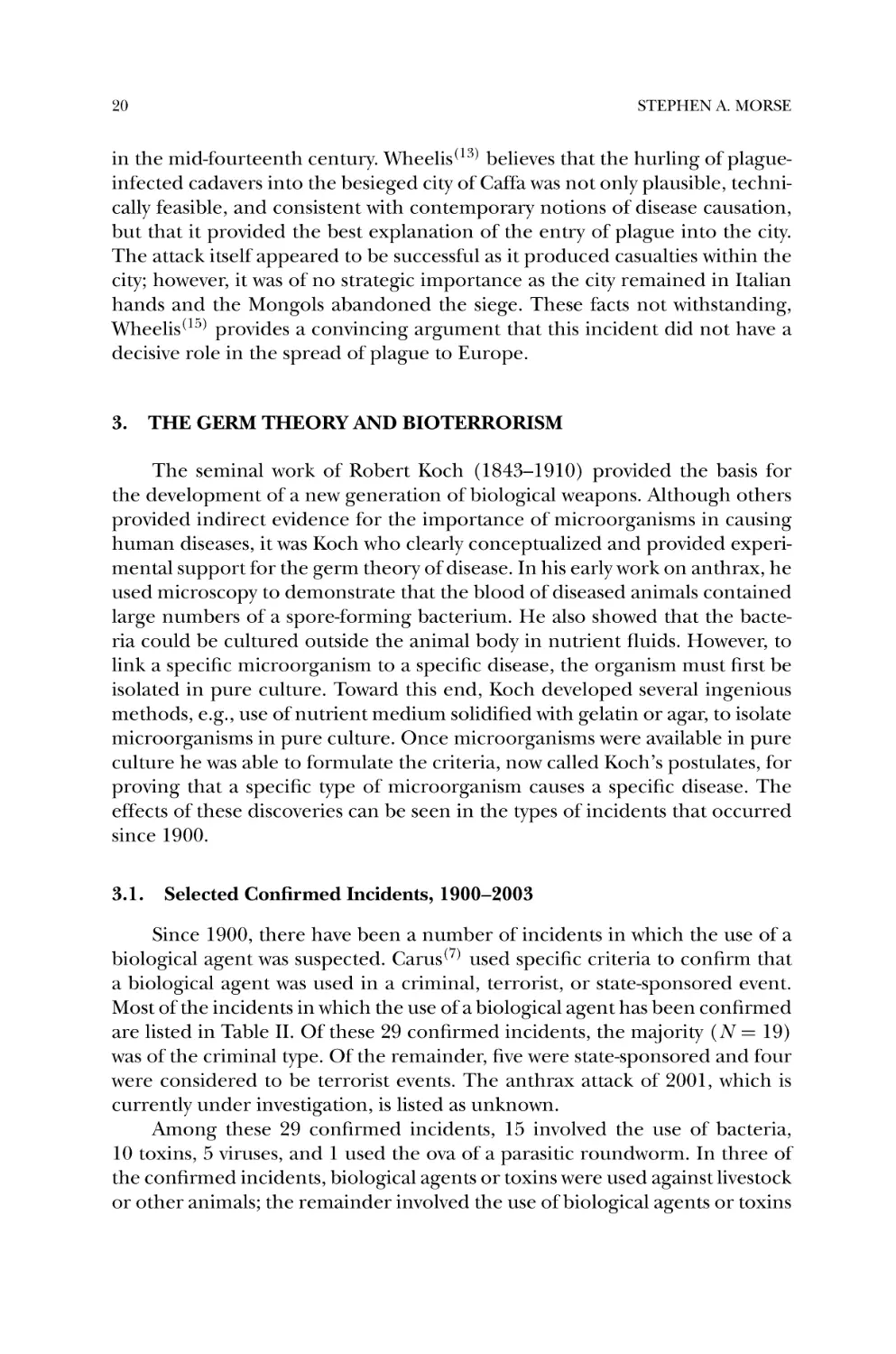
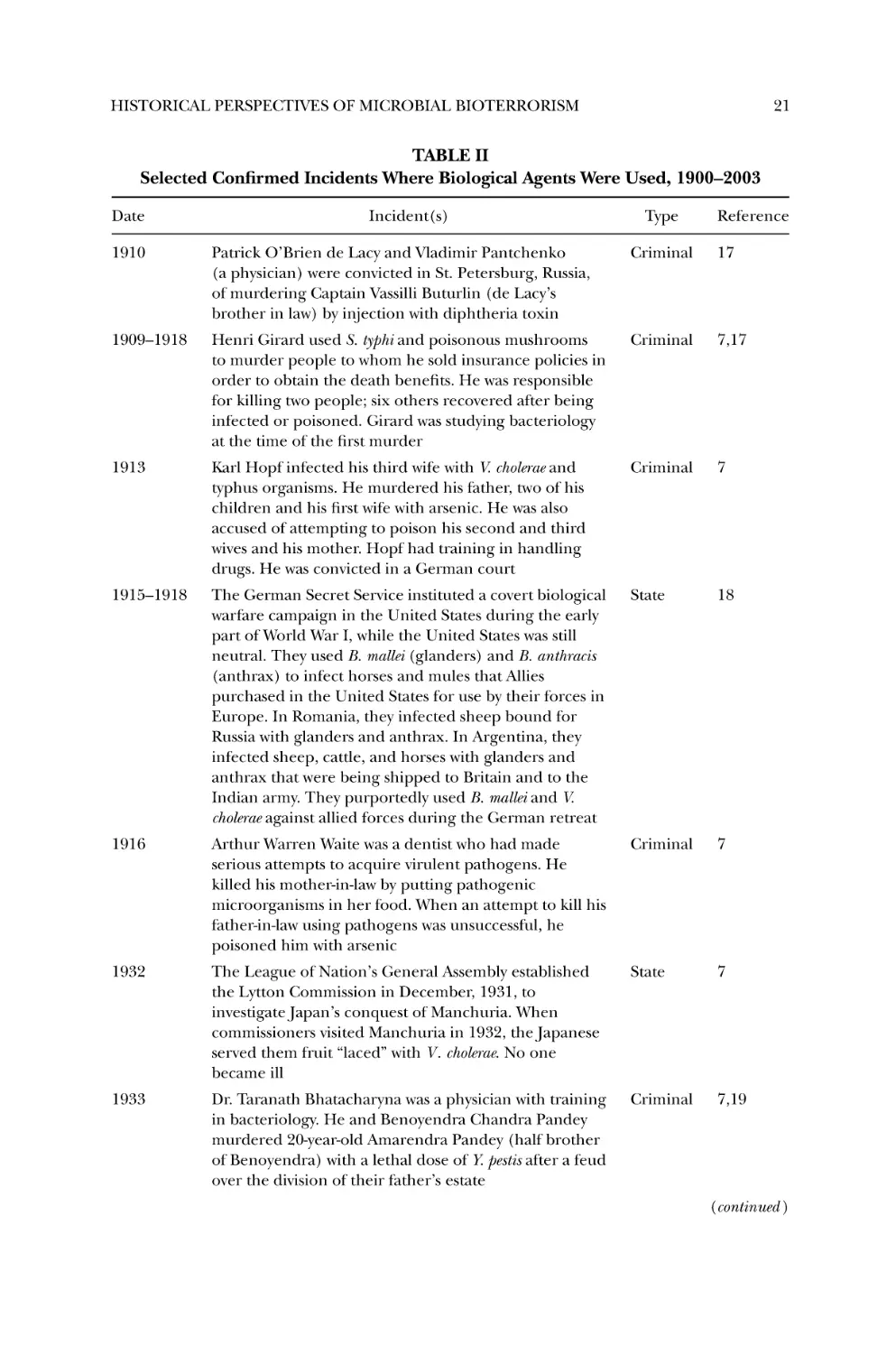
3. The Germ Theory and Bioterrorism .............................................
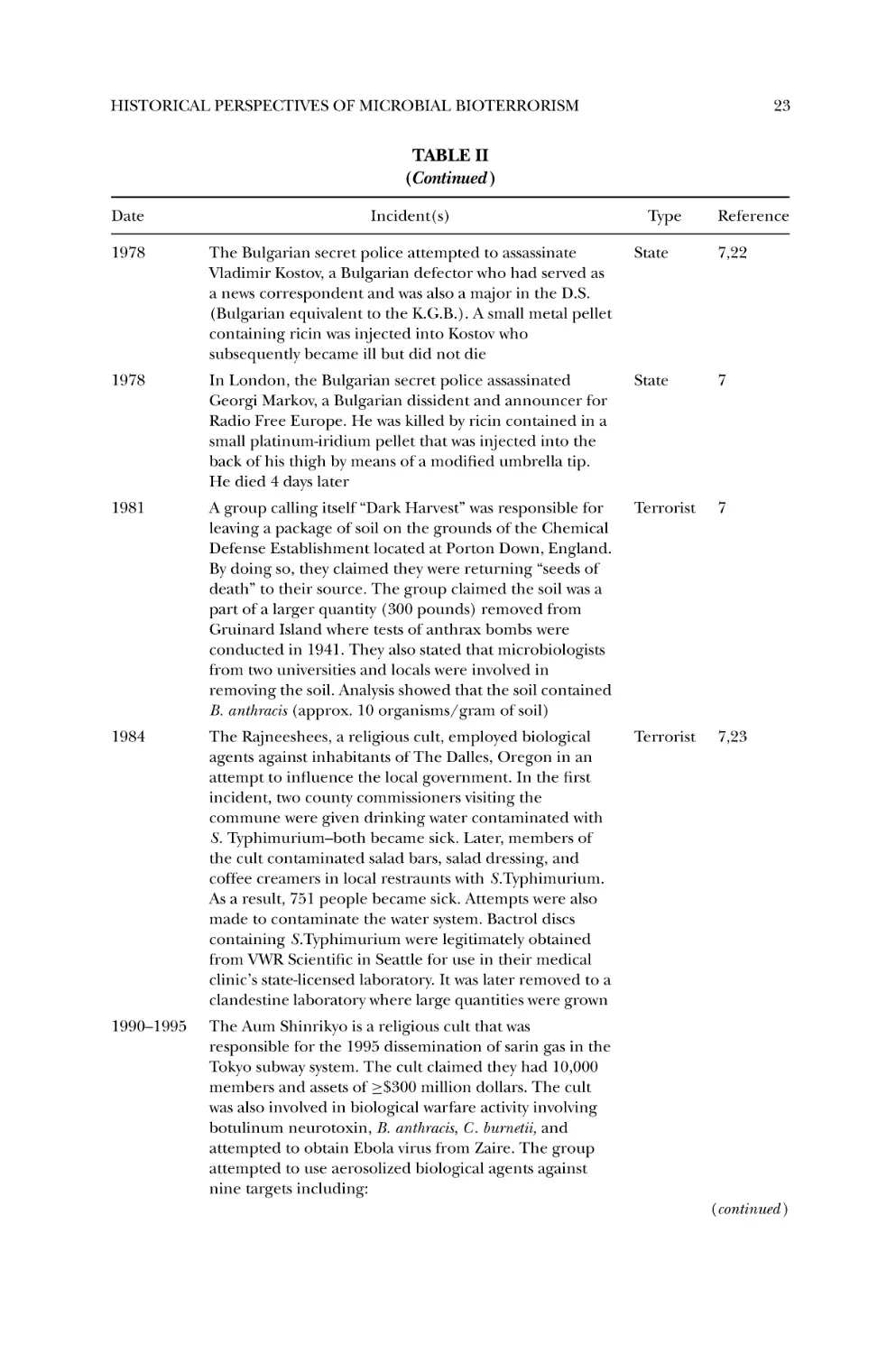
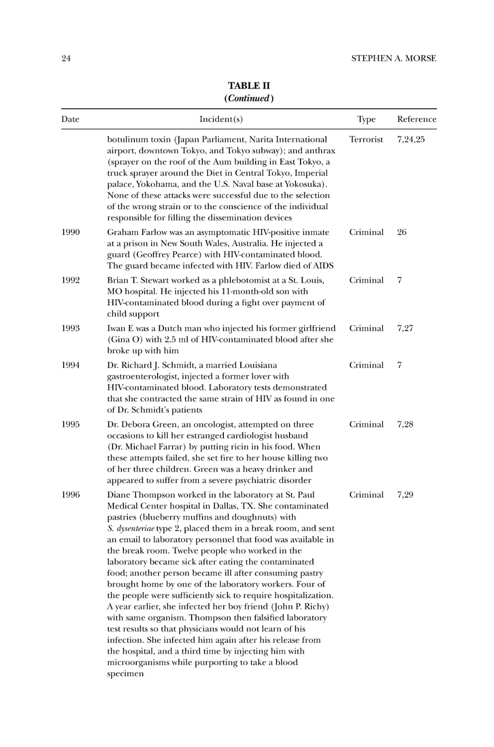
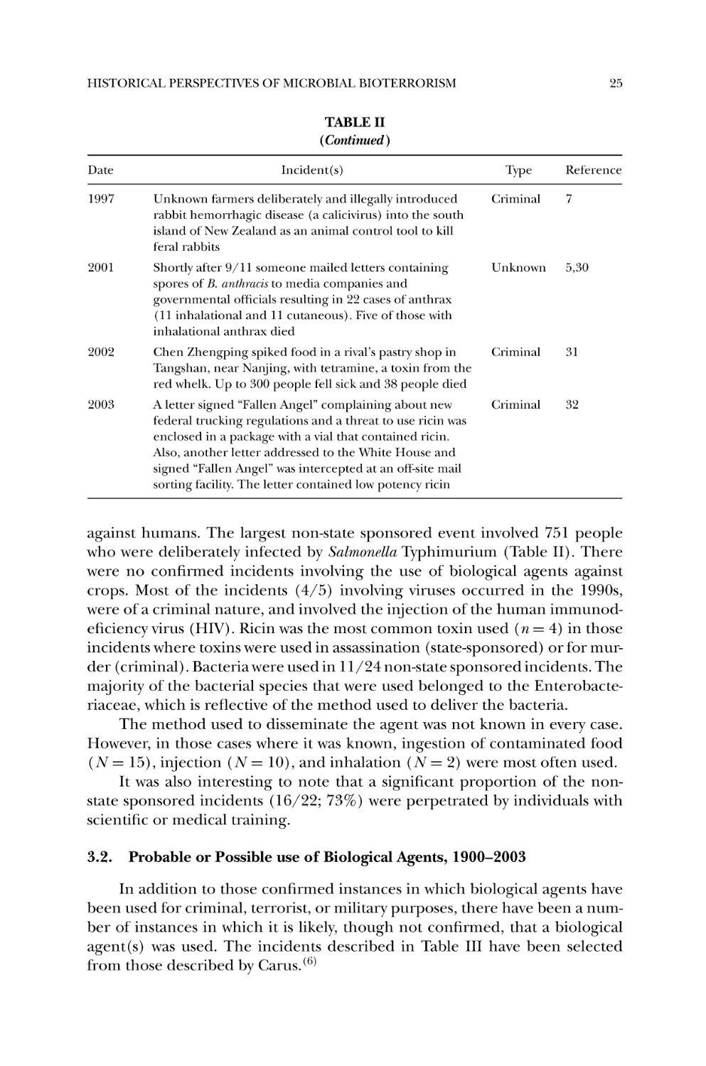
3.1. Selected Confirmed Incidents, 1900–2003 ...............................
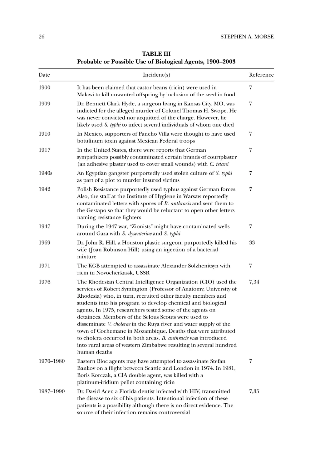
3.2. Probable or Possible use of Biological Agents, 1900–2003 ..........
4. Concluding Remarks and Perspectives ..........................................
References ...............................................................................
xv
15
15
16
17
17
18
20
20
25
27
27
xvi
CONTENTS
3. The Infectious Disease Physician and Microbial Bioterrorism ............ 31
SANDRA G. GOMPF, JORDAN LEWIS, EKNATH NAIK, and
KALEY TASH
1.
2.
3.
4.
Introduction ............................................................................
The Evolution of the Global Community, Infection, and Bioterrorism
The Evolving Practice of Infectious Disease ....................................
Integrating the Infectious Disease Physician with Public Health
Response .................................................................................
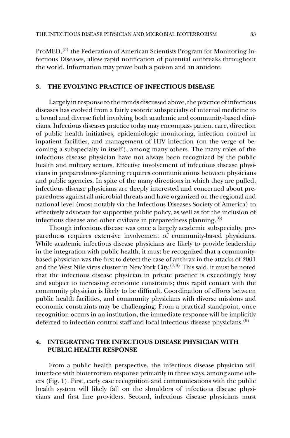
5. Prevention, Early Recognition, and the Infectious Disease Physician ..
6. Education, the Infectious Disease Physician, and Preparedness .........
Summary .................................................................................
References ...............................................................................
31
31
33
33
34
36
37
37
4. Modulation of Innate Immunity to Protect Against Biological
Weapon Threat ......................................................................... 39
KEN ALIBEK and CATHERINE LOBANOVA
1. Bioterrorism and Biological Weapons Threat .................................
1.1. Current Medical Defense Against Biological Weapons ...............
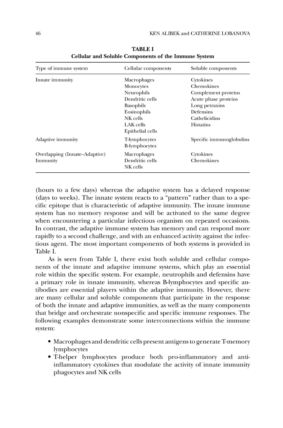
2. Immune System ........................................................................
3. Innate Immunity .......................................................................
4. Pulmonary Innate Immunity .......................................................
4.1. Major Components of Pulmonary Innate Immunity ..................
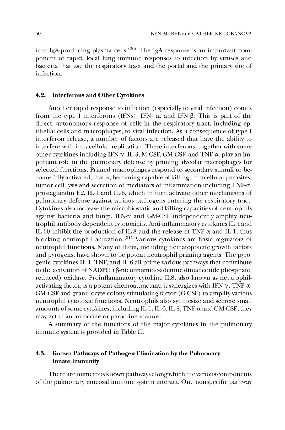
4.2. Interferons and Other Cytokines ...........................................
4.3. Known Pathways of Pathogen Elimination by the Pulmonary
Innate Immunity ................................................................
5. Modulation of Immunity for Protection Against Infection ................
6. Modulation of Innate Immunity to Protect Against Biological
Weapon Threat—a Summary ......................................................
References ...............................................................................
39
42
45
47
48
48
50
50
53
57
58
5. Smallpox: Pathogenesis and Host Immune Responses Relevant to
Vaccine and Therapeutic Strategies............................................... 63
MICHELE A. KUTZLER, KENNETH E. UGEN, and
DAVID B. WEINER
1.
2.
3.
4.
5.
6.
7.
Introduction ............................................................................
History of Smallpox Infection and its Eradication ...........................
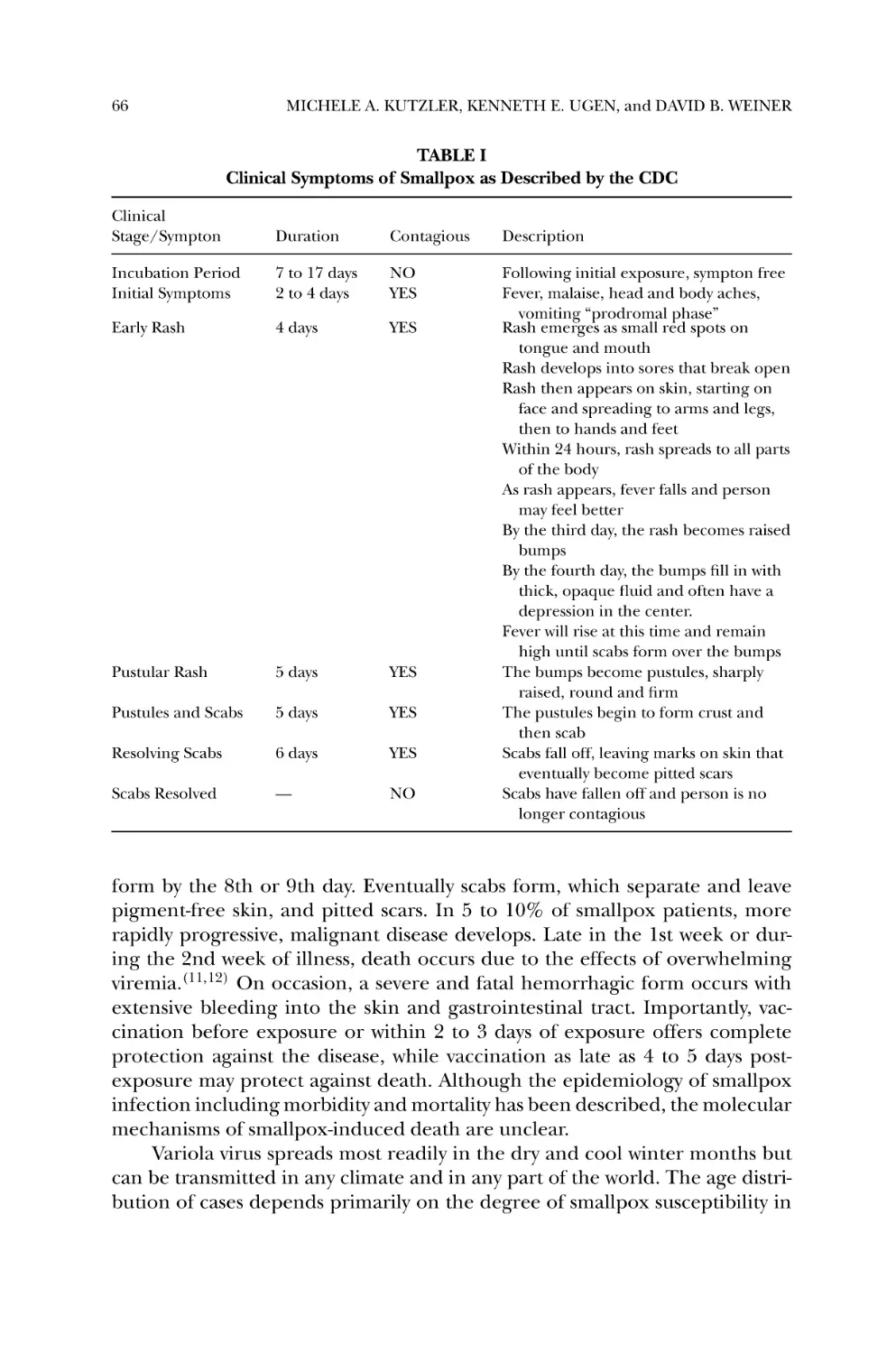
Clinical and Epidemiological Features ..........................................
Virus Structure and Classification .................................................
Pathogenesis, Host Defense, and the Immune Response ..................
Features of Smallpox Making it a Likely Bioterror Agent ..................
History and Potential of Smallpox as a Bioweapon ..........................
63
64
65
67
67
69
69
CONTENTS
xvii
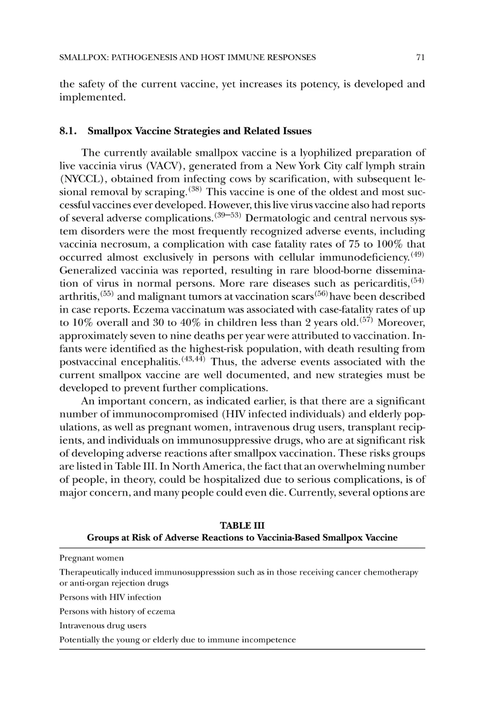
8. Smallpox Vaccines and Antiviral Therapies ....................................
8.1. Smallpox Vaccine Strategies and Related Issues ........................
8.2. Smallpox Antiviral Therapies ................................................
9. Conclusions .............................................................................
References ...............................................................................
70
71
74
76
76
6. Bacillus anthracis: Agent of Bioterror and Disease............................. 83
CHRISTOPHER K. COTE, DONALD J. CHABOT,
ANGELO SCORPIO, THOMAS E. BLANK, WILLIAM A. DAY,
SUSAN L. WELKOS, and JOEL A. BOZUE
1.
2.
3.
4.
5.
Introduction ............................................................................ 83
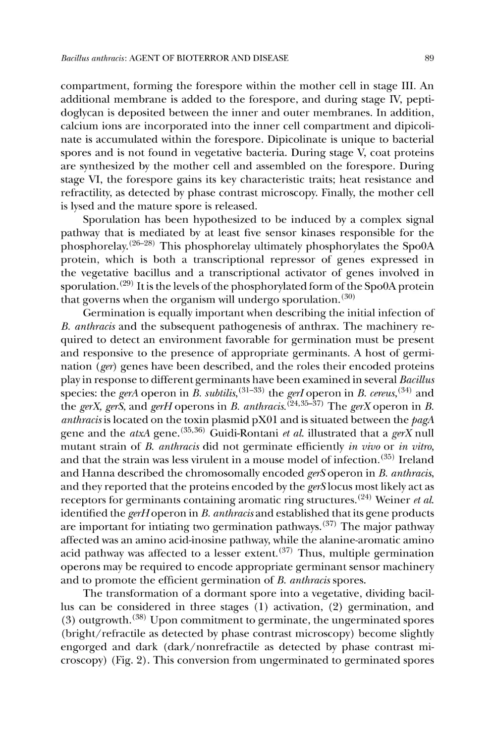
Clinical Presentation of Anthrax .................................................. 84
B. anthracis and Bioterror ........................................................... 86
Evolution into a Pathogen .......................................................... 87
Spore Structure and Function ..................................................... 88
5.1. Sporulation and Germination ............................................... 88
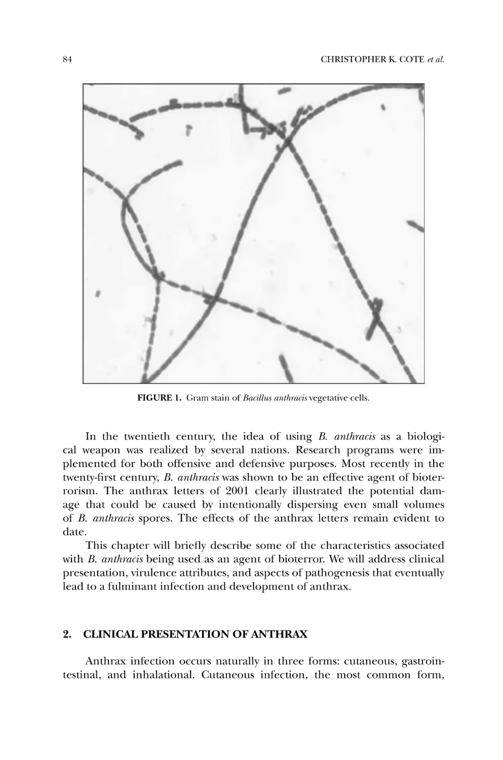
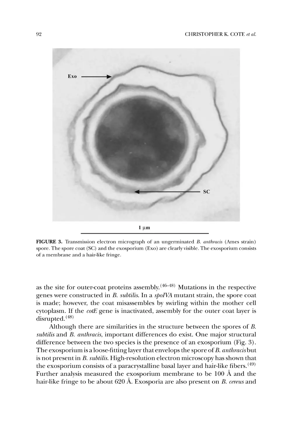
5.2. Spore Coat Proteins and Exosporium ..................................... 91
6. Virulence Factors ...................................................................... 94
6.1. Anthrax Toxins .................................................................. 94
6.2. Anthrax Toxin Receptor-Mediated Internalization .................... 96
6.3. Toxin Gene Regulation ........................................................ 97
6.4. Capsule: Chemistry and Composition ..................................... 98
6.5. Capsule Gene Regulation ..................................................... 100
6.6. Capsule Function ................................................................ 100
6.7. Accessory Virulence Factors ................................................. 102
7. B. anthracis and Macrophage Interactions ...................................... 103
8. Vaccine and Therapeutic Approaches ........................................... 107
References ............................................................................... 111
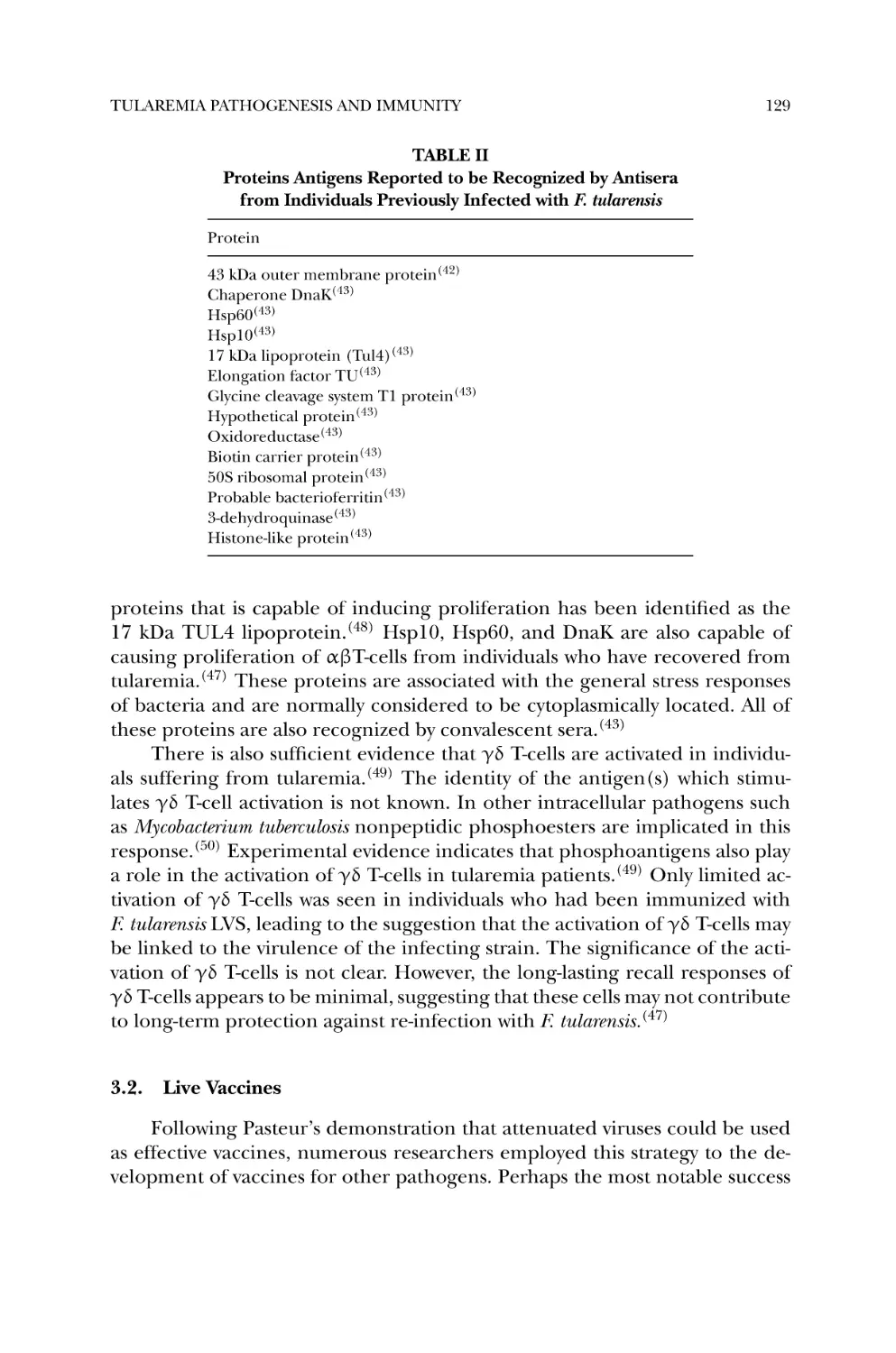
7. Tularemia Pathogenesis and Immunity .......................................... 121
STEPHEN L. MICHELL, KATE F. GRIFFIN, and
RICHARD W. TITBALL
1. Introduction ............................................................................ 121
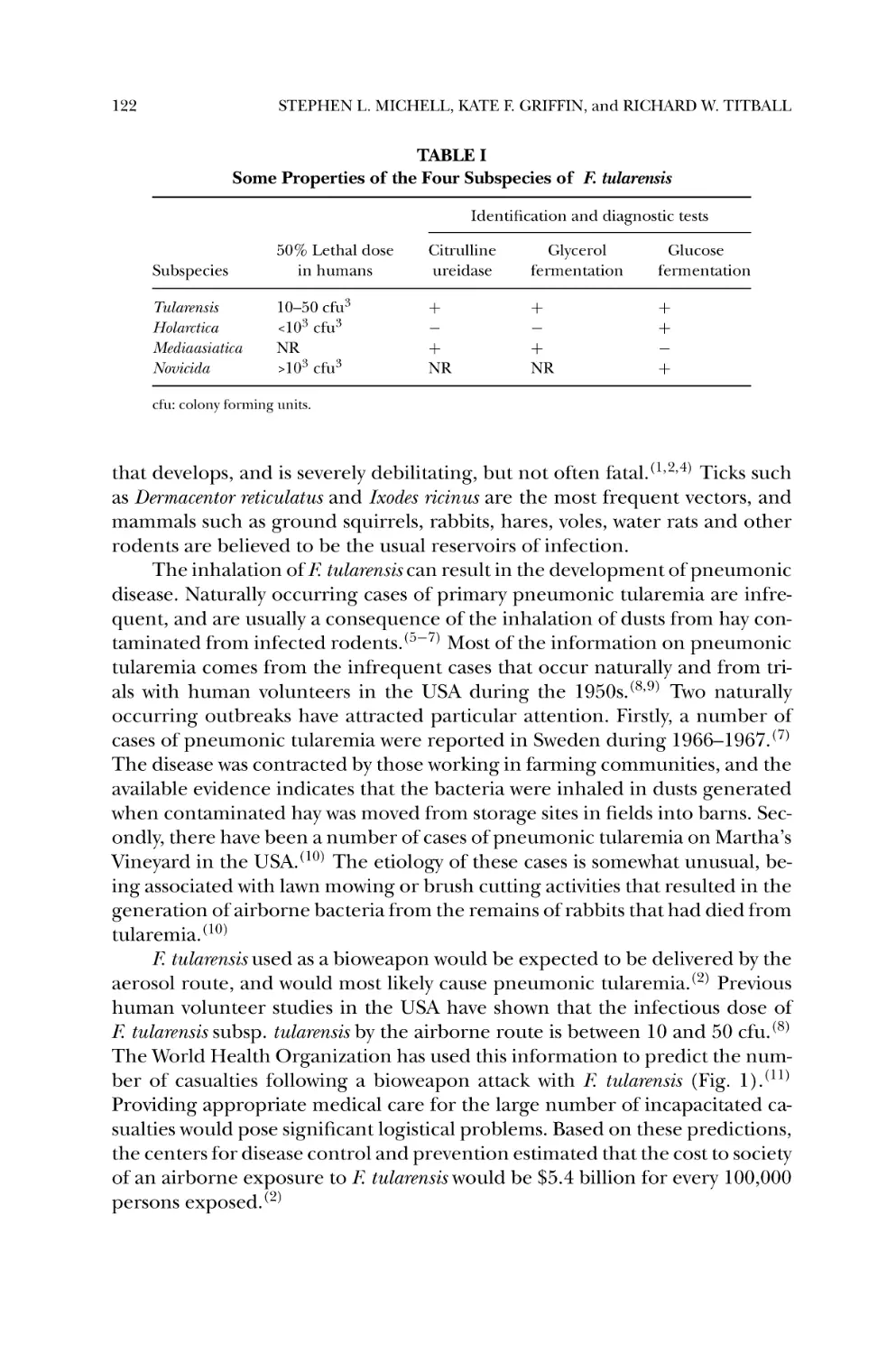
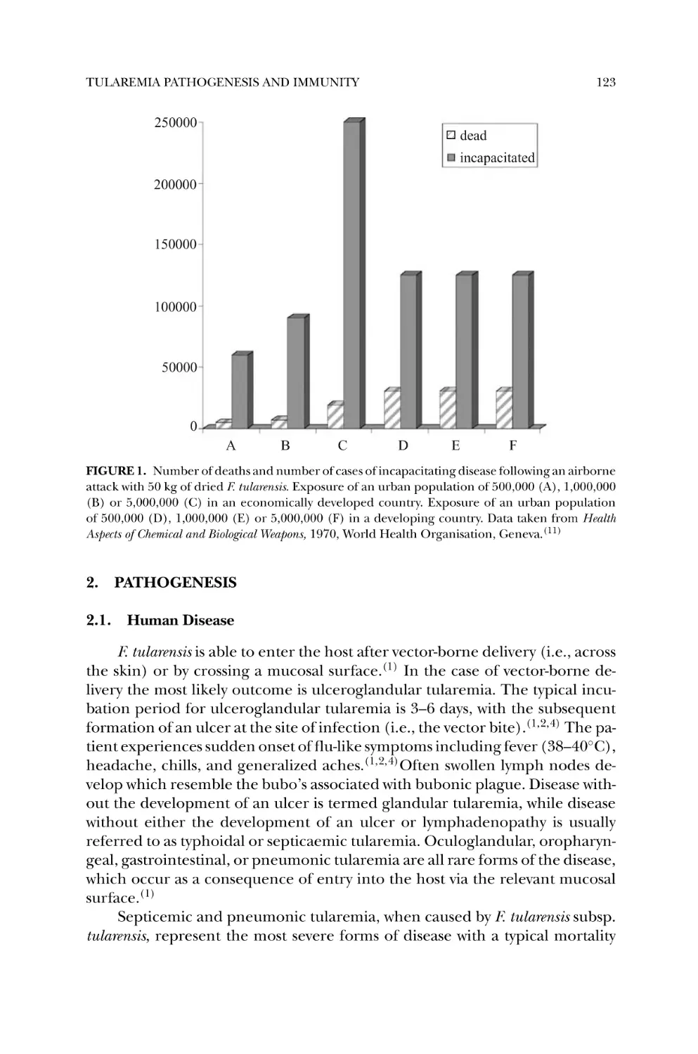
2. Pathogenesis ............................................................................ 123
2.1. Human Disease .................................................................. 123
2.2. Animal Models ................................................................... 124
2.3. Cellular Pathogenesis ......................................................... 125
2.4. Molecular Pathogenesis ....................................................... 126
3. Immunity ................................................................................. 128
3.1. Natural Infection and Immunity ............................................ 128
3.2. Live Vaccines ..................................................................... 129
3.3. Subunit Vaccines ................................................................ 131
xviii
CONTENTS
3.4. Mechanisms of Protection in Adaptive Immunity .................... 132
Conclusions ............................................................................ 133
References ............................................................................. 133
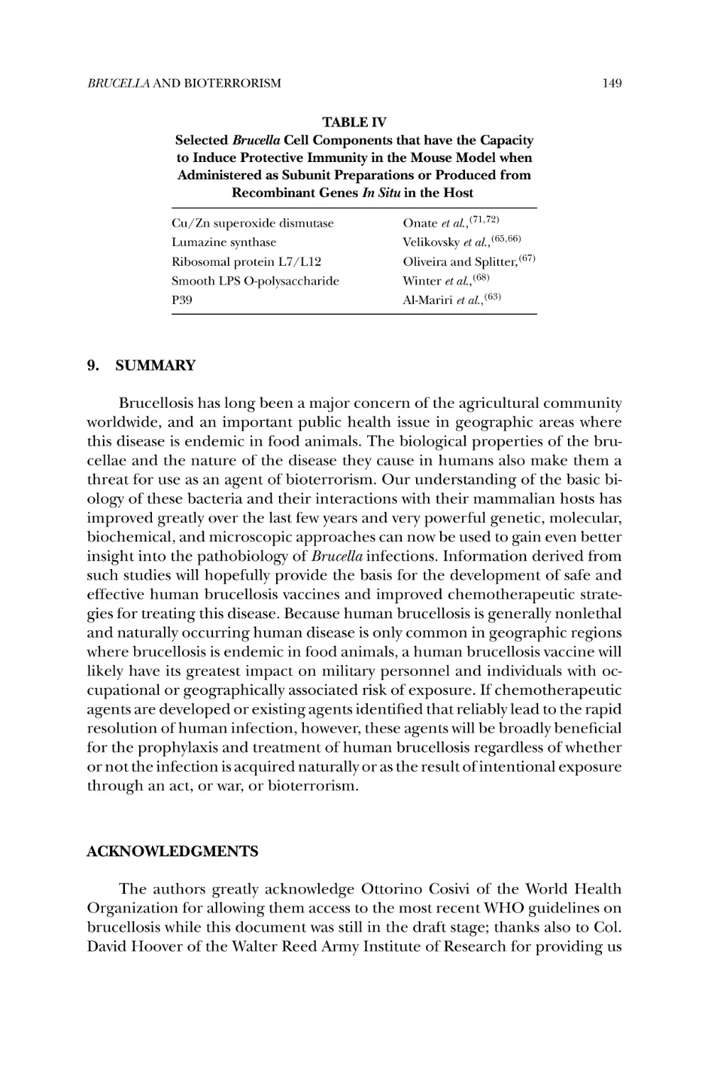
8. Brucella and Bioterrorism........................................................... 139
MICHELLE WRIGHT VALDERAS and R. MARTIN ROOP II
1.
2.
3.
4.
5.
6.
7.
8.
9.
Introduction ........................................................................... 139
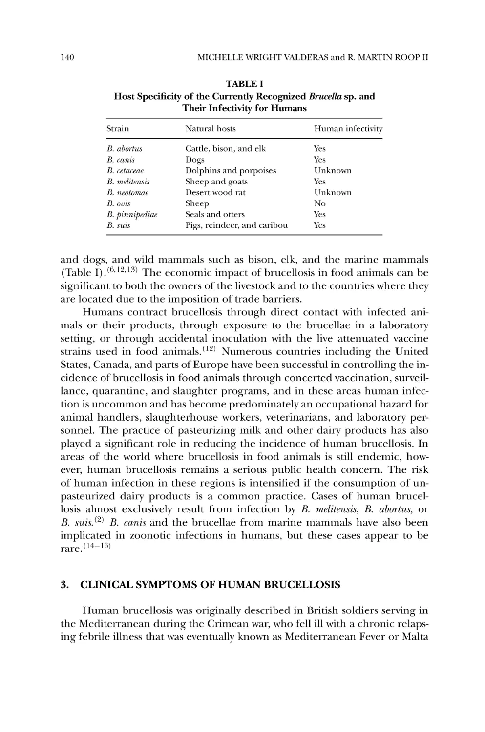
Brucellosis: a Zoonotic Disease ................................................... 139
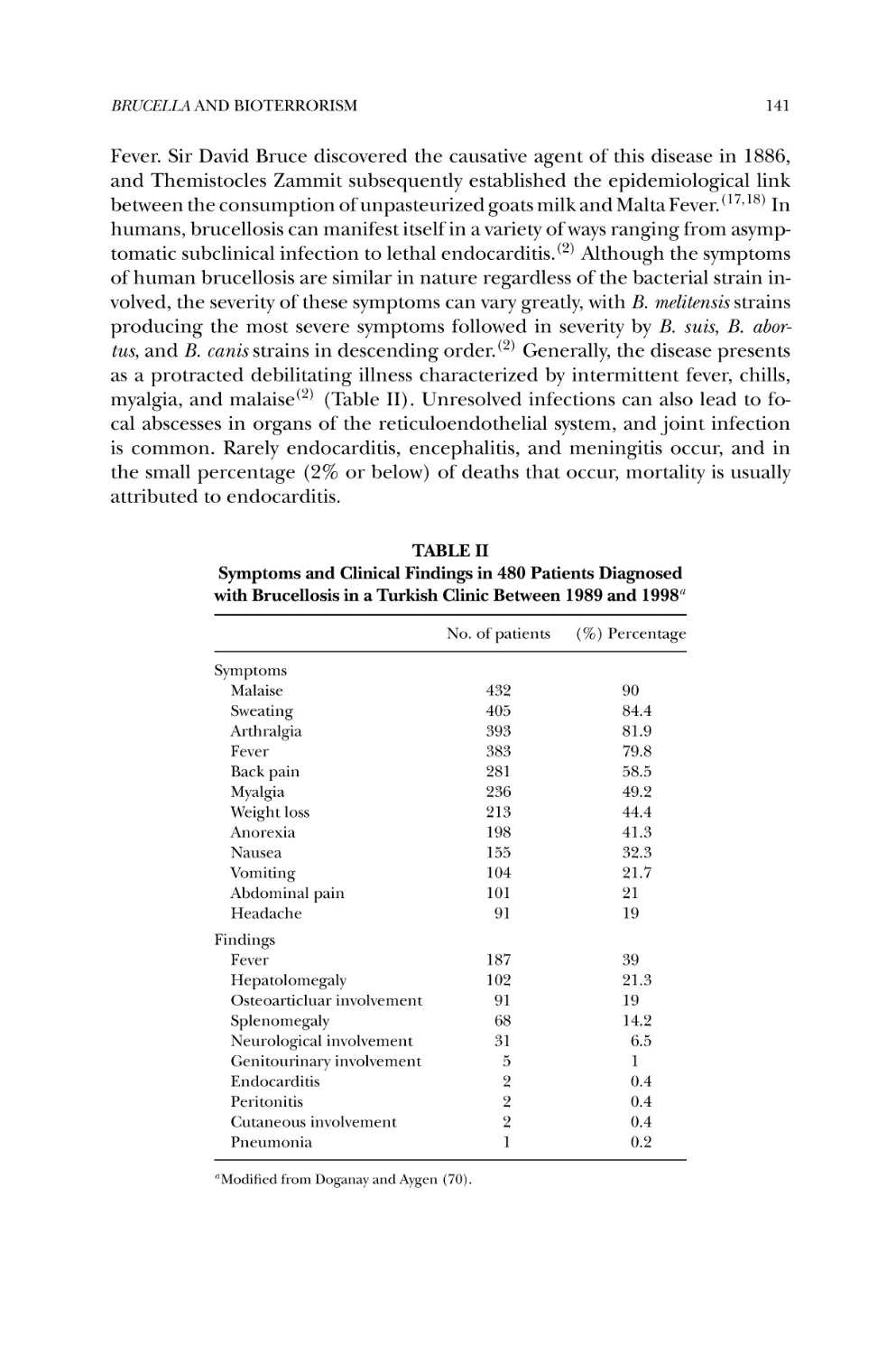
Clinical Symptoms of Human Brucellosis ..................................... 140
Life Within the Macrophage and Subversion of Host Immune
Responses .............................................................................. 142
History of the use of Brucella as an Agent of Biological Warfare ........ 143
Impact of an Attack Using Brucella as an Agent of Bioterrorism ...... 144
Diagnosis and Treatment of Brucellosis ....................................... 145
Vaccine Development: Historical Perspectives and Considerations
for the Future ......................................................................... 147
Summary ............................................................................... 149
Acknowledgments ................................................................... 149
References ............................................................................. 150
9. Pneumonic Plague ................................................................... 155
DAVID L. ERICKSON and B. JOSEPH HINNEBUSCH
1. Introduction ........................................................................... 155
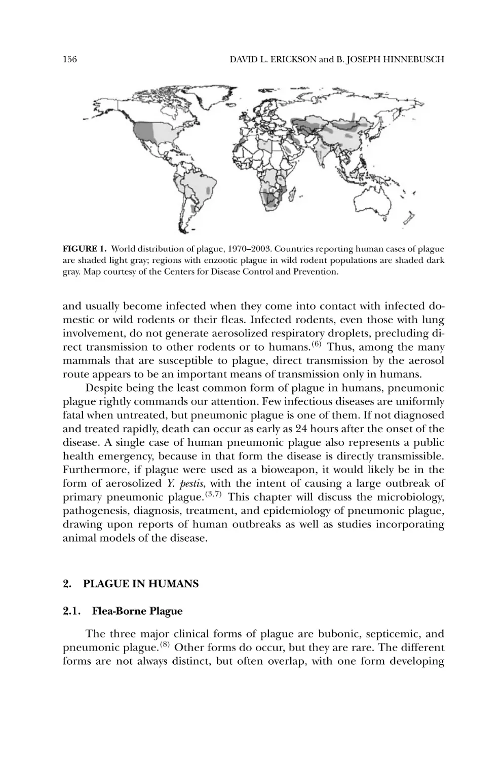
2. Plague in Humans ................................................................... 156
2.1. Flea-Borne Plague ............................................................. 156
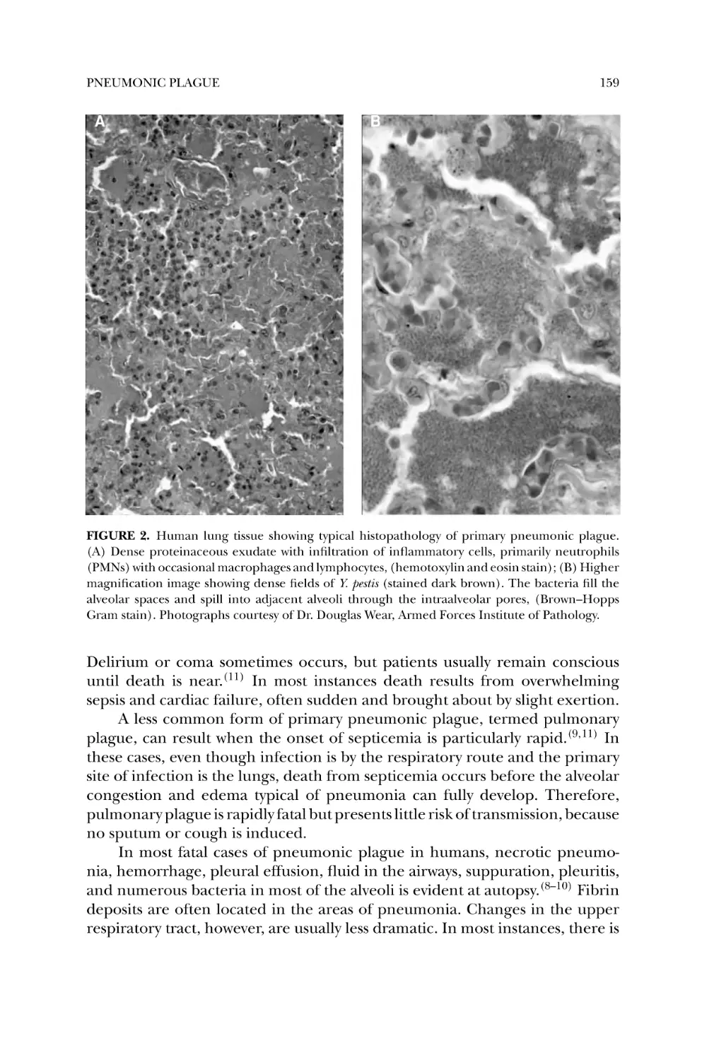
2.2. Aerosol-Transmitted Plague ................................................ 158
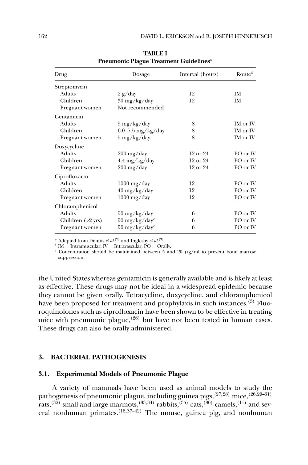
3. Bacterial Pathogenesis .............................................................. 162
3.1. Experimental Models of Pneumonic Plague ........................... 162
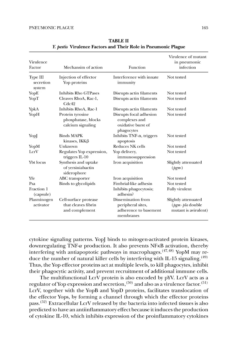
3.2. Y. pestis Virulence Factors .................................................... 164
3.3. Role of Y. pestis Virulence Factors in Pneumonic Plague ........... 166
4. Bacterial Genetics .................................................................... 168
5. Epidemiology of Pneumonic Plague ........................................... 169
5.1. Biological and Epidemiological Determinants of Pneumonic
Plague Epidemics .............................................................. 170
5.2. Management and Control of Pneumonic Plague Outbreaks ..... 171
5.3. Lessons from Modern Pneumonic Plague Outbreaks ............... 172
6. Current and Future Needs ........................................................ 174
References ............................................................................. 174
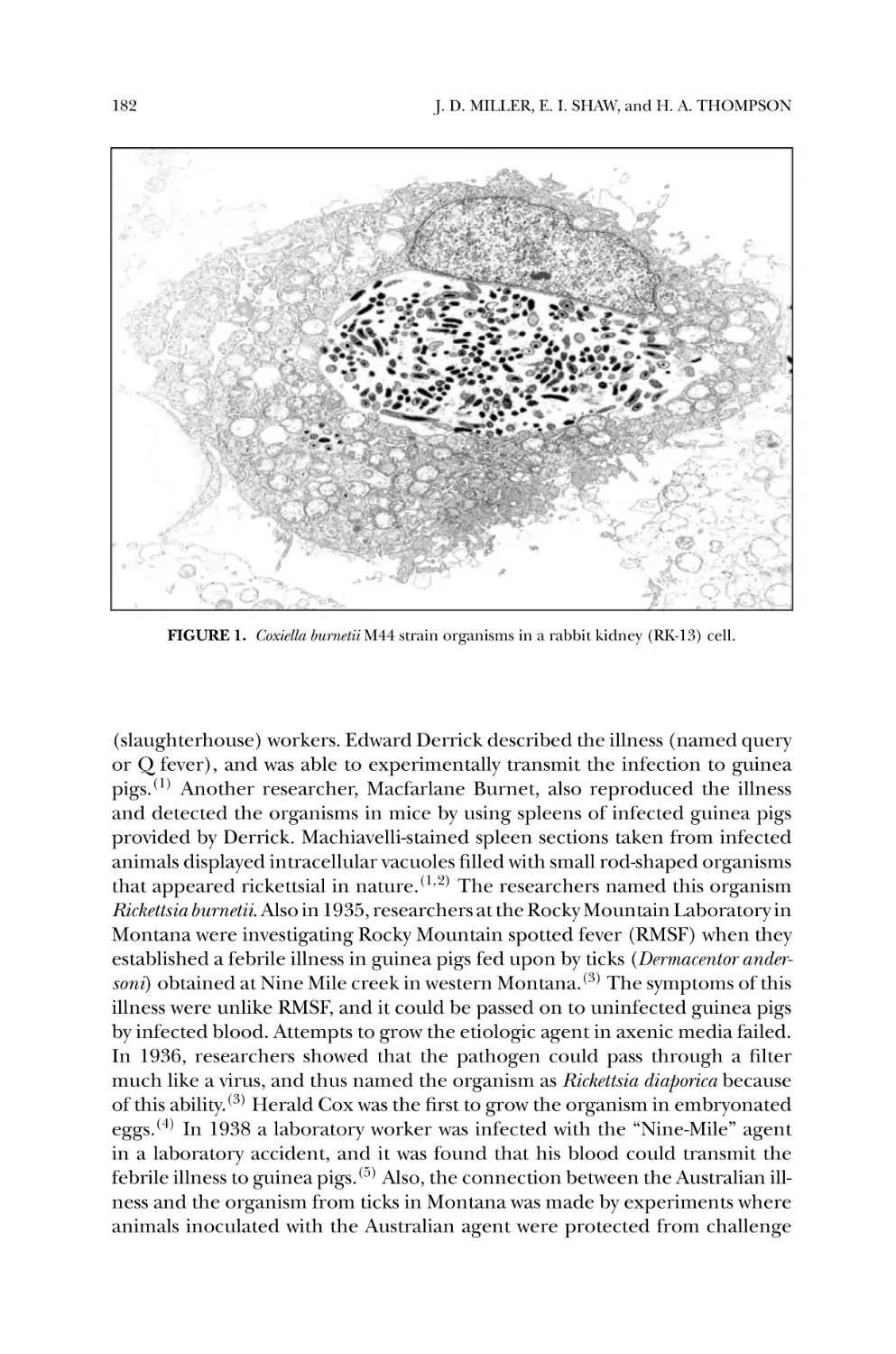
10. Coxiella burnetii, Q Fever, and Bioterrorism.................................... 181
J. D. MILLER, E. I. SHAW, and H. A. THOMPSON
1. Introduction ........................................................................... 181
CONTENTS
2.
3.
4.
5.
6.
7.
8.
9.
10.
11.
12.
13.
14.
15.
16.
17.
18.
19.
xix
A Brief History ........................................................................ 181
Q Fever .................................................................................. 183
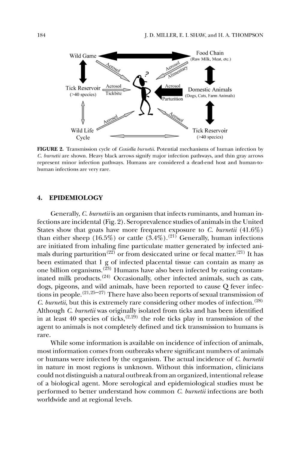
Epidemiology ......................................................................... 184
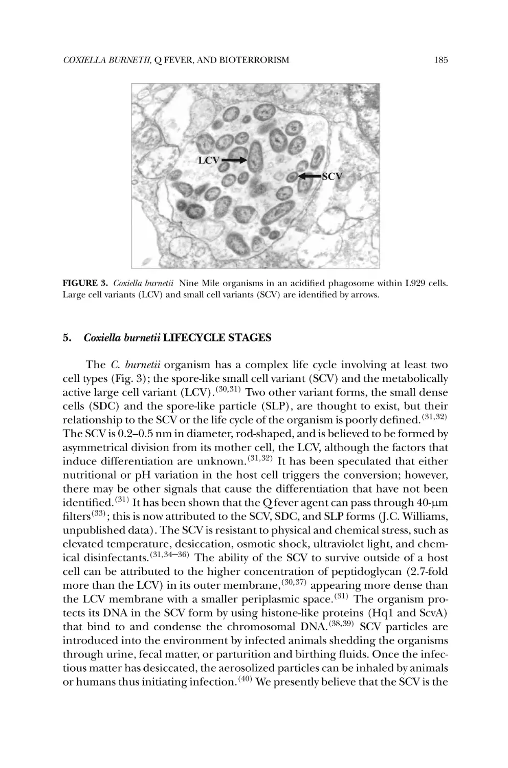
Coxiella burnetii Lifecycle Stages .................................................. 185
Coxiella burnetii Genome ............................................................ 186
Lipopolysaccharide .................................................................. 187
Phase Transition ..................................................................... 188
Invasion of Host Cells ............................................................... 190
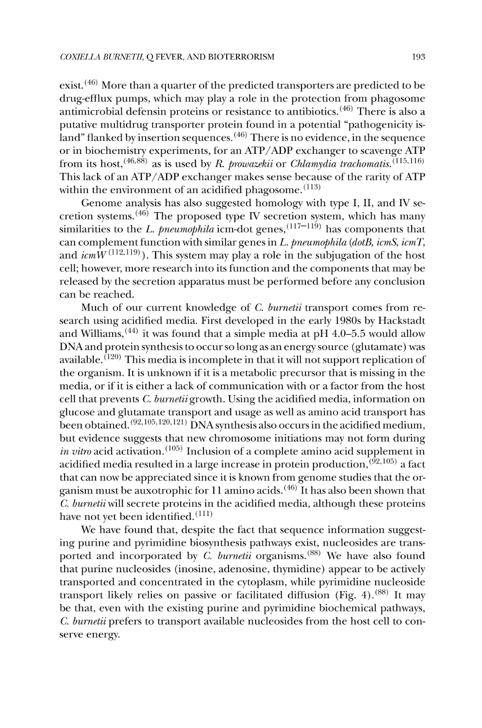
Environment of Acidified Phagosome ......................................... 191
Metabolic Pathways .................................................................. 192
Transport ............................................................................... 192
Transformation Studies ............................................................ 194
Coxiella burnetii Outbreaks in the Military ..................................... 194
Coxiella burnetii as a Biological Weapon ........................................ 195
Sterilization/Disinfection .......................................................... 197
Detection Methods .................................................................. 197
Treatments of Q Fever .............................................................. 199
Vaccine .................................................................................. 200
References ............................................................................. 200
11. Genomic and Proteomic Approaches Against Q Fever .................... 209
JAMES E. SAMUEL, LAURA R. HENDRIX, KASI RUSSELL,
and GUOQUAN ZHANG
1.
2.
3.
4.
5.
6.
7.
8.
9.
Introduction ........................................................................... 209
Disease and Threat .................................................................. 209
Host–Parasite Interaction .......................................................... 210
Secretion of Virulence Factors ................................................... 212
Pathogenesis ........................................................................... 213
Isolate Diversity and Virulence ................................................... 214
Acquired Immunity .................................................................. 215
Whole Cell C. burnetii Vaccines .................................................. 217
New Opportunities with Genomic and Proteomic Approaches ........ 220
9.1. Genomic Comparison of Isolate Groups ................................ 220
9.2. Development of New-Generation Vaccines ............................ 220
Acknowledgement ................................................................... 221
References ............................................................................. 221
12. Rickettsia rickettsii and Other Members of the Spotted Fever
Group as Potential Bioweapons................................................... 227
DONALD H. BOUYER and DAVID H. WALKER
1. Introduction ........................................................................... 227
2. SFG Rickettsiae with Bioweapon Potential .................................... 228
xx
CONTENTS
3. Feasibility of Obtaining, Propagating, Stablizing, and Weaponizing
SFG Rickettsiae ....................................................................... 229
4. Methods of Dispersal ................................................................ 230
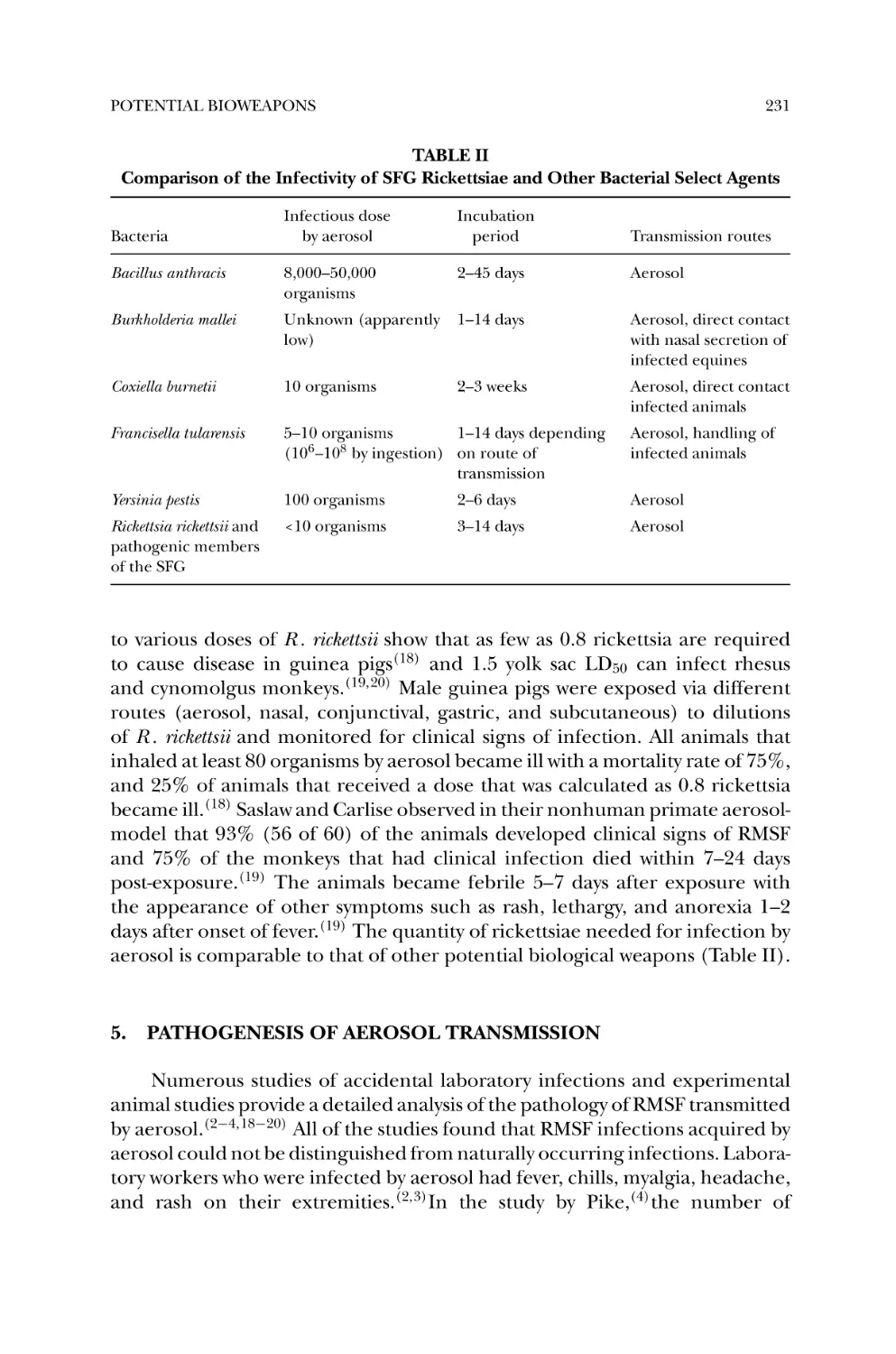
5. Pathogenesis of Aerosol Transmission ......................................... 231
6. Available Methods for Diagnosis, Treatment, and Prevention .......... 232
7. Needed Countermeasures ......................................................... 233
References ............................................................................. 234
Index ..................................................................................... 237
1
Biotechnology and the Public
Health Response to
Bioterrorism
ANDREW CANNONS, PHILIP AMUSO,
and BURT ANDERSON
1.
INTRODUCTION
The tragic events of September 11, 2001 have forced a reconsideration of
international security. The role of relatively small, but well organized, terrorists
groups in inflicting mass casualties must be considered a real threat. In October
2001, Bacillus anthracis spores were intentionally released through the mailing
of contaminated letters distributed through the United States postal service.
Again, apparently small groups, or perhaps an individual, were able to utilize
a biological agent as a weapon to cause death and affect relatively large areas
of the United States, including the District of Columbia, Florida, Connecticut,
New Jersey, and New York.(1) Furthermore, the resulting hysteria and public response has focused recent attention on the use of biological agents as potential
weapons.
The use of the US postal system as an efficient “artificial mechanical vector” has served to remind us that the dispersion and transmission of intentional
infections may share few similarities with naturally acquired infections caused
by the same agent. Characterization of the intentionally released B. anthracis
spores revealed that they were related to the Ames strain—a relatively wellstudied laboratory strain. Much media attention has been focused on the use
ANDREW CANNONS and PHILIP AMUSO • Center for Biological Defense, University of South
Florida Tampa, FL 33612.
BURT ANDERSON • Department of Medical Microbiology and
Immunology, College of Medicine, University of South Florida, Tampa, FL 33612.
1
2
ANDREW CANNONS et al.
of agents other than B. anthracis that might be used as biological weapons.
The identification of such agents and the establishment of the ABC priority
category list is largely based on a historical perspective of offensive biological weapons programs dating to the cold war and earlier.(2,3) The impact of
molecular biology, gene cloning, and biotechnology on not only the control
and prevention, but also the development of biological weapons must remain
clearly in focus.
The entire genome sequence for at least one representative strain for
all category A agents has been determined. Most of these sequences have already been deposited in public access databases. The opportunity to use this
information to enhance the detection, surveillance, and diagnosis of infections caused by bioterrorism (BT) agents is great. Conversely, the potential
for misuse to engineer pathogens for intentional release must also be considered. Such open public access to genome sequences and other scientific
reports describing details about mechanisms of pathogenesis, antibiotic resistance, etc. raises questions about use, misuse, and the dissemination of such
information.
This chapter will describe the public health infrastructure that has been
put in place to respond in the event of acts of bioterrorism as well as the role of
biotechnology in the control and prevention of infections caused by the intentional release of agents of bioterrorism. Specifically, we describe how the
laboratory response network (LRN) successfully responded to the index case
of anthrax in Florida. We also describe the overall operational structure of the
LRN and its role to detect, monitor, and characterize microbial agents that
may have been used as weapons. In addition, we have summarized the potential for the misuse of biotechnology for the development of future biological
weapons. We have also attempted to describe how the concern for such misuse
may have a profound impact on how information is exchanged within the scientific community and among public health officials. Finally, we summarize the
biodefense research funding trends and the reaction to these trends among
the scientific community, as well as public concern about possible risks associated with the establishment of both regional and national biocontainment
laboratory facilities.
2.
US PUBLIC HEALTH SERVICE INFRASTRUCTURE—THE
LABORATORY RESPONSE NETWORK
On October 4, 2001, an index case of inhalational anthrax that occurred in Boca Raton, Florida, was confirmed when a Florida LRN reference laboratory discovered the presence of B. anthracis in a clinical specimen.
This discovery triggered a cascade of events that nearly crippled the public
health and emergency response systems throughout the United States. “White
BIOTECHNOLOGY AND THE PUBLIC HEALTH RESPONSE TO BIOTERRORISM
3
powder” instantly became a household phrase synonymous with “Bacillus anthracis spores”, causing fear and panic throughout the nation.
Although LRN personnel had planned and trained for the “real thing”
for nearly two years, they were unable to predict just how the system would
respond to an actual BT event. Few responsible officials had predicted that
a mere handful of intentionally inflicted anthrax cases would bring the public health system to its knees under a deluge of “suspicious” samples coming
from a terrified public. Across the nation, LRN laboratories tested 125,000
samples by the time the investigation was completed. This amounted to more
than 1 million separate tests. Law enforcement units, HazMat units, fire rescue
units, and public health personnel were occupied around the clock for over a
month.
The LRN was established in 1999 when the US Congress appropriated
funds to address the threat of terrorism. Previously, Congress passed the Response to Weapons of Mass Destruction Act of 1997 (commonly known as
the Nunn-Lugar-Domenici legislation), which focused on providing training
and equipment for emergency first-responder personnel. The 120 largest US
cities were specified in this legislation. The Department of Health and Human
Services (DHHS) sponsors community-level medical and public health preparedness and response associated with emergencies resulting from terrorist
incidents. DHHS, through the Office of Emergency Preparedness (OEP), lead
a national effort to develop Metropolitan Medical Response Systems (MMRS)
to enhance the local ability to respond to the health and medical consequences
of a terrorist incident.
DHHS, through the Centers for Disease Control and Prevention (CDC)
program announcement 99051: “public health preparedness and response for
bioterrorism”, initiated an effort to upgrade national public health capability to counter bioterrorism. Toward this effort, CDC initiated this cooperative
agreement program for States, US Territories, and major local public health
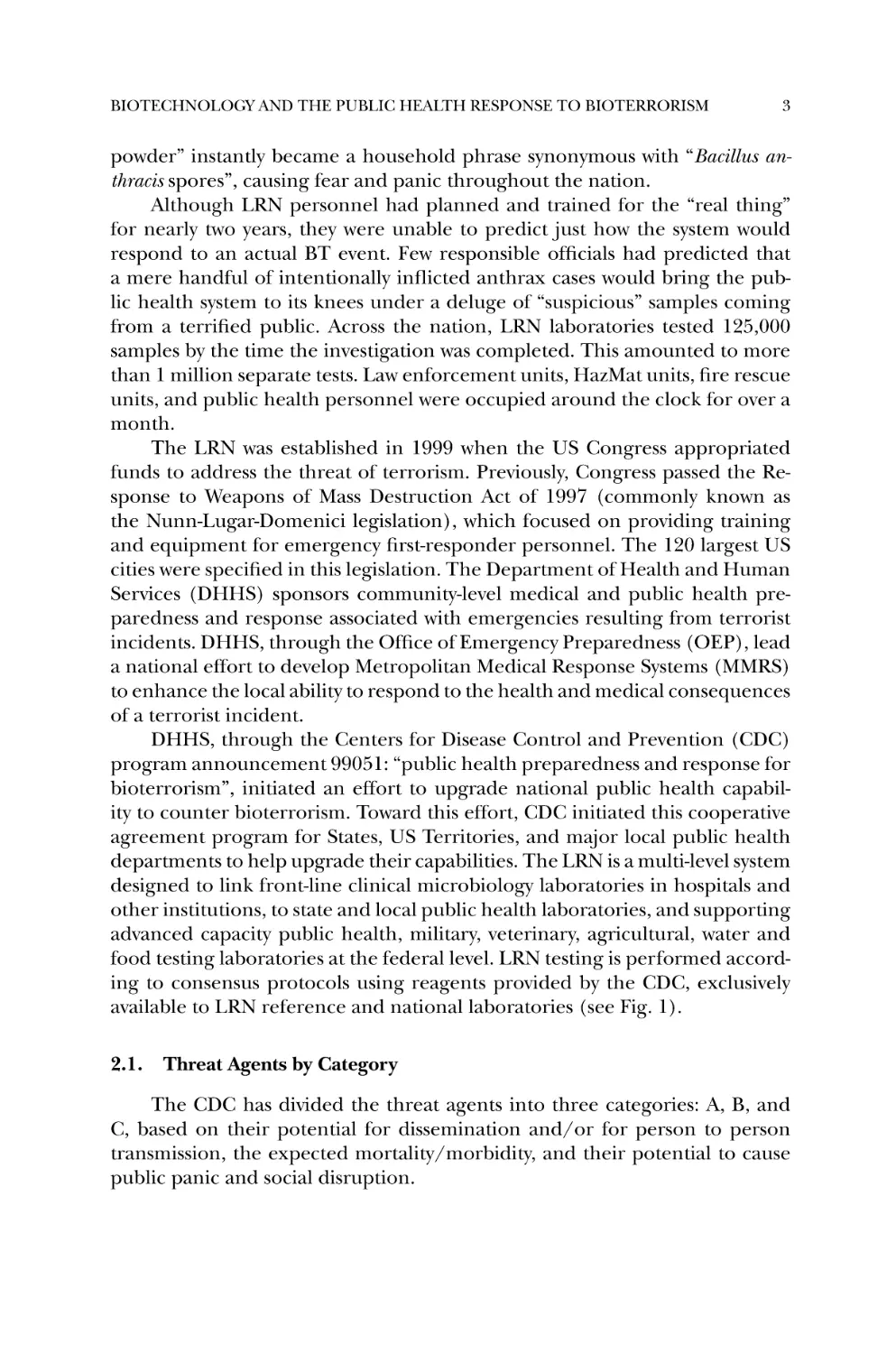
departments to help upgrade their capabilities. The LRN is a multi-level system
designed to link front-line clinical microbiology laboratories in hospitals and
other institutions, to state and local public health laboratories, and supporting
advanced capacity public health, military, veterinary, agricultural, water and
food testing laboratories at the federal level. LRN testing is performed according to consensus protocols using reagents provided by the CDC, exclusively
available to LRN reference and national laboratories (see Fig. 1).
2.1.
Threat Agents by Category
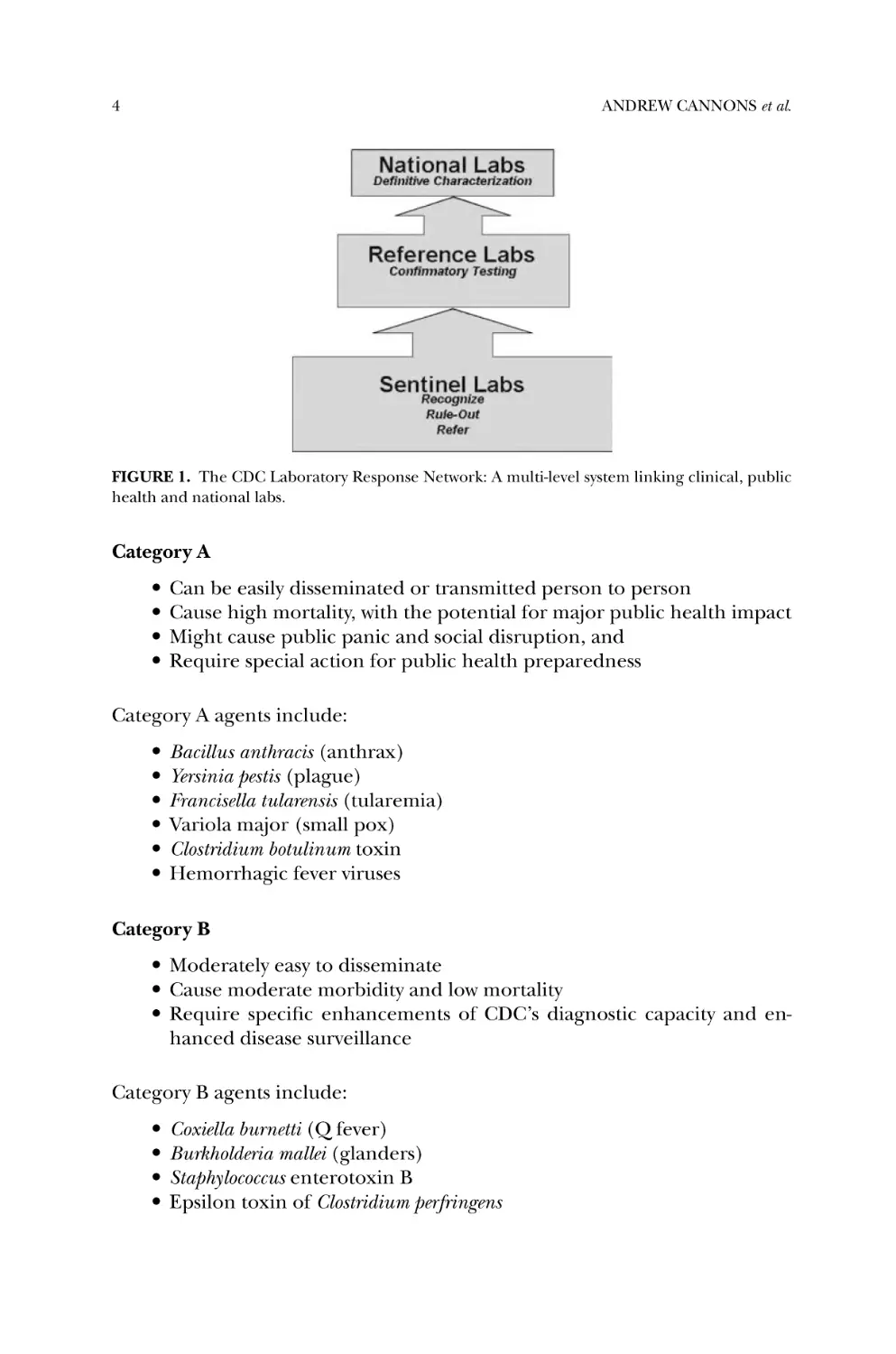
The CDC has divided the threat agents into three categories: A, B, and
C, based on their potential for dissemination and/or for person to person
transmission, the expected mortality/morbidity, and their potential to cause
public panic and social disruption.
4
ANDREW CANNONS et al.
FIGURE 1. The CDC Laboratory Response Network: A multi-level system linking clinical, public
health and national labs.
Category A
r Can be easily disseminated or transmitted person to person
r Cause high mortality, with the potential for major public health impact
r Might cause public panic and social disruption, and
r Require special action for public health preparedness
Category A agents include:
r Bacillus anthracis (anthrax)
r Yersinia pestis (plague)
r Francisella tularensis (tularemia)
r Variola major (small pox)
r Clostridium botulinum toxin
r Hemorrhagic fever viruses
Category B
r Moderately easy to disseminate
r Cause moderate morbidity and low mortality
r Require specific enhancements of CDC’s diagnostic capacity and enhanced disease surveillance
Category B agents include:
r Coxiella burnetti (Q fever)
r Burkholderia mallei (glanders)
r Staphylococcus enterotoxin B
r Epsilon toxin of Clostridium perfringens
BIOTECHNOLOGY AND THE PUBLIC HEALTH RESPONSE TO BIOTERRORISM
5
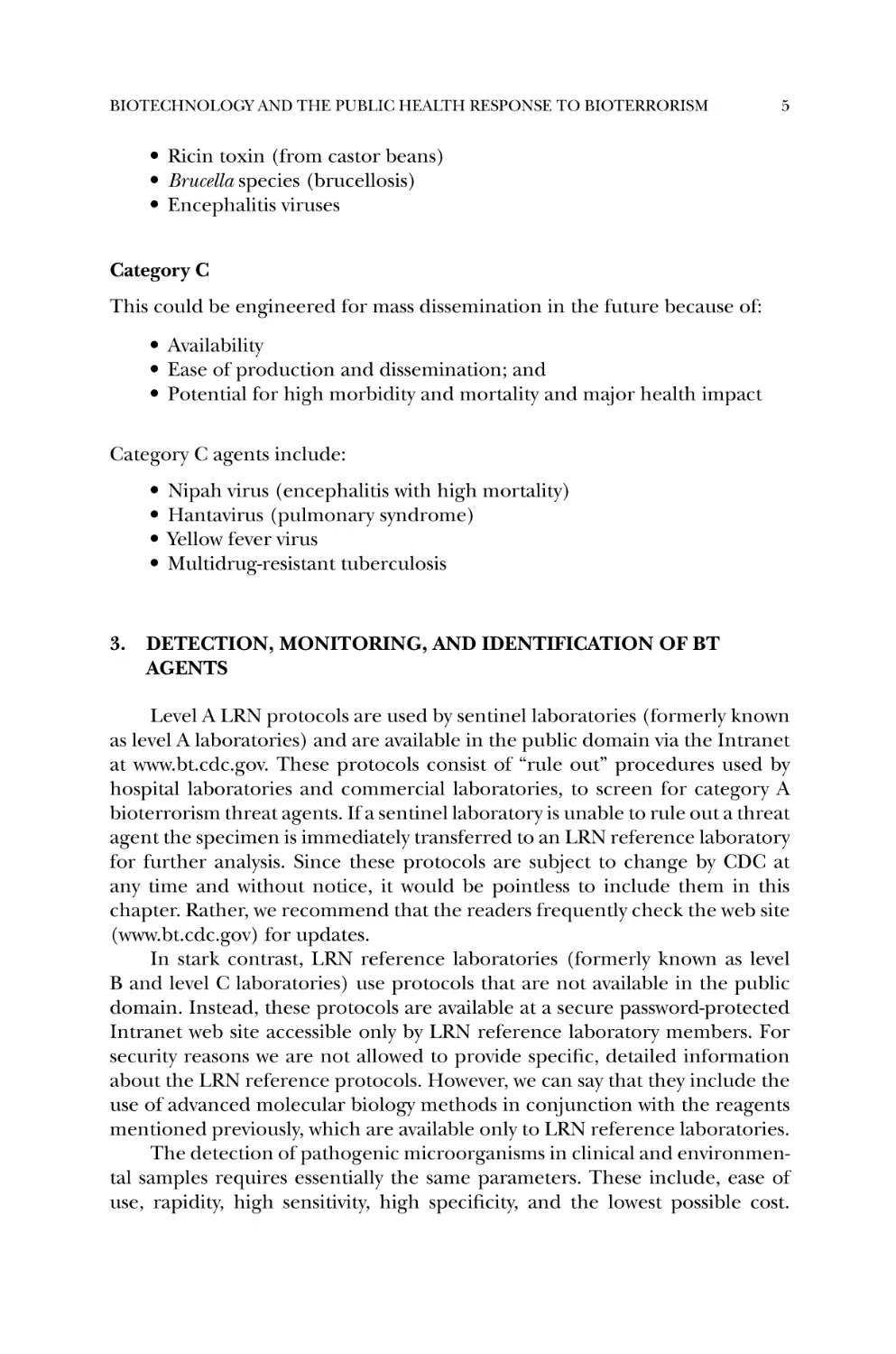
r Ricin toxin (from castor beans)
r Brucella species (brucellosis)
r Encephalitis viruses
Category C
This could be engineered for mass dissemination in the future because of:
r Availability
r Ease of production and dissemination; and
r Potential for high morbidity and mortality and major health impact
Category C agents include:
r Nipah virus (encephalitis with high mortality)
r Hantavirus (pulmonary syndrome)
r Yellow fever virus
r Multidrug-resistant tuberculosis
3.
DETECTION, MONITORING, AND IDENTIFICATION OF BT
AGENTS
Level A LRN protocols are used by sentinel laboratories (formerly known
as level A laboratories) and are available in the public domain via the Intranet
at www.bt.cdc.gov. These protocols consist of “rule out” procedures used by
hospital laboratories and commercial laboratories, to screen for category A
bioterrorism threat agents. If a sentinel laboratory is unable to rule out a threat
agent the specimen is immediately transferred to an LRN reference laboratory
for further analysis. Since these protocols are subject to change by CDC at
any time and without notice, it would be pointless to include them in this
chapter. Rather, we recommend that the readers frequently check the web site
(www.bt.cdc.gov) for updates.
In stark contrast, LRN reference laboratories (formerly known as level
B and level C laboratories) use protocols that are not available in the public
domain. Instead, these protocols are available at a secure password-protected
Intranet web site accessible only by LRN reference laboratory members. For
security reasons we are not allowed to provide specific, detailed information
about the LRN reference protocols. However, we can say that they include the
use of advanced molecular biology methods in conjunction with the reagents
mentioned previously, which are available only to LRN reference laboratories.
The detection of pathogenic microorganisms in clinical and environmental samples requires essentially the same parameters. These include, ease of
use, rapidity, high sensitivity, high specificity, and the lowest possible cost.
6
ANDREW CANNONS et al.
Additional desirable attributes are multiplexing, to detect more than one organism, and the ability to discriminate between naturally occurring pathogens,
pathogens resulting from a BT incident, and hoaxes. Currently, no system in
use incorporates all these parameters. Public health laboratories currently rely
on both culture and molecular procedures for the detection of BT pathogens.
Samples for analysis can be powders used to intentionally deliver agents, such
as talc, cornstarch, and flour; soil; air; clinical (tissues and body fluids); water;
and food. Reference laboratories must be able to process all of these matrices.
Culture, still considered to be the gold standard for testing, has the advantages
of being able to detect more than one agent at a time, does not require huge
investments in instrumentation, high sensitivity, and initial processing can be
rapid.(4) However, growth in culture can be slow, requiring 24–72 hours of incubation and further analysis must be conducted by trained microbiologists. On
the other hand, nucleic-acid-based detection systems have great potential since
they are sensitive, specific, and rapid. The polymerase chain reaction (PCR)
assay is widely used by clinical laboratories for the identification of infectious
disease agents.(5) Real-time PCR, utilizing patented chemistries (e.g., Applied
R
R
Biosystems Taqman
, Roche LightCycler
), improves the time of detection
to hours compared to days. However, most PCR-based detection systems are
limited since they can only target one agent in a single run, the primers and
probes for some of the BT agents may not be available, the equipment and instrumentation can be expensive, and interpretation of data may require a very
high level of technical acumen. Additionally, sample processing for recovery
of PCR-competent nucleic acid can be extensive for some complex matrices
such as environmental powders and food.
Powders and environmental samples are the most common non-clinical
samples submitted to LRN reference laboratories for analysis. Processing of
these samples for real time PCR analysis for B. anthracis can be difficult since
the powder may become aerosolized, thus requiring biosafety level 3 conditions
for safe manipulation. Additionally, the bacterial DNA has to be extracted from
what could be a complex and PCR inhibitory matrix. Methods have been developed that utilize sonication(6) or autoclaving(7) for DNA extraction. A procedure that incorporates sonication, germination, and autoclaving has been
published, providing a safe and rapid method (6 hours) for sample preparation and PCR detection of greater than 10 spores of B. anthracis in various
powders.(8) Should the reference laboratories be deluged with samples for
testing, procedures such as this could help alleviate bottlenecks and decrease
sample processing times.
Food is also a carrier that could be used to purposely distribute pathogens.
Enteric pathogens have been used as BT agents, including the use of Salmonella
enterica in salad bars(9) and the use of Shigella contaminated pastries to sicken
co-workers.(10) Conventional methods to identify bacterial pathogens in foods
are time-consuming and laborious. Real-time PCR detection offers a rapid
alternative,(11) although not without problems. The major challenge comes
BIOTECHNOLOGY AND THE PUBLIC HEALTH RESPONSE TO BIOTERRORISM
7
with sample preparation and extraction of the pathogen DNA from the food.
There are many different food matrices that need to be considered, some
of which can interfere with extraction and/or inhibit the nucleic acid amplification process (http://www.cfsan.fda.gov/∼ebam/bam-1.html). Strategies for
template preparation directly from food have been evaluated(12) with detection limits of 103 CFU/g of food. Increased sensitivity is achieved with initial
enrichment, whereby even five hours growth can increase the detections limits
of pathogens 10–100 fold.
The food emergency response network (FERN) is a counter- terrorism
program formed by the Food and Drug Administration (FDA) and modeled
after the LRN. This is a network of state and federal laboratories committed to
analyzing food samples in the event of a biological, chemical, or radiological
terrorist attack. FERN is developing protocols to be used by state and federal
laboratories for detecting pathogens in food samples. Again, these protocols
will not be available to the public domain and will be maintained on a secure
password-protected web site.
Emergency responders need pathogen detection systems that will yield
results that meet the requirements of laboratory-based detection systems, as
well as the requirements that they be rugged and mobile. Currently, the ideal
system is not available. Most field-use systems rely on immunological detection methods such as lateral flow assays. These assays are generally rapid to
use, taking approximately 15 minutes, require minimal sample processing and
only limited technical expertise. However, immunoassays are typically 1000fold less sensitive than nucleic acid amplification and culture procedures, with
detection limits in the range of 105 CFU/ml.(13) In addition, specificity can
be limited. For example, close relatives of B. anthracis, which are common
contaminants of the environment, can be picked up using some B. anthracis
specific assays.(13) Low sensitivity and specificity lead to false negatives and
false positives, respectively, and can result in an incorrect emergency response.
PCR-based systems for emergency responders are also being developed. These
include instrumentation such as the Handheld Advanced Nucleic Acid AnaR
lyzer (HANAA), a fully automated sample processor that uses TaqMan
-based
PCR for the detection and identification of BT agents and the Ruggedized AdR
vanced Pathogen Identification Device (RAPID
), which is a field-deployable
thermal cycler using LightCycler PCR technology.(14) However, use of PCR in
the field is currently limited because of its complexity, requirement for sophisticated and sensitive equipment, and the need for trained personal for
operation and interpretation of results.
Presently, all BT samples collected by emergency responders are transported to an LRN reference laboratory for characterization and evaluation
using LRN protocols. The use of accurate field detection instruments would improve triage activities and consequently reduce the workload for reference laboratories. However, this can only happen when a suitable and well-characterized
field detection system is available.
8
ANDREW CANNONS et al.
Continual monitoring can be an effective method for early detection and
alleviation of a BT event and thus can supplement and complement the work
of the LRN and emergency responders. This monitoring can be both environmental and/or epidemiological, including surveillance of syndromic data.(15)
Environmental monitoring can be important for pathogen detection immediately after an event/exposure but before symptoms are recognized. As with
pathogen detection in the lab, environmental surveillance requires the development of specialized equipment. Ideally, these monitoring systems should
be automated and be able to discriminate between naturally occurring agents
and those used in a BT event. Presently, few such surveillance systems exist,
such as the Biohazard Detection System (BDS), a PCR-based technology developed by Cepheid(16) and employed in the US postal system; the Biological
Aerosol Sentry and Information System (BASIS), an environmental monitoring system used during the 2002 Winter Olympics utilizing sampling stations to
collect aerosol samples, which were then brought to a laboratory for pathogen
analysis;(15) the nationwide BioWatch program that has been implemented to
monitor the air in several US cities,(15) and was modeled on BASIS; and the
Autonomous Pathogen Detection System (APDS), an automatic aerosol monitoring system for areas of high risk.(14) It is likely that additional technology
will be developed over the next few years that will increase and improve our
capacity for environmental monitoring of pathogenic microorganisms. The
progress in microarray technology and integration of automation will increase
the accuracy and variety of agent detection, while reducing analysis time and
reagent consumption.
4.
THE POTENTIAL FOR MISUSE OF BIOTECHOLOGY
The biotechnology revolution has generated a range of methodologies
that have been exploited for the development of diagnostics, antimicrobials
and vaccines. In particular, the availability of full genome sequences for most
recognized agents of bioterrorism has provided the tools to design diagnostic
reagents as well as new generation vaccines. Clearly, such information has allowed for the improvement of protocols used by the LRN and enabled many
areas of both basic and applied research on these agents. Many different approaches have been used for the development of recombinant vaccines for
anthrax (see Chapter 6) with some impressive successes. The ability to mine
the B. anthracis genome for additional genes that may ultimately constitute new
generation vaccines will likely result in the development of additional vaccine
strategies based on biotechnology.
Since the first gene cloning experiments approximately 30 years ago,
countless molecular biology tools and reagents have been developed. These
techniques allow us to detect, amplify, and express virtually any gene in a
matter of days. Such techniques have rapidly accelerated our ability to study
BIOTECHNOLOGY AND THE PUBLIC HEALTH RESPONSE TO BIOTERRORISM
9
the virulence of bacterial and viral pathogens and have even resulted in the
identification of many new agents of disease. Ultimately, the long-term goal
resulting from the application of modern molecular biology techniques to
infectious disease research is to improve diagnosis and prevention of the diseases that they cause. However, just as these techniques have proven invaluable
in the control and prevention of infectious disease, the same powerful techniques could be used for the development of enhanced biological weapons.
Creating chimeric pathogens with increased virulence, multidrug resistance or
strains that are not targeted by standard vaccines, are now possible.(17) Even
modestly competent molecular biology laboratories have individuals who may
be able to modify many of the “traditional agents” associated with biological
warfare.
The possibility of using biotechnology to engineer a new class of agents,
termed advance biological warfare (ABW) agents, must also be considered.
In addition, biotechnology may have applications supporting weaponization,
dissemination, and delivery as well.(18) Thus, the genetic modification of “traditional agents” associated with biological warfare must be viewed as only a part of
the picture. The form of such ABW agents can only be the subject of conjecture.
It has been suggested that ABW agents could be engineered to target specific
human biological systems such as the cardiovascular, gastrointestinal, neurological, or immunological systems.(18) Transgenic plants and animals could
be engineered to produce large quantities of bioregulatory or toxin proteins.
Transgenic insects, such as bees, wasps, or mosquitoes, could be developed to
produce and deliver biological toxins. For instance, a mosquito could be genetically altered to produce and secrete a biological toxin into its saliva. This
same mosquito would then serve as the vector to deliver the toxin during its
feeding process. Despite the potential to produce toxins that might be effective
at low doses and deliver these toxins, such transgenic insects would likely go
unnoticed. Many of the counterproliferation, detection, and medical countermeasures that have been developed for “traditional agents” will be ineffective
for ABW agents such as protein-based transgenics.(18) Five important attributes
of a biological warfare (BW) agent have been described:(19)
r High virulence coupled with high host specificity
r High degree of controllability
r Lack of timely countermeasures to the attacked population
r Ability to camouflage the BW agent with relative ease
r High degree of resistance to adverse environmental forces
Daly notes that of these five, the last attribute is the most difficult to genetically
engineer into an organism. Accordingly, he suggests that it may be simpler to
engineer BW attributes into organisms that are naturally resistant to environmental forces. This raises the possibility of engineering extremophiles for use
as BT agents. Among these are agents that are resistant to environmental factors
including desiccation, ultraviolet radiation, high temperatures and pressures,
10
ANDREW CANNONS et al.
or decontamination compounds. Deinococcus radiodurans is one such bacterium
that is resistant to multiple environmental factors including radiation, as the
species designation implies. This Gram-positive coccus displays remarkable resistance to even Megarad doses of radiation. Furthermore, availability of the
complete D. radiodurans genome sequence, together with genetic systems to
express foreign genes, makes this bacterium amenable to genetic engineering.
Daly has suggested that it may be possible to synthesize and store viruses within
microorganisms such as D. radiodurans. The use of extremophiles such as D .
radiodurans may seem to be a complicated approach to the development of BT
agents. Furthermore, the ability of such agents to establish efficient infections
in the human population may, in itself, require extensive genetic engineering.
Despite these facts the use of extremophiles as BT agents should be considered
as a threat that must not be ignored.
5.
BIOTECHNOLOGY, PUBLIC HEALTH INTEREST AND
THE EXCHANGE OF SCIENTIFIC INFORMATION
The potential to misuse the so-called dual-use technology (civil and military) is clearly illustrated in genomics research.(20) Genome sequences are
available for most recognized BT agents and countless numbers of bacterial
and viral pathogens. Although this information has undoubtedly enhanced
our ability to detect, prevent, and treat infections caused by BT agents, experts
in the field of genomics recognize the possibility of “tailoring” classical BW
agents to make them harder to detect, diagnose, and treat.(21) How then can
we make pathogen genome sequences, or for that matter scientific information related to possible BT agents, available for valid scientific research? What
types of research constitute studies that should not be published and accessible
to all? These are some of the questions that must be addressed to ensure that
biodefense research findings accessible to the public domain are not used for
nefarious purposes.(22)
In a study from 2001, it was shown the recombinant mousepox virus
that expresses IL-4 suppressed natural killer cell and cytotoxic T-lymphocyte
responses.(23) The natural genetic resistance of some mice strains to mousepox virus was overcome. More importantly, recently immunized genetically
resistant mice were shown to be susceptible to infection with this virus. Thus,
not only does the virus-encoded IL-4 suppress primary antiviral cell-mediated
immune responses, but it also can inhibit the expression of immune memory
responses. The finding that a poxvirus can circumvent immune memory has
potential implications for smallpox vaccination efforts, as well as providing a
tool that could be misused to manipulate the immune system. Not surprisingly, this publication created quite a topic for discussion among both scientists and government officials about the potential for misuse of information in
such publications. In another study, infectious poliovirus was generated from a
BIOTECHNOLOGY AND THE PUBLIC HEALTH RESPONSE TO BIOTERRORISM
11
synthetic cDNA template, showing that it is possible to synthesize infectious
agents in vitro from genome sequence data.(24) Again, this report sounded an
alarm and raised questions from the public and some in the scientific community, about the possibility of synthesizing larger and more complicated viruses
such as HIV, Ebola or even smallpox from genome sequence information.
The Patriot Act (2001) and the Public Health Security and Bioterrorism Preparedness and Response Act of 2002 imposed new regulations on the conduct
of research involving select agents. Concern has been raised in the scientific
community that such regulations will have a “chilling effect upon legitimate scientific inquiry”.(25) The need for scientific self-governance has been suggested
and the essential components of such a system have been proposed.(25)
5.1.
Public Perception of Biodefense Research
Over the next decade, the Unites States will spend billions of dollars to
develop countermeasures against biological and chemical weapons.(26) In the
past, most of this type of research was conducted at a few government facilities.
However, it is likely that much of the research conducted in the coming decade
will be performed in academic settings. Advocacy groups such as the Sunshine
Project of Austin, Texas (http://www.sunshine-project.org) and the Council on
Responsible Genetics in Boston, Mass (http://gene-watch.org), are opposed to
high-security containment laboratories and have taken to monitoring compliance issues at academic research facilities. Citizen and community opposition
to maximum containment biosafety level 4 facilities has been encountered in
response to proposed facilities in both Boston(27) and Montana.(28) Recently,
three laboratory workers at a Boston University laboratory apparently contracted tularemia from mishandling a virulent strain of Francisella tularensis.(29)
This further raised community concern about biosafety in the Boston University laboratories, which was recently awarded the National Institutes of Health
funding to build a National Biocontainment Biosafety Level 4 facility.(30) Again,
public concern and a lawsuit by local residents have resulted.
In addition to biosafety concerns in the public and scientific community,
a backlash reaction to the large spending increases in federal research dollars on biodefense has recently surfaced. In a recent letter to the Director of
NIH, over 700 microbiologists indicated their concern about the redirection
of NIH grant funds from other projects to biodefense-related projects (accessible at: http://waksman.rutgers.edu/NIH-MBC BM/current/). In that letter it
was pointed out that funding for prioritized BW agents has increased by 1500%
from 1996 to 2005. In contrast, research on pathogens that are non-biodefense
associated has decreased by 27%. It was suggested that the greatest threat to
public health are existing and emerging infectious diseases. Basic research on
many of these agents is currently under funded in comparison to the prioritized BW agents. Given the large number of cases, as well as the morbidity
and mortality associated with some of the organisms that are not identified
12
ANDREW CANNONS et al.
as priority agents, as compared to those that are, it is difficult to refute this
argument.
SUMMARY
The events of September 11, 2001, as well as the subsequent intentional
release of anthrax spores in contaminated letters, have changed the way both
the public and the scientific community view the use of biotechnology in
biodefense research. The enhancement of our public health infrastructure
has greatly aided our ability to respond to potential future acts of bioterrorism, and to a certain extent, emerging and re-emerging infectious diseases The
Laboratory Response Network is an example of infrastructure that was already
in place prior to September 11, 2001, but that has been strengthened considerably after that date. The possibility of governmental control over access
to scientific information that might be considered dual use remains an active
topic for debate. Backlash, from the public due primarily to safety concerns
and among scientists over concerns about prioritization of biodefense projects,
are more recent developments related to the government funding for biodefense. The need to balance our obligation to respond to acts of bioterrorism,
along with all other public health threats, will undoubtedly continue to be a
matter of contention for several years to come.
REFERENCES
1. Jernigan, J. A., and Stephens, D. S., et al., 2001, Bioterrorism-related inhalational anthrax: the
first 10 cases reported in the United States, Emerg. Infect. Dis. 7:933–944.
2. Christopher, G. W., and Cieslak, T. J., et al., 1997, Biological warfare. A historical perspective,
JAMA. 278:412–417.
3. Marty, A., 2001, History of the development and use of biological weapons. Laboratory aspects
of biowarfare., Clinics Lab. Med. 21:421–434.
4. Peruski, L. F., and Peruski, A. H., 2003, Rapid diagnostic assays in the genomic biology era:
detection and identification of infectious disease and biological weapon agents, Biotechniques.
35:840–46.
5. Whelan, A. C., and Persing, D. H., 1996, The role of nucleic acid amplification and detection
in the clinical microbiology laboratory, Ann. Rev. Microbiol. 50:349–73.
6. Belgrader, P., and Hansford, D., 1999, A minisonicator to rapidly disrupt bacterial spores for
DNA analysis, Anal. Chem. 71:4232–4236.
7. Dang, J. L., Heroux., K., 2001, Bacillus spore inactivation methods affect detection assays, Appl.
Environ. Microbiol. 67:3665–3670.
8. Luna, V. A., and King, D., 2003, Novel sample preparation method for safe and rapid detection
of Bacillus anthracis spores in environmental powders and nasal swabs, J. Clin. Micro. 41:1252–
1255.
9. Torok, T. J., and Tauxe, R. V., 1997, A large community outbreak of salmonellosis caused by
intentional contamination of restaurant salad bars, JAMA. 278:389–395.
10. Kolavic, S. A., and Kimura, A., 1997, An outbreak of Shigella dysenteriae type-2 among laboratory
workers due to intentional food contamination, JAMA. 278:396–398.
BIOTECHNOLOGY AND THE PUBLIC HEALTH RESPONSE TO BIOTERRORISM
13
11. Jordan, J. A., 2000, Real-time detection of PCR products and microbiology, New technologies for
life sciences: a trends guide. 6:61–66.
12. Heller, L. C., and Davis, C. R., 2003, Comparison of methods for DNA isolation from food
samples for detection of shiga toxin-producing Escherichia coli by real-time PCR, App. Environ.
Micro. 69:1844–1846.
13. King, D., and Luna, V., 2003, Performance assessment of three commercial assays for direct
detection of Bacillus anthracis spores, J. Clin. Micro. 41:3454–3455.
14. Ivnitski, D., O’Neil, D. J. O., 2003, Nucleic acid approaches for detection and identification of
biological warfare and infectious disease agents, BioTechniques. 35:862–869.
15. Fitch, J. P., and Raber, E., et al., 2003, Technology challenges in responding to biological or
chemical attacks in the civilian sector, Science, 302:1350–1354.
16. Meehan, P. J., and Rosenstein, N. E., 2004, Responding to detection of aerosolized Bacillus
anthracis by autonomous detection systems in the workplace, MMWR. 53:1–12.
17. Greenfield, R. A., Lutz, B. D., Huycke, M. M., and Gilmore, M. S., 2002, Unconventional
biological threats and the molecular biological response to biological threats, Am. J. Med. Sci.
323:350–357.
18. Petro, J. B., Plasse, T. R., and McNulty, J. A., 2003, Biotechnology: impact on biological warfare
and biodefense, Biosecurity and bioterrorism: biodefense strategy, practice and science. 1:161–168.
19. Daly, M. J., 2001, The emerging impact of genomics on the development of biological weapons–
threats and benefits posed by engineered extremophiles, Clinics Lab. Med. 21:619–629.
20. Black, J. L., 2003, Genome projects and gene therapy: gateways to next generation biological
weapons, Military Med. 168:864–871.
21. Fraser, C. M., and Dando, M. R., 2001, Genomics and future biological weapons: the need for
preventive action by the biomedical community, Nature Genetics. 29:253–256.
22. Atlas, R. M., 2002, National security and the biological research community, Science. 298:753–
754.
23. Jackson, R. J., Ramsay, A. J., et al., 2001, Expression of mouse interleukin-4 by a recombinant
ectromelia virus suppresses cytolytic lymphocyte responses and overcomes genetic resistance
to mousepox, J. Virol. 75:1205–1209.
24. Cello, J., Paul, A. V., and Wimmer, E., 2002, Chemical synthesis of poliovirus cDNA: generation
of infectious virus in the absence of natural template, Science. 297:1016–1018.
25. Kwik, G., Fitzgerald, J., Inglesby, T. V., and O’Toole, T., 2003, Biosecurity: responsible stewardship of bioscience in an age of catastrophic terrorism. Biosecurity and bioterrorism: biodefense
strategy, practice and science. 1:27–35.
26. Kahn, L., 2004, Biodefense research: can secrecy and safety co-exist? Biosecurity and bioterrorism:
biodefense strategy, practice and science. 2:81–85.
27. Lawler, A., 2004, Boston weighs a ban on biodefense studies, Science (News). 304:665.
28. Kaiser, J., 2004, Citizens sue to block Montana biodefense lab., Science (News). 305:1088.
29. Dalton, R., 2005, Infections scare inflames fight against biodefense network, Nature. 433:344.
30. Lawler, A., 2005, Boston University under fire for pathogen mishap, Science. 307:501.
2
Historical Perspectives of
Microbial Bioterrorism
STEPHEN A. MORSE
1.
INTRODUCTION
A number of events over the last decade have served to focus attention on
the threat of terrorism and the use of biological or chemical weapons against
military and civilian populations for the purpose of causing illness or death.
It is increasingly recognized that agricultural animals and plants also present
a vulnerable target to terrorists.(1,2) Most significantly, the threat of terrorism
has attracted the attention of policy makers in all levels of government in the
United States. However, policy makers and analysts have differed in their assessment of the threat of bioterrorism. Many authorities believed that the threat
of bioterrorism was growing, particularly from non-state sponsored groups.(3)
Some of them contended that it was only a matter of time before a terrorist used
biologic agents to cause mass casualties, while others argued that the historical
record provided no basis for concern. Moreover, some even questioned the
wisdom of funding preparedness efforts.(4) However, the situation changed in
October 2001 when an individual or individuals sent spores of Bacillus anthracis
to media companies in New York City and Boca Raton, Florida(2) resulting in
five deaths and considerable panic throughout the country.
1.1.
Definitions
For the purposes of this article, the working definition of a biological agent
is “a microorganism (or a toxin derived from it) which causes disease in man,
plants, or animals or causes deterioration of material.”(6) In this context, the
biological agents are normally divided into three categories: anti-personnel,
STEPHEN A. MORSE
•
Centers for Disease Control and Prevention, Atlanta, GA, 30333.
15
16
STEPHEN A. MORSE
anti-animal, and anti-plant. In addition, the use of biological agents is often
classified by the manner in which they are used. For example: biological warfare
has been defined as a specialized type of warfare conducted by a government
against a target; bioterrorism has been defined as the threat or use of biological
agents (or toxins) by individuals or groups motivated by political, religious,
ecological, or other ideological objectives.(7) Terrorists can be distinguished
from other types of criminals by their motivation and objective; criminals may
also be driven by psychological pathologies and may use biological agents.
When criminals use biological agents for murder, extortion, or revenge it is
called a biocrime.(7)
1.2.
Development and Prohibition of Biological Weapons
In November 1918, an armistice ended World War I in which eight million
soldiers and nearly as many civilians were killed. However, that armistice could
not halt the even greater ravages of an influenza pandemic. In the course of
a single year beginning in the spring of 1918, the virus spread globally killing
more than 20 million people. No one thought that this influenza pandemic was
a deliberate act of war; however, the magnitude of the impact of this epidemic
apparently impressed the statesmen of the era.(8) When the Geneva Protocol was issued in 1925 to ban, in warfare, the use of asphyxiating, poisonous,
or other gases, which had been responsible for about one million casualties
during World War I,(6) the provision was extended to include bacteriological
agents as well.(9) The Geneva Protocol affirmed that chemical and biological weapons were “justly condemned by the general opinion of the civilized
world.”(8)
In 1972, the convention on the prohibition of the development, production, and stockpiling of bacteriological (biological) and toxin weapons and on
their destruction (referred to as the Biological Weapons Convention or BWC)
was opened for signature. Since it entered into force in 1975, the BWC has been
signed and ratified by 141 countries, signed but not ratified by 18 countries,
and observed by the Government of Taiwan. The BWC prohibits the development, production, stockpiling, or acquisition of microbial or other biological
agents or toxins of types and in quantities that have no justification for prophylactic, protective, or other peaceful purposes.(9) The BWC also prohibits
the weapons, equipment, or means of delivery designed to use such agents or
toxins for hostile purposes or in armed conflict.(9) In addition, it requires that
each State Party (i.e., those countries that both signed and ratified the BWC)
destroy, or divert to peaceful purposes, all agents, toxins, weapons, equipment,
and means of delivery which are in its possession or under its jurisdiction or
control. Each State Party also agreed not to transfer any of the agents, toxins,
weapons, equipment, or means of delivery to any recipient, or induce any State
to manufacture or otherwise acquire such organisms or equipment for nonpeaceful purposes.(9) Unfortunately, the BWC has no verification provisions,
HISTORICAL PERSPECTIVES OF MICROBIAL BIOTERRORISM
17
and there have been significant difficulties in determining the existence or
status of State programs.
The use of biological agents for the purpose of warfare has been associated
with State programs for the development of biological weapons. Some of these
programs were very large, employing thousands of people. Leitenberg(10) has
recently reviewed the activities of several of these programs. Although there are
inherent differences between terrorist and criminal use of biological agents,
the criminal faces many of the same obstacles as the terrorist. Both must acquire, develop, and employ biological weapons, so the technical constraints
will probably be similar.
The purpose of this chapter will be to provide a historical account of the
use of biological agents for warfare, terrorism, or criminal purposes. However,
in doing so one must appreciate that there are problems in assessing the historical use of biological agents because of: (1) difficulties in verifying an alleged
or attempted biological attack; (2) the use of allegations of biological attacks
for propaganda purposes; (3) a lack of pertinent microbiological or epidemiological data; (4) the presence of naturally occurring endemic or epidemic
diseases during hostilities; and (5) the secrecy surrounding biological weapons
programs.(11) This review relies heavily on the extensive research of Carus,(7)
who identified most of the events and attempts to use biological agents since
1900. This review will not cover hoaxes or attempts to acquire biological agents
for nebulous purposes (e.g., Larry Wayne Harris incident(7) ).
2.
EARLY USE OF BIOLOGICAL AGENTS IN WARFARE
An examination of the way in which biological agents were used for warfare
in the fourteenth through the middle of the nineteenth centuries demonstrates
a correlation with the prevailing theories of infectious diseases.
2.1.
Early Theories of Infectious Disease
A number of theories have evolved over time to explain the origin of
epidemics. Two of these have been referred to as miasma and contagion. In the
theory of miasma, epidemics were thought to be due to an atmospheric poison
generated in external nature.(12) Disease was thought to be a consequence
of “bad air” resulting from extensive decomposition. An example of how this
theory might have played a role occurred at the siege of Thun l’eveque (Table I)
where catapults were used to hurl dead horses over the wall into the castle.
Years later and based on eyewitness testimony, the chronicler Froissart wrote
that the “stynke and ayre was so abomydable, that they considered howe that
finally they coude nat long endure” (as cited in ref 13). Miasma originated
from the first-hand observations of Hippocrates (460–360 B.C.) of the effects of
climate, season, and locality on outbreaks of disease.(12) However, it was Galen
18
STEPHEN A. MORSE
(130–200 A.D.) who developed the idea of “miasmic corruption of the air”,
which figured prominently in the way medicine viewed epidemics in the Middle
Ages. Galen thought that epidemic disease resulted from “the inhalation of
air fouled by putrid exhalations that might come from sources far and near,
such as masses of unburied corpses of the slain in battle, or from swamps
and stagnant water in summertime, or from the excessive heat of foul air in
close and in unventilated hovels”.(12) By the fourteenth century, the idea that
the immediate cause of epidemics was some sort of corruption in the air was
widely accepted. It was believed that this corrupted air could gain entrance to
the body by way of the lungs or through wide-open pores in the skin as a result
of excesses, bathing, or heat.
Also in the fourteenth century, additional prominence was given to the
idea of contagion. In the theory of contagion, the “poison” was originally generated in man himself and spread person-to-person by contact with the sick
or dead, or with their personal effects (fomites). The fact that some diseases
were infectious and could be transmitted person-to-person had been long recognized. There are numerous biblical references in the Books of Leviticus,
Numbers, and Deuteronomy to the “infectivity” of lepers. Thucydides thought
that the “plague” of Athens in 430 B.C. was exceedingly infectious.(12) In the
fourteenth century, smallpox and measles were added to the list of infectious
diseases.(12)
In the sixteenth century, Girolamo Fracastoro (1478–1553) recognized
and defined three kinds of contagion. In his treatise De contagione et contagionis
morbis et eorum curatione, Fracastoro attributed certain diseases including plague,
smallpox, and measles, to specific tiny seeds (seminaria) and stated that these
specific contagions could be spread directly from person-to-person, indirectly
via infected clothing, wooden objects, or other fomites, and even at a distance,
such as through the air.(14)
At various times, both theories were combined. Some adherents to the
theory of contagion argued that the sick could radiate infection through the air
in their immediate vicinity (a local miasma), while some adherents to the theory
of miasma admitted a limited degree of contagion at the peak of a severe
epidemic.
2.2.
Selected Incidents from the Fourteenth to the Nineteenth Centuries
Table I lists a number of substantiated and unsubstantiated incidents occurring between 1340 and 1863 in which biological agents were used for the
purpose of warfare. If we assume that there was intent to spread disease in order to weaken or defeat the enemy, it is possible to correlate the methods used
to deliver the disease with the prevailing theories of miasma and contagion.
The claim that biological warfare was used at the 1346 siege of Caffa by the
Mongols deserves special mention because of its association with the spread
of the Black Death, which devastated Europe, the Near East, and North Africa
HISTORICAL PERSPECTIVES OF MICROBIAL BIOTERRORISM
19
TABLE I
Selected Substantiated and Unsubstantiated Incidents of Biological
Warfare, 1340–1863
Period
Incident
Reference
1340
Jean, Duke of Normandy besieged the castle of Thun l’eveque,
which had been captured by the Englishman, Sir Walter of Manny.
Catapults were used to cast dead horses over the wall into the castle.
Chronicled years later by Jean Froissart, based on eyewitness
testimony of participants from both sides.
13
1346
Plague hits Mongol forces besieging the Genoese city of Caffa
(now Feodosija, Ukraine) on the Crimean coast. Fresh corpses of
plague victims were lobbed into the city. Plague breaks out in the
city; 85,000 plague deaths in the region. Mongols abandon siege.
Event chronicled in 1348–1349 by the Italian, Gabriele de’ Mussi.
13,15
1422
At the siege of Karlstein, machines were used to catapult corpses of
those who died in battle, and manure or garbage, into the city. The
incident was described 250 years after the event and is not
considered credible.
13
1500
Pizarro presented the indigenous peoples of South America with
variola-contaminated clothing.
16
1710
Siege of Reval (now Tallin, Estonia), Sweden. Russians were said
to have hurled corpses of plague victims into the besieged city,
following which plague broke out in the city. There is no
documentation to support this claim. The event is referred to
in a Swedish military document.
13
1763
During the French and Indian War (1754–1767), Sir Jeffrey
Amherst, commander of British forces in North America, suggested
the deliberate use of smallpox to “reduce” native American tribes
hostile to the British. Captain Ecuyer (one of Amherst’s
subordinates), fearing an attack on Ft. Pitt from native Americans,
acquired two variola-contaminated blankets and a handkerchief
from a smallpox hospital and in a false gesture of good will
distributed them to the native Americans. Several outbreaks of
smallpox occurred in various tribes in the Ohio River valley.
13
1775
In Boston, British attempted to spread smallpox among the
continental forces by inoculating (variolation) civilians fleeing the
city. In the south, there is evidence that the British were going to
distribute slaves who had escaped during hostilities, and were sick
with smallpox, back to the rebel plantations in order to spread the
disease.
13,14
1861–1863
General W.T. Sherman complained that retreating confederate
troops were deliberately shooting farm animals in ponds so that
their “stinking carcasses” would contaminate water supplies for the
Union forces, resulting in troops weakened and demoralized by
gastrointestinal disease. Allegations that Dr. Luke Blackburn, a
future governor of Kentucky, attempted to infect clothing with
variola and then sell it to unsuspecting Union troops could not be
substantiated.
6
20
STEPHEN A. MORSE
in the mid-fourteenth century. Wheelis(13) believes that the hurling of plagueinfected cadavers into the besieged city of Caffa was not only plausible, technically feasible, and consistent with contemporary notions of disease causation,
but that it provided the best explanation of the entry of plague into the city.
The attack itself appeared to be successful as it produced casualties within the
city; however, it was of no strategic importance as the city remained in Italian
hands and the Mongols abandoned the siege. These facts not withstanding,
Wheelis(15) provides a convincing argument that this incident did not have a
decisive role in the spread of plague to Europe.
3.
THE GERM THEORY AND BIOTERRORISM
The seminal work of Robert Koch (1843–1910) provided the basis for
the development of a new generation of biological weapons. Although others
provided indirect evidence for the importance of microorganisms in causing
human diseases, it was Koch who clearly conceptualized and provided experimental support for the germ theory of disease. In his early work on anthrax, he
used microscopy to demonstrate that the blood of diseased animals contained
large numbers of a spore-forming bacterium. He also showed that the bacteria could be cultured outside the animal body in nutrient fluids. However, to
link a specific microorganism to a specific disease, the organism must first be
isolated in pure culture. Toward this end, Koch developed several ingenious
methods, e.g., use of nutrient medium solidified with gelatin or agar, to isolate
microorganisms in pure culture. Once microorganisms were available in pure
culture he was able to formulate the criteria, now called Koch’s postulates, for
proving that a specific type of microorganism causes a specific disease. The
effects of these discoveries can be seen in the types of incidents that occurred
since 1900.
3.1.
Selected Confirmed Incidents, 1900–2003
Since 1900, there have been a number of incidents in which the use of a
biological agent was suspected. Carus(7) used specific criteria to confirm that
a biological agent was used in a criminal, terrorist, or state-sponsored event.
Most of the incidents in which the use of a biological agent has been confirmed
are listed in Table II. Of these 29 confirmed incidents, the majority (N = 19)
was of the criminal type. Of the remainder, five were state-sponsored and four
were considered to be terrorist events. The anthrax attack of 2001, which is
currently under investigation, is listed as unknown.
Among these 29 confirmed incidents, 15 involved the use of bacteria,
10 toxins, 5 viruses, and 1 used the ova of a parasitic roundworm. In three of
the confirmed incidents, biological agents or toxins were used against livestock
or other animals; the remainder involved the use of biological agents or toxins
HISTORICAL PERSPECTIVES OF MICROBIAL BIOTERRORISM
21
TABLE II
Selected Confirmed Incidents Where Biological Agents Were Used, 1900–2003
Date
Incident(s)
Type
Reference
1910
Patrick O’Brien de Lacy and Vladimir Pantchenko
(a physician) were convicted in St. Petersburg, Russia,
of murdering Captain Vassilli Buturlin (de Lacy’s
brother in law) by injection with diphtheria toxin
Criminal
17
1909–1918
Henri Girard used S. typhi and poisonous mushrooms
to murder people to whom he sold insurance policies in
order to obtain the death benefits. He was responsible
for killing two people; six others recovered after being
infected or poisoned. Girard was studying bacteriology
at the time of the first murder
Criminal
7,17
1913
Karl Hopf infected his third wife with V. cholerae and
typhus organisms. He murdered his father, two of his
children and his first wife with arsenic. He was also
accused of attempting to poison his second and third
wives and his mother. Hopf had training in handling
drugs. He was convicted in a German court
Criminal
7
1915–1918
The German Secret Service instituted a covert biological
warfare campaign in the United States during the early
part of World War I, while the United States was still
neutral. They used B. mallei (glanders) and B. anthracis
(anthrax) to infect horses and mules that Allies
purchased in the United States for use by their forces in
Europe. In Romania, they infected sheep bound for
Russia with glanders and anthrax. In Argentina, they
infected sheep, cattle, and horses with glanders and
anthrax that were being shipped to Britain and to the
Indian army. They purportedly used B. mallei and V.
cholerae against allied forces during the German retreat
State
18
1916
Arthur Warren Waite was a dentist who had made
serious attempts to acquire virulent pathogens. He
killed his mother-in-law by putting pathogenic
microorganisms in her food. When an attempt to kill his
father-in-law using pathogens was unsuccessful, he
poisoned him with arsenic
Criminal
7
1932
The League of Nation’s General Assembly established
the Lytton Commission in December, 1931, to
investigate Japan’s conquest of Manchuria. When
commissioners visited Manchuria in 1932, the Japanese
served them fruit “laced” with V . cholerae. No one
became ill
State
7
1933
Dr. Taranath Bhatacharyna was a physician with training
in bacteriology. He and Benoyendra Chandra Pandey
murdered 20-year-old Amarendra Pandey (half brother
of Benoyendra) with a lethal dose of Y. pestis after a feud
over the division of their father’s estate
Criminal
7,19
(continued )
22
STEPHEN A. MORSE
TABLE II
(Continued )
Date
Incident(s)
Type
Reference
1936
Dr. Tei-Sabro Takahashi was a Japanese physician
who used food contaminated with S. typhi to infect
17 people, including three who subsequently died.
The incidents involved competing physicians, their
families, and his wife
Criminal
7
1939
Dr. Kikuko Hirose, a Japanese physician, gave pastries
contaminated with S. typhi and S. paratyphi to her former
husband who, in turn, shared them with others. Twelve
became ill and one died
Criminal
7
1932–1945
Japan conducted biological weapons research at
facilities in China (e.g., Unit 731). Prisoners were
infected with pathogens including B. anthracis,
N. meningitidis, V. cholerae, Y. pestis, and Shigella spp.
Between 1932 and 1945, more than 10,000 prisoners
died as a result of experimental infection or execution
following experimentation. At least 11 Chinese cities
were attacked with biological weapons sprayed from
aircrafts or introduced into water supplies or food
items. Plague-infected fleas were released from aircraft
over Chinese cities to initiate plague epidemics
State
20
1952
The Mau Mau used the plant toxin from the African
milk bush (Synadenium grantii) to kill livestock in what is
now Kenya.
Terrorist
7
1964
Dr. Mitsuru Suzuki, a Japanese physician with training in
bacteriology, was arrested for infecting four colleagues
with a sponge cake contaminated with dysentery. He was
subsequently linked to a series of typhoid fever and
dysentery outbreaks involving approximately 200
people, including four deaths. Prosecutors claimed he
did this to complete his dissertation, which involved
studies of S. typhi recovered from numerous persons. A
culture of S. typhi was stolen from Japan’s NIH; another
culture was isolated from an infected patient
Criminal
7
1970
Eric Kranz, a postgraduate student in parasitology at
MacDonald College, infected four of his room-mates
using food contaminated with large numbers of
embryonated ova of Ascaris suum, a parasitic roundworm
found in pigs. The infected individuals presented with
symptoms and signs of lower respiratory tract disease,
the more severely ill being in acute respiratory failure
Criminal
21
1977
Arnfinn Nesset, who ran a nursing home for the elderly
in Norway, was convicted of murdering 22 of his
patients by injecting them with curacit, which is derived
from curare
Criminal
7
HISTORICAL PERSPECTIVES OF MICROBIAL BIOTERRORISM
23
TABLE II
(Continued )
Date
Incident(s)
Type
Reference
1978
The Bulgarian secret police attempted to assassinate
Vladimir Kostov, a Bulgarian defector who had served as
a news correspondent and was also a major in the D.S.
(Bulgarian equivalent to the K.G.B.). A small metal pellet
containing ricin was injected into Kostov who
subsequently became ill but did not die
State
7,22
1978
In London, the Bulgarian secret police assassinated
Georgi Markov, a Bulgarian dissident and announcer for
Radio Free Europe. He was killed by ricin contained in a
small platinum-iridium pellet that was injected into the
back of his thigh by means of a modified umbrella tip.
He died 4 days later
State
7
1981
A group calling itself “Dark Harvest” was responsible for
leaving a package of soil on the grounds of the Chemical
Defense Establishment located at Porton Down, England.
By doing so, they claimed they were returning “seeds of
death” to their source. The group claimed the soil was a
part of a larger quantity (300 pounds) removed from
Gruinard Island where tests of anthrax bombs were
conducted in 1941. They also stated that microbiologists
from two universities and locals were involved in
removing the soil. Analysis showed that the soil contained
B. anthracis (approx. 10 organisms/gram of soil)
Terrorist
7
1984
The Rajneeshees, a religious cult, employed biological
agents against inhabitants of The Dalles, Oregon in an
attempt to influence the local government. In the first
incident, two county commissioners visiting the
commune were given drinking water contaminated with
S. Typhimurium–both became sick. Later, members of
the cult contaminated salad bars, salad dressing, and
coffee creamers in local restraunts with S.Typhimurium.
As a result, 751 people became sick. Attempts were also
made to contaminate the water system. Bactrol discs
containing S.Typhimurium were legitimately obtained
from VWR Scientific in Seattle for use in their medical
clinic’s state-licensed laboratory. It was later removed to a
clandestine laboratory where large quantities were grown
Terrorist
7,23
1990–1995
The Aum Shinrikyo is a religious cult that was
responsible for the 1995 dissemination of sarin gas in the
Tokyo subway system. The cult claimed they had 10,000
members and assets of ≥$300 million dollars. The cult
was also involved in biological warfare activity involving
botulinum neurotoxin, B. anthracis, C . burnetii, and
attempted to obtain Ebola virus from Zaire. The group
attempted to use aerosolized biological agents against
nine targets including:
(continued )
24
STEPHEN A. MORSE
TABLE II
(Continued )
Date
Incident(s)
Type
Reference
botulinum toxin (Japan Parliament, Narita International
airport, downtown Tokyo, and Tokyo subway); and anthrax
(sprayer on the roof of the Aum building in East Tokyo, a
truck sprayer around the Diet in Central Tokyo, Imperial
palace, Yokohama, and the U.S. Naval base at Yokosuka).
None of these attacks were successful due to the selection
of the wrong strain or to the conscience of the individual
responsible for filling the dissemination devices
Terrorist
7,24,25
1990
Graham Farlow was an asymptomatic HIV-positive inmate
at a prison in New South Wales, Australia. He injected a
guard (Geoffrey Pearce) with HIV-contaminated blood.
The guard became infected with HIV. Farlow died of AIDS
Criminal
26
1992
Brian T. Stewart worked as a phlebotomist at a St. Louis,
MO hospital. He injected his 11-month-old son with
HIV-contaminated blood during a fight over payment of
child support
Criminal
7
1993
Iwan E was a Dutch man who injected his former girlfriend
(Gina O) with 2.5 ml of HIV-contaminated blood after she
broke up with him
Criminal
7,27
1994
Dr. Richard J. Schmidt, a married Louisiana
gastroenterologist, injected a former lover with
HIV-contaminated blood. Laboratory tests demonstrated
that she contracted the same strain of HIV as found in one
of Dr. Schmidt’s patients
Criminal
7
1995
Dr. Debora Green, an oncologist, attempted on three
occasions to kill her estranged cardiologist husband
(Dr. Michael Farrar) by putting ricin in his food. When
these attempts failed, she set fire to her house killing two
of her three children. Green was a heavy drinker and
appeared to suffer from a severe psychiatric disorder
Criminal
7,28
1996
Diane Thompson worked in the laboratory at St. Paul
Medical Center hospital in Dallas, TX. She contaminated
pastries (blueberry muffins and doughnuts) with
S. dysenteriae type 2, placed them in a break room, and sent
an email to laboratory personnel that food was available in
the break room. Twelve people who worked in the
laboratory became sick after eating the contaminated
food; another person became ill after consuming pastry
brought home by one of the laboratory workers. Four of
the people were sufficiently sick to require hospitalization.
A year earlier, she infected her boy friend (John P. Richy)
with same organism. Thompson then falsified laboratory
test results so that physicians would not learn of his
infection. She infected him again after his release from
the hospital, and a third time by injecting him with
microorganisms while purporting to take a blood
specimen
Criminal
7,29
HISTORICAL PERSPECTIVES OF MICROBIAL BIOTERRORISM
25
TABLE II
(Continued )
Date
Incident(s)
Type
Reference
1997
Unknown farmers deliberately and illegally introduced
rabbit hemorrhagic disease (a calicivirus) into the south
island of New Zealand as an animal control tool to kill
feral rabbits
Criminal
7
2001
Shortly after 9/11 someone mailed letters containing
spores of B. anthracis to media companies and
governmental officials resulting in 22 cases of anthrax
(11 inhalational and 11 cutaneous). Five of those with
inhalational anthrax died
Unknown
5,30
2002
Chen Zhengping spiked food in a rival’s pastry shop in
Tangshan, near Nanjing, with tetramine, a toxin from the
red whelk. Up to 300 people fell sick and 38 people died
Criminal
31
2003
A letter signed “Fallen Angel” complaining about new
federal trucking regulations and a threat to use ricin was
enclosed in a package with a vial that contained ricin.
Also, another letter addressed to the White House and
signed “Fallen Angel” was intercepted at an off-site mail
sorting facility. The letter contained low potency ricin
Criminal
32
against humans. The largest non-state sponsored event involved 751 people
who were deliberately infected by Salmonella Typhimurium (Table II). There
were no confirmed incidents involving the use of biological agents against
crops. Most of the incidents (4/5) involving viruses occurred in the 1990s,
were of a criminal nature, and involved the injection of the human immunodeficiency virus (HIV). Ricin was the most common toxin used (n = 4) in those
incidents where toxins were used in assassination (state-sponsored) or for murder (criminal). Bacteria were used in 11/24 non-state sponsored incidents. The
majority of the bacterial species that were used belonged to the Enterobacteriaceae, which is reflective of the method used to deliver the bacteria.
The method used to disseminate the agent was not known in every case.
However, in those cases where it was known, ingestion of contaminated food
(N = 15), injection (N = 10), and inhalation (N = 2) were most often used.
It was also interesting to note that a significant proportion of the nonstate sponsored incidents (16/22; 73%) were perpetrated by individuals with
scientific or medical training.
3.2.
Probable or Possible use of Biological Agents, 1900–2003
In addition to those confirmed instances in which biological agents have
been used for criminal, terrorist, or military purposes, there have been a number of instances in which it is likely, though not confirmed, that a biological
agent(s) was used. The incidents described in Table III have been selected
from those described by Carus.(6)
26
STEPHEN A. MORSE
TABLE III
Probable or Possible Use of Biological Agents, 1900–2003
Date
Incident(s)
Reference
1900
It has been claimed that castor beans (ricin) were used in
Malawi to kill unwanted offspring by inclusion of the seed in food
7
1909
Dr. Bennett Clark Hyde, a surgeon living in Kansas City, MO, was
indicted for the alleged murder of Colonel Thomas H. Swope. He
was never convicted nor acquitted of the charge. However, he
likely used S. typhi to infect several individuals of whom one died
7
1910
In Mexico, supporters of Pancho Villa were thought to have used
botulinum toxin against Mexican Federal troops
7
1917
In the United States, there were reports that German
sympathizers possibly contaminated certain brands of courtplaster
(an adhesive plaster used to cover small wounds) with C. tetani
7
1940s
An Egyptian gangster purportedly used stolen culture of S. typhi
as part of a plot to murder insured victims
7
1942
Polish Resistance purportedly used typhus against German forces.
Also, the staff at the Institute of Hygiene in Warsaw reportedly
contaminated letters with spores of B. anthracis and sent them to
the Gestapo so that they would be reluctant to open other letters
naming resistance fighters
7
1947
During the 1947 war, “Zionists” might have contaminated wells
around Gaza with S. dysenteriae and S. typhi
7
1969
Dr. John R. Hill, a Houston plastic surgeon, purportedly killed his
wife (Joan Robinson Hill) using an injection of a bacterial
mixture
33
1971
The KGB attempted to assassinate Alexander Solzhenitsyn with
ricin in Novocherkassk, USSR
7
1976
The Rhodesian Central Intelligence Organization (CIO) used the
services of Robert Symington (Professor of Anatomy, University of
Rhodesia) who, in turn, recruited other faculty members and
students into his program to develop chemical and biological
agents. In 1975, researchers tested some of the agents on
detainees. Members of the Selous Scouts were used to
disseminate V. cholerae in the Ruya river and water supply of the
town of Cochemane in Mozambique. Deaths that were attributed
to cholera occurred in both areas. B. anthracis was introduced
into rural areas of western Zimbabwe resulting in several hundred
human deaths
7,34
1970–1980
Eastern Bloc agents may have attempted to assassinate Stefan
Bankov on a flight between Seattle and London in 1974. In 1981,
Boris Korczak, a CIA double agent, was killed with a
platinum-iridium pellet containing ricin
7
1987–1990
Dr. David Acer, a Florida dentist infected with HIV, transmitted
the disease to six of his patients. Intentional infection of these
patients is a possibility although there is no direct evidence. The
source of their infection remains controversial
7,35
HISTORICAL PERSPECTIVES OF MICROBIAL BIOTERRORISM
27
TABLE III
(Continued )
Date
Incident(s)
1989
In South Africa, a covert operation group (Civilian Cooperation
Bureau [CCB]) employed biological agents against the South
West Africa People’s Organization (SWAPO). Dr. Wouter Basson
was the head of Project Coast, codename for South Africa’s covert
biological and chemical weapons program. Roodeplaat Research
Laboratories, which was associated with the program, produced
approx. 500 products including 32 bottles containing V. cholerae
cultures, chocolate and cigarettes laced with B. anthracis, beer
containing botulinum toxin, and sugar containing Salmonella spp.
It was claimed that they infected three Russian advisors to the
ANC and one died
7,34
1990
Nine cases of diarrheal disease due to G. lamblia occurred in
residents of an apartment building in Edinburgh, Scotland.
Investigators discovered that one of the tanks that supplied water
to the building had been broken into and contaminated with
Giardia-containing fecal material
6,36
1992
Canadian authorities in Alberta alleged that Marilyn Tan
deliberately injected Con Boland with HIV-contaminated blood
during a sexual encounter. There was insufficient evidence to
convict Tan
7
4.
Reference
CONCLUDING REMARKS AND PERSPECTIVES
For centuries, biological agents have been used either for warfare, terrorist, or criminal activities. Some of the perpetrators have been governments
(state-sponsored), non-governmental groups (e.g., religious cults), or individuals. Today, the threat of mass casualties from bioterrorism is real; however, from
a historical perspective there have been relatively few confirmed instances in
which a terrorist has used a biological agent. Most of the confirmed instances
involving the use of a biological agent have been for criminal purposes. Current
efforts to prepare the public health system in the United States for bioterrorism
through the enhancement of surveillance, epidemiology, and laboratory capacity should lead to the early detection of the event and minimize the number
of casualties.(37)
REFERENCES
1. Wilkening D. A., 1999, in: The New Terror. Facing the Threat of Biological and Chemical Weapons (S.D.
Drell, A. D.Soafer, and G. D. Wilson eds.), Hoover Institution Press, Stanford, CA., pp. 76–114.
2. Horn F., 2000, in: Hype or Reality? The “New Terrorism” and Mass Casuality Attacks (B. Roberts
ed.), The Chemical and Biological Arms Control Institute, Alexandria, VA, pp. 109–115.
28
STEPHEN A. MORSE
3. Carus W.S., 1998, Biological warfare threats in perspective, Crit. Rev. Microbiol. 24:149–155.
4. Cohen H. W., Gould R. M., and Sidel V. W., 1999, Bioterrorism initiatives: public health in
reverse, Am. J. Pub. Health. 89:1629–1631.
5. Jernigan D. B., Raghunathan P. L., Bell B. P., Brechner R., Bresnitz E. A., Butler J. C., et al., 2002,
Investigation of bioterrorism-related anthrax, United States, 2001: epidemiological findings,
Emerg. Infect. Dis. 8:1019–1028.
6. Smart J. K., 1997, in: Textbook of Military Medicine, Medical Aspects of Chemical and Biological
Warfare (F. R. Sidell, E. T. Takafuji, and D. R. Franz, eds.), Office of the Surgeon General,
Washington D.C., pp. 9–86.
7. Carus W. S., 2002, Bioterrorism and Biocrimes. The Illicit Use of Biological Agents Since 1900, Fredonia
Books, Amsterdam.
8. Steinbruner J. D., 1997–98. Biological weapons: a plague upon all houses, Foreign Policy, Winter
1997–98, pp. 85–96.
9. Atlas R. M., 1998, Biological weapons pose challenge for microbiology community, ASM News
64:1–7.10.
10. Leitenberg M., 2001, Biological weapons in the twentieth century: a review and analysis, Crit.
Rev. Microbiol. 27:267–320.
11. Christopher G. W., Cieslak T. J., Parvin J. A., and Eitzen E. M. Jr., 1997, Biological warfare: a
historical perspective, JAMA 278:412–417.
12. Hirst L. F., 1999, The Conquest of Plague. A Study of the Evolution of Epidemiology, Oxford University
Press, London.
13. Wheelis M., in: SIPRI Chemical & Biological Warfare Studies, 18. Biological and Toxin Weapons:
Research, Development and Use From the Middle Ages to 1945 (E. Geissler and J. E. van Courtland
Moon eds.), Oxford University, Oxford pp. 8–34.
14. Hopkins D. R., 1983, Princes and Peasants. Smallpox in History, University of Chicago Press,
Chicago.
15. Wheelis M., 2002, Biological warfare at the 1346 siege of Caffa, Emerg. Infect. Dis. 8:971–975.
16. Noah D. L., Huebner K. D., Darling R. G., and Waeckerle J. F., 2002, The history and threat
of biological warfare and terrorism, Emerg. Med. Clin. N. Am. 20:255–271.
17. Thompson C. J. S., 1940, Poisons and Poisoners: With Historical Accounts of Some Famous Mysteries
in Ancient and Modern Times, Harold Shayler, London.
18. Wheelis M., 1999, in: SIPRI Chemical & Biological Warfare Studies, 18. Biological and Toxin Weapons:
Research, Development and Use from the Middle Ages to 1945, (E. Geissler and J. E. van Courtland
Moon eds.), Oxford University Press, Oxford, pp. 35–62.
19. Lambert D. P., 1937, The Pakur murder, Medico-legal Criminol Rev. 5:297–302.
20. Harris S., 1999, in: SIPRI Chemical & Biological Warfare Studies, 18. Biological and Toxin Weapons;
Research, Development and Use From the Middle Ages to 1945 (E. Geissler and J. E. van Courtland
Moon, eds.), Oxford University Press, Oxford, pp. 127–152.
21. Phils J. A., Harold A. J., Whiteman G. V., and Perelmutter L., 1972, Pulmonary infiltrates,
asthma and eosinophilia due to Ascaris suum infestation in man, N. Engl. J. Med. 18:965–
970.
22. Kostov V., 1988,The Bulgarian Umbrella: The Soviet Direction and Operation of the Bulgarian Secret
Service in Europe, St. Martins Press, New York.
23. Török T. J., Tauxe R. V., Wise R. P., Livengood J. R., Sokolow R., Mauvais S., et al., 1997, A large
community outbreak of salmonellosis caused by intentional contamination of salad bars, JAMA
278:389–395.
24. Olsen K. B., 1999, Aum Shinrikyo: once and future threat? Emerg. Infect. Dis. 5:513–516.
25. Kaplan D. E. and Marshall A., 1996, The Cult at the End of the World, Crown Publishers, New
York.
26. Jones P. D., 1991, HIV transmission by stabbing despite zidovudine prophylaxis, Lancet
338:884.
HISTORICAL PERSPECTIVES OF MICROBIAL BIOTERRORISM
29
27. Veenstra J., Schurrman R., Cornelissen M., van’t Wout A. B., Boucher C. A. B., Schuitemaker
H., et al., 1995, Transmission of zidovudine-resistant human immunodeficiency virus type 1
variants following deliberate injection of blood from a patient with AIDS: characteristics and
natural history of the virus, Clin. Infect. Dis. 21:556–560.
28. Rule A., 1997, Bitter Harvest: A Woman’s Fury, A Mother’s Sacrifice, Simon & Schuster, New York.
29. Kolavic S. A., Kimura A., Simons S. L., Slutsker S. L., Barth S., and Haley C. E., 1997, An outbreak
of Shigella dysenteriae type 2 among laboratory workers due to intentional food contamination,
JAMA, 278:396–398.
30. Hsu V. P., Lukacs S. L., Handzel T., Hayslett J., Harper S., Hales T., et al., 2002, Opening
a Bacillus anthracis-containing envelope, Capitol Hill, Washington, D.C.: the public health
response, Emerg. Infect. Dis. 8:1039–1043.
31. New York Times, Man admits poisoning food in rival’s shop, killing 38 in China, September 18,
2002.
32. ABC News, New York, February 3, 2004, http://abcnews.go.com/sections/US/
WorldNewsTonight/ricin white house 040203.html.
33. Thompson T., 1976, Blood and Money, Doubleday and Co., Garden City, NY.
34. Mangold T. and Goldberg J., 1999, Plague Wars. A True Story of Biological Warfare, St. Martin’s
Press, New York.
35. Rom M. C., 1997, Fatal Extraction: The Story Behind the Florida Dentist Accused of Infecting His
Patients with HIV and Poisoning Public Health, Jossey-Bass Publishers, San Francisco, CA.
36. Ramsey C. N., and Marsh J., 1990, Giardiasis due to deliberate contamination of water supply,
Lancet 336:880–881.
37. Kahn A. S., Morse S., and Lillibridge S., 2000, Public-health preparedness for biological terrorism in the U.S.A., Lancet 356:1179–1182.
3
The Infectious Disease
Physician and Microbial
Bioterrorism
SANDRA G. GOMPF, JORDAN LEWIS,
EKNATH NAIK, and KALEY TASH
1.
INTRODUCTION
On the morning of September 11, 2001, and later in the weeks that chronicled the spread of anthrax through the U.S. mail, our global consciousness
of the terrorist threat was altered. We had awakened to a nightmare. Microbes
are a perfect metaphor for our fears: our world seemed infected with terrorists,
unlimited in virulence, waiting to emerge from dormancy. The metaphor had
become real. Although the atmosphere evokes cold-war fears, the world of this
century is more complex than that of the McCarthy-era. The infectious disease
physician’s role in bioterrorism response must be framed in this context.
2.
THE EVOLUTION OF THE GLOBAL COMMUNITY, INFECTION,
AND BIOTERRORISM
Modern bioterrorism attacks at the level of the individual, but its origins
are global, and we must acknowledge its roots in the sociopolitical and ecologic changes of the last half-century. The decline of colonial empires and
the breakup of the Soviet block have left a power vacuum in many parts of the
SANDRA
JORDAN
NAIK •
TASH •
G. GOMPF • University of South Florida College of Medicine, Tampa, Florida.
EKNATH
LEWIS • Florida Infectious Disease Institute, Tampa, Florida.
University of South Florida College of Medicine, Tampa, Florida.
KALEY
Harvard University, Boston, Massachussetts.
31
32
SANDRA G. GOMPF et al.
world, and both repressive governments and radical dissidents have sometimes
risen to fill it. Meanwhile, poverty, political oppression, and cultural inequalities inflame the disaffected, providing terrorist organizations with a steady flow
of manpower. Decades of violence—especially in the Middle East, the African
continent, and the former USSR—propagate the very conditions that first lead
to militancy. Perhaps, most disturbingly, first-world governments often send
a mixed message to fledgling nations. With no viable means of enforcing international bans on biological warfare, many countries continue to pursue
biological weapons programs.
Although social and political instability fuel terrorist movements, microbial evolution arms them with new weapons. Human forces have affected microbial ecology in several ways. First, the use of pesticides and antimicrobials in
agriculture and animal husbandry has selected for resistance. In the late 1970s,
fresh from the victory against smallpox, many experts fully expected that infectious disease was a dead science: the microbial threat was to be conquered by
the end of the twentieth century. But germs fought back: the adaptability of biological agents to human assaults has become a demonstration project of sorts
for evolutionary science—sometimes with adverse consequences for national
defense. At the time of writing this, for example, multidrug-resistant Acinetobacter (now endemic to many areas of the Middle East) has unexpectedly spread to
the U.S. military and veterans—care facilities, via injured active duty personnel
who have acquired this pathogen during warfare.(1,2) Second, the spread of humans into new environments permits exposure to new pathogens, particularly
from animal hosts. Many pathogens originating from wild animals are potential
bioterrorist agents—including anthrax from deer, monkeypox from illegally
imported giant Gambian rats,(3) Ebola hemorrhagic fever from game animals,
and SARS-associated metapneumovirus from palm civet cats. The increasing
ease of human travel can quickly spread these infections from remote geographic areas. Third, human-driven environmental influences may impact microbial spread and evolution. Global warming may change migratory patterns
and local biodiversity, allowing microbes to infect new hosts and reservoirs,
including humans.Pesticide resistance in the Anopheles mosquito and favorable
climactic change has recently complicated control of malaria in the Amazon.(4)
Although sociopolitical and ecological factors help us understand the proliferation of both terrorists and germs, how have the two become connected—
and how has bioterrorism reached our hospitals? A look at the global communications network may offer insight. Thanks to the internet and other forms of
digital communication, access to information on weapons manufacture and microbiology is readily available and virtually impossible to regulate. Meanwhile,
as radio and television networks provide 24-hour coverage of world events, terrorists rely upon these institutions to propagate fear. Finally, the global media
makes Western affluence and liberalism increasingly visible among impoverished or oppressed populations. The West has become an easy scapegoat for
radical movements. But while global communications may facilitate bioterrorism, it also strengthens our response. International e-mail listserves, such as
THE INFECTIOUS DISEASE PHYSICIAN AND MICROBIAL BIOTERRORISM
33
ProMED,(5) the Federation of American Scientists Program for Monitoring Infectious Diseases, allow rapid notification of potential outbreaks throughout
the world. Information may prove both a poison and an antidote.
3.
THE EVOLVING PRACTICE OF INFECTIOUS DISEASE
Largely in response to the trends discussed above, the practice of infectious
diseases has evolved from a fairly esoteric subspecialty of internal medicine to
a broad and diverse field involving both academic and community-based clinicians. Infectious diseases practice today may encompass patient care, direction
of public health initiatives, epidemiologic monitoring, infection control in
inpatient facilities, and management of HIV infection (on the verge of becoming a subspecialty in itself ), among many others. The many roles of the
infectious disease physician have not always been recognized by the public
health and military sectors. Effective involvement of infectious disease physicians in preparedness-planning requires communications between physicians
and public agencies. In spite of the many directions in which they are pulled,
infectious disease physicians are deeply interested and concerned about preparedness against all microbial threats and have organized on the regional and
national level (most notably via the Infectious Diseases Society of America) to
effectively advocate for supportive public policy, as well as for the inclusion of
infectious disease and other civilians in preparedness planning.(6)
Though infectious disease was once a largely academic subspecialty, preparedness requires extensive involvement of community-based physicians.
While academic infectious disease physicians are likely to provide leadership
in the integration with public health, it must be recognized that a communitybased physician was the first to detect the case of anthrax in the attacks of 2001
and the West Nile virus cluster in New York City.(7,8) This said, it must be noted
that the infectious disease physician in private practice is exceedingly busy
and subject to increasing economic constraints; thus rapid contact with the
community physician is likely to be difficult. Coordination of efforts between
public health facilities, and community physicians with diverse missions and
economic constraints may be challenging. From a practical standpoint, once
recognition occurs in an institution, the immediate response will be implicitly
deferred to infection control staff and local infectious disease physicians.(9)
4.
INTEGRATING THE INFECTIOUS DISEASE PHYSICIAN WITH
PUBLIC HEALTH RESPONSE
From a public health perspective, the infectious disease physician will
interface with bioterrorism response primarily in three ways, among some others (Fig. 1). First, early case recognition and communications with the public
health system will likely fall on the shoulders of infectious disease physicians and first line providers. Second, infectious disease physicians must
34
SANDRA G. GOMPF et al.
Public Health
System
Local and
Federal Law
Enforcement
First
Responders
Infectious
Disease
Physician
Community
Healthcare
Providers
Local
Healthcare
Facility
Media
FIGURE 1. The infectious disease physician wears many hats in academic and community practice.
While the infectious disease physician’s response to a bioterrorist incident may be affected by many
factors, he/she will likely be involved at the site of patient care, and will communicate with other
health care providers, first responders, involved health care facilities, the public health system,
local and federal law enforcement, and the media. Infectious disease physicians will likely serve in
the education of the health care community as well.
educate other first line providers (such as emergency and primary care physicians, physician extenders, and nurses) and the public and media. Third,
the communications stream between infectious disease physicians, epidemiologists, community health providers, the media, and the public is a complex
issue that must be carefully considered in developing a preparedness program.
5.
PREVENTION, EARLY RECOGNITION, AND THE INFECTIOUS
DISEASE PHYSICIAN
Initial prevention remains in the hands of law enforcement agencies whose
role is to gather and interpret intelligence and threats and act prior to the
THE INFECTIOUS DISEASE PHYSICIAN AND MICROBIAL BIOTERRORISM
35
release of a disease-causing agent. However, the development of new vaccines
and ongoing research into more effective antibiotics are a critical role of the
infectious disease researcher. Antibiotic overuse and rising antibiotic resistance
strains our ability to efficiently treat infectious disease, and amplifies the burden
on health care. Prevention of antibiotic resistance and research focused on
vaccine development is critical to reducing the health-care burden and to
improving bioterrorism preparedness.
Once an attack has occurred, the first indication of a biological incident
will be an increase in persons seeking care from community physicians and
emergency departments. The ability of clinicians to recognize unusual disease
symptoms, order appropriate diagnostics, and notify the local health department will largely determine the final impact on public morbidity and mortality. The rapid detection of any type of pathogenic outbreak is a fundamental
challenge for the current communicable diseases surveillance system. The collaboration of public health and infectious disease physicians will expedite early
diagnosis, treatment, and control.(10) However, passive disease reporting systems lack the speed and force to rapidly implement control measures such as
vaccination, prophylaxis and quarantine. A variety of approaches are currently
being explored to provide a more rapid identification of an outbreak occurrence. Briefly, many of these involve the monitoring of syndromic symptoms in
patients. Many potential bioweapons produce nonspecific clinical symptoms,
and screening for syndromes may facilitate early detection. Therefore syndromic surveillance detects a rise in reported syndromes suggestive of epidemic
pathogens, such as meningitis, rash with fever, and unexplained death, and allows the public health agency to follow up on any significant deviations from
the norm, typically on a per hospital basis or a pooled community. Syndromic
surveillance provides an opportunity for more timely intervention with hopefully minimal overall cost and labor. Local infectious disease physicians and
infection control practitioners may then be contacted for rapid institution of
treatment, transmission control, and to direct investigation of other cases.(11,12)
What constitutes an optimal syndromic surveillance system is currently
under study. Cost issues appear to be low, but differ among the settings in
which it has been instituted. An unforeseen benefit of surveillance that has
been noted in practice is that individual emergency department physicians,
however observant, usually are not on serial shifts and may not readily detect
rising trends; however, using syndromic surveillance and generating daily reports has provided an early warning system for seasonal events such as rotavirus
and influenza epidemics. Armed with such forewarning, providers may update
themselves on current treatment recommendations, plan for infection control
needs, and avoid unnecessary diagnostic tests (along with their attendant costs
and discomforts to patients).(13) Unfortunately, the Health Insurance Portability and Accountability Act (HIPAA) of 1996, which regulates the dissemination
of personal medical information, has arguably been a hindrance to disclosure
in many municipalities using syndromic surveillance.(9)
36
6.
SANDRA G. GOMPF et al.
EDUCATION, THE INFECTIOUS DISEASE PHYSICIAN,
AND PREPAREDNESS
The need for provider and public education became evident after the
anthrax attacks and the resulting hysteria in the fall of 2001. From a practical
perspective, an infectious disease physician’s response to an act of bioterrorism
and the response to any naturally occurring infectious outbreak follow a similar
template. Hantavirus, West Nile virus, SARS, and avian influenza are recently
emerged pathogens to which the response has required rapid recognition,
heightened case surveillance, aggressive control measures, efficient communications, and prompt education of the media, the public, and the health care
infrastructure. These outbreaks have certainly served an educational purpose
in response planning, and have acted as important trial runs of preparedness
programs, highlighting best practices and areas for improvement. Educating
the clinicians and infectious disease physicians will be paramount in the early
recognition and treatment of rare and difficult-to-diagnosis illnesses. Studies
have shown many health professionals are not well informed about CDC class
A pathogens and have poor self-perceived abilities to diagnose and manage
victims of a bioterrorism attack. Clearly, improving the knowledge of health
care professionals in hospitals and the community is needed to permit early
recognition and treatment of victims. Additionally, health care workers are not
immune to fear and hysteria; and SARS proved that health care workers, who
accounted for up to one-fifth of all cases, may be directly in the line of fire during an outbreak.(14) A provider who feels informed, necessary, and competent is
more likely to report to work. Numerous venues exist for education, including
professional meetings, conferences, and online resources, and these should
be relevant to the audience’s level of specialization, workplace, and their likely
level of involvement in an outbreak. The ability of the laboratory system to actively rule out, refer, and confirm begins at the hospital and with the physician.
The infectious disease physician will have a primary role in working with local
academic and community partners in coordinating an appropriate sampling
protocol and coordinating with the public health agency.(15)
An ongoing communication process must be in place prior to any outbreak
incident. The local public health authority must be responsible for optimizing
this process. Ideally, clinicians should know and have frequent contact with
the public health authority in their locality. Each physician should know how
to contact the local health department 24 hours-a-day to report suspect cases
and for consultation purposes. In practice, the day-to-day pressures of infectious disease practice is likely to weigh more heavily on the minds of many
community physicians than the lower likelihood (perceived or real) that an
outbreak of significance will occur in their community on any given day, and
most may expect that public health will notify the community of a problem.
More complex than assuring that community physicians know their public
health contacts is assuring that community infectious disease physicians can
themselves be readily contacted for notification. A ready means of notifying
THE INFECTIOUS DISEASE PHYSICIAN AND MICROBIAL BIOTERRORISM
37
infectious disease physicians and primary care providers in diverse settings,
and at diverse times, is certainly an area of need. Including a variety of media
is more likely to optimize the success of such communications. Vital resources
may include television and radio broadcasts, fax, e-mail, and wireless media
such as cell phones and personal digital assistants.(14)
SUMMARY
In the light of current global ecology and sociopolitical pressures, “preparedness” against microbial terror coincides with prevention, detection, and
treatment of emerging and re-emerging pathogens. Microbial threats will persist beyond real or perceived terrorist pressures, and are more likely to affect public health on a global scale than bioterrorism; diversion of resources
from these issues are more likely to disrupt than benefit human health. Preparedness for both infrequent bioterrorist events and chronic daily threats may
be pursued synergistically and in a multifaceted approach. Importantly, “preparedness” should not be measured or judged by perceived failures when an
outbreak or terrorist event “gets past the safeguards”. The latter is inevitable as
ecological mechanisms and human ingenuity adapt to public health actions.
Rather, public health efforts are necessarily work-in-progress and must evolve
as needs arise. This may, at times, require more flexibility than political climates
currently allow.
The role of the infectious disease physician, and indeed, the public health
system, continues to evolve in unprecedented ways. Infectious disease physicians are a diverse and immeasurable resource to public health preparedness
and response to infectious agents. Many infectious disease physicians follow the
media, recognize the likelihood that they will be involved as both experts and
first-responders to an infectious disease crisis, and are anxious, therefore, to be
included in preparedness efforts. Few have been. Infectious disease physicians
are typically in leadership roles in their hospitals, related to infection control,
and are among the first to be contacted by colleagues and local institutions
seeking advice about unusual infections, their opinions regarding infectious
disease issues in the news, and bioterrorism preparedness in general. At some
level, it is often assumed by other health care workers that infectious disease
physicians are “in the loop” with public health planning. It should be ensured
that indeed they are.
REFERENCES
1. JAMA: Acinetobacter baumannii infections among patients at military medical facilities treating
injured U.S. service members, 2002–2004, MMWR 53:1063–1066.
2. Maragakis, L. L., Cosgrove, S. E., Song, X., et al., 2004, An outbreak of multidrug-resistant
Acinetobacter baumannii associated with pulsatile lavage wound treatment, JAMA. 292:3006–
3011.
38
SANDRA G. GOMPF et al.
3. Guarner, J., Johnson, B. J., Paddock, C. D., et al., 2004, Monkeypox transmission and pathogenesis in prairie dogs, Emerg. Infect. Dis. 10:426–431.
4. Guarda, J. A., Asayag, C. R., and Witzig, R., 1999, Malaria re-emergence in the Peruvian Amazon
region, Emerg. Infect. Dis. 5:209–215.
5. Federation of American scientists program for monitoring emerging diseases (ProMED), available at: http://www.fas.org/promed/. Accessed March 23, 2005.
6. Bartlett, J. G., March 25, 1999, Statement of John G. Bartlett, MD, President, Infectious diseases
society of America, before the subcommittee on public health committee on public health,
education, labor and pensions, United States Senate, on “Bioterrorism: Our Frontline Response, Evaluating U.S. Public Health and Medical Readiness”, available at: http://www.idsociety.org/
Template.cfm?Section=Bioterrorism1&CONTENTID=6944&TEMPLATE=/
ContentManagement/ContentDisplay.cfm
7. Duchin, J. M., 2003, Bioterrorism, WebMD, available at: www.webmd.com.
8. Schoch-Spana, M., December 1999, A west Nile virus post-mortem, [bulletin archive].
Bio-Defense Quarterly. http://www.upmc-biosecurity.org/pages/publications/archive/
quarter1 3.html. Accessed March 23, 2005.
9. Drociuk, D., Gibson, J., and Hodge, J., 2004, Health information privacy and syndromic surveillance systems. (in: Syndromic surveillance: reports from a national conference, 2003). MMWR
53 :221–225.
10. Interim planning guide, improving local and state agency response to terrorist incidents
involving biological weapons, Monograph on the internet, 2000, Department of Defense,
1–32, available at: http://www.chem-bio.com/resource/2000/bwirp interim plan guide.pdf#
search=‘Department%20of%20Defense%20Interim%20Planning%20Guide%20Terrorist%20
Incidents%20Involving%20Biological%20Weapons’
11. Lazarus, R., Kleinman, K., Dashevsky, I., et al., 2002, Use of ambulatory-care encounter records
for detection of acute illness clusters, including potential bioterrorism events. Emerg. Infect.
Dis. 8:753–760.
12. Reingold, A., 2003, If syndromic surveillance is the answer, what is the question? Biosecurity
Bioterrorism, 1:77–81.
13. Cochrane, D. G., 2004, Perspective of an emergency physician group as a data provider for
syndromic surveillance. (in Syndromic surveillance: reports from a national conference, 2003),
MMWR. 53:209–214.
14. Chan-Yeung, M., 2004, Severe acute respiratory syndrome (SARS) and health-care workers,
Int. J. Occup. Environ. Health. 10:421–427.
15. Gebbie, K., Rosenstock, L., and Hernandez, L. M. (eds.), 2003, Who will keep the public healthy? Educating public health professionals for the 21st century [monograph on
the internet], National Academies Press, Washington, available at: http://www.nap.edu/
openbook/030908542X/html/.
16. Katona, P., 2004, Bioterrorism preparedness: practical considerations for the hospital and
physician, Infect. Med. 21:427–432.
17. Perkins, B., Popovic, T., and Yerskey, K., 2002, Public health in the time of bioterrorism, Emerg.
Infect. Dis. 8:1015–1018.
4
Modulation of Innate
Immunity to Protect Against
Biological Weapon Threat
KEN ALIBEK and CATHERINE LOBANOVA
1.
BIOTERRORISM AND BIOLOGICAL WEAPONS THREAT
Quelling terrorism presents one of the biggest challenges of the twentyfirst century. The potential for infectious diseases to be used as weapons has
been recognized for centuries. Throughout history, there have been numerous attempts to kill people using various means to initiate outbreaks and
epidemics.(1) During the twentieth century, however, this potential has been
realized on a large scale with the development of production methods capable of producing large quantities of pathogenic micro-organisms to infect and
disease humans and livestock.(2) For years, numerous countries have carried
out research on the possibility of developing and using biological weapons as
mass casualty weapons in military conflicts and large-scale wars.
Although the international community attained an understanding of the
danger posed by biological weapons, a movement to prohibit the development and use of biological weapons began simultaneously. By the beginning
of the 1970s, it resulted in the creation and ratification of a treaty banning the
research and manufacture of biological weapons. Many countries, however,
continued to work clandestinely in the area of developing and manufacturing
biological weapons until the end of the twentieth century, but the threat of using biological weapons in a military setting was obviously diminishing because
of the end of the cold war.
KEN ALIBEK and CATHERINE LOBANOVA
Mason University.
39
•
National Center for Biodefense, George
40
KEN ALIBEK and CATHERINE LOBANOVA
As this threat diminished, however, a new threat began to emerge—the
threat of bioterrorism. Though it is not entirely recognized by the world as a
new form of terrorism, we in the United States fully understand that we are on
the edge of new incoming threat(3) and we understand the necessity to respond
to this challenge in the nearest future.
Biological weapons are mass casualty weapons based on bacteria, viruses,
rickettsiae, fungi, or toxins. Biological weapons are unique in their diversity
compared to nuclear, chemical, or conventional weapons. Dozens of different
biological agents can be used to make a biological weapon and each agent
produces a markedly different disease.
To date there have been few terrorist attacks using biological weapons, but
terrorist groups are exhibiting increasing interest in unconventional weapons.
In addition, advances in biotechnology have made biological weapons progressively less expensive and easier to produce. Although the most advanced
and effective versions of biological weapons require sophisticated equipment
and scientific expertise, which is only likely to be possessed by a nation or a
state-sponsored terrorist group, there is considerable concern that primitive
biological weapons can be produced in a small area by someone with minimal equipment and limited training. The general consensus among defense
experts has been that a terrorist biological attack is not a matter of “if” but
a matter of “when.” Clearly, the anthrax bioterrorist attack that followed the
September 11, 2001, terrorist attacks has demonstrated that “when” is indeed
“now.”
The continuously growing threat of bioterrorism makes the issues of understanding the threat, and the types of medical defense needed to address
the threat, critical issues. A more comprehensive understanding of the bioterrorism threat will direct us to the appropriate avenues to address this threat
and will result in novel prophylactic and therapeutic methods and means for
diseases caused by biological weapons.
Biological weapons can be deployed in three ways:
r Contamination of food and water supplies, which are ingested by the
victims.
r Release of infected vectors, such as mosquitoes or fleas, which then bite
the victims.
r Creation of an aerosol cloud that is inhaled by the victims.
Water contamination is the least effective method for disseminating biological weapons, particularly in countries with effective water treatment systems. The effectiveness of a biological attack on the water supply would be
limited both by the presence of disinfecting agents such as chlorine in the
water, and also by dilution of the agent in the enormous volume of water
present. Food contamination would most likely be used in a terrorist rather
than in a military attack, since it is difficult to contaminate enough food to gain
a military advantage. The agents that can be disseminated by food or water
INNATE IMMUNITY TO PROTECT AGAINST BIOLOGICAL WEAPON THREAT
41
contamination are limited to those for which the intestinal tract can serve as a
portal of infection.
Release of infected vectors is not a particularly efficient method for military
purposes, but could be used for terrorist purposes to produce disruption of
various vital activities and panic. The choice of agent would be limited to those
agents that are naturally disseminated by vectors.
For either military or terrorist purposes, creation of an aerosol cloud—
usually accomplished by explosion or spraying—is by far the most efficient and
effective mode of deploying biological weapons. Aerosol dispersion is the only
method that can effectively be used against large target areas. Practically any
biological threat agent can cause a pulmonary infection and such infections
are often more severe and more lethal than the naturally occurring form of
the infection. Thus, effective biological defense must first and foremost involve
protection against an aerosol attack.
In military scenarios, the use of aerosolized biological weapons and the
methods used to deploy them are usually designed to provide devastating effects over large areas. For example, in the late 1980s Soviet military planning
determined that one medium-range bomber equipped with two 2-ton spray
tanks would effectively cover more than 1000 square kilometers and result in
extensive casualties across the entire area. However, even a smaller scale military or terrorist attack would have the potential to cause a significant number
of casualties, as well as tremendous panic, and a disruption of the economy
and other vital activities. For example, the Council of Economic Advisors estimates that the cost of a smallpox attack could approach $177 billion dollars
per week.(4, 5)
This enormous destructive potential stems from the peculiarities and uncertainties inherent in a biological attack:
r A biological weapons attack may go undetected until victims begin to fall
ill, which will complicate diagnosis, treatment, and containment efforts.
r Once a biological attack has been detected, additional time will likely
r
r
r
pass before the causative agent has been conclusively identified, again
complicating diagnosis, treatment, and containment efforts.
It will be difficult to determine the size and perimeter of the contaminated areas, making it difficult to estimate the number of exposed, identify those exposed, and determine where to conduct decontamination
operations if necessary.
The target population is not likely to be vaccinated against the threat
agent (for most threat agents, no vaccine is available; if a vaccine is
available, the majority of the population will not be vaccinated because
these agents generally do not present a current public health threat).
The few vaccines that exist against biological threat agents will be of
very little use once an attack has taken place, since they do not reach
full effectiveness until days to weeks after inoculation.
42
KEN ALIBEK and CATHERINE LOBANOVA
r Treatment options are limited or nonexistent for the majority of biological threat agents.
r The public and military health services are not well equipped in
r
terms of personnel, equipment, or pharmaceuticals to accommodate
a widespread epidemic.
A biological attack will incite panic and result in an influx of patients—
many of whom are not ill or were not even exposed—to already overburdened health care facilities.
Thus, the potential results of a biological weapons attack—whether against
the military or civilians—are large numbers of diseased and dead, a panicked
populace, an overwhelmed health care system, and significant disruption of
economic and military activity.
1.1.
Current Medical Defense Against Biological Weapons
Medical defense against biological weapons can be divided into three
categories:
r Early pre-exposure prophylaxis: protective means administered long ber
r
fore a biological attack; this includes vaccines and means for passive
immunization.
Urgent pre- and post-exposure prophylaxis: preemptive use of protective
means before an imminent attack or administered after an attack has
taken place, but before the patient develops symptoms.
Treatment: etiologic, pathogenic, and symptomatic therapy administered after the patient has developed symptoms.
Vaccines are currently the primary means of preexposure prophylaxis. However, the utility of vaccines is limited by:
r Time to reach effectiveness: a vaccine takes weeks to months to reach
full effectiveness.
r Specificity: because it works by prompting the body to produce specific
r
antibodies; currently available vaccines protect only against one specific
disease.
Lack of availability: for most biological weapons threat agents, no vaccine
has been developed.
Vaccines can serve as effective pre-exposure prophylaxis only when:
r The target population is well defined and can be identified well in advance of an attack.
r The biological threat agents in the enemy’s biological weapons arsenal
are known.
r Vaccines for those agents have already been developed.
r The biological agents used are not genetically altered strains capable of
circumventing a vaccine.
INNATE IMMUNITY TO PROTECT AGAINST BIOLOGICAL WEAPON THREAT
43
Some military and almost all terrorist scenarios will not meet all of these
criteria.
Certain drugs can be used as pre-exposure or post-exposure chemoprophylaxis, either alone or, for certain diseases, in combination with vaccines.
Drugs are more rapid-acting than vaccines. However, chemoprophylaxis entails some of the same problems as vaccines. Although antimicrobial drugs
are not as specific as vaccines—for example, a single antibiotic can be effective against several types of bacteria—they still can be useful against a very
limited number of threat agents. Thus, the agent must first be identified for
an effective chemoprophylaxis to be selected. As with vaccines, no effective
chemoprophylaxis has been developed for most threat agents. The situation
is not much better for the treatment of diseases caused by biological weapons,
as effective treatments are available for only a limited number of the possible
threat agents.
It is unlikely that the situation with vaccines, chemoprophylaxis, and treatment will improve significantly in the near future. Current vaccines are by
their very nature, specific, and thus it will take a long time to start understanding how to develop a “universal” vaccine to protect against a wide variety of pathogens. Antimicrobial drugs usually work by disrupting specific
elements of microbial metabolism; these specific elements are common only
to certain limited groups of microbes. Developing a truly broad-spectrum antimicrobial drug would entail finding an element (or elements) of microbial
anabolic or catabolic metabolism that is common to a number of pathogens
but does not exist in human cells (e.g., microbial enzymes).(6) Even though
this approach shows promise, it is unlikely to have such products in the near
future.
The aims of medical defense against biological weapons are to ensure
the survival of the target population and minimize loss of manpower. These
must be accomplished in situations where the target population is large and
probably also poorly defined; where the attack is not immediately apparent or
its scale is unknown; and where the biological agent used in the attack is not
immediately defined. Therefore, our biodefense arsenal should also focus on
approaches that are prophylactic, rapid-acting, long-lasting, effective against a broad
spectrum of threat agents, effective against aerosol attacks, and relatively easy to deliver
to a large population.
Successful prophylactic approaches are likely to have a greater impact than
successful treatment approaches for three reasons. First, successful prophylaxis means no illness and no loss of manpower capability, whereas with successful treatment there is still a period of illness and consequent manpower
loss. Second, prophylaxis is much less manpower- and resource-intensive than
treatment, and in general has a higher probability of success. Third, if the biological agent is contagious, successful prophylaxis will contain an epidemic
much more quickly and effectively than successful treatment.
Rapid-acting approaches do not require that the potential target population be identified months or weeks in advance, as is the case if vaccines
44
KEN ALIBEK and CATHERINE LOBANOVA
are employed as prophylactic measures. A rapid-acting approach increases the
likelihood of preventing disease symptoms from developing, as it expands the
window in which prophylaxis will be successful.
A successful approach must be sufficiently long-lasting so that the frequency of its administration does not present an overwhelming operational
burden or interfere with the war-fighting capability of troops.
Approaches that are effective against a broad spectrum of threat agents
will eliminate the limitations of specificity that existing chemoprophylaxis and
vaccines have. Such approaches can be used as pre-exposure prophylaxis even
when the exact threat agent is unknown, or can be used as post-exposure prophylaxis even when the attack agent has not yet been conclusively identified. A
broad-spectrum approach will also provide effective prophylaxis against agents
for which one does not currently exist.
A successful approach must be effective against an aerosol attack, as this is
the most likely mode of attack in a military scenario. Furthermore, in a terrorist
scenario, an aerosol attack will likely result in far greater numbers of casualties
than attacks delivered by other means.
An approach that is relatively easy to deliver is essential to address the
logistics of a limited number of medical personnel administering prophylaxis
to an enormous potential target population.
The target effectiveness of any eventual product is protection against 10—
20 LD50 (ID50 ) of common biological threat agents.(7) This level of effectiveness
is predicted to provide protection for 90–99% of potential casualties, a prediction based on both Soviet and American data. In the 1970s and 1980s, Soviet
biological weapons scientists analyzed the distribution of LD50 among experimentally infected animals in field tests with aerosol biological weapons. They
found that in standard military situations, using existing dispersion methods,
the great majority (90–99%) of subjects were infected by 1–10 LD50 (ID50 )
of the agent.(6) In 1991, an US estimate of the average concentration of anthrax (biological weapon) to which American soldiers in Operation “Desert
Storm” could be exposed was 6 LD50 .(8) Thus, in theory, prophylaxis that protects against 10 LD50 of an agent could be expected to protect 90–99% of the
potential casualties of a biological attack.
When developing protection against biological weapons it is important
to envision which prophylactic preparations would be applicable to which
scenarios:
r Potential attack: Intelligence indicates that troops are at high risk of bir
ological attack (similar to the situation encountered by troops deployed
in the Persian Gulf War).
Suspected attack:
An unusual explosion or release has been detected that may have been
a biological attack.
A biological attack is suspected based on an unusual disease outbreak.
INNATE IMMUNITY TO PROTECT AGAINST BIOLOGICAL WEAPON THREAT
45
r Confirmed attack:
A biological attack has been confirmed by biological detection equipment.
A biological attack has been confirmed based on an epidemiological
evaluation of an unusual disease outbreak.
One possible way to develop this type of protection is to find effective
and safe approaches and means to modulate the host’s immune system’s response to biological threat agents. Innate immunity might be considered a
feasible “target” for such modulation due to its peculiarities to respond to any
“non-self” agents without having a memory, which is the major requirement
for the adaptive immunity.(9) We need to explore possible ways to develop
protection based on the innate immunity modulation for both full protection
and providing “a golden hour” timetable until specialized medical care is available, or newly formed specific immunity would eliminate the pathogen in the
host.
2.
IMMUNE SYSTEM
The immune system is composed of many different networks of cellular
and soluble components that interact to eliminate pathogens. The immune system is conventionally divided into at least two distinguishable subsystems that
predetermine two different types of immune responses: nonspecific and specific. Against biological agents, the first line of defense is nonspecific (innate)
immunity, which is usually followed by acquired (specific) immune responses.
Innate immunity responds quite rapidly and does not require a previous “memory” to a pathogen to attack and eliminate it. Adaptive (acquired) or specific
immunity is a much better described and “used” part of the immune system.
Among the conditions required to initiate the elimination of a pathogen is
“memory” meaning that in order to react, the immune system should have
encountered the pathogen previously, or the host should live long enough
during the same infection in order to develop this memory. It is important to
mention that the adaptive immunity exists only in vertebrates; all other multicellular organisms have just the innate immune system for battling infectious
diseases.(10) Although, in humans and other vertebrates, innate immunity is
considered to be the early initial response to a pathogen, it alone is potentially
capable of eradicating infection of the host. In other cases, when the innate
immune system is not capable of eliminating the pathogen, it may reduce significantly the pathogen load, allowing for a slower spread of the infection,
reduced pathology, and a more effective clearing of the infection by the later
induced response.(11)
When comparing the innate and adaptive immune systems, several differences can be appreciated.(12) The innate immune system has a rapid response
46
KEN ALIBEK and CATHERINE LOBANOVA
TABLE I
Cellular and Soluble Components of the Immune System
Type of immune system
Cellular components
Soluble components
Innate immunity
Macrophages
Monocytes
Neurophils
Dendritic cells
Basophils
Eosinophils
NK cells
LAK cells
Epithelial cells
Cytokines
Chemokines
Complement proteins
Acute phase proteins
Long petraxins
Defensins
Cathelicidins
Histatins
Adaptive immunity
T-lymphocytes
B-lymphocytes
Specific immunoglobulins
Overlapping (Innate–Adaptive)
Immunity
Macrophages
Dendritic cells
NK cells
Cytokines
Chemokines
(hours to a few days) whereas the adaptive system has a delayed response
(days to weeks). The innate system reacts to a “pattern” rather than to a specific epitope that is characteristic of adaptive immunity. The innate immune
system has no memory response and will be activated to the same degree
when encountering a particular infectious organism on repeated occasions.
In contrast, the adaptive immune system has memory and can respond more
rapidly to a second challenge, and with an enhanced activity against the infectious agent. The most important components of both systems is provided in
Table I.
As is seen from Table I, there exist both soluble and cellular components of the innate and adaptive immune systems, which play an essential
role within the specific system. For example, neutrophils and defensins have
a primary role in innate immunity, whereas B-lymphocytes and specific antibodies are essential players within the adaptive immunity. However, there
are many cellular and soluble components that participate in the response
of both the innate and adaptive immunities, as well as the many components
that bridge and orchestrate nonspecific and specific immune responses. The
following examples demonstrate some interconnections within the immune
system:
r Macrophages and dendritic cells present antigens to generate T-memory
lymphocytes
r T-helper lymphocytes produce both pro-inflammatory and antiinflammatory cytokines that modulate the activity of innate immunity
phagocytes and NK cells
INNATE IMMUNITY TO PROTECT AGAINST BIOLOGICAL WEAPON THREAT
47
r Formation of membrane attack complex of complement as well as the
r
formation of C3 and C5 complement proteins that play an important
role in opsonization and chemotaxis can be initiated via the classical
pathway as a result of antigen–antibody complex formation
Chemokines produced mostly by neutrophils play an important role in
the chemotaxis of lymphocytes
Of course, there exist a number of other examples of a mutual interaction
and influence between the innate and adaptive immune systems. It shows that
the division based on the allocation of specific cellular and soluble components
into one or another part of the immune system has a significant degree of
convention. Attempts to develop a logical division between these two systems
will not succeed because they are highly interconnected.
3.
INNATE IMMUNITY
As with the immune system itself, innate immunity consists of two major
components: soluble and cellular. However, some scientists divide innate immunity into a three-element system composed of mechanical, chemical and
cellular components.(13) The mechanical component is the physical barrier
provided by the skin and mucosa, supported by physical functions such as cilial
action, motility, desquamation, and mucus secretion. The cellular component
includes the cells capable of killing pathogens through various mechanisms
upon activation. This component includes macrophages and monocytes, neutrophils and other granulocytes, NK and LAK cells, epithelial, mast, and dendritic cells. The third component of the innate immune system is the chemical
component, which includes soluble and cell-associated pattern recognition
molecules, antimicrobial proteins and peptides (e.g., acute phase reactants,
complement proteins, etc.), and chemokines and cytokines that mediate the
immune response and function.
A common feature of the innate immunity is that all mechanisms of this
response have evolved to recognize and respond to conserved structures of
microorganisms. These recognized features include particular bacterial carbohydrates, lipopolysaccharide (LPS; recognized by Toll-like receptors), or forms
of bacterial DNA (unmethylated cytosine-guanosine-rich areas known as CpG
sequences) that are not seen in human genes.
Even though this classification is more comprehensive, it cannot be considered a true classification of the innate immunity. Many elements of the first
component do not constitute true immunity. Some of them are actually mechanical barriers with other major functions, and their participation in the
protection against infectious agents is a sort of extension of their basic functions. The inclusion of cell-associated pattern recognition molecules is hard to
justify as a separate component of the innate immunity since these molecules
48
KEN ALIBEK and CATHERINE LOBANOVA
are a part of the cellular component as many other surface molecules that participate in signal transduction. Proceeding from this, it is still logical to divide
the innate immune system into the cellular and soluble component subsystems.
4.
PULMONARY INNATE IMMUNITY
In biological weapons defense, the role of the respiratory tract’s innate
immunity cannot be overestimated. As stated above, significant casualties will
likely be the result of an aerosol biological weapons attack. The great majority
of people diseased as a result of such an attack will be infected via the respiratory
system, the main portal of all aerosol infections. The ability of the respiratory
tract’s innate immunity to effectively respond to the invading pathogens is
critical if the host is to reduce the probability of getting diseased. Pulmonary
innate immunity has a number of various components and pathways involved
in the eliminating of microbial pathogens.
4.1.
Major Components of Pulmonary Innate Immunity
Lysozyme is a major constituent of lavage fluid and sputum, and represents an important antimicrobial defense, particularly against Gram-positive
bacteria. It is produced by glandular serous cells, surface epithelial cells,
and macrophages. It was shown that elimination of pulmonary pseudomonas
and streptococcal infections could be dramatically enhanced by this enzyme. Furthermore, clinical study of susceptibility and resistance to acute
bronchitis showed a correlation of protection with levels of macrophagederived lysozyme.(5) The mechanism of this enzyme’s action is well understood;
lysozyme has a direct lytic effect on bacterial peptidogycans.
Lactoferrin is another component of the constitutive defenses in the airways and is produced by serous cells as well as neutrophils. Like many of the
other mucous constituents, it is able to kill and agglutinate bacteria that it recognizes on the basis of carbohydrate motifs, as well as stimulating superoxide
production by neutrophils. As with lysozyme, the concentration of lactoferrin
is markedly increased in the lower respiratory tract in subjects with chronic
bronchitis.(8)
The α- and β-defensins show broad microbicidal activity against Gramnegative and Gram-positive bacteria, fungi, and some viruses. They are not
specific to the lungs, and are found in other mucosal membranes including the
gut and reproductive tract. They act by inducing permeabilization and are upregulated in the lungs in response to the inflammatory cytokine, interleukin-1
(IL-1).(9) Binding of defensins to complement components also leads to triggering the alternative complement cascade.
Collectins are a large family of proteins that have the ability to recognize and
bind to carbohydrates on the surface of pathogens including both bacteria and
INNATE IMMUNITY TO PROTECT AGAINST BIOLOGICAL WEAPON THREAT
49
viruses. This binding results in a number of defensive mechanisms, including
activation of the alternative pathway of complement cascade as well as activation of macrophages and lymphocytes. Key members of the collectin family
include surfactant proteins A and D (SP-A and SP-D) and mannan-binding
lectin (MBL).
Complement is another major component of innate immunity.
Complement-mediated cytolytic activity is described as an accessory reaction to antigen–antibody interaction and can also be activated by another
pathway that is triggered by susceptible foreign surfaces such as bacteria and
yeast cell walls.(14) A third pathway, the lectin pathway, which, although it is
probably an ancient pathway, was only recently discovered.(15) Complement
proteins and complement receptors play an important role in the respiratory
mucosal immune system. Complement cascade can be directly activated in
response to an encounter with invading pathogens through any of the abovedescribed pathways.(16) Although there are a large number of complement
components that play an important role in the respiratory defense, the third
component of complement, C3, has a central role in all three pathways. C3
fragments are deposited on the surface of pathogenic bacteria, “marking”
them for macrophage phagocytosis. There are other mechanisms and components involved in the pulmonary defense against respiratory pathogens.
For example, the C5a receptor apparently plays an important, nonredundant
role in the transport and activation of neutrophils and macrophages.(17) Even
though debate still continues as to which complement activation pathway
plays the primary role, there is no debate that this cascade is critical for the
protection of the respiratory tract.
Immunoglobulin A, the major immunoglobulin found in the healthy respiratory tract, is considered the most important immunoglobulin in the pulmonary
mucosal immune system. Like other immunoglobulins, it must be made and secreted by B- lymphocytes. However, unlike the other immunoglobulins found in
the lung, specifically IgG and IgE, it is not part of the T-lymphocyte-dependent
immune response. It prevents the adherence and absorption of antigens and
is capable of neutralizing various pathogens, including viruses, directly within
the epithelium. In addition, IgA antibodies are potential participants in inflammatory reactions as they can bind to receptors on a variety of leukocytes and
are apparently capable of activating the alternative complement pathway.(18)
As IgA-producing plasma cells are short-lived (with a half-life of 5 days), the
process of B-cell differentiation into IgA plasma cells is important in maintaining effective mucosal immunity. Cytokines IL-4 and IL-5 have been found
to regulate the two major steps in differentiation of IgA B cells: first, isotype
switch differentiation of surface IgM-bearing B cells into surface IgA-bearing
B cells and, second, terminal differentiation of IgA B cells into IgA-producing
plasma cells.(19) In addition, the human airway epithelium constitutively produces IL-2, tumor growth factor (TGF)-β, IL-6, and IL-10 factors, which are
essential for B-cell clonal proliferation, IgA isotype switch, and differentiation
50
KEN ALIBEK and CATHERINE LOBANOVA
into IgA-producing plasma cells.(20) The IgA response is an important component of rapid, local lung immune responses to infection by viruses and
bacteria that use the respiratory tract and the portal and the primary site of
infection.
4.2.
Interferons and Other Cytokines
Another rapid response to infection (especially to viral infection) comes
from the type I interferons (IFNs), IFN- α, and IFN-β. This is part of the
direct, autonomous response of cells in the respiratory tract, including epithelial cells and macrophages, to viral infection. As a consequence of type I
interferon release, a number of factors are released that have the ability to
interfere with intracellular replication. These interferons, together with some
other cytokines including IFN-γ, IL-3, M-CSF, GM-CSF, and TNF-α, play an important role in the pulmonary defense by priming alveolar macrophages for
selected functions. Primed macrophages respond to secondary stimuli to become fully activated, that is, becoming capable of killing intracellular parasites,
tumor cell lysis and secretion of mediators of inflammation including TNF-α,
prostaglandin E2, IL-1 and IL-6, which in turn activate other mechanisms of
pulmonary defense against various pathogens entering the respiratory tract.
Cytokines also increase the microbiostatic and killing capacities of neutrophils
against bacteria and fungi. IFN-γ and GM-CSF independently amplify neutrophil antibody-dependent cytotoxicity. Anti-inflammatory cytokines IL-4 and
IL-10 inhibit the production of IL-8 and the release of TNF-α and IL-1, thus
blocking neutrophil activation.(21) Various cytokines are basic regulators of
neutrophil functions. Many of them, including hematopoietic growth factors
and pyrogens, have shown to be potent neutrophil priming agents. The pyrogenic cytokines IL-1, TNF, and IL-6 all prime various pathways that contribute
to the activation of NADPH (β-nicotinamide-adenine dinucleotide phosphate,
reduced) oxidase. Proinflammatory cytokine IL8, also known as neutrophilactivating factor, is a potent chemoattractant; it synergizes with IFN-γ, TNF-α,
GM-CSF and granulocyte colony stimulating factor (G-CSF) to amplify various
neutrophil cytotoxic functions. Neutrophils also synthesize and secrete small
amounts of some cytokines, including IL-1, IL-6, IL-8, TNF-α and GM-CSF; they
may act in an autocrine or paracrine manner.
A summary of the functions of the major cytokines in the pulmonary
immune system is provided in Table II.
4.3.
Known Pathways of Pathogen Elimination by the Pulmonary
Innate Immunity
There are numerous known pathways along which the various components
of the pulmonary mucosal immune system interact. One nonspecific pathway
INNATE IMMUNITY TO PROTECT AGAINST BIOLOGICAL WEAPON THREAT
51
TABLE II
Major Cytokines Activities in Pulmonary Host Defense
Cytokine
Roles in pulmonary host defense
IFN-α/β
• induce antiviral state
• induce cell growth
• induce class I MHC antigens
• activate monocytes/macrophages
• activate natural killers
• activate cytotoxic T cells
• modulate immunoglobulin synthesis in B cells
IFN- γ
• induces antiviral state
• induces cell growth
• induces class I and II MHC antigens
• activates monocytes/macrophages
• activates natural killers
• activates cytotoxic T cells
• modulates immunoglobulin synthesis in B cells
• induces Fc receptors in monocytes
• inhibits the growth of nonviral intracellular pathogens
IL-1
• activates monocytes/macrophages
• enhances activation of T cells
IL-2
• induces T cell proliferation and differentiation
• co-stimulates B cell proliferation and differentiation
• augments natural killers
• induces adhesion molecules
IL-8 (and other
chemokines)
• promotes neutrophil chemotaxis and activation (neutrophil activating
factor)
• activates T cells
IL-12
• induces natural killer activity and Th1 responses
• induces IFN-γ production
• enhances cell mediated cytotoxicity
IL-15
• induces T cell proliferation and differentiation (similar to T cell activities
of IL2)
GM-CSF
• activates macrophages
• activates granulocytes
• activates eosinophils
TNF-α
• induces monocytes/macrophage activation
• induces signaling pathways that lead to proliferation
for macrophage elimination of intracellular microorganisms and viruses (virusinfected cells) proceeds as follows:
r Antigen (Ag) is taken up by macrophages and presented (complexed
with class II MHC) to CD4+ cells, which were previously exposed to the
Ag
52
KEN ALIBEK and CATHERINE LOBANOVA
r CD4+ cells (after engaging Ag-MHC) release IFN-γ and IL-2,
macrophage and lymphocyte chemotactic factors
r IFN-γ induces endothelial cells to increase expression of adhesion
r
r
r
r
molecules that leads to intensive egress of macrophages and lymphocytes through the endothelial barrier
IFN-γ and IL-2 act as proliferation and activation signals leading to the
expansion of T-cell memory clones and newly arrived T cells
T cells (activated by IFN-γ and IL-2) start secreting MIF (migration
inhibiting factor) that prevent macrophages from leaving
Simultaneously T cells start secreting IFN-γ and GM-CSF that lead to
activation of macrophages; the activated macrophages are able to eliminate intracellular bacteria and virus-infected cells
Activated macrophages secrete IL-1 and TNF-α to potentiate the secretion of IFN-γ and GM-CSF.
Other specific and nonspecific pathways for the elimination of viral pathogens
include:
r IL-12 pathway: IL-12 activates natural killer cells, which produce IFN-γ
that leads to induction of antiviral state
r Cytotoxicity pathway: IFN-α/β activate natural killer cells, inducing the
natural killers’ cytotoxic activity
r Antibody-dependent cellular cytotoxicity pathway: once virus-specific
r
antibodies are introduced, natural killer cells, using their immunoglobulin receptors, lyse virus-infected cells
IL-8 (and other chemokines) pathway: many chemokines rapidly and
profoundly activate neutrophils that results in the neutrophils’ adherence to opsonized microorganisms, whereas MIP-1-α activates inflammatory response of natural killers
There are a number of other mechanisms for eliminating intracellular
microorganisms (specifically viruses). For example, the antibody effector system reviles its antiviral activity through the complement-mediated cytotoxicity
and/or antibody-dependent cell-mediated cytotoxicity of virus-infected cells,
as well as through neutralization of viruses and opsonization of virus particles. The CD4+ subset effector system also exhibits antiviral activity by aiding
antiviral antibody production.
Effective pulmonary host defense against bacterial and viral infections requires cytokine-dependent activation and recruitment of phagocytic cells and
adequate lymphocyte cell response. Initiation, maintenance, and resolution
of the inflammatory response in the setting of infection are also dependent
on the expression of cytokines. Many studies have demonstrated the importance of cytokine regulation in host immune response, including in pulmonary
(mucosal) host defense, and several studies have indicated that immunologic
manipulation of cytokine expression has considerable potential for the treatment of serious lung infections.
INNATE IMMUNITY TO PROTECT AGAINST BIOLOGICAL WEAPON THREAT
53
Many of the pathways described above lead to the release of mediators
that have the effect of increasing migration of neutrophils (which constitute
the first line of defense against infectious agents or “non-self” substances that
penetrate the body’s physical barriers) to the lung. Within a few hours of experimental infection or LPS injection, there is massive neutrophil recruitment
until these cells constitute 60–80% of bronchial alveolar lavage (BAL) cells.
This can occur through the stimulation of factors including IL-8, complement
activation, or the release of chemokines. Activated neutrophils have an enormous capacity to phagocytize and neutralize bacteria, and can also secrete
various factors including defensins, TNF-α, interleukin-1β (IL-1β) and IL-6
that leads to the additional neutrophil recruitment and the enhanced alveolar
macrophage response, both of these contributing to the accelerated clearance
of the infection.(18)
There is another group of phagocytic cells that play a critical role in lung
defense. These cells are alveolar macrophages whose role in non-specific pulmonary defense cannot be overestimated. They play a pivotal role in host lung
defense mechanisms. Alveolar macrophages are highly versatile and specialized
cell types with an impressive repertoire of functions that are expressed according to the activation status. Activated macrophages engulf bacterial pathogens,
take them into the regional lymph nodes, and eliminate them either in route
or in the lymph nodes.
Viruses attempting to infect cells in the lung face potential attack from one
more cell-type of the rapid, innate response, that is, natural killer (NK) cells. NK
cells derive from the same hematopoietic lineage as T-lymphocytes, but, unlike
those cells, do not have to mature in the thymus and do not express re-arranged
antigen receptors. NK cells use what is now appreciated to be a large number
of families of cellular receptors to survey the body, looking for cells that, for
reasons of viral infection or transformation, have altered expression of human
leukocyte antigen (HLA) class I tissue antigens. If the NK cells fail to receive a
cellular signal that normal HLA class I has been recognized, they enter a program of activation leading to lysis of the infected cell and release of interferon-γ
(IFN-γ). This may in turn lead to recruitment of other cells. For example, in
experimental RSV infection, there is an extremely rapid antiviral NK cell IFNγ response that precedes and leads to recruitment of virus-specific, cytotoxic
T-lymphocytes.(19) Local release of IL-12 is an important early event leading to
stimulation of NK cells for such rapid anti-viral responses in the lung.(20)
5.
MODULATION OF IMMUNITY FOR PROTECTION AGAINST
INFECTION
The immune system can be stimulated nonspecifically in an antigenindependent manner by modulators of immune function. A diverse array of
recombinant, synthetic, and natural immunomodulatory preparations are being investigated extensively in clinical and preclinical studies.
54
KEN ALIBEK and CATHERINE LOBANOVA
Immunotherapeutics may act by neutralizing or direct lysis of pathogens,
by interfering with microbial gene expression and growth, by enhancing and
activating immune cells or a combination thereof.(22)
There are a certain number of publications on the use of immunotherapy for the treatment of bacterial infection, but probably it would be fair to say
that none of them can be comparative with the therapeutic effect of traditional
antibacterial therapy. More progress has been made in the use of immunomodulators for the treatment of certain viral infections including influenza, HIV,
and hepatitis.(23–25) Currently available antifungal chemotherapy often fails
to eradicate fungal infections and application of immunotherapy to enhance
impaired host immune responses has attracted attention with some positive
results. There is increasing evidence that CSFs can alter the course of established fungal infections(26) and IFN-γ can be useful as an adjunctive therapy
for treatment of certain fungal infections.(27) The use of immunotherapeutics
in oncology has also been extensively studied.
Even more promising results are seen in regard to immuno-prophylactics.
However, changing the fine balance of immune mechanisms used to fight a
particular pathogen may lead to an altered inflammatory response and in turn
to inflammatory tissue damage.
Immunomodulators can be classified into the following main categories: whole microbes—attenuated strains, whole heat-killed bacteria; microbial products—bacterial homogenates, isolated fractions; compounds of
natural origin—calf thymic hormones, glucans, plant fractions; synthetic
compounds—isoprinosine, muramyl peptides; and compounds of endogenous
origin—cytokines.(28) Some antibiotics possess immunomodulatory features as
well.
Probiotics, especially lactic acid bacteria, are immunomodulatory.(29) They
are mostly used to prevent antibiotic-associated diarrhea and to treat other diarrheal illnesses caused by bacteria, but there is some evidence that they may have
an effect on some respiratory infections as well. This concept has been proven
correct for bacterial extracts, which have been used as immunomodulators to
prevent recurrent infections of the respiratory tract. It is known that activated
lymphocytes from the gut are capable of conferring protective immunity by
disseminating into the intestinal tract and other mucosal tissues located in
respiratory and urogenital organs. Thus, stimulation of the gut-associated lymphoid tissue can lead to the induction of a generalized response by the whole
mucosal-associated lymphoid tissue. Functional tests on alveolar macrophages
have shown increases in motility and in the production of superoxide anion
and chemiluminescence.(27)
Therapy with thymus extract preparations has been studied and confirms
the effectiveness of treatment with thymomodulin in patients with recurrent
respiratory infections, even though the immunological background of clinical
improvement remains to be elucidated.(30) Methyl inosine monophosphate
(MIMP), a novel thymomimetic purine immunomodulator, was proven to be
INNATE IMMUNITY TO PROTECT AGAINST BIOLOGICAL WEAPON THREAT
55
capable of enhancing a wide variety of immune responses. For example, following influenza challenge infection, mice that were administered MIMP demonstrated complete survival and reduction of viral load.(31)
An immunomodulator, glucan phosphate, induces protection in murine
polymicrobial sepsis through stimulation of the PI3K pathway and may have
some effectiveness for preventing or treating sepsis and/or septic shock.(32)
Lentinan, one of the glucanes that is isolated from an edible Japanese mushroom and currently licensed for antitumor therapy in Japan, also demonstrates
certain protection against influenza and some other viruses.(27)
Regardless of a large number of publications, which show promising results on the use the above-described and other exogenous mediators of innate
immune response, the most promising results, however, in our opinion, should
be expected from the use of immunomodulators of endogenous origin because
they are natural elements of the immune system’s function and response.
The concept of using endogenous immunomodulators for protection and
treatment is not new. Several publications over the past two decades have confirmed the high efficacy of cytokines to protect against and treat viral and
bacterial diseases.(33–35)
Over the last twenty years interferons have received considerable attention as potential preparations to be used for the treatment of a number of
acute and chronic disorders. For example, IFN-γ therapy has been licensed
for the treatment of chronic granulomatosis disease and is considered to be a
promising therapeutic anticancer and antibacterial agent.(36)
However, this approach is not currently used to protect against biological
weapon threat agents. Due to the high probability of a smallpox bioterrorist attack and the severe consequences thereof, we studied how effectively cytokines
protected against orthopoxvirus infections.(37, 38) It was shown that some cytokines, namely IL-15 and especially IFN-α and IFN-γ, could be very good
prospects for antiorthopoxviral defense.
Additional confirmation that endogenous mediators are good prospects
is that a number of cytokines are FDA-approved for treatment or prevention
of certain diseases. These include several types of interferon-α (used for the
treatment of hairy cell leukemia, Kaposi’s sarcoma, condyloma acuminatum,
and chronic hepatitis B and C), interferon-β-1b (used for the treatment of
multiple sclerosis), interferon-γ (for treatment of condyloma acuminatum),
interleukin-2 (used for the treatment of metastatic renal cell carcinoma), epoetin alfa (used in conjunction with zidovudine for the treatment of HIV infection), granulocyte colony-stimulating factor (used to prevent infections after
chemotherapy) and granulocyte-macrophage colony-stimulating factor (used
to prevent infections in myeloid reconstitution after autologous bone marrow
transplantation and in the case of bone marrow engraftment failure or delay).
In addition, extensive study of leukocytic and recombinant interferons
(and other cytokines) as well as interferon inducers indicates that these products have therapeutic and prophylactic effects against many other infectious
56
KEN ALIBEK and CATHERINE LOBANOVA
diseases, including those that could be used in biological weapons. Human
studies have indicated the utility of interferons in the treatment of acute
pneumonia(39) and respiratory viral infections,(40) hemorrhagic fever renal
virus infection,(41) tick-borne encephalitis,(42) and Japanese encephalitis(43) ;
of interferon-γ and GM-CSF in human papilloma virus infection(44) ; of IL-2 in
tuberculosis infection(45) ; and of GM-CSF against fungi and DNA-containing
viruses.(46) In addition, interferons have shown promise in the prophylaxis
of postsurgical wound infections and postpartum infections,(47) and colonystimulating factor in the prevention of infection in myelosuppressed patients.
(48)
Numerous animal studies indicate that various cytokines are also effective
in the prophylaxis or treatment of other infectious agents including Caraparu
virus,(49) Rift Valley fever virus,(50, 51) herpes simplex virus,(52) Banzi virus,(52)
Semliki Forest virus,(52, 52) Hantaan virus,(53) Venezuelan equine encephalitis
virus,(54) Marburg and Ebola viruses,(55) yellow fever virus,(56) dengue virus,(57)
smallpox virus,(58) and various Yersiniae(59) and Candida albicans.(60)
Cytokine therapy can be accompanied by adverse effects, which are sometimes severe. These adverse effects are generally associated with relatively
high doses of a single cytokine. Generally, for the best-studied cytokines—
the various interferons and interleukin-2—lower doses are without such side
effects.(61–64) In addition, localized administration techniques—specifically inhalational administration—have demonstrated a lower rate of adverse effects
(see below). Furthermore, a recent unpublished study has indicated that up
to 60% of the molecules of recombinant cytokine preparations have incorrect
conformations, increasing the likelihood of adverse effects. In transmucosal
administration, such as inhalational administration, the conformation of the
cytokine preparations is reconfigured into the correct (more active and less
toxic) form as they pass through the epithelial and endothelial cells, thereby
decreasing the incidence of adverse effects in comparison with intravenous or
subcutaneous administration.(65)
Inhalational administration of cytokines has been shown effective in a
number of clinical cases. Interleukin-2 (IL-2) has been administered by inhalation in a number of trials to treat pulmonary metastases of renal cell carcinoma
in humans, often in combination with subcutaneously administered interferonα. Response to the treatment was generally good and toxicity was low.(66–68)
Another study of IL-2 compared quality of life for metastatic renal cell carcinoma patients treated with inhalational and intravenous IL-2. Inhalational IL-2
treatment stabilized patient quality of life for a mean of 13.4 months. In contrast, patients receiving intravenous IL-2 had a marked decrease in quality of
life, and 3 of 10 patients had to drop out of the study due to treatment-related
toxicity.(69)
This research confirms the probability of using cytokines as well as other
endogenous and exogenous modulators of innate immune response for prophylaxis and possible therapy of infectious diseases including those caused by
biological weapons.
INNATE IMMUNITY TO PROTECT AGAINST BIOLOGICAL WEAPON THREAT
6.
57
MODULATION OF INNATE IMMUNITY TO PROTECT AGAINST
BIOLOGICAL WEAPON THREAT—A SUMMARY
Currently, in the field of biodefense the major efforts are focused on the
development of vaccines and therapeutic preparations. However, these efforts
are insufficient and another approach should be considered for protecting
troops and civilian population against biological weapons or bioterrorism, especially in the event of a large-scale biological attack using viral pathogens.
It is important to understand that in the field of biodefense medical protective measures could be divided into three major types:
r Early preexposure prophylaxis: protective means administered long ber
r
fore a biological attack; this includes vaccines and means for passive
immunization
Urgent pre- and post-exposure prophylaxis: preemptive use of protective
means before an imminent attack or administered after an attack has
taken place but before the patient develops symptoms
Treatment: etiologic, pathogenetic, and symptomatic therapy administered after the patient has developed symptoms.
The inclusion of urgent prophylaxis is actually one of the major differences between protection against biological weapons and naturally occurring
infections. In the field of protection against naturally occurring infections,
attention is not usually paid to urgent prophylactic measures, nor are many
preventive preparations for both pre- and post-exposure prophylaxis developed. In the case of a large-scale BW attack, however, the situation would be
completely different since large numbers of exposed people (or people suspected of having been exposed) could appear. This situation predetermines
a necessity to develop different approaches and preparations that should be
prophylactic, rapid-acting, long-lasting, effective against a broad spectrum of
threat agents, effective against aerosol attack, and relatively easy to deliver to a
large population.
Understanding that the number of biological agents, which could be used
as weapons of terror, is large and quite diversified, it is impossible at this point
of life science development to devise a preparation that would have a broadspectrum therapeutic effect, especially against viral biological agents. However,
one of the possible ways to develop such prophylactic preparations is through
increasing the nonspecific immune system’s response to biological agents.
Particular attention should be paid to innate immunity, which does not
require that the host have encountered the pathogen before to be activated
(as is the case with adaptive immunity), and which would serve as the most
suitable target for accent.
Currently, preclinical and clinical testing is underway on a diverse array
of recombinant, synthetic, and natural immunomodulatory preparations. The
most promising results are likely to occur with the use of immunomodulators
58
KEN ALIBEK and CATHERINE LOBANOVA
of endogenous origin because they are natural elements of the immune system
function and response. Several cytokines have been already approved by the
FDA for use in the treatment and/or prevention of certain diseases, thus supporting the concept of using endogenous mediators. Recombinant versions
of most known cytokine{XE “Cytokines”} mediators of the immune response
have been developed, produced, and used experimentally for determining
their role in immune processes.
Recent research has demonstrated that cytokines can be used to treat
and prevent many diseases of viral and bacterial origin. Many research data
have indicated that certain cytokines may either up-regulate or down-regulate
both nonspecific and specific immune responses against a broad spectrum of
viruses and bacteria. Preventive preparations based on the innate immunity
modulation could protect against biological agents by themselves or by providing “a golden hour” timetable necessary for the specific immunity formation
to completely eliminate the pathogen. Proceeding from this understanding, it
is extremely important to explore possible ways to research and develop broadspectrum protective preparations based on the innate immunity modulation.
REFERENCES
1. Geissler, E., and Van Courtland Moon, J. E., 1999, Biological and toxin weapons: development
and use from the middle ages to 1945, in: Sipri Chemical and Biological Warfare Studies, Vol. 18,
Oxford University Press, Oxford.
2. Alibek, K., and Hendelman, S., 1999, Biohazard: The Chilling True Story of The Largest Covert
Biological Weapons Program in the World—Told from Inside by the Man Who Ran It, Random House,
New York.
3. Biological Threats and Terrorism: Assessing the Science and Response Capabilities, 2002, Institute of
Medicine, National Academy Press, Washington, DC.
4. Broad, W. J., 2002, White House Debate on Smallpox Shows Plan for Wide Vaccination, New
York Times. 1:20.
5. Broad, W. J., 2002, White House debate on smallpox shows plan for wide vaccination, New York
Times. 1:20.
6. Dunn, B. M., ed., Proteases of Infectious Agents, 1999, Academic Press, New York, p. 220.
7. Alibek, K., and Patrick, W., Top officials of the USSR/Russian and the USA biological weapons
programs, Personal communication.
8. Patrick, W., Biological weapons consultant to the U.S. Government, Personal communication.
9. Beutler, B., 2004, Innate immunity: an overview, Mol. Immunol. 40:845–859.
10. Medzhitov, R., and Janeway, C. A., Jr., 1998, An ancient system of host defense, Curr. Opin.
Immunol. 10:12–15.
11. Lee, S. H., Webb, J. R., and Vidal, S. M., 2002, Innate immunity to cytomegalovirus: the Cmv1
locus and its role in natural killer cell function, Microbes Infect. 4:1491–1053.
12. Levy, J. A., Scott, I., and Mackewicz, C., 2003, Protection from HIV/AIDS: the importance of
innate immunity. Clin. Immunol. 108:167–174.
13. Basset,C., Holton, J., O’Mahony, R., et al., 2003, Innate immunity and pathogen-host interaction., Vaccine. 21:S12–S23.
14. Morgan, B. P., and Harris, C. L., 1999, Complement Regulatory Proteins, Academic Press, New
York.
INNATE IMMUNITY TO PROTECT AGAINST BIOLOGICAL WEAPON THREAT
59
15. Matsushita, M., and Fujita, T., 1992, Cleavage of the third component of complement (c3)
by mannose-binding protein in association with a novel C1s-like serine protease. J. Exp. Med.
176:1497–1502.
16. Kadioglu, A., and Andrew, P. W., 2004, The innate immune response to pneumococcal lung
infection: the untold story. Trends Immun. 25:143–149.
17. Hopken, U. F., et al., 1996, The C5a chemoattractant receptor mediates mucosal defence to
infection. Nature 383:25–26.
18. Lamm, M. E., 1997, Interaction of antigens and antibodies at mucosal surfaces, Annu. Rev.
Microbiol. 51:311–340.
19. Strober, W., 1990, Regulation of IgA B-cell development in the mucosal immune system, J.
Clin. Immunol. 10:56S–63S.
20. Salvi, S., and Holgate, S. T., 1999, Could the airway epithelium play an important role in
mucosal immunoglobulin{XE “Immunoglobulins”} A production? Clin. Exp. Allergy. 29:1597–
1605.
21. Štvrtinová, V., Jakubovský, J., and Hulı́n I., 1995, Inflammation and fever, Pathophysiology: Principles of Diseases, Academic Press, New York.
22. Hengel, H., and Masihi, K. N., 2003, Combinatorial immunotherapies for infectious diseases, Int. Immuno. Pharmaco. 3:1159–1167.
23. Garaci, E., Rocchi, G., Perroni, L., et al., 1994, Combination treatment with zidovudine, thymosin alpha 1 and interferon-alpha in human immunodeficiency virus infection, Int. J. Clin.
Lab. Res. 24:23–28.
24. Sneller, M. C., 1996, Consensus symposium on combined antiviral therapy; overview of interferon and IL-2 combinations for the treatment of HIV infection, Antiviral Res. 29:105–109.
25. Baker, D. E., 2003, Pegylated interferon plus ribavirin for the treatment of chronic hepatitis
C. Rev. Gastroenterol Disord. 3:93–109.
26. Nemunaitis, J., Shannon-Dorcy, K., and Appelbaum, F. R., 1993, Long-term follow-up of patients with invasive fungal disease who received adjunctive therapy with recombinant human
macrophage colony-stimulating factor, Blood. 82:1422–1427.
27. Phillips, P., Forbes, J. C., and Speert, D. P., 1991, Disseminated infection with Pseudollescheria
boydii in a patient with chronic granulomatous disease: response to gamma-interferon plus
antifungal therapy, Pediatr. Infect. Dis. J. 10:536–539.
28. Masihi, K. N., 2000, Immunomodulatory agents for prophylaxis and therapy of infections,. Int.
J. Antimicrob. Agents, 14:181–191. Review.
29. Cano, P. G., and Perdigon, G., 2003, Probiotics induce resistance to enteropathogens in a
re-nourished mouse model. J. Dairy Res. 70:433–40.
30. Galli, L., de Martino, M., Azzari, C., et al., 1990, Preventive effect of thymomodulin in recurrent
respiratory infections in children, Pediatr. Med. Chir. 12:229–32.
31. Masihi, K. N., and Haden, J. W., 2002 May, Protection by methyl inosine monophosphate
(MIMP) against aerosol influenza virus infection in mice. Int. Immunopharmacol. 2(6):835–
841.
32. Williams, D. L., Li, C., Ha, T., et al., 2004 Jan, Modulation of the phosphoinositide 3-kinase
pathway alters innate resistance to polymicrobial sepsis. J. Immunol. 172(1):449–56.
33. Hubel, K., Dale, D. C., and Liles, W. C., 2002, Therapeutic use of cytokines to modulate phagocyte function for the treatment of infectious diseases: current status of granulocyte colonystimulating factor, granulocyte-macrophage colony-stimulating factor, macrophage colonystimulating factor, and interferon-gamma, J. Infect. Dis. 185:1490–1501. Review.
34. Liles, W. C., 2001, Immunomodulatory approaches to augment phagocyte-mediated host defense for treatment of infectious diseases, Semin. Respir. Infect. 16:11–17. Review.
35. Roilides, E., and Pizzo, P. A., 1993, Perspectives on the use of cytokines in the management of
infectious complications of cancer, Clin. Infect. Dis.17:S385–S389. Review.
36. Sechler, J. M., Malech, H. L., White, C. J., et al., 1988, Recombinant human interferon-gamma
reconstitutes defective phagocyte function in patients with chronic granulomatous disease of
childhood. Proc. Natl. Acad. Sci. 85:4874–4878.
60
KEN ALIBEK and CATHERINE LOBANOVA
37. Liu, G., Zhai,Q., Schaffner, D. J., et al., 2004, Prevention of lethal respiratory vaccinia infections
in mice with interferon-alpha and interferon-gamma, FEMS Immunol Med Microbiol. 40:201–
206.
38. Liu, G., Zhai ,Q., Schaffner, D., et al., 2004, IL-15 induces IFN-beta and iNOS gene expression,
and antiviral activity of murine macrophage RAW 264.7 cells, Immunol. Lett. 91:171–178.
39. Bezrukov, K. Yu., et al., 1991, Use of concentrated interferon{XE “Interferons”} in acute
pneumonia in young children, Current Problems in the Chemotherapy of Bacterial Infections: Theses
of Reports of the All-Union Conference, Medbioekonomika, Moscow.
40. Bezrukov, K. Yu., et al., 1991, A basis for the use of interferon{XE “Interferons”} and vitamin
E in acute respiratory infections in children, in Current Problems in the Chemotherapy of Bacterial
Infections: Theses of Reports of the All-Union Conference, Medbioekonomika, Moscow.
41. Ivanov, K. S., Koshil, O. I., Petrushenko, Iu. M., Kapustin, V. M., 1992 Dec., The use of reaferon
in the combined therapy of patients with hemorrhagic fever with renal syndrome. Voem. Med.
Zh. (12):46–7.
42. Malinovskaia, V. V., et al., 1995, The interferon{XE “Interferons”} system in acute tick-born encephalitis and effect on the dynamics of clinical laboratory indicators using different methods
of interferon therapy, Vopr Virusol. 40:234–238.
43. Harinasuta, C., et al., 1985, The effect of interferon-alpha A on two cases of Japanese encephalitis in Thailand, Southeast Asian J. Trop. Med. Public Health 16:332–336.
44. Gaspari, A. A., et al., 1997 , Successful treatment of a generalized human papillomavirus infecction with GM-CSF{XE “Granulocyte/macrophage colony stimulating factor”} and interferongamma immunotherapy in a patient with a primary immunodeficiency and cyclic neutropenia,
Arch. Dermatol.133:491–496.
45. Johnson, B. J., et al. 1997, rhuIL-2 adjunctive therapy in multidrug resistant tuberculosis: a
comparison of two treatment regimens and placebo, Tuber. Lung Dis. 78:195–203.
46. Barbaro, G., 1997, Effect of recombinant human GM-CSF{XE “Granulocyte/macrophage
colony stimulating factor”} on HIV-related leukopenia: a randomized, controlled clinical study,
AIDS 11:1453—1461.
47. Kuznetsov, V. P., et al., 1991, Combined preparations of interferon{XE “Interferons”} and
cytokines{XE “Cytokines”} in bacterial infections. N. F. Gamaleya Scientific Research Institute of Epidemiology and Microbiology, Moscow. Current Problems in the Chemotherapy of
Bacterial Infections: Theses of Reports of the All-Union Conference, October 22–24, Medbioekonomika, Moscow.
48. Viero, P., et al., 1997, G-CSF after or during intensive remission induction therapy for adult
acute lymphoblastic leukemia: effects, role of patient pretreatment characteristics, and costs,
Leuk. Lymphoma, 26:153–161.
49. Brinton, M. A., et al., 1993, Characterization of murine Caraparu Bunyavirus liver infection
and immunomodulator-mediated antiviral protection, Antiviral Res. 20:155–171.
50. Morrill, J. C., et al., 1991, Recombinant human interferon-gamma modulates Rift Valley fever
virus infection in the rhesus monkey, J. Interferon Res. 11:297–304.
51. Kende, M., 1985, Prophylactic and therapeutic efficacy of poly (I, C)–LC against Rift Valley
fever virus infection in mice, J. Biol. Response Mod. 4:503–511.
52. Pinto, A. J., et al., 1990, Comparative therapeutic efficacy of recombinant interferon-alpha,
-beta, and -gamma against alphatogavirus, bunyavirus, and herpesvirus infections, J. Interferon
10:293–298.
53. Tamura, M., et al., I987, Effects of human and murine interferons{XE “Interferons”} against
hemorrhagic fever with renal syndrome virus (Hantaan virus), Antiviral Res. 8:171–178.
54. Bulychev, L. E., et al., 1995, The efficacy of the therapeutic and prophylactic actions of immunomodulators in experimental actions of immunomodulators in experimental infection
due to the causative agent of Venezuelan equine encephalomyelitis,{XE “Venezuelan equine
encephalomyelitis (VEE)”} Antibiot. Khemoter 40:28–31.
INNATE IMMUNITY TO PROTECT AGAINST BIOLOGICAL WEAPON THREAT
61
55. Sergeev, A. N., et al., 1995, The efficacy of the emergency prophylactic and therapeutic actions
of immunomodulators in experimental filovirus infection, Antibiot. Khemoter 40:24–27.
56. Arroyo, J. I., et al., 1988, Effect of human gamma-interferon on yellow fever virus infection,
Am. J. Trop. Hyg. 38:647–650.
57. Hotta, H., et al., 1984, Effect of interferons{XE “Interferons”} on dengue virus multiplication
in cultured monocytes{XE “Monocytes”}/macrophages,{XE “Macrophages”} Biken. J. 27:189–
193.
58. Loginova, S. Ia., et al., 1997, Study of the effectiveness of recombinant alpha-2-interferon and
its inducers in primates with orthopoxvirus infection, Vopr Virusol. 42:186–188.
59. Nakajima, R., and Brubaker, R. R., 1993, Association between virulence of Y. pestis and suppression of gamma-interferon and tumor necrosis factor{XE “Tumor necrosis factor”}-alpha,
Infect. Immun. 61:23–31.
60. Kuhlberg, B. J., et al., 1998, Recombinant murine granulocyte colony stimulating factor protects against acute disseminated Candida albicans infection in nonneutropenic mice, J. Inf.
Dis. 177:175–181.
61. Davey, R. T., Jr. et al., 1997, Subcutaneous administration of interleukin-2 in human immunodeficiency virus type 1-infected person, J. Infect. Dis.175:781–789.
62. Tilg, H., et al., 1993, Pilot study of natural human interleukin-2 in patients with chronic hepatitis
B. Immunomodulatory and antiviral effects, J. Hepatol. 19:259–267.
63. Jacobsen, E. L., et al., 1996, Rational interleukin{XE “Interleukins”} 2 therapy for HIV positive
individuals: daily low doses enhance immune function without toxicity, Proc. Natl. Acad. Sci.
93:10405–10410.
64. Quinnan, G. V., Jr., 1998, Cytokines{XE “Cytokines”} in the treatment and prevention of
infectious diseases, in: Infectious Diseases (2nd ed., Gorbach, S. L., et al., eds.) W. B. Saunders
Company, Philadelphia, pp. 92–101.
65. Professor Vladimir Zavialov, Director, Lyubuchany Institute of Engineering Immunology,
Lyubuchany, Russia, Personal communication.
66. Huland, E., et al., 1999, Treatment of pulmonary metastatic renal-cell carcinoma in 116 patients
using inhaled interleukin-2 (IL-2)., Anticancer Res. 19:2679–2683.
67. Huland, E., et al., 1994, Inhaled interleukin-2 in combination with low-dose systemic
interleukin-2 and interferon alpha in patients with pulmonary metastatic renal-cell carcinoma: effectiveness and toxicity of mainly local treatment, J. Cancer Res. Clin. Oncol. 120:
221–228.
68. Nakamoto, T., et al., 1997, Inhalation of interleukin-2 combined with subcutaneous administration of interferon for the treatment of pulmonary metastases from renal cell carcinoma,
Int. J. Urol. 4:343–348.
69. Heinzer, H., et al., 1999, Subjective and objective prospective, long-term analysis of quality of
life during inhaled interleukin-2 immunotherapy. J. Clin. Oncol. 17:3612–3620.
5
Smallpox: Pathogenesis and
Host Immune Responses
Relevant to Vaccine and
Therapeutic Strategies
MICHELE A. KUTZLER, KENNETH E. UGEN,
and DAVID B. WEINER
1.
INTRODUCTION
Recently, smallpox has been a disease of only historical interest since the
certification of its eradication by the World Health Organization on May 8,
1980.(1) However, there is a growing awareness and apprehension regarding
possible bioterrorist threats with these concerns escalating since the tragedy
on September 11, 2001. Accordingly, there is an increased interest in understanding smallpox-induced pathogenesis as well as in the development of new
vaccines and therapeutics. This chapter will discuss the history of smallpox
infection and its eradication. Discovery of methods for protection against naturally occurring smallpox infection will also be discussed, as well as clinical
and epidemiological features of infection, virus structure and pathogenesis as
well as host defense and the immune response. An improved understanding
of the disease is leading to new methods of prophylaxis and therapy that are
discussed in this chapter. In addition, current vaccination strategies will also
be reviewed since the development and licensure of novel smallpox vaccines
MICHELE A. KUTZLER and DAVID B. WEINER • Department of Pathology and Laboratory
Medicine, University of Pennsylvania, School of Medicine, PA 19104, USA.
KENNETH E.
UGEN • Department of Medical Microbiology and Immunology and Center for Molecular
Delivery, University of South Florida College of Medicine, Tampa, FL 33612, USA.
63
64
MICHELE A. KUTZLER, KENNETH E. UGEN, and DAVID B. WEINER
that may be safely used to immunize those with exposure and/or risk factors
is currently of high priority.
2.
HISTORY OF SMALLPOX INFECTION AND ITS ERADICATION
Once prevalent throughout the world as an endemic infection, wherever
concentrations of population were sufficient to sustain transmission, smallpox usually found its major reservoir in children because there is no animal
reservoir for smallpox. Therefore, the virus had to spread continually from
human to human to survive with epidemics occurring when travelers carried
the agent to outlying populations that lacked immunity. Smallpox was first described in South Africa by de Korte in 1904 and in the United States by Chapin
in 1913, and subsequently became prevalent throughout the United States,
parts of South America, Europe as well as some areas of Eastern and Southern
Africa.(2) The first evidence that smallpox emerged as a pathogen was some
time after the first human agricultural settlements, about 10, 000 B.C., while
the first scientific evidence for smallpox was identified in the mummified remains of the 18th Egyptian dynasty and Ramses V.(3) Written descriptions of
smallpox typically did not appear until the fourth century A.D. in China and
the tenth century in southwestern Asia. However, earlier descriptions, although
rare, did appear such as the one by Thucydides in 430 B.C. in Athens.(4) There
is no Greek or Latin word for smallpox, although the name variola, derived
from the Latin varius, meaning pimple was first used during the sixth century
by Bishop Marius of Switzerland. By the tenth century, the word poc or pocca, a
bag or pouch, was used to describe smallpox and the prefix small was used to
distinguish variola, the “small pox”, from syphilis, the “great pox”.(5)
The first immunization procedure was termed “variolation”, in which material from pustules or scabs from infected persons were deliberately inoculated
into the skin, a method first carried out in India sometime before A.D. 1000.(6)
This method resulted in an infection that was usually less severe than an infection acquired naturally by inhalation of droplets. Importantly, the method of
variolation was brought to England by Lady Mary Wortley Montague. She had
been disfigured by smallpox in 1715 and while in Istanbul with her ambassador
husband she became aware of the practice of variolation. She, in fact, had her
son and daughter “variolated” which led to some acceptance of this method,
which then spread throughout England. Although a small percentage of individuals “purposely” infected by the variolation method did not survive, the
mortality was considerably lower than in those naturally exposed and infected
with smallpox. In 1796, Edward Jenner discovered that infection with a more
benign poxvirus caused by cowpox virus, prevented subsequent smallpox infection. He called the material vaccine, from the Latin vacca, meaning cow. The
process of vaccination then began to be employed widely in many countries of
Europe, and within a decade, it had been transported to countries throughout
SMALLPOX: PATHOGENESIS AND HOST IMMUNE RESPONSES
65
the world. Several reasons existed for opposition to the use of Jenner’s vaccine strategy including being able to find cows infected with cowpox, or in
some cases, there was a significant opposition among religious leaders who
opposed the principle of infecting humans with an animal-derived serum as
being unnatural. Moreover, confidence in the procedure was challenged when
some individuals who had been previously “vaccinated” acquired smallpox infection. With the flank of the calf offering an adequate and safer supply, the
numbers of vaccinations in Europe increased and subsequently the incidence
of smallpox in more industrialized countries diminished more rapidly. However, in less developed areas, smallpox infection continued until the middle of
the twentieth century due to the development of a freeze-dried vaccine.(7) With
such a valuable vaccine available, a global eradication plan was initiated by the
Pan American Sanitary Organization in 1950, followed by a plan in 1958 by the
Union of Soviet Socialist Republics proposed to the World Health Assembly
that a global smallpox eradication plan should be undertaken. However, it was
not until 1966, when the World Health Organization provided more funding
that a more intensified program was initiated. Interestingly, in 1967, an estimated 10 to 15 million smallpox cases occurred in 31 countries in which the
disease was endemic. Therefore, the campaign carried out massive vaccinations
in each country reaching at least 80% of the population, and also developed a
system to contain cases and outbreaks. A total of 3, 234 cases of smallpox were
reported from Eastern Africa to the World Health Organization(8) in the period January 1–December 6, 1977. The last reported indigenous known case of
smallpox occurred in Somalia on October 26, 1977 in the Merca District. The
source of this case was a known outbreak in the nearby district of Kurtuware
and all 211 contacts were traced, revaccinated, and kept under surveillance.
The last known case of smallpox in Ethiopia occurred on August 9, 1976, in
El Kere Region, while in Kenya, the last case was on February 5, 1977, in the
Mandera District (1977). In 1980, the World Health Organization declared
smallpox globally eradicated.(2,9,10)
3.
CLINICAL AND EPIDEMIOLOGICAL FEATURES
Smallpox is a viral disease unique to humans, which is spread from person to person by inhalation of air droplets or aerosols. Twelve to 14 days after
infection, an average patient has a 2- to 5-day period of high fever, malaise,
and prostration with headache and backache followed by the development
of maculopapular rash over the face which then spreads to the extremities.
These clinical symptoms are listed in Table I as compiled and summarized by
the Centers for Disease Control and Prevention (CDC). The rash appears on
the mucosa of the mouth and pharynx, the face, and the forearms and spreads
to the trunk and legs, and becomes vesicular and then pustular, characterized
by round, tense, and deeply embedded in the dermis when crusts begin to
66
MICHELE A. KUTZLER, KENNETH E. UGEN, and DAVID B. WEINER
TABLE I
Clinical Symptoms of Smallpox as Described by the CDC
Clinical
Stage/Sympton
Duration
Contagious
Description
Incubation Period
Initial Symptoms
7 to 17 days
2 to 4 days
NO
YES
Early Rash
4 days
YES
Pustular Rash
5 days
YES
Pustules and Scabs
5 days
YES
Resolving Scabs
6 days
YES
Scabs Resolved
—
NO
Following initial exposure, sympton free
Fever, malaise, head and body aches,
vomiting “prodromal phase”
Rash emerges as small red spots on
tongue and mouth
Rash develops into sores that break open
Rash then appears on skin, starting on
face and spreading to arms and legs,
then to hands and feet
Within 24 hours, rash spreads to all parts
of the body
As rash appears, fever falls and person
may feel better
By the third day, the rash becomes raised
bumps
By the fourth day, the bumps fill in with
thick, opaque fluid and often have a
depression in the center.
Fever will rise at this time and remain
high until scabs form over the bumps
The bumps become pustules, sharply
raised, round and firm
The pustules begin to form crust and
then scab
Scabs fall off, leaving marks on skin that
eventually become pitted scars
Scabs have fallen off and person is no
longer contagious
form by the 8th or 9th day. Eventually scabs form, which separate and leave
pigment-free skin, and pitted scars. In 5 to 10% of smallpox patients, more
rapidly progressive, malignant disease develops. Late in the 1st week or during the 2nd week of illness, death occurs due to the effects of overwhelming
viremia.(11,12) On occasion, a severe and fatal hemorrhagic form occurs with
extensive bleeding into the skin and gastrointestinal tract. Importantly, vaccination before exposure or within 2 to 3 days of exposure offers complete
protection against the disease, while vaccination as late as 4 to 5 days postexposure may protect against death. Although the epidemiology of smallpox
infection including morbidity and mortality has been described, the molecular
mechanisms of smallpox-induced death are unclear.
Variola virus spreads most readily in the dry and cool winter months but
can be transmitted in any climate and in any part of the world. The age distribution of cases depends primarily on the degree of smallpox susceptibility in
SMALLPOX: PATHOGENESIS AND HOST IMMUNE RESPONSES
67
the population. In most areas, cases predominated among children because
adults were protected by immunity induced by vaccination or previous smallpox infection.(13) In rural areas that had not been previously vaccinated or had
smallpox infections, the distribution would be similar to the age distribution
of the population.(13) The triumph of the global smallpox eradication has led
to an irony in that the ensuing worldwide cessation of vaccination has rendered many of today’s population susceptible to infection, which has resulted
in smallpox becoming a significant bioterror weapon.
4.
VIRUS STRUCTURE AND CLASSIFICATION
Poxviruses are the largest and most complex viruses that infect humans
and belong to the genus Orthopoxvirus, family Poxviridae, which includes the
agents vaccinia, monkeypox, cowpox, camelpox, and ectromelia.(14) Poxviruses
are made of a single molecule of double-stranded DNA and have the ability to
replicate in the cytoplasm rather than the nucleus of susceptible cells.(15) The
linear genome contains approximately 200 genes ranging in size from 130-kb
to 260-kb with those in the central region encoding proteins involved in virus
uncoating, genome replication or virion structure. The DNA polymerase has
conserved the sequences of these genes, and the flanking regions contain genes
encoding proteins that modify the intra- and extracellular environment so that
virus can replicate and spread. The virions are large and brick-shaped and range
in size from 160 nm to 300 nm. Poxvirus replication occurs in the cytoplasmic
inclusions, where infecting virions are partly uncoated by cellular enzymes and
fully uncoated by viral enzymes released from the virion core. The replication cycle can be divided into functions controlled by early (pre-replicative)
gene products and late (post-replicative) gene products. Most virions remain
within cells and lack the outer envelope found on naturally released virions.
In addition, each infected cell produces two different kinds of virions.(16) The
majority of intracellular mature virions remain within necrotic cells and are
shed in skin debris or saliva droplets, where they serve as sources of infection.
The second type of virion makes up a small percentage that acquire an additional membrane and are transported to the cell surface where they become
extracellular enveloped virions that are responsible for cell to cell spread and
may participate in systemic dissemination. The membrane antigens may be the
targets for humoral immunity and neutralizing antibody responses; however,
the core antigens are not expressed on the viral membrane and therefore are
only the targets of cellular immunity.
5.
PATHOGENESIS, HOST DEFENSE, AND THE IMMUNE RESPONSE
Naturally occurring smallpox infection is initiated by the implantation of
variola virus on the oropharyngeal or respiratory mucosa. Replication at the
68
MICHELE A. KUTZLER, KENNETH E. UGEN, and DAVID B. WEINER
point of entry is followed by infection of mononuclear phagocytic cells in regional lymph nodes, possibly with further spread through the bloodstream to
similar cells in the liver, spleen, and other tissues. Virions in droplets expressed
from nasal and oropharyngeal secretions are far more infectious than virions
bound in the scab itself. After migration and multiplication in regional lymph
nodes, an asymptomatic viremia develops around the 3rd or 4th day, followed
by multiplication of virus in the spleen, bone marrow, and lymph nodes. A
secondary viremia begins around the 8th day, accompanied by fever and toxemia. This means that the incubation period has ended when the release of
inflammatory mediators from infected cells caused fever and other symptoms,
and the spread of virus (either within infected monocytes or as free virions)
to capillaries in the skin and mucosal membranes initiates the rash. The virus,
contained in leukocytes, localizes in small blood vessels of the dermis beneath
the oral and pharyngeal mucosa and subsequently infects adjacent cells. In
the skin, this process results in the characteristic maculopapular lesions, and
later vesicular and pustular lesions. After reaching the skin, disease severity is
determined by the ability of host responses to limit viral replication during the
incubation period, as reflected by the level of secondary viremia. Secondly, once
viral dissemination has occurred, many features of severe illness are the result
of host inflammatory responses including the release of chemokine, cytokines,
and other mediators into the bloodstream, causing vascular dysfunction, coagulopathy, and multiorgan failure resembling septic shock.(17,18)
Both the humoral and cellular immune responses play important roles in
host defense against smallpox infection. Specifically, the humoral response to
smallpox infection results in the production of short-lived IgM and persistent
IgG, and it may be elicited by inactive virions or viral antigen by non-enveloped
or enveloped virus. There are three classes of antibodies important in the host
immune response to infection.(16,19–22) The first are antibodies elicited against
both non-enveloped virus and enveloped virus resulting in the neutralization
of viral infection. The second types of antibodies are those that combine with
complement to lyse virus-infected cells. The third are antibodies that combine
with circulating antigen to produce immune complexes resulting in some of
the toxic symptoms seen in the host during smallpox infection. These classes
of antibodies are listed in Table II. Hemagglutinin-inhibiting and neutralizing
antibodies could be detected beginning about the 6th day of illness, or about
TABLE II
Classes of Antibodies Important to Host Immune Responses to Smallpox
Elicited against enveloped and non-enveloped virus resulting in neutralization of virus infection
Those combining with complement to lyse virally infected cells
Those which combine with circulating antigen to produce immune complexes resulting in some
of the toxic symptoms noted in the host during smallpox infection
SMALLPOX: PATHOGENESIS AND HOST IMMUNE RESPONSES
69
18 days after infection, while complement-fixing antibodies could be identified
approximately 2 days later.(23,24)
Moreover, in addition to B cell responses, the cellular response generated
following infection also elicits CD4+ T helper and cytotoxic CD8+ T cells, as
well as natural killer cells to combat infection.(19,25) The direct effects of T cells
on virus-infected cells as well as secreted products including IFN-gamma play a
role in host immune responses to smallpox infection. Therefore, it is likely that
both humoral and cell-mediated immunity are important in protecting against
smallpox. Patients with genetic defects in either B cell or T cell immunity are at
increased risk of complications following smallpox vaccination.(26) Moreover,
various animal models have also shown that adoptive transfer of either neutralizing antibodies(27–29) or virus-specific T cells(30,31) can provide full protection
against vaccinia infection.
6.
FEATURES OF SMALLPOX MAKING IT A LIKELY
BIOTERROR AGENT
Of the potential biological weapons, smallpox poses by far the greatest
threat, albeit because of its clinical and epidemiological properties. Smallpox
poses a serious threat because a large segment of the population that has not
been previously vaccinated is now susceptible, due to the continued vaccination program being halted several decades ago coincident with the eradication of the disease. It is currently debated by virologists and immunologists
as to what percentage of the population vaccinated 30 years ago is protected
in terms of morbidity and mortality. It is expected that the case fatality rate
after infection with smallpox in the non-vaccinated/non-protected individuals is 30%. Moreover, virus, in an aerosol form, can survive for 24 hours or
more and is highly infectious at low doses.(32) Other features of smallpox that
make it a likely bioweapon candidate is that it can be produced in large quantities, is stable for storage and transportation, and is spread from person to
person.(33)
7.
HISTORY AND POTENTIAL OF SMALLPOX AS A BIOWEAPON
Smallpox was first used as a biological weapon during the French and Indian Wars (1754–1767) by British forces in North America. Soldiers distributed
blankets that had been used by smallpox patients with the intent of initiating
outbreaks among American Indians. Epidemics occurred, killing more than
50% of affected tribes. However, following the global campaign to eradicate
smallpox globally in 1977, the World Health Organization required that all
countries cease vaccination (1980). The WHO committee later recommended
that all laboratories destroy their stocks of variola virus in June 1999.(34) The
70
MICHELE A. KUTZLER, KENNETH E. UGEN, and DAVID B. WEINER
deliberate reintroduction of smallpox as an epidemic disease would be an
international crime of unprecedented proportions, but it is now regarded as a
possibility because the last 4 years have been marked by escalating concerns in
the United States about the threat of biological weapons. This is not unconceivable, as Dr. Kenneth Alibek, a former first deputy chief of research and production for the Russian biological weapons program, has reported that smallpox
virus had been mounted in intercontinental ballistic missiles and in bombs for
strategic use.(35) Former Soviet scientists successfully weaponized many agents
and created missile delivery systems for smallpox, while active research was
undertaken to engineer more virulent strains. Moreover, with the collapse of
the Soviet Union, microbe stocks, including the smallpox virus and other technologies, have possibly found their way into the hands of unknown individuals,
increasing the risk of transfer of these materials to terrorists. At least 17 nations
are believed to have had offensive biological weapons programs, and scientists
with this type of expertise are believed to have been actively recruited by Libya,
Iran, Syria, Iraq, and North Korea.(11,12,36,37)
With increasing awareness has come a growing attempt to defend against
the possibility of biological warfare and terrorism. One of the best defenses
will continue to be vaccines and other treatment options, and this requires the
development of new and improved vaccines and treatment against smallpox.
8.
SMALLPOX VACCINES AND ANTIVIRAL THERAPIES
The events of September 11, 2001, coincident with the use of anthrax as a
bioweapon, underscored the potential for use of biological agents as weapons.
This concern prompted the Bush administration to make recommendations
for the use of a smallpox vaccine in a pre-event vaccination program. This
has prompted revisiting the safety concerns for the existing smallpox vaccine,
(Dryvax), as well as the need for developing an efficacious but safe vaccine
against smallpox. To that end, eradication of naturally occurring smallpox has
not eliminated the need for an improved smallpox vaccine. The current threat
posed by potential bioterrorist attacks has brought forth the need for new vaccination strategies due to the large numbers of individuals living in North America who are elderly, women of child-bearing age, non-vaccinia-virus (VACV)
vaccinated, and immuno-compromised (HIV infection, transplantation recipients, intravenous drug users as well as other individuals on immunosuppressive
therapies). Although the traditional live VACV vaccine for smallpox has proven
to be effective in conferring protection where only 1 out of 10, 000 individuals
vaccinated experienced significant adverse effects during the 1960s, in today’s
society, where there are a large number of susceptible individuals, the potential
complications arising from adverse events following vaccination are likely unacceptable. It is imperative that instead of abandoning the current live VACV
vaccine, leaving us vulnerable to terrorist attacks, a new strategy that improves
SMALLPOX: PATHOGENESIS AND HOST IMMUNE RESPONSES
71
the safety of the current vaccine, yet increases its potency, is developed and
implemented.
8.1.
Smallpox Vaccine Strategies and Related Issues
The currently available smallpox vaccine is a lyophilized preparation of
live vaccinia virus (VACV), generated from a New York City calf lymph strain
(NYCCL), obtained from infecting cows by scarification, with subsequent lesional removal by scraping.(38) This vaccine is one of the oldest and most successful vaccines ever developed. However, this live virus vaccine also had reports
of several adverse complications.(39–53) Dermatologic and central nervous system disorders were the most frequently recognized adverse events, including
vaccinia necrosum, a complication with case fatality rates of 75 to 100% that
occurred almost exclusively in persons with cellular immunodeficiency.(49)
Generalized vaccinia was reported, resulting in rare blood-borne dissemination of virus in normal persons. More rare diseases such as pericarditis,(54)
arthritis,(55) and malignant tumors at vaccination scars(56) have been described
in case reports. Eczema vaccinatum was associated with case-fatality rates of up
to 10% overall and 30 to 40% in children less than 2 years old.(57) Moreover,
approximately seven to nine deaths per year were attributed to vaccination. Infants were identified as the highest-risk population, with death resulting from
postvaccinal encephalitis.(43,44) Thus, the adverse events associated with the
current smallpox vaccine are well documented, and new strategies must be
developed to prevent further complications.
An important concern, as indicated earlier, is that there are a significant
number of immunocompromised (HIV infected individuals) and elderly populations, as well as pregnant women, intravenous drug users, transplant recipients, and individuals on immunosuppressive drugs, who are at significant risk
of developing adverse reactions after smallpox vaccination. These risks groups
are listed in Table III. In North America, the fact that an overwhelming number
of people, in theory, could be hospitalized due to serious complications, is of
major concern, and many people could even die. Currently, several options are
TABLE III
Groups at Risk of Adverse Reactions to Vaccinia-Based Smallpox Vaccine
Pregnant women
Therapeutically induced immunosuppresssion such as in those receiving cancer chemotherapy
or anti-organ rejection drugs
Persons with HIV infection
Persons with history of eczema
Intravenous drug users
Potentially the young or elderly due to immune incompetence
72
MICHELE A. KUTZLER, KENNETH E. UGEN, and DAVID B. WEINER
available. Firstly, we can go forth and use the currently stockpiled vaccine, and
risk a significantly higher rate of complications than occurred in the 1960s.
Alternatively, we could design and manufacture a novel efficacious and safe
vaccine, disregarding the current one. However, this obviously leaves the country very vulnerable, and without protection, until such a vaccine is developed.
A third strategy would be to continue with the current vaccine while pursuing
studies to improve on its safety profile, while not interfering with its potent
immune responses.
Several vaccine-related issues need to be addressed to ensure public safety.
These include the need for a modern alternative to the live animal-produced
stock and to determine immune correlates relevant for the twenty-first century
in order to test new, safer vaccine candidates. Hammarlund et al.(58) provide
evidence that vaccine-induced immunity persists for many years, perhaps lifelong. This is an interesting finding since millions worldwide, and about 90% of
individuals in the United States over the age of 35, were vaccinated before the
end of the mass vaccination campaigns. Although the status of their immunity
against smallpox has been under debate,(38,59–61) Hammarlund et al.(58) measured T cell immunity against vaccinia virus in 306 vaccinees, up to 75 years
after their last vaccination. Interestingly, within the first 7 years after vaccination, CD4+ and CD8+ T cell responses remained high and then declined slowly
over decades, with the decline in CD4+ T cells occurring more slowly. Yet even
between 41 and 75 years after vaccination, most vaccinees showed some CD4+
and some CD8+ T cell immunity. Conversely, the humoral immune response
in these cohorts showed that most maintained stable antibody responses for
up to 75 years after vaccination, suggesting lifelong immunity. Studying the
usefulness of additional vaccination for people later in life and the expansion
of their T cell responses is important. The persistent immune responses observed by Hammarlund et al.(58) suggest that side- effects of vaccination, such
as eczema vaccinatum, should occur infrequently in revaccinated individuals
because most side-effects of vaccinia are observed upon primary series of immunization. Other implications from this study are that many people in the
United States over 40 years of age are likely have some immunity to smallpox,
aiding in “herd immunity”, therefore, the focus should be on the young population and the immunocompromised for development of new vaccines as they
are unprotected.
Weltzin et al.(62) developed a new tissue culture method for producing smallpox vaccine that bypasses the methodology requiring scraping the
hides of cows infected with vaccinia virus, resulting in replacement stocks
for those without immunity. This is important because the current vaccine
stocks would probably not fulfill the demands of unvaccinated individuals in
the United States.(38,59–61) Weltzin et al., adapted the existing DryVax vaccine,
which is derived from the crossprotective vaccinia virus, to a human cell line
for production in tissue culture. In a small clinical study in humans, 100%
subjects vaccinated with the new vaccine candidate (ACAM-100) versus 97%
SMALLPOX: PATHOGENESIS AND HOST IMMUNE RESPONSES
73
DryVax- vaccinated subjects exhibited the hallmark of vaccine take, a significant cutaneous reaction at the site of inoculation/scarification. The vaccines
had similar safety profiles with each participant experiencing at least one mild
to moderate adverse event. Moreover, DryVax induced higher antibody titers,
while ACAM-100 vaccination seemed to result in stronger CD4+ T cell responses. Taken together, the two aforementioned studies provide new strategies toward the goal of development of next-generation vaccines.
Modified vaccinia virus ankara (MVA) was generated by more than 500
passages of vaccinia virus in chick embryo fibroblasts, during which it acquired
multiple deletions and mutations and lost the capacity to replicate efficiently
in human and most other mammalian cells.(63–66) MVA is being considered as
a replacement for the present smallpox vaccine for those with a high risk of
adverse complications because immune responses elicited by one or two doses
of MVA should approach, although not necessarily equal, those of the licensed
smallpox vaccine, or for more general use as a pre-vaccine since MVA should
reduce the reaction to a subsequent smallpox vaccination without blocking
the resulting immune response. Earl et al.(67) compare the highly attenuated
MVA with the licensed DryVax vaccine in a monkey model, since licensing
includes comparative immunogenecity and protection studies in non-human
primates. After two doses of MVA or one dose of MVA followed by DryVax,
antibody binding and neutralizing titers as well as T cell responses were equivalent or higher than those induced by DryVax alone. After the challenge with
monkeypox virus, non-immunized animals developed more than 500 pustular
skin lesions and became gravely ill or died, whereas vaccinated animals were
healthy and asymptomatic. These findings of similar humoral and cellular immune responses to MVA and DryVax in non-human primates and substantial
protection against a monkeypox virus challenge are important steps in the
evaluation of MVA as a replacement vaccine for those with increased risk of
severe side-effects from the standard live vaccine, or as a pre-vaccine. Perhaps
one approach would be to vaccinate with MVA before a smallpox threat, with
the thought that standard vaccine or MVA would be used as a boost. However,
further experiments would need to be carried out to determine the longevity
of protection, and the consequences of delayed boosting and dosage effects as
well as, most importantly, other approaches to be used in the case of immunocompromised individuals. An important study by Wyatt et al.(68) examines the
safety of MVA in immune-deficient mouse models and shows that overlapping
immune responses protect immune-competent and immune-deficient mice
against a lethal intranasal challenge with a pathogenic vaccinia virus.
Although vaccinia virus is highly immunogenic and is known to confer
long-lasting protective immunity to smallpox, the adverse events associated
with current vaccine strategies pose a significant obstacle to successful vaccination campaigns. Another novel strategy for improved safe vaccines against
smallpox includes the use of DNA vaccines. DNA vaccines induce antigenspecific immune responses following the direct injection of non-replicating
74
MICHELE A. KUTZLER, KENNETH E. UGEN, and DAVID B. WEINER
plasmids into a host target tissue.(69) Once injected, plasmids drive the synthesis of specific foreign proteins within the inoculated host and mimics natural
infection. The host provides post-translational modifications to antigen that
faithfully reproduce native conformations. These host-synthesized viral proteins then become the subject of immune surveillance via both the MHC class-I
and class-II pathways. These processes lead to elicitation of protective immunity
against an infectious agent, or pathogen, primarily by activating both the humoral and cellular arms of the immune system.(70–73) Moreover, DNA vaccines
can be constructed to function with many safety features as well as the specificity of a subunit vaccine, there is little risk of reversion to a disease-causing
form, and there is no risk for secondary infection as the material injected is
non-replicating, and non-infectious. In addition to their added safety, DNA vaccines are highly flexible; encoding genes for immunologic inhibition, or crossreactivity (autoimmunity) can be altered or deleted altogether. DNA vaccines
possess greater stability, and can be easily manufactured on a large scale. The
unique features of nucleic immunization make it well suited as an immunization/immune therapy strategy especially when safety in immunocompromised
individuals is a concern.
Gene gun-delivered-DNA vaccine approach used to test several vaccinia
genes and gene combinations for immunogenicity and protective efficacy in
mice resulted in 100% protection of those mice challenged with a lethal dose
of vaccinia.(74,75) The authors then moved on to a study of a DNA vaccine comprised of four vaccinia virus genes (L1R, A27L, A33R, and B5R) administered
by gene gun in rhesus macaques and were able to first demonstrate significant immunogenicity of the plasmid in this animal model, and later demonstrated protection from severe disease following challenge with monkeypox
virus.(75,76) The authors selected these four immunogens due to their role as
targets of neutralizing or otherwise protective antibody responses.(27–29,77–79)
Animals vaccinated with a single gene L1R, which encodes a target of neutralizing antibodies, developed severe disease but survived with clinical symptoms
of the monkeys challenged with a four-fold lower dose of virus. These data support the notion that a subunit (gene- or protein-based) poxvirus vaccine has
the potential to mimic the protection afforded by live vaccinia administered by
scarification. Such a vaccine would contribute greatly to vaccination strategies
aimed at reducing the health hazards of the present smallpox vaccine.
8.2.
Smallpox Antiviral Therapies
Vaccination against variola has clearly been responsible for the elimination
of naturally occurring smallpox infections in the world. However, the concern
that bioterrorists may use smallpox as a bioweapon has stimulated the interest
to characterize and develop antiviral agents against this poxvirus as an alternative or adjunct to vaccination. In addition, effective antiviral agents are important for the treatment of the potentially serious and life-threatening complications that can occur from smallpox vaccination. Currently, the only available
SMALLPOX: PATHOGENESIS AND HOST IMMUNE RESPONSES
75
treatment of infectious complications resulting from vaccination against smallpox is vaccinia immune globulin (VIG) that is generated from hyperimmune
sera from vaccinees.(80,26,81,82,47) Novel antiviral agents against variola will
therefore be useful as both a therapy against infection as well as an alternative/adjunct to VIG for the treatment of vaccine-induced complications.
Another drug utilized for the treatment of vaccine-induced complications
was methisazone,(83) which although toxic was reported to accelerate the
resolution of eczema vaccinatum and was beneficial against progressive
vaccinia.(83,84) However, the lack of properly controlled studies using this
drug made it difficult to access the specific efficacy of this drug. As such,
methisazone is no longer in use. Several other antivirals have been used to
treat vaccinia infection in animals, but many of them have proven to have too
much systemic toxicity for human use. These antivirals include ribavirin,(85,86)
cidofovir,(86–89) , 5-iodo-2 -deoxyuridine,(90,91) 2-amino-7-[(1,3-dihydroxy-2propoxy)methyl]purine(88) adenine arabinoside(92,93) and trifluorothymidine.(94,95) Of these ribavirin, cidofovir, and trifluorthymidine have had
some clinical use for the treatment of vaccinia vaccine-induced conditions
as well as having utility against other disorders. Other agents with antivaccinia activity have included various nucleoside analogues(96) as well as
interferon.(97,98) Although some of these pharmacologics have been demonstrated to have anti-vaccinia activity, the accepted standard for the treatment
of vaccine-induced complications has been the VIG preparation. However, it is
clear that a more efficient and standardized antibody/antisera preparation is
needed particularly if widespread vaccination/re-vaccination is required to be
implemented in the future. Generation of a cocktail of human or humanized
monoclonal antibodies against vaccinia would potentially be useful as an
alternative or replacement for the VIG preparation.
In addition to the development of antiviral and immune-based approaches
to treat smallpox vaccine-induced adverse events, it is likewise important to develop new prophylactics and therapeutics for smallpox infection itself. Safer
vaccines are needed, which have a lower incidence of induction of adverse
events that are associated with the current vaccinia-based vaccine preparation.
Such novel vaccines would utilize non-live attenuated preparations such as
DNA vaccines. In terms of the development of novel antivirals against variola
it is important to have a comprehensive knowledge of the cell and molecular biology of poxviruses. Novel prophylactic/therapeutic targets would include variola enzymes(99) as well as viral.(100) Byrd and colleagues have recently generated a structural model of the vaccinia virus 17L proteinase using a
homology-based bioinformatics approach and a large library (excess of 50, 000)
of chemical compounds some of which have shown some antiviral activity.(100)
To date, however, the only drug accepted to possess potential directly against
smallpox has been cidofovir ((S)-1-(3-hydroxy-2-phosphonylmethoxypropyl)
cytosine = HPMPC). This drug has been previously established to possess antiviral activity against cytomegalovirus (CMV) and is approved for
clinical use for the treatment of CMV retinitis in AIDS patients(101) . In
76
MICHELE A. KUTZLER, KENNETH E. UGEN, and DAVID B. WEINER
addition, cicofovir has been shown to have biological activity against other herpes viruses including human herpes viruses types 6, 7, and 8 as well as against
varicella zoster virus and some polyoma, papilloma, and adenoviruses.(86)
Notably, and importantly, it has been demonstrated that cidofovir could successfully be used as a preventative and therapy against lethal vaccinia infection in severe combined immune deficient (SCID) mice.(88,102,103) In addition, it has been shown to have efficacy against cowpox infections in mice
and monkeys.(104–106) Lastly, cidofovir has been demonstrated to show efficacy
against poxvirus infection in humans, i.e., molluscum contagiosum and orf
(sheep pox).(107–110) These observations establish cidofovir to currently have
the greatest potential as an antiviral agent against variola infection. It is anticipated that for clinical use against variola in humans cidofovir could be used
in cases where infected individuals are unable to obtain a dose of the vaccine
within 4 days after the initial contact with the dsisease.
9.
CONCLUSIONS
The presence of resurgent smallpox infection is always a concern, especially given the enormous efforts that have been made to eradicate what has
been characterized as one of the most devastating of all diseases. Unfortunately,
smallpox as a bioterrror agent is a legitimate threat, with safety issues with the
current vaccine stocks being of major concern. Moreover, the manufacturing
process used to create the smallpox vaccine previously used is not suitable for
today’s vaccine production standards. MVA is a likely first next-generation approach for novel smallpox vaccines; however, there are major issues as to what
the correlate of protection is and what should be in the boost injection for
these vaccines. Importantly, immune-deficient individuals would continue to
be a high-risk group for these live attenuated-based vaccines. Due to safety and
manufacturing issues, DNA vaccine strategies and other recombinant strategies
are likely important tools for the approaching development of novel vaccines
for smallpox, particularly in the immunocompromised. Most critical to the
development of smallpox strategies is the development of quantitative cellular immunological assays and determination of baseline immune responses to
facilitate vaccine development and possible use as surrogate correlates. In addition, the discovery of new sources of non-immune/vaccine-based therapies
outside of vaccines is important and these studies are currently underway.
REFERENCES
1. World Health Organization on smallpox, 1980, J. Trop. Med. Hyg. 83:47–48.
2. Fenner, F., 1982, A successful eradication campaign. Global eradication of smallpox, Rev.
Infect. Dis. 4:916–930.
SMALLPOX: PATHOGENESIS AND HOST IMMUNE RESPONSES
77
3. Hopkins, D. R., 1985, Smallpox entombed, Lancet. 1:175.
4. Hopkins, D. R., and Berce, Y. M., 1985, The spread of Jenner’s vaccine: social mobilization
in the early nineteenth century, Assignment Child. 69–72:225–230.
5. Cooray, M. P., 1965, Epidemics in the course of history, Ceylon Med. J. 10:88–96.
6. Dixon, M. F., 1970, Smallpox vaccination, Br. Med. J. 2:539.
7. Collier, L. H., 1955, The development of a stable smallpox vaccine, J. Hyg. (London) 53:76–
101.
8. WHO at the crossroads. Report of the Director-General to the thirtieth world health assembly
on the work of WHO in 1976, 1977, WHO Chron. 31:207–238.
9. Fenner, F., 1980, The global eradication of smallpox, Med. J. Aust. 1:455.
10. Fenner, F., 1993, Smallpox: emergence, global spread, and eradication, Hist. Philos. Life Sci.
15:397–420.
11. Henderson, D. A., 1999, The looming threat of bioterrorism, Science. 283:1279–1282.
12. Henderson, D. A., 1999, Smallpox: clinical and epidemiologic features, Emerg. Infect. Dis.
5:537–539.
13. Henderson, D. A., Inglesby T. V., Bartlett, J. G. et al., 1999, Smallpox as a biological weapon:
medical and public health management. Working Group on Civilian Biodefense, JAMA.
281:2127–2137.
14. Fenner, F., and Burnet F. M., 1957, A short description of the poxvirus group (vaccinia and
related viruses), Virology. 4:305–314.
15. Joklik, W. K., 1962, Some properties of poxvirus deoxyriboncleic acid, J. Mol. Biol. 5:2165–
2174.
16. Boulter, E. A., and Appleyard G., 1973, Differences between extracellular and intracellular
forms of poxvirus and their implications, Prog. Med. Virol. 16:86–108.
17. Mitra, A. C., Chatterjee, S. N., Sarkar, J. K. et al., 1966, Viraemia in haemorrhagic and other
forms of smallpox, J. Indian Med. Assoc. 47:112–114.
18. Hotchkiss, R. S., and Karl, I. E., 2003, The pathophysiology and treatment of sepsis, N. Engl.
J. Med. 348:138–150.
19. Sissons, J. G., and Oldstone, M. B., 1980, Antibody-mediated destruction of virus-infected
cells, Adv. Immunol. 29:209–260.
20. Sissons, J. G., and Oldstone, M. B., 1980 Killing of virus-infected cells by cytotoxic lymphocytes,
J. Infect. Dis. 142:114–119.
21. Sissons, J. G., and Oldstone, M. B., 1980, Killing of virus-infected cells: the role of antiviral
antibody and complement in limiting virus infection, J. Infect. Dis. 142:442–448.
22. Sissons, J. G., Oldstone, M. B., Schreiber, R. D. et al., 1980, Antibody-independent activation
of the alternative complement pathway by measles virus-infected cells, Proc. Natl. Acad. Sci. U
S A. 77:559–562.
23. Downie, A. W., Saint Vincent L., Goldstein, L. et al., 1969, Antibody response in nonhaemorrhagic smallpox patients, J. Hyg. (London). 67:609–618.
24. Downie, A. W., L. Saint Vincent L., Rao, A. R. et al., 1969, Antibody response following
smallpox vaccination and revaccination, J. Hyg. (London). 67:603–608.
25. Fenner, F., 1979, Portraits of viruses: the poxviruses, Intervirology 11:137–157.
26. Kempe, C. H., 1960, Studies smallpox and complications of smallpox vaccination, Pediatrics.
26:176–89.
27. Czerny, C. P., and Mahnel H., 1990, Structural and functional analysis of orthopoxvirus
epitopes with neutralizing monoclonal antibodies, J. Gen. Virol. 71:2341–2352.
28. Galmiche, M. C., Goenaga J., Witteck, R. et al., 1999, Neutralizing and protective antibodies
directed against vaccinia virus envelope antigens, Virology. 254:71–80.
29. Ramirez, J. C., Tapia, E., Esteban, M. et al., 2002, Administration to mice of a monoclonal
antibody that neutralizes the intracellular mature virus form of vaccinia virus limits virus
replication efficiently under prophylactic and therapeutic conditions, J. Gen. Virol. 83:1059–
1067.
78
MICHELE A. KUTZLER, KENNETH E. UGEN, and DAVID B. WEINER
30. Zinkernagel, R. M. and Althage, A., 1977, Antiviral protection by virus-immune cytotoxic T
cells: infected target cells are lysed before infectious virus progeny is assembled, J. Exp. Med.
145:644–651.
31. Derby, M., Alexander-Miller M., Tse, R. et al., 2001, High-avidity CTL exploit two complementary mechanisms to provide better protection against viral infection than low-avidity CTL,
J. Immunol. 166:1690–1697.
32. Wehrle, P. F., Posch, J., Richter, K. H. et al., 1970, An airborne outbreak of smallpox in a
German hospital and its significance with respect to other recent outbreaks in Europe, Bull.
World Health Organ. 43:669–679.
33. LeDuc, J. W., and Becher, J., 1999, Current status of smallpox vaccine, Emerg. Infect. Dis.
5:593–594.
34. Breman, J. G., and Henderson D. A., 1998, Poxvirus dilemmas—monkeypox, smallpox, and
biologic terrorism, N. Engl. J. Med. 339:556–559.
35. Alibek, K., 2004, Smallpox: a disease and a weapon, Int. J. Infect. Dis. 8:S3–S8.
36. Henderson, D. A., 1998, Bioterrorism as a public health threat, Emerg. Infect. Dis. 4:488–492.
37. Henderson, D. A., 2002, Countering the posteradication threat of smallpox and polio, Clin.
Infect. Dis. 34:79–83.
38. Rosenthal, S. R., Merchlinsky, M., Kleppinger, C. et al., 2001, Developing new smallpox vaccines, Emerg. Infect. Dis. 7:920–926.
39. Dolgopol, V. B., . Greenberg M., Aronoff, R. et al., 1955, Encephalitis following smallpox
vaccination, AMA. Arch. Neurol. Psychiatry. 73:216–223.
40. Neff, J. M., Lane, J. M., Pert, J. H. et al., 1967, Complications of smallpox vaccination. I.
National survey in the United States, 1963, N. Engl. J. Med. 276:125–132.
41. Neff, J. M., Levine, R. H., Lane, J. M. et al., 1967, Complications of smallpox vaccination
United States 1963. II. Results obtained by four statewide surveys Pediatrics. 39:916–923.
42. Lane, J. M., Ruben, F. L., Neff J. M. et al., 1969, Complications of smallpox vaccination, 1968,
N. Engl. J. Med. 281:1201–1208.
43. Lane, J. M., F. L. Ruben, Neff, J. M. et al., 1970, Complications of smallpox vaccination, 1968:
results of ten statewide surveys, J. Infect. Dis. 122:303–9.
44. Lane, J. M., . Ruben, F. L., Abrutyn, E. et al., 1970, Deaths attributable to smallpox vaccination,
1959 to 1966, and 1968, JAMA. 212:441–444.
45. Mellin, H., Neff, J. M., Garber, H. et al., 1970, Complications of smallpox vaccination, Maryland 1968, John Hopkins Med. J. 126:160–168.
46. Neff, J. M., and Drachman R. H., 1972, Complications of smallpox vaccination, 1968 surveillance in a Comprehensive Care Clinic, Pediatrics. 50:481–483.
47. Goldstein, J. A., Neff, J. M., Lane, J. M. et al., 1975, Smallpox vaccination reactions, prophylaxis,
and therapy of complications, Pediatrics 55:342–347.
48. Neff, J. M., Lane, J. M., Fulginiti, V. A. et al., 2002, Contact vaccinia–transmission of vaccinia
from smallpox vaccination, JAMA 288:1901–1905.
49. Fulginiti, V. A., Papier A., Lane, J. M. et al., 2003, Smallpox vaccination: a review, part I.
Background, vaccination technique, normal vaccination and revaccination, and expected
normal reactions, Clin. Infect. Dis. 37:241–250.
50. Fulginiti, V. A., Papier A., Lane, J. M. et al. 2003, Smallpox vaccination: a review, part II.
Adverse events, Clin. Infect. Dis. 37:251–271.
51. Neff, J. M., Lane, J. M., Fulginiti, V. A. et al., 2003, Smallpox and smallpox vaccination, N.
Engl. J. Med. 348:1920–1925; author reply 1920–1925.
52. Poland, G. A., and Neff, J. M., 2003, Smallpox vaccine: problems and prospects, Immunol.
Allergy Clin. North Am. 23:731–743.
53. Greenberg, R. N., Schosser, R. H., Plummer, E. A. et al., 2004, Urticaria, exanthems, and other
benign dermatologic reactions to smallpox vaccination in adults, Clin. Infect. Dis. 38:958–965.
54. Cangemi, V. F., 1958, Acute pericarditis after smallpox vaccination, N. Engl. J. Med. 258:1257–
1259.
55. Holtzman, C. M., 1969, Postvaccination arthritis, N. Engl. J. Med. 280:111–112.
SMALLPOX: PATHOGENESIS AND HOST IMMUNE RESPONSES
79
56. Marmelzat, W. L., 1968, Malignant tumors in smallpox vaccination scars: a report of 24 cases,
Arch. Dermatol. 97:400–406.
57. Copeman, P. W., and Wallace H. J., 1964, Eczema Vaccinatum, Br. Med. J. 5414:906–908.
58. Hammarlund, E., Lewis, M. W., Hansen, S. G. et al., 2003, Duration of antiviral immunity
after smallpox vaccination, Nat. Med. 9:1131–1137.
59. Mack, T. M., Noble, J., Jr., Thomas, D. B. et al., 1972, A prospective study of serum antibody
and protection against smallpox, Am. J. Trop. Med. Hyg. 21:214–218.
60. Kaplan, E. H., Craft, D. L., Wein, L. M. et al., 2002, Emergency response to a smallpox attack:
the case for mass vaccination, Proc. Natl. Acad. Sci. U S A. 99:10935–10940.
61. Frey, S. E., Newman F. K., Yan, L. et al., 2003, Response to smallpox vaccine in persons
immunized in the distant past, JAMA. 289:3295–3299.
62. Weltzin, R., Liu, J., Pugachev, K. V. et al., 2003, Clonal vaccinia virus grown in cell culture as
a new smallpox vaccine, Nat. Med. 9:1125–1130.
63. Meyer, H., Sutter, G., Mayr, A. et al., 1991, Mapping of deletions in the genome of the highly
attenuated vaccinia virus MVA and their influence on virulence, J. Gen. Virol. 72:1031–1038.
64. Carroll, M. W. and Moss B., 1997, Host range and cytopathogenicity of the highly attenuated MVA strain of vaccinia virus: propagation and generation of recombinant viruses in a
nonhuman mammalian cell line, Virology 238:198–211.
65. Blanchard, T. J., Alcami, A., Andrea, P. et al., 1998 “Modified vaccinia virus Ankara undergoes
limited replication in human cells and lacks several immunomodulatory proteins: implications for use as a human vaccine, J. Gen.Virol. 79:1159–1167.
66. Drexler, I., Heller K., Wahren, B. et al., 1998, Highly attenuated modified vaccinia virus
Ankara replicates in baby hamster kidney cells, a potential host for virus propagation, but
not in various human transformed and primary cells, J. Gen Virol. 79:347–352.
67. Earl, P. L., Americo J. L, Wyatt, L. S. et al., 2004, Immunogenicity of a highly attenuated MVA
smallpox vaccine and protection against monkeypox, Nature. 428:182–185.
68. Wyatt, L. S., Earl, P. L., Eller, L. A. et al., 2004, Highly attenuated smallpox vaccine protects
mice with and without immune deficiencies against pathogenic vaccinia virus challenge, Proc.
Natl. Acad. Sci. U S A. 101:4590–4595.
69. Chattergoon, M. A., Robinson T. M., Boyer, J. D. et al., 1998, Specific immune induction following DNA-based immunization through in vivo transfection and activation of
macrophages/antigen-presenting cells, J. Immunol. 160:5707–5718.
70. Cohen, A. D., Boyer J. D., Weiner, D. B. et al., 1998, Modulating the immune response to
genetic immunization, FASEB. J. 12:1611–1626.
71. Koprowski, H., and Weiner, D. B., 1998, DNA vaccination/genetic vaccination, Curr. Top.
Microbiol. Immunol. 226:V–XIII.
72. Shedlock, D. J. and Weiner D. B., 2000, DNA vaccination: antigen presentation and the
induction of immunity, J. Leukoc. Biol. 68:793–806.
73. Kim, J. J., Yang, J. S., Manson, K. H. et al., 2001, Modulation of antigen-specific cellular immune
responses to DNA vaccination in rhesus macaques through the use of IL-2, IFN-gamma, or
IL-4 gene adjuvants, Vaccine. 19:2496–2505.
74. Hooper, J. W., Custer, D. M., Schmaljohn, C. S. et al., 2000, DNA vaccination with vaccinia virus
L1R and A33R genes protects mice against a lethal poxvirus challenge, Virology 266:329–339.
75. Hooper, J. W., Custer, D. M., Thompson, E. et al., 2003, Four-gene-combination DNA vaccine protects mice against a lethal vaccinia virus challenge and elicits appropriate antibody
responses in nonhuman primates, Virology. 306:181–195.
76. Hooper, J. W., Thompson, E., Wilhelmsen, C. et al., 2004, Smallpox DNA vaccine protects
nonhuman primates against lethal monkeypox, J. Virol. 78:4433–4443.
77. Czerny, C. P., Johann S., Holze, L. et al., 1994, Epitope detection in the envelope of intracellular naked orthopox viruses and identification of encoding genes, Virology 200:764–777.
78. Wolffe, E. J., Vijaya, S., Moss, B. et al., 1995, A myristylated membrane protein encoded by
the vaccinia virus L1R open reading frame is the target of potent neutralizing monoclonal
antibodies, Virology 211:53–63.
80
MICHELE A. KUTZLER, KENNETH E. UGEN, and DAVID B. WEINER
79. Law, M., and Smith, G. L., 2001, Antibody neutralization of the extracellular enveloped form
of vaccinia virus, Virology. 280:132–142.
80. Kempe, C. H., Berge, T. O., England, B. et al., 1956, Hyperimmune vaccinal gamma globulin;
source, evaluation, and use in prophylaxis and therapy, Pediatrics 18:177–188.
81. Sussman, S. and Grossman, M., 1965, Complications of smallpox vaccination. Effects of vaccinia immune globulin therapy, J. Pediatr. 67:1168–1173.
82. Sharp, J. C., and Fletcher, W. B., 1973, Experience of anti-vaccinia immunoglobulin in the
United Kingdom, Lancet 1:656–659.
83. Bauer, D. J., 1965 Chemoprophylaxis of smallpox and treatment of vaccinia gangrenosa with
1-methylisatin 3-thiosemicarbazone, Antimicrobial Agents Chemother. 5:544–547.
84. McLean, D. M., 1977, Methisazone therapy in pediatric vaccinia complications, Ann. N Y
Acad. Sci. 284:118–121.
85. Baker, R. O., Bray, M., Huggins, J. W. et al., 2003, Potential antiviral therapeutics for smallpox,
monkeypox and other orthopoxvirus infections, Antiviral Res. 57:13–23.
86. De Clercq, E., 2001, Vaccinia virus inhibitors as a paradigm for the chemotherapy of poxvirus
infections, Clin. Microbiol. Rev. 14:382–397.
87. Neyts, J., and De Clercq, E., 1993, Efficacy of (S)-1-(3-hydroxy-2-phosphonylmethoxypropyl)
cytosine for the treatment of lethal vaccinia virus infections in severe combined immune
deficiency (SCID) mice, J. Med. Virol. 41:242–246.
88. Smee, D. F., Bailey, K. W., Sidwell, R. W. et al., 2002, Treatment of lethal cowpox virus respiratory infections in mice with 2-amino-7-[(1,3-dihydroxy-2-propoxy)methyl]purine and its
orally active diacetate ester prodrug, Antiviral. Res. 54:113–120.
89. Hanlon, C. A., Niezgoda M., Shankar, V. et al., 1997, A recombinant vaccinia-rabies virus in
the immunocompromised host: oral innocuity, progressive parenteral infection, and therapeutics, Vaccine 15:140–148.
90. Kaufman, H. E., Nesburn, A. B., Maloney, E. D. et al., 1962, Cure of vaccinia infection by
5-iodo-2 -deoxyuridine, Virology 18:567–569.
91. Neyts, J., Verbeken, E., DeClerq, E. et al., 2002, Effect of 5-iodo-2 -deoxyuridine on vaccinia
virus (orthopoxvirus) infections in mice, Antimicrob. Agents Chemother. 46:2842–2847.
92. Hyndiuk, R. A., Okumoto, M., Damiano, R. A. et al., 1976, Treatment of vaccinial keratitis
with vidarabine, Arch. Ophthalmol. 94:1363–1634.
93. Worthington, M., and Conliffe M., 1977, Treatment of fatal disseminated vaccinia virus infection in immunosuppressed mice, J. Gen. Virol. 36:329–333.
94. Hyndiuk, R. A., Seideman, S., Leibsohn, J. M. et al., 1976, Treatment of Vaccinial keratitis
with trifluorothymidine, Arch. Ophthalmol. 94:1785–1786.
95. Lee, S. F., Buller, R., Chansue, E. et al., 1994, Vaccinia keratouveitis manifesting as a masquerade syndrome, Am. J. Ophthalmol. 117:480–487.
96. Snoeck, R., Holy, A., DeWolf-Peters, C. et al., 2002, Antivaccinia activities of acyclic nucleoside phosphonate derivatives in epithelial cells and organotypic cultures, Antimicrob. Agents
Chemother. 46:3356–3361.
97. Jones, B. R., Galbraith, J. E., Al-Hussaini, M. K. et al., 1962, Vaccinial keratitis treated with
interferon, Lancet 1:875–879.
98. De Clercq, E., and De Somer P., 1968, Protective effect of interferon and polyacrylic acid in
newborn mice infected with a lethal dose of vesicular stomatitis virus, Life Sci. 7:925–933.
99. Harrison, S. C., Alberts, B., Ehrenfeld, E. et al., 2004, Discovery of antivirals against smallpox,
Proc. Natl. Acad. Sci .U S A 101:11178–11192.
100. Byrd, C. M., Bolken T. C., Mjalli, A. M. et al., 2004, New class of orthopoxvirus antiviral drugs
that block viral maturation, J. Virol. 78:12147–12156.
101. Safrin, S., Cherrington, J., Jaffe, H. S. et al., 1997, Clinical uses of cidofovir, Rev. Med. Virol.
7:145–156.
102. Smee, D. F., Bailey, K. W., Sidwell, R. W. et al., 2001, Treatment of lethal vaccinia virus respiratory infections in mice with cidofovir, Antivir. Chem. Chemother. 12:71–76.
SMALLPOX: PATHOGENESIS AND HOST IMMUNE RESPONSES
81
103. Smee, D. F., Bailey, K. W., Wong, M. H. et al., 2001, Effects of cidofovir on the pathogenesis
of a lethal vaccinia virus respiratory infection in mice, Antiviral Res. 52:55–62.
104. Bray, M., Martinez M., Smee, D. F. et al., 2000, Cidofovir protects mice against lethal aerosol
or intranasal cowpox virus challenge, J. Infect. Dis. 181:10–19.
105. Smee, D. F., Bailey, K. W., Wong, M. et al., 2000, Intranasal treatment of cowpox virus respiratory infections in mice with cidofovir, Antiviral Res. 47:171–177.
106. Smee, D. F., Bailey, K. W., Sidwell, R. W. et al., 2000, Treatment of cowpox virus respiratory
infections in mice with ribavirin as a single agent or followed sequentially by cidofovir, Antivir.
Chem. Chemother. 11:303–309.
107. Davies, E. G., Thrasher A., Lacey, K. et al., 1999, Topical cidofovir for severe molluscum
contagiosum, Lancet 353:2042.
108. Ibarra, V., Blanco, J. R., Oteo, J. A. et al., 2000, Efficacy of cidofovir in the treatment of
recalcitrant molluscum contagiosum in an AIDS patient, Acta. Derm. Venereol. 80:315–316.
109. Toro, J. R., Wood, L. V., Patel, N. K. et al., 2000, Topical cidofovir: a novel treatment for
recalcitrant molluscum contagiosum in children infected with human immunodeficiency
virus 1, Arch. Dermatol. 136:983–985.
110. Geerinck, K., Lukito, G., Snoeck, R. et al., 2001, A case of human orf in an immunocompromised patient treated successfully with cidofovir cream, J. Med. Virol. 64:543–549.
6
Bacillus anthracis: Agent of
Bioterror and Disease
CHRISTOPHER K. COTE, DONALD J. CHABOT,
ANGELO SCORPIO, THOMAS E. BLANK, WILLIAM A. DAY,
SUSAN L. WELKOS, and JOEL A. BOZUE
1.
INTRODUCTION
Anthrax is an ancient disease described over three thousand years ago by
many cultures, including the Greeks, Egyptians, Romans, and Hindus. More
recently, in the nineteenth century, anthrax played a central role in the development of the germ theory of disease. In addition, the disease was integral to
the development of Koch’s postulates(1) as well as the pioneering vaccine work
of Greenfield,(2) and Pasteur.(3)
Anthrax was also the first disease for which different clinical manifestations
were ascribed to infection with a single agent. Those diseases, which include
cutaneous anthrax, gastrointestinal anthrax, and inhalational anthrax, all result from infection with the Gram-positive, spore-forming bacterium Bacillus
anthracis (Fig. 1). B. anthracis infections begin with germination of environmentally resistant spores within host tissues to produce vegetative, aerobic bacilli
that replicate to high numbers and eventually kill the host.
CHRISTOPHER K. COTE, SUSAN L. WELKOS, JOEL A. BOZUE • Bacteriology Division,
United States Army Medical Research Institute of Infectious Diseases, Fort Detrick, Frederick,
MD, 21702.
DONALD J. CHABOT, ANGELO SCORPIO, THOMAS E. BLANK, WILLIAM
A. DAY • Headquarters, United States Army Medical Research Institute of Infectious Diseases,
Fort Detrick, Frederick, MD, 21702.
Opinions, interpretations, conclusions, and recommendations are those of the authors and are
not necessarily endorsed by the U.S. Army.
83
84
CHRISTOPHER K. COTE et al.
FIGURE 1. Gram stain of Bacillus anthracis vegetative cells.
In the twentieth century, the idea of using B. anthracis as a biological weapon was realized by several nations. Research programs were implemented for both offensive and defensive purposes. Most recently in the
twenty-first century, B. anthracis was shown to be an effective agent of bioterrorism. The anthrax letters of 2001 clearly illustrated the potential damage that could be caused by intentionally dispersing even small volumes
of B. anthracis spores. The effects of the anthrax letters remain evident to
date.
This chapter will briefly describe some of the characteristics associated
with B. anthracis being used as an agent of bioterror. We will address clinical
presentation, virulence attributes, and aspects of pathogenesis that eventually
lead to a fulminant infection and development of anthrax.
2.
CLINICAL PRESENTATION OF ANTHRAX
Anthrax infection occurs naturally in three forms: cutaneous, gastrointestinal, and inhalational. Cutaneous infection, the most common form,
Bacillus anthracis : AGENT OF BIOTERROR AND DISEASE
85
usually results from handling livestock products contaminated with B. anthracis
spores. Entry of the spores at the site of a skin abrasion may result in the formation of a pruritic papule 1 to 12 days after exposure. The papule then
develops into a small painless black eschar, which can appear similar to an insect bite. An infected individual may also present with malaise, headache, and
fever.(4) If not treated, the localized infection may disseminate and become systemic. Treating cutaneous anthrax with antibiotics, such as ciprofloxacin and
doxycycline, results in a mortality rate of less than 1%. Failure to administer
antibiotics, however, may result in a case fatality rate of greater than 20%.(5) Intravenous therapy followed by oral therapy is the recommended treatment for
cutaneous anthrax with signs of systemic involvement or lesions on the head
and neck. As recommended by the Centers for Disease Control (CDC), treatment should be given for 7–10 days for naturally acquired cutaneous infections
or 60 days in the case of cutaneous anthrax due to a bioterrorist attack.(6) Gastrointestinal anthrax is considered to be a relatively rare form of the disease
and is usually caused by ingesting undercooked, contaminated meat. After
an incubation period of 1–7 days, pharyngeal lesions develop at the base of
the tongue with concurrent fever and lymphadenopathy. Anthrax bacilli surviving to the lower intestine may cause inflammation, resulting in abdominal
pain and bloody diarrhea.(5) The relatively mild early symptoms of gastrointestinal anthrax make diagnosis difficult, resulting in a higher mortality rate
(26–60%) than for cutaneous anthrax.(7,8) In July 2000, an investigation was
conducted into human consumption of meat contaminated with B. anthracis.
After eating contaminated hamburger, there were two reports of gastrointestinal illness, including diarrhea, abdominal pain, and fever.(9) Both recovered
without treatment and were placed on a prophylactic antibiotic regimen of
ciprofloxacin.(9)
Inhalational anthrax, as with gastrointestinal anthrax, has an insidious
clinical progression and can rapidly lead to systemic infection before effective antibiotic treatment is administered. After an incubation period of 2 to
60 days, the initial nonspecific symptoms include malaise, myalgia, fatigue, and
low-grade fever but can quickly proceed to septic shock and respiratory failure if untreated. Inhalational anthrax is initiated when spores are deposited in
the lungs and then subsequently engulfed by alveolar macrophages. Spores are
then transported by the lymphatic system to the mediastinal space where germination may occur, causing in many cases a lymphadenopathy and mediastinal
widening that is readily apparent in a chest radiograph. The vegetative bacilli
synthesize an extracellular antiphagocytic capsule, begin to multiply rapidly,
and secrete the two anthrax exotoxins, causing hemorrhagic lymphadenitis.
Bacteremia quickly develops with dissemination to multiple organs. Resistance
of encapsulated anthrax bacilli to phagocytosis and serum killing facilitate massive growth of the organisms in the blood and lymphatic system, producing a
fulminant infection with symptoms of fever, dyspnea, diaphoresis, and shock.
At this stage of the disease, systemic infection can lead to death within hours.(10)
86
CHRISTOPHER K. COTE et al.
Rapid progression of disease from inhalational infection has a mortality rate
of 80–90%.
3. B. anthracis AND BIOTERROR
The October 2001 anthrax cases in the United States were caused by intentionally contaminating letters with B. anthracis spores. A relatively large area
of the United States, including the District of Columbia, Florida, Connecticut,
New Jersey, and New York, was affected.(11) A total of 22 cases of anthrax were
reported from contact with the letters containing B. anthracis spores, 11 cutaneous and 11 inhalational. Epidemiological data indicated that the median
incubation period for the inhalational anthrax patients was 4 days, and initial
symptoms presented as fever, fatigue, cough, dyspnea, nausea, and vomiting.
Six of the first 11 patients were in the initial phase of infection when they were
treated with antibiotics treatment while five exhibited late-stage symptoms at
the time of treatment. The six patients who were treated at the early stage of
the disease survived while the five patients treated after the infection had progressed died. The initial phase of the infection for all patients was difficult to
diagnose because of a lack of specific symptoms. However, all of the patients had
abnormal chest X-rays, including mediastinal widening and pleural effusions.
Additionally, all of the patients cultured prior to receiving antibiotic therapy
were positive for B. anthracis, indicating that the bacilli reach the blood stream
early in infection. Using combination antimicrobial therapy proved valuable,
as six of 11 patients survived, more than the previously reported inhalational
anthrax survival rate of 15%.(11)
The anthrax scare of 2001 brought to public consciousness the real potential of B. anthracis as a potent biowarfare agent. Although recognized and
examined as a biological weapons threat for decades,(12) this latest incident has
spurred a flurry of government, academic, and private institutional research
aimed at combating B. anthracis as an agent of bioterror. Although cutaneous
anthrax is the most common naturally occurring form of the disease, the ability of B. anthracis spores to resist environmental stress and the ease of spore
dispersal makes aerosol delivery a far more potent biological weapon. Furthermore, the nonspecific symptomology associated with inhalational anthrax adds
to the danger of B. anthracis as an agent of bioterror. One of the first documented incidents of mass inhalational anthrax was in 1979, after an accidental
environmental release of anthrax spores from a military research facility in
Sverdlovsk in the former Soviet Union.(13) The affected, exposed individuals
were confined to a narrow zone of the city, directly downwind of the facility and
extending approximately 4 kilometers. Of the 77 documented patients exposed
to anthrax spores, approximately 80% succumbed to infection, indicating the
lethality of the organism, even when dispersed over a relatively large area. The
only documented cases of deliberate spore dispersal, other than the anthrax
Bacillus anthracis : AGENT OF BIOTERROR AND DISEASE
87
letters of 2001, are of a Japanese cult, Aum Shinrikyo, which released aerosols
of B. anthracis spores and botulinum toxin in Tokyo on several occasions.(14)
In this case, the strain of B. anthracis used was an attenuated animal vaccine
strain and the attacks failed to produce illness.(15)
4.
EVOLUTION INTO A PATHOGEN
Two studies support the idea that B. anthracis is a single clonal lineage of
Bacillus cereus. Electrophoretic characteristics of broadly conserved proteins,
sequence comparisons of 16S ribosomal RNA genes, and the fact that the
sequences of the chromosomes of the organisms are highly similar, has led
some investigators to suggest that B. cereus and B. anthracis could represent
a single species.(16,17) This observation is striking when considering the very
different virulence potentials of the two organisms.
B. cereus is considered an opportunistic pathogen, associated with gastrointestinal illness and periodontitis. B. cereus is most often identified as a
commensal organism present in insect and mammalian digestive systems.(154)
In contrast, B. anthracis replicates within mammalian host tissues and can cause
systemic disease resulting in host death. Transformation of the commensal ancestor into the pathogenic B. anthracis likely involved horizontal transfer of
whole blocks of genes that encode traits required for growth and survival within
host tissues.(18)
B. anthracis expresses two toxins and an anti-phagocytic capsule that are
not normally produced by B. cereus. Genes encoding these factors are present on
two large plasmids designated pXO1 (toxin plasmid) and pXO2 (capsule plasmid), which are not part of the ancestral B. cereus genome. Recently, sequences
of each plasmid, the B. anthracis chromosome, and the B. cereus chromosome
were completed. Analysis of the pXO1 sequence data revealed that the DNA is
a chimeric molecule comprised of regions acquired from other genomes.(19,20)
The region encoding the anthrax toxins and regulatory elements is, however,
only present in the B. anthracis pXO1 plasmid and was acquired after horizontal
transfer of a 45-kb block of genes termed a pathogenicity island. Similarly, several regions and genes of the capsule plasmid are also present in the genomes of
other organisms.(19–21) Perhaps, not surprisingly, additional significant genome
modifications were required to accommodate the significant change in growth
environments.
B. anthracis lives in an environment that presents very different selective
pressures from those that shaped the ancestral organism. Several observations
suggest that B. anthracis has become highly adapted to a pathogenic lifestyle
and has traded fitness in the ancestral niche for fitness in the new environment, mammalian tissue and blood. B. anthracis spores have not been clearly
shown to germinate in soil, nor have B. anthracis bacilli been demonstrated to
grow in soil.(22) These characteristics are in sharp contrast to the closely related
88
CHRISTOPHER K. COTE et al.
nonpathogenic ancestor B. cereus that thrives in soil. Consistent with these observations, several traits expressed by the nonpathogenic ancestor B. cereus are
not expressed by B. anthracis. These include, under the usual assay conditions,
hemolysin, lipase, and lecithinase activity, as well as germination in response
to inosine as the sole germinant. Lack of expression of each of the enzymatic
activities in B. anthracis has been traced to a nonsense mutation in the gene encoding the global transcriptional regulator PlcR.(23) PlcR is an autoregulated
trans activator that serves as a pleiotropic regulator of multiple extracellular
virulence factors in B. cereus and B. thuringiensis.
Additional adaptations to a pathogenic lifestyle may include modifications
of germinant receptors that trigger spore outgrowth. If B. anthracis traded fitness in soil for fitness in animal tissues, then eliminating factors that direct
germination in response to signals in soil would be beneficial, as they would
ensure that the B. anthracis spore does not germinate in an environment in
which the organism is unfit. Consistent with this prediction, germination signals that trigger spore outgrowth differ between B. anthracis and B. cereus.(24)
Collectively, these observations suggest that the evolution induced by horizontal transfer enabled what was once a soil and commensal organism to survive
within mammalian blood and tissues.
5.
5.1.
SPORE STRUCTURE AND FUNCTION
Sporulation and Germination
When faced with a nutrient-limiting environment, species of Bacillus are
able to encase themselves in a resilient, multilayered shell, referred to as a spore.
Once converted to this form, Bacillus spores are resistant to a variety of extreme
environmental conditions, such as temperature, desiccation, and ultraviolet
light, making B. anthracis spores particularly suited for use as a bioterror agent.
The dormant spore is the infectious form of B. anthracis.(25) Once in the spore
form, B. anthracis may remain dormant for decades, awaiting a nutritionally
adequate environment in which to germinate. Spore germination is crucial
for the initial infection and subsequent development of disease. The dormant
spores must contain machinery that will recognize and sense sufficient levels
of germinants to initiate transformation from an ungerminated spore to a
vegetative, replicating bacillus.
Sporulation has been investigated in detail in B. subtilis, and those studies
are currently being applied to B. anthracis. As described by Moir and Smith,
bacterial sporulation is divided into six stages, 0, II, III, IV, V, and VI based on
electron microscopy.(25) The initiation stage or commitment to sporulation,
stage 0, is followed by stage II, which is characterized by a septum forming
towards one end of the bacterial cell. The larger region, in the now compartmentalized cell, continues to develop and eventually engulfs the smaller
Bacillus anthracis : AGENT OF BIOTERROR AND DISEASE
89
compartment, forming the forespore within the mother cell in stage III. An
additional membrane is added to the forespore, and during stage IV, peptidoglycan is deposited between the inner and outer membranes. In addition,
calcium ions are incorporated into the inner cell compartment and dipicolinate is accumulated within the forespore. Dipicolinate is unique to bacterial
spores and is not found in vegetative bacteria. During stage V, coat proteins
are synthesized by the mother cell and assembled on the forespore. During
stage VI, the forespore gains its key characteristic traits; heat resistance and
refractility, as detected by phase contrast microscopy. Finally, the mother cell
is lysed and the mature spore is released.
Sporulation has been hypothesized to be induced by a complex signal
pathway that is mediated by at least five sensor kinases responsible for the
phosphorelay.(26–28) This phosphorelay ultimately phosphorylates the Spo0A
protein, which is both a transcriptional repressor of genes expressed in
the vegetative bacillus and a transcriptional activator of genes involved in
sporulation.(29) It is the levels of the phosphorylated form of the Spo0A protein
that governs when the organism will undergo sporulation.(30)
Germination is equally important when describing the initial infection of
B. anthracis and the subsequent pathogenesis of anthrax. The machinery required to detect an environment favorable for germination must be present
and responsive to the presence of appropriate germinants. A host of germination (ger) genes have been described, and the roles their encoded proteins
play in response to different germinants have been examined in several Bacillus
species: the gerA operon in B. subtilis,(31–33) the gerI operon in B. cereus,(34) and
the gerX, gerS, and gerH operons in B. anthracis.(24,35–37) The gerX operon in B.
anthracis is located on the toxin plasmid pX01 and is situated between the pagA
gene and the atxA gene.(35,36) Guidi-Rontani et al. illustrated that a gerX null
mutant strain of B. anthracis did not germinate efficiently in vivo or in vitro,
and that the strain was less virulent in a mouse model of infection.(35) Ireland
and Hanna described the chromosomally encoded gerS operon in B. anthracis,
and they reported that the proteins encoded by the gerS locus most likely act as
receptors for germinants containing aromatic ring structures.(24) Weiner et al.
identified the gerH operon in B. anthracis and established that its gene products
are important for intiating two germination pathways.(37) The major pathway
affected was an amino acid-inosine pathway, while the alanine-aromatic amino
acid pathway was affected to a lesser extent.(37) Thus, multiple germination
operons may be required to encode appropriate germinant sensor machinery
and to promote the efficient germination of B. anthracis spores.
The transformation of a dormant spore into a vegetative, dividing bacillus can be considered in three stages (1) activation, (2) germination, and
(3) outgrowth.(38) Upon commitment to germinate, the ungerminated spores
(bright/refractile as detected by phase contrast microscopy) become slightly
engorged and dark (dark/nonrefractile as detected by phase contrast microscopy) (Fig. 2). This conversion from ungerminated to germinated spores
90
CHRISTOPHER K. COTE et al.
FIGURE 2. The extent of germination of dormant spores in vitro can be detected microscopically as a decrease in refractility under phase contrast and an increase in staining by dyes.(39)
Phase micrographs of spore suspensions containing mainly ungerminated (UG), refractile spores
(A) or germinated, nonrefractile spores (B). Malachite green spore stains of suspensions with
mostly UG green-stained (light) spores and rare germinated ones stained purple (dark) with the
Wright/Giemsa counterstain (C), or approximately equal numbers of UG and germinated organisms (D). Reprinted with permission from Elsevier.(39)
may also be detected by staining the samples with a suspension of malachite
green dye (Fig. 2).(39) As the germinated spores begin to outgrow to vegetative
cells, noticeable enlargement and elongation occurs, and the cells begin to
divide rapidly.(40)
Most B. anthracis strains tested germinate in vitro at relatively the same
rate, approximately 90% of the spores are germinated within 2 hours regardless of the virulence of that strain.(41) Some factors that may affect germination
rates include temperature, spore concentration, and germinants available to
the spores. B. anthracis germination is induced at temperatures ranging from
22 to 37◦ C. It has been hypothesized that optimal germination at temperatures less then 37◦ C may play a role in virulence, particularly establishing a
Bacillus anthracis : AGENT OF BIOTERROR AND DISEASE
91
cutaneous infection.(41) Spore concentration is also important for efficient
spore germination in vitro. Large numbers of spores concentrated in a small
volume of medium germinate slower then a less concentrated sample.(41) It
has been hypothesized that this phenomenon may be a direct response to the
amount of germinant available in the medium to the spores. Finally, the specific
germinant and the amounts of germinant available to the spores are crucial
for inducing germination.
There are reported differences associated with optimal germinant components. Sera collected from different animals affect the germination rates of
spores. For example, spores germinate more rapidly when exposed to bovine
sera or sheep sera as compared to spores exposed to mouse, guinea pig, or
horse sera.(24,41) Complex media such as brain heart infusion broth can induce
rapid germination of B. anthracis spores. In addition, studies have identified single components that are efficient in stimulating spore germination. L-alanine
is a major germinant and can induce germination by itself; other amino
acids and nucleosides, such as inosine, function as co-germinants.(24,37,42)
Significant advances have been made in understanding the process of germination and sporulation of Bacillus species, yet numerous gaps remain in
aspects of these processes, especially as they relate to the pathogenic species,
such as B. anthracis (for example, location and identification of the germinant
receptor).
5.2.
Spore Coat Proteins and Exosporium
Spore formation has been studied much more extensively in B. subtilis than
in B. anthracis. However, more attention has been focused on understanding
the spore coat of B. anthracis recently, as the coat may serve as an important
diagnostic tool and an antigen for vaccine development. There are many similarities in structure of the spore between B. subtilis and B. anthracis; both species
have a core, cortex, and coat. The main body of the spore, known as the core,
houses the chromosome of the bacterium. The cortex, composed of a membrane and a thick layer of peptidoglycan, surrounds the core. The cortex is
then surrounded by the spore coat.
Many of the proteins composing the spore coat of B. anthracis and B. subtilis are homologous, as determined by proteomic analysis.(43) Furthermore,
genomic comparisons between the two species showed that coat proteins involved with the assembly of the spore coat, such as SpoIVA and CotE, are
similar between the two organisms, suggesting that the construction of the
coat occurs by the same mechanism.(44) The SpoIVA and CotE proteins of B.
anthracis, to date, have not yet been characterized as well as they have been
in B. subtilis. The SpoIVA protein in B. subtilis creates scaffolding around the
forespore for protein assembly to occur and to provide proper targeting for the
CotE protein.(45–47) The CotE protein also surrounds the forespore and serves
92
CHRISTOPHER K. COTE et al.
Exo
SC
1 µm
FIGURE 3. Transmission electron micrograph of an ungerminated B. anthracis (Ames strain)
spore. The spore coat (SC) and the exosporium (Exo) are clearly visible. The exosporium consists
of a membrane and a hair-like fringe.
as the site for outer-coat proteins assembly.(46–48) Mutations in the respective
genes were constructed in B. subtilis. In a spoIVA mutant strain, the spore coat
is made; however, the coat misassembles by swirling within the mother cell
cytoplasm. If the cotE gene is inactivated, assembly for the outer coat layer is
disrupted.(48)
Although there are similarities in the structure between the spores of B.
subtilis and B. anthracis, important differences do exist. One major structural
difference between the two species is the presence of an exosporium (Fig. 3).
The exosporium is a loose-fitting layer that envelops the spore of B. anthracis but
is not present in B. subtilis. High-resolution electron microscopy has shown that
the exosporium consists of a paracrystalline basal layer and hair-like fibers.(49)
Further analysis measured the exosporium membrane to be 100 Å and the
hair-like fringe to be about 620 Å. Exosporia are also present on B. cereus and
Bacillus anthracis : AGENT OF BIOTERROR AND DISEASE
93
B. thuringiensis. The exosporium membrane of B. cereus consists of proteins,
polysaccharides, and lipids.(50) As B. cereus, B. anthracis, and B. thuringiensis are
genetically very closely related,(16) the exosporium composition of the three
species will most likely be similar.
Protein components of the exosporium of B. thuringiensis, B. cereus, and
B. anthracis have been identified. The first exosporium protein characterized
was from B. thuringiensis.(51) In this study, 72-kDa and 205-kDa glycoproteins
were identified as different forms of a component of either the exosporium or
the spore coat. Recently, Todd et al. determined the N-terminal sequence of
this purified protein, which corresponded in size and sequence to a protein
purified from the exosporium of B. cereus.(52)
In further studies with the B. cereus exosporium, Todd et al.(52) identified
10 components of purified exosporium from the ATCC 10876 strain of B. cereus.
All of these proteins were also encoded by B. anthracis.(52) Two proteins, ExsB
and ExsC, have some differences between the species. The B. cereus ExsB has
88% homology with the B. anthracis protein. However, between residues 17 to
34 of the mature B. cereus protein, only four of the 18 corresponding residues
of the B. anthracis protein are identical. In addition, the B. cereus ExsC protein
has only 66% identity with the protein encoded on the chromosome of B.
anthracis. The exsC gene of B. anthracis has an inefficient ATA initiation codon
and may not be expressed.
Sylvestre et al. described the first exosporium protein of B. anthracis,
BclA.(53) This protein has many similarities to mammalian glycoproteins, as
the central portion contains a region of GXX motifs, referred to as a collagenlike region (CLR). The CLR is highly polymorphic among strains of B. anthracis, with the number of contiguous GXX triplets, varying from 17 to 91.
The length of the CLR correlates with the length of the filaments of the
exosporium.(54) The bclA gene was deleted in the Sterne 7702 strain of B.
anthracis.(53) When spores were constructed from this mutant, the hair-like
fibers normally present on the exosporium were absent. However, the exosporium layer itself remained intact. Further testing of the mutant showed no
difference as compared to spores of wild-type B. anthracis when assayed under
several extreme conditions. In addition, the LD50 of the mutant strain was similar to that of the parental strain in a mouse subcutaneous model of anthrax
infection.(53)
A study by Steichen et al. identified five of the major proteins from the
exosporium of spores from the Sterne strain of B. anthracis.(53) One of these
proteins was BclA.(53) Two other proteins identified in the exosporium were
BxpA and BxpB. These proteins did not show any significant homology to any
other current open reading frames available in nucleotide databases, although
homologs were identified in the unfinished B. cereus database. As these proteins are absent in B. subtilis that lacks an exosporium, it is believed that these
proteins are necessary to form an exosporium.(55)
94
6.
6.1.
CHRISTOPHER K. COTE et al.
VIRULENCE FACTORS
Anthrax Toxins
B. anthracis produces two toxins, lethal toxin and edema toxin. Both toxins
are classic examples of an AB-type toxin, in that toxin component A is transported to the cytosol of an intoxicated cell by toxin component B.(56) The two
anthrax toxins are composed of protective antigen (PA) coupled to either the
lethal factor (LF) or the edema factor (EF) to produce either lethal toxin (LT)
or edema toxin (ET), respectively. These three components, PA, LF, and EF
are encoded on the pXO1 virulence plasmid and are synthesized and released
from the bacterium as discrete monomeric units before being assembled to
form either toxin.
Protective antigen (PA): PA is the toxin component common to both the
anthrax LT and the anthrax ET. PA plays the role of transporter molecule
to either LF or EF. It is secreted from B. anthracis as an 83-kDa (735 amino
acids), monomeric protein before being converted to the active PA-63.(57) The
crystal structure of PA-83 has four protein domains.(58) Domain I binds two
calcium ions and contains the cleavage site responsible for converting PA-83 to
PA-63. Domains II and III are important for oligomerizing PA-63 and exhibit
characteristics implicated with membrane insertion. Domain IV binds to the
surface receptor on the eukaryotic cell being intoxicated.(59–61)
Once outside the bacterial cell, monomeric PA-83 binds to the anthrax
toxin receptor. PA-83 then undergoes a cleavage event, at which point the active
PA molecule becomes 63 kDa. The PA molecule is cleaved by furin or another
related endogenous protease and is then able to form a heptameric ring/pore
structure that is responsible for transport into the host cell cytosol.(62,63) The
heptamer of PA molecules may then accept three molecules of either EF and/or
LF.(64,65) LF and EF can only stably bind to PA-63 dimers or higher order
oligomers such as the heptameric PA-63 ring.(65–67) This holotoxin is then endocytosed into the cell. Upon trafficking into the endosome, it is hypothesized
that the lower pH triggers the PA heptamer to form a pore within the endosomal membrane, ultimately releasing the EF and/or LF payload into the host
cytosol. The structure and function of this PA heptamer has been extensively
studied and point mutations have been made to characterize key residues in
this process.(68,69) In addition to this cell-dependent model, it has been shown
that PA can complex with LF in a cell-free environment.(70) Ezzell and Abshire
illustrated that PA is predominantly found as a 63-kDa protein in the blood
of infected animals.(70) These authors also reported that this cleavage of the
83-kDa PA protein was being catalyzed by a calcium-dependent, heat-labile
serum protease that was found in numerous animal species ranging from rodents to primates.(70)
Lethal factor (LF): LF is a 90-kDa zinc-dependent metalloproteinase.(71–73) The crystal structure of LF has been determined and has illustrated
Bacillus anthracis : AGENT OF BIOTERROR AND DISEASE
95
the presence of four distinct domains.(74) Domain I of LF is responsible for
binding to the PA heptamer, domains II and III are responsible for substrate
recognition, and domain IV contains the catalytic site.(74) Pannifer et al. observed through crystallography and subsequent analyses that the LF has evolved
into an enzyme with “high and unusual specificity”.(74) For example, LT can
enter most cell types, but only certain cells are susceptible to the effects of the
toxin.(75,76) It has shown by several groups that macrophages appear to be the
primary targets of the lethal toxin effector molecule.(77–78)
Once inside the cell, LF has specificity for mitogen-activated protein kinase
kinases (MAPKKs), thus preventing the phosphorylation and subsequent activation of mitogen-activated protein kinases (MAPKs).(80–82) Park et al. demonstrated that LF cleaves the MAPKKs at a point between their amino terminal
end and the catalytic domain.(83) The inactivation of these kinases selectively
induces apoptosis of activated macrophages.(83) This cellular lethality associated with LF has been described as closely resembling necrosis.(79) The exact
association between the MAPKK specificity and cellular lethality is still under
investigation. Popov et al. observed that sublytic doses of lethal toxin in vitro induce several characteristics indicating apoptosis in RAW264.7 cells, including
change in membrane permeability, loss of mitochondrial function, and fragmentation of DNA.(76) When LF is introduced into the cytosol of macrophages
in the absence of PA, LF retains cytolytic potential.(84) These data suggest that
the toxic effects associated with LT can be attributed to LF.(84)
The effect of LT on cytokine production is somewhat unclear. In one
instance, it was reported that macrophages exposed to LT do not express
proinflammatory cytokines.(85) This is in direct contrast with another report
that cultured macrophages express high levels of tumor necrosis factor (TNF)
and interleukin-1 (IL-1).(79) In the latter report, the authors hypothesize that
the high levels of host cytokines induced in macrophages by the LT, mainly
IL-1, could be responsible for the systemic shock and subsequent death associated with the anthrax infection.(79) A very recent report describes the inhibitory effect of LT on the activation of interferon-regulatory factor 3.(86)
By inhibiting the activation of this regulator, LT also subsequently inhibited cytokine production,(86) lending new support to the work reported by
Erwin et al.(85)
In addition, LF can affect several other cellular processes. Treating
macrophages with LF was associated with a decrease in glycogen synthase
kinase-3-beta, a pleuripotent kinase.(87) Tucker et al. demonstrated that zebra
fish exposed to LT have a delayed pigmentation process, experience cardiac hypertrophy, and have a decrease in cellular tubulin production (2003). Webster
et al. demonstrated that LF can repress glucocorticoid receptor transactivation
even at very low concentrations.(88) Repression of the progesterone receptor B
and the estrogen receptor alpha has also been observed. It is hypothesized that
by repressing the glucocorticoid and other nuclear receptors, LF renders the
infected host more susceptible to infection by removing normally protective
96
CHRISTOPHER K. COTE et al.
anti-inflammatory effects associated with certain hormones.(88) These recent
revelations of LF actions on cellular processes suggest that more work needs
to be done to obtain a more complete understanding of the effects of LF in an
infection.
Edema factor (EF): EF is an 89-kDa, calmodulin-dependent adenylate cyclase that is encoded by the cya gene located on pXO1.(89) As an adenylate
cyclase, EF increases intracellular levels of cyclic AMP, causing edema.(90) EF
has been shown to quickly increase the levels of cyclic AMP in Chinese hamster
ovary (CHO) cells by approximately 200-fold.(91) In addition, the rise of intracellular levels of cyclic AMP in neutrophils inhibits phagocytosis.(92) Kumar
et al. proposed that by raising intracellular levels of cyclic AMP, EF interferes
with normal cell signaling pathways thus inhibiting the immune system and
subsequently promoting the infection.(93)
EF binds poorly to apo-calmodulin, but more tightly to calmodulin loaded
with Ca2+ , illustrating that EF requires calmodulin as a Ca2+ sensor.(94) The
effects of EF have been examined in several cell types including CHO cells,
RAW264.7 cells, human neutrophils, and human lymphocytes.(93) Kumar observed that the time course and intensity of the EF effects may vary, but upon
entry into the cytosol, there is a calcium influx that precedes the increase in
cyclic AMP.(93) Agents that interfere with the calcium influx also inhibit the
actions of EF.(93)
Like LF, EF requires PA to enter the host cell to intoxicate and subsequently
cause damage. It has been shown that the amino acid residues responsible for
binding EF and LF to the PA heptamer are identical.(95) This stretch of amino
acids (Val Tyr Tyr Glu Ile Gly Lys) is located in both EF (residues 136–142)
and LF (residues 147–153). When this region is mutated in EF, binding to PA
is blocked, but adenylate cyclase activity is retained.(95)
6.2.
Anthrax Toxin Receptor-Mediated Internalization
A cellular receptor for anthrax toxin was identified as being a 368-amino
acid protein with extensive identity (the first 364 amino acids) to a protein
known to be upregulated in the colorectal endothelium, TEM8.(96) TEM8 encodes a 27-amino acid signal peptide, a 293-amino acid extracellular domain,
and a 23-amino acid region encoding a putative transmembrane region. Additionally, the anthrax toxin receptor (ATR) protein contains a short 25-amino
acid cytoplasmic tail that is responsible for the divergence between the TEM8
protein and the ATR protein.(96) Amino acid residues 44 through 216 encode
an extracellular von Willebrand factor type A (VWA/I) domain. It is hypothesized that this motif is involved in ATR/protein interactions, as it has been
shown to be important for binding integrins to proteins. It is also of key importance to mention the short cytoplasmic tail contains an acidic cluster (AC)
motif (EESSEE) that is similar to that seen in furin. It is proposed that the AC
motif causes the receptor to localize in the vicinity of or into regions occupied
Bacillus anthracis : AGENT OF BIOTERROR AND DISEASE
97
by the protease that is required to cleave PA-83, creating PA-63 which can then
heptamerize and enter the cell by endocytosis.(96)
In addition, Abrami et al.(97) recently showed that the ATR is associated
with raft-like lipid domains composed of cholesterol and glycosphingolipids
(2003). The ATR proteins are localized to nonionic, detergent-resistant rafts
resulting in heptameric clustering. Upon raft localization and binding of EF
and/or LF, the ATR/PA heptamer complex is rapidly internalized into the
eukaryotic cell. This internalization is by clathrin-dependent machinery and is
compared to the same process observed with B-cell receptors.(97)
For the anthrax toxins to become active, many events must be timed precisely. The PA must bind to the ATR, undergo a cleavage event converting from
PA-83 to PA-63, and heptamerize. Then the LF and/or EF must bind to the
complex to be translocated into the cell where they can be active. These events
must occur outside the cell. Thus, the endocytotic rate associated with the lipid
rafts containing the ATR proteins is critical to toxin function.(97)
More recently, Scobie and co-workers have identified a second potential
receptor for anthrax toxin. The human capillary morphogenesis protein 2
(CMG2) was shown to contain a VWA/I domain that has 60% amino acid
identity with the VWA/I domain from the TEM8/ATR protein.(98) It was established that PA will directly bind CMG2 VWA/I domain and adding soluble
CMG2 VWA/I domain protected CHO-K1 cells from intoxication by anthrax
LT.(98) It was also noted that both TEM8/ATR and CMG2/ATR are found in a
host of different human tissue types, suggesting that these receptors would be
relevant to anthrax pathogenesis.
6.3.
Toxin Gene Regulation
Early studies demonstrated that while PA is a required component of either
anthrax toxin, B. anthracis does not express toxin under all environmental conditions. Because PA is an essential component of any effective anthrax vaccine,
empirical studies were undertaken to determine conditions that maximize PA
production.
Studies demonstrated that PA production is maximal during growth at
37◦ C in defined medium containing minimal nutrients and bicarbonate.(99,100)
This is hypothesized to be the case as several nutrients are less available in
blood (several amino acids and iron) and bicarbonate is the buffering system
of the blood. Thus, Ristroph and Ivins proposed that B. anthracis evolved to
exploit unique environmental cues present in host tissues to induce toxin
expression.(99) Several B. anthracis encoded factors that influence anthrax
toxin expression are identified. Two of these proteins include AtxA and PagR,
which are encoded by genes within the pXO1pathogenicity island. AtxA is
a constitutively expressed transactivator essential for toxin expression and
virulence.(101–103) Analysis of the AtxA protein sequence suggests the presence
of several helix-turn-helix motifs located in the N-terminal region of the protein
98
CHRISTOPHER K. COTE et al.
that may mediate DNA binding. Similar domains are known in a related Streptococcus pyogenes transcription factor essential for virulence named Mga.(104)
Several studies indicated that AtxA is a master regulator that controls or influences expression of many B. anthracis factors, including chromosomally encoded elements.(105,106) AtxA interferes with ancestral sporulation pathways(23)
and is required for optimal B. anthracis growth on minimal medium.(105) An
additional pXO1 locus that influences toxin expression is a DNA-binding protein, termed PagR, which represses not only toxin expression but also chromosomally encoded S-layer protein expression.(107–109) Several chromosomally
encoded loci have also been identified that influence toxin expression. One
such protein is the transition state regulator, AbrB, which represses toxin expression during log phase growth in vitro by repressing an alternative sigma
factor (SigH) which is likely required for RNA polymerase interaction with the
toxin gene promoter.(111)
6.4.
Capsule: Chemistry and Composition
The other essential virulence determinant associated with B. anthracis is the
presence of a large capsule surrounding the vegetative bacilli. By phase and immunofluorescence microscopy, capsule first appears on germinating spores as
blebs that then coalesce on bacilli as they mature.(110) In electron micrographs,
capsule appears as wavy fibers on the outermost surface of bacilli.(112) Though
some of the capsule appears to be released from B. anthracis, much is tightly
bound to the bacillus and protects it from antibodies and other molecules either as a function of its charge or size and density(112,113) (Fig. 4). Like many
other bacterial capsules, it consists of a repeating polyanion, but unlike most
other capsules, it is a protein, albeit a very unusual one. It is believed to be
entirely poly-γ -D-glutamate,(114–117) unlike the capsular material of other Bacillus species, which are poly-γ -D, L-glutamate.(118) It is not known if there is
secondary or tertiary structure to the capsule. However, one study showed that
antiserum directed against capsule reacts more strongly with a branched polyglutamate peptide than linear peptides.(119)
Capsule synthesis is dependent on each of three consecutive genes encoded on pXO2, a 965-kbp plasmid.(101,120) The genes, capB, capC, and capA,
encode proteins of 44, 16, and 46 kDa, respectively,(121) which are believed to
form a membrane complex, similar to that described for B. licheniformis.(123,124)
All three proteins were identified in a membrane fraction when expressed in
Escherichia coli minicells.(121) CapC is believed to be an integral membrane protein, as it is very hydrophobic, and is trypsin resistant when expressed in E. coli
spheroplasts. CapB and CapA each have a hydrophobic stretch of 20 amino
acids that likely anchor them in the membrane, while the other portions are
likely in the cytoplasm. Urushibata et al. suggested that CapB synthesizes capsule on the inner side of the membrane and that CapC transports it across the
Bacillus anthracis : AGENT OF BIOTERROR AND DISEASE
99
(A)
(B)
(C)
FIGURE 4. India Ink staining of B. anthracis strains grown on NBY/bicarbonate agar at 37◦ C with
20% CO2 for 48 hours. This stain clearly shows the development of a robust capsule surrounding
the bacilli in strains that are pXO2+ (panels A and C). (A) Ames (pXO1+ , pXO2+ ), (B)ANR
(pXO1− , pXO2− ), and (C) Ames (pXO1− , pXO2+ ).
membrane.(125) In addition to the three genes required for synthesis of the capsule, another gene has been identified on the capsule operon. DepA, also known
as capD, is believed to be responsible for capsule degradation. DepA is a secreted
protein and theoretically might also enhance virulence by hydrolyzing host
γ -glutamyl compounds.(129) DepA is reported to be an endopeptidase(113,129) ;
however, it has high homology with γ -glutamyltranspeptidases, which hydrolyze
glutathione to glutamate and ammonia.(124) Transcription of capA–D can occur
independently, as each gene has its own promoter, or transcripts encoding the
entire operon can be produced. Northern blots with probes from any of the
cap genes hybridized to a 6–7-kb transcript from bacteria grown with carbon
dioxide/bicarbonate, but transcript was not detected in the absence of carbon
dioxide and bicarbonate.(129,113)
100
6.5.
CHRISTOPHER K. COTE et al.
Capsule Gene Regulation
Regulation of capsule production in B. anthracis by carbon dioxide and bicarbonate has been known for some time.(114,116,128) The physiological basis of
carbon dioxide/bicarbonate regulation is thought to be positive co-regulation
in combination with AtxA regulation,(129) and it is believed that this regulatory mechanism results in capsule and toxin production in vivo.(117) The equilibrium among carbon dioxide, bicarbonate, and carbonate depends upon
pH. Regulation of capsule production by pH may also occur, as one report
shows more than 10 times more capsule on B. anthracis grown at pH 8.5 than
at pH 6.5.(130) Strains that constitutively express capsule have also been described, but mechanisms of deregulation have not been determined.(59,130,131)
Perhaps they result from inactivation of an unidentified inhibitor of capsule production. Such an inhibitor might also be made by some strains of
B. subtilis that have similar genes for capsule synthesis but do not produce
capsule.
There are two functional promoters upstream of the capsule operon.(129)
Transcription of the operon is positively regulated in trans by two proteins that
are 25% identical and appear to act on the same 70-bp sequence upstream of
the operon.(132) One of the regulators is AcpA, and is encoded on pXO2, more
than 11-kbp upstream of the capsule operon.(134) The other regulator, AtxA, is
encoded between cya and pagA on pXO1 and regulates transcription of many
genes, including cya, pagA, and lef.(106) Both of these transcription regulators
require carbon dioxide/bicarbonate to upregulate cap transcription, and acpA
transcription also requires carbon dioxide/bicarbonate. The significance of
acpA was illustrated by finding that the mutation of the gene in a pXO1− strain
eliminated transcription of the capsule operon and subsequent expression of
capsule, even in the presence of carbon dioxide and bicarbonate.(134)
The possibility of a regulator of capsule synthesis being encoded on
pXO1 was suspected because pXO1− strains generally have reduced capsule
expression.(133) Complementation with plasmids containing atxA restored capsule production.(135,132) In addition, an acpA− mutant could be complemented
by atxA.(132) Recently, Drysdale et al. reported the discovery of another positive
regulator of capsule expression in B. anthracis. The new protein was named
AcpB, and is 62% similar to the previously described regulator AcpA. Reports
suggest that a main regulator of capsule synthesis, AtxA, exerts its effects on
the capBCAD operon indirectly by positively regulating acpA and acpB.(136)
6.6.
Capsule Function
A number of reports claim that poly-γ -D-glutamate is non-immunogenic
No peptide-specific antibody was detected to L-glu60 D-ala40 or D-glu60 Lala40 injected into animals and humans.(140) This indicates the weak immunogenicity of D-amino acids in general and poly-D-glutamate in particular
(137–139)
Bacillus anthracis : AGENT OF BIOTERROR AND DISEASE
101
and is consistent with the T-independent nature of bacterial polyanionic
capsules.(141,142) Conversion of capsule to a T-dependent antigen by complexing with a carrier protein was suggested by the work of Goodman and colleagues, which showed enhanced immunogenicity when B. anthracis capsule
was noncovalently linked to methylated bovine serum albumin simply by allowing electrostatic interaction at pH 4.(138) More recently, several reports showed
dramatically enhanced immunogenicity of capsules of Bacillus species after
chemically cross-linking to carrier proteins.(139,143) In addition, bacterial components may enhance antibody response to poly-D-γ -glutamate.(138) There are
reports of strong anti-capsule responses to boiled B. anthracis, though these are
only reported after a large number of injections with large doses of bacteria and
are not consistent.(119,121) Antibody to capsule has also been detected in human
sera; a retrospective study of a 1982 epidemic of cutaneous and oropharyngeal
anthrax in Thailand showed that while only 72% of the patients were positive
by ELISA for antibody to PA, 95–100% had positive anti-capsule titers.(145) It is
not known if the anti-capsule antibody formed in response to natural infection
is opsonic or protective. However, the finding that Hartley guinea pigs were not
protected after vaccination with live vegetative pXO1− , pXO2+ strains, while
they were fully protected by vaccination with pXO1+ strains,(144) suggests that
the antibody response to capsule on live bacteria may not be protective.
Capsule appears to be an indispensable virulence factor. Considering the
growth conditions required for capsule production, it is remarkable that crude
capsule preparations were first shown to be antiphagocytic in 1907 and capsule
was associated with virulence in 1915.(146,147) The mechanism(s) of phagocytosis resistance has not been determined, but the phenomenon was dramatically demonstrated in vitro by the reduction of phagocytosis of strains NP
and Sterne by guinea pig neutrophils after adding exogenous capsule.(148)
Exogenous capsule also enhanced virulence of the Pasteur II strain in guinea
pigs. Other reports described capsule-mediated phagocytosis inhibition in neutrophils and macrophages.(122,143) It is tempting to speculate that the gamma
peptide linkages or the D-configuration unique to B. anthracis capsule might
be required for this activity, but a report shows poly-α-L-glutamate can also
inhibit phagocytosis.(117)
Complement-binding to capsule, perhaps in conjunction with S-layer proteins, has been reported.(13) Capsule-induced inhibition of anthracidal activity of normal horse serum and guinea pig leukocyte extracts has also been
reported.(117) Capsule might also camouflage bacilli from the immune system
by binding host proteins, such as lysozyme, protamine, and albumin.(137) Inability to degrade polymers of D-amino acids within lysosomes is yet another
possible virulence mechanism.(141,149,150)
A recent report attributed virulence of a laboratory strain to antiphagocytic and complement binding properties of released capsule fragments.(113)
Makino and colleagues attributed release of the capsule fragment to the activity
of DepA, encoded by the fourth gene of the capsule operon.(113) Their results
102
CHRISTOPHER K. COTE et al.
suggest capsule fragments might act as sinks for complement and perhaps
other mediators of innate immunity.
6.7.
Accessory Virulence Factors
As described above, the well-defined factors that contribute to the virulence of B. anthracis are the LT, ET, and capsule. Outside of these, few bacterial
products contribute to the progression of anthrax. The possibility that such
accessory factors exist is raised by four types of findings. (1) Mutations in specific genes or pathways reduce the virulence of B. anthracis strains. (2) Analysis
of the recently sequenced B. anthracis Ames strain genome(17,127,151) revealed
genes encoding factors predicted to contribute to virulence. (3) Variations in
virulence and ability to overcome vaccine-induced immunity in certain animal
models have been demonstrated between strains. (4) Attenuated strains exist
that retain the genes for the known virulence factors.
B. anthracis is a member of the B. cereus group of bacteria, which includes
B. thuringiensis, an insect pathogen, and B. cereus, generally considered an opportunistic pathogen of mammals. The genomes of these bacilli are quite
similar.(17,152,153) A niche for B. cereus group strains as commensal inhabitants
of invertebrate and even mammalian guts has been proposed.(154,155) Thus,
B. cereus group chromosomes presumably contain a number of genes that permit colonization of and persistence in host environments. Furthermore, a number of virulence genes that are capable of damaging host cells and tissues have
been identified in B. cereus and B. thuringiensis. Many of these genes, encoding
enterotoxins, phospholipases, hemolysins, and proteases, are also present in
the B. anthracis chromosome.(17,153) Although they would seem to be obvious
candidates for B. anthracis virulence genes, there is currently little evidence of
their expression in vivo or contribution to anthrax. In B. cereus and B. thuringiensis, many of these genes are regulated by PlcR, a transcriptional activator that is
truncated and presumably inactive in B. anthracis.(156) The in vitro expression
of an active phosphatidylcholine-specific phospholipase C (cereolysin A), sphingomyelinase (cerolysin B), and cholesterol-dependent cytolysin (cereolysin
O) by B. anthracis lacking full-length PlcR was recently documented.(158,159)
This suggests that orthologs of B. cereus PlcR-regulated genes may be expressed
in B. anthracis under certain conditions in the absence of full-length PlcR.
Heterologous expression of the plcR gene in B. anthracis induces protease,
lecithinase, and hemolytic activities in vitro with variable success.(109,159) However, the critical observation that the virulence of a recombinant plcR+ strain
of B. anthracis is not increased in mice indicates that PlcR-regulated factors
may not perceptibly contribute to the pathogenicity of B. anthracis.(23) This
may be at odds with an earlier report that expression of cereolysin A and B in
a recombinant, virulent B. anthracis creates a vaccine-resistant strain that can
be overcome only by vaccination with an attenuated strain expressing these
factors.(159)
Bacillus anthracis : AGENT OF BIOTERROR AND DISEASE
103
Regulatory factors contribute indirectly to the virulence of B. anthracis by
controlling the expression of known and perhaps unknown virulence factors.
A mutation in the atxA gene, whose product activates the expression of toxin,
greatly reduces the virulence of B. anthracis in mice.(102) In bacteria, multiple
sigma factors associate with RNA polymerase and modify the expression of
distinct sets of genes. In B. anthracis, only sigma B (σ B ) has yet been studied;
this regulator responds to the stresses of stationary phase and heat shock.(160)
A σ B mutant was partially attenuated in mice, suggesting this factor plays a
supporting role in vivo. The genes comprising the B. anthracis σ B -regulon are
unknown, with the exception of a putative bacterioferritin.(160)
Many other bacterial products likely contribute to the growth of B. anthracis in the host environment, while not being strictly required for bacterial
viability. Such factors contribute indirectly to virulence. These may include systems involved in acquisition of iron(162) and other nutrients obtained from the
host environment, as well as the synthesis of required nutrients that the host
does not adequately provide. As an example, transposon mutants of B. anthracis
deficient in the synthesis of aromatic compounds are attenuated in guinea pigs
and mice.(163) A gerX mutant displays reduced virulence in mice, apparently by
decreasing or delaying spore germination.(35,36) It is anticipated that many additional factors that contribute to germination, colonization, and persistence
in the host will be identified in the future.
Significant variations in virulence between strains of B. anthracis have
been reported.(164,166) This is intriguing, because this bacterium is thought
to exhibit very little genetic variation.(165) Variations in ability to overcome
vaccine-induced immunity between strains in guinea pig infections have also
been noted, but such variation has not been noted in rabbits and nonhuman
primates.(167–172) Furthermore, several B. anthracis strains, including several
that have been used as large-animal vaccines, appear to carry the genes specifying the major virulence factors, yet paradoxically are significantly reduced
in their ability to cause disease in animals.(173–176) It is important to point out
that toxin activity in such strains has not been assayed and could be deficient.
Transduction studies suggest that certain strains are attenuated in a manner
that is not dependent on the pXO1 or pXO2 plasmids.(59,166) The specific defects leading to variations in virulence, vaccine resistance, and attenuation of
various strains await identification.
7.
B. anthracis AND MACROPHAGE INTERACTIONS
Early studies on the pathogenesis of anthrax were performed to determine
the optimal host, route, and host cell for infection to occur. Normal epithelial
surfaces were shown to be relatively resistant to spores of B. anthracis. Young
et al. challenged guinea pigs by varying routes including oral, intraocular, intrarectal, and intra-vaginal.(177) The animals were also able to resist the challenge
104
CHRISTOPHER K. COTE et al.
with as many as 1 × 10⁸ spores. In addition, cutaneous infection in guinea pigs
was not possible unless the epithelium was injured. These results indicated that
B. anthracis spores lack the ability to invade intact epithelial tissue. However,
if B. anthracis spores were inhaled and exposed to the pulmonary epithelial
surface, a fatal infection ensued.(177) Despite the sensitivity of lung tissue to
challenge with B. anthracis, no lesions were apparent in the lung tissue. Young
et al. concluded that the lung serves only as a point of invasion for B. anthracis
and that the organisms are carried to some other part of the body to multiply.
Joan Ross provided histological evidence that alveolar macrophages were
the site of spore germination and multiplication.(178) For these studies, spores
were introduced into the trachea or bronchi of guinea pigs and then a time
course of infection was followed. Immediately after challenge, most spores were
found phagocytized by alveolar macrophages or next to cells lining the alveolar
wall. Within 35 minutes, most of the spores were phagocytized and more phagocytic cells became free within the alveoli. Extracellular B. anthracis spores were
not apparent.(178) Histological sections at this time point showed the bacteria
to counterstain with methylene blue, after acid-fast staining. This color change
indicated that the B. anthracis cells germinated. One hour postinfection, the
number of blue staining bacteria increased. By 2 hours, the macrophages containing the spores passed into the lymphatic vessels. Furthermore, no free
spores were seen in the lymphatic vessels or lymph glands.(178)
The cytological features of anthrax within macrophages were described
by Shafa et al.(179) For these studies, alveolar macrophages were obtained from
rabbits and incubated with B. anthracis Sterne strain spores. The spores were
quickly engulfed by macrophages. The spores then germinated and multiplied
intracellularly and the macrophage eventually disrupted.
Since these seminal studies, molecular approaches have been initiated
to provide further insight into the interaction between anthrax spores and
macrophages. However, the fate of anthrax spores within macrophages continues to be debated. Guidi-Rontani et al. demonstrated that B. anthracis Sterne
strain (7702) spores were able to germinate within BALB/c mouse alveolar
macrophages as they are fusing with lysosomes. Using strains carrying lacZ transcriptional fusions to atxA, lef, pagA, and cya, it was shown that β-galactosidase
activity was present in a phagolysosome of RAW264.7 macrophages, indicating
that the germination of the spore was associated with phagolysosomal fusion
and the expression of these virulence genes. It is postulated that B. anthracis
evolved to respond to sudden changes within the macrophage brought on by
fusion with lysosomes to express its virulence factors.(36)
As the B. anthracis spores germinate within the phagolysosome of a
macrophage, numerous antimicrobial factors must be overcome. Dixon et al.
performed studies to follow the fate of the B. anthracis Sterne strain spores
within RAW264.7 macrophages. From these studies, B. anthracis spores germinated within the phagosome of the macrophage. As the vegetative bacilli were
vulnerable to the hostile antimicrobial environment of the phagolysosome,
Bacillus anthracis : AGENT OF BIOTERROR AND DISEASE
105
the bacteria escaped the phagosome, replicated in the cytoplasm, and were
eventually released from the macrophage. The studies were extended to examine mutant strains of B. anthracis to define the function of various virulence
genes in macrophage intracellular survival.(180) When using strain RP-31, which
is deficient in LT but retains a functional atxA gene, germinated vegetative
cells were present within the macrophages by 30 minutes after infection. By 2
hours, bacilli were escaping the phagolysosome. After 3 hours, replicating vegetative cells were seen within the cytoplasm, leading to eventual macrophage
permeability.(180) These results suggested that LT is not required for phagolysosomal escape or release from the macrophage. However, as described above,
virulence genes present on pXO1, including pagA and lef, are expressed as the
spores germinate within the phagosome.(36) When using strain UT60 that is
able to produce LT but contains a mutated atxA gene, or Sterne, which is
pXO1− , the spores were able to germinate, but the bacilli accumulated in the
macrophage and were not released. These results provided evidence that for B.
anthracis to escape from the macrophage into the extracellular environment,
a functional AtxA regulator was needed.
Other studies showed that when macrophages were challenged with
B. anthracis spores, the bacteria were hindered in their ability to replicate intracellularly. The survival of the Sterne strain 7702 was compared to Sterne
mutant strains carrying mutations in pagA, lef, or cya in Swiss murine peritoneal
macrophages and RAW264.7 cells.(181) No differences in uptake between the
Sterne strain and its derived mutants were observed in RAW264.7 or Swiss
murine peritoneal macrophages. However, 3 hours after uptake of the spores
by macrophages, spores of mutant B. anthracis strains lacking LF and EF were
less viable. Further microscopic analysis of the spores in the primary peritoneal
macrophages demonstrated that the spores devoid of the toxins did germinate
but were killed before producing the S-layer component of the vegetative cell
wall. In contrast, Sterne spores were able to germinate within the phagosome
of the macrophage and survive, but not multiply.(181) The macrophages harboring B. anthracis Sterne strain 7702 had lost membrane integrity and it appears that this cytotoxicity is LF mediated. Culture medium from the infected
macrophages was still toxic to uninfected macrophages.(181) However, if the
medium had monoclonal antibodies to either PA or LF added to it, the uninfected macrophages were protected. In contrast, the data of Dixon et al. showed
that macrophages incubated with a LF-deficient strain of B. anthracis, RP-31,
still experienced membrane permeability, as measured by 51 Cr release.(180)
Macrophages have also been shown to be sporicidal in vitro. Peritoneal
macrophages isolated from A/J and CBA/J mice were challenged with Sterne
spores. After 60 minutes of incubation, macrophages from both strains of
mice had similar phagocytic rates. The survival of B. anthracis was followed
in the macrophages for 24 hours; however, extensive killing of the bacteria
was observed.(182) Results similar to these were observed in later studies with
the fully virulent Ames strain of B. anthracis and with culture medium that
106
CHRISTOPHER K. COTE et al.
supported phagocytosis but not extracellular spore germination, Dulbecco’s
minimal essential medium with 10% horse serum.(183) Welkos et al. compared
the survival of spores from either the fully virulent strain of Ames or the
vaccine Sterne strain phagocytized by RAW264.7 cells or primary peritoneal
macrophages from C3H/HeN mice.(183) For both strains, germination of
the spores was observed within the macrophages and then the number of
intracellular organisms decreased over 24 hours. One difference noted for the
interaction with macrophages between Ames and Sterne spores was that Sterne
spores were much more adherent to macrophages and difficult to remove by
washing. When macrophages were treated with inhibitors of endosomal acidification and then challenged with Ames spores, the spores germinated and were
then able to replicate intracellularly and escape from the macrophage. Taken
together, these results suggest that loss in spore viability after phagocytosis
may be associated with the antimicrobial environment of the macrophage
phagosomem, and that macrophages may be beneficial to the host in vivo.
The contribution of macrophages to the pathogenesis of anthrax infection
has also been an area of debate. To determine the role of macrophages in
intoxication, Hanna et al. depleted macrophages by silica injections in 6-monthold BALB/c mice.(79) This procedure eliminated macrophages from the blood,
peritoneum, liver, and spleen. The silica-treated, macrophage-depleted mice
were resistant to a lethal dose of LT. The mice could be re-sensitized to LT
by providing cultured RAW264.7 cells to the macrophage-depleted mice. In
contrast, if supplied other cultured cells , Vero, CHO-K1, or IC-21, a toxininsensitive murine peritoneal macrophage cell-line, the mice were still resistant
to LT.(79) These results provide evidence that macrophages serve as mediators
of the lethal action of LT in vivo.
In response to LT, macrophages may release cytokines. These cytokines
might be responsible for host death by systemic shock. When RAW 264.7 cells
were exposed to varying amounts of LT, the macrophages produced both IL-1
and TNF in a dose-response manner.(79) In vitro studies were then expanded to
in vivo murine studies. Mice were depleted of IL-1 with antiserum to IL-1, leading to partial protection against challenge with LT. If mice were provided both
anti-IL-1 and anti-TNF, the animals were fully protected against a LT challenge.
Other in vitro data indicate a different effect of LT on macrophages.(85) Exposure of the LT-sensitive macrophage cell lines, RAW 264.7 or J774 cells, or LTresistant murine macrophage cell line, IC-21, to lipopolysaccharide (LPS) from
E. coli, resulted in the expression of significant levels of TNF-α and IL-1β. In
contrast, when the cell lines were first exposed to LT, none of the macrophage
cell cultures expressed either TNF-α or IL-1β in response to LPS. The levels of
LT tested ranged from 10−6 to 102 µg/ml.(85) The authors suggested that the
LT may impair the host response to the anthrax infection, thereby hindering
the inflammatory response. Recently these reports have been supported by new
observations suggesting LT can inhibit the activation of the IFN-regulatory factor 3 and also subsequent cytokine production.(86) These contradictory reports
Bacillus anthracis : AGENT OF BIOTERROR AND DISEASE
107
concerning the role of LT in cytokine induction remain unresolved. Clearly,
the nature of the early interactions between B. anthracis and macrophages in
the pathogenesis of disease are complex and will require greater attention.
8.
VACCINE AND THERAPEUTIC APPROACHES
Current vaccines used for prophylactic therapy against anthrax infection
consist of either cell-free culture filtrates or live attenuated vaccines. The
R
licensed vaccine against anthrax in the United States, Biothrax
(formerly
AVA, Anthrax Vaccine Adsorbed, BioPort, Lansing, MI), has been in use since
1970.(184) It consists primarily of aluminum hydroxide-adsorbed supernatant
material from fermentor cultures of B. anthracis, V770-NP1-R.(185,186) PA is the
primary toxin component detected in the medium used. It is recommended
for individuals at risk of exposure to B. anthracis spores, such as wool workers,
laboratory staff, and military personnel. The vaccine is administered subcutaneously in 0.5-ml doses at 0, 2, and 4 weeks and then at 6, 12, and 18 months
followed by yearly boosters.(187) It is highly effective in rhesus macaques and
rabbits(172) and although there is no human clinical protection data, a vaccine
R
similar to Biothrax
protected humans in a field study in a setting of industrial
exposure to high concentrations of spores.(188) A live attenuated vaccine
strain of B. anthracis, STI-1, which lacks the pX02 plasmid is licensed for use
in Russia and has been used for more than 50 years to vaccinate cattle, as well
as humans.(166) Although effective, adverse reactions to live vaccines, which
increase in frequency with the number of boosters administered, preclude
their use in Western countries. In animal experiments, however, live vaccines
confer superior protection to subunit vaccines in terms of time to onset
of immunity, number of doses required to achieve effective immunity, the
duration of protection, and the breadth of coverage.(187) However, the efficacy
R
of cell-free culture filtrate vaccines (Biothrax
) in animal models other than
rabbits and macaques (ie., mice and guinea pigs) has been variable,(57,172)
although this variability may be related to differences in vaccine formulation
such as certain adjuvants being used.(189) Taken together, these data suggest
that research on development of alternate vaccines is warranted. In addition,
new vaccines will be required to combat the potential threat of recombinant
strains of B. anthracis.
Although AVA has proven to be a relatively safe and immunogenic
vaccine,(190) adverse reactions to the vaccine have been reported.(191,192) The
vaccine is derived from a sterile filtrate of B. anthracis adsorbed to aluminum hydroxide adjuvant and contains small quantities of undefined bacterial products.(193) The strain from which the vaccine is made, V770-NP1-R,
is fully toxigenic (pXO1+ , pXO2− ) and thus requires measures to be taken
for inactivation of small amounts of lethal and edema factor in the final vaccine product. A next-generation PA-based vaccine has recently been developed
108
CHRISTOPHER K. COTE et al.
that is generated from a recombinant asporogenic, nontoxigenic, and nonencapsulated strain of B. anthracis.(194) This vaccine, which contains only purified PA combined with aluminum hydroxide adjuvant, has been shown to be
protective in a nonhuman primate model of anthrax infection(193) and elicits
toxin-neutralizing antibodies that correlate with protection in a rabbit model
of infection.(195) Currently, the recombinant PA vaccine is in the initial stages
of human clinical testing.
The major mechanism of protection conferred by vaccination with
R
Biothrax
is to elicit a strong antibody response against PA, the major component of the vaccine. Anti-PA antibodies neutralize the effect of the B. anthracis
toxins. There are several lines of evidence to date that suggest immune responses to components other than PA may contribute to protective efficacy of
a potential anthrax vaccine. For example, in animal studies, live-spore vaccines
are more protective than PA-based vaccines.(169,196) Additionally, anti-spore
antibodies elicited by vaccination with PA in combination with formaldehydeinactivated spores provided greater protection against spore challenge in both
guinea pigs and mice compared with vaccination with PA alone.(197) Vaccination with spores of an attenuated, recombinant strain expressing PA conferred
greater protection than vaccination with vegetative cells of the same strain.(198)
As anti-PA antibodies primarily protect against toxemia by neutralizing the effects of the toxin, these data suggest that a vaccine that induces synergistic
protective immunity against both toxemia and infection may provide superior
overall protection. The demonstration of spore-induced protective immunity
to B. anthracis has led to renewed interest in identifying spore immunogens
that may potentially serve as subunit vaccines. Surface-exposed spore antigens
provide the most attractive vaccine targets for this purpose.
Recently, a component of the spore exosporium, BclA was described(53)
and experiments on its immunogenicity were conducted.(55) Immunoblotting
with a panel of spore-specific monoclonal antibodies showed BclA, and specifically, the protein component of the glycoprotein, to be the immunodominant
antigen on the spore surface. In these studies, 60% of the monoclonal antibodies blotted against spore extracts reacted with the BclA protein, suggesting
it is the primary immunogenic component. Further development of BclA as a
potential contribution to a subunit vaccine is ongoing.
Although antibodies to BclA have not yet been shown to protect animal
models, the immune response to spores and specific proteins associated with
spores warrants further investigation. It has been shown that PA may be associated with the spore surface.(199,183) In addition, anti-PA antiserum enhances
the disposal of spores by promoting their phagocytosis and killing by murine
intraperitoneal macrophages.(199,183) It also was shown that anti-PA serum moderately inhibited spore germination in vitro. Based on these results, it is reasonable to assume that eliciting high anti-spore-specific antibodies may prove
beneficial in inhibiting the initial stages of B. anthracis infection. These antibodies bind to ungerminated spores and facilitate their disposal possibly by
Bacillus anthracis : AGENT OF BIOTERROR AND DISEASE
109
inhibiting extracellular germination and outgrowth and/or promoting their
phagocytosis and killing by macrophages.(200) Thus, a vaccine that elicits both
anti-PA and anti-spore immune responses may prove beneficial in combating
anthrax infection.
A target for vaccine development that has largely remained uninvestigated
because of its low immunogenicity is the poly-D-glutamic capsule that covers
the surface of B. anthracis vegetative cells. The capsule of B. anthracis consists
of very high-molecular weight polymerized D-glutamic acid that is synthesized
by gene products located on the pX02 plasmid. Its production is regulated
by genes located on both pX01 and pX02 and by temperature, bicarbonate,
and the presence of CO2 .(121) The capsule is anti-phagocytic in vitro(122) and
may provide barrier functions against bactericidal serum proteins, including
complement. Although antibodies to capsule can be detected after systemic
infections,(201) the capsule is poorly immunogenic.(110,201) Recent work showed
that opsonizing IgG antibodies can be elicited in mice by conjugating capsule
fragments to a protein carrier.(143) In these studies, various length peptides of
γ DPGA were conjugated at the C- or N- terminus to PA, bovine serum albumin
or recombinant Pseudomonas aeruginosa exotoxin A and the fusion proteins
administered to mice subcutaneously three times at 2-week intervals. High IgG
titers could be elicited by this protocol with the highest titers from vaccination
with decamers of γ DPGA conjugated at the C-terminus to recombinant PA.
Schneerson also showed that the IgG antibodies were opsonophagocytic in
an in vitro assay with neutrophils and that the level of opsonization directly
correlated with the IgG titer. Although the protective efficacy of the capsule
conjugates was not determined, the results demonstrated the feasibility of
eliciting a strong immune response to the B. anthracis capsule.(143) Adding
capsule antigen to the current anthrax vaccine could potentially enhance
the protection afforded by the vaccine and/or reduce the number of doses
required by the licensed vaccine, currently an initial series of six boosters over
18 months followed by annual boosters. If the opsonizing activity of the anticapsule antibodies is shown to be protective in animals, a capsule-containing
vaccine may prove to be a valuable tool in combating anthrax.
Live avirulent or attenuated bacterial carriers have also been examined
as potential delivery vehicles for anthrax antigens. A plasmid vector, pUB110,
carrying the gene encoding PA (pagA) was used to transform an asporogenic B.
subtilis strain. This recombinant strain secreting PA was used to inoculate guinea
pigs and mice and elicited high serum anti-PA antibody titers and strong protection against lethal anthrax challenge.(144,163) Attenuated strains of Salmonella
typhimurium have also been tested as carriers of PA in an oral delivery and
challenge model of anthrax. In these studies, an aroA-mutant of S. typhimurium
expressing PA conferred partial protection against challenge with wild type
B. anthracis.(203) In addition, recombinant strains of B. anthracis were examined as potential vaccine candidates. Nonencapsulated and nontoxinogenic
strains were transformed with recombinant plasmids expressing PA at various
110
CHRISTOPHER K. COTE et al.
levels.(204) The immunity offered by the live recombinant anthrax vaccine correlated with the amount of PA expressed.(204) Cohen et al. used spores from
nonencapsulated and nontoxinogenic strains of B. anthracis that were engineered to express recombinant PA to vaccinate guinea pigs. This study also
suggested that immunity correlated well with the amount of PA expressed by the
recombinant strains; however, it was suggested that spore-associated antigens
may also be a valuable component of an effective anthrax vaccine.(198) Ivins et al.
inoculated guinea pigs and mice with recombinant B. anthracis strains that were
deficient in the synthesis of aromatic compounds. Significant protection was
achieved by the live Aro-strains of B. anthracis, even when the animals were challenged with the fully virulent B. anthracis Ames strain.(163) Although not as common, viral vectors carrying the PA gene have also been tested in animal models
of anthrax. Recombinant viral vectors (vaccinia vector and Venezuelan equine
encephalitis vector) carrying pagA effectively elicited antibodies against PA and
afforded protection from B. anthracis challenge in mice and guinea pigs.(205,206)
Intramuscular vaccination with DNA encoding PA is another potential
strategy to protect against anthrax infection. Mice vaccinated with eukaryotic expression plasmids carrying the genes encoding PA and/or a truncated
form of LF were protected against challenge with lethal doses of anthrax
toxin.(207,209) These studies found that a combination of PA and LF expressing
plasmids elicited far higher titers than either plasmid alone, suggesting that a
combination subunit vaccine may offer more protection than one in which PA
is the only component. Many strategies have been employed in an attempt to
efficiently induce immunity against B. anthracis. Although progress has been
made, there remains a need for new anthrax vaccine approaches.
Although effective prophylactic vaccines are vital to preventing disease
in exposed human populations and in controlling infection with B. anthracis
in domestic animals, the nonspecific early symptoms, rapid course, and high
mortality associated with inhalation anthrax mandate the availability of effective therapeutics. In addition to several antibiotics used to treat anthrax, other
strategies to neutralize the bacilli in vivo are also being explored. One such
approach to combating anthrax infections that has recently been explored
is by passive treatment with antibodies specific to B. anthracis antigens. The
approach is attractive as it is well established that protection against anthrax
is provided by a strong humoral immune response, in particular against PA.
The protective effects of polyclonal antisera specific to PA, Anthrax Vaccine AbR
sorbed (Biothrax
), or the Sterne spore vaccine have been examined.(208) Only
PA-specific antiserum was significantly protective, resulting in a 67% survival
rate. In a separate study, the protective effects and toxin neutralizing activity
of polyclonal antisera directed against LT components (PA or LF) or against
Sterne were evaluated. It demonstrated that while antibodies to LF strongly
neutralized the effects of toxin in vitro, polyclonal anti-PA-specific serum was
the most effective in protecting against a lethal spore challenge. Note that a
combination dose of both anti-PA and anti-LF serum provided significantly
Bacillus anthracis : AGENT OF BIOTERROR AND DISEASE
111
better protection than anti-PA alone.(210) Thus, an effective passive exposure
regimen should include anti-PA or a combination of anti-PA with other B. anthracis or toxin specific antisera. A combination of antibiotic therapy combined
with active postexposure vaccination may provide the highest probability of successfully treating anthrax bacteremia.(211) Administering polyclonal anti-sera
may also be an effective prophylactic treatment in cases where the prolonged
vaccine regimen is prohibitive or when exposure to high levels of B. anthracis
spores or toxins is a threat, as during a biological terrorist attack.
Another strategy to block the effects of anthrax toxin is the development
of dominant negative mutants of PA. By constructing mutations in domain II of
PA, the domain that is critical for membrane insertion and thus translocation
of EF and LF into the target cell cytosol, the authors showed they could inhibit
toxin activity in vitro and in vivo.(212) The mutant form of PA still bound to the
cell receptor could thus be used to competitively inhibit wild-type PA in a dominant negative fashion. In vivo studies showed that whereas wild-type PA plus LF
killed rats within 60 minutes, an equimolar mix of the dominant negative mutant with-wild-type PA and LF protected the animals, even after 48 hours. These
data raise the possibility of using mutant PA as a therapy for anthrax infection.
Recent work has focused on the use of bacteriophages as a means of treatment and detection of B. anthracis.(213) Bacteriophages produce lytic enzymes
necessary to hydrolyze the bacterial cell wall to release progeny phage. PlyG
lysin from the γ phage could lyse B. anthracis and B. cereus strains belonging to
the B. anthracis cluster. PlyG was lytic to both germinating spores and vegetative
bacilli of B. anthracis. When mice were infected with a B. cereus strain that is
closely related to B. anthracis, 80% of the mice were protected when supplied
with the PlyG lysin 15 minutes after infection.
The potential of using B. anthracis as an agent of bioterror has been studied
for decades. Although the potential was realized many years ago, it was brought
to the forefront most dramatically in the United States in 2001. Although current vaccines and antibiotic therapies have improved estimated survival rates
after exposure to B. anthracis, new threats may emerge. Antibiotic-resistant
strains, super-virulent strains, and engineered strains are likely to be developed. Further studies of the basic biology and pathogenesis of B. anthracis
leading to new vaccine candidates and novel treatments, both prophylactic
and postexposure, are needed to counteract future threats.
REFERENCES
1. Koch, R., 1877, Die aetiologie der Milzbrand-Krankheit, begrundet auf die antwicklungsgeschichte des Bacillus anthracis, Beritage zur Biologie der Pflanzen. 2:277–310.
2. Tigertt, W. D., 1990, Anthrax. William Smith Greenfield, M.D. FCRP. Concerning the priority
due to him for the production of the first vaccine against anthrax, J. Hyg. 85:415–420.
3. Pasteur, L., 1881, De l’attenuation des virus et de leur retour a la virulence, C. R. Acad. Sci.
92:429–435.
112
CHRISTOPHER K. COTE et al.
4. Varkey, P., Poland, G. A., et al., 2002, Confronting bioterrorism: physicians on the front line,
Mayo Clin. Proc. 77:661–672.
5. Brook, I., 2002, The prophylaxis and treatment of anthrax, Int. J. Antimicrob.Agents. 20:320–
325.
6. Malecki, J., Wiersma, S., et al., 2001, Update: investigation of bioterrorism-related anthrax and
interim guidelines for exposure management and antimircobial therapy, MMWR. 50:909–
919.
7. Franz, D. R., Jahrling, P. B., et al., 2001, Clinical recognition and management of patients
exposed to biological warfare agents, Clin. Lab. Med. 21:435–473.
8. Mock, M., and Fouet, A., 2001, Anthrax, Annu. Rev. Microbiol. 55:647–671.
9. Kassenborg, H., Danila, R., et al., 2000, Human ingestion of Bacillus anthracis-contaminated
meat–Minnesota, MMWR. 49:813–816.
10. Inglesby, T. V., Henderson, D. A., et al., 1999, Anthrax as a biological weapon: medical and
public health management. Working group on civilian biodefense, JAMA. 281:1735–1745.
11. Jernigan, J. A., Stephens, D. S., et al., 2001, Bioterrorism-related inhalational anthrax: the
first 10 cases reported in the United States, Emerg. Infect. Dis. 7:933–944.
12. Christopher, G. W., Cieslak, T. J., et al., 1997, Biological warfare. A historical perspective,
JAMA. 278:412–417.
13. Meselson, M., Guillemin, J., et al., 1994, The Sverdlovsk anthrax outbreak of 1979, Science
266:1202–1208.
14. WuDunn, S., Miller, J., et al., 1998, How Japan germ terror alerted world, New York Times 1–6.
15. Keim, P., Smith, K. L., et al., 2001, Molecular investigation of the Aum Shinrikyo anthrax
release in Kameido, Japan, J. Clin. Micro. 39:4566.
16. Helgason, E., Okstad, O. A., et al., 2000, Bacillus anthracis, Bacillus cereus, and Bacillus
thuringiensis—one species on the basis of genetic evidence, Appl. Environ. Microbiol. 66:2627–
2630.
17. Read, T. D., Peterson, S. N., et al., 2003, The genome sequence of Bacillus anthracis Ames and
comparison to closely related bacteria, Nature 423:81–86.
18. Groisman, E. A. and Ochman, H., 1996, Pathogenicity islands: bacterial evolution in quantum
leaps, Cell. 87:791–794.
19. Okinaka, R., Cloud, K., et al., 1999a, Sequence, assembly and analysis of pX01 and pX02, J.
Appl. Microbiol. 87:261–262.
20. Okinaka, R. T., Cloud, K., et al., 1999b, Sequence and organization of pXO1, the
large Bacillus anthracis plasmid harboring the anthrax toxin genes, J. Bacteriol. 181:6509–
6515.
21. Pannucci, J., Okinaka, R. T., et al., 2002, DNA sequence conservation between the Bacillus
anthracis pXO2 plasmid and genomic sequence from closely related bacteria, BMC Genomics.
3:34.
22. Dragon, D. C., and Rennie, P. R., 1995, The ecology of anthrax spores: tough but not invincible, Can. Vet. J. 36:295–301.
23. Mignot, T., Mock, M., et al., 2001, The incompatibility between the PlcR- and AtxA-controlled
regulons may have selected a nonsense mutation in Bacillus anthracis, Mol. Microbiol. 42:1189–
1198.
24. Ireland, J. A., and Hanna, P. C., 2002, Amino acid- and purine ribonucleoside-induced germination of Bacillus anthracis DeltaSterne endospores: gerS mediates responses to aromatic
ring structures, J. Bacteriol. 184:1296–1303.
25. Moir, A., and Smith, D. A., 1990, The genetics of bacterial spore germination, Annu. Rev.
Microbiol. 44:531–553.
26. Burbulys, D., Trach,K. A., et al., 1991, Initiation of sporulation in B. subtilis is controlled by a
multicomponent phosphorelay, Cell. 64:545–552.
27. LeDeaux, J. R., and Grossman, A. D., 1995, Isolation and characterization of kinC, a gene
that encodes a sensor kinase homologous to the sporulation sensor kinases KinA and KinB
in Bacillus subtilis, J. Bacteriol. 177:166–175.
Bacillus anthracis : AGENT OF BIOTERROR AND DISEASE
113
28. LeDeaux, J. R.,Yu, N., et al., 1995, Different roles for KinA, KinB, and KinC in the initiation
of sporulation in Bacillus subtilis, J. Bacteriol. 177:861–863.
29. Spiegelman, G. B., Bird, T. H., et al., 1995, Transcription regulation by theBacillus subtilis
response regulator Spo0A, in Two-component signal transduction (J. A. Hoch and T. J. Silhavy,
eds.), ASM Press, Washington D.C., pp. 159–179.
30. Stephenson, S. J. and Perego, M., 2002, Interaction surface of the Spo0A response regulator
with the Spo0E phosphatase, Mol. Microbiol. 44:1455–1467.
31. Feavers, I. M., Miles, J. S., et al., 1985, The nucleotide sequence of a spore germination gene
(gerA) of Bacillus subtilis 168. Gene. 38:95–102.
32. Irie, R., Okamoto, T., et al., 1986, Characterization and mapping of Bacillus subtilis gerD mutants, J. Gen. Appl. Microbiol. 32:303–315.
33. Zuberi, A. R., Moir, A., et al., 1987, The nucleotide sequence and gene organization of the
gerA spore germination operon of Bacillus subtilis 168, Gene. 51:1–11.
34. Clements, M. O., and Moir, A., 1998, Role of the gerI operon of Bacillus cereus 569 in the
response of spores to germinants, J. Bacteriol. 180:6729–6735.
35. Guidi-Rontani, C., Pereira, Y., et al., 1999a, Identification and characterization of a germination operon on the virulence plasmid pXO1 of Bacillus anthracis, Mol. Microbiol. 33:
407–414.
36. Guidi-Rontani, C., Weber-Levy, M., et al., 1999b, Germination of Bacillus anthracis spores
within alveolar macrophages, Mol. Microbiol. 31:9–17.
37. Weiner, M. A., Read, T. D., et al., 2003, Identification and characterization of the gerh operon
of Bacillus anthracis endospores: A differenetial role for purine nucleosides in germination,
J. Bacteriol. 185:1462–1464.
38. Moberly, B. J., Shafa, F., et al., 1966, Structural details of anthrax spores during stages of
transformation into vegetative cells, J. Bacteriol. 92:220–228.
39. Welkos, S. L., Cote, C. K., et al., 2004, A microtiter fluorometric assay to detect the germination
of Bacillus anthracis spores and the germination inhibitory effects of antibodies, J. Microbiol.
Meth. 56:253–265.
40. Gerhardt, P., 1967, Cytology of Bacillus anthracis, Fed. Proc. 26:1504–1517.
41. Titball, R. W. and Manchee, R. J., 1987, Factors affecting the germination of spores of Bacillus
anthracis, J. Appl. Bacteriol. 62:269–273.
42. Hachisuka, Y., 1969, Germination of B. anthracis spores in peritoneal cavity of rats and establishment of anthrax, Jpn. J. Microbiol. 13:199–207.
43. Lai, E. M., Phadke, N. D., et al., 2003, Proteomic analysis of the spore coats of Bacillus subtilis
and Bacillus anthracis, J. Bacteriol. 185:1443–1454.
44. Driks, A., 2002, Maximum shields: the assembly and function of the bacterial spore coat,
Trends. Microbiol. 10:251–254.
45. Roels, S., Driks, A., et al., 1992, Characterization of spoIVA, a sporulation gene involved in
coat morphogenesis in Bacillus subtilis, J. Bacteriol. 174:575–585.
46. Driks, A., Roels, S., et al., 1994, Subcellular localization of proteins involved in the assembly
of the spore coat of Bacillus subtilis, Genes. Dev. 8:234–244.
47. Pogliano, K., Harry, E., et al., 1995, Visualization of the subcellular location of sporulation proteins in Bacillus subtilis using immunofluorescence microscopy, Mol. Microbiol. 18:
459–470.
48. Zheng, L. B., Donovan, W. P., et al., 1988, Gene encoding a morphogenic protein required
in the assembly of the outer coat of the Bacillus subtilis endospore, Genes. Dev. 2:1047–1054.
49. Gerhardt, P., and Ribi, E., 1964, Ultrastructure of the exosporium enveloping spores of
Bacillus cereus, J. Bacteriol. 88:1774–1789.
50. Matz, L. L., Beaman, T. C., et al., 1970, Chemical composition of exosporium from spores of
Bacillus cereus, J. Bacteriol. 101:196–201.
51. Garcia-Patrone, M., and Tandecarz, J. S., 1995, A glycoprotein multimer from Bacillus
thuringiensis sporangia: dissociation into subunits and sugar composition, Mol. Cell. Biochem.
145:29–37.
114
CHRISTOPHER K. COTE et al.
52. Todd, S. J., Moir, A. J., et al., 2003, Genes of Bacillus cereus and Bacillus anthracis encoding
proteins of the exosporium, J. Bacteriol. 185:3373–3378.
53. Sylvestre, P., Couture-Tosi, E., et al., 2002, A collagen-like surface glycoprotein is a structural
component of the Bacillus anthracis exosporium, Mol. Microbiol. 45:169–178.
54. Sylvestre, P., Couture-Tosi, E., et al., 2003, Polymorphism in the collagen-like region of the
Bacillus anthracis BclA protein leads to variation in exosporium filament length, J. Bacteriol.
185:1555–1563.
55. Steichen, C., Chen, P., et al., 2003, Identification of the immunodominant protein and other
proteins of the Bacillus anthracis exosporium, J. Bacteriol. 185:1903–1910.
56. Little, S. F. and Lowe, J. R., 1991, Location of receptor-binding region of protective antigen
from Bacillus anthracis, Biochem. Biophys. Res. Commun. 180:531–537.
57. Welkos, S. L., Lowe, J. R., et al., 1988, Sequence and analysis of the DNA encoding protective
antigen of Bacillus anthracis, Gene. 69:287–300.
58. Petosa, C., Collier, R. J., et al., 1997, Crystal structure of the anthrax toxin protective antigen,
Nature. 385:833–838.
59. Welkos, S. L., 1991, Plasmid-associated virulence factors of non-toxigenic (pX01-) Bacillus
anthracis, Microb. Pathog. 10:183–198.
60. Khanna, H., Chopra, A. P., et al., 2001, Role of residues constituting the 2beta1 strand of
domain II in the biological activity of anthrax protective antigen, FEMS. Microbiol. Lett. 199:27–
31.
61. Mogridge, J., Mourez, M., et al., 2001, Involvement of domain 3 in oligomerization by the
protective antigen moiety of anthrax toxin, J. Bacteriol. 183:2111–2116.
62. Miller, C. J., Elliott, J. L., et al., 1999, Anthrax protective antigen: prepore-to-pore conversion,
Biochem. 38(32): 10432–10441.
63. Beauregard, K. E., Collier, J. R., et al., 2000, Proteolytic activation of receptor-bound anthrax
protective antigen on macrophages promotes its internalization, Cell. Microbiol. 2:251–258.
64. Mogridge, J., Cunningham, K., et al., 2002a, Stoichiometry of anthrax toxin complexes,
Biochem. 41:1079–1082
65. Mogridge, J., Cunningham, K., et al., 2002b, The lethal and edema factors of anthrax toxin
bind only to oligomeric forms of the protective antigen, Proc. Natl. Acad. Sci. U S A 99:7045–
7048.
66. Chauhan, V., and Bhatnagar, R., 2002, Identification of amino acid residues of anthrax protective antigen involved in binding with lethal factor, Infect. Immun. 70:4477–4484.
67. Cunningham, K., Lacy, D. B., et al., 2002, Mapping the lethal factor and edema factor binding
sites on oligomeric anthrax protective antigen, Proc. Natl. Acad. Sci. U S A. 99:7049–7053.
68. Ahuja, N., Kumar, P., et al., 2001, Rapid purification of recombinant anthrax-protective antigen under nondenaturing conditions, Biochem. Biophys. Res. Commun. 286:6–11.
69. Sellman, B. R., Mourez, M., et al., 2001, Dominant-Negative mutants of a toxin subunit: An
approach to therapy of anthrax, Science 292:695–697.
70. Ezzell, J. W., Jr., and Abshire, T. G., 1992, Serum protease cleavage of Bacillus anthracis protective antigen, J. Gen. Microbiol. 138 :543–549.
71. Robertson, D. L., and Leppla, S. H., 1986, Molecular cloning and expression in Escherichia
coli of the lethal factor gene of Bacillus anthracis, Gene. 44:71–78.
72. Klimpel, K. R., Molloy, S. S., et al., 1992, Anthrax toxin protective antigen is activated by a
cell surface protease with the sequence specificity and catalytic properties of furin, Proc. Natl.
Acad. Sci. U S A 89:10277–10281.
73. Hammond, S. E. and Hanna, P. C., 1998, Lethal factor active-site mutations affect catalytic
activity in vitro, Infect. Immun. 66:2374–2378.
74. Pannifer, A. D., Wong, T. Y., et al., 2001, Crystal structure of the anthrax lethal factor, Nature
414:229–233.
75. Popov, S. G., Villasmil, R., et al., 2002a, Effect of Bacillus anthracis lethal toxin on human
peripheral blood mononuclear cells, FEBS. Lett. 527:211-5.
Bacillus anthracis : AGENT OF BIOTERROR AND DISEASE
115
76. Popov, S. G., Villasmil, R., et al., 2002b, Lethal toxin of Bacillus anthracis causes apoptosis of
macrophages, Biochem. Biophys. Res. Commun. 293:349-55.
77. Friedlander, A. M., 1986, Macrophages are sensitive to anthrax lethal toxin through an aciddependent process, J. Biol. Chem. 261:7123–7126.
78. Singh, Y., Leppla, S. H., et al., 1989, Internalization and processing of Bacillus anthracis lethal
toxin by toxin-sensitive and -resistant cells, J. Biol. Chem. 264:11099–11102.
79. Hanna, P. C., Acosta, D., et al., 1993, On the role of macrophages in anthrax, Proc. Natl. Acad.
Sci. U S A. 90:10198–10201.
80. Vitale, G., Pellizzari, R., et al., 1998, Anthrax lethal factor cleaves the N-terminus of MAPKKs and induces tyrosine/threonine phosphorylation of MAPKs in cultured macrophages,
Biochem. Biophys. Res. Commun. 248:706–711.
81. Duesbery, N. S. and Vande Woude, G. F., 1999, Anthrax lethal factor causes proteolytic inactivation of mitogen-activated protein kinase kinase, J. Appl. Microbiol. 87:289–293.
82. Vitale, G., Bernardi, L., et al., 2000, Susceptibility of mitogen-activated protein kinase kinase
family members to proteolysis by anthrax lethal factor, Biochem. J. 352:739–745.
83. Park, J. M., Greten, F. R., et al., 2002, Macrophage apoptosis by anthrax lethal factor through
p38 MAP kinase inhibition, Science 297:2048–2051.
84. Friedlander, A. M., Bhatnagar, R., et al., 1993a, Characterization of macrophage sensitivity
and resistance to anthrax lethal toxin, Infect. Immun. 61:245–252.
85. Erwin, J. L., DaSilva, L. M., et al., 2001, Macrophage-derived cell lines do not express proinflammatory cytokines after exposure to Bacillus anthracis lethal toxin, Infect. Immun. 69:1175–
1177.
86. Dang, O., Navarro, L., et al., 2004, Cutting Edge: Anthrax lethal toxin inhibits activation of
IFN-Regulatory Factor 3 by lipopolysaccharide, J. Immunol. 172:747–751.
87. Tucker, A. E., Salles, I. I., et al., 2003, Decreased glycogen synthase kinase 3-beta levels and
related physiological changes in Bacillus anthracis lethal toxin-treated macrophages, Cell.
Microbiol.5:523–532.
88. Webster, J. I., Tonelli, L. H., et al., 2003, Anthrax lethal factor represses glucocorticoid and
progesterone receptor activity. Proc. Natl. Acad. Sci. U S A 100:5706–5711.
89. Tippetts, M. T., and Robertson, D. L., 1988, Molecular cloning and expression of the Bacillus
anthracis edema factor toxin gene: a calmodulin-dependent adenylate cyclase, J. Bacteriol.
170:2263–2266.
90. Stanley, J. L., and Smith, H., 1961, Purification of factor I and recognition of a third factor
of the anthrax toxin, J. Gen. Microbiol. 26:49–63.
91. Leppla, S. H., 1982, Anthrax toxin edema factor: a bacterial adenylate cyclase that increases cyclic AMP concentrations of eukaryotic cells, Proc. Natl. Acad. Sci. U S A 79:3162–
3166.
92. O’Brien, J., Friedlander, A., et al., 1985, Effects of anthrax toxin components on human
neutrophils, Infect. Immun. 47:306–310.
93. Kumar, P., Ahuja, N., et al., 2002, Anthrax edema toxin requires influx of calcium for inducing
cyclic AMP toxicity in target cells,Infect. Immun. 70:4997–5007.
94. Ulmer, T. S., Soelaiman, S., et al., 2003, Calcium dependence of the interaction between
calmodulin and anthrax edema factor, J. Biol. Chem. 278:29261–29266.
95. Kumar, P., Ahuja, N., et al., 2001, Purification of anthrax edema factor from Escherichia coli and
identification of residues required for binding to anthrax protective antigen, Infect. Immun.
69:6532–6536.
96. Bradley, K. A., Mogridge, J., et al., 2001, Identification of the cellular receptor for anthrax
toxin, Nature. 414:225–229.
97. Abrami, L., Liu S., et al., 2003, Anthrax toxin triggers endocytosis of its receptor via a lipid
raft-mediated clathrin-dependent process, J. Cell. Biol. 160(3): 321–328.
98. Scobie, H. M., Rainey, G. J., et al., 2003, Human capillary morphogenesis protein 2 functions
as an anthrax toxin receptor, Proc. Natl. Acad. Sci. U S A 100:5170–5174.
116
CHRISTOPHER K. COTE et al.
99. Ristroph, J. D. and Ivins, B. E., 1983, Elaboration of Bacillus anthracis antigens in a new,
defined culture medium, Infect. Immun. 39:483–486.
100. Bartkus, J. M., and Leppla, S. H., 1989, Transcriptional regulation of the protective antigen
gene of Bacillus anthracis, Infect. Immun. 57:2295–2300.
101. Uchida, I., Sekizaki, T., et al., 1985, Association of the encapsulation of Bacillus anthracis with
a 60 megadalton plasmid, J. Gen. Microbiol. 131:363–367.
102. Dai, Z., Sirard, J. C., et al., 1995, The atxA gene product activates transcription of the anthrax
toxin genes and is essential for virulence, Mol. Microbiol. 16:1171–1181.
103. Dai, Z. and Koehler, T. M., 1997, Regulation of anthrax toxin activator gene (atxA) expression
in Bacillus anthracis: temperature, not CO2 /bicarbonate, affects AtxA synthesis, Infect. Immun.
65:2576–2582.
104. McIver, K. S., and Myles, R. L., 2002, Two DNA-binding domains of Mga are required for
virulence gene activation in the group A streptococcus, Mol. Microbiol. 43:1591–1601.
105. Hoffmaster, A. R., and Koehler, T. M., 1997, The anthrax toxin activator gene atxA is associated
with CO2 -enhanced non-toxin gene expression in Bacillus anthracis, Infect. Immun. 65:3091–
3099.
106. Bourgogne, A., Drysdale, M., et al., 2003, Global effects of virulence gene regulators in a
Bacillus anthracis strain with both virulence plasmids, Infect. Immun. 71:2736–2743.
107. Hoffmaster, A. R., and Koehler, T. M., 1999a, Autogenous regulation of the Bacillus anthracis
pag operon, J. Bacteriol. 181:4485–4492.
108. Hoffmaster, A. R., and Koehler, T. M., 1999b, Control of virulence gene expression in Bacillus
anthracis, J. Appl. Microbiol. 87:279–281.
109. Mignot, T., Mock, M., et al., 2003, A plasmid-encoded regulator couples the synthesis of toxins
and surface structures in Bacillus anthracis, Mol. Microbiol. 47:917–927.
110. Ezzell, J., and Abshire, T., 1999, Encapsulation of Bacillus anthracis spores and spore identification, Proceedings of the International Workshop on Anthrax, Salisbury Medical Bulletin
87:42.
111. Saile, E., and Koehler, T. M., 2002, Control of anthrax toxin gene expression by the transition
state regulator abrB, J. Bacteriol. 184:370–380.
112. Mesnage, S., Tosi-Couture, E., et al., 1998, The capsule and S-layer: two independent and yet
compatible macromolecular structures in Bacillus anthracis, J. Bacteriol. 180:52–58.
113. Makino, S., Watarai, M., et al., 2002, Effect of the lower molecular capsule released from the
cell surface of Bacillus anthracis on the pathogenesis of anthrax, J. Infect. Dis. 186:227–233.
114. Ivanovics, G., 1937, Unter welchen bedingungen werden bei der nahrbodenzuchtung der
milzbandbazillen kapseln gebildet?, Zentr. Bact. Parasitenk. Orig. 90:449–455.
115. Bruckner, V., Kovacs, J., et al., 1953, Structure of poly-D-glutamic acid isolated from capsulated
strains of B. anthracis, Nature. 172:508.
116. Ivanovics, G., and Erdos, L., 1937, Ein Beitrag zum Wesen der Kapselsubstnz des Milizbrandbazillus, Z. Immunitatsforsch. 90:5–19.
117. Zwartouw, H. T., Smith, H., 1956, Polyglutamic acid from Bacillus anthracis grown in vivo:
structure and aggress in activity, Biochem. J. 63:437–454.
118. Thorne, C. B., and Leonard, C. G., 1958, Isolation of D- and L-glutamyl polypeptides from
culture filtrates of Bacillus subtilis, J. Biol. Chem. 233:1109–1112.
119. Goodman, J. W., and Nitecki, D. E., 1966, Immunochemical studies on the poly-gammaD-glutamyl capsule of Bacillus anthracis. I. Characterization of the polypeptide and of the
specificity of its reaction with rabbit antisera, Biochemistry 5:657–665.
120. Green, B. D., Battisti, L., et al., 1985, Demonstration of a capsule plasmid in Bacillus anthracis,
Infect. Immun. 49(2): 291–297.
121. Makino, S., Sasakawa, C., et al., 1988, Cloning and CO2-dependent expression of the genetic
region for encapsulation from Bacillus anthracis, Mol. Microbiol. 2:371–376.
122. Makino, S., Uchida, I., et al., 1989, Molecular characterization and protein analysis of the cap
region, which is essential for encapsulation in Bacillus anthracis, J. Bacteriol. 171:722–730.
Bacillus anthracis : AGENT OF BIOTERROR AND DISEASE
117
123. Troy, F. A., 1973, Chemistry and biosynthesis of the poly(-D-glutamyl) capsule in Bacillus
licheniformis. II. Characterization and structural properties of the enzymatically synthesized
polymer, J. Biol. Chem. 248:316–324.
124. Troy, F. A., 1985, Capsular poly-gamma-D-glutamate synthesis in Bacillus licheniformis, Meth.
Enzymol. 113:146–168.
125. Urushibata, Y., Tokuyama, S., et al., 2002, Characterization of the Bacillus subtilis ywsC gene,
involved in gamma- polyglutamic acid production, J. Bacteriol. 184:337–343.
126. Uchida, I., Makino, S., et al., 1993a, Identification of a novel gene, dep, associated with depolymerization of the capsular polymer in Bacillus anthracis, Mol. Microbiol. 9:487–496.
127. Minami, H., Suzuki, H., et al., 2003, A mutant B. subtilis g-glutamyltranspeptidase specialized
in hydrolysis activity, FEMS. Microbiol. Lett. 224:169–173.
128. Sterne, M., 1937, Variation in Bacillus anthracis, Onderstepoort. J. Vet. Sci. Animal. Ind. 8:
271–349.
129. Uchida, I., Hornung, J. M., et al., 1993b, Cloning and characterization of a gene whose product
is a trans-activator of anthrax toxin synthesis, J. Bacteriol. 175:5329–5338.
130. Thorne, C. B., Gomez, C. G., et al., 1952, Biosynthesis of glutamic acid and glutamyl polypeptide by Bacillus anthracis: II. The effect of carbon diolxide on peptide production on solid
media, J. Bacteriol. 63:363–368.
131. Meynell, G. G. and Lawn, A. M., 1965, Inheritance of capsule and the manner of cell-wall
formation in Bacillus anthracis, J. Gen. Microbiol. 39:423–427.
132. Uchida, I., Makino, S., et al., 1997, Cross-talk to the genes for Bacillus anthracis capsule synthesis
by atxA, the gene encoding the trans-activator of anthrax toxin synthesis, Mol. Microbiol.
23:1229–1240.
133. Fouet, A., and Mock, M., 1996, Differential influence of the two Bacillus anthracis plasmids
on regulation of virulence gene expression, Infect. Immun. 64:4928–4932.
134. Vietri, N. J., Marrero, R., et al., 1995, Identification and characterization of a trans-activator
involved in the regulation of encapsulation by Bacillus anthracis, Gene. 152:1–9.
135. Guignot, J., Mock, M., et al., 1997, AtxA activates the transcription of genes harbored by both
Bacillus anthracis virulence plasmids, FEMS Microbiol. Lett. 147:203–207.
136. Drysdale, M., Bourgogne, A., et al., 2004, atxA control Bacillus anthracis capsule synthesis via
acpA and a newly discovered regulator, acpB, J. Bacteriol. 186:307–315.
137. Leonard, C. G., and Thorne, C. B., 1961, Studies on the nonspecific precipitation of basic
serum proteins with g-glutamyl polypeptides, J. Immunol. 87:175–88.
138. Goodman, J. W., and Nitecki, D. E., 1967, Studies on the relation of a prior immune response
to immunogenicity, Immunology 13:577–583.
139. Rhie, G., Roehrl, M. H., et al., 2003, A dually active anthrax vaccine that confers protection
against both bacilli and toxins, Proc. Natl. Acad. Sci. U S A 100:10925–10930.
140. Maurer, P. H., 1965, Antigenicity of polypeptides (poly alpha amino acids) XIII. Immunological studies with synthetic polymers containing only D- or D- and L-a-amino acids, J. Exp.
Med. 121:339–349.
141. Lindberg, A. A., 1999, Glycoprotein conjugate vaccines, Vaccine. 17:S28–S36.
142. Lesinski, G. B., and Westerink, M. A., 2001, Vaccines against polysaccharide antigens, Curr.
Drug. Targets. Infect. Disord. 1:325–334.
143. Schneerson, R., Kubler-Kielb, J., et al., 2003, Poly (gamma-D-glutamic acid) protein conjugates
induce IgG antibodies in mice to the capsule of Bacillus anthracis: a potential addition to the
anthrax vaccine, Proc. Natl. Acad. Sci. U S A 100:8945–8950.
144. Ivins, B. E., Ezzell, Jr., J. W., et al., 1986, Immunization studies with attenuated strains of
Bacillus anthracis, Infect. Immun. 52:454–458.
145. Sirisanthana, T., Nelson, K. E., et al., 1988, Serological studies of patients with cutaneous and
oral-oropharyngeal anthrax from northern Thailand, Am. J. Trop. Med. Hyg. 39:575–581.
146. Gruber, M., and Futaki, K., 1907, Uber die resistenz gegen milzbrand und uber die herkunft
der milzbrandfeindlichen stoffe, Med. Wschr. 54:249.
118
CHRISTOPHER K. COTE et al.
147. Bail, O., 1915, Veranderung der bakterien in tierkorper. Ueber die korrelation zwischen
kapselbildung sporenbildung und infektiositat des milzbrnadbazillus, Zentralbl Bakt Paras
Infekt Krank I Orig. 75:159–173.
148. Keppie, J., Harris-Smith, P. W., et al., 1963, The chemical basis of the virulence of Bacillus
anthracis. IX. Its aggressins and their mode of action, Br. J. Exp. Pathol. 44:446–453.
149. Kishore, B. K., Fuming, L., et al., 1996a, Mechanism of the thesaurismosis and altered lysosomal dynamics induced by poly-D-glutamic acid in kidney proximal tubular cells, Lab. Invest.
74:1025–1037.
150. Kishore, B. K., Maldague, P., et al., 1996b, Poly-D-glutamic acid induces an acute lysosomal
thesaurismosis of proximal tubules and a marked proliferation of interstitium in rat kidney,
Lab. Invest. 74:1013–1023.
151. Read, T. D., Salzberg, S. L., et al., 2002, Comparative genome sequencing for discovery of
novel polymorphisms in Bacillus anthracis, Science 296:2028–2033.
152. Kolsto, A. B., Lereclus, D., et al., 2002, Genome structure and evolution of the Bacillus cereus
group, Curr. Top. Microbiol. Immunol. 264:95–108.
153. Ivanova, N., Sorokin, A., et al., 2003, Genome sequence of Bacillus cereus and comparative
analysis with Bacillus anthracis, Nature 423:87–91.
154. Margulis, L., Jorgensen, J. Z., et al., 1998, The Arthromitus stage of Bacillus cereus: intestinal
symbionts of animals, Proc. Natl. Acad. Sci. U S A 95:1236–1241.
155. Jensen, G. B., Hansen, B. M., et al., 2003, The hidden lifestyles of Bacillus cereus and relatives,
Environ. Microbiol. 5:631–640.
156. Agaisse, H., Gominet, M., et al., 1999, PlcR is a pleiotropic regulator of extracellular virulence
factor gene expression in Bacillus thuringiensis, Mol. Microbiol. 32:1043–1053.
157. Klichko, V. I., Miller, J., et al., 2003, Anaerobic induction of Bacillus anthracis hemolytic activity,
Biochem. Biophys. Res. Commun. 303:855–862.
158. Shannon, J. G., Ross, C. L., et al., 2003, Characterization of anthrolysin O, the Bacillus anthracis
cholesterol-dependent cytolysin, Infect. Immun. 71:3183–3189.
159. Pomerantsev, A. P., Kalnin, K. V., et al., 2003, Phosphatidylcholine-specific phospholipase C
and sphingomyelinase activities in bacteria of the Bacillus cereus group, Infect. Immun. 71(11):
6591–606.
160. Fouet, A., Namy, O., et al., 2000, Characterization of the operon encoding the alternative
sigma (B) factor from Bacillus anthracis and its role in virulence, J. Bacteriol. 182:5036–5045.
161. Pomerantsev, A. P., Staritsin, N. A., et al., 1997, Expression of cereolysine AB genes in Bacillus
anthracis vaccine strain ensures protection against experimental hemolytic anthrax infection,
Vaccine. 15:1846–1850.
162. Papinutto, E., Dundon, W. G., et al., 2002, Structure of two iron-binding proteins from Bacillus
anthracis, J. Biol. Chem. 277:15093–15098.
163. Ivins, B. E., Welkos, S. L., et al., 1990. Immunization against anthrax with aromatic compounddependent (Aro-) mutants of Bacillus anthracis and with recombinant strains of Bacillus subtilis
that produce anthrax protective antigen, Infect. Immun. 58:303–308.
164. Welkos, S. L., Vietri, N. J., et al., 1993, Non-toxigenic derivatives of the Ames strain of Bacillus anthracis are fully virulent for mice: role of plasmid pX02 and chromosome in straindependent virulence, Microb. Pathog. 14:381–388.
165. Keim, P., Price, L. B., et al., 2000, Multiple-locus variable-number tandem repeat analysis
reveals genetic relationships within Bacillus anthracis, J. Bacteriol. 182:2928–2936.
166. Stepanov, A. S., Mikshis, N. I., et al., 1999, Contribution of determinants, located in Bacillus
anthracis chromosomes, in realizing the pathogenic properties of the pathogen, Mol. Gen.
Mikrobiol. Virusol.1:20–23.
167. Auerbach, S., and Wright, G. G., 1955, Studies on immunity in anthrax. VI. Immunizing
activity of protective antigen against various strains of Bacillus anthracis, J. Immunol. 75:129–
133.
168. Ward, M. K., McGann, V. G., et al., 1965, Studies on anthrax infections in immunized guinea
pigs, J. Infect. Dis. 115:59–67.
Bacillus anthracis : AGENT OF BIOTERROR AND DISEASE
119
169. Little, S. F., and Knudson, G. B., 1986, Comparative efficacy of Bacillus anthracis live spore
vaccine and protective antigen vaccine against anthrax in the guinea pig, Infect. Immun.
52:509–512.
170. Ivins, B. E., Fellows, P. F., et al., 1994, Efficacy of a standard human anthrax vaccine against
Bacillus anthracis spore challenge in guinea-pigs, Vaccine. 12:872–874.
171. Coker, P. R., Smith, K. L., et al., 2003, Bacillus anthracis virulence in Guinea pigs vaccinated
with anthrax vaccine adsorbed is linked to plasmid quantities and clonality, J. Clin. Microbiol.
41:1212–1218.
172. Fellows, P. F., Linscott, M. K., et al., 2001, Efficacy of a human anthrax vaccine in guinea
pigs, rabbits, and rhesus macaques against challenge by Bacillus anthracis isolates of diverse
geographical origin, Vaccine. 19:3241–3247.
173. Cataldi, A., Mock, M., et al., 2000, Characterization of Bacillus anthracis strains used for vaccination, J. Appl. Microbiol. 88:648–654.
174. Fasanella, A., Losito, S. et al., 2001, Detection of anthrax vaccine virulence factors by polymerase chain reaction, Vaccine. 19:4214–4218.
175. Adone, R., Pasquali, P., et al., 2002, Sequence analysis of the genes encoding for the major
virulence factors of Bacillus anthracis vaccine strain ‘Carbosap’, J. Appl. Microbiol. 93:117–121.
176. Patra, G., Fouet, A., et al., 2002, Variation in rRNA operon number as revealed by ribotyping
of Bacillus anthracis strains, Res. Microbiol. 153:139–148.
177. Young, G. A., Zelle, M. R., et al, 1946, Respiratory pathogenicity of Bacillus anthracis spores I.
Methods of study and observations on pathogenesis, J. Infect. Dis. 79:233–246.
178. Ross, J. M., 1957, The pathogenesis of anthrax following the administration of spores by the
respiratory route, J. Pathol. Bacteriol. 73:485–494.
179. Shafa, F., Moberly, B. J., et al., 1966, Cytological features of anthrax spores phagocytized in
vitro by rabbit alveolar macrophages, J. Infect. Dis. 116:401–413.
180. Dixon, T. C., Fadl, A. A., et al., 2000, Early Bacillus anthracis-macrophage interactions: intracellular survival survival and escape, Cell. Microbiol. 2:453–463.
181. Guidi-Rontani, C., Levy, M., et al., 2001, Fate of germinated Bacillus anthracis spores in primary
murine macrophages, Mol. Microbiol. 42:931–938.
182. Welkos, S. L., Trotter, R. W., et al., 1989, Resistance to the Sterne strain of B. anthracis: phagocytic cell responses of resistant and susceptible mice, Microb. Pathog. 7:15–35.
183. Welkos, S., Friedlander, A., et al., 2002, In-vitro characterization of the phagocytosis and fate
of anthrax spores macrophages and the effects of anti-PA antibody, J. Med. Microbiol. 51:
821–831.
184. AVA, 1978, Anthrax vaccine adsorbed (package insert), Lansing: Michigan Department of Public
Health.
185. Puziss M., Manning, L. C., et al., 1963, Large-scale production of protective antigen from
Bacillus anthracis anaerobic cultures, Appl. Microbiol. 11:330–334.
186. Puziss, M., and Wright, G. G., 1963, Studies on immunity to anthrax. X. Gel-adsorbed protective antigen for immunization of man, J. Bacteriol. 85:230–236.
187. Friedlander, A. M., Welkos, S. L., et al., 2002, Anthrax vaccines, Curr. Top. Microbiol. Immunol.
271:33–60.
188. Brachman P. S., Plotkin S. A., et al., 1962, Field evaluation of a human anthrax vaccine, Am.
J. Public Health. 19:3241–3247.
189. Ivins, B. E., Welkos, S. L., et al., 1992, Immunization against anthrax with Bacillus anthracis
protective antigen combined with adjuvants, Infect. Immun. 60:662–668.
190. Pittman, P. R., Gibbs, P. H., 2001, Anthrax vaccine: short-term safety experience in humans.Vaccine. 20:972–978.
191. Wasserman, G. M., Grabenstein, J. D., et al., 2003, Analysis of adverse events after anthrax
immunization in US Army medical personnel, J. Occup. Environ. Med. 45:222–233.
192. Gaur, R., Gupta, P. K., et al., 2002, Effect of nasal immunization with protective antigen of
Bacillus anthracis on protective immune response against anthrax toxin, Vaccine. 20:2836—
2839.
120
CHRISTOPHER K. COTE et al.
193. Ivins, B. E., Pitt, M. L., et al., 1998, Comparative efficacy of experimental anthrax vaccine
candidates against inhalation anthrax in rhesus macaques, Vaccine. 16:1141–1148.
194. Farchaus, J. W., Ribot, W. J., et al., 1998, Fermentation, purification, and characterization of
protective antigen from a recombinant, avirulent strain of Bacillus anthracis. Appl. Environ.
Microbiol. 64 982–991.
195. Little, S. F., Ivine, B. E., et al., 2004, Defining a serological correlate of protection in rabbits
for a recombinant anthrax vaccine, Vaccine. 22:422–430.
196. Welkos, S. L., and Friedlander, A. M., 1988, Comparative safety and efficacy against Bacillus
anthracis of protective antigen and live vaccines in mice, Microb. Pathog. 5:127–139.
197. Brossier, F., Levy, M., et al., 2002, Anthrax spores make an essential contribution to vaccine
efficacy, Infect. Immun. 70:661–664.
198. Cohen, S., Mendelson, I., et al., 2000, Attenuated nontoxinogenic and nonencapsulated
recombinant Bacillus anthracis spore vaccines protect against anthrax, Infect. Immun. 68:4549–
4558.
199. Welkos, S., Little, S., et al., 2001, The role of antibodies to Bacillus anthracis and anthrax
toxin components in inhibiting the early stages of infection by anthrax spores, Microbiology.
147:1677–1685.
200. Piris Gimenez, A., Mock, M., et al., 2002, Use of diffusion chamber to explore Bacillus anthracis
development in the host. 5th International Conference on Anthrax (Abstract), Nice, France.
201. Sage H. J., Fasman, G. D., Levine, L., 1964, The serological specificity of the poly-alanine
immune system, Immunochemistry. 1:133–134.
202. Ezzell, J. W., Jr., Abshire, T. G., et al., 1990, Identification of Bacillus anthracis by using monoclonal antibody to cell wall galactose-N-acetylglucosamine polysaccharide, J. Clin. Microbiol.
28:223–2231.
203. Coulson, N. M., Fulop, M., et al., 1994, Effect of different plasmids on colonization of mouse
tissues by the aromatic amino acid dependent Salmonella typhimurium SL 3261, Microb. Pathog.
16:305–311.
204. Barnard, J. P., and Friedlander, A. M., 1999, Vaccination against anthrax with attenuated recombinant strains of Bacillus anthracis that produce protective antigen, Infect. Immun. 67:562–
567.
205. Iacono-Connors, L. C., Welkos, S. L., et al., 1991, Protection against anthrax with recombinant
virus-expressed protective antigen in experimental animals, Infect. Immun. 59:1961–1965.
206. Lee, J. H., Hadjipanayis, A. G., and Welkos, S. L., 2003, Venezuealn equine virus-vectored
vaccines protect mice against anthrax spore challenge, Infect. Immun. 71:1491–1496.
207. Gu, M. L., Leppla, S. H., et al., 1999, Protection against anthrax toxin by vaccination with a
DNA plasmid encoding anthrax protective antigen, Vaccine. 17:340–344.
208. Little, S. F., Ivins, B. E., et al., 1997, Passive protection by polyclonal antibodies against Bacillus
anthracis infection in guinea pigs, Infect. Immun.65:5171–5175.
209. Price, B. M., Liner, A. L., et al., 2001, Protection against anthrax lethal toxin challenge by
genetic immunization with a plasmid encoding the lethal factor protein, Infect. Immun. 69(7):
4509-15.
210. Kobiler, D., Gozes, Y., et al., 2002, Efficiency of protection of guinea pigs against infection
with Bacillus anthracis spores by passive immunization, Infect. Immun. 70:544–560.
211. Friedlander, A. M., Welkos, S. L., et al., 1993b, Postexposure prophylaxis against experimental
inhalation anthrax, J. Infect. Dis. 167:1239–1243.
212. Singh, Y., Khanna, H., et al., 2001, A dominant negative mutant of Bacillus anthracis protective
antigen inhibits anthrax toxin action in vivo, J. Biol. Chem. 276:22090–22094.
213. Schuch, R., Nelson, D., et al., 2002, A bacteriolytic agent that detects and kills Bacillus anthracis,
Nature. 418:884–889.
7
Tularemia Pathogenesis
and Immunity
STEPHEN L. MICHELL, KATE F. GRIFFIN,
and RICHARD W. TITBALL
1.
INTRODUCTION
Francisella tularensis, the etiological agent of tularemia, is one of the most
infectious pathogens known. Human cases of the disease occur infrequently
in the northern hemisphere, mainly in some parts of Scandanavia and in
Russia.(1) It is probably the high infectivity, ease of culture, and low levels
of natural immunity to the bacterium that originally attracted interest in
F. tularensis as a bioweapon.(2) During the 1930s and 1940s the bacterium was
evaluated by Japanese germ warfare units. Later, both the former Soviet Union
(fSU) and the USA reportedly produced weapons capable of disseminating
the bacterium.(2) The programme to develop biological weapons in the USA
was abandoned in 1969. In other countries the status of the programme is
not clear, and there are some suggestions that strains which are resistant to
commonly available antibiotics have been developed for use as bioweapons.(2)
The severity of disease caused by F. tularensis is highly dependent on the
causative strain and the route of entry of the bacterium into the host. Currently,
there are four accepted subspecies (Table I), and F. tularensis subsp. tularensis is
the most virulent in humans. Most naturally acquired cases of disease in humans
are the consequence of a bite from an arthropod vector that has previously fed
on an infected animal.(1) Ulceroglandular tularemia is the usual form of disease
STEPHEN L. MICHELL, KATE F. GRIFFIN, and RICHARD W. TITBALL • Defence Science
and Technology Laboratory, Porton Down, Salisbury, Wiltshire, SP4 0JQ, UK.
121
122
STEPHEN L. MICHELL, KATE F. GRIFFIN, and RICHARD W. TITBALL
TABLE I
Some Properties of the Four Subspecies of F. tularensis
Identification and diagnostic tests
Subspecies
50% Lethal dose
in humans
Citrulline
ureidase
Glycerol
fermentation
Glucose
fermentation
Tularensis
Holarctica
Mediaasiatica
Novicida
10–50 cfu3
<103 cfu3
NR
>103 cfu3
+
−
+
NR
+
−
+
NR
+
+
−
+
cfu: colony forming units.
that develops, and is severely debilitating, but not often fatal.(1,2,4) Ticks such
as Dermacentor reticulatus and Ixodes ricinus are the most frequent vectors, and
mammals such as ground squirrels, rabbits, hares, voles, water rats and other
rodents are believed to be the usual reservoirs of infection.
The inhalation of F. tularensis can result in the development of pneumonic
disease. Naturally occurring cases of primary pneumonic tularemia are infrequent, and are usually a consequence of the inhalation of dusts from hay contaminated from infected rodents.(5−7) Most of the information on pneumonic
tularemia comes from the infrequent cases that occur naturally and from trials with human volunteers in the USA during the 1950s.(8,9) Two naturally
occurring outbreaks have attracted particular attention. Firstly, a number of
cases of pneumonic tularemia were reported in Sweden during 1966–1967.(7)
The disease was contracted by those working in farming communities, and the
available evidence indicates that the bacteria were inhaled in dusts generated
when contaminated hay was moved from storage sites in fields into barns. Secondly, there have been a number of cases of pneumonic tularemia on Martha’s
Vineyard in the USA.(10) The etiology of these cases is somewhat unusual, being associated with lawn mowing or brush cutting activities that resulted in the
generation of airborne bacteria from the remains of rabbits that had died from
tularemia.(10)
F. tularensis used as a bioweapon would be expected to be delivered by the
aerosol route, and would most likely cause pneumonic tularemia.(2) Previous
human volunteer studies in the USA have shown that the infectious dose of
F. tularensis subsp. tularensis by the airborne route is between 10 and 50 cfu.(8)
The World Health Organization has used this information to predict the number of casualties following a bioweapon attack with F. tularensis (Fig. 1).(11)
Providing appropriate medical care for the large number of incapacitated casualties would pose significant logistical problems. Based on these predictions,
the centers for disease control and prevention estimated that the cost to society
of an airborne exposure to F. tularensis would be $5.4 billion for every 100,000
persons exposed.(2)
TULAREMIA PATHOGENESIS AND IMMUNITY
123
250000
dead
incapacitated
200000
150000
100000
50000
0
A
B
C
D
E
F
FIGURE 1. Number of deaths and number of cases of incapacitating disease following an airborne
attack with 50 kg of dried F. tularensis. Exposure of an urban population of 500,000 (A), 1,000,000
(B) or 5,000,000 (C) in an economically developed country. Exposure of an urban population
of 500,000 (D), 1,000,000 (E) or 5,000,000 (F) in a developing country. Data taken from Health
Aspects of Chemical and Biological Weapons, 1970, World Health Organisation, Geneva.(11)
2.
2.1.
PATHOGENESIS
Human Disease
F. tularensis is able to enter the host after vector-borne delivery (i.e., across
the skin) or by crossing a mucosal surface.(1) In the case of vector-borne delivery the most likely outcome is ulceroglandular tularemia. The typical incubation period for ulceroglandular tularemia is 3–6 days, with the subsequent
formation of an ulcer at the site of infection (i.e., the vector bite).(1,2,4) The patient experiences sudden onset of flu-like symptoms including fever (38–40◦ C),
headache, chills, and generalized aches.(1,2,4) Often swollen lymph nodes develop which resemble the bubo’s associated with bubonic plague. Disease without the development of an ulcer is termed glandular tularemia, while disease
without either the development of an ulcer or lymphadenopathy is usually
referred to as typhoidal or septicaemic tularemia. Oculoglandular, oropharyngeal, gastrointestinal, or pneumonic tularemia are all rare forms of the disease,
which occur as a consequence of entry into the host via the relevant mucosal
surface.(1)
Septicemic and pneumonic tularemia, when caused by F. tularensis subsp.
tularensis, represent the most severe forms of disease with a typical mortality
124
STEPHEN L. MICHELL, KATE F. GRIFFIN, and RICHARD W. TITBALL
rate of 30–60%.(1,2) Both of these forms of disease may develop from ulceroglandular or glandular tularaemia, but primary pneumonic disease is the consequence of the inhalation of bacteria. The clinical features of pneumonic
tularemia are quite variable and the disease may present without obvious signs
of a pneumonia.(5) Human volunteer studies during the 1950s have provided
data on the development of disease in a controlled environment. In these
studies the symptoms of pneumonic tularemia developed within 3–5 days of
exposure to airborne bacteria and disease was characterized by a fever of up
to 40◦ C. The signs and symptoms of primary pneumonic tularemia include
brachycardia, chills, dyspnea, and a nonproductive cough.(5) There may also
be headache, sore throat, myalgia, and nausea. There is often hemorrhagic
inflammation of the airways that may progress to a bronchopneumonia. Pleuritis is a common feature and enlargement of the hilar lymph nodes is a common radiological feature.(2) Clinical disease may last from a few days to several
weeks.(5)
2.2.
Animal Models
A number of animal species have been investigated for susceptibility to F.
tularensis infection. A study published in 1946 reported that F. tularensis subsp.
tularensis (strain SchuS4) was virulent in the mouse, guinea pig, hamster, rabbit, and the cotton rat.(1) The cotton rat was shown to have a high degree of
host variation and consequently has received little further attention. A number
of subsequent studies have evaluated the infectivity of the four subspecies of
F. tularensis in mice, guinea pigs, and rabbits. Mice and guinea pigs have been
shown to be susceptible to acute disease caused by F. tularensis subsp. tularensis and subsp. holarctica. Rabbits are most susceptible to strains of subspecies
tularensis.(1) However, the animal species of choice for most studies to date has
been inbred mouse strains with BALB/c or C57BL/6 (and genetic mutants
of each) mice being most commonly used for pathogenesis and protection
studies.
BALB/c and C57BL/6 mice are susceptible to infection with subsp.
tularensis or subsp. holarctica. However, these strains show significant differences in susceptibilities to challenge with the live vaccine strain of F. tularensis
(LVS).(12) The virulence of F. tularensis LVS in mice is also dependent on the
route of delivery. This strain is fully virulent when delivered intraperitoneally,
but is attenuated when delivered intradermally or subcutaneously.(13) Because
F. tularensis LVS is virulent in mice when given by some routes, this strain
has been used extensively for many studies on the pathogenesis of tularaemia.
Conversely, since the intradermal or subcutaneous routes of challenge are analogous to the most frequent route of natural infections, this has led to some
debate over the applicability of the model to accurately mimic natural disease
caused by fully virulent strains of F. tularensis.
TULAREMIA PATHOGENESIS AND IMMUNITY
125
The administration of F. tularensis LVS into mice by the intradermally or
subcutaneous route can result in the induction of protective immunity. However, the nature of this protective response is also dependent on the mouse
strain used. Immunized BALB/c mice have been shown to be protected against
a subsequent challenge with either F. tularensis subsp. holarctica or F. tularensis
subsp. tularensis strains, while immunized C57BL/6 mice show little protection against the latter strains.(14,15) The reasons for this have not yet been
identified.
2.3.
Cellular Pathogenesis
The pathogenesis of tularemia is poorly characterized. However, many
insights have come from studies of F. tularensis infection of mice, a model
generally considered to represent tularemia in humans.(13,16) A major finding was the demonstration that F. tularensis is an intracellular pathogen with
the ability to replicate within macrophages.(17) In cases of ulceroglandular tularemia it is believed that initial replication of the bacteria occurs locally in the
skin within polymorphonuclear leukocytes (neutrophils or PMNs), attracted
by chemokines resulting from a pronounced inflammatory response, and resident macrophages. The bacteria are rapidly transported to regional lymph
nodes and disseminated by leukocytes(18) by systemic circulation to other organs, especially the spleen, kidney, and liver.(2)
In addition to their ability to parasitize macrophages and neutrophils, it
has been demonstrated that F. tularensis can also replicate within hepatocytes
upon arrival in the liver. The inflammatory response to this foci of infection
leads to the recruitment of activated macrophages, NK cells, monocytes, and
T cells.(19) These immune cells function to destroy the infected hepatocytes
and clear the released bacteria by ingestion of activated macrophages, forming
granulomas in the process. The pathogenesis of pneumonic tularaemia is less
well understood. However, studies in non-human primates have provided some
insight into the likely pathogenesis of the disease. The bacteria are initially
confined to the bronchial lymph nodes, and replication appears to occur at
this site.(20) Within a few days bacteria are disseminated to the spleen and
liver where pyogranulomatous lesions are observed,(21) an outcome also seen
following intradermal infection. Similarly, autopsies in fatal cases of tularemia
revealed the presence of necrotic granulomas in several tissues including the
spleen and lymph nodes.(22) Necrosis of the lung and spleen is also seen in
mice following aerosol infection with virulent F. tularensis, thus supporting the
mouse as a model of tularemia in man.(19)
The mechanism(s) by which F. tularensis causes death of some infected
individuals is unknown. However, death is often a consequence of organ failure,
sepsis, with the subsequent development of systemic inflammatory response
syndrome, disseminated intravascular coagulation, and acute and respiratory
distress syndrome.(2,5)
126
2.4.
STEPHEN L. MICHELL, KATE F. GRIFFIN, and RICHARD W. TITBALL
Molecular Pathogenesis
The intracellular niche adopted by Francisella, while affording protection
from serum immune responses, results in the pathogen’s exposure to the potent antimicrobial activity of immune effector cells. However, before entry into
phagocytic cells the bacteria must first evade killing by innate serum components such as complement. It has been shown that a capsule deficient mutant of
F. tularensis LVS is susceptible to the bactericidal effect of nonimmume human
sera, although uptake by PMNs of complement opsonized Francisella prevents
induction of the respiratory burst and killing of the bacteria. In contrast, uptake
of F. tularensis strain LVS by PMNs leads to killing of greater than 75% of the
phagocytosed bacteria.(23) If and when virulent strains of F. tularensis express
this capsule during infection, and subsequently the mechanism of opsonization
and uptake by PMNs and macrophages, remains to be determined.
Bacterial lipopolysaccharide (LPS) is a potent mediator of the proinflammatory response causing infected cells to release cytokines and chemokines
such as TNFα, IFNγ, IL-12, CXCL8 and CCL2, and the activation of other
innate immune cells. LPS from F. tularensis strain LVS is intriguing in its inability to elicit these classic proinflammatory responses. This may be due to
an empirical lack of immunostimulatory properties or as a result of binding
to a host cell receptor that fails to initiate the production of an inflammatory
response. In support of the latter, Telepnev et al.(24) have shown that F. tularensis LPS does not act as a competitive inhibitor of E. coli LPS for the Toll-like
receptor (TLR4). Notwithstanding the latter observations, reflection to the
gross pathology of F. tularensis infection reveals a rapid pronounced inflammatory response, suggesting the existence of an alternative T-cell-independent
activation mechanism of macrophages.
The determination of the role of LPS in disease may be further complicated by the observation that some strains of the bacteria can display two
types of LPS. The predominant type is a nonstimulatory chemotype (FT LPS)
and a second chemotype resembles the LPS produced by Francisella tularensis
subsp. novicida (FN LPS).(25) Analysis of this latter chemotype has revealed that
its immunobiological activities are similar to classic immunocompetent bacterial LPS molecules, inducing robust amounts of IL12 and TNFα from mouse
macrophages.(26) This raises the possibility that expression of FN LPS during
infection may contribute to the pathogenesis of tularemia. However, it has yet
to be demonstrated that FN LPS has a similar immunostimulatory effect on
human macrophages. Also, concurrent with the requirements to determine
the role of capsule in pathogenesis, there is a need to determine when, during
infection, the various chemotypes of LPS are expressed.
F. tularensis has been shown to enter macrophages via a cytochalasin Binsensitive pathway with the result that the respiratory burst is not activated.(27)
Opsonized bacteria taken up by neutrophils do activate the respiratory burst
and while F. tularensis LVS succumbs to this bactericidal action, fully virulent
TULAREMIA PATHOGENESIS AND IMMUNITY
127
strains of F. tularensis subsp. holarctica are able to survive. This difference is probably due to the resistance of fully virulent strains to hypochlorous acid, a potent product of the hydrogen peroxide-myeloperoxidase-chloride system.(28)
Another antibacterial mechanism of phagocytic cells is the production of nitric
oxide (NO). The different chemotypes of LPS can reportedly affect NO production in peritoneal macrophages,(25) suggesting that phase variation of LPS
by Francisella may modulate this innate immune response. The importance of
Francisella LPS as a modulator of the immune response and potential virulence
factor is supported by studies suggesting that the Francisella ABC transporter,
valA, is important in the assembly of LPS.(29) Furthermore, mutation of this
locus renders bacteria susceptible to killing by serum and restricted for growth
within macrophages.(30)
Another significant antimicrobial mechanism of phagocytes is the fusion
of the lysosome, an organelle containing numerous enzymes capable of degrading a range of macromolecules, with the bacteria-laden phagosome. Other
pathogens either prevent phagolysosme fusion, such as Mycobacteria, or as
in the case of Listeria monocytogenes, escape from the phagosome before fusion. Mycobacteria prevent fusion of the lysosome by excluding a host vesicular
proton ATPase from the phagosome resulting in lack of acidification of the
phagosome.(31) In contrast, Francisella requires acidification of the phagosome
for the sequestration of iron.(32) It has been proposed that this acidification of
the phagosome may be a prerequisite for the induction of Francisella virulence
factors that lead to escape from the phagosome to the cytosol.(33) The mechanism by which Francisella escapes the phagosome is as yet undetermined, but
is thought to be distinct from the mechanisms employed by several other intracellular pathogens. L. monocytogenes escapes the phagosome by producing a
pore-forming listeriolysin; however, no such homologue of this virulence factor has been identified in the genome sequence of F. tularensis strain SchuS4
(subspecies tularensis).
Analysis of the protein profile of Francisella expressed within macrophages
identifies very few proteins that are upregulated, suggesting that this pathogen
has evolved to be tolerant of the hostile intracellular environment of host
phagocytic cells.(34) Of the four proteins that do show upregulation during
growth in macrophages, a 23-kDa protein was also shown to be upregulated in
response to exposure to oxidative stress, suggesting that this protein’s function
is related to the adaptation to an intracellular environment. It is interesting
to note that there are two copies of the gene encoding this protein, iglC, in
both F. tularensis subspecies tularensis and F. tularensis LVS, reinforcing the hypothesis that this gene is essential for the intracellular growth of Francisella.
This hypothesis has been further substantiated by the finding that a derivative
of F. tularensis LVS, containing mutations in the genes encoding this protein,
shows impaired multiplication in a macrophage cell line. This mutant is also
attenuated in a mouse model of infection.(35) Another genetic loci that has
been implicated as necessary for intracellular growth of Francisella is the mglAB
128
STEPHEN L. MICHELL, KATE F. GRIFFIN, and RICHARD W. TITBALL
locus.(36) It has been proposed that MglAB may be a transcriptional regulator
given its high similarity to the E. coli regulator SspAB. This idea is corroborated
by the observation that a strain of F. tularensis subspecies novicida harboring a
mutation in mglAB results in a change in expression of several proteins and
precludes intracellular growth.
As with some of the mechanisms of cellular pathogenicity discussed above,
F. tularenis appears to have a distinct method of ultimately killing its host. It
has been shown that the bacteria must multiply intracellularly to induce cytopathogenicity and host cell apoptosis.(37) The mechanism by which apoptosis is effected by Francisella is similar to that of the intrinsic apoptotic pathway
involving the release of cytochrome C from the mitochondria.(38) However,
the upstream mechanisms leading to this programmed cell death remain to
be determined.
3.
3.1.
IMMUNITY
Natural Infection and Immunity
It is generally accepted that recovery from tularemia results in long-lived
immunity, with re-infection reported very infrequently.(39) Agglutinating antibodies in serum appear during the 2nd or 3rd week of disease,(5,39) reaching
a maximum several weeks later, and remaining detectable in some individuals for at least 10 years after infection.(39) The antibody response that develops after infection is primarily directed toward lipopolysaccharide.(39–41) It is
clear that antigens other than LPS are recognized during infection, but some
studies have shown that surface proteins may be partially masked by surface
polysaccharide.(39) Over the past 15 years a number of protein antigens that
are recognized by convalescent sera have been identified,(42,43) work that has
been supported recently by the development of proteomic approaches to the
identification of immunoreactive antigens (Table II). Antibody to the heat
shock protein components Hsp 60 and Hsp10 reportedly predominate,(44)
and surprisingly many of the other immunoreactive proteins would also be
considered to be cytoplasmically located. The antibody responses that develop
to these proteins might be used as the basis of future diagnostic tests for tularemia. However, it is not clear at this stage whether these immunoreactive
proteins might be exploited as components of a subunit vaccine.
In parallel, there has been some work to identify antigens able to activate
T-cells. At least four outer membrane proteins are able to stimulate proliferation of αβT-cells taken from individuals who had previously been vaccinated
with the live vaccine strain (LVS) of F. tularensis or who had previously contracted tularemia.(45,46) These proteins generally appear to stimulate the proliferation of CD4+ T-cells rather than CD8+ T-cells.(47) One of the membrane
TULAREMIA PATHOGENESIS AND IMMUNITY
129
TABLE II
Proteins Antigens Reported to be Recognized by Antisera
from Individuals Previously Infected with F. tularensis
Protein
43 kDa outer membrane protein(42)
Chaperone DnaK(43)
Hsp60(43)
Hsp10(43)
17 kDa lipoprotein (Tul4)(43)
Elongation factor TU(43)
Glycine cleavage system T1 protein(43)
Hypothetical protein(43)
Oxidoreductase(43)
Biotin carrier protein(43)
50S ribosomal protein(43)
Probable bacterioferritin(43)
3-dehydroquinase(43)
Histone-like protein(43)
proteins that is capable of inducing proliferation has been identified as the
17 kDa TUL4 lipoprotein.(48) Hsp10, Hsp60, and DnaK are also capable of
causing proliferation of αβT-cells from individuals who have recovered from
tularemia.(47) These proteins are associated with the general stress responses
of bacteria and are normally considered to be cytoplasmically located. All of
these proteins are also recognized by convalescent sera.(43)
There is also sufficient evidence that γδ T-cells are activated in individuals suffering from tularemia.(49) The identity of the antigen(s) which stimulates γδ T-cell activation is not known. In other intracellular pathogens such
as Mycobacterium tuberculosis nonpeptidic phosphoesters are implicated in this
response.(50) Experimental evidence indicates that phosphoantigens also play
a role in the activation of γδ T-cells in tularemia patients.(49) Only limited activation of γδ T-cells was seen in individuals who had been immunized with
F. tularensis LVS, leading to the suggestion that the activation of γδ T-cells may
be linked to the virulence of the infecting strain. The significance of the activation of γδ T-cells is not clear. However, the long-lasting recall responses of
γδ T-cells appears to be minimal, suggesting that these cells may not contribute
to long-term protection against re-infection with F. tularensis.(47)
3.2.
Live Vaccines
Following Pasteur’s demonstration that attenuated viruses could be used
as effective vaccines, numerous researchers employed this strategy to the development of vaccines for other pathogens. Perhaps the most notable success
130
STEPHEN L. MICHELL, KATE F. GRIFFIN, and RICHARD W. TITBALL
of this approach is the vaccine against tuberculosis, bacille Calmette-Guérin
(BCG). Before World War II, similar approaches were undertaken in the former Soviet Union for the development of a vaccine against tularemia. In the
1930s El’bert et al. demonstrated protection in a small animal model against
a virulent culture of F. tularensis following immunisation with an attenuated
strain.(51) In 1942, an attenuated strain of F. tularensis strain Moscow, was administered to humans with effective protection demonstrated. Development
of live vaccines continued in the former Soviet Union, several of which were received by the United States in the 1950s. From one of these strains, a subspecies
holarctica (Type B), the live vaccine strain (LVS) was developed.(52)
Initial studies with LVS demonstrated that this vaccine was more efficacious
in a small animal model when administered as a viable culture,(52) as is the case
for BCG. The reason for the increased protection observed with live attenuated intracellular pathogens is not fully understood, although it has been proposed for BCG that active secretion of proteins is required for protection.(53)
Although the infectious dose of F. tularensis strain SchuS4 is reported to be
between 10 and 50 cfu,(8) volunteers immunized with F. tularensis LVS were
protected against an aerosol challenge with 200 cfu of strain SchuS4.(54) Other
studies have shown that F. tularensis LVS administered by the respiratory route
affords better protection against an aerogenic challenge than intradermal
immunisation,(55) a finding also observed with the BCG vaccine.(56) However,
F. tularensis LVS is usually administered by scarification.
At present LVS, although an effective vaccine against tularemia, is not
currently licensed for use. Reasons for this may include mixed colonial morphology and variable immunogenicity, and not least a lack of understanding of
the mechanisms of attenuation and protection. However, the finding that an
attenuated strain of F. tularensis can provide protective immunity suggests that
genetically defined and rationally attenuated mutants are a feasible prospect.
Such a mutant should be avirulent, be able to replicate in vivo, but have a limited ability to survive, ensuring that a protective immune response develops
without causing disease. Indeed, for other pathogens, strains containing mutations in genes of essential biosynthetic pathways are already being considered
as potential vaccines.(57) The generation of rationally attenuated auxotrophic
mutants is favorable, as it has been proposed that their limited replication
would allow their administration to immunocompromized hosts without the
threat of disease.(58) Many investigators have targeted genes involved in the
purine biosynthesis pathways for the construction of rationally attenuated mutants. Analysis of the F. tularensis SchuS4 genome sequence indicates that genes
encoding all of the enzymes in this pathway are present, but the functionality of this pathway has not been confirmed experimentally. Other genes,
which play a role in the growth of F. tularensis in macrophages, might also
be targeted for the construction of rationally attenuated mutants and a more
detailed analysis of the genome sequence may reveal other gene targets for
inactivation.
TULAREMIA PATHOGENESIS AND IMMUNITY
3.3.
131
Subunit Vaccines
At around the same time as the Soviet Union was developing live attenuated vaccines, researchers in the United States suggested that immune serum
could be used as a prophylactic treatment of Tularemia in humans. Subsequently, Lee Foshay investigated the possibility of using killed F. tularensis cells
as a vaccine by virtue of its ability to induce a humoral immune response. Studies in mice, nonhuman primates, and also in humans did demonstrate low
level protection against disease,(59) although the reactogenicity of killed whole
cell vaccines and the more favorable protection studies with live attenuated
strains stemmed further research into the development of killed whole cell
vaccines. Nevertheless, the identification of the components of F. tularensis responsible for the induction of a protective response, either after immunisation
with LVS or natural infection, has been the focus of several studies over the past
50 years.
To date the only protective antigen of F. tularensis identified is LPS. Immunization with LPS provides protection against low virulence strains of F.
tularensis, but is less effective against F. tularensis subsp. tularensis.(60,61) The
lack of protection against high virulence strains following immunization with
LPS is thought to result from the requirement of T-cell-mediated immunity for
protection.(39) Thus, the development of a subunit vaccine against tularemia
can be envisaged as containing LPS coupled with antigen that is capable of eliciting cellular immunity. The number of Francisella antigens reported as being
able to induce a cellular response is limited. T-cells taken from humans that
have been immunized with the LVS vaccine showed proliferation to polypeptides of Francisella, having relative molecular weights of 61, 40, 37, 32, 17, and
17.5 kDa.(45) Only the 17-kDa protein and FopA, a 43-kDa protein recognized
by convalescent sera, have been evaluated as protective subunits in the murine
model of disease. Although both are immunogenic, this response did not provide protection against disease.(62,63)
A method currently employed for the identification of protective subunits
uses a novel in silico approach for the identification of putative vaccine antigens from genome sequence data.(64) Similar approaches have successfully
been used for the identification of potential subunit vaccines for extracellular pathogens. These approaches coupled with the recent completion of the
F. tularensis strain SchuS4 genome sequence raises the possibility of the identification of new proteins that could be included in a subunit vaccine. In addition,
for many pathogens there is a dichotomy that virulence factors are also protective antigens. Identification of virulence determinants of Francisella may also
add to the arsenal of potential subunit vaccine candidates. How these antigens should be delivered is a major factor contributing to the development of
subunit vaccines. Classically, the protein antigen is purified from host bacteria
expressing the protein from a plasmid containing its corresponding gene, the
purified protein is then administered with a suitable adjuvant. Developments,
132
STEPHEN L. MICHELL, KATE F. GRIFFIN, and RICHARD W. TITBALL
to enhance immunogenicity, include administering the gene of the antigen
on a plasmid that is recognized by the vaccinee, with subsequent expression of
the antigen in vivo.(65) Similarly, the gene subunit antigen may be administered
in the context of a live attenuated vaccine that invokes an immune response
similar to that required for protection against Francisella.(66)
3.4.
Mechanisms of Protection in Adaptive Immunity
The design of effective new tularemia vaccines requires an understanding of the mechanisms of adaptive immunity that contribute to protection.
In humans immunization with LVS leads to protection against virulent tularemia infection and, although the immune responses stimulated have been
studied,(67,68) those mechanisms essential for protection are unknown. A
murine model of immunization provides a convenient experimental system
that can be manipulated to identify these essential protective components in
a mammalian system.
The role that antibody plays in protection against disease remains controversial. The adoptive transfer of antibodies has been shown to protect mice
against attenuated strains of either F. tularensis subsp. tularensis(69) or the attenuated F. tularensis subsp. holarctica strain LVS.(16,70,71) In contrast, no protection
has been seen against fully virulent strains of F. tularensis subsp. tularensis.(39,69)
However, in experiments using a low dose challenge of a virulent isolate of F. tularensis subsp. holarctica, a reduced bacterial burden in the liver and spleens of Bcell-deficient mice following administration of LVS-specific antibody has been
demonstrated.(71) Antibodies to LPS have conferred passive protection in mice
against challenge with F. tularensis LVS, but not against the F. tularensis subsp.
tularensis strain SchuS4.(60) The utility of anti-LPS antibodies was also seen in
immunization trials with the O-antigen of F. tularensis LVS, which successfully
protected against challenge with a fully virulent strain of subsp. holarctica, but
gave no protection against challenge with a subsp. tularensis strain.(61) The role
of specific antibody in protection against intracellular pathogens has traditionally been regarded as limited due to the protection from effector mechanisms
afforded by the intracellular niche of the pathogen. However, more recent
papers review several mechanisms by which antibody may act on intracellular
pathogens.(72,73) Various mechanisms by which antibody may protect against
tularemia infection have been suggested. LVS and a virulent strain of F. tularensis subsp. holarctica have both been shown to be susceptible to opsonindependent intracellular killing by human polymorphonuclear leukocytes in
an in vitro assay.(74) In the LVS challenge model, efficacy of passive antibody
protection has been shown to be dependent on cellular immunity since no protection was observed in mice deficient in interferon gamma, CD4+ or CD8+
T cells.(75) Thus, the evidence so far suggests that at least in mice antibody is
a mechanism of protection against attenuated strains and virulent strains of
subspecies holarctica, but not against strains of subspecies tularensis. The role of
TULAREMIA PATHOGENESIS AND IMMUNITY
133
antibody in protection of humans should not be discounted. Nonetheless, as
for other intracellular pathogens, T-cell effector functions are likely to be the
major component of resistance to infection.
The role of T cells in protection against tularemia is dependent on the
animal model used. Several studies have demonstrated that mice immunized
with either LPS or F. tularensis LVS can survive a subsequent challenge with
LVS after depletion of CD4+ and/or CD8+ T cells, although T cells are required for clearance of the challenge.(60,76,77) The role of T cells, including the
Thy1.2+CD4-CD8- population, in this model has been reviewed extensively by
Elkins et al.(78) However, mice immunized with LPS followed by an LVS boost
and challenged with the fully virulent F. tularensis subsp. tularensis strain SchuS4
did not survive when depleted of CD4+ and/or CD8+ T cells.(60) The absolute
requirement for T cells in this latter experiment illustrates the difficulty of assessing the importance of a mechanism of protection when using attenuated
strains.
CONCLUSIONS
Although F. tularensis is one of the most infectious pathogens known, very
little is known about the pathogenesis of disease or virulence mechanisms. The
origins of this pathogen are not clear—there are apparently no close relatives
and there may therefore be few parallels with other pathogens which can be
drawn on to inform future work. Notwithstanding this, it is likely that significant
progress will be made in understanding the biology of this organism in the
near future. The determination of the genome sequence of this bacterium,
coupled with the development of methods for the construction of defined
allelic replacement mutants will support this work. Several important questions
need to be addressed. Do similar mechanisms of virulence operate in disease
caused by inhalation and vector-borne delivery of the bacteria into the host?
What are the mechanisms that allow the bacteria to grow within host cells and to
spread from cell to cell, and what is the molecular basis of the clear differences
in virulence of the four subspecies of the bacterium? Two approaches to the
development of a vaccine seem feasible. Firstly, it may be possible to construct
rationally attenuated mutants. The feasibility of this approach is supported
by previous clinical experiences with the LVS strain in humans. However, a
longer-term goal may be to devise a subunit vaccine.
REFERENCES
1. Ellis, J., Oyston, P. C. F., Green M., et al., 2002, Tularemia, Clin. Microbiol. Rev. 15:631–646.
2. Dennis, D. T., Inglesby, T. V., Henderson, D. A., et al., 2001, Tularemia as a biological
weapon–Medical and public health management, JAMA. 285:2763–2773.
134
STEPHEN L. MICHELL, KATE F. GRIFFIN, and RICHARD W. TITBALL
3. Eigelsbach, H. T., and McGann, V. G., 1984, in: Bergey’s Manual of Systematic Bacteriology, Vol. 1
(N. R. Kreig and J. G. Holt, eds.), Williams and Wilkins, Baltimore, pp. 394–399.
4. Evans, M. E., Gregory, D. W., Schaffner W. et al., 1985, 1997, Tularemia: a 30-year-experience
with 88 cases, Medicine. 64:251–69.
5. Gill V., and Cunha, B. A., 1997, Tularemia pneumonia, Sem. Resp. Infect. 12:61–67.
6. Stewart, S. J., 1996,Tularemia: association with hunting and farming, FEMS Immunol. Med.
Microbiol. 13:197–199.
7. Tarnvik, A., Sandstrom, G., and Sjostedt, A., 1996, Epidemiological analysis of tularemia in
Sweden 1931–1993, FEMS Immunol. Med. Microbiol. 13:201–204.
8. McCrumb, F. R., 1961, Aerosol infection of man with Pasteurella tularensis, Bacteriol. Revs.25:262–
267.
9. Saslaw, S., Eigelsbach, H. T., Prior, J. A. et al., 1961, Tularemia vaccine study. II. Respiratory
challenge, Arch. Intern. Med. 107:702–714.
10. Feldman, K. A., Enscore, R.E. Lathrop, S. L., et al., 2001, An outbreak of primary pneumonic
tularemia on Martha’s vineyard, N. Engl. J. Med. 345:1601–1637.
11. Anon, 1970, Health Aspects of Chemical and Biological Weapons, World Health Organisation,
Geneva.
12. Anthony L. S. D., and Kongshavn, P. A. L., 1988, H-2-restriction in acquired cell mediated
immunity to infection with Francisella tularensis LVS, Infect. Immun. 56:452–456.
13. Elkins, K. L., Rhinehart-Jones, T. R., Culkin, S. J., et al., 1996, Minimal requirements for murine
resistance to infection with Francisella tularensis LVS, Infect. Immun. 64:3288–3293.
14. Chen, W. X., Shen, H. Webb, A. et al., 2003, Tularemia in BALB/c and C57BL/6 mice vaccinated
with Francisella tularensis LVS and challenged intradermally, or by aerosol with virulent isolates
of the pathogen: protection varies depending on pathogen virulence, route of exposure, and
host genetic background, Vaccine. 21:3690–3700.
15. Green, M., Choules, G., Rogers D., et al., 2004, Efficacy of the live attenuated Francisella tularensis
vaccine (LVS) in a murine model of disease, Vaccine. 23:2680–2686.
16. Anthony, L. S., and Kongshavn, P. A., 1987, Experimental murine tularemia caused by Francisella tularensis, live vaccine strain: a model of acquired cellular resistance, Microb. Pathog.
2:3–14.
17. Anthony, L. S. D., Burke R. D., and Nano, F. E., 1991, Growth of Francisella spp. in rodent
macrophages, Infect. Immun. 59:3291–3296.
18. Long, G. W., Oprandy, J. J., Narayanan, R. B., et al., 1993, Detection of Francisella tularensis in
blood by polymerase chain reaction, J. Clin. Microbiol. 31:152–154.
19. J. W. Conlan, W. Chen, H. Sheh, A. Webb and R. KuoLee, 2003, Experimental tularemia
in mice challenged by aerosol or intradermally with virulent strains of Francisella tularensis:
bacteriologic and histopathologic studies, Microb. Pathog. 34:239–248.
20. R. L. Schricker, H. T. Eigelsbach, J. Q. Mitten and W. C. Hall, 1972, Pathogenesis of tularemia
in monkeys aerogenically exposed to Francisella tularensis 425, Infect. Immun. 5:734–744.
21. A. Baskerville and P. Hambleton, 1976, Pathogenesis and pathology of respiratory tularemia
in the rabbit, Br. J. Exp. Pathol. 57:339–347.
22. E. W. Goodpasture and S. J. House, 1928, The pathologic anatomy of tularemia in man, Am.
J. Pathol. 4:213–226.
23. G. Sandstrom, S. Lofgren and A. Tarnvik, 1988, A capsule-deficient mutant of Francisella tularensis LVS exhibits enhanced sensitivity to killing by serum but diminished sensitivity to killing by
polymorphonuclear leukocytes, Infect. Immun. 56:1194–1202.
24. M. Telepnev, I. Golovliov, T. Grundstrom, A. Tarnvik and A. Sjostedt, 2003, Francisella tularensis
inhibits Toll-like receptor-mediated activation of intracellular signalling and secretion of TNFalpha and IL-1 from murine macrophages., Cell. Microbiol. 5:41–51.
25. S. C. Cowley, S. V. Myltseva and F. E. Nano, 1996, Phase variation in Francisella tularensis affecting intracellular growth, lipopolysaccharide antigenicity and nitric oxide production, Mol.
Microbiol. 20:867–874.
TULAREMIA PATHOGENESIS AND IMMUNITY
135
26. T. L. Kieffer, S. Cowley, F. E. Nano and K. L. Elkins, 2003, Francisella novicida LPS has greater
immunobiological activity in mice than F. tularensis LPS, and contributes to F. novicida murine
pathogenesis, Microbes. Infect. 5:397–403.
27. A. H. Fortier, S. J. Green, T. Polsinelli, T. R. Jones, R. M. Crawford, D. A. Leiby, K. L. Elkins,
M. S. Meltzer and C. A. Nacy, 1994, Life and death of an intracellular pathogen: Francisella
tularensis and the macrophage, Immunol. Ser., 60:349–361.
28. S. Lofgren, A. Tarnvik, M. Thore and J. Carlsson, 1984, A wild and an attenuated strain of
Francisella tularensis differ in susceptibility to hypochlorous acid—a possible explanation of
their different handling by polymorphonuclear leukocytes, Infect. Immun. 43:730–734.
29. M. K. McDonald, S. C. Cowley and F. E. Nano, 1997, Temperature-sensitive lesions in the
Francisella novicida valA gene cloned into an Escherichia coli msbA lpxK mutant affecting deoxycholate resistance and lipopolysaccharide assembly at the restrictive temperature. J. Bacteriol.
179:7638-7643.
30. K. E. Mdluli, L. S. Anthony, G. S. Baron, M. K. McDonald, S. V. Myltseva and F. E. Nano, 1994,
Serum-sensitive mutation of Francisella novicida: association with an ABC transporter gene,
Microbiology 140:3309–3318.
31. S. Sturgill-Koszycki, P. H. Schlesinger, P. Chakraborty, P. L. Haddix, H. L. Collins, A. K. Fok, R.
D. Allen, S. L. Gluck, J. Heuser and D. G. Russell, 1994, Lack of acidification in Mycobacterium
tuberculosis phagosomes produced by exclusion of the vesicular proton-ATPase, Science 263:678–
681.
32. A. H. Fortier, D. A. Leiby, R. B. Narayanan, E. Asafoadjei, R. M. Crawford, C. A. Nacy and
M. S. Meltzer, 1995,Growth of Francisella tularensis LVS in macrophages: the acidic intracellular compartment provides essential iron required for growth, Infect. Immun. 63:1478–
1483.
33. I. Golovliov, V. Baranov, Z. Krocova, H. Kovarova and A. Sjostedt, 2003, An attenuated strain
of the facultative intracellular bacterium Francisella tularensis can escape the phagosome of
monocytic cells, Infect. Immun. 71:5940–5950.
34. I. Golovliov, M. Ericsson, G. Sandstrom, A. Tarnvik and A. Sjostedt, 1997, Identification
of proteins of Francisella tularensis induced during growth in macrophages and cloning
of the gene encoding a prominently induced 23-kilodalton protein, Infect. Immun. 65:
2183–2189.
35. I. Golovliov, A. Sjostedt, A. N. Mokrievich and V. M. Pavlov, 2003, A method for allelic replacement in Francisella tularensis, FEMS Microbiol. Lett. 222:273–280.
36. G. S. Baron and F. E. Nano, 1998, MglA and MglB are required for the intramacrophage
growth of Francisella novicida, Mol. Microbiol. 29:247–259.
37. X. H. Lai, I. Golovliov and A. Sjostedt, 2001, Francisella tularensis induces cytopathogenicity
and apoptosis in murine macrophages via a mechanism that requires intracellular bacterial
multiplication, Infect. Immun. 69:4691–4694.
38. X.-H. Lai and A. Sjostedt, 2003, Delineation of the molecular mechanisms of Francisella
tularensis-induced apoptosis in murine macrophages, Infect. Immun. 71:4642–4646.
39. A. Tarnvik, 1989, Nature of protective immunity to Francisella tularensis, Revs. Infect. Dis. 11:440–
451.
40. H. E. Carlsson, A. A. Lindberg, G. Lindberg, B. Hederstedt, K. A. Karlsson and B. O. Agell,
1979, Enzyme-linked immunosorbent assay for immunological diagnosis of human tularemia,
J. Clin. Microbiol. 10:615–621.
41. G. Sandstrom, 1994, The tularemia vaccine, J. Chem. Tech. Biotechnol. 59:315–320.
42. L. Bevanger, J. A. Maeland and A. I. Naess, 1988, Agglutinins and antibodies to Francisella
tularensis outer membrane antigens in the early diagnosis of disease during an outbreak of
Tularemia, J. Clin. Microbiol. 26:433–437.
43. J. Havlasova, L. Hernychova, P. Halada, V. Pellantova, J. Krejsek, J. Stullk, A. Macela, P. R.
Jungblut, P. Larsson and M. Forsman, 2002, Mapping of immunoreactive antigens of Francisella
tularensis live vaccine strain, Proteomics 2:857–867.
136
STEPHEN L. MICHELL, KATE F. GRIFFIN, and RICHARD W. TITBALL
44. L. Hernychova, J. Stulik, P. Halada, A. Macela, M. Kroca, T. Johansson and M. Malina, 2001,
Construction of a Francisella tularensis two-dimensional electrophoresis protein database, Proteomics 1:508–515.
45. G. Sandstrom, A. Tarnvik and H. Wolf-watz, 1987, Immunospecific T-lymphocyte stimulation
by membrane proteins from Francisella tularensis, J. Clin. Microbiol. 25:641–644.
46. A. Sjostedt, G. Sandstrom and A. Tarnvik, 1990, Several membrane polypeptides of the live
vaccine strain Francisella tularensis LVS stimulate T cells from naturally infected individuals, J.
Clin. Microbiol. 28:43–48.
47. M. Ericsson, M. Kroca, T. Johansson, A. Sjostedt and A. Tarnvik, 2001, Long-lasting recall
response of CD4(+) and CD8(+) alpha beta T cells, but not gamma delta T cells, to heat
shock proteins of Francisella tularensis, Scand. J. Infect. Dis. 33:145–152.
48. A. Sjostedt, G. Sandstrom, A. Tarnvik and B. Jaurin, 1989, Molecular cloning and expression of a T-cell stimulating memebrane protein of Francisella tularensis, Microb. Pathog. 6:
403–414.
49. Y. Poquet, M. Kroca, F. Halary, S. Stenmark, M. A. Peyrat, M. Bonneville, J. J. Fournie and A.
Sjostedt, 1998, Expansion of V gamma 9V delta 2 T cells is triggered by Francisella tularensisderived phosphoantigens in tularemia but not after tularemia vaccination, Infect. Immun.
66:2107–2114.
50. P. Constant, F. Davodeau, M. A. Peyrat, Y. Poquet, G. Puzo, M. Bonneville and J. J. Fournie,
1994, Stimulation of human gamma-delta T-cells by nonpeptidic Mycobacterial ligands, Science
264:267–270.
51. W. D. Tigertt, 1962, Soviet viable Pasteurella tularensis vaccines: a review of selected articles,
Bacteriol. Rev. 26:354–373.
52. H. T. Eigelsbach and C. M. Downs, 1961, Prophylactic effectiveness of live and killed tularemia
vaccines. I, J. Immunol. 87:415–425.
53. P. Andersen, 1994, Effective vaccination of mice against Mycobacterium tuberculosis infection
with a soluble mixture of secreted mycobacterial proteins, Infect. Immun. 62:2536–2544.
54. H. T. Eigelsbach, R. B. Hornick and J. J. Tulis, 1967, Recent studies on live tularemia vaccine,
Med. Ann. Dist. Columbia. 36:282–286.
55. R. B. Hornick, A. T. Dawkins, H. T. Eigelsbach and J. J. Tulis, 1966, Oral tularemia vaccine in
man, Antimicrob. Agents Chemother. 6:11–14.
56. M. L. Cohn, C. L. Davis and G. Middelbrook, 1958, Airborne immunization against tuberculosis, Science 128:1282–1283.
57. P. V. Scott, J. F. Markham and K. G. Whithear, 1999, Safety and efficacy of two live pasteurella
multocida aro-A mutant vaccines in chickens, Avian Dis. 43:83–88.
58. I. Guleria, R. Teitelbaum, R. A. McAdam, G. Kalpana, W. R. J. Jacobs and B. R. Bloom, 1996,
Auxotrophic vaccines for tuberculosis, Nat. Med. 2:334–337.
59. L. Foshay, Tularemia, 1950, Ann. Rev. Microbiol. 4:313–330.
60. M. Fulop, P. Mastroeni, M. Green and R. W. Titball, 2001, Role of antibody to lipopolysaccharide in protection against low- and high-virulence strains of Francisella tularensis, Vaccine
19:4465–4472.
61. J. W. Conlan, H. Shen, A. Webb and M. B. Perry, 2002, Mice vaccinated with the O-antigen
of Francisella tularensis LVS lipopolysaccharide conjugated to bovine serum albumin develop
varying degrees of protective immunity against systemic or aerosol challenge with virulent type
A and type B strains of the pathogen, Vaccine 20:3465–3471.
62. I. Golovliov, M. Ericsson, L. Akerblom, G. Sandstrom, A. Tarnvik and A. Sjostedt, 1995, Adjuvanticity of iscoms incorporating a T-cell-reactive lipoprotein of the facultative intracellular
pathogen Francisella tularensis, Vaccine 13:261–267.
63. M. Fulop, R. Manchee and R. Titball, 1995, Role of lipopolysaccharide and a major outermembrane protein from Francisella tularensis in the induction of immunity against tularemia,
Vaccine 13:1220–1225.
TULAREMIA PATHOGENESIS AND IMMUNITY
137
64. C. Mayers, M. Duffield, S. Rowe, J. Miller, B. Lingard, S. Hayward and R. W. Titball, 2003,
Analysis of known bacterial protein vaccine antigens reveals biased physical properties and
amino acid composition, J. Comp. Funct. Genomics 4:468–478.
65. H. S. Garmory, K. A. Brown and R. W. Titball, 2003, DNA vaccines: improving expression of
antigens., Gen. Vac. Ther. 1:2.
66. F. Bowe, D. J. Pickard, R. J. Anderson, P. Londono-Arcila and G. Dougan, 2003, Development of
attenuated Salmonella strains that express heterologous antigens, Methods. Mol. Med. 87:83–100.
67. D. W. Waag, K. T. McKee, G. Sandstrom, L. L. K. Pratt, C. R. Bolt, M. J. England, G. O. Nelson
and J. C. Williams, 1995, Cell-mediated and humoral immune responses after vaccination of
human volunteers with the live vaccine strain of Francisella tularensis, Clin .Diagn. Lab. Immunol.
2:143–148.
68. D. M. Waag, A. Galloway, G. Sandstrom, C. R. Bolt, M. J. England, G. O. Nelson and J. C.
Williams, 1992, Cell mediated and humoral immune responses induced by scarification vaccination of human volunteers with a new lot of the live vaccine strain of Francisella tularensis,
J. Clin. Microbiol. 30:2256–2264.
69. W. Allen, 1962, Immunity against tularemia: passive protection of mice by transfer of immune
tissues, J. Exp. Med. 115:411–420.
70. J. J. Drabick, R. B. Narayanan, J. C. Williams, J. W. Leduc and C. A. Nacy, 1994, Passive protection
of mice against lethal Francisella tularensis (Live tularemia vaccine strain) infection by the sera
of human recipients of the live tularemia vaccine, Am. J. Med. Sci. 308:83–87.
71. S. Stenmark, H. Lindgren, A. Tarnvik and A. Sjostedt, 2003, Specific antibodies contribute to
the host protection against strains of Francisella tularensis subspecies holarctica, Microb. Pathog.
35:73–80.
72. A. Casadevall, 1998, Antibody-mediated protection against intracellular pathogens, Trends Microbiol. 6:102–107.
73. A. Casadevall and L. A. Pirofski, 2003, Antibody-mediated regulation of cellular immunity and
the inflammatory response, Trends. Immunol. 24:474–478.
74. S. Lofgren, A. Tarnvik, G. D. Bloom and W. Sjoberg, 1983, Phagocytosis and killing of Francisella
tularensis by human polymorphonuclear leukocytes, Infect. Immun. 39:715–720.
75. T. R. Rhinehart-Jones, A. H. Fortier and K. L. Elkins, 1994, Transfer of immunity against lethal
murine Francisella infection by specific antibody depends on host gamma interferon and T
cells, Infect. Immun. 62:3129–3137.
76. D. Yee, T. R. Rhinehart-Jones and K. L. Elkins, 1996, Loss of either CD4+ or CD8+ T cells
does not affect the magnitude of protective immunity to an intracellular pathogen, Francisella
tularensis strain LVS, J. Immunol. 157:5052–5048.
77. J. W. Conlan, A. Sjostedt and R. J. North, 1994, CD4+ and CD8+ T-cell-dependent and
-independent host defence mechanisms can operate to control and resolve primary and secondary Francisella tularensis LVS infection in mice, Infect. Immun. 62:5603–5607.
78. K. L. Elkins, S. C. Cowley and C. M. Bosio, 2003, Innate and adaptive immune responses to an
intracellular bacterium, Francisella tularensis live vaccine strain, Microbe. Infect. 5:135–142.
8
Brucella and Bioterrorism
MICHELLE WRIGHT VALDERAS and R. MARTIN ROOP II
1.
INTRODUCTION
The Brucella spp. are included on the class B list of select agents as defined
by the Centers for Disease Control and Prevention.(1) Human brucellosis is
rarely fatal and the disease is not transmitted from person to person.(2) Nevertheless, the brucellae are considered to be incapacitating agents capable
of rendering an opposing military force severely debilitated when used as a
bioweapon,(3) or overwhelming hospitals and other medical care facilities if
used as an agent of bioterrorism in an urban setting.(4) The fact that successful
treatment of human brucellosis with antibiotics can be problematic and no safe
and effective vaccine exists for use in humans(2,5) enhances the risk associated
with the potential use of the Brucella spp. as agents of bioterrorism.
2.
BRUCELLOSIS: A ZOONOTIC DISEASE
The Brucella spp. are Gram-negative bacteria that, like other notable agents
of biological warfare such as Bacillus anthracis, Yersinia pestis, and Francisella tularensis, naturally cause zoonotic disease.(6) Six species of Brucella—B. melitenis,
B. abortus, B. suis, B. canis, B. ovis, and B. neotomae are formally recognized,(7)
and two provisional species designations, B. cetaceae and B. pinnipediae, have
been given to the brucellae isolated from marine mammals.(8) For practical purposes, the brucellae are classified into these nomenspecies based on
their phenotypic and metabolic characteristics and their host specificity,(9,7,10)
but at the genetic level all of these strains represent a single genospecies.(11)
The brucellae predominately cause abortion and infertility in their natural
hosts, which include domesticated mammals such as goats, cattle, sheep, pigs,
MICHELLE WRIGHT VALDERAS and R. MARTIN ROOP II • Department of Microbiology
and Immunology, East Carolina University School of Medicine, Greenville, NC 27858-4354, USA.
139
140
MICHELLE WRIGHT VALDERAS and R. MARTIN ROOP II
TABLE I
Host Specificity of the Currently Recognized Brucella sp. and
Their Infectivity for Humans
Strain
Natural hosts
Human infectivity
B. abortus
B. canis
B. cetaceae
B. melitensis
B. neotomae
B. ovis
B. pinnipediae
B. suis
Cattle, bison, and elk
Dogs
Dolphins and porpoises
Sheep and goats
Desert wood rat
Sheep
Seals and otters
Pigs, reindeer, and caribou
Yes
Yes
Unknown
Yes
Unknown
No
Yes
Yes
and dogs, and wild mammals such as bison, elk, and the marine mammals
(Table I).(6,12,13) The economic impact of brucellosis in food animals can be
significant to both the owners of the livestock and to the countries where they
are located due to the imposition of trade barriers.
Humans contract brucellosis through direct contact with infected animals or their products, through exposure to the brucellae in a laboratory
setting, or through accidental inoculation with the live attenuated vaccine
strains used in food animals.(12) Numerous countries including the United
States, Canada, and parts of Europe have been successful in controlling the incidence of brucellosis in food animals through concerted vaccination, surveillance, quarantine, and slaughter programs, and in these areas human infection is uncommon and has become predominately an occupational hazard for
animal handlers, slaughterhouse workers, veterinarians, and laboratory personnel. The practice of pasteurizing milk and other dairy products has also
played a significant role in reducing the incidence of human brucellosis. In
areas of the world where brucellosis in food animals is still endemic, however, human brucellosis remains a serious public health concern. The risk
of human infection in these regions is intensified if the consumption of unpasteurized dairy products is a common practice. Cases of human brucellosis almost exclusively result from infection by B. melitensis, B. abortus, or
B. suis.(2) B. canis and the brucellae from marine mammals have also been
implicated in zoonotic infections in humans, but these cases appear to be
rare.(14−16)
3.
CLINICAL SYMPTOMS OF HUMAN BRUCELLOSIS
Human brucellosis was originally described in British soldiers serving in
the Mediterranean during the Crimean war, who fell ill with a chronic relapsing febrile illness that was eventually known as Mediterranean Fever or Malta
BRUCELLA AND BIOTERRORISM
141
Fever. Sir David Bruce discovered the causative agent of this disease in 1886,
and Themistocles Zammit subsequently established the epidemiological link
between the consumption of unpasteurized goats milk and Malta Fever.(17,18) In
humans, brucellosis can manifest itself in a variety of ways ranging from asymptomatic subclinical infection to lethal endocarditis.(2) Although the symptoms
of human brucellosis are similar in nature regardless of the bacterial strain involved, the severity of these symptoms can vary greatly, with B. melitensis strains
producing the most severe symptoms followed in severity by B. suis, B. abortus, and B. canis strains in descending order.(2) Generally, the disease presents
as a protracted debilitating illness characterized by intermittent fever, chills,
myalgia, and malaise(2) (Table II). Unresolved infections can also lead to focal abscesses in organs of the reticuloendothelial system, and joint infection
is common. Rarely endocarditis, encephalitis, and meningitis occur, and in
the small percentage (2% or below) of deaths that occur, mortality is usually
attributed to endocarditis.
TABLE II
Symptoms and Clinical Findings in 480 Patients Diagnosed
with Brucellosis in a Turkish Clinic Between 1989 and 1998a
No. of patients
(%) Percentage
Symptoms
Malaise
Sweating
Arthralgia
Fever
Back pain
Myalgia
Weight loss
Anorexia
Nausea
Vomiting
Abdominal pain
Headache
432
405
393
383
281
236
213
198
155
104
101
91
90
84.4
81.9
79.8
58.5
49.2
44.4
41.3
32.3
21.7
21
19
Findings
Fever
Hepatolomegaly
Osteoarticluar involvement
Splenomegaly
Neurological involvement
Genitourinary involvement
Endocarditis
Peritonitis
Cutaneous involvement
Pneumonia
187
102
91
68
31
5
2
2
2
1
39
21.3
19
14.2
6.5
1
0.4
0.4
0.4
0.2
a
Modified from Doganay and Aygen (70).
142
4.
MICHELLE WRIGHT VALDERAS and R. MARTIN ROOP II
LIFE WITHIN THE MACROPHAGE AND SUBVERSION OF HOST
IMMUNE RESPONSES
The predominant host cell for the brucellae in humans is the
macrophage.(19,20) Indeed, it is the remarkable capacity of these bacteria to survive and replicate in these host phagocytes that is responsible for the chronicity
of brucellosis. Following ingestion by host macrophages, the brucellae initially
withstand acidification of the phagosome and exposure to the oxidative burst
of these host phagocytes. Shortly after entry into the acidified phagosome,
the brucellae employ their Type IV secretion machinery(21) to elaborate as
yet unidentified effector molecules into the host macrophage that divert the
Brucella -containing vacuole from the endolysosomal pathway into an alternative maturation pathway. This pathway ultimately leads to the brucellae residing in an intracellular compartment bounded by a membrane that is formed
by continual interactions with the endoplasmic reticulum of the host cell.(22)
Although this compartment, known as the replicative phagosome, represents a
favorable environment for intracellular survival and replication of the brucellae, experimental evidence indicates that these bacteria must make significant
physiological adaptations to withstand the environmental stresses encountered
during long-term residence in this intracellular niche.(19,20)
It is well established that cell-mediated immunity is required for the resolution of Brucella infections(23,24) and this is consistent with the intracellular niche
occupied by these bacteria. Specifically, it appears that Th1-type cellular immune responses and the resulting IFN-γ mediated activation of macrophages
plays a crucial role in protective immunity. Experimental evidence also suggests
that the induction of IgG-type antibodies play a role in protective immunity
and it is likely that the opsonic activity of these antibodies enhances the brucellacidal activity of activated macrophages.
Not surprisingly, the brucellae appear to be able to evade specific components of the host immune response and prolong the life span of their host
cell. Undoubtedly, these properties contribute significantly to their ability to
maintain long-term residence in host macrophages and correspondingly produce chronic infection. The O-polysaccharide of the smooth lipopolysaccharide (LPS) of the Brucella spp., for instance, binds to lipid rafts on the surface
of host macrophages, and this interaction appears to play an important role
in the initial stages of development of the replicative phagosome prior to the
participation of the Type IV secretion machinery.(19) The smooth LPS also
interferes with the capacity of infected macrophages to present Brucella antigens via their MHC class II molecules, which leads to a diminished capacity
of these phagocytes to activate Brucella antigen-specific CD4+ T cells.(25) It has
long been known that the lipid A component of the Brucella LPS displays a
much lower level of biological activity than lipid A molecules from other gramnegative bacteria and it has been proposed that this reduced biological activity
allows the brucellae to avoid inducing a full-blown inflammatory response in
BRUCELLA AND BIOTERRORISM
143
the infected host.(26) Experimental evidence also suggests that the brucellae
are able to inhibit TNF-α production by the human monocytic cell line THP-1
and inhibit apoptosis of these cells in culture.(27) Peripheral blood monocytes
from human brucellosis patients are also more resistant to spontaneous or
ligand-induced apoptosis than monocytes from uninfected patients, and this
resistance disappears shortly after successful treatment with antibiotics.(28)
5.
HISTORY OF THE USE OF BRUCELLA AS AN AGENT
OF BIOLOGICAL WARFARE
There are several biological and pathogenic properties of the brucellae
that make them useful as agents of biological warfare.(3,4,5,29) First, they are
highly infectious via the aerosol route, with an infectious dose estimated at
approximately 10–100 organisms.(5) It has been estimated that the release of
50 kg of B. suis from a plane along a 2 km line 10 km upwind of a population center of 500,000 people would result in 500 deaths and 125,000 people
being incapacitated.(3) Second, human brucellosis is a notoriously debilitating
disease,(2) and it is considered an incapacitating agent rather than a lethal
agent.(3) Consequently, the logistics of caring for a large number of patients
with undulant fever can soon become overwhelming. Third, successful treatment of human brucellosis requires prolonged antibiotic treatment (i.e., doxycycline and rifampin for 6 weeks)(2) and relapse rates of 5–10% after apparently
successful treatment are not uncommon.(30,31) Finally, there is no safe, effective
vaccine available for use in humans.
During World War II it became evident through intelligence received by
the United States and other allied countries that Japan and Germany were actively researching the use of bioweaponry.(3,32) Consequently, President Roosevelt ordered the creation of the War Research Service in 1942 to explore the
potential use of biological weapons and to develop strategies for defending
against the use of these agents by an opposing military force. This Executive
Action led to the construction and operation of a number of facilities, including Camp Detrick (which later became known as Fort Detrick), and Dugway
Proving Ground. Following the Korean War, the United States government expanded its offensive biowarfare capabilities and in 1954, B. suis was weaponized
at the Pine Bluff Arsenal in Arkansas. During this period of active research,
B. suis was considered to be one of the major debilitating agents available for
use in the U.S. bioweapons arsenal.
In 1969, new political attitudes about the morality of use of biological
agents for warfare prevailed and the United States destroyed its stockpile of biological weapons and ceased their production altogether. In 1972 it was one of
140 nations that signed a treaty at the Biological Weapons Convention agreeing
“not to develop, produce, stockpile, or otherwise acquire or retain” biological
agents for military use. In 1992, however, U.S. intelligence officials received
144
MICHELLE WRIGHT VALDERAS and R. MARTIN ROOP II
word of the existence of genetically altered, multiply drug resistant Brucella
strains that had been developed by Biopreparat, the agency responsible for the
development of bioweapons in the Former Soviet Union.(33) During this same
time period, military intelligence obtained during and after Operation Desert
Storm indicated that bioweapons (including possibly Brucella spp.) had been
stockpiled in Iraq.(32,34) These incidents resurrected the specter of bioweapons
as a potential threat to U.S. military forces and served as a reminder of the
ill-prepared nature of these forces to protect themselves against deliberate exposure to the Brucella spp. in a battlefield setting. Consequently, scientists at
Walter Reed Army Institute of Research were given the task of developing a vaccine that could be safely used to protect troops against aerosolized Brucella.(35)
The resulting studies have greatly enhanced our understanding of the protective immunity against brucellosis and several attractive vaccine candidates
identified during the course of these studies have either been evaluated in
nonhuman primates,(36) or are presently under evaluation in this model.
6.
IMPACT OF AN ATTACK USING BRUCELLA AS AN AGENT
OF BIOTERRORISM
The same properties that make the brucellae useful as an agent of biological warfare make them a potential terrorist threat. As opposed to the
use of agents such as Bacillus anthracis and Yersinia pestis for such purposes,
in which case many deaths would be expected,(4) the use of Brucella spp. for
bioterrorist purposes would produce a more insidious and long-term effect.
It might take weeks before the exposed individuals develop full-blown undulant fever,(5) at which time effective resolution of the infection would require
lengthy antibiotic treatment and associated medical care.(2,4) In many cases
repeat treatment would be necessary due to the high percentage of relapses.
Indeed, the insidious nature of the infection would likely complicate the process of identifying and appropriately treating exposed individuals, especially
if the exact time and place of the exposure were unknown. Due to the risk of
these bacteria being used as agents of bioterrorism, B. melitensis, B. suis, and
B. abortus are included on the class B list of select agents as defined by the Center for Disease Control,(1) and accordingly these strains are included in the
National Institute of Allergy and Infectious Disease’s Strategic Plan for Biodefense Research.(37)
Over the last 50 years, the United States and many other industrialized nations have spent billions of dollars on vaccination, quarantine, and slaughter
programs aimed at controlling brucellosis in food animals.(38) Consequently,
another important consideration with regard to the use of the Brucella spp. as
agents of bioterrorism is the potential impact on the agricultural community
resulting from the re-introduction of these agents into the food animals populations of countries where brucellosis has been considered to be “eradicated”.
BRUCELLA AND BIOTERRORISM
7.
145
DIAGNOSIS AND TREATMENT OF BRUCELLOSIS
Although the symptoms of human brucellosis are relatively nonspecific,(2)
physicians may have a high index of suspicion for this disease in geographic
areas where Brucella infections are endemic in food animals. In regions where
the disease is rare in food animals and the use of pasteurized dairy products
is widespread, however, human brucellosis is uncommon and can easily be
mistaken for other febrile illnesses in the absence of a patient history that
includes exposure to infected animals or in the laboratory. This low index
of suspicion in areas where brucellosis in food animals is uncommon could
potentially delay the recognition of an outbreak of this disease in humans associated with a bioterrorist event. It is notable in this regard that all of the
bacterial agents that are considered to be potential bioweapons are natural pathogens for animals that produce zoonotic diseases in humans. Thus,
one of the potential “warning signs” of the use of these agents for bioterrorism is the occurrence of clusters of ordinarily zoonotic infections such as
brucellosis, tularemia, anthrax, or plague affecting patients with no obvious
history of exposure to infected animals or occupational exposure to these
agents.
Culture of the brucellae from blood or other tissue is the most definitive
means of diagnosing human brucellosis,(2,39) but isolation of these bacteria
from patients can be problematic. The biphasic culture method described by
Castañeda(40) is considered to be the best traditional approach for isolating
the Brucella spp., but regardless of the culture medium employed, it is important that cultures be maintained for at least 28 days before they are considered
negative. Automated blood culture systems (e.g., BACTEC) also appear to be
efficient for isolation of the Brucella spp. and in some cases may be faster than
traditional culture methods. Polymerase chain reaction (PCR)-based assays
have been designed for rapid detection of the Brucella in patient tissue samples, and in some cases primers specific for these bacteria have been included
in multiplex PCR-based assays designed to screen for the presence of multiple biowarfare agents.(41) Antigen detection-based biosensors have also been
developed to detect the Brucella spp. in the environment.(42)
Although culture of the brucellae is confirmatory, serology is the most
widely used method for diagnosing human brucellosis.(2,39) The presence of
IgG-type antibodies specific for the O-chain of the LPS of B. abortus, B. melitensis, and B. suis in a patient’s serum is considered to be diagnostic for an active infection. The serum agglutination test (SAT) employing killed Brucella
cells is the most widely used serological assay for diagnosing human infection, and IgM and IgA can be removed from patient’s sera prior to evaluation in the SAT by treatment with 2-mercaptoethanol or Rivanol to provide
a more accurate assessment of IgG levels. The Rose Bengal card test can be
used for screening patients’ sera, but positive reactions in this test must be
confirmed by the SAT. Serologic cross reactivity between the Brucella spp. and
146
MICHELLE WRIGHT VALDERAS and R. MARTIN ROOP II
Francisella tularensis, another potential bioterrorism agent, has been reported
in the SAT, but the titers of brucellosis patients are higher against Brucella
antigens than they are against the Francisella tularensis antigens in this assay
and the reverse is true for patients with tularemia. Enzyme-linked immunosorbent assays (ELISA) are also available for use in the serologic diagnosis of
human brucellosis, but they are not used as routinely as the SAT. Rapid dipstick serologic tests have recently been developed for field diagnosis of human brucellosis(43) and these assays may be particularly useful for screening
individuals for exposure to the Brucella spp. during a suspected incident of
bioterrorism.
Proper treatment of brucellosis is imperative as too short a course of antibiotics or use of a single antibiotic has been associated with increased risk
of relapse.(2) The combination of doxycycline for 6 weeks and streptomycin
for 14–21 days or gentamicin for 7–10 days has recently become the primary
treatment regimen recommended by the World Health Organization(44) for
human brucellosis. Streptomycin and gentamicin are administered parenterally (i.e., intramuscularly or intravenously), however, and consequently, doxycyline in combination with rifampin (both of which can be taken orally) is
considered to be the principal alternative therapy for treatment of human brucellosis. Fluoroquinolones or trimethoprim/sulfamethoxazole in combination
with doxycyline or rifampin are recommended as secondary alternative therapies. The duration of treatment with these antibiotic combinations may have
to be extended in cases where complications such as meningitis or endocarditis occur, and in the latter case surgical intervention (e.g., valve replacement)
may also be necessary. Alternative drugs may also have to be added to the
regimen in meningitis cases because the tetracyclines and aminoglycosides do
not penetrate the blood/brain barrier well. Because the tetracyclines cannot
be used in pregnant women and children, trimethoprim/sulfamethoxazole
in combination with gentamicin or rifampin have been recommended for
use in pregnant women and trimethoprim/sulfamethoxazole in combination with streptomycin or gentamicin for use in children less than 8 years
of age.
Even in cases where recommended treatment regimens are completed
and patients appear to have resolved their symptoms, relapse rates of 5–10%
are not uncommon.(30,31) This potential difficulty associated with the effective treatment of human brucellosis is one of the major reasons that this
agent is considered an attractive bioweapon. Accordingly, because the mortality rate of human brucellosis is very low, the development of chemotherapeutic strategies that would reliably shorten the duration of the overt illness
with a shorter course of treatment and less potential for relapse would certainly lessen the potential impact on both civilian populations and military
forces resulting from the use of the Brucella spp. as agents of bioterrorism or
biowarfare.
BRUCELLA AND BIOTERRORISM
8.
147
VACCINE DEVELOPMENT: HISTORICAL PERSPECTIVES AND
CONSIDERATIONS FOR THE FUTURE
No safe, effective human brucellosis vaccine presently exists, but live, attenuated strains of B. abortus, B. melitensis, and B. suis have been used effectively
as vaccines in food animals.(45,46) Although these strains either retain their virulence in humans or display other phenotypic properties that preclude them
from consideration as candidates for a human vaccine, use of these strains as
effective vaccines in their natural hosts has provided us with valuable information concerning protective immunity that can potentially be applied toward
the development of a safe and effective human vaccine.
B. abortus Strain 19(47) has been widely used in cattle as a live, attenuated
vaccine, and the efficacy of this strain is best exemplified by the success of eradication programs in which it has been used to control the incidence of bovine
brucellosis. Most notably, S19 was the centerpiece of the Brucellosis Eradication Program administered by USDA/APHIS(38) that resulted in the incidence
of bovine brucellosis in the United States being reduced from approximately
124,000 infected herds in 1957 to 2 infected herds in 2003. Despite its utility as
a bovine vaccine, however, B. abortus S19 remains fully virulent in humans(47)
and thus presents risk for occupational exposure to veterinarians. This strain
can also occasionally induce abortion if administered to pregnant cattle. From
a practical standpoint, however, probably the most significant problem associated with the use of S19 in cattle was the fact that the administration of this
vaccine to adult cattle produced antibody responses that were indistinguishable
from those present in naturally infected cattle in the standard serologic tests.
In 1996, RB51, an attenuated strain of B. abortus that lacks the O-polysaccharide
component of its LPS,(48) was licensed in the United States as an alternative
live vaccine for use in cattle. This strain has subsequently replaced S19 as the
official bovine vaccine strain in the United States and its efficacy is presently
being examined in other geographic areas.(46) Since RB51 lacks the major
antigenic component that elicits the antibodies detected in standard serologic
assays, RB51-vaccinated and naturally infected cattle can be easily distinguished
by routine serology. B. melitensis Rev1(49) is an attenuated strain that has been
effectively used as a live, attenuated vaccine in programs aimed at controlling
the incidence of brucellosis in sheep and goats in areas of the world where
these animals represent major food animals.(50) B. suis strain 2 has also been
used as a live attenuated vaccine in cattle, goats, sheep, and pigs in China,(51)
but the use of this strain has not been as widespread as the use of strains S19,
Rev1, or more recently RB51. Experimental studies with these vaccine strains
have clearly shown that cellular immunity is essential for protection against
Brucella infection. Moreover, they have established that the use of live, attenuated Brucella strains is the most reliable way to induce protective immunity in
the natural host.
148
MICHELLE WRIGHT VALDERAS and R. MARTIN ROOP II
Both live attenuated strains and cellular subunit preparations have been
tested as experimental vaccines against human brucellosis. A derivative of
B. abortus Strain 19, designated S19-BA, was used extensively to immunize
humans in the Soviet Union during a period beginning in the 1940s and
extending through the 1960s,(52) and an attenuated B. abortus strain known
as 104M has also been used in China to immunize farmers and agricultural
workers during brucellosis outbreaks in rural areas.(52) Although both B. abortus S19-BA and 104M provided protective immunity when administered as live
vaccines in humans, the level of virulence retained by these strains or the
extent of the side-effects associated with their use in humans is difficult to
assess from the literature. A phenol-insoluble subcellular fraction prepared
from B. melitensis M15(54) has also been evaluated as a vaccine candidate in
humans, but this preparation appeared to be ineffective at eliciting protective
immunity.(55)
Current efforts aimed at developing a safe and effective brucellosis vaccine for use in humans are primarily taking two approaches.(35) First, Brucella strains with defined mutations that display the desired level of attenuation and immunogenicity in the mouse model are being evaluated for efficacy in providing protective immunity in nonhuman primates. The most
promising of these strains examined to date is a purE mutant derived from
virulent B. melitensis 16M, which is unable to synthesize purines de novo during residence in host macrophages.(56) Other Brucella genes that may offer attractive targets for the construction of live vaccine candidates based
on the attenuation of the corresponding mutants in the murine model are
listed in Table III. The second approach that is being used to formulate human vaccine candidates is the identification of specific Brucella antigens that
elicit protective cellular immune responses and the design of antigen delivery systems (including DNA vaccine and recombinant DNA-based strategies)
that can be used to elicit Brucella antigen-specific immune responses in the
host.(46,57) Brucella antigens that have been identified as being capable of eliciting protective cellular immune responses in the mouse model are shown in
Table IV.
TABLE III
Selected Brucella Genes that May Serve
as Viable Targets for the Construction
of Novel Live Vaccine Candidates
Gene(s)
References
aroC
Foulongne et al.,(58)
bvrRS
Sola-Landa et al.,(59)
cydB
hfq
Endley et al.,(60)
Robertson and Roop,(61)
virB1-12
O’Callaghan et al.(62)
BRUCELLA AND BIOTERRORISM
149
TABLE IV
Selected Brucella Cell Components that have the Capacity
to Induce Protective Immunity in the Mouse Model when
Administered as Subunit Preparations or Produced from
Recombinant Genes In Situ in the Host
9.
Cu/Zn superoxide dismutase
Onate et al.,(71,72)
Lumazine synthase
Velikovsky et al.,(65,66)
Ribosomal protein L7/L12
Oliveira and Splitter,(67)
Smooth LPS O-polysaccharide
Winter et al.,(68)
P39
Al-Mariri et al.,(63)
SUMMARY
Brucellosis has long been a major concern of the agricultural community
worldwide, and an important public health issue in geographic areas where
this disease is endemic in food animals. The biological properties of the brucellae and the nature of the disease they cause in humans also make them a
threat for use as an agent of bioterrorism. Our understanding of the basic biology of these bacteria and their interactions with their mammalian hosts has
improved greatly over the last few years and very powerful genetic, molecular,
biochemical, and microscopic approaches can now be used to gain even better
insight into the pathobiology of Brucella infections. Information derived from
such studies will hopefully provide the basis for the development of safe and
effective human brucellosis vaccines and improved chemotherapeutic strategies for treating this disease. Because human brucellosis is generally nonlethal
and naturally occurring human disease is only common in geographic regions
where brucellosis is endemic in food animals, a human brucellosis vaccine will
likely have its greatest impact on military personnel and individuals with occupational or geographically associated risk of exposure. If chemotherapeutic
agents are developed or existing agents identified that reliably lead to the rapid
resolution of human infection, however, these agents will be broadly beneficial
for the prophylaxis and treatment of human brucellosis regardless of whether
or not the infection is acquired naturally or as the result of intentional exposure
through an act, or war, or bioterrorism.
ACKNOWLEDGMENTS
The authors greatly acknowledge Ottorino Cosivi of the World Health
Organization for allowing them access to the most recent WHO guidelines on
brucellosis while this document was still in the draft stage; thanks also to Col.
David Hoover of the Walter Reed Army Institute of Research for providing us
150
MICHELLE WRIGHT VALDERAS and R. MARTIN ROOP II
with unpublished and unclassified information regarding the development of
a human vaccine candidate. Work in the laboratory of R.M.R. II was funded
by grants from the National Institute of Allergy and Infectious Disease (AI
48499) and the United States Department of Agriculture’s National Research
Initiative’s Competitive Grants Program (02-02215).
REFERENCES
1. United States Department of Health and Human Services, 2002a, 42 CFR Part 1003 Possession,
use and transfer of select agents and toxins, Fed. Reg. 240:76886–76905.
2. Young, E. J., 2000, Brucella species, in: Principles and Practice of Infectious Disease, 5th ed. (Mandel,
G. L., Bennett, J. E., and Dolin, R., eds.), Churchill-Livingstone, Philadelphia, PA, pp. 2386–
2393.
3. Christopher, G. W., Cieslak, T. J., Pavlin J. A., et al., 1997, Biological warfare. A historical
perspective, JAMA. 279:412–417.
4. Kaufmann, A. F., Meltzer, M. I., and Schmid G. P., 1997, The economic impact of a bioterrorist
attack: are prevention and post-attack intervention programs justifiable? Emerg. Infect. Dis.
3:83–94.
5. Franz, D. R., Jahrling, P. B., McClain, D. J., et al., 2001, Clinical recognition and management
of patients exposed to biological warfare agents, Clin. Lab. Med. 21:435–473.
6. Nicoletti, P., 1989, Relationship between animals and human disease, in: Brucellosis: Clinical and
Laboratory Aspects (Young, E. J. and M. J. Corbel, eds.), CRC Press, Boca Raton, FL, pp. 41–51.
7. Corbel, M. J., and Brinley-Morgan, W. J., 1984, Genus Brucella, in: Bergey’s Manual of Systematic Bacteriology, vol. 1 (N. R. Krieg and J. G. Holt, eds.), Williams & Wilkins, Baltimore,
pp. 377–388.
8. Cloeckart, A., Verger, J. M., Grayon, M., et al., 2001, Classification of Brucella spp. isolated from
marine mammals by DNA polymorphism at the omp2 locus, Microbes Infect. 3:729–738.
9. Alton, G. G., Jones, L. M., Angus R. D., et al., 1988, Techniques for the brucellosis laboratory,
Institute National de la Recherche Agronomique, Paris.
10. Jahans, K.L., Foster, G., and Broughton E.S., 1997, The characterization of Brucella strains
isolated from marine mammals, Vet. Microbiol. 57:373–382.
11. Verger, J. M., F. Grimont, P. A. D. Grimont, and M. Grayon. 1985. Brucella: a monospecific
genus as shown by deoxyribonucleic acid hybridization, Int. J. Syst. Bacteriol. 35:292–295.
12. Acha, P. N., and Szyfres, B., 1980, Zoonoses and Communicable Diseases Common to Man and
Animals, Pan American Health Organization, Washington, DC., pp. 28–45.
13. Foster, G., MacMillan, A. P., Godfroid, J., et al., 2002, A review of Brucella sp. infection of sea
mammals with particular emphasis on isolates from Scotland, Vet. Microbiol. 90:563–580.
14. Polt, S. S., W. E. Dismukes, A. Flint, and J. Schaefer. 1982. Human brucellosis caused by Brucella
canis: clinical features and immune response, Ann. Intern. Med. 97:717-719.
15. Brew, S. D., Perrett, L. L., Stack, J. A., et al., 1999, Human exposure to Brucella recovered from
a sea mammal, Vet. Rec. 144:483.
16. Sohn, A. H., W. S. Probert, C. A. Glaser, N. Gupta, A. W. Bollen, J. D. Wong, E. M. Grace, and
W. C. McDonald. 2003, Human neurobrucellosis with intracerebral granuloma caused by a
marine mammal Brucella spp. Emerg. Infect. Dis. 9:485-488.
17. Hall, W. H., 1989, History of Brucella as a human pathogen, in: Brucellosis: Clinical and Laboratory
Aspects (E. J. Young., and M. J. Corbel., eds.), CRC Press, Boca Raton, FL, pp. 1–9.
18. Nicoletti, P., 2000, A short history of brucellosis, Vet. Microbiol. 90:5–9.
19. Köhler, S., Michaux-Charachon, S. Porte, F., et al., 2003, What is the nature of the replicative
niche of a stealthy bug named Brucella? Trends Microbiol. 11:215–219.
BRUCELLA AND BIOTERRORISM
151
20. Roop, R. M. II, B. H. Bellaire, M. W. Valderas, and J. A. Cardelli, 2004, Adaptation of the
brucellae to their intracellular niche. Mol. Microbiol. 52:621–630.
21. Boschiroli, M. L., Ouahrani-Bettache, S., Foulongne, V., et al., 2002, Type IV secretion and
Brucella virulence, Vet. Microbiol. 90:341–348.
22. Celli, J., de Chastellier, C., Franchini, D. M., et al., 2003, Brucella evades macrophage killing via
VirB-dependent sustained interactions with the endoplasmic reticulum, J. Exp. Med. 198:545–
556.
23. Baldwin, C. L., and Parent, M., 2002, Fundamentals of host immune response against Brucella
abortus: what the mouse model has revealed about control of infection, Vet. Microbiol. 90:367–
382.
24. Doganay, M., and Aygen, B., 2003, Human brucellosis: an overview, Int. J. Infect. Dis. 7:173–182.
25. Forestier, C., Deleuil, F., Lapaque, N., et al., 2000, Brucella abortus lipopolysaccharide in murine
peritoneal macrophages acts as a down-regulator of T cell activation, J. Immunol. 165:5202–
5210.
26. Rasool, O., E. Freer, E. Moreno, and C. Jarstrand, 1992, Effect of Brucella abortus lipopolysaccharide on oxidative metabolism and lysozyme release by human neutrophils, Infect. Immun.
60:1699-1702.
27. Dornand, J., Gross, A., Lafont, V., et al., 2002, The innate immune response against Brucella in
humans,Vet. Microbiol. 90:383–394.
28. Tolomeo, M., P. Di Carlo, V. Abbadessa, L. Titone, S. Miceli, E. Barbusca, G. Cannizo, S. Mancuso, S. Arista, and F. Scarlata, 2003, Monocyte and lymphocyte resistance in acute and chronic
brucellosis and its possible implications in clinical management, Clin. Infect. Dis. 36:1533–
1538.
29. Huxsoll, D. L., Patrick III, W. C., and Parrott, C. D., 1987, Veterinary services in biological
disasters, J. Am. Vet. Med. Assoc. 190:714–722.
30. Hall, W. H., 1990, Modern chemotherapy for brucellosis in humans, Rev. Infect. Dis. 12:1060–
1099.
31. Memish, Z., Mah, M. W., Al Mahmoud, S., et al., 2000, Brucella bacteraemia: clinical and
laboratory observations in 160 patients, J. Infect. 40:59–63.
32. Miller, J., Engelberg, S., and Broad W., 2002, Germs–Biological Weapons and America’s Secret War,
Simon & Schuster, New York.
33. Alibek, K., and Handelman, S., 1999, Biohazard: The Chilling True Story of the Largest Covert
Biological Weapons Program in the World—Told From the Inside by the Man Who Ran It, Random
House, New York.
34. Zilinskas, R. A. 1997. Iraq’s biological weapons – the past as future? J. Amer. Med. Assoc. 278:418–
424.
35. Hoover, D. L., and Borschel R. H., 2004, Medical protection against brucellosis, in: Biological Weapons Defense: Infectious Diseases and Counterbioterrorism (L. E. Lindler, F. J. Lebeda, and
G. Korch, eds.). Humana Press, Totowa, NJ.
36. Hoover, D., Izadjoo, M., Borschel, R. et al., 2003, Oral immunization with WR210, a live, attenuated purine auxotrophic strain of B. melitensis protects mice and nonhuman primates against
respiratory challenge with B. melitensis 16M, in: Abstract, Proc. Brucellosis 2003 International
Research Conference, vol. 7, p. 79.
37. United States Department of Health and Human Services, 2002b, NIAID strategic plan for
biodefense research. NIH Publication No. 03-5306
38. Ragan, V. E., 2002, The Animal and Plant Health Inspection Service (APHIS) brucellosis
eradication program in the United States, Vet. Microbiol. 90:11–18.
39. Chu, M. C., and Weyant, R. S., 2003, Francisella and Brucella, in: Manual of Clinical Microbiology,
8th Edition (P. R. Murray, E. J. Baron, J. H. Jorgensen, et al., eds.), American Society for
Microbiology, Washington, DC., pp. 789–808.
40. Castañeda, M. R., 1947, A practical method for routine blood cultures in brucellosis, Proc. Soc.
Exp. Biol. Med. 64:114–115.
152
MICHELLE WRIGHT VALDERAS and R. MARTIN ROOP II
41. McDonald, R., Cao, T., and Borschel R., 2001, Multiplexing for the detection of multiple
biowarfare agents shows promise in the field, Mil. Med. 166:237–239.
42. Lee, W. E., Thompson, H. G., Hall, J. G., et al., 2000, Rapid detection and identification of
biological and chemical agents by immunoassay, gene probe assay and enzyme inhibition using
a silicon-based biosensor, Biosens. Bioelectron. 14:795–804.
43. Altuglu, I., Zeytinoglu, A., Bilgic, A., et al., 2002, Evaluation of Brucella dipstick assay for diagnosis of acute brucellosis, Diagn. Microbiol. Infect. Dis. 44:241.
44. World Health Organization, 2004, Brucellosis in humans and animals: WHO guidance.
Department of Communicable Disease Surveillance and Response, World Health Organization, Geneva (draft document; Available at: http://www.brucella.org/cost/dynamicfiles/
memberpages/148 WHO%20guid%20Web-2.pdf)
45. Nicoletti, P., 1990, Vaccination, in: Animal Brucellosis (K.Nielsen and J. R.Duncan, eds.), CRC
Press, Boca Raton, FL, pp. 283–299.
46. Schurig, G. G, N. Sriranganathan, and M. J. Corbel. 2002. Brucellosis vaccines: past, present
and future, Vet. Microbiol. 90:479–496.
47. Buck, J. M., 1930, Studies of vaccination during calfhood to prevent bovine infectious abortion,
J. Agric. Res. 41:667–689.
48. Spink, W. W., J. W. Hall III, J. Finstad, and E. Mallet, 1962, Immunization with viable Brucella
organisms. Results of a safety test in humans. Bull. World Health Organ. 26:409-419.
49. Schurig, G. G., R. M. Roop II, T. Bagchi, S. Boyle, D. Buhrman, and N. Sriranganathan. 1991.
Biological properties of RB51: a stable rough strain of Brucella abortus. Vet. Microbiol. 28:171–188.
50. Elberg, S. S., and Faunce, K., 1957, Immunization against Brucella infection. VI. Immunity
conferred on goats by a non-dependent mutant from a streptomycin-dependent mutant strain
of Brucella melitensis, J. Bacteriol. 73:211
51. Alton, G. G., 1990, Brucella melitensis, in: Animal Brucellosis (K.Nielsen, and J. R. Duncan, eds.),
CRC Press, Boca Raton, FL, pp. 383–409.
52. Xin, X., 1986, Orally administrable brucellosis vaccine: Brucella suis strain 2 vaccine, Vaccine
4:212–216.
53. Kolar, J., 1989, Brucellosis in Eastern European countries, in: Brucellosis: Clinical and Laboratory
Aspects (E. J.Young, and M. J. Corbel, eds.), CRC Press, Boca Raton, FL. pp. 163–172.
54. Lu, S. L. and Zhang, J. L., 1989, Brucellosis in China, in: Brucellosis: Clinical and Laboratory Aspects
(E. J.Young, and M. J. Corbel, eds.), CRC Press, Boca Raton, FL, pp. 173–180.
55. Roux, J., J. Asselineau, A. Serre, and C. Lacave. 1967. Immunological properties of a phenolinsoluble extract from Brucella melitensis (P.I. fraction), Ann. Inst. Pasteur (Paris) 113:411–423.
56. Hadjichristodoulou, C., Voulgaris, P. Toulieres, L., et al., 1994, Tolerance of the human brucellosis vaccine and the intradermal reaction test for brucellosis, Eur. J. Clin. Microbiol. Infect.
Dis. 13:129–134.
57. Hoover, D. L., Crawford, R. M., Van der Verg, L. L., et al., 1999, Protection of mice against
brucellosis by vaccination with Brucella melitensis WR201 (16MpurE), Infect. Immun. 67:5877–
5884.
58. Ko, J., and Splitter G. A., 2003, Molecular host-pathogen interaction in brucellosis: current
understanding and future approaches to vaccine development for mice and humans,Clin.
Microbiol. Rev. 16:65–78.
59. Foulongne, V., Walravens, K., Bourg, G., et al., 2001, Aromatic compound-dependent Brucella
suis is attenuated in both cultured cells and mouse models, Infect. Immun. 69:547–550.
60. Sola-Landa, A., J. Pizarro-Cerdá, M. J. Grilló, E. Moreno, I. Moriyón, J. M. Blasco, J. P. Gorvel,
and I. López-Goñi. 1998. A two-component regulatory system playing a critical role in plant
pathogens and endosymbionts is present in Brucella abortus and controls cell invasion and
virulence, Mol. Micro. 29:125–138.
61. Endley, S., McMurray, D., and Ficht T. A., 2001, Interruption of the cydB locus in Brucella
abortus attenuates intracellular survival and virulence in the mouse model of infection, J.
Bacteriol. 183:2454–2462.
BRUCELLA AND BIOTERRORISM
153
62. Robertson, G. T. and R. M. Roop II. 1999, The Brucella abortus host factor I (HF-I) protein
contributes to stress resistance during stationary phase and is a major determinant of virulence
in mice, Mol. Micro. 34:690–700.
63. O’Callaghan, D., C. Cazevieille, A. Allardet-Servent, M. L. Boschiroli, G. Bourg, V. Foulongne,
P. Frutos, Y. Kulakov, and M. Ramuz, 1999, A homologue of the Agrobacterium tumefaciens VirB
and Bordetella pertussis Ptl type IV secretion systems is essential for intracellular survival of
Brucella suis, Mol. Microbiol. 33:1210–1220.
64. Al-Mariri, A., Tibor, A., Mertens, P., et al., 2001, Protection of BALB/c mice against Brucella
abortus 544 challenge by vaccination with bacterioferritin or P39 recombinant proteins with
CpG oligodeoxynucleotides as adjuvant, Infect. Immun. 69:4816–4822.
65. Al-Mariri, A., Tibor, A., Mertens, P., et al., 2001, Induction of immune response in BALB/c
mice with a DNA vaccine encoding bacterioferritin or P39 of Brucella spp., Infect. Immun.
69:6264–6270.
66. Velikovsky, C. A., J. Cassataro, G. H. Giambartolomei, F. A. Goldbaum, S. Estein, R. A. Bowden,
L. Bruno, C. A. Fossati, and M. Spitz, 2002, A DNA vaccine encoding lumazine synthase from
Brucella abortus induces protective immunity in BALB/c mice, Infect. Immun. 70:2507–2511.
67. Velikovsky, C. A., F. A. Goldbaum, J. Cassataro, S. Estein, R. A. Bowden, L. Bruno, C. A. Fossati,
and G. H. Giambartolomei, 2003, Brucella lumazine synthase elicits a mixed Th1-Th2 immune
response and reduces infection in mice challenged with Brucella abortus 544 independently of
the adjuvant formulation used, Infect. Immun. 71:5750–5755.
68. Oliveira, S. C., and G. A. Splitter. 1996. Immunization of mice with recombinant L7/L12
ribosomal protein confers protection against Brucella abortus infection, Vaccine 14:959–962.
69. Winter, A. J., G. E. Rowe, J. R. Duncan, M. J. Eis, J. Widom, B. Ganem, and B. Morein. 1988.
Effectiveness of natural and synthetic complexes of porin and O polysaccharide as vaccines
against Brucella abortus in mice, Infect. Immun. 56:2808–2817.
70. Doganay, M., and B. Aygen. 2003. Human brucellosis: an overview. Int. J. Infect. Dis. 7:173–182.
71. Onate, A. A., R. Vemulapalli, E. Andrews, G. G. Schurig, S. Boyle, and H. Folch. 1999. Vaccination with live Escherichia coli expressing Brucella abortus Cu/Zn superoxide dismutase protects
mice against virulent B. abortus. Infect. Immun. 67:986–988.
72. Onate, A. A., S. Cespedes, A. Cabrera, R. Rivers, A. Gonzalez, C. Munoz, H. Folch, and E.
Andrews. 2003. A DNA vaccine encoding Cu, Zn superoxide dismutase of Brucella abortus
induces protective immunity in BALB/c mice. Infect. Immun. 71:4857–4861.
9
Pneumonic Plague
DAVID L. ERICKSON and B. JOSEPH HINNEBUSCH
1.
INTRODUCTION
Yersinia pestis, the agent of plague, is one of the most feared pathogens
in human history. By some estimates, plague has killed up to 200 million people during three major pandemics.(1) The first recorded pandemic was the
Justinian plague, which originated in Africa and spread around the Mediterranean during the sixth century. The second occurred in Europe during the
fourteenth and fifteenth centuries and is sometimes referred to as the Black
Death. The third pandemic began in China in the nineteenth century, spread
throughout India and Asia, and eventually reached the Americas and other continents. Between these pandemics, the disease has manifested itself in smaller
periodic outbreaks. During the last 50 years, the worldwide annual number
of human plague cases documented by the World Health Organization has
ranged from about 200 to 6000.(2) Plague remains a significant public health
threat today, due to natural outbreaks of the disease, the emergence of antibiotic resistant strains, and its possible use as a biological weapon.(3,4)
Y. pestis is a member of the Enterobacteriaceae, the family of Gram-negative
rod-shaped bacteria that includes Escherichia coli, Salmonella, and Shigella. Although humans are highly susceptible to infection, plague is a zoonosis that
primarily affects rodents. Y. pestis circulates within wild rodent populations in
permanent natural foci of disease scattered throughout every continent except Australia (Figure 1), and is transmitted primarily by fleas.(1,5) Over 200
rodent species have been implicated as being competent hosts, although their
individual importance as reservoirs is mostly unknown.(2,5)
Although outbreaks of human disease are geographically tied to rodent
plague foci, humans are of little or no significance in the ecology of plague,
DAVID L. ERICKSON and B. JOSEPH HINNEBUSCH • Laboratory of Zoonotic Pathogens,
Rocky Mountain Laboratories, National Institute of Allergy and Infectious Diseases, National
Institutes of Health, 903 S. 4th St., Hamilton, MT, USA 59840.
155
156
DAVID L. ERICKSON and B. JOSEPH HINNEBUSCH
FIGURE 1. World distribution of plague, 1970–2003. Countries reporting human cases of plague
are shaded light gray; regions with enzootic plague in wild rodent populations are shaded dark
gray. Map courtesy of the Centers for Disease Control and Prevention.
and usually become infected when they come into contact with infected domestic or wild rodents or their fleas. Infected rodents, even those with lung
involvement, do not generate aerosolized respiratory droplets, precluding direct transmission to other rodents or to humans.(6) Thus, among the many
mammals that are susceptible to plague, direct transmission by the aerosol
route appears to be an important means of transmission only in humans.
Despite being the least common form of plague in humans, pneumonic
plague rightly commands our attention. Few infectious diseases are uniformly
fatal when untreated, but pneumonic plague is one of them. If not diagnosed
and treated rapidly, death can occur as early as 24 hours after the onset of the
disease. A single case of human pneumonic plague also represents a public
health emergency, because in that form the disease is directly transmissible.
Furthermore, if plague were used as a bioweapon, it would likely be in the
form of aerosolized Y. pestis, with the intent of causing a large outbreak of
primary pneumonic plague.(3,7) This chapter will discuss the microbiology,
pathogenesis, diagnosis, treatment, and epidemiology of pneumonic plague,
drawing upon reports of human outbreaks as well as studies incorporating
animal models of the disease.
2.
2.1.
PLAGUE IN HUMANS
Flea-Borne Plague
The three major clinical forms of plague are bubonic, septicemic, and
pneumonic plague.(8) Other forms do occur, but they are rare. The different
forms are not always distinct, but often overlap, with one form developing
PNEUMONIC PLAGUE
157
into the next during the course of the disease. Bubonic plague, the most
common type of plague in humans, occurs when bacteria are transmitted
by fleabite. From the fleabite site, the bacteria enter the lymphatics and migrate to the regional lymph nodes where they multiply, causing severe lymphadenopathy. This results in edema and swelling of the infected lymph nodes
(the “buboes” which characterize bubonic plague). Transient bacteremia can
develop when the bacteria escape the lymph nodes and migrate to other organs, such as the liver and the spleen. If properly diagnosed, bubonic plague
can be successfully treated with appropriate antibiotics at this stage of the disease. Frequently, however, dissemination of the bacteria leads to high-density
bacteremia (septicemic plague), producing endotoxic shock and disseminated intravascular coagulation, often resulting in death in spite of antibiotic
treatment.
Sometimes, infection with Y. pestis results in high bacteremia without evidence of a palpable lymphadenopathy. This is referred to as primary septicemic
plague. Clinically, this often resembles other Gram-negative septicemias. Patients may suffer fever, chills, headache, vomiting, and/or diarrhea. Primary
septicemic plague may be more dangerous because a patient with undifferentiated sepsis might be treated with antibiotics that are not effective against
Y. pestis.
In some bubonic and septicemic plague cases, the bacteria are able to invade the pulmonary tissue from the blood, inducing a neutrophilic inflammatory response.(8–10) As they multiply, the bacteria sometimes enter the alveolar
spaces, resulting in secondary pneumonic plague. For secondary pneumonic
plague to develop, the patient is usually severely ill for several days, and often
the patient dies before invasion of the alveolar spaces can occur. Patients who
do survive long enough for a well-developed pneumonia are often very weak,
and therefore their cough reflex is not vigorous enough to produce finely
aerosolized droplets. Sputum production also tends to be scant and viscous,(10)
which also reduces the frequency of subsequent transmission. The pathology of
secondary pneumonic plague shares many features with primary pneumonic
plague (discussed below), but the secondary form can sometimes be distinguished based on: (i) more widely scattered, diffuse foci of infection in the
lung tissue that show greater inflammation and necrosis; (ii) less coughing,
and production of less copious, more viscous sputum that contains fewer bacteria; (iii) less mucosal and submucosal hemorrhage in the airways and more
bronchial ulceration; and (iv) more evidence of disease in other organs, such
as the presence of buboes, tonsillar infection, or pharyngitis, which are usually seeded from cervical buboes.(10) Secondary pneumonic plague patients
are able to spread the infection via respiratory droplets to their close contacts,
who may develop primary pneumonic plague. Essentially, all naturally occurring pneumonic plague outbreaks arise from cases of bubonic plague with
secondary lung involvement.
158
2.2.
2.2.1.
DAVID L. ERICKSON and B. JOSEPH HINNEBUSCH
Aerosol-Transmitted Plague
Clinical Features and Pathology
In contrast to secondary pneumonic plague, primary pneumonic plague
results from inhalation of infected respiratory droplets directly into the lower
respiratory tract, and the lung is the initial site of infection. In humans, the incubation period for primary pneumonic plague is usually between two and four
days, but can be as short as 24 hours.(11) The first clinical signs are not easily detected and are nonspecific, and may include fever, chills, headache, dizziness,
body pains, and chest discomfort. Gastrointestinal symptoms are also common.
This initial stage of the disease, characterized only by general signs, typically
lasts 24–36 hours. The second stage of the disease begins with a persistent dry
cough, and sputum production is limited initially.(9) In these early stages, the
bacteria are primarily in the bronchioles, but also in the peribronchial lymph
spaces, alveoli, the interlobar septa, and beneath the pleura.(11) They trigger
congestion in the nearby blood vessels and soon the epithelium of the alveoli becomes swollen and an exudate containing erythrocytes, leukocytes, and
bacteria appears in the lumen. As shown in Figure 2A, polymorphonuclear
leukocytes (PMNs) are also recruited to the area and in some parts of the lung
become the predominant cell type. PMN recruitment is sometimes accompanied by a reduction in the number of bacteria at these sites. In contrast to
secondary pneumonic plague, desquamated alveolar epithelial cells are rarely
observed.(9)
The disease progresses quickly, and by the second day there is an increase
in chest pain, accompanied by cough, tachypnea, and dyspnea. The bacteria
rapidly fill the alveolar spaces and can move from one alveolus to the next
through the intraalveolar pores (Figure 2B). Abundant sputum is produced
that is thin and syrupy, sometimes frothy, bloody, or tinged with blood.(9) Bloody
sputum contains large numbers of Y. pestis, nearly in pure culture, and it is
during this stage that the patient is most infectious. At this stage of primary
pneumonic plague, the sputum is rarely mucopurulent, in contrast to the early
stages or in secondary pneumonic plague.
As the infection grows, there is often segmental consolidation followed by
bronchopneumonic spread to other parts of the same or opposite lung.(12) The
damage can be so severe as to cause liquefactive necrosis and/or cavitation,
which can leave severe scarring should the patient survive. Lung sounds typical
of pneumonia are apparent on auscultation, but are variable and may not be
pronounced despite the severity of infection. As with other forms of plague,
the bacteria are adept at entering the bloodstream and spreading to other
organs.(10) The case fatality rate of primary pneumonic plague is greater than
95% without antibiotic therapy, and the mean time to death is only three to
four days after infection (range two to nine days).(11) At the terminal stage of
disease, patients become cyanotic and assume a dazed or anxious appearance.
PNEUMONIC PLAGUE
A
159
B
FIGURE 2. Human lung tissue showing typical histopathology of primary pneumonic plague.
(A) Dense proteinaceous exudate with infiltration of inflammatory cells, primarily neutrophils
(PMNs) with occasional macrophages and lymphocytes, (hemotoxylin and eosin stain); (B) Higher
magnification image showing dense fields of Y. pestis (stained dark brown). The bacteria fill the
alveolar spaces and spill into adjacent alveoli through the intraalveolar pores, (Brown–Hopps
Gram stain). Photographs courtesy of Dr. Douglas Wear, Armed Forces Institute of Pathology.
Delirium or coma sometimes occurs, but patients usually remain conscious
until death is near.(11) In most instances death results from overwhelming
sepsis and cardiac failure, often sudden and brought about by slight exertion.
A less common form of primary pneumonic plague, termed pulmonary
plague, can result when the onset of septicemia is particularly rapid.(9,11) In
these cases, even though infection is by the respiratory route and the primary
site of infection is the lungs, death from septicemia occurs before the alveolar
congestion and edema typical of pneumonia can fully develop. Therefore,
pulmonary plague is rapidly fatal but presents little risk of transmission, because
no sputum or cough is induced.
In most fatal cases of pneumonic plague in humans, necrotic pneumonia, hemorrhage, pleural effusion, fluid in the airways, suppuration, pleuritis,
and numerous bacteria in most of the alveoli is evident at autopsy.(8–10) Fibrin
deposits are often located in the areas of pneumonia. Changes in the upper
respiratory tract, however, are usually less dramatic. In most instances, there is
160
DAVID L. ERICKSON and B. JOSEPH HINNEBUSCH
congestion of the tracheal and bronchial mucosa and sometimes a bloody and
frothy serous fluid covers the bronchial walls.
Lesions in other organs that follow seeding of the blood stream are similar
to those that occur in bubonic plague. In some cases, there may be fibrin
thrombi in the renal glomerular capillaries,(13) likely as a result of disseminated
intravascular coagulation.(14) In the lymph nodes that drain the infected lobes,
there is often liquefactive necrosis, hemorrhage, and large numbers of bacilli.
Bacteria may also be found in the spleen, along with cytolysis in the red pulp,
hemorrhage, necrosis, and fibrin deposits.
2.2.2.
Transmission
The aerobiology underlying person-to-person transmission of pneumonic
plague is not particularly well characterized. The size range of the particles
expelled by a coughing patient is an important determinant of the infectivity
of pneumonic plague. Respiratory droplets less than 5 µm in size are most
likely to be deposited and retained in the lower respiratory tract and alveoli,
large particles greater than 10 µm are mostly trapped in the nasal turbinates,
and intermediate-sized particles in the trachea and bronchioles.(15) Strong and
Teague(16) and Strong, Cromwell, and Teague,(13) who were the first to demonstrate airborne spread of the disease, estimated that coughing patients might
propel Y. pestis-laden droplets up to several yards at most, suggesting that primarily coarse particles were generated. They also observed that dispersal of
the bacteria occurred more readily during coughing than during talking or
breathing. However, the distribution of particle sizes in the aerosol cloud generated by a human with pneumonic plague has never been carefully analyzed,
and may range from fine to coarse particles.
Those exposed may develop a local infection, depending on whether the
bacteria are deposited on the eyes, throat, skin, or upper respiratory tract. In
cases of peripheral infection, lymphadenopathy, and subsequent bacteremia
may develop, similar to flea-borne transmission of plague. Coarse droplets are
likely to lodge on the upper respiratory tract, and may result in tonsillar or
septicemic plague, whereas finer droplets inhaled into the lower respiratory
tract cause primary bronchopneumonia.
2.2.3.
Clinical Laboratory Diagnosis
Since the initial symptoms of primary pneumonic plague are similar to
many other viral and bacterial respiratory tract infections, it is often difficult
to diagnose. Direct smears of blood or sputum often show large numbers of
Gram-negative bacteria and PMNs, but this is not specific for plague. Wright’s,
Giemsa’s, or Wayson’s stains of sputum or blood smears sometimes show the
typical bipolar or safety-pin staining pattern that is typical of Y. pestis.(2) A
presumptive diagnosis of pneumonic plague can be made by detection of
PNEUMONIC PLAGUE
161
Y. pestis-specific antigen or nucleic acid in clinical specimens. For example, the
F1 capsular antigen that surrounds Y. pestis can be specifically visualized in sputum and blood smears by direct fluourescent antibody assay using fluoresceinlabeled anti-F1 antibody.(10) A more rapid method, suitable for use in facilities that lack fluorescence microscopy capability, uses an anti-F1 impregnated
dipstick and a colorimetric readout to detect Y. pestis in clinical samples.(17)
Although these tests specifically detect Y. pestis, they are limited in that the
F1 capsule is produced primarily at 37◦ C; therefore, refrigeration of samples
for more than 30 hours may result in false-negative results.(1) Additionally, F1negative Y. pestis strains have been reported, which, although rare, are capable
of causing pneumonic infection.(18) Polymerase chain reaction (PCR) methods have also been developed to rapidly amplify and detect Y. pestis -specific
DNA sequences in sputum samples.(19)
A definitive diagnosis of plague can be obtained by culture and identification of Y. pestis from sputum or blood, and by serology. Y. pestis grows
well in most standard biological culture media and in automated blood culture systems. In addition to sputum culture, three blood cultures should be
drawn over a 45-minute period. Y. pestis colonies, which usually appear within
48 hours on solid media, are opaque and smooth, with irregular edges and
a raised center.(1) In liquid, growth is nonturbid, and usually forms a floccular deposit.(5) Biochemical tests(20) and PCR methods(21–23) can be used to
identify isolated bacteria. Recently, additional rapid tests to identify Y. pestis
cultures have been developed, such as a fiber optic biosensor assay to detect
the F1 antigen,(24) and fluorescent monoclonal antibodies raised against the
Pla protease.(25) Lysis by specific bacteriophages can also be used to definitively
identify Y. pestis cultures.(1)
Although not rapid, documentation of a significant (fourfold) rise in antibody titer against Y. pestis antigens by serological testing is used to confirm a
plague diagnosis. Seroconversion usually occurs at the end of the first week of
the infection. Several approaches can be used to detect and quantify antibodies
raised against the F1 antigen in patient sera, such as the passive hemagglutination test and the more sensitive and specific enzyme-linked immunosorbent
assay (ELISA).(1,2)
2.2.4.
Treatment
The World Health Organization(2) recommends that when plague is suspected based on clinical and/or epidemiological observations, the patient
should be started on antimicrobial therapy immediately, without waiting for
laboratory confirmation (Table I). There is a lack of published trials evaluating
different antibiotics in treating human plague. The antibiotics that are most
commonly recommended include the aminoglycosides streptomycin and gentamicin. Streptomycin has historically been the preferred choice for treating
plague, and is effective when administered early. However, it is in short supply in
162
DAVID L. ERICKSON and B. JOSEPH HINNEBUSCH
TABLE I
Pneumonic Plague Treatment Guidelinesa
Drug
Dosage
Interval (hours)
Routeb
Streptomycin
Adults
Children
Pregnant women
2 g/day
30 mg/kg/day
Not recommended
12
12
Gentamicin
Adults
Children
Pregnant women
5 mg/kg/day
6.0–7.5 mg/kg/day
5 mg/kg/day
8
8
8
Doxycycline
Adults
Children
Pregnant women
200 mg/day
4.4 mg/kg/day
200 mg/day
12 or 24
12 or 24
12 or 24
PO or IV
PO or IV
PO or IV
Ciprofloxacin
Adults
Children
Pregnant women
1000 mg/day
40 mg/kg/day
1000 mg/day
12
12
12
PO or IV
PO or IV
PO or IV
Chloramphenicol
Adults
Children (>2 yrs)
Pregnant women
50 mg/kg/day
50 mg/kg/dayc
50 mg/kg/dayc
6
6
6
PO or IV
PO or IV
PO or IV
a
b
c
IM
IM
IM or IV
IM or IV
IM or IV
Adapted from Dennis et al.(2) and Inglesby et al.(3)
IM = Intramuscular; IV = Intravascular; PO = Orally.
Concentration should be maintained between 5 and 20 µg/ml to prevent bone marrow
suppression.
the United States whereas gentamicin is generally available and is likely at least
as effective. These drugs may not be ideal in a widespread epidemic because
they cannot be given orally. Tetracycline, doxycycline, and chloramphenicol
have been proposed for treatment and prophylaxis in such instances.(3) Fluoroquinolones such as ciprofloxacin have been shown to be effective in treating
mice with pneumonic plague,(26) but have not been tested in human cases.
These drugs can also be orally administered.
3.
3.1.
BACTERIAL PATHOGENESIS
Experimental Models of Pneumonic Plague
A variety of mammals have been used as animal models to study the
pathogenesis of pneumonic plague, including guinea pigs,(27,28) mice,(26,29–31)
rats,(32) small and large marmots,(33,34) rabbits,(35) cats,(36) camels,(11) and several nonhuman primates.(18,37–42) The mouse, guinea pig, and nonhuman
PNEUMONIC PLAGUE
163
primate models are the best characterized and closely resemble human pneumonic plague in general pathology. Various infectious challenge methods have
also been developed, including nose or head-only aerosol exposure, wholebody aerosol exposure, and delivery of bacterial suspensions either through
intranasal or intratracheal instillation.
In mice, infection with small, aerosolized particles consistently results in
the development of primary pneumonic plague.(31) Infection with larger or
variably sized particles can lead to other additional forms of plague that involve the cervical lymph nodes and bacteremia, but does not always result in
pneumonia.(6) Mice infected by intranasal instillation also develop pneumonic
plague.(29) The disease progression in mice and humans is similar. Within
12–24 hours there is focal cellular infiltration of the peribronchial lymphatics. Between 24 and 36 hours lobular bronchial pneumonia develops, and an
exudate may be present in the alveoli, which contains numerous bacteria. At
approximately 36 hours, septicemic spread of the organism leads to colonization of other organs; at this time bacteria can be readily cultured from the
spleen and bone marrow.(29)
Guinea pigs were used in early studies of pneumonic plague.(16,43) Suspensions of bacteria were sprayed into the air, which resulted in infections
resembling those seen in mice exposed to large particles; i.e., cervical lymph
node and laryngeal involvement with secondary pneumonic infection. Druett
et al.(27) later systematically evaluated the effect of aerosol particle size on pneumonic infection in guinea pigs. Only infectious particles smaller than 1 µm
caused bronchopneumonia; particles greater than 4 µm did not reach the lung.
Larger particles were deposited in the upper airways and caused infection of
the cervical nodes and larynx. In later studies, intratracheal or intranasal instillation of culture suspensions also successfully produced pneumonic plague
in guinea pigs. It was observed that some of the guinea pigs infected by these
methods transmitted the disease to their cage mates, which has not been observed with mice.(29) Other differences with the guinea pig model were that
the infection remained confined to the respiratory tract, and that the animals
did not become visibly ill until “sudden death”, typically 72–96 hours after
infection.(29)
The nonhuman primate species that have been most frequently used in
pneumonic plague studies are the African green monkey (Chlorocebus (Cercopithecus) aethiops) and various species of macaques (Macaca fascicularis, M. mulatta, and M. rhesus). Progression of the disease in monkeys is also very similar
to human pneumonic plague. Clinical signs after infection include fever and
tachypnea, anorexia, dyspnea, and rales.(37,38,44) Pathological changes also resemble those in human disease, and may include pulmonary alveolar flooding,
hemorrhage, fibrinous pleuritis, and fibrin deposits in the blood vessels. Bacteremic spread is typical and bacteria may be found in the spleen. As with the
small-animal models of pneumonic plague, infection of monkeys via aerosol or
intranasal instillation can result in pneumonia, but nonpneumonic forms may
164
DAVID L. ERICKSON and B. JOSEPH HINNEBUSCH
also develop if the bacteria invade via the upper respiratory tract.(6,16) Alternatively, bacteria can be introduced directly by laryngoscopy using a catheter,
to ensure that the bacteria reach the lung.(38,44) As with guinea pigs, crossinfection of contacts of monkeys with pneumonic plague does occur, even
when the animals are individually caged so that no physical contact but free
passage of exhaled air is allowed.(6)
Cats are unique among other predators in their susceptibility to plague.
They also have a propensity to develop lung infection secondary to lymphadenopathy in submandibular or cervical nodes, or tonsils infected as a
result of feeding on infected rodents.(36,45) Cats infected by needle inoculation develop a diffuse interstitial pneumonia, with large areas of necrosis and
infiltrates similar to humans with secondary pneumonic plague.(36) Although
cats with secondary pneumonic plague can readily transmit the infection to
humans, it is not known whether cats themselves are susceptible to primary
pneumonic infection.
3.2. Y. pestis Virulence Factors
Plague is an acute, fulminant, and often rapidly fatal disease, and Y. pestis
is justifiably one of the most feared of all bacterial pathogens. The extreme
virulence of Y. pestis depends on bacterial factors that effectively eliminate the
host’s ability to localize the infection and to initiate an appropriate immune
response, thus allowing unchecked bacterial replication and systemic invasion.
The bacterial factors important in Y. pestis disease are listed in Table II. Chief
among them is a sophisticated, multicomponent Type 3 secretion system that
functions to inject cytotoxic proteins directly into host phagocytic and immune
cells. All three pathogenic Yersinia species have this system, which is encoded
on a highly conserved ∼70-kb virulence plasmid (pYV). Virulence factors encoded by pYV prevent the host from responding appropriately to infection.
This is accomplished partly by preventing phagocytosis directly, and partly by
inhibiting the inflammatory response that leads to activation of other phagocytes and immune cells. The cytotoxic effector proteins, termed Yops (Yersinia
outer proteins) are injected directly into the host cell cytosol following contact with the eukaryotic cell. Once inside, they block or redirect eukaryotic
cell signaling cascade pathways to disrupt the normal response to infection
(reviewed in Cornelis, 2002).(46) YopE, YopT, YpkA, and YopH all target the
actin cytoskeleton. For example, YopE disrupts actin microfilaments and thus
causes rounding of target cells by inhibiting Rho GTPases. YopT disrupts the
host cell cytoskeleton by cleaving RhoA, Rac-1, and Cdc42 from the cell membrane. YpkA also targets RhoA and Rac1, through its serine/threonine kinase
activity. YopH prevents assembly of macrophage focal adhesions and blocks
calcium signaling in neutrophils through its protein-tyrosine phosphatase activity. Other Yops do not target the actin cytoskeleton per se, but inhibit normal
PNEUMONIC PLAGUE
165
TABLE II
Y. pestis Virulence Factors and Their Role in Pneumonic Plague
Virulence
Factor
Type III
secretion
system
YopE
YopT
YpkA
YopH
YopJ
YopM
LcrV
Ybt locus
Yfe
Psa
Fraction 1
(capsule)
Plasminogen
activator
Mechansim of action
Function
Virulence of mutant
in pneumonic
infection
Injection of effector
Yop proteins
Interference with innate
immunity
Not tested
Inhibits Rho GTPases
Cleaves RhoA, Rac-1,
Cdc42
Inhibits RhoA, Rac-1
Protein tyrosine
phosphatase, blocks
calcium signaling
Disrupts actin filaments
Disrupts actin filaments
Not tested
Not tested
Disrupts actin filaments
Disrupts focal adhesion
complexes and
oxidative burst of
phagocytes
Inhibits TNF-α, triggers
apoptosis
Reduces NK cells
Yop delivery,
immunosuppression
Iron acquisition
Not tested
Not tested
Binds MAPK
kinases, IKKβ
Unknown
Regulates Yop expression,
triggers IL-10
Synthesis and uptake
of yersiniabactin
siderophore
ABC transporter
Binds to glycolipids
Cell-surface protease
that cleaves fibrin
and complement
Iron acquisition
Fimbrial-like adhesin
Inhibits phagocytosis;
adhesin?
Dissemination from
peripheral sites,
adherence to basement
membranes
Not tested
Not tested
Not tested
Slightly attenuated
(pgm-)
Not tested
Not tested
Fully virulent
Slightly attenuated
(pgm - pla double
mutant is avirulent)
cytokine signaling patterns. YopJ binds to mitogen-activated protein kinases,
downregulating TNF-α production. It also prevents NFκB activation, thereby
interfering with antiapoptotic pathways in macrophages.(47,48) YopM may reduce the number of natural killer cells by interfering with IL-15 signaling.(49)
Thus, the Yop effector proteins act at multiple levels, to kill phagocytes, inhibit
their phagocytic activity, and prevent recruitment of additional immune cells.
The multifunctional LcrV protein is also encoded by pYV. LcrV acts as a
regulator of Yop expression and secretion,(50) and also as a virulence factor.(51)
LcrV, together with the YopB and YopD proteins, facilitates translocation of
the effector Yops, by forming a channel through which the effector proteins
pass.(52) Extracellular LcrV released by the bacteria into infected tissues is also
predicted to have an antiinflammatory effect because it induces the production
of cytokine IL-10, which inhibits expression of the proinflammatory cytokines
166
DAVID L. ERICKSON and B. JOSEPH HINNEBUSCH
TNF-α and IFN-γ.(51) The ensuing down-regulation of the inflammatory response would reduce the recruitment of professional phagocytes and other
immune cells to the site of infection.
In addition to the pYV virulence plasmid, Y. pestis contains two additional
plasmids that are not found in other Yersinia species.(1) The ∼100-kb pFra contains the ymt phosopholipase D gene required for survival in the flea as well
as the caf locus, which encodes the genes required for synthesis of an extracellular protein capsule.(53) The capsule is a gel-like aggregate of multimeric
fraction 1 capsule protein. The role of capsule in pathogenesis of plague is not
known. It may prevent phagocytosis,(54) but could also prevent secretion of Yop
proteins by the bacteria. Presence of the capsule affects the time of onset of
disease, but is not essential for virulence in mice, guinea pigs, or monkeys,(55,56)
and strains lacking capsule have been isolated from natural infections.(57) No
other genes on pFra have been shown to affect the virulence of Y. pestis in
mammals or its transmission by fleas.
The other Y. pestis -unique plasmid, pPla, contains the pla gene, encoding
the outer membrane plasminogen activator protein.(1) Pla has protease activity
against a number of different substrates, including fibrin and complement.(58)
It also may play a role in adherence to host basement membranes.(59) In the
mammalian host, Pla is necessary for efficient dissemination from peripheral
injection sites, but not when injected directly into the bloodstream.(58)
Several chromosome-encoded functions also affect the virulence of
Y. pestis. These include mechanisms to scavenge iron within the mammalian
host and the pH 6 antigen. The yersiniabactin siderophore-dependent system (Ybt) encoded on the high-pathogenicity island(60) and the siderophoreindependent ABC transporter system (Yfe)(61) have been shown to be required
for full virulence in mice. A third iron-transport-system (Yfu) does not affect
virulence in mice by needle inoculation.(62) The pH 6 antigen (Psa) is a fimbrialike protein that is expressed at a pH below 6.7 and at temperatures above
35◦ C.(1) Inactivation of Psa leads to reduced virulence.(63) Psa has been shown
to bind to β1-linked galactosyl residues in glycosphingolipids(64) but its role in
plague pathogenesis has not been determined.
3.3.
Role of Y. pestis Virulence Factors in Pneumonic Plague
Despite the fact that many of the Y. pestis virulence factors described above
have been known for many years, with a few exceptions their function has not
been explicitly tested in pneumonic plague models (Table II). In fact, very little is known about the molecular mechanisms of pathogenesis of pneumonic
plague in any host. For example, the chromosomal pgm locus contains the
high-pathogenicity island that includes the Ybt iron acquisition and transport
proteins. Strains lacking this ∼102-kb region have been tested in mice and
in African green monkeys.(42) Although Pgm-negative strains are considered
PNEUMONIC PLAGUE
167
avirulent when injected subcutaneously, allowing them to be used as live vaccines, they appear to be less attenuated by aerosol infection. Half of monkeys
challenged with Pgm-negative Y. pestis aerosols died, although the pathology
of the infection differed slightly from infection with wild-type bacteria.(42) This
observation implies that the Ybt iron acquisition system contributes to but is not
absolutely required for pneumonic infection, but this has not been confirmed
directly.
The role of the plasminogen activator protein (Pla) has also been investigated. Infection of mice with aerosols of wild-type Y. pestis CO92 or derivatives
cured of the plasmid pPst containing the pla gene showed that the mutant
strain was only slightly attenuated, as the LD50 of the pPst- strain was just
four times higher.(42) Conversely, a Pgm- version of Y. pestis CO92 with a deletion/frameshift in the pla gene (Pgm-, Pla-) was unable to kill any African green
monkeys at any dosage tested.(42) Significantly, some natural Pestoides isolates
of Yersinia pestis (strains believed to be more ancient than the other Y. pestis biotypes) that lack pla also appear to be fully virulent in mice infected by aerosol
or subcutaneous injection.(65) Pestoides strains may have another gene that is
functionally equivalent to pla.
The contribution of the capsule (F1 antigen) to pneumonic infection has
also been addressed. Infection of African green monkeys with either Y. pestis
CO92, its derivative containing nonsense mutation in the caf locus, or a naturally occurring F1-negative strain resulted in similar mortality rates, time to
death, and pathologic effects.(18) Hence, capsule is not required for pneumonic
plague, at least in African green monkeys.
It is almost certain that LcrV and at least some of the Yop effector proteins
are necessary for pneumonic plague infection, but their role in lung infection
has not been directly tested. However, mice infected by Y. pestis CO92 aerosols
and then treated with antibiotics produce antibodies against LcrV, YopH, and
YopM, and to a lesser extent YopB, YopD, and YopK, but not to YpkA, YopN, or
YopE.(66) This suggests that LcrV and some Yops are expressed in pneumonic
infections. Likewise, it is also probable that one or more of the iron acquisition systems is necessary for pneumonic infection, but this also has not been
tested.
Other possible virulence factors for pneumonic plague include one of the
three quorum-sensing systems (2 LuxI/R systems plus a LuxS homologue).
Quorum-sensing systems contribute to the virulence of many lung pathogens,
including Pseudomonas aeruginosa,(67) Burkholderia cenocepacia(68) B. mallei(69)
and B. pseudomallei.(70) The Y. pestis quorum-sensing systems do not affect virulence in mice infected by needle injection, or transmission by fleas,(71) but may
be involved in lung infection. For instance, the effect of B. mallei quorum sensing systems on virulence appeared to be greater in mice challenged by aerosols
than in hamsters challenged by intraperitoneal injection,(69) which could be
due to host-specific factors, but could also be related to the route of infection.
In addition to quorum sensing, leukocyte adhesion systems might be important
168
DAVID L. ERICKSON and B. JOSEPH HINNEBUSCH
in pneumonic plague. For instance, the Y. pestis genome contains paralogues of
the filamentous hemagglutinin (FHA) adhesin of Bordetella pertussis.(72) Tight
adhesion to host cells is required for Yop translocation and secretion, and the
bacterial structures involved have not been identified, particularly those for
pneumonic infections.
4.
BACTERIAL GENETICS
The genus Yersinia is composed of 10 species of Gram-negative coccobacilli.
Only three of them, Y. enterocolitica, Y. pseudotuberculosis, and Y. pestis, are
pathogenic to humans. Both Y. enterocolitica and Y. pseudotuberculosis cause
self-limiting food- and water-borne enteric diseases, whereas Y. pestis causes
highly lethal invasive disease and is primarily arthropod-borne. Despite their
vastly different pathogenic and epidemiological features, Y. pestis and Y. pseudotuberculosis are nearly identical genetically.(73) It has been proposed that
Y. pestis evolved from its Y. pseudotuberculosis ancestor only within the last 20,000
years.(74) The diseases caused by these two organisms do share some similarity
in that they both have a propensity to cause lymphadenitis, but Y. pseudotuberculosis is much less invasive and rarely causes bacteremia. Y. pseudotuberculosis can cause abscesses in the lungs and other internal organs as a complication of bacteremia,(75) but no instances of pneumonic transmission have been
reported.
Traditionally, Y. pestis has been divided into three biovars based on the
ability to ferment glycerol and arabinose, and to reduce nitrate.(76) These
metabolic properties do not correlate with virulence. It was suggested by Devignat that the three biovars were responsible for the different epidemics; that is,
biovar antiqua is descended from bacteria that caused the Justinian plague, biovar mediaevalis from strains that caused the Black Death, and biovar orientalis
is associated with modern plague, but there is no direct evidence for this.(76)
Analyses of genetic diversity within the species suggest that biovar orientalis is
uniform, most mediaevalis strains are similar, while there is greater diversity
within antiqua strains.(77) Recently, a fourth biovar (microtus) has been proposed for a group of strains isolated in China which are not pathogenic to
humans.(77) Considerable genetic diversity has also been observed in strains
circulating in Asia.(78)
Comparisons of the genome sequences of a Y. pestis biovar orientalis
strain,(53) a biovar mediaevalis strain(79) and a Y. pseudotuberculosis serotype I
strain(73) reveal that the major genetic difference among them is their plasmid
content. There are relatively few Y. pestis-specific genes, although compared
to Y. pseudotuberculosis, Y. pestis has a higher number of pseudogenes, insertion sequences, and numerous chromosomal rearrangements that could alter
gene function.(73) Comparative genomics analyses of Y. pestis antiqua, mediavalis, and orientalis isolates have revealed strain-related differences in genetic
PNEUMONIC PLAGUE
169
content and arrangement.(80) To date, none of these genetic differences have
been shown to be responsible for differences in host range or pathogenicity,
but this will likely become a subject of active research.
Because of the overall genetic homogeneity among Y. pestis populations worldwide, strain discrimination requires high-resolution molecular techniques. Molecular strain-typing methods include ribotyping,(81) insertion sequence (IS) mapping,(82) and profiling hypervariable regions on the Y. pestis
chromosome known as variable-number tandem repeats.(83) These methods
would be useful in epidemiological studies to provide a molecular fingerprint
of the Y. pestis strain, or strains circulating during a natural or bioterroristgenerated plague outbreak.
5.
EPIDEMIOLOGY OF PNEUMONIC PLAGUE
Outbreaks of primary pneumonic plague in human communities typically
arise from a prior outbreak of flea-borne bubonic plague that generates index
cases of secondary pneumonia. Seeding of the lungs by hematogenous spread
is fairly common in many animals with bubonic and septicemic plague, but
the extent of lung infection is usually limited and does not regularly induce
the production of infectious aerosols.(5,9) In humans, transmissible secondary
plague pneumonia is a relatively rare complication that develops in less than
5% of bubonic and septicemic plague patients.(8) Unlike humans, most rodents
with lung infection do not transmit pneumonic plague, because their respiratory tract anatomy is not conducive to the exhalation of infectious particles.(6)
Cats, however, develop transmissible secondary pneumonic plague, and several
cases of primary pneumonic plague in humans have been traced to veterinary
examination of infected cats.(45)
Historical records suggest that each of the three great plague pandemics
consisted of a mixture of both flea-borne bubonic plague and human-to-human
transmitted pneumonic plague, as do plague epidemics in modern times.(11)
Thus, naturally occurring outbreaks begin with bubonic plague, which sets
up the potential for a subsequent, self-sustaining pneumonic plague outbreak that is independent of rodents and fleas. However, the proportion of
primary pneumonic to bubonic plague cases varies widely from outbreak to
outbreak, and the incidence of pneumonic plague can also vary greatly during the course of a single epidemic. During the last pandemic, for example,
pneumonic plague accounted for only a small percentage of the millions of
cases in southern India, southern China, and Indonesia, but large epidemics
consisting entirely of pneumonic plague occurred in Manchuria. In addition,
pneumonic plague is thought to have been prevalent only during certain periods of the medieval European pandemic.(11) Of the 390 cases of plague reported in the United States from 1947 to 1996, only 2% were pneumonic
plague.(3)
170
5.1.
DAVID L. ERICKSON and B. JOSEPH HINNEBUSCH
Biological and Epidemiological Determinants of Pneumonic
Plague Epidemics
Several factors have been proposed to account for the disparity in pneumonic plague incidence rates that is observed among outbreaks in different
plague foci. One early theory was that different Y. pestis strains have intrinsic differences in pathogenicity or tropism for lung tissue. However, Y. pestis isolated
from pneumonic plague patients do not show enhanced virulence or pneumotropism in animal models compared to isolates from bubonic cases.(6,11)
Although no evidence for intrinsic strain differences relevant to pneumonic
plague transmission or pathogenesis has yet been reported, the theory is still
raised, particularly by Russian investigators who detect considerable strain heterogeneity among Y. pestis strains circulating in central Asian plague foci.(78)
For example, the claim has been made that a Y. pestis strain or subspecies associated with marmots in the former Soviet Union is more apt to cause pneumonic
plague in humans than are other subspecies.(84) Regardless of whether or not
there are genetic differences among strains that relate to pneumotropism, it
is likely that Y. pestis growing in the lungs express a phenotype that optimizes
survival in aerobic conditions and enhances subsequent aerosol transmission
and primary infectivity in the lung environment.
There is likewise no evidence for human genetic, age, or sex differences
in susceptibility to pneumonic plague.(11,5) Thus, whether or not pneumonic
plague spreads or not depends primarily on socioeconomic, climactic, and
other extrinsic factors, and not on factors intrinsic to the bacteria or human
host. Pneumonic plague has a reputation for being highly contagious, and
in the right conditions it is so. However, the estimated transmission rate of
primary pneumonic plague is rather low compared to other infectious diseases. The average number of new cases generated per primary case (R0 ) in
eight pneumonic plague outbreaks from 1907 to 1997 was 1.3.(85) Correspondingly, the number of people who develop primary plague pneumonia after
exposure to an infectious patient is usually low. For example, only about 8%
of all contacts and 22% of persons with prolonged contact with sick patients
developed the disease during the 1920–1921 Manchurian pneumonic plague
epidemic.(11) The short infectious period of pneumonic plague, the virtual
absence of asymptomatic carriers, and the necessity for close person-to-person
contact all contribute to the modest transmission rate by the airborne route.
Transmission from an index case of secondary pneumonic plague is probably
even less efficient than subsequent transmission of primary pneumonic plague,
because patients with secondary pneumonic plague exhibit more variation in
extent of lung involvement, produce a more viscous sputum, and cough less
than patients with primary pneumonic plague.(10,11) However, plague transmission dynamics would be completely different in the case of illegitimate release
of an aerosolized Y. pestis bioweapon. It has been estimated that airborne release of 50 kg of Y. pestis over a large city could infect 150,000 people.(3)
PNEUMONIC PLAGUE
171
Pneumonic plague transmission depends on inhalation of airborne particles containing Y. pestis into the lower respiratory tract. The cough of a pneumonic plague patient constitutes the major infectious threat, because large
numbers of plague bacilli are disseminated during coughing, even when no
visible sputum is expelled.(11)
Determining the number of Y. pestis that must be inhaled to cause pneumonia in 50% of susceptible animals (the ID50 ) is difficult, but estimates vary
from 102 to 104 .(7,39,42) In vitro-cultured bacteria are typically used for these
challenges, however, and it is possible that the number may be lower if Y. pestis
exhaled from an infected lung are pre-adapted for growth in the respiratory
tract. Another factor that tends to limit airborne transmission is that aerosolized
Y. pestis in aqueous diluents quickly lose viability through dessication, especially
at low relative humidity.(86,87) Y. pestis does remain viable in drying sputum for
2 to 3 days if not exposed to sunlight, but contact with sputum-soiled clothes
or bedding does not appear to be a major mode of infection.(11,88)
The most important risk factors for transmission, which have been welldocumented during twentieth century pneumonic plague outbreaks, are overcrowding and close proximity to an infected person in an enclosed space with
poor ventilation. Thus, pneumonic plague transmission occurs primarily indoors, and much epidemiological evidence indicates that close contact is critical. The aerosol cloud containing Y. pestis-laden droplets that is expelled by a
coughing patient appears to extend a few yards at most, and rapid diffusion in
air currents and the fragile nature of Y. pestis in small particle aerosols tends to
limit transmission.(11,16,88,89) The observation has been made that simply turning the patient to face the wall is an effective way to reduce infections among
health care workers.(9,11)
Pneumonic plague is often considered to be a disease of cold climates, and
to be rare in warm climates. Likely reasons for this correlation include the fact
that the saturation deficit of air (the difference between the saturation water
vapor pressure and the actual pressure) decreases with air temperature for any
given relative humidity. Dry air at 10◦ C actually has less drying power than air
at 37◦ C and 80% relative humidity.(89) Thus, rates of dessication and loss of
viability of Y. pestis are relatively greater at higher temperatures. Nevertheless,
pneumonic plague outbreaks have occurred regularly in regions with warm
climates, such as Upper Egypt and West Africa.(11) The major epidemiological
factor related to cold weather may be that it drives people indoors, often in
poorly ventilated, overcrowded rooms, designed to conserve heat conditions,
which are conducive to aerosol transmission.
5.2.
Management and Control of Pneumonic Plague Outbreaks
Much of our knowledge of pneumonic plague epidemiology and control
comes from experience gained during two major epidemics of pneumonic
172
DAVID L. ERICKSON and B. JOSEPH HINNEBUSCH
plague in Manchuria, which claimed over 40,000 lives in the fall and winter of
1910–1911 and over 9000 in 1920–1921. Careful studies of the epidemiology,
microbiology, and public health control measures were published in a series of
reports by the North Manchurian Plague Prevention Service, an international
team headed by the Cambridge-educated physician Wu Lien-teh. His detailed
summary treatise on pneumonic plague (1926) should be required reading
for public health officials concerned with responses to pneumonic plague.
Experience gained in Manchuria established a model for management of a
pneumonic plague epidemic. One of the many important practical measures
pioneered during these epidemics was the design and use of an effective face
mask used by medical and public health personnel for personal prophylaxis.
The major elements of a public health program to control an outbreak of
pneumonic plague include:
r Detection and diagnosis of cases
r Treatment and hospitalization of cases under respiratory isolation
r Quarantine and prophylactic treatment of contacts
r Disinfection of dwellings and personal effects of patients
r Public education campaigns
r Controls on public transportation to prevent spread of the epidemic
The extent to which individual control measures are taken depends on
the severity of the outbreak. For example, in the city of Harbin, the central focus of the large Manchurian epidemics, entire facilities were designated as plague hospitals and isolation hospitals, a military cordon was
drawn around the city to control traffic, and railroad passengers were carefully screened and quarantined if deemed necessary.(90,91) Reviews containing updated recommendations for medical and public health management
of pneumonic plague outbreaks have appeared recently, indicative of the
growing concern regarding the potential use of Y. pestis as an agent of
bioterrorism.(3,7) Plague, along with yellow fever and cholera, is one of three
diseases subject to mandatory quarantine control measures under international law.
5.3.
Lessons from Modern Pneumonic Plague Outbreaks
“There is probably no infectious disease which, theoretically, is so easy to suppress
as lung plague.”
Wu Lien-teh(9)
This conclusion, made in the preantibiotic era by one of the great plague
fighters of the twentieth century, is logical given the brief period during which
the disease is infectious, the proven effectiveness of respiratory isolation of
patients, segregation of contacts, prophylactic antibiotic treatment in breaking
the transmission chain, and the absence of asymptomatic chronic carriers. Most
PNEUMONIC PLAGUE
173
pneumonic plague outbreaks are in fact quite circumscribed and self-limiting,
even with minimal intervention, but in certain conditions explosive epidemics
break out, as in Manchuria and fourteenth century Europe.(11,89) Two examples
serve to illustrate some of the practical problems that can complicate control
of an outbreak.
Manchuria, 1920. As was the case for the great 1910–1911 epidemic, the
1920 pneumonic plague epidemic began in late summer with a small cluster of
bubonic plague cases in northwestern Manchuria. These cases probably originated from the neighboring Transbaikal region of Siberia, where a permanent
focus of flea-borne bubonic plague exists in wild marmots. When pneumonic
plague cases subsequently appeared in the fall, public health authorities acted.
Using the control measures listed above and the experience gained during the
first epidemic a decade earlier, the initial outbreak was contained and limited
to 52 cases. However, nine contacts under quarantine observation forcibly escaped and fled. Two returned to their jobs at a coal mine 100 miles away, where
they lived in semiunderground barracks with no sunlight or ventilation. With
outside temperatures at −20◦ C, 60 to 80 men were crowded together in each
20 × 60 foot barracks. These conditions proved ideal for transmission, and
over 1000 of the 4000 miners died of pneumonic plague. This reignited the
epidemic, which in spite of rigid control measures spread to other towns along
railway lines, eventually claiming 9300 lives before subsiding in the spring of
1921.(11,92)
India, 1994. In August 1994, an outbreak of human bubonic plague occurred in Maharashtra State in western India following a plague epizootic in
the local rat population. In September, an increasing number of patients with
fatal pneumonia were seen in Surat, the capital of the neighboring state of
Gujarat. A presumptive diagnosis of pneumonic plague was made for some of
these cases, control measures were implemented, and both outbreaks quickly
abated.(93,94) Except for the fact that it occurred in a densely populated city and
involved more people, the epidemiology of this outbreak was similar to several
other outbreaks that have been documented around the world, most recently
in South America and Africa.(11,95,96) What made the 1994 Indian outbreak
unusual was its psychosocial impact. The lack of specific and rapid diagnostic
capabilities led physicians to label many patients with respiratory infection and
fever as suspected plague cases. The resulting overreporting and uncertainty
over the extent of the outbreak led to anxiety and flight, always a problem in
plague control.(97,98) An estimated 300,000 to 500,000 people fled the area,
despite the widespread availability and use of effective antibiotic prophylaxis,
underscoring the fact that the psychological and social effects of plague can
be greater than the medical ones.(93,97) Fortunately, this mass exodus did not
result in further spread of the outbreak, even from incubating cases that fled
to other large cities. The international response included termination of air
transport to and from India by some carriers and increased surveillance of
passengers and goods by others.(93,99)
174
6.
DAVID L. ERICKSON and B. JOSEPH HINNEBUSCH
CURRENT AND FUTURE NEEDS
Renewed interest in the clinical and public health management of pneumonic plague has come from the recognized potential of aerosolized Y. pestis as
a bioterrorism weapon.(100) Although much is known about the microbiology,
pathology, treatment, and epidemiology of plague, improved medical and public health countermeasures are needed to meet the challenge of pneumonic
plague outbreaks, whether naturally occurring or instigated by bioterrorists.
All levels of the public health infrastructure are being evaluated and new recommendations have been formulated to optimally respond to acts of biological
terrorism.(101) Because it is a zoonosis, routine surveillance programs designed
to monitor the incidence and distribution of plague in peridomestic rodent
populations can provide a first alert to an increased risk of human plague in an
area.(2) The most critical medical needs for better pneumonic plague control
include rapid, sensitive methods to detect and identify Y. pestis in environmental and clinical samples, and an effective vaccine. The lack of readily available,
rapid means of diagnosis is a major weak link in plague control. Currently,
no plague vaccine is available. Previously used plague vaccines likely offered
inadequate protection against pneumonic plague following aerosol challenge,
and such protection is a high priority for any new vaccine.(102)
REFERENCES
1. Perry, R. D., and Fetherston, J. D., 1997, Yersinia pestis etiologic agent of plague, Clin. Microbiol.
Rev. 10:35–66.
2. Dennis, D., Gage, K. L., Gratz, N., et al., 1999, Plague Manual: Epidemiology, Distribution, Surveillance and Control, World Health Organization, Geneva.
3. Inglesby, T. V., Dennis, D. T., Henderson, D. A., et al., 2000, Plague as a biological weapon:
medical and public health management. Working Group on Civilian Biodefense, JAMA.
283:2281–2290.
4. Galimand, M., Guiyoule, A., Gerbaud, G., Rasoamanana, B., Chanteau, S., Carniel, E., and
Courvalin, P., 1997, Multidrug Resistance in Yersinia pestis Mediated by a Transferable Plasmid.
New Engl. J. Med. 337:677–680.
5. Pollitzer, R., 1954, Plague, World Health Organization, Geneva.
6. Meyer, K. F., 1961, Pneumonic plague, Bacteriol. Rev. 25:249–261.
7. Franz, D. R., Jahrling, P. B., Friedlander, A. M., et al., 1997, Clinical recognition and management of patients exposed to biological warfare agents, JAMA. 278:399–411.
8. Butler, T., 1983, Plague and Other Yersinia Infections, Plenum Medical Books, New York.
9. Wu, L.-T., Chun, J. W. H., Pollitzer, R. et al., 1936, Plague: A Manual for Medical and Public
Health Workers, Mercury Press, Shanghai.
10. Dennis, D. T., and Meier, F. A., 1997, Plague, in: Pathology of Emerging Infections (C. R. Horsburgh Jr., and A. M. Nelson, eds.), American Society for Microbiology, Washington, DC.
11. Wu, L.-T., 1926, A Treatise on Pneumonic Plague, League of Nations Health Organization,
Geneva.
12. Alsofrom, D. J., Mettler, F. A., Jr., and Mann, J. M., 1981, Radiographic manifestations of
plague in New Mexico (1975–1980). A review of 42 proved cases, Radiology. 139:561–565.
PNEUMONIC PLAGUE
175
13. Strong, R. P., Cromwell, B. C., and Teague, O., 1912, Studies on pneumonic plague and
plague immunization. VII. Pathology, Philippine J. Tropical Med. 7:203–221.
14. Finegold, M. J., Petery, J. J., Berendt, R. F., and Adams, H. R., 1968, Studies on the pathogenesis of plague. Blood coagulation and tissue responses of Macaca mulatta following exposure
to aerosols of Pasteurella pestis, Am. J. Pathol. 53:99–114.
15. Smith, L. J., 1998, Aerosols, in: Textbook of Pulmonary Diseases, 6th ed. (G. L. Baum, B. R. Celli,
J. D. Crapo and J. B. Barlinsky, eds.) Lippincott-Raven Publishers, Philadelphia, pp. 313–320.
16. Strong, R. P., and Teague, O., 1912, Studies on pneumonic plague and plague immunization.
VI. Bacteriology, Philippine J. Sci. 7:187–202.
17. Chanteau, S., Rahalison, L., Ratsitorahina, M., Mahafaly, Rasolomaharo, M., Boisier, P.,
O’Brien, T., Aldrich, J., Keleher, A., Morgan, C. and Burans, J., 2000, Early diagnosis of
bubonic plague using F1 antigen capture ELISA assay and rapid immunogold dipstick, Int.
J. Med. Microbiol. 290:279–283.
18. Davis, K. J., Fritz, D. L., Pitt, M. L., Welkos, S. L., Worsham, P. L. and Friedlander, A. M., 1996,
Pathology of experimental pneumonic plague produced by fraction 1-positive and fraction
1-negative Yersinia pestis in African green monkeys (Cercopithecus aethiops), Arch. Pathol. Lab.
Med. 120:156–63.
19. Loiez, C., Herwegh, S., Wallet, F., Armand, S., Guinet, F. and Courcol, R. J., 2003, Detection
of Yersinia pestis in sputum by real-time PCR, J. Clin. Microbiol. 41:4873–4875.
20. Bercovier, H. and Mollaret, H. H., 1984, Genus XIV Yersinia, in: Bergey’s Manual of Systematic
bacteriology (Kreig, N. R. and Holt, J. G., eds.), Wiliams & Wilkins, Baltimore, pp. 498–506.
21. Hinnebusch, J., and Schwan, T. G., 1993, New method for plague surveillance using polymerase chain reaction to detect Yersinia pestis in fleas, J. Clin. Microbiol. 31:1511–1514.
22. Leal, N. C., Abath, F. G., Alves, L. C., and de Almeida, A. M., 1996, A simple PCR-based
procedure for plague diagnosis, Rev. Inst. Med. Trop. Sao. Paulo. 38:371–373.
23. Higgins, J. A., Ezzell, J., Hinnebusch, B. J., Shipley, M., Henchal, E. A., and Ibrahim, M. S.,
1998, 5 nuclease PCR assay to detect Yersinia pestis, J. Clin. Microbiol. 36:2284–2288.
24. Cao, L. K., Anderson, G. P., Ligler, F. S., and Ezzell, J., 1995, Detection of Yersinia pestis fraction
1 antigen with a fiber optic biosensor, J. Clin. Microbiol. 33:336–341.
25. Feodorova, V. A., and Devdariani, Z. L., 2000, Development, characterisation and diagnostic
application of monoclonal antibodies against Yersinia pestis fibrinolysin and coagulase. J. Med.
Microbiol. 49:261–269.
26. Byrne, W. R., Welkos, S. L., Pitt, M. L., Davis, K. J., Brueckner, R. P., Ezzell, J. W., Nelson,
G. O., Vaccaro, J. R., Battersby, L. C., and Friedlander, A. M., 1998, Antibiotic treatment of
experimental pneumonic plague in mice, Antimicrob. Agents Chemother. 42:675–681.
27. Druett, H. A., Robinson, J. M., Henderson, D. W., Packman, L., and Peacock, S., 1956, Studies
on respiratory infection. II. The influence of aerosol particle size on infection of the guinea
pig with Pasturella pestis. J. Hyg. 54:37–48.
28. Samoilova, S. V., Samoilova, L. V., Yezhov, I. N., Drozdov, I. G., and Anisimov, A. P., 1996,
Virulence of pPst+ and pPst− strains of Yersinia pestis for guinea-pigs, J. Med. Microbiol. 45:440–
444.
29. Meyer, K. F., Quan, S. F., and Larson, A., 1948, Prophylactic immunization and specific therapy
of experimental pneumonic plague. Am. Rev. Tuberc. 57:312–321.
30. Smith, P. N., McCamish, J., Seely, J., and Cooke, G. M., 1957, The development of pneumonic
plague in mice and the effect of paralysis of respiratory cilia upon the course of infection. J.
Infect. Dis. 100:215–222.
31. Smith, P. N., 1959, Pneumonic plague in mice: gross and histopathology in untreated and
passively immunized animals, J. Infect. Dis. 104:78–84.
32. Williams, J. E., and Cavanaugh, D. C., 1979, Measuring the efficacy of vaccination in affording
protection against plague. Bull World Health Organ. 57:309–313.
33. Wu, L.-T., and Eberson, F., 1917, Transmission of pulmonary and septicaemic plague among
marmots. J. Hyg. 16:1–11.
176
DAVID L. ERICKSON and B. JOSEPH HINNEBUSCH
34. Wu, L.-T., and Jettmar, H. M., 1925–1926, A systematic experimental study of the pathology
of pneumonic plague in the tarabagan and sisel (suslik), North Manchurian Plague Prevention
Service Reports 1925–1926 5:1–26.
35. Batzaroff, A., 1899, La pneumonie pesteuse experimentale, Ann. Inst. Pasteur. 13:385–405.
36. Watson, R. P., Blanchard, T. W., Mense, M. G., and Gasper, P. W., 2001, Histopathology of
experimental plague in cats, Vet. Pathol. 38:165–172.
37. Ehrenkranz, N. J., and White, L. P., 1954, Hepatic function and other physiologic studies in
monkeys with experimental pneumonic plague, J. Infect. Dis. 95:226–231.
38. Ehrenkranz, N. J., and Meyer, K. F., 1955, Studies on immunization against plague. VIII. Study
of three immunizing preparations in protecting primates against pneumonic plague, J. Infect.
Dis. 96:138–144.
39. Speck, R. S., and Wolochow, H., 1957, Studies on the experimental epidemiology of respiratory infections. VIII. Experimental pneumonic plague in Macacus rhesus, J. Infect. Dis.
100:58–69.
40. Finegold, M. J., 1969, Pneumonic plague in monkeys. An electron microscopic study, Am. J.
Pathol. 54:167–185.
41. Chen, T. H., Elberg, S. S. and Eisler, D. M., 1977, Immunity in plague: protection of the vervet
(Cercopithecus aethips) against pneumonic plague by the oral administration of live attenuated
Yersinia pestis, J. Infect. Dis. 135:289–293.
42. Welkos, S., Pitt, M. L., Martinez, M., Friedlander, A., Vogel, P. and Tammariello, R., 2002, Determination of the virulence of the pigmentation-deficient and pigmentation-/plasminogen
activator-deficient strains of Yersinia pestis in non-human primate and mouse models of pneumonic plague, Vaccine 20:2206–2214.
43. Martini, E., 1902, Ueber die Wirkung des Pestserums bei experimenteller Pestpneumonie
an Ratten, Mausen, Katzen, Meerschweinchen und Kaninchen, Klin Jahrb 10:137–176.
44. McCrumb, F. R., Jr., Larson, A., and Meyer, K. F., 1953, The chemotherapy of experimental
plague in the primate host, J. Infect. Dis. 92:273–287.
45. Gage, K. L., Dennis, D. T., Orloski, K. A., Ettestad, P., Brown, T. L., Reynolds, P. J., Pape, W.
J., Fritz, C. L., Carter, L. G. and Stein, J. D., 2000, Cases of cat-associated human plague in
the Western US, 1977–1998. Clin. Infect. Dis. 30:893–900.
46. Cornelis, G. R., 2002, Yersinia type III secretion: send in the effectors, J. Cell. Biol. 158:401–408.
47. Monack, D. M., Mecsas, J., Bouley, D. and Falkow, S., 1998, Yersinia-induced apoptosis
in vivo aids in the establishment of a systemic infection of mice, J. Exp. Med. 188:2127–
2137.
48. Schesser, K., Spiik, A. K., Dukuzumuremyi, J. M., Neurath, M. F., Pettersson, S. and Wolf-Watz,
H., 1998, The yopJ locus is required for Yersinia-mediated inhibition of NF-kappa B activation
and cytokine expression: YopJ contains a eukaryotic SH2-like domain that is essential for its
repressive activity, Mol. Microbiol. 28:1067–1079.
49. Kerschen, E. J., Cohen, D. A., Kaplan, A. M., and Straley, S. C., 2004, The plague virulence
protein YopM targets the innate immune response by causing a global depletion of NK cells,
Infect. Immun. 72:4589–4602.
50. Price, S. B., Cowan, C., Perry, R. D., and Straley, S. C., 1991, The Yersinia pestis V antigen
is a regulatory protein necessary for Ca2(+) -dependent growth and maximal expression of
low-Ca2+ response virulence genes, J. Bacteriol. 173:2649–2657.
51. Brubaker, R. R., 2003, Interleukin-10 and inhibition of innate immunity to Yersiniae: roles of
Yops and LcrV (V antigen). Infect. Immun. 71:3673–3681.
52. Cornelis, G. R., 2000, Molecular and cell biology aspects of plague, Proc. Natl. Acad. Sci. USA
97:8778–8783.
53. Parkhill, J., Wren, B. W., Thomson, N. R., Titball, R. W., Holden, M. T., Prentice, M. B.,
Sebaihia, M., James, K. D., Churcher, C., Mungall, K. L., Baker, S., Basham, D., Bentley, S.
D., Brooks, K., Cerdeno-Tarraga, A. M., Chillingworth, T., Cronin, A., Davies, R. M., Davis,
P., Dougan, G., Feltwell, T., Hamlin, N., Holroyd, S., Jagels, K., Karlyshev, A. V., Leather, S.,
PNEUMONIC PLAGUE
54.
55.
56.
57.
58.
59.
60.
61.
62.
63.
64.
65.
66.
67.
68.
69.
70.
71.
177
Moule, S., Oyston, P. C., Quail, M., Rutherford, K., Simmonds, M., Skelton, J., Stevens, K.,
Whitehead, S., and Barrell, B. G., 2001, Genome sequence of Yersinia pestis, the causative
agent of plague, Nature 413:523–527.
Du, Y., Rosqvist, R., and Forsberg, A., 2002, Role of fraction 1 antigen of Yersinia pestis in
inhibition of phagocytosis. Infect. Immun. 70:1453–1460.
Drozdov, I. G., Anisimov, A. P., Samoilova, S. V., Yezhov, I. N., Yeremin, S. A., Karlyshev, A. V.,
Krasilnikova, V. M. and Kravchenko, V. I., 1995, Virulent non-capsulate Yersinia pestis variants
constructed by insertion mutagenesis, J. Med. Microbiol. 42:264–268.
Welkos, S. L., Davis, K. M., Pitt, L. M., Worsham, P. L., and Freidlander, A. M., 1995, Studies
on the contribution of the F1 capsule-associated plasmid pFra to the virulence of Yersinia
pestis, Contrib. Microbiol. Immunol. 13:299–305.
Meka-Mechenko, T. V., 2003, F1-negative natural Y. pestis strains, Adv. Exp. Med. Biol. 529:379–
381.
Sodeinde, O. A., Subrahmanyam, Y. V., Stark, K., Quan, T., Bao, Y., and Goguen, J. D., 1992,
A surface protease and the invasive character of plague, Science 258:1004–1007.
Lahteenmaki, K., Virkola, R., Saren, A., Emody, L., and Korhonen, T. K., 1998, Expression of
plasminogen activator pla of Yersinia pestis enhances bacterial attachment to the mammalian
extracellular matrix, Infect. Immun. 66:5755–5762.
Bearden, S. W., Fetherston, J. D., and Perry, R. D., 1997, Genetic organization of the yersiniabactin biosynthetic region and construction of avirulent mutants in Yersinia pestis, Infect.
Immun. 65:1659–1668.
Bearden, S. W., and Perry, R. D., 1999, The Yfe system of Yersinia pestis transports iron and
manganese and is required for full virulence of plague, Mol. Microbiol. 32:403–414.
Gong, S., Bearden, S. W., Geoffroy, V. A., Fetherston, J. D. and Perry, R. D., 2001, Characterization of the Yersinia pestis Yfu ABC inorganic iron transport system, Infect. Immun.
69:2829–2837.
Lindler, L. E., Klempner, M. S., and Straley, S. C., 1990, Yersinia pestis pH 6 antigen: genetic,
biochemical, and virulence characterization of a protein involved in the pathogenesis of
bubonic plague, Infect. Immun. 58:2569–2577.
Payne, D., Tatham, D., Williamson, E. D. and Titball, R. W., 1998, The pH 6 antigen of
Yersinia pestis binds to beta 1-linked galactosyl residues in glycosphingolipids, Infect. Immun.
66:4545–4548.
Worsham, P. L., and Roy, C., 2003, Pestoides F, a Yersinia pestis strain lacking plasminogen
activator, is virulent by the aerosol route, Adv. Exp. Med. Biol. 529:129–131.
Benner, G. E., Andrews, G. P., Byrne, W. R., Strachan, S. D., Sample, A. K., Heath, D. G. and
Friedlander, A. M., 1999, Immune response to Yersinia outer proteins and other Yersinia pestis
antigens after experimental plague infection in mice, Infect. Immun. 67:1922–1928.
Erickson, D. L., Endersby, R., Kirkham, A., Stuber, K., Vollman, D. D., Rabin, H. R., Mitchell, I.,
and Storey, D. G., 2002, Pseudomonas aeruginosa quorum-sensing systems may control virulence
factor expression in the lungs of patients with cystic fibrosis, Infect. Immun. 70:1783–1790.
Sokol, P. A., Sajjan, U., Visser, M. B., Gingues, S., Forstner, J. and Kooi, C., 2003, The CepIR
quorum-sensing system contributes to the virulence of Burkholderia cenocepacia respiratory
infections, Microbiology 149:3649–3658.
Ulrich, R. L., Deshazer, D., Hines, H. B., et al., and Jeddeloh, J. A., 2004, Quorum sensing: a
transcriptional regulatory system involved in the pathogenicity of Burkholderia mallei, Infect.
Immun. 72:6589–6596.
Ulrich, R. L., Deshazer, D., Brueggemann, E. E., Hines, H. B., Oyston, P. C. and Jeddeloh,
J. A., 2004, Role of quorum sensing in the pathogenicity of Burkholderia pseudomalle, J. Med.
Microbiol. 53:1053–1064.
Jarrett, C. O., Deak, E., Isherwood, K. E., Oyston, P. C., Fischer, E. R., Whitney, A. R., Kobayashi,
S. D., DeLeo, F. R., and Hinnebusch, B. J., 2004, Transmission of Yersinia pestis from an
infectious biofilm in the flea vector, J. Infect. Dis. 190:783–792.
178
DAVID L. ERICKSON and B. JOSEPH HINNEBUSCH
72. Locht, C., Bertin, P., Menozzi, F. D., and Renauld, G., 1993, The filamentous haemagglutinin,
a multifaceted adhesion produced by virulent Bordetella spp, Mol. Microbiol. 9:653–660.
73. Chain, P. S., Carniel, E., Larimer, F. W., Lamerdin, J., Stoutland, P. O., Regala, W. M.,
Georgescu, A. M., Vergez, L. M., Land, M. L., Motin, V. L., Brubaker, R. R., Fowler, J., Hinnebusch, J., Marceau, M., Medigue, C., Simonet, M., Chenal-Francisque, V., Souza, B., Dacheux,
D., Elliott, J. M., Derbise, A., Hauser, L. J., and Garcia, E., 2004, Insights into the evolution
of Yersinia pestis through whole-genome comparison with Yersinia pseudotuberculosis, Proc. Natl.
Acad. Sci. U S A 101:13826–13831.
74. Achtman, M., Zurth, K., Morelli, G., et al., G., Torrea, G., Guiyoule, A., and Carniel, E., 1999,
Yersinia pestis, the cause of plague, is a recently emerged clone of Yersinia pseudotuberculosis,
Proc. Natl. Acad. Sci. U S A 96:14043–14048.
75. Van Zonneveld, M., Droogh, J. M., Fieren, et al., M. W., Gyssens, I. C., Van Gelder, T.
and Weimar, W., 2002, Yersinia pseudotuberculosis bacteraemia in a kidney transplant patient,
Nephrol. Dial. Transplant. 17:2252–2254.
76. Devignat, R., 1951, Varietes de l’espece Pasturella pestis: nouvelle hyphotheses, Bull WHO.
4:247–263.
77. Zhou, D., Han, Y., Song, Y., Huang, P., and Yang, R., 2004, Comparative and evolutionary
genomics of Yersinia pestis, Microbes. Infection. 6:1226–1234.
78. Anisimov, A. P., Lindler, L. E., and Pier., G. B., 2004, Intraspecific diversity of Yersinia pestis,
Clin. Microbiol. Rev. 17:434–464.
79. Deng, W., Burland, V., Plunkett, G., 3rd, Boutin, A., Mayhew, G. F., Liss, P., Perna, N. T., Rose,
D. J., Mau, B., Zhou, S., Schwartz, D. C., Fetherston, J. D., Lindler, L. E., Brubaker, R. R.,
Plano, G. V., Straley, S. C., McDonough, K. A., Nilles, M. L., Matson, J. S., Blattner, F. R. and
Perry, R. D., 2002, Genome sequence of Yersinia pestis KIM, J. Bacteriol. 184:4601–4611.
80. Wren, B. W., 2003, The yersiniae- a model genus to study the rapid evolution of bacterial
pathogens, Nat. Rev. Microbiol. 1:55–64.
81. Guiyoule, A., Grimont, F., Iteman, I., Grimont, P. A., Lefevre, M., and Carniel, E., 1994, Plague
pandemics investigated by ribotyping of Yersinia pestis strains, J. Clin. Microbiol. 32:634–641.
82. Motin, V. L., Georgescu, A. M., Elliott, J. M., Hu, P., Worsham, P. L., Ott, L. L., Slezak, T. R.,
Sokhansanj, B. A., Regala, W. M., Brubaker, R. R., and Garcia, E., 2002, Genetic variability of
Yersinia pestis isolates as predicted by PCR-based IS100 genotyping and analysis of structural
genes encoding glycerol-3-phosphate dehydrogenase (glpD), J. Bacteriol. 184:1019–1027.
83. Klevytska, A. M., Price, L. B., Schupp, J. M., Worsham, P. L., Wong, J., and Keim, P., 2001,
Identification and characterization of variable-number tandem repeats in the Yersinia pestis
genome, J. Clin. Microbiol. 39:3179–3185.
84. Orent, W., 2004, Plague, Free Press, New York.
85. Gani, R., and Leach, S., 2004, Epidemiologic determinants for modeling pneumonic plague
outbreaks, Emerg. Infect. Dis. 10:608–614.
86. Won, W. D., and Ross, H., 1966, Effect of diluent and relative humidity on apparent viability
of airborne Pasteurella pestis, Appl. Microbiol. 14:742–745.
87. Rose, L. J., Donlan, R., Banerjee, S. N., and Arduino, M. J., 2003, Survival of Yersinia pestis on
environmental surfaces, Appl. Environ. Microbiol. 69:2166–2171.
88. Wu, L.-T., Chun, J. W. H. and Pollitzer, R., 1923, Plague in Manchuria. I. Observations made
during and after the second Manchurian plague epidemic of 1920–21. II. The role of the
tarabagan in the epidemiology of plague, J. Hyg. 21:307–328.
89. bk Hirst, L. F., 1953, The Conquest of Plague, Clarendon Press, Oxford.
90. Gray, G. D., 1911, The septicemic and pneumonic plague outbreak in Manchuria and North
China, Lancet 29:1152–1163.
91. Farrar, R., 1912, Plague in Manchuria, Proc. R. Soc. Med. 5:1–24.
92. Wu, L.-T., 1923, The second pneumonic plague epidemic in Manchuria, 1920–21, J. Hyg.
21:262–288.
93. Dennis, D. T., 1994, Plague in India, Br. Med. J. 309:893–894.
PNEUMONIC PLAGUE
179
94. Ramalingaswami, V., 1995, Plague in India, Nat. Med. 1:1237–1239.
95. Gabastou, J. M., Proano, J., Vimos, A., Jaramillo, G., Hayes, E., Gage, K., Chu, M., Guarner,
J., Zaki, S., Bowers, J., Guillemard, C., Tamayo, H., and Ruiz, A., 2000, An outbreak of plague
including cases with probable pneumonic infection, Ecuador, 1998, Trans. R. Soc. Trop. Med.
Hyg. 94:387–391.
96. Ratsitorahina, M., Chanteau, S., Rahalison, L., Ratsifasoamanana, L., and Boisier, P., 2000,
Epidemiological and diagnostic aspects of the outbreak of pneumonic plague in Madagascar,
Lancet 355:111–113.
97. Altman, L. K., 1994, Was there or wasn’t there a pneumonic plague epidemic? New York Times,
Nov. 15, p. C3.
98. Nandan, G., 1994, Plague spreads in India but is “under control”, Br. Med. J. 309:897.
99. Court, C., 1994, Plague prompts worldwide action, Br. Med. J. 309:897–898.
100. Rotz, L. D., Khan, A. S., Lillibridge, S. R., Ostroff, S. M., and Hughes, J. M., 2002, Public
health assessment of potential biological terrorism agents, Emerg. Infect. Dis. 8:225–230.
101. CDC Working Group, 2000, Biological and chemical terrorism: strategic plan for preparedness and response. Recommendations of the CDC Strategic Planning Workgroup, MMWR
Recomm. Rep. 49:1–14.
102. Titball, R. W., and Williamson, E. D., 2001, Vaccination against bubonic and pneumonic
plague, Vaccine. 19:4175–4184.
10
Coxiella burnetii, Q Fever,
and Bioterrorism
J. D. MILLER, E. I. SHAW, and H. A. THOMPSON
1.
INTRODUCTION
Coxiella burnetii is a gram-negative obligate intracellular bacterium that
must survive and replicate in an acidified phagosome of an infected host cell
(Fig. 1). Most closely related to Legionella pneumophila, it is a member of the
γ-proteobacteria class in the Legionellales order. First discovered in the 1930s
in Australia and the United States, it is an organism with a worldwide distribution and is the causative agent of query (Q) fever in humans. C. burnetii,
like Chlamydia, has a complex life cycle with at least two forms, a metabolically active large cell variant (LCV) and a spore-like small cell variant (SCV).
Unlike Chlamydia, both the cell forms are infectious. Moreover, the SCV (or
another resilient form, see C. burnetii Lifecycle Stages section) can survive in
the environment outside a host for years while remaining infectious and is
small enough to be carried and dispersed for miles by wind. These properties
have renewed interest in C. burnetii, classified as a category B critical biologic
agent, as a biological weapon and terror agent.
2.
A BRIEF HISTORY
C. burnetii was discovered in 1935 independently by two research groups
in Australia and the United States. That year in Brisbane, Queensland,
Australia, there was an outbreak of undiagnosed febrile illness in abattoir
J. D. MILLER, E. I. SHAW, and H. A. THOMPSON • Viral and Rickettsial Zoonoses Branch,
Division of Viral and Rickettsial Diseases, Center for Disease Control, Atlanta, Georgia.
181
182
J. D. MILLER, E. I. SHAW, and H. A. THOMPSON
FIGURE 1. Coxiella burnetii M44 strain organisms in a rabbit kidney (RK-13) cell.
(slaughterhouse) workers. Edward Derrick described the illness (named query
or Q fever), and was able to experimentally transmit the infection to guinea
pigs.(1) Another researcher, Macfarlane Burnet, also reproduced the illness
and detected the organisms in mice by using spleens of infected guinea pigs
provided by Derrick. Machiavelli-stained spleen sections taken from infected
animals displayed intracellular vacuoles filled with small rod-shaped organisms
that appeared rickettsial in nature.(1,2) The researchers named this organism
Rickettsia burnetii. Also in 1935, researchers at the Rocky Mountain Laboratory in
Montana were investigating Rocky Mountain spotted fever (RMSF) when they
established a febrile illness in guinea pigs fed upon by ticks (Dermacentor andersoni) obtained at Nine Mile creek in western Montana.(3) The symptoms of this
illness were unlike RMSF, and it could be passed on to uninfected guinea pigs
by infected blood. Attempts to grow the etiologic agent in axenic media failed.
In 1936, researchers showed that the pathogen could pass through a filter
much like a virus, and thus named the organism as Rickettsia diaporica because
of this ability.(3) Herald Cox was the first to grow the organism in embryonated
eggs.(4) In 1938 a laboratory worker was infected with the “Nine-Mile” agent
in a laboratory accident, and it was found that his blood could transmit the
febrile illness to guinea pigs.(5) Also, the connection between the Australian illness and the organism from ticks in Montana was made by experiments where
animals inoculated with the Australian agent were protected from challenge
COXIELLA BURNETII, Q FEVER, AND BIOTERRORISM
183
by the patient’s blood(5) The agent was renamed Coxiella burnetii to honor the
contribution of Cox and Burnet.(6)
3.
Q FEVER
C. burnetii and Q fever infections have been found throughout the world.
From 1978 to 2002 in the United States, 538 cases of Q fever were reported by
the state health departments to the Centers for Disease Control and Prevention
(J.H. McQuiston, Epidemiology of Human Q Fever in the United States, 1978–
2004, unpublished data). In the Unites States the estimated average annual incidence of reported Q fever in humans is 0.09 cases per million population (J.H.
McQuiston, refered above, unpublished data). Of the reported cases, 76.2%
are male and 44.4% of the people exposed had contact with livestock (J.H.
McQuiston, refered above, unpublished data). The actual number of cases in
the United States is unknown because Q fever has not historically been a reportable disease; it was made a nationally reportable disease in 1999 and 45
states considered it reportable in 2003. Diagnosis of Q fever is complicated
because many acute infections of Q fever are either asymptomatic, mild, or
resemble influenza symptoms.(2) Because of the lack of overt or unique symptomatic features, the actual incidence of Q fever is severely underreported
worldwide and is probably more common than we know. The incubation period following exposure can range from 1 to 3 weeks.(7) Symptoms of acute
Q fever are generally nonspecific and could include fever, frontal headache,
myalgias, and unproductive cough.(7) Other symptoms may include light sensitivity, fatigue, rigors, night sweats, nausea, and vomiting.(2) Roughly 40% of patients reported with granulomatous hepatitis and over 60% with increased liver
enzyme activity.(7,8) Approximately one third of patients have pronounced respiratory signs such as cough and radiographic changes in the lungs.(9) Q fever
has also been linked to complications and abortions during pregnancy.(10,11)
The organism has also been isolated from human placenta tissue.(12) Acute Q
fever is usually self-limiting; however, doxycycline has been found effective for
treatment.(13) Real-time polymerase chain reaction (PCR) has also shown that
C. burnetii is susceptible to tetracycline, rifampicin, and ampicillin.(14) Some
patients that are persistently infected with C. burnetii will develop chronic Q
fever (1–16% of reported patients worldwide(15) ), which is most commonly endocarditis, but can also manifest as an infection of vascular grafts or aneurysms,
osteomyelitis, or hepatitis. Splenomegaly is a common clinical finding. Endocarditis may be accompanied by clubbing of fingers and prolonged fever.(9,13)
Chronic Q fever is verified in the laboratory by the prevalence of phase I
antibodies in the patient’s serum. Some cases of chronic fatigue syndrome
have also been associated with persistent infections of C. burnetii, with the
organism being identified in bone marrow aspirates and liver tissue.(16–18)
Treatments of chronic Q fever include surgical removal and replacement of
infected heart valves, and prolonged therapy (up to 2 years) with doxycycline
and hydroxychloroquine.(19,20)
184
J. D. MILLER, E. I. SHAW, and H. A. THOMPSON
Food Chain
Wild Game
Ae
ros
ol
Tick Reservoir Aerosol
(>40 species)
ro
ry
nta
me
i
l
A
Ae
Aerosol
Domestic Animals
Tickbite
Parturition
Ae
Wild Life
Cycle
(Raw Milk, Meat, etc.)
sol
ol
ros
Ae
ros
o
(Dogs, Cats, Farm Animals)
l
Tick Reservoir
(>40 species)
FIGURE 2. Transmission cycle of Coxiella burnetii. Potential mechanisms of human infection by
C. burnetii are shown. Heavy black arrows signify major infection pathways, and thin gray arrows
represent minor infection pathways. Humans are considered a dead-end host and human-tohuman infections are very rare.
4.
EPIDEMIOLOGY
Generally, C. burnetii is an organism that infects ruminants, and human infections are incidental (Fig. 2). Seroprevalence studies of animals in the United
States show that goats have more frequent exposure to C. burnetii (41.6%)
than either sheep (16.5%) or cattle (3.4%).(21) Generally, human infections
are initiated from inhaling fine particulate matter generated by infected animals during parturition(22) or from desiccated urine or fecal matter.(21) It has
been estimated that 1 g of infected placental tissue can contain as many as
one billion organisms.(23) Humans have also been infected by eating contaminated milk products.(24) Occasionally, other infected animals, such as cats,
dogs, pigeons, and wild animals, have been reported to cause Q fever infections in people.(21,25–27) There have also been reports of sexual transmission of
C. burnetii, but this is extremely rare considering other modes of infection.(28)
Although C. burnetii was originally isolated from ticks and has been identified
in at least 40 species of ticks,(2,29) the role ticks play in transmission of the
agent to animals is not completely defined and tick transmission to humans is
rare.
While some information is available on incidence of infection of animals,
most information comes from outbreaks where significant numbers of animals
or humans were infected by the organism. The actual incidence of C. burnetii
in nature in most regions is unknown. Without this information, clinicians
could not distinguish a natural outbreak from an organized, intentional release
of a biological agent. More serological and epidemiological studies must be
performed to better understand how common C. burnetii infections are both
worldwide and at regional levels.
COXIELLA BURNETII, Q FEVER, AND BIOTERRORISM
185
LCV
SCV
FIGURE 3. Coxiella burnetii Nine Mile organisms in an acidified phagosome within L929 cells.
Large cell variants (LCV) and small cell variants (SCV) are identified by arrows.
5.
Coxiella burnetii LIFECYCLE STAGES
The C. burnetii organism has a complex life cycle involving at least two
cell types (Fig. 3); the spore-like small cell variant (SCV) and the metabolically
active large cell variant (LCV).(30,31) Two other variant forms, the small dense
cells (SDC) and the spore-like particle (SLP), are thought to exist, but their
relationship to the SCV or the life cycle of the organism is poorly defined.(31,32)
The SCV is 0.2–0.5 nm in diameter, rod-shaped, and is believed to be formed by
asymmetrical division from its mother cell, the LCV, although the factors that
induce differentiation are unknown.(31,32) It has been speculated that either
nutritional or pH variation in the host cell triggers the conversion; however,
there may be other signals that cause the differentiation that have not been
identified.(31) It has been shown that the Q fever agent can pass through 40-µm
filters(33) ; this is now attributed to the SCV, SDC, and SLP forms (J.C. Williams,
unpublished data). The SCV is resistant to physical and chemical stress, such as
elevated temperature, desiccation, osmotic shock, ultraviolet light, and chemical disinfectants.(31,34–36) The ability of the SCV to survive outside of a host
cell can be attributed to the higher concentration of peptidoglycan (2.7-fold
more than the LCV) in its outer membrane,(30,37) appearing more dense than
the LCV membrane with a smaller periplasmic space.(31) The organism protects its DNA in the SCV form by using histone-like proteins (Hq1 and ScvA)
that bind to and condense the chromosomal DNA.(38,39) SCV particles are
introduced into the environment by infected animals shedding the organisms
through urine, fecal matter, or parturition and birthing fluids. Once the infectious matter has desiccated, the aerosolized particles can be inhaled by animals
or humans thus initiating infection.(40) We presently believe that the SCV is the
186
J. D. MILLER, E. I. SHAW, and H. A. THOMPSON
form responsible for infections contracted from an environmental, rather than
an animal, source.
The LCV is a gram-negative pleomorphic rod with a distinguishable outer
membrane, periplasmic space, and inner membrane.(31) The LCV differs from
the SCV by its larger size (more than 1-µm long(32) ), lack of ScvA protein,(39) appearance of a 29.5 kDa major outer membrane protein,(41) presence of protein
translation factors,(42) susceptibility to physical and chemical conditions, and
lower concentration of peptidoglycan in its outer membrane.(31,43) It is also
the metabolically active variant in acidic conditions (pH 4.0–5.5).(44) While
the SCV form is generally linked to Q fever exposure in the environment, in
the laboratory it has been observed that both the LCV and the SCV forms can
invade host cells.(2,45)
6. Coxiella burnetii GENOME
The Coxiella burnetii genome is defined by a single circular chromosome
and, in all strains except the Scurry strain, by a resident plasmid that is present
in low copy numbers (perhaps 2–5 copies per cell).(46,47) The origin of replication of the single circular chromosome was implied by the G-C skew of the
chromosomal strands, and also by the location of the dnaA, which encodes
the DnaA protein for replication initiation.(46) In earlier work, carried out
by an “origin search and rescue” technique employing strains of Escherichia
coli, an autonomous replication sequence (ars) was selected, isolated, and
characterized.(48) Although the ars supports the replication of recombinant
plasmids in both E. coli and C. burnetii and contains several DnaA protein binding sites, no evidence was found to suggest that it functioned as an origin during C. burnetii growth in tissue cultures.(48,49) The ars is located approximately
260 kb pairs to the left of the designated origin.
The C. burnetii Nine Mile strain genome consists of a chromosome of
1,995,275 base pairs and a resident plasmid (QpH1) of 37,393 base pairs.(46)
Other C. burnetii strains are known to vary in the type of plasmid carried (QpH1,
QpRS, QpDG, QpDV, or no plasmid).(47,50–54) A plasmidless strain (Scurry
Q217), in which much of the plasmid information has been found inserted
within the main chromosome, has also been characterized.(55) The genome
and QpH1 plasmid contain 2,134 coding regions (possible genes), with 33.7%
of these being hypothetical.(46) The chromosome also contains 29 insertion
sequences, with 21 being the unique transposon IS1111.(46,56) All insertion
sequences have more than 99% DNA identity, suggesting a recent introduction
to the organism.(46) No insertion sequences have been found in the plasmid
(QpH1). The Nine Mile strain genome has a G-C content of 42.6%, with a
lower G-C content in the resident plasmid (39.3%).(46)
Unlike free-living bacteria, C. burnetii has been able to survive the potentially lethal loss of some of its genes by random changes (like point mutations,
COXIELLA BURNETII, Q FEVER, AND BIOTERRORISM
187
deletions, or frame shifts) because it can transport lost metabolic precursors
from the host cell (much like Rickettsia prowazekii).(46,57–59) These 83 pseudogenes, while a significant number, represent a lower percentage of the total
chromosome in C. burnetii than do pseudogenes of R. prowazekii (10.9% and
∼24%, respectively).(46,57) This difference in pseudogene number could mean
that C. burnetii became an intracellular parasite at a later time than R. prowazekii.
C. burnetii has also shown a plasticity in its chromosome, with strains able to
further modify their chromosome to form various avirulent phenotypes when
grown in controlled laboratory conditions (see Phase Transition section).
7.
LIPOPOLYSACCHARIDE
The C. burnetii lipopolysaccharide (LPS) is a macromolecule in the outer
membrane that protects the organism from the external environment, shields
it from complement binding,(60) and hides the charge of its outer membrane
proteins and transporters.(61) The C. burnetii LPS is composed of 74% hydrophilic and 26% hydrophobic regions, with unique Lipid A and outer antigen sugar components.(62) The C. burnetii Nine Mile wild-type LPS is more
than 1,000 times less toxic to egg embryos and mice than the LPS of either
E. coli or S. typhimurium.(62,63) The lipid A moiety of the C. burnetii LPS is
highly conserved between strains examined.(64,65) The structure consists of
a diphosphorylated D-glucosamine disaccharide backbone with four acyl fatty
acid chains.(64) The acyl chain lengths vary in C. burnetii strains, and mass differences indicate a microheterogeneity of substitutions of fatty acids of different
lengths in the lipid A.(64,65) Like other endotoxins, the lipid A has a nonlamellar cubic aggregate structure that corresponds to a strictly conical shape of
the lipid A moiety.(64) However, the inclination angle of the acyl chains to the
D-glucosamine disaccharide backbone is much less (around 40◦ ) than for other
enterobacteria.(64)
The inner core region of the C. burnetii LPS has been examined in detail by studying the phase II variant LPS and resembles other enterobacterial LPS molecules.(66) This region is composed of D-mannose, D-glycero-Dmanno-heptose, and 3-deoxy-D-manno-2-octulosonic acid (Kdo) in a ratio of
2:2:3.(66–68) The inner core attaches to the lipid A region at a Kdo molecule,
and the outer antigen region attaches at the outer core from D-mannose and
D-glycero-D-manno-heptose sugars.(68)
The outer antigen (O-antigen) region of the C. burnetii LPS is a complex structure of repeating components that is poorly defined. Molecules
such as D-mannose, galactosaminuronic acid, glucosamine, L-rhamnose,
6-deoxy-3-C -methyl-D-gulose
(virenose),
3-C -(hydroxymethyl)-L-lyxose
(dihydo-hydroxystreptose), and other unknown components constitute
the branches of the O-antigen region, but the exact sequence of the structure
is unknown.(67,69,70) Virenose and dihydro-hydroxystreptose are rare sugars
188
J. D. MILLER, E. I. SHAW, and H. A. THOMPSON
that have not been observed in other bacterial lipopolysaccharides, and their
closest related sugars are found in antibiotics (virenomycin and hydroxystreptomycin, respectively).(67) This information is interesting because it may,
when taken with the facts that (i) nearly one quarter of the transport genes
are drug efflux systems and (ii) there are pseudogenes with homology to
antimicrobial lantibiotics and β-lactamases,(46) suggest that C. burnetii was
originally a free-living acidophilic organism competing with other bacteria in
the environment. It is suggested, on the basis of phase transition studies of
Nine Mile RSA514, that virenose and dihydro-hydroxystreptose are terminal
sugars.(62,67) The O-antigen region is important to protect C. burnetii from
compliment-mediated killing, because only the avirulent phase II organism
is susceptible to complement.(60) A complete LPS is also considered one of
the molecules important to immunity to C. burnetii, because vaccinating with
phase II whole-cell vaccines does not confer lasting immunity.(71)
Most of the genes for the LPS synthesis, including synthesis of the outer
repeating units of the O-antigen and the LPS inner core, are clustered into two
regions of the genome.(46) These gene clusters also include large numbers of
genes related to nucleotide sugar metabolism involved in the polymerization of
the sugar branches in the O-antigen.(46) Unlike the rest of the LPS biosynthesis
genes, the genes for the synthesis of Lipid A and Kdo components of the LPS
are scattered throughout the genome.(46)
8.
PHASE TRANSITION
When virulent wild-type C. burnetii (phase I) strains are passaged for many
replication cycles in either embryonated eggs or tissue culture systems, the
organisms (when plaque-purified) will undergo a permanent phase transition into avirulent variants (phase II).(58) Our knowledge of the events of
phase transition from a wild-type C. burnetii to an avirulent variant is limited. In many strains of C. burnetii (Australian, M44 Grita, Priscilla, and Nine
Mile) an avirulent phase variant with an LPS truncation occurs, but the genetic modifications of phase transition appear to be variable.(59) In the Nine
Mile strain, two types of chromosomal deletions have been characterized. The
Nine Mile phase II clone 4 organisms have a chromosomal deletion (roughly
26 kb pair region that consists primarily of O-antigen synthesis genes(58) ), exhibit a more severely truncated LPS that lack both rare sugars (virenose and
dihydrohydroxystreptose(72) ) and are avirulent.(60,62,73,74) Despite the chromosomal deletion and modification, the phase II organism is still able to form an
SCV cell form (R.A. Heinzen and E.I.Shaw, unpublished data). The Nine Mile
RSA514 variant displays a larger chromosomal deletion (31.6 kb) in the same
region as the phase II deletion, possesses an intermediate LPS, and is partially
virulent.(58,75,76) The varied phenotypes from the Nine Mile phase variants
suggest that phase II organisms may have other chromosomal deletions or
COXIELLA BURNETII, Q FEVER, AND BIOTERRORISM
189
mutations which have not been characterized, yet play a role in loss of the
complete wild-type phenotype. It is also unknown if the Nine Mile phase transition is consistent, causing the same chromosomal mutations each time a
laboratory-passed C. burnetii organism adapts. Phase transition in the Priscilla
strain occurs by undergoing a loss of LPS sugars in three stages, eventually
leading to a truncated LPS(77) ; however, there is no genetic information available on the avirulent phenotype. Little information is available on the M44
or Australian phase II strain (QD), variants that also display the truncated
LPS phenotype and are avirulent.(78,79) PCR analysis has determined that both
M44 and Australian phase II variants retain many of the genes lost in the deletion of Nine Mile strain phase II organisms, but the fidelity of these genes is
unknown.(59)
The events of phase transition not only affect the phenotype and genome
of the affected organism, but in the Nine Mile strain it appears to alter the
invasion of the host cells. Phase II organisms can invade host cells faster and
are more successful at establishing persistent infections in laboratory cell lines
than phase I organisms.(80,81) C. burnetii Nine Mile phase II invasion studies
suggest that the uptake process is mediated by both the leukocyte response
integrin, αv β3 (LRI) and CR3.(82) This is in agreement with earlier observations that showed that complement components (such as C3) can attach to
and kill phase II organisms.(60) Once the phase II organism has attached to
the host cell the membrane does not visibly ruffle and the organism cannot
delay lysosome fusion to its endosome compartment,(83,84) suggesting that the
phase II organism lacks a factor to modify the behavior of the host cell. The
final vesicle of the C. burnetii phase II organism has characteristics of a mature
phagolysosome, including the marker proteins Rab5, Rab7, and LAMP-1,(85–87)
but it is unknown if the wild-type C. burnetii phagosome is different from the
phase II phagosome. Because of the differences in invasion and survival between the wild-type and the phase-variant C. burnetii organisms, we cannot
assume that the organisms react to or modify the host cell in an identical
manner.
The phase transition event(s) also seem to modify the replication of Nine
Mile strain organisms in certain growth systems; phase II organisms grown in
embryonated eggs display consistently lower yields when compared to phase
I organisms.(88,89) In some growth experiments using Baby Hamster Kidney
(BHK-21) cells ( J.D. Miller and H. Thompson, unpublished observations),
we have observed the number of intracellular phase I organisms to be higher
than that of phase II organisms cultured in identical conditions. This growth
can be significantly improved with the addition of cytidine supplemented to
the tissue culture media, but the reason for this improvement is unknown at
this time. Because of our poor understanding of the phase transition and the
genetic modifications the chromosome undergoes, it is probable that other
modifications exist that are relieved by cytidine or a cytidine product, allowing
for the increased phase II growth.
190
9.
J. D. MILLER, E. I. SHAW, and H. A. THOMPSON
INVASION OF HOST CELLS
The C. burnetii bacterium has an advantage in that, once it has contaminated an environment, it can infect an animal or a person with an extremely
low infectious dose (1–10 organisms).(40) Once the C. burnetii organism has
been introduced into the host, either by inhalation, consumption, or other
route, the organism can bind to and invade a host cell. In laboratory studies,
C. burnetii has exhibited an ability to infect a wide variety of cell lines, including, but not limited to, human monocyte cells (THP-1), fibroblast cells, Vero
cells, chick endodermal (primary culture) cells, Chinese hamster ovary cells
(CHO cells), baby hamster kidney (BHK-21) cells, and mouse macrophage cell
lines.(2,44,80,84,90–94)
Wild-type C. burnetii (phase I) organisms are thought to initiate infection
of host cells by binding to leukocyte response integrin αv β3 (LRI) and integrinassociated protein (IAP) complex, while not engaging the CR3.(82,95) During
the attachment and invasion process, wild-type organisms induce activation of
protein tyrosine kinases (PTK) and tyrosine phosphorylation of several substrates thought to interfere with cytoskeleton organization, causing the host
cell surface to ruffle and change morphology.(84,96) The C. burnetii genome also
contains 13 ankyrin repeat-containing proteins that could mediate interaction
of the plasmid membrane with the membrane skeleton, aiding the invasion
process.(46,97) Other genes such as homologues of the L. pneumophila EnhA,
EnhB, and EnhC may be involved in cell entry, but their role in C. burnetii
invasion needs to be investigated.(46) The organism is then internalized and
shuttled through an endosomal pathway, where vacuole ATPases lower the pH
to 5.5.(83) In laboratory experiments, the organism has been shown to transport
amino acids in acidified media; however, it is unknown at what stage in the invasion process the organism becomes fully active (that is, able to replicate DNA
and synthesize proteins).(83) It has also been shown to transport guanosine at
neutral pH, but the reason for this ability is unknown.(88) In J774A.1 mouse
macrophage cells, wild-type C. burnetii can delay the binding of lysosomes to
its vesicle;(83) however, it is unknown if this event occurs in other cell lines. We
also do not know how the organism modifies its vesicle to promote survival and
replication. Current research is focusing on the signaling mechanisms used by
C. burnetii to influence the host cell.
Once C. burnetii has begun replication within the acidified phagosome,
it has a replication time ranging between 12 and 20 hours.(98) The organism
has only one rRNA operon, which is consistent with organisms that have a slow
growth rate.(99) This slow growth adaptation may have evolved to allow for the
accumulation of the maximum bacterial load in a host cell, while gaining as
much metabolic precursors from the host as possible before killing it. This
would allow for the greatest number of C. burnetii organisms in the blood, increasing the opportunity for the organisms to find new host cells and continue
the cycle. This is thought to be the reason for slow growth in other intracellular
COXIELLA BURNETII, Q FEVER, AND BIOTERRORISM
191
parasites as well, such as R. prowazekii.(100) In the growth compartment, both
LCV and SCV forms are present,(101) perhaps ensuring survival for the organism once the host cell is lysed. C. burnetii organisms are thought to kill their host
cells by replicating within the acidified phagosome compartment, enlarging
the vacuole until it exerts enough internal pressure to lyse the host, thereby
releasing the organisms.
10.
ENVIRONMENT OF ACIDIFIED PHAGOSOME
Once the organism has established itself in the acidified phagosome, C.
burnetii has to survive a harsh environment designed to destroy pathogens.
In recent years, our understanding of the phagolysosome compartment has
improved, but it is still incomplete. The membrane of a mature phagosome
is a complex organelle composed of over 600 different proteins, some of
which are unidentified.(102,103) Some of these proteins are H+ -translocating
ATPases, which lower the pH of the compartment. The C. burnetii organism
has adapted to the acidic pH and apparently can only replicate in this environment. Attempts to reproduce the conditions required for replication by
axenic media have failed, showing that more than an acidic environment,
glucose and a supplement of 20 amino acids and thymidine are required for
the replication.(44,104,105) The organism has also adapted by having a much
higher percentage of basic proteins when compared to other bacteria. This
may be a backup mechanism for the organism to titrate or regulate the H+
ions, along with predicted sodium ion/proton exchangers, from the cytoplasm if it cannot maintain its adenylate energy charge for cytoplasmic pH
maintenance.(46)
It is unknown at this time how closely a mature phagolysosome resembles
the acidified phagosome containing C. burnetii organisms. While C. burnetii
occupies this acidified phagosome, it grows until it becomes the largest compartment in the host cell, dwarfing and pushing aside the cell nucleus.(31) This
may be caused by the host cell fusing lysosomes and possibly endosomes to the
C. burnetii phagosome in an effort to eliminate the organism. Conversely, the
growth of the acidified phagosome compartment may be due to the organism
somehow controlling the host cell and some of its functions. Within the compartment of a normal phagolysosome are a number of enzymes delivered by
lysosomes, which are used to digest pathogens into peptides for MHC presentation as well as digesti DNA and protein material to nucleoside and amino
acids for use by the cell. It is assumed that these enzymes are also present in
the C. burnetii acidified phagosome. The cell also uses nitric oxide and oxygen
radicals,(106) as well as cytokines such as TNF-α and IFN-γ, to induce cytotoxic
or cell-mediated responses and clear the infection. C. burnetii is thought to
protect itself from oxygen radicals and oxidative bursts within its compartment by using acid phosphatase, catalase, and superoxide dismutase (SOD)
192
J. D. MILLER, E. I. SHAW, and H. A. THOMPSON
enzymes.(107–110) C. burnetii has been observed during in vitro acid activation
experiments to release a number of unidentified proteins into the media.(111)
The organism, at acidic pH, may also use a secretion system (possibly type I,
II, or IV) to secrete a number of proteins into the environment.(46,112) At this
time we are unsure of what specific proteins C. burnetii secretes, or what effect
these proteins have on the host cell.
Despite the challenge the mature acidified phagosome poses to the organism and its survival, the compartment is also presumed to be rich in metabolic
precursors the organism requires for metabolism and replication. It is known
that phagolysosomes contain a large quantity of useful nutrients, including nucleosides, amino acids, phosphates, glucose, sulfates, and phosphates.(113,114) If
these components are also available in the C. burnetii acidified phagosome, this
wealth of resources would enable the parasite to become more energy efficient
by discarding or inactivating portions of its genes.
11.
METABOLIC PATHWAYS
Despite the elimination of 83 genes, sequence evidence suggests that C.
burnetii has maintained most of its metabolic capabilities and retains the genes
for glycolysis, the Entner–Doudoroff pathway, the electron transport chain,
the Embden–Meyerhof–Parnas pathway, gluconeogenesis, the pentose phosphate pathway, and the tricarboxylic acid (TCA) cycle.(46) It also appears to
have complete pathways for purines and pyrimidines, fatty acids, phospholipids, and cofactors.(46) The sequence also suggests that C. burnetii can utilize
glucose, xylose, galactose, and glycerol.(46) The chromosome is missing the
glyceraldehyde-3-phosphate (GAP) pyruvate pathway for IPP synthesis, as well
as being auxotrophic for 11 amino acids (including leucine, isoleucine, phenylalanine, tryptophan, valine, histidine, lysine(46) ).
12.
TRANSPORT
Even though C. burnetii has many complete biochemical pathways for
macromolecule synthesis, it also appears to transport many components such
as amino acids, sugars, and nucleosides probably to conserve energy and its
adenylate charge for other requirements. The genome sequence suggests it has
2 sugar transporters (xylose and glucose), 3 peptide transporters, and 15 amino
acid transporters.(46) It has four predicted sodium ion/proton exchangers, presumably for maintenance of the cytoplasmic pH.(46) The genome also contains
a number of sequences that share homology with various types of ABC (ATP
binding cassette) transport proteins.(46) The genome sequence predicts that
three mechanosensitive ion channels and three osmoprotectant transporters
COXIELLA BURNETII, Q FEVER, AND BIOTERRORISM
193
exist.(46) More than a quarter of the predicted transporters are predicted to be
drug-efflux pumps, which may play a role in the protection from phagosome
antimicrobial defensin proteins or resistance to antibiotics.(46) There is also a
putative multidrug transporter protein found in a potential “pathogenicity island” flanked by insertion sequences.(46) There is no evidence, in the sequence
or in biochemistry experiments, for an ATP/ADP exchanger to scavenge ATP
from its host,(46,88) as is used by R. prowazekii or Chlamydia trachomatis.(115,116)
This lack of an ATP/ADP exchanger makes sense because of the rarity of ATP
within the environment of an acidified phagosome.(113)
Genome analysis has also suggested homology with type I, II, and IV secretion systems.(46) The proposed type IV secretion system, which has many
similarities to the L. pneumophila icm-dot genes,(117–119) has components that
can complement function with similar genes in L. pneumophila (dotB, icmS, icmT,
and icmW (112,119) ). This system may play a role in the subjugation of the host
cell; however, more research into its function and the components that may be
released by the secretion apparatus must be performed before any conclusion
can be reached.
Much of our current knowledge of C. burnetii transport comes from research using acidified media. First developed in the early 1980s by Hackstadt
and Williams,(44) it was found that a simple media at pH 4.0–5.5 would allow
DNA and protein synthesis to occur so long as an energy source (glutamate) was
available.(120) This media is incomplete in that it will not support replication of
the organism. It is unknown if it is a metabolic precursor that is missing in the
media, or if it is either a lack of communication with or a factor from the host
cell that prevents C. burnetii growth. Using the acidified media, information on
glucose and glutamate transport and usage as well as amino acid transport has
been obtained.(92,105,120,121) DNA synthesis also occurs in the acidified medium,
but evidence suggests that new chromosome initiations may not form during
in vitro acid activation.(105) Inclusion of a complete amino acid supplement in
acidified media resulted in a large increase in protein production,(92,105) a fact
that can now be appreciated since it is known from genome studies that the organism must be auxotrophic for 11 amino acids.(46) It has also been shown that
C. burnetii will secrete proteins in the acidified media, although these proteins
have not yet been identified.(111)
We have found that, despite the fact that sequence information suggesting purine and pyrimidine biosynthesis pathways exist, nucleosides are transported and incorporated by C. burnetii organisms.(88) We have also found
that purine nucleosides (inosine, adenosine, thymidine) appear to be actively
transported and concentrated in the cytoplasm, while pyrimidine nucleoside
transport likely relies on passive or facilitated diffusion (Fig. 4).(88) It may
be that, even with the existing purine and pyrimidine biochemical pathways,
C. burnetii prefers to transport available nucleosides from the host cell to conserve energy.
194
J. D. MILLER, E. I. SHAW, and H. A. THOMPSON
Inosine (A)
Adenosine (A)
NTPs
Guanosine (A)
NMPs
Thymidine (P)
Uracil
Cytidine (P)
Cytosine
Uridine (P)
FIGURE 4. Diagram of Coxiella burnetii nucleoside transport. Active transport mechanisms are
represented by (A) and heavy black arrows. Passive transport mechanisms are represented by (P)
and thin gray arrows. Compounds not transported into the cytoplasm of C. burnetii Nine Mile strain
are represented by blocked lines.
13.
TRANSFORMATION STUDIES
The ability to stably introduce foreign DNA or manipulate the C. burnetii
chromosome has proven to be a difficult pursuit. A step forward occurred
when an autonomous replication sequence, a possible origin for C. burnetii
DNA replication, was identified.(48) This sequence was utilized as an origin
in a plasmid containing an ampicillin-resistance gene and transformed into
C. burnetii Nine Mile wild-type organisms by electroporation, resulting in the
first transformant of the organism.(49) Other research groups have transformed
C. burnetii Nine Mile strain with a green fluorescence protein,(122) but the
technology is still in its infancy. C. burnetii transformation experiments will
remain difficult due to the limited antibiotics available for selection because
of the number of antibiotics reserved for treatment of chronic Q fever. More
work is required in this area before foreign DNA can be introduced reliably
and remain stable in the organism.
14. Coxiella burnetii OUTBREAKS IN THE MILITARY
For an agent that was discovered in 1938, C. burnetii has played a surprising
role in troop illnesses in the military. There have been thousands of identified
cases of Q fever in the military across the world, ranging from World War II
(WWII) to the first Persian Gulf War.(123) The first documented troop illnesses
caused by Q fever were observed during WWII. It has been estimated that Q
fever in Northern Italy may have accounted for roughly 75% of the cases of
atypical pneumonia in certain army hospitals.(124) It must be stated that most Q
COXIELLA BURNETII, Q FEVER, AND BIOTERRORISM
195
fever cases during WWII were not identified by serology, but by symptoms.(123)
Balkangrippe, an illness in German troops serving during WWII in Bulgaria,
Italy, Crimea, Greece, Ukraine, Corsica, and Yugoslavia, had symptoms that
are similar or identical to acute infections of Q fever with a mortality rate
of less than 1%.(125) Over 1,000 cases of balkangrippe were documented by
Dr. Dennig, a consultant physician to the German forces, during an epidemic
in 1941.(125) In 1945, the causative agent for some cases of balkangrippe was
positively identified as C. burnetii.(125) From December 1944 to June 1945, 1,700
troops, mostly stationed in Italy, were known to have Q fever.(123,125) In these
cases contact with farm animals was not an absolute requirement.(123) From
1946 to 1956, Greek armed forces continued to have large numbers of troops
hospitalized from Q fever infections.(123) Other smaller outbreaks documented
in military personnel include 66 cases of Q fever occurring in 1948 at an army
recruit training camp in Switzerland,(125) an epidemic in Algeria involving 175
French soldiers,(125) 78 cases of acute Q fever in British troops stationed in
Cyprus between December 1974 through June 1975 (associated with an epidemic of spontaneous abortions in goats and sheep),(123) and 2 cases of Q fever
during the Persian Gulf War.(123) In the documented Q fever epidemics involving the military, the loss of manpower in the involved units ranged from 23 to
77%, which could drastically affect unit efficiency and mission readiness.(125)
While the significantly large outbreaks during WWII were probably caused by
C. burnetii contaminated barns and bedding,(125) Q fever will continue to occur in military units because of the dust generated by the movement of heavy
machinery and the worldwide distribution of C. burnetii.
15. Coxiella burnetii AS A BIOLOGICAL WEAPON
Biological warfare agents, as defined by the World Health Organization
(WHO), are agents that display the following features: the ability to multiply in
a host over time, infectivity (proportion of exposed population that becomes infected), virulence (relative severity of disease), lethality (ability to cause death
in a population), pathogenicity (capacity to cause disease), and incubation
period (time elapsed between exposure and first signs of infection).(126) C.
burnetii is an attractive agent due to its high infectivity, its remarkable stability
for storage, its stability and retention of infectivity for long periods of time
once released in the environment, no requirement for complicated release
mechanisms, and its capability to be spread by wind. First, C. burnetii is a naturally occurring organism with a worldwide distribution, and unlike anthrax
spores, the SCV cell form can be carried by the wind for miles without further modification.(22) It also has the advantage of being able to grow large
amounts of the organism in the proper cell system. It is highly infectious, requiring only 1–10 organisms to initiate an infection in an exposed host.(40)
This infectious dose is lower than other developed biological weapons.(127) A
196
J. D. MILLER, E. I. SHAW, and H. A. THOMPSON
commissioned WHO study suggested that if 50 kg of C. burnetii were released
in a 2 km line upwind from a city of 500,000 people, the agent would be able to
travel over 20 km. Further, there would be 125,000 incapacitated people and
150 deaths.(126) Once introduced into a host, it is able to not only replicate and
cause disease, but in some cases can persistently infect the host and cause more
serous diseases such as endocarditis.(13,18) C. burnetii has an incubation period
of 1–3 weeks before classical acute Q fever symptoms appear.(2) C. burnetii can
be lethal, but it will not transmit from person to person, and generally it kills less
than 1% of those exposed (far lower than other agents such as anthrax).(125,126)
C. burnetii is considered an incapacitating agent, and would be useful in spreading panic throughout an exposed population because of its very low infectious
dose. Because so many infections would occur after exposure (despite the fact
that roughly 50% of cases are asymptomatic), psychological consequences after an attack would probably includeacute stress disorder, posttraumatic stress
disorder, depression, psychiatric disability, disruption of communal routine,
and increased use of public health facilities.(128) The possibility of panic and
psychological disorders would probably increase if C. burnetii were used in combination with a more lethal agent such as anthrax, causing high infections and
a higher death rate.(127)
Several countries, including the United States, began biological weapon
programs during or after WWII. The Japanese studied C. burnetii in their infamous germ warfare program during WWII.(129) C. burnetii was investigated and
stockpiled by the US military as an incapacitating agent.(129) In the early 1950s,
Project Whitecoat was initiated, which used animal models and volunteers
from the Seventh Day Adventist Church to test the infectivity of aerosolized
C. burnetii.(130) These tests included detonating biological munitions in a one
million liter hollow metallic sphere (dubbed the “eight-ball”) to determine the
vulnerability of the volunteers to aerosolized pathogens and to test the efficacy
of vaccines and antibiotic prophylaxis.(129,131) Fortunately, during these tests
no deaths occurred in any of the volunteers.(130) In 1969 and 1970, President
Nixon terminated the offensive biological weapons program by executive order, and all antipersonnel biological warfare stocks were destroyed between
May 1971 and May 1972.(130,131)
Despite the destruction of our biological weapons, some extremist groups
and at least 10 nations are believed to have continued to research and develop
biological agents.(132) Some sources indicate that Russia continued to maintain
a biological weapons program well after it signed the 1972 biological weapons
convention.(127) When Russia finally disbanded its research groups on bioterrorism, Libya, Iran, Syria, and North Korea reportedly sought to employ the
Russian scientists to improve their own programs.(132) It was found in 1995 that
the Japanese cult Aum Shinrikyo not only used sarin gas to attack the Tokyo
subway, but was developing Bacillus anthracis, Clostridium botulinum, and C. burnetii for potential release.(129,131) C. burnetii will continue to be a concern not
only because of its availability in nature and its infectivity, but because so little
COXIELLA BURNETII, Q FEVER, AND BIOTERRORISM
197
is understood about its natural occurrence that it might be difficult to discern
the difference between a natural epidemic and a premeditated release.
16.
STERILIZATION/DISINFECTION
Historically, C. burnetii has been considered a difficult organism to eliminate. Many of the decontamination procedures used for other biological agents
are unsuccessful in eliminating C. burnetii, presumably because of the SCV, SDC,
and SLP forms. C. burnetii is also the second highest cause of laboratory infections. The SCV is susceptible to 70% ethanol, 5% chloroform, or 5% MicroChem Plus (National Chemical Laboratories, Philadelphia, PA) when applied
for 30 minutes.(36) Soon after the discovery of C. burnetii in dairy herds and milk
in Southern California, careful thermal inactivation studies were performed to
modify the pasteurization methods and inactivate the organism.(34,133) Based
upon these kill curves, small volumes (1.0 ml) containing liquid suspensions
of the organism are routinely inactivated by heating at 80◦ C for 1 hour to
ensure laboratory safety when necessary. Organisms distributed for use as antigens in serological applications are inactivated by exposure to cobalt (gamma)
irradiation.(134) Humidified formaldehyde gas is effective if the contamination
is contained within a room or an area that can be isolated and sealed.(36) Filtration sterilization does not work because of the small size of the SCV form.
When an accidental release occurs, laboratory workers should use precautions
such as HEPA-filtered respirators and disposable garments to prevent inhalation by aerosol. It is extremely difficult to contain or disinfect an environmental
contamination because of factors such as wind and the difficulty of containing
a spill.
17.
DETECTION METHODS
Currently, Q fever is diagnosed and C. burnetii are detected by a number
of methods. While some assays look for evidence of infection, others detect
the presence of the C. burnetii organism directly. The type of samples available
often dictates the methods used for analysis. In human and animal infections,
blood is the usual sample taken. However, samples as diverse as heart valves,
liver biopsy, and placental tissue are not uncommon, especially for suspected
cases of chronic Q fever. A variety of assays have been developed in an attempt to diagnose and detect C. burnetii infections in a timely and sensitive
manner.
The current sera diagnostic “gold standard” used for the detection of an infection caused by C. burnetii is indirect fluorescence antibody (IFA) microscopy
analysis.(135,136) An example of a positive IFA is shown in Fig. 5. This assay is
designed to detect a rise in serum antibodies specific to C. burnetii phase I and
198
J. D. MILLER, E. I. SHAW, and H. A. THOMPSON
(a)
(b)
FIGURE 5. Indirect fluorescence antibody microscopy of Coxiella burnetii in 1% yolk sac. C. burnetii
homogenized yolk sac was diluted to 1% in PBS pH 7.4 and spotted onto microscope slides. The
slides were acetone-fixed, treated with human primary antibody and goat α-human FITC-labeled
secondary antibody, and examined under (a) 400 and (b) 1,000 magnification using a UV light
source.
phase II antigens. Ideally, in a case of acute Q fever, a rise in antibody titer of
fourfold or more is required for a positive diagnosis. In acute Q fever cases in
humans, the antibody titer against phase II antigen will usually (but not always)
be higher than that against phase I. However, there are factors that must be
taken into account when using IFA as a diagnostic tool for Q fever. These are
(i) when the sample was taken in relation to the onset of symptoms and (ii)
the possibility that the antibodies are from a previous infection with C. burnetii.
As serum antibodies to C. burnetii do not appear until around 10 days postinfection, samples taken prior to that time having C. burnetii specific antibodies
may be the result of a prior infection. This question is resolved by defining
the class of antibody present in the sera. An initial infection will present sera
containing IgM class antibodies early in an immune response (10 days after
onset of symptoms), while sera from a previous infection would contain IgG
antibodies.(135) While employed less frequently, other serodiagnostic assays
are also used to diagnose acute Q fever. They include complement fixation,
ELISA, and immunhistochemistry (IHC).(137–139) IHC is especially useful in
confirming chronic Q fever cases from heart valve samples and in placental
infections.(140)
Direct detection of the C. burnetii organism is often more difficult. The
most sensitive, and most commonly used, is the PCR method designed to detect the IS1111 gene of C. burnetii.(141–143) IS1111 is a bacterial insertion sequence found on the C. burnetii Nine Mile genome in 21 copies(46) (see Coxiella
burnetii Genome section). This high multiplicity makes the IS1111 DNA fragment an attractive target for the detection of C. burnetii DNA in a sample; however, there are some applications where using IS1111 probes do not work well
(H.A. Thompson, unpublished data). Typically, DNA extractions from blood,
COXIELLA BURNETII, Q FEVER, AND BIOTERRORISM
199
tissue, or environmental samples are assayed using this method. It has the capacity to detect as few as five organisms in a reaction.(142) Traditionally, the PCR
approach has been performed using gel-based analysis, but real-time PCR detection methods using fluorescent probes are being rapidly developed which
have equal or greater sensitivity, while also being faster and less labor-intensive
(H.A. Thompson, unpublished data). In the case of a suspected intentional
release of C. burnetii, the initial samples would represent environmental material in which the ability to detect DNA or whole organism would be crucial.
New approaches in processing environmental samples are being undertaken
in a variety of settings and hold promise for heightening our ability to identify
C. burnetii through detecting whole organism or specific DNA markers.
Another method for direct detection is to isolate and culture the organism.
This is not normally done as a means of diagnosis and is usually reserved for
confirmation and strain isolation. While clinical isolates have been successfully
isolated by a tissue culture shell-vial method,(144) the difficulty and relatively
poor success rate of the method makes it impractical for most laboratories. In
addition, isolation of C. burnetii from environmental samples would pose significant problems as this method is acutely susceptible to contamination from
environmental flora. An alternative isolation method for C. burnetii is the use of
mice.(145) With its low ID50 in rodent models,(76,146) a sample containing as few
as 1–10 C. burnetii will cause an infection in mice.(40) Subsequent amplification
of the organism will occur within the mouse, and isolation of the bacterium
from the spleen is then possible. An advantage in using this method is that possible nonpathogenic contaminating flora would be eliminated by the mouse
immune system.
18.
TREATMENTS OF Q FEVER
As stated previously, Q fever infections manifest as either acute or chronic
disease (see Q Fever section). The treatments of the disease forms are quite
different. Generally, acute Q fever is treated with 200 mg tetracycline (doxycycline) daily for 14 days.(2,147) Tetracyclines and fluoroquinolones are contraindicated for pregnant women and children younger than 8 years, and while
erythromycin treatment has proven effective for some Q fever cases,(148,149) others have not responded well.(150) The current suggested antibiotic treatment
for pregnant women is co-trimoxazole for the duration of the pregnancy.(151)
Q fever endocarditis is the most common form of chronic Q fever. Treatment
of these chronic cases of Q fever requires a prolonged antibiotic regimen.
Doxycycline and hydroxychloroquine are used in combination for at least
18 months,(152) with 2–3 years a suggested regimen.(20) Cases where this regimen cannot be administered because of patient intolerance are treated for
3 years with a combination of doxycycline and ofloxacin.(153) For Q fever endocarditis patients, surgical replacement or repair of the effected valve is usually
200
J. D. MILLER, E. I. SHAW, and H. A. THOMPSON
required, in which case it must be performed in conjunction with antibiotic
therapy.(154,155) The concomitant antibiotic treatment is undertaken to prevent
reinfection of the repaired valve.(2,156)
19.
VACCINE
The best preventative measure against contracting Q fever is vaccination.
The first vaccine, a formalin-killed and ether-extracted C. burnetii solution in
10% yolk sac, was developed by Smadel in 1948.(157) The current vaccine, QR
(CSL Limited), is a 30 µg dose of formalin-killed C. burnetii Henzerling
VAX
strain phase I cellular vaccine licensed, used and produced in Australia.(16,158)
Q fever vaccines are considered experimental in the United States and are not
freely available to the public. The United States Army employs a whole-cell
R
killed vaccine very similar to the Australian Q-VAX
. Generally administered
to people in abattoir, laboratory, and agriculture-based occupations, C. burnetii
vaccines could also play a role in protecting soldiers at risk when occupying
R
regions with histories of high Q fever incidences or biological attacks. Q-VAX
(158)
is thought to give protection from Q fever for at least 5 years.
While nearly
100% effective,(158) the vaccine can have side effects that include subcutaneous
abscesses or lipomata formations in the area of the vaccine injection if people have already been exposed to C. burnetii or been vaccinated before.(159,160)
These side effects can mostly be avoided by eliminating patients who test positive to a skin test or a blood antibody titer. Another vaccine, a chloroform–
methanol residue developed in the late 1970s by Rocky Mountain Labs, has
been tested in human volunteers(161) and at one time was being developed
as an alternative to the whole cell vaccine.(162,163) The current status of the
chloroform–methanol residue vaccine is uncertain. While it may not have the
local reactions that occur with administration of killed cellular vaccines such as
R
Q-VAX
, it is unknown if it is as effective in preventing aerosolized C. burnetii
infections in humans.
REFERENCES
1. Derrick, E. H., 1937, “Q” fever, new fever entity: Clinical features, diagnosis, and laboratory
investigation, Med. J. Aust. 2:281–299.
2. Maurin, M., and Raoult, D., 1999, Q fever, Clin. Microbiol. Rev. 12:518–553.
3. Davis, G. E., and Cox, H. R., 1938, A filter-passing infectious agent isolated from ticks. I.
Isolation from Dermacentor andersoni, reactions in animals, and filtration experiments, Public
Health Rep. 53:2259–2261.
4. Cox, H. R., and Bell, E. J., 1939, The cultivation of Rickettsia diaporica in tissue culture and in
the tissues of developing chicken embryos, Public Health Rep. 54:2171–2175.
5. Dyer, R. E., 1938, A filter-passing infectious agent isolated from ticks. Human infection, Public
Health Rep. 53:2277–2282.
COXIELLA BURNETII, Q FEVER, AND BIOTERRORISM
201
6. Philip, C. B., 1948, Comments on the name of the Q fever organism, Public Health Rep. 63:58.
7. Mcquiston, J. H., Childs, J. E., and Thompson, H. A., 2002, Q fever, JAVMA 221:796–
799.
8. Raoult, D., Tissot-Dupont, H., Foucault, C., Gouvernet, J., Fournier, P. E., Bernit, E., Stein, A.,
Nesri, M., Harle, J. R. and Weiller, P. J., 2000, Q fever 1985–1998. Clinical and epidemiologic
features of 1,383 infections, Medicine (Baltimore) 79:109–123.
9. Marrie, T. J., 1988, Q fever: Clinical signs, symptoms, and pathophysiology, in: Biology of
Rickettsial Diseases (D. H. Walker, ed.), CRC Press, Boca Raton, FL.
10. Stein, A., and Raoult, D., 1998, Q fever during pregnancy: A public health problem in southern France, Clin. Infect. Dis. 27:592–596.
11. Langley, J. M., Marrie, T. J., Leblanc, J. C., Almudevar, A., Resch, L., and Raoult, D., 2003,
Coxiella burnetii seropositivity in parturient women is associated with adverse pregnancy outcomes, Am. J. Obstet. Gynecol. 189:228–232.
12. Syrucek, L., Sobeslavsky, O., and Gutvirth, I., 1958, Isolation of Coxiella burnetii from human
placentas, J. Hyg. Epidemiol. Microbiol. Immunol. 2:29–35.
13. Marrie, T. J., and Raoult, D., 2002, Update on Q fever, including Q fever endocarditis, Curr.
Clin. Top. Infect. Dis. 22:97–124.
14. Brennan, R. E., and Samuel, J. E., 2003, Evaluation of Coxiella burnetii antibiotic susceptibilities
by real-time PCR assay, J. Clin. Microbiol. 41:1869–1874.
15. Siegman-Igra, Y., Kaufman, O., Keysary, A., Rzotkiewicz, S., and Shalit, I., 1997, Q fever endocarditis in Israel and a worldwide review, Scand. J. Infect. Dis. 29:41–49.
16. Marmion, B. P., Ormsbee, R. A., Kyrkou, M., Wright, J., Worswick, D. A., Izzo, A. A., Esterman,
A., Feery, B., and Shapiro, R. A., 1990, Vaccine prophylaxis of abattoir-associated Q fever: Eight
years’ experience in Australian abattoirs, Epidemiol. Infect. 104:275–287.
17. Ayres, J. G., Flint, N., Smith, E. G., Tunnicliffe, W. S., Fletcher, T. J., Hammond, K., Ward,
D., and Marmion, B. P., 1998, Post-infection fatigue syndrome following Q fever, Q J Med.
91:105–123.
18. Harris, R. J., Storm, P. A., Lloyd, A., Arens, M., and Marmion, B. P., 2000, Long-term persistence of Coxiella burnetii in the host after primary Q fever, Epidemiol. Infect. 124:543–549.
19. Raoult, D., Houpikian, P., Tissot Dupont, H., Riss, J. M., Arditi-Djiane, J., and Brouqui, P.,
1999, Treatment of Q fever endocarditis: comparison of 2 regimens containing doxycycline
and ofloxacin or hydroxychloroquine, Arch. Intern. Med. 159:167–173.
20. Calza, L., Attard, L., Manfredi, R., and Chiodo, F., 2002, Doxycycline and chloroquine as
treatment for chronic Q fever endocarditis, J. Infect. 45:127–129.
21. Mcquiston, J. H., and Childs, J. E., 2002, Q fever in humans and animals in the United States,
Vector Borne Zoonotic Dis. 2:179–191.
22. Welsh, H. H., Lennette, E. H., Abinanti, F. R., and Winn, J. F., 1958, Air-borne transmission
of Q fever: The role of parturition in the generation of infective aerosols, Ann. N Y Acad. Sci.
70:528–540.
23. Welsh, H. H., Lennette, E. H., Abinanti, F. R., Winn, J. F., and Kaplan, W., 1959, Q fever
studies. XXI. The recovery of Coxiella burnetii from the soil and surface water of premises
harboring infected sheep, Am. J. Hyg. 70:14–20.
24. Fishbein, D. B., and Raoult, D., 1992, A cluster of Coxiella burnetii infections associated with
exposure to vaccinated goats and their unpasteurized dairy products, Am. J. Trop. Med. Hyg. 47:
35–40.
25. Marrie, T. J., Durant, H., Williams, J. C., Mintz, E., and Waag, D. M., 1988, Exposure to
parturient cats: A risk factor for acquisition of Q fever in Maritime Canada, J. Infect. Dis.
158:101–108.
26. Stein, A., and Raoult, D., 1999, Pigeon pneumonia in provence: A bird-borne Q fever outbreak, Clin. Infect. Dis. 29:617–620.
27. Buhariwalla, F., Cann, B., and Marrie, T. J., 1996, A dog-related outbreak of Q fever, Clin.
Infect. Dis. 23:753–755.
202
J. D. MILLER, E. I. SHAW, and H. A. THOMPSON
28. Milazzo, A., Hall, R., Storm, P. A., Harris, R. J., Winslow, W., and Marmion, B. P., 2001, Sexually
transmitted Q fever, Clin. Infect. Dis. 33:399–402.
29. Lee, J. H., Park, H. S., Jang, W. J., Koh, S. E., Park, T. K., Kang, S. S., Kim, B. J., Kook, Y. H.,
Park, K. H., and Lee, S. H., 2004, Identification of the Coxiella Sp. detected from Haemaphysalis
longicornis ticks in Korea, Microbiol. Immunol. 48:125–130.
30. Mccaul, T. F., and Williams, J. C., 1981, Developmental cycle of Coxiella burnetii: Structure and
morphogenesis of vegetative and sporogenic differentiations, J. Bacteriol. 147:1063–1076.
31. Mccaul, T. F., 1991, The developmental cycle of Coxiella burnetii, in: Q Fever: The Biology of
Coxiella burnetii (J.C. Williams and H. A. Thompson, eds.), CRC Press, Boca Raton, FL.
32. Heinzen, R. A., Hackstadt, T., and Samuel, J. E., 1999, Developmental biology of Coxiella
burnettii, Trends Microbiol. 7:149–154.
33. Kordova, N., 1959, Filterable particles of Coxiella burnetii, Acta Virol. 3:25–36.
34. Ransom, S. E., and Huebner, R. J., 1951, Studies on the resistance of Coxiella burnetii to physical
and chemical agents, Am. J. Hyg. 53:110–119.
35. Malloch, R. A. and Stoker, M. G., 1952, Studies on the susceptibility of Rickettsia burneti to
chemical disinfectants, and on techniques for detecting small numbers of viable organisms,
J. Hyg. (Lond) 50:502–514.
36. Scott, G. H., and Williams, J. C., 1990, Susceptibility of Coxiella burnetii to chemical disinfectants, Ann. N Y Acad. Sci. 590:291–296.
37. Amano, K., Williams, J. C., Mccaul, T. F., and Peacock, M. G., 1984, Biochemical and immunological properties of Coxiella burnetii cell wall and peptidoglycan-protein complex fractions,
J. Bacteriol. 160:982–988.
38. Heinzen, R. A., and Hackstadt, T., 1996, A developmental stage-specific histone H1 homolog
of Coxiella burnetii, J. Bacteriol. 178:5049–5052.
39. Heinzen, R. A., D. Howe, Mallavia, L. P., Rockey, D. D., and Hackstadt, T., 1996, Developmentally regulated synthesis of an unusually small, basic peptide by Coxiella burnetii, Mol. Microbiol.
22:9–19.
40. Tigertt, W. D., Benenson, A. S., and Gochenour, W. S., 1961, Airborne Q fever, Bacteriol. Rev.
25:285–293.
41. Mccaul, T. F., Banerjee-Bhatnagar, N., and Williams, J. C., 1991, Antigenic differences between Coxiella burnetii cells revealed by postembedding immunoelectron microscopy and
immunoblotting, Infect. Immun. 59:3243–3253.
42. Seshadri, R., Hendrix, L. R., and Samuel, J. E., 1999, Differential expression of translational
elements by life cycle variants of Coxiella burnetii, Infect. Immun. 67:6026–6033.
43. Amano, K., and Williams, J. C., 1984, Sensitivity of Coxiella burnetii peptidoglycan to lysozyme
hydrolysis and correlation of sacculus rigidity with peptidoglycan-associated proteins, J. Bacteriol. 160:989–993.
44. Hackstadt, T., and Williams, J. C., 1981, Biochemical stratagem for obligate parasitism of
eukaryotic cells by Coxiella burnetii, Proc. Natl. Acad. Sci. USA 78:3240–3244.
45. Wiebe, M. E., Burton, P. R., and Shankel, D. M., 1972, Isolation and characterization of two
cell types of Coxiella burnetii phase I, J. Bacteriol. 110:368–377.
46. Seshadri, R., Paulsen, I. T., Eisen, J. A., Read, T. D., Nelson, K. E., Nelson, W. C., Ward, N.
L., Tettelin, H., Davidsen, T. M., Beanan, M. J., Deboy, R. T., Daugherty, S. C., Brinkac, L.
M., R. Madupu, Dodson, R. J., Khouri, Lee, K. H., Carty, H. A., Scanlan, D., Heinzen, R.
A., Thompson, H. A., Samuel, J. E., Fraser, C. M., and Heidelberg, J. F., 2003, Complete
genome sequence of the Q-fever pathogen Coxiella burnetii, Proc. Natl. Acad. Sci. USA 100:
5455–5460.
47. Samuel, J. E., Frazier, M. E., Kahn, M. L., Thomashow, L. S., and Mallavia, L. P., 1983, Isolation
and characterization of a plasmid from phase I Coxiella burnetii, Infect. Immun. 41:488–493.
48. Suhan, M., Chen, S. Y., Thompson, H. A., Hoover, T. A., Hill, A., and Williams, J. C., 1994,
Cloning and characterization of an autonomous replication sequence from Coxiella burnetii,
J. Bacteriol. 176:5233–5243.
COXIELLA BURNETII, Q FEVER, AND BIOTERRORISM
203
49. Suhan, M. L., Chen, S. Y., and Thompson, H. A., 1996, Transformation of Coxiella burnetii to
ampicillin resistance, J. Bacteriol. 178:2701–2708.
50. Lin, Z., and Mallavia, L. P., 1994, Identification of a partition region carried by the plasmid
QpH1 of Coxiella burnetii, Mol. Microbiol. 13:513–523.
51. Valkova, D., and Kazar, J., 1995, A new plasmid (QpDV) common to Coxiella burnetii isolates
associated with acute and chronic Q fever, FEMS Microbiol. Lett. 125:275–280.
52. Minnick, M. F., Heinzen, R. A., Douthart, R., Mallavia, L. P., and Frazier, M. E., 1990, Analysis
of QpRS-specific sequences from Coxiella burnetii, Ann. N Y Acad. Sci. 590:514–522.
53. Willems, H., Lautenschlager, S., Radomski, K.-U., Jager, C., and Baljer, G., Coxiella burnetii
plasmid types, in: Rickettsia and Rickettsial Diseases at the Turn of the Third Millenium, (D. Raoult,
and P. Brouqui, eds.), Elsevier Press, Paris.
54. Samuel, J. E., Frazier, M. E., and Mallavia, L. P., Correlation of plasmid type and disease
caused by Coxiella burnetii, Infect. Immun. 49:775–779.
55. Willems, H., Ritter, M., Jager, C., and Thiele, D., Plasmid-homologous sequences in the chromosome of plasmidless Coxiella burnetii Scurry Q217, J. Bacteriol. 179:3293–3297.
56. Hoover, T. A., Vodkin, M. H., and Williams, J. C., 1992, A Coxiella burnetti repeated DNA
element resembling a bacterial insertion sequence, J. Bacteriol. 174:5540–5548.
57. Andersson, S. G., Zomorodipour, A., Andersson, J. O., Sicheritz-Ponten, T., Alsmark, U. C.,
Podowski, R. M., Naslund, A. K., Eriksson, A. S., Winkler, H. H., and Kurland, C. G., 1998,
The genome sequence of Rickettsia prowazekii and the origin of mitochondria, Nature 396:
133–140.
58. Hoover, T. A., Culp, D. W., Vodkin, M. H., Williams, J. C., and Thompson, H. A., 2002,
Chromosomal DNA deletions explain phenotypic characteristics of two antigenic variants,
phase II and RSA 514 (crazy), of the Coxiella burnetii nine mile strain, Infect. Immun. 70:6726–
6733.
59. Thompson, H. A., Hoover, T. A., Vodkin, M. H., and Shaw, E. I., 2003, Do chromosomal
deletions in the lipopolysaccharide biosynthetic regions explain all cases of phase variation
in Coxiella burnetii strains? An update, Ann. N Y Acad. Sci. 990:664–670.
60. Vishwanath, S., and Hackstadt, T., 1988, Lipopolysaccharide phase variation determines the
complement-mediated serum susceptibility of Coxiella burnetii, Infect. Immun. 56:40–44.
61. Hackstadt, T., 1990, The role of lipopolysaccharides in the virulence of Coxiella burnetii, Ann.
N Y Acad. Sci. 590:27–32.
62. Amano, K., Williams, J. C., Missler, S. R., and Reinhold, V. N., 1987, Structure and biological
relationships of Coxiella burnetii lipopolysaccharides, J. Biol. Chem. 262:4740–4747.
63. Hackstadt, T., Peacock, M. G., Hitchcock, P. J., and Cole, R. L., 1985, Lipopolysaccharide
variation in Coxiella burnetti: Intrastrain heterogeneity in structure and antigenicity, Infect.
Immun. 48:359–365.
64. Toman, R., Garidel, P., Andra, J., Slaba, K., Hussein, A., Koch, M. H., and Brandenburg, K.,
2004, Physicochemical characterization of the endotoxins from Coxiella burnetii strain Priscilla
in relation to their bioactivities, BMC Biochem. 5:1.
65. Toman, R., Hussein, A., Palkovic, P., and Ftacek, P., 2003, Structural properties of lipopolysaccharides from Coxiella burnetii strains Henzerling and S, Ann. N Y Acad. Sci. 990:563–567.
66. Toman, R., and Skultety, L., 1994, Analysis of the 3-deoxy-D-manno-2-octulosonic acid region
in a lipopolysaccharide isolated from Coxiella burnetii strain Nine Mile in phase II, Acta. Virol.
38:241–243.
67. Mayer, H., Radziejewska-Lebrecht, J., and Schramek, S., 1988, Chemical and immunochemical studies on lipopolysaccharides of Coxiella burnetii phase I and phase II, Adv. Exp. Med. Biol.
228:577–591.
68. Toman, R., and Skultety, L., 1996, Structural study on a lipopolysaccharide from Coxiella
burnetii strain Nine Mile in avirulent phase II, Carbohydr. Res. 283:175–185.
69. Toman, R., and Kazar, J., 1991, Evidence for the structural heterogeneity of the polysaccharide
component of Coxiella burnetii strain Nine Mile lipopolysaccharide, Acta Virol. 35:531–537.
204
J. D. MILLER, E. I. SHAW, and H. A. THOMPSON
70. Toman, R., Skultety, L., Ftacek, P., and Hricovini, M., 1998, NMR study of virenose and
dihydrohydroxystreptose isolated from Coxiella burnetii phase I lipopolysaccharide, Carbohydr.
Res. 306:291–296.
71. Ormsbee, R. A., Bell, E. J., Lackman, D. B., and Tallent, G., 1964, The influence of phase on
the protective potency of Q fever vaccine, J. Immunol. 92:404–412.
72. Schramek, S., Radziejewska-Lebrecht, J., and Mayer, H., 1985, 3-C-branched aldoses in
lipopolysaccharide of phase I Coxiella burnetii and their role as immunodominant factors,
Eur. J. Biochem. 148:455–461.
73. Stoker, M. G., and Fiset, P., 1956, Phase variation of the Nine Mile and other strains of Rickettsia
burneti, Can. J. Microbiol. 2:310–321.
74. Amano, K., and Williams, J. C., 1984, Chemical and immunological characterization of
lipopolysaccharides from phase I and phase II Coxiella burnetii, J. Bacteriol. 160:994–1002.
75. Vodkin, M. H., and Williams, J. C., 1986, Overlapping deletion in two spontaneous phase
variants of Coxiella burnetii, J. Gen. Microbiol. 132(Pt 9):2587–2594.
76. Moos, A., and Hackstadt, T., 1987, Comparative virulence of intra- and interstrain lipopolysaccharide variants of Coxiella burnetii in the guinea pig model, Infect. Immun. 55:1144–
1150.
77. Ftacek, P., Skultety, L., and Toman, R., 2000, Phase variation of Coxiella burnetii strain Priscilla:
Influence of this phenomenon on biochemical features of its lipopolysaccharide, J. Endotoxin.
Res. 6:369–376.
78. Vodkin, M. H., Williams, J. C., and Stephenson, E. H., 1986, Genetic heterogeneity among
isolates of Coxiella burnetii, J. Gen. Microbiol. 132( Pt 2):455–463.
79. Williams, J. C., Thomas, L. A., and Peacock, M. G., 1986, Identification of phase-specific
antigenic fractions of Coxiella burnetti by enzyme-linked immunosorbent assay, J. Clin. Microbiol.
24:929–934.
80. Baca, O. G., Akporiaye, E. T., Aragon, A. S., Martinez, I. L., Robles, M. V., and Warner, N.
L., 1981, Fate of phase I and phase II Coxiella burnetii in several macrophage-like tumor cell
lines, Infect. Immun. 33:258–266.
81. Baca, O. G., and Paretsky, D., 1983, Q fever and Coxiella burnetii: A model for host-parasite
interactions, Microbiol. Rev. 47:127–149.
82. Capo, C., Lindberg, F. P., Meconi, S., Zaffran, Y., Tardei, G., Brown, E. J., Raoult, D., and
Mege, J. L., 1999, Subversion of monocyte functions by Coxiella burnetii: impairment of the
cross-talk between alphavbeta3 integrin and CR3, J. Immunol. 163:6078–6085.
83. Howe, D., and Mallavia, L. P., 2000, Coxiella burnetii exhibits morphological change and delays
phagolysosomal fusion after internalization by J774A.1 cells, Infect. Immun. 68:3815–3821.
84. Meconi, S., Jacomo, V., Boquet, P., Raoult, D., Mege, J. L., and Capo, C., 1998, Coxiella burnetii
induces reorganization of the actin cytoskeleton in human monocytes, Infect. Immun. 66:5527–
5533.
85. Beron, W., Gutierrez, M. G., Rabinovitch, M., and Colombo, M. I., 2002, Coxiella burnetii localizes in a Rab7-labeled compartment with autophagic characteristics, Infect. Immun. 70:5816–
5821.
86. Heinzen, R. A., Scidmore, M. A., Rockey, D. D., and Hackstadt, T., 1996, Differential interaction with endocytic and exocytic pathways distinguish parasitophorous vacuoles of Coxiella
burnetii and Chlamydia trachomatis, Infect. Immun. 64:796–809.
87. Lem, L., Riethof, D. A., Scidmore-Carlson, M., Griffiths, G. M., Hackstadt, T., and Brodsky, F.
M., 1999, Enhanced interaction of HLA-DM with HLA-DR in enlarged vacuoles of hereditary
and infectious lysosomal diseases, J. Immunol. 162:523–532.
88. Miller, J. D., and Thompson, H. A., 2002, Permeability of Coxiella burnetii to ribonucleosides,
Microbiology 148:2393–2403.
89. Waag, D., Williams, J. C., Peacock, M. G. and Raoult, D., 1991, Methods of isolation, amplification, and purification of Coxiella burnetii, in: Q Fever: The Biology of Coxiella burnetii (J. C.
Williams and H. A. Thompson, eds.), CRC Press, Boca Raton, FL.
COXIELLA BURNETII, Q FEVER, AND BIOTERRORISM
205
90. Burton, P. R., Stueckemann, J., Welsh, R. M., and Paretsky, D., 1978, Some ultrastructural
effects of persistent infections by the rickettsia Coxiella burnetii in mouse L cells and green
monkey kidney (Vero) cells, Infect. Immun. 21:556–566.
91. Khavkin, T., Sukhinin, V., and Amosenkova, N., 1981, Host-parasite interaction and development of infraforms in chicken embryos infected with Coxiella burnetii via the yolk sac, Infect.
Immun. 32:1281–1291.
92. Zuerner, R. L., and Thompson, H. A., 1983, Protein synthesis by intact Coxiella burnetii cells,
J. Bacteriol. 156:186–191.
93. Schneider, W., 1989, Titration of Coxiella burnetii in Buffalo green monkey (BGM) cell cultures, Zentralbl Bakteriol. 271:77–84.
94. Veras, P. S., Moulia, C., Dauguet, C., Tunis, C. T., Thibon, M., and Rabinovitch, M., 1995,
Entry and survival of Leishmania amazonensis amastigotes within phagolysosome-like vacuoles
that shelter Coxiella burnetii in Chinese hamster ovary cells, Infect. Immun. 63:3502–3506.
95. Mege, J. L., Maurin, M., Capo, C., and Raoult, D., 1997, Coxiella burnetii: The ‘query’ fever
bacterium. A model of immune subversion by a strictly intracellular microorganism, FEMS
Microbiol. Rev. 19:209–217.
96. Meconi, S., Capo, C., Remacle-Bonnet, M., Pommier, G., Raoult, D., and Mege, J. L., 2001,
Activation of protein tyrosine kinases by Coxiella burnetii: Role in actin cytoskeleton reorganization and bacterial phagocytosis, Infect. Immun. 69:2520–2526.
97. Batrukova, M. A., Betin, V. L., Rubtsov, A. M., and Lopina, O. D., 2000, Ankyrin: structure,
properties, and functions, Biochemistry (Mosc) 65:395–408.
98. Zamboni, D. S., Mortara, R. A., and Rabinovitch, M., 2001, Infection of Vero cells with Coxiella
burnetii phase II: Relative intracellular bacterial load and distribution estimated by confocal
laser scanning microscopy and morphometry, J. Microbiol. Methods 43:223–232.
99. Afseth, G., Mo, Y. Y., and Mallavia, L. P., 1995, Characterization of the 23S and 5S rRNA genes
of Coxiella burnetii and identification of an intervening sequence within the 23S rRNA gene,
J. Bacteriol. 177:2946–2949.
100. Winkler, H. H., 1995, Rickettsia prowazekii, ribosomes and slow growth, Trends Microbiol. 3:
196–198.
101. Mccaul, T. F., Williams, J. C., and Thompson, H. A., 1991, Electron microscopy of Coxiella
burnetii in tissue culture. Induction of cell types as products of developmental cycle, Acta Virol.
35:545–556.
102. Desjardins, M., 2003, ER-mediated phagocytosis: A new membrane for new functions, Nat.
Rev. Immunol. 3:280–291.
103. Garin, J., Diez, R., Kieffer, S., Dermine, J. F., Duclos, S., Gagnon, E., Sadoul, R., Rondeau, C.,
and Desjardins, M., 2001, The phagosome proteome: Insight into phagosome functions, J.
Cell Biol. 152:165–180.
104. Thompson, H. A., 1991, Metabolism in Coxiella burnetii, in: Q Fever: The Biology of Coxiella
burnetii (J. C. Williams and H. A. Thompson, eds.), CRC Press, Boca Raton, FL.
105. Chen, S. Y., Vodkin, M., Thompson, H. A., and Williams, J. C., 1990, Isolated Coxiella burnetii
synthesizes DNA during acid activation in the absence of host cells, J. Gen. Microbiol. 136(Pt
1):89–96.
106. Howe, D., Barrows, L. F., Lindstrom, N. M., and Heinzen, R. A., 2002, Nitric oxide inhibits
Coxiella burnetii replication and parasitophorous vacuole maturation, Infect. Immun. 70:5140–
5147.
107. Samuel, J. E., Kiss, K., and Varghees, S., 2003, Molecular pathogenesis of Coxiella burnetii in a
genomics era, Ann. N Y Acad. Sci. 990:653–663.
108. Baca, O. G., Li, Y. P., and Kumar, H., 1994, Survival of the Q fever agent Coxiella burnetii in
the phagolysosome, Trends Microbiol. 2:476–480.
109. Li, Y. P., Curley, G., Lopez, M., Chavez, M., Glew, R., Aragon, A., Kumar, H., and Baca, O. G.,
1996, Protein-tyrosine phosphatase activity of Coxiella burnetii that inhibits human neutrophils,
Acta Virol. 40:263–272.
206
J. D. MILLER, E. I. SHAW, and H. A. THOMPSON
110. Baca, O. G., Roman, M. J., Glew, R. H., Christner, R. F., Buhler, J. E., and Aragon, A. S.,
1993, Acid phosphatase activity in Coxiella burnetii: A possible virulence factor, Infect. Immun.
61:4232–4239.
111. Redd, T., and Thompson, H. A., 1995, Secretion of proteins by Coxiella burnetii, Microbiology
141 ( Pt 2):363–369.
112. Zamboni, D. S., Mcgrath, S., Rabinovitch, M., and Roy, C. R., 2003, Coxiella burnetii express
type IV secretion system proteins that function similarly to components of the Legionella
pneumophila Dot/Icm system, Mol. Microbiol. 49:965–976.
113. Pisoni, R. L., and Thoene, J. G., 1991, The transport systems of mammalian lysosomes, Biochim.
Biophys. Acta 1071:351–373.
114. Thompson, H. A., 1988, Relationship of the physiology and composition of Coxiella burnetii
to the Coxiella-host cell interaction, in: Biology of Rickettsial Diseases (D. H. Walker, ed.), CRC
Press, Boca Raton, FL.
115. Winkler, H. H., 1976, Rickettsial permeability. An ADP-ATP transport system, J. Biol. Chem.
251:389–396.
116. Tjaden, J., Winkler, H. H., Schwoppe, C., Van Der Laan, M., Mohlmann, T., and Neuhaus, H.
E., 1999, Two nucleotide transport proteins in Chlamydia trachomatis, one for net nucleoside
triphosphate uptake and the other for transport of energy, J. Bacteriol. 181:1196–1202.
117. Segal, G., Russo, J. J., and Shuman, H. A., 1999, Relationships between a new type IV secretion
system and the icm/dot virulence system of Legionella pneumophila, Mol. Microbiol. 34:799–809.
118. Sexton, J. A., and Vogel, J. P., 2002, Type IVB secretion by intracellular pathogens, Traffic
3:178–185.
119. Zusman, T., Yerushalmi, G., and Segal, G., 2003, Functional similarities between the icm/dot
pathogenesis systems of Coxiella burnetii and Legionella pneumophila, Infect. Immun. 71:3714–
3723.
120. Hackstadt, T., and Williams, J. C., 1981, Incorporation of macromolecular precursors by
Coxiella burnetii in an axenic medium, in: Rickettsiae and Rickettsial Diseases (W. Burgdorfer and
R. L. Anacker, eds.), Academic Press, New York.
121. Hendrix, L., and Mallavia, L. P., 1984, Active transport of proline by Coxiella burnetii, J. Gen.
Microbiol. 130(Pt 11):2857–2863.
122. Lukacova, M., Valkova, D., Quevedo Diaz, M., Perecko, D., and Barak, I., 1999, Green fluorescent protein as a detection marker for Coxiella burnetii transformation, FEMS Microbiol.
Lett. 175:255–260.
123. Byrne, W. R., 1997, Q fever, in: Medical Aspects of Chemical and Biological Warfare (F. R. Sidell,
E. T. Takafuji, and D. R. Franz, eds.), TMM Publications, Washington, DC.
124. Moe, J. B., and Pedersen, Jr., C. E., 1980, The impact of rickettsial diseases on military operations, Mil Med. 145:780–785.
125. Spicer, A. J., 1978, Military significance of Q fever: A review, J. Roy. Soc. Med. 71:762–767.
126. Report of a WHO group of consultants 1970, Health aspects of chemical and biological
weapons. WHO, Geneva.
127. Bellamy, R. J., and Freedman, A. R., 2001, Bioterrorism, Q J Med. 94:227–234.
128. Holloway, H. C., and Engel, Jr., C. C., 1997, The threat of biological weapons: Prophylaxis
and mitigation of psychological and social consequences, JAMA 278:425–427.
129. Kagawa, F. T., Wehner, J. H., and Mohindra, V., 2003, Q fever as a biological weapon, Semin.
Respir. Infect. 18:183–195.
130. Noah, D. L., Huebner, K. D., Darling, R. G., and Waeckerle, J. F., 2002, The history and threat
of biological warfare and terrorism, Emerg. Med. Clin. North Am. 20:255–271.
131. Christopher, G. W., and Pavlin, J. A., 1997, Biological warfare: A historical perspective, JAMA
278:412–417.
132. Henderson, D. A., 1999, The looming threat of bioterrorism, Science 283:1279–1282.
133. Enright, J. B., Sadler, W. W., and Thomas, R. C., 1957, Thermal inactivation of Coxiella burnetii
and its relation to pasteurization of milk, Public Health Monogr. 54:1–30.
COXIELLA BURNETII, Q FEVER, AND BIOTERRORISM
207
134. Scott, G. H., Mccaul, T. F., and Williams, J. C., 1989, Inactivation of Coxiella burnetii by gamma
irradiation, J. Gen. Microbiol. 135(Pt 12):3263–3270.
135. Field, P. R., Hunt, J. G., and Murphy, A. M., 1983, Detection and persistence of specific
IgM antibody to Coxiella burnetii by enzyme-linked immunosorbent assay: A comparison with
immunofluorescence and complement fixation tests, J. Infect. Dis. 148:477–487.
136. Peter, O., Dupuis, G., Burgdorfer, W., and Peacock, M., 1985, Evaluation of the complement
fixation and indirect immunofluorescence tests in the early diagnosis of primary Q fever, Eur.
J. Clin. Microbiol. 4:394–396.
137. Muhlemann, K., Matter, L., Meyer, B., and Schopfer, K., 1995, Isolation of Coxiella burnetii
from heart valves of patients treated for Q fever endocarditis, J. Clin. Microbiol. 33:428–431.
138. Mccaul, T. F., and Williams, J. C., 1990, Localization of DNA in Coxiella burnetii by postembedding immunoelectron microscopy, Ann. N Y Acad. Sci. 590:136–147.
139. Thiele, D., Karo, M., and Krauss, H., 1992, Monoclonal antibody based capture ELISA/ELIFA
for detection of Coxiella burnetii in clinical specimens, Eur. J. Epidemiol. 8:568–574.
140. Brouqui, P., Dumler, J. S., and Raoult, D., 1994, Immunohistologic demonstration of Coxiella
burnetii in the valves of patients with Q fever endocarditis, Am. J. Med. 97:451–458.
141. Stein, A., and Raoult, D., 1992, Detection of Coxiella burnetti by DNA amplification using
polymerase chain reaction, J. Clin. Microbiol. 30:2462–2466.
142. Willems, H., Thiele, D., Frolich-Ritter, R., and Krauss, H., 1994, Detection of Coxiella burnetii
in cow’s milk using the polymerase chain reaction (PCR), Zentralbl Veterinarmed B 41:580–587.
143. Musso, D., and Raoult, D., 1995, Coxiella burnetii blood cultures from acute and chronic
Q-fever patients, J. Clin. Microbiol. 33:3129–3132.
144. Raoult, D., Vestris, G., and Enea, M., 1990, Isolation of 16 strains of Coxiella burnetii from
patients by using a sensitive centrifugation cell culture system and establishment of the strains
in HEL cells, J. Clin. Microbiol. 28:2482–2484.
145. Perrin, T. K., and Bengston, I. A., 1942, The histopathology of experimental Q fever in mice,
Public Health Rep. 57:790–794.
146. Scott, G. H., Williams, J. C., and Stephenson, E. H., 1987, Animal models in Q fever: Pathological responses of inbred mice to phase I Coxiella burnetii, J. Gen. Microbiol. 133 (Pt 3):691–700.
147. Spelman, D. W., 1982, Q fever: A study of 111 consecutive cases, Med. J. Aust. 1:547–548, 551,
553.
148. D’angelo, L. J., and Hetherington, R., 1979, Q fever treated with erythromycin, Br. Med. J.
2:305–306.
149. Perez-Del-Molino, A., Aguado, J. M., Riancho, J. A., Sampedro, I., Matorras, P., and GonzalezMacias, J., 1991, Erythromycin and the treatment of Coxiella burnetii pneumonia, J. Antimicrob.
Chemother. 28:455–459.
150. Marrie, T. J., 1990, Coxiella burnetii (Q fever), in: Principles and Practice of Infectious Diseases (G.
L. Mandell and J. E. Bennett, eds.), Churchill Livingtone, New York.
151. Raoult, D., Fenollar, F., and Stein, A., 2002, Q fever during pregnancy: Diagnosis, treatment,
and follow-up, Arch. Intern. Med. 162:701–704.
152. Raoult, D., 1993, Treatment of Q fever, Antimicrob. Agents Chemother. 37:1733–1736.
153. Levy, P. Y., Drancourt, M., Etienne, J., Auvergnat, J. C., Beytout, J., Sainty, J. M., Goldstein, F.,
and Raoult, D., 1991, Comparison of different antibiotic regimens for therapy of 32 cases of
Q fever endocarditis, Antimicrob. Agents Chemother. 35:533–537.
154. Krisstinsson, A., and Bentall, H. H., 1967, Medical and surgical treatment of Q fever endocarditis, Lancet 2:693–695.
155. Fernandez-Guerrero, M. L., Muelas, J. M., Aguado, J. M., Renedo, G., Fraile, J., Soriano, F.,
and De Villalobos, E., 1988, Q fever endocarditis on porcine bioprosthetic valves. Clinicopathologic features and microbiologic findings in three patients treated with doxycycline,
cotrimoxazole, and valve replacement, Ann. Intern. Med. 108:209–213.
156. Pedoe, H. D., 1970, Apparent recurrence of Q fever endocarditis following homograft replacement of aortic valve, Br. Heart J. 32:568–570.
208
J. D. MILLER, E. I. SHAW, and H. A. THOMPSON
157. Smadel, J. E., Snyder, M. J., and Robbins, F. C., 1948, Vaccination against Q fever, Am. J. Hyg.
47:71–81.
158. Ackland, J. R., Worswick, D. A., and Marmion, B. P., 1994, Vaccine prophylaxis of Q fever. A
follow-up study of the efficacy of Q-Vax (CSL) 1985–1990, Med. J. Aust. 160:704–708.
159. Marmion, B. P., Ormsbee, R. A., Kyrkou, M., Wright, J., Worswick, D., Cameron, S., Esterman,
A., Feery, B., and Collins, W., 1984, Vaccine prophylaxis of abattoir-associated Q fever, Lancet
2:1411–1414.
160. Mills, A. E., Murdolo, V., and Webb, S. P., 2003, A rare local granulomatous complication of
Q fever vaccination, Med. J. Aust. 179:166.
161. Fries, L. F., Waag, D. M., and Williams, J. C., 1993, Safety and immunogenicity in human
volunteers of a chloroform-methanol residue vaccine for Q fever, Infect. Immun. 61:1251–
1258.
162. Waag, D. M., England, M. J., and Pitt, M. L., 1997, Comparative efficacy of a Coxiella burnetii
chloroform:methanol residue (CMR) vaccine and a licensed cellular vaccine (Q-Vax) in
rodents challenged by aerosol, Vaccine 15:1779–1783.
163. Waag, D. M., England, M. J., Tammariello, R. F., Byrne, W. R., Gibbs, P., Banfield, C. M., and
Pitt, M. L., 2002, Comparative efficacy and immunogenicity of Q fever chloroform:methanol
residue (CMR) and phase I cellular (Q-Vax) vaccines in cynomolgus monkeys challenged by
aerosol, Vaccine 20:2623–2634.
11
Genomic and Proteomic
Approaches Against Q Fever
JAMES E. SAMUEL, LAURA R. HENDRIX, KASI RUSSELL,
and GUOQUAN ZHANG
1.
INTRODUCTION
Coxiella burnetii is an obligate intracellular bacterium that causes a worldwide zoonotic disease, Q fever. The organism is an occupational hazard and can
develop as an acute self-limiting illness or occasionally manifest as a chronic
infection, with poor prognosis of resolution. The organism passively enters
host cells via actin-based cytoskeletal reorganization and replicates in a parasitophorus vacuole with markers similar to typical phagolysosomes. Recent
progress with genomic and proteomic technologies provide a variety of new
research opportunities for understanding the molecular pathogenesis of the
agent as well as developing diagnostic and vaccine strategies to respond to
disease outbreaks. Review of host parasite interactions, insights provided by
genomic analysis of the prototype isolate, Nine Mile, and immune response to
infection and protective vaccination are included in this chapter. Additionally,
new opportunities for research are highlighted.
2.
DISEASE AND THREAT
Coxiella burnetii, the etiological agent of “Q-fever”, is a category-B bioterrorism agent that is highly infective to both humans and livestock. The first
description of the disease was the result of efforts to understand a mysterious disease outbreak in Australia.(1) Cattle, sheep, and goats are the primary
JAMES E. SAMUEL, LAURA R. HENDRIX, KASI RUSSELL, and GUOQUAN ZHANG
A&M University System Health Science Center, College Station, TX 77843, USA.
209
•
Texas
210
JAMES E. SAMUEL et al.
reservoirs, but isolates have been obtained from a wide variety of wild vertebrates and arthropods.(2,3) Human infection arises primarily from aerosol
transmission, and C. burnetii can withstand desiccation and remain infectious
in contaminated soils for several years.(4) Ease of dissemination via aerosol, environmental persistence, and high infectivity (ID50 = 1) make C. burnetii a serious threat for military personnel and civilians. This agent has been weaponized
and mass-produced under various biological warfare programs.(5–7) The disease manifestations of C. burnetii infection in humans can be separated into
acute and chronic illnesses. Acute disease commonly presents as flu-like illness
with hallmark cyclic fever and periorbital headache. Pneumonitis and hepatitis
are common complications, but acute disease is almost always self-limiting. Various antibiotics, including tetracycline, are effective for abrogating acute disease. Clinically, the illness falls within the group of FUO (fever of unknown origin) syndromes and is not commonly recognized or diagnosed. In many areas
a high percentage of the population (10–20%) has serological evidence of previous infection. In contrast, chronic infection is much less common and has a
grim prognosis. Chronic disease most frequently manifests as endocarditis and
hepatitis with recognition of these infections increasing worldwide. Chronic
infections appear to be associated with a suppression of the cell-mediated immune system. Antigen-driven lymphoproliferation and interferon-γ (IFN-γ)
synthesis are down-regulated.(8–10) Elevated expression of IL-10 is associated
with poor disease outcome.(11) Chronic infections have not responded well to
a variety of antibiotic regimens,(12,13) with patients receiving a combination of
doxycycline plus chloroquine administered over 1–2 years showing the best
outcome.(14)
3.
HOST–PARASITE INTERACTION
Coxiella burnetii replicate in vacuoles in a variety of fibroblast, epithelial
and macrophage-like cell lines with minimal effects on the host cell. C. burnetiiinfected cells show an up-regulation of transferrin receptor synthesis resulting
in an increase in intracellular iron. The inhibition of C. burnetii replication
by desferrioxamine, an intracellular iron chelator, indicates the pathogen requires iron for growth.(15) Adherence and entry into host cells appears to be a
passive process, as inactivated C. burnetii are endocytosed in a microfilamentdependent manner at a rate equal to that of viable bacteria.(16) Other than an
increase in the rate of bacterial uptake in the presence of specific antibody,(17)
there is little information available on host cell receptors involved in entry. However, several studies in THP-1 human monocyte/macrophage cell lines have
described a strategy by virulent C. burnetii to subvert the phagocytic process by
altering the localization of host receptors. Virulent phase I C. burnetii, having an
LPS with a complete O-side chain, were found to bind to THP-1 cells via a complex of leukocyte response integrin (LRI) αν β3 and integrin-associated protein
GENOMIC AND PROTEOMIC APPROACHES AGAINST Q FEVER
211
(IAP). However, avirulent phase II organisms, having a truncated LPS, entered
THP-1 cells via complement CR3 receptors.(18) A transient reorganization of
the host cell actin cytoskeleton by virulent but not avirulent C. burnetii stimulated morphological changes, seen as protrusions, in human monocytes.(19)
CR3 was excluded from the actin- and C. burnetii-containing protrusions.(20) In
this system, virulent organisms were taken up poorly, but survived, while avirulent C. burnetii were readily taken up and were killed.(18) In earlier studies,
purified phase I LPS was able to reduce entry of both phase I and phase II
C. burnetii into L cells, possibly through a similar mechanism.(16)
After engulfment, early phagosomes undergo maturation to form an endocytic compartment with a pH of approximately 4.8.(21) Coxiella burnetii require an acidic pH to activate growth and metabolism inside the host cell and
for metabolic activation in vitro.(22–24) They maintain this acidic pH in persistently infected cells.(25) Many studies, using primarily avirulent phase II cells,
have shown that vacuoles in which C. burnetii replicate fuse with lysosomes and
contain lysosomal markers, including acid phosphatase,(21,26,27,28) cathepsin
D,(27) 5 nucleotidase,(28) vacuolar H+ -ATPase,(27) Rab 7,(29) and the lysosomal
glycoproteins LAMP1 and LAMP2.(27) In THP-1 cells, phase I C. burnetii were
taken up poorly but survived in a vacuole that did not contain cathepsin D.
This same study found that phase II C. burnetii were readily taken up but were
killed in phagolysosomes that were cathepsin D-positive. This is the first report
to indicate a difference in the survival of phase I and phase II C. burnetii in any
cell type in vitro.(18)
Coxiella burnetii exist as small and large cell variants (LCV) inside host
cells.(30) Small cell variants (SCV) have a condensed chromatin and a thick cell
wall. LCV resemble typical gram-negative bacteria and are thought to be the
metabolically active form. Both forms are believed to be infectious.(30) Howe
and Mallavia(15) used antibodies to ScvA, a protein present only in SCV(31) to
show that the number of SCV decrease in the first hour following infection
in a J774 mouse macrophage cell line. In an in vitro assay, numbers of SCV
decreased when C. burnetii were incubated at pH 5.5, but not at pH 7 or 4.5.
They also found that viable phase I C. burnetii appeared to fuse with lysosomes
to a lesser extent in the first 6 hours after infection than did dead C. burnetii
or latex beads, as demonstrated by the number of vacuoles colocalizing with
thorium dioxide or acid phosphatase over time. They hypothesized a delay or
lack of fusion allowed the development of the metabolically active LCV.
Coxiella burnetii protein synthesis was shown to be required for the fusion
of early phagosomes to form large parasitophorus vacuoles (LPV), but not for
the acquisition of lysosomal markers. Replication of C. burnetii was also not
required for LPV formation.(32) Rab7, a GTPase that controls transport and
fusion of late endosomes and lysosomes, was required for the formation of
LPV in C. burnetii-infected HeLa cells.(29) Strains of inbred mice have been
shown to differ in their ability to control the development of LPV, and this
ability correlates with the susceptibility of the mice to infection with C. burnetii.
212
JAMES E. SAMUEL et al.
Strains of mice that were able to control the development of LPV were able to
restrict C. burnetii multiplication.(33) These results may correlate with earlier
findings that showed activated macrophages and secretion of cytokines, including INF-γ, are required to control C. burnetii replication in vitro,(17,34) as several
of the susceptible mouse strains have defects in activation of macrophages.
Howe et al.(35) have gone on to show that nitric oxide controls the production of LPV in infected L929 mouse fibroblasts following treatment with INF-γ
and tumor necrosis factor-α. Similar results were obtained in primary mouse
macrophages.(36) Brennan et al. demonstrated a role for both oxygen radical
(H2 O2 and O-) as well as nitric oxide radicals in control of C. burnetii replication
in INF-γ-treated mouse monocytes/macrophages.(37) The authors also demonstrated that both radicals are expressed at low levels by unstimulated infected
cells and dramatically affect the replication rate of the pathogen. Finally, this
study showed the role of both stress responsive radical products in vivo using
knockout mice to show enhanced disease in animals lacking genes required to
express these radicals.
4.
SECRETION OF VIRULENCE FACTORS
Genomic sequencing has revealed that C. burnetii possess a type IV secretion system similar to its closest relative, Legionella pneumophila.(38) For
Legionella, type IV secretion is required for proper trafficking of the bacterium
in the host cell.(39,40) Several of the genes from the C. burnetii type IV operon
have been shown to complement deletions of homologues in L. pneumophila.(41)
Transcriptional data suggest C. burnetii expresses the type IV operon in infected
cells. It is likely that C. burnetii also use this system to secrete molecules into
the host cell to modify its environment; however, such effector molecules have
not yet been identified.
Three enzymes have been suggested to play a role in intracellular survival
based on their activities in other pathogens. Macrophage infectivity potentiator (Mip), a peptidylprolyl isomerase, was first isolated in Legionella pneumophila through discovery of a mutant attenuated for infection and survival
in macrophages.(42,43) C. burnetii Mip is expressed in three forms from a single mRNA species using alternate translational initiation sites. Two smaller
forms (15 and 15.5 kDa) remain in the cytoplasm, while a larger product
(23.5 kDa) is exported to the periplasm and outer membrane via a signal
sequence.(44,45)
Com-1 is a periplasmic and outer membrane protein found to have homology to the active site of disulfide oxidoreductase enzymes, such as E. coli
DsbA.(46) These enzymes are required by several pathogens for the proper
folding of virulence determinants.(47) The C. burnetii com-1 gene was able to
complement a dsbA mutant and the purified recombinant protein was enzymatically active.
GENOMIC AND PROTEOMIC APPROACHES AGAINST Q FEVER
213
Several pathogens, including Legionella micdadei, have been shown to block
the oxidative burst of phagocytic cells by expressing an acid phosphatase
enzyme.(48) Baca et al.(49) have shown that a partially purified acid phosphatase
from C. burnetii sonic extract blocks production of superoxide anion from
fMetLeuPhe-stimulated neutrophils. Using a series of heteromolybdate complex inhibitors, they showed the C. burnetii phosphatase had a characteristic
pattern of inhibition different from the host cell acid phosphatase.(50) An inhibitor of C. burnetii acid phosphatase greatly reduced the percentage of infected L929 cells in a persistently infected cell line.(49)
5.
PATHOGENESIS
The obligate intracellular nature of C. burnetii has hindered a definition
of its virulence factors due to the difficulty in generating and testing defined
mutants. Other than the requirement for an LPS with a complete O-side chain
(phase I), which is only weakly pyrogenic, C. burnetii has no obvious virulence
factors. It is a persistent but relatively passive parasite that confers no detectable
disturbance to the growth or viability of the host cell and appears to reside in an
unmodified phagolysosomal vacuole in most cell types. Studies using THP-1
cells and circulating human monocytes, however, have indicated a possible
difference between virulent and avirulent C. burnetii in uptake, trafficking,
and survival. Virulent C. burnetii survive through altered phagosome maturation, but do not replicate well in THP-1 cells and human monocytes. Exogenous IFN-γ added to THP-1 cells induced the killing of virulent C. burnetii,
through the restoration of vacuole maturation, as shown by the acquisition of
cathepsin D.(51) Increased replication of virulent C. burnetii in monocytes occurred with the addition of IL-10, a macrophage-deactivating cytokine. Infected
monocytes from patients having chronic Q fever endocarditis were shown to
overproduce IL-10, allowing for the continued replication of C. burnetii in the
bloodstream.(52)
Acute Q fever has been modeled in animals as fever development in
guinea pigs(53) and as splenomegaly(54) or lethality(55) in mice. As few as 10
virulent organisms of the Nine Mile strain administered intraperitoneally in
guinea pigs caused fever within 5 days. Moos and Hackstadt(56) found that
10 inclusion-forming units (IFU) of an acute disease isolate induced fever in
guinea pigs, but even 106 IFU of a chronic disease isolate could not cause
fever, although infection could be confirmed in both cases by isolation of
organisms from their spleens. Immunocompetent animal hosts have not yet
been able to model chronic disease or endocarditis following C. burnetii infection. Several mouse models have employed an immunodeficient host to show
enhancement of the severity of disease using acute disease isolates. Athymic
mice were unable to clear C. burnetii from the blood or spleen and developed
chronic disease.(57) Severe combined immunodeficient (SCID) mice infected
214
JAMES E. SAMUEL et al.
with the Nine Mile phase I acute disease isolate showed persistent symptoms,
severe chronic lesions, and death.(58) Heart lesions were similar to those in
humans with chronic Q fever endocarditis(59) and included focal calcifications
and C. burnetii-containing macrophages.(58) Other experiments to produce
chronic disease in animals relied on methods to create an immunodeficient
host, including the use of steroids,(60) whole-body irradiation,(60) or cyclophosphamide treatment.(61) Others have modeled endocarditis using intracardiac
catheters(62) or electrocoaggulation-induced valvular lesions.(63) While these
studies point to the role of the immune system and/or undamaged heart valves
in the prevention of chronic disease due to C. burnetii infection, they do not answer the question of whether certain isolates have differing virulence potentials.
6.
ISOLATE DIVERSITY AND VIRULENCE
Coxiella burnetii has been isolated from various sources including ticks, milk
and human cases of acute and chronic Q fever worldwide. Previous studies have
shown that C. burnetii isolates originating from ticks, milk, and human cases of
acute Q fever differ in plasmid type,(64) lipopolysaccharide (LPS) profiles,(65)
and chromosomal DNA restriction endonuclease fragment patterns(66) from
many isolates originating from chronic Q fever patients. The differences at the
phenotypic and molecular levels between acute and chronic disease isolates
suggested that acute or chronic Q fever might be caused by different isolates
of C. burnetii. Studies on several C. burnetii isolates from Europe detected either the QpH1 plasmid specific sequences(67,68) or a plasmid type (QpDV)(69)
in both acute and chronic disease isolates, suggesting there was no specific
gene(s) on plasmids responsible for a specific virulence phenotype. This data
supported the notion that chronic disease could result from isolates associated
with acute disease and that host-specific factors may be more important in determining disease presentation. Antigen structures including LPS and membrane
proteins are considered to play an important role in the development of protective immunity to infection by C. burnetii. Hackstadt(65) reported the antigenic
variation in LPS of C. burnetii isolates from various sources. To et al. reported
antigenic differences among 18 isolates originating from various sources by
immunoblotting. The genetic and antigenic heterogeneity among C. burnetii
strains suggests Q fever vaccines may need to be developed to protect against
challenge from different antigenic groups. However, Ormsbee et al. demonstrated phase I whole cell vaccine generated cross-protection against challenge
by various C. burnetii strains in a guinea pig fever model.(71) Recently, study
from our laboratory also indicated that there was complete cross-protection
between Nine Mile phase I and Scurry strain (a chronic disease prototype isolate) (K. Russell et al., manuscript in preparation). These studies suggested
that a vaccine containing a single killed phase I organism can provide full
protection against different isolates.
GENOMIC AND PROTEOMIC APPROACHES AGAINST Q FEVER
215
Coxiella burnetii undergoes a phase variation phenomenon in which virulent phase I (smooth-LPS) convert to an avirulent phase II (rough-LPS) upon
serial passage in a non-immunologically competent host. An early study suggested that phase I whole cell vaccine (WCV-PI) was more protective than
phase II whole cell vaccine (WCV-PII) against virulent phase I challenge in
a guinea pig model.(71) Hackstadt et al.(65) demonstrated that the LPS were
structurally and antigenically varied between phase I and phase II cells, but
the protein components were shared. Since there has been no direct evidence
to demonstrate either phase I LPS or phase I unique protein antigens are involved in the development of protective immunity, it remains unclear why the
protective efficacy is different between WCV-PI and WCV-PII. The only characterized difference between Nine Mile phase I and phase II organisms is LPS
core polysaccharide or O-side chain expression, supporting the hypothesis that
phase I LPS plays a critical role in the development of protective immunity.
Further comparison of immunogenicity between WCV-PI and WCV-PII may
provide important evidence for understanding the fundamental mechanism
of protective immunity.
Since Coxiella infection may cause reproductive disorder, infertility, or
abortion in ruminants, prevention of Coxiella infection in domestic animals has
a high economic impact. But, more importantly, since cattle, sheep, and goats
are considered the main reservoirs for Coxiella infection, controlling Coxiella
infection in these animals could decrease the transmission of the infection to
other domestic animals and humans. Although acute Q fever is a treatable
disease in humans, infection can lead to severe chronic and occasionally fatal
disease. Previous studies indicated that vaccination provided the best way to
control Q fever and Coxiella infection in humans and animals.(73–77) Several
vaccines, including formalin-inactivated phase I or phase II whole cell, attenuated C. burnetii and chloroform:methanol residue (CMR) subunit of Nine Mile
phase I vaccine have been tested in animals and humans.(73,78–80) Formalininactivated WCV-PI was an effective vaccine in protecting against the disease
in humans and animals. However, vaccination with WCV-PI can induce severe local or occasional systemic reactions in previously sensitized individuals. Vaccination with WCV-PI required pre-screening of potential vaccinees by
skin tests, serological tests, or in vitro lymphocyte proliferation assays. However, pre-screening of vaccinees is time-consuming, costly, and may not prevent the development of adverse vaccination reactions. In addition, vaccines
have not completely prevented shedding of C. burnetii into the environment in
animals.
7.
ACQUIRED IMMUNITY
Several reports have described the characterization of acquired immunity
to Coxiella infections in animals and humans.(8,81–85) These studies suggested
216
JAMES E. SAMUEL et al.
that both humoral and cell-mediated immune responses are important for protection against Coxiella infection, with cell-mediated immunity probably playing
the critical role in eliminating C. burnetii in experimental animals, while specific
antibodies accelerate the process. Early studies indicated that infection and vaccination with C. burnetii in animals and humans induced significant antibody
responses against C. burnetii antigens, suggesting humoral immunity plays a
role in the protection of Q fever. Behymer et al.(74) reported the long-term
persistence of agglutinating antibodies in dairy cattle after vaccination with
WCV-PI. Ackland et al.(73) showed that patients with acute Q fever developed
IgM-specific antibodies to phase I and IgM, IgG, IgA and CF antibodies to phase
II antigen, while patients with chronic Q fever have undetectable IgM-specific
antibody to phase I or II and induced high level IgG, IgA and CF antibodies to
both phase I and II antigens. They also indicated that seronegative volunteers
vaccinated with WCV-PI generated dominant IgM antibody response to phase
I and lower IgM and IgG and CF antibodies to phase II antigen, but seropositive subjects developed IgA- and IgG-specific antibody response to phase I and
CF and IgG class response to phase II. These studies suggested that subclass
antibodies may play an important role in the host immune defense and that
measurement of subclass antibody responses in Q fever patients may be useful
for differential diagnosis of acute and chronic Q fever. Previous studies also
demonstrated that antibody plays a direct role in resistance to C. burnetii infections. Abinanti and Marmion(86) first reported that mixtures of antibody and
C. burnetii organisms were not infectious in experimental animals, suggesting
that antibody plays a role in the control of Coxiella infection. Peacock et al.
also demonstrated that anti-phase I IgM antibody suppressed the growth of C.
burnetii in mouse spleen when mixed with the suspension of organisms prior to
inoculation. Studies on the efficacy of formalin-killed phase I and II vaccines in
humans and experimental animals demonstrated that antibodies were involved
in the resistance that developed against C. burnetii antigens.(71,87) Several in vitro
studies also indicated that treatment of C. burnetii with immune serum made
the organisms more susceptible to phagocytosis and to destruction by normal
polymorphonuclear leukocytes or macrophages in culture. These studies provided strong support for the hypothesis that humoral immunity was important
in the development of acquired resistance. Humphres and Hinrichs(81) found
that immune serum could alter the degree of infection within infected mice
and enhance clearance of the rickettsia by the macrophage population of the
host. However, treatment of athymic mice with immune serum 24 hours before
challenge with C. burnetii had no effect on rickettsial multiplication within the
spleens of these T-cell-deficient animals. This study suggested that specific antibodies were able to accelerate the initial interactions of the inductive phase
of the cellular immune response and promote a more rapid development of
activated macrophages to a level that could control C. burnetii replication. On
the other hand, this study provided evidence supporting the notion that cellmediated immunity plays a critical role in controlling C. burnetii replication.
GENOMIC AND PROTEOMIC APPROACHES AGAINST Q FEVER
217
The role of cell-mediated immunity in C. burnetii infection and vaccination has been studied in animals and humans. Kishimoto et al. showed that
peritoneal macrophages from guinea pigs previously immunized with phase
I antigen were capable of killing phase I C. burnetii in the absence of homologous immune serum.(84,85) Subsequently, they demonstrated that both
infection and vaccination with C. burnetii developed cell-mediated immune responses in guinea pigs as determined by the inhibition of macrophage migration and lymphocyte transformation assays.(57,88) Several studies also indicated
that infection and vaccination with C. burnetii in humans induces the longlived ability of peripheral blood lymphocytes to proliferate when cultured with
C. burnetii antigens. These studies demonstrated that cell-mediated immunity
was required for protection against C. burnetii infection. As in other infections
with obligate intracellular pathogens, host defense in Q fever appears to be
dependent on cell-mediated immunity in which specifically activated T cells
enhance the microbicidal machinery of macrophages. Izzo et al. also reported
that T lymphocytes are the major contributor to the cellular immune response
to C. burnetii, as measured by the proliferation of circulating blood lymphocytes on antigen challenge.(82) Recent studies have shown that activation of
guinea pig monocytes, THP-1 monocytes, L929 murine fibroblasts, and primary mouse macrophages with IFN-γ resulted in the inhibition of C. burnetii
replication.(17,34–36,89) In vitro and in vivo studies also demonstrated that IFN-γ
and TNF-α play important roles in the host defense processes leading to the
elimination of C. burnetii.(34,89) IFN-γmediated killing of C. burnetii and death of
infected monocytes were dependent on TNF-α. Thus, cell-mediated immune
response is required for clearance of a C. burnetii infection, and IFN-γ plays a
key role in controlling C. burnetii replication.
8.
WHOLE CELL C. burnetii VACCINES
Experimental vaccines prepared from phase I or phase II organisms
have been developed for several decades to prevent the disease in animals
and humans.(73–77) However, one early study demonstrated that formalininactivated phase I C. burnetii was 100 to 300 times more effective than phase
II organisms in guinea pigs in eliciting antibody and protection against challenge with virulent phase I organisms, with cross-protection among various C.
burnetii strains in vaccinated guinea pigs.(71) WCV-PI has been the most extensively tested vaccine for prevention of Q fever in animals and humans. Previous
studies have demonstrated that WCV-PI was able to induce both humoral and
cell-mediated immune responses to C. burnetii antigens and was effective in
preventing Q fever in animals and humans. Vaccination of domestic animals
with WCV-PI has shown a high level of protection against low fetal weight
and chronic infertility.(76) Biberstein et al. also indicated that vaccination of
dairy cattle with WCV-PI significantly reduced the shedding of C. burnetii in
218
JAMES E. SAMUEL et al.
the milk,(90) suggesting that vaccination may be a good long-term strategy to
reduce the spread of the organisms in this species. In Europe, a combination vaccine containing phase II C. burnetii and Chlamydia psittaci has been
marketed to protect cattle and goats against reproductive disorders caused by
these two organisms. However, a report indicated that a Q fever outbreak was
associated with exposure to vaccinated goats and their unpasteurized dairy
products.(91) In addition, this vaccine was suspected of increasing the shedding of C. burnetii in milk for several months when administered to previously
infected animals.(76) These data suggested that phase II vaccine was not able
to effectively prevent the natural infection in animals. Since vaccination of
domestic animals only provided protection in C. burnetii-negative animals and
WCV-PI was not able to completely control the shedding of the organisms in
fluids and birth products, Q fever vaccines are currently not widely used in
domestic animals in most countries.
Vaccine prophylaxis in humans has been restricted to occupationally atrisk individuals including livestock workers, veterinarians, research laboratory
workers, and personnel of research animal facilities. Several vaccines consisting of either formalin-inactivated phase I or phase II whole cell, attenuated
phase II C. burnetii, or a chloroform:methanol residue (CMR) fraction of Nine
Mile phase I have been developed and tested in humans.(73,78–80) There is no
vaccine licensed for use in the United States. However, individuals at risk can
be vaccinated with an investigational phase I cellular vaccine. Smadel et al. first
reported that a vaccine prepared from the Henzerling strain (isolated from a
soldier in Italy) was highly immunogenic in both guinea pigs and humans.(92)
Subsequently, this vaccine was widely used for laboratory workers. A formalininactivated whole cell C. burnetii vaccine (Q-Vax, Commonwealth Serum Laboratories) produced from phase I Henzerling strain C. burnetii was the only
licensed vaccine for use in Australia in March 1989. Evaluation of the efficacy
of Q-Vax in abattoir workers demonstrated that this vaccine was very effective in preventing clinical Q fever.(73) Marmion, et al.,(93) reported that among
924 nonimmune abattoir workers in Australia, no Q fever case was diagnosed
within 18 months of vaccination, whereas 34 cases were recorded among 1349
unvaccinated employees. In a later investigation, the same group compared the
incidence of Q fever among vaccinated and unvaccinated abattoir workers.(73)
The results indicated that two cases of Q fever were diagnosed among 2555
employees who received a single subcutaneous dose (30 µg) of Q-Vax, while
55 cases were identified among 1365 unvaccinated employees. Since the two Q
fever cases in vaccinated employees were within a few days of vaccination and
may have represented coincidental natural infection and vaccination, the protective efficacy of Q-Vax was considered 100%. This study also demonstrated
that the duration of protection was greater than 5 years. These investigations
suggested that Q-Vax provided extraordinary protection against the disease.
However, adverse effects such as erythema or tenderness at the site of vaccine
GENOMIC AND PROTEOMIC APPROACHES AGAINST Q FEVER
219
inoculation was observed in most of the vaccinated individuals, while more
severe adverse reactions including transient headache, shivering, and flu-like
symptoms occurred in 10% to 18% of vaccinated subjects.(93) Several early
studies also demonstrated that vaccination with whole-cell phase I C. burnetii
could result in severe local or systemic adverse reactions, especially when administered to previously infected populations, and repeat vaccination could
develop severe persistent reactions. Screening for prior immunity using a skin
test can reduce adverse reactions in vaccinated humans.
An attenuated vaccine that was prepared from M-44 C. burnetii strain in
phase II has been successfully tested on Soviet volunteers and has been proposed for use in humans.(78) However, previous studies with this vaccine in
guinea pigs have indicated that the organisms persisted in the animals for a
long time and caused mild lesions in the heart, spleen, and liver, suggesting
a potential for reactivation of the infection in the vaccinated host. Therefore
this live vaccine was not considered safe for humans.
Subsequently, several different chemical extraction procedures have been
applied for treatment of C. burnetii whole cells vaccines in attempts to develop
a vaccine with good immunogenicity and reduced adverse effects. Kazar et al.
developed a chemovaccine using trichloroacetic acid-extracted antigen from
C. burnetii Nine Mile phase I strain and tested this vaccine in persons who had a
high risk of C. burnetii exposure in Czechoslovakia.(80) The results of this study
indicated that there was a significant antibody response to phase I antigen
in vaccinated persons, but more severe local and systemic reactions occurred
in previously infected individuals. A chloroform:methanol residue (CMR) Q
fever vaccine from phase I Henzerling strain C. burnetii was developed jointly
at the Rocky Mountain Laboratories and US Army Research Institute for Infectious Disease in the late 1970s. Initial testing indicated that CMR did not cause
adverse reactions in mice at doses several times larger than doses of WCV that
caused severe adverse effects.(94) Several studies have demonstrated that CMR
was nontoxic, immunogenic, and protective in mice, guinea pigs, and sheep
and could provoke a delayed hypersensitivity response without persistent granulomata in previously sensitized guinea pigs. Recent study also showed that
CMR vaccine was immunogenic and gave protection equivalent to Q-Vax in
a nonhuman primate aerosol challenge model.(95) CMR significantly reduced
adverse reactions in the animals tested. However, a safety test of CMR vaccine
in 35 volunteers indicated that CMR at 30 and 60 µg caused a minimal reaction, while higher doses caused reactions that were qualitatively similar to
the common reactions to 30 µg doses of Q-Vax observed by Marnion and colleagues in Australia.(96) Although chemically inactivated C. burnetii vaccines,
either “chemovaccine” or CMR, have shown reduced adverse reactions in vaccinated animals, there are still no vaccines that completely eliminate the adverse
reaction. Therefore, efforts are underway to create a safe and effective vaccine
that can be administered to a population without pre-screening.
220
9.
9.1.
JAMES E. SAMUEL et al.
NEW OPPORTUNITIES WITH GENOMIC AND
PROTEOMIC APPROACHES
Genomic Comparison of Isolate Groups
The breadth of knowledge concerning this organism was fundamentally
changed with the elucidation of the Nine Mile genome. Because of its obligate
intracellular nature, the generation of specific mutations has not been possible.
Understanding the role in pathogenesis or other processes for specific genetic
loci has been very limited. The most productive approach, prior to genomics,
was to characterize specific genes by cloning into a surrogate expression system
(Escherichia coli) and evaluating the phenotype. But these results are generally
indirect and based on the homologous gene function in genetically tractable
organism(s). Genomics provides a systematic approach via in silico screening.
One of the immediate applications of this approach will be the elucidation
of selected isolate genomes to compare with the Nine Mile prototype. Studies
comparing various isolates suggest significant phylogenetic diversity among isolates. Some phenotypic differences have been reported for isolates. These isolates currently represent an immediately available genetic diversity with which
to understand important issues such as virulence determinants. Therefore, it
is likely that comparative genomic sequences will identify loci that may be responsible for selected phenotypes. Additionally, comparative genomics will also
allow the development of a model for evolutionary diversity for this pathogen.
C. burnetii are likely confined to an intracellular replication niche that does
not allow co-mingling with DNA from other organisms, except perhaps the
host. Therefore, genetic diversity to allow for evolutionary adaptation may be
provided primarily from the host. An example of such acquisition may be the
histone-like protein (Hq1) described by Heinzen et al.(97) This gene has no
close homologues among eubacterium and may have originated through horizontal transfer from a eukaryotic host. Comparative genomics may also provide
clues to the requirement for open reading frames (orfs) for which the annotation has not identified a functional homologue. Comparison between isolates
for this large group of predicted orfs might identify genes that have been lost
or frame-shifted, implying that they do not encode important functions.
9.2.
Development of New-Generation Vaccines
A new generation vaccine for Q fever will be required to confer protection against infection and also have the ability to be administered without
prior screening for immunity. To overcome the problem of current vaccines,
a few efforts have attempted to develop a subunit protein vaccine. Williams
and co-workers demonstrated that a 29 kDa protein, P1, purified from phase
I Nine Mile strain C. burnetii, could confer protection from a lethal challenge
in mice.(98) Zhang et al. demonstrated that a partially purified 67 kDa antigen
GENOMIC AND PROTEOMIC APPROACHES AGAINST Q FEVER
221
from C. burnetii could confer full protection in both guinea pigs and mice.(70)
These studies suggested that subunit protein vaccines could provide protection against C. burnetii infection. However, since these two proteins were not
cloned and are not well characterized, no single protective protein has been
confirmed to deliver protection. Our studies have initially focused on identification and characterization of immunogenic proteins as defined by strong
reactivity with infection-derived serum. Four candidate antigens, including
Com1, P1, Cb-Mip, and P28, have been previously cloned and characterized by
our group.(44,46,54,99) The protective efficacies of these recombinant proteins
were tested in a sublethal challenge BALB/c mouse model using protection
from the development of severe splenomegally as an indicator of vaccinogenic
activity.(100) However, the results indicated that the selected recombinant proteins did not individually confer significant protection against infection in this
model. These results suggested that other unidentified antigens or multiple recombinant proteins and/or an appropriate delivery system would be required
for development of protective immunity. New approaches facilitated by proteomics may be important in characterizing the critical protective epitopes
encoded by C. burnetii. Libraries of the complete set of orfs cloned into expression vectors will allow expression of each orf and subsequent evaluation of
vaccinogenic activity. Alternately, generation of a clone of each orf in a DNA
vaccine expression vector will allow testing in either pooled groups of orf clones
or single clones for protection in new and more sensitive experimental animal
infection models.
ACKNOWLEDGEMENT
This work was supported by Public Health Service Grants AI37744 and
AI448191, from the National Institute of Allergy and Infectious Diseases.
REFERENCES
1. Derrick, E. H., 1937, “Q” fever, a new fever entity: clinical features, diagnosis, and laboratory
investigation, Med. J. Aust. 2:281–299.
2. Babudieri, C., 1959, Q fever: a zoonosis, Adv. Vet. Sci. 5:81–84.
3. Lang, G. H., 1990, Coxiellosis (Q fever) in animals, in: Q Fever. Volume 1: The Disease (T. J.
Marrie, ed.), CRC Press, Boca Raton, FL, pp. 23–49.
4. Tigertt, W. D., Benenson, A. S., and Gochenour W. S., 1961, Airborne Q fever, Bacteriol. Rev.
25:285–293.
5. Cekanac, R., Lukac, V., and Coveljiâc, M., 2002, Epidemija Q groznice u jednoj jedinici Vojske
Jugoslavije u ratnim uslovima. Vojnosanitetski pregled, Mil. Med. Pharm. Rev. 59:157–160.
6. Regis, E., 1999, The Biology of Doom: The History of America’s Secret Germ Warfare Project, Henry
Holt and Associates, New York.
7. Spicer, A. J., 1978, Military significance of Q fever: a review, J. Royal Soc.. Med. 71:762–767.
222
JAMES E. SAMUEL et al.
8. Izzo, A. A., and Marmion, B. A., 1993, Variation in interferon-γ responses to Coxiella burnetii
antigens with lymphocytes from vaccinated and naturally infected subjects, Clin. Exp. Immunol.
94:507–514.
9. Koster, F. T., Williams, J. C., and Goodwin, J. S., 1985, Cellular immunity in Q fever: modulation
of responsiveness by a suppressor T cell-monocyte circuit, J. Immunol. 135:1067–1072.
10. Koster, F. T., Williams, J. C., and Goodwin, J. S., 1985, Cellular immunity in Q fever: specific
unresponsiveness in Q fever endocarditis, J. Infect. Dis. 152:1283–1288.
11. Capo, C., Zaffran, Y., Zugan, F., Houpikian, P., Raoult, D., and Mege, J. L., 1996, Production
of interleukin-10 and transforming growth factor β by peripheral blood mononuclear cells
in Q fever endocarditis, Infect. Immun. 64:4143–4150.
12. Peacock, M. G., Fiset, P., Ormsbee, R. A., and Wisseman, C. L. Jr., 1979, Antibody response
in man following a small intradermal inoculation with Coxiella burnetii phase I vaccine, Acta
Virol. 23:73–81.
13. Raoult, D., 1993, Treatment of Q fever. Antimicro. Agents Chemother. 37:1733–1736.
14. Maurin, M., and Raoult, D., 1996, Optimum treatment of intracellular infection, Drugs 2:45–
55.
15. Howe, D., and Mallavia, L. P., 1999, Coxiella burnetii infection increases transferrin receptors
on J774A.1 cells, Infect. Immun. 67:3236–3241.
16. Baca, O. G., Klassen, D. A., and Aragon, A. S., 1993, Entry of Coxiella burnetii into host cells.
Acta Virologica 37:143–155.
17. Hinricks, D. J., and Jerrells, T. R., 1976, In vitro evaluation of immunity to Coxiella burnetii, J.
Immunol. 117:996–1103.
18. Capo, C., Lindberg, F. P., Meconi, S., Zaffran, Y., Tardei, G., Brown, E. J., Raoult, D., and Mege,
J., 1999, Subversion of monocyte functions by Coxiella burnetii: impairment of the cross-talk
between αvβ2 integrin and CR3, J. Immun. 163:6078–6085.
19. To, H., Hotta, A., Zhang, G. Q., Nguyen, S., Ogawa, M., Yamaguchi, T., Fukushi, H., Avnano,
K., Hirai, K., 1998, Antigenic of polypeptides of Coxiella burnetii isolates, Microbiol. Immunol.
42:81–85.
20. Capo, C., Moynault, A., Collette, Y., Olive, D., Brown, E. J., Raoult, D., and Mege, J. L., Unitâe
des Rickettsies, 2003, Coxiella burnetii avoids macrophage phagocytosis by interfering with
spatial distribution of complement receptor 3, J. immunol. 170:4217–4225.
21. Akporiaye, E. T., Rowatt, J. D., Aragon, A. A., and Baca, O. G., 1983, Lysosomal response of
a murine macrophage-like cell line persistently infected with Coxiella burnetii, Infect. Immun.
40:1155–1162.
22. Hackstadt, T., and Williams, J. C., 1981, Biochemical stratagem for obligate parasitism of
eukaryotic cells by Coxiella burnetii, Proc. Nat. Acad. Sci. U S A 78:3240–3244.
23. Hackstadt, T., and Williams, J. C., 1983, pH dependence of the Coxiella burnetii glutamate
transport system, J. Bacteriol. 154:598–603.
24. Hendrix, L., and Mallavia, L. P., 1984, Active transport of proline by Coxiella burnetii, J. Gen.
Microbiol. 130:2857–2863.
25. Maurin, M., Benoliel, A. M., Bongrand, P., and Raoult, D., 1992, Phagolysosomes of Coxiella
burnetii-infected cell lines maintain an acidic pH during persistent infection, Infect. Immun.
60:5013–5016.
26. Burton, P. R., Stueckemann, J., Welsh, R. M., and Paretsky, D., 1978, Some ultrastructural
effects of persistent infections by the rickettsia Coxiella burnetii in mouse L cells and green
monkey kidney (Vero) cells, Infect. Immun. 21:556–566.
27. Heinzen, R. A., Scidmore, M. A., Rockey, D. D., and Hackstadt, T., 1996, Differential interaction with endocytic and exocytic pathways distinguish parasitophorous vacuoles of Coxiella
burnetti and Chlamydia trachomatis, Infect. Immun. 64:796–809.
28. Burton, P. R., Kordova, N., and Paretsky, D., 1971, Electron microscopic studies of the rickettsia Coxiella burnetii: entry, lysosomal response, and fate of rickettsial DNA in L-cells. Can. J.
Microbiol. 17:143–150.
GENOMIC AND PROTEOMIC APPROACHES AGAINST Q FEVER
223
29. Berâon, W., Gutierrez, M. G., Rabinovitch, M., and Colombo, M. I., Instituto de Histologâia
y Embriologâia, 2002, Coxiella burnetii localizes in a Rab7-labeled compartment with autophagic characteristics, Infect. Immun. 70:5816–5821.
30. Wiebe, M. E., Burton, P. R., and Shankel, D. M., 1972, Isolation and characterization of two
cell types of Coxiella burnetii, J. Bacteriol. 110:368–377.
31. Heinzen, R. A., Howe, D., Mallavia, L. P., Rockey, D. D., and Hackstadt, T., 1996, Developmentally regulated synthesis of an unusually small, basic peptide by Coxiella burnetii, Mol. Microbiol.
22:9–19.
32. Howe, D., Melnicâakovâa, J., Barâak, I., and Heinzen, R. A., 2003, Maturation of the Coxiella
burnetii parasitophorous vacuole requires bacterial protein synthesis but not replication, Cell.
Microbial. 5:469–80.
33. Zamboni, D. S., 2004, Genetic control of natural resistance of mouse macrophages to Coxiella burnetii infection in vitro: macrophages from restrictive strains control parasitophorous
vacuole maturation. Infect. Immun. 72:2395–2399.
34. Turco, J., Thompson, H. A., and Winkler, H., 1984, Interferon-γ inhibits growth of Coxiella
burnetii in mouse fibroblasts, Infect. Immun. 45:781–783.
35. Howe, D., Barrows, L. F., Lindstrom, N. M., and Heinzen, R. A., 2002, Nitric oxide inhibitsCoxiella burnetii replication and parasitophorous vacuole maturation. Infect. Immun.
70:5140–5147.
36. Zamboni, D. S., and Rabinovitch, M., 2003, Nitric oxide partially controls Coxiella burnetii
phase II infection in mouse primary macrophages. Infect. Immun. 71:1225–1233.
37. Brennan, R. E., Russell, K., Zhang, G. Q., and Samuel, J. E., 2004, Both inducible nitric oxide
synthase and NADPH oxidase contribute to the control of virulent phase I Coxiella burnetii
infections, Infect. Immun. 72:6666–6675.
38. Seshadri, R., Heidelberg, J. F., Paulsen, I. T., Eisen, J. A., Read, T. D., Nelson, K. E., Ward,
N., Nelson, W. C., Tettelin, H., Davidsen, T. M., Beanan, M. J., Deboy, R. T., Daugherty,
S. C., Brinkac, L. M., Madupu, R., Dodson, R. J., Lee, K. H., Carty, H. A., Scanlan, D.,
Thompson, H. A., Heinzen, R. A., Samuel, J. E., and Fraser, C. M., 2003, Complete genome
sequence of the Q-fever pathogen, Coxiella burnetii, Proc. Natl. Acad. Sci. U S A 100:5455–
5460.
39. Segal, G., and Shuman, H. A., 1998, Intracellular multiplication and human macrophage
killing by Legionella pneumophila are inhibited by conjugal components on IncQ plasmid
RSF1010. Mol. Microbiol. 30:197–208.
40. Vogel, J. P., and Isberg, R. R., 1999, Cell biology of Legionella pneumophila, Curr. Opin.
Microb. 2:30–34.
41. Zamboni, D. S., McGrath, S., Rabinovitch, M., and Roy, C. R., 2003, Coxiella burnetii express
type IV secretion system proteins that function similarly to components of the Legionella
pneumophila Dot/Icm system, Mol. Microbiol. 49:965–976.
42. Cianciotto, N. P., Bangsborg, J. M., Eisenstein, B. I., and Engleberg, N. C., 1990, Identification
of mip-like genes in the genus Legionella, Infect. Immun. 58:2912–2918.
43. Cianciotto, N. P., Eisenstein, B. I., Mody, C. H., Toews, G. B., and Engleberg, N. C., 1989, A Legionella pneumophila gene encoding a species-specific surface protein potentiates initiation
of intracellular infection, Infect. Immun. 57:1255–1262.
44. Mo, Y. Y., Cianciotto, N. P., and Mallavia, L. P., 1995, Molecular cloning of a Coxiella burnetii
gene encoding a macrophage infectivity potentiator (Mip) analogue, Microbiology 141:2861–
2871.
45. Mo, Y. Y., Seshu, J., Wang, D., and Mallavia, L. P., 1998, Synthesis in Escherichia coli of two
smaller enzymically active analogues of Coxiella burnetii macrophage infectivity potentiator
(CbMip) protein utilizing a single open reading frame from the cbmip gene, Biochem. J.
335:67–77.
46. Hendrix, L. R., Mallavia, L. P., and Samuel, J. E., 1993, Cloning and sequencing of Coxiella
burnetii outer membrane protein gene com1, Infect. Immun. 61:470–477.
224
JAMES E. SAMUEL et al.
47. Yu, J., McLaughlin, S., Freedman, R. B., and Hirst, T. R., 1993, A homologue of the Escherichia
coli DsbA protein involved in disulfide bond formation is required form enterotoxin biogenesis in Vibrio cholerae, Mol. Microbiol. 6:1949–1958.
48. Saha, A. K., Dowling, J. N., LaMarco, K. I., Das, S., Remaley, A. T., Olomu, N., Pope, M.,
and Glew, R. H., 1985, Properties of an acid phosphatase from Legionella micdadei which
blocks superoxide anion production by human neutrophils, Arch. Biochem. Biophys. 243:150–
160.
49. Baca, O. G., Roman, M. J., Glew, R. H., Christner, R. F., Buhler, J. E., and Aragon, A. S.,
1993, Acid phosphatase activity in Coxiella burnetii: a possible virulence factor, Infect. Immun.
61:4232–4239.
50. Li, Y. P., Curley, G., Lopez, M., Chavez, M., Glew, R., Aragon, A., Kumar, H., and Baca, O. G.,
1996, Protein-tyrosine phosphatase activity of Coxiella burnetii that inhibits human neutrophils.
Acta Virolog. 40:163–272.
51. Ghigo, E., Capo, C., Tung, C.-H., Raoult, D., Gorvel, J., and Mege, J., 2002, Coxiella burnetii survival in THP-1 monocytes involves the impairment of phagosome maturation: IFN-g mediates
its restoration and bacterial killing, J. Immunol. 168:4488–4495.
52. Ghigo, E., Capo, C., Raoult, D., and Mege, J. L., 2001, Interleukin-10 stimulates Coxiella
burnetii replication in human monocytes through tumor necrosis factor down-modulation:
role in microbicidal defect of Q fever, Infect. Immun. 69:2345–2352.
53. Heggers, J. P., Billups, L. H., Hinrichs, D. J., and Mallavia, L. P., 1975, Pathophysiologic
features of Q fever-infected guinea pigs. Am. J. Vet. Res. 36:1047–1052.
54. Zhang, G. Q., Kiss, K., Seshadri, R., Hendrix, L. R., and Samuel, J. E., 2004, Identification
and cloning of immunodominant antigens of Coxiella burnetii, Infect. Immun. 72:844–852.
55. Scott, G. H., Williams, J. C., and Stephenson, E. H., 1987, Animal models in Q fever: pathological responses of inbred mice to phase I Coxiella burnetii, J. Gen. Microbiol. 133:691–700.
56. Moos, A., and Hackstadt, T., 1987, Comparative virulence of intra-and interstrain lipopolysaccharide variants of Coxiella burnetii in the guinea pig model, Infect. Immun. 55:1144–1150.
57. Kishimoto, R. A., Rozmiarek, H., and Larson, R. W., 1978, Experimental Q fever infection in
congenitally athymic nude mice, Infect. Immun. 22:69–71.
58. Andoh, M., T. Naganawa, T., Hotta, A., Yamaguchi, T., Fukushi, H., Masegi, T., and Hirai, K.,
2003, SCID mouse model for lethal Q fever, Infect. Immun. 71:4717–4723.
59. Lepidi, H., Houpikian, P., Liang, Z., and Raoult, D., 2003, Cardiac valves in patients with Q
fever endocarditis: microbiological, molecular, and histologic studies, J. Infect. Dis. 187:1097–
1106.
60. Sidwell, R. W., Thorpe, B. D., and Gebhardt, L. P., 1964, Studies of latent Q fever infections
II. Effects of multiple cortisone injections, Am. J Hyg. 79:320–327.
61. Atzpodien, E., Baumgartner, W., Artelt, A., and Thiele, D., 1994, Valvular endocarditis occurs
as a part of a disseminated Coxiella burnetii infection in immunocompromised Balb/cj (H-2d )
mice infected with the Nine Mile isolate of C . burnetii, J. Infect. Dis. 170:223–226.
62. Moos, A., Vishwanath, S., and Hackstadt, T., 1988, Experimental Q fever endocarditis in
rabbits. In Abstracts of the Seventh National Meeting of the American Society for Rickettsiology and
Rickettsial Diseases 1988. American Society for Rickettsiology and Rickettsial Diseases, Santa
Fe, NM.
63. La Scola, B., Lepidi, H., Maurin, M., and Raoult, D., 1998, A guinea pig model for Q fever
endocarditis. J. Infect. Dis. 178:278–281.
64. Samuel, J. E., Frazier, M. E., and Mallavia, L. P., 1985, Correlation of plasmid type and disease
caused by Coxiella burnetii. Infect. Immun. 49:775–779.
65. Hackstadt, T., 1986, Antigenic variation in the phase I lipopolysaccaride of Coxiella burnetii
isolates, Infect. Immun. 52:337–340.
66. Hendrix, L. R., Samuel, J. E., and Mallavia, L. P., 1991, Differentiation of Coxiella burnetii
isolates by analysis of restriction-endonuclease-digested DNA separated by SDS-PAGE, J. Gen.
Microbiol. 137:269–276.
GENOMIC AND PROTEOMIC APPROACHES AGAINST Q FEVER
225
67. Stein, A., and Raoult, D., 1993, Lack of pathotype specific gene in human Coxiella burnetii
isolates, Microb. Pathog. 15:177–185.
68. Thiele, D., and Willems, H., 1994, Is plasmid based differentiation of Coxiella burnetii in ’acute’
and ’chronic’ isolates still valid? Eur. J. Epidemiol. 10:427–434.
69. Valkova, D., and J. Kazar, J., 1995, A new plasmid (QpDV) common to Coxiella burnetii isolates
associated with acute and chronic fever, FEMS Microbiol. Lett. 125:275–280.
70. Zhang, Y. X., Zhi, N., Yu, S. R., Li, Q. R., Yu, G. C., and Zhang, X., 1994, Protective immunity
induced by 67 K outer membrane protein of phase I Coxiella burnetii in mice and guinea pigs,
Acta Virolog. 38:327–332.
71. Ormsbee, R. A., Bell, E. J., Lackman, D. B., and Tallent, G., 1964, The influence of phase on
the protective potency of Q fever vaccine, J. Immunol. 92:404–411.
72. Amano, K., and Williams J. C., 1984, Sensitivity of Coxiella burnetii peptidoglycan to lysosome
hydrolysis and correlation of sacculus rigidity with peptidoglycan-associated proteins, J. Bacteriol. 160:989–993.
73. Ackland, J. R., Worswick, D. A., and Marmion, B. P., 1994, Vaccine prophylaxis of Q fever. A
follow-up study of the efficacy of Q- Vax (CSL) 1985–1990, Med. J. Aust. 160:704–708.
74. Behymer, D. E., Biberstein, E. L., Riemann, H. P., Franti, C. E., Sawyer, M., Ruppanner, R.,
and Crenshaw, G. L., 1976, Q fever (Coxiella burnetii) investigation in dairy cattle:challenge
of immunity after vaccination, Am. J. Vet. Res. 37:631–634.
75. Marmion, B. P., Ormsbee, R. A., Kyrkou, M., Wright, J., Worswick, D. A., Izzo, A. A., Esterman,
A., Feery, B., and Shapiro, R. A., 1990, Vaccine prophylaxis of abattoir-associated Q fever: eight
years’ experience in Australian abattoirs. Epidemiol. Infect. 104:275–287.
76. Schmeer, N., Muller, P., Langel, J., Krauss, H., Frost, J. W., and Wieda, J., 1987, Q fever vaccines
for animals. Zentralbl. Bakteriol. Microbiol. Hyg. [A]. 267:79–88.
77. Waag, D. M., 1990, Acute Q fever, in: Q Fever. Volume 1: The Disease (T. J. Marrie,ed.), CRC
Press, Boca Raton, FL, pp. 107–123.
78. Genig, V. A., 1968, A live vaccine 1/M-44 against Q-fever for oral use, J. Hyg., Epidemiol.,
Microbiol. Immunol. 12:265–273.
79. Hoover, T. A., M. H., V., and Williams, J. C., 1992, A Coxiella burnetii repeated DNA element
resembling a bacterial insertion sequence, J. Bacteriol. 174:5540–5548.
80. Kazar, J., Brezina, R., Palanova, A., Turda, B., and Schramek, S., 1982, Immunogenicity
and reactogenicity of Q fever chemovaccine in persons professionally exposed to Q fever in
Czechoslovakia, Bull. World Health Organ. 60:389–394.
81. Humphres, R. C., and Hinrichs, D. J., 1981, Role for antibody in Coxiella burnetii infection,
Infect. Immun. 31:641–645.
82. Izzo, A. A., Marmion, B. P., and Hackstadt, T., 1991, Analysis of the cells involved in
the lymphoproliferative response to Coxiella burnetii antigens. Clinic. Exp. Immunol. 85:98–
108.
83. Izzo, A. A., Marmion, B. P., and Worsick, D. A., 1988, Markers of cell-mediated immunity after
vaccination with an inactivated, whole-cell Q fever vaccine, J. Infect. Dis. 157:781–789.
84. Kishimoto, R. A., 1977, Appearance of cellular and humoral immunity in guinea pigs after
infection with Coxiella burnetii administration in small particle aerosol, Infect. Immun. 16:518–
521.
85. Kishimoto, R. A. J. W. J., Kenyon, R. H., Ascher, M. S., Larson, E. W., and Pederson, C. E.,
Jr., 1978, Cell mediated immune response of guinea pigs to an inactivated phase I Coxiella
burnetii vaccine, Infect. Immun. 19:194–198.
86. Abinanti, F. R., and Marmion, B. P., 1957, Protective or neutralizing antibody in Q fever, Am.
J. Hyg. 66:173–195.
87. Anacker, R. L., Lackman, D. B., Pickens, E. G., and Ribi, E., 1962, Antigenic and skin-reactive
properties of fractions of Coxiella burnetii, J. Immunol. 89:145–153.
88. Kishimoto, R. A., Veltri, B. J., Shirley, F. G., Canonico, P. G., and Walker, J. S., 1977, Fate of
Coxiella burnetii in macrophages from immune guinea pigs, Infect. Immun. 15:601–607.
226
JAMES E. SAMUEL et al.
89. Dellacasagrande, J., Capo, C., Raoult, D., and Mege, J.-L., 1999, IFN-γ-mediated control
of Coxiella burnetii survival in monocytes: the role of cell apoptosis and TNF, J. Immunol.
176:2259–2265.
90. Biberstain, E. L., H. P. Reiemann, H. P., and Franti, C. E., 1977, Vaccination of dairy cattle
against Q fever (Coxiella burnetii): results of field trials, Am. J. Vet. Res. 38:189–193.
91. Fishbein, D. B., and Raoult, D., 1992, A cluster of Coxiella burnetii infections associated with
exposure to vaccinated goats and their unpasteurized dairy products, Am. J. Tropic. Med. Hyg.
47:35–40.
92. Smadel, J. E., Snyder, M. J., and Robbins, F. C., 1948, Vaccine Against Q Fever, Am. J. Hyg.
47:71–78.
93. Marmion, B. P., Ormsbee, R. A., Kyrkou, M., Wright, J., Worswick, D., Cameron, S., Esterman,
A., Feey, B., and Collins, W., 1984, Vaccine prophylaxis of abattoir-associated Q fever, Lancet
324:1411–1414.
94. Williams, J. C., and Cantrell, J. L., 1982, Biological and Immunological properties of Coxiella
burnetii vaccines in C57BL/10ScN endotoxin-nonresponder mice, Infect. Immun. 35:1091–
1102.
95. Waag, D. M., England, M. J., Tammariello, R. F., Byrne, W.R., Gibbs, P., Banfield, C. M., and M.
Pitt, M. L., 2002, Comparative efficacy and immunogenicity of Q fever chloroform: methanol
residue (CMR) and phase I cellular (Q-Vax) vaccines in cynomolgus monkeys challenged by
aerosol, Vaccine 20:2623–2634.
96. Fries, L. F., Waag, D. M., and Williams, J., 1993, Safety and immunogenicity in human volunteers of a chloroform-methanol residue vaccine for Q fever, Infect. Immun. 61:1251–1258.
97. Heinzen, R. A., and T. Hackstadt, T., 1996, A developmental stage-specific histone H1 homolog of Coxiella burnetii, J. Bacteriol. 178:5049–5052.
98. Williams, J. C., Peacock, M. G., Waag, D. M., Kent, G., England, M. J., Nelson, G., and Stephenson, E. H., 1990, Vaccines against Coxiellosis and Q fever, Annal. N Y Acad. Sci. 590:88–111.
99. Varghees, S., Kiss, K., Frans, G., Braha, O., and Samuel, J. E., 2002, Cloning and porin activity
of the major outer membrane protein P1 from Coxiella burnetii, Infect. Immun. 70:6741–6750.
100. Zhang, G. Q., and Samuel, J. E., 2003, Identification and cloning potentially protective antigens of Coxiella burnetii using sera from mice experimentally infected with Nine Mile phase
I. Rickettsiology: Present and Future Directions 990:510–520.
12
Rickettsia rickettsii and Other
Members of the Spotted Fever
Group as Potential Bioweapons
DONALD H. BOUYER and DAVID H. WALKER
1.
INTRODUCTION
Since the anthrax attacks in the fall of 2001, there has been an increase in
the level of anxiety felt throughout society in general about the use of biological
agents as weapons. Although much of the nation’s and the world’s attention has
been focused upon anthrax, botulinum toxin, and Ebola as weapons,(1) there
are other microbes that also pose a significant threat due to their potential as
bioweapons. Characteristics that are shared by each of these organisms are that
they are easily obtainable in nature, difficult to detect, and highly infectious at
a low dose, can be easily transmitted by aerosol, and have a short incubation
period. One frequently overlooked genus of bacteria that fulfills these criteria
and also poses a significant threat is the Rickettsia. Members of this genus, such
as Rickettsia rickettsii, a spotted fever group (SFG) rickettsia, have long been
recognized as inherently dangerous with many reports of accidental infections
and even deaths because of inhalational transmission to scientists who have
worked with these organisms.(2−5) Rickettsia prowazekii, a member of the typhus
group, not only occurs in periodic outbreaks,(6,7) but also has the infamous
history of having been developed as a bioweapon by both the Japanese Army
during World War II and the former Soviet Union.(8,9) Because of its history
and reputation, many scientists suspect that R. prowazekii is the most probable
DONALD H. BOUYER and DAVID H. WALKER • Department of Pathology and Center for
Biodefense and Emerging Infectious Diseases, University of Texas Medical Branch at Galveston,
Galveston TX 77555-0609 USA.
227
228
DONALD H. BOUYER and DAVID H. WALKER
Rickettsia to be utilized as a bioweapon. There is also a substantial possibility
that R. rickettsii or another member of the SFG would be utilized and that
we should also prepare for that scenario. R. rickettsii is the most pathogenic
rickettsia, and other pathogenic members of the SFG are formidable in
their own right and could have a potential devastating effect if loosed
upon an unsuspecting society and an unprepared medical and public health
system.
2.
SFG RICKETTSIAE WITH BIOWEAPON POTENTIAL
One of the misconceptions accepted by the general public when considering the “bioweapon potential” is that the microbial agent will cause mass
numbers of deaths. While that is a real concern, one should not overlook the
“terror impact” of a potential agent. The effectiveness of a bioweapon can be
also measured in its impact upon altering or inhibiting the daily routines of
a community. For example, if clusters of patients with general symptoms of
fever, headache, and myalgia begin to appear in clinics or hospitals a few days
after the aerosol release from a small airplane, not only would emergency services be overwhelmed, but media coverage would also launch the populace
into panic. If the disease is difficult to diagnose rapidly and it causes a range
of clinical severity from life threatening to temporarily incapacitating, one
could easily imagine the chaos that would ensue. The above scenario could
easily be caused through the utilization of any of the pathogenic members
of the SFG (Table I). The pathogenic members of the SFG are R . rickettsii,
R . conorii, R . australis, R . akari, R . africae/R . parkeri, R . japonica, R . honei, and
R . sibirica.(10) The members of this group have world-wide distribution with
continuous reporting of new endemic areas.(11) The most devastating disease
caused by a Rickettsia is Rocky Mountain spotted fever (RMSF). In the preantibiotic era, case fatality rates were 23%, with 80% fatalities in some regions
and outbreaks.(10) Its case fatality rate is similar to that of bubonic plague and
tularemia and greater than that of Lassa fever and Rift Valley fever, which are
Category A agents. In nature it is transmitted through the bite of Dermacentor
varabilis, D. andersoni, Rhipicephalus sanguineus, or Amblyomma cajennense ticks.
Rickettsia conorii causes boutonneuse or Mediterranean spotted fever (MSF)
and is transmitted by Rhipicephalus sanguineus ticks and has a case-fatality rate
of 1–5%.(10) Rickettsial diseases caused by R . australis (Queensland tick typhus), R . honei (Flinders Island spotted fever), R . akari (rickettsialpox), R .
africae (African tick bite fever), and R . japonica ( Japanese spotted fever) are
less severe than that of RMSF.(10) As stated previously, these less severe rickettsial diseases are still of importance because an outbreak would temporarily
overwhelm the health care system and inhibit the normal daily activities of
society.
POTENTIAL BIOWEAPONS
229
TABLE I
Pathogenic Members of the Spotted Fever Group of Rickettsiae
Organism
Vector(s)
Distribution
Rickettsia rickettsii
The Americas
R . africae/R . parkeri
Dermacentor andersoni,
Dermacentor varabilis,
Rhipicephalus sanguineus,
Amblyomma cajennense
Amblyomma species
Africa, the Americas
R . akari
R . australis
Liponysossides sanguineus
Ixodes holocyclus
Northern Hemisphere temperate zones
Australia
R . conorii
R . japonica
Rhipicephalus sanguineus
Presumably Haemaphysalis
flava, Dermacentor taiwanensis
Aponomma hydrosauri
Dermacentor, Haemaphysalis,
and Hyalomma species
Mediterranean, sub-Saharan Africa, India
Asia
R . honei
R . sibirica
3.
Australia, Asia
Asia, Eastern Russia
FEASIBILITY OF OBTAINING, PROPAGATING, STABLIZING,
AND WEAPONIZING SFG RICKETTSIAE
Scientists tend to believe that in order to create a weapon, the newest
and most recent technology is required. Among legitimate scientists, select
agent research is regulated by myriad rules and governing bodies. However, in
order to protect society against future threats, we will need to understand the
mindset of terrorists, evaluate all possibilities, and recognize what work could
be accomplished in a basement, garage, or warehouse laboratory.
If one takes a critical analysis of the requirements as set forth by CDC
and NIH for an organism to be considered as a potential bioterror weapon,
it is easy to see how R . rickettsii could be converted into a weapon. First, let
us examine the feasibility of obtaining the etiologic rickettsia. One method
to obtain a SFG rickettsia would be to criminally obtain it from an existing
source. However, most laboratories that conduct research on SFG rickettsiae
are heavily regulated, and their stocks are secured and constantly monitored
by authorities. Another potential source is economically desperate scientists
engaged in bioweapons research in the former Soviet Union. Biopreparat was
engaged in research on R . prowazekii, and joint efforts are underway to secure
these agents.(12) Another scenario would involve collecting a large number of
ticks from an endemic region and isolating rickettsiae in guinea pigs, cell culture, or embryonated eggs. All the potential terrorist would need for this is a
tub to hold the animals or an incubator for eggs or flasks of cells. This level of
laboratory safety is in stark contrast to what is done in laboratories in developed
countries. The cumulative result of incidents involving laboratory exposure is
that all present-day rickettsial research must be conducted in a biosafety level 3
230
DONALD H. BOUYER and DAVID H. WALKER
laboratory that adheres to strict safety procedures to prevent aerosoltransmitted infections.(1) In order to combat self-infections by aerosolized rickettsiae, the terrorist could self-medicate with tetracycline.
The drawback to such an approach is that because highly virulent strains
of RMSF kill their tick hosts, the overall tick infection rate is less than 0.1%,
making collection of thousands of ticks likely to be necessary for isolation
of R . rickettsii.(13−15) Niebylski and others reported that 94% of infected
D. andersoni larvae died before molting into the nymphal stage of development.(15) Nymphs that acquired R . rickettsii during feeding fared slightly better with only 35% of the ticks dying before molting into the adult stage. Vertical
transmission of rickettsiae from the mother tick was only observed in 39% of
the offspring.(15) Although the tick lethality property may limit the incidence
of RMSF in nature, R . rickettsii isolations from ticks are regularly achievable.
One aspect of potential RMSF and SFG rickettsial ecology that has been
neglected is whether SFG rickettsiae have the ability to exist in a stable, extracellular, dormant form in arthropod feces. R . prowazekii has such a dormant
form and has been observed in louse feces, and R . typhi in flea feces,(16) and it
stands to reason that a similar ability could be shared by rickettsiae of the SFG.
This hypothesis needs to be further investigated.
Once the starting material is obtained, the next step would be to
propagate sufficient amounts of the organism to be utilized in an attack.
The primary methods to multiply the rickettsial agent would be to use either
embryonated eggs or cell culture. All that the potential bioterrorist would
need to successfully propagate a rickettsial agent is a cell culture hood to
prevent contamination of their cultures and an incubator. For growth in
embryonated eggs, only an incubator would suffice.
Since all Rickettsia species are obligately intracellular organisms, the bioterrorist would need a method to stabilize the agent.(1) A stable form can be
accomplished by lyophilization of the cultures. This material could then be
milled to particles between 1–5 µm diameter. Particles of such a small size
travel deep into the pulmonary alveoli and are retained in the lungs.(17) To
complete the weaponization process, the agent could be treated chemically to
prevent clumping by electrostatic forces.
4.
METHODS OF DISPERSAL
The weaponized R . rickettsii could now be placed in a mechanism for
aerosol dispersal, such as a crop duster sprayer, and dispersed over a location
by plane or dispersed in a semi-enclosed space, such as a subway or in the air
conditioning ducts of a building, where it could be spread by the environmental
circulation of air.
Once in aerosol form, a person would only need to inhale a few organisms
for the disease to occur.(3,4) Aerosol studies where target animals were exposed
POTENTIAL BIOWEAPONS
231
TABLE II
Comparison of the Infectivity of SFG Rickettsiae and Other Bacterial Select Agents
Infectious dose
by aerosol
Incubation
period
Transmission routes
Bacillus anthracis
8,000–50,000
organisms
2–45 days
Aerosol
Burkholderia mallei
Unknown (apparently
low)
1–14 days
Aerosol, direct contact
with nasal secretion of
infected equines
Coxiella burnetii
10 organisms
2–3 weeks
Aerosol, direct contact
infected animals
Francisella tularensis
5–10 organisms
1–14 days depending
(106 –108 by ingestion) on route of
transmission
Aerosol, handling of
infected animals
Yersinia pestis
100 organisms
2–6 days
Aerosol
Rickettsia rickettsii and
pathogenic members
of the SFG
<10 organisms
3–14 days
Aerosol
Bacteria
to various doses of R . rickettsii show that as few as 0.8 rickettsia are required
to cause disease in guinea pigs(18) and 1.5 yolk sac LD50 can infect rhesus
and cynomolgus monkeys.(19,20) Male guinea pigs were exposed via different
routes (aerosol, nasal, conjunctival, gastric, and subcutaneous) to dilutions
of R . rickettsii and monitored for clinical signs of infection. All animals that
inhaled at least 80 organisms by aerosol became ill with a mortality rate of 75%,
and 25% of animals that received a dose that was calculated as 0.8 rickettsia
became ill.(18) Saslaw and Carlise observed in their nonhuman primate aerosolmodel that 93% (56 of 60) of the animals developed clinical signs of RMSF
and 75% of the monkeys that had clinical infection died within 7–24 days
post-exposure.(19) The animals became febrile 5–7 days after exposure with
the appearance of other symptoms such as rash, lethargy, and anorexia 1–2
days after onset of fever.(19) The quantity of rickettsiae needed for infection by
aerosol is comparable to that of other potential biological weapons (Table II).
5.
PATHOGENESIS OF AEROSOL TRANSMISSION
Numerous studies of accidental laboratory infections and experimental
animal studies provide a detailed analysis of the pathology of RMSF transmitted
by aerosol.(2−4,18−20) All of the studies found that RMSF infections acquired by
aerosol could not be distinguished from naturally occurring infections. Laboratory workers who were infected by aerosol had fever, chills, myalgia, headache,
and rash on their extremities.(2,3) In the study by Pike,(4) the number of
232
DONALD H. BOUYER and DAVID H. WALKER
laboratory infections with R . rickettsii attributed to aerosols (217) was greater
than that of parenteral (45 cases) or animal/ectoparasite exposure (66).
Aerosol-infected monkeys developed(19,20) typical lesions with perivascular
infiltration of lymphocytes.
6.
AVAILABLE METHODS FOR DIAGNOSIS, TREATMENT,
AND PREVENTION
It is easy to envision difficulties in diagnosis arising from the utilization
of SFG rickettsiae in a bioterrorist attack. Rickettsioses are notoriously difficult to diagnose and are under- and misdiagnosed even under normal clinical
situations.(21−23) It is not difficult to imagine that during an attack out of the
tick season with patients presenting nonspecific systemic symptoms, a rickettsial
disease would very likely not be considered even by physicians convinced of
a bioterror attack. Anthrax or one of the hemorrhagic fever viruses would be
more likely to be included in the differential diagnosis.
Another problem in the diagnosis of rickettsial diseases is that there is currently no widely available test that is reliably diagnostic during the acute stage
of the diseases. For the diagnosis of rickettsial diseases, most clinical laboratories use the immunofluorescence assay (IFA), which is considered the “gold
standard” for the detection of antibodies to rickettsiae.(24) The advantage of
the IFA is that the antigen contains all the rickettsial conformational proteins
and group-shared lipopolysaccharide antigens. Current recommendations are
that a diagnostic titer for RMSF is ≥64, or for a more confident laboratory confirmation a four-fold change between paired acute and convalescent serum
specimens is required.(24,25) Other serologic tests that could be used for diagnosis are the indirect immunoperoxidase assay, Western immunoblotting, and
enzyme immunoassays (EIA).(26−29) The problem with these serological assays
is that antibodies to most SFG rickettsiae are not detected until the second
week of illness, considerably after appropriate therapeutic decisions should
have been made.(24) Currently, the best method for diagnosis of acute rickettsial infections is PCR amplification of selected rickettsial genes such as the
17-kDa lipoprotein gene, citrate synthase, 16S rRNA (rrs), ompA, and ompB.(24)
Unfortunately, there are only a few laboratories in the United States that perform molecular diagnosis of rickettsioses on a regular basis. There is also a lack
of commercially available kits for rickettsial molecular diagnostics.
In the case of rickettsial diseases, doxycycline or another tetracycline is the
recommended treatment of choice, with chloramphenicol as the alternative
drug. However, there are reports of rickettsiae being rendered resistant to
antibiotics by experimental or naturally occurring means.(9,30) With the real
possibility of an antibiotic-resistant rickettsia being used in a bioterrorist attack,
it is imperative that the development and evaluation of new antirickettsial drugs
be emphasized by the scientific community.
POTENTIAL BIOWEAPONS
233
There have been previous attempts to develop effective vaccines against
RMSF.(20,31,32) Although the vaccines developed had some success in experimental settings, all were discontinued because of the occurrence of laboratoryacquired infections in vaccinated individuals.(3,34)
7.
NEEDED COUNTERMEASURES
It is our opinion that we are woefully underprepared for a bioterrorist
attack using R . rickettsii or any other member of the spotted fever group. We
lack a widely available diagnostic test for rickettsioses that is effective during
the acute stage of illness. There has been little development of therapeutics for
tetracycline- and/or chloramphenicol-resistant rickettsiae; and no new vaccine
has replaced the old vaccine that was withdrawn from the market.
Although there is reason for concern, we should acknowledge the recent
advances in the field of rickettsiology that would allow us to rapidly address
the biothreat of SFG rickettsiae. First, there have been two significant advances
utilizing proteomics and real-time PCR to improve point-of care (POC) diagnostics for acute-stage rickettsial diseases. La Scola and Raoult(34) developed
an antigen capture assay for detection of R . conorii in circulating endothelial cells. In this method, infected endothelial cells are “captured” using magnetic beads coated with monoclonal antibodies against a human endothelial
cell surface antigen and stained for the presence of intracellular rickettsiae
using immunofluorescence. This method has a sensitivity of 50% and a specificity of 94%.(34) Labruna and others recently developed a real-time PCR assay
for the quantification of Rickettsia species in ticks that can detect 1 copy of
R . rickettsii.(35) This assay has been shown to detect both spotted fever and the
typhus group rickettsiae. There are also ongoing efforts to develop proteomic
assays to detect and enhance the signal from rickettsia-specific protein antigens and to define the “biosignature” of the human innate immune response
to rickettsial infections. All of the aforementioned tests focus on the acute stage
of the disease in order to allow clinicians to begin effective treatment in a more
expedited fashion than normally occurs.
There has also been an increased effort to develop improved vaccines
by focusing on subunit vaccines instead of the whole organism approach of
yesterday.(36,37) Experimental vaccines were developed using epitopes of outer
membrane protein A and outer membrane protein B to stimulate protective immunity against R . conorii. A multivalent vaccine combination of DNA
encoding the protective epitopes and booster immunization with its corresponding recombinant proteins provided protection against a lethal challenge
with R . conorii. These antigens could potentially be components of an improved human vaccine. There are also efforts underway to utilize microarray technology and the ever-growing number of sequenced Rickettsia species
genomes not only to identify new vaccine candidates, but also to evaluate the
234
DONALD H. BOUYER and DAVID H. WALKER
mechanisms of rickettsial pathogenesis and to identify targets for therapeutic
intervention.
In the current geopolitical climate, there is an increased possibility of
attacks upon society using biological agents. It would be foolhardy to prepare
only for 2–3 agents due to their notoriety especially when there are reports that
many organisms have been weaponized throughout history. R . rickettsii and the
other members of the spotted fever group are some of the organisms that fall
in the true threat category. They are easy to obtain, weaponize, and use. It will
be only through intense and continued research efforts to develop improved
diagnostics, treatments, and vaccines that society can brace itself against attack.
REFERENCES
1. Walker, D. H., 2003, Principles of malicious use of infectious agents to create terror: reasons
for concern for organisms of the genus Rickettsia, Ann. NY Acad. Sci. 990:739–742.
2. Johnson, J. E., III and Kadull, P. J., 1967, Rocky Mountain spotted fever acquired in a laboratory,
N. Eng. J. Med. 277:842–847.
3. Calia, F. M., Bartelloni, P. J., and McKinney, R. W., 1970, Rocky Mountain spotted fever, laboratory infection in a vaccinated individual, JAMA. 211: 2012–2014.
4. Pike, R. M., 1976, Laboratory-associated infections: summary and analysis of 3921 cases, HLS
13:105–114.
5. Weiss, E., 1988, History of Rickettsiology, in: Biology of Rickettsial Diseases, vol. 1, (D. H. Walker
ed.),CRC Press, Boca Rata, FL, pp. 16–28.
6. Raoult, D., Ndihokubwayo, J. B., Tissot-Dupont, H., Roux, V., Faugere, B., Abegdinni, R., and
Birtles, R. J., 1998, Outbreak of epidemic typhus associated with trench fever in Burundi,
Lancet 352:353–358.
7. Tarasevich, I., Rydkina, E., and Raoult, D., 1998, Outbreak of epidemic typhus in Russia, Lancet
352:1151.
8. Harris, S., 1992, Japanese biological warfare research on humans: a case study of microbiology
and ethics, Ann. NY Acad. Sci. 666:21–49.
9. Alibek, K., 1999, in: Biohazard, Random House, New York, pp. 3–319.
10. Sexton, D. J., and Walker, D. H., 1999, Spotted fever group rickettsioses, in: Tropical Infectious
Diseases, Principles, Pathogens, and Practice (R. L. Guerrant, D. H. Walker, and P. F. Weller, eds.),
Churchill Livingstone, Philadelphia, pp. 579–584.
11. Walker, D. H., and Fishbein, D. B., 1991, Epidemiology of rickettsial diseases, Eur. J. Epidemiol.
7:237–245.
12. Miller, J., Engelberg, S., and Broad, W., 2002, Biological Weapons and America’s Secret War Germs,
Simon & Schuster, New York, pp. 165–201.
13. Phillip, C. B., 1959, Some epidemiological consideration in Rocky Mountain spotted fever,
Public Health Rev. 74:595–600.
14. Philip, R. N., and Casper, E. A., 1981, Serotypes of spotted fever group rickettsiae isolated from
Dermacentor andersoni (Stiles) ticks in western Montana, Am. J. Trop. Med. Hyg. 30:230–238.
15. Niebylski, M. L., Peacock, M. G., and Schwan, T. G., 1999, Lethal effect of Rickettsia rickettsii on
its tick vector (Dermacentor andersoni), Appl. Environ. Microbiol. 65:773–778.
16. Silverman, D. J., Boese J. L., and Wisseman, C. L., Jr., 1974, Ultrastructural studies of Rickettsia
prowazeki from louse midgut cells to feces: search for “dormant” forms, Infect. Immun. 10:257–
263.
17. Hatch, T. F., 1961, Distribution and deposition of inhaled particles in respiratory tract, Bacteriol.
Rev. 25:237–240.
POTENTIAL BIOWEAPONS
235
18. Kenyon, R. H., Kishimoto, R. A., and Hall, W. C., 1979, Exposure of guinea pigs to Rickettsia rickettsii by aerosol, nasal, conjunctival, gastric, and subcutaneous routes and protection afforded
by an experimental vaccine, Infect. Immun. 25:580–582.
19. Saslaw, S., and Carlisle, H. N., 1966, Aerosol infection of monkeys with Rickettsia rickettsii,
Bateriol. Rev. 30:636–644.
20. Gonder, J. C., Kenyon, R. H., and Pedersen, C. E., Jr., 1979, Evaluation of a killed Rocky
Mountain spotted fever vaccine in cynomolgus monkeys, J. Clin. Microbiol. 10:719–723.
21. Zinsser, H., 1935, Rats, Lice and History, Little Brown and Company, New York, pp. 1–301.
22. Billings, A. N., Rawlings, J. A., and Walker, D. H., 1998, Tick-borne diseases in Texas: a 10-year
retrospective examination of cases, Tex. Med. 94:66–76.
23. Walker, D. H., Hudnall, S. D., Szaniawski, W. K., Feng, and H-M., 1999, Monoclonal antibodybased immunohistochemical diagnosis of rickettsialpox: the macrophage is the principal target, Mod. Pathol. 12:529–533.
24. Walker, D. H., and Bouyer, D. H., 2003, Rickettsia in: Manual of Clinical Microbiology, 8th
Edition (P. R. Murray, E. J. Baron, J. H. Jorgensen, et al., eds.)ASM Press, Washington, D.C.,
pp. 1005–1014.
25. Anonymous, 2004, Fatal cases of Rocky Mountain spotted fever in family clusters—three
states, 2003, MMWR 53:407–410.
26. Kaplan, J. E., and Schonberger, L. B., 1986, The sensitivity of various serologic tests in the
diagnosis of Rocky Mountain spotted fever, Am. J. Top. Med. Hyg. 35:840–844.
27. Raoult, D., and Dasch, G. A., 1989, Line blot and western blot immunoassays for diagnosis of
Mediterranean spotted fever, J. Clin. Microbiol. 27:2073–2079.
28. Eremeeva, M. E., Balayeva, N. M., and Raoult, D., 1994, Serologic response of patients
suffering from primary and recrudescent typhus: comparison of complement fixation
reaction, Weil-Felix test, microimmunofluorescence, and immunoblotting, Clin. Diag. Lab.
Immunol. 1:318–324.
29. Kelly, D. J., Chan, C. T., Payton, H., Thompson, K., Howard, R., and Dasch, G. A., 1995,
Comparative evaluation of a commercial enzyme immunoassay for the detection of human
antibody to Rickettsia typhi, Clin. Diag. Lab. Immunol. 2:356–360.
30. Weiss, E., and Dressler, H. R., 1962, Increased resistance to chloramphenicol in Rickettsia
prowazekii with a note on failure to demonstrate genetic interaction among strains, J. Bacteriol.
83:409–414.
31. Fox, J. P., 1949, The relative infectibility of laboratory animals and chick embryos with
rickettsiae of murine or of epidemic typhus, Am. J. Hyg. 49:313–320.
32. Kenyon, R. H., Sammons, L. St. C., and Pedersen, C. E., Jr., 1975, Comparison of three Rocky
Mountain spotted fever vaccines, J. Clin. Microbiol. 2:300–304.
33. Oster, C. H., Burke, D. S., Kenyon, R. H., Ascher, M. S., Harber, P., and Pederson, C. E., Jr.,
1977, Laboratory-acquired Rocky Mountain spotted fever, the hazard of aerosol transmission,
N. Eng.; J. Med. 297:859–863.
34. La Scola, B., and Raoult, D., 1996, Diagnosis of Mediterranean spotted fever by cultivation of
Rickettsia conorii from blood and skin samples using the centrifugation-shell vial technique and
by detection of R. conorii in circulating endothelial cells: a 6-year follow-up, J. Clin. Microbiol.
34:722–2727.
35. Labruna, M. B., Whitworth, T., Horta, M. C., Bouyer, D. H., McBride, J. W., Pinter, A., Popov,
V., Gennari, S. M., and Walker, D. H., 2004. Rickettsia species infecting Amblyomma cooperi ticks
from an area in the state of Sao Paulo, Brazil, where Brazilian spotted fever is endemic, J.
Clin. Microbiol. 42:90–98.
36. Dı́az-Montero, C. M., Feng, H-M., Crocquet-Valdes, P. A., and Walker, D. H., 2001, Identification of protective components of two major outer membrane proteins of spotted fever group
rickettsiae, Am. J. Trop. Med. Hyg. 65:371–378.
37. Crocquet-Valdes, P. A., Dı́az-Montero, C. M., Feng, H-M., Li, H., Barrett, A. D. T., Walker, D.
H., 2002, Immunization with a portion of rickettsial outer membrane protein A stimulates
protective immunity against spotted fever rickettsiosis, Vaccine 20:979–988.
Index
Advance biological warfare (ABW), 9
Aerosol attacks, 41, 44
Anthrax, 83–84; see also Bacillus anthracis
clinical presentation, 84–86
vaccines and therapeutic approaches to, 8,
107–111
Anthrax infection, forms of, 84–85
Anthrax toxin receptor (ATR), 96–97
Anthrax Vaccine Adsorbed (AVA), 107
Antibiotics, 35, 146, 161–162, 199–200
Antigens, 51, 53, 128, 129; see also Protective
antigen
AtxA protein, 97–98
Bacillus anthracis, 1–3, 6–8; see also Anthrax
bioterror and, 86–87
evolution into a pathogen, 87–88
spore coat proteins and exosporium,
91–93
sporulation and germination, 88–91
macrophage interactions and,
103–107
vegetative cells, 84
virulence factors
accessory, 102–103
anthrax toxin receptor-mediated
internalization, 96–97
anthrax toxins, 94–96
capsule chemistry and composition,
98–99
capsule function, 100–102
capsule gene regulation, 100
toxin gene regulation, 97–98
Bacillus Calmette-Guérin (BCG),
130
Bacillus sp.; see also Bacillus anthracis
B. cereus, 87, 88, 92–93, 102
B. subtilis, 91–93
B. thuringiensis, 92, 93, 102
BclA protein, 108
Bicarbonate, 98
Biocrime, defined, 16
Biological agents
categories of, 15–16
defined, 15
Biological warfare (BW), defined, 16
Biological warfare (BW) agents, 9
attributes, 9
defined, 195
early use, 17–20
Biological weapons
current medical defense against, 43–45
categories of, 42
development and prohibition of,
16–17
ways of deploying, 40
Biological Weapons Convention (BWC), 16,
143
Biological weapons threat, 39–42
Biotechnology
potential for misuse of, 8–10
public health interest, exchange of scientific
information, and, 10–12
Bioterrorism (BT), 39–42
defined, 16
evolution of global community, infection,
and, 31–32
peculiarities and uncertainties inherent in,
41–42
terminology, 15–16
237
238
Bioterrorism (BT) agents
categories of, 3–5
detection, monitoring, and identification of,
2, 5–8
Bioterrorism (BT) incidents, 27
from 1900 to 2003
confirmed, 20–25
probable or possible, 25–27
in 14th and 19th centuries, 18–20
Biothrax, 107
Brucella sp., 139
B. abortus Strain 19 (S19), 147–148
B. melitensis, 147, 148
B. suis, 147
as bioterrorism agent
history of, 143–144
impact of, 144
host specificity and infectivity, 139–140
life within macrophage and subversion of
host immune responses, 142–143
Brucellosis, 149
clinical symptoms of human, 140–141
diagnosis and treatment, 145–146
vaccine development, 147–149
as zoonotic disease, 139–140
Bubonic plague, 157, 169, 173; see also Plague
Capillary morphogenesis protein 2 (CMG2),
97
Centers for Disease Control and Prevention
(CDC), 3
Chemoprophylaxis, 43, 44
Chloroform:methanol residue (CMR), 218,
219
Collagen-like region (CLR), 93
Collectins, 48–49
Complement, 49
Contagion, 17, 18
types of, 18
Coxiella burnetii, 181, 182, 185, 209; see also
Q (query) fever
acquired immunity, 215–217
as biological weapon, 195–197
detection methods, 197–199
disease and threat, 209–210
environment of acidified phagosome,
191–192
epidemiology, 184
genome, 186–187, 193
genomic comparison of isolate groups, 220
history, 181–183
host-parasite interaction, 210–212
INDEX
invasion of host cells, 190–191
isolate diversity and virulence, 214–215
lifecycle stages, 185–186
lipopolysaccharide, 187–188
metabolic pathways, 192
Nine Mile strain, 186, 188–189, 194, 213,
214, 220
outbreaks in military, 194–195
pathogenesis, 213–214
phase transition, 188–189
secretion of virulence factors, 212–213
sterilization/disinfection, 197
transformation studies, 194
transmission cycle, 184
transport, 192–194
vaccines
development of new-generation, 220–221
phase I whole cell vaccine (WCV-PI), 215,
217–218
whole-cell, 215, 217–219
variants
small cell variant (SCV) and large cell
variant (LCV), 181, 182, 185, 195, 197,
211–212
small dense cells (SDC) and spore-like
particle (SLP), 185
Cytokines, 50, 56–58
α- and β-defensins, 48
Deinococcus radiodurans, 10
Department of Health and Human Services
(DHHS), 3
Edema factor (EF), 94, 96
Edema toxin (ET), 94
Education, 36–37
Exosporia (Exo), 92–93
Food contamination, 6–7, 40–41
Food emergency response network (FERN), 7
Fracastoro, Girolamo, 18
Francisella tularensis, 121–122, 132–133
antigens recognized by antisera from
persons infected with, 128, 129
immunity
mechanisms of protection in adaptive,
132–133
natural infection and, 128–129
number of deaths and cases of
incapacitating disease from, 122–123
pathogenesis
animal models, 124–125
INDEX
cellular, 125
human disease, 123–124
molecular, 126–128
subspecies, 121–122, 126–128, 130, 131
holartica, 127, 130, 132
tularensis, 121, 122, 127, 131–133
vaccines
live vaccine strain (LVS), 124–133
subunit, 131–132
Galen, 17–18
Geneva Protocol, 16
Germ theory and bioterrorism, 20
Glucan phosphate, 55
Granulocyte colony stimulating factor
(G-CSF), 50
Health and Human Services, Department of, 3
Hippocrates, 17
Human immunodeficiency virus (HIV), 25
ID50 , 44
Immune system, 45–47
components, 45–48
interconnections within, 46–47
Immunity
innate, 47–53, 57–58; see also Immune system
modulation of, for protection against
infection, 53–58
pulmonary, 48
components of, 48–50
pathways of pathogen elimination by,
50–53
Immuno-prophylatics, 43–45, 57, 58
Immunoglobulins, 216
IgA, 49–50
IgG, 109, 216
Immunomodulators, 54, 58; see also Immunity,
modulation of
India, plague in, 173
Infected vectors, release of, 41
Infectious disease
early theories of, 17–18
evolving practice of, 33
Infectious disease physician, 37
education, preparedness, and the, 36–37
prevention, early recognition, and the,
34–35
ways of interfacing with bioterrorism, 33–34
Interferons (IFNs), 50, 55–56, 217
pulmonary host defense and, 50, 51
Interleukin-1 (IL-1), 95
239
Interleukin-2 (IL-2), 56
Internet, 32
Koch, Robert, 20
Laboratory response network (LRN), 2–5,
7, 8
Lactoferrin, 48
LD50 , 44
Legionella sp., 212, 213
Lentinan, 55
Lethal factor (LF), 94–96
Leukocytes: see Polymorphonuclear leukocytes
Level A laboratories and protocols, 5
Lipopolysaccharide (LPS), 126–127, 142,
187–188, 214–215
Listeria monocytogenes, 127
Lysozyme, 48
Macrophages, 103–107, 142–143
Manchuria, plague in, 171–173
Media, 32
Methyl inosine monophosphate (MIMP),
54–55
Miasma, 17–18
Mitogen-activated protein kinase kinases
(MIPKKs), 95
Mousepox retrovirus, recombinant, 10
Mycobacteria, 127
Natural killer (NK) cells, 53
9/11 terrorist attacks, 1, 12
Nine Mile strain genome of Coxiella burnetii,
186, 188–189, 194, 213, 214, 220
pgm, 166–167
Phagocytosis, 101
Phagolysosomes, 191
Phagosomes, 127, 191–192
Physicians: see Infectious disease physician
Plague, 155–156, 174; see also Pneumonic
plague
aerosol-transmitted
clinical features and pathology, 158–160
clinical laboratory diagnosis, 160–161
transmission, 160
treatment, 161–162
bacterial genetics, 168–169
bacterial pathogenesis
experimental models, 162–164
virulence factors, 164–168
clinical forms of, 156–157
240
INDEX
Plague (cont.)
epidemiology, 155–156, 169
biological and epidemiological
determinants of epidemics, 170–171
world distribution, 155, 156
flea-borne, 156–157
Plasminogen activator protein (Pla), 166,
167
PlcR, 102
Pneumonic plague, 157–160; see also Plague
epidemics, 170–171, 173
histopathology, 158, 159
outbreaks
lessons from modern, 172–173
management and control of, 171–172
Poly-γ -D-glutamate, 100–101
Polymerase chain reaction (PCR), 6–8, 145,
161, 233
Polymorphonuclear leukocytes (PMNs), 125,
126, 158
Powders, 6
Poxvirus, 10
pPLA, 166
Preparedness, 36–37
Probiotics, 54
Prophylaxis, 43–45
Protective antigen (PA), 94–97, 107–111;
see also Antigens
Public health interest
biotechnology, exchange of scientific
information, and, 10–12
Public health response to bioterrorism, 1–2,
12
integrating the infectious disease physician
with, 33–34
Public health service infrastructure, 2–5
Public perception of biodefense research,
11–12
Pulmonary immunity: see Immunity,
pulmonary
pYV, 164–166
Rickettsia sp.; see also Spotted fever group
(SFG) rickettsia
R. burnetii, 182, 187
R. conorii, 233
R. prowazekii, 187, 193, 229
Rocky Mountain spotted fever (RMSF), 182,
228, 230–233; see also Spotted fever
group (SFG) rickettsia
Q (query) fever, 183, 209; see also Coxiella
burnetii
treatments, 199–200
Q-VAX, 200, 218
Water contamination, 40–41
World War II (WWII), Coxiella burnetii
outbreaks in military during, 194–196
RB51, 147
Recombinant mousepox retrovirus, 10
Ricin, 25
Salmonella typhimurium, 25, 109
Sentinel laboratories, 5
September 11, 2001 terrorist attacks, 1, 12
Septicemic plague, 157; see also Plague
Serum agglutination test (SAT), 145–146
Spore coat (SC) proteins, 91–93
Sporulation of anthrax, 88–91
Spotted fever group (SFG) rickettsia, 227–228;
see also Rocky Mountain spotted fever
with bioweapon potential, 228
feasibility of obtaining, propagating,
stabilizing, and weaponizing, 229–230
infectivity of other bacterial agents vs., 231
methods for diagnosis, treatment, and
prevention, 232–233
methods of dispersal, 230–231
needed countermeasures, 233–234
pathogenesis of aerosol transmission,
230–232
pathogenic members, 228, 229
TEM8, 96, 97
Tularemia: see Francisella tularensis
Tumor necrosis factor (TNF), 50, 217
Vaccines, 42, 43; see also specific diseases
limitations, 42
Vectors, release of infected, 40, 41
Viral pathogens, pathways for elimination of,
52
von Willebrand factor type A (VWA/I)
domain, 97, 98
Yersinia sp.; see also Plague
Y. pestis, 168–169
virulence factors, 164–168
Y. pseudotuberculosis, 168