/


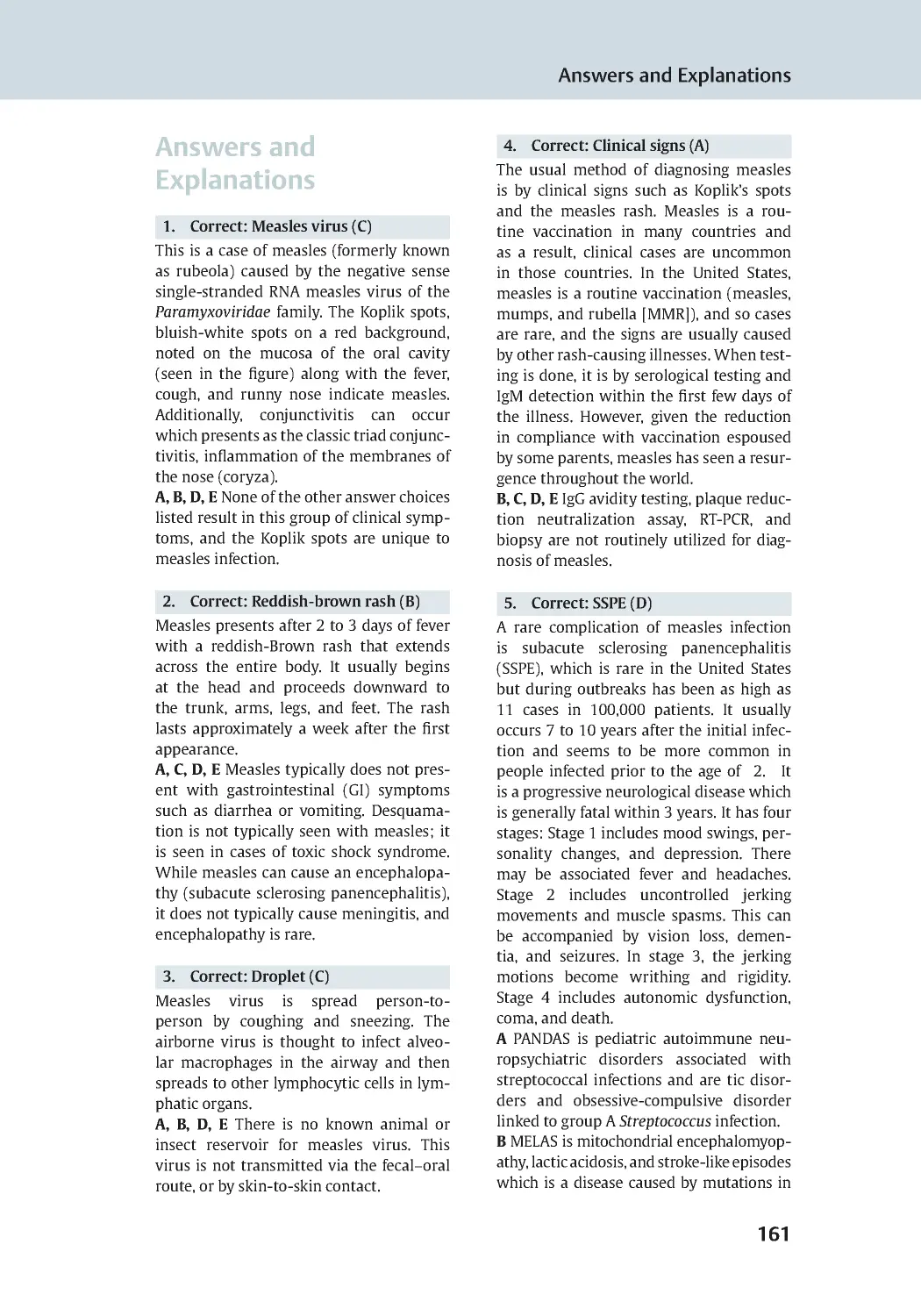


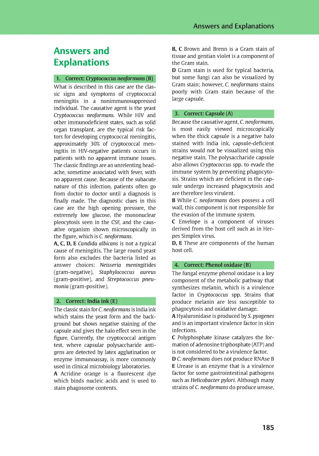


Author: Tracey A. H . T. Baxa D. Sims M.
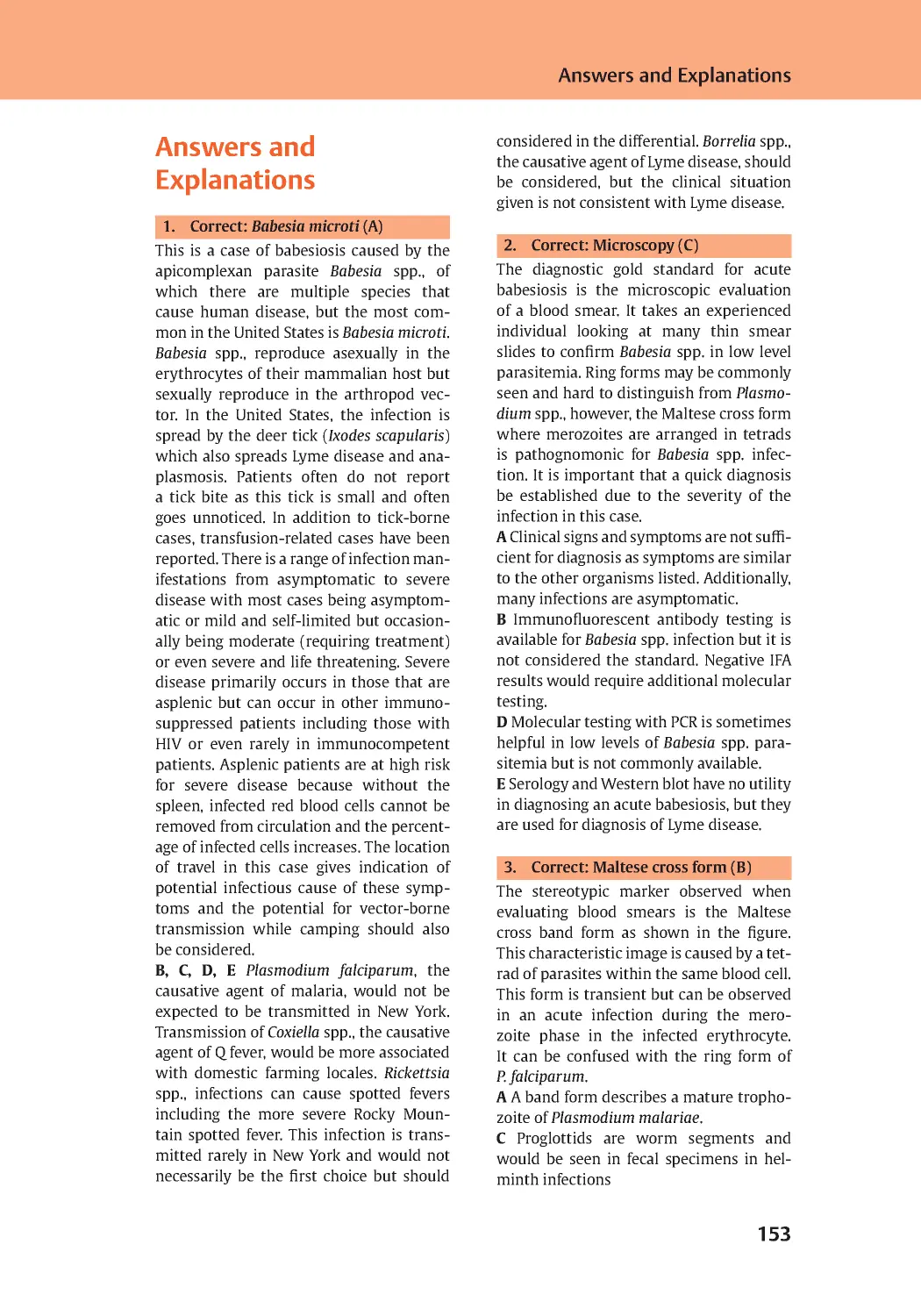
Tags: biology microbiology cell biology infection diseases infection diseases cure practices
Year: 2020


Text
Learning Microbiology and Infectious
Diseases: Clinical Case Prep for the
USMLE®
Tracey A. H . Taylor, PhD
Associate Professor
Department of Foundational Medical Studies
Oakland University William Beaumont School of Medicine
Rochester, Michigan, USA
Dwayne Baxa, PhD
Assistant Professor
Department of Foundational Medical Studies
Oakland University William Beaumont School of Medicine
Rochester, Michigan, USA
Matthew Sims, MD, PhD
Professor
Department of Internal Medicine
Oakland University William Beaumont School of Medicine
Rochester, Michigan, USA
49 illustrations
Thieme
New York • Stuttgart • Delhi • Rio de Janeiro
Library of Congress Cataloging-in-Publication Data is
available from the publisher
Important note: Medicine is an ever-changing science undergo-
ing continual development. Research and clinical experience are
continually expanding our knowledge, in particular our knowl-
edge of proper treatment and drug therapy. Insofar as this book
mentions any dosage or application, readers may rest assured
that the authors, editors, and publishers have made every effort
to ensure that such references are in accordance with the state
of knowledge at the time of production of the book.
Nevertheless, this does not involve, imply, or express any
guarantee or responsibility on the part of the publishers in
respect to any dosage instr uctions and forms of applications
stated in the book. Every user is requested to examine care-
fully the manufact urers’ leaflets accompanying each drug
and to check, if necessary in consultation with a physician or
specialist, whether the dosage schedules mentioned therein
or the contraindications stated by the manufacturers differ
from the statements made in the present book. Such exam-
ination is particularly important with drugs that are either
rarely used or have been newly released on the market. Every
dosage schedule or every form of application used is entire-
ly at the user’s own risk and responsibility. The authors and
publishers request every user to report to the publishers any
discrepancies or inaccuracies noticed. If errors in this work
are found after public ation, errata will be posted at www.
thieme.com on the product description page.
Some of the product names, patents, and registered
designs refer red to in this book are in fact registered trade-
marks or proprietary names even though specific reference
to this fact is not always made in the text . Therefore, the ap-
pearance of a name without designation as proprietary is
not to be constr ued as a representation by the publisher that
it is in the public domain.
This book, including all parts thereof, is legally protected by
copyright. Any use, exploitation, or commercialization outside
the narrow limits set by copyright legislation, without the
publisher’s consent, is illegal and liable to prosecution. This
applies in particular to photostat reproduction, copying, mim-
eographing, preparation of microfilms, and electronic data
processing and storage.
©2020. Thieme. All rights reserved.
Thieme Publishers New York
333 Seventh Avenue, New York, NY 10001 USA
+1 800 782 3488, c ustomerservice@thieme.com
Georg Thieme Verlag KG
Rüdigerstrasse 14, 70469 Stuttgart, Germany
+49 [0]711 8931 421, c ustomerser vice@thieme.de
Thieme Publishers Delhi
A-12 , Second Floor, Sector-2 , Noida-201301
Uttar Pradesh, India
+91 120 45 566 00, customerser vice@thieme.in
Thieme Publishers Rio de Janeiro,
Thieme Publicações Ltda.
Edifício Rodolpho de Paoli, 25o andar
Av. Nilo Peçanha, 50 – Sala 2508,
Rio de Janeiro 20020-906 Brasil
+55 21 3172-2297
Cover design: Thieme Publishing Group
Typesetting by Thomson Digital, India
Printed in USA by King Printing Company, Inc.
54321
ISBN 978-1 -62623 -508-3
Also available as an e-book:
eISBN 978-1-62623 -509-0
Contents
Preface ..................................................................................................................................... vii
Case 1 Adult with a Cough of a Long Duration ......................................................... 1
Case 2 Child with a Sore Throat and Red Eyes ........................................................... 7
Case 3 Adult in Respiratory Distress ............................................................................ 11
Case 4 Adolescent with Sore Throat and Malaise ..................................................... 15
Case 5 Toddler with Upper Respiratory Symptoms .................................................. 19
Case 6 Elderly Male with Two Days of Fever, Chills, and Cough ............................ 23
Case 7 Severely Ill Elderly Male in Respiratory Distress........................................... 27
Case 8 Teenager with a Two-Week-Long Cough....................................................... 31
Case 9 Adult Male with Persistent Cough and Malaise ........................................... 35
Case 10 Infant with Severe Congestion ......................................................................... 39
Case 11 Adult Male with Fever, Myalgias, and Respiratory Distress....................... 43
Case 12 Adult Male with Fever, Chills, and Night Sweats ......................................... 47
Case 13 Child with Bloody Diarrhea ............................................................................... 51
Case 14 Child with Stomach Pain and Fever ................................................................. 55
Case 15 Outbreak of Diarrheal Illness ............................................................................ 59
Case 16 Hospitalized Adult Female Who Develops Diarrhea.................................... 63
Case 17 HIV-Positive Male with Diarrhea...................................................................... 67
Case 18 Adult with Jaundice ............................................................................................ 71
Case 19 Adult with Right Upper Quadrant Pain .......................................................... 75
Case 20 Adult Female with Epigastric Pain after Eating ............................................ 79
Case 21 Elderly Female with Fever and Flank Pain...................................................... 83
Case 22 Adult Male with Rash ......................................................................................... 87
Case 23 Young Female with Joint Pain........................................................................... 91
Case 24 Teenager with Syncopy...................................................................................... 95
Case 25 Adult Male with Painful Penile Ulcers............................................................. 99
Case 26 Adult Male with Back Pain ............................................................................... 103
Contents
Case 27 Adult Female with Vaginal Discharge .......................................................... 107
Case 28 Adult Male with a Red Eye .............................................................................. 111
Case 29 Adult Female with Lesion on Labia ............................................................... 115
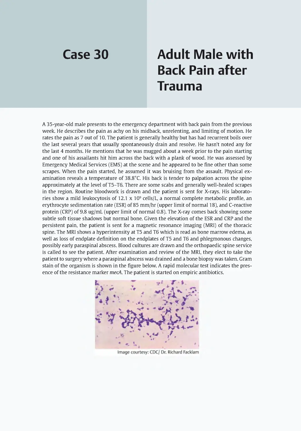
Case 30 Adult Male with Back Pain after Trauma ..................................................... 119
Case 31 Teenage Girl with Expanding Skin Lesion .................................................... 123
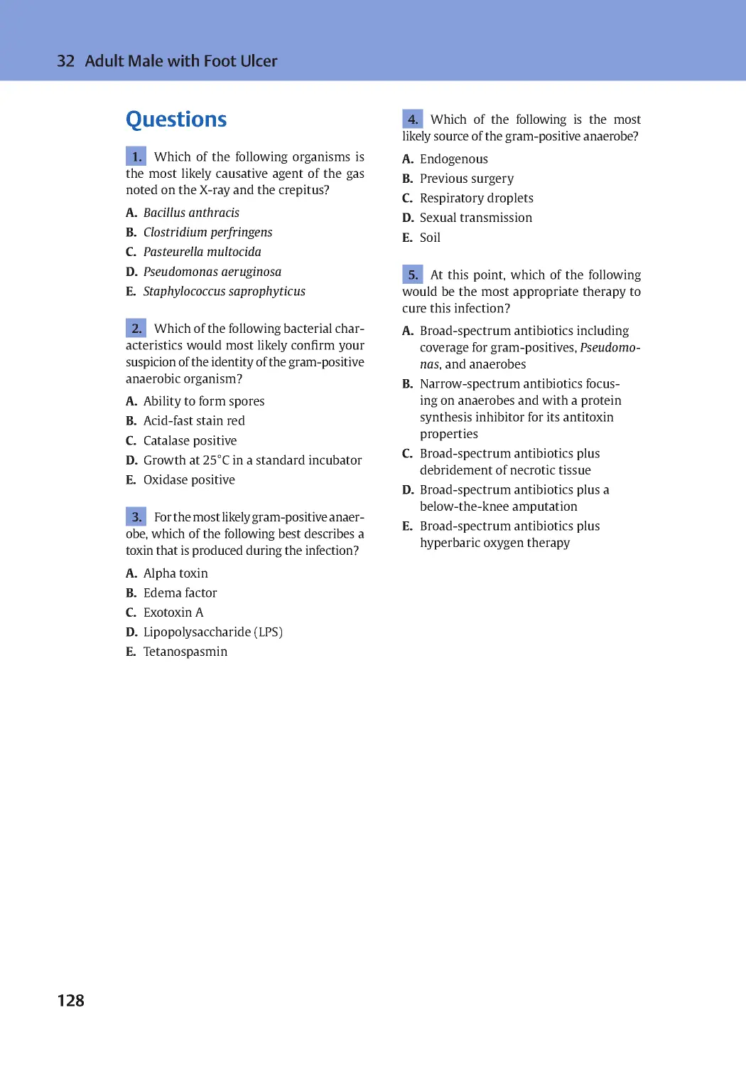
Case 32 Adult Male with Foot Ulcer ............................................................................. 127
Case 33 Adult Male with Flu-Like Illness and Rash ................................................... 131
Case 34 Febrile Teenager with Disseminated Rash .................................................. 135
Case 35 Adult Male with Worsening Shortness of Breath ...................................... 139
Case 36 Adult Female with Facial Pain ........................................................................ 143
Case 37 Adult Female with Painful Rash ..................................................................... 147
Case 38 Adult Female with Headache and Confusion.............................................. 151
Case 39 Adult Male with Painful, Swollen Lymph Nodes ........................................ 155
Case 40 Toddler with High Fever and Upper Respiratory Symptoms................... 159
Case 41 Adult Female with Febrile Illness................................................................... 163
Case 42 Agitated Male with Rapid Progression to Coma........................................ 167
Case 43 Teenager with Headache and Fever.............................................................. 171
Case 44 Elderly Female with Severe Headache and Nausea ................................... 175
Case 45 Adult Female with Headache and Disorientation ...................................... 179
Case 46 Adult Male with Headache for Several Months.......................................... 183
Case 47 Febrile Teenager with Headache and Neck Stiffness................................. 187
Case 48 Travelers with Acute Febrile Illness............................................................... 191
Case 49 Newborn with Jaundice ................................................................................... 195
Case 50 Teenager with Pain in Groin ........................................................................... 199
Bibliography ........................................................................................................................... 203
Index ........................................................................................................................................ 219
Preface
This book is an assortment of clinical cases intended
to advance the learning of microbiology and infec-
tious diseases using case studies. Working with cases
is the most ideal approach to involve students in the
learning process because:
• Every case shows a genuine circumstance, very
close to those seen in everyday practice.
• Every case can elicit several problems that must
be understood and solved. In other words, it is a
problem-based learning process.
• Every case not only requires the knowledge of
several disciplines but also the proper utiliza-
tion of the knowledge to explicit clinical cir-
cumstances.
In terms of the organization of the cases, we pur-
posefully intermingled bacteriology, virology, mycol-
ogy, and parasitology cases in this book, as well as
organ systems. We feel that students will gain a better
understanding of the topics when they consider all
types of organisms for each case, as will be the case
when they are considering the diagnosis of real pa-
tients. The format of each case is as follows:
• Each case provides relevant clinical information;
presenting symptoms and relevant duration,
relevant medical history (which may include
vaccinations), relevant family history, relevant
recent travel, physical examination findings,
and lab findings.
• Images are used to support the cases. Images in-
clude microscopic images such as Gram stains,
electron micrographs, wet mounts, and acid-
fast stains, as well as clinical images and skin
rashes or lesions.
• Each case is followed by a series of five multiple-
choice questions.
○ In most cases, the first question requires
the student to identify the most likely caus-
ative agent.
○ Other questions address the knowledge of
microbiology and infectious diseases that
the student needs to assess the case. For
example, prevention and precaution mea-
sures, transmission, mechanism of action
of pathogenesis, diagnostic methods, possi-
ble complications, etc.
○ The last question is often related to the
pharmacotherapy of the disease.
The questions are designed to prepare students
for the United States Medical Licensing Examination
(USMLE) and/or Comprehensive Osteopathic Medical
Licensing Examination (COMLEX) of the United States
Step 1 and Step 2. Five choices are provided for every
question, but for each question there is only one best
answer.
For each question, answers and explanations are
provided on a separate page. This allows students to
self-test the entire series of questions before revealing
the answers and explanations so that they may com-
pletely work through the case and receive optimal
formative feedback. The explanations include both
the reasons why a given answer is correct and why
the distractors are wrong. Many questions are related
to the higher levels of Bloom’s taxonomy (e.g ., analyz-
ing, applying, or evaluating) rather than being simple
recall questions.
For each case, keywords are provided so that stu-
dents may search for a particular area of interest.
Lastly, we have provided a bibliography section for
the students. Students are invited to use those refer-
ences in case they discover that they require a deeper
understanding of the material or if they become in-
spired to seek additional information.
The rationale for this book is related to the current
trends in medical education. Now, it is evident that
the mere retention of information provided by
books is insufficient for meaningful learning. The
utilization of information is progressively significant.
A vast array of medical problems is available in the
literature today, but there are few clinical case books
related to microbiology and infectious disease. This
book links clinical cases to the application of basic
knowledge of microbiology, as well as explains the
reasons for using specific drugs in real infectious
disease problems, with the goal of promoting
critical thinking.
The main audience of the book is medical school
students. This book will aid these students in their
medical school course work, as well as in preparation
for the USMLE and/or COMLEX Step 1 exam. Medical
students in years 3 and 4 may also find these cases
to be helpful when preparing for the USMLE and/or
COMLEX Step 2 exams, shelf exams, and for their clin-
ical rotation coursework. In addition, this book is also
useful for students in other medical professions edu-
cation, including physician assistant students, nursing
students, and pharmacy students.
Clinical medicine is a fast-evolving discipline. The
authors have referred to reliable sources in order to
provide information in accordance with currently
accepted standards. However, the authors are aware
that in several instances, the information may be con-
troversial. We have tried, as much as possible, to avoid
questions addressing controversial issues.
Preface
This book is meant to be a companion to the
Thieme microbiology question book, thereby giving
students more than one option for learning and
studying microbiology. This book is not intended to
be a substitute for microbiology textbooks. Students
are strongly advised to consult their textbooks of
microbiology for more in-depth coverage of the
subject matter.
Tracey A. H. Taylor, PhD
Dwayne Baxa, PhD
Matthew Sims, MD, PhD
Adult with a
Cough of a Long
Duration
Case 1
A 36-year-old male presents to his primary care physician with a cough of 4 weeks duration.
The cough is paroxysmal, and he sometimes vomits after coughing. Two other people with
whom he works have a similar cough. The patient works at a local automobile assembly plant;
he is married with a 2-year-old child; and he has no history of cigarette smoking.
On physical examination, the patient initially appears in no acute distress, then expe-
riences a severe coughing attack, which leaves him weak and out of breath. Examination of
the head, eyes, ears, nose, and throat (HEENT) revealed a small conjunctival hemorrhage on
the left, and several petechiae were noted on the face. Lungs were clear to auscultation. The
remainder of the physical examination was benign.
Laboratory studies were obtained, and the complete blood count (CBC) with differential
showed a white blood cell (WBC) count of 8,000/ul with a normal differential. Posterior–ante-
rior (PA) and lateral chest X-rays were also obtained and found to be normal.
The physician obtains a nasopharyngeal aspirate that was sent to the microbiology lab-
oratory for Gram stain and culture. An organism not typical for oral-pharyngeal flora and
presumed to be the causative pathogen grew on Regan–Lowe agar after overnight incubation
at 37°C. The Gram stain is shown in the following figure. A confirmatory direct fluorescent
antibody (DFA) staining of the organism isolated from the aspirate was also positive.
1 Adult with a Cough of a Long Duration
2
Questions
1. Which of the following organisms is
the most likely causative agent?
A. Bordetella pertussis
B. Haemophilus influenzae
C. Klebsiella pneumoniae
D. Legionella pneumophila
E. Pseudomonas aeruginosa
2. How was this infection most likely
acquired?
A. Aerosol person to person
B. Arthropod bite
C. Ingestion of food
D. Ingestion of water
E. Sexual transmission
3. For the most likely causative agent,
which of the following best describes the
pathogenic mechanism leading to the clin-
ical symptoms?
A. Activation of adenylate cyclase by
disabling Gi
B. Cleavage of circulating immunoglobu-
lin A (IgA)
C. Inactivation of the 60S ribosome by
cleavage of ribosomal RNA (rRNA)
D. Inactivation of host elongation factor 2
E. Prevention of the release of inhibitory
neurotransmitter
4. Which of the following best describes
the vaccine currently available for preven-
tion of this infection?
A. DNA vaccine
B. Killed whole cell vaccine
C. Live attenuated vaccine
D. Recombinant vector vaccine
E. Subunit vaccine
5. The physician recommends a 5-day
course of azithromycin, and the patient
takes it appropriately. He returns to the
office after completing the course of
therapy complaining that his cough is
unchanged. Which of the following is the
best explanation that the physician can
give for the lack of resolution of the cough?
A. Azithromycin is not expected to clear
this infection, but it was given because
patients expect an antibiotic
B. Coinfection with a second organism is
common in this disease, and it is likely
that administration of a second antibi-
otic is warranted
C. It is likely that the infection was
resistant to azithromycin, and therapy
will need to be altered
D. The purpose of the antibiotic
was not to treat the symptoms of the
disease but to reduce transmission of
the organism
E. The standard dose of azithromycin was
too low for this patient’s disease, and
the dose will need to be doubled
Answers and Explanations
3
Answers and
Explanations
1. Correct: Bordetella pertussis (A)
This case describes whooping cough, or
Bordetella pertussis infection. B. pertussis
is a fastidious gram-negative coccoba-
cillus that primarily infects children and
unvaccinated susceptible individuals. The
presentation of pertussis is separated into
three stages: the catarrhal phase, which
appears as a typical upper respiratory tract
infection; the paroxysmal phase, which
presents with intense coughing jags and
frequently the classic whooping sound of
inspiration against a partially closed air-
way, post-tussive emesis is often seen;
and the convalescent phase, which has a
chronic cough that can last for weeks. In
adults, particularly those who were pre-
viously immunized, the classic symptoms
such as coughing paroxysms, the whoop-
ing sound, and post-tussive emesis may not
be seen. The presence of the classic symp-
toms typically indicates an unvaccinated
or undervaccinated individual. In children,
a high WBC count (often > 20,000) is seen
with a lymphocytosis frequently over 50%.
Seeing a young child with a cough and
high WBC with significant lymphocytosis
is often a diagnostic clue for pertussis. In
adults, however, the elevated WBC is rare,
and lymphocytosis is generally not seen.
B Haemophilus influenzae is incorrect
because infections do not present with a
paroxysmal cough, and H. influenzae are
gram-negative bacilli.
C Klebsiella pneumoniae is incorrect
because infections do not present with a
paroxysmal cough, and K. pneumoniae are
gram-negative bacilli.
D Legionella pneumophila is incorrect
because they are rarely able to be visual-
ized by Gram stain, and infections do not
present with a paroxysmal cough.
E Pseudomonas aeruginosa is incorrect
because these infections are more often
nosocomial or related to cystic fibrosis,
which this patient does not have. Also,
P. aeruginosa are gram-negative bacilli.
2. Correct: Aerosol person to person (A)
Transmission of B. pertussis from person-
to-person occurs via aerosolized respiratory
droplets. There are no identified animal or
environmental reservoirs for this pathogen.
Humans are the reservoir for B. pertussis.
The incubation period is 7 to 10 days on
average with a maximum of up to 20 days.
B Arthropod bite is incorrect because per-
tussis is not vector-borne.
C Ingestion of food is incorrect because
pertussis is not food-borne.
D Ingestion of water is incorrect because
pertussis is not water-borne.
E Sexual transmission is incorrect because
pertussis is not sexually transmitted.
3. Correct: Activation of adenylate
cyclase by disabling Gi (A)
The virulence factors produced by
B. pertussis include adherence to ciliated
epithelial cells of the trachea and bronchi
via pili and filamentous hemagglutinin,
endotoxin, pertussis toxin, hemolysin,
adenylate cyclase toxin, and tracheal cyto-
toxin. Pertussis toxin is an A-B5 exotoxin
that is secreted by a type IV secretion sys-
tem and binds to the G-alpha inhibitory
subunit inhibiting signal transduction,
resulting in increased cyclic adenosine
monophosphate (cAMP) and a subsequent
increase in mucus production and death
of the host cell. This mechanism of action
of the pertussis toxin mimics adenylate
cyclase activity.
B Cleavage of circulating IgA is incorrect.
The cleavage of IgA by bacterial proteases,
such as in the case of H. influenzae, results
in the impairment of antibody-induced
entrapment of microbes in mucus secretions.
C Inactivation of the 60S ribosome by cleav-
age of rRNA is incorrect. This describes the
mechanism of action of Shiga toxin.
D Toxin causing inactivation of host elon-
gation factor 2 is incorrect. This describes
the mechanism of action of P. aeruginosa
exotoxin A.
E Toxin that prevents the release of
inhibitory neurotransmitter is incorrect.
This describes the mechanism of action of
Clostridium botulinum.
1 Adult with a Cough of a Long Duration
4
4. Correct: Subunit vaccine (E)
Prevention of infection is primarily by
routine vaccination of infants, children,
and adults. Though it was not mentioned
explicitly, the patient was likely either
unvaccinated or undervaccinated. Appro-
priate vaccination may have protected him
from infection. The diphtheria, tetanus, and
pertussis (DTap) vaccine in full strength is
given to infants and children, while the
reduced dose tetanus-diphtheria-pertus-
sis (Tdap) vaccine is offered to teens and
adults. The current vaccine for pertussis is
an acellular subunit vaccine, consisting of
inactivated pertussis toxin ± filamentous
hemagglutinin (FHA), fimbriae, and pert-
actin (adhesin). The current vaccine does
not give lifelong immunity, and boosting in
adults is recommended by the Centers for
Disease Control and Prevention (CDC) to
help prevent transmission to children.
A DNA vaccine is incorrect. The pertussis
vaccine is a subunit vaccine. There are no
DNA vaccines approved for use in humans
at present.
B Killed whole cell vaccine is incorrect. The
pertussis vaccine is a subunit vaccine. An
example of a killed whole cell vaccine is
the previous pertussis vaccine, which is no
longer used in the United States.
C Live attenuated vaccine is incorrect. The
pertussis vaccine is a subunit vaccine.
Example of a live attenuated vaccine is
the MMR, nasal influenza, and chickenpox
(varicella).
D Recombinant vector vaccine is incorrect.
The pertussis vaccine is a subunit vaccine.
There are no recombinant vector vaccines
approved for use in humans at present.
5. Correct: The purpose of the
antibiotic was not to treat the
symptoms of the disease but to reduce
transmission of the organism (D)
Treatment of pertussis is generally sup-
portive, particularly in very young children.
Cough suppressants have little impact.
Antibiotics generally have no impact on
the duration of symptoms, though they
may shorten the course and decrease the
severity if given early in the catarrhal phase.
Since pertussis is rarely diagnosed during
the catarrhal phase, the addition of anti-
biotics is to eradicate the B. pertussis from
the nasopharynx to reduce the spread of
the infection (particularly in this case as
this patient lives with a child at home and
works in a populated environment). The
antibiotic of choice is azithromycin (due
to dosing schedule, bioavailability, and
low side effect profile). Alternative antibi-
otics include clarithromycin, erythromy-
cin, and for patients older than 2 months
intolerant of macrolides, trimethoprim–
sulfamethoxazole (TMP–SMX) is a rea-
sonable alternative. TMP–SMX should be
avoided in children younger than 2 months
due to risk for kernicterus. Azithromycin
and erythromycin are both associated with
an increased risk of infantile hypertrophic
pyloric stenosis, and it is unclear if such a
risk exists for clarithromycin. Neither azi-
thromycin nor clarithromycin is approved
by the Food and Drug Administration (FDA)
for use in children younger than 6 months.
Macrolide resistance is very uncommon,
and antibiotic testing is generally not
performed.
A Azithromycin is not expected to clear
this infection, but it was given because
patients expect an antibiotic. This is incor-
rect because it is never correct to give an
antibiotic just because a patient wants it.
B Coinfection with a second organism is
common in this disease, and it is likely that
administration of a second antibiotic is
warranted. This is incorrect because coin-
fection with a second organism is not com-
mon in this disease.
C It is likely that the infection was resistant
to azithromycin, and therapy will need to
be altered. This is incorrect because azi-
thromycin resistance is not commonly
described in pertussis.
E The standard dose of azithromycin was too
low for this patient’s disease and the dose
will need to be doubled. This is incorrect
because a standard dose is all that is needed.
Answers and Explanations
5
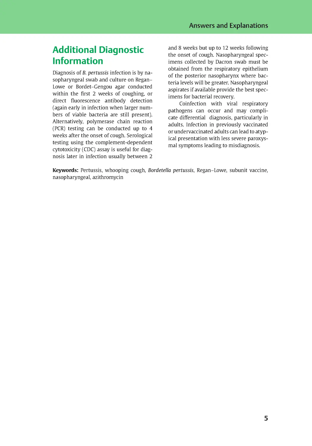
Additional Diagnostic
Information
Diagnosis of B. pertussis infection is by na-
sopharyngeal swab and culture on Regan–
Lowe or Bordet–Gengou agar conducted
within the first 2 weeks of coughing, or
direct fluorescence antibody detection
(again early in infection when larger num-
bers of viable bacteria are still present).
Alternatively, polymerase chain reaction
(PCR) testing can be conducted up to 4
weeks after the onset of cough. Serological
testing using the complement-dependent
cytotoxicity (CDC) assay is useful for diag-
nosis later in infection usually between 2
and 8 weeks but up to 12 weeks following
the onset of cough. Nasopharyngeal spec-
imens collected by Dacron swab must be
obtained from the respiratory epithelium
of the posterior nasopharynx where bac-
teria levels will be greater. Nasopharyngeal
aspirates if available provide the best spec-
imens for bacterial recovery.
Coinfection with viral respiratory
pathogens can occur and may compli-
cate differential diagnosis, particularly in
adults. Infection in previously vaccinated
or undervaccinated adults can lead to atyp-
ical presentation with less severe paroxys-
mal symptoms leading to misdiagnosis.
Keywords: Pertussis, whooping cough, Bordetella pertussis, Regan–Lowe, subunit vaccine,
nasopharyngeal, azithromycin
Case 2
Child with a
Sore Throat and
Red Eyes
A mother brings her 6-year-old daughter to her pediatrician with symptoms of a sore throat
and red, tearing eyes. The patient complains of itchy eyes when she woke up that morning.
Her symptoms started the previous day, and she missed school today. History reveals that
the child’s vaccinations are up to date and that she has been previously healthy with no prior
hospitalizations. Upon examination, the patient is found to have a temperature of 39°C, blood
pressure of 90/70 mmHg, and respiratory rate of 20 breaths/min. She has upper lid edema
with slight subconjunctival hemorrhage. Her mother mentions that other classmates were
also out sick during the past week with similar symptoms.
2 Child with a Sore Throat and Red Eyes
8
Questions
1. What is the most likely condition
experienced by this child?
A. Allergic conjunctivitis
B. Gonococcal conjunctivitis
C. Keratoconjunctivitis
D. Pharyngoconjunctivitis
E. Endophthalmitis
2. Given the patient history, what is the
most likely etiology?
A. Adenovirus
B. Herpes simplex virus type 1 (HSV-1)
C. Neisseria gonorrhoeae
D. Pseudomonas aeruginosa
E. Staphylococcus aureus
3. What is the most likely mode of trans-
mission by the causative agent in this case?
A. Arthropod transmission
B. Bloodborne transmission
C. Droplet transmission
D. Foodborne transmission
E. Waterborne transmission
4. Which is the best method to detect an
outbreak of this agent?
A. Clinical diagnosis
B. Culture with immunofluorescence
assay
C. Polymerase chain reaction (PCR)
D. Serology
E. Rapid antigen test
5. Which of the following precautions
must be used to prevent the spread of this
infection from a patient to a health care
worker?
A. Gown and gloves
B. N-95 mask
C. Surgical mask and eye protection
D. No specific precautions
E. Washing the hands with dilute bleach
Answers and Explanations
9
Answers and
Explanations
1. Correct: Pharyngoconjunctivitis (D)
This is a case of typical pharyngoconjunc-
tivitis. It is highly infectious and usually is
clinically identified by fever, pharyngitis,
with enlarged lymph nodes in addition to
conjunctivitis (pink eye). This infection is
often observed in children. Because of its
contagious nature, outbreaks often occur
in schools, daycares, and dormitory set-
tings. Swimming pool transmission has
also been reported. The upper respiratory
symptoms may precede or follow the ocu-
lar manifestations. The ocular symptoms
may begin in one eye first with an itchy or
gritty feeling and later becoming bilateral.
A Allergic conjunctivitis is incorrect
because these conditions are not associated
with sore throat.
B Gonococcal conjunctivitis is incorrect
because the discharge from the eye is not
purulent.
C Keratoconjunctivitis is incorrect because
there is no apparent corneal involvement
in this case.
E Endophthalmitis is incorrect because
there is no apparent aqueous or vitreous
involvement. It typically presents with
decreasing vision and eye discomfort,
neither of which describes this patient’s
symptoms.
2. Correct: Adenovirus (A)
Adenovirus is the most common viral
cause of conjunctivitis, but approximately
30% of all cases are due to a bacterial cause.
In young children, it is estimated that 5 to
10% of all febrile illness are caused by ade-
noviruses. The genome of adenoviruses is
composed of double-stranded DNA. Ade-
noviruses are non-enveloped and have an
icosahedron shape (see the following fig-
ure). It is common practice to diagnosis
adenovirus pharyngoconjunctivitis by clin-
ical presentation. However, definitive labo-
ratory confirmation should be employed in
order to best determine if antibiotics should
be prescribed. The adenovirus infection is a
self-limiting infection and should resolve
within 2 weeks. During this period the per-
son is still infectious.
B HSV-1 is incorrect because it is less likely
due to the presence of pharyngitis in this
patient. HSV typically causes keratitis,
which this patient does not have.
C Neisseria gonorrhoeae is incorrect
because this patient has no history of risk
for N. gonorrhoeae infection.
D Pseudomonas aeruginosa is incorrect
because P. aeruginosa more commonly
causes nosocomial infections and infec-
tions in patients with predisposing factors
such as cystic fibrosis.
E Staphylococcus aureus is incorrect
because eye infections caused by S. aureus
typically progress rapidly and are charac-
terized by purulent discharge.
3. Correct: Droplet transmission (C)
Adenovirus is a non-enveloped DNA virus
and is relatively stable on environmental
surfaces, surviving from days to months.
Transmission occurs by inhalation of aero-
solized droplets as well as fomites gener-
ated by hand-to-eye and fecal–oral routes.
Transmission has occurred via recreational
waters such as swimming pools and spas
and is most likely a fecal–oral route, but
secretions from eyes or throat can be a
possible source. It is not spread via air-
borne transmission in small particles as
is tuberculosis. Adenovirus has 50 sero-
types grouped into seven subtypes: A to G.
Neutralizing antibodies raised against one
subtype do not provide immunity against
virus of another subtype allowing for
apparent reinfection.
2 Child with a Sore Throat and Red Eyes
10
A Arthropod transmission is incorrect
because adenovirus is not vector-borne.
B Bloodborne transmission is incorrect
because there is no history of blood expo-
sure in this case, and adenovirus is not typ-
ically transmitted via the blood.
D Foodborne transmission is incorrect
because adenovirus is not food-borne.
E Waterborne transmission is incorrect
because while adenovirus can be transmit-
ted through water, transmission was not by
this method in this case.
4. Correct: Polymerase chain reaction
(PCR) (C)
All of the listed answers are capable of
being utilized in diagnosing pharyngocon-
junctivitis. PCR is the best option both for
specificity/sensitivity and turn-around-
time and is commonly available as part of a
respiratory viral panel performed on naso-
pharyngeal swabs.
A While it is certainly commonplace to
diagnose adenovirus by clinical presen-
tation, misdiagnosis is possible and often
leads to lack of antibiotic stewardship.
B Cell culture is available for adenovirus
detection, and the virus produces a char-
acteristic cytopathic effect visible as cell
clumping. However, culture of the virus
may take 4 to 7 days for positive confir-
mation and therefore is rarely used. Shell
vial culture (a modified version of tradi-
tional cell culture) and immunofluorescent
detection can reduce confirmation to
within 3 days.
D Serology testing can detect exposure to
adenovirus, but, as most young children
are antibody positive by age 5, it has lim-
ited utility for diagnosis of acute infection.
Serology is not the correct answer.
E There is a point-of-care test that is avail-
able for screening adenovirus as a cause of
conjunctivitis. However, a negative result
does not rule out adenovirus as a cause,
and other diagnostic measures should be
employed.
5. Correct: Surgical mask and eye
protection (C)
Since adenovirus is spread via droplets which
are expelled by coughing, sneezing, or simply
talking, the personal protective equipment
needed to prevent the spread must include
eye protection and a surgical mask.
A Gowns and gloves are not enough to pro-
tect from the spread of this virus.
B A N-95 mask would certainly prevent
inhalation of adenovirus, but a mask with
such a small weave is overkill for adeno-
virus, and using it without eye protection
will not prevent droplets from infecting
the health care worker via the conjunctiva.
D, E There is no need to wash with dilute
bleach to clean your hands as such mea-
sures will not inactivate this non-envel-
oped virus.
Keywords: Adenovirus, pharyngitis, conjunctivitis, non-enveloped
Case 3
Adult in
Respiratory
Distress
A 30-year-old construction worker from New Mexico with no significant past medical history
was admitted to the hospital with severe shortness of breath. In the emergency room, he
stated that he developed a cough in the past few days with fever, muscle aches, nausea, and
vomiting. His wife reported that prior to his symptoms he was working overtime, renovating
abandoned homes and she initially assumed he was feeling under the weather because he was
working so much and not getting enough sleep.
On physical examination his temperature was 38.5°C, blood pressure was 90/50 mmHg,
respiratory rate was 28 breaths/min, heart rate was 140 bpm, and oxygen saturation was 75%
on room air. A chest X-ray demonstrated bilateral diffuse opacities with Kerley B lines and
peribronchial cuffing. His white blood cell count was 26,000 × 109/L with a predominance of
neutrophils and lymphocytes, and noted atypical lymphocytes were observed. The patient's
platelet count was 75 with an elevated hematocrit level. A rapid polymerase chain reaction
(PCR) test for influenza A is negative. He was admitted into the hospital and given supplemen-
tal oxygen but quickly transferred to the intensive care unit and intubated.
3 Adult in Respiratory Distress
12
Questions
1. Which of the following organisms is
the most likely etiology of this infection?
A. I n fluenza A virus
B. Hantavirus
C. Legionella pneumophila
D. Francisella tularensis
E. Epstein–Barr virus (EBV)
2. Which of the following is the vector
for transmission of the organism?
A. Avian
B. Human
C. Rodent
D. Water
E. Tick
3. Which of the following is the patho-
genesis of the patient’s pulmonary
symptoms?
A. Direct infection of the vascular
endothelium of the lungs leading to
pulmonary edema
B. Neutrophilic invasion of the lung lead-
ing to a multilobar pneumonia
C. Release of toxins into the epithelial
lining of the lungs leading to cell death
D. Induction of mucin secretion leading to
mucous plugging and obstruction
E. Deposition of secreted proteins along
the wall of the alveoli preventing gas
exchange
4. Which of the following assays is
most often used to detect the causative
organism?
A. Culture
B. Enzyme-linked immunosorbent assay
(ELISA)
C. Nucleic acid amplification
D. Western blot
E. Gene-chip analysis
5. Which of the following treatments is
most appropriate for this patient?
A. Cidofovir
B. Peramivir
C. Acyclovir
D. Doxycycline
E. Supportive care only
Answers and Explanations
13
Answers and
Explanations
1. Correct: Hantavirus (B)
This is a case of hantavirus pulmonary syn-
drome (HPS) caused by genus Hantavirus in
the Bunyaviridae family. Hantaviruses are
segmented, negative-stranded RNA viruses.
HPS is difficult to distinguish in the initial
stages from the other respiratory infections;
however, the history of exposure to rodent
excreta should be strongly considered in
the differential diagnosis. A significant clue
in this case is the location of New Mexico,
as this is one of the Four Corners states,
being New Mexico, Arizona, Colorado, and
Utah. This is the region of the first outbreak
of HPS in the United States of America in
1993 caused by the viral strain Sin Nombre
virus (SNV). Since that time, other cases of
HPS caused by other viral strains of hanta-
virus have been identified in other regions
of the United States. The early symptoms
of HPS are fatigue, muscle aches, nausea,
vomiting, cough, and high fever. These are
thought to occur between 1 and 8 weeks
after exposure. The late stage of HPS results
in shortness of breath caused by the lungs
filling with fluid. The virus infects microvas-
cular endothelium resulting in deregulation
of fluid accumulation. The fluid increase
causes pulmonary edema and cardiac fail-
ure. There is 38% mortality in HPS cases.
A While influenza A virus could present
with similar symptoms, it is less likely due
to the negative rapid PCR test.
C Legionella pneumophila can present with
similar symptoms but is often associated
with exposure to an aerosolized water source
such as a cooling tower or air conditioner. In
addition, atypical lymphocytes are not nor-
mally seen with Legionella spp. infection.
D Francisella tularensis is not likely because
this patient was not exposed to common
reservoirs such as rabbits. Pulmonary
tularemia frequently presents with nodu-
lar infiltrates and pleural effusions.
E EBV is not likely because although this
patient has atypical lymphocytes, the other
symptoms are not as commonly associated
with EBV infection. EBV infection often
presents with extreme fatigue.
2. Correct: Rodent (C)
Hantavirus transmission occurs by inha-
lation of dried urine, saliva, or feces from
infected rodents. The primary host of SNV is
the deer mouse (Peromyscus maniculatus).
A Hantavirus is not transmitted by birds.
There are cases of bird-to-human trans-
mission in the case of the H5N1 influenza A
strain.
B Hantavirus is not transmitted by person-
to-person contact. Influenza A virus
strains are transmitted human-to-human.
D Hantavirus is not transmitted by water.
Legionella spp. transmission can occur via
humidified air originating from a contam-
inated source.
E Hantavirus is not transmitted by ticks
and is not a vector-borne infection.
3. Correct: Direct infection of the
vascular endothelium of the lungs
leading to pulmonary edema (A)
Hantavirus antigens are primarily found
in the endothelium of the capillaries
throughout infected patients. Within
the lungs this leads to inflammation and
endothelial leakage causing pulmonary
edema and a variation of acute respiratory
distress syndrome.
B Neutrophilic invasion of the lungs is typi-
cally seen with bacterial pneumonias.
C Certain bacteria including Staphylococcus
aureus and Pseudomonas aeruginosa pro-
duce toxins which can worsen pneumonia
but generally are not the primary mode of
pathogenesis.
D Mucous plugging can be seen with a
number of pulmonary conditions includ-
ing chronic obstructive pulmonary disease
(COPD).
E Buildup of protein along the alveolar
walls is the pathogenesis of pulmonary
alveolar proteinosis, a rare noninfectious
pulmonary syndrome.
3 Adult in Respiratory Distress
14
4. Correct: Enzyme-linked
immunosorbent assay (ELISA) (B)
The detection of Hantavirus infection is most
often done by serological testing as the viral
RNA disappears a few days after symptoms
emerge. In the United States of America, the
primary tests used are immunoglobulin G
(IgG) and IgM ELISAs developed by the Cen-
ters for Disease Control and Prevention.
A Cell culture of the virus is difficult due to
low viral production and requires biosafety
level 3 management, whereas handling of
serological specimens can be done under
biosafety level 2 conditions.
C Currently, there are not any FDA-
approved commercial PCR assays available
for Hantavirus screening.
D While IgG and IgM can be assayed by
Western blot, there is no Western blot
available for Hantavirus. These tests use
antibody created against recombinant viral
N antigen. The viral N antigen is a struc-
tural protein that induces a strong antibody
response.
E Gene-chip analysis is not available for
clinical testing but is strictly used for
research.
5. Correct: Supportive care only (E)
Hantavirus has no specific treatment,
so the only thing that can be done for a
patient with HPS is to provide supportive
care including fluids, pressors, oxygen, and
mechanical ventilation if needed. Some
studies advocate the use of extracorporeal
membrane oxygenation and quote survival
rates of 66%.
A A cidofovir would be of use for dissemi-
nated adenovirus but cidofovir is not of use
for treatment of HPS.
B Peramivir would potentially be of use
for influenza A but has not been studied in
severe cases.
C Acyclovir is used in treating viruses of the
herpes family including varicella-zoster
virus (VZV) and has been used in the
treatment of severe EBV, though the data
to support this are mostly based on case
reports and meta-analyses.
D Doxycycline can be used to treat atypical
pulmonary infections including Legionella;
it has activity against Francisella, and it
treats a number of tick-borne illnesses.
Keywords: Pneumonia, Sin Nombre virus, Hantavirus, hemorrhagic fever with renal
syndrome, robovirus
Case 4
Adolescent with
Sore Throat and
Malaise
A 14-year-old female is brought to her primary care physician by her mother. The patient has
concerns of a sore throat, headache, fever, and fatigue for the past 24 hours. Her mother states
that the patient has had these types of symptoms one or more times per year for the past 5
years and requests some antibiotics. Physical examination reveals a temperature of 38.5°C,
cervical lymphadenopathy, and enlarged tonsils. A white exudate and erythematous mucous
membranes can be seen upon examination of the throat. A rapid strep test is performed in
the office and is negative. A throat swab is obtained and sent for Gram stain and culture. The
Gram stain of the throat swab is shown in the figure below. Twenty-four hours later, cultures
confirm the diagnosis.
4 Adolescent with Sore Throat and Malaise
16
Questions
1. Which of the following organisms is
the most likely causative agent?
A. Adenovirus
B. Epstein–Barr virus
C. Haemophilus influenzae type b
D. Moraxella catarrhalis
E. Streptococcus pyogenes
2. Which of the following is an import-
ant virulence factor of the causative agent?
A. M protein
B. Polyribose-ribitol phosphate capsule
C. Polysaccharide capsule
D. Toxin that inhibits host cell protein
synthesis
E. Toxin that stimulates adenylate cyclase
3. Which of the following is a possible
complication of this patient’s infection if
the organism is inadequately treated?
A. Antibiotic-associated colitis
B. Encephalitis
C. Hemolytic uremic syndrome
D. Polyarthritis
E. Rheumatic fever
4. Which of the following best explains
why the rapid test result was negative?
A. The rapid test detects patient
antibodies
B. The rapid test is less sensitive
C. The sore throat is caused by another
organism
D. The result was interpreted incorrectly
E. There were no organisms present
5. Which of the following is the most
appropriate treatment for this patient?
A. No antimicrobial treatment, supportive
care only
B. Acyclovir
C. Azithromycin
D. Ciprofloxacin
E. Penicillin
Answers and Explanations
17
Answers and
Explanations
1. Correct: Streptococcus pyogenes (E)
This case describes a teenager presenting
with a likely strep throat infection. The
results of the Gram-stained throat swab
show gram-positive cocci in chains, which
if confirmed as a pathogen would rule out
any choices suggesting a viral causative
agent (i.e., adenovirus and Epstein–Barr
virus); however, Gram stain alone from an
oral-pharyngeal swab cannot distinguish
this; there are many bacteria in the mouth
and throat and many be suggestive of
Streptococcus spp. based on morphology. In
this case, we can apply one of the scoring
systems which is used to help determine
if strep throat is the likely cause of the
patient’s illness. A common scoring system
is the modified Centor score, in this system
one point is given for each of the follow-
ing: absence of cough, swollen and tender
cervical lymph nodes, temperature greater
than 38°C, tonsillar swelling or exudates,
and age less than 15 (1 point is subtracted
for age > 44). The patient does not indicate
a runny nose or cough; she has swollen,
tender cervical nodes; a fever; tonsillar
exudate; and is 14 years old. This gives a
Centor score of 5. With 0 point the risk of
Streptococcus pyogenes pharyngitis is 1 to
2.5%, with 1 point the risk is 5 to 10%, with
2 points the risk is 11 to 17%, with 3 points
the risk is 28 to 35%, and with greater than
or equal to 4 points the risk is 51 to 53%.
A, B While the patient's symptoms may
indicate a viral etiology, the image in the
Gram stain shows that it is a bacterial cause.
C Haemophilus influenzae type b are
gram-negative, which is not seen on the
Gram stain. In addition, Hib is less likely
than S. pyogenes (also known as group A
Streptococcus or GAS) to cause the described
symptoms.
D Moraxella catarrhalis are gram-negative
rods, which is not seen on the Gram stain
and are less likely to cause the symptoms
described.
2. Correct: M protein (A)
A major virulence factor of S. pyogenes is
the M protein, which is a surface protein
that protects the organism from phago-
cytosis. The M protein can be used for
serotyping S. pyogenes strains as some are
more prevalent in rheumatic fever (type
5 M protein). Other S. pyogenes virulence
factors include streptolysin O, hyaluroni-
dase, streptokinase, nicotinamide adenine
dinucleotidase (NADase), and pyrogenic
exotoxins. H . influenzae has a polyri-
bose-ribitol phosphate capsule.
B, C Some strains of S. pyogenes produce
a capsule of hyaluronic acid; therefore the
answers of polyribose-ribitol phosphate
capsule and polysaccharide capsule are
incorrect.
D, E S. pyogenes pyrogenic exotoxins
induce fever and lymphocyte blastogene-
sis and act as superantigens. They do not
inhibit host cell protein synthesis or stimu-
late adenylate cyclase.
3. Correct: Rheumatic fever (E)
The major complications of S. pyogenes
infections are acute rheumatic fever
and poststreptococcal glomerulonephritis.
Rheumatic fever involves molecular mim-
icry between certain M-protein epitopes
and host cardiac tissues. Scarlet fever or
scarlatina can also be associated with
S. pyogenes infections.
A Antibiotic-associated colitis can develop
following infection with Clostridium difficile.
B Strep throat is not typically associated
with postinfectious encephalitis.
C Hemolytic uremic syndrome is a possi-
ble complication of an enterohemorrhagic
Escherichia coli (EHEC) infection.
D Polyarthritis is typically associated with
Campylobacter, Salmonella, and Shigella
infections and is termed reactive arthritis.
4. Correct: The rapid test is less
sensitive (B)
This patient’s diagnostic tests are as fol-
lows: the rapid test was negative, but the
cultures were positive. In children and
adolescents, the guidelines state that if the
4 Adolescent with Sore Throat and Malaise
18
patient’s symptoms are suggestive of strep
throat, then cultures should be ordered
even if the rapid test is negative. The rapid
strep test, also called the rapid streptococcal
antigen test or RSAT, involves performing a
throat swab on the patient, and the test can
be processed at the bedside. An enzyme or
acid degradation of the pathogen results in
the extraction of bacterial antigens from
the swab. The presence of the streptococcal
antigens can be detected by the test with a
specificity of greater than 95% and a sensi-
tivity of 70 to 90%. Throat culture is the gold
standard for GAS diagnosis with a sensitivity
of 90 to 95%, as such cultures are more sen-
sitive when compared with the rapid test.
A The rapid test detects bacterial antigens
and not patient antibodies, therefore that
answer is incorrect.
C Given the results of the Gram stain and
culture, it is unlikely the sore throat was
caused by another organism.
D We can presume that an error was not
made in interpreting the results of the
rapid test as the interpretation of the rapid
streptococcal antigen test (RSAT) is quite
simple, especially if positive and negative
controls were run in parallel.
E Given the results of the Gram stain and
culture (see figure), this sore throat was
caused by a bacterial agent.
5. Correct: Penicillin (E)
According to guidelines, penicillin is
still the first choice for the treatment of
S. pyogenes pharyngitis. The reasons for this
choice include proven efficacy and safety,
low cost, and a narrow spectrum of activity.
A Even though S. pyogenes pharyngitis
is a self-limited disease, treatment with
appropriate antibiotics is still indicated.
The reasons for this are the shortening
of symptoms, the elimination of the car-
riage state, the reduction in peritonsillar
abscess, and the reduction in rheumatic
fever (which is still a significant problem in
developing countries).
B Acyclovir is active against herpes sim-
plex virus (HSV), which is the cause of
mononucleosis but in studies has never
been shown to have any significant effect
on the course of mononucleosis and is not
active against strep throat (GAS).
C Azithromycin is a second-line choice for
strep throat to be considered in patients
with significant allergy to penicillin.
D Ciprofloxacin has no significant activity
against S. pyogenes.
Keywords: Streptococcus pyogenes, strep throat, rapid test, sore throat, rheumatic fever
Case 5
Toddler with Upper
Respiratory
Symptoms
A 4-year-old girl is brought to her pediatrician by her mother with complaints of cough,
rhinorrhea, irritability, and sinus congestion. The mother states that her daughter has been
pulling on her right ear, especially when she is laying in bed at night. Past medical history
indicates that the child’s vaccinations are all up to date. Physical examination reveals a cloudy,
bulging eardrum with decreased mobility and a visible meniscus, as well as a low-grade fever.
A tympanocentesis is performed, and purulent fluid is recovered and sent to the microbiology
laboratory for Gram stain and culture. Gram stain reveals short gram-negative bacilli (see
figure below). The next day, there is no growth on blood agar, but gray colonies are seen on
chocolate agar after 48 hours.
5 Toddler with Upper Respiratory Symptoms
20
Questions
1. Which of the following organisms is
the most likely causative agent?
A. Haemophilus influenzae
B. Moraxella catarrhalis
C. Neisseria gonorrhoeae
D. Streptococcus pneumoniae
E. Streptococcus pyogenes
2. Which of the following best describes
the vaccine currently available for preven-
tion of infection by more pathogenic strains
of the most likely causative organism?
A. DNA vaccine
B. Killed whole cell vaccine
C. Live attenuated vaccine
D. Recombinant vector vaccine
E. Subunit vaccine
3. How was this infection most likely
acquired?
A. Aerosol person to person
B. Arthropod bite
C. Ingestion of food
D. Ingestion of water
E. Sexual transmission
4. Which of the following describes an
important virulence factor produced by
the most likely causative agent?
A. Exotoxin A
B. Flagella
C. Hyaluronidase
D. Immunoglobulin A (IgA) protease
E. M protein
5. Per guidelines, the patient is initiated
on treatment with high-dose amoxicillin.
However, 2 days later there is no signifi-
cant improvement. Which of the following
represents the most likely cause of treat-
ment failure and the appropriate next step
in treatment?
A. Admit the patient for intravenous
ceftriaxone as the failure is likely due
to inability to achieve appropriate anti-
biotic levels with oral medications
B. Change the patient to amoxicillin–
clavulanic acid as the frequent
dosing of amoxicillin is likely causing
the patient to miss doses, and the
less frequent dosing schedule for
amoxicillin–clavulanic acid will
eliminate this problem
C. Change the patient to clinda-
mycin as the organism is likely to be
resistant to beta-lactams
D. Change the patient to trimetho-
prim–sulfamethoxazole (TMP–SMX)
since TMP–SMX is the drug of choice
for treating the pathogen isolated
E. Change the patient to amoxicillin–
clavulanic acid as the most likely cause
of failure is presence of a beta-lactamase
which would be inhibited by the
clavulanic acid
Answers and Explanations
21
Answers and
Explanations
1. Correct: Haemophilus influenzae (A)
This describes an otitis media infection. The
most common causative organisms for oti-
tis media in a child over the age of 6 weeks
are Streptococcus pneumoniae, Haemoph-
ilus influenzae, and Moraxella catarrhalis.
S. pneumoniae are gram-positive cocci
while M. catarrhalis are gram-negative
coccobacilli or diplococci, so the most
likely agent based on the Gram stain mor-
phology is H. influenzae. Furthermore,
H. influenzae does not grow on blood agar,
as described in the case.
B This option is incorrect because
M. catarrhalis are gram-negative cocco-
bacilii or diplococci while gram-negative
bacilli can be seen in the image.
C This option is incorrect because N. gon-
orrhoeae are gram-negative cocci and more
commonly diplococci while gram-negative
bacilli can be seen in the image.
D This option is incorrect because S. pneu-
moniae are gram-positive cocci while
gram-negative bacilli can be seen in the
image.
E This option is incorrect because S. pyogenes
are gram-positive cocci while gram-negative
bacilli can be seen in the image.
2. Correct: Subunit vaccine (E)
The H. influenzae type b vaccine, or Hib,
was developed for the prevention of epi-
glottitis and meningitis, also caused by
this organism. The vaccine is composed
of the polyribose-ribitol phosphate (PRP)
capsular antigen of the type b capsule. It is
a subunit vaccine as only a portion of the
organisms antigens is present in the vac-
cine. PRP is a major virulence factor of the
type b strain. It is notable that in this case
the patient has been vaccinated, so it can
be assumed that an unencapsulated strain
of H. influenzae is causing the infection.
Immunization with Hib does not confer
cross-protection against the nonencapsu-
lated strains of the organism.
A DNA vaccine is incorrect. There are no
DNA vaccines approved for use in humans
at present.
B Killed whole cell vaccine is incorrect.
Example of a killed whole cell vaccine is
the previous pertussis vaccine which is no
longer used in the United States.
C Live attenuated vaccine is incorrect.
Examples of a live attenuated vaccine
include the MMR, nasal influenza, and
chickenpox (varicella) vaccines.
D Recombinant vector vaccine is incorrect.
There are no recombinant vector vaccines
approved for use in humans at present.
3. Correct: Aerosol person to person (A)
Because humans are believed to be the nat-
ural host for H. influenzae, this infection
was most likely transmitted via person-
to-person by respiratory droplets. It is
mentioned in the case that the patient also
displays cold-like symptoms, indicating
that the patient may be coinfected with an
upper respiratory tract infection (also com-
monly transmitted by respiratory droplets).
B Arthropod transmission is incorrect
because H. influenzae is not vector-borne.
C Foodborne transmission is incorrect
because H. influenzae is not food-borne.
D Waterborne transmission is incorrect
because H. influenzae is not water-borne.
E Sexual transmission is incorrect because
H. influenzae is not sexually transmitted.
4. Correct: Immunoglobulin A (IgA)
protease (D)
Many nonencapsulated strains of H. influ-
enzae produce IgA protease. Recall that IgA
is important for host defense of mucosal
surfaces, therefore many pathogens that
infect mucosal surfaces are able to produce
a protease that can actively break down this
immunoglobulin. H. influenzae produces
at least three types of IgA proteases that
cleave different regions of the IgA1 hinge.
A Exotoxin A is a virulence factor of Pseu-
domonas aeruginosa that ADP-ribosylates
elongation factor 2, and therefore halts
host cell protein synthesis.
5 Toddler with Upper Respiratory Symptoms
22
B Flagella are produced by motile bacteria,
but H. influenzae are nonmotile and there-
fore do not produce flagella.
C Hyaluronidases are produced by many
streptococcal species and are important
virulence factors during infection to enable
pathogens to hydrolyze hyaluronic acid
and facilitate organism spread along fascial
planes.
E Finally, the M protein is an important
virulence factor for Streptococcus pyogenes
infections and is used for strain typing.
5. Correct: Change the patient to
amoxicillin–clavulanic acid as the
most likely cause of failure is presence
of a beta-lactamase which would be
inhibited by the clavulanic acid (E)
In this case, the most likely cause of
H. influenzae not responding to amoxicillin
is the production of a beta-lactamase, which
accounts for greater than 90% of resistance
to amoxicillin in H. influenzae.
A Oral antibiotics are considered adequate
for nonsevere acute otitis media.
B The guidelines recommend that in cases
of amoxicillin failure, amoxicillin-clavulanic
acid should be used. However, both amox-
icillin and amoxicillin-clavulanic acid are
given at the same dosing schedule of twice
a day and should achieve appropriate levels
for acute otitis media.
C Changing to clindamycin is not appro-
priate because clindamycin is not active
against gram negative bacteria.
D TMP-SMX is not the drug of choice for
H. influenzae and is not in the guidelines for
treatment.
Additional Diagnostic
Information
There are numerous reasons a patient can
fail treatment with an antibiotic. There are
three main categories: (1) the antibiotic
is not active against the bacteria, (2) the
antibiotic is not reaching the bacteria, and
(3) bacteria are not present. Reasons that
an antibiotic may not be active against a
bacterial infection are: (1) incorrect as-
sumption on the identity of the bacteria,
thus using the wrong antibiotic, or (2) the
existence of antibiotic resistance. Reasons
that an antibiotic may fail to reach the
bacteria include the bacterial infection
being in a protected space such as in the
central nervous system where antibiotics
may not penetrate due to the blood–brain
barrier, too low a dose of antibiotic or too
infrequent a dose of antibiotic thus not
enough antibiotic is reaching the bacte-
ria, or inactivation of the antibiotic before
it gets to the bacteria due to pH or oth-
er inhibitors (e.g ., surfactant in the lungs
inactivating daptomycin due to calcium
sequestration). Not treating a bacterial
infection includes diseases which mimic
infections such as cancer, autoimmune
diseases, and connective tissue disorders,
diseases which are sequelae of infections
such as poststreptococcal glomerulone-
phritis, and infections which have pro-
gressed past the point of no return such as
end-stage septic shock from disseminated
meningococcemia.
Keywords: Otitis media, ear infection, Haemophilus influenzae, chocolate agar, Hib vaccine
Elderly Male with
Two Days of Fever,
Chills, and Cough
Case 6
A 69-year-old male presents to the emergency department with complaints of fever, chills,
and cough for the past 48 hours which his wife says is getting worse. Past medical history
reveals that he is a smoker who has smoked one pack of cigarettes a day for the past 50 years
and that he has no recent hospitalizations or illnesses. The patient’s wife reports that he is
frequently coughing up brown-colored sputum in the mornings but that the character of the
sputum has recently changed. He is a retired automobile mechanic and he enjoys camping. No
one else in his family has similar symptoms and his wife reports he has had no sick contacts.
She also states that he has no allergies. Physical examination reveals a slightly confused man
with a temperature of 38.9°C, blood pressure of 130/90 mmHg, a pulse of 105 bpm, a respira-
tory rate of 32 breaths/min, and an oxygen saturation of 92% on room air. A radiograph shows
slightly hyperinflated lungs and scattered bullae as well as an infiltrate that is localized to the
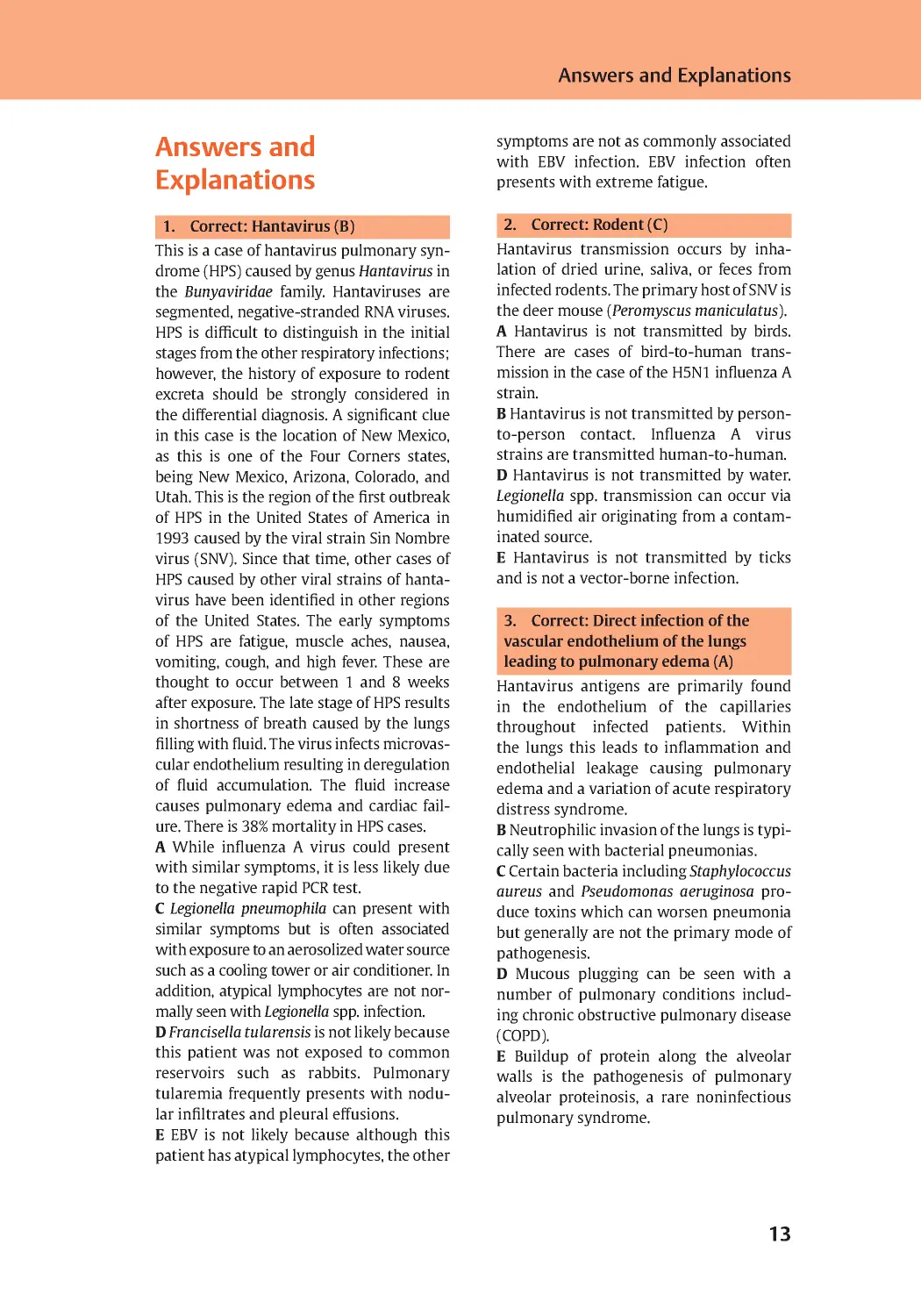
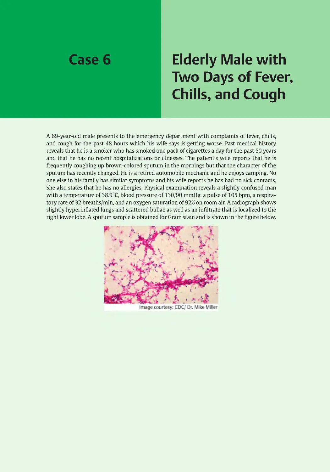
right lower lobe. A sputum sample is obtained for Gram stain and is shown in the figure below.
6 Elderly Male with Two Days of Fever, Chills, and Cough
24
Questions
1. Which of the following organisms is
the most likely causative agent?
A. Chlamydia psittaci
B. Legionella pneumophila
C. Pneumocystis jiroveci
D. Staphylococcus aureus
E. Streptococcus pneumonia
2. In terms of hemolysis and optochin,
which of the following best describes
the laboratory test results that would be
expected if the causative agent was cul-
tured and tested?
A. Alpha hemolysis, optochin sensitive
B. Alpha hemolysis, optochin resistant
C. Beta hemolysis, optochin sensitive
D. Beta hemolysis, optochin resistant
E. Gamma hemolysis, optochin sensitive
F. Gamma hemolysis, optochin resistant
3. A commonly available antigen test
can potentially confirm the diagnosis by
testing which of the following patient
samples?
A. Blood
B. Saliva
C. Stool
D. Sweat
E. Urine
4. Which of the following best describes
the vaccine available for control and pre-
vention of infections by the causative
agent?
A. DNA vaccine
B. Conjugate vaccine
C. Live attenuated vaccine
D. Toxoid vaccine
E. Whole cell heat-killed vaccine
5. Which of the following antibiotic
treatments would be most appropriate to
start in this patient?
A. Penicillin G
B. Azithromycin
C. Vancomycin
D. Piperacillin–tazobactam
E. Ceftriaxone
Answers and Explanations
25
Answers and
Explanations
1. Correct: Streptococcus pneumonia (E)
Streptococcus pneumoniae is the most
common cause of lobar pneumonia.
Rust-colored/brown sputum is also com-
mon for S. pneumoniae infections. Fur-
thermore, the Gram stain of the sputum
shows gram-positive cocci in pairs with
a nonstaining halo (indicative of the cap-
sule), which is the characteristic cellular
morphology for S. pneumoniae.
A Chlamydia psittaci could be possible
because this patient enjoys the outdoors,
so he does have possible exposure to
birds and bird droppings; however, the
Gram stain of the sputum is suggestive
of gram-positive cocci, and C. psittaci are
non-Gram staining.
B Because of the advanced age of the
patient in this case, Legionella pneumoph-
ila should be considered. However, because
the onset is acute and not subacute, and
L. pneumophila do not Gram stain well and
are gram-negative, this is not the most
likely causative agent.
C Pneumocystis jiroveci would be a possi-
ble causative agent if the patient was HIV
positive or immunocompromised, but that
is not the case.
D Staphylococcus aureus would be more
likely if the patient had a recent history of
an influenza infection or was an intrave-
nous drug user.
2. Correct: Alpha hemolysis, optochin
sensitive (A)
S. pneumoniae are alpha hemolytic, mean-
ing that the organisms have the ability to
partially lyse the red blood cells on blood
agar plates when grown in culture. These
organisms are characteristically inhib-
ited by optochin and therefore are unable
to grow (i.e., are sensitive to) in the pres-
ence of optochin on laboratory agar plates.
S. pneumoniae are also characteristically
lysed by bile salts (deoxycholate) due to
the presence of an autolytic enzyme.
B S. pneumoniae is alpha hemolytic but
they are sensitive to optochin. Viridans
group Streptococcus are alpha hemolytic
and are optochin resistant.
C S. pyogenes is beta hemolytic but is
optochin resistant and is not the causative
agent in this case.
D S. pyogenes is beta hemolytic and is
optochin resistant but is not the most likely
causative agent in this case.
E The former Group D Streptococcus have
been reclassified as Enterococcus spp. and
are gamma hemolytic and are not the caus-
ative agent in this case.
F Enterococcus spp. are gamma hemolytic,
optochin resistant, and are not the caus-
ative agent in this case.
3. Correct: Urine (E)
The pneumococcal urinary antigen test
is an assay that detects S. pneumoniae
cell wall antigens from patient urine.
As such, it is noninvasive and does not
require the patient to provide sputum.
This test was developed to fill the need
to a speedy and accurate diagnosis of
pneumonia. The test is approximately 70
to 90% sensitive and 80 to 100% specific
in adults. Cell culture from sputum sam-
ple is the gold standard for diagnosis,
but it requires the patient to generate
sputum, and results take at least 24 to
48 hours. Polymerase chain reaction
(PCR) of pleural fluid will also yield a
definitive diagnosis, but obtaining pleu-
ral fluid is more invasive, and the results
will take longer than for a urine antigen
test. A throat swab rapid test is used to
presumptively diagnose S. pyogenes or
strep throat.
A, B, C, D Other bodily fluids cannot be
used for this rapid test. There is no rapid
antigen test available for blood, saliva,
stool, or sweat.
4. Correct: Conjugate vaccine (B)
Two different types of vaccines for S. pneu-
moniae are approved for use in the United
States. The pneumococcal polysaccharide
vaccine (PPSV) contains capsular polysac-
charides and is used for routine vaccination
6 Elderly Male with Two Days of Fever, Chills, and Cough
26
in selected adults and children younger
than 2 years. PPSV is not used for infants
because polysaccharide antigens do not
illicit a strong immune response. The sec-
ond vaccine is the pneumococcal conjugate
vaccine (PCV). The polysaccharide antigens
are covalently linked to a protein carrier
in order to increase the immunogenicity
of this vaccine. As a result, PCV is used for
routine vaccination of infants and toddlers
as well as adults older than 65 years.
A At present, there are no DNA vaccines for
routine vaccination for humans.
C Examples of live attenuated vaccines
include vaccines against rotavirus, vari-
cella, MMR, and the nasal spray vaccine for
influenza (FluMist).
D An example of a toxoid vaccine is the tet-
anus vaccine (Clostridium tetani).
E Examples of inactivated (whole cell heat-
killed) vaccines include the polio vaccine
(Salk), the injected influenza vaccine, and
the pertussis vaccine (Bordetella pertussis).
5. Correct: Ceftriaxone (E)
All of the listed choices potentially have
activity against S. pneumoniae. Ceftriax-
one is active against S. pneumoniae and
penicillin-resistant S. pneumoniae and is
an appropriate first-line antibiotic for the
treatment of S. pneumoniae.
A According to current guidelines, when
the etiologic agent is established or
strongly suspected, it is reasonable to
switch from an empiric therapy to a more
directed therapy. At this point, no sensitiv-
ity data are known to determine whether
the S. pneumoniae in this case is penicil-
lin-resistant or not. As such penicillin is
not a reasonable choice.
B Azithromycin is reasonable for simple
community-acquired pneumonia (CAP)
where there is no suspected resistance, but
this patient is somewhat more complicated
with underlying chronic obstructive pulmo-
nary disease (COPD), and his CURB-65 score
is at least 3 with confusion, respirations
greater than or equal to 30 breaths/min, and
age greater than or equal to 65. No data are
given for uremia, and his blood pressure
is not less than 90 systolic or 60 diastolic.
As such, azithromycin is not a reasonable
choice as a single agent.
C Vancomycin would be appropriate if he
had a high-grade allergy to beta-lactams
and suspicion for penicillin-resistant
S. pneumoniae; as he has no allergies, van-
comycin is not an appropriate choice.
D Piperacillin–tazobactam is appropriate
for hospital-acquired pneumonia (HAP) or
health care-associated pneumonia (HCAP)
but not for CAP where there is low suspi-
cion for gram-negative pathogens, particu-
larly Pseudomonas aeruginosa.
Keywords: Streptococcus pneumoniae, pneumonia, sputum, hemolysis, pneumococcal vac-
cine, ceftriaxone
Case 7
Severely Ill Elderly
Male in Respiratory
Distress
A 71-year-old male with mild chronic obstructive pulmonary disease (COPD) is brought to the
emergency department by his daughter with symptoms of a high fever, chest pain, shortness
of breath, and a nonproductive cough. He claims that the fever and a headache started 3 days
before. Upon examination, his temperature is found to be 104°F and oxygen saturation at 82%
on room air. The patient was tachycardic at 119 bpm. Coarse breathing sounds were heard
on auscultation. The patient is not fully responsive. The daughter is asked about his medical
history and states that other than the mild COPD, which he only uses some inhalers for, he is
very healthy and hasn’t been hospitalized in years. He hasn’t traveled and has no sick contacts
or animal exposures. He recently moved in with his daughter and her family. Since it is the
summer and the weather is good, he states that he spends most of his time during the day
(when the children are at school) in the garden area of their apartment complex sitting by the
fountain and reading. He is up to date on his immunizations and received PPSV23 and PCV13
as well as herpes zoster vaccination and a booster for pertussis since he is around his grand-
children all the time now.
A chest radiograph showed pleural effusions and confluent infiltrates in the left lower
lobe. A bronchoalveolar lavage (BAL) was conducted. Gram stain of the specimen did not de-
tect any organisms. Culture of the BAL specimen demonstrated growth on buffered charcoal
yeast extract agar but not 5% sheep blood agar. A Gram stain from the culture is shown in the
figure below.
7 Severely Ill Elderly Male in Respiratory Distress
28
Questions
1. Based on the patient history and lab-
oratory findings, what is the most likely
cause of this patient’s symptoms?
A. Influenza virus
B. Legionella pneumophila
C. Mycobacterium tuberculosis
D. Pneumocystis jirovecii
E. Streptococcus pneumoniae
2. In addition to culture, which of the fol-
lowing is the preferred method for diagnosis?
A. Direct fluorescent antibody stain of
lung biopsy
B. Paired serology
C. Polymerase chain reaction (PCR)
D. Sputum Gram stain
E. Urinary antigen test
3
.
What is the most likely transmission
scenario for this illness?
A. Casual contact
B. Contaminated vaccine
C. Food contamination
D. Sexual transmission
E. Water droplets
4. This organism has multiple life stages.
Which of the following best describes its
life cycle characteristics?
A. Facultative anaerobe
B. Facultative capnophile
C. Facultative intracellular
D. Facultative spore forming
E. Facultative thermophile
5. Of the following, which is the most
active therapeutic agent for this patient’s
infection?
A. Azithromycin
B. Ceftriaxone
C. Isoniazid
D. Oseltamivir
E. Trimethoprim–sulfamethoxazole
Answers and Explanations
29
Answers and
Explanations
1. Correct: Legionella pneumophila (B)
This is a case of pneumonia caused by
Legionella pneumophila. Legionella spp.
exist in freshwater environments and can
inhabit artificial water systems where
proper disinfection is lacking and trans-
mission to humans is more likely to occur.
Individuals at greatest risk for infection are
those with chronic lung disease, smokers,
those with weak immune systems, and
people older than 50 years (such as the
patient in this case). Legionella spp. were
initially identified as a result of an outbreak
of pneumonia in 1976 at a convention of the
American Legion in Philadelphia, owning to
its name. A related illness also attributed to
Legionella is known as Pontiac fever and was
first identified in Pontiac, Michigan. This ill-
ness does not result in pneumonia and is
less severe in its manifestations. Symptoms
include fever and muscle aches and usually
last about a week after initial signs.
A Influenza virus is not the correct answer
because of the symptoms described and
also because gram-negative bacilli can be
seen from the Gram stain, indicating a bac-
terial cause rather than viral.
C Mycobacterium tuberculosis is incor-
rect because this patient’s symptoms are
not typical of the clinical presentation of
tuberculosis, and also M. tuberculosis does
not stain by the Gram’s method.
D Pneumocystis jirovecii is incorrect as this
patient would unlikely to be infected with
Pneumocystis carinii pneumonia (PCP) due
to the lack of a diagnosis of HIV infection.
E While Streptococcus pneumoniae is the
most common cause of community-ac -
quired pneumonia; they are gram-positive
cocci, whereas gram-negative bacilli are
seen in the figure.
2. Correct: Urinary antigen test (E)
L. pneumophila are difficult to culture. Legio-
nella spp. have complex nutritional require-
ments and do not grow on standard blood
plates. Agars based on buffered charcoal
yeast extract (BCYE) medium containing
cysteine, high concentrations of iron and
activated charcoal are the usual medium of
choice for culture. The most commonly used
laboratory test for diagnosis is the urinary
antigen test. However, this test only detects
antigen for serogroup 1, which is the most
common cause of legionellosis. There are 42
Legionella species with 64 serogroups capa-
ble of causing human disease. Because of this
diversity, culture is used as a confirmatory
test when urine antigen tests are negative.
A A direct fluorescent antibody (DFA) stain
of lung biopsy can be used for diagnosis
but is not routine due to the invasiveness
of this procedure.
B Paired serology is not common for the
diagnosis of Legionella spp. infection.
C There are currently no Food and Drug
Administration (FDA) approved PCR assays
available, and routine PCR-based tests are
not available at every hospital center.
D Sputum Gram stain is not typically
used for diagnosis because Legionella spp.
are difficult to Gram stain, so a negative
Gram stain is common with Legionella spp.
infection.
3. Correct: Water droplets (E)
Legionella spp. survive in warm water envi-
ronments where improper disinfection
occurs, or they can survive intracellularly
in amoeba, which are not routinely killed
by chlorination. Sources such as shower
heads, water cooling systems, hot tubs
and spas, and water features have all been
documented to be a source of the bacteria.
Transmission to humans occurs when
aerosolized water droplets are inhaled.
A Casual contact is incorrect because of
dose transmission (there would not be
enough Legionella transmitted to cause an
infection).
B Contaminated vaccine is incorrect
because there is no vaccine for Legionella
infection.
C Food contamination is incorrect because
Legionella is not food-borne.
D Sexual transmission is incorrect because
Legionella is not a sexually transmitted
infection.
7 Severely Ill Elderly Male in Respiratory Distress
30
4. Correct: Facultative intracellular (C)
Legionella spp. are unlike other bacterial
pneumonia pathogens, in that they do not
reside within the upper respiratory tract
(i.e ., these bacteria are not normal flora) but
infect alveolar macrophages and alveolar
epithelial cells as intracellular organisms.
In the natural environment, Legionella spp.
are intracellular pathogens of amoeba. In
all instances, cells take up the bacteria via
phagocytic processes. Legionella spp. are
facultative intracellular because they are
also able to survive and grow outside of
eukaryotic host cells. Once internalized, the
bacteria are able to evade phagolysosomal
degradation. While Legionella spp. can be
cultured on artificial media, they are only
pathogenic when they complete an intra-
cellular life cycle.
A Legionella spp. are aerobic organisms,
not anaerobic.
B Legionella spp. are not capnophilic (cap-
nophiles thrive in the presence of high con-
centrations of carbon dioxide).
D Legionella spp. are not able to form
spores.
E Legionella spp. cannot survive at ele-
vated temperatures and are, therefore, not
thermophiles.
5. Correct: Azithromycin (A)
The two most appropriate therapies for
L. pneumophila infections are macrolides,
such as azithromycin, or respiratory quino-
lones, such as levofloxacin.
B Ceftriaxone is the most appropriate treat-
ment for S. pneumoniae (the most common
causative agent of community-acquired
pneumonia), and it is likely this patient
will receive both ceftriaxone and azithro-
mycin as per the guidelines for the treat-
ment of community-acquired pneumonia.
C Isoniazid would be part of a regimen for
tuberculosis (TB) but would only be given
alone for latent TB infection.
D Oseltamivir would be given for suspicion
of influenza virus which is unlikely in the
summer months.
E Trimethoprim–sulfamethoxazole would
be the first-line treatment for PCP but has
no activity against Legionella spp.
Keywords: Legionella pneumophila, atypical pneumonia, waterborne, COPD
Case 8
Teenager with a
Two-Week-Long
Cough
A 16-year-old adolescent female presents to her primary care physician with a cough of
2-weeks duration. No one else in her household has a similar cough. She is a student and has
no history of cigarette smoking. Her past medical history is remarkable for recurrent lung
infections that have been relatively mild; she has never been admitted to the hospital for
treatment of one of her infections before. She states that this cough is much worse than her
“usual” cough or any of the prior infections. She has a previous confirmed molecular genetic
laboratory test finding of being heterozygous with a delta F508 mutation in the cystic fibrosis
transmembrane conductance regulator (CFTR) gene.
Physical examination reveals a temperature of 38.7°C, a heart rate of 120 bpm, and a re-
spiratory rate of 30 breaths/min; she is markedly dyspneic, has a reduced forced vital capacity
(FVC), and the cough is productive of greenish sputum. Her oxygen saturation is found to be
82% while her baseline oxygen saturation is 95%.
Laboratory studies were obtained, and the complete blood count (CBC) with differential
showed white blood cell (WBC) count 18.5 ×10⁹ cells/L with a marked left shift. Posteroante-
rior (PA) and lateral chest X-rays were also obtained and showed hyperinflation and bronchi-
ectasis and “tram tracks” in the upper lobes. There is a dense consolidation with air broncho-
grams involving most of the left lung.
The physician obtains a sample of the sputum that is sent to the microbiology laboratory
for Gram stain and culture. A Gram stain is shown in the figure below. The predominant or-
ganism grew as mucoid colonies on blood agar at 37°C after 24 hours with beta hemolysis and
a metallic sheen. The organism is citrate positive and oxidase positive.
8 Teenager with a Two-Week-Long Cough
32
Questions
1. Which of the following organisms is
the most likely causative agent?
A. Legionella pneumophila
B. Mycoplasma pneumoniae
C. Pseudomonas aeruginosa
D. Staphylococcus aureus
E. Streptococcus pneumonia
2. Which of the following would best
describe the growth of the most likely
causative agent on MacConkey agar?
A. Growth with clearing around each
colony
B. Growth with colorless colonies
C. Growth with pink colonies
D. No growth
E. Swarming growth
3. Which of the following virulence
factors contributes to the mucoid pheno-
type described in the most likely causative
agent?
A. Alginate
B. Elastase
C. Exotoxin A
D. Flagella
E. Quorum sensing
4. Which of the following best describes
the most common mode of transmission
for the causative organism?
A. Air-borne from an infected individual
B. Arthropod-borne from an Aedes spp.
mosquito
C. Blood-borne from a puncture wound
D. Fecal–oral route
E. Ubiquitous from the environment
5. Which of the following is the best
antibiotic treatment strategy for this acute
infection?
A. Start oral ciprofloxacin to give the best
coverage and the highest compliance
B. Start intravenous colistin due to the
high prevalence of resistance in the
likely causative organism
C. Start intravenous vancomycin mono-
therapy to cover the most likely caus-
ative organism
D. Start inhaled tobramycin to treat this
contained pulmonary infection
E. Start a combination of intravenous
piperacillin–tazobactam and tobramy-
cin to cover the most likely causative
organism
Answers and Explanations
33
Answers and
Explanations
1. Correct: Pseudomonas aeruginosa (C)
This case describes a young adult with
a cough. The patient has a past medical
history for a confirmed diagnosis of cystic
fibrosis (CF). Sputum culture of a typical
CF patient will grow Staphylococcus aureus
at a young age, replaced by Pseudomo-
nas aeruginosa as the patient ages. Other
bacteria that may be considered from the
sputum of a CF patient include Haemoph-
ilus influenzae, Burkholderia cepacia, and
Stenotrophomonas maltophilia. Laboratory
results indicated growth on blood agar
after 24 hours and beta hemolysis, which
would be seen for P. aeruginosa or S. aureus
only, of the choices listed (the other organ-
isms listed would require special growth
media). The Gram stain image shows
gram-negative rods, which is the mor-
phology for P. aeruginosa, but not S. aureus.
Lastly, P. aeruginosa are oxidase positive
and citrate positive.
A Legionella pneumophila cannot be visu-
alized by Gram stain.
B Mycoplasma pneumoniae cannot be visu-
alized by Gram stain.
D S. aureus are gram-positive cocci, not
gram-negative bacilli, as seen in the figure.
E Streptococcus pneumoniae are gram-
positive cocci, not gram-negative bacilli, as
seen in the figure.
2. Correct: Growth with colorless
colonies (B)
MacConkey agar is a selective medium that
selects for the growth of many gram-neg-
ative bacilli (it should be noted that not all
gram-negative bacilli will grow on Mac-
Conkey agar). This agar contains bile salts
that inhibit the growth of gram-positive
organisms. MacConkey agar also contains
lactose and a pH change indicator. Lac-
tose-fermenting organisms produce acidic
by-products, resulting in pink colonies,
whereas nonlactose-fermenting organisms
can be viewed as colorless colonies.
P. aeruginosa are gram-negative bacilli, so
they will grow on MacConkey agar. How-
ever, they are nonlactose-fermenters, so the
colonies will be colorless due to the lack of
a pH change.
A There are no organisms of medical
importance that would have this type of
colony morphology on MacConkey agar.
Beta-hemolytic bacteria will grow on
blood agar with a clearing around the col-
ony indicating hemolysis.
C Lactose-fermenting bacteria, such as
E. coli, will grow on MacConkey agar as
bright pink colonies.
D Gram-positive bacteria, as well as
gram-negative bacilli that are not in the
Enterobacteriaceae family will not grow on
MacConkey agar.
E Proteus spp. are bacteria that grow in a
typical swarming pattern on many routine
media agars.
3. Correct: Alginate (A)
Alginate is the loosely associated cap-
sule that surrounds many clinical isolates
of P. aeruginosa. It functions to protect
the bacteria from phagocytosis, as well
as aids in biofilm formation. In the lung
environment of CF patients, P. aeruginosa
produces the polysaccharide alginate that
results in a phenotype conversion from
nonmucoid to mucoid during chronic
colonization.
B Elastase is an enzyme produced by many
strains of P. aeruginosa that aids in coloni-
zation by breaking down host elastin.
C Exotoxin A is an exotoxin that kills
host cells through the mechanism of
ADP-ribosylating elongation factor 2 (EF-2)
and therefore halting protein synthesis,
resulting in cell death.
D Most strains of P. aeruginosa are motile
and produce flagella, but this does not have
an effect on the mucoid phenotype.
E P. aeruginosa is one of many bacteria
that produce quorum sensing molecules,
which allows many bacteria to commu-
nicate at the population level through the
release of chemical signals (autoinducers).
Downregulation of quorum sensing may
be a result of selective pressure due to the
induction of the mucoid phenotype.
8 Teenager with a Two-Week-Long Cough
34
4. Correct: Ubiquitous from the
environment (E)
P. aeruginosa is found ubiquitously in large
quantities in the environment. Most indi-
viduals (healthy or otherwise) have evi-
dence of P. aeruginosa exposure by the age
of 1.
A Air-borne from an infected individual is
incorrect. P. aeruginosa is not air-borne.
B Arthropod-borne from an Aedes spp.
mosquito is incorrect. P. aeruginosa is not
a vector-borne organism.
C Blood-borne from a puncture wound is
incorrect. P. aeruginosa is not transmitted
in the blood.
D Fecal–oral route is incorrect. P. aeruginosa
is not transmitted via the fecal–oral route.
5. Correct: Start a combination of
intravenous piperacillin–tazobactam
and tobramycin to cover the most likely
causative organism (E)
While there is controversy on the best
way to treat acute exacerbations of CF due
to Pseudomonas infections, the patient
described in this case is clearly ill and is
in imminent danger due to the infection.
Pseudomonas is the likely causative organ-
ism, and resistance is a frequent issue in
the treatment of Pseudomonas infections;
however, this patient has never had a sim-
ilar infection, and thus while she may have
resistance, it is unlikely she has developed
resistance to most classes of antipseudo-
monal antibiotics at this time. A combina-
tion of an antipseudomonal beta-lactam
(such as piperacillin–tazobactam) and an
aminoglycoside (such as tobramycin) is
considered appropriate for initial treat-
ment of severe Pseudomonas infections.
A Oral ciprofloxacin might be considered
for a mild exacerbation, but the use of
ciprofloxacin alone for Pseudomonal lung
infections is generally frowned upon due
to the high likelihood of emergence of
resistance.
B Colistin is used only in cases where
extensive resistance is likely due to its high
risk for significant side effects.
C Vancomycin would be appropriate for
treatment of methicillin-resistant S. aureus
(MRSA) but not Pseudomonas.
D Given the extent of her illness, this
patient is not appropriate for treatment
with inhaled antibiotics alone.
Keywords: Cystic fibrosis, Pseudomonas aeruginosa, sputum, alginate, mucoid, MacConkey,
piperacillin-tazobactam, tobramycin
Case 9
Adult Male with
Persistent Cough
and Malaise
A 57-year-old otherwise healthy Caucasian male presented to his primary care physician
(PCP) in Michigan in mid-February with a fever, cough, and fatigue. He describes that his
symptoms have been going on for the past month and indicates that these symptoms have not
significantly changed, and he is frustrated that he is not getting better. The PCP asks about sick
contacts, and he states that he has been exposed to some children who have had typical win-
ter colds, but they all got better within a week. The PCP asks about travel and other possible
exposures and he denies any travel but mentions that he has family all over the world, and he
received several gifts from different family members for the holidays. His brother in Florida
sent a box of theme park merchandise, his cousin in Arizona sent him a live cactus and some
Native American blankets, and his sister in France sent him a few bottles of wine and some
jars of French preserves.
Physical examination reveals that the patient’s temperature is 38.3°C, blood pressure
110/70 mmHg, and pulse 106 bpm. A chest X-ray is obtained and shows consolidations in the
apical regions of the upper right lung. A sputum sample is obtained for laboratory analysis and
microscopy with periodic acid–Schiff (PAS) is shown in the figure below.
9 Adult Male with Persistent Cough and Malaise
36
Questions
1. Which of the following organisms is
the most likely causative agent?
A. A s pergillus fumigatus
B. Blastomyces dermatitidis
C. Coccidioides posadasii
D. Histoplasma capsulatum
E. Mycobacterium tuberculosis
2. Which of the following best describes
the dimorphic nature of the causative agent?
A. Can synthesize both muscarine and
fusariongenin
B. Has a yeast and a mycelial phase
C. Has both septate and aseptate hyphae
D. Infects two different organs at the
same time
E. Produces a sporangiophore and
mycelium
3. Which of the following diagnostic tests
would be the safest to confirm the diagnosis?
A. Culture at 37°C and 25°C
B. Serology with enzyme immunoassay
(EIA)
C. Polymerase chain reaction (PCR) of
sputum sample
D. Radiography
E. Urine antigen test
4. If examination of medical history of
this patient revealed a diagnosis of HIV
infection in the past 5 years, which of the
following possible complications of infec-
tion would be the most cause for worry?
A. Antibiotic-associated colitis
B. Endocarditis
C. Hemolytic uremic syndrome
D. Meningitis
E. Scarlet fever
5. What would be the most appropriate
treatment for this patient at this time?
A. Amphotericin B
B. Ceftriaxone
C. Micafungin
D. Fluconazole
E. Symptomatic care only
Answers and Explanations
37
Answers and
Explanations
1. Correct: Coccidioides posadasii (C)
The patient’s symptoms describe valley
fever pneumonia. Coccidioides posada-
sii infections originate from the defined
geographical location of Arizona or the
southwestern United States (Coccidioides
immitis is more common in California). The
description of the organism as a multinu-
cleate spherule when in a patient sample
(at 37°C) also gives a clue as to the fun-
gal pathogen C. posadasii. Symptoms of
valley fever include cough, fever, fatigue,
chest pain, and weight loss. The infection
was likely transmitted from either the
soil from the cactus plant or dust or sand
in the blankets that were received from
Arizona, where spores of the organism are
commonly found in the desert soil. The
patient likely inhaled arthrospores present
in the sand, dust, or soil. None of the other
choices have the morphology described
although some cause similar symptoms.
A Aspergillus fumigatus are septate hyphae
with dichotomous branching at acute
angles.
B Blastomyces dermatitidis is a broad-
based budding yeast.
D Histoplasma capsulatum are small oval
brown yeast cells at 37°C.
E Mycobacterium tuberculosis are acid-fast
bacilli and would not be visualized under
nonstained light microscopy.
2. Correct: Has a yeast and a mycelial
phase (B)
This patient presents with a Coccidioides
spp. infection, which are dimorphic fungi.
Dimorphic fungi, by definition, have a
yeast phase at 37°C and a hyphal or myce-
lial phase at 25°C.
A, C, D, E None of the other answer choices
describe the two phases of a dimorphic
fungus. C . immitis are dimorphic as mul-
tinucleate spherules at 37°C (e.g ., at body
temperature or when sampled directly
from body fluids such as sputum in this
case) and septate hyphae with arthro-
spores at 25°C, or when grown at room
temperature.
3. Correct: Serology with enzyme
immunoassay (EIA) (B)
Serological testing is routinely done for
patients with suspected coccidioidomyco-
sis. Most serologic tests use EIAs for both
immunoglobulin M (IgM) and IgG. If EIA
is positive, then a confirmatory immuno-
diffusion test is normally performed, as it
gives a qualitative positive for IgG. Other
possible tests include immunodiffusion
tests (which are less sensitive but more
specific), complement fixation tests, or
quantitative immunodiffusion tests for IgG.
A While culture will also give a definitive
diagnosis, there are safety risks associated
because the organism is so easily trans-
missible and so precautions must be taken.
Coccidioides spp. are a leading cause of
laboratory-acquired infections.
C PCR testing is not routinely available.
D Radiography can be used to confirm a
diagnosis of pneumonia but will not give
any clues as to the identification of the
causative agent.
E A urine antigen test is currently under
development, but such a test is not avail-
able at this time.
4. Correct: Meningitis (D)
Any individuals with a compromised T-cell
immunity are at risk for disseminated
Coccidioides spp. infection. The most com-
mon sites of infection are the central ner-
vous system (CNS), skin, and bone. As such,
meningitis is the most common complica-
tion of infection among those individuals
who are HIV positive.
A Antibiotic-associated colitis is a compli-
cation of antibiotic treatment.
B Endocarditis is not a common complica-
tion of coccidioidomycosis.
C Hemolytic uremic syndrome is a compli-
cation of enterohemorrhagic Escherichia
coli (EHEC) infections.
E Scarlet fever is a possible complication of
a Streptococcus pyogenes infection.
9 Adult Male with Persistent Cough and Malaise
38
5. Correct: Symptomatic care only (E)
The clinical description is most consistent
with acute pulmonary coccidioidomyco-
sis or valley fever. There is nothing in the
presentation to suggest severe disease,
disseminated disease, acute worsening,
or concerning comorbidities. As such for
acute valley fever, which usually occurs 1
to 3 weeks after an exposure and can last
for months, current recommendations are
patient education, observation, and sup-
portive measures only.
A Amphotericin is generally reserved
for azole failure or cases with severe
immunosuppression.
B Antibiotic treatment such as ceftriaxone
would not be appropriate and would not
treat Coccidioides.
C Micafungin is not used in cases of
coccidioidomycosis.
D Antifungal treatment would be appro-
priate for patients with debilitating dis-
ease, evidence of dissemination, frailty due
to comorbidities or advanced age, diabe-
tes, or immunosuppression. The generally
accepted first-line treatment for nondis-
seminated severe coccidioidomycosis is
fluconazole.
Keywords: Pneumonia, Coccidioides spp., fungal, dimorphic, select agent, multinucleate spherule
Case 10
Infant with Severe
Congestion
In late January, a mother brought her 5-month-old daughter to her pediatrician because of a
fever of 101°F that began the previous night. She reports that the child has had a very runny
nose for the last 3 days and has required frequent suction to clear the congestion. The child
also has been very sleepy and uninterested in eating or drinking. When asked, the mother
reports that the child has had no diarrhea or vomiting but that a dry cough had started earlier
that morning.
The child lives with her mother and 3-year-old brother. The mother reports that both
children attend daycare three times a week and are watched by their grandmother for 2 other
weekdays. Her big brother was diagnosed previously with cough-variant asthma. There are no
smokers in the home and no pets. The child is up to date with all recommended vaccinations.
Physical examination identified tachypnea and wheezing; nasal flaring and intercostal
retractions were observed on inspiration. Oxygen saturation was low at 91%.
The child was admitted to the hospital and given oxygen. Imaging studies and laboratory
tests were ordered including a complete blood count (CBC) with differential that showed a
slightly elevated white blood cell (WBC) count with lymphocytosis. A nasopharyngeal swab
was collected and sent for respiratory panel testing.
10 Infant with Severe Congestion
40
Questions
1. What is the clinical condition that is
most likely being experienced by this child?
A. Asthma
B. Bronchiolitis
C. The common cold
D. Influenza
E. Pneumonia
2. Given the patient history, which of
the following organisms is most likely the
causative agent?
A. Haemophilus influenzae
B. Influenza A virus
C. Metapneumovirus
D. Respiratory syncytial virus (RSV)
E. Streptococcus pneumoniae
3. How is the causative agent most often
spread?
A. Airborne transmission
B. Fomite transmission
C. Foodborne transmission
D. Vector transmission
E. Sexual transmission
4. The F protein of the causative agent is
required for infection and is also respon-
sible for which of the following cellular
effects?
A. Clumping
B. Granulations
C. Inclusion bodies
D. Syncytia
E. Vacuoles
5. Which of the following is the most
appropriate treatment for this patient?
A. Corticosteroids
B. Intravenous immunoglobulin (IVIg)
C. Palivizumab
D. Ribavirin
E. Supportive care
Answers and Explanations
41
Answers and
Explanations
1. Correct: Bronchiolitis (B)
Bronchiolitis is the most common lower
respiratory tract infection in infants.
A Asthma is a result of airway edema as
compared with bronchiole restriction seen
in bronchiolitis. Imaging studies would aid
in the differentiation.
C A common cold may have a low-grade
fever in children but would not result in
breathing difficulty which was observed in
the child during the physical examination.
D Influenza infection would result in a
much higher fever. Additionally, vomiting
and diarrhea may be observed in young
children.
E Bronchiolitis can progress to pneumonia,
but given the short span of progression, the
time of year, and age of the patient, bron-
chiolitis is the more likely choice.
2. Correct: Respiratory syncytial virus
(RSV) (D)
Given the results of the CBC and differen-
tial, it is less likely that the infection is bac-
terial. The PCV13 vaccine for Streptococcus
pneumoniae is given as a series of three
vaccinations at 2, 4, and 6 months. The
PCV13 pneumococcal conjugate vaccine
covers the same serotypes as the previous
PCV7 version with an additional six sero-
types. Given the age of the child, the last
dose has not yet been issued. The conjugate
vaccine for Haemophilus influenzae (Hib) is
also given in a series of three or four injec-
tions beginning at age 2 months then at 4,
6, and 12 to 15 months. Again, only the first
two doses would have been received in this
case. Both S. pneumoniae and H. influenzae
are a leading cause of pneumonia in unvac-
cinated children but are not a cause of
bronchiolitis. Influenza is a serious disease
for children younger than 2 years of age,
and yearly vaccination is recommended.
However, the vaccine is recommended for
those 6 months of age or older. This child
would not have received the influenza
vaccine. Influenzae A virus does not cause
bronchiolitis. Human metapneumovirus
(hMPV) is a recently described etiologic
agent (2001) for a spectrum of respira-
tory illnesses including bronchiolitis. Most
cases of bronchiolitis (~ 70%) are believed
to be caused by RSV, but the contribution
of hMPV is now appreciated. Both viruses
are negative-stranded RNA viruses belong-
ing to the Paramyxoviridae family. While a
passive immunization prophylactic treat-
ment using a monoclonal antibody is avail-
able for RSV infection, it is generally only
offered to infants at high risk of hospital-
ization due to underlying comorbidities.
No vaccine currently is available.
A H. influenzae does not cause bronchiol-
itis.
B Influenzae A virus does not cause
bronchiolitis.
C Metapneumovirus can cause bronchiol-
itis but in this case it is not the causative
agent.
E S. pneumoniae does not cause bronchiol-
itis.
3. Correct: Airborne transmission (A)
RSV is highly contagious and is primarily
spread by coughing and airborne droplet
transmission. Although it is an enveloped
virus, it is able to remain viable on contam-
inated surfaces for several hours.
B Fomite transmission is possible but sec-
ondary to airborne transmission.
C, D, E RSV is not transmitted via the
foodborne, vector, or sexual transmission
routes.
4. Correct: Syncytia (D)
A characteristic cytopathic effect of RSV
infection is the formation of syncytia or
the fusing of cells to form a single mass
with multiple nuclei. The F protein of
the virus is essential for fusing the mem-
brane of the virus to endocytic vesicles in
cells of the respiratory epithelium after
macropinocytosis.
A The F protein of RSV does not cause cell
clumping.
B The F protein of RSV does not cause gran-
ulations to be formed.
10 Infant with Severe Congestion
42
C The F protein of RSV does not cause the
formation of inclusion bodies inside of host
cells.
E The F protein of RSV does not cause vac-
uoles to form inside of cells.
5. Correct: Supportive care (E)
In young children (< 2 years of age), support-
ive care is the treatment of choice for RSV.
A Corticosteroids may be of use in adults
and older children, but in randomized tri-
als, they have not been shown to be benefi-
cial in young children.
B A systematic review of studies using IVIg
in 2006 failed to show significant benefit.
C Palivizumab is a monoclonal antibody
directed against the RSV F protein, but
studies have shown no benefit in treat-
ment. It is useful for the prevention of RSV
in high-risk children.
D Ribavirin, at one time, was commonly
used to treat children with RSV; however, a
systematic review of the randomized trials
of ribavirin in infants and children did not
support its use.
Keywords: Bronchiolitis, respiratory syncytial virus, cytopathic effect, transmission, pneu-
monia, syncytia
Adult Male with
Fever, Myalgias, and
Respiratory Distress
Case 11
A 38-year-old otherwise healthy male, is seen in the emergency department in late December
with symptoms of 104°F fever, myalgias, and headache for the last 4 days. Currently, he
complains of difficulty breathing and lower respiratory pressure. He does not have any
upper respiratory congestion but does have a cough. His temperature is 101.5°F after taking
600 mg of ibuprofen and his O2 saturation on room air is 86%. Crackles and wheezing are
heard on auscultation. Laboratory results indicate that the patient has a white blood cell
(WBC) count of 7.8 × 10⁹/L (5.8 × 10⁹/L neutrophils; 0.3 × 10⁹/L lymphocytes; 1.7 × 10⁹/L
monocytes). An X-ray of his chest is ordered and is normal. A computed tomography (CT) of
the chest is done and shows diffuse ground-glass changes. A procalcitonin is sent and the
results indicate less than 0.1 ng/mL. When asked, he states that he is not up to date on his
yearly vaccinations.
11 Adult Male with Fever, Myalgias, and Respiratory Distress
44
Questions
1. What is the most likely cause of this
respiratory infection?
A. Adenovirus
B. Influenza A virus
C. Human metapneumovirus
D. Parainfluenza virus
E. Respiratory syncytial virus
2. Which of the following tests is most
commonly used as a first-line test for diag-
nosing this infection?
A. Antigen detection
B. Bead array–based testing
C. Culture
D. Reverse transcription polymerase chain
reaction (RT-PCR)
E. Serology
3. Why is it necessary for this vaccina-
tion to be obtained yearly by this patient?
A. The patient has low procalcitonin
levels
B. The vaccine requires a booster
C. The virus changes frequently requiring
a different vaccine each year
D. The vaccine is therapeutic
E. The patient has an occult immunocom-
promised state
4. For which of the following is this
patient most at risk?
A. Developing a secondary bacterial
infection
B. Developing a relapsing viral infection
C. Developing a reaction to additional
vaccines
D. Developing an autoimmune nervous
system disorder
E. Developing secondary myocarditis
5. Which of the following is the most
appropriate treatment for this patient’s
infection?
A. Amantadine
B. Cidofovir
C. Intravenous immunoglobulin (IVIg)
D. Oseltamivir
E. Ribavirin
Answers and Explanations
45
Answers and
Explanations
1. Correct: Influenza A virus (B)
This is a case of influenza virus infection.
Seasonal flu epidemics occur usually from
October through May with peaks from
December through February, although flu
can occur year-round. See figure below
for seasonal incidence of viral infections.
The human viruses, influenza A and B are
responsible for these seasonal outbreaks.
Symptoms of acute high fever, severe head-
ache, and body aches are usual along with
other common cold symptoms having
greater severity. The white blood cell count
is also indicative of influenza. Children are
more likely to have additional symptoms
of vomiting and diarrhea. The severe flu
symptoms may last 2 to 5 days, whereas
the general cold symptoms of congestion,
cough, and fatigue may last 1 to 2 weeks.
While any of the viruses listed could in the-
ory be responsible for this infection, given
the occurrence is in December, influenza A
is the most common of these viruses.
A Adenovirus is less likely because adeno-
virus rarely presents with myalgia. Infec-
tions tend to peak in the fall.
C Human metapneumovirus would rarely
be this severe in a patient of this age and
health status. The peak season would be
later in the year.
D Parainfluenza virus would rarely be this
severe in a patient of this age and health sta-
tus. The peak season would be later in the year.
E Respiratory syncytial virus is less likely
because the extremes of age (the young
and the elderly) are more likely affected, as
well as those individuals with underlying
respiratory disease (neither of which are
the case for this patient).
2. Correct: Antigen detection (A)
The availability of point-of-care rapid diag-
nostic tools increases the ease of diagnosis
of influenza. There are two types of tests:
antigen detection and molecular testing.
Antigen detection is more commonly used
in outpatient facilities but also frequently
used in hospitals. A result turnaround time
of 15 to 20 minutes is attainable with the
rapid tests as opposed to that of RT-PCR,
bead array–based systems, serology, and
culture. A caveat to the use of rapid point-
of-care testing (POCT) is false-positive and
false-negative rates. Depending on the
sensitivity of the test, when the prevalence
of disease is low, the false-positive rate
increases. Conversely, when the prevalence
of disease is high, the false-negative rate
increases.
B, C, D, E The results of these tests still
require consideration of clinical judgment
to ascertain a true infection. For these
tests, the turnaround time is less rapid
than antigen detection and molecular test-
ing. However, results from one of these
tests, though slower to obtain, are often
required to corroborate the results of the
rapid test due to the increased sensitivity
of these tests.
3. Correct: The virus changes
frequently requiring a different vaccine
each year (C)
The influenza vaccine is offered yearly to
everyone and is comprised of viral hemag-
glutinin (H) and neuraminidase (N) anti-
gens. There are commonly three human
H antigen types and two neuraminidase
types that associate to form multiple viral
strain subtypes, e.g ., H1N1, while other less
common avian strains, for example, H5 or
H7, are also capable of infecting humans.
The vaccine is typically comprised of anti-
gens for the two strains of seasonal influ-
enza A viruses and one or two strains of
influenza B viruses which are predicted
to be circulating during influenza season
based on what is circulating at the time the
vaccines are made. Since there is a several
month lead time in the production of the
vaccine, sometimes the predictions don’t
11 Adult Male with Fever, Myalgias, and Respiratory Distress
46
match the reality of what is circulating well
and the vaccine is less effective. Vaccines
are offered each year as the viruses that
circulate change each year. Subtle changes
to the epitopes of circulating viruses
resulting from acquisition of genetic point
mutations can have a major impact on the
efficacy of a vaccine. These changes are
referred to as antigenic drift and are the
main reason the vaccine is given yearly.
A Procalcitonin can be predictive of a bac-
terial etiology for pneumonia. It is rarely
elevated in viral infection. The level has
nothing to do with response to vaccines.
B While it is true the immune response
generated by the vaccine wanes over time,
this is not truly a booster vaccine. With
influenza there can also be large-scale
genetic changes resulting from the novel
reassortment of the H and N antigens
referred to as antigenic shift. When an
antigenic shift occurs, the vaccines that are
generated are ineffective since the changes
are so large. This is what led to the 1919
influenza pandemic and the more recent
2009 H1N1 outbreak. When this occurs,
rapid development of a new vaccine is
needed as was seen in 2009.
D The influenza vaccine is not a therapeu-
tic vaccine, it is preventative.
E This patient is described as otherwise
healthy; influenza commonly affects
healthy adults; there is no reason to sus-
pect an immunocompromised status in
this patient.
4. Correct: Developing a secondary
bacterial infection (A)
The primary risk to this patient is post-
inf l uenza pneumonia due to bacterial cause.
The resolution of the viral infection may
reduce the inflammatory response thus setting
the stage for subsequent bacterial infection
progression. The primary pathogens involved
are most likely Staphylococcus aureus, Strep-
tococcus pneumoniae, Streptococcus pyogenes,
and Haemophilus influenzae.
B Influenza infections do not “relapse,” and
so that answer is incorrect.
C This infection is not a reaction to the vac-
cine, it is a viral infection, so that answer is
also incorrect.
D, E Complications of influenza infection
do not include autoimmune nervous sys-
tem disorders or development of a second-
ary myocarditis.
5. Correct: Oseltamivir (D)
There are three classes of anti-influenza
antivirals: amantadines, neuraminidase
inhibitors, and a selective inhibitor of
influenza community-acquired pneumo-
nia (CAP)-dependent endonuclease. Aman-
tadines inhibit viral uncoating, whereas
the neuraminidase inhibitors work by
preventing cleavage of the viral particles
from the cell. The neuraminidase inhibi-
tors are first-line therapy, and oseltamivir
is the most commonly used. Baloxavir is
a recently approved inhibitor of influenza
CAP-dependent endonuclease. It is not yet
considered first line for treatment of influ-
enza, but it could be a reasonable choice in
the right person.
A The adamantanes are no longer in use
due to near-universal resistance to them
by influenza virus.
B Cidofovir has no activity against
influenza.
C IVIg might contain protective antibod-
ies against influenza but is not a standard
treatment.
E Ribavirin has no activity against
influenza.
Keywords: Influenza virus, pneumonia, COPD
Case 12
Adult Male with
Fever, Chills, and
Night Sweats
A 38-year-old male presents to urgent care with fever, chills, malaise, and night sweats. He
says he has been feeling weak and has developed a cough. He denies any prior medical prob-
lems or sick contacts but mentions he had dental work recently. Examination of the patient’s
arms is shown in the first figure below. On questioning, he becomes evasive and says he has
no idea what they are. The remainder of the physical examination reveals a loud audible heart
murmur. The patient is admitted to the hospital, and three blood cultures are taken over a
24-hour period. All three cultures turn positive within less than 12 hours and grow the or-
ganism that is shown on the Gram stain (see second figure below). Biochemical testing of the
organism reveals that is it both catalase and coagulase positive. An echocardiogram is ordered
but not yet performed.
12 Adult Male with Fever, Chills, and Night Sweats
48
Questions
1. Which of the following organisms is
the most likely causative agent?
A. Enterococcus faecalis
B. Staphylococcus aureus
C. Staphylococcus epidermidis
D. Streptococcus bovis
E. Viridans group Streptococcus
2. Which of the following is the most
likely source of infection of the causative
agent?
A. Arthropod
B. Endogenous
C. Ingestion
D. Inhalation
E. Needlestick
3. Which of the following valves would
be most likely to have a vegetation in
this patient when the echocardiogram is
performed?
A. Aortic
B. Mitral
C. Pulmonary
D. Tricuspid
4. What is the most appropriate antibi-
otic to start the patient on initially?
A. Nafcillin
B. Cefazolin
C. Vancomycin
D. Piperacillin–tazobactam
E. Linezolid
5. The patient is started on antibiotics,
and when the sensitivities come back for
the original antibiotic prescribed, the min-
imum inhibitory concentration (MIC) is 1,
which is considered sensitive. The patient
clinically improves over the next 3 days,
but despite this, repeat cultures at 48 and
96 hours after initiation of antibiotics are
still positive for the same organism and the
MIC remains at 1. Which of the follow-
ing is the most likely cause of the failure to
clear the bloodstream infection?
A. Lack of source control
B. Rapid development of resistance to the
antibiotic
C. Selection of resistant subpopulations
from the initial infection
D. Introduction of a secondary infection
E. Contamination of the blood cultures
Answers and Explanations
49
Answers and
Explanations
1. Correct: Staphylococcus aureus (B)
The Gram stain shows gram-positive cocci
in clusters, which is a typical microscopic
morphology for Staphylococcus aureus.
Furthermore, S. aureus is the only bacteria
listed that test catalase and coagulase pos-
itive. All of the other organisms listed are
possible causative agents for endocarditis,
which is the clinical description of this case.
S. aureus is the most common causative
agent of endocarditis (Murdoch et al 2009).
A While Enterococcus faecalis are
gram-positive cocci, they typically do not
have a cluster microscopic arrangement
that is shown in the figure. Enterococcus
spp. are catalase negative.
C While Staphylococcus epidermidis are
gram-positive cocci, they typically do not
have a cluster microscopic arrangement
that is shown in the figure. S . epidermidis
are coagulase negative.
D While Streptococcus bovis are gram-
positive cocci, they typically do not have
a cluster microscopic arrangement that is
shown in the figure. Streptococcus species
are all catalase negative.
E While viridans group Streptococcus are
gram-positive cocci, they typically do not
have a cluster microscopic arrangement
that is shown in the figure. Streptococcus
species are all catalase negative.
2. Correct: Needlestick (E)
In this patient with obvious track marks,
S. aureus endocarditis is most likely caused
by injection drug use. S . aureus is the most
common pathogen in injection drug users
who develop endocarditis.
A Endocarditis is rarely caused by arthropod
-borne transmission.
B While the patient notes dental work,
the majority of endocarditis after a dental
procedure is caused by viridans group of
streptococci.
C, D Endocarditis is rarely caused by inges-
tion or inhalation.
3. Correct: Tricuspid (D)
The tricuspid valve is most commonly
affected in an injection drug user with a
native value who develops endocarditis.
A, B, C With intravenous (IV) drug use, the
mitral is the second most common, the
aortic third, and the pulmonic only rarely
involved. In most cases of non-IV drug
abuse–related endocarditis, the mitral is
most common, the aortic the second most
common, and the tricuspid infrequent, and
the pulmonary valve is very rarely affected
during endocarditis.
4. Correct: Vancomycin (C)
This patient has S. aureus endocarditis. In
IV drug users, it is most commonly meth-
icillin-resistant S. aureus (MRSA). Vanco-
mycin is the appropriate choice, is active
against MRSA, and first line in the guide-
lines for empiric treatment of suspected
MRSA endocarditis.
A, B are both beta-lactam antibiotics and
reasonable choices for methicillin-sensitive
S. aureus (MSSA) but not for MRSA.
D Piperacillin–tazobactam is also a beta-
lactam antibiotic but is primarily used
for gram-negative organisms not for
Staphylococcus.
E Linezolid is active against S. aureus and
remains active against MRSA but is a bacte-
riostatic antibiotic against Staphylococcus and
thus not first line for endocarditis. While there
are case reports of using it for endocarditis, it
does not have the indication for it.
5. Correct: Lack of source control (A)
The most common reason a patient does
not clear a bacteremia associated with
endocarditis is lack of source control. This
is caused by the inability of the antibiotic
to sterilize the vegetation. If the patient
is stable and has no other warning signs,
sometimes changing the antibiotic or add-
ing a synergistic antibiotic can help clear it.
However, in general, failure to sterilize the
bloodstream is an indication for surgical
intervention and a valve replacement.
B This option is incorrect. Based on the
stated MIC in subsequent cultures, there is
12 Adult Male with Fever, Chills, and Night Sweats
50
no development of resistance seen. Vanco-
mycin-resistant S. aureus (VRSA) is uncom-
mon with less than 20 cases total ever
reported, it occurs when the van A gene
from vancomycin-resistant enterococci
(VRE) transfers to MRSA in a patient who
is coinfected, which is not described here.
C If a resistant subpopulation were
selected out as can be seen in vancomycin-
intermediate S. aureus (VISA), subsequent
cultures should show an elevated MIC as
the only thing that would remain in the
blood would have an elevated MIC against
vancomycin.
D It is unlikely the patient developed a
secondary S. aureus infection, and if it had
an MIC in the sensitive range, it should be
cleared as well.
E It is unlikely this is a contaminant;
S. aureus is rarely a contaminant, and we
should not see repeated cultures over mul-
tiple days contaminated with the same
organism.
Keywords: Endocarditis, heart, Staphylococcus aureus, catalase, coagulase, vancomycin, MRSA
Case 13
Child with
Bloody Diarrhea
A 5-year-old boy is brought into his pediatrician’s office during urgent care hours. His par-
ents state that he developed diarrhea that began as watery and after a couple of days became
bloody. He had some cramps but no fever, and his parents were able to keep up on hydration.
Initially, they weren’t very worried. After about a week, the diarrhea was starting to improve
but the patient became lethargic. Physical examination reveals that the patient is afebrile,
his heart rate is 110 bpm, his blood pressure is 150/90 mmHg, he is breathing rapidly, and is
indeed lethargic. He has slightly pale conjunctiva, slightly icteric sclera, and he withdraws to
abdominal palpation.
Upon questioning, the mother mentions that 4 days before the onset of diarrhea, the
patient ate at a fast food restaurant with his father and his 12-year-old sister. The father and
sister do not appear ill and both ate chicken sandwiches, while the patient ate a cheeseburger
at the restaurant. With the exception of that meal, similar meals were consumed by the entire
family during the previous several days. Blood is drawn for laboratory work-up. Laboratory
results show a hemoglobin of 9 g/dL, a platelet count of 40,000/uL, a total bilirubin of 2.5, and a
creatinine level of 2.8 mg/dL. Stool is collected and sent for a battery of cultures, one of which
comes back positive after a day.
A Gram stain of the organism from the positive culture is shown in the following figure.
13 Child with Bloody Diarrhea
52
Questions
1. Which of the following organisms is
the most likely causative agent?
A. Campylobacter jejuni
B. Escherichia coli
C. Rotavirus
D. Shigella spp.
E. Vibrio cholerae
2. Which of the following best describes
the mechanism of pathogenesis of this
agent?
A. Adherence to the colonic mucosa
B. Adherence to the small bowel
epithelium
C. Invasion of colonic epithelial cells
D. Production of a cholera-like toxin
E. Production of a Shiga-like toxin
3. Which of the following is the most
likely diagnosis accounting for the patient’s
severe symptoms?
A. Severe dehydration
B. Hemolytic uremic syndrome (HUS)
C. Hemorrhagic colitis
D. Pseudomembranous colitis
E. Typhoid fever
4. The most likely causative agent could
be presumptively diagnosed by isolation
from the patient’s stool and growth on
which of the following laboratory media?
A. Hektoen enteric agar (HEA)
B. MacConkey’s agar (MAC)
C. Sorbitol MacConkey’s agar (SMAC)
D. Thiosulfate citrate bile salts sucrose
(TCBS) agar
E. Xylose lysine deoxycholate (XLD) agar
5. What are the most appropriate next
steps for the patient at this present time?
A. Send home with instructions to con-
tinue with increased oral rehydration
therapy
B. Admit the patient to the hospital for
observation, hydration, and dialysis as
needed
C. Admit the patient to the hospital for
treatment with piperacillin–tazobactam
and emergent dialysis
D. Admit the patient to the hospital for
treatment with intravenous immu-
noglobulin (IVIg), antitoxin, and
plasmapheresis
E. Admit the patient to the hospital for
treatment with eculizumab therapy
Answers and Explanations
53
Answers and
Explanations
1. Correct: Escherichia coli (B)
Reviewing the patient's characteristics and
symptoms will allow identification of the
most likely causative agent. Many enteric
pathogens can cause dysentery, includ-
ing Shigella spp., Campylobacter spp., and
Entamoeba histolytica. Enterohemorrhagic
E. coli (EHEC) infections are commonly asso-
ciated with outbreaks and fast food restaurant
ground beef or hamburgers. The mention of
a fast food restaurant in this case is a clue to
possible E. coli O157:H7 and ground beef
contamination. EHEC has a 3- to 4-day incu-
bation period (range 1-9 days). Also, the
symptoms indicate possible Hemolytic ure-
mic syndrome (HUS), which is a complication
of EHEC, thereby making E. coli the most likely
choice.
A C. jejuni is less likely because of the men-
tion of the ground beef and a fast food
restaurant. Furthermore, C. jejuni are curved
gram-negative bacilli while the image shows
straight rods. Lastly, C. jejuni are not associ-
ated with the development of HUS.
C The patient is 5 years of age, so rotavirus
is an unlikely causative agent, and the Gram
stain indicates a bacterial causative agent.
D There is mention of watery diarrhea,
so Shigella spp. is likely not the caus-
ative agent. Shigellla spp. infections are
more commonly associated with bloody
diarrhea.
E The patient does not present with volu-
minous watery diarrhea, therefore V. chol-
erae is unlikely. Furthermore, the Gram
stain shows gram-negative bacilli that do
not appear to be comma-shaped, which is
characteristic for V. cholerae.
2. Correct: Production of a Shiga-like
toxin (E)
The causative agent in this case is EHEC,
which is also known as Shiga toxin–
producing E. coli (STEC). EHEC or STEC
causes disease via production of a Shiga-
like toxin that acts as a cytotoxin to damage
the vascular endothelium in the bowel and
glomeruli.
A Adherence to the colonic mucosa
describes the mechanism of pathogenesis
for enteroaggregative E. coli (EAggEC or
EAEC).
B Adherence to the small bowel epithelium
describes the mechanism of pathogenesis
for enteropathogenic E. coli (EPEC).
C Invasion of colonic epithelial cells is the
major mechanism of pathogenesis for
enteroinvasive E. coli (EIEC).
D Enterotoxigenic E. coli (ETEC) produces a
cholera-like toxin and colonizes the small
bowl during infection.
3. Correct: Hemolytic uremic
syndrome (HUS) (B)
This patient is suffering from HUS due to
food poisoning caused by EHEC or STEC.
Most patients who develop HUS are
under the age of 10. HUS is the result of
a Shiga-like toxin causing a thrombotic
microangiopathy. It is similar to thrombo-
cytopenic purpura but has a significantly
better prognosis, especially in young chil-
dren. HUS occurs in approximately 5% of
sporadic E. coli O157 infections and can
be much more common in certain out-
breaks. Acute renal failure occurs in one-
half to three-quarters of patients with HUS
and 70 to 85% recover with no sequelae.
Some patients have lifelong complications
including need for dialysis. In 2011, there
was an outbreak of colitis linked to E. coli
O104:H4 due to contaminated sprouted
seeds. Nearly 4,000 people were affected
with over 50 deaths and 800 cases of HUS.
Unusually, the majority of patients in this
outbreak were adults.
A Severe dehydration would be seen with
low blood pressure, not high as seen in
this patient. In addition, this patient is not
dehydrated as is stated that his parents
were able to keep up with hydration.
C EHEC or STEC may also cause hemor-
rhagic colitis, but that is not the case for the
patient described. The patient described
in this case does not show any symptoms
consistent with hemmorhagic colitis.
13 Child with Bloody Diarrhea
54
D Pseudomembranous colitis can be seen
in Clostridioides (Clostridium) difficile
infections. This patient's symptoms are not
consistent with a C. difficile infection.
E Typhoid fever is due to Salmonella typhi.
This patient does not show any typhoid
symptoms.
4. Correct: Sorbitol MacConkey’s agar
(SMAC) (C)
All of the media listed are used for growth
of enteric bacteria. SMAC agar is used rou-
tinely to identify E. coli O157:H7, as these
organisms ferment sorbitol slowly and
therefore appear sorbitol negative (non-
pigmented) while other E. coli species will
grow as pink colonies. All sorbitol-negative
colonies are then confirmed as E. coli using
biochemical tests and also tested for a
reaction to the O157 antigen. In the case of
an outbreak, suspected food contaminants
are also tested in a similar way. SMAC is
most likely to grow STEC when isolated
from stool within the first 6 days following
the onset of diarrhea. Other tests that could
be used include enzyme-linked immuno-
sorbent assay (ELISA) for Shiga toxin 1 and
Shiga toxin 2, and DNA probes for detec-
tion of toxin genes.
A HEA is selective and differential for Sal-
monella and Shigella species, and would
not be useful for identification of E. coli.
B MAC is selective and differential for
gram-negative enteric bacilli. MAC is not
useful to differentiate EHEC or STEC from
other enteric pathogens.
D TCBS agar is used for selection of Vibrio
species and is differential for V. cholerae
O1.
E XLD agar is a selective growth media
used for the isolation of Salmonella and
Shigella species and would not be useful for
isolation of EHEC or STEC.
5. Correct: Admit the patient to the
hospital for observation, hydration, and
dialysis as needed (B)
As HUS disease tends to be self-limited,
the most accepted treatment in children
is supportive care with dialysis as needed
and transfusion in severe anemia. Antibiot-
ics are generally considered to be contrain-
dicated as they may cause additional toxin
release and worsening of the HUS.
A HUS is a serious complication of E. coli
O157 infection and requires admission to
the hospital.
C Antibiotics are generally considered to be
contraindicated as they may cause additional
toxin release and worsening of the HUS.
D IVIg is not used to treat HUS, and there
is no specific antitoxin for the Shiga-like
toxin. Plasmapheresis is used in adults due
to the much more severe outcomes in HUS.
It is generally not used in children though
there are some data to suggest it may have
been used in severe cases.
E Eculizumab is a monoclonal antibody
against complement protein C5 and is
approved for the treatment of paroxysmal
nocturnal hemoglobinuria and atypical
HUS. It was used in the 2011 outbreak of
E. coli O104:H4 in Germany but did not
show efficacy nor did it clearly show lack
of efficacy either. This was not a true con-
trolled study. Currently, there is no indi-
cation for eculizumab therapy in sporadic
E. coli O157-associated HUS.
Keywords: Escherichia coli, EHEC, Enterohemorrhagic, Shiga-like toxin, hemolytic uremic
syndrome
Case 14
Child with
Stomach Pain
and Fever
A 7-year-old boy presents to his pediatrician in June with a 2-day history of fever, abdominal
pain, and diarrhea. His parents have kept him home from school due to the symptoms. No one
else in the household has diarrhea including his three older brothers and sister. There is no
history of unusual foods or exposures and no recent use of antibiotics. He is up to date on his
childhood vaccines and there has been no recent travel. The family has three cats, all of which
are healthy and have been in the house for at least 5 years. The patient was given a new puppy
the week before for his birthday, of which he is very possessive, not letting his brothers or sis-
ter play with the puppy and sleeping with the puppy in his bed. The puppy is healthy and was
checked by the family vet when they got her. To earn the right to have his puppy, his parents
had required him to take care of feeding the cats and cleaning out the litter box for the week
before they gave him the puppy.
Physical examination reveals a mildly distressed patient with a diffusely tender abdomen
in the left lower quadrant and mild dehydration. His heart rate is 110 bpm, his blood pressure
is 100/60 mmHg, and his temperature is 101°F. A stool sample is collected and sent for ova and
parasite analysis and culture. Ova and parasite analysis is negative. The stool sample is found
to contain fecal leukocytes and trace blood. Cultures on selective media grow an organism
that is oxidase positive and grows at 42°C. A Gram stain of the cultured organism is shown in
the figure below.
14 Child with Stomach Pain and Fever
56
Questions
1. Which of the following organisms is
the most likely causative agent?
A. Campylobacter jejuni
B. Listeria monocytogenes
C. Salmonella enteritidis
D. Helicobacter pylori
E. Yersinia enterocolitica
2. For the most likely causative agent,
which of the following best describes a
symptom of a well-known possible com-
plication of this infection?
A. Ascending attack of peripheral myelin
B. Ulcers in the oral mucosa
C. Rose spots on the abdomen
D. Septic arthritis
E. Vomiting
3. Which of the following bacteriolog-
ical medias would be required to culti-
vate the most likely causative agent in the
laboratory?
A. CAMPY media
B. MacConkey’s agar
C. Tellurite media
D. Mueller–Hinton agar
E. Sorbitol MacConkey’s agar
4. Which of the following would have
been the most effective action to prevent
acquisition of the most likely causative
agent in this case?
A. Apply DEET when going outside
B. Wear gloves when emptying the cat
litter
C. Wash his hands well after playing with
the puppy
D. Avoid eating uncooked shellfish
E. Appropriate vaccination
5. Which of the following is the most
appropriate treatment in this case?
A. Admit the patient to the hospital for in-
travenous (IV) hydration and antibiotics
B. Start oral azithromycin
C. Start antidiarrheal medications
D. Admit the patient to the hospital for
plasmapheresis
E. Oral hydration and supportive care only
Answers and Explanations
57
Answers and
Explanations
1. Correct: Campylobacter jejuni (A)
Campylobacter spp. are comma-shaped
gram-negative rods, as is shown in the
figure. They are also oxidase positive and
grow best at 42°C in the laboratory.
B L. monocytogenes are gram-positive rods,
not gram negative as is shown in the figure,
and they are oxidase negative.
C S. enteritidis are gram-negative rods that
are oxidase negative. Transmission of Sal-
monella spp. tends to be associated with
contaminated food products and infection
outbreaks, which are not mentioned in this
case. Furthermore, it would be likely that
other family members would also be ill
with a Salmonella spp. infection.
D H. pylori are also oxidase positive and
gram-negative curved bacilli, as shown in
the figure. H . pylori can look very similar to
C. jejuni, and in fact used to be called Cam-
pylobacter pylori, but they do not cause a
diarrheal illness.
E While Y. enterocolitica can also be trans-
mitted by puppies, these organisms are
motile at 25°C and nonmotile at 37°C but
do not grow well at 42°C.
2. Correct: Ascending attack of
peripheral myelin (A)
This patient has an infection with C. jejuni,
which causes bloody diarrhea and has been
linked to Guillain–Barré syndrome and reac-
tive arthritis. Guillain–Barré syndrome is a
cause of acute peripheral neuropathy that
causes progressive weakness. Most patients
have a preceding viral or gastrointestinal
(GI) illness. Cytomegalovirus (CMV) has also
been associated with this syndrome.
B Ulcers in the oral mucosa are a symptom
of hand-foot-and-mouth disease which is
most commonly caused by Coxsackie A
virus, not Campylobacter infections.
C Rose spots are often seen with typhoid
fever (caused by Salmonella typhi),
which also causes bloody diarrhea and is
gram-negative. However, it is hydrogen
sulfide positive, does not ferment lactose,
and is oxidase negative.
D Septic arthritis is a complication of
Neisseria gonorrhoeae, which does not cause
GI symptoms, and is sexually transmitted.
E Campylobacter spp. infections do not
normally involve vomiting. Food poisoning
caused by Staphylococcus aureus and some
strains of Bacillus cereus can be associated
with vomiting.
3. Correct: CAMPY media (A)
Campylobacter spp. require specialized
media for growth in the laboratory at 42°C
in microaerophilic conditions. The media
used to cultivate Campylobacter spp. is
CAMPY media.
B MacConkey’s agar is selective and differ-
ential media for many enteric bacteria that
are not fastidious. Campylobacter spp. do
not grow on MacConkey’s agar.
C Tellurite media is a selective media for
growth of Corynebacterium diphtheriae.
D H. pylori can grow on Mueller–Hinton
agar when supplement with 10% blood.
E Sorbitol MacConkey’s agar is used to
identify enterohemorrhagic Escherichia coli
(EHEC). Blood agar is used for routine culture
of nonfastidious bacteria, but Campylobacter
spp. do not grow on this media.
4. Correct: Wash his hands well after
playing with the puppy (C)
While the most common source of C. jejuni
in humans is transmission from undercooked
poultry which is almost universally contami-
nated with C. jejuni, it would be unusual for a
young child to be the only one in a household
who develops the infection from such an
exposure. C . jejuni can be transmitted from
puppies that act as carriers for these bacte-
ria even when not sick from it themselves. In
this case, the patient had a brand new puppy
that was his and he was possessive of it, not
letting the others play with it.
A The infection cannot be transmitted by
arthropods, so the application of DEET
would have no effect on transmission as it
would for arthropod-borne infections.
B Cats are not a known carrier of Campy-
lobacter spp.
14 Child with Stomach Pain and Fever
58
D Contaminated seafood is also not a
common transmission vehicle for these
bacteria.
E There is no vaccine available for pre-
vention of Campylobacter spp. infections.
Other prevention methods include avoid-
ing undercooked poultry and unpasteur-
ized dairy products. Utensils, cutting
boards, and other food preparation tools for
raw poultry should be cleaned thoroughly.
5. Correct: Oral hydration and
supportive care only (E)
Most cases of Campylobacter spp. in
humans are self-limited and only last a few
days. In general, even in young children, no
specific treatment other than to make sure
they drink enough to not become dehy-
drated is needed.
A The patient does not have any signs of
severe dehydration which would lead to
the need to hospitalize him and give IV
fluids.
B Azithromycin can be used as a treatment
in more severe cases and can shorten the
illness.
C Antidiarrheal medications are not typ-
ically recommended and can actually
worsen the illness.
D Plasmapheresis would be used not to
treat C. jejuni but might be used to treat
Guillain–Barré syndrome if it develops.
Keywords: Campylobacter jejuni, Gullain-Barré syndrome, oxidase positive, comma-shaped,
diarrhea, abdominal pain
Case 15
Outbreak of
Diarrheal Illness
One hundred and twenty-eight passengers of a full 2,000-passenger capacity cruise ship (6.4%
of the passengers) develop abrupt-onset vomiting, upset stomach, and profuse watery diar-
rhea. An additional 43 passengers (2.2% of passengers) complain of a slight fever and mild
watery diarrhea. Stool samples are collected from 10 of the patients and neither blood nor
mucous is observed. Fecal leukocytes are not seen. Stool cultures from the 10 patients did
not grow any consistent pathogens on routine media. Most of the passengers recover in 2 to
3 days, but each day for the next 5 days new passengers fall ill until nearly 20% of the passen-
gers and 5% of the crew were affected. Two children under the age of 1 year required hospital-
ization due to severe dehydration.
15 Outbreak of Diarrheal Illness
60
Questions
1. Which of the following organisms is
the most likely the causative agent of this
outbreak?
A. Calicivirus
B. Campylobacter jejuni
C. Cytomegalovirus (CMV)
D. Enterotoxigenic Escherichia coli (ETEC)
E. Salmonella typhi
2. Which of the following describes the
genome of the most likely causative agent
of this outbreak?
A. Circular double-stranded DNA
B. Double-stranded RNA
C. Linear double-stranded DNA
D. Negative-sense single-stranded RNA
E. Positive-sense single-stranded RNA
3. If the source of the infection could be
identified, which of the following would be
the most likely source of infection for the
outbreak?
A.Cream-filleddesserts
B. Fresh fruit
C. Poultry
D. Unpasteurized milk
E. Water
4. An epidemiological investigation is
initiated and strain typing is performed to
further identify and control the causative
pathogen of the outbreak. Which of the
following methods would most likely be
used for diagnosis and identification of the
causative strain?
A. Culture on MacConkey’s agar
B. Electron microscopy
C. Gram stain and dark-field microscopy
D. Reverse transcriptase polymerase
chain reaction (RT-PCR)
E. Serology, including convalescent titers
5. In addition to keeping up the hydra-
tion of the affected passengers, what is the
most important policy the cruise ship can
implement?
A. Isolate all affected passengers and crew
to their rooms
B. Require all passengers and crew to use
Pepto-Bismol with each meal
C. Disembark all passengers and clean the
ship with bleach
D. Do not let any new passengers onboard
at any port
E. Encourage frequent hand hygiene
among the passengers and strict hand
hygiene among food service staff
Answers and Explanations
61
Answers and
Explanations
1. Correct: Calicivirus (A)
Caliciviruses are the most common outbreak
cause of nonbacterial gastroenteritis world-
wide. They can cause epidemics in all age
groups, but the young and the old are more
likely to experience more severe symptoms
and complications. Human caliciviruses
include norovirus (previously referred to as
Norwalk virus) and sapovirus. This question
describes a foodborne illness outbreak in a
closed environment where individuals are
living in close quarters. The lack of blood
and fecal leukocytes suggests a noninflam-
matory gastroenteritis, thereby limiting the
correct choices to calicivirus and ETEC.
B Campylobacter jejuni cause inflammatory
gastro-enteritis.
C CMV does not cause outbreaks.
D Calicivirus infections are more com-
monly associated with outbreaks in closed
environments, such as cruise ships. Fur-
thermore, over half of the cases involved
vomiting, which is also more common for
calicivirus infection. Therefore, calicivirus
is a more likely answer than ETEC.
E Salmonella typhi also cause inflamma-
tory gastro-enteritis.
2. Correct: Positive-sense single-
stranded RNA (E)
Norovirus is a virus that has a posi-
tive-sense, single-stranded RNA genome
(+ssRNA). The genome can act directly as
a messenger RNA (mRNA) as well as the
genome during the viral life cycle.
A Bacterial genomes are circular dou-
ble-stranded DNA (dsDNA).
B Some viral genomes such as rotavirus are
double-stranded RNA (dsRNA).
C Some bacterial (Borrelia spp.) and viral
genomes such as poxviruses, have some
linear double-stranded DNA (dsDNA).
D The flu and RSV virus genomes are neg-
ative-sense single stranded RNA (-ssRNA).
3. Correct: Fresh fruit (B)
Most norovirus infections are spread
person-to-person, especially on cruise ships.
Unwashed fresh fruits or vegetables and/or
shellfish contaminated at the source are
the most commonly implicated foods of
norovirus outbreaks, when a source can be
determined.
A Cream-filled desserts are more com-
monly implicated in Bacillus cereus or
Staphylococcus aureus food poisoning.
C Undercooked poultry is more often asso-
ciated with Salmonella infections.
D Listeria monocytogenes is often associ-
ated with illness due to unpasteurized milk.
E Norovirus is not generally transmitted
via water.
4. Correct: Reverse transcriptase
polymerase chain reaction (RT-PCR) (D)
As for most viral infections, nucleic acid-
based detection methods are commonly
used for identification, along with clinical
signs and symptoms and negative results
for other tests (in this case, such as rota-
virus). In addition, techniques such as
RT-PCR can detect a low stool viral load of
less than 100 particles/g, making it a good
test to use for outbreak situations. Nucleic
acid tests have the added advantages of
being able to provide genomic information
such as strain typing, and can be used with
food and environmental samples to iden-
tify the source of contamination.
A Viruses do not grow on MacConkey’s
plates, only some gram-negative bacteria
grow on these plates.
B While viruses can be visualized by elec-
tron microscopy, strains cannot be distin-
guished by microscopy methods.
C Viruses cannot be visualized by Gram
stain or dark-field microscopy. Only bac-
teria and fungi can be visualized using
those methods.
E Serology can be useful for determining
infection of individuals but is not often
used for epidemiological studies.
15 Outbreak of Diarrheal Illness
62
5. Correct: Encourage frequent hand
hygiene among the passengers and strict
hand hygiene among food service staff (E)
The spread of norovirus is person to per-
son via the fecal-oral route. The most
common ways this occurs is eating food or
drinking liquids contaminated with nor-
ovirus, touching contaminated surfaces
and then putting unwashed fingers in the
mouth, and sharing food or utensils with
an affected individual. All of these are best
handled with improved hand hygiene. Pre-
vention of person-to-person spread is best
achieved by restricting access to contami-
nated surfaces.
A It is not reasonable to isolate all affected
passengers and crew to their rooms, plus
this does not address the asymptomatic
individuals.
B Pepto-Bismol does not have an effect on
this viral infection.
C, D These measures will remove the virus
from surfaces but not the people.
Keywords: Diarrhea, foodborne, norovirus, RNA virus
Hospitalized Adult
Female Who
Develops Diarrhea
Case 16
A 29-year-old female is hospitalized for community-acquired pneumonia (CAP) and treated
with 7 days of ceftriaxone and azithromycin. While hospitalized, she improves on these anti-
biotics and she started breathing better over the next 3 days, resolves her chest pain and fever,
and her white blood cell count, which was 15,000 cells/μL on admission, returns to normal
at 6,700 cells/μL. However, on the fifth day of hospitalization, she develops a severe watery
diarrhea where she is going to the bathroom 20 times per day. It is associated with crampy bi-
lateral lower quadrant pain. She has a temperature of 102.4°F and routine laboratories reveal
a white blood cell count of 23,500 cells/μL. A stool test is done that establishes the diagnosis.
16 Hospitalized Adult Female Who Develops Diarrhea
64
Questions
1. Which of the following organisms is
the most likely causative agent?
A. Campylobacter jejuni
B. Clostridioides difficile
C. Helicobacter pylori
D. Salmonella typhi
E. Yersinia enterocolitica
2
.
Which of the following would be the
most likely test to establish the cause of
this woman’s diarrhea?
A. Polymerase chain reaction (PCR) for
toxins
B. Gram stain and culture
C. Urease breath test
D. Fecal lactoferrin
E. Quantitative bacterial cultures
3
.
Which of the following biological fea-
tures allow for the spread of this organism
from the environment?
A. An envelope
B. A thick capsule
C. A nonenveloped capsid
D. A spore
E. Toxin production
4
.
Which of the following describes the
major mode of pathogenesis of the infec-
tion described for this patient?
A. Endotoxin production
B. Exotoxin production
C. Induction of host cyclic adenosine
monophosphate (cAMP)
D. Invasion of the gastric mucosa
E. Urease production
5
.
The patient is treated for her diar-
rhea for 14 days and it resolves. She is
discharged from the hospital and is doing
well but 3 weeks later develops profound
watery diarrhea again and becomes dehy-
drated leading to her readmission to
the hospital. The most likely reason she
developed this second episode of diarrhea
is due to which of the following events?
A. Unrelated to the original infections
B. A delayed necrosis of the colon due to
toxins
C. Inappropriate duration of the initial
treatment
D. She has been reinfected with the origi-
nal pathogen
E. Surviving spores have re-established
the infection
Answers and Explanations
65
Answers and
Explanations
1. Correct: Clostridioides difficile (B)
The clinical scenario described is typical of
a C. difficile infection (Clostridioides difficile
is formerly known as Clostridium difficile),
otherwise known as antibiotic-associated
pseudomembranous colitis. A Gram stain
is shown in the following figure.
The patient is hospitalized and taking
broad-spectrum antibiotics. Antibiotic use is
the major risk factor for C. difficile infections.
The antibiotic treatment affects the normal
flora of the gastrointestinal tract, thereby
making an environment more hospitable to
overgrowth by C. difficile. Furthermore, these
bacteria are often hospital-acquired and are
able to withstand harsh conditions such as
hospital cleaning supplies due to the ability
to form spores. All of the other options are
often community-acquired rather than hos-
pital-acquired. Lastly, the Gram stain shows
gram-positive rods, which is the morphol-
ogy of C. difficile but not any of the other
answer choices listed.
A Campylobacter jejuni is often associated
with transmission from puppies and
gastrointestinal symptoms.
C Helicobacter pylori is associated with
gastrointestinal ulcers.
D Salmonella typhi transmission is usually
food-associated, and symptoms are often
systemic.
E Yersinia enterocolitica often presents
similar to appendicitis, and transmission is
usually food-associated.
2. Correct: Polymerase chain reaction
(PCR) for toxins (A)
The case describes a C. difficile infection.
Because it is often difficult to isolate the organ-
ism directly, diagnosis of C. difficile is routinely
done by PCR for the toxin genes A and B.
B This option is incorrect because Gram
stain and culture often cannot be success-
fully achieved due to the fact that C. difficile
is an anaerobe, and the gastrointestinal
tract contains so many other normal flora
organisms. Gram stain and culture are
nearly always inconclusive due to the large
number of organisms present.
C The urease breath test is used to diagnose
H. pylori, not C. difficile infections.
D Fecal lactoferrin is a marker of intesti-
nal inflammation, but is not specific for
C. difficile infections.
E Quantitative bacterial cultures do not
identify the causative agent, the test would
only be informative of the number of
organisms present.
3. Correct: A spore (D)
C. difficile is an aerobic gram-positive rod.
It is able to produce spores when in harsh
environments, such as heat, acid, and anti-
biotics, in order to survive. Transmission
is often in the hospital setting because
the spores are able to survive treatment
by most common hospital cleaning prod-
ucts that kill most other bacterial pathogens.
C. difficile is highly transmissible via the
fecal–oral route by ingestion of spores
and the bacteria can be found on personal
items in patient rooms, clothing, hands,
and stethoscopes of health care workers.
A The ability to form an envelope is common
to all bacteria and many viruses but does not
specifically allow for survival in the hospital
environment. Lipopolysaccharide (LPS) is a
common component of gram-negative cell
walls, and is not a component of a C. difficile
cell wall.
B The ability to produce a capsule allows
many encapsulated bacteria to avoid
16 Hospitalized Adult Female Who Develops Diarrhea
66
phagocytosis. C . difficile do not typically
produce capsules.
C A nonenveloped capsid allows many
viruses to survive harsh environments, but
C. difficile is a bacterium, not a virus.
E While C. difficile are able to produce tox-
ins, this ability does not enable the bacte-
ria to survive in the environment.
4. Correct: Exotoxin production (B)
C. difficile infections are primarily toxin-
mediated diseases. The organisms produce
small quantities of toxin A and toxin B,
which are exotoxins. Only when the num-
bers of organisms that colonize the gas-
trointestinal tract reach high numbers, the
effects of these exotoxins result in cytotoxic
effects on human cells.
A C. difficile are gram-positive, and so they
do not produce endotoxin (LPS), so A is
incorrect.
C Toxins A and B do not act by induction of
host cAMP, so option C is incorrect. Toxin A
is an enterotoxin that alters host cell perme-
ability through the MAP-kinase pathway.
Toxin B is a cytotoxin that results in host cell
death via the small GTPase Rho proteins.
D C. difficile does not invade the gastric
mucosa. Many gastrointestinal pathogens,
such as S. typhi, Shigella spp., enterohemor-
rhagic Escherichia coli (EHEC), and C. jejuni
are able to invade the gastric mucosa.
E C. difficile does not produce the enzyme
urease. H . pylori produces urease during
infection.
5. Correct: Surviving spores have
re-established the infection (E)
C. difficile is a spore-forming organism, and
even with appropriate treatment, there is an
approximately 25% chance of relapse. This
relapse occurs when surviving spores, which
are unaffected by antibiotic treatment, ger-
minate and reestablish the infection before
the normal beneficial bacteria can reestablish
in the colon which would keep the C. difficile
infection under control. Many patients who
relapse also have a decreased ability to form
antibodies against C. difficile, and a recently
approved adjuvant therapy for preventing the
relapse of C. difficile infection is a monoclonal
antibody against C. difficile toxin B.
A While it is certainly possible that a patient
with recent C. difficile infection can develop
a different infection or cause of acute diar-
rhea, there is nothing given in the question
that points toward that, and the timing is
appropriate for a relapse of the original
C. difficile infection.
B This option is incorrect, as a delayed
necrosis of the colon from the toxins is not
a feature of the pathogenesis of C. difficile.
Some patients will develop a lesser diarrhea
following an episode of C. difficile infection,
but this is due to a postinfectious irritable
bowel and is not consistent with the pro-
found watery diarrhea described in the
question.
C This is incorrect, the appropriate dura-
tion of treatment for C. difficile is 10 to 21
days.
D This option is incorrect. While patients
can be reinfected with C. difficile at a later
date, relapse is much more likely given the
time frame described in the question.
Keywords: Clostridioides (Clostridium) difficile, pseudomembranous colitis, antibiotics,
gastrointestinal, hospitalized, nosocomial, toxin
Case 17
HIV-Positive Male
with Diarrhea
A 32-year-old male was seen by his primary care physician with symptoms of severe stom-
ach cramps, foul-smelling watery diarrhea, and headache that began 8 days earlier. His past
medical history indicates a positive diagnosis of HIV. His current HIV therapy is emtricitabine,
efavirenz, and tenofovir. His last HIV viral load 2 months ago was undetectable. His CD4 count
at that time was 536 cells/mm3. He reports no recent travel or exposure to small children. He
did mention that his partner, who is HIV negative, had traveled to Southeast Asia recently on
business but doesn’t report any symptoms. A microscopic evaluation revealed the organism
that is shown in the following figure.
17 HIV-Positive Male with Diarrhea
68
Questions
1. Which of the following organisms is
most likely the cause of these symptoms?
A. Campylobacter jejuni
B. Cytomegalovirus
C. Giardia lamblia
D. Norovirus
E. Salmonella typhi
2
.
Which of the following tests is the
gold standard for confirming infection
with this organism?
A. Culture and stain
B. Fecal immunoassay
C. Ova and parasite (O&P) examination
D. Polymerase chain reaction (PCR)
E. Rapid immunochromatographic assay
3
.
Which of the following stages of this
organism is found almost exclusively in the
human intestine?
A. Cyst
B. Gametocyte
C. Merozoite
D. Spore
E. Trophozoite
4
.
By which structure does this organ-
ism adhere to the intestinal wall?
A. Axoneme
B. Flagella
C. Marginal plate
D. Parabasal body
E. Ventral disk
5
.
Which of the following is the most
appropriate treatment for the patient at
this time?
A. Bismuth
B. Ciprofloxacin
C. Ganciclovir
D. Imodium
E. Metronidazole
Answers and Explanations
69
Answers and
Explanations
1. Correct: Giardia lamblia (C)
Infection by all of the organisms listed can
result in intestinal infection in humans.
Described in this case is an infection with
the parasite Giardia lamblia. Giardia spp.
are transmitted via the fecal–oral route.
Giardiasis results in watery diarrhea,
abdominal cramps, bloating, and nausea.
While Giardia spp. infections do occur in
the United States, prevalence rates in devel-
oping countries are much higher. Those at
greatest risk of infection are children, close
household contacts, those exposed to con-
taminated water sources, and men who
have sex with men. The majority of adult
infections are asymptomatic. For those who
become symptomatic, symptoms appear 1
to 3 weeks after infection. There is a strong
correlation of giardiasis with HIV infection.
A No gram-negative bacilli are visualized,
eliminating the bacterial options, including
the gram-negative curved bacilli, Campylo-
bacter jejuni. In addition, the foul smell is
not characteristic of infections by C. jejuni.
B The figure shows the causative organism,
which eliminates a viral causative agent.
Therefore cytomegalovirus is incorrect. In
addition, the foul smell is not characteristic
of infections by CMV.
D The figure shows the causative organism,
which eliminates a viral causative agent.
Therefore norovirus is incorrect. In addi-
tion, the foul smell is not characteristic of
infections by notovirus.
E No gram-negative bacilli are visualized,
eliminating the bacterial options, including
the gram-negative bacilli, Salmonella typhi.
In addition, S. typhi infections are rarely
associated with foul smelling diarrhea.
2. Correct: Ova and parasite (O&P)
examination (C)
The O&P is considered the gold standard
for Giardia diagnosis. The test consists of
microscopic examination of slide prepara-
tions from three stool specimens collected
from the patient at 2- to 3-day intervals.
The reason for multiple samples being
required is because the organism is shed
irregularly and may be missed with only
one specimen evaluation.
A Microscopic analysis can detect addi-
tional organisms that may be contributing
to the disease. However Giardia is not rou-
tinely cultured.
B While antigen immunoassays are shown
to have similar sensitivity to microscopic
detection, antigen tests may not detect all
forms of the parasite.
D PCR is useful for subtype identification.
E Rapid immunoassays are replacing micros-
copy as the routine diagnostic method and
may replace O&P as the gold standard in the
future. Microscopy requires expert evalua-
tion, whereas the rapid tests are generally
easier to execute.
3. Correct: Trophozoite (E)
The trophozoite form of the organism is
found primarily in the human intestine.
The trophozoite has a characteristic pear
shape with two nuclei and central karyo-
somes that give the organism a face-like
appearance observed in microscopy (as
shown in the figure).
A The cyst form is most often detected in
feces.
B Gametocytes are more commonly asso-
ciated with Plasmodium spp, the causative
agent of malaria.
C Merozoites are more commonly associ-
ated with Plasmodium spp, the causative
agent of malaria.
D Spores are produced by many human
pathogens but are not normally found in
the human intestine.
4. Correct: Ventral disk (E)
Giardia spp. attach to the epithelium of the
small intestine using its ventral disk. This
appendage acts like a suction device and
is a rigid structure that occupies approxi-
mately two-thirds of the ventral surface of
the organism.
A, B, C, D Other structures listed are not
used by Giardia spp. for attachment or
are not structures associated with this
parasite.
17 HIV-Positive Male with Diarrhea
70
5. Correct: Metronidazole (E)
The most common, effective treatments
for Giardia spp. infections include metroni-
dazole, tinidazole, and nitazoxanide, with
metronidazole being the most commonly
used therapy. There are other alternatives
but not all are available commonly in the
United States and most have not been well
studied.
A Ciprofloxacin doesn't treat Giardia
infections.
B Bismuth doesn't treat Giardia infections.
C Ganciclovir is an antiviral agent.
D Imodium just treats diarrhea but is not
specific for Giardia
.
Keywords: Parasite, diarrhea, HIV, STD, metronidazole, giardiasis
Case 18
Adult with
Jaundice
A 63-year-old male sees his primary care physician for symptoms of fatigue, arthralgia, some
nausea, and swelling in his legs. On physical examination, his vital signs are normal. Some
yellowing of the skin and eyes is noted. Hepatomegaly is noted on palpitation. Total bilirubin
level is determined to be 2.6 mg/dL. Aminotransferase values were three times the upper limit
of normal. He is currently on hydrochlorothiazide for hypertension. He has been married for
41 years to his high school sweetheart. He reports no travel outside of the United States. He
drinks alcohol rarely and has no history of drug use. He has had no prior hospitalizations and
no body piercings. He does have a large tattoo on his left bicep that he received when he was
28 years of age.
18 Adult with Jaundice
72
Questions
1. Which virus is most likely the cause
of this patient's symptoms?
A. Cytomegalovirus (CMV)
B. Hepatitis A virus (HAV)
C. Hepatitis B virus (HBV)
D. Hepatitis C virus (HCV)
E. Hepatitis D virus (HDV)
2
.
Which of the following tests would be
used to detect a current viral infection?
A. Antibody test
B. Clinical evaluation
C. Cytotoxicity assay
D. Nucleic acid test
E. Rapid point-of-care test
3
.
Which of the following conditions is
of most immediate concern for a chron-
ically infected individual?
A. Cirrhosis
B. Dementia
C. Death
D. Immunodeficiency
E. Liver cancer
4
.
Which of the following is the cellular
location of viral replication for this virus?
A. Clathrin-coated pits
B. Endoplasmic reticulum
C. Membranous web
D. Nucleus
E. Plasma membrane
5
.
Which of the following additional
tests are needed to aid in initial treatment
choice?
A. Anti-ribavirin antibody level
B. Gamma-glutamyltransferase
C. Interferon level and subtype
D. Quantitative viral antibody level
E. Viral genotype
Answers and Explanations
73
Answers and
Explanations
1. Correct: Hepatitis C virus (HCV) (D)
This is a case of chronic hepatitis C viral
infection. Risk factors for HCV are primar-
ily blood-to-blood transmission and infec-
tion by contaminated needles. A history of
tattooing prior to the 1990s increases the
risk of infection as the virus was unknown
at that time. There still continues to be an
elevated risk of virus acquisition from tat-
tooing especially for tattoos acquired from
private artists as opposed to commercial
salons. In general, the risk of chronic HCV
is greatest for individuals from the baby
boomer generation. This is thought to be
due to the high prevalence of the virus,
lack of knowledge of the virus, and con-
tamination of the blood supply during the
1960s through the 1980s.
A CMV does not cause chronic hepatitis.
B Hepatitis A virus is food-borne.
C Hepatitis B viruses are most commonly
transmitted through contaminated nee-
dles of intravenous (IV) drug users or other
blood transmission and are not highly
associated with the practices of tattoo
artistry.
E Hepatitis D viruses are associated with
Hepatitis B infections. In fact, HDV can
only cause infection via a co-infection or
super-infection with HBV.
2. Correct: Nucleic acid test (D)
An acute HCV infection results in detect-
able viral nucleic acid but may not yet have
detectable antibody to the virus (recall
that antibody production follows exposure
of the immune system to foreign antigen).
A chronic infection (such as is described
in this case) would have both detectable
antibody and nucleic acid in most scenar-
ios. Someone who was once infected and
cleared the virus will be antibody positive
and nucleic acid negative. Therefore, a cur-
rent infection (such as the chronic infec-
tion described here) requires detection of
viral nucleic acid in order to distinguish
from the other scenarios.
A An acute HCV infection results in detect-
able viral nucleic acid but may not yet have
detectable antibody to the virus (recall
that antibody production follows exposure
of the immune system to foreign antigen).
A chronic infection (such as is described
in this case) would have both detectable
antibody and nucleic acid in most scenar-
ios. Someone who was once infected and
cleared the virus will be antibody positive
and therefore an antibody test would not
be suitable for diagnosis.
B Clinical evaluation is useful to iden-
tify hepatitis but is not sensitive enough
to distinguish HBV from other hepatitis
infections.
C A cytotoxicity assay is not routinely uti-
lized to identify HBV infection.
E At present, there aren’t any FDA-approved
rapid point-of-care tests for HCV.
3. Correct: Cirrhosis (A)
Approximately 75 to 85% of HCV-infected
individuals will become chronically infected.
Of those individuals with a chronic HCV
infection, 10 to 15% will progress to cirrhosis.
Additionally, 5 to 30% of those with cirrho-
sis will experience liver decompensation by
10 years following the diagnosis of cirrhosis.
B While HCV has been associated with
neurocognitive disorders, primary demen-
tia is not one of them.
C While the mortality rate in the extreme
case does increase significantly, the imme-
diate concern of chronic infection is liver
cirrhosis.
D HCV itself does not cause immuno-
deficiency.
E Hepatocellular carcinoma (HCC) is also
a significant risk for a chronically HCV-
infected patient. HCC generally occurs only
after 20 years or more of HCV infection and
after development of cirrhosis or advanced
fibrosis. HCC is nearly 20 times more prev-
alent in HCV-infected cirrhotics than it is in
HCV-negative individuals.
4. Answers: Membranous web (C)
HCV is a positive-sense single-stranded
Hepacivirus of the family Flaviviridae.
Replication of this virus occurs in the
18 Adult with Jaundice
74
cytoplasm of infected hepatocytes. Viral
proteins reorganize the host cell mem-
branes to construct a membranous region
called the membranous web. This web cre-
ates a compartmentalized area whereby
viral replication can occur, perhaps pro-
tected from detection by the cellular
pattern recognition receptors.
A Hepatitis B virus does not replicate in
clathrin-coated pits.
B HBV does not replicate in the endoplas-
mic reticulum.
D HBV does not replicate in the nucleus.
Herpesviruses replicate in the nucleus.
E HBV does not replicate in the plasma
membrane.
5. Correct: Viral genotype (E)
The treatment of HCV has previously
been complex and difficult with high
failure rates and treatments with high
levels of significant side effects. Prior reg-
imens were centered around interferon as
an immunomodulatory agent. Since the
advent of direct-acting antiviral agents,
treatments now have over 90% plus suc-
cess rate with shorter treatment courses
and greatly reduced side effects. There are
numerous tests needed before initiating
treatment, but the first test needed to help
guide the treatment choice is to determine
the genotype of the HCV. HCV has six geno-
types, and different regimens are indicated
for each subtype in order to achieve opti-
mal treatment outcomes.
A, B, C, D These options are incorrect
because the only one of these tests
that impacts the choice of treatment is
genotype.
Keywords: Hepatitis, jaundice, RNA virus, HCV
Case 19
Adult with Right
Upper Quadrant
Pain
A 58-year-old Asian-American male who has been undergoing systemic chemotherapy for
lymphoma for the past several months comes to his family physician with concerns of pain in
the right upper quadrant of his abdomen. The patient also describes nausea and occasional
vomiting. He is currently off cycle for his cancer treatment. He also reports increased fatigue
for the last 2 weeks. Physical examination was remarkable for hepatomegaly. A metabolic pan-
el was ordered that identified alanine aminotransferase levels approximately five times above
the upper limit of normal. Blood testing also identified low platelets and prolonged prothrom-
bin time. Transient elastography measurements identified liver stiffness within the normal
range. A hepatitis viral serology panel is performed and results are shown in Table 19.1.
Table 19.1 Results of hepatitis viral serology panel
Serological Marker
Result
HAV IgM
Negative
HAV IgG
Negative
Anti-HBs
Negative
HBc IgM
Negative
HBc IgG
Positive
HBsAg
Positive
HBeAg
Positive
HBV DNA
7.5 × 10⁸ copies/mL
HCV IgM
Negative
HCV IgG
Negative
HEV IgM
Negative
HEV IgG
Negative
19 Adult with Right Upper Quadrant Pain
76
Questions
1. Which of the following is most likely
the current stage of this patient’s disease?
A. Acute infection, early phase
B. Acute infection, recovery phase
C. Chronic infection
D. Cleared infection
E. Vaccinated individual
2
.
Given this patient’s infection and cur-
rent condition, for which of the following
is the patient most at risk of developing?
A. Atherosclerosis
B. Cirrhosis
C. Hepatic cancer
D. Fulminant renal failure
E. Fulminant liver failure
3
.
What strategy would have been most
effective in preventing the infection from
flaring in this patient after he received
chemotherapy?
A. Avoidance of all alcohol for the dura-
tion of treatment
B. Prophylaxis with anti-HBV
immunoglobulin
C. Prophylaxis with lamivudine
D. Vaccination against hepatitis B virus
(HBV)
E. There is no proven preventative strategy
4
.
Which of the following viral enzymes
is a therapeutic target?
A. Helicase
B. Integrase
C. Protease
D. Reverse transcriptase
E. Thymidine kinase
5
.
Which of the following is the most
appropriate treatment for this patient at
this time?
A. Anti-HBV immunoglobulin
B. Lamivudine
C. Ledipasvir/sofosbuvir
D. Liver transplant
E. Tenofovir
Answers and Explanations
77
Answers and
Explanations
1. Correct: Chronic infection (C)
This is an infection caused by HBV, a
Hepadnaviridae virus. Reactivation of
HBV is well documented in the context
of cancer chemotherapy. Individuals with
chronic HBV infection as well as those who
have cleared a previous infection according
to diagnostic markers have a risk for reacti-
vation. Alanine aminotransferase levels as
an indication of liver function can become
elevated weeks after receiving chemother-
apy and preceded by viral reactivation and
increase in viral load. The risk of reactiva-
tion of HBV varies depending on the type
of infection the patient had prior to receiv-
ing chemotherapy. Reactivation can occur
in both hepatitis B virus surface antigen
(HBsAg)-positive and negative patients.
Hepatitis C virus (HCV) can also be reacti-
vated in the context of chemotherapy. HCV
is a Hepacivirus of the Flaviviridae family
and is much more common than HBV in
the United States; however, the serology for
HCV is negative for the patient in this case.
Additionally, HCV is less common in the
Asian American population with 0.08 cases
per 100,000 population as compared with
the white population having 0.77 cases.
Hepatitis A virus (HAV) is a Hepatovirus of
the Picornaviridae family. Although it may
cause similar symptoms, HAV results in a
self-limited acute infection that resolves,
and the serology is negative in this case.
It is usually transmitted by contaminated
food sources and has no specific relation-
ship to cancer chemotherapy. Hepatitis E
virus is of the family Hepeviridae and is
transmitted by the fecal–oral route. The
serology for this virus is also negative.
Serological testing for HBV is useful to
determine timing of presentation. Individ-
uals who are HBs and HBe antigen posi-
tive are in an active infection stage, and if
they have HB core (c) IgM identified, it is
usually acute infection. Core antigen itself
usually is observed in the asymptomatic
window period but then wanes by the
time symptoms appear, making detection
difficult and it is not generally tested for.
During chronic infection, HBs with or with-
out detectable HBe antigen is observed,
and generally, anti-HBs antibody is not
detected. This patient represents a chronic
infection that was likely under immune
control before the immune system was
suppressed by the chemotherapy and the
infection reactivated.
A, B Individuals who are HBs and HBe anti-
gen positive are in an active infection stage,
and if they have HB core (c) IgM identified,
it is usually acute infection.
D Those who have been infected but had
resolution of their infection would have
both HBs IgG and HBc antibody.
E Vaccinated individuals have detectable
anti-HBs IgG as a marker but have no other
positive serologies and no detectable viral
DNA.
2. Correct: Cirrhosis (B)
The long-term consequence of HBV reac-
tivation in individuals with suppressed
immune systems due to chemotherapy
is decreased liver function. Untreated
chronic HBV infection can lead to develop-
ment of cirrhosis in 5 to 20% of individuals
over a 5-year period. This is exacerbated by
viral reactivation. Of those with cirrhosis,
5 to 30% have a cumulative risk of devel-
oping hepatocarcinoma. While cirrhosis of
the liver in many cases is observed in long-
term chronic infection, thereby making
hepatocellular carcinoma a more proximal
event, the transient elastography results
indicate that cirrhosis in not yet appear-
ing in this case. Transient elastography is
a noninvasive determination of liver stiff-
ness by measuring the shear wave velocity
emanating from an ultrasound probe as it
passes through the liver.
A Atherosclerosis is not common with HBV
infections.
C Of those with cirrhosis, 5 to 30% have a
cumulative risk of developing hepatocarci-
noma. This patient has not yet developed
cirrhosis.
D Fulminant renal failure alone is not
common with HBV infection but could be
19 Adult with Right Upper Quadrant Pain
78
a secondary result of progression of the
disease.
E Fulminant liver failure would likely fol-
low the development of cirrhosis.
3. Correct: Prophylaxis with
lamivudine (C)
All patients undergoing cancer treatment
should be prescreened for any number of
diseases which can reactivate during the
treatment of cancer with immunosuppres-
sive drugs. Among these is HBV. Unfor-
tunately, the adoption of such screening
is poor among oncologists and two large
retrospective studies have shown a very
poor compliance with such screening. If it
were known that the patient was chron-
ically infected with HBV before treatment,
then one of two approaches could have
been taken: monitor for activation of HBV
by serial viral loads and treat if needed or
prophylax with appropriate therapy such
as lamivudine, entecavir, or tenofovir. This
has been shown to be more than 80% effec-
tive in preventing reactivation.
A While avoiding alcohol is beneficial, it
will not prevent a flare of the infection.
B The use of immunoglobulin has no role
for this patient due to the chronic state of
the infection.
D Vaccination, while very effective at
preventing initial HBV infection, has no
role here due to the chronic state of the
infection.
E There is a proven preventative strategy
and it is prophylaxis with lamivudine.
4. Correct: Reverse transcriptase (D)
The genome of HBV encodes for a poly-
merase that has reverse transcriptase
activity. Current therapies that target viral
replication are nucleos(t)ide analog inhib-
itors and interferon. HBV is not known to
possess the remaining enzymes.
A A helicase is responsible for the sepa-
ration of double-stranded DNA or RNA
strands to assist in gaining access to rep-
lication machinery. An example virus that
contains a helicase is HCV.
B An integrase is responsible for integration
of viral nucleic acid into the host genome
as in the case for retrovirus replication.
C A protease enzyme cleaves amino acid
sequences by hydrolyzing peptide bonds.
E Thymidine kinase is a phosphotransfer-
ase that is available in most living cells and
in some viruses such as herpes simplex
virus (HSV). It functions to incorporate
thymidine into growing DNA chains.
5. Correct: Tenofovir (E)
While data on the effectiveness of treat-
ment in patients with reactivated HBV in
chemotherapy are still not clear, it is agreed
that treatment is appropriate. Tenofovir is
a reasonable choice and does not have the
same level of development of resistance
that lamivudine has.
A Immunoglobulin is not an effective treat-
ment for reactivated HBV due to the fact
that it is a chronic infection.
B Development of resistance to lamivudine
is common when used as a single agent
and therefore it is discouraged.
C Ledipasvir/sofosbuvir is a treatment for
HCV not HBV.
D This patient is not yet at the stage where
he would need a liver transplant.
Keywords: Hepatitis, bloodborne pathogen, HBV, blood, alcoholic
Case 20
Adult Female with
Epigastric Pain
after Eating
A 33-year-old female presents with mid-epigastric pain. She states that normally the pain
begins about 1 hour after eating and that the composition of her meal does not seem to impact
the pain. She had been previously treated for duodenal ulcers, but her symptoms re-occurred
even after treatment with antacids. The remainder of her medical history is unremarkable
other than a recent upper respiratory illness for which she received azithromycin. Physical
examination reveals that the patient is afebrile. The patient submitted stool for culture, ova
and parasite examination, and examination for occult blood. A complete blood count was also
performed. Laboratory findings indicate that the patient’s stool samples are positive for occult
blood, but ova and parasite examination was negative. The complete blood count was within
the normal limits. An esophageal gastroduodenoscopy is performed, and a biopsy specimen
is obtained. Microscopic examination of the biopsy is shown in the figure below. In addition,
the biopsy tests positive for urease.
20 Adult Female with Epigastric Pain after Eating
80
Questions
1. Which of the following organisms is
the most likely causative agent?
A. Bacillus cereus
B. Enterotoxigenic Escherichia coli
C. Helicobacter pylori
D. Staphylococcus aureus
E. Vibrio parahaemolyticus
2
.
Which of the following culture con-
ditions would be required to cultivate
the most likely causative agent in the
laboratory?
A. Aerobic atmosphere at 25°C
B. Aerobic atmosphere at 37°C
C. Aerobic atmosphere at 42°C
D. Microaerophilic atmosphere at 25°C
E. Microaerophilic atmosphere at 37°C
F. Microaerophilic atmosphere at 42°C
3
.
The patient comes back for a follow-up
visit 6 months later. She reports that she is
now 4 months pregnant. Which of the fol-
lowing tests could be safely used to most
quickly find out if the causative agent has
been eradicated?
A. Biopsy and histological staining
B. C14 breath test
C. Culture and urease test
D. Serological testing
E. Stool antigen assay
4
.
Which of the following is an import-
ant virulence factor that enables these
microorganisms to cause ulcers?
A. Exotoxin A
B. Flagella
C. Hyaluronidase
D. Pili
E. Teichoic acid
5
.
At the initial presentation, which of the
following would have been the most appro-
priate regimen for treating her illness?
A. Bismuth, metronidazole, tetracycline,
and omeprazole
B. Amoxicillin, clarithromycin, and
omeprazole
C. Clarithromycin, metronidazole, and
omeprazole
D. Amoxicillin, clarithromycin, and
metronidazole
E. High-dose omeprazole and ranitidine
Answers and Explanations
81
Answers and
Explanations
1. Correct: Helicobacter pylori (C)
This case describes a patient with clini-
cal signs and symptoms of a Helicobacter
pylori infection. She has mid-epigastric
pain after eating a meal, and the symptoms
do not resolve with antacid treatment. The
biopsy tests positive for urease, which is a
major virulence factor of H. pylori.
A, B, D Bacillus cereus, enterotoxigenic
Escherichia coli, and Staphylococcus aureus
are all causative agents of food poisoning,
and symptoms are associated with a spe-
cific contaminated food source (which is
not described in this case), therefore those
three answer choices are incorrect.
E Vibrio parahaemolyticus infections are
associated with contaminated seafood
ingestion, which is not described in this
case, and so V. parahaemolyticus is an
incorrect answer.
2. Correct: Microaerophilic
atmosphere at 37°C (E)
The causative agent is H. pylori. These bac-
teria may be cultured in vitro on either
sheep’s blood agar or selective media (e.g .,
Skirrow’s media) in a 5% oxygen atmo-
sphere (microaerophilic conditions) at
37°C. Colonies may take up to 7 days to
become visible, so culture is not recom-
mended in most cases.
A, B, C These bacteria require the microaero-
philic conditions for optimal growth, so aer-
obic atmospheric conditions will result in
slower growth, therefore answer choices A,
B, and C are incorrect.
D, F As with most bacterial human patho-
gens, growth is optimal at normal human
body temperature (37°C), therefore answer
choices D and F are also incorrect.
3. Correct: Stool antigen assay (E)
This question is asking which diagnostic
test can be used during pregnancy and give
the results most quickly. With one excep-
tion, all tests listed may be safely used
during pregnancy. Of all the tests listed, the
stool antigen test is commonly available,
the most rapid, and the most cost-effective.
A, B, C, D The urea breath test (B) uses a
labeled carbon on urea which is ingested by
the patient. If H. pylori is present, the ure-
ase enzyme that is produced will hydro-
lyze the urea to produce labeled CO2 and
ammonia. The labeled CO2 can be detected
by breath samples (noninvasive). The urea
can be labeled with either radioactive 14C
or nonradioactive 13C. The radioactive 14C
is not recommended for young children or
pregnant women. The 13C urea breath test
is safe for both populations; however, it is
not commonly available and rarely used in
clinical practice. The diagnostic tests that
would be the least invasive are the breath
test (B) and the stool antigen assay (E) as
neither would require removal of tissue or
blood from the patient.
4. Correct: Flagella (B)
The causative agent is H. pylori. This micro-
organism produces many major virulence
factors which allow colonization of the
mucosal layer of the gastrointestinal epi-
thelium. These virulence factors include
urease production for producing a micro-
environment of pH neutrality, proteases,
and mucinases to allow penetration into
the mucosal layer, flagella for motility, and
adhesins. Flagella is the best answer choice
of the options listed.
A Exotoxin A is an ADP-ribosylating toxin
produced by Pseudomonas aeruginosa and
is therefore incorrect.
C Hyaluronidase is a degradative enzyme
produced by some strains of S. aureus,
Streptococcus pyogenes, and Clostridium
perfringens to break down host tissue
during infection.
D Pili are used by many bacteria for adher-
ence, but there is no evidence that H. pylori
use pili for adherence to the gastrointesti-
nal epithelia.
E Teichoic acid is a major component of
the gram-positive cell wall and serves as
a virulence factor for most gram-positive
bacteria. However, H. pylori are gram-
negative bacilli and therefore do not contain
teichoic acid in the cell wall.
20 Adult Female with Epigastric Pain after Eating
82
5. Correct: Bismuth, metronidazole,
tetracycline, and omeprazole (A)
When treating H. pylori, a combination of
therapies is always needed, and there are
several choices. Clarithromycin is a pre-
ferred agent, but there are significant risks
of resistance in H. pylori. Current guidelines
for deciding the most appropriate treat-
ment suggest first determining if local rates
of resistance are known to be over 15% or
success of eradication in the area with clar-
ithromycin-based regimens are less than
or equal to 85%. In addition, any recent
exposure to macrolide antibiotics should
be considered a risk of resistance, and this
patient received azithromycin recently.
Once clarithromycin is eliminated from the
choices, the most appropriate regimen is the
quadruple bismuth-based therapy which
includes bismuth, metronidazole, tetracy-
cline, and a proton pump inhibitor (PPI) such
as omeprazole.
B, C These are other regimens when clari-
thromycin can be used.
D This option is incorrect as all regimens
include a PPI.
E This is incorrect as no regimen omits
antibiotics.
Keywords: Gastric ulcer, Helicobacter pylori, urease, flagella, bismuth
Elderly Female
with Fever and
Flank Pain
Case 21
A 77-year-old female presents to her primary care physician with a fever, chills, flank pain,
and cloudy, foul-smelling urine. She has a past medical history of renal calculi and recurrent
urinary tract infections (UTIs). Physical examination reveals a slightly anxious woman with
a temperature of 38.3°C and percussion tenderness over the right flank. A urine dipstick is
done in the office which reveals a urine pH of 8.2, + white blood cells (WBCs), 2+ bacteria,
3+ leukocyte esterase, and + nitrites. Her urine is sent to the microbiology laboratory for formal
urinalysis, Gram stain, and culture. The patient is admitted to the hospital and an abdominal
computed tomography (CT) with stone protocol is obtained. The formal urine analysis with
microscopy identifies more than 100 WBC, 5 to 50 red blood cells (RBCs), and 4+ bacteria. The
abdominal CT shows multiple stones in the right renal pelvis but none are obstructing. After
24 hours, the urine culture grows non–lactose-fermenting, indole-positive, urease-positive or-
ganisms. The Gram stain of the causative agent is shown in following figure.
21 Elderly Female with Fever and Flank Pain
84
Questions
1. Which of the following organisms is
the most likely causative agent?
A. Citrobacter freundii
B. Escherichia coli
C. Proteus mirabilis
D. Pseudomonas aeruginosa
E. Staphylococcus saprophyticus
2
.
Which of the following describes an
important virulence factor produced by
the causative agent?
A. Exotoxin A
B. Flagella
C. Immunoglobulin A (IgA) protease
D. M protein
E. P-pili
3
.
Which of the following bacterial
products from the causative agent are
important for the change in pH observed in
this patient's urine?
A. Flagella
B. Lipopolysaccharide
C. Peptidoglycan
D. P-fimbriae
E. Urease
4
.
Which of the following bacterial
growth media would have been used to
demonstrate that the most likely causative
agent is lactose negative?
A. Blood agar
B. MacConkey’s agar
C. Nutrient agar
D. Sabouraud’s agar
E. Tellurite agar
5
.
The patient is started on ampicillin,
and sensitivities reveal that the organism
is sensitive to ampicillin. However, after
72 hours the patient continues to worsen,
with high fever, continued flank pain, and
shaking chills. Which of the following is
the most likely reason for this patient’s
clinical decline?
A. Infected stones are not treated by anti-
biotics and must always be removed via
cystoscopy.
B. This is not truly a UTI but rather renal
colic.
C. The organism has acquired a
plasmid-borne OMP2B efflux pump
which is making the ceftriaxone
useless.
D. The organism is not actually what was
identified but is actually Acinetobacter
which is biochemically indistinguish-
able from the pathogen which was
identified.
E. The organism has an inducible ampC
gene that has been activated in the
presence of ampicillin and is now
resistant.
Answers and Explanations
85
Answers and
Explanations
1. Correct: Proteus mirabilis (C)
The case describes a Proteus spp. infection,
and all answers are organisms that may be
associated with UTIs and so all are plausi-
ble answers in the absence of the microbi-
ology laboratory results. Proteus mirabilis
are gram-negative bacilli, lactose nonfer-
menters, and urease positive. Infections
are associated with urinary stones.
A Citrobacter freundii are lactose positive
and therefore incorrect.
B Escherichia coli are lactose positive and
therefore incorrect.
D Pseudomonas aeruginosa is a lactose
nonfermenter but is urease negative and
therefore incorrect.
E Staphylococcus saprophyticus is gram-
positive and therefore incorrect.
2. Correct: Flagella (B)
Proteus spp. are one of the most motile of
the pathogenic bacteria. Flagella are per-
itrichous, meaning that they surround the
bacterial cell (i.e., on all sides), leading to
the swarming motility of this genus.
A Exotoxin A is produced by P. aeruginosa.
C IgA protease is produced by Haemophilus
influenzae, Neisseria spp., and Streptococ-
cus pneumoniae. Some species of Proteus
also produce IgA protease, but this viru-
lence factor is not as important as the fla-
gella and is therefore not the best answer
of those listed.
D M protein is important for Streptococcus
pyogenes virulence.
E P-pili are important for adherence of uro-
pathogenic strains of E. coli (UPEC). Proteus
spp. also utilize at least four types of pili
for adherence to the urinary tract, but the
Proteus spp. pili are not termed P-pili.
3. Correct: Urease (E)
Urease is produced by Proteus spp. Urease
causes an alkalinization of the urine pH
(the patient in this case has a high urine
pH of 8.2). Urease hydrolyzes urea to form
ammonia and carbon dioxide. The resulting
ammonia combines with hydrogen ions to
form ammonium which causes an increase
in the urine pH. This promotes struvite
(magnesium ammonium phosphate or tri-
ple phosphate) crystals and urinary stone
formation.
A, B, C, D None of the other options will
result in a pH change.
4. Correct: MacConkey’s agar (B)
MacConkey’s agar is selective and differen-
tial. Lactose is present in the media, as is
a pH indicator. Micro-organisms that are
able to utilize the lactose (such as E. coli)
will produce acidic end products, thereby
causing the pH indicator to change the
media color to bright pink. Growth of non–
lactose fermenters (such as the causative
agent in this case, P. mirabilis) will not
result in a color change on the media.
A Blood agar is a rich medium that is not
differential.
C Nutrient agar is a rich medium that is not
differential.
D Sabouraud’s agar is a rich medium used
to cultivate many fungal organisms.
E Tellurite agar is used to isolate Coryne-
bacterium spp. including Corynebacterium
diphtheriae.
5. Correct: The organism has an
inducible ampC gene that has been
activated in the presence of ampicillin
and is now resistant (E)
Proteus is one of the SPICE organisms
which stands for Serratia, Providencia,
indole-positive Proteus, Citrobacter, and
Enterobacter. These organisms frequently
carry inducible ampC beta-lactamases
that render most beta-lactams inactive.
The drug of choice for these organisms
are non–beta-lactam antibiotics or car-
bapenems. Cefepime in particular is a
weak inducer of ampC in these organisms
and is fairly resistant to the activity of
ampC beta-lactamases and may be usable.
A While infected stones may require a
longer treatment to resolve and may ulti-
mately need to be removed, a patient
with infected stones should improve with
antibiotics.
21 Elderly Female with Fever and Flank Pain
86
B Renal colic could give the pain but should
not give continued clinical decline and
high fevers. In addition, the CT revealed no
obstructing stones.
C Efflux pumps are generally endogenous
and are either upregulated or derepressed
to give high-level resistance. While they
can be plasmid borne, this is not the com-
mon scenario in this example.
D Acinetobacter and Proteus are both easily
separated in the clinical microbiology lab-
oratory by standard biochemical testing.
Keywords: Proteus spp., urinary tract, urease, motility, flagella, carbapenems, ampC
Case 22
Adult Male with
Rash
A 39-year-old homosexual male presents to his primary care physician with a non-itchy mac-
ulopapular rash on his arms and legs and on the palms of his hands (see figure below). He
reports fatigue but no other symptoms. Physical examination identified generalized lymph-
adenopathy and a temperature of 36.8°C. Alanine aminotransferase level was 332 IU/L and the
aspartate aminotransferase level was 187 IU/L. Sexual history includes oral sex with multiple
partners over the past year and infrequent condom use. An HIV point-of-care rapid test result
at the time of presentation was negative.
22 Adult Male with Rash
88
Questions
1. Which of the following organisms is
most likely responsible for this condition?
A. Borrelia burgdorferi
B. Rickettsia rickettsii
C. Streptococcus pyogenes
D. Treponema pallidum
E. Varicella-zoster virus
2
.
Which of the following tests is nor-
mally conducted first to aid in the screen-
ing for this agent?
A. Blood smear with darkfield microscopy
B. Skin biopsy with culture
C. Gram stain of skin scraping
D. Rapid plasma reagin (RPR)
E. Fluorescent treponemal antibody
3
.
What stage of the illness resulted in
these symptoms?
A. Early latent
B. Late latent
C. Primary
D. Secondary
E. Tertiary
4
.
Another disease credited to a sub-
species of this organism is transmitted by
nonsexual human-to-human contact and
is seen in more tropical locales. Which
of the following diseases is most likely
described here?
A. Haverhill fever
B. Carrion’s disease
C. Tropical anhidrotic asthenia
D. Bacillary peliosis
E. Yaws
5
.
The patient undergoes treatment
with an injection and several hours later
develops fever, chills, shakes, flushing,
rapid heart rate, a drop in blood pres-
sure, muscle pain, and his rash flares and
appears angrier looking. Which of the
following best describes the cause of this
patient’s new symptoms?
A. An anaphylactic reaction
B. An Arthus reaction
C. A Jarisch–Herxheimer reaction
D. A sepsis reaction
E. A Wassermann reaction
Answers and Explanations
89
Answers and
Explanations
1. Correct: Treponema pallidum (D)
Given the patient history and symptoms,
syphilis seems the most likely cause in this
case. There is a current rise in the number
of syphilis cases in men who have sex with
men in those who are coinfected with HIV
and those who are not. The etiology of
syphilis is the bacterium Treponema pall-
idum. Treponema spp. are spirochetes that
are gram-negative in structure but cannot
be Gram stained due to the thin diame-
ter. Darkfield microscopy is appropriate
for visualization of these thin bacteria.
T. pallidum are sexually transmitted but may
also be transmitted vertically from mother
to child. Syphilis is one of the TORCH infec-
tions leading to congenital anomalies in
children. Syphilis is in the other (O) category
along with varicella, mumps, parvovirus,
HIV, and now zika virus; the others being
toxoplasmosis (T), rubella (R), cytomegalo-
virus (C), and herpes simplex (H).
A The other organisms listed as answer
choices cause rash-type symptoms, but the
rash often presents in a different manner. For
example, Borrelia burgdorferi are the caus-
ative agents of Lyme disease and are trans-
mitted via arthropod vectors. The typical
rash is described as a “bull's eye” and tends to
expand outward from the site of the tick bite.
B Rickettsia rickettsii cause Rocky Moun-
tain spotted fever (RMSF) and are also
transmitted via arthropod vectors. The
RMSF rash also tends to be non-itchy;
however, it usually appears on the wrists
and ankles first and then spreads in both
directions.
C Streptococcus pyogenes or group A Strep-
tococcus (GAS) causes many types of syn-
dromes, some of which are associated with
rash (i.e., scarlet fever).
E The varicella-zoster virus is the causative
agent of chickenpox.
2. Correct: Rapid plasma reagin (RPR) (D)
Initial testing for syphilis uses non-
Treponema–specific tests such as the Venereal
Disease Research Laboratory (VDRL) test
that detects antibodies produced in
response to infection with T. pallidum.
However, there are several other infec-
tions that can result in a false-positive test
result. An additional nonspecific test used
for screening is the RPR test. Both of these
tests have higher specificity when applied
in the earlier stages of infection. Positive
test results require confirmatory testing
with Treponema-specific tests such as the
T. pallidum particle agglutination test
under the current testing algorithms or
the fluorescent treponemal antibody test.
In some testing sites, a reverse algorithm is
being applied where Treponema-specific
testing is done by automated enzyme
immunoassay. Positive tests are then
ref lexed to RPR testing to determine
staging. At present, rapid point-of-care
Treponema tests are being evaluated for
use in diagnostic algorithms.
A Darkfield microscopy is a way of visualiz-
ing the treponemal spirochete, but it is not
in common use.
B A skin biopsy and culture is not typically
used for diagnosis of syphilis.
C A Gram stain of the skin would not reveal
the spirochetes.
E A Fluorescent treponemal antibody test
is usually done after the initial screening
test for syphilis, the RPR. This is a confir-
matory test for T. pallidum.
3. Correct: Secondary (D)
This is a case of secondary stage syphilis.
Secondary syphilis is characterized often
by the presence of a rash with a rough
and faint reddish-brown appearance over
the body, particularly on the palms of the
hands and soles of the feet. Other symp-
toms beyond what is described in this
case, such as sore throat, patchy hair loss,
headache, and weight loss, may occur. Sec-
ondary syphilis is a progressive illness that
occurs approximately 6 to 8 weeks after
untreated primary syphilis.
A, B Resolution of symptoms of second-
ary syphilis will occur without treat-
ment resulting in a latent stage infection.
An untreated infection that has become
22 Adult Male with Rash
90
latent can be undetected for several years.
This patient does not have latent syphilis
because the patient is symptomatic.
C Primary syphilis is predominantly char-
acterized by a chancre that may be painless
and go unnoticed.
E A small percentage of these infections
may proceed to a more severe form of
infection identified as tertiary or late stage
syphilis. At this stage, the primary symp-
toms are neurological with difficulty of
muscle coordination, peripheral numb-
ness, blindness, or dementia. Damage to
internal organs resulting from the infec-
tion can lead to death.
4. Correct: Yaws (E)
Yaws is a disease that appears as skin
papillomas or ulcers and is caused by the
T. pallidum subspecies pertenue. There are
three stages to infection as in syphilis, but
transmission is via nonsexual skin-to-skin
contact. The secondary stages may involve
bones and joints beyond the skin manifes-
tations. In tertiary yaws, destruction of soft
tissues may occur. Other diseases caused
by different T. pallidum subspecies include
pinta and bejel.
A Haverhill fever is a type of rat-bite fever
caused by Streptobacillus moniliformis.
B Carrion's disease, also known as Oroya
fever is caused by Bartonella bacilliformis.
C Tropical anhidrotic asthenia is another
name for prickly heat.
D Bacillary peliosis is a form of Bartonella
infection seen in HIV patients in which
vascular abnormalities form in the liver.
5. Correct: A Jarisch–Herxheimer
reaction (C)
A commonly noted reaction in patients
treated for infections caused by spirochetes
is the Jarisch–Herxheimer reaction. This is
caused by spillage of bacterial endotoxin
upon lysis of the spirochetes by antibiotics
and is generally self-limiting and, while
uncomfortable and can appear to be seri-
ous, is in general not a truly serious event.
Severe sepsis can certainly present as
described in this question, but the timing
of the reaction makes it much less likely.
A Patients can certainly anaphylax to pen-
icillin which is the treatment of choice for
syphilis, but the spectrum of symptoms
seen in this patient makes anaphylaxis less
likely and Jarisch-Herxheimer more likely.
B An Arthus reaction is typically described
in patients who receive vaccinations where
they already have high levels of pre-existing
antibodies; tetanus is the classic vaccine
that causes Arthus reactions. It is a type
III hypersensitivity reaction in which
antigen-antibody complexes are formed
and cause a localized vasculitis presenting
with erythema, swelling, induration, and
pain at the injection site and can mimic the
appearance of cellulitis.
D Sepsis is when bacteria infect the blood.
This rarely occurs in T. pallidum infections.
E A Wasserman reaction is a complement
fixation test which can be used in the diag-
nosis of syphilis.
Keywords: Syphilis, STD, Treponema pallidum, rash, lymphadenopathy
Case 23
Young Female
with Joint Pain
A 20-year-old female presents to her primary care physician with joint pain for the past week.
She states that the joint pain seems to “move around” and that it is not always the same joint
that is sore each day. Past medical history indicates that she has had no significant medical
issues previously and she is up-to-date on all required vaccinations. Her family history is neg-
ative for any rheumatologic disorders. Her social history is that she is a college student who
works at a local bookstore part-time, a nonsmoker, drinks alcohol on weekends, and usually
gets drunk once a month at parties. She admits to occasional use of marijuana but denies any
other illicit drug use. She has not travelled out of the country recently and has no animal ex-
posures. When asked about her sexual history, she states she is not active and is a virgin; how-
ever, when questioned deeper she reveals that she regularly engages in heterosexual manual
and oral sex, most often fellatio. She reports to have engaged in fellatio with multiple partners
in the past 2 months. Physical examination reveals a temperature of 37°C, blood pressure of
120/75 mmHg, respiratory rate of 14 breaths/min, and pulse of 82 bpm. Her right knee, left
wrist, and left ankle are all swollen and tender. Given her history, a throat swab is sent to the
laboratory for Gram stain and culture. The Gram stain is shown in the figure below. A sample
of synovial fluid is obtained and found to have a high white blood cell count, but a Gram stain
is negative.
23 Young Female with Joint Pain
92
Questions
1. Which of the following organisms
is the most likely causative agent of this
patient’s symptoms?
A. Chlamydia trachomatis
B. Haemophilus influenzae
C. Neisseria gonorrhoeae
D. Streptococcus pyogenes
E. Treponema pallidum
2
.
Which of the following agars would
be best for culture of the most likely caus-
ative agent?
A. Bordet–Gengou agar
B. Charcoal yeast agar
C. Lowenstein–Jensen agar
D. MacConkey’s agar
E. Thayer–Martin agar
3
.
Which of the following is an import-
ant characteristic of the causative agent?
A. Inability to produce adenosine triphos-
phate (ATP)
B. Motility with an undulating membrane
and flagella
C. Production of a triple-layered unit
membrane containing sterol
D. Presence of surface pili
E. Visualization by darkfield microscopy
4
.
Nucleic acid amplification testing
(NAAT) is performed on the patient’s male
partners. Which is the most appropriate
sample for this testing?
A. Urethra
B. Urine
C. Throat
D. Rectum
E. NA AT is not an appropriate test in men
5
.
Which of the following is the most
appropriate therapy for this patient?
A. Ceftriaxone 1 g IV daily for 14 days
B. Ceftriaxone 2 g IV ×1 dose
C. Azithromycin 1 g PO ×1 dose
D. Ciprofloxacin 500 mg PO twice a day
for 14 days
E. Ciprofloxacin 400 mg IV twice a day for
14 days
Answers and Explanations
93
Answers and
Explanations
1. Correct: Neisseria gonorrhoeae (C)
Described in this case is a patient with dis-
seminated gonorrheal infection which is
caused by Neisseria gonorrhoeae. The migra-
tory joint pain is a classic sign of dissemi-
nated gonococcal infection and can be seen in
purulent arthritis or the “arthritis–dermatitis
syndrome,” which generally has a rash and
inflammation of the tendons as well. Neisseria
spp. are intracellular gram-negative diplo-
cocci as shown in the figure. The only other
pathogenic Neisseria species are Neisseria
meningitidis, which does not cause this
syndrome. N . gonorrhoeae are strict human
pathogens with no known animal reservoir.
With disseminated gonorrheal infection there
is generally an initial infection of the mucous
membranes. This patient was likely infected
by the pathogen while performing oral sex
on a male partner who was infected with
N. gonorrhoeae, and thus the organism can
usually be found in the throat. Despite having
a purulent arthritis, the organism is often not
isolated from the joints. Infection by N. gonor-
rhoeae in a man typically manifests as dysuria,
penile discharge, and painful or swollen testes.
A Chlamydia trachomatis is also a sexually
transmitted infection; however, they can-
not be visualized by Gram stain and do not
usually cause sore throat or joint pain.
B Haemophilus influenzae is a causative
agent of sore throat; however joint pain
is rarely a symptom of infection. Further-
more, H. influenzae are gram-negative
bacilli that are not normally intracellular.
D Streptococcus pyogenes is the causative
agent of strep throat. S. pyogenes (or group A
Streptococcus [GAS]) do not normally cause
joint pain and are gram-positive cocci.
E Treponema pallidum is the causative agent
of syphilis and is a spirochete that is nor-
mally visualized by darkfield microscopy.
2. Correct: Thayer–Martin agar (E)
This case describes a patient with joint
pain and a sore throat caused by N. gon-
orrhoeae. These organisms are fastidious
and are unable to grow on routine blood
agar without the presence of X (hemin)
and V (nicotinamide adenine dinucleotide
[NAD]) factors. Thayer–Martin agar is used
to culture Neisseria species as it also con-
tains antibiotics to prevent the overgrowth
of other organisms. Culture is becoming
increasingly important because it allows
for antibiotic susceptibility data to be
obtained.
A Bordet-Gengou agar is used for culturing
Bordetella pertussis, the causative agent of
whooping cough.
B Charcoal yeast agar is used for the isola-
tion of Legionella pneumophila.
C Lowenstein-Jensen agar is the media
used for the growth of mycobacterial spe-
cies such as Mycobacterium tuberculosis,
the causative agent of tuberculosis (TB).
D MacConkey’s agar is a selective and differ-
ential agar is used for enteric gram-negative
bacilli.
3. Correct: Presence of surface pili (D)
This case describes a patient with gonococ-
cal-associated joint pain due to a dissem-
inated infection by N. gonorrhoeae. These
pathogens are facultative intracellular
gram-negative diplococci, as can be seen
in the figure. The first stage of infection by
this pathogen following entry into the body
is attachment to host epithelial cells. The
adhesins for initial attachment are type IV
pili, Opa proteins (opacity-associated pro-
teins), and genus-specific lipooligosaccha-
ride (LOS). In addition to adhesion to host
cells, pili are used for DNA transfer, often
leading to transfer of antibiotic resistance
traits between strains.
A N. gonorrhoeae are able to produce ATP,
unlike C. trachomatis, which has the inabil-
ity to produce ATP.
B Trichomonas vaginalis are protozoa that
are motile by the use of undulating mem-
branes and flagella.
C Fungi produce triple-layered unit mem-
branes that contain sterol.
E T. pallidum, the causative agent of syph-
ilis is normally visualized by darkfield
microscopy due to the thinness of the bac-
terial spirochetes.
23 Young Female with Joint Pain
94
4. Correct: Urine (B)
This case describes a patient with a gonococ-
cal pharyngeal infection that she obtained
while engaging in oral sex with a partner
who was presumably infected with N. gon-
orrhoeae. If possible, NAAT would be per-
formed on the patient’s partners to confirm
the diagnosis and prevent further transmis-
sion of the pathogen. NAAT is an accurate
molecular test with high sensitivity and
specificity for both genital and extragenital
infections. NAAT has the advantage of giving
accurate results more rapidly than cultures.
The biggest disadvantage of NAAT is that anti-
biotic resistance/sensitivity data cannot be
determined (and therefore possible options
for treatment are also not given with NAAT).
A, C, D NAAT can be used with all the spec-
imens listed in this question, however,
given that the patient likely acquired the
infection from fellatio, the partner would
most likely have a genital infection. Both
a urethral swab and urine can be used for
NAAT testing, but in men they have equal
sensitivity, and a urethral swab is fairly
invasive and painful, thus urine is consid-
ered the appropriate specimen.
E NAAT is appropriate for all sexual part-
ners, regardless of gender.
5. Correct: Ceftriaxone 1 g IV daily for
14 days (A)
Currently, ceftriaxone is considered the
first-line therapy for the treatment of dis-
seminated gonorrheal infection. When
treating purulent arthritis, current guide-
lines suggest 1 g IV daily for 7 to 14 days.
B A single dose of ceftriaxone is inappro-
priate for management of purulent arthri-
tis as a longer duration is necessary to clear
the infection.
C A single dose of azithromycin is inap-
propriate for the management of purulent
arthritis.
D, E Ciprofloxacin may work but is no lon-
ger considered first line due to high levels
of resistance. It would not be appropriate
to hold off therapy until antibiotic sus-
ceptibility can be obtained from culture.
Resistance to ceftriaxone is a rising prob-
lem and the Centers for Disease Control
and Prevention (CDC) has declared it one
of three urgent threats due to resistance.
Keywords: Neisseria gonorrohoeae, sexually transmitted infection, Thayer-Martin, type IV pili,
lipooligosaccharide (LOS), antibiotic resistance, nucleic acid amplification testing, ceftriaxone
Case 24
Teenager with
Syncopy
A 16-year-old adolescent male is brought to the emergency department in the evening after a
syncopal episode. He had been in a fistfight at school 2 days earlier which resulted in a nosebleed,
which he self-treated by packing his nostrils with gauze. Upon questioning, the patient stated that
he gets nosebleeds frequently and has often used nasal packing for treatment with no issues. Past
medical history indicates that the patient is an otherwise healthy male with no previous serious
illnesses or surgeries. He is up-to-date on all routine vaccinations. Physical examination reveals
an ill-appearing and somewhat confused young man with a diffuse erythematous rash on his
trunk and extremities. Examination of the nose indicates the presence of the nasal packing which
is removed without issue. No further bleeding occurs at the patient’s nose, and palpation of the
area does not elicit pain. The remainder of the examination of the head reveals a swollen, bumpy
tongue (see figure below) and significant redness of the conjunctiva. The patient’s temperature is
39.5°C, respiratory rate (RR) is 25 breaths/min, heart rate (HR) is 136 bpm, and BP is 89/50 mmHg.
The patient vomited several times in the waiting room as well as during the physical examination.
The patient notes diffuse muscle aches. STAT laboratories are sent and reveal a white blood cell
(WBC) count of 50,000/uL, platelets of 75,000/uL, his blood urea nitrogen (BUN) is 45 and his cre-
atinine is 4.7, his alanine aminotransferase (ALT) is 345 and his aspartate aminotransferase (AST)
is 378; he has a total bilirubin of 1.9 . His creatine phosphokinase (CPK) is 200.
24 Teenager with Syncopy
96
Questions
1. Which of the following organisms is
the most likely causative agent?
A. Clostridium botulinum
B. Clostridium tetani
C. Rickettsia rickettsii
D. Staphylococcus aureus
E. Streptococcus pyogenes
2
.
Which of the following toxins is
the most likely cause of this patient’s
symptoms?
A. Botulinum toxin
B. Enterotoxin C
C. Shiga toxin
D. Tetanospasmin
E. Toxic shock syndrome toxin-1 (TSST-1)
3
.
Which of the following best describes
the mode of action of the virulence factor
most likely responsible for the symptoms
seen in this patient?
A. ADP-ribosylation of elongation factor
2 (EF-2)
B. Cyclic adenosine monophosphate
(cAMP) activity
C. Mobilization of host cell actin
D. Protease
E. Superantigen
4
.
Which of the following best describes
the cellular morphology of the most likely
causative agent?
A. Gram-negative bacilli
B. Gram-negative cocci
C. Gram-positive bacilli
D. Gram-positive cocci
E. Non–Gram-staining bacteria
5
.
In addition to supportive care, which
of the following is the most appropriate
treatment at this point in the patient’s
care?
A. Vancomycin + clindamycin
B. Penicillin + clindamycin
C. Vancomycin + piperacillin–tazobactam
D. Intravenous immunoglobulin (IVIg)
E. Doxycycline
Answers and Explanations
97
Answers and
Explanations
1. Correct: Staphylococcus aureus (D)
This patient presents with signs and
symptoms of toxic shock syndrome (TSS),
caused by Staphylococcus aureus. TSS can
occur upon blood exposure to the organ-
ism (examples include childbirth, surgi-
cal wound infections, mastitis, sinusitis,
osteomyelitis, cutaneous and subcutane-
ous lesions). Historically, TSS is associated
with tampon use in a menstruating female,
especially if left in the vagina for long peri-
ods of time. However, use of tampons or
other packing in the nasal cavity yields
similar results, as illustrated in this case.
Symptoms include a fever above or equal to
38.9°C, seizures, a diffuse and red macular
rash that resembles a sunburn and moves
centripetally and leads to desquamation
of the palms and soles, vomiting, diarrhea,
confusion, and syncope. Hypotensive shock
leads to organ failure, frequently involving
both the kidneys and liver (as indicated by
the laboratory results for this patient) at the
time of diagnosis.
A Clostridium botulinum causes botulism,
an infection that involves paralysis.
B Clostridium tetani causes tetanus, an
infection that involves paralysis.
C Rickettsia rickettsii causes Rocky Moun-
tain spotted fever (RMSF), which is often
associated with a petechial rash and
involves an insect bite.
E Although S. aureus is the causative agent
of TSS, described in this case, Strepto-
coccus pyogenes can also cause a toxic
shock-like syndrome (TSLS). TSLS is more
commonly associated with extreme pain
at the site of the initial trauma (in this
case, the nose), so it is less likely to be the
cause of the toxic shock. In addition, TSS
due to S. aureus is more common with
nasal packing due to frequent coloniza-
tion of the nares by S. aureus.
2. Correct: Toxic shock syndrome
toxin-1 (TSST-1) (E)
This patient presents with signs and symp-
toms of TSS, caused by the TSST-1 pro-
duced by S. aureus. S . aureus also produces
Enterotoxin C (as well as enterotoxins A,
B, D, E, and H), which causes food poison-
ing and not the symptoms described in
this case. Interestingly, there is evidence
for involvement of S. aureus enterotoxin B
and enterotoxin A in TSS; however more
research is required.
A Botulinum toxin is a neurotoxin that
cause flaccid paralysis and is produced by
Clostridium botulinum.
B S. aureus also produced Enterotoxin C,
which is associated with food poisoning,
not mentioned in this case.
C Shiga toxin is produced by Shigella spp.
and some strains of Escherichia coli (Shiga
toxin-producing E. coli [STEC] strains) and
causes dysentery or bloody diarrhea.
D Tetanospasmin toxin is a neurotoxin that
causes spastic paralysis and is produced by
Clostridium tetani.
3. Correct: Superantigen (E)
S. aureus exotoxins, including TSST-1, act
as superantigens. During infection, the
TSST-1 exotoxin is produced by S. aureus
and acts as a superantigen to activate large
numbers of host T cells in an antigen-
independent manner. Superantigens bind
major histocompatibility complex (MHC)
class II with T-cell receptors (TCRs) of the
immune system in an antigen-independent
fashion, thereby activating the T cell and
inducing a massive cytokine production.
Activated T cells release interleukin-1
(IL-1), IL-2, tumor necrosis factor-α (TNF-
α), TNF-β, and interferon-γ (INF-γ) in large
quantities, which result in the symptoms
of toxic shock syndrome.
A This is another typical bacterial exotoxin
activity. ADP-ribosylation of EF-2 is an
activity of Pseudomonas aeruginosa exo-
toxin A and also Corynebacterium diphthe-
riae toxin.
B This is another typical bacterial exo-
toxin activity. Vibrio cholera toxin exhibits
cAMP activity
24 Teenager with Syncopy
98
C This is another typical bacterial exotoxin
activity. Many stains of Listeria monocy-
togenes as well as Trypanosoma cruzi pro-
duce proteins that are able to mobilize of
host cell actin.
D This is another typical bacterial exotoxin
activity. Tetanus and botulinum neurotox-
ins both act as intracellular proteases.
4. Correct: Gram-positive cocci (D)
This case describes TSS, caused by the
TSST-1 produced by S. aureus. Although
the isolation of S. aureus is not required
for diagnosis of staphylococcal TSS, the
organism can be recovered from wounds
and mucosal sites in approximately 80% of
patients with TSS, but only from the blood
in approximately 5% of cases. S . aureus are
gram-positive cocci that are arranged in
clusters when viewed microscopically.
A S. aureus are not gram-negative bacilli,
they are gram-positive cocci.
B S. aureus are not gram-negative cocci,
they are gram-positive cocci.
C S. aureus are not gram-positive bacilli,
they are gram-positive cocci.
E S. aureus are not non-Gram-staining bac-
teria, they are gram-positive cocci.
5. Correct: Vancomycin + clindamycin (A)
The treatment of TSS is first and fore-
most supportive care, IV fluids, pressors,
intubation if needed, etc. The infection is
generally treated with a combination of an
antibiotic targeting the causative organism
and a second antibiotic which is a protein
synthesis inhibitor to stop toxin produc-
tion. The most commonly used antibiotic
for toxin blockade is clindamycin, however,
there are reports of using other protein
synthesis inhibitors such as linezolid when
methicillin-resistant S. aureus (MRSA) is
highly suspected as the cause. MRSA-based
TSS has been widely reported in Japan
but remains relatively rare in the United
States despite the overall common nature
of MRSA. While MRSA is rare as a cause of
TSS, it is not unheard of, as such starting
treatment with vancomycin to cover MRSA
is reasonable and appropriate making
choice A correct.
B Choice B would be correct if streptococ-
cal toxic shock was suspected, but S. aureus
is generally penicillin resistant.
C Choice C is appropriate for empiric treat-
ment of septic shock but not toxic shock.
D IVIg is often used as a supplement for
treatment of TSS which is meant to dimin-
ish the superantigen reaction and has
better evidence for activity against strep-
tococcal toxic shock. It is not meant to take
the place of antibiotics and still has limited
clinical evidence of efficacy.
E Doxycycline would be appropriate for
Rocky Mountain spotted fever but not TSS.
Keywords: Toxic shock syndrome, TSST-1, Staphylococcus aureus, Streptococcus pyogenes,
superantigen
Case 25
Adult Male with
Painful Penile
Ulcers
A 30-year-old male presents to a primary care physician with concerns of significantly painful
swelling in the groin. The patient immigrated to the United States from Nicaragua 1 week ago.
He developed painful ulcers on the penis that appeared approximately 2 weeks previously.
Upon physical examination, a deep painful well-demarcated tissue ulcer of approximately
1 cm in diameter was observed on the base of his penis and two other lesions of approximate-
ly the same size were noted just proximal posterior to the glans (see figure below). There is a
small amount of blood noted on his underwear. Examination of the groin reveals large, tender
fluctuant swellings bilaterally in the inguinal area. An exudate from an ulcer was collected and
placed on a slide for microscopic examination. Under both Gram stain and darkfield micros-
copy, no organisms were identified.
25 Adult Male with Painful Penile Ulcers
100
Questions
1. Which of the following agents most
likely is the cause of this illness?
A. Chlamydia trachomatis
B. Haemophilus ducreyi
C. Herpes simplex virus type 2 (HSV-2)
D. Klebsiella granulomatis
E. Treponema pallidum
2
.
Which of the following are required
for growth of this organism in culture?
A. Cholic acid
B. Hemin
C. Human foreskin fibroblasts
D. Nicotinamide adenine dinucleotide
E. Ornithine
3
.
Which of the following is accepted
as the most probable evidence of a case of
this disease?
A. At least one painful genital ulcer,
nonreactive rapid plasma reagin (RPR),
negative HSV polymerase chain reac-
tion (PCR)
B. Multiple painful genital ulcers at the
base of the glans, nonreactive RPR,
negative Gram stain
C. At least one painful genital ulcer, non-
reactive RPR, negative Tzanck smear
D. Multiple painful genital ulcers at the
base of the glans, nonreactive RPR,
noted bleeding from the ulcers
E. At least one painful genital ulcer,
nonreactive RPR, positive CLIA-verified
PCR test
4
.
Which of the following is considered
the first-line treatment for this infection?
A. Azithromycin
B. Doxycycline
C. Gentamicin
D. Penicillin
E. Trimethoprim–sulfamethoxazole
5
.
What is the most appropriate treat-
ment for the groin lesions in addition to
the antibiotics?
A. No additional treatment is needed; they
will resolve with antibiotic therapy
B. Incision and drainage
C. Wide area resection of the lesion
D. Injection of the same antibiotic into
the lesion
E. Hot compresses to the area until they
drain spontaneously
Answers and Explanations
101
Answers and
Explanations
1. Correct: Haemophilus ducrey (B)
This is a case of chancroid due to Hae-
mophilus ducreyi infection. H. ducreyi is
rarely seen in the United States but is an
important pathogen globally with higher
incidence in underdeveloped countries
of Africa, Latin America, and the Carib-
bean. Given the nationality and age of the
patient, sexually transmitted chancroid
should be considered.
A Chlamydia trachomatis infection often
produces small ulcerative lesions, but
these are usually accompanied by urethral
discharge, not seen in this case.
C HSV usually produces small blisters
without inguinal lymphadenopathy and
buboes.
D Klebsiella granulomatis is the causative
agent of granuloma inguinale, which tends
to manifest as ulcerative lesions result-
ing from painless nodules. Lymphade-
nopathy is most often absent in cases of
K. granulomatous.
E Treponema pallidum, the causative agent
of syphilis, can produce similar lesions
in the genital region during the primary
stage of infection, but the negative result
of dark-field microscopy would make this
pathogen less likely. Furthermore, the
chancre of syphilis is painless while the
chancroid lesions are painful, as seen with
this patient.
2. Correct: Hemin (B)
H. ducreyi is a fastidious gram-negative
organism that requires hemin (X factor) for
growth. A typical Gram stain is shown in
the following figure. It is difficult to culture
and requires a temperature not to exceed
35°C and a microaerophilic atmosphere.
The organism only grows in humans and
must be cultured within 4 hours on com-
plex media.
A Cholic acid is a bile acid that has inhibi-
tory properties on gut microflora.
C Human foreskin fibroblasts are a suitable
cell for production of HSV in culture.
D Other Haemophilus species, such as influ-
enzae may require additional components
such as NAD (V factor). X and V factor can
be used to differentiate Haemophilus spe-
cies in culture.
E Ornithine is a metabolic product of L-
arginine catabolism in Treponema spp.
3. Correct: At least one painful genital
ulcer, nonreactive rapid plasma reagin
(RPR), negative HSV polymerase chain
reaction (PCR) (A)
Definitive diagnosis of H. ducreyi is dif-
ficult and can be made by culture of the
organism on specialized media. At present,
there aren’t any Food and Drug Adminis-
tration (FDA)-cleared molecular tests for
H. ducreyi, but laboratory developed tests
are available and, if run in a CLIA-certified
laboratory, are considered definitive evi-
dence of the infection by the Centers for
Disease Control and Prevention (CDC).
Because of the other pathogens that may
cause ulcerations, clinical signs alone are
not conclusive for diagnosis. The CDC has
a definition of a probable case of chan-
croid which includes the following: (1) the
patient has one or more painful genital
ulcers; (2) the clinical presentation, appear-
ance of genital ulcers, and, if present,
regional lymphadenopathy are typical for
chancroid; (3) the patient has no evidence
of T. pallidum infection by darkfield exam-
ination of ulcer exudate or by a serologic
test for syphilis performed at least 7 days
after onset of ulcers; and (4) an HSV PCR
25 Adult Male with Painful Penile Ulcers
102
test or HSV culture performed on the ulcer
exudate is negative.
B, C, D, E In regard to the first requirement,
at least one painful genital ulcer is needed
which eliminates choices B and D. In regard
to the second requirement, all of the answers
ask for a typical appearance and clinical pre-
sentation and none discussed the buboes/
lymphadenopathy which this patient does
have. In regard to the third requirement,
all of the answers ask for a negative test for
syphilis (RPR). In regard to the last require-
ment, there must be a negative PCR or cul-
ture test for HSV; while the Tzanck smear
is a test for HSV, it does not qualify under
this definition eliminating choice C. Choice
E asks for a positive PCR test which per the
CDC would be a definitive case even though
not FDA approved. This leaves choice A as
the answer which meets all the criteria.
4. Correct: Azithromycin (A)
Azithromycin as a single 1 g dose is consid-
ered first line for treating chancroid. Other
recommended regimens include ceftriax-
one as a single 250 mg intramuscular (IM)
dose, ciprofloxacin 500 mg PO BID for 3
days, and erythromycin 500 mg PO TID for
7 days. The single oral dose makes Azith-
romycin preferred among these regimens.
Both ciprofloxacin and erythromycin are
also less favored due to known resistance
seen worldwide which makes their use dif-
ficult since culture and resistance testing
are not routinely performed.
B, C, D, E The other antibiotics listed as
choices are not used to treat chancroid.
5. Correct: Incision and drainage (B)
Fluctuant buboes from chancroid generally
require drainage which can be accomplished
by standard incision and drainage or nee-
dle aspiration. Needle aspiration due to the
more limited amount drained often needs to
be repeated. Without drainage buboes may
go on to spontaneously drain and form fis-
tulous tracks.
A Treatment by incision and drainage is
required for this infection.
C There is no need for wide area resec-
tion such as would be used in necrotizing
fasciitis.
D There is no need for injection of antibi-
otic directly into a bubo.
E Bringing about spontaneous drainage is
exactly what we try to avoid by perform-
ing an incision and drainage to reduce the
chance of forming fistula. Hot compresses
are often used to bring a boil to the head but
would not be effective in draining a bubo.
Keywords: Buboes, chancroid, Haemophilus ducreyi, sexually transmitted infection
Adult Male with
Back Pain
Case 26
A 38-year-old male immigrant from Africa presents to the emergency department with mid-
back pain which came on slowly. He states that the back pain started about a month ago and
has increased in severity since he first noticed it. Past medical history reveals that the pa-
tient is generally healthy but recalls being hospitalized once as a teenager. He does not recall
whether he was diagnosed with a specific illness at that time. He does remember that he re-
ceived several medications and that the medication the doctor had given him caused his tears
to turn orange. He recalls having a full recovery and has not required hospitalization since that
time. He reports that he has “colds” occasionally (as most people do), but otherwise he feels
that he has been quite healthy until now.
He immigrated to the United States shortly after his experience in the hospital in Africa
and has not traveled outside the United States since. He has no unusual exposures other than
his coming from Africa. He is HIV negative and has no history of incarceration or exposure to
the health care system. Physical examination reveals an uncomfortable man with no specific
findings except pain on palpation of his spine in the thoracic region. His vitals are all within
normal limits. Radiography of the chest shows a paraspinal mass at T7–T8. A biopsy of the
mass is performed by interventional radiology and sent for culture and staining, one of which
reveals the organism shown in the figure below.
26 Adult Male with Back Pain
104
Questions
1. Which of the following is the most
likely special stain that was used to iden-
tify the causative organism?
A. Calcofluor white
B. Brown and Brenn
C. Malachite green
D. Acridine orange
E. Auramine–rhodamine
2
.
For the most likely pathogen, which
agar would be used for isolation?
A. Chocolate agar
B. Eaton’s agar
C. Löffler’s medium
D. Löwenstein–Jensen agar
E. Thayer–Martin media
3
.
Which cells were responsible for ini-
tially containing this infection?
A. CD4+ T lymphocytes and B lymphocytes
B. CD4+ T lymphocytes and macrophages
C. CD4+ T lymphocytes and natural killer
(NK) cells
D. CD4+ T lymphocytes and neutrophils
E. CD8+ T lymphocytes and B lymphocytes
F. NK cells and macrophages
4
.
Which of the following best describes
the outer surface of the most likely caus-
ative agent?
A. Lipid bilayer containing ergosterols
B. Mycolic acid glycolipids
C. Protein capsule
D. Thick peptidoglycan layer
E. Thin peptidoglycan layer plus an outer
membrane
5
.
Given the presentation and history of
this patient, what is the most appropriate
initial therapy?
A. Isoniazid, rifampin, pyrazinamide,
moxifloxacin, streptomycin
B. Isoniazid, rifampin, pyrazinamide,
moxifloxacin, bedaquiline
C. Isoniazid, rifampin, ethambutol,
pyrazinamide
D. Isoniazid, rifampin, ethambutol
E. Isoniazid and rifampin
Answers and Explanations
105
Answers and
Explanations
1. Correct: Auramine–rhodamine (E)
The patient has a tuberculosis reactivation
infection. Tuberculosis is caused by Myco-
bacterium tuberculosis. M. tuberculosis stain
red with an acid-fast stain (as shown in
the figure) and are often described as acid-
fast bacilli (AFB). This patient was infected
with the bacteria at an earlier date and the
infection is reactivated with the symptoms
described. The patient had been diagnosed
with tuberculosis previously and had been
prescribed rifampin, which can cause
orange secretions. Typical symptoms of
reactivation tuberculosis begin insidiously
and develop over weeks or months before
diagnosis. Most patients present with cough,
weight loss, fever, night sweats, and fatigue.
Mycobacteria are visualized using acid-fast
stains. The most common stains used are
carbol fuchsin and Auramine–rhodamine.
Laboratories more frequently use Aur-
amine–rhodamine as it can be seen using
florescence microscopy and is more sensi-
tive than carbol fuchsin which is visualized
using light microscopy.
A, B, C, D Calcofluor white is used for fun-
gal visualization. Brown and Brenn is a tis-
sue stain. Malachite green is used for the
bacterial endospore stain. Acridine orange
stains nucleic acids.
2. Correct: Löwenstein–Jensen agar (D)
The patient described here is suffering from
tuberculosis with typical symptoms of back
pain, fever, weight loss, and night sweats.
M. tuberculosis is not often cultured in the
laboratory due to the fact that growth is
extremely slow, taking 3 to 4 weeks for
growth. The organisms do not grow on rou-
tine culture media and must be grown on
media with albumin such as Löwenstein–
Jensen agar, which is mint green in color.
Alternatively, the organism can be culti-
vated in potato- and egg-based media, such
as Middlebrook 7H10 or 7H11.
A Chocolate agar would be used to grow
fastidious organisms such as Haemophilus
influenzae.
B Eaton’s agar is used for the growth of
Mycoplasma pneumoniae.
C Löffler’s medium is used for the growth
of Corynebacterium diphtheriae.
E Thayer–Martin media is utilized for isola-
tion of Neisseria gonorrhoeae.
3. Correct: CD4+ T lymphocytes and
macrophages (B)
Pulmonary tuberculosis infections are con-
trolled or contained within caseous granu-
lomas. It is primarily CD4+ T lymphocytes
(T-helper lymphocytes) and macrophages
of the immune system that are responsible
for initially containing the infection and
allowing the formation of the granulomas.
Upon entry into the lungs, M. tuberculosis
are taken up by alveolar macrophages and
are able to survive intracellularly within
the phagosomes of those macrophages.
Upon activation, the alveolar macrophages
also produce cytokines and chemokines
that attract other phagocytic cells (neutro-
phils, monocytes, and further alveolar mac-
rophages) to the site of infection. Following
an initial infection, the patient will become
asymptomatic due to the action of the
CD4+ T lymphocytes and the macrophages.
A, C, D, E, F B lymphocytes, NK cells, and
neutrophils are not believed to be involved
with containing tuberculosis infections.
4. Correct: Mycolic acid glycolipids (B)
The outer surface of Mycobacterium spe-
cies is composed of mycolic acid glycolipids
and waxy-like substances that are imper-
meable to most routine staining proce-
dures. The cell envelope is a distinguishing
feature of the Mycobacterium genus. The
cell wall components cause the organism
to resist the destaining with acid-alcohol
step during the Ziehl–Neelsen stain, and
thus stain acid-fast positive.
A, C, D, E A lipid bilayer containing ergos-
terols describes a typical fungal cell enve-
lope. A protein capsule is produced by
Bacillus anthracis; however, most bacterial
capsules are composed of polysaccharides.
26 Adult Male with Back Pain
106
Gram-positive organisms produce a thick
peptidoglycan layer, which is the basis for
the Gram stain procedure. Gram-negative
organisms produce a thin peptidoglycan
layer that is surrounded by an outer mem-
brane that is composed of a lipid inner
leaflet and an outer leaflet containing lipo-
polysaccharide. The relatively impermeable
outer layer of M. tuberculosis is thought to
be an important virulence factor that allows
that organism to survive in the environ-
ment, form a granuloma, and survive intra-
cellularly within alveolar macrophages.
5. Correct: Isoniazid, rifampin,
pyrazinamide, moxifloxacin,
streptomycin (A)
Empiric treatment of tuberculosis should
include a consideration of drug resistance
to help guide therapy. Empiric expanded
therapy for drug-resistant strains should
be considered in patients who fail treat-
ment, who relapse after apparent cure,
with known exposure to drug-resistant
tuberculosis or who traveled to or resided
in an area with high prevalence of drug
- re sistant tuberculosis. This patient was
previously treated many years ago, and
while this could be a new infection, the his-
tory of when he immigrated and his expo-
sures since make this more likely a relapse.
In addition, we know that he specifically
received a rifamycin such as rifampin due
to the history of orange tears. This makes
resistance to rifampin more likely. Empiric
regimens for suspected resistant tuberculo-
sis should include isoniazid, rifampin, and
pyrazinamide plus two additional drugs
including a quinolone and an injectable.
Choice A includes this regimen.
B, C, D, E Choice B replaced the inject-
able with bedaquiline which is a recently
approved anti-tuberculosis drug saved for
resistant tuberculosis and is generally not
added empirically to preserve it as an agent
of last resort. Choice C through E contain
too few drugs for empiric treatment of sus-
pected resistant tuberculosis.
Keywords: Tuberculosis, Mycobacterium tuberculosis, isoniazid, rifampin, pyrazinamide
Case 27
Adult Female
with Vaginal
Discharge
A 46-year-old female was seen by her primary care physician for symptoms of thick, odorless
white–gray vaginal discharge, itching, and a burning sensation when urinating for the past
2 days. She tried to treat herself with over-the-counter topical Monistat (miconazole) but
was unsuccessful. She reports that this is her sixth occurrence of similar symptoms this year
and that she was previously treated with fluconazole by the urgent care clinic. The wom-
an is recently divorced and has had two sex partners over the past year, has not used oral
contraceptives and claims irregular condom use. The woman reports being in generally good
health. On physical examination, all vital signs are normal. Abnormal findings on the exam-
ination include easily detachable white patches on the mucosa of the vagina with underlying
erythematous bases; bilateral detachable white patches on an erythematous base were also
observed on the lateral surfaces of the tongue and elsewhere on the oral mucosa; generalized
lymphadenopathy was noted in the cervical, supraclavicular, and inguinal regions.
Vaginal pH was determined to be 4.5 . A vaginal swab was sent for potassium hy-
droxide (KOH) preparation and a vaginal culture was ordered which grew an organism
on yeast extract peptone dextrose (YPD) medium at 30°C (see figure below). A complete
cell count with differential was conducted with a result of a total white blood cell (WBC)
count of 3,500 cells/mm3, an absolute neutrophil count of 1,700 cells/mm3, and an absolute
lymphocyte count of 700 cells/mm3. Flow cytometry detected an absolute CD4 T-cell num-
ber of 186 cells/mm3. A fasting glucose level was determined to be 84 mg/dL.
27 Adult Female with Vaginal Discharge
108
Questions
1. Which of the following is the most
likely primary cause of the symptoms
which brought her to the doctor?
A. Candida albicans
B. Chlamydia trachomatis
C. Gardnerella vaginalis
D. Neisseria gonorrhoeae
E. Trichomonas vaginalis
2
.
Given the patient history and labo-
ratory results what is the most probable
underlying reason for the recurrence of
these symptoms in this patient?
A. Antibiotic use
B. Diabetes
C. Immunodeficiency
D. Oral contraceptives
E. Sexual transmission
3
.
Which test is necessary to suggest the
cause for the underlying diagnosis?
A. CD4 T-cell count
B. Fasting glucose
C. KOH preparation
D. Vaginal culture
E. Serology
4
.
Which the following pathogens are
most likely the underlying infectious agent
based on the patient history and labora-
tory results?
A. Cytomegalovirus (CMV)
B. Epstein–Barr virus (EBV)
C. Human immunodeficiency virus (HIV)
D. Measles virus
E. Zika virus
5
.
Which of the following tests would
most often be used to confirm the under-
lying diagnosis?
A. Antigen–antibody test
B. Cell culture
C. DNA test
D. Genotyping test
E. RNA test
Answers and Explanations
109
Answers and
Explanations
1. Correct: Candida albicans (A)
Candida albicans is a dimorphic fungus that
grows as yeast at 37°C and as hyphal fila-
ments at 30°C. A yeast infection is an over-
growth of Candida due to environmental
conditions. It is actually not an infection
and is not generally considered a sexually
transmitted disease. The symptoms of a
yeast infection are very similar to other sex-
ually transmitted infections and thus should
be diagnosed on both clinical grounds and
microbial techniques. Visualization of fungi
on wet mount with KOH preparation and in
culture is recommended for recurrent epi-
sodes of yeast outgrowth and when exposed
to fluconazole. Additional culture in selec-
tive media such as Chromagar allows for
speciation. Vaginal discharge due to Candida
usually appears as thick and white with the
consistency of cottage cheese. However, the
color of the discharge is not always a reliable
marker. A whiff test that detects a fishy odor
to the discharge when exposed to KOH is an
indication of a bacterial or parasitic infection
unlike the odorless characteristic of Candida.
Additionally, the vaginal pH is usually in the
range of 3.8 to 4.5 and is observed to be less
than 4.5 in cases of vulvovaginal candidiasis.
A vaginal pH of greater than 4.5 is indicative
of a bacterial cause for the symptoms.
B It is an agent of sexually transmitted
infections.
C It is an example of a cause of bacterial
vaginitis.
D It is an agent of sexually transmitted
infections.
E It is an agent of sexually transmitted
infections.
2. Correct: Immunodeficiency (C)
Vulvovaginal candidiasis is common in both
immunocompetent and immunocompro-
mised women. There are several factors
associated with increased occurrence of
yeast infections and several are listed as
answer choices. The key to the underlying
cause in this case is the number of vaginal
yeast infections that have occurred with this
patient over the past year. Although vaginal
yeast infections are not necessarily an indica-
tion of immunosuppression alone, the obser-
vation of thick white patches on the tongue
strongly correlate with immunosuppression.
A The patient does not report any recent
antibiotic use.
B Uncontrolled diabetes can impact yeast
infections, but this is still due to alterations
in the immune system as well as the abun-
dance glucose as a fungal nutrient source.
Also as the patient’s glucose test is in normal
range, this is unlikely.
D This option is incorrect as the patient has
denied oral contraceptive use.
E C. albicans is not considered to be sexu-
ally transmitted.
3. Correct: CD4 T-cell count (A)
Of the laboratory tests ordered, only the
CD4 T-cell count would be a more direct
test for immunodeficiency. The normal
range for the WBC is 4,500 to 11,000
cells/mm3 with 20 to 40% being lympho-
cytes. The normal CD4 count range is 500
to 1,500 cells/mm3. Lower cell numbers
indicate immunodeficiency.
B, C, D, E The glucose test determines a
condition of prediabetes or diabetes. KOH
preparation is for the detection of Candida
from other fungal infections. The KOH dis-
solves any skin cells and debris to allow
for easier detection of the fungi. The cul-
ture allows for identification of the micro-
bial organisms present depending on the
method used. Serology is incorrect because
most individuals will have positive serol-
ogy to Candida due to frequent exposure.
4. Correct: Human immunodeficiency
virus (HIV) (C)
Options A, C, and D all are viruses that
result in immunosuppression. Only HIV
infection would result in the CD4 count
identified and be associated with both the
oral and vulvovaginal candidiasis.
A For CMV immunosuppression is a result
of acute infection and is transient.
B EBV is activated from a latency state as a
result of immunosuppression.
27 Adult Female with Vaginal Discharge
110
D For measles virus immunosuppression is
a result of acute infection and is transient.
E Zika virus does not cause immunosup-
pression. It is known for birth defects in
pregnant women who are infected.
5. Correct: Antigen–antibody test (A)
According to recent Centers for Disease
Control and Prevention (CDC) HIV testing
recommendations, initial testing of serum
or plasma specimens for HIV should be con-
ducted using Food and Drug Administration
(FDA)-approved combination antigen–
antibody immunoassay that detects HIV-1,
HIV-2, and HIV1 p-24 . For reactive samples,
confirmatory testing that can differentiate
between HIV-1 and HIV-2 antibodies is
required.
B HIV is not cultured for routine diagnosis.
C Nonreactive results on the confirmatory
testing should be tested with FDA-approved
HIV-1 nucleic acid tests.
D Genotyping tests are done for determi-
nation of HIV antiretroviral therapy resis-
tance, not diagnosis.
E HIV RNA test is to quantify viral load and
is used to determine disease progression,
not diagnosis.
Keywords: Candida, HIV, resistance, immunodeficiency, diabetes, CMV, measles virus, EBV
Case 28
Adult Male with
a Red Eye
A 63-year-old male visits his primary care physician in an urgent basis. The gentleman notes
that for the last day, his left eye has been tearing excessively and he feels like there is dirt in
his eye. The eye appears red. This morning he noticed pain when looking toward a bright light.
Past medical history indicates no previous eye disease. On physical examination, the man had
swelling of the eyelid, a red, irritated, tearing left eye and cloudiness of the cornea but was
otherwise essentially normal. A fluorescein stain of the cornea of the infected eye under blue
light shows microdendritic corneal ulcers with branching lesions and terminal bulbus ends. A
scraping of the cornea was sent to the laboratory and was determined negative for Gram stain
and growth in culture after 48 hours.
28 Adult Male with a Red Eye
112
Questions
1. Which of the following pathogens is
most likely the cause of the infection in
this patient?
A. Adenovirus
B. Chlamydia trachomatis
C. Herpes simplex virus (HSV)
D. Neisseria gonorrhoeae
E. Treponema pallidum
2
.
What is the most likely cause of trans-
mission of this infection?
A. Airborne transmission
B. Bloodborne transmission
C. Sexual transmission
D. Vertical transmission
E. Waterborne transmission
3
.
Which of the following laboratory
methods would be best considered for
diagnosis?
A. Direct fluorescent antibody (DFA)
B. Immunoglobulin M (IgM) titer
C. IgG titer
D. Polymerase chain reaction (PCR)
E. Western blot
4
.
Besides the ocular infection, which
of these presentations is of primary con-
cern for progression of the disease in this
individual?
A. Meningoencephalitis
B. Mucositis
C. Osteomyelitis
D. Pneumonia
E. Stromal necrosis
5
.
Which of the following would be the
most appropriate treatment for the patient
at this time?
A. Acyclovir, oral
B. Acyclovir, topically
C. Ceftriaxone
D. Erythromycin
E. Vidarabine
Answers and Explanations
113
Answers and
Explanations
1. Correct: Herpes simplex virus
(HSV) (C)
This is a case of herpes simplex epithelial
keratitis due to HSV infection. Although
most infections of this type are caused by
bacterial species infections in the United
States, all of the answer choices should be
considered in the differential. The indi-
cation of HSV here is the identification of
dendritic lesion on the cornea.
A, B, D, E Adenovirus causes pink eye (con-
junctivitis) and is not usually associated
with keratitis. Chlamydia trachomatis and
Neisseria gonorrhoeae eye infections occur
more often in neonates and are not common
in adults. Treponema pallidum is not likely
because this patient does not have any other
apparent symptoms of syphilis; in addition,
syphilitic eye infection is more common in
neonates and is nonulcerative keratitis.
2. Correct: Sexual transmission (C)
Of the options listed, past sexual transmis-
sion is the most likely answer. Ocular her-
pes virus transmission tends to be HSV-1
instead of HSV-2, however, cases caused by
HSV-2 are increasingly being reported.
A, B, D, E HSV is not transmitted via biting
insects, airborne, or waterborne routes.
While vertical transmission can be a route of
transmission for neonatal ophthalmia, this
patient is elderly. The most correct answer to
describe the scenario is sexual transmission.
3. Correct: Polymerase chain reaction
(PCR) (D)
Diagnosis of epithelial keratitis is often
based on clinical findings. For diagnostic
tests, PCR has become the test of choice in
recent years, Although false negatives have
been reported. While viral culture was the
gold standard for many years, most clini-
cal microbiology laboratories no longer
perform viral culture as it requires main-
taining cell lines for growing the virus, and
as PCR has become a better test and a test
which can be used for many different con-
ditions, it has replaced viral culture.
A, B, C, E Many sources will still list viral
culture as the most appropriate test, but
its availability has become much more
limited. Serology is not always reliable for
determining HSV infection as false neg-
atives occur in adults, and serology only
confirms exposure and implies past or
present infection; it is not a definitive test.
DFA is highly specific but can lack sensitiv-
ity especially from an area such as the eye
where very limited amounts of tissue may
be obtained.
4. Correct: Stromal necrosis (E)
The greatest progression risk of this local-
ized ocular HSV manifestation is infection
of the stroma. Stromal infection by the
virus can lead to an inflammatory response
to actively replicating viral particles. This
response can result in the destruction of
the cornea leading to blindness.
A, B, C, D While all of the listed disease
manifestations can be attributed to HSV
infection, HSV-1 pneumonia is uncommon
and HSV-2 primary infection is more prev-
alent in HSV meningoencephalitis. Osteo-
myelitis in HSV-1 infection is not reported.
5. Correct: Acyclovir, oral (A)
For infectious keratitis, debridement of the
infected cells using a cotton-tipped applica-
tor prior to any therapy will allow for faster
resolution of the condition. Intravenous
acyclovir is the treatment of choice for HSV
of all types.
B While this appears to be a localized
infection, topical therapy is not appropri-
ate as it most likely has spread to the cen-
tral nervous system (CNS) or disseminated
broadly. Intravenous (IV) acyclovir has
been shown to reduce progression.
C, D Ceftriaxone is the treatment for Gonor-
rhoeae and erythromycin is the treatment for
Chlamydia.
E Vidarabine can be used to treat HSV but has
significantly higher toxicities than acyclovir.
Keywords: Ophthalmia, sexually transmitted infection, herpetic dendrites, HSV-1
Case 29
Adult Female
with Lesion on
Labia
A 21-year-old female presented to her gynecologist for routine medical care. She had first
been seen by her gynecologist at the age of 16 but had not seen this physician since. She is sex-
ually active and has had five partners since the age of 16. She reports that she uses condoms.
She has never been pregnant and has never been diagnosed with a sexually transmitted infec-
tion (STI). A pelvic examination was conducted and a single small, flat condyloma was noted
on the internal surface of the labia (see figure below). The patient had never noticed it and it
is causing no symptoms. An acetowhitening test was done to determine the severity of lesions
and noted several areas of blanching of the mucous membranes. A liquid-based Papanicolaou
test was done and the cells were sent to the laboratory for evaluation. The pathology report
described atypical squamous cells of undetermined significance.
29 Adult Female with Lesion on Labia
116
Questions
1. Which of the following organisms is
the most likely causative etiology for these
lesions?
A. Chlamydia trachomatis
B. Herpes simplex virus
C. Human herpesvirus 6 (HHV-6)
D. Human papillomavirus (HPV)
E. Treponema pallidum
2
.
Which of the following strains is most
often associated with genital warts?
A.6
B. 16
C. 18
D. 33
E. 40
3
.
This organism has known high- and
low-risk strains. For which of the following
conditions is this patient at greatest risk of
developing, if she is found to be infected
with one of these high-risk strains?
A. Breast cancer
B. Cervical cancer
C. Colon cancer
D. Lung cancer
E. Melanoma
4
.
Which proteins produced by these
organisms are associated with oncogenic
potential?
A. IncA and IncB
B. gBandgH
C. E6andE7
D. ICP4 and UL21
E. Tp0155 and Tp0483
5
.
Which of the following best describes
the most appropriate next step in the
work-up and treatment considering this
patient’s abnormal Pap result?
A. Biopsy to confirm the diagnosis of
genital warts
B. Cryotherapy to remove the wart
and prevent it from spreading to her
partners
C. Loop electrosurgical excision proce-
dure (LEEP) to remove all abnormal
cells
D. Radical excision of the wart and the ar-
eas of blanching in the genital mucosa
E. Repeat examination and Pap smear in
1 year
Answers and Explanations
117
Answers and
Explanations
1. Correct: Human papillomavirus
(HPV) (D)
HPV is a DNA virus of the Papillomaviridae
family. There are over 100 strains of HPV that
are identified as sexually and nonsexually
transmitted. It is the sexually transmitted viral
strains that are the cause of genital warts. HPV
is spread by skin-to-skin and mucous mem-
brane contact. The risk factors for infection
with sexually transmitted HPV strains are age
at first sexual intercourse, having multiple
partners, and immune status. The infection
can be cleared but risk of viral persistence
increases with age of the infected individual.
A, B, C, E The type of lesion described in
this case is descriptive of HPV, and not
the other sexually transmitted infectious
agents listed in the answer choices.
2. Correct: 6 (A)
Strains 6 and 11 account for approximately
90% of genital warts, but are considered
low-risk strains for HPV-related cervical and
anal cancers.
B, C, D, E Other strains of the virus can
cause nongenital warts. Additional low-
risk strains are 40, 42, 43, 44, 53, 54, 61,
72, 73, and 81.
3. Correct: Cervical cancer (B)
The high-risk strains 16 and 18 are associated
with over 70% of cervical and anal cancers.
Additional high-risk strains identified are
31, 33, 35, 39, 45, 51, 52, 56, 58, 59, and 68.
There are currently vaccines available that
target viral strains 6, 11, 16, and 18, as well as
five additional high-risk types. Current rec-
ommendations are to vaccinate young men
and women beginning at age 11 or 12.
A, C, D, E The high-risk strains are not asso-
ciated with the other cancer types listed in
the answer choices.
4. Correct: E6 and E7 (C)
The viral proteins E6 and E7 are the pre-
dominant agents of oncogenic potential
from HPV infection. The virus infects kera-
tinocytes and mucosal epithelial cells. The
E6 protein interacts with p53 tumor sup-
pressor protein and prevents the infected
cell from undergoing apoptosis. Addition-
ally, the E7 protein binds to retinoblastoma
tumor suppressor gene product to remove
the inhibition on cell growth. Both inter-
actions result in the unchecked growth of
infected cells. Abnormal growth of these
cells is termed dysplasia and is graded
according to progression-relevant staging.
Atypical squamous cells of undetermined
significance is the lowest grade and triggers
continued observation for cancer progres-
sion. A level 1 lesion is termed low-grade
squamous intraepithelial lesion (LSIL)
and is a quantifying term meaning that
approximately one-third of cells are abnor-
mal. High-grade squamous intraepithelial
lesions (HSILs) are two-thirds abnormal.
A severe grading means almost all of the
cells are abnormal. For cervical cells these
are labeled as cervical intraepithelial neo-
plasia (CIN) 1, 2, or 3. All are designations
for precancerous lesions.
A IncA and IncB are proteins of Chlamydia
trachomatis involved in regulation of
fusion in the endolysosomal pathway rel-
evant to inclusion biogenesis.
B gB and gH are surface glycoproteins that
contribute to HHV-6 virus cell fusion.
D ICP4 is a transcriptional regulatory pro-
tein and UL21 is a tegument protein of
HSV-1.
E Tp0155 and Tp0483 are Treponema spp.
fibronectin binding proteins. None of these
other answer choices are demonstrated to
have oncogenic potential.
5. Correct: Repeat examination and
Pap smear in 1 year (E)
For women aged 21 to 24, HPV is consid-
ered a common finding that will often
resolve. Atypical squamous cells of uncer-
tain significance are not an indication
that the patient will necessarily develop
cervical cancer, and as such they warrant
follow-up but not immediate treatment.
In this case question, there are two logical
choices for follow-up. The first is to send
29 Adult Female with Lesion on Labia
118
a sample for HPV testing, and if negative,
simply resume normal routine testing. The
second is to repeat the examination and
Pap in 1 year.
A Biopsy is only needed if lesions are atyp-
ical (e.g ., pigmented, indurated, affixed to
underlying tissue, bleeding, or ulcerated
lesions). Biopsy also might be indicated
in the following circumstances, particu-
larly if the patient is immunocompromised
(including those infected with HIV): (1) the
diagnosis is uncertain; (2) the lesions do
not respond to standard therapy; or (3) the
disease worsens during therapy.
B It has not been shown that cryotherapy
reduces infectivity in patients with HPV.
C LEEP is used for removal of precancerous
and cancerous cells. As the determination
of the Pap was atypical squamous cells
of undetermined significance (ASCUS),
removal is not warranted at this time.
D Removal of a condyloma (or wart) is
generally only for those which are either
causing discomfort or are cosmetically
unappealing. In this case, the wart was
small, flat, and asymptomatic. The patient
didn’t even know she had it, thus, there is
no need for excision or treatment.
Keywords: Genital warts, HPV, cervical cancer
Case 30
Adult Male with
Back Pain after
Trauma
A 35-year-old male presents to the emergency department with back pain from the previous
week. He describes the pain as achy on his midback, unrelenting, and limiting of motion. He
rates the pain as 7 out of 10. The patient is generally healthy but has had recurrent boils over
the last several years that usually spontaneously drain and resolve. He hasn’t noted any for
the last 4 months. He mentions that he was mugged about a week prior to the pain starting
and one of his assailants hit him across the back with a plank of wood. He was assessed by
Emergency Medical Services (EMS) at the scene and he appeared to be fine other than some
scrapes. When the pain started, he assumed it was bruising from the assault. Physical ex-
amination reveals a temperature of 38.8°C. His back is tender to palpation across the spine
approximately at the level of T5–T6. There are some scabs and generally well-healed scrapes
in the region. Routine bloodwork is drawn and the patient is sent for X-rays. His laborato-
ries show a mild leukocytosis of 12.1 x 10⁹ cells/L, a normal complete metabolic profile, an
erythrocyte sedimentation rate (ESR) of 85 mm/hr (upper limit of normal 18), and C-reactive
protein (CRP) of 9.8 ug/mL (upper limit of normal 0.8). The X-ray comes back showing some
subtle soft tissue shadows but normal bone. Given the elevation of the ESR and CRP and the
persistent pain, the patient is sent for a magnetic resonance imaging (MRI) of the thoracic
spine. The MRI shows a hyperintensity at T5 and T6 which is read as bone marrow edema, as
well as loss of endplate definition on the endplates of T5 and T6 and phlegmonous changes,
possibly early paraspinal abscess. Blood cultures are drawn and the orthopaedic spine service
is called to see the patient. After examination and review of the MRI, they elect to take the
patient to surgery where a paraspinal abscess was drained and a bone biopsy was taken. Gram
stain of the organism is shown in the figure below. A rapid molecular test indicates the pres-
ence of the resistance marker mecA. The patient is started on empiric antibiotics.
30 Adult Male with Back Pain after Trauma
120
Questions
1. Which of the following organisms is
the most likely causative agent?
A. Clostridium perfringens
B. Pseudomonas aeruginosa
C. Salmonella enterica
D. Staphylococcus aureus
E. Streptococcus pyogenes
2
.
Which of the following laboratory
test results would you expect for the most
likely causative agent?
Options Catalase Coagulase Oxacillin
A.
+
+
Sensitive
B.
−
+
Sensitive
C.
−−
Sensitive
D.
+
+
Resistant
E.
−
+
Resistant
A. Option A
B. Option B
C. Option C
D. Option D
E. Option E
3
.
Which of the following virulence fac-
tors would be most likely produced from
this organism?
A. Edema factor
B. M protein
C. Panton–Valentine leukocidin (PVL)
toxin
D. Tetanospasmin
E. Toxic shock syndrome toxin-1 (TSST-1)
4
.
Which of the following describes the
most likely source of this pathogen in this
infection?
A. Hematogenous spread
B. Arthropod bite
C. Contact with an infected person
D. Water contamination
E. Direct inoculation
5
.
Given what is known about the
patient’s infection and the likely organism
that caused it, which of the following anti-
bioGrams is most likely to be found when
sensitivities result?
A. Penicillin—S, methicillin—S,
clindamycin—S, bactrim—S,
vancomycin—S
B. Penicillin—R, methicillin—S,
clindamycin—S, bactrim—S,
vancomycin—S
C. Penicillin—R, methicillin—R,
clindamycin—S, bactrim—S,
vancomycin—S
D. Penicillin—R, methicillin—R,
clindamycin—R, bactrim—R,
vancomycin—S
E. Penicillin—R, methicillin—R,
clindamycin—R, bactrim—R,
vancomycin—R
Answers and Explanations
121
Answers and
Explanations
1. Correct: Staphylococcus aureus (D)
The patient’s symptoms, imaging, and lab-
oratory findings describe vertebral osteo-
myelitis. In adults, the most common route
of infection for vertebral osteomyelitis is
hematogenous and the infections are typ-
ically monomicrobial with Staphylococcus
aureus being the most common causative
agent. This patient, however, describes a his-
tory of boils which is also typically caused
by S. aureus. This is generally resulting
from community-acquired methicillin-
resistant S. aureus (CA-MRSA). Most patients
who have recurrent episodes of boils are
colonized with CA-MRSA on their skin,
and the direct injury sustained when the
patient was mugged gives a likely cause
for the deeper infection. The Gram stain
shown in the figure supports this finding as
gram-positive cocci can be seen arranged
in typical cluster patterns. Other causes of
vertebral osteomyelitis are coagulase-
negative Staphylococcus, aerobic gram-
negative bacilli, Streptococcus spp., Enterococ-
cus spp., anaerobes, fungi, and Mycobacterium
spp.
A Clostridium perfringens are gram-
positive bacilli.
B, C Pseudomonas aeruginosa and S. enterica
are gram-negative bacilli.
E While Streptococcus pyogenes are
gram-positive cocci, they are not common
causative agents of osteomyelitis, and so
S. aureus is the best answer.
2. Correct: Option D (D)
This patient has osteomyelitis caused by S.
aureus. S. aureus are gram-positive cocci
that are catalase positive and coagulase
positive. The case presentation reveals that
the causative agent was likely CA-MRSA
given the history of boils and detection
of mecA, which is the gene that codes
for resistance to nafcillin, oxacillin, and
methicillin.
A This describes methicillin-sensitive
S. aureus (MSSA).
B Catalase negative bacteria include
Streptococcus and Enterococcus Coagulase
activity is a test that distinguishes S. Aureus
(+) from other Staphylococcus species.
C Catalase negative bacteria include
Streptococcus and Enterococcus, which are
also coagulase negative. Coagulase negative
Staphylococcus species would not be cata-
lase negative.
E This describes methicillin-resistant
S. aureus MRSA.
3. Correct: Panton–Valentine
leukocidin (PVL) toxin (C)
This case describes a patient with osteo-
myelitis caused by a strain of CA-MRSA.
CA-MRSA strains carry a large genetic
island called the staphylococcal cassette
chromosome which codes for its resis-
tance gene (mecA) and also often codes
for various virulence factors. Most strains
of CA-MRSA produce PVL, believed to
play a role in the pathogenesis of the
infection.
A Edema factor is a toxin produced by
Bacillus anthracis.
B M protein is produced by S. pyogenes, and
certain strains are associated with rheu-
matic fever.
D Tetanospasmin is a toxin produced by
Clostridium tetani.
E TSST-1 toxin is produced by some strains
of S. aureus. Although some MRSA strains
will produce TSST-1, the MRSA strain that
is causing the infection described here
would not be expected to produce TSST-1
during an osteomyelitis infection.
4. Correct: Direct inoculation (E)
MRSA is transmitted by contact from
patient to patient or from surfaces or
fomites to patients, but this generally
causes colonization and not an immedi-
ate infection. When a patient is colonized
by the pathogen, it makes up a portion
of the normal flora of the individual. At
times, the colonizing bacteria can cause an
actual infection when those bacteria reach
a normally sterile site. This case describes
osteomyelitis where transmission was
likely endogenous from the patient’s own
30 Adult Male with Back Pain after Trauma
122
normal skin flora, and likely entered via
direct inoculation when the patient was
mugged and struck across the back by a
plank, causing scrapes and likely splinters.
A Hematogenous spread is via the blood
stream. This type of transmission was less
likely in this case.
B S. aureus is not normally transmitted
through arthropod bites.
C As described previously, person to per-
son spread is not likely given the scenario
of this case.
D S. aureus is not a waterborne pathogen.
5. Correct: Penicillin—R,
methicillin—R, clindamycin—S,
bactrim—S, vancomycin—S (C)
This question asks for a determination of
the sensitivity pattern of the organism
based solely on limited information. We
know the causative agent is a strain of
MRSA, due to the presence of the mecA
gene, and that it is likely CA-MRSA.
A This option is incorrect because most
strains of S. aureus are resistant to penicillin.
B MRSA are resistant to nafcillin, oxacillin,
and methicillin (the three anti-staphylo-
coccal penicillins). Almost all MRSA strains
are sensitive to vancomycin. Vancomy-
cin-resistant S. aureus (VRSA) is extremely
rare with only 13 cases reported in the
United States to date.
D, E These options are incorrect as most
strains of MRSA remain sensitive to bac-
trim and it is frequently used to treat sim-
ple MRSA infections, such as those found
in the skin and soft tissue. Most strains of
CA-MRSA remain sensitive to clindamycin,
which is often considered a good choice for
treatment of CAMRSA infections since it is
a protein synthesis inhibitor that can have
the secondary function of shutting down
production of toxins and other virulence
factors often found in CA-MRSA.
Keywords: Staphylococcus aureus, MRSA, osteomyelitis, vancomycin, catalase, coagulase
Teenage Girl with
Expanding Skin
Lesion
Case 31
A 15-year-old adolescent girl presents with her parents to her family physician with skin
lesions that appeared while recovering from a varicella infection. Past medical history in-
dicates that the patient is diabetic and has only completed some portions of the required
vaccinations. The patient has not been previously vaccinated for varicella. Her mother tells
the physician that the patient works part-time at a pet store. Both parents are employed by
the military and the family moves frequently. She states that the lesions have been present for
approximately 1 day and that she first noticed her varicella rash 6 days previously. Physical
examination reveals numerous healing crusted over vesicular lesions and on the left arm there
is an erythematous area with a violaceous dusky center. The main area that started as a small
red patch is now about 5 cm in diameter and has extended up the arm toward the shoulder
and the elbow. The area is exquisitely tender and even mild pressure causes the girl to cry out
in pain. Enlarged axillary lymph nodes are also noted. Her temperature is 38.9°C and her blood
pressure (BP) is 95/55 mmHg. Emergency Medical Services (EMS) is called and the patient is
rapidly transferred to the emergency department at the local hospital. During the 20-minute
drive, her mother notices that the lesions seem to look more serious and have progressed even
further, now extending past the elbow with increased duskiness. Upon arrival at the emergen-
cy department, the patient is rapidly examined by multiple physicians, has antibiotics started,
and is rushed to surgery. Blood is drawn for aerobic and anaerobic cultures and material from
the lesion is obtained during surgery and sent for culture. Both sources are found to contain a
single organism, which is shown on the Gram stain in the figure below.
31 Teenage Girl with Expanding Skin Lesion
124
Questions
1. Which of the following organisms is
the most likely causative agent?
A. Enterobacter cloacae
B. Enterococcus faecalis
C. Staphylococcus aureus
D. Streptococcus pneumoniae
E. Streptococcus pyogenes
2. For the most likely causative agent,
which of the following is a major virulence
factor related to this condition?
A. Lipopolysaccharide (LPS)
B. M protein
C. Exotoxin A
D. Protein A
E. Immunoglobulin A (IgA) protease
3. Which of the following best describes
the hemolysis expected from the most
likely causative agent?
A. α (alpha) hemolysis
B. β (beta) hemolysis
C. δ (delta) hemolysis
D. ε (epsilon) hemolysis
E. γ (gamma) hemolysis
4. Which of the following is a risk factor
for this patient for the development of nec-
rotizing fasciitis?
A.Age
B. Diabetes
C. Exposure to pets
D. Incomplete vaccination history
E. Residing with an active member of the
military
5. What is the most appropriate therapy
for this patient after receiving the Gram
stain result?
A. Vancomycin
B. Ampicillin/Sulbactam
C. Vancomycin + piperacillin/tazobactam
+ clindamycin
D. Penicillin + clindamycin
E. Penicillin + Gentamicin
Answers and Explanations
125
Answers and
Explanations
1. Correct: Streptococcus pyogenes (E)
This patient has necrotizing fasciitis. With-
out immediate intervention, these infec-
tions have a high mortality rate. Diabetes
is a risk factor, as well as the fact that this
patient had a recent skin infection (with
the virus varicella zoster). Necrotizing fas-
ciitis can be either polymicrobial or due
to Streptococcus pyogenes (also known as
Group A streptococci [GAS]). The Gram
stain in the figure shows gram-positive
(purple) cocci in chains, thereby indicat-
ing that the causative agent is S. pyogenes.
With the exception of Enterobacter cloa-
cae (which is a gram-negative bacilli, and
therefore not the correct answer based
on the Gram stain alone), all of the other
choices listed are gram-positive cocci, so
the arrangement of the cocci on the Gram
stain is important.
A, B, C, D Staphylococcus aureus are cocci
arranged as clusters on a Gram stain,
whereas the Gram stain associated with
this case indicates an arrangement of the
cocci in chains. The remaining choices
Enterococcus faecalis, Streptococcus pneu-
moniae, and S. pyogenes are gram-positive
cocci that are often arranged in chains or
pairs, and so all three are feasible answers
based on the Gram stain alone. However,
only S. pyogenes is associated with the
clinical signs of necrotizing fasciitis that
are described in the case. Furthermore, S.
pneumoniae is often arranged in pairs or
short chains on Gram stain, not the longer
chains that are seen in the figure.
2. Correct: M protein (B)
This case describes a patient with necrotiz-
ing fasciitis due to S. pyogenes. A major vir-
ulence determinant of S. pyogenes is the M
protein. The M protein is located on the bac-
terial cell surface and aids in the prevention
of phagocytosis by macrophages, thereby
allowing for further bacterial growth during
infection. The type 1 and type 3 M protein
are most often associated with necrotizing
fasciitis, while other types are associated
with rheumatic fever. In addition, pyrogenic
exotoxins A, B, or C are often produced by
bacterial strains associated with necrotizing
fasciitis (though those exotoxins are not a
choice in this question).
A S. pyogenes is a gram-positive organism,
and only gram-negative bacteria produce
LPS.
C S. pyogenes do not produce exotoxin A
(a toxin produced by Pseudomonas
aeruginosa).
D Protein A is a virulence factor of S. aureus,
not S. pyogenes.
E IgA protease is a virulence factor pro-
duced by some strains of S. pneumoniae,
Haemophilus influenzae type B, and Neis-
seria spp. that aids in the colonization of
the respiratory mucosa. S . pyogenes do not
produce IgA protease.
3. Correct: β (beta) hemolysis (B)
This case describes necrotizing fasciitis
caused by S. pyogenes. S. pyogenes is also
known as GAS or group A beta-hemolytic
Streptococcus (GABHS). When grown on
agar plates containing blood, bacterial col-
onies are surrounded by zones of complete
clearing, due to total hemolysis of the red
blood cells in the media. Complete hemo-
lysis is termed beta hemolysis.
A Partial hemolysis of the red blood cells
is termed alpha hemolysis, and often leads
to a greenish color zone surrounding each
bacterial colony.
C, D The terms delta and epsilon hemolysis
do not exist.
E The term that describes no hemolysis of
red blood cells is gamma hemolysis.
4. Correct: Diabetes (B)
Diabetes is a risk factor for the develop-
ment of necrotizing fasciitis. Other import-
ant risk factors include drug use, obesity,
immunosuppression (including prolonged
corticosteroid therapy), recent surgery,
malnutrition, and traumatic wounds.
A Advanced age is also a risk factor, but in
this case, the patient is 15 years old, so that
is not a risk factor.
31 Teenage Girl with Expanding Skin Lesion
126
C Exposure to pets from the patient’s place
of employment would possibly make her
more at risk for zoonotic infections such
as cat scratch disease (Bartonella henselae),
Leptospira spp., Salmonella spp., or rabies
virus.
D There is no vaccine for S. pyogenes, so
vaccination would not protect this patient
from the infection.
E There is no mention of military service
in the case.
5. Correct: Penicillin + clindamycin (D)
Surgical debridement is the cornerstone
of treatment of necrotizing fasciitis. All
the antibiotics in the world will not cure
the patient if dead tissue is not removed.
Empiric antibiotic treatment for necrotiz-
ing fasciitis must include broad-spectrum
antibiotics to cover a mixed infection and
the possibility of methicillin-resistant
S. aureus (MRSA) and should include a pro-
tein synthesis inhibitor such as clindamy-
cin for toxin inhibition. However, once it is
clear that the infection is due to GAS, the
treatment can be narrowed and penicillin
plus clindamycin is the preferred regimen.
An aminoglycoside such as gentamicin
(and vancomycin) also inhibits protein
synthesis but has much more significant
dosing challenges and side effects than
clindamycin, as well as less inherent activ-
ity against gram-positive organisms.
A Vancomycin is inappropriate because
S. pyogenes does not need to be treated
with vancomycin and in this clinical sce-
nario, the addition of a protein synthesis
inhibitor such as clindamycin is required.
B Ampicillin–sulbactam would be indi-
cated but is overly broad, and would also
require the addition of a protein synthesis
inhibitor.
C Vancomycin + piperacillin–tazobactam +
clindamycin is overly broad.
E While gentamicin is a protein synthesis
inhibitor, its activity is primarily against
gram-negatives and its toxicities (pri-
marily nephrotoxicity) should be avoided
when there is a reasonable alternative such
as clindamycin.
Keywords: Necrotizing fasciitis, Streptococcus pyogenes, Group A Streptococcus, M protein,
beta-hemolysis
Case 32
Adult Male with
Foot Ulcer
A 45-year-old male presents to a free clinic with an ulcer on the sole of his left foot at the base
of the toes, with exposed bone. The toes are cold and clearly losing the skin, and there are black
lesions over the toes where the skin is lost. There is a malodorous smell which is easily noted. The
patient says that he first noticed the discoloration 2 days earlier and that it is painful even though
his feet are usually numb. The man is currently homeless and has been living in the outdoors
around a large city for the previous 4 months. He says that his only pair of shoes is too small, so he
tends to walk barefoot whenever possible. He also says that he finds that he stumbles frequently.
The patient doesn’t provide much medical history; however, he states that he was previ-
ously told that he was “mildly diabetic” and the doctors wanted to start him on medications,
but he could not afford the medication, so he has never been treated. He denies ever having
issues like this before but does note that his feet tend to have numbness. He also sometimes
experiences shooting pains which he describes as “electric” for more than a year. He also re-
calls being in the hospital about 10 years earlier to have his appendix removed.
The patient states that he has a girlfriend but that she does not have similar symptoms. He
doesn’t know anything about his family history and he denies alcohol or recreational drug use.
Physical examination reveals a temperature of 38.2°C, blood pressure of 95/60 mmHg,
and heart rate of 120 bpm. The left foot displays edema and a thin, dark, malodorous discharge
from the ulcer on the sole. Crepitus is present, but he does not react to pressure on the area. A
bedside fingerstick reveals a blood sugar of 280. An X-ray of the foot shows a soft tissue defect
consistent with the ulcer, generalized edema in the area, periosteal reaction in the bones of
the foot and toes and gas throughout the forefoot, and tracking toward the heel.
Bloodwork including cultures are drawn and a deep swab from the wound is collected and
sent for Gram stain, aerobic and anaerobic cultures. The Gram stain from the wound is shown
in the figure below. Cultures grew multiple organisms, the most prominent were gram-positive
cocci in clusters, gram-positive bacilli that are obligate anaerobes and grow with a double
zone of hemolysis on blood agar, and aerobic gram-negative bacilli.
32 Adult Male with Foot Ulcer
128
Questions
1. Which of the following organisms is
the most likely causative agent of the gas
noted on the X-ray and the crepitus?
A. Bacillus anthracis
B. Clostridium perfringens
C. Pasteurella multocida
D. Pseudomonas aeruginosa
E. Staphylococcus saprophyticus
2. Which of the following bacterial char-
acteristics would most likely confirm your
suspicion of the identity of the gram-positive
anaerobic organism?
A. Ability to form spores
B. Acid-fast stain red
C. Catalase positive
D. Growth at 25°C in a standard incubator
E. Oxidase positive
3. For the most likely gram-positive anaer-
obe, which of the following best describes a
toxin that is produced during the infection?
A. Alpha toxin
B. Edema factor
C. Exotoxin A
D. Lipopolysaccharide (LPS)
E. Tetanospasmin
4. Which of the following is the most
likely source of the gram-positive anaerobe?
A. Endogenous
B. Previous surgery
C. Respiratory droplets
D. Sexual transmission
E. Soil
5. At this point, which of the following
would be the most appropriate therapy to
cure this infection?
A. Broad-spectrum antibiotics including
coverage for gram-positives, Pseudomo-
nas, and anaerobes
B. Narrow-spectrum antibiotics focus-
ing on anaerobes and with a protein
synthesis inhibitor for its antitoxin
properties
C. Broad-spectrum antibiotics plus
debridement of necrotic tissue
D. Broad-spectrum antibiotics plus a
below-the-knee amputation
E. Broad-spectrum antibiotics plus
hyperbaric oxygen therapy
Answers and Explanations
129
Answers and
Explanations
1. Correct: Clostridium perfringens (B)
This case describes a soft tissue polymicro-
bial infection. These types of mixed infec-
tions are usually caused by both anaerobic
and aerobic organisms, as can be seen in
the Gram stain of the wound exudate. The
Gram stain shows both gram-positive and
gram-negative bacilli. Diabetes is a risk fac-
tor due to the decrease in vascular supply,
especially in the extremities. Organisms
are usually introduced into subcutaneous
tissue via trauma, in this case likely a foot
puncture from walking barefoot in the out-
doors. Pain despite typical neuropathy and
numbness of the feet, swelling, gas on the
X-ray, and crepitus are often seen in clos-
tridial infections, caused by Clostridium
perfringens
.
C. perfringens are gram-posi-
tive bacilli that are spore-forming, obligate
anaerobes. The bacteria characteristically
grow with a double zone of hemolysis on
blood agar, as described in this case.
A Bacillus anthracis do clinically manifest as
a black eschar, and are gram-positive bacilli;
however, they are facultative anaerobes and
do not grow with a double zone of hemolysis.
Infections with B. anthracis also do not com-
monly cause crepitus; they are also extremely
rare in the United States and generally are
caused from exposure to animal fur.
C Pasteurella multocida is involved with
dog-bite infections, and there was no
mention of an animal bite in this case.
Furthermore, P. multocida are facultative
anaerobes and infections do not often
cause crepitus or a black lesion.
D Pseudomonas aeruginosa are commonly
seen in diabetic foot infections but are not
typically gas producers and do not generally
cause crepitus.
E Staphylococcus saprophyticus is involved
with urinary tract infections and are gram-
positive cocci.
2. Correct: Ability to form spores (A)
The gram-positive organism in this case is
likely C. perfringens, causing the gangrene
that is described in this patient. C . perfrin-
gens are spore-forming gram-positive bacilli
that are obligate anaerobes. These organ-
isms will form endospores under harsh
conditions in the environment to enhance
survival.
B C. perfringens are non–acid-fast organ-
isms and would therefore stain blue with
an acid-fast stain.
C, D, E A catalase or oxidase test would not
be used to identify C. perfringens, but the
organisms are catalase negative and oxi-
dase negative as well. These organisms are
obligate anaerobes, so while they would
grow at 25°C, they would not be able to
survive the oxygen conditions of a stan-
dard incubator.
3. Correct: Alpha toxin (A)
The most likely gram-positive causative
agent in this case is C. perfringens. During
soft tissue infections, these bacteria pro-
duce many exotoxins, including alpha and
theta toxins. Alpha toxin is a hemolytic
toxin that has phospholipase C (PLC) and
sphingomyelinase activities. Alpha toxin is
essential for pathogenesis of C. perfringens,
and the toxin degrades host tissues and cell
membranes, leading to gangrene and other
soft tissue infections.
B Edema factor is an exotoxin produced by
B anthracis; however B anthracis is not the
causative agent in the case (see Question 1).
C Exotoxin A is an exotoxin produced by
P. aeruginosa, a gram-negative facultative
anaerobe. The enzymatic activity of
exotoxin A results in host cell death by
ADP-ribosylating elongation factor 2 (EF-2)
and halting protein synthesis in host cells.
D LPS is the endotoxin present in nearly all
gram-negative bacteria (C. perfringens is
gram-positive and therefore does not pro-
duce LPS).
E Tetanospasmin is an exotoxin produced
by Clostridium tetani, the causative agent
of tetanus and lockjaw.
4. Correct: Soil (E)
This case describes a soft tissue infection
caused by C. perfringens. These bacteria are
found in the environment in the soil and
32 Adult Male with Foot Ulcer
130
also in marine sediments. They are also
found in the intestinal tract of humans and
animals. Human infections result following
trauma, which introduces the organisms
directly into the tissues. Both the vegeta-
tive and spore forms of the organism may
be introduced into the body prior to infec-
tion. These organisms require anaerobic
conditions for growth, so any condition that
has a compromised vascular supply (such
as diabetes) will enhance growth of these
organisms.
A While C. perfringens are often inhabi-
tants of the human gastrointestinal tract,
infections are rarely endogenous and the
organisms are nearly always acquired from
the environment (soil).
B This patient mentions a previous sur-
gery; however, it would be extremely
unlikely that the surgical wound would be
contaminated with C. perfringens if normal
sterile conditions were present. Further-
more, the surgery was a decade earlier, and
most bacterial infections do not have such
a long incubation period.
C, D C. perfringens are not transmitted via
respiratory droplets or sexual transmission.
5. Correct: Broad-spectrum antibiotics
plus a below-the-knee amputation (D)
This is a case of serious, life-threatening
gangrene of the foot as evidenced by gas
throughout the foot, rapid development,
systemic symptoms, evidence of osteo-
myelitis, and loss of circulation. The most
appropriate therapy is radical resection in
this case with a below-the-knee amputa-
tion. If it were more localized, an attempt
to save part of the foot with a transmeta-
tarsal amputation could be attempted but
that is unlikely to be a viable strategy here
given the extent of the infection. Given the
systemic symptoms it would be appropri-
ate to have the patient on broad-spectrum
antibiotics at least until the amputation is
finished, blood cultures are negative, and
the patient stabilizes.
A, B, C, E Since source control will be
achieved with an amputation that clearly
removes the entire infection, the patient
will not need a long course of antibiotics
to treat osteomyelitis. Antibiotics, whether
broad or narrow, alone will not be sufficient
to cure this infection. While hyperbaric
oxygen may have some role in nonhealing
ulcers and anaerobic infections, it is not
life-saving in the case of diffuse gangrene.
Keywords: Gangrene, Clostridium perfringenes, alpha toxin, diabetes, soft tissue
Case 33
Adult Male with
Flu-Like Illness
and Rash
A 53-year-old male presents to his primary care physician in August with flu-like symptoms.
He complains of a headache, joint pain, mild fever, fatigue, and chills for the past 4 days. He
just returned the week before from a week-long family trip to Pennsylvania where he and his
family were fishing and hiking. but no one else in the family is sick. His physical examination
reveals a temperature of 38°C and mild tachycardia with a heart rate of 103. His physical ex-
amination is fairly normal with some tenderness noted at the wrists and shoulders. When he
removes his shirt, a rash (shown in the first figure below) can be seen across the back of the pa-
tient’s right shoulder. Bloodwork is drawn for an enzyme-linked immunosorbent assay (ELISA)
test and confirmatory Western blot tests for the suspected causative agent. A skin biopsy revealed
the organisms by direct fluorescent antibody (DFA) as shown in the second figure.
33 Adult Male with Flu-Like Illness and Rash
132
Questions
1. Which of the following organisms is
the most likely causative agent?
A. Bartonella henselae
B. Borrelia burgdorferi
C. Ehrlichia ewingii
D. Leptospira interrogans
E. Rickettsia rickettsii
2. What is the vector that most often
transmits this infection to humans?
A. Anopheles spp.
B. Armadillo
C. Dermacentor spp.
D. Ixodes spp.
E. Mouse
3. Which of the following is an import-
ant virulence factor of the most likely caus-
ative agent?
A. Endotoxin
B. Exotoxin
C. Flagella
D. Immunoglobulin A (IgA) protease
E. Pili
4. Which of the following best describes
the oxygen metabolism of the most likely
causative agent?
A. Aerotolerant anaerobe
B. Facultative
C. Microaerophile
D. Obligate aerobe
E. Obligate anaerobe
5. At this point in his disease which
of the following is the most appropriate
treatment?
A. Doxycycline 100 mg by mouth twice a
day for 14 days
B. Ceftriaxone 2 g IV q12 hours
C. Metronidazole 500 mg by mouth q8 hours
D. Trimethoprim–sulfamethoxazole DS,
one tab by mouth q12 hours
E. Ciprofloxacin 400 mg IV q12 hours
Answers and Explanations
133
Answers and
Explanations
1. Correct: Borrelia burgdorferi (B)
This patient has Lyme disease, caused by
Borrelia burgdorferi. The classical bull’s
eye rash (erythema migrans) is described
on the patient’s back. He also describes a
history of being in the outdoors in the geo-
graphic area of the United States where
Lyme disease is endemic, and during the
time of year that the vector (ticks) is typ-
ically seen. In most cases when the rash is
seen, it is a single rash. However, it is esti-
mated that in approximately 10 to 18% of
cases multiple erythema migrans rashes
may not be visible. The peak ages of those
infected are ages 5 to 14 and 50 to 59.
B. burgdorferi is a spirochete and can be
seen in a DFA. It can be visualized with
immunofluorescence (as shown here), or
with a silver stain from skin biopsies.
A Bartonella henselae is the causative
agent of cat scratch disease and are small
gram-negative bacilli, not spirochetes.
C Ehrlichia ewingii cause human granulo-
cytic ehrlichiosis (HGE). E. ewingii can also
be transmitted by ticks; however, they are
small gram-negative rods that are obli-
gately intracellular and are not normally
associated with a rash.
D Leptospira interrogans is the causative
agent of leptospirosis. They are spirochetes
and would look similar to the organisms
seen in the figure; however, infection of
humans is most commonly associated with
exposure to infected animal urine or tissue
(not mentioned in this case). Furthermore,
leptospirosis is more common in the trop-
ics and is associated with risk factors such
as poverty and occupational exposure (not
mentioned in this case).
E Rickettsia rickettsii is the causative agent
of Rocky Mountain spotted fever (RMSF).
R. rickettsii are obligate intracellular
gram-negative bacilli (not spirochetes as
are seen in the figure). While they are also
transmitted by ticks, the geographic dis-
tribution includes the South Atlantic and
Midwestern United States. Clinical features
are similar to Lyme disease and so Rickett-
sia should be in the differential diagnosis.
2. Correct: Ixodes spp. (D)
The vector most commonly associated
with transmission of B. burgdorferi is the
Ixodes spp. tick. The most common species
implicated in transmission are Ixodes rici-
nus, Ixodes scapularis, Ixodes pacificus, and
Ixodes persulcatus. Other tick species may
harbor the bacteria but are unable to trans-
mit the infection.
A The Anopheles spp. is the mosquito vec-
tor for Plasmodium malariae, Plasmodium
vivax, Plasmodium ovale, and Plasmodium
falciparum, the causative agents of malaria.
B Armadillos are the reservoir for Myco-
bacterium leprae, and eating these animals
is associated with transmission.
C Dermacentor spp. are the tick vector
associated with R. rickettsii, the causative
agent to RMSF.
E The mouse is the reservoir for B. burgdor-
feri, not the vector. Other small mammals
have also been implicated as reservoirs for
B. burgdorferi, but there is no evidence that
deer may act as a reservoir.
3. Correct: Flagella (C)
B. burgdorferi are motile spirochetes that
express flagella for motility. The flagella
are located between the inner and outer
membranes and facilitate the characteris-
tic corkscrew motility.
A Borrelia spp. do not produce lipid A and
therefore do not produce endotoxin. How-
ever, lipoproteins are present on the sur-
face that act as toll-like receptors.
B These bacteria do not produce exotoxins or
proteases during infection, and infection is
due primarily to migrating through tissues,
colonizing host cell surfaces, and disseminat-
ing into the bloodstream.
D Furthermore, the bacteria enter the body
via an arthropod bite, so initial infection
is not at a mucosal surface (IgA protease
would be useless).
E B. burgdorferi has not been reported to
express pili or fimbriae.
33 Adult Male with Flu-Like Illness and Rash
134
4. Correct: Microaerophile (C)
B. burgdorferi are microaerophilic organ-
isms that require oxygen but grow opti-
mally under conditions of reduced oxygen
(5-10%). These are the conditions typical of
the small mammal reservoir where these
bacteria are normally found. These organ-
isms are fastidious when grown in vitro
as they are unable to survive high tem-
peratures, drying, disinfectants, and they
require specialized media for growth such
as Barbour-Stoenner-Kelly (BSK) media.
A, B, D, E B. burgdorferi is unable to sur-
vive in obligate aerobic or facultative con-
ditions. Likewise, the organisms are not
aerotolerant because they are unable to
survive in high oxygen atmospheric condi-
tions. Interestingly, B. burgdorferi are able
to grow slowly under obligate anaerobic
conditions; however, microaerophilic con-
ditions are optimal for growth, therefore
they are classified as microaerophiles.
5. Correct: Doxycycline 100 mg by
mouth twice a day for 14 days (A)
While there is a great deal of controversy
over the diagnosis and treatment of Lyme
disease, the current evidence-based guide-
lines suggest treatment for early Lyme dis-
ease, which is what the description of this
patient indicates. The treatment should be
oral and the first-line treatments include
amoxicillin, doxycycline, and cefuroxime.
Of these doxycycline is the only choice
listed as an answer.
B, C, D, E Ceftriaxone is useful when paren-
teral treatment is needed, though the dose
listed here is a meningitic dose, whereas
the dose recommended by the guidelines
is the standard 2 g IV daily.
Guidelines indicate: “Because of a lack of
biologic plausibility, lack of efficacy, absence
of supporting data, or the potential for harm
to the patient, the following are not recom-
mended for treatment of patients with any
manifestation of Lyme disease: first-gen-
eration cephalosporins, fluoroquinolones,
carbapenems, vancomycin, metronidazole,
tinidazole, amantadine, ketolides, isoniazid,
trimethoprim–sulfamethoxazole,
fluco-
nazole, benzathine penicillin G, combina-
tions of antimicrobials, pulsed-dosing (i.e .,
dosing on some days but not others), long-
term antibiotic therapy, anti-Bartonella
therapies, hyperbaric oxygen, ozone, fever
therapy, intravenous immunoglobulin, cho-
lestyramine, intravenous hydrogen perox-
ide, specific nutritional supplements, and
others.” The remaining choices are all listed
in this section of the guidelines.
Keywords: Lyme disease, Borrelia burgdorferi, erythema migrans, Ixodes spp., tick, spirochete,
doxycycline
Case 34
Febrile Teenager
with Disseminated
Rash
A 13-year old adolescent boy is brought to the pediatrician by his father in June. The family
lives in the suburbs of Raleigh, North Carolina, on the edge of a wooded area with their pets,
including a cat and two dogs. For the past 3 days the patient has had a fever as high as 103°F,
belly pain, and muscle aches. He is listless and doesn’t want to eat. Yesterday, he developed a
rash that the father says first started on the boy’s feet and hands and then moved to his trunk
and face (see figure below). The lesions are small macules, are not pruritic, and are also found
on the palms and soles. The patient’s father said he knew he was very sick when he showed no
interest in going outside to play with his friends. The patient normally spends at least 2 hours
playing in the woods every day, especially since school ended for the summer.
Past medical history indicates that the patient has only completed a portion of the re-
quired immunizations. Vital signs include a temperature of 104.5°F, pulse 155, and blood
pressure 105/65 mmHg. Other than the rash, the notable findings on the examination in-
cluded conjunctival injection, abdominal tenderness, and lethargy. Blood is drawn for routine
laboratory examination and white blood cell count is normal, but the patient is thrombocyto-
penic with a platelet count of 145. His liver function tests are above the upper limit of normal
but less than 2× above the upper limit. A skin biopsy with direct immunofluorescence staining
reveals intracellular gram-negative coccobacilli.
34 Febrile Teenager with Disseminated Rash
136
Questions
1. Which of the following organisms is
the most likely causative agent?
A. Anaplasma phagocytophilum
B. Borrelia burgdorferi
C. Leptospira interrogans
D. Rickettsia rickettsii
E. Yersinia pestis
2. Which of the following is the vector
that transmits this infection to humans?
A. Sarcoptes scabiei
B. Culex pipiens
C. Dermacentor variabilis
D. Phthirus pubis
E. Ixodes scapularis
3. Which of the following is the best
prevention for this infection?
A. Active vaccination
B. Apply repellent containing 20% DEET
C. Avoid pets
D. Avoid mouse droppings
E. Passive vaccination
4. Inside which of the following eukary-
otic subcellular locations is the causative
agent able to grow and proliferate?
A. Endoplasmic reticulum
B. Golgi apparatus
C. Lysosome
D. Mitochondria
E. Nucleus
5. Given the patient’s symptoms and age
what is the most appropriate treatment for
this infection?
A. Doxycycline
B. Levofloxacin
C. Meropenem
D. Penicillin G
E. Trimethoprim–sulfamethoxazole
Answers and Explanations
137
Answers and
Explanations
1. Correct: Rickettsia rickettsii (D)
This case describes Rocky Mountain spot-
ted fever (RMSF), which is caused by the
bacteria Rickettsia rickettsii. RMSF is a tick-
borne infection that is seen in the United
States, Canada, Mexico, Central America,
and South America. In the United States, it
is most prevalent in the southeastern and
south-central states, such as North Caro-
lina. Individuals who are exposed to ticks
in the spring and early summer (such as
the child in this case who enjoys playing
in the woods in June) are most susceptible
to infection. Classic clinical manifestations
include a fever, headache, muscle aches, and
rash. In children, the headache is sometimes
missing and they are frequently listless with
abdominal pain and conjunctival injection.
There are two types of rash associated with
RMSF. The early rash is characteristically on
the palms of the hands and the soles of the
feet and spreads from the ankles and wrists
to the trunk; it is macular and nonpruritic.
It generally appears between 2 and 5 days,
earlier in children and later in adults. Ninety
percent of patients develop the rash, but
given the potential mortality of RMSF a high
clinical suspicion in the right circumstances
must be maintained and treatment started.
The later rash is petechial and usually does
not appear before the sixth day and only
occurs in 35 to 60% of patients with RMSF.
The appearance of the petechial rash indi-
cates progression to a more severe presen-
tation of the disease, and it is imperative
to start treatment before the petechial rash
appears. The causative agents of RMSF,
R. rickettsii, are intracellular gram-negative
coccobacilli.
A, B, C, E The other answer choices are
incorrect. Borrelia burgdorferi is the caus-
ative agent of Lyme disease and does not
manifest as a diffuse rash, but instead as a
“bull’s eye rash” also known as erythema
migrans. Furthermore, B. burgdorferi is a
gram-negative spirochete that is rarely,
if ever, intracellular during infection.
Leptospira interrogans are gram-negative
spirochetes that cause Leptospirosis. Infec-
tion is often associated with exposure to
infected rodents and rarely manifests as a
rash. Yersinia pestis is the causative agent
of plague. These bacteria are gram-negative
coccobacilli with bipolar staining but are
usually extracellular. Clinical manifestations
rarely include a rash. Both rodents and fleas
are involved with transmission of Y. pestis.
Anaplasma phagocytophilum is the caus-
ative agent of anaplasmosis which was for-
merly called ehrlichiosis. It is carried by the
Ixodes tick much like Borrelia burgdorferi.
The symptoms of anaplasmosis are similar
to RMSF and the treatment is the same but
the presence of a rash is rare. Anaplasmosis
is sometimes called spotless RMSF.
2. Correct: Dermacentor variabilis (C)
Dermacentor spp. are the tick vector associ-
ated with R. rickettsii, the causative agent of
the infection described in this case, RMSF.
The species varies throughout the country:
in the eastern and south central United
States, it is Dermacentor variabilis (Ameri-
can dog tick); in the mountain states west
of the Mississippi River, it is Dermacentor
andersoni (Rocky Mountain wood tick),
and in the southeastern United States, it is
a different tick species: Rhipicephalus san-
guineus (common brown dog tick).
A, B, D, E Culex spp. is the primary mosquito
vector for West Nile virus. The vector most
commonly associated with transmission of
B. burgdorferi, the causative agent of Lyme
disease, as well as A. phagocytophilum is
the Ixodes spp. tick. Sarcoptes scabiei are the
cause of scabies and P. pubis are pubic lice.
3. Correct: Apply repellent containing
20% DEET (B)
This case describes RMSF, and preven-
tion is primarily by avoidance of the tick
vector. Prevention of tick bites can be by
wearing proper clothing (long-sleeved
shirts and long pants that are tucked into
sock cuffs), awareness of the seasons and
habitat of ticks, and by applying repellent
that contains 20% DEET or permethrin.
34 Febrile Teenager with Disseminated Rash
138
Prompt assessment for ticks and proper
tick removal are also important.
A There is no vaccine for R. rickettsii, so
active vaccination is not an option.
C Avoiding pets would not prevent infec-
tion, unless those pets have been exposed
to the vector (which is not mentioned in
this question).
D Avoidance of mouse droppings would
be important for other zoonotic infections
such as Hantavirus or leptospirosis, but not
for R. rickettsii.
E Passive vaccination is also not available
and would not be used for prevention in
any case.
4. Correct: Nucleus (E)
R. rickettsii are obligate intracellular
gram-negative coccobacilli. Upon infection
of susceptible eukaryotic cells, these bac-
teria lyse the phagosomal membrane and
escape to the cytosol. They replicate by
binary fission in the host cell cytosol and
nucleus. Most other intracellular bacterial
pathogens replicate either within a mem-
brane compartment such as a vacuole or
phagosome, or in the cytosol. R. rickettsii
are characteristically also able to replicate
within the host cell nucleus.
A, B, C, D Virtually no intracellular patho-
genic bacteria reside within the endoplas-
mic reticulum of host eukaryotic cells;
however, there is some evidence for Legio-
nella pneumophila and Brucella spp. to sur-
vive within ribosome-studded vacuoles.
There is no evidence of bacterial growth in
the Golgi apparatus or mitochondria of host
eukaryotic cells. Lastly, many intracellular
bacterial pathogens are able to halt phago-
some–lysosome fusion, or survive within
a phagolysosome; however, there are no
pathogens that reside directly inside of a
host cell lysosome.
5. Correct: Doxycycline (A)
The treatment for RMSF, no matter what
the age of the patient, is doxycycline. Do
not be fooled just because the patient is
a child; doxycycline is used in children to
treat RMSF and it is safe to do so. In smaller
children, under 100 pounds, the dose is
weight based, but otherwise the standard
dose of 100 mg PO every 12 hours is used.
The duration of therapy is at least 3 days
past the resolution of fever and evidence of
clinical improvement.
B, C, D, E None of the other antibiotics
listed are used in the treatment of RMSF.
Keywords: Rocky Mountain spotted fever, Rickettsia rickettsii, tick, arthropod, Dermacentor
spp., nucleus
Case 35
Adult Male
with Worsening
Shortness of Breath
A 49-year-old male presents to his primary care physician with increasing shortness of breath
and a cough for the past 2 to 3 weeks. For the last week, he has noted a fever, increasing head-
aches, and weight loss. Social history indicates that the patient is a teacher, married in a mo-
nogamous relationship for the previous 26 years, and he enjoys fishing with his two teenaged
sons. Past medical history is remarkable for a diagnosis 5 years earlier of chronic obstructive
pulmonary disease (COPD) for which he has been taking inhaled steroids for the past several
years. He has not traveled outside of the United States recently. Physical examination reveals a
temperature of 38.7°C, blood pressure of 135/82 mmHg, and respiratory rate of 24 breaths/min.
Decreased breath sounds are noted on auscultation. During the examination, the patient
coughs and produces thick mucopurulent sputum, which is collected for laboratory testing.
A chest radiograph is taken and reveals cavitation in the upper left lung with bilateral fluffy
infiltrates. A purified protein derivative (PPD) skin test was negative. An acid-fast stain of the
sputum is shown in the figure below. A sputum culture grew a similar organism that is also
aerobic, weakly acid-fast, and gram-positive.
35 Adult with Worsening Shortness of Breath
140
Questions
1. Which of the following organisms is
the most likely causative agent?
A. Aspergillus fumigatus
B. Legionella pneumophila
C. Mycobacterium tuberculosis
D. Nocardia asteroides
E. Pseudomonas aeruginosa
2. Which of the following is an import-
ant virulence factor of the most likely caus-
ative agent of this patient’s infection?
A. Endotoxin
B. Exotoxin A
C. Granuloma formation
D. Resistance to phagocytosis
E. Urease production
3. Which of the following media would
be used to culture the most likely causative
agent in an aerobic environment in the
laboratory?
A. B u ffered charcoal yeast extract agar
B. MacConkey’s agar
C. Mannitol salt agar
D. Tellurite blood agar
E. Thiosulphate-citrate-bile-sucrose
(TCBS) agar
4. Which of the following is the most
likely source of this patient’s infection?
A. Arthropod bite
B. Endogenous
C. Ingestion of food
D. Ingestion of water
E. Inhalation
5. Given the patient’s presentation
and suspected infection what is the most
important next test to obtain?
A. Blood cultures for acid-fast bacilli
B. Bronchioalveolar lavage
C. Computed tomography (CT) of the
brain
D. Echocardiogram
E. Positron emission tomography (PET)
scan
Answers and Explanations
141
Answers and
Explanations
1. Correct: Nocardia asteroides (D)
This case describes a patient with a respi-
ratory infection. He has a predisposing risk
to a more serious respiratory infection
with a past medical history of COPD. It is
also mentioned that he has not recently
traveled outside of the United States,
thereby limiting the possible causative
agents to those more likely to be present
in the United States. The figure shows a
weakly acid-fast pleomorphic bacillus
with branching filaments, indicating that
the organism is likely positive to the mod-
ified acid-fast stain. Nocardia spp. contain
some mycolic acid in their cell walls, caus-
ing these bacteria to stain acid-fast using
the modified Kinyoun stain, which uses
acid alcohol as the decolorizer instead
of 1% sulfuric acid (which is used for tra-
ditional acid-fast staining procedures).
The microscopic morphology also aids in
determining the causative agent. N. aster-
oides typically appear as filamentous and
branching rods that stain gram-positive
on Gram stain. Actinomyces spp. look sim-
ilar on Gram stain but are non–acid-fast,
so those would be ruled out based on
the acid-fast stain. Though Nocardia spp.
appear fungi-like when viewed microscop-
ically as well as macroscopically on media,
they are taxonomically bacteria.
A, B, C, E With the exception of Mycobac-
terium tuberculosis, all of the other choices
would stain non–acid-fast (blue) on the
acid-fast stain and are therefore incorrect.
Aspergillus fumigatus are fungi. Haemophi-
lus influenzae, Legionella pneumophila, and
Pseudomonas aeruginosa are gram-negative
bacilli and do not display a filamentous
morphology. Though M. tuberculosis are
acid-fast, they are bacilli (acid-fast bacilli
[AFB]) and are not filamentous.
2. Correct: Resistance to phagocytosis (D)
This case describes a patient with a respi-
ratory tract infection caused by Nocardia
asteroides. Nocardia spp. grow with a
filamentous morphology in exponential
phase growth, which aid in the resis-
tance to phagocytosis. If the organisms
are phagocytosed, phagosome–lysosome
fusion is inhibited, thereby facilitating
survival of Nocardia spp. within phago-
cytic cells. Furthermore, bacterial cata-
lase and superoxide dismutase increase
bacterial resistance to killing by host
neutrophils.
A Nocardia spp. are gram-positive and
therefore do not produce lipopolysaccha-
ride (LPS) which is also known as endotoxin.
B Nocardia spp. are not known to produce
any exotoxins during pathogenesis. Exo-
toxin A is produced by P. aeruginosa.
C Granuloma formation occurs during
infections caused by M. tuberculosis.
E Helicobacter pylori produce urease
during infection which facilitates survival
of the organism in the low pH environment
of the stomach.
3. Correct: Buffered charcoal yeast
extract agar (A)
This case describes a patient with a N. aster-
oides respiratory infection. Nocardia spp. are
aerobic bacteria that are able to be cultured on
most routine bacterial, fungal, and mycobac-
terial culture media; however, many Nocardia
spp. require up to 21 days for growth. Enrich-
ment media is used to decrease the growth of
other organisms and enhance the growth of
Nocardia spp. Buffered charcoal yeast extract
(media often used for isolation of L. pneu-
mophila) and Thayer–Martin agar (media
often used for isolation of Neisseria spp. or
H. influenzae) are often used for isolation of
Nocardia spp., however Löwenstein–Jensen
media (media often used for isolation of
Mycobacterium spp.) can also be used.
B MacConkey’s agar is selective media used
for the isolation of gram-negative enteric
bacilli.
C Mannitol salt agar is selective and differ-
ential media that contains 7.5% NaCl and
mannitol for isolation of most Staphylococ-
cus aureus and Micrococcus spp. strains.
D Tellurite blood agar is selective medium
for isolation of Corynebacterium diphtheriae.
35 Adult with Worsening Shortness of Breath
142
E TCBS agar is selective medium used to
isolate Vibrio cholerae and other Vibrio
species from stool.
4. Correct: Inhalation (E)
This patient’s respiratory tract infection is
due to N. asteroides. Nocardia spp. are not
normal human flora and are found world-
wide in the environment, primarily soil
and decaying matter. The organisms are
also present in aquatic environments, can
become air-borne and transmitted by inha-
lation to susceptible individuals. Because
this patient is suffering from a respiratory
infection, inhalation is the most likely route
of infection.
A Because this patient enjoys fishing (an
outdoor activity), it is feasible that he could
have obtained the infection via an arthro-
pod bite; however, there is no evidence for
transmission of N. asteroides via arthropod
vectors.
B Nocardia species are not normal flora;
therefore endogenous transmission is not
correct.
C, D In rare cases, Nocardia species are
able to cause infection following ingestion
of contaminated food or water; however,
transmission via ingestion often results in
gastrointestinal or systemic infection in
individuals with multiple risk factors and
a compromised immune system.
5. Correct: Computed tomography (CT)
of the brain (C)
Nocardia spp. have a high propensity to dis-
seminate to the central nervous system and
can often cause brain abscess, even multiple
brain abscesses. This patient, in addition to
simply being at risk due to the Nocardia spp.,
has a symptom of headache that could be
due to brain abscess. Knowing there is a brain
abscess may change the selection of antibiot-
ics toward those with enhanced penetration
of the blood–brain barrier and will extend
the duration of therapy to a minimum of
1 year. This knowledge may also suggest the
addition of a third antibiotic or even possibly
the need for surgical intervention.
A Blood cultures of Nocardia spp. are rarely
positive.
B Given that we already have a positive
sputum culture, there is no need to obtain
a bronchoalveolar lavage (BAL) and would
only be needed if sputum culture was
unrevealing.
D Echocardiogram does not have a role
in this case as Nocardia spp. rarely cause
endocarditis.
E PET scan will not likely reveal any addi-
tional information in this case.
Keywords: Nocardia asteroides, COPD, nocardiosis, modified acid-fast, filamentous, buffered
charcoal yeast extract
Adult Female with
Facial Pain
Case 36
A 46-year-old female presents to the free clinic with complaints of a headache and facial pain.
She is morbidly obese and says she hasn’t seen a doctor since college. She says that she first
noticed the headache 4 days earlier. She has lived in the Midwest her entire life. Physical ex-
amination shows swelling and redness along the right maxillary sinus, with pain upon pal-
pation as well as significant proptosis of the right eye. Her temperature is 99.5°F, her blood
pressure is 160/95 mmHg, and her respiratory rate is 22 breaths/min. She mentions that she
had not eaten breakfast that morning, and so blood is obtained for a fasting glucose and other
blood work. The fasting glucose is 240 mg/dL and hemoglobin (HgA1c) is 12.7%. The patient is
sent to the local hospital and a computed tomography (CT) scan of the patient’s head includ-
ing dedicated sinus and orbit views is obtained. An otolaryngologist is called in to perform
endoscopic examination of the sinuses where a black eschar is noted. The physician obtained
a sample of the eschar from the patient’s sinuses and sent it to the laboratory for culture. As
soon as the physician is done with the endoscopic examination, treatment is immediately
started. Three days later, cultures grew the organism, stained with lactophenol cotton blue, as
seen in the figure below.
36 Adult Female with Facial Pain
144
Questions
1. Which of the following organisms is
the most likely causative agent?
A. Cryptococcus neoformans
B. Mucor spp.
C. Sporothrix schenckii
D. Tinea versicolor
E. Trichophyton spp.
2. Which of the following best describes
a possible complication that may result
from the infection with the most likely
causative agent?
A. Blindness
B. Crepitus
C. Diffuse centripetal rash
D. Septicemia
E. Torticollis
3. Which of the following best describes
the microscopic morphology of the most
likely causative agent?
A. Weakly acid-fast beaded branching
filaments
B. Broad-based budding yeast forms
C. Filamentous mold with broad,
non-septate hyphae
D. Comma-shaped gram-negative rods
E. Gram-negative diplococci
4. Which of the following is an unusual
but well-described risk factor for this
infection?
A. Homeless shelter resident
B. Neonate in the neonatal intensive care
unit (NICU)
C. Sickle cell anemia
D. Survival of a natural disaster
E. Previous urinary tract infection
5. What was the most appropriate treat-
ment which was begun immediately after
the endoscopy was complete?
A. Amphotericin B
B. Voriconazole
C. Deferasirox
D. Micafungin
E. Surgical debridement
Answers and Explanations
145
Answers and
Explanations
1. Correct: Mucor spp. (B)
This patient is suffering from a rhino-
orbital mucormycosis infection. These
infections are also sometimes called zygo-
mycosis. Most infections are caused by
fungi from the fungal taxonomic order
Mucorales, including the genera Mucor,
Rhizopus, and Rhizomucor. Diabetes is
the single most significant risk factor for
Mucor; it is also seen to a lesser extent
in immunocompromised patients. Infec-
tion is rare in healthy immunocompetent
individuals. The high fasting glucose and
HgA1c are indicative of poor control of
blood sugars. Mucorales fungi are able to
proliferate in environments of high ketone
or glucose concentrations. Furthermore,
the figure shows a broad and non-septate
fungal agent, indicative of Mucor or Rhizo-
pus spp.
A Cryptococcus neoformans would more
likely present as a respiratory or meningi-
tis infection and would stain as encapsu-
lated yeast.
C Sporothrix schenckii is the causative agent
of rose handler’s disease and would pres-
ent as a cutaneous or subcutaneous infec-
tion and appear as dimorphic cigar-shaped
yeast at 37°C or rosette conidia at 25°C
D Tinea versicolor (also known as pityri-
asis versicolor) is an infection caused by
the fungi Malassezia furfur that results in
pigmented macules and has a microscopic
appearance described as spaghetti and
meatballs.
E Trichophyton spp. are dermatophyte
fungi that cause tinea (also known as ring-
worm) infections in the cutaneous layers
of the skin.
2. Correct: Blindness (A)
This is a diabetic patient presenting with
ketoacidosis and a mucormycosis infection.
Diabetic ketoacidosis patients are at risk
for mucormycosis, which often presents
as a headache with a black eschar which
can lead to an orbital, facial, or frontal lobe
abscess. The most serious possible compli-
cation is death; however, involvement of
the optic nerve may also lead to blindness.
Given the extent of disease described it is
almost certain she will lose her eye.
B Crepitus is sometimes seen in soft tissue
infections involving Clostridium perfrin-
gens, or gas gangrene.
C The presence of a diffuse centripetal rash
may be seen with infection due to viral eti-
ologies such as Coxsackie A virus, herpes
simplex virus, varicella-zoster, or vaccinia.
Human immunodeficiency virus (HIV) is
not a complication of any infection.
D Sepsis is the poisoning of blood by bac-
terial infection or toxin. Sepsis can be a
complication of mucor infection but this
would be in the context of gastrointestinal
mucormycosis. In such an event, the fugus
can invade the bowel causing perforation
and bacterial translocation.
E Torticollis is a description for a flexion or
extension of muscles of the neck and is not
a complication of mucormycosis.
3. Correct: Filamentous mold with
broad, non-septate hyphae (C)
The patient in this case has a mucormyco-
sis infection that is affecting the orbit and
causing proptosis. A predisposing factor
of these infections is diabetes (and keto-
acidosis), which is also described for this
patient. The most likely causative agent
would be fungi from the order Mucorales,
such as Mucor or Rhizopus spp. Mucor spp.
are fragile filamentous, nonbranching, and
non-septated fungi that fragment easily
and therefore must be handled carefully
to allow for culture. Microscopically, the
morphology can be described as broad,
non-septate hyphae (to distinguish from
A. spergillus spp. which are narrow).
A Nocardia spp. are weakly acid-fast bacilli
that have a beaded branching filamentous
appearance and are not the cause of the
infection described here.
B Broad-based budding yeast describes
Blastomyces spp., which are not the cause
of this infection.
D, E Vibrio spp. are comma-shaped gram-
negative rods that cause gastrointestinal
36 Adult Female with Facial Pain
146
infections, and gram-negative diplococci
such as Neisseria spp. are not shown in the
figure.
4. Correct: Survival of a natural
disaster (D)
This case describes a mucormycosis infec-
tion. It is thought that the largest risk
factor for these infections is diabetes mel-
litus, particularly when ketoacidosis is
involved. Other risk factors include cancer,
transplantation, iron overload, AIDS, and
injection drug use. These infections are
also associated with natural disasters and
combat. There have been cases of mucor-
mycosis following tsunamis, tornados, and
volcanic eruptions, as well as military per-
sonnel who sustained blast injuries.
A While malnutrition would be a risk fac-
tor, residents of homeless shelters should
not be at greater risk than survivors of a
natural disaster.
B A neonate in the NICU would not be at
risk due to a lack of exposure to the fungal
pathogens, which are present in the soil
and outdoor environment.
C Sickle cell anemia patient would actually
be less at risk for mucormycosis due to the
fact that the fungal causative agent thrives
in conditions of high iron.
E A previous urinary tract infection would
have no effect on the risk of mucormycosis
infection, unless the individuals also had
other concurrent risk factors.
5. Correct: Surgical debridement (E)
The treatment of Mucor is to debride all
the dead tissues and the mold until no
more can be debrided. It is very similar
to the treatment of necrotizing fasciitis
where antibiotics are more of an adju-
vant to treatment rather than the treat-
ment itself. Mucor is nearly always fatal if
not caught early enough and not debrided
appropriately. In the case presented here,
the patient will likely have her entire right
eye and sinus network removed. If the
infection is too widespread or invading the
brain, it may be too late for debridement.
When both eyes are affected, often the
debridement will leave the skull unstable.
A, B, C, D Antifungals are used as an adju-
vant to surgery and amphotericin B is the
best studied. Voriconazole has no activity
against Mucor. Although the newer azoles,
posaconazole and isavuconazole, do have
activity, they are generally not considered
first line due to limited data on their use.
Echinocandins like micafungin also have no
activity against Mucor though there is some
evidence that they are synergistic with
amphotericin B. Mucor is an iron-loving
mold and for many years it was thought that
iron chelation would be a good addition to
the treatment of Mucor. In the “DEFEAT
Mucor” clinical trial of deferasirox for
Mucor, the study was halted early due to a
significant difference in mortality between
the deferasirox chelator and the placebo.
Unfortunately, the patients treated with
deferasirox were the group which showed
a significantly higher mortality.
Keywords: Mucormycosis, Mucor spp., Rhizopus spp., Rhizomucor spp., Mucorales, diabetes,
ketoacidosis, non-septate hyphae
Case 37
Adult Female with
Painful Rash
A 61-year-old female presents to her primary care physician in the summer with complaints
of a mildly painful rash. She says that she first noticed the rash about a week earlier and that
she believes she might be allergic to one of the flowers that she has been planting in her
backyard. She mentions that she lives with her grandson who has a pet turtle that lives in a
tank in his room, and that they also have a cat who roams the house freely and she changes
the litter. Her past medical history is unremarkable and she is up-to-date on all recommend-
ed immunizations. Physical examination reveals two pustules with satellite lesions along the
underside of the patient’s left arm. Her temperature is 37°C, blood pressure is 128/84 mmHg,
and heart rate is 70 bpm. A sample of fluid from the lesion is obtained and sent for Gram stain
and culture onto Sabouraud’s dextrose agar incubated at both 37°C and at room temperature.
Microscopic examination of the Gram stain is shown in the figure below.
37 Adult Female with Painful Rash
148
Questions
1. Which of the following organisms is
the most likely causative agent?
A. Bartonella henselae
B. Salmonella agbeni
C. Toxoplasma gondii
D. Histoplasma capsulatum
E. Sporothrix schenckii
2. For the most likely causative agent,
what is the microscopic morphology that
one would expect from the organism cul-
tivated at 25°C?
A. Cigar-shaped budding yeast
B. Gram-negative bacilli
C. Gram-positive cocci
D. A crescent-shaped protozoa
E. Rosette pattern of conidia on
conidiophore
3. Which of the following best describes
the cell wall of the most likely causative
agent?
A. Layer of cellulose
B. Layer of chitin
C. Outer membrane containing
lipo-oligosaccharide (LOS)
D. Outer membrane containing
lipopolysaccharide (LPS)
E. Thick peptidoglycan layer
4. Which of the following is the most
likely source of this patient’s infection?
A.Cat
B. Garden plants
C. Mosquitoes
D. Ticks
E. Turtle in the house
5. What is the most appropriate treat-
ment for this infection?
A. Itraconazole
B. Amphotericin B
C. Pyrimethamine
D. Azithromycin
E. No treatment is needed; it will resolve
on its own
Answers and Explanations
149
Answers and
Explanations
1. Correct: Sporothrix schenckii (E)
This case describes a patient with “rose
handler’s disease” or sporotrichosis, caused
by the fungi Sporothrix schenckii. It is a
subacute or chronic infection that causes
a localized nodule that drains to the lym-
phatics. The infection often involves the
cutaneous or subcutaneous tissues of the
arms or legs following inoculation of the
organism from the soil through the skin. At
risk are any individuals who are involved
with landscaping, gardening, tree farming,
or other outdoor activities that may result
in breaks in the skin with soil present. Fur-
thermore, the Gram stain image shows
cigar-shaped budding yeast, characteristic
of the dimorphic fungi S. schenckii, when
grown at 37°C (body temperature).
A Bartonella henselae is the causative agent
of cat-scratch disease and are small pleo-
morphic gram-negative bacilli-shaped
bacteria.
B Salmonella agbeni is a gram-negative
bacterium causing gastrointestinal illness,
transmitted by ingestion of contaminated
food or water or handling reptiles.
C Toxoplasma gondii is a parasite carried
in cat feces, but the infection it causes pri-
marily affects immunosuppressed patients
and the typical symptoms do not match
this case.
D Histoplasma capsulatum causes respi-
ratory infections in endemic areas, are
transmitted from bat and bird fecal matter,
and are small yeast cells when cultivated
at 37°C.
2. Correct: Rosette pattern of conidia
on conidiophore (E)
This patient is infected with S. schenckii.
S. schenckii exhibit thermal dimorphism.
These fungi are cigar-shaped budding yeast
at 37°C and have a characteristic rosette
pattern of conidia on conidiophore at 25°C
A This question is asking about the mor-
phology at 25°C; therefore, cigar-shaped
budding yeast (such as those seen in the
figure) is an incorrect answer.
B, C S. schenckii are fungi and not bacte-
ria; therefore, gram-negative bacilli and
gram-positive cocci are incorrect answers.
D A crescent-shaped protozoa is the descrip-
tion of T. gondii.
3. Correct: Layer of chitin (B)
This case describes a patient infected with
the fungus S. schenckii. therefore, this
question is asking about the composition
of a typical fungal cell wall (eukaryote).
Fungal membranes are usually stabilized
with ergosterol. The surrounding cell
wall is typically composed of chitin. Plant
(eukaryote) cell walls contain cellulose.
A, C, D, E The other answer choices
describe bacterial (prokaryote) cell walls:
gram-positive bacterial cell walls are
composed of a thick peptidoglycan layer;
gram-negative cell walls are composed of
a thin peptidoglycan layer and an outer
membrane. Most gram-negative outer
membranes contain LPS but some con-
tain LOS, for example Neisseria spp. and
Haemophilus spp.
4. Correct: Garden plants (B)
This patient is infected with the fungus
S. schenckii. This infection is also known
as rose handler’s disease due to the typ-
ical route of infection: a gardener who
becomes infected while pruning rose
bushes and is pricked by a thorn. The
break in the skin allows the fungi to enter
the body from the soil or dust on the
exterior of the plants. In the case of this
patient, the source of the infection was
from her garden plants.
A, C, D, E, S. schenckii are not a part of
normal human flora, therefore, endog-
enous transmission is unlikely. Though
this patient was likely exposed to both
mosquitoes and ticks while tending to her
garden, S. schenckii are not transmitted by
arthropod bites. It is mentioned that the
patient’s grandson owns a turtle. Turtles
are commonly a source of Salmonella spp.
Infections; however there is no evidence
of transmission of S. schenckii. There is,
37 Adult Female with Painful Rash
150
however, evidence of transmission of
S. schenckii by cats and armadillos who
carry soil on their claws and may trans-
mit the organism to humans through a
scratch.
5. Correct: Itraconazole (A)
The most appropriate treatment for mild
cutaneous sporotrichosis is itraconazole.
B Amphotericin B would be used for
severe sporotrichosis or disseminated
sporotrichosis.
C Pyrimethamine is the treatment for
toxoplasmosis.
D Azithromycin is often used for severe
cat-scratch disease.
E In general, while the infection is not life
threatening, it will take several months of
antifungals to resolve.
Keywords: Sporothrix schenckii, rose-handlers disease, dimorphic, fungi, cigar-shaped,
rosette conidia
Case 38
Adult Female
with Headache
and Confusion
A 26-year-old female is brought to the emergency room by her husband. For the past several
days, she has had a fever, chills, and a headache, and this morning she became confused and
less responsive, so her husband brought her to the hospital. The patient was intubated for
airway protection and a Foley catheter was placed. Dark-colored urine was seen draining from
the catheter. The patient was unresponsive, but her husband noted a past medical history of
immune thrombocytopenic purpura as a teenager, which was treated with steroids and even-
tually with a splenectomy at age 19 when it was refractory to steroid treatment. The patient
had no other medical problems and was taking no medications at the time of admission. She
works as an advertising executive and has had no sick contacts. The couple has been married
for 3 years and they have no children. They are sexually active and were using condoms until
the past 3 months when they began trying to have a baby. The patient’s husband says that they
are monogamous, but then admits that he suspects she might have had an affair with a man
they met while camping. They have no pets and she has no unusual exposures. They have not
travelled outside of the country since their honeymoon to Bali 3 years ago, but they did go to
upstate New York 2 weeks prior for a camping trip.
On examination, the patient is intubated and sedated, not responsive to questions or
commands. Her eye examination notes scleral icterus and there is a notable jaundice to the
skin. A scar from the prior splenectomy is noted, but otherwise her examination is normal.
Blood was drawn and sent to the laboratory for evaluation. Her hematocrit is 25 percent,
platelet count is 50,000/mL, and white blood cell (WBC) count is 12,000 cells/mm3. Her serum
lactate dehydrogenase is 450 units/L and total bilirubin is 13 umol/L. The remainder of her
liver function tests and blood urea nitrogen (BUN)/creatinine are normal. A microscopic eval-
uation by thin blood smear was done as shown in the figure below.
38 Adult Female with Headache and Confusion
152
Questions
1. Which of the following organisms is
most likely responsible for this patient’s
symptoms?
A. Babesia microti
B. Borrelia burgdorferi
C. Coxiella burnetii
D. Plasmodium falciparum
E. Rickettsia rickettsii
2. Which of the following tests is the
diagnostic standard for identifying the
causative organism?
A. Clinical signs and symptoms
B. Immunofluorescent antibody
C. Microscopy
D. Polymerase chain reaction (PCR)
E. Western blot
3. Which of the following is a stereo-
typic indication of this organism?
A. Band form
B. Maltese cross form
C. Proglottid form
D. Reticulate body form
E. Ring form
4. Which of the following is most likely
the mode of transmission of this patient’s
infection?
A. Ingestion of contaminated water
B. Inhalation of contaminated air
C. Mosquito bite
D. Sexual contact
E. Tick bite
5. What is the most appropriate initial
treatment for this patient?
A. Artemether–lumefantrine
B. Clindamycin and quinine
C. Doxycycline
D. Supportive care only
E. Vancomycin and
piperacillin–tazobactam
Answers and Explanations
153
Answers and
Explanations
1. Correct: Babesia microti (A)
This is a case of babesiosis caused by the
apicomplexan parasite Babesia spp., of
which there are multiple species that
cause human disease, but the most com-
mon in the United States is Babesia microti.
Babesia spp., reproduce asexually in the
erythrocytes of their mammalian host but
sexually reproduce in the arthropod vec-
tor. In the United States, the infection is
spread by the deer tick (Ixodes scapularis)
which also spreads Lyme disease and ana-
plasmosis. Patients often do not report
a tick bite as this tick is small and often
goes unnoticed. In addition to tick-borne
cases, transfusion-related cases have been
reported. There is a range of infection man-
ifestations from asymptomatic to severe
disease with most cases being asymptom-
atic or mild and self-limited but occasion-
ally being moderate (requiring treatment)
or even severe and life threatening. Severe
disease primarily occurs in those that are
asplenic but can occur in other immuno-
suppressed patients including those with
HIV or even rarely in immunocompetent
patients. Asplenic patients are at high risk
for severe disease because without the
spleen, infected red blood cells cannot be
removed from circulation and the percent-
age of infected cells increases. The location
of travel in this case gives indication of
potential infectious cause of these symp-
toms and the potential for vector-borne
transmission while camping should also
be considered.
B, C, D, E Plasmodium falciparum, the
causative agent of malaria, would not be
expected to be transmitted in New York.
Transmission of Coxiella spp., the causative
agent of Q fever, would be more associated
with domestic farming locales. Rickettsia
spp., infections can cause spotted fevers
including the more severe Rocky Moun-
tain spotted fever. This infection is trans-
mitted rarely in New York and would not
necessarily be the first choice but should
considered in the differential. Borrelia spp.,
the causative agent of Lyme disease, should
be considered, but the clinical situation
given is not consistent with Lyme disease.
2. Correct: Microscopy (C)
The diagnostic gold standard for acute
babesiosis is the microscopic evaluation
of a blood smear. It takes an experienced
individual looking at many thin smear
slides to confirm Babesia spp. in low level
parasitemia. Ring forms may be commonly
seen and hard to distinguish from Plasmo-
dium spp., however, the Maltese cross form
where merozoites are arranged in tetrads
is pathognomonic for Babesia spp. infec-
tion. It is important that a quick diagnosis
be established due to the severity of the
infection in this case.
A Clinical signs and symptoms are not suffi-
cient for diagnosis as symptoms are similar
to the other organisms listed. Additionally,
many infections are asymptomatic.
B Immunofluorescent antibody testing is
available for Babesia spp. infection but it is
not considered the standard. Negative IFA
results would require additional molecular
testing.
D Molecular testing with PCR is sometimes
helpful in low levels of Babesia spp. para-
sitemia but is not commonly available.
E Serology and Western blot have no utility
in diagnosing an acute babesiosis, but they
are used for diagnosis of Lyme disease.
3. Correct: Maltese cross form (B)
The stereotypic marker observed when
evaluating blood smears is the Maltese
cross band form as shown in the figure.
This characteristic image is caused by a tet-
rad of parasites within the same blood cell.
This form is transient but can be observed
in an acute infection during the mero-
zoite phase in the infected erythrocyte.
It can be confused with the ring form of
P. falciparum.
A A band form describes a mature tropho-
zoite of Plasmodium malariae.
C Proglottids are worm segments and
would be seen in fecal specimens in hel-
minth infections
38 Adult Female with Headache and Confusion
154
D, E A reticulate body form is the internal-
ized replicative form for chlamydial infec-
tion. This infection occurs in squamous
epithelia cells and not in erythrocytes.
4. Correct: Tick bite (E)
Babesia spp. is transmitted by a tick bite
from primarily the deer tick, I. scapularis.
The most common reservoir for B. microti
is the white-footed mouse (Peromyscus
leucopus). Humans are an incidental host.
Transmission occurs most often in North-
east and upper Midwest regions of the
United States. It is the nymph stage of the
tick that is responsible for transmission
when taking a blood meal. Transmission
events to humans occur most often in
wooded or grassy areas during the spring
and summer months.
A Babesia spp. do not survive for long
periods of time in contaminated water;
this is the location of Giardia and other
organisms.
B This parasite is not transmitted via con-
taminated air.
C Unlike Plasmodium spp., Babesia spp. is
not transmitted by mosquito.
D Babesia spp. has not been reported to be
transmitted via sexual contact.
5. Correct: Clindamycin and quinine (B)
The standard treatment for severe Babesia
spp. infection is intravenous (IV) clindamy-
cin and oral quinine (IV quinine is no lon-
ger available in the United States). Another
option for treatment is IV azithromycin
plus oral atovaquone. This regimen is often
used for the treatment of mild-to-moder-
ate babesiosis but is less well studied in
severe babesiosis infections, though some
sources place it first line in therapy. Cur-
rent guidelines recommend clindamycin
and quinine as first-line treatment. In an
asplenic patient, it is important to deter-
mine the percentage of red blood cells
which are infected (parasitemia) as an
exchange transfusion is often required
in patients with a parasitemia of greater
than or equal to 10 percent, and the treat-
ment success can be tracked by reduction
of the parasite burden. Failures of therapy
and relapses have been seen in patients in
whom treatment is stopped before resolu-
tion of the parasitemia.
A, C, D, E Doxycycline is used to treat sev-
eral tick-borne diseases including Lyme
disease and anaplasmosis, also carried by
the deer tick and Rickettsia infection, car-
ried by the dog tick. Doxycycline is also
used to treat Q fever caused by Coxiella spp.
However, doxycycline has no role in treat-
ing babesiosis. Artemether–lumefantrine
is used to treat malaria. Vancomycin
and piperacillin–tazobactam are often
used together to empirically treat a sep-
tic patient but have no role in treating
Babesia spp. infections. Supportive care
would never be used in severe babesiosis
or in an asplenic patient with babesiosis.
Some physicians favor supportive care
for mild cases of the disease, but current
guidelines recommend treating all patients
with active, symptomatic babesiosis.
Keywords: Parasite, babesiosis, immunocompromised, blood, insect vector, clindamycin,
quinine
Case 39
Adult Male with
Painful, Swollen
Lymph Nodes
An 18-year-old male presents to the hospital with 24 hours of severely swollen, painful cervi-
cal lymph nodes. He reports he has pain with swallowing, and he can no longer swallow solids
or liquids. He relates that for the past 5 days he has had a fever up to 101°F, chills, night sweats,
headaches, muscle aches, and fatigue. Three days prior he developed a rash. The patient has no
significant past medical history, is not on any medications, and has no known allergies. He is
up-to-date on immunizations and recently had a battery of extra immunizations from a travel
clinic prior to a trip to Africa he took with his family about a month ago. He returned from
Africa 2 weeks ago. While in Africa, he ate native foods including uncooked fruit and vegetables
and he drank local water. He stayed mostly in resorts but did go on a safari where he stayed in
tents and was able to handle smaller captive native animals. He reports an unprotected sexual
encounter with a native woman while he was there.
On examination, he is alert but ill-appearing. His vital signs are: temperature 101.3°F,
blood pressure (BP) 120/75 mmHg, heart rate (HR) 116 bpm, and respiratory rate (RR) 24
breaths/min. He has numerous flat red macules over the face, trunk, and extremities, includ-
ing the palms of the hands (see figure below) and soles of the feet, which he reports had then
spread over his entire body. There are also several papules, vesicles, and pustules within the
patient’s oral cavity. He has a bull-neck appearance with several 2 to 3 cm tender palpable
nodes bilaterally. His lungs are clear and he has no stridor. Heart is tachycardic but with a
normal rhythm. The rest of the examination is essentially normal.
His white blood cell (WBC) count is 17 × 106 cells/μL, but the remainder of his laborato-
ries is normal.
39 Adult Male with Painful, Swollen Lymph Nodes
156
Questions
1. Which of the following is the mostly
likely etiology of this infection?
A. Coxsackie A virus
B. Measles virus
C. Monkeypox virus
D. Neisseria gonorrhoeae
E. Varicella-zoster virus
2. Which of the following best describes
the etiologic agent?
A. A double-stranded DNA virus
B. A single-stranded DNA virus
C. A gram-negative diplococcus
D. A single-stranded RNA virus
E. A retrovirus
3. Which of the following is the most
likely source of this infection?
A. Animal exposure in Africa
B. Natural water in Africa
C. Sexual transmission
D. A source not related to the African trip
E. Uncooked fruit and vegetables in Africa
4. Electron microscopy of tissue from
the lesions will most likely show which of
the following?
A. Bean-shaped bacteria
B. Brick-like virions
C. Helical virions
D. Icosahedral virions
E. Spherical virions
5. The most appropriate therapy for this
patient is which of the following?
A. Vaccination with smallpox vaccine
B. Intravenous immunoglobulin (IVIg)
C. Cidofovir
D. Ceftriaxone
E. Supportive care
Answers and Explanations
157
Answers and
Explanations
1. Correct: Monkeypox virus (C)
This case describes a probable episode
of monkeypox. There are several clues
to suggest this and differentiate it from
the other choices. Monkeypox is endemic
in parts of Africa and sporadic in many
other parts. The incubation period is 5
to 21 days, and the illness begins with a
prodrome that can include fever, head-
ache, myalgias, swollen lymph nodes,
chills, and fatigue. Generally, the rash
begins 1 to 3 days after the onset of fever
which progresses through multiple stages
starting with macules then papules,
vesicles, pustules, and finally scabbing
over. The illness can last up to a month.
The severe lymphadenopathy is one fac-
tor that distinguishes monkeypox from
smallpox which does not typically have
lymphadenopathy.
A Coxsackie A virus is unlikely as the rash
is comprised of a blister-type rash that is
contained to the mouth, palms of the hand,
and soles of the feet, unlike that described
in this case.
B While measles rash does initiate at the
head with Koplik’s spots often evident in
the mouth, the rash progresses toward the
trunk and extremities and does not begin
on the hands and feet. The history of the
spread of this rash makes measles less
likely.
D Neisseria gonorrhoeae is unlikely as dis-
seminated gonorrhea will generally start
with a urethritis which this patient did not
have, and the rash is maculopapular, but
not generally vesicular.
E Varicella-zoster virus is unlikely as the
patient was up-to-date on his immuniza-
tions. This would include varicella zoster.
While it is possible to have failed immu-
nity or break-through infection, this is less
likely. In addition, the rash of chickenpox
usually has many different types of lesions
with some crusting over within 24 hours
and otherwise is primarily vesicular and
rarely on the palms and soles.
2. Correct: A double-stranded DNA
virus (A)
The etiologic agent of monkeypox is mon-
keypox virus which is one of the pox
viruses of the Poxviridae family. It is a dou-
ble-stranded DNA virus.
B The choice of single-stranded DNA virus is
incorrect; most of these types of viruses are
not human pathogens but include several
bacteriophages, and other viruses which
tend to infect microorganisms though there
are some that do infect vertebrate animals.
C Gram-negative diplococcus is incorrect
and would indicate an infection such as
gonorrhea.
D Single-stranded RNA virus is incorrect;
many viruses fall into this category and
can be positive sense including hepatitis C
virus, dengue virus, corona virus, and rhi-
novirus; or negative sense including Ebola
virus, measles, mumps, and rabies.
E Retrovirus is incorrect; retroviruses are
a specific form of positive-sense RNA virus
which uses a DNA intermediate in its repli-
cation cycle. HIV is the archetypical example
of a retrovirus.
3. Correct: Animal exposure in Africa (A)
Monkeypox is generally considered a zoo-
notic disease and the primary hosts are
African rodents that the patient likely was
exposed to on the safari where he was able
to handle small captive native animals.
Transmission from animals can occur when
a person is exposed to contaminated mate-
rial or body fluids from the animal, and the
virus can enter via the respiratory tract,
mucous membranes, or a break in the skin
such as a bite or scratch. Human-to-human
transmission is possible but less common.
B Natural water in Africa can cause such
diseases as amebic dysentery, hepatitis A,
cholera, and typhoid.
C Sexual transmission can lead to infection
by HIV and gonorrhea as well as syphilis,
chlamydia, and numerous other sexually
transmitted infections (STIs).
D Infections not related to the African trip
might include infectious mononucleosis
and chickenpox.
39 Adult Male with Painful, Swollen Lymph Nodes
158
E Uncooked fruit and vegetables are a
common way various gastrointestinal (GI)
pathogens spread, including Escherichia
coli, Salmonella, and various intestinal
parasites.
4. Correct: Brick-like virions (B)
Electron microscopy is frequently a sig-
nificant diagnostic modality in monkey-
pox, though it cannot distinguish it from
other poxviruses. The typical description
of monkeypox virus is brick shaped with
lateral bodies and a central core.
A Bean-shaped bacteria is a description
associated with N. gonorrhoeae, but gener-
ally, bacteria are too large to view via elec-
tron microscopy.
C Helical virions include Ebola virus and
rabies virus.
D Icosahedral virions include common
viruses such as adenovirus and rhinovirus.
E Spherical virions are generally enveloped
icosahedral viral particles where the enve-
lope makes the virion appear more spherical
and includes HIV.
5. Correct: Supportive care (E)
Unfortunately, there is no proven therapy
for monkeypox, and supportive care is gen-
erally the only treatment.
A Older data from Africa suggest small-
pox vaccine may prevent monkeypox as
well, but this vaccine is generally no longer
available. The Centers for Disease Control
and Prevention (CDC) has considered its
use for outbreaks of monkeypox.
B IVIg, specifically Vaccinia immune globu-
lin has also been suggested for use in mon-
keypox, but there are again no data.
C Cidofovir has in vitro activity and has
shown activity in animal models, but there
are no data in humans.
D Ceftriaxone has no activity against viruses,
but it could be used in the treatment of gon-
orrhea, though emerging resistance is limit-
ing its use there.
Keywords: Monkeypox, double stranded DNA, animal exposure, pox virus
Case 40
Toddler with High
Fever and Upper
Respiratory
Symptoms
A 14-month-old girl is brought to urgent care with a 3-day history of high fever, cough, and
runny nose. Upon physical examination, the physician notes small white spots on the inside
of the child’s mouth (see figure below) and catarrhal inflammation within the nose. The child
also shows signs of conjunctivitis in the right eye. The mother reports that the child is lethar-
gic and refuses to eat. The child attends a private day care and is not current with vaccinations.
The family owns a cat and spends a lot of time outdoors.
40 Toddler with High Fever and Upper Respiratory Symptoms
160
Questions
1. Which of the following organ-
isms is the most probable cause of these
symptoms?
A. Adenovirus
B. Epstein–Barr virus
C. Measles virus
D. Streptococcus pneumoniae
E. Streptococcus pyogenes
2. Which of the following signs is most
likely to manifest in this patient within the
next 3 days?
A. Diarrhea
B. Reddish-brown rash
C. Stiff neck
D. Vomiting
E. Desquamation
3. What is the mode of transmission for
this infection?
A. Animal
B. Arthropod-borne
C. Droplet
D. Fecal–oral
E. Skin-to-skin contact
4. Which of the following is the usual
means for diagnosing this infection?
A. Clinical signs
B. Immunoglobulin G (IgG) avidity testing
C. Plaque reduction neutralization assay
D. Reverse transcription polymerase chain
reaction (RT-PCR)
E. Biopsy
5. At the age of 10, this patient develops
mood swings that last for approximately
6 months and then starts to have uncon-
trolled jerking motions. She is seen by
a neurologist who tells her parents that
this is a rare complication of the infection
that the girl had at the age of 14 months.
Which of the following best describes this
complication?
A. PANDAS
B. MELAS
C. MERRF
D. SSPE
E. PML
Answers and Explanations
161
Answers and
Explanations
1. Correct: Measles virus (C)
This is a case of measles (formerly known
as rubeola) caused by the negative sense
single-stranded RNA measles virus of the
Paramyxoviridae family. The Koplik spots,
bluish-white spots on a red background,
noted on the mucosa of the oral cavity
(seen in the figure) along with the fever,
cough, and runny nose indicate measles.
Additionally, conjunctivitis can occur
which presents as the classic triad conjunc-
tivitis, inflammation of the membranes of
the nose (coryza).
A, B, D, E None of the other answer choices
listed result in this group of clinical symp-
toms, and the Koplik spots are unique to
measles infection.
2. Correct: Reddish-brown rash (B)
Measles presents after 2 to 3 days of fever
with a reddish-Brown rash that extends
across the entire body. It usually begins
at the head and proceeds downward to
the trunk, arms, legs, and feet. The rash
lasts approximately a week after the first
appearance.
A, C, D, E Measles typically does not pres-
ent with gastrointestinal (GI) symptoms
such as diarrhea or vomiting. Desquama-
tion is not typically seen with measles; it
is seen in cases of toxic shock syndrome.
While measles can cause an encephalopa-
thy (subacute sclerosing panencephalitis),
it does not typically cause meningitis, and
encephalopathy is rare.
3. Correct: Droplet (C)
Measles virus is spread person-to-
person by coughing and sneezing. The
airborne virus is thought to infect alveo-
lar macrophages in the airway and then
spreads to other lymphocytic cells in lym-
phatic organs.
A, B, D, E There is no known animal or
insect reservoir for measles virus. This
virus is not transmitted via the fecal–oral
route, or by skin-to-skin contact.
4. Correct: Clinical signs (A)
The usual method of diagnosing measles
is by clinical signs such as Koplik’s spots
and the measles rash. Measles is a rou-
tine vaccination in many countries and
as a result, clinical cases are uncommon
in those countries. In the United States,
measles is a routine vaccination (measles,
mumps, and rubella [MMR]), and so cases
are rare, and the signs are usually caused
by other rash-causing illnesses. When test-
ing is done, it is by serological testing and
IgM detection within the first few days of
the illness. However, given the reduction
in compliance with vaccination espoused
by some parents, measles has seen a resur-
gence throughout the world.
B, C, D, E IgG avidity testing, plaque reduc-
tion neutralization assay, RT-PCR, and
biopsy are not routinely utilized for diag-
nosis of measles.
5. Correct: SSPE (D)
A rare complication of measles infection
is subacute sclerosing panencephalitis
(SSPE), which is rare in the United States
but during outbreaks has been as high as
11 cases in 100,000 patients. It usually
occurs 7 to 10 years after the initial infec-
tion and seems to be more common in
people infected prior to the age of 2. It
is a progressive neurological disease which
is generally fatal within 3 years. It has four
stages: Stage 1 includes mood swings, per-
sonality changes, and depression. There
may be associated fever and headaches.
Stage 2 includes uncontrolled jerking
movements and muscle spasms. This can
be accompanied by vision loss, demen-
tia, and seizures. In stage 3, the jerking
motions become writhing and rigidity.
Stage 4 includes autonomic dysfunction,
coma, and death.
A PANDAS is pediatric autoimmune neu-
ropsychiatric disorders associated with
streptococcal infections and are tic disor-
ders and obsessive-compulsive disorder
linked to group A Streptococcus infection.
B MELAS is mitochondrial encephalomyop-
athy, lactic acidosis, and stroke-like episodes
which is a disease caused by mutations in
40 Toddler with High Fever and Upper Respiratory Symptoms
162
the mitochondrial genome and associated
in childhood with muscle weakness, pain,
recurrent headaches and seizures, and
eventually strokes in adult years.
C MERRF is myoclonic epilepsy with rag-
ged red fibers, another disease linked to
mutations in the mitochondrial genome
which causes a progressive form of myoc-
lonic epilepsy.
E PML is progressive multifocal leukoen-
cephalopathy which is caused by John
Cunningham (JC) virus in immunosup-
pressed patients and leads to inflammation
and demyelination of the white mat-
ter causing progressive weakness, visual
changes, speech difficulty, loss of coordi-
nation, seizures, and personality changes.
Keywords: Enanthem, respiratory infection, vaccination, pediatric, measles, rubeola
Adult Female
with Febrile
Illness
Case 41
A physician on a medical mission in Sierra Leone assessed a 31-year-old female patient pre-
senting with an acute illness of approximately 1-week duration. She felt febrile though he did
not take her temperature; she noted extreme fatigue and achiness for the first few days and
then developed abdominal pain, nausea, and diarrhea for the last 3 days. Upon examination,
the patient was found to have a fever of 38.5°C, a blood pressure (BP) of 95/60 mmHg, a heart
rate of 120 bpm, and a respiratory rate of 26 breaths/min. She looked ill and had dry mucous
membranes and a notable maculopapular rash on the arms and torso which was nonpruritic.
The patient was admitted to the hospital and given empiric malaria treatment. Routine labo-
ratories showed leukopenia, thrombocytopenia, elevated creatinine, elevated liver enzymes,
as well as an elevated prothrombin time and international normalized ratio PT/INR. A rapid
malaria test was ordered and the result was negative. Blood was drawn and sent to the nation-
al laboratory where it was determined the genome of the infectious agent was single-strand-
ed negative sense RNA. The following day, the patient complained of worsening abdominal
pain and developed hematochezia, hematuria, and hematemesis. She declined rapidly with
diminishing urine output and decreasing level of consciousness and proceeded to expiration
in the next 24 hours.
41 Adult Female with Febrile Illness
164
Questions
1. Which of the following pathogens is
most likely the cause of the symptoms in
this patient?
A. Ebola virus
B. Lassa fever virus
C. Plasmodium falciparum
D. Salmonella enterica typhi
E. Yellow fever virus
2. Which of the following is the
best method for confirmation of acute
infection?
A. Culture of pathogen
B. Immunoglobulin G (IgG) antibody
detection
C. Immunohistochemistry
D. Rapid test
E. Reverse transcriptase polymerase
chain reaction (RT-PCR) nucleic acid
detection
3. What is the mostly likely transmis-
sion event that caused this infection?
A. Animal bite
B. Blood and body fluid
C. Contaminated food
D. Insect vector
E. Rodent excrement
4. With consistent daily cleaning and
infection control practices, what is the
maximum time contaminated surfaces
within the patient care environment could
remain infectious and transmissible with
the causative pathogen?
A. Two hours
B. Twenty-four hours
C. One week
D. One month
E. Indefinitely
5. Which of the following is the most
appropriate treatment for this patient?
A. Artesunate
B. Cidofovir
C. Ciprofloxacin
D. Intravenous immunoglobulin (IVIg)
E. Supportive care and hydration
Answers and Explanations
165
Answers and
Explanations
1. Correct: Ebola virus (A)
This is a case of Ebola virus infection. Ebola
virus is transmitted from contaminated
objects and body fluids of infected people.
Ebola should be considered in the differ-
ential not only due to presentation but
also because of the location. West Central
Africa has been the site of a large multi-
national outbreak of Ebola virus infection
in recent years. Determination of travel to
these areas should also be considered for
patients who present with similar symp-
toms when identified domestically in the
United States. Ebola may be misdiagnosed
as early symptoms are not specific. Other
hemorrhagic fevers should be considered
in the differential and would be ruled out
by RT-PCR and IgM enzyme-linked immu-
nosorbent assay (ELISA) detection. Ebola
virus is a member of the Filoviridae family.
There are five viral species, four of which
cause disease in humans and originate
in Africa: Zaire ebolavirus, Sudan ebola-
virus, Taï Forest ebolavirus, and Bundib-
ugyo ebolavirus. A transmission electron
microscopy (TEM) of Ebola virus is shown
in the following figure.
B Lassa Fever virus has a long negative
sense and short ambisense RNA genome.
C Marlaria, because of the fever, chills,
and headaches along with gastrointestinal
symptoms, is often the first considered in
a misdiagnosis. Diagnosis of malaria can
be done in the field with rapid diagnostic
testing, but the gold standard is microscopy
of thick and thin blood films looking for
parasites.
D Salmonella does not have an RNA
genome. Typhoid fever has additional
symptoms of rose spot rash appearing on
the anterior trunk and jaundice.
E Yellow Fever virus has a single-stranded
positive sense RNA genome.
2. Correct: Reverse transcriptase
polymerase chain reaction (RT-PCR)
nucleic acid detection (E)
RT-PCR is the best method for detection of
acute infection. Detection of acute infec-
tion should occur within 3 to 10 days of
the presentation of symptoms, and RT-PCR
along with IgM ELISA are best suited for
acute detection. In the severe hemorrhagic
stage, patients often die before an immune
response can be detected making RT-PCR
most valuable.
A, B, C, D Culture of the virus is dangerous
and requires biosafety level 4 containment;
these types of facilities are infrequent in
most countries, and there are currently
only eight such laboratories in the United
States. Detection of IgG is used in the recov-
ery phase of the illness. Immunohisto-
chemistry is used in the detection of virus
from tissues of deceased patients. While
rapid testing for Ebola is available and the
first test has been recently approved by the
U.S. FDA, this is for presumptive diagnosis
and not confirmatory testing at this time.
3. Correct: Blood and body fluid (B)
Ebola virus infects nonhuman primates
as well as humans and has been found to
cause an asymptomatic infection in fruit
bats. It is not certain how the virus enters
the human population, but it is believed
to be a zoonotic infection arising from
butchering and eating bush meat or han-
dling infected bats. Most infections occur
because of a transmission of blood and
body fluids from an infected individual to
those who are caring for them. The virus
is often transmitted by contaminated
objects like bedding. In the 2014 West
African outbreak, a great deal of spread
occurred from the dead to people perform-
ing funeral rites involving washing of the
41 Adult Female with Febrile Illness
166
body. Additionally, sexual transmission has
been reported.
A, C, D, E The virus is not transmitted by
arthropod vector or by contaminated food.
The virus could be transmitted via the bite
of an infected animal but it is thought that
this is a rare transmission event. Rodent
feces have not been identified as a vehicle
for transmission.
4. Correct: Twenty-four hours (B)
Ebola is an enveloped RNA virus with the
capsid morphology having a worm-like
appearance (see figure). Even if a virus may
not be easily treated, an enveloped virus is
more sensitive to decontamination. Envel-
oped viruses in general degrade easily
due to environmental conditions such as
drying and ultraviolet radiation. However,
Ebola can remain infectious on a contami-
nated surface for up to 24 hours depending
on humidity as determined by the study by
Bausch et al. in blood and body fluid spills
as well as damp contaminated laundry,
Ebola may remain infectious in reduced
viral concentrations for several days. It has
recently been understood that the virus is
present for many months in ocular fluid
and semen samples from individuals who
have recovered from Ebola illness.
A Virus remains infectious and transmissi-
ble well beyond two hours on a contami-
nated surface although the risk is low.
C Ebola may remain infectious in reduced
viral concentrations for several days, but
limiting concentrations further reduce
likelihood of transmission.
D, E The time frames of one month
to indefinite are not reported for
transmission.
5. Correct: Supportive care and
hydration (E)
Although there are many experimental
therapies in development for the treat-
ment of Ebola virus infection, the only
accepted treatment is supportive care with
particular attention to fluid and electrolyte
replacement.
A, B, C, D Artesunate is a treatment used for
malaria infections. Because this is a viral
infection, antibiotics such as ciprofloxa-
cin are useless. Therapy for support of the
immune system, such as IVIg, has not been
found to be effective to date. Research into
the development of antiviral therapy spe-
cific for the treatment of Ebola infection is
ongoing, but cidofovir does not have activity
against Ebola.
Keywords: Parasite, bloodborne pathogen, select agent, malaria, Ebola virus
Case 42
Agitated Male
with Rapid
Progression
to Coma
A 34-year-old male was brought to the emergency department by a friend with signs of fever,
pharyngeal spasms, confusion, and agitation. The friend reports that the patient has been ill
on and off for the past month when he returned from a mission trip to Mexico with flu-like
symptoms that lasted for about a week and then resolved. More recently, he complained of
headache and insomnia. An electroencephalogram showed diffuse abnormalities indicative
of encephalopathy. The patient was admitted to the hospital and within 24 hours fell into
a coma. The patient remained on life support for another 10 days before succumbing to the
infection. A postmortem biopsy of the cerebellum tissue is shown in the figure below.
42 Agitated Male with Rapid Progression to Coma
168
Questions
1. Which of the following organisms
is most likely the cause of this patient’s
death?
A. Clostridium tetani
B. Herpes simplex virus
C. Rabies virus
D. Toxoplasma gondii
E. West Nile virus
2. Which of the following pathologies is
indicative of this infection in a postmor-
tem evaluation of tissue?
A. Bielschowsky bodies
B. Cowdry type A bodies
C. Cowdry type B bodies
D. Negri bodies
E. Paschen bodies
3. Which of the following scenarios is
most probable for transmission of this
organism?
A. Animal bite
B. Cat litter box
C. Mosquito bite
D. Sexual
E. Unwashed wound
4. Which of the following is most use-
ful in the antemortem diagnosis of this
infection?
A. Clinical signs
B. Culture
C. Western blot
D. Polymerase chain reaction (PCR)
E. Skin biopsy
5. If the patient had seen a doctor at
the initial time of infection, he could have
been treated and almost certainly sur-
vived. Which of the following describes
the appropriate treatment at the time of
infection?
A. Antibiotic therapy with a carbapenem
and an aminoglycoside
B. Antiviral treatment with cidofovir
C. Immune modulatory therapy with
interferon
D. Immune suppression with high-dose
steroids and cyclosporine, followed by
rituximab
E. Passive immunization with immuno-
globulin and active immunization with
vaccine
Answers and Explanations
169
Answers and
Explanations
1. Correct: Rabies virus (C)
This is a case of rabies infection caused by
the rabies virus, a neurotropic virus of the
Rhabdoviridae family. It is a negative-sense
RNA virus with a characteristic bullet
shape appearance in electron microscopy.
In the United States, most cases of trans-
mission occur from the bite or scratch of
an infected mammal, such as a skunk, bat
or raccoon. There are many strains of the
virus and they are specific to a particular
reservoir host. Transmission by bat bite can
actually go unnoticed as they often occur
when the patient is asleep and the bat’s
fangs are small. Transmission due to dog
bites and scratches can frequently occur in
developing countried that lack animal con-
trol and vaccination efforts.
A, B, D, E All of the other answer choices
listed do not cause the set of symptoms
described in this patient scenario.
2. Correct: Negri bodies (D)
The type of cytopathic effect illustrated in
the figure due to rabies infection shows
Negri bodies. A Negri body is an eosinophilic
inclusion body produced in the cytoplasm of
the infected nerve cell. The inclusion body is
formed by the synthesis of large quantities
of viral proteins and is the site of viral RNA
replication. It has been demonstrated that
toll-like receptor 3 traffics to the site of viral
production and its presence is required for
the formation of the Negri body.
A Bielschowsky bodies are associated
with Jansky–Bielschowsky disease, a rare
genetic disorder of the neuronal ceroid
lipofuscinosis (NCL) family of neurodegen-
erative disorders.
B, C Cowdry bodies are nuclear inclusion
bodies with Cowdry type A bodies often
associated with infection by herpes sim-
plex virus and varicella-zoster virus, and
Cowdry type B bodies often associated
with polio and adenovirus infections.
E Paschen bodies are inclusion bodies asso-
ciated with variola virus infection.
3. Correct: Animal bite (A)
Of the selections offered, the most likely
scenario is the bite from an infected animal
that resulted in the transmission of saliva
to the injury allowing for viral entry into
the surrounding musculature and progres-
sion to the neuromuscular junction. Rabies
virus is transmitted from the saliva of an
infected animal and eventually targets
human neurons.
B, C, D, E Viral transmission from both a
cat litter box as well as sexual transmission
would be unable to travel to the central
nervous system. Viral entry via an arthro-
pod vector, such as a mosquito bite, could
travel to the nervous system; however,
rabies virus is not an arthropod-borne
pathogen. An unwashed wound is a pos-
sible portal of entry of rabies virus; how-
ever, the virus is not able to survive in the
environment for long periods of time and
therefore transmission through a wound is
not as likely.
4. Correct: Skin biopsy (E)
The diagnosis of rabies antemortem
requires multiple tests. Of the options
offered, skin biopsy of hair follicles from the
nape of the neck would be the most appro-
priate. Additional testing of saliva and spinal
fluid would also need to be conducted. Anti-
body testing of serum and cerebrospinal fluid
(CSF) would also be done.
A Clinical signs are often non-specific and
cannot be used for an antemortem diagnosis.
B Rabies virus is not routinely cultured.
C Western blot is not utilized for diagnostic
testing of rabies.
D From this specimen, reverse transcrip-
tion polymerase chain reaction (RT-PCR)
would be required as the molecular test as
rabies is an RNA virus (PCR would not be
relevant).
5. Correct: Passive immunization
with immunoglobulin and active
immunization with vaccine (E)
Rabies is a nearly 100% fatal disease once
symptoms have appeared. If treated
with human rabies immunoglobulin and
42 Agitated Male with Rapid Progression to Coma
170
vaccination at the time or soon after infec-
tion, however, the survival is essentially
100%.
A, B, C, D At the time of infection, cidofovir,
an antiviral, has no use in the treatment of
rabies. Antibiotics, such as carbapenems
or aminoglycosides, have no use in a viral
infection. Neither immune modulation
with interferon nor any form of immune
suppression has a role in the treatment of
rabies.
Keywords: Encephalitis, rabies, travel, negri bodies
Case 43
Teenager with
Headache and
Fever
A 19-year-old college sophomore is brought to her physician with a complaint of extreme
headache and a high fever. She had been vomiting all day. Upon physical examination, she
demonstrated nuchal rigidity and pain with anterior flexion. Purpuric lesions were noted
on her lower legs. Laboratory evaluation of the patient’s cerebrospinal fluid (CSF) identified
20,000 white blood cells/mL (98% neutrophils, 2% lymphocytes). CSF glucose was 15 mg/dL
with a matched serum glucose of 87 mg/dL and protein was 140 mg/dL. A microscopic image
of a Gram stain of the CSF is shown in the following figure.
43 Teenager with Headache and Fever
172
Questions
1. Which of the following organisms is
most likely the cause of this illness?
A. Haemophilus influenzae
B. Neisseria gonorrhoeae
C. Neisseria meningitidis
D. Moraxella catarrhalis
E. Streptococcus pneumonia
2. Which of the following bacterial
products would contribute to the purpuric
lesions observed on the patient’s legs?
A. Capsule
B. Endotoxin
C. Exotoxin
D. Flagella
E. Immunoglobulin A1 (IgA1) protease
3. Which of the following tests is com-
monly used to distinguish the causative
organism from those of the same genus?
A. Carbohydrate utilization test
B. Catalase test
C. Esculin test
D. Gram stain
E. Quellung reaction
4. There are multiple serogroups that
have been identified for this organism.
Which of the following is used to deter-
mine the serogroup type?
A. Capsule
B. Hyaluronic acid
C. Lipopolysaccharide (LPS)
D. Nucleic acid
E. Pili
5. The patient’s boyfriend soon joins her
at the hospital, and when he has a chance,
he pulls the physician aside and confesses
that the two of them had been intimate
just 2 days before. The boyfriend asks if he
should do anything so that he won’t get
sick as well. Which of the following best
describes what his next step should be?
A. Admission for observation
B. Antibiotics
C. High-dose immunoglobulin
D. No treatment
E. Vaccination
Answers and Explanations
173
Answers and
Explanations
1. Correct: Neisseria meningitidis (C)
This is a case of bacterial meningitis. While
all of the listed organisms can cause men-
ingitis (though rarely with Neisseria gonor-
rhoeae and Moraxella catarrhalis), given
the scenario, the most likely cause is
Neisseria meningitidis. A clue to this is the
age of the patient as well as thefact that sheis
a college student. Dormitory living is a risk
factor for meningitis from N. meningitidis
as it is transmitted person-to-personvia
aerosolization. Additional evidence of
N. meningitidis is the presence of purpu-
ric lesions on the extremities that can be
observed with this infection. Lastly, the
Gram stain and microscopy results of this
case indicate N. meningitidis as the most
likely cause, as gram-negative (pink or
red) diplococci can be seen in the Gram
stain shown in the figure. N. meningitidis
are aerobic, gram-negative diplococci that
can be asymptomatically carried in the
nasopharynx of up to 10% of adults. These
organisms can be cultured on both blood
agar and chocolate agar. Selective media for
this organism is Thayer–Martin agar.
A, B, D, E The clinical picture is not com-
patible with Haemophilus influenzae,
N. gonorrhoeae, M. catarrhalis, or Strepto-
coccus pneumonia.
2. Correct: Endotoxin (B)
The purpuric or petechial lesions that
appear as a result of some bacterial
infections is caused by the release of
endotoxin into blood. In the case of
N. meningitidis, the endotoxin is lipo-oli-
gosaccharide (LOS), a shorter version of
typical gram-negative LPS. LOS in the
blood induces disseminated intravascular
coagulation (DIC) that results in insuf-
ficient clotting agents being available to
prevent these small hemorrhages. These
lesions occur in approximately 70% of
cases where septicemia has occurred.
A, C, D, E The other virulence factors
listed (capsule, exotoxin, flagella, and
IgA1 protease) do not cause these types of
lesions.
3. Correct: Carbohydrate utilization
test (A)
Of the tests listed, the carbohydrate utili-
zation test best distinguishes N. meningitidis
from other pathogenic organisms in the
genus N. gonorrhoeae or M. catarrhalis.
N. meningitidis utilizes glucose and malt-
ose as the carbon sources in the process of
the oxidative Entner–Doudoroff pathway.
B The catalase test is used to distinguish
Staphylococcus spp. from other gram-
positive cocci such as Streptococcus spp.
and Enterococcus spp.
C Esculin test is not used for N. meningitidis.
D A Gram stain is only able to distinguish
gram-positive and gram-negative organ-
isms from each other and all Neisseria spp.
have a similar Gram stain morphology.
E Quellung reaction is used for encap-
sulated organisms such as Streptococcus
pneumoniae.
4. Correct: Capsule (A)
There are 13 serogroups of N. meningitidis
identified by the antigenicity of the bacterial
capsule polysaccharides. The contribution of
each serogroup to the development of dis-
ease is associated with age of the patient.
Serogroup B predominates in children under
the age of 1 year. Serogroups C, Y, and W135
are responsible for the majority of meningo-
coccal disease in individuals over the age of
11 years. Some organisms are typed by pili,
but not N. meningitidis.
There are two types of vaccines available
in the United States for meningococcal dis-
ease: conjugate vaccine and serogroup B
vaccine. The conjugate vaccines are poly-
valent and cover types A, C, W, and Y by
conjugating serogroup polysaccharides to
diphtheria toxoid. The serogroup B vaccines
contain recombinant neisserial proteins
from only serogroup B. Vaccination recom-
mendations are in general for children 11
to 12 years of age for the conjugate vaccines
and 16 to 23 years of age for the serogroup
B vaccines.
43 Teenager with Headache and Fever
174
B, C These are virulence factors, so these
options are not correct.
D This option is incorrect as the genetic
sequence of the organism is using for geno-
typing not serotyping.
E Pili is also a virulence factor.
5. Correct: Antibiotics (B)
Close contacts of patients with N. menin-
gitidis include people who live in the same
household and anyone who might have
had direct contact with the patient’s saliva
(including boyfriends and girlfriends). All
close contacts should receive prophylaxis
with appropriate antibiotics. The exact
choice of antibiotics is influenced by age
and possible resistance in the community
but may include ciprofloxacin, ceftriaxone,
or rifampin.
A, C, D, E Immunoglobulin has no use in
the prevention of N. meningitidis. The time
for use of vaccination to prevent meningi-
tis is not after exposure, but the vaccine
should be administered in early teenage
years and before going to college. It would
not be appropriate to do nothing or simply
observe the boyfriend. Given his degree of
contact, it is quite probable that he is also
infected but not yet symptomatic. Since
this is a potentially deadly disease, prophy-
laxis is indicated.
Keywords: Meningitis, hemorrhage, Neisseria meningitidis, CSF, prophylaxis
Case 44
Elderly Female with
Severe Headache
and Nausea
A 73-year-old female is seen in the emergency department with symptoms of fever, severe
headache, and nausea. She reports that her fever and headache began 2 days ago. Comput-
ed tomography (CT) imaging identified inflammation of the meninges. A lumbar puncture is
done and an elevated opening pressure is observed. The cerebrospinal fluid (CSF) was sent for
laboratory testing and found to have increased number of lymphocytes and polymorphonu-
clear cells but normal protein levels. A Gram stain of the CSF was negative for bacteria. The
CSF was cultured and 2 days later was found to be able to grow at 4°C. A history collected from
her daughter reveals that the patient is currently taking treatment for high blood pressure
and diabetes. No recent travel is reported. She has a dog and two cats. The patient is generally
active and attended a family reunion picnic 3 weeks ago.
44 Elderly Female with Severe Headache and Nausea
176
Questions
1. Which of the following would most
likely be the causative agent of these
symptoms?
A. Cryptococcus neoformans
B. Haemophilus influenzae
C. Listeria monocytogenes
D. Streptococcus agalactiae
E. West Nile virus
2. Which of the following is most likely
the source of this infection?
A. Animal contact
B. Contaminated food
C. Fomite transmission
D. Mosquito bite
E. Person to person
3. Which of the following contributes to
cell-to-cell spread?
A. The organism forms syncytia
B. The organism lyses infected cells
C. The organism polymerizes actin
D. The organism is released via exocytosis
E. The organism uses a type IV secretion
system
4. In culture, what type of motility is
observed with this agent?
A. Gliding
B. Linear
C. Sliding
D. Swarming
E. Tumbling
5. This patient has suspected meningi-
tis. What patient factor would most likely
cause the physician to include an addi-
tional antibiotic, beyond the standard
empirical treatment protocol, to treat the
pathogen?
A.Age
B. Diabetes
C. Duration of symptoms
D. Elevated opening pressure
E. Having pets
Answers and Explanations
177
Answers and
Explanations
1. Correct: Listeria monocytogenes (C)
The symptoms presented in this scenario
are indicative of meningitis as described in
the CT findings. All of the agents offered are
capable of causing meningitis, but given the
patient’s age, the first consideration would
be for either Streptococcus pneumoniae or
Listeria monoc ytogenes. The CSF profile iden-
tified a preponderance of lymphocytes that
could indicate a viral etiology. This would
also be supported by the lack of microscopic
detection of bacteria. However, the ability to
culture the organism at 4°C would identify
Listeria spp. as the cause as Listeria spp. can
grow between 1 and 45°C. L. monocytogenes
are gram-positive bacilli, but they might not
be microscopically detected in CSF due to
few organisms being present. However, the
organisms would become visible upon cul-
ture. A Gram stain of the culture would look
similar to the following microscopic image.
A, B, D, E None of the other agents listed
can grow at this lower temperature. The
fact that the organism grew on culture
excludes West Nile virus.
2. Correct: Contaminated food (B)
The patient history provided by the wom-
an’s daughter identifies a family picnic in
the recent past. This scenario also supports
the diagnosis of L. monocytogenes as con-
taminated food is often the source of
infection. While gastroenteritis caused by
L. monocytogenes usually manifests within
24 hours of exposure, invasive disease can
occur up to 28 days postexposure.
A, C, D, E L. monocytogenes is not com-
monly transmitted by any of the other
methods listed. Listeria spp. is not an
arthropod-borne infection nor is there an
animal reservoir.
3. Correct: The organism polymerizes
actin (C)
Listeria spp. is a facultative intracellu-
lar bacterium that can infect cells via the
activity of internalins. Internalins are one
of several virulence factors utilized by the
bacteria to gain entry into cells. These
proteins are located on the surface of the
bacteria and are thought to result in the
interaction with host cell receptors
E-cadherin and Met. This interaction
results in the adhesion of the bacteria to the
cell and the induction of phagocytosis. By
this mechanism, Listeria spp. can enter the
cell and evade the host immune response.
The bacterium lyses the phagocytic vacu-
ole by activity of the pore-forming toxin
listeriolysin O and is released in the cell
cytoplasm. Once in the cell, Lister ia spp.
polymerizes actin filaments to propel itself
through the cytoplasm utilizing the actin
polymerization machinery usurped by the
bacterial protein Act A. This movement
through the cytoplasm can project the bac-
teria from one cell into the next to result in
bacterial spread.
A The organism does not induce syncytia
formation in infected cells.
B The organism does not lyse the infected
cell but does lyse the phagocytic vacuole.
D The organism is not released out-
side of the infected cell via an exocytosis
mechanism.
E A type IV secretion system is used to
transport macromolecules across a cell
membrane. It is not involved in the release
of organisms from an infected cell.
44 Elderly Female with Severe Headache and Nausea
178
4. Correct: Tumbling (E)
Bacterial motility occurs in a variety of
manners and occurs for a variety of rea-
sons, including chemotaxis. Listeria spp.,
in culture, has a characteristic motility
known as tumbling motility. Tumbling
occurs due to multiple peritrichous fla-
gella present around the bacillus. Tumbling
occurs at temperatures other than 37°C. At
this temperature, the bacteria reduce the
flagellar activity and utilize the actin pro-
pulsion mechanism as a means of moving
within the host cell. Listeria spp. motility
appears to be important for biofilm forma-
tion. Biofilms allow for bacterial attach-
ment to surfaces and may be important
for foodborne transmission. One assay
to detect Listeria spp. is the motility test
where cultures are grown at 18 to 25°C in
semisolid motility medium. The organism
is inoculated by stab method and umbrel-
la-shaped growth away from the stab
entry point is indicative of L. monocyto-
genes. Additionally, tumbling motility can
be observed microscopically.
A, B, C, D These options are incorrect
as these other forms of motility are not
observed with Listeria.
5. Correct: Age (A)
The empiric treatment of suspected men-
ingitis is well established and consists of
ceftriaxone and vancomycin (unless con-
traindicated due to a patient factor such as
allergy) to cover the most common organ-
isms involved (Streptococcus pneumoniae,
group B. Streptococcus, Haemophilus influ-
enzae, and Neisseria meningtides). When a
patient is younger than 1 month or older
than 50 years old , they are also at higher
risk for L. monocytogenes meningitis, and
ampicillin should be added to the regimen
empirically.
B, C, D, E None of the other factors listed
without additional specific informa-
tion would change the empiric antibiotic
regimen.
Keywords: Listeria, intracellular, virulence factors, meningitis, tumbling motility
Case 45
Adult Female with
Headache and
Disorientation
A 64-year-old female is brought to the emergency department in September by her husband
who is concerned that she is disoriented. He states that she has been experiencing headache
and body aches for the past week. Upon physical examination, she displays signs of nuchal
rigidity and lower extremity weakness. Her vital signs are a temperature of 38.8°C, blood
pressure of 130/77 mmHg, and heart rate of 84 bpm. Past medical history indicates that the
patient is currently being treated for hypertension. The patient’s husband reports that they
had attended a Labor Day picnic the week previous and oddly he noticed that there was a
cleanup crew in the park taking away several dead birds. A lumbar puncture was performed
and analysis of the cerebrospinal fluid (CSF) identified a white blood cell (WBC) count of
228 cells/mm3, with a preponderance of lymphocytes. Protein was 100 mg/dL and glucose
was normal.
45 Adult Female with Headache and Disorientation
180
Questions
1. Which of the following organisms is
most likely responsible for this patient’s
symptoms?
A. Herpes simplex virus (HSV)
B. West Nile virus (WNV)
C. Neisseria meningitidis
D. Haemophilus influenzae
E. Epstein–Barr Virus (EBV)
2. What is the greatest risk factor for
increased mortality due to this disease?
A.Activity
B. Age
C. Gender
D. Hypertension
E. Diabetes
3. Which of the following is the primary
host for the most likely causative agent?
A. Birds
B. Humans
C. Mosquitoes
D. Rodents
E. Horses
4. Which of the following would be the
most common evidence to confirm this
patient’s infection?
A. Acute immunoglobulin M (IgM) to
convalescent IgM titers
B. Culture
C. Point-of-care rapid antibody test
D. Reverse transcriptase polymerase
chain reaction (RT-PCR)
E. Serology for IgM, IgG, and IgA
5. Which of the following would be
the most appropriate treatment for this
patient’s infection?
A. Ceftriaxone
B. Acyclovir
C. Intravenous immunoglobulin (IVIg)
D. Brincidofovir
E. Supportive care only
Answers and Explanations
181
Answers and
Explanations
1. Correct: West Nile virus (WNV) (B)
The description of this patient’s symp-
toms with disorientation and muscle
weakness suggests encephalitis. The
CSF findings describe a viral infection
where usually lymphocytes predomi-
nate and protein levels are high. The brief
history given of this patient implicates a
Labor Day picnic as a predecessor to her
symptoms. As WNV is transmitted by
mosquito bite, the most likely transmis-
sion scenario is infection during the picnic.
Furthermore, the timing in early September
(Labor Day) is at the peak of West Nile prev-
alence (WNV typically appears in mid-July
through early October), and the noted dead
birds likely indicate the virus killing the
primary host. The figure below shows an
electron micrograph of WNV particles.
A HSV could produce encephalitis and
should be considered in a differential.
However, the facts of the case point to
West Nile virus as the causative agent.
C, D These are bacterial causes of men-
ingitis. Risk factors for Neisseria include
contact with infected individuals in an
institutional setting. Infection with Hae-
mophilus is less likely due to the availabil-
ity of routine vaccination.
E EBV too could produce encephalitis and
should be considered in a differential. How-
ever, the facts of the case point to West Nile
virus as the most likely causative agent.
2. Correct: Age (B)
The risk of severe symptoms increases for
patients over the age of 60 (such as the
patient described in this case). Addition-
ally, individuals with certain underlying
chronic diseases and transplant recipients
are also at increased risk.
A, C, D, E Activity, gender, diabetes, and
hypertension are not known risk factors
for WNV infection.
3. Correct: Birds (A)
When WNV first came to the United States
in 1999, it was noted that birds were the
primary host of the virus. In particular,
song birds had high viral titers. Several
avian species were utilized as sentinels to
monitor the spread of the virus during the
epidemiological investigations.
B, C, D, E The Culex species of mosquito is
the vector for the virus (see figure below),
biting first the infected host bird and then
the human recipients. Although humans
are infected by the virus, human-to-human
spread is not likely as virus does not reach
very high titers in mammals and viremia is
short-lived. As such, mammals are consid-
ered to be dead-end hosts for this virus.
4. Correct: Acute immunoglobulin
M (IgM) to convalescent IgM titers (A)
If symptoms and patient history suggest
WNV, the most common test to confirm
infection is enzyme-linked immunosor-
bent assay (ELISA) for comparison of acute
IgM antibody titers to convalescent IgM
antibody titers. Currently, there are four
45 Adult Female with Headache and Disorientation
182
commercial ELISA antibody tests that are
approved by the Food and Drug Adminis-
tration (FDA). These have a specificity of 92
to 98% and sensitivity of 96 to 100%.
B, C, D, E Viral culture and molecular test-
ing to detect viral nucleic acids can be used
to confirm an infection but are used less
frequently mostly because viremia peaks
around the time of symptoms, and the
ability to detect WNV after initial serocon-
version quickly decreases. At present, there
are no point-of-care rapid tests approved
for WNV detection.
5. Correct: Supportive care only (E)
As with many viral infections, the treat-
ment for WNV infection is supportive care.
A, B, C, D While many antivirals have been
tried, none has shown particular success
against the infection. Appropriate con-
trolled studies are not available at present.
Of particular note, IVIg from areas where
WNV was endemic was considered and
case reports can be found suggesting its
benefit. However, there is no evidence from
clinical trials that show a benefit for this
form of treatment.
Keywords: Encephalitis, West Nile virus, arbovirus, serology
Adult Male with
Headache for
Several Months
Case 46
A 50-year-old male comes to the emergency room complaining of a headache which has per-
sisted for several months. He has been to his primary care physician (PCP), several urgent
care clinics, and even two other emergency rooms. He is generally in good health and takes
no medications. He reports that he smokes a quarter pack of cigarettes a day for the past
30 years. He has no recent travel, no unusual exposures to animals, and no sick contacts. He
does not drink alcohol and does not use any illicit drugs. He is single and sexually active with
multiple partners but has had routine testing for HIV and other sexually transmitted infec-
tions (STIs) through the department of health, and his last tests were all negative. His most re-
cent negative testing for HIV and other STIs was 1 month prior to the onset of symptoms, and
he states he had not had any sexual contacts since at least 1 month prior to that. A review of
the patient’s medical records reveals that both his PCP and one urgent care center thought the
headache was sinusitis, and the other urgent care center thought it was tension headaches.
The emergency rooms dismissed him as drug seeking.
Because of the chronicity and lack of improvement, a computed tomography (CT) of the
head was ordered and was negative. A lumbar puncture (LP) was then performed. Opening
pressure from the LP in left lateral decubitus position was 250 mm H2O, protein was 80 mg/dL,
glucose was 5 mg/dL with a corresponding serum glucose of 100, the white blood cell (WBC)
count in the cerebrospinal fluid (CSF) was 200 cells/mm3 with 75% monocytes. A stain of the
CSF reveals the organisms seen in the figure below.
46 Adult Male with Headache for Several Months
184
Questions
1. Which of the following pathogens
is the most likely cause of this patient’s
symptoms?
A. Candida albicans
B. Cryptococcus neoformans
C. Neisseria meningitides
D. Staphylococcus aureus
E. Streptococcus pneumonia
2. What is the most likely stain used
to visualize the causative organism in the
figure?
A. Acridine orange
B. Brown and Brenn
C. Gentian violet
D. Gram stain
E. India ink
3. A strain deficient in producing which
of the following structures would have
lower pathogenicity and decreased or
absent staining?
A. Capsule
B. Cell wall
C. Envelope
D. Golgi apparatus
E. Inner membrane
4. Which of the following enzymes is
an important virulence factor for the caus-
ative organism?
A. Hyaluronidase
B. Phenol oxidase
C. Polyphosphate kinase
D. RNAse B
E. Urease
5. Which of the following is the most
appropriate initial therapy for this patient?
A. Albendazole
B. Amphotericin B and flucytosine
C. Ceftriaxone and vancomycin
D. Fluconazole
E. Meropenem
Answers and Explanations
185
Answers and
Explanations
1. Correct: Cryptococcus neoformans (B)
What is described in this case are the clas-
sic signs and symptoms of cryptococcal
meningitis in a nonimmunosuppressed
individual. The causative agent is the yeast
Cryptococcus neoformans. While HIV and
other immunodeficient states, such as solid
organ transplant, are the typical risk fac-
tors for developing cryptococcal meningitis,
approximately 30% of cryptococcal men-
ingitis in HIV-negative patients occurs in
patients with no apparent immune issues.
The classic findings are an unrelenting head-
ache, sometime associated with fever, with
no apparent cause. Because of the subacute
nature of this infection, patients often go
from doctor to doctor until a diagnosis is
finally made. The diagnostic clues in this
case are the high opening pressure, the
extremely low glucose, the mononuclear
pleocytosis seen in the CSF, and the caus-
ative organism shown microscopically in
the figure, which is C. neoformans.
A, C, D, E Candida albicans is not a typical
cause of meningitis. The large round yeast
form also excludes the bacteria listed as
answer choices: Neisseria meningitides
(gram-negative), Staphylococcus aureus
(gram-positive), and Streptococcus pneu-
monia (gram-positive).
2. Correct: India ink (E)
The classic stain for C. neoformans is India ink
which stains the yeast form and the back-
ground but shows negative staining of the
capsule and gives the halo effect seen in the
figure. Currently, the cryptococcal antigen
test, where capsular polysaccharide anti-
gens are detected by latex agglutination or
enzyme immunoassay, is more commonly
used in clinical microbiology laboratories.
A Acridine orange is a fluorescent dye
which binds nucleic acids and is used to
stain phagosome contents.
B, C Brown and Brenn is a Gram stain of
tissue and gentian violet is a component of
the Gram stain.
D Gram stain is used for typical bacteria,
but some fungi can also be visualized by
Gram stain; however, C. neoformans stains
poorly with Gram stain because of the
large capsule.
3. Correct: Capsule (A)
Because the causative agent, C. neoformans,
is most easily viewed microscopically
when the thick capsule is a negative halo
stained with India ink, capsule-deficient
strains would not be visualized using this
negative stain. The polysaccharide capsule
also allows Cryptococcus spp. to evade the
immune system by preventing phagocyto-
sis. Strains which are deficient in the cap-
sule undergo increased phagocytosis and
are therefore less virulent.
B While C. neoformans does possess a cell
wall, this component is not responsible for
the evasion of the immune system.
C Envelope is a component of viruses
derived from the host cell such as in Her-
pes Simplex virus.
D, E These are components of the human
host cell.
4. Correct: Phenol oxidase (B)
The fungal enzyme phenol oxidase is a key
component of the metabolic pathway that
synthesizes melanin, which is a virulence
factor in Cryptococcus spp. Strains that
produce melanin are less susceptible to
phagocytosis and oxidative damage.
A Hyaluronidase is produced by S. pyogenes
and is an important virulence factor in skin
infections.
C Polyphosphate kinase catalyzes the for-
mation of adenosine triphosphate (ATP) and
is not considered to be a virulence factor.
D C. neoformans does not produce RNAse B
E Urease is an enzyme that is a virulence
factor for some gastrointestinal pathogens
such as Helicobacter pylori. Although many
strains of C. neoformans do produce urease,
46 Adult Male with Headache for Several Months
186
it is not considered an important virulence
factor of this pathogen.
5. Correct: Amphotericin B and
flucytosine (B)
Treatment of cryptococcosis is complex,
but the initial treatment is an induction
phase generally with a combination of
liposomal amphotericin B and flucyto-
sine. This combination has been shown
to be the most rapidly fungicidal and has
improved mortality over amphotericin B
alone or amphotericin B in combination
with fluconazole.
A Albendazole is an antihelminthic used
for the treatment of various parasitic
worms.
C Ceftriaxone and vancomycin would be
used for empiric treatment of suspected
bacterial meningitis.
D Fluconazole is used in the consolidation
phase of the treatment of cryptococcal
meningitis but is not generally used as the
initial treatment.
E Meropenem can often be substituted
for ceftriaxone in patients with a penicil-
lin allergy due to high penetration to the
CSF and low cross-allergenicity with other
beta-lactams; however, it has no use in
treating Cryptococcus.
Keywords: Cryptococcus neoformans, fungal meningitis, capsule, India ink, phenol oxidase,
amphotericin B, flucytosine
Case 47
Febrile Teenager
with Headache
and Neck Stiffness
A 17-year-old male is seen in the emergency department (ED) with a 24-hour history of
severe headache, vomiting, neck rigidity, and fever. Patient history included a recent spring
break camping trip to South Carolina that included a river rafting excursion. Physical exam-
ination reveals a temperature of 103°F, a blood pressure of 127/88 mmHg, and a heart rate
of 121 bpm. A magnetic resonance imaging (MRI) shows basal meningeal enhancement. A
lumbar puncture was performed, and analysis of the cerebrospinal fluid (CSF) showed signs
of hemorrhage with glucose less than 20 mg/dL, protein level of 469 mg/dL, and neutrophilic
pleocytosis with a white cell count of 8,250 cells/mm3. A wet mount of the CSF revealed bodies
represented in the following figure. A Giemsa stain revealed similar results (data not shown).
The patient was started on multiple antibiotics, antivirals, and antifungals as well as steroids;
however, 2 days after presentation to the ED, the patient became comatose and he died 3 days
after admission.
47 Febrile Teenager with Headache and Neck Stiffness
188
Questions
1. Which of the following is most likely
the cause of this patient’s condition?
A. Histoplasma capsulatum
B. Herpes simplex virus (HSV)
C. Naegleria fowleri
D. Rabies virus
E. Taenia solium
2. How did this patient most likely
acquire this infection?
A. Exposure to contaminated soil
B. Exposure to contaminated water
C. Exposure to contaminated food
D. Being bitten by mosquitos
E. Being bitten by a rodent
3. Which of the following best describes
the infectious form of the causative
pathogen?
A. Cyst stage
B. Flagellated stage
C. Larval stage
D. Trophozoite stage
E. Endospore stage
4. Which of the following is most likely
the patient’s route of exposure to the caus-
ative organism?
A. Intradermal exposure
B. Pulmonary exposure
C. Gastric exposure
D. Intranasal exposure
E. Direct inoculation of the CSF
5. Patients with this disease often have
which of the following conditions that pre-
dispose them to the infection?
A. An anatomic defect allowing the organ-
ism to pass the blood–brain barrier
B. A defect in the neuronal sheaths allow-
ing direct infection of neuronal tissue
C. A hypersensitivity to the toxin pro-
duced by the organism
D. A specific, and often, undiagnosed
defect in natural killer cells
E. No condition has been identified that
increases risk of this infection
Answers and Explanations
189
Answers and
Explanations
1. Correct: Naegleria fowleri (C)
This is a case of primary amoebic menin-
goencephalitis (PAM). PAM is a brain
pathology resulting from the digestion
of neural cells by the parasite N. fowleri.
Diagnosis of this organism is most often
by microscopy and polymerase chain
reaction (PCR). The symptoms are those
of meningitis and include blood in the
CSF resulting from hemorrhagic activity.
There is a very high mortality with this
infection due to its rapid progression. For-
tunately, infection is rare. As of the year
2017, there have only been 142 cases in
the United States since 1962 with a 95%
fatality rate. Lack of response to multi-
ple forms of treatment is a clue that the
organism is not one that responds to stan-
dard therapies; however this does not
exclude many of the choices listed for this
question. The image of the amoeba shown
above makes Naegleria fowleri the clear
causative agent of this patient’s infection.
A Histoplasma capsulatum is a fungus that
has more of an appearance of a knobby
sphere.
B, D These are viruses that would not be
visualized on a wet mount slide or by
Giemsa stain.
E Taenia solium is a tapeworm and the
morphologies identified would be proglot-
tids or worm eggs.
2. Correct: Exposure to contaminated
water (B)
This parasite resides in natural warm
fresh water pools in general but has been
detected in poorly chlorinated swimming
pools as well. It is able to withstand tem-
peratures of approximately 115°F but does
not tend to grow in cooler waters. Thus, it is
much more commonly seen in the southern
United States and not typically in the north.
A, C, D, E Contaminated soil and food are
incorrect choices, and there is no evidence
of an arthropod vector or animal host for
this infection.
3. Correct: Trophozoite stage (D)
The infectious form of N. fowleri is the tro-
phozoite. This is the stage of the parasite
that is capable of feeding and causing PAM.
A The cyst stage of the pathogen is able to
evade the immune system of the host and
forms a protective coat similar to a bacte-
rial spore when the environment becomes
harsh. Cysts can also be inhaled, but the
trophozoite is the disease-causing stage.
B Trophozoites are able to become flagel-
lated and motile temporarily. These motile
forms are also not capable of causing dis-
ease but can be a means of microscopic
identification.
C, E There is no larval or endospore stage
in N. fowleri.
4. Correct: Intranasal exposure (D)
Infection by Naegleria species occurs when
contaminated water gets up the nose and
is deeply inhaled. There are no other major
transmission mechanisms. Swimming
in contaminated water has been identi-
fied as the source of many infections, but
transmission by neti pots has also been
documented. The parasite is thought to
pass through the nasal mucosa and enters
the brain via the olfactory neuroepithe-
lia. Why there are so few infections is
not known and may be a result of host–
pathogen interactions, with some individ-
uals being more susceptible to PAM.
A, B, C, E This parasite enters the brain via
the nasal mucosa and olfactory neuroepi-
thelia. Intradermal, pulmonary or gastric
exposure is not a method of gaining access
to the brain. Direct inoculation of the CSF
could only occur if there was damage to the
skull or spinal cord.
5. Correct: No condition has been
identified that increases risk of this
infection (E)
PAM is a rare condition, and in most cases,
there is no specific issue identified that
puts patients at increased risk of infection.
Exposure to the pathogen seems much
more common than infection, based on
serologic studies. This implies that those
47 Febrile Teenager with Headache and Neck Stiffness
190
who do become infected may have some
subtle deficit, or it may simply be that
exposure via other routes, such as inges-
tion, causes an immune response without
a risk for encephalitis.
A, B, C, D Each of these hypotheses has
been posited but there is currently no
evidence to support one mechanism over
another for all reported cases.
Keywords: Naegleria fowleri, meninigoencephalitis, parasite
Case 48
Travelers with
Acute Febrile
Illness
A heterosexual couple returned from their 10-year anniversary vacation after spending a
week in Punta Cana in the Dominican Republic. While there, they spent time at a resort and
had an overnight trip into the rain forest. They ate native foods and drank water from the tap
on the overnight trip.
About a week after returning to the United States, both experienced high fevers, chills,
headaches, sweats, and fatigue. They both thought they had a virus, but the symptoms wors-
ened and the fevers were noted to be as high as 40°C. The wife called 911 a week later and
they both were taken to the hospital. Upon questioning, the couple indicated that they did not
utilize a travel clinic prior to their trip and took no medication or vaccinations. They did not
use insect repellent or mosquito netting regularly.
Shortly after admission to the hospital, the 41-year-old husband developed seizures. On
examination, he appeared postictal, pale, sweaty, and was breathing rapidly. His vitals were
heart rate (HR) 120 bpm, respiratory rate (RR) 30 breaths/min, temperature 39°C, and blood
pressure (BP) 90/60 mmHg. His sclerae were noted to be icteric. The remainder of his exam-
ination was essentially normal.
On examination, the 40-year-old wife appeared anxious but was otherwise in no distress.
Her vitals were HR 90 bpm, RR 20 breaths/min, temperature 38.3°C, and BP 122/78 mm Hg. It
was noted that she had an enlarged spleen.
Blood cultures, a complete blood count (CBC), and a chem 20 were drawn for both pa-
tients, and at the suggestion of an infectious diseases consultation, blood smears and sero-
logical testing for dengue, yellow fever, Zika, hepatitis A, B, and C, and chikungunya were sent
but will take some time to come back with results. Urine was also collected and the husband’s
urine was dark and tea-colored.
The CBC and chem 20 showed the following relevant tests.
Husband
Wife
WBC
3.2
5.4
Hemoglobin
6.8
8.8
Platelet
75
110
Aspartate aminotransferase (AST)
48
32
Alanine aminotransferase (ALT)
52
27
TB
2.5
1.0
DB
0.5
0.5
48 Travelers with Acute Febrile Illness
192
Questions
1. These patients were infected through
which of the following intervening agents?
A. Aedes aegypti
B. Anopheles gambiae
C. Contaminated food
D. Contaminated water
E. Direct person-to-person spread
2. Based on the blood smear shown in
the figure below which of the following
pathogens is the most likely cause of the
infection?
A. Babesia microti
B. Chikungunya virus
C. Dengue virus
D. Hepatitis A virus
E. Plasmodium falciparum
3. Which of the following life cycle
forms can be seen in the above figure?
A. Amastigote
B. Cyst
C. Maltese cross
D. Schizont
E. Trophozoite
4. Which of the following cell types does
this organism first infect upon entry into
the body?
A. Hepatocyte
B. Megakaryocyte
C. Splenocyte
D. Red blood cell
E. White blood cell
5. The husband has another witnessed
seizure, so he is transferred to the inten-
sive care unit (ICU) for monitoring. Which
of the following treatments would be the
most appropriate to initiate for the hus-
band at this time?
A. Vancomycin and
piperacillin–tazobactam
B. Hepatitis A hyperimmune immuno-
globulin G (IgG)
C. Chloroquine
D. Quinidine gluconate and doxycycline
E. Supportive care and antiseizure
medications
Answers and Explanations
193
Answers and
Explanations
1. Correct: Anopheles gambiae (B)
This is a case of malaria, caused by Plasmo-
dium falciparum. See the explanation for
Question 2 for more information explain-
ing why P. falciparum is the most likely
causative agent. Malaria is transmitted
by the Anopheles gambiae mosquito. The
patients in this case mentioned that they
did not receive any pretravel medication
or vaccinations, so they therefore were not
protected with prophylaxis medication to
protect against malaria. They also did not
use insect repellent or mosquito netting
regularly during their trip, so they did not
actively prevent mosquito bites or trans-
mission of the parasite. As a result, these
two patients were at risk for malaria while
in this endemic area of the world.
A, C, D, E Though they were exposed,
P. falciparum is not transmitted by contam-
inated food, water, or by person-to-person
contact. Aedes aegypti is also a mosquito
vector, but these mosquitoes are responsi-
ble for transmission of the viruses of yel-
low fever, dengue, chikungunya, and Zika.
2. Correct: Plasmodium falciparum (E)
This is a case of malaria, caused by P. fal-
ciparum. A most recognized form of the
Plasmodium spp. parasite is the ring form
observed in infected red blood cells (seen
in the figure). The ring form is distinctive
for P. falciparum species. The viral geno-
types listed would not be identifiable in a
microscopic evaluation of a blood smear.
The wife likely has uncomplicated malaria,
but the husband has severe malaria and
almost certainly cerebral malaria, one of
the deadliest forms with a 20% mortality
even with rapid and appropriate treatment.
There are several diseases that can cause
the described illnesses and are associated
with travel to the Dominican Republic.
The most common would be malaria but
also common could be dengue virus and chi-
kungunya virus.
A, B, C, D Hepatitis A is frequently seen
in travelers as well, but the symptoms
described in these two patients are not
typical for hepatitis and the changes in the
liver function tests (LFTs) are too mild to
indicate hepatitis A. The high spiking fevers
seen are a classic sign to indicate malaria.
Babesia spp. infections also have findings on
blood smear, but this is typically a Maltese
cross, and Babesia spp. infections are not
typically found in the Dominican Repub-
lic. All malaria in the Dominican Republic
is P. falciparum, and it is one of the few
places which still has chloroquine-sensitive
malaria infections. This couple should have
seen a travel clinic before their trip and
taken precautions to prevent this infection.
This illness could have been prevented with
appropriate prophylaxis.
3. Correct: Trophozoite (E)
The ring form of P. falciparum is the tro-
phozoite stage of the parasitic life cycle,
seen in the figure.
A, B, C, D The other stages are the schizont
and merozoite. The amastigote is a stage in
the life cycle of Leishmania. The cyst form
is observed in other parasites such as Nae-
gleria fowleri. The Maltese cross is the term
used to describe Babesia spp. merozoites.
4. Correct: Hepatocyte (A)
Malarial disease results in the lysis of red
blood cells causing an anemic condition.
This is an end-stage effect of the parasitic
life cycle. The cycle begins with the bite of
an infected mosquito. During the taking of
a blood meal the parasite sporozoites are
transmitted to the person. The first cells to
be infected by the parasite are hepatocytes.
These mature to form tissue schizonts that
contain merozoites. The schizonts rupture
and the merozoites are released into the
bloodstream and then go on to infect red
blood cells.
The lysis of the red cells occurs due to a
several activities of the parasite within the
cell. First the red blood cell hemoglobin is
digested leaving a toxic metabolite termed
hemozoin as the merozoite matures to the
trophozoite and then matures schizont
48 Travelers with Acute Febrile Illness
194
stages. The parasite also reduces mem-
brane deformability preventing the cell to
appropriately respond to applied stress.
B Megkaryocytes are bone marrow pro-
genitor cells having a large nucleus. They
are not known to be infected in the life
cycle of Plasmodium spp.
C Splenocytes are white blood cells resid-
ing in the spleen.
D Red blood cells are a target of the par-
asite but not the first cell that becomes
infected post transmission.
E White blood cells are not infected by the
parasite.
5. Correct: Quinidine gluconate and
doxycycline (D)
The treatment for severe malaria (no mat-
ter what region of the world that the patient
was infected in) is currently quinidine glu-
conate plus one of the following drugs:
doxycycline, tetracycline, or clindamycin.
In the case described here, the appropriate
choice is quinidine plus doxycycline. The
treatment should be given parenterally, and
the quinidine should have a loading dose to
reach appropriate levels rapidly. Artesunate
can be considered instead of quinidine but
is considered investigational and only avail-
able from the Centers for Disease Control
and Prevention (CDC) in the United States.
A, B Vancomycin, piperacillin–tazobactam,
and hepatitis A IgG have no use in treating
malaria.
C Chloroquine alone would not be suffi-
cient to treat severe malaria.
E In a patient with these severe symptoms,
supportive care alone would not suffic e
.
Keywords: Malaria, blood, Plasmodium falciparum, quinidine gluconate, Anopheles spp.
mosquito
Case 49
Newborn with
Jaundice
A 27-year-old mother of a 4-year old boy was expecting her second child. At 35 weeks’ gesta-
tion, a girl was delivered by emergency cesarean section due to preeclampsia. Routine group
B. Streptococcus (GBS) testing of the mother was negative. The neonate was 16.1 inches and
3.9 pounds at birth (both below the second percentile). Upon physical examination, the baby
appeared to be jaundiced with elevated alanine aminotransferase and bilirubin levels. Pe-
techiae were seen on the baby’s face and trunk and hepatosplenomegaly was also noted.
The mother did not have any additional medical issues at the time of delivery. However, the
mother did report having a flu-like illness at 13 weeks of gestation (during the first trimester)
that resolved spontaneously. She had not mentioned that illness to her obstetrician at that
time because it seemed minor. At that time, several children at her son’s day care had typical
illnesses, such as upper respiratory infections and enteroviruses. Blood cultures from the
newborn were negative.
49 Newborn with Jaundice
196
Questions
1. Which of the following pathogens
most likely contributed to this baby’s
condition?
A. Cytomegalovirus (CMV)
B. Herpes simplex virus (HSV)
C. Lymphocytic choriomeningitis virus
D. Rubella
E. Streptococcus agalactiae
2. What is the most common conse-
quence for the mother as a result of this
infection?
A. Cardiac issue
B. Hearing loss
C. Neurological deficit
D. No serious consequence
E. Pneumonia
3. For which of the following long-term
health problems is this baby most at risk
from this infection?
A. Hearing loss
B. Joint stiffness
C. Rash
D. Sterility
E. Septicemia
4. What is the best means to confirm
infection in this child?
A. Clinical sign and symptoms
B. Immunoglobulin G (IgG) enzyme-linked
immunosorbent assay (ELISA) in mother
C. IgM ELISA in infant
D. Quantitative polymerase chain reaction
(PCR) 3 weeks post birth
E. Viral culture of infant saliva
5. The suspected diagnosis is confirmed.
What is the most appropriate therapy for
the infant to prevent progression of the
disease and improve the outcome?
A. No treatment, the damage is not
reversible
B. Acyclovir
C. Ganciclovir
D. Cidofovir
E. Penicillin
Answers and Explanations
197
Answers and
Explanations
1. Correct: Cytomegalovirus (CMV) (A)
This is a case of CMV infection in a neo-
natal patient following a primary infection
of the mother with CMV infection. CMV is
one of the TORCH infections which convey
significant risk to neonates either congen-
itally or perinatally. The TORCH infections
include Toxoplasmosis, Other (syphilis,
varicella-zoster virus [VZV], and parvovi-
rus B19 are the most common), Rubella,
CMV, and HSV. It is likely that the mother
was infected due to exposure at her son’s
day care. Day care workers and those who
are in close contact with young children
are at increased risk for infection by CMV.
The most common timing of intrauterine
transmission is during the first trimester
(as seen in this case). According to the
Centers for Disease Control and Preven-
tion (CDC), nearly half of all women are
infected prior to pregnancy. It is believed
that 50 to 75% of congenital CMV infec-
tions are due to reinfection or reactivation
in mothers by the virus. All of the patho-
gens listed in this question should be con-
sidered in the differential for the infection
described.
B HSV infection is less likely in this case,
given the mother has not reported any HSV
risk nor were blisters noted. However, HSV
often goes underreported by mothers and
should be ruled out to confirm the diagno-
sis. What makes HSV less likely is that the
majority of the cases of neonatal HSV are
transmitted during vaginal delivery either
in a symptomatic or asymptomatic mother
with HSV-2 infection. This child was deliv-
ered by cesarean section which greatly
reduces the risk of HSV.
C, D, E The other options listed (lym-
phocytic choriomeningitis virus, rubella
virus, and Streptococcus agalactiae) do not
commonly present as described in this
case. Furthermore, S. agalactiae would
normally be identified in the GBS testing as
S. agalactiae is also known as GBS.
2. Correct: No serious consequence (D)
There are usually no consequences to the
mother’s infection unless there is a severe
immunocompromised state (which is not
true of this particular case). However, the
consequences listed can all be observed in
infected neonates and are therefore poten-
tially very serious.
A, B, C, E The other options are incorrect
for this particular case.
3. Correct: Hearing loss (A)
CMV can become a serious infection in
those individuals who are immunocom-
promised. Infants who are premature and
of very low birth weight as well as con-
genitally infected are also at serious risk
of CMV infection. Babies who have signs of
CMV at birth (including low birth weight,
lung, liver, and spleen problems or sei-
zures) are at increased risk for long-term
health problems such as vision and hearing
loss, mental retardation, seizures, and lack
of muscle control. Sensorineural hearing
loss is the most common sequela of con-
genital CMV and is as high as 50%. It is often
delayed in onset and can be subtle, and fre-
quently progressive. Infection can lead to
profound hearing loss as the child ages.
B, C Joint stiffness and rash are symptoms
of CMV infection apparent at birth but are
not long-term sequelae.
D Sterility is not reported as a long-term
manifestation of congenital CMV infection.
E Septicemia is blood poisoning due to
bacteria or their toxins. CMV is a virus.
4. Correct: Viral culture of infant
saliva (E)
The best test for the diagnosis of congenital
infection is detection of virus in the amni-
otic fluid by PCR or culture prior to birth
if CMV is suspected. Cell culture of CMV is
the traditional method of detection utiliz-
ing human fibroblast cells. Viral infection
results in distinctive cytopathic effects
described as owl’ eye inclusion bodies as
exemplified in the first figure below. A
modified culture method using fibroblasts
cultured on cover slips is also available.
49 Newborn with Jaundice
198
Virus is then detected by monoclonal flu-
orescent antibody directed against imme-
diate early viral antigens as shown in the
second figure.
Postpartum diagnosis of the infant should
be conducted within 3 weeks of birth to
confirm congenital infection versus post-
natal infection.
A CMV is not diagnosed by clinical signs
and symptoms alone.
B, C CMV should not be diagnosed based on
detected antibody from either the mother
or the child. CMV reactivity of the mother,
especially as IgM can be detectable upon
reinfection in the mother or persist for
several months after the initial infection.
Being newly born, serology may be less
reliable for CMV infection diagnosis in the
child, with antibodies being transferred
from the mother.
D PCR can be used to determine infection
in the new born but should be done within
three weeks of birth to rule out infection
acquired during or after delivery.
5. Correct: Ganciclovir (C)
The most effective antiviral for CMV in
general is ganciclovir. Use of ganciclovir is
warranted in this case, at least 6 months
after the child was likely infected in utero.
A Multiple clinical trials have shown sig-
nificant improvement in children treated
with ganciclovir or valganciclovir as com-
parted to placebo.
B, D Acyclovir has less activity, and cidofo-
vir is reserved for cases of ganciclovir resis-
tance due to significant toxicities.
E Penicillin would be used to treat GBS or
S. agalactiae but has no role in the treatment
of CMV.
Keywords: Congenital infection, neonate, CMV, TORCH infections, ganciclovir
Case 50
Teenager with
Pain in Groin
A 13-year-old boy is brought to the emergency room of a rural hospital by his parents in the
summer with a fever and a pain in his groin area. The family lives on a nearby horse ranch
in rural Northern New Mexico. The boy was sent home early that morning from a nearby
outdoor summer sleepaway camp with a headache, chills, and a fever. The parents say that he
complained of sharp pain in his upper legs a few hours later. When questioned more about
the camp, the patient mentions that when he was playing in the woods with his friends 5 days
earlier, they found some dead mice in a nest. He has also been canoeing and swimming in a
local pond throughout his time at camp. Physical examination reveals an ill-appearing boy
with a temperature of 103.6°F, blood pressure (BP) of 110/70, and pulse of 115 bpm. There is
pain and significant swelling of the left inguinal lymph node and it appears to be fluctuant.
The remainder of his examination is unremarkable. Blood is drawn for routine testing. White
blood cell (WBC) count is 20,000/μL, and thrombocytopenia is present. Aspiration from the
swollen lymph node was collected and sent for Gram stain and culture. A Gram stain is shown
in the following figure.
50 Teenager with Pain in Groin
200
Questions
1. Which of the following organisms is
the most likely causative agent?
A. Bacillus anthracis
B. Borrelia burgdorferi
C. Coxiella burnetii
D. Francisella tularensis
E. Yersinia pestis
2. Which of the following is the most
likely source of this patient’s infection?
A. Another camper
B. Horses
C. Mosquitoes
D. Mice
E. Pond
3. Which of the following is an import-
ant virulence factor of the most likely caus-
ative agent?
A. Exotoxin A
B. Lipopolysaccharide (LPS)
C. Lipoteichoic acid
D. Pili
E. Urease
4. The causative agent of this patient’s
symptoms is listed as a category A bioter-
rorism agent by the Centers for Disease
Control and Prevention (CDC). Which of
the following microorganisms is also a cat-
egory A bioterrorism agent?
A. A n aplasma phagocytophilum
B. Bacillus cereus
C. Brucella melitensis
D. Coxiella burnetii
E. Francisella tularensis
5. Which of the following drugs is
the most appropriate treatment for this
patient?
A. Penicillin
B. Streptomycin
C. Vancomycin
D. Erythromycin
E. Ceftriaxone
Answers and Explanations
201
Answers and
Explanations
1. Correct: Yersinia pestis (E)
Described is a case of bubonic plague,
caused by Yersinia pestis. The patient lives in
a geographic area where plague is endemic
(south or southwestern United States)
and he has been exposed to the reservoir
(mice and other small rodents) within
the previous 2 to 7 days before symptoms
first appeared. Symptoms include sudden
onset of fever and the presence of painful
and swollen inguinal lymph nodes, termed
buboes. In some cases, cervical or axillary
lymph nodes may be involved. Furthermore,
the causative agent is a gram-negative
rod seen in the figure. Of note, Y. pestis
often stain as bipolar bacilli on Wayson’s
or Wright’s stain but are sometimes visual-
ized on Gram stain (as shown in the figure).
If untreated, this patient would be at risk of
developing possible life-threatening pneu-
monic plague if the bacteria disseminate to
the lungs. All of the other possible answer
choices are zoonotic infections.
A Bacillus anthracis would more likely
present as a black eschar, and the Gram
stain would show large gram-positive rods
with a characteristic boxcar shape.
B Borrelia burgdorferi is the causative
agent of Lyme disease and is transmitted
by ticks. The Gram stain of B. burgdorferi
would show weekly gram-negative spi-
rochetes, Although it is characterized as
neither gram-negative or gram-positive.
The clinical presentation would include an
erythema migrans rash.
C Coxiella burnetii is the causative agent
of Q fever and is an obligate intracellular
gram-negative coccobacilli, not extracellular
bacilli as are shown in the figure.
D Francisella tularensis is the causative agent
of tularemia, which is more often associated
with exposure to infected rabbits and do not
grow on routine laboratory media.
2. Correct: Mice (D)
This patient has bubonic plague, caused by
the bacteria Y. pestis. The rodent flea is the
vector for Y. pestis and small mammals are
the reservoirs: mice, squirrels, prairie dogs,
rats, chipmunks, rabbits, etc. Infection is
normally from a bite from the vector flea or
handling infected animals. In this case, the
patient most likely contracted the infection
from the dead mice that he and his friends
encountered in the woods.
A The bubonic form of the plague is not
transmitted person to person; however,
the pneumonic form may be transmitted
by respiratory droplets. Therefore, it is
unlikely that this patient could have devel-
oped bubonic plague from a fellow camper.
B Horses are not known reservoirs for this
infection.
C Y. pestis is a zoonotic infection. The vec-
tor is the rodent flea, not the mosquito.
E The pond is not a likely source of Y. pestis.
These bacteria are unable to form spores
and are therefore not likely to be able to
survive in the environment. .
3. Correct: Lipopolysaccharide (LPS) (B)
This case describes bubonic plague, caused
by Y. pestis. These bacteria normally enter
the body via the subcutaneous route and a
low infectious dose is required. They travel
via the lymphatics to regional lymph nodes
where most of the damage is caused by the
host inflammatory response to the infec-
tion. As such, these bacteria do not pro-
duce many classical virulence factors. They
are gram-negative, and as such, produce
LPS on the surface, which is thought to
contribute to the host immune response. In
addition, Y. pestis is able to survive within
macrophages, which may aid in the dis-
semination of the organism to the lymph
nodes during infection.
A Exotoxin A is produced by Pseudomonas
aeruginosa, and the mode of action is to
kill host cells by ADP-ribosylation of elon-
gation factor-2 .
C Lipoteichoic acid is produced by most
gram-positive bacteria, but Y. pestis is a
gram-negative organism.
D Pili are produced by many bacteria and
usually facilitate adhesion, or are involved
in gene exchange via conjugation. While
Yersinia enterocolitica produces a type IV
50 Teenager with Pain in Groin
202
pilus, there is no evidence for pili produc-
tion by Y. pestis. Y. pestis adheres to host
cells via nonpilus adhesin proteins.
E There is no evidence for a Y. pestis
urease enzyme. Urease is produced by
some gram-negative pathogens such as
Helicobacter pylori.
4. Correct: Francisella tularensis (E)
This case describes a Y. pestis infection,
bubonic plague. The airborne form of Y. pestis
infection causes pneumonic plague, which
is a highly transmissible infection with a
high mortality rate. As a result, the CDC lists
Y. pestis as a category A agent due to the high
priority of this agent’s lethality and trans-
missibility. The category A agents are plague,
smallpox, tularemia, and the viral hemor-
rhagic fevers (i.e., Ebola, Marburg, etc.) . Tula-
remia is caused by the bacteria F. tularensis
(the correct answer to this question).
A, B, C, D Category B agents are moderately
easy to disseminate and result in mod-
erate morbidity rates and low mortality
rates. Some category B agents include Bru-
cella melitensis, Coxiella burnetii, and food
or water safety threats. Both Anaplasma
phagocytophilum and Bacillus cereus are
not listed as CDC select agents.
5. Correct: Streptomycin (B)
The choices of treatment for plague are
limited, but the antibiotic with the best
supporting data is streptomycin. Other
drug choices include gentamicin, levo-
floxacin, ciprofloxacin, doxycycline, and
chloramphenicol which are not generally
available in the United States.
A, C, D, E None of the other options listed
are appropriate for treating Y. pestis.
Keywords: Yersinia pestis, bubonic plague, streptomycin, category A agent, lipopolysaccharide
Bibliography
Case 1
Aoyama T, Takeuchi Y, Goto A, Iwai H, Murase
Y, Iwata T. Pertussis in adults. Am J Dis Child
1992;146(2):163–166
Murray P, Rosenthal K, Pfaller M. Medical
Microbiology. Chapter 32: Bordetella. 7th ed.
Saunders an imprint of Elsevier Inc; 2013
Wang K, Bettiol S, Thompson MJ, et al. Symp-
tomatic treatment of the cough in whoop-
ing cough. Cochrane Database Syst Rev
2014;9(9):CD003257
Case 2
Fox JP, Hall CE, Cooney MK. The Seattle Virus
Watch. VII. Observations of adenovirus infec-
tions. Am J Epidemiol 1977;105(4):362-386
Lynch JP 3rd, Kajon AE. Adenovirus: epidemi-
ology, global spread of novel serotypes, and
advances in treatment and prevention. Semin
Respir Crit Care Med 2016;37(4):586–602
Pabbaraju K, Wong S, Fox JD. Detection of ad-
enoviruses. Methods Mol Biol 2011;665:1–15
Sambursky R, Trattler W, Tauber S, et al. Sen-
sitivity and specificity of the AdenoPlus test
for diagnosing adenoviral conjunctivitis.
JAMA Ophthalmol 2013;131(1):17–22
Case 3
Brocato RL, Hammerbeck CD, Bell TM, Wells
JB, Queen LA, Hooper JW. A lethal disease
model for hantavirus pulmonary syndrome
in immunosuppressed Syrian hamsters
infected with Sin Nombre virus. J Virol
2014;88(2):811–819
Hjelle B, Glass GE. Outbreak of hantavirus
infection in the Four Corners region of the
United States in the wake of the 1997–1998
El Nino-southern oscillation. J Infect Dis
2000;181(5):1569çç1573
Machado AM, de Figueiredo GG, Sabino dos
Santos G Jr, Figueiredo LTM. Laboratory diag-
nosis of human hantavirus infection: novel
insights and future potential. Future Virol
2009;4:383–389
MacNeil A, Ksiazek TG, Rollin PE. Hantavirus
pulmonary syndrome, United States, 1993-
2009. Emerg Infect Dis 2011;17(7):1195–1201
Vapalahti O, Mustonen J, Lundkvist A,
Henttonen H, Plyusnin A, Vaheri A.
Hantavirus infections in Europe. Lancet In-
fect Dis 2003;3(10):653–661
Wernly JA, Dietl CA, Tabe CE, et al. Extracor-
poreal membrane oxygenation support im-
proves survival of patients with Hantavirus
cardiopulmonary syndrome refractory to
medical treatment. Eur J Cardiothorac Surg
2011;40(6):1334–1340
Yoshimatsu K, Arikawa J. Serological diagno-
sis with recombinant N antigen for hantavi-
rus infection. Virus Res 2014;187:77–83
Case 4
Baron EJ, Miller JM, Weinstein MP, et al. A
guide to utilization of the microbiology labora-
tory for diagnosis of infectious diseases: 2013
recommendations by the Infectious Diseases
Society of America (IDSA) and the American
Society for Microbiology (ASM)(a). Clin Infect
Dis 2013;57(4):e22–e121
Barsumian EL, Schlievert PM, Watson DW.
Nonspecific and specific immunological mi-
togenicity by group A streptococcal pyrogen-
ic exotoxins. Infect Immun 1978;22(3):681–
688
Bibliography
204
Choby BA. Diagnosis and treatment of
streptococcal pharyngitis. Am Fam Physi-
cian 2009;79(5):383–390
Edmonson MB, Farwell KR. Relationship
between the clinical likelihood of group a
streptococcal pharyngitis and the sensitivi-
ty of a rapid antigen-detection test in a pedi-
atric practice. Pediatrics 2005;115(2):280–
285
Gerber MA, Baltimore RS, Eaton CB, et al.
Prevention of rheumatic fever and diag-
nosis and treatment of acute Streptococ-
cal pharyngitis: a scientific statement
from the American Heart Association
Rheumatic Fever, Endocarditis, and Kawa-
saki Disease Committee of the Council on
Cardiovascular Disease in the Young, the
Interdisciplinary Council on Functional
Genomics and Translational Biology, and
the Interdisciplinary Council on Quality
of Care and Outcomes Research: endorsed
by the American Academy of Pediatrics.
Circulation 2009;119(11):1541–1551
Guilherme L, Cunha-Neto E, Coelho V, et
al. Human heart-infiltrating T-cell clones
from rheumatic heart disease patients rec-
ognize both streptococcal and cardiac pro-
teins. Circulation 1995;92(3):415–420
Peterson PK, Schmeling D, Cleary PP,
Wilkinson BJ, Kim Y, Quie PG. Inhibition
of alternative complement pathway op-
sonization by group A streptococcal M pro-
tein. J Infect Dis 1979;139(5):575–585
Shaikh N, Swaminathan N, Hooper EG.
Accuracy and precision of the signs and
symptoms of streptococcal pharyngitis
in children: a systematic review. J Pediatr
2012;160(3):487–493 .e3
Shulman ST, Bisno AL, Clegg HW, et al; In-
fectious Diseases Society of America. Clin-
ical practice guideline for the diagnosis
and management of group A streptococcal
pharyngitis: 2012 update by the Infectious
Diseases Society of America. Clin Infect Dis
2012;55(10):e86–e102
Stevens DL. Streptococcus pyogenes Infec-
tions. In: Stein JH, ed. Internal Medicine.
4th ed. St. Louis, MO: Mosby; 1994:2078
Case 5
Murray P, Rosenthal K, Pfaller M. Medical
Microbiology. Chapter 24: Haemophilus
and related bacteria. 7th ed. Saunders an
imprint of Elsevier Inc; 2013
Plaut AG. The IgA1 proteases of pathogen-
ic bacteria. Annu Rev Microbiol 1983;37:
603–622
Case 6
Blaschke AJ. Interpreting assays for the de-
tection of Streptococcus pneumoniae. Clin
Infect Dis 2011;52(Suppl 4):S331–S337
Centers for Disease Control and Prevention
(CDC). Recommended child and adolescent
immunization schedule for ages 18 years
or younger, United States, 2019. Available
at: https://www.cdc.gov/vaccines/sched-
ules/hcp/imz/child-adolescent.html. Ac-
cessed December 11, 2019
Gutiérrez F, Masiá M, Rodríguez JC, et al.
Evaluation of the immunochromato-
graphic Binax NOW assay for detection of
Streptococcus pneumoniae urinary anti-
gen in a prospective study of community-
acquired pneumonia in Spain. Clin Infect
Dis 2003;36(3):286–292
Heffron R. Pneumonia, with Special Refer-
ence to Pneumococcus Lobar Pneumonia.
New York, NY: Commonwealth Fund;1939
Kobayashi M. Intervals between PCV13 and
PPSV23 vaccines: evidence supporting cur-
rently recommended intervals and proposed
changes. Advisory Committee on Immuni-
zation Practices, June 25, 2015. Available at:
http://www.cdc.gov/vaccines/acip/meetings/
downloads/slides-2015-06/pneumo-02-
kobayashi.pdf. Accessed August 03, 2015
Metlay JP, Schulz R, Li YH, et al. Influence of
age on symptoms at presentation in patients
with community-acquired pneumonia. Arch
Intern Med 1997;157(13):1453-1459
Moore MR, Link-Gelles R, Schaffner W, et
al. Effect of use of 13-valent pneumococ-
cal conjugate vaccine in children on inva-
sive pneumococcal disease in children and
adults in the USA: analysis of multisite,
Bibliography
205
population-based surveillance. Lancet In-
fect Dis 2015;15(3):301–309
Rosón B, Fernández-Sabé N, Carratalà J,
et al. Contribution of a urinary antigen as-
say (Binax NOW) to the early diagnosis of
pneumococcal pneumonia. Clin Infect Dis
2004;38(2):222–226
Tomasz A, Albino A, Zanati E. Multiple
antibiotic resistance in a bacterium with
suppressed autolytic system. Nature
1970;227(5254):138–140
Case 7
Burillo A, Pedro-Botet MLBE, Bouza E. Mi-
crobiology and epidemiology of Legion-
naire’s disease. Infect Dis Clin North Am
2017;31(1):7–27
Mandell LA, Wunderink RG, Anzueto A, et al;
Infectious Diseases Society of America; Amer-
ican Thoracic Society. Infectious Diseases So-
ciety of America/American Thoracic Society
consensus guidelines on the management of
community-acquired pneumonia in adults.
Clin Infect Dis 2007;44(Suppl 2):S27–S72
Misch EA. Legionella: virulence factors
and host response. Curr Opin Infect Dis
2016;29(3):280–286
Yu VL, Plouffe JF, Pastoris MC, et al. Dis-
tribution of Legionella species and sero-
groups isolated by culture in patients with
sporadic community-acquired legionello-
sis: an international collaborative survey. J
Infect Dis 2002;186(1):127–128
Case 8
Li Z, Kosorok MR, Farrell PM, et al. Longitu-
dinal development of mucoid Pseudomo-
nas aeruginosa infection and lung disease
progression in children with cystic fibrosis.
JAMA 2005;293(5):581–588
Meluleni GJ, Grout M, Evans DJ, Pier GB.
Mucoid Pseudomonas aeruginosa growing
in a biofilm in vitro are killed by opsonic
antibodies to the mucoid exopolysaccha-
ride capsule but not by antibodies produced
during chronic lung infection in cystic fibro-
sis patients. J Immunol 1995;155(4):2029–
2038
Murray P, Rosenthal K, Pfaller M. Medical
Microbiology. Chapter 27: Pseudomonas
and related bacteria. 7th ed. Saunders an
imprint of Elsevier Inc; 2013
O’Sullivan BP, Freedman SD. Cystic fibrosis.
Lancet 2009;373(9678):1891–1904
Ryall B, Carrara M, Zlosnik JE, et al. PLoS
One 2014;9(5):e96166
Case 9
Fisher MC, Koenig GL, White TJ, Taylor JW.
Molecular and phenotypic description of
Coccidioides posadasii sp. nov., previously
recognized as the non-California popula-
tion of Coccidioides immitis. Mycologia
2002;94(1):73–84
Galgiani JN, Ampel NM, Blair JE, et al. 2016
Infectious Diseases Society of America
(IDSA) Clinical Practice Guideline for the
Treatment of Coccidioidomycosis. Clin In-
fect Dis 2016;63(6):e112–e146
Stevens DA, Clemons KV, Levine HB, et al.
Expert opinion: what to do when there is
Coccidioides exposure in a laboratory. Clin
Infect Dis 2009;49(6):919–923
Umeyama T, Sano A, Kamei K, Niimi M,
Nishimura K, Uehara Y. Novel approach to
designing primers for identification and
distinction of the human pathogenic fun-
gi Coccidioides immitis and Coccidioides
posadasii by PCR amplification. J Clin Mi-
crobiol 2006;44(5):1859–1862
Wieden MA, Lundergan LL, Blum J, et al.
Detection of coccidioidal antibodies by
33-kDa spherule antigen, Coccidioides EIA,
and standard serologic tests in sera from
patients evaluated for coccidioidomycosis.
J Infect Dis 1996;173(5):1273–1277
Case 10
Fuller H, Del Mar C. Immunoglobulin
treatment for respiratory syncytial virus
infection. Cochrane Database Syst Rev
2006;(4):CD004883
George-Gay B, Parker K. Understanding the
complete blood count with differential.
J Perianesth Nurs 2003;18(2):96-114, quiz
115–117
Bibliography
206
Hacking D, Hull J. Respiratory syncytial
virus-viral biology and the host response.
J Infect 2002;45(1):18–24
Krzyzaniak MA, Zumstein MT, Gerez JA,
Picotti P, Helenius A. Host cell entry of
respiratory syncytial virus involves mac-
ropinocytosis followed by proteolytic
activation of the F protein. PLoS Pathog
2013;9(4):e1003309
Mastrangelo P, Hegele RG. RSV fusion: time
for a new model. Viruses 2013;5(3):873–
885
Sáez-Llorens X, Moreno MT, Ramilo O,
Sánchez PJ, Top FH Jr, Connor EM; MEDI-
493 Study Group. Safety and pharma-
cokinetics of palivizumab therapy in
children hospitalized with respiratory syn-
cytial virus infection. Pediatr Infect Dis J
2004;23(8):707–712
Ventre K, Randolph A. Ribavirin for respi-
ratory syncytial virus infection of the low-
er respiratory tract in infants and young
children. Cochrane Database Syst Rev
2004;(4):CD000181
Case 11
Belshe RB, Nichol KL, Black SB, et al. Safety,
efficacy, and effectiveness of live, attenu-
ated, cold-adapted influenza vaccine in an
indicated population aged 5-49 years. Clin
Infect Dis 2004;39(7):920–927
Clark A, Potter CW, Jennings R, et al. A com-
parison of live and inactivated influenza A
(H1N1) virus vaccines. 2 . Long-term im-
munity. J Hyg (Lond) 1983;90(3):361-370
Sasaki S, Sullivan M, Narvaez CF, et al. Limit-
ed efficacy of inactivated influenza vaccine
in elderly individuals is associated with
decreased production of vaccine-specific
antibodies. J Clin Invest 2011;121(8):3109–
3119
Sridhar S, Brokstad KA, Cox RJ. Influenza
vaccination strategies: comparing inacti-
vated and live attenuated influenza vac-
cines. Vaccines (Basel) 2015;3(2):373–389
van der Sluijs KF, van der Poll T, Lutter R, Juf-
fermans NP, Schultz MJ. Bench-to-bedside
review: bacterial pneumonia with influen-
za - pathogenesis and clinical implications.
Crit Care 2010;14(2):219
van Elden LJR, van Essen GA, Boucher
CAB, et al. Clinical diagnosis of influenza
virus infection: evaluation of diagnostic
tools in general practice. Br J Gen Pract
2001;51(469):630–634
Case 12
Cahill TJ, Prendergast BD. Infective endo-
carditis. Lancet 2016;387(10021):882–893
Durack DT, Lukes AS, Bright DK; Duke En-
docarditis Service. New criteria for diag-
nosis of infective endocarditis: utilization
of specific echocardiographic findings. Am
J Med 1994;96(3):200–209
Li JS, Sexton DJ, Mick N, et al. Proposed
modifications to the Duke criteria for the
diagnosis of infective endocarditis. Clin In-
fect Dis 2000;30(4):633–638
Murdoch DR, Corey GR, Hoen B, et al; In-
ternational Collaboration on Endocardi-
tis-Prospective Cohort Study (ICE-PCS) In-
vestigators. Clinical presentation, etiology,
and outcome of infective endocarditis in
the 21st century: the International Collabo-
ration on Endocarditis-Prospective Cohort
Study. Arch Intern Med 2009;169(5):463–
473
Case 13
Karmali MA, Petric M, Lim C, Fleming PC,
Arbus GS, Lior H. The association between
idiopathic hemolytic uremic syndrome and
infection by verotoxin-producing Esche-
richia coli. J Infect Dis 1985;151(5):775–
782
Slutsker L, Ries AA, Greene KD, Wells JG,
Hutwagner L, Griffin PM. Escherichia coli
O157:H7 diarrhea in the United States:
clinical and epidemiologic features. Ann
Intern Med 1997;126(7):505–513
Su C, Brandt LJ. Escherichia coli O157:H7
infection in humans. Ann Intern Med
1995;123(9):698–714
Tarr PI, Gordon CA, Chandler WL. Shi-
ga-toxin-producing Escherichia coli and
Bibliography
207
haemolytic uraemic syndrome. Lancet
2005;365(9464):1073–1086
Tarr PI, Neill MA, Clausen CR, Watkins
SL, Christie DL, Hickman RO. Escherichia
coli O157:H7 and the hemolytic uremic
syndrome: importance of early cultures
in establishing the etiology. J Infect Dis
1990;162(2):553–556
Case 14
Bremell T, Bjelle A, Svedhem A. Rheumatic
symptoms following an outbreak of cam-
pylobacter enteritis: a five year follow up.
Ann Rheum Dis 1991;50(12):934–938
Centers for Disease Control and Preven-
tion (CDC). Campylobacter jejuni infection
associated with unpasteurized milk and
cheese-Kansas, 2007. MMWR Morb Mor-
tal Wkly Rep 2009;57(51):1377–1379
Chomel BB. Emerging and re-emerging
zoonoses of dogs and cats. Animals (Basel)
2014;4(3):434–445
Jagusztyn-Krynicka EK, Łaniewski P,
Wyszyńska A. Update on Campylobacter
jejuni vaccine development for prevent-
ing human campylobacteriosis. Expert Rev
Vaccines 2009;8(5):625–645
Nachamkin I, Allos BM, Ho T. Campylo-
bacter species and Guillain-Barré syn-
drome. Clin Microbiol Rev 1998;11(3):
555–567
Rees JR, Pannier MA, McNees A, Shallow
S, Angulo FJ, Vugia DJ. Persistent diarrhea,
arthritis, and other complications of en-
teric infections: a pilot survey based on
California FoodNet surveillance, 1998-
1999. Clin Infect Dis 2004;38(Suppl 3):
S311–S317
Case 15
Beuret C. Simultaneous detection of enter-
ic viruses by multiplex real-time RT-PCR. J
Virol Methods 2004;115(1):1–8
Freeland AL, Vaughan GH Jr, Banerjee SN.
Acute Gastroenteritis on Cruise Ships-Unit-
ed States, 2008-2014. MMWR Morb Mortal
Wkly Rep 2016;65(1):1–5
Hall AJ, Lopman BA, Payne DC, et al. Nor-
ovirus disease in the United States. Emerg
Infect Dis 2013;19(8):1198–1205
Hall AJ, Wikswo ME, Pringle K, Gould LH,
Parashar UD; Division of Viral Diseases,
National Center for Immunization and Re-
spiratory Diseases, CDC. Vital signs: food-
borne norovirus outbreaks-United States,
2009-2012. MMWR Morb Mortal Wkly Rep
2014;63(22):491–495
Lambden PR, Caul EO, Ashley CR, Clarke IN.
Sequence and genome organization of a hu-
man small round-structured (Norwalk-like)
virus. Science 1993;259(5094):516–519
Case 16
Bartlett JG. Narrative review: the new epi-
demic of Clostridium difficile-associated en-
teric disease. Ann Intern Med 2006;145(10):
758–764
Gerding DN, Johnson S, Peterson LR,
Mulligan ME, Silva J Jr. Clostridium difficile-
associated diarrhea and colitis. Infect Control
Hosp Epidemiol 1995;16(8):459–477
Kim KH, Fekety R, Batts DH, et al. Isolation of
Clostridium difficile from the environment
and contacts of patients with antibiotic-asso -
ciated colitis. J Infect Dis 1981;143(1):42–50
Leffler DA, Lamont JT. Clostridium difficile
infection. N Engl J Med 2015;372(16):1539–
1548
Price SB, Phelps CJ, Wilkins TD, Johnson JL.
Cloning of the carbohydrate-binding por-
tion of the toxin a gene of Clostridium diffi-
cile. Curr Microbiol 1987;16:55
Case 17
Agholi M, Hatam GR, Motazedian MH.
HIV/AIDS-associated opportunistic proto-
zoal diarrhea. AIDS Res Hum Retroviruses
2013;29(1):35–41
Esfandiari A, Swartz J, Teklehaimanot
S. Clustering of giardiosis among AIDS
patients in Los Angeles County. Cell Mol
Biol 1997;43(7):1077–1083
Kumar T, Abd Majid MA, Onichandran S,
et al. Presence of Cryptosporidium par-
Bibliography
208
vum and Giardia lamblia in water samples
from Southeast Asia: towards an integrated
water detection system. Infect Dis Poverty
2016;5:3
Levine GI. Sexually transmitted parasitic
diseases. Prim Care 1991;18(1):101–128
Case 18
Chen SL, Morgan TR. The natural history of
hepatitis C virus (HCV) infection. Int J Med
Sci 2006;3(2):47–52
Chiu WC, Tsan YT, Tsai SL, Chang CJ, Wang
JD, Chen PC; Health Data Analysis in Tai-
wan (hDATa) Research Group. Hepatitis C
viral infection and the risk of dementia.
Eur J Neurol 2014;21(8):1068–e59
de Oliveria Andrade LJ, D’Oliveira A, Melo
RC, De Souza EC, Costa Silva CA, Paraná R.
Association between hepatitis C and he-
patocellular carcinoma. J Glob Infect Dis
2009;1(1):33–37
Spengler U, Nattermann J. Immunopatho-
genesis in hepatitis C virus cirrhosis. Clin
Sci (Lond) 2007;112(3):141–155
Tohme RA, Holmberg SD. Transmission of
hepatitis C virus infection through tattooing
and piercing: a critical review. Clin Infect Dis
2012;54(8):1167–1178 Available at: http://
hcvguidelines.org. Accessed August 6, 2019
Case 19
Cai YJ, Dong JJ, Wang XD, et al. A diagnostic
algorithm for assessment of liver fibrosis
by liver stiffness measurement in patients
with chronic hepatitis B. J Viral Hepat
2017;24(11):1005–1015
Chang ML, Liaw YF. Hepatitis B flares in
chronic hepatitis B: pathogenesis, natu-
ral course, and management. J Hepatol
2014;61(6):1407–1417
D’Souza R, Foster GR. Diagnosis and treat-
ment of chronic hepatitis B. J R Soc Med
2004;97(7):318–321
Gonzalez SA, Perrillo RP, Hepatitis B. Hep-
atitis B virus reactivation in the setting of
cancer chemotherapy and other immuno-
suppressive drug therapy. Clin Infect Dis
2016;62(Suppl 4):S306–S313
Kim HY, Kim W. Chemotherapy-related
reactivation of hepatitis B infection:
updates in 2013. World J Gastroenterol
2014;20(40):14581–14588
Liang TJ, Hepatitis B. Hepatitis B: the virus
and disease. Hepatology 2009;49(5, Sup-
pl):S13–S21
U.S. Department of Health and Human
Services. Office of Minority Health, Mi-
nority Population Profiles. Available from:
https://minorityhealth.hhs.gov/omh/
browse.aspx?lvl=4&lvlid=50. Accessed Au-
gust 6, 2019
Yeo W, Johnson PJ. Diagnosis, prevention
and management of hepatitis B virus reac-
tivation during anticancer therapy. Hepa-
tology 2006;43(2):209–220
Case 20
Calvet X, Sánchez-Delgado J, Montserrat A,
et al. Accuracy of diagnostic tests for He-
licobacter pylori: a reappraisal. Clin Infect
Dis 2009;48(10):1385–1391
Goodwin CS, Worsley BW. Microbiology
of Helicobacter pylori. Gastroenterol Clin
North Am 1993;22(1):5–19
Kao CY, Sheu BS, Wu JJ. Helicobacter pylori
infection: an overview of bacterial viru-
lence factors and pathogenesis. Biomed J
2016;39(1):14–23
NIH Consensus Conference. Helicobacter
pylori in peptic ulcer disease. NIH Con-
sensus Development Panel on Helico-
bacter pylori in Peptic Ulcer Disease. JAMA
1994;272(1):65–69
Case 21
Griffith DP. Struvite stones. Kidney Int
1978;13(5):372–382
Madigan Michael T, Martinko John M, Bender
Kelly S, Buckley Daniel H, Stahl David A,
Brock Thomas. Brock Biology of Microorgan-
isms. 14th ed. Harlow, UK: Pearson; 2015
Mobley HL, Island MD, Massad G. Virulence
determinants of uropathogenic Escherichia
coli and Proteus mirabilis. Kidney Int Suppl
1994;47:S129–S136
Bibliography
209
Murray Patrick R, Rosenthal Ken S, Pfaller
Michael A. Enterobacteriaceae. Medical
Microbiology. 2016:251–264 .e1
Preminger Glenn M, Curhan Gary C. Patho-
genesis and clinical manifestations of stru-
vite stones. UpToDate. 2016
Case 22
Binnicker MJ, Jespersen DJ, Rollins LO.
Treponema-specific tests for serodiagnosis
of syphilis: comparative evaluation of seven
assays. J Clin Microbiol 2011;49(4):1313–
1317
Cantor AG, Pappas M, Daeges M, Nelson
HD. Screening for syphilis: updated evi-
dence report and systematic review for
the us preventive services task force. JAMA
2016;315(21):2328–2337
Causer LM, Kaldor JM, Fairley CK, et al. A
laboratory-based evaluation of four rapid
point-of-care tests for syphilis. PLoS One
2014;9(3):e91504
Hallmark CJ, Hill MJ, Luswata C, et al.
Déjà vu? A comparison of syphilis out-
breaks in Houston, Texas. Sex Transm Dis
2016;43(9):549–555
Hook EW. Syphilis. Lancet 2017;389
(10078):1550–1557
Case 23
Centers for Disease Control and Preven-
tion. Recommendations for the laborato-
ry-based detection of Chlamydia tracho-
matis and Neisseria gonorrhoeae-2014.
MMWR Recomm Rep 2014;63:1
Handsfield HH, Sparling PF. Neisseria gon-
orrhoeae. In: Mandell GL, Bennett JE, Dolin
R, eds. Principles and Practice of Infectious
Diseases. 4th ed. New York, NY: Churchill
Livingstone; 1995:1909
Komaroff AL, Aronson MD, Pass TM, Ervin
CT. Prevalence of pharyngeal gonorrhea in
general medical patients with sore throats.
Sex Transm Dis 1980;7(3):116–119
O’Brien JP, Goldenberg DL, Rice PA. Dis-
seminated gonococcal infection: a pro-
spective analysis of 49 patients and a
review of pathophysiology and immune
mechanisms. Medicine (Baltimore) 1983;
62(6):395–406
Rothbard JB, Fernandez R, Wang L, Teng NN,
Schoolnik GK. Antibodies to peptides corre-
sponding to a conserved sequence of gono-
coccal pilins block bacterial adhesion. Proc
Natl Acad Sci U S A 1985;82(3):915–919
Wiesner PJ, Tronca E, Bonin P, Pedersen
AH, Holmes KK. Clinical spectrum of pha-
ryngeal gonococcal infection. N Engl J Med
1973;288(4):181–185
Case 24
Davis JP, Osterholm MT, Helms CM, et al.
Tri-state toxic-shock syndrome study. II .
Clinical and laboratory findings. J Infect Dis
1982;145(4):441–448
Lehn N, Schaller E, Wagner H, Krönke M.
Frequency of toxic shock syndrome toxin-
and enterotoxin-producing clinical isolates
of Staphylococcus aureus. Eur J Clin Micro-
biol Infect Dis 1995;14(1):43–46
Parsonnet J. Mediators in the pathogenesis
of toxic shock syndrome: overview. Rev In-
fect Dis 1989;11(Suppl 1):S263–S269
Reingold AL, Dan BB, Shands KN, Broome
CV. Toxic-shock syndrome not associated
with menstruation. A review of 54 cases.
Lancet 1982;1(8262):1–4
Schlievert PM. Role of superantigens in hu-
man disease. J Infect Dis 1993;167(5):997–
1002
Toxic Shock Syndrome (other than strep-
tococcal) (TSS) 2011 Case Definition.
http://wwwn.cdc.gov/nndss/conditions/
toxic-shock-syndrome-other-than-strep-
tococcal/case-definition/2011/. Accessed
August 25, 2015
Case 25
Ernst AA, Marvez-Valls E, Martin DH. In-
cision and drainage versus aspiration of
fluctuant buboes in the emergency depart-
ment during an epidemic of chancroid. Sex
Transm Dis 1995;22(4):217–220
Bibliography
210
González-Beiras C, Marks M, Chen CY,
Roberts S, Mitjà O. Epidemiology of
Haemophilus ducreyi infections. Emerg
Infect Dis 2016;22(1):1–8 https://www.
cdc.gov/std/tg2015/chancroid.htm.
Accessed August 27, 2019
Lewis DA, Mitjà O. Haemophilus ducreyi:
from sexually transmitted infection to
skin ulcer pathogen. Curr Opin Infect Dis
2016;29(1):52–57
Weiss HA, Thomas SL, Munabi SK, Hayes
RJ. Male circumcision and risk of syphilis,
chancroid, and genital herpes: a systematic
review and meta-analysis. Sex Transm In-
fect 2006;82(2):101–109, discussion 110
Case 26
Anger HA, Proops D, Harris TG, et al. Active
Case finding and prevention of tuberculo-
sis among a cohort of contacts exposed to
infectious tuberculosis cases in New York
City. Clin Infect Dis 2012;54(9):1287–1295
Centers for Disease Control and Prevention
(CDC). Quick reference guide - Laboratory
testing for the diagnosis of HIV infection : up-
dated recommendations United States, 2014.
Available at: https://stacks.cdc.gov/view/
cdc/23446. Accessed December 11, 2019
Jensen PA, Lambert LA, Iademarco MF,
Ridzon R; CDC. Guidelines for preventing the
transmission of Mycobacterium tubercu-
losis in health-care settings, 2005. MMWR
Recomm Rep 2005;54(RR-17):1–141
Kent PT, Kubica GP. Public Health Mycobac-
teriology: A Guide for the Level III Labora-
tory. Atlanta, GA: Centers for Disease Con-
trol, US PHS; 1985
Koneman EW, Allen SD, Janda WM, et al.
Color Atlas and Textbook of Diagnostic Mi-
crobiology. Philadelphia, PA: Lippincott;
1997
McKinney JD, Höner zu Bentrup K,
Muñoz-Elías EJ, et al. Persistence of
Mycobacterium tuberculosis in macro-
phages and mice requires the glyoxylate
shunt enzyme isocitrate lyase. Nature
2000;406(6797):735–738
Rastogi N, David HL. Mechanisms of
pathogenicity in mycobacteria. Biochimie
1988;70(8):1101–1120
Reimer LG, Carroll KC. Role of the microbi-
ology laboratory in the diagnosis of lower
respiratory tract infections. Clin Infect Dis
1998;26(3):742–748
Case 27
Bhattar S, Bhalla P, Rawat D, Tripathi R,
Kaur R, Sardana K. Correlation of CD4 T
cell count and plasma viral load with re-
productive tract infections/sexually trans-
mitted infections in HIV infected females. J
Clin Diagn Res 2014;8(10):DC12-DC14
Lloyd SB, Kent SJ, Winnall WR. The high
cost of fidelity. AIDS Res Hum Retroviruses
2014;30(1):8–16
Lortholary O, Petrikkos G, Akova M, et al;
ESCMID Fungal Infection Study Group.
ESCMID* guideline for the diagnosis and
management of Candida diseases 2012:
patients with HIV infection or AIDS. Clin
Microbiol Infect 2012;18(Suppl 7):68–77
Merenstein D, Hu H, Wang C, et al. Coloni-
zation by Candida species of the oral and
vaginal mucosa in HIV-infected and nonin-
fected women. AIDS Res Hum Retroviruses
2013;29(1):30–34
Case 28
Butler TKH, Spencer NA, Chan CCK, Singh
Gilhotra J, McClellan K. Infective keratitis in
older patients: a 4 year review, 1998-2002 .
Br J Ophthalmol 2005;89(5):591–596
Freeman ML, Sheridan BS, Bonneau RH,
Hendricks RL. Psychological stress compro-
mises CD8+ T cell control of latent herpes
simplex virus type 1 infections. J Immunol
2007;179(1):322–328
Kaye S, Choudhary A. Herpes simplex kerati-
tis. Prog Retin Eye Res 2006;25(4):355–380
Case 29
Centers for Disease Control and Prevention
(CDC). STD Treatment Guidelines. Available
at: https://www.cdc.gov/std/tg2015/warts.
htm. Accessed August 28, 2019.
Bibliography
211
Egawa N, Doorbar J. The low-risk papillo-
maviruses. Virus Res 2017;231:119–127
Karnes JB, Usatine RP. Management of ex-
ternal genital warts. Am Fam Physician
2014;90(5):312–318
Ramakrishnan S, Partricia S, Mathan G. Over-
view of high-risk HPV’s 16 and 18 infected
cervical cancer: pathogenesis to prevention.
Biomed Pharmacother 2015;70(C):103–110
Yanofsky VR, Patel RV, Goldenberg G. Genital
warts: a comprehensive review. J Clin Aes-
thet Dermatol 2012;5(6):25–36 Available
at: http://www.pubmedcentral.nih.gov/
articlerender.fcgi?artid=3390234&tool=p -
mcentrez&rendertype=abstract. Accessed
August 28, 2019
Case 30
David R, Barron BJ, Madewell JE. Osteomy-
elitis, acute and chronic. Radiol Clin North
Am 1987;25(6):1171–1201
Lew DP, Waldvogel FA. Osteomyelitis. N
Engl J Med 1997;336(14):999–1007
Miller LG, Diep BA. Clinical practice: colo-
nization, fomites, and virulence: rethinking
the pathogenesis of community-associated
methicillin-resistant Staphylococcus aureus
infection. Clin Infect Dis 2008;46(5):752–760
Naimi TS, LeDell KH, Como-Sabetti K,
et al. Comparison of community- and
health care-associated methicillin-resis-
tant Staphylococcus aureus infection. JAMA
2003;290(22):2976–2984
Panton PN, Valentine FCO. Staphylococcal
toxin. Lancet 1932;1:506
Patzakis MJ, Rao S, Wilkins J, Moore TM,
Harvey PJ. Analysis of 61 cases of vertebral
osteomyelitis. Clin Orthop Relat Res 1991;
(264):178–183
Case 31
Anaya DA, Dellinger EP. Necrotizing
soft-tissue infection: diagnosis and manage-
ment. Clin Infect Dis 2007;44(5):705–710
Brook I, Frazier EH. Clinical and microbi-
ological features of necrotizing fasciitis.
J Clin Microbiol 1995;33(9):2382–2387
Chelsom J, Halstensen A, Haga T, Høiby
EA. Necrotising fasciitis due to group A
streptococci in western Norway: inci-
dence and clinical features. Lancet 1994;
344(8930):1111–1115
Darenberg J, Luca-Harari B, Jasir A, et al.
Molecular and clinical characteristics of
invasive group A streptococcal infection in
Sweden. Clin Infect Dis 2007;45(4):450–
458
Facklam RR, Padula JF, Thacker LG,
Wortham EC, Sconyers BJ. Presumptive
identification of group A, B, and D strepto-
cocci. Appl Microbiol 1974;27(1):107–113
Schwartz MN, Pasternack MS. Cellulitis and
subcutaneous tissue infections. In: Mandell
GL, Bennett JE, Dolin R, eds. Principles and
Practice of Infectious Diseases. 6th ed. Phila-
delphia, PA: Churchill Livingstone; 2005:1172
Shulman ST, Stollerman G, Beall B, Dale JB,
Tanz RR. Temporal changes in streptococcal
M protein types and the near-disappear-
ance of acute rheumatic fever in the United
States. Clin Infect Dis 2006;42(4):441–447
Singh G, Sinha SK, Adhikary S, Babu KS,
Ray P, Khanna SK. Necrotising infections
of soft tissues-a clinical profile. Eur J Surg
2002;168(6):366–371
Wong CH, Chang HC, Pasupathy S, Khin LW,
Tan JL, Low CO. Necrotizing fasciitis: clini-
cal presentation, microbiology, and deter-
minants of mortality. J Bone Joint Surg Am
2003;85(8):1454–1460
Case 32
Gorbach SL, Thadepalli H. Isolation of Clos-
tridium in human infections: evaluation
of 114 cases. J Infect Dis 1975;131(Sup-
pl):S81–S85
Lorber B. Gas gangrene and other Clostrid-
ium-associated diseases. In: Mandell GL,
Bennett JE, Dolin R, eds. Principles and Prac-
tice of Infectious Diseases. 6th ed. Philadel-
phia, PA: Churchill Livingstone; 2005:2828
McNee JW, Dunn JS. The method of spread
of gas gangrene into living muscle. Br Med
J 1917;1:726.4 –729
Bibliography
212
Stevens DL, Bisno AL, Chambers HF, et al;
Infectious Diseases Society of America.
Practice guidelines for the diagnosis and
management of skin and soft tissue infec-
tions: 2014 update by the Infectious Dis-
eases Society of America. Clin Infect Dis
2014;59(2):e10–e52
Stevens DL, Titball RW, Jepson M, Bayer
CR, Hayes-Schroer SM, Bryant AE. Immu-
nization with the C-Domain of alpha-Toxin
prevents lethal infection, localizes tissue
injury, and promotes host response to chal-
lenge with Clostridium perfringens. J Infect
Dis 2004;190(4):767–773
Stevens DL, Tweten RK, Awad MM, Rood JI,
Bryant AE. Clostridial gas gangrene: evi-
dence that alpha and theta toxins differen-
tially modulate the immune response and
induce acute tissue necrosis. J Infect Dis
1997;176(1):189–195
Case 33
American College of Physicians. Guide-
lines for laboratory evaluation in the di-
agnosis of Lyme disease. Ann Intern Med
1997;127(12):1106–1108
Coulter P, Lema C, Flayhart D, et al. Two-
year evaluation of Borrelia burgdorferi cul-
ture and supplemental tests for definitive
diagnosis of Lyme disease. J Clin Microbiol
2005;43(10):5080–5084
Hyde JA, Trzeciakowski JP, Skare JT. Borrelia
burgdorferi alters its gene expression and
antigenic profile in response to CO2 levels.
J Bacteriol 2007;189(2):437–445
Steere AC, Malawista SE, Snydman DR, et al.
Lyme arthritis: an epidemic of oligoarticu-
lar arthritis in children and adults in three
connecticut communities. Arthritis Rheum
1977;20(1):7–17
Steere AC, Sikand VK. The presenting
manifestations of Lyme disease and the
outcomes of treatment. N Engl J Med
2003;348(24):2472–2474
Takayama K, Rothenberg RJ, Barbour AG.
Absence of lipopolysaccharide in the Lyme
disease spirochete, Borrelia burgdorferi. In-
fect Immun 1987;55(9):2311–2313
Tugwell P, Dennis DT, Weinstein A, et al.
Laboratory evaluation in the diagnosis
of Lyme disease. Ann Intern Med 1997;
127(12):1109–1123
Wormser GP, Dattwyler RJ, Shapiro ED,
et al. The clinical assessment, treatment,
and prevention of lyme disease, human
granulocytic anaplasmosis, and babesio-
sis: clinical practice guidelines by the In-
fectious Diseases Society of America. Clin
Infect Dis 2006;43(9):1089–1134
Zückert WR. Laboratory maintenance of
Borrelia burgdorferi. Curr Protoc Microbiol
2007
Case 34
Burgdorfer W, Anacker RL, Bird RG, Bertram
DS. Intranuclear growth of Rickettsia rick-
ettsii. J Bacteriol 1968;96(4):1415–1418
Cell J, Tsolis RM. Bacteria, the endoplasmic
reticulum and the unfolded protein re-
sponse: friends or foes? Nat Rev Microbiol
2015;13(2):71–82
Chapman AS, Bakken JS, Folk SM, et al;
Tickborne Rickettsial Diseases Working
Group; CDC. Diagnosis and management
of tickborne rickettsial diseases: Rocky
Mountain spotted fever, ehrlichioses, and
anaplasmosis-United States: a practical
guide for physicians and other health-care
and public health professionals. MMWR
Recomm Rep 2006;55(RR-4):1–27
David H. Walker. Rickettsia rickettsii and
other spotted fever group rickettsiae. In:
Mandell G, Bennett J, Dolin R, eds. Princi-
ples and Practice of Infectious Diseases. 8th
ed. Saunders an imprint of Elsevier; 2015:
2198-2205
Drexler NA, Dahlgren FS, Heitman KN,
Massung RF, Paddock CD, Behravesh CB.
National surveillance of spotted fever group
rickettsioses in the United States, 2008-
2012. Am J Trop Med Hyg 2016;94(1):26–34
McQuiston J. CDC Yellow Book. Chapter
3: Infectious disease related to travel.
Rickettsial (Spotted and Typhus Fevers)
and Related Infections (Anaplasmosis
and Ehrlichiosis). Available at: https://
Bibliography
213
wwwnc.cdc.gov/travel/yellowbook/2016/
infectious-diseases-related-to-travel/
rickettsial-spotted-typhus-fevers-relat-
ed-infections-anaplasmosis-ehrlichiosis.
Accessed 29th August, 2019
Thorner AR, Walker DH, Petri WA Jr. Rocky
mountain spotted fever. Clin Infect Dis
1998;27(6):1353-1359, quiz 1360
Case 35
Beaman BL, Black CM, Doughty F, Beaman
L. Role of superoxide dismutase and cata-
lase as determinants of pathogenicity of
Nocardia asteroides: importance in resis-
tance to microbicidal activities of human
polymorphonuclear neutrophils. Infect
Immun 1985;47(1):135–141
Conville PS, Witebsky FG. Nocardia, Rhodo-
coccus, Gordonia, Actinomadura, Strepto-
myces, and other aerobic Actinomycetes.
In: Murray PR, Baron EJ, Jorgensen JH, et al.,
eds. Manual of Clinical Microbiology. 9th
ed. Washington, DC: ASM Press; 2007:515
Lerner PI. Nocardiosis. Clin Infect Dis
1996;22(6):891-903, quiz 904–905
Martínez Tomás R, Menéndez Villanueva R,
Reyes Calzada S, et al. Pulmonary nocardi-
osis: risk factors and outcomes. Respirolo-
gy 2007;12(3):394–400
McNeil MM, Brown JM. The medically im-
portant aerobic actinomycetes: epidemiol-
ogy and microbiology. Clin Microbiol Rev
1994;7(3):357–417
Muricy EC, Lemes RA, Bombarda S, Ferrazoli
L, Chimara E. Differentiation between
Nocardia spp. and Mycobacterium spp.:
critical aspects for bacteriological diagnosis.
Rev Inst Med Trop São Paulo 2014;56(5):
397–401
Sorrel TC, Mitchell DH, Iredell JR, Chen SC-
A. Nocardia species. In: Mandell GL, Bennett
JE, Dolin R, eds. Principles and Practice of In-
fectious Diseases. 7th ed. Philadelphia, PA:
Churchill Livingstone Elsevier; 2010:3199
Wilson JW. Nocardiosis: updates and
clinical overview. Mayo Clin Proc 2012;
87(4):403–407
Case 36
Farmakiotis D, Kontoyiannis DP. Mu-
cormycoses. Infect Dis Clin North Am
2016;30(1):143–163
Greenberg RN, Scott LJ, Vaughn HH, Ribes
JA. Zygomycosis (mucormycosis): emerging
clinical importance and new treatments.
Curr Opin Infect Dis 2004;17(6):517–525
Ibrahim AS, Spellberg B, Walsh TJ,
Kontoyiannis DP. Pathogenesis of mucor-
mycosis. Clin Infect Dis 2012;54(Suppl
1): S16–S22
Kauffman CA, Malani AN. Zygomycosis: an
emerging fungal infection with new op-
tions for management. Curr Infect Dis Rep
2007;9(6):435–440
Kontoyiannis DP. Decrease in the number
of reported cases of zygomycosis among
patients with diabetes mellitus: a hypoth-
esis. Clin Infect Dis 2007;44(8):1089–1090
Maertens J, Demuynck H, Verbeken EK,
et al. Mucormycosis in allogeneic bone
marrow transplant recipients: report of
five cases and review of the role of iron
overload in the pathogenesis. Bone Mar-
row Transplant 1999;24(3):307–312
Nucci M, Marr KA. Emerging fungal diseas-
es. Clin Infect Dis 2005;41(4):521–526
Petrikkos G, Skiada A, Lortholary O, Roilides
E, Walsh TJ, Kontoyiannis DP. Epidemiology
and clinical manifestations of mucormycosis.
Clin Infect Dis 2012;54(Suppl 1):S23–S34
Rajagopalan S. Serious infections in elderly
patients with diabetes mellitus. Clin Infect
Dis 2005;40(7):990–996
Spellberg B, Ibrahim AS, Chin-Hong PV,
et al. The Deferasirox-AmBisome Therapy
for Mucormycosis (DEFEAT Mucor) study:
a randomized, double-blinded, placebo-
controlled trial. J Antimicrob Chemother
2012;67(3):715–722
Case 37
Barros MB, de Almeida Paes R, Schubach
AO. Sporothrix schenckii and Sporotricho-
sis. Clin Microbiol Rev 2011;24(4):633–654
Bibliography
214
Dixon DM, Salkin IF, Duncan RA, et al. Iso-
lation and characterization of Sporothrix
schenckii from clinical and environmental
sources associated with the largest U.S. ep-
idemic of sporotrichosis. J Clin Microbiol
1991;29(6):1106–1113
Kauffman CA. Sporotrichosis. Clin Infect
Dis 1999;29(2):231-236, quiz 237
Kwon-Chung KJ, Bennett JE. Sporotrichosis.
In: Medical Mycology. Philadelphia, PA: Lea
& Febiger; 1992:707
Reiss E. Cell wall composition. In: Howard
DH, ed. Fungi Pathogenic for Humans
and Animals, Part B: Pathogenicity and
Detection. New York, NY: Marcel Dekker;
1985:57
Case 38
Johnson TL, Graham CB, Boegler KA, et al.
Prevalence and diversity of tick-borne
pathogens in nymphal Ixodes scapularis
(Acari: Ixodidae) in Eastern National Parks.
J Med Entomol 2017;54(3):742–751
Kowalski TJ, Jobe DA, Dolan EC, Kessler A,
Lovrich SD, Callister SM. The emergence
of clinically relevant babesiosis in South-
western Wisconsin. WMJ 2015;114(4):
152–157
Vannier EG, Diuk-Wasser MA, Ben Mamoun
C, Krause PJ. Babesiosis. Infect Dis Clin
North Am 2015;29(2):357–370
Wormser GP, Dattwyler RJ, Shapiro ED,
et al. The clinical assessment, treatment,
and prevention of lyme disease, human
granulocytic anaplasmosis, and babesiosis:
clinical practice guidelines by the Infectious
Diseases Society of America. Clin Infect Dis
2006;43(9):1089–1134
Case 39
Bayer-Garner IB. Monkeypox virus: his-
tologic, immunohistochemical and elec-
tron-microscopic findings. J Cutan Pathol
2005;32(1):28–34
Nalca A, Rimoin AW, Bavari S, Whitehouse
CA. Reemergence of monkeypox: preva-
lence, diagnostics, and countermeasures.
Clin Infect Dis 2005;41(12):1765–1771
Reynolds MG, Davidson WB, Curns AT, et
al. Spectrum of infection and risk factors
for human monkeypox, United States,
2003. Emerg Infect Dis 2007;13(9):1332–
1339
Weinstein RA, Nalca A, Rimoin AW, Bavari S,
Whitehouse CA. Reemergence of monkey-
pox: prevalence, diagnostics, and counter-
measures. Clin Infect Dis 2005;41(12):1765–
1771
Case 40
Bester JC. Measles and measles vaccina-
tion: a review. JAMA Pediatr 2016;170(12):
1209–1215
Enanoria WT, Liu F, Zipprich J, et al. The effect
of contact investigations and public health
interventions in the control and preven-
tion of measles transmission: a simulation
study. PLoS One 2016;11(12):e0167160
Gahr P, DeVries AS, Wallace G, et al. An
outbreak of measles in an undervaccinated
community. Pediatrics 2014;134(1):e220–
e228
Case 41
Bausch DG, Towner JS, Dowell SF, et al.
Assessment of the risk of Ebola virus
transmission from bodily fluids and fomites.
J Infect Dis 2007;196(Suppl 2):S142–S147
Broadhurst MJ, Brooks TJG, Pollock NR.
Diagnosis of Ebola virus disease: past,
present, and future. Clin Microbiol Rev
2016;29(4):773–793
Feldmann H, Geisbert TW. Ebola haemor-
rhagic fever. Lancet 2011;377(9768):849–862
World Health Organization. Laboratory diag-
nosis of Ebola virus disease. Interim Guidel.
September 19, 2014 Available at: https://
www.iamat.org/country/sierra-leone/risk/
malaria. Accessed September 9, 2019
Case 42
Centers for Disease Control and Preven-
tion (CDC). Human rabies from exposure
to a vampire bat in Mexico-Louisiana,
2010. MMWR Morb Mortal Wkly Rep
2011;60(31):1050–1052
Bibliography
215
Dietzschold B, Li J, Faber M, Schnell M. Con-
cepts in the pathogenesis of rabies. Future
Virol 2008;3(5):481–490
Dyer JL, Yager P, Orciari L, et al. Rabies sur-
veillance in the United States during 2013.
J Am Vet Med Assoc 2014;245(10):1111–
1123
Ménager P, Roux P, Mégret F, et al. Toll-
like receptor 3 (TLR3) plays a major role
in the formation of rabies virus Negri
Bodies. PLoS Pathog 2009;5(2):e1000315
1000315
Warrell MJ, Warrell DA. Rabies and
other lyssavirus diseases. Lancet 2004;
363(9413):959–969
Zhang G, Fu ZF. Complete genome sequence
of a street rabies virus from Mexico. J Virol
2012;86(19):10892–10893
Case 43
Borrow R, Alarcón P, Carlos J, et al. The
Global Meningococcal Initiative: global
epidemiology, the impact of vaccines on
meningococcal disease and the importance
of herd protection. Expert Rev Vaccines
2017;16(4):313–328
Centers for Disease Control and Prevention
(CDC). Vaccines and Preventable Diseases:
Meningococcal Vaccinaton. Available at:
https://www.cdc.gov/vaccines/vpd/mening/
index.html. Accessed September 2, 2019
Davis CE, Arnold K. Role of meningococcal
endotoxin in meningococcal purpura. J Exp
Med 1974;140(1):159-171
Hill DJ, Griffiths NJ, Borodina E, Virji M.
Cellular and molecular biology of Neisseria
meningitidis colonization and invasive dis-
ease. Clin Sci (Lond) 2010;118(9):547–564
Kim JH. A Challenge in vaccine develop-
ment-Neisseria meningitidis serogroup B.
N Engl J Med 2016;375(3):275–278
Klughammer J, Dittrich M, Blom J, et al.
Comparative genome sequencing reveals
within-host genetic changes in Neisseria
meningitidis during invasive disease. PLoS
One 2017;12(1):e0169892
Siddiqui R, Khan NA. Primary amoebic
meningoencephalitis caused by Naegleria
fowleri: an old enemy presenting new chal-
lenges. PLoS Negl Trop Dis 2014;8(8):e3017
Case 44
Bonazzi M, Lecuit M, Cossart P. Listeria
monocytogenes internalin and E-cadherin:
from bench to bedside. Cold Spring Harb
Perspect Biol 2009;1(4):a003087
Cartwright EJ, Jackson KA, Johnson SD,
Graves LM, Silk BJ, Mahon BE. Listeriosis
outbreaks and associated food vehicles,
United States, 1998-2008 . Emerg Infect Dis
2013;19(1):1–9, quiz 184
Kearns DB. A field guide to bacterial
swarming motility. Nat Rev Microbiol
2010;8(9):634–644
Lemon KP, Higgins DE, Kolter R. Flagel-
lar motility is critical for Listeria mono-
cytogenes biofilm formation. J Bacteriol
2007;189(12):4418–4424
Pagliano P, Ascione T, Boccia G, De Caro F,
Esposito S. Listeria monocytogenes menin-
gitis in the elderly: epidemiological, clin-
ical and therapeutic findings. Infez Med
2016;24(2):105–111
Case 45
City NY, York N, Nile-like W, et al; Centers
for Disease Control and Prevention (CDC).
Outbreak of West Nile-like viral encepha-
litis-New York, 1999. MMWR Morb Mortal
Wkly Rep 1999;48(38):845–849
Madden K. West Nile virus infection and its
neurological manifestations. Clin Med Res
2003;1(2):145–150
Montgomery RR. Age-related alterations in
immune responses to West Nile virus infec-
tion. Clin Exp Immunol 2017;187(1):26–34
Tunkel AR, Glaser CA, Bloch KC, et al. The
management of encephalitis: clinical
practice guidelines by the Infectious Dis-
eases Society of America. Clin Infect Dis
2008;47(3):303–327
Tyler KL, Pape J, Goody RJ, Corkill M,
Kleinschmidt-DeMasters BK. CSF findings in
Bibliography
216
250 patients with serologically confirmed
West Nile virus meningitis and encephalitis.
Neurology 2006;66(3):361–365
Case 46
Almeida F, Wolf JM, Casadevall A. Virulence-
associated enzymes of Cryptococcus neofor-
mans. Eukaryot Cell 2015;14(12):1173–1185
Buchanan KL, Murphy JW. What makes
Cryptococcus neoformans a pathogen?
Emerg Infect Dis 1998;4(1):71–83
Day JN, Chau TTH, Wolbers M, et al. Combina-
tion antifungal therapy for cryptococcal men-
ingitis. N Engl J Med 2013;368(14):1291–
1302
Pappas PG, Perfect JR, Cloud GA, et al.
Cryptococcosis in human immunodefi-
ciency virus-negative patients in the era
of effective azole therapy. Clin Infect Dis
2001;33(5):690–699
Perfect JR, Dismukes WE, Dromer F, et al.
Clinical practice guidelines for the man-
agement of cryptococcal disease: 2010 up-
date by the Infectious Diseases Society of
America. Clin Infect Dis 2010;50(3):291
Zaragoza O, Rodrigues ML, De Jesus M,
Frases S, Dadachova E, Casadevall A. The
capsule of the fungal pathogen Crypto-
coccus neoformans. Adv Appl Microbiol
2009;68:133–216
Case 47
Capewell LG, Harris AM, Yoder JS, et al.
Diagnosis, clinical course, and treatment of
primary amoebic meningoencephalitis in
the United States, 1937–2013. J Pediatric
Infect Dis Soc 2015;4(4):e68–e75
Siddiqui R, Ali IKM, Cope JR, Khan NA. Biol-
ogy and pathogenesis of Naegleria fowleri.
Acta Trop 2016;164:375-394
Siddiqui R, Khan NA. Primary amoebic
meningoencephalitis caused by Naegleria
fowleri: an old enemy presenting new chal-
lenges. PLoS Negl Trop Dis 2014;8(8):e3017
Yoder JS, Eddy BA, Visvesvara GS, Capewell
L, Beach MJ. The epidemiology of pri-
mary amoebic meningoencephalitis in
the USA, 1962-2008 . Epidemiol Infect
2010;138(7):968–975
Case 48
Centers for Disease Control and Prevention
(CDC). Rapid diagnostic tests for malar-
ia-Haiti, 2010. MMWR Morb Mortal Wkly
2010;59(42):1372–1373
Centers for Disease Control and Prevention
(CDC). Transmission of malaria in resort ar-
ea-Dominican Republic, 2004. MMWR Morb
Mortal Wkly 2005;53(51 & 52):1195–1198
Garcia JE, Puentes A, Patarroyo ME. Devel-
opmental biology of sporozoite-host in-
teractions in Plasmodium falciparum ma-
laria: implications for vaccine design. Clin
Microbiol Rev 2006;19(4):686–707
Meibalan E, Marti M. Biology of malaria
transmission. Cold Spring Harb Perspect
Med 2017;7:a025452
Case 49
Bonalumi S, Trapanese A, Santamaria A,
D’Emidio L, Mobili L. Cytomegalovirus in-
fection in pregnancy: review of the litera-
ture. J Prenat Med 2011;5(1):1–8
Boppana SB, Ross SA, Novak Z, et al; Na-
tional Institute on Deafness and Other
Communication Disorders CMV and Hear-
ing Multicenter Screening (CHIMES) Study.
Dried blood spot real-time polymerase
chain reaction assays to screen newborns
for congenital cytomegalovirus infection.
JAMA 2010;303(14):1375–1382
Kimberlin DW, Jester PM, Sánchez PJ, et al;
National Institute of Allergy and Infectious
Diseases Collaborative Antiviral Study
Group. Valganciclovir for symptomatic
congenital cytomegalovirus disease. N Engl
J Med 2015;372(10):933–943
Kimberlin DW, Lin CY, Sánchez PJ, et al;
National Institute of Allergy and Infectious
Diseases Collaborative Antiviral Study
Group. Effect of ganciclovir therapy on
hearing in symptomatic congenital cyto-
megalovirus disease involving the central
nervous system: a randomized, controlled
trial. J Pediatr 2003;143(1):16–25
Bibliography
217
Oliver SE, Cloud GA, Sánchez PJ, et al;
National Institute of Allergy, Infectious
Diseases Collaborative Antiviral Study
Group. Neurodevelopmental outcomes
following ganciclovir therapy in symptom-
atic congenital cytomegalovirus infections
involving the central nervous system. J
Clin Virol 2009;46(Suppl 4):S22–S26
van Zuylen WJ, Hamilton ST, Naing Z, Hall B,
Shand A, Rawlinson WD. Congenital cyto-
megalovirus infection: clinical presentation,
epidemiology, diagnosis and prevention.
Obstet Med 2014;7(4):140–146
Case 50
Centers for Disease Control and Preven-
tion (CDC). Human plague-United States,
1993–1994. MMWR Morb Mortal Wkly
Rep 1994;43(13):242–246
Collyn F, Léty MA, Nair S, et al. Yersinia
pseudotuberculosis harbors a type IV pilus
gene cluster that contributes to pathoge-
nicity. Infect Immun 2002;70(11):6196–
6205
Marketon MM, DePaolo RW, DeBord KL,
Jabri B, Schneewind O. Plague bacteria tar-
get immune cells during infection. Science
2005;309(5741):1739–1741
Perry RD, Fetherston JD. Yersinia pes-
tis-etiologic agent of plague. Clin Microbiol
Rev 1997;10(1):35–66
Prentice MB, Rahalison L. Plague. Lancet
2007;369(9568):1196–1207
Pujol C, Bliska JB. Turning Yersinia pathogen-
esis outside in: subversion of macrophage
function by intracellular yersiniae. Clin Im-
munol 2005;114(3):216–226
Zhou D, Han Y, Yang R. Molecular and phys-
iological insights into plague transmission,
virulence and etiology. Microbes Infect
2006;8(1):273-284 Available at: https://
emergency.cdc.gov/bioterrorism/. Accessed
September 2, 2019
Index
A
Acidfast bacilli (AFB), 105
Acridine orange, 185
Acute febrile illness, 191–194
Acyclovir, 14, 18, 113, 198
Adenovirus, 43, 113
genome of, 9
infection, 9
personal protective equipment needed
to prevent spread of, 10
transmission, 9–10
Aedes aegypti, 193
Albendazole, 186
Alginate, 33
Amantadines, 46
Aminoglycoside, 126
Amoxicillin
Haemophilus influenzae resistance to, 22
for Lyme disease, 134
Amoxicillin-clavulanic acid, 22
AmpC beta-lactamases, 85
Amphotericin, 38
amphotericin B, 186
Ampicillin–sulbactam, 126
Anaplasma phagocytophilum, 137
Anaplasmosis, 137
Anopheles spp., 133
Antibiotic-associated pseudomembranous
colitis. See Clostridioides difficile
infections
Antibiotic resistance, 22
Antibiotic treatment
bacterial meningitis, 174
bubonic plague, 202
Clostridioides difficile infection, 66
Haemophilus ducreyi infection, 102
HUS, 54
lobar pneumonia, 26
necrotizing fasciitis, 126
strep throat infection, 18
toxic shock syndrome, 98
whooping cough, 4
Antidiarrheal medications, 58
Antifungals, 38, 146
Antigen detection and molecular testing, 45
Anti-influenza antivirals, 46
Antipseudomonal antibiotics, 34
Armadillos, 133
Artesunate, 166
Arthritis–dermatitis syndrome, 93
Arthus reaction, 90
Asthma, 41
Azithromycin, 2, 18, 26, 58
Haemophilus ducreyi infection, 102
for whooping cough, 4
Azoles, 146
B
Babesia microti, 153–154
Babesia spp. infections, 193
Babesiosis
causative agent, 153
clinical features, 151
diagnosis, 151
diagnostic gold standard for, 153
Maltese cross band form, 153
physical examination, 151
prevalence, 153
transmission of, 153, 154
treatment, 154
Bacillary peliosis, 90
Bacillus anthracis, 129, 201
Back pain, 103–106
after trauma, 119–122
Bacteremia, 49
Bacterial exotoxin, 97–98
Bacterial meningitis
causative agent, 173
clinical features, 171
close contacts of patients with, 174
Index
220
diagnosis, 171
petechial lesions, 173
physical examination, 171
risk factor for, 173
tests, 173
vaccines, 173
Bacterial motility, 177–178
Bartonella henselae, 133, 149
Bielschowsky bodies, 169
Bismuth-based therapy, 82
Blood agar, 85
Bloody diarrhea, child with, 51–54
Bordetella pertussis
gram-negative coccobacillus, 3
transmission, 3
virulence factors produced by, 3
Bordetella pertussis infection. See
Whooping cough
Bordet-Gengou agar, 93
Borrelia burgdorferi, 89, 133–134, 137, 201
Bor relia spp., 153
Botulinum toxin, 97
Breath, worsening shortness of, 139–142
Bronchiolitis
causative agents, 41
clinical features, 39
cytopathic effect of, 41
diagnosis, 39
physical examination, 39
transmission of, 41
treatment of choice for, 42
Bronchoalveolar lavage (BAL), 27
Bubonic plague
causative agent, 201
clinical features, 199, 201
host inflammatory response, 201
LPS, 201
mortality rate, 202
physical examination, 199
pneumonic plague, 202
transmission, 201
treatment, 202
C
Calcofluor white, 105
Caliciviruses, 61
Calicivirus infections, 61
Campylobacter jejuni, 53, 65
growth media for, 57
Campylobacter jejuni infections
causative agent, 57
clinical features, 55
diagnosis, 55
physical examination, 55
transmission in humans, 57–58
treatment, 58
Candida albicans, 109, 185
Candida albicans infection, 109
Carrion’s disease, 90
Catalase test, 173
CD4 T-cell count, 109
Cefepime, 85
Ceftriaxone, 30, 134, 158, 178, 186
for lobar pneumonia, 26
Centor score, 17
Chancroid, 101, 102
Charcoal yeast agar, 93
Charcoal yeast extract, 141
Chlamydia psittaci, 25
Chlamydia trachomatis, 93, 113, 117
Chlamydia trachomatis infection, 101
Chloroquine-sensitive malaria infections,
193
Chocolate agar, 105
Chronic obstructive pulmonary disease
(COPD), 26, 27
Cidofovir, 14, 158, 170
Ciprofloxacin, 18
Clarithromycin, 4, 82
Clindamycin, 98, 126, 154
Clostridial infections, 129
Clostridioides difficile infections
causative agent, 65
clinical features, 63
cytotoxic effects on human cells, 66
diagnosis, 65
exotoxins, 66
physical examination, 63
risk factor for, 65
transmission, 65–66
treatment, 66
Clostridium perfringens, 121, 129–130
CMV infection, neonatal patient
antiviral for, 198
clinical features, 195
diagnosis of, 197–198
following infection of mother, 197
in immunocompromised individuals,
197
intrauterine transmission, 197
physical examination, 195
Coagulase activity, 121
Coccidioides posadasii, 37
Coccidioides posadasii infections, 37–38
Index
221
Coinfection, 5
Colistin, 34
Coma, rapid progression to, 167–170
Common cold, 41
Community-acquired methicillin- resistant
S. aureus (CA-MRSA), 121, 122
Community-acquired pneumonia (CAP), 26
Complement-dependent cytotoxicity
(CDC) assay, 5
Congestion, infant with severe, 39–42
Corticosteroids, 42
Cough
fever, chills, and, 23–26
of long duration, adult with, 1–5
persistent, malaise and, 35–38
two week-long, 31–34
Cowdry bodies, 169
Coxiella burnetii, 201
Coxiella spp., 153
Coxsackie A virus, 157
Crepitus, 145
Cryptococcal antigen test, 185
Cryptococcal meningitis
causative agent, 185
clinical features, 183
diagnosis, 183, 185
physical examination, 183
treatment, 185–186
Cryptococcus neoformans, 145, 185
Culex species of mosquito, 181
Culex spp., 137
Cystic fibrosis (CF), 31
causative agent, 33
clinical features, 31
diagnosis, 31
lung environment of, 33
physical examination, 31
treatment, 34
Cytomegalovirus (CMV), 57
D
Dermacentor spp., 133, 137
Diabetes, 125
Diabetic ketoacidosis, 145
Diarrhea, 63–66
HIV-positive man with, 67–70
Diarrheal illness, 59–62
Diffuse centripetal rash, 145
Dimorphic fungi, 37
Diphtheria, tetanus, and pertussis (DTap)
vaccine, 4
Disseminated gonorrheal infection
causative agent, 93
clinical features, 91
diagnosis, 91
first-line therapy for, 94
gonococcal pharyngitis with, 93
joint pain and sore throat, 93
NAAT, 94
physical examination, 91
Disseminated intravascular coagulation
(DIC), 173
Disseminated rash, 135–138
DNA vaccine, 21
Doxycycline, 14, 154
for Lyme disease, 134
for RMSF, 138
Drug-resistant tuberculosis, 106
E
Eating, epigastric pain after, 79–82
Eaton’s agar, 105
Ebola virus
culture, 165
genetics of, 166
transmission, 165–166
Ebola virus infection, 13
clinical features, 163
diagnosis, 163
physical examination, 163
prevalence, 165
transmission, 165
treatment, 166
Echinocandins, 146
Eculizumab, 54
Edema factor, 129
Efflux pumps, 86
Ehrlichia ewingii, 133
Elastase, 33
Encephalitis, 176
causative agents, 181
CSF findings, 181
symptoms, 181
transmission, 181
Endocarditis
bacteremia associated with, 49
causative agent, 49
clinical features, 47
diagnosis, 47
by injection drug use, 49
MRSA, treatment of, 49
physical examination, 47
Index
222
Endotoxin, 173
Enteric bacteria, growth media for, 55
Enteroaggregative E. coli (EAEC), 53
Enterobacter cloacae, 125
Enterococcus faecalis, 49, 125
Enterococcus spp., 25
Enterohemorrhagic E. coli (EHEC)
infections, 53
Enteroinvasive E. coli (EIEC), 53
Enteropathogenic E. coli (EPEC), 53
Enterotoxigenic E. coli (ETEC), 53
Enzyme-linked immunosorbent assay
(ELISA), 181–182
Epigastric pain after eating, 79–82
Epithelial keratitis, 113
Erythema migrans, 133
Erythromycin, 4
Exotoxins, 66
exotoxin A, 21, 33, 81, 129, 201
Expanding skin lesion, 123–126
F
Facial pain, 143–146
Febrile illness, 9, 163–166
acute, 191–194
Fever and flank pain, 83–86
Flagella, 21
Flank pain, fever and, 83–86
Fluconazole, 186
Flucytosine, 186
Flu-like illness and rash, 131–134
Food poisoning
causative agents of, 81
and HUS, 53
Foot ulcer, 127–130
Francisella tularensis, 13, 201
G
Ganciclovir, 198
Genital warts, 118. See also HPV infection
Gentamicin, 126
Giardia lamblia
attaching to epithelium, 69
trophozoite form of, 69
Giardia lamblia infections
asymptomatic, 69
clinical features, 67
diagnosis, 67, 69
physical examination, 67
prevalence, 69
treatments, 70
Glucose test, 109
Gonococcal pharyngitis, 93
Gram-stained throat swab, 17
Groin, pain in, 199–202
Guillain–Barré syndrome, 57
H
Haemophilus ducreyi, 101
Haemophilus ducreyi infection
antibiotic treatment, 102
causative agent, 101
clinical features, 99
definitive diagnosis, 101–102
diagnosis, 99
physical examination, 99
prevalence, 101
Haemophilus influenzae, 3, 21, 93
conjugate vaccine for, 41
nonencapsulated strains of, 21
resistance to amoxicillin, 22
type b, 17
type b vaccine, 21
Hantavirus, 13
antigens, 13
transmission, 13
Hantavirus pulmonary syndrome
(HPS)
detection, 13–14
diagnosis, 11
mortality in, 13
outbreak, 13
physical examination, 11
symptoms, 11, 13
treatment, 14
Haverhill fever, 90
Headache
and confusion, 151–154
and disorientation, 179–182
and fever, 171–174
and nausea, severe, 175–178
and neck stiffness, 187–190
for several months, 183–186
Health care-associated pneumonia
(HCAP), 26
Helicobacter pylori, 65, 81, 141
Helicobacter pylori infection
causative agent, 81
clinical features, 79, 81
diagnosis, 79
physical examination, 79
treatment, 82
Index
223
Hemolytic uremic syndrome (HUS)
antibiotics for, 54
causative agents, 53
clinical features, 51
diagnosis, 51
food poisoning and, 53
physical examination, 51
treatment, 54
Hepadnaviridae virus infection
clinical features, 75
diagnostic markers, 77
long-term consequence of, 77–78
physical examination, 75
prevalence, 77
reactivation, 78
resistance to lamivudine, 78
risk factors, 77
serological testing for, 77
treatment, 78
Hepatitis A, 193
Hepatitis C viral infection, acute, 73
Hepatitis C viral infection, chronic
clinical features, 71
physical examination, 71
risk factors for, 73
treatment of, 74
Hepatitis C virus, 73–74
Hepatitis viral serology panel, 75t
Hepatocellular carcinoma (HCC), 73
Herpes simplex epithelial keratitis
causative agents, 113
clinical features, 111
diagnosis, 113
past sexual transmission, 113
physical examination, 111
progression risk of, 113
treatment, 113
High-grade squamous intraepithelial
lesions (HSILs), 117
Histoplasma capsulatum, 149, 189
HIV testing, 110
Hospital-acquired pneumonia (HAP), 26
HPV infection
clinical features, 115
diagnosis, 115
follow-up choices, 117–118
high-risk strains, 117
oncogenic potential of, 117
physical examination, 115
risk factors for, 117
strains 6 and 11, 117
vaccines, 117
HSV infection, 197
Human granulocytic ehrlichiosis (HGE),
133
Human H antigen types, 45
Human metapneumovirus (hMPV), 41, 43
Hyaluronidase, 22, 81, 185
Hypotensive shock, 97
I
IgA cleavage, 3
IgA proteases, 21
IgG
avidity testing, 161
and IgM, 14
Immunodiffusion tests, 37
Immunoglobulin, 78
Immunosuppression, viruses resulting in,
109
Inactivated vaccine, 26
Infectious keratitis. See Herpes simplex
epithelial keratitis
Influenza infection, 41
Influenza vaccine, 45–46
Influenza virus, 29
Influenza virus infection
anti-influenza antivirals for, 46
clinical features, 43
diagnosis, 43, 45
physical examination, 43
prevalence of, 43
symptoms, 43
Injection drug users, 49
Internalins, 177
Intravenous (IV) drug use, 49
Isoniazid, 30
Itraconazole, 150
IVIg, 54, 158
J
Jaundice
adult with, 71–74
newborn with, 195–198
Jarisch–Herxheimer reaction, 90
Joint pain, 91–94
Joint stiffness, 197
K
Killed whole cell vaccine, 21
Klebsiella granulomatis, 101
Klebsiella pneumoniae, 3
KOH preparation, 109
Index
224
L
Labia, lesion on, 115–118
Lassa Fever virus, 165
Legionella pneumophila, 3, 13, 25, 29, 138
culture, 29
transmission to humans, 29
Leptospira interrogans, 133, 137
Leptospirosis, 133, 137
Lipo-oligosaccharide (LOS), 173
Lipopolysaccharide (LPS), 65
Lipoteichoic acid, 201
Listeria monocytogenes, 177
Listeria spp., 177–178
Live attenuated vaccine, 21
Liver function tests (LFTs), 193
Lobar pneumonia
antibiotic for, 26
causative agent, 25
clinical features, 23
physical examination, 23
Löffler’s medium, 105
Loop electrosurgical excision procedure
(LEEP), 118
Lowenstein-Jensen agar, 93
Lower respiratory tract infection, infants.
See Bronchiolitis
Low-grade squamous intraepithelial lesion
(LSIL), 117
Lyme disease, 137
causative agents, 133
clinical features, 131
diagnosis, 131
diagnosis and treatment of, 134
physical examination, 131
prevalence, 133
vector associated with transmission
of, 133
Lymph nodes, swollen, 155–158
M
MacConkey’s agar, 33, 57, 85, 93, 141
Malaria
causative agent, 193
chloroquine-sensitive, 193
clinical features, 191
diagnosis, 191
in Dominican Republic, 193
lysis of red blood cells, 193–194
physical examination, 191
transmission, 193
treatment, 194
Malnutrition, 145
Maltese cross, 193
Mannitol salt agar, 141
Measles
causative agent, 161
clinical features, 159, 161
complications, 161–162
diagnosis, 159, 161
physical examination, 159
rash, 157
RT-PCR for, 165
symptoms, 161
vaccination, 161
Megkaryocytes, 194
Melanin, 185
Meningitis, 175
causative agents, 177
CSF profile, 177
diagnosis, 177
patient history, 177
symptoms, 175, 177
transmission, 178
treatment, 178
Meropenem, 186
Methicillin-resistant S. aureus (MRSA), 49,
50, 98
Methicillin-sensitive S. aureus (MSSA),
121–122
Micafungin, 38
Migratory joint pain, 93
Mitochondrial encephalomyopathy, lactic
acidosis, and stroke-like episodes
(MELAS), 161–162
Monkeypox
clinical features, 155, 157
diagnosis, 155, 158
etiologic agent of, 157
incubation period, 157
physical examination, 155
primary hosts, 157
transmission, 157–158
treatment, 158
Monkeypox virus, 157
Moraxella catarrhalis, 17, 21
Mosquito bites, 193
Mosquito vector, 193
Mucor spp., 145–146
Mucous plugging, 13
Mycobacterium tuberculosis, 29, 105
entry into lungs, 105
growth media, 105
Index
225
outer surface of, 105–106
virulence factor, 105–106
Myoclonic epilepsy with ragged red fibers
(MERRF), 162
N
Naegleria fowleri, 189–190
Necrotizing fasciitis
antibiotic treatment, 126
beta hemolysis, 125
causative agents, 125
clinical features, 123
diabetes and, 125
diagnosis, 123
M protein, 125
physical examination, 123
risk factors, 125–126
surgical debridement, 126
Negri bodies, 169
Neisseria gonorrhoeae, 93, 157
Neisseria meningitidis, 173
Neuraminidase inhibitors, 46
Neutrophilic invasion of lungs, 13
Night sweats, fever, chills, and, 47–50
Nocardia asteroides, 141–142
Nocardia spp., 145
Nonbacterial gastroenteritis
causative agents, 61
clinical features, 59
diagnosis, 59, 61
physical examination, 59
spread of, 61–62
Norovirus infections, 61
Nucleic acid amplification testing (NAAT),
94
O
O&P for Giardia diagnosis, 69
Oral antibiotics, 22
Oral ciprofloxacin, 34
Oseltamivir, 30
Otitis media infection, 19
antibiotic treatment failure, 22
causative organisms, 21
physical examination, 19
transmission, 21
tympanocentesis, 19
P
Pain in groin, 199–202
Palivizumab, 42
Panton–Valentine leukocidin (PVL) toxin, 121
Parainfluenza virus, 43
Parasitemia, 154
Paschen bodies, 169
Pasteurella multocida, 129
PCV13 vaccine
for Streptococcus pneumoniae, 41
Pediatric autoimmune neuropsychiatric
disorders associated with
streptococcal infections
(PANDAS), 161
Penicillin, 18
Penile ulcers, 99–102
Peramivir, 14
Pertussis toxin, 3
Pertussis vaccine, 4
Pharyngoconjunctivitis, 9
clinical presentation, 9
diagnosis, 10
symptoms, 9
transmission, 9
Phenol oxidase, 185
Piperacillin–tazobactam, 26, 34
Pityriasis versicolor, 145
Plague, 137
Plasmapheresis, 58
Plasmodium falciparum, 153, 193
Pneumococcal conjugate vaccine (PCV), 26
Pneumococcal polysaccharide vaccine
(PPSV), 25–26
Pneumococcal urinary antigen test, 25
Pneumocystis jiroveci, 25
Pneumocystis jirovecii, 29
Pneumonia
causative agent, 29
clinical features, 27
diagnosis, 27
physical examination, 27
risk for, 29
symptoms, 29
therapies for, 30
Pneumonic plague, 202
Point-of-care testing (POCT), 10, 45
Polymerase chain reaction (PCR), 5, 25
Polyphosphate kinase, 185
Pontiac fever, 29
Post-influenza pneumonia, 46
Primary amoebic meningoencephalitis
(PAM)
causative agent, 189
clinical features, 187
Index
226
diagnosis, 187, 189
physical examination, 187
risk for, 189–190
transmission mechanisms, 189
Procalcitonin, 46
Proglottids, 153
Progressive multifocal
leukoencephalopathy (PML), 162
Proteus mirabilis, 85
Proteus spp., 85
Proteus spp. infection
associated with UTIs, 85
clinical features, 83
diagnosis, 83
drug of choice for, 85–86
physical examination, 83
Pseudomembranous colitis, 53
Pseudomonas aeruginosa, 3, 9, 33–34, 129
Pseudomonas infections, 33–34
Pulmonary tuberculosis infections, 105
Q
Quinidine plus doxycycline, 194
Quinine, 154
R
Rabies infection
antiviral therapy for, 169–170
causative agent, 169
clinical features, 167, 169
cytopathic effect, 169
diagnosis, 167, 169
physical examination, 167
transmission, 169
Rabies virus, 169
Rapid plasma reagin (RPR) test, 89
Rapid strep test, 18
Rapid streptococcal antigen test (RSAT).
See Rapid strep test
Rash, 87–90, 147–150
disseminated, 135–138
flu-like illness and, 131–134
Rash-type symptoms, 87, 89
Recombinant vector vaccine, 21
Red eye, 111–113
Respiratory distress, 11–14
fever, myalgias, and, 43–46
severely ill elderly man in, 27–30
Respiratory syncytial virus (RSV), 43
bronchiolitis caused by, 41
cytopathic effect, 41
transmission, 41
treatment of choice for, 42
Respiratory tract infection
causative agents, 141
clinical features, 139
diagnosis, 139
by Nocardia asteroides, 141
physical examination, 139
route of infection, 142
treatment, 142
Retroviruses, 157
Reverse transcription polymerase chain
reaction (RT-PCR), 169
Rheumatic fever, 17
Rhino-orbital mucormycosis infection
causative agents, 145
clinical features, 143
complications, 145
diabetic ketoacidosis, 145
diagnosis, 143
physical examination, 143
risk factors for, 145, 146
treatment of, 146
Rhizopus spp., 145
Ribavirin, 42
Rickettsia rickettsii, 89, 133, 137, 138
Rickettsia spp., infections, 153
Right upper quadrant pain, 75–78
Ring forms, 153
Rocky Mountain spotted fever (RMSF), 89,
133, 153
causative agents, 137
clinical features, 135
diagnosis, 135
physical examination, 135
prevalence, 137
prevention, 137–138
tick vector associated with, 137
treatment, 138
Rose handler’s disease. See Sporotrichosis
Rose spots, 57
Rubeola virus, 161
S
Sabouraud’s agar, 85
Salmonella agbeni, 149
Salmonella typhi, 65
Scarlet fever, 17
Secondary stage, 89
Sepsis, 145
Septic arthritis, 57
Index
227
Serological testing, 13–14, 37, 161
Serology testing, 10
Severe shortness of breath, 11
Shiga toxin, 97
Shiga toxin–producing E. coli (STEC), 53
Sickle cell anemia, 145
Sin Nombre virus (SNV), 13
SMAC agar, 55
Smallpox vaccine, 158
Soft tissue polymicrobial infection
causative agents, 129
clinical features, 127
diagnosis, 127
exotoxins, 129
gangrene, 129, 130
physical examination, 127
radical resection in, 130
risk factors, 129
transmission of, 130
Sore throat, 7
and malaise, 15–18
and red eyes, 7–10
Splenocytes, 194
Sporothrix schenckii, 145, 149
fungal membranes, 149
thermal dimorphism, 149
transmission, 149–150
Sporotrichosis
causative agent, 149
clinical features, 147
diagnosis, 147
fungal cell wall, 149
physical examination, 147
source of, 149
thermal dimorphism, 149
treatment, 150
Sputum Gram stain, 29
Staphylococcus aureus, 9, 25, 33, 49, 97, 125
Staphylococcus epidermidis, 49
Staphylococcus saprophyticus, 129
Stomach pain and fever, 55–58
Strep throat infection, 15
complications of, 17
diagnostic tests, 17–18
physical examination, 15
symptoms, 15, 17
treatment, 18
Streptococcus bovis, 49
Streptococcus pneumoniae, 21, 25, 29, 125,
177
alpha hemolytic, 25
PCV13 vaccine for, 41
vaccines for, 25–26
Streptococcus pyogenes, 25, 89, 93, 97, 121,
125
virulence factor of, 17
Streptococcus pyogenes pharyngitis, 17
Streptomycin, 202
Subacute sclerosing panencephalitis
(SSPE), 161
Superantigens, 97
Syncopy, 95–98
Syphilis
clinical features, 87
diagnosis, 87
etiology, 89
initial testing for, 89
Jarisch–Herxheimer reaction, 90
physical examination, 87
prevalence, 89
primary, 89, 90
secondary stage, 89, 90
transmission, 90
T
Taenia, 189
Tattooing, 73
TCBS agar, 55
Teichoic acid, 81
Tellurite agar, 85, 141
Tellurite media, 57
Tenofovir, 78
Tetanospasmin, 121, 129
Tetanospasmin toxin, 97
Thayer–Martin agar, 93, 141
Tick bites, prevention of, 137
Tinea versicolor, 145
TORCH infections, 197
Torticollis, 145
Toxic shock-like syndrome (TSLS), 97
Toxic shock syndrome (TSS)
causative agents, 97
clinical features, 95, 97
diagnosis, 95, 98
physical examination, 95
superantigens, 97
treatment, 98
by TSST-1, 98
Toxic shock syndrome toxin-1 (TSST-1), 121
Toxoid vaccine, 26
Toxoplasma gondii, 149
Treponema pallidum, 89, 93, 101, 113
Index
228
Trichophyton spp., 145
Tricuspid valve, 49
Trimethoprim–sulfamethoxazole, 30
Trimethoprim–sulfamethoxazole
(TMP–SMX), 4
Tuberculosis reactivation infection
causative agents, 105
clinical features, 103, 105
diagnosis, 103
physical examination, 103
prevalence, 106
treatment, 106
Typhoid fever, 165
U
Ulcers in oral mucosa, 57
Upper respiratory symptoms
and fever, toddler with, 159–162
toddler with, 19–22
Urea breath test, 81
Urease, 85
Urinary tract infection, 146
Urine pH, 85
V
Vaccination
measles, 161
whooping cough, 4
Vaccines, 2
bacterial meningitis, 173
H. influenzae type b, 21
HPV infection, 117
for Streptococcus pneumoniae, 25–26
Vaginal discharge, 107–110
Vaginal pH, 109
Valley fever pneumonia, 35
causative agent, 37
clinical features, 35
complications, 37
diagnosis, 35
physical examination, 35
serological testing, 37
sites of infection, 37
symptoms, 37
treatment, 38
Vancomycin, 26, 34, 126, 178, 186
Vancomycin-intermediate S. aureus (VISA),
50
Vancomycin-resistant S. aureus (VRSA), 50
Varicella-zoster virus (VZV), 14, 157
Venereal Disease Laboratory Research
(VDLR) test, 89
Vertebral osteomyelitis
causative agents, 121
clinical features, 119
diagnosis, 119
infection route for, 121
Panton–Valentine leukocidin (PVL)
toxin, 121
pathogenesis of, 121
physical examination, 119
transmission of, 121–122
Vibrio parahaemolyticus infections, 81
Vibrio spp., 145–146
Viral culture, 182
Virions, 158
Voriconazole, 146
Vulvovaginal candidiasis
causative agents, 109
clinical features, 107
diagnosis, 107
HIV testing recommendations, 110
laboratory tests, 109
physical examination, 107
risk factors, 109
vaginal discharge, 107, 109
W
Wasserman reaction, 90
West African outbreak, 165
Whooping cough
clinical features, 1
diagnosis, 1, 4–5
laboratory studies, 1
physical examination, 1
prevention of, 4
symptoms, 3
treatment of, 4
WNV infection, 181–182
WNV particles, 181
X
XLD agar, 55
Y
Yaws, 90
Yellow Fever virus, 165
Yersinia enterocolitica, 65
Yersinia pestis, 137, 201–202
Z
Zoonotic infections, 138
Zygomycosis. See Rhino-orbital
mucormycosis infection