/


Text
Atlas of Breast
Implant
Ultrasound
Jae Hong Kim
123
Atlas of Breast Implant Ultrasound
Jae Hong Kim
Atlas of Breast Implant
Ultrasound
Jae Hong Kim
Breast Center
The W Clinic
Seoul, Korea (Republic of)
ISBN 978-981-16-8281-0 ISBN 978-981-16-8282-7
https://doi.org/10.1007/978-981-16-8282-7
(eBook)
© The Editor(s) (if applicable) and The Author(s), under exclusive license to Springer Nature
Singapore Pte Ltd. 2022
This work is subject to copyright. All rights are solely and exclusively licensed by the Publisher,
whether the whole or part of the material is concerned, specifically the rights of translation,
reprinting, reuse of illustrations, recitation, broadcasting, reproduction on microfilms or in any
other physical way, and transmission or information storage and retrieval, electronic adaptation,
computer software, or by similar or dissimilar methodology now known or hereafter developed.
The use of general descriptive names, registered names, trademarks, service marks, etc. in this
publication does not imply, even in the absence of a specific statement, that such names are
exempt from the relevant protective laws and regulations and therefore free for general use.
The publisher, the authors and the editors are safe to assume that the advice and information in
this book are believed to be true and accurate at the date of publication. Neither the publisher nor
the authors or the editors give a warranty, expressed or implied, with respect to the material
contained herein or for any errors or omissions that may have been made. The publisher remains
neutral with regard to jurisdictional claims in published maps and institutional affiliations.
This Springer imprint is published by the registered company Springer Nature Singapore Pte Ltd.
The registered company address is: 152 Beach Road, #21-01/04 Gateway East, Singapore
189721, Singapore
Preface
Currently, an implant-based augmentation mammaplasty is the most popular
plastic surgery in women worldwide. Moreover, breast cancer is the most
common malignancy in women. Therefore, there has been an increase in the
number of women who undergo breast reconstruction after mastectomy.
According to the 2018 statistics in Korea, breast cancer occurred in approximately 26,000 women. It is also estimated that approximately 20,000 women
receive an implant-based augmentation mammaplasty every year. In particular, women receiving total mastectomy have been covered by the national
health insurance since 2015. This is closely associated with a continual
increase in the number of women receiving an implant-based augmentation
mammaplasty for reconstructive purposes. Breast is a symbol of feminity;
women desire to have a beautiful breast, where breast health is an essential
factor that cannot be overlooked. Women receiving an implant-based augmentation mammaplasty are vulnerable to diverse types of complications as
there has been a dramatic increase in the use of a breast implant, for which an
early detection of post-implantation complications is mandatory for assurance of patient health and safety.
Recently, breast implant-associated anaplastic large cell lymphoma (BIA-
ALCL) has been reported worldwide; three cases of BIA-ALCL have also
been described in three Korean women receiving a textured implant. This
warns healthcare professionals of the importance of an early diagnosis and
treatment of complications that may affect women receiving a breast implant.
I have performed a meticulous observation of women receiving a breast
implant with the use of a high-resolution ultrasound over the decade. I am
now writing this book to share my knowledge and experience with other
healthcare professionals. With images and video presentations in association
with surgical procedures, readers of this book will comprehend overall
aspects of an evidence-based approach to an early detection of post-
implantation complications.
In my experience, breast ultrasound is not only an evidence-based imaging
modality in making a diagnosis of complications in women receiving a breast
implant at the earliest opportunities possible but also a useful tool for identifying a specific type of an implant like shell type, shape type, and the corresponding manufacturer. This book will provide a guidance to practitioners
who are involved in an implant-based augmentation mammaplasty.
Seoul, Korea (Republic of)
Jae Hong Kim
v
Acknowledgment
I dedicate this book to my family, especially wife, Ji Ha Kim, and my daughter, Min Seo Kim, with love and gratitude.
I am greatly thankful to Drs. Nam Sun Paik, Woo Chul Noh, Eun Kyu
Kim, Hyun Ah Kim, Min Ki Sung, Hai Lin Park, Heung Kyu Park, and Young
Bum Yoo for their helpful advice.
I also express gratitude to my colleagues, Drs. Min Soo Kim, Angela Soen
Lee, and Hye Jin Kim, and the members of the Korean Society of Breast
Implant Research for their critical reading of this book.
Finally, I send my gratitude to all the staffs of my clinic: Mrs. Go Eun Bi
Seo, Yeo Jin, Lee, and Hui Xu for their great help.
vii
Contents
1 Current Status and Future Implications of Ultrasound in the
Context of Implant-Based Breast Aesthetic and Reconstructive
Surgery���������������������������������������������������������������������������������������������� 1
1.1 Overview���������������������������������������������������������������������������������� 1
1.2 Diagnostic Imaging Studies in Patients Receiving a Breast
Implant�������������������������������������������������������������������������������������� 2
1.3 The Emerging Value of Ultrasound as a Novel Diagnostic
Modality������������������������������������������������������������������������������������ 2
1.4 Comparison of HRUS and MRI������������������������������������������������ 2
References������������������������������������������������������������������������������������������ 3
2 An Evidence-Based Approach to an Implant-Based
Mammaplasty���������������������������������������������������������������������������������� 5
2.1 Overview���������������������������������������������������������������������������������� 5
2.2 Treatment Protocol�������������������������������������������������������������������� 5
2.2.1 Preoperative Simulation of Postoperative Outcomes���� 5
2.2.2 Surgical Procedures������������������������������������������������������ 5
2.2.3 An Evidence-Based Radiologic Protocol
for the Early Detection of Complications
of an Implant-Based Breast Surgery ���������������������������� 7
2.3 Clinical Cases���������������������������������������������������������������������������� 8
2.3.1 Case 1���������������������������������������������������������������������������� 8
2.3.2 Case 2���������������������������������������������������������������������������� 9
2.3.3 Case 3���������������������������������������������������������������������������� 9
2.3.4 Case 4���������������������������������������������������������������������������� 10
2.3.5 Case 5���������������������������������������������������������������������������� 10
2.3.6 Case 6���������������������������������������������������������������������������� 10
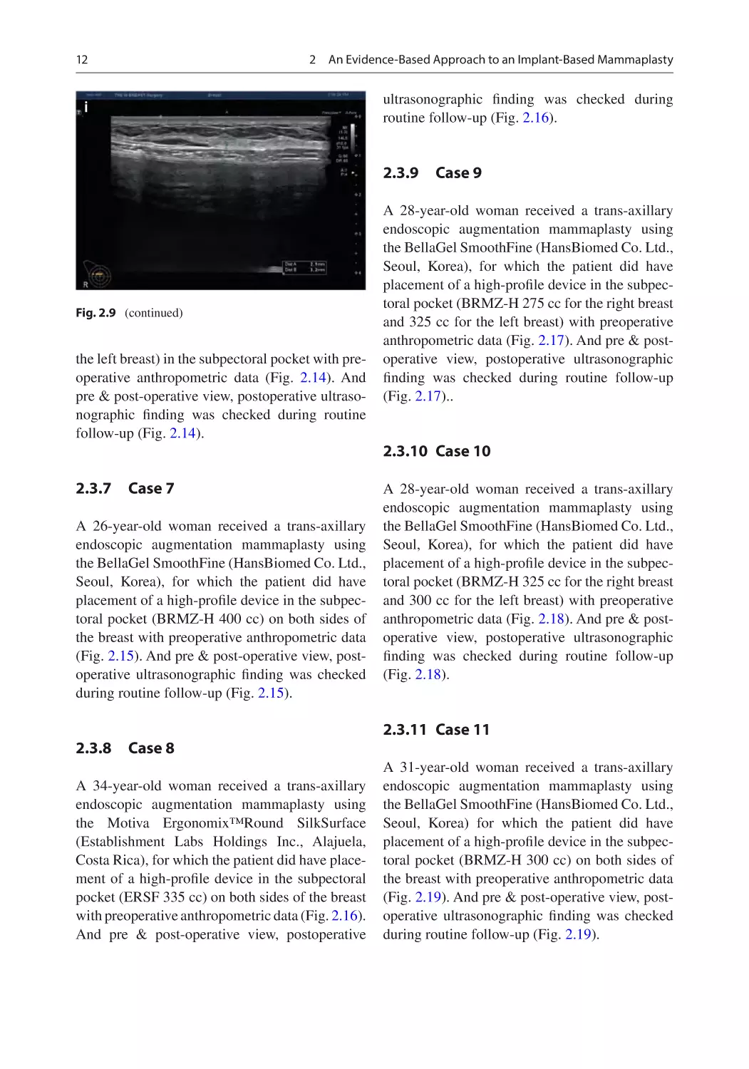
2.3.7 Case 7���������������������������������������������������������������������������� 12
2.3.8 Case 8���������������������������������������������������������������������������� 12
2.3.9 Case 9���������������������������������������������������������������������������� 12
2.3.10 Case 10�������������������������������������������������������������������������� 12
2.3.11 Case 11�������������������������������������������������������������������������� 12
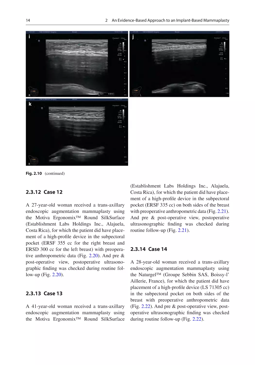
2.3.12 Case 12�������������������������������������������������������������������������� 14
2.3.13 Case 13�������������������������������������������������������������������������� 14
2.3.14 Case 14�������������������������������������������������������������������������� 14
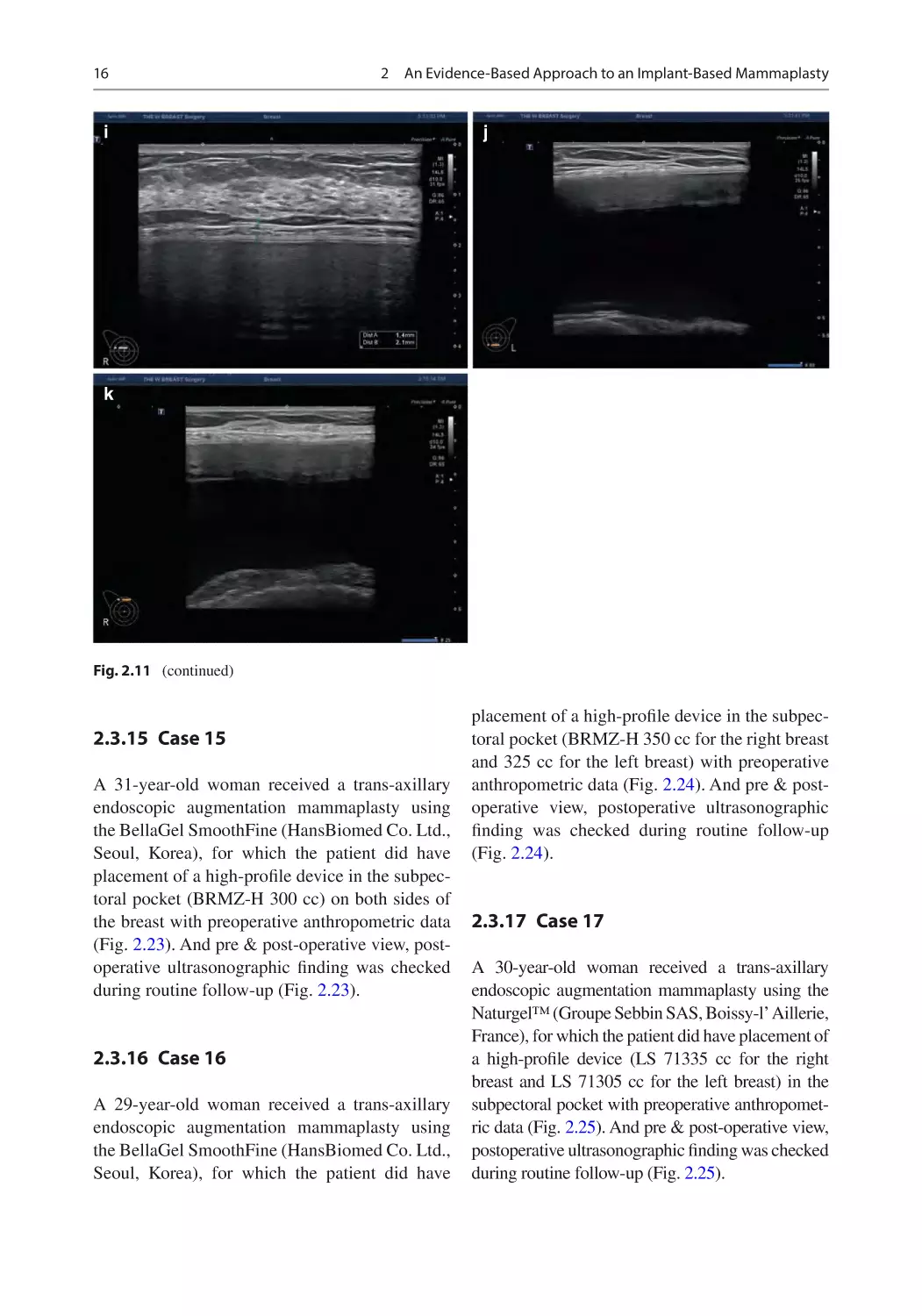
2.3.15 Case 15�������������������������������������������������������������������������� 16
2.3.16 Case 16�������������������������������������������������������������������������� 16
ix
x
Contents
2.3.17 Case 17��������������������������������������������������������������������������
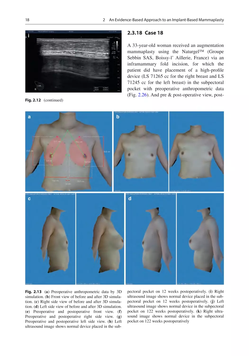
2.3.18 Case 18��������������������������������������������������������������������������
2.3.19 Case 19��������������������������������������������������������������������������
References������������������������������������������������������������������������������������������
3 Role of Ultrasound in the Implant-Based Aesthetic
and Reconstructive Mammaplasty ������������������������������������������������
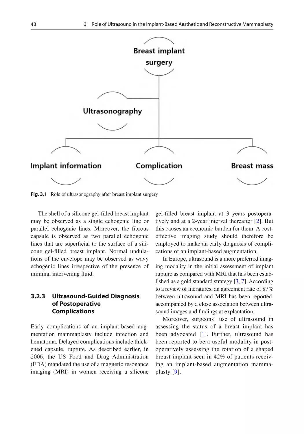
3.1 Overview����������������������������������������������������������������������������������
3.2 Role of Ultrasound in the Context of an Implant-Based
Aesthetic and Reconstructive Mammaplasty����������������������������
3.2.1 Characteristics of a Breast Implant ������������������������������
3.2.2 Ultrasound-Guided Assessment of a Breast Implant����
3.2.3 Ultrasound-Guided Diagnosis of Postoperative
Complications ��������������������������������������������������������������
3.2.4 Ultrasound as a Screening Tool for Patients Who Are
Suspected of Having Breast Implant-Associated
Anaplastic Large Cell Lymphoma (BIA-ALCL)����������
3.2.5 Ultrasound as a Component of a Multi-Disciplinary
Algorithm-Based Approach to an Early Detection of
Complications of an Implant-Based Augmentation
Mammaplasty����������������������������������������������������������������
References������������������������������������������������������������������������������������������
4 Distinguishing Various Types of Breast Implant
Using High-Resolution Ultrasonography��������������������������������������
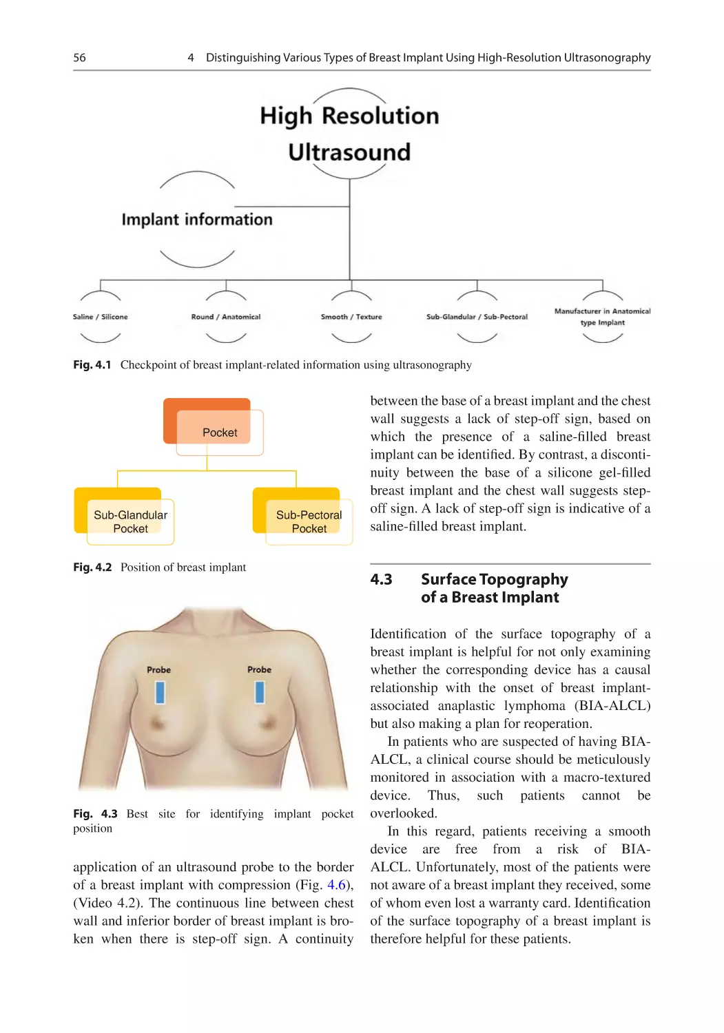
4.1 Location of a Breast Implant����������������������������������������������������
4.2 Constituents of a Breast Implant����������������������������������������������
4.3 Surface Topography of a Breast Implant����������������������������������
4.4 Shape and Manufacturer of a Breast Implant ��������������������������
16
18
41
45
47
47
47
47
47
48
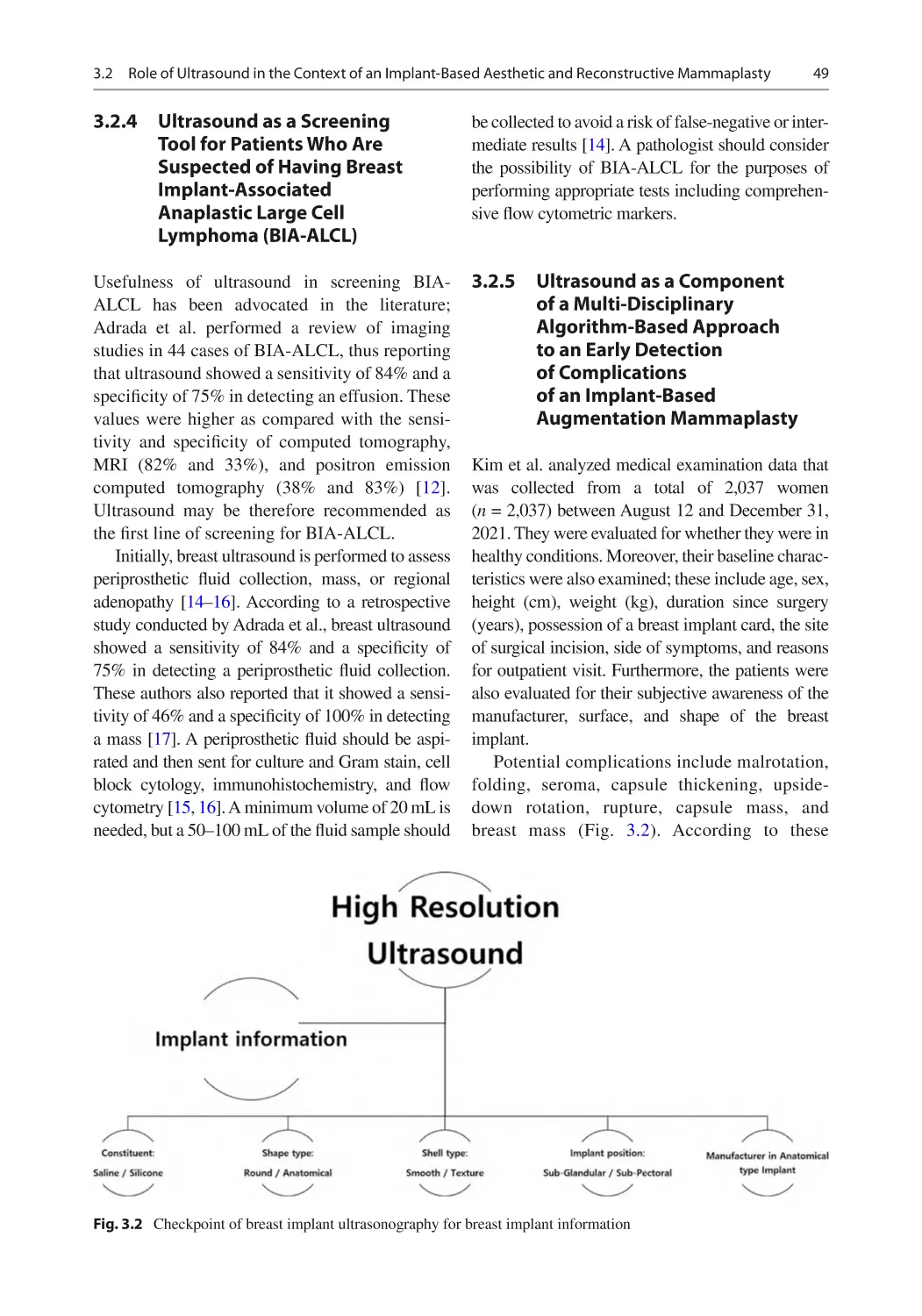
49
49
53
55
55
55
56
60
5 Usefulness of High-Resolution Ultrasound in Detecting
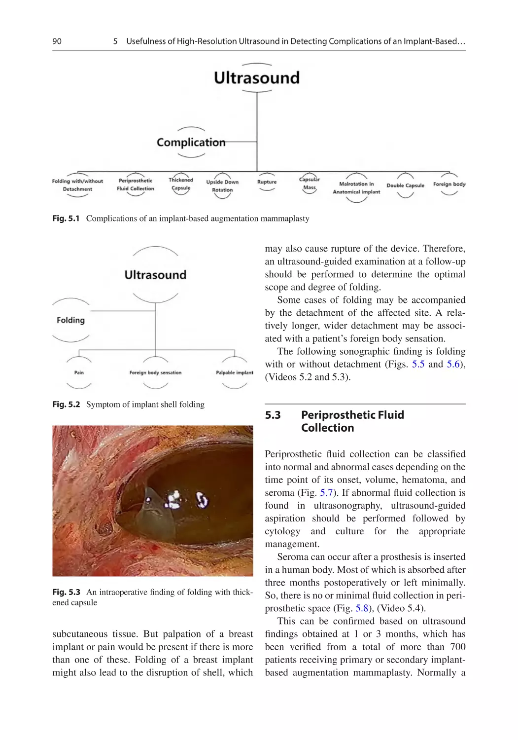
Complications of an Implant-Based Mammaplasty �������������������� 89
5.1 Overview���������������������������������������������������������������������������������� 89
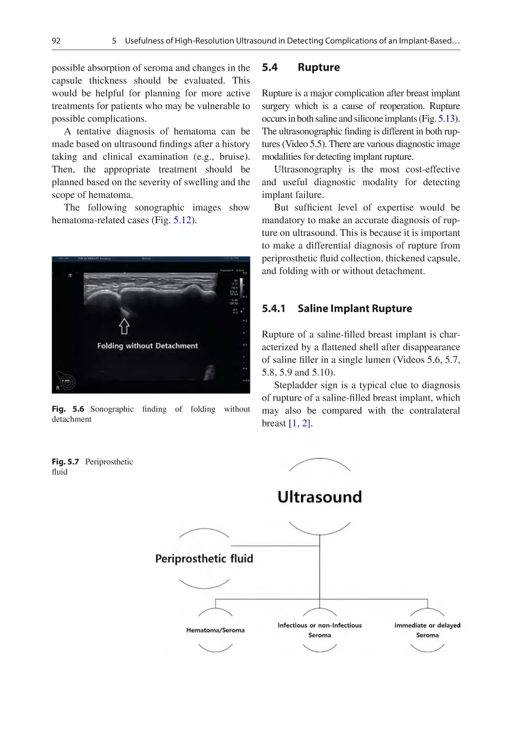
5.2 Folding�������������������������������������������������������������������������������������� 89
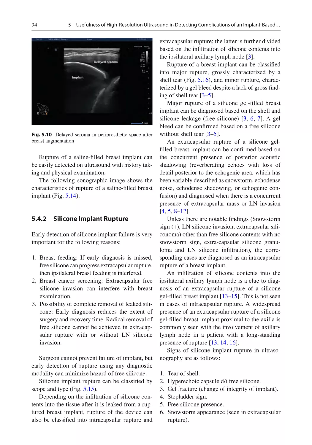
5.3 Periprosthetic Fluid Collection ������������������������������������������������ 90
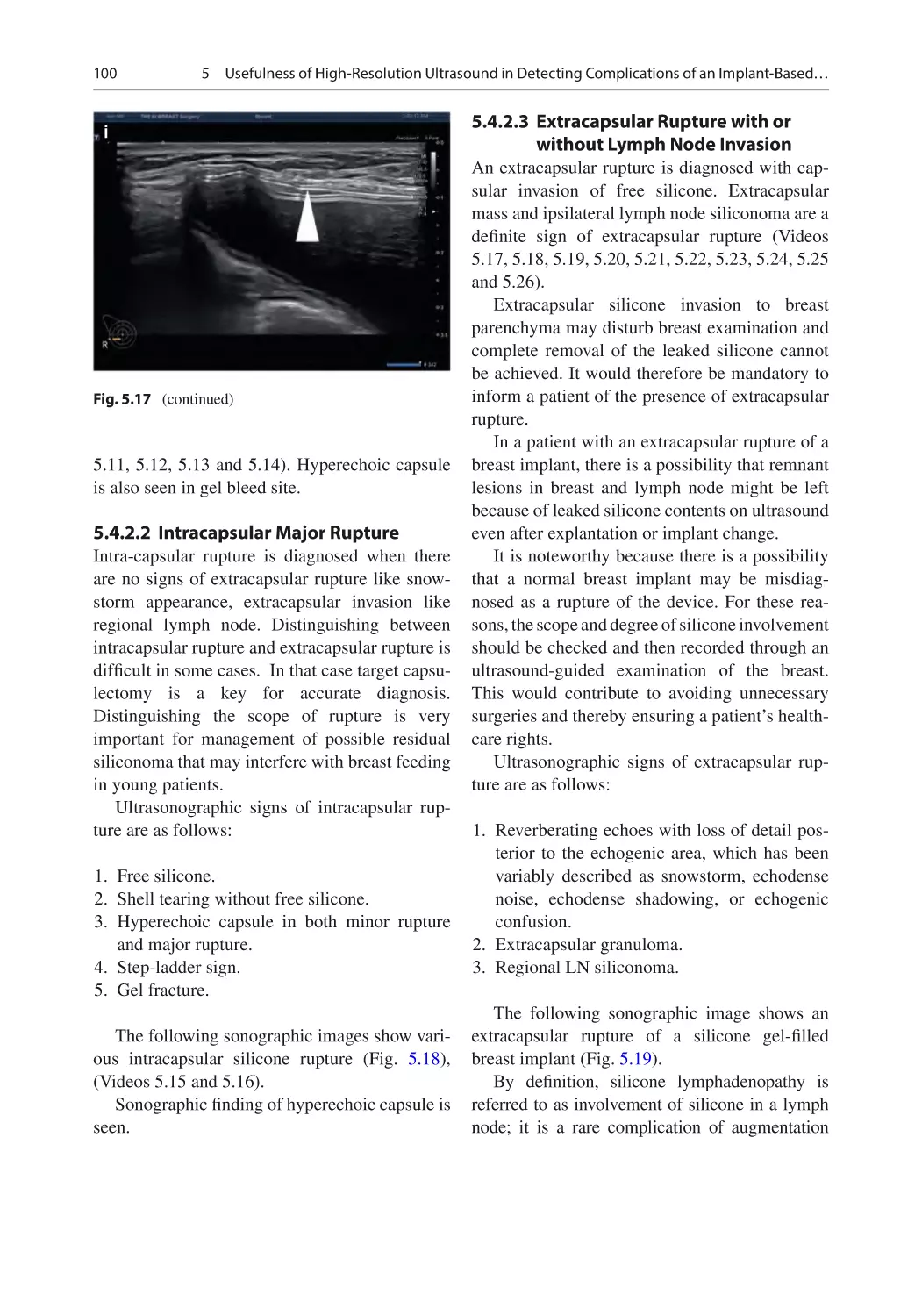
5.4 Rupture�������������������������������������������������������������������������������������� 92
5.4.1 Saline Implant Rupture ������������������������������������������������ 92
5.4.2 Silicone Implant Rupture���������������������������������������������� 94
5.5 Thickened Capsule and Capsular Contracture�������������������������� 101
5.6 Upside-Down (USD) Rotation�������������������������������������������������� 110
References������������������������������������������������������������������������������������������ 122
6 Breast Implant-Associated Anaplastic Large Cell
Lymphoma���������������������������������������������������������������������������������������� 123
6.1 Overview���������������������������������������������������������������������������������� 123
6.2 A Vicious Circle of Crisis of a Breast Implant ������������������������ 123
6.3 Association Between the Onset of BIA-ALCL
and a Textured Implant������������������������������������������������������������� 124
6.4 Approaches to Risk Management for an Early Detection
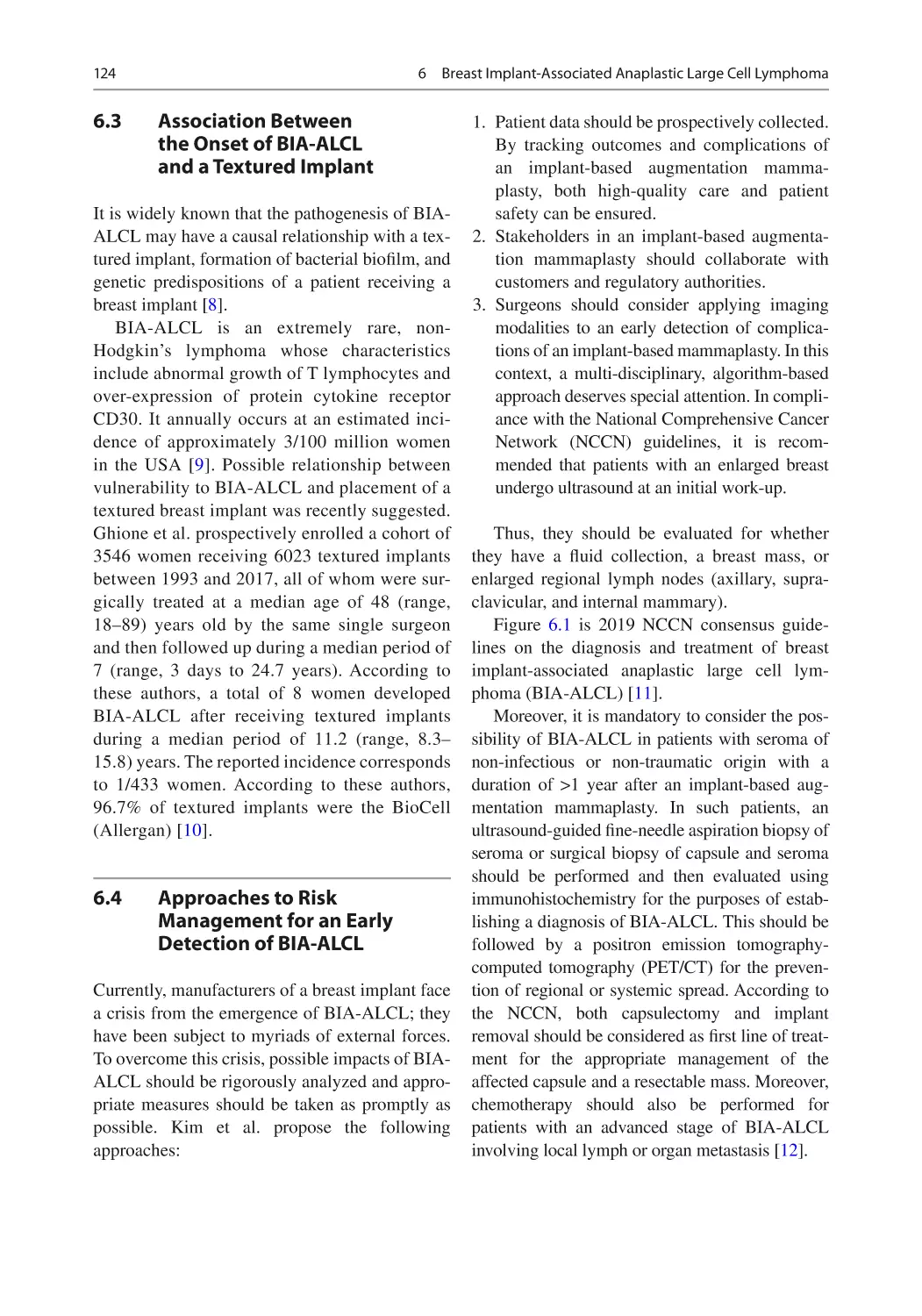
of BIA-ALCL���������������������������������������������������������������������������� 124
Contents
xi
6.5 Conflict of Interest (COI) in Plastic Surgery and Its Possible
Involvement in Crisis of Stakeholders in an Implant-Based
Augmentation Mammaplasty���������������������������������������������������� 125
References������������������������������������������������������������������������������������������ 126
7 Usefulness of High-Resolution Ultrasound (HRUS)
in Planning Revision or Reoperation for Patients Receiving an
Implant-Based Augmentation Mammaplasty������������������������������� 129
7.1 Overview���������������������������������������������������������������������������������� 129
7.2 Preoperative Considerations Based on HRUS
for Reoperation ������������������������������������������������������������������������ 129
7.3 An Algorithm-Based Approach to HRUS-Assisted
Reoperation ������������������������������������������������������������������������������ 131
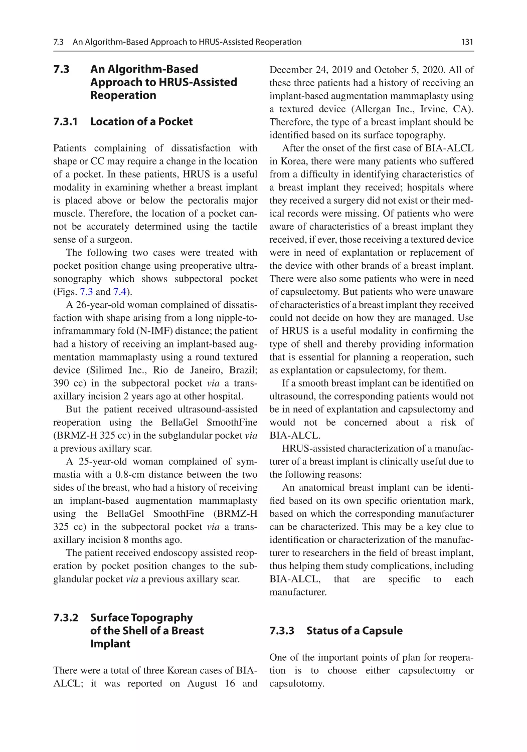
7.3.1 Location of a Pocket ���������������������������������������������������� 131
7.3.2 Surface Topography of the Shell
of a Breast Implant�������������������������������������������������������� 131
7.3.3 Status of a Capsule�������������������������������������������������������� 131
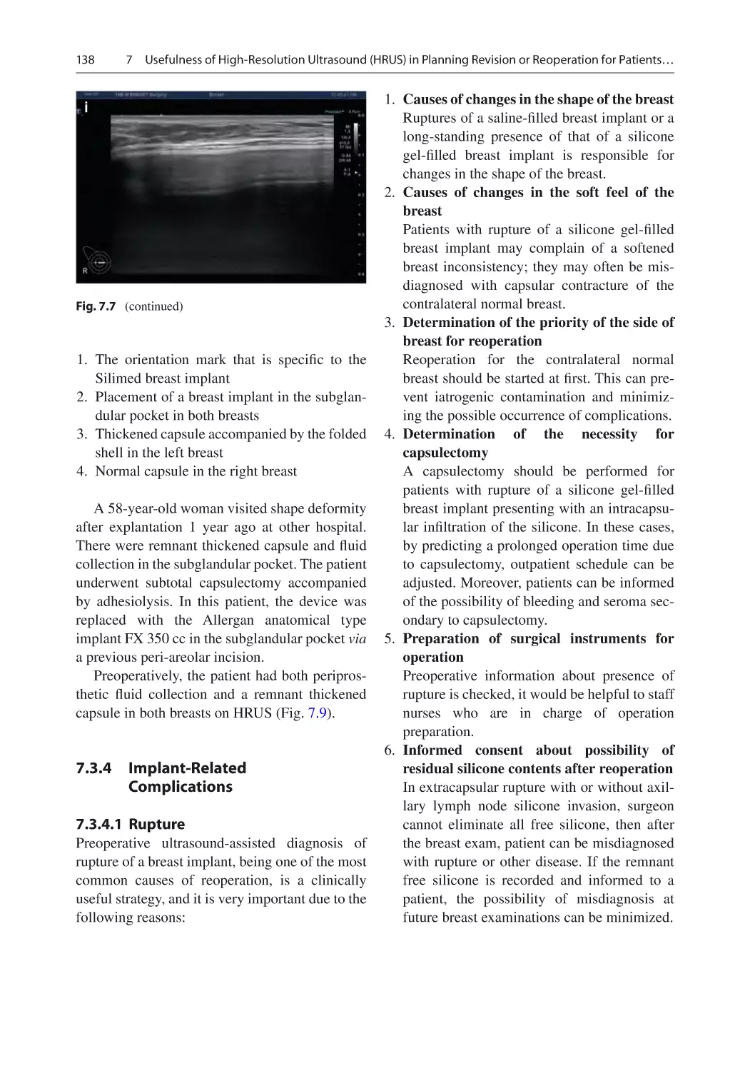
7.3.4 Implant-Related Complications������������������������������������ 138
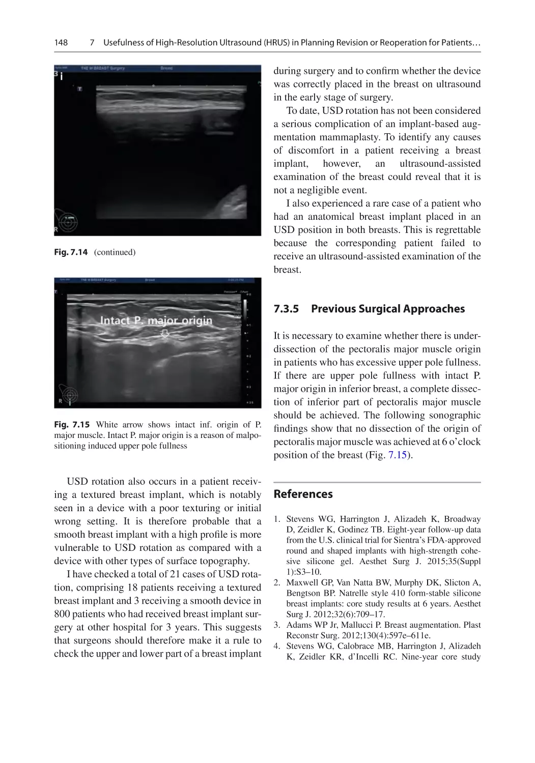
7.3.5 Previous Surgical Approaches�������������������������������������� 148
References������������������������������������������������������������������������������������������ 148
8 Clinical Presentation������������������������������������������������������������������������ 151
8.1 Overview���������������������������������������������������������������������������������� 151
8.2 Illustrative Cases ���������������������������������������������������������������������� 151
8.2.1 Case 1���������������������������������������������������������������������������� 151
8.2.2 Case 2���������������������������������������������������������������������������� 151
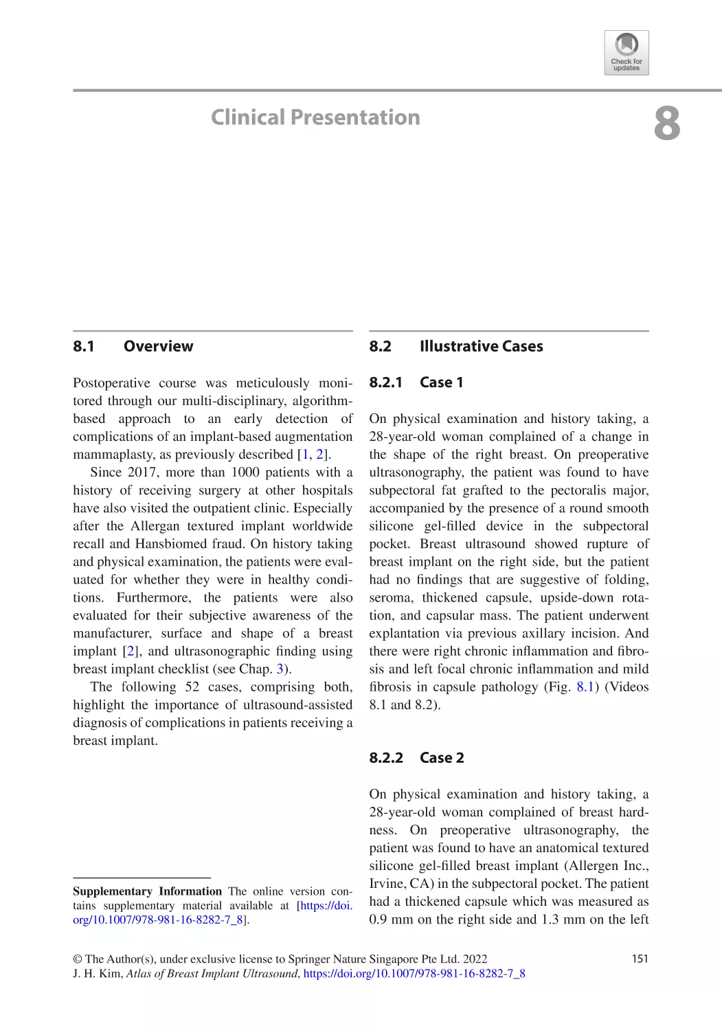
8.2.3 Case 3���������������������������������������������������������������������������� 156
8.2.4 Case 4���������������������������������������������������������������������������� 156
8.2.5 Case 5���������������������������������������������������������������������������� 156
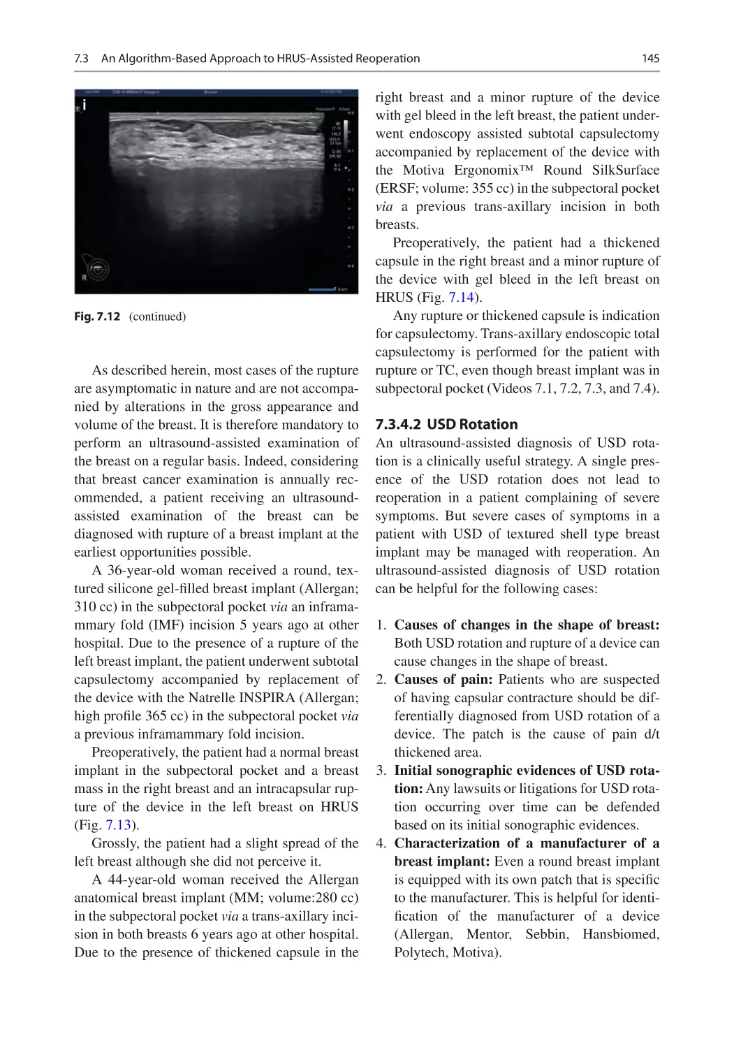
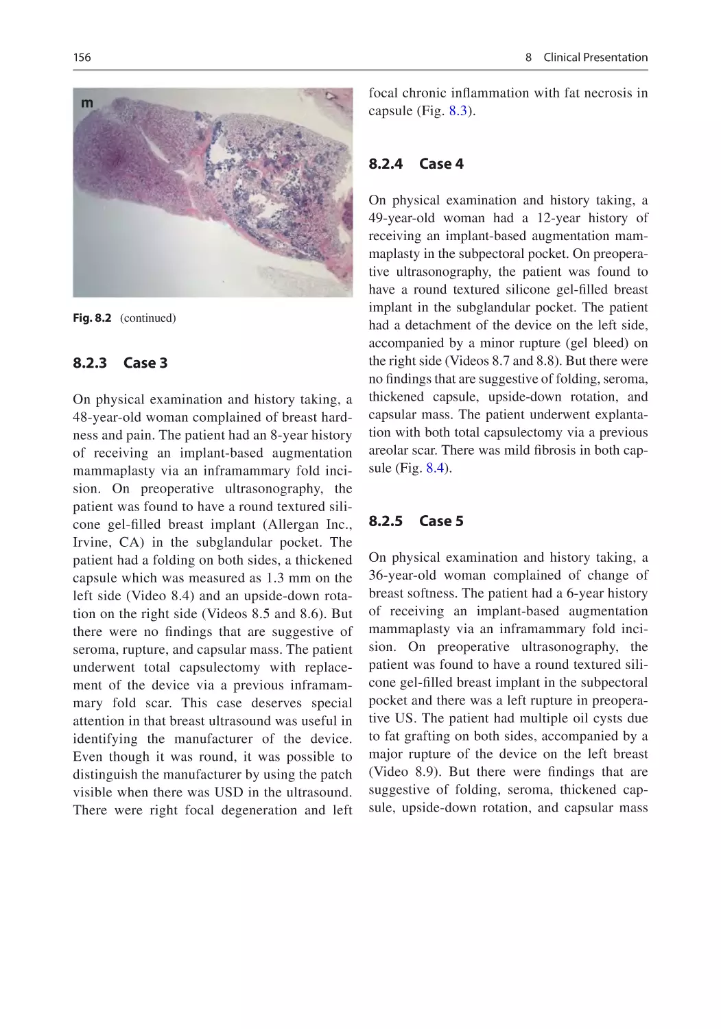
8.2.6 Case 6���������������������������������������������������������������������������� 164
8.2.7 Case 7���������������������������������������������������������������������������� 164
8.2.8 Case 8���������������������������������������������������������������������������� 166
8.2.9 Case 9���������������������������������������������������������������������������� 166
8.2.10 Case 10�������������������������������������������������������������������������� 166
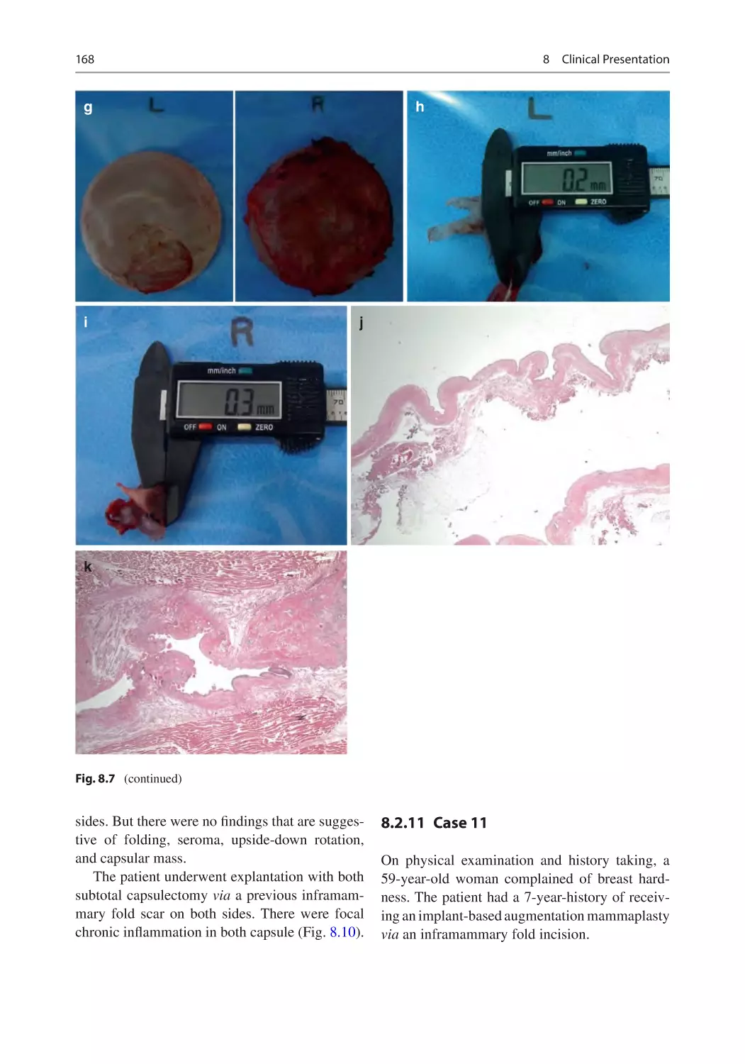
8.2.11 Case 11�������������������������������������������������������������������������� 168
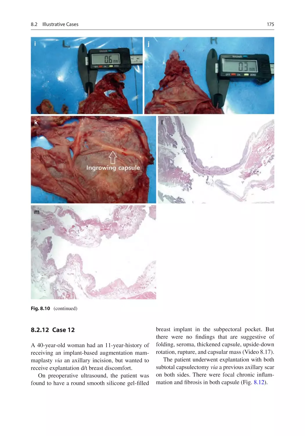
8.2.12 Case 12�������������������������������������������������������������������������� 175
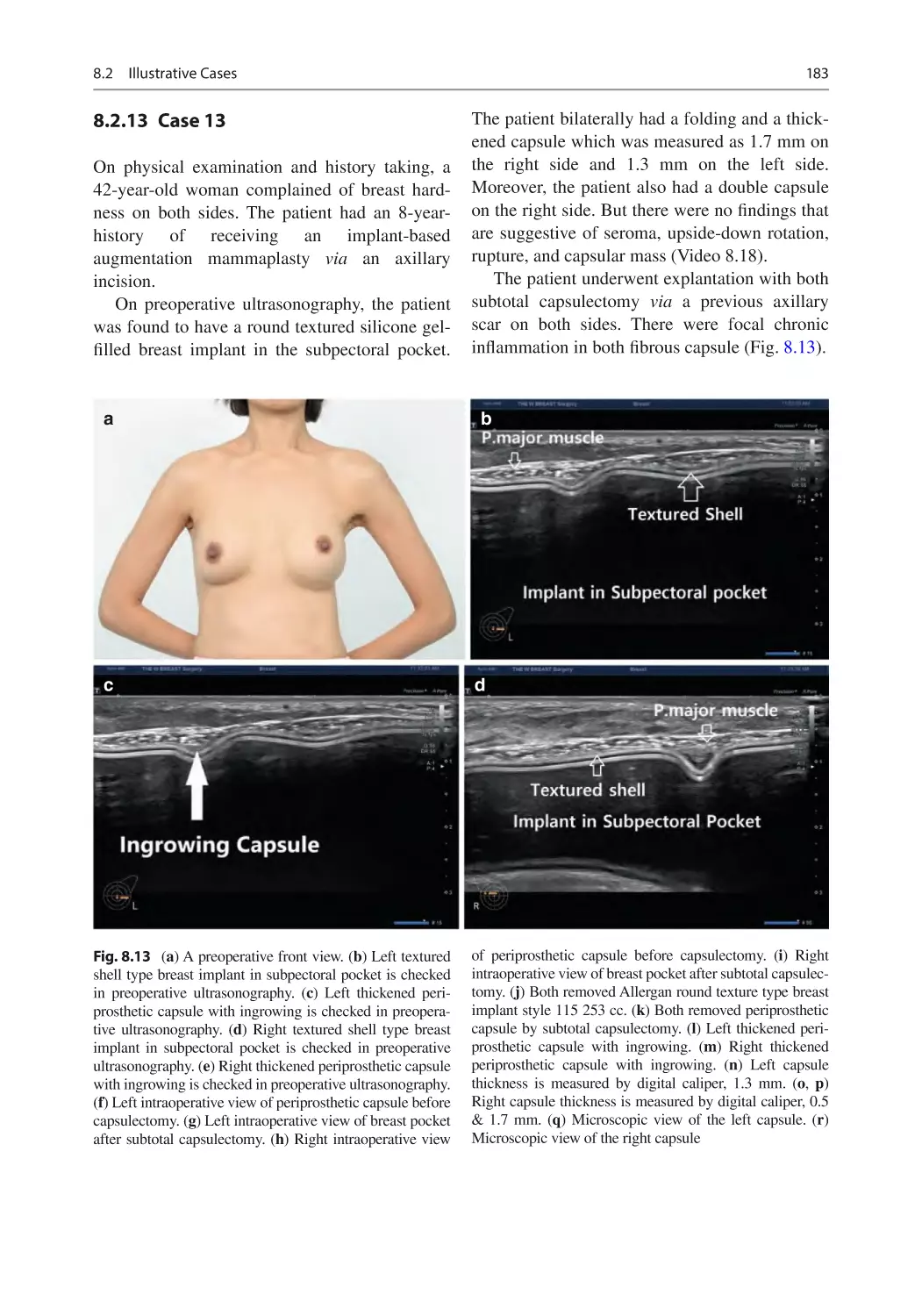
8.2.13 Case 13�������������������������������������������������������������������������� 183
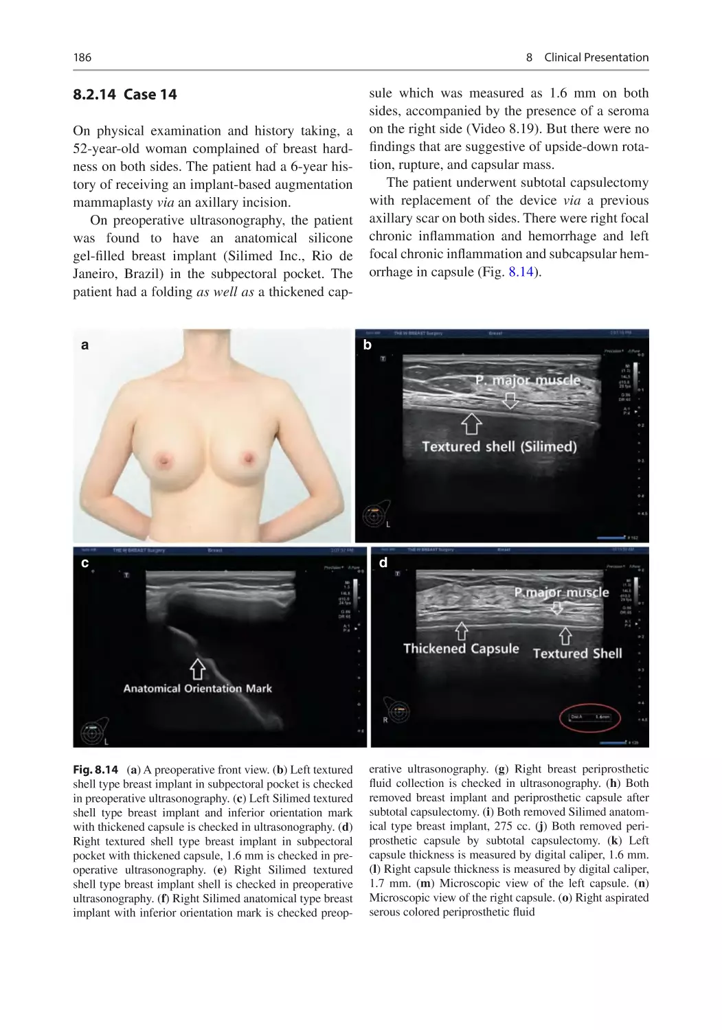
8.2.14 Case 14�������������������������������������������������������������������������� 186
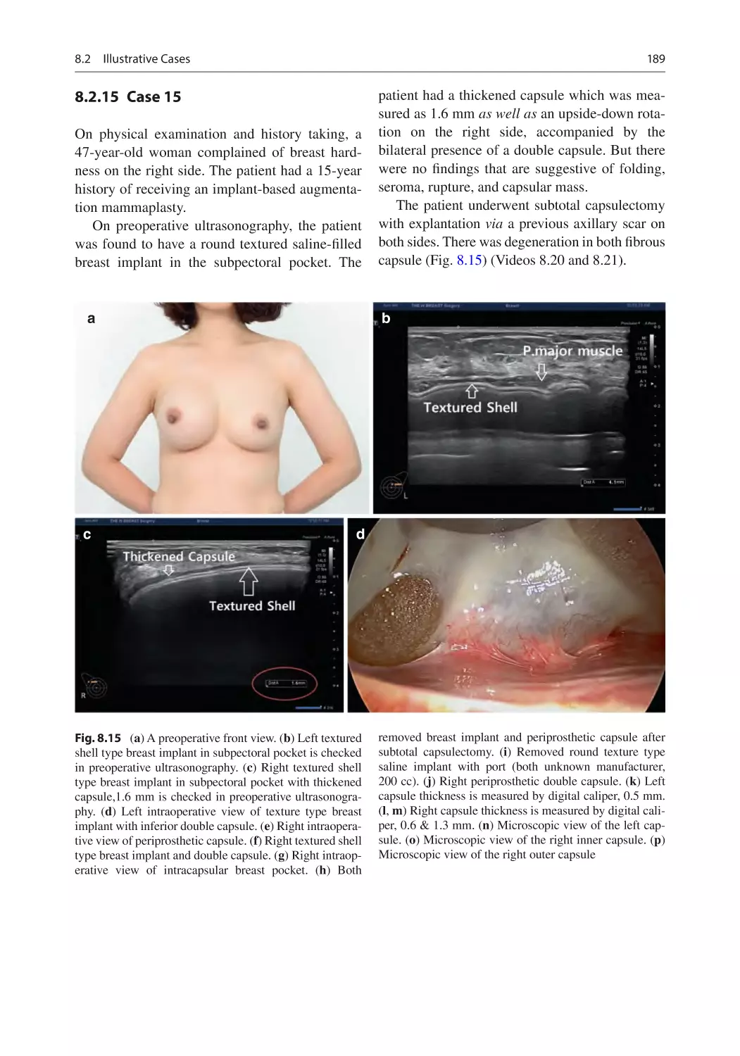
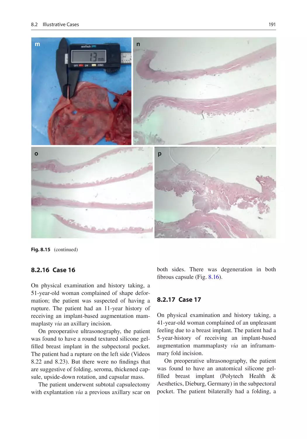
8.2.15 Case 15�������������������������������������������������������������������������� 189
8.2.16 Case 16�������������������������������������������������������������������������� 191
8.2.17 Case 17�������������������������������������������������������������������������� 191
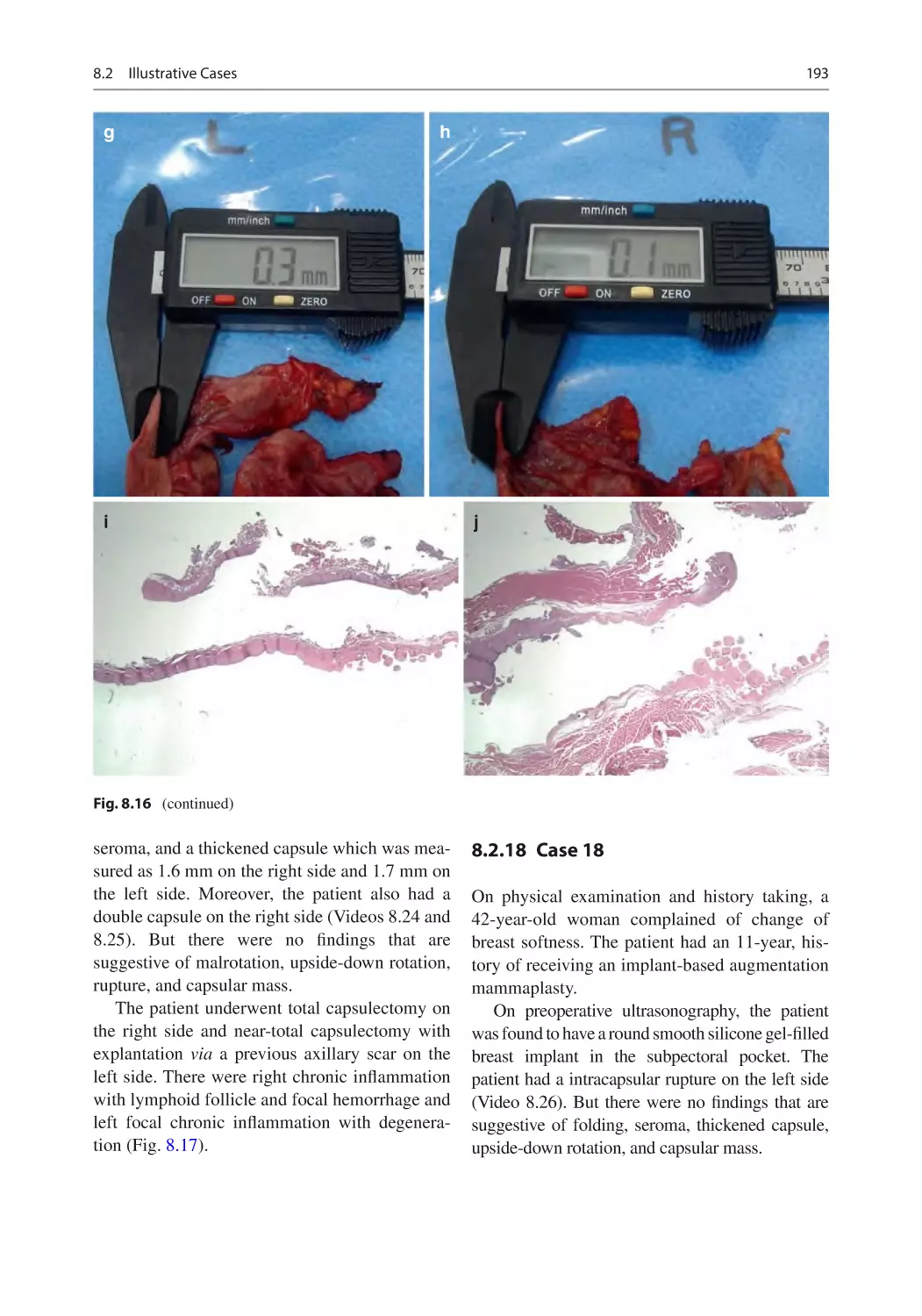
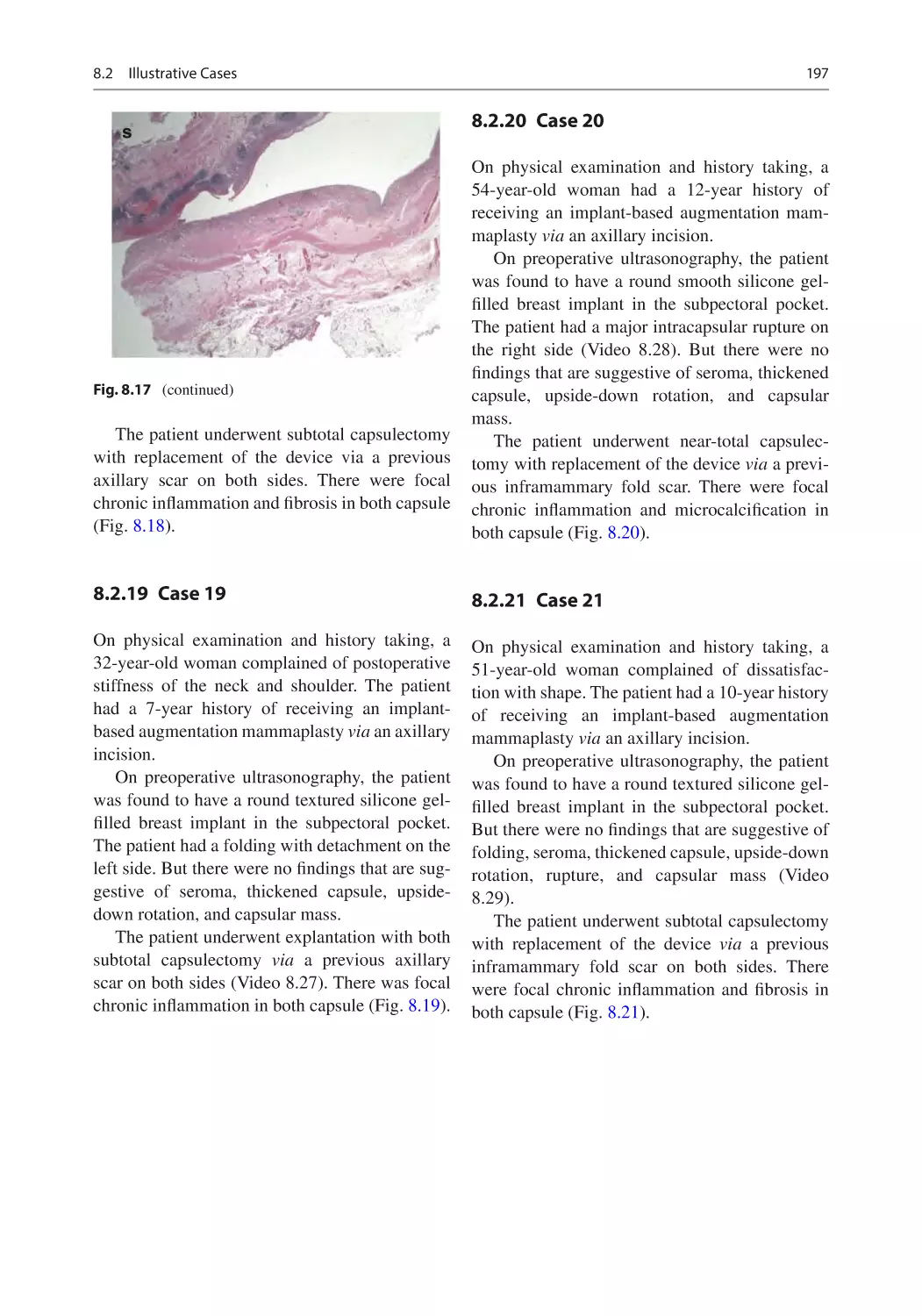
8.2.18 Case 18�������������������������������������������������������������������������� 193
8.2.19 Case 19�������������������������������������������������������������������������� 197
8.2.20 Case 20�������������������������������������������������������������������������� 197
8.2.21 Case 21�������������������������������������������������������������������������� 197
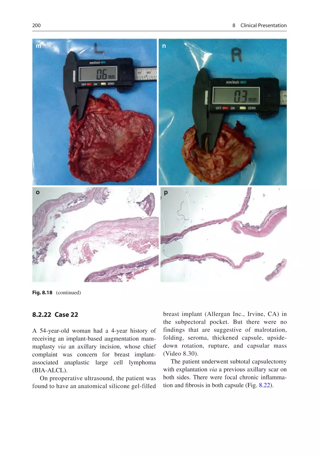
8.2.22 Case 22�������������������������������������������������������������������������� 200
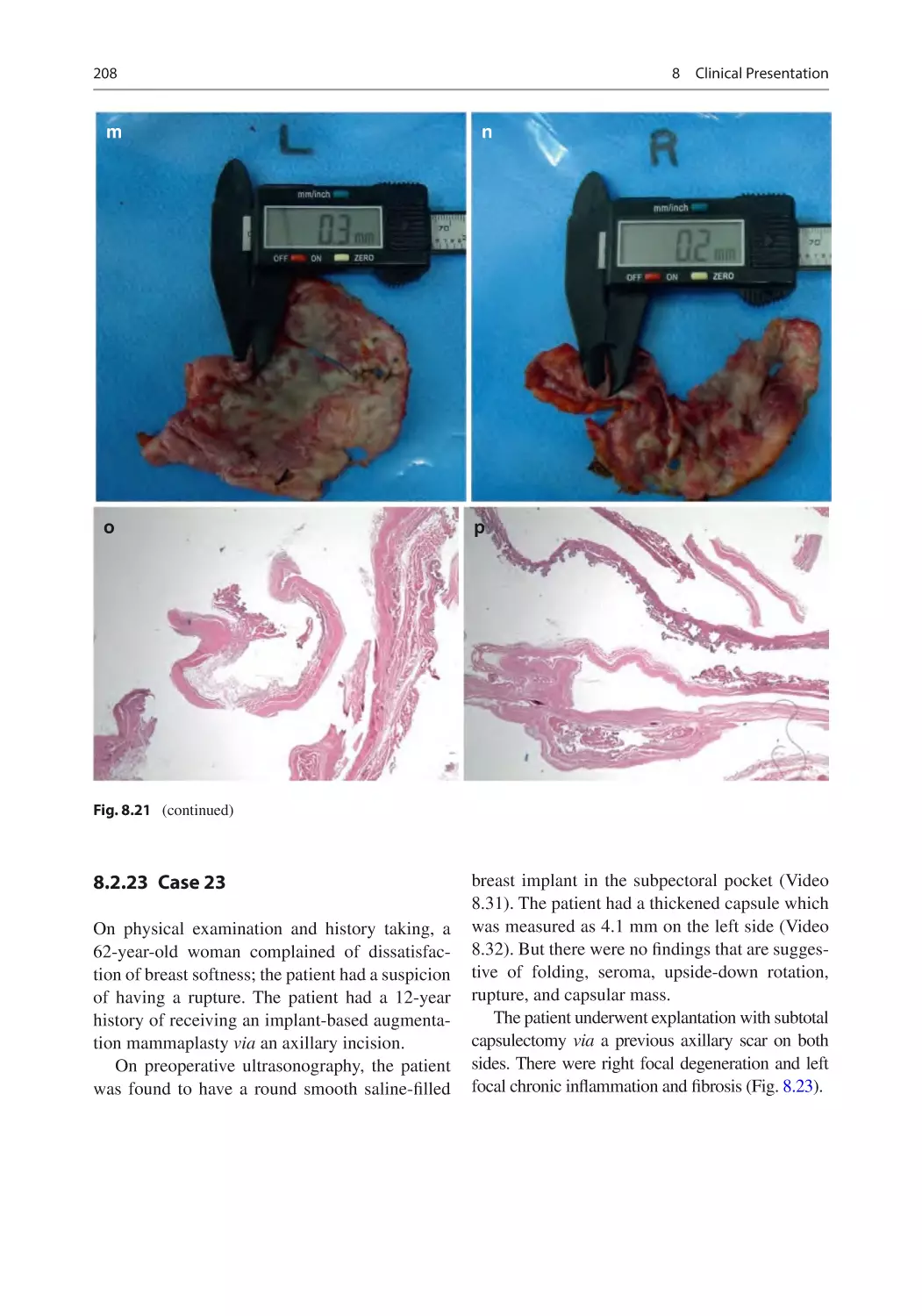
8.2.23 Case 23�������������������������������������������������������������������������� 208
8.2.24 Case 24�������������������������������������������������������������������������� 214
8.2.25 Case 25�������������������������������������������������������������������������� 214
xii
Contents
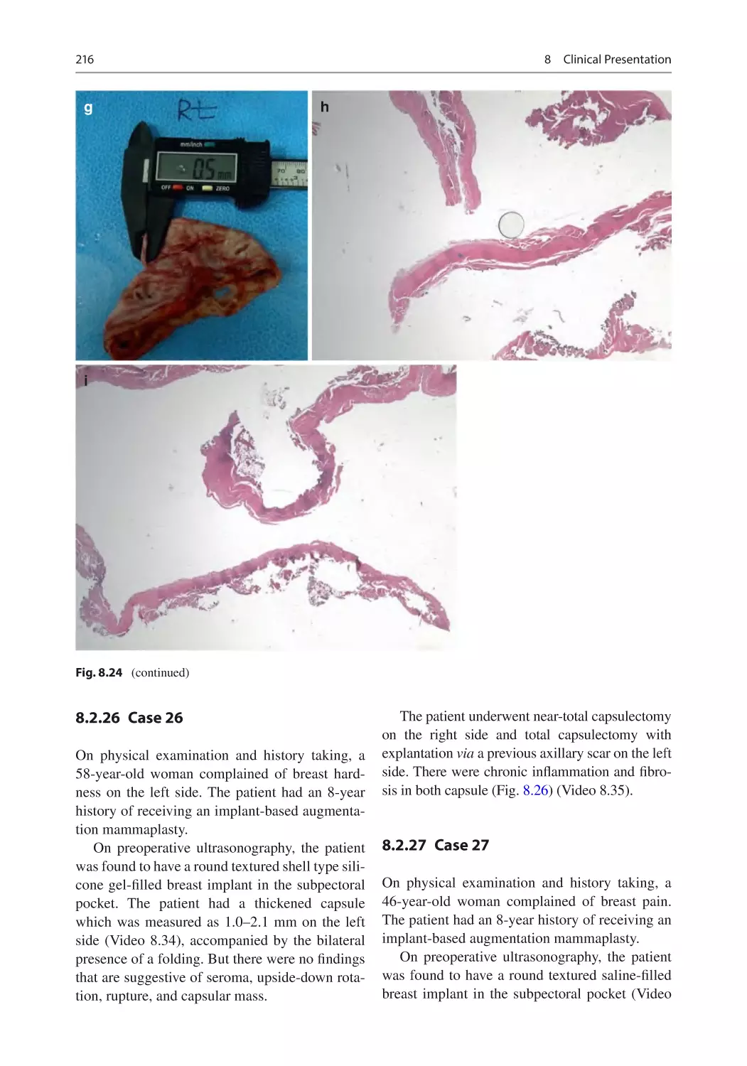
8.2.26 Case 26�������������������������������������������������������������������������� 216
8.2.27 Case 27�������������������������������������������������������������������������� 216
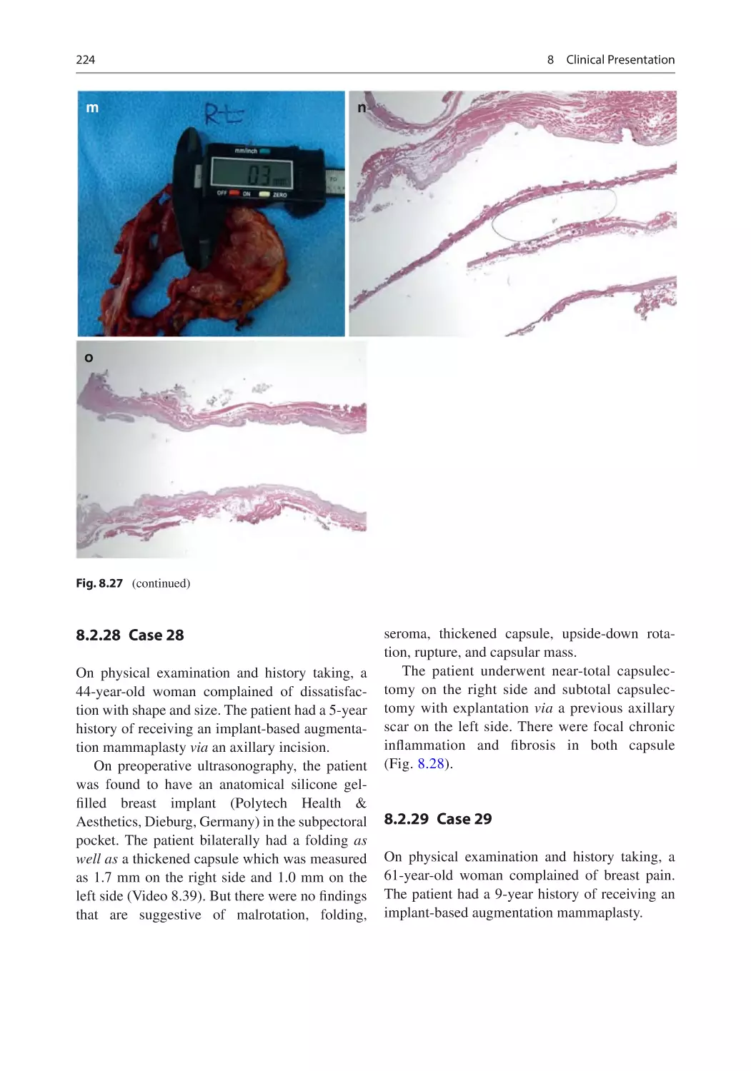
8.2.28 Case 28�������������������������������������������������������������������������� 224
8.2.29 Case 29�������������������������������������������������������������������������� 224
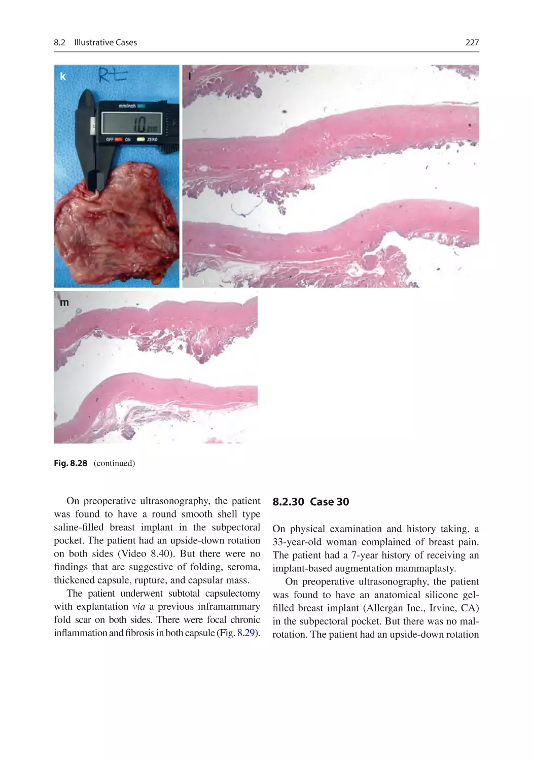
8.2.30 Case 30�������������������������������������������������������������������������� 227
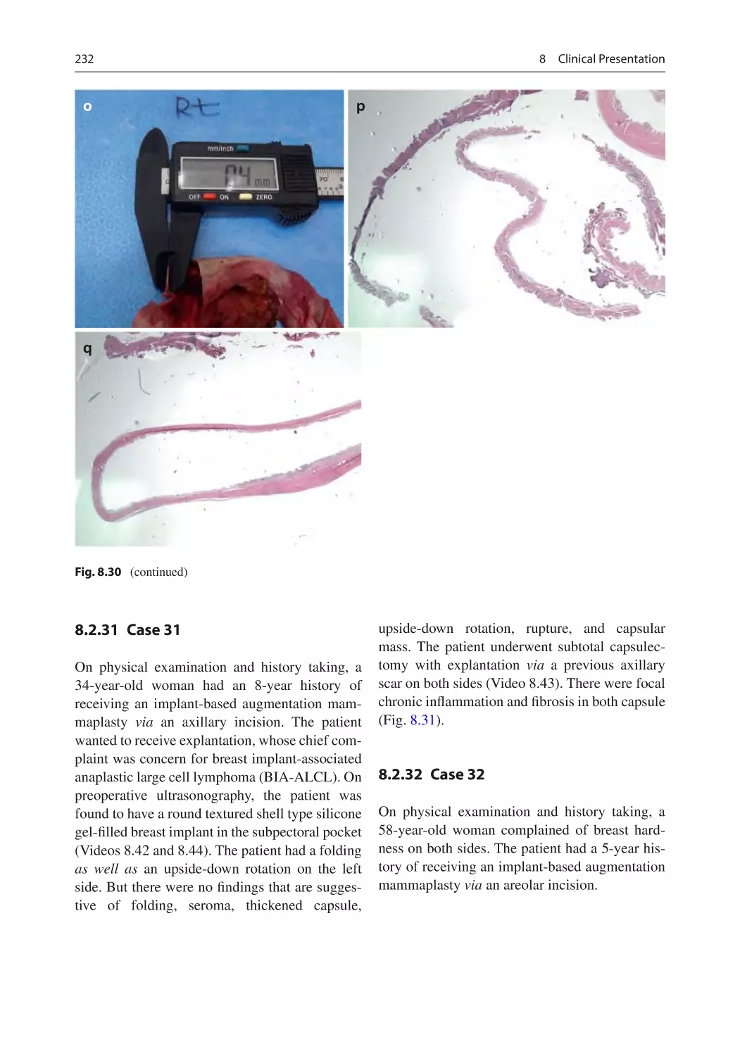
8.2.31 Case 31�������������������������������������������������������������������������� 232
8.2.32 Case 32�������������������������������������������������������������������������� 232
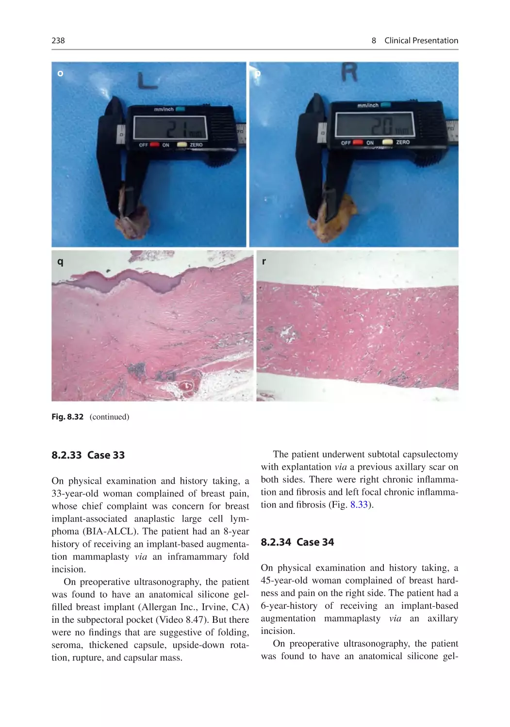
8.2.33 Case 33�������������������������������������������������������������������������� 238
8.2.34 Case 34�������������������������������������������������������������������������� 238
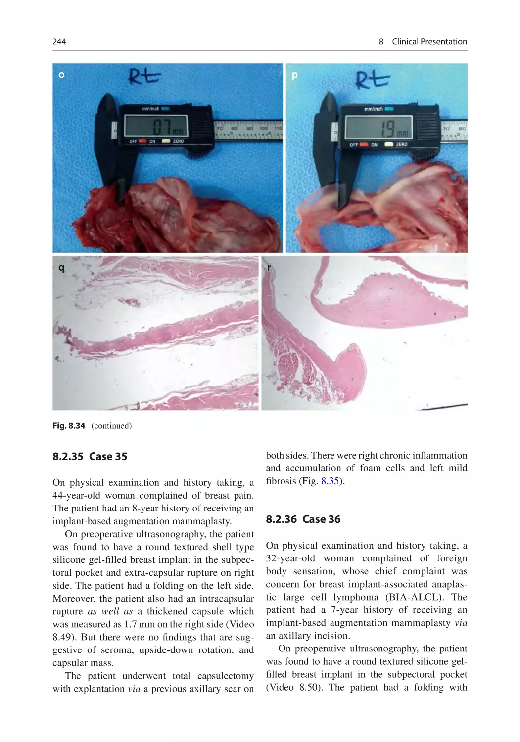
8.2.35 Case 35�������������������������������������������������������������������������� 244
8.2.36 Case 36�������������������������������������������������������������������������� 244
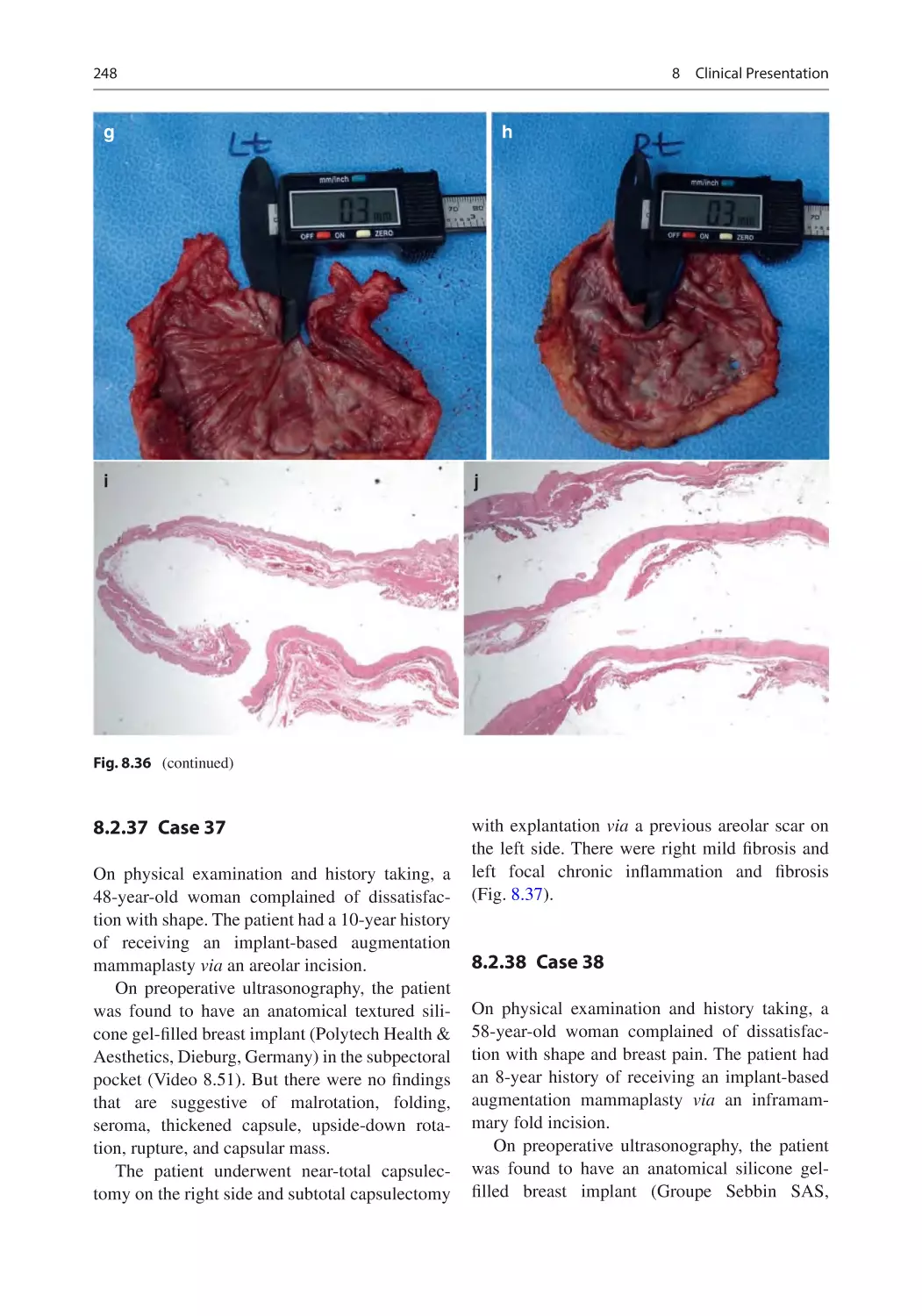
8.2.37 Case 37�������������������������������������������������������������������������� 248
8.2.38 Case 38�������������������������������������������������������������������������� 248
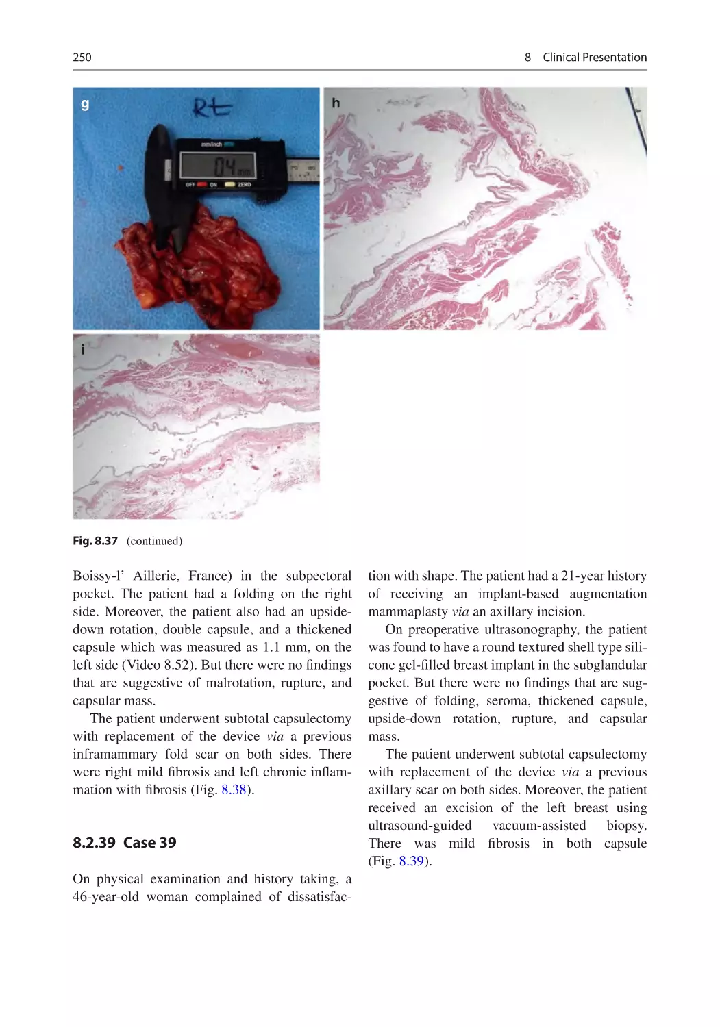
8.2.39 Case 39�������������������������������������������������������������������������� 250
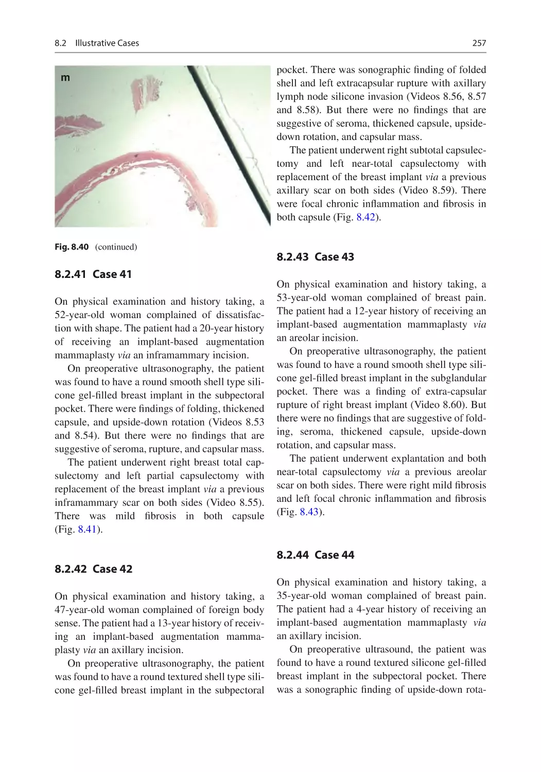
8.2.40 Case 40�������������������������������������������������������������������������� 254
8.2.41 Case 41�������������������������������������������������������������������������� 257
8.2.42 Case 42�������������������������������������������������������������������������� 257
8.2.43 Case 43�������������������������������������������������������������������������� 257
8.2.44 Case 44�������������������������������������������������������������������������� 257
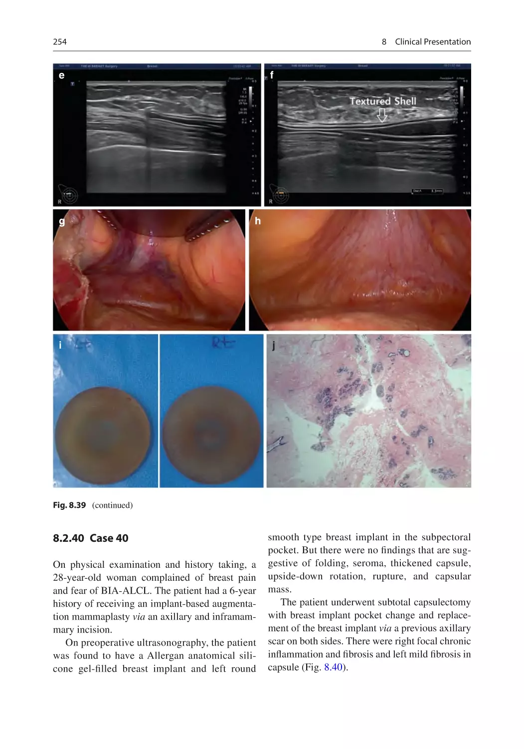
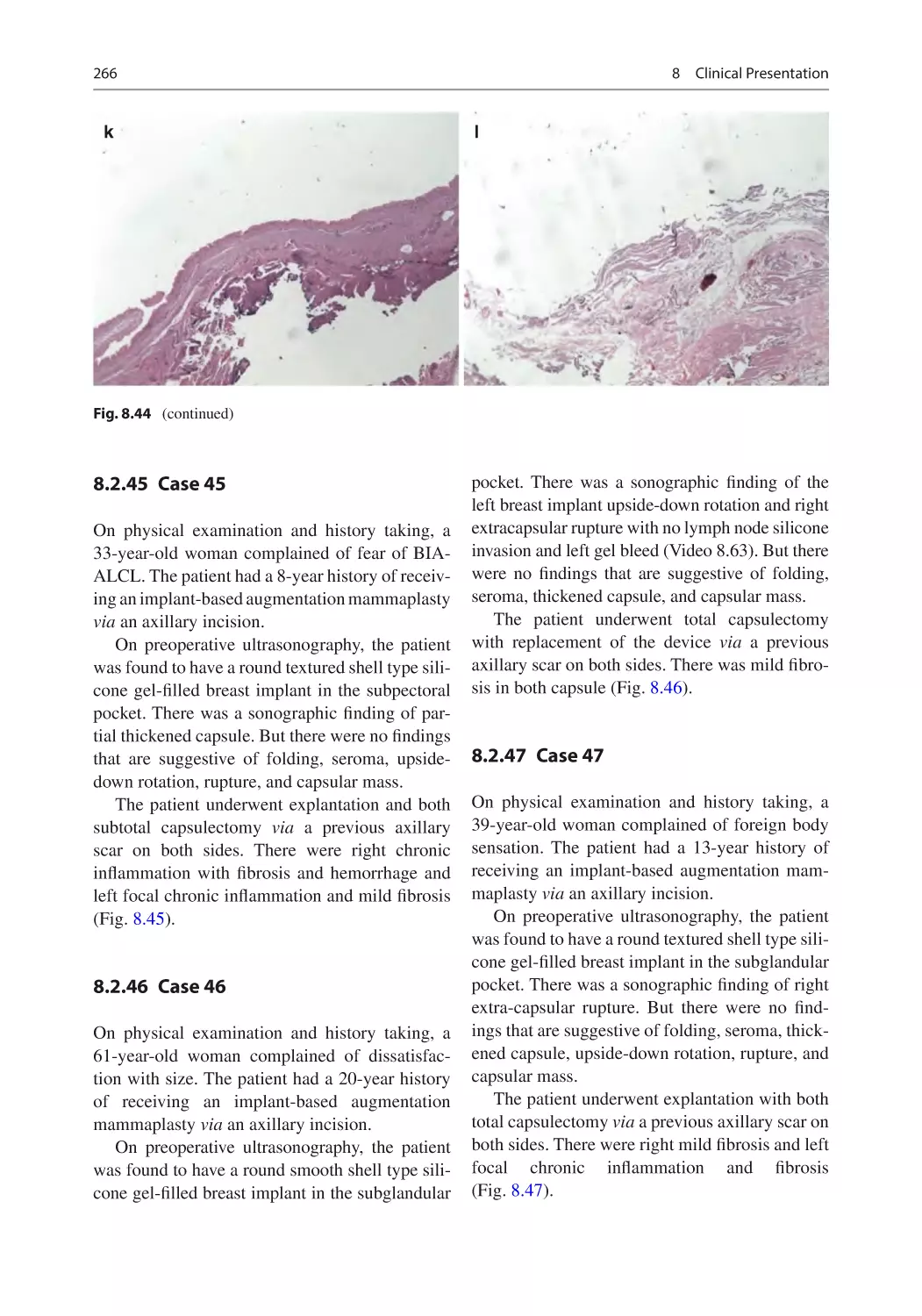
8.2.45 Case 45�������������������������������������������������������������������������� 266
8.2.46 Case 46�������������������������������������������������������������������������� 266
8.2.47 Case 47�������������������������������������������������������������������������� 266
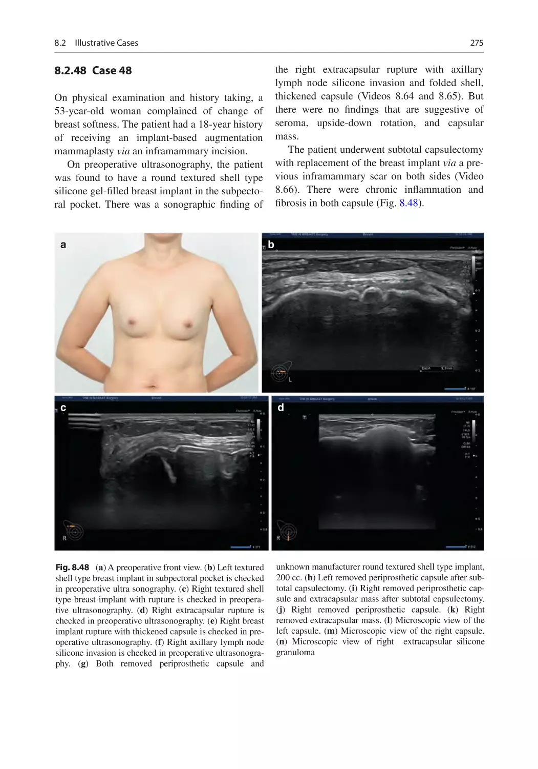
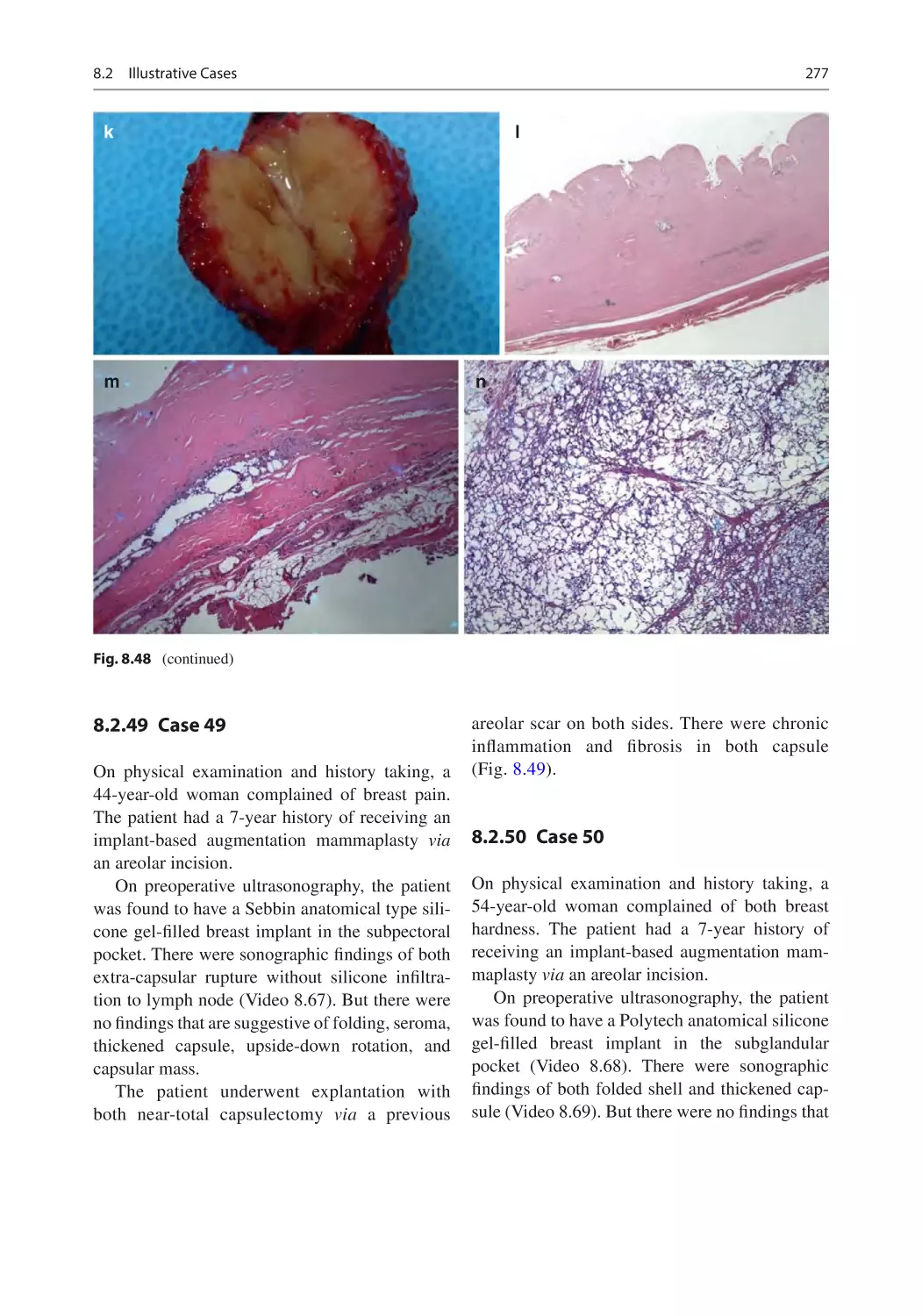
8.2.48 Case 48�������������������������������������������������������������������������� 275
8.2.49 Case 49�������������������������������������������������������������������������� 277
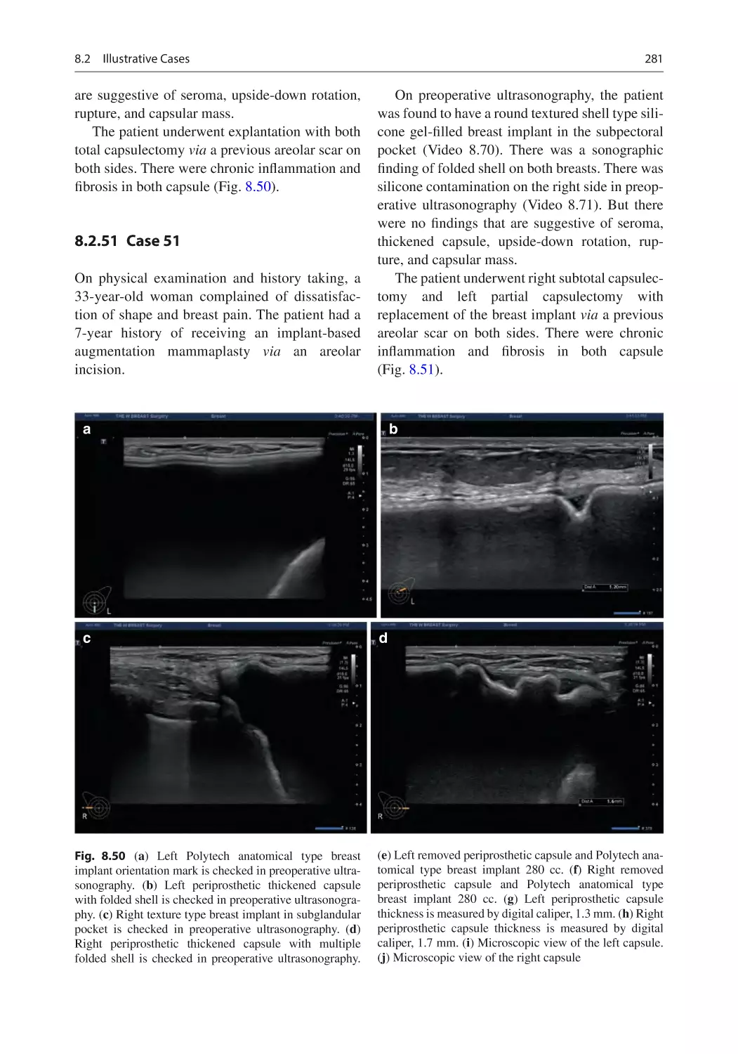
8.2.50 Case 50�������������������������������������������������������������������������� 277
8.2.51 Case 51�������������������������������������������������������������������������� 281
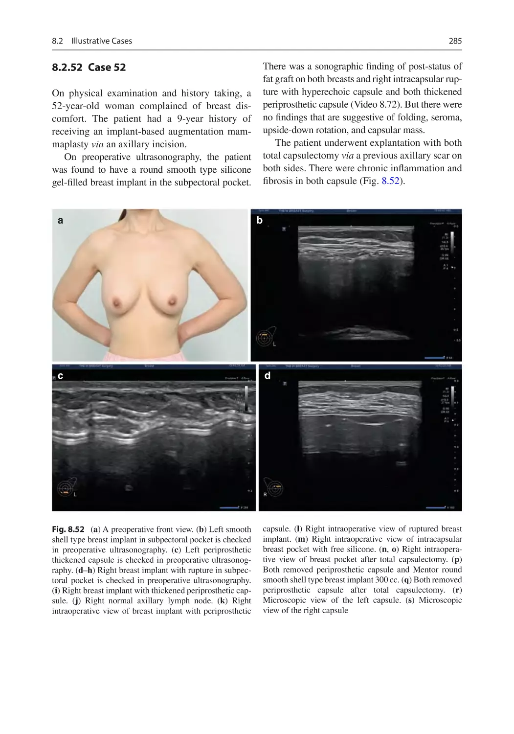
8.2.52 Case 52�������������������������������������������������������������������������� 285
References������������������������������������������������������������������������������������������ 288
9 Conclusions�������������������������������������������������������������������������������������� 289
References������������������������������������������������������������������������������������������ 291
1
Current Status and Future
Implications of Ultrasound
in the Context of Implant-Based
Breast Aesthetic
and Reconstructive Surgery
1.1
Overview
Breast is a symbol of feminity that serves as a key
factor of quality of life [1]. Implant-based augmentation mammaplasty is performed for the
purposes of enlarging a normal breast and reconstructing an absent or abnormal breast [2, 3]. It
remains as one of the most popular procedures in
the setting of aesthetic and reconstructive plastic
surgery. In the USA in 2018, a total of 313,000
procedures were performed. Moreover, it has
also been reported that a total of 1,862,506 procedures were performed worldwide [4].
Still, controversial opinions exist regarding the
safety of silicone gel-filled breast implants. This
deserves special attention [5]. In more detail, there
has been an extensive debate on the safety of breast
implants, classified as a type III medical device,
since they were first introduced in the 1960s; their
use in cosmetic surgery was transiently prohibited
by the US Food and Drug Administration (FDA)
between 1992 and 2006, but it was approved by
the FDA in 2006 on condition that it would be
under a long-term follow-
up, further analyzed
after explanted and released with more detailed
labeling [6, 7]. Nevertheless, a causal relationship
of silicone gel-filled breast implants with postoperative complications, such as cancer, autoimmune
diseases, and connective tissue diseases, has not
been established, as described by a systematic
review and meta-analyses of previously published
studies in this series [7, 8].
Concerns for possible health risks after augmentation mammaplasty arose from earlier case
reports about women exhibiting connective tissue
disease after receiving breast implants or silicone
injections [9, 10]. Such a key public health issue
should be answered with scientific efforts rather
than anti-scientific and irrational methods; it
should be handled with evidence-based scientific
grounds [11].
On the other hand, there are also some concerns that local complications may occur following the use of silicone gel-filled breast implants;
its major drawbacks include capsular contracture
(CC) and a loss of the implant integrity [12, 13].
Thus, safety concerns have shifted from systemic
adverse effects to local complications that are
closely associated with the primary safety profile
of silicone gel-filled breast implants, as described
in a report published by the Institute of Medicine
of the National Academy of Science [14].
Potential postoperative complications include
CC, implant malposition, breast deformation and
asymmetry, wound and skin problems, infection,
hematoma and hemorrhage, implant rupture,
seroma, abscess, silicone granuloma, and implant
extrusion [15–19]. But the incidence, severity,
and long-term sequelae of these local complications have been studied only in a limited scope
[20–24]. Moreover, differences in study design
and methodology have made it difficult to directly
compare the results between the studies in this
series [25].
© The Author(s), under exclusive license to Springer Nature Singapore Pte Ltd. 2022
J. H. Kim, Atlas of Breast Implant Ultrasound, https://doi.org/10.1007/978-981-16-8282-7_1
1
2
1.2
1
Current Status and Future Implications of Ultrasound in the Context of Implant-Based Breast Aesthetic…
iagnostic Imaging Studies
D
in Patients Receiving
a Breast Implant
Patients receiving a breast implant should be
evaluated with diagnostic imaging studies for
several reasons: these include (1) regular screening mammography, (2) additional imaging for
Breast Imaging. Reporting and Data System
(BI-RADS) 0, 3, 4, and 5 lesions, (3) interventional biopsy, (4) certain types of complications
of an implant-based augmentation mammaplasty
(e.g., impaired immune response, connective tissue disorders, or BIA-ALCL), and (5) leakage or
rupture of a breast implant [26–28].
Surgeons often encounter challenges arising
from a breast implant; they should identify and
distinguish shell types of a breast implant, make a
diagnosis of implant failure or other complications of an implant-based augmentation mammaplasty, and perform a regular follow-up [29]. It
has been reported that patients receiving a breast
implant are vulnerable to mortality from malignant conditions [30]. This suggests that diagnostic
imaging studies have their own merits and demerits in detecting complications of an implant-based
augmentation mammaplasty [31]. It can therefore
be concluded that novel imaging studies should
apply to the diagnosis of complications of an
implant-based augmentation mammaplasty.
breast implant are evaluated with magnetic resonance imaging (MRI) scans at three years postoperatively and at a 2-year interval thereafter
[32]. Thus, efforts have been made to detect complications of an implant-based augmentation
mammaplasty, such as capsular contracture and
rupture imaging studies [33]. Since then, the use
of MRI has been considered a gold standard for
the non-invasive assessment of a breast implant
[34]. In addition to its disadvantages including
high cost as well as inconvenience, however, its
sensitivity and specificity for detecting implant
failure have been reported to show discrepancies
between the radiologists [35–37]. Further, it
remains questionable whether an MRI can also
be used to detect implant failure in asymptomatic
patients receiving the fourth and fifth generation
of device [38].
A US has also been used to detect implant failure, although it has been established as an alternative to an MRI [35, 39–45]. With technological
advancements, its specifications have been much
improved. As a non-invasive, cost-effective diagnostic modality for patients receiving a breast
implant, it deserves special attention [46].
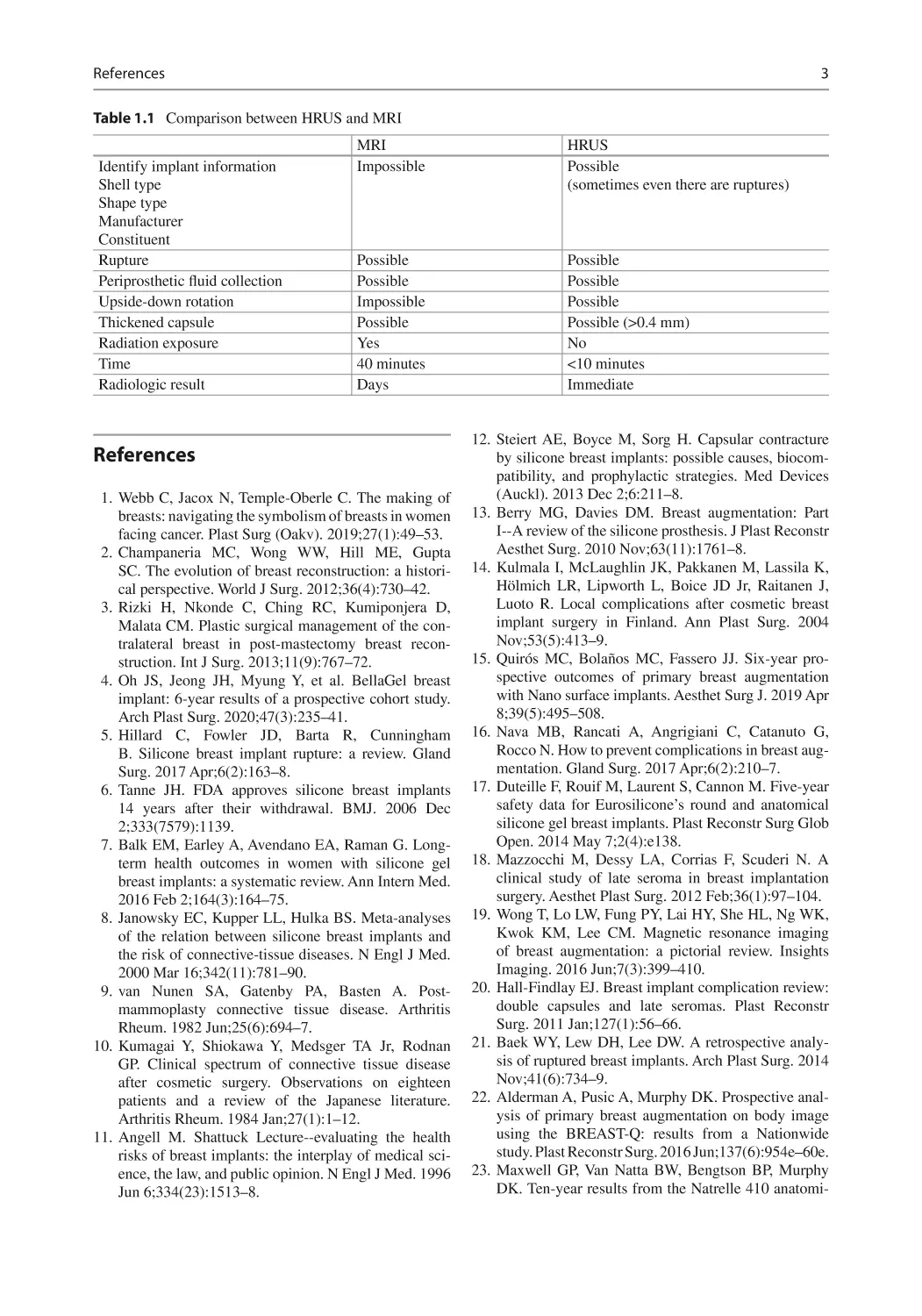
1.4
Comparison of HRUS
and MRI
HRUS has many advantages in identifying various breast implant types and measurement of
1.3
The Emerging Value
thickened capsule and complications like upside-
down rotation.
of Ultrasound as a Novel
The biggest advantage of HRUS are the ability
Diagnostic Modality
to diagnose side effects and excellence in distinSince the US FDA approval of a silicone gel- guishing side effects.
filled breast implant, dated November of 2006, it
So, HRUS is an effective diagnostic image
has been recommended that patients receiving a modality than MRI (Table 1.1).
References
3
Table 1.1 Comparison between HRUS and MRI
Identify implant information
Shell type
Shape type
Manufacturer
Constituent
Rupture
Periprosthetic fluid collection
Upside-down rotation
Thickened capsule
Radiation exposure
Time
Radiologic result
2.
3.
4.
5.
6.
7.
8.
9.
10.
11.
HRUS
Possible
(sometimes even there are ruptures)
Possible
Possible
Impossible
Possible
Yes
40 minutes
Days
Possible
Possible
Possible
Possible (>0.4 mm)
No
<10 minutes
Immediate
12. Steiert AE, Boyce M, Sorg H. Capsular contracture
by silicone breast implants: possible causes, biocompatibility, and prophylactic strategies. Med Devices
(Auckl). 2013 Dec 2;6:211–8.
Webb C, Jacox N, Temple-Oberle C. The making of
breasts: navigating the symbolism of breasts in women 13. Berry MG, Davies DM. Breast augmentation: Part
I--A review of the silicone prosthesis. J Plast Reconstr
facing cancer. Plast Surg (Oakv). 2019;27(1):49–53.
Aesthet Surg. 2010 Nov;63(11):1761–8.
Champaneria MC, Wong WW, Hill ME, Gupta
SC. The evolution of breast reconstruction: a histori- 14. Kulmala I, McLaughlin JK, Pakkanen M, Lassila K,
Hölmich LR, Lipworth L, Boice JD Jr, Raitanen J,
cal perspective. World J Surg. 2012;36(4):730–42.
Luoto R. Local complications after cosmetic breast
Rizki H, Nkonde C, Ching RC, Kumiponjera D,
implant surgery in Finland. Ann Plast Surg. 2004
Malata CM. Plastic surgical management of the conNov;53(5):413–9.
tralateral breast in post-mastectomy breast recon15. Quirós MC, Bolaños MC, Fassero JJ. Six-year prostruction. Int J Surg. 2013;11(9):767–72.
spective outcomes of primary breast augmentation
Oh JS, Jeong JH, Myung Y, et al. BellaGel breast
with Nano surface implants. Aesthet Surg J. 2019 Apr
implant: 6-year results of a prospective cohort study.
8;39(5):495–508.
Arch Plast Surg. 2020;47(3):235–41.
Hillard C, Fowler JD, Barta R, Cunningham 16. Nava MB, Rancati A, Angrigiani C, Catanuto G,
Rocco N. How to prevent complications in breast augB. Silicone breast implant rupture: a review. Gland
mentation. Gland Surg. 2017 Apr;6(2):210–7.
Surg. 2017 Apr;6(2):163–8.
Tanne JH. FDA approves silicone breast implants 17. Duteille F, Rouif M, Laurent S, Cannon M. Five-year
safety data for Eurosilicone’s round and anatomical
14 years after their withdrawal. BMJ. 2006 Dec
silicone gel breast implants. Plast Reconstr Surg Glob
2;333(7579):1139.
Open. 2014 May 7;2(4):e138.
Balk EM, Earley A, Avendano EA, Raman G. Long-
term health outcomes in women with silicone gel 18. Mazzocchi M, Dessy LA, Corrias F, Scuderi N. A
clinical study of late seroma in breast implantation
breast implants: a systematic review. Ann Intern Med.
surgery. Aesthet Plast Surg. 2012 Feb;36(1):97–104.
2016 Feb 2;164(3):164–75.
Janowsky EC, Kupper LL, Hulka BS. Meta-analyses 19. Wong T, Lo LW, Fung PY, Lai HY, She HL, Ng WK,
Kwok KM, Lee CM. Magnetic resonance imaging
of the relation between silicone breast implants and
of breast augmentation: a pictorial review. Insights
the risk of connective-tissue diseases. N Engl J Med.
Imaging. 2016 Jun;7(3):399–410.
2000 Mar 16;342(11):781–90.
van Nunen SA, Gatenby PA, Basten A. Post- 20. Hall-Findlay EJ. Breast implant complication review:
double capsules and late seromas. Plast Reconstr
mammoplasty connective tissue disease. Arthritis
Surg. 2011 Jan;127(1):56–66.
Rheum. 1982 Jun;25(6):694–7.
Kumagai Y, Shiokawa Y, Medsger TA Jr, Rodnan 21. Baek WY, Lew DH, Lee DW. A retrospective analysis of ruptured breast implants. Arch Plast Surg. 2014
GP. Clinical spectrum of connective tissue disease
Nov;41(6):734–9.
after cosmetic surgery. Observations on eighteen
patients and a review of the Japanese literature. 22. Alderman A, Pusic A, Murphy DK. Prospective analysis of primary breast augmentation on body image
Arthritis Rheum. 1984 Jan;27(1):1–12.
using the BREAST-Q: results from a Nationwide
Angell M. Shattuck Lecture--evaluating the health
study. Plast Reconstr Surg. 2016 Jun;137(6):954e–60e.
risks of breast implants: the interplay of medical science, the law, and public opinion. N Engl J Med. 1996 23. Maxwell GP, Van Natta BW, Bengtson BP, Murphy
DK. Ten-year results from the Natrelle 410 anatomiJun 6;334(23):1513–8.
References
1.
MRI
Impossible
4
24.
25.
26.
27.
28.
29.
30.
31.
32.
33.
34.
35.
1
Current Status and Future Implications of Ultrasound in the Context of Implant-Based Breast Aesthetic…
cal form-stable silicone breast implant core study.
Aesthet Surg J. 2015 Feb;35(2):145–55.
Headon H, Kasem A, Mokbel K. Capsular contracture
after breast augmentation: an update for clinical practice. Arch Plast Surg. 2015 Sep;42(5):532–43.
Fryzek JP, Signorello LB, Hakelius L, Lipworth L,
McLaughlin JK, Blot WJ, Nyren O. Local complications and subsequent symptom reporting among
women with cosmetic breast implants. Plast Reconstr
Surg. 2001 Jan;107(1):214–21.
Muir Gray JA. Breast implants: evidence based
patient choice and litigation. BMJ. 1999;318:414.
Balzer BL, Weiss SW. Do biomaterials cause implant
associated mesenchymal tumors of the breast.
Analysis of eight new cases and review of the literature. Hum Pathol. 2009;40:1564–70.
Di Benedetto G, Cecchini S, Grassetti L, Baldassarre
S, Valeri G, Leva L, Giuseppetti GM, Bertani
A. Comparative study of breast implant rupture using
mammography, sonography, and magnetic resonance
imaging: correlation with surgical findings. Breast J.
2008;14:532–7.
Stöblen F, Rezai M, Kümmel S. Imaging in patients
with breast implants-results of the first international
breast (implant) conference 2009. Insights Imaging.
2010;1(2):93–7.
Koot VCM, Peeters PHM, Granath F, Grobbee DE,
Nyren O. Total and cause specific mortality among
Swedish women with cosmetic breast implants: prospective study. BMJ. 2003;326:527–8.
Handel N. The effect of silicone implants on the diagnosis, prognosis, and treatment of breast cancer. Plast
Reconstr Surg. 2007;120(7 Suppl 1):81S–93S.
Sung JY, Jeong JP, Moon DS, et al. Short-term safety
of augmentation Mammaplasty using the BellaGel
implants in Korean women. Plast Reconstr Surg Glob
Open. 2019;7(12):e2566.
Juanpere S, Perez E, Huc O, Motos N, Pont J, Pedraza
S. Imaging of breast implants-a pictorial review.
Insights Imaging. 2011;2(6):653–70.
Wong T, Lo LW, Fung PY, et al. Magnetic resonance
imaging of breast augmentation: a pictorial review.
Insights Imaging. 2016;7(3):399–410.
Goodman CM, Cohen V, Thornby J, Netscher D. The
life span of silicone gel breast implants and a comparison of mammography, ultrasonography, and mag-
36.
37.
38.
39.
40.
41.
42.
43.
44.
45.
46.
netic resonance imaging in detecting implant rupture:
a metaanalysis. Ann Plast Surg. 1998;41:577–85.
Collis N, Litherland J, Enion D, Sharpe DT. Magnetic
resonance imaging and explantation investigation
of long-term silicone gel implant integrity. Plast
Reconstr Surg. 2007;120:1401–6.
Song JW, Kim HM, Bellfi LT, Chung KC. The effect
of study design biases on the diagnostic accuracy of
magnetic resonance imaging for detecting silicone
gel breast implant ruptures: a meta-analysis. Plast
Reconstr Surg. 2011;127:1029–44.
Bengtson BP, Eaves FF 3rd. High-resolution ultrasound in the detection of silicone gel breast implant
shell failure: background, in vitro studies, and early
clinical results. Aesthet Surg J. 2012;32(2):157–74.
Ahn CY. Comparative silicone gel breast implant
evaluation using mammography, sonography, and
magnetic resonance imaging: experience with 59
implants. Plast Reconstr Surg. 1994;94:620–7.
Di Benedetto G, Cecchini S, Grassetti L, et al.
Comparative study of breast implant rupture using
mammography, sonography, and magnetic resonance
imaging: correlation with surgical findings. Breast J.
2008;14:532–7.
Ikeda DM, Borofsky HB, Herfkens RJ, et al. Silicone
gel breast implant rupture: Pitfalls of magnetic resonance imaging and relative efficacies of magnetic resonance, mammography and ultrasound. Plast Reconst
Surg. 1999;104:2054–62.
Rohrich RJ, Adams WP Jr, Beran SJ, et al. An analysis of silicone gel-filled breast implants: diagnosis and
failure rates. Plast Reconstr Surg. 1998;102:2304–8.
Chung KC, Wilkins EG, Beil RJ Jr, et al. Diagnosis
of silicone gel breast implant rupture by ultrasonography. Plast Reconstr Surg. 1996;97:104–9.
Caskey CI, Berg WA, Anderson ND, et al. Breast
implant rupture: diagnosis with US. Radiology.
1994;190:819–23.
Venta LA, Salomon CG, Flisak ME, et al. Sonographic
signs of breast implant rupture. AJR Am J Roentgenol.
1996;166:1413–9.
Chung KC, Malay S, Shauver MJ, Kim
HM. Economic analysis of screening strategies for
rupture of silicone gel breast implants. Plast Reconstr
Surg. 2012;130(1):225–37.
2
An Evidence-Based Approach
to an Implant-Based Mammaplasty
2.1
Overview
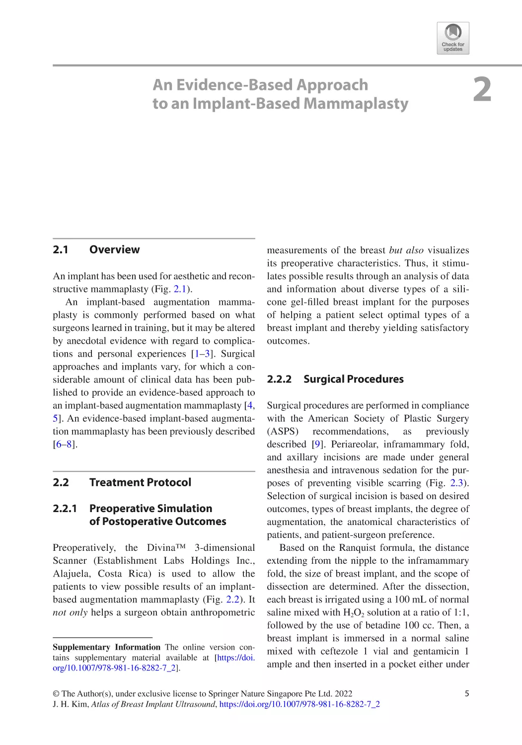
An implant has been used for aesthetic and reconstructive mammaplasty (Fig. 2.1).
An implant-based augmentation mammaplasty is commonly performed based on what
surgeons learned in training, but it may be altered
by anecdotal evidence with regard to complications and personal experiences [1–3]. Surgical
approaches and implants vary, for which a considerable amount of clinical data has been published to provide an evidence-based approach to
an implant-based augmentation mammaplasty [4,
5]. An evidence-based implant-based augmentation mammaplasty has been previously described
[6–8].
2.2
Treatment Protocol
2.2.1
Preoperative Simulation
of Postoperative Outcomes
Preoperatively, the Divina™ 3-dimensional
Scanner (Establishment Labs Holdings Inc.,
Alajuela, Costa Rica) is used to allow the
patients to view possible results of an implantbased augmentation mammaplasty (Fig. 2.2). It
not only helps a surgeon obtain anthropometric
Supplementary Information The online version contains supplementary material available at [https://doi.
org/10.1007/978-981-16-8282-7_2].
measurements of the breast but also visualizes
its preoperative characteristics. Thus, it stimulates possible results through an analysis of data
and information about diverse types of a silicone gel-filled breast implant for the purposes
of helping a patient select optimal types of a
breast implant and thereby yielding satisfactory
outcomes.
2.2.2
Surgical Procedures
Surgical procedures are performed in compliance
with the American Society of Plastic Surgery
(ASPS) recommendations, as previously
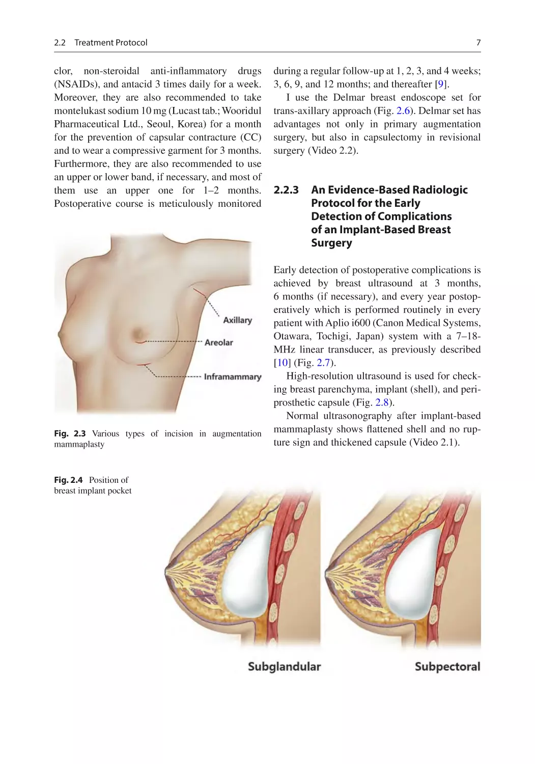
described [9]. Periareolar, inframammary fold,
and axillary incisions are made under general
anesthesia and intravenous sedation for the purposes of preventing visible scarring (Fig. 2.3).
Selection of surgical incision is based on desired
outcomes, types of breast implants, the degree of
augmentation, the anatomical characteristics of
patients, and patient-surgeon preference.
Based on the Ranquist formula, the distance
extending from the nipple to the inframammary
fold, the size of breast implant, and the scope of
dissection are determined. After the dissection,
each breast is irrigated using a 100 mL of normal
saline mixed with H2O2 solution at a ratio of 1:1,
followed by the use of betadine 100 cc. Then, a
breast implant is immersed in a normal saline
mixed with ceftezole 1 vial and gentamicin 1
ample and then inserted in a pocket either under
© The Author(s), under exclusive license to Springer Nature Singapore Pte Ltd. 2022
J. H. Kim, Atlas of Breast Implant Ultrasound, https://doi.org/10.1007/978-981-16-8282-7_2
5
6
2
An Evidence-Based Approach to an Implant-Based Mammaplasty
the pectoralis major muscle (a submuscular
placement) or in the retromammary space above
it (subglandular/submammary placement).
Fig. 2.1 Breast implant surgery
a
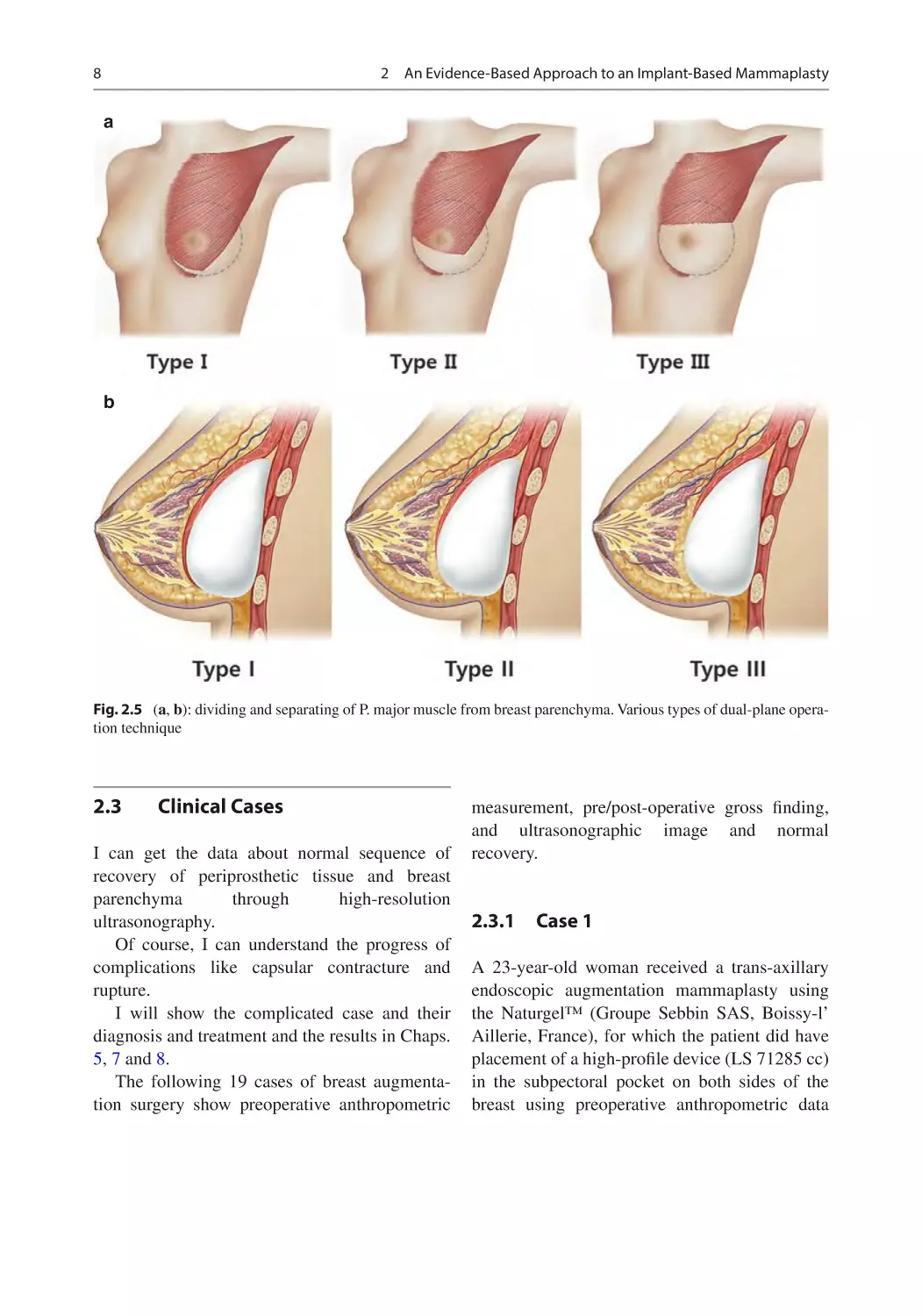
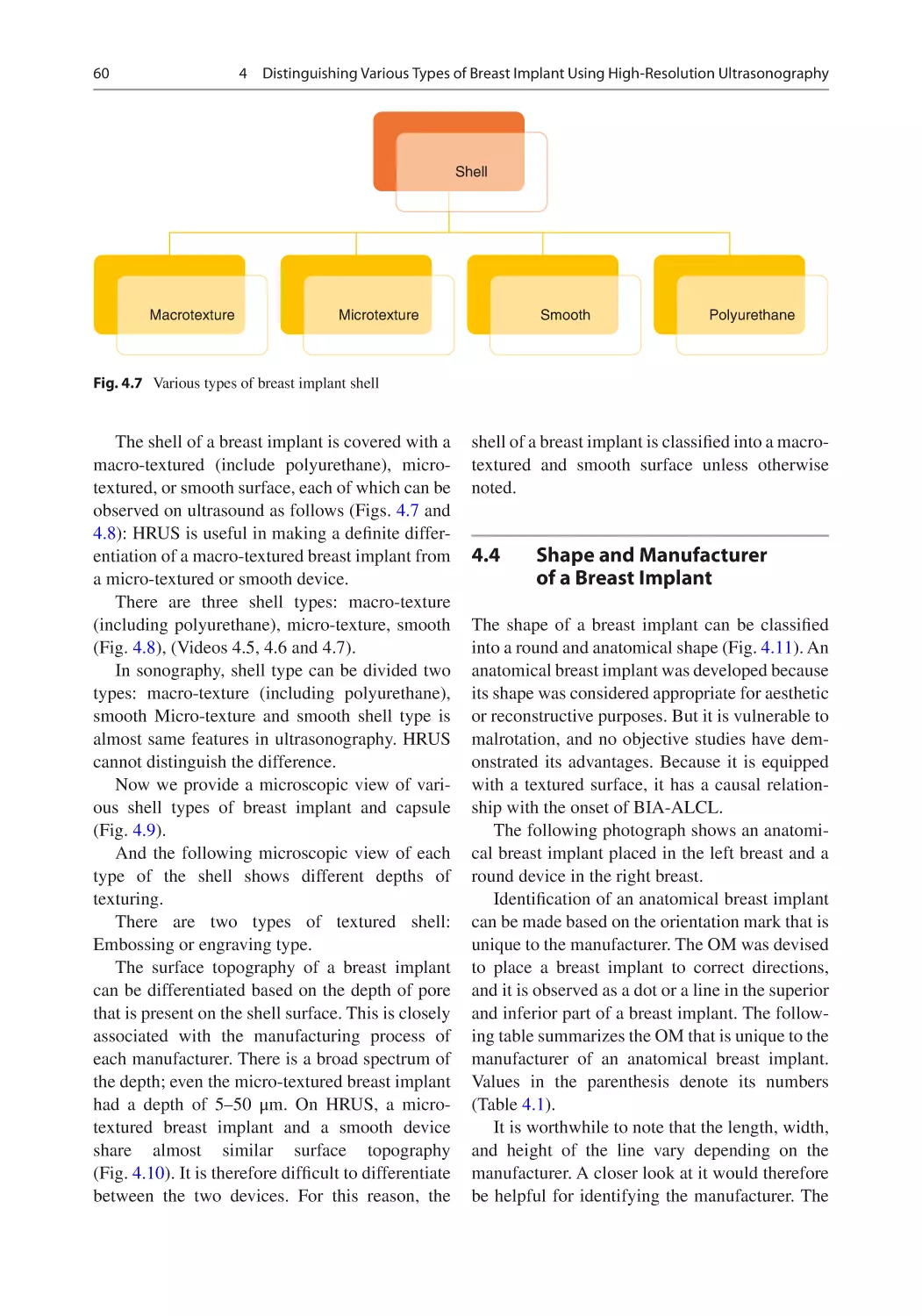
There are two types of breast implant position,
subpectoral and subglandular pocket (Fig. 2.4).
Subpectoral pocket means that breast implant is
positioned under the pectoralis major muscle.
Methods for inserting and positioning a breast
implant in the pocket are dependent on its types,
the degree of augmentation, characteristics of a
patient’s body, and surgeons’ recommendations.
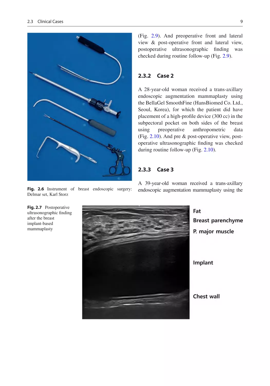
Thus, a dual-plane I/II augmentation is performed on a case-by-case basis.
Dual plane I, II, II is determined by dissection
range and height of P. major muscle during operation for aesthetic result (Fig. 2.5). Best aesthetic
result is achieved by appropriate N-IMF
distance.
Intraoperatively, the patients are intravenously
given ceftezole 1.0 g. Incisions are closed using
layered sutures in the breast tissue. In addition,
skin adhesive or surgical tape is used to close the
skin. Postoperatively, the patients are given cefa-
b
c
Fig. 2.2 Preoperative anthropometric measurement by
3D simulation. (a) Measurement of Breast base width,
Breast base height, Sternal notch- > nipple, Nipple- > midline, Nipple- > inframammary fold, Areolar diameter,
Internal distance, Intermammary distance, Breast volume.
(b) Before and after simulation. (c) Correction of projection asymmetry
2.2
Treatment Protocol
clor, non-steroidal anti-inflammatory drugs
(NSAIDs), and antacid 3 times daily for a week.
Moreover, they are also recommended to take
montelukast sodium 10 mg (Lucast tab.; Wooridul
Pharmaceutical Ltd., Seoul, Korea) for a month
for the prevention of capsular contracture (CC)
and to wear a compressive garment for 3 months.
Furthermore, they are also recommended to use
an upper or lower band, if necessary, and most of
them use an upper one for 1–2 months.
Postoperative course is meticulously monitored
Fig. 2.3 Various types of incision in augmentation
mammaplasty
Fig. 2.4 Position of
breast implant pocket
7
during a regular follow-up at 1, 2, 3, and 4 weeks;
3, 6, 9, and 12 months; and thereafter [9].
I use the Delmar breast endoscope set for
trans-axillary approach (Fig. 2.6). Delmar set has
advantages not only in primary augmentation
surgery, but also in capsulectomy in revisional
surgery (Video 2.2).
2.2.3
n Evidence-Based Radiologic
A
Protocol for the Early
Detection of Complications
of an Implant-Based Breast
Surgery
Early detection of postoperative complications is
achieved by breast ultrasound at 3 months,
6 months (if necessary), and every year postoperatively which is performed routinely in every
patient with Aplio i600 (Canon Medical Systems,
Otawara, Tochigi, Japan) system with a 7–18-
MHz linear transducer, as previously described
[10] (Fig. 2.7).
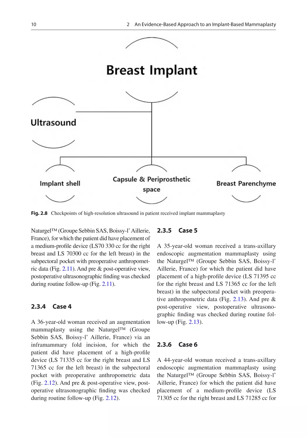
High-resolution ultrasound is used for checking breast parenchyma, implant (shell), and periprosthetic capsule (Fig. 2.8).
Normal ultrasonography after implant-based
mammaplasty shows flattened shell and no rupture sign and thickened capsule (Video 2.1).
8
2
An Evidence-Based Approach to an Implant-Based Mammaplasty
a
b
Fig. 2.5 (a, b): dividing and separating of P. major muscle from breast parenchyma. Various types of dual-plane operation technique
2.3
Clinical Cases
I can get the data about normal sequence of
recovery of periprosthetic tissue and breast
parenchyma
through
high-resolution
ultrasonography.
Of course, I can understand the progress of
complications like capsular contracture and
rupture.
I will show the complicated case and their
diagnosis and treatment and the results in Chaps.
5, 7 and 8.
The following 19 cases of breast augmentation surgery show preoperative anthropometric
measurement, pre/post-operative gross finding,
and ultrasonographic image and normal
recovery.
2.3.1
Case 1
A 23-year-old woman received a trans-axillary
endoscopic augmentation mammaplasty using
the Naturgel™ (Groupe Sebbin SAS, Boissy-l’
Aillerie, France), for which the patient did have
placement of a high-profile device (LS 71285 cc)
in the subpectoral pocket on both sides of the
breast using preoperative anthropometric data
2.3
Clinical Cases
9
(Fig. 2.9). And preoperative front and lateral
view & post-operative front and lateral view,
postoperative ultrasonographic finding was
checked during routine follow-up (Fig. 2.9).
2.3.2
Case 2
A 28-year-old woman received a trans-axillary
endoscopic augmentation mammaplasty using
the BellaGel SmoothFine (HansBiomed Co. Ltd.,
Seoul, Korea), for which the patient did have
placement of a high-profile device (300 cc) in the
subpectoral pocket on both sides of the breast
using
preoperative
anthropometric
data
(Fig. 2.10). And pre & post-operative view, postoperative ultrasonographic finding was checked
during routine follow-up (Fig. 2.10).
2.3.3
Fig. 2.6 Instrument of breast endoscopic surgery:
Delmar set, Karl Storz
Fig. 2.7 Postoperative
ultrasonographic finding
after the breast
implant-based
mammaplasty
Case 3
A 39-year-old woman received a trans-axillary
endoscopic augmentation mammaplasty using the
10
2
An Evidence-Based Approach to an Implant-Based Mammaplasty
Fig. 2.8 Checkpoints of high-resolution ultrasound in patient received implant mammaplasty
Naturgel™ (Groupe Sebbin SAS, Boissy-l’ Aillerie,
France), for which the patient did have placement of
a medium-profile device (LS70 330 cc for the right
breast and LS 70300 cc for the left breast) in the
subpectoral pocket with preoperative anthropometric data (Fig. 2.11). And pre & post-operative view,
postoperative ultrasonographic finding was checked
during routine follow-up (Fig. 2.11).
2.3.4
Case 4
A 36-year-old woman received an augmentation
mammaplasty using the Naturgel™ (Groupe
Sebbin SAS, Boissy-l’ Aillerie, France) via an
inframammary fold incision, for which the
patient did have placement of a high-profile
device (LS 71335 cc for the right breast and LS
71365 cc for the left breast) in the subpectoral
pocket with preoperative anthropometric data
(Fig. 2.12). And pre & post-operative view, postoperative ultrasonographic finding was checked
during routine follow-up (Fig. 2.12).
2.3.5
Case 5
A 35-year-old woman received a trans-axillary
endoscopic augmentation mammaplasty using
the Naturgel™ (Groupe Sebbin SAS, Boissy-l’
Aillerie, France) for which the patient did have
placement of a high-profile device (LS 71395 cc
for the right breast and LS 71365 cc for the left
breast) in the subpectoral pocket with preoperative anthropometric data (Fig. 2.13). And pre &
post-operative view, postoperative ultrasonographic finding was checked during routine follow-up (Fig. 2.13).
2.3.6
Case 6
A 44-year-old woman received a trans-axillary
endoscopic augmentation mammaplasty using
the Naturgel™ (Groupe Sebbin SAS, Boissy-l’
Aillerie, France) for which the patient did have
placement of a medium-profile device (LS
71305 cc for the right breast and LS 71285 cc for
2.3
Clinical Cases
a
11
b
c
d
e
g
Fig. 2.9 (a) Preoperative anthropometric data by 3D
simulation. (b) Front view of before and after 3D simulation. (c) Right side view of before and after 3D simulation. (d) Left side view of before and after 3D simulation.
(e) Preoperative and postoperative front view. (f)
Preoperative and postoperative right side view. (g)
f
h
Preoperative and postoperative left side view. (h) Left
ultrasound image shows normal device placed in the subpectoral pocket on 52 weeks postoperatively. (i) Right
ultrasound image shows normal device placed in the subpectoral pocket on 52 weeks postoperatively
12
2
An Evidence-Based Approach to an Implant-Based Mammaplasty
ultrasonographic finding was checked during
routine follow-up (Fig. 2.16).
i
2.3.9
Fig. 2.9 (continued)
the left breast) in the subpectoral pocket with preoperative anthropometric data (Fig. 2.14). And
pre & post-operative view, postoperative ultrasonographic finding was checked during routine
follow-up (Fig. 2.14).
2.3.7
Case 7
A 26-year-old woman received a trans-axillary
endoscopic augmentation mammaplasty using
the BellaGel SmoothFine (HansBiomed Co. Ltd.,
Seoul, Korea), for which the patient did have
placement of a high-profile device in the subpectoral pocket (BRMZ-H 400 cc) on both sides of
the breast with preoperative anthropometric data
(Fig. 2.15). And pre & post-operative view, postoperative ultrasonographic finding was checked
during routine follow-up (Fig. 2.15).
2.3.8
Case 8
A 34-year-old woman received a trans-axillary
endoscopic augmentation mammaplasty using
the Motiva Ergonomix™Round SilkSurface
(Establishment Labs Holdings Inc., Alajuela,
Costa Rica), for which the patient did have placement of a high-profile device in the subpectoral
pocket (ERSF 335 cc) on both sides of the breast
with preoperative anthropometric data (Fig. 2.16).
And pre & post-operative view, postoperative
Case 9
A 28-year-old woman received a trans-axillary
endoscopic augmentation mammaplasty using
the BellaGel SmoothFine (HansBiomed Co. Ltd.,
Seoul, Korea), for which the patient did have
placement of a high-profile device in the subpectoral pocket (BRMZ-H 275 cc for the right breast
and 325 cc for the left breast) with preoperative
anthropometric data (Fig. 2.17). And pre & post-
operative view, postoperative ultrasonographic
finding was checked during routine follow-up
(Fig. 2.17)..
2.3.10 Case 10
A 28-year-old woman received a trans-axillary
endoscopic augmentation mammaplasty using
the BellaGel SmoothFine (HansBiomed Co. Ltd.,
Seoul, Korea), for which the patient did have
placement of a high-profile device in the subpectoral pocket (BRMZ-H 325 cc for the right breast
and 300 cc for the left breast) with preoperative
anthropometric data (Fig. 2.18). And pre & post-
operative view, postoperative ultrasonographic
finding was checked during routine follow-up
(Fig. 2.18).
2.3.11 Case 11
A 31-year-old woman received a trans-axillary
endoscopic augmentation mammaplasty using
the BellaGel SmoothFine (HansBiomed Co. Ltd.,
Seoul, Korea) for which the patient did have
placement of a high-profile device in the subpectoral pocket (BRMZ-H 300 cc) on both sides of
the breast with preoperative anthropometric data
(Fig. 2.19). And pre & post-operative view, postoperative ultrasonographic finding was checked
during routine follow-up (Fig. 2.19).
2.3
Clinical Cases
a
13
b
c
d
e
g
f
h
Fig. 2.10 (a) Preoperative anthropometric data by 3D
simulation. (b) Front view of before and after 3D simulation. (c) Right side view of before and after 3D simulation. (d) Left side view of before and after 3D simulation.
(e) Preoperative and postoperative front view. (f)
Preoperative and postoperative right side view. (g)
Preoperative and postoperative left side view. (h) Left
ultrasound image shows normal device placed in the sub-
pectoral pocket on 12 weeks postoperatively. (i) Right
ultrasound image shows normal device placed in the subpectoral pocket on 12 weeks postoperatively. (j) Left
ultrasound image shows normal device placed in the subpectoral pocket on 122 weeks postoperatively. (k) Right
ultrasound image shows normal device placed in the subpectoral pocket on 122 weeks postoperatively
14
2
An Evidence-Based Approach to an Implant-Based Mammaplasty
i
j
k
Fig. 2.10 (continued)
2.3.12 Case 12
A 27-year-old woman received a trans-axillary
endoscopic augmentation mammaplasty using
the Motiva Ergonomix™ Round SilkSurface
(Establishment Labs Holdings Inc., Alajuela,
Costa Rica), for which the patient did have placement of a high-profile device in the subpectoral
pocket (ERSF 355 cc for the right breast and
ERSD 300 cc for the left breast) with preoperative anthropometric data (Fig. 2.20). And pre &
post-operative view, postoperative ultrasonographic finding was checked during routine follow-up (Fig. 2.20).
2.3.13 Case 13
A 41-year-old woman received a trans-axillary
endoscopic augmentation mammaplasty using
the Motiva Ergonomix™ Round SilkSurface
(Establishment Labs Holdings Inc., Alajuela,
Costa Rica), for which the patient did have placement of a high-profile device in the subpectoral
pocket (ERSF 335 cc) on both sides of the breast
with preoperative anthropometric data (Fig. 2.21).
And pre & post-operative view, postoperative
ultrasonographic finding was checked during
routine follow-up (Fig. 2.21).
2.3.14 Case 14
A 28-year-old woman received a trans-axillary
endoscopic augmentation mammaplasty using
the Naturgel™ (Groupe Sebbin SAS, Boissy-l’
Aillerie, France), for which the patient did have
placement of a high-profile device (LS 71305 cc)
in the subpectoral pocket on both sides of the
breast with preoperative anthropometric data
(Fig. 2.22). And pre & post-operative view, postoperative ultrasonographic finding was checked
during routine follow-up (Fig. 2.22).
2.3
Clinical Cases
a
15
b
c
d
e
g
Fig. 2.11 (a) Preoperative anthropometric data by 3D
simulation. (b) Front view of before and after 3D simulation. (c) Right side view of before and after 3D simulation. (d) Left side view of before and after 3D simulation.
(e) Preoperative and postoperative front view. (f)
Preoperative and postoperative right side view. (g)
Preoperative and postoperative left side view. (h) Left
ultrasound image shows normal device placed in the sub-
f
h
pectoral pocket on 55 weeks postoperatively. (i) Right
ultrasound image shows normal device placed in the subpectoral pocket on 55 weeks postoperatively. (j) Left
ultrasound image shows normal device placed in the subpectoral pocket on 181 weeks postoperatively. (k) Right
ultrasound image shows normal device placed in the subpectoral pocket on 181 weeks postoperatively
16
2
An Evidence-Based Approach to an Implant-Based Mammaplasty
i
j
k
Fig. 2.11 (continued)
2.3.15 Case 15
A 31-year-old woman received a trans-axillary
endoscopic augmentation mammaplasty using
the BellaGel SmoothFine (HansBiomed Co. Ltd.,
Seoul, Korea), for which the patient did have
placement of a high-profile device in the subpectoral pocket (BRMZ-H 300 cc) on both sides of
the breast with preoperative anthropometric data
(Fig. 2.23). And pre & post-operative view, postoperative ultrasonographic finding was checked
during routine follow-up (Fig. 2.23).
2.3.16 Case 16
A 29-year-old woman received a trans-axillary
endoscopic augmentation mammaplasty using
the BellaGel SmoothFine (HansBiomed Co. Ltd.,
Seoul, Korea), for which the patient did have
placement of a high-profile device in the subpectoral pocket (BRMZ-H 350 cc for the right breast
and 325 cc for the left breast) with preoperative
anthropometric data (Fig. 2.24). And pre & post-
operative view, postoperative ultrasonographic
finding was checked during routine follow-up
(Fig. 2.24).
2.3.17 Case 17
A 30-year-old woman received a trans-axillary
endoscopic augmentation mammaplasty using the
Naturgel™ (Groupe Sebbin SAS, Boissy-l’ Aillerie,
France), for which the patient did have placement of
a high-profile device (LS 71335 cc for the right
breast and LS 71305 cc for the left breast) in the
subpectoral pocket with preoperative anthropometric data (Fig. 2.25). And pre & post-operative view,
postoperative ultrasonographic finding was checked
during routine follow-up (Fig. 2.25).
2.3
Clinical Cases
a
17
b
c
d
e
g
Fig. 2.12 (a) Preoperative anthropometric data by 3D
simulation. (b) Front view of before and after 3D simulation. (c) Right side view of before and after 3D simulation. (d) Left side view of before and after 3D simulation.
(e) Preoperative and postoperative front view. (f)
Preoperative and postoperative right side view. (g)
f
h
Preoperative and postoperative left side view. (h) Left
ultrasound image shows normal device placed in the subpectoral pocket on 52 weeks postoperatively. (i) Right
ultrasound image shows normal device placed in the subpectoral pocket on 52 weeks postoperatively
18
2
An Evidence-Based Approach to an Implant-Based Mammaplasty
2.3.18 Case 18
i
A 33-year-old woman received an augmentation
mammaplasty using the Naturgel™ (Groupe
Sebbin SAS, Boissy-l’ Aillerie, France) via an
inframammary fold incision, for which the
patient did have placement of a high-profile
device (LS 71265 cc for the right breast and LS
71245 cc for the left breast) in the subpectoral
pocket with preoperative anthropometric data
(Fig. 2.26). And pre & post-operative view, post-
Fig. 2.12 (continued)
a
b
c
Fig. 2.13 (a) Preoperative anthropometric data by 3D
simulation. (b) Front view of before and after 3D simulation. (c) Right side view of before and after 3D simulation. (d) Left side view of before and after 3D simulation.
(e) Preoperative and postoperative front view. (f)
Preoperative and postoperative right side view. (g)
Preoperative and postoperative left side view. (h) Left
ultrasound image shows normal device placed in the sub-
d
pectoral pocket on 12 weeks postoperatively. (i) Right
ultrasound image shows normal device placed in the subpectoral pocket on 12 weeks postoperatively. (j) Left
ultrasound image shows normal device in the subpectoral
pocket on 122 weeks postoperatively. (k) Right ultrasound image shows normal device in the subpectoral
pocket on 122 weeks postoperatively
2.3
Clinical Cases
19
e
g
i
k
Fig. 2.13 (continued)
f
h
j
20
a
2
An Evidence-Based Approach to an Implant-Based Mammaplasty
b
c
e
Fig. 2.14 (a) Preoperative anthropometric data by 3D
simulation. (b) Front view of before and after 3D simulation. (c) Right side view of before and after 3D simulation. (d) Left side view of before and after 3D simulation.
(e) Preoperative and postoperative front view. (f)
Preoperative and postoperative right side view. (g)
d
f
Preoperative and postoperative left side view. (h) Left
ultrasound image shows normal device placed in the subpectoral pocket on 12 weeks postoperatively. (i) Right
ultrasound image shows normal device placed in the subpectoral pocket on 12 weeks postoperatively
2.3
Clinical Cases
g
21
h
i
Fig. 2.14 (continued)
a
b
Fig. 2.15 (a) Preoperative anthropometric data by 3D
simulation. (b) Front view of before and after 3D simulation. (c) Right side view of before and after 3D simulation. (d) Left side view of before and after 3D simulation.
(e) Preoperative and postoperative front view. (f)
Preoperative and postoperative right side view. (g)
Preoperative and postoperative left side view. (h) Left
ultrasound image shows normal device placed in the subpectoral pocket on 12 weeks postoperatively. (i) Right
ultrasound image shows normal device placed in the subpectoral pocket on 12 weeks postoperatively
22
2
An Evidence-Based Approach to an Implant-Based Mammaplasty
c
d
e
g
i
Fig. 2.15 (continued)
f
h
2.3
a
Clinical Cases
23
b
c
e
Fig. 2.16 (a) Preoperative anthropometric data by 3D
simulation. (b) Front view of before and after 3D simulation. (c) Right side view of before and after 3D simulation. (d) Left side view of before and after 3D simulation.
(e) Preoperative and postoperative front view. (f)
Preoperative and postoperative right side view. (g)
Preoperative and postoperative left side view. (h) Left
ultrasound image shows normal device placed in the subpectoral pocket on 60 weeks postoperatively. (i) Right
ultrasound image shows normal device placed in the sub-
d
f
pectoral pocket on 60 weeks postoperatively. (j) Left
ultrasound image shows normal device placed in the subpectoral pocket on 120 weeks postoperatively. (k) Right
ultrasound image shows normal device placed in the subpectoral pocket on 120 weeks postoperatively. (l) Left
ultrasound image shows normal device placed in the subpectoral pocket on 156 weeks postoperatively. (m) Right
ultrasound image shows normal device placed in the subpectoral pocket on 156 weeks postoperatively
24
2
An Evidence-Based Approach to an Implant-Based Mammaplasty
g
h
i
j
k
m
Fig. 2.16 (continued)
l
2.3
Clinical Cases
a
25
b
c
d
e
g
Fig. 2.17 (a) Preoperative anthropometric data by 3D simulation. (b) Front view of before and after 3D simulation. (c)
Right side view of before and after 3D simulation. (d) Left
side view of before and after 3D simulation. (e) Preoperative
and postoperative front view. (f) Preoperative and postoperative right side view. (g) Preoperative and postoperative left
side view.(h) Left ultrasound image shows normal device
placed in the subpectoral pocket on 12 weeks postoperatively. (i) Right ultrasound image shows normal device
f
h
placed in the subpectoral pocket on 12 weeks postoperatively. (j) Left ultrasound image shows normal device placed
in the subpectoral pocket on 52 weeks postoperatively. (k)
Right ultrasound image shows normal device placed in the
subpectoral pocket on 52 weeks postoperatively. (l) Left
ultrasound image shows normal device placed in the subpectoral pocket on 119 weeks postoperatively. (m) Right
ultrasound image shows normal device placed in the subpectoral pocket on 119 weeks postoperatively
26
2
An Evidence-Based Approach to an Implant-Based Mammaplasty
i
j
k
l
m
Fig. 2.17 (continued)
2.3
a
Clinical Cases
27
b
c
e
Fig. 2.18 (a) Preoperative anthropometric data by 3D
simulation. (b) Front view of before and after 3D simulation. (c) Right side view of before and after 3D simulation. (d) Left side view of before and after 3D simulation.
(e) Preoperative and postoperative front view. (f)
Preoperative and postoperative right side view. (g)
Preoperative and postoperative left side view. (h) Left
ultrasound image shows normal device placed in the subpectoral pocket on 12 weeks postoperatively. (i) Right
ultrasound image shows normal device placed in the sub-
d
f
pectoral pocket on 12 weeks postoperatively. (j) Left
ultrasound image shows normal device placed in the subpectoral pocket on 52 weeks postoperatively. (k) Right
ultrasound image shows normal device placed in the subpectoral pocket on 52 weeks postoperatively. (l) Left
ultrasound image shows normal device placed in the subpectoral pocket on 113 weeks postoperatively. (m) Right
ultrasound image shows normal device placed in the subpectoral pocket on 113 weeks postoperatively
28
g
2
An Evidence-Based Approach to an Implant-Based Mammaplasty
h
i
j
k
l
m
Fig. 2.18 (continued)
2.3
a
Clinical Cases
29
b
c
e
Fig. 2.19 (a) Preoperative anthropometric data by 3D
simulation. (b) Front view of before and after 3D simulation. (c) Right side view of before and after 3D simulation. (d) Left side view of before and after 3D simulation.
(e) Preoperative and postoperative front view. (f)
Preoperative and postoperative right side view. (g)
Preoperative and postoperative left side view. (h) Left
ultrasound image shows normal device placed in the subpectoral pocket on 12 weeks postoperatively. (i) Right
ultrasound image shows normal device placed in the sub-
d
f
pectoral pocket on 12 weeks postoperatively. (j) Left
ultrasound image shows normal device placed in the subpectoral pocket on 52 weeks postoperatively. (k) Right
ultrasound image shows normal device placed in the subpectoral pocket on 52 weeks postoperatively. (l) Left
ultrasound image shows normal device placed in the subpectoral pocket on 108 weeks postoperatively. (m) Right
ultrasound image shows normal device placed in the subpectoral pocket on 108 weeks postoperatively
30
g
2
An Evidence-Based Approach to an Implant-Based Mammaplasty
h
i
j
k
l
m
Fig. 2.19 (continued)
2.3
a
Clinical Cases
31
b
c
e
Fig. 2.20 (a) Preoperative anthropometric data by 3D
simulation. (b) Front view of before and after 3D simulation. (c) Right side view of before and after 3D simulation. (d) Left side view of before and after 3D simulation.
(e) Preoperative and postoperative front view. (f)
Preoperative and postoperative right side view. (g)
Preoperative and postoperative left side view. (h) Left
ultrasound image shows normal device placed in the sub-
d
f
pectoral pocket on 12 weeks postoperatively. (i) Right
ultrasound image shows normal device placed in the subpectoral pocket on 12 weeks postoperatively. (j) Left
ultrasound image shows normal device placed in the subpectoral pocket on 54 weeks postoperatively. (k) Right
ultrasound image shows normal device placed in the subpectoral pocket on 54 weeks postoperatively
32
g
i
k
Fig. 2.20 (continued)
2
An Evidence-Based Approach to an Implant-Based Mammaplasty
h
j
2.3
a
Clinical Cases
33
b
c
e
Fig. 2.21 (a) Preoperative anthropometric data by 3D
simulation. (b) Front view of before and after 3D simulation. (c) Right side view of before and after 3D simulation. (d) Left side view of before and after 3D simulation.
(e) Preoperative and postoperative front view. (f)
Preoperative and postoperative right side view. (g)
Preoperative and postoperative left side view. (h) Left
ultrasound image shows normal device placed in the subpectoral pocket with benign mass on 12 weeks postopera-
d
f
tively. (i) Right ultrasound image shows normal device
placed in the subpectoral pocket with benign mass on
12 weeks postoperatively. (j) Left ultrasound image shows
normal device placed in the subpectoral pocket with
benign mass on 54 weeks postoperatively. (k) Right ultrasound image shows normal device placed in the subpectoral pocket with benign mass on 54 weeks postoperatively
34
2
An Evidence-Based Approach to an Implant-Based Mammaplasty
g
i
k
Fig. 2.21 (continued)
h
j
2.3
Clinical Cases
a
35
b
c
d
e
g
Fig. 2.22 (a) Preoperative anthropometric data by 3D
simulation. (b) Front view of before and after 3D simulation. (c) Right side view of before and after 3D simulation. (d) Left side view of before and after 3D simulation.
(e) Preoperative and postoperative front view. (f)
Preoperative and postoperative right side view. (g)
Preoperative and postoperative left side view. (h) Left
ultrasound image shows normal device placed in the sub-
f
h
pectoral pocket on 12 weeks postoperatively. (i) Right
ultrasound image shows normal device placed in the subpectoral pocket on 12 weeks postoperatively. (j) Left
ultrasound image shows normal device placed in the subpectoral pocket on 55 weeks postoperatively. (k) Right
ultrasound image shows normal device placed in the subpectoral pocket with benign mass on 55 weeks
postoperatively
36
2
An Evidence-Based Approach to an Implant-Based Mammaplasty
i
j
k
Fig. 2.22 (continued)
a
b
Fig. 2.23 (a) Preoperative anthropometric data by 3D
simulation. (b) Front view of before and after 3D simulation. (c) Right side view of before and after 3D simulation. (d) Left side view of before and after 3D simulation.
(e) Preoperative and postoperative front view. (f)
Preoperative and postoperative right side view. (g)
Preoperative and postoperative left side view. (h) Left
ultrasound image shows normal device placed in the subpectoral pocket on 12 weeks postoperatively. (i) Right
ultrasound image shows normal device placed in the subpectoral pocket on 12 weeks postoperatively
2.3
Clinical Cases
c
37
d
e
g
i
Fig. 2.23 (continued)
f
h
38
a
2
An Evidence-Based Approach to an Implant-Based Mammaplasty
b
c
e
Fig. 2.24 (a) Preoperative anthropometric data by 3D
simulation. (b) Front view of before and after 3D simulation. (c) Right side view of before and after 3D simulation. (d) Left side view of before and after 3D simulation.
(e) Preoperative and postoperative front view. (f)
Preoperative and postoperative right side view. (g)
d
f
Preoperative and postoperative left side view. (h) Left
ultrasound image shows normal device placed in the subpectoral pocket on 12 weeks postoperatively. (i) Right
ultrasound image shows normal device placed in the subpectoral pocket on 12 weeks postoperatively
2.3
Clinical Cases
39
g
h
i
Fig. 2.24 (continued)
a
b
Fig. 2.25 (a) Preoperative anthropometric data by 3D
simulation. (b) Front view of before and after 3D simulation. (c) Right side view of before and after 3D simulation. (d) Left side view of before and after 3D simulation.
(e) Preoperative and postoperative front view. (f)
Preoperative and postoperative right side view. (g)
Preoperative and postoperative left side view. (h) Left
ultrasound image shows normal device placed in the subpectoral pocket on 12 weeks postoperatively. (i) Right
ultrasound image shows normal device placed in the sub-
pectoral pocket on 12 weeks postoperatively. (j) Left
ultrasound image shows normal device placed in the subpectoral pocket on 54 weeks postoperatively. (k) Right
ultrasound image shows normal device placed in the subpectoral pocket on 54 weeks postoperatively. (l) Left
ultrasound image shows normal device placed in the subpectoral pocket on 120 weeks postoperatively. (m) Right
ultrasound image shows normal device placed in the subpectoral pocket on 120 weeks postoperatively
40
2
An Evidence-Based Approach to an Implant-Based Mammaplasty
c
d
e
g
Fig. 2.25 (continued)
f
h
2.3
Clinical Cases
41
i
j
k
l
m
Fig. 2.25 (continued)
operative ultrasonographic finding was checked
during routine follow-up (Fig. 2.26).
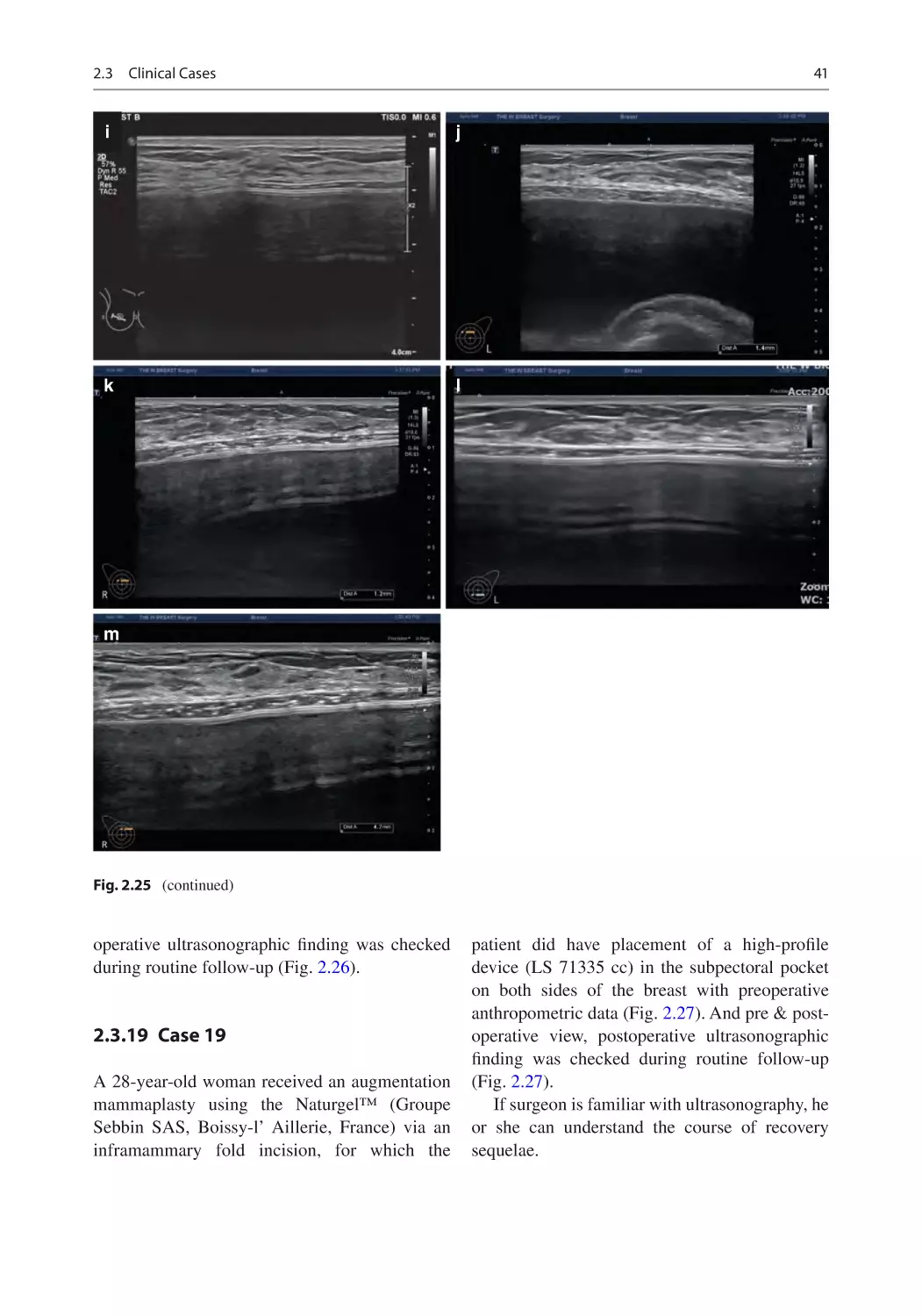
2.3.19 Case 19
A 28-year-old woman received an augmentation
mammaplasty using the Naturgel™ (Groupe
Sebbin SAS, Boissy-l’ Aillerie, France) via an
inframammary fold incision, for which the
patient did have placement of a high-profile
device (LS 71335 cc) in the subpectoral pocket
on both sides of the breast with preoperative
anthropometric data (Fig. 2.27). And pre & post-
operative view, postoperative ultrasonographic
finding was checked during routine follow-up
(Fig. 2.27).
If surgeon is familiar with ultrasonography, he
or she can understand the course of recovery
sequelae.
42
2
a
An Evidence-Based Approach to an Implant-Based Mammaplasty
b
c
e
g
Fig. 2.26 (a) Preoperative anthropometric data by 3D
simulation. (b) Front view of before and after 3D simulation. (c) Right side view of before and after 3D simulation. (d) Left side view of before and after 3D simulation.
(e) Preoperative and postoperative front view. (f)
Preoperative and postoperative right side view. (g)
d
f
h
Preoperative and postoperative left side view. (h) Left
ultrasound image shows normal device placed in the subpectoral pocket with benign mass on 12 weeks postoperatively. (i) Right ultrasound image shows normal device
placed in the subpectoral pocket on 12 weeks
postoperatively
2.3
Clinical Cases
43
i
Fig. 2.26 (continued)
a
b
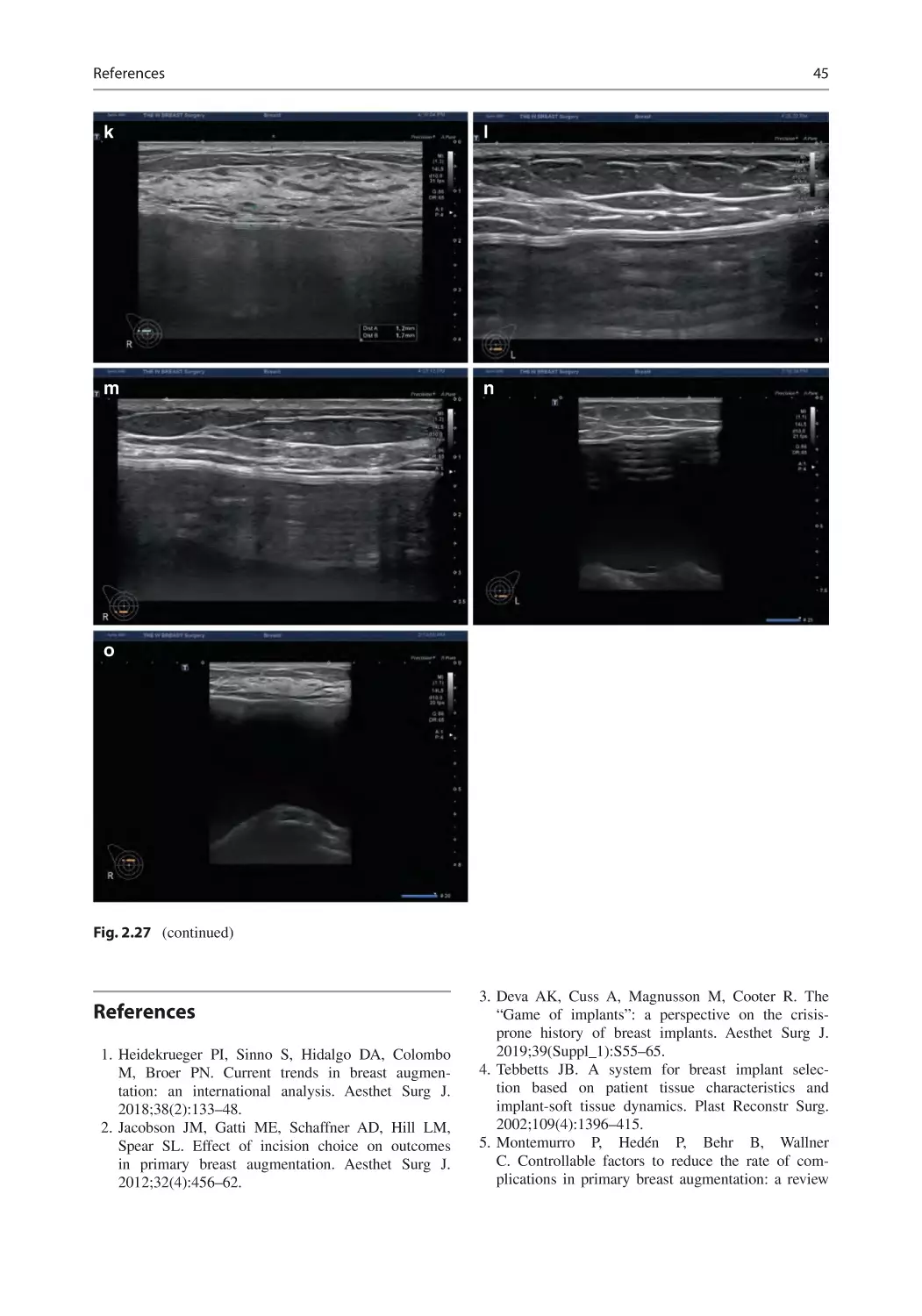
Fig. 2.27 (a) Preoperative anthropometric data by 3D
simulation. (b) Front view of before and after 3D simulation. (c) Right side view of before and after 3D simulation. (d) Left side view of before and after 3D simulation.
(e) Preoperative and postoperative front view. (f)
Preoperative and postoperative right side view. (g)
Preoperative and postoperative left side view. (h) Left
ultrasound image shows normal device placed in the subpectoral pocket on 12 weeks postoperatively. (i) Right
ultrasound image shows normal device placed in the subpectoral pocket on 12 weeks postoperatively. (j) Left
ultrasound image shows normal device placed in the subpectoral pocket on 56 weeks postoperatively. (k) Right
ultrasound image shows normal device placed in the subpectoral pocket on 56 weeks postoperatively. (l) Left
ultrasound image shows normal device on 126 weeks
postoperatively. (m) Right ultrasound image shows normal device on 126 weeks postoperatively. (n) Left ultrasound image shows normal device on 180 weeks
postoperatively. (o) Right ultrasound image shows normal
device on 180 weeks postoperatively
44
c
2
An Evidence-Based Approach to an Implant-Based Mammaplasty
d
e
f
g
i
Fig. 2.27 (continued)
h
j
References
45
k
l
m
n
o
Fig. 2.27 (continued)
References
1. Heidekrueger PI, Sinno S, Hidalgo DA, Colombo
M, Broer PN. Current trends in breast augmentation: an international analysis. Aesthet Surg J.
2018;38(2):133–48.
2. Jacobson JM, Gatti ME, Schaffner AD, Hill LM,
Spear SL. Effect of incision choice on outcomes
in primary breast augmentation. Aesthet Surg J.
2012;32(4):456–62.
3. Deva AK, Cuss A, Magnusson M, Cooter R. The
“Game of implants”: a perspective on the crisis-
prone history of breast implants. Aesthet Surg J.
2019;39(Suppl_1):S55–65.
4. Tebbetts JB. A system for breast implant selection based on patient tissue characteristics and
implant-soft tissue dynamics. Plast Reconstr Surg.
2002;109(4):1396–415.
5. Montemurro P, Hedén P, Behr B, Wallner
C. Controllable factors to reduce the rate of complications in primary breast augmentation: a review
46
2
An Evidence-Based Approach to an Implant-Based Mammaplasty
of the literature [published online ahead of print,
2020 May 1]. Aesthetic Plast Surg. 2020; https://doi.
org/10.1007/s00266-020-01726-x.
6. Thorne CH. An evidence-based approach to augmentation mammaplasty. Plast Reconstr Surg.
2010;126:2184–8.
7. Lista F, Ahmad J. Evidence-based medicine: augmentation mammaplasty. Plast Reconstr Surg.
2013;132:1684–96.
8. Schwartz MR. Evidence-based medicine: breast augmentation. Plast Reconstr Surg. 2017;140:109e–19e.
9. Sung JY, Jeong JP, Moon DS, et al. Short-term safety
of augmentation Mammaplasty using the BellaGel
implants in Korean women. Plast Reconstr Surg Glob
Open. 2019;7(12):e2566.
10. Park AY, Seo BK, Cho KR, Woo OH. The utility
of MicroPure™ ultrasound technique in assessing
grouped microcalcifications without a mass on mammography. J Breast Cancer. 2016;19:83–6.
3
Role of Ultrasound
in the Implant-Based Aesthetic
and Reconstructive Mammaplasty
3.1
Overview
To date, ultrasound has played a role in examining the integrity and rotation of a breast
implant [1–9]. Moreover, its role has been
expanded to manage patients who are suspected of having breast implant-associated
anaplastic large cell lymphoma as well as to
evaluate a breast mass [10, 11]. For the appropriate management of a patient receiving an
implant-based augmentation mammaplasty,
surgeons should perform an ultrasound-guided
assessment of two matters:
(1) information about a breast implant (e.g.,
location, constituents, shell, shape, and manufacturer) and (2) possible occurrence of implant-
related complications (e.g., folding with or
without detachment, periprosthetic fluid collection, thickened capsule, rupture, capsular mass,
malrotation of an anatomical device, upside-
down rotation, and foreign body reactions)
(Fig. 3.1).
Supplementary Information The online version contains supplementary material available at [https://doi.
org/10.1007/978-981-16-8282-7_3].
3.2
Role of Ultrasound
in the Context of an Implant-
Based Aesthetic
and Reconstructive
Mammaplasty
3.2.1
Characteristics of a Breast
Implant
A breast implant is equipped with a single or double
lumen with saline or silicone. The single lumen
breast implant is commonly encountered in a clinical setting even if it is a saline- or silicone gel-filled
breast implant. All the devices are equipped with
implants which have an external silicone shell.
After placement, a breast implant is characterized by the formation of a thin fibrous capsule
around it, considered a normal physiological phenomenon in response to foreign body.
3.2.2
Ultrasound-Guided
Assessment of a Breast
Implant
Patients receiving a breast implant should be
evaluated for the breast implant position, content,
and periprosthetic tissue and axilla on ultrasound.
On ultrasound, a silicone gel-filled breast implant
is characterized by anechoic findings surrounded
by a linear echogenic envelope. Moreover, its
interior side may be characterized by low-level
echoes.
© The Author(s), under exclusive license to Springer Nature Singapore Pte Ltd. 2022
J. H. Kim, Atlas of Breast Implant Ultrasound, https://doi.org/10.1007/978-981-16-8282-7_3
47
48
3
Role of Ultrasound in the Implant-Based Aesthetic and Reconstructive Mammaplasty
Fig. 3.1 Role of ultrasonography after breast implant surgery
The shell of a silicone gel-filled breast implant
may be observed as a single echogenic line or
parallel echogenic lines. Moreover, the fibrous
capsule is observed as two parallel echogenic
lines that are superficial to the surface of a silicone gel-filled breast implant. Normal undulations of the envelope may be observed as wavy
echogenic lines irrespective of the presence of
minimal intervening fluid.
3.2.3
Ultrasound-Guided Diagnosis
of Postoperative
Complications
Early complications of an implant-based augmentation mammaplasty include infection and
hematoma. Delayed complications include thickened capsule, rupture. As described earlier, in
2006, the US Food and Drug Administration
(FDA) mandated the use of a magnetic resonance
imaging (MRI) in women receiving a silicone
gel-filled breast implant at 3 years postoperatively and at a 2-year interval thereafter [2]. But
this causes an economic burden for them. A cost-
effective imaging study should therefore be
employed to make an early diagnosis of complications of an implant-based augmentation.
In Europe, ultrasound is a more preferred imaging modality in the initial assessment of implant
rupture as compared with MRI that has been established as a gold standard strategy [3, 7]. According
to a review of literatures, an agreement rate of 87%
between ultrasound and MRI has been reported,
accompanied by a close association between ultrasound images and findings at explantation.
Moreover, surgeons’ use of ultrasound in
assessing the status of a breast implant has
been advocated [1]. Further, ultrasound has
been reported to be a useful modality in postoperatively assessing the rotation of a shaped
breast implant seen in 42% of patients receiving an implant-based augmentation mammaplasty [9].
3.2 Role of Ultrasound in the Context of an Implant-Based Aesthetic and Reconstructive Mammaplasty
3.2.4
Ultrasound as a Screening
Tool for Patients Who Are
Suspected of Having Breast
Implant-Associated
Anaplastic Large Cell
Lymphoma (BIA-ALCL)
Usefulness of ultrasound in screening BIA-
ALCL has been advocated in the literature;
Adrada et al. performed a review of imaging
studies in 44 cases of BIA-ALCL, thus reporting
that ultrasound showed a sensitivity of 84% and a
specificity of 75% in detecting an effusion. These
values were higher as compared with the sensitivity and specificity of computed tomography,
MRI (82% and 33%), and positron emission
computed tomography (38% and 83%) [12].
Ultrasound may be therefore recommended as
the first line of screening for BIA-ALCL.
Initially, breast ultrasound is performed to assess
periprosthetic fluid collection, mass, or regional
adenopathy [14–16]. According to a retrospective
study conducted by Adrada et al., breast ultrasound
showed a sensitivity of 84% and a specificity of
75% in detecting a periprosthetic fluid collection.
These authors also reported that it showed a sensitivity of 46% and a specificity of 100% in detecting
a mass [17]. A periprosthetic fluid should be aspirated and then sent for culture and Gram stain, cell
block cytology, immunohistochemistry, and flow
cytometry [15, 16]. A minimum volume of 20 mL is
needed, but a 50–100 mL of the fluid sample should
49
be collected to avoid a risk of false-negative or intermediate results [14]. A pathologist should consider
the possibility of BIA-ALCL for the purposes of
performing appropriate tests including comprehensive flow cytometric markers.
3.2.5
Ultrasound as a Component
of a Multi-Disciplinary
Algorithm-Based Approach
to an Early Detection
of Complications
of an Implant-Based
Augmentation Mammaplasty
Kim et al. analyzed medical examination data that
was collected from a total of 2,037 women
(n = 2,037) between August 12 and December 31,
2021. They were evaluated for whether they were in
healthy conditions. Moreover, their baseline characteristics were also examined; these include age, sex,
height (cm), weight (kg), duration since surgery
(years), possession of a breast implant card, the site
of surgical incision, side of symptoms, and reasons
for outpatient visit. Furthermore, the patients were
also evaluated for their subjective awareness of the
manufacturer, surface, and shape of the breast
implant.
Potential complications include malrotation,
folding, seroma, capsule thickening, upside-
down rotation, rupture, capsule mass, and
breast mass (Fig. 3.2). According to these
Fig. 3.2 Checkpoint of breast implant ultrasonography for breast implant information
50
3
Role of Ultrasound in the Implant-Based Aesthetic and Reconstructive Mammaplasty
Fig. 3.3 Checklist of breast implant-related complications after breast implant surgery
authors, a majority of the patients had a past
history of receiving textured implants. Based
on these findings, Kim et al. proposed that such
patients be further evaluated for a risk of BIAALCL through a multi-disciplinary algorithmbased approach to an early detection of
complications of an implant-based augmentation mammaplasty [13].
There is a nine-point checklist for diagnosis of
breast implant-related complications (Fig. 3.3).
1. Folding with or without detachment.
2. Periprosthetic fluid collection.
3. Thickened capsule.
4. Double capsule.
5. Rupture (intracapsular and extracapsular rupture with or without LN silicone invasion).
6. Capsular mass.
7. Malrotation in anatomical type implant.
8. Upside-down rotation.
9. Foreign body.
Usually, there will be minimally 12 photos
and 12 video for patients who have breast implant
for the meticulous follow-up using ultrasonography. And also physician records more photos for
problematic site (Figs. 3.4 and 3.5), (Videos 3.1,
3.2, 3.3, 3.4, 3.5 and 3.6).
3.2 Role of Ultrasound in the Context of an Implant-Based Aesthetic and Reconstructive Mammaplasty
a
Fig. 3.4 (a, b) Breast implant specified ultrasonography.
(a) 6 photos for one breast: each point has a role. ① Point:
Best position for checking breast implant pocket. ② Point:
Checkpoint for upper part of breast implant and periprosthetic space. ③ Point: Checkpoint for lateral part of breast
implant and periprosthetic space. ④ Point: Checkpoint for
medial part of breast implant and periprosthetic space. ⑤
Point: Checkpoint for inferior part of breast implant and
periprosthetic space. ① Point: Best position for identifying
b
the breast implant shell type. (b) 6 videos for one breast.
Video: For checking lateral part of breast implant and
periprosthetic space. ① Video: For checking central part of
breast implant and periprosthetic space. ② Video: For
checking medial part of breast implant and periprosthetic
space. ③ Video: For checking upper part of breast implant
and periprosthetic space. ④ Video: For checking lower
part of breast implant and periprosthetic space. ⑤ Video:
For checking shell of breast implant
a
b
c
d
Fig. 3.5 Example of two different shell types breast
implant ultrasonography. (a–f) Textured shell type breast
implant. (g–l) Smooth shell type breast implant. (a): ①
51
point. (b): ② point. (c): ③ point. (d): ④ point. (e): ⑤ point.
(f): ⑥ point. (g): ① point. (h): ② point. I: ③ point. (j): ④
point. (k): ⑤ point. (l): ⑥ point
52
3
Role of Ultrasound in the Implant-Based Aesthetic and Reconstructive Mammaplasty
e
f
g
h
i
j
k
l
Fig. 3.5 (continued)
References
References
1. Bengtson BP, Eaves FF 3rd. High-resolution ultrasound in the detection of silicone gel breast implant
shell failure: background, in vitro studies, and early
clinical results. Aesthet Surg J. 2012;32:157–74.
2. Chung KC, Malay S, Shauver MJ, et al. Economic
analysis of screening strategies for rupture of silicone gel breast implants. Plast Reconstr Surg.
2012;130:225–37.
3. Rietjens M, Villa G, Toesca A, et al. Appropriate
use of magnetic resonance imaging and ultrasound
to detect early silicone gel breast implant rupture in
postmastectomy reconstruction. Plast Reconstr Surg.
2014;134:13e–20e.
4. Nahabedian MY. Discussion: appropriate use of magnetic resonance imaging and ultrasound to detect
early silicone gel breast implant rupture in postmastectomy reconstruction. Plast Reconstr Surg.
2014;134:21e–3e.
5. Stachs A, Dieterich M, Hartmann S, et al. Diagnosis
of ruptured breast implants through high-resolution
ultrasound combined with real-time elastography.
Aesthet Surg J. 2015;35:410–8.
6. Mennie JC, Quaba O, Smith M, et al. Diagnosing PIP
breast implant failure: a prospective analysis of clinical and ultrasound accuracy. J Plast Reconstr Aesthet
Surg. 2015;68:540–5.
7. Sisti A, Tassinari J, Milonia L, et al. Comparison of
Allergan, Mentor, and Sientra contoured cohesive gel
breast implants: a single surgeon’s 10-year experience. Plast Reconstr Surg. 2016;138:548e–9e.
8. Stivala A, Rem K, Leuzzi S, et al. Efficacy of ultrasound, mammography and magnetic resonance imaging in detecting breast implant rupture: a retrospective
study of 175 reconstructive and aesthetic sub-pectoral
breast augmentation cases. J Plast Reconstr Aesthet
Surg. 2017;70:1520–6.
53
9. Sieber DA, Stark RY, Chase S, et al. Clinical evaluation of shaped gel breast implant rotation using
high-resolution ultrasound. Aesthet Surg J. 2017;37:
290–6.
10. Clemens MW, Brody GS, Mahabir RC, et al. How
to diagnose and treat breast implant-associated anaplastic large cell lymphoma. Plast Reconstr Surg.
2018;141:586e–99e.
11. Shida M, Chiba A, Ohashi M, et al. Ultrasound diagnosis and treatment of breast lumps after breast augmentation with autologous fat grafting. Plast Reconstr
Surg Glob Open. 2017;5:e1603.
12. Adrada BE, Miranda RN, Rauch GM, et al. Breast
implant-associated anaplastic large cell lymphoma:
sensitivity, specificity, and findings of imaging
studies in 44 patients. Breast Cancer Res Treat.
2014;147:1–14.
13. Kim JH, Paik NS, Nam SY, Cho Y, Park HK. The
emerging crisis of stakeholders in implant-based augmentation Mammaplasty in Korea. J Korean Med Sci.
2020;35(15):e103.
14. Mehta-Shah N, Clemens MW, Horwitz SM. How I
treat breast implant-associated anaplastic large cell
lymphoma. Blood. 2018;132:1889–98.
15. O’Neill AC, Zhong T, Hofer SOP. Implications of
breast implant associated anaplastic large cell lymphoma (BIA-ALCL) for breast cancer reconstruction:
an update for surgical oncologists. Ann Surg Oncol.
2017;24:3174–9.
16. Clemens MW, Horwitz SM. NCCN consensus guidelines for the diagnosis and management of breast
implant-associated anaplastic large cell lymphoma.
Aesthet Surg J. 2017;37:285–9.
17. Adrada BE, Miranda RN, Rauch GM, et al. Breast
implant-associated anaplastic large cell lymphoma:
sensitivity, specificity, and findings of imaging
studies in 44 patients. Breast Cancer Res Treat.
2014;147:1–14.
4
Distinguishing Various Types
of Breast Implant Using
High-Resolution Ultrasonography
High-resolution ultrasound (HRUS) should be
used to evaluate some essential factors for a
patient receiving an implant-based aesthetic or
reconstructive mammaplasty; these include location, constituents, surface topography of the
shell, shape, and manufacturer of the breast
implant (Fig. 4.1).
closely associated with a soft feel of the breast.
Once identified, it is a helpful information for
making a plan for revisional surgery or
reoperation.
4.1
A breast implant can be classified into a saline-
filled or silicone gel-filled device. But there are
also other types of breast implant that can be
equipped with a double lumen. Step-off sign is a
clue to characterization of constituents of a breast
implant placed in a pocket. This may be supported by an inlet of a saline-filled breast implant
(Fig. 4.5), (Video 4.2). Sometimes there is no
inlet in the superior part of a saline-filled breast
implant. In patients receiving a breast implant in
an upside-down position, it is important to cautiously differentiate between an inlet and a patch
of a saline-filled breast implant, considering that
the shell of a saline-filled breast implant is also
available as either a smooth or textured surface.
This suggests that there is no correlation between
the surface topography and constituents of a
saline-filled breast implant. Definite sign of
saline breast implant even in dual chamber is no
step-off sign (Videos 4.1, 4.3 and 4.4).
Step-off sign is a definite indicator of a difference in the velocity of ultrasound wave between
a saline-filled and silicone gel-filled breast
implant. Step-off sign can be identified through
Location of a Breast Implant
A breast implant can be placed either above or
below the muscle, and its accurate location can
be identified (Fig. 4.2, Videos 4.11 and 4.12). The
easiest way to identify its location is to examine
the upper quadrant of the breast on its 12 o’clock
position. That is, an ultrasound probe is vertically
positioned on the border of a breast implant on its
12 o’clock position (Fig. 4.3). The following
sonographic finding shows the placement of a
breast implant above the pectoralis major muscle
(Fig. 4.4); this can be further understood on video
(Videos 4.9 and 4.10). A breast implant might be
partly placed below the muscle in patients receiving multiple surgeries or those who did not
achieve a complete dissection of the tissue.
Otherwise, a breast implant could be correctly
placed above or below the muscle. The location
of a breast implant is an essential factor that is
Supplementary Information The online version contains supplementary material available at (https://doi.
org/10.1007/978-981-16-8282-7_4).
4.2
Constituents of a Breast
Implant
© The Author(s), under exclusive license to Springer Nature Singapore Pte Ltd. 2022
J. H. Kim, Atlas of Breast Implant Ultrasound, https://doi.org/10.1007/978-981-16-8282-7_4
55
56
4
Distinguishing Various Types of Breast Implant Using High-Resolution Ultrasonography
Fig. 4.1 Checkpoint of breast implant-related information using ultrasonography
Pocket
Sub-Glandular
Pocket
Sub-Pectoral
Pocket
Fig. 4.2 Position of breast implant
Fig. 4.3 Best site for identifying implant pocket
position
application of an ultrasound probe to the border
of a breast implant with compression (Fig. 4.6),
(Video 4.2). The continuous line between chest
wall and inferior border of breast implant is broken when there is step-off sign. A continuity
between the base of a breast implant and the chest
wall suggests a lack of step-off sign, based on
which the presence of a saline-filled breast
implant can be identified. By contrast, a discontinuity between the base of a silicone gel-filled
breast implant and the chest wall suggests step-
off sign. A lack of step-off sign is indicative of a
saline-filled breast implant.
4.3
Surface Topography
of a Breast Implant
Identification of the surface topography of a
breast implant is helpful for not only examining
whether the corresponding device has a causal
relationship with the onset of breast implant-
associated anaplastic lymphoma (BIA-ALCL)
but also making a plan for reoperation.
In patients who are suspected of having BIA-
ALCL, a clinical course should be meticulously
monitored in association with a macro-textured
device. Thus, such patients cannot be
overlooked.
In this regard, patients receiving a smooth
device are free from a risk of BIA-
ALCL. Unfortunately, most of the patients were
not aware of a breast implant they received, some
of whom even lost a warranty card. Identification
of the surface topography of a breast implant is
therefore helpful for these patients.
4.3 Surface Topography of a Breast Implant
57
a
b
c
d
e
f
g
h
Fig. 4.4 Ultrasonographic images of subglandular and subpectoral implant pocket. (a–d) Breast implant in subglandular pocket. (e–o) Breast implant in subpectoral pocket
58
4
Distinguishing Various Types of Breast Implant Using High-Resolution Ultrasonography
i
j
k
l
m
n
o
Fig. 4.4 (continued)
4.3 Surface Topography of a Breast Implant
59
Content
Saline
Silicone
No Step-off Sign
Step-Off sign
Fig. 4.5 Distinguishing sign saline and silicone breast implant by ultrasonography
a
b
c
Fig. 4.6 Step-off sign and no step-off sign in ultrasonography. (a) No step-off sign in single-lumen saline implant. (b)
No step-off sign in double-lumen saline implant. (c) Step-off sign in silicone implant
60
4
Distinguishing Various Types of Breast Implant Using High-Resolution Ultrasonography
Shell
Macrotexture
Microtexture
Smooth
Polyurethane
Fig. 4.7 Various types of breast implant shell
The shell of a breast implant is covered with a
macro-textured (include polyurethane), micro-
textured, or smooth surface, each of which can be
observed on ultrasound as follows (Figs. 4.7 and
4.8): HRUS is useful in making a definite differentiation of a macro-textured breast implant from
a micro-textured or smooth device.
There are three shell types: macro-texture
(including polyurethane), micro-texture, smooth
(Fig. 4.8), (Videos 4.5, 4.6 and 4.7).
In sonography, shell type can be divided two
types: macro-texture (including polyurethane),
smooth Micro-texture and smooth shell type is
almost same features in ultrasonography. HRUS
cannot distinguish the difference.
Now we provide a microscopic view of various shell types of breast implant and capsule
(Fig. 4.9).
And the following microscopic view of each
type of the shell shows different depths of
texturing.
There are two types of textured shell:
Embossing or engraving type.
The surface topography of a breast implant
can be differentiated based on the depth of pore
that is present on the shell surface. This is closely
associated with the manufacturing process of
each manufacturer. There is a broad spectrum of
the depth; even the micro-textured breast implant
had a depth of 5–50 μm. On HRUS, a micro-
textured breast implant and a smooth device
share almost similar surface topography
(Fig. 4.10). It is therefore difficult to differentiate
between the two devices. For this reason, the
shell of a breast implant is classified into a macro-
textured and smooth surface unless otherwise
noted.
4.4
Shape and Manufacturer
of a Breast Implant
The shape of a breast implant can be classified
into a round and anatomical shape (Fig. 4.11). An
anatomical breast implant was developed because
its shape was considered appropriate for aesthetic
or reconstructive purposes. But it is vulnerable to
malrotation, and no objective studies have demonstrated its advantages. Because it is equipped
with a textured surface, it has a causal relationship with the onset of BIA-ALCL.
The following photograph shows an anatomical breast implant placed in the left breast and a
round device in the right breast.
Identification of an anatomical breast implant
can be made based on the orientation mark that is
unique to the manufacturer. The OM was devised
to place a breast implant to correct directions,
and it is observed as a dot or a line in the superior
and inferior part of a breast implant. The following table summarizes the OM that is unique to the
manufacturer of an anatomical breast implant.
Values in the parenthesis denote its numbers
(Table 4.1).
It is worthwhile to note that the length, width,
and height of the line vary depending on the
manufacturer. A closer look at it would therefore
be helpful for identifying the manufacturer. The
4.4
Shape and Manufacturer of a Breast Implant
61
a
Smooth
Micro-texture
Macro-texture
b
Polyurethane
Fig. 4.8 Various shell types of breast implant in ultrasonography. (a) Smooth, micro-texture, macro-textured shell type
breast implant in HRUS image. (b) Polyurethane shell type breast implant in HRUS image
direction of the superior OM may serve as an
indicator of malrotation. Grossly, the Sebbin and
the Mentor share very similar OM with each
other. It is somewhat difficult to identify an anatomical breast implant manufactured by the
Silimed. This is because its superior OM is not
visible even on HRUS. Therefore, an anatomical
breast implant manufactured by the Silimed
should be identified based on the characteristic
features of its textured surface. Thus, it can be
identified based on a dot that is present on the
base of a breast implant.
The time point of clinical release of an anatomical breast implant is also a useful information for identification of the corresponding
manufacturer. For example, the Polytech anatom-
ical breast implant was first approved for clinical
use in Korea in 2012. This information will be
helpful for the type of a breast implant depending
on the period during which a patient received an
implant-based augmentation mammaplasty.
Gross findings of an anatomical breast implant
are shown below. (Mentor, Allergan, Silimed,
Sebbin, Polytech, Eurosilicone, Hansbiomed)
(Figs. 4.12, 4.13, 4.14, 4.15, 4.16, 4.17 and 4.18),
(Videos 4.33, 4.34, 4.35, 4.36, 4.37, 4.38, 4.39
and 4.40).
Briefly, the OM is unique to an anatomical
breast implant. But there are no anatomical breast
implants branded as the Motiva because the
Motiva Ergonomix Round SilkSurface was solely
imported to Korea. To identify the manufacturer
62
4
Distinguishing Various Types of Breast Implant Using High-Resolution Ultrasonography
a
Smooth
Microtexture
Macrotexture
b
Allergan
Bellagel
Eurosilicone
Sebbin
c
Mentor
Silimed
Fig. 4.9 Three different breast implant shell images and
microscopic view of capsule in microscopy. (a)
Microscopic view of three different shell types. (b)
Microscopic view of embossing shell type breast implant.
(c) Microscopic view of engraving shell type breast
implant. (d) Microscopic view of polyurethane shell. (e)
Polytech
Microscopic view of capsule that had contained macrotextured shell type breast implant that shows many undulations. (f) Microscopic view of capsule that had
contained smooth shell type breast implant that shows
smooth inner line
4.4
Shape and Manufacturer of a Breast Implant
d
63
e
Polyurethane
f
Fig. 4.9 (continued)
of an anatomical breast implant based on the presence of OM, a breast implant should be examined
using an ultrasound probe from the superior to
inferior directions (Videos 4.8, 4.15, 4.18, 4.21,
4.23, 4.24, 4.27, 4.29, 4.30 and 4.32). This is followed by examination of the border of a breast
implant in a circle at an angle of 360°.
Subsequently, specific findings on the shell are
observed (Figs. 4.19, 4.20, 4.21, 4.22, 4.23, 4.24,
4.25 and 4.26). An ultrasound probe should be
rotated when there are any notable findings, based
on which it can be determined whether the OM
has a shape of line or dot. It is necessary to examine the shell of a breast implant manufactured by
the Silimed at a higher magnification. Moreover,
it is also necessary to adjust the depth of probing,
which is essential for examining the inferior part
of a device. Thus, the manufacturer of a breast
implant can be completely identified (Fig. 4.21).
It is common to see patients who do not know
what type of breast implants she has.
So far, you can see the ultrasonographic clue
for identifying the various manufacturers by OM.
From now on, you can see the gross photos
and ultrasonographic images of round texture or
smooth shell type breast implant by various
manufacturers.
In the case of round shape type breast implant,
since there is no mark that specifies the manufacturer, in theory, it is impossible to determine the
manufacturer, but the manufacturer can be identified if there is an RFID chip, upside-down rotation, specific shell type in some manufacturer
(Fig. 4.27).
And we can get the information by upside-
down rotation because posterior patch is manufacturer specific. This will be discussed next in
Chap. 5.
64
4
Distinguishing Various Types of Breast Implant Using High-Resolution Ultrasonography
Smooth
Microtexture
Fig. 4.10 Smooth and micro-textured shell looks almost identical in HRUS
Fig. 4.11 Two different shape types of breast implant: anatomical type and round type
a
b
Fig. 4.12 Orientation mark of Mentor anatomical shape type breast implant. (a) Anterior view. (b) Posterior view
Table 4.1 Various types of orientation mark
Manufacturer
Allergan
Mentor
Silimed
Sebbin
Polytech
Hansbiomed
Eurosilicone
Anterior orientation mark
Line
Dot
None
Yes (3)
Yes (1), Short
None
Yes (1), Long
None
Yes (1), Short
None
Yes (1), Long
None
Yes (1), Short
None
None
Yes (2)
Posterior orientation mark
Line
Dot
None
Yes (4)
Yes (1)
Yes (2)
None
Yes (4)
Yes (1)
Yes (2)
None
Yes (2)
None
Yes (5)
None
Yes (5)
4.4
Shape and Manufacturer of a Breast Implant
65
a
b
Fig. 4.13 Orientation mark of Allergan anatomical shape type breast implant. (a) Anterior view. (b) Posterior view
a
b
Fig. 4.14 Orientation mark of Polytech anatomical shape type breast implant. (a) Anterior view. (b) Posterior view
a
b
Fig. 4.15 Orientation mark of Sebbin anatomical shape type breast implant. (a) Anterior view. (b) Posterior view
66
4
Distinguishing Various Types of Breast Implant Using High-Resolution Ultrasonography
a
b
Fig. 4.16 Orientation mark of Silimed anatomical shape type breast implant. (a) Anterior view. (b) Posterior view
a
b
Fig. 4.17 Orientation mark of Eurosilicone anatomical shape type breast implant. (a) Anterior view. (b) Posterior view
a
b
Fig. 4.18 Orientation mark of Hansbiomed Bellagel anatomical shape type breast implant. (a) Anterior view. (b)
Posterior view
From now, gross and ultrasonographic view of
various round type breast implants is shown.
There are round texture and smooth type of
Mentor, Allergan, Silimed, Sebbin, Hansbiomed,
Polytech, Eurosilicone, Motiva (Figs. 4.28, 4.29,
4.30, 4.31, 4.32, 4.33, 4.34, 4.35, 4.36 and 4.37),
(Videos 4.13, 4.14, 4.16, 4.17, 4.19, 4.20, 4.22,
4.25, 4.26, 4.28 and 4.31).
4.4
Shape and Manufacturer of a Breast Implant
67
a
b
Fig. 4.19 Mentor anatomical type. The OM of Sebbin,
Hansbiomed, Mentor is similar, but there is definite difference in shape, height, and length, so it can be distin-
a
guished in HRUS. (a) Ultrasonographic image of OM
(3 cm Linear type at anterior shell). (b) Gross finding of
OM of Mentor anatomical type implant
b
Fig. 4.20 Allergan anatomical type. Three dots is anterior shell, sometimes two dots type of OM is seen. (a)
Ultrasonographic image of three orientation dot. (b) Gross finding of OM of Allergan anatomical type implant
a
Fig. 4.21 Silimed anatomical type implant (2 stage). For
the identification of Silimed anatomical type, it is necessary to identify Silimed specified texture shell type. After
identification of Silimed textured shell type, then looking
for OM in posterior shell layer is necessary. There is no
difference between anatomical type and round textured
shell type of Silimed implant in anterior shell, only poste-
b
rior dot is seen in ultrasonography, for the identification of
Silimed anatomical type implant, operator should check
posterior shell of implant if Silimed textured shell is
checked in anterior shell image. (a) Identification of
Silimed macro-texture shell type. (b) Ultrasonographic
image of OM. (c) Gross image of posterior OM of Silimed
anatomical breast implant
68
4
Distinguishing Various Types of Breast Implant Using High-Resolution Ultrasonography
c
Fig. 4.21 (continued)
a
Fig. 4.22 Sebbin anatomical type implant. Linear type of
OM is checked by HRUS. Sebbin OM is distinguished
from shape and textured shell image from Mentor,
a
Fig. 4.23 Polytech anatomical type implant. There is a
long line below textured shell. It is very easy to confirm
Polytech anatomical type breast implant. See the video (a)
b
Hansbiomed anatomical type breast implant. (a)
Ultrasonographic image of OM. (b) Gross image of OM
in anterior shell of Sebbin anatomical type breast implant
b
Ultrasonographic image of OM. (b) Gross image of OM
in anterior shell of Polytech anatomical type breast
implant
4.4
Shape and Manufacturer of a Breast Implant
a
69
b
c
Fig. 4.24 Hansbiomed “Bellagel” anatomical type
implant. There is linear type OM that can be distinguished
from Sebbin and Mentor anatomical type by HRUS. (a)
Ultrasonographic image of OM. (b) Ultrasonographic
a
image of OM (Old two dot version of OM of Bellagel
anatomical implant). (c) Gross image of Bellagel anatomical type implant
b
Fig. 4.25 Eurosilicone anatomical type implant. Eurosilicone anatomical OM is similar with Hansbiomed anatomical
type. (a) Ultrasonographic image of OM. (b) Gross image of Eurosilicone anatomical type breast implant
70
4
Distinguishing Various Types of Breast Implant Using High-Resolution Ultrasonography
a
b
c
Fig. 4.26 Polytech polyurethane anatomical implant. (a) Ultrasonographic image of OM (Sagittal view). (b)
Ultrasonographic image of OM (coronal view). (c) Gross image of polyurethane breast implant
Fig. 4.27 RFID chip is seen in the bottom of implant
4.4
Shape and Manufacturer of a Breast Implant
71
a
c
b
d
e
Fig. 4.28 Mentor round texture and smooth type breast
implant. (a) Anterior view of round textured shell type
breast implant. (b) Posterior view of round textured shell
type breast implant. (c) Gross view of posterior patch.
(d–g) Ultrasonographic image of Mentor textured shell.
f
(h) Anterior view of round smooth shell type breast
implant. (i) Posterior view of round smooth shell type
breast implant. (j) Gross view of posterior patch. (k–t)
Ultrasonographic image of smooth shell of Mentor
72
4 Distinguishing Various Types of Breast Implant Using High-Resolution Ultrasonography
g
h
i
j
k
l
m
n
Fig. 4.28 (continued)
4.4
Shape and Manufacturer of a Breast Implant
73
o
p
q
r
s
t
Fig. 4.28 (continued)
74
4
Distinguishing Various Types of Breast Implant Using High-Resolution Ultrasonography
a
b
c
d
e
f
g
Fig. 4.29 Motiva round micro-textured implant gross
with/without RFID chip. (a) Gross finding of anterior
view. (b) Gross finding of posterior view. (c) RFID chip of
posterior patch. (d) No RFID chip of posterior patch.
h
(e–q) Ultrasonographic image of Motiva Ergonomix
Round. SilkSurface breast implant. (r) Ultrasonographic
image of RFID chip (This is the clue for identifying the
manufacturer)
4.4
Shape and Manufacturer of a Breast Implant
75
i
j
k
l
m
n
o
p
Fig. 4.29 (continued)
76
4
Distinguishing Various Types of Breast Implant Using High-Resolution Ultrasonography
q
r
Fig. 4.29 (continued)
a
Fig. 4.30 Hansbiomed Bellagel round micro-textured
shell type breast implant. (a) Anterior view of round
micro-texture shell type breast implant. (b) Posterior view
of round micro-texture shell type breast implant. (c)
Posterior patch of Bellagel micro-texture breast implant.
(d, e) Ultrasonographic image of Bellagel micro-texture
b
shell. (f) Ultrasonographic image of Bellagel micro-texture shell. (g) Ultrasonographic image shows different
shell layers of Bellagel micro-texture shell. (h–l) Different
shell layers in same patient of Bellagel micro-texture
shell. (High-resolution ultrasonography shows the shell
layer modification)
4.4
Shape and Manufacturer of a Breast Implant
c
77
d
e
g
f
h
i
j
k
l
Fig. 4.30 (continued)
78
4
Distinguishing Various Types of Breast Implant Using High-Resolution Ultrasonography
a
b
c
d
e
f
g
h
Fig. 4.31 Sebbin round macro-textured shell type breast
implant. (a) Gross image of anterior view of Sebbin round
texture implant. (b) Gross image of posterior view of
Sebbin round texture implant. (c) Gross image of posterior patch. (d–h) Ultrasonographic image of Sebbin
macro-textured shell type breast implant
4.4
Shape and Manufacturer of a Breast Implant
a
c
79
b
d
e
f
g
Fig. 4.32 Sebbin round smooth shell type breast implant.
(a) Gross image of anterior view of Sebbin round smooth
shell type implant. (b) Gross image of posterior view of
Sebbin round smooth shell type implant. (c) Gross image
of posterior patch. (d–g) Ultrasonographic image of
Sebbin smooth shell type breast implant
80
4
Distinguishing Various Types of Breast Implant Using High-Resolution Ultrasonography
a
c
b
d
e
g
Fig. 4.33 Sebbin round micro-textured shell type breast
implant. (a) Gross image of anterior view of Sebbin
micro-texture implant. (b) Gross image of posterior view
f
h
of Sebbin micro-texture implant. (c) Gross image of posterior patch. (d–m) Ultrasonographic image of Sebbin
micro-textured shell type breast implant
4.4
Shape and Manufacturer of a Breast Implant
81
i
j
k
l
m
Fig. 4.33 (continued)
82
4
Distinguishing Various Types of Breast Implant Using High-Resolution Ultrasonography
a
c
b
d
e
Fig. 4.34 Silimed round macro-textured shell type breast
implant. (a) Gross image of anterior view of Silimed
macro-texture implant. (b) Gross image of posterior view
f
of Silimed macro-texture implant. (c) Gross image of posterior patch. (d–i) Ultrasonographic image of Silimed
macro-textured shell type breast implant
4.4
Shape and Manufacturer of a Breast Implant
g
83
h
i
Fig. 4.34 (continued)
a
Fig. 4.35 Allergan round smooth shell type breast
implant. (a) Gross image of anterior view of Allergan
round smooth shell type implant. (b) Gross image of pos-
b
terior view of Allergan round smooth shell type implant.
(c) Gross image of posterior patch. (d–l) Ultrasonographic
image of Allergan smooth shell type breast implant
84
c
4
Distinguishing Various Types of Breast Implant Using High-Resolution Ultrasonography
d
e
f
g
h
i
j
Fig. 4.35 (continued)
4.4
Shape and Manufacturer of a Breast Implant
85
k
l
Fig. 4.35 (continued)
a
b
c
d
Fig. 4.36 Allergan round macro-textured shell type
breast implant. (a) Gross image of anterior view of
Allergan macro-texture implant. (b) Gross image of pos-
terior view of Allergan macro-texture implant. (c) Gross
image of posterior patch. (d–l) Ultrasonographic image of
Allergan macro-textured shell type breast implant
86
4
Distinguishing Various Types of Breast Implant Using High-Resolution Ultrasonography
e
f
g
h
i
j
k
l
Fig. 4.36 (continued)
4.4
Shape and Manufacturer of a Breast Implant
a
c
87
b
d
e
g
Fig. 4.37 Eurosilicone round micro-textured shell type
breast implant. (a) Gross image of anterior view of
Eurosilicone micro-texture implant. (b) Gross image of
posterior view of Eurosilicone micro-texture implant. (c)
f
h
Gross image of posterior patch. (d–i) Ultrasonographic
image of Eurosilicone micro-textured shell type breast
implant
88
i
Fig. 4.37 (continued)
4
Distinguishing Various Types of Breast Implant Using High-Resolution Ultrasonography
5
Usefulness of High-Resolution
Ultrasound in Detecting
Complications of an Implant-Based
Mammaplasty
5.1
Overview
When a breast implant is inserted in a human’s
body, it may cause diverse complications
(Fig. 5.1). An early diagnosis of them is essential
for ensuring the safety of a patient.
For the past 15 years, I have performed aesthetic or reconstructive implant-based mammaplasty. During this period, I have made all efforts
to meticulously monitor the patient’s postoperative course. I have therefore found that most of
the postoperative complications could not be
diagnosed on physical examination and they
should be accurately diagnosed on ultrasound in
association with signs and symptoms that are
suggestive of them. Thus, I have regularly performed a breast ultrasound for patients receiving
an implant-based augmentation mammaplasty
after 3 months postoperatively, thus making all
efforts to diagnose postoperative complications
at the earliest opportunities possible. Here, I provide numerous images and videos which I have
accumulated until now.
The current book is advantageous in simultaneously examining radiological, pathological,
and plastic surgical findings, thus contributing to
evaluating a patient’s findings based on clinical
findings and imaging characteristics. The following complications are closely associated with a
Supplementary Information The online version contains supplementary material available at (https://doi.
org/10.1007/978-981-16-8282-7_5.)
patient’s discomfort, particularly including pain
as well as the shape and feel of breast.
5.2
Folding
Folding is a very common sonographic finding
after breast implant augmentation. It is not a serious complication in almost any case (Fig. 5.2),
(Video 5.1). But folding may cause pain, foreign
body sensation or palpation of a breast implant
which a patient can exhibit at the outpatient
clinic.
Its occurrence is associated with a relatively
smaller size of pocket; it may occur as a result of
capsular contracture (CC) and under-dissection.
It is commonly manifested on ultrasound as
shown below (Figs. 5.3 and 5.4).
An ultrasound-guided monitoring of a
patient’s clinical course is useful in determining
the scope of dissection at surgery. This is because
an excessive mobility of the device due to over-
dissection requires a reduction in the scope of
dissection. From this context, the scope of dissection should be re-considered in a patient who
commonly presents with a folding of the device
due to under-dissection even in the absence of
CC. This would be helpful for preventing a surgeon from repeatedly making mistakes.
The severity of a patient’s symptoms may vary
depending on the scope of folding and the depth
of groove. For example, there would be no discomfort without TC, deep groove, and thin
© The Author(s), under exclusive license to Springer Nature Singapore Pte Ltd. 2022
J. H. Kim, Atlas of Breast Implant Ultrasound, https://doi.org/10.1007/978-981-16-8282-7_5
89
90
5
Usefulness of High-Resolution Ultrasound in Detecting Complications of an Implant-Based…
Fig. 5.1 Complications of an implant-based augmentation mammaplasty
may also cause rupture of the device. Therefore,
an ultrasound-guided examination at a follow-up
should be performed to determine the optimal
scope and degree of folding.
Some cases of folding may be accompanied
by the detachment of the affected site. A relatively longer, wider detachment may be associated with a patient’s foreign body sensation.
The following sonographic finding is folding
with or without detachment (Figs. 5.5 and 5.6),
(Videos 5.2 and 5.3).
Fig. 5.2 Symptom of implant shell folding
Fig. 5.3 An intraoperative finding of folding with thickened capsule
subcutaneous tissue. But palpation of a breast
implant or pain would be present if there is more
than one of these. Folding of a breast implant
might also lead to the disruption of shell, which
5.3
Periprosthetic Fluid
Collection
Periprosthetic fluid collection can be classified
into normal and abnormal cases depending on the
time point of its onset, volume, hematoma, and
seroma (Fig. 5.7). If abnormal fluid collection is
found in ultrasonography, ultrasound-guided
aspiration should be performed followed by
cytology and culture for the appropriate
management.
Seroma can occur after a prosthesis is inserted
in a human body. Most of which is absorbed after
three months postoperatively or left minimally.
So, there is no or minimal fluid collection in periprosthetic space (Fig. 5.8), (Video 5.4).
This can be confirmed based on ultrasound
findings obtained at 1 or 3 months, which has
been verified from a total of more than 700
patients receiving primary or secondary implant-
based augmentation mammaplasty. Normally a
5.3 Periprosthetic Fluid Collection
small amount of seroma (<10 cc) on the medial
side of the breast can be present persistently.
In a patient who grossly had a swelling or did
have a widespread presence of seroma around a
breast implant rather than a minimal periprosthetic fluid collection on ultrasound, aspiration
should be performed (Fig. 5.9). Thus, additional
examination should be performed and the appropriate treatment should be planned accordingly.
The following sonographic image shows large
amount of delayed seroma formed around a
breast implant (Fig. 5.10). Physician should distinguish between seroma and rupture.
As shown above, an ultrasound-guided aspiration should be performed to avoid damages to a
breast implant in a patient with a large amount of
periprosthetic fluid collection. In a patient who is
suspected of having an infection based on the
color and smell of the specimen, empirical antibiotics is prescribed with cytology and culture test.
Fig. 5.4 Sonographic finding of folding
a
91
If necessary, however, antibiotics can be changed
based on cytology and culture.
In my experience, CC is inevitable following
condition even if there were appropriate management of infectious seroma which is immediate
postoperative onset. If a patient has such condition, surgeon may consider explantation or second stage reoperation option. Explantation
should be performed in severe infectious conditions or refractory to antibiotics therapy.
The following sonographic image shows a
case of non-infectious seroma occurring after
drinking alcohol at 6 months postoperatively.
This case was treated with several sessions of
ultrasound-guided aspiration (Fig. 5.11).
A sudden appearance of seroma with textured
breast implant should be considered serious if it
disappeared postoperatively, for which a rule-out
diagnosis of breast implant-associated anaplastic
large cell lymphoma (BIA-ALCL) and infection
should be made on ultrasound-guided aspiration.
This should be followed by cytology, culture, and
CD30-positive immunohistochemistry.
Delayed seroma should also be considered
serious because a patient receiving a macro-
textured breast implant is vulnerable to BIA-
ALCL. An early detection of delayed seroma
would therefore be essential. It would also be
mandatory to examine whether it is of infectious
origin. Even a patient with non-infectious delayed
seroma would be at increased risk of developing
capsular thickening over time. Such patient
should be meticulously followed up using ultrasound with appropriate treatment, by which the
b
Fig. 5.5 Folding with detachment. (a) Transverse view of detachment. (b) Longitudinal view of detachment
92
5
Usefulness of High-Resolution Ultrasound in Detecting Complications of an Implant-Based…
possible absorption of seroma and changes in the
capsule thickness should be evaluated. This
would be helpful for planning for more active
treatments for patients who may be vulnerable to
possible complications.
A tentative diagnosis of hematoma can be
made based on ultrasound findings after a history
taking and clinical examination (e.g., bruise).
Then, the appropriate treatment should be
planned based on the severity of swelling and the
scope of hematoma.
The following sonographic images show
hematoma-related cases (Fig. 5.12).
5.4
Rupture is a major complication after breast implant
surgery which is a cause of reoperation. Rupture
occurs in both saline and silicone implants (Fig. 5.13).
The ultrasonographic finding is different in both ruptures (Video 5.5). There are various diagnostic image
modalities for detecting implant rupture.
Ultrasonography is the most cost-effective
and useful diagnostic modality for detecting
implant failure.
But sufficient level of expertise would be
mandatory to make an accurate diagnosis of rupture on ultrasound. This is because it is important
to make a differential diagnosis of rupture from
periprosthetic fluid collection, thickened capsule,
and folding with or without detachment.
5.4.1
Fig. 5.6 Sonographic finding of folding without
detachment
Fig. 5.7 Periprosthetic
fluid
Rupture
Saline Implant Rupture
Rupture of a saline-filled breast implant is characterized by a flattened shell after disappearance
of saline filler in a single lumen (Videos 5.6, 5.7,
5.8, 5.9 and 5.10).
Stepladder sign is a typical clue to diagnosis
of rupture of a saline-filled breast implant, which
may also be compared with the contralateral
breast [1, 2].
5.4 Rupture
a
93
b
Fig. 5.8 Normal US finding in postoperatively 12 weeks. (a) No medial fluid collection. (b) Minimal fluid collection
a
b
c
Fig. 5.9 Fluid collection after reoperation d/t capsular
contracture. (a) Sonographic image during US-guided
needle aspiration for the left medial fluid collection. (b)
Sonographic image after aspiration with intact breast
implant. (c) Aspirated seroma for cytology and culture
94
5
Usefulness of High-Resolution Ultrasound in Detecting Complications of an Implant-Based…
Fig. 5.10 Delayed seroma in periprosthetic space after
breast augmentation
Rupture of a saline-filled breast implant can
be easily detected on ultrasound with history taking and physical examination.
The following sonographic image shows the
characteristics of rupture of a saline-filled breast
implant (Fig. 5.14).
5.4.2
Silicone Implant Rupture
Early detection of silicone implant failure is very
important for the following reasons:
1. Breast feeding: If early diagnosis is missed,
free silicone can progress extracapsular r upture,
then ipsilateral breast feeding is interfered.
2. Breast cancer screening: Extracapsular free
silicone invasion can interfere with breast
examination.
3. Possibility of complete removal of leaked silicone: Early diagnosis reduces the extent of
surgery and recovery time. Radical removal of
free silicone cannot be achieved in extracapsular rupture with or without LN silicone
invasion.
Surgeon cannot prevent failure of implant, but
early detection of rupture using any diagnostic
modality can minimize hazard of free silicone.
Silicone implant rupture can be classified by
scope and type (Fig. 5.15).
Depending on the infiltration of silicone contents into the tissue after it is leaked from a ruptured breast implant, rupture of the device can
also be classified into intracapsular rupture and
extracapsular rupture; the latter is further divided
based on the infiltration of silicone contents into
the ipsilateral axillary lymph node [3].
Rupture of a breast implant can be classified
into major rupture, grossly characterized by a
shell tear (Fig. 5.16), and minor rupture, characterized by a gel bleed despite a lack of gross finding of shell tear [3–5].
Major rupture of a silicone gel-filled breast
implant can be diagnosed based on the shell and
silicone leakage (free silicone) [3, 6, 7]. A gel
bleed can be confirmed based on a free silicone
without shell tear [3–5].
An extracapsular rupture of a silicone gel-
filled breast implant can be confirmed based on
the concurrent presence of posterior acoustic
shadowing (reverberating echoes with loss of
detail posterior to the echogenic area, which has
been variably described as snowstorm, echodense
noise, echodense shadowing, or echogenic confusion) and diagnosed when there is a concurrent
presence of extracapsular mass or LN invasion
[4, 5, 8–12].
Unless there are notable findings (Snowstorm
sign (+), LN silicone invasion, extracapsular siliconoma) other than free silicone contents with no
snowstorm sign, extra-capsular silicone granuloma and LN silicone infiltration), the corresponding cases are diagnosed as an intracapsular
rupture of a breast implant.
An infiltration of silicone contents into the
ipsilateral axillary lymph node is a clue to diagnosis of an extracapsular rupture of a silicone
gel-filled breast implant [13–15]. This is not seen
in cases of intracapsular rupture. A widespread
presence of an extracapsular rupture of a silicone
gel-filled breast implant proximal to the axilla is
commonly seen with the involvement of axillary
lymph node in a patient with a long-standing
presence of rupture [13, 14, 16].
Signs of silicone implant rupture in ultrasonography are as follows:
1.
2.
3.
4.
5.
6.
Tear of shell.
Hyperechoic capsule d/t free silicone.
Gel fracture (change of integrity of implant).
Stepladder sign.
Free silicone presence.
Snowstorm appearance (seen in extracapsular
rupture).
5.4 Rupture
a
c
95
b
d
e
Fig. 5.11 Non-infectious seroma treatment using
US-guided aspiration. (a) Gross finding of POD 13 weeks.
(b) Ultrasonographic finding of the right medial small
fluid collection. (c) Gross finding of POD 30 weeks. (d)
Ultrasonographic image of LIQ area. (e) Ultrasonographic
f
image of POD 35 weeks. (f) Ultrasonographic image of
POD 47 weeks. (g) Ultrasonographic image of POD
50 weeks. (h) Gross finding of the right breast. (i)
Ultrasonographic image of POD 55 weeks. (j)
Ultrasonographic image of POD 74 weeks
96
5
Usefulness of High-Resolution Ultrasound in Detecting Complications of an Implant-Based…
g
i
h
j
Fig. 5.11 (continued)
Fig. 5.12 Postoperative hematoma
5.4.2.1 Gel Bleed
There are some points to consider in detecting a
rupture of a silicone gel-filled breast implant on
ultrasound. Depending on the degree of rupture,
rupture of a silicone gel-filled breast implant can
be classified into major and minor ruptures.
Gel bleed called with minor rupture is checked
free silicone without shell tearing.
Fig. 5.13 Breast implant rupture
The following image shows minor rupture of a
silicone gel-filled breast implant: High-resolution
ultrasonography will make it possible for early
detection of minor rupture (Fig. 5.17), (Videos
5.4 Rupture
97
a
b
c
d
e
f
g
Fig. 5.14 Ultrasonographic image of saline implant rupture d/t shell tear or leakage. (a) White arrow indicates saline
implant shell with no remnant saline. (b–g) Deflated saline breast implant with saline leakage
98
5
Usefulness of High-Resolution Ultrasound in Detecting Complications of an Implant-Based…
a
Fig. 5.15 Scope of silicone implant rupture by ultrasonography. (a) Classified by silicone invasion: Intracapsular
rupture and extracapsular rupture. (b) Classified by shell
a
b
tear: Major rupture (shows shell tear in gross finding) and
minor rupture (no shell tear in gross finding, gel bleed)
b
c
Fig. 5.16 The following sonographic image shows the
characteristics of major rupture of a silicone gel-filled
breast implant: (a) White triangle is the point of tear of
shell. (b) White triangle is leaked silicone and white arrow
is hyperechoic capsule. (c) Gross finding of ruptured
breast implant
5.4 Rupture
99
a
c
b
d
e
g
Fig. 5.17 Gross and ultrasonographic image of minor
rupture (gel bleeding) which is lack of shell tear. (a, b):
Case I. (c, d): Case II. (e–g): Case III. (h, i): Case IV. (a)
Gross finding of gel bleed. (b) Ultrasonographic image of
gel bleed (white arrow shows abnormal shell condition,
white triangle shows gel fracture). (c) Gross finding of gel
f
h
bleed. (d) Preoperative ultrasonographic diagnosis of gel
bleed (white triangle). (e) Gross finding of gel bleed. (f)
Preoperative diagnosis of gel bleed (white triangle). (g)
Preoperative diagnosis of gel bleed (white triangle). (h)
Gross finding of gel bleed. (i) Preoperative ultrasonographic diagnosis of gel bleed (white triangle)
100
5
Usefulness of High-Resolution Ultrasound in Detecting Complications of an Implant-Based…
i
Fig. 5.17 (continued)
5.11, 5.12, 5.13 and 5.14). Hyperechoic capsule
is also seen in gel bleed site.
5.4.2.2 Intracapsular Major Rupture
Intra-capsular rupture is diagnosed when there
are no signs of extracapsular rupture like snowstorm appearance, extracapsular invasion like
regional lymph node. Distinguishing between
intracapsular rupture and extracapsular rupture is
difficult in some cases. In that case target capsulectomy is a key for accurate diagnosis.
Distinguishing the scope of rupture is very
important for management of possible residual
siliconoma that may interfere with breast feeding
in young patients.
Ultrasonographic signs of intracapsular rupture are as follows:
1. Free silicone.
2. Shell tearing without free silicone.
3. Hyperechoic capsule in both minor rupture
and major rupture.
4. Step-ladder sign.
5. Gel fracture.
The following sonographic images show various intracapsular silicone rupture (Fig. 5.18),
(Videos 5.15 and 5.16).
Sonographic finding of hyperechoic capsule is
seen.
5.4.2.3 E
xtracapsular Rupture with or
without Lymph Node Invasion
An extracapsular rupture is diagnosed with capsular invasion of free silicone. Extracapsular
mass and ipsilateral lymph node siliconoma are a
definite sign of extracapsular rupture (Videos
5.17, 5.18, 5.19, 5.20, 5.21, 5.22, 5.23, 5.24, 5.25
and 5.26).
Extracapsular silicone invasion to breast
parenchyma may disturb breast examination and
complete removal of the leaked silicone cannot
be achieved. It would therefore be mandatory to
inform a patient of the presence of extracapsular
rupture.
In a patient with an extracapsular rupture of a
breast implant, there is a possibility that remnant
lesions in breast and lymph node might be left
because of leaked silicone contents on ultrasound
even after explantation or implant change.
It is noteworthy because there is a possibility
that a normal breast implant may be misdiagnosed as a rupture of the device. For these reasons, the scope and degree of silicone involvement
should be checked and then recorded through an
ultrasound-guided examination of the breast.
This would contribute to avoiding unnecessary
surgeries and thereby ensuring a patient’s healthcare rights.
Ultrasonographic signs of extracapsular rupture are as follows:
1. Reverberating echoes with loss of detail posterior to the echogenic area, which has been
variably described as snowstorm, echodense
noise, echodense shadowing, or echogenic
confusion.
2. Extracapsular granuloma.
3. Regional LN siliconoma.
The following sonographic image shows an
extracapsular rupture of a silicone gel-filled
breast implant (Fig. 5.19).
By definition, silicone lymphadenopathy is
referred to as involvement of silicone in a lymph
node; it is a rare complication of augmentation
5.5 Thickened Capsule and Capsular Contracture
mammaplasty using a silicone gel-filled breast
implant [16–18]. This poses a diagnostic dilemma
for surgeons. Malignancy may be initially considered, but its possibility should be ruled out in a
patient with a lump in the neck or axilla who had a
past history of receiving augmentation mammaplasty using a silicone gel-filled breast implant
[19]. To date, two different mechanisms underlying the migration of silicone to the tissue have
been proposed; these include rupture or erosion of
a silicone-containing surface or continued leakage
through an intact surface [20, 21]. The migration
of silicone to the tissue causes fibrosis and foreign
body granulomatous reactions [22]. This may be
followed by the transportation of silicone particles
to regional lymph nodes via macrophages in the
reticulo-endothelial system [23]. Consequently,
foreign body reactions may cause local swelling of
the involved lymph node. It has been documented
that silicone lymphadenopathy primarily affects
the axillary lymph node in a patient receiving augmentation mammaplasty using a silicone gel-filled
breast implant [20, 24].
The following ultrasonographic image shows
silicone invasion to lymph node (Fig. 5.20).
Axillary lymph node ultrasonography should
be performed with or without rupture.
5.5
Thickened Capsule
and Capsular Contracture
Known as one of the major causes of reoperation,
CC is characterized by thickening and ingrowth
of the capsule composed of collagen around a
breast implant, resulting in alterations in the
shape and location of the device, which may lead
to pain and hardening sensation.
CC can be classified depending on the capsule
thickness, the elasticity of the capsule, and the
scope of thickened capsule; it is divided into mild
and severe cases. In association with this, the
Baker classification system was introduced to
express its degree in 1978.
For the past several years, I have studied correlations between the Baker grades I-IV and capsule thickness measured on breast ultrasound.
101
Table 5.1 Thickness of capsule with Baker grade
Baker grade
I
II
III
IV
The capsule thickness on breast ultrasound
<0.4 mm
0.4–0.8 mm
0.8–1.4 mm
>1.4 mm
The following sonographic image shows a
normal breast without CC (Fig. 5.21). It is characterized by a lack of notable TC between the
inferior border of the pectoralis major muscle
and the shell of a breast implant.
Capsular contracture is a result of extensive
thickened capsule (Fig. 5.22), (Videos 5.27, 5.28,
5.29, 5.30, 5.31 and 5.32). We can easily see the
folding of shell in Baker III, IV capsular
contracture.
Fortunately many patients have thickened
capsule but are asymptomatic d/t focal or partial
TC range. In these patients, careful observation
of changes in the thickness and range of TC is
necessary with ultrasonography. Then early
detection of Baker III, IV capsular contracture is
possible.
By measuring TC, cut-off values for the Baker
grades I-IV were estimated as follows:
Correlation between the Baker classification
system and the capsule thickness on breast
ultrasound.
According to “the value of capsule thickness on
breast ultrasound as an indicator of the severity of
capsular contracture and its correlation with the
baker classification,” the capsule thickness might be
used as an indicator of the severity of CC alternative
to the Baker classification system [2], (Table 5.1).
As mentioned earlier, not only the thickness of
capsule, but also the extent of thickened capsule
and the elasticity of the capsule are important in
severity of symptoms.
According to a study about periprosthetic capsule, there was no consistency in the capsule
thickness, as shown above in the ultrasonographic
image of thickened capsule.
But the place where a patient complains of the
greatest discomfort was the point that has most
thickened capsule. If there was a TC, it was
matched to a patient’s symptoms.
102
5
Usefulness of High-Resolution Ultrasound in Detecting Complications of an Implant-Based…
a
c
e
b
d
f
g
Fig. 5.18 Ultrasonographic finding of intracapsular silicone
rupture. (a) White triangle is shell disruption, white arrow is
free silicone. (b) White triangle is free silicone. (c) White
triangle is free silicone. (d) White triangle is free silicone. (e)
White arrow is free silicone. (f) White triangle is free silicone. (g) White triangle is free silicone. (h) White arrow is
free silicone. (i) White triangle is free silicone. (j) White triangle is free silicone. (k) White arrow is free silicone. (l)
White triangle is free silicone. (m) White triangle is free silicone. (n) White triangle is free silicone. (o) White triangle is
free silicone. (p) White triangle is free silicone. (q) White
triangle is free silicone. (r) White arrow is free silicone. (s)
White arrow is free silicone. (t) White triangle is free silicone. (u) White arrow is free silicone. (v) White triangle is
free silicone. (w) White triangle is free silicone. (x) White
triangle is ruptured implant. (y) White triangle is free sili-
cone. (z1) White triangle is free silicone, white arrow is
hyperechoic capsule. (z2) White triangle is free silicone. (z3)
White triangle is hyperechoic capsule d/t rupture. (z4) White
triangle is free silicone and white arrow is hyperechoic capsule. (z5) White triangle is hyperechoic capsule d/t rupture.
(z6) White triangle is free silicone and white arrow is hyperechoic capsule. (z7) White triangle is free silicone and white
arrow is hyperechoic capsule. (z8) White triangle is free silicone and white arrow is hyperechoic capsule. (z9) White
triangle is hyperechoic capsule d/t rupture. (z10) White triangle is free silicone and white arrow is hyperechoic capsule.
(z11) White triangle is free silicone and white arrow is hyperechoic capsule. (z12) White triangle is free silicone and
white arrow is hyperechoic capsule. (z13) White triangle is
free silicone and white arrow is hyperechoic capsule. (z14)
White triangle is hyperechoic capsule d/t rupture
5.5 Thickened Capsule and Capsular Contracture
103
h
i
j
k
l
m
n
o
Fig. 5.18 (continued)
104
5
Usefulness of High-Resolution Ultrasound in Detecting Complications of an Implant-Based…
p
q
r
s
t
u
v
w
Fig. 5.18 (continued)
5.5 Thickened Capsule and Capsular Contracture
105
x
y
z1
z2
z3
z4
z5
z6
Fig. 5.18 (continued)
106
5
Usefulness of High-Resolution Ultrasound in Detecting Complications of an Implant-Based…
z7
z8
z9
z10
z11
z12
z13
Z14
Fig. 5.18 (continued)
5.5 Thickened Capsule and Capsular Contracture
a
c
107
b
d
e
Fig. 5.19 Ultrasonographic finding of extracapsular rupture. (a) White arrow is extracapsular invasion of leaked
silicone. It is impossible to assess the status of the inferior
breast due to posterior acoustic shadowing in a patient
with an extracapsular rupture of the device. (b) White
arrow is preoperative ultrasonographic image of extracap-
f
sular mass. (c) Gross finding of capsule and extracapsular
mass (white arrow). (d) Cross-sectional view of extracapsular mass. (e, f) Histopathologic image (H&E stain)
(g–o) Snowstorm appearance in extra-capsular rupture in
ultrasonography
108
5
Usefulness of High-Resolution Ultrasound in Detecting Complications of an Implant-Based…
g
h
i
j
k
l
m
n
Fig. 5.19 (continued)
5.5 Thickened Capsule and Capsular Contracture
109
o
Fig. 5.19 (continued)
a
b
Fig. 5.20 (a, b) White arrow is preoperative ultrasonographic image of axillary lymph node silicone invasion
Fig. 5.21 Normal invisible capsule after breast implant-
based mammaplasty. Micro-textured implant with invisible normal capsule thickness in subpectoral pocket
110
5
Usefulness of High-Resolution Ultrasound in Detecting Complications of an Implant-Based…
Even the same capsule thickness may cause a
varying degree of discomfort depending on a
patient, which may be related to the elasticity of
the capsule.
The presence of early thickened capsule without symptom within 6 months after surgery is an
important ultrasonographic indicator to monitor
possibility of future capsular contracture
deterioration.
5.6
Upside-Down (USD) Rotation
Without ultrasound, the presence of USD rotation would not be revealed. USD is related with
pain, hardness d/t thick patch.
It is often observed that a breast implant is
rotated in reverse directions rather than horizontal
ones. It remains obscure, however, whether the
device was placed in a wrong position or its location was changed later. This would not be clarified
unless there are immediate postoperative data. It is
also probable, however, that a textured breast
implant or an anatomical device might have been
placed in a wrong location at surgery. This should
be cautioned by surgeons, who should examine
whether a patient has a USD rotation by palpating
a process or patch of the device.
I experienced 20 cases of USD rotation on ultrasound in patients who had been surgically treated at
other hospitals. Of these, 90% (18/20) had a textured breast implant. Moreover, there were some
patients receiving an anatomical device; even some
patients had an anatomical device placed in an
upside-down position in both breasts.
A patient with USD rotation may be vulnerable to alterations in the shape and may present
with pain due to a thick patch on the base of a
device. This suggests that USD rotation is a complication that deserves special attention.
Of note, a patch is unique to a manufacturer of
a breast implant; it serves as a clue to the identification of the manufacturer as an orientation mark
does. Similarly, the patch can also be used to
identify the manufacturer of a round device.
More continuous efforts based on ultrasound-
guided examination should be made to collect the
data about a patch of all the breast implants that
are commercially available in Korea.
Upside-down rotation is confirmed by patch.
Sometimes it is necessary to distinguish between
the saline injection port of the saline implant and
the patch, so you need to be careful. The following
images are various shapes of patch by manufacturers and ultrasonographic findings (Fig. 5.23),
(Videos 5.33, 5.34, 5.35, 5.36, 5.37, 5.38, 5.39,
5.40 and 5.41). If the physician knows all shapes of
patch by various manufacturers, it is easy to confirm the manufacturer.
As shown above, there is a variability in the
shape of patch depending on the manufacturer.
That is, it is classified into the patch with or without an embossing dot.
The video shows a complete difference in the
shape between the patches, which may serve as a
clue to identification of a manufacturer of a breast
implant placed in an up-side down position
(Videos 5.33, 5.34, 5.35, 5.36, 5.37, 5.38, 5.39,
5.40 and 5.41). Moreover, orientation mark on
the base serves as another clue.
5.6 Upside-Down (USD) Rotation
a
Fig. 5.22 The following sonographic image shows a
thickened capsule. (a) Thickened capsule is seen with textured breast implant in subpectoral pocket. (b)
Ultrasonographic image of ingrowing thick capsule with
textured breast implant. (c) Ultrasonographic image of
periprosthetic thickened capsule. (d) Ultrasonographic
image of ingrowing thickened capsule. (e)
Ultrasonographic image of TC with detachment. (f) White
arrow is thickened capsule with textured type breast
implant. (g) White triangle is thickened capsule with textured type breast implant. (h) White triangle is thickened
capsule with textured type breast implant. (i) White arrow
is thickened capsule with textured type breast implant. (J)
White arrow is thickened capsule with textured type
breast implant. (k) White arrow is thickened capsule with
textured type breast implant. (l) White triangle is thickened capsule with textured type breast implant. (m) White
arrow is thickened capsule with textured type breast
implant. (n) White triangle is thickened capsule with textured type breast implant. (o) White triangle is thickened
capsule with textured type breast implant. (p) White triangle is thickened capsule with textured type breast
implant. (q) White triangle is thickened capsule with textured type breast implant. (r) White triangle is thickened
capsule with textured type breast implant. (s) White triangle is thickened capsule with textured type breast
implant. (t) White triangle is thickened capsule with
smooth type breast implant. (u) White triangle is thickened capsule with textured type breast implant. (v) White
triangle and arrow are thickened capsule with textured
type breast implant. (w) White triangle is thickened capsule with textured type breast implant. (x) White triangle
is thickened capsule with smooth type breast implant. (y)
White triangle is thickened capsule with textured type
breast implant. (z1) White triangle is thickened capsule
with textured type breast implant. (z2) White triangle is
thickened capsule with textured type breast implant. (z3)
White triangle is thickened capsule with textured type
breast implant. (z4) White triangle is thickened capsule
111
b
with textured type breast implant. (z5) White triangle is
thickened capsule with smooth type breast implant. (z6)
White triangle is thickened capsule with textured type
breast implant. (z7) White triangle is thickened capsule
with textured type breast implant. (z8) White triangle is
thickened capsule with textured type breast implant. (z9)
White triangle is thickened capsule with textured type
breast implant. (z10) White triangle is thickened capsule
with textured type breast implant. (z11) White arrow is
thickened capsule with smooth type breast implant. (z12)
White triangle is thickened capsule with textured type
breast implant. (z13) White triangle is thickened capsule
with textured type breast implant. (z14) White triangle is
thickened capsule with textured type breast implant. (z15)
White triangle is thickened capsule with textured type
breast implant. (z16) White triangle is thickened capsule
with textured type breast implant. (z17) White triangle is
thickened capsule with textured type breast implant. (z18)
White triangle is thickened capsule with textured type
breast implant. (z19) White triangle is thickened capsule
with textured type breast implant. (z20) White triangle is
thickened capsule with textured type breast implant. (z21)
White triangle and arrow is thickened capsule with textured type breast implant. (z22) White triangle is thickened capsule with textured type breast implant. (z23)
White arrow is thickened capsule with textured type
breast implant (Polytech anatomical type). (z24) White
triangle is thickened capsule with smooth type breast
implant. (z25) White triangle is thickened capsule with
smooth type breast implant. (z26) White triangle is thickened capsule with textured type breast implant. (z27)
White triangle is thickened capsule with smooth type
breast implant. (z28) White triangle is thickened capsule
with textured type breast implant. (z29) White arrow is
thickened capsule with textured type breast implant. (z30)
White arrow is thickened capsule with textured type
breast implant. (z31) White triangle is maximal thickened
capsule, 3.1 mm with textured type breast implant
112
5
Usefulness of High-Resolution Ultrasound in Detecting Complications of an Implant-Based…
c
d
e
f
g
h
Fig. 5.22 (continued)
5.6 Upside-Down (USD) Rotation
113
i
j
k
l
m
n
o
p
Fig. 5.22 (continued)
114
5
Usefulness of High-Resolution Ultrasound in Detecting Complications of an Implant-Based…
q
r
s
t
u
v
w
x
Fig. 5.22 (continued)
5.6 Upside-Down (USD) Rotation
115
y
z1
z2
z3
z4
z5
z6
z7
Fig. 5.22 (continued)
116
5
Usefulness of High-Resolution Ultrasound in Detecting Complications of an Implant-Based…
z8
z9
z10
z11
z12
z13
z14
z15
Fig. 5.22 (continued)
5.6 Upside-Down (USD) Rotation
117
z16
z17
z18
z19
z20
z21
z22
z23
Fig. 5.22 (continued)
118
5
Usefulness of High-Resolution Ultrasound in Detecting Complications of an Implant-Based…
z24
z25
z26
z27
z28
z29
z30
z31
Fig. 5.22 (continued)
5.6 Upside-Down (USD) Rotation
a
119
b
c
Fig. 5.23 Gross and ultrasonographic finding of various
breast implant manufacturers. (a, b) Gross and ultrasonographic image of Allergan breast implant posterior patch.
(a) Gross finding of Allergan patch: Dot in patch. (b)
Ultrasonographic finding of Allergan posterior patch. (c–
e) Gross and ultrasonographic image of Mentor breast
implant posterior patch. (c) Gross finding of Mentor posterior patch: Dot in center. (d) Ultrasonographic image of
Mentor posterior patch. (e) Ultrasonographic image of
Mentor posterior patch. (f, g, h) Gross and ultrasonographic image of Sebbin breast implant. (f) Gross finding
of Sebbin posterior patch: No dot in patch. (g)
Ultrasonographic image of Sebbin posterior patch. (h)
Ultrasonographic image of Sebbin posterior patch. (i, j, k)
Gross and ultrasonographic image of Hansbiomed breast
implant. (i) Gross image of Bellagel patch and US find-
d
ing: Dot in center. (j) Sonographic image of Hansbiomed
breast implant posterior patch. (k) Sonographic image of
Hansbiomed breast implant posterior patch. (l, m) Gross
and ultrasonographic image of Polytech breast implant
posterior patch. (l) Gross finding of Polytech breast
implant posterior patch: No dot in patch. (m)
Ultrasonographic image of Polytech breast implant posterior patch. (n, o) Gross and ultrasonographic image of
Motiva breast implant posterior patch. (n) Gross finding
of Motiva breast implant posterior patch. (o)
Ultrasonographic finding of Motiva breast implant posterior patch. (p, q) Gross and ultrasonographic image of
Eurosilicone breast implant posterior patch. (p) Gross
finding of Eurosilicone breast implant posterior patch. (q)
Ultrasonographic image of Eurosilicone breast implant
posterior patch
120
5
Usefulness of High-Resolution Ultrasound in Detecting Complications of an Implant-Based…
e
f
g
i
Fig. 5.23 (continued)
h
j
5.6 Upside-Down (USD) Rotation
m
o
Fig. 5.23 (continued)
121
n
p
122
5
Usefulness of High-Resolution Ultrasound in Detecting Complications of an Implant-Based…
q
11.
12.
13.
14.
Fig. 5.23 (continued)
15.
References
1. Ciurea A, Gersak M, Onoe R, Ivan O, Ciortea C. The
role of ultrasound in the imaging assessment of the
augmented breast. A pictorial review Med Ultrason.
2014;16:256–61.
2. Kim JH, et al. The value of capsule thickness on breast
ultrasound as an indicator of the severity of capsular
contracture and its correlation with the baker classification. Aesthetic Plast Surg. 2021 Aug 30; https://doi.
org/10.1007/s00266-021-02544-5.
3. Hillard C, Fowler JD, Barta R, Cunningham
B. Silicone breast implant rupture: a review. Gland
Surg. 2017;6:163–8.
4. Juanpere S, Perez E, Huc O, Motos N, Pont J, Pedraza
S. Imaging of breast implants-a pictorial review.
Insights Imaging. 2011;2:653–70.
5. Shah AT, Jankharia BB. Imaging of common breast
implants and implant- 21 related complications: a pictorial essay. Indian J Radiol Imaging. 2016;26:216–25.
6. Zingaretti N, Fasano D, Baruffaldi PFW, et al.
Suspected breast implant rupture: our experience,
recommendations on its management and a proposal
for a model of informed consent. Eur J Plast Surg.
2020;43:569–76.
7. Baek WY, Lew DH, Lee DW. A retrospective analysis of ruptured breast implants. Arch Plast Surg.
2014;41:734–9.
8. Yang N, Muradali D. The augmented breast: a pictorial review of the abnormal and unusual. AJR Am J
Roentgenol. 2011;196:W451–60.
9. Wong T, Lo LW, Fung PY, et al. Magnetic resonance
imaging of breast augmentation: a pictorial review.
Insights Imaging. 2016;7:399–410.
10. Hölmich LR, Vejborg I, Conrad C, Sletting S,
McLaughlin JK. The diagnosis of breast implant rup-
16.
17.
18.
19.
20.
21.
22.
23.
24.
ture: MRI findings compared with findings at explantation. Eur J Radiol. 2005;53:213–25.
Berg WA, Caskey CI, Hamper UM, et al. Diagnosing
breast implant rupture with MR imaging, US, and
mammography. Radiographics. 1993;13:1323–36.
Bengtson BP, Eaves FF 3rd. High-resolution ultrasound in the detection of silicone gel breast implant
shell failure: background, in vitro studies, and early
clinical results. Aesthet Surg J. 2012;32:157–74.
Dragoumis DM, Assimaki AS, Vrizas TI, Tsiftsoglou
AP. Axillary silicone lymphadenopathy secondary
to augmentation mammaplasty. Indian J Plast Surg.
2010;43:206–9.
Klang E, Amitai MM, Raskin S, et al. Association
between enlarged axillary lymph nodes and silicone
breast implant ruptures seen on magnetic resonance
imaging. Isr Med Assoc J. 2016;18:719–24.
Berg WA, Nguyen TK, Middleton MS, Soo MS,
Pennello G, Brown SL. MR imaging of extracapsular
silicone from breast implants: diagnostic pitfalls. AJR
Am J Roentgenol. 2002;178:465–72.
Lee Y, Song SE, Yoon ES, Bae JW, Jung SP. Extensive
silicone lymphadenopathy after breast implant insertion mimicking malignant lymphadenopathy. Ann
Surg Treat Res. 2017;93:331–5.
Zambacos GJ, Molnar C, Mandrekas AD. Silicone
lymphadenopathy after breast augmentation: case
reports, review of the literature, and current thoughts.
Aesthet Plast Surg. 2013;37:278–89.
Dragoumis DM, Assimaki AS, Vrizas TI, Tsiftsoglou
AP. Axillary silicone lymphadenopathy secondary
to augmentation mammaplasty. Indian J Plast Surg.
2010;43:206–9.
Omakobia E, Porter G, Armstrong S, Denton
K. Silicone lymphadenopathy: an unexpected cause
of neck lumps. J Laryngol Otol. 2012;126:970–3.
Adams ST, Cox J, Rao GS. Axillary silicone lymphadenopathy presenting with a lump and altered sensation in the breast: a case report. J Med Case Rep.
2009;3:6442.
Hausner RJ, Schoen FJ, Mendez-Fernandez MA,
Henly WS, Geis RC. Migration of silicone gel to axillary lymph nodes after prosthetic mammoplasty. Arch
Pathol Lab Med. 1981;105:371–2.
Van Diest PJ, Beekman WH, Hage JJ. Pathology of
silicone leakage from breast implants. J Clin Pathol.
1998;51:493–7.
Tabatowski K, Elson CE, Johnston WW. Silicone
lymphadenopathy in a patient with a mammary
prosthesis. Fine needle aspiration cytology, histology and analytical electron microscopy. Acta Cytol.
1990;34:10–4.
Kulber DA, Mackenzie D, Steiner JH, Glassman H,
Hopp D, et al. Monitoring the axilla in patients with
silicone gel implants. Ann Plast Surg. 1995;35:580–4.
6
Breast Implant-Associated
Anaplastic Large Cell Lymphoma
6.1
Overview
a causal relationship with autoimmune diseases.
In 2010, the second crisis happened in associaBoth patients’ safety and health-related quality of tion with the Poly.
life are essential factors forming the highest priImplant Prothèse (PIP) implant that was found
ority in an implant-based augmentation mamma- to be manufactured using a cheap, non-approved
plasty [1]. In this context, surgeons, patients, and industrial-grade silicone. The number of patients
manufacturers of a breast implant should be with a past history of receiving the PIP implant
aware of detrimental effects of breast implant- was estimated at >600,000 in 65 countries [5].
associated anaplastic large cell lymphoma (BIA-
Recently, the third crisis happened in associaALCL); it should be accurately diagnosed at the tion with a textured device whose causal relationearliest opportunities possible [2]. Its onset has ship with the onset of BIA-ALCL has been well
posed challenges to the safety of a breast implant, described in the literature [Temp. Ref. 6]. In
that with a textured surface in particular. It should December 2018, the CE mark for the Biocell and
be noted, however, that BIA-ALCL is an Microcell implants (Allergan, Irvine, CA) was
extremely rare disease entity with a good progno- suspended by the French Agency for the Safety
sis after explantation and capsulectomy [3, 4]. of Health Products (ANSM). This has led to the
Continuous efforts should therefore be made to removal of both products from 37 countries [7].
update the current treatment strategies for patients
In April 2019, the use of all the macro-textured
with BIA-ALCL.
or polyurethane-coated breast implants was prohibited by the ANSM [7]. In the USA in May 2019,
however, the Food and Drug Administration (FDA)
6.2
A Vicious Circle of Crisis
issued the letter to the effect that there was no sufficient evidence demonstrating a causal relationship
of a Breast Implant
between a textured device and BIA-ALCL; it finally
To date, there has been a vicious circle of crisis of announced that it would not ban it.
breast implant; it has been approved and then
As of January 5, 2020, the Food and Drug
removed from the market [5]. This has threatened Administration (FDA) has received a total of 733
the safety of patients receiving a breast implant.
US and global medical device reports (MDRs) of
The first crisis happened in 1982, when the breast implant-associated anaplastic large cell
Dow Corning breast implant was reported to have lymphoma (BIA-ALCL).
© The Author(s), under exclusive license to Springer Nature Singapore Pte Ltd. 2022
J. H. Kim, Atlas of Breast Implant Ultrasound, https://doi.org/10.1007/978-981-16-8282-7_6
123
124
6.3
6
Association Between
the Onset of BIA-ALCL
and a Textured Implant
Breast Implant-Associated Anaplastic Large Cell Lymphoma
1. Patient data should be prospectively collected.
By tracking outcomes and complications of
an implant-based augmentation mammaplasty, both high-quality care and patient
It is widely known that the pathogenesis of BIA-
safety can be ensured.
ALCL may have a causal relationship with a tex- 2. Stakeholders in an implant-based augmentatured implant, formation of bacterial biofilm, and
tion mammaplasty should collaborate with
genetic predispositions of a patient receiving a
customers and regulatory authorities.
breast implant [8].
3. Surgeons should consider applying imaging
BIA-ALCL is an extremely rare, non-
modalities to an early detection of complicaHodgkin’s lymphoma whose characteristics
tions of an implant-based mammaplasty. In this
include abnormal growth of T lymphocytes and
context, a multi-disciplinary, algorithm-based
over-expression of protein cytokine receptor
approach deserves special attention. In compliCD30. It annually occurs at an estimated inciance with the National Comprehensive Cancer
dence of approximately 3/100 million women
Network (NCCN) guidelines, it is recomin the USA [9]. Possible relationship between
mended that patients with an enlarged breast
vulnerability to BIA-ALCL and placement of a
undergo ultrasound at an initial work-up.
textured breast implant was recently suggested.
Ghione et al. prospectively enrolled a cohort of
Thus, they should be evaluated for whether
3546 women receiving 6023 textured implants they have a fluid collection, a breast mass, or
between 1993 and 2017, all of whom were sur- enlarged regional lymph nodes (axillary, supragically treated at a median age of 48 (range, clavicular, and internal mammary).
18–89) years old by the same single surgeon
Figure 6.1 is 2019 NCCN consensus guideand then followed up during a median period of lines on the diagnosis and treatment of breast
7 (range, 3 days to 24.7 years). According to implant-associated anaplastic large cell lymthese authors, a total of 8 women developed phoma (BIA-ALCL) [11].
BIA-ALCL after receiving textured implants
Moreover, it is mandatory to consider the posduring a median period of 11.2 (range, 8.3– sibility of BIA-ALCL in patients with seroma of
15.8) years. The reported incidence corresponds non-infectious or non-traumatic origin with a
to 1/433 women. According to these authors, duration of >1 year after an implant-based aug96.7% of textured implants were the BioCell mentation mammaplasty. In such patients, an
(Allergan) [10].
ultrasound-guided fine-needle aspiration biopsy of
seroma or surgical biopsy of capsule and seroma
should be performed and then evaluated using
6.4
Approaches to Risk
immunohistochemistry for the purposes of establishing a diagnosis of BIA-ALCL. This should be
Management for an Early
followed by a positron emission tomographyDetection of BIA-ALCL
computed tomography (PET/CT) for the prevenCurrently, manufacturers of a breast implant face tion of regional or systemic spread. According to
a crisis from the emergence of BIA-ALCL; they the NCCN, both capsulectomy and implant
have been subject to myriads of external forces. removal should be considered as first line of treatTo overcome this crisis, possible impacts of BIA- ment for the appropriate management of the
ALCL should be rigorously analyzed and appro- affected capsule and a resectable mass. Moreover,
priate measures should be taken as promptly as chemotherapy should also be performed for
possible. Kim et al. propose the following patients with an advanced stage of BIA-ALCL
approaches:
involving local lymph or organ metastasis [12].
6.5 Conflict of Interest (COI) in Plastic Surgery and Its Possible Involvement in Crisis of Stakeholders…
125
Diagnosis BIA-ALCL
Symptoms
Breast Imaging
Effusion, mass,
skin rash/ulcer
>1year implant
(Average 8-10y)
Ultrasound
Or MRI
Finding
Effusion
FNA fluid
(>50ml)
Mass
Incisional/core
needle bx mass
Further imaging
Inconclusive
Path Workup
Essential for Dx
- 1. Cytology
- 2. Flow
cytometry for T
cell clone
- 3. IHC for CD30
Additional
differentiation
markers: CD2, CD3,
CD4, CD5, CD7, CD8,
CD45, ALK
Path Results
Indeterminate
Secondary eval
at tertiary
cancer center
Negative for
Lymphoma
(Normal cells,
Scant CD30)
Treat as benign
seroma
Confirmation of
BIA-ALCL
Treatment BIA-ALCL
Disease Workup
H&P
Labs: CBC with diff
Cmp, LDH
Imaging: PET/CT scan
Recommend multidisc team
Oncologist lymphoma
Surgical oncologist
Plastic Surgery
Hemepahologist
Surgery
En bloc resection:
Total capsulectomy
Explantation
Exc mass
Exc biopsy node(s)
Consider contralateral
Consider delayed or
immediate recon
Staging
Disease confined
to capsule (IA-IC
Mass (IIA)
Advanced Disease
(IIB-IV)
Adjuvant Treatment
Complete excision
no residual disease
Incomplete
excision or
partial
capsulectomy
with residual
disease
Follow up
Observation
H&P for every 3–6
mo for 2y and
then as indicated
Systemic therapy
± CT or PET/CT
Brentuximab vedotin
6 mo for 2y then
Anthracycline-based
systemic ALCL regimens as clinically
indicated
(CHOP, daEPOCH)
RT (24-36 Gy) for local
residual disease
Fig. 6.1 NCCN Guidelines of BIA-ALCL
6.5
onflict of Interest (COI)
C
in Plastic Surgery and Its
Possible Involvement
in Crisis of Stakeholders
in an Implant-Based
Augmentation
Mammaplasty
COI is referred to a condition in which professional decision-making in research, involving a
primary interest (e.g., patients’ safety or welfare
or the validity of its design or outcomes) is prone
to influence by a secondary one (e.g., financial
benefit) [13]. To date, concerns have been raised
regarding its potential impacts on patient care,
clinical practice and biomedical research; this
has been well described in the literature [14, 15].
In the USA in 2007, biomedical and clinical
studies were sponsored by industry, and the
amount of funding exceeded USD 58 billion. But
federal and private foundations paid only USD
36 billion for studies [16]. Moreover, there was a
decrease in the amount of federal funding, but
that of industrial one rose from 32% to 62%
between 1980 and 2000 [17]. As a result,
industry-sponsorship has become such a pivotal
source of research funding that it has contributed
to promoting the delivery of healthcare services
and improving treatment outcomes [18]. This is
closely associated with the fact that most of the
industry-sponsored studies have shown positive
findings about their products [19–24].
Disadvantages of shaped breast implants
include a high rate of malrotation (42%), palpable margins, the possibility of causing double
capsule and seroma, and high cost as compared
with their smooth round counterparts [25, 26].
Moreover, a high degree of vulnerability to BIA-
ALCL and a lack of aesthetic advantage have
also been reported to be problems due to the use
of textured breast implants [27–30].
Over decades, however, textured, shaped
implants have been reported to be better as compared with their lower-cost counterparts. This is
in agreement with a previously published study
showing that some plastic surgeons form a favorable relationship with manufacturers of a textured, shaped breast implant and are misled by
them [31].
According to Swanson and Brown, the relationship with manufacturers of a breast implant
could also be found in an industry-sponsored
peer-reviewed article [27]. The authors of that
article reported that complications (only one case
126
6
Breast Implant-Associated Anaplastic Large Cell Lymphoma
of hematoma but no cases of malposition, pain,
rippling, rupture, erythema, and capsular contracture) occurred at an overall incidence of 0.3%
and a reoperation rate of <1% following the use
of nano-textured, micro-textured implant in 4103
cases of augmentation mammaplasty [28].
Although the authors of that article declared no
conflict of interest relationship with the manufacturer, the corresponding author was designated as
a medical advisor immediately after submission
to a journal [27].
A favorable relationship between plastic surgeons and manufacturers of a breast implant has
also been found at the recent US FDA meeting;
representatives of manufactures of a textured
implant, such as the Allergan, Mentor and Sientra,
maintained that their devices remained commercially available, and plastic surgeons did not recommend that such products be banned from the
market [29]. Swanson lamented that plastic surgeons in the USA defended a textured implant,
who formerly advocated the use of ultrasound as a
first line of screening tool for BIA-ALCL [30, 32].
8.
9.
10.
11.
12.
13.
14.
15.
References
1. Pusic AL, Chen CM, Cano S, et al. Measuring quality
of life in cosmetic and reconstructive breast surgery: a
systematic review of patient-reported outcomes instruments. Plast Reconstr Surg. 2007;120(4):823–39.
2. Calobrace MB, Schwartz MR, Zeidler KR, et al.
Long-term safety of textured and smooth breast
implants. Aesthet Surg J. 2017;38:38–48.
3. Mehta-Shah N, Clemens MW, Horwitz SM. How I
treat breast implant-associated anaplastic large cell
lymphoma. Blood. 2018;132(18):1889–98.
4. Fitzal F, Turner SD, Kenner L. Is breast implant-
associated anaplastic large cell lymphoma a hazard of
breast implant surgery? Open Biol. 2019;9(4):190006.
5. Deva AK, Cuss A, Magnusson M, Cooter R. The
“game of implants”: perspective on the crisis-
prone history of breast implants. Aesthet Surg J.
2019;39(Suppl_1):S55–65.
6. Collett DJ, Rakhorst H, Lennox P, Magnusson M,
Cooter R, Deva AK. Current risk estimate of breast
implant-associated anaplastic large cell lymphoma
in textured breast implants. Plast Reconstr Surg.
2019;143(3S A Review of Breast Implant-Associated
Anaplastic Large Cell Lymphoma):30S–40S.
7. Munhoz AM, Clemens MW, Nahabedian MY. Breast
implant surfaces and their impact on current practices:
16.
17.
18.
19.
20.
21.
22.
where we are now and where are we going? Plast
Reconstr Surg Glob Open. 2019;7(10):e2466.
Rastogi P, Deva AK, Prince HM. Breast implant-
associated anaplastic large cell lymphoma. Curr
Hematol Malig Rep. 2018;13:516–24.
Berlin E, Singh K, Mills C, Shapira I, Bakst RL,
Chadha M. Breast implant-associated anaplastic large
cell lymphoma: case report and review of the literature. Case Rep Hematol. 2018;2018:2414278.
Cordeiro PG, Ghione P, Ni A, et al. Risk of breast
implant associated anaplastic large cell lymphoma
(BIA-ALCL) in a cohort of 3546 women prospectively followed long term after reconstruction with
textured breast implants. J Plast Reconstr Aesthet
Surg. 2020;73(5):841–6.
Clemens MW. 2019 NCCN consensus guidelines
on the diagnosis and treatment of breast implant-
associated anaplastic large cell lymphoma (BIA-
ALCL). Aesthet Surg J. 2019;39(S1):S3–S13.
Kim JH, Paik NS, Nam SY, Cho Y, Park HK. The
emerging crisis of stakeholders in implant-based augmentation mammaplasty in Korea. J Korean Med Sci.
2020;35(15):e103.
Thompson DF. Understanding financial conflicts of
interest. N Engl J Med. 1993;329:573–6.
Dunn AG, Coiera E, Mandl KD, Bourgeois
FT. Conflict of interest disclosure in biomedical
research: a review of current practices, biases, and the
role of public registries in improving transparency.
Res Integr Peer Rev. 2016;1:1.
Fabbri A, Parker L, Colombo C, et al. Industry
funding of patient and health consumer organisations: systematic review with meta-analysis. BMJ.
2020;368:16925.
Dorsey ER, de Roulet J, Thompson JP, et al. Funding
of US biomedical research, 2003-2008. JAMA.
2010;303:137–43.
Moses H III, Martin JB. Academic relationships
with industry: a new model for biomedical research.
JAMA. 2001;285:933–5.
Santilli J, Vogenberg FR. Key strategic trends that
impact healthcare decision-making and stakeholder
roles in the new marketplace. Am Health Drug
Benefits. 2015;8(1):15–20.
Friedman LS, Richter ED. Relationship between conflicts of interest and research results. J Gen Intern
Med. 2004;19:51–6.
Peppercorn J, Blood E, Winer E, Partridge
A. Association between pharmaceutical involvement
and outcomes in breast cancer clinical trials. Cancer.
2007;109:1239–46.
Bhandari M, Busse JW, Jackowski D, et al. Association
between industry funding and statistically significant
pro-industry findings in medical and surgical randomized trials. CMAJ. 2004;170:477–80.
Kjaergard LL, Als-Nielsen B. Association between
competing interests and authors’ conclusions:
Epidemiological study of randomized clinical trials
published in the BMJ. BMJ. 2002;325:249.
References
23. Leopold SS, Warme WJ, Fritz Braunlich E, Shott
S. Association between funding source and study outcome in orthopaedic research. Clin Orthop Relat Res.
2003;415:293–301.
24. Brown A, Kraft D, Schmitz SM, et al. Association
of industry sponsorship to published outcomes in
gastrointestinal clinical research. Clin Gastroenterol
Hepatol. 2006;4:1445–51.
25. Sieber DA, Stark RY, Chase S, Schafer M, Adams WP
Jr. Clinical evaluation of shaped gel breast implant
rotation using high-resolution ultrasound. Aesthet
Surg J. 2017;37(3):290–6.
26. Hall-Findlay EJ. Breast implant complication review:
double capsules and late seromas. Plast Reconstr
Surg. 2011;127(1):56–66.
27. Swanson E, Brown T. A discussion of conflicts of
interest in plastic surgery and possible remedies. Plast
Reconstr Surg Glob Open. 2018;6(12):e2043.
127
28. Sforza M, Zaccheddu R, Alleruzzo A, Seno A, Mileto
D, Paganelli A, et al. Preliminary 3-year evaluation
of experience with SilkSurface and VelvetSurface
Motiva silicone breast implants: a single-center experience with 5813 consecutive breast augmentation
cases. Aesthet Surg J. 2018;38(suppl_2):S62–73.
29. Swanson E. Plastic surgeons defend textured breast
implants at 2019 U.S. Food and Drug Administration
Hearing: why it is time to reconsider. Plast Reconstr
Surg Glob Open. 2019;7(8):e2410.
30. Swanson E. The expanding role of diagnostic ultrasound in plastic surgery. Plast Reconstr Surg Glob
Open. 2018;6(9):e1911.
31. Hall-Findlay EJ. Discussion: late seromas and breast
implants: theory and practice. Plast Reconstr Surg.
2012;130(2):436–8.
32. Swanson E. The textured breast implant crisis: a call
for action. Ann Plast Surg. 2019;82:593–4.
7
Usefulness of High-Resolution
Ultrasound (HRUS) in Planning
Revision or Reoperation
for Patients Receiving
an Implant-Based Augmentation
Mammaplasty
7.1
Overview
These important preoperative considerations
can be easily achieved by preoperative high-
Despite the popularity of augmentation mamma- resolution ultrasonography.
plasty, reoperation after primary augmentation
The following cases are commonly encounremains a serious issue. There is a gradual time- tered in a clinical setting, which have incorrectly
dependent increase in its rate; it rises from 10% been planned for reoperation without HRUS.
by 2 years and reaches approximately 20% by
6–10 years postoperatively [1–4]. Reoperation 1. Patients for whom reoperation was planned
after primary augmentation mammaplasty is
without an awareness of a breast implant
commonly performed due to reasons, such as
information (shell type, shape type, manufacrupture, capsular contracture (CC), implant malturer, etc.)
position, and a patient’s wish to change the size 2. Patients for whom reoperation was planned
or shape of a breast implant [1, 2, 4]. In addition,
because upside-down (USD) rotation was
other reasons for reoperation also include foreign
misdiagnosed as CC. Thick implant patch is
body sensation, dissatisfaction with size, animamisdiagnosed to CC.
tion deformity, symmastia and neck and shoulder 3. Patients for whom reoperation was planned
pain, fear of BIA-ALCL, fraud of Hansbiomed
for capsulectomy for suspected presence of
breast implant manufacturer [5–7].
breast implant-associated anaplastic large cell
lymphoma (BIA-ALCL) although they
received a smooth device.
7.2
Preoperative Considerations
4. Patients for whom reoperation was planned at
normal breast. The patient had a softened
Based on HRUS
breast feeling d/t ruptured breast implant in
for Reoperation
contralateral breast. Then normal breast was
For successful reoperation or revisional surgery,
misdiagnosed as CC.
surgeons should preoperatively consider the 5. Patients who have suspicious breast cancer
scope of pocket size, position, capsulectomy/capsymptoms, actually there were extracapsular
sulotomy (Fig. 7.1).
siliconoma and axillary lymph node silicone
invasion d/t silicone implant rupture.
Supplementary Information The online version contains supplementary material available at (https://doi.
org/10.1007/978-981-16-8282-7_7).
HRUS-assisted reoperation should be performed in an algorithm-based manner, for which
surgeons should consider factors associated with it.
© The Author(s), under exclusive license to Springer Nature Singapore Pte Ltd. 2022
J. H. Kim, Atlas of Breast Implant Ultrasound, https://doi.org/10.1007/978-981-16-8282-7_7
129
130
7
Usefulness of High-Resolution Ultrasound (HRUS) in Planning Revision or Reoperation for Patients…
Fig. 7.1 Three point of reoperation plan
Fig. 7.2 Checkpoint of preoperative HRUS
Such factors include (1) the location of a pocket,
(2) surface topography of the shell, (3) the status of
a capsule, (4) implant-related complications, and
(5) previous surgical approaches (Fig. 7.2). All of
these factors are interrelated. That is, the status of a
capsule is a determinant of whether a patient should
undergo capsulectomy, based on which surgeons
can predict implant-
related complications and
length of operation time. This is essential for making an accurate plan for reoperation or revisional
surgery. Therefore, rupture deserves special attention as a serious implant-related complication.
7.3
An Algorithm-Based Approach to HRUS-Assisted Reoperation
7.3
An Algorithm-Based
Approach to HRUS-Assisted
Reoperation
7.3.2
Surface Topography
of the Shell of a Breast
Implant
131
December 24, 2019 and October 5, 2020. All of
these three patients had a history of receiving an
implant-based augmentation mammaplasty using
a textured device (Allergan Inc., Irvine, CA).
7.3.1 Location of a Pocket
Therefore, the type of a breast implant should be
identified based on its surface topography.
Patients complaining of dissatisfaction with
After the onset of the first case of BIA-ALCL
shape or CC may require a change in the location in Korea, there were many patients who suffered
of a pocket. In these patients, HRUS is a useful from a difficulty in identifying characteristics of
modality in examining whether a breast implant a breast implant they received; hospitals where
is placed above or below the pectoralis major they received a surgery did not exist or their medmuscle. Therefore, the location of a pocket can- ical records were missing. Of patients who were
not be accurately determined using the tactile aware of characteristics of a breast implant they
sense of a surgeon.
received, if ever, those receiving a textured device
The following two cases were treated with were in need of explantation or replacement of
pocket position change using preoperative ultra- the device with other brands of a breast implant.
sonography which shows subpectoral pocket There were also some patients who were in need
(Figs. 7.3 and 7.4).
of capsulectomy. But patients who were unaware
A 26-year-old woman complained of dissatis- of characteristics of a breast implant they received
faction with shape arising from a long nipple-to- could not decide on how they are managed. Use
inframammary fold (N-IMF) distance; the patient of HRUS is a useful modality in confirming the
had a history of receiving an implant-based aug- type of shell and thereby providing information
mentation mammaplasty using a round textured that is essential for planning a reoperation, such
device (Silimed Inc., Rio de Janeiro, Brazil; as explantation or capsulectomy, for them.
390 cc) in the subpectoral pocket via a trans-
If a smooth breast implant can be identified on
axillary incision 2 years ago at other hospital.
ultrasound, the corresponding patients would not
But the patient received ultrasound-assisted be in need of explantation and capsulectomy and
reoperation using the BellaGel SmoothFine would not be concerned about a risk of
(BRMZ-H 325 cc) in the subglandular pocket via BIA-ALCL.
a previous axillary scar.
HRUS-assisted characterization of a manufacA 25-year-old woman complained of sym- turer of a breast implant is clinically useful due to
mastia with a 0.8-cm distance between the two the following reasons:
sides of the breast, who had a history of receiving
An anatomical breast implant can be identian implant-based augmentation mammaplasty fied based on its own specific orientation mark,
using the BellaGel SmoothFine (BRMZ-H based on which the corresponding manufacturer
325 cc) in the subpectoral pocket via a trans- can be characterized. This may be a key clue to
axillary incision 8 months ago.
identification or characterization of the manufacThe patient received endoscopy assisted reop- turer to researchers in the field of breast implant,
eration by pocket position changes to the sub- thus helping them study complications, including
glandular pocket via a previous axillary scar.
BIA-ALCL, that are specific to each
manufacturer.
7.3.3
Status of a Capsule
One of the important points of plan for reoperaThere were a total of three Korean cases of BIA- tion is to choose either capsulectomy or
ALCL; it was reported on August 16 and capsulotomy.
132
7
Usefulness of High-Resolution Ultrasound (HRUS) in Planning Revision or Reoperation for Patients…
a
b
c
d
e
f
g
h
Fig. 7.3 (A–C) Preoperative and postoperative view,
POD #1 yr. (D, E) Preoperative US finding. (F–I)
Postoperative US finding: (a) front view, (b) right side
view, (c) left side view, (d) left textured shell type breast
implant in subpectoral pocket in preoperative sonography,
(e) right textured shell type breast implant in subpectoral
pocket in preoperative sonography, (f) left smooth shell
type breast implant in subglandular pocket, POD #161,
(g) right smooth shell type breast implant in subglandular
pocket, POD #161, (h) left smooth shell type breast
implant in subglandular pocket, POD #341, (i) right
smooth shell type breast implant in subglandular pocket,
POD #34
7.3
An Algorithm-Based Approach to HRUS-Assisted Reoperation
i
Fig. 7.3 (continued)
If HRUS confirms the presence of thickened
capsule following the onset of CC, it becomes
possible to inform patients of the importance of
capsulectomy. This may also explain the reasons
that no capsulectomy can be performed for
patients with no thickened capsule or those receiving a smooth breast implant. If a surgeon preoperatively decides whether to perform capsulectomy
or capsulotomy or not, this would be of great help
for predicting and informing a patient of the possible occurrence of complications, adjusting the
operation time and outpatient schedule and monitoring the postoperative course.
The following five cases display patients
receiving capsulectomy at reoperation following
the confirmation of thickened capsule on preoperative ultrasonography (Fig. 7.5 through 7.9).
A 42-year-old woman received the BellaGel
SmoothFine (HansBiomed Co. Ltd, Seoul,
Korea; BRMZ-H) (volume: 350 cc for the right
breast and 325 cc for the left breast) in the subpectoral pocket via a trans-axillary incision
1 year ago. Due to the presence of thickened capsule, the patient underwent subtotal capsulectomy accompanied by replacement of the device
with the BellaGel SmoothFine (BRMZ-H) (volume: 350 cc for the right breast and 325 cc for the
left breast) in the subpectoral pocket via a previous trans-axillary incision.
Preoperatively, the patient had a thickened
capsule measuring as 1.1 and 2.3 mm in the left
breast on HRUS (Fig. 7.5).
133
A 53-year-old woman received the Mentor
saline-filled breast implant with a round shape
and a smooth surface (volume: 225 cc for the
right breast and 250 cc for the left breast) in the
subpectoral pocket via a trans-axillary incision
12 years ago. Due to the presence of thickened
capsule, the patient underwent subtotal capsulectomy accompanied by replacement of the device
with the Sebbin Sublimity (Groupe Sebbin SAS,
Boissy-l’ Aillerie, France; LS 71) (volume:
350 cc) in the subpectoral pocket via a previous
trans-axillary incision.
Preoperatively, the patient had a thickened
capsule measuring as 1.5 mm in the left breast on
HRUS (Fig. 7.6).
A 50-year-old woman received a textured
shell type saline-filled breast implant with a (volume: 120 cc) in the subpectoral pocket via a peri-
areolar incision 8 years ago at other hospital.
The patient underwent reoperation with both
subtotal capsulectomy accompanied by replacement of the device with the BellaGel SmoothFine
(BRMZ-H,275 cc) in the subpectoral pocket via a
previous peri-areolar incision due to capsular
contracture (presence of thickened capsule).
Preoperatively, the patient had textured shell
type breast implant placed with thickened periprosthetic capsule in the subpectoral pocket in
both breasts on HRUS (Fig. 7.7).
A 45-year-old woman received the Silimed
anatomical breast implant (volume: 275 cc) in the
subglandular pocket via a trans-axillary incision
in both breasts 5 years ago at other hospital. Due
to the presence of thickened capsule, the patient
underwent endoscopy assisted subtotal capsulectomy accompanied by replacement of the device
with the Motiva Ergonomix ™ Round Silk
Surface (volume: 450 cc) in the subpectoral
pocket via a previous trans-axillary incision.
Preoperatively, the patient had a device placed
in the subglandular pocket in both breasts, as
identified by the orientation mark. Moreover, the
patient also had a thickened capsule in the left
breast accompanied by the folded shell despite
the presence of the normal capsule in the right
breast on HRUS (Fig. 7.8).
134
a
7
Usefulness of High-Resolution Ultrasound (HRUS) in Planning Revision or Reoperation for Patients…
b
c
e
d
f
g
Fig. 7.4 (A–C) Preoperative and postoperative view,
POD #446. (D, E) Preoperative ultrasonography. (F, G)
Postoperative ultrasonography: (a) Front view, (b) right
side view, (c) left side view, (d) 0.8 cm distance between
both breast implant, (e) symmastia sign is checked in pre-
operative ultrasonography, (f) left smooth shell type breast
implant in subglandular pocket in postoperative US, POD
#446, (g) Right smooth shell type breast implant in subglandular pocket in postoperative US, POD #446
7.3
An Algorithm-Based Approach to HRUS-Assisted Reoperation
a
c
e
135
b
d
f
g
Fig. 7.5 (A–C) Preoperative and postoperative view,
POD #1 yr. (D, E) Preoperative US finding. (F, G)
Postoperative US finding, POD #72: (a) Front view, (b)
right side view, (c) left side view, (d) left thickened capsule, 1.1 mm, is checked in preoperative sonography, (e)
left thickened capsule, 2.3 mm, is checked in preoperative
sonography, (f) left smooth shell type breast implant with
normal periprosthetic capsule, POD #72, (g) right smooth
shell type breast implant with normal periprosthetic capsule, POD #72
136
7
Usefulness of High-Resolution Ultrasound (HRUS) in Planning Revision or Reoperation for Patients…
a
b
c
d
e
f
g
Fig. 7.6 (A–C) Preoperative and postoperative view,
POD #365. (D, E) Preoperative US finding. (F, G)
Postoperative US finding, POD #365: (a) front view, (b)
right side view, (c) left side view, (d) left thickened capsule, 1.5 mm, is checked in preoperative sonography, (e)
right smooth shell type breast implant with normal peri-
prosthetic capsule is checked in preoperative sonography,
(f) left smooth shell type breast implant with normal periprosthetic capsule, POD #365, (g) right smooth shell type
breast implant with normal periprosthetic capsule, POD
#365
7.3
An Algorithm-Based Approach to HRUS-Assisted Reoperation
a
b
c
d
e
f
g
h
Fig. 7.7 (A–C) Preoperative and postoperative view,
POD #189. (D, E) Preoperative US finding. (F–I)
Postoperative US finding: (a) front view, (b) right side
view, (c) left side view, (d) left thickened capsule, 1.5 mm,
is checked in preoperative sonography, (e) right textured
shell type breast implant with thickened periprosthetic
capsule is checked in preoperative sonography, (f) left
137
smooth shell type breast implant with normal periprosthetic capsule, POD #84, (g) right smooth shell type
breast implant with normal periprosthetic capsule, POD
#84, (h) left smooth shell type breast implant with normal
periprosthetic capsule, POD #189, (i) right smooth shell
type breast implant with normal periprosthetic capsule,
POD #189
138
7
Usefulness of High-Resolution Ultrasound (HRUS) in Planning Revision or Reoperation for Patients…
i
Fig. 7.7 (continued)
1. The orientation mark that is specific to the
Silimed breast implant
2. Placement of a breast implant in the subglandular pocket in both breasts
3. Thickened capsule accompanied by the folded
shell in the left breast
4. Normal capsule in the right breast
A 58-year-old woman visited shape deformity
after explantation 1 year ago at other hospital.
There were remnant thickened capsule and fluid
collection in the subglandular pocket. The patient
underwent subtotal capsulectomy accompanied
by adhesiolysis. In this patient, the device was
replaced with the Allergan anatomical type
implant FX 350 cc in the subglandular pocket via
a previous peri-areolar incision.
Preoperatively, the patient had both periprosthetic fluid collection and a remnant thickened
capsule in both breasts on HRUS (Fig. 7.9).
7.3.4
Implant-Related
Complications
7.3.4.1 Rupture
Preoperative ultrasound-assisted diagnosis of
rupture of a breast implant, being one of the most
common causes of reoperation, is a clinically
useful strategy, and it is very important due to the
following reasons:
1. Causes of changes in the shape of the breast
Ruptures of a saline-filled breast implant or a
long-standing presence of that of a silicone
gel-filled breast implant is responsible for
changes in the shape of the breast.
2. Causes of changes in the soft feel of the
breast
Patients with rupture of a silicone gel-filled
breast implant may complain of a softened
breast inconsistency; they may often be misdiagnosed with capsular contracture of the
contralateral normal breast.
3. Determination of the priority of the side of
breast for reoperation
Reoperation for the contralateral normal
breast should be started at first. This can prevent iatrogenic contamination and minimizing the possible occurrence of complications.
4. Determination of the necessity for
capsulectomy
A capsulectomy should be performed for
patients with rupture of a silicone gel-filled
breast implant presenting with an intracapsular infiltration of the silicone. In these cases,
by predicting a prolonged operation time due
to capsulectomy, outpatient schedule can be
adjusted. Moreover, patients can be informed
of the possibility of bleeding and seroma secondary to capsulectomy.
5. Preparation of surgical instruments for
operation
Preoperative information about presence of
rupture is checked, it would be helpful to staff
nurses who are in charge of operation
preparation.
6. Informed consent about possibility of
residual silicone contents after reoperation
In extracapsular rupture with or without axillary lymph node silicone invasion, surgeon
cannot eliminate all free silicone, then after
the breast exam, patient can be misdiagnosed
with rupture or other disease. If the remnant
free silicone is recorded and informed to a
patient, the possibility of misdiagnosis at
future breast examinations can be minimized.
7.3
a
An Algorithm-Based Approach to HRUS-Assisted Reoperation
139
b
c
d
e
f
g
h
Fig. 7.8 (A–C) Preoperative and postoperative view,
POD #180. (D–G) Preoperative US finding: (a) Front
view, (b) right side view, (c) left side view, (d) inferior
orientation dot of Silimed anatomical type breast implant
is checked in preoperative sonography, (e) left breast
implant in subglandular pocket is checked in preoperative
sonography, (f) right textured shell type breast implant in
subglandular pocket is checked in preoperative sonography, (g) folded shell of left breast implant is checked in
preoperative sonography, (h) texture shell type breast
implant with normal periprosthetic capsule
140
7
Usefulness of High-Resolution Ultrasound (HRUS) in Planning Revision or Reoperation for Patients…
a
b
c
d
e
f
g
Fig. 7.9 (A–C) Preoperative and postoperative view
(POD 15 month). (D–G) Preoperative US finding: (a)
front view, (b) right side view, (c) left side view, (d) fluid
collection and remnant thickened capsule in the left subglandular space are checked in preoperative sonography.
There was no breast implant, (e) fluid collection and rem-
nant thickened capsule in the right subglandular space are
checked in preoperative sonography. There was no breast
implant. (f) left breast implant with normal periprosthetic
capsule is checked in postoperative sonography, (g) right
breast implant with normal periprosthetic capsule in subglandular pocket in postoperative sonography
7.3
An Algorithm-Based Approach to HRUS-Assisted Reoperation
a
b
c
d
e
f
g
h
Fig. 7.10 (A–C) Preoperative and postoperative view,
POD #1 yr. (D, E) Preoperative US finding. (F–K)
Postoperative US finding: (a) front view, (b) right side
view, (c) left side view, (d) Left ruptured saline breast
implant is seen in preoperative sonography, (e) right normal round textured shell type breast implant in subpectoral pocket is checked in preoperative sonography, (f) left
breast implant with normal periprosthetic capsule is
checked in postoperative sonography, pod #89, (g) right
breast implant with normal periprosthetic capsule in sub-
141
pectoral pocket in postoperative sonography, pod #89, (h)
left breast implant with normal periprosthetic capsule is
checked in postoperative sonography, pod #178, (i) right
breast implant with normal periprosthetic capsule in subpecotral pocket in postoperative sonography, pod #178, (j)
left breast implant with normal periprosthetic capsule is
checked in postoperative sonography, pod #367, (k) right
breast implant with normal periprosthetic capsule in subpectoral pocket in postoperative sonography, pod #367
142
7
Usefulness of High-Resolution Ultrasound (HRUS) in Planning Revision or Reoperation for Patients…
i
j
k
Fig. 7.10 (continued)
A 42-year-old woman had received round textured shell type saline-filled breast implant (volume: 200 cc) in the subpectoral pocket via
trans-axillary incision 4 years ago at other hospital. Due to a rupture of left breast implant, the
patient underwent endoscopy assisted subtotal
capsulectomy accompanied by replacement of the
device with the Motiva Ergonomix ™ Round Silk
Surface (ERSF 315 cc) in the subpectoral pocket
via a previous trans-axillary incision (Fig. 7.10).
A 44-year-old woman received a round, textured saline-filled breast implant (volume:
250 cc) in the subpectoral pocket via a trans-
axillary incision 11 years ago at other hospital.
Due to a rupture of the left breast implant, the
patient underwent endoscopy assisted subtotal
capsulectomy accompanied by replacement of
the device with the BellaGel SmoothFine
(BRMZ-H 375 cc) in the subpectoral pocket via a
previous trans-axillary incision.
Preoperatively, the patient had a round, textured device in the subpectoral pocket in the right
breast and a rupture of a saline-filled breast
implant in the subpectoral pocket in the left breast
on HRUS (Fig. 7.11).
A 50-year-old woman received a round,
smooth shell type silicone gel-filled breast
implant (220 cc) in the subpectoral pocket via a
trans-axillary incision 15 years ago at other hospital. Due to both rupture of silicone gel-filled
breast implant, the patient underwent endoscopy
assisted subtotal capsulectomy accompanied by
replacement of the device with the BellaGel
SmoothFine (BRMZ-H 225 cc) in the subpectoral pocket via a previous trans-axillary incision.
Grossly, the patient had no notable findings
but complained of alterations in the soft feel.
Therefore, the patient received HRUS-assisted
examination of the breast, rupture sign was
checked in ultrasonography (Fig. 7.12).
7.3
An Algorithm-Based Approach to HRUS-Assisted Reoperation
a
143
b
c
d
e
f
g
Fig. 7.11 (A–C) Preoperative and postoperative view
(POD #268). (D, E) Preoperative US finding. (F, G)
Postoperative US finding: (a) front view, (b) right side view,
(c) left side view, (d) left ruptured saline breast implant in
subpectoral pocket is checked in preoperative sonography,
(e) right normal round textured shell type breast implant in
subpectoral pocket is checked in preoperative sonography,
(f) left smooth shell type breast implant with normal periprosthetic capsule is checked in postoperative sonography,
pod #268, (g) right smooth shell type breast implant with
normal periprosthetic capsule in subpectoral pocket in postoperative sonography, pod #268
144
7
Usefulness of High-Resolution Ultrasound (HRUS) in Planning Revision or Reoperation for Patients…
a
b
c
d
e
f
g
h
Fig. 7.12 (A–C) Preoperative and postoperative view
(POD #730). (D, E) Preoperative US finding. (F–I)
Postoperative US finding: (a) front view, (b) right side
view, (c) left side view, (d) left ruptured silicone breast
implant in subpectoral pocket is checked in preoperative
sonography, (e) right ruptured breast implant in subpectoral pocket and breast benign mass were checked in preoperative sonography, (f) left smooth shell type breast
implant with normal periprosthetic capsule is checked in
postoperative sonography, pod #90, (g) right smooth shell
type breast implant with normal periprosthetic capsule in
subpectoral pocket in postoperative sonography, pod #90,
(h) left smooth shell type breast implant with normal periprosthetic capsule is checked in postoperative sonography, pod #730, (i) right smooth shell type breast implant
with normal periprosthetic capsule in subpectoral pocket
in postoperative sonography, pod #730
7.3
An Algorithm-Based Approach to HRUS-Assisted Reoperation
i
Fig. 7.12 (continued)
As described herein, most cases of the rupture
are asymptomatic in nature and are not accompanied by alterations in the gross appearance and
volume of the breast. It is therefore mandatory to
perform an ultrasound-assisted examination of
the breast on a regular basis. Indeed, considering
that breast cancer examination is annually recommended, a patient receiving an ultrasound-
assisted examination of the breast can be
diagnosed with rupture of a breast implant at the
earliest opportunities possible.
A 36-year-old woman received a round, textured silicone gel-filled breast implant (Allergan;
310 cc) in the subpectoral pocket via an inframammary fold (IMF) incision 5 years ago at other
hospital. Due to the presence of a rupture of the
left breast implant, the patient underwent subtotal
capsulectomy accompanied by replacement of
the device with the Natrelle INSPIRA (Allergan;
high profile 365 cc) in the subpectoral pocket via
a previous inframammary fold incision.
Preoperatively, the patient had a normal breast
implant in the subpectoral pocket and a breast
mass in the right breast and an intracapsular rupture of the device in the left breast on HRUS
(Fig. 7.13).
Grossly, the patient had a slight spread of the
left breast although she did not perceive it.
A 44-year-old woman received the Allergan
anatomical breast implant (MM; volume:280 cc)
in the subpectoral pocket via a trans-axillary incision in both breasts 6 years ago at other hospital.
Due to the presence of thickened capsule in the
145
right breast and a minor rupture of the device
with gel bleed in the left breast, the patient underwent endoscopy assisted subtotal capsulectomy
accompanied by replacement of the device with
the Motiva Ergonomix™ Round SilkSurface
(ERSF; volume: 355 cc) in the subpectoral pocket
via a previous trans-axillary incision in both
breasts.
Preoperatively, the patient had a thickened
capsule in the right breast and a minor rupture of
the device with gel bleed in the left breast on
HRUS (Fig. 7.14).
Any rupture or thickened capsule is indication
for capsulectomy. Trans-axillary endoscopic total
capsulectomy is performed for the patient with
rupture or TC, even though breast implant was in
subpectoral pocket (Videos 7.1, 7.2, 7.3, and 7.4).
7.3.4.2 USD Rotation
An ultrasound-assisted diagnosis of USD rotation is a clinically useful strategy. A single presence of the USD rotation does not lead to
reoperation in a patient complaining of severe
symptoms. But severe cases of symptoms in a
patient with USD of textured shell type breast
implant may be managed with reoperation. An
ultrasound-assisted diagnosis of USD rotation
can be helpful for the following cases:
1. Causes of changes in the shape of breast:
Both USD rotation and rupture of a device can
cause changes in the shape of breast.
2. Causes of pain: Patients who are suspected
of having capsular contracture should be differentially diagnosed from USD rotation of a
device. The patch is the cause of pain d/t
thickened area.
3. Initial sonographic evidences of USD rotation: Any lawsuits or litigations for USD rotation occurring over time can be defended
based on its initial sonographic evidences.
4. Characterization of a manufacturer of a
breast implant: Even a round breast implant
is equipped with its own patch that is specific
to the manufacturer. This is helpful for identification of the manufacturer of a device
(Allergan, Mentor, Sebbin, Hansbiomed,
Polytech, Motiva).
146
7
Usefulness of High-Resolution Ultrasound (HRUS) in Planning Revision or Reoperation for Patients…
a
b
c
e
d
f
g
Fig. 7.13 (A–C) Preoperative and postoperative view,
POD #93. (D, E) Preoperative US finding. (F, G)
Postoperative US finding: (a) front view, (b) right side
view, (c) left side view, (d) left ruptured silicone breast
implant in subpectoral pocket is checked in preoperative
sonography, (e) right textured shell type breast implant in
subpectoral pocket and breast benign mass were checked
in preoperative sonography, (f) left smooth shell type
breast implant with normal periprosthetic capsule is
checked in postoperative sonography, pod #93, (g) right
smooth shell type breast implant with normal periprosthetic capsule in subpectoral pocket in postoperative
sonography, pod #93
7.3
An Algorithm-Based Approach to HRUS-Assisted Reoperation
a
b
c
d
e
f
g
h
Fig. 7.14 (A–C) Preoperative and postoperative view
(POD #185). (D, E) Preoperative US finding. (F–I)
Postoperative US finding: (a) front view, (b) right side
view, (c) left side view, (d) gel bleed of the left textured
shell type silicone breast implant in subpectoral pocket is
checked in preoperative sonography, (e) right textured
shell type breast implant in subpectoral pocket was
checked in preoperative sonography, (f) left smooth shell
type breast implant with normal periprosthetic capsule is
147
checked in postoperative sonography, pod #84, (g) right
smooth shell type breast implant with normal periprosthetic capsule in subpectoral pocket in postoperative
sonography, pod #84, (h) left smooth shell type breast
implant with normal periprosthetic capsule is checked in
postoperative sonography, pod #185, (i) right smooth
shell type breast implant with normal periprosthetic capsule in subpectoral pocket in postoperative sonography,
pod #185
148
7
Usefulness of High-Resolution Ultrasound (HRUS) in Planning Revision or Reoperation for Patients…
i
Fig. 7.14 (continued)
during surgery and to confirm whether the device
was correctly placed in the breast on ultrasound
in the early stage of surgery.
To date, USD rotation has not been considered
a serious complication of an implant-based augmentation mammaplasty. To identify any causes
of discomfort in a patient receiving a breast
implant, however, an ultrasound-assisted
examination of the breast could reveal that it is
not a negligible event.
I also experienced a rare case of a patient who
had an anatomical breast implant placed in an
USD position in both breasts. This is regrettable
because the corresponding patient failed to
receive an ultrasound-assisted examination of the
breast.
7.3.5
Fig. 7.15 White arrow shows intact inf. origin of P.
major muscle. Intact P. major origin is a reason of malpositioning induced upper pole fullness
USD rotation also occurs in a patient receiving a textured breast implant, which is notably
seen in a device with a poor texturing or initial
wrong setting. It is therefore probable that a
smooth breast implant with a high profile is more
vulnerable to USD rotation as compared with a
device with other types of surface topography.
I have checked a total of 21 cases of USD rotation, comprising 18 patients receiving a textured
breast implant and 3 receiving a smooth device in
800 patients who had received breast implant surgery at other hospital for 3 years. This suggests
that surgeons should therefore make it a rule to
check the upper and lower part of a breast implant
Previous Surgical Approaches
It is necessary to examine whether there is under-
dissection of the pectoralis major muscle origin
in patients who has excessive upper pole fullness.
If there are upper pole fullness with intact P.
major origin in inferior breast, a complete dissection of inferior part of pectoralis major muscle
should be achieved. The following sonographic
findings show that no dissection of the origin of
pectoralis major muscle was achieved at 6 o’clock
position of the breast (Fig. 7.15).
References
1. Stevens WG, Harrington J, Alizadeh K, Broadway
D, Zeidler K, Godinez TB. Eight-year follow-up data
from the U.S. clinical trial for Sientra’s FDA-approved
round and shaped implants with high-strength cohesive silicone gel. Aesthet Surg J. 2015;35(Suppl
1):S3–10.
2. Maxwell GP, Van Natta BW, Murphy DK, Slicton A,
Bengtson BP. Natrelle style 410 form-stable silicone
breast implants: core study results at 6 years. Aesthet
Surg J. 2012;32(6):709–17.
3. Adams WP Jr, Mallucci P. Breast augmentation. Plast
Reconstr Surg. 2012;130(4):597e–611e.
4. Stevens WG, Calobrace MB, Harrington J, Alizadeh
K, Zeidler KR, d’Incelli RC. Nine-year core study
References
data for Sientra’s FDA-approved round and shaped
implants with high-strength cohesive silicone gel.
Aesthet Surg J. 2016;36(4):404–16.
5. Oo M, Myint Z, Sakakibara T, Kasai Y. Relationship
between brassiere cup size and shoulder-neck pain in
women. Open Orthop J. 2012;6:140–2.
6. Nicoletti G, Mandrini S, Finotti V, Dall’Angelo
A, Malovini A, Chierico S, Faga A, Dalla Toffola
149
E. Objective clinical assessment of posture patterns
after implant breast augmentation. Plast Reconstr
Surg. 2015;136(2):162e–70e.
7. Swanson E. Prospective photographic measurement
study of 196 cases of breast augmentation, mastopexy,
augmentation/mastopexy, and breast reduction. Plast
Reconstr Surg. 2013;131(5):802e–19e.
8
Clinical Presentation
8.1
Overview
Postoperative course was meticulously monitored through our multi-disciplinary, algorithm-
based approach to an early detection of
complications of an implant-based augmentation
mammaplasty, as previously described [1, 2].
Since 2017, more than 1000 patients with a
history of receiving surgery at other hospitals
have also visited the outpatient clinic. Especially
after the Allergan textured implant worldwide
recall and Hansbiomed fraud. On history taking
and physical examination, the patients were evaluated for whether they were in healthy conditions. Furthermore, the patients were also
evaluated for their subjective awareness of the
manufacturer, surface and shape of a breast
implant [2], and ultrasonographic finding using
breast implant checklist (see Chap. 3).
The following 52 cases, comprising both,
highlight the importance of ultrasound-assisted
diagnosis of complications in patients receiving a
breast implant.
Supplementary Information The online version contains supplementary material available at [https://doi.
org/10.1007/978-981-16-8282-7_8].
8.2
Illustrative Cases
8.2.1
Case 1
On physical examination and history taking, a
28-year-old woman complained of a change in
the shape of the right breast. On preoperative
ultrasonography, the patient was found to have
subpectoral fat grafted to the pectoralis major,
accompanied by the presence of a round smooth
silicone gel-filled device in the subpectoral
pocket. Breast ultrasound showed rupture of
breast implant on the right side, but the patient
had no findings that are suggestive of folding,
seroma, thickened capsule, upside-down rotation, and capsular mass. The patient underwent
explantation via previous axillary incision. And
there were right chronic inflammation and fibrosis and left focal chronic inflammation and mild
fibrosis in capsule pathology (Fig. 8.1) (Videos
8.1 and 8.2).
8.2.2
Case 2
On physical examination and history taking, a
28-year-old woman complained of breast hardness. On preoperative ultrasonography, the
patient was found to have an anatomical textured
silicone gel-filled breast implant (Allergen Inc.,
Irvine, CA) in the subpectoral pocket. The patient
had a thickened capsule which was measured as
0.9 mm on the right side and 1.3 mm on the left
© The Author(s), under exclusive license to Springer Nature Singapore Pte Ltd. 2022
J. H. Kim, Atlas of Breast Implant Ultrasound, https://doi.org/10.1007/978-981-16-8282-7_8
151
152
a
8
Clinical Presentation
b
c
e
Fig. 8.1 (a): A preoperative front view. (b) Six-month
postoperative view. (c, d) Right ruptured breast implant in
preoperative ultrasonography. (e) Normal left breast
implant shell in preoperative ultrasonography. (f) Both
removed Sebbin (Groupe Sebbin SAS, Boissy-l’ Aillerie,
France) round micro-textured shell type breast implant,
LS 71285 cc. (g) Both removed periprosthetic capsule. (h)
d
f
Right intraoperative view of breast pocket before capsulectomy. (i) Right intraoperative view of breast pocket
after subtotal capsulectomy. (j) Left intraoperative view
of breast pocket before capsulectomy. (k) Left intraoperative view of breast pocket after subtotal capsulectomy. (l)
Microscopic view of the right capsule. (m) Microscopic
view of the left capsule
8.2 Illustrative Cases
153
g
i
k
m
Fig. 8.1 (continued)
h
j
l
154
side. But there were no findings that are suggestive of malrotation, folding, seroma, upside-down
rotation, rupture, and capsular mass. The patient
underwent near-total capsulectomy with replacement of the device via a previous inframammary
8
fold scar. Intraoperatively, the patient had a double capsule below the device. There was mild
fibrosis in both capsule (Fig. 8.2). And vacuum-
assisted breast mass excision was done during the
surgery (Video 8.3).
a
b
c
d
e
f
Fig. 8.2 (a) A preoperative front view. (b) Right breast
periprosthetic thickened capsule is checked in preoperative ultrasonography. (c) Right Allergan anatomical
implant orientation mark is checked in preoperative ultrasonography. (d) Right textured shell type breast implant in
subpectoral pocket and breast mass. (e) Left Allergan anatomical type breast implant orientation mark is checked.
(f) Left textured shell type breast implant in subpectoral
pocket is checked in ultrasonography. (g) Left breast
Clinical Presentation
implant with thickened periprosthetic capsule and the
thickness is measured. (h) Both removed periprosthetic
capsule and Allergan anatomical type 410 MF 335 cc. (i)
Left capsule thickness is measured by digital caliper,
1.5 mm. (j) Right capsule thickness is measured by digital
caliper, 1.3 mm. (k) Microscopic view of the left capsule.
(l) Microscopic view of the right capsule. (m) Microscopic
view of removed right breast benign mass
8.2 Illustrative Cases
155
g
h
i
k
Fig. 8.2 (continued)
j
l
156
8
focal chronic inflammation with fat necrosis in
capsule (Fig. 8.3).
m
8.2.4
Fig. 8.2 (continued)
8.2.3
Clinical Presentation
Case 3
On physical examination and history taking, a
48-year-old woman complained of breast hardness and pain. The patient had an 8-year history
of receiving an implant-based augmentation
mammaplasty via an inframammary fold incision. On preoperative ultrasonography, the
patient was found to have a round textured silicone gel-filled breast implant (Allergan Inc.,
Irvine, CA) in the subglandular pocket. The
patient had a folding on both sides, a thickened
capsule which was measured as 1.3 mm on the
left side (Video 8.4) and an upside-down rotation on the right side (Videos 8.5 and 8.6). But
there were no findings that are suggestive of
seroma, rupture, and capsular mass. The patient
underwent total capsulectomy with replacement of the device via a previous inframammary fold scar. This case deserves special
attention in that breast ultrasound was useful in
identifying the manufacturer of the device.
Even though it was round, it was possible to
distinguish the manufacturer by using the patch
visible when there was USD in the ultrasound.
There were right focal degeneration and left
Case 4
On physical examination and history taking, a
49-year-old woman had a 12-year history of
receiving an implant-based augmentation mammaplasty in the subpectoral pocket. On preoperative ultrasonography, the patient was found to
have a round textured silicone gel-filled breast
implant in the subglandular pocket. The patient
had a detachment of the device on the left side,
accompanied by a minor rupture (gel bleed) on
the right side (Videos 8.7 and 8.8). But there were
no findings that are suggestive of folding, seroma,
thickened capsule, upside-down rotation, and
capsular mass. The patient underwent explantation with both total capsulectomy via a previous
areolar scar. There was mild fibrosis in both capsule (Fig. 8.4).
8.2.5
Case 5
On physical examination and history taking, a
36-year-old woman complained of change of
breast softness. The patient had a 6-year history
of receiving an implant-based augmentation
mammaplasty via an inframammary fold incision. On preoperative ultrasonography, the
patient was found to have a round textured silicone gel-filled breast implant in the subpectoral
pocket and there was a left rupture in preoperative US. The patient had multiple oil cysts due
to fat grafting on both sides, accompanied by a
major rupture of the device on the left breast
(Video 8.9). But there were findings that are
suggestive of folding, seroma, thickened capsule, upside-down rotation, and capsular mass
8.2 Illustrative Cases
157
a
b
c
e
Fig. 8.3 (a) A preoperative front view. (b) Three-month
postoperative view. (c) Right textured shell type breast
implant in subpectoral pocket is checked in preoperative
ultrasonography. (d, e) Right breast implant upside-down
rotation with folded shell is checked in preoperative ultrasonography. (f, g, h) Left breast implant folded shell and
detachment, and 1.3 mm thickened periprosthetic capsule
d
f
is checked in preoperative ultrasonography. (i) Both
removed periprosthetic capsule by total capsulectomy and
Allergan round texture style 120 340 cc. (j) Left capsule
thickness is measured by digital caliper, 1.5 mm. (k) Right
capsule thickness is measured by digital caliper, 0.3 mm.
(l) Microscopic view of the left capsule. (m) Microscopic
view of the right capsule
158
8
g
h
i
k
Fig. 8.3 (continued)
j
l
Clinical Presentation
8.2 Illustrative Cases
159
m
Fig. 8.3 (continued)
a
c
Fig. 8.4 (a) A preoperative front view. (b) Right textured
shell type breast implant in subglandular pocket with gel
bleeding is checked in preoperative ultrasonography. (c)
Right textured shell type breast implant in subglandular
pocket is checked in preoperative ultrasonography. (d, e)
Left breast textured shell type breast implant with detachment in subglandular pocket. (f, g) Both removed peri-
b
d
prosthetic capsule by total capsulectomy and Mentor
round texture type breast implant, 225 cc. (h) Right breast
implant surface with gel bleed. (i) Left capsule thickness
is measured by digital caliper, 0.4 mm. (j) Right capsule
thickness is measured by digital caliper, 0.4 mm. (k)
Microscopic view of the left capsule. (l) Microscopic
view of the right capsule
160
e
g
Fig. 8.4 (continued)
8
f
h
Clinical Presentation
8.2 Illustrative Cases
i
k
Fig. 8.4 (continued)
161
j
l
162
8
on the right breast (Video 8.10). The patient
underwent subtotal capsulectomy with replacement of the device via a previous inframammary
fold scar on both sides. Intraoperatively, the
a
c
e
Fig. 8.5 (a) A preoperative front view. (b, c) Left ruptured breast implant in subpectoral pocket is checked in
preoperative ultrasonography. (d) Shear wave elastography of ruptured left breast implant. (e) Right textured
shell type breast implant in subpectoral pocket is checked
in preoperative ultrasonography. (f) Both removed periprosthetic capsule and Allergan round texture type breast
Clinical Presentation
patient had a double capsule below the right
breast implant. There was focal degeneration in
both capsule (Fig. 8.5).
b
d
f
implant 310 cc. (g) The ruptured left silicone breast
implant. (h) Both removed periprosthetic capsule by subtotal capsulectomy. (i) Left capsule thickness is measured
by digital caliper, 0.2 mm. (j) Right capsule thickness is
measured by digital caliper, 0.3 mm. (k) Microscopic
view of the left capsule with silicone invasion to capsule.
(l) Microscopic view of the right capsule
8.2 Illustrative Cases
g
i
k
Fig. 8.5 (continued)
163
h
j
l
164
8.2.6
8
Case 6
On physical examination and history taking, a
35-year-old woman complained of foreign body
sensation, whose chief complaint was concern
for breast implant-associated anaplastic large cell
lymphoma (BIA-ALCL). The patient had a
2-year history of receiving an implant-based
augmentation mammaplasty via an axillary incision. On preoperative ultrasonography, the
patient was found to have an anatomical silicone
gel-filled breast implant (Allergan Inc., Irvine,
CA) in the subpectoral pocket. The patient had a
folding on the right side and a minimal seroma in
the left axilla. But there were no findings that are
suggestive of malrotation, thickened capsule,
upside-down rotation, rupture, and capsular mass
(Video 8.11).
The patient underwent endoscopic subtotal
capsulectomy with replacement of the device via
a previous axillary scar. There was focal generation of both capsule (Fig. 8.6).
8.2.7
Case 7
On physical examination and history taking, a
33-year-old woman complained of foreign body
sensation. The patient had a history of receiving
an implant-based augmentation mammaplasty
via an axillary incision and wanted to receive
explantation.
a
b
c
d
Fig. 8.6 (a) A preoperative front view. (b) Left textured
shell type breast implant in subpectoral pocket is checked
in preoperative ultrasonography. (c) Sonographic finding
of periprosthetic fluid collection in the left axillary area.
(d) Right Allergan anatomical breast implant is checked in
preoperative ultrasonography. (e) Left intraoperative view
of periprosthetic capsule. (f) Right intraoperative view of
periprosthetic capsule. (g) Left intraoperative view of
Clinical Presentation
breast pocket after subtotal capsulectomy. (h): Left intraoperative view of breast pocket after subtotal capsulectomy. (i) Both removed periprosthetic capsule by subtotal
capsulectomy and Allergan anatomical type 410 FX
280 cc. (j) Left capsule thickness is measured by digital
caliper, 0.3 mm. (k) Right capsule thickness is measured
by digital caliper, 0.3 mm. (l) Microscopic view of left
capsule. (m) Microscopic view of right capsule
8.2 Illustrative Cases
165
e
f
g
h
i
k
Fig. 8.6 (continued)
j
l
166
8
Clinical Presentation
seroma, upside-down rotation, rupture, and capsular mass.
The patient underwent explantation with both
total capsulectomy via a previous inframammary
fold scar on both sides. There were focal chronic
inflammation and fibrosis in both capsule
(Fig. 8.8).
m
8.2.9
Case 9
On physical examination and history taking, a
55-year-old woman complained of foreign body
sensation. The patient had an 11-year-history of
On preoperative ultrasonography, the patient receiving an implant-based augmentation mamwas found to have a round textured silicone gel- maplasty via an inframammary fold incision, but
filled breast implant in the subpectoral pocket. wanted to receive explantation.
But there were no findings that are suggestive of
On preoperative ultrasonography, the patient
folding, seroma, thickened capsule, upside-down was found to have a round textured silicone gel-
rotation, rupture, and capsular mass in ultraso- filled breast implant in the subpectoral pocket.
nography (Video 8.12).
The patient had a folding on the right side. But
The patient underwent endoscopic subtotal there were no findings that are suggestive of
capsulectomy with explantation via a previous seroma, thickened capsule, upside-down rotaaxillary scar. There were fibrotic capsule with tion, rupture, and capsular mass (Video 8.14).
focal chronic inflammation and some multinucleThe patient underwent explantation with both
ated giant cells in both capsule (Fig. 8.7).
subtotal capsulectomy via a previous inframammary fold scar on both sides. There was focal
degeneration in both capsule (Fig. 8.9).
Fig. 8.6 (continued)
8.2.8
Case 8
On physical examination and history taking, a
37-year-old woman complained of breast pain.
The patient had a history of receiving an implant-
based augmentation mammaplasty via an inframammary fold incision.
On preoperative ultrasonography, the patient
was found to have an anatomical textured silicone gel-filled breast implant (Polytech Health &
Aesthetics, Dieburg, Germany) in the subpectoral
pocket. The patient had a thickened capsule
which was measured as 2.0 mm on the right side
and 2.1 mm on the left side, accompanied by the
bilateral presence of a folding (Video 8.13). But
there were no findings that are suggestive of
8.2.10 Case 10
On physical examination and history taking, a
41-year-old woman complained of change of
breast tactile sense. The patient had a 7-year history of receiving an implant-based augmentation
mammaplasty via an inframammary fold
incision.
On preoperative ultrasonography, the patient
was found to have a round textured silicone gel-
filled breast implant in the subpectoral pocket.
The patient had an major intracapsular rupture on
the left side (Video 8.15) and a thickened capsule, accompanied by capsular ingrowth, on both
8.2 Illustrative Cases
a
167
b
c
d
e
f
Fig. 8.7 (a) A preoperative front view. (b) Left textured
shell type breast implant in subpectoral pocket is checked
in preoperative ultrasonography. (c) Preoperative sonographic finding of the right breast textured implant in subpectoral pocket. (d) Right intraoperative view of
periprosthetic capsule before capsulectomy. (e) Right
intraoperative view of texture type breast implant,
Allergan. (f) Right intraoperative view of breast implant
texturing between shell and capsule. (g) Both removed
Allergan round texture type breast implant style 120
300 cc(left), 260 cc(right). (h) Left capsule thickness is
measured by digital caliper, 0.2 mm. (i) Right capsule
thickness is measured by digital caliper, 0.3 mm. (j)
Microscopic view of the left capsule. (k) Microscopic
view of the right capsule
168
8
g
i
Clinical Presentation
h
j
k
Fig. 8.7 (continued)
sides. But there were no findings that are suggestive of folding, seroma, upside-down rotation,
and capsular mass.
The patient underwent explantation with both
subtotal capsulectomy via a previous inframammary fold scar on both sides. There were focal
chronic inflammation in both capsule (Fig. 8.10).
8.2.11 Case 11
On physical examination and history taking, a
59-year-old woman complained of breast hardness. The patient had a 7-year-history of receiving an implant-based augmentation mammaplasty
via an inframammary fold incision.
8.2 Illustrative Cases
a
169
b
c
e
Fig. 8.8 (a) A preoperative front view. (b) Left Polytech
anatomical breast implant in ultrasound is checked in preoperative ultrasonography. (c) Left textured shell type
breast implant in subpectoral pocket is checked. (d) Left
breast implant folded shell and ingrowing capsule. (e, f)
Left periprosthetic thickened capsule, 1.7 mm. (g, h)
Right textured shell type breast implant in subpectoral
pocket with thickened capsule is checked in preoperative
ultrasonography. (i) Right breast implant folded shell due
d
f
to thickened capsule is checked. (j, k) Right periprosthetic
thickened capsule with ingrowing is measured by ultrasound, 2.0 mm. (l) Both removed breast implant and periprosthetic capsule by total capsulectomy. (m) Left capsule
thickness is measured digital caliper, 2.5 mm. (n–p) Left
capsule with multiple rugae. (q) Right capsule thickness is
measured by digital caliper, 2.1 mm. (r) Right capsule
with multiple capsular rugae. (s) Microscopic view of the
right capsule. (t) Microscopic view of the right capsule
170
8
g
h
i
j
k
m
Fig. 8.8 (continued)
l
n
Clinical Presentation
8.2 Illustrative Cases
171
o
q
p
r
s
t
Fig. 8.8 (continued)
On preoperative ultrasonography, the patient
was found to have an anatomical silicone gel-
filled breast implant (Allergan Inc., Irvine, CA)
in the subglandular pocket. The patient had a
folding, double capsule, and a thickened capsule
which was measured as 1.2 mm on both sides
(Video 8.16). But there were no findings that are
suggestive of seroma, upside-down rotation, rupture, and capsular mass.
The patient underwent explantation with both
total capsulectomy via a previous inframammary
fold scar on both sides. There were focal chronic
inflammation and degeneration in both fibrous
capsule (Fig. 8.11).
172
a
8
Clinical Presentation
b
c
e
Fig. 8.9 (a) A preoperative front view. (b) Left breast
implant in subpectoral pocket in ultrasound is checked in
preoperative ultrasonography. (c) Left textured shell type
breast implant is checked in preoperative ultrasonography.
(d) Right breast implant in subpectoral pocket is checked
in preoperative ultrasonography. (e) Right textured shell
type breast implant is checked in preoperative ultrasonography. (f): Right breast implant folded shell and detach-
d
f
ment is checked in ultrasonography. (g) Both removed
periprosthetic capsule by subtotal capsulectomy and
Mentor round texture type breast implant 225 cc. (h) Left
capsule thickness is measured by digital caliper, 0.6 mm.
(i) Right capsule thickness is measured by digital caliper,
0.7 mm. (j) Microscopic view of the left capsule. (k)
Microscopic view of the right capsule
8.2 Illustrative Cases
173
g
i
k
Fig. 8.9 (continued)
h
j
174
8
a
b
c
e
Clinical Presentation
d
f
g
Fig. 8.10 (a) A preoperative front view. (b) Left ruptured
breast implant in subpectoral pocket is checked in preoperative ultrasonography. (c) Right breast implant in subpectoral pocket in ultrasonography. (d) Right textured
shell type breast implant is checked in preoperative ultrasonography. (e) Right periprosthetic capsule with ingrowing is checked in ultrasonography. (f) Both removed
implant and periprosthetic capsule. Gross finding of the
right ruptured implant. (g) Both removed Allergan round
h
textured shell type breast implant style 115 290 cc (left),
272 cc (right) breast implant. (h) Both removed periprosthetic capsule by subtotal capsulectomy. (i) Left capsule
thickness is measured by digital caliper, 0.6 mm. (j) Right
capsule thickness is measured by digital caliper, 0.3 mm.
(k) Right periprosthetic capsule with ingrowing. (l)
Microscopic view of the left capsule with silicone invasion to capsule. (m) Microscopic view of the right
capsule
8.2 Illustrative Cases
i
175
j
k
l
m
Fig. 8.10 (continued)
8.2.12 Case 12
A 40-year-old woman had an 11-year-history of
receiving an implant-based augmentation mammaplasty via an axillary incision, but wanted to
receive explantation d/t breast discomfort.
On preoperative ultrasound, the patient was
found to have a round smooth silicone gel-filled
breast implant in the subpectoral pocket. But
there were no findings that are suggestive of
folding, seroma, thickened capsule, upside-down
rotation, rupture, and capsular mass (Video 8.17).
The patient underwent explantation with both
subtotal capsulectomy via a previous axillary scar
on both sides. There were focal chronic inflammation and fibrosis in both capsule (Fig. 8.12).
176
a
8
Clinical Presentation
b
c
d
e
f
Fig. 8.11 (a) A preoperative front view. (b) Left breast
implant in subglandular pocket in ultrasound is checked in
preoperative ultrasonography. (c) Left breast implant with
thickened capsule is checked. (d) Left textured shell type
breast implant is checked in preoperative ultrasonography. (e–i) Right textured shell type breast implant with
thickened capsule and folded shell in subglandular pocket
is checked in preoperative ultrasonography. (j) Left intraoperative view of breast pocket after total capsulectomy.
(k) Right intraoperative view of breast pocket after total
capsulectomy. (l) Both removed periprosthetic capsule
and Allergan anatomical type breast implant 410 MM
215 cc. (m) Both removed periprosthetic capsule, double
capsule by total capsulectomy. (n, o) Left removed double
capsule. (p, q) Left double capsule thickness is measured
by digital caliper, 0.9 & 0.3 mm. (r) Left periprosthetic
capsule thickness with ingrowing is measured, 8 mm. (s,
t) Right double capsule finding. (u, v) Right double capsule thickness is measured by digital caliper, 08 mm &
0.3 mm. (w) Right inner capsule thickness is measured,
6 mm. (x) Right breast implant with periprosthetic double
capsule. (y) Microscopic view of the left capsule. (z)
Microscopic view of the right capsule
8.2 Illustrative Cases
177
g
i
h
j
k
m
Fig. 8.11 (continued)
l
n
178
8
o
p
q
s
Fig. 8.11 (continued)
r
t
Clinical Presentation
8.2 Illustrative Cases
u
w
y
Fig. 8.11 (continued)
179
v
x
z
180
a
8
b
c
d
e
f
Fig. 8.12 (a) A preoperative front view. (b) Left breast
implant in subpectoral pocket is checked in preoperative
ultrasonography. (c) Left smooth shell type breast implant
is checked in ultrasonography. (d) Right breast implant in
subpectoral pocket is checked in ultrasonography. (e) Right
smooth shell type breast implant is checked in ultrasonography. (f) Left intraoperative view of smooth type breast
implant with periprosthetic capsule. (g) Left intraoperative
view of breast pocket. (h) Left intraoperative view of breast
pocket after subtotal capsulectomy. (i) Right intraoperative
Clinical Presentation
view of periprosthetic capsule. (j) Right intraoperative view
of breast pocket after subtotal capsulectomy. (k) Both
removed periprosthetic capsule and Mentor round smooth
type breast implant, 250 cc. (l) Left removed periprosthetic
capsule after subtotal capsulectomy. (m) Right removed
periprosthetic capsule by subtotal capsulectomy. (n) Left
capsule thickness is measured by digital caliper, 0.2 mm.
(o) Right capsule thickness is measured by digital caliper,
0.1 mm. (p) Microscopic view of the left capsule. (q)
Microscopic view of the right capsule
8.2 Illustrative Cases
g
i
k
Fig. 8.12 (continued)
181
h
j
l
182
m
o
q
Fig. 8.12 (continued)
8
n
p
Clinical Presentation
8.2 Illustrative Cases
183
8.2.13 Case 13
The patient bilaterally had a folding and a thickened capsule which was measured as 1.7 mm on
On physical examination and history taking, a the right side and 1.3 mm on the left side.
42-year-old woman complained of breast hard- Moreover, the patient also had a double capsule
ness on both sides. The patient had an 8-year- on the right side. But there were no findings that
history of receiving an implant-based are suggestive of seroma, upside-down rotation,
augmentation mammaplasty via an axillary rupture, and capsular mass (Video 8.18).
The patient underwent explantation with both
incision.
On preoperative ultrasonography, the patient subtotal capsulectomy via a previous axillary
was found to have a round textured silicone gel- scar on both sides. There were focal chronic
filled breast implant in the subpectoral pocket. inflammation in both fibrous capsule (Fig. 8.13).
a
c
Fig. 8.13 (a) A preoperative front view. (b) Left textured
shell type breast implant in subpectoral pocket is checked
in preoperative ultrasonography. (c) Left thickened periprosthetic capsule with ingrowing is checked in preoperative ultrasonography. (d) Right textured shell type breast
implant in subpectoral pocket is checked in preoperative
ultrasonography. (e) Right thickened periprosthetic capsule
with ingrowing is checked in preoperative ultrasonography.
(f) Left intraoperative view of periprosthetic capsule before
capsulectomy. (g) Left intraoperative view of breast pocket
after subtotal capsulectomy. (h) Right intraoperative view
b
d
of periprosthetic capsule before capsulectomy. (i) Right
intraoperative view of breast pocket after subtotal capsulectomy. (j) Both removed Allergan round texture type breast
implant style 115 253 cc. (k) Both removed periprosthetic
capsule by subtotal capsulectomy. (l) Left thickened periprosthetic capsule with ingrowing. (m) Right thickened
periprosthetic capsule with ingrowing. (n) Left capsule
thickness is measured by digital caliper, 1.3 mm. (o, p)
Right capsule thickness is measured by digital caliper, 0.5
& 1.7 mm. (q) Microscopic view of the left capsule. (r)
Microscopic view of the right capsule
184
8
e
f
g
h
i
k
Fig. 8.13 (continued)
j
l
Clinical Presentation
8.2 Illustrative Cases
185
m
n
o
p
q
r
Fig. 8.13 (continued)
186
8.2.14 Case 14
On physical examination and history taking, a
52-year-old woman complained of breast hardness on both sides. The patient had a 6-year history of receiving an implant-based augmentation
mammaplasty via an axillary incision.
On preoperative ultrasonography, the patient
was found to have an anatomical silicone
gel-filled breast implant (Silimed Inc., Rio de
Janeiro, Brazil) in the subpectoral pocket. The
patient had a folding as well as a thickened cap-
a
c
Fig. 8.14 (a) A preoperative front view. (b) Left textured
shell type breast implant in subpectoral pocket is checked
in preoperative ultrasonography. (c) Left Silimed textured
shell type breast implant and inferior orientation mark
with thickened capsule is checked in ultrasonography. (d)
Right textured shell type breast implant in subpectoral
pocket with thickened capsule, 1.6 mm is checked in preoperative ultrasonography. (e) Right Silimed textured
shell type breast implant shell is checked in preoperative
ultrasonography. (f) Right Silimed anatomical type breast
implant with inferior orientation mark is checked preop-
8
Clinical Presentation
sule which was measured as 1.6 mm on both
sides, accompanied by the presence of a seroma
on the right side (Video 8.19). But there were no
findings that are suggestive of upside-down rotation, rupture, and capsular mass.
The patient underwent subtotal capsulectomy
with replacement of the device via a previous
axillary scar on both sides. There were right focal
chronic inflammation and hemorrhage and left
focal chronic inflammation and subcapsular hemorrhage in capsule (Fig. 8.14).
b
d
erative ultrasonography. (g) Right breast periprosthetic
fluid collection is checked in ultrasonography. (h) Both
removed breast implant and periprosthetic capsule after
subtotal capsulectomy. (i) Both removed Silimed anatomical type breast implant, 275 cc. (j) Both removed periprosthetic capsule by subtotal capsulectomy. (k) Left
capsule thickness is measured by digital caliper, 1.6 mm.
(l) Right capsule thickness is measured by digital caliper,
1.7 mm. (m) Microscopic view of the left capsule. (n)
Microscopic view of the right capsule. (o) Right aspirated
serous colored periprosthetic fluid
8.2 Illustrative Cases
e
187
f
g
i
Fig. 8.14 (continued)
h
j
188
8
k
m
o
Fig. 8.14 (continued)
l
n
Clinical Presentation
8.2 Illustrative Cases
189
8.2.15 Case 15
On physical examination and history taking, a
47-year-old woman complained of breast hardness on the right side. The patient had a 15-year
history of receiving an implant-based augmentation mammaplasty.
On preoperative ultrasonography, the patient
was found to have a round textured saline-filled
breast implant in the subpectoral pocket. The
a
c
patient had a thickened capsule which was measured as 1.6 mm as well as an upside-down rotation on the right side, accompanied by the
bilateral presence of a double capsule. But there
were no findings that are suggestive of folding,
seroma, rupture, and capsular mass.
The patient underwent subtotal capsulectomy
with explantation via a previous axillary scar on
both sides. There was degeneration in both fibrous
capsule (Fig. 8.15) (Videos 8.20 and 8.21).
b
d
Fig. 8.15 (a) A preoperative front view. (b) Left textured
shell type breast implant in subpectoral pocket is checked
in preoperative ultrasonography. (c) Right textured shell
type breast implant in subpectoral pocket with thickened
capsule,1.6 mm is checked in preoperative ultrasonography. (d) Left intraoperative view of texture type breast
implant with inferior double capsule. (e) Right intraoperative view of periprosthetic capsule. (f) Right textured shell
type breast implant and double capsule. (g) Right intraoperative view of intracapsular breast pocket. (h) Both
removed breast implant and periprosthetic capsule after
subtotal capsulectomy. (i) Removed round texture type
saline implant with port (both unknown manufacturer,
200 cc). (j) Right periprosthetic double capsule. (k) Left
capsule thickness is measured by digital caliper, 0.5 mm.
(l, m) Right capsule thickness is measured by digital caliper, 0.6 & 1.3 mm. (n) Microscopic view of the left capsule. (o) Microscopic view of the right inner capsule. (p)
Microscopic view of the right outer capsule
190
8
e
f
g
i
k
Fig. 8.15 (continued)
h
j
l
Clinical Presentation
8.2 Illustrative Cases
m
191
n
o
p
Fig. 8.15 (continued)
8.2.16 Case 16
On physical examination and history taking, a
51-year-old woman complained of shape deformation; the patient was suspected of having a
rupture. The patient had an 11-year history of
receiving an implant-based augmentation mammaplasty via an axillary incision.
On preoperative ultrasonography, the patient
was found to have a round textured silicone gel-
filled breast implant in the subpectoral pocket.
The patient had a rupture on the left side (Videos
8.22 and 8.23). But there were no findings that
are suggestive of folding, seroma, thickened capsule, upside-down rotation, and capsular mass.
The patient underwent subtotal capsulectomy
with explantation via a previous axillary scar on
both sides. There was degeneration in both
fibrous capsule (Fig. 8.16).
8.2.17 Case 17
On physical examination and history taking, a
41-year-old woman complained of an unpleasant
feeling due to a breast implant. The patient had a
5-year-history of receiving an implant-based
augmentation mammaplasty via an inframammary fold incision.
On preoperative ultrasonography, the patient
was found to have an anatomical silicone gel-
filled breast implant (Polytech Health &
Aesthetics, Dieburg, Germany) in the subpectoral
pocket. The patient bilaterally had a folding, a
192
8
a
b
c
d
e
Fig. 8.16 (a) A preoperative front view. (b) Left textured
shell type saline breast implant with rupture in subpectoral pocket is checked in preoperative ultrasonography. (c)
Right textured shell type breast implant in subpectoral
pocket is checked in preoperative ultrasonography. (d)
Right textured shell type breast implant is checked in
ultrasonography. (e) Right breast implant saline port is
Clinical Presentation
f
checked in ultrasonography. (f) Both removed round texture type breast implant (unknown manufacturer and
size). (g) Left capsule thickness is measured by digital
caliper, 0.3 mm. (h) Right capsule thickness is measured
by digital caliper, 0.1 mm. (i) Microscopic view of the left
capsule. (j) Microscopic view of the right capsule
8.2 Illustrative Cases
g
193
h
i
j
Fig. 8.16 (continued)
seroma, and a thickened capsule which was measured as 1.6 mm on the right side and 1.7 mm on
the left side. Moreover, the patient also had a
double capsule on the right side (Videos 8.24 and
8.25). But there were no findings that are
suggestive of malrotation, upside-down rotation,
rupture, and capsular mass.
The patient underwent total capsulectomy on
the right side and near-total capsulectomy with
explantation via a previous axillary scar on the
left side. There were right chronic inflammation
with lymphoid follicle and focal hemorrhage and
left focal chronic inflammation with degeneration (Fig. 8.17).
8.2.18 Case 18
On physical examination and history taking, a
42-year-old woman complained of change of
breast softness. The patient had an 11-year, history of receiving an implant-based augmentation
mammaplasty.
On preoperative ultrasonography, the patient
was found to have a round smooth silicone gel-filled
breast implant in the subpectoral pocket. The
patient had a intracapsular rupture on the left side
(Video 8.26). But there were no findings that are
suggestive of folding, seroma, thickened capsule,
upside-down rotation, and capsular mass.
194
a
8
Clinical Presentation
b
c
e
Fig. 8.17 (a) A preoperative front view. (b) Left Polytech
anatomical textured shell type breast implant in subpectoral pocket is checked in preoperative ultrasonography. (c)
Left periprosthetic thickened capsule and folded shell is
checked in ultrasonography. (d) Left breast implant in
subpectoral pocket is checked in ultrasonography. (e)
Right textured shell type breast implant in subpectoral
pocket is checked in preoperative ultrasonography. (f)
Right periprosthetic fluid collection and double capsule is
checked in ultrasonography. (g) Right capsule thickness is
measured by ultrasound, 0.8 mm. (h) Left intraoperative
view of periprosthetic capsule before capsulectomy. (i, j)
d
f
Left intraoperative view of breast pocket after total capsulectomy. (k) Both removed breast Polytech anatomical
breast implant 305 cc (left), 280 cc (right). (l) Left
removed breast implant and periprosthetic capsule. (m)
Right removed breast implant and inner, outer capsule
(double capsule) after near-total capsulectomy. (n) Right
inner capsule. (o) Left capsule thickness is measured by
digital caliper, 1.9 mm. (p, q) Right capsule thickness is
measured by digital caliper, 1.8 mm & 0.8 mm. (r)
Microscopic view of the left capsule. (s) Microscopic
view of the right capsule
8.2 Illustrative Cases
g
i
k
Fig. 8.17 (continued)
195
h
j
l
196
m
8
n
o
q
Fig. 8.17 (continued)
p
r
Clinical Presentation
8.2 Illustrative Cases
s
197
8.2.20 Case 20
The patient underwent subtotal capsulectomy
with replacement of the device via a previous
axillary scar on both sides. There were focal
chronic inflammation and fibrosis in both capsule
(Fig. 8.18).
On physical examination and history taking, a
54-year-old woman had a 12-year history of
receiving an implant-based augmentation mammaplasty via an axillary incision.
On preoperative ultrasonography, the patient
was found to have a round smooth silicone gel-
filled breast implant in the subpectoral pocket.
The patient had a major intracapsular rupture on
the right side (Video 8.28). But there were no
findings that are suggestive of seroma, thickened
capsule, upside-down rotation, and capsular
mass.
The patient underwent near-total capsulectomy with replacement of the device via a previous inframammary fold scar. There were focal
chronic inflammation and microcalcification in
both capsule (Fig. 8.20).
8.2.19 Case 19
8.2.21 Case 21
On physical examination and history taking, a
32-year-old woman complained of postoperative
stiffness of the neck and shoulder. The patient
had a 7-year history of receiving an implant-
based augmentation mammaplasty via an axillary
incision.
On preoperative ultrasonography, the patient
was found to have a round textured silicone gel-
filled breast implant in the subpectoral pocket.
The patient had a folding with detachment on the
left side. But there were no findings that are suggestive of seroma, thickened capsule, upside-
down rotation, and capsular mass.
The patient underwent explantation with both
subtotal capsulectomy via a previous axillary
scar on both sides (Video 8.27). There was focal
chronic inflammation in both capsule (Fig. 8.19).
On physical examination and history taking, a
51-year-old woman complained of dissatisfaction with shape. The patient had a 10-year history
of receiving an implant-based augmentation
mammaplasty via an axillary incision.
On preoperative ultrasonography, the patient
was found to have a round textured silicone gel-
filled breast implant in the subpectoral pocket.
But there were no findings that are suggestive of
folding, seroma, thickened capsule, upside-down
rotation, rupture, and capsular mass (Video
8.29).
The patient underwent subtotal capsulectomy
with replacement of the device via a previous
inframammary fold scar on both sides. There
were focal chronic inflammation and fibrosis in
both capsule (Fig. 8.21).
Fig. 8.17 (continued)
198
8
a
b
c
d
e
Fig. 8.18 (a) A preoperative front view. (b) Left ruptured
breast implant in subpectoral pocket is checked in preoperative ultrasonography. (c) Right breast implant in subpectoral pocket is checked in preoperative ultrasonography.
(d) Left intraoperative view of breast implant with periprosthetic capsule. (e) Left intraoperative view of inner
capsule with free silicone. (f) Left intraoperative view of
inner capsule after gauze irrigation for removal of free
silicone. (g) Left intraoperative view of breast pocket after
subtotal capsulectomy. (h) Right intraoperative view of
Clinical Presentation
f
capsulectomy. (i) Right intraoperative view of breast
pocket after subtotal capsulectomy. (j) Both removed
Mentor smooth type breast implant, 250 cc. (k) Both
removed breast implant. (l) Both removed periprosthetic
capsule by subtotal capsulectomy. (m) Left capsule thickness is measured by digital caliper, 0.6 mm. (n) Right capsule thickness is measured by digital caliper, 0.3 mm. (o)
Microscopic view of the left capsule with silicone invasion to capsule. (p) Microscopic view of the right
capsule
8.2 Illustrative Cases
g
i
k
Fig. 8.18 (continued)
199
h
j
l
200
8
m
n
o
p
Clinical Presentation
Fig. 8.18 (continued)
8.2.22 Case 22
breast implant (Allergan Inc., Irvine, CA) in
the subpectoral pocket. But there were no
A 54-year-old woman had a 4-year history of findings that are suggestive of malrotation,
receiving an implant-based augmentation mam- folding, seroma, thickened capsule, upsidemaplasty via an axillary incision, whose chief down rotation, rupture, and capsular mass
complaint was concern for breast implant- (Video 8.30).
The patient underwent subtotal capsulectomy
associated anaplastic large cell lymphoma
with explantation via a previous axillary scar on
(BIA-ALCL).
On preoperative ultrasound, the patient was both sides. There were focal chronic inflammafound to have an anatomical silicone gel-filled tion and fibrosis in both capsule (Fig. 8.22).
8.2 Illustrative Cases
201
a
c
e
Fig. 8.19 (a) A preoperative front view. (b) Left breast
implant in subpectoral pocket is checked in preoperative
ultrasonography. (c) Left textured shell type breast
implant is checked in preoperative ultrasonography. (d)
Left breast implant shell folding is checked in ultrasonography. (e) Left textured shell type breast implant with
detachment is checked in preoperative ultrasonography.
(f) Right breast implant in subpectoral pocket is checked
in ultrasonography. (g) Right textured shell type breast
implant is checked in ultrasonography. (h) Left intraoperative view of breast implant and periprosthetic capsule.
(i) Left intraoperative view of intracapsular breast pocket.
b
d
f
(j) Left intraoperative view of breast pocket after subtotal
capsulectomy. (k) Right intraoperative endoscopic view
of breast implant and periprosthetic capsule. (l) Right
intraoperative view of intracapsular breast pocket. (m)
Right intraoperative view of breast pocket after subtotal
capsulectomy. (n) Both removed Mentor round texture
type breast implant, 325 cc. (o) Both removed periprosthetic capsule after subtotal capsulectomy. (p) Left capsule thickness is measured by digital caliper, 0.2 mm. (q)
Right capsule thickness is measured by digital caliper,
0.3 mm. (r) Microscopic view of the left capsule. (s)
Microscopic view of the right capsule
202
g
i
8
h
j
k
l
m
n
Fig. 8.19 (continued)
Clinical Presentation
8.2 Illustrative Cases
o
203
p
r
q
s
Fig. 8.19 (continued)
a
Fig. 8.20 (a) A preoperative front view. (b) Left smooth
shell type breast implant in subpectoral pocket is checked
in preoperative ultrasound. (c, d) Right ruptured smooth
shell type shell type breast implant in subpectoral pocket
is checked in preoperative ultrasonography. (e) Left intraoperative view of periprosthetic capsule. (f) Left intraoperative view of intracapsular breast pocket. (g) Left
intraoperative view of breast pocket after near-total capsulectomy. (h) Right intraoperative view of periprosthetic
b
capsule. (i) Right intraoperative view of breast pocket
after near-total capsulectomy. (j) Both removed Mentor
smooth type breast implant, 400 cc. (k) Right ruptured
breast implant. (l) Both removed periprosthetic capsule by
near-total capsulectomy. (m) Left capsule thickness is
measured by digital caliper, 0.5 mm. (n) Right capsule
thickness is measured by digital caliper, 0.5 mm. (o)
Microscopic view of the left capsule. (p) Microscopic
view of the right capsule
204
8
c
d
e
f
g
i
Fig. 8.20 (continued)
h
j
Clinical Presentation
8.2 Illustrative Cases
205
k
m
o
Fig. 8.20 (continued)
l
n
p
206
a
c
8
Clinical Presentation
b
d
e
Fig. 8.21 (a) A preoperative front view. (b) Left textured
shell type breast implant in subpectoral pocket is checked
in preoperative ultrasonography. (c) Right textured shell
type breast implant in subpectoral pocket is checked in
preoperative ultrasonography. (d) Left intraoperative view
of breast implant with periprosthetic capsule. (e) Left
intraoperative view of periprosthetic capsule. (f) Left
intraoperative view of breast pocket after subtotal capsulectomy. (g, h) Right intraoperative view of texture type
breast implant with periprosthetic capsule. (i) Right intra-
f
operative view of intracapsular breast pocket. (j) Right
intraoperative view of breast pocket after subtotal capsulectomy. (k) Both removed round texture type breast
implant (unknown manufacturer both 160 cc). (l) Both
removed periprosthetic capsule by subtotal capsulectomy.
(m) Left capsule thickness is measured by digital caliper,
0. 3 mm. (n) Right capsule thickness is measured by digital caliper, 0.2 mm. (o) Microscopic view of the left capsule. (p) Microscopic view of the right capsule
8.2 Illustrative Cases
207
g
h
i
j
k
Fig. 8.21 (continued)
l
208
m
o
8
Clinical Presentation
n
p
Fig. 8.21 (continued)
8.2.23 Case 23
On physical examination and history taking, a
62-year-old woman complained of dissatisfaction of breast softness; the patient had a suspicion
of having a rupture. The patient had a 12-year
history of receiving an implant-based augmentation mammaplasty via an axillary incision.
On preoperative ultrasonography, the patient
was found to have a round smooth saline-filled
breast implant in the subpectoral pocket (Video
8.31). The patient had a thickened capsule which
was measured as 4.1 mm on the left side (Video
8.32). But there were no findings that are suggestive of folding, seroma, upside-down rotation,
rupture, and capsular mass.
The patient underwent explantation with subtotal
capsulectomy via a previous axillary scar on both
sides. There were right focal degeneration and left
focal chronic inflammation and fibrosis (Fig. 8.23).
8.2 Illustrative Cases
a
209
b
c
d
e
f
Fig. 8.22 (a) A preoperative front view. (b) Left Allegan
anatomical type breast implant in subpectoral pocket is
checked in preoperative ultrasonography. (c) Right texture
type breast implant in subpectoral pocket is checked in
preoperative ultrasonography. (d) Right preoperative
sonographic findings; Allergan orientation mark; three
dots is checked. (e) Left intraoperative view of breast
implant with periprosthetic capsule. (f) Left intraoperative
view of breast pocket after subtotal capsulectomy. (g)
Right intraoperative view of texture type breast implant
with periprosthetic capsule. (h) Right intraoperative finding of Allergan breast anatomical type implant. (i) Right
intraoperative view of periprosthetic capsule. (j) Right
intraoperative view of breast pocket after subtotal capsulectomy. (k) Both removed implant and periprosthetic
capsule after subtotal capsulectomy. (l) Both removed
Allergan anatomical type breast implant 410 FM 270 cc.
(m) Both removed periprosthetic capsule by subtotal capsulectomy. (n) Left capsule thickness is measured by digital caliper, 0.4 mm. (o) Right capsule thickness is
measured by digital caliper, 0.4 mm. (p) Microscopic
view of the left capsule. (q) Microscopic view of the right
capsule
210
8
g
h
i
j
k
m
Fig. 8.22 (continued)
l
n
Clinical Presentation
8.2 Illustrative Cases
211
o
p
q
Fig. 8.22 (continued)
a
Fig. 8.23 (a) A preoperative front view. (b) Left smooth
shell type breast implant in subpectoral pocket is checked
in preoperative ultrasonography. (c) Left thickened periprosthetic capsule is measured 4.1 mm in preoperative
ultrasonography. (d) Shear wave elastography of left
breast implant and thickened capsule. It shows hardness
(red color means hardness). (e) Right smooth type breast
implant in subpectoral pocket is checked in preoperative
ultrasonography. (f) Left intraoperative view of smooth
type breast implant with periprosthetic capsule. (g) Left
intraoperative view of intracapsular breast pocket. (h)
b
Left intraoperative view of breast pocket after subtotal
capsulectomy. (i) Right intraoperative view of intracapsular breast pocket. (j) Right intraoperative view after subtotal capsulectomy. (k) Both removed Mentor smooth type
saline breast implant, 225 cc. (l) Both removed breast
implant. (m) Both removed periprosthetic capsule by subtotal capsulectomy. (n) Left capsule thickness is measured
by digital caliper, 4.0 mm. (o) Right capsule thickness is
measured by digital caliper, 0.1 mm. (p) Microscopic
view of the left capsule. (q) Microscopic view of the right
capsule
212
8
c
e
g
i
Fig. 8.23 (continued)
d
f
h
j
Clinical Presentation
8.2 Illustrative Cases
213
k
l
m
o
Fig. 8.23 (continued)
n
p
214
8
Clinical Presentation
q
Fig. 8.23 (continued)
8.2.24 Case 24
8.2.25 Case 25
On physical examination and history taking, a
26-year-old woman complained of dissatisfaction with shape due to rippling as well as an
unpleasant feeling due to a breast implant. The
patient had a 4-year history of receiving an
implant-based augmentation mammaplasty via
an inframammary fold incision.
On preoperative ultrasonography, the patient
was found to have an anatomical silicone gel-
filled breast implant (Mentor Worldwide LLC,
Santa Barbara, CA) in the subpectoral pocket.
But there were no findings that are suggestive of
folding, seroma, thickened capsule, upside-down
rotation, and capsular mass.
The patient underwent subtotal capsulectomy
with replacement of the device via a previous inframammary fold incision, concurrently with the use
of allograft dermal matrix, on both sides. There
was mild fibrosis in both capsule (Fig. 8.24).
On physical examination and history taking, a
30-year-old woman wanted to receive explantation. The patient had a 6-year history of
receiving an implant-based augmentation
mammaplasty.
On preoperative ultrasonography, the patient
had an anatomical silicone gel-filled breast
implant (Polytech Health & Aesthetics, Dieburg,
Germany) in the subglandular pocket. The patient
had a folding as well as a thickened capsule
which was measured as 0.6 mm on the left side
(Video 8.33). But there were no findings that are
suggestive of seroma, upside-down rotation, rupture, and capsular mass.
The patient underwent total capsulectomy
with explantation via a previous areolar scar on
both sides. There were chronic inflammation and
fibrosis in both capsule (Fig. 8.25).
8.2 Illustrative Cases
a
c
e
Fig. 8.24 (a) A preoperative front view. (b) Left textured
shell type breast implant in subpectoral pocket is checked
in preoperative ultrasonography. (c) Right Mentor anatomical type breast implant is checked in preoperative
ultrasonography. (d) Right textured shell type breast
implant in subpectoral pocket. (e) Both removed Mentor
215
b
d
f
anatomical breast implant, 295 cc and periprosthetic capsule after subtotal capsulectomy. (f) Left capsule thickness is measured by digital caliper, 0.5 mm. (g) Right
capsule thickness is measured by digital caliper, 0.5 mm.
(h) Microscopic view of the left capsule. (i) Microscopic
view of the right capsule
216
g
8
Clinical Presentation
h
i
Fig. 8.24 (continued)
8.2.26 Case 26
On physical examination and history taking, a
58-year-old woman complained of breast hardness on the left side. The patient had an 8-year
history of receiving an implant-based augmentation mammaplasty.
On preoperative ultrasonography, the patient
was found to have a round textured shell type silicone gel-filled breast implant in the subpectoral
pocket. The patient had a thickened capsule
which was measured as 1.0–2.1 mm on the left
side (Video 8.34), accompanied by the bilateral
presence of a folding. But there were no findings
that are suggestive of seroma, upside-down rotation, rupture, and capsular mass.
The patient underwent near-total capsulectomy
on the right side and total capsulectomy with
explantation via a previous axillary scar on the left
side. There were chronic inflammation and fibrosis in both capsule (Fig. 8.26) (Video 8.35).
8.2.27 Case 27
On physical examination and history taking, a
46-year-old woman complained of breast pain.
The patient had an 8-year history of receiving an
implant-based augmentation mammaplasty.
On preoperative ultrasonography, the patient
was found to have a round textured saline-filled
breast implant in the subpectoral pocket (Video
8.2 Illustrative Cases
a
c
e
Fig. 8.25 (a) A preoperative front view. (b) Left textured
shell type breast implant in subglandular pocket is
checked in preoperative ultrasonography. (c): Left
Polytech anatomical type breast implant with orientation
mark is checked in preoperative ultrasonography. (d, e)
Left breast implant folded shell and detachment is checked
in ultrasonography. (f) Both removed Polytech anatomical
217
b
d
f
breast implant and periprosthetic capsule. (g) Both
removed Polytech anatomical breast implant 275 cc. (h)
Both removed periprosthetic capsule after total capsulectomy. (i) Left capsule thickness is measured by digital
caliper, 0.7 mm. (j) Right capsule thickness is measured
by digital caliper, 0.2 mm. (k) Microscopic view of the
left capsule. (l) Microscopic view of the right capsule
218
8
g
i
k
Fig. 8.25 (continued)
h
j
l
Clinical Presentation
8.2 Illustrative Cases
a
219
b
c
d
e
f
Fig. 8.26 (a) A preoperative front view. (b–e) Left textured shell type breast implant in subpectoral pocket with
folded shell and thickened capsule is checked in preoperative ultrasonography. (f, g) Right textured shell type breast
implant in subpectoral pocket with folding is checked in
preoperative ultrasonography. (h) Left intraoperative view
of breast implant with periprosthetic capsule. (i) Left
intraoperative view of periprosthetic capsule. (j) Left
intraoperative view of breast pocket after total capsulec-
tomy. (k, l) Right intraoperative view of breast implant
with periprosthetic capsule. (m) Right intraoperative view
of breast pocket after near-total capsulectomy. (n) Both
removed Allergan round texture type breast implant style
115 272 cc. (o–q) Left capsule thickness is measured by
digital caliper, 2.0 mm, 8.9 mm, and 8.4 mm. (r) Right
capsule thickness is measured by digital caliper, 0.1 mm.
(s) Microscopic view of the left capsule. (t) Microscopic
view of the right capsule
220
g
8
h
i
k
m
Fig. 8.26 (continued)
j
l
n
Clinical Presentation
8.2 Illustrative Cases
221
o
q
s
Fig. 8.26 (continued)
p
r
t
222
8
8.37). The patient had an upside-down rotation
on the right side (Video 8.38). But there were no
findings that are suggestive of folding, seroma,
thickened capsule, rupture, and capsular mass
(Fig. 8.27) (Video 8.36).
The patient underwent near-total capsulectomy
on the right side and subtotal capsulectomy on the
left side with replacement of the new breast implant
via a previous axillary scar. There were focal
chronic inflammation and fibrosis in both capsule.
a
b
c
d
e
Fig. 8.27 (a) A preoperative front view. (b) Left textured
shell type breast implant in subpectoral pocket is checked
in preoperative ultrasonography. (c) Right breast implant
in subpectoral pocket is checked in preoperative ultrasonography. (d) Right textured shell type breast implant is
checked in preoperative ultrasonography. (e) Right breast
implant upside-down rotation is checked by patch in preoperative ultrasonography. (f) Left intraoperative view of
breast implant with periprosthetic capsule. (g) Left
Intraoperative view of breast pocket after subtotal capsu-
Clinical Presentation
f
lectomy. (h) Right intraoperative view of breast implant
with periprosthetic capsule. (i) Right intraoperative view
of breast pocket after near-total capsulectomy. (j) Both
removed breast implant, unknown manufacturer, round
texture saline breast implant 200 cc. (k) Both removed
periprosthetic capsule. (l) Left capsule thickness is measured by digital caliper, 0.3 mm. (m) Right capsule thickness is measured by digital caliper, 0.3 mm. (n)
Microscopic view of the left capsule. (o) Microscopic
view of the right capsule
8.2 Illustrative Cases
g
223
h
i
k
Fig. 8.27 (continued)
j
l
224
m
8
Clinical Presentation
n
o
Fig. 8.27 (continued)
8.2.28 Case 28
On physical examination and history taking, a
44-year-old woman complained of dissatisfaction with shape and size. The patient had a 5-year
history of receiving an implant-based augmentation mammaplasty via an axillary incision.
On preoperative ultrasonography, the patient
was found to have an anatomical silicone gel-
filled breast implant (Polytech Health &
Aesthetics, Dieburg, Germany) in the subpectoral
pocket. The patient bilaterally had a folding as
well as a thickened capsule which was measured
as 1.7 mm on the right side and 1.0 mm on the
left side (Video 8.39). But there were no findings
that are suggestive of malrotation, folding,
seroma, thickened capsule, upside-down rotation, rupture, and capsular mass.
The patient underwent near-total capsulectomy on the right side and subtotal capsulectomy with explantation via a previous axillary
scar on the left side. There were focal chronic
inflammation and fibrosis in both capsule
(Fig. 8.28).
8.2.29 Case 29
On physical examination and history taking, a
61-year-old woman complained of breast pain.
The patient had a 9-year history of receiving an
implant-based augmentation mammaplasty.
8.2 Illustrative Cases
a
c
e
Fig. 8.28 (a) A preoperative front view. (b) Left Polytech
anatomical breast implant is checked in preoperative
ultrasonography. (c) Left textured shell type breast
implant in subpectoral pocket is checked in preoperative
ultrasonography. (d) Left breast implant shell detachment
is checked in preoperative ultrasonography. (e) Right textured shell type breast implant in subpectoral pocket is
checked in preoperative ultrasonography. (f) Right breast
implant shell folding is checked in preoperative ultraso-
225
b
d
f
nography. (g) Right periprosthetic thickened capsule
(1.0 mm) is checked in preoperative ultrasonography. (h)
Both removed breast implant, Polytech anatomical type
300 cc breast implant. (i) Both removed periprosthetic
capsule. (j) Left capsule thickness is measured by digital
caliper, 1.1 mm. (k) Right capsule thickness is measured
by digital caliper, 1.0 mm. (l) Microscopic view of the left
capsule. (m) Microscopic view of the right capsule
226
g
i
Fig. 8.28 (continued)
8
h
j
Clinical Presentation
8.2 Illustrative Cases
k
227
l
m
Fig. 8.28 (continued)
On preoperative ultrasonography, the patient
was found to have a round smooth shell type
saline-filled breast implant in the subpectoral
pocket. The patient had an upside-down rotation
on both sides (Video 8.40). But there were no
findings that are suggestive of folding, seroma,
thickened capsule, rupture, and capsular mass.
The patient underwent subtotal capsulectomy
with explantation via a previous inframammary
fold scar on both sides. There were focal chronic
inflammation and fibrosis in both capsule (Fig. 8.29).
8.2.30 Case 30
On physical examination and history taking, a
33-year-old woman complained of breast pain.
The patient had a 7-year history of receiving an
implant-based augmentation mammaplasty.
On preoperative ultrasonography, the patient
was found to have an anatomical silicone gel-
filled breast implant (Allergan Inc., Irvine, CA)
in the subpectoral pocket. But there was no malrotation. The patient had an upside-down rotation
228
a
8
b
c
d
e
f
Fig. 8.29 (a) A preoperative front view. (b): Left breast
implant in subpectoral pocket is checked in preoperative
ultrasonography. (c) Left smooth shell type breast implant
is checked in preoperative ultrasonography. (d) Left breast
implant upside-down rotation is checked in preoperative
ultrasonography. (e) Right breast implant upside-down
rotation is checked in preoperative ultrasonography. (f)
Right smooth shell type breast implant is checked in pre-
Clinical Presentation
operative ultrasonography. (g) Both removed Mentor
smooth shell type breast implant, 200 cc. (h) Both
removed breast implant and periprosthetic capsule after
subtotal capsulectomy. (i) Left capsule thickness is measured by digital caliper, 0.2 mm. (j) Right capsule thickness is measured by digital caliper, 0.1 mm. (k)
Microscopic view of the left capsule. (l) Microscopic view
of the right capsule
8.2 Illustrative Cases
g
229
h
i
k
Fig. 8.29 (continued)
j
l
230
8
on the right side, accompanied by the bilateral
presence of a double capsule (Video 8.41). But
there were no findings that are suggestive of folding, seroma, thickened capsule, rupture, and capsular mass.
a
The patient underwent near-total capsulectomy with replacement of the device via a previous axillary scar on both sides. There were focal
chronic inflammation and fibrosis in both capsule
(Fig. 8.30).
b
c
d
e
f
Fig. 8.30 (a) A preoperative front view. (b) Left Allergan
anatomical type breast implant is checked in preoperative
ultrasonography. (c) Right preoperative sonographic finding; textured shell type breast implant in subpectoral pocket.
(d) Right breast implant upside-down rotation is checked in
preoperative ultrasonography. (e) Left intraoperative view of
breast implant with periprosthetic capsule. (f) Left intraoperative view of inferior double capsule. (g) Left intraoperative view of breast pocket after subtotal capsulectomy. (h)
Right intraoperative view of breast implant with peripros-
Clinical Presentation
thetic capsule. (i) Right intraoperative view of textured shell
type breast implant with periprosthetic capsule. (j) Right
intraoperative view of breast pocket after near-total capsulectomy. (k) Both removed Allergan anatomical type breast
implant 410 FM 252 cc (right), FF 272 cc (left). (l) Both
removed periprosthetic capsule. (m) Left double capsule.
(n) Left capsule thickness is measured by digital caliper,
0.3 mm. (o) Right capsule thickness is measured by digital
caliper, 0.4 mm. (p) Microscopic view of the left capsule.
(q) Microscopic view of the right capsule
8.2 Illustrative Cases
231
g
h
i
k
m
Fig. 8.30 (continued)
j
l
n
232
o
8
Clinical Presentation
p
q
Fig. 8.30 (continued)
8.2.31 Case 31
On physical examination and history taking, a
34-year-old woman had an 8-year history of
receiving an implant-based augmentation mammaplasty via an axillary incision. The patient
wanted to receive explantation, whose chief complaint was concern for breast implant-associated
anaplastic large cell lymphoma (BIA-ALCL). On
preoperative ultrasonography, the patient was
found to have a round textured shell type silicone
gel-filled breast implant in the subpectoral pocket
(Videos 8.42 and 8.44). The patient had a folding
as well as an upside-down rotation on the left
side. But there were no findings that are suggestive of folding, seroma, thickened capsule,
upside-down rotation, rupture, and capsular
mass. The patient underwent subtotal capsulectomy with explantation via a previous axillary
scar on both sides (Video 8.43). There were focal
chronic inflammation and fibrosis in both capsule
(Fig. 8.31).
8.2.32 Case 32
On physical examination and history taking, a
58-year-old woman complained of breast hardness on both sides. The patient had a 5-year history of receiving an implant-based augmentation
mammaplasty via an areolar incision.
8.2 Illustrative Cases
233
a
b
c
d
e
Fig. 8.31 (a) A preoperative front view. (b) Left textured
shell type breast implant in subpectoral pocket is checked
in preoperative ultrasonography. (c) Left breast implant
shell folding is checked in preoperative ultrasonography.
(d) Left breast implant upside-down rotation is checked in
preoperative ultrasonography. (e, f) Right textured shell
type breast implant in subpectoral pocket is checked in
preoperative ultrasonography. (g, h) Left intraoperative
view of breast implant with periprosthetic capsule. (i) Left
intraoperative view of breast pocket after subtotal capsulectomy. (j) Right intraoperative view of breast implant
f
with periprosthetic capsule. (k) Right intraoperative view
of texturing between breast implant and capsule. (l) Right
intraoperative view of breast pocket after subtotal capsulectomy. (m) Both removed breast implant Allergan round
texture style 115 272 cc. (n) Both removed periprosthetic
capsule after subtotal capsulectomy. (o) Left capsule
thickness is measured by digital caliper, 0.3 mm. (p) Right
capsule thickness is measured by digital caliper, 0.4 mm.
(q) Microscopic view of the left capsule. (r) Microscopic
view of the right capsule
234
g
8
h
i
j
k
m
Fig. 8.31 (continued)
l
n
Clinical Presentation
8.2 Illustrative Cases
o
q
235
p
r
Fig. 8.31 (continued)
On preoperative ultrasonography, the patient
was found to have a round smooth shell type silicone gel-filled breast implant on the right side
and a round textured shell type one on the left
side in the subglandular pocket. The patient had a
folding as well as a thickened capsule on both
sides (Video 8.45). But there were no findings
that are suggestive of seroma, upside-down rotation, rupture, and capsular mass.
The patient underwent total capsulectomy
with replacement of the device via a previous
areolar scar on both sides (Video 8.46). There
were focal chronic inflammation with fibrosis
and multinucleated giant cells, suspicious for foreign body reaction in both capsule (Fig. 8.32).
236
a
c
e
Fig. 8.32 (a) A preoperative front view. (b, c) Left texture type breast implant in subglandular pocket is checked
in preoperative ultrasound. (d) Left breast implant shell
folding is checked in preoperative ultrasound. (e) Left
periprosthetic thickened capsule is checked in preoperative ultrasound. (f) Right breast implant in subglandular
pocket is checked in preoperative ultrasound. (g) Right
smooth type shell is checked in preoperative ultrasound.
(h) Left intraoperative view of breast implant with periprosthetic capsule. (i) Left intraoperative view of breast
pocket after total capsulectomy. (j, k) Right intraoperative
8
Clinical Presentation
b
d
f
view of breast implant with periprosthetic capsule. (l)
Right intraoperative view of breast pocket after total capsulectomy. (m) Both removed breast implant; left Allergan
round texture style 115 222 cc, right Sebbin round smooth
type breast implant 220 cc. (n) Both removed periprosthetic capsule after total capsulectomy. (o) Left capsule
thickness is measured by digital caliper, 2.1 mm. (p) Right
capsule thickness is measured by digital caliper, 2.1 &
1.3 mm. (q) Microscopic view of the left capsule. (r)
Microscopic view of the right capsule
8.2 Illustrative Cases
237
g
h
i
j
k
m
Fig. 8.32 (continued)
l
n
238
o
q
8
Clinical Presentation
p
r
Fig. 8.32 (continued)
8.2.33 Case 33
On physical examination and history taking, a
33-year-old woman complained of breast pain,
whose chief complaint was concern for breast
implant-associated anaplastic large cell lymphoma (BIA-ALCL). The patient had an 8-year
history of receiving an implant-based augmentation mammaplasty via an inframammary fold
incision.
On preoperative ultrasonography, the patient
was found to have an anatomical silicone gel-
filled breast implant (Allergan Inc., Irvine, CA)
in the subpectoral pocket (Video 8.47). But there
were no findings that are suggestive of folding,
seroma, thickened capsule, upside-down rotation, rupture, and capsular mass.
The patient underwent subtotal capsulectomy
with explantation via a previous axillary scar on
both sides. There were right chronic inflammation and fibrosis and left focal chronic inflammation and fibrosis (Fig. 8.33).
8.2.34 Case 34
On physical examination and history taking, a
45-year-old woman complained of breast hardness and pain on the right side. The patient had a
6-year-history of receiving an implant-based
augmentation mammaplasty via an axillary
incision.
On preoperative ultrasonography, the patient
was found to have an anatomical silicone gel-
8.2 Illustrative Cases
239
a
b
c
d
e
f
Fig. 8.33 (a) A preoperative front view. (b) Left breast
implant in subpectoral pocket is checked in preoperative
ultrasonography. (c) Left textured shell type breast
implant in subpectoral pocket is checked in preoperative
ultrasonography. (d) Right textured shell type breast
implant in subpectoral pocket is checked in preoperative
ultrasonography. (e) Right Allergan anatomical type
breast implant orientation mark is checked in ultrasonography. (f) Left intraoperative view of breast implant with
periprosthetic capsule. (g) Left intraoperative view of texture type breast implant with periprosthetic capsule. (h)
Left intraoperative view of breast implant pocket. (i) Left
intraoperative view of breast pocket after subtotal capsu-
lectomy. (j) Right intraoperative view of breast implant
with periprosthetic capsule. (k) Right intraoperative view
of texture type breast implant with capsule. (l) Right intraoperative view of periprosthetic capsule after explantation. (m) Right intraoperative view of breast implant
pocket. (n) Right intraoperative view after subtotal capsulectomy. (o) Both removed Allergan anatomical type
breast implant 410 MF 295 cc. (p) Both removed periprosthetic capsule after subtotal capsulectomy. (q) Left
capsule thickness is measured by digital caliper, 0.2 mm.
(r) Right capsule thickness is measured by digital caliper,
0.1 mm. (s) Microscopic view of the left capsule. (t)
Microscopic view of the right capsule
240
8
g
h
i
j
k
m
Fig. 8.33 (continued)
l
n
Clinical Presentation
8.2 Illustrative Cases
o
241
p
q
s
r
t
Fig. 8.33 (continued)
filled breast implant (Allergan Inc., Irvine, CA) in
the subpectoral pocket. The patient had a rupture
(gel bleed) on the left side (Video 8.48). Moreover,
the patient also had a double capsule as well as a
thickened capsule which was measured as 1.1–
2.6 mm on the right side. But there were no findings that are suggestive of malrotation, folding,
seroma, upside-down rotation, and capsular mass.
242
8
The patient underwent total capsulectomy
with replacement of the device via a previous
axillary scar on both sides. There were right focal
a
Clinical Presentation
chronic inflammation and fibrosis and left chronic
inflammation with fibrosis (Fig. 8.34).
b
c
e
Fig. 8.34 (a) A preoperative front view. (b) Left textured
shell type breast implant in subpectoral pocket is checked
in preoperative ultrasonography. (c) Left breast implant
folded shell with detachment is checked in preoperative
ultrasonography. (d) Left breast implant shell disruption is
checked. (e) Right textured shell type breast implant in subpectoral pocket. (f) Right periprosthetic thickened capsule
is checked in preoperative ultrasonography. (g) Left intraoperative view of breast implant with periprosthetic capsule. (h) Left Intraoperative view of breast pocket after total
d
f
capsulectomy. (i, j) Right intraoperative view of breast
implant with periprosthetic capsule. (k) Right intraoperative view of breast pocket after total capsulectomy. (l) Both
removed Allergan anatomical type breast implant 410 MM
280 cc and periprosthetic capsule after total capsulectomy.
(m) Right double capsule. (n) Left capsule thickness is
measured by digital caliper, 0.3 mm. (o, p) Right capsule
thickness is measured by digital caliper, 0.7 & 1.9 mm. (q)
Microscopic view of the left capsule with silicone invasion
to capsule. (r) Microscopic view of the right capsule
8.2 Illustrative Cases
243
g
h
i
j
k
m
Fig. 8.34 (continued)
l
n
244
8
o
q
Clinical Presentation
p
r
Fig. 8.34 (continued)
8.2.35 Case 35
On physical examination and history taking, a
44-year-old woman complained of breast pain.
The patient had an 8-year history of receiving an
implant-based augmentation mammaplasty.
On preoperative ultrasonography, the patient
was found to have a round textured shell type
silicone gel-filled breast implant in the subpectoral pocket and extra-capsular rupture on right
side. The patient had a folding on the left side.
Moreover, the patient also had an intracapsular
rupture as well as a thickened capsule which
was measured as 1.7 mm on the right side (Video
8.49). But there were no findings that are suggestive of seroma, upside-down rotation, and
capsular mass.
The patient underwent total capsulectomy
with explantation via a previous axillary scar on
both sides. There were right chronic inflammation
and accumulation of foam cells and left mild
fibrosis (Fig. 8.35).
8.2.36 Case 36
On physical examination and history taking, a
32-year-old woman complained of foreign
body sensation, whose chief complaint was
concern for breast implant-associated anaplastic large cell lymphoma (BIA-ALCL). The
patient had a 7-year history of receiving an
implant-based augmentation mammaplasty via
an axillary incision.
On preoperative ultrasonography, the patient
was found to have a round textured silicone gel-
filled breast implant in the subpectoral pocket
(Video 8.50). The patient had a folding with
8.2 Illustrative Cases
a
245
b
c
d
e
f
Fig. 8.35 (a) A preoperative front view. (b, c) Left textured shell type breast implant in subpectoral pocket is
checked in preoperative ultrasonography. (d) Left breast
implant folded shell is checked in preoperative ultrasonography. (e, f) Right ruptured silicone breast implant in
subpectoral pocket is checked in preoperative ultrasonography. (g) Both removed periprosthetic capsule and
unknown manufacturer, 240 cc. (h) Both removed implant
within periprosthetic capsule. (i) Both removed periprosthetic capsule after total capsulectomy. (j) Left capsule
thickness is measured by digital caliper, 0.3 mm. (k)
Right capsule thickness is measured by digital caliper,
1.7 mm. (l) Microscopic view of the left capsule. (m)
Microscopic view of the right capsule with silicone invasion to capsule
246
g
8
h
i
k
m
Fig. 8.35 (continued)
j
l
Clinical Presentation
8.2 Illustrative Cases
247
detachment on the right side. But there were no
findings that are suggestive of seroma, thickened
capsule, upside-down rotation, rupture, and capsular mass.
The patient underwent near-total capsulectomy with replacement of the device via a previous axillary scar on both sides. There were right
focal chronic inflammation and fibrosis and left
mild fibrosis (Fig. 8.36).
a
b
c
d
e
f
Fig. 8.36 (a) A preoperative front view. (b) Left textured
shell type breast implant in subpectoral pocket is checked
in preoperative ultrasonography. (c) Right textured shell
type breast implant in subpectoral pocket is checked in
preoperative ultrasonography. (d) Right breast implant
detachment is checked in ultrasonography. (e) Both
removed Mentor round textured shell type breast implant,
both 250 cc and periprosthetic capsule. (f) Both removed
periprosthetic capsule by near-total capsulectomy. (g)
Left capsule thickness is measured by digital caliper,
0.3 mm. (h) Right capsule thickness is measured by digital caliper, 0.3 mm. (i) Microscopic view of the left capsule. (j) Microscopic view of the right capsule
248
8
g
i
Clinical Presentation
h
j
Fig. 8.36 (continued)
8.2.37 Case 37
On physical examination and history taking, a
48-year-old woman complained of dissatisfaction with shape. The patient had a 10-year history
of receiving an implant-based augmentation
mammaplasty via an areolar incision.
On preoperative ultrasonography, the patient
was found to have an anatomical textured silicone gel-filled breast implant (Polytech Health &
Aesthetics, Dieburg, Germany) in the subpectoral
pocket (Video 8.51). But there were no findings
that are suggestive of malrotation, folding,
seroma, thickened capsule, upside-down rotation, rupture, and capsular mass.
The patient underwent near-total capsulectomy on the right side and subtotal capsulectomy
with explantation via a previous areolar scar on
the left side. There were right mild fibrosis and
left focal chronic inflammation and fibrosis
(Fig. 8.37).
8.2.38 Case 38
On physical examination and history taking, a
58-year-old woman complained of dissatisfaction with shape and breast pain. The patient had
an 8-year history of receiving an implant-based
augmentation mammaplasty via an inframammary fold incision.
On preoperative ultrasonography, the patient
was found to have an anatomical silicone gel-
filled breast implant (Groupe Sebbin SAS,
8.2 Illustrative Cases
a
249
b
c
e
Fig. 8.37 (a) A preoperative front view. (b) Left textured
shell type breast implant in subpectoral pocket is checked
in preoperative ultrasonography. (c) Right Polytech anatomical breast implant orientation mark in subpectoral
pocket is checked in preoperative ultrasonography. (d)
Both removed Polytech anatomical type breast implant
d
f
(250 cc) with periprosthetic capsule. (e) Both removed
periprosthetic capsule by capsulectomy. (f) Left capsule
thickness is measured by digital caliper, 0.3 mm. (g) Right
capsule thickness is measured by digital caliper, 0.4 mm.
(h) Microscopic view of the left capsule. (i) Microscopic
view of the right capsule
250
g
8
Clinical Presentation
h
i
Fig. 8.37 (continued)
Boissy-l’ Aillerie, France) in the subpectoral
pocket. The patient had a folding on the right
side. Moreover, the patient also had an upside-
down rotation, double capsule, and a thickened
capsule which was measured as 1.1 mm, on the
left side (Video 8.52). But there were no findings
that are suggestive of malrotation, rupture, and
capsular mass.
The patient underwent subtotal capsulectomy
with replacement of the device via a previous
inframammary fold scar on both sides. There
were right mild fibrosis and left chronic inflammation with fibrosis (Fig. 8.38).
8.2.39 Case 39
On physical examination and history taking, a
46-year-old woman complained of dissatisfac-
tion with shape. The patient had a 21-year history
of receiving an implant-based augmentation
mammaplasty via an axillary incision.
On preoperative ultrasonography, the patient
was found to have a round textured shell type silicone gel-filled breast implant in the subglandular
pocket. But there were no findings that are suggestive of folding, seroma, thickened capsule,
upside-down rotation, rupture, and capsular
mass.
The patient underwent subtotal capsulectomy
with replacement of the device via a previous
axillary scar on both sides. Moreover, the patient
received an excision of the left breast using
ultrasound-
guided vacuum-assisted biopsy.
There was mild fibrosis in both capsule
(Fig. 8.39).
8.2 Illustrative Cases
a
251
b
c
d
e
f
Fig. 8.38 (a) A preoperative front view. (b) Left textured
shell type breast implant in subpectoral pocket is checked
in preoperative ultrasonography. (c) Left breast implant
upside-down rotation, thickened capsule, double capsule
is checked in preoperative ultrasonography. (d) Right
breast implant in subpectoral pocket is checked in preoperative ultrasonography. (e) Right textured shell type
breast implant is checked in ultrasonography. (f) Sebbin
anatomical type orientation mark is checked in ultraso-
nography. (g) Both removed Sebbin anatomical type
breast implant, 245 cc. (h) Both removed periprosthetic
capsule by subtotal capsulectomy. (i) Left double capsule
thickness is measured by digital caliper, 1.8 mm. (j) Left
periprosthetic capsule thickness is measured by digital
caliper, 1.4 mm. (k) Right capsule thickness is measured
by digital caliper, 0.3 mm. (l) Microscopic view of the left
capsule. (m) Microscopic view of the right capsule
252
8
g
h
i
k
Fig. 8.38 (continued)
j
l
Clinical Presentation
8.2 Illustrative Cases
253
m
Fig. 8.38 (continued)
a
b
c
Fig. 8.39 (a) A preoperative front view. (b) Left breast
implant in subglandular pocket is checked in preoperative
ultrasonography. (c): Left textured shell type breast
implant is checked in preoperative ultrasonography. (d)
Left breast BI-RADs category C3 lesion is checked. (e)
Right breast implant in subglandular pocket is checked in
preoperative ultrasonography. (f) Right textured shell type
breast implant is checked in preoperative ultrasonography.
d
(g) Left intraoperative view of breast pocket, hematoma is
seen in upper extracapsular site after vacuum-assisted
breast excision. (h) Right intraoperative view of breast
pocket with periprosthetic capsule. (i) Right intraoperative view of breast pocket after subtotal capsulectomy. (j)
Microscopic view of the left breast benign mass
254
8
e
g
Clinical Presentation
f
h
i
j
Fig. 8.39 (continued)
8.2.40 Case 40
On physical examination and history taking, a
28-year-old woman complained of breast pain
and fear of BIA-ALCL. The patient had a 6-year
history of receiving an implant-based augmentation mammaplasty via an axillary and inframammary incision.
On preoperative ultrasonography, the patient
was found to have a Allergan anatomical silicone gel-filled breast implant and left round
smooth type breast implant in the subpectoral
pocket. But there were no findings that are suggestive of folding, seroma, thickened capsule,
upside-down rotation, rupture, and capsular
mass.
The patient underwent subtotal capsulectomy
with breast implant pocket change and replacement of the breast implant via a previous axillary
scar on both sides. There were right focal chronic
inflammation and fibrosis and left mild fibrosis in
capsule (Fig. 8.40).
8.2 Illustrative Cases
a
255
b
c
Fig. 8.40 (a) A preoperative front view. (b) Left smooth
shell type breast implant in subglandular pocket is
checked in preoperative ultrasonography. (c) Right
Allergan anatomical type breast implant orientation mark
is checked in preoperative ultrasonography. (d) Right textured shell type breast implant is checked in ultrasonography. (e) Left intraoperative view of breast pocket after
subtotal capsulectomy. (f, g) Right intraoperative view of
texture shell type breast implant with periprosthetic cap-
d
sule. (h) Right intraoperative view of periprosthetic capsule after explantation for capsulectomy. (i) Removed
Allergan style 15 339 cc (left) and Allergan anatomical
type 410 MF 335 cc (right). (j) Left removed periprosthetic capsule after subtotal capsulectomy. (k) Right
removed periprosthetic capsule after subtotal capsulectomy. (l) Microscopic view of the left capsule. (m)
Microscopic view of the right capsule
256
8
e
f
g
i
k
Fig. 8.40 (continued)
h
j
l
Clinical Presentation
8.2 Illustrative Cases
m
Fig. 8.40 (continued)
8.2.41 Case 41
On physical examination and history taking, a
52-year-old woman complained of dissatisfaction with shape. The patient had a 20-year history
of receiving an implant-based augmentation
mammaplasty via an inframammary incision.
On preoperative ultrasonography, the patient
was found to have a round smooth shell type silicone gel-filled breast implant in the subpectoral
pocket. There were findings of folding, thickened
capsule, and upside-down rotation (Videos 8.53
and 8.54). But there were no findings that are
suggestive of seroma, rupture, and capsular mass.
The patient underwent right breast total capsulectomy and left partial capsulectomy with
replacement of the breast implant via a previous
inframammary scar on both sides (Video 8.55).
There was mild fibrosis in both capsule
(Fig. 8.41).
8.2.42 Case 42
On physical examination and history taking, a
47-year-old woman complained of foreign body
sense. The patient had a 13-year history of receiving an implant-based augmentation mammaplasty via an axillary incision.
On preoperative ultrasonography, the patient
was found to have a round textured shell type silicone gel-filled breast implant in the subpectoral
257
pocket. There was sonographic finding of folded
shell and left extracapsular rupture with axillary
lymph node silicone invasion (Videos 8.56, 8.57
and 8.58). But there were no findings that are
suggestive of seroma, thickened capsule, upside-
down rotation, and capsular mass.
The patient underwent right subtotal capsulectomy and left near-total capsulectomy with
replacement of the breast implant via a previous
axillary scar on both sides (Video 8.59). There
were focal chronic inflammation and fibrosis in
both capsule (Fig. 8.42).
8.2.43 Case 43
On physical examination and history taking, a
53-year-old woman complained of breast pain.
The patient had a 12-year history of receiving an
implant-based augmentation mammaplasty via
an areolar incision.
On preoperative ultrasonography, the patient
was found to have a round smooth shell type silicone gel-filled breast implant in the subglandular
pocket. There was a finding of extra-capsular
rupture of right breast implant (Video 8.60). But
there were no findings that are suggestive of folding, seroma, thickened capsule, upside-down
rotation, and capsular mass.
The patient underwent explantation and both
near-total capsulectomy via a previous areolar
scar on both sides. There were right mild fibrosis
and left focal chronic inflammation and fibrosis
(Fig. 8.43).
8.2.44 Case 44
On physical examination and history taking, a
35-year-old woman complained of breast pain.
The patient had a 4-year history of receiving an
implant-based augmentation mammaplasty via
an axillary incision.
On preoperative ultrasound, the patient was
found to have a round textured silicone gel-filled
breast implant in the subpectoral pocket. There
was a sonographic finding of upside-down rota-
258
a
8
Clinical Presentation
b
c
e
Fig. 8.41 (a) A preoperative front view. (b) Left smooth
shell type breast implant in subpectoral pocket with
upside-down rotation is checked in preoperative ultrasonography. (c) Right smooth shell type breast implant in
subpectoral pocket with thickened periprosthetic capsule
is checked in preoperative ultrasonography. (d) Right
breast implant with upside-down rotation is checked in
preoperative ultrasonography. (e) Right breast peripros-
d
f
thetic thickened capsule with folded shell is checked in
ultrasonography. (f) Both removed Allergan smooth type
breast implant, 210 cc. (g) Right outer surface of removed
periprosthetic capsule. (h) Right inner surface of removed
periprosthetic capsule. (i) Right capsule is measured by
digital caliper, 2.2 mm. (j) Microscopic view of the right
capsule
8.2 Illustrative Cases
259
g
i
Fig. 8.41 (continued)
h
j
260
a
8
b
c
d
e
f
Fig. 8.42 (a) A preoperative front view. (b) Left textured
shell type breast implant in subpectoral pocket is checked
in preoperative ultrasonography. (c) Left extracapsular
silicone invasion is checked in preoperative ultrasonography. (d, e) Left axillary lymph node silicone invasion in
ultrasonography. (f) Right textured shell type breast
implant in subpectoral pocket is checked in preoperative
ultrasonography. (g) Left intraoperative view of peripros-
Clinical Presentation
thetic capsule before capsulectomy. (h) Left intraoperative view of ruptured breast implant within capsule. (i)
Left intraoperative view of breast pocket after near-total
capsulectomy. (j) Left removed unknown manufacturer
breast implant with periprosthetic capsule. (k) Left periprosthetic capsule thickness is measured by digital caliper, 0.6 mm. (l) Microscopic view of left capsule with
silicone invasion to capsule
8.2 Illustrative Cases
261
g
h
i
k
Fig. 8.42 (continued)
j
l
262
a
c
e
Fig. 8.43 (a) A preoperative front view. (b) Left smooth
shell type breast implant is checked in preoperative ultrasonography. (c) Right ruptured breast implant in preoperative ultrasonography. (d) Shear wave elastography of
right ruptured breast implant. (e) Both removed Mentor
round smooth shell type breast implant, 200 (left) and
225 cc (right). (f) Left removed periprosthetic capsule
8
Clinical Presentation
b
d
f
after near-total capsulectomy. (g) Right removed periprosthetic capsule after near-total capsulectomy. (h) Left periprosthetic capsule thickness is measured by digital caliper,
0.4 mm. (i) Right periprosthetic capsule thickness is measured by digital caliper, 0.4 mm. (j) Microscopic view of
the left capsule. (k) Microscopic view of the right capsule
with silicone invasion
8.2 Illustrative Cases
g
j
Fig. 8.43 (continued)
263
h
i
k
264
tion of right breast implant (Video 8.61). But
there were no findings that are suggestive of
folding, seroma, thickened capsule, rupture, and
capsular mass.
The patient underwent right breast near-total
capsulectomy and left breast subtotal capsulec-
8
tomy with replacement of the breast implant via a
previous axillary scar on both sides (Video 8.62).
There were right mild fibrosis and left mild fibrosis and hemorrhage (Fig. 8.44).
a
b
c
d
e
Fig. 8.44 (a) A preoperative front view. (b) Left textured
shell type breast implant in subpectoral pocket is checked
in preoperative ultrasonography. (c) Right textured shell
type breast implant in preoperative ultrasonography. (d)
Right Polytech round breast implant upside-down rotation
is checked in preoperative ultrasonography. (e) Right
intraoperative view of breast implant with periprosthetic
capsule. (f) Both removed Polytech round texture type
Clinical Presentation
f
breast implant, 270 cc. (g) Left removed periprosthetic
capsule after subtotal capsulectomy. (h) Right removed
periprosthetic capsule after near-total capsulectomy. (i)
Left periprosthetic capsule thickness is measured by digital caliper, 0.3 mm. (j) Right periprosthetic capsule thickness is measured by digital caliper, 0.4 mm. (k)
Microscopic view of the left capsule. (l) Microscopic
view of the right capsule
8.2 Illustrative Cases
265
g
i
Fig. 8.44 (continued)
h
j
266
k
8
Clinical Presentation
l
Fig. 8.44 (continued)
8.2.45 Case 45
pocket. There was a sonographic finding of the
left breast implant upside-down rotation and right
On physical examination and history taking, a extracapsular rupture with no lymph node silicone
33-year-old woman complained of fear of BIA- invasion and left gel bleed (Video 8.63). But there
ALCL. The patient had a 8-year history of receiv- were no findings that are suggestive of folding,
ing an implant-based augmentation mammaplasty seroma, thickened capsule, and capsular mass.
The patient underwent total capsulectomy
via an axillary incision.
On preoperative ultrasonography, the patient with replacement of the device via a previous
was found to have a round textured shell type sili- axillary scar on both sides. There was mild fibrocone gel-filled breast implant in the subpectoral sis in both capsule (Fig. 8.46).
pocket. There was a sonographic finding of partial thickened capsule. But there were no findings
that are suggestive of folding, seroma, upside- 8.2.47 Case 47
down rotation, rupture, and capsular mass.
The patient underwent explantation and both On physical examination and history taking, a
subtotal capsulectomy via a previous axillary 39-year-old woman complained of foreign body
scar on both sides. There were right chronic sensation. The patient had a 13-year history of
inflammation with fibrosis and hemorrhage and receiving an implant-based augmentation mamleft focal chronic inflammation and mild fibrosis maplasty via an axillary incision.
On preoperative ultrasonography, the patient
(Fig. 8.45).
was found to have a round textured shell type silicone gel-filled breast implant in the subglandular
pocket. There was a sonographic finding of right
8.2.46 Case 46
extra-capsular rupture. But there were no findOn physical examination and history taking, a ings that are suggestive of folding, seroma, thick61-year-old woman complained of dissatisfac- ened capsule, upside-down rotation, rupture, and
tion with size. The patient had a 20-year history capsular mass.
The patient underwent explantation with both
of receiving an implant-based augmentation
total capsulectomy via a previous axillary scar on
mammaplasty via an axillary incision.
On preoperative ultrasonography, the patient both sides. There were right mild fibrosis and left
was found to have a round smooth shell type sili- focal chronic inflammation and fibrosis
cone gel-filled breast implant in the subglandular (Fig. 8.47).
8.2 Illustrative Cases
267
a
c
e
Fig. 8.45 (a) A preoperative front view. (b) Left textured
shell type breast implant in subpectoral pocket is checked
in preoperative ultrasonography. (c) Right textured shell
type breast implant with thickened periprosthetic capsule
is checked in preoperative ultrasonography. (d) Left intraoperative view of breast implant with periprosthetic capsule. (e) Left intraoperative view of Allergan breast
implant patch. (f) Right intraoperative view of breast
implant with periprosthetic capsule. (g) Right intraoperative view of Allergan breast implant patch. (h) Right intraoperative view of breast pocket with periprosthetic
capsule before capsulectomy. (i) Right intraoperative
b
d
f
view of breast pocket after subtotal capsulectomy. (j)
Both removed Allergan round texture type breast implant
style 120, 220 cc and periprosthetic capsule. (k) Both
removed periprosthetic capsule after subtotal capsulectomy. (l) Left periprosthetic capsule thickness is measured
by digital caliper, 0.6 mm. (m) Right periprosthetic capsule thickness is measured by digital caliper, 1.5 mm. (n)
Right periprosthetic capsule thickness is measured by
digital caliper, 0.5 mm. (o) Microscopic view of the left
capsule. (p) Microscopic view of the right thickened capsule. (q) Microscopic view of the right capsule
268
g
8
h
i
k
Fig. 8.45 (continued)
j
l
Clinical Presentation
8.2 Illustrative Cases
m
o
q
Fig. 8.45 (continued)
269
n
p
270
8
a
b
c
d
e
f
Fig. 8.46 (a) A preoperative front view. (b, c) Left textured shell type breast implant with gel bleeding is
checked in preoperative ultrasonography. (d) Left breast
implant with upside-down rotation (white triangle) is
checked in ultrasonography. (e) Right breast implant with
rupture sign in preoperative ultrasonography. (f) Right
breast implant rupture with extracapsular silicone invasion is checked in preoperative ultrasonography. (g) Right
normal axillary lymph node is checked in ultrasonography. (h) Right breast implant in subglandular pocket is
Clinical Presentation
checked in preoperative ultrasound. (i) Both removed
Dow Corning round textured shell type breast implant,
125 cc. (j) Both removed periprosthetic capsule after total
capsulectomy. (k) Left removed periprosthetic capsule;
inner view. (l) Right removed periprosthetic capsule;
inner view. (m) Microscopic view of the left capsule. (n)
Microscopic view of the right capsule with silicone invasion to capsule. (o) Microscopic view of the right extracapsular mass
8.2 Illustrative Cases
271
g
i
k
Fig. 8.46 (continued)
h
j
l
272
8
m
Clinical Presentation
n
o
Fig. 8.46 (continued)
a
Fig. 8.47 (a) A preoperative front view. (b) Left smooth
shell type breast implant in subglandular pocket is
checked in preoperative ultrasonography. (c) Right breast
implant with intracapsular rupture is checked in preoperative ultrasonography. (d) Left intraoperative view of periprosthetic capsule in subglandular pocket. (e) Left
intraoperative view of intracapsular breast implant pocket.
(f, g) Right intraoperative view of periprosthetic capsule.
(h) Right intraoperative view of intracapsular breast
b
implant pocket. (i) Right intraoperative view of breast
pocket after total capsulectomy. (j) Both removed Allergan
round smooth type breast implant style 40 220 cc. (k) Left
removed periprosthetic capsule after total capsulectomy.
(l) Right removed periprosthetic capsule after total capsulectomy. (m) Microscopic view of the left capsule. (n)
Microscopic view of the right capsule with silicone
invasion
8.2 Illustrative Cases
c
273
d
e
f
g
h
i
Fig. 8.47 (continued)
j
274
8
k
l
m
n
Fig. 8.47 (continued)
Clinical Presentation
8.2 Illustrative Cases
275
8.2.48 Case 48
the right extracapsular rupture with axillary
lymph node silicone invasion and folded shell,
thickened capsule (Videos 8.64 and 8.65). But
there were no findings that are suggestive of
seroma, upside-down rotation, and capsular
mass.
The patient underwent subtotal capsulectomy
with replacement of the breast implant via a previous inframammary scar on both sides (Video
8.66). There were chronic inflammation and
fibrosis in both capsule (Fig. 8.48).
On physical examination and history taking, a
53-year-old woman complained of change of
breast softness. The patient had a 18-year history
of receiving an implant-based augmentation
mammaplasty via an inframammary incision.
On preoperative ultrasonography, the patient
was found to have a round textured shell type
silicone gel-filled breast implant in the subpectoral pocket. There was a sonographic finding of
a
c
Fig. 8.48 (a) A preoperative front view. (b) Left textured
shell type breast implant in subpectoral pocket is checked
in preoperative ultra sonography. (c) Right textured shell
type breast implant with rupture is checked in preoperative ultrasonography. (d) Right extracapsular rupture is
checked in preoperative ultrasonography. (e) Right breast
implant rupture with thickened capsule is checked in preoperative ultrasonography. (f) Right axillary lymph node
silicone invasion is checked in preoperative ultrasonography. (g) Both removed periprosthetic capsule and
b
d
unknown manufacturer round textured shell type implant,
200 cc. (h) Left removed periprosthetic capsule after subtotal capsulectomy. (i) Right removed periprosthetic capsule and extracapsular mass after subtotal capsulectomy.
(j) Right removed periprosthetic capsule. (k) Right
removed extracapsular mass. (l) Microscopic view of the
left capsule. (m) Microscopic view of the right capsule.
(n) Microscopic view of right extracapsular silicone
granuloma
276
8
e
f
g
i
Fig. 8.48 (continued)
h
j
Clinical Presentation
8.2 Illustrative Cases
277
k
m
l
n
Fig. 8.48 (continued)
8.2.49 Case 49
On physical examination and history taking, a
44-year-old woman complained of breast pain.
The patient had a 7-year history of receiving an
implant-based augmentation mammaplasty via
an areolar incision.
On preoperative ultrasonography, the patient
was found to have a Sebbin anatomical type silicone gel-filled breast implant in the subpectoral
pocket. There were sonographic findings of both
extra-capsular rupture without silicone infiltration to lymph node (Video 8.67). But there were
no findings that are suggestive of folding, seroma,
thickened capsule, upside-down rotation, and
capsular mass.
The patient underwent explantation with
both near-total capsulectomy via a previous
areolar scar on both sides. There were chronic
inflammation and fibrosis in both capsule
(Fig. 8.49).
8.2.50 Case 50
On physical examination and history taking, a
54-year-old woman complained of both breast
hardness. The patient had a 7-year history of
receiving an implant-based augmentation mammaplasty via an areolar incision.
On preoperative ultrasonography, the patient
was found to have a Polytech anatomical silicone
gel-filled breast implant in the subglandular
pocket (Video 8.68). There were sonographic
findings of both folded shell and thickened capsule (Video 8.69). But there were no findings that
278
a
8
Clinical Presentation
b
c
d
e
f
Fig. 8.49 (a) A preoperative front view. (b, c) Left ruptured textured shell type breast implant in subpectoral
pocket is checked in preoperative ultrasonography. (d)
Left Sebbin anatomical type breast implant orientation
mark is checked in preoperative ultrasonography. (e) Left
axillary normal lymph node status is checked in preoperative ultrasonography. No LN silicone invasion. (f, g) Right
ruptured textured shell implant in subpectoral pocket is
checked in preoperative ultrasonography. (h) Right
Sebbin anatomical type breast implant orientation mark is
checked in preoperative ultrasonography. (i) Right breast
implant in subpectoral pocket is checked in preoperative
ultrasonography. (j) Right axillary lymph node in preoperative ultrasonography. No LN silicone invasion. (k) Left
ruptured Sebbin anatomical type breast implant and
removed periprosthetic capsule after near-total capsulectomy. (l) Right ruptured Sebbin anatomical type breast
implant and removed periprosthetic capsule after neartotal capsulectomy. (m) Microscopic view of the left capsule with silicone invasion to capsule. (n) Microscopic
view of the right capsule with silicone invasion to
capsule
8.2 Illustrative Cases
279
g
h
i
j
Fig. 8.49 (continued)
280
k
m
Fig. 8.49 (continued)
8
l
n
Clinical Presentation
8.2 Illustrative Cases
are suggestive of seroma, upside-down rotation,
rupture, and capsular mass.
The patient underwent explantation with both
total capsulectomy via a previous areolar scar on
both sides. There were chronic inflammation and
fibrosis in both capsule (Fig. 8.50).
8.2.51 Case 51
On physical examination and history taking, a
33-year-old woman complained of dissatisfaction of shape and breast pain. The patient had a
7-year history of receiving an implant-based
augmentation mammaplasty via an areolar
incision.
281
On preoperative ultrasonography, the patient
was found to have a round textured shell type silicone gel-filled breast implant in the subpectoral
pocket (Video 8.70). There was a sonographic
finding of folded shell on both breasts. There was
silicone contamination on the right side in preoperative ultrasonography (Video 8.71). But there
were no findings that are suggestive of seroma,
thickened capsule, upside-down rotation, rupture, and capsular mass.
The patient underwent right subtotal capsulectomy and left partial capsulectomy with
replacement of the breast implant via a previous
areolar scar on both sides. There were chronic
inflammation and fibrosis in both capsule
(Fig. 8.51).
a
c
Fig. 8.50 (a) Left Polytech anatomical type breast
implant orientation mark is checked in preoperative ultrasonography. (b) Left periprosthetic thickened capsule
with folded shell is checked in preoperative ultrasonography. (c) Right texture type breast implant in subglandular
pocket is checked in preoperative ultrasonography. (d)
Right periprosthetic thickened capsule with multiple
folded shell is checked in preoperative ultrasonography.
b
d
(e) Left removed periprosthetic capsule and Polytech anatomical type breast implant 280 cc. (f) Right removed
periprosthetic capsule and Polytech anatomical type
breast implant 280 cc. (g) Left periprosthetic capsule
thickness is measured by digital caliper, 1.3 mm. (h) Right
periprosthetic capsule thickness is measured by digital
caliper, 1.7 mm. (i) Microscopic view of the left capsule.
(j) Microscopic view of the right capsule
282
e
g
Fig. 8.50 (continued)
8
f
h
Clinical Presentation
8.2 Illustrative Cases
i
283
j
Fig. 8.50 (continued)
a
c
Fig. 8.51 (a) A preoperative front view. (b) Left textured
shell type breast implant in subpectoral pocket is checked
in preoperative ultrasonography. (c) Abnormal intraluminal silicone finding of right breast implant preoperative
ultrasonography but there was not rupture in gross finding. (d) Right round textured shell type breast implant is
checked in preoperative ultrasonography. (e) Both
b
d
removed periprosthetic capsule and Mentor round texture
breast implant, 250 cc. (f) Left periprosthetic capsule
thickness is measured by digital caliper, 0.4 mm. (g) Right
periprosthetic capsule thickness is measured by digital
caliper, 0.5 mm. (h) Microscopic view of the left capsule.
(i) Microscopic view of the right capsule. (j) Gross finding of the right removed breast implant
284
8
e
g
i
Fig. 8.51 (continued)
f
h
j
Clinical Presentation
8.2 Illustrative Cases
285
8.2.52 Case 52
There was a sonographic finding of post-status of
fat graft on both breasts and right intracapsular rupture with hyperechoic capsule and both thickened
periprosthetic capsule (Video 8.72). But there were
no findings that are suggestive of folding, seroma,
upside-down rotation, and capsular mass.
The patient underwent explantation with both
total capsulectomy via a previous axillary scar on
both sides. There were chronic inflammation and
fibrosis in both capsule (Fig. 8.52).
On physical examination and history taking, a
52-year-old woman complained of breast discomfort. The patient had a 9-year history of
receiving an implant-based augmentation mammaplasty via an axillary incision.
On preoperative ultrasonography, the patient
was found to have a round smooth type silicone
gel-filled breast implant in the subpectoral pocket.
a
c
Fig. 8.52 (a) A preoperative front view. (b) Left smooth
shell type breast implant in subpectoral pocket is checked
in preoperative ultrasonography. (c) Left periprosthetic
thickened capsule is checked in preoperative ultrasonography. (d–h) Right breast implant with rupture in subpectoral pocket is checked in preoperative ultrasonography.
(i) Right breast implant with thickened periprosthetic capsule. (j) Right normal axillary lymph node. (k) Right
intraoperative view of breast implant with periprosthetic
b
d
capsule. (l) Right intraoperative view of ruptured breast
implant. (m) Right intraoperative view of intracapsular
breast pocket with free silicone. (n, o) Right intraoperative view of breast pocket after total capsulectomy. (p)
Both removed periprosthetic capsule and Mentor round
smooth shell type breast implant 300 cc. (q) Both removed
periprosthetic capsule after total capsulectomy. (r)
Microscopic view of the left capsule. (s) Microscopic
view of the right capsule
286
8
e
g
i
k
Fig. 8.52 (continued)
f
h
j
l
Clinical Presentation
8.2 Illustrative Cases
m
o
q
s
Fig. 8.52 (continued)
287
n
p
r
288
References
1. Sung JY, Jeong JP, Moon DS, et al. Short-term safety
of augmentation mammaplasty using the BellaGel
implants in Korean women. Plast Reconstr Surg Glob
Open. 2019;7(12):e2566.
8
Clinical Presentation
2. Kim JH, Paik NS, Nam SY, Cho Y, Park HK. The
emerging crisis of stakeholders in implant-based augmentation mammaplasty in Korea. J Korean Med Sci.
2020;35(15):e103.
9
Conclusions
A silicone gel-filled breast implant was first regulations. Since then until 2006, its clinical use
developed by Cronin and Gerow in 1962 [1]. had been strongly prohibited except for clinical
Subsequently, it became commercially available trials in the USA [8]. Meanwhile, a saline-filled
by the Dow Corning Corporation (Midland, MI) breast implant had been used alternatively in the
[2]. This initiated the era of the first generation of USA, while a silicone gel-filled one remained
a silicone gel-filled breast implant, extending commercially available in the rest of the world
from 1962 to 1970, which is composed of a thick [9]. But this is also accompanied by continuous
outer silicone shell and a dense inner silicone gel efforts to improve the design of a silicone gel-
[3]. But their unnatural feel and appearance and a filled breast implant, thus initiating the era of the
high rate of scar tissue formation around the fourth and fifth generation of devices that are curdevice, i.e., capsular contracture (CC), remained rently commercially available in the market.
problematic. To resolve this, the second- In 2006, the US FDA approved the clinical use
generation of a silicone gel-filled breast implant of a silicone gel-filled breast implant. The fourth
was developed and then commercially manufac- generation of a silicone gel-filled breast implant,
tured during a period between 1970 and 1982; it also known as a round device, is characterized by
was characterized by a softer and less viscous a rigorous manufacturing process in compliance
inner silicone gel as well as a thinner and semi- with guidelines of the American Society for
permeable outer shell [4, 5]. But the second gen- Testing Methodology (ASTM). Its advantages
eration of a silicone gel-filled breast implant was include diverse types of shape and surface texture
disadvantageous in that it caused diffusion of sili- [1, 10, 11].
cone gel through the implant shell [6].
In 2012, the fifth generation of a silicone gel-
The third generation of a silicone gel-filled filled breast implant, also known as a shaped
breast implant emerged and was circulated in the device, emerged in the market. It is characterized
market between 1982 and 1992, aiming to by a more cohesive silicone gel forming a tearachieve a balance between the softness and drop shape, thus being advantageous in not only
strength of a device. It was equipped with a achieving ideal goals of breast aesthetics but also
multi-layered silicone elastomer shell with less maintaining the breast integrity in response to
permeability as well as a slightly more viscous physiological forces [1, 12, 13]. This explains
silicone gel [1, 7].
why the fifth generation of a silicone gel-filled
In 1992, the US Food and Drug Administration breast implant is also called as a form-stable,
(FDA) mandated moratorium on a silicone gel- highly-cohesive or anatomical device [1, 12, 14].
filled breast implant because of a paucity of the
Currently, a silicone gel-filled breast is manusafety data and modifications to medical device factured by several companies around the world;
© The Author(s), under exclusive license to Springer Nature Singapore Pte Ltd. 2022
J. H. Kim, Atlas of Breast Implant Ultrasound, https://doi.org/10.1007/978-981-16-8282-7_9
289
290
these include Allergan Inc. (Irvine, CA), Mentor
Worldwide LLC (Santa Barbara, CA), Sientra
Inc. (Santa Barbara, CA), Groupe Sebbin SAS
(Boissy-l’ Aillerie, France), HansBiomed Co.
Ltd. (Seoul, Korea), Establishment Labs Holdings
Inc. (Alajuela, Costa Rica), GC Aesthetics PLC
(Apt Cedex, France), Polytech Health &
Aesthetics (Dieburg, Germany), Nagor Ltd.
(Glasgow, UK), and Guangzhou Wanhe Plastic
Materials Co. Ltd. (Guangzhou, China) [15, 16].
Since breast implants are a medical device
that is placed in a human body, their safety should
be rigorously assessed. Most of the published
studies in this series include those about the
safety of breast implants manufactured by
Allergan and Mentor.
One of the most common complications of
augmentation mammaplasty is CC. This is a
pathologic hardening and tightening of the capsule around the implant. Still, little is known
about its exact etiologic and pathophysiologic
mechanisms, for which various hypotheses have
been proposed [17–19].
Previous published studies have shown that
the incidence of CC is relatively lower in patients
undergoing augmentation mammaplasty using
textured implants [20, 21]. But this remains controversial; the use of smooth implants has also
been advocated based on reports that there is no
significant difference in the incidence of CC
between the textured and smooth types [22–25].
Textured implants are characterized by an ability
to modify the host response to wound healing.
Tissue ingrowth may not only stabilize the interface of implants but also increase compatibility.
This leads to an inhibition of the formation of CC
[26]. In more detail, irregular surface properties
of textured implants promote the growth of fibroblasts into and around their interface. The resulting contact inhibition effect may lead to the
formation of a thinner capsule around the implant
[27, 28]. By contrast, smooth implants promote
the fibrosis characterized by the deposition of
collagen fibrils in a capsule composed of the connective tissue around the implant [29, 30].
To date, various types of silicone gel-filled
breast implants have become commercially available and then used in a clinical setting. Their
9
Conclusions
safety profiles have been well studied and
described in the literature. According to the
10-year follow-up results about Natrelle 410 anatomical form-stable silicone-filled breast
implants, CC of Baker grades III and IV occurred
at an incidence of 9.2% in patients who underwent breast augmentation. Moreover, other complications include rupture (9.4%), malposition
(4.7%), asymmetry (6.9%), and seroma (1.6%).
Furthermore, there was a case of breast-implant-
associated anaplastic large cell lymphoma (BIA-
ALCL) [31]. On the other hand, the 410 Allergan
core study has drawn a conclusion that capsular
contracture occurred most frequently after breast
implant surgery; there was a time-dependent
increase in the risk of capsular contracture despite
a relatively lower incidence of capsular contracture seen in the Natrelle round gel (fourth generation) core study (56.2%) [32]. In a similar context,
according to the 6-year follow-up data about the
form-stable Mentor Contour Profile Gel (CPG)
implants (Mentor Worldwide LLC, Santa
Barbara, CA), the incidence of capsular contracture was lower as compared with smooth-surface
round gel breast implants [33, 34]. Of note, the
implant rippling or wrinkling occurred at a very
low rate of 0.9% in patients undergoing breast
augmentation according to the 10-year data [35].
In addition to the formation of CC, patients
undergoing augmentation mammaplasty using
prosthetic implants are also vulnerable to other
complications, such as infection, hematoma,
seroma, rupture, malposition, and ripple deformity [36, 37]. Furthermore, BIA-ALCL is the
most fatal complication of an implant-based augmentation mammaplasty. It also occurred in
Korea. There were a total of three cases of BIA-
ALCL; it was reported on August 16 and
December 24, 2019 and October 5, 2020. This
warns stakeholders in an implant-based augmentation mammaplasty of the importance of
evidence-based approach to an early detection of
its complications in women receiving augmentation mammaplasty [16].
Breast implant ultrasound is somewhat unfamiliar to readers of this book. Over the past
decade, as a surgical practitioner, I have made
considerable efforts to broaden the understanding
References
of how it is a useful modality in making a diagnosis of complications of an implant-based augmentation mammaplasty. Then, I have dedicated
myself to collecting patients’ data in an effort to
accumulate evidences demonstrating the feasibility of breast ultrasound in examining the integrity
of breast implant as well as making an accurate
diagnosis of complications of an implant-based
augmentation mammaplasty. I strongly believe
that this is the best way to perform evidence-
based clinical practice for patients receiving it.
Knowledge about a breast implant is an essential factor for surgeons dealing with it and other
physicians who are involved in examination of it.
Still, however, there is a paucity of publications
about it. I have therefore made an effort to innovate methods for examining patients, which will
be of interest to readers of this book.
Currently, use of high-resolution ultrasound
makes it possible to examine the integrity of a
breast implant and to obtain detailed information
about it. A surgeon’s use of high-resolution ultrasound is useful in detecting postoperative complications at the earliest opportunities possible,
taking active measures against patients’ discomfort and establishing a good rapport with them.
Moreover, it is also useful in accurately confirming the location of breast pocket, which may contribute to developing the surgical technique.
Thus, it is an efficient modality in identifying a
manufacturer of a breast implant and its types,
thus enabling a surgeon to identify a textured
implant associated with BIA-ALCL. It would
therefore be mandatory for a surgeon to examine
whether patients complain of symptoms arising
from the breast parenchyma or an implant when
receiving an implant-based augmentation mammaplasty. I feel confident that this book will provide healthcare professionals with useful
information; a complete understanding of the
breast implant ultrasound would be a surprise for
them.
In preparation for the future, I’m currently
involved in developing a portable high-resolution
ultrasound equipment that transmits the data
from a patient to healthcare professionals.
In this book, I have described the current and
future perspectives of ultrasound-assisted exami-
291
nation of a breast implant. Use of high-resolution
breast ultrasound is an operator-dependent
modality and therefore requires a long learning
curve. I hope that this book will be help readers
obtain a sufficient understanding of it.
In the near future, based on the image data
accumulated by the author so far, AI software
related to implant failure and distinguishing by
ultrasonography will be completed. It is expected
that this will provide practical help to many plastic surgeons.
www.waibio.co.kr
References
1. Maxwell GP, Gabriel A. Breast implant design. Gland
Surg. 2017;6(2):148–53.
2. Cronin TD, Brauer RO. Augmentation mammaplasty.
Surg Clin North Am. 1971;51:441–52.
3. Cronin TD, Greenberg RL. Our experiences with the
silastic gel breast prosthesis. Plast Reconstr Surg.
1970;46:1–7.
4. Oh JS, Jeong JH, Myung Y, Oh J, Kang SH, Park E,
Kim A, Bang SI, Heo CY. BellaGel breast implant:
6-year results of a prospective cohort study. Arch
Plast Surg. 2020;47(3):235–41.
5. Headon H, Kasem A, Mokbel K. Capsular contracture
after breast augmentation: an update for clinical practice. Arch Plast Surg. 2015;42(5):532–43.
6. Vázquez G, Pellón A. Polyurethane-coated silicone
gel breast implants used for 18 years. Aesthet Plast
Surg. 2007;31(4):330–6.
7. Maxwell GP, Gabriel A. The evolution of breast
implants. Plast Reconstr Surg. 2014 Jul;134(1
Suppl):12S–7S.
8. Young VL, Watson ME. Breast implant research:
where we have been, where we are, where we need to
go. Clin Plast Surg. 2001;28:451–83.
9. Jewell ML. Silicone gel breast implants at 50: the state
of the science. Aesthet Surg J. 2012;32(8):1031–4.
10. Handel N, Garcia ME, Wixtrom R. Breast implant
rupture: causes, incidence, clinical impact, and management. Plast Reconstr Surg. 2013;132(5):1128–37.
11. Nava MB, Rancati A, Angrigiani C, Catanuto G,
Rocco N. How to prevent complications in breast augmentation. Gland Surg. 2017;6(2):210–7.
12. O’Shaughnessy K. Evolution and update on current
devices for prosthetic breast reconstruction. Gland
Surg. 2015;4(2):97–110.
13. Hedén P, Jernbeck J, Hober M. Breast augmentation with anatomical cohesive gel implants: the
world’s largest current experience. Clin Plast Surg.
2001;28(3):531–52.
14. Choi MS, Chang JH, Seul CH. A multi-center, retrospective, preliminary observational study to assess
292
15.
16.
17.
18.
19.
20.
21.
22.
23.
24.
25.
26.
9
the safety of BellaGel® after augmentation mammaplasty. Eur J Plast Surg. 2020;43:577–82.
Kim JH, Paik NS, Nam SY, Cho Y, Park HK. The
emerging crisis of stakeholders in implant-based augmentation mammaplasty in Korea. J Korean Med Sci.
2020;35(15):e103.
Chao AH, Garza R 3rd, Povoski SP. A review of the
use of silicone implants in breast surgery. Expert Rev
Med Devices. 2016;13(2):143–56.
Steiert AE, Boyce M, Sorg H. Capsular contracture
by silicone breast implants: possible causes, biocompatibility, and prophylactic strategies. Med Devices.
2013;6:211–8.
Huang CK, Handel N. Effects of Singulair (montelukast) treatment for capsular contracture. Aesthet Surg
J. 2010;30:404–8.
Bachour Y, Bargon CA, de Blok CJM, Ket JCF, Ritt
MJPF, Niessen FB. Risk factors for developing capsular contracture in women after breast implant surgery:
a systematic review of the literature. J Plast Reconstr
Aesthet Surg. 2018;71:e29–48.
Spear SL, Elmaraghy M, Hess C. Textured-surface
saline-filled silicone breast implants for augmentation mammaplasty. Plast Reconstr Surg.
2000;105:1542–52.
Pollock H. Breast capsular contracture: a retrospective study of textured versus smooth silicone implants.
Plast Reconstr Surg. 1993;91:404–7.
Biggs TM, Yarish RS. Augmentation mammaplasty: a comparative analysis. Plast Reconstr Surg.
1990;85:368–72.
Handel N, Jensen JA, Black Q, Waisman JR,
Silverstein MJ. The fate of breast implants: a critical analysis of complications and outcomes. Plast
Reconstr Surg. 1995;96:1521–33.
Becker H, Springer R. Prevention of capsular contracture. Plast Reconstr Surg. 1999;103:1766–8.
Fagrell D, Berggren A, Tarpila E. Capsular contracture around saline-filled fine textured and smooth
mammary implants: a prospective 7.5-year follow-up.
Plast Reconstr Surg. 2001;108:2108–12.
Bergmann PA, Tamouridis G, Lohmeyer JA, Mauss
KL, Becker B, Knobloch J, Mailänder P, Siemers
F. The effect of a bacterial contamination on the formation of capsular contracture with polyurethane
breast implants in comparison with textured silicone
implants: an animal study. J Plast Reconstr Aesthet
Surg. 2014;67:1364–70.
Conclusions
27. Lei ZY, Liu T, Li WJ, Shi XH, Fan
DL. Biofunctionalization of silicone rubber with
microgroove-patterned surface and carbon-ion
implantation to enhance biocompatibility and
reduce capsule formation. Int J Nanomedicine.
2016;11:5563–72.
28. Minami E, Koh IH, Ferreira JC, Waitzberg AF,
Chifferi V, Rosewick TF, Pereira MD, Saldiva PH, de
Figueiredo LF. The composition and behavior of capsules around smooth and textured breast implants in
pigs. Plast Reconstr Surg. 2006;118:874–84.
29. Moyer KE, Ehrlich HP. Capsular contracture after
breast reconstruction: collagen fiber orientation and
organization. Plast Reconstr Surg. 2015;131:680–5.
30. Spano A, Palmieri B, Taidelli TP, Nava MB. Reduction
of capsular thickness around silicone breast implants
by zafirlukast in rats. Eur Surg Res. 2008;41:8–14.
31. Maxwell GP, Van Natta BW, Bengtson BP, Murphy
DK. Ten-year results from the Natrelle 410 anatomical form-stable silicone breast implant core study.
Aesthet Surg J. 2015;35:145–55.
32. Spear SL, Murphy DK; Allergan Silicone Breast
Implant U.S. Core Clinical Study Group. Natrelle
round silicone breast implants: Core Study results at
10 years. Plast Reconstr Surg. 2014;133:1354–61.
33. Hammond DC, Migliori MM, Caplin DA, Garcia ME,
Phillips CA. Mentor Contour Profile Gel implants:
clinical outcomes at 6 years. Plast Reconstr Surg.
2012;129:1381–91.
34. Cunningham B, McCue J. Safety and effectiveness
of Mentor’s MemoryGel implants at 6 years. Aesthet
Plast Surg. 2009;33:440–4.
35. Hammond DC, Canady JW, Love TR, Wixtrom RN,
Caplin DA. Mentor contour profile gel implants:
clinical outcomes at 10 years. Plast Reconstr Surg.
2017;140:1142–50.
36. Ballard TNS, Hill S, Nghiem BT, Lysikowski JR,
Brandt K, Cederna PS, Kenkel JM. Current trends in
breast augmentation: analysis of 2011-2015 maintenance of certification (MOC) tracer data. Aesthet Surg
J. 2019;39:615–23.
37. McGuire P, Reisman NR, Murphy DK. Risk factor
analysis for capsular contracture, malposition, and
late seroma in subjects receiving natrelle 410 form-
stable silicone breast implants. Plast Reconstr Surg.
2017;139:1–9.