/


Text
The Molecular
Orqanoqraphy
of Plants
OXFORD
Ll
J
\
)
i
I I I ( I ' F(l
The Molecular Organography of Plants
The Molecular
Organography of
Plants
Quentin Cronk
OXFORD
UNIVERSITY PRESS
OXFORD
UNIVERSITY PRESS
Great Clarendon Street, Oxford OX2 6DP
Oxford University Press is a department of the University of Oxford.
It furthers the University's objective of excellence in research, scholarship,
and education by publishing worldwide in
Oxford New York
Auckland Cape Town Dares Salaam Hong Kong Karachi
Kuala Lumpur Madrid Melbourne Mexico City Nairobi
New Delhi Shanghai Taipei Toronto
With offices in
Argentina Austria Brazil Chile Czech Republic France Greece
Guatemala Hungary Italy Japan Poland Portugal Singapore
South Korea Switzerland Thailand Turkey Ukraine Vietnam
Oxford is a registered trade mark of Oxford University Press
in the UK and in certain other countries
Published in the United States
by Oxford University Press Inc., New York
© Quentin Cronk 2009
The moral rights of the author have been asserted
Database right Oxford University Press (maker)
First published 2009
All rights reserved. No part of this publication may be reproduced,
stored in a retrieval system, or transmitted, in any form or by any means,
without the prior permission in writing of Oxford University Press,
or as expressly permitted by law, or under terms agreed with the appropriate
reprographics rights organization. Enquiries concerning reproduction
outside the scope of the above should be sent to the Rights Department,
Oxford University Press, at the address above
You must not circulate this book in any other binding or cover
and you must impose the same condition on any acquirer
British Library Cataloguing in Publication Data
Data available
Library of Congress Cataloging in Publication Data
Data available
Typeset by Newgen Imaging Systems (P) Ltd., Chennai, India
Printed in Great Britain
on acid-free paper by
CPI Antony Rowe, Chippenham, Wiltshire
ISBN 978-0-19-955035-7 (Hbk) ISBN 978-0-19-955036-4 (Pbk.)
10 987654321
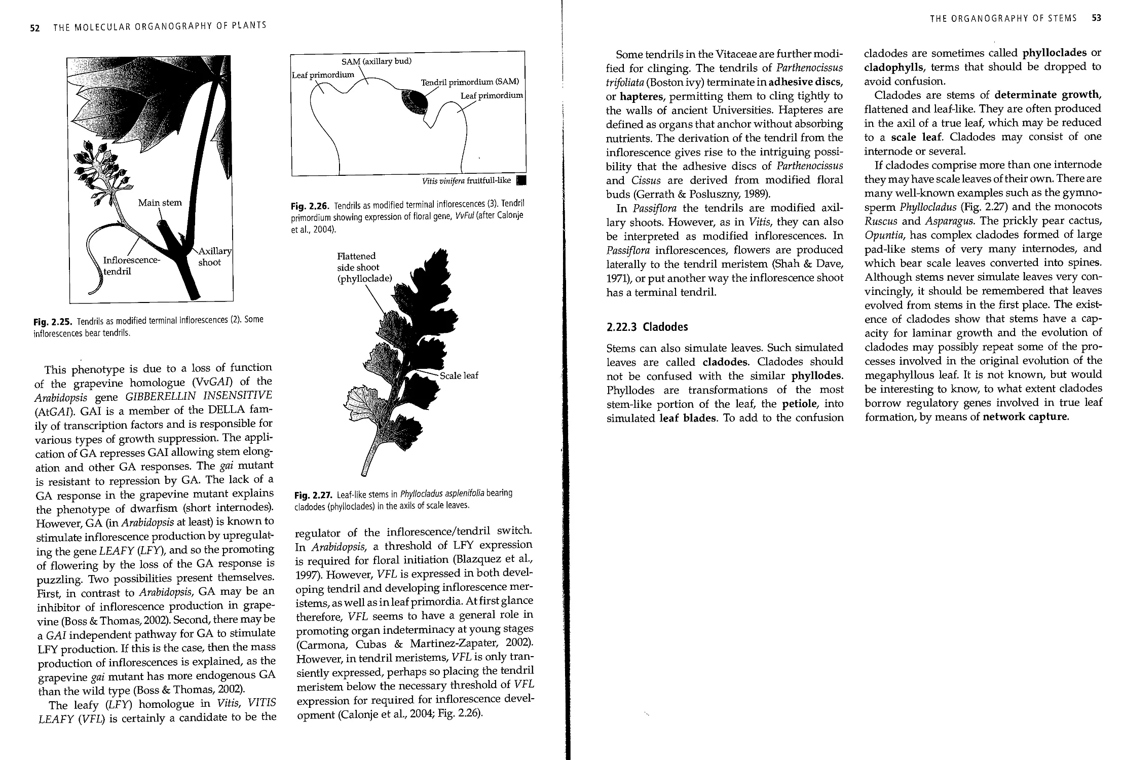
Now it must be recorded that the botanist Pfitzer1 had a favorite subject commensurate
with his pedantic mind—the difference between thorns and spines. The popular saying,
"There is no rose without thorns," is false because a rose has no thorns but spines (dermal
products), while the hawthorn has thorns (transformed stems). The pedant never gave
an examination without asking about this piece of morphology. Like every student I was
prepared for this question, and, true, it was the first thing Pfitzer asked. But there must
have been a gremlin in my cockpit.21 answered giving the definition of thorn for spine and
vice versa. Pfitzer looked deeply insulted, bit his lips, and all but fainted. With subdued
and reproachful voice he continued for the prescribed half hour, asking me questions, all
of which I answered. Then he dismissed me with the grade C, which was the end of my
hope for summa cum laude.
Goldschmidt, R. B.3 (1956). The Golden Age of Zoology. University of Washington Press, Seattle
(originally published as: Portraits from Memory: Recollections of a Zoologist).
1 Ernst Hugo Heinrich Pfitzer was born on 26 March 1846 in Konigsberg and died on 3 December 1906 in
Heidelberg. In 1872 he succeeded Hofmeister as Professor and Director of the Botanical Garden in Heidelberg.
He is the author of Grundzuge einer vergleichenden morphologic der orchideen (1882) and Morphologische studien Uber
die orchideenbliite (1886).
2 This gremlin, with whom we are all familiar, is remarkably tenacious. A rose has prickles (dermal products),
whereas spines generally refer to transformed leaves.
3 -Richard Benedict Goldschmidt (April 12,1878-April 24,1958), zoologist.
Preface
J. Alfred Prufrock measured out his life in coffee
spoons,1 and so the life of the biosphere may
be measured out in the organs of plants. They
mark the seasons as they are iteratively
produced, and as they are shed or die. Their decay
drives the earth's geochemical cycles. Their
evolution and increasing complexity marks time in
the great geological intervals, and the nature
of available plant organs has determined how
the terrestrial biosphere has changed over time.
It is plant organs that provide the diversity of
plant life. Plant diversity is in the organized
riot of green organs turned to the sun to form
the thin photochemical envelope investing the
earth, transducing the energy that drives the
biosphere. Plant diversity is also in the tangle
and soar of plant structures that provide the
food and living space for millions of species,
microbiological, invertebrate and vertebrate.
The survival of these species depends on a
successful outcome, day-to-day, of their interaction
with particular plant organs. This is true for
the eagle building a nest, the leaf-cutter ant on
the forest floor or the rhizobial bacterium
inducing root nodules far belowground. The study
of the plant organ involves bringing together
evolutionary history, plant form, developmental
processes and genomic information. This book
is not a textbook of plant morphology—there
are many fine ones already written. Neither can
it offer more than a snapshot of
molecular-developmental genetics. Such a fast moving field
is constantly changing. This book is, rather, an
eclectic and personal tour through both
subjects, with the intention of indicating how these
1 Eliot, T. S. (1917). The love song of J. Alfred Prufrock. In
Prufrock, and Other Observations. The Egoist Ltd., London.
two subjects may be brought together, and also
pointing to what we do not know, but need to
know in order to achieve that unity. Despite the
title, much of plant organography is still
without a molecular basis and thus large parts of
the book lack accounts of genetic mechanisms.
These parts are included to give some idea of
the large amount we do not know, and I hope
they will be a stimulus for research. If molecular
mechanisms are not given, the unwritten
subtext "we need to know the molecular basis for
this" should be assumed. Another subtext that
should be assumed is that covering exceptions.
In a group as diverse as the land plants there
are likely to be some exceptions to even the
most general statement. "Flowering plants have
leaves" (as a generalism) is true. "All flowering
plants have leaves" (as a definition) is not true
(certain angiosperms such as Cuscuta are
essentially leafless). Statements of this kind should
be assumed to be generalisms rather than
definitions unless stated otherwise. The subject of
this book is a large one and I have had to be
selective. I apologize to all those whose work I
have not included. I would be grateful for
information about other work that should have been
included. Even in a selective and short book
such as this it is probably inevitable that there
will be some errors, or at least points of
contention. I would be grateful to have those drawn to
my attention by email at: quentin.cronk@ubc.ca.
I would like to thank all those, too numerous
to mention, who, through conversation or their
research work, have changed the way I think
about plants. For stimulating discussion of the
contents of this book, and/or helpful comments
on parts of the manuscript, or other assistance
I would like to thank especially Paula Rudall,
VII
viii PREFACE
Peter Endress, Philip Benfey, David Baum, Wilf
Schofield, Guenter Theissen, Paul Kenrick, Julia
Nowak, Barry Tomlinson, Richard Bateman,
Stefan Gleissberg, Mike Whitlock, Pat Gensel
and an anonymous reviewer. I thank Lindsay
McGhee for preparing the figures and copyright
holders and publishers for permission to redraw
some originals, particularly the Company of
Biologists (figs. 2.11, 2.13, 4.17, 4.30, 5.17, 5.23).
The work was written in part during the
tenure of a Sabbatarian fellowship at the National
Evolutionary Synthesis Centre (NESCENT) at
Duke University and I would like to thank the
National Science Foundation and NESCENT.
I thank the National Tropical Botanic Garden
(Kampong) for facilitating the photographs in the
plates. Research in my laboratory is funded by
the Natural Sciences and Engineering Research
Council (Canada). I would also like to
acknowledge gratefully the editorial promptings of
Ian Sherman, Helen Eaton and Carol Bestley of
Oxford University Press. Finally, I affectionately
dedicate this book to Laura, Sophie, and William
Cronk and to the memory of my parents.
Contents
Preface vii
1. Introduction 1
1.1 The start 1
1.2 Evolutionary innovations and the diversification of plant life on land 1
1.3 Plant versus animal development and evolution 2
1.4 The plant genome 3
1.5 Homology and serial homology 4
1.6 Latent homology 5
1.7 Processes of development 6
1.8 Evolution of development (evo-devo) 7
1.9 Evolutionary innovation: organs of novel form 9
1.10 Regressive evolution: the loss or reduction of organs and reversal
of character states 11
1.11 Fossils and phylogeny 12
1.12 Empirical morphology versus typology 16
1.13 Plant teratology 18
1.14 Corner and Sporne: a study in opposites 19
2. The organography of stems 22
2.1 Origin and homology of stems 22
2.2 General characteristics of stems 24
2.3 The structure of the seed plant SAM 26
2.4 Variation in SAM morphology 27
2.5 Molecular signaling in the maintenance of the SAM 28
2.6 Development of stem structure 30
2.7 Organization of the stele 34
2.8 Oriented development in stems 36
2.9 Molecular control of stem orientation 37
2.10 Change of directionality with stem age 37
2.11 Chirality in stem morphology 38
2.12 Architecture of the stem 40
2.13 Molecular control of determinacy and phase change in stems 41
2.14 The thickness of stems and its consequences 42
2.15 Origin of new stems through dichotomous or lateral branching 42
2.16 Stem integration and the shoot 43
CONTENTS
2.17 Forms adopted by the stem 44
2.18 Stem morphology associated with life form 44
2.19 Buds as specialized stems: survival and vegetative reproduction 46
2.20 Surface form 48
2.21 Stems associated with flowers 48
2.22 Special modifications of stems 50
3. The organography of the root 54
3.1 The era of the root 54
3.2 The origin of roots 55
3.3 Homology of roots 56
3.4 Differences between root and stem 57
3.5 The radicle and its origin in the embryo 58
3.6 The root apex and root cap 60
3.7 The RAM and its control 60
3.8 Evolution of the root apex 61
3.9 Rhizodermis and root hairs 62
3.10 Primary and secondary root systems 64
3.11 Patterns of root branching 65
3.12 Secondary thickening of roots 66
3.13 Evolutionary loss of roots 67
3.14 Roots in light and air 68
3.15 Roots specialized for mechanical support 69
3.16 Root morphology and disease 71
3.17 Roots for specialized nutrition 71
4. The organography of the leaf 79
4.1 The nature of the leaf 79
4.2 Homology of leaf-like organs . 80
4.3 Molecular basis of relationship with the shoot apical meristem 82
4.4 Axes of polarity in leaf development 84
4.5 Laminal extension and the bifacial theory 85
4.6 The parts of the leaf 87
4.7 Leaf insertion 89
4.8 The shape of the simple lamina 90
4.9 Meristems, blastozones and growth of the simple leaf 92
4.10 Molecular control of simple leaf size and shape 93
4.11 Heterophylly, heteroblasty and hormones 94
4.12 Morphology of the leaf tip and base 96
4.13 Venation 97
4.14 Lobes: leaf dissection related to primary venation 98
4.15 Teeth: leaf dissection independent of the primary venation TOO
4.16 Compound leaves 101
4.17 Homology of the compound leaf 103
4.18 Molecular control of compound-leaf development 104
4.19 Pseudocompound leaves 105
CONTENTS xi
4.20 Stipules 106
4.21 Leaves in unusual places 108
4.22 Special forms of leaves 109
4.23 Epipeltate and ascidiate leaves 110
4.24 Peltatocompound and diplophyllous leaves 112
4.25 Cotyledons 113
4.26 Scale leaves 114
4.27 Prophylls 115
4.28 Scale leaves underground 115
4.29 Phyllotaxy: orthostichies and parastichies 116
4.30 Helical phyllotaxy 117
4.31 Whorled and decussate phyllotaxy 118
4.32 Phyllotaxy: molecular control of divergence angle in the SAM 119
4.33 Leaf rolling and folding: ptyxis 120
5. Sporangium to seed 123
5.1 Alternation of generations 123
5.2 Evolutionary significance of the diplohaplontic life cycle 125
5.3 Gene expression and the diplohaplontic life cycle 126
5.4 Epigenetics and parental conflict 126
5.5 The evolutionary history of the life cycle 128
5.6 The sporangium 130
5.7 The sporogonium 131
5.8 The superficial sporangium 132
5.9 Megasporangia and microsporangia 133
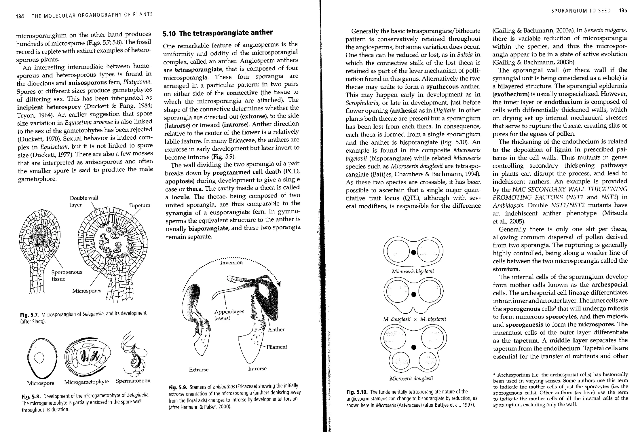
5.10 The tetrasporangiate anther 134
5.11 The ovule as a covered megasporangium • 136
5.12 The morphology of the ovule 138
5.13 The molecular control of ovule development 139
5.14 Sporogenesis and spores 142
5.15 The gametophyte and prothallus 143
5.16 The prothallus of seed plants 144
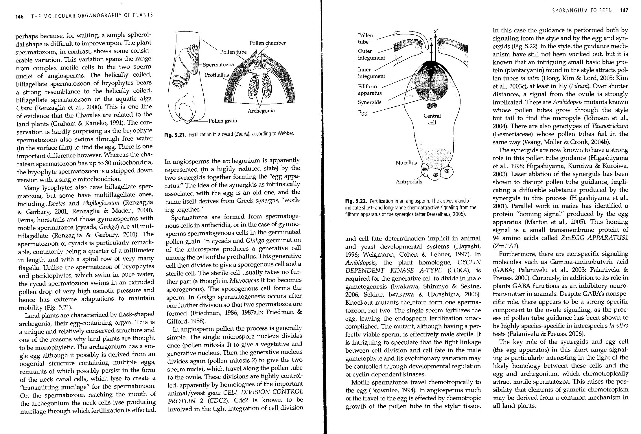
5.17 Gametogenesis and fertilization 145
5.18 The seed 148
5.19 Formation of the embryo 148
5.20 Structure of the seed-plant embryo 149
5.21 The grass embryo 150
5.22 Food reserves of the embryo 151
5.23 Molecular control of endosperm development 151
5.24 The origin of the endosperm 153
5.25 Polyembryony 153
5.26 Agamospermy 154
5.27 The biological context of agamospermy 156
5.28 The mechanism of agamospermy 157
5.29 Dormancy and growth of the new sporophyte 158
xii CONTENTS
6. Sporophyll to flower
6.1
6.2
6.3
6.4
6.5
6.6
6.7
6.8
6.9
6.10
6.11
6.12
6.13
6.14
6.15
The origin and history of the sporophyll
Phyllospory and stachyospory in the euphyllophytes
Sporophylls of monilophytes
Simple strobili of gymnosperms
The compound strobili of conifers
The angiosperm flower as a simple strobilus
Neomorphological interpretations of the flower
The origin of the carpel from a gymnospermous precursor
Molecular theories for the origin of the flower
Rules of the flower: patterning of floral organs
Molecular control of floral organ patterning
The perianth
Merosity of the perianth
Modifications of the perianth members
A mirror up to nature: floral symmetry
6.16 The molecular mechanism underlying monosymmetry
6.17
Outside the flower: the epicalyx, calycle and petaloid bracts
6.18 The floral nectary
6.19 The androecium
6.20 The form of the stamen
6.21
The staminode
6.22 The carpel
6.23 The gynoecium
6.24 Placentation
6.25 Style and stigma
6.26 The sinking seed: epigyny and the receptacle
6.27 The fruit
6.28 The classification of fruits
6.29 Distribution of sex in flowers and inflorescences
6.30 Floral reduction and pseudanthy
6.31
6.32
The inflorescence: phase change in the SAM
Inflorescences
6.33 Molecular basis of determinacy in the inflorescence
6.34 Terminal and lateral inflorescences
6.35 Types of racemose inflorescence
6.36
Types of cymes
6.37 The leaves of inflorescences
6.38 Leaves of grass partial inflorescences
References
Index
160
160
161
162
163
164
165
166
167
168
169
171
172
173
174
178
180
182
183
184
185
186
186
188
189
190
190
192
193
193
196
198
199
199
201
202
204
205
206
208
243
CHAPTER 1
Introduction
1.1 The start
The study of plant evolution, at its grandest,
can be seen as the study of how mutations in
genes and genomes have affected the way in
which the planet functions. For instance the
evolution of oxygenic photosynthesis
(involving two photosystems) from anoxygenic
photosynthesis (using a single photosystem) changed
the history of life. It involved gene changes in
prokaryotic life forms (Allen & Martin, 2007)
and was the first step toward plant evolution.
This event, at around 2700 million years before
the present (MyrBP), changed the atmospheric
chemistry profoundly. The oxygen so produced
permitted the evolution of complex life forms. It
even changed the color of the earth, as oxidation
tinted the land. Without the evolution of
photosystems, geological formations with oxidized
iron, such as the Devonian "new red sandstone"
of Britain, would not be as they are.
Another example of a key innovation of
planetary importance is the evolution (around 600
MyrBP) of parenchymatous cell division in
plants from its precursor, filamentous cell
division. This ultimately permitted the evolution of
complex organization in plants. Filamentous cell
division is still present in most algae including
some which appear to have, at least superficially,
quite complex organization, like the pseudopa-
renchymatous Codium. Parenchymatous cell
division is found in certain green algae where
it has evolved at least twice, in the ulvophytes
and in the charophytes. Control of cell division
in three dimensions, as in the parenchymatous
system, implies greatly increased control over
the cell division process.
1.2 Evolutionary innovations
and the diversification of plant
life on land
The colonization of the land by plants (c. 450
MyrBP) was a major event in earth's history, and
therefore were the evolutionary innovations that
permitted this: cuticle, stomates, lignin, sporo-
pollenin (Table 1.1). However, the focus of this
book is on those features that permit
terrestrial ecosystems to diversify. The first of these
(detailed in Chapter 2) was the axis (450-400
MyrBP), the radial, strengthened, tip-growing
cylinder of tissue that allowed sporangia to be
lifted out of the boundary layer (for spores to
disperse over vast monoclonal mats), and the
plant body to be lifted toward the sun. In this
is the beginning of competition for light that
would culminate in giant redwood forests
rising over 100 m above the ground.
The next is the leaf (Chapter 3) at c. 400
MyrBP, for which there are at least two
separate origins, microphyll and megaphyll. Lateral,
dorsiventrally flattened organs increased
the efficiency of carbon cycling, and greatly
increased ecosystem productivity and the
drawdown of carbon from the carbon-dioxide-rich
atmosphere. The origin of the root (Chapter 4)
at c. 400 MyrBP was also important in allowing
the evolution of complex plants. Roots provided
anchorage for taller organisms and a
mechanism for water and nutrient absorption from
soil directly and through mycorrhizal
symbiosis. However, most importantly for planetary
history, the origin of the root allowed the
phenomenon of soil to come into existence for the
first time. Soil formation is engendered by the
1
2 THE MOLECULAR ORGANOGRAPHY OF PLANTS
Table 1.1 An overview of levels of land plant evolution.
Level and date
Activity
Characters
Level -1 (prior to 450 MyrBP)
Level 0 (c. 450 MyrBP)
Level +1 (c. 450-420 MyrBP)
Level +2 (c. 420-380 MyrBP)
Level +3 (c. 365 MyrBP)
Level+4 (c. 150-80 MyrBP)
Level +5 (c. 80-30 MyrBP)
Prior to colonization of the land (increase in cellular
complexity)
Colonization of the land
Early evolution of land plants
Diversification of reproductive and vegetative organs
Tree building, seed building
Early angiosperm diversification
Later angiosperm diversification
Parenchymatous growth, differentiated cell types
Cuticle, stomates, sporopollenin
Sporophyte building, axis building
Roots, leaves, heterospory
Massive construction and seeds
The flower and associated features
Evolutionary innovations of derived clades such as
grasses, legumes, orchids, Asteraceae
injection of carbon and organic acids deep into
weathered geological strata, irrevocably
changing geochemical cycles, patterns of weathering
and sedimentary processes.
Another innovation of major impact was
heterospory and the seed (Chapter 5) at around 365
MyrBP. The seed, as an agent of dispersal that
was both resistant and well-resourced, allows
plant regeneration under conditions that would
otherwise be hostile. The seed appears to have
allowed plants of the late Devonian to
colonize the uplands, so completing the terrestrial
plant conquest of the land and enhancing plant-
induced changes to global geochemical cycles.
Finally, the origin of the flower (Chapter 6) at
around 150 MyrBP commenced the rise to
dominance of the angiosperms (flowering plants). The
flower promotes efficient and specialised gene
flow and that, in turn, promotes diversification.
The flower and especially its component, the
carpel, can therefore be seen as the essential
evolutionary innovations that led to the immensely
species-rich biomes of the present, such as the
lowland tropical rainforest.
1.3 Plant versus animal development
and evolution
It is striking that in animals complex multi-
cellularity appears to have evolved only once,
whereas in plants it has evolved several times.
For instance it evolved in the clade that includes
the unicell Chlamydomonas and the multicell
Ulva (sea lettuce). It also evolved in the clade that
contains the unicell Mesostigma and the multicell
Arabidopsis (thale cress). In contrast, the
colonization of the land by complex multicellular animals
has happened several times, whereas in plants,
complex multicellular algae colonized the land
only once. This contrast tells us a lot about the
different way of life of plants and animals and
their different evolution.
The predisposition to multicellularity in plants
points up a difference in cell division: in plants
there is a sticky cell wall and division is by
formation of a specialized microtubule structure
(a phragmoplast in charophytes and land plants,
a phycoplast in other green algae) on which the
new cell walls form in intimate contact as a cell
plate, so clumping of daughter cells is hard to
avoid. Kinesin-like calmodulin-binding protein
(KCBP) appears to be essential to this kind of
cytokinesis in both land plants and green algae
(Dymek et al., 2006), suggesting that this form
of cytokinesis is a green plant innovation. In
animals, cell division is by scission, which has
the effect of separating the cells more cleanly,
although the "midbody" of the animal cell can be
viewed as having an analogous role to the
phragmoplast (Otegui, Verbrugghe & Skop, 2005).
The substantial plant cell wall is a
distinguishing feature of plants, a secreted lattice of
carbohydrates and proteoglycans. It has
numerous important functions in plant biology. First, it
acts like a sponge in holding water by capillary
action, creating a water and ion store called the
apoplast. Its comparative stickiness and rigidity
prevents cell movement in plant development.
Animal cells can, and do, slide past each other to
take up programmed developmental positions.
INTRODUCTION 3
In plants, however, cell-cell positional signaling
generally determines cell fate during
development (Berger et al., 1998), rather than
developmental history, that is, cell lineage effects, which
are less common. One result is the lack of
distinction in plants between germline and soma.
Instead, cell position determines the
development of germ cells (male pollen and megaspore
mother cells) rather than cell lineage.
Land plants, unlike animals, have an open
morphogenesis in that they grow continuously
during their life cycle, adding modules in an iterative
fashion (Tomlinson, 1984). This iterative growth
is made more complex by interacting with the
environment, causing a persistent environment-
development signaling system. Furthermore,
the many modules (e.g. leaves, roots and stems)
produced by an individual plant must be
physiologically integrated. By contrast, most animals
have closed morphogenesis, with a single
developmental trajectory Animal development, when
completed at maturity, generally produces a fixed
number of organs which is largely independent
of the environment. Humans raised on a high
protein diet are often taller, but they do not have
excess arms and legs. There is thus no
continuous feedback between the major features of
animal development and the environment. Instead,
and in contrast with plants, animals have the
power of changing their location and hence their
environment by motility.
Animals are often separated from the
environment by exoskeleton, scales or fur, or by
the physiological separation of homoeostasis.
Changing environments, even moving into the
hostile environment of the land from the sea, is
therefore relatively straightforward. For plants,
which are much more in equilibrium with their
environment, such transitions are rare. This
may be the reason why complex multicellular
plants colonized the land only once. The
evolution of the cuticle, a semi-impermeable surface
layer made up of a lipid polymer called cutin (Li
et al., 2007), was the key innovation that gave
plants sufficient separation from the hostile
aerial environment to dominate the land.
An extraordinary feature of plants, at least
when seen from an animal perspective, is the
alternation of generations (Bell, 1989; Kenrick,
1994; Sheffield, 1994). Many plants (and all land
plants) have a life cycle in which haploid and
diploid generations alternate with each other.
These generations most often have very
different developmental trajectories, and so a given
genome has within it the potential to create two
phenotypically different organisms. The
different developmental paths undertaken by haploid
spores and diploid zygotes therefore indicate
that radically different transcriptomes are
possible depending on epigenetic control (such as
chromatin remodeling and DNA methylation)
triggered by ploidy level, or at least by ploidy
level transition.
1.4 The plant genome
Ultimately, it is the changes in plant genomes that
have created the ecosystems on which human
life depends. Although these changes took place
hundreds of millions of years ago, the genomic
basis of revolutions in the phenome are
discoverable by comparative and functional morphology
and genomics. Although sequence divergence
between the major clades of land plants is
extensive, only some of these divergences are
responsible for the key innovations that have
built the biosphere. Molecular-developmental
genetics must therefore sieve through millions
of genomic changes to discover those that have
been critical in plant evolution.
Plants have three genomes. The mitochondrial
genome is generally about 10 times the size of
the animal mitochondrion but is very variable
in length (-200-2500 kb); it is conservative in
sequence but highly labile in gene arrangement.
The chloroplast genome is generally 120-160 kb,
but in Pelargonium it is 217 kb (Chumley et al.,
2006). The chloroplast is fairly conservative in
gene sequence and arrangement, and, although
with limited effect on overall plant morphology,
this genome does encode much of the
machinery for autotrophy
It is the nuclear genome, however, that is
responsible for most of the morphological
diversity of plants. It is enormously variable in
size (115 Mb in Arabidopsis, yet 250,000 Mb in
Psiloturn). It is also variable in gene sequence and
arrangement. Nuclear genomes can increase in
4 THE MOLECULAR ORGANOGRAPHY OF PLANTS
size by doubling through polyploidy (Adams &
Wendel, 2005), or be hugely bloated by
multiplication of transposable elements.
There are three major types of change
relevant to the evolution of plant development.
First, gene duplication by polyploidy or
tandem duplication allows two gene copies, at first
identical, to diverge in function. This is called
neofunctionalization if one copy retains the
old function and the other copy acquires a new
function. The alternative is subfunctionaliza-
tion, when each copy retains part of the
ancestral function. Second, existing genes can change
their expression patterns, usually by mutations
in regulatory regions (cts-regulatory change).
This is an important possible mechanism of het-
erochronic and heterotopic change. Third, there
may be a direct change in protein function. If
this change occurs in a regulatory gene, such
as a transcription factor, major changes can
occur in the regulation of other genes (trans-
regulatory changes).
Whole genome sequencing is of great
importance in the elucidation of developmental
evolution, not least because it allows the
diversification of developmentally important gene
families to be assessed (Leebens-Mack et al., 2006).
For evolutionary-developmental genetics it was
somewhat unfortunate, although inevitable, that
early genome sequencing focused so heavily
on angiosperms, a limited phylogenetic spread.
This was not the case for animals, for which early
sequences of yeast, nematode, fruit-fly and human
quickly provided a wide phylogenetic sampling.
However, the plant community is catching up
rapidly. In 2005, the moss Physcomitrella entered
the Community Sequencing Program (CSP) of
the Joint Genome Institute (JGI), and genome
sequencing is now completed for Physcomitrella
(Rensing et al, 2008).1
The liverwort Marchantia has been added to
the CSP of the JGI for 2008 as a "small project"
for a mixture of conventional Sanger sequencing
and pyrosequencing. The sequencing of the club-
moss Selaginella moellendorffii (Wang et al., 2005)
is now substantially completed and the sequence
1 http://genome.jgi-psf.org/Phypal_l/Phypal_l.home.html
is currently in annotation, the reads having been
released in 2007. Sequenced genomes of a fern
(for instance Ceratopteris; Nakazato et al., 2006)
and a hornwort (such as Anthoceros or Notothylas)
would also be of great scientific value.
1.5 Homology and serial homology
A necessary objective of comparative biology is
to compare like structures with like structures,
which in an evolutionary sense means that such
structures derive from developmental programs
(genes and modes of gene action) inherited from
a common ancestor that also had that
developmental program. Another way of putting this
is "resemblance caused by a continuity of
information" (Van Valen, 1982). Such resemblance is
termed homology. It is quite different from
similarity (with which it is sometimes confused) as
similarity can be superficial with little biological
significance. However, homologous structures
may not look particularly similar. A palm leaf
and a cactus spine are not particularly similar
but they are homologous as phyllomes, and, at a
deep level, share a developmental program that
they have inherited from a leafy ancestor with
that program.
There are a number of ways of assessing
homology. First, there is the ascertainment that
resemblances are not merely superficial, as they
would be if the structures were analogous rather
than homologous. A powerful way of assessing
deep as opposed to superficial resemblance is to
look at development. Development (or
"embryology") was central to Darwin's definition of
homology as
that relation between parts which results from their
development from corresponding embryonic parts,
either in different animals, as in the case of the arm
of man, the fore-leg of a quadruped, and the wing of
a bird; or in the same individual, as in the case of the
fore and hind legs in quadrupeds, and the segments
or rings and their appendages of which the body of
a worm, a centipede, etc., is composed. The latter is
called serial homology. The parts which stand in such
a relation to each other are said to be homologous, and
one such part or organ is called the homologue of the
other. In different plants the parts of the flower are
INTRODUCTION 5
homologous, and in general these parts are regarded
as homologous with leaves. (Darwin, 1872)
Second, and more recently, molecular data
has added an important means of assessing
homology. The involvement of homologous
genes, acting in similar networks and with
similar expression patterns, points to homology
(Jaramillo & Kramer, 2007).
Finally, and also unavailable to Darwin, there
is the phylogenetic criterion. The presence of
structures that result from inherited
information from a common ancestor should be a syn-
apomorphy, or shared derived character, for the
clade (monophyletic group) that derives from
that ancestor. Homologous structures should
therefore map perfectly to the structure of the
phylogenetic tree. Thus the presence of micro-
phylls maps to a clade within the lycophytes
(and including all extant lycopsids). This is
evidence that the microphyll arose only once, and
that all microphylls are homologous.
These criteria can be used together in cross-
species comparisons of organs. However, in
assessing serial homology (for instance between
petals and leaves within an organism) the
phylogenetic criterion is unavailable. By contrast in
assessing gene homology, phylogeny is the only
usable criterion (Patterson, 1988). The
phylogenetic criterion, when available, is the strongest, as
it is the surest way to distinguish homology from
analogy. Similarity alone (even detailed
similarity) may always be, at least in theory, the result of
parallel evolution of analogous structures. This
is an enduring problem for serial homology for
which the phylogenetic criterion is not available.
However, the developmental and molecular
criteria are particularly powerful in assessing
serial homology as the development can be studied
in the same organism, with the same genotype.
Furthermore transitional organs often occur
between serial homologues (such as
transitions between stamen and petal in the water
lily Nymphaea). Traditionally this has been taken
to indicate that the developmental pathways of
both are sufficiently similar to intergrade, and
this in turn has been taken as evidence of
homology. With molecular genetics, this hypothesis
can now be tested directly. However, it should
be borne in mind that the relationship between
gene pathway and phenotype may be complex,
and gene pathways are constantly evolving,
giving rise to the potential for co-option and
dissociation of genes to and from a particular
structure (Jaramillo & Kramer, 2007).
As phylogenetically determined homologies
are equivalent to synapomorphies, it is clear
that they will be phylogenetically nested. It is
therefore important to be clear about the level
at which homology between particular organs
is being discussed. It is not controversial that
leaves, petals and stamens are homologous
at the level of phyllomes. What is less clear is
whether petals and stamens share homology at
the level of microsporophylls, from which leaves
and sepals are excluded. The staminopetal
interpretation of petals of the eudicots suggests that
petals evolved as sterilized microsporangia.
This view is widely supported, although it has
been challenged (De Craene, 2007).
1.6 Latent homology
In discussing homology it should b e noted that
features that are independently derived, and
therefore not homologous, may result from homology
of an underlying feature at a deep level. This has
variously been called latent homology,
phylogenetic prepatterning or synapomorphic tendency
(Cronk, 2002). As an example, floral zygomor-
phy in asterids (such as Antirrhinum) and rosids
(such as Leguminosae) may be mentioned. It is
apparent from phylogenetic evidence that zygo-
morphy has an independent origin in the two
clades. However, the same genes, homologues of
CYCLOIDEA, are involved in both Antirrhinum
(Corley et al., 2005; Luo et al, 1996) and legumes
(Citerne et al, 2003; Feng et al, 2006). This is not
coincidence but results from the presence of a
key gene of similar basic function in both rosids
and asterids. The homology (or latent homology)
is "genetic competence to evolve zygomorphy."
Another example is nodulation by actino-
bacteria or rhizobial bacterial in the eurosid 1
clade (Table 1.2). Nodulation has arisen
specifically in this clade, but it has arisen many times
6 THE MOLECULAR ORGANOGRAPHY OF PLANTS
Table 1.2 Distribution of nodulation in families of the eurosid 1 clade, largely representing independent evolutionary events but
indicating a latent homology for the nodulation competence within this clade.
Type of nodulation
Rhizobial
Actinobacterial (Frankia)
Family
Leguminosae (Fabaceae)
Ulmaceae
Betulaceae
Casuarinaceae
Coriariaceae
Datiscaceae
Eleagnaceae
Myricaceae
Rhamnaceae
Rosaceae
Genera
Various
Parasponia
Alnus
Allocasuarina, Casuarina, Ceuthostoma, Gymnostoma
Coriaria
Datisca
Eleagnus, Hippophae, Shepherdia
Comptonia, Myrica
Ceanothus, Colletia, Discaria, Kentrothamnus, Tetanilla, Talguenea, Trevoa
Cercocarpus, Chamaebatia, Cowania, Dryas, Purshia
independently (Swensen, 1996). There are
developmental differences between actinobacterial
nodules, with root-like vasculature, and
rhizobial nodules with stem-like vasculature (Sprent,
2001) although both have a first stage, which is
the formation of a "prenodule." More
importantly, the association of nodulation with a single
clade indicates that there is an underlying latent
homology for competence to evolve nodulation
that characterizes this clade.
Darwin was aware of this phenomenon and
called it "the tendency to vary in like manner"
caused by a "community of descent" (Darwin,
1859). Later Darwin (1868) named this the "law
of homologous variation" under which name it
is often wrongly attributed to N.I. Vavilov who
documented it extensively in cereals. Darwin
illustrated the phenomenon by the independent
origin of swollen "roots" (hypocotyl) inbrassicas,
particularly swede or ruta-baga (Brassica napus
var. napobrassica) and turnip (Brassica rapa var.
rapa). To these might be added radish (Raphanus
sativus) in the closely related genus Raphanus.
This latent homology (the competence to form
swollen roots in Brassica and related genera)
most probably has a molecular-developmental
basis, but the details are unknown.
1.7 Processes of development
The main processes of development are given in
Table 1.3. Highly important among these are the
processes that give rise to the position, identity
and polarity of organs. A phyllome arising as
a primordium on the flank of an apical meri-
stem is taken as an example. Its position will
be set by phyllotaxis, a patterning mechanism
based on the requirement for auxin for
primordium development, and the auxin sink provided
by the other primordia. The highest auxin
concentration will therefore be at a distance from
the other primordia, and this is where the new
primordium forms (Chapter 3, Section 4.32).
The developing primordium also has what is
termed an identity. For instance if B- and C-class
MADS-box genes are expressed, downstream
gene action ensues which ensures the
development of that phyllome as a stamen. The MADS-
box genes have conferred stamen identity on the
developing primordium long before any
stamenlike features are visible (Chapter 6, Section 6.11).
Furthermore, identity may be established as a
gradient across an organ, and this is known as
polarity. A good example of polarity is the abax-
ial-adaxial polarity that establishes across a leaf
primordium that ensures that leaves develop as
bifacial organs; in other words, the lower surface
is different from the uppersurface (Chapter 4,
Section 4.6).
These developmental concepts, position,
identity and polarity as well as others, all have a
molecular basis in gene expression and in the
chemical signaling to which genes respond.
Most of these concepts and their molecular basis
have been worked out in model eudicots,
especially Arabidopsis. However, these developmental
INTRODUCTION 7
Table 1.3 A table showing the main processes of plant development.
I. Processes of quality and quantity
II. Spatiotemporal processes
Process
Identity
Polarity
Organ patterning
Modularity
Growth
Relative growth
Position
Timing or duration
Effect
Nature of organs
Identity across organs (base to tip = proximodistal; relative
to apex = adaxial/abaxial; side to side = mediolateral)
Identity within organs (e.g. stripes, spots)
Number of organs
Size
Shape
Spatial: positional signaling
Temporal
Note: Compare with Table 1.4.
Table 1.4 A summary of important evolutionary-developmental change.
Evolutionary-developmental change
Effect
I. Changes of quality and quantity Change in degree of polarity
. Spatiotemporal changes
Homeosis
Meristic or modular change
Change of size (absolute growth or heterometry)
Shape change
Heterochrony
Heterotopy
Symmetry changes (more/less asymmetry)
Change of identity
Number
Reduction/loss, or elaboration
Altered relative growth
Change of timing or duration
Change of position
Wore: Compare with Table 1.3.
concepts are applicable to other groups of land
plants, even though the details of the
molecular mechanisms may not be. The elucidation of
more general principles for the land plants as a
whole is an important challenge for molecular-
developmental biology.
1.8 Evolution of development
(evo-devo)
The morphological phenotype is the result of
developmental processes. In the same way,
evolutionary changes in the phenotype are the
result of evolutionary changes in
developmental processes. The study of evolutionary changes
in developmental processes is sometimes
contracted to "evo-devo" and it represents a very
fertile way of investigating evolution.
Developmental processes result from
underlying gene action and so comparative molecular-
developmental genetics is the most basic, powerful
and satisfying means of testing evolutionary
hypotheses and describing evolutionary change
in the form of organisms. The developmental
processes listed in Table 1.3 have counterparts
as evolutionary processes, and a list of these
corresponding evolutionary processes is given in
Table 1.4. One of these is change in absolute growth
or heterometry (Webster & Zelditch, 2005). For
instance, Isoetes is the extant survivor of the clade
that includes the extinct rhizomorphic lycopsids,
yet Isoetes species generally have a cormoid stem
often less than a centimetre high, whereas the
Carboniferous Lepidodendronf may have grown
up to 50 m or more in height. (The dagger
represents extinct taxa.) The molecular-developmental
changes underlying the heterometry in this
lineage are not known. Another process,
heterochrony or change of timing in development, may
be involved: if the attainment of sexual maturity
and the consequent cessation of growth is
accelerated, absolute size will be decreased.
8 THE MOLECULAR ORGANOGRAPHY OF PLANTS
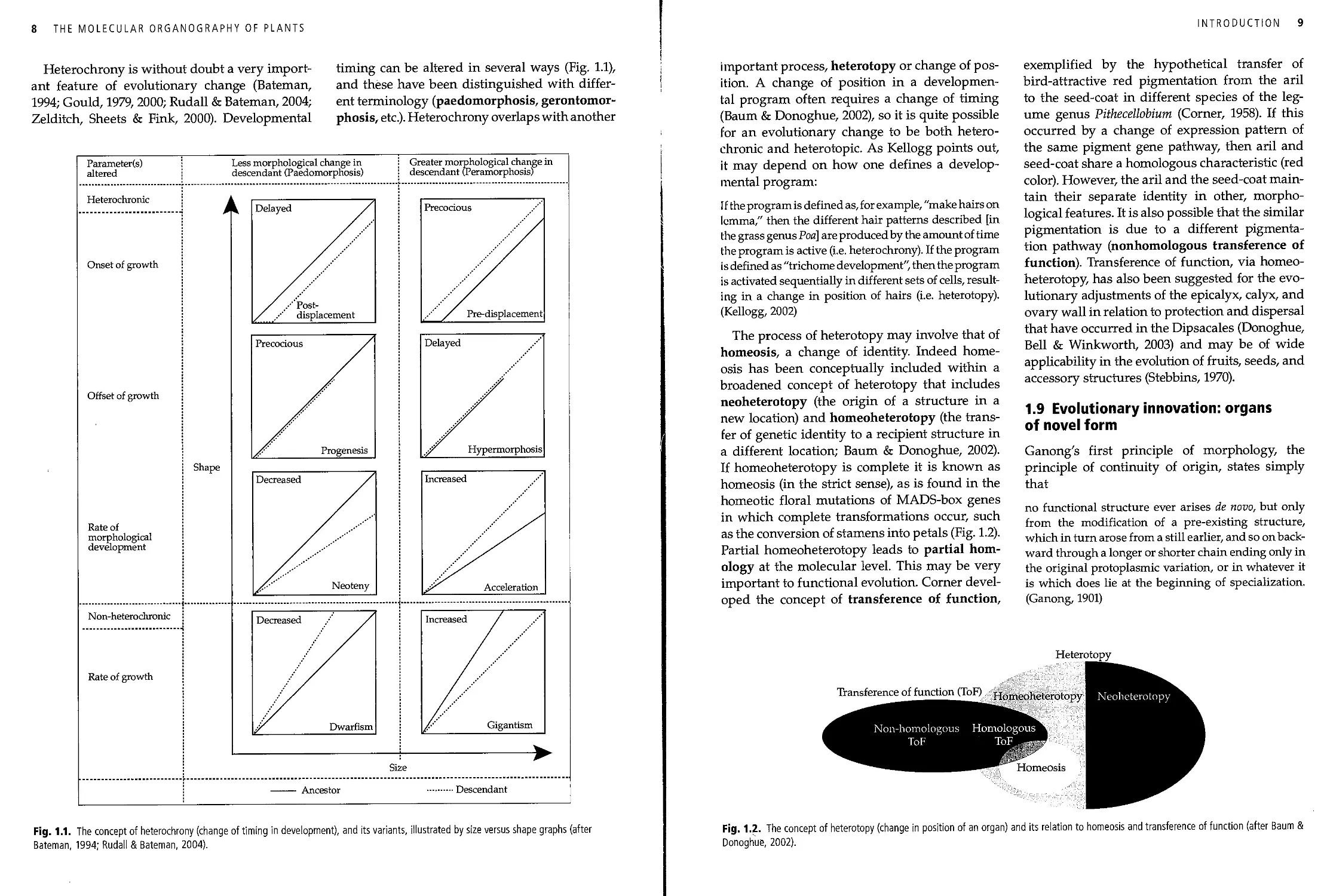
Heterochrony is without doubt a very
important feature of evolutionary change (Bateman,
1994; Gould, 1979, 2000; Rudall & Bateman, 2004;
Zelditch, Sheets & Fink, 2000). Developmental
timing can be altered in several ways (Fig. 1.1),
and these have been distinguished with
different terminology (paedomorphosis, gerontomor-
phosis, etc.). Heterochrony overlaps with another
Parameter(s)
altered
Less morphological change in
descendant (Paedomorphosis)
Greater morphological change in
descendant (Peramorphosis)
Heterochronic
Onset of growth
Offset of growth
Shape
Rate of
morphological
development
Non-heterochronic
Rate of growth
Size
Ancestor
■ Descendant
Fig. 1.1. The concept of heterochrony (change of timing in development), and its variants, illustrated by size versus shape graphs (after
Bateman, 1994; Rudall & Bateman, 2004).
INTRODUCTION 9
important process, heterotopy or change of
position. A change of position in a
developmental program often requires a change of timing
(Baum & Donoghue, 2002), so it is quite possible
for an evolutionary change to be both
heterochronic and heterotopic. As Kellogg points out,
it may depend on how one defines a
developmental program:
[f the program is defined as, for example, "make hairs on
lemma," then the different hair patterns described [in
the grass genus Poa] are produced by the amount of time
the program is active (i.e. heterochrony). If the program
is defined as "trichome development", then the program
is activated sequentially in different sets of cells,
resulting in a change in position of hairs (i.e. heterotopy).
(Kellogg, 2002)
The process of heterotopy may involve that of
homeosis, a change of identity Indeed home-
osis has been conceptually included within a
broadened concept of heterotopy that includes
neoheterotopy (the origin of a structure in a
new location) and homeoheterotopy (the
transfer of genetic identity to a recipient structure in
a different location; Baum & Donoghue, 2002).
If homeoheterotopy is complete it is known as
homeosis (in the strict sense), as is found in the
homeotic floral mutations of MADS-box genes
in which complete transformations occur, such
as the conversion of stamens into petals (Fig. 1.2).
Partial homeoheterotopy leads to partial
homology at the molecular level. This may be very
important to functional evolution. Corner
developed the concept of transference of function,
exemplified by the hypothetical transfer of
bird-attractive red pigmentation from the aril
to the seed-coat in different species of the
legume genus Pithecellobium (Corner, 1958). If this
occurred by a change of expression pattern of
the same pigment gene pathway, then aril and
seed-coat share a homologous characteristic (red
color). However, the aril and the seed-coat
maintain their separate identity in other,
morphological features. It is also possible that the similar
pigmentation is due to a different
pigmentation pathway (nonhomologous transference of
function). Transference of function, via
homeoheterotopy, has also been suggested for the
evolutionary adjustments of the epicalyx, calyx, and
ovary wall in relation to protection and dispersal
that have occurred in the Dipsacales (Donoghue,
Bell & Winkworth, 2003) and may be of wide
applicability in the evolution of fruits, seeds, and
accessory structures (Stebbins, 1970).
1.9 Evolutionary innovation: organs
of novel form
Ganong's first principle of morphology, the
principle of continuity of origin, states simply
that
no functional structure ever arises de novo, but only
from the modification of a pre-existing structure,
which in turn arose from a still earlier, and so on
backward through a longer or shorter chain ending only in
the original protoplasmic variation, or in whatever it
is which does lie at the beginning of specialization.
(Ganong, 1901)
Heterotopy
Transference of function (ToF) Hmnpnhri^m^nv
-homologous Homologous
ToF ToF -
Homeosis
Fig. 1.2. The concept of heterotopy (change in position of an organ) and its relation to homeosis and transference of function (after Baum &
Donoghue, 2002).
10 THE MOLECULAR ORGANOGRAPHY OF PLANTS
Such an axiom can also be couched in terms of
gene networks or developmental pathways. A
novel gene network does not spring into
existence, de novo, but evolves from preexisting gene
networks. The same applies to genes themselves.
New genes do not arise sui generis within the
genome but derive by mutation or duplication
from preexisting genes.
Thus, in the context of land plants, the
developmental process leading to axis formation was
a basic one from which the developmental
pathway to leaf, root and stem differentiated. Leaf,
root and stem each in their turn differentiated
into the large number of derivative organs,
generically lumped (in Worsdell's terminology)
under the terms phyllome, rhizome2 and cau-
lome (Worsdell, 1905).
The importance of gene regulation in
evolution was pointed out in early work on the protein
differences between humans and chimpanzees
(King & Wilson, 1975). It is difficult to account for
the large anatomical and behavioral differences
between humans and chimpanzees by the rather
small degree of molecular divergence. Instead
regulatory changes controlling the expression of
genes were postulated (Carroll, 2005). This idea
continues to gather empirical support,
particularly the notion that changes in cz's-regulatory
regions of transcription factors (i.e. regulation of
regulators) are of key importance in
morphological modification (Cronk, 2001; Doebley & Lukens,
1998). A good example is found in the evolution
of maize (Zea mays). The maize allele of the gene
TEOSINTE-BRANCHED 1 (TBI) confers the
nontillering trait of domesticated maize,
relative to its tillering progenitor (Zea parviglumis).
There is evidence of a strong selective sweep of
the 5-prime flanking regions in domesticated
maize suggesting that the ds-regulatory regions
of this gene have undergone intense selection for
altered expression patterns of TBI (Clark et al,
2006; Wang et al, 1999).
2 This usage, rhizome as a collective term for root-derived
organs, used by Worsdell in what he calls the "original,
literal sense," has for obvious reasons not been taken up, due
to prior use in the sense of "underground stem." Radicome
would be a better term if one were needed. It might be
objected that it mixes Latin and Greek, but this is now
common practice in scientific word formation.
An important mechanism for evolutionary
innovation, although poorly documented in
plants is network capture. Through changes
of ds-regulation, a gene may be captured by an
existing gene regulatory network, so resulting in
gene co-option to a new function. A good
example of this is the evolutionary gain of a novel
wing pigmentation spot in Drosophila biarmipes
(Carroll et al, 2005; Gompel et al., 2005). The
general pigmentation gene, YELLOW, was captured
by the regulatory network controlling wing
development. The capture involved a mutation
in ds-regulatory sequence that brought it under
the control of the ENGRAILED regulatory
protein. Such mutations tend to be genetically
dominant, as they involve a gain of function.
Gene redundancy caused by gene duplication
has long been recognized as an important raw
material for evolution (Ohno, 1970). The prone-
ness of plants to polyploidy, perhaps due in part
to plant synthesis of spindle inhibiting alkaloids,
such as nicotine and colchicine, makes
duplication events particularly common in plants
(Cronk, 2001). Also plants are often subjected to
heat and cold shocks, having few means of
temperature control, and these shocks are known
to disrupt spindle formation. Even Arabidopsis
thaliana, with just five chromosomes, has 60%
of its genome segmentally duplicated by paleo-
polyploidization events (Blanc & Wolfe, 2004).
Plant genomes undergo frenetic change, when
viewed on a geological timescale, with genes
being duplicated, destroyed and continuously
repatterned on chromosomes.
Gene duplication can lead to loss of one copy
by pseudogene formation and a return to a
single gene state. Alternatively, it can lead to
retention of both copies and divergence of gene
function by subfunctionalization and neofunc-
tionalization (Causier et al, 2005; Hileman &
Baum, 2003; Lynch & Force, 2000). By this means,
some ancient duplication events may have had
a major effect on plant form. An ancient
duplication separates the KNOX homeodomain-
containing genes into two clades, class 1 and
class 2 (Champagne & Ashton, 2001; Singer &
Ashton, 2007). KNOX genes that are important
in apical meristem determination and control,
such as SHOOTMERISTEMLESS (STM), are
INTRODUCTION 11
class 1 KNOX genes. This duplication probably
occurred c. 430-475 MyrBP, at around the time
of the origin of the land flora. It therefore may
have been important in allowing the evolution
of a class of genes with meristem specific
members, and in facilitating complex organization in
land plants.
Repeated doubling of the plant genome has
led to some very large gene families, and some
of these gene expansions may be important in
the evolution of morphology. In the human
genome there are some 43 MYB transcription factors,
yet in Arabidopsis this number is 198 (Chen et al.,
2006). MYB transcription factors have a
conserved DNA-binding domain consisting of up to
three imperfect repeats of 51-53 amino acids (Rl,
R2 and R3). Most plant MYB genes contain only
R2 and R3 (the so-called R1R2 MYBs of which
there are 126). The existence of numerous R1R2
M YB genes is a plant sp ecific trait. S everal of these
genes are known to be important regulators of
morphogenesis, such as the ARP genes
controlling leaf development (Chapter 4, Section 4.4).
Through the evolutionary divergence of the
MYB family (Zhang, Chopra & Peterson, 2000),
and other similar gene families, genome
duplication has had an enormously important impact
on plant complexity.
1.10 Regressive evolution: the loss or
reduction of organs and reversal of
character states
In regressive evolution, a clear distinction needs
lo be made between the loss of an entity (such
us an organ) and the change of a quality (i.e. the
reversal of a character state transition; Mabee
et al., 2007). The loss of leaves in Cuscuta (for
instance) is an evolutionary innovation in terms
of the leafy clade to which Cuscuta belongs. To
see this as a reversal it is necessary to go back
to leafless ancestors of the seed plants, which is
not an appropriate comparison. The reverting of
a character state, such as round leaves reverting
to elliptic, may indeed be considered evolution
in reverse. Another type of reversal is the regain
of a lost entity (e.g. an organ type), such as the
regain of inflorescence bracts in some mutants of
Arabidopsis. These three types of regressive
evolution are set out in Table 1.5.
Loss of organs may be by active suppression
due to a gain-of-function (i.e. genetically
dominant) mutation at a suppressor locus. An
example of this appears to be the loss of bracts in the
Brassicaceae, including Arabidopsis. In most eud-
icots the inflorescence branches are subtended
by bracts. However, bracts are usually
missing from the inflorescences of the Brassicaceae,
although there is vestigial expression of bract-
specific genes where bracts would be expected.
These vestiges have been called "cryptic bracts."
The leaf developmental gene JAGGED is required
to form bracts (Dinneny et al, 2004) and this is
excluded from the inflorescence of Arabidopsis
by the action of the BLADE ON PETIOLE (BOP)
genes acting in concert with the developmental
gene LEAFY (Norberg, Holmlund & Nilsson,
2005). Thus bracts are actively suppressed in
Arabidopsis. Loss-of-function mutations in the
suppression gene network allows bracts to
form, after having been evolutionarily lost, by
desuppression. As the suppressed gene has an
Table 1.5 Genetic landscape of loss and reversal (see text for explanation).
Dominant mutations
Gain of function
Recessive mutations
Reduction of function Loss of function in
pleiotropic gene
Loss of function
in specific gene
I iiss of organ Suppression Loss with vestige
Hwnrting of character state Cryptic innovation Reversal with vestige
llti(|iiln from loss Recapture by conserved Partial desuppression
network
Loss with possible neomorphism
Reversal with possible
neomorphism
Desuppression with secondary
phenotype
Perfect loss
Perfect reversal
Desuppression
12 THE MOLECULAR ORGANOGRAPHY OF PLANTS
important role in leaf development elsewhere in
the plant there is no likelihood that the JAGGED
gene will degenerate making the reoccurrence
of bracts impossible.
Alternatively, organ loss may be via loss or
reduction of the function of a key gene required
for organ formation. These loss-of-function
mutations will be genetically recessive. If the
loss of function is partial, vestigial organs will
remain (staminodes are a common example of
this). If the gene has pleiotropic effects then a
neomorphism may accompany organ loss. An
example of a neomorphism is the change in
petal number accompanying a reversal to actin-
omorphy in Antirrhinum mutants. Antirrhinum
cycloidea/dichotoma mutants commonly have six
petals instead of the expected five. As well as
being a key controller of floral symmetry, these
genes apparently have an effect on primordium
initiation, which leads to the neomorphic pheno-
type (six petals) that accompanies loss of radial
symmetry. Perfect loss or perfect reversal, that
is, loss without vestige or neomorphism,
involving the clean deletion of an entity or quality,
appears to be rare. However, it does occur. An
example may be in lost petals of some legumes,
in which there is no vestigial ontogeny whatever
(in other legumes petal primordia are initiated
but growth past a certain stage fails to occur;
Tucker, 2001).
The apparent reversal of a character state
transition may sometimes take place using a very
different developmental mechanism to achieve
the evolutionary prior condition. This is known
as cryptic innovation (Citerne, Pennington &
Cronk, 2006). An example is found in the bird-
pollinated legume Cadia, which has reverted to
an apparently primitive condition of radially
symmetrical flowers instead of the strongly
zygomorphic bee-pollinated flowers
characteristic of other papilionoid legumes. However, this
does not result from a loss-of-function mutation
in the genes controlling floral symmetry. Instead
the dorsally expressed LEGUME CYCLOIDEA
(LEGCYC) gene controlling floral symmetry
has expanded its expression pattern in Cadia to
include all petals equally. All petals have
therefore been homeotically converted into dorsal
petals (Citerne, Pennington & Cronk, 2006). This
is therefore a cryptic innovation that uses the
machinery of zygomorphy to mimick an
ancestral radial state.
The regain of lost organs is an interesting type
of reversal. The easiest way to achieve this is if
the original loss was by suppression. A loss-
of-function mutation, to knock out the
suppression, might reactivate the original organ, as in
the JAGGED/BOP example above. However, it is
probably necessary that the relevant gene
pathway has a secondary function so it is not lost to
deleterious mutation and pseudogene formation
while inactive. Another means of organ
reactivation is cz's-regulatory network recapture, the
reconnection of a conserved network to some
key component. Network capture has been
discussed in the previous section. The example
given was the ENGRAILED network capturing
the YELLOW gene by gain of czs-regulatory
elements. One can also imagine this cz's-regulatory
element being lost, and regained, leading to
evolutionary flicker (Marshall, Raff & Raff, 1994).
1.11 Fossils and phytogeny
Fossils and molecular phylogenetic trees of
extant organisms provide the most direct, robust
and reliable forms of evidence for the history of
character evolution. Fossils present actual
characters existing at a particular geological time.
Phylogenetic trees, when well supported by a
plethora of congruent characters, which
molecular trees increasingly are, allow the
reconstruction of character state evolution by character state
mapping. New methods for incorporating fossils
into joint morphological and molecular trees are
therefore particularly exciting. Fossils, however,
are patchy both in their occurrence and in the
information that can be obtained from them. In
the history of the early land plants, no deposit
has been as important as the Rhynie Chert of
Scotland, which holds exquisitely preserved
petrifaction fossils, in which many details
indicative of early plant biology can be seen (Edwards,
1986; Satterthwait & Schopf, 1972).
The early fossil history of flowering plants
was based, until recently, largely on pollen,
INTRODUCTION 13
leaf and wood fossils, as few flower fossils,
although useful for reconstructing angiosperm
evolution, were known. Lately several excellent
early flower fossil have been discovered (Friis,
Pedersen & Crane, 2006) such as the Cretaceous
Archaeanthus flower (Cretaceous) which has
stalked follicles, emarginate leaves and scars of
stamens, inner and outer tepals and bract scales
(Dilcher & Crane, 1984). Another interesting
fossil is the still controversial Archaefructus which
has been interpreted variously as having
elongated flowers in a "prefloral state" or an
inflorescence of reduced flowers (Friis et al., 2003;
Sun et al., 1998).
There is increasing consensus on the
characteristics of the primitive flower, based on a
combination of fossil evidence and phylogenetic
trees (Doyle & Endress, 2000; Endress, 2004,2006;
Soltis & Soltis, 2004). A view of such a flower is
emerging as radially symmetrical with
numerous, separate, spirally arranged, poorly
differentiated parts, simple pistils (composed of one
carpel), a perianth of tepals (calyx and corolla
not differentiated into petals and sepals), the
stamen is somewhat leaf-like, without strongly
differentiated filaments and ascidiate carpels
(peltate tubular leaves; Table 1.6).
Molecular phylogenetic work has shed
considerable light on the early evolutionary history
of the land plants (Qiu et al, 2006, 2007). There
are, however, still many uncertainties, such as
the branching order of the three first divergent
clades mosses, liverworts and hornworts (Fig. 1.3
suggests a likely solution). Recent phylogenies
also strongly point to a monophyletic "monilo-
phyte" group (ferns, horsetails, adder's-tongues
and whisk-plants, see Figs 1.4 and 1.5; Des
Marais et al., 2003; Pryer et al., 2001; Schneider
et al., 2004). Furthermore, a monophyletic
gymnosperm clade is increasingly strongly
Polysporangiophytes
Embryophytes
(land plants)
■ Horneophytoni
■ Aglaophytoni
_wTracheophytes
(vascular plants)
. Anthocerotes
(hornworts)
_Bryophytes s.s.
(mosses)
_ Hepatics
(liverworts,
marchantiophytes)
Fig. 1.3. Phylogenetic hypothesis for the embryophytes. There is
still uncertainty over the branching order and relationships of the
three earliest diverging groups.
Table 1.6 The three early divergent clades of extant angiosperms (from evidence of molecular phylogeny) with their putatively primitive and
derived characters from a combination of fossil and phylogenetic (character state reconstruction) evidence.
Primitive characters
Derived characters
Amborellales (Amborella)
Nymphaeales (water lilies:
Nymphaeaceae, Cabombaceae,
Hydatellaceae)
Austrobaileyales (star anise and
relatives: llliciaceae s.l. [including
Schisandraceae], Trimeniaceae,
Austrobaileyaceae)
Wood vesselless, perianth parts undifferentiated,
spiral, numerous/indefinite in number, pollen
inaperturate, pistils simple (single carpel)
Wood vesselless, perianth parts numerous and spirally
arranged, separate, stamens numerous and spirally
arranged, sometimes grading into petals, pollen
monosulcate, pistils simple
Perianth parts numerous and spirally arranged,
separate; stamens numerous and spirally arranged;
pistils simple
Flowers unisexual, carpels in a single whorl
Plants herbaceous, aquatic (in Hydatellaceae
flowers reduced to single pistils or stamens)
Vessels in wood; triaperturate pollen but
not homologous with tricolpate pollen
of eudicots; leaves with ethereal oil cells
(single spherical cells filled with aromatic
compounds)
14 THE MOLECULAR ORGANOGRAPHY OF PLANTS
Euphyllophytes
(megaphyllous
plants) I
Tracheophytes
(vascular plants)
—- Psilophytoni
Radiatopsids
-^ (seed plants and
progymnosperms)
_w Monilophytes
(ferns and horsetails)
_^. Lycophytes
(clubmosses)
Cooksoniai
Rhyniai
Fig. 1.4. Phylogenetic hypothesis for the tracheophytes.
Lycopsids
Lycophytes
Zosterophylls
■ Lycopodium
■ Selaginella
Rhizomorphic T ,
, ., r i Isoetes
lycopsids
Lepidodendronf
Leclercqiaf
(protolepidoden
drales)
■ Asteroxylonf
■ Zosterophyllumf
Fig. 1.5. Phylogenetic hypothesis for the lycophytes.
supported as sister group to the flowering plants
(angiosperms, see Figs 1.6 and 1.7; Bowe, Coat &
de Pamphilis, 2000; Chaw et al., 2000; Hajibabaei,
Xia & Drouin, 2006; Nickerson & Drouin, 2004;
Rydin, Kallersjo & Friist, 2002).
Recent phylogenetic studies on
flowering plants are beginning to reach a
consensus about the major features, although some
problems still remain (Fig. 1.8; Chase, Fay &
Savolainen, 2000; Qiu et al, 2005; Savolainen
& Chase, 2003; Savolainen et al., 2000; Soltis,
Gitzendanner & Soltis, 2007). The new
information on angiosperm relationships has been
summarized and systematized by the Angiosperm
Phylogeny Group (APG), a group of molecular
and morphological systematists who joined
together to translate the results of molecular
systematics of the 1990s into a classification in
which all taxa are monophyletic. This led to the
first published APG system (Bremer et al, 1998)
and later a revised version, APG II (Bremer et al.,
2003). APG uses informal names for higher taxa
INTRODUCTION 15
Ferns
-Filicoids
-Marattioids
' Equisetoids
i Ophioglossoids
' Psilotoids
Extant
ferns
True
ferns
Monilophytes
Sphenopsids
Filicoids
(leptosporangiate
Marattioids
(marattia ferns)
— Rhacophytonf
■— Pseudosporochnusi
— Equisetoids
—- Sphenophyllumf
— Ibykair
Ophioglossoids
(adders-tongues)
Psilotoids
(Psilotum, Tmesipteris)
Fig. 1.6. Phylogenetic hypothesis for the monilophytes. The inset shows the uncertainty in the placement of the equisetoids and the broader
usage of "ferns" adopted by some authors.
Sphenophytes
Radiatopsids
Extant
gymnosperms
Spermatophytes
(seed plants) ifr
■ Perticai
■ Archaeopteridalest
- Aneurophytalest
- Cycads
- Ginkgo
— Conifers (includes
Gnetales)
-►Angiosperms
>f-Excluded: fossil seed plants of uncertain relationships:
Callistophytaceaet, Glossopteridaceaet, Czekanowskiaceaet,
Peltaspermaceaet, Corystospermaceaet, Caytoniaceaet,
Pentoxyalest, Bennettitalest
Fig. 1.7. Phylogenetic hypothesis for the radiatopsids (progymnosperms and seed plants). There is still some uncertainty over the
relationships within the gymnosperms.
16 THE MOLECULAR ORGANOGRAPHY OF PLANTS
Extant
angiosperms
■ Amborella
■ Nymphaeales (Nymphaea,
Cabomba,
Hydatella)
■ Austrobaileyales (Ilicium,
Schisandra)
Chloranthaceae
• Magnoliids (Piper,
Magnolia,
Laurus)
■ Monocots
■ Eudicots
Fig. 1.8. Phylogenetic hypothesis for the angiosperms. There is still uncertainty about the relationships of Amborella to the Nymphaeales,
Chloranthaceae to the magnoliids and monocots to the magnoliids.
Table 1.7 Classification of extant land plants.
Major clade
Subclade
Further subclade
1. Liverworts or hepatics (hepatophytes)
2. Mosses (bryophytes s.s.)
3. Anthocerotes (hornworts or anthocerophytes)
4. Lycophytes (clubmosses and quillworts)
5. Euphyllophytes
5.1 Lignophytes (radiatopsids)
5.1.1 Gymnosperms (conifers, gnetoids,
ginkgoids, cycads)
5.1.2 Angiosperms (flowering plants)
5.2 Monilophytes
5.2.1 Equisetoids (horsetails)
5.2.2 Ophioglossoids (adder's-tongues, including
Psilotaceae or whisk-plants)
5.2.3 Marattioids (eusporangaite ferns)
5.2.4 Filicoids (leptosporangiate ferns)
and provides monophyletic but as far as possible
traditional treatments for families and orders3
(see Table 1.7).
3 An easily accessible version of the APG classification, by
P.F. Stevens, with a wealth of morphological and other
information, is available on the web: http://www.mobot.org/
MOBOT/research/APweb/.
1.12 Empirical morphology
versus typology
A question can be asked whether the organ
concepts described in this book, for instance leaves,
stems and roots, are realities, or whether they
are abstractions. Organs and morphological
structures are treated here within the context of
"empirical morphology." That is, organ concepts,
INTRODUCTION 17
based initially on morphological similarity, are
treated as hypotheses until they are tested within
a developmental, molecular and phylogenetic
framework to determine that all instances of a
particular organ or morphological structure are
homologous.
Much miscommunication has occurred in
the history of plant morphology because of
the tension between two intellectual world-
views: the German idealism deriving from
Immannuel Kant (1724-1804) and English
empiricism deriving from John Locke (1632-
1704). In the nineteenth century, Friedrich
Schelling (1775-1854), and others, developed an
idealistic "Naturphilosophie," proposing that
nature could be understood through a mixture
of archetypal forms and vitalist essences. This
was such a poor model for science that rather
few German scientists after 1820 were much
affected by idealistic philosophy However,
Alexander Braun (1805-1877), otherwise a
brilliant plant morphologist, worked until near
the end of his life in an idealistic framework.
Darwin's (1859) Origin of Species (a tour-de-force
of inductive empiricism which posited phylog-
eny rather than archetypes as the explanation
for patterns of form in nature) as well as the
emerging physiological viewpoint of Schleiden,4
Sachs and others were powerful antidotes to
idealism. The greatest German morphologists
Wilhelm Hofmeister (1824-1877) and Karl von
Goebel (1855-1932) were strictly empirical in
outlook.
Nevertheless, a strand of idealism remained,
sufficient for Wilhelm von Troll (1897-1978),
despite having been a pupil of Goebel's, to return
to an idealist stance. Troll saw morphology as a
search for "types" that would intuitively appear
in the mind of the investigator once the range
of morphological variation had been examined.
To quote Kaplan: "Like Goethe, Troll believed
the types were real, not just abstractions, and
4 Schleiden, the father of cell theory, held an
uncompromising reductionist view, expressed in his statement: "We
should reject absolutely any hypothesis or induction that
attempts to explain processes occurring at the level of the
plant without taking into account changes at the level of
individual cells" (Schleiden, 1842).
that they stood behind the diversity one saw in
the physical world. In many ways Troll held a
platonic view of the biological world" (Kaplan,
2001). At its best, one might allow that idealistic
morphology may provide a temporarily helpful
framework for the ordering of information; in
the spirit of Schopenhauer's dictum: "The task
is not so much to see that which no one yet has
seen, but to think what no one has yet thought,
about that which everyone sees." Temporary,
that is, until these concepts can be tested
empirically in a phylogenetic or developmental
framework. At its worst, idealistic morphology may be
viewed as a junkyard of abstract ideas obscuring
biological reality.
It is unsurprising that typological approaches
have provoked strong antipathy. One reaction
has been to downplay the existence of any organ
concepts, even those that have strong empirical
support, and this has led to what is variously
called "process morphology", "continuum
morphology" or "fuzzy morphology" (Rutishauser,
1995; Rutishauser & Isler, 2001; Sattler, 1996).
Such a reaction is understandable but extreme,
and not without critics (Weston, 1999).
Roots, stems and leaves do appear to have
an empirical reality: with very little training it
is possible for students to identify these organs
correctly, without equivocation, in any of
thousands of plants in the euphyllophyte clades.
The general stability of these organs appears to
result from the rather stable and conserved gene
networks underpinning them. Exceptions that
cause problems are rare. The most interesting
of these, as in the bladderworts (Utricularid) and
the podostems (Podostemaceae), are phylogenet-
ically isolated examples that appear to be highly
derived. They have not yet been fully examined
in a molecular-developmental context. It is not
a problem for empirical morphology that
intermediate organs occur: indeed they are of great
interest for the empirical morphologist. To use
an analogy, the existence of a gasoline/electric
hybrid car does not necessarily reveal the
gasoline car and the electric car to be empty
abstractions. Rather it indicates the possibilities that
come from mixing two different things. The
question of why organ identities are so rarely
18 THE MOLECULAR ORGANOGRAPHY OF PLANTS
mixed is therefore an important one, and the
answer is likely to involve developmental or
functional constraints.
1.13 Plant teratology
Plant teratology is the study of aberrant pheno-
types (teratomorphs), whether naturally
occurring or induced, or whether genetically based or
the result of developmental aberration. If used
cautiously, that is, within a developmental and
genetic framework, teratomorphs can be
important sources of information about the
developmental nature of plant traits. The angiosperm
flower, in particular, has proved a rich source
of these, often the result of the mutation of a
critical gene after application of the
chemical mutagen ethylmethane sulphonate (EMS;
Meyer, 1966; Meyerowitz, Smyth & Bowman,
1989) or the insertion of a transposon, in lines
with high transposon activity (Luo et al., 1991).
These floral mutants have been pivotal in the
advancement of plant molecular-developmental
genetics.
i Fascination with such phenotypes is
longstanding. Linnaeus wrote extensively on the
teratomorphic (peloric) phenotype of Linaria
with five spurs instead of one (Cubas, Vincent &
Coen, 1999; Linnaeus & Rudberg, 1749). More
importantly, Goethe based his influential
theory of plant metamorphosis on terata (Goethe,
1790). In the nineteenth and early twentieth
centuries, compendia of abnormalities were
published, notable among these being the Vegetable
Teratology of Maxwell T. Masters (1833-1907;
Masters, 1869). Sometime later attempts were
also made to interpret these abnormalities in an
evolutionary manner (Worsdell, 1916). Worsdell
advanced the theory that terata represented
atavisms, that is, throwbacks to a primitive
evolutionary state. The interpretation by Saunders
of Matthiola fruit terata provides an example of
such thinking. She believed that aberrant fruits
she discovered mMatihiola (with four rather than
two valves) were atavistic: "in all these cases we
are witnessing the reappearance of an
ancestral character" (Saunders, 1923). Arber rightly
gave a detailed rebuttal to both this particular
interpretation and indeed to any interpretations
that attempted to polarize character evolution
using terata (Arber, 1931).
However, terata do have a legitimate use, and
that is to reveal developmental possibilities.
These possibilities can suggest explicit models
of development, or provide tests of such models.
Powerful insights can be so gained, as Goethe
found from viewing plant terata. Guedes puts it
thus: "terata are abnormal but no way
haphazardly organized. Never will a stamen be
transformed into a root or stem. Always it will become
a leaf, a petal, a carpel or some other phyllome"
(Guedes, 1979). This is highly suggestive that the
stamen shares molecular-developmental
features with other phyllomes, and if an
evolutionary interpretation is required, we can say that it
is homologous to other phyllomes.
These transformations can take two forms.
First, a primordium occurring in a location (e.g.
the third whorl of the flower) that is
associated with a particular organ may develop as an
organ associated with another location (e.g. the
second whorl of the flower). This is homeosis
(or complete homeoheterotopy). Alternatively,
a primordium may develop as an organ
intermediate between two organ types (incomplete
homeoheterotopy). This latter case is similar to
the intermediate organ types that occur in
intermediate positions in normal development, for
instance the intermediates between petals and
stamens in many water lilies (Nymphaea).
A most striking modern use of teratology
to develop an explicit developmental model
is the use of homeotic terata of the flowers of
Arabidopsis and Antirrhinum to develop the
molecular-developmental ABC model of floral
morphogenesis (Bowman, Smyth & Meyerowitz,
1991; Coen et al., 1991; Coen & Meyerowitz, 1991).
One advantage of recent work over traditional
teratology is the use of single gene mutants
generated by artificial mutagenesis. This
circumvents the complicating problem associated with
naturally occurring terata: their cause may be
due to viruses, environmental insult or a host of
other factors.
Despite the simplicity of artificially induced
terata resulting from mutagenesis, recently
INTRODUCTION 19
there has been considerable interest in naturally
occurring terata (Cubas, Vincent & Coen, 1999;
Hintz et al, 2006; Rudall & Bateman, 2003).
1.14 Corner and Sporne: a study
in opposites
It is not possible here to attempt any sort of
history of plant morphology. However, I would like
to conclude this introduction with a reflection
on the work of two botanists I knew personally
and learnt much from, E.J.H. Corner (1906-1996)
and K.R. Sporne (1917-1990). This, I hope, will
give a little insight into some of the
morphological preoccupations of the last century.
Corner is most widely known for his
masterly survey of the plant world The Life of Plants
(Corner, 1959) and Sporne for his three small but
lapidary books on the morphology of pterido-
phytes, gymnosperms and angiosperms (Sporne,
1962,1965,1974). Both were interested in a
problem that consumed much of plant morphology
in the twentieth century: which of the characters
possessed by flowering plants are primitive and
which advanced? During much of the twentieth
century the fossil record was not very helpful in
resolving what Darwin had termed "an
abominable mystery," the origin of flowering plants.
Instead botanists sought to come at the problem
by working back from neobotany.
The year 1939 finds Kenneth Sporne starting
doctoral work in Cambridge on floral vascular
systems, and Corner in Singapore establishing
a mastery of tropical botany. When, during the
early years of the Second World War, Sporne was
asked by relatives what he was doing, he would
reply, quite truthfully, that he was researching
the question of which came first, willows or
buttercups? It was generally assumed that he must
be involved in war-work of the utmost
importance and secrecy to require such an apparently
ridiculous cover story. Actually, while Europe
descended into chaos, he was seeking to resolve
the opposing views attributed to Adolf Engler
(1844-1930), the botanical colossus of Berlin,
whose system implied that the first angiosperm
had catkins, and Agnes Arber (1879-1960), the
sage from Cambridge's Huntingdon Road, who,
with her husband E.A. Newell Arber (1870-1918),
favored an ancestor more akin to a buttercup or
Magnolia.5 It was assumed at the time that
floral vascular systems were more conserved than
other parts of the flower,6 and thus might
provide a window into the past.
In 1939 Sporne developed a method of clearing
thick slices of flowers in lactic acid and examining
them as whole mounts (Sporne, 1948). He
assiduously taught this technique to all undergraduate
students who passed under his tutelage, and I
remember well mounding up the waxy putty of
lanolin and resin to contain the pond of lactic
acid necessary to examine the vasculature of the
spike of Ophioglossum. However, Sporne soon
lost faith in floral vasculature as a means of
solving big questions in plant evolution, in part from
his own experience and in part in response to
the widespread criticism (Schmid, 1972) that the
idea of vascular conservatism attracted.
An alternative, more physiological, view held
that the vascular system of the flower was merely
what was needed to irrigate the flower, and
perhaps more importantly, irrigate the developing
fruit. Years later, Corner would goad Sporne by
presenting him with a loofah (the retted
vascular system of the fruit of Luffa cylindrica, whose
tangled and woody vascular bundles can be
used as a back-scrub) with the rhetorical
question: "tell me what that says about plant
evolution!" Sporne took such goading in good part,
and gave the loofah pride of place in his practical
classes as an example of a complex floral
vascular system.
In the rain forests of Malaya, Corner looked
to the plant diversity of the tropics as a more
likely place to find primitive characters than the
5 The euanthial theory, as formulated by E.A.N. Arber and
Parkin in 1907, was already implicit in the systems of Bessey
(1897) and Hallier (1905). However, as the euanthial theory
was most fully developed by Arber and Parkin, it is
generally associated with those two names. Both the Bessey
and Hallier systems, commencing the dicotyledons with
Magnolia, were in direct opposition to Engler (commencing
with Casuarina). As Hallier wryly noted: "The most striking
point in the reception of my system is the silence of Professor
Engler."
6 The hypothesis of vascular conservatism was put forward
by Henslow in 1890 but was mainly developed by Eames (see
for instance Eames, 1931).
20 THE MOLECULAR ORGANOGRAPHY OF PLANTS
recently glaciated fields of Cambridgeshire. In
the tropics Corner found many angiosperms of
massive construction (similar to the cycads and
the fossil Bennettitales) that he called "pachy-
caul." These plants have thick stems and short
internodes, which are able to carry large flowers
developing into large fruits bearing large seeds
with arils. Here, Corner reasoned, was the
primitive angiospermous syndrome, a syndrome he
developed in a series of papers as the "Durian
theory" after the massively constructed
tropical fruit tree, Durio zibethinus (Corner, 1949,1953,
1954a,b).
This combination of intuition and logic
presented in dazzling style did not suit Sporne who
desired a more rational means of identifying
primitive characters. To this end he set to work,
not making original observations on rainforest
flowers, but in a dusty reprint collection,
collecting data on the distribution of characters
in angiosperm families in what would now be
called a "meta-analysis." His reasoning was
that while advanced character states should be
scattered over the angiosperms, primitive
character states should be correlated, as they once
occurred together in a single lineage. This way,
he felt, he was able to replace intuition by the
cool judgement of statistics (Sporne, 1949, 1980,
1982). Despite the different methods, some of his
conclusions were strikingly in agreement with
Corner's, for instance in the primitiveness of the
arillate seed.
Sporne, like many others, was deeply skeptical
of Corner's intuition and liked to cite their
disagreement over the yew (Taxus) as an example.
The question at issue was whether Taxus was
a conifer. Sporne followed Florin's seemingly
unassailable logic that Taxus had no close
relationship with modern conifers but represented
a separate line of gymnosperm evolution. The
reasoning went like this: Florin had shown
that the conifer cone derives from a compound
polyaxial structure. Taxus with its single
terminal ovule could only derive from the complex
female reproductive structures of conifers if it is
the result of extreme reduction and the very end
of a long reduction series. However, as the fossil
history of Taxus is apparently very ancient (the
fossil Palaeotaxusi, with an ovule like a yew but
a different cuticle, comes from the lowermost
Jurassic), it cannot be the end point of a
reduction series (Florin, 1948).
This logical edifice held no sway with Corner
for whom yew was clearly a conifer. Sporne told
the story of Corner coming to him brandishing
a branch of yew and saying "Just look at it, you
can tell it's a conifer!" Sporne liked to tell that
story as an example of how intuition can lead
one into error. However, it can no longer be seen
that way. Modern molecular phylogenetics
unequivocally places Taxus right in the middle of the
extant conifers, just as Corner maintained. Logic
can fail as surely as intuition if the assumptions
on which it is based are flawed.
What mattered to Corner was "thought", and
by thought he meant not mere logic-chopping
but the joining of reason with observation and
inspiration as the only way to drag ourselves
out of the librarian-guarded mire of the
existing corpus. He made no secret to me that this
was a mire he regarded Sporne as inhabiting. As
for Sporne, he concluded his lecture series with
the observation that the most important
qualities in any science are humility and skepticism,
humility deriving from how little we know and
skepticism deriving from how little other people
know. And when he spoke of skepticism I fancy
he may have had Corner's ideas in the back of
his mind.
How have Sporne and Corner's ideas of
primitive angiosperm characters fared in the modern
age? Toward the close of the twentieth century
a number of exquisitely preserved early
angiosperm flowers were discovered (Friis, Pedersen &
Crane, 2006), greatly improving the contribution
of paleobotany to the problem. Furthermore, the
advent of fairly reliable molecular phylogenies
for the angiosperms allowed characters to be
directly optimized on trees and primitive
character states to be determined much more directly
and reliably (Endress, 2001b; Friis, Pedersen &
Crane, 2005; Soltis & Soltis, 2004). As might be
expected, it seems that neither Sporne nor Corner
were wholly wrong or wholly right. Some of
their character states appear to be primitive in
at least some clades of angiosperms although
INTRODUCTION 21
surprisingly few can be regarded as primitive
in the context of the angiosperms as a whole.
Sporne's identification of the unisexual flower
as primitive is a notable failure, likely due to
an artefact in his statistical method (Thompson,
1986). However, it is evident that unisexual
flowers, by reduction, evolved early in a number of
lineages. Similarly, there is little evidence that
early angiosperms were pachycauls, as Corner
suggested, although the pachycaul habit does
appear early in some clades and there is even a
member of an early diverging angiosperm clade,
the water lily Nymphaea, which has the massive
stems with short internodes, the large flowers
and fruit, and seeds with arils, just as required
by the Durian theory.
CHAPTER 2
The organography of stems
2.1 Origin and homology of stems
Of the three fundamental units of the plant body
(stem, leaf and root), the stem or axis is the
oldest and arguably the most interesting. In gameto-
phyte, the stem is a unifacial development of the
bifacial and often irregularly growing thallus.
The first land plants seem to have had a
prostrate thalloid "stem" in the gametophyte rather
like today's thalloid liverworts. This has evolved
into axial stem structures in both liverworts and
mosses. It should be noted that the word "stem"
is sometimes used in a narrow sense to denote
only leafy axes with a stem/node architecture.
However, making a distinction between leafy and
leafless stems merely obscures the basic
developmental commonality of all radial subaerial axes.
In the sporophyte, the stem originally
functioned as the stalk of the sporangium. In seed
plants, stems are now hugely elaborated,
distinguished from roots by their exogenous rather
than endogenous branching. Before the end of
the Silurian period, stems circular in cross section,
like those of Cooksonia, which may well have been
erect, had evolved (Fig. 2.1). The terrestrial
environment of the Silurian was not a highly
competitive environment so it is unlikely that these early
stems were reaching for the light. Rather, the erect
stems were sporophytes raising sporangia above
the ground for more efficient dispersal. Every
centimeter of increased height helps to escape
the "boundary layer" of still air near the ground.
Later, after the evolution of leaves, the primary
function of stems became one of supporting the
leaf canopy to optimize photosynthesis.
It is interesting to consider the homology of
the gametophytic bryophyte stem and the sporo-
phytic vascular plant stem. In mosses and leafy
liverworts stems occur mainly in the gametophyte
(although the sporangium is usually raised on a
stem-like seta). In vascular plants, by contrast,
the stem is characteristically elaborated mainly
by the sporophyte, and gametophytes are often
not at all stem-like (as in many fern prothalli).
However, there are exceptions and in lycophytes,
although the gametophytes are underground,
they are often somewhat stem-like (cormoid) in
morphology. The cormoid gametophyte is
horizontal (diageotropic) rather than upright
(negatively geotropic), but that is a property of many
underground sporophyte stems.
It is hypothesized that the first land plants had
thalloid gametophytes with sessile sporophytes
and that this gametophyte thallus then evolved
into a stem-like form. There are two plausible
mechanisms for the gametophyte stem: the
thalloid hypothesis and sporangiophoral
hypothesis. In a thallus, the functions of growth and
support are combined with that of
photosynthesis. The thalloid hypothesis suggests that
stems evolved as a result of increasing division
of function between a central core for growth
and support and lateral outgrowths (phyllidia)
for photosynthesis. Modern day thalloid
liverworts, such as Riccardia (with a narrow, much-
lobed thallus), serve as analogues for possible
intermediate states.
The alternative hypothesis, sporangiophoral
origin of stems, states that stems evolved as
specialized organs (sporangiophores) of thalloid
liverworts: stalks to raise the sporophyte into the air.
The sporangiophore then came to dominate over
the thallus by heterochronic development,
specifically gerontomorphosis (i.e. the encroachment
of juvenile stages by mature stages). An analogue
for a possible intermediate stage is found in the
22
THE ORGANOGRAPHY OF STEMS 23
Gametophytes
cf
Lyonophyton rhyniensisi
Sporophytes
Cooksonia pertonii
Fig. 2.1. The stems of gametophytes and
sporophytes of the early land plant Cooksonia (after
Taylor, Kerp & Hass, 2005).
Stalkless
sporoj
Gametophyte
-Pseudopodium
-Leafy shoot
Fig. 2.2. The pseudopodium of Sphagnum, a leafless
sporophyte stem.
thalloid liverwort Marchantia, which has strongly
developed sporangiophores that are erect and
circular in cross section. Developmentally
however, the Marchantia sporangiophore is very
different from a stem, being tubular and deriving
from an invagination of the thallus.
If the sporangiophoral hypothesis is correct,
there has been transference of function from
the gametophyte sporangiophore to the
sporophyte seta in most mosses and leafy liverworts.
However, in some mosses (e.g. Sphagnum) the
gametophyte produces a stem-like (but leafless)
organ called a pseudopodium that raises the
Htalkless sporophyte (Fig. 2.2).
Turning now to the sporophyte stem, it is
probable that this has evolved from the
progressive elaboration of a stalk subtending the
' sporangium. This is the interpolation
hypothesis (Blackwell, 2003; Kato & Akiyama, 2005).
This suggests that a de novo structure is
interpolated as a developmental stage between the
sporophyte foot (the transfer tissue at the base
of the sporangium) and the sporangium itself.
This interpolation hypothesis was first proposed
by Bower1 as the antithetic theory (Blackwell,
2003; Bower, 1908,1935). Bower was challenging
Pringsheim's alternative theory that the
ancestors of land plants had isomorphic alternation
of generations like certain seaweeds, and that
on land the two generations diverged from their
originally identical state. Modern phylogenetic
knowledge has shown that the isomorphic
theory (or homologous theory as it is traditionally
called) is incorrect. The closest relatives of the
land plants, Chara and Coleochaete, are haplon-
tic (i.e. with haploid phase of the life cycle
dominant) and there is no evidence for isomorphic
ancestry. On the other hand, the probable hap-
lontic ancestry of the land plants is consistent
with an interpolated origin in which the
complex sporophyte represents a de novo,
interpolated structure (Fig. 2.3).
It should be noted, however, that the seta
subtending the sporangium of present day mosses
and liverworts is unlikely to be homologous
to vascular plant sporophyte stems, as it was
once thought. The seta is best interpreted as
an enlarged foot (Kato & Akiyama, 2005). Its
1 Although Bower proposed the theory under the name
"antithetic theory" in 1908, he later (Bower, 1935) came to
prefer the term "interpolation theory."
24 THE MOLECULAR ORGANOGRAPHY OF PLANTS
Sporangium
\ I f—f Intercalated
?\ / shoot system
\
v-^ Shoot
I \ meristem
^-J Foot
Mosses Pteridophytes
Fig. 2.3. The interpolation of the sporophyte generation (after
Kato & Akiyama, 2005).
mechanism of growth is different from that of
vascular plant stems, being precocious and
without a persistent apical cell. This is particularly
clear in the seta of leafy liverworts in which most
of the elongation growth is by cell expansion
and not apical growth. Furthermore, vascular
plant stems are characterized by the possession
of certain derived biochemical properties, which
are probably important in their robustness and
complexity. An example is the possession of
certain xylan types, universal in vascular plant
secondary cell walls, but in bryophytes only found
in hornwort pseudoelaters and spores (Carafa
et al., 2005). At present it seems that the bryo-
phyte sporophyte is therefore best regarded as a
sporogonium (a footed sporangium) rather than
as a stemmed sporangium.
Although the sporophytic stem of vascular
plants may be regarded as a de novo
interpolation, there may have been transfer of
developmental programs between the sporophyte and
gametophyte phases of the life cycle. Initially,
this was probably transfer of developmental
capacity from stem-like gametophyte organs such as
the sporophore or radially organized thallus to
the interpolated sporophyte stem. Subsequently,
there may have been transfer of stem
developmental pathways to the gametophyte. The
similarity between gametophyte and
sporophyte stems may be illustrated by apogamous
psilotoid Tmesipteris (Fig. 2.4). The Tmesipteris
gametophyte takes the form of a mycotrophic
subterranean stem, radial in cross section and
with persistent apical growth. If cultured in
Fig. 2.4. Gametophyte sporophyte transition in apogamous
Tmesipteris, showing seamless transition from gametophytic to
sporophytic morphogenesis (after Whittier, 2004).
the correct conditions (Whittier, 2004), they
convert from producing gametophyte tissue to
producing sporophyte tissue without a change
in ploidy level (apogamy). When this happens,
there is a seamless transition from gametophyte
stem to sporophyte stem indicating the
fundamental developmental unity of the two organ
types.
Stem formation in the gametophyte of mosses
is an orchestrated developmental process. In
mosses a spore germinates to form a
filamentous (and unstemlike) protonema. In response
to hormone signals, specific cells of the green
protonema (chloronema) develop as a new
filament type called caulonema. Subapical cells of
the caulonema produce a lateral outgrowth at
their apex, and this becomes a separate initial
cell. The initial cell divides to form an
undifferentiated mass of cells that eventually grows into
the leafy shoot (Schumaker & Dietrich, 1998).
2.2 General characteristics of stems
The stem presents a way of organization almost
unique to plants. The only animal analogues
are found in the growth of monopodial thecate
hydrozoa such as Dynamena pumila. These
anomalous hydrozoa have a stem-tip which grows to
produce an elongated stem mass, circular in
cross section like a typical plant stem, which
produces polyps by lateral budding (Berking,
Hesse & Herrmann, 2002). Terminal growth is
THE ORGANOGRAPHY OF STEMS 25
provided in these hydrozoa by a terminal polyp
primordium, which remains undifferentiated in
a persistent growth state. In plants the terminal
growth persists through the maintenance of a
population of one or more stem cells. Continued
division of cell lineages resulting from these stem
cells causes the stem cells to rise on a mound of
derivatives, creating a self-extruding cylinder.
The evolution of variations on this simple basic
principle has given rise to myriad life forms in
plants.
The term "stem cell" is originally a zoological
term for an undifferentiated cell that is
competent to be maintained in a state of cell division
or to differentiate into other cells. It derives from
"stem" only in the genealogical sense of a
fundamental stock or line. However, it is highly
suitable when adopted into botany as stems
themselves are the creation of stem cells. In
vascular plants the stem cell region at the apex of
stems is called the shoot apical meristem (SAM).
There is scope for further confusion here as the
term meristem carried no original connotation
with stems or with stem cells in its etymology
but instead derives from the Greek term meristos
(divided) and refers to a place in a plant where
cell division occurs in an organized fashion.
Stems take many different forms, from
typical stems to flat disc structures (in bulbous
nngiosperms), leaf-like structures and thorns.
Nevertheless, stems show a variety of
unifying characteristics, and the term caulome can
he used for the generalized and collective stem-
nystem of a plant, as opposed to a specific stem.
All types develop from a SAM, even though
many stems can form secondary meristems to
I'omplete their growth. In vascular plants width
growth can be completed by a peripheral meri-
Htem called a cambium (this is a feature shared
with roots). Stems can also elongate from meri-
Hlcmatic tissue below the apex. These meristems,
which are inserted (intercalated) into the mature
Nlem, are called intercalary meristems. This
allows stems to grow from the base. This feature
Ih a means by which a rapidly elevated canopy
Ih produced by many grasses. For instance, the
phenomenally high growth rate of some
tropical bamboos is contributed to by active
intercalary meristems in the internodes. An intercalary
meristem is also found in some leaves, which
can grow by means of a basal meristem (at the
base of the leaf blade).
A characteristic feature of stem is that they
may bear leaves (or phyllidia in the case of
bryophytes). Indeed leaves are such a universal
feature of vascular plant stems, that the term shoot,
meaning a combined organ (stem with leaves),
is often used as the fundamental unit of plant
construction along with the root. However, the
association with leaves, although widespread,
should not be used to define stems as many early
land plant fossils are known without leaves.
Once gained, very rarely has the ability to
produce leaves been lost but there are occasions. An
example is the parasite Cuscuta, which consists
of stems, haustoria (possibly modified roots) and
only a few small scale leaves.
Leaves are borne on the stem at nodes,
giving stems their characteristic alternating node
and internode structure, as stem vasculature is
perturbed at nodes by the presence of vascular
connections to leaves (leaf traces), often to the
extent of leaving gaps in the stem vasculature
(leaf gaps).
Stems are generally persistent organs in
contrast to leaves, which are expendable. Leaves
have a lifespan that is generally between 1 and
10 years, whereas stems can last hundreds or
even thousands of years. However, there are
circumstances in certain plants under which stem
units form an abscission layer and are shed in
a manner analogous to that of leaves. This
process is known as branch drop (cladoptosis) and
is characteristic of the response to drought in
conifers such as Thuja plicata.
Although stems derive from a bifacial thallus
they generally have radial symmetry in cross
section and are unifacial (having only one
morphological surface). This is in contrast to the
generally bifacial nature of leaves. The internal
architecture of stems is usually radial too, being
dominated by the vascular cylinder or stele. One
of the prime functions of stems is to transport
water from the ground to the dry aerial
environment and therefore stems are often highly
vascularized. In young stems there is often a
26 THE MOLECULAR ORGANOGRAPHY OF PLANTS
thick central medulla (pith) surrounded by the
xylem tissue. In species with strong secondary
thickening this subsequently gets to be squeezed
out of existence by continuing production of
xylem tissue. In contrast to the initially thick
central medulla, the cortex or rind of the stem
is often a relatively thin layer encasing the
vascular tissue. In this there is a marked contrast to
roots in which there is little or no medulla and
a thick cortex—a consequence of the centralized
position of the root vasculature.
2.3 The structure of the seed plant SAM
The seed plant SAM has a complex organization
whose spatial structure can be broken down
in two ways depending on where the focus is
on (1) the plane of cell division (tunica-corpus
model) or on (2) the organizational control (zonal
model). The tunica-corpus model (Schmidt, 1924)
reflects the fact that apical meristems have a skin
caused by cells in the outermost layers dividing
periclinally (around the slope), that is, in the
surface plane, therefore producing discrete cell
layers. Although these layers can vary in
number from one to five (in Hippuris), there are often
only two tunic layers, designated LI (the
epidermis) and L2 (the subepidermis). Below these two
layers is a cell mass in which anticlinal (against
the slope) divisions are much more common, so
no layers are obviously distinguishable. This is
the corpus (sometimes called the L3). The lack of
cell exchange between these skins and the inner
stem tissues received great support from classic
work on periclinal chimeras (Satina, Blakeslee &
Avery, 1940) using histological markers (in this
case artificially induced polyploid nuclei) to
follow cell lineages.
The zonal model takes an alternative view.
This recognizes four main zones (Foster, 1941):
the zone of apical initials (AI), the organizing
center (OC), the peripheral zone (PZ) and the
rib zone (RZ) (Fig. 2.5). The AI is the zone that
contains the stem cells and so ultimately is the
source of all cells of the apex. In contrast, the OC
is the zone of very slowly dividing cells whose
function is to maintain the AI in stem cell state.
The AI and the OC are sometimes lumped into
a single zone: the central zone (CZ).2 However,
apart from the fact that their functions are
interconnected (see next section) there is little to
connect the two zones, which are maintained in
very different physiological states. OC cells are
often large and vacuolated, whereas the stem
cells of the AI are small and fast dividing. The
PZ contains cells that are fast dividing and
contribute to the lateral organs and outer parts of
the stem. The RZ contains the cells that divide to
contribute to the central core of stem.
Foster referred to the OC cells as "central
mother cells." This is a misnomer as they are
not mother cells in the sense that they give rise
to other cells. Rather they function to provide
a micro-environment (the stem cell niche) that
permits a stem cell population to be
continuously maintained. They are thus best thought
of as accessory cells that maintain the correct
molecular signaling environment for the stem
cells. Interestingly, Buvat (1952) recognized
the quiescent nature of the OC, and called it
the "meristeme d'attente" or waiting meristem.
Waiting for what? The meristeme d'attente was
thought, wrongly, to be waiting, Cinderella-like,
to be turned into a flower.
Both views of the SAM (tunica-corpus and
zonal) are useful but it should be noted that as
they are overlapping rather than
complementary concepts, they are difficult to combine
Fig. 2.5. The organization of the seed plant meristem showing the
different regions. Al= apical initials, PZ = peripheral zone, OC =
organizing center and RZ = rib zone. The lengths of the arrows give
an approximate indication of the amount of cell divisions orientated
in that direction.
2 The CZ apparently appears to be derived from Foster's
(1939) use of the term "central tissue" for the RZ and OC
together in Cycas revoluta (the apical initials were excluded
from this region).
THE ORGANOGRAPHY OF STEMS 27
into a single view. It is possible to designate
the upper cells of the AI zone as dermatogen
cells. This is because they are distinct in only
dividing periclinally, and so only give rise to the
tunic layers. This is not always so simple. Some
conifers have open dermatogen (e.g. Taxus) in
which there is frequent exchange of cell lineages
between inner and outer layers of the AIs by
means of anticlinal divisions. In contrast, other
conifers (the Cupressaceae are examples) have
closed dermatogen: the tunic-forming outer AIs
are positionally very stable and thus chimeras
formed by mutations in the dermatogen are very
stable (Guedes, 1979).
2.4 Variation in SAM morphology
The SAM is the engine of stem production. In its
simplest form it consists of a single tetrahedral
apical cell and the surrounding cells, as was well
documented 150 years ago by Hofmeister (1857).
The tetrahedral apical cell cuts off derivatives
from its three internal faces. However, despite
the simplicity of this system, we do not know
what signaling processes maintain the apical cell
in a stem cell state, and most of our knowledge
of SAMs comes from angiosperms, which, like
gymnosperms, have a population of multiple
progenitor cells, whose organization is tightly
controlled. A recent study of the apical meristem
of Selaginella has shown that the two AI cells are
transient implying a position-specific rather than
n lineage-specific mode of stem cell maintenance
(Harrison, Rezvani & Langdale, 2007).
Apical meristems vary from 50 um across to
3 mm across. Size of apical meristem is
correlated with size of plant stem. Therefore plants
of massive construction such as cycads, palms
and barrel cacti have apical meristems typically
in the millimeter range. Rapidity of stem
elongation also affects meristem size. Twining plants
with rapidly elongating long internodes tend
to have very small apical meristems. In these
plants the leaf primordia that would contribute
to meristem bulk are quickly carried away from
the meristem by elongation.
Most angiosperm SAMs are in the range
between 0.1 and 0.3 mm. Meristem size varies in
those species with episodic leaf production (e.g.
decussate species). During the part of the plas-
tochron (developmental time separating leaf
initiation) when leaf primordia are absent, the apex
is relatively broad. It then becomes narrow while
leaves are initiated as much of the PZ is
appropriated by the leaf primordia.
The shape of the apical dome varies too. Some
are almost flat while others, such as Hypericum,
are steeply domed. The shape of the SAM
appears to have important effects on leaf
production with the enlarged meristems of the abphyll
mutants of maize leading to a decussate phyllo-
taxy (Jackson & Hake, 1999) and the flat SAMs of
the sho mutants of rice (Itoh et al., 2000) leading
to irregular phyllotaxy. The sho mutants suggest
that domed meristems appear to have a greater
number of stem cells in proportion to nonstem
cells. In plants with long internode length, the
apical meristem is relatively protuberant. In
plants with short internodes, such as rosette
ferns and cycads, the SAM is often deeply sunk
into a pit formed by the enlarging primordia
surrounding the apex, sometimes to the extent
of making the apex difficult to dissect out.
The single apical cell as the unitary stem cell
appears to be the norm in bryophytes, lyco-
phytes and monilophytes (White & Turner,
1995). Mostly these cells are tetrahedral, each of
the three internal faces cutting off blocks of cells
(merophytes). The merophytes may form
helices implying a regular pattern of divisions from
the three faces. In Salvinia and Azolla (Croxdale,
1978) the apical cell is reported to have only two
faces, the two merophytes correlating with the
abaxial-adaxial patterning of the horizontal
shoot system.
In Lycopodium and the marattioid ferns, a single
apical cell is difficult to discern and these groups
may represent the ones that have undergone a
transition to a more diffuse stem cell
population. What is not yet clear is whether there is a
population of cells responsible for maintaining
the stem cell state of the apical cell. If the apical
cell is homologous to the AIs of seed plants,
then patterns of expression of CLAVATA3 (CLV3)
and WUSCHEL {WUS) homologues in ferns
would be very interesting to study. These might
28 THE MOLECULAR ORGANOGRAPHY OF PLANTS
Fig. 2.6. Expression patterns of some important genes in the
SAM. CLVV3 = CLAVATA1/3, WUS = WUSCHEL The three dotted
lines mark the approximate boundaries of the outer tunica L1/L2
and the central corpus. This layering is caused by a predominance
of periclinal rather than anticlinal divisions in the outer layers that is
characteristic of angiosperms (after Wurschum et al., 2006).
answer both the question of whether there are
pteridophytes with multiple stem cells (CLV3
is a stem cell marker) and where the OC lies in
ferns (WUS is an OC marker; Fig. 2.6). There is
some indication that the monilophyte apex is
zoned and in some species "central cells" have
been described, which could possibly be the seat
of the OC. Alternatively, the apical cell might be
self-organizing, remaining in a stem cell state
through endogenous processes. In the past it
has even been suggested that the apical cell itself
might be the OC, in relative stasis, stimulating
adjacent cells to gain stem cell status. However,
recent histological evidence is against this latter
hypothesis (White & Turner, 1995).
Mention should be made here of the highly
abnormal teratological forms of the apical meri-
stem called fasciations. The most common type
is the banded fasciation. In this it appears that
the SAM divides into several in one plane,
without separating. This creates a stem in the form
of a flattened strap with ridges corresponding to
each of the meristemoids (hence fasciation, from
the Latin fascis, a bundle). Some plants, such as
Linaria and Forsythia, seem more prone to
fasciation than others, possibly due to susceptibility
to causal pathogens. One pathogen known to
cause fasciation is the bacterium Rhodococcus
(Corynebacterium) fascians. Rhodococcus secretes
signal molecules that disrupt meristem
function (Crespi et al., 1992; Vereecke et al., 2002).
Fasciation can also be caused by mutation. The
cockscomb celosia (Celosia argentea var. cristata) is
a horticultural fasciation mutant that is caused
by a single gene (recessive) mutation. The gene
responsible is not known but Arabidopsis
mutations in genes important for meristem function
can cause fasciation-like phenotypes. Examples
are CLAVATA1 (CLVT), FASCIATA1 (FAST) and
FAS2 (Leyser & Furner, 1992). The FAS genes
correspond to subunits of the chromatin
assembly factor protein (CAF1), which is necessary to
maintain the correct epigenetic state for meristem
function (Kaya et al., 2001; Ono et al, 2006).
Another type of fasciation is the ring
fasciation often found in the inflorescences (capitula)
of composites. In this the inflorescence apex
assumes the form of a ring. In species with ray
and disc flowers, ray florets form both on the
inside and outside of the ring with disc florets
in between.
2.5 Molecular signaling in the
maintenance of the SAM
A central problem is how the stem cell
population is maintained and regulated. During the life
of a plant, the apical meristems vary somewhat
in size, in accordance with the size of the branch
or main stem being produced. However, the size
of the apical meristem must be tightly regulated
as runaway expansion or contraction would be
disastrous for the plant.
At the core of the regulation is the WUSCHEL-
CLAVATA regulatory feedback loop (Brand
et al., 2000; Schoof et al, 2000). In the center of
a SAM there is an OC that is the equivalent of
the quiescent center (QC) of the root. Above this
is the stem cell zone, and WUSCHEL (WUS)
expression in the OC induces stem cell fate in
the overlying cells. Cells of the OC can
therefore be thought of as providing a suitable micro-
environment within the stem cell niche for the
maintenance of stem cells. However, the signal
from the OC might progressively accumulate
causing the stem cell zone to expand. This is
prevented by the stem cells expressing CLAVATA3
(CLV3) a small mobile protein that activates the
CLAVATA1-CLAVATA2 (CLV1-CLV2) receptor
complex, which in turn leads to suppression
of WUS. This negative feedback loop therefore
checks any tendency toward WUS overactivity,
hence regulating meristem size (Fig. 2.7).
Fig. 2.7. Gene networks responsible for meristem maintenance
(top) and meristem size (bottom). STM = SHOOTMERISTEMLESS,
AS = ASYMMETRIC LEAVES 1/2, CLV= CLAVATA 1/3, KNAT =
ARABIDOPSIS KNOTTED-UKE, WUS= WUSCHEL, CK =
CYTOKININ, GA = GIBBERELLIN (after Baurle & Laux, 2003).
WUS acts, at least partially, by repressing the
negative regulation of cytokinin (CK) response.
High CK levels are necessary for the stem cell
niche microenvironment and are promoted by
KNOTTED HOMEOBOX (KNOX) genes (Jasinski
et al., 2005; Yanai et al., 2005). Normally when
CK levels are locally elevated ARABIDOPSIS
RESPONSE REGULATOR (ARR) genes are
induced to moderate the positive effects of CK
on cell division. In the SAM, no such moderation
is appropriate and WUS knocks down negative
regulation by ARRs.
In the same way that CK are positive
contributors to stem cell fate, gibberellins (GA)
have adverse effect. KNOX genes (particularly
SHOOTMERISTEMLESS, STM), in addition
to promoting CK, also inhibit GA synthesis
(Chen, Banerjee & Hannapel, 2004; Hay et al.,
2002; Sakamoto et al., 2001). STM expression is
therefore vital to maintain stem cell fate and
to protect stem cells and their derivatives from
THE ORGANOGRAPHY OF STEMS 29
STM expression H
Fig. 2.8. STM expression in the SAM showing the elimination of
STM expression from the site of leaf initiation but possible retention
of expression in axillary positions (after Grbic & Bleecker, 2000).
premature differentiation. STM expression is
eliminated from the developing leaf, and this
control of expression is now known to be due
to the K box, an upstream conserved noncoding
sequence (Uchida et al, 2007; Fig. 2.8).
Plant growth by SAMs is rather like having
numerous small tightly controlled cancers at the
extremities. The result of control failures (often
leading to a type of developmental abnormality
called fasciation) is serious for the plant. Perhaps
for this reason the CLV3 negative feedback loop
is not the only negative regulator of WUS. Other
genes such as HANABA TARANU (HAN) are
also involved. HAN is a strong negative
regulator of WUS that is expressed at the SAM margin
and appears to limit the spatial extent of the OC
(Zhao et al., 2004).
Part of WUS regulation is through positive
regulators, which are required for correct
meristem function. One of these mechanisms is
chromatin regulation. The chromatin
remodeling factor SPLAYED (SYD) is a gene which
directly targets WUS and presumably facilitates
transcription by allowed the transcriptional
machinery better access to the DNA (Kwon,
Chen & Wagner, 2005).
Another type of positive regulation is
provided by STIMPY/WOX9 (STIP). This gene is a
W!IS-like homeobox gene that appears to
stimulate WUS production and is, like WUS,
negatively regulated by CLV3 adding an extra layer
30 THE MOLECULAR ORGANOGRAPHY OF PLANTS
of control on the system. Fascinatingly, the STIP
phenotype can be rescued by supplying sucrose
(Wu, Dabi & Weigel, 2005). Sucrose, it seems, is
thus able to stimulate WUS expression. This is
perhaps an indication of how signals about the
nutritional status of the plant can be transduced
to regulate the size and activity of the SAM.
Nutritional status clearly has a major impact on
meristem size from the spindly shoots of plants
growing in poor conditions to the thick shoots of
plants under optimal conditions.
What is clear from this recent work is that
the meristem is subject to tight controls, many
of which act through WUS. WUS is therefore
a key node in meristem regulatory network. It
has been shown that the positioning and size
of the WUS expression domain (in Arabidopsis)
can be modeled (Jonsson et al., 2005) in terms of
the Turing reaction-diffusion field that emerges
from a correctly tuned interaction between a
WUS activation signal and a WUS inhibitor
(plausibly CLV3). It will be interesting to take
this to a comparative level and examine whether
such a mechanism works with meristems of
different sizes and shapes.
2.6 Development of stem structure
2.6.1 Development of procambium
The SAM rises higher carried up on the pile of
cells it produces. Some of these cells become
elongated and mark the beginning of what
will become, in seed plants, vascular
bundles. These files of precursor cells are called
procambial strands. The first sign that cells
are fated to become procambial (at least in the
leaves) is the expression of At HOMEOBOX
GENE8 (HB8; Scarpella, Francis & Berleth, 2004).
Developmentally we can take either a forward or
a back view of the differentiation of procambial
strands. The process can be envisaged as being
controlled by the SAM, which stacks back
procambial strands on top of existing vasculature.
Alternatively one can see this development as
being driven by the existing vascular bundles,
which project themselves forward by causing
cells above them to become vascular.
^Applied
/auxin
! Induced
vascularization
Fig. 2.9. Auxin-induced vascularization showing polar effect (after
Berleth et al, 2007).
Both ways of seeing are valid, as both
signaling from the apex and signaling from the stem
are involved. Polar auxin transport (PAT) from
the lateral organ primordia at the flanks of the
apex is important (Fukuda, 2004; Stein, 1993),
but its interaction with existing vascular tissue
is critical. PAT ensures that the differentiation
of procambial strands is highly controlled and
the integrity of the vascular system, as a
single system, is maintained (Fig. 2.9). When PAT
is disrupted, for instance by a bud or a wound,
PAT "whirlpools" can be formed. Normal
vascular differentiation in long strands is prevented
and instead vascular circles form (Sachs &
Cohen, 1982). Interestingly, the progymnosperm
Archaeopterisi, the first fossil to have trunks like
those of modern gymnosperm and angiosperm
trees, also shows these vascular circles (Rothwell
& Lev-Yadun, 2005). This is a rare case where
there is direct evidence for a cellular process in
the fossil record (Fig. 2.10).
A great step forward would be a complete
understanding of this process at the molecular
level, and information about the genes involved
in this process is now emerging.
First, the gene network responsible for PAT is
clearly of critical importance. Auxin, produced
in lateral organ primordia, travels down the
shoot by PAT and induces cells at the head of
existing procambial strands to differentiate as
more procambium. The canalization
hypothesis (Sachs, 1981, 2000) has been put forward to
explain this, whereby the procambial strand acts
as an efficient drain for auxin exiting the shoot
apex, creating local auxin concentrations around
Fig. 2.10. Vascular whirls in a fossil induced by circular patterns
of auxin flux. An example of the fossilization of physiology (after
Rothwell & Lev-Yadun, 2005).
procambial strands. Auxin regulates PINOID
(PID), which in turn spatially positions the auxin
efflux carrier PIN-FORMED (PIN) at the base of
cells so establishing polarity (Friml et al., 2004).
This is potentially a self-organizing process
whereby auxin establishes polar flow, which in
turn draws more auxin to the region.
The auxin flowing through the procambial
region stimulates auxin-dependent
differentiation pathways. For instance, MONOPTEROS
is a gene specifically expressed in the
procambium that is necessary for the correct
differentiation of vascular tissue (Fukuda, 2004). It is an
auxin response factor (ARF) gene and activates
transcription of those genes with the
appropriate auxin response elements in their control
regions. This is presumably the mechanism by
which auxin exerts direct effects on vascular
differentiation.
As well as an auxin-dependent mechanism
of vascular patterning, there appears to be an
auxin-independent mechanism. Two Arabidopsis
mutants are known that have precocious
differentiation of interfascicular cambium, the
cambium that forms between the vascular bundles
to create a complete ring of vascular tissue
during secondary growth.
In wild-type Arabidopsis the stems exhibit a
pattern of discrete bundles, and the
interfascicular cells differentiate as fibers. In the loss-
of-function mutant high cambial activity (hca), the
interfascicular cells become cambial and it is
THE ORGANOGRAPHY OF STEMS 31
Wild type
^^^ ^W ^j^v I Phloem
^ ^^t\ ' Xylem
Cortex
#/ Ordered pattern of vascular
^^B ^^^/ bundles and interfascicular
^^^Hm^l^y' cambium
^— i '
Interfascicular
xylem
Almost continuous
vascular ring of
phloem and xylem
covl mutant
Fig. 2.11. The COV mutant of Arabidopsis (after Parker et al.,
2003)
suggested that this is due to increased
sensitization to CK (Pineau et al, 2005). The HCA gene is
therefore a negative regulator of vascularization
perhaps by blocking the CK responsivity of the
interfascicular cells. The continuous vascular
ring mutant (covl) similarly develops
interfascicular vascular tissue, except in this case there
is no strong cambial activity (Parker et al., 2003).
The gene COV1 is thus also a negative regulator
of vascular development (Fig. 2.11).
As negative regulators of woodiness, these
genes are therefore, at least partly, responsible
for maintaining Arabidopsis as a herbaceous
plant. It may therefore be interesting to examine
these genes in woody plants that have evolved
from herbaceous ancestors, as appears to have
happened on islands (the phenomenon of
insular woodiness).
2.6.2 Primary xylem and phloem
The procambial strands are composed of meri-
stematic cells, which divide to produce cells
that differentiate into xylem and phloem while
the stem is still elongating. This is the primary
xylem and primary phloem. Bizarrely, primary
32 THE MOLECULAR ORGANOGRAPHY OF PLANTS
xylem can be divided into two distinct types,
depending on when it forms. The xylem that
differentiates first has narrow tracheids (or
vessels in the case of angiosperms) and practically
no xylem pith (parenchyma) or fibers (scleren-
chyma). This is dubbed protoxylem. Later, so-
called metaxylem forms with larger tracheary
elements. Metaxylem may also contain scleren-
chyma cells and the cells may be arranged in
rows. In all these characters the metaxylem is
transitional to secondary xylem. It is possible
to make a similar distinction between pro-
tophloem and metaphloem but as the
differences are usually slight this is scarcely worth
doing.
This division of the primary xylem into two
types is very ancient within vascular plants. It
can be plainly seen (as small versus large lumen
widths of tracheary elements) in cross sections
of petrified fossil stems, and it is a great aid to
paleobotanists in providing extra characters.
The spatial relations between protoxylem and
metaxylem also provide characters for the
classification of fossils. When the protoxylem is
external (toward the outside of the stem) to the
metaxylem it is denoted exarch, when internal
endarch. A third pattern, when the protoxylem
is surrounded by metaxylem, is mesarch. The
endarch pattern in protosteles (which have the
vascular tissue in the center of the stem) means
that the protoxylem is exactly in the center of
the stem and this pattern is sometime called
centrarch.
Why should there be two clearly identifiable
types of primary xylem? It is possible that there
is a physiological reason and that the
protoxylem morphology is more suitable, in some ways,
for very young stems. Alternatively it may be an
inevitable consequence of the vascular
developmental process and that conditions in the very
young stem make the formation of highly
differentiated metaxylem impossible. At the moment
the answer is not known. However, metaxylem
does appear to be an evolutionary innovation,
as the size distribution of all tracheary elements
in Devonian fossils like Rhynia is consistent with
protoxylem, as is the protoxylem-like helical and
annular thickening.
2.6.3 Molecular basis of primary vascular
patterning
In searching for the molecular basis to primary
vascular patterning, two processes have to be
considered. The first is the provision of
positional information along the radial axis. Much
of the patterning appears to be along this inside-
outside gradient. For instance, phloem is usually
on the outside and xylem on the inside, and in
endarch plants protoxylem is on the inside
relative to the metaxylem on the outside.
Second, differentiation of vascular tissue
appears to follow a "decision tree" in
responding to positioning signals. The first fork in the
decision tree is xylem versus phloem and the
second, within xylem-fated cells, is metaxylem
versus protoxylem. We expect the existence of
molecular switches to inform these decisions
following the transduction of appropriate
positional information.
Types of positional information along the
radial axis include (1) cell-cell signaling between
successive cell layers, (2) a radial gradient of
biochemical signals such as phytohormones or (3)
position-specific expression of transcription
factors, induced by biochemical gradients.
An example of cell-cell signaling is
provided by the secreted proteoglycan, and ara-
binogalactan/lipid transfer protein dubbed
xylogen (Motose, Sugiyama & Fukuda, 2004).
Evidence for xylogen came from the Zinnia
system, in which Zinnia leaf mesophyll cells can be
Fig. 2.12. Tracheary induction in the Zinnia mesophyll system
mediated by the xylogen protein ZeXYPI (after Fukuda, 2004).
THE ORGANOGRAPHY OF STEMS 33
induced to differentiate into tracheary elements
in culture (Fig. 2.12). Under certain conditions,
differentiation is highly clumped implying
cell-cell inductive reinforcement of tracheary
element differentiation. The underlying gene,
Zinnia elegans XYLOGEN PROTEIN1 (ZeXYPI),
has orthologues in Arabidopsis (AtXYPl and
AtXYPl). Double knockout of XYPl and XYPl
in Arabidopsis give rise to mutant phenotypes
with defective vasculature.
Broader biochemical gradients across stems
may potentially be established by molecules
produced peripherally in the epidermis or
cambium versus signals produced centrally in
the inner parenchyma. For instance, there is
evidence that a brassinosteroid ligand may be
produced in the center of stems, which potentially
Wild type
B Phloem
H Xylem
□ Cortex
Normal vascular
bundle patterning
Ectopic vascular
bundles
HD-ZIPIII gain of
function mutants
Fig. 2.13. HD-Ziplll mutants in Arabidopsis (after Sieburth &
Deyholos, 2005). The lower part shows the amphivasal vascular
bundles, characteristic of HD-ZIP III overexpression.
©^
activates a pathway leading to HD-ZIPIII gene
expression in specific domains (Cano-Delgado
et al., 2004; Emery et al, 2003; Fig. 2.13).
It should be noted that the inside of stems has
some developmental equivalence to the adaxial
surface of lateral organs, and the outer surface
of stems is equivalent to the abaxial surface of
lateral organs (Chapter 4; Fig. 2.14). Thus genes
conferring adaxial identity are expressed in the
center of stems and those conferring abaxial
identity are expressed to the outside of stems. This
is of considerable importance to xylem versus
phloem differentiation. For instance, the abaxial
identity gene KANADI is expressed in
developing phloem, while the adaxial identity HD-ZIPIII
gene, REVOLUTA (REV), is expressed in
developing xylem (Emery et al., 2003; Fig. 2.13).
After the transduction of positional
information comes cell-fate switching. ALTERED
PHLOEM DEVELOPMENT (APL) is a
transcription factor of the MYB coiled-coil-type that
confers phloem identity (Bonke et al., 2003). It thus
appears to be part of the machinery required for
the xylem versus phloem switch. Eliminating
APL expression causes the cells to develop xylem
fates, implying that APL may be antagonistic to
an equivalent xylem identity gene.
Possible components of the molecular switch
for protoxylem versus metaxylem have been
identified in Arabidopsis and Populus (Kubo et al.,
2005). These are the NAC-domain transcription
factors, VASCULAR-RELATED NAC-DOMAIN6
(VND6) and VASCULAR-RELATED NAC-
DOMAIN6 (VND7). Repression of VND6
inhibits metaxylem while repression of VND7 inhibits
protoxylem. These NAC-domain genes belong
to a family of related vascular-expressed genes.
Timing the initial divergence of this gene family
would be of great interest as it potentially codi-
versified with land plant vascular systems.
^=5^
PI Inner/adaxial (HD-Zip III expression)
I I Outer/ abaxial (YABBY expression)
Fig. 2.14. Scheme illustrating the evolution of a bifacial
leaf (right) from a radial stem (left).
34 THE MOLECULAR ORGANOGRAPHY OF PLANTS
2.6.4 Secondary growth
Later, after the stem has stopped lengthening, at
least some secondary growth is likely to occur,
increasing stem width. Some thickening of the
primary tissues may occur bulking up the
parenchyma ground tissue and increasing stem
width. An extreme example of this is found in
the swollen stems of kohlrabi (Brassica oleracea
var. caulorapa). These are massive with storage
parenchyma derived from primary thickening.
In most seed plants the increase in stem girth
is due mainly to secondary thickening. In
this process, secondary xylem and secondary
phloem form within the vascular bundle from
the bundle cambium (fascicular cambium).
Furthermore, a between-bundle cambium
(interfascicular cambium) forms, and secondary
xylem is produced all around the stem. The
formation of an interfascicular cambium is a key
stage in the development from a primary eustele
(a network of vascular bundles) to the secondary
hyperstele (a woody trunk).
Monocots have lost the ability to make an
interfascicular cambium and instead have
anomalous secondary thickening. The
secondary thickening (such as it is) results from a
diffuse differentiation of fibro-vascular bundles in
a massive parenchyma produced by prolonged
primary thickening. Thus palm trees can have
sturdy and thick trunks due to the density of
vascular bundles each associated with large
number of heavily lignified fibers (scleren-
chyma). In dicotyledonous angiosperms there
are relatively few vessels but a large proportion
of sclerenchyma fibers. It is these fibers that
make hardwoods hard. In conifer softwoods
there are relatively more thin-walled tracheids
than fibers. It is the water-conducting cells that
make softwood soft. Sclerenchyma fibers are of
immense economic importance in the forestry
industry. In contrast, secondary thickening of
the swede or rutabaga (Brassica napus var. napo-
brassica) produces little sclerenchyma but much
unlignified secondary wood parenchyma. The
vegetable is a tuber formed from the secondary
hypocotyl and if it was extensively sclerified
it would be inedible. Cultivated beetroot {Beta
vulgaris) is also an edible swollen hypocotyl, but
one with highly anomalous secondary
thickening. Continued growth takes place through the
formation of successive cambia.
2.7 Organization of the stele
In archaeology a stele refers to a standing block
of wood or stone that was erected for
commemorative purposes by ancient civilizations. In
plants it refers to the "block of wood"
(vascular system) inside a plant, comprising the xylem,
phloem and their accessory tissues. These
comprise all the developmental derivatives of the
procambium. It can be thought of as both the
plumbing and the support system of the plant as
it contains the bulk of both the conducting tissue
and the lignified tissues of the plant. As plants
have diversified, different evolutionary solutions
to the problems of support and conduction have
led to an at first bewildering diversity of
solutions, manifested as different stele types (Beck,
Schmid & Rothwell, 1982). Botanists have named
these stele types by prefixing a suitable word to
stele, as in dictyostele, plectostele, actinostele,
and so forth (see Box 2.1).
There are three basic types of stele (Fig. 2.15).
First there is the protostele, in which the stele
occupies the central position in the stem. This is
characteristic of mosses and lycophytes in which
the xylem (or hydroids in mosses) is at the very
center and the phloem (or leptoids in mosses) is
around this.
Second there is the siphonostele, in which
the stele forms a tube around the center of the
stem. This tube is not usually complete but has
holes in it where vascular connections leave to
supply the leaves. The siphonostele is
characteristic of the monilophytes. A siphonostele may
have phloem on the outside of the tube only, in
which case it is called ectophloic (e.g. Osmunda).
Alternatively, the phloem may be on the outside
and the inside of the tube, in which case it is
known as amphiphloic (e.g. Dicksonia).
Finally there is the eustele, in this the stele
forms as a ring of discrete strands, called
vascular bundles, with phloem on the outside and
xylem on the inside. This is characteristic of all
THE ORGANOGRAPHY OF STEMS 35
Box 2.1 Synopsis of stelar types.
1 Protostele: stele a central mass
1.1 Haplostele: stele a simple rod (e.g.
mosses—with endarch differentiation of
hydrome; Selaginella—often dorsoventrally
flattened; Gleichenia; Rhyniat)
1.2 Actinostele: stele star shaped (e.g.
Psilotum, Asteroxylont).
1.3 Plectostele: stele a series of xylem plates
surrounded by phloem (e.g. Lycopodium).
1 Siphonostele: stele a cylinder
2.1 Solenostele: a stele consisting of an
obvious cylinder with few holes caused by
leaf gaps (e.g. Adiantum)
2.2 Dictyostele: a stele consisting of a highly
dissected cylinder by virtue of numerous
leaf gaps (e.g. Dryopteris).
3 Eustele: stele (at least at first) a ring of discrete
elements each with phloem on the outside and
xylem on the inside (e.g. Archaeopterisi, Pinus,
Quercus)
3.1 Primary eustele: a stele composed of a
ring of vascular bundles (which may
branch and interconnect to varying
degrees) (e.g. Quercus, actively growing
young shoot of oak)
3.2 Hyperstele: a eustele after extensive
secondary thickening, forming the entirety
of the plant stem (a trunk) (e.g. Quercus,
oak trunk).
Simple
t
Dissected
Stelar type
Protostelic
o
Simple protostele Simp!
Actinostele
Plectostele
Siphonostelic
Dictyostele
Lycophytes and
monilophytes
Eustelic
Primary
eustele
Secondary.
thickening
Hyperstele
Seed plants
Fig. 2.15. Different types of stelar organization.
Xylem = gray, phloem = black.
seed plants. Later in development an
interfascicular cambium may form between the
bundles of a eustele, joining the bundles up into a
tube, in which case the vascular tissue comes to
resemble an ectophloic siphonostele. It is not a
siphonostele, however, as its origin is completely
different. It is merely a eustele with secondary
development.
Many eustelic plants (i.e. trees) have the
ability to engage in massive and prolonged
secondary growth. This destroys, by compression,
all the tissues in the center of the stem (trunk).
Furthermore, the cortex is often exfoliated and
replaced by phloem fibers (bark). Thus, the stele
comes to occupy every bit of the stem volume.
Such massive eusteles, representing a stelar
36 THE MOLECULAR ORGANOGRAPHY OF PLANTS
takeover of the whole stem, are termed hyper-
steles. The origin of hyperstele was a
momentous event in earth history as it enabled plants to
rise to heights of up to 100 m, carried up on their
massive hypersteles. The first plant in the fossil
record to exhibit a hyperstele is the progymno-
sperm Archaeopterisi.
The evolution of stele types may be
interpreted biomechanically (Roth & Mosbrugger,
1996; Speck & Vogellehner, 1988). The
earliest vascularized land plants such as Cooksonia
(Edwards, 2003) were protostelic. Subsequent
evolution of siphonosteles and eusteles therefore
involves moving the vascular and supporting
tissue from the center to the periphery of stems
and dispersing it among the ground tissue. This
has biomechanical benefits relative to both (1)
rigidity (weight for weight, a tube is more rigid
than a rod) and (2) the efficient irrigation of the
parenchyma tissues and the transpiring plant
surface.
2.8 Oriented development in stems
Stems are generally erect (orthotropic) as
they are negatively geotropic. They thus grow
straight up against the force of gravity. This
particularly applies to the primary shoot or first-
order branches (i.e. those expressing apical
dominance). There is often only one shoot of
this type, called the leading shoot or leader.
Second-order shoots (i.e. lateral branches from
the primary stem) are rarely completely ortho-
tropic but instead have a characteristic branch
angle. Third-order shoots (lateral shoots from
lateral shoots) may have yet another
characteristic branch angle. A good example is
furnished by the silver birch (Betula penduld). The
first-order branches are vertical, second-order
approximately horizontal and third-order
pendulous. The branch angle changes by around 90°
at each order of branching.
Branch angle (also called the setpoint angle)
is thought to be the result of a balance between
orthogravitropism (the tendency for organs
to align themselves parallel to the direction
of gravity) and epinasty (the tendency of
lateral organs to bend away from the main axis;
De Vries, 1872). Epinasty is an active
developmental process that counteracts the orthotropic
response to gravity (negative geotropism). Very
strong epinasty results in the horizontal (pla-
giotropic) branching seen in many trees
including cedars of Lebanon (Cedrus), common beech
(Fagus silvaticd) and Pinus excelsa. Loss of epinasty
gives rise to fastigiate branching in which all
branches are erect. A large number of trees have
fastigiate mutants including Populus nigra, the
European black poplar. The fastigiate Lombardy
form (P. nigra var. italica) is planted throughout
temperate regions. Loss of negative geotropism
gives rise to pendulous branching. The weeping
willow (Salix chrysocoma or Salix babylonica) is a
natural example. However, there are many
horticultural mutants that show this characteristic.
An example is the weeping form of the common
ash (Fraxinus excelsior var. penduld). The most
common mutants are these loss-of-function
mutations, either loss of epinasty or loss of
negative geotropism. Mutations that change branch
angle quantitively (i.e. change the outcome of the
antagonism between epinasty and negative
geotropism) are subtler and are either less common
or more often overlooked. There are, however,
plants with plagiotropic growth-forms such as
Juniperus horizontalis.
The silver birch, B. pendula, is an interesting
example. The first-order shoots are erect
(orthotropic). The second-order shoots (main branches)
are horizontal (plagiotropic) and the third-order
shoots (minor branches) hang down vertically.
Thus through the development of the tree, shoot
orientation goes through three perpendicular
switches.
As mentioned above, loss of negative geot-
ropy in second-order branches when the
primary stem is erect allows for branches that hang
down. However, if no shoots are erect, there is
nothing to hang down from, and all the stems
lie flat along the ground. Such stems are termed
procumbent or prostrate.
Climbing plants also appear to have reduced
negative geotropism. They tend to follow a
support; however, that support might be inclined.
Thigmotropism therefore trumps geotropism.
THE ORGANOGRAPHY OF STEMS 37
An exception to this orientation model that
balances epinasty against orthogravitropy may
well apply to horizontally growing shoots like
rhizomes. An alternative model suggests that
these may be directly diagravitropic and thus
respond to gravity in a direction perpendicular
to orthogravitropic shoots (LaMotte & Pickard,
2004a,b).
2.9 Molecular control of stem
orientation
The orientation of stems is due to a combination
of tropic responses and epinasty. Phototropy
and gravitropy are mechanisms for sensing the
external environment and, in most stems, for
growing toward light or away from gravitational
pull (negative geotropism). There are examples
of stems that are negatively phototropic, such as
the pedicels of Cymbalaria muralis fruits, which
function to bury the fruits in crevices. These
pedicels are positively phototropic when young
so there is a developmental switch that reverses
the polarity of the process after fertilization.
Auxin appears to be the key regulator of all
tropic responses, including epinasty (Blancaflor
& Masson, 2003; Esmon, Pedmale & Liscum,
2005; Esmon et al., 2006; Romano, Cooper &
Klee, 1993). This is an old discovery: an auxin
gradient across the stem is the basis of the 1928
Cholodny-Went hypothesis (Went & Thimann,
1937) for plant tropisms. However, it is only
recently that the molecular basis of the response
has been elucidated. In Ardbidopsis, relocaliza-
tion of the auxin efflux transporter PIN3 occurs
as a response to gravistimulation (Friml, 2003;
Friml et al., 2002), leading in turn to an
asymmetric distribution of auxin across the stem and
asymmetric (corrective) growth. Auxin has a
direct effect on growth by derepressing AUXIN
RESPONSE FACTOR (ARF) genes that in turn
promote transcription of those growth genes
with auxin response elements (AREs; Esmon
et al, 2006).
The gravity signal appears to be picked up
initially by sedimentation of amyloplasts (starch
grains) in the shoot endodermis. The endo-
dermis is a tissue of small starchy cells, which
Cham rhizoid
Mineral-rich
statoliths
Gravitropic
sensing
- :-ii)
-^kto V
Fig. 2.16. Gravitropic sensing in the Chara rhizoid mediated
by the sedimentation of cytoplasmic particles (after Blancaflor I
Masson, 2003).
forms the innermost cell layer of the cortex.
When stems are bent sideways the amyloplasts
sediment against the cell side walls. It is thought
that this triggers a signal cascade from
membrane bound receptor proteins that is
eventually transduced into the auxin transport system
(Fig- 2.16).
In the phototropic response of Arabidopsis
PHOTOTROPIN genes {PHOT1 and PHOT2) act
as the light receptors, interacting with members
of the NRL (NPH3/RPT2-like) gene family, such
as NONPHOTOTROPIC HYPOCOTYL3 (NPH3)
and root PHOTOTROPISM2 (RPT2), to form
photosignaling complexes (Inada et al., 2004;
Motchoulski & Liscum, 1999). Blue light then
causes this photosignaling complex to change
phosphorylation state. This difference is then
transduced into changed auxin concentrations
between the light and dark side of the stem.
2.10 Change of directionality with
stem age
A common feature of stems is a change in growth
directionality with age (Fig. 2.17). Decumbent
stems are prostrate but rise up at the tips, as in
Sagina decumbens. Ascending plants are a
version of this, with stems erect, although curved or
horizontal at the base. Drooping refers to a shoot
that is erect but with the terminal part horizontal.
Nodding refers to a more extreme type of droop,
when the stem is erect except for the youngest
38 THE MOLECULAR ORGANOGRAPHY OF PLANTS
part, which hangs down. Many floral stems nod,
and when this occurs in the mature flower it is
for the obvious reason of inclining the flowers
downward as a likely adaptation to pollinators
or other feature of floral biology which may
be as simple as keeping out the rain. Nodding
flowers are well exemplified by fritillaries (e.g.
Fritillaria meleagris). However, for reasons that are
obscure, many plants nod in bud only Examples
include poppies (Papaver) when in bud. In the
most extreme example of this behavior, Euphorbia
characias has vegetative stems that nod when
they are forming floral buds. By this means it is
possible to determine which stems of E. characias
will flower, before any floral parts are visible.
Nodding buds in species with erect flowers
may be a vestigial feature, at least in groups
where nodding flowers are an ancestral feature.
Alternatively nodding buds may have evolved
before nodding flowers, for reasons unknown.
This feature would then facilitate the transition
to nodding flowers. In poppies bud nodding
may have permitted the evolution of species
that nod in flower, such as in Meconopsis puni-
cea, by a process of heterochrony (a change of
r
r
Decumbent Ascending Erect Drooping Nodding
Fig. 2.17. Main forms of directionality change during plant
development.
developmental timing). If the flower opening is
brought forward into the nodding phase, or if the
nodding phase is extended to flower opening, a
nodding flower will result. Nodding or drooping
flowers may be random with respect to external
direction as in Narcissus. Alternatively they may
be directionally oriented as in the sunflower,
Helianthus (in Helianthus the direction of nod is
southerly, that is, toward the sun). As erect stems
are radially symmetrical there do not appear to
be any internal asymmetries to determine the
direction of nod.
2.11 Chirality in stem morphology
The helical stem is an important morphological
innovation as it allows twining as a mechanism
of climbing. In twining plants the main stem
wraps around a support and hence twiners are
also referred to as voluble plants, from the Latin
volvere, to turn. Initial contact with a support is by
a phenomenon called circumnutation (Fig. 2.18).
Darwin used ingenious experiments to study this
process in real time. The stem apex describes an
arcing sweep through space caused by
asymmetrical growth pulses. On contacting a support the
asymmetrical growth allows helical twining, the
winding response. In addition to the winding
response (exaggerated circumnutation), changes
in stem anatomy occur to fix the helical growth.
This is known as thigmomorphogenesis, or
morphogenesis resulting from touch stimuli.
Circumnutation and the winding response
require the presence of a functional endoder-
mis (Kitazawa et al., 2005), which is the seat of
Arabidopsis Wild type
2 cm
Arabidopsis sgrl-l/scr-3
End
Start
2 cm
Phaseolus vulgaris
End
Darwin (1875)
Fig. 2.18. Circular movement in stems (circumnutation), showing normal circumnutation in the shoot gravity response (sgr) and
Scarecrow{scr) double mutant (after Kitazawa et al., 2005—left and centre).
THE ORGANOGRAPHY OF STEMS 39
the gravitropism response in stems. The endo-
dermis contains a sensitive detection system for
starch grain (amyloplast) sedimentation within
the cells. The amyloplasts act as the plant's
"plumb line" in gravitropism.
In Ipomoea (Pharbitis) nil, the Japanese morning
glory, the variety "Weeping" lacks an endoder-
mis and fails to circumnutate or wind (Kitazawa
et al., 2005). Weeping's thigmomorphogenesis is
unimpaired. In this mutant it has been shown
that SCARECROW, a GRAS transcription
factor important in root and shoot development,
is defective. SCR and the related GRAS gene
SHORTROOT (SHR) are responsible for aspects of
tissue patterning in developing roots and stems
including the formation of the endodermis. SHR
controls SCR expression and so acts upstream
of SCR in the same pathway This is an elegant
demonstration that a functional gravitropic
response is required for circumnutation, but it is
not yet clear why, as circumnutation is a helical
and not up or down growth.
The helix described by twining plants may
be left-handed twining (sinistrorse) or right-
handed twining (dextrorse). A left-handed, or
anticlockwise, helix ascends to the left from the
perspective of an observer within the spiral
looking toward the growing point and is commoner
that right-handed twining. An example of
sinistrorse twining is Calystegia sepium, or bellbine.
Dextrorse plants include Hurnulus and Lonicera.
The existence of both dextrorse and sinistrorse
twining has even been celebrated in song.3
The systematic distribution of dextrorse and
.sinistrorse individuals is interesting. In most
twining genera all the species twine the same
way. However, in Dioscorea both left- and right-
handed individuals are known. In Solarium
dulcamara there is variation at the level of individuals.
So, in an analogous way to how handedness is
distributed in humans, some individual plants
iire left-handed, some right-handed. Even more
I'xtreme is Loasa aurantiaca which may have left-
iiitd right-handed stems on the same plant.
' A comic song by Michael Flanders and Donald Swann
iMUitled "Misalliance" is an allegorical tale concerning the
ilnlliance of a honeysuckle (Lonicera sp.) and a bindweed
(('iilystegia sp.), see Hussey (2002).
Chirality in plant stem is an interesting
phenomenon because there are no obvious cues in
the external environment that could impose left-
or right-handedness, and neither, in a radially
symmetrical stem, is there any obvious
anatomical cues for handedness. However, chirality is
a natural property of certain molecules, which
could therefore potentially supply handedness
cues at the biochemical level.
The orientation of cell expansion is
maintained by the cortical microtubule cytoskeleton
(i.e. the pattern of microtubules underlying the
cell-wall associated plasma membrane,
transverse to the direction of cell elongation). There
are numerous genes responsible for keeping cell
orientation straight, through the cortical
microtubule cytoskeleton. These include SPIRAL1
{SPR1) and OTHER SPIRAL1-LIKE (SP1L) genes.
A loss-of-function mutation in SPR1 results in
dextrorse helical growth (Nakajima et al., 2004).
Furthermore, mutations that alter the nature
of the cortical cytoskeletal tubulins themselves
also affect chirality (Fig. 2.19). In Arabidopsis,
mutant forms of the tubulin genes TUBULIN
ALPHA4 (TUA4) and TUBULIN ALPHA6 (TUA6)
have been found that have sinistrorsely twisted
growth phenotypes, leftyl and leftyl (Thitamadee,
Tsuchihara & Hashimoto, 2002). These
phenotypes may result from the mutant tubulins
having a right-handed oblique orientation within
the cortical microtubule cytoskeleton. What is
not yet clear is the extent to which these helical
Twisted epidermal
cell files
Wild type leftyl mutant
Fig. 2.19. The lefty mutant in Arabidopsis showing the helically
oriented epidermal cell files (after Thitamadee et al., 2002).
40 THE MOLECULAR ORGANOGRAPHY OF PLANTS
^
Dichotomous Anisodichotomous Pseudomonopodial-
dichotomous
0.<
yoy
<\
Monopodial Sympodial Pseudomonopodial Pseudodichotomous
Sympodial types
Fig. 2.20. Branching types. Branching may be
either dichotomous (top row), monopodial or
sympodial. The circle indicates cessation of growth
through determinacy (usually flowering).
cytoskeletal mutants have any bearing on the
growth of naturally helical twining plants.
Occasionally helical stems are found in non-
climbing plants, usually as mutants. An example
is the spiral rush, Juncus effusus f. spiralis in
which internode growth of the calamus is spiral.
More disorganized helical growth occurs in the
"contorted" mutants of trees. Probably the best
known of these is Corylus avellana var. contorta.
Many of these appear to be single gene mutants,
although as they are generally sterile this has
not been formally demonstrated. The
asymmetrical growth is possibly due to a problem with
the hormonal regulation. The mutation may be
a loss-of-function mutation in a gene that
normally downregulates the sensitivity to hormones
of plant tissues in particular places.
2.12 Architecture of the stem
Growth may be sympodial, monopodial or
dichotomous, and these concepts are extremely
important for understanding plant architecture.
Most plant stems have a SAM that is capable of
indefinitely prolonged or indeterminate growth.
Growth of this sort is termed monopodial.
However, in some plants the SAM ceases growth
in a regular pattern, either because it aborts or
because it converts to a structure with
determinate, that is, limited growth. The commonest
determinate structures terminating a SAM are
reproductive structures such as a flower or an
inflorescence. When this happens the stem can
only continue to grow by the activation of
lateral buds below the terminated apex. This form
of growth is called sympodial. Trees can thus
usefully be distinguished as sympodial, with
terminal inflorescences (Aesculus, Magnolia) or
monopodial, with lateral inflorescences (Acer,
Fraxinus). The same distinction applies to bulbs.
Tulips (Tulipa) and hyacinths (Hyacinthus) are
sympodial, with a terminal inflorescence and
subsequent growth being axillary. Ornithogalum
caudatum, the false sea onion, is an example of
a monopodial buld in which the inflorescences
are axillary.
Another possibility is that the SAM may
divide into two daughter SAMs (dichotomous
growth). This is exceedingly rare in seed plants
but common in nonseed plants. Dichotomous
growth can also mimic monopodial growth if
one meristem of the two daughter meristems
is dominant (i.e. anisodichotomous), and there
thus appears to be a strong continuity of a
single meristem. This type of growth is therefore
pseudomonopodial-dichotomous (Fig. 2.20).
In a similar way, a surprisingly large number
of flowering plants are sympodial but mimic
monopodial growth. These are termed
pseudomonopodial. In this case the axillary bud
which takes over growth assumes a terminal
position on the stem, so to the casual observer,
THE ORGANOGRAPHY OF STEMS 41
growth appears as a prolongation of a single
stem. Almost all the temperate deciduous trees
with alternate leaves are pseudomonopodial, a
trait that is well illustrated by elm (Ulmus).* The
apical bud aborts in the autumn and a lateral
bud at the tip of the stem (subterminal) resumes
growth in the spring assuming a terminal
position on the twig.
In contrast, most temperate trees with
opposite leaves, such as ash (Fraxinus), are truly
monopodial. The association of growth habit with
leaf arrangement is curious and remains
unexplained. However, as opposite-leaved trees have
two axillary buds at the same level, one would
have to grow, and not the other, if growth is to be
pseudomonopodial. It may be that the difficulties
in regulating this contribute to the infrequency
of the pseudomonopodial habit in opposite-
leaved trees. In the opposite-leaved Syringa and
Staphylea, however, the terminal bud aborts and
both subterminal axillary buds develop (they are
mixed buds, producing both vegetative shoots
and inflorescences) and thus growth is dichasial
(pseudodichotomous), and sympodial by
abortion rather than by flowering.
Another plant that is pseudomonopodial is
the grapevine (Vitis). The SAM terminates in a
determinate structure (a tendril or an
inflorescence: for the switching between them see
section below) at almost every node. Growth is
taken over by the axillary bud of the nearest
leaf, but this bud assumes a terminal position
so a grapevine stem looks superficially like an
unbranched monopodial structure, when it is in
fact sympodial.
2.13 Molecular control of determinacy
and phase change in stems
Stems are determinate if the SAM arrests
through abortion, permanent cessation of
growth or by becoming a flower or determinate
inflorescence. Growth can then only continue by
means of lateral branches (sympodial growth).
1 This is very common in temperate trees: other examples
include Tilia, Robinia, Corylus, Betula, Salix, some Quercus and
Fagus. '■■
Determinacy is often associated with a phase
change from vegetative to reproductive, as most
(although not all) inflorescences are determinate
structures. A racemose inflorescence is usually
determinate by petering out after producing a
number of lateral flowers. A cymose
inflorescence is determinate by virtue of producing a
terminal flower.
The gene CENTRORADIALIS (CEN) in
Antirrhinum is required to maintain prolonged
indeterminacy in the inflorescence, which is
a long spike that continues growing for some
time (Bradley et al, 1996,1997). The
loss-of-function mutation of CEN has a more determinate
phenotype with a terminal flower. Conversely,
CEN overexpression, for instance in tobacco
(Nicotiana), delays both flowering and the
cessation of shoot growth. The highly
indeterminate plants that result grow for much longer
without flowering (Amaya, Ratcliffe & Bradley,
1999). The promotion of indeterminacy by CEN
and its homologues is very widespread in the
angiosperms. Similar effects have been reported
for Arabidopsis (TERMINAL FLOWER1, TFL1),
tomato (SELF-PRUNING, SP) and grapevine
(VvTFLl) with minor differences due to specific
plant architecture (Boss, Sreekantan & Thomas,
2006; Bradley et al., 1997; Pnueli et al., 1998).
In Arabidopsis when a phase change to
inflorescence occurs, the effects of TFL1 are
overridden by transcriptions factors including LEAFY
(LFY; Parcy, Bomblies & Weigel, 2002) and such
transcriptional repression of the indeterminate
vegetative phase is required for the
maintenance of the determinate floral phase. Reversals
are rare. However, in Impatiens balsamina,
developed as an important model by Battey and
coworkers (Tooke et al., 2005), reversals from flower
to vegetative phase are the norm under certain
conditions. I. balsamina flowers under short-day
conditions, and if flowering plants are
transferred to long-day conditions the shoot apex
reverts. It seems that in this species a signal
produced in the leaves is required to maintain
the determinate flowering phase (Pouteau et al.,
1997; Tooke & Battey, 2000).
The phase change from vegetative to floral
phase is initiated through the transduction of
42 THE MOLECULAR ORGANOGRAPHY OF PLANTS
external environmental and internal
physiological signals, which have been the subject
of intensive recent study in Arabidopsis. For
instance, in long days the phloem-expressed
gene CONSTANS (CO) activates
transcription of FLOWERING LOCUS T (FT), a RAF-
kinase-inhibitor-like protein (An et al., 2004).
FT mRNA moves to the SAM as a flowering
induction signal (Huang et al., 2005). An
important integrator of these signals, and an
important gene for the maintenance of the
flowering phase, is the gene LFY (FLORICAULA
(FLO), is the homologue in Antirrhinum; Blazquez
et al., 1997). Transition to the flowering phase
is marked by LFY upregulation and artificially
induced constitutive expression of LFY causes
premature flowering (Weigel & Nilsson, 1995).
Flowering is not the only phase change during
the life cycle of the plant. Trees and shrubs often
have a phase change between juvenile and adult,
which may affect the SAM profoundly. In the
tree composite Lachanodes arborea (Asteraceae),
the juvenile phase has a tall, unbranched
woody stem with a large SAM making large
leaves (Cronk, 1981). In the adult phase the plant
is much branched with thin twigs and small
leaves, indicating a major change in the
regulation of the SAM. Plants such as these would be
interesting models for studies of phase change
regulation.
2.14 The thickness of stems and its
consequences
Another useful pair of terms to describe plant
architecture is pachycaul and leptocaul coined
by E. J. H. Corner. Pachycaul, or thick stemmed,
plants have a massive SAM producing a massive
stem. However, this comes with a syndrome
of associated characters and the term
pachycaul refers to the syndrome and not just to the
thickness of the stem (Niklas, Cobb & Marler,
2006). The internodes of pachycaul plants are
short and therefore the leaves tend to be
clustered in terminal rosettes. They also tend to
produce few branches. The big stems can also
support big leaves, big flowers and big fruit. An
extreme example is the palm family (Arecaceae)
in which the majority of species have a massive
unbranched stem with a rosette of leaves at the
top. Some palms such as the coconut (Cocos) and
the double coconut (Lodoicea) use this massive
construction to support massive fruit.
The opposite is leptocaul (thin-stemmed). In
leptocaul plants the SAM is small, the stems are
in consequence narrow. The internodes, tend
to be large and the leaves are in consequence
widely distributed. Leptocaul plants produce
bulk not because of the size of their individual
stems, but rather by the number of them. They
are well branched. In consequence of the
slender but numerous stems, the leaves, flowers and
fruit tend to be small but numerous, following
Corner's rules (White, 1983a,b). An example of
a highly leptocaul tree is willow (Salix), whose
slender branches form elegant trees. There is
a continuum of form between the extremes of
leptocauly and pachycauly but the terms are
useful to express an axis of tree architectural
variation.
Pachycauly, branching pattern and the
occurrence of sympodial versus monopodial
branching were features originally used by Halle and
Oldeman (Halle, Oldeman & Tomlinson, 1978) to
classify tree architecture into a series of
architectural models or types. These range from
Holttum's model (the palm tree with its simple
unbranched pachycaul stem) to complex
models like Troll's model, to which many
temperate deciduous trees belong. Halle and Oldeman
based their work on the tree diversity of the
tropical rain forest. It is here that the majority
of the models occur, and there is a progressive
reduction of architectural diversity toward the
poles.
2.15 Origin of new stems through
dichotomous or lateral branching
Shortly after a phyllome forms on the flank of
the SAM, a new but very small SAM forms in
association with it (the axillary bud). The
tightness of the developmental linkage between leaf
and axillary bud in seed plants is extraordinary
and unparalleled. In seed plants the bud is in
the axil of the phyllome but in monilophytes it
THE ORGANOGRAPHY OF STEMS 43
may be below the phyllome and this association
appears to have been independently evolved. In
bryophytes there is no such association between
phyllidia and lateral meristems. Neither do lyco-
phyte microphylls have axillary buds (although
they may have axillary sporangia and
axillary ligules in the case of Sellaginellaceae and
Isoetaceae).
Instead, bryophytes and lycopods
characteristically branch by dichotomous division of
the SAM. In dichotomous branching, which
appears to be the ancestral method of producing
new SAMs, the single apical cell cuts off a cell,
which, instead of being nonstem cell, is also a
stem cell. Thus two apical cells then each
produce clusters of daughter cells around them and
soon are pushed apart and establish separate
stems. Dichotomous branching also occurs, but
more rarely, in seed plants. A good example of
this is the palm Hyphaene, and it is also known in
Chamaedorea and Strelitzia (Fisher, 1974,1976).
There is a major divide between dichotomous
branching and lateral branching through lateral
meristems. Some bryophytes do produce lateral
buds and shoots but these are sporadic rather
than associated with every leaf.
The nearly constant association between the
axils of leaves and lateral buds, which is only
found in seed plants, is a mysterious
evolutionary feature. It is true that floral phyllomes
in angiosperms lack axillary buds, and that
some gymnosperms sometimes produce leaves
with empty axils. Nevertheless, the association
between leaves and buds is striking. Even
cotyledons have axillary buds, and in some plants
the epicotyl dies and the adult plant is
produced solely from stems arising in the cotyle-
donary axils, as is common in Euphorbia section
Anisophyllum.
The axillary bud is likely to have first evolved
in the progymnosperms (Progymnospermopsidai)
but it is difficult to know, as paleobotanists
pay little attention to axillary regions in their
reconstructions. The best reconstructed pro-
gymnosperm, Archaeopterisi, still lacks a
reliable reconstruction of the axil to know whether
it had axillary buds. It was certainly branched
and this branching appears to be lateral rather
than dichotomous. However, its lateral
branching might be from adventitious buds or from
lateral meristems produced instead of leaves at
the SAM.
It is questionable whether these lateral SAMs
(axillary meristems) are formed de novo from
cells that have already left the meristematic
state, as in adventitious buds, or whether they
represent fragments of the apical meristem
which become marooned on the flanks of the
original SAM.
Stems may form de novo, as adventitious
buds, from epidermal or other tissues of a
variety of organs, including leaves and roots.
Adventitious buds on the roots of trees (root
suckers) are an important means of regeneration
in aspens (Populus spp.), in which the stems form
endogenously. Adventitious buds also form
commonly on leaves. In ferns this is an
important means of vegetative reproduction. In bracken
(Pteridium aquilinum), buds form at the base of
the petiole. In Asplenium they either form on
the lamina (Asplenium bulbiferum) or at the leaf
tip (Asplenium erectum). In flowering plants this
means of reproduction is less frequent, perhaps
because of greater leaf determinacy However,
Bryophyllum (Bryophyllum daigremontianum and
other species) is well known to form rows of
"plantlets" at the margin of the lamina.
Also, although stems are well provided with
axillary buds in the axils of leaves, adventitious
buds form readily on stems too. In annual species
of Linaria the main stem in often supplemented
by stems that form from adventitious buds on
the hypocotyl. In woody plants new buds form
from the cambium, mainly when stimulated by
mechanical damage.
2.16 Stem integration and the shoot
In extant plants the stem rarely occurs in
isolation. Instead, leaf and parent stem are tightly
integrated into a single functional unit, the
shoot, in many plants. Furthermore, in many
extant plants there is tight integration between
a leaf and its associated axillary meristem, even
to the extent that the axillary stem can be
subsumed into the leaf in some species, leading to
44 THE MOLECULAR ORGANOGRAPHY OF PLANTS
shoots appearing to issue from the leaf, a
phenomenon known as epiphylly (Dickinson, 1978).
An example of this is the epiphyllous
inflorescence of Helwingia japonica, in which an
inflorescence primordium that formed in proximity to
a leaf primordium gets transported up the leaf
as the leaf primordium expands (Dickinson &
Sattler, 1975).
Despite the tight integration that has evolved
through the modern SAM, there are examples of
stem existing without leaves. Cuscuta (dodder)
never forms any leaves other than minute scale
leaves, and does not even form cotyledons
(Truscott, 1966). Leafless plants also occur in the
fossil record and thus it is convenient to consider
the stem as a more basic component of plant
organization than the shoot.
2.17 Forms adopted by the stem
The stem, considered as the all nonleaf product
of the SAM, is referred to generally as the cau-
lome in distinction to the phyllome (leaf-organs).
It is an evolutionarily plastic organ taking many
forms in response to ecological conditions and
developmental position. Many of these stem
units have been given special names.
One stem unit in seed plants that is
considered morphologically special enough to be given
a separate name is the hypocotyl. It is the first
stem of the new sporophyte. Whereas most stem
units lie between two nodes, the hypocotyl is
unique in being between a node and the root.
At its base is the cross-zone, the region of
conversion between the vascular systems of the root
and the stem. It is also the part of the stem that
is formed as part of the embryo and is contained
within the seed. It is therefore the stem
counterpart of the cotyledons (leaves which are also
given a special name by the slender virtue of
their being present in the seed). Similarly, the
stem between the cotyledons and the first
subsequent leaves is called the epicotyl. Very rarely
the two cotyledons become separated due to
asymmetrically produced stem tissue, called
mesocotyl, between them. This state is found in
Streptocarpus (Moller & Cronk, 2001) and certain
other Gesneriaceae. However, the term
mesocotyl is also used for the stem between the
cotyledon and coleoptile in grasses.
Another type of stem that is sufficiently
distinctive to be given a special name is the pseudo-
podium. The pseudopodium is found only in
bryophytes such as Sphagnum and Andreaea; it is
a leafless stem that bears the sporogonium on
its swollen tip. The pseudopodium occurs in a
number of different groups of bryophyte and
appears to be independently derived. However,
the striking similarities in form indicate that
similar developmental pathways are involved
in all cases. The sporophore of Marchantia can
be thought of as an extreme form of
pseudopodium. The name pseudopodium is not a very
good one as it is also used for the protoplasmic
extensions of amoeboid protozoa.
Other forms adopted by the stem are
usually associated with ecology. The amazing
evolutionary plasticity of the caulome has
allowed numerous life forms to evolve in
flowering plants. These life forms are the basis of
much of the structure and function of modern
terrestrial ecosystems.
There is thus a direct link between the mor-
phogenetic potential of the stem and the Earth's
major biomes. Arguably the role of the stem is
even more telling than that of the root and the
leaf. Part of this potential is due to the
evolutionary plasticity of the SAM, and part due to a
multiplicity of types of secondary growth. Between
them, the two major meristems of the stem, the
SAM and the cambium, have allowed plants to
rise 100 m into the air or to retreat belowground
to escape drought, cold and fire.
2.18 Stem morphology associated
with life form
2.18.1 Aerial stems
When secondary thickening is extensive, a trunk
is formed as in the tree life form. In forming a
tree the stele expands by secondary thickening
to form the entirety of the stem as the cortex
and pith are eventually lost (a hyperstele). An
alternative to this is the evolutionary expansion
of the cortex to form fleshy tissue, perhaps by
THE ORGANOGRAPHY OF STEMS 45
neoteny (Altesor, Silva & Ezcurra, 1994). These
stem succulents, or sarcocaulescents, include
many members of the Cactaceae, Apocynaceae
(Asclepiadaceae) and species of Euphorbia
(Euphorbiaceae).
Alternatively, in flooded conditions, the cortex
and pith (medulla) may develop large
intercellular air spaces to promote long distance transport
of gases to and from submerged stems or roots.
Tissue with a large amount of intercellular air
space is called aerenchyma. The mangroves such
as Rhizophora and Avicennia are examples of aer-
enchymatous plants. Gas exchange between the
atmosphere and stem aerenchyma may be either
through stomata via the leaf spongy mesophyll
or more directly through lenticels (pores) on the
stem surface.
Grasses have a specialized type of stem called
a culm or straw. Culms can grow rapidly and
have little secondary thickening. They are
unbranched stems with long internodes and
distinct, often thickened, nodes. Culms grow
not only by means of a SAM but also by means
of an intercalary meristem. This is a zone of
meristematic cells in the mature internode that
can lengthen the culm by growth from near the
base. This makes grasses an effective life form
in the presence of large herbivores because the
loss of the SAM does not necessarily stop culm
growth. The small cells or the intercalary
meristem are well provided with sugars and are the
pale sweet stem bases that are often chewed by
country people. More importantly, most of the
world's food is produced on culms, notably on
flowering culms of wheat (Triticum), maize (Zea),
barley (Hordeum), oats (Avena), rye (Secale),
sorghum (Sorghum), millet (Panicum) and rice (Oryza).
Woody grasses such as bamboos (Bambusoideae)
have culms with extensive sclerenchyma,
allowing them to be used in the construction of
buildings and for sundry other uses.
Length of culm is an important agronomic
trait. Long culms are difficult to harvest and
prone to lodging (collapse). The "green
revolution" is the name given to a fruitful period
in plant breeding in which rice and wheat
varieties were bred that had short culms. The
genes underlying this trait are now known to
Wild type
TBI mutant
Fig. 2.21. The role of the gene TEOSINTE BRANCHED1
(TBI) in controlling tillering in domesticated maize. Tillering is
disadvantageous under cultivation as it prevents the formation
of a large single stem capable of bearing large ears.
be mutations in growth inhibiting DELLA
transcription factors which make them resistant to
the growth derepressing effects of giberellin
(Peng et al., 1999). Culms are often unbranched
above, but produce many branches (called
tillers) at ground level. Tillering allows grasses to
form clumps. The absence of tillering is one of
the main differences between cultivated maize
and its direct ancestor, teosinte (Zea mays ssp.
parviglumis), from which it was domesticated in
southern Mexico not more than 9000 years ago.
The control of tillering is largely due to the TCP
transcription factor, TEOSINTE BRANCHEDl
(TBI; Wang et al, 1999; Fig. 2.21).
An extreme form of culm is distinguished as
a calamus or rush. A calamus is a stem formed
of a single very long internode, and is therefore
leafless. Calami are green and alone provide
for the photosynthetic needs of the plant.
Examples include rushes (Juncus) and bull-
rushes (Holoschoenus). The cattail (Typha), often
mistakenly called bullrush, is most certainly not
a rush, as it has true leaves. The stem
morphology of rushes has an economic consequence as
dried calami of rushes may be woven into mats
and baskets with the minimum of preparation,
as there are no leaves to remove from the long
stems.
46 THE MOLECULAR ORGANOGRAPHY OF PLANTS
2.18.2 Stems at or below the ground
There are many interesting morphological
modifications of stems due to their being on
or under the ground. The stolon, or runner, is
a horizontal stem with long internodes that is
efficient at vegetative spread and in foraging for
favorable environmental conditions. Some
stolons creep along the ground typically rooting at
the nodes as in the strawberry (Fragaria), others
form arching stems which root at the tip, as in
the blackberry (Rubus). A horizontal (i.e. diageo-
tropic) stem underground is termed a rhizome.
These may be primarily storage organs, as in Iris,
or primarily for propagating and foraging as in
the sand sedge (Carex arenaria). The rhizomes of
C. arenaria can cover huge distances in straight
lines under sand, producing two types of node,
which alternate regularly. Some nodes bear
only scale leaves with undeveloped axillary
buds, other nodes develop aboveground shoots.
Rhizomes usually produce adventitious roots
in abundance, and often the leaves produced by
the rhizome develop into normal aboveground
foliage leaves. In contrast, the tuber is
primarily a storage organ. It is a swollen, and
apparently leafless, caulome, although it may have
scale leaves. In the potato (Solanum tuberosum)
the tubers are produced at the end of rhizomes
and consist of many internodes (the "eyes" of
a potato are the axillary buds). On the other
hand, in horsetail (Equisetum arvense), each tuber
is a single internode. The diminutive Australian
Fig. 2.22. Specialized stem shapes: the stem of a bulb and the
receptacle of a compositae capitulum.
lycopsid, Phylloglossum drummondii is
remarkable in producing the so-called "marsupial
tuber", a leafless geotropic branch.
Rosette herbs have a stock, or caudex as it is
termed in some literature. The stock is a short,
stout, vertical underground stem. It has very
short internodes (leaves of rosette herbs being
tightly packed together). The stock is-the only,
or at least the main, persistent stem. The only
aboveground stems are generally inflorescence
stalks. Good familiar examples of a rosette herb
are Hypochaeris radicata and Taraxacum officinale,
two Eurasian weeds that are now cosmopolitan.
The stock may be directly contiguous with a
persistent primary root as in T. officinale. Others
plants have only adventitious roots produced
by the stock after the primary root aborts, as
in Valeriana dioica. The stock may branch, in
which case a tight clump of shoots is formed (as
opposed to a patch of shoots formed by a
horizontal underground rhizome). A swollen stock
is known as a corm (as in Musa, Cyclamen)
Finally, bulbs exhibit a very distinctive
underground stem. Bulbs have a small, flattened stem
at the very base of the bulb called a disc stem
(not to be confused with a floral disc). The disc
bears the swollen storage leaves of the bulb and
has a SAM in its center from which arise above-
ground leaves and inflorescences. The existence
of the disc indicates the enormous flexibility of
the SAM. In thorns, the SAM produces a stem
that is narrowed to a sharp point and
precociously lignified, while in bulb discs the SAM
produces a very obtuse (sometimes almost flat)
structure (Fig. 2.22).
2.19 Buds as specialized stems: survival
and vegetative reproduction
A specialized form of stem is the resting bud.
These are stems formed in miniature and held
under hormonal arrest. Axillary buds, formed
in the axils of leaves, are such buds. They are
often arrested by the apical dominance of the
SAM of the parent axis. However, even the SAM
of the main stem can go into arrest on the onset
of unfavorable seasons and these are called
terminal buds. The formation of a resting bud from
THE ORGANOGRAPHY OF STEMS 47
a continuously growing axis is accompanied
by major morphogenetic changes in the apex.
Apical growth slows, internodes shorten and,
usually, minute scale leaves (prophylls) form,
instead of normal leaves, to protect the resting
bud (almost all temperate trees are of this type).
Buds invested with a covering or tegument of
scales is called a scaly bud. This contrasts with
the naked bud as in Frangula alnus. In naked
buds the SAM is covered only by undeveloped,
but otherwise normal, foliage leaves.
The terminal bud is the form in which most
temperate trees survive summer drought and
winter cold. They then produce a flush of growth
when winter ends and temperatures increase.
This new growth is called spring, as it literally
springs from the old stems. In the English
language, the first season of the year is named after
these shoots. By early summer the growth of
temperate trees is usually completed and new
terminal buds are formed. However, in wet years,
a second flush of growth can occur around the
month of August and these shoots so produced
are called lammas shoots after the pagan
summer festival of Lammas.5 In turn these lammas
shoots will form a terminal bud to overwinter
the SAM.
Shoots that grow in flushes between periods
of growth arrest, marked by the formation of
resting buds, are termed sylleptic. Shoots that
continuously grow without the formation of a
resting bud are termed proleptic. This is a more
common state in the wet tropics where equable
climate permits continuous, or at least a seasonal
growth.
Buds, often with rudiments of leaves or even
inflorescences can lie dormant for some time
before becoming active. This is known as
preformation. Many temperate trees form
vegetative or inflorescence buds the summer before
flowering. These buds overwinter in a dormant
state until resuming growth in the spring. Such
preformed buds may contain just
inflorescences or just leaves (e.g Ulmus, Fagus), or may
1 Lammas is held on August 1. It is one of the quarter year
lire festivals in the Celtic calendar, after Beltane (May 1) and
liofore Samhain (October 31).
be mixed, with leaves and flowers (e.g. Syringa,
Aesculus). Preformation may be extreme,
particularly in arctic environments. The arctic-alpine
herb Acomastylis {Geum) rossii (alpine avens)
requires three years for development of its buds
(Meloche & Diggle, 2001).
The Danish botanist Raunkiagr (Raunkiaer,
1934) used the position of the winter resting
buds to classify temperate plants into life forms.
Many of his terms have become common in
botany. Thus many herbaceous plants are hemic-
ryptophytes defined as having perennating
buds at soil level (hemicryptos means literally
"half-hidden"). Phanerophytes include trees
and have winter buds on stems that are raised
aboveground while geophytes perennate with
buds belowground. Water plants produce
winter buds that may detach from the main stem
which are called turions (or hibernacula). An
example is given by the turions of Hydrocharis
and Stratiotes.
In some plants, buds are modified for dispersal
and drop from the plant. They often have
nutritive reserves, and if these are in the leaf part they
are called bulbils and if in the stem part, tuber
buds. Tuber buds occur in Ranunculus ficaria
and as "air potatoes" in Dioscorea. Bulbils occur
in ferns and angiosperms, as buds on phyllomes
(e.g. Cystopteris bulbifera), in axils of phyllomes
(Lilium bulbiferum) or in place of flowers, as in
Allium vineale and Titanotrichum (Wang & Cronk,
2003). In Titanotrichum the transition from
floral to bulbiliferous inflorescences is marked
with a pronounced downregulation of the
transcription factor GESNERIACEAE FLORICAULA
(GFLO; Wang, Moller & Cronk, 2004a). This is
the Geseneriaceae homologue of the Antirrhinum
gene FLORICAULA (FLO) and the Arabidopsis
gene LEAFY (LFY).
Mosses and liverworts often produce
reproductive buds called gemmae. These are small
amorphous pieces of thallus (caulome) capable
of developing into stems. They may develop
from rhizoids as in some species of Tortula
and Barbula (rhizoidal gemmae). Alternatively,
they may develop from phyllidia, thallus or
stems. They may be produced in specialized
gemmae cups as in Tetraphis and Marchantia or
48 THE MOLECULAR ORGANOGRAPHY OF PLANTS
alternatively directly from other organs, such as
stems, as in the cauline gemmae of Aulacomnium
androgynum.
2.20 Surface form
Although stems often form simple cylinders,
they may take a variety of other forms. If they
are circular in cross section with no obvious
surface features they are called terete. However,
they may be angular in cross section. Stems of
monocots are commonly triangular in cross
section, as in the triangular inflorescence stalk of
sedges (Carex). Stems of the Lamiaceae are often
quadrangular in cross section.
Surface features also vary. The surface may
be uneven with longitudinal ribs and furrows.
The relative sizes of the ribs and furrows
determine whether the stems are described as ribbed
or furrowed. Ribs that are taller than they are
broad are called wings. Winged (alate) stems
are common in the Asteraceae as in yellow
starthistle, Centaurea solstitialis. Some of the
patterning of stems may derive from the leaves.
Internodes form from the growth of plates of
cells between the leaves. If this growth includes
the leaf bases then features of petioles can
extend down the stem (i.e. they are decurrent).
Thus, if the petiole is winged, these wings are
frequently decurrent down the stem. These
prolongations are called leaf tails. It has even been
suggested that the ribs at the angles of labiate
stems (formed of sclerified tissue called collen-
chyma) can be interpreted as prolongations of
leaf bases (Guedes, 1979).
Further oddities can form by the mixing of
leaf and stem tissue in internode elongation. In
concaulescence the internode elongates between
a leaf and its axillary bud, thus carrying the leaf
away from it bud, as in Symphytum. The axillary
bud can thus appear to have formed just under
the leaf above instead of in the axil of its parent
leaf. Recaulescence is the elongation of the
lateral tissue below an axillary bud so that leaf and
axillary bud appear to be displaced up a lateral
stem away from the main stem, a phenomenon
often seen in the bracts of solanaceous plants
including Lycopersicum (Weberling, 1989).
Stems may bear processes such as hairs (tri-
chomes), prickles or warts (tubercles). Lenticels
(pores functioning for gas exchange) are a
characteristic feature of young woody stems, often
distinguished from the surrounding epidermal
cells by cork or wax production. In the Cactaceae
the stem is swollen into a leaf cushion at the point
of leaf (i.e. spine) insertion. The genus Mamillaria
gets its name from its pronounced leaf cushions
that resemble teats (from the Latin mamilld).
Stems after secondary thickening may have
a bark. In corky barks this may form from a
separate cork cambium (phellogen) producing
corky tissue known as phellem. More usually,
however, bark is fibrous and is formed from
phloem fiber cells, ultimately produced from
the cambium of the stele. Either way, when a
bark forms, the original cells of the epidermis
and cortex are lost. Bark morphology can be
quite variable depending on the developmental
and anatomical feature of the underlying
cambium and phloem (Whitmore, 1962). Tropical
foresters utilize these characters (slash
characters) to identify tropical trees by slashing the
bark with a machete.
2.21 Stems associated with flowers
2.21.1 The receptacle
The floral parts are borne on a very interesting
stem called a receptacle. It is a greatly modified
stem and the modifications are very important
in the evolution of flowers. The receptacle may
be elongated and cylindrical or compressed and
discoid. It may be cone shaped or hollow, like a
cup, or even as a deep pitcher that completely
encloses the gynoecium as in a rosehip (fruit
of Rosa sp.). The receptacles often have terminal
outgrowths or plates, which are called discs,
often in the shape of a ring (annular discs).
These rings may be outside the stamen whorl
(extrastaminal discs) or inside the stamen whorl
(intrastaminal discs). These often function
as nectaries by having either a nectar-bearing
surface or specific nectarial discal glands (as
in Reseda and Cadaba). The disc may even bear
discal scales (as in Passiflora and Bridelia).
THE ORGANOGRAPHY OF STEMS 49
The great plasticity of the receptacular stem
has contributed greatly to the diversity of the
flower. Most importantly the capacity of the
receptacle to undergo invagination and enclose
the carpels during evolution has permitted
the evolution of the inferior ovary (epigyny).
The inferior ovary is one of the most
important evolutionary trends and it has arisen many
times in evolution (Rudall, 2002; Soltis, Fishbein
& Kuzoff, 2003) possibly as a protection from
insect predation on ovules. In doing so, the
receptacle has often taken on a dispersal
function, taken in other plants by the fruit wall. In
apples, the receptacle has become fleshy, antho-
cyanin pigmented and delicious. The transfer of
characteristics from the fruit wall (a leaf derived
organ) to the receptacle (a stem derived organ)
is an example of what E. J. H. Corner calls
transference of function (Baum & Donoghue, 2002).
A word of caution is needed here about the
role of the receptacle in the inferior ovary. It is
exceedingly difficult to distinguish between
tissue derived from the receptacle and tissue
derived from congenital fusion (adnation) of the
bases of the sepals and petals. In the last century
this caused both a lot of anguish, and a lot of ink
to be expended on the matter. Payer, Hofmeister
and Sachs suggested a receptacular origin and
Van Tieghem and Velenovsky promoted the
adnation theory. Gene expression studies could
potentially resolve this issue. If genes
characteristic of the receptacle are expressed in the per-
igynous tissue it would confirm the receptacular
theory, while the contrary would be true if genes
characteristic of petals or sepals were expressed
(Gustafsson & Albert, 1999). The problem with
such studies is that as the nature of the organ
has changed the gene expression patterns are
likely to have changed too. Like many
controversies in science, it is probable that both views
are right depending on the particular example.
Given the importance of the receptacular
inferior ovary in plant evolution, it would be very
interesting to know the developmental genetic
basis of this trait. We may assume that it involves
a repatterning of the SAM of the flower so that
instead of growth being even, there is a cessation
of stem growth in the middle and a transfer of
growth to the flanks of the meristem so that the
sepal, petal and stamen primordia are carried up
well above the level of the carpel primordia.
It would be of great interest to study how the
genes important in controlling the SAM behave
during the development of an inferior ovary. It
is important to realize that the receptacle of the
flower is the seat of the SAM that produces the
floral phyllomes. It is determinate, generally
being used up in the production of the last floral
phyllomes, that is, the carpels. However, it is not
always determinate. Environmental conditions
can reactivate the floral SAM and it can revert to
a vegetative apex, as in the terminal flowers of
some cultivated Impatiens and in Goethe's rose.
Such cases are always striking, as a vegetative
stem growing out of the center of a flower is
always unexpected.
2.21.2 Pedicel, peduncle and scape
Other stems associated with flowers are given
special names not because of any intrinsic
qualities but because of their topographical
relations with other parts. They are morphological
way-markers in a morphologically complex
region: the inflorescence. The pedicel is defined
as the stem of a single flower. The peduncle is
defined as all other stems of an inflorescence that
are not pedicels. The peduncle may be a much-
branched system in a complex inflorescence,
and we may recognize a primary peduncle as
the stalk at the base of the entire inflorescence.
Remaining portions of the peduncle may be
designated as first-order, second-order, third-
order peduncle branches, and so on.
The peduncle and pedicel have the important
function of supplying water and nutrients to the
flower and fruit. They can also form abscission
layers to control reproduction, as in the "June
drop" of apples (natural self-thinning of the
apple crop, when the fruits are young, prevents
an overburden of mature fruit).
Some plants have sessile flowers, in which
the pedicel and peduncle are so reduced as to be
almost undetectable. An example is provided by
the Asteraceae, in which the flowers are sessile
in the head (capitulum). Where inflorescences
50 THE MOLECULAR ORGANOGRAPHY OF PLANTS
have been reduced to a single flower a node
can usually be detected between the peduncle
and pedicel; so although the peduncle subtends
a single flower, a clear distinction can still be
made between it and the pedicel.
The scape is a type of peduncle characteristic
of two life forms, rosette herbs and cormophytes
(especially bulbs). It is a largely historical
contingency that this type of peduncle, rather than
others, was singled out to be dignified as a
separate morphological concept. However, the term
is widely used and so merits discussion here. A
scape is an inflorescence stalk which arises
directly from the ground and is generally the only
aboveground stem of the plant. It may be naked
and consist of a single internode only as in
daffodils (Narcissus) and snowdrops (Galanihus), or
it may have a few scale leaves as in turmeric
(Curcuma longa). It may bear a single flower
(Narcissus) or many, as in lawn daisies (Bellis) or
dandelions (Taraxacum), but is never branched.
2.22 Special modifications of stems
2.22.1 Thorns and hooks
Stems have proved versatile in producing
morphological innovations. A strikingly modified
stem is the thorn. A thorn is a short shoot (i.e. a
lateral stem of determinate growth) that
functions as defence against vertebrate herbivores. It
is a stem in which the SAM progressively
narrows the width of the stem as it grows until
the stem ceases growth in a sharp point. The
molecular developmental control of this
progressive narrowing of the SAM would be
interesting to study. For instance, the CLAVATA gene
network is known to negatively regulate meri-
stem size (see above).
The thorn is also unusual in that it lignif ies
rapidly right to a hardened (indurated) tip. Thorns
may either be naked or have a few scale leaves.
Sometimes the scale leaves bear axillary buds,
which in Pyracantha (Rosaceae) even develop
into inflorescences. Such complex thorns are
best termed thorn-shoots.
Thorns can usually be recognized as
modified stems by the presence of a subtending leaf.
Determinate hardened
Fig. 2.23. A thorn form from the terminal potion of a shoot in
Crataegus.
A thorn uses up an axillary bud, but serial
buds may form in the same axil and these may
develop into ordinary vegetative shoots.
Thorns should not be confused with spines
(modified leaves, as in Cactaceae) or prickles
(outgrowths of the epidermis). Prickles, as
developments of the stem epidermis, are familiar in
roses (Rosa) and cane fruit (Rubus). In both cases
the degree of production of prickles is a target
of breeding, as thornless varieties make for
easier cultivation. The number of prickles is related
to the degree to which localized meristematic
activity occurs in the epidermis. Quantitative
trait loci have been identified for prickliness
(Crespel et al., 2002) and although the underlying
genes have not yet been identified, meristematic
control genes are obvious candidates.
True thorns are widely dispersed in the
angiosperms and are well known in the
Leguminosae (e.g. Gleditschia), Rosaceae (e.g.
Crataegus, Pyracantha), Rhamnaceae (e.g. Zizyphus),
Eleagnaceae (e.g. Hippophae), Punicaceae (Punica)
and Rutaceae (e.g. Citrus, Zanthoxylum). Inherbivore
defence there may be advantages to producing
thorns quickly on new shoots (Fig. 2.23). Thus, in
Punica the axillary buds develop into thorns in
the same year that the parent shoot is produced.
THE ORGANOGRAPHY OF STEMS 51
Such early produced branches are known as
anticipated shoots.
Similar in development to the thorn but more
complex, because of the asymmetrical growth
necessary to produce curvature, is the hook or
uncus, used for climbing. In Uncaria these are
formed in the inflorescence from sterile
pedicels. In Ancistrocladus they are formed from stem
apices, and in Lavunga from short shoots, in a
manner similar to thorns. However, it should
be noted that hooks may also be formed from
prickles and leaves, and that even trichomes
may curve into hooklets.
2.22.2 Tendrils
Tendrils are specialized twining organs. Some
tendrils are shoot-derived while others are leaf-
derived. A good example of the shoot-derived
tendril is in the family Vitaceae in which the
tendril is thought to have originated from the
primary shoot axis, which is converted to a tendril
immediately after forming a node.
The main evidence for this is that the
tendril is not axillary but is on the opposite side
of the stem to the leaf (i.e. it is leaf-opposed).
Under this model, shoot growth continues only
from the activation of the axillary bud. The
center of the old shoot is usurped by the axillary
bud and the tendril is displaced to become leaf-
opposed. Because of this, it appears to the casual
observer that the tendril (a terminal structure)
is an adventitious lateral organ on the opposite
side of the stem to the leaf. However, although
this sympodial process is almost certainly the
origin of the leaf-opposed tendril, the system
has evolved from historically sympodial to
functionally and developmentally monopodial, with
a continuity of meristematic growth.
In the Vitaceae, the tendrils may be further
interpreted as modified terminal inflorescences.
Tendrils and inflorescences are therefore both
part of the fundamentally sympodial
organization of the family. There are several lines
of evidence for the derivation of tendrils from
inflorescences. First, the genus Ampelopsis does
not produce tendrils, but does have tendrillate
inflorescences. Second, when inflorescences are
Fig. 2.24. Tendrils in Vitis as modified terminal inflorescences (1).
Some tendrils bear a few flowers.
produced in Vitis, they are produced instead
of tendrils (Fig. 2.24; 2.25). Third, in Vitis there
is a tendency for otherwise normal tendrils to
produce a few flowers. Fourth, floral genes are
expressed in developing tendrils (Fig. 2.26).
Meristems of Vitis differentiating at a node
can thus adopt either tendril fate or
inflorescence identity. Interestingly, tendril meristems,
as well as inflorescence meristems, express the
floral genes Vitis vinifera FRUITFULL (VvFUL)
and V vinifera APETALA1 (VvAPl), further
exemplifying the inflorescence nature of
tendrils (Calonje et al., 2004). However, this does
not explain how tendril versus inflorescence
identity is induced.
It appears that the tendril/inflorescence
developmental switching involves GA. The evidence
for this comes from a mutant of the grape
variety Pinot Noir called Pinot Meunier. Pinot
Meunier is thought to be genetically identical to
Pinot Noir except for a somatic mutation in the
outer layer of the plants (i.e. the cells deriving
from the LI layer of the apical meristem). When
whole plants were produced from the LI layer
by tissue culture they were dwarf and produced
inflorescences in place of tendrils.
52 THE MOLECULAR ORGANOGRAPHY OF PLANTS
Fig. 2.25. Tendrils as modified terminal inflorescences (2). Some
inflorescences bear tendrils.
This phenotype is due to a loss of function
of the grapevine homologue (VvGAF) of the
Arabidopsis gene GIBBERELLIN INSENSITIVE
(AtGAI). GAI is a member of the DELLA
family of transcription factors and is responsible for
various types of growth suppression. The
application of GA represses GAI allowing stem
elongation and other GA responses. The gai mutant
is resistant to repression by GA. The lack of a
GA response in the grapevine mutant explains
the phenotype of dwarfism (short internodes).
However, GA (in Arabidopsis at least) is known to
stimulate inflorescence production by upregulat-
ing the gene LEAFY (LFY), and so the promoting
of flowering by the loss of the GA response is
puzzling. Two possibilities present themselves.
First, in contrast to Arabidopsis, GA may be an
inhibitor of inflorescence production in
grapevine (Boss & Thomas, 2002). Second, there maybe
a GAI independent pathway for GA to stimulate
LFY production. If this is the case, then the mass
production of inflorescences is explained, as the
grapevine gai mutant has more endogenous GA
than the wild type (Boss & Thomas, 2002).
The leafy [LFY) homologue in Vitis, VITIS
LEAFY (VFL) is certainly a candidate to be the
SAM (axillary bud)
Leaf primordium \
V~x /^ ^v_ Tendril primordium (SAM)
Vitis vinifera fruitfull-like B
Fig. 2.26. Tendrils as modified terminal inflorescences (3). Tendril
primordium showing expression of floral gene, VvFul (after Calonje
et al., 2004).
Fig. 2.27. Leaf-like stems in Phyllocladus asplenifolia bearing
cladodes (phyllodades) in the axils of scale leaves.
regulator of the inflorescence/tendril switch.
In Arabidopsis, a threshold of LFY expression
is required for floral initiation (Blazquez et al.,
1997). However, VFL is expressed in both
developing tendril and developing inflorescence mer-
istems, as well as in leaf primordia. At first glance
therefore, VFL seems to have a general role in
promoting organ indeterminacy at young stages
(Carmona, Cubas & Martinez-Zapater, 2002).
However, in tendril meristems, VFL is only
transiently expressed, perhaps so placing the tendril
meristem below the necessary threshold of VFL
expression for required for inflorescence
development (Calonje et al, 2004; Fig. 2.26).
THE ORGANOGRAPHY OF STEMS 53
Some tendrils in the Vitaceae are further
modified for clinging. The tendrils of Parthenocissus
trifoliata (Boston ivy) terminate in adhesive discs,
or hapteres, permitting them to cling tightly to
the walls of ancient Universities. Hapteres are
defined as organs that anchor without absorbing
nutrients. The derivation of the tendril from the
inflorescence gives rise to the intriguing
possibility that the adhesive discs of Parthenocissus
and Cissus are derived from modified floral
buds (Gerrath & Posluszny, 1989).
In Passiflora the tendrils are modified
axillary shoots. However, as in Vitis, they can also
be interpreted as modified inflorescences. In
Passiflora inflorescences, flowers are produced
laterally to the tendril meristem (Shah & Dave,
1971), or put another way the inflorescence shoot
has a terminal tendril.
2.22.3 Cladodes
Stems can also simulate leaves. Such simulated
leaves are called cladodes. Cladodes should
not be confused with the similar phyllodes.
Phyllodes are transformations of the most
stem-like portion of the leaf, the petiole, into
simulated leaf blades. To add to the confusion
cladodes are sometimes called phylloclades or
cladophylls, terms that should be dropped to
avoid confusion.
Cladodes are stems of determinate growth,
flattened and leaf-like. They are often produced
in the axil of a true leaf, which may be reduced
to a scale leaf. Cladodes may consist of one
internode or several.
If cladodes comprise more than one internode
they may have scale leaves of their own. There are
many well-known examples such as the gymno-
sperm Phyllocladus (Fig. 2.27) and the monocots
Ruscus and Asparagus. The prickly pear cactus,
Opuntia, has complex cladodes formed of large
pad-like stems of very many internodes, and
which bear scale leaves converted into spines.
Although stems never simulate leaves very
convincingly, it should be remembered that leaves
evolved from stems in the first place. The
existence of cladodes show that stems have a
capacity for laminar growth and the evolution of
cladodes may possibly repeat some of the
processes involved in the original evolution of the
megaphyllous leaf. It is not known, but would
be interesting to know, to what extent cladodes
borrow regulatory genes involved in true leaf
formation, by means of network capture.
CHAPTER 3
The organography of the root
3.1 The era of the root
The root is a vascular organ, so the first land
plants, being nonvascular, cannot have had roots.
Vascular plants radiated between c. 410 and 395
million years ago and the first vascular plant
fossils have well-developed axes ("stems") but no
roots.1 Subsequently, rooting structures appear,
at first seeming like modified stems (rhizomes),
then unequivocal roots. Thus it appears that the
root evolved as a de novo structure perhaps some
15 million years after the first appearance of
vascular plants (Raven & Edwards, 2001). The root
is therefore entirely a feature of the land flora
and the holdfast of seaweeds represents a
nonhomologous structure.
Roots are now ubiquitous in vascular plants,
with the exception of a few plants that have
secondarily lost them. This ubiquity of the root has
had a major impact on terrestrial ecosystems.
The evolution of the root was largely responsible
for the formation of soil by promoting
weathering of rock and the incorporation of organic
matter into sediment. Roots altered biogeochem-
istry by injecting carbon dioxide belowground
by root respiration, thereby promoting the
conversion of silicates into carbonates (Raven &
Edwards, 2001). This process sequestered huge
amounts of atmospheric carbon in mineral form
1 It should be noted that the use of the term "stems" for
early land plants (before the root/shoot dichotomy was
established) is inappropriate if stems are considered
antithetic to roots in vascular plants. "Telomes" or "subaerial
axes" are more general terms. However, the sub-aerial axes
of early vascular plants and the stems of later land plants
share functional and morphological characteristics (and
probably developmental mechanisms). Using a different
term obscures this basic commonality of radial sub-aerial
axes (stems in the broad sense).
in the earth. Root turnover, the birth and death
of roots, further injected large quantities of
carbon into the earth, this time in organic form.
The root may thus be considered as a one-way
air to earth carbon pump. It is thought that there
may have been a 10-fold decrease in atmospheric
carbon dioxide in the Devonian period, 400-350
million years ago (Raven & Edwards, 2001). If
this is correct, the root is the prime candidate
as the effector of this massive drawdown, as the
Devonian saw a progressive increase in the size
and complexity of roots.
Today roots are important determinants of
ecosystem function. Almost all mineral ions
entering terrestrial ecosystems do so either by
crossing a cell membrane in the root (directly or
through mycorrhizal associates) or by crossing a
cell membrane of a bacterium. Of the two
pathways, the root pathway is much more important.
The fine roots responsible for this uptake are
calculated to have a surface area greater than leaf
surface area and to represent 5% of atmospheric
carbon (Jackson, Mooney & Schulze, 1997).
Furthermore, taking into account rates of root
growth, it has been suggested that fine roots
represent a third of global net primary
productivity (Jackson, Mooney & Schulze, 1997).
In most ecosystems over half of this root
biomass is within 10-30 cm of the surface
(Schenk & Jackson, 2002). However, roots are
known to penetrate to great depths. Mesquite
roots (Prosopis) have been reported to
penetrate to 175 ft (Phillips, 1963). Cave systems
can provide natural root observation chambers
(rhizotrons) to observe root growth at depth.
Studies of cave systems in Texas have revealed
numerous forest trees that root to 5 m or more,
with Quercus fusiformis rooting down to 25 m
54
THE ORGANOGRAPHY OF THE ROOT 55
(Jackson et al., 1999). These deep roots probably
have an important role in "hydraulic
redistribution." This is the extraction of water at depth to
recharge the water status of the bulk root
biomass occurring in the dry surface layers of the
soil. Hydraulic redistribution from depth allows
plants to maintain functional surface root
systems, even in drought, so maintaining evapo-
transpiration and mineral uptake. The efficient
propulsion of water from great depths into the
atmosphere appears to have a significant effect
on global climate (Lee et al., 2005c).
3.2 The origin of roots
Extant nonvascular plants do not have roots
but have analogous absorptive structures called
rhizoids. These are outgrowths of the surface
layers of the gametophytes and usually consist of a
single cell file. However, they can become more
complex structures and even regenerate stems
as in the rhizoidal gemmae of mosses. Rhizoidal
gemmae are cell clumps that form on rhizoids
and act as vegetative propagules. Whole
gametophytes can regenerate from them. Interestingly
the rhizoids of moss gametophytes share at least
some of their molecular developmental
machinery with the root hairs of flowering plant sporo-
phytes. The development of both rhizoids and
root hairs is controlled by basic helix-loop-helix
transcription factors, genes of the ROOT HAIR
DEFICIENT6-LIKE (RHD6-like) group (Menand
et al., 2007).
The nearest a bryophyte comes to producing
an organ with the essential characters of a root is
in the specialized leafless stems of the peculiar
mosses, Takakia and Haplomitrium (Grubb, 1970).
However, these structures are not homologous to
vascular plant roots and are more stem-like than
root-like in many of their features. Nevertheless,
they illustrate how stems can become dimorphic
and evolve different developmental trajectories,
just as we assume happened in the evolution of
the root.
Early vascular plants appear to have had no
roots but dimorphic subaerial axes ("stems" in
the broad sense). Some of these were upright
(probably negatively geotropic and positively
phototropic) and bore sporangia and, in some
cases, leaves. Others were probably
horizontal in growth and performed an anchoring and
nutritive function by bearing rhizoids or hosting
vesicular-arbuscular (VA) fungal association,
similar to modern mycorrhizae. These latter
stems or "rhizomes," as present in early fossils
like Aglaophytoni, are good candidates for
evolution into roots by progressive divergence of
their developmental trajectory. The presence of
fossil rhizomes with VA fungi (Phipps & Taylor,
1996; Taylor et al., 1995) is of particular interest
as it indicates that the root was possibly a
mycorrhizal organ from its inception. Mycorrhiza-like
associations are rare in modern stems, but are a
nearly universal feature of roots.
It has been suggested that an important driver
of the evolution of the root was the selective
advantage inherent in providing a more suitable
habitat for VA fungi (Brundrett, 2002). It is even
possible to speculate that some of the features
of the root may have originally been plastic
responses of the phenotype to fungal endophyte
invasion that were later hard-wired into
development, as endophyte invasion became
ubiquitous. Endophyte invasion of plant tissues are
extremely common, whether mycorrhizal or
not, and are common in nonvascular plants. The
evolution of the land flora therefore cannot be
divorced from their coevolving fungal
associates. The Glomales, the fungal group now
commonly responsible for VA mycorrhizae, have a
fossil history as long as land plants (Redecker,
Kodner & Graham, 2000).
Rhizomes (but apparently not roots) have
been described in the protracheophytes,
Horneophytonf and Aglaophytonf, all plants with
little vascularization, as well as in the fully
vascular plant, Rhyniat Rhizomes are also present
in early lycophytes such as Asteroxylonf and
Zosterophyllumf, and in early euphyllophytes
such as Psilophytonf and Protopteridophytont The
earliest convincing roots appear in Lycophytes
during the early Devonian, as in the case of
Drepanophycus thuringensisi (Li & Edwards,
1995). However, in the euphyllophyte clade the
first really convincing roots do not occur until
the middle Devonian, as in the cladoxylalean
56 THE MOLECULAR ORGANOGRAPHY OF PLANTS
Lorophyton goensef (Fairon-Demaret & Li, 1993;
Raven & Edwards, 2001).
3.3 Homology of roots
The root is recognized by a suite of characters,
the possession of all or most of which serves to
distinguish it from the stem. However, in the
transformation of stems to roots, it follows that
organs of intermediate and mixed character
are likely to have existed. This makes it
problematic to pinpoint the origin of the root. This
uncertainty about what constitutes a root also
bedevils attempts to say whether the root has
one evolutionary origin or many.
One important question is whether roots are
homologous between extant lycophytes and
other vascular plants (euphyllophytes). The
lycophytes are considered by some to have diverged
from euphyllophytes at a time when plants
were still rootless, implying independent
derivation of roots in both lineages. The placement of
apparently rootless plants like Zosterophyllumf
and Psilophytonf is crucial in this regard. If these
plants are correctly inserted into the phylogenetic
tree above the divergence between lycophytes
and the other vascular plants, it implies that
vascular plants were rootless at the split, and roots
must have been gained twice. Zosterophyllumf
is usually placed with the lycophyte clade and
Psilophytonf with the euphyllophyte clade,
supporting a dual origin of roots. However, it should
be borne in mind that the phylogeny of these
fossils may be incorrectly reconstructed. There
is nothing obvious in the root morphologies of
lycophytes versus other vascular plants to
suggest that they must have separate origins.
The zosterophylls may hold the key to
the evolution of roots in lycophytes (Gensel,
Kotyk & Basinger, 2001). Zosterophylls show
H-branching, which consists of very divergent
dichotomies, which have the obvious potential
to be distinguished into root-like and stem-like
portions. This indeed happens in the notable
K-branching of Bathurstiaf and Drepanophycusf,
in which one branch of the dichotomy is
stemlike with microphylls and the other naked
and oriented in a reverse direction, apparently
resulting in a rooting structure and a stem from
the same axis (Gensel & Berry, 2001). K-branching
may thus represent a first step in the evolution of
the lycopod root (Fig. 3.1).
Other lycophytes have a root system that is
possibly directly comparable to the Zosterophyll
K-branching, although it may, alternatively,
represent a different system entirely. This is the
stigmarian root system, for instance, of the
arborescent lycophyte, Lepidocarponi. It is produced
from a dichotomous division of the embryonic
axis (shoot), with one branch becoming root-like
(the rhizomorph) and one shoot-like (i.e. a leaf-
bearing stem). The stigmarian root system
therefore seems to be homologous with a stem. This
root system bears lateral organs, called
stigmarian rootlets. It follows that if the root system is
a lycophyte stem structure, then these lateral
organs are possibly homologous to microphylls.
As this root system appears to be a derived
feature of rhizomorphic (i.e. lepidodendralean)
lycophytes, it possibly represents an
independent origin of the root (Raven & Edwards, 2001;
Rothwell, 1984; Rothwell & Erwin, 1985).
Is the stigmarian system homologous to any
extant root? The closest living relatives of the
Lepidodendralesf are the Isoetalean lycophytes,
Isoetes and Stylites. Remarkably, the roots of
Isoetes are similar in structure to stigmarian
rootlets, with a single vascular bundle on one
side of a large central cavity. They thus may not
be homologous to roots of other lycophytes or
euphyllophytes (both of which usually have a
root cap and root hairs). It may be significant
that the leaves of isoetaleans function, in part,
as absorptive organs for carbon dioxide (Keeley,
Osmond & Raven, 1984), a function performed
by leaves in most plants.
Fig. 3.1. Scheme for the evolution of the lycophyte root from a
dimorphic branch system (after Gensel & Berry, 2001).
THE ORGANOGRAPHY OF THE ROOT 57
This curious structure, the rhizophore of
Selaginella, is often mentioned in connection
with the origin of roots (Lu & Jernstedt, 1996).
However, the rhizophore is a leafless stem that
is only superficially root-like. Although it is
smooth and leafless like a root, it arises exogen-
ously with a stem-like meristem and has no root
cap. The rhizophore meristem is situated at the
point where the normal stems bifurcate. After
outgrowth, at a later stage in development, it
bears true roots. As a synapomorphy for a clade
of the genus Selaginella (Korall & Kenrick, 2002),
the rhizophore is best regarded as a unique
structure homologous to a stem. However, in
combining features of root and stem, the
rhizophore perhaps indicates that these two classes
of organ are best viewed, not as discrete entities,
but rather as opposite poles of a continuum of
characters pertaining to the plant axis.
Are all euphyllophyte roots homologous? The
two great crown-clades of the euphyllophytes, the
seed plants and the monilophytes, all bear what
appear to be similar roots. However, their roots
differ in developmental origin (Groff & Kaplan,
1988). The roots of ferns and other monilophytes
(and like those of the lycophytes) develop
laterally from the stem and are called (in Goebel's
terminology) homorhizic (i.e. the root is lateral
relative to the embryonic axis of the embryo).
In contrast most of the root mass of seed plants
typically derives from a single, centrally
positioned root (i.e. the primary root). Stem-borne
roots may be produced later and are termed
adventitious roots. Goebel called this system
allorhizic (shoot and roots are at opposite ends
of the embryonic axis). The primary root of seed
plants is that root which develops intercalated
between the suspensor and the stem, aligned in
direct opposition to the stem axis. If this
distinction is taken to be fundamental it may indicate
that roots of monilophytes and seed plants are
nonhomologous. However, there is very little
other evidence for this, and the allorhizic
condition may simply be a derived character that has
evolved from homorhizy. The formation of the
radicle in seed plants is more precocious than
the formation of the stem-borne roots of
monilophytes and it may be that the central and dom-
Shoot apex
of sporeling
2° Leaf-associated
root
Leaf-associated
root
Embryonic
root
Fig. 3.2. Organization of roots in the fern. Here roots are
lateral structures. The first is produced in the embryo, the others
associated with leaves (below the leaf) (after Hou & Hill, 2002).
inant primary root is a result of heterochrony
with the root shifting to a more central position
as a result of earlier initiation, followed by
suppression of subsequent root production. In ferns,
in contrast, roots are produced laterally from the
stem, each one in association with a leaf (Hou &
Hill, 2002, 2004; Fig. 3.2).
It would be very useful to know the genes
responsible for root development in extant
lycophytes versus extant euphyllophytes (both
monilophytes and seed plants). This would allow
assessment of whether there is molecular
support for the homology of extant roots. However,
it is always possible that orthologous genes have
been independently recruited in the evolution
of roots in both lycophytes and other vascular
plants.
3.4 Differences between root and stem
What are the fundamental differences between
root and stem? Many of the obvious differences
are only superficial. Roots are generally brown,
yellow or white rather than green, but some
roots will become green on exposure to light.
Roots are generally positively geotropic, in
contrast to the negative geotropism of the shoot.
However, roots are perfectly capable of growing
upward in response to nutrients or oxygen, as
in the example of pneumatophores. Root hairs
58 THE MOLECULAR ORGANOGRAPHY OF PLANTS
are only found on roots, as outgrowths of the
root epidermis or rhizodermis. However, these
bear many similarities to the trichomes of
stems. Indeed, the fact that some genes,
including GLABRA2, affect both trichomes and root
hairs suggests a fundamental connection.
The main and most striking difference, then, is
in the internal vascular anatomy and in the root
apex. The vasculature of roots is usually formed
from a single central procambial strand which is
surrounded by a relatively massive cortex,
contrasting with young stems (at least in euphyllo-
phytes), which have a ring of procambial strands
around the periphery. This very different
plumbing has to crossover and realign at the junction
of root and stem, called the cross zone.
Two rings of cells bound the cortex. These are
the exodermis on the outside and the
endodermis on the inside. The endodermis surrounds
the vascular system at the center of the root.
These cells isolate the cell wall fluid (apoplast)
of the inner vascular tissue from the outer
cortical apoplast. This is achieved by means of a
waterproof strip in the cell walls (the casparian
strip) in all cells of the endodermis. Immediately
inside the endodermis lies a tissue called the
pericycle that surrounds the central
vasculature. The pericycle is important as it is the
tissue that forms new lateral roots and root buds
(adventitious shoots).
A consequence of this centralized vascular
system is that lateral roots must arise in the
center of the root; root branching is said to be
endogenous (i.e. arising on the inside). New
lateral roots thus originate as meristematic
divisions in the pericycle deep within the root. The
newly formed lateral root then pushes (and
possibly digests) its way out through the cortex.
The root apical meristem (RAM) is
somewhat differently organized than the shoot apical
meristem (SAM). The most striking difference
is that most roots have root cap initials, a set of
meristematic cells that are dedicated to the
production of a root cap. The cap and its initials
are not involved in forming the body of the root
but (in eudicots) the inner layer of the root cap
commonly forms the root epidermis (Clowes,
2000). Other cells of the root cap, the root border
cells, are shed and they contribute to conditioning
body of the root
root cap
Fig. 3.3. Organization of the root apical meristem (RAM) and
the patterns of gene expression associated with the quiescent
center. The quiescent centre (QC) occurs at the overlap of cells
expressing SCARECROW [SCR), i.e. the endodermal cell file, and
cells expressing PLETHORA (PLT), i.e. the stem cell population (after
Vernoux & Benfey, 2005).
the microenvironment of the root (Hawes et al.,
2000). The root cap is also the gravitropic organ
of the root (Darwin & Darwin, 1880); gravitropy
is the ability derived from the sensing of the
sedimentation of amyloplasts in the central or
columella cells of the root cap (Chen, Rosen &
Masson, 1999). Finally, it functions to produce
mucilage, which may assist in lubricating the
passage of the root tip through the soil. The root
cap and its initials form an important second
system of the RAM, one that does not have an
obvious counterpart in the SAM.
3.5 The radicle and its origin in
the embryo
The radicle is the root portion of the embryo of
seed plants. In the seed it usually points toward
the micropyle and is the first part of the
seedling to emerge on germination. It is also called
the primary root, as it forms at an early embryo
stage, between the suspensor and the stem,
on the same axis as the rest of the embryo.
Secondary roots, and all roots of monilophytes,
form laterally on stems.
Because of the interesting position of
formation and the very early initiation, it is of interest
to consider how the radicle meristem is
specified in the embryo. In gymnosperms, the early
divisions of the zygote result in a mass of nuclei
without cell walls being laid down at first. In the
angiosperm Degeneria, cell walls are laid down
THE ORGANOGRAPHY OF THE ROOT 59
but cell divisions are not very orderly and result
in a mass of cells. In most flowering plants,
however, the first cell divisions of the zygote are
very orderly. The first division leads to an apical
cell and a basal cell, the subsequent divisions
of which follow one of several main patterns.
Typically the further divisions of the apical cell
give rise to the hypocotyl, cotyledons and SAM
while the basal cell gives rise to the
suspensor and radicle. However, there is variation. In
Lactuca the apical cell divides less than the basal
cell, and only gives rise to the cotyledons and
SAM. In Sagina, by contrast, the apical cell gives
rise to the whole embryo and most of the
suspensor, while the basal cell does not divide at
all. This evolutionary plasticity is suggestive that
cell lineage is not very important in specifying
cell fate within the embryo. Instead, cell position
at critical points during development are more
likely to be important in determining cell fate.
However, in all seed plants, a unipolar axis is
soon established within the embryo. This
specifies an apex (where the SAM will develop) and
a base (the suspensor). Later this changes to a
bipolar axis when the root apex is established
between the shoot apex and the suspensor. In
Arabidopsis the unipolarity is established as
early as the two-celled stage. The cause of this is
polar auxin flux from basal to apical cell,
associated with the localization of a specific auxin
efflux component, PIN7, at the apical end of
the basal cell (Friml et al., 2003; Jenik & Barton,
2005). Apical-basal unipolarity is characteristic
of all embryos and thus the polar auxin flux that
appears to maintain it is likely to be ancient in
origin. What is unique to seed plants, however,
is the subsequent establishment of a second pole,
the root pole, and hence the conversion of
unipolar axis to a bipolar one (see also Chapter 5,
Section 19; Fig. 5.23).
Remarkably, the second pole appears to be
established via the same mechanism as the first
pole. A developmentally regulated reversal of
l'IN7 occurs, which flips the auxin gradient at
the base of the embryo, thus establishing the root
pole (Friml et al., 2003). In Arabidopsis this auxin
flip happens at the 32-cell stage of the embryo
imd the main target of the reversed auxin flux
appears to be the hypophysis (hypophyseal
cell), which is the basalmost cell of the embryo
at the very top of the suspensor. The
hypophyseal cell will later become part of the root
meristem (specifically the quiescent centre and the
columella root cap). At the same time as PIN7
locates to the basal ends of the suspensor cells,
another PIN protein, PIN1, locates to the basal
ends of the central provascular cells, helping to
drive the basalward auxin flux. As the bipolar
embryo is a synapomorphy of the seed plants,
we may speculate that the auxin flip is
approximately coeval with the origin of this clade.
The end result of the auxin accumulation in the
hypophysis is the activation of the PLETHORA
genes {PLT1-4 in Arabidopsis). The PLETHORA
genes (AP2-domain transcription factors) not only
promote the correct division of the hypophysis to
form the RAM, but are also critical for the
maintenance of the stem cell population in the RAM,
by specifying the quiescent center (QC) with
its important organizing role (Aida et al., 2004;
Aida, Beis & Scheres, 2004; Fig. 3.3). They work
additively in a dose-dependent manner (Galinha
et al., 2007). PLETHORA genes are activated by
auxin and in turn promote accumulation of PIN
proteins thereby establishing a positive feedback
loop to maintain the auxin flux. This is
important, as the continued functioning of the RAM is
critically dependent on a permanent presence of
relatively high auxin levels.
Given their importance in the functioning of
the RAM it is interesting that PLETHORA genes
also act as root identity genes, and, if expressed
in the shoot, they lead to the expression of
rootlike characteristics (Aida et al., 2004). Thus they
are key genes in generating the distinct root
phenotype. In this, they appear to be the root
counterparts of the WUSCHEL genes expressed
in the SAM (Sablowski, 2004). Like PLETHORA,
WUSCHEL (WUS) is an important identity gene
(but for stems) and is also important in
maintaining the stem cell population by specifying the
organizing center (Mayer et al., 1998), which can
perhaps be considered the SAM counterpart of the
QC. If WUSCHEL is expressed in the root, it
confers stem-like characteristics (Gallois et al., 2004).
However, the two types of genes, although
60 THE MOLECULAR ORGANOGRAPHY OF PLANTS
having analogous effects, are not related. WOX
genes (WLTSCHEL-like homeobox genes) encode
a specific type of homeodomain protein while
PLETHORA encodes an APETALA2-like (AP2-
like) transcription factor.
3.6 The root apex and root cap
On comparing the longitudinal sections of shoot
and root apices, the most obvious differences are,
at least in angiosperm roots, the prominent root
cap and absence of leaf primordia, together with
the apparent great regularity of the root apex
with orderly files of cells radiating out from
behind the apex. Most roots have a root cap of
parenchymatous cells protecting the root apex
proper. The cells of the root cap are continually
destroyed as the root pushes through the soil
but are replenished by meristematic division at
the base of the root cap. In the center of the root
cap is a block of cell files called the columella
that results from these meristematic divisions.
The root apex under the root cap consists of
a layer of cells overlying the end of the stele. It
is in this cortical layer, just underneath the root
cap, that the RAM resides, and this is the meri-
stem responsible for the bulk of root growth.
Typically, new cells are produced in files by
rapidly dividing cells around a central QC where
few divisions take place. As the files of cells
lengthen in response to cell division at the apex
the root is pushed forward into the soil,
accelerated by the fact that the cells grow further by
elongation in an elongation zone a short
distance behind the apex.
Similarities in the basic structure of the root
apex can be noted between lineages that diverged
between 300 and 400 million years ago, so the root
is fundamentally a conservative organ. However,
in other respects the structure of the root apex
varies considerably, even between quite closely related
lineages. In angiosperms the main differences are
(1) in the contribution that the cortex makes to the
root cap and (2) in the contribution the root cap
makes to the rhizodermis (Clowes, 2000).
In most eudicots the cell files of the root trace to
clonally distinct tiers of initials. Such root apices
are called closed meristems as there is a
boundary between the root cap and the rest of the root
meristem. However, in other plants (particularly
early diverging angiosperms and monocots),
open meristems may occur. In the open
meristem the cell files of the root trace to initials but
not to clonally distinct tiers of initials. Instead
the central initials form a relatively disorganized
mass. Open meristems appear to be primitive and
closed meristems advanced (Baum, Dubrovsky &
Rost, 2002; Groot et al, 2004; Jiang & Feldman,
2005). However, open meristems do occur in the
eudicots, apparently as a derived state. In these it
is common for the central initials to expand into
the root cap forming a secondary columella and
blurring the division between the RAM and the
root cap (Clowes, 1981).
In most dicots the root cap forms the root
epidermis (rhizodermis). However, in monocots
the cortex makes the rhizodermis, and many
"monoaperturate" dicots have a mixed origin of
the rhizodermis. There is a third way of
forming the root epidermis, which is rare but found
in some waterplants, such as Lemna, Pistia and
Hydrocharis. In these plants, the rhizodermis is
produced by a separate meristem (Clowes, 1990).
Root apices can therefore be thought of as a
complex of two or three meristem zones, a root cap
meristem and a root apex meristem (and
possibly a separate epidermal meristem). However,
apart from its possible contribution to the outer
layers, the root cap contributes little to the bulk
of the root. It is the RAM therefore that is the
important creator of the root.
3.7 The RAM and its control
The cortex at the end of the root consists of two
cell regions, the meristematic zone where cell
divisions occur rapidly and the QC in which
the time between cell divisions is some 15 times
longer. Laser ablation experiments have shown
that the QC is the source of non-cell-autonomous
signals that maintain the stem cells in an
undifferentiated state (vandenBerg et al., 1997), thus
acting as an organizing center for the meristem. In
Arabidopsis, two GRAS transcription factor genes
are vital for the specification and maintenance
THE ORGANOGRAPHY OF THE ROOT 61
of the QC and these are SHORTROOT (SHR)
and SCARECROW (SCR; Nakajima et al, 2001;
Sabatini et al., 2003). SHR protein is produced
in the stele where it functions as a cell
differentiation factor, moving into the endodermis
to act as a key regulator in endodermis
differentiation, being trapped in the endodermis by
protein-protein interaction with SCR (Cui et al.,
2007). The SHR-SCR interaction activates more
SCR, ensuring that no SHR remains uncaptured
(and so the endodermis is tightly limited to a
single cell layer). SHR activation of SCR, is also
important as SCR is required, in combination
with PLETHORA, to specify and maintain the
QC (Benfey, 2005; Birnbaum & Benfey, 2004).
PLETHORA genes are the auxin-regulated root
stem cell identity genes (Aida et al., 2004). Where
PLT is expressed alone, it specifies the active
meristematic cell field, but where it is expressed
together with SCR, it specifies the QC (Fig. 3.3).
Thus auxin, PLT, SCR and SHR act together as
a network to specify and maintain the
essential structure of the RAM. This network can
be extended to include the five PINHEAD-like
(PIN) genes that act to focus the auxin maximum
and therefore PLT expression (Blilou et al., 2005).
The PIN genes are in turn regulated by PLT
creating a highly stable positive feedback loop.
The role of WLTSCHEL-like (WOX) genes
in the root is less well known at present. In
the SAM, WUSCHEL (WUS) is a very
important regulator. It is expressed in a group of less
actively dividing cells that can be regarded as
the SAM counterpart of the QC. WLTS nonau-
tonomously maintains the actively dividing
stem population above this region and causes
them to express CLAVATA3 (CLV3), which
then negatively regulates WLTS forming a
stable control loop for the SAM. In Arabidopsis the
QC of the embryonic root is known to express
a WLTSCHEL-like gene, WOX5, while in rice a
WOX gene, OsQUIESCENT-CENTER-SPECIFIC
HOMEOBOX (QHB), is expressed in the QC of
mature roots (Haecker et al., 2004; Kamiya et al.,
2003). Root expressed CLV3 homologues are also
known (Casamitjana-Martinez et al, 2003; Hobe
et al., 2003). However, the phenotypes of plants
mutant for these genes are either mild or not
comparable to those of the shoot counterparts.
This perhaps indicates that the systems have
diverged considerably.
Not only are RAM and SAM governed by
different genes, but the functions of these gene sets
may also no longer be directly comparable. This
implies that the gene systems in the SAM and
RAM have been strongly decoupled over
evolutionary time. This is perhaps because they
diverged at least 300 million years ago, before
the complexity characteristic of the angiosperm
meristem had evolved. To the extent that they
are in the future shown to share gene networks,
these will probably be those for ancient and
ancestral developmental systems.
3.8 Evolution of the root apex
The three major groups of plants that possess
roots (viz. the Lycophytes, the Monilophytes and
the seed plants) all have rather different root
apices, which raises the question of how the root
apex has evolved. In the lycophytes there is a
lamentable lack of comparative data on the root
apex. A tetrahedral apical cell has been reported
in some species whereas in others it seems that
a prominent apical cell is not evident (Barlow,
1995). In the monilophytes the root apex is always
organized around a single apical cell, which is
usually tetrahedral, but in the Marattiales and
Osmundales the apical cell may be a higher-
order polyhedron, generally with five or six
faces. A tetrahedral cell cuts off cells in three
ranks from its three inwardly facing (basiscopic)
planes. A more complex polyhedron can cut off
cells in more directions and can potentially form
a larger and more complex root. All lycophytes
and momilophytes thus have a root apex with a
single structural initial (apical cell).
In the seed plants, the situation is very different
(Barlow, 1995). There are almost always multiple
structural initials and these structural initials
are higher-order polyhedra (usually with eight
faces). Furthermore, when the structural initials
are very numerous (as in angiosperms and some
gymnosperms), these initials are divided into
functional initials (rapidly dividing) and
quiescent initials (slowly dividing). The functional
62 THE MOLECULAR ORGANOGRAPHY OF PLANTS
initials surround the quiescent initials, which
form a QC. In some gymnosperms, however,
particularly the Cycadales and Gingkoales, there
are rather few initials and no obvious QC.
3.9 Rhizodermis and root hairs
As we have seen in the previous section, in most
plants a specific population of cells in the root
apex, either in the root cap or in the cortex, divide
to produce the root epidermis (rhizodermis) as
the root grows. Root hairs are a characteristic
feature of roots. They consist of a single epidermal
cell that produces a remarkable tube-like protru-
berance that may extend through the soil for
typically about 1 mm. Root hairs increase the surface
area of young roots massively to promote water
and nutrient gathering. When seedlings wilt after
being transplanted it is probably because the root
hairs have been inadvertently destroyed. When
the roots have grown sufficiently to produce a
new crop of root hairs the plants will recover.
Root hairs are not formed at the growing tip of
roots as this is being actively pushed through soil
by cell division and elongation, a process that
would rip off any root hairs. Root hairs
therefore form some distance behind the growing tip
where the cells are fully elongated.
Root hairs differ widely in their prevalence on
plants, they can be very numerous, for instance
as dense as 200 mm2 in pea (Pisum). On the other
hand they are completely absent in many water
plants such as Azolla and Lemna, and in plants
with specialized nutrition such as the parasite,
Cuscuta, in which adventitious roots form hau-
storia. There is a compensatory relationship
between root hairs and mycorrhizae. Most plants
are capable of forming both, but in plants that
rarely form mycorrhizae, like the Brassicaceae,
root hair production is usually particularly well
developed. However, there are numerous plants,
such as the coconut (Cocos nucifera), which have
no root hairs at all, and they instead rely on
mycorrhizal fungi for nutrient absorption. Root
hairs are generally absent, too, from older roots
as they, and indeed all the epidermal cells, are
fragile. The root epidermis is therefore, after a
while, completely destroyed. The outer surface
of older roots then consists of the cutinized outer
layer of the cortical parenchyma: a layer known
as the exodermis.
In most angiosperms root hairs arise from
epidermal cells in no obvious pattern, with some
proportion of epidermal cells entering the hair
fate pathway. In some plants every epidermal
cell forms a root hair, as in Tropaeolum, while in
others, none do. In such plants the assumption
of a hair fate seems to occur relatively late in
development and the first sign is a small bulge
on the surface of the epidermal cell. However,
in some angiosperms, including Arabidopsis, root
hairs form in very regular patterns, with a
regular alternation of root hair cells (H) and non-
hair cells (nonpiliferous cells, N; Clowes, 2000).
These patterns may result from a vertical
asymmetric division into two daughter cells with
opposing fates: one daughter (a short cell called
the trichoblast) becomes the root hair, and the
other daughter (a long cell called the atrichob-
last) becomes a nonhair cell. The direction of
asymmetry of the division varies. In water lilies
and many aquatic monocots, the daughter
furthest from the root apex becomes the trichoblast,
while in many grasses the daughter nearest to
the root apex becomes the trichoblast.
In most plants belonging to the Brassicales
and the Caryophyllales, a very different type of
patterning occurs. This does not result from
an asymmetric division but instead from
positional signaling by the cortical cells below the
epidermis. Cell files directly on top of cortical
cell files become atrichoblasts whereas cell files
that straddle two underlying cortical cell files
become trichoblasts. This results in a striking
pattern of stripes of root hairs on the root (Dolan
& Costa, 2001). The best predictor appears to be
whether the epidermal cell makes contact with
an anticlinal cortical cell wall (i.e. a cell wall on
the radius of the root) or just a periclinal (outer)
cell wall. If it contacts an anticlinal cell wall, it
assumes a hair-cell fate.
In Arabidopsis a very beautiful molecular
switch has been demonstrated that is able to
toggle between the hair-cell (H) and nonhair cell
(N) fates (Fig. 3.4). The GLABRA! (GL2) gene is
a transcriptional repressor that specifies N-fate
THE ORGANOGRAPHY OF THE ROOT 63
(Masucci et al., 1996). It does this by repressing
genes like phospholipase D-zeta that are required
for hair development (Ohashi et al., 2003). GL2
production is blocked in H-cells by the action
of CAPRICE (CPC) protein (Wada et al., 2002).
Remarkably, CPC protein is made in N-cells,
but is then exported to the neighboring H-cells
to promote H-fate (Kurata et al., 2005). Thus
this small-scale cell patterning is achieved by
means of an intriguing lateral feedback
mechanism involving the intercellular movement of
protein (Fig. 3.5).
Numerous genes are involved in the
patterning process. For instance, it appears that CPC
protein blocks GL2 transcription by
inactivating GLABRA3 protein, a key component in the
transcriptional complex that activates
transcription of GL2. GLABRA3 is also subject to
lateral transfer as it is made in the H-cells and
moves to the N-cells where, in the absence
of CPC, it can form a transcriptional complex
with WEREWOLF (WER) and ENHANCER OF
GLABRA3 (EGL3) to promote the
transcription of GL2 (Bernhardt et al, 2003, 2005). WER
appears to be a key controller as it regulates
transcription of CPC, thus controlling both
H-fate and N-fate (Ryu et al., 2005).
What is not yet known at the time of writing is
the nature of the positional signal (presumably
Fig. 3.4. Patterning of the root hairs in alternating rhizodermal
cell files. Hair cells are those epidermal cells that contact two
cortical cells (after Dolan, 2005).
originating from underlying cortical cells),
which starts the process. However, the gene for
its putative cell surface receptor has been found,
called SCRAMBLED (Kwak, Shen & Schiefelbein,
2005). Scrambled mutants have a random
distribution of hairs. However, the SCRAMBLED (SCM)
receptor is generally distributed so cannot, itself,
be the source of the positional information. It is
thus likely that the positional information comes
instead from a highly localized signal molecule.
Root hairs bear some similarity to the hairs
(trichomes) of stems and leaves. However,
whereas trichomes may be multicellular, the root
hair is merely an outgrowth of the epidermal cell
itself, so there is never any cell division involved
in its formation. Root hair formation and trich-
ome development appear to share a common
mechanism, but with intriguing differences
(Schiefelbein, 2003). Thus GL2, the
transcriptional repressor so important in blocking hairs
in the root, is also the same gene that produces
hairs in the shoot (Ohashi et al, 2002). In the root
of Arabidopsis, hairs can perhaps be thought of as
the "default state" and have to be repressed to
generate hairless cells (when this repression fails,
as in Tropaeolum, every epidermal cell develops a
hair). In the shoot, however, "hairlessness" is the
default, and seems to require repression in order
to allow hairs to form.
Most of the elements of the trichome pathway
and the root hair patterning pathway are identical.
Fig. 3.5. Some of the genes involved in cortical/epidermal
signaling in the development of root hairs from H-cells (right) and
blocking of development in N-cells (left). See text for explanation
(after Schiefelbein, 2003).
64 THE MOLECULAR ORGANOGRAPHY OF PLANTS
However, one gene component has differentiated
(although not by much) between roots and shoots.
Thus WEREWOLF (noted above as a key
regulator of root hair formation) is involved in root hair
development, but its counterpart in trichome
formation is another gene called GLABRA1 (GL1;
Larkin et al., 1993). However, these genes are not
only closely related (they are both R2R3-MYBs),
but they also encode proteins that are
functionally identical, as judged by promoter swap
experiments (Lee & Schiefelbein, 2001). Only the
regulatory regions of these genes are
functionally different, causing WER to be expressed in
roots and GL1 in shoots.
The existence of a common mechanism for
epidermal patterning in roots and shoots is truly
remarkable given that roots and shoots diverged
such a long time ago. As axes ancestral to roots
and shoots did not have trichomes or root hairs,
roots and shoots have clearly recruited from
elsewhere the same epidermal patterning
module more recently. It is likely that this patterning
module pre-existed for stomatal patterning (as
stomates are early evolved features of primitive
land plant axes).
3.10 Primary and secondary
root systems
The primary root is a feature of seed plants only.
It is that root that develops from the root
meristem present in the embryo. This root meristem
forms between the suspensor and the hypocotyl
centrally in line with the meristem axis. In some
plants, this is long persistent, and produces many
orders of branches. In annual plants it is usual
for the primary root (and the roots derived from
it) to be the only root system. However, in many
plants, even when the primary root system
is considerable, a secondary root system (of
so-called fibrous roots) often develops laterally
from the base of the stem. Typically, this forms
a mat of surface roots without much secondary
thickening. These cauline roots are often called
adventitious roots but this term should be used
carefully as it might be taken to imply that
there is something unusual about these roots.
On the contrary, the development of roots from
stem is a very normal feature of development
in plants such as monilophytes, which have no
primary root; they are the only means of root
development. In monilophytes, the roots are
formed from the unbranched vertical rhizome
(e.g. Dryopteris) or the branched horizontal
rhizome (e.g. Equisetum) and in these cases
adventitious root production bears a strict relation to
the distribution of leaf production (phyllotaxis)
on the rhizome.
In angiosperms, it is common for the primary
root to abort almost immediately and it is by
necessity replaced by cauline roots from the
base of the stem. The most common pattern is
for a fibrous root system to be formed
adventitiously from the very base of the stem at the same
time the primary root aborts, usually
immediately after germination. Palms (Arecaceae) are
a good example of this. They have no primary
root and the hemispherical base of the stem is
covered with adventitious roots. In the coconut
(Cocos nucifera) these are said to number up to
6000. Streptocarpus (Gesneriaceae), with primary
root abortion just after germination, is another
example of this. Cauline roots may be
continually produced during the life of the plant. A
regular relation between root production and
the morphology of the stem is found in some
rhizomatous monocots such as Acorus calamus.
However, other geophytes, such as the
rhizomatous Polygonatum, and bulbs, produce roots
liberally over the rhizome or (in the case of bulbs)
liberally over the flat stem-disc at the base of
the bulb. The relationship between phyllotaxis
and root production is sometimes mixed. In the
ground orchid, Orchis, cauline roots are
produced irregularly, but one particular root, the
adventitious root tuber, is always produced
from within a lateral bud of the stem. There
may be an adaptive reason for this, producing
a direct nutritional link between stem and
storage root.
The widespread abortion of the primary root
system is a curious and significant
phenomenon in plants and, as yet, the physiological or
molecular basis of it is not known. It would be
interesting therefore to study gene expression in
primary roots of related species that differed in
the persistence of the primary root. One
hypothesis is that genes that promote the formation of
THE ORGANOGRAPHY OF THE ROOT 65
adventitious roots are antagonistic to the
maintenance of the primary root or vice versa.
A good model system for the study of
adventitious and primary root growth is provided
by the grasses. They show strong primary root
growth, which penetrates the soil typically 30
cm in oats and 60 cm in wheat. However, many
grasses have a well-developed, but secondarily
produced, root system originating at the root-
shoot junction, the buds of which are formed in
embryo (typically three buds in oats and five in
wheat). Furthermore, grasses show a third type
of root, crown roots, which arise from nodes at
the base of the stem. Very many crown roots can
be formed at each node and these penetrate the
soil often to form an extensive additional root
system. They are particularly noticeable at the
base of maize plants where they look like
miniature stilt roots. Roots produce large amounts of
mucilage and the crown roots of maize are one
of the few places where root mucilage can be
conveniently collected above the soil.
3.11 Patterns of root branching
The branching of roots is an interesting
character, both ecologically and morphologically. The
aerial roots of the Orchidaceae and the roots of
duckweeds (Lemna) are generally unbranched.
However, most other roots are variously
branched with the branching under a
combination of genetic and environmental control. Stilt
roots are generally unbranched when aerial, but
branch when they reach the ground. When roots
branch they do not occur at random on the root
surface but may show distinct rhizotaxy (the
root equivalent of phyllotaxy). This is because of
the underlying architecture of the root. Lateral
roots arise endogenously in the pericycle but
tend to arise more commonly in that part of the
pericycle nearest to xylem cells. Roots of dicots
commonly have four xylem arms, so roots can
frequently be seen to have four equivalent rows
of rootlets, each associated with a xylem
bundle. This is called the isostichous arrangement
(Guedes, 1979). When the root has only two
xylem arms, rootlets may initiate on each side
of each xylem bundle. There are still four rows,
but as they are in two pairs, this arrangement is
called diplostichous.
Another pattern of the root system relates to
morphological differentiation. If the main and
lateral roots do not differ in any major way,
the root system is referred to as homomorphic.
However, if roots are strongly differentiated the
root system is referred to as heteromorphic.
Examples of strongly heteromorphic root
systems are found in carrot (Daucus carota) and
salsify (Tragopogon porrifolius), both of which have
a swollen tap-root (i.e. a dominant vertical
primary root) but unswollen lateral roots. In carrot
the basal part of the tap-root is swollen but the
terminal part remains thin, whereas in salsify
the entire tap-root is swollen and only the lateral
roots are thin. Pronounced root heteromorphy
is found in species with contractile roots.
Contractile roots are a solution to the problem
that seeds of many bulbous or cormous plants
germinate near the soil surface but subsequently
reposition the plant body at depths of 15 cms or
so. Contractile roots are often swollen, so they
are effective at making a channel through the
soil through which the stem (as bulb or corm)
can be pulled (Putz, Huning & Froebe, 1995).
However, the main mechanism for movement is
a shortening of the root due to a collapse of the
cortical cells in bands (Ruzin, 1979; Smith, 1930).
The architecture of the root system as a whole,
called radication, is influenced not only by the
pattern of branching but also how vigorous the
branches are when they arise. Both these features
are strongly influenced by the soil environment.
Roots of most plants respond to a lack of soil
phosphate by suppressing growth of the main
root and producing numerous lateral branches.
Phosphate is not mobile in the soil, and so roots
have to "forage" for this ion. More mobile ions,
such as nitrate, will tend to diffuse to the root
or be washed to the root by soil water. For these
ions, a root architectural response is less
important (Fitter et al, 2002).
Modification of root architecture by
phosphate shortage requires several stages. First the
phosphate status must be sensed (Ticconi &
Abel, 2004), and then this must lead to the start
of cell division in the pericycle through an
66 THE MOLECULAR ORGANOGRAPHY OF PLANTS
auxin-dependent upregulation of cell cycle genes.
Auxin pathway genes, such as SOLITARY ROOT/
IAA14, are thus key regulators of the activation
of cell division (Fukaki et al., 2002). However,
cell proliferation in the pericycle is not in itself
enough for lateral root formation, and cell fate of
the pericycle cells must also be respecified to a
lateral root fate (Vanneste et al., 2005).
Auxin has been known to promote lateral
branching in the angiosperm root for some time
(Torrey, 1950), and it is tempting to conclude that
this is true for nonangiosperms too. However,
recent experiments with the fern Ceratopteris
appear to indicate that fern lateral root formation
is auxin independent (Hou, Hill & Blancaflor,
2004). Wider comparative studies would be of
considerable interest to determine the role if any
of auxin in monilophyte root patterning.
An extreme form of phosphate scavenging
by means of root branching is seen in the
cluster roots. Cluster roots are dense formations of
determinate short root branches, which exude
carboxylates such as malate and citrate, as
organic acids to increase the solubility of soil
phosphate and other ions (Skene, 2000, 2001;
Fig. 3.6). Phosphate is often unavailable in soil
as it exists as insoluble compounds such as
calcium phosphate, but it is released by the action of
organic acids. Cluster roots were first observed
in the family Proteaceae, and for this reason
are often called proteoid roots. With the
exception of the Proteaceae (an early diverging rosid
family), plants forming cluster roots tend to be
in the group of angiosperms in which bacterial
nodulation occurs (the eurosid 1 clade). This
association with nodulation (even though cluster
roots are not themselves nodulated) may
indicate that common gene pathways are involved.
Cluster roots are known in the Betulaceae,
Casuarinaceae, Eleagnaceae, Leguminosae,
Moraceae, Myricaceae and Proteaceae. Recently
Lupinus albus (white lupin) has emerged as a
model organism for studying cluster roots.
Lupinus albus cluster roots actively secrete
citrate. The putative organic acid transporter gene
has recently been identified (Uhde-Stone et al.,
2005). This gene, Lupinus albus MULTIDRUG
AND TOXIN EFFLUX (LaMATE), is, as expected,
strongly upregulated in response to phosphate
deficiency (Uhde-Stone et al., 2003).
3.12 Secondary thickening of roots
The filamentous roots of grasses and those
of many annual eudicots show minimal or no
secondary xylem. However, in perennial
herbaceous plants secondary thickening of the
primary root by the root cambium can lead to large
Cluster root formtion
at regular intervals on
the root of Grevillea
\
Cluster root detail
Fig. 3.6. Pattern of cluster root formation on the root
system of Grevillea (after Skene, 2000).
THE ORGANOGRAPHY OF THE ROOT 67
tap-roots, either fusiform (spindle-shaped) as in
carrot, or napiform as in the extremely swollen
example of the turnip. Alternatively, lateral or
adventitious roots can thicken to form
specialized storage organs called root tubers by a
proliferation of parenchymatous growth. Some root
tubers have stem shoot buds on them, which use
up the reserves when they develop. Other root
tubers, such as Dahlia, have no stem buds of their
own. Instead it is the buds situated on the same
stem that produced the root tuber that use their
reserves. Many important tropical crops are root
tubers, including the sweet potato (Ipomoea
batatas) and cassava (Manihot utilissima).
The roots of monocots, as well as some dicots
with specialized roots such as Daviesia (Pate
et al., 1989), have multiple root cambia and these
species are said to have anomalous secondary
thickening.
Tree roots may form a root trunk
indistinguishable from stem trunk. An example of this
is found in trees that germinate on tree stumps
and the root grows down to the soil. When the
tree stump rots away a normal looking tree is left
and it is difficult, if not impossible, to tell that the
base of the tree trunk is of root origin. This is due
to the fact that the root cambium in light tends
to produce stem-like wood. In contrast, roots
underground tend to have wider vessel elements
and so underground root wood is usually clearly
distinguishable from trunk wood. Most of the
secondary thickening of trees occurs near to the
main trunk, and so root thickness tapers rapidly
away from the trunk. Secondary thickening in
this zone of root taper (ZRT) may be due in part
to stimulation by mechanical stresses imposed
by the canopy above, such wood being called
reaction wood. An extreme form of root
secondary thickening, that appears to have mechanical
significance, is that of root buttresses resulting
from extreme vertical secondary thickening of
surface roots near the trunk.
Fine roots of trees are the source of much of
the nutrient absorption within forest
ecosystems as well as a sink for much of the forest's
primary productivity. When first produced they
have no secondary tissue, and as turnover of
fine roots is high, many die before secondary
growth commences, leading to a marked
dichotomy (heterorhizy) of root diameters. It is those
fine roots that are formed with a greater amount
of protoxylem (the first formed xylem of newly
formed organs) that are more likely to survive
and secondarily thicken (Hishi & Takeda, 2005).
However, when secondary thickening
commences, a cork cambium (phellogen) forms a
corky layer (periderm) on the root, which ends
its function as an absorptive organ. Furthermore,
the epidermis and cortex of the root are lost (and
with them the ability to form mycorrhizal
associations). In large, old roots, as in mature trunks,
the root bark may be formed from expended
phloem tissue rather than by the action of a cork
cambium.
3.13 Evolutionary loss of roots
Mosses, liverworts and hornworts are entirely
without roots and this appears to represent a
primary condition rather than a secondary loss.
In contrast, roots are almost universal in the
vascular plants. Roots have, however, been
secondarily lost sporadically across the vascular plants.
In some ferns, leaves have usurped the function
of roots. Thus roots have been lost in two
epiphytic groups of filmy ferns (Hymenophyllaceae;
Schneider, 2000). Filmy ferns have delicate leaves
that are capable of absorbing water and
nutrients. Furthermore, some rootless species have
evolved novel structures such as root-like shoots
and adhesive hairs. Roots have also been lost
in ferns in consequence of an aquatic habit. In
Salvinia the function of roots has been taken over
by specialized submerged leaves. At each "node"
of the shoot two floating leaves are produced
along with an axillary bud and a submerged leaf
(Lemon & Posluszny, 1997). The floating leaves
are flat structures produced by a marginal meri-
stem whereas the submerged leaves are highly
dissected, functioning as both absorptive and
stabilizing organs. Psilotum appears to be an
example of a terrestrial rootless monilophyte.
However, it has an underground organ that is
difficult to classify as root or stem (Takiguchi,
Imaichi & Kato, 1997), which may represent an
anomalous, developmentally mixed structure.
68 THE MOLECULAR ORGANOGRAPHY OF PLANTS
In angiosperms there has also been secondary
loss associated with the epiphytic and aquatic
habits. Spanish moss (Tillandsia usneoides in the
Bromeliaceae) is an example of an epiphyte that
has lost roots. The root function is performed
by the shoot, which absorbs water and nutrients
from the air. Tillandsia is not completely rootless
as there is a rudimentary radicle in the seed.
However, this does not develop (Cecchi et al.,
1996). Ceratophyllum is a genus of two species of
water-plant, both of which have lost roots. The
submerged shoot fulfils the absorptive
function while modified branches in the substrate
provide anchorage. Other rootless water plants
include Wolffia arrhiza (Lemnaceae), Utricularia
(Lentibulariaceae) and Aldrovandra (Droseraceae).
Aldrovandra and Utricularia are both carnivorous
plants and so have alternative means of mineral
nutrition.
Loss of roots in angiosperms is caused by
the failure of the embryonic radicle to develop,
coupled with the absence of adventitious roots.
Many monocots and some dicots display an
early cessation of primary root growth and
rootless plants may be seen as an exaggeration of
this feature. However, in plants with early
primary root abortion, secondary roots usually
appear from the radicle/hypocotyl junction. In
rootless plants this does not occur. There are
numerous mutants known which suppress
lateral root growth, including those of the auxin
pathway discussed previously. It would
therefore be interesting to functionally examine the
genes of rootless plants that are homologous to
those for lateral root outgrowth.
3.14 Roots in light and air
Roots have frequently evolved into specialized
structures that are often nutrition or water related.
The aerial roots produced by orchids and aroids
provide an example of roots with the function
of atmospheric water collection. In this they are
assisted by a velamen, a thick external layer of
dead parenchymatous cells (developed from the
rhizodermis). These cells form a microhabitat
that is not only highly absorbent, but becomes a
whole ecosystem, complete with fungi, bacteria,
Fig. 3.7. Velamenous and photosynthetic roots of Vanda and
Polyradicion, respectively (after Benzing et al., 1983).
cyanobacteria and protists. Directly below the
velamen is the exodermis. In most roots with
an exodermis, the exodermal cells are suber-
ized and serve to protect the root. However, in
roots with a conspicuous velamen, some cells
of the exodermis are thin-walled passage cells
important for absorption. In addition to their
absorptive function, some velaminous orchid
roots are green and serve as important
photosynthetic organs (as in Taeniophyllum, Polyrhiza
and Angraecurn), in which case they have
adaptations for gas exchange. Gas exchange is effected
through pneumathodes, which are groups of
air-filled, as opposed to water-attracting, cells in
the velamen (Benzing et al, 1983; Fig. 3.7)}
Such green roots may be strongly flattened and
ribbon like, as in the thallus-root of the
extraordinary aquatic plant family Podostemaceae.
Green roots indicate that roots retain a capacity
to become photosynthetic, and that absence of
2 Polyrhiza (Polyradicion) lindenii is the famous ghost orchid of
the Florida everglades. It has no leaves, subsisting entirely by
means of its photosynthetic roots.
THE ORGANOGRAPHY OF THE ROOT 69
assimilatory ability is not a fundamental
difference between roots and shoots. This is confirmed
by the fact that many normal roots, such as those
of the bog-bean (Menyanthes) will become green
when exposed to light. It is curious that many
roots (and perhaps all roots) possess an ability to
respond to light by greening, as this can rarely
be adaptively significant. Blue light is very
effective in greening Arabidopsis roots (Usami et al,
2004). It would be interesting to survey
systematically plant groups for the ability of roots to
green in light but as far as I am aware no one has
done this. It does, however, indicate that the root
shares basic physiology with the stem, an organ
that predates it in evolution. If the root evolved
by modification of stems, as seems likely, it is not
surprising to find vestigial stem-like behavior in
the root. However, we would not expect to find
any potentially root-like behavior in the stem.
The most remarkable of the green roots are the
thallus-roots of the Podostemaceae. These crust-
aceous green thalli are formed from root tissue
that assumes an assimilatory and anchoring role
in the absence of strongly developed
vegetative shoot. The evolution of the thallus involves
dorsiventral flattening of the root and the
concomitant loss of the root cap (Rutishauser, 1997).
As the Podostemoids occur typically on rocks
in very fast flowing rivers and waterfalls, the
thallus functions as a holdfast, and is often
disclike, with adhesive "rhizoids." The roots may
have a marginal meristem flanking the RAM,
thus allowing for lateral growth (Koi & Kato,
2003). Remarkably, when vegetative and
flowering stems form, they form endogenously on this
thallus.
Roots may also be important for gas exchange.
In waterlogged soil conditions, roots growing
vertically into the air allow oxygen to diffuse
into the root. These are the pneumatophores,
found best developed in mangroves, such as
Avicennia and Sonneratia. Pneumatophores bear a
spot called pneumathodes, composed of spongy
tissue that allows air passage into the root. The
knee roots of Taxodium, although different in
detail, provide a gymnosperm example. The
aquatic plant Ludwigia has an interesting
adaptation for gas exchange. It has dimorphic roots
consisting of first, negatively geotropic roots
anchoring the plant in the substrate and second,
positively geotropic roots that are gas filled in
consequence of their being composed of aer-
enchyma tissue (parenchyma with a massive
amount of intercellular space; Ellmore, 1981a).
These roots may reach the water surface and act
as a conduit for atmospheric oxygen to the nodes
in the mud below (Ellmore, 1981b).
Another function of roots, although very
rare, is protection from large herbivores. In
such cases, adventitious roots are
metamorphosed into thorns. These root-thorns are found
in Acanthorhiza (Arecaceae) and Myrmecodia
(Rubiaceae). In Acanthorhiza the thorns are
branched and are produced at the base of the
stem between leaf scars. A more common
adaptation is the transformation of roots into organs
of support, as discussed below.
3.15 Roots specialized for
mechanical support
The root system in the soil provides an important
mechanical function distributing the stresses and
strains produced by the aboveground canopy as
a result of its weight and movement in response
to wind. However, certain roots are specialized
to provide additional mechanical support. A
good example of roots promoting mechanical
stability is the stilt root. Stilt roots are
adventitious roots arising often high on the plant body.
They may initially function as aerial roots, and
are generally unbranched initially, but on
reaching the soil they branch (Greig & Mauseth, 1991).
In tropical figs (Ficus) they often descend from
branches vertically and on reaching the ground
they form new trunks, supporting the limbs
above them. By this means an individual
tropical fig can cover large areas. In Pandanus, Piper
and Rhizophora mangroves they exit the main
stem obliquely forming prop roots. Prop roots
are particularly important in arborescent
monocots with limited secondary thickening of the
primary stem. Thus in palms like Socratea and
Iriartea they permit vertical growth earlier in
development than would otherwise be possible
(Schatz et al., 1985; Fig. 3.8).
70 THE MOLECULAR ORGANOGRAPHY OF PLANTS
Some stilt roots, as in certain Ficus species,
grow intimately associated with the trunk of
the tree thus forming root buttresses rather than
prop roots. When mature they are virtually
indistinguishable from stem buttresses. Normal root
buttresses are formed by asymmetrical
secondary thickening near the trunk by surface roots.
These are extremely important for the mechanical
support of large tropical trees. Their functional
effectiveness is increased by the occurrence of
sinker roots put down vertically at the ends of the
buttress roots, so increasing anchorage strength
(Crook, Ennos & Banks, 1997; Fig. 3.9).
Climbing plants frequently use roots for
attachment to a support. Certain tropical plants
climb by means of root-tendrils. Roots tendrils
may act by twining along or around supports,
or may act as root hooks, which catch onto other
Ground root
plants. Root tendrils are found in orchids such as
Vanilla, Melastomataceae (Medinilla, Dissochaeta,
etc.) and Loranthaceae (e.g. Struthanthus and
Phthirusa). Root-tendrils have been around for
a long time, apparently being the mechanism
of climbing in the pteridosperm Callistophyton^
(Krings, 2003; Krings et al, 2003).
Ivy (Hedera) produces clinging roots on its
stems that adhere tightly to surfaces. The
adhesion mechanism is also found in the specialized
clinging roots of twig epiphytes such as the
bulbophylline orchids. Similarly, the climbing
fig, Ficus pumila, produces adventitious roots
in pairs on young stems. The tips of these bear
root hairs that stick together to form an adhesive
pad. These root hair pads secrete a sticky
mucilaginous substance that adheres to the substrate
(Groot, Sweeney & Rost, 2003; Fig. 3.10).
Adventitious roots may also be modified into
flotation devices. Floating roots are produced
from the nodes of certain water plants such
as Jussiaea. They support the plant in water by
virtue of the mass of aerenchyma tissue
developed in the roots, the air spaces of which are gas
filled.
Adhesive
root mass
Paired root
initiation
Stem
Fig. 3.8. Three types of root in Piper (after Greig & Mauseth, 1991).
Fig. 3.10. Adventitious adhesive roots in ficus pumila (after Groot
et al., 2003).
Aglaia/Nephelium
Buttress
root
Aglaia/Nephelium
Sinker
root
Mallotus
/
Surface
root
Fig. 3.9. Root buttresses and sinker roots in tropical trees
(after Crook et al., 1997).
THE ORGANOGRAPHY OF THE ROOT 71
3.16 Root morphology and disease
Many root diseases are known to affect root
morphology. Root-inhabiting nematodes such as
the genus Meloidogyne produce root knots. The
fungus Plasmodiophora brassicae produces root
hypertrophy known as club root, while the virus
Beet Necrotic Yellow Vein Virus (BNYVV)
produces rhizomania (root proliferation) in sugar
beet. However, the root disease that is best
studied is the bacterial disease, hairy root caused
by Agrobacterium rhizogenes. This is of particular
developmental interest because A. rhizogenes acts
as a natural genetic engineer to induce roots.
A. rhizogenes is a soil-dwelling bacterium which
has a piece of extrachromosomal DNA called a
root-inducing (Ri) plasmid. Upon infection, a
portion of the Ri plasmid DNA is incorporated
into the host cell genome (Chilton et al., 1982).
This portion, the transferred DNA (T-DNA),
carries as many as 18 open reading frames (ORFs),
the expression of which promotes the formation
of adventitious roots. These roots have the
characteristic hairy root morphology. They are fast
growing and have a reduced geotropic response
and increased branching. Four of these ORFs,
named root locus A-D (roZA-D) have the
greatest effect on root morphology. In addition, an
extra ORF, orfl3, appears to promote meristem
formation by inducing the expression of KNOX
genes (Stieger et al., 2004). Hairy roots can be
maintained indefinitely in culture and are thus
a promising experimental system for the study
of root function and development (Uhde-Stone
et al, 2005).
3.17 Roots for specialized nutrition
3.17.1 Haustoria
Roots penetrate soil and are also the organ of
choice for penetrating other plant tissue. Root
haustoria are roots specialized to provide
parasitic or hemiparasitic connections with other
species. They may be relatively superficial
organs or they may grow and branch within
the host to form a significant endophyte. For
instance, dodder (Cuscuta) is an entirely leafless
parasite that produces haustorial lateral organs.
These intrusive organs are considered to be
homologous with adventitious roots. From the
tip of the intrusive organ, structures reminiscent
of root hairs develop, known as search hyphae.
These may grow between the cells of the host,
or even penetrate cells as intracellular structures
(Kuijt, 1977).
Haustoria are very variable in structure. The
remarkable tree mistletoe of Western Australia,
Nuytsia (Loranthaceae) has a notable haustorial
development. Two opposing folds of the parasite
root cortex grow out. These grow round each
side of the host root, completely encircling it like
a pincer, and eventually forming a continuous
collar around the host root. Within the collar
a sickle-shaped sclerenchymatous cutting
tissue develops. As the collar thickens it constricts
the host root and drives this haustorial sickle
through the stele of the host (Calladine & Pate,
2000; Calladine, Pate & Dixon, 2000). This
severing allows close connection between host and
parasite vascular systems. The sickle is so sharp
that when removed from the root it can cut
tissue paper (Calladine & Pate, 2000; Fig. 3.11).
The Orobanchaceae are a family of haustorial
parasites that includes both obligate achloro-
phyllous parasites and facultative hemiparasites
Cortical fold
of parasite \
Acacia host (-
rootlet \ ^-
"^\ ^^Developing Nuytsia haustorium
Fusion of ^- -^ C\
cortical folds/ \r-A w 1 j=~"'
Ivlature Nuytsia
haustorium
Fig. 3.11. Haustorium of Nuytsia (Santalaceae) (after Calladine &
Pate, 2000).
72 THE MOLECULAR ORGANOGRAPHY OF PLANTS
that are capable of free-living existence. In
obligate parasites like the witchweed, Striga,
germination is stimulated by root hydroquinones
and the tip of the radicle becomes a primary
haustorium, ceasing growth until root tissues
are penetrated. In contrast to the primary
haustorium of obligate parasites, the haustoria of
hemiparasites are all formed laterally and the
root itself continues to grow.
The stages of haustorium development in
hemiparasites such as owl clover (Tryphysaria)
are as follows. First there is a cessation of root
tip growth. Second, a dense ring of epidermal
hairs (root hairs) forms just below the root tip
(when mature these secrete an adhesive
substance which sticks the haustorium to the host
root). Third, cortical cells under the ring of
hairs expand to form a bulge in the root. Next,
meristematic divisions commence to form the
intrusive organ and, finally, the root apex
recommences growth. In Triphysaria this whole process
takes about 12 h (Tomilov et al., 2005). The
intrusive part of the haustorium penetrates to the
vascular tissue of the host and forms a xylem
bridge to the host vasculature (Heide-jorgensen
& Kuijt, 1995).
It is small organic molecules from the host that
trigger haustorium formation. These haustorial
induction factors (HIFs) leach out of plant roots
into the rhizosphere. In Triphysaria, the most
potent HIFs are a quinone and an anthocyanidin
(2,6-dimethoxy-p-benzoquinone and peonidin;
Albrecht, Yoder & Phillips, 1999). This process is
somewhat reminiscent of the triggering of nodu-
lation genes in rhizobial bacteria by flavonoids
from the legume host.
Parallels with micro-organisms in the
manner of haustorial development and induction
have suggested the possibility that haustoria
evolved by capturing genes from fungi or
bacteria as a result of lateral gene transfer (Atsatt,
1973). However, such genes have not yet been
found and at present it seems more likely that
regulatory mutations in plant genes have led
to the alteration of preexisting developmental
pathways to form novel structures in parasites.
For instance, auxin is a key regulator of lateral
root formation and it also appears to be involved
in haustorium formation. In experiments with
Triphysaria, exogenous application of auxin has
been shown to promote haustorium formation
and the expression of a reporter gene driven by
an auxin-responsive promoter has been shown
to increase during haustorial induction (Tomilov
et al., 2005).
3.17.2 The mycorrhizal root
The plant root is fundamentally a symbiotic
organ, termed a mycorrhiza or "fungus root."
The fungus root can be considered a distinctive
organ with unique morphological and functional
characteristics. The intimate association between
fungi and roots is likely to have been present
since the evolutionary origin of the root. Early
fossils of rootless organisms show indications of
mycorrhizal associations in their rhizomes. This
association is facultative, as roots are capable of
functioning without mycorrhizae. Indeed some
groups of plants, such as the Brassicaceae, appear
to have lost the ability to form mycorrhizal
associations entirely. Nevertheless, in most cases
the root functions much more efficiently when
fungal hyphae, with their vast extent and huge
surface area, are co-opted to forage for mineral
nutrients.
It is estimated that more than 80% of land plants
have mycorrhizal roots (Newman & Reddell,
1987). It is fungi, therefore, that capture most of
the phosphate required by green plants. Green
plants need phosphate to make the enzyme,
ribulose bisphosphate carboxylase (rubisco),
the enzyme responsible for drawing down
carbon dioxide from the atmosphere. In return for
the flow of phosphate to the green plant, there is
a counter-flow of fixed carbon from autotroph to
mycorrhizal fungi, amounting annually to some
5 billion tons globally (Bago, Pfeffer & Shachar-
Hill, 2000). However, this is only a small part
of the approximately 120 billion tons of carbon
fixed annually by the autotrophs.
There are two main types of mycorrhizae.
The first type comprises those made with the
Glomeromycota, known as arbuscular or VA
mycorrhizae (AM or YAM). The second type comprises
those made mainly with the Basidiomycota, called
THE ORGANOGRAPHY OF THE ROOT 73
ectomycorrhizae (ECM). However, these are
merely extremes and there is a big diversity of
minor but distinctive types such as those found
in the Ericaceae. Bizarrely, the Australian mono-
cot Thysanotus (Anthericaceae) has a type all to
itself, in which the fungal hyphae form a layer
under the epidermal cells (McGee, 1988). In some
conifers the mycorrhizae take the form of
nodules (Furman, 1970), similar to the actinorhizal
nodules of eurosid angiosperms. Interestingly,
one eurosid angiosperm (Gymnosperma in the
Casuarinaceae) has mycorrhizal nodules in
addition to actinorhizal nodules (Duhoux et al., 2001;
Fig. 3.12).
Successful mycorrhizal infection alters the
morphology of the host root in interesting
ways. In YAM, this is variable, but generally
they become fatter, shorter and devoid of root
hairs. In ECM the pattern is more striking,
leading to a pronounced heteromorphic root system
of nonmycorrhizal long roots and mycorrhizal
short lateral roots, which may be dichotomously
branched (as in the genus Pinus; Fig. 3.13). The
short roots appear to grow very slowly, which
may assist the formation of the mycorrhiza.
Conversely, the faster-growing long roots
usually form a periderm as they age, so preventing
the formation of mycorrhizae.
ECMs develop as a result of hyphae
recognizing and sticking to the surface of young roots.
A hyphal sheath eventually forms around the
root called the mantle. From the mantle hyphae
Fig. 3.12. Mycorrhizal nodules of Gymnostoma (Casuarinaceae)
(after Duhoux et al., 2001).
penetrate between the cells of the epidermis
and cortex where they ramify to form a finely
branched and anastomosing structure called
a hartig net. In contrast, YAM do not have a
mantle but there is direct penetration of hyphae
through slits that open between epidermal cells.
Details vary depending on the fungal genus, but
typically the fungus follows the following
infection pattern. Once in the cortex, the hypha forms
an appressorium on a cortical cell allowing it
to enter the cell. Once in the cell the hypha is
accommodated within a perifungal membrane
that is continuous with the plasma membrane.
By this means the fungus proceeds intracellu-
larly to the inner cortex where it emerges into
the intercellular space and may spread freely
about the root. At intervals these hyphae then
penetrate cells of the inner cortex and form
branched intracellular structures called arbus-
cules. The hyphae may also form vesicles that
function as storage sacs.
The establishment of mycorrhizal interactions
involves complex signaling between the
partners. The presence of the plant root increases
branching in hyphae nearby by means of the
diffusion of a branching factor from the root
(Giovannetti et al., 1993). In turn, the presence
of hyphae induces expression of early nodula-
tion genes such as EARLY NODULATION11
(ENOD11), evidence for a fungal signaling factor
(Kosuta et al, 2003). Curiously, when the fungus
is not in contact with the root, ENOD11
expression is widespread in the root. However, when
hyphae contact the root, ENOD11 expression
Fig. 3.13. Morphology of mycorrhizal roots.
74 THE MOLECULAR ORGANOGRAPHY OF PLANTS
becomes restricted to the infection positions
only (Chabaud et al., 2002; Genre et al., 2005),
implying that a negative regulatory mechanism
is activated on contact to target gene expression
more precisely.
Remarkable progress has been made recently
in understanding how the fungal signal may be
transduced through a signaling cascade within
the plant. The progress results from work on
nodulation in legumes as many of the nodula-
tion defective mutants that have been detected
are also YAM defectives (Parniske, 2004). This
indicates that the mycorrhizal signaling pathway
shares a common core with the nodulation
pathway. As mycorrhizal infection is much older than
nodulation, we must assume that when
nodulation evolved (in the rosid one clade) it co-opted a
large part of a preexistent molecular system for
mycorrhizae. At the core of this common system
is a proposed signal transduction cascade in
which three key plant genes have been
identified (see Box 3.1). These are the plant
receptorlike kinase, SYMBIOSIS RECEPTOR KINASE
(SYMRK; Endre et al., 2002; Stracke et al., 2002),
the predicted ion-channel DOES NOT MAKE
INFECTIONS1 (DMI1; Ane et al., 2004), and a
calcium and calmodulin dependent protein kinase
DOES NOT MAKE INFECTIONS3 {DMI3; Levy
et al., 2004; Mitra et al, 2004). A likely
hypothesis is that the fungal signal is sensed directly
or indirectly by SYMRK, which activates the
1. Receptor kinase. Probable location: plasma
membrane. Genes: l\NOD-FACTOR RECEPTOR!
(NFR1), L\NOD-FACTOR RECEPTOR5 (NFR5);
MtNOD-FACTOR PERCEPTION (NFP); PsSYMW
(Amor et al., 2003; Madsen et al., 2003;
Radutoiu et al., 2003)
2. Leucine-rich receptor kinase. Probable
location: plasma membrane. Genes:
[^SYMBIOSIS RECEPTOR-LIKE KINASE (SYMRK);
MtDOES NOT MAKE INFECTIONS2 (DM12);
PsSYM19 (Capoen et al., 2005; Endre et al.,
2002; Stracke et al., 2002)
ion-channel DMI1 causing a calcium spike,
which is transduced by DMI3.
The genetic evidence from mutant analysis
suggests that a large number of downstream
control genes are activated at various points in
the infection process. By this means, the ingress
of the fungus into the root is tightly controlled by
the plant, which marshals the growth of hyphae
through the tissues with precision.
3.17.3 The mycoheterotrophy and
the coralloid root
The standard mycorrhizal association is a
mutualistic one. Some plants have taken this
use of fungi to a logical extreme by becoming
entirely mycoheterotrophic (Bidartondo, 2005).
Mycoheterotrophic plants are typically
without chlorophyll and derive all their carbon from
fungi. In such cases, plants can be thought of
as managing fungi in a somewhat analogous
way as that by which attine ants farm fungi.
Mycoheterotrophy is phylogenetically extremely
widespread, including the liverwort Cryptothallus
and the moss, Buxbaumia. Many monilophyte
gametophytes are mycoheterotrophic including
Psilotum, Ophioglossum and Schizaea.
In angiosperms mycoheterotrophy is also
widespread, occurring notably in the families
Burmanniaceae, Ericaceae, Gentianaceae, Orchi-
daceae and Triuridaceae. Mycoheterotrophy has
3. Cation channel. Probable location:
plastid membrane. Genes: l\POLLUX,
L\CASTOR; MtDOES NOT MAKE INFECTIONS!
(DM11; Ane et al., 2004; Imaizumi-Anraku
et al., 2005)
4. Calcium-calmodulin dependent kinase.
Probable location: nuclear membrane. Gene:
MtDOES NOT MAKE INFECTIONS3 (DM13;
Levy et al., 2004)
5. GRAS transcription factor. Probable location:
nucleus. Gene: UXNODULATION SIGNALING
PATHWAY! (NSP1; Smit et al., 2005)
Box 3.1 Major genes of the early signaling cascade of legume mycorrhizal infection (Lj =
Lotus japonicus; Mt = Medicago truncatula; Ps = Pisum sativum).
THE ORGANOGRAPHY OF THE ROOT 75
a major impact on the morphology of the root.
Such plants often have coralloid roots with
many short stubby branches and so resembling
a lump of coral. An example is the
mycoheterotrophic orchid Comllorhiza, which is named for its
characteristic root form. Another good example
of the coralline root form is found in the snow
plant (Sarcodes sanguined).
Coralloid roots provide an intriguing link
between fungal and bacterial symbioses, as
some actinorhizal roots (for instance Eleagnus
roots with actinorhizal nodulation with Frankia)
also show a strongly coralline form. The
nitrogen-fixing cyanobacterial symbiosis of cycads
also involves coralloid roots. Cycads produce
coralloid roots in the absence of cyanobacteria,
but colonization is required to complete
development (Vessey, Pawlowski & Bergman, 2004).
Coralloid roots of cycads have a highly
distinctive morphology (Fig. 3.14). They are covered with
a papillose sheath and grow upward (apogeo-
tropic growth). Cyanobacteria (usually species
of Nostoc) enter the root from the soil, probably
through the prominent lenticels. Inside the root
they form a cyanobacterial zone, which is a
specialized layer of the outer cortex. Curiously, even
though the coralloid roots are produced
underground, the cyanobacterial zone is still green, as
cyanobacteria synthesize chlorophyll even in the
absence of light.
Fig. 3.14. Morphology of prokaryotic root associations (after
Vessey et al., 2004).
3.17.4 Actinorhizae and bacterial nodules
Root nodules are stubby root outgrowths
stimulated by symbiotic infection by rhizobia (rhizo-
bial bacteria) or actinobacteria (hypha-forming
bacteria) from the soil (Vessey, Pawlowski &
Bergman, 2004; Fig. 3.14). Well-known examples
of root nodules are those of Alnus and papilionoid
legumes. When nodules are root-like, as in the
case of many of the actinobacterial associations,
they are known as actinorhizae (literally
"actinobacterial roots"). All actinobacterial symbionts
are members of the genus Frankia whereas rhizo-
bial symbionts are members of the rhizobial
complex (Allorhizobium, Azorhizobium, Bradyrhizobium,
Mesorhizobium, Rhizobium s.s. and Sinorhizobium).
All the symbionts are nitrogen-fixing (diazo-
trophic) bacteria with molybdenum-iron nitro-
genases encoded by NIF genes that function to
convert atmospheric nitrogen to ammonium that
can be used by plants. In return, the plant
supplies the bacteria with nutrients and habitat.
All nodulating plants come from families in
the eurosid 1 clade. The trait is usually
considered to have been independently derived many
times within this clade. If correct, the multiple
origin of nodulation in a single clade is evidence
for some molecular predisposition in this clade
for the evolution of root nodules. This
phenomenon has been called a latent homology, but the
molecular basis of this particular latent
homology has not yet been discovered. However, it
should be noted that although the multiple gain
reconstruction involves the fewest
evolutionary changes, it is impossible, on phylogenetic
grounds alone, to rule out a single origin
followed by a great many losses. The metabolic cost
of maintaining the nitrogen-fixing symbiosis is
a plausible reason for the evolutionary loss of
nodulation under conditions of adequate soil
nitrogen (Layzell, Gaito & Hunt, 1988; Vessey,
Pawlowski & Bergman, 2004).
The rhizobial nodulating families are
Leguminosae (many genera) and Ulmaceae
(Aphananthe, Celtis and Parasponia; Trinick,
1973). Actinorhizal nodulators are found in the
families Rosaceae, Eleagnaceae, Rhamn-
aceae, Betulaceae, Casuarinaceae, Myricaceae,
76 THE MOLECULAR ORGANOGRAPHY OF PLANTS
Coriariaceae and Datiscaceae. Nodulation is
usually scattered in these families rather than
universal. For instance in Rhamnaceae the majority
of genera do not nodulate, but nodulation has
been reported in at least some species oiAdolphia,
Ceanothus, Colletia, Discaria, Kentrothamnus,
Retanilla, Talguenea and Trevoa. As noted above,
the nodulation symbiosis has co-opted parts of
the older mycorrhizal molecular pathway. Many
diazotrophic bacteria are free-living, associated
with the vicinity of roots (rhizo sphere) or the root
surface (rhizoplane). It is therefore possible that
the nodulation symbiosis started when a
molecule from a soil bacterium incidentally tripped
the mycorrhizal developmental pathway,
producing improved habitat for the bacterium or even
bacterial ingress into the plant. The symbiosis is
now exquisitely specific and precisely engineered
through coevolution, and a particular species of
bacterium generally has a relatively small range
of host plants it can successfully infect.
The presence of bacteria within the root causes
the formation of nodule primordia, usually
opposite the protoxylem poles. Actinobacterial
nodules form from primordia in the pericycle,
as do lateral root primordia. In contrast, rhizo-
bial nodule primordia arise in the cortex. In
initiation and subsequent growth, there is an
important difference between indeterminate
nodules (found in Medicago, Visum and Trifolium)
and determinate nodules (Lotus, Glycine and
Phaseolus). Indeterminate nodules form deep
within the cortex and initiation may involve
some pericycle cells as well as inner cortical cells
(Fig. 3.15). Furthermore, indeterminate nodules
have a persistent apical meristem and continue
growing while the nodule is functional,
resulting in a somewhat elongated shape. Determinate
nodules initiate in the outer cortex, do not have
persistent growth and are therefore spherical in
shape.
In both actinorhizal and rhizobial symbioses,
bacteria may gain entrance to the root
(depending on the species) either intercellularly, through
cracks in the surface, or intracellularly, through
an invagination process. The most remarkable
of the latter is the shepherd's crook a root hair
invagination, in which the tip of a root hair
Determinate
legume nodule
Coralloid
root lobe
Indeterminate
legume nodule
Infection
Actinorhizal
nodule
(_") Apical meristem m Vasculature
| ill Zone of infection | ] Cortex ^| Periderm
Fig. 3.15. Organization of nodules (after Vessey et al., 2004).
senses the presence of bacteria on the surface
and bends over to sandwich the bacterium. An
infection thread then forms (Fig. 3.16). This is
an intercellular tube formed of cell wall material
that conducts the bacteria down into the outer
cortex (in the case of determinate nodules) or
the inner cortex (indeterminate nodules). Within
the nodule the bacteria develop into specialized
forms known as bacteroids. The bacteroids are
enclosed in a plant membrane called the peri-
bacteroid membrane).
All actinorhizal nodules, as well as the
rhizobial nodules of the Ulmaceae, are obviously
modified lateral roots. Even though they lack
a root cap, they are root-like in development,
originating as cell divisions in the pericycle,
as do lateral roots. They are also root-like in
internal anatomy, having central
vascularization. Furthermore, they may developmentally
switch into structures that are typical of roots
THE ORGANOGRAPHY OF THE ROOT 77
Shepherd's -^
crook
Root hair-- "
Infection -^^"^
thread ^__—pi y—,-^___^^
Epidermis n ^____LA(___^ 1
Cortex —f^l^^diir—
Nodule -U\jJ J
primordium ( T/)
Fig. 3.16. Infection process in nodule formation (after Vessey
et al., 2004).
in all features except apogeotropic growth. This
morphological phenomenon is found in several
species including Myrica and Casuarina; and
the clusters of nodules crowned with a tangle of
upward-growing roots are very striking.
The homology of legume nodules is more
problematic. They have peripheral vascularization,
like stems, and originate from cortical cell
divisions. These characters have led to them being
homologized as root-borne shoots. However, the
probable common origin (and general similarity)
with actinorhizal nodules should make us wary
of such an interpretation. An alternative view is
that they are homologous to lateral roots but in a
highly derived state. The peripheral
vascularization may be a result of heterotopy, of clear
functional importance as it permits the central part of
the nodule to be reserved for the symbionts. The
cortical origin of the legume nodule is not very
clear-cut, as in many species with
indeterminate nodules the zone of stimulated cell division
extends down from the cortex to the pericycle
of the parent root. Furthermore, actinorhizal
nodules develop by first forming a prenodule.
The prenodule results from cortical cell
divisions and only later does the nodule properly
arise from pericycle divisions. Thus the cortical
origin of the legume nodule could be explained
by heterochrony, with such a nodule displaying
juvenile features relative to actinorhizal nodules
(persistent juvenility is a phenomenon known as
pedomorphosis).
A third possibility is that legume nodules
are homologous to neither lateral roots or to
root-borne stems, but are instead merely
hypertrophic proliferations of the cortex of the parent
root. Running counter to this is the possession
of a persistent meristem at the tip of
indeterminate legume nodules. This implies a degree of
organization beyond that of mere hypertrophy.
On present evidence, it seems most likely that
the legume nodule is indeed homologous to a
lateral root, albeit in a highly derived condition.
However, the test of this hypothesis will come
from determining whether genes characteristic
of lateral root initiation are also activated when
nodules form.
The development of the Lotus root
nodule has been the object of intense study at the
molecular level as there are large quantities of
genomic information on both Lotus and on the
rhizobial bacterium responsible for inducing
nodules (nodulating bacteria). A complex set of
cell-cell signaling occurs between host and
bacterium that causes cellular proliferation of the
root (Geurts, Fedorova & Bisseling, 2005; Stacey
et al., 2006).
The first step is the secretion of chemicals,
typically flavonoids, from the host root. These act
as bacterial attractants and activate the bacterial
NOD genes. As a result of NOD gene expression
the bacterium secretes lipochitooligosaccharide
nod-factors. The nod-factors trip the host's nod-
signal receptor (a plasma membrane receptor
kinase) and thereby cause a cascade of
molecular changes within the plant. Many elements of
this cascade are now known from analysis of
nod-defective mutants (Stacey et al, 2006; see
Box 3.1). It is thought that (1) the receptor
kinase activates (2) a leucine-rich receptor kinase
that in turn activates (3) a cation channel and
so causes a calcium spike that (4) activates a
calcium-calmodulin dependent kinase that
(5) activates a GRAS transcription factor that
is a key regulator of the further, as yet poorly
characterized, downstream gene expression
required for nodule formation. This cascade is
remarkably fast with the calcium spike
occurring within minutes of the application of the
nod-factor.
The downstream genes activated by this
cascade are responsible for nodule development and
78 THE MOLECULAR ORGANOGRAPHY OF PLANTS
for the synthesis of the late nodule-specific
proteins (late nodulins) that include the
hemoglobins. The hemoglobins function in oxygen control,
which is one of the most interesting aspects of
nodule biology. Nodules have an oxygen paradox.
The energy for nitrogen fixation must come from
oxidative phosphorylation, which requires
oxygen. Nitrogen fixation itself, however, requires
highly reducing conditions and the exclusion of
oxygen. Most nodules have a periderm that is
probably impervious to oxygen and in such cases
oxygen diffusion into the nodule is thus entirely
through lenticels, in a spatially controlled
manner. Within the nodule the microdistribution of
free oxygen appears to be controlled by
hemoglobin. The very high oxygen affinity of hemoglobin
is therefore the mechanism that eliminates
oxygen from regions of the nodule where it is not
physiologically required. The presence of
relatively large amounts of hemoglobin is the reason
that many nodules turn strikingly pink on being
cut open.
CHAPTER 4
The organography of the leaf
4.1 The nature of the leaf
The word "leaf," as used generally, is a vague
term comprising many independently derived
but functionally and morphologically similar
organs in different groups. Leaves form laterally
on stems and their primary function (at least in
leaves which have not become evolutionarily
specialized for other functions) is photosynthesis.
As photosynthetic organs, leaves are
generally more efficient than stems. The relatively
thin plate of light-harvesting cells that is
characteristic of many leaves results in an increase
of photosynthetic area for comparatively
little investment in biomass. They are analogous
in this respect to the ears of African elephants
(Loxodonta africana) which may comprise 20% of
the surface area of the elephant but only 2% of
the body mass. Elephants' ears are for cooling
(Phillips & Heath, 1992), rather than
photosynthesis, but the principle is the same.
Leaf-like organs grouped under this term
include the phyllidia of bryophytes (mosses and
liverworts), the microphylls of the lycophytes, and
the euphylls (megaphylls) of the monilophytes
(ferns and horsetails) and the spermatophytes
(seed plants). Monilophytes and spermatophytes
together comprise the euphyllophyte clade. In this
book, the word "leaf," when used without
qualification, refers to the euphyllophyte leaf.
Euphyllophyte leaves are the lateral units
of the shoot. They are evolutionarily derived
from stems but with many distinctive features
that differentiate them from stems. This is in
contrast to lateral roots, which are
morphologically similar to primary roots. The nearest roots
come to producing distinctive lateral organs is
in the production of specialized organs such as
nodules. Leaves are produced by the growing
apex of stem, and therefore form in strict acro-
petal succession, a feature not found in roots.
When newly formed, leaves grow faster than
the stem, and so young leaves usually hide the
apical meristem. By contrast, the primary root
apical meristem is never concealed entirely by
lateral roots. Furthermore, leaves are produced
in a regular arrangement (phyllotaxy) whereas
lateral roots are typically produced in a more
irregular fashion, in response to local water
and nutrient conditions in the soil. Leaves share
with stems the characteristic of being uniformly
exogenous in origin (i.e. formed from outer
tissue layers) whereas roots are endogenous.
One very remarkable feature of the
euphyllophyte leaf, but only in seed plants, is the
constant association of leaves with lateral stem
meristems in the form of axillary buds. Leaves
themselves, however, are without other organs
except by lateral displacement, or when
adventitious meristems are formed. They tend to
be dorsiventral structures, appearing as
flattened chlorophyllous plates, not strongly ligni-
fied, often unbranched and never with nodes
and internodes. Because they are determinate
organs with limited life spans they are, of
necessity, caducous (falling off) organs, often with an
absciss layer.
Because of the requirement for photosynthetic
gas exchange the euphyllophyte leaf has many
holes or stomates, typically concentrated under
the leaf (hypophyllous). Such stoma
arrangements lead to hypostomatous leaves. However,
epistomatous leaves occur (with stomates on the
upper surface) as well as amphistomatous
(stomates more or less equally on both surfaces). In
bearing numerous stomates euphyllophyte leaves
79
80 THE MOLECULAR ORGANOGRAPHY OF PLANTS
contrast with the phyllidia of mosses, which
never bear stomata (gas exchange is not a
problem as phyllidia are typically one cell-layer thick).
For this reason, stomata inbryophytes (or at least
their analogous gas-exchange pores) are features
of either sporophyte stems, as in mosses, or of
thick gametophyte thallus as in Marchantia.
Leaf ground tissue is generally
parenchymatous with abundant air spaces and the vascular
tissue mostly schizostelous (i.e. composed of
meristeles, vascular units with few vascular
bundles and no medulla (pith)). Secondary
thickening is not extensive but there may be some
thickening of the meristeles. In contrast to stems,
leaves are generally determinate in growth and
there is therefore very limited apical growth.
Instead of having organized tip growth, leaf
meristematic activity tends to quickly become
diffused, with laminal, marginal and basal
meristem zones replacing apical meristematic
activity. An exception to this is found in the fern
Lygodium in which the leaves are extremely
stemlike in behavior forming indeterminate
branching and climbing units that scramble up into the
forest canopy (Fig. 4.21). In Lygodium japonicum,
an Asian species invasive in Florida, these leaves
may be up to 20 m long.
4.2 Homology of leaf-like organs
4.2.1 Phyllidia and microphylls
Our understanding of the homology of plant
leaves comes from mapping leaf-like structures
onto the phylogeny of land plants. The inclusion
of leafless fossil types is particularly important
to this exercise. As new fossils become
available and as ideas on the phylogenetic
position of fossils change, so will our views on
the homology of leaf-like organs. Hornworts
(Anthocerotophyta) have no leaf-like organs but
other groups of the bryophyte grade do have
leaf-like organs, called phyllidia. The phyllidia
of mosses (i.e. Bryophyta in the strict sense)
and leafy liverworts (order Jungermanniales
of the Hepaticophyta) share many similarities
of both structure and development and so are
plausibly homologous. However, the phylogenetic
reconstruction of the earliest diverging land
plants that is supported by current evidence
suggests that the phyllidia-bearing mosses and
jungermannialean liverworts are well separated
by leafless marchantialean and metzgerialean
liverworts and hornworts.1 This suggests
independent origins for moss phyllidia and liverwort
phyllidia. The earliest unequivocal liverwort
megafossils are thalloid (metzgerialean) as are
the earliest liverwort-like fossils (Bateman et al.,
1998; Edwards, Duckett & Richardson, 1995).2
The microphyll, characteristic of the lyco-
phytes, is dissimilar developmentally and
morphologically from the phyllidium and thus there
is no suggestion that these are homologous
structures. Furthermore, many of the earliest macro-
fossils of vascular plants, such as Cooksoniai from
the Silurian period, are completely leafless. This
is further evidence that the vascular plant leaf is
a de novo structure, unconnected with anything
in bryophyte clades. The microphyll, in contrast
to more elaborate vascular plant leaves, has only
a simple vascularization, typically consisting of a
single unbranched vascular strand. The junction
of this vascular strand with the main vascular
system of the stem is not marked by any
perturbation of stem vasculature. This is in contrast
with euphyllophytes in which the
vascularization of the leaf is associated with changes in the
stem vascular system, giving rise to a distinct
nodal anatomy.
An elegant theory for the origin of the
microphyll is the sterilized sporangium theory of
Kenrick and Crane (Kenrick & Crane, 1997).
This suggests that lateral sporangia
progressively lost their sporangial nature and became
leaf-like. Support for this theory comes from
the fact that the related leafless zosterophylls
1 This takes an entirely phylogenetic view of homology.
However, using Van Valen's view of homology as the
transfer of information from ancestor to descendant (see Chapter
1, Section 1.5), it is possible that there is "homology" between
phyllidia and microphylls if they share common elements of
their developmental pathways.
2 One possibility, first suggested by Hagemann, is that some
characteristics of sporophyte stems (ability to branch) and
leaves (dorsoventrality) first evolved in the flat thalli of
early land plant gametophytes and were co-opted into the
sporophyte.
THE ORGANOGRAPHY OF THE LEAF 81
(Zosterophyllalest) bear many stalkless lateral
sporangia on their reproductive axes, a
condition putatively plesiomorphic for the lycopsids.
It is therefore possible to imagine how an axis
covered with sporangia could become an axis
covered by microphylls. The main difficulty
with this theory, however, is that microphylls in
lycophytes are either sterile or bear a sporangium
on their adaxial surface. If the microphyll results
from sterilization of a sporangium, where do the
sporangia of microphylls come from?
Kenrick and Crane solved this problem with
two ingenious scenarios. One proposes that the
fertile microphyll results from the expression,
in one organ, of two developmental pathways:
for both a sterile and a fertile sporangium (an
ontogenetic fusion). The other suggests that
developmental pathway for the fertile
microphyll might derive from the partial sterilization
of paired sporangia. In this scenario the
microphyll is homologous to sporangium number one
and the sporangium homologous to sporangium
number two.
An alternative theory for the evolution of the
microphyll is that it represents the end point of
an extreme reduction of a fertile lateral branch.
Such a theory would predict the existence of
intermediates with complex branched
microphylls on the lycopsid stem lineage. There are
lycopsids, such as Leclerqiai, which have branched
microphylls, but this appears to be nested well
within the core lycopsid clade and therefore the
branched microphylls probably represent an
elaboration of simple microphylls rather than a
reduction from a more complex structure. More
promising candidates are the fossils referred to
the genus Estinnophytoni, which have branched
leaves bearing multiple sporangia and have
traditionally been considered lycopsids. However,
these fossils are quite possibly primitive sphe-
nopsids, in which case the leaves would be
reduced megaphylls rather than complex
microphylls (Hao, Wang & Wang, 2004).
A third theory for microphyll origin is that
proposed by the plant morphologist F. O. Bower
(1855-1945), called the enation theory (Bower,
1935). This suggests that microphylls derive
from the vascularization and planation of
de novo outgrowths of the stem called enations.
Such outgrowths in the form of bumps, spines
and other processes are widespread in primitive
fossil vascular plants, but it is not clear whether
these bumps represent an organogenetic
deadend or lead to extant organs. The main evidence
for the enation theory is that the fossil genera
Sawdoniai, Asteroxyloni and Drepanophycusi can
be arranged in a plausible series of progressive
leaf vascularization, implying that
vascularization of microphylls is a derived state. The
enations of Sawdonia are unvascularized whereas
Asteroxyloni has a vascular strand that extends to
the base of the leaf but not into it. Drepanophycus,
on the other hand, has well-vascularized
spinelike leaves. Under the sporangial sterilization
hypothesis, the leaves of Sawdonia have either
secondarily lost vascularization or represent
nonhomologous structures (enations).
4.2.2 The megaphyll
The megaphyll is the form of leaf possessed by
the seed plants and the monilophytes (ferns and
horsetails), together forming the euphyllophyte
clade. The structural complexity attained by the
megaphyll (especially at the level of
vascularization) implies that it is not homologous to the
microphyll. Taking this view allows the
microphyll to be considered a synapomorphy of the
lycopsids. Bower suggested (Bower, 1908) that
the megaphyll evolved via increasing
elaboration of the microphyll. However, the discovery
of numerous leafless fossils of otherwise high
complexity prompted Bower to change his view
and postulate separate origins for the megaphyll
and the microphyll (Bower, 1935). The megaphyll
is thought to have arisen from lateral branch
systems by processes of the sort enumerated by the
telome theory. The evolution of leaves requires
that lateral branch systems become
determinate, dorsiventral and laterally expanded. These
changes correspond to the telome theory
postulates of "reduction," "planation" and
"webbing". At the level of lateral branch systems all
megaphylls are probably homologous, as all the
euphyllophytes are derived from an ancestor
that was probably Psilophyton-like with lateral
82 THE MOLECULAR ORGANOGRAPHY OF PLANTS
branches. However, as there are fossil stem-
group seed plants, stem-group equisetaleans
and stem-group ferns with lateral branches that
are not fully leaf-like, it seems that the derived
leaf-like features of all three groups evolved
independently.
The fossils that are of vital importance to this
interpretation are (1) for the seed plants,
leafless progymnospermst such as Aneurophytoni
(Aneurophytales); (2) for the ferns, leafless
stem fern fossils such as Pseudosporochnusi and
Rhacophytoni and (3) for equisetaleans, leafless
stem equisetalean fossils such as Ibykai and
Archaeocalamitesi. Although these fossils are
called "leafless" they do have determinate or
subdeterminate lateral branches from which
conventional megaphylls could be plausibly derived.
However, the lateral branches do not show
evidence of planation or webbing. As the leaf-like
features of the three groups therefore appear to
have been independently obtained, a case could
be made for distinguishing these features as
spermatophylls, filicophylls and sphenophylls.
All spermatophylls appear to be strictly
homologous as current evidence points to spermato-
phytes being monophyletic and derived from
within the leaf-possessing pteridosperm clade.
However, the situation in the monilophytes is
far from clear. Recent phylogenetic work has
suggested a sister group relationship between
the adder's-tongue/whisk fern clade and the
other monilophytes including ferns and
horsetails. This would strongly suggest that the leaf in
the adder's-tongues (Ophioglossales) and whisk
ferns (Psilotales) represents another
independent derivation of the leaf. Similarly the leaf in
the marattialean clade (possibly a sister group to
the sphenopsids) may also have independently
derived its advanced leaf-like features.
In the most extreme view, the lamina-bearing
megaphyll may be considered independently
derived in spermatophytes, ferns, marattialeans,
ophioglossoids and sphenopsids (the multiple-
megaphyll hypothesis). If this is accepted, it raises
the question of how such multiple evolutionary
convergence came about at the genetic level.
One possibility is that the genetic apparatus for
the developmental processes of determinacy,
dorsiventrality and laminal outgrowth are
preexistent to the evolution of megaphylls,
having evolved for other functions within the plant.
It is then possible to hypothesize that these
pathways were independently recruited into
megaphyll evolution in several lineages.
An alternative to the multiple-megaphyll
hypothesis is the early megaphyll hypothesis,
namely, that the megaphyll evolved early in
plant evolution and the leafless fossils represent
reversals. This is supported by the existence
of megaphyllous plants very early in the fossil
record of vascular plants, such as Eophyllophytoni
of the early Devonian (Hao, Beck & Wang, 2003)
and by the existence of the complex-leaved pro-
gymnosperm Archaeopterisi in the fossil record
along with leafless Aneurophytalean
progymnospermst. Under the early megaphyll
hypothesis these plants represent the stem group of
the euphyllophytes and leafless fossils
represent reversals. However, under the multiple-
megaphyll hypothesis these plants represent
additional early derivations of the megaphyll
on separate lineages. It also looks increasingly
likely that Eophyllophytoni represents a derived
lycophyte with elaborate microphylls, thus
removing the problem posed by this taxon.
4.3 Molecular basis of relationship
with the shoot apical meristem
In the section above, the possibility was noted
that the molecular developmental pathways
required for leaf evolution might predate the leaf.
The first of these mechanisms to consider is the
mechanism that distinguishes a leaf from a stem
as an organ of separate identity (even though it
is derived from a stem). A major facet of that
separate identity lies in the determinate rather
than indeterminate growth of leaves versus
stems. Indeterminacy of stems is facilitated by
the expression of KNOX type 1 genes (knotted-
like homeobox genes of the type associated
with meristems), particularly those KNOX type
1 genes that are expressed in the central zone
(CZ) of the meristem. In eudicots with simple,
highly determinate leaves, KNOX gene
expression is excluded from the leaf primordium, a
process that appears to be very important in
promoting leaf over stem identity and determinate
THE ORGANOGRAPHY OF THE LEAF 83
organ fate. KNOX genes are excluded from the
primordium by the action of ARP genes (MYB
transcription factors of the type represented in
Arabidopsis by ASYMMETRIC LEAVES1, in Zea
by ROUGH SHEATH2 and in Antirrhinum by
PHANTASTICA). The KNOX-inhibitory role of
ARP genes involves interaction with a histone
chaperone suggesting an epigenetic component
of this effect (Phelps-Durr et al., 2005).
Reciprocally, CZ-type KNOX genes are known
to repress ARP genes in the meristem (Byrne
et al., 2000; Kim et al, 2003b). The ARP-KNOX
regulatory module therefore works as a
reciprocal duet that is probably functionally highly
conserved in plant evolution.
Some KNOX gene expression is, however,
found in leaves of some species, particularly
compound leaves (Bharathan et al., 2002), and
overexpression on KNOX genes leads to leaf lob-
ing (Lincoln et al., 1994; Fig. 4.1). It appears that
interactions between KNOX and ARP genes are
required to govern the correct development of
complex leaf morphologies (Kim et al., 2003a).
Crudely speaking KNOX and ARP genes act as
"accelerator" and "brake" to govern spatial
variation in leaf development. Two related species,
namely, the compound-leaved Cardamine hirsuta
and the simple-leaved Arabidopsis thaliana
provide a useful pair in which to examine evolving
ARP-KNOX interactions. The differences in leaf
architecture between these two species are due
to changes in KNOX cz's-regualtory elements,
which allow KNOX gene expression to reappear
in the Cardamine leaf and not in the Arabidopsis
leaf (Hay & Tsiantis, 2006).
It is known that type 1 KNOX genes are
widespread (probably ubiquitous) in land plants,
including mosses (Champagne & Ashton, 2001)
WT KNOX+
Fig. 4.1. Effect of KNOX gene overexpression on leaf morphology
(after Tsukaya, 2005).
but the ubiquity of the KNOX-ARP interaction
has been less certain. In the fern Anogramma chaer-
ophylla, as in compound-leaved angiosperms,
there is no KNOX downregulation at leaf
inception (Bharathan et al., 2002). In the fern Osmunda
regalis it has been shown that KNOX genes and
ARP genes are coexpressed in the meristem
and in the leaf primordium, contrasting with
the mutually exclusive pattern characteristic
of angiosperms (Harrison et al., 2005a). It may
be that ferns and angiosperms have a similar
ARP-KNOX regulatory pattern but that in ferns
the onset of KNOX repression is later,
consistent with the idea that highly divided fern leaves
are fundamentally less determinate than the
simple leaves of angiosperm model organisms.
Leaves of Lygodium, which may be up to 20 m
long (Fig. 4.21), offer a graphic example of this
lack of determinacy in fern leaves.
The question is then raised of whether ARP-
KNOX interactions extend to microphylls,
widely supposed (for the reasons given above)
to be nonhomologous with megaphylls. The
lycophyte Selaginella kraussiana has two type
1 KNOX genes (Harrison et al., 2005a). One is
expressed in the stem and the other is expressed
in the apex (in the cells around the apical cell)
and there is one ARP gene. As in angiosperms,
but not ferns, ARP genes but not KNOX genes
are expressed in the leaf and primordia,
consistent with possible interactions causing the strict
determinacy of Selaginella microphylls (Harrison
et al., 2005a). However, ARP and KNOX genes
are coexpressed in the apex (as in ferns),
implying either that lycophyte ARP and KNOX genes
lack an angiosperm-type interaction, or that
in plants with a dichotomizing axis, such as
Selaginella stems and fern rhizomes, ARP
expression is required in the apex to control meristem
behavior during the dichotomization process
(Harrison et al., 2005a).
Although expression studies do not prove
ARP-KNOX interaction they are highly
suggestive, as is the remarkable result that Selaginella
ARP gene can function in Arabidopsis to
complement ARP knockout mutations. As microphylls
and megaphylls have independent origins, in
would appear that ARP-KNOX interactions have
been independently recruited as mechanisms
84 THE MOLECULAR ORGANOGRAPHY OF PLANTS
of leaf development. This in turn implies that
ARP-KNOX interactions were preexistent to the
evolution of leaves and so present for
recruitment in multiple lineages. One possibility is that
ARP-KNOX interactions controlled dichoto-
mous branching in leafless Rhyniophytest and
so were able to control anisotomous branching
and, eventually, leaf formation.
4.4 Axes of polarity in leaf
development
The word polarity was first applied to plants
in 1888 (Voechting, 1878-1884) referring to
experiments with the stems of willow (Salix)
that "remembered their original growth
direction and produced roots and shoots
appropriately when hanging upside down in a damp
chamber." Polarity is therefore equivalent to
asymmetry, in this case developmental
asymmetry: one part of a structure is not the same
as another and therefore the structure is
asymmetrical. The establishment of polarity therefore
is equivalent to the breaking of symmetry and
is accomplished, in a developmental context, by
asymmetrical gene expression. There are three
axes of polarity in a leaf. The proximodistal axis
runs from the leaf base (proximal to the stem)
to the leaf apex (distal to the stem). The
proximodistal axis is marked by transitions from leaf
base and petiole to leaf lamina and leaf apex. In
the previous section it was noted that the
differences in expression between KNOX genes
characteristic of the stem and ARP genes
characteristic of the leaf could be responsible in part
for the establishment of such a polarity.
Another major axis of polarity is the medi-
olateral, across the breadth of the leaf. In many
leaves the mediolateral axis involves a clear
transition from the rachis or midrib through the
lamina to the leaf margin. However, it is common
for leaves to be bilaterally symmetrical about the
midrib and have equivalent but mirror-reversed
axes on each side of the midrib. However, this
is not always the case, and in leaves of Begonia,
Ulmus and many Eucalyptus species the leaves
show strong left-right asymmetry about the
midrib in the mediolateral axis (Fig. 4.2).
Mediolateral asymmetry
Eucalyptus Ulmus Begonia
Fig. 4.2. Examples of mediolateral asymmetry in leaves.
Adaxial-abaxial Mediolateral
Fig. 4.3. Axes of polarity in the leaf.
The third axis of polarity in the leaf is the
adaxial/abaxial or dorsiventral axis (Fig. 4.3).
The botanical terms adaxial (toward the axis)
and abaxial (away from the axis) are to be
preferred as less ambiguous than the zoological
terms dorsal and ventral, when used in a
botanical context. Leaves have no "back" (dorsus) or
"stomach" (venter). However, to the extent that
leaves do have a "back" it is the lower side of
the leaf. Thus in the older literature the ventral
surface of the leaf always refers to the upper
(adaxial) surface. However, in recent usage the
reverse is sometimes erroneously intended.
It is evident from turning over leaves that very
commonly there is a big difference between
adaxial and abaxial leaf surfaces. In general
terms this is because the adaxial surface functions
as a light-receiving surface and to shed water,
THE ORGANOGRAPHY OF THE LEAF 85
whereas the abaxial surface functions in gas
exchange. There are exceptions of course, but in
most seed-plant leaves this difference in
function holds and translates into a striking
difference in surface morphology.
Before the evolution of the leaf, stem systems
had a single continuous surface. This raises the
question of which of the two surfaces of the
bifacial leaf is homologous to the stem surface
and which is the new surface. It is easy from a
cursory examination of a selection of plants to
see that the abaxial surface is the "old" surface
and the adaxial surface has evolved de novo. The
stem surface usually shows a smooth transition
with the petiole and lower leaf surface. By
contrast, the leaf adaxial surface is usually abruptly
distinguished from that of the stem. The
evolution of the adaxial leaf surface as a new plant
structure is therefore one of the key transitions
in terrestrial evolution. Developmentally, the
distinction between abaxial and adaxial leaf
identity is maintained by the interplay of
abaxial genes of the YABBY (YAB) and KANADI
(KAN) types and adaxial identity genes of the
HD-ZipIII family (Fig. 4.4).
YAB and KAN genes determine the "outer"
identity of plant organs (Eshed et al., 2004).
HD-ZipIII genes (e.g. PHABULOSA and
PHAVOLUTA in Arabidopsis) negatively
regulate YAB and KAN genes in the adaxial regions
of the leaf allowing the differentiation of
adaxial cell types and maintenance of adaxial
identity (McConnell et al., 2001). In turn these
genes are prevented from affecting the abaxial
regions of the leaf by cleavage of their
messenger RNA directed by a 21-bp micro-RNA and
mediated by ARGONAUTE proteins (Kidner &
Martienssen, 2004).
It is interesting to note that another HD-ZipIII
gene, REVOLUTA (REV), is necessary for the
correct development of internal polarity in both
the meristem and in developing vasculature.
Therefore, this antagonistic system, originally
for the maintenance of correct radial polarity in
the stem, was likely to have been preexistent to
leaves. It would therefore have been available for
recruitment as a "ready-made" system to
regulate bifacial identities in leaves. In this context, it
Apical
meristem
Leaf
primordium
Abaxial
domain
Adaxial
domain
/"A
STM
KNAT1
AS1
Fill
YABBY
KAN1
PHB
PNH
Fig. 4.4. Expression domains of major leaf-related developmental
genes. Note the mutually exclusive pattern of the YABBY genes
(e.g. FIL1) versus the HD-ZIPIII genes such as PHB (after Tsukaya,
2002). STM = SHOOTMERISTEMLESS, KNAT1 = Antidropsis KN1-
like, AS1 = ASYMMETRIC LEAVES1, FIL1 = FILAMENTOUS FLOWER],
KANI= KANADI1, PHB= PHABULOSA, PNH = PINHEAD.
is interesting that the micro-RNA sequence that
negatively regulates HD-ZipIII genes is highly
conserved in all land plants (Floyd & Bowman,
2004). REV also takes part in an elegant
negative feedback loop to stabilize its activity
levels. REV promotes transcription of the LITTLE
ZIPPER gene ZPR3 which heterodimerizes
with REV dampening its activity (Wenkel et al.,
2007). Given the importance of HD-ZipIII genes
in the morphological patterning of land plants
it is highly likely that this feedback loop will be
found to be deeply conserved.
4.5 Laminal extension and
the bifacial theory
There appears to be a connection between bifa-
ciality (adaxial-abaxial polarity) and the
production of a lamina (Fig. 4.5). This is evident
from those plants, often monocots, which
naturally produce unifacial leaves, such as onions
(Allium spp.) and rushes (Juncus spp.). These
plants tend to have radial leaves without a flat
lamina. The association is also evident from
unifacial leaf mutants that are abaxialized or
adaxialized due to expression changes in the
abaxial or adaxial identity genes (Golz et al.,
86 THE MOLECULAR ORGANOGRAPHY OF PLANTS
Q Adaxial surface (HD-ZIPIII)
□ Abaxial surface (KAN,YAB)
Nonpeltate
Peltation
Fig. 4.5. Diagram showing the dependency of lamina
extension on bifaciality and how the different distribution
of bifaciality gives rise to different leaf morphologies.
2004; McConnell et al., 2001). These tend to have
radial, needle-like leaves. It is also evident from
surgical manipulations that abaxialize leaf pri-
mordia by isolating them from the leaf apex at
an early stage (Sussex, 1954,1955).
In developing Drosophila wings, there is an
analogous specification of dorsal and ventral
identity. At the boundary between dorsal and
ventral cell types, there is an interaction that is
required for the formation of a developmental
organizing center at the dorsiventral boundary
of the wing (Brook, DiazBenjumea & Cohen,
1996). This raises the question of whether there
might be a similar interaction at the
boundaries of adaxial-abaxial zones that is required to
promote lamina outgrowth, an outgrowth that
requires the localized effect of KNOX genes to
maintain meristematic activity at the margin
of the developing lamina. In a
ground-breaking paper Waites and Hudson (1995) proposed
that such a juxtaposition of cells with adaxial
and abaxial identity are required for lamina
outgrowth. They based this on their study of
abaxialized and radialized phantastica mutants
in Antirrhinum, with defects in the ARP gene
PHANTASTICA.
The role of ARP genes in abaxial-adaxial
patterning is still unclear. In Arabidopsis and
some other species, in contrast to Antirrhinum,
ARP genes appear only to have minor effects
on adaxial-abaxial polarity and may function
by interacting with other genes rather than by
direct action. Furthermore, the abaxialization of
primordia that have been surgically separated
from the apex indicates that signaling between
the apex and the leaf is required at early stages
to promote adaxial identity. This is no surprise
as the adaxial part of the leaf is defined by its
proximity to the apex. In addition, the flattening
of the leaf primordium commences very early,
at a stage when the influence of the apex is to
be expected.
The bifacial theory of lamina outgrowth is
supported by other abaxial-adaxial
patterning mutants, such as leafbladeless in maize,
which develops ectopic patches of abaxial
identity on the adaxial surface of the leaf. At the
edge of these patches (i.e. at the boundary of
the two identity types) ectopic lamina forms
(Timmermans et al., 1998). Similarly, the begonia
mutant, Begonia hispida var. cucullata, also
develops ectopic patches of abaxial cell identity on
the adaxial surface. Around these patches
lamina develops as little cups on the leaf surface,
with the abaxial surface innermost, contiguous
with the abaxial patches.
What is not yet clear is exactly how adaxial-
abaxial signaling acts to regulate KNOX effects
in the leaf. One candidate for part of this
pathway is the gene SERRATE, a zinc-finger protein
that not only works in the micro-RNA pathway
regulating adaxial identity genes, but which also
acts to limit the competence of cells to respond
to KNOX gene expression (Grigg et al., 2005).
Thus, SERRATE provides a link between leaf
adaxial-abaxial polarity and the developmental
effects of KNOX genes, of a sort required by the
bifacial theory of lamina outgrowth.
4.6 The parts of the leaf
4.6.1 The leaf base or sheath
In a "complete" angiosperm leaf there are three
parts: the base, the petiole and the lamina
(Fig. 4.6). The variation of angiosperm leaves
is such that one or more of these parts may be
undeveloped or missing. When the base of the
leaf is clearly differentiated from the petiole it is
usually sheathing to some degree. Its close
proximity to the stem makes this inevitable. When
a leaf sheath is well developed, as in grasses, it
can be a very distinctive and important organ.
Often, as in grasses, the junction of the leaf
sheath and the rest of the leaf has the action of a
pulvinus to regulate the angle of the lamina. In
many dicots, the entire leaf base is pulvinus-like
(e.g. Aesculus).
A leaf sheath is formed when the base of the
leaf primordium occupies a large part of, or
even all, the circumference of the apical meris-
tem. As the stem matures, this leaf tissue
inevitably develops to be wrapped around the stem
at maturity. If the leaf primordium completely
encircles the apical dome a tubular sheath or
vagina canbe formed. The subsequent leaves and
stem then have to grow out through this tube. If
the internodes are short this results in a nesting
of tubes one inside the other. Leaf sheaths may
thus provide protection for the apical meristem,
which may be completely enclosed in a
succession of sheaths. Lateral buds too are enclosed in
the sheath and may thus be protected from
damage by desiccation or herbivores. When these
sheaths are robust they can even form a false
stem or pseudocaul, as in the banana (Musa).
In banana, the apical bud is near groundlevel
and above it towers a column (pseudocaul) of
overlapping leaf sheaths. In the pseudocaul the
sheath has an obvious function of mechanical
support. However, it has also been shown that in
grasses also the sheaths contribute considerably
to the rigidity of the stems (Niklas, 1998).
Many dicots do not have a leaf sheath as their
primordia are small points on a relatively large
apical meristem, and these leaves therefore
consist only of petiole and blade. Although well-
developed sheaths are found in a few dicots they
THE ORGANOGRAPHY OF THE LEAF 87
Fig. 4.6. Diagram showing the equivalence of scale leaves in
gooseberry {Ribes) with the leaf base of foliage leaves.
are particularly common in the monocot clade,
as this clade often has broad-based primordia.
Palms (Arecaceae), gingers (Zingiberaceae) and
grasses (Poaceae) are all examples of groups
of plants having a pronounced sheath. In the
grasses, the leaf sheath is particularly well
demarcated, being separated from the blade
by an angular deflexion and the presence of
a ligule and auricle (lateral outgrowths at the
base of the blade). The vast majority of grasses
have a ligule although it is occasionally reduced
to a ring of hairs. When present, this small flap
of tissue is always situated at adaxial side of the
sheath-blade junction.
The sheath represents a major part of the
patterning of the leaf along the proximodistal axis.
Like the petiole, a case can be made for the sheath
having a greater degree of stem identity than
the lamina, possibly due to homeobox-gene-like
function. This is supported by the change from
blade to sheath identity that occurs when a
KNOX1 homeobox gene that normally does not
occur in leaves, ZmLIGULELESS3 (LGS) is leaf
expressed (Muehlbauer et al., 1999). The
conversion to sheath only occurs if LG3 is expressed
early in development. Later expression produces
ligule, auricle or altered lamina, in that order,
implying a progressive loss of competency
during development to change identity along the
proximodistal axis, with sheath identity being
set first (Muehlbauer, Fowler & Freeling, 1997).
The very precise boundary between blade
and sheath in grasses, marked by a precisely
positioned ligule and auricle, represents a very
88 THE MOLECULAR ORGANOGRAPHY OF PLANTS
Fig. 4.7. Highly diagrammatic view of the parts of a grass leaf,
focusing particularly on the morphological complexity at the
sheath-blade boundary.
beautiful system in plant development which
is not yet fully understood (Foster et al., 2004)
(Fig. 4.7). The boundary is set by a complex
network ofgenes including ZmLIGULELESSl (LG1),
ZmLIGULELESS2 (LG2) and ZmEXTENDED
AURICLE1 (ETA1; Harper & Freeling, 1996;
Osmont, Jesaitis & Freeling, 2003). Curiously,
while etal mutant plants resemble knox overex-
pression mutants, no KNOX expression can be
detected, implying that ETA1 may be providing
a downstream link with the KNOX pathway.
4.6.2 Petiole
The petiole, or leaf stalk, is the organ between
stem and leaf blade (lamina). It may be directly
connected to the stem, or in those plants with
sheathing leaves there may be a leaf sheath or
base between the petiole and the stem. It
supports the lamina as well as positioning and
orienting it. It is therefore a highly adaptive organ
for optimizing light capture. Unsurprising, given
their role in support, petioles are rather woody
and stem-like. The woodiness comes from the
high proportion of meristeles, compared to
other tissue, that run through the petiole
carrying water to irrigate the lamina, which often has
a high water demand. Like stems they are often
unifacial being composed entirely or mainly
of abaxial surface (i.e. the surface topologically
equivalent to that possessed in the stem). Petioles
occasionally have a swollen base called a pulvi-
nus or leaf-cushion.3 Pulvini are characteristic
of the Leguminosae where they are implicated
in changes to the angular deflexion of the leaf on
a diurnal cycle (the so-called sleep movements).
Pulvini also occur on petiolules, the petiole-like
leaflet-stalks in compound leaves.
Most petioles are rounded in cross section,
another stem-like feature, with a clear
distinction between leaf blade and petiole. However,
some petioles are winged (alate) as in Citrus
and many other Rutaceae. These flattened
petioles are much more blade-like but in the case of
Citrus there is a clear demarcation between the
petiole and blade by means of a constriction. In
Citrus this demarcation is an indication of the
unifoliolate nature of this leaf.
In other plants the distinction between petiole
and lamina is blurred by the possession of a
decurrent lamina, where the leaf blade narrows
gradually into petiole. Thus it is sometimes
difficult to say where the petiole ends and the
blade starts. Does a rachis with a very narrow
lamina constitute a winged petiole or a narrow
blade? The difference here is that the
developmental programs responsible for blade and
petiole are either sharply demarcated in time and
space, as in Citrus, or alternatively, responsive
to a gradient so that petiole identity declines as
lamina identity increases. The development of a
lamina requires a highly active lateral meristem-
atic activity at the edge of the leaf primordium
whereas the petiole has little or no lateral meris-
tematic activity. It would be interesting to know
the genes responsible for this and how they are
spatially controlled.
There is another characteristic form that
the petiole may take and that is the phyllode
(Fig. 4.9). Phyllodes are leaf-like developments
of the petiole usually associated with loss of
the lamina. However, "leaf blades" that have
developed evolutionarily from petioles are
sufficiently different from the real thing to be easily
3 It is often difficult to determine whether the pulvinus
develops from what is morphologically leaf base, petiole or
the junction between the two, especially when these parts of
the leaf are not well developed or differentiated.
THE ORGANOGRAPHY OF THE LEAF 89
Self-shading
Leaflet mediolateral
asymmetry
Fig. 4.8. Functional analysis of leaflet mediolateral asymmetry in
Heradeum (hogweed).
distinguished. For one thing they are sometimes
unifacial, like many petioles. The evolution of
phyllodes, like cladodes, has the effect of
making a radical departure from the
developmental canalization that may limit the evolutionary
potential of the lamina. The ecological and
physiological consequences are significant as many
phyllodes are drought tolerant or spiny, and the
character is found in Mediterranean or desert
environments, as in Australian Acacia. Phyllodes
are also found, but more rarely in mesic
environments as in Laihyrus aphaca of Europe.
4.6.3 Leaf blade
The leaf blade, or lamina, is usually the main
part of the leaf and is generally flat. Unlike the
petiole and the sheath, which are never branched,
the lamina may either be unbranched, as in a
simple leaf, or be extensively branched to form
a compound leaf (Fig. 4.8).
The leaf lamina has an enormous impact on
the carbon cycle of the planet being the place
in which most fixation of carbon dioxide takes
place via the enzyme ribulose bisphosphate
carboxylase-oxygenase (rubisco). Leaves require
so much rubisco as to make this the most
abundant protein on the planet. The carbon dioxide
concentration in the atmosphere is measured
at frequent intervals at Mauna Loa, Hawaii.
The resultant "Keeling curve" (after its
discoverer, Charles Keeling) shows the well-known
phyllode of
petiole
(and rachis)
Fig. 4.9. Leaf series from Acacia showing the derivation of the
leaf-like phyllode from expanded petiole and rachis.
year-by-year rise of carbon dioxide associated
with human activities. However, within this
rising curve a marked annual cycle is visible as C02
levels drop in spring and summer as temperate
forests leaf out, and rise in autumn and winter
as biomass decays. This annual cycle, which
has been described as "planet breathing" is due
almost entirely to the activities of leaf laminas.
The surface morphology of the leaf lamina
appears to be strongly tied to the
physiological functions of light interception, gas exchange
and photosynthesis. In this, leaf bifaciality is
extremely important, providing two
distinctive physiological surfaces. Stomates tend to be
more abundant on the lower (abaxial) surface
of the leaf (the hypostomatous arrangement),
where water loss can be controlled by epidermal
features, for instance, by hairs and scales which,
on the lower surface, do not affect light
interception. However, some leaves are epistomatous
with stomates on the upper (adaxial) surface
and others amphistomatous (both surfaces).
4.7 Leaf insertion
Leaf insertion is a term that refers to how the
organs of the leaf relate to the stem. Usually
the junction between the leaf and the stem is at
90 THE MOLECULAR ORGANOGRAPHY OF PLANTS
the sheath or the petiole. However, where these
are lacking and the lamina sits directly on the
stem, the leaf is termed sessile as opposed to
petiolate. Members of the family Boraginaceae
(e.g. Myosotis or forget-me-not) have mostly
sessile leaves, with the lamina meeting the stem.
The lamina of sessile leaves may take on
special forms where it meets the stem. A decurrent
leaf is one in which the lamina extends down on
either side of the leaf insertion to form wings
on the stem. This is due to the primordium
developing laterally on the shoot apex so that
the primordia are contiguous. When the inter-
nodes lengthen, a small piece of lamina
therefore remains present all along the internode as
in Verbascum thapsiforme and Lathyrus tuberosus.
Where leaves are inserted at the same level as
in opposite or whorled leaves, the leaf bases of
the different leaves can grow together by post-
genital or congenital fusion to become connate.
Examples of connate leaves are to be found in
Lonicera (Fig. 4.10), Dipsacus, Succisa and Montia
perfoliata. Alternatively, individual leaves can
entirely encircle the stem, as leaf sheaths do.
Such leaves are called perfoliate. This is caused
by blastozones at the basal part of the leaf
primordium causing expansion and fusion. The
difference between perfoliate and sheathing leaves
is that the encircling portion in a perfoliate leaf
is clearly lamina and is flat or cup-shaped rather
than sheathing. The genus Bupleurum (Apiaceae)
contains many examples of perfoliate plants
(e.g. Bupleurum rotundifolium). If the leaf does not
Fig. 4.10. Progressive leaf connation in Lonicera (honeysuckle).
The perfoliate leaves are found subtending inflorescences.
completely encircle the stem but nevertheless
has basal growth of the lamina that clasps the
stem, it is known as amplexicaul, as in Sonchus
olemceus.
4.8 The shape of the simple lamina
The shapes of simple leaves are important for
taxonomy and clearly represent a major axis of
evolutionary variation. The shapes of leaves are
the result of the differential activities of lateral
and distal meristem growth in different places
around the leaf outline at different times. The
spectrum of different shapes can be modeled as
a series of transformations from the basic ellipse.
Probably the commonest leaf shape is the
elliptical or oval shape, widest in the middle and
narrowed at the tip and where the blade meets the
petiole. Lateral meristematic activity in
elliptical leaves is greater in the middle of the lamina
and this activity reduces smoothly toward either
end. The length of elliptical leaves is typically 1.5
to 3.0 times the width, due to a greater proximo-
distal meristem activity than lateral meristem
activity (e.g. Cornus sanguined, Fagus sylvatica).
If proximodistal growth is reduced, an
orbicular leaf may be formed in which the
length/breadth ratio is 1:1 (e.g. Rhus cotinus and
Lysimachia nummularia). On the contrary, if
proximodistal growth is considerable, an oblong-
elliptic or lanceolate leaf is formed. These leaf
types may be arbitrarily defined as either length
3 to 4 times the width (oblong-elliptic) or length
more that 4 times the width (lanceolate). Aucuba
japonica is an example of an oblong-elliptic leaf
and Salix alba is an example of a lanceolate one.
In all the cases above, the lateral
meristematic activity declines symmetrically toward the
lamina tip and base so the widest part of the leaf
is in the middle. It is common, however, for the
lateral meristematic growth to be greater nearer
the base causing the widest point of the leaf to
be located toward the base. In this case, an ovate
(egg-shaped) leaf is the result, as in Syringa
vulgaris. More rarely, but still frequent, the activity
may be greatest nearer the tip and this is called
obovate (i.e. like an inverted egg) as in hazel,
Corylus avellana. A special and extreme case of
THE ORGANOGRAPHY OF THE LEAF 91
obovate is spathulate (spatula-shaped),
exemplified by the common lawn-daisy Bellis peren-
nis. This is an "obovate" leaf with the top section
orbicular and the bottom section long extended.
From the paragraphs above, it is clear that two
variables, the position of the widest point and
the length/breadth ratio, can together be used
to define the majority of the variation between
the common leaf shapes of different species.
All these leaf shapes are based on
modifications of the basic ellipse and have parabolically
curved margins. However, straight-sided leaves
also occur and thus curvature (or lack of it) is
another important type of variation in
describing leaf shape. For instance, a variant of the
ellipse with straight sides is the rhomboid or
diamond-shaped leaf, found quite commonly in
plants (Box 4.1).
All the terms discussed so far assume that
the leaf is a two-dimensional object. However,
in many cases there is no pronounced lamina
production and so three-dimensional shape
terms are required. An example is the ensiform
(pennant shaped) leaves of Iris, Xyris (Sajo &
Box 4.1 The curvature of the leaf margin.
In elliptical leaves the outline describes a
parabolic curve that reduces from the widest
point to the narrow ends. Depending on the
length/width ratio this type of leaf may vary
from orbicular to narrowly elliptical. In some
species, parts of the ellipse are represented by
nearly straight lines and the leaf is therefore
diamond-shaped or rhomboid, as in some
species of Betula. The two axes, length to
breadth ratio and position of the widest point
are important in both curved and straight-
line leaves. Long narrow leaves are narrowly
rhomboid and short ones broadly rhomboid.
If the widest point of the leaf is toward the
bottom, the leaf is trowel-shaped or trullate.
This is the straight-margin equivalent of the
ovate leaf. If the leaf has the widest point at
the very bottom it will be triangular (specifically
taken to be an isosceles triangle—Atriplex
hortensis) or deltoid (an equilateral
Rudall, 1999) and other monocots (including
some orchids). These are unifacial leaves that do
not develop a lamina in the conventional sense
(consistent with the bifacial theory of lamina
development). However, meristematic activity
on the adaxial and abaxial side of the leaf
produces a false lamina oriented in the adaxial-
abaxial plane rather than the mediolateral plane.
Unifacial leaves that develop no lamina of any
kind and bifacial leaves in which lamina
development is minimal may be termed subulate
(awl-shaped) as in Juniperus communis and Ulex
europaeus, or acerose (needle-shaped) as in Pinus
and Picea.
Another leaf oddity that should be
mentioned here is the occurrence of asymmetrical
leaves. The vast majority of leaves are
symmetrical along their midrib. There is in other
words no left-right polarity in the mediolateral
axis. However, a few plants have blades that are
prominently unequal-sided. Examples are found
in Ulmus, Begonia and Eucalyptus (Fig. 4.2). This
asymmetry takes two major forms (with
intermediates between). The petiole may be straight
triangle—Populus deltoides). If it is widest near
the top it is obtrullate (the straight-margin
equivalent of obovate). If this is extreme,
an obtriangular leaf results. Obtrullate and
obtriangular leaves are shaped like an inverted
trowel or inverted triangle respectively. Inverted
triangular leaves are sometimes also called
cuneate or wedge-shaped, as in Ginkgo biloba.
These leaves above all have margins divergent or
convergent over most of their length. However,
it is very common for leaves of monocots, as
well as some other groups, to be more or less
parallel sided over, their entire length. In such
leaves, there is no single place that can be
identified as the widest point. Such leaves are
described as linear if they are relatively short as
in many conifers such as Taxus and some grasses
(Poaceae). If they are very long, such leaves are
usually termed ligulate (strap-shaped) as in
Typha and Butomus.
92 THE MOLECULAR ORGANOGRAPHY OF PLANTS
and there may be unequal outgrowth of lamina
on each side. Alternatively, the whole leaf may
be curved and hence asymmetrical, even though
the differential growth of lamina on either side
of the midrib may be small. This is a common
pattern in many Eucalyptus species.
4.9 Meristems, blastozones and growth
of the simple leaf
We have seen in the section above how the spatial
distribution of lateral and proximodistal growth
leads to different leaf shapes. To understand leaf
growth it is therefore necessary to understand
meristematic activity in the leaf. Stems grow at
the tips as a result of apical meristem activity.
Many leaves, especially those of ferns and plants
with dichotomous venation, also grow at the tips
and margins, as a result of the activities of tip
meristems (increasing cell numbers in
lengthwise files) and marginal meristems (increasing
cell numbers in lateral files). In the angiosperms,
however, the situation is less clear.
Early studies of eudicot leaf development,
for instance on tobacco (Nicotiana; Avery, 1933),
emphasized the activities of the tip and
marginal meristems (especially at the beginning
of leaf development). Later studies, however,
such as those on Xanthium (Maksymowych &
Wochok, 1969) that used the incorporation of
radioactively labeled thymidine to localize DNA
synthesis and therefore cell division, suggested
a more complex picture with lamina growth
being driven by the cell division by what was
called a submarginal meristem as well as more
dispersed cell divisions in a so-called plate
meristem. Likewise, studies suggested that in some
species lengthwise growth is driven by the
activities of intercalary meristems. The history
of leaf developmental studies has been one of
dispute about the relative importance and even
existence of these meristems, particularly the
marginal meristem, as cell divisions are never
completely restricted to one location. The current
view is that the margin does not constitute a
discrete meristem, but it is a very important source
of brassinosteroid signaling that affect lamina
growth and shape (Reinhardt et al., 2007).
In angiosperms marginal meristematic
activity is short lived and later histogenetic cell
division is diffuse. Arabidopsis has been recently
studied using transformation with a cyclin::beta-
glucuronidase reporter construct to determine
spatial and temporal patterns of cell division in
the developing leaf (Donnelly et al, 1999). Using
this technique it is apparent that in Arabidopsis
laminal expansion is correlated with marginal
cell division, while later histogenetic cell
division is more diffuse (perhaps equivalent to the
plate meristem concept). In eudicots such as
Arabidopsis the pattern of cell maturation is
notably basiplastic (i.e. maturation from tip to base;
Donnelly et al, 1999).4
On the other hand, some fern and gymno-
sperm leaves do grow by prolonged marginal
meristematic activity and a marker of this
phenomenon is dichotomous venation that runs to
the margin (as in Ginkgo) indicating the
formation of new vein material at the margin of the
leaf where growth is occurring, driven by
marginal auxin production. In these cases, the
pattern of cell maturation is likely to be acroplastic
(base to tip). In contrast, the reticulate venation
of angiosperms requires the formation of new
vein material in midlamina and indicates more
diffuse growth.
In angiosperm compound leaves, leaflets
arise by the formation of specialized meristems
called marginal blastozones (i.e. morphogenetic
meristems as opposed to histogenetic
meristems; Hagemann & Gleissberg, 1996). These
meristems are different from regions of
histogenetic cell division that occur in the developing
leaf as blastozones have specific organogenetic
potential. They are also distinguished by
specific molecular markers. UNIFOLIATA {UNI) is
thus a marker of blastozone formation in pea
(Pisum).
In most dicots the tip meristem (an apical
blastozone with apical and subapical initials) is only
active, for a short time, in the very early stages
4 Although euphyllophyte leaves are thought to have evolved
from branch systems, in deep evolutionary history, the
basiplastic development of most leaves, in contrast to the
universally acroplastic development of axes, is a strong indication
that they are no longer developmentally comparable.
THE ORGANOGRAPHY OF THE LEAF 93
of primordium development, providing length
to what is, at this stage, a primordium roughly
cylindrical at the distal end. Soon afterwards,
lateral growth is established followed by growth
distributed throughout the lamina. This last type
of cell division is the last to cease, and it ceases as
a result of controlled arrest. Arrest usually starts
at the tip, consistent with the basiplastic pattern
of leaf maturation, and propagates to the base in
what is known as the arrest front. Maintenance
of cell division in organs may involve an as yet
unknown signal, produced by the Arabidopsis
cytochrome P450 KLUH gene, which acts as a
stimulator of plant organ growth. The cessation
of organ growth may therefore be regulated,
at least in part, by the expression dynamics of
KLUH (Anastasiou et al, 2007).
To form petioles, and in the formation of many
monocot leaves with parallel venation, there is
strong activity of intercalary meristems with
cell divisions oriented along the proximodistal
axis. Intercalary meristems are often situated at
the base of the leaf, in which case they are called
basal meristems.
The final part of leaf growth is cell expansion,
which adds the greatest part to the dimensions
of the leaf. Leaf expansion tends to be greater
in the proximodistal axis and this is particularly
marked in grasses in which longitudinal
expansion of the cells adds greatly to leaf length and
little to leaf width (Woodward, 1979).
Apical and marginal leaf blastozones are
somewhat reminiscent of shoot apical
meristems, SAMs (apical blastozones). These
blastozones are more persistent in ferns and some
gymnosperms. The situation is somewhat
different in dicots with complex compound leaves or
persistent tip growth like Chisocheton and Guarea
in the Meliaceae (Fisher, 2002; Steingraeber &
Fisher, 1986). In Chisocheton the leaf may resume
tip growth to add an extra pair of leaflets every
year for several years. It therefore shows
indeterminate growth and a persistent apical
meristematic organization. However, in the context of
angiosperms, the indeterminacy of Chisocheton
is a derived feature.
Compound-leaf growth by means of
blastozones may be the primitive means of leaf growth
in megaphylls. However, in angiosperms with
simple and entire leaves, such as Antirrhinum,
Arabidopsis and Nicotiana, blastozone formation
does not occur and leaf growth is only by much
more distributed meristematic growth,
controlled by arrest fronts. The molecular initiation,
suppression and coordination of the disparate
blastozones and other regions of cell division to
generate precise patterns of leaf size and shape
is one of the great challenges of plant molecular
organography.
4.10 Molecular control of simple leaf
size and shape
Many mutations affect leaf size and shape as any
defect in cell cycling or cell expansion will have
an effect. Auxin is a key regulator of leaf growth
so many of these effects will be auxin related.
What is not yet clear is which of these genetic
pathways are responsible for the evolution of
differences in leaf size and shape between related
species.
Candidates for such effects fall into four
classes: (1) genes which affect the overall
activity of specific meristems by their effects on the
frequency of cell cycling, (2) genes that affect
the polarity of cell divisions, (3) genes that affect
the polarity and extent of cell expansion and (4)
genes that control the spatial and temporal
progression of the distal to proximal growth arrest
front. The analysis of mutants in Arabidopsis has
identified genes in all four classes. What is now
required is careful developmental and genetic
work to locate adaptive evolution relative to
these candidate pathways.
One gene that has an important role in
extending the duration of cell cycling downstream of
auxin is AINTEGUMENTA (ANT), while ANT
expression in turn is prolonged by the
expression of the auxin inducible gene ARGOS (Hu,
Xie & Chua, 2003). The ANGUSTIFOLIA3 {AN3)
gene has been shown to be required for the
normal functioning of the plate meristem. Mutants
defective in this gene tend to have narrower
leaves as a result (Horiguchi, Kim & Tsukaya,
2005). Other genes appear to affect lamina
meristematic activity in specific ways. Overexpression
94 THE MOLECULAR ORGANOGRAPHY OF PLANTS
of LEAFY PETIOLE (LEP) causes lamina to form
on parts of the petiole where it is normally
lacking. It is thus a candidate for promoting
proximal meristem activity (van der Graaff et al.,
2000). Conversely JAGGED (JAG)
overexpression increases lamina growth in distal regions
(Dinneny et al, 2004).
ROTUNDIFOLIA4 (ROT4) and other
ROTFOUR-LIKE (RTFL) genes appear to
promote cell division in a mediolateral direction
and so overexpression mutants have relatively
broad (round) leaves (Narita et al., 2004). On
the other hand, ROTUNDIFOLIA3 (ROT3) is
an unrelated gene that affects cell expansion
along the proximodistal axis (Tsuge, Tsukaya &
Uchimiya, 1996), while ANGUSTIFOLIA (AN)
affects expansion in the mediolateral direction
(Kim et al, 2002).
The control of the arrest front is a promising
candidate for evolutionary fine-tuning of leaf
shape. The gene CINCINNATA (CIN) has been
identified from Antirrhinum as a gene whose
expression precedes the arrest front and it appears
to sensitize cells to an arrest signal (Nath et al.,
2003). Loss of CIN function causes leaf margins
to delay arrest and the greater marginal growth
Fig. 4.11. Control of leaf development by the CIN mutant, which
is responsible for the orderly arrest of cell division, from apex
to base, in the developing leaf. This is an essential part of leaf
development necessary for the production of a perfectly flat leaf
(after Nath et al., 2003).
causes an imbalance that leads to curved or
crinkly leaves (Fig. 4.11). CIN is negatively regulated
by a micro-RNA (miR-JAW). Overexpression of
miR-JAW therefore causes the same crinkly phe-
notype. Interestingly, CIN appears to have
different action in petals where it influences cell
differentiation and promotes growth (Crawford
et al, 2004), rather than inhibiting growth as in
leaves. It therefore appears that rather than
having a fundamental role, CIN is recruited into
diverse underlying pathways as a key
developmental regulator. Its function would therefore
be regulator with extremely precise spatial and
temporal expression, of a sort that is required for
the development of very precise organ shapes.
4.11 Heterophylly, heteroblasty
and hormones
Heterophylly refers to the production of two or
more forms of leaves on one plant. Commonly,
this is in response to environmental feedback.
Thus, many water plants have narrow or even
highly dissected underwater leaves and broader,
laminate floating leaves. The underwater leaves
function to minimize resistance to water current
and to promote diffusion of oxygen and carbon
dioxide. A good example of this is provided by
the aquatic Ranunculus species in the subgenus
Batrachium (Cook, 1969) and also by Potamogeton,
Callitriche, Nuphar and Sparganium to mention
only a few.
Integration of environmental signals into
a leaf morphogenetic response appears to be
accomplished by hormones or light. Two
important signaling molecules in aquatic plant
heterophylly are ethylene gas and abscissic acid (ABA).
Ethylene promotes the submerged leaf form. It is
poorly soluble in water and does not readily
diffuse out of plant tissues underwater. However,
it readily diffuses out of plant tissues in air. In
Ludwigia arcuata, underwater leaves have been
produced by artificial application of ethylene
(Kuwabara et al., 2003a; Kuwabara & Nagata,
2005). When underwater leaves are exposed to
air not only does ethylene diffuse away but also
the leaves are draughted. ABA is a drought
hormone and accordingly ABA treatments induce
THE ORGANOGRAPHY OF THE LEAF 95
the formation of terrestrial leaves (Kuwabara
et al, 2003a/b).
This is potentially an excellent system with
which to study the genes controlling leaf
morphogenesis. Unfortunately, however, as ABA and
ethylene have multiple roles within the plant,
the genes upregulated in response to these
hormones extend far beyond those concerned with
leaf morphogenesis. The laboratory of Bai-Ling
Lin (Hsu et al., 2001) has identified numerous
putative ABA-responsive heterophylly genes
from the water-fern Marsilea quadrifolia. Many of
these are transcription factors that could
potentially be involved in leaf morphogenetic
switching, but this will remain a speculation until
functional studies are carried out.
Heteroblasty is a particular kind of
heterophylly that is plant-age related rather than
environmentally related. In other words, leaf
morphogenesis is responding to internal
physiological signals relating to plant size, architecture
and condition, rather than to external
environmental cues. Leaf morphogenesis may, for
instance, respond to plant phase change. Phase
changes are conditioned changes in the SAM. As
leaves develop on the flanks of SAMs we would
expect links between the condition of the SAM
and leaf morphogenesis. An example of a phase
change is the change of a SAM from vegetative
to flowering, a process that is usually associated
with a change in leaf morphology.
One of the most dramatic examples of
heteroblasty is in the New Zealand lancewood,
Pseudopanax crassifolius (Gould, 1993; Fig. 4.12).
So dramatic in fact, that when the plant was
discovered, the juvenile and adult phases, with long
thin leaves and shorter wider leaves, respectively,
were placed in different genera.5 It would be very
exciting to discover the differences in gene
regulation in the SAM and in the leaf responsible
for these dramatic differences. Without genomic
tools for Pseudopanax, the study of heteroblasty
5 As seedlings, the leaves start out ovate and pass through
a palmately three-lobed stage and then a simple and linear
stage. In the juvenile phase (marked by a tall unbranched
stem), the leaves are very long (-60 cm), linear and strongly
deflexed. The adult phase is marked by a branching tree
habit and the production of short (-15 cm) oblanceolate
leaves (Gould, 1993).
Heteroblastic
leaf development
in Pseudopanax
crassifolius
«
Juvenile Adult
Fig. 4.12. Heteroblasty in Pseudopanax (lancewood) (after
Gould, 1993).
may for the present be confined to model
organisms such as Arabidopsis, which has slight, but
nevertheless quantifiable, heteroblasty (Tsukaya
et al, 2000). However, any candidate genes
discovered in Arabidopsis could at a later stage be
investigated in plants, like Pseudopanax, which
exhibit marked heteroblasty.
The embryo may be considered the first
"phase" of plant growth with cotyledons the
leaves of this phase. The Arabidopsis gene LEAFY
COTYLEDON1 (LECT) promotes the embryonic
phase of the life cycle and is required for normal
embryo development. It encodes a subunit of the
CCAAT-binding transcription factor (Kwong
et al., 2003). The LEC1 mutants have trichomes
on their cotyledons and are more like typical
leaves than normal cotyledons (Meinke, 1992;
Meinke et al., 1994). Thus, cotyledons may be
considered part of a heteroblastic succession of
leaf development, characteristic of the embryonic
phase. One interpretation of the LEC1 mutants is
that when the embryonic phase is disrupted, the
cotyledons are freed from their phase constraint
to adopt a more general leaf developmental
program marked by the presence of trichomes
(Tsukaya et al, 2000).
96 THE MOLECULAR ORGANOGRAPHY OF PLANTS
4.12 Morphology of the leaf tip
and base
The leaf tip or leaf apex is an interesting
morphological entity. The variety of forms is
important for plant systematics and may have adaptive
significance. This variety results from a
corresponding variety of meristematic behavior at
the distal end of the leaf, the genetic control of
which is poorly understood.
If there is little lamina production at the distal
end of the leaf, the apex is acute (sharp). This
is the characteristic apex of lanceolate, ovate,
rhomboid or triangular leaves. However, if
lamina production extends to the end of the leaf, the
leaf apex will be obtuse (blunt), characteristic of
orbicular, obovate or spathulate blades.
An extreme form is the acuminate (tapered or
drawn out) apex. In this the apex extends
further than would be expected from examining
the curvature in the immediate neighborhood
of the tip. Examples of this are found in many
tree genera including Tilia and Ulmus. If the
acuminate tip is downcurved it may function
to shed water from the leaf as a so-called drip
tip. A long acuminate tip is usually taken as an
indication that the apical growth of the leaf pri-
mordium, which usually ceases at an early stage,
is persistent. In monocots, where the main part
of the lamina is bifacial the tip may be unifa-
cial, represented by a small tip circular in cross
section. This is the forerunner tip, often taken
as a vestigial indication that the bifacial leaf in
monocots has evolved from a unifacial ancestral
form.6
An extreme form of apex is the emarginated
(notched) leaf exemplified by Liriodendron tulip-
ifera (Fig. 4.13). This is the opposite of the
acuminate form, as the tip ceases growth abnormally
6 Eichler (1861) first suggested that the monocot leaf blade is
formed from a different part of the primordium compared to
nonmonocots. Arber in a series of papers argued that mono-
cot leaves are phyllodinous (Arber, 1918, 1922, 1925, 1950).
Indeed eudicot leaves derived by expansion of the leaf base
(such as Plantago) are often strikingly monocot-like. However,
the phyllodinous theory is currently disfavored. Rather, that
monocots appear to form leaves out of the "lower leaf zone"
(unterblatt) whereas eudicots form leaves out of the upper
primordium zone (Kaplan, 1973).
Liriodendron Rumex
Fig. 4.13. Notching at apex and base of leaves.
early, rather than late, and is overtopped by
lateral marginal growth. The formation of the
emarginated leaf of Liriodendron is due to the
complete arrest of the growth of the apical tip of
the leaf primordium. By contrast, strong growth
occurs in the distal leaf lobes, which dwarf and
overtop the minute mucro-like leaf apex which
soon becomes scarcely visible in the gap between
the enlarged lateral lobes (Pray, 1955).
The lamina base is another very interesting
part of the leaf. This is because, just as at the tip,
the proximal part of the leaf is subject to special
meristematic processes that create structures
quite different from the main part of the lamina.
For instance, although many leaves have a sharp
transition from lamina to petiole, other species
have lamina that is decurrent on the petiole. In
this the lamina tapers out very gradually as a
winged petiole. We have seen in a previous
section how there are lamina meristem genes that
are active particularly at the proximal end of the
leaf such as the LEAFY PETIOLE (LEP) gene of
Arabidopsis (van der Graaff et al., 2000) and these
are candidate loci for determining the difference
between species with decurrent and nondecur-
rent lamina.
Just as at the leaf tip, a fundamental
distinction at the lamina base is between acute (sharp),
characteristic of rhomboid, lanceolate or cuneate
leaves, and obtuse (blunt) bases typical of ovate
or orbicular leaves. Acute and obtuse lamina
bases have the lamina meeting the petiole at less
than or greater than 45°, respectively. When the
THE ORGANOGRAPHY OF THE LEAF 97
lamina meets the petiole at a right angle the leaf
base is described as truncate (cut off), which is
typical of triangular or deltoid leaves.
Hypertrophy of the lamina at the lamina base
is very common and the molecular basis of this
phenomenon is unknown. However, it appears
that lamina meristem activity frequently evolves
a positive response to markers of proximal
polarity. Where there is basal hypertrophy, the lamina
base is described as notched, and notched leaves
are due to the activity of basal meristems on
either side of the petiole. Owing to various types
of basal hypertrophy, the lamina base may take
many forms (see Box 4.2).
4.13 Venation
A leaf has to be porous to a certain extent for gas
exchange during photosynthesis. This porosity
Box 4.2 The form of the lamina tip and base,
Tip. The tip is usually either acute or obtuse
depending on whether it makes an angle of less
than or greater than 90°. Alternatively, it can be
long drawn out into an acuminate tip. Where
the leaf tip is abruptly tapered from broad to a
narrow point this is referred to as mucronate,
the point being a mucro. If there is an abrupt
tapering from broad to narrow without a mucro,
the leaf apex is referred to as truncate (cut off).
If the apex is depressed the tip is emarginate
and extreme forms are called notched, as in
Liriodendron (Fig. 4.13).
Base. In addition to the common angles of
junction between lamina and petiole at the base
of the blade (acute, obtuse, and truncate),
hypertrophy may occur at the lamina base
resulting in leaves that are notched at the base,
that is, the basal lamina lobes extend below the
insertion of the lamina onto the petiole. Different
types of notched leaves may be described as
cordate/reniform, sagittate, hastate or auriculate.
Cordate (heart-shaped) and reniform (kidney-
shaped) are leaf descriptive terms that have
the same basal morphology but differ in their
entails water loss so the leaf has to be efficiently
irrigated by a network of veins that may be
under evolutionary pressure to develop in a
way that offers least resistance to the passage of
water. In plants with thin leaves, the main veins
are often thicker than the lamina and project
out of the lower surface of the leaf. A substantial
vein of this sort is called a rib or costa. These
have a secondary function of providing
support (analogous to a skeleton) for the relatively
flimsy lamina. Well-developed costae are
therefore needed to support the large leaves of plants
such as bananas (Musa) and aroids (Araceae).
A leaf generally has a main vein centrally
placed on the lamina. This is called the
midrib. It extends from base to apex and
generally divides the lamina into two equal halves
(except in unequal leaves). From the midrib
extend the primary veins, which, if they form
tip form. They both have blunt basal lobes.
However, a cordate leaf has an acute tip whereas
a reniform leaf has a rounded tip. Examples of
cordate leaves include Aristolochia sipho and
Petasites officinalis. Cordate leaves are very
common as this leaf form is an elaboration
of the even more common ovate form, by
increased meristematic activity at the base. In the
same way, the reniform leaf represents a basal
elaboration of the orbicular leaf. Examples of
reniform leaves include Glechoma hederacea and
Nuphar luteum.
Remarkable basal lamina shapes include
sagittate (arrowhead-shaped) and hastate
(halberd-shaped). Both of these have acutely
pointed narrow segments (laciniae) at the base.
In the sagittate type these are downwardly
directed (e.g. Sagittaria sagittifolia) and in
the hastate type they are horizontally directed
(e.g. Rumex acetosella). Finally, leaves may
have small basal lobes resembling little ears
(auricles). The auricles of auriculate leaves
often clasping the stem (e.g. Salvia officinalis and
Solanum dulcamara).
98 THE MOLECULAR ORGANOGRAPHY OF PLANTS
well-developed "ribs," may be termed lateral
costae (from the Latin costa for rib). Veins that
are not thickened into ribs are sometimes
called nerves and thus nervation is a synonym
of venation in denoting the general layout of
veins in a leaf. As well as the midrib and
primary veins there may be further orders of vein
branching, finally ending in veinlets, which
may form loops or may be blind ending.
A typical angiosperm leaf has primary veins
coming off the midrib along its length like
branches of a feather from the main shaft and
are thus termed feather-veined or penninerved
(e.g. Quercus). However, venation has to follow
leaf shape and in palmate leaves the primary
veins tend to branch off the midrib at the base,
as in an Acer saccharum leaf. Leaves of this sort
are said to be palmately veined or palminerved
(Fig. 4.14).
Parallel venation is typical of monocots
although there are many monocots that do not
conform to this. In parallel venation, the veins
run the length of the leaf and meet up at the top.
In parallel-veined plants with linear leaves, the
leaves are straight-veined (rectinerved) as in
grasses. However, parallel-veined plants with
broad leaves are curved-veined (curvinerved)
as in Hosta and Convallaria. This difference is a
consequence of the activity of a plate meristem
providing extra lamina between the veins in
curvinerved species.7
Auxin has an important role in venation
patterning. It is produced by the margins of
primordial, and pathways of polar auxin transport
set the routes of vein development (Scarpella
et al, 2006; Scheres & Xu, 2006). A striking local
occurrence of auxin precedes vein
differentiation in developing Arabidopsis leaves (Mattsson,
Ckurshumova & Berleth, 2003). Further evidence
that the auxin transport pathway is central
to vein patterning comes from mutants. For
7 In response to the need for exquisite precision in
describing fossil leaves, in which venation may be one of the few
characters available, paleontologists have elaborated a fine-
scale classification on venation patterns (Hickey, 1973, 1974;
Hickey and Wolfe, 1975). It should be noted that venation
patterns tend to follow patterns of meristematic activity (and
auxin distribution).
Leaf dissection dependent on
primary venation (lobation)
Acer " Quercus
Fig. 4.14. Palminerved and penninerved leaf lobation in Acer
(maple) and Quercus (oak).
instance, SCARFACE (SCF) is a regulator of
vesicle trafficking required for normal auxin efflux
that is also required for normal vein
patterning (Deyholos et al., 2000; Sieburth et al., 2006).
Given this mode of patterning, we can seek to
explain two major patterns of venation. The two
types are (1) parallel-dichotomous venation that
extends straight to the margin, as in many ferns,
many extinct seed-ferns and Ginkgo and (2)
reticulate venation in which not all veins are oriented
to the margin and more complex patterns of
veinal growth occur. In the first type, it is likely
that leaf growth is strictly from marginal
meristematic growth and auxin transport is marginal
to basal. In the reticulate pattern, nonmarginal
patterns of leaf growth and more complex
patterns of auxin concentration and transport are
likely to occur.
4.14 Lobes: leaf dissection related to
primary venation
When considering the dissection8 of a
simple leaf (or leaflet) the most important
distinction is whether the dissection of the lamina is
8 The term "dissection" is a misleading one because leaf
dissection usually results from unequal growth and not from
cutting or division. However, in palms and certain aroids
active division of the leaf does occur, by an abscission-like
process (palms) or by apoptosis (Araceae), see Section 4.19.
THE ORGANOGRAPHY OF THE LEAF 99
independent of the primary veins or
alternatively whether it follows the primary venation
(vein-related dissection). Primary veins are the
main veins that branch directly from the midrib
(center vein). If it is independent it represents
superficial heterogeneity in marginal growth,
as in the formation of teeth (see next section).
However, if it reflects the primary venation it
makes the simple leaf akin to a compound leaf
and there seems to be a continuum between the
dissected leaf and the completely divided
compound leaf. An example is the 5-palmatilobed
leaf of some Acer species (Sapindaceae). Many
other members of the Sapindaceae (including
some species of Acer) have compound leaves
and this state is probably an ancestral condition
in the family. Pinnate leaves also occur in other
members of the genus Acer. It is thus plausible
that the palmatilobed Acer leaf has evolved from
a compound ancestor.
As this sort of dissection is vein related, it is of
interest to examine the connection between
primary venation and lobing. It might be thought
that it simply resides in the auxin production of
lobe primordia, which then guides major
vascular differentiation in the lobe. However, simple
leaves also have primary veins, so it appears that
there is a morphogenetic template for primary
venation in the absence of lobation. Alternatively,
Box 4.3 Lobing of leaves.
The most significant axis of variation of primary
vein-related dissection is the depth of division of
the lamina. This is arbitrarily described as lobed
(lobate) if the divisions do not extend halfway to
the midrib. If the divisions extend about halfway
down the primary veins toward the midrib it
is described as split (fissate). If the lamina is
deeply divided, significantly more than half
way to the lamina it is known as cut (sectate).
Deeply cut leaves, which have very narrow lobes
(i.e. laciniae) presumably due to low marginal
meristem activity, are known as laciniate. The
term laciniate is usually used for teratomorphs
in which there is often also abnormal dissection
therefore, one can view the phenomenon as
suppression of lamina growth in the sinuses (gaps
between lobes). The spatial pattern of these
sinuses may be responding to the same
morphogenetic template as is governing the formation
of primary veins, but in a reciprocal way. Where
lobed leaves have evolved from simple-leaved
progenitors it may be useful to see lobed leaves
from a sinus-driven perspective: a lobe simply
being the absence of a sinus and a lobed-leaf
akin to an incompletely developed simple leaf.
There are two main axes of variation used to
describe vein-related dissection (see Box 4.3). The
depth of dissection varies, and this has given
rise to a variety of terms to describe it. Second
there is a difference of whether the lobes
originate from a well-developed midrib (pinnate), or
from the lamina base in cases where the midrib
is not well developed (palmate).
The degree of dissection is of presumed
adaptive significance. A highly dissected leaf is
more efficient at dissipating heat by convective
cooling under conditions of high insolation. In
Geranium sanguineum, forms exist with either
palmatilobed or palmatisect leaves. The pinna-
tisect plants are commoner in continental parts
of Europe subject to high summer temperatures
and the palmatilobed types are more common
on the cooler Atlantic coast.
along the secondary veins as well as the primary
ones. Such teratomorphs are curious: although
lamina formation has been deeply disturbed the
pattern of primary venation has not, implying that
the two are developmentally separable.
To complete the description of a dissected
leaf the distribution of the primary veins,
whether palmate or pinnate, can be added
to the term. Thus we may have leaves that are
described as palmatilobed (e.g. Hedera helix),
pinnatilobed (Quercus robur), palmatifid (Acer
pseudoplatanus), pinnatifid (Quercus rubra),
palmatisect (Ranunculus acris) or pinnatisect
(as in many Taraxacum leaves).
100 THE MOLECULAR ORGANOGRAPHY OF PLANTS
The degree of dissection results from the rela- 4.15 Teeth: leaf dissection independent
tive balance between tip and marginal growth of the primary venation
in the lobe primordium. This appears to be
evolutionary quite labile, and under relatively
simple genetic control, as cut-leaved (sectate) or
slashed-leaved (laciniate) variants of cultivated
plants are commonly grown.
Furthermore, the transition between
compound leaves and dissected simple leaves occurs
very frequently. Also common are transitions
between pinnately (or palmately) compound
leaves and pinnatisect (or palmatisect) leaves.
However, what is not yet clear is whether this
transition should be regarded as heterochronic,
with lobed leaves being the result of incomplete
development of compound leaves at maturity
(paedomorphosis). Alternatively, an active
process of suppression of lamina growth in sinuses
between lobe primordia may be involved. There
appears to be a relationship between the degree
of lamina outgrowth in the sinuses and the
degree of midrib development. Strong midrib
development is correlated with less sinus
lamina tissue, but the causes of this relationship are
under explored.
Leaves with a toothed margin are extremely
common in temperate regions whereas untoothed
(entire) margined plants predominate in the
tropics. So marked is this pattern that it has
even been used as an indicator of paleoclimate
in paleobotanical assemblages. Teeth have an
angle (the point of the tooth) and the sinus (the
hollow between the teeth) (Box 4.4).
The formation of teeth may be more due to
suppression of growth in the sinus than
enhancement of growth at the tooth itself. The
suppressor gene CUP-SHAPED COTYLEDON! (CUC2),
also known to be involved in the separation of
shoot lateral organs, suppresses growth in the
sinuses, thus causing serration in Arabidopsis.
CUC2 expression is regulated by miRNA164a
and it is probable that the balance between
regulator and target (miR164a and CUC2) determines
the extent of toothing (Nikovics et al., 2006).
Teeth are considered to be of adaptive
significance to increase gas exchange for
photosynthesis and to promote convective cooling of the leaf.
Box 4.4 Description of toothing.
The morphological classification of teeth
depends on the form of the angle and the
sinus, specifically whether the angle they
make is acute or obtuse (Fig. 4.15). A leaf with
an obtuse angle and an obtuse sinus is called
repand (e.g. Alliaria petiolata). Where the
angle and sinus are both acute the leaf is
called serrate (saw-toothed, as in Urtica
dioica). In between are leaves with an acute
angle and an obtuse sinus (dentate) or an obtuse
angle and an acute sinus (crenate). Examples
of dentate plants include Castanea sativa and
Ilex aquifolium. Crenate plants include Caltha
palustris and Althaea rosea. A characteristic
of serrate leaves is that the teeth are often
themselves toothed. Such leaves, with toothed
teeth, are described as biserrate, to indicate the
two orders of serration.
Sinus
obtuse
Sinus
acute
Angle obtuse
Repand
Crenate
Angle acute
Dentate
--AWA--
Serrate
Fig. 4.15. Common classes of leaf serration according
to the sharpness of the sinuses and angles.
THE ORGANOGRAPHY OF THE LEAF 101
They do this by promoting turbulent airflow
across the leaf therefore breaking the boundary
layer, even at very low air speeds.
Teeth are rare in the paleodicots and monocots,
and so developmental mechanisms that
elaborate teeth appear to be a feature primarily of the
eudicots. The feature is caused by topical
variation in marginal meristem activity at the leaf
margin. Neither the developmental mechanisms
nor the underlying genes are well known, but it
is a character that appears to change readily in
plant evolution with losses appearing to
outnumber gains. Thus toothing is often characteristic
of families although there may be many
toothless species scattered within otherwise toothed
families. There are also numerous examples of
toothless leaves occurring in genera that are
otherwise toothed. Such species are often denoted
by the epithet integrifolius.
Teeth may be veinless or be supplied with a
veinlet, which may be shy of the margin or may
run up in the angle of the tooth. The vein may
even end in a thickening (gland) that projects
out of the tooth. An example of this is the gland-
tipped tooth, which, because it is characteristic of
the salicoid clade of the Salicaceae (Salix, Populus,
Idesia, Carrierea, etc.), is sometimes referred to as
the salicoid tooth.
4.16 Compound leaves
The euphyllophyte leaf has evolved from a
branch system and therefore may retain some
features of stemminess in its developmental
patterns, as in the strong indeterminacy found in the
leaves of some ferns such as Lygodium (Fig. 4.21).
This thinking has sometimes been extended, but
with little justification, to the compound leaf of
angiosperms. However, the compound leaf is
probably best thought of in terms of the behavior
of different leaf-specific meristems. Compound
leaves form marginal blastozones (organogenetic
meristems) in addition to undergoing meristem-
atic lamina expansion. Growth by blastozones
allows the formation of repeating modules
(leaflets) each reminiscent of a simple leaf and each
borne on an individual leaflet stalk called a
petiolule. A petiolule is thus defined as the stalk
Compound
Simple
Fig. 4.16. Progressive levels of leaf dissection and compounding
caused by the relative preponderance of blastozone formation
versus lamina expansion (after Corner, 1959).
subtending an individual leaflet. All other stalks
within the laminate portion of the compound
leaf are termed rachis. The petiolule may be
very short in cases where the leaflets are sessile.
The term petiole is thus reserved for the stalk
lying between the stem and the laminate
portion of the compound leaf that is homologous to
the petiole of a simple leaf.
Petiole and rachis may be anatomically
indistinguishable but morphologically it is important
to separate the two, as the rachis is equivalent to
the midrib of a simple leaf and not to the petiole.
Thus leaflets are not outgrowths of the petioles
(if so they would be called petiolar stipules).
Instead they are better thought of as
equivalent to dissections of the lamina in lobed leaves,
caused by the formation of specific marginal
blastozones in the developing leaf (Fig. 4.16).
If compound leaves have a second or third
order of branching they are termed bipinnate or
tripinnate respectively9 (or if palmate, bipalmate
9 Examples of bipinnate leaves are Dryopteris filix-mas and
Levisticum officinale (lovage). Tripinnate leaves are those of
Conium maculatum (hemlock) and Anthriscus sylvestris (cow
parsley)
102 THE MOLECULAR ORGANOGRAPHY OF PLANTS
and tripalmate; Box 4.5). The additional rachises termed imparipinnate or imparipalmate (e.g.
so created are equivalent to the primary veins Rosa, Robinia). It is thus this terminal leaflet
of a simple leaf (in the bipinnate case) or to the that bestows clear determinacy on the stem-like
primary and secondary veins of a simple leaf compound leaf. However, in some species, such
(in the tripinnate case).
as Chisocheton in the Meliaceae, a tip meristem
The developmental processes responsible for is persistent and a terminal leaflet does not
the difference between simple and compound form. The leaf is thus effectively indeterminate,
leaves are relatively simple. They involve the Alternatively, in some compound leaves the
dissection of the simple lamina into a lamina determinacy is provided by an abortion of the
of rachises and leaflets. The simple lamina is tip meristem, or the conversion of the tip into a
formed from tip growth accompanied by mar- tendril or spine, rather than the conversion of the
ginal meristematic growth to give a continuous tip meristem into a terminal leaflet. These plants
sheet of tissue. The compound lamina has a are known are paripinnate (e.g. Visum sativum).
greater amount of tip-meristem activity (a char-
As leaflets often mirror the shape of simple
acteristic of stems) and restricted and highly leaves in the same family it is likely that simi-
localized blastozone activity to give individual lar developmental programs are used in leaflets
leaflets (Box 4.6). In compound leaves new lateral as in simple leaves. It also means that the same
tip meristems form on the primordium at the set of morphological-descriptive terms that are
points where primary veins would be inserted
in a simple leaf. This provides the branches and
contrasts with the more generalised
meristematic activity promoting a continuous margin in
the simple leaf.
appropriate for simple leaves are also
appropriate for leaflets.
In some species interrupting leaflets are
found. These are small leaflets that occur on
the rachis between the ordinary leaflet pairs
Determinacy of the compound leaf may be as in Solarium tuberosum, Potentilla anserina and
provided by abortion of the tip or by the con- Filipendula ulmaria. Such leaves are referred to
version of the tip into a leaflet. The latter is as interruptedly pinnate. These are leaflet-like
by far the most common. In both palmate and rather than stipule-like and should not be con-
pinnate compound leaves, the number of leaf- fused with stipellas which are stipules of
leaflets is mostly odd as there is a terminal leaflet. lets in a compound leaf. These occur very rarely
Compound leaves with a terminal leaflet are but may be seen in Robinia pseudacacia and
Box 4.5 Types of compound leaf.
Compound leaves are either palmate or pinnate.
In pinnate leaves there is a strongly developed
main rachis and the leaflets are disposed on
either side of this. If pairs of leaflets are inserted
into the rachis at the same point, the leaves are
referred to as jugate and pairs of leaflets as juga
(literally "yokes"). Bijugate leaves (a common
type) thus have two pairs of leaves.
In palmate leaves the leaflets are all inserted
by their petiolules onto the top of the petiole.
The palmate leaf is most easily explained as being
equivalent to a pinnate leaf in which the main
rachis has not developed or at least is very short.
Examples of palmate leaves include the horse
chestnut, Aesculus hippocastanum (with seven
leaflets) Cannabis sativa (with nine leaflets) and
Lupinus polyphyllus (with many leaflets).
Second-order branching also occurs in both
pinnate and palmate types, leading to bipaimate
and bipinnate leaves in which the same
morphology is successively iterated. There are,
however, interesting cases where the morphology
changes. An example is the palmato-pinnate
legume, Mimosa pudica, in which a number of
pinnate rachises are palmately arranged at the top
of the petiole.
THE ORGANOGRAPHY OF THE LEAF 103
Box 4.6 Leaflet number in compound leaves
in leaves with two leaflets (bifoiiolate) or
three leaflets (ternate or trifoliolate), it is
scarcely meaningful to distinguish between
palmate and pinnate classes. With such a
small number of leaflets, the rachis cannot be
developed so the leaf is, by default, palmate.
The broad bean {Vicia faba) is an example of a
bifoiiolate leaf and Trifolium and Menyanthes
trifoliata example of ternate leaves. Some
compound leaves have been reduced in evolution
to a single leaflet and are called unifoliolate
(e.g. Citrus, some Lupinus spp.). Unifoliolate
leaves can be distinguished from simple leaves
by the presence of a joint (articulation)
representing the rachis (specifically the point
of insertion of missing lateral leaflets).
Other special terms for particular leaflet
numbers are quadrifoliolate and quinate.
Quadrifoliolate refers to a palmate leaf with
four leaflets (e.g. the water-fern Marsiiea or the
supposedly lucky "four-leaved" clover). Quinate
refers to a palmate leaf with five leaflets as in
Akebia quinata. Aegopodium podagrarium has a
bipaimate leaf based on a ternate construction in
which each leaflet is ternate again. This is termed
biternate.
Thalictrum species. They are found at the base of complicated leaves. A good example is chinab-
the petiolules or at the base of the leaflets.
4.17 Homology of the compound leaf
The compound leaf of euphyllophytes is thought
erry, Melia azedarach, in the Meliaceae. This has
leaves up to 60 cm long that are bipinnate. Thus
the rachis is branched and these rachis branches
branched again into leaflets.
What is going on here? The simple leaf (which
to have evolved from lateral branch systems. represents in evolution a condensed branch sys-
These lateral branch systems were in turn tern) has changed the activity and timing of its
derived from dichotomously branching ones, lamina-producing growth and the activity of
which became monopodial by unequal branch- marginal organogenetic blastozones so that the
ing. At the ends of the branches increasing dor- simple leaf is expanded, by a reiteration of devel-
siventrality evolved, finally leading to lamina opmental programs. In angiosperms this appears
outgrowth which evolved to give distal leaflets to be an advanced feature. An angiosperm com-
(pinnae and pinnules). A heterochronic shift at pound leaf is therefore no nearer to the photosyn-
the start of lamina outgrowth to an early devel- thetic branch systems of the progymnosperms
opmental stage would result in a continuous
lamina. This scenario is similar to that
envisaged in Zimmerman's "telome theory/' except
that his concept of "ontogenetic webbing" to
than is a simple leaf.
The simple leaf is homologous to the entire
compound leaf. The simple leaf is not
homologous (at the organ level) to a single leaflet of
give continuous lamina does not seem to have the compound leaf, despite the, often uncanny,
any realistic empirical basis.
similarity between a leaflet and a simple leaf.
The question canbe asked whether any of these This similarity between a leaflet and a sim-
stem features of early leaf evolution can be seen pie leaf is to be expected, as the developmen-
in the modern leaf, particularly the compound tal processes are the same. The heterochrony
leaf. The ubiquity of simple leaves in so many
early-diverging angiosperm clades leads to the
conclusion that the most recent common
ancestor of extant angiosperms was simple leaved.
However, many angiosperms, particularly
those in, the eudicot clade, have evolved very
that expands a simple leaf to a compound one
causes the iteration of the lamina development
processes later in development to form leaflets,
rather than early in development to form a
simple leaf. The processes themselves are, however,
the same.
104 THE MOLECULAR ORGANOGRAPHY OF PLANTS
The interconversion between simple and
compound leaves can run in both directions,
and frequently has during eudicot evolution.
Furthermore, as the interconversion is a
heterochrony process all intermediates between
extremes are possible. As the genetic control of
leaf development has changed during evolution,
so too might have the mechanism by which het-
erochronic evolution happens during evolution.
srm
by being unif oliolate, as in Citrus. This is a
completely different type of transition.
It is also evident that novel mechanisms have
evolved to give pseudocompound leaves (see
Section 4.19) that mimic those uncondensed
by heterochrony. If the term "compound" is
reserved for the latter, the more general term
"dissected leaf" may be used to cover all
complex leaf types while being neutral about
mechanism. From these considerations we would
expect the molecular control of leaf dissection to
differ in different clades. Nevertheless, the work
of Neelima Sinha and her colleagues has shown
some remarkable commonalities of pattern, the
most striking of which involve the expression of
KNOX genes (Bharathan et al, 2002).
4.18 Molecular control of compound-
leaf development
The expression of type-IKNOX genes is
characteristic of the SAMs of branch systems. The
expression of KNOX genes in leaf blastozones (Hareven
et al., 1996) could be taken as some indication
of the evolution of leaves from branch systems
but is more probably simply a requirement for
meristem function. It should be noted that even
in compound-leaved angiosperms there is a
transient downregulation of KNOX genes at leaf
primordium inception. In simple-leaved plants,
KNOX genes are never reexpressed.
Very roughly, it appears that there is a
correlation between the complexity of the leaf
primordium and the delay in final cessation of KNOX
expression (Bharathan et al, 2002). The
cessation of KNOX expression therefore indicates the
adult phase of leaf development with the
establishment of determinacy and lamina production.
In simple-leaved species, such as Arabidopsis,
KNOX is never expressed in the developing
leaf. This can be viewed as an extreme example
of peramorphosis, or the attaining of adult
features (absence of KNOX expression) at a juvenile
developmental stage. Conversely, compound
leaves may be considered examples of pedomor-
phosis: the retention of juvenile characteristics
(e.g. KNOX expression) late into development.
(See Fig. 4.17.)
KNOX gene expression is part of a larger
regulatory network that is not yet fully elucidated.
However, it is known that KNOX acts, in part,
within a KNOX/gibberellin antagonistic
regulatory module. In tomato, KNOX gene expression
downregulates gibberellin (GA) and increases
leaf complexity Conversely, the application of GA
results in a decrease of leaf dissection, by
antagonizing the KNOX gene mediated increase in leaf
complexity (Hay et al, 2002). It would be
interesting to known whether other compound-leaved
species show a similar reaction to GA application
and hence the generality of the KNOX/GA
regulatory module in leaf development.
There are two important twists in the KNOX
story. One involves secondarily simple leaves
and the other involves legumes. Some simple-
leaved plants such as anise, Pimpinella anisum,
Amborella
Oxalis
Compound
leaf
No KNOX1 in
developing
leaves
2° KNOX1
expression
KNOX1 protein
accumulation
Fig. 4.17. Secondary KNOX expression in compound leaves, as
opposed to simple leaves (after Champagne & Sinha, 2004).
THE ORGANOGRAPHY OF THE LEAF 105
have complex primordia with KNOX expression implies that both ARP and KNOX genes are
(Bharathan et al, 2002). This condition would required for leaflet formation and that, at least
normally be associated with dissected leaves. in tomato, they act constructively rather than
However, processes late in development gave antagonistically. However, an adaxial surface
rise to simple leaves despite the complex start. does not form in wiry mutants of tomato, as
Other Pimpinella species, and other members LePHAN is required for correct development of
of the family Apiaceae, have highly dissected
leaves and it is reasonable to suggest that simple
leaves in anise may be secondarily derived, in
abaxial-adaxial polarity. Bifaciality is required
for lamina formation so it may be that leaflets
cannot form in tomato as an incidental conse-
which case the complex primordium is a vestige quence of ARP effects on another aspect of leaf
of their compound-leaved ancestry.
development, adaxial-abaxial polarity Indeed
The other exception is found in legumes. The variation in the pattern of bifaciality in diverse
compound-leaf primordia of pea do not express species is highly correlated with leaf form (Kim
KNOX genes. Instead this role appears to be et al., 2003a).
taken by UNIFOLIATA (UNI), the pea homo- A similar mutant to wiry of tomato is found
logue of AthLEAFY (LFY). This is very curi- in pea (Tattersall et al., 2005). CRISPA is the pea
ous, as LFY is better known as a gene affecting ARP homologue, and loss-of-function mutants
the vegetative-floral transition and its loss-of- have compromised adaxial identity, like tomato
function mutant does not appear to have any wiry mutants. However, leaflets form in the
leaf phenotype in Arabidopsis. Nevertheless, pea mutants while they do not in the tomato
there are some intriguing hints that LFY may mutants. There are two possible considerations
have some subtle role in leaf development in to be borne in mind in contemplating this dif-
nonlegumes. First, there is a slight leaf pheno- ference. First, in pea the adaxial identity may not
type (altered compoundness) in tomato associ- be sufficiently compromised to prevent leaflet
ated with loss-of-f unction of the LFY homologue outgrowth. Second, the basipetal (i.e. tip to base)
in tomato (Molinero-Rosales et al., 1999). Second, process of leaflet formation in tomato may occur
at a developmentally later stage than the acrop-
etal process in pea (where the first leaflets form
the gene UNUSUAL FLORAL ORGANS (UFO),
which is linked with LFY in function, has a leaf
phenotype in Arabidopsis. Furthermore, this leaf basally and early). If this is the case then leaf-
phenotype is not expressed in plants that have let development in tomato may be more lamina
no LFY expression (Lee et al, 1997). These hints dependent than leaflet formation in pea (as lam-
suggest that LFY has some slight role in leaf ina development is a late-stage developmental
development. In turn this may explain why it process). Whatever the reason, we should not be
has been co-opted in pea, and at least some other surprised at different nuances of genetic control
legumes, to regulate indeterminacy (blastozone of leaf dissection in different species, particu-
formation) in place of KNOX genes.
There is still some confusion over the role of
larly as compound leaves in tomato (euasterid 1)
and pea (eurosid 1) represent independent evo-
ARP genes in the development of compound lutionary events in very different lineages,
leaves. ARP genes are responsible for downreg-
ulating KNOX genes. They are thus important
candidates for a role in the development of leaf
4.19 Pseudocompound leaves
complexity, by restricting KNOX expression and There is a limit to how big a simple leaf can be,
promoting simple rather than complex leaves. set by four physical problems of a massive con-
However, the reverse appears to be the case in tinuous lamina. These are irrigating with water,
tomato (Lycopersicum esculentum). In the absence supporting the weight, protecting from wind
of the tomato ARP gene LePHAN, there is damage and shedding heat from the lamina. The
no leaflet development, as evidenced by the largest simple leaves are therefore usually found
wiry mutant of tomato (Kim et al, 2003b). This in the understory of tropical rain forest where
106 THE MOLECULAR ORGANOGRAPHY OF PLANTS
««. is no problem supporting the iamina PCD in Pl-MG-nb^ *»****<■ £
against wind/and the absence of direct sunlight yet known which of these are active in
prevents overheating. Examples include Araceae morphogenesis.
(Alocasia robusta has simple leaves up to 3 m
The pinnacle of pseudocompound leaves is
long) and simple-leaf palms. Other big simple reached in the palms (Arecaceae). Palms have
leaves are found in the giant water lilies (such as what is probably the weirdest of all types of
Victoria in the Nymphaeaceae), where the lamina leaf development (Corner, 1966b). Here the pri-
is supported by water. Compound leaves solve mordium starts out, as normal, as a rod-shaped
these problems by having an increased amount structure. However, at a very early stage a
of rachis to lamina to provide water and sup- series of tightly folded pleats form in the mid-
port, and by offering less wind resistance and die of the primordium. These pleats mark the
being more efficient at convective cooling. For places where the leaf will fracture to generate
this reason most massive leaves, such as those the pseudoleaflets. The fracturing is assisted by
of the palm Corypha, which may be up to 5 m an abcission-like process (Nowak, Dengler &
across, are highly divided.
Posluszny, 2007). The rupturing may take place
As we have seen, the majority of compound along the top fold of the pleat, giving rise to leaf-
leaves are formed from branching of the leaf lets (pinnae) that are v-shaped in cross section
primordium by meristematic activity Certain (reduplicate leaflets). Alternatively, it may take
clades, however, have not evolved branched place along the bottom fold of the pleat, giving
compound leaves but have instead achieved a rise to A-shaped leaflets (induplicate leaflets),
compound-leaf phenotype by the physical dis- It is possible that, at least in some species, PCD
ruption of a simple lamina. The simplest form occurs along the rupture zone, but this has not
of this is wind-tatter as seen in banana (Musa been demonstrated. Alternatively it may be that
spp). When growing deep in the forest, bananas a simple process of cell separation in files of cells
have simple leaves. In contrast, when grow- is at work. Certainly mechanical rupture is
completed by means of special-purpose expansion
cells.
If no subsequent elongation of the rachis
ing in exposed areas, banana leaves tatter into
strips as a result of wind action. Such leaves are
still photosynthetically functional but offer less ^ *^ „—^^
wind resistance and cool much more efficiently. follows, then the leaflets are left in a fan-like
Banana leaves that have not tattered, but which arrangement and these species are called fan
are exposed to the full tropical sun, often show palms. However, if the rachis subsequently elon-
brown patches indicating heat damage where gates the leaflets are carried apart in a feather-
the leaf lamina has heated up over a critical
threshold.
like arrangement, and such species are known
as feather palms. A few palm species have a
Other plants have a much more controlled branched rachis and these palms are known
version of this process involving a type of pro- as fish-tail palms because in species such as
grammed cell death (PCD) similar to apoptosis Caryota the leaflets resemble fish tails,
in animal cells. Populations of lamina cells die
at an early developmental stage to create holes - -~ CHnnlAc
or cuts in the leaf lamina. An example of this is "
found in the swiss-cheese plant (Monstera deli- Stipules are paired leaflet-like outgrowths on
ciosa), which forms multiple holes in the lam- either side of the insertion of the leaf on the
ina and at the lamina margin (Gunawardena stem. Stipules are extremely important in plant
et al., 2005). PCD is marked by the degradation taxonomy particularly at higher levels, as the
of nuclear DNA into random small fragments occurrence of stipules is very constant within
by endonucleases and this process has been families. Plant families that do not have stip-
observed in Monstera (Gunawardena et al., 2005). ules are termed exstipulate. Or ga no graphically,
There are numerous pathways and triggers for stipules are part of the leaf even though a few
THE ORGANOGRAPHY OF THE LEAF 107
Lamina
Stipules
stipules appear to originate in stem tissues.
However, they are all associated with the leaf
base and even when appearing to arise in the
stem, they originate within the same population
of meristematic cells as the leaf primordium
although they may become separated by
subsequent development of the shoot. The only
exception to the rule that stipules are all associated
with the leaf base is with stipellas. These are
rare organs that appear to be development ally
equivalent with stipules but are found at the
bases of leaflets on a compound leaf.
How, then, do we interpret stipules develop-
mentally? In stipulate compound leaves,
stipules are usually associated with acropetal leaflet
development, as in pea (the first leaflets form at
the base followed by progressively more apical
leaflets). Other plants are basipetal (tip to base)
in leaflet development, or centrifugal
(developing first in the middle of the leaf and
spreading to the apex and the base). Stipules are far
more common in strongly acropetal rather than
strongly basipetal types. Thus the acropetal pea Associated with their wide range of apparent
has strongly developed stipules and the basip- function is a wide range of morphology that
etal tomato is exstipulate. In this sense they can derives primarily from variation in two proc-
be viewed as the first leaflets of the leaf, extremes esses: (1) various patterns of adnation with the
Fig. 4.18. Stipules (outgrowths of the leaf base) in Liriodendron.
Their protective function in enclosing buds and young leaves is
evident.
of an acropetal developmental tendency.
petiole and (2) various patterns of congenital
Stipules are significant for their effect on fusion with each other (see Box 4.7).
nodal anatomy. As they form so early, these
Stipules also differ in their persistence and
basal branches of the primordium can affect this is related to their ecological function. If the
the subsequent formation of procambial strands stipule is adnate to the petiole the fate of the stip-
and hence stem vasculature. They are thus com- ule is clearly tied to the fate of the leaf. However,
free stipules can have an independent fate. They
may be caducous, dropping soon after leaf
expansion. Caducous stipules are usually a form
of protection for the bud they therefore quickly
with the possession of stipules (Sinnott & Bailey, become redundant after the shoot expands.
1914). When the stipules are big the whole of the Examples are Ulmus campestris and F. sylvatica.
lateral two traces may be used to irrigate the In these temperate trees with a spring flush of
stipules. However, irrigation of smaller stipules growth involving the expansion of leaves formed
is generally accomplished by branches from in bud, the stipules are shed soon after the win-
those lateral traces, and if the stipules are very ter bud opens. Unless plants with caducous stip-
monly irrigated by parts of the leaf traces that
branch off while still in the stem. It is common
for leaves to be provided with three traces from
the stem and this character has been correlated
small they may have no traces at all.
ules are examined at early stages they may be
Stipules have a range of apparent functions. mistakenly thought to be completely lacking in
When large they may be important as extra stipules (exstipulate). Deciduous stipules are
leaf area for photosynthesis. More usually they shed either around the same time the parent
serve to protect the developing leaf and the api- leaf is shed as in L. tulipifera (Fig. 4.18) and Viola
cal meristem as they often develop precociously. tricolor. Persistent stipules, on the other hand,
108 THE MOLECULAR ORGANOGRAPHY OF PLANTS
Box 4.7 Types of stipules: patterns of adnation and congenital fusion.
Stipules are classified according to the degree of
fusion to the petiole (adnation) and fusion
to each other (connation) and by their size,
shape and persistence. If stipules are not fused
to the petiole or with each other they are known
as free stipules. Examples are L aphaca and V.
tricolor. Alternatively they may be adnate to the
petiole as in roses (Rosa) or clovers {Trifolium). In
these plants the stipules, called adnate stipules,
appear as a basal wing to the petiole. This
happens when stipular initial cells divide along
with the basal petiole cells so the stipules are
carried along with growth of the petiole.
Stipules may be joined with each other.
Interpetiolar stipules are the result of stipules
of opposite leaves ontogenetically fusing.
Neighboring stipules from different leaves may
fuse to produce a continuous membrane of tissue
between the opposing petioles. This is particularly
characteristic of Rubiaceae but is also found in
Humulus (hop).
In axillary stipules a pair of stipules from one
leaf fuses together on the axillary side of the
stem. The opposite is the antidromous stipule
in which the two members of a pair of stipules
fuse on the opposite side of the stem to the leaf.
The most extreme form of stipular fusion is found
in ochreate stipules. In these organs the stipules
are joined on both sides of the stem forming
a sheath around the stem often protecting the
young shoot. It is considered as a single organ
and is called an ochrea. It is characteristic of
the Polygonaceae, Platanaceae (the plane tree,
Platanus) and figs (Ficus in the Moraceae).
last much longer than their parent leaf. This is so initiated on the leaf. By contrast, it is exceed-
for stipular spines such as those of R. pseudaca- ingly uncommon for leaves to form on roots. The
qia. Stipular spines are usually strongly lignified reverse is not true as ectopic roots form readily
and may remain on the plant long after the rest on many leaves and shoots.
of the leaf has dropped.
An example of leaf-borne leaves is found
A pteridophyte organ analogous to the stipule in maize plants mutant for the gene ZmLAX
is the ligule of the lycophytes (e.g. Selaginella and MIDRIB1 (LXM2; Schichnes, Schneeberger &
Isoetes). Ligule is a very vague word in morphol- Freeling, 1997). In these mutants leaf-like flaps
ogy meaning a small flap of tissue. It is also used arise at the base of the leaf, often paired on each
for a flap on the leaves of grasses at the junction side of the midrib. Interestingly, these do not
of sheath and blade, as well as for a small flap at appear to initiate from an ectopic meristem on
the junction of claw and blade in certain petals of the leaf, but instead from leaf cells that appear
certain Caryophyllaceae (e.g. Lychnis flos-cuculi). to have retained leaf-making competency in the
These latter uses are not homologous with each mutant. The LXM1 gene has not yet been cloned,
other, or with the pteridophyte ligule. but it is possible that it is an upstream regulator
of KNOX genes as the phenotype shares some
features with the knotted mutants of maize. The
involvement of KNOX genes would suggest some
Leaves are almost always formed as lateral similarity with the process of leaf formation (on
organs of stems, formed on the flanks of the SAM other leaves) that is characteristic of rosulate
as primordia. However, it is not uncommon for species of Streptocarpus (Harrison et al., 2005b).
leaf-like organs, at least in mutants, to form on
other leaves. Thus we can conclude that the leaf
developmental pathway, although usually
initiated from cells of the SAM, is not completely tied
to the SAM and under certain conditions can be
4.21 Leaves in unusual places
In these species of Streptocarpus this is the only
means of leaf production, as there is no
vegetative stem or SAM.
A rather different situation is found in B. hisp-
ida var. cucullifera. In this plant, patches of abaxial
cells form on the adaxial surface, promoting
ectopic lamina growth at the boundaries of the
adaxial-abaxial cell identity (Sattler & Maier,
1977). This phenomenon is further discussed
in the section on the bifacial theory of lamina
development.
Just as it is possible to find leaves developing
on leaves, it is also possible to find shoots
developing on leaves. These are often at the margin
as in the reproductive propagules (plantlets) of
Kalanchoe pinnata. In Kalanchoe, the molecular
developmental genes LEAFY COTYLEDON1
(LEC1) and SHOOTMERISTEMLESS {STM),
characteristic of organogenesis (STM) and embryo-
genesis (LECT) have been co-opted into leaf
development (Garces et al., 2007).
Shoots may arise from the leaf midrib, as in
the inflorescences of unifoliate and rosulate
Streptocarpus species. These do not have a
conventional SAM but instead develop an
inflorescence-producing groove meristem on the leaf
midrib (Moller & Cronk, 2001).
However, in most cases where flowers or
inflorescences appear to arise on the leaves,
and are said to be epiphyllous, as in Helwingia
japonica, this is a superficial connection only. The
epiphyllous flowers of Helwingia (Fig. 4.19) are in
Fig. 4.19. Epiphyllous flowers in Helwingia.
THE ORGANOGRAPHY OF THE LEAF 109
fact axillary and are carried onto the leaf blade
ontogenetically by meristematic growth of the
leaf below the axillary flowers (Dickinson, 1978).
4.22 Special forms of leaves
The leaf has proved to be an enormously
important organ in the diversification of seed plants.
Leaves can take on a huge number of forms
besides the familiar foliage leaves. Floral leaves
such as the integuments of the ovule, carpels,
stamens and other sporophylls, nectarophylls,
petals, sepals, bracts and bracteoles will be
dealt with in other sections. Here the range of
leaves not associated with reproduction will be
examined. These are twining leaves, leaf
tendrils, cataphylls (scale leaves), ascidiate leaves
(pitchers and bladders), spines and cotyledons.
Cataphylls and cotyledons often have important
functions as storage leaves, examples being bulb
scales and hypogeous cotyledons. Cotyledons in
other species, such as many palms, do not
function as storage leaves or photo synthetic leaves.
Instead they are absorption leaves for absorbing
the nutrition of the endosperm.
Stems can become twining organs or tendrils,
and this is also true of leaves (Fig. 4.20). In this
similarity, leaves and stem show their
morphologically similar nature. When a leaf is a twining
Fig. 4.20. Stipular tendrils in Smilax.
110 THE MOLECULAR ORGANOGRAPHY OF PLANTS
organ, it is usually the petiole that twines, or in can be formed as a prolongation of the midrib or
the case of compound leaves, the rachis. A good the rachis. Leaf tendrils are stem-like, without
example of a twining leaf is the fern Lygodium. lamina and essentially similar to stem tendrils.
This is a compound leaf with a twining rachis Examples are the tendrils of the Cucurbitaceae
(Fig. 4.21). Another way leaves can be used for and tendrils in Leguminosae such as the pea
climbing is by means of leaf tendrils. The whole (P. sativum; Fig. 4.22) and Lathyrus aphaca.
leaf may be converted to a tendril, or a tendril
Sterile
pinna
Fertile
pinna
Leaf
indeterminate
growing point
Compound
leaf
Rhizome
Fig. 4.21. Evergrowing climbing leaf of Lygodium with an
indeterminate growing point.
Afila mutant
of Visum
Tendril
Branching
rachis
Fig. 4.22. The afila mutant of pea which has normal stipules but
instead of leaflets a branching rachis terminating in tendrils.
Leaves frequently form spines, the leaf
equivalent of thorns (stem organs). These may represent
whole leaves reduced to a sharp pointed midrib
as in Cactaceae and Berberis. The leaf-derived
nature of such thorns can often be determined
from the presence of shoots developing in the
axils of the thorns as in Berberis. Alternatively,
they may represent stipules as in the paired stip-
ular spines of Robinia.
Some Acacia species, such as the bull-horn
acacia (Acacia cornigera), have hugely enlarged
stipular ant-spines, which function as homes for
ants beneficial to the plant. The ants perforate
the enlarged spines and hollow out the contents.
In addition there are two other leaf adaptations
relevant to this mutualism: the acacia provides
food for the ants from extrafloral nectaries on
the petiole and beltian bodies at the ends of the
leaflets (rich in lipids and proteins). Curiously,
the sugar solution produced by the extrafloral
nectaries consists almost entirely of invert sugar
(sucrose cleaved postsecretionby plant invertase
enzymes). This is not favored by generalist ants
but is apparently required by the specialist ants,
which have very low levels of invertase in their
guts (Heil, Rattke & Boland, 2005).
4.23 Epipeltate and ascidiate leaves
The phenomenon of peltation is an extremely
important one in plant morphology It results
in the junction between the petiole and lamina
being in the center of the underside of the leaf
rather than at the lamina margin. As the
surface of the petiole is continuous with the abaxial
(lower) surface of the lamina, the petiole is uni-
facial with only abaxial surface. Examples are
leaves of Tropaeolum and Hydrocotyle.
Peltation follows naturally from any
weakening of adaxial identity at the base of the
leaf primordium as a consequence of the
bifacial theory of lamina extension. As the entire
THE ORGANOGRAPHY OF THE LEAF 111
petiole surface in peltate leaves has abaxial
identity, the boundary between adaxial and abaxial
identity must run across the primordium (in a
so-called cross-zone). This naturally results in
lamina extension across the petiole as well as to
the sides resulting in a peltate leaf (Fig. 4.23).
As the boundary between abaxial and
adaxial identity, so important in peltation, is set by
the differential expression of abaxial YABBY
genes and adaxial MYB genes, one would
expect to see in peltate leaves an expansion in
the domain of leaf YABBY genes. This appears
to be the case in Tropaeolum (Gleissberg et al.,
2005). The Tropaeolum majus homologue of the
Arabidopsis gene Ath FILAMENTOUS FLOWER
leaf primordium leaving only a small region of
apparent adaxial identity.
The common form of peltation is known as
epipeltation as it results from a weakening of
adaxial identity and a consequent extension of
abaxial identity. Following the same reasoning it
is easy to see how the opposite, namely a
weakening of abaxial identity and extension of
adaxial identity, could give rise to an inverse peltate
or hypopeltate leaf with an adaxially surfaced
leaf stalk running into the middle of the
adaxial surface of the leaf (Fig. 4.24). However, this
is exceedingly rare. Well-studied examples
include certain bracts of Pelargonium (Dupuy &
Guedes, 1979) and the sepals of Viola arvensis
(TmFIL) is widely expressed in the developing (Jaeger, 1963). A curiosity occurs in the leaves
Peltate leaf
Peltatocompound leaf
Tropaeolum minus
Tropaeolum ciliatum
Fig. 4.23. Peltate and peltatocompound leaves in
Tropaeolum.
Epipeltation
Hypopeltation
Adaxial
Epiunifacial
Phloem
Abaxial
Bifacial
Hypounifacial
Xylem
Fig. 4.24. Scheme of epipeltation and hypopeltation in relation to a normal bifacial leaf (centre). Tropaeolum leaves are epipeltate (the
commonest sort) and their petioles resemble the figure at left in cross-section.
112 THE MOLECULAR ORGANOGRAPHY OF PLANTS
on Codiaeum variegatum cv. "Appendiculatum,"
which has a hypopeltate appendage growing out
of the leaves (Baum, 1952b).
Some leaves, however, are completely inverse
unifacial or hypounifacial (i.e. the top
surface has spread under the leaf, which therefore
has only adaxial surface). Cactaceae leaves (for
instance Opuntia) are reportedly hypounifacial
(Guedes, 1979), at least in some species (they are
possibly best considered homologous to petioles
and therefore are technically phyllodes). Cactus
leaves, however, are not peltate as there is no
second surface to promote laminal outgrowth.
It may be significant that these examples of
hypopeltation and hypounifaciality have no
evident leaf base, which is the part of the leaf
in which the adaxial surface is likely to be most
weakly expressed.
When peltate leaves have differential growth
across the lamina they may deform into a cup,
or, in extreme cases, sac-like or ascidiate leaves.
Ascidiate leaves take the form of deep pitchers
for trapping insects in Cephalotus, Darlingtonia,
Sarracenia and Nepenthes. All these are examples
of epiascidiate leaves. Occasionally, mutants
or cultivars of noncarnivorous plants produce
ascidiate leaves, as in the Codiaeum mentioned
above. In Codiaeum the hypounifacial outgrowth
often develops a hypoascidiate tip (Baum,
1952b). These varieties are interesting as they
potentially give some idea of how ascidiate
leaves might evolve.
The ovule of angiosperms has been
ingeniously interpreted (Guedes & Dupuy, 1970) as a
meg a sporangium-containing ascidiate "leaflet"
at the margin of the carpel (a modified leaf). The
evidence for this is almost entirely teratological.
It is fairly common in development ally abnormal
plants for ovules to be folarized (becoming
leaflike). Strongly foliarized ovules have no outer
integument and the inner integument is opened
into a leaf-like blade with the inner surface of the
integument equivalent to the adaxial surface of
the leaf-like growth. The interpretation of
teratologic observations is problematic, as mentioned
earlier. However, in support of this
interpretation is the observation that ascidiate leaflets
and leaf margins are known as nonteratologic
features of leaves. For instance, the glands at the
top of the petiole in Prunus padus are ascidiate
microleaflets. This interpretation of the ovule of
angiosperms is at variance with interpretations
of the seed-plant ovule from the fossil record
of pteridosperms. However, it is not impossible
that the ovule in angiosperm ancestors had a
separate origin to ovules in other groups of seed
plants.
Another ascidiate structure that should be
mentioned is the carpel itself. The classical
theory considers the carpel to be derived evo-
lutionarily from a folded lamina. Many carpels,
in early development, do indeed seem to
illustrate a folded stucture. Many carpels, however,
including those of many relatively primitive
plants such as Drimys (Winteraceae) and Persea
(Lauraceae), exhibit sac-like (ascidiate) growth
at early stages of development. Thus the same
processes that are active in producing the
ascidiate structure of leaves may have been present in
the evolution of some carpels.
4.24 Peltatocompound and
diplophyllous leaves
A variant of the peltate leaf is when the peltate
leaf is itself compound, and the same
mechanisms apply to the peltatocompound as to the
peltate leaf. If the petiole is unifacial and
adaxial surface therefore occurs only at the end of the
leaf, then the adaxial-abaxial junction occurs all
around the leaf tip. Thus leaflets are produced
symmetrically around 360° (Fig. 4.25). This is
well seen in lupins (Lupinus spp., Leguminosae).
In general with peltate leaves peltatocompound
leaves may occur as well. An example of such a
genus is Tropaeolum. In addition to peltate
species such as Tropaeolum major and Tropaeolum
minus there are numerous peltatocompound
species such as Tropaeolum edule and Tropaeolum
polyphyllum. Thus, in a genus with a common
peltate ground plan the difference between
peltate and peltatocompound species is determined
by whether the lamina production is continuous
or segmented.
An interesting variant on the peltatocompound
leaf is the diplophyllous leaf. In this, the petiole
THE ORGANOGRAPHY OF THE LEAF 113
is unifacial but instead of lamina production stamens are equivalent to diplophyllous leaves,
being circular as in Tropaeolum, there is strong In this case the filament would be equivalent to
proximodistal bias in lamina production with the unifacial petiole and the anther is assumed
one "flap" of lamina being formed distally and to be composed of the two parts of the stamen
one flap being formed proximally. Thus there is folded inwards each bearing a marginal sporan-
the appearance of two leaves (hence "diplophyl- gium on each side of the lamina. Opposing spor-
lous"). Alternatively, it can be thought of as a pel- angia on each of the leaflets then are united into
tatocompound leaf with two leaflets, one distal synangia to give the bilocular tetrasporangiate
and one proximal. Diplophyllous leaves are very condition. Thus the adaxial part of the anther
rare but exceedingly curious when they occur. is equivalent to one leaflet and the abaxial to
They are present in monophyletic group of sub- another. Not only does this very neatly explain
Antarctic marsh marigold species such as Caltha all the major features of the stamen, there is evi-
dionaeifolia from Tierra del Fuego and Caltha dence for the diplophyllous stamen from organs
novae-zelandiae from New Zealand (Fig. 4.26; transitional between stamens and petals that
frequently occur in some species (Baum, 1952a).
It follows that, if the stamen is correctly inter-
Schuettpelz & Hoot, 2004).
The extensive studies of Baum and Leinfellner
(Baum & Leinfellner, 1953) are suggestive that preted as a diplophyllous organ, diplophylly
may be considered one of the most important
Peltatocompound lamina
conditions in angiosperm evolution.
Fig. 4.25. Peltatocompound leaf in a fern {Marsilea drummondii).
4.25 Cotyledons
Cotyledons are merely the first leaves of the
plants but as they develop in the unusual
environment of the seed they are usually distinctive and
therefore dignified with their own name. Some
morphologists have considered them
equivalent to prophylls (see below) in which case the
hypocotyl (the stem between the radical and the
cotyledons) is equivalent to the hypopodium of
sylleptic new shoots. However, there is no
evidence for any meaningful comparison with
prophylls apart from, in some species, the reduced
complexity and altered phyllotaxy. Cotyledons
are thus best considered as specifically modified
Increasing diplophylly
Caltha palustris
Caltha sagittata
Caltha obtusa Caltha dionaeifolia
Fig. 4.26'. Diplophylly in Caltha (marsh marigolds) (after Schuettpelz & Hoot, 2004).
114 THE MOLECULAR ORGANOGRAPHY OF PLANTS
foliage leaves, or even as the starting point of a
heteroblastic series from juvenile leaves to adult
(Tsukaya et al, 2000).
In gymnosperms there may be numerous
cotyledons (up to 24 in Pinus maximartinezii)
but they are green, photosynthetic and borne
aboveground. In the angiosperms the number
of cotyledons is more stable at one or two (very
rarely three or more). Furthermore, a major
distinction occurs between those cotyledons that
are pushed aboveground by the elongation of
the hypocotyls (epigeous) and those that remain
underground as storage organs (hypogeous).
The evolution of cotyledons as hypogeous
storage organs has had a major effect on the human
diet. Hypogeous cotyledons of peas and beans
are extremely protein rich and nutritious.
A third type of cotyledon is used for neither
storage nor photosynthesis but for absorption.
This is the absorption cotyledon found in palms
(Arecacae) and in the water chestnut (Trapa
natans). In these species the main seed
nutrient reserves are in the form of endosperm. The
cotyledons are therefore modified into
absorptive and transport organs in order to convey
endosperm nutrition to the rest of the embryo.
They thus remain embedded in the seed on
germination.
A curious cotyledon anomaly occurs in uni-
foliate (single leaf) members of the genus
Streptocarpus. These plants are monophyllous
because they develop no apical meristem. Instead
one cotyledon aborts and one cotyledon
continues growing with a basal meristem. It grows and
grows, reaching 75 cm in some species. A groove
meristem subsequently forms on the midrib of
the cotyledon allowing inflorescence stalks to
sprout directly from the cotyledon. This
cotyledon is so distinctive and unusual, with so many
stem-like characters that it has been given the
name phyllomorph. The molecular mechanism
of this unorthodox system is still unknown.
However, what is known is that some species
upregulate KNOX gene expression to produce
further phyllomorphs from the primary
phyllomorph. This habit is called rosulate, in contrast
to the strictly unifoliate species (Harrison et al,
2005b). Curiously there are also perfectly normal
or caulescent species within the genus, which
produce a normal apical meristem and opposite
leaves on stems. Evolutionary transitions appear
to be possible between all three types (Moller &
Cronk, 2001).
4.26 Scale leaves
Scale leaves, also known as cataphylls, are
leaves that are generally reduced in both size
and complexity compared to foliage leaves.
They may, however, be conspicuously swollen if
their function is storage. Developmentally scale
leaves appear to be heterochronic, maturing at
what would be an early stage of development in
foliage leaves. Therefore scale leaves are usually
morphologically equivalent to the basal parts of
the fully developed foliage leaf. When a series of
intermediates between scale leaves and foliage
leaves is found, it is easy to discern their
developmental nature.
Scale leaves are usually small, of simple
structure and very often without chlorophyll. Rather
than green they are often colorless, brown or
yellow. They lack prominent veins and according
to their function they may be chaffy, leathery or
succulent. Scales are also found on stems and
leaves (particularly in ferns). However, these are
outgrowths of the epidermis and not
morphologically related to leaves.
Shoots often have a pronounced scale-leaf zone
(cataphyllary region) at the base (as in Pinus
and Quercus), and this may be well demarcated
or may gradually transition into foliage leaves
(Fig. 4.27). In leaves with pronounced dormant
buds, the most obvious cataphylls at the base of
the incipient shoot are the bud scales, which are
usually caducous when the shoot begins
extension growth. The cataphyllary regions may bo
associated with stem adaptation. For instance,
certain orchids, such as Dendrobium, have a
pronounced cataphyllary region at the base where
the stems are swollen into pseudobulbs
consisting of several internodes with no foliage leaves.
In extreme cases the entire plant may be
cataphyllary. Many achlorophyllous plants have only
scale leaves. Their leaves, no longer constrained
by a photosynthetic function, are thus highly
THE ORGANOGRAPHY OF THE LEAF 115
Fig. 4.27. Short shhots of a tropical pine (Ducampopinus krempfi)
showing the scale leaves in two ranks and two flattened foliage
leaves.
reduced throughout the plant (e.g. Monotropa,
Neottia, Orobanche).
The evolutionary developmental problems
posed by cataphylls come in two parts. First
there is the question of how the
developmental programs responsible for foliage leaves are
truncated, or otherwise modified, to give scale
leaves. The second question is how cataphyllary
regions are demarcated on the developing shoot.
The existence of such basal cataphyllary regions
implies a phase change in the SAM.
4.27 Prophylls
Prophyll is a name given to a special type of
scale leaf or cataphyll. They are the first leaves of
new shoots (excepting bud scales). They are only
worth distinguishing from foliage leaves or basal
cataphylls if they have a distinctive placement
(phyllotaxy) and are reduced in form compared
to normal foliage leaves. They are
morphologically interesting as they often show a definite
phyllotactic relationship relative to each other or
to the parent stem of the branch, rather than f all-
ing into the phyllotactic series of the subsequent
foliage leaves of the shoot.
Prophylls are most commonly distinguished in
Inflorescences where they are also referred to as
bracts 6r bracteoles. However, some vegetative
stems exhibit prophylls, but more rarely. The
cotyledons have occasionally been considered
as the prophylls of the primary shoot. However,
there is little justification or utility in this. The
cataphylls of the inflorescence and flower,
particularly bract and bracteole, will be dealt with
in other sections.
Monocots usually have a single prophyll
whereas dicots usually have two, although
many plants do not have anything worth
distinguishing as a prophyll. In monocots they are
usually placed on the adaxial side of the base of
the branch. These are called adaxial prophylls.
Dicots generally have transverse prophylls,
in which the paired prophylls are placed on a
branch transversely, relative to the parent stem.
Dicot prophylls are often opposite or subop-
posite. There is usually a different phyllotactic
divergence angle between prophyll and the first
foliage leaf, compared to the divergence angle
between foliage leaves.
In sylleptic branches the prophylls are often
carried a little away from the parent branch by
the growth of the shoot. In this case the basal
stem under a prophyll is called a hypopodium.
If the prophyll is basal there is correspondingly
no hypopodium and this is characteristic of
proleptic branches.
The existence of prophylls raises the question
of how they are differentiated, at a
developmental level, from subsequent foliage leaves. The
first leaves of a lateral SAM do not have
previous leaf primordia to instruct their phyllotactic
pattern by acting as auxin sinks (see Section
4.32). Instead they may be influenced by the
SAM of the parent axis (still in close proximity
when the prophylls form), and by the
subtending leaf. This external influence theory (EIT)
predicts that prophylls should be absent from
lateral shoots formed de novo on older stems as
a result of wounding or other stimulation of
the secondary cambium (or at least different in
form).
4.28 Scale leaves underground
Scale leaves are often the only leaves formed
on underground stems. Underground they are
116 THE MOLECULAR ORGA JOGRAPHY OF PLAMS
known as rhizome scales, corm scales or bulb
scales.
Scales on rhizomes may be merely vestigial
versions of leaves. In bulbs they have a storage or
protective function. An important
morphological distinction between bulbous types hinges on
the extent of the cataphyllary region in bulbs. In
Lilium and Fritillaria the entire bulb is composed
of bulb scales. However, in Galanthub and Gagea
the cataphylls are only the outermost leaves
forming a sheath around the bulb. In these
plants the storage function is primarily taken by
the swollen bases of the normal foliage leaves
which rise above the ground. When the above-
ground parts of the leaves die, the swollen bases
are often persistent, as in Galanthus. Thus what
may appear to be bulb scales are in fact swollen
leaf bases of normal leaves.
Another important distinction to make about
bulbs is whether they are tunicated or naked.
Bulbs with an outer cataphyll that wholly
surrounds .the bulb as a sheath are called tunicated
bulbs and the cataphyllary sheath is called a
tunic. Bulbs in which each outer cataphvll only
partially surrounds the bulb, as in Lilium, are
called naked bulbs.
Corms (i.e. tubers) also usually have a
cataphyllary region at their base to provide a tunic of
scale leaves as in Colchicum, Malaxis and Crocus.
Often these tunicating scale leaves are very
persistent so they contain the original tubers and
subsequent ones produced. In Arum maculatum,
however, they are less persistent so the older
tubers are exposed and only the younger one*;
tunicated.
4.29 Phyllotaxy: orthostichies
and parastichies
Phyllotaxy is the arrangement of leaves on a
stem relative to each other. It is dependent on
the position on the flanks of the SAM in which
successive leaf primordia develop. This is evolu-
tionarily constrained by two main factors. First,
there are developmental constraints on the
successful packing of leaf primordia onto the apical
dome without interference. Second, ecological
constraints may influence phyllotactic patterns
of how mature leaves are stacked on top of one
another. This is something that has adaptive
consequences for light interception and efficient
photosynthesis, as it will influence the amount
of self-shading in the canopy Many plants can
escape self-shading by petiolar bending and
torsion to carry out fine adjustment of leaf position.
However, phyllotaxy is the primary means of
building an efficient leaf canopy and is of great
adaptive importance.
Primordia are successively initiated around the
perimeter of the SAM. The helix joining succe~
sive primordia is called the genetic helix. There
is an angle, called a divergence angle, between
successive primordia. If that angle divideb
exactly into 360° (or multiples of 360' I then rows
of leaves up the stem will result. For instance in
the case of Carex, which has leaves in three ranks,
the divergence angle is 120°. By imagining that
leaf primordia produced successively 120° apart,
it is easy to see that three ranks of leaves will
be produced. The fourth leaf primordium will
be positioned exactly in line with the first and
with others in that rank. These straight vertical
ranks of leaves are called orthostichies. Carex is
therefore tristichous (three-ranked) with three
orthostichies (three straight ranks). If the
divergence angle is 180°, two orthostichies will result
and the leaf arrangement is called distichous
(two-ranked). This is found in some grasses
(e.g. Zea mays) and other monocots and in some
dicots such as certain shoots of elms (Ulmus).
A potential adaptive problem with
orthostichies is that as the ranks are vertical some self-
shading is likely to occur, at least when the sun
is overhead. There are two ways in which
phyllotaxy can depart from the rigid orthostichy. One
is to have poor developmental control of
divergence angle so that the orthostichies, although
statistically present, are irregular. This appears
to happen in some Carex species in which the
tristichous arrangement is sometimes difficult
to discern. However, the divergence angle of
phyllotaxy is amazingly constant in most plants,
and appears to be under quite tight
developmental control. Neatly ranked orthostichies can bo
broken, however, depending on the divergence
angle. If the divergence angle does not divide
A
B
PLATE I. Forms of the stem. (A) Curmous .tern of a banana species (Mira balbisiana). This photograph shews an old corm (i.e. a short
erect, swollen underground stem, action 2.18.2) from which the pseudostem of leaves (see section 4.6.1) has rotted away, revealing the
circular leaf scars. (B) Filiform, functionally leafli yellow stems of the parasitic member of the Lauraceae, Cas^ytha filiformis (Lauraceae).
The only leaves are the cotyledon- which remain in the '™d coat (germination hypogeal) and the minute alternate scale leaves, in the axils
of which the inflorescences arp bornp The superficially similar genus Cuscuta (Convolvulaceae) scarcely forms leaves (_ee section 2.16).
(C) Stems can be modified into thorns and hook '--? section 2 22.1). The vjidespread liana Pisonia aculeata (f-'yctaginaceae) has axillary
shoots in the form of thorns, produced in grapple-like pairs at the nod( .. (D) The inflorescence hook of Artabouys hexapetalus (Annonaceae)
a modification of the inflorf rence _tem That a ;is1 climbing.
PLATE II. Forms of the root. (A) Pendulous prop roots (■ ?ction 3.15) of Ficus benghalensis. Note the proliferation of root branches where
thp roots have been cut. (B) Coralloid re of a cycad (Bowenia sp). The1 > surface roots have a distinctive coralloid form (3.17.3), and are
negatively geotropic, normally providing o nome to photosynthebizmg colonies of nitrogen fixing cyanobacteria. (C; Vining, thigmotropic
root tendrils (section 3.15) of Philodendron sp. v0) Transver. .ection of velamen of a Dendrobium orchid. The velamen is a multiple-layered
epidermis (rhizodermis) of dead cells that fill pa vely with i aier during wet periods (section 3.14). The pink staining indicates suberin
deposition and the green tells are the phottrvnthetic chloroplast containing cells of the cortev belo\" The outermost velamenous layer has
ry litt'e suberin deposition.
>
B
>v
PLATE III. Forms of the leaf. 'A) fenestrate leaf cf Aponryeton mada"=scariens„ (A. fenestra, s). The holes ir the lea es are caused
' prec>ely contro ed a,,^ -.s 'programmed cell death) 'section 4.19). (B) Fenestrate leaves of a palm Reinhard* a gracilr (Arr .acea"). 1
fenestratio caused b, th" mnrnp1 separation of'-iflef c-paration rf leafl"*s in palms a ^ears to be caused b" an a^scLj.on-1
proc .{secu 4.1°). (C) ueter istic leaves (section 4.11) c,. ^ t Indiai swingle plant, tercgw a reaiflora {" srcgr; __ae). (C mall
bm m cale lea' _> (se_. n 4.2') of th< Can y Island but' 's b jom r~nele andro~yna (RuscaceaeJ. Tr > large gieen '?af-like orgar" are
"dodes (flattened stems, '^cuon 2.2"1 ij borne in the axils of uie e leaves i htp' JU,ge fit... . are borne along t.ie edges of the
rladodes rom "nrde< ..hin „ne rladode).
PLATE IV. Sporangium and seed, d") Na eaovuli ont^efe—' obn 'tetu uai n\ (B) 'tien-like microsporophy s in the
—tale strobili of Cnetum gnemon. P'idimen%y ovules are preset !n thQ male sjobi ,i fu™-t|on as ne«\„.ial omai.s and ass\.t in
ollination. One of... > ".„;„ pollination drcr."'. arrov ed (C) Di ion of a lamia c , wing a " >st" of seven r ale game'ipr
jrroied)gn ing do fro~ b polle-chamber - "-'aiding through th" rM™ to" rds *he frmale alius f,_ut c * (cf. F nur°
5 i ,j ""' imetoph"tes of a cyc?d [Zamia ca). The microhm, phytes will eventual'/ burst to release moJe sperm. (D) A L _ected (
^ocos nudfera) showing germir l. The Transfer of, edo " r ""jjem^-dded mtLe ciconut fl~'u '—'"—", Err"'ging
"eyr" ' > a s*" 'ture nmpriing the ndicle, "d <=donary "etiole. Jh° "'um'ilp is encas.. < **«> tubular roi 'edonar, p<* j|p
frim whir " bter broak
A
B
PLATE V. Sporophylls and sporangia in Cycas. A female Cy iuwing indeterminate one" of young megasporophylls with
the apex reverting to the production of foliage leavi above (("■» divicola) (see se-'tion 6. ). (B) A femaL Ocas at a later stage shewing the
elongated megasporophylls and the enlargpd egg-li e ovules/' >ds(C, __ rumphii). (C i / oung Ci 5 megasporophyll showing six marginal
megasporangia. (D) A male plant of Cvc sho 'ing the determinate male cone (C,-"s mc'uiiochit,). The microsporophylls are subtended by
transitional, somewhat ..pinous, l<
PLATE VI. Specialized flowers in the Zingiberales. (A) and (B). Tripped and untripped flowers in Marantochloa (Marantaceae)
shewing the fundamental rsymmetry of th"e flower^ (action 6.15) I show the flower in untripped state and (B) shows the same flower
having being artificially "tripped" as it would be by a pollinating insect. (<") Longitudinal s.ction through a flower of Costussp. (Costaceae),
showing the single tamen adnate to the roof of the flower, and the ;tyle threaded through the stamer (D) Flower; of Alpinia zerumbet
(Zingiberaceae) shaving the prominent laHlum (lip) which is of ^ammo^ial ongln Section 6.21)
A
B
PLATE VII. Specialized flowers in the Leguminosae. (A) The bird pollinated Delonix regia (Caesalpinioideae) with 10 stamens.
(B) Bauhinia monandra (Caesalpinioideae) with a similar floral form but showing extreme staminal reduction faction 6.30) with the androecium
reduced to a single stamen. The flower in the centre is pollinator-receptive. The flower to the left is post-pollination, showing the bent back
(retroflexed) adaxial petal and a colour change, features also shown by Delonix regia. (0 The inflorescence of African sicklebush, Dichrostachys
cinerea (Mimosoideae) shows floral dimorphism. The upper (pink flowers) are sterile, serving to increase the attractiveness of the inflorescence
to pollinators. The lower (yellow) flowers are fertile. (D) The regular flower of Cadia purpurea (Papilionoideae) has evolved from typically
monosymmetnc legume flowers by means of a change in expression of floral symmetry genes (see section 6.16). A change of pollination
syndromes, from bee pollination to a bird pollination syndrome i associated with this change in symmetry.
D *
PLATE VIII. Fruit and seed. (A) The pineapple, Ananas comosus, an indeterminate syncarp (section 6.27). The meristem of the fleshy
inflorescence has reverted to the production of fouage leaves at the top, but has stopped growing. Growth will only resume if the top of the
syncarp is cut off and planted, which is a method of propagation. (B) The syconium (fig) of Ficus dammaropsis, a syncarp that is effectively
determinate. This fig is unusual in that numerous inflorescence bracts are prominent, showing the derivation of the fruit wall from peduncle
*:ssue. (C) Excavated seed of a germinated double coconut (Lodoicea maldivica) showing the seedling a short distance away. Not visible is the
cotyledonary tube (sheath) that connects the haustorial cotyledon within the beed to the rest of the seedling. Lodoicea has the distinction of
bearing the world's largest seed. (D) The leafy fruit of PeresHa grandifolia "lactaceae), demonstrating th^t the fruit wall is derived from stem
ssue in the epigynous flowers of the Cactaceae (section 6.26)
THE ORGANOGRAPHY OF THE LEAF 117
exactly into 360° (or into low multiples of 360°)
then leaves will not be positioned in vertical
ranks.
It is easy to see the effect of a divergence angle
of 120°, as noted above. However, if the
divergence angle is slightly more, say 125L, the fourth
primordium, instead of coming to be positioned
exactly over the first, will be offset by 15°, and so
on. This progressive offsetting causes the ranks
to take the form of a helix. These helical ranks
are called parastichies. In the case of the 125°
divergence angle, there would be three
parastichies each with an offset angle of 15°
Similarly, a divergence angle of 144° gives five
orthostichies. This arrangement is known as a
pentastichous or quincuncial phyllotaxy. A
divergence angle of 1° more, 145°, would give five
parastichies with a 5' offset angle. The potential
adaptive advantage of offset phyllotaxy is that
leaves are only rarely exactly over another.
4.30 Helical phyllotaxy
The phyllotaxy literature is extensive going back
hundreds of years. Generations of botanists
have become fascinated by the mathematical
patterns that emerge from the phyllotactic
helices. This has tended to cause some
mystification of the subject. It should be borne in mind
that the patterns and spirals have no intrinsic
geometric or biological significance. They are
merely consequences of natural selection for
different divergence angles between primordia.
These divergence angles have evolved because
(1) they are developmentally possible on the
SAM and (2) they create leaf arrangements that
are adaptively viable in the ecosystem. The
divergence angle is thus the fundamental basis
of plant phyllotaxy. It should be noted that in
the phyllotaxy literature the divergence angle
is often given as a fraction of 360°. Under this
system, 120° (120/360) is denoted as 1/3 and 144°
(144/360) as 2/5.
In the cone of Firms strobus the divergence
angle of the cone scales is approximately 138.5°
or 5/13 (Fig. 4.28). This can be visualized as
13 orthostichies with every thirteenth
primordium perfectly aligned with the primordium 13
( Divergence
3. 8 an8le
11 /- V " ~-'\.. \138.46°
Fig. 4.28. Spiral phyllotaxis of cone scales in a pine (Pinus
strobus)
before it. This is not very easy to see or very
useful as a model. Alternatively, this phyllotaxy
can be modeled as a deviation from the
pentastichous divergence angle of 144° (2/5), giving
rise to five parastichies with a 27.5° offset angle.
It is easy to see the five helices in this sort of
pinecone. However, it could also be modeled as
a deviation on tristichy with three parastichies
with an offset of 55.5° (a very shallow helix), but
these three shallow helices are more difficult to
see. Of course it is completely arbitrary whether
we consider a pinecone to be made up of 13
orthostichies or 5 parastichies or 3 parastichies.
The counting of helices is just a way of
modeling the placement of primordia on the shoot
and they do not have any intrinsic biological
significance.
Almost all divergence angles, in the
production of successive leaves, are between 90° and
180°. An angle of 90° is the tetrastichous (four-
ranked) arrangement. At angles less than this,
the successive primordia are produced very
near to each other on the same side of the apical
dome. The developing primordia are therefore
very likely to interfere with each other's
development. Probably in consequence these cases
are very rare. Nevertheless even one-ranked or
118 THE MOLECULAR ORGANOGRAPHY OF PLANTS
monostichous (divergence angle 0°) or offset-
monostichous arrangements (divergence angle
small) are found occasionally. The reason that
divergence angles greater than 180° are not
found is that these would be equivalent to their
complement which would be less that 180°, and
by convention the smaller angle is taken as the
divergence angle. Thus a divergence angle of
240° can be treated as a divergence angle of 120°,
but going the other way.
In helical phyllotaxy most divergence angles
are between 128° and 140°. Arguably the most
efficient divergence angles are between 135° and
140°, as these tend to produce large numbers
of orthostichies or highly offset parastichies.
There is consequently very little overlap of
successive organs and so little self-shading and
efficient space packing. Space packing is
important if large numbers of substantial organs are
produced with short internodes, as in the cone
scales of a pine (Pinus) or fir (Abies).
4.31 Whorled and decussate phyllotaxy
Rather than single primordia being initiated at
any one time, it is common for sets of primordia
to be initiated at once. If pairs of leaves are
initiated simultaneously this is known as opposite
phyllotaxy. If more than two leaves are initiated
it is known as whorled phyllotaxy and these
sets are called leaf whorls (verticils) at maturity.
If whorls are of three leaves, they are called ter-
nate or trimerous and if of five, pentamerous.
Sometimes paired leaves can mimic whorled as
in certain Rubiaceae (e.g. Galium cruciata) which
appear to have four leaves in a whorl but two
of these leaves are thought to derive from the
interpetiolar stipules of what is only a single
leaf pair. Examples of true whorls are found in
Nerium oleander and /. communis. They are also
found in many water plants such as Hippuris and
Elodea canadensis.
There are two ways of arranging whorls. One
is so that the leaves are always directly above a
member of the preceding whorl. This
arrangement produces the same number of ranks as there
are leaves in a whorl. This is rare but is found
in a floral condition known as obdiplostemony.
In this the outer whorl of stamens is on the same
radii as the preceding whorl of petals. It is also
found in the fossil sphenopsid Archaeocalamitesi.
In this the lateral branch structures (protomega-
phylls) are directly above each other in
successive whorls. In angioperms with many leaves,
however, the presence of whorl-members directly
above each other means that successive
primordia are very close on apical dome and likely to
interfere. In Archaeocalamitesi therefore this
system is only possible because the lateral branches
are widely separated by internodal tissue, even
at a young stage.
The other way is to offset each successive
whorl so that each leaf is produced between
two leaves of the preceding, and succeeding,
whorls. This has the effect of keeping the
primordia apart on the apical dome and lessens
the degree of potential self-shading. It produces
twice as many orthostices as there are members
of the whorl. This is the commonest form (and it
predominates in most flowers in which the floral
leaves are in whorls).
Opposite leaves (in pairs) are extremely
common and although distichous arrangements
occur, successive pairs are usually positioned
at right angles. This arrangement is called
decussate in which the leaves are opposite and
alternate. It is the characteristic arrangement of
most Lamiaceae and in the genus Acer. The
orientation of primordia pairs at right angles to the
preceding primordia allows them to initiate as
far as possible from existing primordia. When
stems bearing decussate leaves are oriented
horizontally the leaves are usually brought into one
plane by twisting of the internodes or petioles or
both. These pseudodistichous horizontal shoots
are well seen in many decussate shrubs
producing arching stems, such as Deutzia, Philadelphus
and the larger species of Hebe.
In cases where the pairs of leaves in
decussate phyllotaxy are unequal, either by a slight
difference in insertion, in axillary bud
development or in leaf size, two arrangements are
possible. Leaves of a particular type can be
helically arranged down the stem. This is the usual
pattern in the Caryophyllaceae, in which this
helical-decussate phyllotaxy occurs frequently.
THE ORGANOGRAPHY OF THE LEAF 119
However, in Fraxinus and Cuphea leaves of
a particular type occur in two ranks only,
zigzagging down the stem. The shoot is thus,
remarkably, asymmetrical. On one side of the
stem leaves are of one sort, and on the other side
of the stem they are of the other sort.
4.32 Phyllotaxy: molecular control of
divergence angle in the SAM
Divergence angle is the determinant of
phyllotaxy. It is therefore of great interest to know
what determines divergence angle and what
processes are responsible for the constancy of
divergence angle in individual shoots and in
taxonomic groups.
The place in which a new primordium
initiates is very constant with respect to preexisting
primordia. New primordia generally initiate as
far as possible from existing primordia, where
there is available space (Snow & Snow, 1952).
Microsurgical experiments using lasers show
that, in tomato, the two youngest primordia
(PI and P2) are sufficient to determine the
position of the incipient primordium (Reinhardt
et al., 2005). P3 and P4 are responsible for fine
tuning, determining the size and boundaries
of the primordium. An obvious explanation for
this would be the diffusion of a chemical signal.
Alan Turing modeled this as his well-known
diffusion-reaction hypothesis (Wardlaw, 1953).
Under this model it was assumed that the source
of the diffusible signal was the primordia
themselves. It is now known that the opposite is true.
Primordia are sinks for a promoting substance,
not sources of an inhibitor.
The diffusible signal is auxin (Reinhardt,
Mandel & Kuhlemeier, 2000; Reinhardt et al.,
2003), which is necessary for leaf primordium
initiation. The auxin originates elsewhere
in the plant and is transported, under polar
transport, toward the apical meristem through
the outer cell layers of the shoot apices. It is in
these layers that the auxin efflux transporter,
AraPINFORMEDl (PIN1), and the auxin influx
transporter, AUXIN RESISTANT1 (AUX1) are
known to be coexpressed (Reinhardt et al., 2003).
This might produce a uniform auxin field in the
surface layers of the apical meristem were it
not for the effect of the existing leaf primordia,
which act as powerful sinks for auxin, locally
depleting auxin from their vicinity. It appears
that only in regions of the apical dome that are
away from primordia can a critical threshold of
auxin be reached to promote primordium
initiation (Fig. 4.29).
It appears that auxin acts by repressing
KNOX1 genes, which in turn repress the ARP
genes that are vital for leaf formation. Thus,
locally high concentrations of auxin will lead to
the expression of ARP genes and the
commencement of leaf formation. Clearly differences in the
amount of auxin in the system, differences in the
strength of the primordium auxin sink, and
variation in the critical auxin threshold for leaf
formation could all potentially lead to variation in
phyllotaxy. However, the size of the apical dome
will also be a factor.
This appears to be the case in the abphyll
mutant of maize (Zed), which has opposite
rather than alternate leaves (Jackson & Hake,
1999). KNOX1 genes stimulate the production of
cytokinin and thus fuel increased cell division.
Increased cell division would, in the absence of
negative regulators, lead to an expansion of the
apical meristem. The ZmABPHYLl (ABPHY1)
Auxin transport Auxin transport
not leading to leaf leading to leaf
initiation initiation
Fig. 4.29. The control of phyllotaxis at the level of the SAM.
Existing primordia keep auxin away from the SAM. Auxin represses
SHOOTMERISTEMLESS (STM) triggering the gene cascade leading
to primordium development (after Champagne & Sinha, 2004).
AS = ASYMMETRIC LEAVES, BP = BREVIPEDICELLUS, KNAT2 =
Arabidopsis KN0TTED-LIKE2, LOB = LATERAL ORGAN BOUNDARIES.
120 THE MOLECULAR ORGANOGRAPHY OF PLANTS
gene is cytokinin inducible and probably
negatively regulates this cytokinin-induced
expansion of the meristem (Giulini, Wang & Jackson,
2004). When ABPHY1 action is lacking, as in the
abphy mutant, a larger meristem results, which
is apparently permissive to a change in phyllo-
taxy (Fig. 4.30).
It would be extremely instructive to
examine cases in which phyllotactic patterns change
within a plant, for instance associated with het-
erophylly and to correlate this with hormonally
driven phase changes in plant development. It
would be instructive too to consider how general
the primordium auxin sink model is. The early
microsurgical experiments (Wardlaw, 1950) were
done with ferns, particularly Dryopteris, and the
results are consistent with this model. However,
the microphylls of lycophytes and the phyllidia
of bryophytes also have precisely determined
phyllotaxy and it would be interesting to know
how similar the determining system is.
It has been suggested that divergence angle
may be affected by mechanical signaling due
to the deformation on the apical dome pro-
Wild type ABPHY1 mutant
Fig. 4.30. Transition the distichous-alternate phyllotaxy in wild-
type maize and the decussate arrangement of the ABPHY1 mutant
(after Jackson & Hake, 1999).
duced by developing primordia (Green, Steele
& Rennich, 1996). This might create stresses in
the sheet of cells flanking the apical meristem
and it has been suggested that physical
buckling might be a cause of primordium initiation.
Alternatively, cells have signal transduction
mechanisms responsive to mechanical stress
that might be involved in stimulating meristem-
atic activities at certain positions. There is some
experimental evidence for this from
experiments on artificially changing the mechanical
stresses within sunflower (Helianthus)
receptacles and noting the change in floral initiation
(Hernandez & Green, 1993). The sunflower
receptacle initiates flower buds in a helical
pattern similar to the helical initiation of leaves in
phyllotaxy. However, it could be that mechanical
stress in some way changes auxin flux through
the apical meristem.
4.33 Leaf rolling and folding: ptyxis
Linnaeus first introduced the terms vernation
and aestivation to denote how foliage leaves and
perianth leaves are packed into vegetative buds
or flower buds respectively. However, the terms
are not completely equivalent. The central
problem of packing in the flower bud is that many
flowers have perianth parts in whorls so they
must overlap in bud. It is this overlap that is
centrally treated in the terminology of aestivation.
The packing problem of a leaf bud is different,
the relatively large and stout young leaves have
to be rolled and folded as well as to have
patterns of overlap. For precision therefore, the
pattern of folding and rolling of an individual organ
is distinguished by the term ptyxis (Fig. 4.31;
Cullen, 1978). Vernation is therefore either used
as a more general term, referring to both the
folding of individual leaves and their overlap in
bud, or, in the strict sense, only to the pattern of
overlap between different leaves in bud.
These patterns are generally fairly constant
and evolutionarily stable. They are consequently
useful in distinguishing plant families. The
patterns derive from morphogenetic processes
(tissue bending) in the primordium that are
rather poorly understood. On the expansion of
THE ORGANOGRAPHY OF THE LEAF 121
Ptyxis
Supervolute
Hedychium
Plicate
Gunnera
Corrugate
Rheum
Revolute
Rhododendron
Fig. 4.31. Some patterns of ptyxis in leaves.
the mature lamina, ptyxis is normally lost and
the leaves become planar. However, in certain
cases vestiges remain at maturity, for instance
in the pleated leaves of palms and in leaves with
rolled margins.
The primary division is between rolled (volute)
and folded ptyxis. In rolled ptyxis the
immature leaves may be rolled on a mediolateral axis,
that is, inwardly rolled from margin toward the
midrib. Alternatively, they may be rolled back
from the apex, that is, on a proximodistal axis,
which is known as circinate. Circinate ptyxis
is found in ferns, in the cycad Bowenia in the
cotyledons of Zannichellia. The leaves are
usually rolled back along the adaxial side of the
leaf (antrorsely circinate), as in ferns. However,
a few plants have the leaves rolled back along
the abaxial side (retrorsely circinate). An
interesting pair in this regard is Drosera (antrorsely
circinate) and Drosophyllum (retrorsely
circinate). Thus, types of ptyxis are differentiated,
inter alia, according to their behavior relative to
the doirsiventral axis.
Laterally rolled leaves may be rolled up on
both sides toward the midrib on the adaxial side
(inrolled or involute) or the abaxial side (down-
rolled or revolute). Involute plants include
poplar, Pyrus, Nymphaea, Viola, Sambucus, Lonicera,
Weigela, Alisma. Revolute leaves include those of
Salix, Rhododendron, Cycas revoluta, Polygonaceae,
Nerium and Clematis.
An extreme type of rolled leaves is the tube-
rolled leaf (supervolute leaf). This sort of leaf is
rolled up from one side only so that one
margin is inside the roll and the other outside the
roll. A well-known example of this is found in
the banana (Musa), and it is characteristic of the
whole of the Zingiberales as well as other mono-
cots such as Convallaria. However, it is also found
in dicots such as Prunus and Berberis.
Folded leaves may be crumpled (corrugate),
pleated (plicate), infolded (conduplicate),
marginally infolded (induplicate), marginally
downfolded (reduplicate) or inflexed (reclinate).
Crumpled leaves are scrunched up without
any definite pattern like a handkerchief stuffed
122 THE MOLECULAR ORGANOGRAPHY OF PLANTS
into a pocket. Examples include rhubarb leaves
(Rheum) or the petals of poppy (Papaver). Pleated
leaves are folded like a fan along the primary
veins. This is a form found in many very large
leaves, for instance in palms (Arecaceae), as it is
an effective means of compaction. However, it is
found in a surprising number of smaller leaves
such as Ribes, Vitis, Fagus, Alchemilla and in some
petals, as in the Solanaceae.
Inf lexed leaves, in which the lamina is bent back
along the top of the petiole, occur in Liriodendron,
Anemone and Aconitum. In Liriodendron this has
the result of making the leaves fit easily within
the protective envelope formed by the stipules
(Fig. 4.18), even though the petiole is relatively
long. It is often combined with conduplicate
ptyxis of the lamina. Conduplicate ptyxis is
probably the commonest type. In this the two
halves of the lamina are folded up along the
midrib, rather in the manner of a closed book,
with the adaxial side innermost. This type
includes Liriodendron, Syringa, Quercus and many
others.
As noted above, ptyxis is generally an
evolutionary stable character. However, different
patterns do sometimes occur in related species
making for interesting comparisons. In Primula,
involute and revolute ptyxis both occur (Conti
et al., 2000) while in Nothofagus four types occur:
conduplicate, revolute, plicate and flat (plane;
Philipson & Philipson, 1979). Sometimes the
orientation of the rolling or folding changes
between abaxial to adaxial as in the related
genera Populus versus Salix or Drosera versus
Drosophyllum. In other cases, rolling, as opposed
to folding, maybe matters of degree. An extreme
of variation is found in the compound leaves
of cycads (Cycadales), in which several types
of ptyxis can be found in the same leaf. For
instance in species of Bowenia the longitudinal
ptyxis (i.e folding along the proximodistal axis)
is circinate while the horizontal ptyxis (i.e.
folding along the mediolateral axis) is involute. The
pinnae repeat the circinate ptyxis of the whole
leaf while the pinnules have plane (flat) ptyxis
(Stevenson, 1981).
CHAPTER 5
Sporangium to seed
5.1 Alternation of generations
A very remarkable feature of all land plants
(and some algae) is that they have two distinct
generations in their life cycle: a haploid game-
tophyte (gamete-bearer) that produces gametes
and a diploid sporophyte (spore-bearer) that
produces haploid spores by a reduction division
(meiosis). The fusion of gametes at fertilization
to form a diploid zygote is the gateway from the
gametophyte to the sporophyte. Sporogenesis,
the formation of haploid spores from the diploid
sporophyte, is the gateway from the sporophyte
to the gametophyte.
This contrasts with animals. Generally animals
are diploid and the reduction division produces
not spores but gametes directly, without the
interposition of a gametophyte organism. Some algae,
such as the bladderwrack (Fucus), also have this
animal-like or diplontic life cycle (Fig. 5.1).
Other algae, including those in the lineage that
includes land plants, are the reverse: haplontic.
In haplontic algae the adult plant is haploid and
produces gametes, which on fertilization form a
zygote. The zygote undergoes a reduction
division immediately after germination to produce
another haploid generation. Charophytes (such
as Chora and Nitella) and Coleochaete (Fig. 5.2) are
the algae most closely related to land plants, and
they are all haplontic.
Coleochaete thalli produce attached eggs and
dispersed sperm. Fertilization gives rise to a
zygospore (oospore), which then undergoes a
combination of somatic and reduction divisions
to form up to 32 haploid zoospores, each one
of which is capable of producing a new thallus
(Graham & Wilcox, 1983; Wesley, 1930). There is
good evidence that the developing zygote is fed
by the mother (matrotrophy) probably by the
provision of hexose sugars through the
maternal transfer cells abutting the zygote (Graham
& Wilcox, 1983, 2000). From a life cycle point of
view the production of eggs, the retention of
eggs on the mother and the maternal feeding
of the next generation (even if it is only a zygote
and its few descendant cells) are important
preadaptations in algae for the subsequent evolution
of the land plants.
The land plants have further delayed the
reduction division of the descendant cells of the zygote
and interpolated further somatic cell divisions
at the diploid level to form a complex organism:
the sporophyte generation. They therefore have a
diplohaplontic life cycle and so display
alternation of generations. Seed plants typically have
very large sporophytes and gametophytes that
are evolutionarily reduced. The result is that in
large trees the interpolated sporophyte
generation may occupy 100 trillion times the volume of
the minute gametophytes.
In bryophytes such as mosses the alternation
of generations is evident, as the gametophyte
(moss, i.e. protonema and gametophore) is very
different from the sporophyte (a stalked spore
capsule borne on the moss plant). In ferns the
alternation of generations is also evident, as
both the gametophyte and sporophyte are free
living. The sporophyte is familiar as the
commonplace fern. The gametophyte is less familiar
as it is smaller. However, any close examination
of suitable habitat, such as shady clay banks in
moist temperate or tropical regions, will reveal
the small green thalli of fern gametophytes
(sometimes they are filamentous, as in Schizaea).
123
124 THE MOLECULAR ORGANOGRAPHY OF PLANTS
Diploid
Haploid
Meiosis
Sporophyte
organism
t
Fertilization ■
\ Zygote |<-| ■—[Sperm |
tl
Diplontic
Meiosis
Gametophyte
organism
| Zygote !<-r»—I Sperm k-
Fertilization
J
Haplontic
Meiosis
Sporophyte
organism
Fertilization
Gametophyte
organism
\ Zygote [*-
1 i—| Sperm k r
^ !—lEeek '
Diplohaplontic
Fig. 5.1. Life cycles of plants. The diplohaplontic life cycle is found in all land plants, while the haplontic life cycle is found in those algae
most closely related to land plants. Diplontic life cycles are found only in a few algae and in animals.
Coleochaete thallus
Oospores
Fig. 5.2. Plate-like thallus of Coleochaete showing
parenchymatous cellularity and the presence of oospores in the
thallus.
In seed plants, however, alternation of
generations is not obvious; the gametophyte is
microscopic or hidden. The discovery that all land
plants show alternation of generations
therefore revolutionized botany, and the story of
its discovery is one of the most brilliant pieces
of detective work in the history of science. Most
of the credit is due to Wilhelm Hofmeister
(1824-1877), who first laid out the life cycle
homologies of all land plants in his magisterially
detailed "Comparative researches" (Hofmeister,
1851; Kaplan & Cooke, 1996).
SPORANGIUM TO SEED 125
Before Hofmeister, the gulf between
the Linnaean groups of Cryptogamia and
Phanerogamia1 seemed unbridgeable. Attempts
at homologies were wide of the mark. For instance,
the fern prothallus was widely assumed to be
the fern's "cotyledon." By showing the
universal fertilization of eggs by sperm in free-living
plant gametophytes and finding the
homologous processes in seed plants, Hofmeister built
bridges between the previously unbridgeable.
5.2 Evolutionary significance of the
diplohaplontic life cycle
Two very important questions are raised by the
universality of the diplohaplontic life cycle in
land plants. First, what were the selective forces
that drove the origin of the diplohaplontic life
cycle from haplontic algal ancestors? Second,
what are the evolutionary consequences of this
life cycle for the land plants?
The green plant clade to which green algae
and land plants belong (Viridiplantae) is largely
haplontic but has evolved alternation of
generations independently in the sea lettuces (Ulva
and relatives)2 and in the land plants. The two
cases appear to be rather different. In the ulvo-
phytes the two generations are usually
identical (isomorphic alternation of generations). A
complete transfer of developmental information
has therefore taken place between diploid and
haploid. Defining homology as a "continuity of
information," the two phases are "homologous."
However, in extant land plants, the alternation
of generations is always heteromorphic and so
requires a different developmental trajectory for
each generation.
1 Cryptogamia, Linnaeus' term for "lower" plants, derives
literally meaning "secret marriage" from the Greek, cryptos,
secret, gamein, to marry) and derives from their absence of
pollination, which Linnaeus correctly associated with sex.
Phanerogamia, Linnaeus' term for seed plants, derives
from the Greek, phaneros, manifest or apparent. J. Hedwig
(1730-1799) made a major contribution to breaking down this
system by discovering the sexual organs of mosses in 1774.
2 The ulvophyte lineage consists of six groups (the Ulvales,
Ulotrichales, Trentepohliales, Cladophorales, Caulerpales
and Dasycladales). Alternation of generations is recorded in
all of these except the Ulotrichales.
In the ulvophytes, in which both generations
are independent and free living it is possible to
postulate an ecological explanation for the origin
of the haplodiplontic life cycle. Slight differences
in ecological preferences between phases may
allow a more comprehensive exploitation of the
environment than one phase alone (Hughes &
Otto, 1999). In the land plants there is
dependence of the sporophyte on the gametophyte. For
instance a fern sporophyte can grow only in a
position previously occupied by a gametophyte.
This argues against the ecological hypothesis.
However, some fern gametophytes do have a
wider ecological range than the sporophytes,
maintaining themselves by asexual
reproduction (e.g. Rumsey et al., 1998,1999).
Another explanation is the dispersal
hypothesis. The diplohaplontic life cycle in land plants
provides, at its most basic level, two dispersal
phases in the life cycle, sperm and spore. Sperms
have very limited dispersal capacity, mainly
through rain splash. Spores, however, can travel
thousands of miles to colonize remote oceanic
islands. The interpolation of a sporophyte
generation, capable of making innumerable spores,
therefore had a profound effect on plant
distribution. Heterosporous plants largely threw
away that advantage by making spores large
enough to provide a home for the female
gametophyte. Seed plants have partially regained the
advantage by wrapping their fertilized female
gametophytes in structures suitable for
dispersal (i.e. seeds and fruits).
A third explanation is that a sporophyte phase
is advantageous as it can mask the effects of
deleterious alleles. The masking hypothesis has
been examined theoretically and experimentally
(Mable & Otto, 1998, 2001; Otto & Goldstein,
1992) and found to be plausible under relatively
high rates of recombination. However, if linkage
is not broken up by recombination the haploid
phase is favored.
The masking hypothesis works only if the
diploid phase is more elaborate than the haploid,
and there are consequently many genes that are
not expressed in the haploid. If the two phases
are isomorphic all the alleles will be exposed
anyway during the gametophyte stage. However,
126 THE MOLECULAR ORGANOGRAPHY OF PLANTS
the masking hypothesis may well explain why
large organisms such as the giant Redwoods of
California are diploid not haploid. Quite apart
from the difficulty of sperm swimming from
tree to tree, long-lived organisms will
accumulate somatic mutations, which will be masked
only in diploids. The need for masking may also
explain the widespread occurrence, in algae, of
coenocytic cells (cells with multiple nuclei). Such
cells allow for internucleus masking of somatic
mutations, even in haploid organisms. A similar
logic applies to the defense hypothesis (Nuismer
& Otto, 2004). Sporophytes are likely to be more
able to defend themselves from pathogenic
organisms as they may have allelic diversity in
parasite recognition and defense genes, making
it harder for parasites to evade the host defense.
Another attractive hypothesis is the delayed
sex hypothesis (Richerd, Couvet & Valero, 1993).
Sex is a feature of gametophytes as only game-
tophytes produce sex cells (sperm and eggs).
However, sex may have a cost, for example, the
waste of producing numerous male gametes for
every successful fertilization event, or the
difficulty of achieving fertilization in dry terrestrial
conditions, and in particular cross-fertilization
between suitable individuals. If sex is delayed
by interpolating a sporophyte generation, then
the costs of sex are reduced. One problem with
this theory, however, is that there is an
alternative, simpler, response to high-cost sex:
vegetative reproduction.
Perhaps the most intriguing hypothesis of
all is the paternal forcing hypothesis (Haig &
Wilczek, 2006). This posits that the paternally
and maternally derived complements in the
genome of the zygote would be in conflict. It
might be in the evolutionary interests of the
maternal genome to limit allocation of maternal
resources to the zygote. However, the paternal
genome would benefit from the extraction of
extra resources from the mother to interpolate
extra cell divisions between zygote and meiosis
if this were to result in the formation of more
spores and more new gametophytes carrying
the paternal genome. Thus paternally expressed
genes may have been responsible for
initiating mitotic divisions of the zygote. However,
long-term evolution of the sporophyte would be
impossible without mutual advantages for both
maternal and paternal genomes.
5.3 Gene expression and the
diplohaplontic life cycle
Extant land plants have strongly heteromorphic
alternation of generations implying very
different patterns of gene expression in each. There
thus appears to be extensive repatterning of
gene expression as plants pass through life cycle
checkpoints (zygote and spore). The repatterning
may be in part epigenetic through, for instance,
differences in chromatin distribution. Whatever
the mechanism, the set of expressed genes (tran-
scriptome) of Arabidopsis is very different between
gametophyte and sporophyte. In the male game-
tophyte (at the pollen stage) a recent study found
that 40% of the 992 expressed genes detected
were expressed in the gametophyte only (Honys
& Twell, 2004). However, for the 60% of genes that
are expressed in both gametophyte and
sporophyte, gametophyte selection (which exposes all
alleles to selection) can potentially purge
recessive mutations deleterious in the sporophyte.
In contrast, the female gametophyte of
Arabidopsis, being enclosed in an indehiscent
megasporangium of sporophyte origin (the
ovule), is very difficult to isolate, and therefore
its transcriptome is difficult to assess. However,
the mutant sporocyteless (spl, also called
nozzle, nzz), that produces nearly normal ovules but
no gametophytes, can be used to subtract
sporophyte gene expression from an ovular pool
(Yu, Hogan & Sundaresan, 2005). The SPL gene
is a transcriptional regulator required for both
microsporocyte and megasporocyte
development (Yang et al., 1999). The mutant therefore has
no megasporocytes and therefore no megagame-
tophytes. Using the mutant a significant number
of genes have been discovered with expression
confined to the female gametophyte.
5.4 Epigenetics and parental conflict
The nourishment of the developing sporophyte
phase by the female gametophyte means that a
SPORANGIUM TO SEED 127
haploid organism may be investing in an
organism that carries an alien gene set (the paternal
contribution to the genome). There is thus scope
for sexual conflict between the two genomes,
maternal and paternal (Haig & Wilczek, 2006).
Only in cases where the gametophyte makes
more than one sporophyte, or engages in
vegetative reproduction (and so has the ability to
invest in its own genome alone) are these effects
expected to be significant. In these
circumstances it may be advantageous for the paternal
genome to increase the maternal investment in
the sporophyte and for the maternal genome to
limit it. Conflict should be particularly strong
in bryophytes where the gametophyte makes
a substantial investment in the sporophyte
(matrotrophy), and has the capacity for
vegetative reproduction.
A consequence of sexual conflict might be
parent-of-origin-based gene imprinting (Haig &
Wilczek, 2006). This is the biased expression of
allelic copies deriving from a particular parental
genome, and results from the mitotically stable
epigenetic silencing of one genome by another.
It would be extremely fruitful to search for such
imprinting in "bryophytes."
Another approach would be to look for the
signature of sexual conflict in the physiology of
matrotrophy. Of particular interest are the
transfer cells responsible for passing food from
gametophyte to sporophyte. These are anticipated to
be the major battleground of sexual conflict. In
bryophytes dead and collapsed cells are found
at this junction (Ligrone, Duckett & Renzaglia,
1993), which may represent the collateral
damage from the sex war (Haig & Wilczek, 2006).
In flowering plants, the situation is somewhat
different as there are two "mothers." There is the
maternal gametophyte and the maternal
sporophyte on which the gametophyte is parasitic.
Much of the nutrition comes from the
maternal sporophyte, so the conflict is between the
differing sporophyte genotypes (mother and
embryo). Thus the miniaturization of the female
gametophyte has led to a situation akin to that
found in diplontic organisms that nourish their
young, like mammals. Mammals have extensive
imprinting (for instance, of genes expressed in
the placenta) that is consistent with parental
conflict theory, and so too do flowering plants.
The placenta of mammals is foetal tissue that is
invasive into maternal tissue therefore
providing a conflict. The flowering plant endosperm is
in some ways analogous to the mammalian
placenta as it extracts food from the maternal
sporophyte for the nourishment of the embryo. It has
the same genotype as the embryo but with the
addition of an extra haploid copy of the maternal
genotype (i.e. it is triploid).
Gymnosperms may be somewhat different as
the nutrition of the embryo is provided for by
relatively large gametophytes that in some
species, such as Ginkgo, grow even if there is no
embryo. So there should be little conflict between
the sporophyte and the embryo. Furthermore,
as the female gametophyte usually produces
only a single embryo there should be no conflict
between the gametophyte and the embryo. The
circumstances in Ginkgo should therefore
attenuate imprinting, and this would be interesting
to test at the molecular level.
In angiosperms the gametic genomes are
substantially nonequivalent. As a result, only the
maternal copies of certain key genes control
endosperm development. The evolution of this
uniparental genomic imprinting, that is,
switching off the paternal alleles, is predicted by
theory, as a means for the mother to control selfish
resource extraction, which benefits particular
paternal genotypes. The main imprinted
pathway is the FIE/MEA pathway (FERTILIZATION
INDEPENDENT ENDOSPERM/MEDEA). The
action of this gene pathway is essential for
normal endosperm and embryo development. Two
genes of this pathway, FWA and FERTILIZATION
INDEPENDENT SEED2 (FIS2), are known to be
imprinted by DNA methylation (Jullien et al.,
2006b). MEA itself is imprinted by histone
methylation, and by an intriguing twist MEA is self-
imprinted. In endosperm, the maternal MEA
allele encodes an essential component of the
imprinting machinery, maintaining the silenced
state of the paternal MEA allele (Jullien et al.,
2006a). Medea is an apt name for a gene involved
in parental conflict because, in Greek tradition,
Medea killed her two children as part of her
128 THE MOLECULAR ORGANOGRAPHY OF PLANTS
Endosperm
r
P )
f: m 3>
>■
Transcription
Fig. 5.3. Gene silencing by continuous MET1 action coupled with uniparental demethylation by DME (after Jullien et al., 2006b).
revenge on her errant lover, Jason (Euripides,
431 bce).
An interesting aspect of uniparental gene
silencing to be considered is that the gene is
rendered functionally haploid, and therefore
deleterious mutants can be purged even in the
diploid phase of the life cycle. The maintenance
of a haploid stage with significant transcriptome
provides an important means of selection for
genetic health and imprinting extends this
process. Because uniparental gene silencing occurs
in both plants and mammals it is tempting to see
it as an ancient feature conserved between them.
However, in plants and mammals imprinting
has arisen independently driven by their
individual physiological circumstances. This is clear
when one considers that in mammals, it is the
silenced state that is imposed de novo. However,
in plant genes such as FWA, methylation is the
ground state (Fig. 5.3), and it is the maternal
allele that is released from silencing de novo, by
the action of the DNA glycosylase DEMETER
{DME; Kinoshita et al., 2004). FWA does not need
to be resilenced as the endosperm does not
contribute to the next generation.
5.5 The evolutionary history of
the life cycle
The evolutionary history of the gametophyte in
land plants is one of reduction, and indeed it is
one of the best examples of prolonged directional
reductive evolution. In angiosperms this reaches
an extreme, with the female gametophyte
consisting typically of eight nuclei, all contained
within the megaspore cell wall (the embryo
sac wall). The male gametophyte is smaller still
and contained within the microspore cell wall.
It consists typically of 1-2 vegetative nuclei and
two sperm nuclei (one to fertilize the egg, the
other to fertilize the endosperm).
The sporophyte, on the other hand is a new
phase in the life cycle that evolved during the
transition to land, probably to take advantage
of aerial dispersal of sporopollenin-protected
spores. From small beginnings the sporophyte
has evolved with increasing complexity
culminating in the relatively massive plant bodies of
modern trees.
We are hindered by the lack of fossils of the
gametophytes, or sporophytes, of the earliest
land plants or "eoembryophytes." Our
knowledge of the eoembryophytes comes solely from
spore tetrads that first appeared in the mid-
Ordovician, some 465 million years ago (Mya),
along with fragmentary cellular remains that
are liverwort-like. These eoembryophytes may
plausibly have resembled liverworts (Edwards,
Duckett & Richardson, 1995; Kenrick & Crane,
1997b; Kroken, Graham & Cook, 1996), maybe
like the modern thalloid liverwort Sphaerocarpos,
which also has spores in tetrads. The sporophyte
of Sphaerocarpos is a small mass of sporogenous
tissue that produces spores in tetrads much like
the Ordovician fossils. The spores are released
when the plant disintegrates and remain in the
SPORANGIUM TO SEED 129
soil or blow around with soil particles during
dry seasons. The resistant sporopollenin wall
protects them from all these vicissitudes.
However, 65 million years later, by the time the
Rhynie Chert in Scotland was laid down (c. 400
Mya) the situation is dramatically different, with
both sporophytes and gametophytes that are
complex tracheophytic organisms. The Rhynie
gametophytes (Fig. 5.4 and Box 5.1) consist
of small axes, about 2 mm wide and 1-2 cm
long, supporting cup- or disc-shaped gametan-
giophores up to 1 cm across, bearing either
archegonia or antheridia (they are unisexual
structures). The axes have vascular conducting
tissue (protosteles) and arise from small basal
stem masses (protocorms) anchored in the
ground by means of rhizoids and penetrated by
mycorrhizal fungi (Remy, Gensel & Hass, 1993;
Taylor, Kerp & Hass, 2005). The sporophytes of
the Rhynie plants are generally larger and more
robust. This is especially true in Aglaophytonf,
which has sporangia borne terminally on axes
that may be up to 18 cm high.
The gametophyte protocorms may conceivably
be reduced thalli retained from a thalloid
ancestry. It is very interesting that the gametangia
are borne aloft on stems, as are the gametangia
of certain extant liverworts such a Marchantia.
In Marchantia the lofted gametangia serve to
facilitate cross-fertilization by rain splash of
the sperm, and this may be the purpose of the
lofted gametangia of the Rhynie plants. Thus
the evolution of the stem may initially have
been a means of raising sperms and eggs into
the air.
The parallel with Marchantia ends there. The
gametangiophore in Marchantia is a hollow
tube and not a protostelic stem. Furthermore
Marchantia produces a sporophyte in the form
of a spore capsule that does not become free
living, but instead remains on the
gametangiophore, which also therefore functions as a
sporangiophore. In the Rhynie plants, however,
the weight of the developing sporophyte must
have collapsed the gametangiophore allowing
the sporophyte to attain a free-living existence
on the ground, where it is capable of vegetative
spread and is the dominant part of the life cycle.
Nothiai
Aglaophytont
Rhyniai
Horneophytoni
Kidstonophytoni
Lyonophytonf O
Remyophytonf
Langiophytoni '
Fig. 5.4. Sporophytes (left) and gametophytes (right) of some
early fossil land plants (after Remy et al., 1993; Taylor et al., 2005).
The dagger indicates extinct taxa.
Gametophytes of the sort found in the Rhynie
plants have entirely disappeared from the extant
tracheophyte lineages. It may be that although
they functioned well in the low competition
environment of the lower Devonian, their small
size and probable shade intolerance (their
horizontal area to volume ratio is low, exactly
opposite to the fern prothallus) made them very
vulnerable to competition from their own larger
and more vigorous sporophytes. In this scenario
they, as it were, harbored the zygotes of their
own destruction.
Unable to compete with the increasing vigor
of Devonian sporophytes, there are three ways
Box 5.1 Gametophyte-sporophyte pairs in the Rhynie Chert (400 Mya).
130 THE MOLECULAR ORGANOGRAPHY OF PLANTS
1. Aglaophyton major (S)/Lyonophyton rhyniensis
(G) (Rhyniopsida). Gametophyte unisexual (both
sexes known)
2. Rhynia gwynne-vaughnii (S)/Remyophyton
delicatum (G) (Rhyniopsida). Gametophyte
unisexual (both sexes known)
out of the nutritional dilemma. One is for the
gametophytes to reduce still further producing
gametangia directly on the protocorm, which then
may rely on the food reserves of the megaspore
or become wholly mycotrophic (the situation in
lycophytes and ophioglossoids). Another is for
the protocorm to become a shade-tolerant foli-
ose structure, as in the fern prothallus. Finally,
and most spectacularly, the gametophyte may
become parasitic on the sporophyte, as in the
seed plants. All these options may be considered
means of escaping competition.
5.6 The sporangium
A sporangium is an organ that produces spores.
During development a sporangium produces
archesporial cells at the center. These develop
into spore mother cells, which undergo meio-
sis to form four haploid daughter cells. These
then develop into spores, typically with a tough
outer wall made of the highly resistant polymer
sporopollenin.
In bryophytes, the sporophytes have but a
single sporangium, as part of a specialized
organism called a sporogonium. In other land plants
(the polysporangiophytes) a sporophyte may
produce many sporangia. In lycophytes the
sporangia are borne in the axils of microphylls. In
other extant plants the sporangium is generally
interpreted as leaf borne, although this has been
controversial. Leaves bearing sporangia are
termed sporophylls.
The position in which sporangia are borne is of
evolutionary interest. In early land plant fossils
such as Cooksoniai and Rhyniai, the sporangia
3. Horneophyton lignieri (S)/Langiophyton
mackiei (G) (Horneophytopsida). Gametophyte
unisexual (both sexes known)
4. Nothia aphylla (S)/Kidstonophyton discoides
(G) (Zosterophyllopsida?). Gametophyte unisexual
(male only known)
are borne terminally on naked stem axes.
Therefore in these early land plants there was a
developmental mechanism for phase change at
the apical meristem. Stem apices become
determinate and differentiate into a sporangium in an
analogous way to how the stem apex of a
flowering plant can become determinate and form
a terminal flower. In Zosterophyllumi the
sporangia are lateral on the stems, presumably by
highly unequal dichotomous branching. From
this rather irregular arrangement of lateral,
short stalked sporangia, the later zosterophylls
evolved several interesting patterns of
sporangium arrangement (sporangiotaxis). These
include helical arrangement and arrangement in
rows (monostichous and distichous). Remnants
of these patterns are seen in the extant relatives
of the zosterophylls, the modern lycophytes. In
modern lycophytes the sporangia are generally
associated with microphylls. Indeed microphylls
themselves have been interpreted as sterilized
sporangia (see Chapter 4, Section 4.2.1).
The lateralization of sporangia in lycophytes
maybe interpreted as solving a growth problem.
Terminal sporangia terminate the growth of axes
and therefore of the plant. In nonlycophyte
vascular plants this problem appears to have been
solved by producing the sporangia on lateral
organs that evolved from stems: megaphylls.
Sporangia in monilophytes and most seed plants
may be interpreted as borne on megaphylls, or
on structures derived from megaphylls.
If this is so, it is curious that few plants have
retained stem borne sporangia. It may be that
genes involved in terminal sporangium
formation, such as genes for determinacy, were
SPORANGIUM TO SEED 131
Fig. 5.5. Synangium of Tmesipteris, an ophioglossoid fern.
appropriated to leaf developmental pathways
early in leaf evolution thus establishing a
developmental link between leaf and sporangium.
In this view a leaf becomes seen as a sterilized
reproductive branch.
The attachment and the arrangement of
sporangia vary. In some ferns there is a sporang-
ial stalk, but most other sporangia are sessile
(sitting directly on the lamina). Some sporangia
are solitary, but others are joined into synangia
(composite organs formed of many sporangia).
Examples of synangia are found in the
apparently terminal trilocular synangia of Psilotum, the
leaf-associated synangia of Tmesipteris (Fig. 5.5)
and the synangia of marattialean ferns.
5.7 The sporogonium
In bryophytes the sporangium develops as a
complex organ, which represents the entire
sporophyte generation. This is the sporogonium, or
footed sporangium, an organ that differs greatly
in size" and robustness. It generally consists of
two parts: a foot and a capsule (theca)
containing the spores. The foot abuts the gametophyte
tissues and contains transfer cell through which
nutrition passes. The foot may be extended into
a long stalk (seta). The capsule is equivalent to
the sporangium and often bears specialized
tissues for spore dispersal. The homologies of
the sporogonium are uncertain. The stalk has
been interpreted as homologous to the stem of
polysporangiophytes. However, the
development of the stalk is rather different to that of
a stem, and it is best regarded as a bryophyte
specific development of tissue between foot and
capsule (Kato & Akiyama, 2005), hence the use
of the term sporogonium for the entire organ.
As the sporogonium represents the entire
sporophyte generation of the plant, it begins its
development in the archegonium (the usually
flask-shaped structure that contains the egg and
the developing sporogonial embryo). At first, the
archegonial wall expands with the developing
sporogonium allowing it to complete much of its
development while protected by the archegonial
wall. However, when the stalk starts to elongate,
the archegonial wall ruptures freeing the
sporogonium. The pattern of archegonial rupture
is highly characteristic. It may rupture at the
base, in which case the top of the archegonium
is carried up by the developing sporogonium as
a cap, or calyptra, which covers the capsule. This
is characteristic of most mosses. Alternatively it
may rupture at the top, in which case the
sporogonium grows through the archegonial wall
leaving it as a sheath around the base of the seta,
as happens in most liverworts and Sphagnum.
The stalk (seta) of the sporogonium consists
of a foot at the base, and the main part, which
may be very long or absent. Thus the stalk of
the Anthoceros sporogonium consists of only a
foot, which in this case is covered with rhizoid-
like hairs. In other species such as the aquatic
liverwort, Riccia, even the foot is lacking and
there is no stalk at all. The foot may be
swollen and nearly spherical as in Sphagnum or it
may merely be tapered as in Polytrichum. At the
top of the stalk is the apophysis where the stalk
meets the theca. These often bear stomata and
may be swollen into a ring as in Polytrichum or
132 THE MOLECULAR ORGANOGRAPHY OF PLANTS
into a large plate as in Splachnum. The enlarged
apophysis in Splachnum is often red colored and
attracts flies which are thought to aid in spore
dispersal between its specialized substrate
patches, animal dung.
The capsule consists of a capsule wall, a
central column (columella) and a spore sac. The
capsule wall may be only a single cell layer as in the
liverworts, Riccia and Marchantia. On the other
hand it may be comparatively thick-walled and
be provided with stomata as in many mosses
and Anthoceros. The central column also varies. It
is absent in most liverworts, and in Anthoceros it
is present as a thin line of cells only. In mosses it is
more massive, sometimes reaching the top of the
capsule (Funarid) or just being present as a dome
at the base (Sphagnum) or is ephemeral (Takakia).
The spore sac is a membranous cell layer
surrounding the spore mass. The spore mass itself
is composed of spores and highly differentiated
cellular structures called elaters that are thought
to assist with spore dispersal. Elaters are absent
in mosses and some liverworts, e.g. Riccia. Most
liverworts, for example Marchantia, have
unicellular elaters provided with spiral thickenings on
the cell walls. In Anthoceros the elaters are
usually pericellular and often branched.
In some species, for example Riccia, Sphaerocarpus
and Pleuridium, the capsule is indehiscent and
the wall just decays when the spores are ripe.
However, other species have special
mechanisms, which involve either valvate dehiscence
or operculate dehiscence. Valvate dehiscence
is characteristic of liverworts (four valves) and
anthocerotes (two valves). In valvate dehiscence
the capsule wall splits into strips. If the valves
are short, the strips form teeth over the
opening of the capsule as in Marchantia. The moss
Andreaea has valvate dehiscence with strips that
are united at the top forming a cage through
which the spores exit. More usual in mosses,
however, is operculate dehiscence in which the
top of the capsule falls off as a lid (operculum).
This is usually due to the split at a dehiscence
ring (annulus) of specially differentiated
epidermal cells. When the operculum drops off, one or
two sets of teeth called the peristome may be
revealed underneath, although this is lacking in
some mosses (Sphagnum, Oedipodium). The
peristome teeth are hygroscopic. In dry weather they
bend up, releasing the spores, while closing in
wet weather. In cases where there is a double
peristome the outer peristome is usually
toothlike and the inner peristome is usually hair-like.
5.8 The superficial sporangium
In most polysporangiophytes the sporangium
is superficial, that is, it occurs on the surface
of other organs. The superficial sporangium
occurs in lycopods, ferns and in the
pollen-producing sporangia of seed plants. There are some
minor exceptions: in Ophioglossum the sporangia
are deeply embedded within the rather thick
fleshy tissue of the sporophyll. In seed plants
the megasporangium is tightly wrapped in a
covering of sporophyte tissue (the integument).
However, sporangia are mostly completely
exposed for an obvious reason: their spores must
be released into the atmosphere.
The sporangium wall varies in thickness but is
always initially several cell layers thick. In ferns,
however, this is reduced to a single cell layer at
maturity. Most ferns have a highly distinctive
sporangium called a leptosporangium, which
is small, stalked and thin-walled, but, most
telling of all, develops from a single superficial cell
of the fern leaf (Fig. 5.6). This superficial cell
divides periclinally (parallel to the surface)
giving a surface daughter cell and an inner
daughter cell. The ferns develop their sporangia from
the daughter surface cell. The marattioids and
the ophioglossoids however, although also
having a periclinal division, develop their sporangia
(called eusporangia) from the inner daughter
cell (and usually some surrounding cells too).
Cells of the outer layer of the sporangial wall
usually have thickenings on the cell walls. The
consequence of this fibrous layer is to set up
differential stresses on the sporangial wall as it
dries out. This ruptures the sporangial wall and
liberates the spores. In the anthers (i.e. microspo-
rangia) of angiosperms the fibrous layer is called
the endothecium. Ferns have their thickened
cells in the form of a ring, called the annulus,
running over the top of the sporangium. Cells of
SPORANGIUM TO SEED 133
Leptosporangium
Danaea synangia
Synangium
Eusporangium Leaf lamina
Fig. 5.6. The stalked, thin-walled leptosporangium of the filicoid ferns contrasted with the thick-walled eusporangia of the marattioid ferns.
this ring have their bottom walls and their radial
sidewalls are thickened. The top wall is unthick-
ened. When it dries out, the top of the annulus
collapses ripping the annulus out of the
sporangial wall and liberating the spores. Some ferns
such as the Schizaeaceae have a complete
annulus, which runs right over the sporangium. Other
fern families, such as the Polypodiaceae have an
incomplete annulus that runs part of the way
over the sporangium, while in the Osmundaceae
there is a reduced annulus which consists only of
a group of cells with thickened walls positioned
just below the apex of the sporangium.
The inner part of the sporangial wall is called
the tapetum. The tapetum provides nutrition to
the developing spores. The cells are very protein-
aceous and although it is sometimes persistent,
as in Lycopodium, it often breaks down to produce
a mucilaginous periplasm, which bathes the
developing spores, as in ferns and angiosperm
pollen sacs. Fern and bryophyte spores are
particularly interesting in this regard as they often
have a well-defined outer skin called a perine,
which is derived from the breakdown products
of this sporangial tissue.
In its basic form the sporangium is a simple
chamber filled with sporogenous tissue, all of
which develops into spores. However, it may be
divided into chambers by plates of cells called
trabeculae. In some sporangia not all the
contents become spores. In liverworts and horn-
worts some of the sporogenous cells differentiate
as elaters, specialized elongated cells whose
function is to assist spore dispersal. Equisetum
also has elaters but in this case they are derived
from outgrowths of the spore wall. In most
sporangia the spores form as separate units.
However, they can also occur as aggregations
(massulae). Some spores such as the microspores
of Ericaceae and the spores of Sphaerocarpos
remain in tetrads (four cells that are the product
of a single meiosis). However, spores can
aggregate as larger masses or polyads. Spore masses
that incorporate dispersal structures are known
as pollinia (in the orchids and asclepiads).
5.9 Megasporangia and microsporangia
Most ferns and bryophytes are homosporous,
producing only one type of spore. However, a
few aquatic ferns, many lycophytes and all seed
plants are heterosporous producing megaspores
in megasporangia and microspores in micro-
sporangia. Megaspores are large and germinate
to produce female prothalli, and microspores
are smaller and germinate to produce male
prothalli. Heterospory is important as the pathway
to the seed, in which the megasporangium is a
wrapped and indehiscent structure that retains
the megaspores (of which only one may be
functional) on the parent sporophyte.
The lycophytes provide interesting variation
as Lycopodium is homosporous and Selaginella
heterosporous. Selaginella typically produces
only four megaspores in the megasporangium
and not all of these may be functional. The
134 THE MOLECULAR ORGANOGRAPHY OF PLANTS
microsporangium on the other hand produces
hundreds of microspores (Figs. 5.7; 5.8). The fossil
record is replete with extinct examples of hetero-
sporous plants.
An interesting intermediate between homo-
sporous and heterosporous types is found in
the dioecious and anisosporous fern, Platyzoma.
Spores of different sizes produce gametophytes
of differing sex. This has been interpreted as
incipient heterospory (Duckett & Pang, 1984;
Tryon, 1964). An earlier suggestion that spore
size variation in Equisetum arvense is also linked
to the sex of the gametophytes has been rejected
(Duckett, 1970). Sexual behavior is indeed
complex in Equisetum, but it is not linked to spore
size (Duckett, 1977). There are also a few mosses
that are interpreted as anisosporous and often
the smaller spore is said to produce the male
gametophore.
Double wall
Fig. 5.7. Microsporangium of Selaginella, and its development
(after Slagg).
Microspore Microgametophyte Spermatozoon
Fig. 5.8. Development of the microgametophyte of Selaginella.
The microgametophyte is partially enclosed in the spore wall
throughout its duration.
5.10 The tetrasporangiate anther
One remarkable feature of angiosperms is the
uniformity and oddity of the microsporangial
complex, called an anther. Angiosperm anthers
are tetrasporangiate, that is composed of four
microsporangia. These four sporangia are
arranged in a particular pattern: in two pairs
on either side of the connective (the tissue to
which the microsporangia are attached). The
shape of the connective determines whether the
sporangia are directed out (extrorse), to the side
(latrorse) or inward (introrse). Anther direction
relative to the center of the flower is a relatively
labile feature. In many Ericaceae, the anthers are
extrorse in early development but later invert to
become introrse (Fig. 5.9).
The wall dividing the two sporangia of a pair
breaks down by programmed cell death (PCD,
apoptosis) during development to give a single
case or theca. The cavity inside a theca is called
a locule. The thecae, being composed of two
united sporangia, are thus comparable to the
synangia of a eusporangiate fern. In gymno-
sperms the equivalent structure to the anther is
usually bisporangiate, and these two sporangia
remain separate.
,..-""" Inversion
Extrorse Introrse
Fig. 5.9. Stamens of Enkianthus (Ericaceae) showing the initially
extrorse orientation of the microsporangia (anthers dehiscing away
from the floral axis) changes to introrse by developmental torsion
(after Hermann & Palser, 2000).
SPORANGIUM TO SEED 135
Generally the basic tetrasporangiate/bithecate
pattern is conservatively retained throughout
the angiosperms, but some variation does occur.
One theca can be reduced or lost, as in Salvia in
which the connective stalk of the lost theca is
retained as part of the lever mechanism of
pollination found in this genus. Alternatively the two
thecae may unite to form a synthecous anther.
This may happen early in development as in
Scrophularia, or late in development, just before
flower opening (anthesis) as in Digitalis. In other
plants both thecae are present but a sporangium
has been lost from each theca. In consequence,
each theca is formed from a single sporangium
and the anther is bisporangiate (Fig. 5.10). An
example is found in the composite Microseris
bigelovii (bisporangiate) while related Microseris
species such as Microseris douglasii are
tetrasporangiate (Battjes, Chambers & Bachmann, 1994).
As these two species are crossable, it has been
possible to ascertain that a single major
quantitative trait locus (QTL), although with
several modifiers, is responsible for the difference
Microseris bigelovii
M. douglasii x M. bigelovii
Microseris douglasii
Fig. 5.10. The fundamentally tetrasporangiate nature of the
angiosperm stamens can change to bisporangiate by reduction, as
shown here in Microseris (Asteraceae) (after Battjes et al., 1997).
(Gailing & Bachmann, 2003a). In Senecio vulgaris,
there is variable reduction of microsporangia
within the species, and thus the
microsporangia appear to be in a state of active evolution
(Gailing & Bachmann, 2003b).
The sporangial wall (or theca wall if the
synangial unit is being considered as a whole) is
a bilayered structure. The sporangial epidermis
(exothecium) is usually unspecialized. However,
the inner layer or endothecium is composed of
cells with differentially thickened walls, which
on drying set up internal mechanical stresses
that serve to rupture the thecae, creating slits or
pores for the egress of pollen.
The thickening of the endothecium is related
to the deposition of lignin in prescribed
patterns in the cell walls. Thus mutants in genes
controlling secondary thickening pathways
in plants can disrupt the process, and lead to
indehiscent anthers. An example is provided
by the NAC SECONDARY WALL THICKENING
PROMOTING FACTORS (NST1 and NST2) in
Arabidopsis. Double NST1/NST2 mutants have
an indehiscent anther phenotype (Mitsuda
et al., 2005).
Generally there is only one slit per theca,
allowing common dispersal of pollen derived
from two sporangia. The rupturing is generally
highly controlled, being along a weaker line of
cells between the two microsporangia called the
stomium.
The internal cells of the sporangium develop
from mother cells known as the archesporial
cells. The archesporial cell lineage differentiates
into an inner and an outer layer. The inner cells are
the sporogenous cells3 that will undergo mitosis
to form numerous sporocytes, and then meiosis
and sporogenesis to form the microspores. The
innermost cells of the outer layer differentiate
as the tapetum. A middle layer separates the
tapetum from the endothecium. Tapetal cells are
essential for the transfer of nutrients and other
3 Archesporium (i.e. the archesporial cells) has historically
been used in varying senses. Some authors use this term
to indicate the mother cells of just the sporocytes (i.e. the
sporogenous cells). Other authors (as here) use the term
to indicate the mother cells of all the internal cells of the
sporangium, excluding only the wall.
136 THE MOLECULAR ORGANOGRAPHY OF PLANTS
substances required for the correct development
of the microspores. The setting of the cell fate
boundary between the tapetum and the sporog-
enous cells is a very important step in microspo-
rangium development as it regulates the number
of sporogenous cells and hence the number of
microspores (Fig. 5.11).
In Arabidopsis TAPETUM DETERMINANT!
(TPD1) gene is probably the master gene required
to maintain tapetal identity (Yang et al, 2003b).
The maintenance of tapetal cell fate probably
requires a signal from the sporogenous tissue.
The TAPETUM DETERMINANT1 protein has
the characteristics of a secreted protein and
may be this signal. Furthermore, TPD1 interacts
with a member of a putative signaling pathway
to determine the cell fate boundary (Yang et al.,
2005). This is the leucine rich receptor kinase
EXCESS MICROSPOROCYTES1 (EMS1; Zhao
Microsporocytes
Y\Vs^^— Endothecium
Wild type
Microsporocytes
EMS mutant
Fig. 5.11. Diagram showing the five cells layers of the wild
type anther, and the deletion of the tapetum in the EMS mutant.
The concentric cell layers of the sporangium are determined by a
signaling network. Tapetal cells are replaced by extra sporogenous
cells in the mutant, showing the important role of EMS in the
determination of the tapetum (after Zhao et al., 2002).
et al, 2002) also called EXTRA SPOROGENOUS
CELLS (EXS; Canales et al, 2002).
Other members of this putative signaling
pathway are now emerging, including the SOMATIC
EMBRYOGENESIS RECEPTOR-LIKE KINASES1
and 2 (SERK1 and 2), which like EMS1, are
leucine rich receptor kinases, probably forming
signaling complexes in the secondary parietal
cell ready to respond to a signal from the
sporogenous cells and determine tapetal cell fate
(Albrecht et al., 2005; Fig. 5.12).
Different gene systems probably specify the
identities of the outer cells layers. The most
promising candidates so far are the CLAVATAl-like
receptor kinases, BARELY ANY MERISTEM1
and 2 (BAMl and BAMl; Hord et al, 2006). These
genes may be involved in the cell-cell signaling
required for the exquisitely precise cell layer
determination in theca development. The double
mutants (baml/bam2) fail to develop endothecium,
middle layer and tapetum.
Much of the molecular control of anther
development remains to be discovered, but the anther
is emerging as a promising system for the study
of cell fate specification and other biological
processes. One such is PCD (apoptosis), which
is involved in anther processes, including anther
dehiscence and maturation of the tapetum (Ku
et al., 2003; Schreiber, Bantin & Dresselhaus,
2004). Anther dehiscence is an active process
that in Arabidopsis is synchronized with flower
opening. The gene DEFECTIVE IN ANTHER
DEHISCENCE1 (DAD1) was found to encode
an enzyme in jasmonic acid (JA) biosynthesis
(Ishiguro et al., 2001). It seems that JA
synthesized in the filaments regulates water flow in
both petals and stamens to bring about petal
expansion, filament elongation and anther
dehiscence at the same time, in a remarkable economy
of mechanism.
5.11 The ovule as a covered
megasporangium
The ovule is an integumented
megasporangium, and the provision of a coat (integument)
for protection during dispersal of the whole
megasporangium and to control access to the
SPORANGIUM TO SEED 137
Fig. 5.12. Model showing the development of the four layers of the anther tissues from an archesporial cell. A model of signaling between
the inner layers necessary for the correct specification of the tapetum is also indicated, involving numerous genes important in anther
development and patterning (after Albrecht et al., 2005).
megasporangium by microgametophytes, was
one of the most important events in land plant
evolution, around 365 Mya (Gerrienne et al.,
2004).
Some plants have ovules borne on sporo-
phylls, and this is particularly evident in Cycas
in which the sporophylls are obviously leaf-like
(see Chapter 6). However, in conifers although
the ovules are borne on leaf-like ovuliferous
scales (Tomlinson & Takaso, 2002), these are
interpreted as a reduction of a radial stem
system on the basis of fossil evidence (Florin, 1951,
1954). In Ginkgo the ovule rests on a collar of
tissue atop a radial stem system, and in the conifer
Taxus (yew) the ovuliferous scale is lacking by
reduction and the ovule is terminal on the shoot.
In angiosperms the sporophyll is called a carpel,
and this structure encloses the ovules it bears.
The integument is a covering wrapped around
the megasporangium and constitutes the key
character of the seed plants and the main
difference between megasporangia and ovules. This
structure is therefore of great morphological
interest. The seed-plant integument is thought
to arise from the cupule, a small cup-shaped
structure surrounding the megasporangia seen
in certain fossils.
Two main hypotheses are put forward for the
origin of these cupules. The first is the branch
theory (or telomic theory), in which the cupule
is composed of a reduced and united ring of
lateral branches subtending the megasporangium.
The other hypothesis is the synangial theory
(Benson, 1904; Kenrick & Crane, 1997a). In this
the integument is formed from a ring of
sterilized stalked sporangia surrounding the
remaining fertile megasporangium. In the synangial
theory the cupule is thus homologous to part
of a progymnosperm sporophyll, for instance
of the Aneurophytalesi, with the distal parts
becoming laminar. Thus it is reminiscent with
the evolution of the bifacial megaphyll, which
also involved distal parts of branch structures
becoming laminar.
There is generally one integument in gymno-
sperms (which are therefore called unitegmic).
However, in angiosperms there are two
integuments (bitegmic). Although there are many
unitegmic angiosperms, these appear to have
evolved by the loss of one integument (McAbee,
138 THE MOLECULAR ORGANOGRAPHY OF PLANTS
Kuzoff & Gasser, 2005). In bitegmic ovules the
integuments are differentiated as inner
integument and outer integument.
As the second integument is an evolutionary
innovation characteristic of the angiosperms, it
is of considerable interest. There are two main
hypotheses for the origin of the second
integument of angiosperms. It may be derived from a
leaflet of the sporophyll bearing the ovule: the
leaflet hypothesis. This is suggested by the
leaflike cupules surrounding the ovules in some
fossil pteridosperms, and some angiosperm
teratomorphs in which the outer integument
becomes leafy. Alternatively it may result from
a reiteration of developmental program of the
first integument: the reiteration hypothesis.
Reiteration is supported by the comparative ease
with which supernumerary integuments can
evolve in angiosperms. The true aril (a fleshy
third integument that is important in the seed
dispersal of many groups of plants) is an
example. In this case integuments may be viewed
simply as protective or attractive outgrowths of
the ovule stalk (funicle).
5.12 The morphology of the ovule
The integuments usually wrap completely
around the megasporangium leaving only a
small entrance pore called a micropyle, through
which pollen tubes (angiosperms) or pollen
grains (gymnosperms) gain access to the ovule.
In gymnosperms the integuments may be drawn
out so that the micropyle takes the form of a small
tube, and there may also be a hollow chamber,
called a pollen chamber, at the base of the
micropyle to receive the pollen. In bitegmic ovules
the mouth of the outer integument is called the
exostome and the mouth of the inner
integument is called the endostome. The exostome and
endostome together make up the micropyle. In
Euphorbiaceae, the micropyle is blocked by a plug
of tissue formed by an outgrowth of the placenta,
called an obturator. The pollen tube has to grow
through the obturator, which thus represents an
extra barrier to fertilization.
Inside the integuments is the nucellus. This
tissue corresponds to the megasporangium
wall. The tissue at the base of the ovule, where
the integuments originate, is called the chalaza.
Some angiosperms such as elm trees (Ulmus)
display chalazogamy, or penetration of the
pollen tube by invasive growth through the chalaza
rather than through the micropyle. Some
angiosperm seeds are crassinucellate and pach-
ychalazal, with a thick nucellus and chalaza.
These characteristics are usually thought to be
primitive, with ovules evolving by reduction to
become small and thin-walled: tenuinucellate
and leptochalazal.
The ovule stalk (funicle) in angiosperms
connects the ovule to the ovule-bearing part of the
carpel, called a placenta. An ovule stalk is not
present in many gymnosperms where the ovule
is sessile on the sporophyll. However, the ovu-
liferous scale in conifers may well represent the
fused stalks of ovules that have been lost in the
reduction of the number of ovules from several
to one per cone scale. The funicle can produce
outgrowths of morphological importance. The
aril represents a fleshy extra integument, and
appears to derive from a funicular outgrowth
rather than be of foliar origin. The aril is
attractive to seed-dispersing animals. A familiar aril
is the lychee (Litchi chinensis). Other examples
include water lilies (Nymphaea) and yew (Taxus).
If the outgrowth of the ovule stalk is small it is
called a strophiole, as in Viola, Chelidonium and
Colchicum. These often function as elaiosomes
to attract ants for seed dispersal. If the
funicle is adnate to the outer integument for some
distance the resulting ridge of tissue is called a
raphe (Fig. 5.13).
The orientation of the ovule, brought about by
asymmetric growth along the adaxial-abaxial
axis, is an interesting character in evolution.
There is considerable variation of this character
in angiosperms, but it is nevertheless conserved
enough to provide a taxonomically useful
character. Bending of the ovule is presumably
important to bring the ovule into a suitable
orientation for pollination or seed development. If
there is no curvature of the ovule, it is known
as orthotropous, whereas if it is bent over to be
inverted it is anatropous. These are the
commonest types in angiosperms. If the inversion
SPORANGIUM TO SEED 139
is not complete the ovule is hemianatropous,
whereas if the ovule is doubly inverted, as in
Opuntia, it is known as circinotropous. In all
these types the embryo sac is straight, as the
bending is at the base of the ovule. However, the
Embryo sac
Chalaza
Nucellus
Inner
integument
Outer
integument
Raphe
Micropyle
Funiculus
Fig. 5.13. The mature ovule of angiosperms is a megasporangium
(nucellus) containing a germinated megaspore (embryo sac)
surrounded by one or more (generally two) coats called
integuments.
bending may occur in the middle of the ovule
in which case the embryo sac itself is bent. If
the bending is at the tip, it is known as campy-
lotropous, and if the bending is in the middle
it is known as amphitropous. The extent and
precise location of asymmetric growth across
the ovule therefore determines ovule type and
it would be interesting to know the mechanism
of this asymmetric growth at the molecular
level (Fig. 5.14).
5.13 The molecular control of ovule
development
The enigmatic nature of the ovule raises the
question of whether a molecular understanding
of ovule development will shed light on its
morphological nature. Ovule development starts as
a radially symmetrical primordium with
patterning established on a proximodistal axis. The
proximodistal patterning divides the ovule into
three regions: funicle, chalaza and nucellus.
Ovule erect
Ovule
inflected
Ovule
inverted
Ovule doubly
inverted
Megasporangium
straight
Orthotropous
Hemianatropous
Anatropous
Megasporangium
curved at tip
Campylotropous
Megasporangium
curved at middle
Fig. 5.14. Diversity of ovule orientation.
140 THE MOLECULAR ORGANOGRAPHY OF PLANTS
Leaf
KAN/YAB
J^_^ ' *\^T
Ovule
Fig. 5.15. Parallels between the adaxial-abaxial patterning of
leaves (upper) and integuments (lower). At the molecular level
there is plausible homology between integuments and leaves (after
McAbee et al., 2006). See text for explanation; AD1 and AD2 refer
to putative HD-zip adaxial factors.
There is general agreement as to the
interpretation of the nucellus: it is the megasporangium.4
The chalaza is the region that bears the
integuments (one in gymnosperms, often two or more
in angiosperms). It is around these integuments
that there is most debate as to their evolutionary
origin, so it is particularly interesting to consider
the development of these. There are immediate
parallels with foliar organs. Integuments are
obviously laminar structures, and their outgrowth
seems to require the interaction of abaxial and
adaxial factors, as is the case in leaves (McAbee
et al., 2006). As in leaves, the abaxial factors are
KANADI and YABBY family genes. Specifically,
for the outer integument of Arabidopsis, KANADI1
(KAN1) and KANADI2 {KAN2) are implicated
along with the YABBY gene INNER NO OUTER
(INO). For the inner integument the KANADI
gene ABERRANT TESTA SHAPE {ATS) is
implicated. The putative adaxial factors are not yet
known with certainty, but the HD-Zip gene
PHABULOSA {PHB), important as a leaf adaxial
factor, is expressed in the adaxial region of the
inner integument (Sieber et al., 2004; Fig. 5.15).
The parallels between the integuments and
leaves are consistent with the leaflet theory
of the ovule (Guedes & Dupuy. 1970). Guedes
4 Although it has been suggested, but not widely accepted,
that it represents a highly reduced megasporangial stalk
(Herr, 1995).
was struck by the common morphological
pattern displayed by teratologic ovules. Abnormal
ovules in nature are foliarized. In ovule foli-
arization the outer integument disappears, the
chalaza flattens to become blade-like and the
inner integument becomes tube-like. The leafi-
ness of the chalaza is often striking, even with
the formation of palisade parenchyma
(characteristic of foliage leaves). Of course foliarization
of ovules is no surprise now that we know that
elements of the genetic mechanism of leaf
formation are expressed during the development of
normal ovules. If these genes are misexpressed
during teratologic development, leafy structures
are to be expected.
However, the leafy nature of the integuments
is not borne out by the fossil record. The pteri-
dosperm fossil record can be interpreted as the
evolution of integumented megasporangia from
the progressive fusion of either branches (tel-
omes) or sterile megasporangia stalks present
around a central megasporangium: the branch
and synangial theories, respectively (Kenrick &
Crane, 1997a). For instance in Hedeia corymbosaf
(Cookson, 1935) a series of sterile branches
surround the megasporangium, whereas in Elkinsia
polymorpha\ (Rothwell, Scheckler & Gillespie,
1989) an integument of linear lobes surrounds
the megasporangium. In Eurystoma angular'et
the integumentary elements are nearly entirely
fused (Camp & Hubbard, 1963).
The filamentous structures formed in the place
of integuments in certain mutant backgrounds
(for instance INO double knockout mutants with
another gene PRETTY FEW SEEDS (PFS)) have
even been interpreted, perhaps somewhat
hopefully, as mimicking such original branches or
telomes (Park, Hwang & Hauser, 2004). However,
as leaves become filamentous as a result of loss
of abaxial-adaxial polarity in kanl/kan2/kan3
mutants, this observation might be considered
equally consistent with a leaf interpretation of
integuments.
At first sight, there appears to be a discrepancy
between the telomic (branch or stalk)
interpretation of the basic seed-plant integument, as
gained from fossils, and the leafy interpretation
of integuments emerging from the molecular
SPORANGIUM TO SEED 141
genetics. The cupule predates the advanced
megaphyll, characteristic of pteridosperms and
angiosperms. However, it may be that in the
evolutionary transformation of fertile telome systems
into megasporophylls, leaf-like characteristics
were not only acquired by the sporophyll but by
the integuments also. In the synangial theory
the integument is interpreted as homologous to
part of a progymnosperm fertile telome system.
There is therefore potential for the evolution of
the integument and the megasporophyll to be
developmentally linked. Under this scenario of
"developmental hitchhiking" it would not be
surprising for genes characteristic of leaf
development to be integrated into integument
development.
On the other hand it may be that the stem-
derived cupules surrounding the megasporangia
of pteridosperms are simply not homologous to
the potentially leaf or leaflet-derived
integuments of angiosperms.
The outer integument appears to be a structure
unique to angiosperms. It frequently displays
asymmetric growth, thus bending the ovule
and giving the ovule as a whole abaxial-adaxial
polarity. Thus the outer integument can be seen
to have two major roles: an early role of
bringing the ovule into a precise orientation
appropriate for fertilization within the carpel, and a late
role of giving extra protection to the seed and
extra anatomical complexity to the seed coat.
However, in many angiosperms the outer
integument has been lost. This derived unitegmy is
characteristic of the euasterid clade and appears
to result from the congenital fusion of the two
integuments. Derived unitegmy also occurs in
other groups, notably Impatiens (McAbee, Kuzoff
& Gasser, 2005) in which both unitegmic and
bitegmic species occur together with species
having a bifid integument (bitegmic at the apex
and unitegmic at the base). Such variation lends
itself as a model system for studying the origin
of derived unitegmy (McAbee, Kuzoff & Gasser,
2005; Fig. 5.16).
It is interesting to consider how the precise
proximodistal ordering of funicle—chalaza
(outer integument)—chalaza (inner integument)—
nucellus arose. The gene NOZZLE (NZZ, also
bitegmy unitegmy
ats mutant
Fig. 5.16. Model of unitegmy in the mo and ats mutants. Active
abaxial and adaxial identity is required for laminal outgrowth. The
absence of functionality in the inner integument abaxial identity
factor {ats) or the outer integument identity factor {ino) leads to a
failure of integumentary outgrowth (after McAbee et al., 2005, 2006).
called SPOROCYTELESS, SPL) has emerged as a
key regulator of this. Interestingly NZZ is involved
in both anther and ovule patterning, indicating
that these two organs are broadly equivalent at
the level of megasporangial structures and have
likely evolved from a common, homosporous,
structure (Schiefthaler et al., 1999). This
conclusion is further supported by evidence from the
MULTIPLE SPOROCYTE1 (MSP1) gene in rice
(Nonomura et al., 2003). This gene too is active
in both microsporangia and megasporangia. It
encodes a leucine rich receptor kinase involved in
cell fate specification within both sporangia.
In many nzz mutants the nucellus and the
pollen sacs fail to form, suggesting that NZZ is
involved in patterning. NZZ spatially and
temporally regulates INO expression and thus
controls the growth of the outer integument from the
abaxial domain of the chalaza (Balasubramanian
& Schneitz, 2002). Furthermore NZZ is required
to restrict PHB to the region of initiation of the
inner integument (the distal part of the
chalaza; Sieber et al., 2004). The homeobox gene
WUSCHEL (WUS) is expressed in the nucellus
142 THE MOLECULAR ORGANOGRAPHY OF PLANTS
from where its expression is required noncell
autonomously for integument development in
the chalaza. It appears that NZZ is required
for WUS expression in the nucellus too (Sieber
et al, 2004).
A complex network of genes regulating ovule
development is thus emerging (Skinner, Hill &
Gasser, 2004). Other genes involved in this
network are BELLI (essential for INO expression)
and AINTEGUMENTA {ANT; Balasubramanian
& Schneitz, 2002). ANT is an AP2 domain
transcription factor that has a pleiotropic role
in both ovule development and floral organ
growth (Elliott et al, 1996). In the ovule it
functions in concert with NZZ to regulate the
temporal expression of INO (Balasubramanian &
Schneitz, 2002). The ultimate specifiers of the
sex organs are the floral homeotic genes. It is
significant that, at least in the stamen, NZZ is
activated by AGAMOUS (AG), thus linking
sporangium development directly to the MADS-box
floral identity genes (Ito et al., 2004).
5.14 Sporogenesis and spores
In the sporangium, the cells at the center assume
an archesporial cell identity distinct from that
of the sporangium wall cells. In bryophytes the
archesporial cells give rise only to sporogenous
cells (or to sporogenous cells and elaters in
liverworts and hornworts). In pteridophytes, the
archesporial cells give rise to sporogenous cells
and tapetal cells, whereas in angiosperm micro-
sporangia the archesporium gives rise to all of
the following: sporogenous cells, tapetum,
middle layer and endothecium. In the angiosperm
ovule the archesporium gives rise to what is
usually a single sporogenous cell, as well as
inner parts of the nucellus. The inner wall of the
nucellus is somewhat tapetum-like in some
species. This is known as an integumentary
tapetum, but it is unlikely to be homologous to the
tapetum of microsporangia.
Considering the above, it is clear that there is
evolutionary variation in the degree to which
early divisions of the archesporium lead to
sporogenous tissue, or to other tissue types that
play an assisting role rather than a direct role
in sporogenesis. In the angiosperm microspor-
angium the decision tree for the acquisition of
cellular identity is as follows. Each
archesporial cell divides periclinally to give a primary
sporogenous cell and a primary parietal cell.
The resultant primary parietal cell layer then
divides into two secondary parietal cell
layers. From these two secondary cell layers derive
the endothecium, middle layer and tapetum by
appropriate cell fate specification (Davis, 1966;
Scott, Spielman & Dickinson, 2004).
The number of mitoses that the primary
sporogenous cells undergo determines the
number of sporocytes and hence the number of
spores. Regulation of the number of mitoses is
therefore an important step in the evolution of
heterospory, and its effects may be seen to the
fern Platyzoma and the moss Archidium in which
sporangia with fewer mitoses produce fewer,
larger spores (Tryon, 1964). Megasporangia
generally produce fewer spores than
microsporangia, culminating in the monomegaspory seen
in Marsilea (Schneider & Pryer, 2002) and seed
plants.
Within land plants the details of sporogenesis
and meiosis are remarkably conserved (Renzaglia
et al., 2000; Scott, Spielman & Dickinson, 2004).
However, except in the basic processes of meiosis,
plants differ markedly from animals as a
consequence of the demands of alternation of
generations. In plants long distance dispersal units are
formed from the products of meiosis, and, in the
case of heterosporous plants, these units may
become homes to developing gametophytes.
A tetrad of four cells is generally formed from
meiosis (Fig. 5.17). However, in the Arabidopsis
mutant tetraspore (fes) the absence of a
molecular motor-like kinesin protein results in a
failure of the four products of meiosis to pull apart.
Large "tetraspores" that contain all the products
of meiosis are instead formed (Spielman et al.,
1997; Yang et al., 2003a).
Developing spores are generally enveloped in
a highly resistant coat formed of a remarkable
polymer called sporopollenin. Sporopollenin
has traditionally been regarded as a polymer of
carotenoid esters. However, more recently this
view has been rejected in favor of sporopollenin
SPORANGIUM TO SEED 143
Wild type
Tetraspore mutant
Microtubules
establish
spore domains
I Kinesin+
^ cytokinesis
Four
uninucleate
microspores
Kinesin
no cytokinesis
Coenocytic
microspore
formation
Fig. 5.17. Diagram illustrating the requirement
for kinesin in the formation of microspore tetrads,
as illustrated by the fef mutant. Only three spores
are shown as in a tetrahedral arrangement, the
fourth spore of the tetrad is in a different plane
(after Spielman et al., 1997).
being a mixture of long chain fatty acids with
ether-bonded polyphenolics (Ahlers et al., 1999,
2000; Dominguez et al., 1999). Whatever its
make up this very effective protective material
is vitally important in the function of the spores
as dispersal units. The megaspore of seed plants,
being permanently encapsulated in an indehis-
cent sporangium, does not have this protective
wall, although some transient deposition of
sporopollenin may occur (Pettitt, 1977).
The seed-plant megasporangium usually forms
a single functioning sporocyte (spore mother
cell), and of the four products of meiosis that
this sporocyte produces, three generally abort
leaving a single spore. This is not universally the
case, however, as some angiosperms require two
spores to form an embryo sac. Such embryo sacs
are known as disporic as opposed to the usual
monosporic ones. Nevertheless, the usual
abortion of three products of meiosis to leave a single
spore is an interesting apoptotic process, which
must be under tight physiological control.5 In
linear tetrads the surviving spore is generally
at the chalazal end, as in Ginkgo (Stewart &
Gifford, 1967), suggesting that a tetrad polarity
5 It has been suggested that this process uses as a mechanism
the segregation of a lethality factor at meiosis. This
remarkable theory (Bell, 1996) is based solely on the results of a
single piece of experimental work on Mirabilis jalapa described
by Correns in 1900. No further evidence in support of the
idea has yet been forthcoming.
is established. In some angiosperms this
polarity is reversed, but there are no reported cases of
the surviving spore being the middle ones of a
linear tetrad.
5.15 The gametophyte and prothallus
The gametophyte has evolved many forms
characteristic of different groups of organisms.
It ranges from chlorophyllous and foliose (as
in mosses and leafy liverworts) or
cylindrical, achlorophyllous and mycotrophic (as in
lycophytes and ophioglossoids). The
gametophyte must produce gametangia in which the
processes of oogenesis and spermatogenesis
occur, although in angiosperms these have been
reduced away during evolution. The arche-
gonium is the female gametangium of land
plants. It is a relatively highly developed
structure consisting of a stalk, a bag (venter), and a
neck (through which the sperms gain entry).
Within the venter are the unfertilized sex cell
(the oosphere) and the ventral canal cell, and
within the neck are the neck canal cells. The
walls of the archegonium are generally a
single cell layer. The male gametangium is called
an antheridium, which when well-developed
consists of a sac containing spermatocytes that
develop into spermatozoa.
In all extant plants with a dominant sporo-
phyte (i.e. all plants except mosses, liverworts
144 THE MOLECULAR ORGANOGRAPHY OF PLANTS
Antheridia
Lateral
meristem
Fig. 5.18. Development of male and hermaphrodite prothalli in
homosporous ferns (after Banks, 1999).
and anthocerotes) the spore germinates to
produce a prothallus. Prothallus is a term for a
gametophyte with a low order of complexity In
ferns and lycopods prothalli are free living. In
ferns they constitute a small plate of green tissue
(Fig. 5.18) formed from a single apical cell (e.g.
Dryopteris) or a filamentous system (resembling
moss protonema, as in Schizaea and Trichomanes).
In lycophytes with a free-living prothallus, it is
usually a small brown mycotrophic tuber. In
Ecjuisetum the prothalli are generally strongly
lobed (Fig. 5.19).
In strongly heterosporous plants, however,
the prothallus is usually small and contained
entirely within the spore wall. In the case of the
megaspore of Selaginella, the large unicellular
megaspore germinates by forming cell walls
within the spore. The spore wall splits open
and archegonia form on top of the prothallial
mass bursting from the spore. Sperm-producing
microspores are carried to the megaspores in the
surface water film.
5.16 The prothallus of seed plants
In seed plants this germination within the
megaspore is taken to extremes. In the center of
a mature ovule the single megaspore has
germinated to produce a prothallus within the spore
Degenerating
archegonia
Fertilized
archegonium
Embryo
Rhizoids
Fig. 5.19. Gametophyte of Equisetum showing fertilized and
unfertilized archegonia (after Hofmeister).
wall. As the megaspore wall is unthickened and
elastic, the wall remained intact as a cellular
prothallus grows inside the spore wall (gymno-
sperms) or a free nuclear prothallus forms inside
the spore (angiosperms). The angiosperm
prothallus is the most reduced in the plant kingdom.
A germinated megaspore in seed plants is
called an embryo sac. An embryo sac is bounded
by the unbroken spore wall and spore cell
membrane. In plants with massive ovules, such as
cycads, this leads to a single cell membrane being
(in some species) big enough to cover the surface
of a hen's egg and therefore perhaps one of the
largest cells in existence. In gymnosperms, the
SPORANGIUM TO SEED 145
prothallus within the megaspore is cellular and
forms a number of rather reduced but still
recognizable archegonia, each containing an egg. If
more than one egg is fertilized, all but one of the
resulting embryos usually abort.
In angiosperms, germination of the megaspore
leads to a prothallus, very often of eight nuclei
distributed in seven cells (Fig. 5.20). There are
numerous patterns of prothallus development in
angiosperms, and in some cases fewer or more
nuclei are formed (Davis, 1966; Maheshwari,
1950; Williams & Friedman, 2004; Fig. 5.20).
The eight-nucleate "Polygonum" type is found
in some 70% of angiosperms, however. In this
type, at maturity, the female prothallus generally
consists of (1) a large central cell with two
centrally located "polar nuclei," (2) an egg apparatus
consisting of an egg cell and two synergid cells
at the micropylar end of the ovule and (3) three
antipodal cells at the chalazal end of the ovule.
In angiosperms both the egg and the central
cell are fertilized by sperm nuclei. In Arabidopsis
the type 1 MADS-box identity gene, AGL80, is
essential for specifying the central cell in
development (Portereiko et al., 2006).
The two synergids, companion cells adjacent
to the egg, are thought to be homologous to the
archegonium, mainly because of their
proximity to the egg—in cell lineage and spatially. It is
interesting that the early diverging angiosperm
Amborella has three synergids and therefore
appears to be less reduced (Friedman, 2006).
However, it is an open question whether the
cells of the bryophyte and pteridophyte
prothallus can be homologized in any strict fashion
with the cells of the angiosperm prothallus, as
prothallial development has diverged so much.
It is easy to see how molecular organography
could be interestingly used to test putative arche-
gonial homology. If genes specific in expression
to the archegonium of bryophytes and pterido-
phytes were found to have orthologues, which
were specific in expression to the two egg
companion cells, the hypothesis of homology would
be greatly bolstered, and an extraordinary case of
conservation of gene function would have been
uncovered. Recently a gene apparently involved
in specifying the development of the synergid
Egg cell
Nympheales
Modular duplication
Egg cell
Eudicots
Fig. 5.20. Development of the four- and eight-nucleate embryo
sac types in flowering plants. The eight-nucleate type may derive
from a modular duplication of the four-nucleate type (after Friedman
and Williams, 2004).
filiform apparatus has been discovered, MYB98
(Kasahara et al., 2005). The filiform apparatus
is part of the synergid that is a likely source of
chemiattraction by the egg apparatus. Indeed
mutants lacking this myb transcription
factor could not attract pollen tubes to the egg. It
would be of great interest to determine whether
homologues of this gene are expressed in the
archegonia of bryophytes and ferns. The filiform
apparatus accumulates a receptor-like kinase
protein, FERONIA, which is necessary for short
distance signaling between synergids and
pollen tube at fertilization. The specificity of this
system is probably a major interspecies
reproductive barrier (Escobar-Restrepo et al., 2007).
5.17 Gametogenesis and fertilization
"If two persons wish to find each other it is
better for one to wait while the other searches"
(Corner, 1964). This is one way of expressing the
logic of sperm and egg (heterogamety), to which
might be added "especially if she who waits has
a powerful chemiattractive beacon." Land plants
are heterogamous, with eggs and spermatozoa
that clearly derive from algal precursors. The
egg appears to be evolutionarily conservative,
146 THE MOLECULAR ORGANOGRAPHY OF PLANTS
perhaps because, for waiting, a simple
spheroidal shape is difficult to improve upon. The plant
spermatozoon, in contrast, shows some
considerable variation. This variation spans the range
from complex motile cells to the two sperm
nuclei of angiosperms. The helically coiled,
biflagellate spermatozoon of bryophytes bears
a strong resemblance to the helically coiled,
biflagellate spermatozoon of the aquatic alga
Chara (Renzaglia et al., 2000). This is one line
of evidence that the Charales are related to the
land plants (Graham & Kaneko, 1991). The
conservation is hardly surprising as the bryophyte
spermatozoon also swims through free water
(in the surface film) to find the egg. There is one
important difference however. Whereas the cha-
ralean spermatozoon has up to 30 mitochondria,
the bryophyte spermatozoon is a stripped down
version with a single mitochondrion.
Many lycophytes also have biflagellate
spermatozoa, but some have multiflagellate ones,
including, Isoetes and Phylloglossum (Renzaglia
& Garbary, 2001; Renzaglia & Maden, 2000).
Ferns, horsetails and those gymnosperms with
motile spermatozoa (cycads, Ginkgo) are all
multiflagellate (Renzaglia & Garbary, 2001). The
spermatozoon of cycads is particularly
remarkable, commonly being a quarter of a millimeter
in length and with a spiral row of very many
flagella. Unlike the spermatozoa of bryophytes
and pteridophytes, which swim in pure water,
the cycad spermatozoon swims in an extruded
pollen drop of very high osmotic pressure and
hence has extreme adaptations to maintain
mobility (Fig. 5.21).
Land plants are characterized by flask-shaped
archegonia, their egg-containing organ. This is
a unique and relatively conserved structure and
one of the reasons why land plants are thought
to be monophyletic. The archegonium has a
single egg although it possibly is derived from an
oogonial structure containing multiple eggs,
remnants of which possibly persist in the form
of the neck canal cells, which lyse to create a
"transmitting mucilage" for the spermatozoon.
On the spermatozoon reaching the mouth of
the archegonium the neck cells lyse producing
mucilage through which fertilization is effected.
Fig. 5.21. Fertilization in a cycad (Zamia), according to Webber.
In angiosperms the archegonium is apparently
represented (in a highly reduced state) by the
two synergids together forming the "egg
apparatus." The idea of the synergids as intrinsically
associated with the egg is an old one, and the
name itself derives from Greek synergos,
"working together."
Spermatozoa are formed from spermatoge-
nous cells in antheridia, or in the case of
gymnosperms spermatogenous cells in the germinated
pollen grain. In cycads and Ginkgo germination
of the microspore produces a generative cell
among the cells of the prothallus. This generative
cell then divides to give a sporogenous cell and a
sterile cell. The sterile cell usually takes no
further part (although in Microcycas it too becomes
sporogenous). The sporogenous cell forms the
sperm. In Ginkgo spermatogenesis occurs after
one further division so that two spermatozoa are
formed (Friedman, 1986, 1987a,b; Friedman &
Gifford, 1988).
In angiosperm pollen the process is generally
simple. The single microspore nucleus divides
once (pollen mitosis 1) to give a vegetative and
generative nucleus. Then the generative nucleus
divides again (pollen mitosis 2) to give the two
sperm nuclei, which travel along the pollen tube
to the ovule. These divisions are tightly
controlled, apparently by homologues of the important
animal/yeast gene CELL DIVISION CONTROL
PROTEIN 2 (CDC2). Cdc2 is known to be
involved in the tight integration of cell division
SPORANGIUM TO SEED 147
Fig. 5.22. Fertilization in an angiosperm. The arrows x and x'
indicate short- and long-range chemoattractive signaling from the
filiform apparatus of the synergids (after Dresselhaus, 2005).
and cell fate determination implicit in animal
and yeast developmental systems (Hayashi,
1996; Weigmann, Cohen & Lehner, 1997). In
Arabidopsis, the plant homologue, CYCLIN
DEPENDENT KINASE A-TYPE (CDKA), is
required for the generative cell to divide in male
gametogenesis (Iwakawa, Shinmyo & Sekine,
2006; Sekine, Iwakawa & Harashima, 2006).
Knockout mutants therefore form one
spermatozoon, not two. The single sperm fertilizes the
egg, leaving the endosperm fertilization
unaccomplished. The mutant, although having a
perfectly viable sperm, is effectively male sterile. It
is intriguing to speculate that the tight linkage
between cell division and cell fate in the male
gametophyte and its evolutionary variation may
be controlled through developmental regulation
of cyclin dependent kinases.
Motile spermatozoa travel chemotropically to
the egg (Brownlee, 1994). In angiosperms much
of the travel to the egg is effected by chemotropic
growth of the pollen tube in the stylar tissue.
In this case the guidance is performed both by
signaling from the style and by the egg and
synergids (Fig. 5.22). In the style, the guidance
mechanism have still not been worked out, but it is
known that an intriguing small basic blue
protein (plantacyanin) found in the style attracts
pollen tubes in vitro (Dong, Kim & Lord, 2005; Kim
et al., 2003c), at least in lily (Lilium). Over shorter
distances, a signal from the ovule is strongly
implicated. There are Arabidopsis mutants known
whose pollen tubes grow through the style
but fail to find the micropyle (Johnson et al.,
2004). There are also genotypes of Titanotrichum
(Gesneriaceae) whose pollen tubes fail in the
same way (Wang, Moller & Cronk, 2004b).
The synergids are now known to have a strong
role in this pollen tube guidance (Higashiyama
et al., 1998; Higashiyama, Kuroiwa & Kuroiwa,
2003). Laser ablation of the synergids has been
shown to disrupt pollen tube guidance,
implicating a diffusible substance produced by the
synergids in this process (Higashiyama et al.,
2001). Parallel work in maize has identified a
protein "homing signal" produced by the egg
apparatus (Marton et al., 2005). This homing
signal is a small transmembrane protein of
94 amino acids called ZmEGG APPARATUS1
(ZmEAl).
Furthermore, there are nonspecific signaling
molecules such as Gamma-aminobutyric acid
(GABA; Palanivelu et al, 2003; Palanivelu &
Preuss, 2000). Curiously, in addition to its role in
plants GABA functions as an inhibitory
neurotransmitter in animals. Despite GABA's
nonspecific role, there appears to be a strong specific
component to the ovule signaling, as the
process of pollen tube guidance has been shown to
be highly species-specific in interspecies in vitro
tests (Palanivelu & Preuss, 2006).
The key role of the synergids and egg cell
(the egg apparatus) in this short range
signaling is particularly interesting in the light of the
likely homology between these cells and the
egg and archegonium, which chemotropically
attract motile spermatozoa. This raises the
possibility that elements of gametic chemotropism
may be derived from a common mechanism in
all land plants.
148 THE MOLECULAR ORGANOGRAPHY OF PLANTS
Whether or not the mechanism shares
common elements, the net result is the same: the
fusion of gametes to form a zygote. Gametic
interaction before fusion is almost certainly a
highly controlled process involving cell-cell
signaling of proteins on the membrane surfaces.
Recently a particular protein has been implicated
in this. GENERATIVE CELL SPECIFIC! (GCS1)
is required for gametic fusion in Arabidopsis
(Mori et al, 2006). With fertilization effected, the
ovule becomes a seed.
5.18 The seed
A seed is a fertilized integumented megasporan-
gium, functioning as a propagule and is therefore
the ultimate development of the ovule. It has a
seed coat, or testa, formed by maturation of the
integuments, and a seed stalk formed by
maturation of the funicle. In bitegmic angiosperms
the seed coat derived from the inner
integument is sometimes distinguished as a tegmen.
Under this scheme, when most of the seed coat
is derived from the outer integument the seed
is known as testal. Alternatively, if most of the
seed coat derives from the inner integument it is
referred to as a tegmic seed. It should be borne
in mind however that parts of the nucellus, and
even the endosperm, can contribute to the
formation of the seed coat.
The seed coat can be many cell layers thick,
or, as in orchids, reduced evolutionarily to a
single cell layer. The outside of the seed coat
can bear many features such as a coma, or hair
tuft, for dispersal. Alternatively the seed coat
may become winged (alate) by outgrowth of the
testa. Other outgrowths of the testa
occasionally occur and include a caruncle, an
excrescence in the micropylar region as in Ricinus,
usually connected to dispersal. An arillode is
similar to an aril but is distinguished in being
formed as an outgrowth primarily of the testa,
rather than the funicle. A raphe (adnation of the
funicle to the seed coat) may be present as a
clearly visible feature of the mature seed, as in
Nympaea. It represents a portion of the funicle
adnate to the seed. The base of the ovule, the
chalaza may also be visible as a surface feature
of the mature seed, usually as a bump, as in
Vitis vinifera. Finally the micropyle of the ovule
can sometimes, although by no means always,
be discerned as a surface feature of the mature
seed. The micropyle is an important landmark
in seed anatomy and if it cannot be discerned
on the surface it can usually be located by
cutting open the seed as the radicle of the embryo
is usually directly beneath it and pointing
toward it. It is therefore through the vestige
of the micropyle that germination usually, but
not always, occurs. When the seed is dispersed
the vestiges of the funicle may be left as a scar
(hilum) and, of course, by funicular outgrowths
such as the aril or strophiole.
The seed coat is exceedingly complex and
variable in its cellular development, a fact
documented in great detail by E. J. H. Corner in his
The Seeds of Dicotyledons (Corner, 1976). Various
layers form with different physical properties.
The main ones are a fleshy external layer called
a sarcotesta to aid in dispersal when an aril is
not present. There may be a very hard stony
layer to protect the seed called a sclerotesta.
Sometimes there is a membranous inner layer
called an endotesta. Various cell layers of the
integuments, or mixtures of them can contribute
to these seed coat layers, so there is no necessary
or simple correspondence between layers in the
integuments and layers in the seed coat.
At the heart of the seed lies the embryo and its
food reserves.
5.19 Formation of the embryo
In Embryophytes (land plants), the new sporo-
phyte commences its development from a single
diploid cell (zygote) within specialized nutritive
and protective structures (either archegonia or
embryo sacs).
The zygote results from the fusion of haploid
gametes, fertilization of egg by spermatozoon. It
proceeds to divide and so form the new diploid
organism. In vascular plants the first cell
divisions are interesting as they are filamentous. This
produces a small pluricellular structure called a
"suspensor" contained within the prothallus or
the remains of the embryo sac.
SPORANGIUM TO SEED 149
PIN1
MP
BDL
GN
PIN7
/
(i
Auxin
influx
Two-celled
embryo
] Auxin transport proteins
Production
of auxin
GN
MP
PIN4|
BDL
Early globular
embryo
| Auxin response proteins
WUS
expression
Primary
WOX root
expression
Early heart-
shaped embryo
Fig. 5.23. The role of auxin and meristem determining genes (WUS and WOX) in determining the patterning of the angiosperm embryo. The
left panel shows the strong expression of genes for auxin transport and auxin response in the two-celled embryo resulting in auxin-controlled
embryogenesis. The middle panel shows changing auxin gene distribution at the globular stage. The right panel shows the location of organ
initials at the heart stage. After Friml et al., (2003) and Scheres et al., (1994) (PIN = PIN-FORMED, MP = M0N0PTER0S, BDL = B0DENL0S,
GN = GNOM, WUS = WUSCHEL, WOX = WUSCHEL-UKE HOMEO BOX)
The production of the suspensor appears to
be the result of a sort of "germination" of the
zygote, and it is tempting, but speculative, to see
this as a recapitulation of filamentous diploid
generations of some algae, or as analogous to the
filamentous protonema of Chara or of mosses.
The protonema of mosses is the initial product of
germination of the haploid spore and later gives
rise to the more complex haploid moss organism
(gametophore).
Many pteridophytes and all gymnosperms
and angiosperms so far examined (with the
possible exception of Ginkgo) have a
recognizable suspensor. In mosses and liverworts there
does not appear to be a suspensor, suggesting
that this feature is a derived character and a
probable synapomorphy for the tracheophytes.
However, the significance of the suspensor
is unlikely to be clear until its development,
together with that of other filamentous
structures in the green plants, is understood at the
molecular level.
The suspensor has an apical cell from which
the main embryo derives, although in seed plants
the root derives from the uppermost cell of the
suspensor (called a hypophysis). The
suspensor plus apical cell is sometimes referred to as
a proembryo. However, it is the division of the
apical cell, as the main precursor of the embryo
proper, that is the critical checkpoint in embryo
development, setting in train the auxin
signalling cascade that guides further organogenesis
(Fig. 5.23; see also Chapter 3, Section 5).
As the embryo develops, the constituent parts
form. In bryophytes these consist of the foot
and the seta; in ferns the first leaf initials and
the first lateral root; and in seed plants the stem
(hypocotyl), cotyledons and primary root (see
next section). These embryos either continue to
develop in situ or, in the case of seed plants, are
dispersed with the enclosing parental sporo-
phyte tissues to establish the new sporophyte in
a different location.
5.20 Structure of the seed-plant embryo
The embryo itself is made up of radicle,
hypocotyl and cotyledons. They also contain
the root and shoot primary meristems. Embryos
that are well developed may have adventitious
root buds at the base of the hypocotyl and a
plumule (i.e. a bud with the makings of a shoot)
with primordia of the first cauline leaves (i.e.
noncotyledon leaves). Cauline leaves formed
in embryo are referred to as primary cauline
leaves. Many plants have two of these, but
150 THE MOLECULAR ORGANOGRAPHY OF PLANTS
more may be formed as in the almond (Prunus
dulcis), which may have up to eight. The embryo
is very often oriented in a u-shape so that both
the cotyledons and the radicle point toward the
hilum or seed scar (marking the point of
attachment of the seed stalk or funicle). The hilum is
the usual point of germination for the embryo.
However, this is not always the case. In walnut
(Juglans regia), for instance, the radical is turned
away from the hilum.
The cotyledons are the first leaves of the
plant, remarkable because they form in embryo.
In gymnosperms they are often very
numerous, with up to 12 or 15 in Pinus. However, in
angiosperms there are one or two (three occurs
as an aberration). There is one in
monocotyledons and two in other angiosperms
(dicotyledons). They are usually strongly differentiated
in morphology from subsequent leaves as a
consequence of their unique environment of
formation. They are usually of a simplified shape and
smaller size, but are mostly green and leaf-like,
carried aboveground by the elongation of the
hypocotyl (epigeous cotyledons).
However, in some plants the cotyledons are
grossly swollen with food reserves and are not
carried aloft to photosynthesize but remain
underground to supply nutrients to the seedling
while only the plumule expands aboveground.
These are termed hypogaeus cotyledons (e.g.
Aesculus, Phaseolus, Quercus, Vicia). In many
palms the cotyledons are organs of transfer for
nutrients from the endosperm to the
developing plumule. These are called transfer
cotyledons. They are well exemplified by the date
palm (Phoenix dactylifera) and the Double coconut
(Lodoicea). Transfer cotyledons are also found in
Rhizophora and Trapa.
The cotyledons are folded up in the embryo
in various ways. The commonest pattern is that
the cotyledons are flat and are folded back to lie
beside the radicle with either the edges against
the radicle or the flat face against the radicle.
Alternatively the cotyledons, especially when
they are relatively large, may be folded. Folding
may occur at the midrib (Raphanus), or by
spirally rolling (Bunias) or plicate folding, as in Acer
pseudoplatanus.
In certain Gesneriaceae, such as Streptocarpus
and Monophyllaea, growth of the cotyledons is
asymmetric. In extreme cases one cotyledon
aborts and the other, the macrocotyledon,
enlarges massively to become the only leaf of the
plant. In Streptocarpus the apical meristem also
aborts and inflorescences form adventitiously on
the midrib of the macrocotyledon. This macro-
cotyledon is therefore called a phyllomorph as it
has stem-like and leaf-like properties. It is highly
meristematic, having a basal meristem (for
continuous, indeterminate basal lamina growth)
and a groove meristem (to produce
inflorescences). Streptocarpus is a morphologically
interesting genus, as plants with ordinary stem
morphology occur (caulescent Streptocarpus), as
well as plants consisting of a single phyllomorph
only (unifoliate Streptocarpus) and plants which
develop further phyllomorphs from the groove
meristem (rosulate Streptocarpus).
5.21 The grass embryo
The grass embryo is particularly complex, often
having adventitious root buds and extra organs
of as yet unknown homology. One of these is the
coleorhiza. This is a pouch that covers the root,
and, in some cases, the developing adventitious
root buds. On germination of the embryo the
radicle punches through the coleorhiza, which
may remain for some time as a ring around the
base of the radicle.
As the coleorhiza surrounds the radicle, the
plumule is surrounded by the coleoptile. This
is a sheathing leaf that completely covers the
developing cauline shoot. It may be homologous
to a cotyledon or to a specialized prophyll.
There are two other grass-specific organs in
the embryo: the scutellum and the epiblast.
The scutellum is a shield-shaped outgrowth that
abuts the endosperm and has a function in the
absorption of nutrition. It probably represents an
outgrowth of the cotyledon, either all of it or part
of it. If the coleoptile is also homologous to the
cotyledon, the scutellum may represent the
bottom half of the cotyledon. A small scale called
the epiblast is usually found opposite the
scutellum, although this is not present in all grasses.
SPORANGIUM TO SEED 151
The epiblast is another candidate for homology
with the cotyledon. If the epiblast is indeed
homologous to the cotyledon then the scutellum
and the coleoptile must represent one or more
specialized prophylls.
5.22 Food reserves of the embryo
The mature embryo varies considerably in
development. In orchids it is minute, while in
other groups root and shoot are well-developed
and very frequently completely fills the interior,
or kernel, of the seed to the exclusion of other
tissues. This is the case in hypogeous species
like oaks and horsechestnuts, but also in
epigeous species such as peas and cucurbits. In
these cases the food reserves of the embryo are
often stored in the cotyledons (as in broad bean,
Vicia faba) or the hypocotyl and radicle in the
case of the brazil-nut (Lecythidaceae). However,
food reserves are often found in other tissues.
These may be (1) prothallus, (2) endosperm and
(3) nucellus (perisperm).
Prothallial reserves are only found in seed
plants with a well-developed prothallus such
as pines (Pinus) and other gymnosperms. In
angiosperms the prothallus is reduced to a few
cells, so it can only form reserves if there is
secondary development by means of double
fertilization (a second fertilization at the same time
as the fertilization to form the zygote). The
tissue resulting from double fertilization is called
endosperm. However, in some angiosperms
the nucellus develops into storage tissue called
perisperm. In plants with a perisperm, there is
usually endosperm too. However, in some plants
(e.g. Piper) the perisperm forms the bulk of the
food reserves.
Double fertilization is an extremely
significant morphological innovation and a synapo-
morphy shared by all angiosperms. In many
gymnosperms prothallial food reserves are
formed by continued development of the seed,
regardless of whether there is an embryo.
This is wasteful of resources, often producing
large amounts of sterile seed. In angiosperms
the food reserves (endosperm) usually only
form when an embryo is formed, as both are
dependent on spermatozoa from the male
prothallus (pollen tube). This is generally much
more efficient as sterile and wasteful seeds and
fruits are therefore not formed in the absence
of fertilization.
However, occasionally endosperm
development may be autonomous, occurring without
double fertilization. Normally endosperm is
triploid, formed from a triple fusion of two
polar nuclei of the prothallus with a sperm
from the male prothallus. However, this may
differ depending on the number of polar nuclei.
Consequently it is diploid in Oenothera and pen-
taploid in Plumbago. However, on occasion, such
as in some elms, endosperm development is
triggered merely by the fusion of the two polar
nuclei in the absence of a sperm. This diploid
endosperm often develops for long enough to
trigger the development of a fruit: the
phenomenon of parthenocarpy.
The endosperm has its own morphology both
in terms of shape orientation and physical
properties. It may be straight or curved or
ruminate. Ruminate endosperm is formed when
outgrowths of the testa divide the endosperm
in an irregular pattern as in some Annonaceae
and Myristicaceae. Ruminate endosperm is most
familiar in nutmeg (Myristicafragrans). The
physical nature of the endosperm at maturity varies
from floury (farinose) as in cereals, fleshy (car-
nose) as in Ricinus (castor oil plant) or stony as
in many palms, such as the date palm (Phoenix
dactylifera).
5.23 Molecular control of endosperm
development
Because of its vast economic importance and
structural simplicity the molecular aspects of
endosperm development have recently received
much attention (Berger, 2003; Olsen, 2004). The
endosperm generally begins life with the
fertilization of the polar nuclei (usually two) in the
large central cell of the embryo sac by a
spermatozoon. However, in Arabidopsis the fertilization
of the egg cell, even without the second
fertilization, is sufficient to trigger endosperm
development, at least for a time (Iwakawa, Shinmyo
152 THE MOLECULAR ORGANOGRAPHY OF PLANTS
& Sekine, 2006; Sekine, Iwakawa & Harashima,
2006). This implies that there is a signal produced
as a result of egg cell fertilization that promotes
endosperm formation.
The events allowing endosperm development
to proceed are under tight control by imprinted
polycomb-type genes that are specific to the
central cell. These include FERTILIZATION
INDEPENDENT ENDOSPERM {FIE; Ohad
et al., 1999), MEDEA (MEA; Kiyosue et al., 1999),
FERTILIZATION INDEPENDENT SEED2 (FIS2;
Luo et al, 1999) and MULTICOPY SUPPRESSOR
OF IRA (MSI2; Guitton & Berger, 2005; Guitton
et al., 2004). These genes all play a controlling
role in normal endosperm development, and
knockout mutants of any one of them will allow
endosperm to form in the absence of
fertilization. A likely explanation is that these proteins
form a complex that represses genes involved in
endosperm development (Guitton et al., 2004).
The existence of these repressor genes should
be no surprise. Endosperm development is an
exponential cell-proliferative process, and the
consequences of failure to control it may be
severe.
There are two main forms of endosperm
development, cellular and free nuclear. Most
angiosperms have a free nuclear stage in their
endosperm development, under which a syn-
citium develops in the central cell. In other
species endosperm is cellular from the
beginning, and in a rather odd type (found in some
monocots) there is a cellular division of the
central cell into two compartments followed
by a reversal to free nuclear development in
one or both compartments. This is the helobial
type of endosperm development. Endosperm is
absent in orchids (Orchidaceae) and podostems
(Podostemaceae) although in some orchids
(Bletilla is an example) a few cells do form and
persist.
Arabidopsis has a free nuclear endosperm,
and the syncitial stage lasts for eight rounds of
mitosis (up to 256 nuclei), whereas it lasts nine
rounds in maize (Zea, up to 512 nuclei). When the
syncitial stage comes to an end, cellularization
begins, generally on the outside first
progressing toward the middle. The control of the onset
of cellularization is of great developmental
interest. It is so tightly coupled to the eighth round
of mitosis that its control probably involves the
same genes that control somatic cytokinesis
(Sorensen et al., 2002), such as the cell-division-
specific syntaxin gene KNOLLE, the Seel
protein gene KEULE and the kinesin-like protein
gene HINKEL. There is an intriguing parallel to
the syncitial-cellular transition in the Drosophila
embryo, which is also linked to the machinery
of cytokinesis and tripped by a critical nucleo-
cytoplasmic ratio (Edgar & Lehner, 1996).
In cereals cellularization is eventually
completed throughout the endosperm, which is
solid at maturity. However, sometimes
cellularization is not completed and the endosperm
remains liquid. This is clearly seen in the
coconut (Cocos nucifera). The outer endosperm is cel-
lularized (the coconut "flesh") while the inner
layer is syncitial (the coconut "milk"). In coconut
milk the mitoses are often incomplete leading
to highly polyploidy (superploid) nuclei in the
syncitial fluid (liquid endosperm; Abraham &
Mathew, 1963). In coconut the nuclei may reach
dodecaploidy (12n). It seems that maintenance
of normal mitosis in such a giant syncitium is
difficult. This type of endosperm superploidy
(above 3n) is a phenocopy of endosperm
superploidy that can be induced mutationally, as in
the very numerous titan-class mutants (Tzafrir
et al., 2002). The failure of many different
components can cause endosperm superploidy, such
as knockouts of the tubulin folding complex
PILZ genes (Steinborn et al., 2002) and
knockouts of the condensin gene TITAN3 (TTN3; Liu
et al, 2002).
After cellularization comes the patterning
phase. Many endosperms have a threefold
differentiation along the proximodistal axis of the
seed. At the micropylar end is the micropylar
endosperm (called embryo surrounding region,
or ESR, in cereals). In the middle is the starchy
endosperm (the main source of stored
carbohydrate). At the base is the chalazal endosperm
(called the transfer layer or basal transfer
layer in cereals and involved in reserve
acquisition). In wheat, and some other cereals, there
is another endosperm region, forming an outer
SPORANGIUM TO SEED 153
layer, called the aleurone layer. This layer
contains storage proteins that are essential to bread-
making (aleuron is Greek for wheat flour). It also
shows the existence of radial patterning as well
as a proximodistal patterning.
The existence of proximodistal patterning
implies the action of identity genes capable of
responding to spatial context information to
specify endosperm regions. A candidate is the
maize transfer layer specific MYB
transcription factor ZmMYB RELATED PROTEIN1
(ZmMRPl; Gomez et al., 2002). In Arabidopsis
there is evidence that this basal endosperm
identity is restricted to the chalazal end by the FIE/
MEA endosperm repression pathway. Mutants
in genes associated with this pathway show an
expanded distribution of chalazal endosperm
(Sorensen et al, 2001).
Candidates have also been identified for radial
patterning (i.e. specifying the outer or
aleurone layer). In fact a large number of mutants
are known from maize that fail to specify or
properly develop an aleurone layer (Becraft &
Asuncion-Crabb, 2000). In addition to these, a
gene of particular interest is the OUTER CELL
LAYER4 (OCL4), encoding an HD-ZipIV homeo-
domain protein (Ingram et al., 2000).
5.24 The origin of the endosperm
An important question in plant evolution has
been the origin and nature of endosperm. There
are two scenarios. First the endosperm may be
a development of the prothallus thus making it
directly comparable with the prothallial reserves
of gymnosperms, but with a different timing
and the intercalation of an addition genome
through double fertilization. Alternatively, as
it results from a fertilization event it may be
homologous to a separate organism: an
altruistic second embryo that becomes food for the
first embryo (Friedman, 1992). It would therefore
be a case of differentiated multiple embryos or
polyembryony. The chief evidence for this is
that endosperm formation requires fertilization
like an embryo and that some gymnosperms,
including members of the Gnetales, form
multiple embryos.
The gymnosperm prothallus may produce
multiple archegonia, which may be
fertilized by different sperm. It is thus possible to
find dispersed gymnosperm seeds with twin
embryos (Buchholz, 1920; Clare & Johnston,
1931). However, it is also probably common for
all but one of the resultant embryos to abort,
leaving a single embryo in the seed at maturity.
This could be seen as an intermediate step in the
formation of endosperm with a differentiation
between successful and "suppressed" embryos
in the same seed.
In 1990 Friedman put forward a bold
hypothesis for endosperm origin (Friedman, 1990) based
on a study of fertilization in the gnetophyte,
Ephedra. In Ephedra, a sister nucleus of the egg
nucleus is fertilized by a second sperm nucleus.
Suggestively, the first divisions are reminiscent
of the endosperm development. There is an
initial process of free nuclear division and then
a period of cellularization (Friedman, 1992).
Eventually, however, these cells lead to the
production of additional embryos. As
gymnosperms are only distantly related to angiosperms
this double fertilization process is probably not
homologous between Ephedra and angiosperms.
However, it may provide an excellent analogue
model for early evolutionary stages of endosperm
formation.
If a similar mechanism is responsible for
endosperm in angiosperms, we are led to the
conclusion that the endosperm is homologous
to a supernumerary embryo. The conclusion is
bolstered by the discovery that in some early
divergent groups of angiosperms, such as the
Nymphaeales (but not Amborella), the endosperm
is diploid, involving a single polar nucleus
(Williams & Friedman, 2002), and thus more
reminiscent of an embryogenic fertilization. It
is possible that triploid endosperm, involving
fusion of two polar nuclei before fertilization,
may have evolved more than once and may
postdate the evolution of endosperm.
5.25 Polyembryony
Polyembryony is the presence of multiple
embryos in a single seed. Polyembryony from
154 THE MOLECULAR ORGANOGRAPHY OF PLANTS
multiple fertilization events is called simple
polyembryony. Simple polyembryony is
relatively common in Ginkgo and cycads and has
even been recorded in the fossil record. Fossils
of a Permian seed from Antarctica have been
found with two archegonia, each one
containing an embryo with suspensor (Smoot &
Taylor, 1986).
Another form of polyembryony that should be
mentioned is cleavage polyembryony in which
the first divisions of a zygote give rise to two
embryos rather in the manner of the formation
of identical twins in mammals. The first division
of the zygote is asymmetric, giving rise to an
apical cell and a basal cell. The apical cell forms
an embryo and the basal cell forms a
suspensor. The suspensor is highly embryogenic, but
its embryogenic potential is suppressed,
possibly by the signaling from the apical cell (Vernon
et al, 2001; Vernon & Meinke, 1994). If the
apical cell is compromised, as it is by mutations
at the TWIN2 (TWN2) locus, the embryogenic
potential of the suspensor is released and
multiple embryos can form. TWN2 encodes a valine-
tRNA ligase, which appears to be needed for the
correct functioning of the apical cell (Zhang &
Somerville, 1997).
A third means of generating multiple embryos
is adventitious polyembryony. This is a form
of vegetative reproduction called adventitious
embryony. In this, the embryo is not formed
by sexual means but rather apomictically from
diploid nucellar cells. If the nucellus is highly
embryogenic, then several embryos may form in
the ripe seed as in some cultivars of Citrus. This
is one of the forms of agamospermy, which will
be further discussed below.
5.26 Agamospermy
Agamospermy or seed apomixes is the asexual
formation of seed. In seed plants it is a means of
using the apparatus of alternation of generations
to produce a new sporophyte while bypassing
the genetic consequences of alternation of
generations. Agamospermy produces new sporo-
phytes that are identical copies of the parent
sporophyte, and it is thus a form of asexual
reproduction or apomixis.6
There are different ways of circumventing
alternation of generations depending on the stage at
which the process is "hijacked." The three main
mechanisms are (1) diplospory, (2) apospory and
(3) adventitious embryony (Fig. 5.24).
In diplospory, the process of meiosis is
hijacked so than diploid spores are produced. In
this process the reduction division converts to
a mitosis so forming an unreduced restitution
nucleus. The female gametophyte produced
from spore germination is therefore diploid and
identical to the parent sporophyte. Diplospory
(diploid spores) is sometimes referred to as
apomeiosis (without meiosis). The egg nucleus
from the diploid female prothallus can therefore
become a diploid embryo without fertilization.
The process by which a female gamete develops
into an embryo without fertilization is termed
parthenogenesis. There are thus two separate
processes involved: apomeiosis and
parthenogenesis. These two processes may have separate
genetic control as in Taraxacum (Van Dijk et al.,
1999) and Erigeron (Noyes & Rieseberg, 2000).
Alternatively both processes may apparently fall
under the control of a single locus.
In apospory (without a spore) the process of
meiosis and sporogenesis proceeds as normal
except that all four spores abort (Fig. 5.25) and the
spore function is assumed by a somatic cell (apo-
spore). The female gametophyte is made from a
diploid sporophytic cell (from the megasporang-
ial wall), and the female prothallus is genetically
identical to the parent. Again, a nucleus from the
diploid female prothallus becomes an embryo
without fertilization by parthenogenesis.
6 The term apomixis is sometimes used synonymously with
agamospermy (seed apomixis; Bicknell and Koltunow, 2004).
However, it is convenient to call apomixis all those processes
involving the conversion of sexual reproductive structures
to asexual reproduction, including the conversion of flowers
to bulbils as in some Allium species. The term agamospermy
is then used to indicate seed apomixis specifically. Winkler
originally introduced the term apomixis in 1908 to mean
"substitution of sexual reproduction by an asexual
multiplication process."
SPORANGIUM TO SEED 155
HAPLOID
meiosis
megaspore
germination
embryosac
egg cell
fertilization
DIPLOID
diplospory
apospory
adventitious
embryony
sexual
| zygote |
| embryo |
egg cell aborts,
embryosac
invaded by
embryogenic
diploid
nucellus cell
\
| embryo|
megaspore
aborts, diploid
embryosac
forms from
nucellus cell
diploid egg
cell
J
| "zygote"|
| embryo]
meiosis
aborts,diploid
megaspore
from
restitution nucleus
i
diploid
embryosac
diploid egg
cell
I
| "zygote"!
I
lembryo]
Fig. 5.24. Summary diagram of different forms of agamospermous reproduction, contrasted with normal sexual reproduction (left). Each
form of agamospermy involves a departure from the wild type pattern at a different stage. (Not shown, apospory, like diplospory except that
the spore originates from a diploid nucellar cell.)
Usurping
• diploid
apospore
Active
megaspore
Megaspores
targeted for
death
— All megaspores
targeted for
death
AtFIS2:GUS
Fig. 5.25. The targeting of three of four megaspore for apoptotic death is correlated with FIS2 expression. In apospory all four megaspores
are targeted for death (after Bicknell & Koltunow, 2004).
The third mechanism, adventitious
embryony, hijacks the process of alternation of
generations right at the end. In adventitious embryony,
spores are made and a haploid female
prothallus forms. However, the new embryo is not made
by fertilization of the egg but from invasion of
the embryo sac by an embryo formed from a
diploid cell of the nucellus. The new embryo
is therefore genetically identical to the parental
sporophyte. This process is known as somatic
156 THE MOLECULAR ORGANOGRAPHY OF PLANTS
embryogenesis, a common feature of some
plant tissues under some conditions. Very often
not one but two or more embryos form in the
seeds of plant with adventitious embryony An
example is Hosta ventricosa, an apomict very often
with multiple embryos. Adventitious embryony
has a footnote in history as it is the first form of
agamospermy to be discovered, over 150 years
ago, in Alchornea (Euphorbiaceae; Smith, 1841).
Just circumventing the sexual process of
embryogenesis from egg and sperm is not
enough. The endosperm is also the product of
a fertilization event and is essential for proper
seed development. Normally the endosperm will
not develop in the absence of fertilization, as a
diploid endosperm is repressed by the FIE/MEA
repression pathway. The normal requirement
for endosperm is that it should be In (maternal)
plus In (paternal). Many apomictic plants get
around this by having normal fertilization of
the endosperm but not of an egg. This is called
pseudogamy. As there is a requirement for
pollination in pseudogamous apomicts, apomixes
can be difficult to establish in such plants. Usual
tests for apomixes such as seed set with the
exclusion of pollen, fail in pseudogamous apomicts
which still require pollen. Genetic tests such as
fixed heterozygosity at multiple loci are
therefore applied (without recombination, no
segregation of alleles is to be expected in apomictic
populations).
Other apomictic plants are, however, able to
produce a functional diploid endosperm, without
a paternal genome. Such plants are said to have
autonomous endosperm development. Normal
endosperm regulation by imprinted genes and
methylation can therefore no longer apply One
possible mechanism for this that has been
proposed is to downregulate the imprinted FIE/MEA
pathway and to have DNA that is less methylated
(genomic hypomethylation; Vinkenoog & Scott,
2001; Vinkenoog et al., 2000; Box 5.2).
5.27 The biological context of
agamospermy
The molecular basis of agamospermy has not yet
been worked out in any system, even though it is
usually found, on genetic analysis, to result from
one or a few major loci. For instance, in Taraxacum
a single dominant locus on the satellite-bearing
chromosome is responsible for diplospory (van
Dijk & Bakx-Schotman, 2004). A dominant major
locus has been implicated in both apospory and
diplospory in diverse systems (Bicknell, Borst &
Koltunow, 2000; Nogler, 1984; Sherwood, Berg
& Young, 1994). Although agamospermy
comprises several different mechanisms, there are a
number of generalizations about agamospermy
that suggest that these phenomena may be
related to some underlying biological principles
and these generalizations may even point the
way to the molecular basis of the mechanisms
(Bicknell & Koltunow, 2004).
First, agamospermy is not randomly
distributed in the angiosperms but is characteristic of
Box 5.2 Examples of genera containing apomictic species.
1. Diplospory with pseudogamy
Alliaceae: Allium. Brassicaceae: Boechera
holboellii. Poaceae: Tripsacum
2. Diplospory with autonomous endosperm
Asteraceae: Erigeron, Taraxacum, Eupatorium
3. Apospory with pseudogamy
Poaceae: Brachiaria, Cenchrus, Panicum,
Paspalum, Pennisetum, Poa. Hypericaceae:
Hypericum. Rosaceae: Amelanchier,
Crataegus, Potentilla, Rubus. Ranunculaceae:
Ranunculus
4. Apospory with autonomous endosperm
Asteraceae: Hieracium
5. Adventitious embryony
Euphorbiaceae: Alchornea. Rutaceae: Citrus
SPORANGIUM TO SEED 157
certain clades suggesting that there maybe
preadaptations that are required.
Second, agamospermy is strongly associated
with hybridity and polyploidy (and hence
especially with allopolyploidy). In Taraxacum there
are sexual diploids and apomictic polyploids.
Residual sexuality in apomicts (particularly
tetraploids) allows the production of new
diploids and triploids. The diploids are always
strictly sexual and the triploids always
obligate apomicts. Thus there is microevolutionary
cycling between agamosperms and sexuals (the
sexual-asexual cycle) that is tightly associated
with ploidy level (Verduijn, Van Dijk & Van
Damme, 2004).
Third, there seems to be some association
between types. Although the mechanisms are
different, there are many cases of diplospory
and apospory occurring in the same genus, in
the same species, or even in the same ovule
(Bonilla & Quarin, 1997; Smith, 1963), implying
that the evolutionary pathways may share
common elements.
Fourth, although generally under clear genetic
control, agamospermy is often facultative and
behaves as a quantitative trait (Barcaccia et al.,
2006; Houliston & Chapman, 2004). Some plants
are strictly agamospermous, but the trait can be
variable and apparently affected by
environmental factors. For instance, early studies on
Hieracium flagellar e revealed that different florets
in the same capitulum could vary between sexual
and agamospermous (Rosenberg, 1906).
The association with polyploidy is particularly
intriguing. It suggests that there is a feature of
polyploid biology that is intrinsically suitable for
agamospermy (Quarin et al., 2001). It has been
suggested that agamospermy depends on
disparate gene copies from different genomes having
subtly different expression domains or timing
(Carman, 1997).
5.28 The mechanism of agamospermy
The hijacking of the normal developmental
sequence, for instance spore mother cell function
being taken over by a nucellar cell (aposporous
initialcell), may happen in two ways. First, there
may be an identity change or homeosis, where
the nucellar cell takes on the identity of a spore.
This would be a top-level alteration brought
about by identity genes. Alternatively there
may be a heterotopic or heterochronic change
in which developmental events characteristic of
one time or place are shifted in spatial or
temporal domain. These alternatives intergrade, and
are best viewed as opposite ends of a continuum
(Baum & Donoghue, 2002). Nevertheless it is
useful to distinguish between the two possibilities
in elucidating the biology of apomixis.
To do this, markers of developmental events,
such as callose (Tucker et al., 2001) and cytologi-
cal characteristics (Grimanelli et al., 2003) have
been employed.
Most revealing of all, however, has been the
use of molecular markers. Tucker et al.
transformed sexual and agamospermous Hieracium
with GUS reporter constructs driven by the
promoters of a number of Arabidopsis genes that are
known to mark key stages in the sexual cycle
(Tucker et al., 2003). The genes used were NZZ,
SERKl, MEA, FIS2 and FIE.
In sexuals and apomicts NZZ promoter-driven
GUS is expressed in the megaspore mother cell.
However, in apomicts, the NZZ promoter-driven
GUS expression is absent from the aposporous
initial cells, indicating that there is no home-
otic transformation of this somatic cell into a
megaspore mother cell at the level of the
transcriptional regulators of NZZ.
FIS2-associated GUS expression was
particularly revealing. In contrast to Arabidopsis,
in sexual Hieracium, the FIS2 promoter is
activated in the three megaspores destined for
PCD. The fertile megaspore does not show the
FIS2-associated GUS until it divides. In
agamospermous Hieracium all four megaspores show
FIS2-associated GUS and all four abort. The
aposporous initial does not show expression until
it divides, like the megaspore in the sexual line.
This implies that there is a homeotic transfer of
megaspore identity to the aposporous initial,
or, at least, a heterotopic change of expression
domain of some genes. The later stage
expression of the SERKl promoter-driven GUS did not
change, indicating that the rest of the sexual
158 THE MOLECULAR ORGANOGRAPHY OF PLANTS
pathway remains intact despite the upstream
changes.
The FIE/MEA pathway (with which FIS2
is associated) is clearly of importance in the
establishment of agamospermy, but changes in
its expression may be consequences rather than
causes of agamospermy. In Poa pratensis a SERK
homologue, PpSERK, has been put forward as
the switch specifying the aposporous initials
(Albertini et al., 2005), but again changes in
SERK expression patterns may be a consequence
rather than a cause. The complete elucidation of
the molecular mechanism awaits the cloning
and characterization of one of the genetically
dominant major loci responsible for the
inheritance of apospory or diplospory
5.29 Dormancy and growth of
the new sporophyte
In seed plants a period of dormancy may be
induced in the embryo that can last years.
Dormancy is a very important part of a plant's
ecology, and the importance of this trait is a
reason for the success of seed plants and the
evolution of the seed.
Seeds have evolved many kinds of dormancy,
some of which are intrinsic in the embryo and
require, for instance, cold treatment
(stratification) for a number of days or weeks before
germination (physiological dormancy). Other types
of dormancy involve inhibitors in the seed coat
as a physical barrier to hydration (physical
dormancy). Yet other seeds have a light requirement
for germination, or have an immature embryo
when the seed is dispersed. Western white pine
(Pinus monticola) seeds in North America are
immature when harvested by Clark's
nutcrackers (birds which collect and store pine seeds in
caches), but the embryo continues to develop in
the seeds after they have been cached.
Abscissic acid (ABA) is the major factor
promoting physiological dormancy. White pine
seeds exhibit deep dormancy requiring months
of moist chilling to germinate. When dry, the
seeds have very high levels of ABA, which is
catabolized during moist chilling (Feurtado
et al., 2004). The importance of ABA is also borne
out by the great abundance of ABA response
mutants from Arabidopsis that have a phenotype
of altered germination response (Koornneef,
Bentsink & Hilhorst, 2002).
Notable among these ABA response genes
are ABA INSENSITIVE3 (ABB) and its maize
homologue VIVIPAROUS1 (VP1). Knockout of
ABB/VPl leads to loss of ABA sensitivity and
hence complete loss of dormancy and
precocious sprouting of the embryo before seed
dispersal (vivipary). Vivipary in the wild is rare
but occurs in the red mangrove (Rhizophora;
Farnsworth, 2000). ABB/VPl is an important
regulator of the ABA signaling pathway that
is required for the activation of ABA regulated
genes (Suzuki et al, 2003).
A downstream consequence of the ABA
response in seeds is the accumulation of the LATE
EMBRYOGENESIS ABUNDANT (LEA) proteins,
including the seed-specific group 1 LEA genes,
during embryo maturation. The precise function
of these very hydrophilic proteins is still unclear
but they appear to have a role in the
desiccation tolerance, which is part-and-parcel of seed
dormancy. The moss Physcomitrella patens has a
single group 1 LEA homologue, PpLEA-1, which,
significantly, is regulated by ABA and osmotic
stress providing an intriguing link in
mechanisms of desiccation tolerance across land plants
(Kamisugi & Cuming, 2005; Noyes, 2000; Oliver,
Velten & Mishler, 2005). Desiccation tolerance is
a characteristic trait of mosses and other poikilo-
hydric organisms, that is those that have little or
no endogenous control of water loss. Desiccation
tolerance is also likely to have been a quickly
evolved trait of early land plant, particularly
before complex vascular tissues and closable sto-
mata allow the evolution of homeohydry.
The breaking of dormancy starts the growth
of the embryo (called germination in the context
of a seed) and so starts the life of the new
sporophyte. The subsequent growth of the sporophyte
is a developmental journey to spore formation.
In the case of flowering plants it involves the
iteration of numerous (perhaps millions) further
organs. In the case of the bryophyte it involves
merely the enlargement of a unitary and
simple sporogonium with foot, seta and capsule
SPORANGIUM TO SEED 159
but no multiple iteration of those organs (moss
sporophytes may occasionally be branched but
this is rare and the result of developmental
abnormality).
The evolutionary history of the sporangium
is a remarkable one, involving a transition from
free-sporing plants with dominant gameto-
phytes and sperm swimming in liquid water,
to plants that encapsulate the most important
transitions in the life cycle, spore, gametophyte
and fertilization, all within an integumented
megasporangium. By integumenting their life
cycle, the seed plants have been able to master
the environmental vicissitudes of life on land.
It is in the angiosperm seed, with its marvelous
capacities for dormancy and dispersal, that the
integumented megasporangium reaches the
highest level of ecological effectiveness.
CHAPTER 6
Sporophyll to flower
6.1 The origin and history of
the sporophyll
In mosses, liverworts and hornworts the
sporangium terminates a simple stem-like structure.
However, in the extant lycophytes, and many
extant euphyllophytes, the sporangia are borne
on leaves called sporophylls.
In lycophytes the sporangia are borne on the
adaxial surface of the microphylls (or at least in
the axils). Primitive fossil lycophytes without
leaves, such as Zosterophyllumi, bear the
sporangia as bumps (enations) on the surface of the
stems, so it seems that, on the evolution of the
microphyll, the sporangia were "developmen-
tally captured" by leaves. One theory of the
origin of the lycophyte microphyll is that they are
sterilized sporangiophores (Kenrick & Crane,
1997). If this is true then a fertile microphyll
(sporophyll) of the lycophytes represents a
double structure: evolutionary fusion of a sterile and
a fertile sporangium (Fig. 6.1).
Lycophytes vary as to whether the
sporophylls are strongly differentiated from sterile
microphylls. The sporophylls may be in zones
on the stem and scarcely distinguishable from
the vegetative leaves apart from the presence
of sporangia, as in Huperzia. Alternatively
sporophylls may occur in terminal cones, as in
Diphasiastrum, in which case the sporophylls are
usually clearly differentiated from vegetative
leaves. Thus morphological differentiation of the
lycophyte sporophyll is linked to determinacy of
the fertile shoot. The molecular mechanism for
this is not known.
Heterosporous plants produce separate micro-
sporangia and megasporangia. In some plants,
such as the heterosporous fern Marsilea, both
microsporangia and megasporangia may be
produced on the same sporophyll. Alternatively,
there may be separate microsporophylls and
megasporophylls, as in angiosperms. Lycophyte
leaves have a single sporangium so the leaves
are of necessity either megasporophylls or
microsporophylls, even though there is usually
little discernable difference between the two.
When both megasporophylls and
microsporophylls occur in the same cone, the
megasporangia are usually at the base (basigynic). This
is true of the fossil lycophyte, Flemingites scottiii
(Lepidostrobus veltheimianus; Scott & Hemsley,
1993), and this arrangement is also found in
modern Selaginella, including Selaginella kraus-
siana, which has a single megasporophyll at the
base of the cone. However, in Selaginella species
with horizontal cones, the microsporophylls
are found on the upper side and the
megasporophylls on the lower side (Horner, 1966;
Horner & Arnott, 1963). It has been suggested
that the differentiation of microsporangia and
megasporangia is determined at a late stage in
Selaginella by the action of ethylene reducing
the number of megasporocytes and thus
producing megasporangia (Brooks, 1973; Brooks &
Tepfer, 1972).
The euphyllophyte leaf, or megaphyll, has a
separate origin to the leaf in lycophytes and so,
therefore, does the euphyllophyte sporophyll.
The leaf in euphyllophytes is derived from a
branch system. Thus, before there were mega-
phylls, sporangia were borne at the tips of
lateral branch structures and not on leaves. In most
groups of euphyllophytes sporangia were
incorporated into the evolving leaf structure, giving
rise to modern sporophylls. However, some
groups of plants apparently retained lateral
160
SPOROPHYLL TO FLOWER 161
Microphyll-associated
sporangia
Selaginella
Microsporangium
Microsporophyll
Megasporangium
Ligule
Megasporophyll
Sporangia without
microphylls
Sporangia
Zosterophyllumi
Fig. 6.1. Sporangia of lycophytes: leaf-associated
in Selaginella, and as simple outgrowths in
Zosterophyllum.
branch-like structures (sporoclades) for their
sporangia, even after the evolution of leaves.
6.2 Phyliospory and stachyospory
in the euphyllophytes
Plant groups with sporangia terminal on
branches are known as stachysporous, whereas
those with sporangia on leaves are phyllospo-
rous (Lam, 1950). Although once erroneously
used to make distinctions in the angiosperms
(which are all phyllosporous), the terms do
capture a fundamental distinction if used for
euphyllophytes as a whole. The distinction
depends on whether the origin of leaves was a
vegetative phenomenon only or a reproductive
and vegetative phenomenon. It also suggests a
deep phylogenetic split between stachyosporous
and phyllosporous groups, before megaphylls
were fully elaborated.
In monilophytes (ferns and fern allies), all
groups are considered phyllosporous with the
exception of horsetails, which are usually
interpreted as stachyosporous, with the peltate spor-
angiophore representing a condensed branch
system. This is supported by the fossil record,
which reveals plants such as Sphenophyllumi to
have had branched sporangiophores in the axils
of leaf-like bracts. They can thus be interpreted
as having distinct sporoclades and megaphylls.
These organs can be considered to have had a
separate evolutionary history going back to
leafless equisetophytes, such as Ibykaf, which had
Fig. 6.2. Pinna of leaf of the eusporangiate fern Danaea showing
the synangia (groups of united sporangia).
differentiated sterile and fertile branch systems
(Skog & Banks, 1973; Fig. 6.2).
The leaf-synangium unit of the Psilotaceae
(Psilotum and Tmesipteris) has also often been
considered a condensed branch system
axillary to a leaf. However, the recent realization
that Psilotum is an ophioglossoid (Bateman,
1996; Pryer, Smith & Skog, 1995) has swung the
interpretation back in favor of an interpretation
of the leaf-synangium unit as a reduced
compound leaf (now reduced to a forked flap) with
three sporangia (now fused into a synangium).
The interpretation of the reproductive unit of
Psilotum as a reduction was first put forward on
the basis of teratology (Sahni, 1923), although the
teratologies were then interpreted as indicating
a reduction from an axial structure. Bierhorst
162 THE MOLECULAR ORGANOGRAPHY OF PLANTS
took an extreme view in homologizing the entire
aerial structures of Psilotum with single leaves.
He explained the fact that these "leaves" are
terminal on rhizome axes rather than
appendicular by comparing them with known occurrences
of nonappendicular leaves in Stromatopteris and
gleicheniaceous ferns (Bierhorst, 1969, 1977).
Whatever the merits of Bierhorst's
interpretation, it too is consistent with a phyllosporous
Psilotaceae.
In the lignophytes (which include all the
extant seed plants) both phyllosporous and
stachyosporous groups occur. The angiosperms
and the cycads are phyllosporous, while the
conifers, ginkgophytes and gnetophytes are
generally interpreted as stachyosporous. This division
implies very deep phylogenetic divisions within
the seed plants, possibly pointing to separate
origins of the seed habit from progymnosperm
ancestors (Fig. 6.3).
The conifers have complex compound cones
with ovules borne on sporoclades in the axils
of cone leaves (described below). The
reproductive organization of the gnetophytes is
problematic to interpret, but the recent
realization that the gnetophytes are closely related to
the conifers (and possibly nested within them)
Cycas (megasporophyll)
W////amson/e//at
(microsporophyll)
Phyllospory
Ginkgo (megasporoclade)
Corda/fest (uniovulate
megasporoclades)
Stachyospory
Fig. 6.3. Sporangia borne on axile and leaf-like structures.
raises the possibility that the gnetophytes too
have reduced compound cones, but one that has
undergone reduction and evolutionary shifts.
However, in Ginkgo the stachyosporous nature
of the reproductive organization is easy to see.
The male sporoclades are produced in the axils
of leaves on the short shoots. They consist on
a main axis with lateral branches (sporangi-
ophores) each bearing two microsporangia.
The female sporoclade also arises in the axils of
leaves on the short shoots, and likewise consists
of a main axis. However, this main axis
generally only produces two branches by a terminal
bifurcation. Each very short branch terminates
in an ovule.
Cycads on the other hand are clearly
phyllosporous. This is most obvious in species of
the genus Cycas, which have strikingly leaf-like
megasporophylls. In other extant cycads (e.g.
Zamia, Encephalartos and Dioon) both the micro-
sporophylls and megasporophylls are borne in
cones and are reduced to scale-like organs in
comparison to the large compound vegetative
leaves. However, there is no convincing evidence
that they are anything but leaves.
6.3 Sporophylls of monilophytes
As noted above, all the monilophytes, with the
exception of equisetophytes, are generally
interpreted as phyllosporous. These comprise the
ophioglossoids, the marrattioids and the f ilicoids
(leptosporangiate ferns).
Typical ophioglossoids such as Ophioglossum
and Botrychium have leaves with a sterile blade
and a fertile portion apparently arising from the
adaxial surface of the leaf. There is good
evidence that this fertile portion derives from the
union of two basal pinnae of a compound leaf
(Deckereisel & Hagemann, 1978; Eames, 1936;
St John, 1952; Fig. 6.4).
Perhaps the most familiar type of sporophyll is
that found in the leptosporangiate ferns, which
often have large compound leaves with patches
of sporangia called sori either marginal or
scattered over the abaxial surface. A sorus may be
naked or be protected by a flap of tissue called
an indusium.
SPOROPHYLL TO FLOWER 163
The form and spatial and temporal
distribution of sporophylls on the stem are of
morphological interest. In some ferns all leaves will
bear sporangia at maturity. In others there is
an alteration of sterile leaves and sporophylls
but there may be little or no morphological
differentiation between sterile and fertile leaves.
However, in some ferns there is strong
differentiation between fertile and sterile leaves. In
such cases the sporophyll usually has a highly
reduced lamina and the remaining part is
covered with sporangia (e.g. Osmunda, Blechnum).
These sporophylls usually occur in zones on the
stem interspersed by the production of sterile
leaves.
Production of sterile and fertile leaves may
be regulated by seasonal cues. In the Mexican
Acrostichum danaeifolium sterile leaves are
continuously produced, but fertile leaves are season-
timed to allow spore release during the rainy
season (Mehltreter & Palacios-Rios, 2003).
By analogy it is possible to imagine the
mechanism by which strobili (cones) could evolve
from such pulsed production of fertile leaves
on an axis. This requires strong differentiation
of sterile and fertile leaves and a linkage of
Ophioglossum Ophioglossum
vulgatum palmatum
Psilotum I Tmesipteris
Fig. 6.4.' Leaf-synangium structure in ophioglossoids.
sterile-fertile phase change with shoot deter-
minacy at the molecular level, so that a cone of
limited growth is produced.
An interesting modification of the sporophyll
in ferns is the sporocarp, exemplified by the
water ferns of the family Marsileaceae: Pilularia,
Regnellidium and Marsilea. The sporocarp
represents part of a leaf folded up to form a
capsule divided into compartments, each segment
containing several micro- and megasporangia
(Bilderback, 1978).
Sporocarps occur among sterile leaves and
function as highly specialized organs of
dormancy and dispersal. Not only do they protect
the spores in a desiccated state for many years
but also by passing undigested through the guts
of waterfowl, they effect long-distance
dispersal, thus replacing spore dispersal by wind that
is characteristic of other ferns. Although the
sporocarp clearly represents an evolutionarily
specialized innovation, sporocarp-bearing ferns
have a surprisingly long fossil history (Lupia
et al., 2000).
The sporophyll of marattioids is similar to
that of the filicoids: a large compound leaf with
numerous sporangia. However, the sporangia
have a eusporangiate development and are
gathered together into synangia.
6.4 Simple strobili of gymnosperms
A simple cone or strobilus is a group of
sporophylls borne terminally on a shoot of
determinate growth. They may be at the tip of a shoot
that has normal foliage leaves at the base, or (as
is usual in the compound cones of conifers) they
may represent entire shoots that do not produce
any normal foliage leaves. Such simple cones are
found in the fossil groups Bennettitalest and
Pentoxylalest as well as in extant cycads and
angiosperms.
The cones may be unisexual, composed only
of microsporophylls or megasporophylls, as is
the case in cycads. In contrast, the angiosperm
flower represents a bisexual cone in which the
megasporophylls are apical (i.e. an acrogynic
cone). There is only one example of apparently
basigynic sporophylls in the angiosperms
164 THE MOLECULAR ORGANOGRAPHY OF PLANTS
and that is Lacandonia schismatica in the
Triuridaceae.
Examples of bisexual cones (similar to
acrogynic) also occur in fossil Bennettitalest,
such as in genus Cycadoideai and sometimes
in Williamsonia-type bennettitaleans, in which
the bisexual strobili have been called Amarjoliai
(Bose, Banerji & Pal, 1984). Other Williamsonia-
type bennettitaleans have unisexual cones,
as in Weltrichiai (male), Williamsoniai (female;
Sahni, 1932).
The bisexual cone of most Cycadeoidea
species comprises numerous protective hairy bracts
at the top of a short peduncle. There is a basal
whorl of microsporophylls inside. Each micro-
sporophyll has numerous adaxial projections
(trabeculae) that bear bean-shaped synangia
(fused microsporangia). Inside these is a
receptacle covered with numerous stalked ovules
(each assumed to be a much reduced megaspo-
rophyll) intermixed with sterile scales, called
interseminal scales (assumed to be sterilized
megasporophylls). The angiosperm perianth
is probably also derived from sterilized spo-
rophylls (i.e. microsporophylls). However, the
numerous bracts surrounding some bennet-
titalean cones could represent either sterilized
microsporophylls or modified vegetative leaves.
Another group that has been interpreted as
having simple strobili is the Pentoxylalest. In
these plants the female reproductive organs
are branched axes terminating in small "cones"
composed only of densely packed ovules. The
male "cones" are made up of a whorl of sporan-
giophores (each bearing numerous sporangia) on
a basal receptacle (Drinnan & Chambers, 1985;
Sahni, 1948; Vishnu-Mittre, 1953). Although these
reproductive structures have traditionally been
called cones because of their whorled or spiral
organization (Sahni, 1948), it is unclear whether
they are really composed of sporophylls. They
could alternatively be interpreted as condensed
sporoclades, somewhat reminiscent of those of
the Ginkgoales.
In cycads (Cycadales), most species have
sporophylls in unisexual cones. An exception is
Cycas, which has strikingly leaf-like
megasporophylls borne not in cones but alternating with
the vegetative leaves. The ovules, from two
to eight, are borne at the margins. The
microsporophylls of Cycas are also leaf-like, even
though they are borne in cones. The
microsporophylls bear numerous microsporangia (up to
1000) on the abaxial surface. Other cycad
genera have a strictly strobilar arrangement of their
sporophylls.
6.5 The compound strobili of conifers
Strobili may develop as compound structures,
by branches originating in the axils of the sterile
cone scales. Plants with such compound
strobili are therefore stachysporous not phyllospo-
rous. Early interpretations of the conifer cone
as composed only of sterile bracts and simple
sporophylls (ovuliferous scales) proved
untenable on examination of the fossil record (Florin,
1951). Fossils of the early conifers Cordaitesf and
Lebachia^ reveal compound cones made up of a
leafy shoot bearing leafless sporoclades in the
axils of the cone leaves. Some of the sporoclade
branches are sterile and some fertile
(terminating in sporangia). Florin showed that there is a
series of fossil intermediates connecting these
lax compound cones to the cones of modern
conifers (Florin, 1951).
Thus, in Pinus, the cones are composed of two
types of scale: cone scales (cone leaves, cone
bracts, bract scales) and ovuliferous scales.
Ovuliferous scales are borne in the axils of the
cone scales and are composed of flattened sterile
tissue and two ovules. This ovuliferous scale is
considered homologous to a sporoclade, with the
sterile part composed of united sterile branches
of the sporoclade. Cryptomeria is interesting in
this regard as the ovuliferous scale has five
distinct lobes, each of which may represent a sterile
branch of the sporoclade.
As the ovuliferous scale is not considered
homologous to a leaf but to a reduced stem system
that has become flattened, the molecular
investigation of how the ovuliferous scale obtained its
scale-like (i.e. leaf-like) appearance is an
important challenge.
The compound cones of modern conifers
(particularly the female cones) have attained
SPOROPHYLL TO FLOWER 165
enormous diversity through a series of
modifications. The main modifications are (1) reduction,
(2) fusion of parts and (3) change in physical
properties (fleshiness or woodiness). This is
seen to the extreme in some Podocarpaceae such
as Dacrycarpus dacrydioides. Here the cone is
reduced to a single ovule, and the sterile cone
scale is joined to the ovuliferous scale. The
ovuliferous scale (so modified that it is called
an epimatium) is wrapped around the ovule
and fused with the integument. The cone axis
below the ovule is swollen into a receptacle that
promotes bird dispersal.
In some Cupressaceae (Juniperus) the cone
scales become fleshy and promote bird
dispersal. Such a fleshy cone is known as a galbulus.
6.6 The angiosperm flower as
a simple strobilus
There is no absolute distinction between a
simple cone and a flower. However, if a cone has all
or most of a series of special features it may be
called a flower. These special features include
(1) the strong compression of the axis so that
the parts are in a tight spiral or in whorls; (2)
strong determinacy so that a relatively limited
number of parts are produced; (3) production of
megasporophylls and microsporophylls in the
same bisexual, acrogynous organ and (4)
occurrence of basal leaves forming a sterile perianth
for the purposes of protection or attraction of
pollinators.
These perianth organs may be sterilized
microsporophylls or bracts co-opted into the flower.
The perianth members are often developed in
significant ways, for instance being colored or
scented. It is the possession of a perianth that is
usually the most obvious feature distinguishing
flowers from cones. There are angiosperm
flowers that have lost their perianth by reduction, but
these are still homologous with true flowers and
consequently called flowers.
A group of fossil gymnosperms, the
Bennettitalest, have structures similar to a
perianth. They also have compressed and
determinate cones that are often bisexual. These fully
meet any reasonable definition of flowers and
there is absolutely no reason not to call them
flowers. However, it should be borne in mind
that they may be independently derived and so
not homologous to angiosperm flowers.
Regarding the angiosperm flower as a
simple cone requires that stamens and carpels be
homologized as microsporophylls and
megasporophylls. This view of the origin of the flower is
the classical view and was well articulated by
Arber and Parkin, as their "strobilus theory" of
the flower (Arber & Parkin, 1907).1 Arber and
Parkin postulated that angiosperms and
bennettitaleans evolved from a common ancestor,
a hypothetical "amphisporangiate" with both
megasporophylls and microsporophylls in a
strobilus. In turn they hypothesized that the
amphisporangiates and the monosporangiate
cycads (with unisexual cones) evolved from a
pteridosperm ancestor.
In a subsequent work they developed their
theory to interpret the reproductive structures of the
Gnetales as simple strobili or "pro-anthostrobili."
Thus the Gnetales became, in their view, the
closest relatives of the angiosperms (Arber & Parkin,
1908). This part of the strobilar theory proved
a major difficulty for many morphologists and
subsequently fell into scientific eclipse. It was
briefly revived as the "anthophyte hypothesis"
when morphological cladistic analyses placed
the Gnetales as sister to the angiosperms (Crane,
1985; Doyle & Donoghue, 1986). Subsequently the
finding that the Gnetales were related to
conifers (Chaw et al, 2000; Wang, 2004; Winter, 1999)
meant that the anthophyte hypothesis became
untenable (Donoghue & Doyle, 2000; Doyle, 2006).
However, there is no evidence that yet rules out a
single origin of true simple strobilate groups (e.g.
bennettitaleans and angiosperms) from a
common pteridosperm ancestor.
The strobilar theory has now received
substantial support from molecular-developmental
models of the flower. The organs of the flower
have been found to be remarkably equivalent at
the molecular level, their identities controlled by
1 This is sometimes called the "euanthial" theory to
distinguish it from an alternative, although now unimportant,
"pseudanthial" theory.
166 THE MOLECULAR ORGANOGRAPHY OF PLANTS
relatively simple molecular triggers. Homeotic
interconversion of all the organs is possible with
simple molecular changes. Furthermore, the
molecular development of all the organs of the
flower has been found to be leaf-like in many
details. All this accumulating evidence is
completely concordant with the strobilar theory, and
it is now hard to see the flower as anything but
a simple strobilus.
6.7 Neomorphological interpretations
of the flower
As we have seen, there is a convincing case that
the angiosperm flower is exactly what it appears
to be, a simple strobilus of phyllomes around an
unbranched axis. This is the classical view,
implicit in the writings of Linnaeus but mainly
developed by authors such as Goethe and Candolle
(Candolle, 1827; Coen, 2001; Goethe, 1790). Arber
and Parkin formalized the classical theory in
an evolutionary and paleobotanical context as
their "strobilus theory" (Arber & Parkin, 1907).
As mentioned above, the classical theory is fully
concordant with recent findings of molecular
developmental genetics.
However, there are many ingenious
alternative interpretations of the flower.2 Guedes
termed these "neomorphologies" to distinguish
them from the classical view (Guedes, 1979).
These neomorphologies go back a long way and
have raised considerable controversy. As Guedes
puts it,
morphology has in fact been harassed by many
neomorphologies since the 1830s...That
neomorphologies are mostly neglected, it should be granted, is no
proof of their falsity. That they are many and none
is in agreement with all the others, however, may
well hint at the falsity of at least all but one. (Guedes,
1979)
The most notable neomorphologies surviving
to the present day are those of Croizat, Meeuse
and Melville. Croizat views the carpel as "very
2 The following authors are among those who have
supported neomorphological interpretations of the flower:
Croizat, Emberger, Gregoire, Meeuse, Melville, Nozeran and
Plantefol.
variable body of essentially compound nature"
(Croizat, 1961). In this view the carpel becomes
a leaf-stem composite structure composed of a
cauline structure bearing ovules (the placenta),
enclosed by a leaf-like structure (the carpel wall).
Little evidence has yet accrued for the
interpretation of the placenta as an independently derived
stem structure. It is true that certain so-called
acarpellary flowers exist in which a single basal
ovule is attached not to the carpel wall but to
the top of the floral axis in an apparently cauline
position, as in Basella (Sattler & Lacroix, 1988)
and some other members of the Centrospermae.
However, the phylogenetic evidence points to
such flowers being highly derived, and it is easy
to see how such flowers could arise by
reduction from the carpellary flowers of their close
relatives. However, Croizat's hypothesis is
testable as it predicts that the placenta is a separate
organ to the rest of the carpel, and that the
placenta is cauline. We would expect gene
expression patterns or homeotic mutations to give an
indication of this, but so far there is no evidence
forthcoming from molecular studies.
Theanthocorm theory of Meeuse may be seen as
a modern version of the "pseudanthial theory" of
Eichler and others (Eichler, 1876) who viewed the
catkin, particularly that of Casuarina (Wettstein,
1907), as the primitive form of angiosperm
reproductive structure, derived from a gymno-
spermous cone. The more conventional flower,
such as that of Magnolia, is in turn derived from
the catkin-as-cone by condensation. The theory
gained widespread exposure (if not acceptance)
from the encyclopedic work of Engler and Prantl,
who placed catkin-bearing plants (their group
Amentiferae) at the beginning of the Englerian
system, although this was merely to indicate
their simplicity, not their primitiveness (Engler,
1897). Phylogenetic evidence has ruled out the
pseudanthial theory, at least in its original
conception, as catkin-bearing plants are all nested
within groups characterized by conventional
flowers. The catkin is clearly a derived feature
representing an inflorescence of reduced flowers,
not the forerunner of whole flowers.
In a somewhat similar manner, Meeuse
interprets the flower as an ancestral branch system
SPOROPHYLL TO FLOWER 167
bearing simple reproductive structures (the
"anthocorm") that has been similarly condensed
(Meeuse, 1973, 1975). The flower is thus
interpreted as a pluriaxial rather than a uniaxial
structure. Other botanists generally received
Meeuse's theory unenthusiastically (Corner,
1966), and despite Meeuse's prolific writing on
the subject (including numerous books in which
he invented an extensive language of anthocor-
mic terminology) few have taken up his ideas.
Molecular support for the anthocorm would be
forthcoming if homeotic mutations were found
that transformed floral parts into anthocormic
stem structures or if genes associated with stem
development were found to be expressed in
floral organs indicating their shoot rather than
leaf nature. However, no experimental evidence
consistent with the anthocorm theory has yet
been forthcoming.
The gonophyll theory of Melville also treats
the angiosperm flower as a composite structure.
Instead of stamens and carpels as sporophylls,
Melville postulated that they were derived
from a hypothetical fundamental unit called a
"gonophyll." A gonophyll was conceived as
consisting of a leaf-bearing epiphyllous, a dichotom-
ously branching stem structure with sporangia
(Melville, 1962,1963). With such a generous
starting unit it is not surprising that any type of flower
can be derived from the reduction or fusion of
multiple gonophylls. Furthermore, by
interpreting the reproductive structures of glossopterid-
alean fossils as gonophylls, the origin of the
angiosperm was neatly solved. However, apart
from its ability to explain floral structure (albeit
less parsimoniously than the classical theory)
no clear evidence has accrued for the existence
of gonophylls (Corner, 1963). Some molecular-
developmental evidence could be expected for
the composite stem-leaf nature of stamens and
carpels but none has been forthcoming.
On the contrary, the euanthial theory is
strongly supported by the ease with which
homeotic transitions can be achieved by simple
gene mutations, and by the power of the ABC
model of floral development (implicit in which
is homology between floral organs and leaves).
So despite all the neomorphological challenges,
the classical theory, now greatly buoyed up by
molecular evidence, is still the most widely
accepted. As Corner notes, "It is a most elegant
theory, comparable with celestial mechanics
because it assumes the minimum and derives
the most" (Comer, 1966).
6.8 The origin of the carpel from a
gymnospermous precursor
A great deal of interest is attached to
understanding the ancestry of the angiosperm megasporo-
phyll. The salient features are as follows:
1. Phyllospory
2. Megasporophyll, a simple leaf. Megasporo-
phylls should be capable of peltation to form an
ascidiate carpel.
3. Bitegmic ovules (with the first integument
possibly a cupule derived from sterilized spor-
angial axes (from the stachyosporous phase) and
the second integument apparently a leaf- or
leaflet-like organ from the phyllosporous phase).
There are numerous candidate gymnosperms
that meet these three criteria to a lesser or greater
degree. Extant gymnosperms can be ruled out,
both on phylogenetic and morphological grounds.
First, they form a monophyletic clade sister to the
angiosperms (i.e. they are more closely related to
each other than to angiosperms). Second, they are
all stachyosporous with the exception of cycads,
which are not cupulate, so have no candidate
structure for the second integument.
Most paleozoic pteridosperms (Permian and
earlier) can also be ruled out, as the cupules in
these plants are derived from fused sterile axes
(telomes) rather than from leaflets (Stewart &
Rothwell, 1993). There are a number of more
promising groups among the Mesozoic (Triassic
and later) fossil gymnosperms.
The Caytoniales (named after Cayton Bay in
North Yorkshire) have compound megaspo-
rophylls consisting of a stalk-like (apparently
abaxialized unifacial) rachis bearing
ovule-containing cupular leaflets. The cupular leaflets are
excellent candidates for the second integument
(Doyle, 2006). There are several ovules per cupule
in Caytoniai (Caytoniaceae), but in Umkomasiai
168 THE MOLECULAR ORGANOGRAPHY OF PLANTS
and Pilophorospermai (Corystospermaceae) these
have been reduced to one. However, the rachis
is less promising as the progenitor of the
carpel blade. An adaxial surface would have had
to reemerge to form the cross zone necessary
for peltate laminar growth, or else angiosperms
evolved from an ancestral-type Caytoniales with
more blade and less rachis on the megasporo-
phyll. Nevertheless, the Caytoniales provide a
plausible path to the angiosperm carpel (Fig).
Another candidate is the group Glossop-
teridales. These plants have simple leaves.
Megasporophylls are similar to foliage leaves
but bear what is best interpreted as an ovulif-
erous petiolulate leaflet.3 Curiously this leaflet
occurs in a basal median position on the
adaxial side of the parent leaf. This position for a
leaflet is unusual in plants but not unknown.
Analogous organization is found in diplophyl-
lous leaves of angiosperms and in the LAX
MIDRIB {LXM1-0) mutant of maize (Schichnes
& Freeling, 1993; Schichnes, Schneeberger &
Freeling, 1997; Schichnes & Freeling, 1998). In
Glossopterids the ovules are borne on what is,
topologically, the adaxial surface of the leaflet,
facing the adaxial surface of the parent leaf. The
leaflet is often called a cupule and is an excellent
candidate for the origin of the outer integument,
particularly as in some species the cupule inrolls
and the ovule number is reduced. If the parent
leaf were to become peltate around the cupule,
it should not escape our attention that a carpel
rather similar to that of Amborella would result,
with a single ovule in a median position.
The Bennettitales (cycadeoids) have excited
some interest as their cones look superficially
like angiosperm flowers (and are even amphis-
porangiate in Williamsoniellai). However, it is
possible to homologize these cones with a single
radialized leaf, rather than an axis (Delevoryas,
1968,1982). Certainly the naked ovules, inserted
directly onto the cone axis and surrounded by
interseminal scales, provide a particularly poor
starting point for the evolution of a carpel with
bitegmic ovules.
3 The morphological interpretation of reproductive
structures of the Glossopteridales has been controversial.
However, the leaf nature of the ovuliferous structure was
clearly established by R. E. Gould and T. Delevoryas in 1977.
6.9 Molecular theories for the origin
of the flower
Accepting Arber and Parkin's conception of the
flower (Arber & Parkin, 1907) as "an amphispor-
angiate strobilus of determinate growth with an
involucre of modified bracts" requires an
explanation for three things (listed here in the possible
order in which they arose in evolution):
1. How a cone became determinate?
2. How bisexuality (i.e. the amphisporangiate
condition) arose from a unisexual state?
3. How the modified bracts (perianth) arose?
To these a further question may be added:
4. How the perianth became differentiated into
inner and outer types (sepals and petals)?
Various attempts have been made to provide
models that explain the evolution of the flower
at the level of evolving molecular mechanism. At
present all such attempts are extremely
speculative, but provide hypotheses for testing in the
future.
Determinacy and compression, as opposed to
a long indeterminate floral axis, is an important
aspect of the flower. The current Arabidopsis-
based model for floral whorl regulation (Jack,
2004; Lohmann et al, 2001; Parcy et al, 1998)
supposes that microsporophylls develop within
a domain of UNUSUAL FLORAL ORGANS
(UFO) expression. Megasporophylls (carpels),
on the other hand, develop within the domain
that expresses WUSCHEL (WUS, which
activates C-class genes) but lacks UFO. If the axis
was elongating, all primordia would soon pass
out of the zone of WUS expression and these
expression zones would not be stable. Therefore,
it is clear that the molecular patterning of the
modern flower is dependent on compression and
determinacy.
Determinacy may have come about as a result
of the floral meristem coming under the
influence of negative regulators (Baum & Hileman,
2006; Lohmann et al, 2001). APETALA2 regulates
stem cell maintenance in the shoot apical
meristem (SAM) through the CLAVATA-WUSCHEL
(CLV-WUS) pathway (Wurschum, Gross-Hardt
& Laux, 2006), and AGAMOUS, the C-class gene,
SP0R0PHYLL TO FLOWER 169
is an important negative regulator of APETALA2
function, as is the micro-RNA miR172 (Zhao
et al., 2007), a micro-RNA apparently of ancient
origin in land plants (Axtell & Bartel, 2005). We
may speculate that the establishment of miR172
expression in floral apices, followed by the
evolution of an antagonistic relationship between
AGAMOUS and APETALA2 (Mizukami & Ma,
1992), resulted in a determinate cone.
Frohlich first tackled the origin of
bisexuality at a molecular level with his mostly male
(MM) hypothesis (Frohlich, 2003; Frohlich &
Parker, 2000). MM assumes a gynomonoecious
intermediate (with female and bisexual strobili)
between monoecy (separate male and female
strobili) and bisexuality of all strobili. It
suggests that ovules were ectopically added to the
tips of male strobili, and when these feminized
male strobili evolved into functionally bisexual
strobili the female strobili were lost. Consistent
with this, the gene NEEDLY, a LEAFY {LFY)
homologue that is expressed in female
strobili in Pinus and other gymnosperms, appears
to have been lost in angiosperms. The selective
force driving gynomonoecy may have been
pollination, as carpels may have produced exudates
that attracted pollinating insects. Putting some
carpels near the microsporophylls would serve
to enhance attraction (Endress, 1996; Frohlich,
2003; Hufford, 1996).
An alternative view to the mostly male
hypothesis is the out of female (OOF) hypothesis
or the out of male (OOM) hypothesis (Theissen
& Becker, 2004; Theissen et al. 2002). OOM also
supposes a gynomonoecious intermediate, but
is based on homeosis rather than an ectopic
mechanism. This homeosis, it is argued, is due
to a decline in B-class gene expression toward
the cone apex, perhaps due to some biochemical
gradient being set up within the cone to which
gene expression responds. Baum and Hileman
suggest a mechanism based on a LFY gradient
driving C-class competition (Baum & Hileman,
2006). This hypothesis suggests that increasing
levels of LFY expression over time within the
cone during development will drive
increasing B-class and C-class gene expression at the
tip. If C-class genes have a higher maximal
level of accumulation than B-class, then C-class
genes will come to predominate, competitively
excluding B-class genes from protein
multiplexes (Baum & Hileman, 2006).
However, a simpler explanation has to do with
the determinacy of the flower. APETALA2
promotes B-function, probably through an
antagonistic relationship with AG (Zhao et al., 2007),
but in the center of the flower AP2-function is
blocked by miR172, to make a determinate flower.
This exclusion also has the effect of helping to
exclude B-function from the center (Zhao et al.,
2007). Thus the evolution of the association
between B-function and AP2 would have the
effect of feminizing the AP2 protein-free center
of a determinate male cone.
The origin of the perianth is complicated by the
two possible origins: floralized bracts (bracte-
otepals) or sterilized outer stamens (androte-
pals). Baum and Hileman favor an androtepal
origin through the co-option of WUSCHEL, the
meristem gene, as a coregulator of AGAMOUS,
the C-class gene, (Baum & Hileman, 2006) most
likely by the addition of WUS binding sites in the
C-class gene's cz's-regulatory regions (Lohmann
et al, 2001). As WUSCHEL is expressed only in
the center of the flower the peripheral regions
would be free of C-class expression but have
B-class expression.
The origin of the dimorphic perianth is an
important evolutionary event and one
characteristic of the majority of eudicots (Ronse
De Craene, 2004). In Arabidopsis the B gene,
APETALA3 {APS), is expressed only in regions
with both LFY and UFO expression. As UFO is
absent from the first whorl of the flower (Parcy
et al., 1998), so is AP3 B-gene expression. Baum
and Hileman speculated that coregulation of
B-activity by UFO may have been the key
evolutionary step in the evolution of the eudicot-type
of dimorphic perianth (Baum & Hileman, 2006).
6.10 Rules of the flower: patterning
of floral organs
There are two very remarkable rules of the
flower. The first is the acrogynous rule. This
rule notes the fact that the gynoecium is always
in the center of the flower and that consequently
the stamens are inserted below the carpels.
170 THE MOLECULAR ORGANOGRAPHY OF PLANTS
Of some 250,000 angiosperms only one truly
breaks this rule. This is Lacandonia schismatica
an unusual mycoheterotrophic plant in the
Triuridaceae from the Lacandon rainforest of
Mexico. It came to prominence after the
publication of an account of it, and its basigynous
flowers, in Flora Neotropica (Maas & Rubsamen,
1986). Similar hermaphroditic flowers are
occasionally found in the related dioecious Triuris
brevistylis (Vergara-Silva et al., 2003).
However, it was pointed out that many
members of the order to which Lacandonia belongs
(the Pandanales) sometimes have reduced
flowers (occasionally reduced to single carpels or
stamens) clustered into heads or pseudanthia.
This opened the possibility that the basigynous
flowers of Lacandonia were in fact pseudanthial
inflorescences (Rudall, 2003). In pseudanthia,
as in the Araceae, it is common for the pistils
(reduced pistillate flowers) to be at the base of
the inflorescence and the stamens (reduced
staminate flowers) to be at the top.
The floral development, on the other hand,
is consistent with a euanthial model (Ambrose
et al., 2006), albeit an unusual one with
peripheral carpels. Six common promordia develop.
The inner three form three stamens and
numerous carpels by polymery. The outer three prim-
ordia form numerous carpels by polymery.
Thus it seems that Triuridaceae flowers are
only inflorescence-like due to carpel
multiplication, and flowers in this family may even be
derived from an ancestor with only a single free
carpel (Rudall & Bateman, 2006). The
centrifugal initiation is a strong indication of secondary
polymery, as it is in centrifugal stamens as found
in Dilleniaceae and Hypericaceae (Corner, 1946).
The molecular change responsible for the
inside-out flowers of Lacandonia may well involve
altered spatial expression (heterotopy) of B-class
MADS-box identity genes (Ambrose et al.,
2006). However, an upstream regulator of the
MADS-box identity genes such as SUPERMAN
(Bowman et al, 1992; Sakai et al., 2000) may be
ultimately responsible (Ambrose et al., 2006).
The second "great rule" of the angiosperm
flower is the four whorl rule, that there are
very commonly four sets of floral organs,
corresponding to sepals, petals, stamens and
carpels. Even where this is not immediately
evident, for instance in secondarily unisexual
flowers, on close inspection rudiments of the missing
organs can often be found (Parkin, 1957), as
staminodes or carpellodes.
The sterile leaves of the flower comprise the
perianth. Most angiosperms have a
differentiated perianth, forming two outer sets of organs,
sepals and petals. However, sometimes these
undifferentiated morphologically and
separable only phyllotactically, in which case they are
referred to as tepals. It is common for petaloid
monocots such as Tulipa to have two sets of tepals
differentiated phyllotactically as two discrete
whorls of three, in which case they are referred to
as inner tepals and outer tepals. In other cases,
such as in Nymphaea (Nymphaeaceae) Calycanthus
(Calycanthaceae) and Camellia (Theaceae) there is
a gradual morphological transition from sepaloid
tepals to petaloid tepals, which are sometimes all
on the same phyllotactic spiral.
If there is truly a single set of perianth
elements it is usually due to the evolutionary loss of
one set. It is rare to find a truly undifferentiated
perianth, in which tepals are completely
undifferentiated morphologically and phyllotactically
(i.e. by whorl) so no division into two sets can be
made. Even in the earliest divergent lineages of
angiosperms such as Amborella (Buzgo, Soltis &
Soltis, 2004) and Austrobaileyales (Endress 2001b)
some differentiation occurs and such perianths
may be described as partially differentiated. In
Amborella the outer tepals have thin translucent
margins like the bracts, whereas the inner tepals
have a more stamen-like appearance (Buzgo,
Soltis & Soltis, 2004).
Thus in the overwhelming majority of
angiosperms a fourfold patterning of floral phyl-
lomes can be seen. This is particularly evident
in the flowers of the core eudicots such as the
model organisms Arabidopsis (Brassicaceae) and
Antirrhinum (Plantaginaceae). Here, as in most
flowers, the four morphologically highly distinct
organ sets are obvious and arranged in discrete
whorls. The fourfold patterning of the angiosperm
flower has its origin in the interaction of three
major classes of floral identity genes (see below).
SP0R0PHYLL TO FLOWER 171
Two other rules of the flower, namely, acrop-
etal development and alternation of whorls,
will be discussed under androecia (below),
as it is in androecia (centrifugal stamens and
obdiplostemony respectively) that these rules
are broken.
6.11 Molecular control of floral organ
patterning
Understanding of the molecular control of floral
patterning in eudicots began with the detection
of floral mutations in Arabidopsis and Antirrhinum
(Coen & Meyerowitz, 1991; Schwarz-Sommer
et al., 1990). The mutations could be classified
into three general types: A-class mutations
that affected calyx and corolla; B-class
mutations that affected the corolla and stamens and
C-class mutations that affected stamens and
carpels (Bowman, Smyth & Meyerowitz, 1989;
Carpenter & Coen, 1990).
The genes underlying these phenotypes
specify organ identity in an overlapping manner.
However, instead of specifying identity in a "one
gene/one organ" manner, they interact in
overlapping domains (Bowman, Smyth & Meyerowitz,
1991). The fact that B-class function (B-function)
overlaps with A and C, but that A-function and
C-function are mutually exclusive and
antagonistic (Drews, Bowman & Meyerowitz, 1991;
Gustafson-Brown, Savidge & Yanofsky, 1994),
allows three types of gene to specify four states
in an elegant combinatorial code.
Thus A-function on its own specifies sepals,
A- and B-function together specify petals, B-
and C-function specify stamens and C-function
alone specifies carpels. This simple scheme of
gene interaction, called the ABC model (Coen &
Meyerowitz, 1991) appears to be fairly well
conserved through the angiosperms (Bowman, 1997)
and neatly explains the conservation of fourfold
patterning of flowers and has served as a
"unifying principle" of floral development (Jack, 2004).
The A-function appears to be the least
conserved, with the Arabidopsis A-function gene
APETALA2 (AP2) and the Antirrhinum "A-function
gene" AmSQUAMOSA (AmSQUA) functioning
as much as floral meristem determining genes
as organ identity genes. It is therefore possible to
see A-function as being a "ground state" set by
floral meristem determining genes and therefore
not so dependent on organ identity genes (an idea
traceable back to Schwarz-Sommer et al. (1990)).
This may be the reason for the greater functional
variation in A-function genes. Thus the AmLIFl
and AmLIP2 genes (homologues of AP2) assist
in setting organ identity, but they are not floral
meristem determinants (Keck et al., 2003). The
other Antirrhinum supposed (from orthology)
A-function gene, AmSQUA is a floral meristem
determinant and does not appear to function
primarily as an organ identity gene (Huijser et al.,
1992).
A-, B- and C-class genes are all MIKC
transcription factors (type II MADS-box genes with three
extra-conserved domains I, K and C; Becker &
Theissen, 2003). Similar genes have been found
that control ovule identity, such as SEEDSTICK
(STK; Pinyopich et al., 2003). These genes,
originally found in Petunia (Colombo et al., 1995) have
been referred to as D-class genes in an extension
of the ABC model. They include the AGAMOUS
relatives, SHATTERPROOF1 and 2 (SHP2, SHP2)
as well as STK. All these four function as ovule
identity genes (Pinyopich et al., 2003) and all four
can form multimeric protein complexes with
proteins of the SEPALLATA (SEP) and B-sister
genes (Favaro et al., 2003). B-sister genes are
B-class-like genes such as ABS/TT16, which take
a small but significant role in the normal
development of the ovule (Kaufmann et al., 2005).
The proteins produced by the ABC genes
function through specific regulatory DNA
binding. This is controlled by the MADS-domain
(Riechmann, Wang & Meyerowitz, 1996).
However, dimerization of the proteins, as
mediated by the I- and K-domains (Riechmann, Krizek
& Meyerowitz, 1996), is needed for effective DNA
binding. The C-domain may mediate formation of
even larger protein units. They dimerize not only
with each other but with proteins of additional
MIKC genes of the SEPALLATA (SEP) group
(SEPALLATA1-4; Pelaz et al, 2000,2001). Because
the SEP genes are necessary cofactors of the ABC
gene model, they have been called E-class genes,
allowing the ABC model to be extended into an
172 THE MOLECULAR ORGANOGRAPHY OF PLANTS
ABCDE model (Theissen, 2001). SEP genes are
not usually considered to be organ identity genes
in their own right, as their expression domains
are more general. However, a SEP homologue is
implicated in determining the identity of the
single petaloid calcarate sepal in Impatiens (Geuten
et al., 2006). SEP genes may thus be available to
be evolutionarily co-opted as identity specifiers
for novel organs.
Generally, it is thought that SEP genes operate
by forming functionally critical protein dimers
with the A-class and C-class proteins, APETALA1
(API) and AGAMOUS (AG). Furthermore, there
is a strong suspicion that dimers associate into
tetramers (assisted by the C-domain) to control
organ identity (Kaufmann, Melzer & Theissen,
2005; Melzer, Kaufmann & Theissen, 2006;
Theissen & Saedler, 2001). For instance, dimers of
APETALA3 (AP3) and PISTILLATA (PI; Winter
et al., 2002; Yang, Fanning & Jack, 2003; Yang &
Jack, 2004) are thought to associate with API/
SEP dimers to form an AP3/PI-AP1/SEP tetra-
mer controlling petal identity. This is known
as the quartet model (Theissen, 2001; Robles &
Pelaz, 2005; Fig. 6.5, Table 6.1).
Thus although the ABC model for the fourfold
patterning of the flower is simple and elegant,
it is deceptively so. Underlying it are extremely
complex gene interactions and networks,
including miRNA control (Cartolano et al., 2007), which
are still being worked out.
Furthermore, the ABC model has best
applicability to eudicots with four clearly defined
whorls of floral organs. "Basal" angiosperms4,
in contrast, often have floral organs that are
helically arranged and that grade into each other,
as in the petal/stamen transition in Calycanthus
(Staedeler, Weston & Endress, 2007). Basal
angiosperms have A, B and C genes but their
expression is generally more widespread among
floral organs and does not show clear
boundaries. This particularly applies to the B-class
genes (Kim et al., 2005). How then does the ABC
model apply to basal angiosperms? As a
solution to this problem the "fading boundaries"
4 Early diverging groups of angiosperms is a better term.
"Basal" is used here as a shorthand.
Fig. 6.5. Expression patterns and quartet structure of the MADS-
box genes responsible for patterning the flower (after Krizek &
Fletcher, 2005).
hypothesis (Soltis et al, 2007) has been put
forward, which suggests that organ identity is based
on the degree of expression and overlap, and is
therefore a more quantitative than qualitative
phenomenon. This is a testable hypothesis that
forms a promising framework for further
investigation. It follows from the "fading boundaries"
hypothesis that the critical innovation of the
eudicots has been the tight canalization (hard
boundaries) of a formerly much looser system.
This canalization accompanied the change to a
whorled organization and increasing synorgan-
ization of the flower.
6.12 The perianth
The perianth is the collective name for the
sterile basal leaves of the flower. The perianth is
usually of two whorls, specified, as noted above,
by the A-class and B-class floral organ identity
genes. These two whorls are often heteroch-
lamydeous, that is, clearly differentiated into an
outer calyx composed of modified leaves called
sepals (generally green and leaf-like), and an
inner whorl comprising a corolla also composed
of modified leaves called petals. Alternatively,
SP0R0PHYLL TO FLOWER 173
Table 6.1 A-, B- and C-function MADS-box genes in Arabidopsis and Antirrhinum.
Arabidopsis
Antirrhinum
A-function APETALA1; APETALA2 SQUAMOSA; UPV2
B-function APETALA3; PISTILLATA DEFICIENS; GLOBOSA
C-function AGAMOUS; SHATTERPR00F1/2 FARINELU; PLENA
the two whorls may be homochlamydeous, or
undifferentiated and morphologically similar, in
which case both whorls may be said to comprise
a perigon, made up of modified leaves called by
the neutral term tepals. The tepals can take the
character of either petals or sepals. The perigon
is termed sepaloid if the tepals resemble sepals,
and it is termed petaloid if the tepals resemble
petals. Most ornamental monocots (such as
daffodils and tulips) have a petaloid perigon. Many
other monocots, such as Juncaceae, have a
sepaloid perigon.
The molecular mechanism underlying the
petaloid perigon has been examined in Tulipa
(van Tunen, Eikelboom & Angenent, 1993), Lilium
(Theissen et al., 2000) and Agapanthus (Nakamura
et al., 2005). In all cases the results are
consistent with an ABC model in which B-function has
expanded to encompass both whorls of tepals: in
molecular terms they are both "petals." On the
other hand, the less convincingly petaloid outer
tepals of Asparagus do not express B-function
genes suggesting other mechanisms are
responsible for the patterns (Park et al., 2003,2004; Park,
Kanno & Kameya, 2003).
Occasionally flowers may have only a single
perianth whorl, usually by reduction and loss of
one whorl. If it is not obvious which whorl has
been lost (as is usually the case) the members of
the single remaining whorl are usually referred
to as tepals. The plant family Aristolochiaceae
provides an example. One genus, Saruma, has
a perianth of two whorls, while other
genera, including Aristolochia, have a single whorl.
Curiously, although Saruma shows a pattern of
B-class homeotic gene expression consistent
with the ABC model, Aristolochia shows a
pattern that suggests these genes may have been
removed from the process of specifying organ
identity in these bizarre "Dutchman's pipe"
flowers (Jaramillo & Kramer, 2004).
Further reduction may lead to the complete
loss of a perianth. Naked flowers of this type are
comparatively rare, but do occur, particularly
when flowers are much reduced and compacted
into large inflorescences, as in the flowers of the
Piperaceae.
Rather than being whorled, a perianth may
also show helical phyllotaxy If so, the perianth
members may all be similar or submit to slight
gradations of form. This single zone of
similar perianth members is a perigon of tepals.
Alternatively they may exist in two zones with
rapid transition of form between the two zones.
In the same way as in whorled floral phyllotaxy,
the outer zone (generally green and leaf-like) is
called the calyx and the inner zone the corolla.
The primary organs of the perianth are therefore
sepals, petals and tepals. Other organs
associated with the perianth, such as the epicalyx
(outside the perianth) and nectarial leaves (between
the petals and the stamens), will be considered
elsewhere (Fig. 6.6, see also Plate 8).
Even when floral phyllotaxis is helical, there is
normally an abrupt change in phyllotactic
relations in the transition from the foliage leaves to
the floral phyllomes. Exceptions are found in
some basal angiosperms. In Amborella the bracts
grade into the tepals both morphologically and
phyllotactically (Buzgo, Soltis & Soltis, 2004).
An important question is how the eudicot
ABC model applies to these basal angiosperms
without the eudicot four-whorled structure.
6.13 Merosity of the perianth
Early in perianth evolution there were multiple
changes from numerous and indeterminate
174 THE MOLECULAR ORGANOGRAPHY OF PLANTS
numbers of organs to a fixed number usually
three (characteristic of monocots) or five
(characteristic of eudicots; De Craene, Soltis & Soltis,
2003; Endress & Doyle, 2007; Zanis et al, 2003).
This is normally accompanied by a change from
spiral phyllotaxy of the perianth members to
a whorled phyllotaxy, and undifferentiated to
differentiated morphology. In a true whorl the
perianth members are inserted at the same level
on the floral meristem, have divergence angles
summing to 360° and are usually initiated
simultaneously. However, some apparent whorls have
members that, on close inspection, are initiated
successively, possibly as a remnant of spiral
phyllotaxy. This is common in perianths that are not
strongly differentiated. For instance, in Rumex
(Polygonaceae) the outer tepals are initiated
successively whereas the inner whorl is initiated
simultaneously.
Usually there is a single petal whorl,
consisting of three or five petals. However, changes of
merosity5, in both number of whorls and
number of members within a whorl are quite
common. Within Berberidaceae and Annonaceae two
trimerous petal whorls are commonly found,
and in Nandina, five trimerous petal whorls.
Dryas octopetala has eight petals yet is nested
within the actinorhizal Rosaceae clade including
Top view Bottom view
Chimonanthus
Fig. 6.6. Intergradation of inner and outer tepals (left) and bracts
and tepals (right) in Chimonanthus (after Arber).
5 "Merosity" is an awkward derivation from the Greek but
distinguishes this term from the literary use of the more
correct "merism", i.e. a synecdoche in which a totality is
expressed by a listing of parts (e.g. "lock, stock and barrel")
Cercocarpus, Chamaebatia and Purshia, all of which
have pentamerous flowers. Similarly
tetramery (derived from ancestral pentamery) is the
norm in the clade comprising the Capparaceae,
Cleomaceae and Brassicaceae (the clade that
includes Arabidopsis).
Interestingly the PERIANTHIA mutation of
Arabidopsis is usually pentamerous, implying that
PERIANTHIA {PAN) may be part of the pathway
responsible for the innovation of tetramery in the
clade. PAN encodes a basic region/leucine
zipper (bZIP) transcription factor, hypothesized to
have been recruited to an existing merosity
control pathway (likely acting by primordium
inhibition) to modify it from pentamery to tetramery
(Chuang et al, 1999). PAN thus appears to be
responsible for more distant spacing of
primordia leading to fewer primordia. At present little
is known about PAN's interaction with other
genes except that it interacts with the important
patterning genes BLADE-ON-PETIOLE1 and 2
{BOP1/BOP2; Hepworth et al, 2005). Its action
may well prove to be connected to PETAL LOSS
{PTL), a transcription factor of the trihelix
family, involved in spacing of sepals by inhibiting
growth between sepal primordia (Brewer et al,
2004). As petal primordia subsequently develop
in the spaces between the sepal primordia, loss
of this gene's function has strong negative effects
on petal development.
6.14 Modifications of the
perianth members
6.14.1 Fusion
The perianth is enormously variable as it is the
chief unit for adaptation to pollination
mechanism, and particularly to specific pollinators. The
main modifications are connation (ontogenetic
fusion of a whorl) and adnation (ontogenetic
fusion between whorls) of parts (often to form
a tube). Other modifications are proximodistal
patterning (differentiation of claw and limb),
outgrowths (such as spurs and corona),
conversion (for instance into trichomes or lodicules)
and secondary polymery.
Congenital fusion of members of perianth
whorls to give a tube that is sympetalous, rather
SP0R0PHYLL TO FLOWER 175
than polypetalous whorls of free perianth
organs, is a very important evolutionary change
in angiosperms. It is characteristic of plants with
specialized insect pollination as the length of the
tube can adapt to the length of a particular
proboscis. Generally the perianth organs are only
united at the base and the free apical parts are
called lobes. At the junction of the lobes with
the tube (the throat) there is usually an
inflection, as the lobes are usually reflexed relative to
the tube.
Perianth fusion is less often postgenital
(fusion of parts after formation), although this
is the case, for instance, in Correa (Rutaceae).
In the Apocynaceae, a family with a
propensity for postgenital fusion, there may be a
mixture of postgenital and congenital fusion. More
commonly it is purely congenital (failure of
separate parts to form). The primordia may start out
separate, but soon expand and coalesce
eventually forming a torus that elongates as a tube,
surmounted by the original free primordia,
which develop into the lobes. Congenital fusion
is generally due to a failure to maintain
boundaries between organs (the boundary hypothesis)
rather than to an expansion of meristems caused
by an overexpression of meristem determining
genes, such as KNOX homeobox genes (the over-
expression hypothesis). The boundary
hypothesis would result in sympetaly being a recessive
trait, whereas the overexpression hypothesis
would result in a dominant trait.
Boundary genes are expressed between organs
and retard cell growth processes to maintain organ
separation (Aida & Tasaka, 2006a,b). Some of these
genes are characterized such as the NAC
transcription factors CUP-SHAPED COTYLEDON1-3
(CUC2-3) in Arabidopsis that affects organ
separation on the SAM (Aida, Ishida & Tasaka, 1999).
UFO as well as genes in the same pathway such
as RABBIT EARS {RBE) and UFO modifiers such
as FUSED FLORAL ORGANS1-3 {FFOl-3) also
appear to play a role in boundary formation
(Krizek, Lewis & Fletcher, 2006; Levin et al, 1998;
Levin & Meyerowitz, 1995). Any of these genes,
and the many boundary control genes that are
probably yet to be discovered, are potential
candidates for a role in the evolution of sympetaly.
6.14.2 Proximodistal patterning
Perianth members may show strong
proximodistal patterning. We have seen how
sympetalous perianth whorls may be divided into tube,
throat and limb. Whereas the tube is always
composed of united perianth parts, the limb
may be in the form of free lobes. Even in actino-
morphic flowers, the relative growth of these
units creates a rich array of sympetalous flower
shapes, which are very important for pollinator
interactions (Table 6.2).
Similarly in polypetalous flowers there may
be strong differentiation into a claw (basal part)
and limb (apical part) as is particularly
characteristic of the Caryophyllaceae, Brassicaceae and
Onagraceae {Clarkia). The claw is generally sub-
unifacial and narrow while the limb has more
extensive development of the lamina. Thus, in
molecular genetic terms, the same mechanisms
that prevent lamina outgrowth at the petiole
(Chapter 2) may be responsible for the failure
of lamina outgrowth in the claw. However, it
should be noted that the claw is not necessarily
homologous to a petiole.
6.14.3 Outgrowths of the perianth
Perianth members may sport noticeable
outgrowths. These outgrowths may be scale-like
(a corona) or sac-like (a spur). The scales
(squamae) of a corona may be free (as in Symphytum
or Silene) or congenitally fused into a ring (as
in Narcissus). In Narcissus, which has the best-
known corona, the six tepals (two whorls of
three) are congenitally fused into a tube basally,
but free apically At the throat, a ring of
congenitally fused blades produce a prominent raised
"trumpet" that functions as a continuation of
the tube. Guedes has interpreted the corona of
Narcissus not as scale-like outgrowths of tepals
but as an example of diplophylly (Chapter 2) in
which the fused lobes of the corona represent
the ventral blades (cross leaflets) of the tepals
(Guedes, 1966). This makes a simple testable
prediction: that the outer surface of the corona is
adaxial and the inner abaxial (i.e. reversed from
normal perianth orientation). This is testable by
176 THE MOLECULAR ORGANOGRAPHY OF PLANTS
examining the identity markers for adaxial and
abaxial identity.
Spurs are usually nectarial outgrowths for
pollinator attraction and may be sepaloid or petal-
oid in origin. Spurred (calcarate) plants with
sepal spurs include Tropaeolum and Vochysia.
Petal spurs include Aquilegia, Viola and Linaria.
Delphinium has both petal and sepal spurs. The
Orchidaceae have tepal spurs. Impatiens is of
particular interest as a single sepal is both spurred
and petaloid, whereas the other sepals are
typically sepaloid and unspurred. Remarkably, this
hetertopic petaloidy is associated with specific
expression of a sepallata (E-class) gene (SEF3) in
the petaloid sepal (Geuten et al, 2006).
Intriguing evince as to how spurs might
evolve structurally comes from analysis of
dominant mutations of the Antirrhinum genes
AmHIRZINA (HIRZ) and AmINVAGINATA
(INA) which cause ectopic spur-like petal tubes
on the Antirrhinum corolla (Golz, Keck & Hudson,
2002). Antirrhinum, in contrast to the closely
related and long-spurred Linaria, is unspurred,
being shallowly saccate at most. AmHIRZ and
AmINA are both KNOX homeobox genes, which
appear to promote cell growth and delayed
determinancy in specific cell populations in the
corolla tube. This additional growth forms the
spur-like petal tubes.
6.14.4 Shape and texture
Another peculiarity of perianth form displayed
by Antirrhinum is the upfolding that develops
in the abaxial corolla, which has the effect of
allowing perianth growth to block the mouth
of the tube with a hinge mechanism. This type
of corolla is termed personate, and it is what
puts the "snap" in snapdragon. It has the
consequence to pollination that only those bees (for
instance Bombus) with the weight, and strength
of thoracic musculature, to open the flower can
gain the reward. This is likely to greatly improve
the pollinator specificity and the efficiency of
pollination as well as reducing the amount of
reward-robbing by nonpollinators. Possibly
implicated in the development of the fold is the
CINCINNATA (CIN) gene of Antirrhinum, as it is
expressed in the petal at the fold and acts as a
growth-promoting gene, apparently to control
aspects of the morphology and micromorphol-
ogy of individual perianth members (Crawford
et al., 2004). It also promotes the formation of
conical epidermal cells in petals. Curiously,
while CIN promotes growth in petals, in leaves
it causes growth arrest (see Chapter 2; Fig. 6.7).
Conical cells in the epidermis of petals are
enormously important in controlling
pollinator-perceived petal texture, presentation, color
intensity and brightness (Glover & Martin, 1998;
Noda et al., 1994). In Antirrhinum, the MIXTA
gene, an R2R3 MYB transcription factor, is the
main determinant of conical epidermal cell
shape (Baumann et al., 2007). As related MIXTA-
like genes control trichome formation in many
asterid plants (Jaffe, Tattersall & Glover, 2007;
Payne et al., 1999), conical cells can be seen as
"failed" (or at least undeveloped) trichomes. It
SPOROPHYLL TO FLOWER 177
should be noted, however, that the unrelated
MYB gene, GLABROUS1, is required for trichome
development in Arabidopsis.
Normal MIXTA activity produces only conical
cells, but in transgenics it can also initiate
trichome development, depending on the timing of
its expression in development (Payne et al., 1999;
Perez-Rodriguez et al., 2005). An R2R3 MYB
transcription factor very similar to MIXTA, MYB
MIXTA-LIKE1 (AmMYBMLl), has multiple
functions in the abaxial corolla: in the corolla tube it
produces trichomes, on the petal hinge
epidermis it produces conical cells and it reinforces the
hinge through differential expansion of petal
mesophyll cells (Perez-Rodriguez et al., 2005).
Trichomes have many roles in asterid floral
function. Solanum species have anther cones (an
adaptation for buzz pollination). In the tomato
(Solanum lycopersicum) the anthers are stuck
together by interlocking trichomes. However, in
Solanum dulcamara the anther cones are merely
stuck together by secretion, and ectopic
expression of MIXTA in transgenic plants will
produce trichomes which break up the anther cones
(Glover, Bunnewell & Martin, 2004).
6.14.5 Conversion and polymery
Perianth parts may be converted to strikingly
different organs. The grasses have tepal-derived
lodicules. These no longer look like tepals, being
minute fleshy scales that function to swell at
anthesis and open the grass floret. In Eriophorum
(cotton grass in the Cyperaceae) the tepals have
been converted to hairs (cotton) that function in
seed dispersal. Similarly, the calyx of Asteraceae
and Valerianaceae has been converted to hairs
called pappus, which also function in seed
dispersal.
Interesting molecular tests have been used
to confirm that these "new" organs are indeed
perianth-derived. The hypothesis of perianth
derivation makes predictions about which
MADS-box genes will be expressed (A- and
B-function genes). In Oryza, B-function genes
(APETALA3 and PISTILLATA homologues—
SUPERWOMAN1 (SPW1) and OsMADS2/2,
respectively) are indeed required for lodicule
CIN expression
Fig. 6.7. Highly specific expression pattern of cincinnata (C/W)
in the developing corolla of Antirrhinum. This gene has a role in
determining the final shape of the corolla (after Crawford et al.,
2004).
identity (Kang et al, 1998; Nagasawa et al., 2003).
Similarly, in maize (Zea) the AP3 homologue
SILKY1 is required (Ambrose et al., 2000).
Similar tests have been performed for the
pappus of Asteraceae. In Gerbera, patterns of MADS-
box gene expression as well as transgenic studies
suggest a sepaloid derivation of pappus (Yu
et al, 1999).
In many petals the primordia, once formed,
can split to produce more than one organ. Such
primordia are known as compound
primordia and the process is known as secondary
polymery. That such a process occurs is
unsurprising as it is possible to induce the formation
of two organs from one primordium by surgery
(Hicks, 1973). Natural splitting of primordia may
lead to mild polymery as in Ranunculus ficaria.
In Podophyllum the upper of the two trimerous
petal whorls may produce up to nine petals by
division of the primordia. Secondary polymery
is important horticulturally as the source of
the double carnation, in which fascicles of
petals form from the original five petal
primordia. Perhaps the best-known case of secondary
polymery, centrifugal stamens, will be discussed
elsewhere. Secondary polymery may result from
the inverse of the mechanism that leads to sym-
petaly Instead of a failure of boundary
specification, boundaries are specified within a single
primordium.
178 THE MOLECULAR ORGANOGRAPHY OF PLANTS
Finally a very important modification of
the perianth, one that often affects other floral
organs too, is the dorsoventral patterning that
alters the symmetry relations of the flower. This
is dealt with in the next section.
6.15 A mirror up to nature: floral
symmetry
Shakespeare describes the art of acting as "to hold,
as 'twere, the mirror up to nature'." However,
nature holds the mirror up to itself in many places
including the development of the flower.
Symmetry refers to property of an object
such that when an operation is performed
on that structure, an identical structure is
retrieved. Operations include translation
(linear movement in space), rotation and
reflection. Flowers with parts in whorls are radial
structures and remain the same after rotation
or reflection. Thus a pentamerous flower may
be rotated 72° (or multiples of 72°) and still
remain congruent to the original. Similarly
there are five planes of reflectional symmetry
along which the flower may be reflected
without change. Such flowers are therefore known
as polysymmetric (alternatively actinomorphic
or radial). Symmetry relations thus form a
useful descriptive model with which to examine
flower morphology (Fig. 6.8; Box 6.1).
Disturbing this symmetry, by changing the
morphology on different sides of the flower,
obviously will cause the level of symmetry to decline.
If the change in morphology is produced equally
on both sides of a flower (disturbance
symmetrical along one axis), a disymmetric flower
results, as in the flower of Dicentra. However, if
the disturbance is unequal, a monosymmetric
(bilaterally symmetric or zygomorphic) flower
results, and this is a very common occurrence in
floral development (Endress, 1999). This happens
when the adaxial part of the flower develops
differently to the abaxial part of the flower, as in
Antirrhinum. The two sides of the flower remain
the same so the flower can still be reflected over
the adaxial-abaxial axis.
Disturbing the symmetry along two axes
results in an asymmetric flower with no
reflectional symmetry at all. This happens when a
flower that is already monosymmetric
develops a left-right asymmetry. This may happen
due to left-right asymmetric organ abortion as
Box 6.1 Types of perianth floral symmetry.
Symmetry level
Asymmetry
Monosymmetry
Disymmetry
Polysymmetric types
Trisymmetry
Tetrasymmetry
Pentasymmetry
Hexasymmetry
Heptasymmetry
Octosymmetry
High polysymmetry
Symmetry class
Asymmetric
Bilateral
—
Radial
Radial
Radial
Radial
Radial
Radial
Radial
Symmetry type
Zygomorphy
Zygomorphy
—
Actinomorphy
Actinomorphy
Actinomorphy
Actinomorphy
Actinomorphy
Actinomorphy
Actinomorphy
Example
Canna
Antirrhinum
Dicentra
Lilium
Oenothera
Linum
Lythrum
Trientalis
Dryas
Sempervivum
SPOROPHYLL TO FLOWER 179
Translational
symmetry
Rotational
symmetry
Reflectional
symmetry
Copper frieze, 2600 BC, Iraq
Vinca
Manx
triskelion
Clathrin
triskelion
Polysymmetric Disymmetric Monosymmetric
Fig. 6.8. Types of symmetry.
in Centmnthus, Qualea and Canna (Donoghue,
Bell & Winkworth, 2003; Kirchoff, 1983; Litt &
Stevenson, 2003). Alternatively there may be
an asymmetric organ growth as in the keel
petals of Phaseolus, or the corolla of Pedicularis.
Alternatively there may be left-right asymmetric
organ orientation, as in the enantiostyly (lateral
style orientation) of plants such as Saintpaulia.
A single flower may have one type of
symmetry relations in one whorl and a different type
in another whorl. Therefore, when describing
flower symmetry relations it is very important
to specify symmetry whorl by whorl or of the
flower as a whole. The most notable expression
of symmetry relations is in the corolla and this
is usually what is meant when a whole flower is
ascribed to a particular symmetry However, it
should be remembered that symmetry often
differs in interesting ways whorl by whorl.
The monosymmetric flower has developed
numerous times in flowering plants (Cubas,
2004; Donoghue, Ree & Baum, 1998), but this is
particularly characteristic of the asterid clade,
legumes and orchids. It is a powerful
mechanism for promoting pollinator specificity in
flowers and thus for increasing the efficiency
of pollen use (i.e. for improving male function;
Cronk & Moller, 1997). There is a functional and
a developmental reason for why monosymmetry
almosf always develops in the adaxial-adaxial
plane. First, when a flower is formed laterally
on an inflorescence axis (as in a raceme) the axis
itself forms a powerful referent to orient the
flower. Diffusion of signals from the axis will
create a biochemical asymmetry even where
there is no morphological asymmetry. Any
plant that links morphological developmental
genes to this underlying adaxial-abaxial
asymmetry can become monosymmetric (Clark &
Coen, 2002; Cubas, Coen & Zapater, 2001). This
underlying asymmetry is called a prepattern
(Almeida, Rocheta & Galego, 1997; Clark &
Coen, 2002). Furthermore, monosymmetry in
this plane makes functional sense as, if the
inflorescence axis is vertical, floral symmetry
will be in the same plane as the dorsoventral
symmetry of a flying pollinator, thus allowing
for the evolution of close adaptation between
floral morphology and pollinator morphology
and animal behavior.
A potential problem arises if the inflorescence
is not vertical. Usually this is compensated for by
gravitropic orientation of the flowers by means of
a twisting of the pedicel. Monosymmetric
flowers are generally much more strongly
responsive to gravity than actinomorphic flowers, for
obvious reasons: they depend on orientation to
interact correctly with approaching pollinators.
Pollinators generally do not like flying upside
down. In monosymmetric flowers with hanging
180 THE MOLECULAR ORGANOGRAPHY OF PLANTS
racemes, such as Laburnum, the flowers are resu-
pinate, twisting themselves 180° to come to the
correct orientation.
Another potential problem arises with cymose
rather than racemose inflorescences. In dichasial
cymes, the axis terminates with a flower, and
produces branches symmetrically beneath
symmetrically bearing more orders of terminal
flowers. In such a system there is no axis to produce
a positional referent for the adaxial-abaxial
polarity in the flower. This explains why mono-
symmetric flowers are very rare in plants with
dichasial cymes. It also explains why in
racemose monosymmetric plants, when a terminal
flower is produced as an aberration, that
terminal flower is polysymmetric. Being terminal
it has no abaxial-adaxial axis along which to be
monosymmetric.
Such aberrations have been studied at the
molecular level in Antirrhinum. The
racemose inflorescence of Antirrhinum is maintained
in an indeterminate condition by the gene
CENTRORADIALIS (CEN), a homologue of the
TERMINALFLOWER1 (TFLT) geneinArabidopsis.
When CEN is not functional the inflorescence
axis becomes determinate and produces a
terminal flower that is polysymmetric, not as a
direct result of the cen knockout, but as a pleio-
tropic effect (normal developmental positional
signals are not available to this flower; Bradley
et al., 1996, 1997). The production of
polysymmetric flowers in normally monosymmetric
species is called peloria, after a name used in a
study by Linnaeus in Linaria (Cubas, Vincent &
Coen, 1999; Linnaeus & Rudberg, 1749; Rudall
& Bateman, 2003). Another example of terminal
peloria, like the CEN mutant of Antirrhinum,
is found in naturally occurring individuals of
Digitalis (foxglove). Other examples of peloria
affect all the flowers on a plant, as they result
from mutations in the genes directly responsible
for monosymmetry (next section).
Given the above, it may seem difficult to
explain those cases of monosymmetric
flowers with cymose inflorescences. Indeed, plants
with single terminal flowers, or terminal
regular cymes, are never monosymmetric.
However, where cymes are small and lateral,
adaxial-abaxial positional information can be
provided by the main stem, and subsequently
from intrinsic developmental asymmetries in
the cyme itself (monochasial cymes are
intrinsically asymmetrical; Clark & Coen, 2002). Thus
developmental asymmetries laid down early in
development (when the whole inflorescence is
in close proximity to a main axis) can be
propagated through the cyme as an "asymmetry
cascade" (Box 6.1)
6.16 The molecular mechanism
underlying monosymmetry
Using a transposon mutagenesis approach, the
genes underlying the peloric (radially
symmetric) mutant of the monosymmetric Antirrhinum
have been cloned (Luo et al., 1996). Darwin
himself studied the peloric mutant of Antirrhinum
(Darwin, 1868) and scientific studies on the peloric
mutant of the closely related Linaria go back even
further (Linnaeus & Rudberg, 1749). The cloning
of the genes involved was thus of considerable
interest, and a considerable achievement.
In Antirrhinum two closely related loci,
CYCLOIDEA (CYC) and DICHOTOMA (DICH),
were shown to encode transcription factors of
the TCP class (Cubas et al., 1999). CYC and DICH
show partial redundancy, but the cyc/dich
double knockout mutant is fully radial. Interestingly,
the radial phenotype is that of the abaxial part
of the wild-type flower implying that the
abaxial part is the default and it is the adaxial part
that has been altered by CYC/DICH to create a
monosymmetric flower. This was confirmed by
expression studies that showed CYC and DICH
to be restricted in expression to the adaxial part
of the flower (the top two corolla lobes). The
mechanism of monosymmetry seemed elegantly
clear. By responding to an adaxial signal, CYC/
DICH is expressed adaxially and triggers
morphological changes at the top of the flower only,
so creating a monosymmetric flower.
However, this does not explain how the lateral
petals also came to be somewhat different to the
abaxial (default) petals. It seems that CYC acts
at a distance to alter the lateral petals. This was
explained by the analysis of another peloria-type
Wild type dich eye cyc/dich
Fig. 6.9. Gene interaction in the symmetry patterning of
Antirrhinum (after Corley et al., 2005).
mutant, RADIALIS (RAD). RAD encodes a MYB
transcription factor that acts downstream in the
CYC/DICH pathway (Corley et al., 2005; Costa
et al., 2005). Its expression is activated by CYC, so
it has a similar expression pattern. However, the
protein is very small (93 amino acids) and is
possibly able to move to adjacent parts of the floral
meristem, thus affecting the lateral petals (Corley
et al., 2005). If protein movement is the correct
explanation for the noncell-autonomous action of
the CYC and RAD pathway, it is reminiscent of
the movement of CAPRICE (CPC) protein in root
hair development (Fig. 6.9, see also Plate 9).
Although CYC has some RAD-independent
effects, much of the morphological influence of
CYC is mediated by RAD, which probably
functions by being a binding-site competitor with
other MYB proteins (Corley et al., 2005; Costa
et al., 2005).
A third gene in this interacting network is the
gene responsible for maintaining abaxial identity,
DIVARICATA (DIV; Almeida, Rocheta, Galego,
1997; Galego & Almeida, 2002; Perez-Rodriguez
et al, 2005). DIV is another MYB transcription
factor and the DIV protein is antagonized by
RAD, which keeps DIV function (but not DIV
expression) out of the adaxial parts of the flower
(Corley et al., 2005). Thus each part of the flower
has a unique gene function set: the adaxial part
SPOROPHYLL TO FLOWER 181
Abaxialized Wild type Adaxialized
Papilionoid Radial legume
flower (Lotus) flower (Cadia)
Fig. 6.10. Diagram showing the evolution of a radial flower
phenotype {Cadia) in legumes by adaxialization of petal identity
(after Cronk 2006).
has RAD/CYC/DICH function. The lateral part
has RAD function (and probably some DIV
function) and the abaxial part has DIV function
only. Much of RAD's phenotypic effect can be
explained by its role in eliminating DIV
function. However, RAD has some DIV
independent effects, and so may antagonize other, as yet
unknown, MYB proteins.
The CYC/DICH pathway has whorl-specific
effects. In the corolla of Antirrhinum it triggers
downstream genes to enlarge the adaxial lobes,
which are showy. On the other hand, in the
androecium it causes the adaxial stamen to be
reduced to a staminode. Changes in both the
expression pattern of CYC and in the
whorl-specific effect of the CYC pathway appear to be the
mechanisms underlying changes in the details
of monosymmetry from species to species. In
Mohavea (a relative of Antirrhinum) expansion of
the CYC expression domain is correlated with
the abortion of the lateral stamens as well as
the adaxial stamens (Hileman, Kramer & Baum,
2003). In Cadia (Fabaceae) even greater expansion
of the expression domain of a CYC homologue
is correlated with almost complete
adaxialization of the floral phenotype and the creation of a
radial flower with an adaxial phenotype (in
contrast with peloric flowers which are radial but
abaxialized; Citerne, Pennington & Cronk, 2006).
The mode of CYC control of monosymmetry in
Iberis (Brassicaceae) is interesting. Here, early
182 THE MOLECULAR ORGANOGRAPHY OF PLANTS
expression is symmetric, but when petal growth
is at its maximum CYC expression becomes
asymmetric and suppresses growth of the inner,
shorter petals (Busch & Zachgo, 2007; Fig. 6.10,
see also Plate 10).
Mention has been made of the partial
redundancy of the close paralogues CYC and DICH.
DICH has a more restricted expression domain
and a lesser phenotypic effect. It appears to be
intrafunctionalized relative to CYC, to which it
bears a helper relationship. It is also subject to
less purifying selection (Hileman & Baum, 2003).
In particular it increases localized
morphological differentiation within the adaxial corolla
lobes, so conferring some internal asymmetry.
In Mohavea the expression domain of DICH has
been reduced and there is an associated increase
in the internal symmetry of the adaxial corolla
lobes (Hileman, Kramer & Baum, 2003).
At first view it seems remarkable that
although legumes (Fabaceae) and Antirrhinum
(Plantaginaceae) certainly gained their
characteristic forms of floral asymmetry independently,
they have recruited the same genes to produce it.
Legumes have the morphology of adaxial part of
the flower set by the expression of CYC homo-
logues (Citerne, Pennington & Cronk, 2006;
Feng et al., 2006). However, this is less
surprising when it is realized that TCP genes respond
to adaxial signals even in plants, like Arabidopsis,
with no monosymmetry (Cubas, Coen & Zapater,
2001). Thus the evolution of monosymmetry
results from downstream floral developmental
genes coming to respond to CYC. CYC already
has, in preexistence, the necessary
functionality for monosymmetry. Thus, monosymmetry
throughout the eudicots is likely to involve CYC
homologues. The situation in monocots is less
clear, and the mechanism of monosymmetry in
the most diverse monosymmetric group of all,
the orchids (Orchidaceae), at the time of writing
remains unknown.
6.17 Outside the flower: the epicalyx,
calycle and petaloid bracts
The flower is a strongly modified shoot, but
only occasionally is the distinction between the
vegetative shoot and the flower a sharp
transition. More commonly the developmental
mechanisms producing flowers have strong effects on
adjacent leaves, which commonly take the form
of bracts and bracteoles, specialized leaves of the
inflorescence. The whole flower-bearing shoot
is thus affected by the reproductive transition,
not just the flower itself. In the early divergent
angiosperm, Amborella, there is no clear
distinction between bracts and outer tepals, which
grade into each other. In the eudicots on the
other hand, there is usually a clear distinction,
in morphology and phyllotaxy, between the
outer perianth (sepals) and the extrafloral leaves
(bracts and bracteoles).
However, there are some cases in which the
extrafloral leaves have become floralized, and
apparently absorbed into some floral
developmental programs that have expanded to include
them. Floralization is therefore of considerable
interest in understanding how
developmental pathways are spatially regulated. Examples
of floralization include the trimerous epicalyx
of the Malvaceae (in the broad sense), in which
bracts have taken on a whorled phyllotaxy
characteristic of the floral organs, as well as a position
adjacent to the perianth, so forming a fifth whorl.
In Goethea strictiflora the epicalyx (and sepals) are
even petaloid. The epicalyx is thought to have
evolved from a basic unit of the malvalean
inflorescence, the so-called bicolor unit after Theobroma
bicolor, bearing three bracts and several flowers. It
is possible that on reduction of these bicolor units
to a single flower the three bracts were retained,
now in intimate association with the remaining
flower (Bayer, 1999). An epicalyx is also
characteristic of the condensed inflorescences of the
Dipsacales, where it is formed of bracts and
bracteoles (Roels & Smets, 1994).
Another example is the adoption of petal-like
characters by bracts. These petaloid bracts are
usually associated with the transference of
function of pollinator attraction from the usually small
and insignificant flower to the showy bracts. The
bracts may be arranged around a compact
inflorescence in a manner resembling perianth parts.
The whole therefore forms a pseudanthium,
or false flower. Examples of petaloid bracts are
SPOROPHYLL TO FLOWER 183
Fig. 6.11. Intergradation between leaves and sepals sometimes
seen in Paeonia (after Arber).
found in some species of Cornus (Cornaceae), and
in Euphorbia (Euphorbiaceae), such as the familiar
poinsettia of commerce (Fig. 6.11).
One apparent instance of a fifth whorl of
sepaloid organs resembling an epicalyx is
commonly found in the Rosaceae (including Fragaria,
Potentilla and Hagenia). However, this is not an
epicalyx but a calycle, formed from the stipules
of the sepals and not from extrafloral bracts.
Other specialized bracts, intimately associated
with flowers or pseudanthia, such as involucral
bracts, spathe, spathilla, utricle and glumes
will be treated in the section on inflorescence
leaves (Section 6.37).
6.18 The floral nectary
Most floral organs have highly conserved
spatial relations with other organs. In contrast,
nectaries are the vagrants of plant morphology.
They generally consist of discrete patches of
small, highly proteinaceous cells that secrete a
liquid containing sugars and other substances,
including amino acids (Carter et al., 2006). They
are a highly characteristic and important part
of the animal pollinated angiosperm flower.
However, the wide range of nectarial structure
indicates that they are unlikely all to be
homologous. Some nectaries even consist of secretory
hairs, or even modified stomata on the labellum
of orchids through which nectar is secreted
(Davies, Stpiczynska & Gregg, 2005).
Nectaries are often composite organs as they
frequently depend for their function on
nectarial modifications of the organ that bears them,
which may be receptacle, sepal, petal, filament
or carpel, or even organs outside the flower in
the case of extrafloral nectaries.
In many monocots, adjacent carpel walls (that
would normally be joined as septa of the ovary)
bear nectaries, which may have an opening via
slits at the septal radii of the ovary. These are
called septal nectaries (Rudall, 2002). In the
Iridaceae, septal nectaries are probably the
original nectary type, but members of the family
are found with nectaries on the perianth, the
perianth tube, the filaments and the style base.
This diversity is conceivably due to heterochronic
changes in the timing of nectary development to
coincide with the development of other organs
(Rudall, Manning & Goldblatt, 2003).
Ring-shaped nectaries around the flower
axis are called discs, or more specifically
nectarial discs. The discs of the Tamaricaceae
and Rhizophoraceae may represent stamen
outgrowths, but most are probably receptacular
in origin, being thickenings of the floral axis. In
the Brassicaceae the nectaries form on the
floral receptacle but in close association with floral
organs (typically at the base of stamens).
Nectaries are often associated with
perianth members, and with modifications of
perianth parts as in sepal spurs or petal spurs.
In Ranunculus (Ranunculaceae) the petals are
diplophyllous structures with a small flap of
tissue on the adaxial side caused by epipelta-
tion. This forms a small cup for the nectary. In
Helleborus and Nigella (also Ranunculaceae) the
peltation is more extreme and the inner
petals take the form of ascidiate cups filled with
nectar.
Epipeltation is caused by the spread of abax-
ial identity at the expense of adaxial identity
in floral phyllomes. Members of the YABBY
gene family are involved in specifying abax-
ial identity. It is therefore interesting, although
probably coincidental, that a YABBY gene CRABS
184 THE MOLECULAR ORGANOGRAPHY OF PLANTS
CLAW (CRC) is involved in nectary development
in Arabidopsis, even though the Arabidopsis
nectary is receptacular in origin.
CRC is necessary but not sufficient for
nectary formation, as crc knockout mutants have no
nectary but ectopic expression of CRC does not
cause ectopic nectaries (Bowman & Smyth, 1999).
In Arabidopsis the nectaries are associated with
the third whorl, even in some homeotic mutants
that change third whorl identity from stamens
to other organs (Baum, Eshed & Bowman, 2001).
However, the ABC floral homeotic genes also
influence nectary formation (Lee et al., 2005a).
This implies a complex regulatory network in
nectary development involving CRC as well as
floral organ identity {FOI) genes and floral meri-
stem identity (FMI) genes such as LFY and UFO
(Lee et al., 2005a).
CRC has a taxonomically wide role in
nectary development in rosids and asterids (Lee
et al., 2005b). However, CRC homologues do
not appear to be associated with nectary
development in basal eudicots such as Aquilegia
(Ranunculaceae) in which CRC is restricted to
carpel development. This may be due to the
tendency of basal eudicots to have
perianth-associated nectaries whereas the core eudicots tend to
have nectaries more centrally in the flower. CRC
functions in the radial development of carpels
and it has been suggested that the movement of
nectaries inward in the flower may be linked to
nectary development coming under the control
of a gene expressed centrally in the flower (Lee
et al, 2005b).
6.19 The androecium
Stamens together are called an androecium.
In eudicots the androecium is commonly
composed of one or two whorls of five stamens
(three stamens in monocots). However, there
may be many whorls, or the androecium may be
helically arranged rather than whorled. In such
circumstances there may be many stamens. By
convention if there are more than 20 stamens
the flower is said to have an indefinite number
of stamens and to be polyandrous. If there is
only one whorl of stamens, they are known as
Fig. 6.12. Different patterns of intergradation between petals
and stamens seen in Camellia (eudicot) and Nymphaea (basal
angiosperm).
haplostemonous, and if two diplostemonous
(Fig. 6.12).
A fairly common but anomalous pattern is
that there are two whorls of stamens and the
outer whorl is opposite the petals (antipetalous)
instead of alternating with the petals as would
be expected. In this state the flower is called
obdiplostemonous. Obdiplostemonous androe-
cia break a cardinal rule of the flower, that of the
alternation of successive whorls. When organs
of successive whorls lie against each other as
in antipetalous stamens, it is usually taken
as an indication that an intervening whorl has
been lost.
Stamens have a great evolutionary propensity
for congenital fusion, either with themselves
or with other organs. The filament bases in an
androecium may be fused into an androecial
tube. If all the filaments are united into one
tube the stamens are said to be monadelphous
(e.g. Althaea officinalis and Laburnum anagyroides).
If there are two groups they are said to be
diadelphous. This is a very common pattern in
Fabaceae, for instance in Trifolium and Lotus, in
which there is a bundle of nine stamens and one
free stamen (the 9+1 pattern).
The filaments may also, but rarely, be united
with the style to form a compound organ called a
gynostemium. Such flowers are said to be gynan-
drous (a term now rarely used). In Asteraceae it
SP0R0PHYLL TO FLOWER 185
is of common occurrence for the filaments to be
free but the anthers to be united into a tube. This
condition is known as syngenesious. It is also
found in species of Viola, Solanum and Gentiana.
The propensity for congenital fusion often
makes it appear that the androecium is inserted
on another whorl. The most common form of
displaced insertion is when there is a corolla tube
and the stamens are inserted on the tube. This is
very common in the Lamiales, Boraginaceae and
Asteraceae. Other forms of displaced insertion
occur in epigyny and perigyny.
Occasionally plants are met with that have
numerous bunches (fascicles) of stamens and
these are called polyadelphous (e.g. Ricinus
communis, Citrus aurantium and Hypericum spp.). The
explanation for these fascicles is generally that
the stamen primordia are compound primordia
and divide to produce more stamens in a basip-
etal (centrifugal) direction. Such centrifugal
stamens break another cardinal rule, the rule of
the flower that is of acropetal organ initiation.
Floral primordia are (almost) always acropetal
(centripetal) in developmental succession, with
the youngest organs at the tip, just as are
primordia in vegetative shoots.
6.20 The form of the stamen
The stamen is the microsporophyll of the
angiosperms, bearing two bilocular synangia.
A synangium is a concrescence of sporangia,
and thus the angiosperm stamen is generally tet-
rasporangiate, as each synangium is composed
of two sporangia. The four sporangia together
comprise the anther. The sporangia of the anther
are dealt with in Chapter 5. This account will
focus on the form of the stamen as a whole.
The stamens of some plants in early divergent
clades of angiosperms are leaf-like with a
pronounced blade notably Degeneria (Degeneriaceae)
and Austrobaileya (Austrobaileyaceae; Bailey &
Smith, 1942; Bailey & Swamy, 1949). The
characters possessed by the Austrobaileya stamen
are good candidates for primitive ones. These
include a pronounced dorsiventral blade and
anthers superficial (i.e. not marginal) on the
adaxial side. The stamens of Degeneria in the
Magnoliales are more problematic as they bear
anthers in an abaxial position, which is
anomalous in basal angiosperms.
Further evidence for the leaf-like nature of
stamens comes from species with a transition
series from petals or tepals to stamens. Such
plants have leaf-like (petaloid) outer stamens
even though the inner stamens are not
leaflike. A well-known example is the water lily,
Nymphaea, which produces organs intermediate
between petals and stamens in the transition
zone between the two organ types. Nymphaea
has a floral phyllotaxy of multiple whorls so
transitional positions occur (Endress, 2001b).
In plants with definite whorls the organ
identities are usually very constant, as the transition
zones found in helical phyllotaxy are absent.
Nevertheless, homeotic transf ormationsbetween
stamens and petals occur. One that is very
common in horticulture is a form of double flower,
in which stamens are converted to petals.
Usually stamens do not resemble leaves, as
the blade of the microsporophyll is reduced to
a simple stalk bearing the anther. This stalk is
the filament and is often thin, cylindrical and
colorless. The filament is an abaxialized unifa-
cial organ. For this reason the eudicot stamen
as a whole has been interpreted, with good
evidence from transitional forms, as a peltate
(actually diplophyllous) structure, with the anther
connective to the remains of the peltate blade
(Baum & Leinfellner, 1953). Under this
interpretation the stamen can be considered a "rolled up"
version of a leaf-like sporophyll. The molecular
basis of peltation in leaves is given in Chapter 2,
but little is yet known about the molecular
origin of peltation in stamens.
Filament length is frequently of biological
interest. In Cassia, for example, the abaxial
stamens are long and the adaxial stamens are short.
This occurs very frequently in four-stamened
Lamiales. Here there are two long and two short
stamens, and either the adaxial or the abaxial
may be the longer, depending on the example.
This case is common enough to have received
a special name, and they are referred to as
didynamous. Alternatively different whorls
of stamens can be of different lengths as is the
186 THE MOLECULAR ORGANOGRAPHY OF PLANTS
case in tristylous plants (e.g. Lythrum) with
three style lengths and three stamen lengths,
variously displayed in different individuals as
part of a heteromorphic self-incompatibility
system. Another case of stamens in different
whorls being of different lengths occurs in the
Brassicaceae where there are four long stamens
and two short. This pattern is distinctive enough
to have received the name tetradynamous.
The attachment of the anther to the filament
is variable: it may be innate, adnate or versatile.
Innate anthers are attached to the filament by
their base, as for instance in Ranunculus spp., and
commonly in many dicots. Adnate anthers are
attached to the filament by their whole length,
as in Magnolia. Versatile anthers are attached
to the filament by a small and flexible
connection positioned in the middle of the anther. This
allows the anther to swing about, which in turn
promotes pollen dispersal. Perhaps for that
reason versatile anthers are often found in wind
pollinated plants, notably the grasses (Poaceae).
However, versatile anthers are also found in
many insect pollinated plants including lilies
(Lilium).
Anther dehiscence is another variable
character. Two apertures must form, one for each lobe
(theca). Dehiscence may be introrse, extrorse,
marginal, porate or valvate. In introrse
dehiscence the aperture forms as longitudinal slits on
the adaxial (inner, facing the carpels) side of the
anther (Viola; Asteraceae). Extrorse is the same
but on the abaxial (outer, facing the perianth)
side, as in Iris pseudacorus. If the slits are on the
lateral margins, as in Begonia and Fritillaria impe-
rialis, the dehiscence is termed lateral. In porate
dehiscence a pore-like aperture forms, generally
at the top of each anther lobe as in Solanum and
Rhododendron. In valvate dehiscence pores form
by the uplift of a flap as in Berberis.
6.21 The staminode
Loss of stamens occurs frequently. This may
take the form of the loss of individual stamens
in a whorl or the suppression of whole whorls.
Where such loss has occurred, a vestigial
stamen is left called a staminode (De Craene &
Smets, 2001). It is rare for stamens to be lost
without trace.
A staminode is a sterile stamen, and although
usually reduced and vestigial, they can be large
and even conspicuous. Far from being vestigial,
these staminodes, although sterile, have been
converted to alternative functions in floral
biology (Walker-Larsen & Harder, 2000). In Parnassia
palustris the staminodes form conspicuous false
and true nectaries to attract insects (Sandvik
& Totland, 2003). The conspicuous hairy
staminodes of Penstemon function mechanically
in enhancing pollination (Walker-Larsen &
Harder, 2001).
The showy petaloid labellum in the
Zingiberaceae has a staminodial origin (Kirchoff,
1991). It is therefore possible that organs that
look like petals have a staminodial origin. This
staminodial origin of petals may be
important in angiosperm evolution. Thus the petals of
Ranunculus and Helleborus in the Ranunculaceae
have been suggested to have a staminodial origin
(Erbar, Kusma & Leins, 1998), along with other
groups in the Ranunculales and Caryophyllales
(Aizoaceae). It appears that the angiosperm
petal has two origins: as a bracteopetal (derived
from outer floral members) or as a staminop-
etal (derived from inner floral members; Albert,
Gustafsson & Di Laurenzio, 1998; Ronse De
Craene, 2003; Ronse De Craene & Smets, 2001).
6.22 The carpel
The carpel is the megasporophyll of the
flowering plant. It bears the integumented megaspor-
angia called ovules. Ovules have been discussed
in Chapter 5. Here the general features of their
enclosing organs will be dealt with.
In wild normal flowers, although
intermediates between petals and stamens are very
frequently found, intermediates between stamens
and carpels are rarely found. However, they
are frequent enough in some species, such as
Sempervivum tectorum to indicate clear homology
between stamens and carpels at the level of spo-
rophylls (Guedes, 1972). Interesting
stamen-carpel intermediates are also found as terata, such
as in Nicotiana cybrids (Fitter et al., 2005).
5P0R0PHYLL TO FLOWER 187
There has been much debate as to the
primitive form of the carpel, hindered by the absence
of credible phylogenetic data. Now that the main
features of angiosperm phylogeny are well
established it is possible to reconstruct the likely
ancestral state from the pattern of carpel morphologies
in the early divergent clades of angiosperms.
From an examination of the carpels of basal
angiosperms, it seems that the ancestral carpel
was much as in Amborella. Primitive carpels were
several, free (apocarpous), helically arranged,
shortly stalked (stipitate), with marginal ovules,
sac-like (ascidiate) by peltation, congenitally
fused below and postgenitally fused above
(Doyle & Endress, 2000; Endress, 2001b).
Ascidiate carpels are usualinbasalangiosperms
such as Amborella and the Nymphaeales (Buzgo,
Soltis & Soltis, 2004; Endress, 2005), and it is a
common part of the development of carpels
in other species too (Guedes, 1971; Guedes &
Schmid, 1978). In Amborella (Buzgo, Soltis &
Soltis, 2004) there is a single ovule that develops
from the cross meristem of the ascidiate (peltate)
carpel. Thus it is a marginal ovule, but median
rather than lateral. Lateral placement of the
marginal ovules is usually the case when there are
multiple ovules as in some Austrobaileyales,
and it is strikingly seen in the follicle of the basal
eudicot Caltha (Ranunculaceae).
Despite the frequent occurrence of the ascidiate
carpel in the Austrobaileyales (Endress, 2001b;
Tucker & Bourland, 1994), Illicium (Williams,
Sage & Thien, 1993) departs from this pattern
by having folded or plicate carpels,
implying that peltation has been overridden in that
genus. It was formerly argued (Bailey & Swamy,
1951) that the primitive carpel was a folded
(plicate) type, characteristic of Drimys and other
Winteraceae, called conduplicate (flat-folded
with the ovules on the sides rather than the
margins of the walls). These carpels, it was thought,
evolved into the induplicate carpels (inrolled-
folded with ovules on the margins) such as the
follicle of Caltha (Fig. 6.13).
It now appears that both types of plicate
carpel are derived. However, the carpel of Drimys
presents a possible intermediate between the
ascidiate carpel and the strongly plicate carpel
of Caltha. The Drimys carpel is unifacially
stipitate, and ascidiate at the base. Above that, the
walls, with their hypertrophied stigmatic crest,
are conduplicate, having outgrown the basal
peltation.
If the basic ascidiate nature of the carpel is
accepted, then peltation has clearly played a
major role in carpel evolution. Peltation is due
to the spread of the abaxial domain in what is
otherwise a dorsoventral structure with both
adaxial and abaxial surface. As the abaxial
domain comes to surround the adaxial, meri-
stematic adaxial-abaxial boundary growth will
occur all round the adaxial surface, which soon
comes to be enclosed in a sac. Thus carpels have
adaxial surface on the inside and abaxial surface
on the outside. When carpels are stalked
(stipitate), as they often are, the surface of the stipe is
completely abaxial, as in the petiole of a peltate
leaf (e.g. Tropaeolum).
YABBY genes are abaxial identity genes, and a
particular YABBY gene, namely, CRABS CLAW
Ontogeny
Caytonia\
Angiosperm
Fig. 6.13. Scheme of possible homology of structures
between the fossil Caytonia and angiosperms, showing
the difficulties. If the cupules of Caytonia are equivalent to
ovules (gray) then the leaf part of the carpel is equivalent to
the nonleaf-like cupule stalk in Caytonia (black) (after Doyle,
2006).
188 THE MOLECULAR ORGANOGRAPHY OF PLANTS
(CRC) is implicated in carpel development of
Arabidopsis. Loss-of-function crc mutants have
abnormal gynoecia, with a failure of carpel
fusion at the tip (Bowman & Smyth, 1999; Eshed,
Baum & Bowman, 1999). A putative orthologue
of CRC has been found in Amborella raising the
possibility that CRC has been central to
carpel development throughout the history of the
angiosperms. This is supported by the finding
that the expression pattern of CRC is highly
conserved between Amborella and Arabidopsis, being
characteristic of the abaxial domain of the carpel
wall in both (Fourquin et al, 2005, 2007; Scutt
et al., 2006). This similarity suggests that there
is conservatism of radial patterning in angio-
sperm carpels.
Against this, however, is the apparent CRC
orthologue in rice (Oryza) called DROOPING
LEAF (DL). DL has a different expression
pattern in the carpel and is expressed in the leaf.
Loss-of-function of dl mutants produce stamens
in place of carpels (Yamaguchi et al., 2004),
suggesting that DL may function in rice as a
carpel identity gene rather than a carpel
development gene. DL and CRC clearly do not have a
conserved function.
One striking difference in Amborella compared
to Arabidopsis is the expression of AmbCRC in
the stamen filaments (Fourquin et al., 2005).
Stamens of Amborella superficially look
remarkably similar to carpels (Buzgo, Soltis & Soltis,
2004), and the presence of CRC expression in
stamens could possibly indicate an ancestral role
for CRC in the peltation of all sporophylls, a role
that has been restricted to carpels in eudicots.
A greater understanding of the complete
network of genes involved in carpel development
is greatly needed.
Another gene of interest in carpel development
is TOUSLED (TSL). TSL is protein kinase that is
involved in the proximodistal patterning of the
gynoecium in Arabidopsis as it shows a peak of
expression in the carpel apex and is required
for correct development of the apex (Roe,
Nemhauser & Zambryski, 1997; Roe et al., 1993).
A putative TSL orthologue has been detected in
the basal angiosperm Cabomba (Cabombaceae).
This too is expressed at a high level in the apex
of the carpel (Fourquin et al., 2005), raising the
possibility that the genetic mechanism of
proximodistal patterning in the carpel may be widely
conserved through the angiosperms.
6.23 The gynoecium
The female part of the flower is made up of
one or more carpels and collectively called the
gynoecium. The gynoecium may be composed
of a single carpel (a single ovary), many free
carpels (many ovaries), or many carpels congenitally
or postgenitally fused into a single compound
ovary. This is a pluricarpellate female organ. As
expected, tricarpellate ovaries are common in
monocots and pentacarpellate ovaries are
common in eudicots.
A pistil is the term given to an ovary and a
style together. Like ovaries pistils may be
designated simple (of one carpel) or compound. In
epigynous and perigynous flowers the ovary
may be surrounded by receptacle tissue.
When the gynoecium is composed of one
(Fabaceae) or more (Ranunculaceae) free
carpels the gynoecium is termed apocarpous. On
the other hand, if the gynoecium is a compound
ovary of fused carpels it is termed syncarpous. If
the carpels are free, an abaxial line and an adax-
ial suture can usually be discerned. The adax-
ial (ventral) suture corresponds to the meeting
place of the two margins of the megasporophyll
and the ovules are usually borne here making
them, morphologically, marginal ovules. The
abaxial (dorsal) line corresponds to the midrib
of the megasporophyll.
The compound ovary contains the ovules
in one or more chambers, called loculi. It
consists of an outer wall and inner walls
(dissepiments). The inner walls typically correspond to
the united walls of adjacent carpels, and if so
are called septa. In this case each locule will
represent one carpel. However, the dissepiments
may be formed as outgrowths of the placenta, as
in the false septum or replum of the Cruciferae,
or as outgrowths of the midribs of the carpels as
in the false septa of flax (Linum usitatissimum).
The gynoecium is occasionally stalked and this
stalk is termed a gynophore (e.g. Capparaceae,
SP0R0PHYLL TO FLOWER 189
Leguminosae). The gynophore of the peanut,
Arachis, may be up to 20 cm long, whereas that of
Trifolium is very short. This stalk may be a recep-
tacular feature from elongation of the receptacle,
or a carpellary one by elongation of the carpel
base. If carpellary, it is tempting to suggest that it
is homologous with the petiole of the
megasporophyll, but there is no evidence for this.
It has long been known that even though it is
required for geotropism, auxin has an inhibitory
effect on the gynophore elongation of Arachis
(Jacobs, 1951). Therefore it is of little surprise
that mutations in auxin response factor genes,
such as ETTIN (ETT) or MONOPTEROS {MP),
cause a significant elongation of the gynophore
in Arabidopsis (Nishimura et al., 2005). In wild-
type Arabidopsis, the gynophore is minute and
scarcely noticeable. The auxin sensitivity is the
reason why gynophores are an early
developmental feature of gynoecia, as developing fruits
are a major source of auxin.
Finally, in unisexual flowers, males may have
vestigial sterile carpels. These are called carpel-
lodes in counterpoise to staminodes.
6.24 Placentation
The ovary contains regions of tissue to which
the ovules are attached, called placentae. The
distribution of the placentae is variable, and
particular types are often characteristic of plant
families, so placentation has been well studied
as a taxonomic character.
The main types of placentation are parietal,
axile and central. In parietal placentation (e.g.
Viola) the placentae are on the outer wall of the
ovary, while in axile placentation (e.g. Tulipa)
the placentae are in the middle of a plurilocular
ovary. In central placentation (e.g. Primula) the
placentae are on a central column of a
unilocular ovary, which may extend all the way from
top to bottom of the ovary or only part of the
way up (free central placentation).
Placentae appear as outgrowths of the carpel
margins, consistent with the notion that ovules
are fundamentally marginal on the carpel.
Sometimes the placentae can be very substantial
as in the bicarpellate gynoecia of Brassicaceae.
Here they arise at the carpel margins and form a
plate right across the gynoecium: the false
septum (replum) characteristic of the family.
The placentae are cushions of physiologically
active tissue. It is probable that the evolution of
a well-developed placenta is an adaptation to
buffer the ovules from stress. The placenta is the
site of phloem unloading for sugars en route to
the ovule. Furthermore, the placenta is
physiologically responsive to plant water stress that
might damage the ovules. It has been shown
that stress gene activation (Yu & Setter, 2003)
and abscissic acid (ABA) levels (Wang, Mambelli
& Setter, 2002) in the placenta of maize (Zea)
increase dramatically on plant draughting.
Basal angiosperms with ascidiate, and often
uniovulate, carpels, such as Amborella, do not
have prominent placentae. Therefore a strong
development of placental tissue must be
considered to be an evolutionary innovation: an
innovation characteristic of many but not all
angiosperms, probably in response to spread
into more xeric habitats. Likewise the absence
of strongly developed placentae in some basal
angiosperms is consistent with an origin for
angiosperms in wet habitats.
Two evolutionary processes, congenital fusion
and reduction of the inner carpel walls (septa),
are largely responsible for the different
placentation types. When many carpels are brought
together in a simple plurilocular ovary, the adax-
ial carpel margins bearing placentae come to be
in the center of the gynoecium (axile
placentation). Congenital fusion means that each
placenta is derived from the two margins of a single
carpel (and consequently often has two rows of
ovules). A plurilocular ovary with axile
placentation is the most basic type of syncarpous ovary.
If the carpel walls are reduced, and develop
only partially, and there is no division between
the carpels (locules), then the ovary is said to
be unilocular. The wall reduction can take two
forms. If the abaxial parts of the carpel wall are
the only parts left, then the placentae are adjacent
to the outer wall of the ovary and placentation is
parietal. Each placenta in parietal placentation is
derived from the margins of adjacent but
different carpels.
190 THE MOLECULAR ORGANOGRAPHY OF PLANTS
However, if only the adaxial part of the
carpel wall develops strongly, and is ovuliferous,
then a central column may form (central placen-
tation). The central placenta is derived from the
adaxial part of all the carpel walls, congenitally
fused in the center of the ovary.
6.25 Style and stigma
The apical part of carpels or compound gynoe-
cia forms the style and stigma. The style is the
organ through which the pollen tube must grow
for fertilization. In the typical primitive carpel
the style is formed from the mouth of the
saclike carpel grown up into a tubular beak, which
is then plugged with mucilage through which
the pollen tubes grow. The carpel is technically
open and each carpel has its own style.
A syncarpous ovary may have several styles,
and if so the number usually equals the number
of carpels comprising the ovary. Alternatively,
there may be a single style, indicating that the
styles corresponding to each carpel have been
evolutionarily united into a single column. The
union of styles may not be complete but may take
place only apically or basally. Nevertheless, any
form of stylar union represents a very
important evolutionary event as it gives any
germinating pollen grain the potential to fertilize any
ovule in the flower, much more efficient than the
separate pollinations required for each style if
they are separate. A point of stylar union,
allowing such crossing over of pollen tubes, is called
a compitum. The presence of a compitum is a
good example of the evolutionary advantages to
be had from synorganization of the gynoecium.
Running through the center of the style, there
may be a stylar canal of soft conducting tissue,
or even mucilage, providing easy passage for
pollen tubes.
The stigma is the pollen-receptive end
portion of the style. It may be sticky and papillate
making for easy pollen attachment. The "wet
stigma" is correlated with gametophytic
incompatibility systems. However, many angiosperms
(including Amborella) possess a dry stigma—a
trait associated with sporophytic
incompatibility (Heslop-Harrison & Shivanna, 1977).
If the style derives from more than one carpel
it may branch into several stigmatic arms at the
apex. The number of stigmas will generally equal
the number of carpels and they are normally
positioned in line with the center of the locules.
In certain plants such as poppies (Papaver) the
style is reduced so the stigmatic surface is sessile
on the ovary. In the case of poppies, these
stigmas represent the marginal stigmas of two
carpels, and they are therefore positioned over the
septa. These are called commissural stigmas.
At the junction of the style and ovary there
may be a swelling of the style called a stylopo-
dium, which is often nectar-bearing. It is a
particularly common feature in the Apiaceae.
6.26 The sinking seed: epigyny and
the receptacle
The axis of the flower on which the floral phyl-
lomes are borne is called the receptacle. The
receptacle at growth arrest is often dome-shaped.
This is to be expected, as this is the normal
shape of a shoot apex. However, in some species,
such as Magnolia (Magnoliaceae) and Myosurus
(Ranunculaceae) the receptacle is notably
elongated. This is associated with the delayed deter-
minacy involved in producing large numbers
of helically arranged carpels. Alternatively, if
apical growth arrests before that of the flanks,
a flattened receptacle can be formed as in many
Crassulaceae (Sedum, Aichryson). If the axis
flanks continue to grow (or secondarily start into
growth) after the arrest of the axis tip, a bowl-
shaped receptacle (floral cup) can be formed
with the carpels in the center sunken below the
level of the stamens and petals at the edge of the
bowl (e.g. Exochorda). Accentuation of this
process has led to the complete concealment of the
gynoecium within a deep floral cup.
The perianth and androecium are
conventionally inserted below the level of the carpels, and
they are therefore said to be hypogynous (as in
Ranunculus, Geranium, Papaver). However, when
these outer whorls are inserted above the
carpels on the rim of a floral cup, they are said to be
perigynous (as in Calycanthus, Prunus, Daphne).
If the ovary wall is fused to the floral cup, the
SPOROPHYLL TO FLOWER 191
perianth and androecium appear to be inserted
right on top of the ovary and they are called epi-
gynous (as in Fuchsia, Apiaceae, Asteraceae). In
this instance, the flower is said to have an inferior
ovary, in contrast to the superior ovary of
hypogynous and perigynous flowers. In some
families, such as the Asteraceae, it is characteristic of
the entire family. In others, such as the Rosaceae
and Saxifragaceae (Soltis & Hufford, 2002), it is a
variable character. In the Rosaceae, for instance,
there are genera with perianths that are
variously hypogynous (Fragaria), perigynous {Rosa)
and epigynous (Pyrus).
Such relative sinking of the gynoecium has
taken place independently in many groups of
plants and is a major trend in angiosperm
evolution. The most likely suggestion is that sinking
the gynoecium removes the danger of damage
to the carpels by pollinators (Grant, 1950a,b)
such as pollen feeding beetles (which may graze
on carpels if they are readily available) or birds
(which may damage carpels accidentally while
probing for nectar). In addition, the provision
of two walls (the ovary wall and the wall of the
floral cup) may provide extra protection against
ovule and seed feeding insects.
In the above it has been assumed that the
floral cup is an axial structure formed from
outgrowths of the receptacle. However, it is
also possible to imagine a floral cup formed of
congenitally fused bases of the perianth and
androecium (called a hypanthium). In this case
it is a phyllomic rather than an axial structure
Box 6.2 Major trends in floral evolution.
The following trends can be seen as parts of a
single trend for greater synorganization within
the flower, from indeterminate aggregations
of individual organs to the flower behaving,
developmentally and functionally, as a single
unit. The shift from indeterminate to determinate
numbers of floral organs is the essential first step
for synorganization to proceed (Endress, 2001a).
1. Helical phyllotaxy of floral phyllomes to
whorled phyllotaxy (Section 6.12)
d but would have the same effect of bringing the
i- apparent insertion of the outer whorls above that
n of the carpels. There are cases in which a hyp-
ir anthial tube extends a long way above the ovary
i- as in Fuchsia and some species of Passiflora, in
i- which the tube is part of a hummingbird pollin-
if ation floral syndrome.
e In the early years of the last century a debate
a raged as to which of these explanations of the f lo-
;, ral cup was correct in various species (Douglas,
i- 1944,1957). If the ovary wall is an early develop-
i) mental feature this is a profitless quest, as the
growth of the base of the flower occurs before
s there is much histogenesis characteristic of axis
if or phyllomes. It is best simply to conclude that the
i- ovary wall is formed from the base of the flower
g and to leave the homology with late develop-
e mental features of the flower moot. However, an
>) axial component can clearly be demonstrated if
e the receptacular growth that forms the floral cup
s commences late in development after the recep-
e tacle has been vascularized and is deep-seated
a enough to carry that vascularization up into the
e floral cup as bulges or loops (Puri, 1952). Such
it is the case in Darbya (Santalaceae) in which the
vascular bundles rise up the floral cup to the
e perianth and stamens and then descend all
a the way down to supply the gynoecium at the
s bottom of the floral cup (Smith & Smith, 1942).
if The floral cup of Darbya is thus unequivocally
i wholly axial. Other floral cups are more likely
e to be phyllomic and it is possible that some are
e of mixed origin.
2. Indeterminate to determinate numbers of
floral phyllomes (Section 6.13)
3. Free perianth parts to perianth parts united
into tubes (Section 6.14)
4. Apocarpy (free carpels) to syncarpy (fused
carpels; Section 6.22)
5. Superior to inferior ovaries (Section 6.26)
Actinomorphy (polysymmetry) to
zygomorphy (monosymmetry or asymmetry;
Section 6.15)
192 THE MOLECULAR ORGANOGRAPHY OF PLANTS
In principle, it should be possible (Gustafsson
& Albert, 1999) to distinguish axial and and
phyllomic components of floral cups by means
of molecular developmental tests. For instance,
if the tissues of the floral cup express,
during development, identity genes specifying the
outer floral whorls, it would strongly argue for a
phyllomic origin (Box 6.2).
6.27 The fruit
A fruit is an ovary when fully developed after
pollination (together with any accessory parts).
In structure it is therefore similar to the ovary,
but it is developmentally interesting as it shows
further differentiation between parts and new
features such as color and fleshiness. In plants
with an apocarpous gynoecium as in Ranunculus,
there are as many fruits (achenes) that develop
from each flower, as there are carpels. Each
carpel constitutes a separate ovary and therefore a
separate fruit. However, in syncarpous flowers
there is only one ovary and therefore only one
fruit.
The simple fruit is composed of pericarp (the
tissue surrounding the seeds) and the seeds
themselves. Following the terminology
introduced for the ovary, the pericarp can be divided
into the wall (the outer wall), the dissepiments
(walls between chambers) and the placentae
(tissue that bears the seeds). The wall is often
usefully divided into exocarp, mesocarp and
endocarp. For instance in a peach, plum or
cherry (Prunus), the exocarp is represented by
the leathery skin, the mesocarp by the flesh
and the endocarp by the hard stone. There is a
single seed within the endocarp, and the seed
is protected by the endocarp. In this genus the
endocarp is composed of extremely hard woody
sclerenchyma tissue.
In addition to the ovary, certain other parts
may be added to the fruit as accessory organs.
These are often important to the fruit for
protection, ripening or dispersal. In epigynous
flowers the ovary is invested in receptacle-derived
tissue and this becomes part of the fruit if there
is a single ovary as in an apple (Malus). If there
are multiple ovaries, as in fruit developed from
a perigynous flower, such as rose hips (Rosa),
the receptacle is an accessory organ containing
many small simple fruits (fruitlets). A similar
situation exists in the strawberry (Fragaria) where
the numerous simple fruits are embedded on
the red, juicy accessory organ, the "strawberry"
derived from a single fleshy receptacle. Both
rose hips and strawberries are therefore a type
of false fruit, derived from many simple fruit-
lets of a single flower. This false fruit is called an
anthocarp. Another type of false fruit is derived
from an inflorescence, either because the fruits
of many different flowers have grown together
(Morus) or because the main functional part of
the fruit is formed by growth of the peduncle of
the inflorescence (Ficus). This type of false fruit
is called a syncarp.
Other possible accessory organs include the
style and stylopodium, which may be
persistent. The floral receptacle may be fleshy and an
important accessory part of the fruit. Bracts may
be important as in pineapple (Ananas sativus). A
pineapple is a syncarp, representing a spike-like
inflorescence in which fleshy bracts and simple
fruits have grown together in one mass. In the
Fagaceae and Nothof agaceae an important
accessory part of the fruit is the cupule. The cupule is
a coenosome derived from a cymose
ramification system (Fey & Endress, 1983).
The calyx may be persistent and an important
accessory organ for fruit protection or fruit
dispersal. The "Chinese lanterns" or inflated calyx
tubes in Physalis (Solanaceae) provide an example
of fruit protection. The calyx, initially normal-
sized, continues growing after flowering (i.e. it
is accrescent). The calyx comes to surround
completely the mature fruit (a berry). Accrescence of
the calyx has been shown to involve a MADS-
box transcription factor, MPF2, which positively
regulates cell proliferation in the sepals (He &
Saedler, 2005; He et al, 2007). The potato ortho-
logue of this gene (StMADSU) is expressed in
vegetative organs not flowers and potato does
not have an accrescent phenotype. It therefore
seems that heterotopic expression of this gene in
the evolution of the Physalis lineage has brought
about the accrescent phenotype. Interestingly
the phenotype is linked to fertilization and fruit
5P0R0PHYLL TO FLOWER 193
formation so the expression of MPF2 is not, by
itself, sufficient. The action of the gene requires
the plant hormones cytokinin and gibberellin,
which act as cofactors in the accrescence (He &
Saedler, 2007).
The calyx modified for dispersal is
exemplified by the scaly, bristly or hairy "pappus" of the
Asteraceae. The sepaloid nature of the pappus
in the Asteraceae has been investigated at the
molecular level (Yu et al., 1999), using Gerbera as
a model system, and the results of MADS-box
gene expression studies are quite consistent with
the morphological nature of pappus bristles as
modified sepals, as expected.
6.28 The classification of fruits
The fruit, as the mature ovary of the plant, is
of exceeding ecological importance. In
consequence it is evolutionarily highly labile. While
genera typically have similar fruit types there
is great variation within families. Thus the
Rubiacaceae, Solanaceae and Rosaceae all
contain both dry and fleshy fruits. Two important
characteristics are generally used as the basis for
classifying fruits: fleshy versus dry and dehiscent
(splitting open) versus indehiscent. Both these
characters are of considerable ecological and
functional significance.
Fleshy fruits are animal dispersed and almost
always indehiscent for the simple reasons that
drying of the ovary wall is generally required
for dehiscence, to set up the tensions required to
rip through the pericarp. There are some
exceptions to this rule and a few fleshy fruits, such
as Myristica, can split (in these instances they
tend to be leathery rather than truly fleshy).
Furthermore, in the fleshy fruit, individual seeds
are separately dispersed through the destructive
or digestive activity of animals so there is no
need for a dehiscence mechanism.
Dehiscence is a means of individually
dispersing seeds from many seeded fruits without
relying on animals. Although dehiscence does not
generally allow for the long-distance dispersal
of seed that is provided by birds and mammals,
there are exceptions, such as when seeds have
seed hairs for dispersal as in Asclepias, Populus
and Aeschynanthus. Dehiscence confers reliability
(providing dry conditions occur, it is a difficult
strategy in the everwet tropics) and dispersal in
particular environmental conditions and in some
cases over a long period of time. An example is
provided by the "censer mechanism" of capsules
with porate dehiscence like the poppy (Papavef).
In strong winds a few seeds will shake through
the pores and it many take weeks or months for
the capsule to empty. There is a potential conflict
between favorable conditions for dehiscence
being dry and favorable conditions for growth
being wet. This has been solved elegantly by the
twisted-fruit Gesneriaceae such as Streptocarpus.
The fruit splits open on drying but on further
drying twists shut. High atmospheric
humidity untwists the fruits and lets seed out of the
valves, until dry conditions cause it to twist shut
again. Again, complete emptying of the capsule
can take months.
Attempts to classify fruit are fraught with
difficulty as form of fruit is multivariate rather
than hierarchical. Thus any attempt at
hierarchical classification is bound to contain anomalies.
The value of such an attempt is didactic rather
than scientific, and in this spirit a traditional
treatment is presented in Table 6.3.
6.29 Distribution of sex in flowers
and inflorescences
We have seen that the organs of sexual function,
stamens and carpels, have a regulated
distribution within the bisexual (hermaphrodite or
perfect) flower. However, as we have also seen, not
all flowers are bisexual, and so there is a
possibility of regulated distribution of stamens and
carpels between and within individuals quite
separate from patterning within the flower.
If a plant species contains more than one
gender, then these genders are usually
determined in individuals by segregating genetic
factors. These may be whole chromosomes, as
in Marchantia, Ginkgo, Silene and Rumex, or one
or more segregating loci on autosomal
chromosomes (Dellaporta & Calderon-Urrea, 1993; Ming
et al., 2007; Tanurdzic & Banks, 2004; Vyskot &
Hobza, 2004).
194 THE MOLECULAR ORGANOGRAPHY OF PLANTS
Table 6.3 Synopsis of some common temperate fruit types.
Dry dehiscent
Follicle (fruit of a single carpel)
True follicle (dehiscence along the ventral suture: Paeonia, Delphinium,
Caltha)
Legume (dehiscence along the ventral suture and dorsal line: Fabaceae)
Capsule (fruit, formed of a syncarpous
ovary, releasing seeds through apertures.
Capsules can be further divided according
to the nature of these apertures: next
column)
Loculicidal valvate (aperture corresponds to almost the entire carpel
midrib, the commonest type: Syringa, Lilium)
Septicidal valvate (aperture corresponds to the line of union between two
adjacent carpels: Gentiana, Colchicum)
Toothed (separation only at the top: Lychnis, Cerastium)
Cleft (as valvate dehiscence but splitting only partial: Orchidaceae, Oxalis)
Porate (dehiscence by small apertures usually at the top of the fruit:
Papaver, Antirrhinum, Campanula)
Circumscissile (the top of the capsule is thrown off as a circular lid or
operculum: Hyoscyamus, Plantago, Anagallis—this type of fruit is
sometimes called a pyxis)
Silique/silicle (fruit of two carpels joined at the margin only, but separated
by a false septum (replum) of outgrown placentae; dehiscence is by two
valves that remain attached to the replum at their tops: Brassicaceae—a
silicle is simply a broad version of a silique)
Schizocarp (fruit splits into one-seeded
portions—mericarps)
Diachenial to polyachenial schizocarps (fruit splits into two or more
mericarps, or "nutlets," which remain closed: Galium [2 mericarps];
Tropaeolum [3]; Lamiaceae, Boraginaceae [4]; Malvaceae [many])
Diachenial samara (fruit splits into two mericarps each of which is winged:
Acer)
Dicoccous to pentacoccous schizocarp (fruit splits into two or more
opening mericarps: Mecurialis [2 mericarps]; Euphorbia [3]; Geranium [5])
Dry indehiscent
(single-seeded)
Caryopsis (grain; the pericarp and the
seed-coat cohere: Poaceae)
Achene (pericarp leathery, no coherence
between pericarp and seed coat)
True achene (Ranunculus, Rosa, Fragaria)
Cypsela (formed from an inferior ovary: Asteraceae, Valerianaceae)
Nut (pericarp brittle or woody; no
coherence between pericarp and
seed coat)
True nut (usually woody: Quercus, Fagus, Corylus)
Samara or key (winged nut: Fraxinus, Ulmus)
Fleshy indehiscent
Berry (one/many seeds free within a
fleshy fruit)
True berry (flesh from fruit wall: Solanum, Musa, Vitis, Cucumis)
Hesperidium (flesh formed from hairs on inner surface of fruit wall: Citrus)
Drupe (stone fruit, seeds enclosed within a
stony endocarp or pyrene)
Single-stoned (monopyrenous) drupe {Prunus, Olea, and individual fruits
within the aggregate fruit of Rubus)
Multistoned (dipyrenous to polypyrenous) drupe: Cornus [2]; Sambucus [3];
Ilex [4]; Sararanga [many]
Pome (drupaceous fruit with leathery rather than stony endocarp: Malus)
Not treated here are aggregate fruits (etaerio) as in raspberry {Rubus), which is an aggregate of drupes; anthocarps (accessory fruits) such as
strawberry (Fragaria), which is a collection of achenes on a fleshy receptacle, or syncarps (multiflower fruit) such as fig (Ficus). Furthermore, much
tropical fruit diversity is hard to place in such a scheme. For instance, Litchi chinensis (lychee) is a nut by virtue of its tough pericarp, yet fleshy because
of the aril on the seed; Myristica (nutmeg) is a berry (fleshy fruit), yet dehiscent by two valves (flaps of pericarp left after the splitting of the fruit).
SPOROPHYLL TO FLOWER 195
In plants where the gametophyte generation is
dominant, the segregating sex factor will have its
action in the gametophyte, and the sporophyte
is sexless. This is not the case in plants where
the sporophyte is dominant. In seed plants the
sex of the gametophyte is not determined by the
genetic sex factor it carries. Instead it is
determined epigenetically by the sporophyte. The
sexual identity of the gametophyte is thus at
the "whim" of the sporophyte, gametophytes
can be thought of as having "epigender" only
At the genetic level the gender of gametophytes
is marked only by the methylation, or lack of it,
of certain genes (Spielman et al., 2001). As E.J.H.
Corner puts it in The Life of Plants: "By
transferring maleness and femaleness, that is sexuality,
from the gametophyte to the sporophyte, the
gametophyte part of the life cycle is eliminated
as a free-living state of the plant." As Corner
also notes, this process of "transference of
sexuality" from gametophyte to sporophyte starts,
in a small way, with heterospory (homosporous
sporophytes being quite sexless).
In plants with a single gender the
distribution of male, female and hermaphrodite flowers
on the plant is regulated by hormonal
signaling, the details of which vary between plant
species but frequently involve ethylene and/or
gibberellin. This has been studied in maize, a
monoecious grass. The ancestors of maize were
andromonoecious, with male and bisexual
florets. The first step in the evolution of sexuality
in maize is therefore the elimination of
pistils from some flowers, which in the panicoid
grasses is effected by targeting of the cells of the
gynoecium for apoptotic cell death very early
in development (Malcomber & Kellogg, 2006).
TASSELSEED2 (TS2) is the key pistil-destroying
gene and it has been implicated as a likely sex
determination gene, working in the same
pathway as other TASSELSEED genes (TS1 and TS5)
and the SILKLESS1 {SKI) gene. All these genes
specifically affect gynoecial development but
TS2 appears to be upstream of the others. TS2
encodes a short-chain dehydrogenase/reductase
(SDR) thought to be involved in hormone
metabolism (Delong, Calderon-Urrea & Dellaporta,
1993). Further elucidation of this mechanism
requires the characterization of SKI with which
TS2 has an important interaction. SKI is a pistil-
promoting gene, and it may be that TS2 acts by
keeping SKI out of male flowers (Veit et al., 1993)
or alternatively that SKI protects bisexual and
female flowers from the masculinizing effect of
TS2 (Calderon-Urrea & Dellaporta, 1999).
The masculinizing effect of TS2 and its partners
is sufficient for the evolution of andromonoecy,
but maize has gone further and become fully
monoecious by anther abortion in the ear florets.
This abortion is mediated by gibberellin (GA).
Mutations that negatively affect GA synthesis
can restore anthers to the ear. One such mutant
is ANTHER EAR1 (ANT), which is is involved in
the synthesis of ent-kaurene, the first
tetracyclic intermediate in the GA biosynthetic pathway
(Bensen et al., 1995).
Cucumber (Cucumis sativa) is another plant
well studied for sexual development. Wild-
type cucumber is trimonoecious, producing
first female flowers then bisexual flowers then
male flowers in orderly sequence (Shifriss, 1961).
Unisexual flowers are produced by the
developmental suppression of stamens or gynoecia,
in a whorl-specific manner (Kater et al., 2001).
Several loci have been found that affect sex
expression, either pushing it in a female or male
direction. Of particular interest is the FEMALE
(F) locus. The dominant F mutation has the
effect of completely feminizing the plant. The
basis of the F mutation has been shown to be
a novel duplicate copy of the gene encoding
1-aminocyclopropane-l-carboxylate synthase
(ACS), the rate-limiting enzyme in the ethylene
biosynthesis pathway (Mibus & Tatlioglu, 2004;
Trebitsh, Staub & O'Neill, 1997). Not only do F
plants have an extra copy of this gene (CsACST)
but the copy (CsACSlg) has some different
putative cz's-regulatory elements. It is these that may
explain the different responsiveness of CsACSlg
to developmental and hormonal factors (Knopf
& Trebitsh, 2006).
Recently some progress has been made in the
determination of sex in dioecious and trioecious
species at the genomic level (Ming & Moore, 2007).
Silene latifolia has a relatively recently evolved sex
chromosome (XY) system, and the Y chromosome
196 THE MOLECULAR ORGANOGRAPHY OF PLANTS
Table 6.4 A table of the eight genders of plants (top), and the nine ways in which these genders are associated in plant species (left).
Bisexual Male Female Male flowers Male flowers Female flowers Female flowers,
flowers flowers flowers and female and bisexual and bisexual male flowers and
flowers flowers flowers bisexual flowers
Hermaphroditism
Monoecy
Andromonoecy
Gynomonoecy
Trimonoecy
Dioecy
Androdioecy
Gynodioecy
Trioecy
+
+
+
+
+
+
+
+
+
+
shows three "evolutionary strata" (Bergero et al.,
2007) of degeneration (degeneration is inevitable
after recombination stops). These strata show
how a sex locus (a recombination dead spot) on
an autosome expanded until eventually
recombination ceased over the whole chromosome and
a full Y chromosome was born.
Carica papaya (pawpaw) is a dioecious or trio-
ecious species, which has recently been shown
to have a large male-specific region associated
with one of the autosomes, which may be an
incipient Y chromosome (Liu et al., 2004). The
male-specific region shows a high degree of
recombination suppression and occupies about
10% of the chromosome. Now that the genomic
regions responsible for sex determination have
been identified in these systems, there is the
potential for full elucidation of the
determining genes and the genetic networks they entrain
(Table 6.4).
6.30 Floral reduction and pseudanthy
The primitive flower consists of an acrogynous
strobilus with an indefinite number of spo-
rophylls possibly helically arranged and
surrounded by bracts and bracteotepals. However,
early in angiosperm evolution multiorgan
flowers such as this began a process of progressive
reduction in several lineages. Reduction was
probably driven by (1) evolution of wind rather
than insect pollination, (2) reduction in size of
the vegetative plant leading to smaller meristems
with less available space for formation of large
organs and (3) lower investment in reproduction
relative to vegetative growth as ecological
competition in the vegetative phase becomes more
important with the rise of angiosperm-domi-
nated plant communities.
Reduction involves both the reduction in size
of organs and, more strikingly, the suppression
of primordia so that organs are lost altogether.
The extreme end point of such reduction is a
male flower consisting of a single stamen and
a female flower consisting of a single carpel,
and this extreme situation is found in the basal
angiosperm family Hydatellaceae (Saarela et al.,
2007), where it is associated with extreme
reduction in vegetative size and wind pollination.
The family Chloranthaceae, although not in
the basal ANA grade (i.e. Amborella, Nympheales,
Austrobaileyales), is a family that probably
diverged just above the ANA grade. In this
family of tropical understory shrubs, considerable
floral reduction has also taken place. Flowers
are generally perianthless (although the female
flowers of Hedyosmum have three tepals) with
a small number of stamens and a single carpel
and are often unisexual, as in Ascarina (Doyle,
Eklund & Herendeen, 2003).
In the monocots and eudicots too, extreme
floral reduction has occurred. In Lemna (the dwarf
floating aquatic aroid) both the flowers and the
inflorescence have been severely reduced. There
5P0R0PHYLL TO FLOWER 197
is a tiny spathe containing the evolutionary
remains of a spadix: a single pistil representing
a female flower and two stamens, each
representing a male flower). In Euphorbia, the female
flowers have been reduced to a single trilocular
ovary and the male flowers to a single stamen.
Revealingly the anther stalk in Euphorbia bears
a joint (seen as a slight ring-like constriction)
indicating that the anther stalk consists of both a
filament (above the joint) and a floral axis (below
the joint).
A rather different form of reduction occurs
in many genera of tropical legumes, of which
Bauhinia is a good example. Ten stamens are
expected (as is usual in legumes). However, in
Bauhinia there is a reduction series, and species
occur with ten, five or three stamens, and there
are several species with only a single stamen.
Some of the species with a single stamen have
a reduced number of petals also, down to a
single petal in some cases (Wunderlin, 1983). These
species have large showy flowers and the
reduction in organ number is not due to dwarfism,
or wind pollination, but rather due to complex
coevolution with animal pollinators. The
ginger family Zingiberaceae has similarly evolved
single stamen flowers, in this case with the
stamen intimately associated with the style. This
is also a mechanism for highly precise animal
pollination.
As organ loss is highly positionally specific,
it is likely to involve targeted active
suppression rather than loss of organogenetic potential.
Suppression mutations would obviously be
dominant, whereas loss of organogenetic potential
would be recessive. In Lepidium (Brassicaceae),
species with four-stamen flowers occur by loss
of the two lateral stamens of the flower. Genetic
analysis shows this to be a dominant mutation.
Intriguingly, it has also been argued that spe-
ciation by allopolyploidy will spread this trait.
If one parent has the dominant suppression
gene then this trait will be incorporated stably
in the derivative allopolyploid genome (Lee,
Mummenhoff & Bowman, 2002).
In theory at least, loss of suppression should
enable evolution to reverse and the
missing organs to reappear. However, in lineages
where reduction is deep-seated, such reversal
is exceedingly rare; it seems that after a
certain amount of evolutionary time, the ability
to produce the missing organs deteriorates. It
is apparent that there is often selective
pressure for reversal to large flowers but this is
instead accomplished by a developmental trick.
Reduced flowers may be (and frequently are)
aggregated into pseudanthia (literally "false
flowers"). A familiar type of pseudanthium
is the capitulum of the Asteraceae that brings
together numerous small flowers into what is
functionally a single unit. However, the small
flowers (florets) of the Asteraceae are perfect.
They are small by virtue of small size and not
organ loss, and the pseudanthium is therefore
obvious to interpret. Things are more difficult
when flowers have been reduced to single
stamens or pistils. The cyathium of Euphorbia with
a single ovary at the center (female flower)
surrounded by stamens (male flowers) looks very
much like a single flower (and was interpreted
as one by Linnaeus). However, there are clues
that this is a pseudanthium, notably the cymose
arrangement of the stamens and the joints on
the anther stalks.
Other instances give rise to real (and often
unresolved) debate. The flower of Triglochin
has been interpreted both as pseudanthial
(Charlton, 1981; Eames, 1961; Rudall, 2003) and
euanthial (Lieu, 1979). Similarly the "inside-out"
(basigynal) flowers of Lacandonia have a possible
pseudanthial interpretation (Rudall, 2003) but
also admit of a euanthial one (Ambrose et al.,
2006; Rudall & Bateman, 2006). Of relevance in
Lacandonia, many pseudanthia, such as the spa-
dices of Araceae, are basigynal, as opposed to
the almost universally acrogynal nature of the
flower.
The question may be raised as to whether
molecular tests will help resolve the euanthial-
pseudanthial debate. This may be difficult. When
flowers are reduced to single stamens and
carpels, their molecular signature may be similarly
reduced. Bringing such flowers together in a
single apex might have the effect of
reassembling the molecular signature of single flower.
Alternatively the existence of a flower-like
198 THE MOLECULAR ORGANOGRAPHY OF PLANTS
pseudanthium might result from the continued
existence of molecular-developmental pathways
capable of developing floral organization, under
the control of which single organ flowers might
fall to be organized. Such floral control pathways
might include the floral meristem identity genes,
such as LFY and APETALA1/CAULIFLOWER
{AP1/CAL; Lamb et al., 2002; Sablowski, 2007),
or UFO (Hepworth, Klenz & Haughn, 2006).
From either way of looking, that is, molecular-
developmental reassembly of floral
developmental patterns or a florally organized inflorescence,
one could argue that there is partial homology,
at the molecular level, established between pseu-
danthia and flowers, and that these pseudanthia
are in a ref loral state. It has also been suggested
that some monocot pseudanthia represent not a
reassembly from reduced flowers but instead a
breakdown of the inflorescence/flower identity
boundary, this identity boundary perhaps not
having been established very strongly anyway,
in contrast to eudicots (Rudall, 2003). The fact
that some floral meristem identity genes, such as
LFY, also function as inflorescence identity genes
gives a possible mechanism by which the
boundary might be dissolved. Such flowers would be in
a quasif loral state. Further understanding awaits
detailed molecular studies of pseudanthia.
6.31 The inflorescence: phase change
in the SAM
Plants, particularly trees, may have a long
juvenile phase and only flower when a certain level
of maturity is reached. This may mean only on
branches at the top of tall trees or in the case
of rosette plants, only when the rosette reaches
a certain size and age. A phase change
happens which either converts preexisting SAMs
to reproductive mode or allows new lateral
SAMs to be produced that are reproductive
rather than vegetative. The "green fuse" that
drives the flower is the florigen, the protein of
FLOWERING LOCUS T (FT) that is transported
from leaves, through the phloem, to the shoot
apex (Corbesier et al., 2007). Once at the shoot
apex, FT stimulates floral meristem genes such
as APETALA1 in an effect mediated by bZIP
protein FD (Wigge et al., 2005).
The conversion of a vegetative SAM to
reproductive mode, as terminal inflorescences or
flowers, generally terminates vegetative growth.
Mostly, the inflorescence terminates the shoot
either by a terminal flower being produced or
by abortion of the SAM after a certain number
of lateral flowers or inflorescences have been
produced. However, in a few cases there is grow-
through of a terminal inflorescence or even
grow-through of the terminal flower. Examples
of inflorescence grow-through are provided by
many species of Myrtaceae including the genus
Callistemon. Bands of flower production may be
seen on a single branch. Another example is
pineapple (Ananas): in this the inflorescence
continues to produce foliage leaves after flower
production has ceased. This can be seen as the leafy
top on pineapples. That the SAM is still fully
functional is evidenced by the fact that
pineapples can be propagated from these tops.
Grow-through of flowers is rare but the terminal
flower of Impatiens can be manipulated through
environmental changes to revert from a floral to a
vegetative SAM (Battey & Lyndon, 1984). Another
example is Goethe's proliferous rose, which had
Fig. 6.14. Goethe's proliferous rose (after Candolle, 1827).
SPOROPHYLL TO FLOWER 199
a stem bearing another flower issuing from the
center of a lower flower (Fig. 6.14). As Goethe
himself describes it: "Calyx and corolla are arranged
and developed around the axis; however, the
seed container in the center is not contracted nor
are the male and female sexual parts arranged
on and around it in ordered sequence; instead,
the stalk shoots upward again" (Goethe, 1790).
The phenomenon of reversion has been
studied extensively in Impatiens, where there seems
to be an innate susceptibility to reversion,
probably resulting from the developmental control of
flower formation in this species. It has also been
shown that Impatiens, unusually, requires a
continuous leaf-produced signal to complete
flowering (Tooke & Battey, 2000). The molecular basis of
the "lack of commitment" in the Impatiens floral
meristem is still obscure (Ordidge et al., 2005;
Tooke et al., 2005; Fig. 15).
6.32 Inflorescences
The distribution of flowers (or other strobili)
on the plant, and relative to each other, shows
great evolutionary variation and is
morphologically interesting. Flowers are often produced in
groups called inflorescences. There is a
fundamental morphological difference between two
Proliferous
long shoot
main types of inflorescence, cymose and
racemose. In racemose inflorescences flowers are
produced laterally on a main stem, which continues
growing. A raceme is therefore monopodial and
indeterminate. It should be noted, however, that
although a raceme is in theory indeterminate it is,
in practice, determinate by abortion of the SAM
after a certain number of flowers are produced.
Furthermore there are occasional examples of a
racemose inflorescence terminating in a flower
as, for example, in Berberis, Rubus and Prunus
laurocerasus (Fig. 6.16).
A cyme on the other hand is an inflorescence
in which early in development the main axis
terminates by producing a single flower. Further
flower production can only occur by branching
beneath this flower. These branches terminate in
a flower and further branches may again be
produced below, and so on. This is therefore a sym-
podial structure. In racemes the flowers at the
bottom open first followed successively by
flowers higher up the stem. This development is
therefore acropetal. In cymes, in contrast, the central
flower opens first followed by lateral flowers in
the successive orders of branching. Flower
opening therefore often appears to be centrifugal.
6.33 Molecular basis of determinacy
in the inflorescence
Some plants, such as Magnolia, bulbous monocots
such as tulips (Tulipa) and some eudicots such as
Initial dichasium
succeeded by
monochasiaN^..-
Central flower
preceded by
umbellate
branching
Terminal—Q
flower Qf
Sedum
(umbel of cymes)
Rubus
(determinate raceme)
Fig. 6.15. A conifer equivalent to the proliferous rose: a
proliferous cone in larch (Larix) (after Candolle, 1827).
Fig. 6.16. Types of inflorescences. Rubus departs from typical
racemes by having a terminal flower. Sedum shows a mixed
(thyrsoid) inflorescence (after Rickett, 1955).
'8.
Wild type
a
%
■8.
'a
cen
Antirrhinum
Wild type
Tobacco
Tomato
Fig. 6.17. Changes in inflorescence architecture due to loss-of-function mutation in CENTRORADIALIS or its homologues. The mutant
phenotype is more determinate. The wild-type tobaccosomewhat resembles the cen mutation of Antirrhinum (after Amaya et al., 1999).
200 THE MOtECULAR ORGANOGRAPHY OF PLANTS
Papaver somniferum, terminate their vegetative
shoots with a single terminal flower. This can be
seen as a poor use of a SAM. Terminal
inflorescences are common: in these, although the
vegetative shoot is used up, some indeterminacy is
maintained so that numerous lateral flowers can
be thrown off. Examples are Aloe, Agave, Digitalis,
Helianthus, Fragaria, Rubus and the Brassicaceae,
all racemose inflorescences. If determinacy is
weaker still, the plant can "grow-through" the
inflorescence and reestablish vegetative shoot
identity, as in Callistemon. Finally, some plants
can enter the reproductive phase with no
determinacy of the vegetative shoot, merely throwing
off lateral flowers while maintaining vegetative
shoot identity, as in Galanthus. Lateral flowers
and inflorescences will be discussed below.
These mechanisms can be seen as a continuum
of varying determinacy in flowering, from
absolute (converting a leaf-producing vegetative
shoot into a single flower) to none (maintaining
vegetative shoot identity while producing
lateral flowers). All plants reside somewhere along
this continuum. This continuum has been called
"vegetativeness" (veg), and associated with two
meristem genes: TERMINAL FLOWER 1 {TFL1),
which increases veg, and LFY, which reduces
veg (Prusinkiewicz et al., 2007). Using a model
assuming different timing and magnitude ol
expression, a variety of inflorescence types,
mapping to real world types, can be generated
(Prusinkiewicz et al, 2007; Fig. 6.17).
The relationship between TFL1 (CENTRORA
DIALIS in Antirrhinum) and LFY {FLORICAULA
in Antirrhinum) is antagonistic (Liljegren et al.,
1999; Ratcliffe, Bradley & Coen, 1999). Thin
antagonism functions to exclude the activity
of floral identity genes from the inflorescence
shoot apex and to exclude indeterminacy from
floral apices (Amaya, Ratcliffe & Bradley, 199');
Ratcliffe, Bradley & Coen, 1999). This is critical
in setting a clear distinction between flower
and inflorescence. A failure of this mechan
ism, leading to strong coexpression of both
LFY and TFL1 might lead to an indetermin
ate organ somewhere between a flower and an
inflorescence. Such structures are not found
in nature, except perhaps for the flower-like
terminal structures (FLTS) in the racemes ol
the Piperales and some other plants (Sokolofl,
Rudall & Remizowa, 2006).
Despite the antagonism, there appears to be
some important cross talk between LFY and
TFL1 in patterning the inflorescence. The TFl.l
protein is mobile and spreads throughout tin-
inflorescence shoot meristem, even when the
SPOROPHYLL TO FLOWER 201
First-order
axis
Helleborus Galanthus
Fig. 6.18. Inflorescences relative to orders of branching in the plant (after Moll).
Veronica
gene is expressed only in the center. Surprisingly
this mobility is stopped in Ify mutants,
suggesting that LFY expressing floral meristems signal
to the vegetative inflorescence meristem (Conti
& Bradley, 2007).
6.34 Terminal and lateral inflorescences
A single terminal flower is the ultimate in
determinacy. However, there is a further twist
for those plants producing a single terminal
flower: another way of escaping from absolute
determinacy. They may create a "compound
flower" by branching from the prophylls below
the terminal flower. This is a cymose
inflorescence, and a good way of drawing out more
flowers from a terminal flower system (Fig. 6.18).
The various different ways of compounding the
terminal flower, leading to many different types
of cyme, will be discussed in another section.
In plants with terminal inflorescences,
vegetative growth ends when flowering occurs. Further
growth can only occur sympodially, by the
growth of axillary buds below the inflorescence.
If this does not occur, growth is monocarpic,
and the plants die after flowering. This occurs in
many annuals and in biennials and hapaxanthic
plants, like the century plant (Agave). An
alternative to terminal inflorescences is to mask the
main shoots from the reproductive transition
and produce lateral inflorescences. This
masking is carried out through the action of genes such
as AtTERMINAL FLOWER 2 (TFL2). TFL2 is the
Arabidopsis homologue of the animal/yeast chro-
modomain protein gene HETEROCHROMATIN
PROTEIN1/SWITCHING GENE6 (HP1/SWI6).
Curiously, in Arabidopsis, it targets euchromatic
genes and is capable of silencing B-class, C-class
and E-class MADS-box floral identity genes
(Nakahigashi et al., 2005). It is a powerful anti-
flower gene.
The advantage of the lateral inflorescences is
that the main axis can continue growing
uninterruptedly, so growth can be monopodial. Lateral
inflorescences are very common, as for instance
in the Leguminosae: Vicia, Trifolium, Lotus,
Medicago, and so on. Some plants have scattered
lateral flowers, in which flowers are produced in
the axils of foliage leaves and not in any specific
region of the stem that can be identified as an
inflorescence. These solitary flowers may be
considered developmentally separate from the lateral
flowers that form in an inflorescence in the axils
of bracts. An extreme case of lateral flowering
Flat-topped
Elongate or pyramidal
Narrow
Shape determined by
Corymb (flat-topped by differential Raceme (pedicels moderately short,
differential growth of growth growth); Umbel (as
pedicels and internodes corymb but branching whorled)
Shape determined by
swelling of the peduncle
Capitulum (of Asteraceae);
Coenanthium (e.g. Dorstenia,
flowers set on a flat or dish-
shaped swollen peduncle)
flowers well-spaced by internodes,
giving cylindrical inflorescences);
Panicle (pyramidal inflorescence)
Spike (pedicels and internodes
usually short, giving a
compact, narrowly cylindrical
inflorescence)
Spadix (flowers set in a
swollen club-shaped peduncle
(Araceae))
Note: the inflorescence of Dorstenia is, strictly speaking, not racemose but thyrsoid.
202 THE MOLECULAR ORGANOGRAPHY OF PLANTS
Table 6.5 Shape-based classification of racemose inflorescences.
is provided by the phenomenon of cauliflory.
In this, flowers or inflorescences are produced
from slow developing or dormant reproductive
axillary buds in old stems or even in the trunks
of trees. Examples include Theobroma (cocoa) and
Cercis siliquastrum (Judas tree).
Many families contain both lateral and
terminal flowering species. In the Arecaceae, the
date palm (Phoenix) is lateral flowering, while
palms like Corypha umbraculifera are monocar-
pic, dying after flowering as the single apical
meristem is entirely used up in inflorescence
production.6 In the Orchidaceae many orchids
such as Vanilla and Epipactis are terminal
flowering while others like Phaius are lateral. In the
Musaceae, the banana (Musa) is terminal and
the banana stem dies after flowering. However,
in the banana relative, the traveller's palm
(Ravenala), the inflorescences are lateral allowing
the main stem to continue growing monopodi-
ally to produce a tall trunk.
6.35 Types of racemose inflorescence
Racemose inflorescences may be simple or
compound. Simple racemes usually take the form
of a loose elongated inflorescence with well-
developed pedicels on the flowers. However,
if the pedicels or internodes are exceptionally
6 Corypha umbraculifera (Talipot palm) has the largest
inflorescence of any plant. Observations made by H. F. Macmillan
in Peradeniya in 1899 estimated the number of flowers in a
single large inflorescence to reach 60 million.
developed or undeveloped in different parts of
the inflorescences, different shapes are produced
which are distinguished by a variety of names
(Table 6.5). Some insight into how raceme shape
may evolve is gained from mutants in Arabidopsis.
Wild-type Arabidopsis inflorescences are
conspicuously not flat-topped (corymbose) as, first,
early elongation of the pedicels puts the young
flowers above the central buds, and, second,
elongation of the older inflorescence internodes
elevates the flowers and buds above the level of
the older flowers and developing fruit. Mutations
in the ERECTA (ER) gene give a flat-topped
tendency as er plants are negatively affected in
pedicel growth, so preventing the flowers from rising
above the buds. Alternatively, mutations in the
CORYMBOSA2 (CRM2) gene negatively affect
internode growth and so prevent the young
flowers and buds from overtopping the spent ones
(Suzuki, Takahashi & Komeda, 2002). It is easy
to see how genes that affect pedicel and
internode length in inflorescences (such as ER and
CRM2 and probably others) could together
produce an array of raceme shapes, including
naturally corymbose inflorescences such as those of
Iberis. Another important factor is inflorescence
branching, which is, for instance, important in
the development of the corymbose compound
raceme of the elderberry, Sambucus nigra.
Raceme shapes are of considerable ecological
importance. Corymbose inflorescences provide
a flat landing pad for insects such as flies and
butterflies that like to forage vertically into
flowers. Insects such as bees that commonly have a
SPOROPHYLL TO FLOWER 203
Radial anthotaxy Unilateral anthotaxy
horizontal approach into flowers are suited by
laterally placed flowers on an elongated raceme,
such as those of Antirrhinum.
Many grasses have spike-like inflorescences,
but these are compound inflorescences (spikes
of spikes) as the spikelet is itself a miniature
spike. The term catkin is often used for the
characteristic (usually unisexual and
pendulous) spike-like inflorescences of monoecious
and dioecious trees. However, when these are
examined closely, they are sometimes found to
be derived from compound inflorescences, as in
the Betulaceae.
Heads of flowers (capitula) are usually very
compact, as in the Asteraceae, Campanulaceae
and Dipsacaceae. The shape of a capitulum is
strongly influenced by the shape of the peduncle
(the inflorescence receptacle). The inflorescence
of the Asteraceae is particularly interesting for
the division of labor that frequently occurs.
Many Asteraceae have evolved dimorphic
flowers, placing attractive male-sterile, zygomorphic
flowers (ligulate florets or ray florets), with
an attractive petaloid outgrowth on the abaxial
side, at the margin of the capitulum. Smaller less
showy, actinomorphic, hermaphrodite flowers
(disc florets) occupy the center.
Where the peduncle itself is the dominant
organ in the form of an extensive plate or cup
(Dorsteinia, Antiaris) the term coenanthium
is used. The extreme of this is reached with
the fig-inflorescence (syconium) of Ficus. The
term "head" is also used for the loose head (of
Trifolium, for instance, or the heads of male stro-
bili in Cephalotaxus fortunei), for which the term
glomerulus is better.
Compound racemes produce lateral racemes
(racemules), of the same type as the main
raceme. There may be one or more iterations
of such branching. Compound spikes (Lolium,
Gunnera), compound umbels (Daucus) or
compound heads (Echinops). Alternatively there
may be a change of raceme type thus Hedera
has racemes of umbels. Avena has a compound
raceme of spikelets.
It should be noted that it is sometimes
difficult to draw the line between inflorescences and
inflorescence branches in compound racemose
Hippocrepis Melilotus
Fig. 6.19. Different positioning of the flowers (anthotaxy) leading
to functionally different racemose inflorescence shapes in two
leguminous examples.
inflorescences, particularly those that are loosely
organized. The basal inflorescence branches
may be large and they may reiterate the main
inflorescence axis (and are often subtended by
foliage-like leaves too). Although they are
associated with the main inflorescence they have
many properties of independent inflorescences
rather than inflorescence branches. The term
coflorescence has been usefully used for these.
In Arabidopsis the inflorescence gives rise to two
types of lateral axis: (1) flowers (lateral axes that
terminate in a single flower) that are not
subtended by a bract and (2) coflorescences (lateral
axes which recapitulate the main inflorescence)
that are subtended by a bract (Fig. 6.19).
Racemes often do not end in a terminal flower,
instead the inflorescence meristem just peters
out. However, in many cases they do end in a
terminal flower, as in Rubus. In actinomorphic
species the terminal flower is normal (or slightly
enlarged) but in zygomorphic species the
terminal flower is actinomorphic (for the reasons
discussed in the section on floral symmetry). In
many cases terminal flowers are produced as
terata (Rudall & Bateman, 2003), as in Digitalis,
Campanula and Antirrhinum. In Antirrhinum the
terminal flower terata have been shown to result
from cen mutations (Bradley et al., 1996).
204 THE MOLECULAR ORGANOGRAPHY OF PLANTS
6.36 Types of cymes
Cymes are determinate inflorescences that
branch below the terminal flower, leading to
more flowers, and so on. The amount of
branching below the terminal flower follows the
number of bracteoles (prophylls). A single bracteole
below flowers leads to a single branch at any
one branch-point (monochasium). Alternatively,
there may be two bracteoles and two branches
at any branch-point (dichasium) or even three
(trichasium) as in thyrsoid inflorescences such
as Eriogonum.
The monochasium, in particular, is the source
of fascinating variation depending on the
pattern of branching (Fig. 6.20) created as it interacts
with the axes of polarity within the inflorescence
and the plant (Buys & Hilger, 2003). Branches
may be produced abaxially (sickle or
drepanium), as in the Juncaceae (or as an analogue
in the segmentation of the Amorphophallus leaf).
Alternatively, branches may be produced adaxi-
ally (rhipidium), or laterally (screw or bostryx),
either laterally left (sinistral screw) or right
(dextral screw). Branches may also be produced
alternatively left and right (cincinnus). The
cincinnus is a cyme familiar to all, being the
inflorescence predominant in the Boraginaceae
(e.g. Heliotropium, Cordia, Symphytum). The
cincinnus may look superficially like a raceme
but it can be recognized by usually being rolled
up (scorpioid), at least at the top, and the flowers
being in two rows (as a result of the alternate left
and right branching).
Two special cymes should be mentioned as
they are enormously important in two major
families. The first is the verticillaster, common
in the Lamiaceae. In this, two dichasia are
produced in the axils of opposite leaves that then
form an encircling ring of flowers around the
Types of monochasial cyme
name
sickle or
drepanium
fan or
rhipidium
screw or
bostryx
cincinnus
diagram
•A
^
i-^
u ^
i
!
i
f*
c^V^t
branching type
abaxial
adaxial
dextral
sinistral
alternating
example
Juncus
Iris
Hemerocallis
Boraginaceae
Fig. 6.20. Types of monochasial cyme. It should be
noted that the drepanium has additional bracts and
is best considered a cyme-like monochasium rather
than a cyme in the strict sense.
SPOROPHYLL TO FLOWER 205
stem, superficially resembling a whorl (verticil)
of flowers. The other is the cyathium, a very
interesting thyrsoid inflorescence characteristic
of the genus Euphorbia. It has a central female
flower surrounded by five bracts forming an
involucre, in the axils of which are cincinni of
male flowers.
In addition, it should be noted that
inflorescences are frequently of a mixed type, even
mixtures of cymes and racemes. In such mixed
inflorescences the branching pattern of the
primary inflorescence unit is different from that
of the secondary inflorescence unit. A raceme
of cymes, one of the commonest of the mixed
inflorescence patterns, is given its own name, a
thyrse (Table 6.6).
6.37 The leaves of inflorescences
Although the peduncles (inflorescence stalks)
and pedicels (flower stalks) of some
inflorescences have leaves resembling foliage leaves
this is not usual. More often the leaves of
inflorescences are reduced to bract-like organs,
which may be bracts (subtending inflorescence
branches) and bracteoles (small bracts
subtending or preceding flowers). Furthermore, there
may be sterile bracts (subtending nothing) on
the peduncle and pedicel respectively. The leaf
Table 6.6 Mixed inflorescences.
Primary unit Secondary unit
Heads Umbels Dichasia Screws Cincinni
Solidago Aesculus
Betula, Alnus
Haemanthus Armeria
Cichorium Caucalis, Chelidonium
Juncus
Table 6.7 Protective leaves in the inflorescence.
Part enclosed Single leaf Multiple leaves
Flower Utricle (Cyperaceae) Epicalyx or involucel (Malvaceae)
Partial inflorescence Spathilla (Arecaceae) Glumes (Poaceae)
Whole inflorescence Spathe (Araceae) Involucre (Asteraceae, Dipsacaceae)
or leaves at the first node of a branch are often
referred to as prophylls, especially if they have
a distinctive form or position. In species where
there is one leaf on the pedicel (which is
common) this will be a bracteole (as it precedes the
flower) but it will also be a prophyll (as it is at the
first node on a branch).
The reduction of normal foliage leaves to
bracts in an inflorescence is a consequence of,
and a marker of, the major shoot identity shift
(from vegetative to reproductive) on transition
to flowering. Generally, leaves of some sort are
essential on an inflorescence as all inflorescence
branches spring from the axils of these leaves.
It is likely that the presence of bracts in
inflorescences is primitive. Many basal angiosperms,
such as Amborelh (Endress & Igersheim, 2000),
are well endowed with bracts, which probably
perform a protective function.
Some of the most distinctive inflorescence
leaves (particularly in the monocots) are those
that are specialized for an obvious protective
function. These leaves have their own names
(see Table 6.7). It should be noted that the
epicalyx, a protective ring of bracteoles
subtending the flower, and particularly common in the
Malvaceae, indicates a multiflowered
inflorescence unit reduced to a single flower, as detailed
in Section 6.17.
Raceme of
Spike of
Head of
Screw of
Sickle of
206 THE MOLECULAR ORGANOGRAPHY OF PLANTS
However, there is a negative side to having
bracts, particularly in compact or evolutionar-
ily reduced inflorescences. The bract primordia
can crowd the developing perianth primordia
causing developmental abnormalities including
chimaeric organs (Masiero et al., 2004). Probably
for this reason some plants suppress all or some
of these leaves and the inflorescence may even
appear to be naked. Arabidopsis has lost bracts
in the inflorescence, and something is known of
the molecular events leading to bract
suppression in this plant.
Rather than loss by a loss-of-function
(recessive) mutation, the loss of Arabidopsis bracts is a
case of active suppression as a result of a gain
of function (dominant) mutation in a suppressor
gene. Despite the absence of bracts in Arabidopsis,
there is vestigial expression of bract-specific
genes where bracts would be expected. These
vestiges have been called "cryptic bracts." The leaf
developmental gene JAGGED is required to form
bracts (Dinneny et al., 2004) and this is excluded
from the inflorescence of Arabidopsis by the action
of the BLADE ON PETIOLE (BOP) genes acting
in concert with the developmental gene LFY
(Norberg, Holmlund & Nilsson, 2005). With this
system it can be seen that bract can easily return,
through loss-of-function mutations in the
suppression gene network (desuppression). As the
suppressed gene, JAGGED, has an important role
in leaf development elsewhere in the plant, there
is no likelihood that it will degenerate, making
the reoccurrence of bracts impossible.
A rather different mechanism appears to have
been recruited for the suppression of bracte-
oles in Antirrhinum. Here the MADS-box gene
INCOMPOSITA (INCO) acts to suppress bracts.
The loss-of-function mutation of INCO shows the
"reemergence" of bracteoles (Masiero et al., 2004).
It might be asked why in both these cases
bracts are lost by active suppression (reversible
by loss of suppression) rather than loss of gene
function (probably irreversible). The most likely
explanation is that, as bracts are leaves, any loss of
function will lead to undesirable effects on other
leaves, whereas targeted suppression does not. A
consequence of the inherent reversibility of bract
loss means that this character can potentially
"flicker" on and off in evolutionary time (Endress,
2001a). Loss and gain of bracts are of frequent
occurrence in nature. The effect is particularly
noticeable in the Apiaceae: parsnip, fennel and
dill (Pastinaca, Foeniculum, Anethum) have no
significant bracts, yet in carrot, hemlock and celery
(Daucus, Conium, Apium) they are obvious.
6.38 Leaves of grass partial
inflorescences
Grasses (Poaceae) have numerous specialized
leaves on their partial inflorescences. These
partial inflorescences are called spikelets, and
in common parlance the bract-like leaves they
bear are called chaff. There are two leaves called
glumes at the base of the peduncle of the spike-
let. These are both usually referred to as bract
but more specifically one of these (the lower)
is likely the bract of the spikelet and the other
the first prophyll. The palea and lemma both
appear to subtend the flower and have generally
therefore been considered bracteoles. Under this
interpretation, the perianth is reduced to lodi-
cules (small lumps of tissue that swell to push
back the lemma and palea and expose the sexual
organs at anthesis).
The interpretation of lodicules as inner
perianth is elegantly supported by molecular studies.
Palea
Anther
Arrhenatherum elatius
Fig. 6.21. Grass floret.
SPOROPHYLL TO FLOWER 207
The lodicules are marked by B-class gene
expression that is characteristic of the stamens and
inner tepal whorl of monocots (Whipple et al.,
2007), whether it be the petaloid inner tepal
whorl of petaloid monocots, the bracteoid inner
tepal whorl of the basal grass Streptochaeta (that
does not have lodicules) or lodicules themselves
(the inner tepal whorl of the majority of grasses;
Fig. 6.21).
What then of the outer tepal whorl? Has it
simply been lost? A recent interpretation suggests
that the traditional designation of the lemma
and palea as bracteole-derived organs may be
incorrect. There is now some molecular support
for the idea that the lemma and palea are derived
from the "missing" outer tepal whorl (Preston &
Kellogg, 2007).
It has been proposed (Preston & Kellogg, 2007)
that the monocot A-class gene FRUITFULL2
(FUL2) interacts with the SEP-like gene, LEAFY
HULL STERILE1 (LHS1) to specify the identity of
the outermost floral organs, and that subsequent
evolutionary change in FLTL2 action has led to the
modification of sepals (i.e. outer tepals) to form
the bract-like lemma and palea. In grasses, FUL1
and FUL2 are the duplicated paralogues of the
Arabidopsis AP1/FUL genes, and expression
studies (Preston & Kellogg, 2007) indicate that whereas
FLTL2 has a general role in floral organ identity,
FUL2 has a more specific role in floral outer whorl
identity. If FUL2 expression is taken to reveal the
outer tepal nature of palea and lemma, then it is
reasonable to suggest that the palea is actually
derived from the connation of the two adaxial
tepals of the outer whorl and, further, that the
lemma may represent the abaxial tepal.
This book started with minute Silurian land
plants, it ends here at the adaxial tepal of a grass.
In between, sketched so briefly and superficially,
lies the 400 million year upward struggle of the
embryophytes. This struggle that has made and
remade the terrestrial ecosphere so many times.
It is also the story of genetic inheritance that is
constantly being duplicated, co-opted, changed
and, has created the expanding morphological
possibilities. This energetic brew of genes has
propelled some disc of green cells barely
covering a penny, such as is today found in Coleochaete,
to the giant fig that sheltered Alexander's army
of 7,000 under limbs that stretch not only to the
sky, but deep into human culture.7
7 Alexander the Great, in his conquest of the Punjab in 326
bce, is said to have found this huge tree, probably Ficus ben-
galensis, covering a huge area by means of prop roots. The
notes made by his scientists were used by Theophrastus,
who discussed the morphological nature of the prop roots.
References
Abraham, A. & Mathew, P. M. (1963). Cytology of
coconut endosperm. Annals of Botany 27, 505-512.
Adams, K. L. & Wendel, J. F. (2005). Polyploidy and
genome evolution in plants. Current Opinion in Plant
Biology 8,135-141.
Ahlers, R, Bubert, H., Steuernagel, S. & Wiermann,
R. (2000). The nature of oxygen in sporopollenin
from the pollen of Typha angustifolia L. Zeitschrift
Fur Naturforschung C-A Journal of Biosciences 55,
129-136.
Ahlers, R, Thom, I., Lambert, J., Kuckuk, R. &
Wiermann, R. (1999). H-l NMR analysis of
sporopollenin from Typha angustifolia. Phytochemistry 50,
1095-1098.
Aida, M., Beis, D., Heidstra, R., Willemsen, V., Blilou,
I., Galinha, C. et al. (2004). The PLETHORA genes
mediate patterning of the Arabidopsis root stem cell
niche. Cell 119,109-120.
Aida, M., Beis, D. & Scheres, B. (2004). PLETHORA1
and PLETHORA2 are involved in the formation and
maintenance of Arabidopsis root stem cells. Plant and
Cell Physiology 45, S16.
Aida, M., Ishida, T. & Tasaka, M. (1999). Shoot
apical meristem and cotyledon formation during
Arabidopsis embryogenesis: interaction among the
CUP-SHAPED COTYLEDON and SHOOT MERIST-
EMLESS genes. Development 126,1563-1570.
Aida, M. & Tasaka, M. (2006a). Genetic control of
shoot organ boundaries. Current Opinion in Plant
Biology 9, 72-77.
Aida, M. & Tasaka, M. (2006b). Morphogenesis
and patterning at the organ boundaries in the
higher plant shoot apex. Plant Molecular Biology 60,
915-928.
Albert, V. A., Gustafsson, M. H. G. & Di Laurenzio,
L. (1998). Ontogenetic systematics, molecular
developmental genetics, and the angiosperm petal. In
Molecular Systematics of Plants. II. DNA Sequencing
(eds D. E. Soltis, P. S. Soltis and J. J. Doyle), pp.
349-374. Kluwer Academic Publishers, Boston.
Albertini, E., Marconi, G., Reale, L., Barcaccia, G.,
Porceddu, A., Ferranti, F. et al. (2005). SERK and
APOSTART Candidate genes for apomixis in Poa
pratensis. Plant Physiology 138, 2185-2199.
Albrecht, C, Russinova, E., Hecht, V., Baaijens,
E. & de Vries, S. (2005). The Arabidopsis thaliana
SOMATIC EMBRYOGENESIS RECEPTOR-LIKE
KINASES1 and 2 control male sporogenesis. Plant
Cell 17, 3337-3349.
Albrecht, H., Yoder, J. I. & Phillips, D. A. (1999).
Flavonoids promote haustoria formation in the root
parasite Triphysaria versicolor. Plant Physiology 119,
585-591.
Allen, J. & Martin, W. (2007). Out of thin air. Nature
445, 610-612.
Almeida, J., Rocheta, M. & Galego, L. (1997). Genetic
control of flower shape in Antirrhinum majus.
Development 124,1387-1392.
Altesor, A., Silva, C. & Ezcurra, E. (1994).
Allometric neoteny and the evolution of
succulence in cacti. Botanical Journal of the Linnean Society
114, 283-292.
Amaya, I., Ratcliffe, O. J. & Bradley, D. J. (1999).
Expression of CENTRORADIALIS (CEN) and CEN-
like genes in tobacco reveals a conserved
mechanism controlling phase change in diverse species.
Plant Cell 11,1405-1417.
Ambrose, B. A., Espinosa-Matias, S., Vazquez-
Santana, S., Vergara-Silva, R, Martinez, E.,
Marquez-Guzman, J. et al. (2006). Comparative
developmental series of the Mexican triurids
support a euanthial interpretation for the unusual
reproductive axes of Lacandonia schismatica (Triuridaceae).
American Journal of Botany 93,15-35.
Ambrose, B. A., Lerner, D. R., Ciceri, P., Padilla,
C. M., Yanofsky, M. R & Schmidt, R. J. (2000).
Molecular and genetic analyses of the silkyl gene
reveal conservation in floral organ specification
between eudicots and monocots. Molecular Cell 5,
569-579.
Amor, B. B., Shaw, S. L., Oldroyd, G. E., Maillet, R,
Penmetsa, R. V., Cook, D. et al. (2003). The NFP
locus of Medicago truncatula controls an early step
of Nod factor signal transduction upstream of a
208
REFERENCES 209
rapid calcium flux and root hair deformation. Plant
Journal 34, 495-506.
An, H., Roussot, C, Suarez-Lopez, P., Corbesier, L.,
Vincent, C, Pineiro, M. et al. (2004). CONSTANS
acts in the phloem to regulate a systemic signal
that induces photoperiodic flowering of Arabidopsis.
Development 131, 3615-3626.
Anastasiou, E., Kenz, S., Gerstung, M., MacLean, D.,
Timmer, J., Fleck, C. et al. (2007). Control of plant
organ size by KLUH/CYP78A5-dependent
intercellular signaling. Developmental Cell 13, 843-856.
Ane, J. M., Kiss, G. B., Riely, B. K., Penmetsa, R. V.,
Oldroyd, G. E., Ayax, C. et al. (2004). Medicago
truncatula DMI1 required for bacterial and fungal sym-
bioses in legumes. Science 303,1364-1367.
Arber, A. (1918). The phyllode theory of the monocot-
yledonous leaf, with special reference to anatomical
evidence. Annals of Botany, 32, 465-501.
Arber, A. (1922). On the nature of the "blade" in
certain monocotyledonous leaves. Annals of Botany, 36,
329-351
Arber, A. (1925). Monocotyledons. Cambridge
University Press, Cambridge. (Reprint, 1961; J.
Cramer, Weinheim.)
Arber, A. (1931). Studies in floral morphology. II.
Some normal and abnormal crucifers: with a
discussion on teratology and atavism. New Phytologist
30,172-203.
Arber, A. (1950). The Natural Philosophy of Plant Form.
Cambridge University Press, Cambridge.
Arber, E. A. N. & Parkin, J. (1907). On the origin of
angiosperms. Journal of the Linnean Society 38,29-80.
Arber, E. A. N. & Parkin, J. (1908). The relationship
of the angiosperms to the Gnetales. Annals of Botany
22, 489-515.
Atsatt, P. R. (1973). Parasitic flowering plants: how
did they evolve? American Naturalist 107, 502-510.
Avery, G. S. (1933). Structure and development
of tobacco leaves. American Journal of Botany 20,
565-592.
Axtell, M. J. & Bartel, D. P. (2005). Antiquity of
microRNAs and their targets in land plants. Plant
Cell 17,1658-1673.
Bago, B., Pfeffer, P. E. & Shachar-Hill, Y. (2000).
Carbon metabolism and transport in arbuscular
mycorrhizas. Plant Physiology 124, 949-958.
Bailey, I. W. & Smith, A. C. (1942). Degeneriaceae, a
new family of flowering plants from Fiji. Journal of
the Arnold Arboretum 30, 64-70.
Bailey, I. W. & Swamy, B. G. L. (1949). The
morphology and relationships of Austrobaileya. Journal of the
Arnold. Arboretum 30, 211-226.
Bailey, I. W. & Swamy, B. G. L. (1951). The condu-
plicate carpel of dicotyledons and its initial trends
of specialization. American Journal of Botany 38,
373-379.
Balasubramanian, S. & Schneitz, K. (2002). NOZZLE
links proximal-distal and adaxial-abaxial pattern
formation during ovule development in Arabidopsis
thaliana. Development 129, 4291-4300.
Barcaccia, G., Arzenton, R, Sharbel, T. R, Varotto,
S., Parrini, P. & Lucchin, M. (2006). Genetic
diversity and reproductive biology in ecotypes of the
facultative apomict Hypericum perforatum L. Heredity
96, 322-334.
Barlow, P. W. (1995). Structure and function at the root
apex—phylogenetic and ontogenetic perspectives
on apical cells and quiescent centre. In Structure and
Function of Roots (eds R Baluska, M. Ciamporora, O.
Gasparikova and P. W. Barlow), pp. 3-18. Kluwer,
Dordrecht, The Netherlands.
Bateman, R. M. (1994). Evolutionary-developmental
change in the growth architecture of fossil rhizo-
morphic lycopsids—scenarios constructed on cla-
distic foundations. Biological Reviews of the Cambridge
Philosophical Society 69, 527-597.
Bateman, R. M. (1996). Two steps forward, one step
back: non-floral iteration and paedomorphosis in
living and fossil land-plants. In Homoplasy and the
Evolutionary Process (eds M. J. Sanderson and L.
Hufford). Academic Press, London.
Bateman, R. M., Crane, P. R., DiMichele, W. A.,
Kenrick, P. R., Rowe, N. P., Speck, T. et al. (1998).
Early evolution of land plants: phylogeny,
physiology, and ecology of the primary terrestrial
radiation. Annual Reviews of Ecology and Systematics 29,
263-292.
Battey, N. H. & Lyndon, R. R (1984). Changes in
apical growth and phyllotaxis on flowering and
reversion in Impatiens balsamina L. Annals of Botany 54,
553-567.
Battjes, J., Chambers, K. L. & Bachmann, K.
(1994). Evolution of microsporangium numbers in
Microseris (Asteraceae, Lactuceae). American Journal
of Botany 81, 641-647.
Baum, D. A. & Donoghue, M. J. (2002). Transference
of function, heterotopy, and the evolution of plant
development. In Developmental Genetics and Plant
Evolution (eds Q. C. B. Cronk, R. M. Bateman and
J. Hawkins), pp. 52-69. Taylor & Francis, London.
Baum, D A. & Hileman, L. C. (2006). A
developmental genetic model for the origin of the flower. In
Flowering and its Manipulation (ed. C. Ainsworth),
pp. 3-27. Blackwell Publishing, Sheffield, UK.
210 REFERENCES
Baum, H. (1952a). Die doppelspreitigen Petalen
von Ranunculus auricomus und neapolitanus.
Osterreichischen botanischen Zeitschrift 99, 65-77.
Baum, H. (1952b). Normale und inverse Unifazialitat
an den laubblattern von Codiaeum variega-
tum. Osterreichischen botanischen Zeitschrift 99,
421-451.
Baum,H. &Leinfellner,W. (1953).Dieontogenetischen
Abanderungen des diplophyllen Grundbaues der
Staubblatter. Oesterreichische Botanische Zeitschrift
100, 91-135.
Baum, S. R, Dubrovsky, J. G. & Rost, T. L. (2002). Apical
organization and maturation of the cortex and
vascular cylinder in Arabidopsis thaliana (Brassicaceae)
roots. American Journal of Botany 89, 908-920.
Baum, S. R, Eshed, Y. & Bowman, J. L. (2001). The
Arabidopsis nectary is an ABC-independent floral
structure. Development 128, 4657-4667.
Baumann, K., Perez-Rodriguez, M., Bradley,
D., Venail, J., Bailey, P., Jin, H. L. et al. (2007).
Control of cell and petal morphogenesis by
R2R3 MYB transcription factors. Development 134,
1691-1701.
Baurle, I. & Laux, T. (2003). Apical meristems: the
plant's fountain of youth. BioEssays 25, 961-970.
Bayer, C. (1999). The bicolor unit—homology and
transformation of an inflorescence structure
unique to core Malvales. Plant Systematics and
Evolution 214,187-198.
Beck, C. B., Schmid, R. & Rothwell, G. W. (1982).
Stelar morphology and the primary vascular
system of seed plants. Botanical Review 48, 691-815.
Becker, A. & Theissen, G. (2003). The major clades
of MADS-box genes and their role in the
development and evolution of flowering plants. Molecular
Phylogenetics and Evolution 29, 464-489.
Becraft, P. W. & Asuncion-Crabb, Y. (2000). Positional
cues specify and maintain aleurone cell fate in
maize endosperm development. Development 127,
4039-4048.
Bell, P. R. (1989). The alternation of generations.
Advances in Botanical Research Incorporating Advances
in Plant Pathology 16, 55-93.
Bell, P. R. (1996). Megaspore abortion: a consequence
of selective apoptosis. International Journal of Plant
Sciences 157,1-7.
Benfey, P. N. (2005). Developmental networks. Plant
Physiology 138, 548-549.
Bensen, R. J., Johal, G. S., Crane, V. C, Tossberg, J.
T, Schnable, P. S., Meeley, R. et al. (1995). Cloning
and characterization of the maize Anl gene. Plant
Cell 7, 75-84.
Benson, M. (1904). Telangium scotti, a new Species of
Telangium (Calymmatotheca) showing structure.
Annals of Botany, Old Seriesl8,161-177.
Benzing, D. H., Friedman, W. E., Peterson, G. &
Renfrow, A. (1983). Shootlessness, velamentous
roots, and the pre-eminence of Orchidaceae in the
epiphytic biotope. American Journal of Botany 70,
121-133.
Berger, R (2003). Endosperm: the crossroad of seed
development. Current Opinion in Plant Biology 6,
42-50.
Berger, R, Haseloff, J., Schiefelbein, J. & Dolan, L.
(1998). Positional information in root epidermis is
defined during embryogenesis and acts in domains
with strict boundaries. Current Biology 8, 421-430.
Bergero, R., Forrest, A., Kamau, E. & Charlesworth,
D. (2007). Evolutionary strata on the X chromosomes
of the dioecious plant Silene latifolia: evidence from
new sex-linked genes. Genetics 175,1945-1954.
Berking, S., Hesse, M. & Herrmann, K. (2002). A
shoot meristem-like organ in animals; monopodial
and sympodial growth in Hydrozoa. International
Journal of Developmental Biology 46, 301-308.
Berleth, T, Scarpella, E. & Prusinkiewicz, P. (2007)
Towards the systems biology of auxin-transport-
mediated patterning. Trends in Plant Science 12,
151-159.
Bernhardt, C, Lee, M. M., Gonzalez, A., Zhang, R,
Lloyd, A. & Schiefelbein, J. (2003). The bHLH genes
GLABRA3 (GL3) and ENHANCER OF GLABRA3
(EGL3) specify epidermal cell fate in the Arabidopsis
root. Development 130, 6431-6439.
Bernhardt, C, Zhao, M. Z., Gonzalez, A., Lloyd, A.
& Schiefelbein, J. (2005). The bHLH genes GL3 and
EGL3 participate in an intercellular regulatory
circuit that controls cell patterning in the Arabidopsis
root epidermis. Development 132, 291-298.
Bessey, C. E. (1897). The phylogeny and taxonomy of
the angiosperms. Botanical Gazette 24,145-178.
Bharathan, G., Goliber, T. E., Moore, C, Kessler, S.,
Pham, T. & Sinha, N. R. (2002). Homologies in leaf
form inferred from KNOXI gene expression during
development. Science 296,1858-1860.
Bicknell, R. A., Borst, N. K. & Koltunow, A. M.
(2000). Monogenic inheritance of apomixis in two
Hieracium species with distinct developmental
mechanisms. Heredity 84, 228-237.
Bicknell, R. A. & Koltunow, A. M. (2004).
Understanding apomixis: recent advances and
remaining conundrums. Plant Cell 16, S228-S245.
Bidartondo, M. I. (2005). The evolutionary ecology of
myco-heterotrophy. New Phytologist 167, 335-352.
REFERENCES 211
Bierhorst, D. W. (1969). On stromatopteris and its
ill-defined organs. American Journal of Botany 56,
160-174.
Bierhorst, D. W. (1977). Systematic position of Psilotum
and Tmesipteris. Brittonia 29, 3-13.
Bilderback, D. E. (1978). Development of sporocarp
of Marsilea vestita. American Journal of Botany 65,
629-637.
Birnbaum, K. & Benfey, P. N. (2004). Network
building: transcriptional circuits in the root. Current
Opinion in Plant Biology 7, 582-588.
Blackwell, W. H. (2003). Two theories of origin of
the land-plant sporophyte: which is left standing?
Botanical Review 69,125-148.
Blanc, G. & Wolfe, K. H. (2004). Widespread pale-
opolyploidy in model plant species inferred from
age distributions of duplicate genes. Plant Cell 16,
1667-1678.
Blancaflor, E. B. & Masson, P. H. (2003). Plant grav-
itropism. Unraveling the ups and downs of a
complex process. Plant Physiology 133,1677-1690.
Blazquez, M. A., Soowal, L. N., Lee, I. & Weigel, D.
(1997). LEAFY expression and flower initiation in
Arabidopsis. Development 124, 3835-3844.
BLILOU, I., XU, J., WlLDWATER, M., WlLLEMSEN, V.,
Paponov, I., Friml, J. et al. (2005). The PIN auxin
efflux facilitator network controls growth and
patterning in Arabidopsis roots. Nature 433, 39-44.
Bonilla, J. R. & Quarin, C. L. (1997). Diplosporous
and aposporous apomixis in a pentaploid race of
Paspalum minus. Plant Science 127, 97-104.
Bonke, M., Thitamadee, S., Mahonen, A. P., Hauser,
M. T. & Helariutta, Y. (2003). APL regulates
vascular tissue identity in Arabidopsis. Nature 426,
181-186.
Bose, M. N., Banerji, J. & Pal, P. K. (1984). Amarjolia
dactylota (Bose) comb, nov., a bennettitalean bisexual
flower from the Rajmahal Hills, India. Palaeobotanist
32, 217-229.
Boss, P. K., Sreekantan, L. & Thomas, M. R. (2006). A
grapevine TFL1 homologue can delay flowering
and alter oral development when overexpressed in
heterologous species. Functional Plant Biology 33,31-41.
Boss, P. K. & Thomas, M. R. (2002). Association of
dwarfism and floral induction with a grape 'green
revolution' mutation. Nature 416, 847-850.
Bowe, L. M., Coat, G. & dePamphilis, C. W (2000).
Phylogeny of seed plants based on all three genomic
compartments: extant gymnosperms are mono-
phyletic and Gnetales' closest relatives are conifers.
Proceedings of the National Academy of Sciences of the
United States of America 97, 4092-4097.
Bower, R O. (1908). The Origin of a Land Flora.
Macmillan & Co., London.
Bower, R O. (1935). Primitive Land Plants. Macmillan
& Co., London.
Bowman, J. L. (1997). Evolutionary conservation of
angiosperm flower development at the molecular
and genetic levels. Journal of Biosciences 22, 515-527.
Bowman, J. L., Sakai, H, Jack, T, Weigel, D., Mayer, U.
& Meyerowitz, E. M. (1992). Superman, a regulator
of floral homeotic genes in Arabidopsis. Development
114, 599-615.
Bowman, J. L. & Smyth, D. R. (1999). CRABS CLAW, a
gene that regulates carpel and nectary development
in Arabidopsis, encodes a novel protein with zinc
finger and helix-loop-helix domains. Development 126,
2387-2396.
Bowman, J. L., Smyth, D. R. & Meyerowitz, E. M.
(1989). Genes directing flower development in
Arabidopsis. Plant Cell 1, 37-52.
Bowman, J. L., Smyth, D R. & Meyerowitz, E. M.
(1991). Genetic interactions among floral homeotic
genes of Arabidopsis. Development 112,1-20.
Bradley, D, Carpenter, R., Copsey, L., Vincent, C,
Rothstein, S. & Coen, E. (1996). Control of
inflorescence architecture in Antirrhinum. Nature 379,
791-797.
Bradley, D, Ratcliffe, O., Vincent, C, Carpenter,
R. & Coen, E. (1997). Inflorescence commitment and
architecture in Arabidopsis. Science 275, 80-83.
Brand, U., Fletcher, J. C, Hobe, M., Meyerowitz,
E. M. & Simon, R. (2000). Dependence of stem cell
fate in Arabidopsis on a feedback loop regulated by
CLV3 activity. Science 289, 617-619.
Bremer, B., Bremer, K., Chase, M. W, Reveal, J. L.,
Soltis, D. E., Soltis, P. S. et al. (2003). An update of
the Angiosperm Phylogeny Group classification for
the orders and families of flowering plants: APGII.
Botanical Journal of the Linnean Society 141, 399-436.
Bremer, K., Chase, M. W., Stevens, P. R, Anderberg,
A. A., Backlund, A., Bremer, B. et al. (1998). An
ordinal classification for the families of flowering
plants. Annals of the Missouri Botanical Garden 85,
531-553.
Brewer, P. B., Howles, P. A., Dorian, K., Griffith,
M. E., Ishida, T, Kaplan-Levy, R. N. et al. (2004).
PETAL LOSS, a trihelix transcription factor gene,
regulates perianth architecture in the Arabidopsis
flower. Development 131, 4035-4045.
Brook, W J., DiazBenjumea, R J. & Cohen, S. M.
(1996). Organizing spatial pattern in limb
development. Annual Review of Cell and Developmental
Biology 12,161-180.
212 REFERENCES
Brooks, K. E. (1973). Reproductive biology of
Selaginella. 1. Determination of megasporangia by
2-chloroethylphosphonic acid, an ethylene-releas-
ing compound. Plant Physiology 51, 718-722.
Brooks, K. E. & Tepfer, S. S. (1972). Reproductive
biology of Selaginella—experimental control of
sex expression. American Journal of Botany 59, 675
[abstract].
Brownlee, C. (1994). Tansley review No-70—signal-
transduction during fertilization in algae and
vascular plants. New Phytologist 127, 399-423.
Brundrett, M. C. (2002). Coevolution of roots and
mycorrhizas of land plants. New Phytologist 154,
275-304.
Buchholz, J. T. (1920). Embryo development and
polyembryony in relation to the phylogeny of conifers.
American Journal of Botany 7,125-145.
Busch, A. & Zachgo, S. (2007). Control of corolla
monosymmetry in the Brassicaceae Iberis amara.
Proceedings of the National Academy of Sciences of the
United States of America 104,16714-16719.
Buvat, R. (1952). Structure, evolution et fonction-
nement du meristeme apical de quelques dicoty-
ledones. Annates des Sciences Naturelles (Series 11,
Botany) 8,199-300.
Buys, M. H. & Hilger, H. H. (2003). Boraginaceae
cymes are exclusively scorpioid not helicoid. Taxon
52, 719-724.
Buzgo, M., Soltis, P. S. & Soltis, D. E. (2004). Floral
developmental morphology of Amborella trichop-
oda (Amborellaceae). International Journal of Plant
Sciences 165, 925-947.
Byrne, M. E., Barley, R., Curtis, M., Arroyo,
J. M., Dunham, M., Hudson, A. et al. (2000).
Asymmetric leavesl mediates leaf patterning
and stem cell function in Arabidopsis. Nature 408,
967-971.
Calderon-Urrea, A. & Dellaporta, S. L. (1999). Cell
death and cell protection genes determine the fate
of pistils in maize. Development 126, 435-441.
Calladine, A. & Fate, J. S. (2000). Haustorial
structure and functioning of the root hemiparas-
tic tree Nuytsia floribunda (Labill.) R.Br, and water
relationships with its hosts. Annals of Botany 85,
723-731.
Calladine, A., Pate, J. S. & Dixon, K. W. (2000).
Haustorial development and growth benefit to
seedlings of the root hemiparasitic tree Nuytsia
floribunda (Labill.) R.Br, in association with various
hosts. Annals of Botany 85, 733-740.
Calonje, M., Cubas, P., Martinez-Zapater, J. M. &
Carmona, M. J. (2004). Floral meristem identity
genes are expressed during tendril development in
grapevine. Plant Physiology 135,1491-1501.
Camp, W. H. & Hubbard, M. M. (1963). On the origins of
the ovule and cupule in lyginopterid pteridosperms.
American Journal of Botany 50, 235-243.
Canales, C, Bhatt, A. M., Scott, R. & Dickinson, H.
(2002). EXS, a putative LRR receptor kinase,
regulates male germline cell number and tapetal
identity and promotes seed development in Arabidopsis.
Current Biology 12,1718-1727.
Candolle, A. P. D. (1827). Organographie vegetale.
Deterville, Paris.
Cano-Delgado, A., Yin, Y. H., Yu, C, Vafeados, D.,
Mora-Garcia, S., Cheng, J. C. et al. (2004). BRL1
and BRL3 are novel brassinosteroid receptors that
function in vascular differentiation in Arabidopsis.
Development 131, 5341-5351.
Capoen, W., Goormachtig, S., De Rycke, R.,
Schroeyers, K. & Holsters, M. (2005). SrSymRK, a
plant receptor essential for symbiosome formation.
Proceedings of the National Academy of Sciences of the
United States of America 102,10369-10374.
Carafa, A., Duckett, J. G., Knox, J. P. & Ligrone, R.
(2005). Distribution of cell-wall xylans in bryo-
phytes and tracheophytes: new insights into basal
interrelationships of land plants. New Phytologist
168, 231-240.
Carman, J. G. (1997). Asynchronous expression
of duplicate genes in angiosperms may cause
apomixis, bispory, tetraspory, and polyembryony.
Biological Journal of the Linnean Society 61, 51-94.
Carmona, M. J., Cubas, P. & Martinez-Zapater, J. M.
(2002). VFL, the grapevine FLORICAULA/LEAFY
ortholog, is expressed in meristematic regions
independently of their fate. Plant Physiology 130, 68-77.
Carpenter, R. & Coen, E. S. (1990). Floral homeotic
mutations produced by transposon-mutagenesis in
Antirrhinum majus. Genes & Development 4,1483-1493.
Carroll, S. B. (2005). Evolution at two levels: on genes
and form. PLOS Biology 3,1159-1166.
Carroll, S. B., Gompel, N., Prud'homme, B., Wittkopp,
T. & Kassner, V. (2005). Chance caught on the wing:
cis-regulatory evolution and the origins of novelty.
Developmental Biology 283, 584.
Carter, C, Shafir, S., Yehonatan, L., Palmer, R. G.
& Thornburg, R. (2006). A novel role for proline in
plant floral nectars. Naturwissenschaften 93, 72-79.
Cartolano, M., Castillo, R., Efremova, N.,
Kuckenberg, M., Zethof, J., Gerats, T. et al. (2007).
A conserved microRNA module exerts homeotic
control over Petunia hybrida and Antirrhinum majus
floral organ identity. Nature Genetics 39, 901-905.
REFERENCES 213
Casamitjana-Martinez, E., Hofhuis, H. F., Xu,
J., Liu, C.-M., Heidstra, R. & Scheres, B. (2003).
Root-specific CLE19 overexpression and the soll/2.
Current Biology 13,1435-1441.
Causier, B., Castillo, R., Zhou, J. L., Ingram, R., Xue,
Y B., Schwarz-Sommer, Z. et al. (2005). Evolution
in action: following function in duplicated floral
homeotic genes. Current Biology 15,1508-1512.
Cecchi, A. F, Palandri, M. R., DiFalco, P. & Tani, G.
(1996). Cytological aspects of thehypocotylcorrelated
to the behavior of the embryo radicle of Tillandsia
atmospheric species. Caryologia 49,113-124.
Chabaud, M., Venard, C, Defaux-Petras, A.,
Becard, G. & Barker, D. G. (2002). Targeted
inoculation of Medicago truncatula in vitro root cultures
reveals MtENODll expression during early stages
of infection by arbuscular mycorrhizal fungi. New
Phytologist 156, 265-273.
Champagne, C. E. M. & Ashton, N. W. (2001).
Ancestry of KNOX genes revealed by bryophyte
(Physcomitrella patens) homologs. New Phytologist
150, 23-36.
Champagne, C. & Sinha, N. (2004). Compound
leaves: equal to the sum of their parts? Development
131, 4401-4412.
Charlton, W. A. (1981). Features of the inflorescence
of Triglochin maritima. Canadian Journal of Botany-
Revue Canadienne De Botanique 59, 2108-2115.
Chase, M. W., Fay, M. F. & Savolainen, V. (2000).
Higher-level classification in the angiosperms: new
insights from the perspective of DNA sequence
data. Taxon 49, 685-704.
Chaw, S. M., Parkinson, C. L., Cheng, Y. C, Vincent,
T. M. & Palmer, J. D (2000). Seed plant
phylogeny inferred from all three plant genomes:
monophyly of extant gymnosperms and origin of
Gnetales from conifers. Proceedings of the National
Academy of Sciences of the United States of America 97,
4086-4091.
Chen, H., Banerjee, A. K. & Hannapel, D J. (2004).
The tandem complex of BEL and KNOX partners is
required for transcriptional repression of ga20oxl.
Plant Journal 38, 276-284.
Chen, R., Rosen, E. & Masson, P. H. (1999). Gravitropism
in higher plants. Plant Physiology 120, 343-350.
Chen, Y H., Yang, X. Y, He, K., Liu, M. H., Li, J. G.,
Gao, Z. F. et al. (2006). The MYB transcription factor
superfamily of Arabidopsis: expression analysis and
phylogenetic comparison with the rice MYB family.
Plant Molecular Biology 60,107-124.
Chilton, M. D, Tepfer, D. A., Petit, A., David, C,
Cassedelbart, F. & Tempe, J. (1982). Agrobacterium
rhizogenes inserts T-DNA into the genomes of the
host plant-root cells. Nature 295, 432-434.
Chuang, C F, Running, M. P., Williams, R. W. &
Meyerowitz, E. M. (1999). The PERIANTHIA gene
encodes a bZIP protein involved in the
determination of floral organ number in Arabidopsis thaliana.
Genes & Development 13, 334-344.
Chumley, T. W., Palmer, J. D., Mower, J. P., Fourcade,
H. M., Calie, P. J., Boore, J. L. et al. (2006). The
complete chloroplast genome sequence of Pelargonium x
hortorum: organization and evolution of the largest
and most highly rearranged chloroplast genome
of land plants. Molecular Biology and Evolution 23,
2175-2190.
Citerne, H. L., Luo, D., Pennington, R. T, Coen, E. &
Cronk, Q. C. B. (2003). A phylogenomic investigation
of CYCLOIDEA-like TCP genes in the Leguminosae.
Plant Physiology 131,1042-1053.
Citerne, H. L., Pennington, R. T. & Cronk, Q. C.
B. (2006). An apparent reversal in floral symmetry
in the legume Cadia is a homeotic transformation.
Proceedings of the National Academy of Sciences of the
United States of America 103,12017-12020.
Clare, T. S. & Johnston, G. R. (1931). Polyembryony
and germination of polyembryonic coniferous
seeds. American Journal of Botany 18, 674-683.
Clark, J. I. & Coen, E. S. (2002). The cycloidea gene can
respond to a common dorsoventral prepattern in
Antirrhinum. Plant Journal 30, 639-648.
Clark, R. M., Wagler, T. N., Quijada, P. & Doebley, J.
(2006). A distant upstream enhancer at the maize
domestication gene tbl haspleiotropic effects on plant
and inflorescent architecture. Nature Genetics 38,
594-597.
Clowes, F. A. L. (1981). The difference between open
and closed meristems. Annals of Botany 48, 761-767.
Clowes, F. A. L. (1990). The discrete root epidermis of
floating plants. New Phytologist 115,11-15.
Clowes, F. A. L. (2000). Pattern in root meristem
development in angiosperms. New Phytologist 146,
83-94.
Coen, E. (2001). Goethe and the ABC model of flower
development. Comptes Rendus De L Academie Des
Sciences Serie Hi-Sciences De La Vie-Life Sciences 324,
523-530.
Coen, E. S., Doyle, S., Romero, J. M., Elliott, R.,
Magrath, R. & Carpenter, R. (1991). Homeotic
genes controlling flower development in
Antirrhinum. Development 149-156.
Coen, E. S. & Meyerowitz, E. M. (1991). The war of
the whorls—genetic interactions controlling flower
development. Nature 353, 31-37.
214 REFERENCES
Colombo, L., Franken, J., Koetje, E., Vanwent, J.,
Dons, H. J. M., Angenent, G. C. et al. (1995). The
petunia MADS box gene FBP11 determines ovule
identity. Plant Cell 7,1859-1868.
Conti, E., Suring, E., Boyd, D., Jorgensen, J., Grant,
J. & Kelso, S. (2000). Phylogenetic relationships and
character evolution in Primula L.: the usefulness of
ITS sequence data. Plant Biosystems 134, 385-392.
Conti, L. & Bradley, D (2007). TERMINAL FLOWER1
is a mobile signal controlling Arabidopsis
architecture. Plant Cell 19, 767-778.
Cook, C. D. K. (1969). On the determination of leaf
form in Ranunculus aquatilis. New Phytologist 68,
469-480.
Cookson, I. C. (1935). On plant-remains from the
Silurian of Victoria, Australia, that extend and
connect floras hitherto described. Philosophical
Transactions of the Royal Society of London Series
B-Biological Sciences 225,127-148.
Corbesier, L., Vincent, C, Jang, S. H., Fornara, F.,
Fan, Q. Z., Searle, I. et al. (2007). FT protein
movement contributes to long-distance signaling in floral
induction of Arabidopsis. Science 316,1030-1033.
Corley, S. B., Carpenter, R., Copsey, L. & Coen, E. (2005).
Floral asymmetry involves an interplay between
TCP and MYB transcription factors in Antirrhinum.
■ Proceedings of the National Academy of Sciences of the
United States of America 102, 5068-5073.
Corner, E. J. H. (1946). Centrifugal stamens. Journal of
the Arnold Arboretum 27,423-437.
Corner, E. J. H. (1949). The durian theory or the
origin of the modern tree. Annals of Botany 13,
367-414.
Corner, E. J. H. (1953). The durian theory extended—I.
Phytomorphology 3,465-476.
Corner, E. J. H. (1954a). The durian theory
extended—II. The arillate fruit and the compound
leaf. Phytomorphology 4,152-165.
Corner, E. J. H. (1954b). The durian theory
extended—III. Pachycauly and megaspermy—
conclusion. Phytomorphology 4, 263-274.
Corner, E. J. H. (1958). Transference of function.
Botanical Journal of the Linnean Society 56, 33-40.
Corner, E. J. H. (1959). The Life of Plants. Weidenfeld &
Nicolson, London.
Corner, E. J. H. (1963). A criticism of the gonophyll
theory of the flower. Phytomorphology 13, 290-292.
Corner, E. J. H. (1964). The Life of Plants. Weidenfeld &
Nicolson, London.
Corner, E. J. H. (1966a). Debunking new morphology.
New Phytologist 65, 398-404.
Corner, E. J. H. (1966b). The Natural History of Palms.
Wiedenfield & Nicolson, London.
Corner, E. J. H. (1976). The Seeds ofDicotyledons, Volumes
1 and 2. Cambridge University Press, Cambridge.
Costa, M. M. R., Fox, S., Hanna, A. I., Baxter, C. &
Coen, E. (2005). Evolution of regulatory interactions
controlling floral asymmetry. Development 132,
5093-5101.
Crane, P. R. (1985). Phylogenetic analysis of seed
plants and the origin of angiosperms. Annals of the
Missouri Botanical Garden 72, 716-793.
Crawford, B. C. W., Nath, U., Carpenter, R. & Coen,
E. S. (2004). CINCINNATA controls both cell
differentiation and growth in petal lobes and leaves of
Antirrhinum. Plant Physiology 135, 244-253.
Crespel, L., Chirollet, M., Durel, C. E., Zhang, D.,
Meynet, J. & Gudin, S. (2002). Mapping of
qualitative and quantitative phenotypic traits in Rosa using
AFLP markers. Theoretical and Applied Genetics 105,
1207-1214.
Crespi, M., Messens, E., Caplan, A. B., van Montagu,
M. & Desomer, J. (1992). Fasciation induction by the
phytopathogen Rhodococcus fascians depends upon a
linear plasmid encoding a cytokinin synthase gene.
EMBO Journal 11, 795-804.
Croizat, L. (1961). Principia Botanica. Published
Privately, Caracas.
Cronk, Q. C. B. & Moller, M. (1997). Genetics of
floral symmetry revealed. Trends in Ecology & Evolution
12, 85-86.
Cronk, Q. C. B. (1981). Senecio redivivus and its
successful conservation in St-Helena. Environmental
Conservation 8,125-126.
Cronk, Q. C. B. (2001). Plant evolution and
development in a post-genomic context. Nature Reviews
Genetics 2, 607-619.
Cronk, Q. C. B. (2002). Perspectives and paradigms in
plant evo-devo. In Developmental Genetics and Plant
Evolution (eds Q. C. B. Cronk, R. M. Bateman and
J. Hawkins), pp. 1-14. Taylor & Francis, London.
Cronk, Q. C. B. (2006). Legume flowers bear fruit.
Proceedings of the National Academy of Sciences of the
United States of America 103,4801-4802.
Crook, M. J., Ennos, A. R. & Banks, J. R. (1997). The
function of buttress roots: a comparative study of
the anchorage systems of buttressed {Aglaia and
Nephelium rambouian species) and non-buttressed
(Mallotus wrayi) tropical trees. Journal of Experimental
Botany 48,1703-1716.
Croxdale, J. G. (1978). Salvinia leaves. 1. Origin and
early differentiation of floating and submerged
leaves. Canadian Journal of Botany-Revue Canadienne
De Botanique 56,1982-1991.
Cubas, P. (2004). Floral zygomorphy, the recurring
evolution of a successful trait. Bioessays 26,1175-1184.
REFERENCES 215
Cubas, P., Coen, E. & Zapater, J. M. M. (2001). Ancient
asymmetries in the evolution of flowers. Current
Biology 11,1050-1052.
Cubas, P., Lauter, N., Doebley, J. & Coen, E. (1999).
The TCP domain: a motif found in proteins
regulating plant growth and development. Plant Journal
18, 215-222.
Cubas, P., Vincent, C. & Coen, E. (1999). An epigenetic
mutation responsible for natural variation in floral
symmetry. Nature 401,157-161.
Cui, H., Levesque, M. P., Vernoux, T, Jung, J. W.,
Paquette, A. J., Gallagher, K. L. et al. (2007). An
evolutionarily conserved mechanism delimiting
SHR movement defines a single layer of endoder-
mis in plants. Science 316, 421-425.
Cullen, J. (1978). A preliminary survey of ptyxis
(venation) in the angiosperms. Notes of the Royal Botanic
Garden Edinburgh yi, 161-214.
Darwin, C R. (1859). On the Origin of Species, First
edition. Murray, London.
Darwin, C. R. (1868). The Variation of Animals and
Plants Under Domestication, First edition. Murray,
London.
Darwin, C. R. (1872). On the Origin of Species, Sixth
edition. Murray, London.
Darwin, C. R. & Darwin, F. (1880). The Power of
Movement in Plants. John Murray, London.
Davies, K. L., Stpiczynska, M. & Gregg, A. (2005).
Nectar-secreting floral stomata in Maxillaria anceps
Ames & C. Schweinf. (Orchidaceae). Annals of Botany
96, 217-227.
Davis, G. L. (1966). Systematic Embryology of the
Angiosperms. John Wiley & Sons, New York.
De Vries, H. (1872). Uber einige Ursachen der
Richtung bilateralsymmetrischer pflanzentheile.
Arbeite Botanische Institut Wurzburg 1, 223-277.
Deckereisel, C. & Hagemann, W (1978). Origin of
fertile spike of Ophioglossum pedunculosum. Plant
Systematics and Evolution 130,143-155.
Delevoryas, T. (1968). Some aspects of cycadeoid
evolution. Journal of the Linnean Society of London
{Botany) 61,137-146.
Delevoryas, T. (1982). Perspectives on the origin of
cycads and cycadeoids. Review of Palaeobotany and
Palynology 37,115-132.
Dellaporta, S. L. & Calderon-Urrea, A. (1993).
Sex determination in flowering plants. Plant Cell 5,
1241-1251.
Delong, A., Calderon-Urrea, A. & Dellaporta, S. L.
(1993). Sex determination gene Tasselseedl of maize
encodes a short-chain alcohol-dehydrogenase
required for stage-specific floral organ abortion.
Cell 74,757-768.
Des Marais, D L., Smith, A. R., Britton, D M. &
Pryer, K. M. (2003). Phylogenetic relationships and
evolution of extant horsetails, equisetum, based on
chloroplast DNA sequence data (rbcL and trnL-F).
International Journal of Plant Sciences 164, 737-751.
Deyholos, M. K., Cordner, G., Beebe, D. & Sieburth,
L. E. (2000). The SCARFACE gene is required for
cotyledon and leaf vein patterning. Development 127,
3205-3213.
Dickinson, T. A. (1978). Epiphylly in angiosperms.
Botanical Review 44,181-232.
Dickinson, T. A. & Sattler, R. (1975). Development of
the epiphyllous inflorescence of Helwingia japonica.
American Journal of Botany 62, 962-973.
Dilcher, D. L. & Crane, P. R. (1984). Archaeanthus—an
early angiosperm from the Cenomanian of the
western interior of North America. Annals of the Missouri
Botanical Garden 71, 351-383.
Dinneny, J. R., Yadegari, R., Fischer, R. L.,
Yanofsky, M. L. & Weigel, D. (2004). The role of
JAGGED in shaping lateral organs. Development
131,1101-1110.
Doebley, J. & Lukens, L. (1998). Transcriptional
regulators and the evolution of plant form. Plant Cell 10,
1075-1082.
Dolan, L. (2005). Positional information and mobile
transcriptional regulators determine cell pattern in
the Arabidopsis root epidermis. Journal of Experimental
Botany, 57, 51-54.
Dolan, L. & Costa, S. (2001). Evolution and genetics
of root hair stripes in the root epidermis. Journal of
Experimental Botany 52,413-417.
Dominguez, E., Mercado, J. A., Quesada, M. A. &
Heredia, A. (1999). Pollen sporopollenin:
degradation and structural elucidation. Sexual Plant
Reproduction 12,171-178.
Dong, J., Kim, S. T. & Lord, E. M. (2005). Plantacyanin
plays a role in reproduction in Arabidopsis. Plant
Physiology 138, 778-789.
Donnelly, P. M., Bonetta, D, Tsukaya, H., Dengler,
R. E. & Dengler, N. G. (1999). Cell cycling and cell
enlargement in developing leaves of Arabidopsis.
Developmental Biology 215, 407-419.
Donoghue, M. J., Bell, C. D. & Winkworth, R. C.
(2003). The evolution of reproductive characters in
Dipsacales. International Journal of Plant Sciences 164,
S453-S464.
Donoghue, M. J. & Doyle, J. A. (2000). Seed plant
phylogeny: demise of the anthophyte hypothesis?
Current Biology 10, R106-R109.
Donoghue, M. J., Ree, R. H. & Baum, D. A. (1998).
Phylogeny and the evolution of flower symmetry in
the Asteridae. Trends in Plant Science 3, 311-317.
216 REFERENCES
Douglas, G. (1944). The inferior ovary, 1. Botanical
Review 10,125-186.
Douglas, G. (1957). The inferior ovary, 2. Botanical
Review 23,1-46.
Doyle, J. A. (2006). Seed ferns and the origin of
angiosperms. Journal of the Torrey Botanical Society
133,169-209.
Doyle, J. A. & Donoghue, M. J. (1986). Seed plant phyl-
ogeny and the origin of angiosperms: an
experimental cladistic approach. Botanical Review 52, 321-431.
Doyle, J. A., Eklund, H. & Herendeen, P. S. (2003).
Floral evolution in Chloranthaceae: implications of
a morphological phylogenetic analysis. International
Journal of Plant Sciences 164, S365-S382.
Doyle, J. A. & Endress, P. K. (2000). Morphological
phylogenetic analysis of basal angiosperms:
comparison and combination with molecular data.
International Journal of Plant Sciences 161, S121-S153.
Dresselhaus, T. (2005). Cell-cell communication
during double fertilization. Current Opinion in Plant
Biology 9, 41-47.
Drews, G. N., Bowman, J. L. & Meyerowitz, E. M.
(1991). Negative regulation of the Arabidopsis home-
otic gene1 Agamous by the Apetalal product. Cell 65,
991-1002.
Drinnan, A. N. & Chambers, T. C. (1985). A
reassessment of Taeniopteris daintreei from the Victorian early
Cretaceous—a member of the Pentoxylales and a
significant Gondwanan plant. Australian Journal of
Botany 33, 89-100.
Duckett, J. G. (1970). Spore size in the genus Equisetum.
New Phytologist 69, 333-346.
Duckett, J. G. (1977). Towards an
understanding of sex determination in Equisetum—analysis
of regeneration in gametophytes of subgenus
Equisetum. Botanical Journal of the Linnean Society 74,
215-242.
Duckett, J. G. & Pang, W. C. (1984). The origins of het-
erospory—a comparative-study of sexual-behavior
in the fern Platyzoma microphyllum Rbr and the
horsetail Equisetum Giganteum L. Botanical Journal of
the Linnean Society 88,11-34.
Duhoux, E., Rinaudo, G., Diem, H. G., Auguy, F.,
Fernandez, D., Bogusz, D. et al. (2001). Angiosperm
gymnostoma trees produce root nodules colonized
by arbuscular mycorrhizal fungi related to glomus.
New Phytologist 149,115-125.
Dupuy, P. & Guedes, M. (1979). Hypoascidiate bracts
in Pelargonium. Botanical Journal of the Linnean Society
78,117-121.
Dymek, E. E., Goduti, D, Kramer, T. & Smith, E. F.
(2006). A kinesin-like calmodulin-binding protein
in Chlamydomonas: evidence for a role in cell
division and flagellar functions. Journal of Cell Science
119, 3107-3116.
Eames, A. J. (1931). The vascular anatomy of the flower
with refutation of the theory of carpel
polymorphism. American Journal of Botany 18,147-188.
Eames, A. J. (1936). Morphology of vascular plants. Lower
groups (Psylophytales et Filiciales). McGraw-Hill,
New York.
Eames, A. J. (1961). Morphology of Angiosperms. McGraw-
Hill, New York.
Edgar, B. A. & Lehner, C. F. (1996). Developmental
control of cell cycle regulators: a fly's perspective.
Science 274,1646-1652.
Edwards, D. (2003). Xylem in early tracheophytes.
Plant Cell and Environment 26, 57-72.
Edwards, D., Duckett, J. G. & Richardson, J. B. (1995).
Hepatic characters in the earliest land plants. Nature
374, 635-636.
Edwards, D. S. (1986). Aglaophyton major, a non-vascular
land-plant from the Devonian Rhynie chert. Botanical
Journal of the Linnean Society 93,173-204.
Eichler, A. W. (1861). Zur Entwickelungsgeschichte des
Blattes mit Besonderer Berticksichtigung der Nebenblatt-
Bildungen. Inaug. Diss., University of Marburg.
Eichler, A. W. (1876). Syllabus der Vorlesungen tiber
spezielle und medizinisch-pharmazeutische Botanik.
Borntraeger, Berlin.
Elliott, R. C, Betzner, A. S., Huttner, E., Oakes,
M. P., Tucker, W. Q. J., Gerentes, D. et al. (1996).
AINTEGUMENTA, an APEIALA2-like gene of
Arabidopsis with pleiotropic roles in ovule
development and floral organ growth. Plant Cell 8,155-168.
Ellmore, G. S. (1981a). Root dimorphism in Ludwigia
peploides (Onagraceae)—development of two root
types from similar primordia. Botanical Gazette 142,
525-533.
Ellmore, G. S. (1981b). Root dimorphism in Ludwigia
peploides (Onagraceae)—structure and gas
content of mature roots. American Journal of Botany 68,
557-568.
Emery, J. F., Floyd, S. K., Alvarez, J., Eshed, Y,
Hawker, N. P., Izhaki, A. et al. (2003). Radial
patterning of Arabidopsis shoots by class IIIHD-ZIP and
KANADI genes. Current Biology 13,1768-1774.
Endre, G., Kereszt, A., Kevei, Z., Mihacea, S., Kalo,
P. & Kiss, G. B. (2002). A receptor kinase gene
regulating symbiotic nodule development. Nature 417,
962-966.
Endress, P. K. (1996). Structure and function of
female and bisexual organ complexes in gnetales.
International Journal of Plant Sciences 157, S113-S125.
REFERENCES 217
Endress, P. K. (1999). Symmetry in flowers: diversity
and evolution. International Journal of Plant Sciences
160, S3-S23.
Endress, P. K. (2001a). Origins of flower morphology.
Journal of Experimental Zoology 291,105-115.
Endress, P. K. (2001b). The flowers in extant basal
angiosperms and inferences on ancestral flowers.
International Journal of Plant Sciences 162,1111-1140.
Endress, P. K. (2004). Structure and relationships of
basal relictual angiosperms. Australian Systematic
Botany 17, 343-366.
Endress, P. K. (2005). Carpels vnBrasenia (Cabombaceae)
are completely ascidiate despite a long stigmatic
crest. Annals of Botany 96, 209-215.
Endress, P. K. (2006). Angiosperm floral evolution:
morphological developmental framework. Advances
in Botanical Research Incorporating Advances in Plant
Pathology, 44,1-61.
Endress, P. K. & Doyle, J. A. (2007). Floral phyllotaxis
in basal angiosperms—development and evolution.
Current Opinion in Plant Biology 10, 52-57.
Endress, P. K. & Igersheim, A. (2000). Reproductive
structures of the basal angiosperm Amborella tri-
chopoda (Amborellaceae). International Journal of
Plant Sciences 161, S237-S248.
Engler, A. (1897). Nachtrage. In Die natiirlichen
Pflanzenfamilien (eds A. Engler and K. Prantl).
Engelmann, Leipzig.
Erbar, C, Kusma, S. & Leins, P. (1998). Development and
interpretation of nectary organs in Ranunculaceae.
Flora 194, 317-332.
Escobar-Restrepo, J. M., Huck, N., Kessler, S.,
Gagliardini, V., Gheyselinck, J., Yang, W. C. et al.
(2007). The FERONIA receptor-like kinase mediates
male-female interactions during pollen tube
reception. Science 317, 656-660.
Eshed, Y, Baum, S. F. & Bowman, J. L. (1999). Distinct
mechanisms promote polarity establishment in
carpels of Arabidopsis. Cell 99,199-209.
Eshed, Y, Izhaki, A., Baum, S. E, Floyd, S. K. &
Bowman, J. L. (2004). Asymmetric leaf development
and blade expansion in Arabidopsis are mediated by
KANADI and YABBY activities. Development 131,
2997-3006.
Esmon, C. A., Pedmale, U. V. & Liscum, E. (2005).
Plant tropisms: providing the power of
movement to a sessile organism. International Journal of
Developmental Biology 49, 665-674.
Esmon, C. A., Tinsley, A. G., Ljung, K., Sandberg, G.,
Hearne, L. B. & Liscum, E. (2006). A gradient of
auxin and auxin-dependent transcription precedes
tropic growth responses. Proceedings of the National
Academy of Sciences of the United States of America 103,
236-241.
Euripides. (431BCE). Medea.
Fairon-Demaret, M. & Li, C. S. (1993). Lorophyton
goense gen et sp. nov. from the Lower Givetian of
Belgium and a discussion of the Middle Devonian
Cludoxylopsida.. Review of Paleobotany and Palynology
77,1-22.
Farnsworth, E. (2000). The ecology and physiology of
viviparous and recalcitrant seeds. Annual Review of
Ecology and Systematics 31,107-138.
Favaro, R., Pinyopich, A., Battaglia, R., Kooiker, M.,
Borghi, L., Ditta, G. et al. (2003). MADS-box
protein complexes control carpel and ovule
development in Arabidopsis. Plant Cell 15, 2603-2611.
Feng, X. Z., Zhao, Z., Tian, Z. X., Xu, S. L., Luo, Y H.,
Cai, Z. G. et al. (2006). Control of petal shape and
floral zygomorphy in Lotus japonicus. Proceedings of
the National Academy of Sciences of the United States of
America 103, 4970-4975.
Feurtado, J. A., Ambrose, S. J., Cutler, A. J., Ross,
A. R. S., Abrams, S. R. & Kermode, A. R. (2004).
Dormancy termination of western white pine (Pinus
monticola Dougl. Ex D. Don) seeds is associated with
changes in abscisic acid metabolism. Planta 218,
630-639.
Fey, B. S. & Endress, P. K. (1983). Development and
morphological interpretation of the cupule in the
Fagaceae. Flora 173, 451-468.
Fisher, J. B. (1974). Axillary and dichotomous
branching in the palm Chamaedorea. American Journal of
Botany 61,1046-1056.
Fisher, J. B. (1976). Development of axillary branching
and axillary buds in Strelitzia. Canadian Journal of
Botany-Revue Canadienne De Botanique 54, 578-592.
Fisher, J. B. (2002). Indeterminate leaves of Chisocheton
(Meliaceae): survey of structure and development.
Botanical Journal of the Linnean Society 139, 207-221.
Fitter, A., Williamson, L., Linkohr, B. & Leyser, O.
(2002). Root system architecture determines fitness
in an Arabidopsis mutant in competition for immobile
phosphate ions but not for nitrate ions. Proceedings of
the Royal Society of London Series B-Biological Sciences
269, 2017-2022.
Fitter, J. T, Thomas, M. R., Niu, C. & Rose, R. J. (2005).
Investigation of Nicotiana tabacum (plus) N. suaveo-
lens cybrids with carpelloid stamens. Journal of Plant
Physiology 162, 225-235.
Florin, R. (1948). On the morphology and
relationships on the Taxaceae. Botanical Gazette 110, 31-39.
Florin, R. (1951). Evolution in cordaites and conifers.
Acta Horti Bergiani 15, 285-388.
218 REFERENCES
Florin, R. (1954). The female reproductive organs of
conifers and taxads. Biological Reviews 29, 367-389.
Floyd, S. K. & Bowman, J. L. (2004). Gene
regulation: ancient microRNA target sequences in plants.
Nature 428, 485-486.
Foster, A. S. (1939). Structure and growth of the shoot
apex of Cycas revoluta. American Journal of Botany 26,
372-385.
Foster, A. S. (1941). Zonal structure of the shoot apex
of Dioon edule Lindl. American Journal of Botany,
557-564.
Foster, T., Hay, A., Johnston, R. & Hake, S. (2004).
The establishment of axial patterning in the maize
leaf. Development 131,3921-3929.
Fourquin, C, Vinauger-Douard, M., Chambrier, P.,
Berne-Dedieu, A. & Scutt, C. P. (2007). Functional
conservation between CRABS CLAW orthologues
from widely diverged angiosperms. Annals of Botany
100, 651-657.
Fourquin, C, Vinauger-Douard, M., Fogliani, B.,
Dumas, C. & Scutt, C. P. (2005). Evidence that
CRABS CLAW and TOUSLED have conserved their
roles in carpel development since the ancestor of
the extant angiosperms. Proceedings of the National
Academy of Sciences of the United States of America 102,
4649-4654.
Friedman, W. E. (1986). Growth and development of
the male gametophyte of Ginkgo biloba. American
Journal of Botany 73, 627
Friedman, W. E. (1987a). Growth and development
of the male gametophyte of Ginkgo biloba within
the ovule (in vivo). American Journal of Botany 74,
1797-1815.
Friedman, W. E. (1987b). Morphogenesis and
experimental aspects of growth and development of the
male gametophyte of Ginkgo biloba in vitro. American
Journal of Botany 74,1816-1830.
Friedman, W. E. (1990). Double fertilization in Ephedra,
a nonflowering seed plant—its bearing on the
origin of angiosperms. Science 247, 951-954.
Friedman, W. E. (1992). Evidence of a pre-angiosperm
origin of endosperm—implications for the
evolution of flowering plants. Science 255, 336-339.
Friedman, W. E. (2006). Embryological evidence for
developmental lability during early angiosperm
evolution. Nature 441, 337-340.
Friedman, W. E. & Gifford, E. M. (1988). Division
of the generative cell and late development in the
male gametophyte of Ginkgo biloba. American Journal
of Botany 75,1434-1442.
Friedman, W. E. & Williams, J. H. (2004).
Developmental evolution of the sexual process in
ancient flowering plant lineages. The Plant Cell 16,
S119-S132.
Friis, E. M., Doyle, J. A., Endress, P. K. & Leng, Q.
(2003). Archaefructus—angiosperm precursor or
specialized early angiosperm? Trends in Plant Science
8, 369-373.
Friis, E. M., Pedersen, K. R. & Crane, P. R. (2005).
When Earth started blooming: insights from the
fossil record. Current Opinion in Plant Biology 8, 5-12.
Friis, E. M., Pedersen, K. R. & Crane, P. R. (2006).
Cretaceous angiosperm flowers: innovation and
evolution in plant reproduction. Palaeogeography
Palaeoclimatology Palaeoecology 232, 251-293.
Friml, J. (2003). Auxin transport—shaping the plant.
Current Opinion in Plant Biology 6, 7-12.
Friml, J., Vieten, A., Sauer, M., Weijers, D.,
Schwarz, H., Hamann, T. et al. (2003). Efflux-
dependent auxin gradients establish the apical-basal
axis of Arabidopsis. Nature 426,147-153.
Friml, J., WiSniewska, J., Benkova, E., Mendgen, K. &
Palme, K. (2002). Lateral relocation of auxin efflux
regulator PIN3 mediates tropism in Arabidopsis.
Nature 415, 806-809.
Friml, J., Yang, X., Michniewicz, M., Weijers,
Dv Quint, A., Tietz, O. et al. (2004). A PINOID-
dependent binary switch in apical-basal PIN polar
targeting directs auxin efflux. Science 306, 862-865.
Frohlich, M. W. (2003). An evolutionary scenario
for the origin of flowers. Nature Reviews Genetics 4,
559-566.
Frohlich, M. W. & Parker, D. S. (2000). The mostly
male theory of flower evolutionary origins: from
genes to fossils. Systematic Botany 25,155-170.
Fukaki, H., Tameda, S., Masuda, H. & Tasaka, M.
(2002). Lateral root formation is blocked by a gain-
of-function mutation in the SOLITARY-ROOT/
IAA14 gene of Arabidopsis. Plant Journal 29,153-168.
Fukuda, H. (2004). Signals that control plant
vascular cell differentiation. Nature Reviews Molecular Cell
Biology 5,379-391.
Furman, T. E. (1970). The nodular mycorrhizae of
Podocarpus rospigliosii. American Journal of Botany 57,
910-915.
Gailing, O. & Bachmann, K. (2003a). QTL mapping
reveals a two-step model for the evolutionary
reduction of inner microsporangia within the asteracean
genus Microseris. Theoretical and Applied Genetics 107,
893-901.
Gailing, O. & Bachmann, K. (2003b). The anthers
of Senecio vulgaris (Asteraceae): saltatory evolution
caught in the act. Plant Systematics and Evolution
240,1-10.
REFERENCES 219
Galego, L. & Almeida, J. (2002). Role of DIVARICATA
in the control of dorsoventral asymmetry in
Antirrhinum flowers. Genes & Development 16,
880-891.
Galinha, C, Hofhuis, H., Luijten, M., Willemsen,
V., Blilou, I., Heidstra, R. et al. (2007). PLETHORA
proteins as dose-dependent master
regulators of Arabidopsis root development. Nature 449,
1053-1057
Gallois, J. L., Nora, F. R., Mizukami, Y & Sablowski,
R. (2004). WUSCHEL induces shoot stem cell activity
and developmental plasticity in the root meristem.
Genes & Development 18, 375-380.
Ganong, W. F. (1901). The cardinal principles of
morphology. Botanical Gazette 31,426-434.
Garces, H. M., Champagne, C. E., Townsley, B. T.,
Park, S., Malho, R., Pedroso, M. C. et al. (2007).
Evolution of asexual reproduction in leaves of the
genus Kalanchoe. Proceedings of the National Academy
of Sciences of the United States of America 104,
15578-15583.
Genre, A., Chabaud, M., Timmers, T., Bonfante, P. &
Barker, D G. (2005). Arbuscular mycorrhizal fungi
elicit a novel intracellular apparatus in Medicago
truncatula root epidermal cells before infection.
Plant Cell 17, 3489-3499.
Gensel, P. G. & Berry, C. M. (2001). Early lycophyte
evolution. American Fern Journal 91, 74-98.
Gensel, P. G., Kotyk, M. & Basinger, J. F. (2001).
Morphology of above- and below-ground
structures in early Devonian (Pragian-Emsian) plants. In
Plants Invade the Land: Evolutionary and Environmental
Perspectives (eds P. G. Gensel and D Edwards), pp.
83-102. Columbia University Press, New York.
Gerrath, J. M. & Posluszny, U. (1989). Morphological
and anatomical development in the Vitaceae. 3.
Vegetative development in Parthenocissus inserta.
Canadian Journal of Botany-Revue Canadienne De
Botanique 67, 803-816.
Gerrienne, P., Meyer-Berthaud, B., Fairon-Demaret,
M., Streel, M. & Steemans, P. (2004). Runcaria, a
middle devonian seed plant precursor. Science 306,
856-858.
Geurts, R., Fedorova, E. & Bisseling, T. (2005). Nod
factor signaling genes and their function in the
early stages of Rhizobium infection. Current Opinion
in Plant Biology 8, 346-352.
Geuten, K., Becker, A., Kaufmann, K., Caris, P.,
Janssens, S., Viaene, T. et al. (2006). Petaloidy and
petal identity MADS-box genes in the balsaminoid
genera Impatiens and Marcgravia. Plant Journal 47,
501-518.
Giovannetti,M.,Sbrana,C, Avio,L.,Citernesi, A. S. &
Logi, C. (1993). Differential hyphal morphogenesis
in arbuscular mycorrhizal fungi during preinfec-
tion stages. New Phytologist 125, 587-593.
Giulini, A., Wang, J. & Jackson, D. (2004). Control of
phyllotaxy by the cy tokinin-inducible response
regulator homologue ABPHYL1. Nature 430,1031-1034.
Gleissberg, S., Groot, E. P., Schmalz, M., Eichert,
M., Kolsch, A. & Hutter, S. (2005). Developmental
events leading to peltate leaf structure in Tropaeolum
majus (Tropaeolaceae) are associated with
expression domain changes of a YABBY gene. Development
Genes and Evolution 215, 313-319.
Glover, B. J., Bunnewell, S. & Martin, C. (2004).
Convergent evolution within the genus Solanum: the
specialised anther cone develops through
alternative pathways. Gene 331,1-7.
Glover, B. J. & Martin, C. (1998). The role of petal
cell shape and pigmentation in pollination success
in Antirrhinum majus. Heredity 80, 778-784.
Goethe, J. W. v. (1790). Versuch die Metamorphose der
Pflanzen zu erklaren. Ettinger, Gotha.
Golz, J. F, Keck, E. J. & Hudson, A. (2002). Spontaneous
mutations in KNOX genes give rise to a novel
floral structure in Antirrhinum. Current Biology 12,
515-522.
Golz, J. F, Roccaro, M., Kuzoff, R. & Hudson, A. (2004).
GRAMINIFOLIA promotes growth and polarity of
Antirrhinum leaves. Development 131, 3661-3670.
Gomez, E., Royo, J., Guo, Y., Thompson, R. & Hueros,
G. (2002). Establishment of cereal endosperm
expression domains: identification and properties
of a maize transfer cell-specific transcription factor,
ZmMRP-1. Plant Cell 14, 599-610.
Gompel, N., Prud'homme, B., Wittkopp, P. J., Kassner,
V. A. & Carroll, S. B. (2005). Chance caught on the
wing: ris-regulatory evolution and the origin of
pigment patterns in Drosophila. Nature 433, 481-487
Gould, K. S. (1993). Leaf heteroblasty in Pseudopanax
crassifolius—functional significance of leaf
morphology and anatomy. Annals of Botany 71, 61-70.
Gould, R. E. & Delevoryas, T. (1977). The
biology of Glossopteris: evidence from petrified seed-
bearing and pollen-bearing organs. Alcheringa: An
Australasian Journal of Palaeontology 1, 387-399.
Gould, S. J. (1979). Importance of heterochrony for
evolutionary biology. Systematic Zoology 28,224-226.
Gould, S. J. (2000). Of coiled oysters and big brains:
how to rescue the terminology of heterochrony, now
gone astray. Evolution & Development 2, 241-248.
Graham, L. E. & Kaneko, Y (1991). Subcellular
structures of relevance to the origin of land plants
220 REFERENCES
(embryophytes) from green-algae. Critical Reviews
in Plant Sciences 10, 323-342.
Graham, L. E. & Wilcox, L. W. (1983). The
occurrence and phylogenetic significance of putative pla-
cental-transfer cells in the green-alga Coleochaete.
American Journal of Botany 70,113-120.
Graham, L. K. E. & Wilcox, L. W. (2000). The origin
of alternation of generations in land plants: a focus
on matrotrophy and hexose transport. Philosophical
Transactions of the Royal Society of London Series
B-Biological Sciences 355, 757-766.
Grant, V. (1950a). The pollination of Calycanthus
occidentalis. American Journal of Botany 37, 294-297.
Grant, V. (1950b). The protection of the ovules in
flowering plants. Evolution 4,179-201.
Grbi<5, V. & Bleecker, A. B. (2000) Axillary meri-
stem development in Arabidopsis thaliana. The Plant
Journal 21, 215-223.
Green, P. B., Steele, C. S. & Rennich, S. C. (1996).
Phyllotactic patterns: a biophysical mechanism for
their origin. Annals of Botany 77, 515-527.
Greenberg, J. T. (1996). Programmed cell death: a
way of life for plants. Proceedings of the National
Academy of Sciences of the United States of America
93,12094-12097.
Greig, N. & Mauseth, J. D. (1991). Structure and
function of dimorphic prop roots in Piper auritum L.
Bulletin of the Torrey Botanical Club 118,176-183.
Grigg, S. P., Canales, C., Hay, A. & Tsiantis, M.
(2005). SERRATE coordinates shoot meristem
function and leaf axial patterning in Arabidopsis. Nature
437,1022-1026.
Grimanelli, D., Garcia, M., Kaszas, E., Perotti, E.
& Leblanc, O. (2003). Heterochronic expression
of sexual reproductive programs during apomic-
tic development in Tripsacum. Genetics 165, 1521-
1531.
Groff, P. A. & Kaplan, D. R. (1988). The relation of
root systems to shoot systems in vascular plants.
Botanical Review 54, 387-422.
Groot, E. P., Doyle, J. A., Nichol, S. A.& Rost, T. L.
(2004). Phylogenetic distribution and evolution
of root apical meristem organization in
dicotyledonous angiosperms. International Journal of Plant
Sciences 165, 97-105.
Groot, E. P., Sweeney, E. J. & Rost, T. L. (2003).
Development of the adhesive pad on climbing fig
(Ficus pumila) stems from clusters of adventitious
roots. Plant and Soil 248, 85-96.
Grubb, P. J. (1970). Observations on the structure and
biology of Haplomitrium and Takakia, hepatics with
roots. New Phytologist 69, 303-326.
Guedes, M. (1966). Stamen tepal and corona in
Narcissus. Advancing Frontiers of Plant Science 14,
43-108.
Guedes, M. (1971). Carpel peltation and syncarpy in
Coriaria ruscifolia L. New Phytologist 70, 213-227.
Guedes, M. (1972). Stamen-carpel homologies. Flora
161,184-208.
Guedes, M. (1979). Morphology of Seed-Plants. J. Cramer,
Vaduz.
Guedes, M. & Dupuy, P. (1970). Further remarks on
the leaflet theory of the ovule. New Phytologist 69,
1081-1092.
Guedes, M. & Schmid, R. (1978). Peltate (ascidiate)
carpel theory and carpel peltation in Actinidia chinensis
(Actinidiaceae). Flora 167, 525-543.
Guitton, A. E. & Berger, F. (2005). Loss of function
of MULTICOPY SUPPRESSOR OF IRA 1 produces
nonviable parthenogenetic embryos in Arabidopsis.
Current Biology 15, 750-754.
Guitton, A. E., Page, D. R., Chambrier, P., Lionnet,
C, Faure, J. E., Grossniklaus, U. et al. (2004).
Identification of new members of Fertilisation
Independent Seed Polycomb Group pathway
involved in the control of seed development in
Arabidopsis thaliana. Development 131, 2971-2981.
Gunawardena, A., Sault, K., Donnelly, P.,
Greenwood, J. S. & Dengler, N. G. (2005).
Programmed cell death and leaf morphogenesis in
Monstera obliqua (Araceae). Planta 221, 607-618.
GUSTAFSON-BROWN, C, SavIDGE, B. & YaNOFSKY, M. F.
(1994). Regulation of the Arabidopsis floral homeotic
gene Apetalal. Cell 76,131-143.
Gustafsson, M. H. G. & Albert, V. A. (1999). Inferior
ovaries and angiosperm diversification. In
Molecular Systematics and Plant Evolution (eds P. M.
Hollingsworth, R. M. Bateman and R. J. Gornall),
pp. 403-431. Taylor & Francis, London.
Haecker, A., Gross-Hardt, R., Geiges, B., Sarkar,
A., Breuninger, H., Herrmann, M. et al. (2004).
Expression dynamics of WOX genes mark cell fate
decisions during early embryonic patterning in
Arabidopsis thaliana. Development 131, 657-668.
Hagemann, W. & Gleissberg, S. (1996). Organogenetic
capacity of leaves: the significance of marginal
blastozones in angiosperms. Plant Systematics and
Evolution 199,121-152.
Haig, D. & Wilczek, A. (2006). Sexual conflict and
the alternation of haploid and diploid
generations. Philosophical Transactions of the Royal Society
B-Biological Sciences 361, 335-343.
Hajibabaei, M., Xia, J. N. & Drouin, G. (2006).
Seed plant phylogeny: gnetophytes are derived
REFERENCES 221
conifers and a sister group to Pinaceae. Molecular
Phylogenetics and Evolution 40, 208-217
Halle, F, Oldeman, R. A. A. & Tomlinson, P. B. (1978).
Tropical Trees and Forests: An Architectural Analysis.
Springer-Verlag, Berlin.
Hallier, H. (1905). Provisional scheme of the
natural (phylogenetic) system of flowering plants. New
Phytologist 4,151-162.
Hao, S., Beck, C. B. & Wang, D (2003). Structure of the
earliest leaves: adaptations to high concentrations
of atmospheric C02. International Journal of Plant
Sciences 164, 71-75.
Hao, S. G., Wang, D M. & Wang, Q. (2004). A new
species of Estinnophyton from the Lower Devonian
Posongchong formation, Yunnan, China; its
phylogenetic and palaeophytogeograpical signficance.
Botanical Journal of the Linnean Society 146, 201-216.
Hareven, D., Gutfinger, T., Parnis, A., Eshed, Y &
Lifschitz, E. (1996). The making of a compound
leaf: genetic manipulation of leaf architecture in
tomato. Cell 84, 735-744.
Harper, L. & Freeling, M. (1996). Interactions of
ligulelessl and liguleless2 function during ligule
induction in maize. Genetics 144,1871-1882.
Harrison, C. J., Corley, S. B., Moylan, E. C,
Alexander, D. L., Scotland, R. W & Langdale,
J. A. (2005a). Independent recruitment of a
conserved developmental mechanism during leaf
evolution. Nature 434, 509-514.
Harrison, C. J., Rezvani, M. & Langdale, J. A. (2007).
Growth from two transient apical initials in the
meristem of Selaginella kraussiana. Development 134,
881-889.
Harrison, J., Moller, M., Langdale, J., Cronk, Q. &
Hudson, A. (2005b). The role of KNOX genes in the
evolution of morphological novelty in Streptocarpus.
Plant Cell 17, 430-443.
Hawes, M. C, Gunawardena, U., Miyasaka, S. &
Zhao, X. W (2000). The role of root border cells in
plant defense. Trends in Plant Science 5,128-133.
Hay, A., Kaur, H., Phillips, A., Hedden, P., Hake,
S. & Tsiantis, M. (2002). The gibberellin pathway
mediates KNOTTEDl-type homeobox function in
plants with different body plans. Current Biology 12,
1557-1565.
Hay, A. & Tsiantis, M. (2006). The genetic basis for
differences in leaf form between Arabidopsis
thaliana and its wild relative Cardamine hirsuta. Nature
Genetics 38, 942-947
Hayashi, S. (1996). A Cdc2 dependent checkpoint
maintains diploidy in Drosophila. Development 122,
1051-1058.
He, C. Y & Saedler, H. (2005). Heterotopic
expression of MPF2 is the key to the evolution of the
Chinese lantern of Physalis, a morphological
novelty in Solanaceae. Proceedings of the National
Academy of Sciences of the United States of America
102, 5779-5784.
He, C. Y & Saedler, H. (2007). Hormonal control of
the inflated calyx syndrome, a morphological
novelty, in Physalis. Plant Journal 49, 935-946.
He, C. Y, Sommer, H., Grosardt, B., Huijser, P. &
Saedler, H. (2007). PFMAGO, a MAGO NASHI-like
factor, interacts with the MADS-domain protein
MPF2 from Physalis floridana. Molecular Biology and
Evolution 24,1229-1241.
Heide-jorgensen, H. S. & Kuijt, J. (1995). The
haustorium of the root parasite Triphysaria
(Scrophulariaceae), with special reference to xylem
bridge ultrastructure. American Journal of Botany 82,
782-797.
Heil, M., Rattke, J. & Boland, W (2005). Postsecretory
hydrolysis of nectar sucrose and specialization in
ant/plant mutualism. Science 308, 560-563.
Henslow, G. (1890). On the vascular systems of floral
organs, and their importance in the interpretation
of the morphology of flowers. Botanical Journal of
Linnean Society 28,151-197.
Hepworth, S. R., Klenz, J. E. & Haughn, G. W.
(2006). UFO in the Arabidopsis inflorescence apex is
required for floral-meristem identity and bract
suppression. Planta 223, 769-778.
Hepworth, S. R., Zhang, Y L., McKim, S., Li, X.
& Haughn, G. (2005). BLADE-ON-PETIOLE-
dependent signaling controls leaf and floral
patterning in Arabidopsis. Plant Cell 17,1434-1448.
Hermann, P. M. & Palser, B. F. (2000). Stamen
development in the Ericaceae. I. Anther wall, microspo-
rogenesis, inversion, and appendages. American
Journal of Botany 87, 934-957.
Hernandez, L. F. & Green, P. B. (1993). Transductions
for the expression of structural pattern—analysis in
sunflower. Plant Cell 5,1725-1738.
Herr, J. M. (1995). The origin of the ovule. American
Journal of Botany 82, 547-564.
Heslop-Harrison, Y & Shivanna, K. R. (1977). The
receptive surface of the angiosperm stigma. Annals
ofBotany 41,1233-1258.
Hickey, L. J. (1973). Classification of the architecture
of dicotyledonous leaves. American Journal of Botany
60,17-33
Hickey, L. J. (1974). A revised classification of the
architecture of dicotyledonous leaves. In Anatomy
of the Dicotyledons (eds C. R. Metcalfe and L. Chalk),
222 REFERENCES
Volume I, Second edition, pp. 25-39. Clarendon
Press, Oxford.
Hickey, L. J. & Wolfe, J. A. (1975). The bases of
angiosperm phylogeny: vegetative
morphology. Annals of the Missouri Botanical Garden 62,
538-589.
Hicks, G. S. (1973). Studies on tobacco petal primordia
in culture—petal duplication induced by surgery.
Botanical Gazette 134,154-160.
HlGASHIYAMA, T., KUROIWA, H., KAWANO, S. &
Kuroiwa, T. (1998). Guidance in vitro of the pollen
tube to the naked embryo sac of Torenia fournieri.
Plant Cell 10, 2019-2031.
Higashiyama, T., Kuroiwa, H. & Kuroiwa, T. (2003).
Pollen-tube guidance: beacons from the female
gametophyte. Current Opinion in Plant Biology
6, 36-41.
HlGASHIYAMA, T., YABE, S., SASAKI, N., NlSHIMURA, Y.,
Miyagishima, S., Kuroiwa, H. et al. (2001). Pollen
tube attraction by the synergid cell. Science 293,
1480-1483.
Hileman, L. C. & Baum, D. A. (2003). Why do para-
logs persist? Molecular evolution of CYCLOIDEA
and related floral symmetry genes in Antirrhineae
(Veronicaceae). Molecular Biology and Evolution 20,
591-600.
Hileman, L. C, Kramer, E. M. & Baum, D. A. (2003).
Differential regulation of symmetry genes and the
evolution of floral morphologies. Proceedings of the
National Academy of Sciences of the United States of
America 100,12814-12819.
Hintz, M., Bartholmes, C, Nutt, P., Ziermann, J.,
Hameister, S., Neuffer, B. et al. (2006). Catching a
'hopeful monster': shepherd's purse (Capsella bursa-
pastoris) as a model system to study the evolution of
flower development. Journal of Experimental Botany
57, 3531-3542.
Hishi, T. & Takeda, H. (2005). Dynamics of heter-
orhizic root systems: protoxylem groups within
the fine-root system of Chamaecyparis obtusa. New
Phytologist 167, 509-521.
Hobe, M., Muller, R., Grunewald, M., Brand, U.
& Simon, R. (2003). Loss of CLE40, a protein
functionally equivalent to the stem cell restricting
signal CLV3, enhances root waving in Arabidopsis.
Development Genes and Evolution 213, 371-381.
Hofmeister, W. (1851). Vergleichende Untersuchungen
der Keimung, Entfaltung und Fruchtbuildung hoherer
Kryptogamen (Moose, Fame, Equisetaceen, Rhizokarpeen
und Lycopodiaceen) und der Samenbildung der Coniferen.
F. Hofmeister, Leipzig.
Hofmeister, W (1857). Beitrage zur Kenntniss
der Gefasskryptogamen. II. Abhandlungen der
Mathematisch-Physischen Classe der Koniglich S'dch-
sischen Gesellschaft der Wissenschaften 3, 601-682.
Honys, D. & Twell, D. (2004). Transcriptome
analysis of haploid male gametophyte development in
Arabidopsis. Genome Biology 5 [epub.].
Hord, C. L., Chen, C, Deyoung, B. J., Clark, S. E.
& Ma, H. (2006). The BAM1/BAM2 receptor-like
kinases are important regulators of Arabidopsis early
anther development. Plant Cell 18,1667-1680.
Horiguchi, G., Kim, G. T. & Tsukaya, H. (2005). The
transcription factor AtGRF5 and the transcription
coactivator AN3 regulate cell proliferation in leaf
primordia of Arabidopsis thaliana. Plant Journal 43,
68-78.
Horner, H. T. (1966). Developmental aspects of het-
erospory in genus Selaginella. A light and electron
microscope study. American Journal of Botany 53, 610
[abstract].
Horner, H. T. & Arnott, H. A. (1963). Sporangial
arrangement in North American species of
Selaginella. Botanical Gazette 124, 371-383.
Hou, G. C. & Hill, J. P. (2002). Heteroblastic root
development in Ceratopteris richardii (Parkeriaceae).
International Journal of Plant Sciences 163, 341-351.
Hou, G. C. & Hill, J. P. (2004). Developmental anatomy
of the fifth shoot-borne root in young sporophytes
of Ceratopteris richardii. Planta 219, 212-220.
Hou, G. C, Hill, J. P. & Blancaflor, E. B. (2004).
Developmental anatomy and auxin response of
lateral root formation in Ceratopteris richardii. Journal of
Experimental Botany 55, 685-693.
Houliston, G. J. & Chapman, H. M. (2004).
Reproductive strategy and population
variability in the facultative apomict Hieracium pilosella
(Asteraceae). American Journal of Botany 91, 37-44.
Hsu, T. C, Liu, H. C, Wang, J. S., Chen, R. W., Wang,
Y C. & Lin, B. L. (2001). Early genes responsive
to abscisic acid during heterophyllous induction
in Marsilea quadrifolia. Plant Molecular Biology 47,
703-715.
Hu, Y, Xie, Q. & Chua, N. (2003) The Arabidopsis
auxin-inducible gene ARGOS controls lateral organ
size. The Plant Cell 15,1951-1961.
Huang, T., Bohlenius, H., Eriksson, S., Parcy, F. &
Nilsson, O. (2005). The mRNA of the Arabidopsis
gene FT moves from leaf to shoot apex and induces
flowering. Science 309,1694-1696.
Hufford, L. (1996). The morphology and
evolution of male reproductive structures of gnetales.
International Journal of Plant Sciences 157, S95-S112.
Hughes, J. S. & Otto, S. P. (1999). Ecology and the
evolution of biphasic life cycles. American Naturalist
154, 306-320.
REFERENCES 223
Huijser, P., Klein, J., Lonnig, W E., Meijer, H.,
Saedler, H. & Sommer, H. (1992). Bracteomania, an
inflorescence anomaly, is caused by the loss of
function of the MADS-box gene squamosa in Antirrhinum
majus. EMBO Journal 11,1239-1249.
Hussey, P. J. (2002). Cytoskeleton—microtubules do
the twist. Nature 417,128-129.
Imaizumi-Anraku, H., Takeda, N., Charpentier, M.,
Perry, J., Miwa, H., Umehara, Y et al. (2005). Plastid
proteins crucial for symbiotic fungal and bacterial
entry into plant roots. Nature 433, 527-531.
Inada, S., Ohgishi, M., Mayama, T., Okada, K. &
Sakai, T. (2004). RPT2 is a signal transducer involved
in phototropic response and stomatal opening by
association with phototropin 1 in Arabidopsis
thaliana. Plant Cell 16, 887-896.
Ingram, G. C, Boisnard-Lorig, C, Dumas, C. &
Rogowsky, P. M. (2000). Expression patterns of genes
encoding HD-ZipIV homeo domain proteins define
specific domains in maize embryos and meristems.
Plant Journal 22, 401-414.
ISHIGURO, S., KAWAI-ODA, A., UEDA, J., NlSHIDA, I. &
Okada, K. (2001). The DEFECTIVE IN ANTHER
DEHISCENCE! gene encodes a novel phospholi-
pase Al catalyzing the initial step of jasmonic acid
biosynthesis, which synchronizes pollen
maturation, anther dehiscence, and flower opening in
Arabidopsis. Plant Cell 13, 2191-2209.
Ito, T., Wellmer, F., Yu, H., Das, P., Ito, N., Alves-
Ferreira, M. et al. (2004). The homeotic protein
AGAMOUS controls microsporogenesis by
regulation of SPOROCYTELESS. Nature 430, 356-360.
Itoh, J. I., Kitano, H., Matsuoka, M. & Nagato, Y
(2000). SHOOT ORGANIZATION genes regulate
shoot apical meristem organization and the pattern
of leaf primordium initiation in rice. Plant Cell 12,
2161-2174.
Iwakawa, H, Shinmyo, A. & Serine, M. (2006).
Arabidopsis CDKA;1, a cdc2 homologue, controls
proliferation of generative cells in male gametogen-
esis. Plant Journal 45, 819-831.
Jack, T. (2004). Molecular and genetic mechanisms of
floral control. Plant Cell 16, S1-S17.
Jackson, D. & Hake, S. (1999). Control of phyllotaxy
in maize by the abphyll gene. Development 126,
315-323.
Jackson, R. B., Mooney, H. A. & Schulze, E. D. (1997).
A global budget for fine root biomass, surface area,
and nutrient contents. Proceedings of the National
Academy of Sciences of the United States of America 94,
7362-7366.
Jackson, R. B., Moore, L. A., Hoffmann, W. A.,
Pockman, W. T. & Linder, C. R. (1999). Ecosystem
rooting depth determined with caves and DNA.
Proceedings of the National Academy of Sciences of the
United States of America 96,11387-11392.
Jacobs, W. P. (1951). Auxin relationship in an
intercalary meristem. Further studies on the gynophore of
Arachis hypogaea L. American Journal of Botany 38,
307-310.
Jaeger, I. (1963). Die hypopeltaten Sepalen von Viola
arvensis und V. mirabilis. Osterreichischen botanischen
Zeitschrift 110, 417.
Jaffe, F. W., Tattersall, A. & Glover, B. J. (2007). A
truncated MYB trans criptionfactor iromAntirrhinum
majus regulates epidermal cell outgrowth. Journal of
Experimental Botany 58,1515-1524.
Jaramillo, M. A. & Kramer, E. M. (2004). APETALA3
and PISTILLATA homologs exhibit novel
expression patterns in the unique perianth of Aristolochia
(Aristolochiaceae). Evolution & Development 6,
449-458.
Jaramillo, M. A. & Kramer, E. M. (2007). The role of
developmental genetics in understanding homology
and morphological evolution in plants. International
Journal of Plant Sciences 168, 61-72.
Jasinski, S., Piazza, P., Craft, J., Hay, A., Woolley, L.,
Rieu, L. et al. (2005). KNOX action in Arabidopsis is
mediated by coordinate regulation of cytokinin and
gibberellin activities. Current Biology 15,1560-1565.
Jenik, P. D. & Barton, M. K. (2005). Surge and
destroy: the role of auxin in plant embryogenesis.
Development 132, 3577-3585.
Jiang, K. & Feldman, L. J. (2005). Regulation of root
apical meristem development. Annual Review of Cell
and Developmental Biology 21, 485-509.
Johnson, M. A., von Besser, K., Zhou, Q., Smith, E.,
Aux, G., Patton, D et al. (2004). Arabidopsis hapless
mutations define essential gametophytic functions.
Genetics 168, 971-982.
Jonsson, H., Heisler, M., Reddy, G. V., Agrawal, V., Gor,
V., Shapiro, B. E. et al. (2005). Modeling the
organization of the WUSCHEL expression domain in the
shoot apical meristem. Bioinformatics 21,1232-1240.
Jullien, P. E., Katz, A., Oliva, M., Ohad, N. & Berger,
F. (2006a). Polycomb group complexes self-regulate
imprinting of the polycomb group gene MEDEA in
Arabidopsis. Current Biology 16, 486-492.
Jullien, P. E., Kinoshita, T., Ohad, N. & Berger, F.
(2006b). Maintenance of DNA methylation during
the Arabidopsis life cycle is essential for parental
imprinting. Plant Cell 18,1360-1372.
Kamisugi, Y. & Cuming, A. C. (2005). The evolution of
the abscisic acid-response in land plants:
comparative analysis of group 1 LEA gene expression in moss
and cereals. Plant Molecular Biology 59, 723-737.
224 REFERENCES
Kamiya, N., Nagasaki, H., Morikami, A., Sato, Y. &
Matsuoka, M. (2003). Isolation and characterization
of a rice WUSCHEL-type homeobox gene that is
specifically expressed in the central cells of a
quiescent center in the root apical meristem. Plant Journal
35, 429-441.
Kang, H. G., Jeon, J. S., Lee, S. & An, G. H. (1998).
Identification of class B and class C floral organ
identity genes from rice plants. Plant Molecular
Biology 38,1021-1029.
Kaplan, D. R. (1973). The problem of leaf morphology
and evolution in the monocotyledons. Quarterly
Review of Biology 48, 437-457
Kaplan, D. R. (2001). The science of plant
morphology: definition, history, and role in modern
biology. American Journal of Botany 88,1711-1741.
Kaplan, D. R. & Cooke, T J. (1996). The genius of
Wilhelm Hofmeister: the origin of causal-analytical
research in plant development. American Journal of
Botany 83,1647-1660.
Kasahara, R. D., Portereiko, M. F., Sandaklie-
Nikolova, L., Rabiger, D. S. & Drews, G. N. (2005).
MYB98 is required for pollen tube guidance and
synergid cell differentiation in Arabidopsis. Plant
Cell 17, 2981-2992.
Kater, M. M., Franken, J., Carney, K. J., Colombo, L.
& Angenent, G. C. (2001). Sex determination in the
monoecious species cucumber is confined to
specific floral whorls. Plant Cell 13, 481-493.
Kato, M. & Akiyama, H. (2005). Interpolation
hypothesis for origin of the vegetative sporophyte of land
plants. Taxon 54, 443-450.
Kaufmann, K., Anfang, N., Saedler, H. & Theissen,
G. (2005). Mutant analysis, protein-protein
interactions and subcellular localization of the Arabidopsis
B-sister (ABS) protein. Molecular Genetics and
Genomics 274,103-118.
Kaufmann, K., Melzer, R. & Theissen, G. (2005).
MIKC-type MADS-domain proteins: structural
modularity, protein interactions and network
evolution in land plants. Gene 347,183-198.
Kaya, H., Shibahara, K. I., Taoka, K. L, Iwabuchi, M.,
Stillman, B. & Araki, T. (2001). FASCIATA genes for
chromatin assembly factor-1 in Arabidopsis maintain
the cellular organization of apical meristems. Cell
104,131-142.
Keck, E., McSteen, P., Carpenter, R. & Coen, E. (2003).
Separation of genetic functions controlling organ
identity in flowers. EMBO Journal 22,1058-1066.
Keeley, J. E., Osmond, C. B. & Raven, J. A. (1984).
Stylites, a vascular land plant without stomata
absorbs COz via its roots. Nature 310, 694-695.
Kellogg, E. A. (2002). Are macroevolution and
microevolution qualitatively different? Evidence
from Poaceae and other families. In Developmental
Genetics and Plant Evolution (eds Q. C. B. Cronk, R.
M. Bateman and J. Hawkins), pp. 70-84. Taylor &
Francis, London.
Kenrick, P. (1994). Alternation of generations in land
plants—new phylogenetic and paleobotanical
evidence. Biological Reviews of the Cambridge Philosophical
Society 69, 293-330.
Kenrick, P. & Crane, P. R. (1997c). The Origin ami
Early Diversification of Land Plants: A Cladistic Study.
Smithsonian Institution Press, Washington, DC.
Kenrick, P. & Crane, P. R. (1997a). The Origin and
Early Evolution of Land Plants: A Cladistic Study.
Smithsonian Institution Press, Washington, DC.
Kenrick, P. & Crane, P. R. (1997b). The origin and
early evolution of plants on land. Nature 389, 33-39.
Kidner, C. A. & Martienssen, R. A. (2004). Spatially
restricted microRNA directs leaf polarity through
ARGONAUTE1. Nature 428, 81-84.
Kim, G. T, Shoda, K., Tsuge, T., Cho, K. H., Uchimiya,
H., Yokoyama, R. et al. (2002). The ANGUSTIFOL1A
gene of Arabidopsis, a plant CtBP gene, regulates
leaf-cell expansion, the arrangement of cortical
microtubules in leaf cells and expression of a gene
involved in cell-wall formation. EMBO Journal 21,
1267-1279.
Kim, M., McCormick, S., Timmermans, M. & Sinha, N.
(2003a). The expression domain of PHANTASTICA
determines leaflet placement in compound leaves,
Nature 424, 438-443.
Kim, M., Pham, T., Hamidi, A., McCormick, S., Kuzow,
R. K. & Sinha, N. (2003b). Reduced leaf complexity
in tomato wiry mutants suggests a role for PHA N
and KNOX genes in generating compound leaves.
Development 130, 4405-4415.
Kim, S., Koh, J., Yoo, M. J., Kong, H., Hu, Y., Ma, H. el
al. (2005). Expression of floral MADS-box genes in
basal angiosperms: implications for the evolution ol
floral regulators. Plant Journal 43, 724-744.
Kim, S., Mollet, J. C, Dong, J., Zhang, K. L., PAKh,
S. Y & Lord, E. M. (2003c). Chemocyanin, a small
basic protein from the lily stigma, induces pol
len tube chemotropism. Proceedings of the National
Academy of Sciences of the United States of America 100,
16125-16130.
King, M. C. & Wilson, A. C. (1975). Evolution ill '.»
levels in humans and chimpanzees. Science 1HH,
107-116.
Kinoshita, T., Miura, A., Choi, Y H., Kinosiiita,
Y, Cao, X. F., Jacobsen, S. E. et al. (2004). One-w.iv
REFERENCES 225
control of FWA imprinting in Arabidopsis
endosperm by DNA methylation. Science 303, 521-523.
Kirchoff, B. K. (1983). Floral organogenesis in 5
genera of the Marantaceae and in Canna (Cannaceae).
American Journal of Botany 70, 508-523.
Kirchoff, B. K. (1991). Homeosis in the flowers of
the Zingiberales. American Journal of Botany 78,
833-837
Kitazawa, D., Hatakeda, Y., Kamada, M., Fujii, N.,
Miyazawa, Y, Hoshino, A. et al. (2005). Shoot cir-
cumnutation and winding movements require
gravisensing cells. Proceedings of the National
Academy of Sciences of the United States of America
102,18742-18747
Kiyosue, T., Ohad, N., Yadegari, R., Hannon, M.,
Dinneny, J., Wells, D et al. (1999). Control of
fertilization-independent endosperm development by
the MEDEA polycomb gene Arabidopsis. Proceedings
of the National Academy of Sciences of the United States
of America 96, 4186-4191.
Knopf, R. R. & Trebitsh, T. (2006). The female-specific
Cs-ACS1G gene of cucumber. A case of gene
duplication and recombinationbetween the non-sex-specific
1-aminocyclopropane-l-carboxylate synthase gene
and a branched-chain amino acid transaminase
gene. Plant and Cell Physiology 47,1217-1228.
Koi, S. & Kato, M. (2003). Comparative
developmental anatomy of the root in three species of Cladopus
(Podostemaceae). Annals of Botany 91, 927-937
Koornneef, M., Bentsink, L. & Hilhorst, H. (2002).
Seed dormancy and germination. Current Opinion
in Plant Biology 5, 33-36.
Korall, P. & Kenrick, P. (2002). Phylogenetic
relationships in Selaginellaceae based on RBCL sequences.
American Journal of Botany 89, 506-517.
Kosuta, S., Chabaud, M., Lougnon, G., Gough, C,
Denarie, J., Barker, D G. et al. (2003). A diffusible
factor from arbuscular mycorrhizal fungi induces
symbiosis-specific MtENODll expression in roots
of Medicago truncatula. Plant Physiol 131, 952-962.
Krings, M. (2003). A pteridosperm stem with clusters
of shoot-borne roots from the Namurian B (Upper
Carboniferous) of Hagen-Vorhalle (Germany).
Review of Palaeobotany and Palynology 123, 289-301.
Krings, M., Kerp, H., Taylor, T. N. & Taylor, E. L.
(2003). How Paleozoic vines and lianas got off the
ground: on scrambling and climbing Carboniferous-
early Permian pteridosperms. Botanical Review 69,
204-224.
Krizek, B. A. & Fletcher, J. C. (2005). Molecular
mechanisms of flower development: an armchair
guide. Nature Reviews Genetics 6, 688-698.
Krizek, B. A., Lewis, M. W. & Fletcher, J. C. (2006).
RABBIT EARS is a second-whorl repressor of
AGAMOUS that maintains spatial boundaries in
Arabidopsis flowers. Plant Journal 45, 369-383.
Kroken, S. B., Graham, L. E. & Cook, M. E. (1996).
Occurrence and evolutionary significance of
resistant cell walls in charophytes and bryophytes.
American Journal of Botany 83,1241-1254.
Ku, S. J., Yoon, H., Suh, H. S. & Chung, Y Y (2003).
Male-sterility of thermosensitive genie male-sterile
rice is associated with premature programmed cell
death of the tapetum. Planta 217, 559-565.
Kubo, M., Udagawa, M., Nishikubo, N., Horiguchi,
G., Yamaguchi, M., Ito, J. et al. (2005). Transcription
switches for protoxylem and metaxylem vessel
formation. Genes & Development 19,1855-1860.
Kuijt, J. (1977). Haustoria of phanerogamic parasites.
Annual Review of Phytopathology 17, 91-118.
Kurata, T, Ishida, T., Kawabata-Awai, C, Noguchi,
M., Hattori, S., Sano, R. et al. (2005). Cell-to-cell
movement of the CAPRICE protein in Arabidopsis
root epidermal cell differentiation. Development 132,
5387-5398.
Kuwabara, A., Ikegami, K., Koshiba, T. & Nagata, T.
(2003a). Effects of ethylene and abscisic acid upon
heterophylly in Ludwigia arcuata (Onagraceae).
Planta 217, 880-887
Kuwabara, A., Ikegami, K., Koshiba, T. & Nagata, T.
(2003b). Interactions between ethylene and
abscisic acid upon heterophylly in Ludwigia arcuata
(Onagraceae). Plant and Cell Physiology 44, S10.
Kuwabara, A. & Nagata, T. (2005). Orientation of cell
division was affected by ethylene upon
heterophyllous changes in Ludwigia arcuata (Onagraceae). Plant
and Cell Physiology 46, S30.
KWAK, S. H., SHEN, R. & SCHIEFELBEIN, J. (2005).
Positional signaling mediated by a receptor-like
kinase in Arabidopsis. Science 307,1111-1113.
Kwon, C. S., Chen, C. B. & Wagner, D (2005).
WUSCHEL is a primary target for transcriptional
regulation by SPLAYED in dynamic control of
stem cell fate in Arabidopsis. Genes & Development
19, 992-1003.
Kwong, R. W, Bui, A. Q., Lee, H., Kwong, L. W.,
Fischer, R. L., Goldberg, R. B. et al. (2003). LEAFY
COTYLEDON1-LIKE defines a class of regulators
essential for embryo development. Plant Cell 15, 5-18.
Lam, H. J. (1950). Stachyospory and phyllospory as
factors in the natural system of the Cormophyta.
Svensk Botanisk Tidskrift 44, 517-534.
Lamb, R. S., Hill, T. A., Tan, Q. K. G. & Irish, V. F.
(2002). Regulation of APETALA3 floral homeotic
226 REFERENCES
gene expression by meristem identity genes.
Development 129, 2079-2086.
LaMotte, C. E. & Pickard, B. G. (2004a). Control of
gravitropic orientation. I. Non-vertical
orientation by primary roots of maize results from decay
of competence for orthogravitropic induction.
Functional Plant Biology 31, 93-107.
LaMotte, C. E. & Pickard, B. G. (2004b). Control
of gravitropic orientation. II. Dual receptor model
for gravitropism. Functional Plant Biology 31,
109-120.
Larkin, J. C, Oppenheimer, D. G., Pollock, S. &
Marks, M. D. (1993). Arabidopsis Glabrousl gene
requires downstream sequences for function. Plant
Cell 5,1739-1748.
Layzell, D. B., Gaito, S. T. & Hunt, S. (1988). Model
of gas-exchange and diffusion in legume nodules.l.
Calculation of gas-exchange rates and the energy-
cost of N2 fixation. Planta 173,117-127.
Lee, I., Wolfe, D. S., Nilsson, O. & Weigel, D. (1997).
A LEAFY co-regulator encoded by UNUSUAL
FLORAL ORGANS. Current Biology 7, 95-104.
Lee, J. Y, Baum, S. F, Alvarez, J., Patel, A., Chitwood,
D. H. & Bowman, J. L. (2005a). Activation of CRABS
CLAW in the nectaries and carpels of Arabidopsis.
Plant Cell 17, 25-36.
Lee, J. Y, Baum, S. F, Oh, S. H., Jiang, C. Z., Chen, J.
C. & Bowman, J. L. (2005b). Recruitment of CRABS
CLAW to promote nectary development within the
eudicot clade. Development 132, 5021-5032.
Lee, J. E., Oliveira, R. S., Dawson, T. E. & Fung, I.
(2005c). Root functioning modifies seasonal climate.
Proceedings of the National Academy of Sciences of the
United States of America 102,17576-17581.
Lee, J. Y, Mummenhoff, K. & Bowman, J. L. (2002).
Allopolyploidization and evolution of species with
reducedfloralstructuresinLepz'dz'MmL.(Brassicaceae).
Proceedings of the National Academy of Sciences of the
United States of America 99,16835-16840.
Lee, M. M. & Schiefelbein, J. (2001). Developmentally
distinct MYB genes encode functionally
equivalent proteins in Arabidopsis. Development 128,
1539-1546.
Leebens-Mack, J. H., Wall, K., Duarte, J., Zheng, Z.
G., Oppenheimer, D. & Depamphilis, C. (2006). A
genomics approach to the study of ancient
polyploidy and floral developmental genetics. Advances
in Botanical Research Incorporating Advances in Plant
Pathology 44, 527-549.
Lemon, G. D. & Posluszny, U. (1997). Shoot
morphology and organogenesis of the aquatic floating fern
Salvinia molesta DS. Mitchell, examined with the aid
of laser scanning confocal microscopy. International
Journal of Plant Sciences 158, 693-703.
Levin, J. Z., Fletcher, J. C, Chen, X. M. & Meyerowitz,
E. M. (1998). A genetic screen for modifiers of UFO
meristem activity identifies three novel FUSED
FLORAL ORGANS genes required for early flower
development in Arabidopsis. Genetics 149, 579-595.
Levin, J. Z. & Meyerowitz, E. M. (1995). Ufo—an
Arabidopsis gene involved in both floral meristem
and floral organ development. Plant Cell 7, 529-548.
Levy, J., Bres, C, Geurts, R., Chalhoub, B.,
Kulikova, O., Due, G. et al. (2004). A putative Ca21
and calmodulin-dependent protein kinase required
for bacterial and fungal symbioses. Science 303,
1361-1364.
Leyser, H. M. O. & Furner, I. J. (1992). Characterisation
of three shoot apical meristem mutants of Arabidopsis
thaliana. Development 116, 397-403.
Li, C. S. & Edwards, D. (1995). A reinvestigation of
Halle Drepanophycus spinaeformis Gopp from the
Lower Devonian of Yunnan Province, Southern
China. Botanical Journal of the Linnean Society 118,
163-192.
Li, Y H., Beisson, R, Koo, A. J. K., Molina, I., Pollard,
M. & Ohlrogge, J. (2007). Identification of acyl-
transferases required for cutin biosynthesis and
production of cutin with suberin-like monomers,
Proceedings of the National Academy of Sciences of the
United States of America 104,18339-18344.
Lieu, S. M. (1979). Organogenesis in Triglochin striata.
Canadian Journal of Botany-Revue Canadienne De
Botanique 57,1418-1438.
Ligrone, R., Duckett, J. G. & Renzaglia, K. S. (1993).
The gametophyte-sporophyte junction in land
plants. Advances in Botanical Research Incorporating
Advances in Plant Pathology 19, 231-317.
Liljegren, S. J., Gustafson-Brown, C, Pinyopich, A.,
Ditta, G. S. & Yanofsky, M. F. (1999). Interactions
among APETALA1, LEAFY, and TERMINAL
FLOWER1 specify meristem fate. Plant Cell 11,
1007-1018.
Lincoln, C, Long, J., Yamaguchi, J., Serikawa, K.
& Hake, S. (1994). A Knottedl-Like homeobox genu
in Arabidopsis is expressed in the vegetative
meristem and dramatically alters leaf morphology when
overexpressed in transgenic plants. Plant Cell 6,
1859-1876.
Linnaeus, C. & Rudberg, D. (1749). De Peloria [reprint
of Rudberg's 1744 dissertation]. In Amoenitates aca-
demicae [Botanical delights], Vol. 1, pp. 280-298.
Litt, A. & Stevenson, D. W. (2003). Floral
development and morphology of Vochysiaceae. II. The
REFERENCES 227
position of the single fertile stamen. American
Journal of Botany 90,1548-1559.
Liu, C. M., McElver, J., Tzafrir, I., Joosen, R., Wittich,
P., Patton, D et al. (2002). Condensin and cohesin
knockouts in Arabidopsis exhibit a titan seed pheno-
type. Plant Journal 29, 405-415.
Liu, Z. Y, Moore, P. H., Ma, H., Ackerman, C. M.,
Ragiba, M., Yu, Q. Y et al. (2004). A primitive Y
chromosome in papaya marks incipient sex
chromosome evolution. Nature 427, 348-352.
Lohmann, J. U., Hong, R. L., Hobe, M., Busch, M. A.,
Parcy, F., Simon, R. et al. (2001). A molecular link
between stem cell regulation and floral patterning
in Arabidopsis. Cell 105, 793-803.
Lu, P. Z. & Jernstedt, J. A. (1996). Rhizophore and
root development in Selaginella martensii: meristem
transitions and identity. International Journal of Plant
Sciences 157,180-194.
Luo, D., Carpenter, R., Vincent, C, Copsey, L. &
Coen, E. (1996). Origin of floral asymmetry in
Antirrhinum. Nature 383, 794-799.
Luo, D., Coen, E. S., Doyle, S. & Carpenter, R. (1991).
Pigmentation mutants produced by transposon
mutagenesis in Antirrhinum majus. Plant Journal 1,
59-69.
Luo, M., Bilodeau, P., Koltunow, A., Dennis, E. S.,
Peacock, W J. & Chaudhury, A. M. (1999). Genes
controlling fertilization-independent seed
development in Arabidopsis thaliana. Proceedings of the
National Academy of Sciences of the United States of
America 96, 296-301.
Lupia, R., Schneider, H., Moeser, G. M., Pryer, K. M.
& Crane, P. R. (2000). Marsileaceae sporocarps and
spores from the Late Cretaceous of Georgia, USA.
International Journal of Plant Sciences 161, 975-988.
Lynch, M. & Force, A. (2000). The probability of
duplicate gene preservation by subfunctionaliza-
tion. Genetics 154, 459-473.
Maas, P. J. M. & Rubsamen, T. (1986). Triuridaceae.
Flora Neotropica 40,1-55.
Mabee, P. M., Ashburner, M., Cronk, Q. C. B.,
Gkoutos, G. V, Haendel, M., Segerdell, E. et al.
(2007). Phenotype ontologies: the bridge between
genomics and evolution. Trends in Ecology and
Evolution 22, 345-350.
Mable, B. K. & Otto, S. P. (1998). The evolution of life
cycles with haploid and diploid phases. Bioessays 20,
453-462.
Mable, B. K. & Otto, S. P. (2001). Masking and
purging mutations following EMS treatment in haploid,
diploid and tetraploid yeast (Saccharomyces cerevi-
siae). Genetical Research 77, 9-26.
Madsen, E. B., Madsen, L. H., Radutoiu, S., Olbryt,
M., Rakwalska, M., Szczyglowski, K. et al. (2003). A
receptor kinase gene of the LysM type is involved in
legume perception of rhizobial signals. Nature 425,
637-640.
Maheshwari, P. (1950). An Introduction to the Embryology
of' Angiosperms. McGraw-Hill, New York.
Maksymowych, R. & Wochok, Z. S. (1969). Activity of
marginal and plate meristems during leaf
development of Xanthium pennsylvanicum. American Journal
of Botany 56, 26-30.
Malcomber, S. T. & Kellogg, E. A. (2006). Evolution
of unisexual flowers in grasses (Poaceae) and the
putative sex-determination gene, TASSELSEED2
(TS2). New Phytologist 170, 885-899.
Marshall, C. R., Raff, E. C. & Raff, R. A. (1994).
Dollo's law and the death and resurrection of genes.
Proceedings of the National Academy of Sciences of the
United States of America 91,12283-12287
Marton, M. L., Cordts, S., Broadhvest, J. &
Dresselhaus, T. (2005). Micropylar pollen tube
guidance by egg apparatus 1 of maize. Science 307,
573-576.
Masiero, S., Li, M. A., Will, I., Hartmann, U,
Saedler, H., Huijser, P. et al. (2004). INCOMPOSITA:
a MADS-box gene controlling prophyll
development and floral meristem identity in Antirrhinum.
Development 131, 5981-5990.
Masters, M. T. (1869). Vegetable Teratology. Ray Society,
London.
Masucci, J. D., Rerie, W. G., Foreman, D. R., Zhang,
M., Galway, M. E., Marks, M. D. et al. (1996).
The homeobox gene GLABRA 2 is required for
position-dependent cell differentiation in the root
epidermis of Arabidopsis thaliana. Development 122,
1253-1260.
Mattsson, J., Ckurshumova, W & Berleth, T. (2003).
Auxin signaling in Arabidopsis leaf vascular
development. Plant Physiology 131,1327-1339.
Mayer, K. F. X., Schoof, H, Haecker, A., Lenhard,
M., Jurgens, G. & Laux, T. (1998). Role of WUSCHEL
in regulating stem cell fate in the Arabidopsis shoot
meristem. Cell 95, 805-815.
McAbee, J. M., Hill, T. A., Skinner, D. J., Izhaki, A.,
Hauser, B. A., Meister, R. J. et al. (2006). ABERRANT
TESTA SHAPE encodes a KANADI family
member, linking polarity determination to separation
and growth of Arabidopsis ovule integuments. Plant
Journal 46, 522-531.
McAbee, J. M., Kuzoff, R. K. & Gasser, C. S. (2005).
Mechanisms of derived unitegmy among Impatiens
species. Plant Cell 17,1674-1684.
228 REFERENCES
McConnell, J. R., Emery, J., Eshed, Yv Bao, N, Bowman,
J. & Barton, M. K. (2001). Role of PHABULOSA and
PHAVOLUTA in determining radial patterning in
shoots. Nature 411, 709-713.
McGee, P. A. (1988). Growth-response to and
morphology of mycorrhizas of Thysanotus
(Anthericaceae, Monocotyledonae). New Phytologist
109, 459-463.
Meeuse, A. D. J. (1973). Neomorphology and
angiosperm phylogeny. Acta Botanica Neerlandica
22, 246.
Meeuse, A. D. J. (1975). Changing floral concepts—
anthocorms, flowers, and anthoids. Acta Botanica
Neerlandica 24, 23-36.
Mehltreter, K. & Palacios-Rios, M. (2003).
Phenological studies of Acrostichum danaeifolium
(Pteridaceae, Pteridophyta) at a mangrove site on
the Gulf of Mexico. Journal of Tropical Ecology 19,
155-162.
Meinke, D. W. (1992). A homeotic mutant of
Arabidopsis thaliana with leafy cotyledons. Science
258,1647-1650.
Meinke, D. W., Franzmann, L. H., Nickle, T. C. &
Yeung, E. C. (1994). Leafy cotyledon mutants of
Arabidopsis. Plant Cell 6,1049-1064.
Meloche, C. G. & Diggle, P. K. (2001). Preformation,
architectural complexity, and developmental
flexibility in Acomastylis rossii (Rosaceae). American
journal of Botany 88, 980-991.
Melville, R. (1962). A new theory of the angiosperm
flower. I. The gynoecium. Kew Bulletin 16,1-50.
Melville, R. (1963). A new theory of the angiosperm
flower: II. The androecium. Kew Bulletin 17,1-63.
Melzer, R., Kaufmann, K. & Theissen, G. (2006).
Missing links: DNA-binding and target gene
specificity of floral homeotic proteins. Advances in
Botanical Research Incorporating Advances in Plant
Pathology, 44, 209-236.
Menand, B., Yi, K., Jouannic, S., Hoffmann, L.,
Ryan, E., Linstead, P. et al. (2007). An ancient
mechanism controls the development of cells
with a rooting function in land plants. Science 316,
1477-1480.
Meyer, V. G. (1966). Flower abnormalities. Botanical
Review 32,165-218.
Meyerowitz, E. M., Smyth, D. R. & Bowman, J. L.
(1989). Abnormal flowers and pattern-formation in
floral development. Development 106, 209-217.
Mibus, H. & Tatlioglu, T. (2004). Molecular
characterization and isolation of the F/f gene for femaleness
in cucumber (Cucumis sativus L.). Theoretical and
Applied Genetics 109,1669-1676.
Ming, R. & Moore, P. H. (2007). Genomics of sex
chromosomes. Current Opinion in Plant Biology 10,
123-130.
Ming, R., Wang, J. P., Moore, P. H. & Paterson,
A. H. (2007). Sex chromosomes in flowering plants.
American Journal of Botany 94,141-150.
Mitra, R. M., Gleason, C. A., Edwards, A., Hadfield,
J., Downie, J. A., Oldroyd, G. E. et al. (2004). A Ca27
calmodulin-dependent protein kinase required for
symbiotic nodule development: gene
identification by transcript-based cloning. Proceedings of the
National Academy of Sciences of the United States of
America 101, 4701-4705.
Mitsuda, N., Seki, M., Shinozaki, K. & Ohme-Takagi,
M. (2005). The NAC transcription factors NST1 and
NST2 of Arabidopsis regulate secondary wall
thickenings and are required for anther dehiscence. Plant
Cell 17, 2993-3006.
Mizukami, Y & Ma, H. (1992). Ectopic expression of
the floral homeotic gene agamous in transgenic
Arabidopsis plants alters floral organ identity. Cell
71,119-131.
Molinero-Rosales, N, Jamilena, M., Zurita, S.,
Gomez,P.,Capel,J.&Lozano,R.(1999).FALSIFLORA,
the tomato orthologue of FLORICAULA and LEAFY,
controls flowering time and floral meristem
identity. Plant Journal 20, 685-693.
Moller, M. & Cronk, Q. C. B. (2001). Evolution of
morphological novelty: a phylogenetic analysis of
growth patterns in Streptocarpus (Gesneriaceae).
Evolution 55, 918-929.
Mori, T, Kuroiwa, H., Higashiyama, T. & Kuroiwa,
T. (2006). GENERATIVE CELL SPECIFIC 1 is
essential for angiosperm fertilization. Nature Cell Biology
8, 64-71.
Motchoulski, A. & Liscum, E. (1999). Arabidopsis
NPH3: a NPH1 photoreceptor-interacting protein
essential for phototropism. Science 286, 961-964.
Motose, H., Sugiyama, M. & Fukuda, H. (2004).
A proteoglycan mediates inductive interaction
during plant vascular development. Nature 429,
873-878.
Muehlbauer, G. J., Fowler, J. E. & Freeling, M. (1997).
Sectors expressing the homeobox gene liguleless3
implicate a time-dependent mechanism for cell fate
acquisition along the proximal-distal axis of the
maize leaf. Development 124, 5097-5106.
Muehlbauer, G. J., Fowler, J. E., Girard, L., Tyers,
R., Harper, L. & Freeling, M. (1999). Ectopic
expression of the maize homeobox gene Liguleless3
alters cell fates in the leaf. Plant Physiology 119,
651-662.
REFERENCES 229
Nagasawa, N., Miyoshi, M., Sano, Y, Satoh, H.,
Hirano, H., Sakai, H. et al. (2003). SUPERWOMAN1
and DROOPING LEAF genes control floral organ
identity in rice. Development 130, 705-718.
Nakahigashi,K.,Jasencakova,Z.,Schubert,I.&Goto,
K. (2005). The Arabidopsis HETEROCHROMATIN
PROTEIN1 homolog (TERMINAL FLOWER2)
silences genes within the euchromatic region but
not genes positioned in heterochromatin. Plant and
Cell Physiology 46,1747-1756.
Nakajima, K., Furutani, I., Tachimoto, H.,
Matsubara, H. & Hashimoto, T. (2004). SPIRAL1
encodes a plant-specific microtubule-localized
protein required for directional control of
rapidly expanding Arabidopsis cells. Plant Cell 16,
1178-1190.
Nakajima, K., Sena, G., Nawy, T. & Benfey, P. N.
(2001). Intercellular movement of the putative
transcription factor SHR in root patterning. Nature 413,
307-311.
Nakamura, T, Fukuda, T, Nakano, M., Hasebe,
M., Kameya, T. & Kanno, A. (2005). The modified
ABC model explains the development of the peta-
loid perianth of Agapanthus praecox ssp orientalis
(Agapanthaceae) flowers. Plant Molecular Biology 58,
435-445.
Nakazato, T., Jung, M. K., Housworth, E. A.,
Rieseberg, L. H. & Gastony, G. J. (2006). Genetic
map-based analysis of genome structure in the
homosporous fern Ceratopteris richardii. Genetics 173,
1585-1597.
Narita, N. N, Moore, S., Horiguchi, G., Kubo, M.,
Demura, T., Fukuda, H. et al. (2004). Overexpression
of a novel small peptide ROTUNDIFOLIA4
decreases cell proliferation and alters leaf shape in
Arabidopsis thaliana. Plant Journal 38, 699-713.
Nath, LI, Crawford, B. C. W., Carpenter, R. &
Coen, E. (2003). Genetic control of surface
curvature. Science 299,1404-1407.
Newman, E. I. & Reddell, P. (1987). The distribution
of mycorrhizas among families of vascular plants.
New Phytologist 106, 745-751.
Nickerson, J. & Drouin, G. (2004). The sequence of
the largest subunit of RNA polymerase II is a useful
marker for inferring seed plant phylogeny. Molecular
Phylogenetics and Evolution 31, 403-415.
Niklas, K. J. (1998). The mechanical roles of
clasping leaf sheaths: evidence from Arundinaria tecta
(Poaceae) shoots subjected to bending and twisting
forces. Annals of Botany 81, 23-34.
Niklas, K. J., Cobb, E. D. & Marler, T. (2006). A
comparison between the record height-to-stem diameter
allometries of pachycaulis and leptocaulis species.
Annals of Botany 97, 79-83.
Nikovics, K., Blein, T, Peaucelle, A., Ishida, T, Morin,
H., Aida, M. et al. (2006). The balance between the
miR164a and CUC2 genes controls leaf margin
serration in Arabidopsis. Plant Cell 18, 2929-2945.
Nishimura, T, Wada, T., Yamamoto, K. T. &
Okada, K. (2005). The Arabidopsis STV1 protein,
responsible for translation reinitiation, is required
for auxin-mediated gynoecium patterning. Plant
Cell 17, 2940-2953.
Noda, K., Glover, B. J., Linstead, P. & Martin, C.
(1994). Flower color intensity depends on
specialized cell-shape controlled by a Myb-related
transcription factor. Nature 369, 661-664.
Nogler, G. A. (1984). Genetics of apospory in apo-
mictic Ranunculus auricomus. 5. Conclusion. Botanica
Helvetica 94, 411-422.
Nonomura, K. I., Miyoshi, K., Eiguchi, M., Suzuki,
T, Miyao, A., Hirochika, H. et al. (2003). The MSP1
gene is necessary to restrict the number of cells
entering into male and female sporogenesis and to
initiate anther wall formation in rice. Plant Cell 15,
1728-1739.
Norberg, M., Holmlund, M. & Nilsson, O. (2005).
The BLADE ON PETIOLE genes act redundantly
to control the growth and development of lateral
organs. Development 132, 2203-2213.
Nowak, J., Dengler, N. G. & Posluszny, U. (2007).
The role of abscission during leaflet separation in
Chamaedorea elegans (Arecaceae). International Journal
of Plant Sciences 168, 533-545.
Noyes, R. D. (2000). Diplospory and parthenogenesis
in sexual x agamospermous (apomictic) Erigeron
(Asteraceae) hybrids. International Journal of Plant
Sciences 161,1-12.
Noyes, R. D. & Rieseberg, L. H. (2000). Two
independent loci control agamospermy (apomixis) in the tri-
ploid flowering plant Erigeron annuus. Genetics 155,
379-390.
Nuismer, S. L. & Otto, S. P. (2004). Host-parasite
interactions and the evolution of ploidy. Proceedings
of the National Academy of Sciences of the United States
of America 101,11036-11039.
Ohad, N, Yadegari, R., Margossian, L., Hannon, M.,
Michaeli, D., Harada, J. J. et al. (1999). Mutations in
FIE, a WD polycomb group gene, allow endosperm
development without fertilization. Plant Cell 11,
407-415.
Ohashi, Y., Oka, A., Rodrigues-Pousada, R.,
Possenti, M., Ruberti, I., Morelli, G. et al.
(2003). Modulation of phospholipid signaling by
230 REFERENCES
GLABRA2 in root-hair pattern formation. Science
300,1427-1430.
Ohashi, Y., Oka, A., Ruberti, L, Morelli, G. &
Aoyama, T. (2002). Entopically additive expression
of GLABRA2 alters the frequency and spacing of
trichome initiation. Plant Journal 29, 359-369.
Ohno, S. (1970). Evolution by Gene Duplication. Springer-
Verlag, New York.
Oliver, M. J., Velten, J. & Mishler, B. D. (2005).
Desiccation tolerance in bryophytes: a reflection of
the primitive strategy for plant survival in
dehydrating habitats? Integrative and Comparative Biology
45, 788-799.
Olsen, O. A. (2004). Nuclear endosperm development
in cereals and Arabidopsis thaliana. Plant Cell 16,
S214-S227.
Ono, T., Kaya, H., Takeda, S., Abe, M., Ogawa, Y.,
Kato, M. et al. (2006). Chromatin assembly factor 1
ensures the stable maintenance of silent chromatin
states in Arabidopsis. Genes Cells 11,153-162.
Ordidge, M., Chiurugwi, T., Tooke, F. & Battey,
N. H. (2005). LEAFY, TERMINAL FLOWER1 and
AGAMOUS are functionally conserved but do not
regulate terminal flowering and floral determinacy
in Impatiens balsamina. Plant Journal 44, 985-1000.
Osmont, K. S., Jesaitis, L. A. & Freeling, M. (2003).
The extended auriclel (etal) gene is essential for the
genetic network controlling postinitiation maize
leaf development. Genetics 165,1507-1519.
Otegui, M. S., Verbrugghe, K. J. & Skop, A. R. (2005).
Midbodies and phragmoplasts: analogous
structures involved in cytokinesis. Trends in Cell Biology
15,404-413.
Otto, S. P. & Goldstein, D. B. (1992). Recombination
and the evolution of diploidy. Genetics 131, 745-751.
Palanivelu, R., Brass, L., Edlund, A. F. & Preuss, D.
(2003). Pollen tube growth and guidance is
regulated by POP2, an Arabidopsis gene that controls
GABA levels. Cell 114, 47-59.
Palanivelu, R. & Preuss, D. (2000). Pollen tube
targeting and axon guidance: parallels in tip growth
mechanisms. Trends in Cell Biology 10, 517-524.
Palanivelu, R. & Preuss, D. (2006). Distinct short-
range ovule signals attract or repel Arabidopsis
thaliana pollen tubes in vitro. BioMed Central Plant
Biology 6, 7 [epub.].
Parcy, F., Bomblies, K. & Weigel, D. (2002). Interaction
of LEAFY, AGAMOUS and TERMINAL FLOWER1 in
maintaining floral meristem identity in Arabidopsis.
Development 129, 2519-2527.
Parcy, F., Nilsson, O., Busch, M. A., Lee, I. & Weigel,
D. (1998). A genetic framework for floral patterning.
Nature 395, 561-566.
Park, J. H., Ishikawa, Y., Yoshida, R., Kanno, A.
& Kameya, T. (2003). Expression of AODEF, a
B-functional MADS-box gene, in stamens and inner
tepals of the dioecious species Asparagus officinalis
L. Plant Molecular Biology 51, 867-875.
Park, J. H., Ishikawa, Y, Ochiai, T., Kanno, A. &
Kameya, T. (2004). Two GLOBOSA-like genes are
expressed in second and third whorls of homoch-
lamydeous flowers in Asparagus officinalis L. Plant
and Cell Physiology 45, 325-332.
Park, J. H., Kanno, A. & Kameya, T. (2003). Isolation
and characterization of class B floral organ
identity gene from garden Asparagus. Plant and Cell
Physiology 44, S183.
Park, S. O., Hwang, S. & Hauser, B. A. (2004). The
phenotype of Arabidopsis ovule mutants mimics the
morphology of primitive seed plants. Proceedings of
the Royal Society of London Series B-Biological Sciences
271, 311-316.
Parker, G., Schofield, R., Sundberg, B. & Turner,
S. (2003). Isolation of COV1, a gene involved in the
regulation of vascular patterning in the stem of
Arabidopsis. Development 130, 2139-2148.
Parkin, J. (1957). The unisexual flower again—a
criticism. Phytomorphology 7, 7-9.
Parniske, M. (2004). Molecular genetics of the arbus-
cular mycorrhizal symbiosis. Current Opinion in
Plant Biology 1, 414-421.
Pate, J. S., Kuo, J., Dixon, K. W. & Crisp, M. D. (1989).
Anomalous secondary thickening in roots of
Daviesia (Fabaceae) and its taxonomic significance.
Botanical Journal of the Linnean Society 99,175-193.
Patterson, C. (1988). Homology in classical and
molecular biology. Molecular Biology and Evolution
5, 603-625.
Payne, T, Clement, J., Arnold, D. & Lloyd, A. (1999).
Heterologous myb genes distinct from GL1 enhance
trichome production when overexpressed in
Nicotiana tabacum. Development 126, 671-682.
Pelaz, S., Ditta, G. S., Baumann, E., Wisman, E. &
Yanofsky, M. F. (2000). B and C floral organ identity
functions require SEPALLATA MADS-box genes.
Nature 405, 200-203.
Pelaz, S., Gustafson-Brown, C, Kohalmi, S. E.,
Crosby, W. L. & Yanofsky, M. F. (2001). APETALA1
and SEPALLATA3 interact to promote flower
development. Plant Journal 26, 385-394.
Peng, J., Richards, D. E., Hartley, N. M., Murphy,
G. P., Devos, K. M., Flintham, J. E. et al. (1999).
'Green revolution' genes encode mutant gibberellin
response modulators. Nature 400, 256-261.
Perez-Rodriguez, M., Jaffe, F. W., Butelli, E., Glover,
B. J. & Martin, C. (2005). Development of three
REFERENCES 231
different cell types is associated with the activity
of a specific MYB transcription factor in the ventral
petal of Antirrhinum majus flowers. Development 132,
359-370.
Pettitt, J. M. (1977). Megaspore wall in gymno-
sperms—ultrastructure in some zooidogamous
forms. Proceedings of the Royal Society of London Series
B-Biological Sciences 195, 497-515.
Phelps-Durr, T. L., Thomas, J., Vahab, P. &
Timmermans, M. C. (2005). Maize rough sheath2
and its Arabidopsis orthologue ASYMMETRIC
LEAVES1 interact with HIRA, a predicted histone
chaperone, to maintain knox gene silencing and
determinacy during organogenesis. Plant Cell 17,
2886-2898.
Philipson, W. R. & Philipson, M. N. (1979). Leaf
vernation in Nothofagus. New Zealand Journal of Botany
17, 417-421.
Phillips, P. K. & Heath, J. E. (1992). Heat-exchange
by the pinna of the African elephant (Loxodonta
africana). Comparative Biochemistry and Physiology
a-Physiology 101, 693-699.
Phillips, W. S. (1963). Depth of roots in soil. Ecology
44, 424-424.
Phipps, C. J. & Taylor, T. N. (1996). Mixed arbuscu-
lar mycorrhizae from the Triassic of Antarctica.
Mycologia 88, 707-714.
Pineau, C, Freydier, A., Ranocha, P., Jauneau, A.,
Turner, S., Lemonnier, G. et al. (2005). hca: an
Arabidopsis mutant exhibiting unusual cambial
activity and altered vascular patterning. Plant
Journal 44, 271-289.
PlNYOPICH, A., DlTTA, G. S., SAVIDGE, B., LlLJEGREN, S.
J., Baumann, E., Wisman, E. et al. (2003). Assessing
the redundancy of MADS-box genes during carpel
and ovule development. Nature 424, 85-88.
Pnueli, L., Carmel-Goren, L., Hareven, D,
Gutfinger, T., Alvarez, J., Ganal, M. et al. (1998).
The SELF-PRUNING gene of tomato regulates
vegetative to reproductive switching of sympodial
meristems and is the ortholog of CEN and TFL1.
Development 125,1979-1989.
PORTEREIKO, M. F, LLOYD, A., StEFFEN, J. G., PUNWANI,
J. A., Otsuga, D. & Drews, G. N. (2006). AGL80 is
required for central cell and endosperm
development in Arabidopsis. Plant Cell 18,1862-1872.
Pouteau, S., Nicholls, D., Tooke, F., Coen, E. &
Battey, N. (1997). The induction and
maintenance of flowering in Impatiens. Development 124,
3343-3351.
Pray, T. R. (1955). Foliar venation of angiosperms.
II. Histogenesis of the venation of Liriodendron.
American Journal of Botany 42,18-27.
Preston, J. C & Kellogg, E. A. (2007). Conservation
and divergence of APETALAl/FRUITFULL-like gene
function in grasses: evidence from gene expression
analyses. Plant Journal 52, 69-81.
Prusinkiewicz, P., Erasmus, Y., Lane, B., Harder,
L. D. & Coen, E. (2007). Evolution and
development of inflorescence architectures. Science 316,
1452-1456.
Pryer, K. M., Schneider, H., Smith, A. R., Cranfill,
R., Wolf, P. G., Hunt, J. S. et al. (2001). Horsetails
and ferns are a monophyletic group and the
closest living relatives to seed plants. Nature 409,
618-622.
Pryer, K. M., Smith, A. R. & Skog, J. E. (1995).
Phylogenetic relationships of extant ferns based
on evidence from morphology and rbcL sequences.
American Fern Journal 85, 205-282.
Puri, V. (1952). Floral anatomy and inferior ovary.
Phytomorphology 2,122-129.
Putz, N., Huning, G. & Froebe, H. A. (1995). Cost and
advantage of soil channel formation by
contractile roots in successful plant movement. Annals of
Botany 75, 633-639.
Qiu, Y L., Dombrovska, O., Lee, J., Li, L. B., Whitlock,
B. A., Bernasconi-Quadroni, F. et al. (2005).
Phylogenetic analyses of basal angiosperms based
on nine plastid, mitochondrial, and nuclear genes.
International Journal of Plant Sciences 166, 815-842.
Qiu, Y L., Li, L. B., Wang, B., Chen, Z. D., Dombrovska,
Q, Lee, J. et al. (2007). A nonflowering land plant
phylogeny inferred from nucleotide sequences of
seven chloroplast, mitochondrial, and nuclear genes.
International Journal of Plant Sciences 168, 691-708.
Qiu, Y L., Li, L. B., Wang, B., Chen, Z. D., Knoop, V,
Groth-Malonek, M. et al. (2006). The deepest
divergences in land plants inferred from phy-
logenomic evidence. Proceedings of the National
Academy of Sciences of the United States of America
103,15511-15516.
Quarin, C. L., Espinoza, F., Martinez, E. J., Pessino,
S. C. & Bovo, O. A. (2001). A rise of ploidy level
induces the expression of apomixis in Paspalum
notatum. Sexual Plant Reproduction 13, 243-249.
Radutoiu, S., Madsen, L. H., Madsen, E. B., Felle, H.
H., Umehara, Y, Gronlund, M. et al. (2003). Plant
recognition of symbiotic bacteria requires two LysM
receptor-like kinases. Nature 425, 585-592.
Ratcliffe, O. J., Bradley, D J. & Coen, E. S. (1999).
Separation of shoot and floral identity in Arabidopsis.
Development 126,1109-1120.
Raunkijer, C. (1934). The Life Forms of Plants and
Statistical Plant Geography. Oxford University Press,
Oxford.
232 REFERENCES
Raven, J. A. & Edwards, D. (2001). Roots: evolutionary
origins and biogeochemical significance. Journal of
Experimental Botany 52, 381-401.
Redecker, D., Kodner, R. & Graham, L. E. (2000).
Glomalean fungi from the Ordovician. Science 289,
1920-1921.
Reinhardt, B., Hanggi, E., Muller, S., Bauch, M.,
Wyrzykowska, J., Kerstetter, R. et al. (2007).
Restoration of DWF4 expression to the leaf margin
of a dwf4 mutant is sufficient to restore leaf shape
but not size: the role of the margin in leaf
development. Plant Journal 52,1094-1104.
Reinhardt, D., Frenz, M., Mandel, T. &
Kuhlemeier, C. (2005). Microsurgical and laser
ablation analysis of leaf positioning and dorsoven-
tral patterning in tomato. Development 132,15-26.
Reinhardt, D., Mandel, T. & Kuhlemeier, C. (2000).
Auxin regulates the initiation and radial position of
plant lateral organs. Plant Cell 12, 507-518.
Reinhardt, D., Pesce, E. Rv Stieger, P., Mandel,
T., Baltensperger, K., Bennett, M. et al. (2003).
Regulation of phyllotaxis by polar auxin transport.
Nature 426, 255-260.
Remy, W., Gensel, P. G. & Hass, H. (1993). The game-
tophyte generation of some early Devonian land
plants. International Journal of Plant Sciences 154,
35-58.
Rensing, S. A., Lang, D., Zimmer, A. D., Terry, A.,
Salamov, A., Shapiro, H. et al. (2008). The phys-
comitrella genome reveals evolutionary insights into
the conquest of land by plants. Science 319, 64-69.
Renzaglia, K. S., Duff, R. J., Nickrent, D. L. &
Garbary, D. J. (2000). Vegetative and reproductive
innovations of early land plants: implications for a
unified phylogeny. Philosophical Transactions of the
Royal Society of London Series B-Biological Sciences
355, 769-793.
Renzaglia, K. S. & Garbary, D. J. (2001). Motile
gametes of land plants: diversity, development,
and evolution. Critical Reviews in Plant Sciences 20,
107-213.
Renzaglia, K. S. & Maden, A. R. (2000). Microtubule
organizing centers and the origin of centrioles during
spermatogenesis in the pteridophyte Phylloglossum.
Microscopy Research and Technique 49,496-505.
Richerd, S., Couvet, D. & Valero, M. (1993). Evolution
of the alternation of haploid and diploid phases in
life-cycles. 2. Maintenance of the haplo-diplontic
cycle. Journal of Evolutionary Biology 6, 263-280.
Rickett, H. W. (1955). Materials for a dictionary of
botanical terms, III. Inflorescences. Bulletin of the
Torrey Botanical Club 82, 419-445.
Riechmann, J. L., Krizek, B. A. & Meyerowitz, E. M.
(1996). Dimerization specificity of Arabidopsis MADS
domain homeotic proteins APETALA1, APETALA3,
PISTILLATA, and AGAMOUS. Proceedings of the
National Academy of Sciences of the United States of
America 93,4793-4798.
Riechmann, J. L., Wang, M. Q. & Meyerowitz, E. M.
(1996). DNA-binding properties of Arabidopsis MADS
domain homeotic proteins APETALA1, APETALA3,
PISTILLATA and AGAMOUS. Nucleic Acids Research
24, 3134-3141.
Robles, P. & Pelaz, S. (2005). Flower and fruit
development in Arabidopsis thaliana. International Journal
of Developmental Biology 49, 633-643.
Roe, J. L., Nemhauser, J. L. & Zambryski, P. C. (1997).
TOUSLED participates in apical tissue formation
during gynoecium development in Arabidopsis.
Plant Cell 9, 335-353.
Roe, J. L., Rivin, C. J., Sessions, R. A., Feldmann, K. A.
& Zambryski, P. C. (1993). The TOUSLED gene in
A. thaliana encodes a protein-kinase homolog that
is required for leaf and flower development. Cell 75,
939-950.
Roels, P. & Smets, E. (1994). A comparative
floral ontogenic study between Adoxa moschatellina
and Sambucus ebulus. Belgian Journal of Botany 127,
157-170.
Romano, C. P., Cooper, M. L. & Klee, H. J. (1993).
Uncoupling auxin and ethylene effects in
transgenic tobacco and Arabidopsis plants. Plant Cell 5,
181-189.
Ronse De Craene, L. P. (2003). The evolutionary
significance of homeosis in flowers: a morphological
perspective. International Journal of Plant Sciences
164, S225-S235.
Ronse De Craene, L. P. (2004). Floral development of
Berberidopsis corallina: a crucial link in the evolution
of flowers in the core eudicots. Annals of Botany 94,
741-751.
Ronse De Craene, L. P. (2007). Are petals sterile
stamens or bracts? The origin and evolution of petals
in the core eudicots. Annals of Botany 100, 621-630.
Ronse De Craene, L. P. & Smets, E. F. (2001).
Staminodes: their morphological and evolutionary
significance. Botanical Review 67, 351-402.
Ronse De Craene, L. P., Soltis, P. S. & Soltis, D.
E. (2003). Evolution of floral structures in basal
angiosperms. International Journal of Plant Sciences
164, S329-S363.
Rosenberg, O. (1906). Uber die Embryobildung in
der Gattung Hieracium. Berichte der Deutschen
Botanischen Gesellschaft 24,157-161.
REFERENCES 233
Roth, A. & Mosbrugger, V. (1996). Numerical
studies of water conduction in land plants: evolution of
early stele types. Paleobiology 22, 411-421.
Rothwell, G. W. (1984). The apex of Stigmaria
(Lycopsida), rooting organ of Lepidodendrales.
American Journal of Botany 71,1031-1034.
Rothwell, G. W. & Erwin, D. M. (1985). The rhizo-
morph apex of Paurodendron—implications for
homologies among the rooting organs of Lycopsida.
American Journal of Botany 72, 86-98.
Rothwell, G. W. & Lev-Yadun, S. (2005). Evidence
of polar auxin flow in 375 million-year-old fossil
wood. American Journal of Botany 92, 903-906.
Rothwell, G. W., Scheckler, S. E. & Gillespie, W. H.
(1989). Elkinsia gen. nov, a late Devonian gymno-
sperm with cupulate ovules. Botanical Gazette 150,
170-189.
Rudall, P. J. (2002). Homologies of inferior ovaries and
septal nectaries in monocotyledons. International
Journal of Plant Sciences 163, 261-276.
Rudall, P. J. (2003). Monocot Pseudanthia revisited:
floral structure of the mycoheterotrophic family
Triuridaceae. International Journal of Plant Sciences
164, S307-S320.
Rudall, P. J. & Bateman, R. M. (2003). Evolutionary
change in flowers and inflorescences: evidence from
naturally occurring terata. Trends in Plant Science 8,
76-82.
Rudall, P. J. & Bateman, R. M. (2004). Evolution of
zygomorphy in monocot flowers: iterative patterns
and developmental constraints. New Phytologist 162,
25-44.
Rudall, P. J. & Bateman, R. M. (2006). Morphological
phylogenetic analysis of Pandanales: testing
contrasting hypotheses of floral evolution. Systematic
Botany 31, 223-238.
Rudall, P. J., Manning, J. C. & Goldblatt, P. (2003).
Evolution of floral nectaries in Iridaceae. Annals of
the Missouri Botanical Garden 90, 613-631.
RUMSEY, F. J., VOGEL, J. C, RUSSELL, S. J., BARRETT, J.
A. & Gibby, M. (1998). Climate, colonisation and
celibacy: population structure in central European
Trichomanes speciosum (Pteridophyta). Botanica Acta
111, 481-489.
RUMSEY, F. J., VOGEL, J. C, RUSSELL, S. J., BARRETT, J. A.
& Gibby, M. (1999). Population structure and
conservation biology of the endangered fern Trichomanes
speciosum Willd. (Hymenophyllaceae) at its northern
distributional limit. Biological Journal of the Linnean
Society 66, 333-344.
Rutishauser, R. (1995). Developmental patterns of
leaves in Podostemaceae compared with more
typical flowering plants—saltational evolution and
fuzzy morphology. Canadian Journal of Botany-Revue
Canadienne De Botanique 73,1305-1317.
Rutishauser, R. (1997). Structural and developmental
diversity in Podostemaceae (river-weeds). Aquatic
Botany 57, 29-70.
Rutishauser, R. & Isler, B. (2001). Developmental
genetics and morphological evolution of flowering
plants, especially bladderworts (Utricularia): fuzzy
arberian morphology complements classical
morphology. Annals of Botany 88,1173-1202.
Ruzin, S. E. (1979). Root contraction in Freesia
(Iridaceae). American Journal of Botany 66, 522-531.
Rydin, C, Kallersjo, M. & Friist, E. M. (2002). Seed
plant relationships and the systematic position of
Gnetales based on nuclear and chloroplast DNA:
conflicting data, rooting problems, and the mono-
phyly of conifers. International Journal of Plant
Sciences 163,197-214.
Ryu, K. H., Kang, Y. H., Park, Y H., Hwang, D.,
Schiefelbein, J. & Lee, M. M. (2005). The WEREWOLF
MYB protein directly regulates CAPRICE
transcription during cell fate specification in the Arabidopsis
root epidermis. Development 132, 4765-4775.
Saarela, J. M., Rai, H. S., Doyle, J. A., Endress, P.
K., Mathews, S., Marchant, A. D. et al. (2007).
Hydatellaceae identified as a new branch near the
base of the angiosperm phylogenetic tree. Nature
446, 312-315.
Sabatini, S., Heidstra, R., Wildwater, M. & Scheres,
B. (2003). SCARECROW is involved in positioning
the stem cell niche in the Arabidopsis root meristem.
Genes Dev 17, 354-358.
Sablowski, R. (2004). Root development: the embryo
within? Current Biology 14, R1054-R1055.
Sablowski, R. (2007). Flowering and determinacy
in Arabidopsis. Journal of Experimental Botany 58,
899-907.
Sachs, T. (1981). The control of the patterned
differentiation of vascular tissues. Advances in Botanical
Research Incorporating Advances in Plant Pathology 9,
151-262.
Sachs, T. (2000). Integrating cellular and organismic
aspects of vascular differentiation. Plant and Cell
Physiology 41, 649-656.
Sachs, T. & Cohen, D. (1982). Circular vessels and
the control of vascular differentiation in plants.
Differentiation 21, 22-26.
Sahni, B. (1923). On the theoretical significance of
certain so-called abnormalities in the sporangiophores
of Psilotum and Tmespteris. Journal of the Indian
Botanical Society 3,185-191.
234 REFERENCES
Sahni, B. (1932). A petrified Williamsonia (W. sewardi-
ana sp. nov.) from the Rajmahal Hills, India. Memoir
Geological Survey India, Palaeontologia indica, New
Series 20,1-19.
Sahni, B. (1948). The Pentoxyleae: a group of Jurassic
gymnosperms from the Rajmahal Hills of India.
Botanical Gazette 110, 47-80.
Sajo, M. G. & Rudaix, P. J. (1999). Development of
ensiform leaves and other vegetative structures in
Xyris. Botanical Journal of the Linnean Society 130,
171-182.
Sakai, H., Krizek, B. A., Jacobsen, S. E. & Meyerowitz,
E. N. (2000). Regulation of SUP expression identifies
multiple regulators involved in Arabidopsis floral
meristem development. Plant Cell 12,1607-1618.
Sakamoto, T., Kamiya, N., Ueguchi-Tanaka, M.,
Iwahori, S. & Matsuoka, M. (2001). KNOX homeo-
domain protein directly suppresses the expression
of a gibberellin biosynthetic gene in the tobacco
shoot apical meristem. Genes & Development 15,
581-590.
Sandvik, S. M. & Totland, O. (2003). Quantitative
importance of staminodes for female
reproductive success in Parnassia palustris under
contrasting environmental conditions. Canadian journal of
Botany-Revue Canadienne De Botanique 81, 49-56.
Satina, S., Blakeslee, A. & Avery, A. (1940).
Demonstration of the three germ layers in the shoot
apex of Datura by means of induced polyploidy in
periclinal chimeras. American Journal of Botany 27,
895-905.
Satterthwait, D. F. & Schopf, J. W. (1972). Structurally
preserved phloem zone tissue in Rhynia. American
Journal of Botany 59, 373-376.
Sattler, R. (1996). Classical morphology and
continuum morphology: opposition and continuum.
Annals of Botany 78, 577-581.
Sattler, R. & Lacroix, C. (1988). Development and
evolution of basal cauline placentation—Basella
rubra. American Journal of Botany 75, 918-927.
Sattler, R. & Maier, U. (1977). Development of
epiphyllous appendages of Begonia hispida var.
cucullifera: implications for comparative
morphology. Canadian Journal of Botany 55, 411-425.
Saunders, E. R. (1923). A reversionary character in the
stock (Matthiola incana) and its significance in regard
to the structure and evolution of the gynoecium in
the Rhoedales, the Orchidaceae, and other families.
Annals of Botany 37,451-482.
Savolainen, V. & Chase, M. W. (2003). A decade of
progress in plant molecular phylogenetics. Trends in
Genetics 19, 717-724.
Savolainen, V., Chase, M. W., Hoot, S. B., Morton, C.
M., Soltis, D. E., Bayer, C. et al. (2000). Phylogenetics
of flowering plants based on combined analysis of
plastid atpB and rbcL gene sequences. Systematic
Biology 49,306-362.
Scarpella, E., Francis, P. & Berleth, T. (2004). Stage-
specific markers define early steps of procambium
development in Arabidopsis leaves and correlate
termination of vein formation with mesophyll
differentiation. Development 131, 3445-3455.
Scarpella, E., Marcos, D., Friml, J. & Berleth, T.
(2006). Control of leaf vascular patterning by polar
auxin transport. Genes & Development 20,1015-1027.
Schatz, G. E., Williamson, G. B., Cogswell, C. M. &
Stern, A. C. (1985). Stilt roots and growth of
arboreal palms. Biotropica 17, 206-220.
Schenk, H. J. & Jackson, R. B. (2002). The global bioge-
ography of roots. Ecological Monographs 72, 311-328.
SCHERES, B., WOLKENFELT, H., WlLLEMSEN, V, TERLOVW,
M., Lawson, E., Dean, C. & Weisbeek, P. (1994).
Embryonic origin of the Arabidopsis primary root
and root meristem initials. Development 120, 2475-
2487.
Scheres, B. & Xu, L. (2006). Polar auxin transport and
patterning: grow with the flow. Genes & Development
20, 922-926.
Schichnes, D E. & Freeling, M. (1993). A mutant
affecting the developmental timing of the leaves and
shoots of maize. Journal of Cellular Biochemistry
Supplement 17B, 35-35.
Schichnes, D., Schneeberger, R. & Freeling, M.
(1997). Induction of leaves directly from leaves in
the maize mutant Lax midribl-O. Developmental
Biology 186, 36-45.
Schichnes, D. E. & Freeling, M. (1998). Lax midribl-O
heterochronic mutant of maize. American Journal of
Botany 85,481-491.
Schiefelbein, J. (2003). Cell-fate specification in the
epidermis: a common patterning mechanism in the
root and shoot. Current Opinion in Plant Biology 6,
74-78.
SCHIEFTHALER, U., BALASUBRAMANIAN, S., SlEBER, P.,
Chevalier, D., Wisman, E. & Schneitz, K. (1999).
Molecular analysis of NOZZLE, a gene involved
in pattern formation and early sporogenesis
during sex organ development in Arabidopsis thaliana.
Proceedings of the National Academy of Sciences of the
United States of America 96,11664-11669.
Schleiden, M. J. (1842). Grundziige der wissenschaftliche
Botanik. Wilhelm Engelmann, Leipzig.
Schmid, R. (1972). Floral bundle fusion and vascular
conservatism. Taxon 21, 429-446.
REFERENCES 235
Schmidt, A. (1924). Histologische Studien an phan-
erogamen Vegetationspunkten. Botanische Archiv 8,
345-404.
Schneider, H. (2000). Morphology and anatomy
of roots in the filmy fern tribe Trichomaneae H.
Schneider (Hymenophyllaceae, Filicatae) and the
evolution of rootless taxa. Botanical Journal of the
Linnean Society 132, 29-46.
Schneider, H. & Pryer, K. M. (2002). Structure and
function of spores in the aquatic heterosporous fern
family Marsileaceae. International Journal of Plant
Sciences 163, 485-505.
Schneider, H., Schuettpelz, E., Pryer, K. M.,
Cranfill, R., Magallon, S. & Lupia, R. (2004).
Ferns diversified in the shadow of angiosperms.
Nature 428, 553-557.
Schoof, H., Lenhard, M., Haecker, A., Mayer, K.
F. X., Jurgens, G. & Laux, T. (2000). The stem cell
population of Arabidopsis shoot meristems is
maintained by a regulatory loop between the CLAVATA
and WUSCHEL genes. Cell 100, 635-644.
Schreiber, D N., Bantin, J. & Dresselhaus, T.
(2004). The MADS box transcription factor
ZmMADS2 is required for anther and pollen
maturation in maize and accumulates in apoptotic
bodies during anther dehiscence. Plant Physiology
134, 1069-1079.
Schuettpelz, E. & Hoot, S. B. (2004). Phylogeny and
biogeography of Caltha (Ranunculaceae) based on
chloroplast and nuclear DNA sequences. American
Journal of Botany 91, 247-253.
Schumaker, K. S. & Dietrich, M. A. (1998). Hormone-
induced signaling during moss development.
Annual Review of Plant Physiology and Plant Molecular
Biology 49, 501-523.
SCHWARZ-SOMMER, Z., HUIJSER, P., NACKEN, W.,
Saedler, H. & Sommer, H. (1990). Genetic
control of flower development by homeotic genes in
Antirrhinum majus. Science 250, 931-936.
Scott, A. C. & Hemsley, A. R. (1993). The spores of
the Dinantian lycopsid cone Flemingites scottii from
Pettycur, Fife, Scotland. In Studies in Palaeobotany
and Palynology in Honour of Professor W. G. Chaloner,
vol. 49. Special Papers in Palaeontology, pp. 31-41.
Palaeontological Association, London.
Scott, R. J., Spielman, M. & Dickinson, H. G. (2004).
Stamen structure and function. Plant Cell 16,
S46-S60.
SCUTT, C. P., VlNAUGER-DOUARD, M., FOURQUIN, C,
Finet, C. & Dumas, C. (2006). An evolutionary
perspective on the regulation of carpel development.
Journal of Experimental Botany 57, 2143-2152.
Serine, M., Iwakawa, H. & Harashima, H. (2006).
Arabidopsis CDKA;1, a Cdc2 homolog, controls
proliferation of generative cells in male gametogenesis.
Plant and Cell Physiology 47, S12.
Shah, J. J. & Dave, Y. S. (1971). Morpho-histogenic
studies on tendrils of Passiflora. Annals of Botany 35,
627-635.
Sheffield, E. (1994). Alternation of generations in
ferns—mechanisms and significance. Biological
Reviews of the Cambridge Philosophical Society 69,
331-343.
Sherwood, R. T, Berg, C. C. & Young, B. A. (1994).
Inheritance of apospory in Buffelgrass. Crop Science
34,1490-1494.
Shifriss, O. (1961). Sex control in cucumbers. Journal
of Heredity 52, 5-12.
Sieber, P., Gheyselinck, J., Gross-Hardt, R., Laux,
T, Grossniklaus, U. & Schneitz, K. (2004). Pattern
formation during early ovule development in
Arabidopsis thaliana. Developmental Biology ITS,
321-334.
Sieburth, L. E. & Deyholos, M. K. (2005). Vascular
development: the long and winding road. Current
Opinion in Plant Biology 9, 48-54.
Sieburth, L. E., Muday, G. K., King, E. J., Benton,
G., Kim, S., Metcalf, K. E. et al. (2006). SCARFACE
encodes an ARF-GAP that is required for normal
auxin efflux and vein patterning in Arabidopsis.
Plant Cell 18,1396-1411.
Singer, S. D & Ashton, N. W. (2007). Revelation of
ancestral roles of KNOX genes by a functional
analysis of Physcomitrella homologues. Plant Cell Reports
26, 2039-2054.
Sinnott, E. W. & Bailey, I. W. (1914). Investigations
on the phylogeny of the angiosperms. 3. Nodal
anatomy and the morphology of stipules. American
Journal of Botany 1, 441-453.
Skene, K. R. (2000). Pattern formation in cluster roots:
some developmental and evolutionary
considerations. Annals of Botany 85, 901-908.
Skene, K. R. (2001). Cluster roots: model
experimental tools for key biological problems. Journal of
Experimental Botany 52, 479-485.
Skinner, D. J., Hill, T. A. & Gasser, C. S. (2004).
Regulation of ovule development. Plant Cell 16,
S32-S45.
Skog, J. E. & Banks, H. P. (1973). Ibyka amphikoma,
gen. et sp. n.—new protoarticulate precursor from
late Middle Devonian of New York State. American
Journal of Botany 60, 366-380.
Smit, P., Raedts, J., Portyanko, V., Debelle, F, Gough,
C, Bisseling, T et al. (2005). NSP1 of the GRAS
236 REFERENCES
protein family is essential for rhizobial Nod factor-
induced transcription. Science 308,1789-1791.
Smith, F. H. (1930). Corm and contractile roots of
Brodiaea lactea. American Journal of Botany 17,916-927
Smith, R H. & Smith, E. C. (1942). Anatomy of the
inferior ovary of Darbya. American Journal of Botany
29, 464-471.
Smith, G. L. (1963). Studies in Potentilla L. II.
Cytological aspects of apomixis in P. crantzii (Cr.)
Beck ex Fritsch. New Phytologist 62, 283-300.
Smith, J. (1841). Notice of a plant which produces
perfect seeds without any apparent action of
pollen. Transactions of the Linnean Society of London 18,
509-512.
Smoot, E. L. & Taylor, T. N. (1986). Evidence of simple
polyembryony in Permian seeds from Antarctica.
American Journal of Botany 73,1079-1081.
Snow, M. & Snow, R. (1952). Minimum areas and leaf
determination. Proceedings of the Royal Society of
London B 139, 545-566.
Sokoloff, D., Rudall, P. J. & Remizowa, M. (2006).
Flower-like terminal structures in racemose
inflorescences: a tool in morphogenetic and
evolutionary research. Journal of Experimental Botany 57,
3517-3530.
Soltis, D. E., Chanderbali, A. S., Kim, S., Buzgo, M.
& Sqltis, P. S. (2007). The ABC model and its
applicability to basal angiosperms. Annals of Botany 100,
155-163.
Soltis, D. E., Fishbein, M. & Kuzoff, R. K. (2003).
Reevaluating the evolution of epigyny: data from
phylogenetics and floral ontogeny. International
Journal of Plant Sciences 164, S251-S264.
Soltis, D. E., Gitzendanner, M. A. & Soltis, P. S.
(2007). A 567-taxon data set for angiosperms: the
challenges posed by bayesian analyses of large
data sets. International Journal of Plant Sciences 168,
137-157
Soltis, D. E. & Hufford, L. (2002). Ovary position
diversity in Saxifragaceae: clarifying the homology
of epigyny. International Journal of Plant Sciences 163,
277-293.
Soltis, P. S. & Soltis, D. E. (2004). The origin and
diversification of angiosperms. American Journal of
Botany 91,1614-1626.
Sorensen, M. B., Chaudhury, A. M., Robert, H.,
Bancharel, E. & Berger, R (2001). Polycomb group
genes control pattern formation in plant seed.
Current Biology 11, 277-281.
Sorensen, M. B., Mayer, U., Lukowitz, W., Robert,
H., Chambrier, P., Jurgens, G. et al. (2002).
Cellularisation in the endosperm of Arabidopsis
thaliana is coupled to mitosis and shares multiple
components with cytokinesis. Development 129,
5567-5576.
Speck, T. & Vogellehner, D. (1988). Biophysical
examinations of the bending stability of various
stele types and the upright axes of early vascular
land plants. Botanica Acta 101, 262-268.
Spielman, M., Preuss, D., Li, R L., Browne, W. E., Scott,
R. J. & Dickinson, H. G. (1997). TETRASPORE is
required for male meiotic cytokinesis in Arabidopsis
thaliana. Development 124, 2645-2657.
Spielman, M., Vinkenoog, R., Dickinson, H. G. &
Scott, R. J. (2001). The epigenetic basis of gender in
flowering plants and mammals. Trends in Genetics
17, 705-711.
Sporne, K. R. (1948). A note on a rapid clearing
technique of wide application. New Phytologist 47,
290-291.
Sporne, K. R. (1949). A new approach to the
problem of the primitive flower. New Phytologist 48,
259-276.
Sporne, K. R. (1962). The Morphology of Pteridophytes:
The Structure of Ferns and Allied Plants. Hutchinson,
London.
Sporne, K. R. (1965). The Morphology of Gymnosperms:
The Structure and Evolution of Primitive Seed-plants.
Hutchinson, London.
Sporne, K. R. (1974). The Morphology of Angiosperms:
The Structure and Evolution of Flowering Plants.
Hutchinson, London.
Sporne, K. R. (1980). A reinvestigation of character
correlations among dicotyledons. New Phytologist
85,419-449.
Sporne, K. R. (1982). The advancement index
vindicated. New Phytologist 91,137-145.
Sprent, J. I. (2001). Nodulation in Legumes. Royal Botanic
Gardens, Kew.
St. John, E. P. (1952). The evolution of the
Ophioglossaceae of the eastern United States. III.
The evolution of the leaf. Quarterly Journal of the
Florida Academy of Sciences 15,1-19.
Stacey, G., Libault, M., Brechenmacher, L., Wan,
J. & May, G. D. (2006). Genetics and functional
genomics of legume nodulation. Current Opinion in
Plant Biology 9,110-121.
Staedler, Y. M., Weston, P. H & Endress, P. K.
(2007). Floral phyllotaxis and floral architecture in
Calycanthaceae (Laurales). International Journal of
Plant Sciences 168, 285-306.
Stebbins, G. L. (1970). Transference of function as a
factor in evolution of seeds and their accessory
structures. Israel Journal of Botany 19, 59-70.
REFERENCES 237
Stein, W (1993). Modeling the evolution of stelar
architecture in vascular plants. International Journal
of Plant Sciences 154, 229-263.
Steinborn, K., Maulbetsch, C., Priester, B.,
Trautmann, S., Pacher, T, Geiges, B. et al. (2002).
The Arabidopsis PILZ group genes encode tubulin-
folding cofactor orthologs required for cell
division but not cell growth. Genes & Development 16,
959-971.
Steingraeber, D. & Fisher, J. B. (1986). Indeterminate
growth of leaves in Guarea (Meliaceae): a twig
analogue. American Journal of Botany 73, 852-862.
Stevenson, D. W. (1981). Observations on ptyxis,
phenology, and trichomes in the Cycadales and their
systematic implications. American Journal of Botany
68,1104-1114.
Stewart, K. D. & Gifford, E. M. (1967). Ultrastructure
of developing megaspore mother cell of Ginkgo
biloba. American Journal of Botany 54, 375-383.
Stewart, W. N. & Rothwell, G. W. (1993). Palaeobotany
and the Evolution of Plants, Second edition. Cambridge
University Press, Cambridge.
Stieger, P. A., Meyer, A. D., Kathmann, P., Frundt, C,
Niederhauser, I., Barone, M. et al. (2004). The orfl3
T-DNA gene of Agrobacterium rhizogenes confers
meristematic competence to differentiated cells.
Plant Physiology 135,1798-1808.
Stracke, S., Kistner, C, Yoshida, S., Mulder, L.,
Sato, S., Kaneko, T. et al. (2002). A plant
receptorlike kinase required for both bacterial and fungal
symbiosis. Nature 417, 959-962.
Sun, G., Dilcher, D. L., Zheng, S. L. & Zhou, Z. K.
(1998). In search of the first flower: a Jurassic
angiosperm, Archaefructus, from northeast China.
Science 282,1692-1695.
Sussex, I. M. (1954). Experiments on the cause of
dorsiventrality in leaves. Nature 174, 351-352.
Sussex, I. M. (1955). Morphogenesis in Solarium
tuberosum L.: experimental investigation of leaf
dorsiventrality and orientation in the juvenile shoot.
Phytomorphology 5, 286-300.
Suzuki, M., Ketterling, M. G., Li, Q. B. & McCarty,
D R. (2003). Viviparousl alters global gene
expression patterns through regulation of abscisic acid
signaling. Plant Physiology 132,1664-1677.
Suzuki, M., Takahashi, T. & Komeda, Y. (2002).
Formation of corymb-like inflorescences due to
delay in bolting and flower development in the
corymbosal mutant of Arabidopsis. Plant and Cell
Physiology 43, 298-306.
Swensen, S. M. (1996). The evolution of actinorhizal
symbioses: evidence for multiple origins of the
symbiotic association. American Journal of Botany 83,
1503-1512.
Takiguchi, Y, Imaichi, R. & Kato, M. (1997). Cell
division patterns in the apices of subterranean axis and
aerial shoot of Psilotum nudum (Psilotaceae):
morphological and phylogenetic implications for the
subterranean axis. American Journal of Botany 84, 588-596.
Tanurdzic, M. & Banks, J. A. (2004). Sex-determining
mechanisms in land plants. Plant Cell 16, S61-S71.
Tattersall, A. D, Turner, L., Knox, M. R.,
Ambrose, M. J., Ellis, T. H. N. & Hofer, J. M. I.
(2005). The mutant crispa reveals multiple roles for
PHANTASTICA in pea compound leaf
development. Plant Cell 17,1046-1060.
Taylor, T. N., Kerp, H. & Hass, H. (2005). Life
history biology of early land plants: deciphering the
gametophyte phase. Proceedings of the National
Academy of Sciences of the United States of America
102, 5892-5897
Taylor, T. N., Remy, W., Hass, H. & Kerp, H. (1995).
Fossil arbuscular mycorrhizae from the Early
Devonian. Mycologia 87, 560-573.
Theissen, G. (2001). Development of floral organ
identity: stories from the MADS house. Current Opinion
in Plant Biology 4, 75-85.
Theissen, G. & Becker, A. (2004). Gymnosperm ortho-
logues of class B floral homeotic genes and their
impact on understanding flower origin. Critical
Reviews in Plant Sciences 23,129-148.
Theissen, G., Becker, A., Di Rosa, A., Kanno, A., Kim,
J. T., Munster, T. et al. (2000). A short history of
MADS-box genes in plants. Plant Molecular Biology
42,115-149.
Theissen, G, Becker, A., Kirchner, C, Munster, T.,
Winter, K-U & Saedler, H. (2002) How land plants
learned their floral ABCS: the role of MADS-box
genes in the evolutionary origin of flowers. In
Developmental Genetics and Plant Evolution (eds
Q. C. B. Cronk, R. M. Bateman and J. Hawkins),
pp. 173-205. Taylor & Francis, London.
Theissen, G. & Saedler, H. (2001). Floral quartets.
Nature 409, 469-471.
Thitamadee, S., Tsuchihara, K. & Hashimoto, T.
(2002). Microtubule basis for left-handed helical
growth in Arabidopsis. Nature 417,193-196.
Thompson, K. (1986). Are unisexual flowers primitive?
New Phytologist 103, 597-601.
Ticconi, C. A. & Abel, S. (2004). Short on phosphate:
plant surveillance and countermeasures. Trends in
Plant Science 9, 548-555.
Timmermans, M. C. P., Schultes, N. P., Jankovsky,
J. P. & Nelson, T. (1998). Leafbladelessl is required
238 REFERENCES
for dorsoventrality of lateral organs in maize.
Development 125, 2813-2823.
Tomilov, A. A., Tomilova, N. B., Abdallah, I. &
Yoder, J. I. (2005). Localized hormone fluxes and
early haustorium development in the hemipara-
sitic plant Triphysaria versicolor. Plant Physiology 138,
1469-1480.
Tomlinson, P. B. (1984). Homology in modular
organisms—concepts and consequences. Introduction.
Systematic Botany 9, 373.
Tomlinson, P. B. & Takaso, T. (2002). Seed cone
structure in relation to development and pollination: a
biological approach. Canadian Journal of Botany 80,
1250-1273.
Tooke, F. & Battey, N. H. (2000). A leaf-derived
signal is a quantitative determinant of floral form in
Impatiens. Plant Cell 12,1837-1847
Tooke, R, Ordidge, M., Chiurugwi, T. & Battey, N.
(2005). Mechanisms and function of flower and
inflorescence reversion. Journal of Experimental
Botany 56, 2587-2599.
Torrey, J. G. (1950). The induction of lateral roots by
indoleacetic acid and root decapitation. American
Journal of Botany 37, 257-264.
Trebitsh, T., Staub, J. E. & O'Neill, S. D. (1997).
Identification of a 1-aminocyclopropane-l-carboxy-
lic acid synthase gene linked to the female (F) locus
that enhances female sex expression in cucumber.
Plant Physiology 113, 987-995.
Trinick, M. J. (1973). Symbiosis between Rhizobium
and the non-legume, Trema aspera. [Note: "Trema
aspera" is now considered a Parasponia]. Nature 244,
459-460.
Truscott, F. H. (1966). Some aspects of
morphogenesis in Cuscuta gronovii. American Journal of Botany
53, 739-750.
Tryon, A. F. (1964). Platyzoma—a Queensland fern with
incipient heterospory. American Journal of Botany 51,
939-942.
Tsuge, T., Tsukaya, H. & Uchimiya, H. (1996). Two
independent and polarized processes of cell
elongation regulate leaf blade expansion in Arahidopsis
thaliana (L) Heynh. Development 122,1589-1600.
Tsukaya, H. (2002). Leaf development. The Arahidopsis
Book (online resource).
Tsukaya, H. (2005). Leaf shape: genetic controls
and environmental factors. International Journal of
Developmental Biology 49, 547-555.
Tsukaya, H., Shoda, K., Kim, G. T & Uchimiya, H.
(2000). Heteroblasty in Arahidopsis thaliana (L.)
Heynh. Planta 210, 536-542.
Tucker, M. R., Araujo, A. C. G., Paech, N. A., Hecht,
V., Schmidt, E. D. L., Rossell, J. B. et al. (2003).
Sexual and apomictic reproduction in Hieracium
subgenus Pilosella are closely interrelated
developmental pathways. Plant Cell 15,1524-1537
Tucker, M. R., Paech, N. A., Willemse, M. T. M. &
Koltunow, A. M. G. (2001). Dynamics of callose
deposition and beta-l,3-glucanase expression
during reproductive events in sexual and apomictic
Hieracium. Planta 212, 487-498.
Tucker, S. C. (2001). The ontogenetic basis for missing
petals in Crudia (Leguminosae: Caesalpinioideae:
Detarieae). International Journal of Plant Sciences 162,
83-89.
Tucker, S. C. & Bourland, J. A. (1994). Ontogeny of
staminate and carpellate flowers of Schisandra glabra
(Schisandra). Plant Systematics and Evolution Suppl. 8,
137-158.
Tzafrir, I., McElver, J. A., Liu, C. M., Yang, L. J., Wu,
J. Q., Martinez, A. et al. (2002). Diversity of TITAN
functions in Arahidopsis seed development. Plant
Physiology 128, 38-51.
Uchida, N., Townsley, B., Chung, K. H. & Sinha, N
(2007). Regulation of SHOOT MERISTEMLESS
genes via an upstream-conserved noncoding
sequence coordinates leaf development. Proceedings
of the National Academy of Sciences of the United States
of America 104,15953-15958.
Uhde-Stone, C, Liu, J. Q., Zinn, K. E., Allan, D. L.
& Vance, C. P. (2005). Transgenic proteoid roots of
white lupin: a vehicle for characterizing and
silencing root genes involved in adaptation to P stress.
Plant Journal 44, 840-853.
Uhde-Stone, C, Zinn, K. E., Ramirez-Yanez, M., Li,
A. G., Vance, C. P. & Allan, D. L. (2003). Nylon filter
arrays reveal differential gene expression in
proteoid roots of white lupin in response to phosphorus
deficiency. Plant Physiology 131,1064-1079.
USAMI, T, MOCHIZUKI, N., KONDO, M., NlSHIMURA, M.
& Nagatani, A. (2004). Cryptochromes and phyto-
chromes synergistically regulate Arahidopsis root
greening under blue light. Plant and Cell Physiology
45,1798-1808.
Van der Graaff, E., Den Dulk-Ras, A., Hooykaas,
P. J. J. & Keller, B. (2000). Activation tagging
of the LEAFY PETIOLE gene affects leaf petiole
development in Arahidopsis thaliana. Development
127,4971-4980.
Van Dijk, P. J. & Bakx-Schotman, J. M. T (2004).
Formation of unreduced megaspores (diplospory)
i
in apomictic dandelions (Taraxacum officinale, s.l) is
controlled by a sex-specific dominant locus. Genetics
166, 483-492.
Van Dijk, P. J., Tas, I. C. Q., Falque, M. & Bakx-
Schotman, T (1999). Crosses between sexual and
REFERENCES 239
apomictic dandelions (Taraxacum). II. The
breakdown of apomixis. Heredity 83, 715-721.
Van Tunen, A. J., Eikelboom, W. & Angenent, G. C.
(1993). Floral organogenesis in Tulipa. Flowering
Newsletter 16, 33-37
Van Valen, L. (1982). Homology and causes. Journal of
Morphology 173, 305-312.
VandenBerg, C, Willemsen, V, Hendriks, G.,
Weisbeek, P & Scheres, B. (1997). Short-range
control of cell differentiation in the Arahidopsis root
meristem. Nature 390, 287-289.
Vanneste, S., De Rybel, B., Beemster, G. T S., Ljung,
K., De Smet, L, Van Isterdael, G. et al. (2005). Cell
cycle progression in the pericycle is not sufficient
for SOLITARY ROOT/IAA14-mediated lateral
root initiation in Arahidopsis thaliana. Plant Cell 17,
3035-3050.
Veit, B., Schmidt, R. J., Hake, S. & Yanofsky, M. F.
(1993). Maize floral development—new genes and
old mutants. Plant Cell 5,1205-1215.
Verduijn, M. H., Van Dijk, P. J. & Van Damme, J.
M. M. (2004). The role of tetraploids in the sexual-
asexual cycle in dandelions (Taraxacum). Heredity 93,
390-398.
Vereecke, D., Cornelis, K., Temmerman, W., Jaziri,
M., van Montagu, M., Holsters, M. et al. (2002).
Chromosomal locus that affects pathogenicity of
Rhodococcus fascians. Journal of Bacteriology 184,
1112-1120.
Vergara-Silva, R, Espinosa-Matias, S., Ambrose,
B. A., Vazquez-Santana, Sv Martinez-Mena, A.,
Marquez-Guzman, J. et al. (2003). Inside-out
flowers characteristic of Lacandonia schismatica evolved
at least before its divergence from a closely related
taxon, Triuris hrevistylis. International Journal of Plant
Sciences 164, 345-357
Vernon, D. M., Hannon, M. J., Le, M. & Forsthoefel,
N. R. (2001). An expanded role for the TWN1 gene
in embryogenesis: defects in cotyledon pattern and
morphology in the TWN1 mutant of Arahidopsis
(Brassicaceae). American Journal of Botany 88,
570-582.
Vernon, D. M. & Meinke, D W. (1994). Embryogenic
transformation of the suspensor in twin, a poly-
embryonic mutant of Arahidopsis. Developmental
Biology 165,566-573.
Vernoux, T & Benfey, P. (2005). Signals that regulate
stem cell activity during plant development. Current
Opinion in Genetics and Development 15, 388-394.
Vessey, J. K., Pawlowski, K. & Bergman, B. (2004).
Root-based N-2-fixing symbioses: legumes, actinor-
hizal plants, Parasponia sp and cycads. Plant and Soil
266, 205-230.
Vinkenoog, R. & Scott, R. J. (2001). Autonomous
endosperm development in flowering plants: how
to overcome the imprinting problem? Sexual Plant
Reproduction 14,189-194.
Vinkenoog, R., Spielman, M., Adams, S., Fischer,
R. L., Dickinson, H. G. & Scott, R. J. (2000).
Hyp ome thy lat ion promotes autonomous endosperm
development and rescues postfertilization lethality
in fie mutants. Plant Cell 12, 2271-2282.
Vishnu-Mittre. (1953). A male flower of Pentoxyleae
with remarks on structure of the female cones of the
group. Palaeohotanist 2, 75-84.
Voechting, H. (1878-1884). Ueher organhildung im
pflanzenreich. Physiologische untersuchungen ueher
wachsthumsursachen und lehenseinheiten. Cohen,
Bonn, Germany.
Vyskot, B. & Hobza, R. (2004). Gender in plants: sex
chromosomes are emerging from the fog. Trends in
Genetics 20, 432-438.
Wada, T, Kurata, T, Tominaga, R., Koshino-Kimura,
Y, Tachibana, T, Goto, K. et al. (2002). Role of a
positive regulator of root hair development, CAPRICE,
in Arahidopsis root epidermal cell differentiation.
Development 129, 5409-5419.
Waites, R. & Hudson, A. (1995). Phantastica—a gene
required for dorsoventrality of leaves in Antirrhinum
majus. Development 121, 2143-2154.
Walker-Larsen, J. & Harder, L. D (2000). The
evolution of staminodes in angiosperms:
patterns of stamen reduction, loss, and functional
re-invention. American Journal of Botany 87,
1367-1384.
Walker-Larsen, J. & Harder, L. D (2001). Vestigial
organs as opportunities for functional innovation:
the example of the Penstemon staminode. Evolution
55, 477-487.
Wang, C. N. & Cronk, Q. C. B. (2003). Meristem fate
and bulbil formation in Titanotrichum (Gesneriaceae).
American Journal of Botany 90,1696-1707
Wang, C. N., Moller, M. & Cronk, Q. C. B. (2004a).
Altered expression of GFLO, the Gesneriaceae
homologue of FLORIC AULA/LEAFY, is
associated with the transition to bulbil formation in
Titanotrichum oldhamii. Development Genes and
Evolution 214,122-127.
Wang, C. N., Moller, M. & Cronk, Q. C. B. (2004b).
Aspects of sexual failure in the reproductive
processes of a rare bulbiliferous plant, Titanotrichum
oldhamii (Gesneriaceae), in subtropical Asia. Sexual
Plant Reproduction 17, 23-31.
Wang, R. L., Stec, A., Hey, J., Lukens, L. & Doebley, J.
(1999). The limits of selection during maize
domestication. Nature 398, 236-239.
240 REFERENCES
Wang, W, Tanurdzic, Mv Luo, Mv Sisneros, N., Kim,
H. R., Weng, J. K. et al. (2005). Construction of a
bacterial artificial chromosome library from the
spikemoss Selaginella moellendorffii: a new resource
for plant comparative genomics. BMC Plant Biology
5, Issue 10.
Wang, Z. L., Mambelli, S. & Setter, T. L. (2002).
Abscisic acid catabolism in maize kernels in
response to water deficit at early endosperm
development. Annals of Botany 90, 623-630.
Wang, Z. Q. (2004). A new Permian gnetalean cone
as fossil evidence for supporting current molecular
phylogeny. Annals of Botany 94, 281-288.
Wardlaw, C. W. (1950). The comparative
investigation of apices of vascular plants by experimental
methods. Philosophical Transactions of the Royal
Society, Series B 234,583-604.
Wardlaw, C. W. (1953). A commentary on Turing's
diffusion-reaction theory of morphogenesis. New
Phytologist 52, 40-47.
Weberling, R (1989). Morphology of Flowers and
Inflorescences. Cambridge University Press,
Cambridge.
Webster, M. &,Zelditch, M. L. (2005). Evolutionary
modifications of ontogeny: heterochrony and
beyond. Paleobiology 31, 354-372.
Weigel, D. & Nilsson, O. (1995). A developmental
switch sufficient for flower initiation in diverse
plants. Nature 377, 495-500.
Weigmann, K., Cohen, S. M. & Lehner, C. R (1997).
Cell cycle progression, growth and patterning
in imaginal discs despite inhibition of cell
division after inactivation of Drosophila Cdc2 kinase.
Development 124, 3555-3563.
Wenkel, S., Emery, J., Hou, B. H., Evans, M. M. &
Barton, M. K. (2007). A feedback regulatory
module formed by LITTLE ZIPPER and HD-ZIPIII
genes. Plant Cell 19, 3379-3390.
Went, R W & Thimann, K. V. (1937). Phytohormones.
Macmillan, New York.
Wesley, O. C. (1930). Spermatogenesis in Coleochaete
scutata. Botanical Gazette 89,180-192.
Weston, P. H. (1999). Process morphology from a
cladistic perspective. In Homology and Systematics
(eds R. Scotland and R. T Pennington), pp. 124-144.
Taylor & Francis, London.
Wettstein, R. (1907). Handbuch der systematischen
Botanik. Deuticke, Leipzig.
Whipple, C. J., Zanis, M. J., Kellogg, E. A. & Schmidt,
R. J. (2007). Conservation of B class gene expression
in the second whorl of a basal grass and outgroups
links the origin of lodicules and petals. Proceedings
of the National Academy of Sciences of the United States
of America 104,1081-1086.
White, P. S. (1983a). Corners rules in Eastern
deciduous trees—allometry and its implications for the
adaptive architecture of trees. Bulletin of the Torrey
Botanical Club 110, 203-212.
White, P. S. (1983b). Evidence that temperate east
North-American evergreen woody-plants follow
Corners rules. New Phytologist 95,139-145.
White, R. A. & Turner, M. D. (1995). Anatomy and
development of the fern sporophyte. Botanical
Review 61, 281-305.
Whitmore, T. C. (1962). Studies in
systematic bark morphology. I. Bark morphology in
Dipterocarpaceae. New Phytologist 61,191-207
Whittier, D. P. (2004). Induced apogamy in Tmesipteris
(Psilotaceae). Canadian Journal of Botany-Revue
Canadienne De Botanique 82, 721-725.
Wigge, P. A., Kim, M. C, Jaeger, K. E., Busch,
W, Schmid, M., Lohmann, J. U. et al. (2005).
Integration of spatial and temporal information
during floral induction in Arabidopsis. Science 309,
1056-1059.
Williams, E. G., Sage, T L. & Thien, L. B. (1993).
Functional syncarpy by inter carp ellary growth of
pollen tubes in a primitive apocarpous angiosperm,
lllicium floridanum (Illiciaceae). American Journal of
Botany 80,137-142.
Williams, J. H. & Friedman, W E. (2002). Identification
of diploid endosperm in an early angiosperm
lineage. Nature 415, 522-526.
Williams, J. H. & Friedman, W E. (2004). The four-
celled female gametophyte of lllicium (Illiciaceae;
Austrobaileyales): implications for understanding
the origin and early evolution of monocots, eumag-
noliids, and eudicots. American Journal of Botany 91,
332-351.
Winter, K. U., Becker, A., Munster, T, Kim, J. T,
Saedler, H., Theissen, G. (1999) MADS-box genes
reveal that gnetophytes are more closely related to
conifers than to flowering plants. Proceedings of the
National Academy of Sciences of the United States of
America 96, 7342-7347
Winter, K. U., Weiser, C, Kaufmann, K., Bohne,
A., Kirchner, C, Kanno, A. et al. (2002). Evolution
of class B floral homeotic proteins: obligate het-
erodimerization originated from homodimeriza-
tion. Molecular Biology and Evolution 19, 587-596.
Woodward, R I. (1979). Differential temperature
responses of the growth of certain plant species
from different altitudes. 2. Analyses of the control
and morphology of leaf extension and specific leaf
REFERENCES 241
area of Phleum bertolonii DC. and Phleum alpinum L.
New Phytologist 82, 397-405.
Worsdell, W C. (1905). The principles of morphology.
I. New Phytologist 4,124-133.
Worsdell, W C. (1916). The Principles of Plant Teratology.
Ray Society, London.
Wu, X. L., Dabi, T. & Weigel, D (2005). Requirement
of homeobox gene STIMPY/WOX9 for Arabidopsis
meristem growth and maintenance. Current Biology
15, 436-440.
Wunderlin, R. P. (1983). Revision of the Arborescent
Bauhinias (Fabaceae, Caesalpinioideae, Cercideae)
native to middle America. Annals of the Missouri
Botanical Garden 70, 95-127
Wurschum, T, Gross-Hardt, R. & Laux, T (2006).
APETALA2 regulates the stem cell niche in the
Arabidopsis shoot meristem. Plant Cell 18, 295-307.
Yamaguchi, T, Nagasawa, H, Kawasaki, S.,
Matsuoka, M., Nagato, Y. & Hirano, H. Y (2004).
The YABBY gene DROOPING LEAF regulates
carpel specification and midrib development in Oryza
sativa. Plant Cell 16, 500-509.
Yanai, O., Shani, E., Dolezal, K., Tarkowski, R,
Sablowski, R., Sandberg, G. et al. (2005). Arabidopsis
KNOXI proteins activate cytokinin biosynthesis.
Current Biology 15,1566-1571.
Yang, C. Y., Spielman, M., Coles, J. P., Li, Y., Ghelani,
S., Bourdon, V. et al. (2003a). TETRASPORE encodes
a kinesin required for male meiotic cytokinesis in
Arabidopsis. Plant Journal 34, 229-240.
Yang, S. L., Jiang, L. Xv Puah, C. S., Xie, L. R, Zhang,
X. Q., Chen, L. Q. et al. (2005). Over expression of
TAPETUM DETERMINANT! alters the cell fates
in the Arabidopsis carpel and tapetum via genetic
interaction with EXCESS MICROSPOROCYTES1/
EXTRA SPOROGENOUS CELLS. Plant Physiology
139,186-191.
Yang, S. L., Xiea, L. R, Mao, H. Z., Puah, C. S.,
Yang, W C, Jiang, L. X. et al. (2003b). TAPETUM
DETERMINANT1 is required for cell specialization
in the Arabidopsis anther. Plant Cell 15, 2792-2804.
Yang, W C, Ye, D, Xu, J. & Sundaresan, V. (1999).
The SPOROCYTELESS gene of Arabidopsis is
required for initiation of sporogenesis and encodes
a novel nuclear protein. Genes & Development 13,
2108-2117
Yang, Y Z., Fanning, L. & Jack, T (2003). The K domain
mediates heterodimerization of the Arabidopsis
floral organ identity proteins, APETALA3 and
PISTILLATA. Plant Journal 33, 47-59.
Yang, Y. Z. & Jack, T (2004). Defining subdomains of
the K domain important for protein-protein
interactions of plant MADS proteins. Plant Molecular
Biology 55, 45-59.
YU, D Y, KOTILAINEN, M., POLLANEN, E., MEHTO, M.,
Elomaa, P., Helariutta, Y et al. (1999). Organ
identity genes and modified patterns of flower
development in Gerbera hybrida (Asteraceae). Plant Journal 17,
51-62.
Yu, H. J., Hogan, P. & Sundaresan, V. (2005). Analysis
of the female gametophyte transcriptome of
Arabidopsis by comparative expression profiling.
Plant Physiology 139,1853-1869.
Yu, L. X. & Setter, T L. (2003). Comparative
transcriptional profiling of placenta and endosperm in
developing maize kernels in response to water
deficit. Plant Physiology 131, 568-582.
Zanis, M. J., Soltis, P. S., Qiu, Y L., Zimmer, E. &
Soltis, D E. (2003). Phylogenetic analyses and
perianth evolution in basal angiosperms. Annals of the
Missouri Botanical Garden 90,129-150.
Zelditch, M. L., Sheets, H. D. & Fink, W. L. (2000).
Sp atiotemp or al reorganization of growth rates in
the evolution of ontogeny. Evolution 54,1363-1371.
Zhang, J. Z. & Somerville, C. R. (1997). Suspensor-
derived polyembryony caused by altered
expression of valyl-tRNA synthetase in the twnl mutant
of Arabidopsis. Proceedings of the National Academy
of Sciences of the United States of America 94,
7349-7355.
Zhang, P. R, Chopra, S. & Peterson, T. (2000). A
segmental gene duplication generated differentially
expressed myb-homologous genes in maize. Plant
Cell 12, 2311-2322.
Zhao, D Z., Wang, G. R, Speal, B. & Ma, H. (2002).
The EXCESS MICROSPOROCYTES1 gene encodes
a putative leucine-rich repeat receptor protein
kinase that controls somatic and reproductive cell
fates in the Arabidopsis anther. Genes & Development
16, 2021-2031.
Zhao, L., Kim, Y. J., Dinh, T T. & Chen, X. M. (2007).
miR172 regulates stem cell fate and defines the
inner boundary of APETALA3 and PISTILLATA
expression domain in Arabidopsis floral meristems.
The Plant Journal 51, 840-849.
Zhao, Y, Medrano, L., Ohashi, K., Fletcher, J. C,
Yu, H., Sakai, H. et al. (2004). HANABA TARANU
is a GATA transcription factor that regulates
shoot apical meristem and flower development in
Arabidopsis. Plant Cell 16, 2586-2600.
Index
Note on the Index: terms are indexed to the sections in which they occur; as each section is approximately one page
long this is equivalent to a page reference but more faithfully locates the term and related material. Figures are
indexed both by the figure number and (in brackets) the section in which it occurs. Terms are categorized by font
style as follows: [bold] - technical or descriptive term relating to plant organ; [italic lower case] - latin name of
organism or taxon; [ITALIC UPPER CASE] - gene name; [lower case plain text] - more general term, non-botanical term or
English name of organism; [UPPER CASE PLAIN TEXT]: protein.
1-AMINOCYCLOPROPANE-l-
CARBOXYLATE SYNTHASE
(ACS), 6.29
ABA INSENSITIVE3, 5.29
ABA, see abscissic acid
abaxial, 4.6.3,4.24
ABC model, of flower, 6.7, 6.11
ABERRANT TESTA SHAPE (ATS),
5.13, Fig. 5.15 (5.13)
ABI3, see ABA INSENSITIVE3
Abies, 4.30
ABPHYL1 gene, 2.4, 4.32
ABS/TT16, see ARABIDOPSIS
B-SISTER
abscissic acid (ABA), 4.11,
5.29, 6.24
abscission layer, 2.2,2.21.2,4.1
absorption leaves, 4.22
Acacia, 4.6.2,4.22
Acanthorhiza, 3.14
accessory fruit, Tab. 6.3 (6.28)
accessory organs, 6.27
accrescent, 6.27
Acer, 2.12, 4.13,4.14,4.31, BOX 4.3
(4.14), Fig. 4.14 (4.14), 5.20, Tab. 6.3
(6.28)
achene, 6.27, Tab. 6.3 (6.28)
A-class MADS-box genes, 6.11,
Tab. 6.1 (6.11)
Acomastylis, 2.19
Aconitum, 4.33
Acorus, 3.10
acrogyny, 6.4, 6.10, 6.30
acropetal development, 4.1,
6.10,6.19
acroplastic, cell maturation
pattern, 4.9
Acrostichiim, 6.3
ACS, see 1-AMINOCYCLO-
PROPANE-1-CARBOXYLATE
SYNTHASE
Actinobacteria, 3.17.4
actinomorphy, 6.15
actinorhizae, 3.17.3, 3.17.4
actinostele, BOX 2.1 (2.7)
acuminate leaf, 4.12, BOX 4.2 (4.12)
acute leaf, 4.12, BOX 4.2
(4.12)
adaxial prophylls, 4.27
adaxial, 4.6.3,4.33
adaxial-abaxial junction, 4.24
adaxial-abaxial polarity,
see polarity, adaxial-abaxial
adhesive disc, see haptere
Adiantum, BOX 2.1 (2.7)
adnate stipules, BOX 4.7
(4.20)
adnation, 2.21.1, BOX 4.7 (4.20)
Adolphia, 3.17.4
adventitious embryony, 5.25, 5.26
adventitious root, 2.18.2, 3.17.1,
Fig. 3.10 (3.15)
Aegopodiwn, BOX 4.6 (4.16)
aerenchyma, 2.18.1, 3.15
aerial roots, see roots, aerial
Aeschynanthus, 6.28
Aesculus, 2.12, 2.19,4.6.1, 5.20,
BOX 4.5 (4.16), Tab. 6.6 (6.36)
aestivation, 4.33
AFILA mutant, Fig. 4.22 (4.22)
agamospermy, 5.26,5.27, mechanism
of, 5.28
AGAMOUS (AG), 5.13, 6.9, 6.11, Tab.
6.1 (6.11)
AGAMOUS-LIKE80 (AGL80), 5.16
Agapanthus, 6.12
Agave, 6.33, 6.34
AGL80, see AGAMOUS-LIKE80
Aglaophytoni, 3.2, 5.5, BOX 5.1 (5.5)
Agrobacterium, 3.16
Aichryson, 6.26
AINTEGUMENTA (ANT), 4.10, 5.13
Aizoaceae, 6.21
Akebia, BOX 4.6 (4.16)
alate petiole, 4.6.2
alate seed coat, 5.18
Alchemilla, 4.33
Alchornea, 5.26, BOX 5.2 (5.28)
Aldrovandra, 3.13
aleurone layer, 5.23
Alexander the Great, 6.38n
Alisma, 4.33
Alliaria, BOX 4.4 (4.15)
Allium, 2.19, Fig. 2.22 (2.18.2), 4.5,
BOX 5.2 (5.28)
Allocasuarina, Tab. 1.2 (1.6)
Allorhizobium, 3.17.4
allorhizy (cf. homohizy), 3.3
Alnus, 3.17.4, Tab. 1.2 (1.6),
Tab. 6.6 (6.36)
Alocasia, 4.19
Aloe, 6.33
Alpinia, plate 6
ALTERED PHLOEM
DEVELOPMENT (APL), 2.6.3
alternation of generations, 1.3, 2.1,
5.1, hypotheses of, 5.2
Althaea, BOX 4.4 (4.15), 6.19
Amarjoliai, 6A
Amborella, Tab. 1.6 (1.11), Fig.4.17
(4.18),5.16, 5.24/6.8, 6.10, 6.12, 6.17,
6.22, 6.24, 6.25, 6.30, 6.37
Amelanchier, BOX 5.2 (5.28)
Amorphophallus, 6.36
243
244
NDEX
Ampelopsis, 2.22.2
amphiphloic stele, 2.7
amphisporangiate strobilus, 6.9
amphistomatous leaf, 4.1, 4.6.3
amphitropous ovule, 5.12
amplexicaul leaf base, 4.7
amyloplast, 2.9, 211
AN1, see ANTHER EAR1
ANA grade (i.e. Amborella,
Nympheales, Austrobaileyales),
6.30
Anagcdlis, Tab. 6.3 (6.28)
Ananas, 6.27, 6.31, plate 8
anatropous ovule, 5.12
Ancistrocladus, 2.22.1
Andreaea, 2.17,5.7
androdioecy, 4.2.2, Tab. 6.4 (6.29)
androecium, 619
andromonoecy, Tab. 6.4 (6.29)
androtepals, 6.9
Anemone, 4.33
Anethum, 637
Aneurophytalesi, 4.2.2, 5.11
angiosperms (flowering plants),
Tab. 1.7 (1.11), Fig. 1.8 (1.11)
angle, of leaf tooth, 4.15
Angraecum, 3.14
ANGUSTIFOLIA (AN), 4.10
animals, 1.3, 6.15
Anisophyllum, 2.15
anisospory, 5.9
Annonaceae, 6.13
annulus, 5.7, 5.8, types of, 5.8
Anogramma, 4.3
ANTHER EAR1 (AND, 6.29
anther, 4.24, 5.8, 5.10, Fig. 5.11 (5.10),
Fig. 5.12 (5.10), 6.20, as peltate
structure, 6.20, attachment of
(adnate, innate), 6.20, dehiscence
of (marginal, porate, introrse,
extrorse), 6.20, in abaxial position,
6.20, bisporangiate, Fig. 5.10 (5.10)
anther-cones, 6.14.4
Anthericaceae, 3.17.2
antheridium, 5.15
anthesis, 5.10
anthocarp, 6.27, Tab. 6.3 (6.28)
Anthoceros, 1.4, 5.7, Tab. 1.7 (1.11)
anthotaxy, Fig. 619 (6.35)
Antiaris, 6.35
anticipated shoot, 2.22.1
antidromous stipule, BOX 4.7 (4.20)
antipetaly, 6.19
Antirrhinum, 1.6,1.10,1.13, 2.13,2.19,
4.3, 4.5, 4.9, 410, 6.10, 6.11, 6.14.3,
6.14.4, 6.15, 616, 6.33, 6.35, BOX 6.1
(6.15), Tabs. 6.11, 6.28, Fig. 6.9 (6.16)
ant-spines, 4.22
APETALA1 (API), 2.22.2,
6.11, 6.30, 6.31, 6.38,
Tab. 61 (6.11)
APETALA2 (API), 6.9, 6.11,
Tab. 6.1 (611)
APETALA24ike (AP2~\ike), 3.5
APETALA3 (APS), 69,6.11, 6.14.5,
Tab. 61 (6.11)
Apiaceae, 4.7, 6.25,6.26
apical cell, 2.4, 3.17.2, basiscopic
faces of, 3.8, tetrahedral, 3.8
apical dome, 4.29
apical dominance, 2.8
apical meristem, see under: root
apical meristem, shoot apical
meristem
Apium, 637
apocarpy, 6.22, 6.23, 6.27
Apocynaceae, 2.18.1, 6.14.1
apogeotropic growth, 3.17.3
apomeiosis, 5.26
apomixis, 5.26, Fig. 5.24 (5.26)
Aponogeton, plate 3
apophysis, 5.7
apoplast, 3.4
apoptosis, see programmed cell
death
apospory, 5.26, Fig. 5.25 (5.26)
appressorium, 3.17.2
Aquilegia, 6.14.3,6.18
ARABIDOPSIS B-SISTER/
TRANSPARENT TESTA16 (ABS/
TT16), 6.11
ARABIDOPSIS RESPONSE
REGULATOR (ARR), 2.5
Arabidopsis, 1.9,1.10,1.13,1.3,1.4,
2.4-2.6.1, 2.6.3, 2.9, 2.11, 2.13, 2.19,
2.22.2, 3.14, 3.5, 3.7, 3.9, 4.3, 4.5,
4.9-16, 4.15, 4.18, 4.23, 5.3, 5.10,5.13,
514, 5.16, 5.17, 5.23, 5.28, 5.29, 6.9,
6.10, 6.11, 613, 6.14.4, 6.15, 6.16, 6.18,
6.22, 6.23, 6.34, 6.35, 6.37, Tab. 6.1
(6.11), Fig. 2.18 (2.11)
Araceae, 4.13, Tab. 6.7 (637)
Arachis, 6.23
Arber, Agnes, 1.13.1.14
Arber, EA. Newell, 1.14, 6.6, 6.7
arbuscules, 3.172
Archaeanthusi, 1.11
Archaefructusf, 1.11
Archaeocalamitesi, 4.2.2,4.31
Archaeopterisf, 2.6.1, BOX 2.1 (2.7), 2.7,
2.15,4.2.2
i
archegonium, 5.7, 5.15, Fig. 5.19 (5.15)
archesporial cell, 5.6,5.10,5.14
Archidium, 5.14
Arecaceae, 2.14,3.10, 3.14,4.6.1,4.19,
4.25,4.33, Tab. 6.7 (637), plate 3
ARE see AUXIN RESPONSE
FACTOR
ARGONAUTE (AGO), 4.4
ARGOS, 4.10
aril, 511, 5.12
arillode, 5.18
Aristolochia, BOX 4.2 (4.12), 612
Armeria, Tab. 6.6 (636)
ARP genes, 1.9,4.3-4.5,418,4.32
arrest front, 4.9
arrowhead-shaped, see sagittate
Artabotrys, plate 1
Arum, 4.28
Ascarina, 6.30
ascidiate leaf, 4.22,4.23
ascidiation, 6.18, 6.22
Asclepiadaceae, 2.18.1, 6.28
Asparagus, 2.22.3
Asplenium, 215
Asteraceae, 2.13,2.20,2.21.2,6.14.5,
6.19, 6.20, 6.26, 6.27, 6.35, Tab. 6.3
(6.28), Tab. 6.7 (6.37)
Asteroxylonf, 3.2,4.2, BOX 2.1 (2.7)
ASYMMETRIC LEAVES1 (AS1), 4.3
asymmetry, 6.15; in relation to
polarity, 4.4; of flower, 6.15,
BOX 61 (6.15); of leaf shape, 4.8,
Fig. 4.2 (4.4)
triplex, BOX 4.1 (4.8)
Atropa, Tab. 6.2 (6.14.2)
ATS, see ABERRANT TESTA
SHAPE
Aucuba, 4.8
Aulacomnium, 2.19
auricle, 4.6.1, BOX 4.2 (4.12)
Austrobaileyales, Tab. 1.6 (1.11),
6.20, 6.30
autonomous endosperm, 5.26
auxin, 2.9, Fig. 2.10 (2.6.1), 4.10, 4.32,
Fig. 4.29 (4.32), Fig. 5.23 (5.19);
induction of vascularization by,
Fig. 2.9 (2.6.1)
AUXIN RESISTANT1 (AUX1), 4.32
AUXIN RESPONSE FACTOR (ARF),
2.6.1, 2.9
available space hypothesis,
of phyllotaxy, 4.32
Avena, 2.18.1, 6.35
avens, alpine, see Geum
Avicennia, 2.18.1,3.14
axil, 215, empty, 2.15
axile placentation, 6.24
axillary bud, 218.2, 2.22.2,4.1
axillary meristem, 2.16,
Fig. 2.8 (2.5)
axillary stipules, BOX 4.7 (4.20)
axis, 1.2
Azolla, 2.4, 3.9
INDEX 245
bacterial nodules,
see nodules
bacteroids, 3.17.4
Bambusoideae, 2.18.1
Barbula, 2.19
BARELY ANY MERISTEM1
& 2 (BAM 1 & 2), 510
basal meristem, 4.9,5.20
basal transfer layer, 5.23
base, of leaf, 4.6.1
Basella, 6.7
Basidiomycota, 3.17.2
basigyny, 6.1, 6.4, 6.30
basipetal development, 6.19
Bathurstiaf, 3.3
Batrachium, 4.11
Bauhinia, 6.30, plate 7
B-class MADS-box gene, 6.9, 6.11,
6.34, Tab. 6.1 (6.11)
Beet Necrotic Yellow Vein Virus
(BNYVV), 3.16
Begonia, 4.4,4.5,4.8, 4.21, Fig. 4.2 (4.4),
6.20; variety cucullifera of, 4.5,4.21
BELLI, 513
Bellis, 2.21.2,4.8
beltian bodies, 4.22
Bennettitalest, see Cycadeoidea
Berberidaceae, 6.13,4.22,4.33, 613,
6.20,6.32
berry, Tab. 6.3 (6.28)
Beta, 2.6.4
Betulaceae, Tab. 1.2 (1.6), 2.8,3.11, 3.11,
6.35, Tab. 6.6 (6.36),
bifacial theory of lamina extension,
4.5, 4.21, 4.23, Fig. 4.5 (4.5)
bifaciality, 4.5,4.6.3,4.12
bifoliolate, BOX 4.6 (4.16)
bijugate, BOX 4.5 (4.16)
bilocular anther, 4.24
bipalmate, -pinnate, 4.16,
BOX 4.5 (4.16)
bipolar axis, 3.5
bisexual flowers, 6.9, 6.29,
Tab. 6.4 (6.29)
bisporangiate anther, 5.10,
Fig. 5.10 (5.10)
bitegmic ovule, 5.11
bladder, 4.22
blade, of leaf, 4.6.2
blade, of microsporophyll, 6.20
BLADE-ON-PETIOLE1 &2 (BOP1 &
2), 1.10, 6.13, 6.37
blastozone, 4.9, 4.16
Blechnum, 6.3
Bletilla, 5.23
blunt, 4.12
Boechera, BOX 5.2 (5.28)
Bombus, 6.14.4
BOP, see BLADE-ON-PETIOLE
Boraginaceae, k.7, 6.19, Tab. 6.3 (6.28)
bostryx, 6.36
Botrychium, 63
Bowenia, 4.33, plate 2
Brachiaria, BOX 5.2 (5.28)
bracteoles, 4.22,4.27, 6.37
bracteotepals, 6.9, 6.21
bracts, 4.22,4.27, 6.37, floralized, 6.9,
involucral, 6.17, petaloid, 6.17
Bradyrhizobium, 3.17.4
branch drop, see cladoptosis
branch, 2.8, angle, 2.8
branching factor, hyphal, from root,
3.17.2
branching, 2.8,2.15, Fig. 2.20 (2.12);
anisodichotomous, 2.12;
pseudodichotomous, 212
Brassicaceae, 1.6,1.10, 2.6.4,3.9, 6.10,
6.13, 6.14.2, 6.18, 6.20, 6.23, 6.24,
6.33, Tab. 6.3 (6.28)
Brassicales, 3.9
Bridelia, 2.211
Bryophyllum, 2.15
bryophyte, 4.1,4.32
bud scales, 4.26
buds (vegetative), 2.12,
2.15, 2.19
bulb scales, 4.22, 4.28
bulb, 2.18.2, 4.28; naked, 4.28;
tunicated, 4.28
bulbil, 2.19
Bunias, 5.20
Bupleurum, 4.7
Burmanniaceae, 317.3
Butomus, BOX 4.1 (4.8)
buttress root, Fig. 3.9 (3.15)
Buxbaumia, 3.17.3
bZIP gene (basic region/leucine
zipper), 6.13, 6.31
Cabomba, 6.22
Cactaceae, 2.18.1,2.22.1,
4.22
Cadaba, 2.21.1
Cadia, 1.10, 6.16, Fig. 6.10 (6.16),
plate 7
caducous, 41,4.20
CAF1, see CHROMATIN ASSEMBLY
FACTOR1
calamus, 2.11,2.18.1
calcarate flower, see spur
calcium spike, 317.4
calcium-calmodulin
dependent kinase, 3.174
Callistemon, 6.31, 6.33
Callistophytonf, 3.15
Callitriche, 4.11
Caltha, 4.24, BOX 4.4 (4.15),
Fig. 4.26 (4.24), 6.22, Tab. 6.3 (6.28)
Calycanthus, 6.10, 611, 6.26
calycle, 617
calyptra, 5.7
Calystegia, 2.11
calyx, 6.12
cambium, 2.15,2.2, bet we en-bundle,
2.6.1, 2.6.4, bundle, 2.6.4
Camellia, 6.10, Fig. 6.12 (6.19)
Campanulaceae, 6.35, Tab. 6.2 (6.14.2),
Tab. 6.3 (6.28)
campanulate (corolla), Tab. 6.2
(6.14.2)
campylotropous ovule, 5.12
Carina, 6.15, BOX 6.1 (6.15)
Cannabis, BOX 4.5 (4.16)
capitulum, 2.21.2, 2.4, 6.35,
Tab. 6.5 (6.35), Tab. 6.6 (636)
Capparaceae, 6.13, 6.23
CAPRICE (CPC), 3.9,
capsule, Tab. 6.3 (6.28)
Cardamine, 4.3
Carex, 218.2, 2.20,4.29
Carica, 6.29
carpel, 6.11, 618, 6.22, 6.22, orgin
of, 6.8, sutures of, 6.23, free, 6.23,
proximodistal patterning of,
6.22, radial patterning of, 6.22
carpellodes, 610, 6.23
carpels, 2.21.1,4.22
Carrierea, 4.15
caruncle, 5.18
Caryophyllaceae, 4.20,4.31,
6.14.2
Caryophyllales, 3.9
caryopsis, Tab. 6.3 (6.28)
Gary ota, 4.19
casparian strip, 3.4
Cassia, 6.20
Cassytha, plate 1
Castanea, BOX 4.4 (4.15)
CASTOR, BOX 3.1 (3.17.4)
Casuarinaceae, 311, 3.17.2, 3.17.4, 6.7,
Tab. 1.2 (1.6)
cataphylls, 4.22, 4.26,
4.28
cation channel, 3.17.4
catkin, 6.35
Caucalis, Tab. 6.6 (636)
caudex, 2.18.2
CAULIFLOWER (CAL), 6.30
cauline leaves, 5.20
caulome, 1.9, 2.2, 2.17
caulonema, 2.1
Caytoniai, Fig. 6.13 (6.22), 6.8
CCAAT-binding transcription
factor, 4.11
246 INDEX
C-class MADS-box genes, 6.9, 6.11,
6.34, Tab. 6.1 (6.11)
CDC2, see CELL DIVISION
CONTROL PROTEIN 2
CDKA, see CYCLIN DEPENDENT
KINASE A-TYPE
Ceanothus, 3.17.4, Tab. 1.2 (1.6)
Cedrus, 2.8
CELL DIVISION CONTROL
PROTEIN 2 (CDC2), 5.17
cell plate, 1.3
Celosia, 2 A
CBN, see CENTRORADIALIS
Cenchrus, BOX 5.2 (5.28)
Centaurea, 2.20
central column, 6.24
central placentation, 6.24
central zone (CZ), 4.3
Centranthus, 6.15
centrarch protostele, 2.6.2
centrifugal stamens, 6.19
centripetal, see acropetal
CENTRORADIALIS (CEN), 2.13, 6.15,
6.33, 6.35, Fig. 6.17 (6.33)
Cephalotaxus, 6.35
Cephalotus, 4.23
Cerastium, Tab. 6.3 (6.28)
Ceratophyllum, 3.13
Ceratopteris, 1.4, 3.11
Cera's,,6.34
Cercocarpus, 6.13, Tab. 1.2 (1.6)
Ceuthostoma, Tab. 1.2 (1.6)
chaff, 6.38
chalaza, 5.12
chalazogamy, 5.12
Chamaebatia, 6.13, Tab. 1.2 (1.6)
Chamaedorea, 2.15
Cham, 2.1, 5.1, 5.17, 5.19, Fig. 2.16 (2.9)
character states, reversal of, 1.10
Chelidonium, 5.12, Tab. 6.6 (6.36)
Chimonanthus, Fig. 6.6 (6.11)
Chisocheton, 4.9,4.16
Chlamydomonas, 1.3
Chloranthaceae, 6.30
chloronema, 2.1
CHROMATIN ASSEMBLT FACTORl
(CAF1), 2.4
chromatin remodeling, 1.3
Chrysanthemum, Fig. 2.22 (2.18.2)
Cichorium, Tab. 6.6 (6.36)
CINCINNATA (CIN), 4.10,6.14.4, Fig.
4.11 (4.10), Fig. 6.7 (6.14.4)
cincinnus, 6.36, Tab. 6.6 (6.36)
circinotropous ovule, 5.12
circumnutation, 2.11, Fig. 2.18 (2.11)
cis-regulatory change, 1.4 (cf.
trans-regulatory change)
Cissus, 2.22.2
Citrus, 2.22.1,4.6.2,4.17, BOX 4.6
(4.16), 5.25, BOX 5.2 (5.28), 6.19,
Tab. 6.3 (6.28)
cladode, 2.22.3, Fig. 2.27 (2.22.3),
plate 3
cladophyll, 2.22.3
cladoptosis, 2.2
Clarkia, 6.14.2
CLAVATA1 (CLV1), 2.4,2.5, 2.22.1,
5.10, 6.9
CLAVATA2 (CLV2), 2.5
CLAVATA3 (CLV3), 2.4,
2.5, 3.7
claw, of petal, 6.14.2
cleavage polyembryony, 5.25
Clematis, 4.33
Cleomaceae, 6.13
climbing habit, 2.11, plants
with, 2.8
club root, 3.16
clump, as plant growth
form, 2.18.2
cluster roots, 3.11, Fig. 3.6 (3.11)
Cocos, 2.14, 3.9, 3.10, 5.23, plate 4
Codiaeum, variety appendiculatum of,
4.23 4.23
Codium, 1.1
coenanthium, 6.35,
Tab. 6.5 (6.35)
coenocytic cells, 5.2
coenosome, 6.27
coflorescence, 6.35
Colchicum, 4.28, 5.12, Tab. 6.3 (6.28)
Coleochaete, 2.1,5.1, 6.38,
Fig. 5.2 (5.1)
coleoptile, 5.21
coleorhiza, 5.21
collenchyma, 2.20
Colletia, 3.17.4, Tab. 1.2 (1.6)
columella, 3.4, 3.6, 5.7;
secondary, 3.6
coma, 5.18
commisural stigmas, 6.25
compitum, 6.25
compound leaf, 4.6.3,
4.16-4.18, Fig. 4.16 (4.16),
homology of, 4.17, molecular
control of development, 4.18
compound ovary, 6.23
compound primordia, 6.14.5, 6.19
compound strobilus, 6.5
Comptonia, Tab. 1.2 (1.6)
concaulescence, 2.20
conducting tissue, 6.25
conduplicate (carpel), 4.33,6.22,
see also ptyxis
cone, Fig. 4.28 (4.30), 6.3-6.6,
plate 5; scales of, 6.5
congenital fusion, 2.21.1, 6.19, 6.24
conifer, Fig. 6.15; compound
strobili of, 6.5
Conium, 6.37
connate, connation, 4.7,
BOX 4.7 (4.20)
connective, 5.10
CONSTANS (CO), 2.13
CONTINUOUS VASCULAR RING
(COV1), 2.6.1
contractile roots, 3.11
Convallaria, 4.13,4.33
Convolvulus, Tab. 6.2 (6.14.2)
Cooksoniaf, 2.1,2.6.2,4.2.1, 5.6
coralloid root, 3.17.3, plate 2
Corallorhiza, 3.17.3
Cordaitesf, 6.5
Cordia, 6.36
Coriaria, Tab. 1.2 (1.6)
cork cambium, 2.20,3.12
corm scales, 4.28
corm, 2.18.2, plate 1
cormophyte, 2.21.2
Corner, E.J.H., botanist, 1.14
Cornus, 4.8, 6.17, Tab. 6.3 (6.28)
corolla, 6.12, lobes of, 6.14.1, throat
of, 6.14.1, tube of, 6.14.1, 6.19
corona, 6.14.3
Correa, 6.14.1
corrugate, 4.33
cortex, 2.2, 3.4, 3.6, 3.12
cortical microtubule
cytoskeleton, 2.11
cortical parenchyma, 3.9
Corylus, 2.11,4.8, Tab. 6.3 (6.28)
corymb, 6.35, Tab. 6.5 (6.35)
CORYMBOSA2 (CRM2), 6.35
Corynebacterium, see Rhodococcus
Corypha, 4.19, 6.34
Corystospermaceaei, 6.8
costa, 4.13
Costus, plate 6
cotyledonary tube, plate 8
cotyledons , 5.20, 5.22, 2.17,4.11,
4.22,4.25
COV1, Fig. 2.11 (2.6.1)
Cowania, Tab. 1.2 (1.6)
CPC, see CAPRICE
CRABS CLAW (CRC),
6.18, 6.22
crassinucellate ovule, 5.12
Crataegus, 2.22.1, BOX 5.2 (5.28),
Fig. 2.23 (2.22.1)
CRC, see CRABS CLAW
crenate, BOX 4.4 (4.15)
NDEX 247
CRISPA, 4.18
CRM2, see CORYMBOSA
Crocus, 4.28
cross zone (root-stem junction),
2.17, 3.4
cross-meristem (cross-zone), of leaf,
4.23, 6.22
Cruciferae, see Brassicaceae
cryptic innovation, 1.10,
Tab. 1.5 (1.10)
Cryptomeria, 6.5
Cryptothallus, 3.17.3
CUC1-3, see CUP-SHAPED
COTYLEDON
Cucumis, 6.29, Tab. 6.3 (6.28)
Cucurbitaceae, 4.22
culm, 2.18.1
Cuphea, 4.31
Cupressaceae, 6.5
CUP-SHAPED COTYLEDON1-3
(CUC1-3), 4.15, 6.14.1
cupule, 5.11
Curcuma, 2.21.2
Cuscuta, 1.10, 2.2,2.16, 3.9, 3.17.1
cuticle, 1.2, Tab. 1.1 (1.2)
cyanobacterial zone, 3.17.3
cyathium, 6.30, 6.36
CYC, see CYCLOIDEA
Cycadales (cycads), 4.33,5.16,
6.4, plate 2
Cycadeoideai, 6A, 6.6, 6.8
Cycas, 4.33,5.11, 6.2, 6A, plate 5
CYCLIN DEPENDENT KINASE
A-TYPE (CDKA), 5.17
CYCLOIDEA (CYC), 1.6,
1.10, 6.16
Cymbalaria, 2.9
cyme, 6.34, types of, 6.36
Cyperaceae, 6.14.5, Tab. 6.7 (6.37)
cypsela, Tab. 6.3 (6.28)
Cystopteris, 2.19
cytokinin (CK), 2.5, 4.32
CZ, see central zone
Dacrycarpus, 6.5
damage, mechanical, 2.15
Danaea, Fig. 5.6 (5.8), Fig. 6.2 (6.2)
Daphne, 626
Darbya, 6.26
Darlingtonia, 4.23
Datisca, Tab. 1.2 (1.6)
Daucus, 3.11, 6.35, 6.37
Daviesia, 3.12
D-class MADS-box genes, 6.11
deciduous stipules, 4.20
decurrence, 4.6.2,4.7,4.12
decussate phyllotaxy, 2.4, 4.31
DETECTIVE IN ANTHER
DEHISCENCE! (DAD1), 510
DEFICIENS (DEF), Tab. 6.1 (6.11)
Degeneria, 3.5, 6.20
dehiscence, 5.7
DELLA transcription factors, 2.22.2
Delonix, plate 7
Delphinium, 6.14.3, Tab. 6.3 (6.28)
deltoid, BOX 4.1 (4.8)
DEMETER (DME), 5.4
demethylation, Fig. 5.3 (5.4.1
Dendrobium, 4.26, plate 2
dentate, BOX 4.4 (4.15)
derived characters,
Tab. 1.6 (1.11)
dermatogen, closed vs open, 2.3
desiccation tolerance, 5.29
desuppression, 1.10, Tab. 1.5 (1.10)
determinacy, of floral axis, 2.21.1
determinacy, of stems, 2.13
determinate growth, 2.12, 2.22.1,
2.22.3,6.9
Deutzia, 4.31
development, 1.7, evolution of
(evo-devo), 1.8, processes of, 1.7,
endogenous, 2.1, exogenous, 2.1
dextral screw, 6.36
dextrorse twining, 2.11
diadelphous androecium,
6.19
diageotropy, 2.18.2
diagravitropy, 2.8
Dicentra, 6.15, BOX 6.1 (6.15)
dichasial branching, see branching,
pseudodichotomous
dichasium, 6.36, Tab. 6.6 (6.36)
DICHOTOMA (DICH),
1.10, 6.16
dichotomous branching, 2.12, 2.15,
see also branching
dichotomous venation, 4.9
Dichrostachys, plate 7
Dicksonia, 2.7
dictyostele, BOX 2.1 (2.7)
didynamous androecium, 6.20
diffusion-reaction hypothesis, of
phyllotaxy, 4.32
Digitalis, 510, 6.15, 6.33, 6.35
Dilleniaceae, 6.10
dioecy, Tab. 6.4 (6.29)
Dioon, 6.2
Dioscorea, 2.11, 2.19
Diphasiastrum, 61
diplohaplontic life cycle, 5.1,
evolutionary significance of, 5.2,
gene expression in, 5.3
diploid endosperm, 5.22
diploidy, 1.3
diplontic lifecycle, 5.1
diplophylly, 4.24, 6.14.3, 618, 6.20,
Fig. 4.26 (4.24)
diplospory, 5.26
diplostemony, 6.19
diplostichous, root branching, 3.11
Dipsacaceae, 4.7, 6.35, Tab. 67 (6.37)
disc, nectarial, 6.18
disc floret, 6.35
disc stem, 2.18.2, 2.21.1, Fig. 2.22
(2.18.2)
discal gland, 2.21.1
Discaria, 3.17A, Tab. 1.2 (1.6)
disk, see disc
displaced insertion, 6.19
disporic embryosac, 514
dissection, of leaf, 4.14
dissepiment, 6.23, 6.27
Dissochaeta, 3.15
distichous, 4.29,5.6
disymmetry, BOX 6.1 (6.15)
DIVARICATA (DIV), 6.16
divergence angle, 4.29, 4.29,
molecular control of, 4.32, between
prophylls and foliage leaves, 4.27
DL, see DROOPING LEAF
DNA methylation, 1.3
dodder, see Cuscuta
DOES NOT MAKE INFECTIONS1-3
(DMI1-3), 3.17.2, BOX 3.1 (3.17.4)
dominant mutation, 1.9,
Tab. 1.5 (1.10)
dormancy, 5.29
dorsiventral axis, see polarity,
adaxial-abaxial
dorsiventrality, 4.1
Dorstenia, 6.35, Tab. 6.5 (6.34)
double fertilization, 5.22
double flower, 6.20
drepanium, 6.36
Drepanophycusi, 3.2,
3.3,4.2.1
Drimys, 4.23, 6.22
drip tip, 4.12
DROOPING LEAF (DL), 6.22
Droseraceae, 3.13,4.33
Drosophila, 1.9,4.5, 5.23
Drosophyllum, 4.33
drupe, Tab. 6.3 (6.28)
dry stigma, 6.25
Dryas, 6.13, BOX 6.1 (6.15),
Tab. 1.2 (1.6)
Dryopteris, 3.10,4.32,5.15,
BOX 2.1 (2.7)
Durio (durian tree), 114
Dynamena, 2.2
248 INDEX
EAl, see EGG APPARATUS1
EARLY NODULATION11
(ENOD11), 3.17.2
Echinops, 6.35
E-class MADS-box genes, 6.11,
6.14.3, 6.34
ectomycorrhiza (ECM),
see mycorrhiza, ecto-
ectophloic stele, 2.7
EGG APPARATUSl (EAl), 5.17
EGL3, see ENHANCER OF
GLABRA3
elaiosomes, 5.12
elaters, 5.7, 5.8
Eleagnaceae, 2.22.1, 3.11,3.17.3, Tab.
1.2 (1.6)
Elkinsiai, 5.13
elliptical leaf, 4.8, BOX 4.1 (4.8)
Elodea, 4.31
elongation zone, 3.6
emarginate leaf tip, 4.11, BOX 4.2
(4.12)
embryo patterning, Fig. 5.23 (5.19)
embryo sac, 5.16
embryo surrounding region (ESR),
5.23
embryo, 3.5, 4.11, 5.19, food reserves
of, 5.22, formation of, 5.19, in
grasses, 5.21, structure of in seed
plant, 5.20
embryophyte, Fig. 1.3 (1.11)
embryosac, Fig. 5.20 (5.16), Fig. 5.22
(5.17)
EMS (gene), see EXCESS
MICROSPOROCYTES1
enantiostyly, 6.15
enation, 4.2.1
Encephalartos, 6.2
endarch protostele, 2.6.2
endocarp, 6.27
endodermis, 2.9, 3.4
endogenous branching, 3.4, 3.11, 4.1
endophyte, 3.17.1
endosperm, 5.18, 5.22, chalazal, 5.23,
helobial, 5.23, molecular control
of development in, 5.23, origin of,
5.24, ruminate, 5.22, triploid, 5.22
endostome, 5.12
endotesta, 5.18
endothecium, 5.8
Engler, Adolf, 1.14n, 6.7
ENGRAILED, 1.9,1.10
ENHANCER OF GLABRA3 (EGL3),
3.9
Enkianthus, Fig. 5.9 (5.10)
ensiform leaf, 4.8
entire, leaf outline, 4.15
Eophyllophytonf, 4.2.2
Ephedra, 5.24
epiascidiate, 4.23, cf.
hypoascidiate
epiblast, 5.21
epicalyx, 6.17, Tab. 6.7 (6.37)
epicotyl, 2.17
epidermis, of root, 3.4, 3.6, 3.9, 3.12,
3.14, Fig. 3.4 (3.9)
epigenetic control, 1.3,5.4
epigeous cotyledons, 5.20,
4.25
epigyny, 2.21.1, 6.19, 6.23, 6.26, 6.27,
plate 8
epimatium, 6.5
epinasty, 2.8, 2.9
Epipactis, 6.34
epipeltation, 4.23, 6.18, Fig. 4.24
(4.23)
epiphylly, 2.16,4.21
epistomatous leaf, 4.1, 4.6.3
epiunifacial, Fig. 4.24 (4.23), cf.
hypo-unifacial
equisetoids (horsetails), Tab. 1.7 (1.11)
Equisetum, 2.18.2, 3.10,5.9, 5.15, Fig.
5.19 (5.15)
ERECTA (ER), 6.35
Ericaceae, 3.172,3.17.3,5.8, 5.10, Tab.
6.2 (6.14.2)
Erigeron, 5.26, BOX 5.2 (5.28)
Eriogonum, 6.36
Eriophorum, 6.14.5
Estinnophytonf, 4.2.1
etaerio, Tab. 6.3 (6.28)
ethyl methane sulfonate (EMS), 1.13
ethylene, 4.11
ETTIN (ETT), 6.23
euanthial theory, 1.14n, 6.6n
Eucalyptus, 4.4,4.8, Fig. 4.2 (4.4)
eudicots, 4.15
Eupatorium, BOX 5.2 (5.28)
Euphorbiaceae (Euphorbia), 2.15, 2.18.1,
2.10, 6.17, 6.30, 6.36, Tab. 6.3 (6.28),
6.17
euphyll, 4.1
euphyllophyte, 4.1, Tab. 1.7 (1.11)
Eurystomaf, 5.13
eusporangia, 5.8
eustele, 2.7, BOX 2.1 (2.7)
evolution, 1.8, Tab. 1.4 (1.8), of
development (evo-devo), 1.8, innovation
in, 1.2,1.9, regressive, 1.10
exarch protostele, 2.6.2
EXCESS MICROSPOROCYTES1
(EMS), 5.10, Fig. 5.11 (5.10)
exocarp, 6.27
Exochorda, 6.26
exodermis, 3.4, 3.9, 3.14
exogenous development, 4.1
exostome, 5.12
exothecium, 5.10
expansion cells, 4.19
exstipulate, 4.20
EXTENDED AURICLE1 (ETA1), 4.6.1
EXTRA SPOROGENOUS CELLS
(EXS), 5.10
extrafloral nectaries, 4.22, 6.18
extrorse anther, Fig. 5.9 (5.10.1), 5.10
Fabaceae (Leguminosae), Tab. 1.2 (1.6),
2.22.1, 3.11,4.22,4.24, 6.16, 6.19,
6.23, Tab. 6.3 (6.28), plate 7
Fagaceae (Fagus), 6.27, 2.8,2.19,4.8,
4.20, 4.33, 6.27, Tab. 6.3 (6.28)
false fruit, 6.27
false septum, see replum
false stem, see pseudocaul
fan palms, 4.19
FARINELL1 (FAR), Tab. 6.1 (6.11)
FASCIATA1& 2 (FAS1&2), 2.4
fasciation, 2.5, ring, 2.4
fascicle, 6.19
fascicular cambium see
cambium, bundle
fastigiate growth, 2.8
FD protein, 6.31
feather palms, 4.19
FEMALE (F) gene, 6.29
fern, 4.32; leptosporangium of, Fig.
5.6 (5.8); root of, Fig. 3.2 (3.3)
FERONIA, 5.16
FERTILIZATION INDEPENDENT
ENDOSPERM (FIE), 5.4,5.23,5.26,
5.28
FERTILIZATION INDEPENDENT
SEED2 (FIS2), 5.4, 5.23, 5.28
fertilization, 5.1, 5.17, Fig. 5.22 (5.17)
FFOl-3, see FUSED FLORAL
ORGANS
fibre, 2.6.2
Ficus, 3.15,6.27,6.35, Tab. 6.3 (6.28),
BOX 4.7 (4.20),
Fig. 3.10 (3.15), plate 2, plate 8
FIE, see FERTILIZATION
INDEPENDENT ENDOSPERM
fig, see Ficus
filament, 4.24, 6.18, 6.20
filamentous cell
division, 1.1
FILAMENTOUS FLOWER (FIL), <U;i
filicoids (leptosporangiate ferns),
Tab. 1.7 (1.11), 6.3
filiform apparatus, 5.16
Filipendula, 4.16
NDEX 249
FIS2, see FERTILIZATION
INDEPENDENT SEED2
fish-tail palms, 4.19
Flanders and Swan, songwriting
duo, 2.11, 2.11n
Flemingitesi, 6.1
flicker, 1.10
floral cup, 6.26
floral evolution, major trends in,
BOX 6.2 (6.26)
floral meristem identity (FMI), 6.18
floral organ identity (FOI),
6.18
floral organs, molecular
control of patterning in, 6.11
floral reduction, 6.30
floral symmetry, see symmetry,
floral
floralization, 6.17
FLORICAULA (FLO), 2.13, 2.19, 6.33
(see also LEAFY)
florigen, 6.31
flower, 1.2,1.14, 6.6 ff.; alternation of
successive whorls in, 6.10, 6.19; as
a simple strobilus, 6.6; molecular
theories of, 6.9; neomorphological
interpretations of, 6.7;
patterning of organs in, 6.10; rule of four
whorls in, 6.10; terminal, 2.13;
types of regular sympetalous,
Tab. 6.2 (6.14.2)
FLOWERING LOCUS T (FT),
2.13, 6.31
flower-like terminal
structures (FLTS), 6.33
Foeniculum, 6.37
folding, of leaf, see ptyxis
foliage leaves, 4.22,4.26
follicle, Tab. 6.3 (6.28)
foot, 2.1,5.7
forerunner tip, 4.12
fossil plants, 1.11, Fig. 5.4 (5.5)
Fragaria, 2.18.2, 6.17, 6.26, 6.27, 6.33,
Tab. 6.3 (6.28)
Frangula, 2.19
Frankia, 3.17.3, 3.17.4, Tab. 1.2 (1.6)
Fraxinus, 2.8, 2.12, 4.31,
Tab. 6.3 (6.28)
free-central placentation,
6.24
free stipules, BOX 4.7 (4.20)
Fritillaria, 2.10; 4.28; 6.20
fruit, 6.27, plate 8; classification of,
6.28; simple, 6.27; wall of, 6.27
FRUITFULL1 & 2 (FUL1 &2),
2.22.2, 6.38
FRUITFULL-LIKE, Fig. 2.26 (2.22.2)
FT, see FLOWERING LOCUS T
Fuchsia, 6.26
Fucus, 5.1
FUL1 & 2, see FRUITFULL
Funaria, 5.7
f unicle, 5.11,5.12
FUSED FLORAL ORGANS1-3
(FFOl-3), 6.14.1
fusiform root, 3.12
fusion, congenital and postgenital,
6.14.1
fusion, of stipules, by connation and
adnation, BOX 4.7 (4.20)
FWA, 5.4
GA, see gibberellin
Gagea, 4.28
GAI, see GIBBERELLIN
INSENSITIVE
gain of function, 1.9, Tab. 1.5 (1.10)
(cf. loss of function)
Galanthus, 2.21.2,4.28, 6.33, Fig. 6.18
(6.33)
galbulus, 6.5
Galium, 4.31, Tab. 6.3 (6.28)
gametangia, 5.15
gametogenesis, 5.17
gametophyte, 2.1, 5.1, 5.15, BOX 5.1
(5.5), Fig. 2.1 (2.1),
Fig. 2.4 (2.1), Fig. 5.4 (5.5); fern, Fig.
5.18 (5.15); thallus of, 4.1
Gamma-ami nobutyric acid (GABA),
5.17
GCS1, see GENERATIVE CELL
SPECIFIC1
gemmae, gemmae cups, 2.19
gene, 1.9; co-option of, 1.9;
imprinting of, 5.4
GENERATIVE CELL SPECIFIC1
(GCSV, 5.17
genetic helix, in phyllotaxy, 4.29
genome, plant, 1.4
Gentiana, 6.19, Tab. 6.3 (6.28)
Gentianaceae, 3.17.3
geotropism, negative, 2.1, 2.8,2.9
Geranium, 4.14, 6.26, Tab. 6.3 (6.28)
Gerbera, 6.14.5, 6.27
germination, 5.29
gerontomorphosis, 1.8, 2.1
GESNERIACEAE FLORICAULA
(GFLO), 2.19 (see also LEAFY)
Gesneriaceae, 3.10,5.20
Geum, 2.19
gibberellin (GA), 2.5, 2.22.2,
4.18,6.29
GIBBERELLIN INSENSITIVE
(GAI), 2.22.2
Ginkgo, 4.9,4.13, BOX 4.1 (4.8), 5.4,
5.11, 5.14, 5.17, 5.19, 5.25, 6.2, 6.29
GLABRA, see GLABROUS
GLABROUS1 (GL1), 3.9, 6.14.4
GLABROUS2 (GL2), 3.4, 3.9
GLABROUS3 (GL3), 3.9
gland-tipped tooth, 4.15
Glechoma, BOX 4.2 (4.12)
Gleditschia, 2.22.1
Gleichenia, BOX 2.1 (2.7)
GLOBOSA (GLO), Tab. 6.1 (6.11)
Glomales, 3.2
Glomeromycota, 3.17.2
glomerulus, 6.35
Glossopteridales, 6.8
glume, 6.17, 6.38, Tab. 6.7
(6.37)
Glycine, 3.17.4
Gnetales, 6.6
Gnetum, plate 4
Goethe, Johann Wolfgang von, 1.13,
6.7, Fig. 6.14 (6.31)
Goethea, 6.17
grain, see caryopsis
Gramineae, see Poaceae
grapevine, see Vitis
GRAS (GAI, RGA, SCR)
genes, 2.11, 3.7, 3.17.4, BOX 3.1
(3.17.4)
grass, see Poaceae
gravistimulation, 2.9
gravitropic organ, 3.4
gravitropism, 2.9, 6.15, Fig. 2.16 (2.9)
Grevillea, Fig. 3.6 (3.11)
groove meristem, 4.21, 5.20
Guarea, 4.9
Gunnera, 6.35
GUS staining, 5.28
Gymnosperma, 3.172
gymnosperms, Tab. 1.7 (1.11); simple
strobili of, 6.4
Gymnostoma, Tab. 1.2 (1.6),
Fig. 3.12 (3.17.2)
gynandry, 6.19
gynodioecy, Tab. 6.4 (6.29)
gynoecium, 2.21.1, 6.23, 6.23
gynomonoecy, 6.9, Tab. 6.4 (6.29)
gynophore, 6.23
gynostemium, 6.19
Haemanthus, Tab. 6.6 (6.36)
haemoglobin, 3.17.4
Hagenia, 6.17
hair precursor cells (H), 3.9
hair tuft, 5.18
hair, see trichome
hair, of root, 3.9
250 INDEX
hairy root, 3.16
HANABA TARANU (HAN), 2.5
haploid organism, 1.3
Haplomitrium, 3.2
haplontic life cycle, 5.1
haplostele, BOX 2.1 (2.7)
haplostemony, 6.19
haptere, 2.22.2
hartig net, 3.17.2
haustoria, 3.9, Fig. 3.11 (3.17.1)
haustorial induction factors (HIFs),
3.17.1
haustorial sickle, 3.17.1
HB8 see HOMEOBOX GENE8
H-branching, see K-branching
HD-ZIPIII genes, 2.6.3,4.4,5.13, Fig.
2.13 (2.6.3), Fig. 2.14 (2.6.3)
head, see capitulum
Hebe, 4.31
Hedeiai, 5.13
Hedera, 3.15, 6.35, BOX 4.3 (4.14)
Hedwig, J., 5.3n
Hedyosmum, 6.30
Helianthus, 2.10,4.32, 6.33
helical phyllotaxy, 4.30
Heliotropium, 6.36
Helleborus, 6.18, 6.21, Fig. 6.18 (6.33)
Helwingia, 4.21, Fig. 4.19 (4.21)
hemianatropous ovule, 5.12
hemic'ryptophyte, 2.19
hemiparasite, 3.17.1, Fig. 3.11 (3.17.1)
hermaphroditism, of flower, 6.29,
Tab. 6.4 (6.29)
hesperidium, Tab. 6.3
(6.28)
heteroblasty, 4.11, Fig. 4.12 (4.11),
plate 3
heterochlamydeous, 6.12
HETEROCHROMATIN PROTEIN1/
SWITCHING GENE6 (HP1/SWI6),
6.34
heterochrony, 1.8, Tab. 1.4 (1.8), 2.1,
2.10, 3.3, 3.17.4, 4.14,4.17,4.26, Fig.
1.1 (1.8)
heterometry, 1.8, Tab. 1.4 (1.8)
heterophylly, 4.11,4.32
heterorhizy, 3.12
heterospory, 5.9; incipient, 5.9
heterotopy, 1.8, Tab. 1.4 (1.8), 3.17.4,
Fig. 1. 2 (1.8)
hibernaculum, 2.19
Hieracium, 5.27,5.28, BOX 5.2 (5.28),
HIGH CAMBIAL ACTIVITY (HCA),
2.6.1
hilum, 5.18, 5.20
HINKEL, 5.23
Hippophae, 2.22.1, Tab. 1.2 (1.6)
Hippuris, 2.3,4.31
HIRZINA (HIRZ), 6.14.3
holdfast, 3.1
Holoschoenus, 2.18.1
Holttum's model, 2.14
HOMEOBOX GENE8 (HB8), 2.6.1
homeoheterotopy, 1.13,1.8
homeohydry, 5.29
homeosis, 1.13,1.8, Tab. 1.4 (1.8), 6.9,
6.20
homochlamydeous, 6.12
homology, 1.5; latent, 1.6, 3.17.4;
partial; 1.8, serial; 1.5
homorhizy, 3.3 (cf. allorhizy)
homospory, 5.9
hook, see uncus
Hordeum, 2.18.1
horizontal stems, 2.8
hormones, plant growth, 4.11
Horneophytoni, 3.2, BOX 5.1 (5.5)
Hosta, 4.13,5.26
HP1/SWI6, see
HETEROCHROMATIN
PROTEIN1/ SWITCHING GENE6
Humulus, 2.11, BOX 4.7 (4.20)
Huperzia, 6.1
Hyacinthus, 2.12
Hydatellaceae, 6.30
Hydrocharis, 2.19,3.6
Hydrocotyle, 4.23
hydroid, 2.7
Hymenophyllaceae, 3.13
Hyoscyamus, Tab. 6.3 (6.28)
hypanthium (hypanthial tube), 6.26
Hypericaceae, 6.10
Hypericum, 2.4, 6.19, BOX 5.2 (5.28)
hyperstele, BOX 2.1 (2.7), 2.7,2.18.1
hypertrophy, 3.16, 3.17.4
Hyphaene, 2.15
hypoascidiate, 4.23, cf.
epiascidiate
Hypochaeris, 2.18.2
hypocotyl, 2.15, 2.17, 4.25,
5.20, 5.22
hypocrateriform, of corolla, Tab. 6.2
(6.14.2)
hypogeous cotyledon, 4.22,
4.25, 5.20
hypogynous perianth, 6.26
hypomethylation, 5.26
hypopeltation, 4.23, Fig. 4.24 (4.23)
hypophyllous stomata,
see hypostomatous leaf
hypophyseal cell, 3.5
hypophysis, 5.19
hypopodium, 4.25, 4.27
hypostomatous leaf, 4.1,4.6.3
hypo-unifacial, 4.23, Fig. 4.24 (4.23),
cf. epiunifacial
Iberis, 6.35
Ibykai, 4.2.2, 6.2
identity, 1.7, Tab. 1.3 (1.7)
Idesia, 4.15
Ilex, BOX 4.4 (4.15), Tab. 6.3 (6.28)
Illicium, 6.22
imparipalmate, -pinnate, 4.16
Impatiens, 2.13, 2.21.1,5.13, 6.11,6.31
imprinting, Fig. 5.3 (5.4)
INA, see INVAGINATA
INCOMPOSITA (INCO), 6.37
indehiscent sporangium, 5.3, 5.7, 5.9
indeterminacy, of
inflorescence, 2.13
indeterminate growth, 2.12
indeterminate nodules,
3.17.4
induplicate folding, 4.19, 4.33, 6.22;
see also ptyxis
indurated shoot tip, 2.22.1
indusium, 6.3
infection thread, 3.17.4
inferior ovary, see ovary,
inferior
inflorescence, 2.13, 6.32, Fig. 6.18
(6.33); architecture of, Fig. 6.16
(6.33); cymose, types of, 6.36;
determinacy of, Fig. 6.17 (6.33);
grass, 6.38; lateral, 6.34; leaves of,
6.37; mixed, 6.36; molecular basis
of determinacy in, 6.33; partial,
Tab. 6.7 (6.37); phase change in the
SAM, 6.31; primordium of, 2.16;
racemose, types of, 6.35; receptacle
of, 6.35; terminal, 6.34
infundibuliform corolla, Tab. 6.2
(6.14.2)
initial cell, 2.1
INNER NO OUTER (INO), 5.13
innovation, evolutionary, 1.9
integrifolius, as epithet, 4.15
integumentary tapetum, 5.14
integumented megasporangium,
5.11, 6.22
integuments, 4.22, 5.11, origin of,
5.11, 5.13
intercalary meristems, 4.9
interfascicular cambium, see
cambium, between bundle
internode, 2.2, 4.1, 6.35
interpetiolar stipules, 4.31, BOX 4.7
(4.20)
interpolated sporophyte,
hypothesis of, 2.1, 5.1
NDEX 251
interrupting leaflets (interruptedly
pinnate), 4.16
interseminal scales, 6.4, 6.8
introrse anther, 5.10, Fig. 5.9 (5.10)
intrusive organs, in haustoria
formation, 3.17.1
INVAGINATA (INA), 6.14.3
inverse peltate, see hypopeltate
involucel, Tab. 6.7 (6.37)
involucre, 6.36, Tab. 6.7 (6.37)
Ipomoea, 2.11,3.12
Iriartea, 3.15
Iridaceae, 6.18
Iris, 2.18.2,4.8, 6.20
Isoetes, 1.8,3.3,4.20, 5.17
isomorphic alternation of
generations, 2.1
isostichous root branching, 3.11
iterative growth, 1.3
JAGGED (JAG), 1.10,4.10,6.37
jasmonic acid (JA), 5.10
joint, in stamen, 6.30
juga, -te, BOX 4.5 (4.16)
Juglans, 5.20
Juncus, 2.11, 2.18.1,4.5, Tab. 6.6 (6.36)
Juniperus, 2.8, 4.8, 4.31, 6.5
Jussiaea, 3.15, syn. Ludwigia
Kalanchoe, 4.21
KANADI (KAN) genes, 2.6.3,4.4,5.13
K-branching, 3.3
Kentrothamnus, 3.17.4, Tab. 1.2 (1.6)
KEULE, 5.23
key, see samara
Kidstonophytonf, BOX 5.1 (5.5)
KINESIN-LIKE CALMODULIN-
BINDING PROTEIN (KCBP), 1.3
KLUHgene, see P450 KLUH
knee roots, 3.14
KNOLLE, 5.23
KNOTTED-like homeobox genes
(KNOX), 1.9, 2.5, 3.16, 4.3-4.5, 4.17,
4.18, 4.21, 4.25,4.32, 6.14.1, 6.14.3;
CZ-type, 4.3; inhibition of, 4.3;
leaves,
expression in, Fig. 4.17 (4.18); over-
expression, Fig. 4.1 (4.3)
KNOX genes, see KNOTTED-like
homeobox genes
labellum, plate 6, 6.21
Laburnum, 6.15,6.19
Lacandonia, 6A, 6.10,6.30
Lachanodes, 2.13
laciniate, laciniae, 4.14,
BOX 4.3 (4.14)
Lactuca, 3.5
Lamiaceae, 2.20,4.31, Tab. 6.3 (6.28)
Lamiales, 6.19, 6.20
lamina, 4.1,4.6.2, 4.6.3, growth of, 4.5
lammas shoots, 2.19
Langiophytonf, BOX 5.1
(5.5)
Larix, Fig. 6.15 (6.31)
LATE EMBRYOGENESIS
ABUNDANT (LEA), 5.29
lateral inflorescences, 6.34
lateral costa, 4.13
Lathyrus, 4.6.2, 4.7,4.22, BOX 4.7
(4.20)
latrorse anther, 5.10
Lauraceae, 4.23
Lavunga, 2.22.1
LAX MIDRIB1 (LXM1), 4.21, 6.8
LEA, see LATE EMBRYOGENESIS
ABUNDANT
leading shoot (leader), 2.8
leaf, 1.2, 2.2,4.1 ff.; apex of, 4.12;
architecture of, Fig. 4.16 (4.16);
asymmetry in, Fig. 4.2 (4.4), Fig.
4.8 (4.6.2); base of, 4.6.1; blade of,
2.22.3, 4.6.3; compound, 4.16-4.18;
diplophyllous, 4.24; dissection of,
independent of the primary
venation, 4.15; dissection of, related to
primary venation, 4.14; epipeltate
and ascidiate, 4.23; fenestrate,
plate 3; growth of, 4.9, Fig. 4.5 (4.5);
homology of, 4.19; insertion, 4.7;
lobation in, Fig. 4.14 (4.14);
margin of, Fig. 4. (4.18.1); morphology
of tip and base, 4.12, parts of,
4.6.1-4.6.3; peltato-compound, 4.24;
primordium of, 4.6.1; pseudocom-
pound, 4.19; rolling and folding
of, see ptyxis; simple, 4.6.3; sheath
of, 4.6.1; size and shape, molecular
control of, 4.10; special forms of,
4.22; stalk, 4.6.2; tails, 2.20;
tendrils, 4.22; tip, 4.12, Fig. 4.3 (4.12);
trace, 2.2; unusual position, 4.21;
whorls, 4.31
LEAFBLADELESS, 4.5
leaf-cushion, 2.20,4,6,2
leaf-gap, 2.2
leaflet, 4.16,4.19; stalk of, 4.6.2,4.16
LEAFY (LFY), 1.10, 2.13, 2.19, 2.22.2,
4.18, 6.9, 6.18, 6.30, 6.33, 6.37
LEAFY COTYLEDON1 (LEC1), 4.11,
4.21
LEAFY HULL STERILE1 (LHS1), 6.38
LEAFY PETIOLE (LEP), 4.10, 4.12
Lebachiaf, 6.5
Leclerqiaf, 4.2.1
Lecythidaceae, 5.22
left-handed twining, see sinistrorse
twining
LEFTY (gene), 2.11, Fig. 2.19 (2.11)
legume (fruit), Tab. 6.3 (6.28)
LEGUME CYCLOIDEA (LEGCYC),
1.10
Leguminosae, see Fabaceae
lemma, 6.38
Lemna, 3.6, 3.9,3.11, 6.30
Lemnaceae, 3.13
Lentibulariaceae, 3.13
lenticel, 2.20, 3.17.3, 3.17.4
Lepidium, 6.30
Lepidocarponi, 3.3
Lepidodendralesi, 3.3
Lepidodendronf, 1.8
Lepidostrobusf, 6.1
leptocaul (thin stemmed), 2.14
leptochalazal, 5.12
leptoid, 2.7
leptosporangium, 5.8
leucine-rich receptor kinase, 3.17.4
LFY, see LEAFY
LHS1, see LEAFY HULL STERILE
lid, see operculum
life cycle, 2.1, Fig. 5.1 (5.1);
evolutionary history of, 5.5
life form, of plants, 2.17
lignin, 1.2
lignophyte (radiatopsid), Tab. 1.7
(1.11)
ligulate florets, see ray florets
ligule, of grass, 4.6.1; of lycophyte,
4.20
LIGULELESSl-3 (LGl-3), 4.6.1
Lilium, 2.19, 4.28, 5.17, 6.12, BOX 6.1
(6.15), 6.20, Tab. 6.3 (6.28)
limb, of petal, 6.14.2
Linaria, 1.13,2.15, 6.14.3, 6.15, 6.16
Linum, 6.23, BOX 6.1 (6.15)
LIP, see LIPLESS
LIPLESS1 & 2 (LIP1 & 2), 6.11
Liriodendron, 4.12, 4.20, 4.33, BOX 4.2
(4.12), Fig. 4.3 (4.12), Fig. 4.18 (4.20)
Litchi, 5.12, Tab. 6.3 (6.28)
LITTLE ZIPPER genes, 4.4
liverworts (hepatophytes),
Tab. 1.7 (1.11)
Loasa, 2.11
lobes, lobate, lobed, BOX 4.3 (4.14),
of corolla, 6.14.2, of leaves, 4.14
locule, 5.10, 6.23
lodging (collapse), 2.18.1
Lodoicea, 2.14,5.20, plate 8
Lolium, 6.35
252 INDEX
Lonicera, 2.11,4.7,4.33, Fig. 4.10 (4.7)
Loranthaceae, 3.15, 3.17.1
Lorophytoni, 3.2
loss (of gene function). Tab. 1.5 (1.10);
mutations, 2.8 (cf. gain of function
mutations)
loss (of organ), Tab. 1.5 (1.10),
Tab. 1.4 (1.8)
Lotus, 3.17.4, 6.19, 6.34, BOX 3.1 (3.14.3)
Loxodonta (elephant), 4.1
Ludwigia, All, see also Jussiaea
Luffa, 1.14
Lupinus, 3.11,4.24, BOX 4.5 (4.16),
BOX 4.6 (4.16)
LXM1-0, see LAX MIDRIB
Lychnis, 4.20, Tab. 6.3 (6.28)
Lycopersicum, 2.20
lycophyte, Fig. 1.5 (1.11), Tab. 1.7
(1.11), 3.3,4.1,4.32,5.6; roots of, Fig.
3.1 (3.3)
Lycopodium, 2.4, BOX 2.1 (2.7), 5.8, 5.9
Lygodium, 4.1,4.3,4.16,4.22, Fig. 4.21
(4.22)
Lyonophytonf, BOX 5.1 (5.5)
Lysimachia, 4.8
Lythrum, 6.20, BOX 6.1 (6.15)
I
macrocotyledon, 5.20
MADS1 & 2 (Oryza), 6.14.5
MADS16 (potato), 6.27
MADS-box genes, 1.7,5.13,5.16, 6.11,
6.14.5, 6.27, 6.34, 6.37, Fig. 6.5 (6.10)
Magnolia, 1.14, 2.12,6.7, 6.20, 6.26, 6.33
Magnoliaceae, 6.26
Malaxis, 4.28
Malus, 6.27, Tab. 6.3 (6.28)
Malvaceae, 6.17, 6.37, Tab. 6.3 (6.28),
Tab. 6.7 (6.37)
mamilla, 2.20
Manihot, 3.12
mantle, 3.17.2
Maranta, plate 6
Marattiales, 3.8
marattioids (eusporangiate ferns),
2.4, Tab. 1.7 (1.11), 6.3
Marcgravia, plate 3
Marchantia, 1.4, 2.1,2.17, 2.19,5.5,5.7,
6.29
Marsilea, 4.11, BOX 4.6 (4.16), 5.14, 6.1,
6.3, Fig. 4.25 (4.24)
massulae, 5.8
matrotrophy, 5.1
Matthiola, 1.13
MEA, see MEDEA
mechanical signaling, 4.32
Meconopsis, 2.10
Mecurialis, Tab. 6.3 (6.28)
MEDEA (MEA), 5.4, 5.23, 5.26, 5.28
Medicago, 3.17.4, 6.34
Medinilla, 3.15
mediolateral axis, 4.4,4.33
medulla, see pith
megaphyll, 4.1, 4.2.1, 6.1
megasporangium, 5.9,5.11, 5.13, 6.1,
plate 5; stalk of, 5.13n
megaspore, 5.9
megasporophyll, 6.1, 6.4, 6.22,
as carpel, 6.9, plate 5
Melastomataceae, 3.15
Melia, 417
Meloidogyne, 3.16
Menyanthes, 3.14, BOX 4.6 (4.16)
meristeles, 4.1,4.6.2
meristem, 2.2; basal, 2.2,4.1;
intercalary, 2.2, secondary, 2.2; of leaf, 4.9;
of root, closed vs open, 3.6;
marginal, of leaves, 4.1,4.9
meristem, shoot-apical, see shoot-
apical meristem (SAM)
merosity, 6.13
mesarch protostele, 2.6.2
mesocarp, 6.27
mesocotyl, 2.17
Mesorhizobium, 3.17.4
metaphloem, 2.6.2
metaxylem, 2.6.2
methylation, see DNA
methylation
Microcycas, 5.17
microgametophyte, 5.15;
development of, Fig. 5.8 (5.9); of Zamia,
plate 4
microphyll, 2.15,4.1,4.2.1,4.2.2,5.6,
6.1; sterilized sporangium theory
of, 4.2.1
micropylar endosperm, 5.23
micropyle, 5.12
microRNA, 4.4, 4.5, 4.10, 4.15,
6.9, 6.11
Microseris, 5.10
microsporangium, 5.8, 5.9,
6.1, 6.2
microspore, 5.9, 5.10;
development of, Fig. 5.17 (5.14)
microsporophyll, 6.1, 6.4,6.20,
plate 4
middle layer, of anther wall, 5.10
midrib, 4.13,4.14
MIKC domains, 6.11
Mimosa, BOX 4.5 (4.16)
miR-JAW, 4.10
miRNA, see microRNA
miRNA164a, 4.15
MIXTA, 6.14.4
modularity, Tab. 1.3 (1.7), 1.3
Mohavea, 616
moniliophytes, 4.1, Tab. 1.7 (1.11), Fig.
1.6 (1.11); sporophylls of, 6.3
monochasial cyme, Fig. 6.20 (6.36)
monochasium, 6.36
monocots, 4.15
monoecy, 6.9, Tab. 6.4 (6.29)
Monophyllaea, 5.20
monopodial growth, 2.12, 2.14,4.17
MONOPTEROS (MP), 6.23
monosporic embryosac, 5.14
monostichy, 4.30, 5.6
monosymmetry, 6.15, BOX 6.1 (6.15);
molecular mechanism of, 6.16
Monotropa, 4.26
Monstera, 4.19
Montia, 4.7
Moraceae, 3.11, BOX 4.7 (4.20)
morphogenesis, closed vs open, 1.3
morphology, 1.12, empirical, 1.12,
typological, 1.12
Morus, 6.27
moss, 3.17.3, Tab. 1.7 (1.11)
mostly male (MM) theory,
6.9
MP, see MONOPTEROS
MPF2, 6.27
MRP1, see MYB RELATED
PROTEIN1
MSI1, see MULTICOPY
SUPPRESSOR OF IRA
MSP1, see MULTIPLE SPOROCYTE1
mucilage, 6.25; root, 3.4, 3.10
mucro, mucronate,
BOX 4.2 (4.12)
MULTICOPY SUPPRESSOR OF IRA
(MSI1), 5.23
MULTIDRUG AND TOXIN EFFLUX
(MATE), 3.11
MULTIPLE SPOROCYTE1 (MSP1),
5.13
Musa, 4.6.1,4.13,4.19,4.33,
6.34, Tab. 6.3 (6.28), plate 1
MYB gene, 1.9, 2.6.3, 4.3,
4.23, 6.16
MYB MIXTA-LIKE1 (MYBML1),
6.14.4
MYB RELATED PROTEIN1
(MRP1), 5.23
MYB98, 5.16
mycoheterotrophy, 3.17.3
mycorrhiza, 3.2; arbuscular or
vesicular-arbuscular
(AM or YAM), 3.2,3.17.2;
ecto- (ECM), 3.17.2; morphology of,
Fig. 3.13 (3.17.2);
mycorrhizal nodule,
Fig. 3.12 (3.17.2)
Myosotis, Tab. 6.2 (6.14.2)
Myosurus, 6.26
Myrica, Tab. 1.2 (1.6)
Myricaceae, 3.11, 3.17.4
Myristica, 5.22, 6.28,
Tab. 6.3 (6.28)
Myrmecodia, 3.14
NAC SECONDARY WALL
THICKENING PROMOTING
FACTOR1 & 2 (NAC1 & 2), 5.10
NAC transcription factor, 6.14.1
NAC-domain, 2.6.3
Nandina, 6.13
napiform root, 3.12
Narcissus, 2.21.2, 6.14.3
neck canal cells, 5.15
nectarial disc, 6.18
nectary, 6.18, 6.21, septal, 6.18
nectarophyll, 4.22
NEEDLY (NLY), 6.9
neofunctionalization, 1.4
neoheterotopy, 1.8
neomorphism, 1.10
neoteny, 2.18.1
Neottia, 426
Nepenthes, 4.23
Nerium, 4.31,4.33
nervation, 4.13
network capture, 1.10,1.9,
2.22.3
Nicotiana, 2.13,4.9, 6.22
Nigella, 6.18
Nitella, 5.1
NITROGEN FIXATION (NIF)
genes, 3.17.4
node, 2.2, 4.1
NOD-FACTOR PERCEPTION (NFP),
BOX 3.1 (3.17.4)
NOD-FACTOR RECEPTOR1&5
(NFR1 & 5), BOX 3.1 (3.17.4)
NODULATION (NOD) genes,
3.17.4
NODULATION SIGNALLING
PATHWAY1 (NSP1), BOX 3.1
(3.17.4)
nodulation, Tab. 1.2 (1.6)
nodule, 3.17.4; bacterial, 3.17.4;
determinate and indeterminate,
3.17.4; primordia of, 3.17.4; root,
3.17.4, Fig. 3.12 (3.17.2), Fig. 3.16
(3.17.4); structure of, Fig. 3.15
(3.17.4); types of, Fig. 3.14 (3.17.4)
nodule-specific proteins, 3.17.4
nodulins, 3.17.4
non-hair cell (N), 3.9
NONPHOTOTROPIC HYPOCOTYL3
(NPH3), 2.9
non-piliferous cell, see non-hair cell
Nostoc, 3.17.3
notched leaf tip, 4.12,
BOX 4.2 (4.12)
Nothiai, BOX 5.1 (5.5)
Nothofagus, 4.33
Notothylas, 1.4
NOZZLE (NZZ), see
SPOROCYTELESS
NRL gene family (NPH3/RPT2-
LIKE), 2.9
NST1 & 2, see NAC SECONDARY
WALL THICKENING
PROMOTING FACTOR1 & 2
nucellus, 5.12, 5.18, 5.22
Nuphar, 4.11, BOX 4.2 (4.12)
nut, Tab. 6.3 (6.28)
Nuytsia, 3.17.1, Fig. 3.11 (3.17.1)
Nymphaea, -ales (waterlilies and
relatives), 113,1.14,1.5, Tab. 1.6
(1.11), 4.33, 5.12, 5.18, 5.24, 6.10,
6.20, 6.22, 6.30, Fig. 6.12 (6.19)
NZZ, see SPOROCYTELESS
obdiplostemony, 4.31, 6.19
oblong-elliptic, 4.8
obovate leaf, 4.8,
BOX 4.1 (4.8)
obturator, 5.12
obtuse angle, 4.12, BOX 4.2 (4.12)
ochrea, BOX 4.7 (4.20)
OCL4, see OUTER CELL LAYER4
Oedipodium, 5.7
Oenothera, 5.22, BOX 6.1
(6.15)
offset angle, in phyllotaxy, 4.29
Olea, Tab. 6.3 (6.28)
Onagraceae, 6.14.2
one-ranked, see monostichy
oogenesis, 5.15
oosphere, 5.15
oospore, 5.1
open meristem, see meristem, of
root, closed vs open
open reading frames (ORFs), of
T-DNA, 3.16
operculum, 5.7
ophioglossoids (adder's-tongues,
including Psilotaceae), Tab. 1.7
(1.11), Fig. 6.4 (6.3)
Ophioglossum, 1.14, 3.17.3,
5.8, 6.3
opposite and alternate
phyllotaxy, see decussate
INDEX 253
opposite leaves, 4.7,4.31
Opuntia, 2.22.3,4.23,5.12
orbicular leaf, 4.8, BOX 4.1 (4.8)
Orchidaceae, 3.11, 3.17.3,5.23, 6.14.3,
6.16, Tab. 6.3 (6.28),
Fig. 3.7 (3.14), plate 2.
Orchis, 3.10
organizing centre (OC),
of SAM, 3.5
organ, 2.1, bifacial, 2.1, cormoid, 2.1,
unifacial, 2.1; loss and regain of,
1.10
Ornithogalum, 2.12
Orobanchaceae, 3.17.1
Orobanche, 4.26
orthogravitropism, 2.8
orthostichy, 4.29
orthotropic shoot, 2.8
orthotropous ovule, 5.12
Oryza, 2.18.1, 6.14.5, 6.22
Osmunda, 2.7,4.3, 6.3
Osmundaceae, 5.8
Osmundales, 3.8
out of male (OOM) theory, 6.9
OUTER CELL LAYER4
(OCL4), 5.23
ovary, 6.23, 6.27; inferior, 2.21.1, 6.26;
pluricarpellate, 6.23; plurilocular,
6.24; superior, 6.26
ovate leaf, 4.8, BOX 4.1 (4.8), BOX 4.2
(4.12)
ovule, 5.11, 6.22, plate 4;
development of, Fig. 5.15 (5.13); diagram
of, Fig. 5.13 (5.12); ectopic, 6.9;
marginal, 6.22, 6.23; morphology
of, 5.12; molecular control of
development in, 5.13; stalk of, 5.11; types
of, Fig. 5.13 (5.12)
ovuliferous scale, 5.12, 6.5
Oxalis, Fig. 4.17 (4.18), Tab. 6.3 (6.28)
P450 KLUH gene, 4.9
pachycaul, 2.14
pachychalaza, 5.12
paedomorphosis, 1.8,3.17.4,4.14
Paeonia, Fig. 6.11 (6.17), Tab. 6.3 (6.28)
palaeodicot, 4.15
Palaeotaxusf, 1.14
palea, 6.38
Palmae, see Arecaceae
palmate, palmat-, BOX 4.3 (4.14),
BOX 4.5 (4.16); -ifid, -ilobed, -isect,
BOX 4.3 (4.14); venation, 4.13
palms, see Arecaceae
PAN, see PERIANTHIA
Pandanales, 6.10
Pandanus, 3.15
254 INDEX
panicle, Tab. 6.5 (6.35)
Panicum, 2.18.1, BOX 5.2 (5.28)
Pa-paver, 2.10, 4.33, 6.25, 6.26,6.28,
6.33, Tab. 6.3 (6.28)
parabolic curve, BOX 4.1 (4.8)
parallel venation, 4.13
parasitic plant, 3.17.1
Parasponia, Tab. 1.2 (1.6)
parastichy, 4.29
parenchyma, 2.6.2; of leaf, 4.1
parenchymatous cell
division, 1.1
parental conflict, 5.4
parietal cell, primary, 5.14;
secondary, 5.14
parietal placentation, 6.24
paripinnate leaf, 4.16
Parnassia, 6.21
parthenocarpy, 5.22
Parthenocissus, 2.22.2
parthenogenesis, 5.26
Paspalum, BOX 5.2 (5.28)
Passiflora, 2.21.1, 2.22.2,6.26
Pastinaca, 6.37
patch, as plant growth form, 2.18.2
PCD, see programmed cell death
pedicel, 2.9, 2.21.2, 6.35
Pedicularis, 6.15
peduncle, 2.21.2
Pelargonium, 1.4, 4.23
peloria, 6.15, terminal, 6.15
peltate leaf, 4.23,4.24, Fig. 4.23 (4.23);
compound (peltato-
compound), 4.24
peltation, 4.23, 6.18, 6.22, Fig. 4.24
(4.23), Fig. 4.5 (4.5)
pendulous stem, 2.8
pennant shaped, see ensiform
Pennisetum, BOX 5.2 (5.28)
Penstemon, 6.21
pentamery, 4.31
Pereskia, plate 8
perfoliate leaf, 4.7
perianth, 6.10, 6.12, 6.12, 6.9;
differentiated, 6.10; dimorphic, 6.9;
merosity of, 6.13; modification by
conversion and polymery, 6.14.5;
modification by outgrowths, 6.14.3;
modification by proximodistal
patterning, 6.14.2; modification of
shape and texture, 6.14.4;
modifications by fusion, 6.14.1
PERIANTHIA (PAN), 6.13
peribacteroid membrane, 3.17.4
pericarp, 6.27
pericycle, 3.4,3.11, 3.17.4
periderm, 3.12, 3.17.4
perifungal membrane, 3.172
perigon, 6.12
perigyny, 6.19, 6.23, 6.26, 6.27
perine, 5.8
periplasm, 5.8
perisperm, 5.22
peristome, 5.7
Persea, 4.23
persistence of stipules, 4.20, BOX
4.7 (4.20)
personate corolla, 6.14.4
petal, 4.22, 6.9, 6.11, 6.12,6.18, 6.20;
parts of, 6.14.2; spur, 6.14.3, 6.18
PETAL LOSS (PTL), 6.13
petaloidy, of perigon, 6.12; of label-
lum, 6.21
Petasites, BOX 4.2 (4.12)
petiolar stipule, 4.16
petiole, petiolate, 2.20, 2.22.3,4.6.1,
4.6.2,4.7; modifications of, Fig. 4.9
(4.6.2)
petiolule, 4.6.2,4.16
PFS, see PRETTY FEW SEEDS
PHABULOSA (PHB), 4.4,5.13
Phaius, 6.34
phanerophyte, 2.19
PHANTASTICA (PHAN), 4.3,4.5,4.18
Pharbitis, see Ipomoea
phase of growth (floral, juvenile,
vegetative, adult), 2.13
Phaseolus, 3.17.4, 5.20, 6.15,
Fig. 2.18 (2.11)
PHAVOLUTA, 4.4
PHB, see PHABULOSA
phellem, 2.20
phellogen, 2.20, 3.12
Philadelphus, 4.31
Philodendron, plate 2
phloem, 2.6.2, 2.6.4, 2.7, 3.12
Phoenix, 5.20, 5.22, 6.34
PHOSPHOLIPASE D-zeta, 3.9
photosystem, 1.1
PHOTOTROPIN1 & 2 (PHOT1,
PHOT2), 2.9
phototropism [phototropy], 2.9;
negative, 2.9
phragmoplast, 1.3
Phthirusa, 3.15
phycoplast, 1.3
phyllidia, 2.1, 2.2, 2.15,4.1,4.2.1,4.32
phylloclade, 2.22.3, Fig. 2.27 (2.22.3),
see also cladode
Phyllocladus, 2.22.3, Fig. 2.27 (2.22.3)
phyllode, 2.22.3,4.6.2, Fig. 4.9 (4.6.2)
Phylloglossum, 5.17
phyllome (leaf-organs), 1.9,2.15,2.17
phyllomorph, 4.25, 5.20
phyllospory, 6.2, Fig. 6.3 (6.2)
phyllotaxy, 3.10, 3.11,4.1, 4.27, 4,29,
4.30,4.32, Fig. 4.28 (4.30), Fig. 4.30
(4.32); molecular
control of, Fig. 4.29 (4.32);
pentastichous, 4.29
phylogeny, 1.11, Fig. 1.3 - Fig. 1.8
(1.11)
Physalis, 6.27
Physcomitrella, 1.4,5.29
Picea, 4.8
Pilophorospermai, 6.8
Pilularia, 6.3
PILZ, 5.23
Pimpinella, 4.18
PIN-FORMED {PIN) genes, 2.6.1, 3.5,
3.7; PIN1, 4.32
PINHEAD-like (ZWILLE-PINHEAD-
like, ZPL), 3.7
pinna, pinnate, pinnat-, 4.19, BOX
4.3 (4.14), BOX 4.5 (4.16); -if id,
-ilobed, -isect, BOX 4.3 (4.14)
PINOID (PID), 2.6.1
Pinus, BOX 2.1 (2.7), 2.8,3.17.2,4.8,
4.25, 4.26, 4.30, 5.20, 5.22, 5.29, 6.9,
Fig. 4.27 (4.26), Fig. 4.28 (4.30)
Piper, 5.22, Fig. 3.8 (3.15)
Piperaceae, 6.12
Pisonia, plate 1
Pistia, 3.6
pistil, 6.23
PISTILLATA (PI), 6.11, 6.14.5, Tab. 6.1
(6.11)
Pisum, 3.9, 3.17.4,4.9,4.16,4.22, Fig.
4.22 (4.22)
pitcher, 4.22
pith (medulla), 2.2, 2.18.1,4.1
Pithecellobium, 1.8
placenta, 5.12, 6.24, 6.27
plagiotropic growth, 2.8
Plantaginaceae, 6.10,6.16
Plantago, Tab. 6.3 (6.28)
Plasmodiophora, 3.16
plastochron, 2.4
Platanus, BOX 4.7 (4.20)
plate meristem, 4.9
Platyzoma, 5.9, 5.14
plectostele, BOX 2.1 (2.7)
PLENA (PLE), Tab. 6.1 (6.11)
PLETHORA (PLT), 3.5,3.7
Pleuridium, 5.7
plication, of carpel, 6.22; of leaf, 4.33
Plumbago, 5.22
plumule, 5.20
pneumathode, 3.14
pneumatophore, 3.4,3.14
Poa, 1.8,5.28, BOX 5.2 (5.28)
NDEX 255
Poaceae, 4.6.1, BOX 4.1 (4.8), 6.20,
6.38, Tab. 6.3 (6.28), Tab. 6.7 (6.37);
embryo of, 5.21; floret of, Fig. 6.21
(6.38); leaf sheath of, Fig. 4.7 (4.6.1)
Podocarpaceae, 6.5
Podophyllum, 6.14.5
Podostemaceae, 1.12, 3.14, 5.23
poikilohydry, 5.29
polar auxin transport (PAT), 2.6.1
polar nuclei, 5.22
polarity, 1.7, Tab. 1.3 (1.7), 4.4;
adaxial-abaxial, 4.4,4.5,4.33;
axes of, in leaf development,
4.4, Fig. 4.3 (4.4); mediolateral,
4.4; proximodistal, 4.4, 4.33
pollen, 5.12; tube, 5.12
pollination-chamber, 5.12
p oil in i a, 5.8
POLLUX, BOX 3.1 (3.17.4)
polyads, 5.8
polyandry, 6.19
polyembryony, 5.24,5.25; simple, 5.25
Polygonaceae, 4.33, BOX 4.7 (4.20)
Polygonatum, 3.10, Tab. 6.2 (6.14.2)
polypetaly, 6.14.1
Polypodiaceae, 5.8
Polyrhiza, 3.14
polysporangiophyte, 5.6
polysymmetry, 6.15, BOX 6.1 (6.15)
Polytrichum, 5.7
pome, Tab. 6.3 (6.28)
Populus, 2.6.3, 2.8, 2.15, 4.15, 4.33,
BOX 4.1 (4.8), 6.28
position, 1.7; signaling of,
Tab. 1.3 (1.7)
Potamogeton, 4.11
Potentilla, 4.16, 6.17, BOX 5.2 (5.28)
preformation, 2.19
pre-nodule, 3.17.4
prepattern, 6.15
PRETTY FEW SEEDS
(PFS), 5.13
prickle, 2.20, 2.22.1
primary phloem, see phloem
primary shoot, 2.8
primary sporogenous
cell, 5.14
primary vascular patterning,
molecular basis of, 2.6.3
primary xylem, see xylem
primitive character,
Tab. 1.6 (1.11)
primordium, 4.5, 4.19, 4.29;
development, 4.9
Primula, 4.33, 6.24
procambial strand, 2.6.1, 3.4
procambium, 2.6.1, 2.7
procumbent stem, 2.8
proembryo, 5.19
programmed cell death (PCD), 4.19,
5.10, plate 3
Progymnospermopsidaf, 2.15
progymnospermst, 4.2.2
proleptic shoot, 2.19, 4.27
prop root, 3.15, Fig. 3.8 (3.15)
prophyll, 4.25, 4.27, 6.37;
external influence theory (EIT)
of, 4.27; opposite or subopposite in
dicot, 4.27; transverse, 4.27
Prosopis, 3.1
Proteaceae, 3.11
proteoid root, 3.11
prothallial reserves, 5.22
prothallus, 2.1, 5.15, 5.22; female and
male, 5.9; of seed plants, 5.16
protonema, 2.1
protophloem, 2.6.2
Protopteridophyton t, 3.2
protostele, 2.6.2, 2.7,
BOX 2.1 (2.7)
protoxylem, 2.6.2, 3.12;
poles, 3.17.4
protracheophyte, 3.2
proximodistal axis, see polarity,
proximodistal
Prunus, 4.23,4.33, 5.20, 6.26,6.27,6.32,
Tab. 6.3 (6.28)
pseudanthium, 6.17, 6.30
pseudocaul, 4.6.1
pseudocompound leaf, 4.19
pseudodistichous shoot, 4.31
pseudogamy, 5.26
pseudomonopodial growth, 2.12
pseudomonopodial-
dichotomous branching, 2.12
Pseudopanax, 4.11, Fig. 4.12 (4.11)
pseudopodium, 2.1, 2.17, Fig. 2.2 (2.1)
Pseudosporochnusf, 4.2.2
Psilophytoni, 3.2,3.3, 4.2.2
Psilotum, 1.4, 3.13, 3.17.3, 5.6, 6.2, BOX
2.1 (2.7)
Pteridium, 2.15
PTL, see PETAL LOSS
ptyxis, 4.33, Fig. 4.31 (4.33)
pulvinus, 4.6.1, 4.6.2
Punka, 2.22.1
Purshia, 6.13, Tab. 1.2 (1.6)
Pyracantha, 2.22.1
Pyrus, 4.33, 6.26
pyxis, Tab. 6.3 (6.28)
QHB, see quiescent-center-specific
homeobox
Qualea, 6.15
quantitative trait locus (QTL), 5.10
quartet model, of MADS-box gene
action, 6.11, Fig. 6.5 (6.10)
quasifloral state, 6.30
Quercus, 3.1,4.13,4.26,4.33, BOX 4.3
(4.14), 5.20, BOX 2.1 (2.7), Fig. 4.14
(4.14)
quiescent centre (QC), 2.5, 3.5, 3.6,
3.7, 3.8, Fig. 3.3 (3.5)
QUIESCENT-CENTRE-SPECIFIC
HOMEOBOX (QHB) gene, of rice,
3.7
quinate, BOX 4.6 (4.16)
quincuncial phyllotaxy, 4.29
R2R3-MYBS, 3.9
RABBIT EARS (RBE), 6.14.1
raceme, 6.33, 6.35, Tab. 6.5 (6.35),
Tab. 6.6 (6.36)
racemule, 6.35
rachis, 4.16, 4.19, BOX 4.5
(4.16)
RADIALIS (RAD)f 616
radiatopsid, Fig. 1.7 (1.11)
radication, 3.11
radicle, 3.5, 3.13, 3.17.1, 5.20, 5.22
RAF-kinase-inhibitor-like protein,
2.13
RAM, see root apical meristem
ranks, of leaves, 4.29
Ranunculaceae, 6.18, 6.21,6.22, 6.23,
6.26
Ranunculus, 2.19,4.11, BOX 4.3 (4.14),
6.14.5, 6.18, 6.20, 6.21, 6.26, 6.27, Tab.
6.3 (6.28), BOX 5.2 (5.28)
Raphanus, 1.6,5.20
raphe, 5.12, 5.18
Ravenala, 6.34
ray florets, 6.35
RBE, see RABBIT EARS
reaction wood, 3.12
recaulescence, 2.20
receptacle, 2.21.1, 6.18, 6.26, 6.5, Fig.
2.22 (2.18.2)
receptor kinase, leucine rich, gene
class, 3.17.4
recessive mutations, Tab. 1.5 (1.10)
reduction, 6.24, Tab. 1.4 (1.8); in leaf
evolution, 4.2.1; series, 6.30
reduplicate folding, of leaf, 4.19,
4.33
refloral state, 6.30
Regnellidium, 6.3
Reinhardtia, plate 3
relative growth, Tab. 1.3 (1.7),
Tab. 1.4 (1.8)
Remyophytonf, BOX 5.1 (5.5)
256 INDEX
replum, 6.23, 6.24, Tab. 6.3 (6.28)
Reseda, 2.21.1
restitution nucleus, 5.26
resupination, 6.15
Retanilla, 3.17.4
reversal, evolutionary, Tab. 1.5 (1.10)
REVOLUTA (REV), 2.6.3,4.4
Rhacophytonf, 4.2.2
Rhamnaceae, 2.22.1, Tab. 1.2 (1.6)
RHD6~like, see ROOT HAIR
DEFICIENT6-like
Rheum, 4.33
rhipidium, 6.36
rhizobial nodule infection,
Fig. 3.16 (3.17.4)
Rhizobium, 3.17.4, Tab. 1.2 (1.6)
rhizodermis, see epidermis,
of root
rhizoidal gemmae, 2.19
rhizomania, 3.16
rhizome, 1.9, 2.18.2, 3.2;
scales of, 4.28
rhizomorph, 3.3
Rhizophora, 2.18.1,3.15,5.20,5.29
Rhizophoraceae, 6.18
rhizophore, 3.3
rhizoplane, 3.17.4
rhizosphere, 3.17.1, 3.17.4
rhizotaxy, 3.11
Rhodocofcus, 2.4
Rhododendron, 4.33, 6.20
Rhus, 4.8
Rhyniai, 3.2,5.6, BOX 5.1 (5.5),
BOX 2.1 (2.7)
Rhynie Chert, BOX 5.1 (5.5)
Rhyniophytet, 4.3
rib, see costa
Ribes, 4.33; Fig. 4.6 (4.6.1)
RIBULOSE BISPHOSPHATE
CARBOXYLASE-OXYGENASE
(RUBISCO), 3.17.2,4.6.3
Riccardia, 2.1
Riccia, 5.7
Ricinus, 5.18, 5.22, 6.19
right-handed twining,
see dextrorse twining
rind, 2.2
Robinia, 4.16,4.20,4.22
root, 1.2, 2.2, 3.1 ff; adventitious,
3.10; aerial, 3.11, 3.14; root
buttresses, 3.12, 3.15; root cambium,
3.12; cauline, 3.10; clinging, 3.15;
compared to stem, 3.4; crown,
3.10, dimorphism of, Fig. 3.8 (3.15);
diseases of, 3.16; evolution of, Fig.
3.1 (3.3); evolutionary loss of, 3.13;
fern, Fig. 3.2 (3.3); fibrous, 3.10,
floating, 3.15; green, 3.14; haustor-
ial, 3.171; homology of, 3.3; lateral,
3.4; mycorrhizal, 3.17.2; origin of,
3.2; patterning of, Fig. 3.5 (3.9);
primary, 2.18.2, 3.10; prop, plate 2;
secondary thickening of, 3.12; suckers
from, 2.15; supporting, 3.15
root apex, 3.6; evolution of, 3.8
root apical meristem (RAM), 3.4,
3.6, 3.7, Fig. 3.3 (3.5)
root border cell, 3.4
root branching, patterns of, 3.11
root cap, 3.4, 3.6
root epidermis, see epidermis, of
root
ROOT HAIR DEFICIENT6~\ike
(RHD6-like) genes, 3.2
root hair, 3.4, 3.9; patterning of,
Fig. 3.4 (3.9)
root knot, 3.16
ROOT LOCUS A-D (rolA-D), 3.16
root nodule, see nodule
ROOT PHOTOTROPISM2
(RPT2), 2.9
root proliferation, see rhizomania
root system, 3.10, secondary vs
primary, 3.10, homomorphic/
heteromorphic, 3.11
root-inducing (Ri) plasmid, 3.16
root-tendril, 3.15, plate 2
root-thorn, 3.14
root-trunk, 3.12
root-tuber, 3.10, 3.12
Rosa, 2.21.1, 2.22.1, 4.16, BOX 4.7
(4.20), 6.26, 6.27, Tab. 6.3 (6.28),
Fig. 6.14 (6.31)
Rosaceae, 6.17, 6.26, Tab. 1.2 (1.6)
rosette herbs, 2.18.2, 2.21.2
rotate corolla, Tab. 6.2 (6.14.2)
ROTFOUR-LIKE (RTFL), 4.10
ROTUNDIFOLIA3 & 4 (ROT3 & 4),
4.10
ROUGH SHEATH2,4.3
Rubiaceae, 3.14,4.31, BOX 4.7 (4.20)
RUBISCO, see RIBULOSE
BISPHOSPHATE CARBOXYLASE-
OXYGENASE
Rubus, 2.18.2, 2.22.1, 6.32, 6.33, 6.35,
Tab. 6.3 (6.28), Tab. 6.3 (6.28), BOX
5.2 (5.28), Fig. 6.16 (6.33)
Rumex, 6.13,6.29, BOX 4.2 (4.12),
Fig. 4.3 (4.12)
runner, see stolon
Ruscus, 2.22.3
rush, 2.18.1
Rutaceae, 2.22.1,4.6.2,6.14.1
sac-like, see ascidiate
Sagina, 2.10, 3.5
Sagittaria, BOX 4.2 (4.12)
Saintpaulia, 6.15
Salicaceae, 4.15
salicoid tooth, 4.15
Salix, 2.8, 2.14, 4.4, 4.8, 4.15, 4.33
Salvia, 5.10, BOX 4.2 (4.12)
Salvinia, 2.4, 3.13
SAM, see shoot apical meristem
samara, Tab. 6.3 (6.28)
Sambucus, 4.33, 6.35, Tab. 6.3 (6.28)
Santalaceae, 6.26
Sapindaceae, 4.14
Sararanga, Tab. 6.3 (6.28)
sarcocaulescent, 2.18.1
Sarcodes, 3.17.3
sarcotesta, 5.18
Sarracenia, 4.23
Saruma, 6.12
Sawdonia% 4.2.1
saw-toothed, see serrate
Saxifragaceae, 6.26
scale leaf, 2.18.2, 2.22.3,4.22, 4.26,
Fig. 4.6 (4.6.1), Fig. 4.27 (4.26), plate
3; underground, 4.28
scape, 2.21.2
SCARECROW (SCR), 2.11,3.7
SCARFACE (SCF), 4.13
scattered lateral flowers, 6.34
Schizaea, 3.173,5.1,5.15
Schizaeaceae, 5.8
schizocarp, Tab. 6.3 (6.28)
schizostely, of leaves, 4.1
sclerenchyma, 2.6.2, 2.18.1, fibres,
2.6.4
sclerotesta, 5.18
SCRAMBLED, 3.9
screw, 6.36, Tab. 6.6 (6.36)
Scrophularia, 5.10
scutellum, 5.21
sea lettuce, 1.3
search hyphae, 3.17.1
Secale, 2.18.1
secondary polymery, 6.14.5
secondary thickening (growth), 2.6.4;
anomalous, 3.12
secondary xylem, see xylem
Sedum, 6.26, Fig. 6.16 (6.33)
seed, 1.2, 4.25, 5.18, plate 8; coat of,
5.18; stalk of, 5.18
seed apomixis, 5.26
seed plant, Fig. 1.7 (1.11)
SEEDSTICK (STK), 6.11
Selaginella, 1.4,2.4, BOX 2.1 (2.7), 3.3,
4.3, 4.20, 5.9, 5.15, 6.1, Fig. 6.1 (6.1);
sporangium of, Fig. 5.7 (5.9)
SELF PRUNING (SP), 2.13
self-incompatibility (SI),
heteromorphic, 6.20
NDEX 257
self-shading, 4.29
Semele, plate 3
Sempervivum, 6.22, BOX 6.1 (6.15)
Senecio, 5.10
SEP, see SEPALLATA
sepal, 4.22, 6.9, 6.11, 6.12, 6.18
sepal spur, 6.14.3, 6.18
SEPALLATA (SEP) genes, 6.11, 6.14.3
SEPALLATA-LIKE (SEP-LIKE) genes,
6.38
sepaloid perigon, 6.12
SEP-LIKE, see SEPALLATA-LIKE
septum, 6.23; false, see replum
serial bud, 2.22.1
SERK1 & 2, see SOMATIC
EMBRYOGENESIS
RECEPTORLIKE KINASES
SERRATE (gene), 4.5
serrate toothing, BOX 4.4 (4.15)
sessile, 4.7, 4.16, 5.6; flower, 2.21.2
seta, 2.1, 5.7
setpoint angle, 2.8
sex, distribution in flowers and
inflorescences, 6.29
sexual conflict, 5.4
shape, of leaves, 4.8
SHATTERPROOF! & 2 (SHP1 & 2),
6.11, Tab. 6.1 (6.11)
sheath, of leaf, 4.6.1, Fig. 4.7 (4.6.1)
shepherd's crook, 3.17.4
Shepherdia, Tab. 1.2 (1.6)
sho mutant, 2.4
shoot, 2.2, 2.16
shoot apical meristem (SAM), 1.9,
2.2-2.6.1, 2.12-2.19, 2.22.1, 3.4, 3.5,
3.7, 4.1, 4.3, 4.11, 4.21, 4.26,4.27, 4.29,
4.30, 6.9, 6.31, 6.32; apical initials
(AI) of, 2.3; central zone (CZ) of,
2.3; gene expression in, Fig. 2.6
(2.4); LI layer, 2.22.2;
maintenance of, 2.5; organization of, Fig.
2.5 (2.4); organizing centre (OC)
of, 2.3; peripheral zone (PZ) of,
2.3; regulation of, Fig. 2.7 (2.5); rib
zone (RZ) of, 2.3; structure of, 2.3;
variation in morphology of, 2.4
SHOOTMERISTEMLESS (STM), 1.9,
2.5, 4.21;
expression, Fig. 2.8 (2.5)
short shoot, 2.22.1, Fig. 4.27 (4.26)
SHORT-CHAIN DEHYDROGENASE/
REDUCTASE (SDR), 6.29
SHORTROOT (SHR), 2.11, 3.7
SHP1 & 2, see SHATTERPROOF
sickle, 6.36, Tab. 6.6 (6.36)
Sitene, 6.14.3,6.29
silicle, Tab. 6.3 (6.28)
silique, Tab. 6.3 (6.28)
SILKLESS1 (SKI), 6.29
SILKY1 (SI1), 6.14.5
simple leaf, 4.6.3
sinistral screw, 6.36
sinistrorse twining, 2.11
sinker root, 3.15Fig. 3.9 (3.15)
Sinorhizobium, 3.17.4
sinus, of leaf lobes/teeth, 4.14,4.15
siphonostele, 2.7, BOX 2.1 (2.7)
SKI, see SILKLESS
Smilax, Fig. 4.20 (4.22)
Socratea, 3.15
soil, 1.2
Solanaceae, 6.27
Solatium, 2.11, 2.18.2, 4.16, BOX 4.2
(4.12), 6.14.4, 6.19, 6.20, Tab. 6.3
(6.28)
solenostele, BOX 2.1 (2.7)
Solidago, Tab. 6.6 (6.36)
SOMATIC EMBRYOGENESIS
RECEPTOR-LIKE KINASESl
& 2, 5.10, 5.28, Fig. 5.12 (5.10)
somatic embryogenesis, 5.26
Sonchus, 4.7
Sonneratia, 3.14
Sorghum, 2.18.1
sori, 6.3
space-packing, 4.30
spadix, 6.30, Tab. 6.5 (6.35)
Sparganium, 4.11
spathe, 6.17, 6.30, Tab. 6.7 (6.37)
spathilla, 6.17, Tab. 6.7 (6.37)
spathulate (spatula-shaped), 4.8
spermatocytes, 5.15
spermatogenesis, 5.15
spermatophytes, 4.1
spermatozoa, 5.15
Sphaerocarpos, 5.5,5.7,5.8
Sphagnum, 2.1, 2.17,5.7,
Fig. 2.2 (2.1)
Sphenophyllumf, 6.2
spike, Tab. 6.5 (6.35), Tab. 6.6 (6.36)
spikelets, 6.38
spine, 2.22.1, 4.22, 4.23
spiral rush, see Juncus
SPIRAL! (SPR1), 2.11
SPIRAL1-LIKE (SP1L), 2.11
SPLAYED (SYD), 2.5
sporangiophore, 2.1, 6.2
sporangiotaxis, 5.6
sporangium, 2.1, 4.2.1, 4.24, 5.6,
5.7, 5.8, 5.14, 6.20; axillary, 2.15;
stalk of, 5.6
spore mother cells, 5.6
spores, 5.6, 5.14
Sporne, K.R., botanist, 1.14
sporocarp, 6.3
sporoclades, 6.1
SPOROCYTELESS (SPL), 5.3,5.13,
5.28
sporocytes, 5.10
sporogenesis, 5.1, 5.10, 5.14
sporogenous cell, primary, 5.14
sporogonium, 2.1,5.7
sporophore, 2.17
sporophyll, 4.22,5.6, 6.1, 6.4, plate 5;
origin and history, 6.1
sporophyte, 2.1, 2.17; 4.1, 5.1;
germination from embryo,
5.29, Fig. 2.1 (2.1), interpolated,
Fig. 2.3 (2.1)
sporopollenin, 1.2, 5.6
spur, 6.14.3,6.18
SPW1, see SUPERWOMAN
SQUA, see SQUAMOSA
squamae, 6.14.3
SQUAMOSA (SQUA), 6.11
stachyospory, 6.2, Fig. 6.3 (6.2)
stamens, 4.22,4.24, 6.11, 6.20, Fig.
6.12 (6.19); form of, 6.20; indefinite
number of, 6.19; monadelphous,
6.19; polyadelphous, 6.19; sterilized
outer, 6.9; syngenesious, 6.19
staminode, 1.10, 6.10. 6.21, 6.23
staminopetal (staminodial origin
of petals), 6.21
Staphylea, 2.12
starch, grains of, 2.9; in
endosperm, 5.23
stele, 3.6, organization of, 2.7, types
of, Fig. 2.15 (2.7)
stem, 2.1 ff., 2.10,3.3; architecture
of, 2.12; associated with flowers,
2.21.1, 2.21.2; below the ground,
2.18.2; branching of, 2.15; buds as
specialized, 2.19; change of
directionality with age, 2.10; chirality
in, 2.11; compared to root, 3.4;
diageotropic, 2.1; directionality
of, Fig. 2.17 (2.10); endogenous
formation of, 2.15; erect, 2.8; forms
adopted by, 2.17; fossils, of early,
Fig. 2. (2.1.1); general
characteristics of, 2.2; helical growth in,
2.11, Fig. 2.19 (2.11); integration of,
2.16; modification of, 2.22.1-2.22.3;
molecular control of determinacy
and phase change in, 2.13;
molecular control of orientation of, 2.9;
morphology associated with life
form, 2.18.1-2.19; prostrate, 2.8;
oriented development in, 2.8;
orientation of (ascending, decumbent,
drooping, nodding), 2.10; origin
and homology of, 2.1;
quadrangular, 2.20; ribs and furrows of, 2,20;
258
NDEX
sporangiophoral hypothesis of, 2.1;
surface form of, 2.20; thalloid
hypothesis of, 2.1; thickness of,-2.14;
triangular, 2.20; winged, 2.2,2.20
stem cells, 2.2
stem structure, development of,
2.6.2, 2.6.3, 2.6.4
stem succulents, 2.18.1
sterile bracts, 6.37
stigma, 6.25
stigmarian root system, 3.3
stilt root, 3.11, 3.15
STIMPY/WOX9 (STIP), 2.5
stipellas, 4.16, 4.20
stipitate carpels, 6.22
stipular spines, 4.20, 4.22
stipules, 4.20, Fig. 4.18 (4.20),
Fig. 4.20 (4.22)
STK, see SEEDSTICK
stock (basal stem), 2.18.2
stolon, 2.18.2
stomates, 1.2, 4.1, 4.6.3
stomium, 5.10
storage leaves, 4.22
stratification, 5.29
Stratiotes, 2.19
straw, 2.18.1
Strelitzia, 2.15
Streptocarpus, 2.17,3.10,4.21,
4.25,5.20
Streptochaeta, 6.38
Striga, 3.17.1
strobilus, see cones
Stromatopteris, 6.2
strophiole, 5.12
structural initial cell, of root,
see apical cell
Struthanthus, 3.15
stylar canal, 6.25
style, 6.23, 6.25
Stylites, 3.3
stylopodium, 6.25
subfunctionalization, 1.4
submarginal meristem, 4.9
Succisa, 4.7
superior ovary, 6.26
SUPERMAN (SUP), 6.10
SUPERWOMAN1 (SPW1), 6.14.5
suppression, of organs,
Tab. 1.5 (1.10)
suspensor, 3.3
swollen leaf bases, 4.28
syconium, 6.35, plate 8
sylleptic shoot, 2.19, 4.25, 4.27
SYMBIOSIS RECEPTOR KINASE
(SYMRK), 3.172, BOX 3.1 (3.17.4)
symmetry, 2.2; bilateral, 4.4; floral,
6.15, Fig. 6.10 (6.16); types of
floral, BOX 6.1 (6.15), Fig. 6.8 (6.15);
molecular control of floral, 6.16,
Fig. 6.9 (6.16)
sympetaly, 6.14.1
Symphytum, 2.20, 6.14.3, 6.36
sympodial growth, 2.12-2.14
synangia, 4.24, 5.6, 5.10, 6.3, 6.4,
Fig. 6.2 (6.2), Fig. 6.4 (6.3);
bilocular, 6.20
synangial theory, of integument,
5.11,5.13
synapomorphy, 5.22
syncarp, plate 8
syncarpy, 6.23, 6.27, Tab. 6.3 (6.28)
synergids, 5.17
synorganization, BOX 6.2 (6.26)
synthecous, 5.10
Syringa, 2.12-2.19,4.8,4.33,
Tab. 6.2 (6.14.2), Tab. 6.3 (6.28)
Taeniophyllum, 3.14
Takakia, 3.2, 5.7
Talguenea, 3.17.4, Tab. 1.2 (1.6)
Tamaricaceae, 6.18
TAPETUM DETERMINANT!
(TPD1), 5.10
tapetum, 5.8,5.10
tap-root, 3.11,3.12
Taraxacum, 2.18.2-2.21.2,5.26,5.27,
BOX 5.2 (5.28), BOX 4.3 (4.14)
TASSELSEED (TS1, 2 & 5), 6.29
Taxodium, 3.14
Taxus, 1.14, 5.11, 5.12, BOX 4.1 (4.8)
TCP genes, 6.16
T-DNA, 3.16
teeth, 4.15
tegmen, 5.18
tendril, 2.22.2, Fig. 2.24 (2.22.2) - Fig.
2.26 (2.22.2), Fig. 4.20 (4.22);
leaf-opposed, 2.22.2
tenuinucellate, 5.12
TEOSINTE BRANCHEDl (TBI), 1.9,
2.18.1, Fig. 2.21 (2.18.1)
tepals, 6.10, 6.12, 6.20, inner and
outer, 6.10
terata, teratology, teratomorph, 1.13,
BOX 4.3 (4.14), 6.22
terminal flower, 6.33
TERMINAL FLOWER1 (TFL1), 2.13,
6.15, 6.33
TERMINAL FLOWER2 (TFL2), 6.34
terminal inflorescences, 6.33
ternate leaf arrangement, 4.31,
BOX 4.6 (4.16)
testa, 5.18
Tetanilla, Tab. 1.2 (1.6)
tetrads, 5.8
Tetraphis, 2.19
tetrasporangiate, 5.10
TETRASPORE (TES), 5.14, mutant,
Fig. 5.17 (5.14)
tetrastichous, 4.30
TFL1, see TERMINAL FLOWER1
TFL2, see TERMINAL FLOWER2
thale cress, 1.3
Thalictrum, 4.16
thallus (caulome), 2.1,2.19
thallus-root, 3.14
Theaceae, 6.10
theca, 5.7, 5.10
Theobroma, 6.17, 6.34
thigmomorphogenesis, 2.11
thorn, 2.18.2, 2.22.1, 4.22, Fig. 2.23
(2.22.1), plate 1
thorn-shoot, 2.22.1
three-ranked, see tristichous
Thuja, 2.2
thyrse, 6.36
Thysanotus, 3.17.2
Tilia, 4.12
Tillandsia, 3.13
tiller, 2.18.1
tip meristems, 4.9
TITAN3 (TTN3), 5.23
Titanotrichum (Gesneriaceae), 5.17,
2.19
Tmesipteris, 2.1, 5.6, 6.2, Fig. 2.4 (2.1),
Fig. 5.5 (5.6)
toothed, 4.15
Tortula, 2.19
TOUSLED (TSL), 6.22
TPD1, see TAPETUM
DETERMINANT! (TPD1)
trabeculae, 5.8, 6.4
tracheid whirl, Fig. 2.10 (2.6.1)
tracheophyte, Fig. 1.4 (1.11)
Tragopogon, 3.11
transcriptome, 5.3
transfer cotyledons, 5.20
transfer layer, 5.23
transference of function, 1.8,
2.21.1, 6.17
trans-regulatory changes, 1.4 (cf.
cis-regulatory changes)
Trapa, 4.25,5.20
tree, 2.18.1
Trevoa, 3.17.4, Tab. 1.2 (1.6)
trichasium, 6.36
Trichomanes, 5.15
trichome, 2.20, 2.22.1, 3.4, 3.9, 4.11
Trientalis, BOX 6.1 (6.15)
Trifolium, 3.17A, 6.19, 6.23, 6.34, 6.35,
BOX 4.6 (4.16), BOX 4.7 (4.20)
trimery, 4.31
trimonoecy, Tab. 6.4 (6.29)
trioecy, Tab. 6.4 (6.29)
INDEX 259
triple fusion, 5.22
triploid endosperm, 5.24
Tripsacum, BOX 5.2 (5.28)
tristichy, 4.29
tristyly, 6.20
Triticum, 2.18.1
Triuridaceae, 3.17.3, 6.4
Triuris, 6.10
Troll's model, 2.14
Tropaeolum, 3.9, 4.23, 4.24, 6.14.3, 6.22,
Tab. 6.3 (6.28),
Fig. 4.23 (4.23)
trunk, 2.18.1
Tryphysaria, 3.17.1
TS1,2&5, see TASSELSEED
TSL, see TOUSLED
TT16 (TRANSPARENT TESTA16),
see ARABIDOPSIS B-SISTER
TTN3, see TITAN3
tube, of corolla, 6.14.2, Tab. 6.2
(6.14.2)
tuber, 2.18.2, buds of, 2.19
tubercles, 2.20
TUBULIN ALPHA4 & 6 (TUA4
& TUA6), 2.11
Tulipa, 111, 612, 6.24, 6.33
tunicated bulbs, 4.28
turions, 2.19
TWIN2 (TWN2), 5.25
twining, 2.11; leaf, 4.22
two-ranked, see distichy
Typha, 2.18.1, BOX 4.1 (4.8)
UFO, see UNUSUAL FLORAL
ORGANS
Ulex, 4.8
Ulmaceae, Tab. 1.2 (1.6)
Ulmus, 2.12-2.19, 4.4, 4.8, 4.12, 4.20,
4.29, 5.12, Fig. 4.2 (4.4), Tab. 6.3
(6.28)
Ulva, 1.3, 5.2
umbel, Tab. 6.5 (6.35), Tab. 6.6 (6.36)
Umbelliferae, see Apiaceae
Umkomasia% 6.8
Uncaria, 2.22.1
uncus, 2.22.1, plate 1
unifaciality, 4.5, 4.12, 4.24, 6.20
UNIFOLIATA (UNI), 4.9,4.18
unifoliate, 4.25
unifoliolate, 4.6.2, 4.17, BOX 4.6 (4.16)
unilocular ovary, 6.24
unipolar axis, 3.5
unisexuality, 6.4
unitegmy, 5.11, Fig. 5.16 (5.13)
UNUSUAL FLORAL ORGANS
(UFO), 4.18, 6.9, 6.14.1, 6.18, 6.30
urceolate corolla, Tab. 6.2 (6.14.2)
Urtica, BOX 4.4 (4.15)
utricle, 6.17, Tab. 6.7 (6.37)
Utricularia, 1.12, 3.13
vagina, see sheath, tubular
Valeriana, 2.18.2
Valerianaceae, 6.14.5, Tab. 6.3 (6.28)
valine-tRNA ligase, 5.25
valvate dehiscence, 5.7
valves, of fruit. Tab. 6.3 (6.28)
Vanilla, 3.15, 6.34
vascular bundle, 2.6.1 - 2.7, 4.1
vascular patterning genes,
Fig. 2.11 (2.6.1)
vascularization, auxin-induced,
Fig. 2.9 (2.6.1)
VASCULAR-RELATED NAC-
DOMAIN6 (VND6 [& 7]), 2.6.3
vein, 4.13; primary and secondary,
4.13, 4.14, 4.16
veinlets, 4.13
vein-related dissection, 4.14
velamen, 3.14, Fig. 3.7 (3.14), plate 2
venation, 4.13
venter, 5.15
ventral canal cell, 5.15
Verbascum, 4.7
vernation, 4.33
Veronica, Fig. 6.18 (6.33)
versatile anthers, 6.20
verticil, 4.31, 6.36
verticillaster, 6.36
vesicles, 3.17.2
vesicular-arbuscular
mycorrhiza (VAM),
see mycorrhiza
vestigial character, 2.10
vestigial organ, 1.10; stamen as, 6.21
Vicia, 5.20, 5.22, 6.34, BOX 4.6 (4.16)
Victoria, 4.19
Viola, 4.20, 4.23, 4.33, BOX 4.7 (4.20),
5.12, 6.14.3, 6.19, 6.20, 6.24
Vitaceae, 2.22.2
Vitis FLORICAULA/LEAFY (VFL),
see LEAFY
Vitis, 2.12, 2.13, 2.22.2, 4.33, 5.18,
Figs. 2.24, 2.25, 2.26 (2.22.2),
Tab. 6.3 (6.28)
VIVIPAROUS1 (VP1), 5.29
vivipary, 5.29
VND6, see VASCULAR-RELATED
NAC-DOMAIN6
Vochysia, 6.14.3
voluble plants, see twining plants
VP1, see VIVIPAROUS1
warts, 2.20
wedge-shaped, see cuneate
Weigela, 4.33
Weltrichiai, 6.4
WEREWOLF (WER), 3.9,
Fig. 3.5 (3.9)
whorl, 6.36, 6.20
whorled phyllotaxy, 4.7, 4.31, 4.33
Williamsoniaf, 6.4
Williamsoniellaf, 6.8
L
winding response, 2.11
winged petiole, see alate
petiole
wings, on stem, 4.7
Winteraceae, 4.23
Wolffia, 3.13
WOX, see WUSCHEL-related
homeobox
WUS, see WUSCHEL
WUSCHEL (WUS), 2.4,2.5,3.5, 3.7,
5.13, 6.9
WUSCHEL-CLAVATA feedback
loop, 2.5
WUSCHEL-related homeobox (WOX)
genes, 3.5,3.7
Xanthium, 4.9
xylem, 2.2, 2.6.2, 2.6.4, 2.7, Fig. 2.12
(2.6.3); pith, 2.6.2; primary, 2.6.2;
of root, 3.11; secondary, 3.12
XYLOGEN PROTEIN1 (XYP1 [& 2]),
2.6.3
Xyris, 4.8
YABBY (genes), 4.4, 4.23, 5.13, 6.18,
6.22, Fig. 2.14 (2.6.3)
YELLOW (gene), 1.10,1.9
Zamia, 6.2, plate 4; fertilization of,
Fig. 5.21 (5.17)
Zannichellia, 4.33
Zanthoxylum, 2.22.1
Zea, 1.9,2.18.1,4.3,4.6.1,4.29,4.32,
5.17, 5.23, 6.14.5, 6.24, Fig. 2.21
(2.18.1), Fig. 4.7 (4.6.1), Fig. 4.30
(4.32)
Zingiberaceae, 4.6.1, 6.21,
6.30
Zingiberales, plate 6
Zinnia, 2.6.3, Fig. 2.12 (2.6.3)
Zizyphus, 2.22.1
zone of root taper (ZRT),
3.12
zosterophylls (Zosterophyllalesi), 4.2.1,
BOX 5.1 (5.5)
ZostervphyllumX 3.2,3.3,5.6,6.1, Fig.
6.1 (6.1)
ZPR3, see LITTLE ZIPPER
genes
zygomorphy, 6.15, BOX 6.1 (6.15)
zygospore, 5.1