/


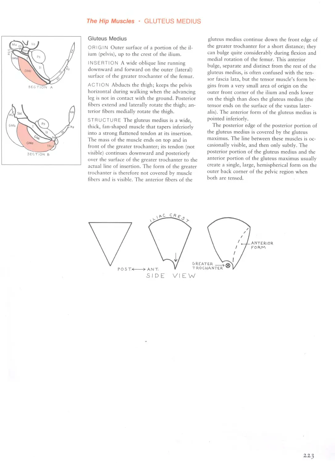


Text
HUMAN ANATOMY
FOR ARTISTS
THE ELEMENTS OF FORM
ELIOT GOLDF
ELIOT GOLDFINGER
HUMAN
ANATOMY
FOR ARTISTS
The Elements of Form
Oxford University Press
1991
The Skeleton • SKULL
GLABELLA
BROW RIDGE
ORBIT
NA5AL BONE
ZYGOMATIC ARCH
ANGLE OF JAW
MANDIBLE
EXTERNAL OCCIPITAL
PROTUBERANCE
PARIETAL EMINENCE
SUPERIOR TEMPORAL LINE
TEMPORAL
FO5SA
MA5TOID PROCESS
NA5AL APERTURE
FRONTAL EMINENCE
EXTERNAL
AUDITORY MEATUS
ZYGOMATIC ARCH
RAMUS
ANGLE
+ BODY
OF MANDIBLE
BROW
RIDGE
BONE
' ANT
NASAL
SPINE
SIDE
VIEW
MENTAL PROTUBERANCE
MENTAL TUBERCLE
MENTAL
FRONT VIEW
PROTUBERANCE
ANGLE OF JAW
ZYGOMATIC ARCH
MANDIBLE
FORAMEN MAGNUM
CONDYLAR PROCESS
PARIETAL
SUPERIOR TEMPORAL LINE
ANGLE OF JAW
MANDIBLE
PARIETAL
EMINENCE
NASAL
BONE
YHARD
PALATE
'BROW
RIDGE
MENTAL
TUBERCLE
SUPERIOR /
NUCHAL LINE
EXTERNAL 1
OCCIPITAL '
PROTUBERANCE
MA5TOID PROCESS
BOTTOM VIEW
P05T.*~*ANT.
6
The Skeleton • VERTEBRAL COLUMN
IO
COCCYX SACRUfA LUMBAR VERTEBRAE THORACIC VERTEBRAE С E RVIC A L VE RT E В R AE
DI5C
The Skeleton • RIB CAGE
Ist THORACIC VERTEBRA
MANUBRIUM
Ist RIB
COSTAL
CARTILAGE
XIPHOID
PROCESS
VERTEBRAL
COLUMN
SPINOUS
PROCESS
TWELFTH RIB
ANGLE OF RIB
BODY OF STERNUM
5PINOUS PROCESS
THORACIC
FOR CLAVICLE
ARTICULAR SURFACE
/MANUBRIUM
STERNAL ANGLE
MANUBRIUM
BODY OF 5TERNUM
POST.
ANT
COSTAL
MANUBRIUM
ANT.
POST
THORACIC
VERTEBRA
RIGHT SIDE VIEW
P0ST.<->ANT.
The Skeleton • RIGHT HAND
TRAPEZOID CAPITATE
PT
METACARPAL
LUNATE
PI5IFORM
BASE
HAMULUS
OF THE
HAMATE
FRONT (PALM) VIEW
RADIAL*—» ULNAR
SHAFT
HEAD
SCAPHOID
(tubercles)
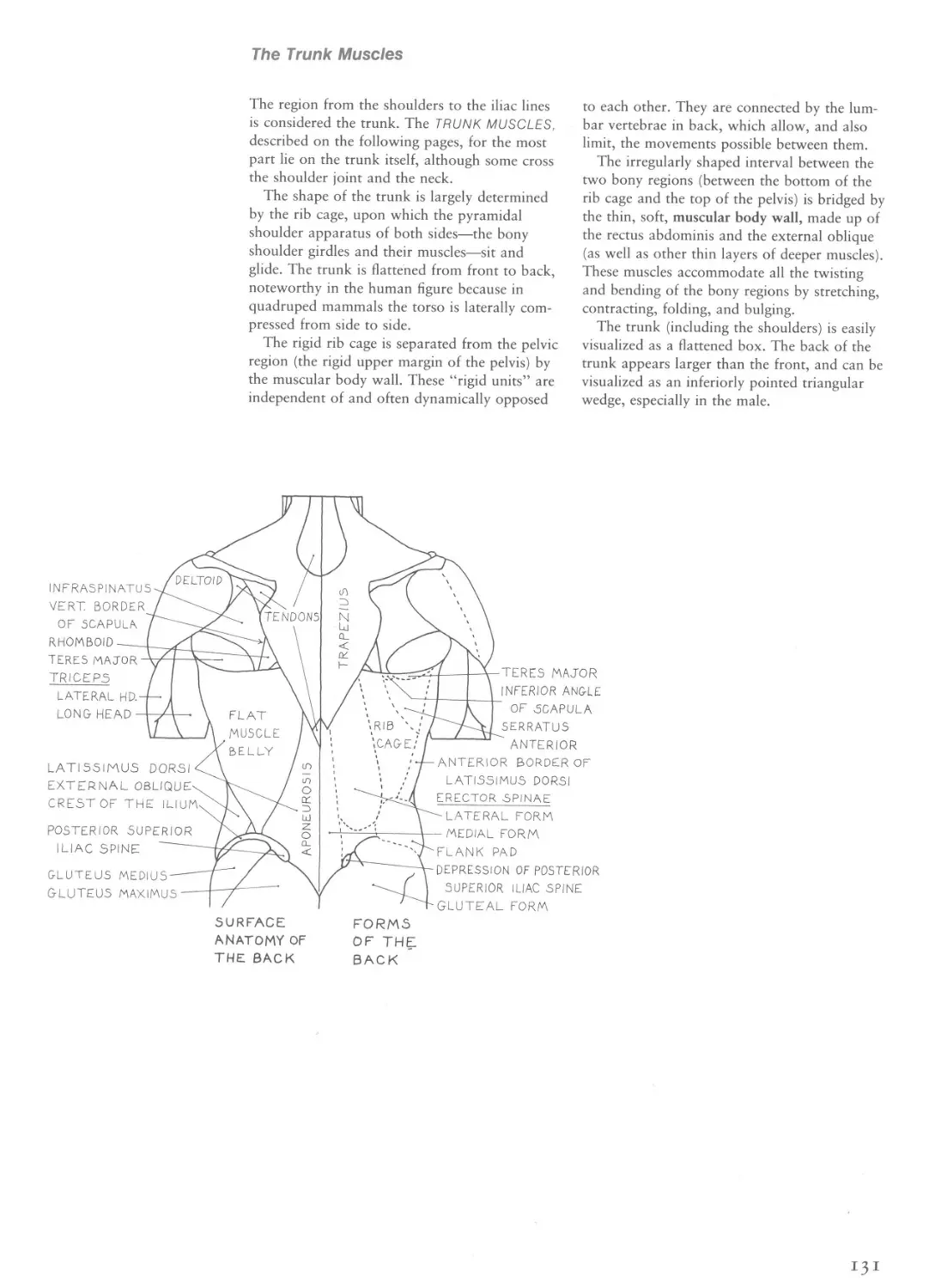
TRAPEZIUM
PROXIMAL
PHALANX
MIDDLE
PHALANX
LUNATE CAPITATE
DI5TAL _
PHALANX
<PHOID
TRAPEZOID
HAMATE
BASE
SHAFT
HEAD
PROXIMAL
PHALANX
BACK (dorsal) view
ULNAR*—> RADIAL
TRIQUETRAL
PISIFORM
SCAPHOID
TRAPEZIUM
SCAPHOID
RAPEZIUM
CAPITATE
TRAPEZOID
dorsal view)
k OF THUMB. /
PISIFORM
HAMULUS OF
THE HAMATE
LUNATE
TRIQUETRAL
RADIAL *—£-* ULNAR
PALMAR
END VIEW OF CARPUS
SCAPHOID
LUNATE
UETRAL
HAMATE
PALMAR VIEW)
k OF THUMB )
THUMB SIDE (RADIAL view)
LITTLE FINGER SIDE (ULNAR VIEW)
DORSAL*---> PALMAR
PALMAR *--> DORSAL
2.6
The Skeleton • HAND
The SKELETON OF THE HAND is designed to
perform the most heavy brutal chores, as well
as extremely precise, sensitive, and delicate
movements, in life, the hand is predominantly
bony, with its surface form based for the most
part on its skeleton, especially its back or dorsal
surface. It consists of the carpus, the metacar-
pus, and the phalanges. The phalanges together
are longer than the metacarpals, which in turn
are longer than the carpals. These proportions
are opposite to those found in the foot between
the tarsals, metatarsals, and phalanges.
The CARPUS consists of eight small irregu-
larly shaped carpal bones arranged in two
rows—a proximal and a distal—of four bones
each. The proximal row contains the scaphoid,
lunate, triquetral, and pisiform. The distal row
contains the trapezium, trapezoid, capitate, and
hamate. As a unit, the carpal bones form half a
disc which is curved side to side, so that its
dorsal surface is convex; the concave palmar
surface is transformed into the carpal tunnel for
passage of the flexor tendons to the fingers.
On the back of the carpus, between the two
carpal rows, is a depression (located toward the
radial side). It is especially noticeable when the
wrist joint is flexed. Also noticeable in this posi-
tion is another depression between the distal
end of the radius and the proximal carpal row
(the articulating proximal surface of the carpus
becomes a raised prominence between the two
depressions).
On the palmar side of the carpus, bony
prominences are formed by the pisiform at the
ulnar side (at the base of hypothenar eminence)
and by the tubercles of the trapezium and sca-
phoid together on the radial side (at the base of
the thenar eminence). These bony projections
are especially noticeable when the wrist joint is
extended.
At the wrist joint, the arched proximal carpal
row articulates primarily with the distal end of
the radius and minimally with the fibrous disc
attached to the distal end of the ulna.
The METACARPUS consists of the five meta-
carpal bones. The boxlike base of each bone is
located at the proximal extremity of the shaft,
while the rounded head is located at the distal
extremity. Metacarpals two through five (index
to little finger) as a group radiate distally. The
dorsal surfaces of the metacarpals are wide and
flattened, especially distally, and all have a
slight convex curvature. Metacarpals two
through five create a plane on the back of the
hand that curves side to side; it is especially
noticeable at the knuckles. This curve, or trans-
verse arch, helps create the concavity of the
palm. The curvature of the transverse arch flat-
tens when the open hand is pressed against a
flat surface.
Distally, the heads of metacarpals two
through five are connected to each other by lig-
aments; the head of the thumb is unattached,
allowing it to have a great range of motion at
the carpometacarpal joint and the ability to op-
pose the other fingers in grasping. The thumb’s
metacarpal is rotated around its long axis so
that its dorsal surface faces laterally when the
palm is directed forward (its dorsal surface is
almost perpendicular to the dorsal surfaces of
the other metacarpals).
The heads of metacarpals two through five
are somewhat spherical. Their distal surfaces
become exposed in life as rounded knuckles
when the fingers are flexed at the metacarpo-
phalangeal joints. These four metacarpals end
distally along a curved line, with the middle
finger projecting the farthest. The width across
these four metacarpal heads is greater than the
width across the humeral epicondyles at the el-
bow. Because the thumb’s metacarpal head is
not as rounded as the others, and because of
limited motion at the metacarpophalangeal joint
of the thumb, the distal surface of this metacar-
2-7
The Skeleton • PELVIS
CRE5T
ACETABULUM
PUBIC SYMPHYSIS
ISCHIAL TUBEROSITY
PUBIC
TUBERCLE
ISCHIUM,
PUBIS
FRONT VIEW
POSTERIOR SUPERIOR
ILIAC SPINE
TUBERCLE
I LI U ГЛ
SUPERIOR
ILIAC SPINE
ANTERIOR
INFERIOR
ILIAC SPINE
POSTERIOR
INFERIOR
ILIAC SPINE
COCCYX
ISCHIAL
SPINE
BACK VIEW
CRE5T
POSTERIOR
INFERIOR
ILIAC SPINE
SACRUM
ISCHIAL
5PINE
COCCYX
I SC H ЮГА
I5CHIAL
TUBEROSITY
ARTICULAR SURFACE
FOR SACRUM
POSTERIOR
SUPERIOR
ILIAC SPI
ANTERIOR SUPERIOR ILIAC SPINE
ANTERIOR INFERIOR ILIAC SPINE
ACETABULUM
PUBIC TUBERCLE
ARTICULAR SURFACE
AT PUBIC SYMPHYSIS
INFERIOR RAMUS OF PUBIS\ I5CHIOPUBIC
INFERIOR RAMUS OF ISCHIUM) RADIUS
ILIUM
CRE5T
ISCHIAL 5PINE
OBTURATOR
FORAMEN
ISCHIAL
TUBEROSITY
POSTERIOR
SUPERIOR
ILIAC SPINE
I0R INFERIOR
ILIAC SPINE
OUTSIDE VIEW
POST.*-* ANT.
(RIGHT HIP BONE)
INSIDE VIEW
ANT**POST.
(RIGHT HIP BONE)
REENTERING
ANGLE
CREST
SACRUM
POSTERIOR SUPERIOR
ILIAC SPINE
OUTER LIP
OF CREST
ANT.
TOP VIEW ANTERIOR SUPERIOR
ILIAC SPINE
ANTERIOR INFERIOR
TUBERCLE
ANTERIOR SUPERIOR
ILIAC SPINE
PUBIC SYMPHYSIS ILIAC
PINE
ANT
ISCHIAL
TUBEROSITY
SACRUM
POST.
POSTERIOR
SUPERIOR
ILIAC SPINE
COCCY
BOTTOM VIEW
30
The Skeleton • PATELLA
The PATELLA, or kneecap, is the largest sesa-
moid bone of the body. It is attached to the
deep surface of the tendon of insertion of the
quadriceps muscle of the thigh. The function of
the patella is to move this tendon away from
the joint, giving the quadriceps greater mechani-
cal advantage. The deep surface of the patella
articulates with the distal extremity of the fe-
mur, protecting the articular cartilages of the
condyles, especially while kneeling.
The patella is triangular with rounded cor-
ners and is flattened from front to back. Its
apex is directed inferiorly. The two upper
rounded corners are prominent on the surface
in life. The anterior surface of the patella is
convex, while the posterior surface has a wide
vertical ridge which glides in the groove be-
tween the condyles of the femur. The inferior
apex, attaching to the patellar ligament, is not
seen on the surface in the straight extended
knee; however, a horizontal skin furrow may
appear at this attachment. When the knee is
flexed, the anterior surface of the patella forms
an angle with the patellar ligament, revealing
the apex (in side view). The upper edge of the
patella is seen in the flexed knee, not in the
extended knee, because the quadriceps tendon
meets the patella at an angle during flexion.
In the standing position with the thigh re-
laxed, the bottom of the patella sits at a level
just slightly higher than the line of the knee
joint; it rises slightly when the thigh muscles
are tensed (with no motion taking place at
the knee joint between the femur and tibia).
The patella is wider than the distance across the
anterior portion of the ridges of the medial and
lateral condyles of the femur, and therefore cov-
ers a portion of these edges when the knee is
extended.
When the knee is straight, the patella sits in
the anterior shallow part of the femoral condyle
groove. As the knee is flexed, the patella slides
down into deeper parts of the groove. This ac-
tion displaces the patella posteriorly in relation
to the anterior profile of the lower leg. When
the knee is flexed, the thigh appears longer, be-
cause the patella has been dragged around to
the distal surface of the condyles of the femur,
adding its thickness to the length of the thigh.
RIGHT PATELLA
POST.
LAT*-J-»M£D.
ANT.
FRONT VIEW
L AT*-» MFD.
OUTSIDE VIEW
POST*-» ANT
BACK VIEW
MED.-*LAT.
INSIDE VIEW
ANT*-»POST,
FRONT VIEW
LAT<--->f*AED.
TOP VIEW
- FEMUR
7—PATELLA
«"PATELLA*
ligament
TIBIA
PATELLA
SHIFTS
POSTERIORLY
|N RELATION
TO FRONT
edge OF
TIBIA
EXTENDED FLEXED
KNEE poST«->ANT. KNEE
5IDE VIEW
35
The Skeleton • RIGHT FOOT
TALUS
LATERAL CUNEIFORM
INTERMEDIATE CUNEIFORM.
MIDDLE
PHALANX
OUTSIDE VIEW
FIFTH METATARSAL
POST. *—>ANT.
SECOND METATARSAL
PROXIMAL
DISTAL
PHALANX
NAVICULAR
TALUS
INSIDE VIEW
POST.*—>ANT.
-CALCANEUS
TALUS
NAVICULAR
TUBERCLE
BACK VIEW
MED.*-* L AT.
CALCANEUS
LATERAL CUNEIFORM
NAVICULAR TUBERCLE
INTERMEDIATE CUNEIFORM
MEDIAL CUNEIFORM
SHAFT HEAD
TOP VIEW
MED.
PO5T«-£->ANT
CUBOID
OF FIFTH METATARSAL
TUBEROSITY OF FIFTH METATARSAL
40
The Skeleton • FOOT
The SKELETON OF THE FOOT is designed to
provide stability in supporting and distributing
the body’s weight and resiliency when receiving
the impact of the body’s weight while walking,
running, and jumping. It also acts as a lever,
pushing the body forward during locomotion. It
is rigid upon takeoff and flexible upon impact.
The foot skeleton is composed of the tarsus,
metatarsus, and phalanges, and except for the
phalanges of the lateral four toes, its bones are
characterized by their thickness and strength.
The phalanges are shorter than the metatarsals,
which in turn are shorter than the tarsals. This
arrangement is opposite to that found in the
hand, where the carpus is short and the pha-
langes are long. In the foot, the big toe is re-
stricted to lying parallel to the other toes, un-
like the thumb, which is capable of opposing
the other fingers.
The foot has two perpendicular arches: a lon-
gitudinal arch from front to back and a trans-
verse arch from side to side. These arches not
only distribute weight but also afford the foot a
degree of elasticity, providing shock absorption.
The longitudinal arch is actually a series of five
almost parallel (slightly radiating) arches pass-
ing through the long axes of the five toes. There
is a high medial arch on the inside of the foot
and a very low lateral arch on the outside (the
base of the metatarsal of the little toe does not
support weight in the foot flat on the floor).
The transverse arch curves across the foot,
bringing the outer edge of the foot down
toward the ground. It is most noticeable at the
midpoint of the foot, where it is high on the
inside and low on the outside. The muscles, ten-
dons, ligaments, and fasciae of the foot main-
tain these arches.
The TARSUS contains seven stout tarsal
bones: the talus, calcaneus, cuboid, navicular,
and three cuneiforms. There is substantial
movement within the tarsus.
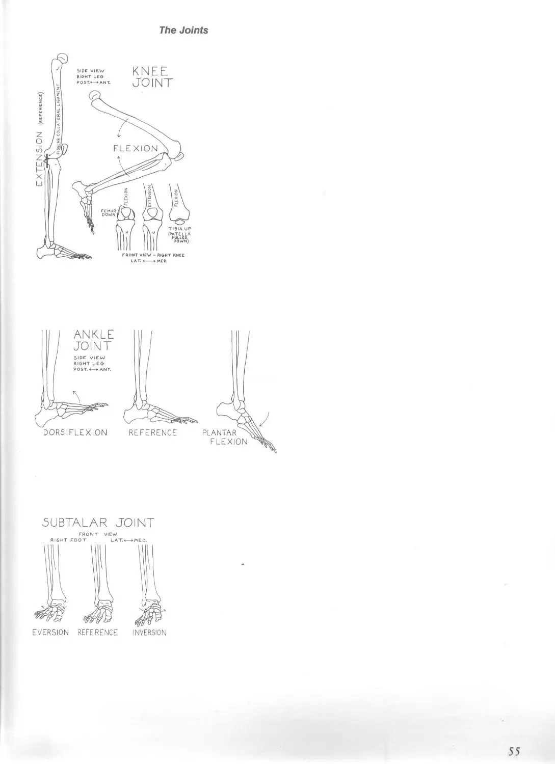
The talus (astragalus), located above the cal-
caneus near the back end of the medial side of
the foot, consists of a body, an anteriorly pro-
jecting neck, and a rounded head in front. The
upper articular portion of the body, the
trochlea, is spool-shaped. It is the highest bone
of the foot and articulates superiorly with the
tibia and fibula to form the ankle joint. Only
flexion and extension are possible at this joint.
The talus articulates inferiorly with the calca-
neus at the subtalar joint and anteriorly with
the navicular. The subtalar joint permits inver-
sion and eversion of the foot. The lateral edge
of the trochlea of the talus becomes visible on
the surface in front of the lateral malleolus of
the fibula during extreme plantar flexion of the
foot (toe pointing). During inversion of the foot
(facing the foot inward), the lateral part of the
head of the talus shows under the tendons of
the extensor digitorum longus, while the medial
part of the head shows during eversion (facing
the foot outward) between the medial malleolus
and the navicular tubercle. These points of the
talus, when seen, appear as subtle prominences.
The calcaneus, or heel bone, is blocklike, flat-
tened side to side, and rounded on its posterior
surface. It is the largest foot bone. It is inclined
upward, forward, and slightly laterally, resting
its lower back edge on the ground (protected
below by the fatty cushion of the sole of the
foot). The calcaneus acts as a lever. Most of the
lateral surface and part of the posterior surface
(between the insertion of the Achilles tendon
and the heel cushion) are subcutaneous. The
Achilles tendon conceals the upper part of its
back surface.
NARROW
Post. «->ant.
OUTSIDE VIEW
*MED.
RAMP
CURVES
DOWN
LATERALLY.
CROSS SECTION
THROUGH FOOT
FRONT VIEW
METATARSAL
'—/ Phalanges
BIG- TOE
SIDE VIEW
41
The Joints
TEMPOROMANDIBULAR JOINT
45
The Joints
ADDUCTION
(with slight flexion)
ABDUCTION
REFERENCE
PALM SIDE
OF HAND
FRONT VIEW
DORSAL
SIDE OF
HAND
END VIEW OF
FOREARM BONES
(NOT TO scale)
OF ULNA
(DOESN’T MOVE)
PALMAR
t
DORSAL
SUPINATION
(reference)
PRONATION
49
The Joints
WRIST
JOINT
SIDE (RADIAL) VIEW
RIGHT HAND
POST «—> ANT.
EXTENSION
WRI5T
JOINT
FRONT (PALM)
VIEW
RADIAL
DEVIATION
ULNAR
DEVIATION
THUMB
FRONT (palm) VIEW —RIGHT hand
CAR POME TACARPAL.
JOINT
Side (radial) view—right hand
EXTENSION FLEXION
ADDUCTION ABDUCTION
51
The Joints
THUMB
OUTSIDE (radial) view
RIGHT HAND
PHALANX PROXIMAL METACARPAI
REFERENCE
(slight hyperextension at
INTERPHALANGEAL joint)
FLEXION
EXTENSION
INDEX FINGER
OUTSIDE (RADIAL) VIEW
RIGHT HAND
hyperextension at
METACARPOPHALANGEAL
JOINT
DISTAL MIDDLE PROXIMAL
PHALANX PHALANX PHALANX METACARPAL
REFERENCE
METACARPO-
PHALANGEAL
JOINT
FRONT (PALM) VIEW
RIGHT HAND
LAT. «—» MED.
FLEXION
REFERENCE
53
The Joints
DORSIFLEXION
REFERENCE
5UBTALAR JOINT
55
The Joints
METATARSAL
PROXIMAL _
PHALAN MIDDLE
phalanx
REFERENCE
DISTAL
PHALANX
(foot on ground)
ALIGNMENT
SIDE VIEW
57
The Muscles • STRUCTURE, FUNCTION, AND FORM
usually not possible to make a fist when the
wrist is fully flexed, because the finger flexors
are too short to contract further (active insuffi-
ciency) and the finger extensors are fully
stretched (passive insufficiency), preventing the
fingers from flexing fully.
In analyzing muscular activity, it is important
to think in terms of the combined actions of all
the muscles in a given area—some work indi-
vidually, others work as groups; some stabilize
one region of the body while others create mo-
tion in another region; some begin an action
while other muscles complete it; some oppose
the pull of gravity. During complex actions,
note the sequence of the contraction and relax-
ation of the numerous muscles that are func-
tioning. Observe the action, visualize the skele-
ton deep in the body and what changes are tak-
ing place at its joints, and then determine which
muscles are working in order to perform that
action. For further information on function, re-
fer to the most recent updated medical, physiol-
ogy, kinesiology, and physical therapy texts.
Form
The muscles of the body are independent con-
vex forms, and when placed side by side and
one on top of the other to make up the figure,
they structure the figure into a series of adja-
cent, bulging, convex forms which are covered
by the skin. An appreciation of the fullness of
these convexities will produce artwork with vi-
tality and a feeling of energetic inner life radiat-
ing from the center outward. True concavities
show up only rarely in the body, and one must
be careful not to mistake a series of convexities
for a concavity. Know the attachments of mus-
cles, because that is where their form begins
and ends. Know the basic individual shapes of
each muscle. Think of the form of a muscle as a
swollen axis passing between the centers of its
areas of attachment.
Beware of illustrations that simply diagram
the surface muscles’ outlines without giving in-
dication of the deeper structures that may ac-
tually be creating the forms we see (very little
of the form of the latissimus dorsi is directly
seen; it is the forms of the deeper structures—
erector spinae, serratus anterior, teres major,
and rib cage—that create surface form). Keep in
mind that the form of one muscle can blend
with the form of another muscle or with fat
pads, that the shapes of muscles are sometimes
affected by overlying fascia, that muscles appear
shorter and thicker when contracted and longer
and thinner when stretched, and that they be-
come more defined when they are contracted. In
other words, the same muscle will appear dif-
ferently in different actions and poses.
The artist must understand all the anatomical
components that create human surface form,
and when studying the living model, be able to
mentally impose these structures diagrammati-
cally on the model. For while everything is
there, most of it is usually concealed to the un-
trained eye. It becomes the artist’s job to ana-
lyze, select, and then create.
Note: In the following illustrations, muscle at-
tachments on the skeleton are indicated by solid
red areas, while nonbony attachment areas are
usually indicated by red cross-hatching. They
are labeled О for origin and I for insertion. A
curved arrow indicates that an attachment is
hidden from view. All illustrations are of the
right side of the body.
NEUTRAL.
64
The Muscles • STRUCTURE, FUNCTION, AND FORM
PARALLEL
BIPENNATE KULTIPENNATE TRIANGULAR
ROUND
FLE5HY BELLY
NO VISIBLE
TENDONS
TENDON AT
ONE END
FLAT =
TENDONS AT
BOTH ENDS
1—4—
Wl D E
TENDON
LATISSIMUS
DORSI MUSCLE
CONFORMS TO LIMB CIRCUMFERENCE
CONFORMS TO INDIVIDUAL MUSCLES
Relaxed limb
Tensed limb
The Muscles • STRUCTURE, FUNCTION, AND FORM
T£MPORALIS\.
ORBICULARIS OCULK^-
ZYGOMATICUS MINOR^
ZYGOMATICUS MAJOR
MAS5ETER------
RISORI US---------
PLATYSMA----------
DEPRESSOR ANGULI ORIS
LEVAT. SCAPULAE
TRAPEZIUS
CORACDBRACHIALIS-^
TRICEPS-LATERAL HEAD
BRACHIALIS-----________
BICEPS BRACHII
TRICEPS-LONG HEAD----
TRICEPS-MEDIAL HEAD
BRACHIORADIALIS —
BRACHIALIS-------
EXTEN.CARR RAD. LONG.
EXTEN CARP. RAD. BREVIS-
PRONATOR TERES
PALMARIS LONGUS----
FLEXOR CARPI RADIALIS--
FLEXOR DIGIT. SUPERFIC.—Z
FLEX.CARPI ULNARIS----Hi
FLEX. POLLICIS LONGUS
ABD. POLLICIS LONG.
ABD POLLICI5 BREV'
FLEX.POLL BREV.^
ADDUCTOR POLLICIS
ABD. DIGITI
MINIMI
FRONT VIEW
QUADRICEPS TENDON
FLEX. DIGIT
MIN. 0REV.
PATELLAR LIGAMENT
HEAD OF FIBULA----
PERONEUS TERTIUS---------
LATERAL MALLEOLUS (FIBULA)
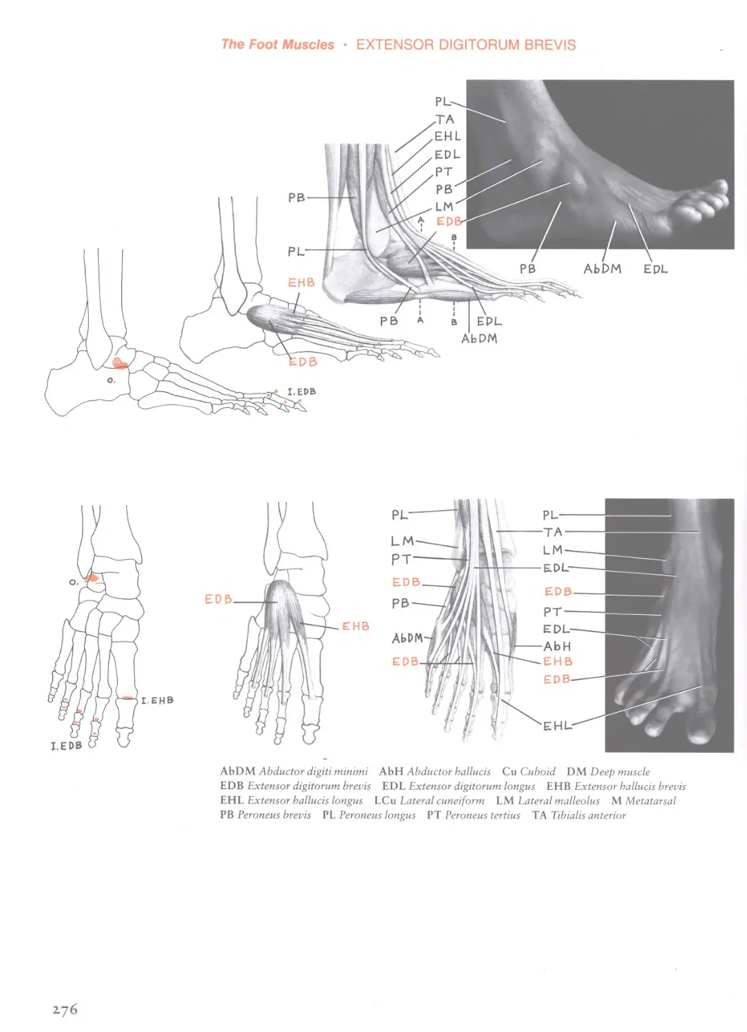
EXTENSOR DIGITORUM BREVI5-
FRONTALIS
PROCERUS
LEV. LABII SUP ALAE NAS.
LEVATOR LABII SUPER.
DILATOR NARIS
ORBICULARIS ORIS
DEPRES. LABII INFER.
MENTALIS
STERNOMASTOID
OMOHYOID
‘STERNOHYOID
DELTOID -----
TERES MINOR
INFRASPINATUS —
TERE5 MAJOR-----
PECTORALIS MAJOR
RHOMBOID MAJOR
SERRATOS ANTERIOR
EXTERNAL OBLIQUE
LATISSIMUS DORSI —
-SEMILUNAR LINE
H-LINEA ALBA
RECTUS ABDOMINIS
EXTERNAL OBLIQUE-
PELVIC CREST (ILIUM)
GLUTEUS MEDIU5 —
GLUTEUS MAXIMUS-
ILI0P50AS
TENSOR FASCIA LATA
SARTORIUS
ADDUCTOR LONGUS
ADDUCTOR MAGNU5-
RECTUS FEM0RI5
GRACILIS---------
VASTUS LATERALIS-
BICEPS FEMORIS (L H.)
SEMITENDINOSUS —
VA5TUS MEDIALIS
SEMIMEMBRANOSUS
ILIOTIBIAL TRACT
PLANTARIS
PATELLA
BICEPS FEMORIS ($ h)
7—SARTORIUS---------
L—TIBIALIS ANTERIOR
PERONEU5 LONGUS
SOLEUS
GASTROCNEMIUS —
TIBIA
TENDON OF GASTROC
EXTEN. DIGITORUM LONG.
SOLEUS----------
PERONEUS LONGUS
PERONEUS BREVIS
FLEX. DIGITORUM LONG.^
EXTENSOR HALLUCIS LONG
EXTENSOR HALLUC. BREV.
TIBIALIS POSTERIOR
ABDUCTOR HALLUCIS
CRANIUM
SEMISPINALI5 CAPITIS
5PLENIUS CAPITIS
- — 5TERNOMASTOID
LEVATOR SCAPULAE
TRAPEZIUS
SPINE OF SCAPULA
BACK VIEW
FIRST DORSAL
INTEROSSEOUS
MEDIAL MALLEOLUS (TIBIA)
ACHILLES TENDON
EXTENSOR DIGITORUM BREVIS
PERONEUS BREVIS
GREATER
TROCHAN.
TRICEPS-LONG HEAD
TRICEPS-LATERAL HEAD
TRICEPS-MEDIAL HEAD
TRICEPS TENDON
BRACHIORADIALIS
EXTENSOR CARPI RADIALIS LONG.
ULNA (OLECRANON)
ANC0NEU5
EXTEN. CARPI RADIALIS BREV
EXTENSOR DIGITORUM
FLEXOR CARPI ULNARI5
EXTENSOR CARPI ULNARIS
-EXTENSOR DIGITI MINIMI
ABDUCTOR POLLICIS LONG.
EXTEN POLLICIS BREVIS
EXTEN. POLL. LONG.
ABDUCTOR
DIGITI
MINIMI
66
The Facial Muscles
glabella
MENTOLABIAL 5ULCUS
CHIN BOSS
FRONTAL eminence
INFRA0R8ITAL triangle;
BROW RIDGE
EYEBROW
EYE COVER FOLD
UPPER. EYELID -
<7—ROOT OF NOSE —
BRIDGE OF NOSE -
LOWER. EYELID-----
TEAR BAG -----
CHEEK BONE----
INFRAORBITAL FURROW
WING OF NOSE ----
NASOLABIAL FURROW -
PHILTRUM
NODE ---
MOUTH ANGLE FURROW
PILLAR OF MOUTH---
SUPERFICIAL
EYEBROW
Cn
о
Pl
m
H
<0
BUG.
TO NEC r
Dn-ZA
OOc.
DLI
MEN.
OOr.
IRL) I
(RL) 1
(orb.j
LL5AN
UPPER LABIAL
TRACTOR
LOWER LABIAL
TRACTOR
UPPER LIP
LOWER LIP
PRIMARY MUSCLES
OF FACIAL
EXPRESSION AND
THEIR DIRECTION
OF PULL
MU5CLE PULL
ON NODE
NASOLABI AL
FURROW
BUC Buccinator COR Corrugator supercilii DAO Depressor anguli oris
DLI Depressor labii inferioris DN Dilator naris DS Depressor supercilii
F (L) Frontalis — lateral portion F( M) Frontalis — medial portion ILI /ncisivus labii inferioris
ILS htcisivits labii superioris LLS Levator labii superioris LLSAN Levator labii superioris alaeque nasi
LP Labial platysma LI’S Levator palpebrae superioris MAL Malaris MEN Mentalis N Node
N P Nodular platysma OOc(O R B) (Orbicularis oculi—orbital portion
OOc(PAL) Orbicularis oculi—palpebral portion OOr(OUT) Orbicularis oris —outer portion
OOr(RL) Orbicularis oris —red lip portion Z M AJ Zygomaticus major Z MIN Zygomaticus minor
The Facial Muscles • FRONTALIS
MEDIAL + LATERAL PORTIONS
MEDIAL PORTION (t CORROGATOR)
MEDIAL^LATERAL PORTIONS
MEDIAL PORTION (+CORRUG.)
DS Depressor supercilii F(L) Frontalis —lateral portion F(M) Frontalis —medial portion
GA Galea aponeurotica OOc Orbicularis oculi P Procerus
70
The Facial Muscles • FRONTALIS
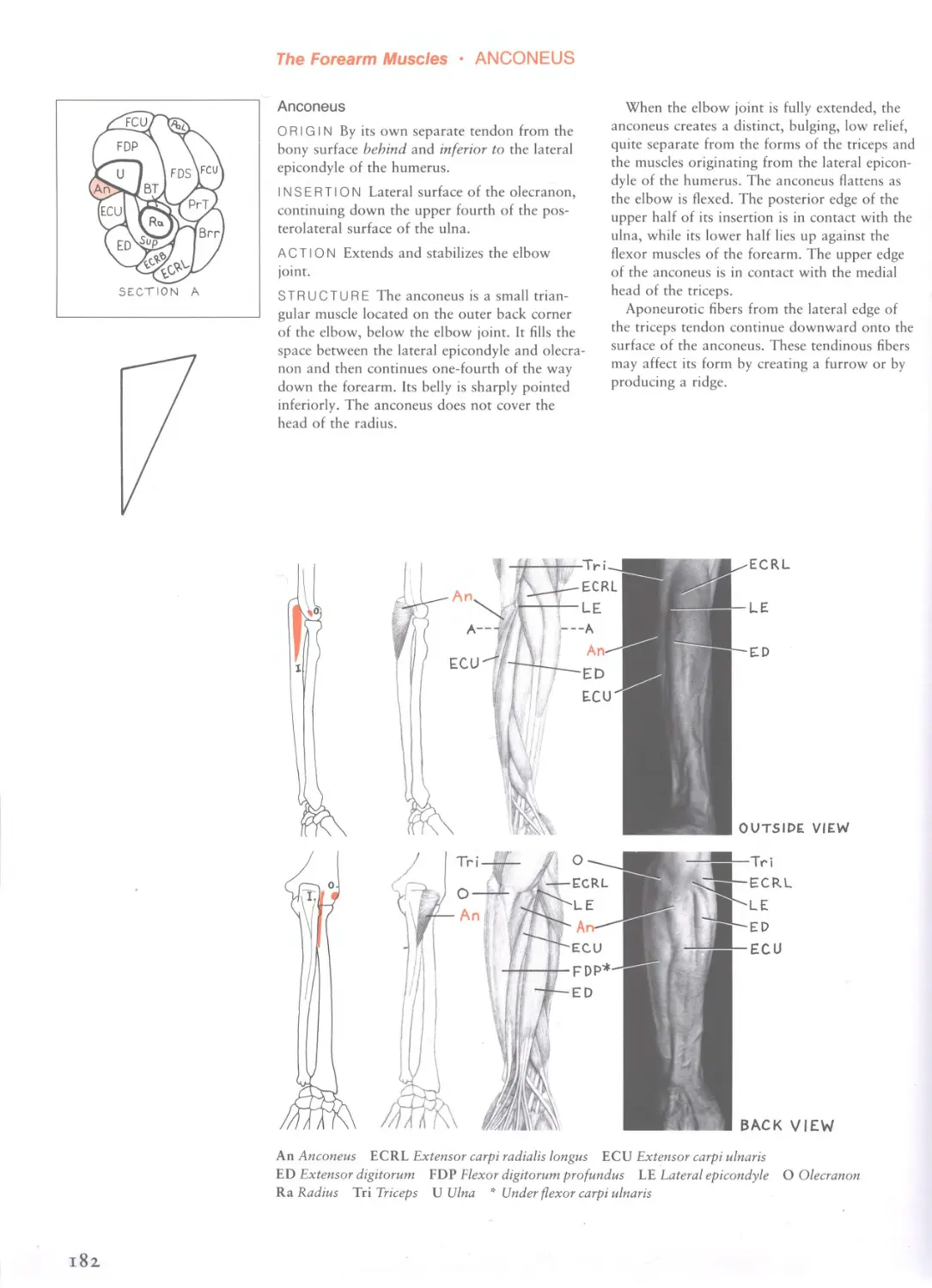
Frontalis
The frontalis is the anterior muscle belly of the
extensive epicranius muscle.
The epicranius muscle consists of a broad
layer of tendon (the galea aponeurotica) and
thin muscle bellies; it covers the top and sides
of the skull, from the brows in front to the base
of the skull in back, and over to the tops of the
ears.
ORIGIN The front edge of the galea aponeuro-
tica, just posterior to the top of the forehead.
INSERTION The skin at the eyebrow and root
of the nose, blending with the fibers of the pro-
cerus, orbicularis oculi, and corrugator super-
cilii.
ACTION Medial and lateral portions together:
Raise the entire eyebrow, increasing its overall
curvature (the central portion is pulled higher).
They also pull the scalp (hairline) slightly for-
ward as they pull on the galea aponeurotica.
Medial portion only: Pulls up the medial end of
the eyebrow. Lateral portion only: Pulls the
middle and lateral end of the eyebrow upward,
giving the eyebrow a strong curvature.
The medial portions of both sides of the face
always contract together and are almost always
accompanied by contraction of the corrugator
supercilii (as in sadness). The lateral portion
only occasionally contracts by itself, and usually
in one eyebrow only.
STRUCTURE The frontalis is divisible func-
tionally into medial and lateral parts. The two
frontalis bellies of the head are in contact with
each other at their lower portion, on the mid-
line of the forehead.
EFFECT Contraction of the frontal bellies pro-
duces horizontal wrinkles across the forehead.
The wrinkles are superiorly curved above each
eyebrow and dip downward on the centerline.
When only the medial part of the frontal belly
contracts, horizontal wrinkles form on the cen-
ter of the forehead only. In this case the medial
ends of the eyebrows are pulled upward. How-
ever, depending on the position (obliquity) of
the eyebrows in the resting position, the medial
ends may not appear to have been raised. Hori-
zontal wrinkles on the center of the forehead
are diagnostic of medial frontalis contraction.
Because the corrugator typically contracts when
only the medial frontalis contracts, the eyebrow
develops a “kinking” near its medial end or an
overall S-shaped curvature; its medial end is
pulled upward by the medial frontalis, and its
center is pulled downward by the corrugator.
As a person ages, the forehead wrinkles do
not fully disappear when the frontal bellies are
relaxed.
When the eyebrows are raised, the root of the
nose becomes thinner and the glabella becomes
smooth. Raising the eyebrows also pulls the
skin between the eyebrow and upper eyelid up-
ward, exposing more of the eyelid. The skin
below the eyebrow (the eye cover fold) then
loses its form (its bulging quality) and becomes
stretched over the bone beneath, revealing the
form of the bony orbit. The distance between
the medial end of the eyebrow and the inner
corner of the eye increases; this is especially
noticeable when only the medial portions are
contracted.
When the eyebrows are raised, the ears also
rise slightly.
EXPRESSION Medial and lateral portions of
both sides of the face: surprise, astonishment;
fear, fright, horror; interest, attention; question-
ing, doubt. Medial portions of both sides of the
face (with corrugator): sadness, grief, suffering;
sometimes fear. Medial and lateral portions of
one side of the face: inquisitiveness, skepticism.
Lateral portion of one side of the face: sly, de-
vious look.
Contraction of the entire frontalis is often
used as a greeting signal or for conversational
emphasis. This use is not an expression of
emotion.
MEDIAL + LATERAL PORTIONS
MEDIAL PORTION ONLY
LATERAL PORTION ONLY
EYEBROWS
MEDIAL + LATERAL PORTIONS
+ CORRUG-ATOR
MEDIAL PORTION + CORRUG-ATOR
71
The Facial Muscles • ORBICULARIS OCULI
0UTE.R PORTION
OUTER PORTION
DS Depressor supercilii F Frontalis LLS Levator labii superioris
LLSAN Levator labii superioris alaeque nasi Mai Malaris OOc Orbicularis oculi
OOc(Or) Orbicularis oculi—orbital portion OOc(Pal) Orbicularis oculi—palpebral portion
P Procerus Те Temporalis ZMa Zygomaticus major ZMi Zygomaticus minor
72-
The Facial Muscles • CORRUGATOR SUPERCILII
Corrugator Supercilii
ORIGIN The bone at the medial end of the
superciliary arch, near the upper inner corner of
the orbit.
INSERTION The skin of the middle portion of
the eyebrow, and the skin of the forehead im-
mediately above it. The fibers interlace with fi-
bers of the frontalis and orbicularis oculi.
ACTION Pulls the middle section of the eye-
brow downward and medially in an oblique di-
rection, thereby pulling the brows together. It
usually works simultaneously with the procerus
and depressor supercilii to lower the eyebrows
and pull them together. It can work without
these muscles, pulling the eyebrows together,
when the eyebrows are raised by the frontalis.
Both sides usually work together, except when
one eyebrow is pulled downward and the other
upward.
STRUCTURE The corrugator supercilii is a
narrow, elongated muscle. Most of the muscle
is deep to the orbicularis oculi, but the lateral
tips of its fibers pierce the orbicularis to pass
superficially and insert into the skin.
EFFECT The corrugator supercilii typically
creates strong vertical or slightly oblique wrin-
kles and vertical bulges on the glabella between
the medial ends of the eyebrows. It also swells
the skin at the medial end of the eyebrow into a
wide, thick, crescent-shaped bulge. This action
pulls the brows shelflike over the eyes, shading
them from strong light.
A depression forms in the skin above the
middle of the eyebrow (actually closer to the
medial end) as the corrugator contracts. It often
gives the eyebrow an S-shaped curve—the eye-
brow develops a central downward dip (usually
close to the medial end) as the muscle pulls the
middle portion of the eyebrow downward and
inward. This is especially noticeable if the me-
dial portion of the frontalis is also lifting the
medial end of the eyebrow. The corrugator may
also create a long, oblique, shallow furrow on
the lateral side of the front of the forehead.
This furrow is directed upward and outward
from the medial end of the eyebrow or from the
skin depression located above the middle of the
eyebrow.
The lowering of the brow by the corrugator
narrows the eye opening by pushing the skin
below the eyebrow and upper eyelid downward.
EXPRESSION Anger; sadness, suffering, grief,
pain; frowning; interest, thoughtful reflection,
concentration (focused thought), curiosity, con-
fusion. The corrugator can be considered a
muscle of negative feelings, being typically used
in sadness, fear, and anger. Concentration and
determination, which also make strong use of
the corrugator, may be regarded as unresolved
stressful (negative) states.
CS Corrugator supercilii DS Depressor supercilii F Frontalis OOc Orbicularis oculi P Procerus
75
The Facial Muscles • LEVATOR PALPEBRAE SUPERIORIS
Levator Palpebrae Superiors
ORIGIN Deep in the back of the eye socket,
from the roof of the orbit.
INSERTION The entire lower edge of the up-
per eyelid.
ACTION Raises the upper eyelid. It holds the
eye open in the normal neutral position.
Expression develops only after the eyelid has
been raised above this position.
STRUCTURE The levator palpebrae superioris
is a long muscle which passes over the eyeball,
deep in the eye socket, along the roof of the
orbit. Narrow behind, it widens as it advances
anteriorly, becoming aponeurotic before insert-
ing into the margin of the upper eyelid.
EFFECT Exposes the upper portion of the iris
and the white of the eye (sclera) above the iris.
By continued action it pulls the upper eyelid
back so it disappears from view under the eye
cover fold.
EXPRESSION Slight raising of the upper eye-
lid beyond the normal open eye produces a
staring, fixed gaze or the expression of surprise.
Full contraction produces intense expressions
ranging from fear, terror, shock, astonishment,
and intense joy to a blank, hypnotic look. Slight
relaxation of the muscle (causing the upper eye-
lid to droop) with the eye still remaining par-
tially open suggests drowsiness or intoxication,
especially if the frontalis is also contracted in an
attempt to hold the eyes open by raising the
eyebrows. This expression exposes more of the
upper eyelid, which has descended onto the up-
per portion of the iris. Upper eyelid droop and
gentle eye closure (when the lower eyelid does
not rise) are caused by relaxation of the levator
palpebrae superioris, not by contraction of the
orbicularis oculi.
DS Depressor supercilii F Frontalis LPS Levator palpebrae superioris
76
The Facial Muscles • PROCERUS
Procerus
(Pyramidalis Nasi)
ORIGIN The fascia covering the lower part of
the nasal bones and the upper part of the lat-
eral nasal cartilages.
INSERTION The skin between the eyebrows
and on the lower portion of the center of the
forehead. Its fibers blend with fibers of the fron-
talis in the same region.
ACTION Pulls down the skin of the middle of
the forehead and the medial ends of the eye-
brows. This muscle works in conjunction with
the corrugator supercilii and depressor supercilii
to lower the medial ends of the eyebrows. The
procerus automatically contracts when the leva-
tor labii superioris alaeque nasi is tensed. It can
be seen working by itself if the eyebrows are
first raised by the frontalis and then the levator
labii superioris alaeque nasi is contracted.
STRUCTURE The procerus muscles of both
sides form a single triangular sheet of muscle
between the eyebrows.
EFFECT The procerus may occasionally pro-
duce a horizontal wrinkle across the bridge of
the nose.
EXPRESSION Anger, aggression, pain; frown,
concentration; contributes to disgust.
OOc(Or) Orbicularis oculi—orbital portion OOc(Pal) Orbicularis oculi—palpebral portion P Procerus
77
The Facial Muscles • NASALIS
DN Dilator naris LLSAN Levator labii superioris alaeque nasi NA Nasalis —alar part
NT Nasalis — transverse part OOr Orbicularis oris P Procerus
78
The Facial Muscles • DILATOR NARIS
Dilator Naris
(Alaris)
ORIGIN Edge of the nasal notch of the max-
illa (bony origin) and the lesser alar cartilages
and lower edge of the lateral crus of the greater
alar cartilage (cartilaginous origins).
INSERTION The skin at the inferior margin
of the wing of the nose.
ACTION Dilates (flares) the wing of the nose.
Both sides work simultaneously.
STRUCTURE The dilator naris is a very thin
rectangular muscle that lies on the outer surface
of the wing of the nose.
EFFECT Enlarges the opening of the nostril.
The upper edges of the wings of the nose be-
come depressed (pulled medially), and the fur-
row at the upper edge of the wing becomes
more pronounced. In side view, the middle of
the lower edge of the wing is raised, exposing
more of the nostril.
EXPRESSION Passion; anger, rage; labored
breathing, exertion.
DN Dilator naris LLSAN Levator labii superioris alaeque nasi NT Nasalis — transverse part
80
The Facial Muscles • ORBICULARIS ORIS
OUTER PORTION
REP LIP PORTION
REP LIP PORTION
DLI Depressor labii inferioris LLS Levator labii superioris LLSAN Levator labii superioris alaeque nasi
Me Mentalis NLF Nasolabial furrow No Node OOr(Out) Orbicularis oris — outer portion
OOr(RL) Orbicularis oris—red lip portion PIL Platysma—labial portion ZMi Zygomaticus minor
82
The Facial Muscles • INCISIVUS LABII SUPERIORIS AND INFERIORS
ILI Incisivus labii inferioris ILS Incisivus labii superioris No Node
OOr Orbicularis oris
84
The Facial Muscles • LEVATOR LABII SUPERIORIS ALAEQUE NASI
DN Dilator naris DS Depressor supercilii LLS Levator labii superioris
LLSAN Levator labii superioris alaeque nasi NLF Nasolabial furrow NT Nasalis —transverse part
OOc Orbicularis oculi OOr Orbicularis oris P Procerus
86
The Facial Muscles • LEVATOR LABII SUPERIORIS
LLS Levator labii superioris LLSAN Levator labii superioris alaeque nasi NLF Nasolabial furrow
OOc Orbicularis oculi OOr Orbicularis oris ZMi Zygomaticus minor
88
The Facial Muscles • ZYGOMATICUS MINOR
Zygomaticus Minor
(Quadratus Labii Superioris—
Zygomatic Head)
ORIGIN The front surface of the zygomatic
bone, below the lateral edge of the orbit (below
the outer corner of the eye).
INSERTION The skin of the middle section of
the nasolabial furrow and into the cheek fat.
Other fibers continue further downward to the
red lip, passing over as well as through the
mass of the orbicularis oris (see insertion of lev-
ator labii superioris).
ACTION Pulls the middle section of the naso-
labial furrow and the middle portion of one
side of the upper lip outward and slightly up-
ward. This is a subtle, oblique pull, whereas the
pull of the levator labii superioris is straight up.
The zygomaticus minor does not pull the angle
of the mouth.
STRUCTURE From its origin, this small mus-
cle passes medially and then curves downward.
It lies deep to the orbicularis oculi at its origin.
EFFECT The zygomaticus minor deepens the
portion of the middle section of the nasolabial
furrow located midway between the level of the
bottom of the nose and the top of the upper
lip.
EXPRESSION Sadness. It pulls the upper lip
into the crying position. The zygomaticus minor
does not express disgust or happiness, although
its action may look similar to disgust.
LAO Levator anguli oris LLS Levator labii superioris NLF Nasolabial furrow No Node
The Facial Muscles • LEVATOR ANGULI ORIS
Levator Anguli Oris
(Caninus)
ORIGIN The canine fossa of the maxilla, be-
low the middle of the lower edge of the orbit.
INSERTION The muscular node at the angle
of the mouth.
ACTION Pulls the node, and therefore the an-
gle of the mouth (not the upper lip), straight
up. This is a difficult action to do voluntarily.
STRUCTURE This somewhat fan-shaped mus-
cle converges inferiorly into the node. It lies
deep to other facial muscles.
EFFECT Pulling the angles of the mouth
straight up, the levator anguli oris curves the
mouth line upward at its ends. This is a
rounder, tighter curvature than that caused by
the zygomaticus major during a normal, true
smile. The levator anguli oris also stretches the
lips. The front of the cheek is lifted and puffed
out. The nasolabial furrow rises and moves
laterally.
EXPRESSION Uncomfortable, uncertain, or
“stupid” smile. This muscle is probably not
used in expressing the basic emotions; it may be
used primarily to stabilize the node.
OOc Orbicularis oculi OOr Orbicularis oris ZMa Zygomaticus major
ZMi Zygomaticus minor
91
The Facial Muscles • ZYGOMATICUS MAJOR
Bu Buccinator LAO Levator anguli oris Ma Masseter Mai Malaris NLF Nasolabial furrow
No Node ZMa Zygomaticus major ZMi Zygomaticus minor
92-
The Facial Muscles • RISORIUS
Risorius
ORIGIN The fascia of the cheek overlying the
masseter muscle (a nonbony origin).
INSERTION The muscular node at the angle
of the mouth and sometimes into the posterior
edge of the upper end of the depressor anguli
oris.
ACTION Pulls the node, and therefore the an-
gle of the mouth, backward and outward—hor-
izontally—toward the ear lobe. This very weak
action is probably used for creating subtle
movements of the mouth during speech.
Strong retraction of the angle of the mouth is
produced by the nodular portion of the pla-
tysma, which also pulls it slightly downward.
STRUCTURE The risorius is one of the most
variable of the facial muscles. A good percent-
age of people have no risorius at all, while oth-
ers have an expansive one. In some individuals,
the muscle is present on one side of the face
only. The nodular platysma, however, is present
in all people. Structurally, the risorius ranges
from a triangular muscular sheet that converges
into the node to a narrow band that may con-
tain only a few strands of muscular fiber.
EFFECT The risorius widens the mouth,
stretching and flattening the lips. It pulls the
lower end of the nasolabial furrow outward
and backward.
EXPRESSION When present, the risorius may
contribute to facial expressions that involve re-
traction of the angle of the mouth, especially
smiling (happiness), along with the more pow-
erful zygomaticus major.
Bu Buccinator Ma Masseter No Node PIN Platysma — nodular portion Rs Risorius
94
The Facial Muscles • BUCCINATOR
Buccinator
ORIGIN The outer surfaces of the upper and
lower jaws, just above and below the three
back molars respectively, and from a tendinous
band that spans across the posterior end of
these bony origins.
INSERTION Into the muscular node at the an-
gle of the mouth.
ACTION The buccinator pulls the medial edge
of the node and the angle of the mouth (the
extreme lateral end of the mouth line) straight
back (posteriorly, not laterally), and usually
slightly upward. It compresses the cheeks to
force out air after they have been distended (or
prevents them from being distended) and keeps
food between the teeth while chewing.
STRUCTURE This relatively deep, quadrilat-
eral muscle forms the lateral wall of the cheek,
lying on the outside of the teeth. The thick buc-
cal (cheek) fat pad lies on the muscle’s outer
surface.
EFFECT The angle of the mouth is pulled in-
ward or puckered inward and is usually also
pulled slightly upward, curving the mouth line
slightly upward at its ends. Lines may radiate
from the angle of the mouth, especially one that
passes outward and downward. The lips are
widened and elongated as they are pulled
around the cylinder of the muzzle, and the
fleshy prominence of the chin is widened and
flattened slightly. The lower end of the nasola-
bial furrow may deepen, while the cheek tissue
just lateral to the dimpled angle of the mouth
develops several bulged forms.
EXPRESSION Sarcastic smirk, annoyance,
contempt, disapproval. These emotions are ex-
pressed by either unilateral or bilateral contrac-
tion of the buccinator.
ZMa Zygomaticus major
95
The Facial Muscles • DEPRESSOR ANGULI ORIS
Depressor Anguli Oris
(Triangularis; Triangularis Menti)
ORIGIN Deep head (long head): A line on the
outside of the body of the mandible, extending
forward to the mental tubercle. This line is vari-
able in length—often the long head arises only
from the mental tubercle at the corner of the
chin. Superficial head (caput latum): The fascia
covering the platysma and depressor labii in-
ferioris.
INSERTION The muscular node at the angle
of the mouth.
ACTION Pulls the node (and angle of the
mouth) only slightly downward and laterally
from its normal resting position. This muscle is
the antagonist of the zygomaticus major, and
the long head may be seen on the surface as a
raised ridge during strong smiling action as the
zygomaticus pulls the node upward and out-
ward.
STRUCTURE The depressor anguli oris is
made up of two layers: a superficial head aris-
ing from fascia, and a deep head arising from
bone. The deep head is often narrow and strap-
like, arising from the small bony area of the
mental tubercle. Together, both heads create a
fan-shaped muscle that converges into the node.
EFFECT The depressor anguli oris curves the
mouth downward at its outer corners, which
also lengthens the nasolabial furrow while deep-
ening its lower end (producing the “long face”
of sadness). It widens the mouth slightly and
stretches the lips, flattening them. Strong con-
traction will produce several bulging forms be-
low and lateral to the angle of the mouth. The
furrow descending obliquely from the angle of
the mouth is deepened and prolonged.
EXPRESSION Sadness, grief, sorrow, depres-
sion; disgust.
DAO Depressor anguli oris DLI Depressor labii inferioris Me Mentalis No Node
96
The Facial Muscles • DEPRESSOR LABII INFERIORIS
Depressor Labii Inferioris
(Quadratus Labii Inferioris; Quadratus Menti)
ORIGIN An oblique line on the outside of the
body of the mandible, just superior to the ori-
gin of the depressor anguli oris.
INSERTION The skin of the medial portion of
each half of the lower lip, almost up to the red
part, and into the substance of the orbicularis
oris. The fibers of these muscles of each side of
the face blend at the midline just below the
lower lip.
ACTION The muscles of both sides contract
together to pull the middle third of the entire
lower lip straight downward. This parts the
lips, exposing the teeth and lower gum (also see
action of labial platysma). The depressor labii
inferioris assists in eversion of the lower lip,
exposing the inside wet mucous membrane. Its
medial fibers push the chin prominence down-
ward.
STRUCTURE The depressor labii inferioris is
a quadrilateral muscle. Its lower lateral portion
lies deep to fibers of the depressor anguli oris
and the labial platysma.
EFFECT The lower lip curves downward at its
center. A horizontal skin fold (the mentolabial
sulcus) below the lower lip may develop or
deepen. The fleshy prominence of the chin is
pushed downward. The mouth does not widen.
EXPRESSION Primarily used in speaking.
OOr Orbicularis oris P1L Platysma —labial portion
97
The Facial Muscles • MENTALIS
ATTACHMENTS
ON ONE SIDE
MUSCLES OF
BOTH SIDES
DLI Depressor labii inferioris Me Meutalis OOr Orbicularis oris
98
The Facial Muscles • PLATYSMA
entire: NU5CLE t
Ac Acromion C Clavicle D Deltoid DAO Depressor anguli oris DLI Depressor labii inferiors
Ma Masseter No Node OOr Orbicularis oris Pla Platysma P1L Platysma — labial portion
P1M Platysma —mandibular portion PIN Platysma —nodular portion PMa Pectoralis major
Rs Risorius Sh Sternohyoid Stm Sternomastoid
IOO
Expression of the Emotions
come slightly parted. When the levator labii su-
perioris alaeque nasi contracts unilaterally, skin
wrinkles form on that side of the nose only.
° The corners of the mouth are pulled straight
back (posteriorly, not laterally) and dimpled, on
both sides or on one side only (buccinator).
Disgust and contempt are closely related yet
distinct, and are grouped together.
Disgust often blends with anger and surprise.
It is brought about by the actual experiencing
of unpleasant sights, tastes, smells, or tactile ex-
periences, or just by the thought of them. Stick-
ing the tongue out, as in an attempt to remove
undesired matter from the mouth, is an extreme
form of disgust. Vomiting is the ultimate dis-
gust reaction.
Contempt is the expression of condescension
toward people, with a feeling of superiority felt,
or hoped for, by the expresser. The head may
be tipped back so the eyes look down at the
other person. In this position, the nose is raised
and the upper eyelids are lowered.
Interest/Excitement
Variations of the basic emotion (type/intensity):
alertness, brightness, attentiveness, expectancy
and anticipation.
Appearance and muscle action:
The eyebrows are raised, producing horizon-
tal wrinkles across the entire forehead (fron-
talis—both medial and lateral portions).
The eyebrows may be pulled together (in
either their normal or raised positions), creating
vertical furrows between them (corrugator su-
percilii).
The mouth is open and may be relaxed or
rounded and pursed (incisivus labii superioris
and inferioris).
The head may be advanced forward.
Interest/excitement is also characterized by a
focused gaze with continuous eye tracking of
the person or object of interest. Slight turning
of the head so as to direct the ear toward the
sound stimulus may also be present. Interest/
excitement is the opposite of boredom, and
often accompanies other emotions (happiness,
fear). It is similar to surprise, but surprise is a
very brief emotion. If the appearance of surprise
lasts for an extended period of time, it is proba-
bly interest/excitement.
Pain/Distress
Variations of the basic sensation (type/inten-
sity): Discomfort, ache, hurt, unbearable pain.
Appearance and muscle action:
° The eyes are completely and tightly closed for
long periods of time; the cheeks are raised (or-
bicularis oculi—both orbital and palpebral por-
tions).
The eyebrows are pulled together and down-
ward (corrugator supercilii, procerus, depressor
supercilii).
° The mouth is usually wide open, and the lips
are squared-off in preparation for crying or
screaming. The angles of the mouth are pulled
outward and downward (upper labial tractors,
labial and nodular platysma).
0 The mouth may be tightly pressed together,
and the jaw tightly clenched (orbicularis oris—
outer portion, mentalis, nasalis—transverse por-
tion, temporalis, masseter).
0 High skin ridges appear on the neck (all three
parts of the platysma).
Pain/distress is usually characterized by in-
tense muscular contraction. It is often accompa-
nied by sadness, crying, or screaming.
Author’s note: The complex subject of what ac-
tually constitutes the emotions, and their
expression on the face, is still under much dis-
cussion in the psychological literature.
Expression of the Emotions
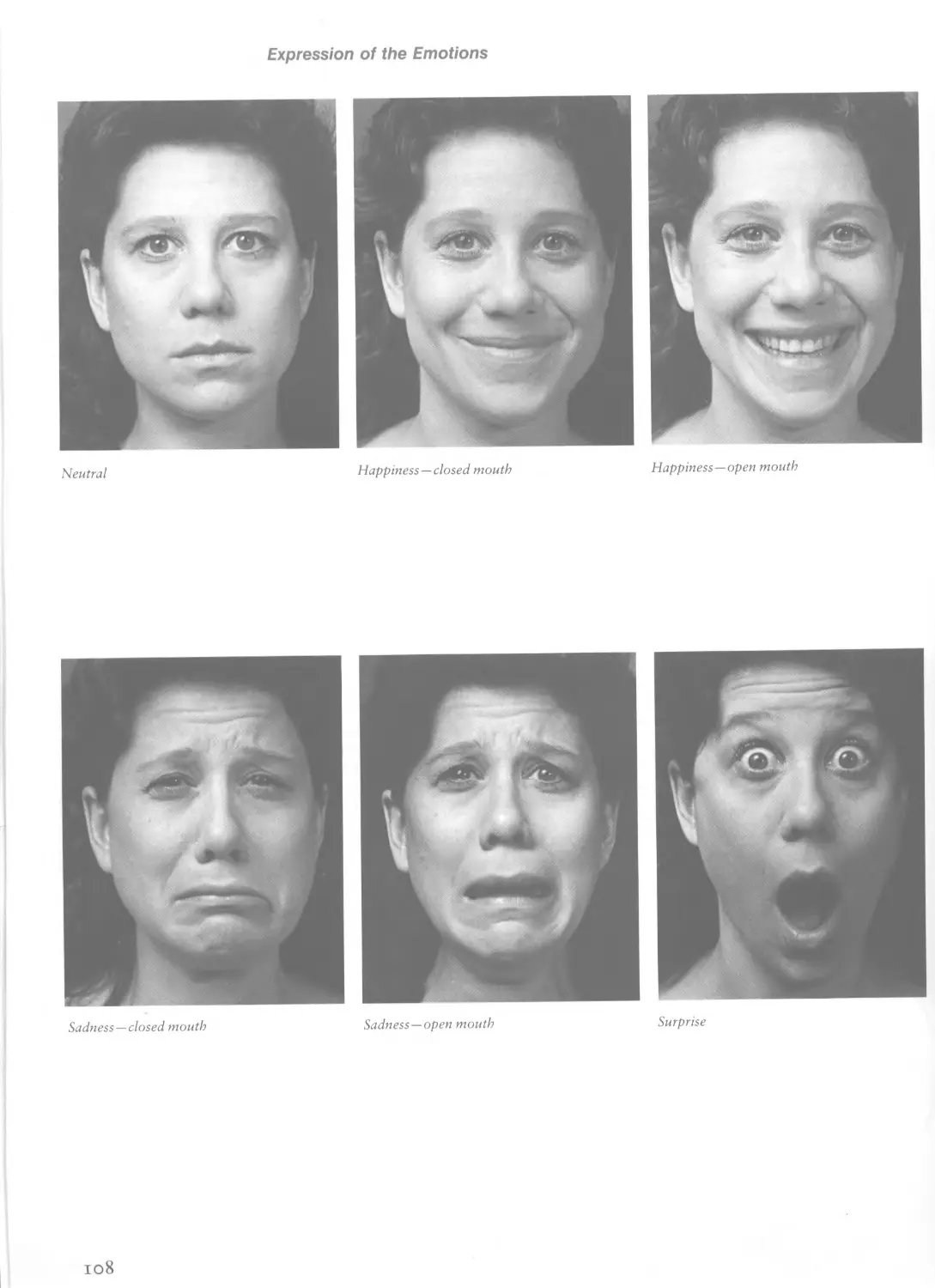
Neutral
Happiness—closed mouth
Happiness—open mouth
Sadness —closed mouth
Sadness—open mouth
Surprise
108
Expression of the Emotions
Fear Anger—closed mouth Anger—open mouth
Disgust — Levator labii superioris alaeque nasi Disgust—Levator labii superioris Cotitempt
109
The Head and Neck Muscles
CYLINDER
OF NECK
OF
NECK
ANC-UE. OF JAV/
POST.
OMOHYOID
EXTERNAL
JUGULAR
VEIN
BRANCH TO
EXT JUG. V.
CROSS
S ECTION
OMOHYOID
OMOHYOID
FRONT
VIEW
SIDE
VI EW
MASTOID
ANT.
PLANE OF
INTERVAL
BETWEEN
OMOHYOID
TRAPEZIUS
AND 5TERN0-
no
STERHO-
MASTOID
STERNOMASTOiD
^ANG-LE OF JAVJ
The Head and Neck Muscles • TEMPORALIS
Ma Masseter Mai Malaris OOc Orbicularis oculi Те Temporalis ZA Zygomatic arch
The Head and Neck Muscles • TEMPORALIS
P 0 5 T. <-* ANT.
PORTION
DEEP
PORTION
Temporalis
ORIGIN Entire temporal fossa on the side of
the skull and the deep surface of the temporal
fascia covering the muscle.
INSERTION Apex and deep surface of the co-
ronoid process of the mandible (jaw bone) and
into the front edge of the ramus of the mandi-
ble almost as far down as the last molar.
ACTION Lifts the mandible, which closes the
jaw; the posterior fibers pull the jaw backward.
The temporalis holds the jaw closed when the
head is upright, and it clenches the teeth for
chewing.
STRUCTURE The temporalis is a flat, fan-
shaped muscle, thin at its margin, that fills the
depression on the side of the skull. The side of
the skull (the area of origin of the temporalis)
and the inner surface of the temporal fascia
(which covers the temporalis) form a flattened
container from whose inside surfaces the tem-
poralis originates. The fibers of the temporalis
converge into a strong tendon deep to the zygo-
matic arch.
The superficial portion of the temporalis pro-
duces a horizontal oval relief on the surface
when seen. The anterior portion of the muscle
may be slightly recessed from the outer bony
portion of the orbit, which then becomes prom-
inent. The posterior portion of the temporalis is
usually covered by hair.
The temporal fascia is a strong, dense layer
of fascia that lies on top of the temporalis, at-
taching to the skull just beyond the margin of
the muscle (the muscle attaches to the inferior
temporal line on the skull; the temporal fascia
continues past it to attach to the superior tem-
poral line). Inferiorly (a short distance superior
to the zygomatic arch), the temporal fascia
leaves the surface of the temporalis to attach to
the upper edge of the zygomatic arch. At this
point, a substantial accumulation of fat lies be-
neath the fascia (on top of the muscle). The
plane of this lower fascia is basically oriented
obliquely outward and upward, in contrast to
the vertical orientation of the larger portion of
the temporal fascia, which lies on the tempor-
alis muscle belly. Because the plane of the lower
fascia is directed slightly upward, it receives
more light than the vertical temporalis. The
lower fascia flows into the zygomatic arch,
creating a single, rounded, horizontal form on
the side of the cheek. There is usually no visible
separation between the top edge of the zygo-
matic arch and lower portion of the fascia. The
plane of the lower fascia gradually blends into
the plane of the temporalis.
Because the lower portion of the fascia spans
over to the zygomatic arch, the temporalis
never appears to actually pass under the zygo-
matic arch. The bony zygomatic arch itself is
usually not sharply defined. Only occasionally
does it show clearly.
At the temple (posterior to the bony orbit of
the eye and superior to the zygomatic arch), the
temporalis is seen bulging when the jaw is
closed (the belly is shortened and thickened)
and sinking in when it is opened (the belly is
stretched and thinned). When the jaw is fully
opened, however, the condyle and ramus of the
mandible move forward, compressing the fat
and muscular tissue in front of them. This
pushes the fat lodged under the zygomatic arch
upward, which bulges the lower portion of the
temporal fascia outward. The advancing man-
dibular condyle and compressed tissue also
bulge the masseter muscle below the zygomatic
arch outward (laterally), the net result being
that the horizontal zygomatic arch becomes re-
cessed between the fatty bulge above and the
muscular bulge below. This is unusual, because
stretched and elongated muscles always become
thinner, increasing the projection of adjacent
bony areas.
The Head and Neck Muscles • MASSETER
side view
Masseter
ORIGIN Superficial part: Anterior two-thirds
of the inferior border of the zygomatic arch,
continuing forward to the zygomatic process of
the maxilla. Deep parts: Inner (deep) surface of
the entire zygomatic arch.
INSERTION Lateral surfaces of the coronoid
process, ramus, and angle of the jaw.
ACTION Closes the jaw by lifting the mandi-
ble; clenches the teeth for chewing.
STRUCTURE The masseter is a thick quadri-
lateral muscle that lies obliquely on the side of
the jaw. It is directed from the lower margin of
the zygomatic arch and cheekbone downward
and backward to the angle of the jaw. Only its
superficial portion is seen. When clenching the
teeth because of tension, or when chewing, the
bulge of the masseter belly can be seen separat-
ing into several thin, elongated, parallel bun-
dles, directed along the overall axis of the form
of the muscle. The most anterior of these bun-
dles shows up on the lower half of the front
edge of the belly. The entire front edge of the
muscle may be seen if the cheek fat is thin,
whereas the back edge is not distinct as it is
softened by the overlying parotid gland.
The surface planes of the masseters* on both
sides of the head usually converge sjightly infe-
riorly, but may at times be parallel or even di-
verge inferiorly. The surface of the masseter
may flow into the surface of the zygomatic arch
(no separation), or the superior end of the mas-
seter may be recessed slightly, leaving the lower
edge of the zygomatic arch defined.
AJ Angle of jaw Bu Buccinator DiA Digastric—anterior belly Hy Hyoid bone Ma Masseter
Md Mandible Mo Molar Mx Maxilla My Mylohyoid PG Parotid gland
The Head and Neck Muscles • MYLOHYOID
POST. <------> ANT.
Mylohyoid
ORIGIN A line along the anterior three-quar-
ters of the inner surface of the body of the
lower jaw.
INSERTION A median tendinous line, or
raphe (actually part of the mylohyoid itself),
which passes from the midline at the bottom of
the chin to the midline on the front of the
hyoid bone. Most of the muscular fibers of the
mylohyoid insert into this raphe (the fibers from
the muscles of both sides meet here), while the
most posterior fibers insert directly into the
front surface of the hyoid bone.
ACTION Lifts the floor of the mouth and the
hyoid bone when swallowing.
STRUCTURE The mylohyoid is a flat, sheet-
like triangular muscle. The muscles of both
sides span the bottom of the jaw and are up-
wardly recessed from the lower border of the
body of the mandible at their lateral edges
(their origins). The overlying anterior bellies of
the digastric, submandibular glands, and fat
join with the mylohyoids in creating the bottom
plane of the jaw.
The mylohyoid of each side passes medially
and downward to its insertion on the median
raphe and hyoid bone. Looking at the side view
of the bottom plane of the jaw, its profile (at
the midline) passes backward and slightly
downward (is almost horizontal), whereas the
line of the bottom edge of the mandible passes
backward and slightly upward (is oblique).
V I £. W
Rf Raphe Rs Risorius SG Submandibular gland
PIN Platysma —nodular portion
ZA Zygomatic arch ZMa Zygomaticus major
The Head and Neck Muscles • DIGASTRIC
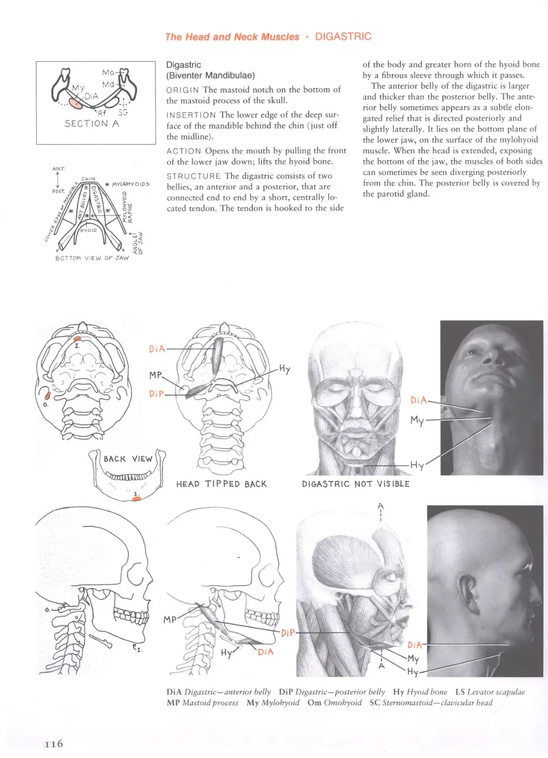
Digastric
(Biventer Mandibulae)
ORIGIN The mastoid notch on the bottom of
the mastoid process of the skull.
INSERTION The lower edge of the deep sur-
face of the mandible behind the chin (just off
the midline).
ACTION Opens the mouth by pulling the front
of the lower jaw down; lifts the hyoid bone.
STRUCTURE The digastric consists of two
bellies, an anterior and a posterior, that are
connected end to end by a short, centrally lo-
cated tendon. The tendon is hooked to the side
of the body and greater horn of the hyoid bone
by a fibrous sleeve through which it passes.
The anterior belly of the digastric is larger
and thicker than the posterior belly. The ante-
rior belly sometimes appears as a subtle elon-
gated relief that is directed posteriorly and
slightly laterally. It lies on the bottom plane of
the lower jaw, on the surface of the mylohyoid
muscle. When the head is extended, exposing
the bottom of the jaw, the muscles of both sides
can sometimes be seen diverging posteriorly
from the chin. The posterior belly is covered by
the parotid gland.
Di A Digastric—anterior belly DiP Digastric—posterior belly Hy Hyoid bone LS Levator scapulae
MP Mastoid process My Mylohyoid От Omohyoid SC Sternomastoid—clavicular head
116
The Head and Neck Muscles • OMOHYOID
Omohyoid
ORIGIN A short line on the superior border of
the scapula, just medial to the notch at the base
of the coracoid process, and sometimes from a
ligament that crosses this notch.
INSERTION Inferior border of the side of the
body of the hyoid bone toward the front (just
lateral to the insertion of the sternohyoid).
ACTION Pulls the hyoid bone down.
STRUCTURE The omohyoid consists of two
narrow, flat bellies—a superior and an infe-
rior—connected end to end by a short tendon.
The tendon lies under the sternomastoid mus-
cle. The omohyoid bends at this tendon as it
ascends upward and forward on the side of the
neck. The omohyoid is occasionally seen, part
directly, part indirectly, when speaking, when
the muscles on the bottom of the jaw are tensed
or when the head is fully turned to one side.
When contracted, it straightens between its two
points of attachment, lifting the sternomastoid
in the process. The tendinous portion beneath
the sternomastoid is then seen as a thick,
oblique, cordlike relief whose form is continued
both superiorly and inferiorly by the now visi-
ble superficial bellies.
SM Scalenus medius SS Sternomastoid—sternal head
SG Submandibular gland Sh Sternohyoid
Stm Sternomastoid TC Thyroid cartilage
Tr Trapezius Ve Vertebra
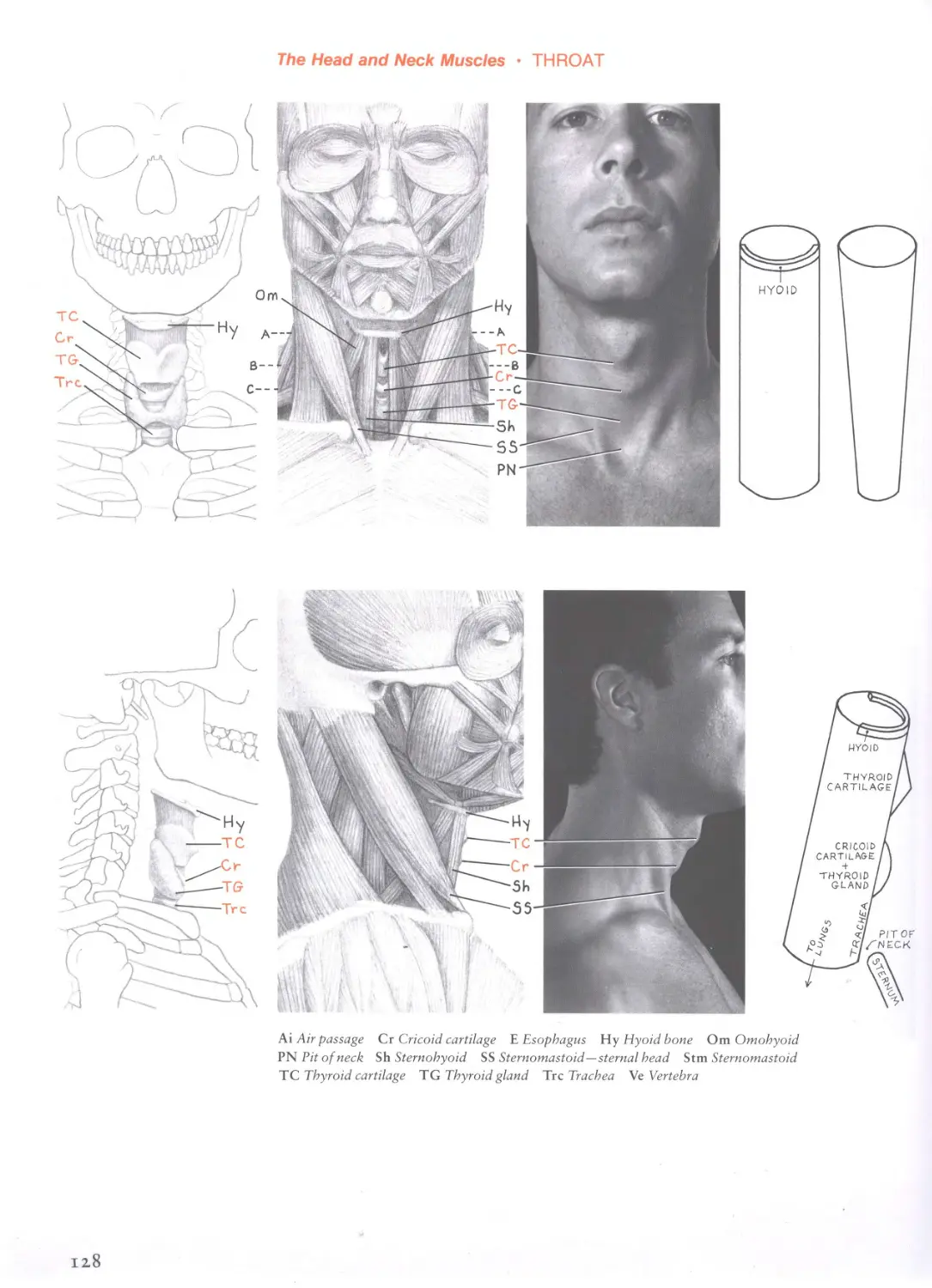
The Head and Neck Muscles • STERNOHYOID
Cr Cricoid cartilage Hy Hyoid bone Om Omohyoid Sh Sternohyoid
SS Sternomastoid — sternal head TC Thyroid cartilage
The Head and Neck Muscles • STERNOHYOID
FRONT VIEW
Sternohyoid
ORIGIN Posterior surface of the medial end of
the clavicle; the upper lateral portion of the
posterior surface of the manubrium of the ster-
num near the notch for the clavicle; and the
ligament between these two bones.
INSERTION Lower border of the anterior
portion of the body of the hyoid bone, just to
the side of the midline.
ACTION Pulls the hyoid bone down, as in
swallowing; assists in flexion of the head and
neck.
STRUCTURE The sternohyoid is a thin, nar-
row, straplike muscle located on the front of
the neck, just to the side of the midline. Com-
monly separated by a short distance at the pit
of the neck, the muscles of both sides converge
as they ascend, but never actually touch. The
space between the two muscles therefore nar-
rows superiorly. The thyroid and cricoid carti-
lages and the thyroid gland are partly covered
by these muscles, but are subcutaneous in the
interval between them. The narrow, flat form of
the sternohyoid lies on top of, but doesn’t con-
ceal, the forms of the throat (thyroid and cri-
coid cartilages, thyroid gland).
When contracting, the sternohyoids occasion-
ally appear as two raised straps on the front of
the neck, oftentimes confused with the anterior
edges of the platysma. The anterior edge of the
platysma is more stringy, more oblique, and lo-
cated more laterally than the almost vertical
sternohyoids. Also, the platysma passes over the
sternomastoid, whereas the sternohyoid passes
under it.
The Head and Neck Muscles • SEMISPINALIS CAPITIS
DM Deep muscle Nu Nuchal ligament Se Semispinalis capitis
SpC Splenius capitis
Stm Sternomastoid Tr Trapezius * Under trapezius
120
The Head and Neck Muscles • SEMISPINALIS CAPITIS
SECTION A
SECTION В
Semispinalis Capitis
(Complexus)
ORIGIN Tips of the transverse processes of the
vertebrae from the seventh cervical down to the
sixth or seventh thoracic vertebrae and from the
aritcular processes of the fourth through sixth
cervical vertebrae.
INSERTION An elongated area on the base of
the skull between the superior and inferior nu-
chal lines of the occipital bone, just lateral to
the midline.
ACTION Extends the head and rotates it so
that the face is directed to the opposite side.
STRUCTURE Primarily a deep muscle on the
back of the neck that thickens as it ascends, the
semispinalis capitis makes an important contri-
bution to the form of this region. (A very small
part of it is actually subcutaneous just below
the skull, but is is never distinctly seen and is
usually covered by the hair.) It lies under the
thin uppermost portion of the trapezius, and
both muscles create the muscular column on the
back of the neck immediately to the side of the
nuchal ligament. These muscles of both sides of
the body are therefore partly responsible for
creating the two thick cords that characteristi-
cally appear on the back of the neck (except
when the head is fully flexed). The furrow on
the midline between these two columns (the
posterior free edge of the nuchal ligament)
deepens as it approaches the skull, ending supe-
riorly at the external occipital protuberance (the
cranial attachment of the nuchal ligament). A
depression may form between the upper end of
the semispinalis capitis and the sternocleidomas-
toid, in which the subcutaneous portion of the
splenius capitis lies.
Basically vertical, the thick, somewhat flat-
tened belly of the semispinalis capitis wraps
around the deeper structures of the neck, best
seen in cross section. The semispinalis capitis is
part of the transversospinalis muscle group—the
deepest muscle group of the back—which in-
cludes the multifidus. The medial portion of
the semispinalis capitis, which tends to merge
with the spinalis capitis, is called the biventer
cervicis.
121
The Head and Neck Muscles • LEVATOR SCAPULAE
LS Levator scapulae Se Semispinalis capitis SM Scalenus medius SpC Splenius capitis
Stm Sternomastoid Tr Trapezius
The Head and Neck Muscles • LEVATOR SCAPULAE
SECTION В
Levator Scapulae
ORIGIN Transverse processes of the first four
cervical vertebrae.
INSERTION Upper portion of the vertebral
border of the scapula, between the superior an-
gle and the base of the spine of the scapula.
ACTION Raises the scapula while pulling it
slightly medially. It assists in rotation of the
scapula by raising the vertebral border, which
produces downward rotation of the acromion.
When the shoulder is fixed, it will bend the
neck laterally.
STRUCTURE Beginning deep in the side of
the neck just below the skull, and ending deep
in the back at the top of the shoulder, the leva-
tor scapulae is superficial only in its middle
third, in the space between the sternomastoid
and trapezius on the side of the neck. When the
muscles located in this interval are defined, the
levator scapulae is usually the most prominent.
In side view, the axis of its narrow, cylindrical,
subcutaneous form is oriented on a line directed
from the earlobe to the deep superior angle of
the scapula. The levator scapulae is located an-
terior to the splenius capitis, but whereas the
splnius capitis is directed downward and medi-
ally toward the midline of the back, the levator
scapulae is directed downward and laterally
toward the top of the scapula. The levator sca-
pulae is the more prominent of the two, but
they may also blend together.
The levator scapulae is located at the front
edge of the muscle mass of the back of the
neck. This mass is made up of the trapezius,
semispinalis capitis, splenius capitis, and levator
scapulae; it can be seen separating from the
sternomastoid and the front half of the neck
when the head is fully rotated to one side. The
levator scapulae and scalenus medius are re-
sponsible for the width at the side of the base
of the neck.
The levator scapulae is often difficult to see,
regardless of muscular development. It is most
likely to be seen in individuals with very little
fat and in the elderly, especially when the arms
are carrying weight and the shoulder is slightly
raised, or when the neck is bent laterally
against resistance. When seen, the combined
forms of the levator scapulae and sternomastoid
create an inverted V, with the upper end of the
former passing deep to the latter.
123
The Head and Neck Muscles • SPLENIUS CAPITIS
Splenius Capitis
ORIGIN The lower half of the posterior free
edge of the nuchal ligament and the spinous
processes of the seventh cervical and first three
or four thoracic vertebrae.
INSERTION A line at the side of the base of
the skull, just deep to the insertion of the ster-
nomastoid, from the tip of the mastoid process
extending back about two inches.
ACTION Rotates the head, turning it toward
the same side as the active muscle. The muscles
of both sides contracting together extend the
head.
STRUCTURE The splenius capitis is a broad,
sheetlike, inconspicuous muscle located on the
back of the neck. Only a small area of its supe-
rior portion is superficial. When seen, its super-
ficial portion is directed to the mastoid process.
The front edge of the muscle, which lies at the
back edge of the form of the levator scapulae,
can be traced along a line from the tip of the
mastoid process downward to the back of the
base of the neck. The form of the levator scapu-
lae is stronger and more pronounced than that
of the splenius capitis.
LS Levator scapulae Nu Nuchal ligament Om Omohyoid SA Scalenus anterior
SC Sternomastoid—clavicular head Se Semispinalis capitis SM Scalenus medius SP Scalenus posterior
The Head and Neck Muscles • SCALENUS MEDIUS
SIDE VIEW
Scalenus Medius
ORIGIN Transverse processes of the second
through seventh cervical vertebrae (and occa-
sionally from the atlas).
INSERTION Upper surface of the first rib,
from its midpoint back to the tubercle.
ACTION Bends the neck laterally; lifts the first
rib during forced inspiration.
STRUCTURE Largest of the scalene muscles,
the scalenus medius shows up on the middle of
the lower half of the side of the neck, but not
very prominently. It adds thickness to the bot-
tom of the side of the neck where the neck
flares, and is usually covered by some of the
supraclavicular fat. The muscle appears as a
short, vertical, conical form, pointed superiorly,
between the inverted V of the forms of the ster-
nomastoid and levator scapulae. The scalenus
medius may appear during forced inspiration.
The scalenus posterior, arising from the
transverse processes of the fourth through sixth
cervical vertebrae, and inserting onto the top
surface of the second rib posterior to its mid-
point, is never distinct, but blends into the back
of the form of the scalenus medius. The sca-
lenus anterior is too deep to affect surface form.
At times, some of the nerves of the brachial
plexus can be seen as a raised cordlike ridge
passing down along the anterior edge of the
form of the scalenus medius.
SpC Splenius capitis Stm Sternomastoid Tr Trapezius Ve Vertebra
The Head and Neck Muscles • STERNOCLEIDOMASTOID
LS Levator scapulae
SA Scalenus anterior
SpC Splenius capitis
Mn Manubrium MP Mastoid process Om Omohyoid PG Parotid gland
SC Sternomastoid—clavicular head Sh Sternohyoid SM Scalenus medius
SS Sternomastoid—sternal head Stm Sternomastoid TG Thyroid gland
12.6
The Head and Neck Muscles • STERNOCLEIDOMASTOID
SECTION В
SECTION C
Sternocleidomastoid
(Sternomastoid)
ORIGIN Sternal head: Upper part of the ante-
rior surface of the manubrium of the sternum,
near the midline. Clavicular head: Upper sur-
face of the medial third of the clavicle, exclud-
ing the medial expansion of the clavicle. The
area of origin extends downward onto the front
surface of the clavicle.
INSERTION A line along the tip, anterior bor-
der, and lateral surface of the mastoid process
of the skull (behind the ear), continued around
posteriorly into the lateral part of the superior
nuchal line of the occipital bone at the base of
the skull.
ACTION Both sides together flex the head and
neck forward. Acting alone, it will bend the
head and neck laterally and rotate the face
toward the opposite side.
STRUCTURE The sternocleidomastoid (ster-
nomastoid) is a wide, basically straplike muscle,
thickest at its center, that spirals obliquely
down the side of the cylindrical form of the
neck. It passes from behind the ear toward the
pit of the neck, and stands out prominently in
action. The single belly consists of two heads,
which when tensed can be seen separating into
the thicker, more prominent, fusiform sternal
head and the wider, flattened, straplike clavicu-
lar (“cleido”) head. The sternal head can be
seen from origin to insertion; the clavicular
head is usually visible on the lower half of the
neck only (the upper half of the clavicular head
blends into the dominant sternal head).
The superior end of the muscle (the single
belly made up of its two fused heads) becomes
aponeurotic, especially posteriorly, before it in-
serts into the skull. Inferiorly, the sternal and
clavicular heads are usually separated by a
small triangular interval just above the medial
expansion of the clavicle.
The sternal head tapers inferiorly into a
strong tendon that appears round, conspicuous,
and cordlike on the surface as it passes medial
to the expanded medial end of the clavicle (not
in front of it). When the head (skull) faces for-
ward, the inferior end of the sternal head curves
slightly forward toward the sternum. When the
head is completely rotated to one side, the ster-
nal head becomes vertical and almost cylindri-
cal in cross section, and forms the anterior pro-
file of the neck. Along with the upper edge of
the manubrium of the sternum, the tendons of
the sternal heads of both sides of the body (not
the medial ends of the clavicles) define the pit
of the neck.
The inferior end of the clavicular head usu-
ally dips downward slightly before heading for-
ward to the clavicle (creating a gentle concav-
ity). The widened form of the clavicular head
usually ends inferiorly at the top of the clavicle,
leaving the form of the front of the clavicle visi-
ble. This head continues onward to the front
surface of the clavicle, but only via its nonvisi-
ble flattened tendon. The upper half of the clav-
icular head normally becomes partially or com-
pletely covered by the sternal head as the two
heads blend together.
The most posterior fibers of the clavicular
head are usually inconspicuous, but when ex-
tremely tensed (head bent laterally against resis-
tance), they become visible along their entire
length, passing from the skull to the clavicle.
They then appear as one or more independent
narrow bundles descending from the aponeu-
rosis at the very posterior end of the line of
attachment on the skull to the lateral end of the
line of attachment on the clavicle. The interval
between the posterior edge of the sternocleido-
mastoid and the anterior edge of the trapezius
then appears narrower.
The sternocleidomastoids form the sides of
the neck, converging as they advance anteriorly
in their descent. When relaxed, the sternomas-
toids contribute to the cylindrical nature of the
neck; when tensed, they become reliefs that rise
up from the surface of the cylinder. The neck
widens when it is extended, becoming widest
two-thirds of the way down the sternocleido-
mastoids.
127
The Head and Neck Muscles • THROAT
Ai Air passage Cr Cricoid cartilage E Esophagus Hy Hyoid bone Om Omohyoid
PN Pit of neck Sh Sternohyoid SS Sternomastoid—sternal head Stm Sternomastoid
TC Thyroid cartilage TG Thyroid gland Trc Trachea Ve Vertebra
128
The Head and Neck Muscles • THROAT
The THROAT is the windpipe, or breathing
tube, and consists of the larynx and trachea.
The thyroid gland contributes to its form.
The throat is basically a semirigid cylindrical
structure, located on the front of the neck,
which connects the mouth to the lungs. The
upper front edge of the throat cylinder is
formed by the U-shaped hyoid bone (the hyoid
bone inconspicuously lies at the junction be-
tween the bottom plane of the jaw and the
front of the throat cylinder). The throat cylin-
der is directed downward and backward into
the chest cavity to the lungs, passing behind the
manubrium of the sternum. The cylinder is rela-
tively wide—almost half the width of the
neck—and lies between the bellies of the sterno-
mastoid muscles. Because the throat cylinder ta-
pers inferiorly, and because its lower portion is
partially covered on the sides by the sternomas-
toids, it appears on the surface to be an in-
verted cone. It is also covered on its front by
the straplike infrahyoid muscles (sternohyoids,
omohyoids, etc.), which contribute to its cylin-
drical form.
As a unit, the throat moves up when the head
is extended or the tongue protruded, and down
when the head is flexed. During swallowing, it
is pulled up to, and then drops down from, the
hyoid bone. The throat cylinder has two bumps
on its anterior surface: the thyroid cartilage on
its upper third and the combined cricoid carti-
lage and thyroid gland on its middle third (the
form of the cricoid cartilage predominates in
the male; in the female, the thyroid gland is
often most prominent). The trachea accounts
for the lower third of the cylinder, but is not
seen directly as it is covered by the skin draped
over the structures of the lower portion of the
throat. What remains is a concave depression at
the pit of the neck. In the male, the upper
bump is larger than the lower one. In the adult
female, both bumps are more or less equal in
size; sometimes both are inconspicuous, form-
ing a simple curve along the front profile of the
neck, fhe bumps become more prominent when
the head and neck are extended.
The larynx, or “voice box,” consists, in part,
of the thyroid cartilage, vocal cords, and cricoid
cartilage.
The thyroid cartilage is the largest cartilage
of the throat. Suspended by a membrane from
the hyoid bone, it is shaped like the bow of a
ship. Its two sides, or laminae, meet at a sharp
angle to form its front edge (more acute and
sharper in the male than in the female). In side
view, the upper and lower portions of its front
edge are cut back to produce a point on its
anterior edge. The upper portion has an elon-
gated V-shaped notch, which is easily palpated
and often seen (especially in the extended head).
The thyroid cartilage is larger and more promi-
nent in the adult male than in the adult female,
and is called the Adam’s apple.
The cricoid cartilage is a cartilaginous ring
located inferior to the considerably larger thy-
roid cartilage and superior to the narrower tra-
chea. It has a wide U-shaped notch on the front
of its upper edge, which registers as a subtle
depression on the surface. The cricoid cartilage
is subcutaneous and is visible in the male,
where it creates the smaller, rounder, inferior
bump on the front of the throat. In the female,
its form is masked (although not covered) by
the swelling of the larger thyroid gland.
The thyroid gland lies roughly at the level of
the cricoid cartilage. It consists of a relatively
large, oval lobe on each side of the throat (cov-
ered by the sternohyoids) connected in front by
a narrow isthmus of gland across the upper
portion of the trachea (over the second and
third tracheal rings). The gland is visible in the
adult female and in youth, where it creates the
soft swelling of the inferior bump on the front
of the neck, while concealing the cricoid carti-
lage. In men, the gland is usually inconspicuous.
The trachea, beginning just below the cricoid
cartilage, is the inferior component of the sub-
cutaneous windpipe. It continues the form of
the throat cylinder behind the sternum into the
chest. Although its actual cylindrical form is
concealed by the thyroid gland superiorly and
the suprasternal fat inferiorly, it can easily be
palpated. Its individual cartilaginous rings are
not visible.
129
о
The Trunk Muscles
The region from the shoulders to the iliac lines
is considered the trunk. The TRUNK MUSCLES,
described on the following pages, for the most
part lie on the trunk itself, although some cross
the shoulder joint and the neck.
The shape of the trunk is largely determined
by the rib cage, upon which the pyramidal
shoulder apparatus of both sides—the bony
shoulder girdles and their muscles—sit and
glide. The trunk is flattened from front to back,
noteworthy in the human figure because in
quadruped mammals the torso is laterally com-
pressed from side to side.
The rigid rib cage is separated from the pelvic
region (the rigid upper margin of the pelvis) by
the muscular body wall. These “rigid units” are
independent of and often dynamically opposed
to each other. They are connected by the lum-
bar vertebrae in back, which allow, and also
limit, the movements possible between them.
The irregularly shaped interval between the
two bony regions (between the bottom of the
rib cage and the top of the pelvis) is bridged by
the thin, soft, muscular body wall, made up of
the rectus abdominis and the external oblique
(as well as other thin layers of deeper muscles).
These muscles accommodate all the twisting
and bending of the bony regions by stretching,
contracting, folding, and bulging.
The trunk (including the shoulders) is easily
visualized as a flattened box. The back of the
trunk appears larger than the front, and can be
visualized as an inferiorly pointed triangular
wedge, especially in the male.
POSTERIOR SUPERIOR
ILIAC SPINE
EXTERNAL OBLIQUE
GLUTEUS MAXIMUS
LATERAL HD.
LONG HEAD -
INFRASPINATUS
VERT BORDER
OF SCAPULA
RHOMBOID___
TERES MAJOR
TEND0N5
MUSCLE
CASE
FORMS
BACK
SURFACE
ANATOMY OF
-TERES MAJOR
INFERIOR ANGLE
OF SCAPULA
SERRATUS
ANTERIOR
ANTERIOR BORDER OF
LATISSIMUS DORSI
ERECTOR SPINAE
LATERAL FORM
MEDIAL FORM
FLANK PAD
DEPRESSION OF POSTERIOR
SUPERIOR ILIAC SPINE
GLUTEAL FORM
U)
и )
DELTOIP
The Trunk Muscles • ERECTOR SPINAE
DM Deep muscle ES Erector spinae ES(L) Erector spinae—lateral form
ES(M) Erector spinae—medial form FF Flank, fat pad GMa Gluteus maximus GMe Gluteus medius
I Ilium L Eatissimus dorsi Mu Multifidus PSIS Posterior superior iliac spine
QL Quadratus lumborum R Ribs Sac Sacrum SeA Serratus anterior Tr Trapezius Ve Vertebra
* Under latissimus dorsi
The Trunk Muscles • ERECTOR SPINAE
Erector Spinae
(Sacrospinalis)
ORIGIN A continuous line which begins on
the inner surface of the posterior third of the
iliac crest, passes down the lateral crest of the
sacrum (not the lateral edge), curves around to
and then up the median crest of the sacrum (on
the midline), and continues straight up the tips
of the spinous processes and supraspinal liga-
ment of the lumbar and lower two thoracic ver-
tebrae. (There are additional deep sites of origin
further up the back.)
INSERTION By many muscular slips into nu-
merous points on the backs of all twelve ribs,
between the angles of the ribs laterally and the
tubercles of the ribs medially; into the spinous
and/or transverse processes of all the thoracic
and cervical vertebrae; and into the mastoid
process of the skull.
ACTION Extends the vertebral column and
the head. Portions of the erector spinae assist
in rotation and lateral flexion of the vertebral
column.
STRUCTURE The erector spinae, or sacro-
spinalis consists of several combined muscles
which form a thick, elongated muscle mass on
the back to the side of the spinous processes of
the vertebral column. It extends from the sac-
rum to the base of the skull. It is thickest in the
lumbar and cervical regions, and thinner and
wider in the thoracic region. In the thoracic re-
gion, it widens out to, but not past, the angles
of the ribs. At about the level of the bottom of
the scapula, its form becomes less distinct as it
thins and passes upward under the rhomboids
and the trapezius.
In the side view of the body, the back profile
of rhe muscle creates a transition between the
down plane of the back of the thorax and the
up plane of the back of the sacrum. The back
profile usually appears as a series of two con-
vexities, as the erector spinae separates into me-
dial and lateral forms (see below). The lumbar
portion of the erector spinae forms a powerful
and prominent muscular column on each side
of the spine in the lower back, referred to as
the strong cords or the common mass.
The erector spinae is simple and tendinous at
its origin. As it ascends, it divides into several
muscles which split into numerous bundles;
new fibers arise as lower fibers insert. The sepa-
rate muscles and bundles are occasionally seen
on the surface—usually momentarily during
motion.
When the figure is standing erect, and the
erector spinae is relaxed, the muscle appears as
a single wide muscular form on either side of
the median spinal furrow (the depressed ridge
of the spinous processes). As the thorax flexes
forward at the waist, the muscle immediately
tenses and hardens, and the cylindrical form of
each side divides into two forms—a narrow,
lower, vertical medial form and a wider, higher,
oblique lateral form.
The medial form is made of the multifidus
muscle inferiorly (lying under the tendon of the
erector spinae) and part of the belly of the erec-
tor spinae superiorly. This form begins mid-sac-
rum, slightly below the level of the posterior
superior iliac spine, and ends superiorly just be-
low the lower tip of the trapezius. It lies be-
tween the spinous processes of the lumbar ver-
tebrae and the depression (or bump) of the
posterior superior iliac spine. The inferior end
of the medial form widens slightly on the poste-
rior surface of the sacrum.
The lateral form begins inferiorly at the level
of the top of the crest of the ilium and disap-
pears superiorly at the level of the bottom of
the scapula, where it thins out and passes under
the trapezius (it is occasionally seen higher,
bulging under the trapezius). This form is
oblique, being directed upward and slightly me-
dially. It is made up of fleshy muscle fibers of
the erector spinae. The lateral form is usually
wider inferiorly (the portion of the belly be-
tween the rib cage and the pelvis bulges lat-
erally because of the fullness of the muscle). It
narrows superiorly (its lateral edge shifts medi-
ally) as the ascending muscle bundles along its
lateral side thin into nonvisible tendons. The
tendons continue to insert out to the angles of
the ribs, but the visible form does not extend
that far laterally. The lower end of the lateral
form is created where its fleshy fibers attach
into its wide tendon of origin.
As the trunk reaches full flexion (flexing at
the vertebral column, not at the hip joints), the
erector spinae is pulled relatively thin and flat,
and the spinous processes of the lumbar and
sacral vertebrae, as well as the posterior supe-
rior iliac spines, begin to project. When the fig-
ure is erect, the vertical median furrow of the
back (at the bottom of which lie the vertebral
spinous processes) ends inferiorly halfway down
the sacrum.
The latissimus dorsi may confuse the shape of
the erector spinae in two ways. First, if the
erector spinae is relaxed when the latissimus
dorsi is tensed, the latissimus pulls on its wide,
sheetlike tendon of origin (also called the thora-
columbar fascia), which lies on the surface of
the erector spinae. This pulling causes ridges or
furrows to develop across the long axis of the
soft erector spinae. These ridges are in line with
the direction of the muscle fibers of the latissi-
mus, and are therefore almost horizontal superi-
orly and more oblique inferiorly. Second, when
133
The Trunk Muscles • ERECTOR SPINAE
the latissimus is tensed, the line of attachment
of its muscle fibers into its wide tendon of ori-
gin occasionally becomes visible—its muscle fi-
bers are in slight raised relief where they meet
the tendon. This visible diagonal line, either
curved or straight, sits on top of the lateral
form of the erector spinae. It is located lateral
to the oblique line which separates the erector
spinae into its medial and lateral forms. The
latissimus line appears only when the latissimus
dorsi is tensed, regardless of whether the erector
spinae is relaxed or contracted.
CONTINUES UNDER
134
The Trunk Muscles • MULTIFIDUS
Multifidus
(Multifidi)
ORIGIN The medial half of the posterior sur-
face of the sacrum, the inner aspect of the pos-
terior superior iliac spine, the posterior sacro-
iliac ligaments, and various points on the back
of the lateral processes of all the vertebrae ex-
cept the first three cervical vertebrae.
INSERTION The spinous processes of all the
vertebrae, except the atlas.
ACTION Extends, laterally flexes, and rotates
the vertebral column.
STRUCTURE The multifidus (multifidi) lies
deep to the erector spinae. It fills the bony
groove on both sides of the spinous processes of
the vertebrae along the entire vertebral column.
It consists of numerous short bundles passing
obliquely upward and medially. They variously
cross over two to five vertebrae at a time, de-
pending on their length, to insert into a higher
vertebra.
The upper portion of the muscle is deep to
the other spinal muscles. It progressively be-
comes thicker and wider as it descends. Inferi-
orly, it is covered only by the tendon of the
erector spinae in the lower lumbar and sacral
region. The gluteus maximus originates in part
from the surface of the lateral portion of this
tendon. The multifidus therefore directly creates
surface form in the lower lumbar and sacral
areas.
When the back muscles are tensed, the multi-
fidus creates the lower portion of the narrow
vertical form that lies immediately on either
side of the spinous processes of the sacrum and
lumbar vertebrae.
ES Erector spinae ES(L) Erector spinae—lateral form ES(M) Erector spinae—medial form
GMa Gluteus maximus Mu Multifidus PSIS Posterior superior iliac spine Sac Sacrum Ve Vertebra
* Under latissimus dorsi f Under erector spinae
135
The Trunk Muscles • RECTUS ABDOMINIS
Ap Aponeurosis Car Cartilage EO External oblique EpD Epigastric depression IO Internal oblique
LA Linea alba PMa Pectoralis major RA Rectus abdominis SL Semilunar line
TI Tendinous intersection X Xiphoid process
136
The Trunk Muscles • RECTUS ABDOMINIS
SeA
L
ES
SECTION
R
EO
к ^Car
RA
X
Rectus Abdominis
ORIGIN Pubic crest of the pubic bone (a short
horizontal line at the top of the pubic sym-
physis) and from ligaments on the front of the
pubic symphysis.
INSERTION Anterior surfaces of the costal
cartilages of the fifth, sixth, and seventh ribs
and the anterior surface of the xiphoid process.
ACTION Flexes the trunk at the lumbar verte-
brae.
STRUCTURE The rectus abdominis is a long,
flat, vertical muscle on the front of the abdo-
men. It is wide and thin above and tapers be-
low. The muscles of each side of the body meet
at the midline at a tendinous vertical line, the
linea alba (white line), which extends from the
xiphoid process to the pubis. The linea alba ap-
pears as a vertical furrow between the two bel-
lies, gradually disappearing below the navel.
The navel is variably situated halfway to two-
thirds of the way down this line. During lateral
trunk flexion, only the portion of the linea alba
above the navel curves laterally (corresponding
to the curving lumbar vertebrae in back).
The lateral edge of the rectus abdominis is
separated from the fibers of the external oblique
by a tendinous furrow called the semilunar line.
It is located medial to a line dropped from the
nipple (or straight down from mid-clavicle)
which is directed downward toward the middle
of the thigh (in the anatomical position). The
semilunar line begins just below the lower bor-
der of pectoralis major (on the surface of the
rib cage, where it is faint) and continues down-
ward beyond the rib cage, where it is more pro-
nounced. Inferiorly, at the level of the navel, the
semilunar line begins to widen into a tendinous
triangular expansion.
Usually, three fibrous bands, the tendinous
intersections, are situated in the muscle and in-
scribe transverse furrows across the belly. They
do not necessarily pass straight across both rec-
tus abdominis muscles, but are sometimes lo-
cated at different levels on each side, giving the
abdomen an asymmetrical appearance. An indi-
vidual intersection may be either straight or zig-
zag. Classically, the lowest tendinous intersec-
tion is horizontal and is located at, or slightly
above, the level of the navel. The highest is di-
agonal and is directed approximately from the
tip of the xiphoid process to the point where
the semilunar line crosses the ninth costal carti-
lage. The middle tendinous intersection is lo-
cated at or above the level of the waist, and its
degree of obliquity is midway between the other
two. Occasionally a fourth tendinous intersec-
tion is found below the navel, but is not always
seen. The obliquity of the intersections is highly
variable, and they may all be horizontal.
The three tendinous intersections divide the
rectus abdominis into four fleshy, muscular seg-
ments. The superior segment is continued up-
ward over the costal cartilage on the front of
the rib cage and is the least conspicuous of the
four. Its upper edge usually coincides with the
lower border of the pectoralis major; therefore,
this segment ends before reaching the bottom of
the sternum, leaving the infrasternal notch (epi-
gastric depression) exposed. It may end higher,
continuing under the pectoralis, or lower, ex-
posing costal cartilage. Sometimes this segment
divides into three vertical bundles of unequal
length, progressively becoming longer and in-
serting higher laterally. The overall plane of the
upper segment is directed slightly upward. The
medial bundle of this segment attaches into the
seventh cartilage; the middle bundle, into the
sixth; and the lateral bundle, into the fifth carti-
lage and its bony rib. The lowest segment—long
and tapering inferiorly—bulges the most. This
segment is part of the pelvic volume, and the
segments of the two sides of the body are usu-
ally not separated by the linea alba. The corners
of all the segments are slightly rounded.
The rounded abdominal or Greek arch, as
configured in classical Greek sculpture, is com-
plicated, and is composed of several struc-
tures—cartilage, muscle belly, and tendinous in-
tersection. It begins laterally and inferiorly at
the costal cartilages of the ribs (from the tenth
rib) and continues up and over through the up-
permost tendinous intersection of the rectus ab-
dominis. The upper most fleshy segments of the
rectus abdominis of both sides bulge above the
tendinous intersection and fill the sharp, angu-
lar summit between the two sides of the upper
ends of the costal cartilages, where they meet
the lower end of the sternum at an angle of 90°
(more acute in the female). The tip of the xiph-
oid process projects forward, pushing muscle
and tendinous intersection anteriorly, which
also helps to round off the abdominal arch.
Since the rectus abdominis muscle belly crosses
over the costal cartilage, in muscular individuals
the entire form of the upper segment may pre-
dominate and be seen as a distinct fleshy bulge,
whereas in thin individuals the form of the un-
derlying costal cartilage may predominate.
The two “arches” to look for at the front of
the rib cage are the rounded Greek arch, pro-
duced by the rectus abdominis muscle and its
uppermost tendinous intersection, or the sharp
skeletal arch, produced by the costal cartilages
meeting the bottom of the sternum just above
the pit of the stomach. The sharp skeletal arch
often predominates in the female, while the
rounded Greek arch usually predominates in the
male.
Faint oblique ridges on the surface of the rec-
137
The Trunk Muscles • RECTUS ABDOMINIS
tus abdominis, especially at its upper end, are
occasionally created when the pectoralis major
and external oblique contract and pull on the
thin abdominal aponeurosis that covers the rec-
tus abdominis. These ridges are in line with the
direction of the fibers of the pectoralis and ex-
ternal oblique; the fibers of the rectus abdom-
inis are vertical.
During forward trunk flexion, the muscle fi-
bers shorten and the tendinous intersections
move closer together. Two horizontal skin
folds, seen at or above the level of the navel,
separate the thoracic and pelvic masses. Lat-
erally, these skin folds end at or just beyond the
semilunar line, which remains visible.
When inhaling, the diaphragm pushes the ab-
dominal contents downward, causing the rectus
abdominis to protrude. When the trunk is
flexed forward, the belly (and the rectus ab-
dominis) bulges below the level of the navel.
FRONT
side: view
The Trunk Muscles • EXTERNAL OBLIQUE
Ap Aponeurosis ASIS Anterior superior iliac spine Car Cartilage CI Crest of ilium
EO External oblique EO(F) External oblique—flank portion EO(T) External oblique —thoracic portion
FF Flank fat pad GMe Gluteus medius IL Inguinal ligament ILi Iliac line IO Internal oblique
L Latissimus dorsi PMa Pectoralis major R Rib RA Rectus abdominis SeA Serratus anterior
SL Semilunar line
14О
The Trunk Muscles • EXTERNAL OBLIQUE
pad posteriorly—has a high curved arc. The il-
iac line has a subtle, elongated S curve. It inter-
sects with the iliac crest at a point one-third of
the way back on the crest (at the widest point
of the pelvis). The actual overlap of the poste-
rior portion of the flank pad, past the iliac crest
and onto the surface of the gluteus medius, con-
sists of fat and skin. The overlap increases
when the thorax is bent laterally toward that
side (the iliac line descends), as the extra skin
accumulates. When bent to the other side, the
iliac line and the overlap of the flank pad even-
tually completely disappear as they are
stretched, which can expose almost the entire
iliac crest. When the trunk is flexed forward,
the posterior part of the iliac line disappears.
The semilunar line is the tendinous furrow
located on the front of the abdomen between
the rectus abdominis and external oblique. Infe-
riorly, at the level of the navel, it begins to
expand into a triangular tendinous area whose
surface bulges outward because of the abdomi-
nal contents deep to it. Part of the deep internal
oblique muscle lies directly beneath this tendi-
nous expansion, occasionally producing a nar-
row, oblique relief form beneath it, directed
downward and medially (immediately above
and parallel to the inguinal ligament).
The muscle fibers of the external oblique that
insert into its aponeurosis do so via separate
bundles, which often creates a jagged, irregular
line (the lateral edge of the rectus abdominis is
smooth).
A triangular depression may occur in the por-
tion of the external oblique belly immediately
below the costal cartilage—just lateral to the
semilunar line and just above the front portion
of the flank pad. In this case, the costal carti-
lage becomes prominent.
The inguinal ligament (Poupart’s ligament) is
the thickened lower border of the aponeurosis
of the external oblique (it is not a separate
structure). It is attached laterally to the inside
of the anterior superior iliac spine and medially
to the pubic tubercle, near the midline. It forms
either a curved or straight line, marking the
separation between the abdomen and thigh. It
straightens and deepens when the thigh is
flexed. When seen in the female (where it is
usually very faint), the inguinal ligaments of
both sides form a wider V than in the male.
This is because in the female the level of the
anterior superior iliac spines is closer to the
level of the pubis, and the two anterior superior
iliac spines are relatively farther apart (the fe-
male pelvis is shorter and wider than the male
pelvis).
FLANK PAD
BACK VIEW
tendinous ABDOHiNAL
triangle
CRO55 5FCTI0N
THORACIC PORTION ON
RIB CAGE
FRONT VIEW
CARTILAGE
^depression
TRANSITIONAL
Form
FLANK
PORTION
The Trunk Muscles • PECTORALIS MAJOR
BB Biceps brachii BL Biceps brachii—long head BS Biceps brachii—short head Car Cartilage
D Deltoid DA Deltoid—anterior portion EO External oblique H Humerus Mn Manubrium
PAb Pectoralis—abdominal portion PC Pectoralis—clavicular portion PM a Pectoralis major
PMi Pectoralis minor PSt Pectoralis—sternocostal portion R Rib RA Rectus abdominis
SeA Serratus anterior Ste Sternum
I42
The Trunk Muscles • PECTORALIS MAJOR
SECTION A
Pectoralis Major
ORIGIN The anterior surface of the medial
half of the clavicle, the entire length of the ante-
rior surface of the sternum toward its lateral
edge (not from the xiphoid process), the carti-
lages of the first six or seven ribs, and the upper
end of the aponeurosis of the external oblique
muscle (abdominal aponeurosis) lying on the
top of the rectus abdominis muscle.
INSERTION Lateral lip of the intertubercular
sulcus (bicipital groove) on the front of the hu-
merus.
ACTION Adducts the arm, pulling it across the
chest; medially rotates the arm. The clavicular
portion will raise (flex) the arm, while the lower
lateral sternocostal portion will then pull it
down (extend it, but not hyperextend it, back
past the rib cage).
STRUCTURE The pectoralis major is a large,
thick, fan-shaped muscle lying on the front of
the thorax. It crosses the front of the armpit,
where it remains thick, and then attaches to its
thin, square tendon of insertion, which enables
it to pass under the deltoid to its insertion. No
muscular fibers of the pectoralis pass under the
deltoid—only its tendon does.
The pectoralis is divided into clavicular and
sternocostal portions, based on the origin of
these sections. The most lateral and inferior di-
vision of the sternocostal portion is called the
abdominal portion. Classically, the pectoralis
major is flat, planar, and squared-off, but it can
also be rounded and bulging, depending on its
variable outline and the development of its
muscular mass. The pectoralis major forms the
thick muscular anterior wall of the armpit. The
deep and unseen pectoralis minor lies behind it,
adding thickness to the medial end of the wall
of the armpit. A small part of the pectoralis
minor may actually come to the surface be-
tween the pectoralis major and the uppermost
visible digitation of the serratus anterior, but it
is not defined or seen as a distinct form.
As they come off their origin, the muscle fi-
bers of the pectoralis major divide into numer-
ous bundles. The bundles taper and converge
laterally, progressively overlying one another, a
superior bundle crossing in front of the bundle
immediately inferior to it. These bundles be-
come quite conspicuous when the muscle is
contracted. The uppermost clavicular fibers are
directed downward and laterally toward the
bottom of the line of insertion; the lowest and
most lateral abdominal fibers from the side of
the chest are directed upward and laterally
toward the top of the line of insertion; and the
remaining large group of bundles between them
are, as a group, directed across toward the
middle of that line. This arrangement creates a
thick, muscular, crisscross twisting of the fibers
in front of the armpit, with the clavicular por-
tion on top (in front) and the abdominal por-
tion deep in back. All these fibers attach into a
square common tendon which then passes
through the narrow space under the deltoid and
over the biceps to reach its insertion on the
humerus. Several vertical skin wrinkles appear
in life on top of the crisscrossing fibers when
ABDOMINAL
FRONT VIEW
LAT. «-> MED.
М3
The Trunk Muscles • LATISSIMUS DORSI
Ap Aponeurosis CI Crest of ilium EO External oblique ES Erector spinae FF Flank, fat pad
GMa Gluteus maximus GMe Gluteus medius ILi Iliac line In Infraspinatus L Latissimus dorsi
R Rib Rh Rhomboids SeA Serratus anterior TMa Teres major Tr Trapezius
* Under latissimus dorsi
146
The Trunk Muscles • LATISSIMUS DORSI
teres major, rib cage (and individual ribs), and
especially the serratus anterior. These forms are
more clearly seen when the trunk is flexed for-
ward, the arm raised, and the shoulder slid for-
ward, stretching and thinning the latissimus.
The direct evidence of the latissimus on the sur-
face, in order of prominence, is its lateral bor-
der, superior border, and the line of attachment
of its muscular fibers into its tendon. The mus-
cle sheet of the latissimus thickens as it ap-
proaches the armpit, filling the space surround-
ing the forms of the teres major and serratus
anterior.
147
The Trunk Muscles • TRAPEZIUS
C7 Seventh cervical vertebra ES Erector spinae In Infraspinatus L Latissimus dorsi
LS Levator scapulae Nu Nuchal ligament Rh Rhomboids S Scapula Se Semispinalis capitis
SM Scalenus medius SpC Splenius capitis SSc Spine of scapula T1 First thoracic vertebra
Tr Trapezius Ve Vertebra
148
The Trunk Muscles • TRAPEZIUS
SECTION A
SECTION В
SECTION C
Trapezius
ORIGIN A line that passes across the medial
third of the superior nuchal line, at the base of
the skull, over to the external occipital protu-
berance. It then descends along the posterior
free edge of the nuchal ligament, the vertebral
spinous processes down to the twelfth thoracic
vertebra, and the intervening supraspinal liga-
ments. The line of origin is an upside-down L.
INSERTION A continuous line that passes
across the posterior border of the lateral third
of the clavicle, onto the medial border of the
acromion, and along the upper edge of the
spine of the scapula (excluding the expanded
triangular area at the spine’s medial end). The
line of insertion then doubles back around for a
short distance along the lower edge of the spine
of the scapula, ending at the tubercle of the
spine. Seen from above, the line of insertion is a
V, pointed laterally, on the top of the bony
shoulder girdle.
ACTION Rotates the scapula by lifting its lat-
eral end and pulling its medial end downward.
Working separately, the upper portion lifts the
scapula, the middle portion pulls it medially,
and the lower portion pulls it downward.
STRUCTURE The trapezius is an extensive,
wide, triangular muscle located on the back of
the chest, the top of the shoulder, and the back
of the neck.
The upper fibers of the trapezius pass down-
ward and outward; the middle fibers pass hori-
zontally outward; and the lower fibers pass up-
ward and outward, heading toward the tubercle
on the spine of the scapula via a small aponeu-
rosis. The muscle varies in thickness in its dif-
ferent areas. The part on the back of the neck is
very thin; along with the deep semispinalis capi-
tis muscle it creates a muscular column on
either side of the midline. The nuchal ligament
lies on the midline of the back of the neck, at
the bottom of the vertical groove between these
columns when the neck is upright. It becomes a
raised ridge when the head and neck are flexed
forward. In side view, the back profile of the
neck can be concave, straight, or convex.
In nonmuscular individuals, or when the sca-
pula has been slid laterally and rotated so the
upper portion of the trapezius becomes elon-
gated and thin, the vertebral border of the sca-
pula located above the base of the spine may
become evident on the surface, beneath the
muscle. When the arm is abducted, the oval
form of the supraspinatus can be seen bulging
deep to the trapezius just above the medial por-
tion of the spine of the scapula.
The lowest fibers of the trapezius may sepa-
rate visually, creating an independent elliptical
or elongated form, directed upward and out-
ward toward the scapular tubercle. This portion
can be visualized as a separate flat muscle belly
with flattened tendons at both ends.
The lower lateral edge of the trapezius can be
seen passing obliquely upward and outward on
the middle of the back; it is clearly seen when
the arm is held horizontally out to the side and
pushed down against resistance.
The entire trapezius can be reduced to three
major planes: the back plane of the neck, the
top plane of the shoulder, and the vertical plane
of the back.
The majority of the trapezius creates the ver-
tical plane of the back, also called the rhom-
boid plane (based on the orientation of the un-
derlying rhomboid muscle). This vertical plane
is directed medially and anteriorly from the ver-
tebral border of the scapula to the spinous pro-
cesses of the vertebrae on the midline of the
body. The rhomboid planes of both muscles
(both sides of the body), along with both scapu-
lar planes (the planes created by the infraspina-
tus and spine of the scapula), create a wide W
configuration when viewed from the top. When
the shoulder blades are pulled together in back,
the rhomboid planes disappear and the trape-
zius muscles of both sides bulge and touch each
other, producing a tight vertical cleft between
them.
The top plane of the shoulder, for the most
part, is directed upward and backward. The
posterior edge of this plane is formed by the
spine of the scapula. Anteriorly, the shoulder
plane curves over the top of the shoulder.
The back plane of the neck, created by the
cervical portions of the trapezius of both sides
of the body, can be visualized in two ways—on
the one hand it is often flattened and planar,
while at other times it is rounded (side to side)
and participates in producing the cylindrical
volume of the neck.
There are three triangular aponeurotic or ten-
dinous areas in the trapezius, as follows:
i. A relatively large triangular area surround-
ing the seventh cervical vertebra. These ten-
dinous areas of the muscles of both sides of
the body create a diamond, oval, or tear-
drop-shaped depression, prolonged upward,
on the back of the neck. It usually reaches
up to the base of the skull and ends inferi-
orly at the level of the spinous process of the
third thoracic vertebra. The prominent spi-
nous processes of the seventh cervical and
first thoracic vertebrae project at the center
of this tendinous, depressed area.
z. A smaller triangular area at the upper lateral
end of the fibers of the lower half of the
trapezius. This tendinous area lies partially
on the expanded triangular base of the me-
149
The Trunk Muscles • TRAPEZIUS
dial end of the spine of the scapula (which
shows through as a bony plane) and par-
tially on the surface of the infraspinatus
muscle. The infraspinatus portion of the ten-
don may appear as a depression, caused by
the fullness of the surrounding muscular and
bony forms. The lower edge of this tendi-
nous triangle can sometimes be seen con-
stricting the muscular form of the infraspina-
tus. T he lateral ends of the lower muscle
fibers of the trapezius attach into the infra-
spinatus portion of the tendon in an elon-
gated S-curve line which lies lateral to the
underlying vertebral border of the scapula.
This produces a plane change of rhe trape-
zius belly as its fibers pass over this scapular
border; most of the belly lies medial to the
vertebral border on the rhomboid plane,
while only a very small portion of the belly
lies lateral to it on the scapular plane. When
the arm is abducted away from the side of
the body, automatically rotating the scapula,
the scapula and overlying infraspinatus slide
out laterally from under the small scapular
portion of the belly of the trapezius.
3. The smallest triangular area at the inferior
tip of the muscle. It is this small triangular
aponeurosis, not the actual muscle fibers,
that reaches the very bottom of the line of
origin of the trapezius (at the spinous pro-
cess of the twelfth thoracic vertebra). Be-
cause this tendinous area is not visible as it
lies on the erector spinae, the inferior end of
the form of the muscular belly of the trape-
zius ends superior to the final attachment.
The triangular tendinous areas of both sides
of the body create a diamond shape, with
the inferior ends of both sides of the muscu-
lar form of the trapezius ending in relief as
an inverted V, or at least appearing blunt,
rather than as a single sharp point.
PLANE
CHANGE
CROSS SECTION
150
The Trunk Muscles • RHOMBOIDS
Rhomboids
(Major and Minor Together)
ORIGIN From the inferior portion of the nu-
chal ligament down to the fifth thoracic verte-
bra, along the spinous processes and intercon-
necting supraspinal ligament of the vertebrae
(on the midline of the upper back).
INSERTION The very edge of the medial (ver-
tebral) border of the scapula, extending from
the top of the triangular expansion of the base
of the spine down to the inferior angle.
ACTION Together the rhomboids adduct and
raise the scapula. They assist in rotating the
scapula (during rotation, the bottom of the sca-
pula moves medially and the acromion moves
inferiorly).
STRUCTURE The rhomboid major and rhom-
boid minor muscles are barely separable and
are here considered as a single, flat, quadrila-
teral muscle on the upper back. When contract-
ing and shortening (pulling the scapula towards
the midline), the rhomboids may produce an
oval bulge which can be seen under the trape-
zius. This form lies closer to the scapula than to
the midline of the back.
The lateral portion of the oblique inferior
border of the muscle is superficial, but is not
always easily seen. It lies in the triangular space
bordered by the medial border of the scapula,
the lateral edge of the trapezius, and the top
edge of the latissimus dorsi. The rest of the in-
ferior edge can sometimes be seen under the
trapezius, heading diagonally upward and medi-
ally to its attachment on the vertebral column.
Any line or form directed upward and medi-
ally from the inferior angle of the scapula to
the midline is part of the rhomboids, not the
trapezius.
ES Erector spinae In Infraspinatus L Latissimus dorsi Rh Rhomboids S Scapula TMa Teres major
Tr Trapezius Ve Vertebra
The Trunk Muscles • SERRATUS ANTERIOR
EO External oblique L Latissimus dorsi PMa Pectoralis major PMi Pectoralis minor R Rib
S Scapula SeA Serratus anterior Sub Subscapularis TMa Teres major Tv Trapezius
* Under latissimus dorsi
The Trunk Muscles • SERRATUS ANTERIOR
Serratus Anterior
(Serratus Magnus)
ORIGIN A line of small triangular areas on
the outer surfaces and superior borders of the
first eight or nine ribs and the aponeuroses cov-
ering the intervening intercostal muscles.
INSERTION Narrow strip on the costal (deep)
surface of the scapula at its vertebral border,
which expands in area at the inferior angle of
the scapula.
ACTION Slides the scapula around forward, as
in punching, and by its inferior fibers rotates its
inferior angle laterally, which elevates the lat-
eral end of the shoulder, and therefore the arm.
STRUCTURE When considered in its entirety,
the serratus anterior is a large quadrilateral
muscle which embraces the curved form of the
rib cage. It is divided into fingerlike digitations
(bundles), each originating from an area on the
side and upper border of a rib and the intercos-
tal space above. The muscle can be subdivided
into upper and lower portions. The upper por-
tion partially lies inconspicuously against the
upper side of the rib cage in the armpit and
partially sits sandwiched between the scapula
and rib cage.
The lower portion consists of five or six
pointed digitations which create a thick, fan-
shaped mass radiating forward from the inferior
angle of the scapula. Only the lowest three or
four of these digitations can be seen—their an-
terior half is superficial on the side of the chest
(where they interdigitate with the bundles of the
external oblique), but as they head toward the
inferior angle of the scapula, they pass deep to
the latissimus dorsi; however, the form of the
lower portion remains visible under the thin la-
tissimus. The uppermost visible digitation be-
gins at or just above the level of the inferior
border of the pectoralis major. The digitations
can begin quite far forward on the side of the
rib cage, almost reaching the front of the chest.
The line of origin of the tips of the exposed
digitations on the side of the rib cage forms a
toothed (serrate), anteriorly curved line, which
appears to be directed from the nipple to the
posterior superior iliac spine when looking at
the figure in side view. Therefore, the length of
the exposed part of each of the digitations,
from its tip to the lateral border of the latissi-
mus dorsi, diminishes inferiorly. Sometimes the
tip of the lowest digitation may be completely
hidden from view under the front edge of the
latissimus, especially if the latissimus is drawn
forward in the advanced shoulder.
The digitations of the serratus anterior are
bulged and more horizontal than the flatter and
more oblique digitations of the external
oblique. The ribs, often seen under the external
oblique, are not seen under the serratus.
The inferior border of the serratus anterior
will sometimes continue inserting past the infe-
rior angle of the scapula into the lower edge
of the rhomboid major muscle for a short
distance; consequently, the form of the serra-
tus under the latissimus will extend further
medially.
BACK VIEW
The Shoulder Muscles
SCAPULA
ON
PROFILE
NORMAL
NECK
CAGE
RIB CAGE
ON
PROFILE
POST *—> ANT.
ADVANCED
S I
154
The Shoulder Muscles • DELTOID
BB Biceps brachii BL Biceps brachii—long head Br Brachialis BS Biceps brachii—short head
C Clavicle CV Cephalic vein D Deltoid DA Deltoid — anterior portion
DAc Deltoid—acromial portion DP Deltoid—posterior portion H Humerus In Infraspinatus
PC Pectoralis —clavicular portion PMa Pectoralis major SSc Spine of scapula
TLa Triceps — lateral head TLo Triceps—long head TMa Teres major TMi Teres minor
Tr Trapezius
The Shoulder Muscles • DELTOID
Deltoid
ORIGIN A continuous line passing along the
anterior surface of the lateral third of the clavi-
cle, the external border and adjacent top sur-
face of the acromion of the scapula, and the
lower edge of the spine of the scapula (extend-
ing medially up to, but not including, its medial
triangular expansion). The deltoid roughly orig-
inates just below the insertion of the trapezius
(the scapular spine remains subcutaneous).
INSERTION The deltoid tuberosity halfway
down the outside of the shaft of the humerus.
ACTION The anterior portion flexes the arm
(raises it in front of the body) and pulls the
horizontal arm toward the front of the body;
the middle acromial portion powerfully abducts
the arm; and the posterior portion extends the
arm backward and pulls the horizontal arm
backward. The anterior and posterior portions
are antagonists when they flex and extend the
arm at the shoulder joint.
STRUCTURE The deltoid is a thick triangular
muscle, pointed inferiorly, which cups the
shoulder joint. The muscle is divided into three
portions: an anterior (clavicular) portion, an ac-
romial (middle) portion, and a posterior por-
tion. In front, the deltoid and pectoralis major
are separated for a short portion of their
lengths, just below the clavicle, by a triangular
depression called the infraclavicular fossa.
The anterior portion of the deltoid, originat-
ing from the clavicle, is shorter than the other
two portions. It shows up as a teardrop or oval
relief on the surface, and sometimes splits down
the middle into two forms. The lateral form
descends slightly further. The belly of the ante-
rior portion ends in its own wide tendon a
short distance above the final insertion on the
humerus. This belly, and the belly of the clavi-
cular portion of the pectoralis major, end at
about the same level (the anterior portion of the
deltoid actually descends slightly further). The
fibers, and therefore the forms, of these two
muscles lie side by side—the deltoid does not
cover the muscular fibers of the clavicular pec-
toralis; however, the tendon of the clavicular
pectoralis (without fibers) does pass under the
anterior deltoid. The tendon of insertion of the
anterior portion of the deltoid then descends on
the front (not outer) surface of the acromial
fibers of the deltoid and inserts toward the
front surface of the humerus, adjacent to the
origin of the brachialis. It is actually the other
two portions of the deltoid that insert onto the
outside of the humerus.
The anterior portion of the deltoid projects
anteriorly owing to the forward projection of
the underlying spherical proximal extremity of
the humerus, especially when the arm is hyper-
extended back past the trunk.
The posterior portion of the deltoid is located
on the back of the shoulder. It thins into an
SECTION D
157
The Shoulder Muscles • SUPRASPINATUS
SECTION A
Supraspinatus
ORIGIN Medial two-thirds of the posterior
surface of the scapula superior to the spine (su-
praspinatus fossa).
INSERTION Superior facet of the greater tu-
bercle on the top of the lateral part of the prox-
imal extremity of the humerus.
ACTION Abducts the arm; stabilizes the shoul-
der joint from above by pulling the head of the
humerus into its socket.
STRUCTURE The supraspinatus passes lat-
erally over the top of the shoulder joint (under
the acromion) to its insertion on the outside of
the top of the proximal extremity of the hume-
rus. Although it lies deep to the trapezius under
a substantial thickness of muscle, when the arm
is abducted, its form is occasionally revealed as
a bulging egg-shaped or teardrop form (pointed
end directly laterally) above the medial half of
the spine of the scapula. In this position, the
supraspinatus is at its shortest and thickest.
If the scapula is prevented from rotating
(either by external resistance or internal muscle
fixation) as the arm is abducted to the horizon-
tal away from the side of the body, any bulge
developing above the scapular spine must be the
supraspinatus, since in this case there is no
shortening of the trapezius.
When the arm is down by the side of the
body, there is no indication of the supraspina-
tus on the surface. A slight hollow may actually
develop above the scapular spine, as the supra-
spinatus is now stretched thin.
Om Omohyoid S Scapula
SSc Spine of scapula Su Supraspinatus Tr Trapezius
* Under trapezius
159
The Shoulder Muscles • INFRASPINATUS
SECTION A
Infraspinatus
ORIGIN Most of the posterior surface of the
scapula below the spine (infraspinatus fossa).
INSERTION Middle facet of the greater tuber-
cle of the humerus.
ACTION Laterally rotates the arm; helps stabi-
lize the shoulder joint from behind by holding
the head of the humerus in its socket.
STRUCTURE The infraspinatus is a flat trian-
gular muscle that sits on the back of the shoul-
der blade below the spine. Although typically
exhibiting a slightly planar surface surrounded
by bulging muscles, the infraspinatus itself can
bulge considerably when the humerus is lat-
erally rotated to its maximum (this action
shortens and thickens the muscle). Because its
lateral portion is covered by the deltoid, the
superficial area of the infraspinatus may appear
as a wide vertical form when the arm is down.
When the arm is raised, the posterior portion of
the deltoid slides upward, exposing more sur-
face area of the infraspinatus.
The muscle fibers, directed outward and up-
ward, seem to be folded on themselves, which
occasionally divides the infraspinatus into two
forms—a superior and an inferior—separated
by a shallow furrow that is directed outward
and upward toward the head of the humerus.
This furrow may or may not reach all the way
over to the medial border of the scapula.
D Deltoid DP Deltoid—posterior portion H Humerus In Infraspinatus Rh Rhomboids S Scapula
SeA Serratus anterior SSc Spine of scapula TLo Triceps—long head TMa Teres major
The Shoulder Muscles • TERES MINOR
' M£t>«—>LAT.
BACK VIEW
Teres Minor
ORIGIN Raised ridge of bone on the posterior
surface of the upper two-thirds of the lateral
(axillary) border of the scapula, and partly from
the fascia covering the posterior surface of the
infraspinatus and the deep surface of the medial
end of the teres major.
INSERTION The lowest of the three facets of
the greater tubercle of the humerus and a short
distance immediately below it on the shaft.
ACTION Laterally rotates the arm and weakly
adducts it; helps stabilize the shoulder joint by
holding the head of the humerus in its socket.
STRUCTURE Rarely seen on the surface, the
teres minor is a small, elongated, cylindrical
muscle that usually blends with the plane cre-
ated by the infraspinatus muscle. Both muscles
are covered by a layer of dense fascia which
consolidates their forms. The teres minor runs
obliquely upward and outward from its origin
and inserts via a thick, flat tendon onto the
back of the superior part of the humerus. It is
occasionally seen when the arm is held horizon-
tally, laterally rotated, and then pushed down-
ward and backward against resistance.
TMi Teres minor Tr Trapezius
* Under latissimus dorsi
The Shoulder Muscles • TERES MAJOR
DP Deltoid—posterior portion In Infraspinatus L Latissimus dorsi Rh Rhomboids S Scapula
SeA Serratus anterior Sub Subscapularis TLo Triceps—long head TMa Teres major
TMi Teres minor Tr Trapezius * Under latissimus dorsi
162
The Shoulder Muscles • TERES MAJOR
Teres Major
ORIGIN Raised oval area on the posterior sur-
face of the inferior angle of the scapula (exclud-
ing a narrow vertical area immediately adjacent
to the medial border), continuing one-third of
the way up its lateral border, and partly from
the adjacent fascia covering the outer surface of
the infraspinatus and teres minor muscles.
INSERTION A short vertical line on the me-
dial lip of the bicipital groove (intertubercular
sulcus) on the inside front corner of the hume-
rus, one-fourth of the way down the humerus
(medial to and slightly lower than the insertion
of the latissimus dorsi).
ACTION Adducts and medially rotates the
arm; extends the flexed arm. Note: the teres
major and minor both adduct the arm, but are
antagonists for rotation of the humerus—the
major rotates it medially; the minor, laterally.
STRUCTURE The teres major is a cylindrical,
but somewhat flattened, thick, bulging muscle.
Laterally, it flattens into a broad tendon as it
inserts. The medial end of the belly does not
quite reach the medial border of the scapula.
The teres major creates part of the posterior
wall of the armpit, crossing from the bottom of
the back of the shoulder (bottom of the sca-
pula) to the inside front corner of the arm. Div-
ing into the armpit, its axis is directed outward,
upward, and forward. When the arm lies
against the body and the teres major is tensed,
the belly is shortened, becoming oval and
bulged (the infraspinatus and teres minor are
flattened and planar). In this position, the teres
major may not appear to be directed toward
the inner front corner of the humerus, but it
ultimately is. In the raised arm position, the
stretched teres major becomes noticeably longer
and thinner, and the upper edge of the latissi-
mus dorsi can often be seen as a raised relief
lying across its belly. The latissimus covers the
lower medial portion of the teres major.
Because the teres major inserts lower (distal)
onto the humerus than does the latissimus, the
cylindrical form of the teres major can be seen
as it leaves the sling of latissimus, giving the
posterior wall of the armpit a double convex
curve—the teres major is seen closer to the arm
(lateral and higher) and latissimus closer to the
body (medial and lower). This is best seen when
the arm is held horizontal out to the side of the
body (see Latissimus Dorsi).
The Upper Arm Muscles
POST.
ANT.
164
CROSS SECTIONS (miP ARM)
RIGHT HUHERU5
FRONT VIEW
The Upper Arm Muscles • BRACHIALIS
В A Bicipital aponeurosis BB Biceps brachii Br Brachialis Brr Brachioradialis ВТ Biceps tendon
Co Coracobrachialis Cp Capitulum DAc Deltoid—acromial portion H Humerus
MIS Medial intermuscular septum PrT Pronator teres TLa Triceps—lateral head
TMe Triceps—medial head Tro Trochlea
l66
The Upper Arm Muscles • BRACHIALIS
SECTION A
SECTION В
Brachialis
ORIGIN Lower half of the anterior surface of
the shaft of the humerus (the upper end of the
origin continues around to the lateral side of
the bone, embracing the insertion of deltoid),
and from both the lateral and medial intermus-
cular septa.
INSERTION Anterior surface of the coronoid
process of the ulna.
ACTION Flexes the forearm.
STRUCTURE Lying under the biceps and in
front of the elbow joint, this relatively wide,
fleshy muscle is seen only on the outside of the
upper arm. The narrow visible portion passes
obliquely downward and forward, first between
the biceps and triceps and then between the bi-
ceps and brachioradialis. Its obliquity increases
as the elbow is flexed; it also becomes shorter
and rounder during elbow flexion, appearing as
an oval relief on the outside of the arm at full
flexion. The brachialis lies on and adds muscu-
lar fullness to the front of the lower half of the
humerus. Its front surface is somewhat flattened
to receive the flattened posterior surface of the
biceps. The fibers at the upper outer end of the
brachialis blend directly with some of the in-
serting fibers of the deltoid. A depression may
appear at this fusion.
On the inside of the arm, the brachialis is
concealed by nerves, arteries, veins, and espe-
cially fat. When the elbow is partially flexed
and the muscles tensed, this area appears as a
flat plane, bordered in front by the biceps and
the raised medial edge of the bicipital aponeu-
rosis and behind by the medial head of the tri-
ceps. The brachialis may occasionally be seen in
this area as a recessed bulge, but it is always
covered by blood vessels and fat that protects
these vessels. When the elbow is fully extended,
this portion of the brachialis is pushed forward
at the elbow joint by the underlying trochlea of
the humerus. It is still not seen independently;
however, this area becomes fuller as it is pushed
forward.
SECTION C
FRONT VIEW
EXTENSION FLEXION
5IDE VIEW
167
The Upper Arm Muscles • BICEPS BRACHII
INSIPE VIEW
В A Bicipital aponeurosis BB Biceps brachii BL Biceps brachii—long head Br Brachialis
Brr Brachioradialis BS Biceps brachii—short head ВТ Biceps tendon Co Coracobrachialis
CV Cephalic vein D Deltoid DA Deltoid—anterior portion DAc Deltoid—acromial portion
H Humerus PC Pectoralis —clavicular portion PMa Pectoralis major PrT Pronator teres
PSt Pectoralis—sternocostal portion TLo Triceps —long head TMe Triceps—medial head
168
The Upper Arm Muscles • BICEPS BRACHII
SECTION A
SECTION В
SECTION C
Biceps Brachii
ORIGIN Long head: Supraglenoid tubercle on
the top of the glenoid fossa (socket) of the sca-
pula. Short head: Tip of the coracoid process of
the scapula (in common with the coracobra-
chialis).
INSERTION Tuberosity of the radius. The bi-
ceps has no attachment to the humerus—it has
two origins from the scapula and one insertion
into the radius.
ACTION Flexes and/or supinates the forearm.
Supination is most powerful when the forearm
has been flexed to 90°. It also assists in flexing
the arm in front of the body at the shoulder
joint. The long head holds the head of the hu-
merus in its socket, which also keeps the head
away from the acromion when the arm is ab-
ducted.
STRUCTURE The fusiform fleshy belly of the
biceps is divided into a long and a short head.
In its entirety, it extends from the shoulder to
the elbow. Emerging from the under the pector-
alis major, the belly expands into its familiar
bulging shape and ends abruptly into a very
strong, narrow tendon. This tendon inserts deep
in the elbow between the extensor and flexor
masses of the forearm.
The front profile of the biceps (seen in side
view) is straight for a considerable length when
the elbow is extended, but becomes quite
rounded and bulging when the elbow is fully
flexed. The deep posterior surface of the belly
of the biceps is flattened where it lies on the
brachialis. The separation between the heads,
which at times may extend all the way down
the belly, is usually not very pronounced. The
different characteristics of the two bellies can be
seen at the upper end of the belly when the
elbow is fully flexed—the long head is fuller,
more rounded, and directed toward the head of
the humerus, whereas the short head is more
elongate and directed toward the armpit.
At the inferior end of the muscle, the fibers at
the medial side of the belly descend lower than
those of the lateral side. The medial portion of
the belly may further divide longitudinally,
forming a narrow, elongated muscle slip on its
extreme medial edge. This is not the separation
between the medial and lateral heads.
The bicipital aponeurosis (lacertus fibrosus) is
a broad, sheetlike, aponeurotic expansion which
originates off the anterior surface of the muscle
fibers on the medial side of the lower end of the
biceps and off the front surface of the upper
portion of its tendon (it lies medial to the ten-
don of insertion of the biceps). It fans out
downward and medially, wrapping around and
constricting the bulging form of the forearm
flexor mass a couple of inches below their ori-
gin at the medial epicondyle of the humerus.
The constricting action of the aponeurosis cre-
ates either a wide or narrow furrow, or a flat-
tening, on the forearm flexor mass, so that the
flexor mass bulges above and below the apo-
169
The Upper Arm Muscles • CORACOBRACHIALIS
SECTION В
Coracobrachialis
ORIGIN Tip of the coracoid process of the
scapula (in common with the short head of the
biceps).
INSERTION Short vertical line halfway down
the humerus on its medial surface.
ACTION Flexes and adducts the arm at the
shoulder joint.
STRUCTURE The coracobrachialis is a thick
cylindrical muscle that is partially seen on the
inside of the upper end of the raised arm, near
the armpit. It runs alongside, but separate from,
the short head of the biceps. The belly of the
coracobrachialis is seen on the surface for only
a short distance before it dives deeply between
the biceps and the medial head of the triceps.
The short head of the biceps covers most of the
anterior surface of the coracobrachialis.
The coracobrachialis and biceps are separated
from the triceps by a groove within which lie
the nerves and blood vessels (surrounded by fat)
of the arm.
BB Biceps brachii Br Brachialis BS Biceps brachii—short head Co Coracobrachialis H Humerus
PMa Pectoralis major TLo Triceps —long head TMe Triceps—medial head
T7T
The Upper Arm Muscles • TRICEPS BRACHII—LATERAL AND MEDIAL HEADS
INSIDE VIEW
An Anconeus BB Biceps brachii Br Brachialis Brr Brachioradialis Co Coracobrachialis
D Deltoid ECRL Extensor carpi radialis longus H Humerus LE Lateral epicondyle
ME Medial epicondyle MIS Medial intermuscular septum О Olecranon TLa Triceps—lateral head
TLo Triceps—long head TMe Triceps —medial head TT Triceps tendon
T72
The Upper Arm Muscles • TRICEPS BRACHII—LATERAL AND MEDIAL HEADS
Triceps Brachii—Lateral and Medial Heads
ORIGIN Lateral head: A vertical line on the
outer back corner of the shaft of the humerus,
beginning just below the head of the humerus
and ending almost halfway down the bone, and
the adjacent lateral intermuscular septum. Me-
dial head: A large, elongated, triangular area
occupying the lower portion of the back surface
of the humerus, which extends upward on the
medial side of the back surface of the shaft, and
from the posterior surfaces of part of the lateral
intermuscular septum and all of the medial in-
termuscular septum.
INSERTION Lateral head: Upper two-thirds
of the lateral edge and deep aspect of the upper
portion of the triceps tendon. Medial head:
Lower third of both the medial and lateral
edges and the lower portion of the deep aspect
of the triceps tendon. The triceps tendon inserts
into the horizontal posterior edge of the top of
the olecranon process of the ulna.
ACTION Powerfully extend the forearm at the
elbow joint.
STRUCTURE The majority of the superficial
portion of the lateral head appears as an ellipti-
cal or teardrop-shaped form located on the
middle third of the outer back corner of the
upper arm (more on the side than on the back).
The remaining fibers of the lateral head con-
tinue down the lateral edge of the triceps ten-
don, from the bottom of the teardrop, as a de-
scending “tail.” These fibers insert into the edge
of the tendon in an irregular fashion. When the
triceps extends the elbow joint, the elongated
muscular ridge of the tail appears as a narrow
inverted cone. The lateral head can easily be
seen from the front view.
The medial head by itself is a flattened tear-
drop shape, pointed superiorly, that curves
around the back of the lower half of the hume-
rus (side to side, see cross section). Because it is
mostly covered by the long head and the triceps
tendon, it is seen on the surface as a long, nar-
row, cylindrical form on the lower half of the
inner back corner of the upper arm. It can
sometimes be seen from the front view. Superi-
orly, this form dives deeply between the long
head of the triceps and the biceps to reach the
upper extent of its attachment on the humerus;
it never appears on the surface on the upper
half of the inside of the upper arm.
A large part of the medial head lies under the
triceps tendon. Although a narrow portion
comes to the surface past the lateral edge of the
tendon, the medial head is usually not distinct
there, but may add to the form of the lower
end of the tail of the lateral head. Most of the
fibers of the medial head attach to the deep
aspect of the tendon, but some of them actually
insert into the olecranon directly.
On either side of the humerus, the medial
head takes origin from the posterior surfaces of
the intermuscular septa. The subcutaneous
edges of the medial and lateral intermuscular
septa can occasionally be seen on the surface—
medially as a short, thin, cordlike elevation as-
cending from the medial epicondyle of the hu-
merus (easily felt when the elbow is fully ex-
tended) and laterally as a short ridge rising
from the lateral epicondyle of the humerus
(when the elbow joint is flexed 90° and ex-
tended against resistance). The edge of the lat-
eral intermuscular septum lies anterior to the
lateral edge of the triceps tendon (just above
the elbow), and the two may or may not be
separated by the muscular fibers of the tail of
the lateral head. Inferiorly, the intermuscular
septum attaches to the lateral epicondyle,
whereas the triceps tendon attaches to the ole-
cranon or onto the surface of the anconeus.
Because the lateral and medial heads origi-
nate in part from the lateral and medial inter-
muscular septa, the width of the belly of the
triceps is greater than the width of its origin
from the humerus.
173
The Upper Arm Muscles • TRICEPS BRACHII—LONG HEAD
INSIDE VIEW
BB Biceps brachii Br Brachialis Co Coracobrachialis D Deltoid DP Deltoid—posterior portion
H Humerus L Latissimus dorsi О Olecranon ТЕ Tendinous expansion TLa Triceps—lateral head
TLo Triceps—long head TMa Teres major TMe Triceps—medial head TMi Teres minor
TT Triceps tendon
174
The Upper Arm Muscles • TRICEPS BRACHII—LONG HEAD
SECTION A
Triceps Brachii—Long Head
ORIGIN Infraglenoid tubercle at the bottom of
the socket of the scapula and a short distance
below it along the lateral (axillary) border of
the scapula. It does not originate from the hu-
merus.
INSERTION Upper two-thirds of the medial
edge of the tendon of insertion of the triceps,
which in turn inserts into the horizontal poste-
rior edge of the top of the olecranon process of
the ulna.
section в
SECTION C
ACTION Extends the forearm at the elbow
joint; extends the arm from a previously flexed
position at the shoulder joint.
STRUCTURE Largest of the three heads of
the triceps, this flattened, teardrop-shaped head
is located on the inner back corner of the upper
arm. It descends three-fourths of the way down
the arm. The inferior end of the head is
pointed.
At the upper portion of the inside of the arm,
the volume of the long head projects quite far
forward, reaching almost three-fourths of the
way toward the front of the arm. The upper
end of the long head (maintaining a wide, fleshy
thickness) dives between the teres major and
teres minor muscles toward its attachment on
the scapula. With the arm down by the side, the
HUMERUS
NARROW
^ROUND +
SHORT
LATI5SIMUS
SCAPULA
MAJOR
HUMERUS
MEDIAL.
HEAP
LONG HEAD
ANT FORM
P05T. FORM
TENDINOUS
EXPANSION
FRONT VICW
RIGHT ARM
LAT.<—>M£D.
DEEP
SURFACE
upper portion of the long head, just before it
disappears between the teres muscles, is crossed
by an almost vertical skin fold (running perpen-
dicular to the long head muscle fibers).
As the long head emerges from between the
teres muscles, it is flattened from above to be-
low. It then twists medially, flattens consider-
ably side to side, and expands front to back.
Thin, tendinous material coming off the origin
of the long head at the scapula descends and
expands onto the surface of the upper part of
this flattened head (on the medial aspect of the
arm). This nonelastic material compresses the
fleshy head when it is tensed, creating a wide
vertical furrow that separates the upper end of
the long head into anterior and posterior forms.
The elongated anterior form, part of the long
head, is commonly mistaken for the medial
head of the triceps.
In the inside view of the upper arm, the en-
tire triceps muscle is seen to have a double
curve along its front edge—the long head curves
anteriorly on the upper half of the arm, the
medial head curves anteriorly on the lower half.
In the armpit, there is usually a tendinous
and/or muscular connection, the triceps-latissi-
mus connection, between the anterior edge of
the long head of the triceps and the tendon of
the latissimus dorsi near its insertion. This con-
nection represents the dorsoepitrochlearis bra-
*75
The Forearm Muscles
5UPINATED
tfl
£
D
I
RIGHT FOREARhA
POSTERIOR (DORSAL)
MED. <---> LAT.
ANTERIOR (PALMAR) VIEW
LAT <-------> MED.
V I FW
WC - wrist
CABLE
FC - FINGER
CABLE
WR I S T
CROSS
SECTIONS
FC
О
wc
FCR
ECU
PALMAR
(F1EXORS)
ECR
z DORSAL 4
EXTENSORS)
RADIUS
ULNA
ED
PL
ECU
FOR F О j/TO
< A p rm^RiNG
О stdo FINGER
Radi us
oo
£
c
R
L
£
C
R
0
ECU
ULNA
ooooo
E
P EP
ECU
BOX OF WRIST
(cross section)
F D
Brr Brachioradialis ECR Extensor carpi radialis ECRB Extensor carpi radialis brevis
ECRL Extensor carpi radialis longus ECU Extensor carpi ulnaris ED Extensor digitorum
EPL Extensor pollicis longus FCR Flexor carpi radialis FCU Flexor carpi ulnaris FD Flexor digitorum
FDS Flexor digitorum superficialis PL Palmaris longus
178
The Forearm Muscles
FLEXORS
(from medial epicondyle)
I
-FLEXOR CARPI
RADIALIS
EXTENSORS RIDGE MUSCLES
(FROM LATERAL EPICONDYLE) (FROM SUPRACONDYLAR RIDGE)
i i
-EXTENSOR CARPI
RADIALIS BREVIS
-EXTENSOR CARPI
RADIALIS LONGUS
-FLEXOR CARPI
ULNARIS
-EXTENSOR CARPI
ULNARIS
-BRACHIORADIALIS
-FLEXOR DIGITORUM\
SUPER FICIALIS (+ PALM, l)
ADDITIONS
-PRONATOR TERES
-EXTENSOR DIGITORUM
ADDITIONS
I
-ANCONEUS
180
The Forearm Muscles • ANCONEUS
Anconeus
ORIGIN By its own separate tendon from the
bony surface behind and inferior to the lateral
epicondyle of the humerus.
INSERTION Lateral surface of the olecranon,
continuing down the upper fourth of the pos-
terolateral surface of the ulna.
ACTION Extends and stabilizes the elbow
joint.
STRUCTURE The anconeus is a small trian-
gular muscle located on the outer back corner
of the elbow, below the elbow joint. It fills the
space between the lateral epicondyle and olecra-
non and then continues one-fourth of the way
down the forearm. Its belly is sharply pointed
inferiorly. The anconeus does not cover the
head of the radius.
When the elbow joint is fully extended, the
anconeus creates a distinct, bulging, low relief,
quite separate from the forms of the triceps and
the muscles originating from the lateral epicon-
dyle of the humerus. The anconeus flattens as
the elbow is flexed. The posterior edge of the
upper half of its insertion is in contact with the
ulna, while its lower half lies up against the
flexor muscles of the forearm. The upper edge
of the anconeus is in contact with the medial
head of the triceps.
Aponeurotic fibers from the lateral edge of
the triceps tendon continue downward onto the
surface of the anconeus. These tendinous fibers
may affect its form by creating a furrow or by
producing a ridge.
An Anconeus ECRL Extensor carpi radialis longus ECU Extensor carpi ulnaris
ED Extensor digitorum FDP Flexor digitorum profundus LE Lateral epicondyle
Ra Radius Tri Triceps U Ulna * Under flexor carpi ulnaris
О Olecranon
The Forearm Muscles • EXTENSOR DIGITORUM
AbPL Abductor pollicis longus An Anconeus ECRB Extensor carpi radialis brevis
ECRL Extensor carpi radialis longus ECU Extensor carpi ulnaris ED Extensor digitorum
EDM Extensor digiti minimi El Extensor indicis EPB Extensor pollicis brevis
EPL Extensor pollicis longus IM Interosseous muscle LE Lateral epicondyle M Metacarpal
Ra Radius Sup Supinator U Ulna
184
The Forearm Muscles • EXTENSOR DIGITORUM
SECTION A
SECTION e>
digitorum, just proximal to the heads of the
metacarpals, on the back of the hand. They
limit full extension of one finger when an adja-
cent finger is flexed and conversely limit flexion
of one finger when an adjacent finger is ex-
tended. The intertendinous connections are thin,
wide, and flat and are rarely seen directly. Be-
cause the tendons of the extensor digitorum are
layered, the intertendinous connections will pull
the various layers of these tendons sideways
when the fingers are flexed and extended. This
reveals the layers, especially those of the
middle, ring, and little fingers.
When making a fist, the tendons become dull
crests on the rounded ends of the heads of the
metacarpal bones. When the fingers are ex-
tended, the tendons disappear from view about
one-third to one-half of the way down the back
of the proximal phalanges, as they flatten into
the dorsal extensor aponeurosis. The tendon to
the index finger can occasionally be traced all
the way down to the proximal interphalangeal
joint.
EDM
SECTION C
ECU EDS PaL_.FCR
FPL
ECU
EDM Eb EPLECRB
SECTION D
ECRL
El
RqPQ epb
SECTION E
The Forearm Muscles • EXTENSOR CARPI ULNARIS
AbDM Abductor digiti minimi An Anconeus ECU Extensor carpi ulnaris ED Extensor digitorum
EDM Extensor digiti minimi El Extensor indicis EPL Extensor pollicis longus
FDP Flexor digitorum profundus HU Head of ulna LE Lateral epicondyle Ra Radius U Ulna
* Under flexor carpi ulnaris
186
The Forearm Muscles • EXTENSOR CARPI ULNARIS
SECTION A
EDM
SECTION В
Extensor Carpi Ulnaris
ORIGIN Lateral epicondyle of the humerus (by
the common extensor tendon), and by an apo-
neurosis along the upper two-thirds of the pos-
terior border of the ulna.
INSERTION Tubercle on the outer surface of
the base of the fifth metacarpal bone (of the
little finger).
ACTION Extension and ulnar deviation of the
hand at the wrist joint.
STRUCTURE The extensor carpi ulnaris is a
narrow, elongated, flattened muscle, whose
overall axis curves toward the ulna between its
proximal and distal points of attachment. On
the back of the middle third of the forearm, the
extensor carpi ulnaris and forearm flexor mus-
cles are separated by a groove, at the bottom of
which lies the posterior border of the ulna.
The tendon of the extensor carpi ulnaris be-
gins three-fourths of the way down the forearm.
The muscle fibers continue down each side of
the tendon, almost all the way down to the
head of the ulna. The muscle and tendon may
show up as a single elongated form, or the
lower muscular portion may split into two elon-
gated, parallel forms (the tendon appears re-
cessed as a furrow). Distally, before reaching
the wrist, the tendon may appear raised on the
surface.
The tendon then passes through the bony
groove in the head of the ulna, where it is
lodged inconspicuously when the forearm is
pronated. It may be seen distal to the head of
the ulna on the outer corner of the wrist as it
crosses the wrist joint. The tendon becomes
prominent at, above, and below the head of the
ulna when the forearm is supinated. It becomes
prominent during supination because the ten-
don is pulled slightly out of the bony groove in
the head of the ulna by the rotation of its inser-
tion point on the hand away from the head of
the ulna (the ulna stays fixed and the hand ro-
tates along with the radius during supination).
The wrist can be visualized as a box. When
the forearm is pronated, the tendon of the ex-
tensor carpi ulnaris lies inconspicuously in the
notch of the head of the ulna, on the ulnar side
of the box of the wrist. During full supination,
the tendon becomes raised and conspicuous and
is located on the back of the box of the wrist.
In the middle position of pronation/supination,
the tendon is located in the actual corner of the
box of the wrist, again lying inconspicuously
within the notch of the head of the ulna. This
shifting of the tendon relative to the box of the
wrist occurs because during pronation and supi-
nation, the distal end of the radius (which is
responsible for forming two planes of the box)
rotates around the fixed (nonmoving) head of
the ulna. Since the relationship of the tendon to
the head of the ulna is also fixed (lodged in the
notch), as the radius rotates it exposes different
surface areas of the head of the ulna. The
planes of the radius (or the surface of the box)
changes its relationship to the tendon, and the
tendon therefore appears in different locations
on the box.
ULNAR <->RADIAI_
BACK VIEW
SUPINATION
MID-
P05ITI0N
ULNA POES
NOT MOVE-
RADIUS
ROTATES
PRONATION
187
The Forearm Muscles • EXTENSOR CARPI RADIALIS BREVIS
AbPL Abductor pollicis longus ECRB Extensor carpi radialis brevis
ECRL Extensor carpi radialis longus ECU Extensor carpi ulnaris ED Extensor digitorum
EPB Extensor pollicis brevis EPL Extensor pollicis longus LE Lateral epicondyle PrT Pronator teres
Ra Radius Sup Supinator U Vina
188
The Forearm Muscles • EXTENSOR CARPI RADIALIS BREVIS
SECTION A
SECTION В
Extensor Carpi Radialis Brevis
ORIGIN Lateral epicondyle of the humerus (by
the common extensor tendon) and the radial
collateral ligament of the elbow joint.
INSERTION Dorsal surface of the base of the
metacarpal bone of the middle finger, toward
its radial side.
ACTION Extends and radially deviates the
hand at the wrist joint.
STRUCTURE The extensor carpi radialis
brevis begins proximally at the lateral epicon-
dyle as a short, wide, flattened tendon com-
pressed between the bellies of the extensor carpi
radialis longus and extensor digitorum; its
fleshy fibers, and therefore its form, do not be-
gin at its origin at the lateral epicondyle. It
soon expands into a thick fusiform belly located
on the outer surface of the radius. Its fibers end
halfway to two-thirds of the way down the
forearm into a long, narrow, flat tendon. The
belly of the extensor carpi radialis brevis there-
fore shows up as a well-defined fusiform relief
on the extensor side of the forearm, beginning a
couple of inches below the elbow joint and end-
ing just below the middle of the forearm. The
belly appears to “float” longitudinally in this
region of the forearm, reaching neither the el-
bow nor the wrist, as the other epicondyle ex-
tensors do. The axis of the belly runs parallel to
the long axis of the forearm. The belly is more
prominent during supination (with the wrist ex-
tended) than during pronation.
The tendon of the extensor carpi radialis
brevis, beginning on the radial side of its belly,
travels down the forearm next to the tendon of
the extensor carpi radialis longus and passes
down the middle of the back surface of the
lower end of the radius in its own bony groove.
Together, the tendons of extensors carpi radialis
longus and brevis, barely visible as a single lin-
ear ridge on the lower half of the forearm when
the wrist is extended, pass under the abductor
pollicis longus and extensor pollicis brevis. At
the point where they cross the wrist joint at the
end of the radius, they create a small, separate,
transitional plane between the back of the wrist
and the back of the hand when the wrist is
extended.
The oblique belly of the extensor carpi radi-
alis longus covers the superior tendon of the
extensor carpi radialis brevis.
SECTION C
SECTION D
TENDON
189
The Forearm Muscles • EXTENSOR CARPI RADIALIS LONGUS
AbPL Abductor pollicis longus Br Brachialis Brr Brachioradialis Cp Capitulum
ECRB Extensor carpi radialis brevis ECRL Extensor carpi radialis longus ECU Extensor carpi ulnaris
ED Extensor digitorum EPB Extensor pollicis brevis EPL Extensor pollicis longus H Humerus
LE Lateral epicondyle Ra Radius Sup Supinator Tri Triceps U Ulna UO Ulna — olecranon
190
The Forearm Muscles • EXTENSOR CARPI RADIALIS LONGUS
SECTION A
SECTION В
LONG
TENDON
Extensor Carpi Radialis Longus
ORIGIN Lower third of the lateral supracondy-
lar ridge of the humerus, the front of the adja-
cent lateral intermuscular septum, and mini-
mally from the common extensor tendon
originating from the lateral epicondyle of the
humerus.
INSERTION Dorsal surface of the metacarpal
bone of the index finger, on its radial side.
ACTION Extends and radially deviates the
hand at the wrist joint; assists in flexing the
elbow joint.
STRUCTURE The extensor carpi radialis lon-
gus consists of a short, flat, fleshy belly that
ends at the upper third of the forearm and a
long, narrow, flat tendon that continues down
the outer edge of the radius, alongside the ten-
don of the extensor carpi radialis brevis. The
tendon then passes down the back surface of
the end of the radius in its own bony groove.
On the lower half of the forearm, the tendons
of these two radial wrist extensors, passing un-
der the abductor pollicis longus and extensor
pollicis brevis, may appear as a thin raised ridge
when the wrist is extended. After these tendons
leave the distal end of the radius, they create a
small transitional plane between the back of the
wrist and the back of the hand when the wrist
is extended.
At their superior ends, the extensor carpi ra-
dialis longus and brachioradialis begin side by
side at their origin, but as they descend, the
belly of the extensor carpi radialis longus is
soon partially covered by the belly of the bra-
chioradialis. These two muscle bellies typically
blend together into a single form when the el-
bow is extended (there may be a slight separa-
tion if the wrist is also extended). When the
elbow joint is flexed to 90° with the wrist ex-
tended, the belly of the extensor carpi radialis
longus is at its shortest and becomes a relatively
prominent, short, triangular relief when tensed,
quite separate from the brachioradialis. A
depression forms where the belly of the exten-
sor carpi radialis longus attaches to its tendon,
near the upper end of the forearm. This belly is
considerably shorter than the belly of the bra-
chioradialis.
On the outside of the elbow, the lower (pos-
terior) edge of the extensor carpi radialis longus
lies oblique to the direction of the forearm ex-
tensors, which are directed more or less straight
from their origin on the lateral epicondyle of
the humerus toward the back of the hand (com-
pare the axis of the extensor digitorum with the
lower border of the extensor carpi radialis lon-
gus when the elbow is extended).
The belly of the extensor carpi radialis longus
noticeably bulges more, and is separate from,
the epicondyle extensors. In other words, the
two ridge muscles (brachioradialis and extensor
carpi radialis longus—originating from the lat-
eral supracondylar ridge of the humerus) and
the three epicondyle muscles (extensor carpi ra-
dialis brevis, extensor digitorum, and extensor
carpi ulnaris—originating from the lateral epi-
condyle of the humerus) are very different and
separate at the elbow. The ridge muscles as a
group are oblique (from side to front) to the
axis of the forearm, whereas the epicondyle
muscles are straight in line with the axis of the
forearm, and their upper ends lie recessed from
the ridge muscles. Lower down on the forearm,
however, all five muscles blend to create the
cylindrical (ovoid) form of the forearm.
Occasionally, the ridge muscles will blend
with the extensor digitorum, separating from
the recessed proximal end of the extensor carpi
ulnaris.
The Forearm Muscles • BRACHIORADIALIS
AbPL Abductor pollicis longus BB Biceps brachii Br Brachialis Brr Brachioradialis
ECRB Extensor carpi radialis brevis ECRL Extensor carpi radialis longus EPB Extensor pollicis brevis
FCR Flexor carpi radialis FPL Flexor pollicis longus H Humerus PaL Palmaris longus
PrT Pronator teres Ra Radius TLa Triceps—lateral head TMe Triceps—medial head
TT Triceps tendon U Ulna
192
The Forearm Muscles • BRACHIORADIALIS
SECTION A
SECTION В
SECTION C
Brachioradialis
(Supinator Longus)
ORIGIN Upper two-thirds of the lateral supra-
condylar ridge of the humerus and the front of
the adjacent lateral intermuscular septum.
INSERTION Lateral (thumb) side of the lower
end of the radius, just proximal to the styloid
process.
ACTION Flexes the forearm at the elbow joint.
It does not pronate or supinate the forearm.
STRUCTURE Originating two-thirds of the
way down the humerus between the triceps and
brachialis, the belly of the brachioradialis be-
gins wide and flat at its origin, twists toward
the front of the arm as it descends (becoming
rounder in cross section at the elbow), and wid-
ens and flattens again before ending in a flat
tendon approximately one-half to two-thirds of
the way down the forearm. The superior end of
the brachioradialis begins near (approximately
two inches from) the insertion of the deltoid.
On the forearm, the belly creates a transition
between the flexor and extensor muscle groups,
partially overlapping the extensor carpi radialis
longus on one side and the pronator teres and
flexor carpi radialis on the other. The tendon of
the brachioradialis lies on the radius and usu-
ally is not seen. Unlike most of the long ten-
dons of the forearm, the tendon of the bra-
chioradialis does not cross the wrist joint, but
rather stops at the distal end of the radius.
The brachioradialis begins side by side with
the extensor carpi radialis longus on the supra-
condylar ridge, but the former soon partially
overlaps the medial edge of the latter as they
both descend. Because they both originate from
the supracondylar ridge above the lateral epi-
condyle of the humerus, the muscular form on
the outside of the elbow extends higher than
the muscular form of the flexor muscles on the
inside, which originates lower down on the me-
dial epicondyle. This is especially noticeable
when the elbow is flexed.
When the brachioradialis is relaxed and the
elbow is in any position of flexion, the bra-
chioradialis bends across the middle of its belly
at the front of the elbow. The upper fibers in-
conspicuously contribute to the laterally com-
pressed cylindrical form of the upper arm, while
its lower fibers participate in producing the
ovoid form of the forearm. If the muscle is then
tensed (the forearm flexed against resistance),
its belly pops up, creating a thickened raised
ridge which passes from the outside of the
lower end of the upper arm to the middle of the
radial side of the forearm. The form of this
ridge, however, remains slightly constricted in
front of the elbow, where the bend appears in
the relaxed condition. During elbow flexion,
two or more skin folds cross the front of the
elbow region, passing over the brachioradialis.
The distal skin fold coincides with the bend in
the belly.
When the elbow is straightened, the brachio-
radialis and extensor carpi radialis longus—the
“ridge muscles’’—typically blend together to
create a single bulging form on the outside of
the elbow.
SECTION D
193
The Forearm Muscles • ABD. POLLICIS LONGUS AND EXT. POLLICIS BREVIS
AbPB Abductor pollicis breuis AbPL Abductor pollicis longus Brr Brachioradialis
ECRB Extensor carpi radialis brevis ECRL Extensor carpi radialis longus ED Extensor digitorum
EPB Extensor pollicis brevis EPL Extensor pollicis longus FDI First dorsal interosseous
Ra Radius U Ulna
194
The Forearm Muscles • ABD. POLLICIS LONGUS AND EXT. POLLICIS BREVIS
Abductor Pollicis Longus
(Extensor Ossis Metacarpi Pollicis)
ORIGIN Areas on the posterior surfaces of the
ulna and radius and the intervening interos-
seous membrane.
INSERTION Base of the metacarpal bone of
the thumb, on its back surface toward its radial
side (near the palm).
ACTION Abducts and extends the metacarpal
of the thumb. It also radially deviates the hand
and assists in flexing the hand at the wrist.
Extensor Pollicis Brevis
ORIGIN Area on the posterior surface of the
radius and the adjacent interosseous membrane.
INSERTION Back surface of the base of the
proximal phalanx of the thumb.
ACTION Extends the proximal phalanx of the
thumb, and by continued action extends its me-
tacarpal bone. It also deviates the hand radially.
STRUCTURE The abductor pollicis longus
and extensor pollicis brevis combine to create
an oblique, narrow, triangular muscular form
which wraps around the lower end of the ra-
dius. Together they can be considered the
“oblique carpal muscle group.” Their single tri-
angular form can be seen as a small swelling on
the lower portion of the lateral side of the ra-
dius. The two muscles separate when they are
strongly contracted. The oblique carpal muscles
produce a small but important convexity three-
fourths of the way down the forearm on its
outer (radial) profile.
Arising deep in the back of the forearm, the
two flattened muscle bellies emerge from under
the extensor digitorum and then cross over the
tendons of the extensor carpi radialis brevis and
longus in their oblique descent down the lower
portion of the forearm. They become tendinous
on the outer edge of the radius and pass down-
ward together through a shallow bony groove
on the lower end of the outside of the radius.
The tendons usually appear to end at the base
of the metacarpal of the thumb, forming the
palmar tendon of the anatomical snuffbox at
the wrist (see Extensor Pollicis Longus). As they
cross the wrist joint, the tendon of the extensor
pollicis brevis lies dorsal to, and usually adja-
cent to, the tendon of the abductor pollicis
longus.
The tendon of the abductor pollicis longus
inserts into the base of the metacarpal of the
thumb, frequently sending tendinous slips to the
trapezium bone of the carpus and the surface of
the abductor pollicis brevis muscle at its origin.
This tendon is thick and strong and actually
consists of up to seven or eight combined ten-
dons. Several of these are often seen on the sur-
face, especially when the thumb is moved for-
ward and back at the carpometacarpal joint.
The very slender tendon of the extensor polli-
cis brevis passes down the back of the metacar-
pal of the thumb. It may be inconspicuous or
quite distinct, first passing down the middle of
the snuffbox and then down the back surface
of the metacarpal of the thumb. When the ex-
tended thumb is advanced forward away from
the palm (along a plane perpendicular to the
plane of the palm), the tendons of extensor pol-
licis brevis and longus converge distally at the
metacarpophalangeal joint of the thumb.
195
The Forearm Muscles • EXTENSOR POLLICIS LONGUS
AbPB Abductor pollicis brevis AbPL Abductor pollicis longus ECRB Extensor carpi radialis brevis
ECRL Extensor carpi radialis longus ED Extensor digitorum EPB Extensor pollicis brevis
EPL Extensor pollicis longus FDI First dorsal interosseous M Metacarpal Ra Radius U Ulna
196
The Forearm Muscles • EXTENSOR POLLICIS LONGUS
BONE5 OF THE
thumb
Extensor Pollicis Longus
ORIGIN Middle third of the lateral portion of
the posterior surface of the ulna and the adja-
cent area on the interosseous membrane.
INSERTION Back surface of the base of the
distal phalanx of the thumb.
ACTION Extends the distal phalanx of the
thumb, and by continued action, extends its
proximal phalanx and metacarpal bone. It also
adducts the thumb at the carpometacarpal joint,
even when the thumb is flexing at the metacar-
pophalangeal and interphalangeal joints. It as-
sists in extending the hand at the wrists.
STRUCTURE Beginning deep in the forearm,
only the tendon of the extensor pollicis longus
becomes visible after it leaves its oblique bony
groove on the middle of the back of the distal
end of the radius. At this point, it turns sharply
toward the thumb, crosses over the tendons of
the extensor carpi radialis brevis and longus,
and descends along the back of the thumb—not
directly on the midline, but slightly toward its
ulnar (index finger) side. The tendon can usu-
ally be seen as far down as the interphalangeal
joint. This tendon is very prominent when the
thumb is extended and disappears when the
thumb is flexed at the carpometacarpal joint. If
the thumb is extended and then adducted
toward the index finger, the visible tendon of
the extensor pollicis longus bends (changes di-
rection) at the metacarpophalangeal joint of the
thumb.
Tendons to the Thumb
The thumb has three bones: a metacarpal,
proximal phalanx, and distal phalanx. The base
of each bone receives a tendon from a muscle.
In other words, there are three bones and three
tendons, one tendon per bone. The base of the
metacarpal receives the tendon of the abductor
pollicis longus (often a complex multiple ten-
don), the base of the proximal phalanx receives
the tendon of the extensor pollicis brevis, and
the base of the distal phalanx receives the ten-
don of the extensor pollicis longus. The tendons
of the abductor pollicis longus and extensor
pollicis brevis usually combine together visually,
especially at the anatomical snuffbox (see be-
low).
The three tendons to the thumb are part of
the extrinsic muscle group of the hand. These
muscles originate in the forearm and insert into
the hand. Only their tendons are present in the
hand (on the thumb).
The Anatomical Snuffbox
The anatomical snuffbox is the square, hollow
depression located on the radial side of the
wrist. It is bordered by the tendon of the exten-
sor pollicis longus on the dorsal (posterior) cor-
ner of the wrist, the combined tendons of the
extensor pollicis brevis and abductor pollicis
longus on the palmar (ventral or anterior) cor-
ner of the wrist, the distal end of the radius
proximally, and the base of the metacarpal of
the thumb distally. The lateral carpal bones lie
at the floor of the snuffbox.
The snuffbox appears when the thumb is ex-
tended with the tendons prominent. If the
thumb is then abducted away from the palm
(along a plane perpendicular to the plane of the
palm), the tendons bordering the snuffbox con-
verge distally.
The tendon of the extensor pollicis brevis
may either combine visually with the tendon of
the abductor pollicis longus, lie inconspicuously
on the outside of the wrist, or become visible
between the tendons of the extensor pollicis
longus and abductor pollicis longus as it passes
down the center of the snuffbox and then
down the dorsal surface of the metacarpal of
the thumb.
197
The Forearm Muscles • FLEXOR CARPI RADIALIS
BA Bicipital aponeurosis Brr Brachioradialis FCR Flexor carpi radialis
FDS Flexor digitorum superficialis FPL Flexor pollicis longus FR Flexor retinaculum
ME Medial epicondyle PaL Palmar is longus PrT Pronator teres Ra Radius ThE Thenar eminence
U Ulna
The Forearm Muscles • FLEXOR CARPI RADIALIS
SECTION A
SECTION В
Flexor Carpi Radialis
ORIGIN Medial epicondyle of the humerus (by
the common flexor tendon).
INSERTION Bases of the metacarpal bones of
the index and middle fingers, deep in the palm.
ACTION Flexes and radially deviates the hand
at the wrist joint.
STRUCTURE The fleshy belly of the flexor
carpi radialis, located on the upper half of the
forearm, is slightly flattened distally. Its tendon,
beginning one-third of the way down the fore-
arm, is flat, wide, and pointed proximally and
rounded distally. The muscle fibers of the belly
split and pass down each side of the tendon for
a short distance. Because of this arrangement,
when the muscle is tensed, the wide proximal
end of the tendon usually becomes slightly de-
pressed between the bulging V-shaped distal
end of the belly. The muscle fibers on the radial
side of the tendon then blend visually with the
form of the brachioradialis.
Just proximal to the wrist, the cordlike ten-
don of the flexor carpi radialis is very promi-
nent, especially when the wrist is flexed and
radially deviated. It lies just off the midline,
toward the radial side of the forearm. This ten-
don is thicker than the tendon of the palmaris
longus, is directed toward the index finger
(when the hand is straight), and passes out of
view at the base of the hand. In the anatomical
position (supinated forearm), the entire flexor
carpi radialis lies oblique on the forearm, as it
passed from its origin to its insertion.
SECTION C
SECTION D
199
The Forearm Muscles • FLEXOR CARPI ULNARIS
An Anconeus Ap Aponeurosis В A Bicipital aponeurosis BV Basilic vein ECU Extensor carpi ulnaris
ED Extensor digitorum FCU Flexor carpi ulnaris FDP Flexor digitorum profundus
FDS Flexor digitorum superficialis HyE Hypothenar eminence ME Medial epicondyle О Olecranon
PaL Palmaris longus Ra Radius U Ulna * Under flexor carpi ulnaris
200
The Forearm Muscles ♦ FLEXOR CARPI ULNARIS
SECTION A
SECTION C
Flexor Carpi Ulnaris
ORIGIN Humeral head: Medial epicondyle of
the humerus (by the common flexor tendon).
Ulnar head: Medial border of the olecranon,
continuing down the upper two-thirds of the
posterior border of the ulna.
INSERTION Pisiform bone of the wrist.
ACTION Flexion and ulnar deviation of the
hand at the wrist joint.
STRUCTURE The humeral and ulnar heads of
the flexor carpi ulnaris combine to form a sin-
gle flat belly which is extremely difficult to iso-
late on the surface. Basically, this muscle adds
bulk to the general form of the flexor muscle
mass on the forearm. The belly of the flexor
carpi ulnaris tapers distally as its muscle fibers
insert along the ulnar edge of its tendon, almost
all the way down to the insertion. The tendon
is relatively prominent on the distal quarter of
the forearm, but less so than the tendons of the
palmaris longus and flexor carpi radialis. At the
distal fifth of the forearm, when the wrist joint
is straight or flexed, the tendon of the flexor
carpi ulnaris does not lie in contact with the
ulna, and the interval between them is filled
with fat. When the wrist is extended, the ten-
don approaches the bone, and the fat is
squeezed out. The tendon directly creates the
inner front corner of the box of the wrist (in
the anatomical position).
The ulnar edge of the proximal two-thirds of
the belly of the flexor carpi ulnaris thins into an
aponeurosis which in turn passes over the sur-
face of the flexor digitorum profundus, to ulti-
mately attach along the posterior border of the
ulna. In other words, the flexor digitorum pro-
fundus is located in the interval between the
belly of the flexor carpi radialis and the poste-
rior border of the ulna, covered only by the
thin aponeurosis of the flexor carpi ulnaris
(which has no form of its own). The ulnar edge
of the belly of the flexor carpi ulnaris may on
rare occasion show up as a furrow. The basilic
vein is at times located in the middle third of
this furrow.
When the little finger is abducted, the abduc-
tor digiti minimi pulls on the pisiform bone.
During this action, the flexor carpi ulnaris si-
multaneously contracts to stabilize the pisiform,
and its tendon then becomes more prominent.
SECTION D
CROSS SECTION OF WRIST
201
The Forearm Muscles • FLEXOR DIGITORUM SUPERFICIALIS
AdP Adductor pollicis BA Bicipital aponeurosis FCR Flexor carpi radialis FCU Flexor carpi ulnaris
FDP Flexor digitorum profundus FDS Flexor digitorum superficialis FPL Flexor pollicis longus
FR Flexor retinaculum HyE Hypothenar eminence ME Medial epicondyle PaL Palmaris longus
PrT Pronator teres Ra Radius ThE Thenar eminence U Ulna * To ring finger
202
The Forearm Muscles • FLEXOR DIGITORUM SUPERFICIALIS
SECTION A
SECTION В
SECTION C
Flexor Digitorum Superficialis
(Flexor Digitorum Sublimis)
ORIGIN Originates from all three arm
bones—humerus, ulna, and radius—as follows:
the medial epicondyle of the humerus (by the
common flexor tendon); the medial side of the
coronoid process of the ulna and the ulnar col-
lateral ligament of the elbow joint (the ligament
connects the coronoid process of the ulna to the
medial epicondyle of the humerus); and an
oblique line on the anterior surface of the ra-
dius, beginning at the radial (bicipital) tuberos-
ity and ending halfway down the bone.
INSERTION The edges of the palmar surfaces
of the middle phalanges of the index, middle,
ring, and little fingers (fingers two through five).
It has no attachment to the thumb.
ACTION Flexes the middle phalanges of fin-
gers two through five, and by continued action,
flexes their proximal phalanges and then the
wrist joint. It does not flex the distal phalanges.
STRUCTURE The flexor digitorum superfici-
alis is a wide, fairly large muscle layer in the
forearm, mostly deep but partly superficial. The
superficial part comes to the surface at several
places, but can only occasionally be seen di-
rectly, when the fist is clenched, in the long
narrow intervals between the flexor carpi radi-
alis, palmaris longus, and flexor carpi ulnaris.
On the distal quarter of the forearm, the sub-
cutaneous tendon of the flexor digitorum super-
ficialis to the ring finger is usually easily seen,
lying between the tendons of the palmaris lon-
gus and flexor carpi ulnaris. It is most readily
seen when the hand is clenched into a fist and
the wrist is slightly flexed. If the hand is then
deviated toward the ulnar side, the tendon of
the flexor digitorum superficialis to the middle
finger slides laterally (toward the ulna) out from
under the tendon of the palmaris longus.
In the palm, the tendons of the flexor digito-
rum superficialis all lie on the same plane as
they pass distally to their respective fingers.
When the palmar fat is thin, and the fingers are
partly flexed against resistance, these tendons
may be seen in relief on the palm, radiating
from the wrist toward the fingers.
When the wrist is fixed in the straight posi-
tion and the fingers are repeatedly flexed and
extended, the belly of the flexor digitorum su-
perficialis can be seen sliding up and down the
lower portion of the forearm. Because the flexor
digitorum profundus is also working during
these actions, both these finger flexors influence
the shape of the forearm—they thicken the up-
per mass of the forearm during finger flexion
(they get shorter and thicker when they con-
tract).
PALMAR
A
LAT. <--->MED.
D0R5AL
GR0S5 SECTION of WRIST
203
The Forearm Muscles • FLEXOR DIGITORUM PROFUNDUS
FRONT
(PALM)
VIEW
An Anconeus ВТ Biceps tendon ECU Extensor carpi ulnaris EDM Extensor digiti minimi
FCU Flexor carpi ulnaris FDP Flexor digitorum profundus FDS Flexor digitorum superficial
FPL Flexor pollicis longus ME Medial epicondyle О Olecranon PaL Palmaris longus
PQ pronator quadratus Ra Radius U Ulna * Under flexor carpi ulnaris
204
The Forearm Muscles • FLEXOR DIGITORUM PROFUNDUS
SECTION A
SECTION В
EDM
SECTION C
Flexor Digitorum Profundus
ORIGIN Extensive, continuous area on the
ulna—from the middle third of its posterior
border, the upper three-fourths of its medial
surface (exclusive of its uppermost one inch),
and the middle third of its anterior surface. It
also originates from the adjacent ulnar half of
the interosseous membrane. (It does not origi-
nate from the radius or the humerus.)
INSERTION Palmar aspect of the base of the
distal phalanges of the index, middle, ring, and
little fingers (fingers two through five). It has no
attachment to the thumb.
ACTION Powerfully flexes the distal phalanges
of fingers two through five, and by continued
action, flexes the other finger joints, as well as
the wrist.
STRUCTURE Situated for the most part deep
in the forearm, the flexor digitorum profundus
is the largest of the forearm muscles and adds
substantially to the volume of the flexor muscle
mass. At the wrist, its four tendons lie deep to
the other tendons of the forearm. A portion of
its belly lies just under the thin aponeurotic ex-
pansion of the flexor carpi ulnaris, and actually
produces surface form along the flexor side of
the posterior border of the ulna.
This superficial strip of flexor digitorum pro-
fundus belly may on occasion be distinctly seen
on the surface as a narrow form, tapering infe-
riorly, on the upper two-thirds of the back of
the forearm. It is located between the posterior
border of the ulna (and the medial side of the
olecranon) and the edge of the muscle fibers of
the flexor carpi ulnaris. It is most likely to be
seen when the forearm is pronated, the wrist
extended, and the fingers repeatedly flexed and
extended. Whether or not this portion of the
flexor digitorum profundus is distinctly seen on
the surface, it, not the flexor carpi ulnaris, is
responsible for directly creating the form of the
flexor mass immediately adjacent to the poste-
rior border of the ulna.
In the middle third of the forearm, the flexor
muscles (specifically the flexor digitorum pro-
fundus) are separated from the extensor muscles
(specifically the extensor carpi ulnaris) by a fur-
row, at the bottom of which lies the posterior
border of the ulna.
When the wrist is fixed in the straight posi-
tion and the fingers are repeatedly flexed and
extended, the bellies of the flexor digitorum
profundus and superficialis influence the shape
of the forearm. When they contract to flex the
fingers, they enlarge the medial side of the
proximal half of the forearm as they shorten
and thicken.
ECU FQS PaL_rcf<
FPL
ECU
EDM ED EPLfCRB
ECRL
El
SECTION D
ropq epb
The Forearm Muscles • FLEXOR POLLICIS LONGUS
SECTION A
Flexor Pollicis Longus
ORIGIN Middle half of the anterior surface
of the radius and the adjacent interosseous
membrane.
INSERTION Palmar aspect of the base of the
distal phalanx of the thumb.
ACTION Flexes the distal phalanx of the
thumb.
STRUCTURE The flexor pollicis longus is sit-
uated for the most part deep in the forearm,
where it adds some fullness on the anterior sur-
face of the radius. It becomes partially superfi-
cial on the front of the lower half of the fore-
arm. Never distinct, the belly of the flexor
pollicis longus can be seen moving in this area
as the distal phalanx of the thumb is flexed and
extended. When the interphalangeal joint of the
thumb is flexed, the belly shortens, and a slight
depression forms a short distance proximal to
the wrist as the distal end of the belly slides
upward. Also in this position, it will be noticed
that the thickness (dorsal to palmar) of the
proximal phalanx of the thumb has increased,
caused by the pulling away of the tendon of the
flexor pollicis longus from the phalanx.
The flexor pollicis longus therefore very sub-
tly affects surface form in two areas: on the
front of the lower end of the forearm and on
the proximal phalanx of the thumb.
BA Bicipital aponeurosis Brr Brachioradialis FCR Flexor carpi radialis FCU Flexor carpi ulnaris
FDP Flexor digitorum profundus FDS Flexor digitorum superficialis FPL Flexor pollicis longus
FR Flexor retinaculum HyE Hypothenar eminence ME Medial epicondyle PaL Palmaris longus
The Forearm Muscles • PALMARIS LONGUS
Palmaris Longus
ORIGIN Medial epicondyle of the humerus (by
the common flexor tendon).
INSERTION Distal part of the flexor retinacu-
lum of the wrist and the apex of the palmar
aponeurosis.
ACTION Flexes the hand at the wrist.
STRUCTURE The palmaris longus consists of
a short, narrow, fusiform muscle belly proxi-
mally and its very long, slender, slightly flat-
tened tendon. Its belly may be seen only rarely
as a short elliptical relief descending from the
medial epicondyle of the humerus. The tendon
can easily be seen passing down the middle of
the lower two-thirds of the anterior (flexor) as-
pect of the forearm. It widens slightly just as it
reaches the base of the hand. This tendon is the
most conspicuous tendon at the wrist; it be-
comes especially prominent when the wrist is
partially flexed and the hand is clawed.
The tendon frequently sends a tendinous slip
to the surface of the muscles of the thumb,
which creates a subtle raised ridge on the
thenar eminence (on the abductor pollicis
brevis) when the thumb is abducted.
The palmaris longus is reported to be ab-
sent in approximately twelve percent of the
population.
PAp Palmar aponeurosis PQ Pronator quadratus Ra Radius ThE Thenar eminence U Ulna
* To ring finger
207
The Forearm Muscles • PRONATOR TERES
BA Bicipital aponeurosis BB Biceps brachii Br Brachialis Brr Brachioradialis ВТ Biceps tendon
CF Cubital fossa FCR Flexor carpi radialis FDS Flexor digitorum superficialis H Humerus
ME Medial epicondyle MIS Medial intermuscular septum PrT Pronator teres Ra Radius
TMe Triceps—medial head U Ulna
The Forearm Muscles • PRONATOR TERES
SECTION В
Pronator Teres
ORIGIN The medial epicondyle of the hume-
rus and the adjacent anterior surface of the
bone. The origin is located immediately proxi-
mal to the origin of the common flexor tendon
of the forearm flexors. It also originates from
the common flexor tendon itself, as well as
from the medial intermuscular septum of the
upper arm.
INSERTION A short, rough, vertical line lo-
cated halfway down the lateral surface of the
radius (at its point of maximum convexity).
ACTION Pronates the forearm; assists in flex-
ing the elbow joint.
STRUCTURE The pronator teres is a short,
fleshy, cylindrical muscle which begins slightly
higher than, and then contributes to, the flexor
mass on the forearm. The distal end of its belly,
covered by the brachioradialis, ends in a flat
tendon before inserting. Although the pronator
teres is for the most part superficial, it is diffi-
cult to see in isolation; its oblique form is most
likely to be seen when the elbow is partially
flexed, the forearm pronated, the wrist fully ex-
tended, and all the muscles tensed. It is sepa-
rated from the brachioradialis by a small trian-
gular interval, the cubital fossa, best seen in the
aforementioned position.
The form of the pronator teres is not affected
by the constricting action of the bicipital apo-
neurosis. The ulnar head of this muscle is small
and deep; it has no effect on surface form.
The Hand Muscles
PALM VIEW
FIRST
THENAR
EMINENCE
HYPOTHENAR
EMINENCE
BACK (DORSAL) VIEW
THUMB SIDE VIEW
3 TEARDROP MUSCLE MASSES OF HAND
(right hand)
TENDON OF PALMARIS LONGUS
FLEXOR RETINACULUM
PISIFORM BONE
PALMARIS BREVIS
HYPOTHENAR EMINENCE
PALMAR APONEUROSIS
FLEXOR TENDONS
PALM VIEW- RIGHT HAND
210
The Hand Muscles • FIRST DORSAL INTEROSSEOUS
First Dorsal Interosseous
(Abductor Indicis)
ORIGIN Proximal half of the ulnar side of the
shaft of the metacarpal of the thumb and the
proximal three-fourths of the radial side of the
shaft of the metacarpal of the index finger (a V-
shaped origin).
INSERTION Radial side of the base of the
proximal phalanx of the index finger, with a
small attachment to the dorsal extensor aponeu-
rosis of the index finger.
ACTION Adducts the index finger away from
the middle finger; flexes its metacarpophalan-
geal joint; extends both interphalangeal joints
via the dorsal extensor aponeurosis. It does not
adduct the metacarpal of the thumb.
STRUCTURE The first dorsal interosseous is
the largest of the four dorsal interosseous mus-
cles and is the only one that creates surface
form. It is located on the back of the hand in
the V-shaped interval between the metacarpal
bones of the thumb and index finger (the dorsal
surfaces of these bones remain subcutaneous).
Its two heads, each coming from a metacarpal,
usually show up as a single bulging form that is
either teardrop-shaped or oval. A slight separa-
tion between the heads may occasionally show
at the proximal end of the belly when the mus-
cle is tensed. The muscle belly, and therefore its
bulging form, tapers into a tendon before reach-
ing the level of the metacarpophalangeal joint.
AbPB Abductor pollicis brevis AdP Adductor pollicis ECRB Extensor carpi radialis brevis
ECRL Extensor carpi radialis longus ED Extensor digitorum EPL Extensor pollicis longus
FDI First dorsal interosseous FDP Flexor digitorum profundus FDS Flexor digitorum superficialis
212
The Hand Muscles • ADDUCTOR POLLICIS—TRANSVERSE HEAD
Adductor Pollicis—Transverse Head
ORIGIN Distal two-thirds of the palmar sur-
face of the metacarpal of the middle finger.
INSERTION Ulnar side of the base of the
proximal phalanx of the thumb.
ACTION Adducts and flexes the thumb at the
carpometacarpal joint; flexes its proximal pha-
lanx at the metacarpophalangeal joint.
STRUCTURE The flat, triangular, transverse
head of the adductor pollicis passes from the
middle of the palm to the thumb. Barely notice-
able in life, its palmar surface and distal edge
are covered by a layer of loose skin. A small
portion, however, can be seen on the dorsal
side of the hand between the first dorsal inter-
osseous and the base of the proximal phalanx
of the thumb. The tendon of insertion of the
adductor pollicis contains the ulnar sesamoid
bone of the metacarpophalangeal joint of the
thumb.
The more proximal oblique head of the ad-
ductor pollicis lies deep to the thenar eminence
in the palm.
FPB Flexor pollicis brevis FPL Flexor pollicis longus FR Flexor retinaculum IM Interosseous muscle
M Metacarpal ThE Thenar eminence
The Hand Muscles • ABDUCTOR POLLICIS BREVIS
Abductor Pollicis Brevis
ORIGIN Anterior surface of the lateral half of
the flexor retinaculum and the adjacent palmar
surfaces of the trapezium (its tubercle) and sca-
phoid. It also frequently originates in part from
the tendon of the abductor pollicis longus and
occasionally from the tendon of the palmaris
longus.
INSERTION Radial side of the base of the
proximal phalanx of the thumb and into the
tendon of the extensor pollicis longus.
ACTION Abducts the thumb away from (ante-
rior to) the palm (on a plane perpendicular to
the palm) at the carpometacarpal joint; it simul-
taneously rotates the thumb medially at the
same joint (a saddle joint). It assists in flexing
the metacarpophalangeal joint of the thumb,
and by its attachment into the tendon of the
extensor pollicis longus, assists in extending the
interphalangeal joint of the thumb.
STRUCTURE Largest of the thenar muscles,
the abductor pollicis brevis is a flat, elongated,
triangular muscle. When the thumb is abducted,
the form of this muscle divides longitudinally
into two narrow parallel forms: a more promi-
nent lateral form that originates from the carpal
bones and tendon of insertion of the abductor
pollicis longus and a medial form that origi-
nates from the flexor retinaculum. These forms
are produced by the descent of tendinous slips
from the tendons of the abductor pollicis longus
and palmaris longus onto the surface of the
belly of the abductor pollicis brevis. When the
thumb is abducted, numerous thin, transverse
skin wrinkles develop across the surface of this
muscle.
AbPB
AbPB Abductor pollicis brevis AbPL Abductor pollicis longus AdP Adductor pollicis
EPB Extensor pollicis brevis EPL Extensor pollicis longus FCR Flexor carpi radialis
FDS Flexor digitorum superficialis FPB Flexor pollicis brevis FPL Flexor pollicis longus
The Hand Muscles • FLEXOR POLLICIS BREVIS
Flexor Pollicis Brevis
ORIGIN Palmar surface of the trapezium (an
area medial and distal to the origin of the ab-
ductor pollicis brevis) and the distal border of
the lateral half of the flexor retinaculum.
INSERTION Radial and part of the palmar
side of the base of the proximal phalanx of the
thumb.
ACTION Flexes the proximal phalanx of the
thumb; assists in opposition of the thumb
against the other fingers. It also assists in me-
dial rotation of the metacarpal of the thumb at
the carpometacarpal joint.
STRUCTURE The flexor pollicis brevis is a
small, narrow muscle, located medial to the ab-
ductor pollicis brevis, which contributes to the
form of the thenar eminence. Although it is for
the most part subcutaneous with its medial edge
at the medial border of the thenar eminence, its
actual form is usually obscured by a layer of
palmar fat. Its tendon of insertion contains the
radial sesamoid bone of the metacarpophalan-
geal joint of the thumb.
FR Flexor retinaculum HyE Hypothenar eminence M Metacarpal OP Opponens pollicis
PaL Palmaris longus ThE Thenar eminence
The Hand Muscles • ABDUCTOR DIGITI MINIMI
Abductor Digiti Minimi
ORIGIN Tip of the pisiform bone and from
the anterior surface of the ligament connecting
the pisiform bone to the base of the fifth meta-
carpal.
INSERTION Ulnar side of the base of the
proximal phalanx of the little finger (in com-
mon with the flexor digiti minimi brevis) and
ulnar border of the dorsal extensor aponeurosis
of the tendon of the extensor digiti minimi (on
the back of the proximal phalanx of the little
finger).
ACTION Abducts and flexes the little finger at
the metacarpophalangeal joint; extends the in-
terphalangeal joints (via its insertion into the
dorsal extensor aponeurosis).
STRUCTURE The abductor digiti minimi is
the largest of the hypothenar muscles. It wraps
around the ulnar side of the hand up to the side
of the metacarpal of the little finger; therefore,
it can be seen from the back view of the hand.
AbDM Abductor digiti minimi ECU Extensor carpi ulnaris EDM Extensor digiti minimi
FCU Flexor carpi ulnaris FDMB Flexor digiti minimi brevis FDS Flexor digitorum superficialis
The Hand Muscles • FLEXOR DIGITI MINIMI BREVIS
Flexor Digit! Minimi Brevis
ORIGIN Medial surface of the hook of the ha-
mate bone and anterior surface of the medial
half of the flexor retinaculum.
INSERTION Ulnar and part of the palmar
side of the base of the proximal phalanx of the
little finger (in common with the abductor digiti
minimi).
ACTION Flexes the proximal phalanx of the
little finger; assists in abducting it.
STRUCTURE The flexor digiti minimi brevis
is a very narrow muscle located between the
abductor digiti minimi and opponens digiti min-
imi. Contributing to the volume of the hypoth-
enar eminence, its actual form is obscured by a
layer of palmar fat.
FR Flexor retinaculum HyE Hypothenar eminence M Metacarpal OD Opponens digiti minimi
PaL Palmaris longus Pi Pisiform hone ThE Thenar eminence
218
The Hip Muscles
DEPRESSION-»
PCUSCLE
GREATER
TROCHANTER
BUTTOCKS
ABDOMEN
THIGH
UPPER OUTER
CROdS SECTION
BACK VICW
SIDE VIEW
The Hip Muscles • TENSOR FASCIA LATA
FLEXEP HIP JOINT I
ASIS Anterior superior iliac spine BFS Biceps femoris—short head CI Crest of ilium Fe Femur
GMa Gluteus maximus GMe Gluteus medius GT Greater trochanter ItT Iliotibial tract Pa Patella
QT Quadriceps tendon RF Rectus femoris Sa Sartorius T Tibia TFL Tensor fascia lata
VL Vastus lateralis * On vastus lateralis
220
The Hip Muscles • TENSOR FASCIA LATA
SECTION A
SECTION В
Tensor Fascia Lata
ORIGIN Anterior part of the outer lip of the
iliac crest and a small area of the adjacent outer
surface of the ilium immediately posterior to
the anterior superior iliac spine.
INSERTION The upper portion the iliotibial
tract, which in turn inserts into the lateral con-
dyle of the tibia.
ACTION Flexes, abducts, and medially rotates
the thigh. The tensor fascia lata pulling on the
iliotibial tract stabilizes the outside of the knee
joint.
STRUCTURE The tensor fascia lata is a short,
thick, teardrop-shaped muscle located on the
outer front corner of the pelvis. Its form begins
at the front point of the hip and ends where the
muscle attaches into the iliotibial band, at a
level just below and in front of the greater
trochanter of the femur (near the level of the
bottom of the buttocks). Its wider, flattened,
distal end lies on top of the form of the vastus
lateralis.
In the standing position, the belly becomes
stretched into an elongated oval shape (some-
what pointed inferiorly), but as the thigh is
flexed, it changes into its characteristic teardrop
shape. As the thigh continues flexing, the distal
end widens (front to back). At full hip flexion,
the belly is crossed transversely by a deep skin
fold.
When the thigh is partly flexed at the hip
joint and then medially rotated, the tensor fas-
cia lata partially or completely merges with the
anterior portion of the gluteus medius to pro-
duce a single, oval, bulging form. When they
are distinct from each other, the gluteus medius
extends to the bottom of the front edge of the
greater trochanter, whereas the tensor descends
lower and does not attach to the greater tro-
chanter. The tensor muscle is separated from
the greater trochanter by fleshy fibers of the
gluteus medius.
The lower end of the tensor muscle is often
the widest point of the male hips.
The iliotibial tract is a long, wide, thin band
that lies on the outer surface of the thigh. It
begins superiorly at the level of the greater tro-
chanter and ends inferiorly where it distinctly
inserts into the lateral condyle of the tibia. This
tract is a thickening of the normal fascia that
surrounds the entire leg. Both the tensor fascia
lata and gluteus maximus insert into its upper
end, and the tract functions as their tendon of
insertion into the tibia below (it is technically
fascia, not tendon). The iliotibial tract wraps
around the posterior surface of the vastus later-
alis and fuses with the lateral intermuscular sep-
tum, which eventually attaches to the linea as-
pera on the back of the femur.
When the muscles of the leg are tensed, the
lower end of the iliotibial tract regularly shows
up as a distinct, narrow, vertical form on the
outside of the extended knee (after it passes off
the form of the vastus lateralis). There are no
muscle fibers in this form. Its posterior edge
continues downward from the posterior edge of
the vastus lateralis (seen in side view). This
form lies anterior to the belly of the short head
of the biceps femoris and is in contact with it
or is separated from it by a narrow interval.
As it lies on the vastus lateralis, the thin ili-
otibial tract has no form of its own. But when
pulled taut by the two muscles that attach into
its upper end, the iliotibial tract creates numer-
ous long, vertical furrows and ridges in the un-
derlying vastus lateralis muscle mass, much like
a pillowcase would do when pulled tight
against the surface of a soft pillow inside it (see
Vastus Lateralis).
CRO55
section OF
PELVIS
EXTENDED
HIP
BASIC
TEARDROP
FLEXED
HIP
OUTER FRONT CORNER
POST.*--> ANT.
5ID£ VIEW
The Hip Muscles • GLUTEUS MEDIUS
CI Crest of ilium EO External oblique EE Flank fat pad GMa Gluteusmaximus
GMe Gluteus medius GMi Gluteus minimus GT Greater trochanter I Ilium II Iliacus ILi Iliac line
ItT Iliotibial tract (on vastus lateralis) L Latissimus dorsi PSIS Posterior superior iliac spine
Sa Sartorius Sac Sacrum TEL Tensor fascia lata VL Vastus lateralis
222
The Hip Muscles • GLUTEUS MEDIUS
Gluteus Medius
ORIGIN Outer surface of a portion of the il-
ium (pelvis), up to the crest of the ilium.
INSERTION A wide oblique line running
downward and forward on the outer (lateral)
surface of the greater trochanter of the femur.
ACTION Abducts the thigh; keeps the pelvis
horizontal during walking when the advancing
leg is not in contact with the ground. Posterior
fibers extend and laterally rotate the thigh; an-
terior fibers medially rotate the thigh.
STRUCTURE The gluteus medius is a wide,
thick, fan-shaped muscle that tapers inferiorly
into a strong flattened tendon at its insertion.
The mass of the muscle ends on top and in
front of the greater trochanter; its tendon (not
visible) continues downward and posteriorly
over the surface of the greater trochanter to the
actual line of insertion. The form of the greater
trochanter is therefore not covered by muscle
fibers and is visible. The anterior fibers of the
gluteus medius continue down the front edge of
the greater trochanter for a short distance; they
can bulge quite considerably during flexion and
medial rotation of the femur. This anterior
bulge, separate and distinct from the rest of the
gluteus medius, is often confused with the ten-
sor fascia lata, but the tensor muscle’s form be-
gins from a very small area of origin on the
outer front corner of the ilium and ends lower
on the thigh than does the gluteus medius (the
tensor ends on the surface of the vastus later-
alis). The anterior form of the gluteus medius is
pointed inferiorly.
The posterior edge of the posterior portion of
the gluteus medius is covered by the gluteus
maximus. The line between these muscles is oc-
casionally visible, and then only subtly. The
posterior portion of the gluteus medius and the
anterior portion of the gluteus maximus usually
create a single, large, hemispherical form on the
outer back corner of the pelvic region when
both are tensed.
ANTERIOR
FOKbA
223
The Hip Muscles • GLUTEUS MAXIMUS
AdM Adductor magnus В FL Biceps femoris — long head BFS Biceps femoris—short head
Cx Coccyx DM Deep muscle ES Erector spinae Fa Fat Fe Femur G Gracilis
GC Gluteal cleft GFa Gluteal fat pad GFo Gluteal fold GMa Gluteus maximus
GMe Gluteus medius GT Greater trochanter Ham Hamstring muscles I Ilium Is Ischium
ItT Iliotibial tract Mu Multifidus PSIS Posterior superior iliac spine Sac Sacrum
Sm Semimembranosus St Semitendinosus TFL Tensor fascia lata VL Vastus lateralis
* On vastus lateralis f Under erector spinae
224
The Hip Muscles • GLUTEUS MAXIMUS
SECTION C
Gluteus Maximus
ORIGIN Expanded and roughened posterior
fourth or fifth of the crest of the ilium, includ-
ing the posterior superior iliac spine and the
small area below it on the outer surface of the
ilium; the posterior surfaces of the lateral edges
of the sacrum’s lower half and the coccyx (not
from the last two coccygeal vertebrae); the sac-
rotuberous ligament between the sacrum and is-
chial tuberosity; the sacroiliac ligaments be-
tween the sacrum and ilium; the lateral surface
of the aponeurosis of the erector spinae (which
lies on the multifidus); and the surface of the
posterior portion of the gluteus medius.
INSERTION Upper half: Upper portion of the
iliotibial tract, which ultimately inserts into the
lateral condyle of the tibia. Lower half: Superfi-
cial fibers into the iliotibial tract; deep fibers
into the gluteal tuberosity of the femur, which
is a thickened vertical line (an upward exten-
sion of the linea aspera) on the back of the
femur.
ACTION Powerfully extends and laterally ro-
tates the thigh at the hip joint; extends the pel-
vis when it is flexed forward. The upper fibers
assist in abducting the thigh; the lower fibers
assist in adducting it.
STRUCTURE The gluteus maximus is a thick,
fleshy, quadrilateral muscle that forms the ma-
SECTION D
DONUT ЕОР.ГА OF
&LUTEAL MUSCLE5
jor volume of the buttock. It is directed down-
ward and outward from its origin and wraps
around the outer back corner of the pelvis. It
consists of coarse muscle fibers which may at
times form several wide, elongated bundles on
the surface. The upper edge of the muscle is
faintly indicated by a slight furrow where it
overlaps the posterior portion of the gluteus
medius. Its lower edge, passing from the bottom
of the coccyx down to the outside of the leg, is
concealed in the standing leg by a crescent-
shaped fat pad that lies on the lower inner cor-
ner of the muscle. When the hip joint is com-
pletely flexed, the lower edge of the muscle may
occasionally be seen in its entirety. The gluteus
maximus covers the back but not the bottom of
the ischial tuberosity, even when the hip joint is
flexed, which prevents muscle fibers from get-
ting damaged in the sitting position.
The gluteus maximus and the posterior half
of the gluteus medius create a somewhat
doughnut-shaped form on the outer back corner
of the pelvis. Its back profile (seen in side view)
is sometimes rounded almost into a perfect arc
of a circle. The lower half of the arc straightens
when the muscle is tensed (without the hip joint
moving).
The inferior end of the form of the gluteus
maximus, on the outer back corner of the thigh,
is directed between the forms of the vastus la-
teralis and the long head of the biceps femoris
The Hip Muscles • ILIOPSOAS
Iliopsoas
(lliacus and Psoas Major)
ORIGIN lliacus: Upper two-thirds of the iliac
fossa and the base of the sacrum. Psoas major:
Anterior surfaces and lower borders of the
transverse processes of all the lumbar vertebrae;
by five slips from the upper and lower margins
of the sides of the bodies of the vertebrae and
the intervening intervertebral discs, beginning
from the lower margin of the last thoracic ver-
tebra and ending at the upper margin of the
fifth lumbar vertebra.
INSERTION lliacus: Lateral side of the ten-
don of insertion of the psoas major and onto
the shaft of the femur for an inch or so immedi-
ately below and anterior to lesser trochanter.
Psoas major: Lesser trochanter of the femur.
ACTION Powerful flexor of the hip joint; it
flexes the thigh (when kicking) or pelvis (and
trunk—when doing a sit-up). It also laterally
rotates the thigh. The psoas assists in flexing
the lumbar vertebrae.
STRUCTURE The iliopsoas is made up of the
large, flat, triangular iliacus and the fusiform
psoas major. Considered as a single muscle
group, it begins deep in the back of the abdo-
men and converges toward its insertion on the
inner back corner of the femur. It passes down
the bony notch on the front of the acetabulum
of the pelvis (directly in front of the hip joint),
just below the anterior inferior iliac spine.
Coursing downward under the inguinal liga-
ment, the iliopsoas becomes partly subcuta-
neous as it leaves the abdomen, but soon disap-
pears as it dives deep.
The iliopsoas is never prominent. It lies par-
tially under the upper end of the sartorius,
which it pushes slightly forward in the extended
hip. The superficial portion of the iliopsoas, pri-
marily the iliacus, is located just medial to the
superior end of the sartorius and immediately
below the inguinal ligament. It bulges slightly
when the hip is extended. This superficial por-
tion becomes slightly recessed from the surface
when the hip joint is flexed (as the sartorius is
raised forward). Its form may be seen when the
leg is abducted toward the horizontal and lat-
erally rotated, at which time the superficial area
is slightly recessed, but appears as a short,
downwardly pointed triangular form.
Ap Aponeurosis of external oblique ASIS Anterior superior iliac spine Fa Pat Fe Femur
GMe Gluteus medius I Ilium IL Inguinal ligament II Iliacus IO Internal oblique Ip Iliopsoas
Pe Pectmeus PS Pubic symphysis Ps Psoas major Pu Pubis RF Rectus femoris Sa Sartorius
The Upper Leg Muscles
FORMS QUADRICEPS
1. VASTUS MED.N
2. (deep) VA5TUS LAT Vfemur VASTUS INTER.)
3. RECTU5FEM.
FORMS
MED- fl.
GROUP 1
HAMSTRINGS
ADDUCTORS
LAT.
GROUP
5EMITEND. A
SEMI MEMB. I
BICEPS FEM.I
-LONG HEAP /
FROM
PELVIS
FROM
PELVIS
^ADDUCTOR LONG.)to
ApDU0T0R j F E M U R
(gracilis
-SHORT HEAdXeSwR
BELLY
simplified
QUADRICEPS
MU5CLE MASS
FRONT VIEW
AXES OF
MUSCLE MASSES
FRONT VIEW
LAT.<—> MED.
SQUARED-OFF
AT ORIGIN OF
INVERTED CONE
OF ADDUCTOR MAS5
quadriceps
infra-
patellar
FAT-----
PAD
CURVED
FRONT VIEW
V)
Э
SHAPED
tendon of
CURVED RECTUS FEM.
FRONT VIEW
LAT. 4->MED.
POST.
CROSS SECTION
OF MID-THIGH
ID iz)
EXTENDED KNEE
LAT. *---> MED.
FRONT VIEW OF
BACK VIEW OF KNEE UPPER THIGH
ADDUCTOR longus
228
The Upper Leg Muscles • VASTUS LATERALIS
BFL Biceps femoris — long head BFS Biceps femoris—short head Fe Femur GMa Gluteus maximus
GMe Gluteus medius GT Greater trochanter IF Infrapatellar fat pad ItT Iliotibial tract Pa Patella
PLi Patellar ligament QT Quadriceps tendon RF Rectus femoris TFL Tensor fascia lata
TTu Tibial tuberosity VI Vastus intermedins NT Vastus lateralis VM Vastus medialis
* On vastus lateralis
232
The Upper Leg Muscles • VASTUS LATERALIS
SECTION A
Vastus Lateralis
(Vastus Externus)
ORIGIN A line that begins on the front of the
greater trochanter of the femur, curves around
the outside of the femur below the trochanter,
reaches the linea aspera on the midline of the
back of the femur, and descends along its lat-
eral lip to end halfway down, or lower, on the
bone. It also originates from the lateral inter-
muscular septum between the vastus lateralis
and short head of the biceps femoris.
INSERTION By the quadriceps tendon into
the lateral border and upper outer corner of the
patella. It ultimately inserts into the tibial tuber-
osity via the patellar ligament.
ACTION Extends the lower leg at the knee
joint (lifts the body from a squatting position
by extending the knee joint or lowers the body
against the pull of gravity by controlling the
rate of knee flexion).
STRUCTURE Largest of the quadriceps mus-
cles, the vastus lateralis occupies the lateral side
of the thigh, yet also extends partially around
to both the front and back of the thigh. Note
that the line of origin begins on the front sur-
face at the top of the femur, and it is here
where its muscle mass begins to bulge forward.
The upper portion of the muscle, lying on the
outer surface of the femur just below the
greater trochanter, begins quite thin, but thick-
ens as it descends.
The lower portion of the posterior edge (in
side view) may be straight. Its outer surface is
somewhat flattened. The muscle mass of the
vastus lateralis ends inferiorly a couple of
inches above the top of the patella, at a level
lower than the bottom of the rectus femoris and
higher than the bottom of the vastus medialis.
The muscle fibers do not reach the patella, nor
do they cover the lateral condyle of the femur.
When the knee is flexed, the vastus lateralis is
stretched and becomes thinner.
When the iliotibial tract, which lies on top of
the vastus lateralis, is pulled taut, one or more
longitudinal furrows, elongated flattened planes,
and/or raised ridges appear on the outer surface
of the belly of the vastus lateralis. Various pat-
terns may emerge. Because the tensor fascia lata
and gluteus maximus insert into the superior
end of the iliotibial tract, and because the tract
ultimately inserts into the lateral condyle of the
tibia, the tension furrows and flattenings are
usually directed from various parts of these
contracting muscles above to the insertion point
below.
An oblique furrow directed downward and
forward may develop from the bottom of the
tensor fascia lata. Because it is not directed
toward the outside of the knee, it is not caused
by a tensed iliotibial tract, but rather by a por-
tion of the fascia of the leg anterior to the ac-
tual tract. At the outside of the knee, the ilioti-
bial tract is continued downward from the
posterior edge of the vastus lateralis. The tract
creates its own elongated surface form after it
leaves the surface of the vastus lateralis.
The vastus intermedius is an extensive muscle
that lies deep to all the other quadriceps bellies,
adding muscular fullness under them.
5IPE VIEW
233
The Upper Leg Muscles • VASTUS MEDIALIS
INSIDE VIEW
AdL Adductor longus AdM Adductor magnus Fe Femur IF Infrapatellar fat pad Pa Patella
PLi Patellar ligament QT Quadriceps tendon RF Rectus femoris Sa Sartorius TTu Tibial tuberosity
VI Vastus intermedius VL Vastus lateralis VM Vastus medialis
The Upper Leg Muscles • VASTUS MEDIALIS
ROUND
POINTED
FRONT У|£\л/ INSIDE VIEW
ANT*-* ROST
Vastus Medialis
(Vastus Internus)
ORIGIN A line beginning on the front of the
femur at the level of the lesser trochanter which
curves around the inside of the shaft of the
bone to reach the linea aspera on the back of
the femur. It descends along its medial lip, then
veers medially into the upper part of the medial
supracondylar line of the femur. The vastus me-
dialis also originates from the tendons of the
adductor longus and magnus and from the me-
dial intermuscular septum.
INSERTION By the quadriceps tendon into
the medial border of the patella and by muscle
fibers directly into the patella’s upper inner cor-
ner. The vastus medialis ultimately inserts into
the tibial tuberosity via the patellar ligament.
ACTION Extends the leg at the knee joint.
Functionally, the vastus medialis can be di-
vided into two portions. The upper portion, or
vastus medialis longus, extends the knee. The
lower portion, or vastus medialis oblique, does
not extend the knee, but rather pulls the patella
medially and posteriorly to counteract the lat-
eral pull of the vastus lateralis. This keeps the
patella tracking in its groove between the femo-
ral condyles.
STRUCTURE Lowest of the quadriceps mus-
cles, the vastus medialis is a bulging teardrop
form located on the inner front corner of the
lower two-thirds of the thigh. The very bottom
of its form ends on the inside of the knee
(either rounded or pointed), at the level of the
middle of the patella. Its form touches the up-
per inner corner of the patella, where an
oblique skin fold appears, especially when the
muscle is tensed.
The form of this muscle is thin at the very
top of the shaft of the femur (deep to other
muscles) and gets thicker as it descends. When
the lower leg is flexed at the knee joint, the
medial side of the bent knee appears smooth
and rounded, because the fleshy vastus medialis
becomes stretched over and blankets the medial
condyle of the femur. The muscle, however,
does not completely cover the medial condyle,
regardless of knee position. In the flexed knee
position, a hollow may appear posterior to the
vastus medialis, located between the vastus and
sartorius. A portion of the vastus medialis lying
adjacent and parallel to the rectus femoris occa-
sionally shows up as a separate, elongated, thin
form and is sometimes confused with the rectus
femoris.
The band of Richer is a thickening of the
deep fascia of the leg into a nonelastic narrow
band. It descends obliquely from the outside to
the inside of the leg across the front of the
lower portion of the thigh and produces an
oblique furrow across the soft muscular forms
deep to it. The band technically begins at the
iliotibial tract on the outside of the leg, three-
fourths of the way down the thigh, and ends on
the inside of the knee, inserting as far down as
the medial surface of the shaft of the tibia.
Most noticeable in the straight leg across the
lower end of the vastus medialis and sometimes
across the vastus lateralis (it does not affect the
rectus femoris), the furrow becomes pro-
nounced when the quadriceps is relaxed, and
usually, but not always, disappears when it is
tensed. The relaxed vastus medialis can bulge
considerably below the band. On the inside of
the knee (in front of the sartorius), the band of
Richer can sometimes produce a fine, vertical,
cordlike raised ridge as its medial end leaves the
form of the vastus medialis to insert into the
tibia.
235
The Upper Leg Muscles • RECTUS FEMORIS
Fe Femur IF Infrapatellar fat pad Ip Iliopsoas Fa Patella PLi Patellar ligament
QT Quadriceps tendon RF Rectus femoris Sa Sartorius TFL Tensor fascia lata TTu Tibial tuberosity
VI Vastus intermedins VL Vastus lateralis VM Vastus medialis
The Upper Leg Muscles • RECTUS FEMORIS
Rectus Femoris
ORIGIN Anterior inferior iliac spine. (This is
the origin of the straight tendon. The reflected
tendon, arising from the top of the acetabulum,
is small and deep, with no effect on surface
form.)
INSERTION Top of the patella; therefore, it
ultimately inserts into the tibial tuberosity via
the patellar ligament.
ACTION Extends the leg at the knee joint;
flexes the thigh at the hip joint.
STRUCTURE The rectus femoris is one of the
four bellies of the quadriceps muscle group. It is
the only two-joint muscle in this group, cross-
ing both knee and hip joints. Its belly is long,
flattened, and fusiform, sometimes showing a
furrow down the middle of its upper half in
well-developed individuals. The furrow is
caused by the bipennate arrangement of the
muscle fibers inserting into its median tendon
and therefore divides the belly into lateral and
medial forms. The lateral form is larger, higher,
and more prominent than the medial form. The
lower lateral edge of the belly can be either
convex or concave (looking at in front view).
The inferior end of the belly can be quite
tapered and pointed, and also slightly de-
pressed, as the vasti muscles bulge on either
side of it. The ribbonlike tendon of insertion
attaches to the top of the patella. It lies on, yet
is independent of, the quadriceps tendon, and
may be seen during partial knee flexion when
the quadriceps is contracted.
The lower end of the muscular belly of the
rectus femoris ends approximately a hand’s
breadth above the top of the patella. It ends
highest of all the quadriceps bellies; the vastus
medialis ends the lowest, and the vastus later-
alis ends at a level between them.
A depression is present three finger widths
below the anterior superior iliac spine of the
pelvis; it is especially evident when the thigh is
flexed at the hip joint. This depression marks
the point where the rectus femoris dives deep
between the sartorius and tensor fascia lata,
toward its insertion on the anterior inferior iliac
spine. The upper end of the rectus femoris is
covered by the sartorius. When looking at the
figure from the side view with the thigh flexed
(as in the sitting position), the top (anterior)
surface of the thigh is directed toward the ante-
rior inferior iliac spine, the origin of the rectus
femoris, not the anterior superior iliac spine.
The rectus femoris sits in a muscular groove
on the front of the thigh. The vastus interme-
dins creates the floor of the groove, and the
vasti medialis and lateralis form the sides (see
cross section).
(л
237
The Upper Leg Muscles • BICEPS FEMORIS
AdM Adductor magnus BFL Biceps femoris —long head BFS Biceps femoris —short head Fa Fat
Fe Femur GL Gastrocnemius —lateral head GMa Gluteus maximus HF Head of fibula
ItT Iliotibial tract Pl Plantaris Sm Semimembranosus So Soleus St Semitendinosus T Tibia
Tn Tendon VL Vastus lateralis
The Upper Leg Muscles • BICEPS FEMORIS
Biceps Femoris
ORIGIN Long head: Ischial tuberosity of the
pelvis and the adjacent lower part of the sacro-
tuberous ligament. Short head: A line on the
middle third of the back of the femur.
INSERTION Outer surface of the head of the
fibula (with a tendinous slip to the lateral con-
dyle of the tibia).
ACTION Both heads flex the lower leg at the
knee joint and then rotate the tibia laterally; the
long head also extends and assists in lateral ro-
tation of the thigh at the hip joint. The long
head is a two-joint muscle, while the short head
is a one-joint muscle.
STRUCTURE The biceps femoris is comprised
of two heads, a long and a short, and their
single tendon of insertion. This common ten-
don, located on the outer back corner of the
knee and shared by both heads, begins aponeu-
rotically on the lower posterior surface of the
belly of the long head, leaves the belly, and
then tapers into a strong, wide, flat tendon. The
tendon descends over the posterior portion of
the short head. The fibers of the short head
insert into the deep aspect of this tendon all the
way down to the head of the fibula.
In the straight leg, the two heads generally
produce a single elongated fusiform shape
(barely separable from the semitendinosus, but
very distinct from the vastus lateralis), and the
tendon is not seen. The two heads may, how-
ever, be discernible in the straight leg. The
fleshy muscular form of the short head eventu-
ally passes downward on the outside of the
knee to the head of the fibula, sometimes ap-
pearing quite wide and bulging prominently. Its
form may end a short distance above the head
of the fibula.
In the extended knee, the medial (posterior)
edge of the common tendon often produces a
vertical furrow on the lateral side of the back of
the knee, defining the lateral limit of the poplit-
eal bulge.
The lower end of the belly of the long head
ends approximately three-fourths of the way
down the thigh. Its fibers continue inserting for
a distance down the medial edge of the com-
mon tendon. When the biceps femoris contracts
during flexion of the knee, the long head be-
comes shorter, wider, rounder, conspicuous,
and separate from the form of the short head.
The form of the long head, especially its lower
end, now lies medial to the now prominent
short head, whose posterior edge (and overlying
tendon) has been raised into a ridge. The over-
lying tendon now curves around the short
head’s posterior surface, and the lower end of
the long head appears to insert into the medial
side of the form of the short head. The axes of
the forms of the long and short heads are no
longer in line with each other in the flexed
knee—the long head is oblique, while the short
head is vertical (seen from the back view). In
this position, the lower half of the short head
pulls away from the femur, and a depression
forms between this portion of the short head
and the vastus lateralis (seen in side view).
EXTENDED KNEE
EXTENDED KNEE
239
The Upper Leg Muscles • SEMIMEMBRANOSUS
FLEXED KNEEt
INSIDE VIEW
AdM Adductor magnus BFL Biceps femoris—long head BFS Biceps femoris—short head Fa Fat
Fe Femur G Gracilis GM Gastrocnemius—medial head GMa Gluteus maximus Sa Sartorius
Sm Semimembranosus St Semitendinosus
240
The Upper Leg Muscles • SEMIMEMBRANOSUS
Semimembranosus
ORIGIN Upper lateral facet of the ischial tub-
erosity of the pelvis (under and lateral to the
origin of the long head of the biceps femoris).
INSERTION A horizontal groove on the pos-
teromedial surface of the medial condyle of the
tibia (not onto the shaft).
ACTION Flexes the lower leg at the knee joint
and then medially rotates it; extends the thigh
at the hip joint.
5ECTION В
SECTION C
HEDIAL
PORTION ON
INNER SACK
CORNER OF
THIGH
STRUCTURE The semimembranosus consists
of a fleshy muscle belly with a tendon at each
end. It begins superiorly as a wide “membra-
nous” tendon that is curved side to side to form
a longitudinal channel. The channel extends
down into its fleshy muscle belly, and the belly
of the semitendinosus lies in this gully. The
semimembranosus ends in a strong, flattened
tendon that arises from the medial side of its
muscle belly. This tendon is located between the
tendons of the semitendinosus and gracilis on
the inner back corner of the knee, but is never
visible on the surface. The muscle fibers of the
fleshy belly extend downward to the level of the
middle of the medial condyle of the femur,
which brings these fleshy fibers in contact with
the fleshy fibers of the medial head of the gas-
trocnemius.
The muscle belly of the semimembranosus is
partly superficial on either side of the semitendi-
nosus—medial to it as an elongated form on
almost the entire length of the inner back cor-
ner of the thigh and lateral to the lower portion
of its belly and tendon. The lower lateral por-
tion lies just above the back of the knee, where
it fills most of the popliteal space and butts up
against the biceps femoris. This portion can be
thought of as a downward extension of the
elongated form of the semitendinosus belly situ-
ated immediately superior to it; the combined
form lies just medial to the midline of the back
of the thigh. The lower lateral portion bulges
during full knee extension, although it is par-
tially or completely covered by the popliteal fat
pad. When the fat is thin, this portion of the
semimembranosus contributes directly to the
popliteal bulge that develops lateral to the fur-
row caused by the semitendinosus tendon (only
in the extended knee).
The medial portion of the semimembranosus
does not extend all the way up to the gluteus
maximus, but ends in a point where the semi-
tendinosus and adductor magnus bellies meet.
An oblique tendinous intersection, directed
downward and medially and parallel to the me-
dial edge of the adductor magnus, is located
across the upper part of this medial form. It
sometimes separates the portion of the semi-
membranosus above it into an independent
form.
When the thigh is completely flexed at the
hip joint and the knee is extended, the semi-
membranosus is stretched tight. It becomes
thinner and the width of the medial portion of
its belly forms a wide, depressed furrow be-
tween the bellies of the semitendinosus laterally
and adductor magnus and gracilis medially.
As the knee joint is fully flexed, more of the
belly of the semimembranosus is revealed on
the back of the thigh as the semitendinosus
belly shortens and slides upward. Much of the
belly of the semimembranosus now lies against
the belly of the biceps femoris on the lower
two-thirds of the back of the thigh. The upper
portion of the semitendinosus tendon forms a
furrow in the now-exposed upper portion of
the belly of the semimembranosus. At the back
of the flexed knee, the semitendinosus tendon
moves posteriorly away from the knee, pulling
the skin with it. The lower end of the belly of
the semimembranosus is then no longer seen on
the surface.
24I
The Upper Leg Muscles • SEMITENDINOSUS
FLEXED KNEE|
AdM Adductor magnus BFL Biceps femoris —long head BFS Biceps femoris—short head Fa Fat
Fe Femur G Gracilis GM Gastrocnemius—medial head GMa Gluteus maximus Sa Sartorius
Sm Semimembranosus St Semitendinosus T Tibia
The Upper Leg Muscles • SEMITENDINOSUS
SECTION A
Semitendinosus
ORIGIN Ischial tuberosity of the pelvis (in
common with the long head of the biceps
femoris).
INSERTION Upper part of the medial surface
of the shaft of the tibia, below the medial con-
dyle, in common with the sartorius and gracilis.
ACTION Flexes the lower leg and then medi-
ally rotates it; extends the thigh at the hip joint.
STRUCTURE The long, narrow, fleshy belly
of the semitendinosus ends two-thirds of the
way down the thigh at its tendon and sits in the
shallow, longitudinal muscular groove formed
in the semimembranosus muscle. It is located
just medial to the midline of the back of the
thigh. Its long, cordlike tendon of insertion be-
gins on the medial side of its belly, passes down
on the middle of the lower half of the belly of
the semimembranosus, continues behind the
medial condyles of the femur and tibia, and fi-
nally curves forward (lying against the medial
head of the gastrocnemius) to reach its inser-
tion. This tendon becomes very prominent dur-
ing knee flexion as it moves away from the
back of the upper leg (it also moves away from
the semimembranosus). When the knee joint is
extended, the tendon is pulled tight, producing
a narrow vertical furrow on the back of the
knee, medial to the popliteal bulge (made up of
the bulging popliteal fat and the underlying part
of the semimembranosus). The furrow is more
pronounced in the female.
When the knee is extended and the pelvis is
flexed forward at the hip joint, stretching the
hamstring muscles, the muscle belly of the semi-
tendinosus and the lower lateral portion of the
semimembranosus immediately below it to-
gether produce an elongated raised form down
the back of the thigh just medial to the midline.
When the knee is flexed, the inferior end of the
belly of the semitendinosus rises considerably
(exposing more of the belly of the semimembra-
nosus). The upper portion of its long tendon of
insertion then becomes a furrow, and the lower
portion of the tendon becomes a very promi-
nent raised ridge. In this position, the semiten-
dinosus tendon on the medial side of the back
of the knee projects further posteriorly than
does the biceps femoris tendon on the lateral
side of the back of the knee.
The superior end of the belly of the semiten-
dinosus maintains its width as it passes under
the lower edge of the gluteus maximus. At this
point, it is in contact with the adductor magnus
medially, not the semimembranosus.
knee: knee
EXTENDED
KNEE
ANT. «
> POST.
FLEXED
KNEE
243
, Mt GGr^lis
ГЙ1 triangle f S(irtortK5
ce Fen*wr Rectus fern
us f.w* 1 \„г»№ R
mv M*d“;sS-’”pb’s,s
Pe Pectit»eMS
• The Upper Leg Muscles • ADDUCTOR LONGUS
SECTION A
Adductor Longus
ORIGIN Small round area on the outer surface
of the anterior portion of the pubic bone of the
pelvis, just below the pubic tubercle and just
lateral to the pubic symphysis.
INSERTION A vertical line on the middle
third of the back of the femur (on the medial
lip of the linea aspera).
ACTION Adducts, flexes, and medially rotates
the thigh.
STRUCTURE The adductor longus is a long,
triangular muscle, originating with both fleshy
fibers and a strong tendon. The tendinous por-
tion is often conspicuous in the groin when the
legs are apart. Distally, the fleshy belly thins
into a wide, flat tendon where it inserts into the
femur between the vastus medialis and adductor
magnus. The adductor longus passes from the
front of the pelvis to the back of the femur. It
lies on the front of the mass of the adductor
group, medial to the fat and neurovascular bun-
dle of the femoral triangle. Inferiorly, it disap-
pears under the sartorius. It is usually not de-
fined on the surface since the adductor muscles
usually appear as a single mass rather than as
individual muscles. However, when it does
show up, it is seen as an oval or triangular
swelling on the medial portion of the front of
the upper half of the thigh. This form narrows
as it nears its origin on the pelvis.
When both tendons of origin of the adductor
longus and gracilis are seen in the groin, the
rounder (more cylindrical) adductor longus ten-
don is located anterior and lateral to the flat
gracilis tendon.
245
The Upper Leg Muscles • ADDUCTOR MAGNUS
INSIDE VIEW /У\ \ BACK VIEW
AdB Adductor brevis AdL Adductor longus AdM Adductor magnus BFL Biceps femoris—long head
BFS Biceps femoris—short head Fa Fat Fe Femur G Gracilis GF о Gluteal fold
GMa Gluteus maximus Pe Pectineus Sm Semimembranosus St Semitendinosus
246
The Upper Leg Muscles • ADDUCTOR MAGNUS
5ECTI0N A
Adductor Magnus
ORIGIN A line beginning on the lower lateral
portion of the ischial tuberosity, extending
along the inferior edge of the lateral surface of
the ramus of the ischium, and then continuing
forward along the posterior part of the ramus
of the pubis (just above its inferior edge).
INSERTION A line down the middle of the
back of the femur, beginning at the level of the
lesser trochanter, leading to and passing down
the linea aspera, and the adductor tubercle of
the femur, just above the medial condyle.
ACTION Powerfully adducts the thigh. Grav-
ity, however, usually adducts the thigh in the
standing position, with the adductor magnus re-
maining relaxed. The oblique anterior fibers
(upper or obturator portion) assist in flexion of
the thigh, while the long, vertical, posterior por-
tion (lower portion or ischial head) assists in
extension of the thigh, especially if the hip joint
has already been flexed (it functions like a
hamstring hip joint extensor).
STRUCTURE The adductor magnus is a large
powerful muscle located on the inside of the
thigh. Its inferiorly pointed, somewhat conical
form contributes substantially to the mass of
the adductor group, which in turn blends with
the hamstring group to create the large oblique
form on the back of the thigh (directed upward,
medially, and forward). The long internal edge
of the adductor magnus passes between the
masses of the hamstring and quadriceps groups
to attach to its long line of insertion on the
back of the femur. Two-thirds of the way down
the thigh, the inverted conical form of the ad-
ductor magnus differentiates into a rounded
tendon (not visible), which inserts into the ad-
ductor tubercle on the top of the medial con-
dyle of the femur.
Only a small triangular area of the muscle is
actually superficial. In low-fat individuals, its
superficial part can be distinctly seen on the in-
ner back corner of the upper end of the thigh
when the pelvis is completely flexed forward at
the hip joint and the knee remains extended. In
this position the separation between the adduc-
tor magnus and the adjacent semitendinosus
and semimembranosus is clearly seen. It is only
occasionally distinguishable from the overlying
gracilis. In this same position of full hip flexion,
the adductor magnus is clearly seen emerging
from under cover of the gluteus maximus. It
disappears from view almost halfway down
the thigh between the gracilis and semimem-
branosus.
When the thigh is first completely flexed at
the hip joint and the knee is then extended as
far as possible, the semimembranosus is
stretched tight and creates a wide, depressed
furrow; the soft form of the adductor magnus
bulges medial to this furrow.
The small uppermost portion of the adductor
magnus is called the adductor minimus. It is not
visible on the surface.
The Upper Leg Muscles • GRACILIS
FLEXED KNEE |
IN5IPE VIEW
AdB Adductor brevis AdL Adductor longus AdM Adductor magnus Fe Femur G Gracilis
GM Gastrocnemius —medial head GMa Gluteus maximus PS Pubic symphysis Pu Pubis
Sa Sartorius Sm Semimembranosus St Semitendinosus T Tibia VM Vastus medialis
248
The Upper Leg Muscles • GRACILIS
SECTION A
Gracilis
ORIGIN Inferior edge of the midsection of the
pubic arch between the pubic symphysis and
ischial tuberosity.
INSERTION Superior part of the medial sur-
face of the shaft of the tibia, below the medial
condyle, in common with the insertions of the
sartorius and semitendinosus.
ACTION Adducts the thigh at the hip joint;
flexes the lower leg at the knee joint and then
assists in medial rotation of the lower leg.
STRUCTURE The gracilis is a thin, wide,
bandlike muscle. Its flattened belly is wider
above and tapers inferiorly. Sitting on the inside
of the thigh, this muscle is the most superficial
and medial of the adductors. Beginning from its
thin, tendinous upper end (approximately two
inches wide), its belly passes down the inner
surface of the thigh and develops into a
rounded tendon two-thirds of the way down
the thigh. The lower tendon is never seen in the
straight leg. Only rarely is it seen on the inside
back corner of the knee when the lower leg is
flexed at the knee and medially rotated. When
visible in this position, the gracilis tendon is
directed along the inside of the thigh toward
the midline of the base of the pelvis, whereas
the more prominent semitendinosus tendon is
directed obliquely toward the ischial tuberosity
of the pelvis and is situated more laterally. Both
tendons are easily palpated in the flexed knee
when the muscles are tensed.
Although the thin muscular form of the gra-
cilis for the most part blends with the mass of
the other adductor muscles, it can often be seen
prominently at its tendinous origin at the pelvis
when the legs are apart, just posterior and me-
dial to the rounded tendon of origin of the ad-
ductor longus. The belly of the gracilis may oc-
casionally become defined on the surface.
In the front view, the gracilis disappears be-
hind the vastus medialis two-thirds of the way
down the thigh.
SECTION C
249
The Upper Leg Muscles • SARTORIUS
Add Adductors AdL Adductor longus AdM Adductor magnus ASIS Anterior superior iliac spine
Fa Fat Fe Femur G Gracilis GM Gastrocnemius — medial head GMe Gluteus medius
IL Inguinal ligament Ip Iliopsoas Pa Patella Pe Pectineus PLi Patellar ligament
RF Rectus femoris Sa Sartorius Sm Semimembranosus St Semitendinosus T Tibia
TFL Tensor fascia lata VM Vastus medialis
The Upper Leg Muscles • SARTORIUS
SECTION D
Sartorius
ORIGIN Just below the tip of the anterior su-
perior iliac spine (“front point of the pelvis”),
in the upper half of the notch between the ante-
rior superior and anterior inferior iliac spines.
INSERTION Superior part of the medial sur-
face of the shaft of the tibia, below the medial
condyle, in common with the insertions of the
gracilis and semitendinosus.
ACTION Strongly flexes, weakly abducts, and
laterally rotates the thigh at the hip joint; flexes
the lower leg and then minimally rotates it me-
dially. It is a powerful flexor of the thigh, even
though it is narrow, because it lies some dis-
tance in front of the hip joint.
STRUCTURE The sartorius is a long, narrow,
thick, ribbonlike muscle that spirals down the
thigh. It begins on the front of the hip and ends
on the inside of the knee region. It usually sits
inconspicuously between the masses of the
quadriceps and adductors when the leg is at
rest. When the thigh is flexed and laterally ro-
tated at the hip joint, and the knee is also ex-
tended, the sartorius becomes prominent as it
lifts out of the valley between these bordering
muscle masses. Its upper portion lies on the
belly of the rectus femoris and is in contact
laterally with the tensor fascia lata for a short
distance below its origin. The very superior ex-
tremity of the sartorius tapers slightly before
attaching to the pelvis.
From its origin on the front point of the pel-
vis, the sartorius passes downward and inward
to the medial surface of the thigh. It then de-
scends vertically, hooking behind the vastus me-
dialis. The fleshy fibers end just below the knee
SECTION E
FRO NT
> ГЛЕ D.
joint on the surface of the medial condyle of the
tibia, and the muscle continues via its flat ten-
don to its insertion on the shaft of the tibia.
This tendon is not visible. The distal fleshy por-
tion of the sartorius adds a muscular fullness to
the inside of the knee. From the front view of
the thigh, the sartorius is usually not visible as
it hooks behind the vastus medialis, but re-
appears below it on the inside of the knee.
When looking at the inside (medial) view of the
knee, the anterior edge of the sartorius is on or
just posterior to the midline (from the front
profile to the back profile of the knee).
As the knee joint is flexed, the sartorius gets
shorter and thicker. When the knee is com-
pletely flexed, it either merges with the bulging
form of the adductor muscles or creates its own
wide form on the medial surface of the thigh. In
this position, a skin flexion fold develops on the
back of the knee and cuts across the distal end
of the sartorius. Also during knee flexion, the
lower portion of the belly of the sartorius
moves posteriorly and away from the distal end
of the vastus medialis, and a slight depression
forms between the two muscles on the inside
surface of the knee region.
The upper end of the sartorius, located be-
tween the anterior superior iliac spine and the
line of the thigh (genital fold), joins with the fat
of this region to form a short, narrow, indepen-
dent form in the extended hip joint.
The pes anserinus is a flattened aponeurosis
made up of the combined inserting distal ends
of the tendons of the sartorius, semitendinosus,
and gracilis. It does not create any form on the
surface, and the form of the tibia predominates.
The Lower Leg Muscles
GASTROC.
5 О L E US
PER.
TIB.
TIB.
ANT.
+ EXT.
1Л
О
ш
in
13
a!
d) o
S3
No
L
az
klo
SIMPLE, SINGLE,
double-curved
SURFACE
X
и
ZLu)
<A
FORMS
id
20
О
FORMS
SINGLE
CABLE
O<
1-1Л
lU +
OUTSIDE VIEW-UPPER CALF
ACHILLES
TENPON
— GAP
ANKLE
SLOCK
EPL EHL
3 CABLES ON FRONT
OF ANKLE JOINT
CROSS SECTION
THROUGH ANKLE
3
GAST.
SOL.
INSIDE VIEW
FDL
FRONT VIEW
SOLEUS
P.L
TIB.
ANT
DEEP
M.
GA5TR.
EHL
TIB-
ANT.
INFERIOR
EXTENSOR
RETINACULUM
SUPERIOR
EXTENSOR
RETINACULUM
F-
SUPERIOR
EXTENSOR
RETINAC.
UPPER
BAND
LOWER
BAND
INSIDE VIEW
ANT.A--» POST.
tib. post
INFERIOR
RETINAC.
flexor
RETINAC.
4)
ct °,
lat. malleolus
(A
О
SUP. EXTENSOR
retinaculum
J
и
CALCAN.
ext hall.long.
INF. EXTENSOR
RETINACULUM
FRONT VIEW
LAT. <->MED.
OUTSIDE VIEW
POST. <-->ANT.
5
AT Achilles tendon EDB Extensor digitorum brevis EDL Extensor digitorum longus
EHL Extensor hallucis longus F Fibula FDL Flexor digitorum longus PB Peroneus brevis
PL Peroneus longus PT Peroneus tertius T Tibia TA Tibialis anterior TP Tibialis posterior
252.
The Lower Leg Muscles
PLANTARIS
AT Achilles tendon BFL Biceps femoris-long head BPS Biceps femoris—short head Pt Fat
Be Femur GL Gastrocnemius—lateral head GM Gastrocnemius—medial head Pl Plantans
Sm Semimembranosus Tn Tendon
2-54
The Lower Leg Muscles • PLANTARIS
Plantaris
ORIGIN Lower end of the lateral supracondy-
lar ridge of the femur, just above the lateral
condyle (immediately above the origin of the
lateral head of the gastrocnemius).
INSERTION Very small area on the posterior
surface of the calcaneus, medial to the insertion
of the Achilles tendon.
ACTION Weak action of flexion of the knee
joint and plantar flexion of the foot.
STRUCTURE The belly of the plantaris is
slender, short, and fusiform; it contributes only
slightly to the muscular column of the lateral
head of the gastrocnemius. Its extremely long,
thin tendon at first lies deep between the soleus
and gastrocnemius and then emerges on the me-
dial side of the Achilles tendon. This tendon is
too thin to be seen on the surface, but when the
knee is partly flexed, the foot plantar flexed,
and pressure is placed against the heel, part of
the muscle belly of the plantaris may be seen on
the back of the bent knee, just lateral to the
midline.
255
The Lower Leg Muscles • GASTROCNEMIUS
AT Achilles tendon BFL Biceps femoris — long head BFS Biceps femoris — short head F Fibula
Fa Fat FDL Flexor digitorum longus GL Gastrocnemius—lateral head
GM Gastrocnemius — medial head GTe Gastrocnemius tendon HF Head of fibula
LM Lateral malleolus MM Medial malleolus PB Peroneus brevis PL Peroneus longus Pl Plantaris
Po Popliteus Sm Semimembranosus So Soleus St Semitendinosus T Tibia
ТЕ Tendinous expansion * On soleus
256
The Lower Leg Muscles • GASTROCNEMIUS
Gastrocnemius
ORIGIN Lateral head: A facet on the lateral
side of the lateral condyle of the femur and the
posterior surface of the shaft immediately above
this area. Medial head: A facet on the upper
part of the medial condyle of the femur and the
adjacent roughened area above it.
INSERTION Middle part of the posterior sur-
face of the calcaneus, in common with the so-
leus (the insertion of the Achilles tendon).
ACTION Foot free: Plantar flexes the foot
(points the foot). Foot on ground: Raises the
heel, which lifts the body. The gastrocnemius
may assist minimally in flexing the knee joint. It
will not plantar flex the foot when the knee is
completely flexed (because it is then actively in-
sufficient).
STRUCTURE The gastrocnemius is made up
of a lateral head, a medial head, and their sin-
gle tendon of insertion. The medial head is
larger, descends lower, and wraps around the
leg more toward the front than does the lateral
head. The lower lateral edge of the lateral head
may be an oblique straight line, rather than a
rounded teardrop. The muscular heads end at,
or slightly above, the middle of the leg, where
they attach to their tendon. The tendon is wide
and thin at its upper end and becomes narrower
and thicker as it descends. It fuses with the ten-
don of the soleus lying immediately deep to it,
forming the Achilles tendon (see Soleus).
From its origin, each head begins as a thick,
rounded, muscular column, separated from its
fellow at the back of the knee. They soon come
together, with a cleft between them that usually
disappears at the middle of the belly. When the
muscle is tensed, the columns separate from the
rest of the head. Together they form a raised,
thick, fleshy V on the back of the calf. The me-
dial column is more vertical, the lateral more
oblique and sometimes larger.
The separation of the muscular column from
the rest of the head is caused by an aponeurotic
sheet of tendon that begins at the origin of each
head at the femur and expands downward onto
the posterior surface of the upper portion of
each head. This nonelastic tendinous sheet does
not cover the column, and when the gastrocne-
mius contracts, it prevents the fleshy muscle
mass immediately deep to it from expanding,
but allows the exposed muscular column to
bulge. The area of muscle restricted by the apo-
neurosis appears flat and planar in the con-
tracted muscle. A sharp furrow may separate
the column from the flat, tendon-covered area.
From the side view (medial or lateral), the
posterior profile of the gastrocnemius is seen as
a convex double curve—the muscular columns
bulging above, the remainder of the heads be-
low. In front view, the inside profile of the me-
dial head is directed toward the center of the
patella. From the inside view, the lower edge of
the medial head ranges from a simple down-
ward curve or a straight line to an S curve.
V-SHAPED
FORM
STRAIGHT
TWO
COLUMNS OR
POST.
The Lower Leg Muscles • SOLEUS
KT Achilles tendon BFS Biceps femoris—short bead F Fibula Fa Fat FDL Flexor digitorum longus
FHL Flexor hallucis longus GL Gastrocnemius—lateral head GM Gastrocnemius — medial head
GTe Gastrocnemius tendon on soleus HF Head of fibula LM Lateral malleolus MM Medial malleolus
PB Peroneus brevis PL Peroneus longus Pl Plantaris So Soleus T Tibia
The Lower Leg Muscles • SOLEUS
SECTION A
Soleus
ORIGIN Posterior surfaces of the head and up-
per third of the shaft of the fibula; from a ten-
dinous arch connecting the head of the fibula to
the tibia; and from the raised soleal line on the
back of the tibia. The tendinous arch is contin-
uous with the soleal line.
INSERTION Middle part of the posterior sur-
face of the calcaneus, in common with the gas-
trocnemius (the insertion of the Achilles ten-
don).
SECTION e>
SECTION C
SECTION 0
ACTION Foot free: Plantar flexes the foot
(points the foot). Foot on the ground: Raises
the heel, which lifts the body, and by continu-
ous contraction, prevents the body from falling
forward in the standing position.
STRUCTURE The soleus is a thick muscle lo-
cated on the back of the lower leg; it joins the
gastrocnemius to form the calf muscle group. It
is thickest at its middle, where its flattened form
curves around the deep flexor muscles of the
foot and toes. The medial side of the belly pro-
jects more anteriorly (on the inside of the lower
leg) than does the lateral side. Its upper portion
is covered by the heads of the gastrocnemius.
The muscle mass of the soleus tapers inferi-
orly and descends quite low, reaching a level
just above the ankles. Its tendon, which contin-
ues on to its insertion on the heel, begins quite
high on the posterior surface of the belly. It
soon fuses with the overlying gastrocnemius
tendon, and this combined tendon from mid-leg
downward is called the Achilles tendon (tendo
calcaneus). This flattened tendon is narrowest
(about three-fourths of an inch) and most
prominent (most defined) at the level of the lat-
eral malleolus. It widens slightly before it in-
serts. There is a fat-filled interval between the
Achilles tendon and the back of the ankle
bones. The medial and lateral edges of the com-
bined tendons of the soleus and gastrocnemius,
lying on the belly of the soleus, may compress
the soleus and create two vertical furrows when
the calf muscles are contracted. The form of the
soleus is then divided into three vertical muscu-
lar columns: two narrow columns on either side
of the edges of the tendon and a wide, raised
relief up the middle of the muscle between the
edges.
In side view, the back profile of the muscular
belly of the soleus is either straight or only
slightly convex. It meets the Achilles tendon at
a slight angle, and below this angle the profile
appears as a straight line when the calf is tensed
or as a subtle concave line when relaxed (usu-
ally with the foot partly plantar flexed). After
the tendon inserts, the bony calcaneus of the
heel bulges posteriorly, and a second bulge be-
low the calcaneus is created by the fat pad un-
der the heel. In the front or rear view, the so-
leus often forms the profile on the outer side of
the upper half of the lower leg (the peroneus
brevis forms the profile on the lower half). The
medial side of the belly can be seen on the
middle third of the lower leg from the front
view.
IN5IPE VIEW
MED.«--> LAT.
CROSS 5ECTI0N
259
The Lower Leg Muscles • TIBIALIS ANTERIOR
EDL Extensor digitorum longus EHL Extensor hallucis longus F Fibula ItT Iliotibial tract
MM Medial malleolus PL Peroneus longus PLi Patellar ligament T Tibia TA Tibialis anterior
TP Tibialis posterior TTu Tibial tuberosity
The Lower Leg Muscles • TIBIALIS ANTERIOR
SECTION A
SECTION В
SECTION C
SECTION D
Tibialis Anterior
ORIGIN Lateral condyle of the tibia, lateral
surface of the upper half of the shaft of the
tibia, adjacent interosseous membrane, and the
intermuscular septum between this muscle and
the extensor digitorum longus.
INSERTION The bottom of the medial arch of
the foot, midway from the front to the back of
the foot; specifically, into the medial and bot-
tom surfaces of the medial cuneiform bone and
into the base of the first metatarsal bone.
ACTION Dorsiflexes and inverts the foot. Dur-
ing walking, it controls the descent of the foot
after the heel strikes the ground.
STRUCTURE Thick and fleshy above, the tibi-
alis anterior tapers into its long, prominent ten-
don two-thirds of the way down the lower leg.
The muscle belly is the first form lateral to the
tibial tuberosity and anterior edge (the shin) of
the tibia. The sharp edge of the shin is not cov-
ered by this muscle belly. In side view, the mus-
cle belly bulges forward, anterior to the shin, to
form the front profile of the top half of the
lower leg. It runs down the outside of the shaft
of the tibia. Its tendon then crosses diagonally
(medially) in front of the ankle joint toward its
insertion into the medial arch of the foot. The
tibialis anterior therefore subtly spirals down
the lower leg, passing from the outside of the
knee to the inside of the foot.
The lateral fibers of the upper portion of the
belly thin out and partially cover the upper end
of the extensor digitorum longus, obscuring the
separation between the muscles and creating
one wide form (see cross section). The extreme
upper ends of these two muscles can at times be
seen separately during strong dorsiflexion and
eversion of the foot (as they bulge). During full
plantar flexion of the foot, the very top ends of
these muscles form a depression just below the
lateral condyle of the tibia (because they have
been stretched out), while the superior end of
the peroneus longus bulges (because it has been
shortened, thus becoming thicker).
The muscle fibers of the lower end of the
tibialis anterior belly continue inserting for a
considerable distance onto the deep aspect of its
tendon. This produces the elongated taper at
the inferior end of the belly. The tendon is wide
and flattened where it begins at the lower end
of the belly; it gradually rounds off and be-
comes quite prominent as it descends. On the
front of the ankle, the tendon is located medial
to the midline. It widens again just before it
inserts. At the ankle joint, it is held in place by
the inferior extensor retinaculum, which may
occasionally be seen stretched between this ten-
don and the medial malleolus of the tibia when
the foot is inverted.
With the foot and big toe flat on the floor,
the tendon of the tibialis anterior is the most
anterior tendon at the front of the ankle. It
spans the gap between the lower end of the
tibia and the foot bones (over the ankle joint).
The entire tendon can occasionally form a con-
cave curve along the front profile of the lower
half of the lower leg (seen in side view). This
concavity is the inferior continuation of the
convex profile of the belly above. Careful exam-
ination of the lower half of the lower leg will
show that the front profile is often made up of
a series of straight lines. This is caused by the
constriction of the tendon at several points by
bands of the extensor retinacula, separating the
tendon into short, straight portions.
5 1 DE VIEW Posre->ANT.
261
The Lower Leg Muscles • EXTENSOR DIGITORUM LONGUS
EDB Extensor digitorum brevis EDL Extensor digitorum longus EHB Extensor hallucis brevis
EHL Extensor hallucis longus F Fibula HF Head of fibula ICu Intermediate cuneiform
ItT Iliotibial tract LCu Lateral cuneiform LM Lateral malleolus M Metatarsal
PB Peroneus brevis PL Peroneus longus PT Peroneus tertius T Tibia TA Tibialis anterior
The Lower Leg Muscles • EXTENSOR DIGITORUM LONGUS
SECTION A
Extensor Digitorum Longus
ORIGIN The outer side of the lateral condyle
of the tibia; the upper three-fourths of the ante-
rior surface (or front edge) of the fibula; the
adjacent interosseous membrane; and the inter-
muscular septa between this muscle and the ti-
bialis anterior on one side and the peroneal
muscles on the other side.
INSERTION Top surfaces of the middle and
distal phalanges of toes two through five (to the
lateral four toes; the extensor digitorum longus
does not go to the big toe).
ACTION Extends toes two through five; dorsi-
flexes the foot at the ankle joint (raises foot)
and weakly everts it (faces sole outward). The
toes separate when they are extended (they
come together when they are flexed).
STRUCTURE The long, thin extensor digito-
rum longus actually consists of four combined
bellies and their tendons (one to each of the
lateral four toes). The upper portion of the
muscle is usually not seen, as it is partly cov-
ered by the tibialis anterior, which usually
blends with it (dense fascia covering the bellies
also obscures their separation). The extensor
digitorum longus is unipennate—it develops its
tendons along the anterior edge of its belly, be-
ginning just above the midpoint of the lower
leg. The belly itself ends approximately three-
fourths of the way down the lower leg. Its elon-
gated form is continued inferiorly by the belly
of the peroneus tertius, which although a sepa-
rate muscle, is somewhat attached.
On the lower portion of the lower leg, the
muscle belly narrows, and the four tendons re-
main close together. They appear to be a single
combined tendon, or wide “cable,” as they pass
down the front of the ankle, lateral to the mid-
line. This cable becomes most prominent when
the foot is everted. When the combined tendon
reaches the dorsum of the foot, its four tendons
separate and diverge; they may clearly be seen
on the top of the foot when the toes are ex-
tended. Distally, the tendons disappear from
sight on top of the middle of the proximal pha-
langes, as they widen into a tendinous expan-
sion, the digital extensor hood, which continues
onward to the insertion. The visible tendon of
the little toe is directed toward the head of its
metatarsal, not toward the tip of the toe. The
others are directed straight toward the ends of
the toes.
On occasion, the very superior tip of the
fleshy belly of the extensor digitorum longus, at
its origin on the lateral condyle of the tibia (be-
tween the tibialis anterior and peroneus longus),
may be seen. It will appear as a short, raised,
linear form when the muscle is contracted to
extend the toes (the entire muscle gets shorter
and thicker). If the toes are flexed, the extensor
digitorum longus will become stretched, and
this same area will show up as a narrow, verti-
cal depression.
SECTION D
BELLY
SECTION E
SlN&LE
}CABLE
AT ANKLE
l\\ 4 TENDONS
foot
The Lower Leg Muscles • PERONEUS TERTIUS
Peroneus Tertius
(Fibularis Tertius)
ORIGIN Lower third of the anterior surface of
the shaft of the fibula (not including the lateral
malleolus), the adjacent interosseous membrane,
and the intermuscular septum between it and
the peroneus brevis.
INSERTION Variably into the dorsal surface
of the fifth metatarsal bone (of the little toe) at
its base or slightly forward of it.
ACTION Dorsiflexes (lifts) and everts the foot.
STRUCTURE The belly of the peroneus ter-
tius can be visualized as a downward continua-
tion of the form of the belly of the extensor
digitorum longus. Its fleshy belly descends as
low as the lateral malleolus. Its tendon begins
on the front edge of its belly and passes down-
ward alongside the tendons of the extensor digi-
torum longus. These tendons all pass down the
front of the ankle together (lateral to the mid-
line) and then separate on the dorsum of the
foot. The flattened tendon of the peroneus ter-
tius then passes over the belly of the extensor
digitorum brevis and widens just before it fi-
nally inserts. This subtle tendon is seen only
occasionally, either as a raised tendon or as a
furrow on the belly of the extensor digitorum
brevis.
Superiorly, the peroneus tertius lies against
the peroneus brevis, but the forms of these two
muscles usually blend together. The peroneus
tertius then passes downward and slightly for-
ward, separating from the peroneus brevis and
exposing the lower portion of the shaft of the
fibula.
DM Deep muscle EDB Extensor digitorum brevis EDL Extensor digitorum longus
EHB Extensor hallucis brevis EHL Extensor hallucis longus F Eibula LM Lateral malleolus
Z64
The Lower Leg Muscles • EXTENSOR HALLUCIS LONGUS
SECTION A
Extensor Hallucis Longus
ORIGIN Middle half of the medial surface of
the fibula (medial to the origin of the extensor
digitorum longus) and the adjacent interosseous
membrane.
SECTION В
INSERTION The top of the base of the distal
phalanx of the big toe.
ACTION Extends the big toe; assists in dorsi-
flexion of the foot.
extensor
halluc/s lon&uS
STRUCTURE The belly of the extensor hallu-
cis longus for the most part lies deep in the
lower leg. Its tendon, beginning on the anterior
border of its muscle belly, emerges from be-
tween the extensor digitorum longus and tibialis
anterior roughly two-thirds of the way down
the lower leg, but is usually not clearly seen at
this point. When the big toe is extended, the
tendon becomes visible as it passes down the
midline of the front of the ankle and shows up
quite prominently as it descends along the up-
per surface of the foot on top of the metatarsal
of the big toe. It finally disappears just before
reaching the interphalangeal joint of the big toe.
In the side profile view of the ankle, with the
foot standing flat on the floor, the most anterior
tendon is that of the tibialis anterior. When the
big toe is raised (extended), the tendon of the
extensor hallucis longus becomes the most ante-
rior tendon.
With the foot dorsiflexed and the big toe al-
ternately flexing and extending, the lower end
of the belly of the extensor hallucis longus can
be seen sliding up and down at the point where
it emerges on the front of the lower leg—it ap-
pears as a depression when the toe is extended
(the belly shortens and its lower end slides up
and disappears) and as a small, elongated bulge
when the toe is flexed (the tendon pulls the
belly down, exposing it).
M Metatarsal PB Peroneus brevis PL Peroneus longus PT Peroneus tertius T Tibia
TA Tibialis anterior
The Lower Leg Muscles • PERONEUS BREVIS
AT Achilles tendon EDB Extensor digitorum brevis EDL Extensor digitorum longus F Fibula
Fa Fat FHL Flexor hallucis longus LM Lateral malleolus PB Peroneus brevis PL Peroneus longus
PT Peroneus tertius So Soleus T Tibia
266
The Lower Leg Muscles • PERONEUS BREVIS
SECTION A
5ECTI0N В
SECTION C
Peroneus Brevis
ORIGIN Lower two-thirds of the lateral sur-
face of the shaft of the fibula (except the very
distal end of the shaft) and the intermuscular
septa on both the anterior and posterior sides
of its belly.
INSERTION Enlarged base of the metatarsal
bone of the little (fifth) toe, on its dorsolateral
surface.
ACTION Everts and plantar flexes the foot.
STRUCTURE The peroneus brevis is an elon-
gated, flattened muscle located on the outside of
the lower half of the lower leg. The belly of the
larger peroneus longus covers the upper portion
of the belly of the peroneus brevis, while the
tendon of the former lies on the outer surface
of the latter. When the peroneus longus and
brevis muscles are relaxed, they create a single
elongated form on the outside of the lower leg.
When they are tensed, the tendon of the longus
presses into the belly of the brevis, dividing it
into vertical anterior and posterior forms. The
anterior form is often more prominent. The
posterior form is sometimes joined by those fi-
bers of the peroneus longus that travel down
the back edge of its tendon. The fleshy fibers of
the peroneus brevis continue all the way down
to the bottom of the back of the lateral malleo-
lus. After its tendon passes down the groove on
the back of the lateral malleolus, deep to (ante-
rior to) the tendon of the peroneus longus, it
sharply turns diagonally forward at the bottom
of the malleolus and heads toward its insertion
on the middle of the outside of the foot. It fans
out as it inserts.
The tendon of the peroneus longus is usually
more prominent for a short distance immedi-
ately below the lateral malleolus, but then the
tendon of the brevis often becomes more promi-
nent a little further along on the outside of the
foot.
The belly of the peroneus brevis forms the
outer profile of the lower half of the lower leg
from the front view, up to the point at which
the inferior portion of the tendon of the pero-
neus longus becomes raised and visible (above
the ankle).
PER0NCU5
LONGU-5
TENPON
POSTERIOR
BULGES OF
PERONEUS
MALLEOLUS
267
The Lower Leg Muscles • PERONEUS LONGUS
AT Achilles tendon BFS Biceps femoris—short head EDB Extensor digitorum brevis
EDL Extensor digitorum longus F Fibula FHL Flexor hallucis longus HF Head of fibula
ItT Iliotibial tract LM Lateral malleolus PB Peroneus brevis PL Peroneus longus PT Peroneus tertius
So Soleus T Tibia TA Tibialis anterior
268
The Lower Leg Muscles • PERONEUS LONGUS
SECTION A
SECTION В
SECTION C
SECTION 0
Peroneus Longus
ORIGIN Lateral surfaces of the head and up-
per two-thirds of the shaft of the fibula, a small
area on the lateral condyle of the tibia, and the
intermuscular septa on both sides of the muscle.
INSERTION Under the foot into the base of
the metatarsal bone of the big toe and into the
medial cuneiform (tarsal) bone.
ACTION Everts and plantar flexes the foot.
Important in maintaining the arches of the foot.
STRUCTURE The flattened, elongated, ovoid
muscle belly of the peroneus longus sits on the
upper half of the outside of the lower leg, while
its tendon lies on top of the peroneus brevis on
the lower half of the leg. The tendon begins
approximately at the middle of the outside of
the lower leg. The anterior fibers of the belly
insert into the tendon’s anterior edge for a short
distance at the upper end of the tendon,
whereas the posterior fibers continue further,
inserting into the posterior edge of the tendon
at a level two-thirds to three-quarters of the
way down the lower leg (sometimes they con-
tinue all the way to the lateral malleolus).
When relaxed, the peroneus longus and
brevis combine to create a single elongated
form on the outside of the lower leg, directed
downward behind the lateral malleolus. When
tensed, the two muscles differentiate, and the
superior end of the tendon of the peroneus lon-
gus becomes depressed where its muscle fibers
attach into it. The upper portion of the tendon
then registers as a vertical furrow separating the
belly of the peroneus brevis into narrow ante-
rior (larger) and posterior (smaller) vertical
bulges. The lower portion of the peroneus lon-
gus tendon becomes a prominent vertical raised
ridge passing downward behind the lateral mal-
leolus of the fibula. At the ankle, the tendon
remains visible as it passes down the bony
groove on the back of the lateral malleolus,
posterior and superficial to the tendon of the
peroneus brevis. It hooks forward on the out-
side of the foot below the lateral malleolus,
running parallel yet heading deeper than the
peroneus brevis tendon. It finally disappears as
it curves under the outside of the skeleton of
the foot to head toward its insertion. Except for
a short distance immediately below the lateral
malleolus, the tendon of the peroneus longus is
usually less prominent on the outside of the
foot than is the tendon of the peroneus brevis.
The outside profile of the upper half of the
lower leg (from the front view) is usually
caused by the soleus, not the peroneus longus.
The small portion of the peroneus longus that
attaches to the lateral condyle of the tibia may
show up as a distinct thin form when the mus-
cle is contracted. Also, the anterior one-fourth
of the muscle belly and its tendon may occa-
sionally separate into what appears to be a dis-
tinct, thin muscle.
269
The Lower Leg Muscles • TIBIALIS POSTERIOR
AbH Abductor hallucis AT Achilles tendon F Fibula FDL Flexor digitorum longus
FHL Flexor hallucis longus MM Medial malleolus So Soleus T Tibia TA Tibialis anterior
TP Tibialis posterior
The Lower Leg Muscles • TIBIALIS POSTERIOR
SECTION A
SECTION В
Tibialis Posterior
ORIGIN Lateral half of the posterior surface
of the tibia, from below the lateral condyle to a
point two-thirds of the way down the bone; the
upper two-thirds of the medial surface of the
fibula; and the interosseous membrane between
these two bony origins.
INSERTION Deep, into the bottom (plantar
surface) of the middle of the foot skeleton—
variously into six tarsal bones (navicular, three
cuneiforms, cuboid, and talus) and the bases of
the second, third, and fourth metatarsals. The
visible tendon, on the inside of the ankle, is
seen directed to its insertion on the navicular
bone just below the prominent navicular tuber-
osity. The tendon then continues onward to ex-
pand under the foot, out of view.
ACTION Plantar flexes and inverts the foot. It
is the strongest support of the longitudinal arch
of the foot.
STRUCTURE Beginning deep at the upper end
of the back of the lower leg, only the tendon of
the tibialis posterior is superficial and visible for
a short distance above, behind (lying in its bony
groove), and below the medial malleolus. Its
deep muscle belly and tendon run diagonally
downward and medially, deep to the more ver-
tical flexor digitorum longus. Just above the an-
kle, the oblique tendon of the tibialis posterior
emerges superficially on the medial side of the
vertical flexor digitorum longus. It is most
prominent from where it angles forward below
the medial malleolus to where it disappears un-
der the prominence of the tuberosity of the na-
vicular bone, on the medial side of the foot.
Deep under the foot the tendon fans out as it
inserts.
When the foot is inverted, the tendons of the
tibialis anterior and posterior can be seen con-
verging distally. A depression forms between
them in front of the medial malleolus.
MEDIAL
/MALLLOLU5
(r I 61 A)
NAVICULAR .
TUBEROSITY
BELLY OF
TENDON OF
POSTERIOR
DIGITORUM
LONGUS
NAVICULAR
TU&ERO5ITY
» POST.
INSIDE
VIEW
The Lower Leg Muscles • FLEXOR DIGITORUM LONGUS
AbH Abductor hallucis AT Achilles tendon F Fibula Fa Fat FDL Flexor digitorum longus
FHL Flexor hallucis longus MM Medial malleolus So Soleus T Tibia TA Tibialis anterior
TP Tibialis posterior
272
The Lower Leg Muscles • FLEXOR DIGITORUM LONGUS
SECTION A
SECTION C
Flexor Digitorum Longus
ORIGIN Middle half of the back surface of the
shaft of the tibia, beginning just below the so-
leal line.
INSERTION Bases of the distal phalanges of
toes two through five on their plantar surfaces
(not into the big toe).
ACTION Flexes the distal phalanges of the lat-
eral four toes, especially for the final push-off in
walking and running. By continued action, it
plantar flexes and inverts the foot.
STRUCTURE Beginning deep in the lower leg
on the back of the tibia, and ending deep under
the foot, only a small portion of the belly of the
flexor digitorum longus is subcutaneous on the
lower third of the inside of the lower leg, just
posterior to the medial surface of the shaft of
the tibia. It is not distinct, but rather fills a
small area between the tibia and lower anterior
edge of the soleus. The exposed muscle fibers
taper into a tendon that becomes concealed as
it shifts away from the surface, lateral to the
tendon of the tibialis posterior, which has now
emerged from between the tibia and flexor digi-
torum longus and become superficial. The ten-
don of the flexor digitorum longus then curves
behind the pulley of the medial malleolus (in
the same bony compartment as the tibialis pos-
terior tendon) and divides into the four tendons
heading to the lateral four toes. Although the
tendon of the flexor digitorum longus lies adja-
cent to but on a slightly deeper plane than the
conspicuous tendon of the tibialis posterior, it is
not seen.
The flexor digitorum longus is attached to the
deep flexor hallucis longus muscle (flexor of the
big toe) under the foot by a tendinous slip.
When the toes are alternately flexed and ex-
tended together, the inferior ends of the bellies
of both these muscles can be seen rising and
descending just behind and below the medial
malleolus. A noticeable depression is created be-
tween the medial malleolus and Achilles tendon
when the toes are fully flexed. Even though the
flexor hallucis longus is deep, the ascent of the
inferior end of its belly affects surface form.
Because of the tendinous connection between
them, the flexor hallucis longus and flexor digi-
torum longus must be flexed together to achieve
full flexion of all the toes.
273
The Foot Muscles
RELATIONSHIP OF FOOT VOLUME
TO LOWER LEG + GROUND
DORSIFLEXION NEUTRAL PLANTAR FLEXION NEUTRAL DORSIFLEXION NEUTRAL PLANTAR FLEXION
(HEEL STRIKE) (FOOTFALLS) t-LOWER LEG ADVANCES-(TOES HYPER- (PUSH-OFF)
FOOT FLAT ON GROUND EXTENDED)
WALKING---------» FORWARD LOCOMOTION ----------------------->
SIDE VIEW
EVERSION NEUTRAL INVERSION
FOOT FLAT ON GROUND
5IDE-TO-5IDE MOVEMENT OF LOWER LEG
FRONT VIEEW
2-74
The Foot Muscles • EXTENSOR DIGITORUM BREVIS
AbDM Abductor digiti minimi AbH Abductor hallucis Cu Cuboid DM Deep muscle
EDB Extensor digitorum brevis EDL Extensor digitorum longus EHB Extensor hallucis brevis
EHL Extensor hallucis longus LCu Lateral cuneiform LM Lateral malleolus M Metatarsal
PB Peroneus brevis PL Peroneus longus PT Peroneus tertius TA Tibialis anterior
The Foot Muscles • EXTENSOR DIGITORUM BREVIS
Extensor Digitorum Brevis
(including Extensor Hallucis Brevis)
ORIGIN Entire muscle: The upper surface of
the front end of the calcaneus, the inferior ex-
tensor retinaculum, and the lateral talocalcaneal
ligament.
INSERTION To the medial four toes as fol-
lows: Extensor hallucis brevis (to the big toe):
into bone; it inserts into the upper surface of
the base of the proximal phalanx of the big toe.
Extensor digitorum brevis (to the middle three
toes): into tendon; its tendons join the lateral
sides of the tendons of the extensor digitorum
longus heading to the second, third, and fourth
toes. It therefore ultimately inserts into the
middle and distal phalanges of the middle three
toes. These tendon-to-tendon fusions occur on
the top of the proximal phalanges.
ACTION Extensor hallucis brevis: extends the
proximal phalanx of the big toe. Extensor digi-
torum brevis: extends the phalanges of the sec-
ond, third, and fourth toes at the interphalan-
geal and metatarsophalangeal joints; pulls these
toes laterally at the metatarsophalangeal joints.
STRUCTURE The overall extensor digitorum
brevis muscle is made up of four slender, flat
bellies that pass forward and medially over the
top of the foot. These bellies develop into ten-
dons distally. The combined bellies create an
oval bulge on the outside of the top of the foot,
located below and in front of the lateral malleo-
lus. The bulge, directed forward, upward, and
medially, is always present, but the individual
bellies and their tendons are only occasionally
visible.
The most medial portion of the extensor digi-
torum brevis, inserting into the big toe, is also
referred to as the extensor hallucis brevis. It
differs from the other bellies of this muscle by
inserting into bone, rather than into the sides of
tendons. When the big toe is flexed, the narrow,
flat belly of the extensor hallucis brevis may be
seen on the dorsum of the foot, between the
tendons of the extensor hallucis longus and ex-
tensor digitorum longus of the second toe. Al-
though flexing the big toe will stretch the belly
of the extensor hallucis brevis and make it thin-
ner, it also pulls the belly distally, so it can be
seen in the interval between the tendons that
border it on the dorsum of the foot.
The tendons to the middle three toes can oc-
casionally be seen on top of the distal half of
the metatarsus, in the three spaces between the
four long tendons of the extensor digitorum
longus. These tendons of the extensor digitorum
brevis are directed posteriorly toward the lateral
malleolus, whereas the tendons of the extensor
digitorum longus are directed toward the
middle of the front of the ankle.
Note: The extensor digitorum brevis (including
the extensor hallucis brevis) does not insert into
the little toe; it inserts into the medial four toes
only.
PROX.
X— INSERTION
PO/NT
TENDON OF
EXTENSOR.
PlGlTORUM
S-endon of
EXTENSOR
PIG-ITORUM
L0N&U5
The Foot Muscles • ABDUCTOR HALLUCIS
AbH Abductor hallucis DM Deep muscle EHL Extensor hallucis longus
FDL Flexor digitorum longus FHB Flexor hallucis brevis FHL Flexor hallucis longus M Metatarsal
MCu Medial cuneiform MM Medial malleolus PA Plantar aponeurosis SB Sesamoid bone
TA Tibialis anterior TP Tibialis posterior
The Foot Muscles • ABDUCTOR HALLUCIS
Abductor Hallucis
ORIGIN Medial process on the bottom of the
calcaneus, the anterior edge of the flexor retina-
culum of the ankle (stretching between the heel
and medial malleolus), and the plantar aponeu-
rosis.
INSERTION Medial side of the base of the
proximal phalanx of the big toe.
ACTION Flexes more than it actually abducts
the metatarsophalangeal joint of the big toe.
STRUCTURE The abductor hallucis begins
from a wide origin near the posterior end of the
WIPE
* POST
inside of the foot. Its posterior two-thirds is
fleshy muscle belly; the anterior third is tendon.
Muscle fibers continue to insert along the top
edge of the tendon for a variable distance. The
abductor hallucis combines with the flexor hal-
lucis brevis, which is located anteriorly under
its tendon, to create a slight raised relief along
the medial border of the foot. This form lies on
the inner arch of the foot, below the tuberosity
of the navicular bone, and curves upward at its
center to conform to the rounded medial arch.
Its elongated oblong form stretches from the
heel to the metatarsophalangeal joint of the big
toe.
The Foot Muscles • ABDUCTOR DIGITI MINIMI
Ab DM Abductor digiti minimi EDB Extensor digitorum brevis EDL Extensor digitorum longus
FDL Flexor digitorum longus FDMB Flexor digiti minimi brevis LM Lateral malleolus M Metatarsal
PA Plantar aponeurosis PB Peroneus brevis PL Peroneus longus PT Peroneus tertius
The Foot Muscles • ABDUCTOR DIGITI MINIMI
Abductor Digit Minimi
ORIGIN Both the medial and lateral processes
and the intervening bone between these areas,
on the bottom of the calcaneus, and from the
plantar aponeurosis.
INSERTION Lateral aspect of the base of the
proximal phalanx of the little toe.
ACTION Abducts and assists in flexing the lit-
tle toe.
STRUCTURE This muscle lies under the lat-
eral border of the foot. In side view, it appears
to have two bellies, one anterior and one poste-
rior to the expanded base of the fifth metatarsal
bone. Its tendon develops on the deep (superior)
aspect of the belly before reaching the level of
the base of the fifth metatarsal, but its fleshy
fibers usually continue anteriorly up to the head
of the fifth metatarsal. At the midpoint of the
foot, the tendon glides over the smooth under-
surface of the expanded base of the fifth meta-
tarsal.
Passing anteriorly from the origin of the ab-
ductor digiti minimi, some fibers insert into the
base of the fifth metatarsal, while some new
fibers take origin from it.
This muscle is for the most part concealed by
the dense, fibrous fat pad of the sole of the
foot. However, the anterior portion of the belly
located between the base of the fifth metatarsal
and the metatarsophalangeal joint can occasion-
ally be seen on the surface lying lateral to the
shaft of the fifth metatarsal. Its form is very
subtle. The posterior portion is concealed by
the plantar fat.
Eye
overhang the lower eyelid slightly. The inner
and outer corners usually lie on a horizontal
line or else the outer corner sits at a level
slightly higher than the inner corner. The inner
corner is located more anteriorly, while the
outer corner sits more posteriorly toward the
side of the eyeball; the axis through the corners
of the eye therefore passes laterally and posteri-
orly. A depression is located above the inner
corner of the eye.
The front surface of the open eye (eyeball
and lids) can be simplified into a plane that is
curved side to side and directed forward and
downward. The side-to-side curvature is ac-
counted for by the roundness of the eyeball.
The plane faces downward because the edge of
the upper eyelid projects more anteriorly (it is
near the equator) than the edge of the lower
eyelid. In other words, the plane of the front of
the eye is created by the lower half of the front
of the eyeball and the eyelids.
When the eye is focused at infinity, the axes
of both eyeballs are parallel. As the eyes focus
on nearer and nearer objects, the axes converge,
ultimately assuming a cross-eyed position at
their greatest convergence. The axes never di-
verge (become “wall-eyed”) in normal eyes.
The eye cover fold is the skin flap below the
lateral portion of the eyebrow and above the
lateral portion of the upper eyelid. It is seen as
a bulging, oblique skin form (somewhat tear-
drop-shaped) when the eye is in the normal
open position. It conceals the upper and outer
edges of the bony orbit. Laterally, the wide end
of this form ends past the outer corner of the
eye. It is sometimes more flat than round, and
creates a down plane under the eyebrow. When
the eyebrows are raised, this form disappears as
the skin is stretched, revealing the shape of the
bony orbit.
The lower border of the lower eyelid meets
the upper portion of the front of the cheek at a
definite line, or change of plane. The crescent-
shaped tear bag is located below this line. It
becomes more prominent as a person ages.
Z84
EYE COVER. FOLD»
SCLERA
PUPIL
FRONT VIEW
I R IS
CARUNCULA
LACRiMALIS
PLICA
"SEMILUNARIS
SIDE VIEW
Nose
The NOSE is made up of bone, cartilage, and
fatty tissue. It is pyramidal in shape, consisting
of a long narrow plane on its front edge, two
inclined side planes, and a triangular bottom
plane.
The bridge of the nose is formed by the two
nasal bones. The junction of these bones with
the frontal bone at the nasion is called the root
of the nose. The middle section of the nose is
made up of the flat, paired lateral cartilages
(upper cartilages), each triangular in shape. The
junction on the front edge of the nose between
the nasal bones and lateral cartilages is the wid-
est part of the bridge. In side view, the nasal
bones meet the lateral cartilages at an angle,
forming the “bump” of the nose. The maxilla
bone, located posterior to the nasal bone and
nasal cartilages, contributes to the formation of
the side plane of the nose.
The tip or apex of the nose is made up of the
paired greater alar cartilages (lower or tip carti-
lages). Each greater alar cartilage is somewhat
U-shaped, but is better visualized as having a
larger, flattened, ovoid portion, the lateral wall
(lateral crus), and an inferior extension or tail,
the medial wall (medial crus). The lateral wall
forms the tip of the nose and then extends to
the side of the nose between the lateral cartilage
and wing of the nose. The greater alar carti-
lages may inconspicuously blend into the rest of
the nose, or they may be clearly defined all
around and separated by a vertical groove or
notch where the two meet in front. They also
range from being rounded to exceedingly
pointed at the tip of the nose (a short distance
on either side of the midline). The bottom of
the ovoid lateral wall may develop a distinct,
narrow, sharp plane. The medial wall forms the
movable nasal septum, located at the midline
on the bottom of the nose between the nostrils.
This septum is visible from the side view be-
cause it descends lower than the wing of the
nose. The plane of the opening of each nostril is
therefore directed downward and slightly out-
ward, permitting the opening of the nostril to
be seen partially from the side view. From be-
low, the entire greater alar cartilage (both lat-
eral and medial walls) is seen bordering the an-
terior half of the nostril.
Two to four lesser alar cartilages (small, or
sesamoid, cartilages) are found on each side of
the nose at the posterior end of the lateral wall
of the greater alar cartilage. They are not seen
in life. The rounded wing of the nose, or alae
nasi, made up of dense, fibrous, fatty connective
tissue, surrounds the nostrils, or nares. These
oval apertures taper anteriorly and are directed
anteriorly and medially when seen from below.
The rigid, vertical septal cartilage lies inconspic-
uously between the paired cartilages, on the
midline of the nose. It does not participate in
the formation of the movable nasal septum.
The nasal bones are completely rigid, the lat-
eral cartilages semirigid, the greater alar carti-
lages slightly flexible, and the fatty wings soft
and flexible. The nose is therefore harder and
strongly attached superiorly and softer and
more movable inferiorly.
ROOT OF
N05E
NA5AL BONE
GREATER ALAR CARTILAGE
MAXILLA
WING
WING
MEDIAL WALL
LATERAL —
CARTILAGE
LE55ER
CARTILAGES
GREATER
ALAR
CARTILAGE
GREATER
ALAR
CARTILAGE
FRONT VIEW
SIDE VIEW
BOTTOM VIEW
285
Mouth
The MOUTH consists of the muscular, highly
movable upper and lower lips, or labia. The
mouth region includes more than just the red
portion of the lips; it technically extends from
the base of the nose down to the fleshy promi-
nence of the chin (to the mentolabial sulcus).
The red color of the lips is caused by blood
capillaries lying close to the surface. The lips
are normally dry. They curve around the cylin-
der of the teeth.
The upper lip is made up of three forms: a
small rounded form located on the midline, the
tubercle, and two elongated forms on either
side. The upper lip is flatter overall (more
planar) than the lower lip, and because it is a
“down” plane, it usually appears darker. Its up-
per edge forms a continuous, upwardly curving
arch, interrupted in the middle by a wide V- or
U-shaped notch where the base of the philtrum
meets the tubercle. The tubercle is nonmuscular,
whereas the rest of the lips is made up of the
orbicularis oris muscle. The edge of the upper
lip where the red part meets the skin may de-
velop into a ridge, and a narrow top plane may
develop across the upper surface of this ridge.
Because this top plane may catch direct light
rays, it is often conspicuous.
The nasolabial furrow or sulcus, passing
downward and outward from the top of the
wing of the nose to the outside of the angle of
the mouth, technically delineates the upper lip
region from the cheek. The vertical, wide, me-
dian furrow or depression located between the
upper lip and base of the nose is called the
philtrum. It is bordered by a vertical ridge on
either side. The oblique, oval muscular form lo-
cated immediately lateral to the angle of the
mouth is called the node or modiolus (see Fa-
cial Muscles).
The lower lip is made up of two elongated,
horizontal forms that meet at the midline. It is
heavier and fuller than the upper lip, and its
lower edge is more squared-off. It has a raised
edge along its border where the red part meets
the skin. This border may not appear at the
middle of the lower lip, but it does develop
laterally. It is especially prominent at the angle
of the mouth, where there is a strong separation
between this ridge and the large form of the red
part of the lower lip. The surface of the lower
lip may be continuous with the skin below (no
edge), and the lip proper is distinguished by
color change alone. Averaged out, the red part
of the lower lip is an “up” plane, and usually
receives direct light, making it appear lighter
than the upper lip.
The lower lip region is separated from the
chin by the horizontal mentolabial sulcus. Col-
umnar forms passing downward and outward
from below each half of the red part of the
lower lip are referred to as the pillars of the
mouth.
When the mouth is closed, the forms of both
lips touch at the mouth line, with the convexi-
ties of one lip lodging in the hollows of the
other. The mouth line is therefore multiply
curved—from the midline outward, this line in-
clines upward for a short distance, downward
for a longer distance, and then upward again
for a very short distance. It lies at the level of
the middle of the upper front teeth.
In the front view, the angle of the mouth
approximately aligns with a line dropped from
the pupil of the eye. In side view, the angle
aligns with a line dropped from the front of the
eyeball.
FRONT VIEW
SIDE VIEW
286
Ear
The external EAR consists of the auricle and
external auditory meatus. The auricle, or pinna
(the outer ear proper), is a complex, ovoid, car-
tilaginous funnel, larger above, that directs
sound into the ear hole, or external auditory
(acoustic) meatus. The auricle is made up of a
single complex cartilage, as well as fatty skin.
The concha is the deep, bowl-shaped part of
the cartilage that attaches directly to the side of
the head at the front of the mastoid process of
the skull. The ear hole is located in the lower
front corner of the concha. The helix rings the
uppermost portion of the ear. It begins deep on
the floor of the concha as the crus of the helix,
dividing the concha into a smaller upper por-
tion and a larger lower one. It passes forward,
outward, and upward and then curves down
the posterior edge of the ear, to finally end
when it reaches (blends into) the earlobe. For
the most part, the helix is cartilaginous, but its
lower back portion is formed by skin. Its inner
edge on the upper half of the ear is sharp. An
anteriorly pointing tubercle is often seen on
the inner edge of the upper back corner of the
helix.
The antihelix roughly parallels the helix,
lying internal to it. It also borders the back and
top of the concha. It splits superiorly (ante-
riorly) into a U-shaped fork having a rounded
and large upper leg and a narrow and sharp
lower leg. The legs are separated by a depres-
sion called the fossa triangularis. The antihelix
ends inferiorly just before reaching the earlobe
and antitragus. The elongated, curving furrow
between the helix and antihelix is the scapha.
The oblique ear notch, or intertragic notch, is
located on the front edge of the ear just above
the earlobe, it is bordered by cartilage. The
tragus is a double-pointed cartilaginous projec-
tion located above the ear notch. It lies just
posterior to the condyle of the mandible at the
posterior end of the zygomatic arch. The anti-
tragus—projecting opposite the tragus—is lo-
cated above the lobe. The tragus and antitragus
partially project over the openings of the con-
cha and ear hole. The tragus is separated from
the helix by a notch. The earlobe, or lobule, is
a softy, fatty skin form that hangs off the bot-
tom of the ear. It is usually independent of the
side of the jaw, but is sometimes attached.
From the front view, the ear is widest at the
top (at the helix). The antihelix projects lat-
erally at its midpoint, receding inward toward
the head both superiorly and inferiorly. From
the side view, the ear appears oblique, its long
axis directed downward and forward. This axis,
as well as the front edge of the ear, is roughly
parallel to both the back of the ramus of the
lower jaw and the profile of the nose. From the
back view, the outer edge of the helix makes an
S curve, while the bowl-shaped concha attaches
to the side of the head. The concavities seen in
the side (outer) view of the ear become convexi-
ties on the cranial (medial) side.
The top of the ear is approximately in line
with the brow, while the bottom of the lobe
usually coincides with the level of the base of
the nose. These alignments do vary quite a bit,
though. The straight front edge of the ear is
located approximately midway between the
front and back of the head; the ear hole is lo-
cated just posterior to the midline. The auricle
of the ear sits on the side of the base of the
cranium. The overall plane of the ear is directed
outward, slightly forward, and slightly down-
ward.
Male Fat Pads
PECTORAL
PAD
FILLER
FEMORAL
TRIANGLE
PECTORAL
FAT PAD
SEMIMEMBRANOSUS
- GASTROCNEMIUS
if
PATELLAR
LIGAMENT
GLUTEAL
CLEFT
GLUTEAL
SKIN
FOLD
TENDON OF
BICEPS FEMORIS
INFRAPATELLAR
FAT PAD
FORM OF ERECTOR SPINAE
EXTERNAL OBLIQUE MU5CLE
FLANK FAT PAD
ILIAC LINE
PUBIC
FAT PAD
GLUTEUS
GLUTEUS
POSTERIOR
MEDIUS
MAXIMUS
GLUTEAL
INFERIOR GLUTEAL
FAT EXTENSION
POPLITEAL FAT PAD
INFRAPATELLAR
FAT PAD
X92
TENDON
OF SEMI-
TENDINOSUS
PUBIC
FAT
PAD
FRONT VIEW
BACK VIEW
SIDE VIEW
Female Fat Pads
BREAST
BREAST
EXTERNAL
OBLIQUE MUSCLE
FLANK
FAT PAD
ABDOM.
FAT PA D
FLANK FAT PAD
LATERAL
GLUTEAL
FAT PAD
FEMORAL
TRIANGLE
I
OUTER
THIGH
FAT PAD
LOWER
ANTERIOR
THIGH ~
FAT PAD
i i
i \
i \
» \
» \
i \
i i \
\ ' \
\ v '
\ i
\ v
\ \
\ \
\ i
\ i
\ »
\ t
INFRAPATELLAR
FAT PAD
DIMPLE
OF
POSTER.
SUPER.'
SPINE
PUBIC
FAT PAD
GLUTEAL
FOLD X
INNER /
THIGH
FAT PAD
йр&ЬП-да.;.!
.v.v
LATERAL GLUTEAL
POSTERIOR GLUTEAL
GREATER __
TROCHANTER
INFERIOR GLUTEAL
FAT EXTENSION “
ill
№
li
i
i
<
i
i
i
i
।
I,
ABDOMINAL
И AT PAD
PUBIC
FAT PAD
LOWER
ANTERIOR
THIGH
FAT PAD
INFRAPATELLAR
FAT PAD
FRONT VIEW
BACK VIEW
SIDE VIEW
293
Skin Hand and Foot
Back of hand
Palm of hand
Top of foot Sole of foot
2-94
Breasts, Genitals
Female breasts
Female breasts
Female breasts—top view
RIGHT MALE NIPPLE
RIGHT FEMALE BREA5T
FRONT VIEW
LAT. <—*MED.
areola
PAPILLA
FLUSH
AREOLA
SIDE
RAISEP
AREOLA
VIEW
MALE NAVEL
X — depression
RIDGE
SWELLING
RIDGE
X---DEPRESSION
FRONT VIEW
300
Breasts, Genitals
ЯО2.
Uncircutncised penis
Uncircumcised penis
Female pubic region
Veins
CEPHALIC V.
CEPHALIC V
DORSAL VENOUS ARCH
ACCESSORY
CEPHALIC V.
EXTERNAL
JUGULAR V.
SUPERFICIAL
TEMPORAL V
SUPERFICIAL
TEMPORAL
ARTERY
MEDIAN
CUBITAL V.
SUPRATROCHLEAR V.
THORACOEPIGASTRIC
BASILIC V.
FRONT VIEW
BRANCH TO
EXTERNAL
JUGULAR V.
SUPERFICIAL
CIRCUMFLEX V.
ANTERIOR
JUGULAR V.
SUPERFICIAL
EPIGASTRIC V.
GREAT
SAPHENOUS V
GREAT
SAPHENOUS V
MEDIAN
ANTEBRACHIAL V
GREAT
SAPHENOU5 V
BASILIC V.
CEPHALIC V.
DORSAL VENOUS ARCH
BACK VIEW
DORSAL
VENOUS
NETWORK
ACCESSORY
CEPHALIC V.
DORSAL
DIGITAL V
OF THUMB
DORSAL
DIGITAL V.
SMALL
SAPHENOUS V.
DORSAL
METACARPAL
Veins
CEPHALIC
MEDIAN
CUBITAL
BASILIC V.
DORSAL
VENOUS
NETWORK
INSIDE VIEW
SUPRATROCHLEAR V.
EXTERNAL
ANTERIOR JUGULAR V.
CEPHALIC V.
THORACOEPIGASTRIC V.
BASILIC V.
ACCESSORY CEPHALIC V.
SUPERFICIAL ILIAC CIRCUMFLEX V.
SUPERFICIAL
TEMPORAL V
SUPERFICIAL
EPIGASTRIC V.
SUPERFICIAL TEMPORAL
ARTERY (FRONTAL BRANCH)
BRANCH TO
EXTERNAL JUGULAR V.
MEDIAN
ANTEBRACHIAL V.
SMALL
SAPHENOUS V.
OUTSIDE VIEW
INSIDE VIEW
DORSAL DIGITAL
V. OF THUMB
DORSAL VENOUS
NETWORK
SAPHENOUS V.
DORSAL
VENOUS
R6H
DORSAL
VENOUS
ARCH
307
Proportion
Paul Richer— 1890
Paul Richer—1920
Proportion
PROPORTION is the most subjective aspect of
the study of human structure. Our understand-
ing of proportion is based on a combination of
several factors: historical conceptions of the fig-
ure throughout art history (it also varied in dif-
ferent geographical regions during the same
time period, e.g., German and Italian Renais-
sance), society’s present standard of beauty, our
own particular sensibilities as an artist, and,
when working from the live model, the particu-
lar set of proportions of that individual.
Proportion deals with comparisons—a height
to another height, a height to a width, a height
to a depth, a width to a depth, etc. Useful
lengths are those that are equal to another
length or are multiples of that length (twice as
long, three and a half times as long, etc.). The
figure, or its components, can also be divided
into equal units, such as halves, thirds, fourths,
etc. A common unit of measurement for the
body is a head length (head height), which can,
for example, be divided into the figure six,
seven, seven and one-half, eight, or even more
times, each having very different results.
The body can be measured from its bony
landmarks (top and bottom of the sternum, me-
dial epicondyle of the humerus, front point of
the pelvis, patella, etc.) and from its fleshy land-
marks (nipple, navel, horizontal gluteal fold,
etc.). Fleshy landmarks are more variable
among individuals, and tend to shift when the
figure changes pose.
Finally, and obviously, proportions vary ac-
cording to age, sex, and race.
The following illustrations present some tra-
ditional systems of proportion developed by
well-known artist/anatomists.
PROPORTIONS OF MALE AND FEMALE FIGURES IN HEADS AND HALFHEADS
Arthur Thomson — 1896
Fig. 129.—General Comparative Proportions.
1. /"From Plinth to top of Patella.
2. \ From centre of Patella to Iliac process.
3. t From Pubic line to top of Sternum.
These three proportions are generally equal.
The Arm.
4. Three heads in length from Acromion to
first articulation of index finger.
5. From Acromion to head of Ulna one head
and a half.
6. From head of Ulna to first articulation of
finger one head and a half.
From Plinth to top of Patella two heads.
From centre of Patella to Iliac process two
heads.
Edouard Lanteri—1922
309
Mass Conceptions of the Figure
Fl LLED
DEFIN ITION
BETWEEN
FORMS
SINGLE CURVATURE
CONVEX-STRAIGHT
(cylinder)
DOUBLE CURVATURE
CONVEX-CONVEX
(sphere)
DOUBLE CURVATURE
CONVEX-CONCAVE
(donut)
no curvature:
STRAIGHT-STRAIGHT
(flat plane)
double curvature
CONCAVE-CONCAVE
(bowl)
Mass Conceptions of the Figure
RELATIONSHIP BETWEEN FORMS All
form in the figure is produced by anatomy—
bone, cartilage, muscle, tendon, fat, skin, veins,
etc. Besides the usual geometric shapes, forms
in the figure are often teardrop or fusiform (ta-
pering toward each end). Forms can be con-
sidered in high or low relief depending on their
projection from the surrounding surface, inde-
pendent of the definition between them. Defini-
tion between forms varies, depending on the de-
gree of subcutaneous fat and the amount of
contraction of the muscles. The contact between
forms can be sharp (cleft, cleavage), rounded
(furrow, groove), or obscured (filled—continu-
ous surface between forms). On close examina-
tioin of the figure, its individual forms can be
isolated and their borders defined. Capturing
this extensive network of interrelated surface
forms, or surface modeling, in an accurate way
gives artwork great fidelity to nature, vitality,
and realism.
312
Mass Conceptions of the Figure
Mass Conceptions of the Figure
Blocks
Continuous
planes
Mass Conceptions of the Figure
Mass Conceptions of the Figure
Tapered
cylinders
(cones)
О vo ids
Mass Conceptions of the Figure
Mass Conceptions of the Figure
Male—life
Female —life
318
Mass Conceptions of the Figure
Mass Conceptions of the Head
Box
Rounded
form
320
Mass Conceptions of the Head
Mass Conceptions of the Head
Single
rounded
planes
Underlying
volumes
Mass Conceptions of the Head
323
Mass Conceptions of the Head
Simplified
planes
Male—life
...
148
an i
Mass Conceptions of the Head
325
Mass Conceptions of the Head
Skull
Muscles
Mass Conceptions of the Head
327
Mass Conceptions of the Hand
Skeleton
(actual bone)
328
Mass Conceptions of the Hand
I
Mass Conceptions of the Hand
Blocks and
planes
Male—life
330
Mass Conceptions of the Hand
331
Advance praise for
HUMAN ANATOMY
FOR ARTISTS
"We have learned from the great thinkers of ancient and modern
times that our perceptions and all our standards of beauty are
derived from the understanding of the human body .... Eliot
Goldfinger's massive and thorough study of this vital subject,
describing the volumes and rhythms in precise and intimate
detail, will help every reader to better understand the volumes
and rhythms of this, our most precious possession."
Jack Beal, Painter, New York Academy of Art
"Eliot Goldfinger provides an excellent 'field guide' here to help
beginner and professional alike .... This is a book to which we
will come back again and again because the human form is always
far less obvious than it may seem."
Richard McDermott Miller, Sculptor, President, National Academy of Design
"We are fortunate to have Eliot Goldfinger. He approaches the
world of artistic anatomy with the love of an artist and the com-
pulsive inquisitiveness of a scientist. The resulting book has beau-
tifully organized information for the student and the master
artist, and everyone in between."
Marc Richard Mellon, Sculptor, President, The Artists' Fellowship, Inc.
"Human Anatomy for Artists will add a new dimension to anatomical
literature .... This will be a valuable addition to any artist's
library."
Audrey Flack, Painter and Sculptor, The Cooper Union, Pratt Institute
' Ъ ' f
"Eliot Goldfinger, sculptor, artist, anatomist, and lecturer, has
made a book that is appropriate at this time in art history, a much
needed appendage to the already existing artists' anatomy library.
Its simple and forthright approach to solving complex anatomy
problems will be of great help to the aspiring artist."
Frank Mason, Art Students League
9 780195 052060
90000>
I £3
rn Tint _ 11