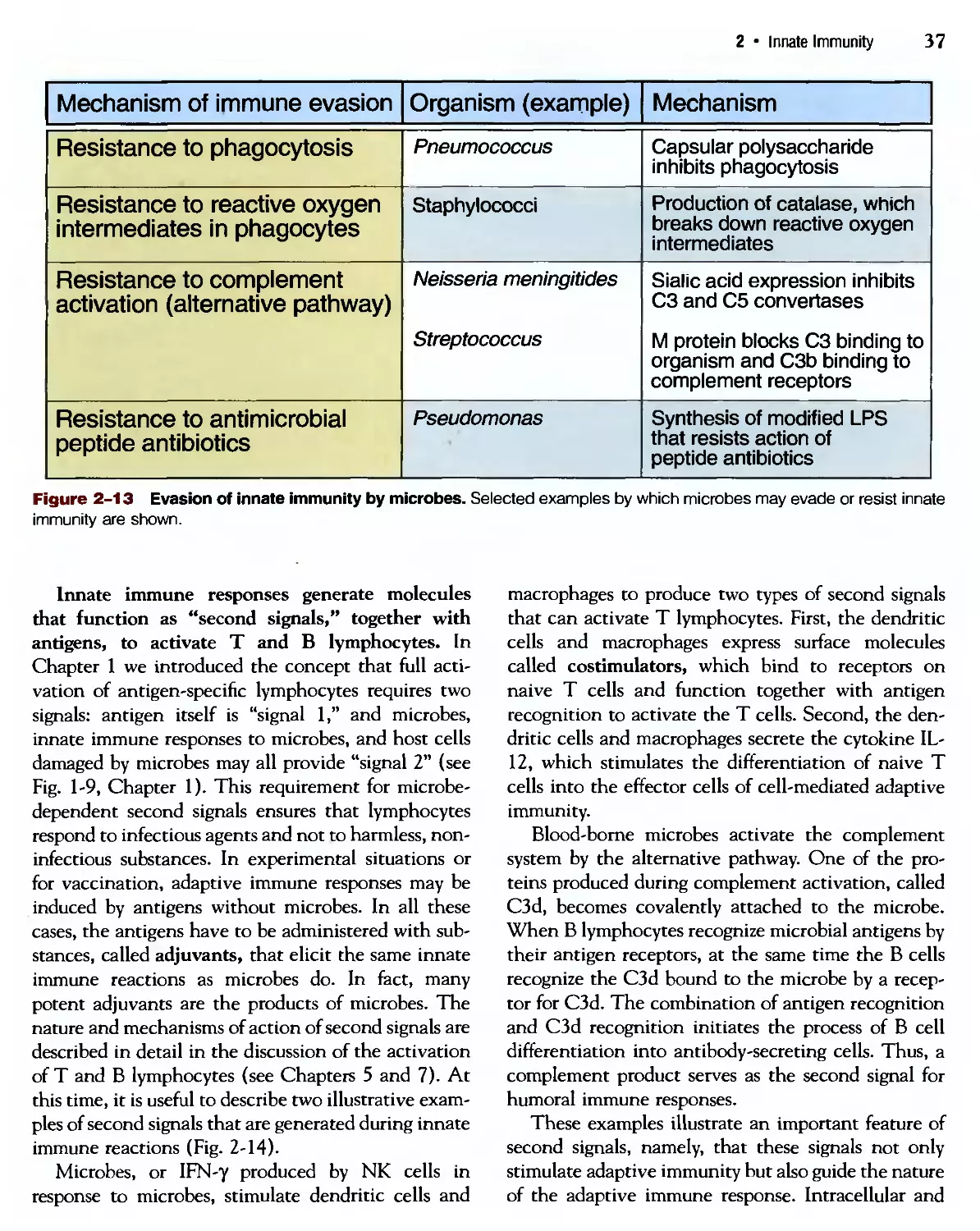
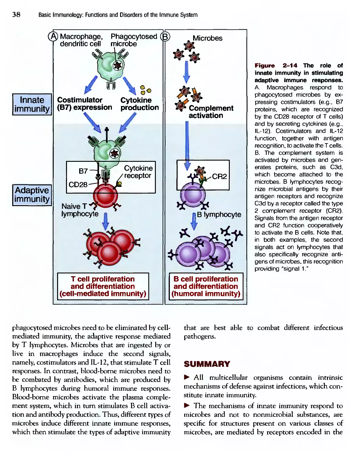
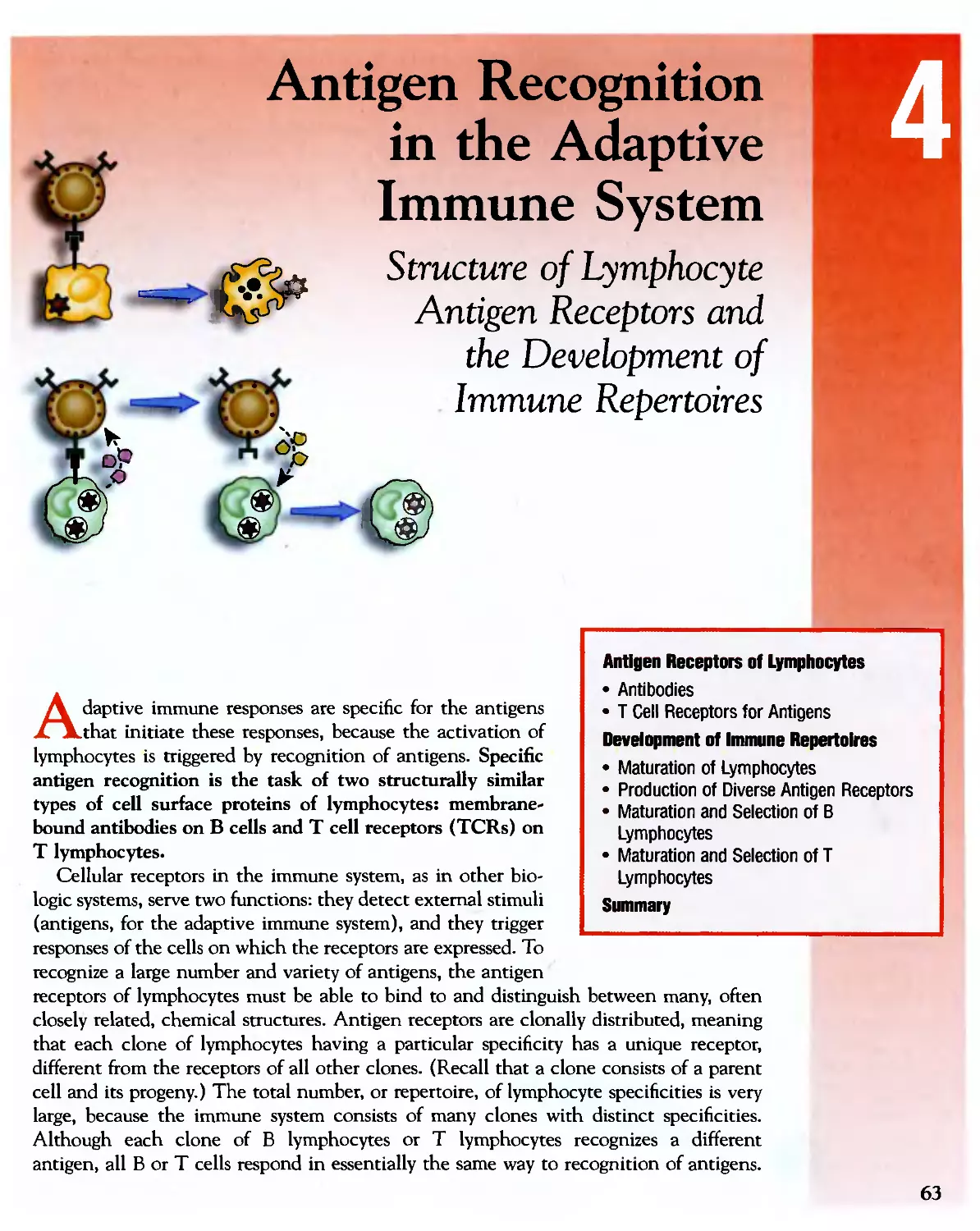
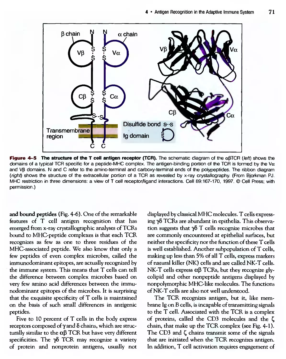
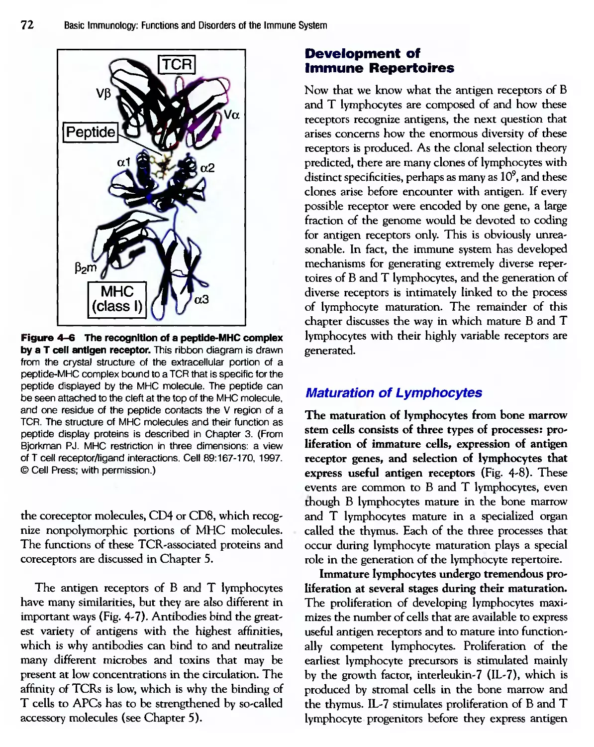
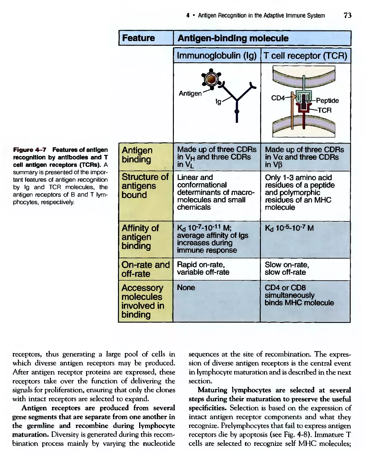
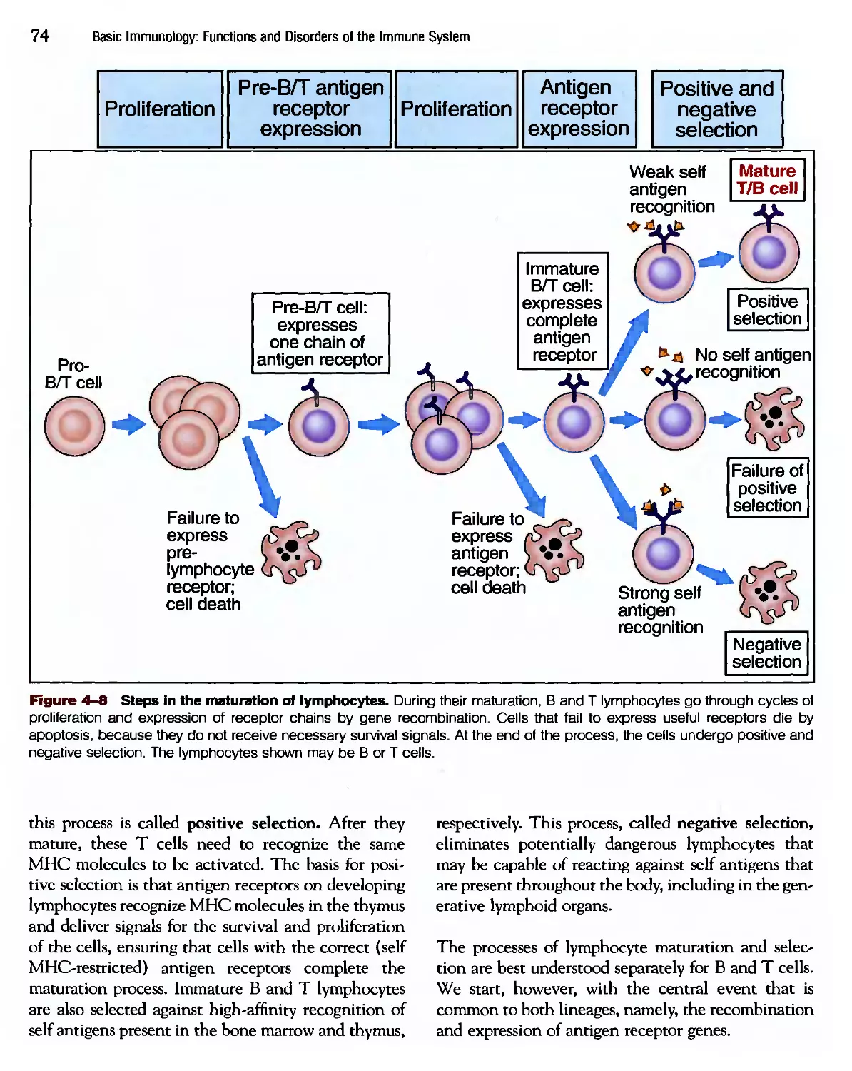
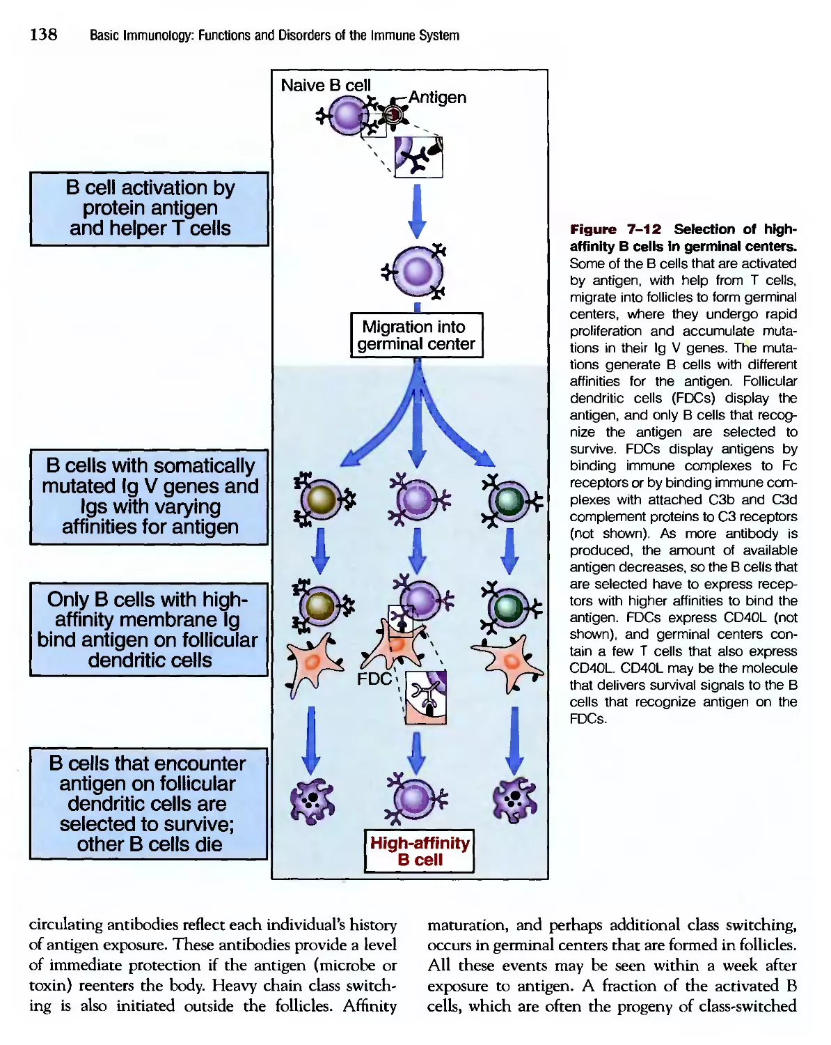
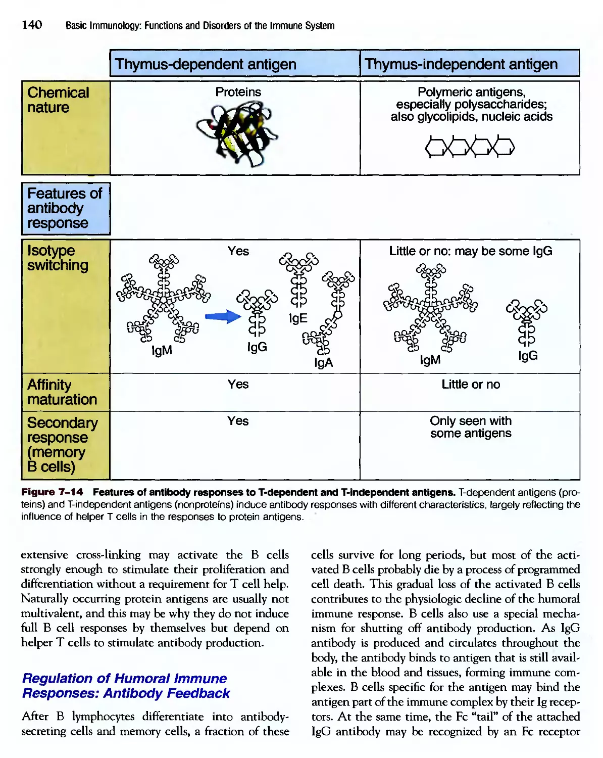
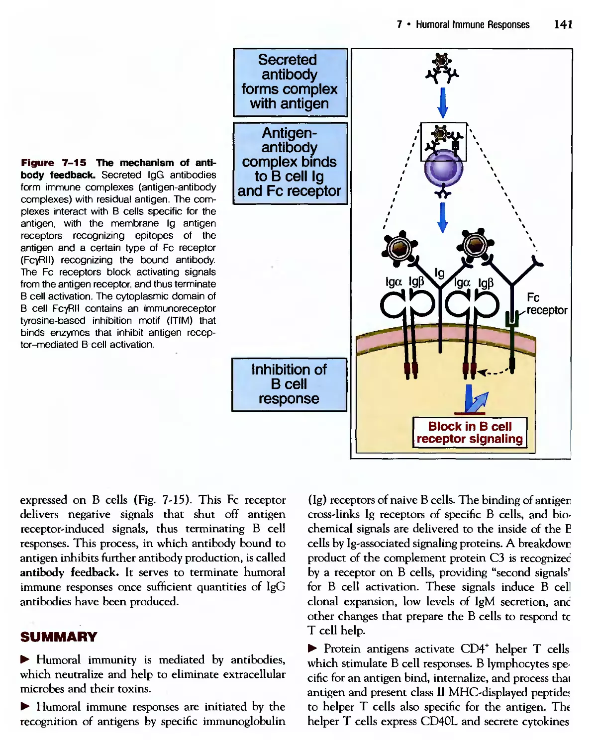
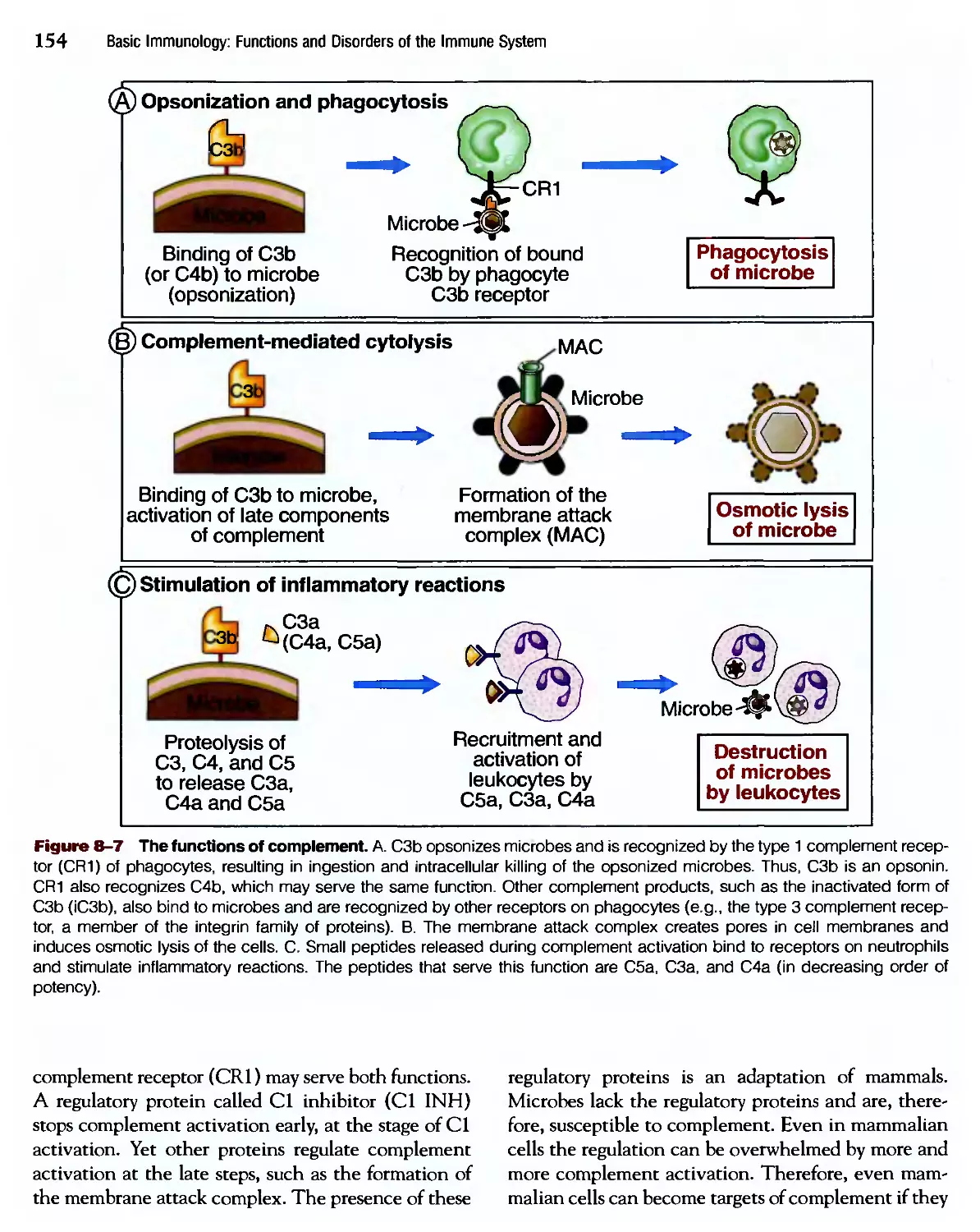
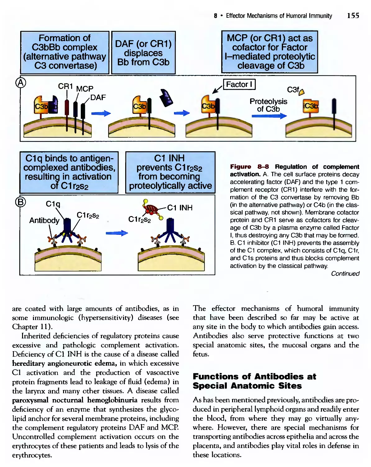
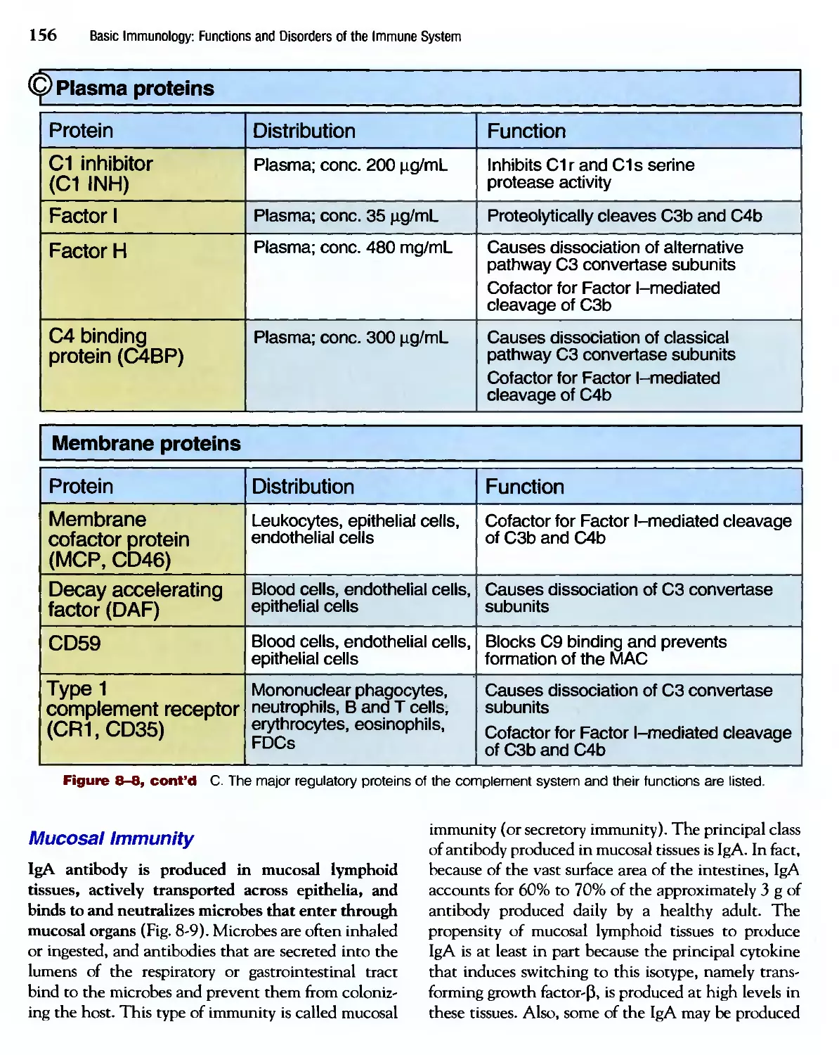
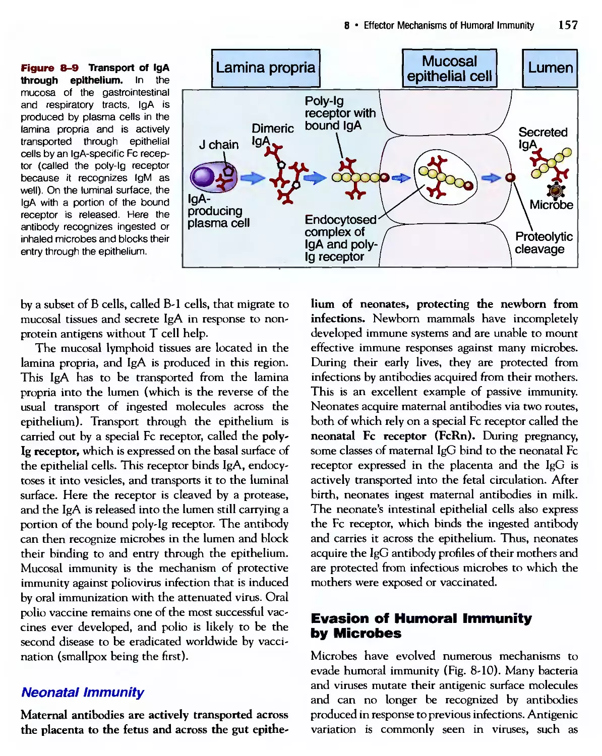
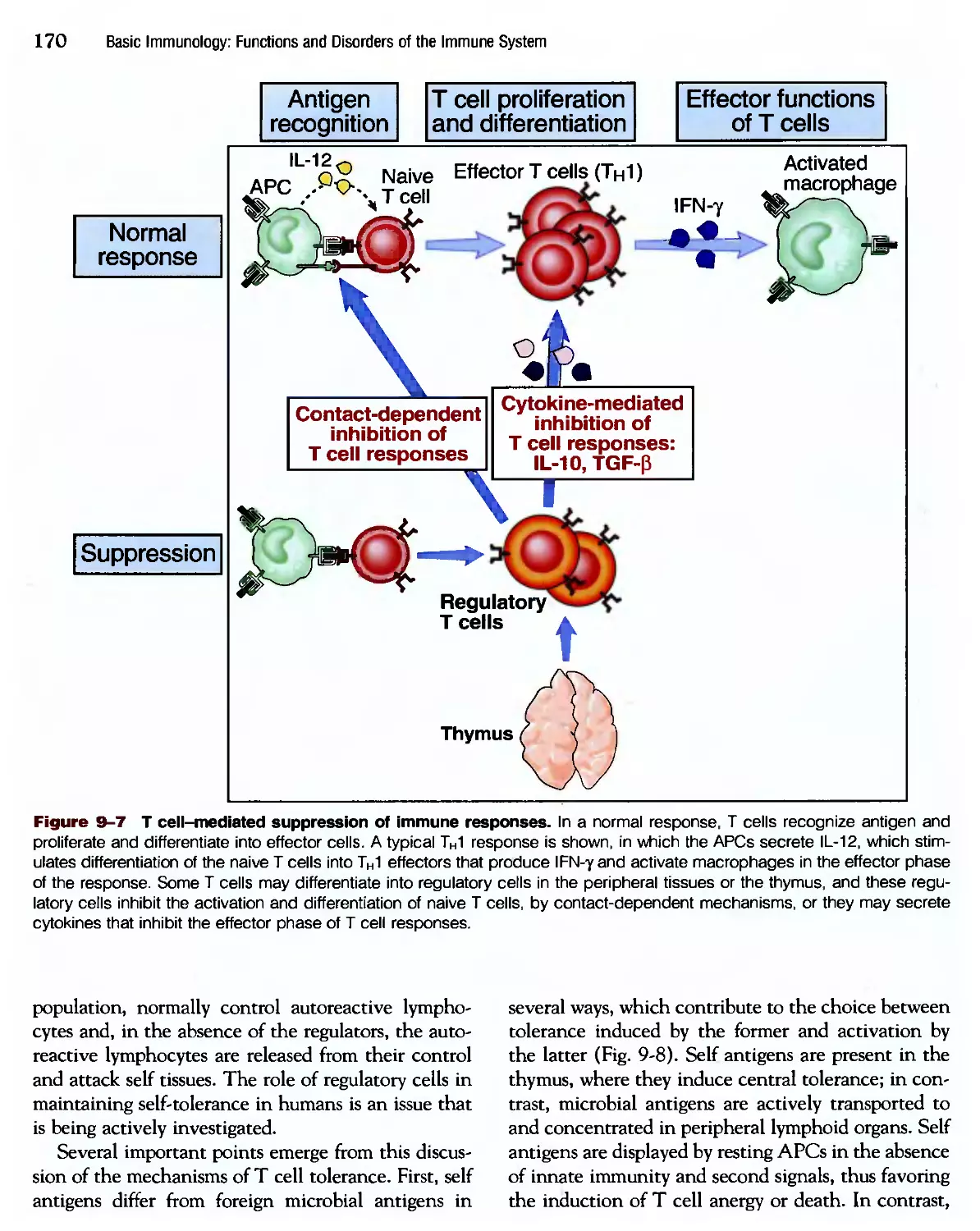
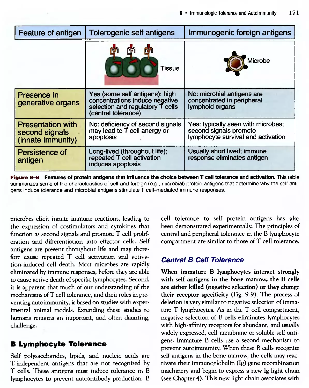
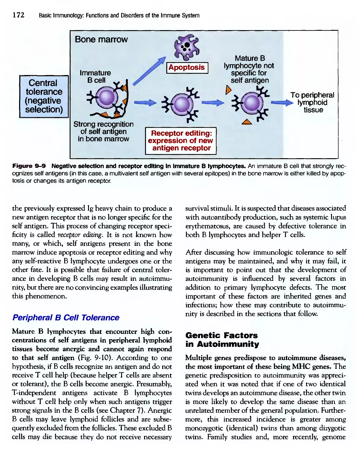
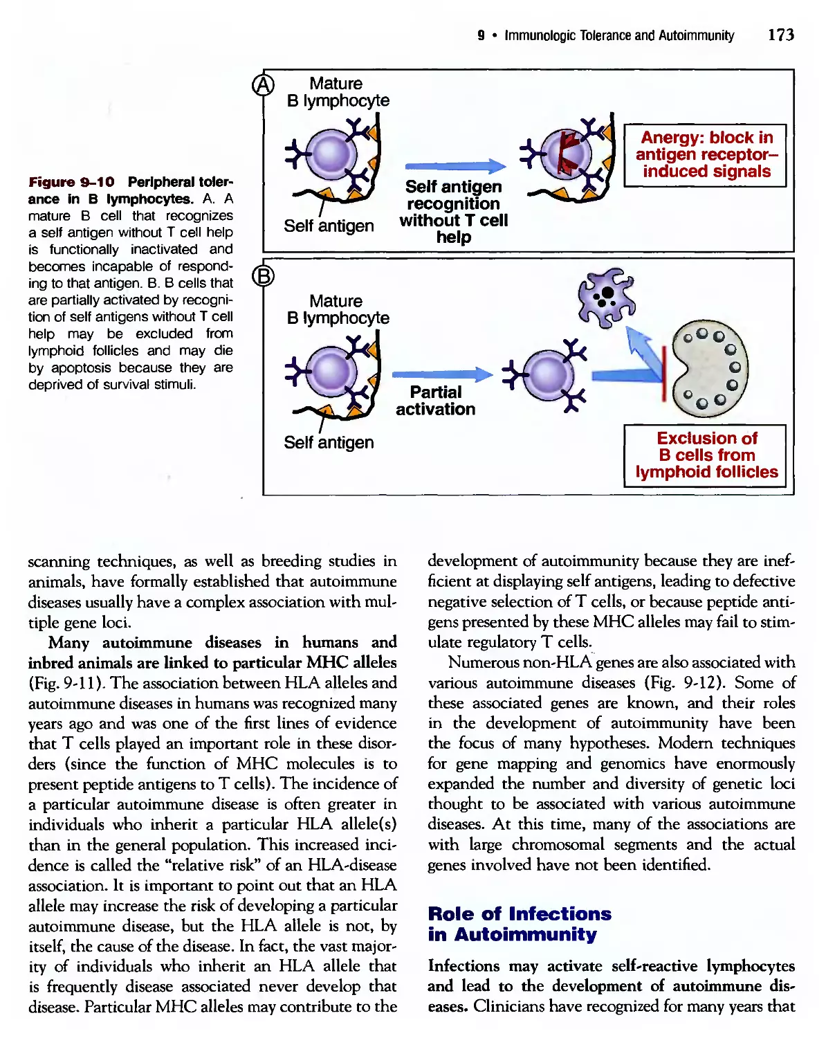
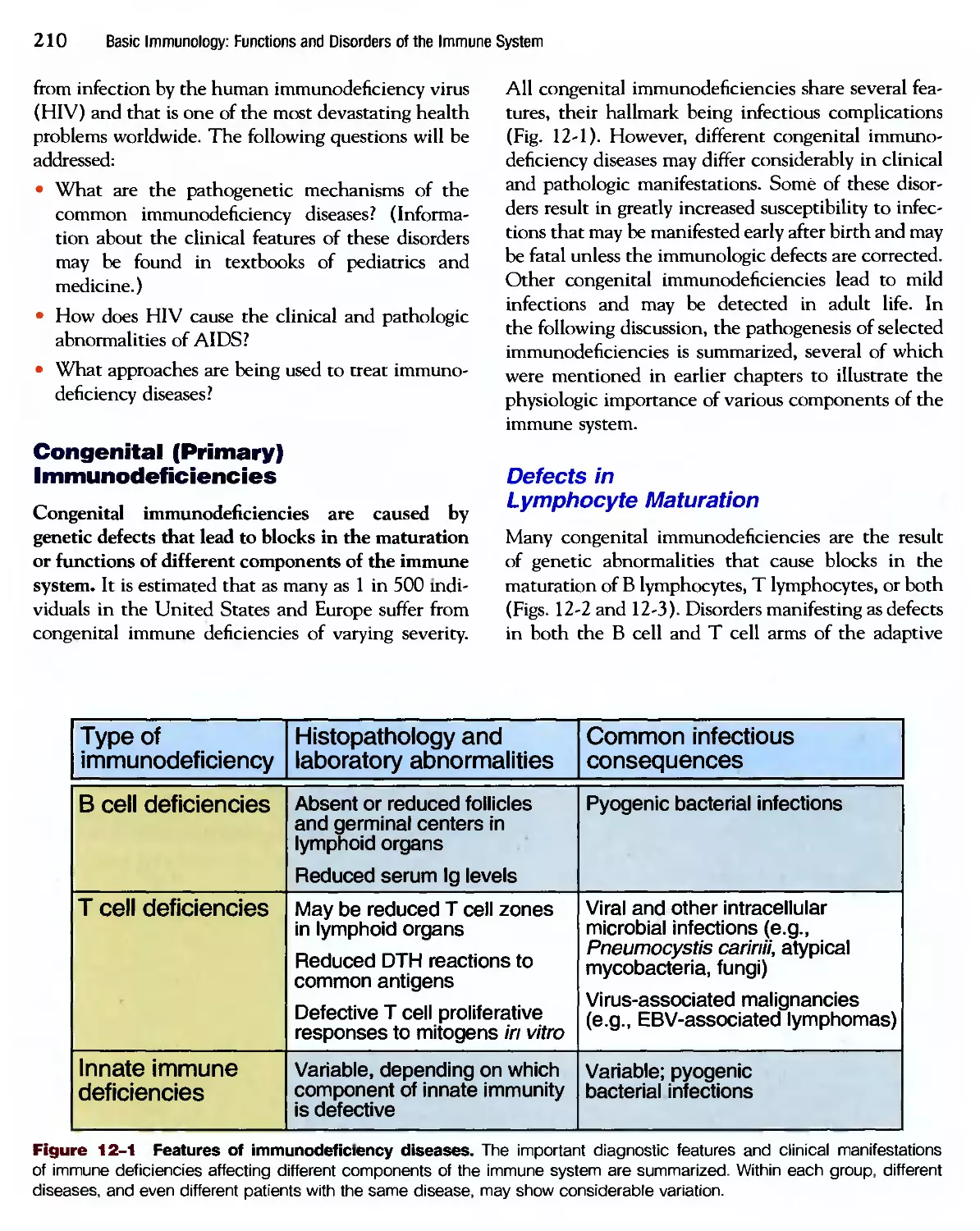
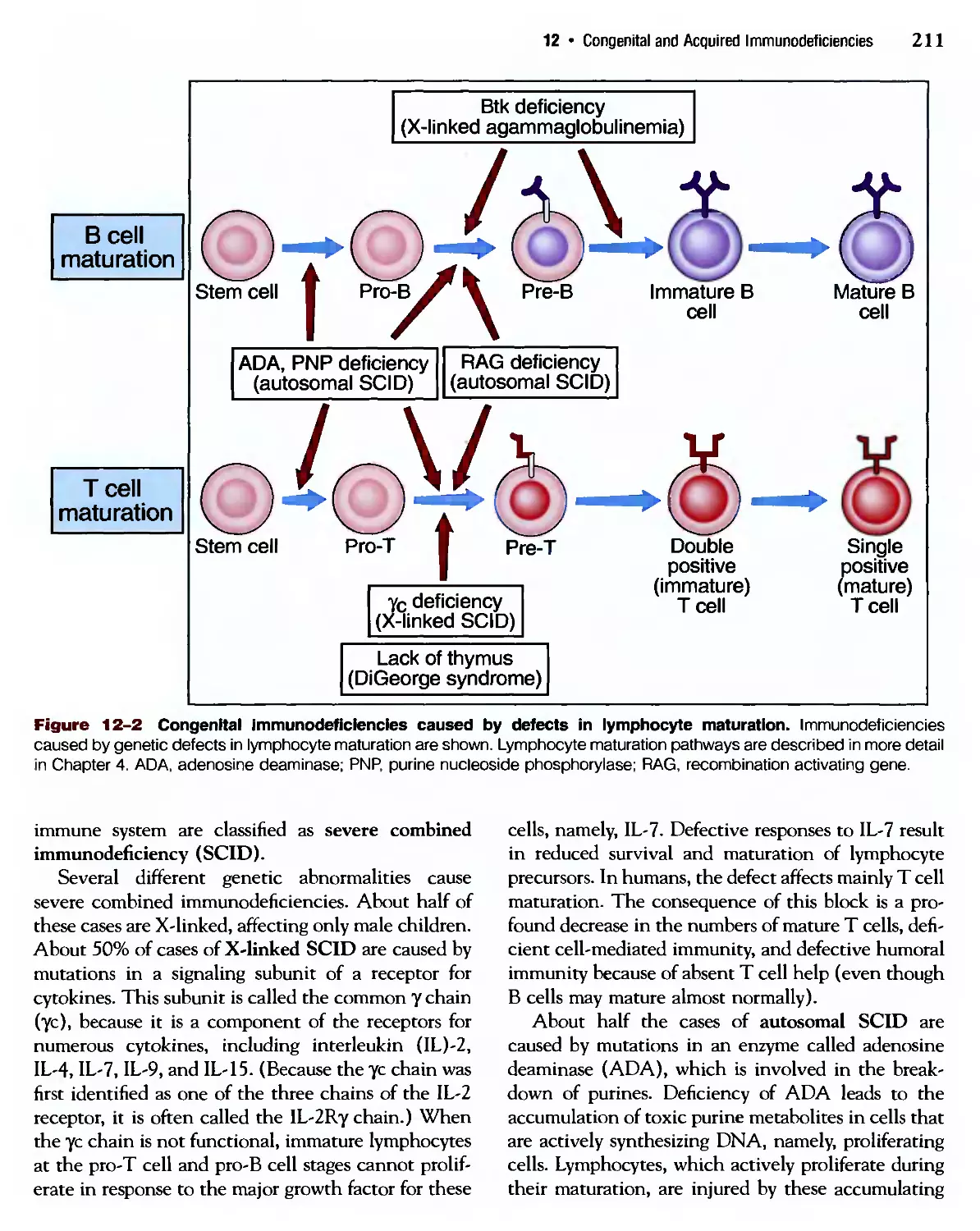
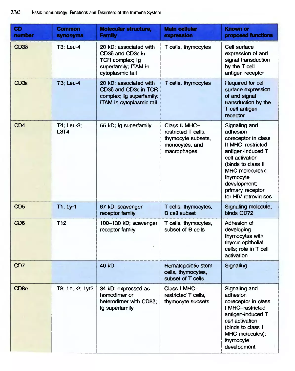
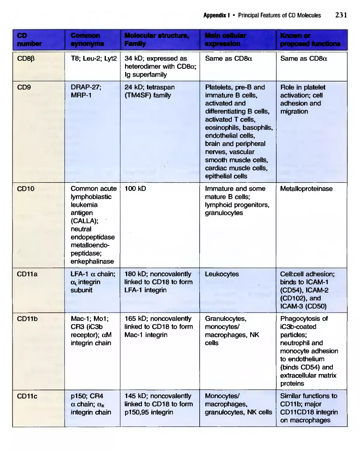
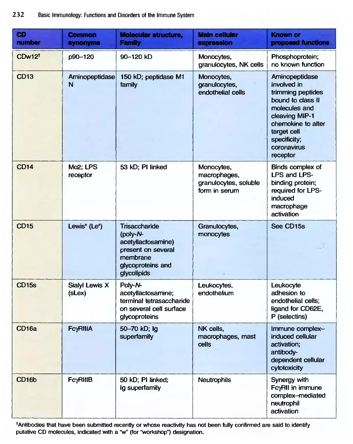
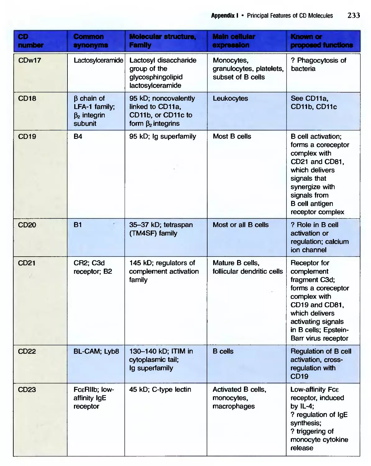
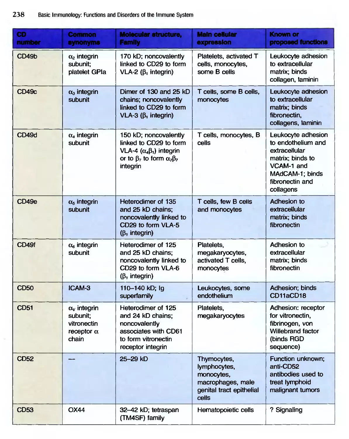
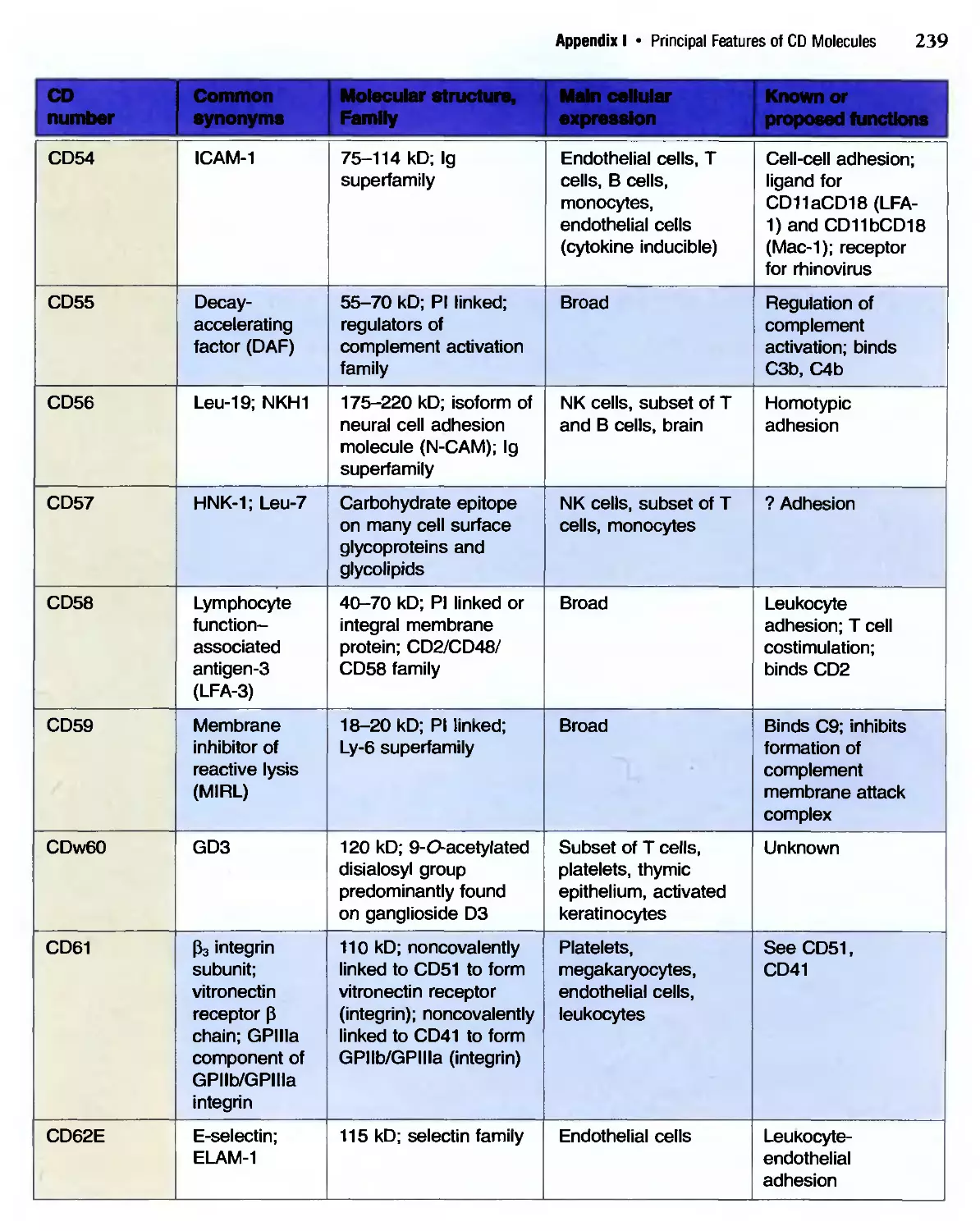
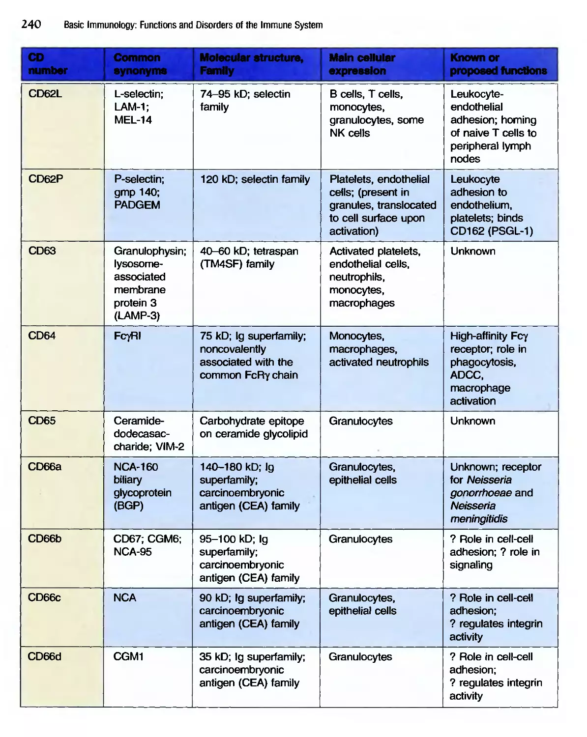
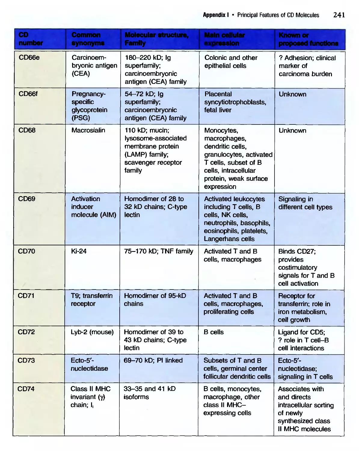
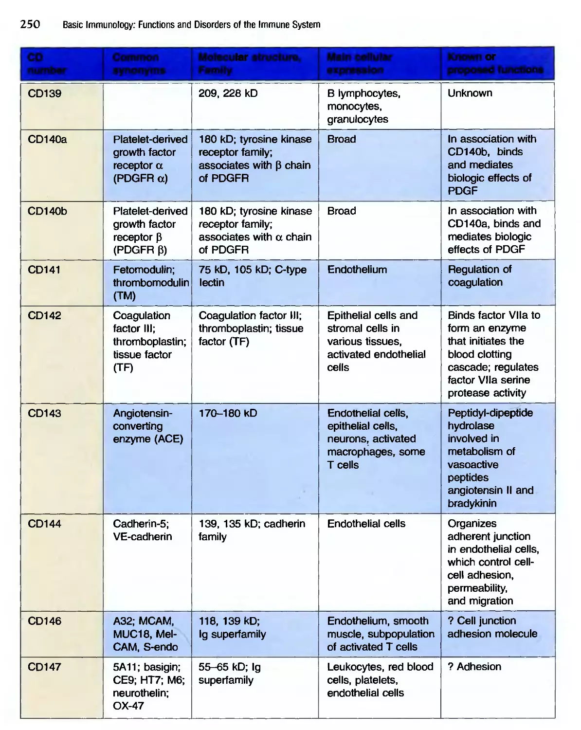
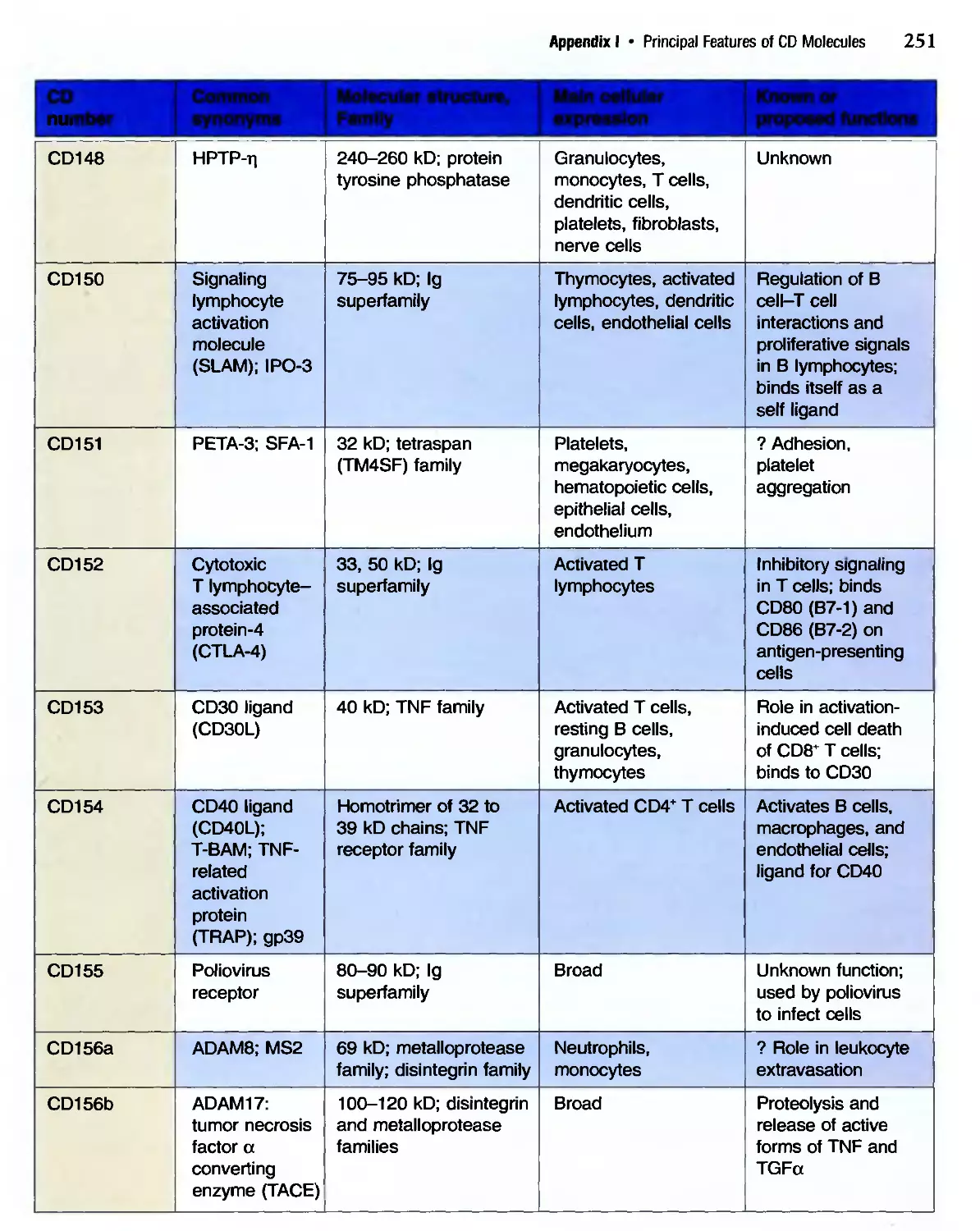
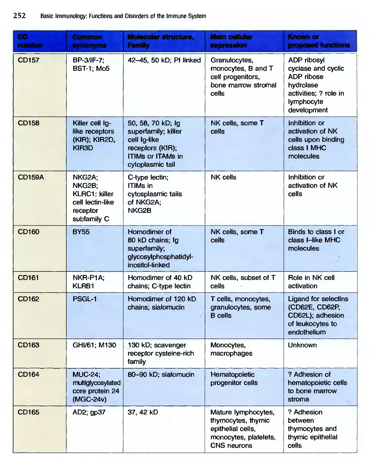
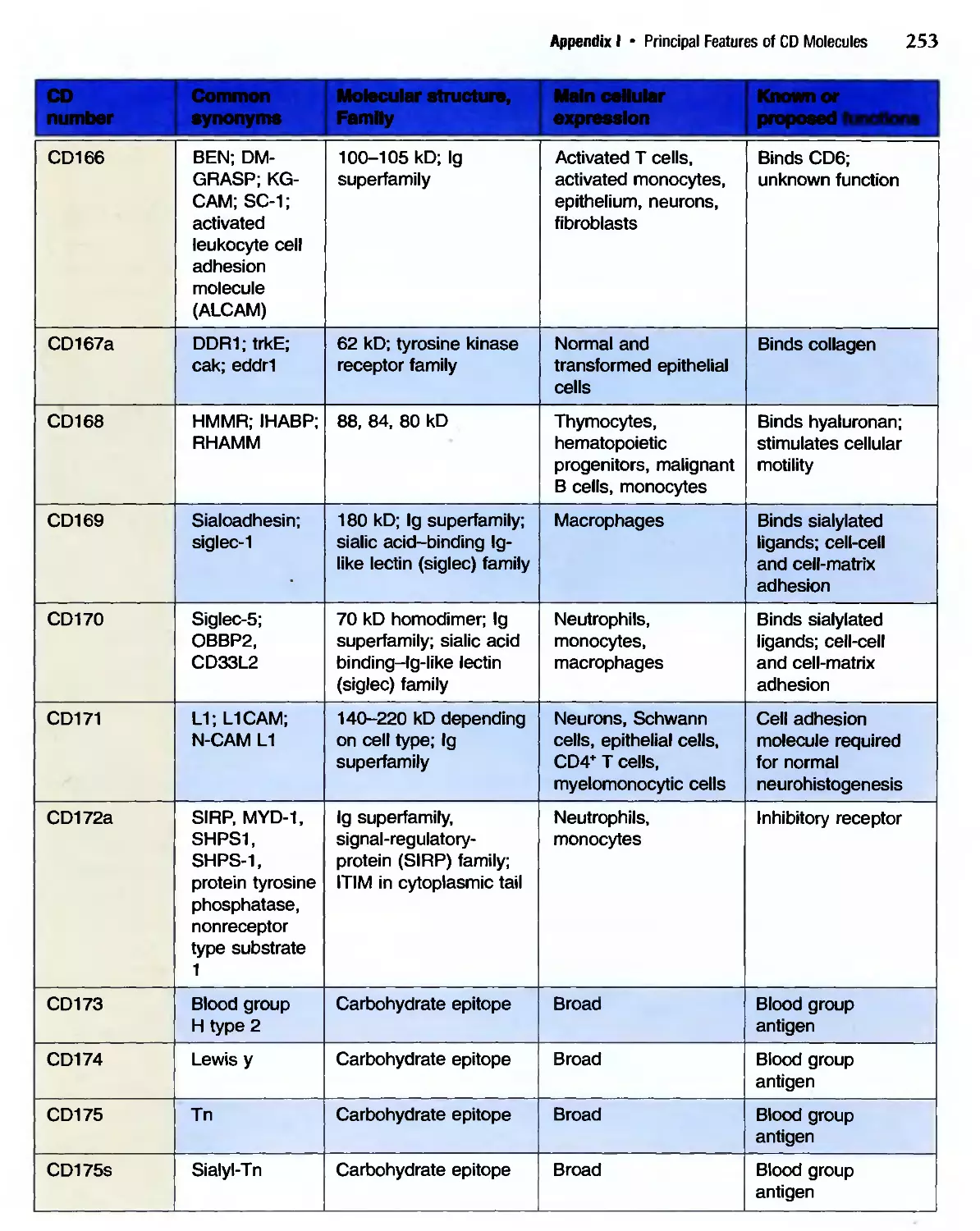
/


Similar


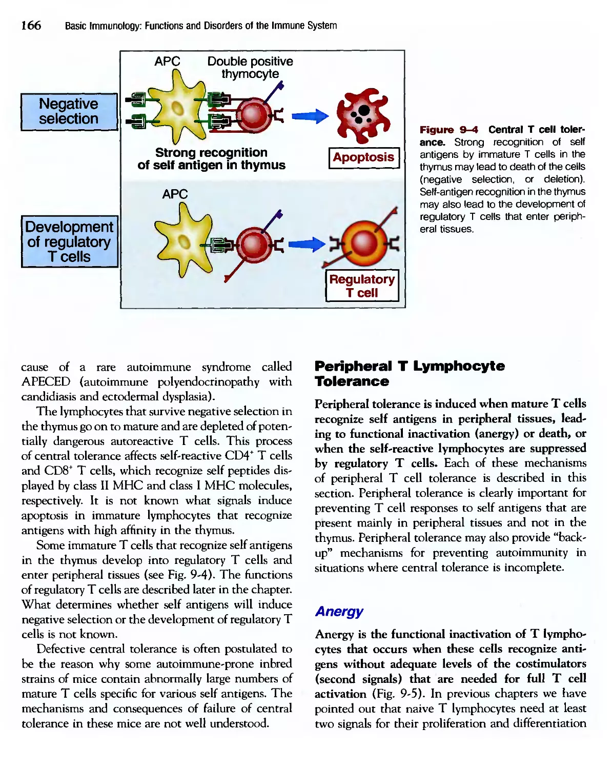
Text
к к
1 I * I I
Andre , Lichtma
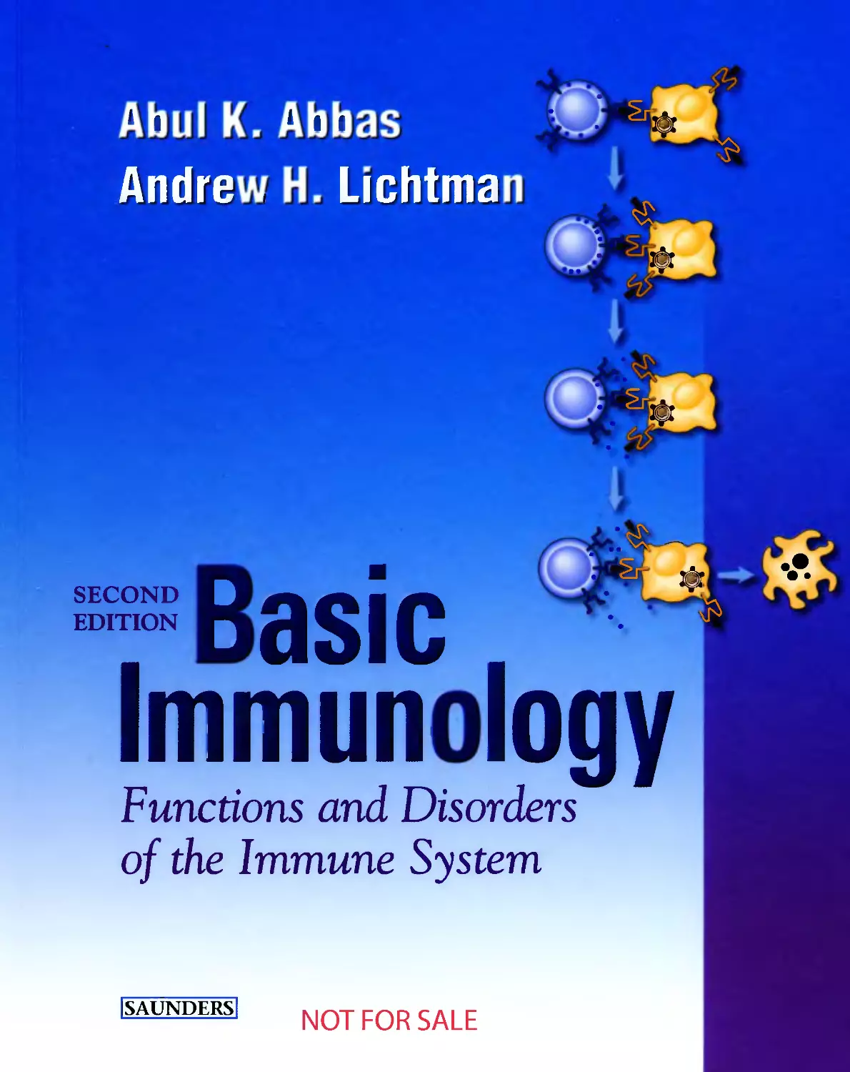
SECOND
EDITION
B Q С If1
Immun logy
Functions and Disorders
of the Immune System
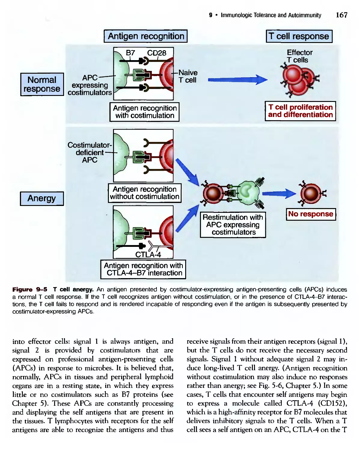
NOT FOR SALE
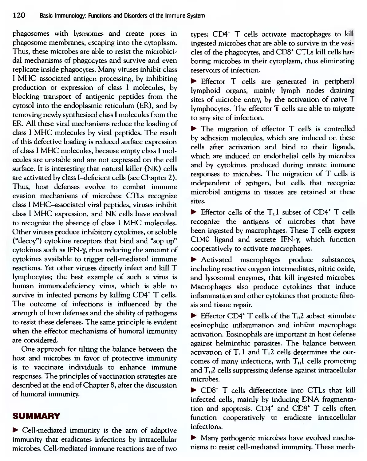
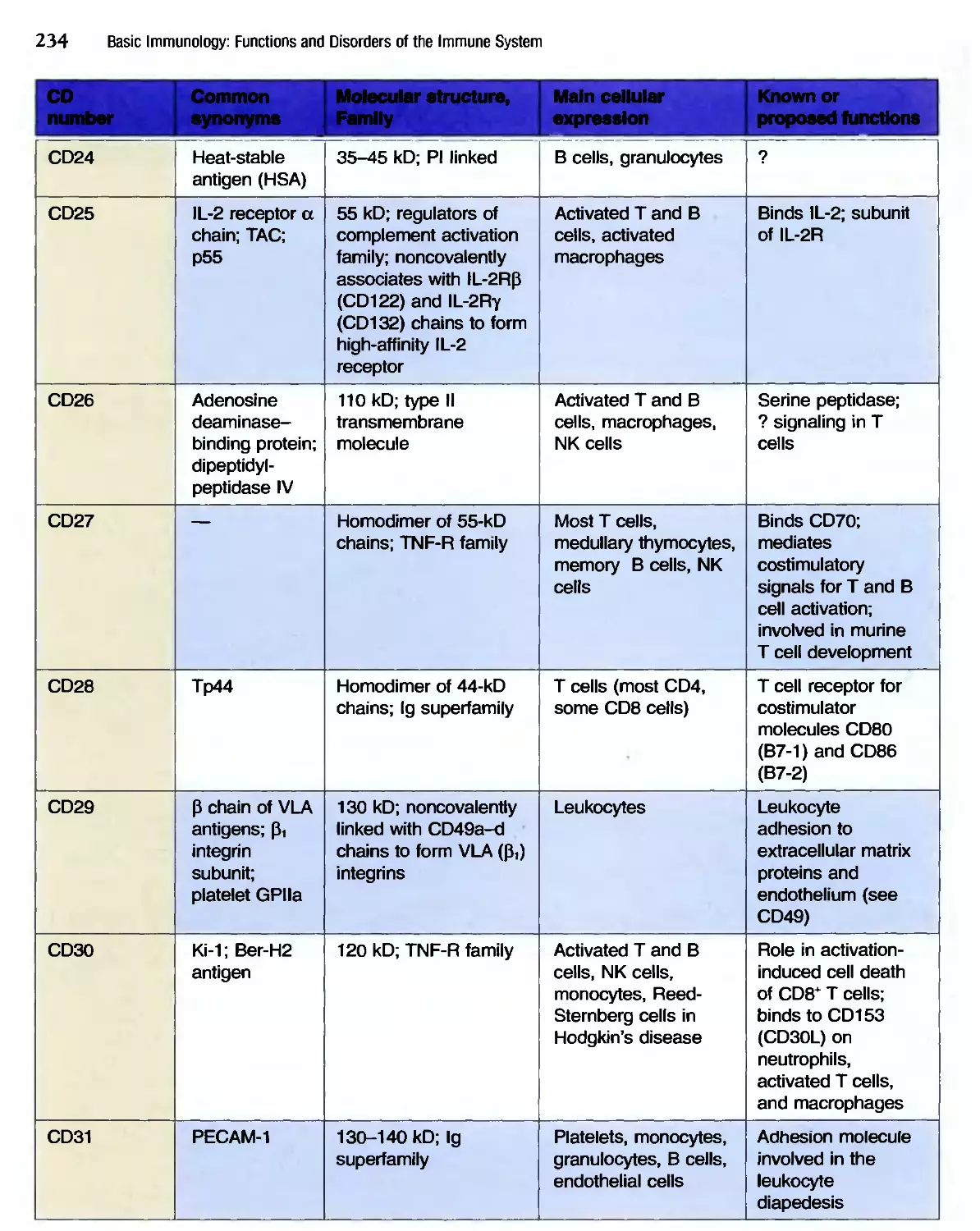
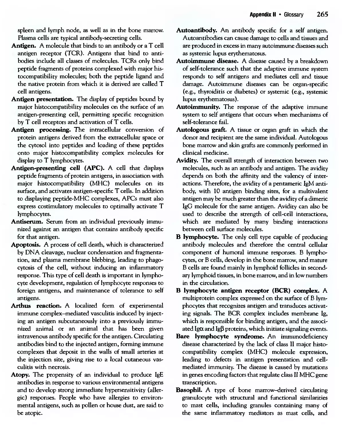
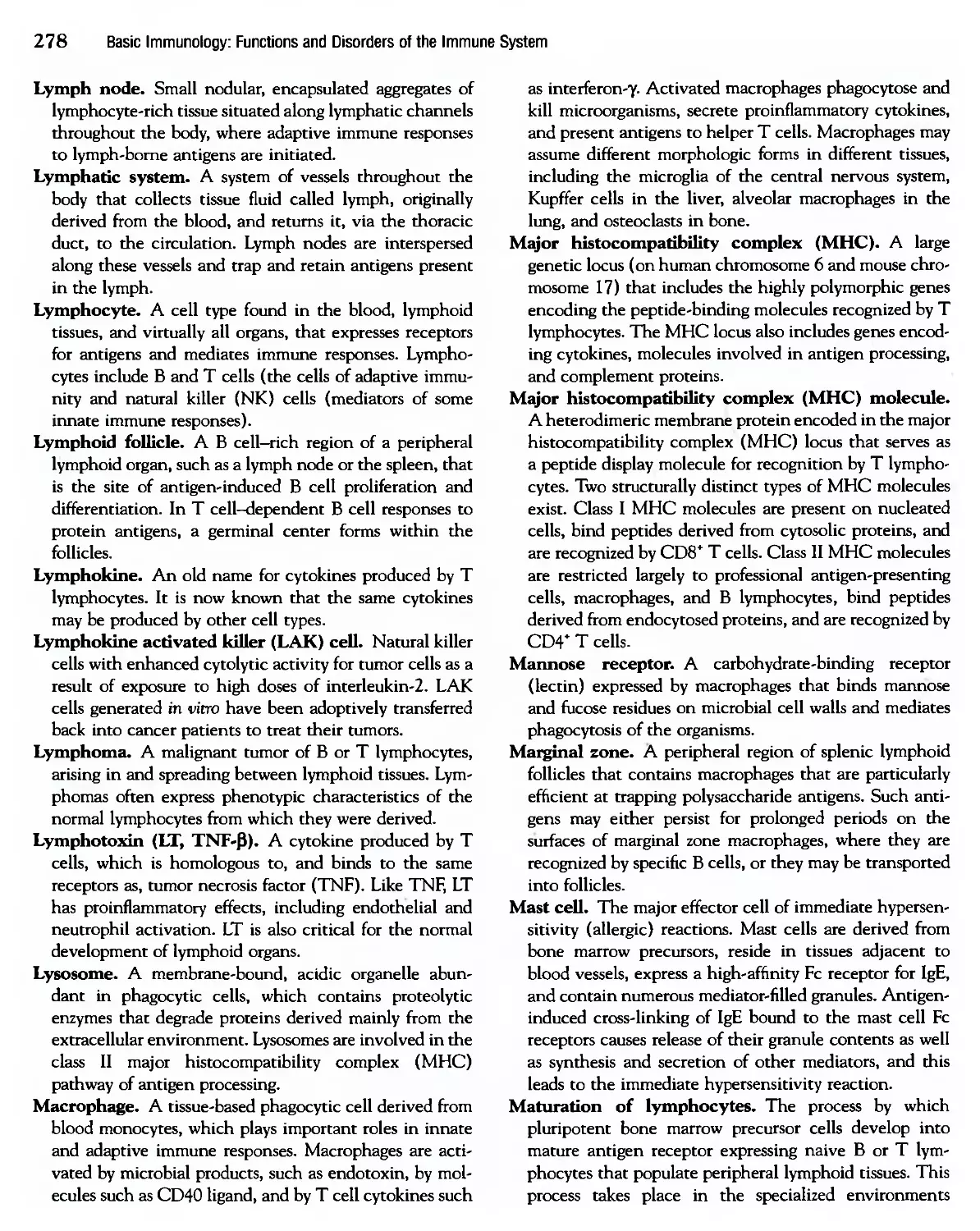
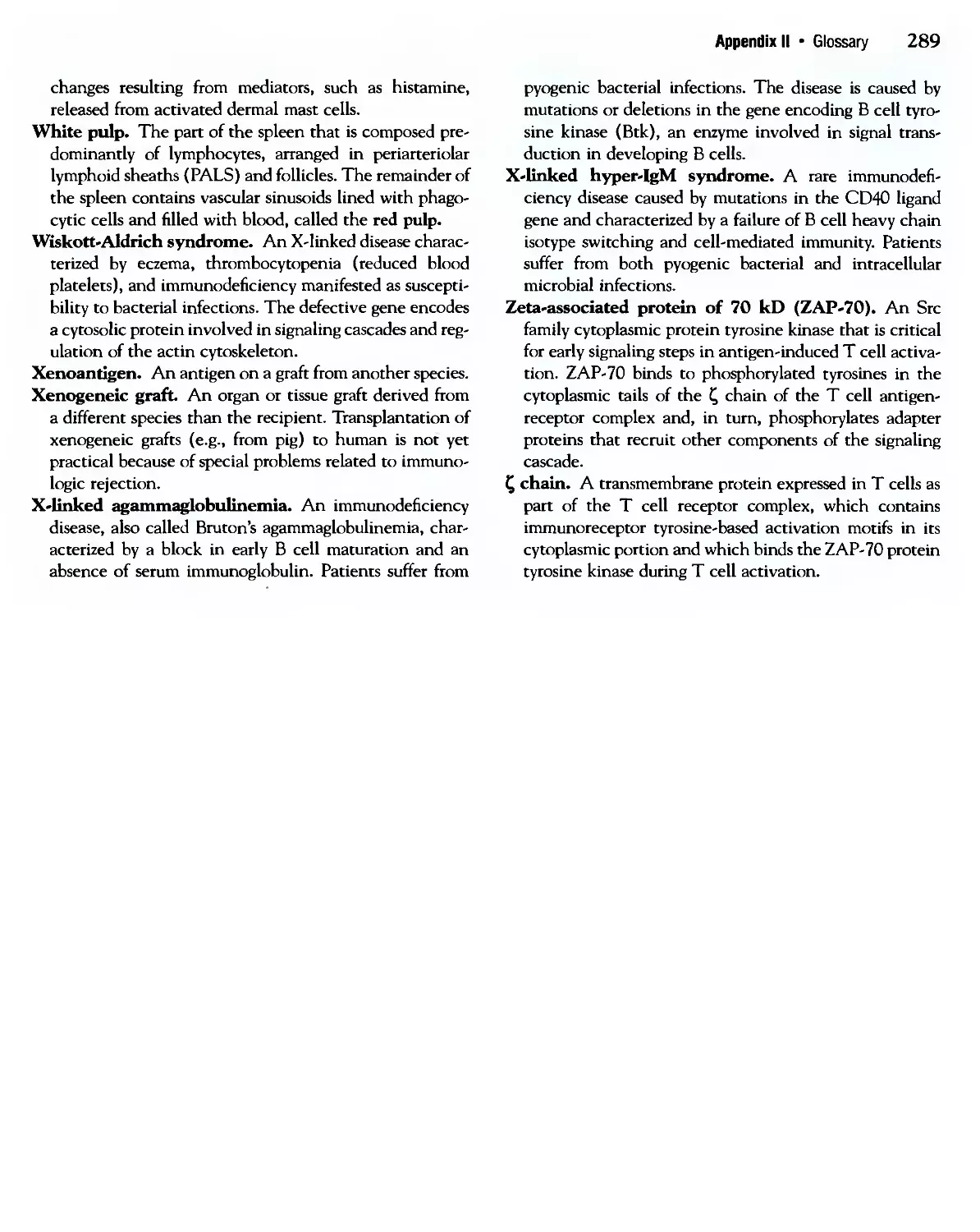
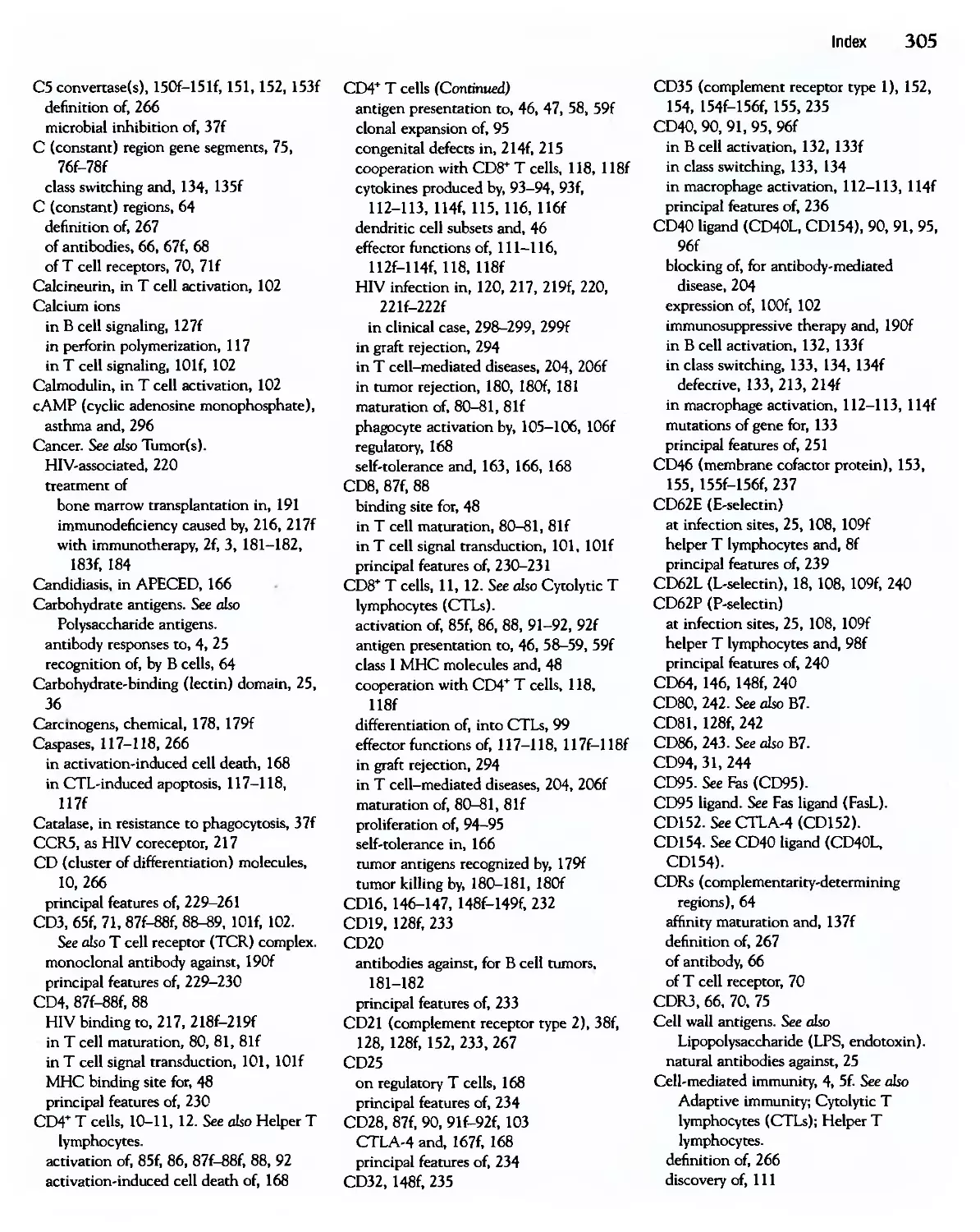
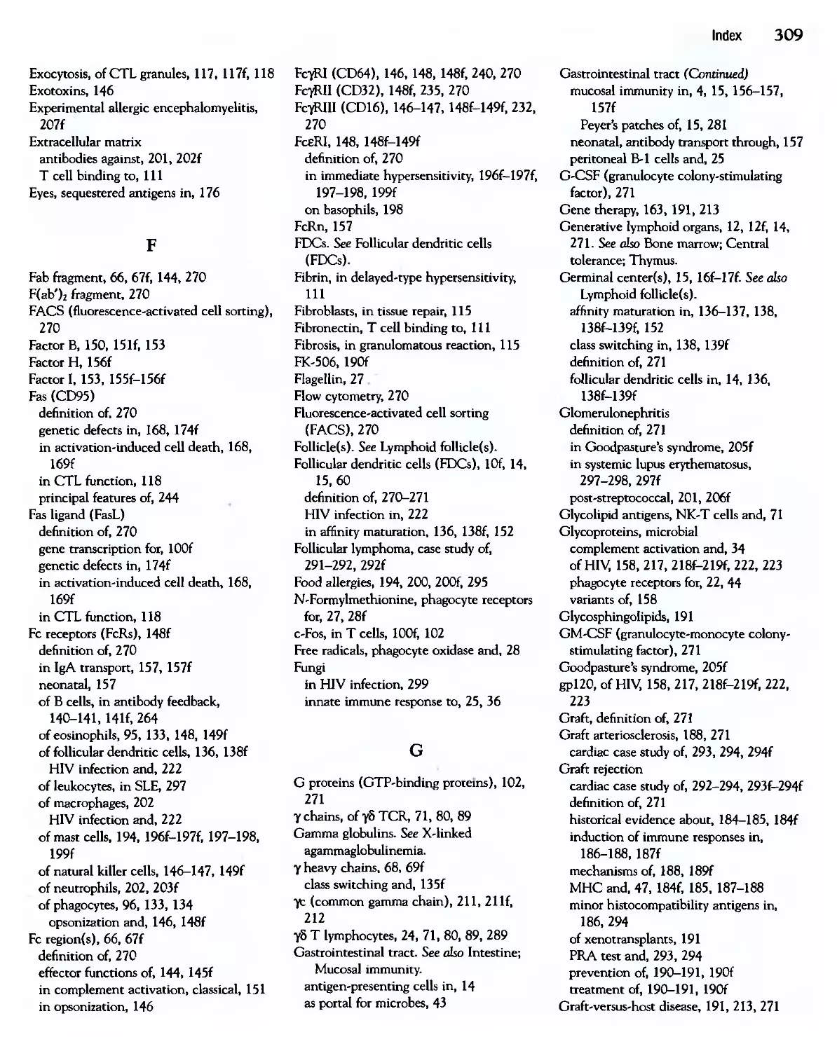
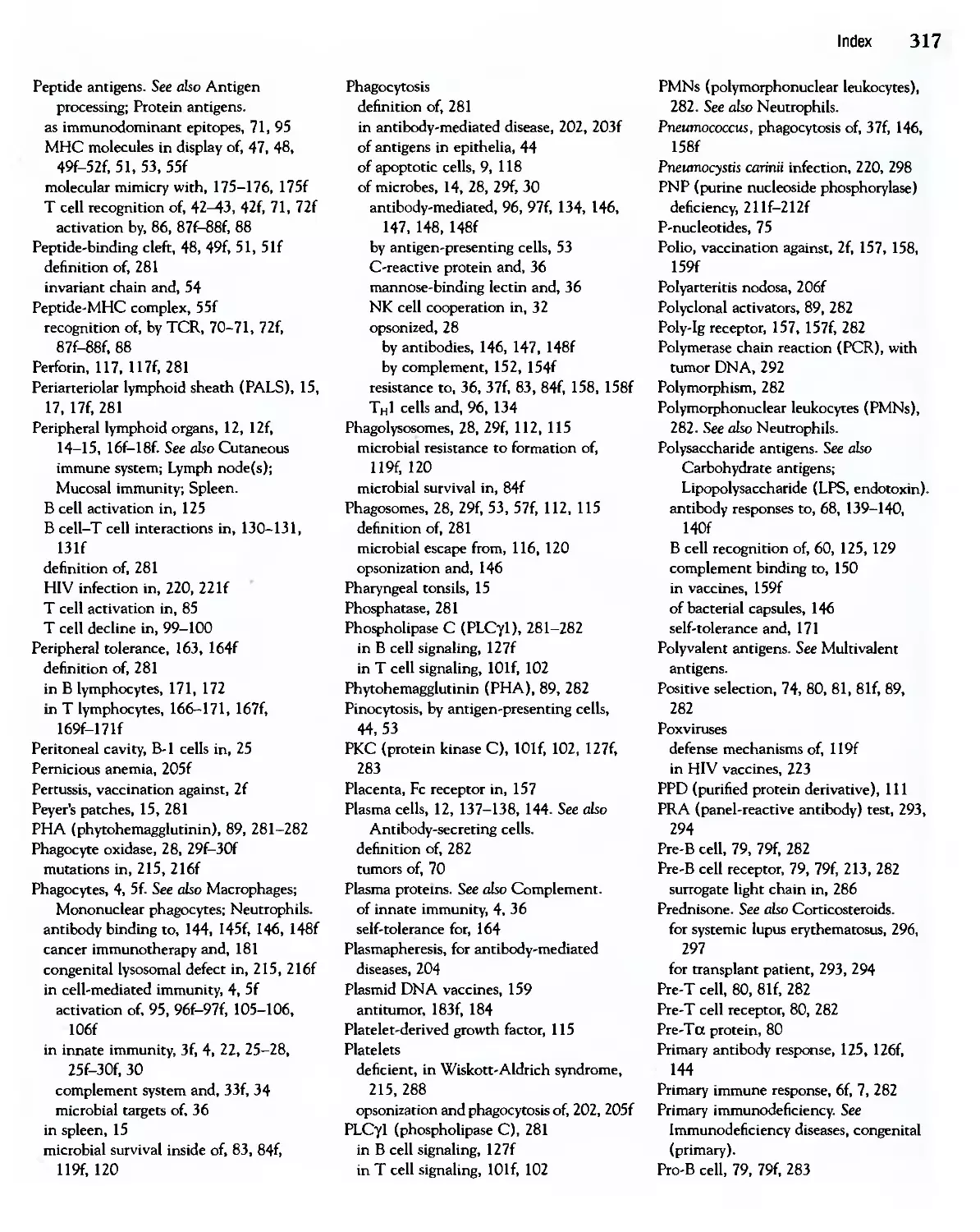
C1q
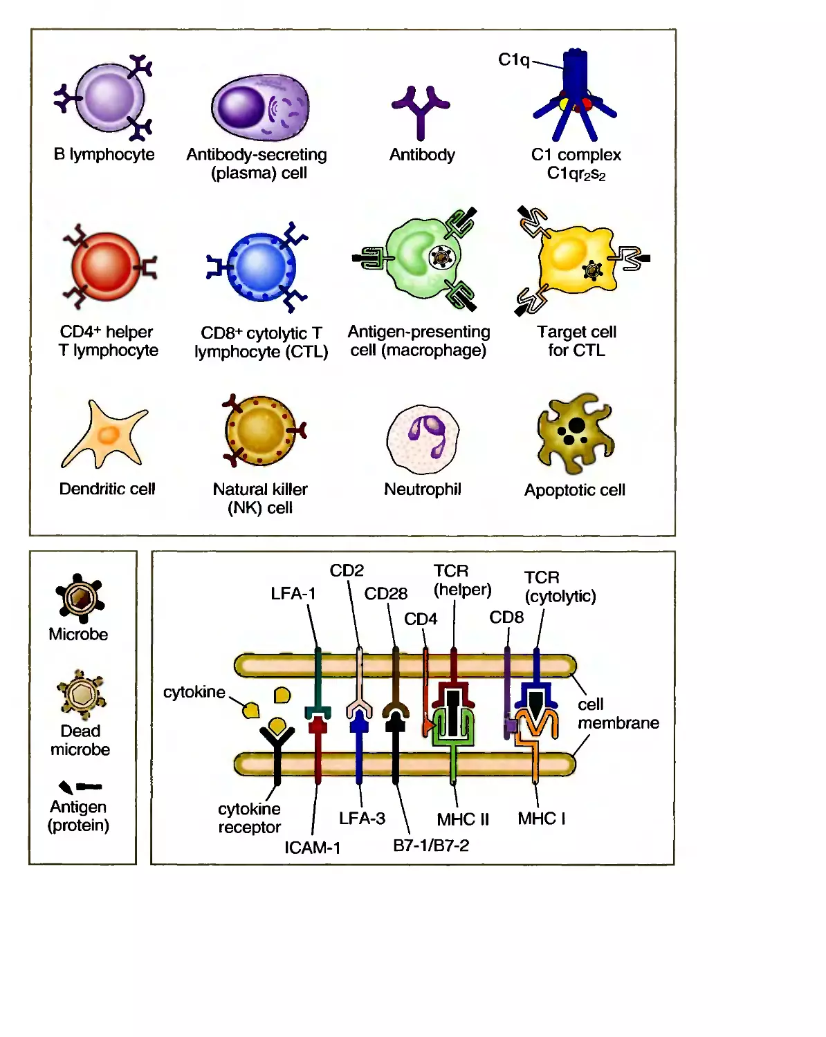
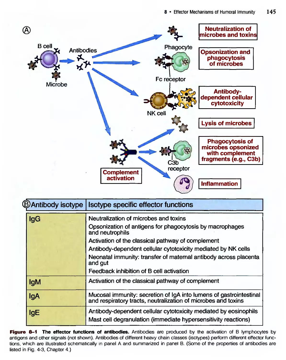
В lymphocyte Antibody-secreting
(plasma) cell
Antibody
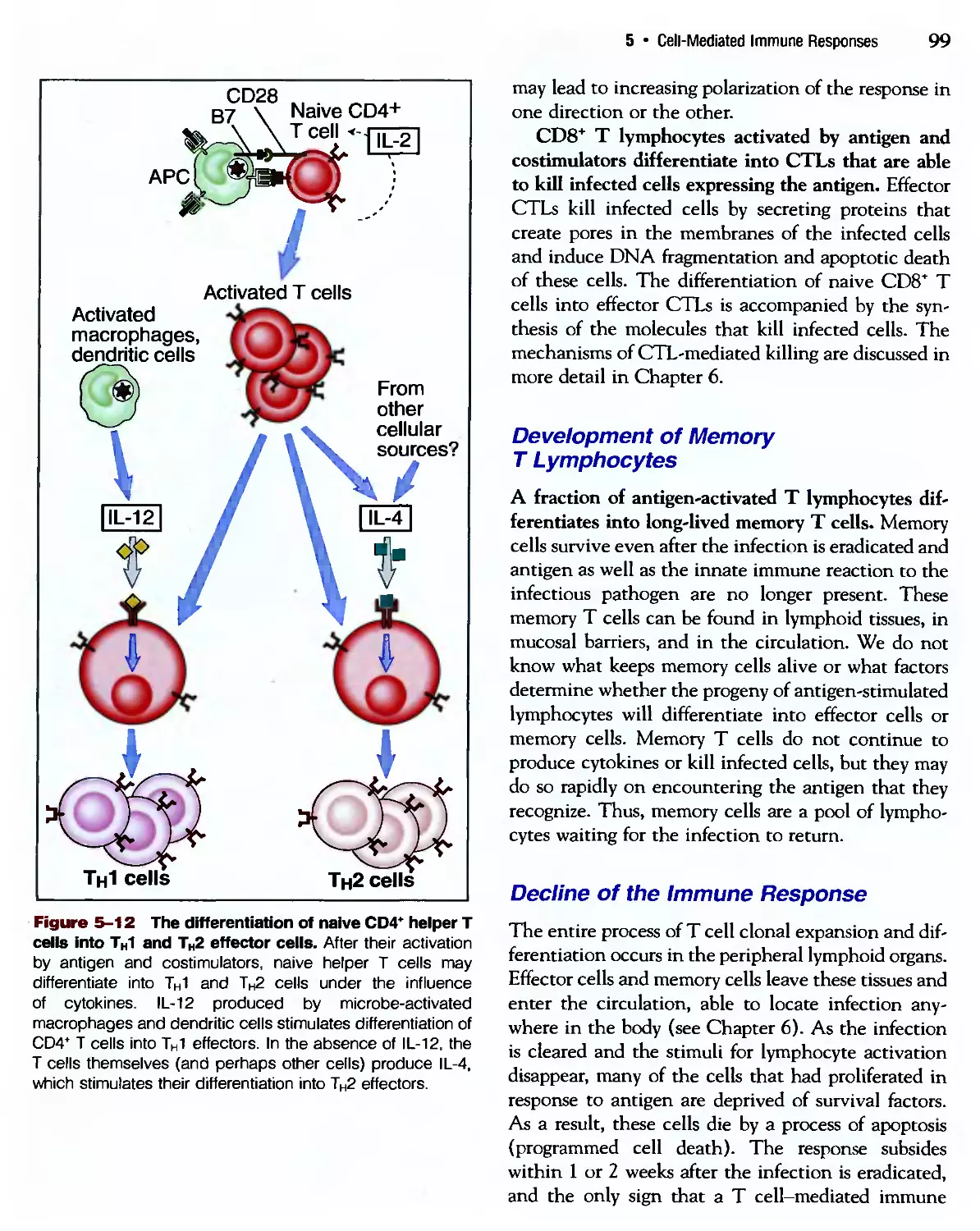
CD4+ helper
T lymphocyte
<t
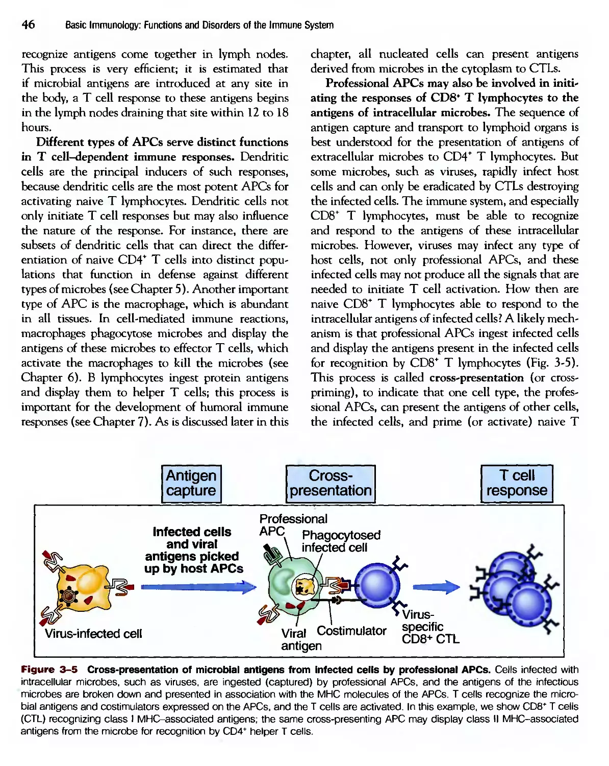
CD8+ cytolytic T Antigen-presenting
lymphocyte (CTL) cell (macrophage)
C1 complex
C1qr2s2
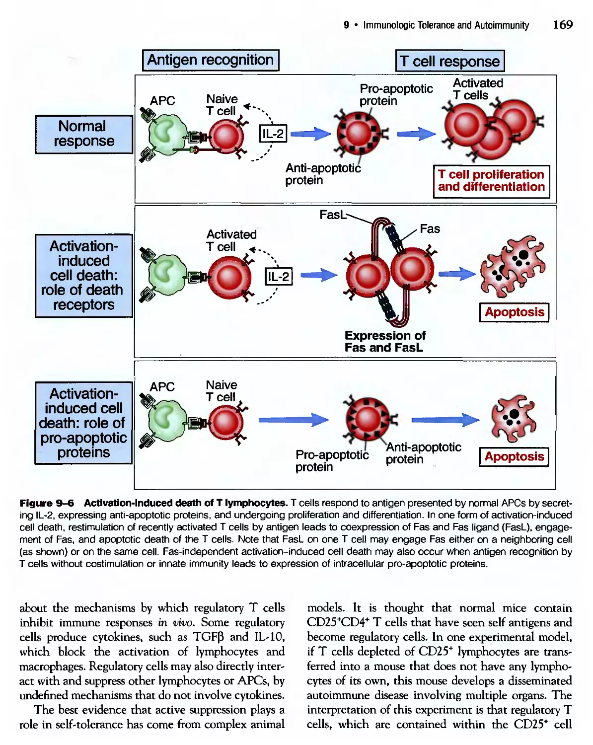
Target cell
for CTL
Dendritic cell
Natural killer
(NK) cell
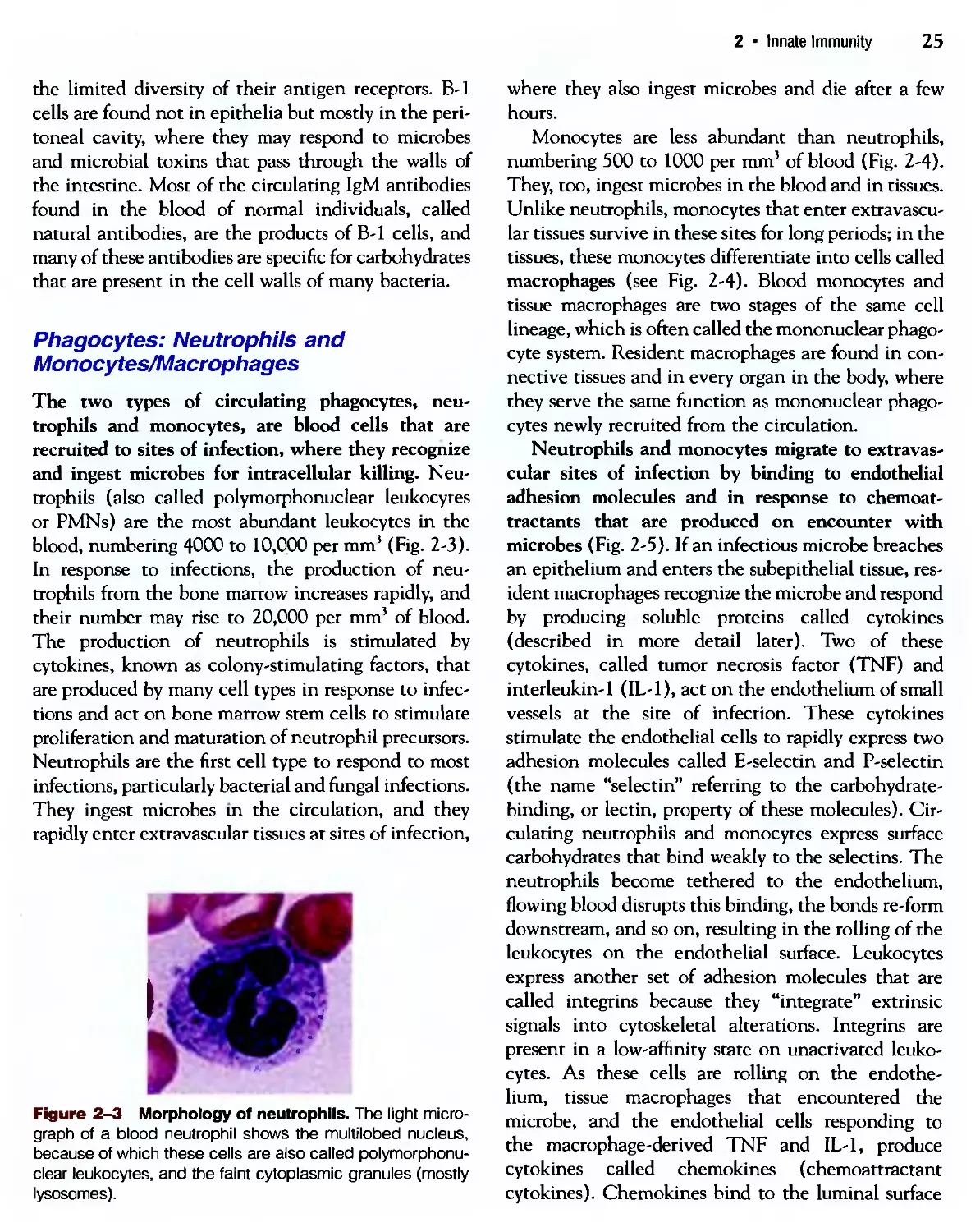
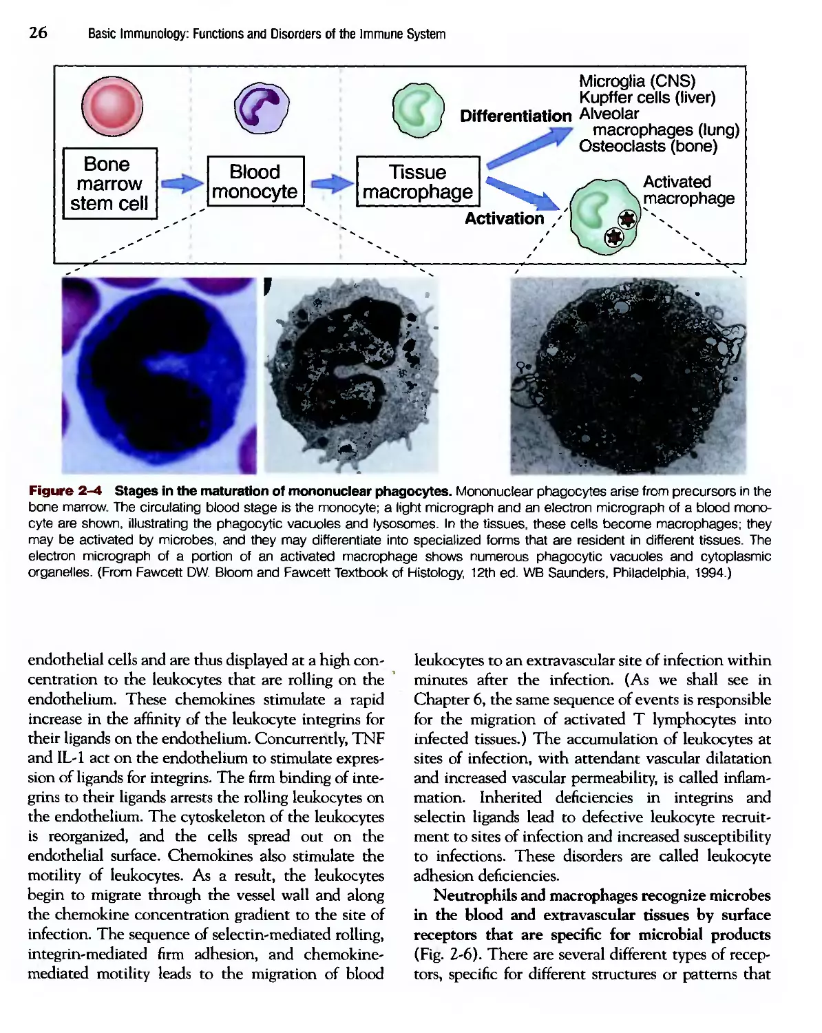
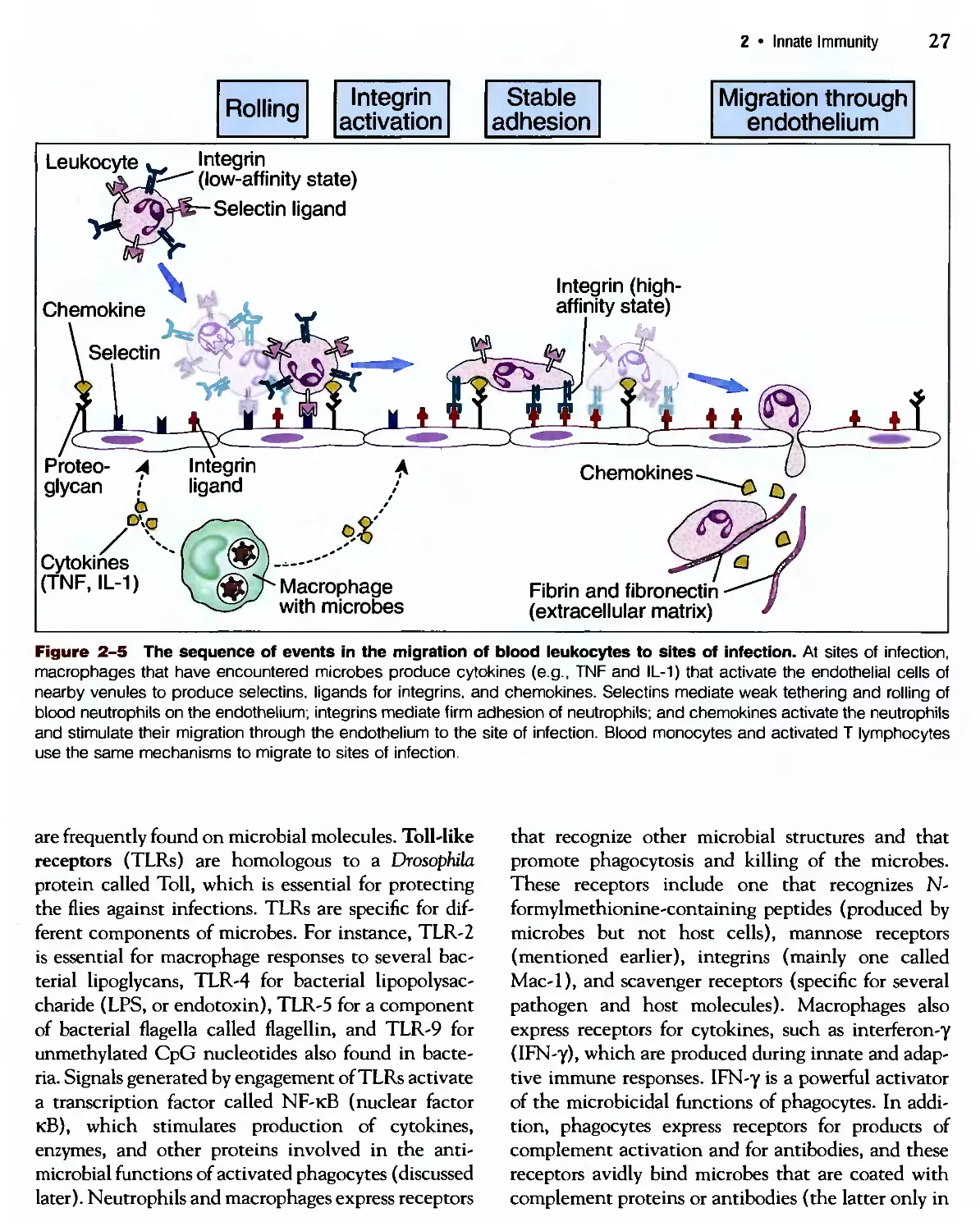
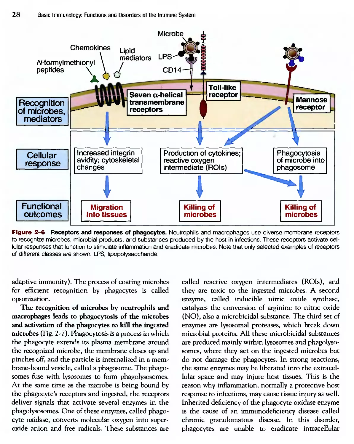
Neutrophil
Apoptotic cell
Microbe
Dead
microbe
Antigen
(protein)
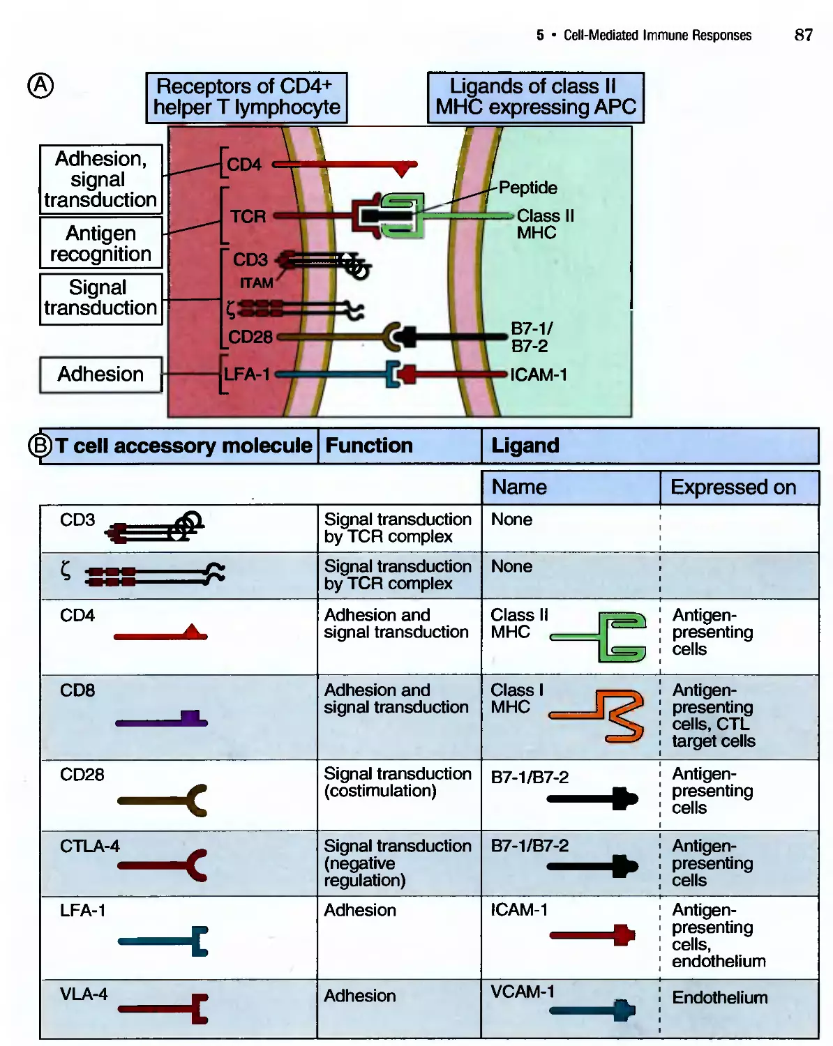
CD2
TCR
LFA-1 \ CD28 (helper)
TCR
cytokine ^^ Q
■ wh ce"
|[\V/)I membrane
cytokine
receptor
LFA-3 \ MHC II MHC I
ICAM-1 B7-1/B7-2
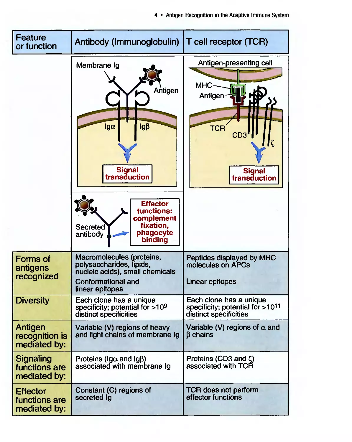
BASIC IMMUNOLOGY
BASIC IMMUNOLOGY
Functions and Disorders of the Immune System
Second Edition
Abul K. Abbas, MBBS
Professor and Chair
Department of Pathology
University of California, San Francisco, School of Medicine
San Francisco, California
Andrew H. Lichtman, MD, PhD
Associate Professor of Pathology
Harvard Medical School
Brigham and Women's Hospital
Boston, Massachusetts
Illustrated by David L. Baker, MA, and Alexandra Baker, MS, CMI
[SAUNDERSl
An Imprint of Elsevier
SAUNDERS
An Imprint of Elsevier
The Curtis Center
Independence Square West
Philadelphia, PA 19106-3399 ISBN: 0-7216-0241-X
BASIC IMMUNOLOGY: FUNCTIONS AND DISORDERS OF THE IMMUNE SYSTEM
Copyright © 2004, 2001 Elsevier Inc. All rights reserved.
No part of this publication may be reproduced or transmitted in any form or by any means,
electronic or mechanical, including photocopy, recording, or any information storage and
retrieval system, without permission in writing from the publisher.
Permissions may be sought directly from Elsevier Inc. Rights Department in Philadelphia,
USA: phone: (+1J15 238 7869, fax: (+1J15 238 2239, email: healthpermissions@elsevier.com.
You may also complete your request on-line via the Elsevier Science homepage
(http://www.elsevier.com), by selecting "Customer Support" and then "Obtaining Permissions."
NOTICE
Immunology is an ever-changing field. Standard safety precautions must be followed, but as
new research and clinical experience broaden our knowledge, changes in treatment and drug
therapy may become necessary or appropriate. Readers are advised to check the most current
product information provided by the manufacturer of each drug to be administered to verify
the recommended dose, the method and duration of administration, and contraindications.
It is the responsibility of the treating physician, relying on experience and knowledge of the
patient, to determine dosages and the best treatment for each individual patient. Neither
the publisher nor the editor assume any liability for any injury and/or damage to persons or
property arising from this publication.
The Publisher
First Edition 2001. Second Edition 2004-
Library of Congress Cataloging-in-Publication Data
Abbas, Abul K.
Basic immunology: functions and disorders of the immune system / Abul K. Abbas,
Andrew H. Lichtman; illustrated by David L. Baker and Alexandra Baker. — 2nd ed.
p. ; cm.
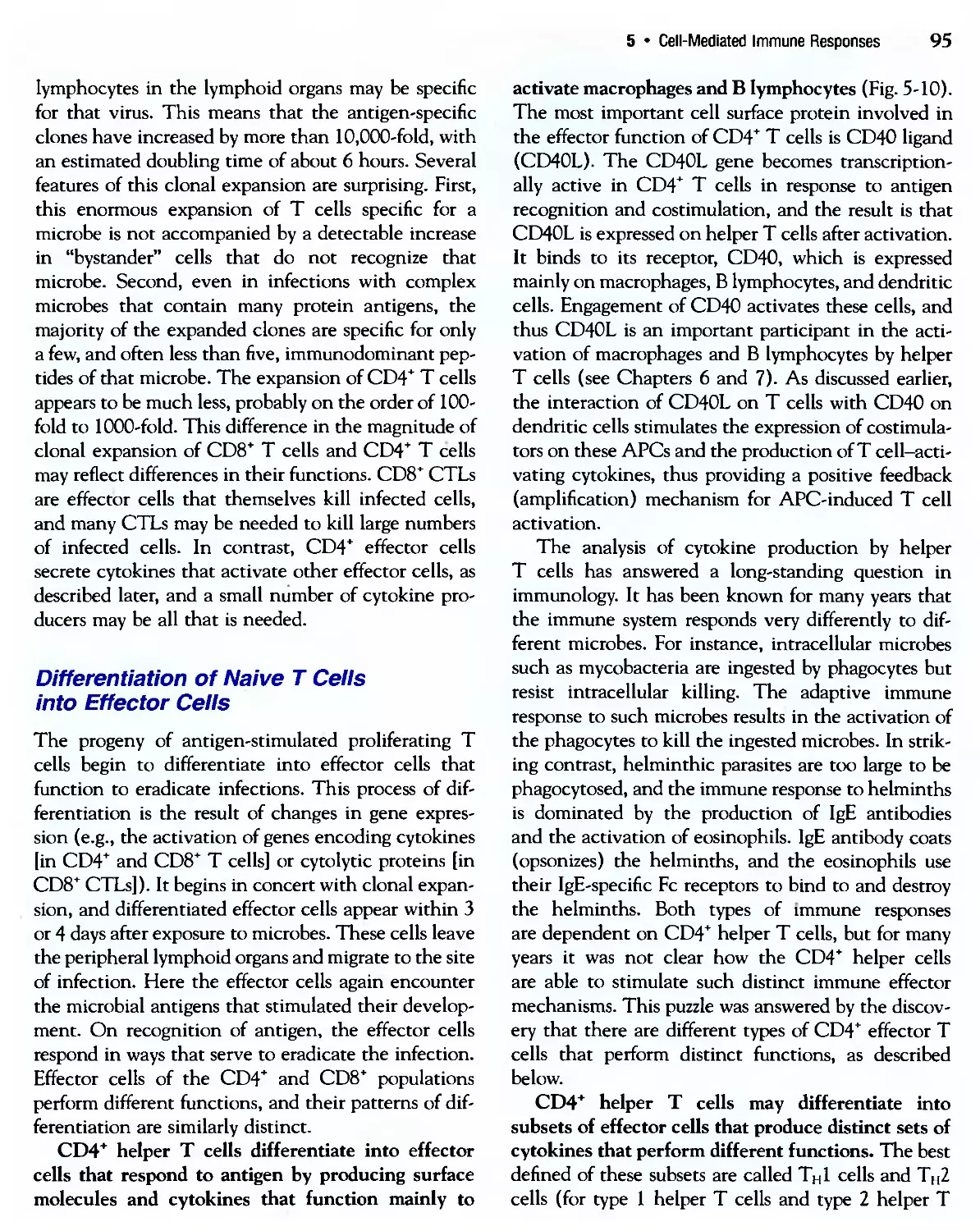
Includes index.
ISBN 0-7216-0241-X
1. Immunology. 2. Immunity. 3. Immunologic diseases. I. Lichtman, Andrew H.
II. Title.
[DNLM: 1. Immunity. 2. Hypersensitivity. 3. Immune System-physiology.
4. Immunologic Deficiency Syndromes. QW 504 A 122b 2004]
QR181.A28 2004
616.07'9-dc21 2003050607
Acquisitions Editor: Jason Malley
Project Manager: Linda Lewis Grigg
Designer: Gene Harris
BS/CTP
Printed in China.
Last digit is the print number: 987654321
To
Ann, Jonathan, Rehana
Sheila, Eben, Ariella, Amos, Ezra
Preface
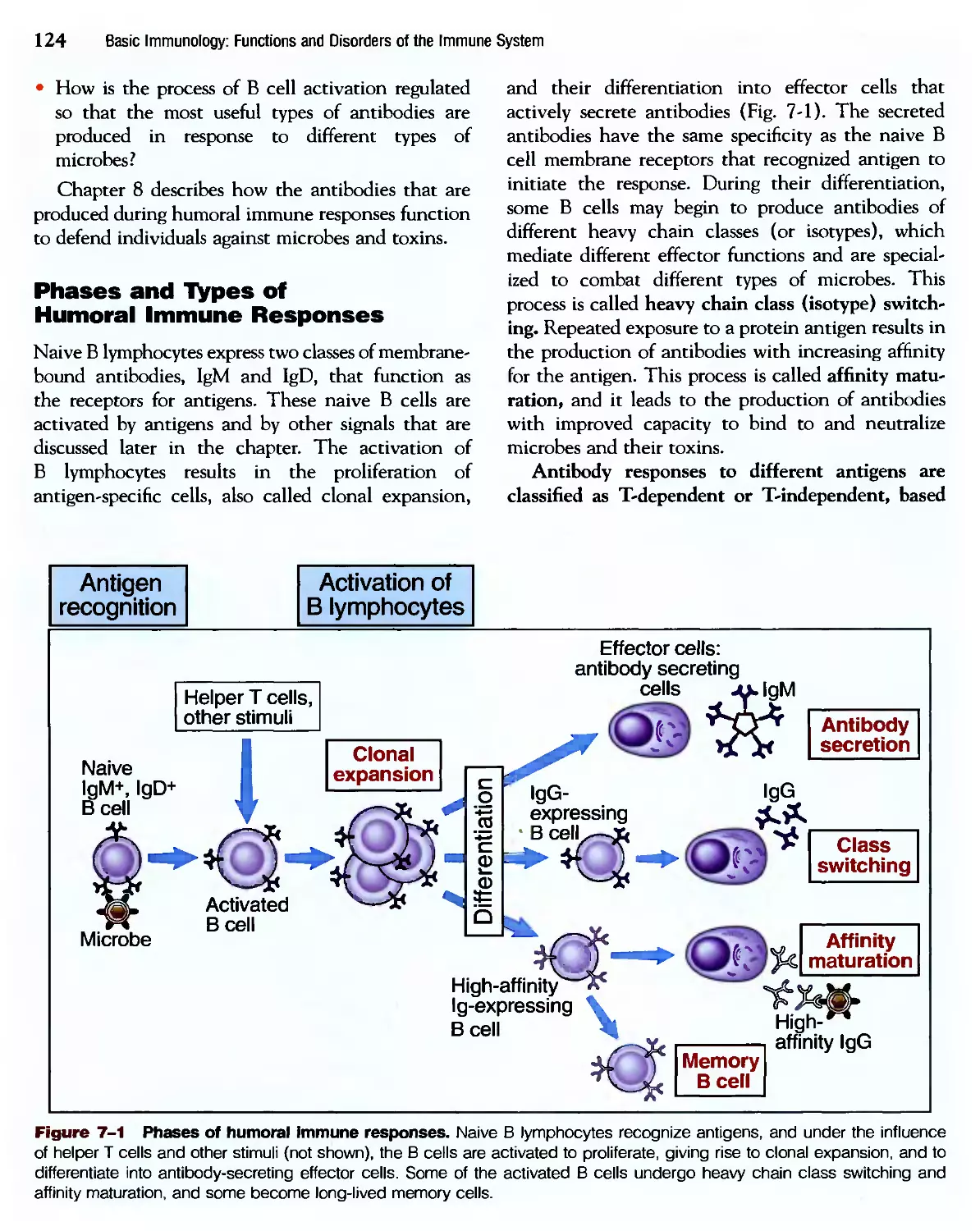
The second edition of Basic Immunology has been
revised to reflect new advances in our understanding
of the immune system and to improve on the presen-
presentation of information in ways most useful to students
and teachers. We have been extremely gratified by
how well the first edition of Basic Immunology has
been received by students in. the courses that we
teach, and the guiding principles on which the book
is based have not changed from the first edition. As
teachers of immunology, we are becoming increas-
increasingly aware that assimilating detailed information and
experimental approaches is difficult in many medical
school and undergraduate courses. The problem of
how much detail is appropriate has become a pressing
one because of the continuous and rapid increase in
the amount of information in all the biomedical
sciences. This problem is compounded by the de-
development of integrated cunicula in many medical
schools, with reduced time for didactic teaching and
an increasing emphasis on social and behavioral sci-
sciences and primary health care. For all these reasons,
we have realized the value for many medical students
of presenting the principles of immunology in a
concise and clear manner.
It is our view that several developments have come
together to make the goal of a concise and modern
consideration of immunology a realistic one. Most
important, immunology has matured as a discipline,
so that it has now reached the stage when the essen-
essential components of the immune system, and how they
interact in immune responses, are understood quite
well. There are, of course, many details to be filled in,
and the longstanding challenge of applying basic
principles to human diseases remains a difficult task.
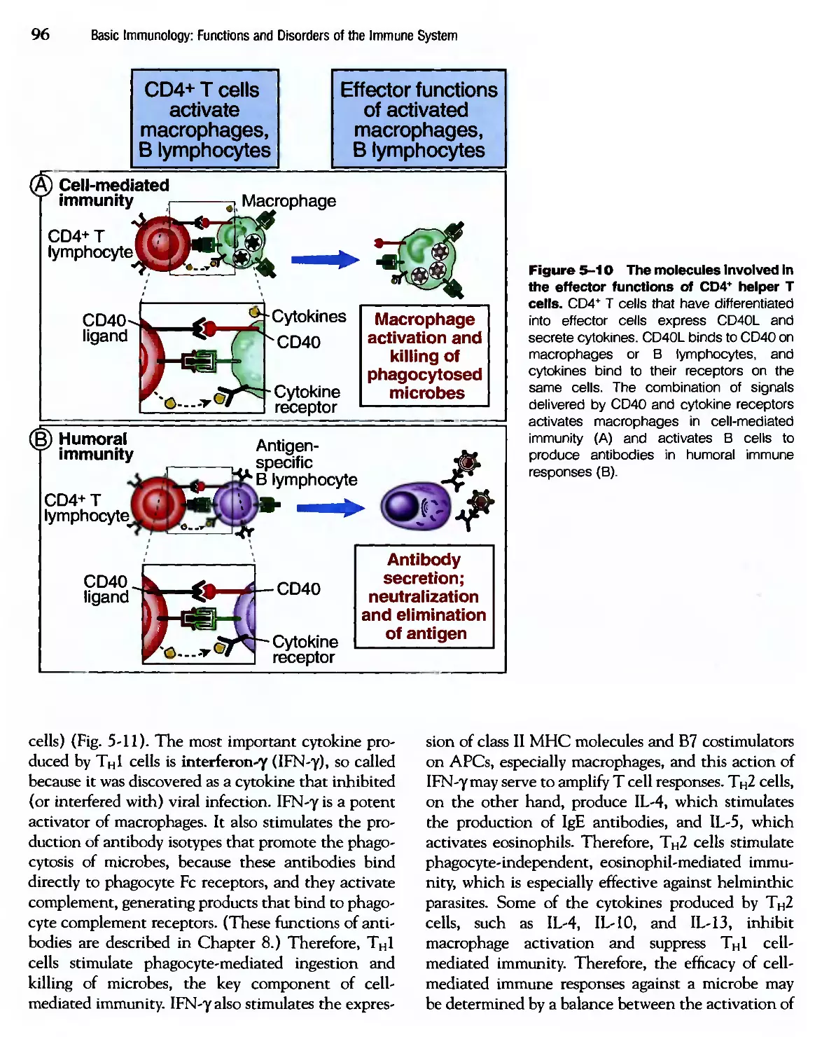
Nevertheless, we can now teach our students, with
reasonable confidence, how the immune system
works. The second important development has been
an increasing emphasis on the roots of immunology,
which lie in its role in defense against infections. As
a result, we are better able to relate experimental
results, using simple models, to the more complex, but
physiologically relevant, issue of host defense against
infectious pathogens.
This book has been written to address the per-
perceived needs of both medical school and under-
undergraduate curricula and to take advantage of the new
understanding of immunology. We have tried to
achieve several goals. First, we have presented the
most important principles governing the function of
the immune system. Our fundamental objective has
been to synthesize the key concepts from the vast
amount of experimental data that emerge in the
rapidly advancing field of immunology. The choice of
what is most important is based largely on what is
most clearly established by experimentation, what our
students find puzzling, and what explains the won-
wonderful efficiency and economy of the immune system.
Inevitably, however, such a choice will have an
element of bias, and our bias is toward emphasizing
the cellular interactions in immune responses and
limiting the description of many of the underlying
biochemical and molecular mechanisms to the essen-
essential facts. Second, we have focused on immune
responses against infectious microbes, and all our
discussions of the immune system are in this con-
context. Third, we have emphasized immune responses in
humans (rather than experimental animals), drawing
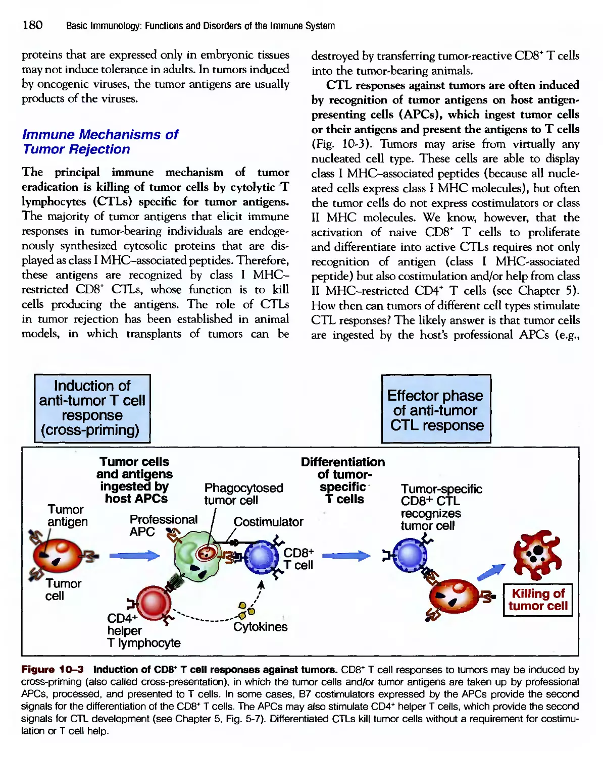
on parallels with experimental situations whenever
VH
VU1
Preface
necessary. Fourth, we have made liberal use of illus-
illustrations to highlight important principles but have
reduced factual details that may be found in more
comprehensive textbooks. Fifth, we have discussed
immunologic diseases also from the perspective of
principles, emphasizing their relation to normal
immune responses and avoiding details of clinical
syndromes and treatments. We have added selected
clinical cases in the Appendix, to illustrate how the
concepts of immunology may be applied to common
human diseases. Finally, we have realized that in any
concise discussion of complex phenomena, it is
inevitable that exceptions and caveats will fall by the
wayside. We have avoided exceptions and caveats
without hesitation, but with a willingness to modify
our conclusions as new information continues to
emerge.
It is our hope that students will find this book clear,
cogent, and manageable. Most important, we hope
the book will convey our sense of wonder about the
immune system and excitement about how the field
has evolved and how it continues to be relevant to
human health and disease. Finally, although we were
spurred to tackle this project because of our associa-
associations with medical school courses, we hope the book
will be valued more widely by students of allied health
and biology as well. We will have succeeded if the
book can answer many of the questions these students
have about the immune system and, at the same time,
encourage them to delve even more deeply into
immunology.
Several individuals played key roles in the writing
of this book. Our editor, Jason Malley, has been a
skilled and helpful colleague throughout. We have
been fortunate to again work with David and
Alexandra Baker of DNA Illustrations, who have
translated ideas into pictures that are informative and
aesthetically pleasing. Our project manager, Linda
Grigg, kept the project organized and on track despite
pressures of time and logistics. To all of them we owe
our many thanks.
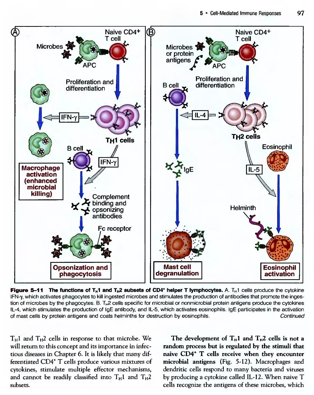
АЫ K. Abbas
Andrew H. Lichtman
Contents
Introduction to the Immune System
The Nomenclature, General Properties, and
Components of the Immune System 1
Innate Immunity
The Early Defense Against Infections 21
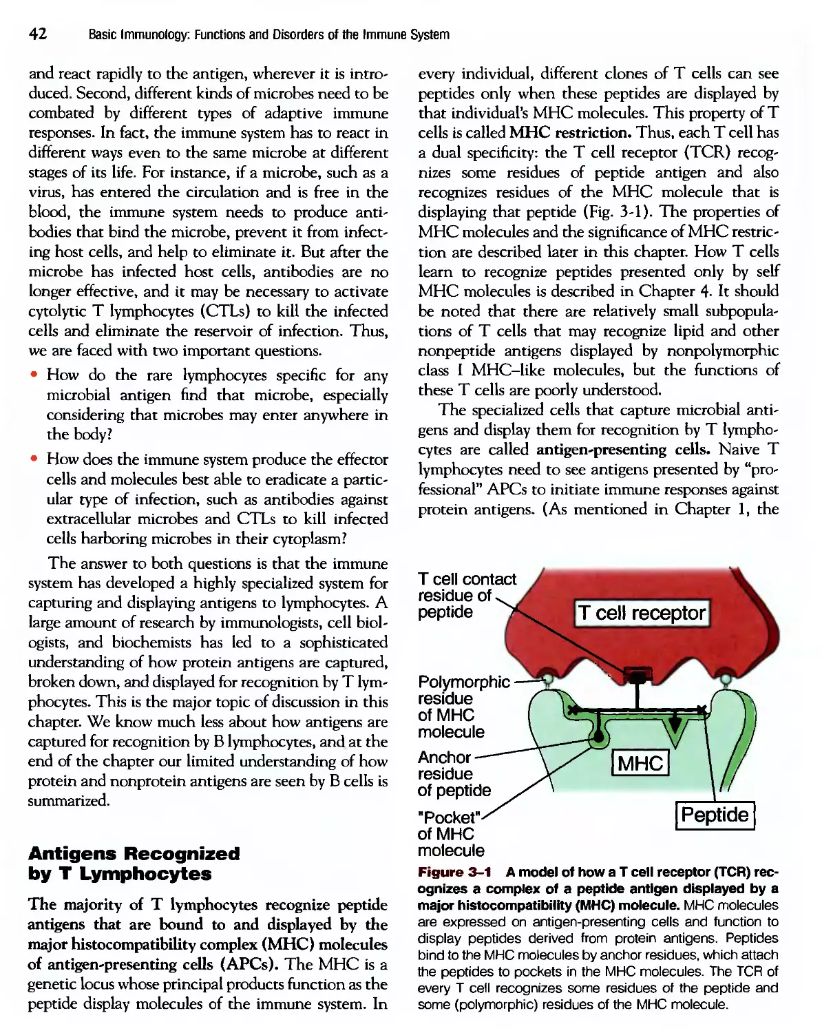
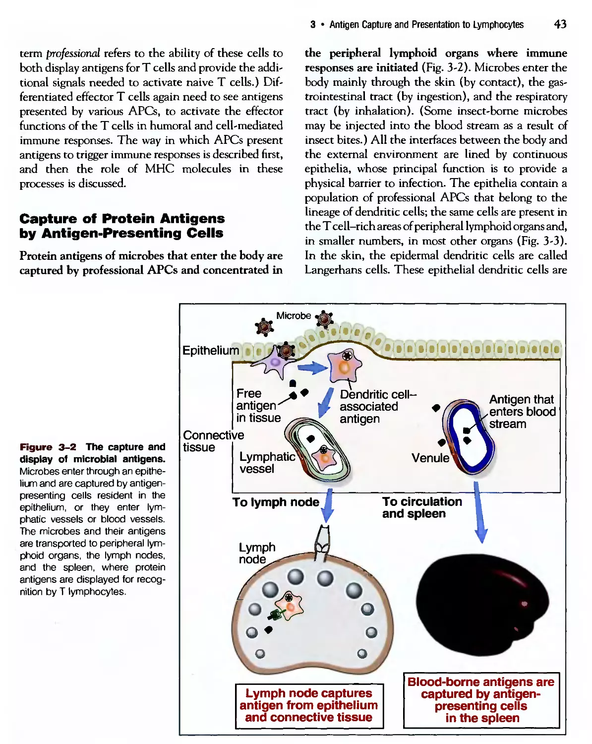
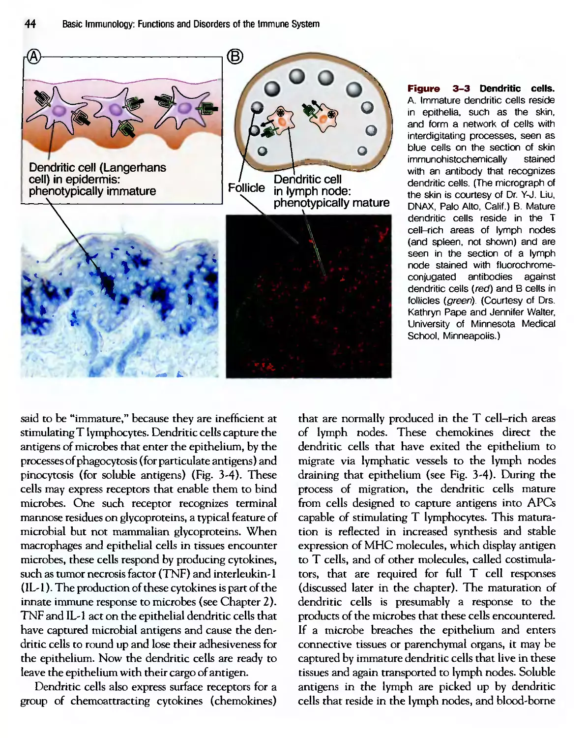
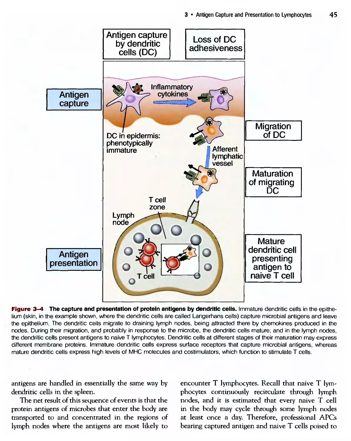
3 Antigen Capture and Presentation to
Lymphocytes
What Lymphocytes See 41
4 Antigen Recognition in the Adaptive
Immune System
Structure of Lymphocyte Antigen Receptors
and the Development of Immune Repertoires 63
Cell-Mediated Immune Responses
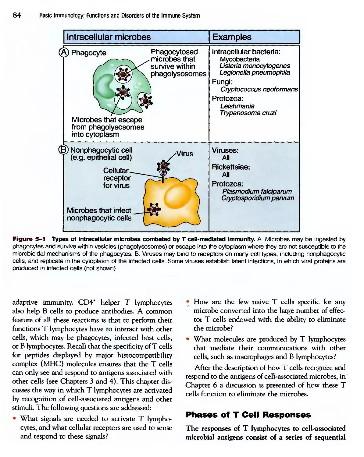
Activation of T Lymphocytes by
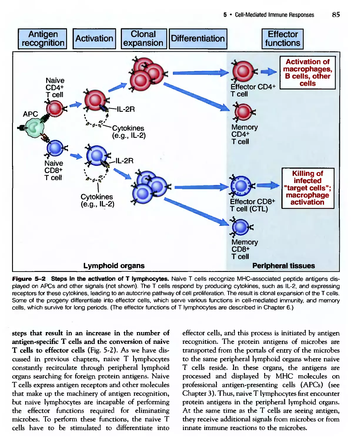
Cell-Associated Microbes 83
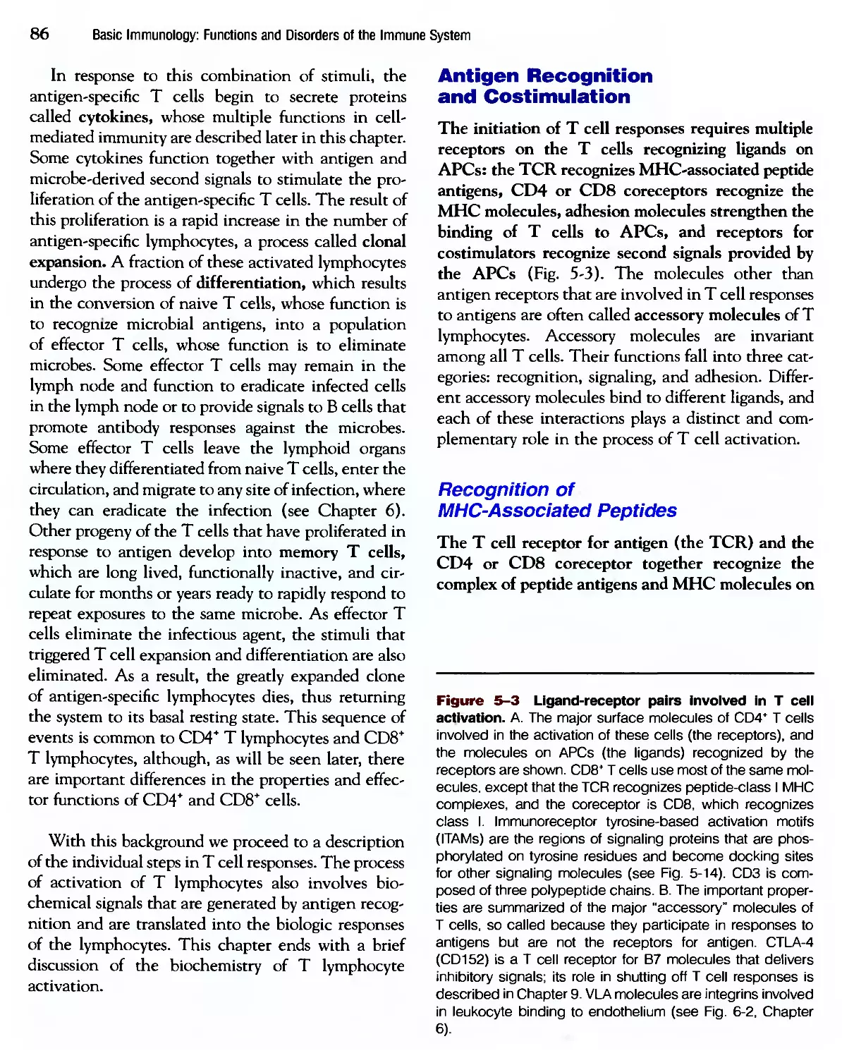
6 Effector Mechanisms of Cell-Mediated
Immunity
Eradication of Intracettular Microbes 105
Humoral Immune Responses
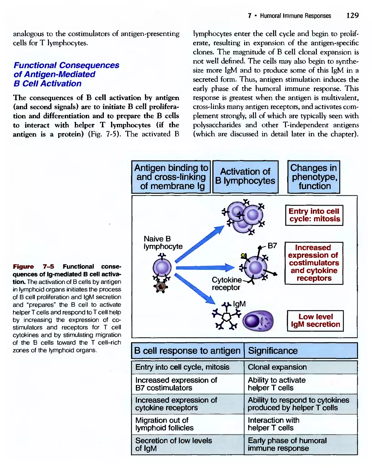
Activation of В Lymphocytes and Production
of Antibodies 123
Effector Mechanisms of Humoral Immunity
The Elimination of Extracellular Microbes
and Toxins 143
9 Immunologic Tolerance and
Autoimmunity
Self—Nonself Discrimination in the Immune
System and Its Failure 161
10 Immune Responses Against Tumors and
Transplants
Immunity to Noninfectious Transformed and
Foreign Cells 177
Hypersensitivity Diseases
Disorders Caused by Immune Responses 193
12 Congenital and Acquired
Immunodeficiencies
Diseases Caused by Defective Immune
Responses 209
Suggested Readings 225
APPENDIX I
Principal Features of CD Molecules 229
APPENDIX II
Glossary 263
APPENDIX III
Clinical Cases 291
Index 301
ix
Introduction to
the Immune System
The Nomenclature, General
Properties, and Components
of the Immune System
1
Immunity is defined as resistance to disease, specifically
infectious disease. The collection of cells, tissues, and
molecules that mediate resistance to infections is called the
immune system, and the coordinated reaction of these cells and
molecules to infectious microbes is the immune response.
Immunology is the study of the immune system and its
responses to invading pathogens. The physiologic function of
the immune system is to prevent infections and to eradicate
established infections, and this is the principal context in
which immune responses are discussed throughout this book.
The importance of the immune system for health is dra-
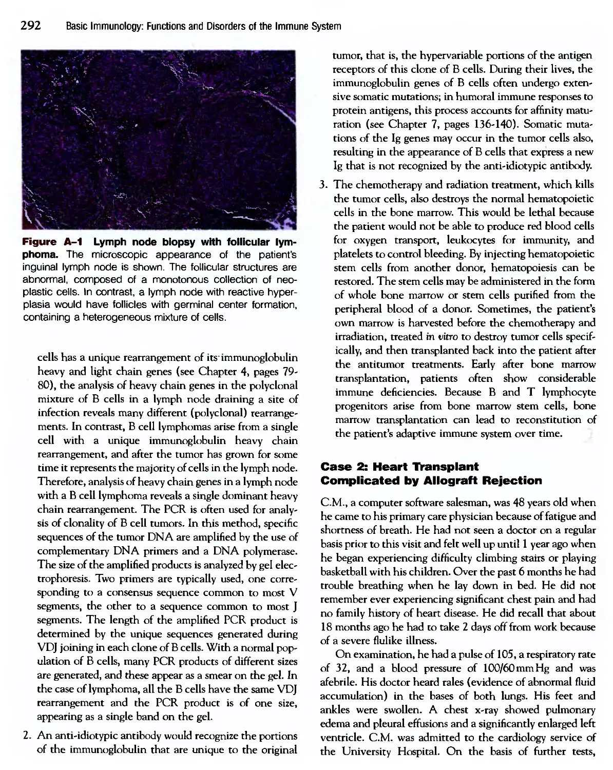
dramatically illustrated by the frequent observation that individ-
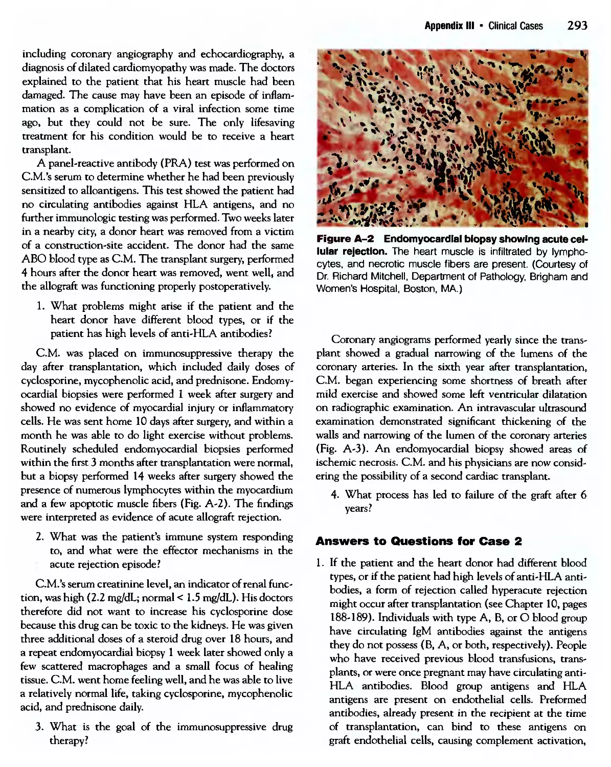
individuals with defective immune responses are susceptible to serious,
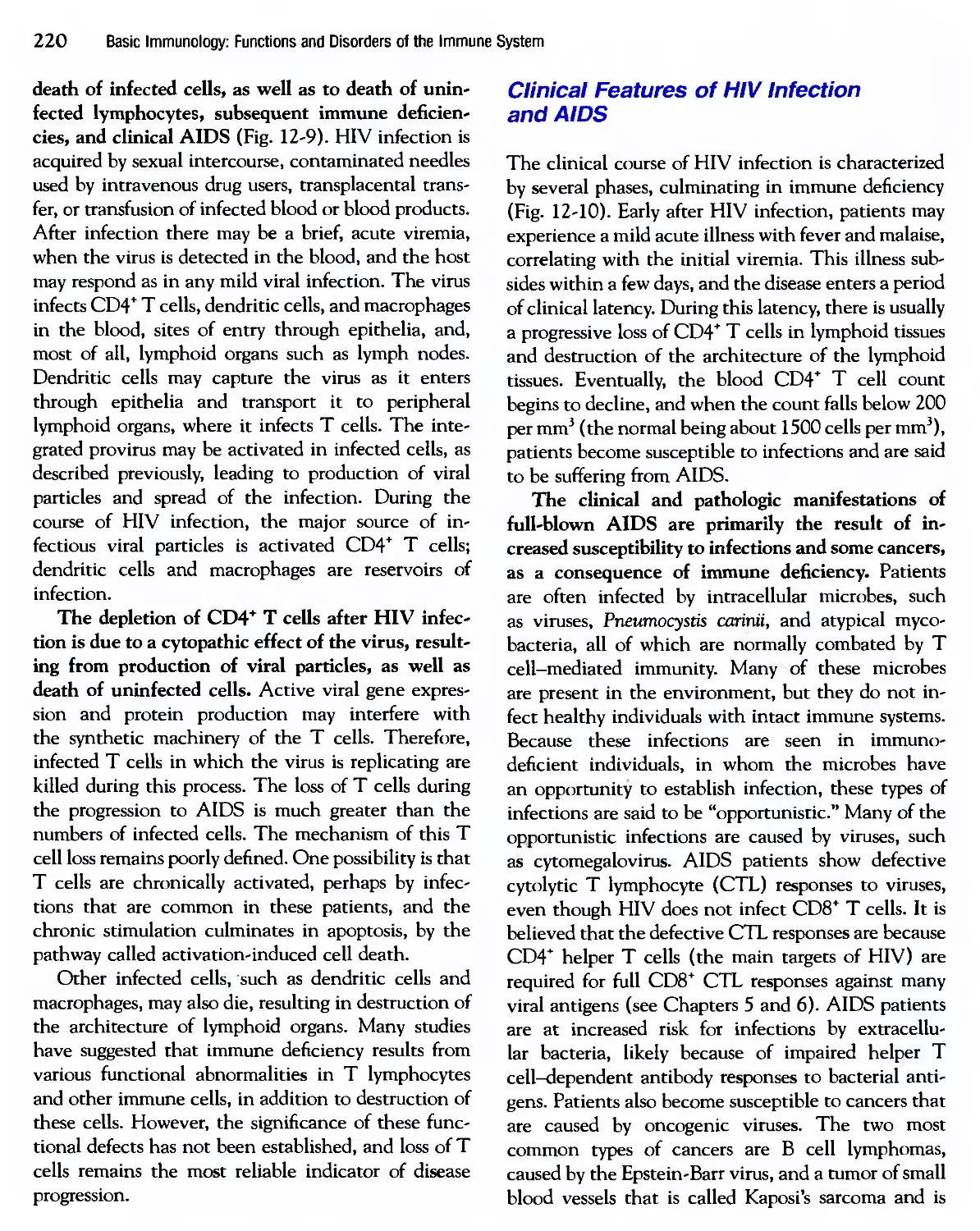
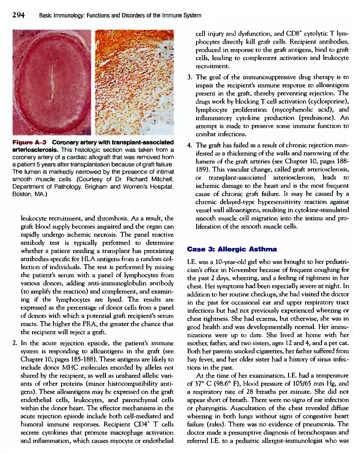
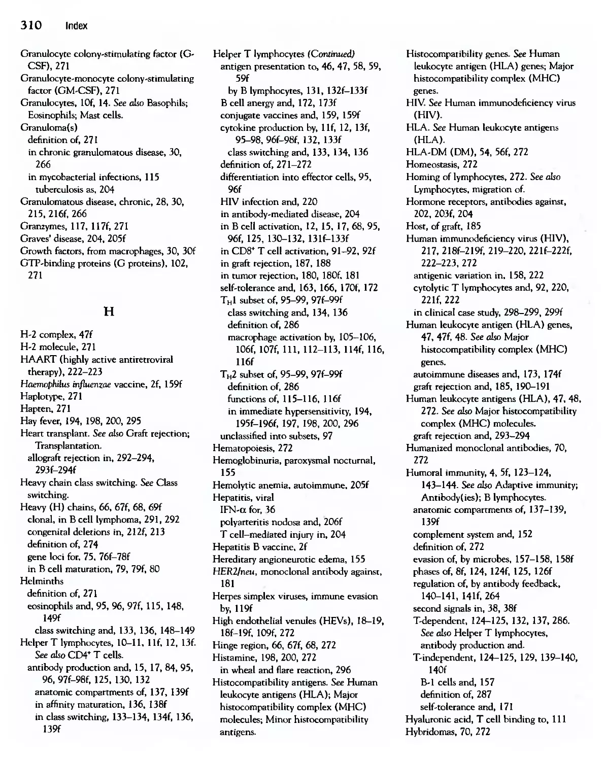
often life-threatening infections (Fig. 1-1). Conversely, stimulating immune responses
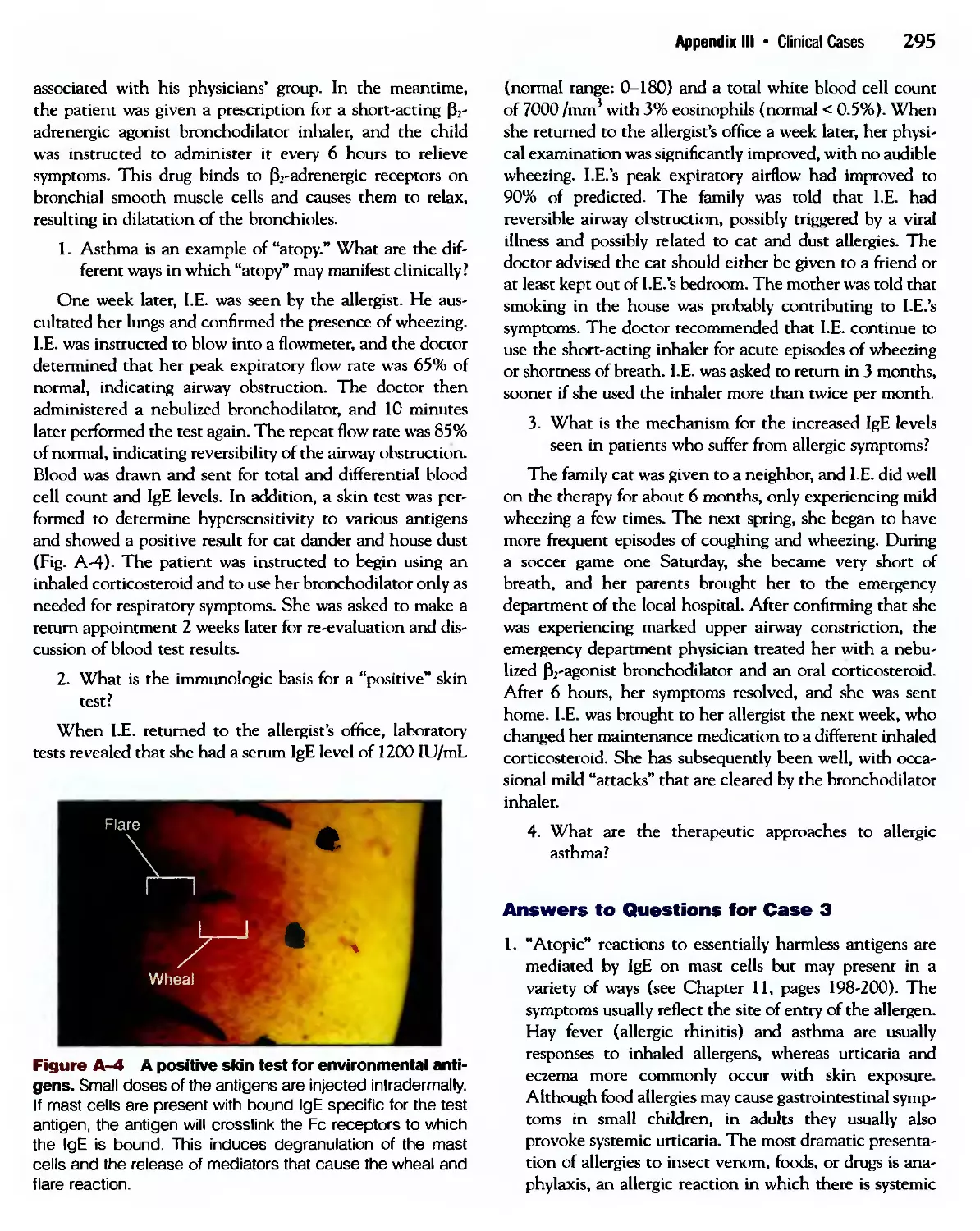
against microbes by the process of vaccination is the most effective method for protect-
protecting individuals against infections and is, for example, the approach that has led to the
worldwide eradication of smallpox (Fig. 1-2). The emergence of the acquired immuno-
immunodeficiency syndrome (AIDS) since the 1980s has tragically emphasized the importance
of the immune system for defending individuals against infections. But the impact of
immunology goes beyond infectious disease (see Fig. 1-1). The immune response is the
major barrier to successful organ transplantation, an increasingly used therapy for organ
Innate and Adaptive Immunity
Types of Adaptive Immunity
Properties of Adaptive Immune Responses
• Specificity
• Memory
Phases of Immune Responses
Cells of the Immune System
• Lymphocytes
• Antigen-Presenting Cells
• Effector Cells
Tissues of the Immune System
• Peripheral Lymphoid Organs
• Lymphocyte Recirculation
Summary
Basic Immunology: Functions and Disorders of the Immune System
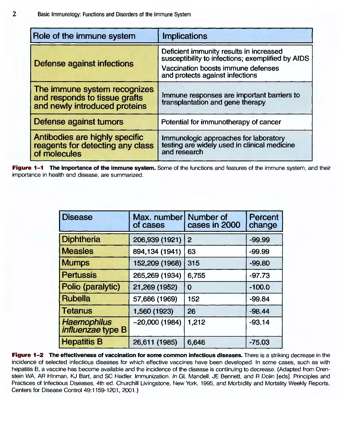
Role of the immune system
Defense against infections
The immune system recognizes
and responds to tissue grafts
and newly introduced proteins
Defense against tumors
Antibodies are highly specific
reagents for detecting any class
of molecules
Implications
Deficient immunity results in increased
susceptibility to infections; exemplified by AIDS
Vaccination boosts immune defenses
and protects against infections
Immune responses are important barriers to
transplantation and gene therapy
Potential for immunotherapy of cancer
Immunologic approaches for laboratory
testing are widely used in clinical medicine
and research
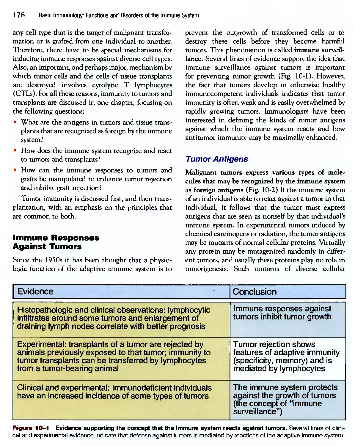
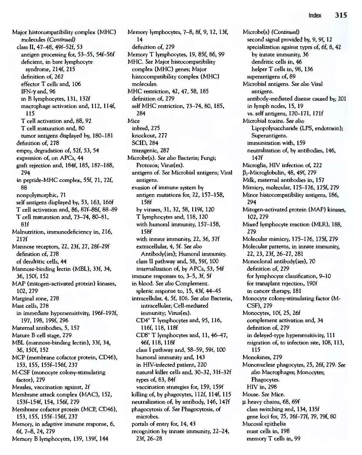
Figure 1-1 The importance of the immune system. Some of the functions and features of the immune system, and their
importance in health and disease, are summarized.
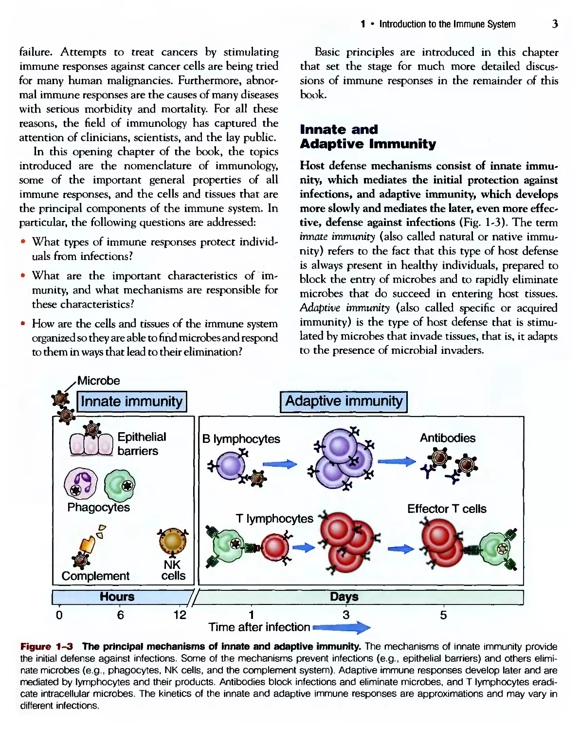
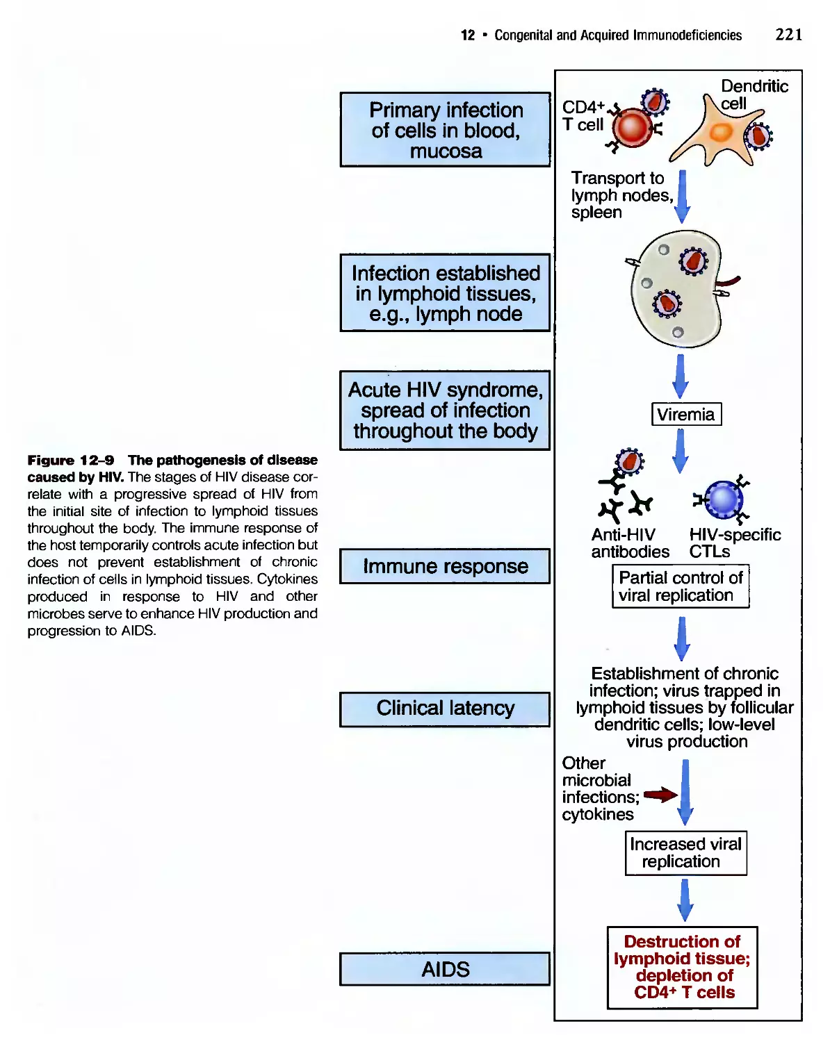
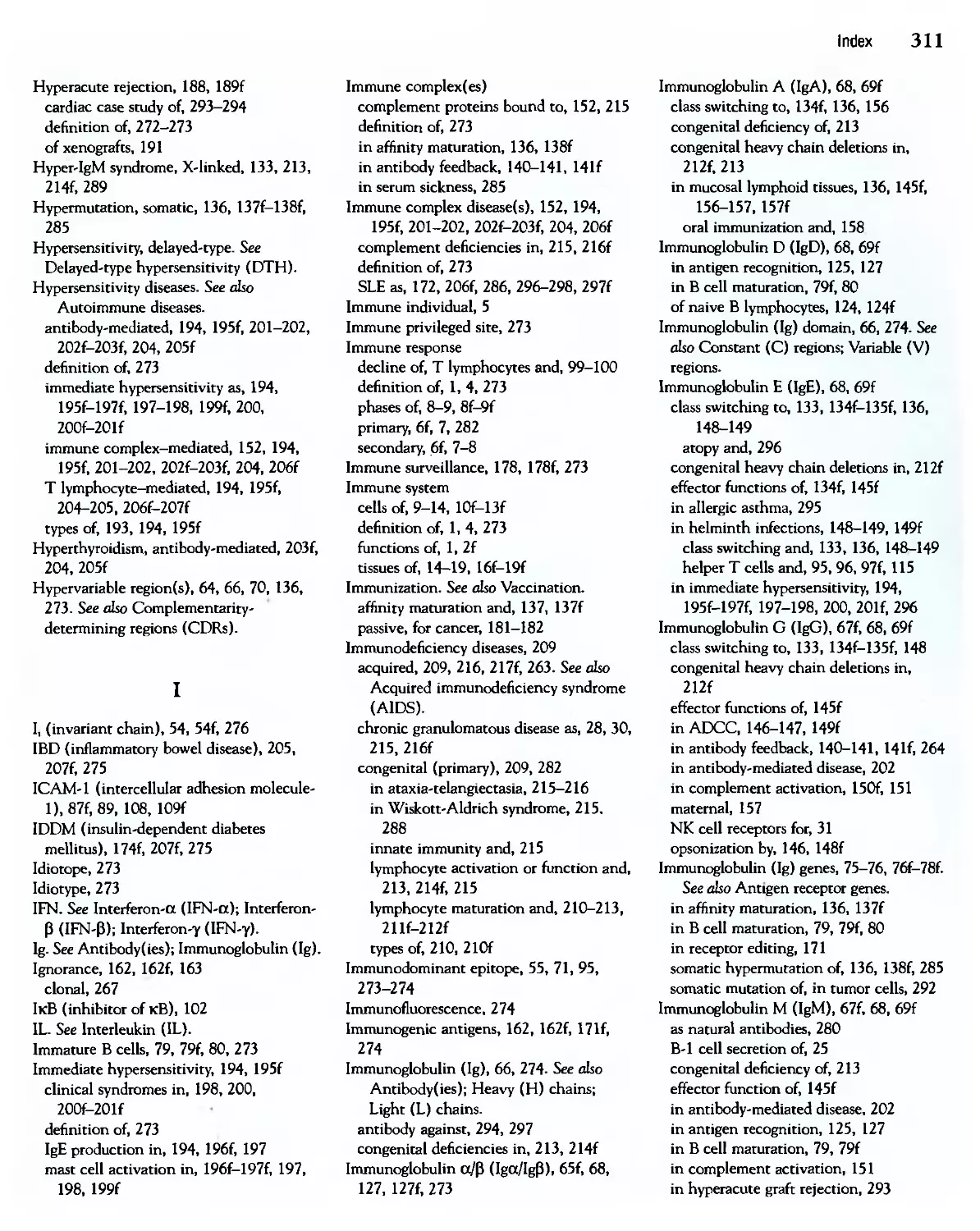
Disease
Diphtheria
Measles
Mumps
Pertussis
Polio (paralytic)
Rubella
Tetanus
Haemophilus
influenzae type В
Hepatitis В
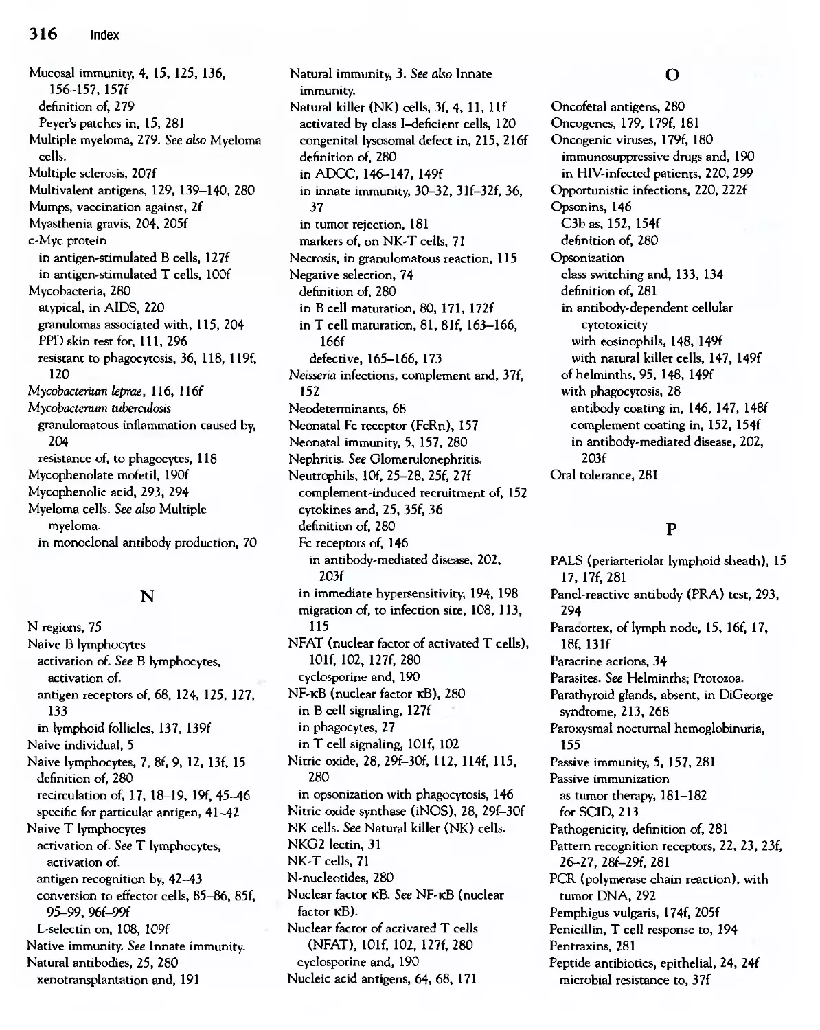
Max. number
of cases
206,939A921)
894,134A941)
152,209A968)
265,269A934)
21,269A952)
57,686A969)
1,560A923)
-20,000A984)
26,611 A985)
Number of
cases in 2000
2
63
315
6,755
0
152
26
1,212
6,646
Percent
change
-99.99
-99.99
-99.80
-97.73
-100.0
-99.84
-98.44
-93.14
-75.03
Figure 1-2 The effectiveness of vaccination for some common infectious diseases. There is a striking decrease in the
incidence of selected infectious diseases for which effective vaccines have been developed. In some cases, such as with
hepatitis B, a vaccine has become available and the incidence of the disease is continuing to decrease. (Adapted from Oren-
stein WA, AR Hinman, KJ Bart, and SC Hadler. Immunization. In GL Mandell, JE Bennett, and R Dolin [eds]. Principles and
Practices of Infectious Diseases, 4th ed. Churchill Livingstone, New York, 1995, and Morbidity and Mortality Weekly Reports,
Centers for Disease Control 49:1159-1201, 2001.)
Introduction to the Immune System
failure. Attempts to treat cancers by stimulating
immune responses against cancer cells are being tried
for many human malignancies. Furthermore, abnor-
abnormal immune responses are the causes of many diseases
with serious morbidity and mortality. For all these
reasons, the field of immunology has captured the
attention of clinicians, scientists, and the lay public.
In this opening chapter of the book, the topics
introduced are the nomenclature of immunology,
some of the important general properties of all
immune responses, and the cells and tissues that are
the principal components of the immune system. In
particular, the following questions are addressed:
• What types of immune responses protect individ-
individuals from infections?
• What are the important characteristics of im-
immunity, and what mechanisms are responsible for
these characteristics?
• How are the cells and tissues of the immune system
organized so they are able to find microbes and respond
to them in ways that lead to their elimination?
, Microbe
Basic principles are introduced in this chapter
that set the stage for much more detailed discus-
discussions of immune responses in the remainder of this
book.
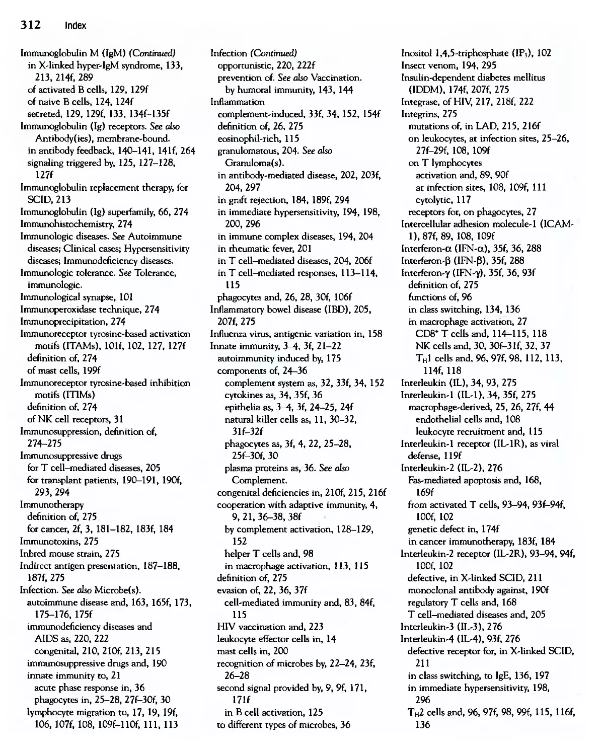
Innate and
Adaptive Immunity
Host defense mechanisms consist of innate immu-
immunity, which mediates the initial protection against
infections, and adaptive immunity, which develops
more slowly and mediates the later, even more effec-
effective, defense against infections (Fig. 1-3). The term
innate immunity (also called natural or native immu-
immunity) refers to the fact that this type of host defense
is always present in healthy individuals, prepared to
block the entry of microbes and to rapidly eliminate
microbes that do succeed in entering host tissues.
Adaptive immunity (also called specific or acquired
immunity) is the type of host defense that is stimu-
stimulated by microbes that invade tissues, that is, it adapts
to the presence of microbial invaders.
[Adaptive immunity
Complement
NK
cells
В lymphocytes
Antibodies
Hours
If
Days
0 6 12 1
Time after infection ■
Figure 1-3 The principal mechanisms of innate and adaptive immunity. The mechanisms of innate immunity provide
the initial defense against infections. Some of the mechanisms prevent infections (e.g., epithelial barriers) and others elimi-
eliminate microbes (e.g., phagocytes, NK cells, and the complement system). Adaptive immune responses develop later and are
mediated by lymphocytes and their products. Antibodies block infections and eliminate microbes, and T lymphocytes eradi-
eradicate intracellular microbes. The kinetics of the innate and adaptive immune responses are approximations and may vary in
different infections.
Basic Immunology: Functions and Disorders of the Immune System
The first line of defense in innate immunity is
provided by epithelial barriers and by specialized cells
and natural antibiotics present in epithelia, all of
which function to block the entry of microbes. If
microbes do breach epithelia and enter the tissues or
circulation, they are attacked by phagocytes, special-
specialized lymphocytes called natural killer (NK) cells, and
several plasma proteins, including the proteins of the
complement system. All these mechanisms of innate
immunity specifically recognize and react against
microbes but do not react against noninfectious foreign
substances. Different mechanisms of innate immunity
may be specific for molecules produced by different
classes of microbes. In addition to providing early
defense against infections, innate immune responses
enhance adaptive immune responses against the infec-
infectious agents. The components and mechanisms of
innate immunity are discussed in detail in Chapter 2.
Although innate immunity can effectively combat
many infections, microbes that are pathogenic for
humans (i.e., capable of causing disease) have evolved
to resist innate immunity. Defense against these infec-
infectious agents is the task of the adaptive immune
response, and this is why defects in the adaptive
immune system result in increased susceptibility to
infections. The adaptive immune system consists of
lymphocytes and their products, such as antibodies.
Whereas the mechanisms of innate immunity recog-
recognize structures shared by classes of microbes, the cells
of adaptive immunity, namely, lymphocytes, express
receptors that specifically recognize different sub-
substances produced by microbes as well as noninfectious
molecules. These substances are called antigens.
Adaptive immune responses are only triggered if
microbes or their antigens pass through epithelial bar-
barriers and are delivered to lymphoid organs where they
can be recognized by lymphocytes. Adaptive immune
responses generate mechanisms that are specialized to
combat different types of infections. For example,
antibodies function to eliminate microbes in extra-
extracellular fluids, and activated T lymphocytes eliminate
microbes living inside cells. These specialized mech-
mechanisms of adaptive immunity are described through-
throughout the book. Adaptive immune responses often use
the cells and molecules of the innate immune system
to eliminate microbes, and adaptive immunity func-
functions to greatly enhance these antimicrobial mecha-
mechanisms of innate immunity. For instance, antibodies (a
component of adaptive immunity) bind to microbes,
and these coated microbes avidly bind to and activate
phagocytes (a component of innate immunity), which
ingest and destroy the microbes. There are many
similar examples of the cooperation between innate
and adaptive immunity that are referred to in later
chapters. By convention the terms immune system and
immune response refer to adaptive immunity, unless
stated otherwise.
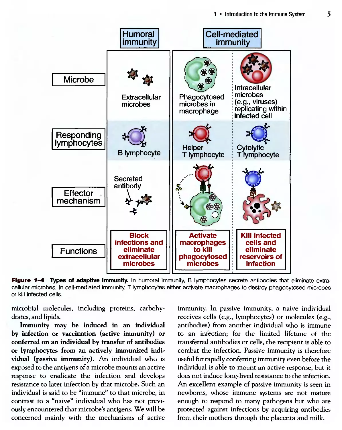
types of Adaptive Immunity
There are two types of adaptive immunity, called
humoral immunity and cell-mediated immunity, that
are mediated by different cells and molecules and are
designed to provide defense against extracellular
microbes and intracellular microbes, respectively
(Fig. 1-4). Humoral immunity is mediated by proteins
called antibodies, which are produced by cells called В
lymphocytes. Antibodies are secreted into the circula-
circulation and mucosal fluids, and they neutralize and elimi-
eliminate microbes and microbial toxins that are present in
the blood and in the lumens of mucosal organs, such as
the gastrointestinal and respiratory tracts. One of the
most important functions of antibodies is to stop
microbes that are present at mucosal surfaces and in
the blood from gaining access to and colonizing host
cells and connective tissues. In this way, antibodies
prevent infections from ever getting established.
Antibodies do not have access to microbes that live
and divide inside infected cells. Defense against
such intracellular microbes is called cell-mediated
immunity because it is mediated by cells called T
lymphocytes. Some T lymphocytes activate phago-
phagocytes to destroy microbes that have been ingested
by the phagocytes into phagocytic vesicles. Other
T lymphocytes kill any type of host cells that are
harboring infectious microbes in the cytoplasm. As is
discussed in Chapter 3 and later chapters, the anti-
antibodies produced by В lymphocytes are designed to spe-
specifically recognize extracellular microbial antigens,
whereas T lymphocytes recognize antigens produced
by intracellular microbes. Another important differ-
difference between В and T lymphocytes is that most T cells
recognize only microbial protein antigens, whereas
antibodies are able to recognize many different types of
1 • Introduction to the Immune System
Microbe
Responding
lymphocytes
Effector
mechanism
Functions
Humoral
immunity
Cell-mediated
immunity
Extracellular
microbes
«a
В lymphocyte
Block
infections and
eliminate
extracellular
microbes
Phagocytosed
microbes in
macrophage
Helper
T lymphocyte
Activate
macrophages
to kill
phagocytosed
microbes
Intracellular
microbes
(e.g., viruses)
replicating within
infected cell
Cytolytic
T lymphocyte
Kill infected
cells and
eliminate
reservoirs of
infection
Figure 1-4 Types of adaptive immunity. In humoral immunity, В lymphocytes secrete antibodies that eliminate extra-
extracellular microbes. In cell-mediated immunity, T lymphocytes either activate macrophages to destroy phagocytosed microbes
or kill infected cells.
microbial molecules, including proteins, carbohy-
carbohydrates, and lipids.
Immunity may be induced in an individual
by infection or vaccination (active immunity) or
conferred on an individual by transfer of antibodies
or lymphocytes from an actively immunized indi-
individual (passive immunity). An individual who is
exposed to the antigens of a microbe mounts an active
response to eradicate the infection and develops
resistance to later infection by that microbe. Such an
individual is said to be "immune" to that microbe, in
contrast to a "naive" individual who has not previ-
previously encountered that microbe's antigens. We will be
concerned mainly with the mechanisms of active
immunity. In passive immunity, a naive individual
receives cells (e.g., lymphocytes) or molecules (e.g.,
antibodies) from another individual who is immune
to an infection; for the limited lifetime of the
transferred antibodies or cells, the recipient is able to
combat the infection. Passive immunity is therefore
useful for rapidly conferring immunity even before the
individual is able to mount an active response, but it
does not induce long-lived resistance to the infection.
An excellent example of passive immunity is seen in
newborns, whose immune systems are not mature
enough to respond to many pathogens but who are
protected against infections by acquiring antibodies
from their mothers through the placenta and milk.
6
Basic Immunology: Functions and Disorders of the Immune System
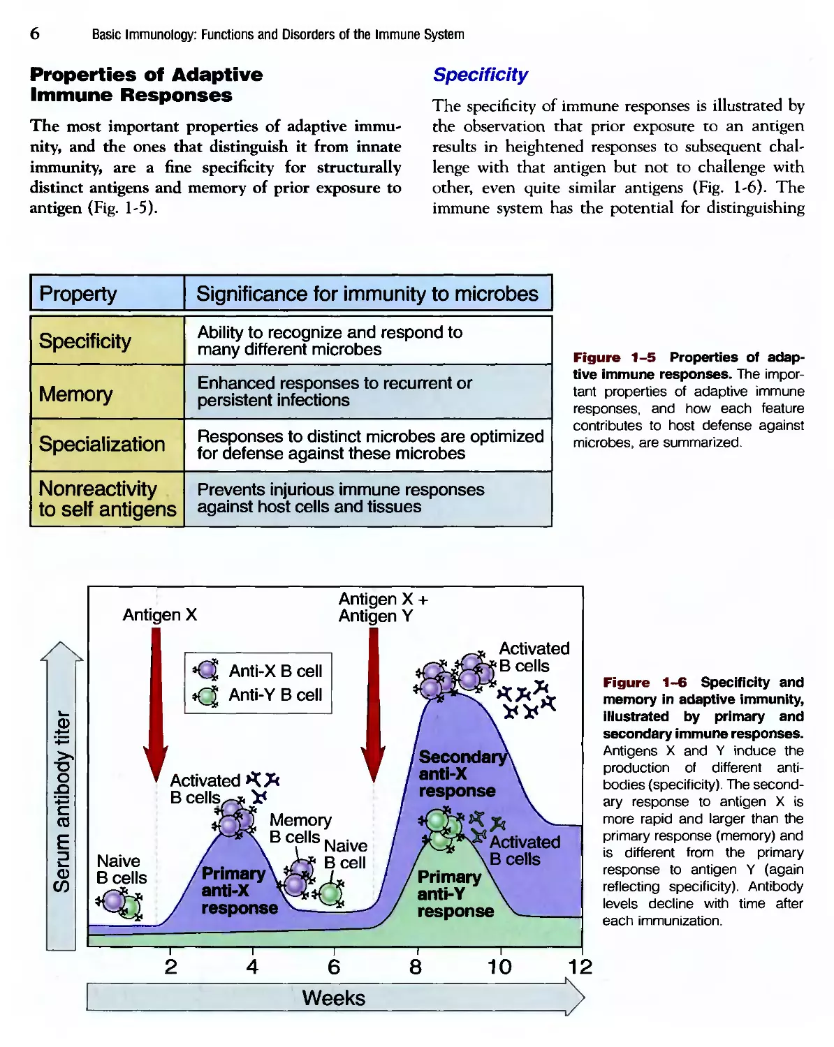
Properties of Adaptive
Immune Responses
The most important properties of adaptive immu-
immunity, and the ones that distinguish it from innate
immunity, are a fine specificity for structurally
distinct antigens and memory of prior exposure to
antigen (Fig. 1-5).
Specificity
The specificity of immune responses is illustrated by
the observation that prior exposure to an antigen
results in heightened responses to subsequent chal-
challenge with that antigen but not to challenge with
other, even quite similar antigens (Fig. 1-6). The
immune system has the potential for distinguishing
Property
Specificity
Memory
Specialization
Nonreactivity
to self antigens
Significance for immunity to microbes
Ability to recognize and respond to
many different microbes
Enhanced responses to recurrent or
persistent infections
Responses to distinct microbes are optimized
for defense against these microbes
Prevents injurious immune responses
against host cells and tissues
Figure 1-5 Properties of adap-
adaptive immune responses. The impor-
important properties of adaptive immune
responses, and how each feature
contributes to host defense against
microbes, are summarized.
CD
>4
T3
О
JO
V-»
я
I
о
Antigen X
Antjgen X +
Antigen Y
Anti-XBcell
«Q Anti-YBcell
Activated* *
t
Memory
Bcells Naive
'Primary— >Bcel1
anti-X
response
Weeks
Activated
4B cells
Primary
anti-Y
response
Figure 1-6 Specificity and
memory in adaptive immunity,
illustrated by primary and
secondary immune responses.
Antigens X and Y induce the
production of different anti-
antibodies (specificity). The second-
secondary response to antigen X is
more rapid and larger than the
primary response (memory) and
is different from the primary
response to antigen Y (again
reflecting specificity). Antibody
levels decline with time after
each immunization.
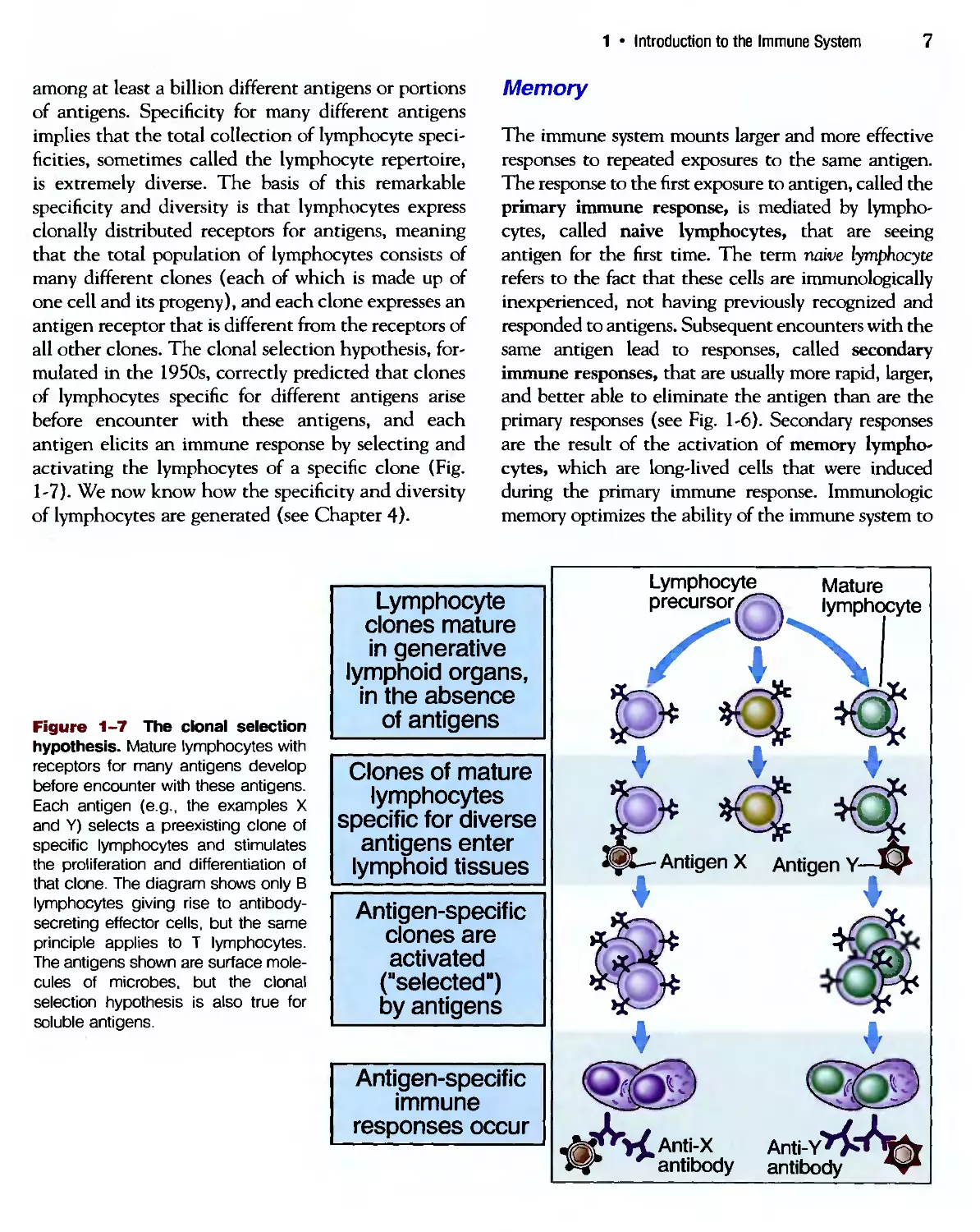
Introduction to the Immune System
among at least a billion different antigens or portions
of antigens. Specificity for many different antigens
implies that the total collection of lymphocyte speci-
specificities, sometimes called the lymphocyte repertoire,
is extremely diverse. The basis of this remarkable
specificity and diversity is that lymphocytes express
clonally distributed receptors for antigens, meaning
that the total population of lymphocytes consists of
many different clones (each of which is made up of
one cell and its progeny), and each clone expresses an
antigen receptor that is different from the receptors of
all other clones. The clonal selection hypothesis, for-
formulated in the 1950s, correctly predicted that clones
of lymphocytes specific for different antigens arise
before encounter with these antigens, and each
antigen elicits an immune response by selecting and
activating the lymphocytes of a specific clone (Fig.
1-7). We now know how the specificity and diversity
of lymphocytes are generated (see Chapter 4).
Memory
The immune system mounts larger and more effective
responses to repeated exposures to the same antigen.
The response to the first exposure to antigen, called the
primary immune response, is mediated by lympho-
lymphocytes, called naive lymphocytes, that are seeing
antigen for the first time. The term naive lymphocyte
refers to the fact that these cells are immunologically
inexperienced, not having previously recognized and
responded to antigens. Subsequent encounters with the
same antigen lead to responses, called secondary
immune responses, that are usually more rapid, larger,
and better able to eliminate the antigen than are the
primary responses (see Fig. 1-6). Secondary responses
are the result of the activation of memory lympho-
lymphocytes, which are long-lived cells that were induced
during the primary immune response. Immunologic
memory optimizes the ability of the immune system to
Figure 1-7 The clonal selection
hypothesis. Mature lymphocytes with
receptors for many antigens develop
before encounter with these antigens.
Each antigen (e.g., the examples X
and Y) selects a preexisting clone of
specific lymphocytes and stimulates
the proliferation and differentiation of
that clone. The diagram shows only В
lymphocytes giving rise to antibody-
secreting effector cells, but the same
principle applies to T lymphocytes.
The antigens shown are surface mole-
molecules of microbes, but the clonal
selection hypothesis is also true for
soluble antigens.
Lymphocyte
clones mature
in generative
lymphoid organs,
in the absence
of antigens
Clones of mature
lymphocytes
specific for diverse
antigens enter
lymphoid tissues
Antigen-specific
clones are
activated
("selected")
by antigens
Antigen-specific
immune
responses occur
Lymphocyte Mature
precursor/"^ lymphocyte
Antigen X Antigen Y-
WAnti-X Anti-Y
•antibody antibody
8
Basic Immunology: Functions and Disorders of the Immune System
combat persistent and recurrent infections, because
each encounter with a microbe generates more memory
cells and activates previously generated memory cells.
Memory is also one of the reasons why vaccines confer
long-lasting protection against infections.
Immune responses have other characteristics that
are important for their functions (see Fig. 1-5).
Immune responses are specialized, and different
responses are designed to best defend against different
classes of microbes. The immune system is able to
react against an enormous number and variety of
microbes and other foreign antigens, but it normally
does not react against the host's own potentially anti-
genic substances, so-called self antigens. All immune
responses are self-limited and decline as the infection
is eliminated, allowing the system to return to a
resting state, prepared to respond to another infec-
infection. Much of the science of immunology is devoted
to understanding the mechanisms underlying these
characteristics of adaptive immune responses.
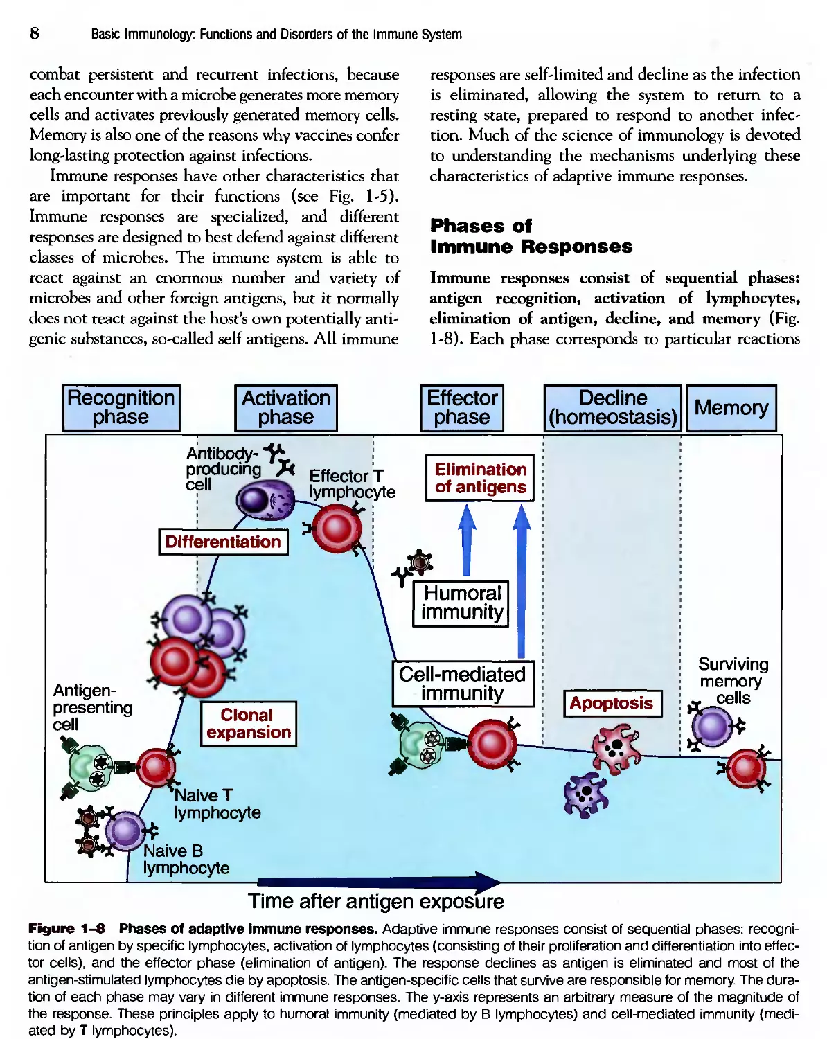
Phases of
Immune Responses
Immune responses consist of sequential phases:
antigen recognition, activation of lymphocytes,
elimination of antigen, decline, and memory (Fig.
1-8). Each phase corresponds to particular reactions
Recognition
phase
Activation
phase
Effector
phase
Decline
(homeostasis)
Memory
Antibody- f
producing Я EffectorT
lymphocyte
Elimination
of antigens
t
Humoral
immunity
Antigen-
presenting
cell
Clonal
expansion
Cell-mediated
immunity
| Apoptosis
Surviving
memory
cells
Naive T
lymphocyte
Naive В
lymphocyte
Time after antigen exposure
Figure 1-8 Phases of adaptive immune responses. Adaptive immune responses consist of sequential phases: recogni-
recognition of antigen by specific lymphocytes, activation of lymphocytes (consisting of their proliferation and differentiation into effec-
effector cells), and the effector phase (elimination of antigen). The response declines as antigen is eliminated and most of the
antigen-stimulated lymphocytes die by apoptosis. The antigen-specific cells that survive are responsible for memory. The dura-
duration of each phase may vary in different immune responses. The y-axis represents an arbitrary measure of the magnitude of
the response. These principles apply to humoral immunity (mediated by В lymphocytes) and cell-mediated immunity (medi-
(mediated by T lymphocytes).
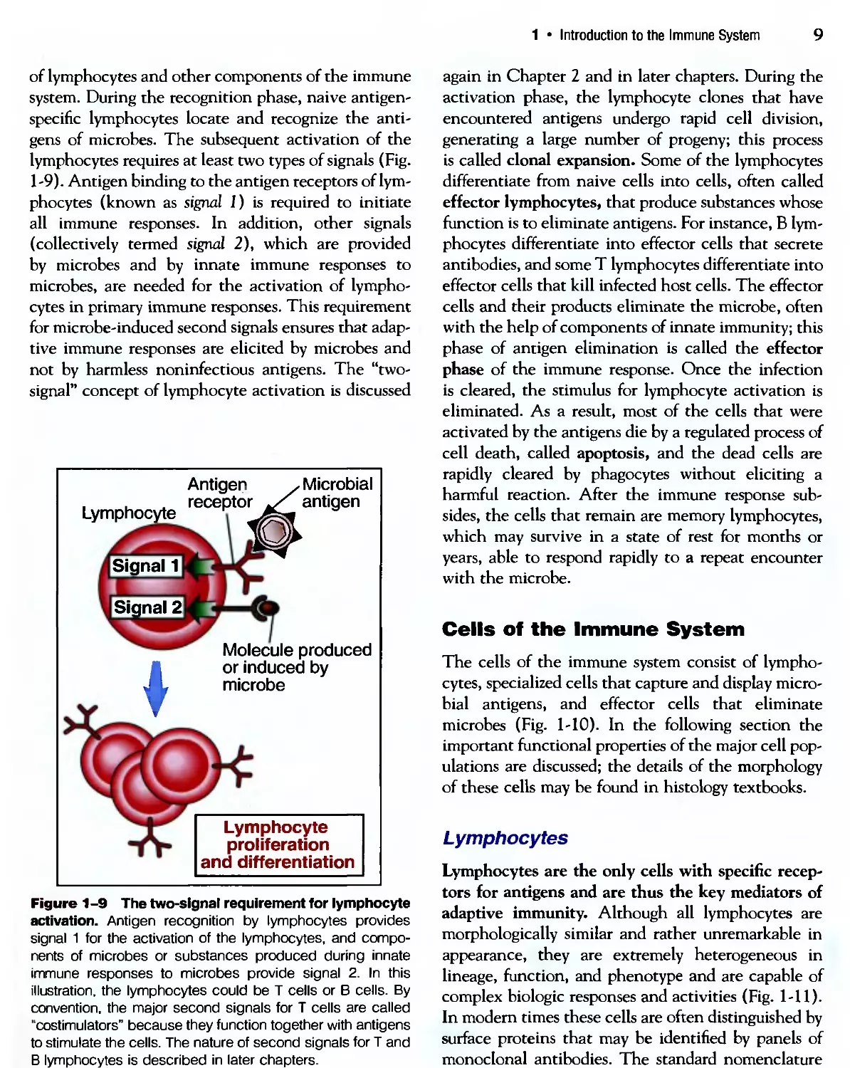
1 • Introduction to the Immune System
of lymphocytes and other components of the immune
system. During the recognition phase, naive antigen-
specific lymphocytes locate and recognize the anti-
antigens of microbes. The subsequent activation of the
lymphocytes requires at least two types of signals (Fig.
1-9). Antigen binding to the antigen receptors of lym-
lymphocytes (known as signal J) is required to initiate
all immune responses. In addition, other signals
(collectively termed signal 2), which are provided
by microbes and by innate immune responses to
microbes, are needed for the activation of lympho-
lymphocytes in primary immune responses. This requirement
for microbe-induced second signals ensures that adap-
adaptive immune responses are elicited by microbes and
not by harmless noninfectious antigens. The "two-
signal" concept of lymphocyte activation is discussed
Lymphocyte
Signal 1
Signal 2
Antigen
receptor
Microbial
antigen
Molecule produced
or induced by
microbe
Lymphocyte
proliferation
and differentiation
Figure 1 -9 The two-signal requirement for lymphocyte
activation. Antigen recognition by lymphocytes provides
signal 1 for the activation of the lymphocytes, and compo-
components of microbes or substances produced during innate
immune responses to microbes provide signal 2. In this
illustration, the lymphocytes could be T cells or В cells. By
convention, the major second signals for T cells are called
"costimulators" because they function together with antigens
to stimulate the cells. The nature of second signals for T and
В lymphocytes is described in later chapters.
again in Chapter 2 and in later chapters. During the
activation phase, the lymphocyte clones that have
encountered antigens undergo rapid cell division,
generating a large number of progeny; this process
is called clonal expansion. Some of the lymphocytes
differentiate from naive cells into cells, often called
effector lymphocytes, that produce substances whose
function is to eliminate antigens. For instance, В lym-
lymphocytes differentiate into effector cells that secrete
antibodies, and some T lymphocytes differentiate into
effector cells that kill infected host cells. The effector
cells and their products eliminate the microbe, often
with the help of components of innate immunity; this
phase of antigen elimination is called the effector
phase of the immune response. Once the infection
is cleared, the stimulus for lymphocyte activation is
eliminated. As a result, most of the cells that were
activated by the antigens die by a regulated process of
cell death, called apoptosis, and the dead cells are
rapidly cleared by phagocytes without eliciting a
harmful reaction. After the immune response sub-
subsides, the cells that remain are memory lymphocytes,
which may survive in a state of rest for months or
years, able to respond rapidly to a repeat encounter
with the microbe.
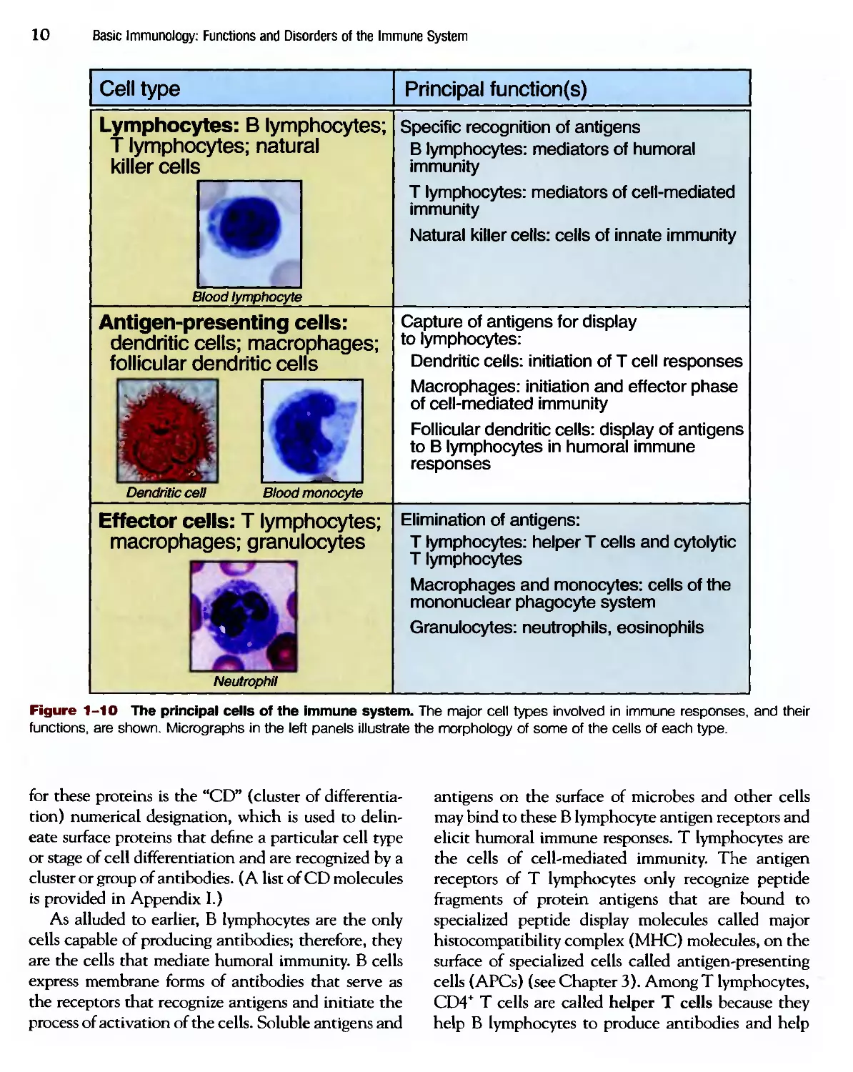
Cells of the Immune System
The cells of the immune system consist of lympho-
lymphocytes, specialized cells that capture and display micro-
microbial antigens, and effector cells that eliminate
microbes (Fig. 1-10). In the following section the
important functional properties of the major cell pop-
populations are discussed; the details of the morphology
of these cells may be found in histology textbooks.
Lymphocytes
Lymphocytes are the only cells with specific recep-
receptors for antigens and are thus the key mediators of
adaptive immunity. Although all lymphocytes are
morphologically similar and rather unremarkable in
appearance, they are extremely heterogeneous in
lineage, function, and phenotype and are capable of
complex biologic responses and activities (Fig. 1-11).
In modern times these cells are often distinguished by
surface proteins that may be identified by panels of
monoclonal antibodies. The standard nomenclature
10
Basic Immunology: Functions and Disorders of the Immune System
Cell type
Lymphocytes: В lymphocytes;
T lymphocytes; natural
killer cells
Blood lymphocyte
Antigen-presenting cells:
dendritic cells; macrophages;
follicular dendritic cells
Dendritic cell
Blood monocyte
Effector cells: T lymphocytes;
macrophages; granulocytes
Neutrophil
Principal function(s)
Specific recognition of antigens
В lymphocytes: mediators of humoral
immunity
T lymphocytes: mediators of cell-mediated
immunity
Natural killer cells: cells of innate immunity
Capture of antigens for display
to lymphocytes:
Dendritic ceils: initiation of T cell responses
Macrophages: initiation and effector phase
of cell-mediated immunity
Follicular dendritic cells: display of antigens
to В lymphocytes in humoral immune
responses
Elimination of antigens:
T lymphocytes: helper T cells and cytolytic
T lymphocytes
Macrophages and monocytes: cells of the
mononuclear phagocyte system
Granulocytes: neutrophils, eosinophils
Figure 1-10 The principal cells of the immune system. The major cell types involved in immune responses, and their
functions, are shown. Micrographs in the left panels illustrate the morphology of some of the cells of each type.
for these proteins is the "CD" (cluster of differentia-
differentiation) numerical designation, which is used to delin-
delineate surface proteins that define a particular cell type
or stage of cell differentiation and are recognized by a
cluster or group of antibodies. (A list of CD molecules
is provided in Appendix I.)
As alluded to earlier, В lymphocytes are the only
cells capable of producing antibodies; therefore, they
are the cells that mediate humoral immunity. В cells
express membrane forms of antibodies that serve as
the receptors that recognize antigens and initiate the
process of activation of the cells. Soluble antigens and
antigens on the surface of microbes and other cells
may bind to these В lymphocyte antigen receptors and
elicit humoral immune responses. T lymphocytes are
the cells of cell-mediated immunity. The antigen
receptors of T lymphocytes only recognize peptide
fragments of protein antigens that are bound to
specialized peptide display molecules called major
histocompatibility complex (MHC) molecules, on the
surface of specialized cells called antigen-presenting
cells (APCs) (see Chapter 3). Among T lymphocytes,
CD4+ T cells are called helper T cells because they
help В lymphocytes to produce antibodies and help
1 • Introduction to the Immune System 11
В
lymphocyte
Helper T
lymphocyte
Cytolytic T
lymphocyte
(CTL)
Natural
killer
(NK) cell
Antigen recognition
Effector functions
<3
Microbe
Antibody
Cytokines
а
Microbial antigen
presented
by antigen-
presenting cell
Infected cell
expressing
microbial antigen
Neutralization
of microbe,
phagocytosis,
complement
activation
Activation of
macrophages
Inflammation
Activation
(proliferation and
differentiation)
of T and В
lymphocytes
Killing of
infected cell
Killing of
infected cell
Target cell
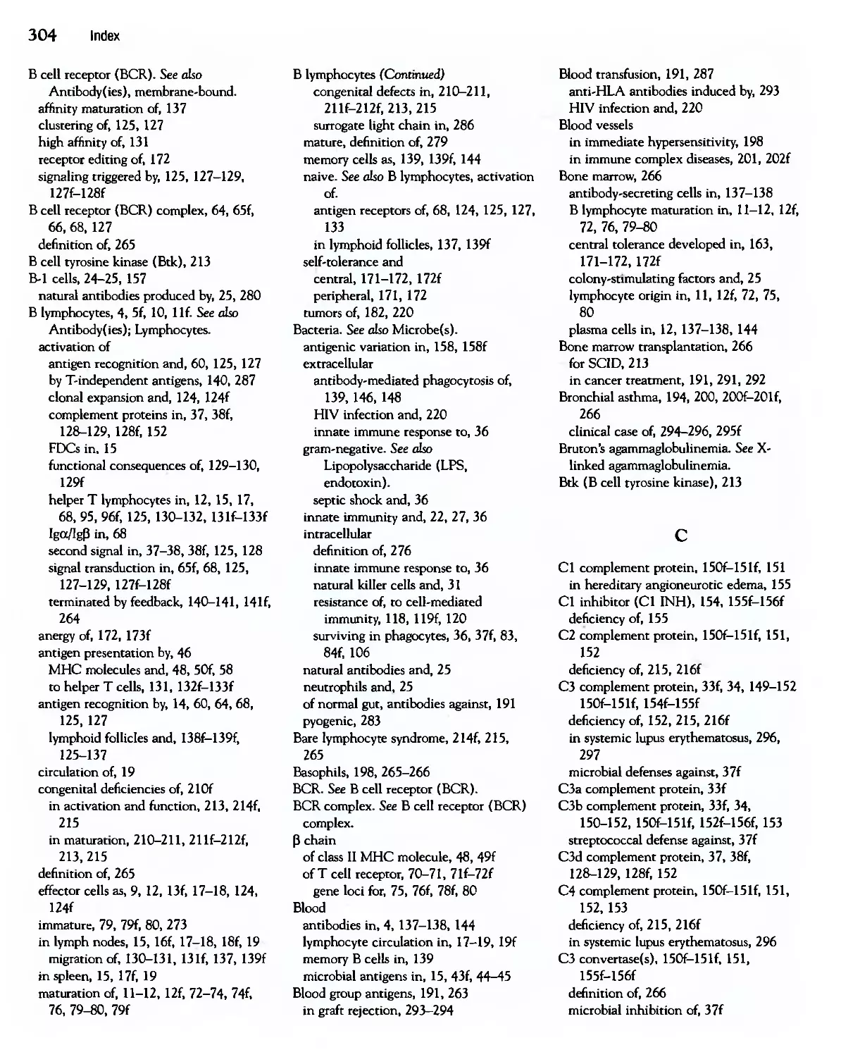
Figure 1-11 Classes of lymphocytes. Different classes of lymphocytes recognize distinct types of antigens and differen-
differentiate into effector cells whose function is to eliminate the antigens. В lymphocytes recognize soluble or cell surface antigens
and differentiate into antibody-secreting cells. Helper T lymphocytes recognize antigens on the surfaces of antigen-present-
antigen-presenting cells and secrete cytokines, which stimulate different mechanisms of immunity and inflammation. Cytolytic T lymphocytes
recognize antigens on infected cells and kill these cells. (Note that T lymphocytes recognize peptides that are displayed by
MHC molecules; this process is discussed in much more detail in Chapter 3.) Natural killer cells recognize changes on the
surface of infected cells and kill these cells.
phagocytes to destroy ingested microbes. CD8* T
lymphocytes are called cytolytic, or cytotoxic, T lym-
lymphocytes (CTLs) because they kill cells harboring
intracellular microbes, that is, they lyse other cells. A
third class of lymphocytes is called natural killer (NK)
cells; these cells are mediators of innate immunity and
do not express the kinds of clonally distributed
antigen receptors that В cells and T cells do.
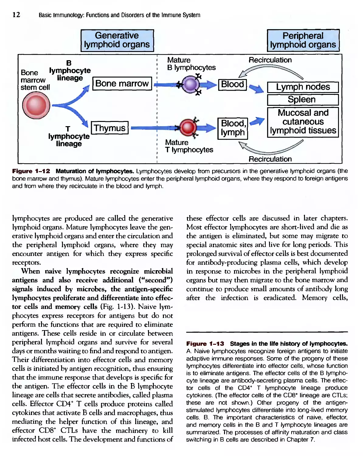
All lymphocytes arise from stem cells in the bone
marrow (Fig. 1-12). В lymphocytes mature in the
bone marrow, and T lymphocytes mature in an organ
called the thymus; these sites in which mature
12
Basic Immunology: Functions and Disorders of the Immune System
Generative
lymphoid organs
Peripheral
lymphoid organs
в
Bone lymphocyte
marrow lineage
stem cell
т ^ | Thymus
lymphocyte
lineage
Mature
В lymphocytes
Recirculation
[Blood]
Mature
T lymphocytes
Blood,
lymph
Lymph nodes
Spleen
Mucosal and
cutaneous
lymphoid tissues
Recirculation
Figure 1-12 Maturation of lymphocytes. Lymphocytes develop from precursors in the generative lymphoid organs (the
bone marrow and thymus). Mature lymphocytes enter the peripheral lymphoid organs, where they respond to foreign antigens
and from where they recirculate in the blood and lymph.
lymphocytes are produced are called the generative
lymphoid organs. Mature lymphocytes leave the gen-
generative lymphoid organs and enter the circulation and
the peripheral lymphoid organs, where they may
encounter antigen for which they express specific
receptors.
When naive lymphocytes recognize microbial
antigens and also receive additional ("second")
signals induced by microbes, the antigen-specific
lymphocytes proliferate and differentiate into effec-
effector cells and memory cells (Fig. 1-13). Naive lym-
lymphocytes express receptors for antigens but do not
perform the functions that are required to eliminate
antigens. These cells reside in or circulate between
peripheral lymphoid organs and survive for several
days or months waiting to find and respond to antigen.
Their differentiation into effector cells and memory
cells is initiated by antigen recognition, thus ensuring
that the immune response that develops is specific for
the antigen. The effector cells in the В lymphocyte
lineage are cells that secrete antibodies, called plasma
cells. Effector CD4+ T cells produce proteins called
cytokines that activate В cells and macrophages, thus
mediating the helper function of this lineage, and
effector CD8+ CTLs have the machinery to kill
infected host cells. The development and functions of
these effector cells are discussed in later chapters.
Most effector lymphocytes are short-lived and die as
the antigen is eliminated, but some may migrate to
special anatomic sites and live for long periods. This
prolonged survival of effector cells is best documented
for antibody-producing plasma cells, which develop
in response to microbes in the peripheral lymphoid
organs but may then migrate to the bone marrow and
continue to produce small amounts of antibody long
after the infection is eradicated. Memory cells,
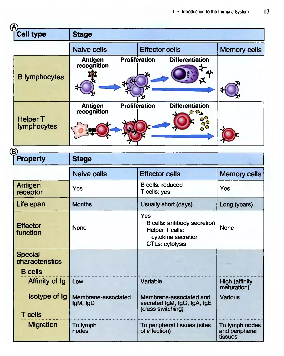
Figure 1-13 Stages in the life history of lymphocytes.
A. Naive lymphocytes recognize foreign antigens to initiate
adaptive immune responses. Some of the progeny of these
lymphocytes differentiate into effector cells, whose function
is to eliminate antigens. The effector cells of the В lympho-
lymphocyte lineage are antibody-secreting plasma cells. The effec-
effector cells of the CD4* T lymphocyte lineage produce
cytokines. (The effector cells of the CD8* lineage are CTLs;
these are not shown.) Other progeny of the antigen-
stimulated lymphocytes differentiate into long-lived memory
cells. B. The important characteristics of naive, effector,
and memory cells in the В and T lymphocyte lineages are
summarized. The processes of affinity maturation and class
switching in В cells are described in Chapter 7.
1 • Introduction to the Immune System
13
[Cell type
Stage
Naive cells
Effector cells
Memory cells
Antigen
recognition
Proliferation Differentiation
В lymphocytes
Helper T
lymphocytes
Antigen Proliferation Differentiation
recognition
t>
■is
I Property
Antigen
receptor
Life span
Effector
function
Special
characteristics
В cells
Affinity of Ig
Isotype of Ig
T cells
Migration
Stage
Naive cells
Yes
Months
None
Low
Membrane-associated
IgM, IgD
To lymph
nodes
Effector cells
В cells: reduced
T cells: yes
Usually short (days)
Yes
В cells: antibody secretion
Helper T cells:
cytokine secretion
CTLs: cytolysis
Variable
Membrane-associated and
secreted IgM, IgG, IgA, IgE
(class switching)
To peripheral tissues (sites
of infection)
Memory cells
Yes
Long (years)
None
High (affinity
maturation)
Various
To lymph nodes
and peripheral
tissues
14
Basic Immunology: Functions and Disorders of the Immune System
which are also generated from the progeny of antigen-
stimulated lymphocytes, do survive for long periods
of time in the absence of antigen. Memory cells are
functionally silent: they do not perform effector func-
functions unless stimulated by antigen. When memory
cells encounter the same antigen that induced their
development, the cells rapidly respond to give rise to
secondary immune responses. Very little is known
about the signals that generate memory cells, the
factors that determine whether the progeny of
antigen-stimulated lymphocytes will develop into
effector or memory cells, or the mechanisms that keep
memory cells alive in the absence of antigen or innate
immunity.
Antigen-Presenting Cells
The common portals of entry for microbes, namely,
the skin, gastrointestinal tract, and respiratory
tract, contain specialized cells located in the epithe-
epithelium that capture antigens and transport them to
peripheral lymphoid tissues. This function of antigen
capture is best understood for a cell type called
dendritic cells because of their long dendrite-like
processes. Dendritic cells capture protein antigens of
microbes that enter through the epithelia and trans-
transport the antigens to regional lymph nodes. Here the
antigen-bearing dendritic cells display portions of
the antigens for recognition by T lymphocytes. If a
microbe has invaded through the epithelium, it may
be phagocytosed by macrophages that live in tissues
and in various organs. Macrophages are also capable
of displaying protein antigens to T cells. The process
of antigen presentation to T cells is described in
Chapter 3.
Cells that are specialized to display antigens to T
lymphocytes have another important feature that
gives them the ability to trigger T cell responses.
These specialized cells respond to microbes by pro-
producing surface and secreted proteins that activate
naive T lymphocytes, thus providing the "second
signals" for T cell proliferation and differentiation
(see Fig. 1-9). Specialized cells that display antigens
to T cells and provide second signals are called
"professional" APCs. The prototypic professional
APCs are dendritic cells, but macrophages and a few
other cell types may serve the same function. The
importance of second signals and APCs is discussed
further in later chapters.
Much less is known about cells that may capture
antigens for display to В lymphocytes, or even if such
specialized cells exist. В lymphocytes may directly
recognize antigens of microbes, or cells in lymphoid
organs may capture antigens and deliver them to В
cells. A type of dendritic cell called the follicular den-
dendritic cell (FDC) resides in the germinal centers of
lymphoid follicles in the peripheral lymphoid organs
and displays antigens that stimulate the differentia-
differentiation of В cells in the follicles. The role of FDCs is
described in more detail in Chapter 7. FDCs do not
present antigens to T cells and are quite different from
the dendritic cells just described that function as pro-
professional APCs for T lymphocytes.
Effector Cells
The cells that eliminate microbes are called effector
cells and consist of lymphocytes and other leuko-
leukocytes. We have earlier referred to the effector cells of
the В and T lymphocyte lineages. The elimination
of microbes often requires the participation of other,
nonlymphoid leukocytes, such as granulocytes and
macrophages. These leukocytes may function as
effector cells in both innate immunity and adaptive
immunity. In innate immunity, macrophages and
some granulocytes directly recognize microbes and
eliminate them (see Chapter 2). In adaptive immu-
immunity, the products of В and T lymphocytes call in
other leukocytes and activate the leukocytes to kill
microbes.
Tissues of
the Immune System
The tissues of the immune system consist of the
generative (also called primary, or central) lymphoid
organs, in which T and В lymphocytes mature
and become competent to respond to antigens, and
the peripheral (or secondary) lymphoid organs, in
which adaptive immune responses to microbes are
initiated (see Fig. 1-12). The generative lymphoid
organs are described in Chapter 4, when we discuss
the process of lymphocyte maturation. In the follow-
following section, we highlight some of the features of
1 • Introduction to the Immune System
15
peripheral lymphoid organs that are important for the
development of adaptive immunity.
Peripheral Lymphoid Organs
The peripheral lymphoid organs, which consist of
the lymph nodes, the spleen, and the mucosal and
cutaneous immune systems, are organized to con-
concentrate antigen, APCs, and lymphocytes in a way
that optimizes interactions among these cells and
the development of adaptive immunity. The immune
system has to locate microbes that enter at any site in
the body and then respond to these microbes and
eliminate them. In addition, as we have mentioned
earlier, in the normal immune system very few T and
В lymphocytes are specific for any one antigen,
perhaps as few as 1 in 100,000 to 1 in 1 million cells.
The anatomic organization of peripheral lymphoid
organs enables lymphocytes in these organs to locate
and respond to microbes. This organization is com-
complemented by a remarkable ability of lymphocytes to
circulate throughout the body in such a way that
naive lymphocytes preferentially go to the specialized
organs in which antigen is concentrated and effector
cells go to sites of infection, from where microbes
have to be eliminated. Furthermore, different types of
lymphocytes often need to communicate to generate
effective immune responses. For instance, helper T
cells specific for an antigen interact with and
help В lymphocytes specific for the same antigen,
resulting in antibody production. An important func-
function of lymphoid organs is to bring these rare cells
together in a way that will enable them to interact
productively.
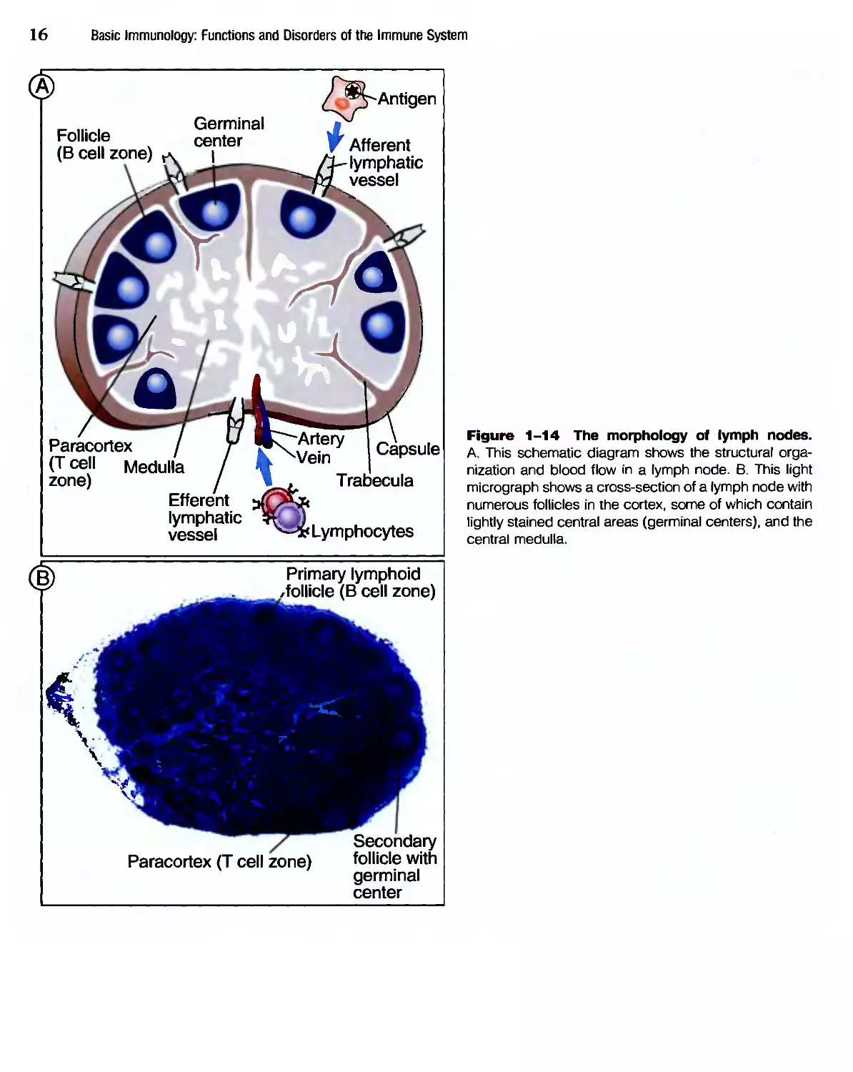
Lymph nodes are nodular aggregates of lymphoid
tissues located along lymphatic channels throughout
the body (Fig. 1-14). Fluid from all epithelia and
connective tissues and most parenchymal organs is
drained by lymphatics, which transport this fluid,
called lymph, from the tissues to the lymph nodes.
Therefore, the lymph contains a mixture of substances
that are absorbed from epithelia and tissues. As the
lymph passes through lymph nodes, APCs in the
nodes are able to sample the antigens of microbes that
may enter through epithelia into tissues. In addition,
dendritic cells pick up antigens of microbes from
epithelia and transport these antigens to the lymph
nodes. The net result of these processes of antigen
capture and transport is that the antigens of microbes
that enter through epithelia or colonize tissues
become concentrated in draining lymph nodes.
The spleen (Fig. 1-15) is an abdominal organ that
serves the same role in immune responses to blood-
borne antigen as that of lymph nodes in responses
to lymph-borne antigens. Blood entering the spleen
flows through a network of channels (sinusoids).
Blood-borne antigens are trapped and concentrated
by dendritic cells and macrophages in the spleen. The
spleen contains abundant phagocytes, which ingest
and destroy microbes in the blood.
The cutaneous and mucosal lymphoid systems are
located under the epithelia of the skin and the gas-
gastrointestinal and respiratory tracts, respectively. Pha-
ryngeal tonsils and Peyer's patches of the intestine
are two mucosal lymphoid tissues. Cutaneous and
mucosal lymphoid tissues are sites of immune
responses to antigens that breach epithelia, much as
the lymph nodes and spleen are the sites of response
to antigens that enter the lymph and blood.
Within the peripheral lymphoid organs, T lym-
lymphocytes and В lymphocytes are segregated into dif-
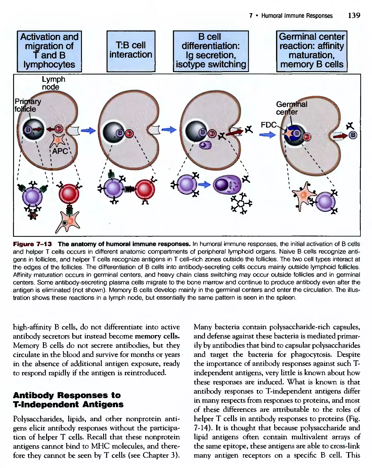
different anatomic compartments (Fig. 1-16). In lymph
nodes, the В cells are concentrated in discrete struc-
structures, called follicles, located around the periphery, or
cortex, of each node. If the В cells in a follicle have
recently responded to an antigen, this follicle may
contain a central region called a germinal center. The
role of germinal centers in the production of anti-
antibodies is described in Chapter 7. The T lymphocytes
are concentrated outside, but adjacent to, the folli-
follicles, in the paracortex. The follicles contain the FDCs
that are involved in the activation of В cells, and the
paracortex contains the dendritic cells that present
antigens to T lymphocytes. In the spleen, T lympho-
lymphocytes are concentrated in periarteriolar lymphoid
sheaths surrounding small arterioles, and В cells reside
in the follicles.
The anatomic organization of peripheral lymphoid
organs is tightly regulated to allow immune responses
to develop. В lymphocytes are located in the follicles
because FDCs secrete a protein that belongs to a class
of cytokines called chemokines ("chemoattractant
cytokines"), for which naive В cells express a recep-
receptor. (Chemokines and other cytokines are discussed in
16
Basic Immunology: Functions and Disorders of the Immune System
Follicle
(B cell zone)
Germinal
center
I
Antigen
v Afferent
/i-lymphatic
^fd. vessel
/.
/ I
Paracortex /
(T cell Medulla
zone)
Efferent
lymphatic
vessel
Capsule
Trabecula
Lymphocytes
Primary lymphoid
/follicle (B cell zone)
V
Paracortex (T cell zone)
Secondary
follicle with
germinal
center
Figure 1-14 The morphology of lymph nodes.
A. This schematic diagram shows the structural orga-
organization and blood flow in a lymph node. B. This light
micrograph shows a cross-section of a lymph node with
numerous follicles in the cortex, some of which contain
lightly stained central areas (germinal centers), and the
central medulla.
1 • Introduction to the Immune System 17
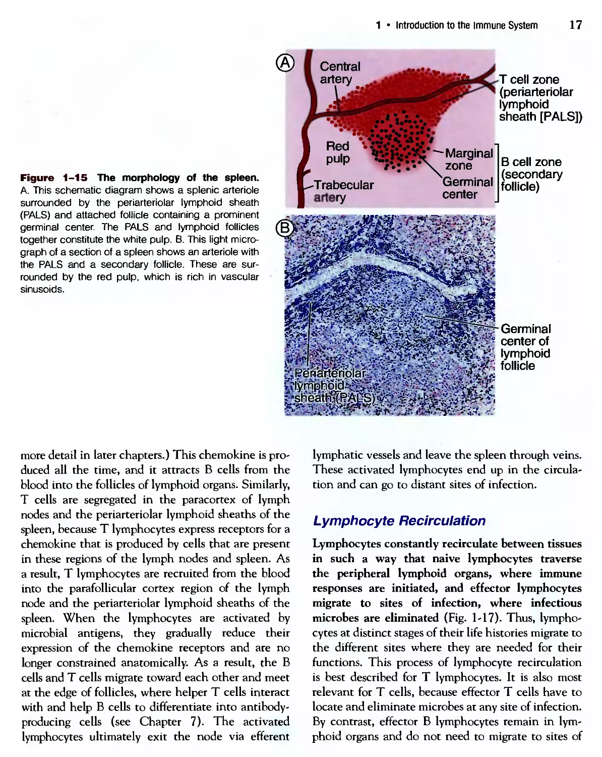
Figure 1-15 The morphology of the spleen.
A. This schematic diagram shows a splenic arteriole
surrounded by the periarteriolar lymphoid sheath
(PALS) and attached follicle containing a prominent
germinal center. The PALS and lymphoid follicles
together constitute the white pulp. B. This light micro-
micrograph of a section of a spleen shows an arteriole with
the PALS and a secondary follicle. These are sur-
surrounded by the red pulp, which is rich in vascular
sinusoids.
Central
artery
» •*.
•*V .••*.*:::v»V *
T cell zone
(periarteriolar
lymphoid
sheath [PALS])
В cell zone
(secondary
follicle)
Germinal
center of
lymphoid
follicle
more detail in later chapters.) This chemokine is pro-
produced all the time, and it attracts В cells from the
blood into the follicles of lymphoid organs. Similarly,
T cells are segregated in the paracortex of lymph
nodes and the periarteriolar lymphoid sheaths of the
spleen, because T lymphocytes express receptors for a
chemokine that is produced by cells that are present
in these regions of the lymph nodes and spleen. As
a result, T lymphocytes are recruited from the blood
into the parafollicular cortex region of the lymph
node and the periarteriolar lymphoid sheaths of the
spleen. When the lymphocytes are activated by
microbial antigens, they gradually reduce their
expression of the chemokine receptors and are no
longer constrained anatomically. As a result, the В
cells and T cells migrate toward each other and meet
at the edge of follicles, where helper T cells interact
with and help В cells to differentiate into antibody-
producing cells (see Chapter 7). The activated
lymphocytes ultimately exit the node via efferent
lymphatic vessels and leave the spleen through veins.
These activated lymphocytes end up in the circula-
circulation and can go to distant sites of infection.
Lymphocyte Recirculation
Lymphocytes constantly recirculate between tissues
in such a way that naive lymphocytes traverse
the peripheral lymphoid organs, where immune
responses are initiated, and effector lymphocytes
migrate to sites of infection, where infectious
microbes are eliminated (Fig. 1-17). Thus, lympho-
lymphocytes at distinct stages of their life histories migrate to
the different sites where they are needed for their
functions. This process of lymphocyte recirculation
is best described for T lymphocytes. It is also most
relevant for T cells, because effector T cells have to
locate and eliminate microbes at any site of infection.
By contrast, effector В lymphocytes remain in lym-
lymphoid organs and do not need to migrate to sites of
18
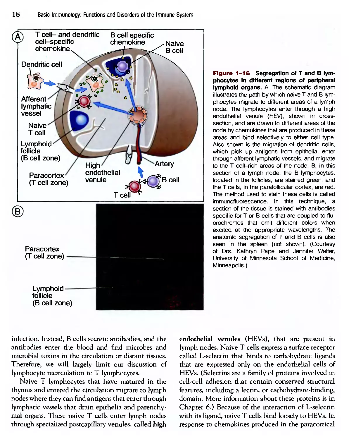
Basic Immunology: Functions and Disorders of the Immune System
Ч Т cell- and dendritic
' cell-specific
chemokine.
Dendritic cell
В cell specific
chemokine
Naive
В cell
Afferent
lymphatic
vessel
Naive
T cell •
Lymphoid
follicle
(B cell zone)
Paracortex
(T cell zone)
High
endothelial
venule
Tcell
Artery
В cell
Paracortex
(T cell zone)
Figure 1-16 Segregation of T and В lym-
lymphocytes in different regions of peripheral
lymphoid organs. A. The schematic diagram
illustrates the path by which naive T and В lym-
lymphocytes migrate to different areas of a lymph
node. The lymphocytes enter through a high
endothelial venule (HEV), shown in cross-
section, and are drawn to different areas of the
node by chemokines that are produced in these
areas and bind selectively to either cell type.
Also shown is the migration of dendritic cells,
which pick up antigens from epithelia, enter
through afferent lymphatic vessels, and migrate
to the T cell-rich areas of the node. B. In this
section of a lymph node, the В lymphocytes,
located in the follicles, are stained green, and
the T cells, in the parafollicular cortex, are red.
The method used to stain these cells is called
immunofluorescence. In this technique, a
section of the tissue is stained with antibodies
specific for T or В cells that are coupled to flu-
orochromes that emit different colors when
excited at the appropriate wavelengths. The
anatomic segregation of T and В cells is also
seen in the spleen (not shown). (Courtesy
of Drs. Kathryn Pape and Jennifer Walter,
University of Minnesota School of Medicine,
Minneapolis.)
Lymphoid
follicle
(B cell zone)
infection. Instead, В cells secrete antibodies, and the
antibodies enter the blood and find microbes and
microbial toxins in the circulation or distant tissues.
Therefore, we will largely limit our discussion of
lymphocyte recirculation to T lymphocytes.
Naive T lymphocytes that have matured in the
thymus and entered the circulation migrate to lymph
nodes where they can find antigens that enter through
lymphatic vessels that drain epithelia and parenchy-
mal organs. These naive T cells enter lymph nodes
through specialized postcapillary venules, called high
endothelial venules (HEVs), that are present in
lymph nodes. Naive T cells express a surface receptor
called L-selectin that binds to carbohydrate ligands
that are expressed only on the endothelial cells of
HEVs. (Selectins are a family of proteins involved in
cell-cell adhesion that contain conserved structural
features, including a lectin, or carbohydrate-binding,
domain. More information about these proteins is in
Chapter 6.) Because of the interaction of L-selectin
with its ligand, naive T cells bind loosely to HEVs. In
response to chemokines produced in the paracortical
1 • Introduction to the Immune System 19
| Lymph node
| Peripheral tissue
Artery
Blood
vessel
Activated
Tcell
Naive T cell
Peripheral
blood vessel
endothelial
venule
Efferent
lymphatic
vessel
Figure 1-17 Recirculation of T lymphocytes. Naive T lymphocytes migrate from the blood through high endothelial
venules (HEVs) into the T cell zones of lymph nodes, where the cells are activated by antigens. Activated T cells exit the
nodes, enter the bloodstream, and migrate preferentially to peripheral tissues at sites of infection and inflammation. The adhe-
adhesion molecules involved in the attachment of T cells to endothelial cells are described in Chapter 6.
regions of the lymph node, the naive T cells bind
more firmly to and migrate through the HEVs into
this region of the lymph nodes, where antigens are
displayed by professional APCs.
If a naive T cell encounters the antigen that it
specifically recognizes, that T cell is activated. Such
an encounter between an antigen and a specific lym-
lymphocyte is likely to be a random event, but most T
cells in the body circulate through some lymph nodes
at least once a day. As a result, some of the cells in
the total population of T lymphocytes have an excel-
excellent chance of encountering antigens that these cells
recognize. As we mentioned earlier and will describe
in more detail in Chapter 3, the likelihood of the
correct T cell finding its antigen is increased in
peripheral lymphoid organs, particularly lymph nodes,
because microbial antigens are concentrated in the
same regions of these organs through which naive T
cells circulate. In response to the microbial antigen,
the naive T cells are activated to proliferate and dif-
differentiate. During this process, the expression of
adhesion molecules and chemokine receptors on the
T cells changes such that differentiated effector T
cells tend to leave the lymph nodes and enter the
circulation. These effector cells preferentially
migrate into the tissues that are colonized by infec-
infectious microbes, where the T lymphocytes perform
their function of eradicating the infection. This
process is described in more detail in Chapter 6, where
cell-mediated immune reactions are discussed.
Memory T cell populations appear to consist of
some cells that recirculate through lymph nodes,
where they can mount secondary responses to cap-
captured antigens, and other cells that migrate to sites of
infection, where they can respond rapidly to eliminate
the infection.
We do not know much about lymphocyte circula-
circulation through the spleen or other lymphoid tissues or
about the circulation pathway of naive and activated
В lymphocytes. В lymphocytes appear also to enter
lymph nodes through HEVs, but after they respond
to antigen, their differentiated progeny either remain
in the lymph nodes or migrate mainly to the bone
marrow. The spleen does not contain HEVs, but the
general pattern of lymphocyte migration through this
organ is probably similar to migration through lymph
nodes.
SUMMARY
► The physiologic function of the immune system is
to protect individuals against infections.
20
Basic Immunology: Functions and Disorders of the Immune System
► Innate immunity is the early line of defense, medi-
mediated by cells and molecules that are always present
and ready to eliminate infectious microbes. Adaptive
immunity is the form of immunity that is stimulated
by microbes, has a fine specificity for foreign sub-
substances, and responds more effectively against each
successive exposure to a microbe.
► Lymphocytes are the cells of adaptive immunity,
and the only cells with clonally distributed receptors
for antigens.
► Adaptive immunity consists of humoral immunity,
in which antibodies neutralize and eradicate extra-
extracellular microbes and toxins, and cell-mediated
immunity, in which T lymphocytes eradicate intra-
cellular microbes.
► Adaptive immune responses consist of sequential
phases: antigen recognition by lymphocytes, activa-
activation of the lymphocytes to proliferate and to differ-
differentiate into effector and memory cells, elimination of
the microbes, decline of the immune response, and
long-lived memory.
► There are different populations of lymphocytes
that serve distinct functions and may be distinguished
by the expression of particular membrane molecules.
► В lymphocytes are the only cells that produce anti-
antibodies. В lymphocytes express membrane antibodies
that recognize antigens, and effector В cells secrete
the antibodies that neutralize and eliminate the
antigen.
► T lymphocytes recognize peptide fragments
of protein antigens displayed on other cells. Helper
T lymphocytes activate phagocytes to destroy
ingested microbes and activate В lymphocytes to
produce antibodies. Cytolytic (cytotoxic) T lympho-
lymphocytes kill infected cells harboring microbes in the
cytoplasm.
► Antigen-presenting cells capture antigens of
microbes that enter through epithelia, concentrate
these antigens in lymphoid organs, and display the
antigens for recognition by T cells.
► Lymphocytes and antigen-presenting cells are
organized in peripheral lymphoid organs, where
immune responses are initiated and develop.
► Naive lymphocytes circulate through the periph-
peripheral lymphoid organs searching for foreign antigens.
Effector T lymphocytes migrate to peripheral sites of
infection, where they function to eliminate infectious
microbes. Effector В lymphocytes remain in lymphoid
organs and the bone marrow, from where they secrete
antibodies that enter the circulation and find and
eliminate microbes.
Review Questions
1 What are the two types of adaptive immunity, and
what types of microbes do these adaptive immune
responses combat?
2 What are the principal classes of lymphocytes, how
do they differ in function, and how may they be
identified and distinguished?
3 What are the important differences among naive,
effector, and memory T and В lymphocytes?
4 Where are T and В lymphocytes located in lymph
nodes, and how is their anatomic separation main-
maintained?
5 How do naive and effector T lymphocytes differ in
their patterns of migration?
Innate Immunity
The Early Defense
Against Infections
2
Recognition of Microbes by the Innate
Immune System
Components of Innate Immunity
• Epithelial Barriers
• Phagocytes: Neutrophils and
Monocytes/Macrophages
• Natural Killer Cells
• The Complement System
• Cytokines of Innate Immunity
• Other Plasma Proteins of Innate
Immunity
Evasion of Innate Immunity by Microbes
Role of Innate Immunity in Stimulating
Adaptive Immune Responses
Summary
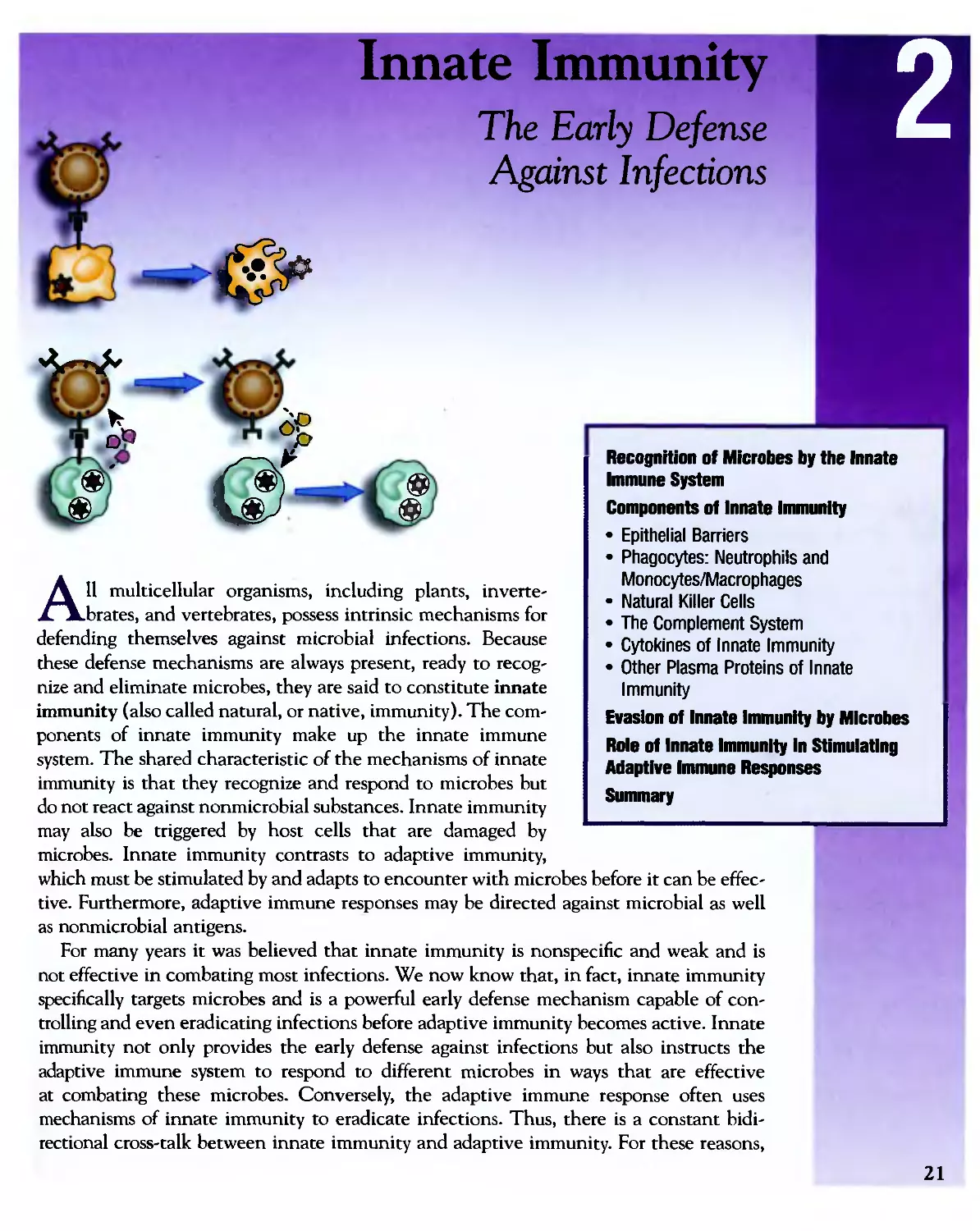
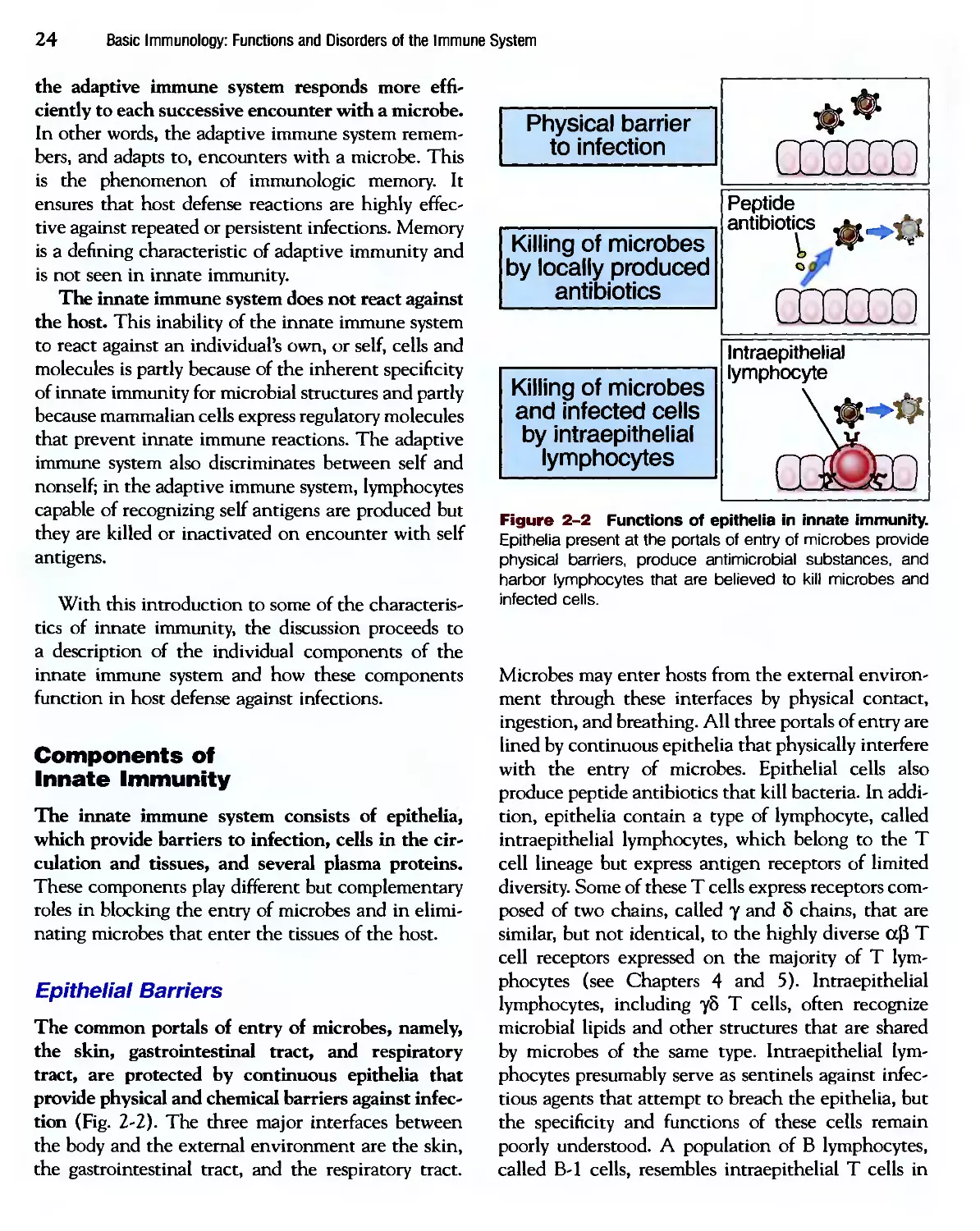
All multicellular organisms, including plants, inverte-
invertebrates, and vertebrates, possess intrinsic mechanisms for
defending themselves against microbial infections. Because
these defense mechanisms are always present, ready to recog-
recognize and eliminate microbes, they are said to constitute innate
immunity (also called natural, or native, immunity). The com-
components of innate immunity make up the innate immune
system. The shared characteristic of the mechanisms of innate
immunity is that they recognize and respond to microbes but
do not react against nonmicrobial substances. Innate immunity
may also be triggered by host cells that are damaged by
microbes. Innate immunity contrasts to adaptive immunity,
which must be stimulated by and adapts to encounter with microbes before it can be effec-
effective. Furthermore, adaptive immune responses may be directed against microbial as well
as nonmicrobial antigens.
For many years it was believed that innate immunity is nonspecific and weak and is
not effective in combating most infections. We now know that, in fact, innate immunity
specifically targets microbes and is a powerful early defense mechanism capable of con-
controlling and even eradicating infections before adaptive immunity becomes active. Innate
immunity not only provides the early defense against infections but also instructs the
adaptive immune system to respond to different microbes in ways that are effective
at combating these microbes. Conversely, the adaptive immune response often uses
mechanisms of innate immunity to eradicate infections. Thus, there is a constant bidi-
bidirectional cross-talk between innate immunity and adaptive immunity. For these reasons,
21
22
Basic Immunology: Functions and Disorders of the Immune System
there is great interest in defining the mechanisms of
innate immunity and learning how to harness these
mechanisms for optimizing defense against infections.
Most of this book is devoted to a description of the
adaptive immune system and how lymphocytes, the
cells of adaptive immunity, recognize and respond to
infectious microbes. Before starting a discussion of
adaptive immunity, the early defense reactions of
innate immunity are discussed in this chapter. The
discussion focuses on three main questions:
• How does the innate immune system recognize
microbes?
• How do the different components of innate immu-
immunity function to combat different kinds of
microbes?
• How do innate immune reactions stimulate adap-
adaptive immune response?
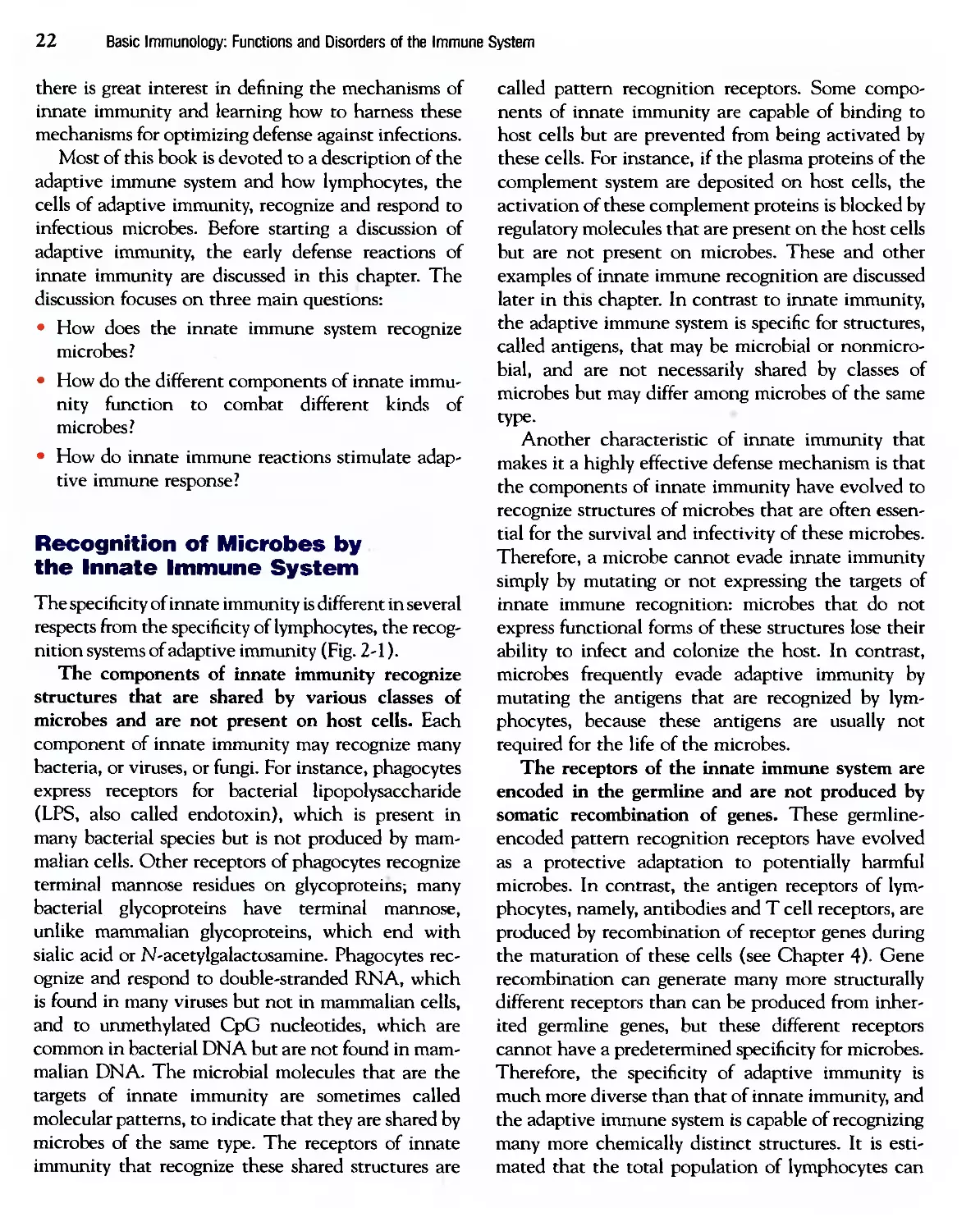
Recognition of Microbes by
the Innate Immune System
The specificity of innate immunity is different in several
respects from the specificity of lymphocytes, the recog-
recognition systems of adaptive immunity (Fig. 2-1).
The components of innate immunity recognize
structures that are shared by various classes of
microbes and are not present on host cells. Each
component of innate immunity may recognize many
bacteria, or viruses, or fungi. For instance, phagocytes
express receptors for bacterial lipopolysaccharide
(LPS, also called endotoxin), which is present in
many bacterial species but is not produced by mam-
mammalian cells. Other receptors of phagocytes recognize
terminal mannose residues on glycoproteins; many
bacterial glycoproteins have terminal mannose,
unlike mammalian glycoproteins, which end with
sialic acid or N-acetylgalactosamine. Phagocytes rec-
recognize and respond to double-stranded RNA, which
is found in many viruses but not in mammalian cells,
and to unmethylated CpG nudeotides, which are
common in bacterial DNA but are not found in mam-
mammalian DNA. The microbial molecules that are the
targets of innate immunity are sometimes called
molecular patterns, to indicate that they are shared by
microbes of the same type. The receptors of innate
immunity that recognize these shared structures are
called pattern recognition receptors. Some compo-
components of innate immunity are capable of binding to
host cells but are prevented from being activated by
these cells. For instance, if the plasma proteins of the
complement system are deposited on host cells, the
activation of these complement proteins is blocked by
regulatory molecules that are present on the host cells
but are not present on microbes. These and other
examples of innate immune recognition are discussed
later in this chapter. In contrast to innate immunity,
the adaptive immune system is specific for structures,
called antigens, that may be microbial or nonmicro-
bial, and are not necessarily shared by classes of
microbes but may differ among microbes of the same
type.
Another characteristic of innate immunity that
makes it a highly effective defense mechanism is that
the components of innate immunity have evolved to
recognize structures of microbes that are often essen-
essential for the survival and infectivity of these microbes.
Therefore, a microbe cannot evade innate immunity
simply by mutating or not expressing the targets of
innate immune recognition: microbes that do not
express functional forms of these structures lose their
ability to infect and colonize the host. In contrast,
microbes frequently evade adaptive immunity by
mutating the antigens that are recognized by lym-
lymphocytes, because these antigens are usually not
required for the life of the microbes.
The receptors of the innate immune system are
encoded in the germline and are not produced by
somatic recombination of genes. These germline-
encoded pattern recognition receptors have evolved
as a protective adaptation to potentially harmful
microbes. In contrast, the antigen receptors of lym-
lymphocytes, namely, antibodies and T cell receptors, are
produced by recombination of receptor genes during
the maturation of these cells (see Chapter 4). Gene
recombination can generate many more structurally
different receptors than can be produced from inher-
inherited germline genes, but these different receptors
cannot have a predetermined specificity for microbes.
Therefore, the specificity of adaptive immunity is
much more diverse than that of innate immunity, and
the adaptive immune system is capable of recognizing
many more chemically distinct structures. It is esti-
estimated that the total population of lymphocytes can
Innate Immunity
23
Innate immunity
Adaptive immunity
Specificity
For structures shared by classes of
microbes ("molecular patterns")
For structural detail of microbial
molecules (antigens);
may recognize nonmicrobial
antigens
Different
microbes
Identical
mannose
receptors
Different
microbes-
Distinct S
antibody
molecules
Receptors
Encoded in germline; limited diversity
Encoded by genes produced by
somatic recombination of gene
segments; greater diversity
Toll-like
receptor
/V-formyl
methionyl
receptor
Mannose
receptor
TCR
ig
Distribution
of receptors
Nonclonal: identical receptors on
all cells of the same lineage
Cjonal: clones of lymphocytes
with distinct specificities express
different receptors
Discrimination
of self and
nonself
Yes; host cells are not recognized or they
may express molecules that prevent innate
immune reactions
Yes; based on selection against
self-reactive lymphocytes; may
be imperfect (giving rise to
autoimmunity)
Figure 2-1 The specificity of innate immunity and adaptive immunity. The important features of the specificity and
receptors of innate and adaptive immunity are summarized, with selected examples, some of which are illustrated in the boxed
panels.
recognize over a billion different antigens; in contrast,
all the receptors of innate immunity probably recog-
recognize less than a thousand microbial patterns. Further-
Furthermore, the receptors of the adaptive immune system
are clonally distributed, meaning that each clone of
lymphocytes (B cells and T cells) has a different
receptor specific for a particular antigen. In contrast,
in the innate immune system the receptors are non-
clonally distributed; that is, identical receptors are
expressed on all the cells of a particular type, such as
macrophages. Therefore, many cells of innate immu-
immunity may recognize the same microbe.
The innate immune system responds in the same
way to repeat encounters with a microbe, whereas
24 Basic Immunology: Functions and Disorders of the Immune System
the adaptive immune system responds more effi-
efficiently to each successive encounter with a microbe.
In other words, the adaptive immune system remem-
remembers, and adapts to, encounters with a microbe. This
is the phenomenon of immunologic memory. It
ensures that host defense reactions are highly effec-
effective against repeated or persistent infections. Memory
is a defining characteristic of adaptive immunity and
is not seen in innate immunity.
The innate immune system does not react against
the host. This inability of the innate immune system
to react against an individual's own, or self, cells and
molecules is partly because of the inherent specificity
of innate immunity for microbial structures and partly
because mammalian cells express regulatory molecules
that prevent innate immune reactions. The adaptive
immune system also discriminates between self and
nonself; in the adaptive immune system, lymphocytes
capable of recognizing self antigens are produced but
they are killed or inactivated on encounter with self
antigens.
With this introduction to some of the characteris-
characteristics of innate immunity, the discussion proceeds to
a description of the individual components of the
innate immune system and how these components
function in host defense against infections.
Components of
Innate Immunity
The innate immune system consists of epithelia,
which provide barriers to infection, cells in the cir-
circulation and tissues, and several plasma proteins.
These components play different but complementary
roles in blocking the entry of microbes and in elimi-
eliminating microbes that enter the tissues of the host.
Epithelial Barriers
The common portals of entry of microbes, namely,
the skin, gastrointestinal tract, and respiratory
tract, are protected by continuous epithelia that
provide physical and chemical barriers against infec-
infection (Fig. 2-2). The diree major interfaces between
the body and the external environment are the skin,
the gastrointestinal tract, and the respiratory tract.
Physical barrier
to infection
Killing of microbes
by locally produced
antibiotics
Killing of microbes
and infected cells
by intraepithelial
lymphocytes
QXCDD
Peptide
antibiotics
l
СГШХ)
Intraepithelial
lymphocyte
Figure 2-2 Functions of epithelia in innate immunity.
Epithelia present at the portals of entry of microbes provide
physical barriers, produce antimicrobial substances, and
harbor lymphocytes that are believed to kill microbes and
infected cells.
Microbes may enter hosts from the external environ-
environment through these interfaces by physical contact,
ingestion, and breathing. All three portals of entry are
lined by continuous epithelia that physically interfere
with the entry of microbes. Epithelial cells also
produce peptide antibiotics that kill bacteria. In addi-
addition, epithelia contain a type of lymphocyte, called
intraepithelial lymphocytes, which belong to the T
cell lineage but express antigen receptors of limited
diversity. Some of these T cells express receptors com-
composed of two chains, called у and 6 chains, that are
similar, but not identical, to the highly diverse сф Т
cell receptors expressed on the majority of T lym-
lymphocytes (see Chapters 4 and 5). Intraepithelial
lymphocytes, including y5 T cells, often recognize
microbial lipids and other structures that are shared
by microbes of the same type. Intraepithelial lym-
lymphocytes presumably serve as sentinels against infec-
infectious agents that attempt to breach the epithelia, but
the specificity and functions of these cells remain
poorly understood. A population of В lymphocytes,
called B-l cells, resembles intraepithelial T cells in
2 • Innate Immunity
25
the limited diversity of their antigen receptors. B-l
cells are found not in epithelia but mostly in the peri-
peritoneal cavity, where they may respond to microbes
and microbial toxins that pass through the walls of
the intestine. Most of the circulating IgM antibodies
found in the blood of normal individuals, called
natural antibodies, are the products of B-l cells, and
many of these antibodies are specific for carbohydrates
that are present in the cell walls of many bacteria.
Phagocytes: Neutrophils and
Monocytes/Macrophages
The two types of circulating phagocytes, neu-
neutrophils and monocytes, are blood cells that are
recruited to sites of infection, where they recognize
and ingest microbes for intracellular killing. Neu-
Neutrophils (also called polymorphonuclear leukocytes
or PMNs) are the most abundant leukocytes in the
blood, numbering 4000 to 10,000 per mm' (Fig. 2-3).
In response to infections, the production of neu-
neutrophils from the bone marrow increases rapidly, and
their number may rise to 20,000 per mm' of blood.
The production of neutrophils is stimulated by
cytokines, known as colony-stimulating factors, that
are produced by many cell types in response to infec-
infections and act on bone marrow stem cells to stimulate
proliferation and maturation of neutrophil precursors.
Neutrophils are the first cell type to respond to most
infections, particularly bacterial and fungal infections.
They ingest microbes in the circulation, and they
rapidly enter extravascular tissues at sites of infection,
Figure 2-3 Morphology of neutrophils. The light micro-
micrograph of a blood neutrophil shows the multilobed nucleus,
because of which these cells are also called polymorphonu-
polymorphonuclear leukocytes, and the faint cytoplasmic granules (mostly
lysosomes).
where they also ingest microbes and die after a few
hours.
Monocytes are less abundant than neutrophils,
numbering 500 to 1000 per mm' of blood (Fig. 2-4).
They, too, ingest microbes in the blood and in tissues.
Unlike neutrophils, monocytes that enter extravascu-
extravascular tissues survive in these sites for long periods; in the
tissues, these monocytes differentiate into cells called
macrophages (see Fig. 2-4). Blood monocytes and
tissue macrophages are two stages of the same cell
lineage, which is often called the mononuclear phago-
phagocyte system. Resident macrophages are found in con-
connective tissues and in every organ in the body, where
they serve the same function as mononuclear phago-
phagocytes newly recruited from the circulation.
Neutrophils and monocytes migrate to extravas-
extravascular sites of infection by binding to endothelial
adhesion molecules and in response to chemoat-
tractants that are produced on encounter with
microbes (Fig. 2-5). If an infectious microbe breaches
an epithelium and enters the subepithelial tissue, res-
resident macrophages recognize the microbe and respond
by producing soluble proteins called cytokines
(described in more detail later). Two of these
cytokines, called tumor necrosis factor (TNF) and
interleukin-1 (IL-1), act on the endothelium of small
vessels at the site of infection. These cytokines
stimulate the endothelial cells to rapidly express two
adhesion molecules called E-selectin and P-selectin
(the name "selectin" referring to the carbohydrate-
binding, or lectin, property of these molecules). Cir-
Circulating neutrophils and monocytes express surface
carbohydrates that bind weakly to the selectins. The
neutrophils become tethered to the endothelium,
flowing blood disrupts this binding, the bonds re-form
downstream, and so on, resulting in the rolling of the
leukocytes on the endothelial surface. Leukocytes
express another set of adhesion molecules that are
called integrins because they "integrate" extrinsic
signals into cytoskeletal alterations. Integrins are
present in a low-affinity state on unactivated leuko-
leukocytes. As these cells are rolling on the endothe-
endothelium, tissue macrophages that encountered the
microbe, and the endothelial cells responding to
the macrophage-derived TNF and IL-1, produce
cytokines called chemokines (chemoattractant
cytokines). Chemokines bind to the luminal surface
26
Basic Immunology: Functions and Disorders of the Immune System
Bone
marrow
stem cell
Blood
monocyte
Microglia (CNS)
Kupffer cells (liver)
Differentiation Alveolar
macrophages (lung)
Osteoclasts (bone)
Tissue
macrophage
Activated
macrophage
Activation
* ■•
' <" * л -2-
*•/ т
• ./^ *!&::. ;.л^.
* •.
Figure 2-4 Stages in the maturation of mononuclear phagocytes. Mononuclear phagocytes arise from precursors in the
bone marrow. The circulating blood stage is the monocyte; a light micrograph and an electron micrograph of a blood mono-
monocyte are shown, illustrating the phagocytic vacuoles and lysosomes. In the tissues, these cells become macrophages; they
may be activated by microbes, and they may differentiate into specialized forms that are resident in different tissues. The
electron micrograph of a portion of an activated macrophage shows numerous phagocytic vacuoles and cytoplasmic
organelles. (From Fawcett DW. Bloom and Fawcett Textbook of Histology, 12th ed. WB Saunders, Philadelphia, 1994.)
endothelial cells and are thus displayed at a high con-
concentration to the leukocytes that are rolling on the
endothelium. These chemokines stimulate a rapid
increase in the affinity of the leukocyte integrins for
their ligands on the endothelium. Concurrently, TNF
and IL-1 act on the endothelium to stimulate expres-
expression of ligands for integrins. The firm binding of inte-
integrins to their ligands arrests the rolling leukocytes on
the endothelium. The cytoskeleton of the leukocytes
is reorganized, and the cells spread out on the
endothelial surface. Chemokines also stimulate the
motility of leukocytes. As a result, the leukocytes
begin to migrate through the vessel wall and along
the chemokine concentration gradient to the site of
infection. The sequence of selectin-mediated rolling,
integrin-mediated firm adhesion, and chemokine-
mediated motility leads to the migration of blood
leukocytes to an extravascular site of infection within
minutes after the infection. (As we shall see in
Chapter 6, the same sequence of events is responsible
for the migration of activated T lymphocytes into
infected tissues.) The accumulation of leukocytes at
sites of infection, with attendant vascular dilatation
and increased vascular permeability, is called inflam-
inflammation. Inherited deficiencies in integrins and
selectin ligands lead to defective leukocyte recruit-
recruitment to sites of infection and increased susceptibility
to infections. These disorders are called leukocyte
adhesion deficiencies.
Neutrophils and macrophages recognize microbes
in the blood and extravascular tissues by surface
receptors that are specific for microbial products
(Fig. 2-6). There are several different types of recep-
receptors, specific for different structures or patterns that
2 • Innate Immunity
27
Rolling
Integrin
activation
Stable
adhesion
Migration through
endothelium
Leukocyte
Integrin
' (low-affinity state)
-Selectin ligand
Chemokine
Integrin (high-
affinity state)
Macrophage
with microbes
z>
Proteo-
glycan
Cytokines
(TNF, IL-1)
Fibrin and fibronectin
(extracellular matrix)
Figure 2-5 The sequence of events in the migration of blood leukocytes to sites of infection. At sites of infection,
macrophages that have encountered microbes produce cytokines (e.g., TNF and IL-1) that activate the endothelial cells of
nearby venules to produce selectins. ligands for integrins. and chemokines. Selectins mediate weak tethering and rolling of
blood neutrophils on the endothelium; integrins mediate firm adhesion of neutrophils; and chemokines activate the neutrophils
and stimulate their migration through the endothelium to the site of infection. Blood monocytes and activated T lymphocytes
use the same mechanisms to migrate to sites of infection.
are frequently found on microbial molecules. Toll-like
receptors (TLRs) are homologous to a Drosophila
protein called Toll, which is essential for protecting
the flies against infections. TLRs are specific for dif-
different components of microbes. For instance, TLR-2
is essential for macrophage responses to several bac-
bacterial lipoglycans, TLR-4 for bacterial lipopolysac-
charide (LPS, or endotoxin), TLR-5 for a component
of bacterial flagella called flagellin, and TLR-9 for
unmethylated CpG nucleotides also found in bacte-
bacteria. Signals generated by engagement of TLRs activate
a transcription factor called NF-кВ (nuclear factor
kB), which stimulates production of cytokines,
enzymes, and other proteins involved in the anti-
antimicrobial functions of activated phagocytes (discussed
later). Neutrophils and macrophages express receptors
that recognize other microbial structures and that
promote phagocytosis and killing of the microbes.
These receptors include one that recognizes N-
formylmethionine-containing peptides (produced by
microbes but not host cells), mannose receptors
(mentioned earlier), integrins (mainly one called
Mac-1), and scavenger receptors (specific for several
pathogen and host molecules). Macrophages also
express receptors for cytokines, such as interferon-y
(IFN-y), which are produced during innate and adap-
adaptive immune responses. IFN-y is a powerful activator
of the microbicidal functions of phagocytes. In addi-
addition, phagocytes express receptors for products of
complement activation and for antibodies, and these
receptors avidly bind microbes that are coated with
complement proteins or antibodies (the latter only in
28
Basic Immunology: Functions and Disorders of the Immune System
Chemokines
/V-formylmethionyl
peptides
Recognition
of microbes,
mediators
Cellular
response
Functional
outcomes
Lipid
mediators
Microbe
LPS
CD14
Seven a-helical
transmembrane
receptors
Toll-like
receptor
Mannose
receptor
Increased integrin
avidity; cytoskeletal
changes
Production of cytokines;
reactive oxygen
intermediate (ROIs)
Phagocytosis
of microbe into
phagosome
Migration
into tissues
Killing of
microbes
Killing of
microbes
Figure 2-6 Receptors and responses of phagocytes. Neutrophils and macrophages use diverse membrane receptors
to recognize microbes, microbial products, and substances produced by the host in infections. These receptors activate cel-
cellular responses that function to stimulate inflammation and eradicate microbes. Note that only selected examples of receptors
of different classes are shown. LPS, lipopolysaccharide.
adaptive immunity). The process of coating microbes
for efficient recognition by phagocytes is called
opsonization.
The recognition of microbes by neutrophils and
macrophages leads to phagocytosis of the microbes
and activation of the phagocytes to kill the ingested
microbes (Fig. 2-7). Phagocytosis is a process in which
the phagocyte extends its plasma membrane around
the recognized microbe, the membrane closes up and
pinches off, and the particle is internalized in a mem-
membrane-bound vesicle, called a phagosome. The phago-
somes fuse with lysosomes to form phagolysosomes.
At the same time as the microbe is being bound by
the phagocyte's receptors and ingested, the receptors
deliver signals that activate several enzymes in the
phagolysosomes. One of these enzymes, called phago-
phagocyte oxidase, converts molecular oxygen into super-
oxide anion and free radicals. These substances are
called reactive oxygen intermediates (ROIs), and
they are toxic to the ingested microbes. A second
enzyme, called inducible nitric oxide synthase,
catalyzes the conversion of arginine to nitric oxide
(NO), also a microbicidal substance. The third set of
enzymes are lysosomal proteases, which break down
microbial proteins. All these microbicidal substances
are produced mainly within lysosomes and phagolyso-
phagolysosomes, where they act on the ingested microbes but
do not damage the phagocytes. In strong reactions,
the same enzymes may be liberated into the extracel-
extracellular space and may injure host tissues. This is the
reason why inflammation, normally a protective host
response to infections, may cause tissue injury as well.
Inherited deficiency of the phagocyte oxidase enzyme
is the cause of an immunodeficiency disease called
chronic granulomatous disease. In this disorder,
phagocytes are unable to eradicate intracellular
2 * Innate Immunity
29
Figure 2-7 Phagocytosis and
intracellular killing of microbes.
Macrophages and neutrophils
express many surface receptors
that may bind microbes for sub-
subsequent phagocytosis; selected
examples of such receptors are
shown. Microbes are ingested
into phagosomes, which fuse with
lysosomes, and the microbes are
killed by enzymes and several
toxic substances produced in the
phagolysosomes. The same sub-
substances may be released from the
phagocytes and may kill extracel-
extracellular microbes (not shown). iNOS,
inducible nitric oxide synthase;
NO, nitric oxide; ROI, reactive
oxygen intermediate.
Microbes bind to
phagocyte receptors
Mac-1
integrin
Mannose
receptor
Phagocyte
membrane zips up
around microbe
Scavenger
receptor
Microbe ingested
in phagosome
Fusion of
phagosome
with lysosome
Phagosome
with ingested £
microbe enzymes
Activation of
phagocyte
Phagolysosome
Killing of
microbes by
lysosomal
enzymes in
phagolysosomes
Phagocyte
oxidase
Killing of
phagocytosed
microbes by
ROIs and NO
30
Basic Immunology: Functions and Disorders of the Immune System
microbes, and the host tries to contain the infection
by calling in more macrophages and lymphocytes,
resulting in collections of cells around the microbes
that are called granulomas.
In addition to killing phagocytosed microbes,
macrophages perform several functions that play
important roles in defense against infections (Fig.
2-8). Macrophages produce cytokines that are impor-
important mediators of host defense (see later discussion).
Macrophages secrete growth factors and enzymes that
serve to remodel injured tissue and replace it with
connective tissue. Macrophages also stimulate T lym-
lymphocytes and respond to products of T cells: these
reactions are important in cell-mediated immunity
and are described in Chapter 6.
Natural Killer Cells
Natural killer (NK) cells are a class of lymphocytes
that respond to intracellular microbes by killing
infected cells and by producing the macrophage-
activating cytokine, IFN-y (Fig. 2-9). Natural killer
Microbe
Toll-like
receptor-4
Q IFN-Y
Q IFN-y
receptor
Molecules
produced
in activated
macrophages
Effector
functions
of activated
macrophages
I
Phagocyte
oxidase
Reactive
oxygen
intermediates
(ROIs)
Cytokines
(TNF, IL-12)
Nitric
oxide
Fibroblast
growth factors,
angiogenic factors,
metalloproteinases
Increased MHC
molecules,
costimulators
4 f
¥
Killing of
microbes
Inflammation,
enhanced
adaptive
immunity
Tissue
remodeling
Enhanced
antigen
presentation
Figure 2-8 Functions of activated macrophages. Macrophages may be activated by signals from many surface recep-
receptors. The two examples shown are the receptor for bacterial endotoxin (LPS), which transduces signals via an attached Toll-
like receptor, and the receptor for the most important macrophage-activating cytokine, IFN-y. Signals from activating receptors
stimulate the production of several proteins, which mediate the important functions of macrophages. Different macrophage
surface receptors may stimulate distinct or overlapping responses. The biochemical signaling pathways used by these recep-
receptors are complex; their common feature is that they stimulate the production of transcription factors, which result in the pro-
production of various proteins.
2 • Innate Immunity
31
NK cell
Virus-infected
cell
Killing of
infected cells
Macrophage
with
phagocytosed
microbes
Killing of
phagocytosed
microbes
Figure 2-9 Functions of natural killer (NK) cells. A. NK
cells kill host cells infected by intracellular microbes, thus
eliminating reservoirs of infection. B. NK cells respond to IL-
12 produced by macrophages and secrete IFN-y, which acti-
activates the macrophages to kill phagocytosed microbes.
cells comprise about 10% of the lymphocytes in the
blood and peripheral lymphoid organs. These cells
contain abundant cytoplasmic granules and express
characteristic surface markers, but they do not express
immunoglobulins or T cell receptors, the antigen
receptors of В and T lymphocytes, respectively. NK
cells recognize host cells that have been altered by
microbial infection. Although the mechanisms of NK
recognition are incompletely understood, it is known
that NK cells express various receptors for molecules
on host cells, and some of these receptors activate the
NK cells and some inhibit the NK cells. Among the
activating receptors are those that recognize cell
surface molecules that are commonly expressed on
host cells infected with viruses and on phagocytes har-
harboring viruses and intracellular bacteria. Other acti-
activating NK cell receptors recognize normal host cell
surface molecules, which could theoretically activate
NK cells to kill normal cells. This does not usually
occur because NK cells also express inhibitory recep-
receptors that recognize normal host cells and inhibit the
activation of the NK cells. These inhibitory receptors
are specific for various alleles of self class I major his-
tocompatibility complex (MHC) molecules, which
are proteins expressed on all nucleated cells in every
individual. (In Chapter 3 the important function
of MHC molecules in displaying peptide antigens
to T lymphocytes is described.) Two major families
of NK cell inhibitory receptors are the killer cell
immunoglobulin-like receptors (KIRs), so called
because they share structural homology to immuno-
globulin molecules (described in Chapter 4), and
receptors consisting of a protein called CD94 and a
lectin subunit called NKG2. Both families of
inhibitory receptors contain in their cytoplasmic
domains structural motifs called immunoreceptor
tyrosine-based inhibitory motifs (ITIMs), which
become phosphorylated on tyrosine residues when the
receptors bind class I MHC molecules. The phospho-
phosphorylated ITIMs bind and promote the activation of
cytoplasmic protein tyrosine phosphatases. These
phosphatases remove phosphate groups from the tyro-
tyrosine residues of various signaling molecules and thus
block the activation of NK cells through activating
receptors. Therefore, when the inhibitory receptors of
NK cells encounter self MHC molecules, the NK cells
are shut off (Fig. 2-10). Many viruses have mecha-
mechanisms to block expression of class I molecules in
infected cells, which allows them to evade killing by
virus-specific CD8* cytolytic T lymphocytes (CTLs)
(see Chapter 6). When this happens, the NK cell
inhibitory receptors are not engaged, and they
become activated to eliminate cells infected by such
viruses. The ability of NK cells to protect against
infections is enhanced by cytokines secreted by
macrophages that have encountered microbes. One of
these NK-activating cytokines produced by macro-
macrophages is called interleukin-12 (IL-12). Natural killer
cells also express receptors for the Fc portions of some
IgG antibodies and use these receptors to bind to cells
coated with antibodies. The role of this reaction in
antibody-mediated humoral immunity is described in
Chapter 8.
When NK cells are activated, they respond in two
ways (see Fig. 2-9). First, activation triggers the dis-
discharge of proteins contained in the NK cells' cyto-
32
Basic Immunology: Functions and Disorders of the Immune System
(A) Inhibitory receptor engaged
NK cell
Activating
receptor
Ligand
forNK
cell
Normal
autologous cell
Inhibitory
receptor
Self class I
MHC-self
peptide complex
) Inhibitory receptor not engaged
,NKcell
NK cell
not activated;
no cell killing
Virus inhibits
class I MHC
expression
Virus-infected
cell (class I
MHC negative)
NK cell
activated;
killing of
infected cell
Figure 2-10 The function of
inhibitory receptors of NK cells.
A. The inhibitory receptors of NK cells
recognize self class I MHC mole-
molecules, thus ensuring that NK cells do
not attack normal host cells (which
always express class I MHC mole-
molecules containing bound self pep-
tides). B. NK cells are activated by
infected cells in which class I MHC
expression is reduced, because the
inhibitory receptors are not engaged
in the absence of class I MHC mol-
molecules. The result is that the infected
cells are killed.
plasmic granules toward the infected cells. These NK
cell granule proteins include molecules that create
holes in the plasma membrane of the infected cells
and other molecules that enter the infected cells
and activate enzymes that induce apoptotic death.
The cytolytic mechanisms of NK cells are the same
as the mechanisms used by CTLs to kill infected
cells (see Chapter 6). The net result of these reactions
is that NK cells kill infected host cells. By killing
infected host cells, NK cells, like CTLs, function
to eliminate cellular reservoirs of infection and
thus eradicate infections by obligate intracellular
microbes, such as viruses. Second, activated NK cells
synthesize and secrete the cytokine, IFN-y. IFN-y
activates macrophages to become more effective
at killing phagocytosed microbes. Thus, NK cells and
macrophages function cooperatively to eliminate
intracellular microbes: macrophages ingest microbes
and produce 1L-12, IL-12 activates NK cells to secrete
IFN-y, and IFN-y in turn activates the macrophages
to kill the ingested microbes. As is discussed in
Chapter 6, essentially the same sequence of reactions
involving macrophages and T lymphocytes is central
to the cell-mediated arm of adaptive immunity.
Thus, hosts and microbes are engaged in a constant
evolutionary struggle: the host uses CTLs to recognize
MHC-displayed viral antigens, viruses shut off MHC
expression, and NK cells have evolved to respond to
the absence of MHC molecules. Whether the host or
the microbe wins this kind of evolutionary struggle,
of course, determines the outcome of the infections.
The Complement System
The complement system is a collection of circulating
and membrane-associated proteins that are important
in defense against microbes. Many complement pro-
proteins are proteolytic enzymes, and complement acti-
activation involves the sequential activation of these
enzymes, sometimes called an enzymatic cascade. The
complement cascade may be activated by one of three
pathways (Fig. 2-11). The alternative pathway is trig-
triggered when some complement proteins are activated
on microbial surfaces and cannot be controlled
because complement regulatory proteins are not
present on microbes (but are present on host cells).
This pathway is a component of innate immunity.
The classical pathway is triggered after antibodies
2 • Innate Immunity
33
Initiation of
complement
activation
Early steps
Late steps
Effector
functions
Alternative! Classical • Lectin
pathway ! pathway ! pathway
Mannose
binding
lectin
C3a:
Inflammation
C3b:
opsonization
and
phagocytosis
C3b is
deposited
on microbe
C5a:
Inflammation
Complement
proteins form
membrane pore
Lysis of
microbe
Figure 2-11 Pathways of complement activation. The activation of the complement system may be initiated by three
distinct pathways, all of which lead to the production of C3b (the early steps). C3b initiates the late steps of complement
activation, culminating in the production of numerous peptides and polymerized C9 (which forms the "membrane attack
complex," so called because it creates holes in plasma membranes). The principal functions of proteins produced at differ-
different steps are shown. The activation, functions, and regulation of the complement system are discussed in much more detail
in Chapter 8.
34
Basic Immunology: Functions and Disorders of the Immune System
bind to microbes or other antigens and is thus a com-
component of the humoral arm of adaptive immunity. The
lectin pathway is activated when a plasma protein,
mannose-binding lectin, binds to terminal mannose
residues on the surface glycoproteins of microbes. This
lectin activates proteins of the classical pathway, but
because it is initiated in the absence of antibody it is
a component of innate immunity. Activated comple-
complement proteins function as proteolytic enzymes to
cleave other complement proteins. The central com-
component of complement is a plasma protein called C3,
which is cleaved by enzymes generated in the early
steps. The major proteolytic fragment of C3, called
C3b, becomes covalently attached to microbes and
is able to activate downstream complement proteins
on the microbial surface. The three pathways of com-
complement activation differ in how they are initiated,
but they share the late steps and perform the same
effector functions.
The complement system serves three functions in
host defense. First, C3b coats microbes and promotes
the binding of these microbes to phagocytes, by
virtue of receptors for C3b that are expressed on the
phagocytes. Second, some breakdown products of
complement proteins are chemoattractants for neu-
trophils and monocytes and promote inflammation
at the site of complement activation. Third, com-
complement activation culminates in the formation of
a polymeric protein complex that inserts into the
microbial cell membrane, forming pores that lead to
the influx of water and ions and death of the microbe.
A more detailed discussion of the activation and
functions of complement is in Chapter 8, where
the effector mechanisms of humoral immunity are
considered.
Cytokines of Innate Immunity
In response to microbes, macrophages and other
cells secrete proteins called cytokines that mediate
many of the cellular reactions of innate immunity
(Fig. 2-12). Cytokines are soluble proteins that
mediate immune and inflammatory reactions and are
responsible for communications between leukocytes
and between leukocytes and other cells. Most of the
molecularly defined cytokines are called interleukins,
by convention, implying that these molecules are pro-
produced by leukocytes and act on leukocytes. (In reality,
this is too limited a definition, because many
cytokines are produced by or act on cells other than
leukocytes, and many cytokines that fulfill these
criteria are given other names for historical reasons.)
In innate immunity, the principal sources of cyto-
cytokines are macrophages activated by recognition of
microbes. For instance, binding of LPS to its receptor
on macrophages is a powerful stimulus for cytokine
secretion by the macrophages. Bacteria elicit much
the same response via other macrophage receptors,
many of which are members of the Toll-like receptor
family. Cytokines are also produced in cell-mediated
immunity. In this type of adaptive immunity, the
major sources of cytokines are helper T lymphocytes
(see Chapter 5).
All cytokines are produced in small amounts in
response to an external stimulus, such as a microbe.
Cytokines bind to high-affinity receptors on target
cells. Most cytokines act on the cells that produce
them (called autocrine actions) or on adjacent cells
(paracrine actions). In innate immune reactions
against infections, enough macrophages may be acti-
activated that large amounts of cytokines are produced,
and they may be active distant from their site of
secretion.
The cytokines of innate immunity serve various
functions in host defense. As discussed earlier in this
chapter, TNF, IL-1, and chemokines are the principal
Figure 2-12 Cytokines of innate immunity. A. Macro-
Macrophages responding to microbes produce cytokines that
stimulate inflammation (leukocyte recruitment) and activate
NK cells to produce the macrophage-activating cytokine
IFN-y. B. Some important characteristics of the major
cytokines of innate immunity are listed. Note that IFN-y is a
cytokine of both innate and adaptive immunity and is referred
to again in Chapter 5. The name "tumor necrosis factor"
(TNF) arose from an experiment showing that a cytokine
induced by LPS killed tumors in mice. We now know that this
effect is the result of TNF-induced thrombosis of tumor blood
vessels, which is an exaggerated form of a reaction seen
in inflammation. The name "interferon" arose from the ability
of these cytokines to interfere with viral infection. IFN-y is a
weak antiviral cytokine compared with the type I IFNs.
2 • Innate Immunity
35
Activation of
macrophages
and NK cells
Microbes
Natural
killer cell
TNF.IL-1,
chemokines
Macrophage
(Й) Cytokine
Principal
cell source(s)
Principal cellular targets
and biologic effects
Tumor necrosis
factor (TNF)
Macrophages, T cells
Endothelial cells: activation (inflammation,
coagulation)
Neutrophils: activation
Hypothalamus: fever
Liver: synthesis of acute phase proteins
Muscle, fat: catabolism (cachexia)
Many cell types: apoptosis
Interleukin(IL-i)
Macrophages, endothelial
cells, some epithelial cells
Endothelial cells: activation (inflammation,
coagulation)
Hypothalamus: fever
Liver: synthesis of acute phase proteins
Chemokines
Macrophages, endothelial
cells, T lymphocytes,
fibroblasts, platelets
Leukocytes: chemotaxis, activation
Interleukin-12(IL-12)
Macrophages,
dendritic cells
NK cells and T cells: I FN-y synthesis,
increased cytolytic activity
T cells: Th1 differentiation
Interferon-Y(IFN-y)
NK cells, T lymphocytes
Activation of macrophages
Stimulation of some antibody responses
Type I IFNs
(IFN-a, IFN-P)
IFN-a: Macrophages
IFN-p: Fibroblasts
All cells: antiviral state, increased class I
MHC expression
NK cells: activation
lnterleukin-10(IL-10)
Macrophages, T cells
(mainly Тн2)
Macrophages: inhibition of IL-12 production,
reduced expression of costimulators and
class II MHC molecules
lnterleukin-6 (IL-6)
Macrophages, endothelial
cells, T cells
Liver: synthesis of acute phase proteins
В cells: proliferation of antibody-producing
cells
Interleukin-15(IL-15)
Macrophages, others
NK cells: proliferation
T cells: proliferation
Interleukin-18(IL-18)
Macrophages
NK cells and T cells: IFN-y synthesis
36
Basic Immunology: Functions and Disorders of the Immune System
cytokines involved in recruiting blood neutro-
phils and monocytes to sites of infection. At high
concentrations, TNF promotes thrombosis of blood
and reduces blood pressure by a combination of
reduced myocardial contractility and vascular dilata-
dilatation. Severe, disseminated gram-negative bacterial
infections sometimes lead to a potentially lethal clin-
clinical syndrome called septic shock, which is charac-
characterized by low blood pressure (shock), disseminated
intravascular coagulation, and metabolic distur-
disturbances. All the clinical and pathologic manifestations
of septic shock are caused by very high levels of TNF,
which is produced by macrophages responding to the
bacterial LPS. Macrophages also produce IL-12 in
response to LPS and many phagocytosed microbes.
The role of IL-12 in activating NK cells, ultimately
leading to macrophage activation, has been men-
mentioned previously. Natural killer cells produce IFN-y,
whose function as a macrophage-activating cytokine
has also been described earlier. Because IFN-y is
produced by T cells as well, it is considered a cytokine
of both innate immunity and adaptive immunity.
In viral infections, macrophages and other infected
cells produce cytokines called type I interferons,
which inhibit viral replication and prevent spread
of the infection to uninfected cells. A type I IFN
called IFN-a is used clinically to treat chronic viral
hepatitis.
Other Plasma Proteins of
Innate Immunity
Several circulating proteins in addition to comple-
complement proteins are involved in defense against infec-
infections. Plasma mannose-binding lectin (MBL) is a
protein that recognizes microbial carbohydrates and
can coat microbes for phagocytosis or activate the
complement cascade by the lectin pathway. MBL
belongs to the collectin family of proteins, which
share homology to collagen and contain a carbo-
carbohydrate-binding (lectin) domain. Surfactant proteins
in the lung also belong to the collectin family and
protect the airways from infection. C-reactive protein
(CRP) binds to phosphorylcholine on microbes and
coats the microbes for phagocytosis by macrophages,
which express a receptor for CRP. The circulating
levels of many of these plasma proteins increase
rapidly after infection. This protective response is
called the acute phase response to infection.
Innate immune responses to different types of
microbes may vary and are designed to best eliminate
these microbes. Extracellular bacteria and fungi are
combated by phagocytes and the complement system
and by acute phase proteins. Defense against intra-
cellular bacteria and viruses is mediated by phagocytes
and NK cells, with cytokines providing the commu-
communications between the phagocytes and NK cells.
Evasion of Innate Immunity
by Microbes
Pathogenic microbes have evolved to resist the mech-
mechanisms of innate immunity and are thus able to enter
and colonize their hosts (Fig. 2-13). Some intracellu-
lar bacteria resist destruction inside phagocytes. Liste-
ria monocytogenes produces a protein that enables it to
escape from phagocytic vesicles and enter the cyto-
cytoplasm of infected cells, where it is no longer suscepti-
susceptible to reactive oxygen intermediates and nitric oxide
(which are produced mainly in phagolysosomes). The
cell walls of mycobacteria contain a lipid that inhibits
fusion of vesicles containing ingested bacteria with
lysosomes. Other microbes have cell walls that are
resistant to the actions of complement proteins. As
discussed in Chapters 6 and 8, the same mechanisms
enable microbes to resist the effector mechanisms of
cell-mediated and humoral immunity, the two arms of
adaptive immunity.
Role of Innate Immunity
in Stimulating Adaptive
Immune Responses
So far we have focused on how the innate immune
system recognizes microbes and functions to combat
infections. We mentioned at the beginning of this
chapter that, in addition to its defense functions, the
innate immune response to microbes serves an impor-
important warning function by alerting the adaptive
immune system that an effective immune response
is needed. In this final section of the chapter, some
of the mechanisms by which innate immune
responses stimulate adaptive immune responses are
summarized.
2 • Innate Immunity
37
Mechanism of immune evasion
Resistance to phagocytosis
Resistance to reactive oxygen
intermediates in phagocytes
Resistance to complement
activation (alternative pathway)
Resistance to antimicrobial
peptide antibiotics
Organism (example)
Pneumococcus
Staphylococci
Neisseria meningitides
Streptococcus
Pseudomonas
Mechanism
Capsular polysaccharide
inhibits phagocytosis
Production of catalase, which
breaks down reactive oxygen
intermediates
Sialic acid expression inhibits
C3 and C5 convertases
M protein blocks C3 binding to
organism and C3b binding to
complement receptors
Synthesis of modified LPS
that resists action of
peptide antibiotics
Figure 2-13 Evasion of innate immunity by microbes. Selected examples by which microbes may evade or resist innate
immunity are shown.
Innate immune responses generate molecules
that function as "second signals," together with
antigens, to activate T and В lymphocytes. In
Chapter 1 we introduced the concept that full acti-
activation of antigen-specific lymphocytes requires two
signals: antigen itself is "signal 1," and microbes,
innate immune responses to microbes, and host cells
damaged by microbes may all provide "signal 2" (see
Fig. 1-9, Chapter 1). This requirement for microbe-
dependent second signals ensures that lymphocytes
respond to infectious agents and not to harmless, non-
infectious substances. In experimental situations or
for vaccination, adaptive immune responses may be
induced by antigens without microbes. In all these
cases, the antigens have to be administered with sub-
substances, called adjuvants, that elicit the same innate
immune reactions as microbes do. In fact, many
potent adjuvants are the products of microbes. The
nature and mechanisms of action of second signals are
described in detail in the discussion of the activation
of T and В lymphocytes (see Chapters 5 and 7). At
this time, it is useful to describe two illustrative exam-
examples of second signals that are generated during innate
immune reactions (Fig. 2-14).
Microbes, or IFN-y produced by NK cells in
response to microbes, stimulate dendritic cells and
macrophages to produce two types of second signals
that can activate T lymphocytes. First, the dendritic
cells and macrophages express surface molecules
called costimulators, which bind to receptors on
naive T cells and function together with antigen
recognition to activate the T cells. Second, the den-
dendritic cells and macrophages secrete the cytokine IL-
12, which stimulates the differentiation of naive T
cells into the effector cells of cell-mediated adaptive
immunity.
Blood-borne microbes activate the complement
system by the alternative pathway. One of the pro-
proteins produced during complement activation, called
C3d, becomes covalently attached to the microbe.
When В lymphocytes recognize microbial antigens by
their antigen receptors, at the same time the В cells
recognize the C3d bound to the microbe by a recep-
receptor for C3d. The combination of antigen recognition
and C3d recognition initiates the process of В cell
differentiation into antibody-secreting cells. Thus, a
complement product serves as the second signal for
humoral immune responses.
These examples illustrate an important feature of
second signals, namely, that these signals not only
stimulate adaptive immunity but also guide the nature
of the adaptive immune response. Intracellular and
38 Basic Immunology: Functions and Disorders of the Immune System
(A) Macrophage, Phagocytosed
dendritic cell microbe
Innate
immunity
Adaptive
immunity
Costimulator Cytokine
(B7) expression production
Naive T
lymphocyte
T cell proliferation
and differentiation
(cell-mediated immunity)
Microbes
Complement
activation
п В lymphocyte
В cell proliferation
and differentiation
(humoral immunity)
Figure 2-14 The role of
innate immunity in stimulating
adaptive immune responses.
A. Macrophages respond to
phagocytosed microbes by ex-
expressing costimulators (e.g., B7
proteins, which are recognized
by the CD28 receptor of T cells)
and by secreting cytokines (e.g.,
IL-12). Costimulators and IL-12
function, together with antigen
recognition, to activate the T cells.
B. The complement system is
activated by microbes and gen-
generates proteins, such as C3d,
which become attached to the
microbes. В lymphocytes recog-
recognize microbial antigens by their
antigen receptors and recognize
C3d by a receptor called the type
2 complement receptor (CR2).
Signals from the antigen receptor
and CR2 function cooperatively
to activate the В cells. Note that,
in both examples, the second
signals act on lymphocytes that
also specifically recognize anti-
antigens of microbes, this recognition
providing "signal 1."
phagocytosed microbes need to be eliminated by cell-
mediated immunity, the adaptive response mediated
by T lymphocytes. Microbes that are ingested by or
live in macrophages induce the second signals,
namely, costimulators and IL-12, that stimulate T cell
responses. In contrast, blood-borne microbes need to
be combated by antibodies, which are produced by
В lymphocytes during humoral immune responses.
Blood-borne microbes activate the plasma comple-
complement system, which in turn stimulates В cell activa-
activation and antibody production. Thus, different types of
microbes induce different innate immune responses,
which then stimulate the types of adaptive immunity
that are best able to combat different infectious
pathogens.
SUMMARY
► All multicellular organisms contain intrinsic
mechanisms of defense against infections, which con-
constitute innate immunity.
► The mechanisms of innate immunity respond to
microbes and not to nonmicrobial substances, are
specific for structures present on various classes of
microbes, are mediated by receptors encoded in the
2 • Innate Immunity
39
germline, and are not enhanced by repeat exposures
to microbes.
► The principal components of innate immunity are
epithelia, phagocytes and natural killer (NK) cells,
cytokines, and plasma proteins, including the proteins
of the complement system.
► Epithelia provide physical barriers against mi-
microbes, produce antibiotics, and contain lymphocytes
that may prevent infections.
► The principal phagocytes, neutrophils and mono-
cytes/macrophages, are blood cells that are recruited
to sites of infection, where they recognize microbes
by several receptors. Neutrophils and macrophages
ingest microbes for intracellular destruction, secrete
cytokines, and respond in other ways that contribute
to elimination of microbes and repair of infected
tissues.
► Natural killer cells kill host cells infected by intra-
intracellular microbes and produce the cytokine IFN-y,
which activates macrophages to kill phagocytosed
microbes.
► The complement system is a family of proteins that
are activated sequentially on encounter with some
microbes and by antibodies (in the humoral arm of
adaptive immunity). Complement proteins coat
(opsonize) microbes for phagocytosis, stimulate
inflammation, and lyse microbes.
► Cytokines of innate immunity function to stimu-
stimulate inflammation (TNF, IL-1, chemokines), activate
NK cells (IL-12), activate macrophages (IFN-y), and
prevent viral infections (type IIFN).
► In addition to providing the early defense against
infections, innate immune responses provide "second
signals" for the activation of В and T lymphocytes.
The requirement for these second signals ensures that
adaptive immunity is elicited by microbes (the natural
inducers of innate immune reactions) and not by non-
microbial substances.
Review Questions
1 How does the specificity of innate immunity differ
from that of adaptive immunity?
2 Give three examples of the ability of innate
immune mechanisms to recognize microbes but
not mammalian cells.
3 What are the mechanisms by which the epithelium
of the skin prevents the entry of microbes?
4 How do phagocytes ingest and kill microbes?
5 What is the role of MHC molecules in the recog-
recognition of infected cells by NK cells, and what is the
physiologic significance of this recognition?
6 What are the roles of the following cytokines in
defense against infections: (a) TNF, (b) IL-12, and
(c) type I interferon?
7 How do innate immune responses enhance adap-
adaptive immunity?
Antigen Capture
and Presentation
to Lymphocytes
What Lymphocytes See
3
Adaptive immune responses are initiated when the
antigen receptors of lymphocytes recognize antigens. В
and T lymphocytes differ in the types of antigens they recog-
recognize. The antigen receptors of В lymphocytes, namely, mem-
membrane-bound antibodies, can recognize a wide variety of
macromolecules (proteins, polysaccharides, lipids, and nucleic
acids) as well as small chemicals in soluble or cell surface-
associated form. Therefore, В cell-mediated humoral immune
responses may be generated against many types of microbial
cell wall and soluble antigens. Most T lymphocytes, on the
other hand, can only see peptide fragments of protein antigens,
and only when these peptides are presented by specialized
peptide display molecules on host cells. Therefore, T
cell-mediated immune responses may be generated only
against the protein antigens of microbes that are associated with host cells. This chapter
focuses on the nature of the antigens that are recognized by lymphocytes. Chapter 4
describes the receptors that lymphocytes use to detect these antigens.
The induction of immune responses by antigens is a remarkable process that has to
overcome many seemingly insurmountable barriers. The first of these barriers is the low
frequency of naive lymphocytes in the body specific for any one antigen, which may be
less than about 1 in every 10\ This small fraction of the body's lymphocytes has to locate
Antigens Recognized by T Lymphocytes
Capture of Protein Antigens by
Antigen-Presenting Cells
The Structure and Function of MHC
Molecules
Processing of Protein Antigens
• Processing of Internalized Antigens for
Display by Class II MHC Molecules
• Processing of Cytosolic Antigens for
Display by Class I MHC Molecules
• The Physiologic Significance of MHC-
Associated Antigen Presentation
Functions of Antigen-Presenting Cells in
Addition to Antigen Display
Antigens Recognized by В Lymphocytes
Summary
41
42
Basic Immunology: Functions and Disorders of the Immune System
and react rapidly to the antigen, wherever it is intro-
introduced. Second, different kinds of microbes need to be
combated by different types of adaptive immune
responses. In fact, the immune system has to react in
different ways even to the same microbe at different
stages of its life. For instance, if a microbe, such as a
virus, has entered the circulation and is free in the
blood, the immune system needs to produce anti-
antibodies that bind the microbe, prevent it from infect-
infecting host cells, and help to eliminate it. But after the
microbe has infected host cells, antibodies are no
longer effective, and it may be necessary to activate
cytolytic T lymphocytes (CTLs) to kill the infected
cells and eliminate the reservoir of infection. Thus,
we are faced with two important questions.
• How do the rare lymphocytes specific for any
microbial antigen find that microbe, especially
considering that microbes may enter anywhere in
the body?
• How does the immune system produce the effector
cells and molecules best able to eradicate a partic-
particular type of infection, such as antibodies against
extracellular microbes and CTLs to kill infected
cells harboring microbes in their cytoplasm?
The answer to both questions is that the immune
system has developed a highly specialized system for
capturing and displaying antigens to lymphocytes. A
large amount of research by immunologists, cell biol-
biologists, and biochemists has led to a sophisticated
understanding of how protein antigens are captured,
broken down, and displayed for recognition by T lym-
lymphocytes. This is the major topic of discussion in this
chapter. We know much less about how antigens are
captured for recognition by В lymphocytes, and at the
end of the chapter our limited understanding of how
protein and nonprotein antigens are seen by В cells is
summarized.
Antigens Recognized
by T Lymphocytes
The majority of T lymphocytes recognize peptide
antigens that are bound to and displayed by the
major histocompatibility complex (MHC) molecules
of antigen-presenting cells (APCs). The MHC is a
genetic locus whose principal products function as the
peptide display molecules of the immune system. In
every individual, different clones of T cells can see
peptides only when these peptides are displayed by
that individual's MHC molecules. This property of T
cells is called MHC restriction. Thus, each T cell has
a dual specificity: the T cell receptor (TCR) recog-
recognizes some residues of peptide antigen and also
recognizes residues of the MHC molecule that is
displaying that peptide (Fig. 3-1). The properties of
MHC molecules and the significance of MHC restric-
restriction are described later in this chapter. How T cells
learn to recognize peptides presented only by self
MHC molecules is described in Chapter 4- It should
be noted that there are relatively small subpopula-
tions of T cells that may recognize lipid and other
nonpeptide antigens displayed by nonpolymorphic
class I MHC-like molecules, but the functions of
these T cells are poorly understood.
The specialized cells that capture microbial anti-
antigens and display them for recognition by T lympho-
lymphocytes are called antigen-presenting cells. Naive T
lymphocytes need to see antigens presented by "pro-
"professional" APCs to initiate immune responses against
protein antigens. (As mentioned in Chapter 1, the
T cell contact
residue of
peptide
T cell receptor
Polymorphic
residue
of MHC
molecule
Anchor
residue
of peptide
"Pocket"
of MHC
molecule
Figure 3-1 A model of how a T cell receptor (TCR) rec-
recognizes a complex of a peptide antigen displayed by a
major histocompatibility (MHC) molecule. MHC molecules
are expressed on antigen-presenting cells and function to
display peptides derived from protein antigens. Peptides
bind to the MHC molecules by anchor residues, which attach
the peptides to pockets in the MHC molecules. The TCR of
every T cell recognizes some residues of the peptide and
some (polymorphic) residues of the MHC molecule.
3 • Antigen Capture and Presentation to Lymphocytes
43
term professional refers to the ability of these cells to
both display antigens for T cells and provide the addi-
additional signals needed to activate naive T cells.) Dif-
Differentiated effector T cells again need to see antigens
presented by various APCs, to activate the effector
functions of the T cells in humoral and cell-mediated
immune responses. The way in which APCs present
antigens to trigger immune responses is described first,
and then the role of MHC molecules in these
processes is discussed.
Capture off Protein Antigens
by Antigen-Presenting Cells
Protein antigens of microbes that enter the body are
captured by professional APCs and concentrated in
the peripheral lymphoid organs where immune
responses are initiated (Fig. 3-2). Microbes enter the
body mainly through the skin (by contact), the gas-
gastrointestinal tract (by ingestion), and the respiratory
tract (by inhalation). (Some insect-borne microbes
may be injected into the blood stream as a result of
insect bites.) All the interfaces between the body and
the external environment are lined by continuous
epithelia, whose principal function is to provide a
physical barrier to infection. The epithelia contain a
population of professional APCs that belong to the
lineage of dendritic cells; the same cells are present in
the T cell-rich areas of peripheral lymphoid organs and,
in smaller numbers, in most other organs (Fig. 3-3).
In the skin, the epidermal dendritic cells are called
Langerhans cells. These epithelial dendritic cells are
Figure 3-2 The capture and
display of microbial antigens.
Microbes enter through an epithe-
epithelium and are captured by antigen-
presenting cells resident in the
epithelium, or they enter lym-
lymphatic vessels or blood vessels.
The microbes and their antigens
are transported to peripheral lym-
lymphoid organs, the lymph nodes,
and the spleen, where protein
antigens are displayed for recog-
recognition by T lymphocytes.
Microbe
Epithelium]!
Free
antigen
in tissue
/Dendritic cell-
associated ♦
antigen
Connective
tissue
Lymphatic
vessel
Antigen that
enters blood
stream
To lymph node
Lymph
node
To circulation
and spleen
t
Lymph node captures
antigen from epithelium
and connective tissue
Blood-borne antigens are
captured by antigen-
presenting cells
in the spleen
44 Basic Immunology: Functions and Disorders of the Immune System
Dendritic cell (Langerhans
cell) in epidermis:
phenotypically immature
Dendritic cell
Follicle jn lymph node:
^v phenotypically mature
Figure 3-3 Dendritic cells.
A. Immature dendritic cells reside
in epithelia, such as the skin,
and form a network of cells with
interdigitating processes, seen as
blue cells on the section of skin
immunohistochemically stained
with an antibody that recognizes
dendritic cells. (The micrograph of
the skin is courtesy of Dr. Y-J. Liu,
DNAX, Palo Alto, Calif.) B. Mature
dendritic cells reside in the T
cell-rich areas of lymph nodes
(and spleen, not shown) and are
seen in the section of a lymph
node stained with fluorochrome-
conjugated antibodies against
dendritic cells (red) and В cells in
follicles (green). (Courtesy of Drs.
Kathryn Pape and Jennifer Walter,
University of Minnesota Medical
School, Minneapolis.)
^ i.
said to be "immature," because they are inefficient at
stimulating T lymphocytes. Dendritic cells capture the
antigens of microbes that enter the epithelium, by the
processes of phagocytosis (for paniculate antigens) and
pinocytosis (for soluble antigens) (Fig. 3-4). These
cells may express receptors that enable them to bind
microbes. One such receptor recognizes terminal
mannose residues on glycoproteins, a typical feature of
microbial but not mammalian glycoproteins. When
macrophages and epithelial cells in tissues encounter
microbes, these cells respond by producing cytokines,
such as tumor necrosis factor (TNF) and interleukin-1
(IL-1). The production of these cytokines is part of the
innate immune response to microbes (see Chapter 2).
TNF and IL-1 act on the epithelial dendritic cells that
have captured microbial antigens and cause the den-
dendritic cells to round up and lose their adhesiveness for
the epithelium. Now the dendritic cells are ready to
leave the epithelium with their cargo of antigen.
Dendritic cells also express surface receptors for a
group of chemoattracting cytokines (chemokines)
that are normally produced in the T cell-rich areas
of lymph nodes. These chemokines direct the
dendritic cells that have exited the epithelium to
migrate via lymphatic vessels to the lymph nodes
draining that epithelium (see Fig. 3-4). During the
process of migration, the dendritic cells mature
from cells designed to capture antigens into APCs
capable of stimulating T lymphocytes. This matura-
maturation is reflected in increased synthesis and stable
expression of MHC molecules, which display antigen
to T cells, and of other molecules, called costimula-
tors, that are required for full T cell responses
(discussed later in the chapter). The maturation of
dendritic cells is presumably a response to the
products of the microbes that these cells encountered.
If a microbe breaches the epithelium and enters
connective tissues or parenchymal organs, it may be
captured by immature dendritic cells that live in these
tissues and again transported to lymph nodes. Soluble
antigens in the lymph are picked up by dendritic
cells that reside in the lymph nodes, and blood-borne
3 • Antigen Capture and Presentation to Lymphocytes
45
Antigen
capture
Antigen
presentation
Antigen capture
by dendritic
cells (DC)
Loss of DC
adhesiveness
Inflammatory
cytokines
DC in epidermis:
phenotypically
immature
I
Afferent
lymphatic
vessel
Lymph
node
Migration
of DC
Maturation
of migrating
DC
Mature
dendritic cell
presenting
antigen to
naive T cell
Figure 3-4 The capture and presentation of protein antigens by dendritic cells. Immature dendritic cells in the epithe-
epithelium (skin, in the example shown, where the dendritic cells are called Langerhans cells) capture microbial antigens and leave
the epithelium. The dendritic cells migrate to draining lymph nodes, being attracted there by chemokines produced in the
nodes. During their migration, and probably in response to the microbe, the dendritic cells mature; and in the lymph nodes,
the dendritic cells present antigens to naive T lymphocytes. Dendritic cells at different stages of their maturation may express
different membrane proteins. Immature dendritic cells express surface receptors that capture microbial antigens, whereas
mature dendritic cells express high levels of MHC molecules and costimulators, which function to stimulate T cells.
antigens are handled in essentially the same way by
dendritic cells in the spleen.
The net result of this sequence of events is that the
protein antigens of microbes that enter the body are
transported to and concentrated in the regions of
lymph nodes where the antigens are most likely to
encounter T lymphocytes. Recall that naive T lym-
lymphocytes continuously recirculate through lymph
nodes, and it is estimated that every naive T cell
in the body may cycle through some lymph nodes
at least once a day. Therefore, professional APCs
bearing captured antigen and naive T cells poised to
46
Basic Immunology: Functions and Disorders of the Immune System
recognize antigens come together in lymph nodes.
This process is very efficient; it is estimated that
if microbial antigens are introduced at any site in
the body, a T cell response to these antigens begins
in the lymph nodes draining that site within 12 to 18
hours.
Different types of APCs serve distinct functions
in T cell-dependent immune responses. Dendritic
cells are the principal inducers of such responses,
because dendritic cells are the most potent APCs for
activating naive T lymphocytes. Dendritic cells not
only initiate T cell responses but may also influence
the nature of the response. For instance, there are
subsets of dendritic cells that can direct the differ-
differentiation of naive CD4+ T cells into distinct popu-
populations that function in defense against different
types of microbes (see Chapter 5). Another important
type of APC is the macrophage, which is abundant
in all tissues. In cell-mediated immune reactions,
macrophages phagocytose microbes and display the
antigens of these microbes to effector T cells, which
activate the macrophages to kill the microbes (see
Chapter 6). В lymphocytes ingest protein antigens
and display them to helper T cells; this process is
important for the development of humoral immune
responses (see Chapter 7). As is discussed later in this
chapter, all nucleated cells can present antigens
derived from microbes in the cytoplasm to CTLs.
Professional APCs may also be involved in initi-
initiating the responses of CD8* T lymphocytes to the
antigens of intracellular microbes. The sequence of
antigen capture and transport to lymphoid organs is
best understood for the presentation of antigens of
extracellular microbes to CD4+ T lymphocytes. But
some microbes, such as viruses, rapidly infect host
cells and can only be eradicated by CTLs destroying
the infected cells. The immune system, and especially
CD8+ T lymphocytes, must be able to recognize
and respond to the antigens of these intracellular
microbes. However, viruses may infect any type of
host cells, not only professional APCs, and these
infected cells may not produce all the signals that are
needed to initiate T cell activation. How then are
naive CD8+ T lymphocytes able to respond to the
intracellular antigens of infected cells? A likely mech-
mechanism is that professional APCs ingest infected cells
and display the antigens present in the infected cells
for recognition by CD8+ T lymphocytes (Fig. 3-5).
This process is called cross-presentation (or cross-
priming), to indicate that one cell type, the profes-
professional APCs, can present the antigens of other cells,
the infected cells, and prime (or activate) naive T
Antigen
capture
Cross-
presentation
Tcell
response
Infected cells
and viral
antigens picked
up by host APCs
Professional
Phagocytosed
infected cell
Virus-infected cell
Viral Costimulator
antigen
CTL
Figure 3-5 Cross-presentation of microbial antigens from infected cells by professional APCs. Cells infected with
intracellular microbes, such as viruses, are ingested (captured) by professional APCs, and the antigens of the infectious
microbes are broken down and presented in association with the MHC molecules of the APCs. T cells recognize the micro-
microbial antigens and costimulators expressed on the APCs, and the T cells are activated. In this example, we show CD8* T cells
(CTL) recognizing class I MHC-associated antigens; the same cross-presenting APC may display class II MHC-associated
antigens from the microbe for recognition by CD4+ helper T cells.
3 • Antigen Capture and Presentation to Lymphocytes
47
lymphocytes specific for these antigens. The profes-
professional APCs that ingest infected cells may also
present the microbial antigens to CD4+ helper T lym-
lymphocytes. Thus, both classes of T lymphocytes, CD4*
and CD8+ cells, specific for the same microbe are acti-
activated close to one another. As we shall see in Chapter
6, this process may be important for the antigen-
stimulated differentiation of naive CD8+ T cells to
effector CTLs, which often requires help from CD4+
cells. Once the CD8+ T cells have differentiated into
CTLs, they kill infected host cells without any need
for professional APCs or signals other than recogni-
recognition of antigen (see Chapter 6).
Now that we know how protein antigens are cap-
captured, transported to, and concentrated in peripheral
lymphoid organs, the next question is how are these
antigens displayed to T lymphocytes? To answer this
question, we first need to understand what MHC
molecules are and how they function in immune
responses.
The Structure and Function
of MHC Molecules
The MHC molecules are membrane proteins on
APCs that display peptide antigens for recognition
by T lymphocytes. The MHC was discovered as the
genetic locus that is the principal determinant of
acceptance or rejection of tissue grafts exchanged
between individuals. In other words, individuals that
are identical at their MHC locus (inbred animals and
identical twins) will accept grafts from one another,
and individuals that differ at their MHC loci will
reject such grafts. Graft rejection is, of course, not a
natural biologic phenomenon, and therefore MHC
genes, and the molecules they encode, could not have
evolved only to mediate graft rejection. We now
know that the physiologic function of MHC mole-
molecules is to display peptides derived from protein anti-
antigens to antigen-specific T lymphocytes. This function
of MHC molecules is the explanation for the phe-
phenomenon of MHC restriction of T cells, which was
mentioned earlier.
The MHC locus is a collection of genes found in
all mammals (Fig. 3-6). Human MHC proteins are
called human leukocyte antigens (HLA), because
these proteins were discovered as antigens of leuko-
leukocytes that could be identified with specific antibodies.
The genes encoding these molecules make up the
HLA locus. In all species, the MHC locus contains
two sets of highly polymorphic genes, called the class
I and class II MHC genes. These genes encode the
class I and class II MHC molecules that display
Figure 3-6 The genes of the
MHC locus. Schematic maps
of the human MHC (called the
HLA complex) and the mouse
MHC (called the H2 complex)
are shown, illustrating the major
genes that code for molecules
involved in immune responses.
Sizes of genes and distances
between them are not drawn to
scale.
Human:HLA
"Class III"
Class II MHC locus —■■—
DP DQ DRir
MHC
Class I MHC locus
T С А
ГУ
I I
r
DM Proteasome complement Cytokines: Class Mike genes
тлт о proteins: C4, LTp.TNF, LT and pseudogenes
' ' Factor В, С2
h
Class I
Class I MHC locus
MHC locus
L"Class Ill-J
MHC locus
Class I
MHC locus
48
Basic Immunology: Functions and Disorders of the Immune System
peptides to T cells. In addition to the polymorphic
genes, the MHC locus contains many nonpolymorphic
genes. Some of these nonpolymorphic genes code for
proteins involved in antigen presentation, and others
code for proteins whose function is not known.
Class I and class II MHC molecules are mem-
membrane proteins that each contains a peptide-binding
cleft at its aminoterminal end. Although the subunit
composition of class I and class II molecules is differ-
different, their overall structure is very similar (Fig. 3-7).
Each class I molecule consists of an a chain noncova-
lently attached to a protein called p2-microglobulin
that is encoded by a gene outside the MHC. The
amino-terminal <xl and <x2 domains of the class I
MHC molecule form a peptide-binding cleft, or
groove, that is large enough to accommodate peptides
of 8 to 11 amino acids. The floor of the peptide-
binding cleft is the region that binds peptides for
display to T lymphocytes, and the sides and tops of the
cleft are the regions that are contacted by the T cell
receptor (which, of course, contacts part of the dis-
displayed peptide as well) (see Fig. 3-1). The polymor-
polymorphic residues of class I molecules, that is, the amino
acids that differ among different individuals' MHC
molecules, are located in the cd and <x2 domains of the
a chain. Some of these polymorphic residues con-
contribute to variations in the floor of the peptide-binding
cleft and thus in the ability of different MHC mole-
molecules to bind peptides. Other polymorphic residues
contribute to variations in the tops of the clefts and
thus influence recognition by T cells. The аЗ domain
is invariant and contains the binding site for the T cell
coreceptor CD8. As we shall see in Chapter 5, T cell
activation requires recognition of MHC-associated
peptide antigen by the TCR and simultaneous recog-
recognition of the MHC molecule by the coreceptor.
Therefore, CD8+ T cells can only respond to peptides
displayed by class I MHC molecules, the MHC mol-
molecules to which the CD8 coreceptor binds.
Each class II MHC molecule consists of two chains,
called a and p. The amino-terminal regions of both
chains, called the ccl and pi domains, contain poly-
polymorphic residues and form a cleft that is large enough
to accommodate peptides of 10 to 30 residues. The
nonpolymorphic P2 domain contains the binding site
for the T cell coreceptor CD4- Because CD4 binds
to class II MHC molecules, CD4+ T cells can only
respond to peptides presented by class II MHC mole-
molecules.
There are several features of MHC genes and mol-
molecules that are important for the normal function of
these molecules (Fig. 3-8).
MHC genes are codominantly expressed,
meaning that the alleles inherited from both parents
are expressed equally. Because there are three poly-
polymorphic class I genes, called HLA-A, HLA-B, and
HLA-C in humans, and each person inherits one set
of these genes from each parent, any cell can express
six different class I molecules. There are also three
sets of polymorphic class II genes, called HLA-DR,
HLA-DQ, and HLA-DP, but in this case both the
a chain and the P chain are polymorphic, and the
a chain from one allele may associate with the P
chain from the other allele. This kind of mixing may
give rise to some "hybrid" class II molecules, so that
up to 10 to 20 different class II molecules may be
expressed.
MHC genes are highly polymorphic, meaning
that there are many different alleles present among
the different individuals in the population. The poly-
polymorphism is so great that no two individuals in the
usual outbred population have exactly the same set of
MHC genes and molecules. Because the polymorphic
residues determine which peptides are presented by
which MHC molecules, the existence of multiple
alleles ensures that there are always some members
of the population that will be able to present any
particular microbial protein antigen. It has been
suggested that the evolution of MHC polymorphism
ensures that a population will not succumb to a new
microbe or to an old microbe that mutates its proteins,
because at least some individuals will be able to
mount effective immune responses to the peptide
antigens of newly introduced or mutated microbes.
MHC molecules are encoded by inherited DNA
sequences, and variations (accounting for the poly-
polymorphism) are not induced by gene recombination
(as they are in antigen receptors; see Chapter 4).
Class I molecules are expressed on all nucleated
cells, but class II molecules are expressed mainly on
professional APCs, such as dendritic cells, and on
macrophages and В lymphocytes. The physiologic
significance of this strikingly different expression
pattern is described later in the chapter.
3 • Antigen Capture and Presentation to Lymphocytes
49
Figure 3-7 The structure of
class I MHC and class II MHC
molecules. The schematic dia-
diagrams and models of the crystal
structures of class I MHC and
class II MHC molecules illustrate
the domains of the molecules
and the fundamental similarities
between them. Both types of
MHC molecules contain peptide-
binding clefts and invariant por-
portions that bind CD8 (the cc3
domain of class I) or CD4 (the 02
domain of class II). P2m, p2-
microglobulin. (Crystal structures
courtesy of Dr. P. Bjorkman, Cali-
California Institute of Technology,
Pasadena.)
Class I MHC
- Peptide-binding
cleft
ccl
Peptide
"Л, 4 cc2
cc3
P2-
microglobulin
Transmembrane
region^
Disulfide bond
Ig domain
S---S
D
Class II MHC
• Peptide-binding cleft
Trans-
membrane
region
50
Basic Immunology: Functions and Disorders of the Immune System
Feature
Significance
Co-dominant
expression:
Both parental
alleles of each
MHC gene are
expressed
Increases number of
different MHC molecules
that can present peptides
to T cells
T cells -
MHC
molecules
Parental
chromosomes
Polymorphic
genes:
Many different
alleles are
present in the
population
Ensures that different
individuals are able to
present and respond
to different microbial
peptides
ilsi ''Л'' 'rdf
MHC-expressing
cell types:
Class II:
Professional
APCs,
macrophages,
В cells
Class I: All
nucleated
cells
CD4+ helper
T lymphocytes interact
with dendritic cells,
macrophages,
В lymphocytes
CD8+ CTLs can kill any
virus-infected cell
Dendritic cell
Macrophage
В cell
Figure 3-8 Properties of MHC molecules and genes. Some of the important features of MHC molecules are listed, with
their significance for immune responses.
3 • Antigen Capture and Presentation to Lymphocytes 51
The peptide-binding clefts of MHC molecules
bind peptides derived from protein antigens and
display these peptides for recognition by T cells (Fig.
3-9). There are pockets in the floors of the peptide-
binding clefts of most MHC molecules. The side
chains of amino acids in the peptide antigens fit into
these MHC pockets and anchor the peptides in the
cleft of the MHC molecule. Peptides that are
anchored in the cleft by these side chains (also called
anchor residues) contain some residues that bow
upward and are recognized by the antigen receptors of
T cells.
Several features of the interaction of peptide anti-
antigens with MHC molecules are important for under-
understanding the peptide display function of MHC
molecules (Fig. 3-10).
Each MHC molecule can present only one
peptide at a time, because there is only one cleft,
but each MHC molecule is capable of presenting
many different peptides. As long as the pockets of
the MHC molecule can accommodate the anchor
residues of the peptide, that peptide can be displayed
by the MHC molecule. Therefore, only one or two
residues in a peptide have to fit into an MHC mole-
molecule's cleft. Thus, MHC molecules are said to have a
"broad" specificity for peptide binding: each molecule
can bind many but not all possible peptides. This, of
course, is an essential feature, because each individ-
individual has only a few different MHC molecules that must
be able to present a vast number and variety of anti-
antigens. Except for rare exceptions, MHC molecules
bind only peptides and not other types of antigens.
This is why MHC-restricted CD4+ T cells and CD8+
T cells can only recognize and respond to protein
antigens, the natural source of peptides. The binding
of peptides to MHC molecules is a low-affinity inter-
interaction with a very slow off-rate, up to hours or days
in solution. The low affinity required for binding
ensures that there are few structural constraints on
this binding, so that many different peptides can bind
to the same MHC molecule. The slow off-rate ensures
that once an MHC molecule has acquired a peptide,
it will display the peptide for a long time, maximizing
the chance that a T cell will find the peptide and
initiate a response.
MHC molecules acquire their peptide cargo
during their biosynthesis and assembly inside cells.
ccl
Class I
MHC
molecule
CCl
Class II
MHC
molecule
Peptide
cc2
Peptide
Peptide
Pockets in floor of peptide
binding cleft of class II
MHC molecule
Anchor
residue
of peptide
Figure 3-9 Binding of peptides to MHC molecules.
A. These top views of the crystal structures of MHC mole-
molecules show how peptides (in yelloW) lie on the floors of the
peptide-binding clefts and are available for recognition by T
cells. (Courtesy of Dr. P. Bjorkman, California Institute of
Technology, Pasadena.) B. The side view of a cut-out of a
peptide bound to a class II MHC molecule shows how anchor
residues of the peptide hold it in the pockets in the cleft of
the MHC molecule. (From Scott CA, PA Peterson, L Teyton,
and IA Wilson. Crystal structures of two l-Ad-peptide com-
complexes reveal that high affinity can be achieved without large
anchor residues. Immunity 8:319-329, 1998. © Cell Press;
with permission.) These structures are the basis for the
schematic view of peptide recognition by T cells shown in
Figure 3-1.
Feature
Significance
Each MHC
molecule
displays one
peptide at a time
Each T cell responds to
a single peptide bound
to an MHC molecule
Peptides are
acquired during
intracellular
assembly
Class I and class II MHC
molecules display
peptides from different
cellular compartments
Peptide in endocytic vesicle
a+ P
li
Г|.
Class II MHC
P2-
microglobulin
Cytosolic peptide,
transported into ER
Class I MHC
Low affinity,
broad specificity
Many different peptides
can bind to the same
MHC molecule
Very slow
off-rate
MHC molecule displays
bound peptide for long
enough to be located
by T cell
P2-
microglobulin a Peptide
\ +
Days
Stable
expression
requires peptide
Only MHC molecules that
are displaying peptides
are expressed for
recognition by T cells.
MHC
molecule with
bound peptide
"Empty"
... MHC
I)) molecule
MHC molecules
bind only
peptides
MHC-restricted T cells
respond only to protein
antigens, and not to
other chemicals
Lipids
Carbohydrate ■
sugars
Nucleic
acids
Proteins
HHHHHHHHH
1-C-C-C-C-C-C-C-C-С-И
\ н н и н н is '- -
oooo
Figure 3-10 Features of peptide binding to MHC molecules. Some of the important features of peptide binding to MHC
molecules are listed, with their significance for immune responses.
3 • Antigen Capture and Presentation to Lymphocytes 53
Therefore, MHC molecules display peptides derived
from microbes that are inside host cells, and this is
why MHC-restricted T cells recognize cell-associated
microbes and are the mediators of immunity to intra-
cellular microbes. Also, class I MHC molecules
acquire peptides from cytosolic proteins and class II
molecules from proteins in intracellular vesicles. The
mechanisms and significance of these processes
are discussed later in the chapter. Only peptide-
loaded MHC molecules are stably expressed on cell
surfaces. The reason for this is that MHC molecules
must assemble both their chains and bound
peptides to achieve a stable structure and "empty"
molecules are degraded inside cells. This requirement
for peptide binding ensures that only "useful" MHC
molecules, that is, those that are displaying peptides,
are expressed on cell surfaces for recognition by T
cells.
In each individual, the MHC molecules can
display peptides derived from foreign, that is, micro-
bial, proteins as well as peptides from that individ-
individual's own proteins. This inability of MHC molecules
to discriminate between foreign antigens and self
antigens raises two questions. First, at any time the
quantity of self proteins is certain to be much greater
than that of any microbial antigens. Why then are the
available MHC molecules not constantly occupied by
self peptides and unable to present foreign antigens?
The likely answer is that new MHC molecules are
constantly being synthesized, ready to accept peptides,
and they are adept at capturing any peptides that are
present in cells. Also, a single T cell may only need
to see a peptide displayed by as few as 0.1% to 1% of
the approximately 105 MHC molecules on an APC,
so that even rare MHC molecules displaying a peptide
are enough to initiate an immune response. The
second problem is, if MHC molecules are constantly
displaying self peptides, why do we not develop
immune responses to self antigens, so-called auto-
autoimmune responses? The answer to this question is
that T cells specific for self antigens are either killed
or inactivated; this process is discussed in Chapter 9.
Although it seems puzzling that MHC molecules
present self peptides, this is actually the key to the
normal surveillance function of T cells. Thus, T cells
are constantly patrolling the body looking at MHC-
associated peptides, not reacting to peptides derived
from self proteins but able to respond to rare micro-
microbial peptides.
MHC molecules are capable of displaying peptides
but not intact microbial protein antigens. It follows
that there must be mechanisms for converting
naturally occurring proteins into peptides able to
bind to MHC molecules. This conversion is called
antigen processing, and it is described in the next
section.
Processing of
Protein Antigens
Extracellular proteins that are internalized by pro-
professional APCs into vesicles are processed and dis-
displayed by class II MHC molecules, whereas proteins
in the cytosol of nucleated cells are processed and
displayed by class I MHC molecules (Fig. 3-11).
These two pathways of antigen processing involve dif-
different cellular organelles and proteins (Fig. 3-12).
They are designed to sample all the proteins present
in the extracellular and intracellular environments.
The segregation of antigen processing pathways also
ensures that different classes of T lymphocytes recog-
recognize antigens from different compartments, as is dis-
discussed later.
Processing of Internalized
Antigens for Display by Class II
MHC Molecules
APCs may internalize extracellular microbes or
microbial proteins by several mechanisms (Fig. 3-13).
Microbes may bind to surface receptors specific for
microbial products or to receptors that recognize anti-
antibodies or products of complement activation that are
attached to the microbes. В lymphocytes internalize
proteins that specifically bind to the cells' antigen
receptors (see Chapter 7). Some APCs may phagocy-
tose microbes or pinocytose proteins without any
specific recognition event. After internalization into
APCs by any of these pathways, the microbial pro-
proteins enter intracellular vesicles called endosomes or
phagosomes, which may fuse with lysosomes. In these
vesicles the proteins are broken down by proteolytic
enzymes, generating many peptides of varying lengths
and sequences.
54 Basic Immunology: Functions and Disorders of the Immune System
Antigen
processing
MHC
biosynthesis
Peptide-MHC
association
Endocytosis of
extracellular
microbe
Class II MHC pathway
Invariant
chain (Ij)
CD4+
Tcell
Cytosolic
»microbe
... ...
Microbial
protein
Peptides
incytosol
Class
MHC
Unfolded
protein
Class I MHC pathway
Figure 3-11 Pathways of intracellular processing of protein antigens. The class II MHC pathway converts protein anti-
antigens that are endocytosed into vesicles of APCs into peptides that bind to class II MHC molecules for recognition by CD4* T
cells. The class I MHC pathway converts proteins in the cytoplasm into peptides that bind to class I MHC molecules for recog-
recognition by CD8* T cells. ER, endoplasmic reticulum.
APCs constantly synthesize class II MHC mole-
molecules in the endoplasmic reticulum (ER). Each newly
synthesized class II molecule carries with it an
attached protein called the invariant chain, which
contains a sequence (called the class II invariant
chain peptide, or CLIP) that binds tightly to the
peptide-binding cleft of the class II molecule. Thus,
the cleft of the newly synthesized class II molecule is
occupied. This "inaccessible" class II molecule begins
its transport to the cell surface in an exocytic vesicle,
which then fuses with the endosomal vesicle con-
containing broken-down peptides derived from ingested
extracellular proteins. The same endosomal vesicle
contains a class II—like protein called DM, whose
function is to remove CLIP from the class II MHC
molecule. After removal of CLIP, the cleft of the class
II molecule becomes available to accept peptides. If
the class II MHC molecule is able to bind one of the
peptides generated from the ingested proteins, the
complex becomes stable and is delivered to the cell
surface. If the MHC molecule does not find a peptide
it can bind, the empty molecule is unstable and is
degraded by proteases in the endosomes. Any one
protein antigen may give rise to many peptides, only
a few of which (perhaps only one or two) may bind
to the MHC molecules present in the individual.
3 • Antigen Capture and Presentation to Lymphocytes
55
Feature
Composition of
stable peptide-MHC
complex
Types of APCs
Responsive T cells
Source of protein
antigens
Enzymes responsible
for peptide generation
Site of peptide
loading of MHC
Molecules involved in
transport of peptides
and loading of MHC
molecules
Class II MHC Pathway
Polymorphic a and p chains,
peptide
Peptide ■
Л
Dendritic cells, mononuclear
phagocytes, В lymphocytes;
endothelial cells, thymic
epithelium
CD4+ T cells (mostly helper
T cells)
Endosomal/lysosomal
proteins (mostly internalized
from extracellular environment)
Endosomal and lysosomal
proteases (e.g., cathepsins)
Specialized vesicular
compartment
Invariant chain, DM
Class 1 MHC pathway
Polymorphic a chain,
P2-microglobulin, peptide
Peptide ■
«^ p2-microglobulin
All nucleated cells
CD8+ T cells
Cytosolic proteins (mostly
synthesized in the cell; may
enter cytosol from phagosomes)
Cytosolic proteasome
Endoplasmic reticulum
TAP
Figure 3-12 Features of the pathways of antigen processing.
Therefore, only these peptides from the intact antigen
stimulate immune responses in that individual; such
peptides are said to be the immunodominant epitopes
of the antigen.
Processing of Cytosolic
Antigens for Display by Class I
MHC Molecules
Antigenic proteins may be produced in the cytoplasm
from viruses that are living inside infected cells, from
some phagocytosed microbes that may break through
vesicles and escape into the cytoplasm, and from
mutated or altered host genes, as in tumors. All these
proteins, as well as the cell's own cytoplasmic pro-
proteins that have outlived their usefulness, are targeted
for destruction by proteolysis. These proteins are
unfolded, covalently tagged with a small peptide
called ubiquitin, and "threaded" through a proteolytic
organelle called the proteasome, where the unfolded
proteins are degraded by enzymes (Fig. 3-14). Some
classes of proteasomes efficiently cleave cytosolic
56
Basic Immunology: Functions and Disorders of the Immune System
Uptake of
extracellular proteins
into vesicular
compartments of APC
Processing of
internalized proteins in
endosomal/lysosomal
vesicles
Biosynthesis
and transport of
class II MHC
molecules
to endosomes
Association of
processed peptides
with class II MHC
molecules in vesicles
Expression of
peptide-MHC
complexes
on cell surface
Endocytic/
vesicle
Figure 3-13 The class II
MHC pathway of processing of
Internalized vesicular antigens.
Protein antigens are ingested by
APCs into vesicles, where they are
degraded into peptides. Class II
MHC molecules enter the same
vesicles and lose the CLIP peptide
that occupies the cleft of newly
synthesized class II molecules.
These class II molecules are able
to bind peptides derived from
the endocytosed protein. The
peptide-class II MHC complexes
are transported to the cell surface
and are recognized by CD4* T
cells.
CD4+
helper T cell
3 * Antigen Capture and Presentation to Lymphocytes
57
Figure 3-14 The class I MHC
pathway of processing of cytosolic
antigens. Proteins enter the cytoplasm
of cells either from phagocytosed
microbes or from endogenous synthe-
synthesis by microbes, such as viruses, that
reside in the cytoplasm of infected
cells. Cytoplasmic proteins are un-
unfolded, ubiquitinated, and degraded in
proteasomes. The peptides that are
produced are transported by the TAP
transporter into the endoplasmic retic-
ulum (ER), where the peptides bind to
newly synthesized class I MHC mole-
molecules. The peptide-class I MHC com-
complexes are transported to the cell
surface and are recognized by CD8* T
cells.
Production
of proteins in
the cytosol
Proteolytic
degradation of
cytosolic proteins
Transport of
peptides from
cytosol to ER
Assembly of
peptide-class I
complexes in ER
Surface
expression
of peptide-class I
complexes
Virus'
Phagosome
Viral protein
synthesis in
cytoplasm
Protein
antigen of
ingested
microbe
transported to
cytosol
Ubiquitinated
unfolded protein
Proteasome
-TAP
CD8+ cytolytic
T lymphocyte
CD8
58
Basic Immunology: Functions and Disorders of the Immune System
proteins into peptides with the size and sequence
properties typical of class I MHC-binding peptides.
But the cell faces another challenge: the peptides
are in the cytoplasm while the MHC molecules are
being synthesized in the ER, and the two have to
come together. This problem is overcome by a spe-
specialized transport molecule, called the transporter
associated with antigen processing (TAP), which
picks up peptides from the cytoplasm and actively
pumps the peptides across the ER membrane into the
interior of the ER. (This, of course, is the reverse of
the normal direction of protein traffic, which is
from the site of synthesis in the ER out into the cyto-
cytoplasm or to the plasma membrane.) Newly synthesized
class I MHC molecules are loosely attached to the
interior face of the TAP molecule. Thus, as peptides
enter the ER, they can be captured by the class I
molecules. (Recall that in the ER, the class II mole-
molecules are not able to bind peptides because of the
invariant chain.) If a class I molecule finds a peptide
with the right fit, the complex is stabilized and
transported to the cell surface. During this transport,
the class I—peptide complex may intersect endosomes,
but now the class I molecule is not available to
bind peptides, and, being stable, it is able to resist
proteolysis by endosomal proteases. If a class I
molecule does not find a peptide in the ER, the
molecule becomes unstable and is degraded by
proteases.
The constant struggle between microbes and their
hosts is well illustrated by the numerous strategies that
viruses have developed to block the class I MHC
pathway of antigen presentation. These strategies
include removing newly synthesized MHC molecules
from the ER, inhibiting the transcription of MHC
genes, and blocking peptide transport by the TAP
transporter. By inhibiting the class I MHC pathway,
viruses reduce presentation of their own antigens to
CD8* T cells and are thus able to evade the adaptive
immune system. These viral evasion strategies are
partly counterbalanced by the ability of natural killer
cells of the innate immune system to recognize and
kill virally infected cells that have lost class I MHC
expression (see Chapter 2). Further discussion of
these mechanisms of immune evasion by viruses is
found in Chapter 6.
The Physiologic Significance
of MHC-Associated
Antigen Presentation
It is expected that such a precisely regulated system
for protein antigen processing and presentation plays
an important role in stimulating immune responses.
In fact, many fundamental features of T cell-mediated
immunity are closely linked to the peptide display
function of MHC molecules.
The advantage of the restriction of T cell recog-
recognition to MHC-associated peptides is that T cells
will see and respond only to cell-associated antigens.
This is partly because MHC molecules are cell mem-
membrane proteins and partly because peptide loading and
subsequent expression of MHC molecules are depend-
dependent on intracellular biosynthetic and assembly steps.
In other words, MHC molecules can be loaded with
peptides only inside cells, where the antigens of
phagocytosed and intracellular pathogens are present.
Therefore, T lymphocytes can only recognize the
antigens of phagocytosed and intracellular microbes,
which are the types of microbes that have to be com-
combated by T cell—mediated immunity.
By segregating the class I and class II pathways
of antigen processing, the immune system is able to
respond to extracellular and intracellular microbes
in ways best able to combat these microbes (Fig 3-
15). Extracellular microbes are captured by APCs,
including В lymphocytes and macrophages, and are
presented by class II molecules, which, of course, are
expressed mainly on these APCs (and on dendritic
cells). Because of the specificity of CD4 for class II,
class II-associated peptides are recognized by CD4+ T
lymphocytes, which function as helper cells. These
helper T cells help В lymphocytes to produce anti-
antibodies, and they help phagocytes to ingest and destroy
microbes, thus activating the two effector mecha-
mechanisms best able to eliminate extracellular and ingested
microbes. Neither of these mechanisms is effective
against viruses that live in the cytoplasm of host cells.
Cytosolic antigens are processed and displayed by
class I MHC molecules, which are expressed on all
nucleated cells—again, as expected, because all
nucleated cells can be infected with some viruses.
Class I-associated peptides are recognized by CD8* T
3 • Antigen Capture and Presentation to Lymphocytes
59
Antigen uptake
or synthesis
Antigen
presentation
T cell effector
functions
I Class II MHC-associated presentation of
extracellular antigen to helper T cells
Macrophage
Class II
MHC
Extracellular
microbe
Cytokines
Microbial antigen-
specific В cell
CD4+ helper
T lymphocyte
Extracellular
microbe
Cytokines
Macrophage
activation:
destruction of
phagocytosed
microbe
В cell antibody
secretion: antibody
binding to microbe
) Class I MHC-associated presentation of
cystolic antigen to cytolytic T lymphocytes
Xytoplasmic
Class I microbe
MHC
CD8+ cytolytic
T lymphocyte
Killing of
antigen-expressing
infected cell
Figure 3-15 The role of MHC-associated antigen presentation in the recognition of microbes by CD4* and CD8* T
cells. A. Protein antigens of microbes that are endocytosed from the extracellular environment by macrophages and В lym-
lymphocytes enter the class II MHC pathway of antigen processing. As a result, these proteins are recognized by CD4+ helper T
lymphocytes, whose functions are to activate macrophages to destroy phagocytosed microbes and activate В cells to produce
antibodies against extracellular microbes and toxins. B. Protein antigens of microbes that live in the cytoplasm of infected cells
enter the class I MHC pathway of antigen processing. As a result, these proteins are recognized by CD8* CTLs, whose func-
function is to kill infected cells.
lymphocytes, which differentiate into CTLs. The
CTLs kill the infected cells and eradicate the infec-
infection, this being the most effective mechanism for
eliminating cytoplasmic microbes. Thus, the nature of
the protective immune response to different microbes
is optimized by linking several features of antigen
presentation and T cell recognition: the pathways of
processing of vesicular and cy tosolic antigens, the cel-
cellular expression of class II and class I MHC molecules,
the specificity of CD4 and CD8 coreceptors for class
II and class I molecules, and the functions of CD4+
cells as helper cells and of CD8+ cells as CTLs.
This chapter began with two questions: how do rare
antigen-specific lymphocytes find antigens, and how are
the appropriate immune responses generated against
extracellular and intracellular microbes? Understanding
the biology of APCs and the role of MHC molecules in
displaying the peptides of protein antigens has provided
satisfying answers to both questions, specifically for T
cell-mediated immune responses.
60
Basic Immunology: Functions and Disorders of the Immune System
Functions of Antigen-
Presenting Cells in Addition
to Antigen Display
APCs not only display peptides for recognition by
T cells but, in response to microbes, also express
"second signals" for T cell activation. The "two-
signal" concept of lymphocyte activation was intro-
introduced in Chapter 1 and will be returned to when the
responses of T and В cells are discussed (see Chapters
5 and 7). Recall that antigen is the necessary signal
1, and signal 2 is provided by microbes or APCs react-
reacting to microbes. The requirement for "signal 2"
ensures that adaptive immune responses are generated
against microbes and not against harmless noninfec-
tious substances, even though there may be lympho-
lymphocytes capable of recognizing such substances.
Different types of microbial products and innate
immune responses may activate APCs to express
second signals for lymphocyte activation. For
instance, many bacteria produce a substance called
lipopolysaccharide (LPS, also called endotoxin).
When the bacteria are captured by APCs for presen-
presentation of their protein antigens, LPS acts on the same
APCs and stimulates two changes. In response to LPS,
the APCs express surface proteins, called costimula-
tors, that are recognized by receptors on T cells, and
the APCs secrete cytokines that are recognized by
cytokine receptors on T cells. The costimulators and
cytokines act in concert with antigen recognition by
the TCR to stimulate the proliferation and differen-
differentiation of the T cells. In this case, antigen is signal 1
and costimulators and cytokines provide signal 2 for
the development of T cell-mediated immunity.
Antigens Recognized by
В Lymphocytes
В lymphocytes use membrane-bound antibodies to
recognize a wide variety of antigens, including pro-
proteins, polysaccharides, lipids, and small chemicals.
These antigens may be expressed on microbial sur-
surfaces (e.g., capsular or envelope antigens) or they may
be in soluble form (e.g., secreted toxins). В cells dif-
differentiate in response to antigen and other signals
into cells that secrete antibodies (see Chapter 7). The
secreted antibodies enter the circulation and mucosal
fluids and bind to the antigens, leading to their neu-
neutralization and elimination. The antigen receptors of
В cells and the antibodies that are secreted usually
recognize antigens in the native conformation,
without any requirement for antigen processing or
display by a specialized system. There is also no appar-
apparent requirement for a specialized population of APCs
to present antigens to naive В cells. Therefore,
antigen recognition by В cells appears to be much less
regulated than that by T lymphocytes. Because В cell
activation occurs in the peripheral lymphoid organs,
such as the spleen and lymph nodes, there may well
be mechanisms for capturing microbes and even non-
microbial foreign antigens of diverse chemical com-
composition in these organs. Clearly, if there are such
mechanisms, they must keep the antigens in their
native conformation and make these antigens avail-
available to В lymphocytes. But little is known about how
В lymphocytes specific for a particular antigen (which
are as rare as T cells specific for a peptide) find that
antigen in the lymphoid organs.
The В cell—rich lymphoid follicles of the lymph
nodes and spleen contain a population of cells called
follicular dendritic cells (FDCs), whose function is to
display antigens to activated В cells. FDCs use their
Fc receptors to bind antigens that are coated with
antibodies, and their receptors for the C3d com-
complement protein, to bind antigens with attached
complement. These antigens are seen by specific В
lymphocytes during humoral immune responses, and
they function mainly to select В cells that bind the
antigens with high affinity. This process is discussed
in Chapter 7.
SUMMARY
► The induction of immune responses to the protein
antigens of microbes is dependent on a specialized
system for capturing and displaying these antigens for
recognition by the rare naive T cells specific for any
antigen. Microbes and microbial antigens that enter
the body through epithelia are captured by profes-
professional antigen-presenting cells (APCs), mainly den-
dendritic cells, located in the epithelia and transported to
regional lymph nodes, or are captured by APCs resi-
resident in lymph nodes and spleen. The protein antigens
of the microbes are displayed by the APCs to naive T
3 • Antigen Capture and Presentation to Lymphocytes 61
lymphocytes that recirculate through the lymphoid
organs.
► The function of displaying peptides derived from
protein antigens is performed by molecules encoded
in the major histocompatibility complex (MHC).
► Proteins that are ingested by APCs from the extra-
extracellular environment are proteolytically degraded
within the vesicles of the APCs, and the peptides that
are generated bind to the clefts of newly synthesized
class II MHC molecules. Class II MHC molecules are
recognized by CD4, because of which CD4+ helper T
cells are specific for class II MHC-associated peptides
that are derived mainly from extracellular proteins.
► Proteins that are produced by microbes living in
the cytoplasm of infected cells, or enter the cytoplasm
from phagocytosed microbes, are degraded by cytoso-
lic proteases and bind to the clefts of newly synthe-
synthesized class I MHC molecules. Class I MHC molecules
are recognized by CD8, because of which CD8+
cytolytic T lymphocytes are specific for class I
MHC-associated peptides derived from cytosolic
proteins.
► The role of MHC molecules in antigen display
ensures that T cells only see cell-associated protein
antigens, and the correct type of T cell (helper or
cytolytic cell) responds to the type of microbe that T
cell is best able to combat.
► Microbes activate APCs to express membrane pro-
proteins (called costimulators) and to secrete cytokines
that provide signals that function in concert with
antigens to stimulate specific T cells. The requirement
for these second signals ensures that T cells respond
to microbial antigens and not to harmless, nonmicro-
bial substances.
► В lymphocytes recognize proteins as well as non-
protein antigens, even in their native conformations.
It is not known if a specialized system of antigen
display is essential for the induction of В cell
responses. Follicular dendritic cells (FDCs) display
antigens to germinal center В cells and select the
high-affinity В cells during humoral immune
responses.
Review Questions
1 When antigens enter through the skin, in what
organs are they concentrated? What cell type(s)
play important roles in this process of antigen
capture?
2 What are MHC molecules? What are human
MHC molecules called? How were they discov-
discovered, and what is their function?
3 What are the differences between the antigens that
are displayed by class I and class II MHC mole-
molecules?
4 Describe the sequence of events by which class I
and class II MHC molecules acquire antigens for
display.
5 Which functional subsets of T cells recognize anti-
antigens presented by class I and class II MHC mole-
molecules? What molecules on T cells contribute to
their specificity for either class I or class II
MHC-associated peptide antigens?
Antigen Recognition
in the Adaptive
Immune System
Structure of Lymphocyte
Antigen Receptors and
the Development of
Immune Repertoires
4
Adaptive immune responses are specific for the antigens
that initiate these responses, because the activation of
lymphocytes is triggered by recognition of antigens. Specific
antigen recognition is the task of two structurally similar
types of cell surface proteins of lymphocytes: membrane-
bound antibodies on В cells and T cell receptors (TCRs) on
T lymphocytes.
Cellular receptors in the immune system, as in other bio-
biologic systems, serve two functions: they detect external stimuli
(antigens, for the adaptive immune system), and they trigger
responses of the cells on which the receptors are expressed. To
recognize a large number and variety of antigens, the antigen
receptors of lymphocytes must be able to bind to and distinguish between many, often
closely related, chemical structures. Antigen receptors are clonally distributed, meaning
that each clone of lymphocytes having a particular specificity has a unique receptor,
different from the receptors of all other clones. (Recall that a clone consists of a parent
cell and its progeny.) The total number, or repertoire, of lymphocyte specificities is very
large, because the immune system consists of many clones with distinct specificities.
Although each clone of В lymphocytes or T lymphocytes recognizes a different
antigen, all В or T cells respond in essentially the same way to recognition of antigens.
Antigen Receptors of Lymphocytes
• Antibodies
• T Cell Receptors for Antigens
Development of Immune Repertoires
• Maturation of Lymphocytes
• Production of Diverse Antigen Receptors
• Maturation and Selection of В
Lymphocytes
• Maturation and Selection of T
Lymphocytes
Summary
63
64
Basic Immunology: Functions and Disorders of the Immune System
To link antigen recognition to lymphocyte activation,
the antigen receptors transmit biochemical signals
that are fundamentally the same in all lymphocytes
and are unrelated to specificity. These features of
lymphocyte recognition and antigen receptors raise
two important questions:
• How do the antigen receptors of lymphocytes rec-
recognize extremely diverse antigens and transmit
quite conserved activating signals to the cells?
• How is the vast diversity of receptor structures
generated in lymphocytes? The diversity of antigen
recognition implies the existence of many struc-
structurally different antigen receptor proteins, more
than can be reasonably encoded in the inherited
genome (germline). Therefore, there must be
special mechanisms for generating this diversity.
In this chapter, we describe the structures of the
antigen receptors of В and T lymphocytes and how
these receptors recognize antigens. Also to be dis-
discussed is how the diversity of antigen receptors is gen-
generated during the process of lymphocyte maturation,
thus giving rise to the repertoire of mature lympho-
lymphocytes. The process of antigen-induced lymphocyte
activation is described in later chapters.
Antigen Receptors
of Lymphocytes
The antigen receptors of В and T lymphocytes have
several features that are important for the functions
of these receptors in adaptive immunity (Fig. 4-1).
The antigen receptors of В and T lymphocytes
recognize chemically different structures. В lym-
lymphocytes are able to recognize the shapes, or con-
conformations, of native macromolecules, including
proteins, lipids, carbohydrates, and nucleic acids, as
well as simple small chemical groups and parts of
macromolecules. In striking contrast, most T cells see
only peptides, and only when these peptides are
displayed on antigen-presenting cells (APCs) bound
to membrane proteins encoded in the major histo-
compatibility complex (MHC) genetic locus. The
properties and functions of MHC molecules were
described in Chapter 3.
Antigen receptor molecules consist of regions, or
domains, that are involved in antigen recognition
and, therefore, vary between clones of lymphocytes
and other regions that are required for structural
integrity and for effector functions and are rela-
relatively conserved among all clones. The antigen-
recognizing portions of the receptors are called the
variable (V) regions, and the conserved portions are
the constant (C) regions. Even within the V regions,
much of the sequence variability is concentrated
within short stretches, which are called hypervariable
regions, or complementarity determining regions
(CDRs), because they form the parts of the receptor
that bind antigens (i.e., they are complementary to
the shapes of antigens). By concentrating sequence
variation in small regions of the receptor, it is possi-
possible to maximize the variability while retaining the
basic structures of the receptors. Furthermore, as will
be seen later in this chapter, there are special genetic
mechanisms for introducing variations in the antigen-
recognizing regions of these receptors while using a
limited set of genes to code for most of the receptor
polypeptides.
Antigen receptors are noncovalently attached to
other invariant molecules whose function is to
deliver to the inside of the cell the activation signals
that are triggered by antigen recognition (see Fig.
4-1). Thus, the two functions of lymphocyte receptors
for antigen—specific antigen recognition and signal
transduction—are mediated by different polypeptides.
This again allows variability to be segregated in one
set of molecules (the receptors themselves) while
leaving the conserved function of signal transduction
in other, invariant, proteins. The collection of
antigen receptors and signaling molecules in В
lymphocytes is called the В cell receptor (BCR)
Figure 4-1 Properties of antibodies and T ceil antigen
receptors (TCRs). Antibodies (also called immunoglobulins,
or Ig) may be expressed as membrane receptors or secreted
proteins; TCRs only function as membrane receptors.
When Ig or TCR molecules recognize antigens, signals are
delivered to the lymphocytes by proteins associated with
the antigen receptors. The antigen receptors and attached
signaling proteins form the В cell receptor (BCR) and TCR
complexes. Note that single antigen receptors are shown
recognizing antigens, but signaling requires the cross-linking
of two or more receptors by binding to adjacent antigen
molecules. The important characteristics of these antigen-
recognizing molecules are summarized.
4 • Antigen Recognition in the Adaptive Immune System
Feature
or function
Antibody (Immunoglobulin)
T cell receptor (TCR)
Membrane Ig
Antigen-presenting cell
Secreted
antibody
Effector
functions:
complement
fixation,
phagocyte
binding
Forms of
antigens
recognized
Macromolecules (proteins,
polysaccharides, lipids,
nucleic acids), small chemicals
Conformational and
linear epitopes
Peptides displayed by MHC
molecules on APCs
Linear epitopes
Diversity
Each clone has a unique
specificity; potential for >109
distinct specificities
Each clone has a unique
specificity; potential for >1011
distinct specificities
Antigen
recognition is
mediated by:
Variable (V) regions of heavy
and light chains of membrane Ig
Variable (V) regions of a and
P chains
Signaling
functions are
mediated by:
Proteins (Igcc and Igp)
associated with membrane Ig
Proteins (CD3 and Q
associated with TCR
Effector
functions are
mediated by:
Constant (C) regions of
secreted Ig
TCR does not perform
effector functions
66 Basic Immunology: Functions and Disorders of the Immune System
complex, and in T lymphocytes it is called the T cell
receptor (TCR) complex. When adjacent antigen
receptors of lymphocytes bind to two or more antigen
molecules, the receptors are pulled together into an
aggregate. This process is called cross-linking, and
it brings the associated signaling proteins of the
receptor complexes into close proximity. When this
happens, enzymes attached to the cytoplasmic por-
portions of the signaling proteins catalyze the phospho-
rylation of other proteins. Phosphorylation triggers
complex signaling cascades that culminate in the
production of numerous molecules that mediate
the responses of the lymphocytes. We will return to
the processes of T and В lymphocyte activation in
Chapters 5 and 7, respectively.
Antibodies may be membrane-bound antigen
receptors of В cells or secreted proteins, but TCRs
exist only as membrane receptors of T cells.
Secreted antibodies are present in the blood and
mucosal secretions, where they function to neutralize
and eliminate microbes and toxins (i.e., they are the
effector molecules of humoral immunity). Antibodies
are also called immunoglobulins (Ig), referring to
immunity-conferring proteins with the characteristic
electrophoretic mobility of plasma globulins. Secreted
antibodies recognize microbial antigens and toxins by
their variable domains just like the membrane-bound
antigen receptors of В lymphocytes. The constant
regions of some secreted antibodies have the ability
to bind to other molecules that participate in the
elimination of antigens; these molecules include
receptors on phagocytes and proteins of the comple-
complement system. Thus, antibodies serve two functions in
humoral immunity: В cell membrane-bound anti-
antibodies recognize antigens to initiate humoral immune
responses, and secreted antibodies eliminate antigens
in the effector phase of such responses. In cell-
mediated immunity, the effector function of microbe
elimination is performed by T lymphocytes them-
themselves. The antigen receptors of T cells are involved
only in antigen recognition and T cell activation, and
these proteins do not mediate effector functions and
are not secreted.
With this introduction, we proceed to a descrip-
description of the antigen receptors of lymphocytes, first
antibodies and then TCRs.
Antibodies
An antibody molecule is composed of four polypep-
tide chains, including two identical heavy (H)
chains and two identical light (L) chains, with each
chain containing one variable region and one
constant region (Fig. 4-2). The four chains are
assembled to form a Y-shaped molecule. Each light
chain is attached to one heavy chain, and the two
heavy chains are attached to each other, all by disul-
fide bonds. A light chain is made up of one V and one
С domain, and a heavy chain has one V and three or
four С domains. Each domain folds into a character-
characteristic three-dimensional shape, which is called the
immunoglobulin (Ig) domain. Ig domains are present
in many other proteins in the immune system as well
as outside the immune system, and most of these
proteins are involved in sensing signals from the envi-
environment and from other cells. All these proteins are
said to be members of the Ig superfamily, and they may
have evolved from a common ancestral gene.
Each variable region of the heavy chain (called
Vh) or of the light chain (called Vl) contains three
hypervariable regions, or CDRs. Of these three, the
greatest variability is in CDR3, which is located at the
junction of the V and С regions. As expected, CDR3
is also the portion of the Ig molecule that contributes
most to antigen binding. Regions of antibody mole-
molecules are often named based on the properties of pro-
teolytic fragments of immunoglobulins. The fragment
of an antibody that contains a whole light chain (with
its single V and С domains) attached to the V and
first С domains of a heavy chain contains the portion
of the antibody required for antigen recognition and
is therefore called Fab (fragment antigen binding).
The remaining heavy chain С domains make up the
Fc region, with Fc referring to fragment crystalline (so
named because this fragment tends to crystallize in
solution). In each Ig molecule, there are two identi-
identical Fab regions that bind antigen and one Fc region
that is responsible for most of the biologic activity and
effector functions of the antibodies. (As will be seen
later, some antibodies consist of two or five Ig mole-
molecules attached to one another.) Between the Fab and
Fc regions of most antibody molecules is a flexible
portion called the hinge region. The hinge allows the
two antigen-binding Fab regions of each antibody
4 • Antigen Recognition in the Adaptive Immune System 67
(A) Secreted IgG
Antigen-
binding site
Fab
region
Fc receptor/ L
complement I
binding sites L
Tail piece ^ С С
Fc
region
Disulfide bond S-S
Ig domain
D
B) Membrane IgM
N
Antigen-
binding site
Plasma
membrane
of В cells
С С
Crystal structure of secreted IgG
%'
CH2
Figure 4-2 The structure of antibodies. Schematic
diagrams of a secreted IgG (A) and a membrane form of
IgM (B) are shown, illustrating the domains of the heavy
and light chains and the regions of the proteins that par-
participate in antigen recognition and effector functions. N and
С refer to the amino-terminal and carboxy-terminal ends of
the polypeptide chains, respectively. The crystal structure
of a secreted IgG molecule (C) illustrates the domains and
their spatial orientation. In the crystal structure, the heavy
chains are colored blue and red, and the light chains are
colored green, carbohydrates are shown in gray. (Courtesy
of Dr. Alex McPherson, University of California, Irvine.)
68
Basic immunology: Functions and Disorders of the Immune System
molecule to move, enabling them to simultaneously
bind antigen epitopes that are separated from one
another by varying distances. The C-terminal end of
the heavy chain may be anchored in the plasma mem-
membrane, as seen in В cell receptors, or it may terminate
in a tail piece that lacks the membrane anchor so that
the antibody is produced as a secreted protein. Light
chains are not attached to cell membranes.
There are two types of light chains, called к and X,
that differ in their С regions but do not differ in func-
function. There are five types of heavy chains, called Ц,
5, y, e, and a, that also differ in their С regions. Each
type of light chain may complex with any type of
heavy chain in an antibody molecule. Antibodies that
contain different heavy chains are said to belong to
different isotypes, or classes, and are named accord-
according to their heavy chains (i.e., IgM, IgD, IgG, IgE, and
IgA), regardless of the light chain class. Different iso-
isotypes differ in their physical and biologic properties
and in their effector functions (Fig. 4-3). The anti-
antigen receptors of naive В lymphocytes, which are
mature В cells that have not encountered antigen, are
membrane-bound IgM and IgD. After stimulation by
antigen and helper T lymphocytes, the antigen-
specific clone of В lymphocytes may expand and dif-
differentiate into progeny that secrete antibodies. Some
of the progeny of IgM- and IgD-expressing В cells may
secrete IgM, and other progeny of the same В cells
may produce antibodies of other heavy chain classes.
This change in Ig isotype production is called heavy
chain class (or isotype) switching, and its mechanism
and importance are discussed further in Chapter 7.
Although heavy chain С regions may switch during
humoral immune responses, each clone of В cells
maintains its specificity, because the V regions do not
change. The light chain class (i.e., к or A.) also
remains fixed throughout the life of each В cell clone.
Antibodies are capable of binding a wide variety
of antigens, including macromolecules and small
chemicals. The reason for this is that the antigen-
binding region of antibody molecules forms a flat
surface capable of accommodating many different
shapes (Fig. 4-4). Antibodies bind to antigens by
reversible, noncovalent interactions, including hy-
hydrogen bonds and charge interactions. The parts
of antigens that are recognized by antibodies are
called epitopes, or determinants. Different antigenic
determinants may be recognized based on sequence
(linear determinants) or shape (conformational deter-
determinants). Some of these epitopes are hidden within
antigen molecules and are exposed as a result of a
physicochemical change (neodeterminants).
The strength with which one antigen-binding
surface of an antibody binds to one epitope of an
antigen is called the affinity of the interaction. Affin-
Affinity is often expressed as the dissociation constant
(Kj), which is the molar concentration of an antigen
required to occupy half the available antibody mole-
molecules in a solution; the lower the Kj, the higher the
affinity. Most antibodies produced in a primary
immune response have a Kj in the range of 10 to
10'9M, but with repeated stimulation (e.g., in a sec-
secondary immune response) the affinity increases to a
Kj of 10'8 to 10" M. This increase in antigen-binding
strength is called affinity maturation; its mechanisms
and importance are discussed in Chapter 7. Each IgG,
IgD, and IgE antibody molecule has two antigen-
binding sites. Secreted IgA is a dimer and therefore
has four antigen-binding sites, and secreted IgM is a
pentamer, with 10 antigen-binding sites. Therefore,
each antibody molecule can bind 2 to 10 epitopes of
an antigen, as long as identical epitopes are present
sufficiently close together, e.g., on a cell surface, in
an aggregated antigen or in some lipids, polysaccha-
rides, and nucleic acids that contain multiple repeated
epitopes. The total strength of binding is much
greater than the affinity of a single antigen-antibody
bond and is called the avidity of the interaction.
Antibodies produced against one antigen may bind
other, structurally similar, antigens. Such binding to
similar epitopes is called a cross-reaction.
In mature В lymphocytes, membrane-associated Ig
molecules recognize antigens, but this recognition is
not enough to activate the В cells. The Ig molecules
are noncovalently attached to two other proteins,
called Iga and IgP, that make up the В cell receptor
complex. When the Ig receptor recognizes antigen,
the associated proteins transmit the signals to the
interior of the В cell that initiate the process of В cell
activation. These and other signals in humoral
immune responses are discussed further in Chapter 7.
The realization that one clone of В cells makes an
antibody of one specificity has been exploited to
4 • Antigen Recognition in the Adaptive Immune System
69
Isotype
of
antibody
IgA
IgD
IgE
IgG
igM
Subtypes
igAi,2
None
None
IgGI-4
None
H chain
aA or 2)
5
e
YA.2.3
or 4)
Ц
Serum
concentr.
(mg/mL)
3.5
Trace
0.05
13.5
1.5
Serum
half-life
(days)
6
3
2
23
5
Secreted form
Monomer.dimer,
trimer qp
IgA (dirtier) У
None
Monomer
Monomer
IgGi^r?^
Pentamer
Functions
Mucosal immunity,
neonatal passive
immunity
Naive В cell
antigen receptor
Mast cell activation
(immediate
hypersensitivity)
Opsonization,
complement
activation, antibody-
dependent cell-
mediated cytotoxicity,
neonatal immunity,
feedback inhibition
of В cells
Naive В cell antigen
receptor, complement
activation
Figure 4-3 Features of the major isotypes (classes) of antibodies. The table summarizes some important features of
the major antibody isotypes of humans. Isotypes are classified on the basis of their heavy chains; each isotype may contain
either к or X light chain. The schematic diagrams illustrate the distinct shapes of the secreted forms of these antibodies. Note
that IgA consists of two subclasses, called lgA1 and lgA2, and IgG consists of four subclasses, called IgGI, lgG2, lgG3, and
lgG4. (IgG subclasses are given different names in other species, for historical reasons; in mice, they are called IgGI, lgG2a,
lgG2b, and lgG3.) The serum concentrations are average values in normal individuals.
70 Basic Immunology: Functions and Disorders of the Immune System
Antigen
Antigen
Figure 4-4 Binding of an antigen
by an antibody. This model of a protein
antigen bound to an antibody molecule
shows how the antigen-binding site can
accommodate soluble macromolecules
in their native (folded) conformation. The
heavy chains of the antibody are red, the
light chains are yellow, and the antigen
is colored blue. (Courtesy of Dr. Dan
Vaughn, Cold Spring Harbor Laboratory,
Cold Spring Harbor, NY.)
produce monoclonal antibodies, one of the most
important technical advances in immunology, with
far-reaching implications for clinical medicine and
research. To produce monoclonal antibodies, В cells
from an animal immunized with an antigen are fused
with myeloma cells (tumors of plasma cells). The
myeloma cell line is mutated to lack an enzyme,
because of which it does not grow in the presence of
a certain toxic drug, but fused cells do grow as the
normal В cells provide the enzyme. Thus, by fusing
the two cell populations and selecting them by culture
with the drug, it is possible to grow out fused cells
derived from the В cells and the myeloma, which are
called hybridomas. From a population of hybridomas,
one can select and clone the cells that secrete the
antibody of desired specificity; such antibodies are
monoclonal antibodies. It is possible to make mono-
monoclonal antibodies against virtually any antigen. Most
of these antibodies are made by fusing cells from
immunized mice with mouse myelomas. Such mouse
monoclonal antibodies cannot be injected repeatedly
into humans, because humans see the mouse Ig as
foreign and make an immune response to the injected
antibodies. This problem has been overcome by
retaining the antigen-binding V regions of the mouse
monoclonal antibody and replacing the rest of the Ig
with human Ig; such "humanized" antibodies are suit-
suitable for administration to humans. More recently,
monoclonal antibodies have been synthesized by
using recombinant DNA technology to clone the
DNA complementary to messenger RNA encoding
human antibodies and by selecting antibodies of
desired specificity. Another recent approach is to
express human antibody genes in mice whose own Ig
genes have been deleted and then immunize these
mice with an antigen. Monoclonal antibodies are now
in widespread use as therapeutic and diagnostic
reagents in many diseases.
T Cell Receptors for Antigens
The TCR for peptide antigen displayed by MHC
molecules is a heterodimer composed of an a chain
and a P chain, each chain containing one variable
(V) region and one constant (C) region (Fig. 4-5).
The V and С regions are homologous to immunoglob-
ulin V and С regions. In the V region of each TCR
chain there are three hypervariable, or complemen-
complementarity-determining, regions. As in antibodies, CDR3 is
the most variable among different TCRs. Unlike anti-
antibodies, both TCR chains are anchored in the plasma
membrane, and TCRs are not produced in a secreted
form. Also, TCRs do not undergo class switching or
affinity maturation during the life of a T cell clone.
Both the a chain and the P chain of the TCR
participate in specific recognition of MHC molecules
4 • Antigen Recognition in the Adaptive Immune System 71
Transmembrane
region
Disulfide bond s--s
Ig domain
Ca
С С
Figure 4-5 The structure of the T cell antigen receptor (TCR). The schematic diagram of the сфТСЯ (left) shows the
domains of a typical TCR specific for a peptide-MHC complex. The antigen-binding portion of the TCR is formed by the Va
and Vp domains. N and С refer to the amino-terminal and carboxy-terminal ends of the polypeptides. The ribbon diagram
(right) shows the structure of the extracellular portion of a TCR as revealed by x-ray crystallography. (From Bjorkman PJ.
MHC restriction in three dimensions: a view of T cell receptor/ligand interactions. Cell 89:167-170, 1997. © Cell Press; with
permission.)
and bound peptides (Fig. 4-6). One of the remarkable
features of T cell antigen recognition that has
emerged from x-ray crystallographic analyses of TCRs
bound to MHC-peptide complexes is that each TCR
recognizes as few as one to three residues of the
MHC-associated peptide. We also know that only a
few peptides of even complex microbes, called the
immunodominant epitopes, are actually recognized by
the immune system. This means that T cells can tell
the difference between complex microbes based on
very few amino acid differences between the immu-
immunodominant epitopes of the microbes. It is surprising
that the exquisite specificity of T cells is maintained
on the basis of such small differences in antigenic
peptides.
Five to 10 percent of T cells in the body express
receptors composed of у and 6 chains, which are struc-
structurally similar to the 0$ TCR but have very different
specificities. The y5 TCR may recognize a variety
of protein and nonprotein antigens, usually not
displayed by classical MHC molecules. T cells express-
expressing y8 TCRs are abundant in epithelia. This observa-
observation suggests that yS T cells recognize microbes that
are commonly encountered at epithelial surfaces, but
neither the specificity nor the function of these T cells
is well established. Another subpopulation of T cells,
making up less than 5% of all T cells, express markers
of natural killer (NK) cells and are called NK-T cells.
NK-T cells express сф TCRs, but they recognize gly-
colipid and other nonpeptide antigens displayed by
nonpolymorphic MHC-like molecules. The functions
of NK-T cells are also not well understood.
The TCR recognizes antigen, but it, like mem-
membrane Ig on В cells, is incapable of transmitting signals
to the T cell. Associated with the TCR is a complex
of proteins, called the CD3 molecules and the £
chain, that make up the TCR complex (see Fig. 4-1).
The CD3 and C, chains transmit some of the signals
that are initiated when the TCR recognizes antigen.
In addition, T cell activation requires engagement of
72
Basic Immunology: Functions and Disorders of the Immune System
Figure 4-6 The recognition of a peptide-MHC complex
by a T cell antigen receptor. This ribbon diagram is drawn
from the crystal structure of the extracellular portion of a
peptide-MHC complex bound to a TCR that is specific for the
peptide displayed by the MHC molecule. The peptide can
be seen attached to the cleft at the top of the MHC molecule,
and one residue of the peptide contacts the V region of a
TCR. The structure of MHC molecules and their function as
peptide display proteins is described in Chapter 3. (From
Bjorkman PJ. MHC restriction in three dimensions: a view
of T cell receptor/ligand interactions. Cell 89:167-170, 1997.
© Cell Press; with permission.)
the coreceptor molecules, CD4 or CD8, which recog-
recognize nonpolymorphic portions of MHC molecules.
The functions of these TCR-associated proteins and
coreceptors are discussed in Chapter 5.
The antigen receptors of В and T lymphocytes
have many similarities, but they are also different in
important ways (Fig. 4-7). Antibodies bind the great-
greatest variety of antigens with the highest affinities,
which is why antibodies can bind to and neutralize
many different microbes and toxins that may be
present at low concentrations in the circulation. The
affinity of TCRs is low, which is why the binding of
T cells to APCs has to be strengthened by so-called
accessory molecules (see Chapter 5).
Development of
Immune Repertoires
Now that we know what the antigen receptors of В
and T lymphocytes are composed of and how these
receptors recognize antigens, the next question that
arises concerns how the enormous diversity of these
receptors is produced. As the clonal selection theory
predicted, there are many clones of lymphocytes with
distinct specificities, perhaps as many as 109, and these
clones arise before encounter with antigen. If every
possible receptor were encoded by one gene, a large
fraction of the genome would be devoted to coding
for antigen receptors only. This is obviously unrea-
unreasonable. In fact, the immune system has developed
mechanisms for generating extremely diverse reper-
repertoires of В and T lymphocytes, and the generation of
diverse receptors is intimately linked to the process
of lymphocyte maturation. The remainder of this
chapter discusses the way in which mature В and T
lymphocytes with their highly variable receptors are
generated.
Maturation of Lymphocytes
The maturation of lymphocytes from bone marrow
stem cells consists of three types of processes: pro-
proliferation of immature cells, expression of antigen
receptor genes, and selection of lymphocytes that
express useful antigen receptors (Fig. 4-8). These
events are common to В and T lymphocytes, even
though В lymphocytes mature in the bone marrow
and T lymphocytes mature in a specialized organ
called the thymus. Each of the three processes that
occur during lymphocyte maturation plays a special
role in the generation of the lymphocyte repertoire.
Immature lymphocytes undergo tremendous pro-
proliferation at several stages during their maturation.
The proliferation of developing lymphocytes maxi-
maximizes the number of cells that are available to express
useful antigen receptors and to mature into function-
functionally competent lymphocytes. Proliferation of the
earliest lymphocyte precursors is stimulated mainly
by the growth factor, interleukin-7 (IL-7), which is
produced by stromal cells in the bone marrow and
the thymus. IL-7 stimulates proliferation of В and T
lymphocyte progenitors before they express antigen
4 • Antigen Recognition in the Adaptive Immune System 73
Figure 4-7 Features of antigen
recognition by antibodies and T
cell antigen receptors (TCRs). A
summary is presented of the impor-
important features of antigen recognition
by Ig and TCR molecules, the
antigen receptors of В and T lym-
lymphocytes, respectively.
Feature
Antigen
binding
Structure of
antigens
bound
Affinity of
antigen
binding
On-rate and
off-rate
Accessory
molecules
involved in
binding
Antigen-binding molecule
Immunoglobulin (Ig)
Antigen "^ J\S
Made up of three CDRs
in Vh and three CDRs
inVL
Linear and
conformational
determinants of macro-
molecules and small
chemicals
Kd10-7-10-11M;
average affinity of Igs
increases during
immune response
Rapid on-rate,
variable off-rate
None
T cell receptor (TCR)
CD4-
ШШ-Peptide
ISF-TCR
Made up of three CDRs
in Va and three CDRs
invp
Only 1-3 amino acid
residues of a peptide
and polymorphic
residues of an MHC
molecule
Kd10-5-10-7M
Slow on-rate,
slow off-rate
CD4 or CD8
simultaneously
binds MHC molecule
receptors, thus generating a large pool of cells in
which diverse antigen receptors may be produced.
After antigen receptor proteins are expressed, these
receptors take over the function of delivering the
signals for proliferation, ensuring that only the clones
with intact receptors are selected to expand.
Antigen receptors are produced from several
gene segments that are separate from one another in
the germline and recombine during lymphocyte
maturation. Diversity is generated during this recom-
recombination process mainly by varying the nucleotide
sequences at the site of recombination. The expres-
expression of diverse antigen receptors is the central event
in lymphocyte maturation and is described in the next
section.
Maturing lymphocytes are selected at several
steps during their maturation to preserve the useful
specificities. Selection is based on the expression of
intact antigen receptor components and what they
recognize. Prelymphocytes that fail to express antigen
receptors die by apoptosis (see Fig. 4-8). Immature T
cells are selected to recognize self MHC molecules;
74
Basic Immunology: Functions and Disorders of the Immune System
Proliferation
Рге-ВЯ antigen
receptor
expression
Proliferation
Antigen
receptor
expression
Positive and
negative
selection
Weak self
antigen
recognition
Mature
T/B cell
Pro-
ВЛ cell
Рге-ВЯ cell:
expresses
one chain of
antigen receptor
Immature
ВЯ cell:
expresses
complete
antigen
receptor
Failure to
express
pre-
lymphocyte
receptor;
cell death
\
Failure to
express
antigen
receptor;
cell death
Positive
selection
No self antigen
recognition
Failure of
positive
selection
Strong self
antigen
recognition
Negative
selection
Figure 4-8 Steps in the maturation of lymphocytes. During their maturation, В and T lymphocytes go through cycles of
proliferation and expression of receptor chains by gene recombination. Cells that fail to express useful receptors die by
apoptosis, because they do not receive necessary survival signals. At the end of the process, the cells undergo positive and
negative selection. The lymphocytes shown may be В or T cells.
this process is called positive selection. After they
mature, these T cells need to recognize the same
MHC molecules to be activated. The basis for posi-
positive selection is that antigen receptors on developing
lymphocytes recognize MHC molecules in the thymus
and deliver signals for the survival and proliferation
of the cells, ensuring that cells with the correct (self
MHC-restricted) antigen receptors complete the
maturation process. Immature В and T lymphocytes
are also selected against high-affinity recognition of
self antigens present in the bone marrow and thymus,
respectively. This process, called negative selection,
eliminates potentially dangerous lymphocytes that
may be capable of reacting against self antigens that
are present throughout the body, including in the gen-
generative lymphoid organs.
The processes of lymphocyte maturation and selec-
selection are best understood separately for В and T cells.
We start, however, with the central event that is
common to both lineages, namely, the recombination
and expression of antigen receptor genes.
4 • Antigen Recognition in the Adaptive Immune System 75
Production of Diverse
Antigen Receptors
The expression of В and T lymphocyte antigen
receptors is initiated by somatic recombination of
gene segments that code for the variable regions of
the receptors, and diversity is generated during this
process. Hematopoietic stem cells in the bone
marrow, and early lymphoid progenitors, contain Ig
and TCR genes in their inherited, or germline, con-
configuration. In this configuration, Ig heavy chain and
light chain loci and the TCR a chain and P chain
loci each contain multiple variable region (V) genes,
numbering up to a few hundred, and one or a few con-
constant region (C) genes (Fig. 4-9). Between the V and
С genes are several small stretches of nucleotides,
which are called joining (J) and diversity (D) gene
segments. (All antigen receptor gene loci contain V,
J, and С genes, but only the Ig heavy chain and TCR
P loci also contain D gene segments.) The commit-
commitment of a lymphocyte progenitor to become а В
lymphocyte is associated with recombination of one
Ig Vh gene segment with one D and one J segment,
the segments being selected randomly (Fig. 4-10).
Thus, the committed but immature В cell now has
a recombined V-D-J gene in the heavy chain locus.
This gene is transcribed; and in the primary RNA,
the V-D-J complex is spliced onto the first С region
RNA, which happens to encode the ц chain, to
form the complete ц mRNA. This Ц mRNA is
translated to produce the \i heavy chain, which is
the first Ig protein synthesized during В cell matura-
maturation. A similar sequence of DNA recombination
and RNA splicing leads to production of a light
chain in В cells and of the TCR a and P chains in T
lymphocytes.
The somatic recombination of V and J, or V, D,
and J, gene segments is mediated by a collection of
enzymes called the V(D)J recombinase. The lym-
phoid-specific component of the V(D)J recombinase,
which is composed of the recombinase-activating
gene (RAG)-l and RAG-2 proteins, recognizes DNA
sequences that flank all antigen receptor V, D, and J
gene segments. As a result of this recognition, the
recombinase brings the V, D, and J segments close
together. Exonucleases then cut the DNA at the ends
of the segments, and the DNA breaks are repaired by
ligases, producing a full-length recombined V-J or V-
D-J gene (see Fig. 4-10). The lymphoid-specific com-
component of the V(D)J recombinase is expressed only in
immature В and T lymphocytes. Although the same
enzymes can mediate recombination of all Ig and
TCR genes, intact Ig H and L chain genes are
expressed only in В cells, and TCR a and P genes are
expressed only in T cells. The mechanisms responsi-
responsible for this lineage specificity of receptor expression
are not known.
Diversity of antigen receptors is produced by the
use of different combinations of V, D, and J gene
segments in different clones of lymphocytes (called
combinatorial diversity) and even more by changes
in nucleotide sequences introduced at the junctions
of V, (D), and J gene segments (called junctional
diversity) (Fig. 4-11). Combinatorial diversity is
limited by the number of available V, D, and J gene
segments, but junctional diversity is almost unlimited.
This junctional diversity is produced by two types
of sequence changes, both of which generate more
sequences than are present in the germline genes.
First, exonucleases may remove nucleotides from V,
D, and J gene segments at the time of recombination,
and if the resulting recombined sequences do not
contain stop or nonsense codons, many different and
new sequences may be produced. Second, an enzyme
called terminal deoxyribonucleotidyl transferase
(TdT) takes nucleotides that are not parts of germline
genes and adds these nucleotides randomly to the sites
of V(D)J recombination forming the so-called N
regions. In addition, during an intermediate stage of
the process of V(D)J recombination, overhanging
DNA sequences may be generated that are then filled
in by "P-nucleotides," introducing even more vari-
variability at the sites of recombination. As a result of
junctional diversity, every antibody and TCR differs
from every other antibody and TCR in the nucleotide
sequence at the site of V(D)J recombination. This
junction encodes the amino acids of CDR3, which
was mentioned as the most variable of the CDRs and
the one most important for antigen recognition.
Thus, junctional diversity maximizes the variability in
the antigen-binding regions of antibodies and TCRs.
In the process of creating junctional diversity, many
genes may be produced that cannot code for proteins
and are, therefore, useless. This is a price the immune
76
Basic Immunology: Functions and Disorders of the Immune System
Transmembrane
andcytoplasmic
domains
H chain locus (chromosome 14)| ДИДДЦ
(n = -45) \ /
LV1 Vn D(n>20) J \C|j.XC8
| к chain locus (chromosome 2)
(n = -35)
LV1 Vn
54O4D
mm
TCR P chain locus (chromosome 7)
L V1
(n = -50)
Vn D1
Л
Illlll
C1 D2 J2
Illlll
C2
31
TCR a chain locus (chromosome 14)
L V1
(n = -45)
Vn
J (n = -55)
Illlll
31
Figure 4-9 The germline organization of antigen receptor gene loci. In the germline, inherited antigen receptor gene
loci contain coding segments (exons, shown as blocks of varying sizes) that are separated by segments that are not expressed
(introns, shown as lines). Each Ig heavy chain constant (C) region and TCR С region consists of multiple exons that encode
the domains of the С regions; the organization of the Сц exon in the Ig heavy chain locus is shown as an example. The dia-
diagrams illustrate the antigen receptor gene loci in humans; the basic organization is the same in all species, although the
precise order and number of gene segments may vary. The sizes of the segments and the distances between them are not
drawn to scale. L, leader sequence (a small stretch of nucleotides that guides proteins through the endoplasmic reticulum
and is cleaved from the mature proteins); C, constant segments; D, diversity; J, joining; V, variable.
system pays for generating tremendous diversity. The
risk of producing nonfunctional genes is also the
reason why the process of lymphocyte maturation
contains several checkpoints at which cells with
useful receptors are selected to survive.
Maturation and Selection of
В Lymphocytes
The maturation of В lymphocytes occurs mainly in
the bone marrow (Fig. 4-12). Progenitors committed
4 • Antigen Recognition in the Adaptive Immune System
77
Germline
DNA
Recombined
DNA in two
В cell clones
Recombined
DNA in two
В cell clones
Primary RNA
transcript
Messenger
RNA (mRNA)
Ig (x chains
in two
В cell clones
V1 Vn
D1-Dn
J1-6
Somatic
recombination
(D-J joining) in two
В cell clones
V1 Vn
C|i
Somatic
recombination
(V-DJ joining) in two
В cell clones
31 51
Transcription
V1 Vn D3J2
V1D1J1
Сц
•AAA
RNA processing
(splicing)
Translation
V Сц
CDR3
CDR3
Figure 4-10 Recombination and expression of Ig genes. The expression of an Ig heavy chain involves two gene recom-
recombination events (D-J joining, followed by joining of a V region to the DJ complex, with deletion and loss of intervening gene
segments). The recombined gene is transcribed, and the VDJ segment is spliced onto the first heavy chain RNA (which is ц),
to give rise to the ц mRNA. The mRNA is translated to produce the ц heavy chain protein. The recombination of other antigen
receptor genes, that is, the Ig light chain and the TCR a and p chains, follows essentially the same sequence, except that in
loci lacking D segments (Ig light chains and TCR a) a V gene recombines directly with a J gene segment.
78
Basic Immunology: Functions and Disorders of the Immune System
Number of V
gene segments
Number of diversity (D)
gene segments
Number of joining (J)
gene segments
Immunoglobulin
Heavy
chain
45
23
6
к
35
0
5
T cell receptor
a
45
0
-50
P
50
2
12
Mechanism
Combinatorial diversity:
Number of possible
V-(D)-J combinations
Junctional diversity:
Total potential
repertoire with
junctional diversity
V1 D1J1
Vn D2J2 С
Ig: -106
TCR: -3X10⁶
V1 D1J1
V1D1J1
ю
t
Removal of
nucleotides•
V1 D1 Л С
Addition of nucleotides
(N-region or P-nucleotides)
TCR: -1016
Figure 4-11 Mechanisms of diversity in antigen receptors. Diversity in immunoglobulins (Ig) and T cell receptors (TCRs)
is produced by random combinations of V, D, and J gene segments, which is limited by the numbers of these segments, and
by removal and addition of nucleotides at the V-J or V-D-J junctions, which is almost unlimited. Both mechanisms maximize
diversity in the CDR3 regions of the antigen receptor proteins. The estimated contributions of these mechanisms to the poten-
potential size of the mature В and T cell repertoires are shown. Also, diversity is increased by the ability of different Ig heavy and
light chains, or different TCR a and p chains, to associate in different cells, forming different receptors (not shown). Although
the upper limit on the number of Ig and TCR proteins that may be expressed is very large, it is estimated that each individ-
individual contains on the order of only 10r clones of В cells and T cells with distinct specificities and receptors; in other words, only
a fraction of the potential repertoire may actually be expressed. (Adapted from Davis MM, and PJ Bjorkman. T-cell antigen
receptor genes and T-cell recognition. Nature 334:395-402, 1988. © 1988, Macmillan Magazines Ltd; with permission.)
4 • Antigen Recognition in the Adaptive Immune System 79
IgM,
\^y
Ig DNA,
RNA
ig
expression
Stem
cell
Germline
DNA
None
Pro-B
Germline
DNA
None
Pre-B
Recombined
H chain
gene (VDJ);
umRNA
Cytoplasmic \i
and pre-B
receptor-
associated |X
Immature В
Recombined H
chain gene,
к or X genes;
ц and к or
XmRNA
Membrane IgM
(ц+к or
X light chain)
Mature В
Alternative splicing
of primary transcript
to form Сц and
C8 mRNA
Membrane
IgM and IgD
Figure 4-12 Steps in the maturation and selection of В lymphocytes. The maturation of В lymphocytes proceeds through
sequential steps, each of which is characterized by particular changes in Ig gene expression and in the patterns of Ig protein
expression. At the pro-B cell and pre-B cell stages, failure to express functional antigen receptors (Ig heavy chain and Ig light
chain, respectively) results in death of the cells by a default pathway of apoptosis.
to the В cell lineage proliferate under the influence of
IL-7, giving rise to a large number of precursors of В
cells, called pro-B cells. In the next stage of matura-
maturation, called pre-B cells, Ig genes in the heavy chain
locus of one chromosome recombine and give rise to
the (j. heavy chain protein. Most of this protein
remains in the cytoplasm, and cytoplasmic [I is the
hallmark of pre-B cells. Some of the (j. protein is
expressed on the cell surface in association with two
other, invariant, proteins that resemble light chains,
to form the pre-B cell receptor (pre-BCR) complex.
It is not clear what, if anything, the pre-BCR recog-
recognizes, and simply the assembly of the components
of this complex may deliver signals that promote the
survival and proliferation of the cells on which the
pre-B cell receptor is expressed. This is the first
checkpoint in В cell development, and it selects and
expands all the pre-B cells that express a functional
(j. heavy chain. If the (j. chain protein is not produced,
perhaps because of faulty recombination of the (j.
gene, the cell cannot be selected, and it dies by pro-
programmed cell death (apoptosis).
The Ц protein and the pre-BCR complex signal
two other processes. One process shuts off recombi-
recombination of Ig heavy chain genes on the second chro-
chromosome, because of which each В cell can express Ig
from only one of the two inherited parental alleles.
This process is called allelic exclusion, and it ensures
that each cell can express receptors of a single speci-
specificity. A second signal triggers recombination at the
Ig light chain locus, first к and then "k. Whichever
functional light chain is produced associates with the
(j. chain to form the complete membrane-associated
IgM antigen receptor. This receptor again delivers
signals that promote survival and proliferation, thus
preserving and expanding cells that express complete
antigen receptors (the second checkpoint during mat-
maturation). Signals from the antigen receptor shut off
production of the recombinase enzyme and further
recombination at unrecombined light chain loci. As
a result, each В cell produces either one к or A, light
chain from one of the inherited parental alleles. The
presence of two sets of light chain genes simply
increases the chance of completing successful gene
recombination and receptor expression. The IgM-
expressing В lymphocyte is the immature В сей. Its
further maturation may occur in the bone marrow or
after it leaves the bone marrow and enters peripheral
80
Basic Immunology: Functions and Disorders of the Immune System
lymphoid tissues. The final maturation step involves
coexpression of IgD with IgM, which occurs because
the recombined V(D)J heavy chain RNA may be
spliced onto the C\i RNA or the C8 RNA, giving rise
to а Ц or 8 mRNA, respectively. We know that the
ability of В cells to respond to antigens develops
together with the coexpression of IgM and IgD, but
why both classes of receptor are needed is not known.
The IgM* IgD+ cell is the mature В cell, able to respond
to antigen in peripheral lymphoid tissues.
The В cell repertoire is further shaped by negative
selection. In this process, if an immature В cell binds
an antigen in the bone marrow with high affinity,
further maturation is stopped. The В cell either dies
by apoptosis, or it may reactivate the recombinase
enzyme, generate a second light chain, and change
the specificity of the antigen receptor (a process called
receptor editing). The antigens most commonly
found in the bone marrow are self antigens that are
abundantly expressed throughout the body (i.e., are
ubiquitous), such as blood proteins, and membrane
molecules common to all cells. Therefore, negative
selection eliminates potentially dangerous cells that
can recognize and react against ubiquitous self
antigens.
The process of Ig gene recombination is random
and cannot be inherently biased toward recognition
of microbes, yet the receptors that are produced are
able to recognize the antigens of the large number and
variety of microbes that the immune system must
defend against. It is likely that the repertoire of В lym-
lymphocytes is generated randomly, selected positively for
expression of intact receptors, and selected negatively
against strong recognition of self antigens. What is
left after these selection processes is the collection of
mature В cells able to recognize all the microbial anti-
antigens one may encounter.
Maturation and Selection
of T Lymphocytes
The process of T lymphocyte maturation has some
unique features, which are largely related to the speci-
specificity of different subsets of T cells for peptides dis-
displayed by different classes of MHC molecules. T cell
progenitors migrate from the bone marrow to the
thymus, where the entire process of maturation occurs
(Fig. 4-13). The most immature progenitors are called
pro-T cells or double-negative T cells because they do
not express CD4 or CD8. These cells expand in
number mainly under the influence of IL-7 produced
in the thymus. Some of the progeny of double-
negative cells undergo TCR E gene recombination,
mediated by the V(D)J recombinase. (The y5 T cells
undergo similar recombination involving the TCR у
and 8 loci, but they appear to be a distinct lineage,
and they will not be discussed further.) If the E chain
protein is synthesized, it is expressed on the surface in
association with an invariant protein called pre-Ta,
to form the pre-TCR complex of pre-T cells. If the
complete p chain is not produced in a pro-T cell, that
cell dies. The pre-TCR complex delivers intracellular
signals in response to assembly alone or the recogni-
recognition of some unknown ligand. These signals promote
survival, proliferation, allelic exclusion at the TCR P
chain locus, and TCR a chain gene recombination,
much like the signals from the pre-BCR complex
in developing В cells. Failure to express the a chain
and the complete TCR again results in death of
the cell. The surviving cells express both the CD4
and CD8 coreceptors, and these cells are called
double'positive T cells (or double-positive thymocytes).
Different clones of double-positive T cells express dif-
different ap* TCRs. If the TCR of а Т cell recognizes an
MHC molecule in the thymus, which has to be a self
MHC molecule displaying a self peptide, that T cell
is selected to survive. T cells that do not recognize an
MHC molecule in the thymus die by apoptosis; these
T cells would not be useful because they would be
incapable of seeing MHC-displayed cell-associated
antigens in that individual. This preservation of self
MHC-restricted (i.e., useful) T cells is the process of
positive selection. During this process, T cells whose
TCRs recognize class I MHC-peptide complexes
preserve the expression of CD8, the coreceptor that
binds to class I MHC, and lose expression of CD4,
the coreceptor specific for class II MHC molecules.
Conversely, if a T cell recognizes class II MHC-
peptide complexes, that cell maintains expression
of CD4 and loses expression of CD8. Thus, what
emerges are single-positive T cells, which are either
CD8+ class 1 MHC restricted or CD4+ class II MHC
restricted. During this process, the T cells also become
functionally segregated: the CD8+ T cells are capable
4 • Antigen Recognition in the Adaptive Immune System
81
Stem
cell
Double
negative
(CD4-CD8-)
Pro-T cell
Pre-T
cell
Double
positive
(CD4+CD8+)
immature
Tcell
Weak recognition of
class II MHC + peptide
Mature
CD4+
Tcell
Weak recognition of
class I MHC + peptide
Mature
CD8+
Tcell
No recognition of
MHC + peptide
Apoptosis
Strong recognition of
either class I or class II
MHC + peptide/
Apoptosis
Positive
selection
Positive
selection
Failure of
positive
selection
("death by
neglect")
Negative
selection
Figure 4-13 Steps In the maturation and selection of MHC-restrlcted T lymphocytes. The maturation of T lymphocytes
in the thymus proceeds through sequential steps that are often defined by the expression of the CD4 and CD8 coreceptors.
The TCR p chain is first expressed at the double-negative pre-T cell stage, and the complete TCR is expressed in double-
positive cells. Maturation culminates in the development of CD4* and CD8* single-positive T cells. As in В cells, failure to
express antigen receptors at any stage leads to death of the cells by apoptosis.
of becoming CTLs on activation, and the CD4.+ cells
are helper cells. How the functional segregation
accompanies coreceptor expression is not known.
Immature, double-positive T cells whose receptors
strongly recognize MHC-peptide complexes in the
thymus undergo apoptosis. This is the process of
negative selection, and it serves to eliminate T lym-
lymphocytes that could react in a harmful way against self
proteins that are present in the thymus, and presum-
presumably throughout the body. It may seem surprising
that both positive selection and negative selection
are mediated by recognition of the same set of self
MHC-self peptide complexes in the thymus. (Note
that the thymus can only contain self MHC molecules
and self peptides; microbial peptides are concentrated
in peripheral lymphoid tissues and tend not to enter
the thymus.) The likely explanation for these distinct
outcomes is that if the antigen receptor of a T cell
recognizes a self MHC-self peptide complex with low
avidity, the result is positive selection, but high-
avidity recognition leads to negative selection. High-
avidity recognition happens if the self peptide is
abundant in the thymus (and therefore everywhere in
the body) and if the T cell expresses a TCR that has
a high affinity for that self peptide. These are the
situations in which antigen recognition could lead to
harmful immune responses against the self antigen,
and so the T cell has to be eliminated. Low-avidity
82
Basic Immunology: Functions and Disorders of the Immune System
recognition of self is unlikely to be harmful. As in the
case of В cells, the ability to recognize foreign
antigens seems to rely on chance: T cells that weakly
recognize self antigens in the thymus may strongly
recognize and respond to foreign microbial antigens
in the periphery.
SUMMARY
► In the adaptive immune system, the molecules
responsible for specific recognition of antigens are
antibodies and T cell antigen receptors.
► Antibodies (also called immunoglobulins, or Ig)
may be produced as membrane receptors of В lym-
lymphocytes and as proteins secreted by antigen-
stimulated В cells that have differentiated into
antibody-secreting cells. Secreted antibodies are the
effector molecules of humoral immunity, capable of
neutralizing microbes and microbial toxins and
eliminating them by activating various effector
mechanisms.
► T cell antigen receptors (TCRs) are membrane
receptors and are not secreted.
► The core structure of antibodies consists of two
heavy chains and two light chains forming a disulfide-
linked complex. Each chain consists of a variable (V)
region, which is the portion that recognizes antigen,
and a constant (C) region, which provides structural
stability and, in heavy chains, performs the effector
functions of antibodies.
► T cell receptors consist of an a chain and a E
chain. Each chain contains one V region and one С
region, and both chains participate in the recognition
of antigens, which for most T cells are peptides dis-
displayed by MHC molecules.
► The V regions of Ig and TCR molecules contain
hypervariable segments, also called complementarity-
determining regions, which are the regions of contact
with antigens.
► The genes that encode antigen receptors consist of
multiple segments that are separate in the germline
and are brought together during the maturation of
lymphocytes. In В cells, the Ig gene segments undergo
recombination as the cells mature in the bone
marrow, and in T cells the TCR gene segments
undergo recombination during maturation in the
thymus.
► Receptors of different specificities are generated in
part by different combinations of V, D, and J gene
segments. The process of recombination introduces
variability in the nucleotide sequences at the sites of
recombination by adding or removing nudeotides
from the junctions. The result of this introduced vari-
variability is the development of a diverse repertoire of
lymphocytes, in which clones of cells with different
antigen specificities express receptors that differ in
sequence and recognition, and most of the dif-
differences are concentrated at the regions of gene
recombination.
► During their maturation, lymphocytes undergo
alternating cycles of proliferation and antigen recep-
receptor expression and traverse several checkpoints at
which they are selected such that only cells with com-
complete functional antigen receptors are preserved and
expanded. In addition, T lymphocytes are positively
selected to recognize peptide antigens displayed by
self MHC molecules.
► Immature lymphocytes that strongly recognize self
antigens are negatively selected and prevented from
completing their maturation, thus eliminating cells
with the potential of reacting in harmful ways against
self tissues.
Review Questions
1 What are the functionally distinct domains
(regions) of antibody and T cell receptor mole-
molecules? What features of the amino acid sequences
in these regions are important for their functions?
2 What are the differences in the types of antigens
recognized by antibodies and T cell receptors?
3 What mechanisms contribute to the diversity of
antibody and TCR molecules? Which of these
mechanisms contributes the most to the diversity?
4 What are some of the checkpoints during lympho-
lymphocyte maturation that ensure survival of the useful
cells?
5 What is the phenomenon of negative selection,
and what is its importance?
CelbMediated
Immune Responses
Activation of T Lymphocytes
by Cell-Associated Microbes
5
dP
о
Cell-mediated immunity is the arm of the adaptive immune
response whose role is to combat infections by intracellu-
lar microbes. This type of immunity is mediated by T lympho-
lymphocytes. Two types of infections may lead to microbes finding a
haven inside cells, from where they have to be eliminated by
cell-mediated immune responses (Fig. 5-1). First, microbes are
ingested by phagocytes as part of the early defense mechanisms
of innate immunity, but some of these microbes have evolved
to resist the microbicidal activities of phagocytes. Many patho-
pathogenic intracellular bacteria and protozoa are able to survive, and
even replicate, in the vesicles of phagocytes. Some of these
phagocytosed microbes may enter the cytoplasm of infected
cells and multiply in this compartment, using the nutrients of
the infected cells. Cytoplasmic microbes are protected from
microbicidal mechanisms, because these mechanisms are con-
confined to vesicular compartments (where they cannot damage the host cells). Second,
viruses may bind to receptors on a wide variety of cells and are able to infect and replicate
in the cytoplasm of these cells. These cells often do not possess intrinsic mechanisms for
destroying the viruses. Some viruses cause latent infections, in which the viral DNA is
integrated in the host genome and viral proteins, but not infectious viral particles, are pro-
produced in the infected cells. The elimination of microbes that are able to live in phagocytic
vesicles or in the cytoplasm of infected cells is the main function of T lymphocytes in
Phases of T Cell Responses
Antigen Recognition and Costimulation
• Recognition of MHC-Associated Peptides
• Role of Adhesion Molecules in T Cell
Activation
• Role of Costimulation in T Cell Activation
Responses of T Lymphocytes to Antigens
and Costimulation
• Secretion of Cytokines and Expression of
Cytokine Receptors
• Clonal Expansion
• Differentiation of Naive T Cells into
Effector Cells
• Development of Memory T Lymphocytes
• Decline of the Immune Response
Biochemical Pathways of T Ceil
Activation
Summary
83
84
Basic Immunology: Functions and Disorders of the Immune System
Intracellular microbes
Phagocyte
Phagocytosed
microbes that
survive within
phagolysosomes
Microbes that escape
from phagolysosomes
into cytoplasm
(B) Nonphagocytic cell
I (e.q. epithelial cell)
Virus
Cellular
receptor
for virus
Microbes that infect
nonphagocytic cells
Examples
Intracellular bacteria:
Mycobacteria
Listeria monocytogenes
Legionella pneumophila
Fungi:
Cryptococcus neoformans
Protozoa:
Leishmania
Trypanosoma cruzi
Viruses:
All
Rickettsiae:
All
Protozoa:
Plasmodium falciparum
Cryptosporidium parvum
Figure 5-1 Types of intracellular microbes combated by T cell-mediated immunity. A. Microbes may be ingested by
phagocytes and survive within vesicles (phagolysosomes) or escape into the cytoplasm where they are not susceptible to the
microbicidal mechanisms of the phagocytes. B. Viruses may bind to receptors on many cell types, including nonphagocytic
cells, and replicate in the cytoplasm of the infected cells. Some viruses establish latent infections, in which viral proteins are
produced in infected cells (not shown).
adaptive immunity. CD4+ helper T lymphocytes
also help В cells to produce antibodies. A common
feature of all these reactions is that to perform their
functions T lymphocytes have to interact with other
cells, which may be phagocytes, infected host cells,
or В lymphocytes. Recall that the specificity of T cells
for peptides displayed by major histocompatibility
complex (MHC) molecules ensures that the T cells
can only see and respond to antigens associated with
other cells (see Chapters 3 and 4). This chapter dis-
discusses the way in which T lymphocytes are activated
by recognition of cell-associated antigens and other
stimuli. The following questions are addressed:
• What signals are needed to activate T lympho-
lymphocytes, and what cellular receptors are used to sense
and respond to these signals?
• How are the few naive T cells specific for any
microbe converted into the large number of effec-
effector T cells endowed with the ability to eliminate
the microbe?
• What molecules are produced by T lymphocytes
that mediate their communications with other
cells, such as macrophages and В lymphocytes?
After the description of how T cells recognize and
respond to the antigens of cell-associated microbes, in'
Chapter 6 a discussion is presented of how these T
cells function to eliminate the microbes.
Phases of T Cell Responses
The responses of T lymphocytes to cell-associated
microbial antigens consist of a series of sequential
5 * Cell-Mediated Immune Responses
85
Antigen
recognition
Activation
Clonal
expansion
Differentiation
Effector
functions
Naive
CD4+
Tcell
Naive
CD8+
Tcell
*■— IL-2R
f
Cytokines
(e.g., IL-2)
-IL-2R
T
Cytokines
(e.g., IL-2)
Lymphoid organs
Effector CD4+
Tcell
Activation of
macrophages,
В cells, other
cells
Memory
CD4+
Tcell
Effector CD8+
T cell (CTL)
Killing of
infected
"target cells";
macrophage
activation
■$>
Memory
CD8+
Tcell
Peripheral tissues
Figure 5-2 Steps in the activation of T lymphocytes. Naive T cells recognize MHC-associated peptide antigens dis-
displayed on APCs and other signals (not shown). The T cells respond by producing cytokines, such as IL-2, and expressing
receptors for these cytokines, leading to an autocrine pathway of cell proliferation. The result is clonal expansion of the T cells.
Some of the progeny differentiate into effector cells, which serve various functions in cell-mediated immunity, and memory
cells, which survive for long periods. (The effector functions of T lymphocytes are described in Chapter 6.)
steps that result in an increase in the number of
antigen-specific T cells and the conversion of naive
T cells to effector cells (Fig. 5-2). As we have dis-
discussed in previous chapters, naive T lymphocytes
constantly recirculate through peripheral lymphoid
organs searching for foreign protein antigens. Naive
T cells express antigen receptors and other molecules
that make up the machinery of antigen recognition,
but naive lymphocytes are incapable of performing
the effector functions required for eliminating
microbes. To perform these functions, the naive T
cells have to be stimulated to differentiate into
effector cells, and this process is initiated by antigen
recognition. The protein antigens of microbes are
transported from the portals of entry of the microbes
to the same peripheral lymphoid organs where naive
T cells reside. In these organs, the antigens are
processed and displayed by MHC molecules on
professional antigen-presenting cells (APCs) (see
Chapter 3). Thus, naive T lymphocytes first encounter
protein antigens in the peripheral lymphoid organs.
At the same time as the T cells are seeing antigen,
they receive additional signals from microbes or from
innate immune reactions to the microbes.
86
Basic Immunology: Functions and Disorders of the Immune System
In response to this combination of stimuli, the
antigen-specific T cells begin to secrete proteins
called cytokines, whose multiple functions in cell-
mediated immunity are described later in this chapter.
Some cytokines function together with antigen and
microbe-derived second signals to stimulate the pro-
proliferation of the antigen-specific T cells. The result of
this proliferation is a rapid increase in the number of
antigen-specific lymphocytes, a process called clonal
expansion. A fraction of these activated lymphocytes
undergo the process of differentiation, which results
in the conversion of naive T cells, whose function is
to recognize microbial antigens, into a population
of effector T cells, whose function is to eliminate
microbes. Some effector T cells may remain in the
lymph node and function to eradicate infected cells
in the lymph node or to provide signals to В cells that
promote antibody responses against the microbes.
Some effector T cells leave the lymphoid organs
where they differentiated from naive T cells, enter the
circulation, and migrate to any site of infection, where
they can eradicate the infection (see Chapter 6).
Other progeny of the T cells that have proliferated in
response to antigen develop into memory T cells,
which are long lived, functionally inactive, and cir-
circulate for months or years ready to rapidly respond to
repeat exposures to the same microbe. As effector T
cells eliminate the infectious agent, the stimuli that
triggered T cell expansion and differentiation are also
eliminated. As a result, the greatly expanded clone
of antigen-specific lymphocytes dies, thus returning
the system to its basal resting state. This sequence of
events is common to CD4+ T lymphocytes and CD8+
T lymphocytes, although, as will be seen later, there
are important differences in the properties and effec-
effector functions of CD4+ and CD8+ cells.
With this background we proceed to a description
of the individual steps in T cell responses. The process
of activation of T lymphocytes also involves bio-
biochemical signals that are generated by antigen recog-
recognition and are translated into the biologic responses
of the lymphocytes. This chapter ends with a brief
discussion of the biochemistry of T lymphocyte
activation.
Antigen Recognition
and Costimulation
The initiation of T cell responses requires multiple
receptors on the T cells recognizing ligands on
APCs: the TCR recognizes MHC-associated peptide
antigens, CD4 or CD8 coreceptors recognize the
MHC molecules, adhesion molecules strengthen the
binding of T cells to APCs, and receptors for
costimulators recognize second signals provided by
the APCs (Fig. 5-3). The molecules other than
antigen receptors that are involved in T cell responses
to antigens are often called accessory molecules of T
lymphocytes. Accessory molecules are invariant
among all T cells. Their functions fall into three cat-
categories: recognition, signaling, and adhesion. Differ-
Different accessory molecules bind to different ligands, and
each of these interactions plays a distinct and com-
complementary role in the process of T cell activation.
Recognition of
MHC-Associated Peptides
The T cell receptor for antigen (the TCR) and the
CD4 or CD8 coreceptor together recognize the
complex of peptide antigens and MHC molecules on
Figure 5-3 Ligand-receptor pairs involved in T cell
activation. A. The major surface molecules of CD4* T cells
involved in the activation of these cells (the receptors), and
the molecules on APCs (the ligands) recognized by the
receptors are shown. CD8* T cells use most of the same mol-
molecules, except that the TCR recognizes peptide-class I MHC
complexes, and the coreceptor is CD8, which recognizes
class I. Immunoreceptor tyrosine-based activation motifs
(ITAMs) are the regions of signaling proteins that are phos-
phorylated on tyrosine residues and become docking sites
for other signaling molecules (see Fig. 5-14). CD3 is com-
composed of three polypeptide chains. B. The important proper-
properties are summarized of the major "accessory" molecules of
T cells, so called because they participate in responses to
antigens but are not the receptors for antigen. CTLA-4
(CD152) is a T cell receptor for B7 molecules that delivers
inhibitory signals; its role in shutting off T cell responses is
described in Chapter 9. VLA molecules are integrins involved
in leukocyte binding to endothelium (see Fig. 6-2, Chapter
6).
5 • Cell-Mediated Immune Responses
87
Receptors of CD4+
helper T lymphocyte
Ligands of class II
MHC expressing APC
Adhesion,
signal
transduction
Antigen
recognition
Signal
transduction
Adhesion
CD4
TCR
CD3 -==
ITAM
"j
LL
CD28
LFA-1
Peptide
■ Class I
MHC
B7-1/
B7-2
ICAM-1
)T cell accessory molecule
Function
Ligand
Name
Expressed on
CD3
Signal transduction
by TCR complex
None
Signal transduction
by TCR complex
None
CD4
Adhesion and
signal transduction
Class II
MHC
Antigen-
presenting
cells
CD8
Adhesion and
signal transduction
Class I
MHC
Antigen-
presenting
cells, CTL
target cells
CD28
Signal transduction
(costimulation)
B7-1/B7-2
Antigen-
presenting
cells
CTLA-4
Signal transduction
(negative
regulation)
B7-1/B7-2
Antigen-
presenting
cells
LFA-1
Adhesion
ICAM-1
Antigen-
presenting
cells,
endothelium
VLA-4
Adhesion
VCAM-1
Endothelium
88
Basic Immunology: Functions and Disorders of the Immune System
Antigen
recognition
CD4
CD3
ii
Tcell
TCR
I
Signal
transduction
Figure 5-4 Antigen recognition and signal transduc-
transduction during T cell activation. Different T cell molecules rec-
recognize antigen and deliver the signal to the interior of the cell
as a result of antigen recognition. Note that two or more TCRs
need to be cross-linked to initiate signals, but only a single
TCR is shown tor simplicity. The CD3 and £ proteins are non-
covalently attached to the TCR a and p chains by interac-
interactions between charged amino acids in the transmembrane
domains of these proteins (not shown). The figure illustrates
a CD4* T cell; the same interactions are involved in the acti-
activation of CD8* T cells, except that the coreceptor is CD8 and
the TCR recognizes a peptide-class I MHC complex.
APCs, and this recognition provides the first, or
initiating, signal for T cell activation (Fig. 5-4). As
we discussed in Chapter 3, when protein antigens are
ingested by APCs from the extracellular milieu into
vesicles, these antigens are processed into peptides
that are displayed by class II MHC molecules. In con-
contrast, protein antigens that are present in the cyto-
cytoplasm are processed into peptides that are displayed
by class I molecules. The TCR consists of an a chain
and a P chain, both of which participate in antigen
recognition (see Chapter 4). The TCR of a peptide
antigen-specific T cell recognizes the displayed
peptide and simultaneously recognizes residues of the
MHC molecule that are located around the peptide-
binding cleft. Every mature MHC-restricted T cell
expresses either CD4 or CD8, which are called core-
ceptors because they function with the TCR to bind
MHC molecules. At the same time as the TCR is
recognizing the peptide-MHC complex, CD4 or CD8
recognizes the class II or class I MHC molecule,
respectively, at a site separate from the peptide-
binding cleft. Thus, CD4+ T cells, which function as
cytokine-producing helper cells, recognize microbial
antigens that are ingested from the extracellular
milieu and are displayed by class II MHC molecules,
and CD8+ T cells, which function as cytolytic T lym-
lymphocytes (CTLs), recognize peptides derived from
cytoplasmic microbes displayed by class I MHC mol-
molecules. The specificity of CD4 and CD8 for different
classes of MHC molecules and the distinct pathways
of processing of vesicular and cytosolic antigens
ensure, that the "correct" T cells respond to different
microbes (see Fig. 3-15, Chapter 3). Two or more
TCRs and coreceptors need to be engaged simultane-
simultaneously to initiate the T cell response, because only if
multiple TCRs and coreceptors are brought together
can appropriate biochemical signaling cascades be
activated (discussed later in the chapter). Therefore,
any one T cell can respond only if it encounters an
array of peptide-MHC complexes on an APC. Also,
each T cell needs to engage antigen (i.e., MHC-
associated peptides) for a long period, at least for
several minutes, or multiple times to generate enough
biochemical signals to initiate a response. Once
these conditions are achieved, the T cell begins its
activation program.
The biochemical signals that lead to T cell
activation are triggered by a set of proteins that are
linked to the TCR to form the TCR complex and
by the CD4 or CD8 coreceptor (see Fig. 5-4). Dif-
Different T cells must possess antigen receptors that are
variable enough to recognize diverse antigens and
other molecules that serve the conserved signaling
roles and do not need to be variable. In lymphocytes,
these two types of functions, antigen recognition and
signaling, are segregated into different sets of mole-
molecules. The TCR recognizes antigens, but it is not able
to transmit biochemical signals to the interior of the
cell. The TCR is noncovalently associated with a
complex of three proteins that make up CD3 and with
a homodimer of another signaling protein called the
C, chain. The TCR, CD3, and £ chain make up the
TCR complex. In the TCR complex, the function of
antigen recognition is performed by the variable TCR
a and P chains whereas the conserved signaling func-
5 • Cell-Mediated Immune Responses
89
tion is performed by the attached CD3 and C, proteins.
The mechanisms of signal transduction by these
proteins of the TCR complex are discussed later in
the chapter.
A small subset of T cells expresses TCRs made up
of у and 6 chains, which are structurally similar to the
a and P chains of the TCRs that are present in most
T cells. y8 T cells are often found at epithelial sur-
surfaces and are believed to defend the host against
pathogens that are commonly encountered at epithe-
lia. Most y8 T cells do not recognize MHC-associated
peptides; instead, they recognize lipids and other mol-
molecules that may be common to many microbes.
T cells can also be activated by molecules that bind
to the TCRs of many or all clones of T cells, regard-
regardless of the peptide-MHC specificity of the TCR.
These polyclonal activators of T cells include anti-
antibodies specific for the TCR or associated CD3 pro-
proteins, polymeric carbohydrate-binding proteins such
as phytohemagglutinin, and certain microbial pro-
proteins called superantigens. Polyclonal activators are
often used as experimental tools to study T cell acti-
activation responses and in clinical settings to test for T
cell function or to prepare metaphase spreads for chro-
chromosomal analyses. Microbial superantigens may cause
serious disease by causing activation and excessive
cytokine release from many T cells.
Role of Adhesion Molecules
in T Cell Activation
Adhesion molecules on T cells recognize their
ligands on APCs and stabilize the binding of the T
cells to the APCs. Most TCRs bind the peptide-
MHC complexes for which they are specific with low
affinity. A possible reason for this weak recognition is
that T cells are positively selected during their matu-
maturation by weak recognition of self antigens, and their
ability to recognize foreign microbial peptides is
fortuitous and not predetermined (see Chapter 4).
(Recall that this type of selection is inevitable con-
considering that the thymus, where T cells mature,
cannot possibly contain the entire universe of micro-
microbial peptides, and the antigens that maturing T cells
can encounter in the thymus are self antigens.) It is,
therefore, not surprising that T cells recognize foreign
antigens weakly. To induce a productive response, the
binding of T cells to APCs must be stabilized for a
sufficiently long period that the necessary signaling
threshold is achieved. This stabilization function is
performed by adhesion molecules on the T cells whose
ligands are expressed on APCs. The most important
of these adhesion molecules belong to the family of
heterodimeric (two-chain) proteins called integrins.
The major T cell integrin involved in binding to
APCs is leukocyte function—associated antigen-1
(LFA-1), whose ligand on APCs is called intercellu-
intercellular adhesion molecule-1 (ICAM-1).
Integrins play an important role in enhancing
T cell responses to microbial antigens in two ways
(Fig. 5-5). On resting naive T cells, which are cells
that have not previously recognized and been acti-
activated by antigen, the LFA-1 integrin is in a low-
affinity state. If a T cell is exposed to chemokines
produced as part of the innate immune response to
infection, that T cell's LFA-1 molecules are converted
to a high-affinity state and cluster together within
minutes. As a result, T cells bind strongly to APCs at
sites of infection. Antigen recognition by a T cell also
increases the affinity of that cell's LFA-1. Therefore,
once a T cell sees antigen, it increases the strength of
its binding to the APC presenting that antigen,
providing a positive feedback loop. Thus, integrin-
mediated adhesion is critical for the ability of T cells
to bind to APCs displaying microbial antigens.
Integrins also play an important role in directing
the migration of effector T cells from the circulation
to sites of infection. This process is discussed in
Chapter 6.
Role of Costimulation
in T Cell Activation
The full activation of T cells is dependent on the
recognition of costimulators on APCs (Fig. 5-6). We
have previously referred to costimulators as "second
signals" for T cell activation (see Chapters 2 and 3).
The name "costimulator" derives from the fact that
these molecules provide stimuli to T cells that func-
function together with stimulation by antigen. The best
defined costimulators for T cells are two related pro-
proteins called B7-1 (CD80) and B7-2 (CD86), both of
which are expressed on professional APCs and whose
expression is greatly increased when the APCs
90 Basic Immunology: Functions and Disorders of the Immune System
Integrin
activation
Tcell-APC
adhesion
r
Tcell
■A*
Integrin
(low-affinity
state)
Ligand for—T__ i^
integrin /A \
APC
Integrin (high-
affinity state)
Clustering
of integrins
Weak adhesion
no T cell
response
Signals delivered by
chemokines and
antigen recognition
act on integrins
Clustering and
increase in affinity
of integrins =>
strong T cell-APC
adhesion => T cell
response
Figure 5-5 Regulation of integrin avidity. Integrins are present in a low-affinity state in resting T cells. Chemokines pro-
produced by APCs and signals induced by the TCR when it recognizes antigen both act on integrins and lead to their cluster-
clustering and to conformational changes that increase the affinity of the integrins for their ligands. As a result, the integrins bind
with high avidity to their ligands on APCs, and thus promote T cell activation.
encounter microbes. These B7 proteins are recognized
by a receptor called CD28, which is expressed
on virtually all T cells. Signals from CD28 on T cells
binding to B7 on APCs work together with signals
generated by binding of the TCR and coreceptor to
peptide-MHC complexes on the same APCs. CD28-
mediated signaling is essential for initiating the
responses of naive T cells; and in the absence of
CD28-B7 interactions, engagement of the TCR alone
is unable to activate the T cells and may even lead to
long-lived T cell unresponsiveness. (The importance
of this type of unresponsiveness for preventing
immune reactions to self antigens will be discussed in
Chapter 9.) The requirement for costimulation
ensures that naive T lymphocytes are activated fully
by microbial antigens, because, as stated previously,
microbes stimulate the expression of B7 costimulators
on APCs. The APCs express several other molecules
that are structurally similar to B7-1 and B7-2 and may
also function as costimulators or as negative regula-
regulators of T cell responses. These different B7-like mol-
molecules may be particularly important in activation or
regulation of effector T cells.
Another set of molecules that participate in
increasing costimulatory signals for T cells are CD40
ligand (CD154) on the T cells and CD40 on APCs.
These molecules do not directly enhance T cell acti-
activation. Instead, CD40L expressed on an antigen-
stimulated T cell binds to CD40 on APCs and
activates the APCs to express more B7 costimulators
and to secrete cytokines, such as IL-12, that enhance
T cell differentiation. Thus, the CD40L-CD40 inter-
interaction promotes T cell activation by making APCs
better APCs.
The role of costimulation in T cell activation
explains an old observation that we have mentioned
in earlier chapters. Protein antigens, such as those
used as vaccines, fail to elicit T cell-dependent
immune responses unless these antigens are adminis-
administered with substances that activate macrophages
and other APCs. Such substances are called
adjuvants, and they function mainly by inducing
5 • Cell-Mediated Immune Responses
91
Antigen recognition
T cell response
"Resting"
(costimulator-
deficient) APC - -
CD28
Naive
-T cell
• <X
No response
or functional
inactivation (anergy)
Activation of APCs
by microbes, innate
immune response
Activated APC:
increased
expression of
costimulators,
secretion of
cytokines
Effector
T cells
Cytokines (e.g., IL-12)
T cell proliferation
and differentiation
Figure 5-6 The role of costimulation in T cell activation. Resting APCs, which have not been exposed to microbes or
adjuvants, may present peptide antigens but they do not express costimulators and are unable to activate naive T cells. Naive
T cells that have recognized antigen without costimulation may become unresponsive to subsequent exposure to antigen,
even if costimulators are present, and this state of unresponsiveness is called anergy. Microbes, and cytokines produced
during innate immune responses to microbes, induce the expression of costimulators, such as B7 molecules, on the APCs.
The B7 costimulators are recognized by the CD28 receptor on naive T cells, providing "signal 2," and in conjunction with
antigen recognition ("signal 1"), this recognition initiates T cell responses.
the expression of costimulators on APCs and by
stimulating the APCs to secrete cytokines that
activate T cells. Most adjuvants are products of
microbes (e.g., killed mycobacteria) or substances
that mimic microbes. Thus, adjuvants convert
inert protein antigens into mimics of pathogenic
microbes.
Understanding the nature and biology of costimu-
costimulators is an evolving story, and much remains to be
learned about the structure and functions of this
family of proteins. These issues are of practical impor-
importance because enhancing the expression of costimu-
costimulators may be useful for stimulating T cell responses
(e.g., against tumors), and blocking costimulators
may be a strategy for inhibiting unwanted responses.
Clinical trials of agents that block B7:CD28
and CD40:CD40L interactions are now ongoing in
transplant recipients to reduce or prevent graft rejec-
rejection (see Chapter 10).
The activation of CD8* T cells is stimulated by
recognition of class I MHC-associated peptides
and requires costimulation and/or helper T cells
(Fig. 5-7). CD8* T cells recognize peptides that may
be produced from cytoplasmic proteins, such as viral
proteins, in any nucleated cell. The development of
CD8+ CTLs in some viral infections requires the con-
concomitant activation of CD4* helper T cells. It is
believed that in such infections, infected cells are
ingested by host APCs, mainly dendritic cells, and the
viral antigens are "cross-presented" by the APCs (see
92
Basic Immunology: Functions and Disorders of the Immune System
CD8+ T cells
and CD4+ T cells
recognize antigen on
APC that has
ingested infected cell
CD8+ T cells
recognize antigen
on infected APC
Professional
Infected cell APC phagocytosed
^ phagocytosed
Sj* --) by host APCs
Infected cell
CD4+ helper
Tcell
Costimulators
(B7) CD28 Cytokines
Л T J
-4.
Cytokines
V
Effector CTLs
Naive
Professional CD8+
APC T cell
Clonal
expansion and
differentiation
Figure 5-7 Activation of CD8* T cells. A. In some infections, APCs may ingest infected cells and present microbial anti-
antigens to CD8* T cells and to CD4* helper T cells. The helper T cells then produce cytokines that stimulate the expansion and
differentiation of the CD8* T cells. It is also thought that helper cells may activate APCs to make them competent at stimulat-
stimulating CD8* T cells (not shown). B. CD8* T cell recognizes class I MHC-associated peptides and receives costimulatory signals
if a professional APC harbors a cytoplasmic microbe.
Fig. 3-5, Chapter 3). The same APC may present viral
antigens from the cytosol in complexes with class I
MHC molecules and from vesicles in complex with
class II MHC molecules. Thus, both CD8+ T cells and
CD4+ T cells specific for viral antigens are activated
near one another. The CD4+ T cells may produce
cytokines or membrane molecules that help to activate
the CD8+T cells; thus, the clonal expansion of CD8+
and their differentiation into effector and memory
CTLs may be dependent on help provided by CD4* T
cells. This is a likely explanation for the defective CTL
responses to many viruses in patients infected with the
human immunodeficiency virus (HIV), which kills
CD4+ but not CD8+ T cells. CTL responses to some
viruses do not appear to require help from CD4+ T cells
for reasons that are not known.
Now that the stimuli that are required to activate
naive T lymphocytes have been described, the next
question to be addressed is how the T cells respond to
these stimuli.
Responses of
T Lymphocytes to Antigens
and Costimulation
The recognition of antigen and costimulators by T
cells initiates an orchestrated set of responses that cul-
culminate in the expansion of the antigen-specific clones
of lymphocytes and the differentiation of the naive T
cells into effector cells and memory cells (see Fig.
5-2). Many of the responses of T cells are mediated
by cytokines that are secreted by the T cells and act
on the T cells themselves and on many other cells
involved in immune defenses. In the following section
each component of the biologic responses of T cells
is discussed.
5 • Cell-Mediated Immune Responses
93
Secretion of Cytokines
and Expression of
Cytokine Receptors
In response to antigen and costimulators, T lym-
lymphocytes, especially CD4+ T cells, rapidly secrete
several different cytokines that have diverse activi-
activities (Fig. 5-8). Cytokines are a large group of proteins
that function as mediators of immunity and inflam-
inflammation. In innate immune responses, cytokines are
produced mainly by macrophages (see Chapter 2);
and in adaptive immunity, cytokines are secreted
by T cells. These proteins share some important
properties, although different cytokines have distinct
activities and play different roles in immune responses.
The first cytokine to be produced by CD4+ T cells,
within 1 to 2 hours after activation, is interleukin-2
(IL-2). (The term interleukin refers to the fact that
many of these proteins are produced by leukocytes and
act on leukocytes.) Activation also rapidly enhances
the ability of T cells to bind and respond to IL-2, by
regulating the expression of the IL-2 receptor (Fig. 5-
9). The high-affinity receptor for IL-2 is a three-chain
molecule. Naive T cells express two signaling chains
of this receptor but do not express the chain that
enables the receptor to bind IL-2 with high affinity.
Figure 5-8 Properties of the
major cytokines produced by
CD4* helper T lymphocytes.
A. The general properties of all
cytokines and the mechanisms
responsible for these properties
are summarized. B. The biologic
actions of selected cytokines
involved in T cell-mediated immu-
immunity are summarized. TGF-p func-
functions mainly as an inhibitor of
immune responses; its role is
discussed in Chapter 9. The
cytokines of innate immunity are
shown in Figure 2-11.
A) General properties of cytokines
Property
Produced transiently in response
to antigen
Usually acts on same cell that
produces the cytokine (autocrine)
or nearby cells (paracrine)
Pleiotropism: each cytokine has
multiple biologic actions
Redundancy: multiple cytokines
may share the same or similar
biologic activities
Mechanism
TCR signal and costimulation
induce cytokine gene transcription
T cell activation induces
expression of both cytokines and
high-affinity receptors for cytokines
Many different cell types
may express receptors for a
particular cytokine
Many cytokines use same
conserved signaling pathways
V Biologic actions of selected T cell cytokines
Cytokine
Interleukin-2
(IL-2)
IL-4
IL-5
Interferon-y
(IFN-y)
TGF-p
Principal action
T cell growth stimulation
В cell switching to IgE
Activation of eosinophils
Activation of
macrophages
Inhibition of
T cell activation
Cellular source(s)
CD4+ and CD8+ T cells
CD4+ T cells, mast cells
CD4+ T cells, mast cells
CD4+ and CD8+ T cells,
natural killer cells
CD4+ T cells; many
other cell types
94
Basic Immunology: Functions and Disorders of the Immune System
T cell activation
by antigen
+ costimulator
Secretion of IL-2
Expression of
IL-2Rct chain;
formation of
high-affinity
IL-2Rct(fy
complex
IL-2-induced
Tcell
proliferation
APC
IL-2R|3yc complex
Resting
(naive)
T cell
Costimulator (B7)
IL-2R|3y<
Low-affinity
IL-2R
(Kd~10-9M)
,IL-2R<x|3Yc complex
I
IL-2Rapy<
High-affinity
IL-2R
M)
Figure 5-9 The role of IL-2
and IL-2 receptors in T cell pro-
proliferation. Naive T cells express
the low-affinity IL-2 receptor (IL-
2R) complex, made up of the p
and yc chains ("it" refers to the
"common 7" chain because it is a
component of the receptors for
several other cytokines). On acti-
activation by antigen recognition and
costimulation, the cells produce
IL-2 and express the a chain of
the IL-2R, which associates with
the p and yc chains to form the
high-affinity IL-2 receptor. Binding
of IL-2 to its receptor initiates pro-
proliferation of the T cells that recog-
recognized the antigen.
Within hours after activation by antigens and co-
stimulators, the T cells produce the third chain of the
receptor and now the complete IL-2 receptor is able
to bind IL-2 strongly. Thus, IL-2 produced by an
antigen-stimulated T cell preferentially binds to and
acts on the same T cell. The principal action of IL-2
is to stimulate proliferation of T cells; for this reason
IL-2 is also called T cell growth factor. IL-2 stimulates
T cells to enter the cell cycle and begin to divide,
resulting in an increase in the number of the antigen-
specific T cells. Differentiated effector CD4+ T cells
produce many other cytokines, and the functions of
some of the major ones are described later.
CD8+ T lymphocytes that recognize antigen and
costimulators do not appear to secrete large amounts
of IL-2, yet, as we shall see later, these lymphocytes
proliferate prodigiously during immune responses. It
is possible that antigen recognition is able to drive the
proliferation of CD8+ T cells without a requirement
for much IL-2. Alternatively, as we mentioned earlier,
in some cases, CD8* T activation may require help
from CD4+ T cells that are activated nearby to
provide IL-2.
Clonal Expansion
Within 1 or 2 days after activation, T lymphocytes
begin to proliferate, resulting in expansion of
antigen-specific clones. This expansion quickly pro-
provides a large pool of antigen-specific lymphocytes
from which effector cells can be generated to combat
infection. The magnitude of clonal expansion is
remarkable, especially for CD8* T cells. For instance,
before infection, the number of CD8+ T cells specific
for any one microbial protein antigen is about 1 in 105
or 106 lymphocytes in the body. At the peak of some
viral infections, which may be within a week after
the infection, as many as 10% to 20% of all the
5 • Cell-Mediated Immune Responses
95
lymphocytes in the lymphoid organs may be specific
for that virus. This means that the antigen-specific
clones have increased by more than 10,000-fold, with
an estimated doubling time of about 6 hours. Several
features of this clonal expansion are surprising. First,
this enormous expansion of T cells specific for a
microbe is not accompanied by a detectable increase
in "bystander" cells that do not recognize that
microbe. Second, even in infections with complex
microbes that contain many protein antigens, the
majority of the expanded clones are specific for only
a few, and often less than five, immunodominant pep-
tides of that microbe. The expansion of CD4* T cells
appears to be much less, probably on the order of 100-
fold to 1000-fold. This difference in the magnitude of
clonal expansion of CD8+ T cells and CD4+ T cells
may reflect differences in their functions. CD8* CTLs
are effector cells that themselves kill infected cells,
and many CTLs may be needed to kill large numbers
of infected cells. In contrast, CD4+ effector cells
secrete cytokines that activate other effector cells, as
described later, and a small number of cytokine pro-
producers may be all that is needed.
Differentiation of Naive T Cells
into Effector Cells
The progeny of antigen-stimulated proliferating T
cells begin to differentiate into effector cells that
function to eradicate infections. This process of dif-
differentiation is the result of changes in gene expres-
expression (e.g., the activation of genes encoding cytokines
[in CD4+ and CD8* T cells] or cytolytic proteins [in
CD8+ CTLs]). It begins in concert with clonal expan-
expansion, and differentiated effector cells appear within 3
or 4 days after exposure to microbes. These cells leave
the peripheral lymphoid organs and migrate to the site
of infection. Here the effector cells again encounter
the microbial antigens that stimulated their develop-
development. On recognition of antigen, the effector cells
respond in ways that serve to eradicate the infection.
Effector cells of the CD4+ and CD8* populations
perform different functions, and their patterns of dif-
differentiation are similarly distinct.
CD4+ helper T cells differentiate into effector
cells that respond to antigen by producing surface
molecules and cytokines that function mainly to
activate macrophages and В lymphocytes (Fig. 5-10).
The most important cell surface protein involved in
the effector function of CD4+ T cells is CD40 ligand
(CD40L). The CD40L gene becomes transcription-
ally active in CD4* T cells in response to antigen
recognition and costimulation, and the result is that
CD40L is expressed on helper T cells after activation.
It binds to its receptor, CD40, which is expressed
mainly on macrophages, В lymphocytes, and dendritic
cells. Engagement of CD40 activates these cells, and
thus CD40L is an important participant in the acti-
activation of macrophages and В lymphocytes by helper
T cells (see Chapters 6 and 7). As discussed earlier,
the interaction of CD40L on T cells with CD40 on
dendritic cells stimulates the expression of costimula-
tors on these APCs and the production of T cell—acti-
cell—activating cytokines, thus providing a positive feedback
(amplification) mechanism for APC-induced T cell
activation.
The analysis of cytokine production by helper
T cells has answered a long-standing question in
immunology. It has been known for many years that
the immune system responds very differently to dif-
different microbes. For instance, intracellular microbes
such as mycobacteria are ingested by phagocytes but
resist intracellular killing. The adaptive immune
response to such microbes results in the activation of
the phagocytes to kill the ingested microbes. In strik-
striking contrast, helminthic parasites are too large to be
phagocytosed, and the immune response to helminths
is dominated by the production of IgE antibodies
and the activation of eosinophils. IgE antibody coats
(opsonizes) the helminths, and the eosinophils use
their IgE-specific Fc receptors to bind to and destroy
the helminths. Both types of immune responses
are dependent on CD4+ helper T cells, but for many
years it was not clear how the CD4+ helper cells
are able to stimulate such distinct immune effector
mechanisms. This puzzle was answered by the discov-
discovery that there are different types of CD4* effector T
cells that perform distinct functions, as described
below.
CD4+ helper T cells may differentiate into
subsets of effector cells that produce distinct sets of
cytokines that perform different functions. The best
defined of these subsets are called TH1 cells and TH2
cells (for type 1 helper T cells and type 2 helper T
96
Basic Immunology: Functions and Disorders of the Immune System
CD4+ T cells
activate
macrophages,
В lymphocytes
Effector functions
of activated
macrophages,
В lymphocytes
Cell-mediated
immunity
CD4+T
lymphocyte
CD40
ligand
Macrophage
^j-Cytokines
CD40
Cytokine
receptor
Macrophage
activation and
killing of
phagocytosed
microbes
у Humoral
immunity
CD4+T
lymphocyte^
CD40.,
ligand
Antigen-
specific
Cytokine
receptor
Antibody
secretion;
neutralization
and elimination
of antigen
Figure 5-10 The molecules Involved In
the effector functions of CD4* helper T
cells. CD4* T cells that have differentiated
into effector cells express CD40L and
secrete cytokines. CD40L binds to CD40 on
macrophages or В lymphocytes, and
cytokines bind to their receptors on the
same cells. The combination of signals
delivered by CD40 and cytokine receptors
activates macrophages in cell-mediated
immunity (A) and activates В cells to
produce antibodies in humoral immune
responses (B).
cells) (Fig. 5-11). The most important cytokine pro-
produced by ThI cells is interferon^y (IFN-y), so called
because it was discovered as a cytokine that inhibited
(or interfered with) viral infection. IFN-y is a potent
activator of macrophages. It also stimulates the pro-
production of antibody isotypes that promote the phago-
phagocytosis of microbes, because these antibodies bind
directly to phagocyte Fc receptors, and they activate
complement, generating products that bind to phago-
phagocyte complement receptors. (These functions of anti-
antibodies are described in Chapter 8.) Therefore, TH1
cells stimulate phagocyte-mediated ingestion and
killing of microbes, the key component of cell-
mediated immunity. IFN-y also stimulates the expres-
expression of class II MHC molecules and B7 costimulators
on APCs, especially macrophages, and this action of
IFN-y may serve to amplify T cell responses. Тн2 cells,
on the other hand, produce IL-4, which stimulates
the production of IgE antibodies, and IL-5, which
activates eosinophils. Therefore, Тн2 cells stimulate
phagocyte-independent, eosinophil-mediated immu-
immunity, which is especially effective against helminthic
parasites. Some of the cytokines produced by Тн2
cells, such as IL-4, IL-10, and IL-13, inhibit
macrophage activation and suppress ThI cell-
mediated immunity. Therefore, the efficacy of cell-
mediated immune responses against a microbe may
be determined by a balance between the activation of
5 * Cell-Mediated Immune Responses
97
Naive CD4+
Tcell
Microbes
APC
Proliferation and
differentiation
I
Th1 cells
Macrophage
activation
(enhanced
microbial
killing)
• v Complement
y* «*f binding and
"tj opsonizing
antibodies
Fc receptor
Opsonization and
phagocytosis
Naive CD4+
Tcell
Microbes
or protein
antigens
Proliferation and
В cell w differentiation
I
Eosinophil
activation
Mast cell
degranulation
Figure 5-11 The functions of TH1 and TH2 subsets of CD4* helper T lymphocytes. A. TH1 cells produce the cytokine
IFN-y, which activates phagocytes to kill ingested microbes and stimulates the production of antibodies that promote the inges-
tion of microbes by the phagocytes. B. TH2 cells specific for microbial or nonmicrobial protein antigens produce the cytokines
IL-4, which stimulates the production of IgE antibody, and IL-5, which activates eosinophils. IgE participates in the activation
of mast cells by protein antigens and coats helminths for destruction by eosinophils. Continued
ThI and Th2 cells in response to that microbe. We
will return to this concept and its importance in infec-
infectious diseases in Chapter 6. It is likely that many dif-
differentiated CD4+ T cells produce various mixtures of
cytokines, stimulate multiple effector mechanisms,
and cannot be readily classified into ThI and Тц2
subsets.
The development of TH1 and TH2 cells is not a
random process but is regulated by the stimuli that
naive CD4+ T cells receive when they encounter
microbial antigens (Fig. 5-12). Macrophages and
dendritic cells respond to many bacteria and viruses
by producing a cytokine called IL-12. When naive T
cells recognize the antigens of these microbes, which
98
Basic Immunology: Functions and Disorders of the Immune System
(ф Property
Cytokines produced
IFN-y, IL-2, TNF
IL-4, IL-5, IL-13
IL-10
IL-3, GM-CSF
Cytokine receptor
expression
IL-12Rp chain
IL-18R
Chemokine receptor
expression
CCR3, CCR4
CXCR3, CCR5
Ligands for E- and
P- selectin
Antibody isotypes
stimulated
Macrophage activation
Th1 subset
+++
+/-
++
++
++
+/-
++
++
lgG2a
(mouse)
+++
Th2 subset
+++
++
++
-
++
+/-
+/-
IgE; lgG1 (mouse)/
lgG4 (humans)
-
Figure 5-11, Cont'd С The main differences between TH1 and TH2 subsets of helper T cells are summarized Note that
many helper T cells are not readily classified into these distinct and polarized subsets. The chemokine receptors are called
CCR or CXCR because they bind chemokines classified into CC or CXC chemokines based on whether key cysteines are
adjacent or separated by one amino acid. Different chemokine receptors control the migration of different types of cells. These,
in combination with the selectins, determine whether TH1 or TH2 cells dominate in different inflammatory reactions in various
tissues.
are being presented by the same APCs, the T cells
are also exposed to IL-12. IL-12 promotes the
differentiation of the T cells into the ThI subset,
which then produce IFN-y to activate macrophages
to kill the microbes. This sequence illustrates an
important principle that has been mentioned in
earlier chapters, that the innate immune response—
in this case, IL-12 production by APCs—influences
the nature of the subsequent adaptive immune
response, driving it toward ThI cells. If the infectious
microbe does not elicit IL-12 production by APCs, as
may be the case with helminths, the T cells them-
themselves produce IL-4, which induces the differentiation
of these cells towards the Тн2 subset. The balance
between ThI and Тн2 differentiation may be influ-
influenced by types of dendritic cells that initially respond
to particular infections. Several subsets of dendritic
cells have been identified that differ in the classes of
microbes they respond to and the cytokines they
secrete when activated by the microbes and, there-
therefore, in the types of effector T cells (ThI or Тн2) that
they induce.
The differentiation of CD4+ helper T cells into
TH1 and TH2 subsets is an excellent example of the
specialization of adaptive immunity, illustrating how
immune responses to different types of microbes are
designed to be most effective against these microbes.
Furthermore, once ThI or Тн2 cells develop from
antigen-stimulated helper T cells, each subset pro-
produces cytokines that enhance the differentiation of
T cells toward that subset and inhibits development
of the reciprocal population. This "cross-regulation"
5 • Cell-Mediated Immune Responses
99
CD28
Naive CD4+
Tcell
APC
Activated T cells
Activated
macrophages,
dendritic cells
From
other
cellular
sources?
Th1 cells
TH2 cells
Figure 5-12 The differentiation of naive CD4* helper T
cells into TH1 and TH2 effector cells. After their activation
by antigen and costimulators, naive helper T cells may
differentiate into TH1 and TH2 cells under the influence
of cytokines. IL-12 produced by microbe-activated
macrophages and dendritic cells stinnulates differentiation of
CD4* T cells into TH1 effectors. In the absence of IL-12, the
T cells themselves (and perhaps other cells) produce IL-4,
which stimulates their differentiation into TH2 effectors.
may lead to increasing polarization of the response in
one direction or the other.
CD8+ T lymphocytes activated by antigen and
costimulators differentiate into CTLs that are able
to kill infected cells expressing the antigen. Effector
CTLs kill infected cells by secreting proteins that
create pores in the membranes of the infected cells
and induce DNA fragmentation and apoptotic death
of these cells. The differentiation of naive CD8* T
cells into effector CTLs is accompanied by the syn-
synthesis of the molecules that kill infected cells. The
mechanisms of CTL-mediated killing are discussed in
more detail in Chapter 6.
Development of Memory
T Lymphocytes
A fraction of antigen-activated T lymphocytes dif-
differentiates into long-lived memory T cells. Memory
cells survive even after the infection is eradicated and
antigen as well as the innate immune reaction to the
infectious pathogen are no longer present. These
memory T cells can be found in lymphoid tissues, in
mucosal barriers, and in the circulation. We do not
know what keeps memory cells alive or what factors
determine whether the progeny of antigen-stimulated
lymphocytes will differentiate into effector cells or
memory cells. Memory T cells do not continue to
produce cytokines or kill infected cells, but they may
do so rapidly on encountering the antigen that they
recognize. Thus, memory cells are a pool of lympho-
lymphocytes waiting for the infection to return.
Decline of the Immune Response
The entire process of T cell clonal expansion and dif-
differentiation occurs in the peripheral lymphoid organs.
Effector cells and memory cells leave these tissues and
enter the circulation, able to locate infection any-
anywhere in the body (see Chapter 6). As the infection
is cleared and the stimuli for lymphocyte activation
disappear, many of the cells that had proliferated in
response to antigen are deprived of survival factors.
As a result, these cells die by a process of apoptosis
(programmed cell death). The response subsides
within 1 or 2 weeks after the infection is eradicated,
and the only sign that a T cell-mediated immune
100 Basic Immunology: Functions and Disorders of the Immune System
response had occurred is the pool of surviving memory
lymphocytes.
The generation of a useful T cell response has to
overcome several problems, and T cells have evolved
numerous mechanisms to achieve this goal. First,
naive T cells have to find the antigen. The problem of
locating antigen is solved by APCs that capture the
antigen and concentrate it in the specialized lymphoid
organs through which naive T cells recirculate.
Second, the correct type of T lymphocytes (i.e., CD4*
helper T cells or CD8+ CTLs) must respond to anti-
antigens from the extracellular and intracellular compart-
compartments. This selectivity is determined by the specificity
of the CD4 and CD8 coreceptors for class II and class
IMHC molecules, and the segregation of extracellular
(vesicular) and intracellular (cytoplasmic) protein
antigens for display by class II and class I MHC mole-
molecules, respectively. Third, T cells must interact with
antigen-bearing APCs for long enough to be acti-
activated. Adhesion molecules that stabilize T cell
binding to APCs ensure sufficiently long T cell-APC
contacts. Fourth, T cells should respond to microbial
antigens but not to harmless proteins. This preference
for microbes is maintained because T cell activation
requires costimulators that are induced on APCs by
microbes. Finally, antigen recognition by a small
number of T cells has to be converted into a response
that is large enough to be effective. This conversion is
maximized by several amplification mechanisms that
are induced by microbes and by activated T cells
themselves and lead to enhanced T cell activation.
As we have seen in this chapter, the biology of T
cell activation is quite well understood. On the other
hand, we still have only an incomplete understanding
of the biochemical links between recognition of
antigen and costimulators and the biologic responses
of the lymphocytes. In the final section of the chapter
the current views of signal transduction by the TCR
complex are summarized, taking into account that
this remains an area of investigation with many un-
unanswered questions.
Biochemical Pathways of
T Cell Activation
On recognition of antigens and costimulators,
T cells express proteins that are involved in
proliferation, differentiation, and effector functions
of the cells (Fig. 5-13). Naive T cells that have not
encountered antigen (so-called resting cells) have a
low level of protein synthesis. Within minutes of
antigen recognition, new gene transcription and
protein synthesis are seen in the activated T cells. The
functions of many of these newly expressed proteins
have been mentioned earlier.
100-
£75-
50
CD40 IL-2
ligand receptor DNA
c-Fos \ IL-2 / synthesis
25
1 234 5612 1
I Hours
Days
5
Gene Product
Transcription factors
c-Fos
c-Myc
Membrane effector
molecules
CD40 ligand
Fas ligand
Cytokines
IL-2
IFN-y
IL-4
Cytokine receptors
IL-2
Time of expression
Minutes
Hours
Hours
Hours
Hours
Hours to days
Hours to days
Hours
Figure 5-13 Proteins produced by antigen-stimulated
T cells. Antigen recognition by T cells results in the synthe-
synthesis and expression of a variety of proteins, examples of
which are shown. The kinetics of production of these proteins
are approximations and may vary in different T cells and with
different types of stimuli. The possible effects of costimula-
tion on the patterns or kinetics of gene expression are not
shown.
5 ■ Cell-Mediated Immune Responses 101
The biochemical pathways that link antigen
recognition with T cell responses consist of the acti-
activation of enzymes, recruitment of adapter proteins,
and production of active transcription factors (Fig.
5-14)- These biochemical pathways are initiated by
cross-linking of the TCR, and they occur at or near
the TCR complex. Multiple TCRs and coreceptors
are brought together when they bind MHC-peptide
complexes on APCs. In addition, there is an orderly
redistribution of other proteins in both the APC and
T cell membranes at the point of cellxell contact;
these proteins are involved in adhesion and signaling
and are important for optimal induction of activating
signals in the T cell. This region of contact between
the APC and T cell, including the redistributed mem-
membrane proteins, is called the immunologic synapse.
The clustering of CD4 or CD8 coreceptors activates
a tyrosine protein kinase called Lck that is attached
to the cytoplasmic tails of these coreceptors. As we
discussed in Chapter 4 and earlier in this chapter,
APC
CD4/CD8
Adapter
proteins
Tcell
Initiation of
TCR-mediated
signals
Biochemical
intermediates
Active
enzymes
Transcription
factors
PLCyi
activation
/
GTP/GDP exchange
on Ras, Rac
i
Increased cytosolic Ca2+|| Diacylglycerol (DAG) | | Ras»GTP, RacGTP]
Л
Calcineurin
I
1ERK, JNK |
NFAT
NF-kB
I AP-1 I
Figure 5-14 Signal transduction pathways in T lymphocytes. Antigen recognition by T cells induces early signaling
events, which include tyrosine phosphorylation of molecules of the TCR complex and the recruitment of adapter proteins to
the site of T cell antigen recognition. These early events lead to the activation of several biochemical intermediates, which in
turn activate transcription factors that stimulate transcription of genes whose products mediate the responses of the T cells.
The possible effects of costimulation on these signaling pathways are not shown. PLCyi refers to the yl isoform of phos-
phatidylinositol-specific phospholipase С
102 Basic Immunology: Functions and Disorders of the Immune System
several transmembrane signaling proteins are associ-
associated with the TCR, including the CD3 and C, chains.
CD3 and £ contain tyrosine-rich motifs, called immuno-
receptor tyrosine-based activation motifs (ITAMs),
that are critical for signaling. Once it is activated,
Lck phosphorylates tyrosine residues contained with-
within the ITAMs of the C, and CD3 proteins. The phos-
phorylated ITAMs of the £ chain become docking
sites for a tyrosine kinase called ZAP-70 (^-associated
protein of 70 kD), which is also phosphorylated by Lck
and thereby made enzymatically active. The active
ZAP-70 then phosphorylates various adapter proteins
and enzymes, which assemble near the TCR complex
and mediate additional signaling events. Two major
signaling pathways linked to £ chain phosphorylation
and ZAP-70 are the calcium-NFAT pathway and the
Ras/Rac-MAP kinase pathway.
Nuclear factor of activated T cells (NFAT) is a
transcription factor whose activation is dependent on
Ca2+ ions. The calcium-NFAT pathway is initiated by
ZAP-70-mediated phosphorylation and activation of
an enzyme called phospholipase С (PLC), which cat-
catalyzes the hydrolysis of plasma membrane inositol
phospholipids. One byproduct of PLC-mediated
phospholipid breakdown, called inositol 1,4,5-
triphosphate (IP3), stimulates release of Ca2+ ions
from intracellular stores. At the same time, signals
from the TCR complex lead to the influx of extracel-
extracellular Ca2* into the cell. Cytoplasmic Ca2* binds a
protein called calmodulin, and the Ca2+-calmodulin
complex activates a phosphatase called calcineurin.
This enzyme removes phosphates from an inactive
cytosolic transcription factor called nuclear factor of
activated T cells. Once dephosphorylated, NFAT is
able to migrate into the nucleus, where it binds to and
activates the promoters of several genes, including the
genes encoding the T cell growth factor interleukin-
2 and components of the IL-2 receptor. A drug called
cyclosporine binds to and inhibits the activity of cal-
calcineurin and thus inhibits the production of cytokines
by T cells. This agent is widely used as an immuno-
suppressive drug to prevent graft rejection; its advent
has been one of the major factors in the success of
organ transplantation in the past decade (see Chapter
10).
The Ras/Rac-MAP kinase pathways include the
guanosine triphosphate (GTP) binding Ras and Rac
proteins, which are biologically active when bound to
GTP, several adapter proteins, and a cascade of
enzymes that eventually activate one of a family of
mitogen-activated protein (MAP) kinases. The path-
pathways are initiated by ZAP-70-dependent phosphory-
phosphorylation and accumulation of adapter proteins at the
plasma membrane, leading to the recruitment of Ras
or Rac and their activation by exchange of GTP and
guanosine diphosphate (GDP). Both Ras-GTP and
Rac-GTP initiate different enzyme cascades, leading
to the activation of distinct MAP kinases. The
terminal MAP kinases in these pathways, called
extracellular signal regulated kinase (ERK) and c-
Jun amino(N)-terminal kinase (JNK), promote the
expression of a protein called c-Fos and the phospho-
phosphorylation of another protein called c-Jun. C-Fos and
phosphorylated c-Jun combine to form the active
transcription factor AP-1 (activating protein-1),
which enhances the transcription of several T cell
genes.
Other biochemical events involved in TCR sig-
signaling include activation of the serine-threonine
kinase called protein kinase С (РКС) and activation
of the transcription factor nuclear factor-кВ (NF-кВ).
РКС is activated by diacylglycerol, which, like IP),
is generated by phospholipase C-mediated hydrolysis
of membrane inositol lipids. A T cell-specific PKC
isoform, PKC-6, is linked to activation of NF-кВ. NF-
kB exists in the cytoplasm of resting T cells in an
inactive form, bound to an inhibitor called IkB. TCR
signals generated by antigen recognition lead to phos-
phosphorylation and dissociation of the bound inhibitor
of NF-кВ. As a result, NF-кВ is released and able to
move to the nucleus, where it activates the transcrip-
transcription of several genes.
The various transcription factors we have men-
mentioned, including NFAT, AP-1, and NF-кВ, stimulate
transcription and subsequent production of cytokines,
cytokine receptors, cell cycle inducers, and effector
molecules such as CD40L (see Fig. 5-13). All these
signals are initiated by antigen recognition, because
binding of the TCR and coreceptors to antigen
(peptide-MHC complexes) is necessary to assemble
the signaling molecules and initiate their enzymatic
activity.
It was stated earlier that recognition of costimula-
tors, such as B7 molecules, by their receptor (i.e.,
5 • Cell-Mediated Immune Responses 103
CD28) is essential for full T cell responses. The signals
transduced by CD28 on binding to B7 costimulators
are even less defined than are TCR-triggered signals.
It is possible that CD28 engagement amplifies TCR
signals or that CD28 initiates a distinct set of signals
that complement TCR signals. These possibilities, of
course, are not mutually exclusive.
SUMMARY
► T lymphocytes are the cells of cell-mediated
immunity, the arm of the adaptive immune system
that combats intracellular microbes, which may be
microbes that are ingested by phagocytes and live
within these cells or microbes that infect nonphago-
cytic cells.
► The responses of T lymphocytes consist of sequen-
sequential phases: recognition of cell-associated microbes by
naive T cells, expansion of the antigen-specific clones
by proliferation, and differentiation of some of the
progeny into effector cells and memory cells.
► T cells use their antigen receptors to recognize
peptide antigens displayed by MHC molecules on
antigen-presenting cells (which accounts for the
specificity of the ensuing response) and polymorphic
residues of the MHC molecules (accounting for the
MHC restriction of T cell responses).
► Antigen recognition by the TCR triggers signals
that are delivered to the interior of the cells by mole-
molecules associated with the TCR (the CD3 and £ chains)
and by the coreceptors, CD4 or CD8, which recognize
class II or class I MHC molecules, respectively.
► The binding of T cells to antigen-presenting cells
is enhanced by adhesion molecules, notably the inte-
grins, whose affinity for their ligands is increased by
chemokines produced in response to microbes and by
antigen recognition by the TCR.
► APCs exposed to microbes or to cytokines pro-
produced as part of the innate immune reactions to
microbes express costimulators that are recognized by
receptors on T cells and deliver necessary "second
signals" for T cell activation.
► In response to antigen recognition and costimula-
tion, T cells secrete cytokines, some of which induce
proliferation of the antigen-stimulated T cells and
others mediate the effector functions of T cells.
► CD4+ helper T cells may differentiate into subsets
of effector cells that produce restricted sets of
cytokines and perform different functions. ThI cells,
which produce IFN-y, activate phagocytes to elimi-
eliminate ingested microbes and stimulate the production
of opsonizing and complement-binding antibodies.
Th2 cells, which produce IL-4 and IL-5, stimulate IgE
production and activate eosinophils, which function
mainly in defense against helminths.
► CD8+ T cells recognize peptides of intracellular
(cytoplasmic) protein antigens and may require help
from CD4+ T cells to differentiate into effector CTLs.
The function of CTLs is to kill cells producing cyto-
cytoplasmic microbial antigens.
► The biochemical signals triggered in T cells by
antigen recognition result in the activation of various
transcription factors that stimulate the expression of
genes encoding cytokines, cytokine receptors, and
other molecules involved in T cell responses.
Review uestions
1 What are the components of the TCR complex?
Which of these components are responsible for
antigen recognition and which for signal
transduction?
2 What are some of the accessory molecules that T
cells use to initiate their responses to antigens, and
what are the functions of these molecules?
3 What is costimulation? What is the physiologic
significance of costimulation? What are some of
the ligand-receptor pairs that are involved in
costimulation?
4 What is the principal growth factor for T cells?
Why do antigen-specific T cells expand more than
other ("bystander") T cells on exposure to an
antigen?
5 What are the major subsets of CD4+ helper T cells,
and how do they differ?
6 What signals are required to induce the responses
ofCD8+T cells?
7 Summarize the links between antigen recognition,
the major biochemical signaling pathways in T
cells, and the production of transcription factors.
Effector Mechanisms
of CelbMediated
Immunity
Eradication of
Intracellular Microbes
i
The specialized immune mechanisms that function to
eradicate intracellular microbes constitute cell-mediated
immunity. The effector phase of cell-mediated immunity is
carried out by T lymphocytes, and antibodies play no role in
eradicating infections by microbes that are living inside host
cells. The phases of cell-mediated immunity consist of the acti-
activation of naive T cells to proliferate and to differentiate into
effector cells and the elimination of cell-associated microbes
by the actions of these effector T cells. In Chapter 3 the func-
function of major histocompatibility complex (MHC) molecules in
displaying the antigens of intracellular microbes for recogni-
recognition by T lymphocytes was described, and in Chapter 5 the way
in which naive T cells recognize these antigens in lymphoid
organs and develop into effector cells was discussed. In this chapter, the following
questions are addressed:
• How do effector T lymphocytes locate intracellular microbes at any site in the body?
• How do effector T cells eradicate infections by these microbes?
Types of Cell-Mediated Immunity
There are two types of cell'mediated immune reactions designed to eliminate different
types of intracellular microbes: CD4* T cells activate phagocytes to destroy microbes
Types of Cell-Mediated Immunity
Migration of Effector T Lymphocytes to
Sites of Infection
Effector Functions of CD4* T Lymphocytes
• T Cell-Mediated Macrophage Activation
• Elimination of Microbes by Activated
Macrophages
• Role of TH2 Cells in Cell-Mediated
Immunity
Effector Functions of CD8* Cytolytic
T Lymphocytes
Resistance of Pathogenic Microbes to
Cell-Mediated Immunity
Summary
105
106 Basic Immunology: Functions and Disorders of the Immune System
) Phagocytes with ingested
microbes; microbial antigens
in vesicles
CD4+
effector
T cells
(TH1 cells)'
Cytokine secretion
Macrophage
activation =>
killing of
ingested
microbes
| Inflammation
Infected cell
with microbes
in cytoplasm
CD8+
T cells
(CTLs)
I
Killing of
infected cell
Figure 6-1 Cell-mediated immunity
against intracellular microbes. A. Effec-
Effector T cells of the CD4* TH1 subset recog-
recognize the antigens of microbes ingested
by phagocytes and activate the phago-
phagocytes to kill the microbes and induce
inflammation. Phagocyte activation and
inflammation are responses to cytokines
produced by the T cells (discussed
later). CD8* T lymphocytes also produce
cytokines that elicit the same reactions,
but CD8* T cells recognize microbial
antigens in the cytoplasm of infected
cells (not shown). B. CD8* CTLs kill
infected cells with microbes in the cyto-
cytoplasm. CTLs, cytolytic T lymphocytes.
residing in the vesicles of these phagocytes, and CD8*
T cells kill any cell containing microbes or microbial
proteins in the cytoplasm, thus eliminating the reser-
reservoir of infection (Fig. 6-1). This separation of the
effector functions of T lymphocytes is not absolute.
Some CD4+ T cells are capable of killing infected
macrophages, and CD8+ T cells activate macrophages
to eliminate phagocytosed microbes. Nevertheless,
phagocyte activation, which is the principal function of
CD4+ T cells in cell-mediated immunity, and CD8+
mediated killing of infected cells are fundamentally dif-
different immune reactions and are described separately.
Microbial infections may occur anywhere in the
body, and some infectious pathogens are able to infect
and live within host cells. Pathogenic microbes that
infect and survive inside host cells include A) many
bacteria and some protozoa that are ingested by
phagocytes but resist the killing mechanisms of these
phagocytes and live in vesicles or cytoplasm and B)
viruses that infect phagocytic and nonphagocytic cells
and live in the cytoplasm of these cells (see Fig. 5-1,
Chapter 5). Effector T cells whose function is to erad-
eradicate these microbes are generated from naive T cells
that were stimulated by microbial antigens in lymph
nodes and spleen (see Chapter 5). The differentiated
effector T cells then migrate to the site of infection.
Phagocytes at these sites that have ingested the
microbes into intracellular vesicles display peptide
fragments of microbial proteins attached to class II
MHC molecules for recognition by effector T cells of
the CD4+ subset. Peptide antigens derived from
microbes living in the cytoplasm of infected cells are
displayed by class 1 MHC molecules for recognition
by CD8+ effector T cells. Antigen recognition by the
effector T cells then activates these cells to perform
their task of eliminating the infectious pathogens.
Thus, in cell-mediated immunity, T cells recognize
protein antigens at two stages: naive T cells recognize
antigens in lymphoid tissues and respond by prolifer-
proliferating and by differentiating into effector cells, and
effector T cells recognize the same antigens anywhere
in the body and respond by eliminating these
microbes (Fig. 6-2).
In the remainder of this chapter, we will describe first
how differentiated effector T cells locate microbes in
tissues and then how CD4+ and CD8* cells eliminate
these microbes.
Migration off Effector
T Lymphocytes to Sites
off Infection
Effector T cells migrate to sites of infection because
these lymphocytes express high levels of adhesion
6 • Effector Mechanisms of Cell-Mediated Immunity 107
Figure 6-2 The induc-
induction and effector phases of
cell-mediated immunity.
Induction of response: CD4*
T cells and CD8* T cells rec-
recognize peptides that are
derived from protein antigens
and presented by profes-
professional antigen-presenting
cells in peripheral lymphoid
organs. The T lymphocytes
are stimulated to proliferate
and differentiate, and effec-
effector cells enter the circulation.
Migration of effector T
cells and other leukocytes to
the site of antigen: Effector T
cells and other leukocytes
migrate through blood
vessels in peripheral tissues
by binding to endothelial
cells that have been acti-
activated by cytokines produced
in response to infection in
these tissues.
CD4+
effector
T cells
(Th1 cells)
Induction of
response
Antigen recognition
in lymphoid organs
\
T cell expansion
and differentiation
Differentiated
effector T cells
enter circulation
Migration of effector
T cells and other
leukocytes to site
of antigen
Cell with
intracellular microbes
Effector T cells
encounter antigens
in peripheral tissues
Macrophage activation =>
killing of phagocytosed
microbes
Activation of
effector T cells
T cell effector
functions
CTL killing of
target cell
108 Basic Immunology: Functions and Disorders of the Immune System
molecules that bind to ligands that are expressed on
endothelium on exposure to microbes and because
chemoattractant cytokines are produced at the
infection site. The process of differentiation of naive
T lymphocytes into effector cells is accompanied by
changes in the profiles of adhesion molecules that are
expressed on these cells (Fig. 6-3). After their activa-
activation, T lymphocytes are able to migrate out of
the lymph nodes. T cell activation also leads to an
increase in the expression of adhesion molecules that
bind to molecules expressed on microbe- or cytokine-
stimulated endothelium in peripheral tissues. The
most important of these T cell adhesion molecules
are glycoprotein ligands for E- and P-selectins and
the high-affinity forms of the integrins LFA-1 (LFA,
leukocyte function—associated antigen) and VLA-4
(VLA referring to very late activation molecules,
because they appear later than LFA-1 during the
course of T cell activation). Meanwhile, at the site
of infection, one of the innate immune responses to
the infection is the secretion of cytokines by macro-
phages responding to the pathogen. Two of these
macrophage-derived cytokines, tumor necrosis factor
(TNF) and interleukin-1 (IL-1), act on the endothe-
lial cells of small blood vessels adjacent to the infec-
infection site. TNF and IL-1 stimulate the endothelial cells
to increase expression of E- and P-selectins as well as
ligands for integrins, especially ICAM-1 (intercellu-
(intercellular adhesion molecule-1, the ligand for LFA-1) and
VCAM-1 (vascular cell adhesion molecule-1, the
ligand for the VLA-4 integrin). Effector T cells that
are passing through the blood vessels at the infection
site bind weakly to the selectins and roll along the
endothelial surface. When the integrins of these effec-
effector T cells encounter their ligands on the endothe-
endothelium, the T cells bind firmly to the endothelium and
begin the process of migrating out of the blood vessels
to the site of infection. Essentially the same molecu-
molecular interactions are responsible for the migration of
other leukocytes, such as neutrophils and monocytes,
to sites of infection (see Chapter 2, Fig. 2-5). On acti-
activation, T cells not only increase the expression of
the adhesion molecules that enable them to bind
to vessels at sites of infection but also lose expression
of L-selectin, a molecule that mediates naive T cell
migration into lymph nodes. Therefore, activated T
cells tend to stay out of normal lymph nodes. This, of
course, makes sense, because naive T cells need to
enter lymph nodes to locate microbes and protein
antigens and initiate immune responses, but the cells
do not need to do this after they have been activated.
At the same time as effector T lymphocytes are
being arrested on the endothelium, macrophages and
endothelial cells respond to the infectious microbes
by producing another set of cytokines, the
chemokines. The principal function of chemokines is
to attract and stimulate the motility of leukocytes.
Chemokines are often displayed on endothelial cells
bound to cell surface proteoglycans, thus providing
a high local concentration near the site of infec-
infection. Chemokines are produced at the extravascular
infection site by leukocytes that are reacting to the
infectious microbe, and this creates a concentration
gradient of chemokines toward the infection. The
endothelial cell-associated chemokines act on loosely
adherent T cells to increase the affinity of their inte-
integrins for endothelial ligands (see Fig. 5-5, Chapter 5).
The chemokines also act on firmly adherent T cells
and stimulate the motility of these cells, and the con-
concentration gradient draws the T cells through the
vessel wall into the site of infection. Thus, circulat-
circulating effector T lymphocytes migrate, or "home," to
sites of infection and become concentrated at these
sites.
The homing of effector T cells to a site of infec-
infection is independent of antigen recognition, but
lymphocytes that recognize microbial antigens are
preferentially retained at the site (Fig. 6-4). Because
the homing of effector T cells to sites of infection is
dependent on adhesion molecules and chemokines,
and not on antigen recognition, all effector T cells
present in the blood that were generated in response
to different microbial infections can enter the site of
any infection. This nonselective migration presum-
presumably maximizes the ability of effector lymphocytes to
search out the microbes they can specifically recog-
recognize and eliminate. However, the same lack of selec-
selectivity creates a problem: how are lymphocytes specific
for a microbe focused on to that microbe for long
enough to perform their function? A likely answer is
that an effector T lymphocyte that has left the
circulation and entered a tissue specifically recognizes
microbial antigen, and the cell is again activated.
One consequence of activation is an increase in the
6 • Effector Mechanisms of Cell-Mediated Immunity 109
| Lymph node
Peripheral tissue
Artery
Activated
Naive T cell
Blood
vessel
Peripheral
blood vessel
v
Efferent
lymphatic , ^_^ •
vessel / T^lTt
L-selectin
ligand
L-selectin
E- or P-
selectin
ligand
E- or P- Integrin
selectin (LFA-1 or
VLA-4).
High endothelial
venule in lymph node
Endothelium
at the site of infection
3) T cell
homing receptor
Naive T cells
И L-selectin
Activated (effector
and memory) T cells
Ш E-andP-
|^ selectin ligand
M LFA-1 (P2 integrin) or
| VLA-4 (P1 integrin)
Ligand on
endothelial cell
I ^ L-selectin
ligand
U Е- or P-
щ selectin
T ICAM-1 or VCAM-1
Function of receptor:
ligand pair
Adhesion of naive T cells to
high endothelial venule
in lymph node
Initial weak adhesion of effector and
memory T cells to cytokine-activated
endothelium at peripheral site
of infection
Stable arrest on cytokine-activated
endothelium at peripheral
site of infection
Figure 6-3 Migration of naive and effector T lymphocytes. A. Naive T lymphocytes home to lymph nodes as a result of
L-selectin binding to its ligand on high endothelial venules (HEVs), which are present only in lymph nodes. Activated T lym-
lymphocytes, including effector cells, home to sites of infection in peripheral tissues, and this migration is mediated by E-selectin
and P-selectin and integrins. In addition, different chemokines that are produced in lymph nodes and sites of infection also
participate in the recruitment of T cells to these sites (not shown). B. The functions of the principal T cell homing receptors
and their ligands are shown.
110 Basic Immunology: Functions and Disorders of the Immune System
Increased expression
of adhesion
molecules on
endothelium at
site of infection =>
stable binding of
activated T cells
Effector T cells
enter peripheral
tissues
Naive
Tcell
Adhesion
molecules
Antigen recognition
by T cells
specific for microbe
T cell not
specific for
microbe
¥
Activated antigen-
specific T cells are
retained at site of
infection and perform
effector functions
T cells that do not
recognize antigen
return to circulation
Figure 6-4 Migration and retention of effector T cells at sites of infection. Effector T cells migrate to sites of infection
by using receptors to bind to ligands that are induced on endothelium by cytokines produced during innate immune reactions
to microbes. T cells that recognize microbial antigens in extravascuiar tissues are retained at these sites by integrin-mediated
adhesion to the extracellular matrix. These antigen-specific T cells perform their effector function of eradicating the infection,
whereas T cells that do not see antigen return via lymphatic vessels to the circulation.
6 • Effector Mechanisms of Cell-Mediated Immunity 111
expression and binding affinity of VLA integrins on
the T cells. Some of these integrins specifically bind
to molecules present in the extracellular matrix, such
as hyaluronic acid and fibronectin. Therefore, the
antigen-stimulated lymphocytes adhere firmly to the
tissue near the antigen and the cells stay long enough
to respond to the microbe and eradicate the infection.
Lymphocytes that enter the tissue but do not recog-
recognize an antigen are not activated to adhere. They
enter lymphatic vessels draining the tissue and return
to the circulation, prepared to home to another site
of infection in search of the microbial antigen for
which they are specific.
The net result of this sequence of cell migration
and retention is that effector T lymphocytes, which
were produced in the peripheral lymphoid organs in
response to an infection, are able to locate that infec-
infectious microbe at any site in the body. These effector
lymphocytes are activated by the microbe and
respond in ways that eliminate the microbe. In con-
contrast to the activation of naive T cells, which requires
antigen presentation and costimulation by profes-
professional antigen-presenting cells (APCs), differentiated
effector cells are activated by antigen recognition and
appear to be less dependent on costimulation than are
naive cells. Because of this difference, the prolifera-
proliferation and differentiation of naive T cells are confined
to lymphoid organs where professional APCs display
antigens, but the functions of effector T cells may be
directed at any host cell displaying microbial antigens,
not just professional APCs.
Although both CD4+ helper T lymphocytes and
CD8+ cytolytic T lymphocytes (CTLs) produce
cytokines that participate in eliminating infections,
CTLs also employ a distinct mechanism to directly
kill infected cells. Therefore, we will discuss the
effector mechanisms of these lymphocyte classes
individually and conclude by describing how the
two classes of lymphocytes may cooperate to get rid
of intracellular microbes.
Effector Functions of CD4+
T Lymphocytes
Cell-mediated immunity was discovered as a form of
immunity to an intracellular bacterial infection that
could be transferred from immune animals to naive
animals by cells (now known to be T lymphocytes)
but not by serum antibodies (Fig. 6-5). It was known
from the earliest studies that the specificity of cell-
mediated immunity against different microbes was
a function of the lymphocytes, but the elimination
of the microbes was a function of activated
macrophages. The roles of T lymphocytes and
phagocytes in cell-mediated immunity are now well
understood.
In cell-mediated immunity, CD4* T lymphocytes
of the TH1 subset activate macrophages that have
phagocytosed microbes, resulting in increased
microbicidal activities of the phagocytes and killing
of the ingested microbes. The ability of T cells to
activate macrophages is dependent on antigen recog-
recognition, accounting for the specificity of the reaction.
Essentially, the same reaction may be elicited by
injecting a microbial protein into the skin of an
individual who has been immunized against the
microbe by prior infection or vaccination. This reac-
reaction is called delayed-type hypersensitivity (DTH),
because it occurs 24 to 48 hours after an immunized
individual is challenged with a microbial protein (i.e.,
the reaction is delayed) and because it reflects an
increased sensitivity to antigen challenge. The delay
occurs because it takes 24 to 48 hours for circulating
effector T lymphocytes to home to the site of antigen
challenge, respond to the antigen at this site,
and induce a detectable reaction. DTH reactions
are manifested by infiltrates of T cells and monocytes
into the tissues, edema and fibrin deposition caused
by increased vascular permeability in response to
cytokines produced by CD4+ T cells, and tissue
damage induced by the products of macrophages acti-
activated by T cells (Fig. 6-6). DTH reactions are often
used to determine if individuals have been previously
exposed to and have responded to an antigen. For
instance, a DTH reaction to a mycobacterial antigen
(called PPD, for purified protein derivative) is an
indicator of a T cell response to the mycobacteria.
This is the basis for the PPD skin test, which is
frequently used to detect past or active mycobacterial
infection.
The following section describes how T lympho-
lymphocytes activate macrophages and how the macrophages
eliminate phagocytosed microbes.
112 Basic Immunology: Functions and Disorders of the Immune System
10 п
o£^
T lymphocytes adoptively
transfer specific immunity
Immune T cells
Nonimmune T cells
2 3
Days post infection
100 и
Only activated macrophages
kill Listeria in vitro
Immune T cells
Resting macrophages
Activated macrophages
1.0 2.0 3.0 4.0 5.0 6.0
Leukocytes added (x10-6)
b
V
■g
.CO
.Q
со
■>
"o
w
(D
.Q
E
z
10-1
3 g
5
о
*-~^ fi
с
Ш
"o. 4"
CO
£ 2-
0-
Serum fails to
transfer specific immunity
-X- Immune serum m
-H- Nonimmune serum ^5ч
12 3 4
Days post infection
Figure 6-5 Cell-mediated immunity to an intracellular
bacterium, Listeria monocytogenes. In this experiment, lym-
lymphocytes or serum (a source of antibodies) was taken from a
mouse that had previously been exposed to a sublethal dose
of Listeria bacteria (immune mouse) and transferred to a
normal (naive) mouse, and the recipient of the "adoptive trans-
transfer" was challenged with the bacteria. The numbers of bacte-
bacteria were measured in the spleen of the recipient mouse to
determine if the transfer had conferred immunity. Protection
against bacterial challenge (seen by reduced recovery of live
bacteria) was induced by the transfer of immune lymphoid
cells, now known to be T cells (A), but not by the transfer of
serum (B). The bacteria were killed in vitro by activated
macrophages but not by T cells (C). Therefore, protection is
dependent on antigen-specific T lymphocytes, but bacterial
killing is the function of activated macrophages.
T Cell-Mediated
Macrophage Activation
Effector T lymphocytes of the TH1 subset that
recognize macrophage-associated antigens activate
the macrophages by CD40 ligand-CD40 interac-
interactions and by secreting the macrophage-activating
cytokine interferon-y (IFN-y) (Fig. 6-7). As we
discussed in Chapter 3, macrophages ingest microbes
into intracellular vesicles, called phagosomes, that
fuse with lysosomes to form phagolysosomes. The
microbial proteins in these vesicles are processed, and
a few microbial peptides are displayed by class IIMHC
molecules on the surface of the macrophages. Effector
CD4+ T cells specific for these peptides recognize the
class II—associated peptides. The T cells respond by
expressing on their surface the effector molecule
CD40 ligand (CD40L, or CD154), which binds to the
CD40 receptor that is expressed on macrophages. At
the same time, the effector T cells, being of the TH1
subset, secrete the macrophage-activating cytokine
IFN-y, which binds to its receptors on macrophages.
Binding of IFN-Y to its receptor functions together
with engagement of CD40 to trigger biochemical sig-
signaling pathways that lead to the production of several
transcription factors. These transcription factors turn
on the transcription of genes that encode lysosomal
proteases and enzymes that stimulate the synthesis of
microbicidal reactive oxygen intermediates and nitric
oxide. The requirement for the membrane-associated
CD40L-CD40 interaction ensures that macrophages
that are in direct contact with T cells are the ones
6 • Effector Mechanisms of Cell-Mediated Immunity 113
Figure 6-6 The morphology
of a delayed-type hypersensi-
tivity (DTH) reaction. In an indi-
individual previously exposed to an
antigen, skin challenge with that
antigen elicits a DTH reaction.
Histopathologic examination of
the reaction shows perivascular
mononuclear cell infiltrates in the
dermis (A). At higher magnifica-
magnification, the infiltrate is seen to
consist of activated lymphocytes
and macrophages surrounding
small blood vessels in which the
endothelial cells are activated (B).
(Courtesy of Dr. J. Faix, Depart-
Department of Pathology, Stanford
University School of Medicine,
Palo Alto, CA.)
Perivascular
cell infiltrates
• С'-**- ,V •?.
Vessel with
activated
endothelial
„. cells
ЙЧГ Activated
lymphocytes
and macro-
macrophages
that are activated best. The macrophages that contact
T cells are also the macrophages that are presenting
antigens of phagocytosed microbes, and these are the
phagocytes that need to be activated. The secreted
IFN-y enhances macrophage activation and amplifies
the response.
The interaction between macrophages and T
lymphocytes is an excellent example of bidirectional
interactions between cells of the innate and
adaptive immune systems (i.e., macrophages and T
lymphocytes) (Fig. 6-8). Macrophages that have
phagocytosed microbes produce the cytokine IL-12.
IL-12 stimulates the differentiation of naive CD4+ T
cells to the TH1 subset, which produces IFN-y on
encountering macrophage-associated microbial anti-
antigens; IL-12 also increases the amount of IFN-y pro-
produced by these T cells. The IFN-y then activates the
phagocytes to kill the ingested microbes, thus com-
completing the circle.
CD4+ T lymphocytes perform functions in addition
to macrophage activation in cell-mediated immune
reactions. Antigen-stimulated CD4+ T cells secrete
cytokines such as TNF, which act on vascular
endothelium to increase the expression of adhesion
molecules and production of chemokines. As a result,
more T cells and other leukocytes, including blood
neutrophils and monocytes, are recruited into the site
of infection. Thus, the T cell response is amplified, and
additional phagocytes are called in to assist in eradi-
eradicating the infection. This T cell-stimulated cellular
infiltration, and an accompanying vascular reaction,
are typical of inflammation. Inflammation is a compo-
114 Basic Immunology: Functions and Disorders of the Immune System
Activation of
macrophages
Responses of
activated macrophages
| Killing of phagocytosed microbes
CD40
CD40L
Macrophage with
ingested microbes
Increased
expression of
MHC molecules
and costimulators
(B7 molecules)
CD4+ effector
T cell (TH1 cell)
receptor
Secretion of
cytokines (TNF, IL-1,
chemokines, IL-12)
Figure 6-7 Activation of macrophages by T lymphocytes. Effector T lymphocytes recognize the antigens of ingested
microbes on macrophages. In response to this recognition, the T lymphocytes express CD40L, which engages CD40 on the
macrophages, and the T cells secrete IFN-v, which binds to IFN-y receptors on the macrophages. This combination of signals
activates the macrophages to produce microbicidal substances that kill the ingested microbes. Activated macrophages also
secrete cytokines that induce inflammation (TNF, IL-1, chemokines) and activate T cells (IL-12), and they express more MHC
molecules and costimulators, which enhance T cell responses. The illustration shows a CD4* T cell recognizing class II
MHC-associated peptides and activating the macrophage, but the same reaction may be elicited by a CD8* T cell that
recognizes class I MHC-displayed peptides derived from cytoplasmic microbial antigens.
Antigen-presenting
cell (dendritic cell
or macrophage) with
ingested microbes
Naive CD4+
Activation of
macrophages =>
killing of microbes
Effector T cell
(differentiated
TH1 cell)
Figure 6-8 Cytokine-mediated interactions between
T lymphocytes and macrophages in cell-mediated
immunity. Macrophages that encounter microbes secrete
the cytokine IL-12, which stimulates naive CD4* T cells to
differentiate into IFN-y-secreting TH1 cells and enhances
IFN-y production. IFN-y activates the macrophages to kill
ingested microbes.
nent of T cell-mediated reactions, such as DTH, and
is also seen in innate immune reactions to microbes
(see Chapter 2). In addition to their role in helping
macrophages eradicate phagocytosed microbes, CD4/
T cells help CD8+ T cells to differentiate into active
GTLs and help В lymphocytes to differentiate into
antibody-producing cells (see Chapters 5 and 7).
CD8+ T lymphocytes that recognize class I
MHC-associated microbial peptides on macrophages
are also able to activate macrophages to kill intra-
cellular microbes. Recall that class I MHC-associated
peptides are produced from cytoplasmic proteins,
which may be derived from phagocytosed microbes
(and, of course, from infection of nonphagocytic
cells). Some microbes are ingested by macrophages
into vesicles, and the microbes or their proteins
pass through the membranes of the vesicles into the
cytoplasm, where they are processed into class I
MHC-binding peptides. In such infections, CD8+ T
cells also function to activate the macrophages, by
essentially the same mechanism as that used by CD4*
cells, namely, CD40L- and IFN-y-mediated activa-
6 • Effector Mechanisms of Cell-Mediated Immunity 115
tion. Macrophage activation is not useful for defense
against microbes, such as viruses, that live and repli-
replicate only in the cytoplasm, because the microbicidal
mechanisms of macrophages are largely limited to
vesicles. Obviously, macrophage activation is also of
little value for eliminating viral infections of cells
other than these phagocytes.
Elimination of Microbes by
Activated Macrophages
Macrophage activation leads to the expression of
enzymes that catalyze the production of microbici-
microbicidal substances in phagosomes and phagolysosomes
(see Fig. 6-7). We described the microbicidal mecha-
mechanisms of activated phagocytes in Chapter 2, when we
discussed the role of phagocytes in innate immunity
(see Fig. 2-7, Chapter 2). To reiterate the key points,
the major microbicidal substances produced in the
lysosomes of macrophages are reactive oxygen inter-
intermediates (ROls), nitric oxide (NO), and proteolytic
enzymes. These mechanisms are activated in innate
immunity when macrophages encounter microbes. As
described previously, effector TH1 cells are potent
activators of the same microbicidal mechanisms in
cell-mediated immunity. Cell-mediated immunity
is critical for host defense in two situations:
when macrophages are not activated by the microbes
themselves (i.e., when innate immunity is ineffective)
and when pathogenic microbes have evolved to resist
the defense mechanisms of innate immunity. In these
situations, the additional macrophage activation by T
cells changes the balance between microbes and host
defense in favor of the macrophages, thus serving to
eradicate intracellular infections.
The substances that are toxic to microbes may
injure normal tissues if they are released into the
extracellular milieu, because these substances do not
distinguish between microbes and host cells. This is
the reason for tissue injury (a reflection of "hypersen-
sitivity") in DTH reactions, which often accompany
protective cell-mediated immunity. It is also the
reason why prolonged macrophage activation in
chronic cell-mediated immune reactions is associated
with considerable injury to adjacent normal tissues.
For instance, in mycobacterial infections, which are
difficult to eradicate, much of the pathology is caused
by a sustained T cell and macrophage response that
attempts to wall off the bacteria. Histologically, such
chronic cell-mediated immune responses often appear
as granulomas, which are collections of activated
lymphocytes and macrophages with fibrosis and
tissue necrosis around the microbe.
Activated macrophages serve several roles, in addi-
addition to killing microbes, that are important in cell-
mediated immunity (see Fig. 6-8 and Fig. 2-8, Chapter
2). Activated macrophages secrete cytokines, includ-
including TNF, IL-1, and chemokines, which stimulate the
recruitment of neutrophils, monocytes, and effector
T lymphocytes to the site of infection. Macrophages
produce other cytokines, such as platelet-derived
growth factor, that stimulate the growth and activi-
activities of fibroblasts and endothelial cells, helping to
repair tissue after the infection is cleared. Macrophage
activation also leads to the increased expression of
class II MHC molecules and costimulators on these
cells, thus enhancing their antigen-presenting func-
function, which promotes T cell activation and amplifies
the cell-mediated immune reaction.
Role of TH2 Cells in
Cell-Mediated Immunity
The TH2 subset of CD4* T lymphocytes stimulates
eosinophil-rich inflammation and also functions to
limit the injurious consequences of macrophage
activation. When differentiated TH2 cells recognize
antigens, the cells produce the cytokines IL-4 and
IL-5 (and also IL-10, which is produced by many
other cell populations). IL-4 stimulates the produc-
production of IgE antibody, and IL-5 activates eosinophils.
This reaction is important for defense against
helminthic infections, because eosinophils bind to
IgE-coated helminths and the helminths are killed by
the granule proteins of eosinophils.
Several cytokines produced by TH2 cells, including
IL-4, IL-10, and IL-13, also inhibit macrophage acti-
activation. Because of this action, TH2 cells may serve to
terminate TH1-mediated DTH reactions and thus
limit the tissue injury that often accompanies TH1
cell—mediated protective immunity. The relative acti-
activation of TH1 and TH2 cells in response to an infec-
infectious microbe may determine the outcome of the
infection (Fig. 6-9). For instance, the protozoan
116 Basic Immunology: Functions and Disorders of the Immune System
TH1 cell_.
Naive ^ч—-т-
J
Tcell
IFN-7, TNF
Л
Macrophage
activation: cell-
mediated immunity
Inhibits macrophage
activation
TH2 cell
ТГ
I IL-10, IL-4, 11_-1з1
Infection
Leishmania major
Mycobacterium
leprae
Response
Most mouse strains: Тн1 =>
BALB/c mice: Тн2 =>
Some patients: Тн1 =>
Some patients: Defective
Th1 or dominant Th2 =>
Outcome
Recovery
Disseminated infection
Tuberculoid leprosy
Lepromatous leprosy
(high bacterial count)
Figure 6-9 The balance between TH1 and TH2 cell activation determines the outcome of intracellular infections. Naive
CD4* T lymphocytes may differentiate into TH1 cells, which activate phagocytes to kill ingested microbes, and TH2 cells, which
inhibit macrophage activation. The balance between these two subsets may influence the outcome of infections, as illustrated
by Leishmania infection in mice and leprosy in humans.
parasite Leishmania major lives inside macrophages
and its elimination requires the activation of the
macrophages by L. major-specific TH1 cells. Most
inbred strains of mice make an effective TH1 response
to the parasite and are thus able to eradicate the infec-
infection. In some inbred mouse strains the response to L.
major is dominated by TH2 cells, and these mice
succumb to the infection. Mycobacterium leprae, the
bacterium that causes leprosy, is a pathogen for
humans that also lives inside macrophages and may
be eliminated by cell-mediated immunity. Some indi-
individuals infected with M. leprae are unable to eradicate
the infection and develop destructive lesions, called
lepromatous leprosy. In contrast, other patients
develop strong cell-mediated immunity with acti-
activated T cells and macrophages around the infection
and few surviving bacteria; this form of less destruc-
destructive disease is called tuberculoid leprosy. Some studies
have shown that the tuberculoid form is associated
with the activation of M. leprae-specific TH1 cells,
whereas the destructive lepromatous form is associ-
associated with a defect in TH1 cell activation and a dom-
dominant Th2 response. The same principle, that the T
cell cytokine response to an infectious pathogen is an
important determinant of the outcome of the infec-
infection, may be true for many other infectious diseases.
As we mentioned earlier, activated macrophages are
best at killing microbes that are confined to vesicles,
but microbes that directly enter the cytoplasm (e.g.,
viruses) or escape from phagosomes into the cyto-
cytoplasm (e.g., some phagocytosed bacteria) are rela-
relatively resistant to the microbicidal mechanisms of
phagocytes. Eradication of such pathogens requires
the second major effector mechanism of cell-mediated
immunity, namely, cytolytic T lymphocytes (CTLs).
6 • Effector Mechanisms of Cell-Mediated Immunity 117
Effector Functions of CD8+
Cytolytic T Lymphocytes
CD8+ CTLs recognize class I MHC-associated
peptides on infected cells and kill these cells, thus
eliminating the reservoir of infection (Fig. 6-10).
The sources of class I—associated peptides are protein
antigens synthesized in the cytoplasm and protein
antigens of phagocytosed microbes that escape from
phagocytic vesicles into the cytoplasm (see Chapter
3). Differentiated CD8+ CTLs recognize class I
MHC-peptide complexes on the surface of infected
cells by their T cell receptor (TCR) and by the CD8
coreceptor. (These infected cells are also called
"targets" of CTLs, because they are destined to be
killed by the CTLs.) Cytolytic T lymphocytes adhere
tightly to cells, mainly by virtue of integrins on the
CTLs binding to their ligands on the infected cells.
The antigen receptors and coreceptors of the CTL
cluster at the site of contact with the target cell. The
CTLs are activated by antigen recognition and firm
adhesion; at this stage in their lives, the CTLs do not
require costimulation or T cell help for activation.
Therefore, differentiated CTLs are able to kill any
infected cell in any tissue.
Antigen recognition by effector CTLs results in
the activation of signal transduction pathways that
lead to the exocytosis of the contents of the CTL's
granules to the region of contact with the targets.
CTLs kill target cells mainly as a result of their
granule contents creating pores in target cell mem-
membranes and introducing into the target cells substances
that induce DNA fragmentation and apoptosis. The
pore-forming protein of CTL granules is called per-
perforin. When perforin is secreted from CTLs, it inserts
into the target cell membrane and is induced to poly-
polymerize by the high concentration of Ca2+ ions present
in the extracellular environment. Polymerized per-
perforin forms a pore in the target cell membrane. At the
same time the CTLs secrete granule enzymes called
granzymes, which enter target cells through the
perforin pores or by binding to receptors on target
cell membranes followed by endocytosis. Granzymes
cleave and thereby activate enzymes called caspases
that are present in the cytoplasm of the target cells,
and the active caspases induce apoptosis. (Caspases
,CTL
Infected
"target"
cell
Antigen
recognition
and conjugate
formation
CTL activation |
CTL granule
exocytosis
Killing of
target cell
Granzymes
enter through
perforin holes =
activation of
caspases
Granzymes
Figure 6-10 Mechanisms of killing of infected cells by
CD8* CTLs. CTLs recognize class I MHC-associated pep-
peptides of cytoplasmic microbes in infected cells and form tight
adhesions ("conjugates") with these cells. Adhesion mole-
molecules, such as integrins, stabilize the binding of the CTLs to
infected cells (not shown). The CTLs are activated to release
("exocytose") their granule contents toward the infected cell
(referred to as "targets" of CTL killing). The granule contents
include perforin, which forms pores in the target cell mem-
membrane, and granzymes, which enter the target cell through
these pores (or by receptor-mediated endocytosis) and
induce apoptosis.
118 Basic Immunology. Functions and Disorders o\ the Immune System
are so named because they are cysteine proteases that
cleave proteins at aspartic acid residues; their major
function is to induce apoptosis.) Activated CTLs also
express a membrane protein called Fas ligand, which
binds to a death-inducing receptor, called Fas
(CD95), on target cells (see Chapter 9, Fig. 9-6).
Engagement of Fas activates caspases and induces
target cell apoptosis; this pathway of CTL killing does
not require granule exocytosis and is probably a minor
pathway. The net result of these effector mechanisms
of CTLs is that the infected cells are killed. Cells that
have undergone apoptosis are rapidly phagocytosed
and eliminated. The mechanisms that induce frag-
fragmentation of target cell DNA, which is the hallmark
of apoptosis, may also break down the DNA of
microbes living inside the infected cells. Each CTL
can kill a target cell, detach, and go on to kill addi-
additional targets.
As we mentioned earlier, CD8+ T lymphocytes
also secrete the cytokine IFN-y, which activates
macrophages to destroy phagocytosed microbes and
enhance the recruitment of additional leukocytes.
Thus, CD8+ CTLs, like CD4+ helper cells, contribute
to the elimination of microbes ingested by phagocytes.
Although we have described the effector functions
of CD4+ T cells and CD8* T cells separately, it is clear
from our discussion that these types of T lymphocytes
function cooperatively to eradicate intracellular
microbes (Fig. 6-11). If microbes are phagocytosed
and remain sequestered in macrophage vesicles,
CD4+ T cells may be enough to eradicate these
infections by secreting IFN-7 and activating the
microbicidal mechanisms of the macrophages. If,
however, the microbes are able to escape from
vesicles into the cytoplasm, they become insuscepti-
insusceptible to T cell-mediated macrophage activation, and
their elimination requires killing of the infected cells
by CD8+ CTLs.
Resistance of
Pathogenic Microbes to
Cell-Mediated Immunity
Different microbes have evolved diverse mecha-
mechanisms to resist T lymphocyte-mediated host defense
(Fig. 6-12). Many intracellular bacteria, such as
Mycobacterium tuberculosis, Legionella pneumophila,
and Usteria monocytogenes, inhibit the fusion of
Phagocytosed microbes
in vesicles and cytoplasm
CD4+
Tcell
Viable microbe
in cytoplasm
Killing of microbes
in phagolysosomes
Killing of
infected cell
Figure 6-11 Cooperation between
CD4* and CD8+ T cells in the eradication
of intracellular infections. In a macro-
macrophage infected by an intracellular bacterium,
some of the bacteria are sequestered in
vesicles (phagosomes) and others may
escape into the cytoplasm. CD4* T cells
recognize antigens derived from the vesic-
vesicular microbes and activate the macrophage
to kill the microbes in the vesicles. CD8*
T cells recognize antigens derived from
the cytoplasmic bacteria and are needed to
kill the infected cell, thus eliminating the
reservoir of infection.
6 • Effector Mechanisms of Cell-Mediated Immunity 119
Microbe
Mechanism
Mycobacteria
Inhibition of
phagolysosome fusion
Phagosome
with ingested
mycobacteria
V
o Lysosome
'о with
enzymes
Mycobacteria
survive within
phagosome
Herpes simplex
virus (HSV)
Inhibition of antigen
presentation: HSV
peptide interferes with
TAP transporter
Cytomegalovirus
(CMV)
Inhibition of antigen
presentation: inhibition
of proteasomal activity;
removal of class I
MHC molecules from
endoplasmic
reticulum (ER)
Epstein-Barr
virus (EBV)
Inhibition of antigen
presentation: inhibition
of proteasomal activity
Inhibition of
proteasomal
activity:
EBV, human
CMV
Block
in TAP
transport:
HSV
Removal of
class 1 from
ER: CMV
[jnh
ibi
a
0)
3
(Q
■o
(P
@
№
0)
О
Epstein-Barr
virus (EBV)
Production of IL-10,
inhibition of
macrophage activation
EBV infected
В lymphocyte
EBV
Macrophage
Inhibition of
macrophage
activation
Pox virus
Inhibition of effector
cell activation:
production of soluble
cytokine receptors
P°x virus
Soluble
IL-1 or IFN-7
receptors
Block cytokine
activation of
effector cells
oО IL-1,
u О IFN-y
Figure 6-12 Evasion of cell-mediated immunity by microbes. Different bacteria and viruses resist the effector
mechanisms of cell-mediated immunity by different mechanisms, selected examples of which are shown in this
figure. TAP, transporter associated with antigen processing.
120 Basic Immunology: Functions and Disorders of the Immune System
phagosomes with lysosomes and create pores in
phagosome membranes, escaping into the cytoplasm.
Thus, these microbes are able to resist the microbici-
dal mechanisms of phagocytes and survive and even
replicate inside phagocytes. Many viruses inhibit class
I MHC-associated antigen processing, by inhibiting
production or expression of class I molecules, by
blocking transport of antigenic peptides from the
cytosol into the endoplasmic reticulum (ER), and by
removing newly synthesized class I molecules from the
ER. All these viral mechanisms reduce the loading of
class I MHC molecules by viral peptides. The result
of this defective loading is reduced surface expression
of class I MHC molecules, because empty class I mol-
molecules are unstable and are not expressed on the cell
surface. It is interesting that natural killer (NK) cells
are activated by class I-deficient cells (see Chapter 2).
Thus, host defenses evolve to combat immune
evasion mechanisms of microbes: CTLs recognize
class I MHC-associated viral peptides, viruses inhibit
class I MHC expression, and NK cells have evolved
to recognize the absence of class I MHC molecules.
Other viruses produce inhibitory cytokines, or soluble
("decoy") cytokine receptors that bind and "sop up"
cytokines such as IFN-y, thus reducing the amount of
cytokines available to trigger cell-mediated immune
reactions. Yet other viruses directly infect and kill T
lymphocytes; the best example of such a virus is
human immunodeficiency virus, which is able to
survive in infected persons by killing CD4+ T cells.
The outcome of infections is influenced by the
strength of host defenses and the ability of pathogens
to resist these defenses. The same principle is evident
when the effector mechanisms of humoral immunity
are considered.
One approach for tilting the balance between the
host and microbes in favor of protective immunity
is to vaccinate individuals to enhance immune
responses. The principles of vaccination strategies are
described at the end of Chapter 8, after the discussion
of humoral immunity.
SUMMARY
► Cell-mediated immunity is the arm of adaptive
immunity that eradicates infections by intracellular
microbes. Cell-mediated immune reactions are of two
types: CD4+ T cells activate macrophages to kill
ingested microbes that are able to survive in the vesi-
vesicles of the phagocytes, and CD8+ CTLs kill cells har-
harboring microbes in their cytoplasm, thus eliminating
reservoirs of infection.
► Effector T cells are generated in peripheral
lymphoid organs, mainly lymph nodes draining
sites of microbe entry, by the activation of naive T
lymphocytes. The effector T cells are able to migrate
to any site of infection.
► The migration of effector T cells is controlled
by adhesion molecules, which are induced on these
cells after activation and bind to their ligands,
which are induced on endothelial cells by microbes
and by cytokines produced during innate immune
responses to microbes. The migration of T cells is
independent of antigen, but cells that recognize
microbial antigens in tissues are retained at these
sites.
► Effector cells of the TH1 subset of CD4+ T cells
recognize the antigens of microbes that have
been ingested by macrophages. These T cells express
CD40 ligand and secrete IFN-y, which function
cooperatively to activate macrophages.
► Activated macrophages produce substances,
including reactive oxygen intermediates, nitric oxide,
and lysosomal enzymes, that kill ingested microbes.
Macrophages also produce cytokines that induce
inflammation and other cytokines that promote fibro-
sis and tissue repair.
► Effector CD4+ T cells of the TH2 subset stimulate
eosinophilic inflammation and inhibit macrophage
activation. Eosinophils are important in host defense
against helminthic parasites. The balance between
activation of TH1 and TH2 cells determines the out-
outcomes of many infections, with TH1 cells promoting
and TH2 cells suppressing defense against intracellular
microbes.
► CD8+ T cells differentiate into CTLs that kill
infected cells, mainly by inducing DNA fragmenta-
fragmentation and apoptosis. CD4+ and CD8+ T cells often
function cooperatively to eradicate intracellular
infections.
► Many pathogenic microbes have evolved mecha-
mechanisms to resist cell-mediated immunity. These mech-
6 • Effector Mechanisms of Cell-Mediated Immunity 121
anisms include inhibiting phagolysosome fusion,
escaping from the vesicles of phagocytes, inhibiting
the assembly of class I MHC-peptide complexes, and
producing inhibitory cytokines or decoy cytokine
receptors.
Review Questions
1 What are the types of T lymphocyte-mediated
immune reactions that eliminate microbes that are
sequestered in the vesicles of phagocytes and
microbes that live in the cytoplasm of infected host
cells?
2 Why do differentiated effector T cells (which have
been activated by antigen) migrate preferentially
to tissues that are sites of infection and not to
lymph nodes?
3 What are the mechanisms by which T cells acti-
activate macrophages, and what are the responses of
macrophages that result in the killing of ingested
microbes?
4 What are the roles of ThI and TH2 cells in defense
against intracellular microbes and helminthic
parasites?
5 How do CD8+ CTLs kill cells infected with
viruses?
6 What are some of the mechanisms by which intra-
intracellular microbes resist the effector mechanisms of
cell-mediated immunity?
Humoral Immune
Responses
Activation of
В Lymphocytes and
Production of Antibodies
7
Humoral immunity is mediated by antibodies and is the
arm of the adaptive immune response that functions
to neutralize and eliminate extracellular microbes and micro-
bial toxins. Humoral immunity is more important than cellu-
cellular immunity in defending against microbes with capsules rich
in polysaccharides and lipids, and against polysaccharide and
lipid toxins. The reason for this is that В cells respond to, and
produce antibodies specific for, many types of molecules, but T
cells, the mediators of cellular immunity, recognize and
respond only to protein antigens. Antibodies are produced by
В lymphocytes and their progeny. Naive В lymphocytes recog-
recognize antigens but do not secrete antibodies, and activation
of these cells stimulates their differentiation into antibody-
secreting effector cells. In this chapter, the process and mech-
mechanisms of В cell activation and antibody production are
described, with the focus on the following questions:
• How are receptor-expressing В lymphocytes activated and
converted to antibody-secreting cells?
Phases and Types of Humoral Immune
Responses
Stimulation of В Lymphocytes by Antigen
• Antigen-Induced Signaling in В Cells
• The Role of Complement Proteins in В
Cell Activation
• Functional Consequences of Antigen-
Mediated В Cell Activation
The Function of Helper T Lymphocytes in
Humoral Immune Responses to Protein
Antigens
• Activation and Migration of Helper T Cells
• Presentation of Antigens by В
Lymphocytes to Helper T Cells
• Mechanisms of Helper T Cell-Mediated
Activation of В Lymphocytes
• Heavy Chain Class (Isotype) Switching
• Affinity Maturation
Antibody Responses to T-lndependent
Antigens
Regulation of Humoral Immune
Responses: Antibody Feedback
Summary
123
124 Basic Immunology: Functions and Disorders of the Immune System
• How is the process of В cell activation regulated
so that the most useful types of antibodies are
produced in response to different types of
microbes?
Chapter 8 describes how the antibodies that are
produced during humoral immune responses function
to defend individuals against microbes and toxins.
Phases and Types of
Humoral Immune Responses
Naive В lymphocytes express two classes of membrane-
bound antibodies, IgM and IgD, that function as
the receptors for antigens. These naive В cells are
activated by antigens and by other signals that are
discussed later in the chapter. The activation of
В lymphocytes results in the proliferation of
antigen-specific cells, also called clonal expansion,
and their differentiation into effector cells that
actively secrete antibodies (Fig. 7-1). The secreted
antibodies have the same specificity as the naive В
cell membrane receptors that recognized antigen to
initiate the response. During their differentiation,
some В cells may begin to produce antibodies of
different heavy chain classes (or isotypes), which
mediate different effector functions and are special-
specialized to combat different types of microbes. This
process is called heavy chain class (isotype) switch-
switching. Repeated exposure to a protein antigen results in
the production of antibodies with increasing affinity
for the antigen. This process is called affinity matu-
maturation, and it leads to the production of antibodies
with improved capacity to bind to and neutralize
microbes and their toxins.
Antibody responses to different antigens are
classified as T-dependent or T-independent, based
Antigen
recognition
Activation of
В lymphocytes
Helper T cejls,
other stimuli
Effector cells:
antibody secreting
cells
Naive
laM+, lgD+
cell
Antibody
secretion
Microbe
IgG
Class
switching
High-affinity
Ig-expressing
В cell
Affinity
maturation
Memory
В cell
affinity IgG
Figure 7-1 Phases of humoral immune responses. Naive В lymphocytes recognize antigens, and under the influence
of helper T cells and other stimuli (not shown), the В cells are activated to proliferate, giving rise to clonal expansion, and to
differentiate into antibody-secreting effector cells. Some of the activated В cells undergo heavy chain class switching and
affinity maturation, and some become long-lived memory cells.
7 • Humoral Immune Responses 125
on the requirement for T cell help. В lymphocytes
recognize and are activated by a wide variety of anti-
antigens, including proteins, polysaccharides, lipids, and
small chemicals. Protein antigens are processed in
antigen-presenting cells and recognized by helper T
lymphocytes, which play an important role in В cell
activation and are powerful inducers of heavy chain
class switching and affinity maturation. (The term
helper T lymphocytes came from the discovery that
some T cells stimulate, or help, В lymphocytes to
produce antibodies.) In the absence of T cell help,
protein antigens elicit weak or no antibody responses.
Therefore, protein antigens, and the antibody
responses to these antigens, are called "T-dependent."
Polysaccharides, lipids, and other nonprotein anti-
antigens stimulate antibody production without the
involvement of helper T cells. Therefore, these non-
protein antigens, and the antibody responses to them,
are called "T-independent." The antibodies produced
in response to T-independent antigens show relatively
little heavy chain class switching and affinity matura-
maturation. We know a great deal about the role of helper T
cells in antibody production, and much of this chapter
is devoted to antibody responses to T-dependent
protein antigens. Responses to T-independent anti-
antigens are discussed further at the end of the chapter.
Antibody responses to the first and subsequent
exposures to an antigen, called primary and second-
secondary responses, differ quantitatively and qualitatively
(Fig. 7-2). The amounts of antibody produced after
the first encounter with an antigen (i.e., primary
responses) are smaller than the amounts of antibody
produced on repeated immunization (i.e., secondary
responses). With protein antigens, secondary
responses also show increased heavy chain class
switching and affinity maturation, because repeated
stimulation by an antigen leads to increases in the
numbers of helper T lymphocytes.
With this introduction, the discussion proceeds to the
stimuli that activate В lymphocytes, how naive В cells
differentiate into antibody-secreting cells, and the
processes of heavy chain class switching and affinity
maturation. The activation of naive В lymphocytes is
initiated by the recognition of antigen. Therefore, the
discussion begins with a description of how В cells
recognize and respond to these antigens.
Stimulation of
В Lymphocytes by Antigen
Humoral immune responses are initiated when
antigen-specific В lymphocytes in the lymphoid
follicles of the spleen, lymph nodes, and mucosal
lymphoid tissues recognize antigens. Some of the
antigens of microbes that enter tissues or are present
in the blood are transported to and concentrated in
the В cell—rich follicles of the peripheral lymphoid
organs; the mechanisms responsible for this uptake of
antigen into the В cell zones are not well defined.
В lymphocytes specific for an antigen use their
membrane-bound immunoglobulin (Ig) receptors to
recognize the antigen in its native conformation (i.e.,
without a need for processing). The recognition of
antigen triggers signaling pathways that initiate В cell
activation. As for T lymphocytes, В cell activation
also requires second signals, many of which are pro-
produced during innate immune reactions to microbes.
In the following section, the signals for В cell activa-
activation are described, followed by discussion of the func-
functional consequences of these signals.
Antigen-Induced Signaling
in В Cells
Antigen-induced clustering of membrane Ig recep-
receptors triggers biochemical signals that are transduced
by receptor-associated signaling molecules (Fig. 7-3).
The process of В lymphocyte activation is, in prin-
principle, similar to the activation of T cells (see Chapter
5). In В cells, Ig receptor-mediated signal transduc-
tion requires the bringing together (cross-linking)
of two or more receptor molecules. Receptor cross-
linking occurs when two or more antigen molecules
in an aggregate, or repeating epitopes of one antigen
molecule, bind to adjacent Ig molecules in the mem-
membrane of а В cell. Polysaccharides, lipids, and other
nonprotein antigens often contain multiple identical
epitopes in each molecule and are therefore able
to bind to numerous Ig receptors on а В cell at the
same time.
Signals initiated by antigen receptor cross-linking
are transduced by receptor-associated proteins. Mem-
Membrane IgM and IgD, the antigen receptors of naive В
lymphocytes, are highly variable proteins with short
126 Basic Immunology: Functions and Disorders of the Immune System
Secondary
antibody response
Primary
antibody response
Repeat
infection
First
infection
Antibody-
secreting
Antibody-secreting
cells in peripheral
lymphoid tissues
Low-level
antibody
production
."V
С
Activated
В cells
Memory
В cell
Plasma cells
in bone marrow
Memory
В cell
Days after antigen exposure
Days after antigen ex osure
®
Lag after
immunization
Peak
response
Antibody
isotype
Antibody
affinity
Primary response
Usually 5-10 days
Smaller
Usually lgM>lgG
Lower average affinity,
more variable
Secondary response
Usually 1-3 days
Larger
Relative increase in IgG and, under
certain situations, in IgA or IgE
(heavy chain class switching)
Higher average affinity
(affinity maturation)
Figure 7-2 Features of primary and secondary antibody responses. Primary and secondary antibody responses differ
in several respects, illustrated schematically in panel A and summarized in panel B. In a primary response, naive В cells in
peripheral lymphoid tissues are activated to proliferate and differentiate into antibody-secreting cells and memory cells. Some
antibody-secreting plasma cells may migrate to and survive in the bone marrow for long periods. In a secondary response,
memory В cells are activated to produce larger amounts of antibodies, often with more heavy chain class switching and
affinity maturation. Many of the features of secondary responses (e.g., heavy chain class switching and affinity maturation)
are seen mainly in responses to protein antigens, because these changes in В cells are stimulated by helper T cells and only
proteins activate T cells. The kinetics of the responses may vary with different antigens and types of immunization.
7 • Humoral Immune Responses 127
Microbe
Cross-linking
of membrane
Ig by antigen
Adapter
proteins
Tyrosine
phosphorylation
events
Biochemical
intermediates
Active
enzymes
Transcription
factors
PLCyi
activation
Inositol trisphosphate =>
increased cytosolic Ca2+
\
GTP/GDP exchange
on Ras, Rac
| Diacylglycerol (DAG)| | Ras«GTP, RacGTP |
[Ca2+-dependent enzymes |
ERK.JNK
±
NFAT
NF-kB
Figure 7-3 Antigen receptor-mediated signal transduction in В lymphocytes. Cross-linking of Ig receptors of В cells
by antigen triggers biochemical signals that are transduced by the Ig-associated proteins Iga and Igb. These signals induce
early tyrosine phosphorylation events, activation of various biochemical intermediates and enzymes, and activation of tran-
transcription factors. Similar signaling events are seen in T cells after antigen recognition. Note that signaling requires cross-linking
of at least two Ig receptors by antigens, but only a single receptor is shown for simplicity.
cytoplasmic domains. These membrane receptors
recognize antigens but do not themselves transduce
signals. The receptors are noncovalently attached to
two proteins, called Iga and IgP, to form the В cell
receptor (BCR) complex (analogous to the T cell
receptor [TCR] complex of T lymphocytes). The
cytoplasmic domains of Iga and IgP contain con-
conserved immunoreceptor tyrosine-based activation
motifs (ITAMs), which are found in signaling sub-
units of many other activating receptors in the
immune system (e.g., the CD3 and £ proteins of the
TCR complex; see Chapter 5). When two or more
antigen receptors of а В cell are clustered, the
tyrosines in the ITAMs of Iga and IgP are phospho-
rylated by kinases associated with the BCR complex.
These phosphotyrosines become docking sites for
128 Basic Immunology: Functions and Disorders of the Immune System
adapter proteins that themselves get phosphorylated
and then recruit a number of signaling molecules. The
components of receptor-induced signaling cascades
are not as well understood in В cells as they are in
T lymphocytes, but the signaling events are essen-
essentially similar in the two lymphocyte populations (see
Chapter 5, Fig. 5-14). The net result of receptor-
induced signaling in В cells is the activation
of transcription factors that turn on genes whose
protein products are involved in В cell prolifera-
proliferation and differentiation. Some of the important
proteins are described later in this section of the
chapter.
The Role of Complement Proteins
in В Cell Activation
В lymphocytes express a receptor for a protein of
the complement system that provides signals for the
activation of the cells (Fig. 7-4). The complement
system is a collection of plasma proteins that are
activated by microbes and by antibodies attached to
microbes and whose function as effector mechanisms
of host defense is well known (see Chapter 8). When
the complement system is activated by a microbe, the
microbe becomes coated with breakdown products of
the most abundant complement protein, C3. One of
these breakdown products is a fragment called C3d. В
lymphocytes express a receptor, called the type 2 com-
complement receptor (CR2, or CD21), that binds C3d.
В cells that are specific for a microbe's antigens
recognize the antigen by their Ig receptors and
simultaneously recognize the bound C3d by the CR2
receptor. Engagement of CR2 greatly enhances
antigen-dependent activation responses of В cells.
Thus, complement proteins provide second signals for
В cell activation, functioning in concert with antigen
(which is "signal 1") to initiate В cell proliferation
and differentiation. This role of complement in
humoral immune responses again illustrates an idea
we have mentioned previously, that microbes or
innate immune responses to microbes provide signals
in addition to antigen that are necessary for lympho-
lymphocyte activation. In humoral immunity, complement
activation is the relevant innate immune response
and C3d is the second signal for В lymphocytes,
Complement
activation
Recognition
by В cells
Signals from
Ig and CR2
complex
Microbe
\ /
В cell activation
Figure 7-4 The role of the complement protein
C3d in В cell activation. Activation of complement by
microbes leads to the binding of a complement break-
breakdown product, C3d, to the microbes. The В cell simul-
simultaneously recognizes a microbial antigen (by the Ig
receptor) and bound C3d (by the CR2 receptor). CR2
is attached to a complex of proteins (CD19, CD81)
that are involved in delivering activating signals to the
В cell.
7 • Humoral Immune Responses 129
analogous to the costimulators of antigen-presenting
cells for T lymphocytes.
Functional Consequences
of Antigen-Mediated
В Cell Activation
The consequences of В cell activation by antigen
(and second signals) are to initiate В cell prolifera-
proliferation and differentiation and to prepare the В cells
to interact with helper T lymphocytes (if the
antigen is a protein) (Fig. 7-5). The activated В
lymphocytes enter the cell cycle and begin to prolif-
proliferate, resulting in expansion of the antigen-specific
clones. The magnitude of В cell donal expansion is
not well defined. The cells may also begin to synthe-
synthesize more IgM and to produce some of this IgM in a
secreted form. Thus, antigen stimulation induces the
early phase of the humoral immune response. This
response is greatest when the antigen is multivalent,
cross-links many antigen receptors, and activates com-
complement strongly, all of which are typically seen with
polysaccharides and other T-independent antigens
(which are discussed in detail later in the chapter).
Figure 7-5 Functional conse-
consequences of Ig-mediated В cell activa-
activation. The activation of В cells by antigen
in lymphoid organs initiates the process
of В cell proliferation and IgM secretion
and "prepares" the В cell to activate
helper T cells and respond to T cell help
by increasing the expression of co-
stimulators and receptors for T cell
cytokines and by stimulating migration
of the В cells toward the T cell-rich
zones of the lymphoid organs.
Antigen binding to
and cross-linking
of membrane Ig
Activation of
В lymphocytes
Changes in
phenotype,
function
Naive В
lymphocyte
Entry into cell
cycle: mitosis
Increased
expression of
costimulators
and cytokine
receptors
Low level
IgM secretion
В cell response to antigen
Entry into cell cycle, mitosis
Increased expression of
B7 costimulators
Increased expression of
cytokine receptors
Migration out of
lymphoid follicles
Secretion of low levels
of IgM
Significance
Clonal expansion
Ability to activate
helper T cells
Ability to respond to cytokines
produced by helper T cells
Interaction with
helper T cells
Early phase of humoral
immune response
130 Basic Immunology: Functions and Disorders of the Immune System
Most soluble protein antigens do not contain multi-
multiple identical epitopes, are not capable of cross-linking
many receptors on В cells, and will therefore stimu-
stimulate weak responses on their own. Antigen stimula-
stimulation induces at least three other changes in В
lymphocytes that enhance the ability of these В cells
to interact with helper T lymphocytes. В cell activa-
activation leads to increased expression of B7 costimulators,
which provide second signals for T cell activation,
and the expression of receptors for cytokines, which
are the secreted mediators of helper T cell functions.
Activated В cells also reduce their expression of
receptors for chemokines that are produced in lym-
phoid follicles and whose function is to keep the В
cells in these follicles. As a result, the activated В cells
migrate out of the follicles and toward the anatomic
compartment where helper T cells are concentrated.
So far we have described how В lymphocytes rec-
recognize antigens and receive the signals that initiate
humoral immune responses. As stated at the outset,
antibody responses to protein antigens require the
participation of helper T cells. In the next section the
interactions of helper T cells with В lymphocytes are
described.
The Function of Helper
T Lymphocytes in Humoral
Immune Responses to
Protein Antigens
For a protein antigen to stimulate an antibody
response, В lymphocytes and helper T lymphocytes
specific for that antigen must come together in lym-
phoid organs and interact in a way that stimulates В
cell proliferation and differentiation. We know this
process works very efficiently, because protein anti-
antigens elicit excellent antibody responses within 3 to 7
days of antigen exposure. The efficiency of the process
raises many questions. How do В cells and T cells spe-
specific for epitopes of the same antigen find one another,
considering that both types of lymphocytes specific
for any one antigen are rare, probably less than 1 in
100,000 of all the lymphocytes in the body? How do
helper T cells specific for an antigen interact with В
cells specific for the same antigen and not with irrel-
irrelevant В cells? What signals are delivered by helper T
cells that stimulate not only the secretion of antibody
but also the special features of the antibody response
to proteins, namely, heavy chain class switching and
affinity maturation? As is apparent in the discussion
that follows, the answers to these questions are now
well understood.
Activation and Migration of
Helper T Cells
Helper T cells that have been activated to differ-
differentiate into effector cells interact with antigen-
stimulated В lymphocytes at the edges of lymphoid
follicles in the peripheral lymphoid organs (Fig. 7-
6). Naive CD4* helper T lymphocytes are stimulated
to proliferate and differentiate into cytokine-
producing effector cells as a result of recognizing anti-
antigens on professional antigen-presenting cells (APCs)
in the lymphoid organs. The process of T cell activa-
activation was described in Chapter 5. To reiterate the impor-
important points, the initial activation of T cells requires
antigen recognition and costimulation. Therefore, T
cell activation is induced best by microbial antigens
and by protein antigens that are administered with
adjuvants, which stimulate the expression of costimu-
costimulators on professional APCs. Also, the antigens
that stimulate CD4+ helper T cells are derived from
extracellular microbes and proteins that are processed
and displayed bound to class II major histocompati-
bility complex (MHC) molecules of APCs in the T
cell—rich zones of peripheral lymphoid tissues. Here,
the CD4+ T cells that recognize the antigens may
differentiate into effector cells capable of producing
various cytokines; the ThI and Тн2 subsets described
in Chapter 5 are examples of such differentiated
effector cells. Differentiated effector T cells begin to
migrate out of their normal sites of residence. As
discussed in Chapter 6, some of these T lymphocytes
enter the circulation, find microbial antigens at
distant sites, and eradicate the microbes by the reac-
reactions of cell-mediated immunity. Other differentiated
helper T cells migrate toward the edges of lymphoid
follicles at the same time that antigen-stimulated В
lymphocytes within the follicles are beginning to
migrate outward. This directed migration of the В and
T cells toward one another depends on changes in
the expression of certain chemokine receptors on the
activated lymphocytes and the production of the
Humoral Immune Responses 131
Lymph /
node /
Antigen presentation
and T cell activation
Antigen recognition
by В cells
.Microbe
Naive
CD4+
Tcell
Parafollicular
cortex (T cell
zone)
Lymphoid
follicle
(B cell
\zone)
Microbe
T cell proliferation
and differentiation
Effector
CD4+
T cells
T:B cell
interactions
Figure 7-6 The interactions of helper T cells and В cells in lymphoid tissues. CD4* helper T cells recognize processed
protein antigens displayed by professional APCs and are activated to proliferate and differentiate into effector cells. These
effector T cells begin to migrate toward lymphoid follicles. Naive В lymphocytes, which reside in the follicles, recognize anti-
antigens in this site and are activated to migrate out of the follicles. The two cell populations come together at the edges of the
follicles and interact.
chemokines that bind to these receptors in the
follicles and T cell zones of the lymph node. The В
and T cells encounter one another at the edges of
lymphoid follicles, and the next step in their interac-
interaction occurs here.
Presentation of Antigens by
В Lymphocytes to Helper T Cells
В lymphocytes that bind protein antigens by their
specific antigen receptors endocytose these antigens,
process them in endosomal vesicles, and display
class II MHC-associated peptides for recognition by
CD4+ helper T cells (Fig. 7-7). The membrane Ig of
В cells is a high-affinity receptor that enables а В cell
to specifically bind a particular antigen even when the
extracellular concentration of the antigen is very
low. In addition, antigen bound by membrane Ig is
endocytosed very efficiently and is delivered to the
intracellular endosomal vesicles where proteins are
processed into peptides that bind to class II MHC
molecules (see Chapter 3). Therefore, В lymphocytes
are very efficient APCs for the antigens they specifi-
specifically recognize. Note that any one В cell may bind a
conformational epitope of a protein antigen, inter-
internalize and process the protein, and display multiple
peptides of that protein for T cell recognition. There-
Therefore, В cells and T cells recognize different epitopes of
the same protein antigen. Because В cells present the
antigen for which they have specific receptors, and
helper T cells specifically recognize peptides derived
from the same antigen, the ensuing interaction
remains antigen specific. As we mentioned earlier,
antigen-activated В lymphocytes also express costim-
ulators, such as B7 molecules, that stimulate the
helper T cells that recognize antigen displayed by the
В cells.
Mechanisms of Helper
T Cell-Mediated Activation of
В Lymphocytes
Helper T lymphocytes that recognize antigen pre-
presented by В cells activate the В cells by expressing
132 Basic Immunology: Functions and Disorders of the Immune System
В cell recognition
of native protein
antigen
Receptor-mediated
endocytosis
of antigen
Antigen processing
and presentation
T cell recognition
of antigen
«ч В cell
%%>.—^
i
Microbial
JK -.protein
VJJgjJ; antigen
B7
Class II
MHC-peptide
A complex
7* /
CD4+
Tcell
\
CD28
Figure 7-7 Antigen presentation by В lym-
lymphocytes to helper T cells. В cells specific
for a protein antigen bind and internalize that
antigen, process it, and present peptides
attached to class II MHC molecules to helper T
cells. The В cells and helper T cells are specific
for the same antigen, but the В cells recognize
native (conformational) epitopes and the helper
T cells recognize peptide fragments of the
antigen. В cells also express costimulators
(e.g., B7 molecules) that play a role in T cell
activation.
CD40 ligand (CD40L) and by secreting cytokines
(Fig. 7-8). The process of helper T cell-mediated В
lymphocyte activation is analogous to the process
of T cell-mediated macrophage activation in cell-
mediated immunity (see Chapter 6). CD40L on acti-
activated helper T cells binds to CD40 expressed on В
lymphocytes. Engagement of CD40 delivers signals to
the В cells that stimulate proliferation (clonal expan-
expansion) and the synthesis and secretion of antibodies.
At the same time, cytokines produced by the helper
T cells bind to cytokine receptors on В lymphocytes
and stimulate more В cell proliferation and Ig pro-
production. The requirement for the CD40L-CD40
interaction ensures that only T and В lymphocytes in
physical contact engage in productive interactions.
As we described previously, the antigen-specific lym-
lymphocytes are the ones that physically interact, thus
ensuring that the antigen-specific В cells are also the
ones that are activated. Helper T cell signals also
stimulate heavy chain class switching and affinity
maturation, which are typically seen in antibody
responses to T-dependent protein antigens.
Humoral Immune Responses 133
В cell presents
antigen to
helper T cell
Helper T cell
is activated;
expresses CD40L,
secretes cytokines
В cells are
activated by
CD40 engagement,
cytokines
В cell
proliferation
and
differentiation
Figure 7-8 Mechanisms of helper T cell-mediated activation of В lymphocytes. Helper T cells recognize peptide anti-
antigens presented by В cells and costimulators (e.g., B7 molecules) on the В cells. The helper T cells are activated to express
CD40L and secrete cytokines, both of which bind to their receptors on the same В cells and activate the В cells.
Heavy Chain Class (Isotype)
Switching
Helper T cells stimulate the progeny of IgM + IgD
expressing В lymphocytes to produce antibodies of
different heavy chain classes (isotypes) (Fig. 7-9).
The importance of class switching is that it enables
humoral immune responses to different microbes to
adapt in order to optimally combat these microbes.
For instance, an important defense mechanism
against the extracellular stages of most bacteria and
viruses is to coat (opsonize) these microbes with
antibodies and cause them to be phagocytosed by
neutrophils and macrophages. This reaction is best
mediated by antibody classes, such as IgG 1 and IgG3
(in humans), that bind to high-affinity phagocyte Fc
receptors specific for the у heavy chain (see Chapter
8). In contrast, helminths are best eliminated by
eosinophils, and therefore defense against these para-
parasites involves coating them with antibodies to which
eosinophils bind. The antibody class that is able to
do this is IgE, because eosinophils have high-affinity
receptors for the Fc portion of the e heavy chain.
Thus, effective host defense requires that the immune
system should make different antibody isotypes in
response to different microbes, even though all naive
В lymphocytes specific for all these microbes express
the same antigen receptors, which are of the IgM and
IgD isotypes. The process of heavy chain class switch-
switching provides this plasticity in humoral immune
responses.
Heavy chain class switching is initiated by
CD40L-mediated signals, and switching to different
classes is stimulated by different cytokines. The
signals delivered by CD40L and cytokines act on
activated В cells and induce switching in some of the
progeny of these cells. In the absence of CD40 or
CD40L, В cells secrete only IgM and fail to switch
to other isotypes, indicating the essential role of this
ligand-receptor pair in class switching. A disease
called the X-linked hyper-IgM syndrome is caused by
inactivating mutations in the CD40L gene, which is
located in the X chromosome. In this disease, much
of the serum antibody is IgM, because of defective
heavy chain class switching. Patients also suffer from
defective cell-mediated immunity against intracellu-
lar microbes, because CD40L is important for T
cell—mediated immunity (see Chapter 6). Cytokines
influence which heavy chain class an individual В cell
and its progeny will switch to.
134 Basic Immunology: Functions and Disorders of the Immune System
В cell
Isotype
switching
Principal
effector
functions
| Helper T cells: CD40L, cytokines]
[lFNy| 1IL-4 1
f T
Mucosal tissues;
cytokines, e.g. TGF-p
igM
Complement
activation
IgG subclasses
(lgG1,lgG3)
Fc receptor-
dependent
phagocyte
responses;
complement
activation;
neonatal immunity
(placental transfer)
Immunity
against
helminths
Mast cell
degranulation
(immediate
hypersensitivity)
igA
Mucosal
immunity
(transport of
IgA through
epithelia)
Figure 7-9 Ig heavy chain class (isotype) switching. Antigen-stimulated В lymphocytes may differentiate into IgM
antibody-secreting cells, or, under the influence of CD40L and cytokines, some of the В cells may differentiate into cells
that produce different Ig heavy chain classes. The principal effector functions of some of these classes are listed, all classes
may function to neutralize microbes and toxins. IFN, interferon; IL, interleukin; TGF, transforming growth factor.
The molecular basis of heavy chain class switch-
switching is understood quite precisely (Fig. 7-10). IgM-
producing В cells, which have not undergone switching,
contain in their Ig heavy chain locus a rearranged
VDJ gene adjacent to the first constant region cluster,
which is C|i. The heavy chain mRNA is produced by
splicing of VDJ RNA to C|! RNA, and this mRNA
is translated to produce the \i heavy chain, which
combines with a light chain to give rise to IgM anti-
antibody. Thus, the first antibody produced by В cells is
IgM. Signals from CD40 and cytokine receptors
stimulate transcription through one of the constant
regions that is downstream of C|i. In the intron 5'
of each constant region (except C8) is a conserved
nucleotide sequence called the switch region. When
a downstream constant region becomes transcription-
ally active, the switch region 3' of C|i recombines
with the switch region 5' of that downstream constant
region, and all the intervening DNA is deleted. The
enzyme activation-induced deaminase plays a key
role in these events. This process is called switch
recombination. It brings the rearranged VDJ adjacent
■to a downstream С region. The result is that the В
cell begins to produce a new heavy chain class (which
is determined by the С region of the antibody) with
the same specificity as the original В cell (since
specificity is determined by the rearranged VDJ).
Cytokines produced by helper T cells determine
which heavy chain class is produced by influencing
which heavy chain constant region gene participates
in switch recombination (see Fig. 7-9). For instance,
the production of opsonizing antibodies, which bind
to phagocyte Fc receptors, is stimulated by interferon
(IFN)-y , the signature cytokine of TH1 cells. These
opsonizing antibodies promote phagocytosis, a
prelude to microbe killing by phagocytes. IFN-y is also
a phagocyte-activating cytokine, and it stimulates
the microbicidal activities of phagocytes. Thus, the
7 • Humoral Immune Responses 135
Naive В cell
71
Rearranged DNA in
IgM-producing cells
Microbial
antigen
Oy Oy Og Og
In response to
T cell signals,
recombination
of S^ with Sy
or Se; deletion
of intervening
С genes
Transcription;
RNA splicing
Translation
No signals from
helper T cells
Signals from helper T cells
(CD40 ligand, cytokines)
\
VDJCu
mRNA-ГТГТААА
protein
♦
VDJ
S7 Cy Se CE
▼
VDJ ▼ Cy Se Ce
VDJCyT
4 II I—aaa ymRNA
у protein
VDJS^CuC8 SyCySeCe
VDJ
I
-c
I
♦
emRNA
e protein
Figure 7-10 Mechanism of Ig heavy chain class switching. In an IgM-secreting В cell (left panel), the primary transcript
of the rearranged VDJ heavy chain gene is spliced onto the ц RNA to produce the ц heavy chain and IgM antibody, because
the p gene is closest to the VDJ gene. Signals from helper T cells (CD40 ligation and cytokines) may induce recombination
of switch (S) regions such that the rearranged VDJ gene is moved close to а С gene downstream of Сц. (Switch recombi-
recombination is shown by dashed lines.) Subsequently, the VDJ primary RNA is spliced onto the RNA from the downstream С gene,
producing a heavy chain with a new constant region and, therefore, a new class of Ig. The two right panels illustrate how the
progeny of an activated В cell may switch to produce two different antibody classes, IgG and IgE. (Exons encoding у chain
subtype heavy chains and a chain heavy chains are not shown for simplicity.)
136 Basic Immunology: Functions and Disorders of the Immune System
actions of IFN-y on В cells complement the actions
of this cytokine on phagocytes. Many bacteria and
viruses stimulate ThI responses, which activate the
effector mechanisms that are best at eliminating these
microbes. In contrast, switching to the IgE class is
stimulated by interleukin (IL)-4, the signature
cytokine of TH2 cells. IgE functions to eliminate
helminths, acting in concert with eosinophils, which
are activated by the second TH2 cytokine, IL-5.
Predictably, helminths induce strong TH2 responses.
Thus, the nature of the helper T cell response to a
microbe guides the subsequent antibody response,
making it optimal for combating that microbe. These
are excellent examples of how different components
of the immune system are regulated coordinately and
function together in defense against different types of
microbes, and how helper T cells may function as the
"master" controllers of immune responses.
The nature of antibody classes produced is also
influenced by the site of immune responses. For
instance, IgA antibody is the major isotype produced
in mucosal lymphoid tissues. This is probably because
mucosal tissues contain large numbers of В cells able
to switch to IgA and helper T cells whose cytokines
stimulate switching to IgA. IgA is the principal
antibody class that can be actively secreted through
mucosal epithelia (see Chapter 8), and this is pre-
presumably why mucosal lymphoid tissues are the major
sites of IgA production.
Affinity Maturation
Affinity maturation is the process by which the
affinity of antibodies produced in response to a
protein antigen increases with prolonged or repeated
exposure to that antigen. Because of affinity matura-
maturation, the ability of antibodies to bind to a microbe
or microbial antigen increases if the infection is per-
persistent or recurrent. The molecular mechanism of
affinity maturation was defined when individual
(monoclonal) antibodies were isolated at different
stages of an immune response to a protein antigen and
analyzed for their affinities for the antigen. It was
found that the affinity of the antibody increased with
prolonged or repeated antigen exposure. This increase
in affinity is due to point mutations in the V regions,
and particularly in the antigen-binding hypervari-
able regions, of the antibodies produced (Fig. 7-11).
Affinity maturation is seen only in responses to helper
T cell-dependent protein antigens, suggesting that
helper cells are critical in the process. These findings
raise two intriguing questions: how do В cells undergo
Ig gene mutations, and how are the high-affinity (i.e.,
most useful) В cells selected to become progressively
more numerous?
Affinity maturation occurs in the germinal
centers of lymphoid follicles and is the result of
somatic hypermutation of Ig genes in dividing
В cells followed by the selection of high-affinity В
cells by antigen displayed by follicular dendritic
cells (Fig. 7-12). Some of the progeny of activated В
lymphocytes enter lymphoid follicles and form germi-
germinal centers. Within these germinal centers, the В cells
proliferate rapidly, with a doubling time of approxi-
approximately 6 hours, so that one cell may produce about
5000 progeny within a week. (The name "germinal
center" came from the morphologic observation that
some follicles contain lightly stained centers, and the
light staining is a result of the large numbers of divid-
dividing cells, many of which are also dying.) During this
proliferation, the Ig genes of the В cells become
susceptible to point mutations by a process involving
the enzyme activation-induced deaminase. The fre-
frequency of Ig gene mutations is estimated to be one in
103 base pairs per cell per division, which is a thou-
thousandfold more than the mutation rate in most genes.
For this reason, Ig mutation is called somatic hyper-
hypermutation. This extensive mutation results in the gen-
ejation of different В cell clones whose Ig molecules
may bind with widely varying affinities to the antigen
that initiated the response.
Germinal center В cells die by apoptosis unless
they are rescued by antigen recognition. At the same
time as somatic hypermutation of Ig genes is going on
in germinal centers, the antibody that was secreted
earlier during the immune response binds residual
antigen. The antigen-antibody complexes that are
formed may activate complement. These complexes
are displayed by cells, called follicular dendritic cells,
that reside in the germinal center and express recep-
receptors for the Fc portions of antibodies and for com-
complement products, both of which help to display the
antigen-antibody complexes. Thus, В cells that have
undergone somatic hypermutation are given a chance
Day 7
primary
Day 14
primary
[Secondary]
Tertiary
7 • Humoral Immune Responses 137
Heavy chain
V regions
Light chain
V regions
Clone CDR1 CDR2 CDR3
CDR1 CDR2 CDR3 10 7M
1
2
3
4
5
6
7
8
9
3.6
4.0
-I- 6.0
II M
41 I—II
II I I
| mutation
(D)
—M— 0.9
0.02
1.1
4- - <0.03
-= <0.03
4 <0.03
Figure 7-11 Affinity maturation in antibody responses. Analysis of several individual antibodies produced by different
clones of В cells against one antigen at different stages of primary, secondary, and tertiary immune responses shows that with
time and repeated immunization the antibodies that are produced contain increasing numbers of mutations in their antigen-
binding regions (the complementarity-determining regions [CDRs]). The antibodies also show increasing affinities for the
antigen, as revealed by the lower dissociation constants (Kd). These results imply that the mutations are responsible for
the increased affinities of the antibodies for the immunizing antigen. Secondary and tertiary responses refer to responses to
the second and third immunizations with the same antigen. (Adapted from Berek C, and С Milstein. Mutation drift and reper-
repertoire shift in the maturation of the immune response. Immunol Rev 96:23-41, 1987; with permission.)
to bind antigen on follicular dendritic cells and
be rescued from death. As the immune response
develops, or with repeated immunization, the amount
of antibody produced increases. As a result, the
amount of available antigen decreases. The В cells
that are selected to survive must be able to bind
antigen at lower and lower concentrations, and there-
therefore these are cells whose antigen receptors are of
higher and higher affinity. The selected В cells leave
the germinal center and secrete antibodies, resulting
in increasing affinity of the antibodies produced as the
response develops.
The various stages of antibody responses to T cell-
dependent protein antigens occur sequentially and
in different anatomic compartments of lymphoid
organs (Fig. 7-13). Mature, naive В lymphocytes
recognize antigens in lymphoid follicles and migrate
out to encounter helper T cells at the edges of the
follicles. This interface of the В cell-rich zones and
the T cell—rich zones is the site where В cell prolifer-
proliferation and differentiation into antibody-secreting cells
begin. The antibody-secreting cells that develop as a
consequence of this interaction reside in lymphoid
organs, usually outside the В cell-rich follicles,
and the secreted antibodies enter the blood. Some
antibody-secreting plasma cells migrate to the bone
marrow, where they may live for months or years, con-
continuing to produce antibodies even after the antigen
is eliminated. It is estimated that more than half the
antibodies in the blood of a normal adult are produced
by these long-lived antibody-secreting cells, and thus
138 Basic Immunology: Functions and Disorders of the Immune System
В cell activation by
protein antigen
and helper T cells
В cells with somatically
mutated Ig V genes and
Igs with varying
affinities for antigen
Only В cells with high-
affinity membrane Ig
bind antigen on follicular
dendritic cells
В cells that encounter
antigen on follicular
dendritic cells are
selected to survive;
other В cells die
Naive В cell
Antigen
Migration into
germinal center
High-affinity
Bcell
Figure 7-12 Selection of high-
affinity В cells In germinal centers.
Some of the В cells that are activated
by antigen, with help from T cells,
migrate into follicles to form germinal
centers, where they undergo rapid
proliferation and accumulate muta-
mutations in their Ig V genes. The muta-
mutations generate В cells with different
affinities for the antigen. Follicular
dendritic cells (FDCs) display the
antigen, and only В cells that recog-
recognize the antigen are selected to
survive. FDCs display antigens by
binding immune complexes to Fc
receptors or by binding immune com-
complexes with attached C3b and C3d
complement proteins to C3 receptors
(not shown). As more antibody is
produced, the amount of available
antigen decreases, so the В cells that
are selected have to express recep-
receptors with higher affinities to bind the
antigen. FDCs express CD40L (not
shown), and germinal centers con-
contain a few T cells that also express
CD40L. CD40L may be the molecule
that delivers survival signals to the В
cells that recognize antigen on the
FDCs.
circulating antibodies reflect each individual's history
of antigen exposure. These antibodies provide a level
of immediate protection if the antigen (microbe or
toxin) reenters the body. Heavy chain class switch-
switching is also initiated outside the follicles. Affinity
maturation, and perhaps additional class switching,
occurs in germinal centers that are formed in follicles.
All these events may be seen within a week after
exposure to antigen. A fraction of the activated В
cells, which are often the progeny of class-switched
7 • Humoral Immune Responses 139
Activation and
migration of
Tand В
lymphocytes
T:B cell
interaction
В cell
differentiation:
Ig secretion,
isotype switching
Germinal center
reaction: affinity
maturation,
memory В cells
Figure 7-13 The anatomy of humoral immune responses. In humoral immune responses, the initial activation of В cells
and helper T cells occurs in different anatomic compartments of peripheral lymphoid organs. Naive В cells recognize anti-
antigens in follicles, and helper T cells recognize antigens in T cell-rich zones outside the follicles. The two cell types interact at
the edges of the follicles. The differentiation of В cells into antibody-secreting cells occurs mainly outside lymphoid follicles.
Affinity maturation occurs in germinal centers, and heavy chain class switching may occur outside follicles and in germinal
centers. Some antibody-secreting plasma cells migrate to the bone marrow and continue to produce antibody even after the
antigen is eliminated (not shown). Memory В cells develop mainly in the germinal centers and enter the circulation. The illus-
illustration shows these reactions in a lymph node, but essentially the same pattern is seen in the spleen.
high-affinity В cells, do not differentiate into active
antibody secretors but instead become memory cells.
Memory В cells do not secrete antibodies, but they
circulate in the blood and survive for months or years
in the absence of additional antigen exposure, ready
to respond rapidly if the antigen is reintroduced.
Antibody Responses to
T-lndependent Antigens
Polysaccharides, lipids, and other nonprotein anti-
antigens elicit antibody responses without the participa-
participation of helper T cells. Recall that these nonprotein
antigens cannot bind to MHC molecules, and there-
therefore they cannot be seen by T cells (see Chapter 3).
Many bacteria contain polysaccharide-rich capsules,
and defense against these bacteria is mediated primar-
primarily by antibodies that bind to capsular polysaccharides
and target the bacteria for phagocytosis. Despite
the importance of antibody responses against such T-
independent antigens, very little is known about how
these responses are induced. What is known is that
antibody responses to T-independent antigens differ
in many respects from responses to proteins, and most
of these differences are attributable to the roles of
helper T cells in antibody responses to proteins (Fig.
7-14). It is thought that because polysaccharide and
lipid antigens often contain multivalent arrays of
the same epitope, these antigens are able to cross-link
many antigen receptors on a specific В cell. This
140 Basic Immunology: Functions and Disorders of the Immune System
Thymus-dependent antigen
Thymus-independent antigen
Chemical
nature
Proteins
Polymeric antigens,
especially polysaccharides;
also glycolipids, nucleic acids
Features of
antibody
response
Isotype
switching
Yes
Little or no: may be some IgG
Affinity
maturation
Yes
Little or no
Secondary
response
(memory
В cells)
Yes
Only seen with
some antigens
Figure 7-14 Features of antibody responses to T-dependent and T-independent antigens. T-dependent antigens (pro-
(proteins) and T-independent antigens (nonproteins) induce antibody responses with different characteristics, largely reflecting the
influence of helper T cells in the responses to protein antigens.
extensive cross-linking may activate the В cells
strongly enough to stimulate their proliferation and
differentiation without a requirement for T cell help.
Naturally occurring protein antigens are usually not
multivalent, and this may be why they do not induce
full В cell responses by themselves but depend on
helper T cells to stimulate antibody production.
Regulation of Humoral Immune
Responses: Antibody Feedback
After В lymphocytes differentiate into antibody-
secreting cells and memory cells, a fraction of these
cells survive for long periods, but most of the acti-
activated В cells probably die by a process of programmed
cell death. This gradual loss of the activated В cells
contributes to the physiologic decline of the humoral
immune response. В cells also use a special mecha-
mechanism for shutting off antibody production. As IgG
antibody is produced and circulates throughout the
body, the antibody binds to antigen that is still avail-
available in the blood and tissues, forming immune com-
complexes. В cells specific for the antigen may bind the
antigen part of the immune complex by their Ig recep-
receptors. At the same time, the Fc "tail" of the attached
IgG antibody may be recognized by an Fc receptor
7 • Humoral Immune Responses 141
Figure 7-15 The mechanism of anti-
antibody feedback. Secreted IgG antibodies
form immune complexes (antigen-antibody
complexes) with residual antigen. The com-
complexes interact with В cells specific for the
antigen, with the membrane Ig antigen
receptors recognizing epitopes of the
antigen and a certain type of Fc receptor
(FcyRII) recognizing the bound antibody.
The Fc receptors block activating signals
from the antigen receptor, and thus terminate
В cell activation. The cytoplasmic domain of
В cell FC7RII contains an immunoreceptor
tyrosine-based inhibition motif (ITIM) that
binds enzymes that inhibit antigen recep-
receptor-mediated В cell activation.
Secreted
antibody
forms complex
with antigen
Antigen-
antibody
complex binds
to В cell Ig
and Fc receptor
Inhibition of
В cell
response
Block in В cell
receptor signaling
expressed on В cells (Fig. 7-15). This Fc receptor
delivers negative signals that shut off antigen
receptor-induced signals, thus terminating В cell
responses. This process, in which antibody bound to
antigen inhibits further antibody production, is called
antibody feedback. It serves to terminate humoral
immune responses once sufficient quantities of IgG
antibodies have been produced.
SUMMARY
► Humoral immunity is mediated by antibodies,
which neutralize and help to eliminate extracellular
microbes and their toxins.
► Humoral immune responses are initiated by the
recognition of antigens by specific immunoglobulin
(Ig) receptors of naive В cells. The binding of antigen
cross-links Ig receptors of specific В cells, and bio-
biochemical signals are delivered to the inside of the E
cells by Ig-associated signaling proteins. A breakdown
product of the complement protein C3 is recognizec
by a receptor on В cells, providing "second signals'
for В cell activation. These signals induce В cell
clonal expansion, low levels of IgM secretion, anc
other changes that prepare the В cells to respond tc
T cell help.
► Protein antigens activate CD4+ helper T cells
which stimulate В cell responses. В lymphocytes spe-
specific for an antigen bind, internalize, and process thai
antigen and present class II MHC-displayed peptide;
to helper T cells also specific for the antigen. The
helper T cells express CD40L and secrete cytokines
142 Basic Immunology: Functions and Disorders of the Immune System
which function together to stimulate high levels of В
cell proliferation and differentiation.
► Heavy chain class switching (or isotype switching)
is the process by which the isotype, but not the speci-
specificity, of the antibodies produced in response to an
antigen change as the humoral response proceeds.
Isotype switching depends on the combination of
CD40L and cytokines. Different cytokines induce
switching to different antibody classes, enabling the
immune system to respond in the most effective way
to different types of microbes.
► Affinity maturation is the process by which the
affinity of antibodies for protein antigens increases
with prolonged or repeated exposure to the antigens.
The process occurs when some activated В cells
migrate into follicles and form germinal centers. Here
the В cells proliferate rapidly and their Ig V genes
undergo extensive somatic mutations. The antigen
complexed with secreted antibody is displayed by
follicular dendritic cells in the germinal centers. В
cells that recognize the antigen with high affinity are
selected to survive, giving rise to affinity maturation
of the antibody response.
► Polysaccharides, lipids, and other nonprotein
antigens are called T-independent antigens because
they induce antibody responses without T cell help.
Most T-independent antigens contain multiple iden-
identical epitopes that are able to cross-link many Ig
receptors on а В cell, providing signals for the В cells
that are adequate even in the absence of helper T
cell activation. Antibody responses to T-independent
antigens show less heavy chain class switching and
affinity maturation than do responses to T-dependent
protein antigens.
► Secreted antibodies form immune complexes with
residual antigen and shut off В cell activation by
engaging an inhibitory Fc receptor on В cells.
Review Questions
1 What are the signals that induce В cell responses
to A) protein antigens and B) polysaccharide
antigens?
2 What are some of the differences between primary
and secondary antibody responses to a protein
antigen?
3 How do helper T cells specific for an antigen inter-
interact with В lymphocytes specific for the same
antigen? Where in a lymph node do these interac-
interactions mainly occur?
4 What are the mechanisms by which helper T cells
stimulate В cell proliferation and differentiation?
What are the similarities between these mecha-
mechanisms and the mechanisms of T cell-mediated
macrophage activation?
5 What are the signals that induce heavy chain class
switching, and what is the importance of this
phenomenon for host defense against different
microbes?
6 What is affinity maturation? How is it induced,
and how are high-affinity В cells selected to
survive?
7 What are the characteristics of antibody responses
to polysaccharides and lipids? What types of
bacteria stimulate mostly these kinds of antibody
responses?
Effector Mechanisms
of Humoral Immunity
The Elimination
of Extracellular
Microbes and Toxins
8
Humoral immunity is the type of host defense that is
mediated by secreted antibodies and is important for
protection against extracellular microbes and their toxins. Pre-
Preventing infection is an important function of the adaptive
immune system, and only antibodies mediate this function.
Antibodies prevent infections by blocking the ability of
microbes to bind to and infect host cells. Antibodies also
bind to microbial toxins and prevent them from damaging
host cells. In addition, antibodies function to eliminate
microbes, toxins, and infected cells from the body. Both anti-
antibodies and T lymphocytes participate in the destruction of
microbes that have colonized and infected hosts. Antibodies
are the only mechanism of adaptive immunity against extra-
extracellular microbes, but they cannot reach microbes that live
inside cells. However, humoral immunity is vital even for
defense against microbes that live and divide inside of cells, such as viruses, because anti-
antibodies can bind to these microbes before they enter host cells and thus prevent infection.
Properties of Antibodies That Determine
Their Effector Functions
Neutralization of Microbes and Microbial
Toxins
Opsonization and Phagocytosis
Antibody-Dependent Cellular Cytotoxicity
Activation of the Complement System
• Pathways of Complement Activation
• Functions of the Complement System
• Regulation of Complement Activation
Functions of Antibodies at Special
Anatomic Sites
• Mucosal Immunity
• Neonatal Immunity
Evasion of Humoral Immunity by Microbes
Vaccination
Summary
143
144 Basic Immunology: Functions and Disorders of the Immune System
Defects in antibody production are associated with
increased susceptibility to infections by many bacte-
bacteria, viruses, and parasites. Most of the effective vac-
vaccines that are currently in use work by stimulating the
production of antibodies.
This chapter describes how antibodies function in
host defense against infections. The following ques-
questions are addressed:
• What are the mechanisms used by circulating
antibodies to combat different types of infectious
agents and their toxins?
• What is the role of the complement system in
defense against microbes?
• How do antibodies combat microbes that enter via
the gastrointestinal and respiratory tracts?
• How do antibodies protect the fetus and newborn
from infections?
Before describing the mechanisms by which anti-
antibodies function in host defense, the features of anti-
antibody molecules that are important for these functions
are summarized.
Properties off Antibodies
That Determine Their
Effector Functions
Antibodies may function distant from their sites of
production. Antibodies are produced after stimulation
of В lymphocytes by antigens in peripheral lymphoid
organs (i.e., the lymph nodes, the spleen, and mucosal
lymphoid tissues). Some of the antigen-stimulated В
lymphocytes differentiate into antibody-secreting
cells, which synthesize and secrete antibodies of differ-
different heavy chain classes (isotypes). These antibodies
enter the blood, from where they may reach any
peripheral site of infection, and mucosal secretions,
where they prevent infections by microbes that try to
enter through the epithelia. Thus, antibodies are able
to perform their functions throughout the body.
Protective antibodies are produced during the
first (primary) response to a microbe and in larger
amounts during subsequent (secondary) responses
(see Fig. 7-2, Chapter 7). Antibody production begins
within the first week after infection or vaccination.
Some of the antibody-secreting plasma cells migrate to
the bone marrow and live in this tissue, continuing to
secrete small amounts of antibodies for months or
years. If the microbe again tries to infect the host, the
continuously secreted antibodies provide immediate
protection. Some antigen-stimulated В lymphocytes
differentiate into memory cells, which do not secrete
antibodies but lie in wait for the antigen. On subse-
subsequent encounter with the microbe, these memory cells
rapidly differentiate into antibody-producing cells,
providing a large burst of antibody for more effective
defense against the infection. A goal of vaccination is
to stimulate the development of long-lived antibody-
secreting cells and memory cells.
Antibodies use their antigen-binding (Fab)
regions to bind to and block the harmful effects of
microbes and toxins, and they use their Fc regions
to activate diverse effector mechanisms that elimi-
eliminate these microbes and toxins (Fig. 8-1). This
spatial segregation of the antigen recognition and
effector functions of antibody molecules was intro-
introduced in Chapter 4. Antibodies block the infectivity
of microbes and the injurious effects of microbial
toxins simply by binding to the microbes and toxins,
using their Fab regions to do so. Other functions
of antibodies require the participation of various
components of host defense, such as phagocytes and
the complement system. The Fc portions of immuno-
globulin (Ig) molecules, made up of the heavy chain
constant regions, contain the binding sites for phago-
phagocytes and complement. The effective binding of
phagocytes and complement to antibodies occurs only
after several Ig molecules recognize and become
attached to a microbe or microbial antigen. There-
Therefore, even the Fc-dependent functions of antibodies
require antigen recognition by the Fab regions. This
feature of antibodies ensures that they activate effec-
effector mechanisms only when they need to, that is, when
they recognize their target antigens.
Heavy chain class (isotype) switching and affin-
affinity maturation enhance the protective functions of
antibodies. Class switching and affinity maturation
are two changes that occur in the antibodies pro-
produced by antigen-stimulated В lymphocytes, especially
during responses to protein antigens (see Chapter 7).
Heavy chain class switching results in the production
of antibodies with distinct Fc regions, capable of
different effector functions (see Fig. 8-1). Thus, by
switching to different antibody classes in response to
8 • Effector Mechanisms of Humoral Immunity 145
Neutralization of
microbes and toxins
Opsonization and
phagocytosis
of microbes
Antibody-
dependent cellular
cytotoxicity
Lysis of microbes
Phagocytosis of
microbes opsonized
with complement
fragments (e.g., C3b)
Complement
activation
Inflammation
dp Antibody isotype
IgG
IgM
IgA
Isotype specific effector functions
Neutralization of microbes and toxins
Opsonization of antigens for phagocytosis by macrophages
and neutrophils
Activation of the classical pathway of complement
Antibody-dependent cellular cytotoxicity mediated by NK cells
Neonatal immunity: transfer of maternal antibody across placenta
and gut
Feedback inhibition of В cell activation
Activation of the classical pathway of complement
Mucosal immunity: secretion of IgA into lumens of gastrointestinal
and respiratory tracts, neutralization of microbes and toxins
Antibody-dependent cellular cytotoxicity mediated by eosinophils
Mast cell degranulation (immediate hypersensitivity reactions)
Figure 8-1 The effector functions of antibodies. Antibodies are produced by the activation of В lymphocytes by
antigens and other signals (not shown). Antibodies of different heavy chain classes (isotypes) perform different effector func-
functions, which are illustrated schematically in panel A and summarized in panel B. (Some of the properties of antibodies are
listed in Fig. 4-3, Chapter 4.)
146 Basic Immunology: Functions and Disorders of the Immune System
various microbes, the humoral immune system is
able to recruit host mechanisms that are optimal for
combating these microbes. The process of affinity
maturation is triggered by prolonged or repeated
antigen stimulation, and it leads to the production of
antibodies with higher and higher affinities for the
antigen. This change increases the ability of anti-
antibodies to bind to and neutralize or eliminate
microbes, especially if the microbes are persistent or
capable of recurrent infections.
With this introduction, the discussion proceeds to the
mechanisms used by antibodies to combat infections.
Much of the chapter is devoted to effector mecha-
mechanisms that are not influenced by anatomic considera-
considerations; that is, they may be active anywhere in the
body. At the end of the chapter, the special features
of antibody functions at particular anatomic locations
are described.
Neutralization off Microbes
and Microbial Toxins
Antibodies bind to and block, or neutralize, the
infectivity of microbes and the interactions of
microbial toxins with host cells (Fig. 8-2). Most
microbes use molecules in their envelopes or cell walls
to bind to and gain entry into host cells. Antibodies
may attach to these microbial envelope or cell wall
molecules and prevent the microbes from infecting
and colonizing the host. Neutralization is a very useful
defense mechanism because it does not allow an infec-
infection to take hold. The most effective vaccines avail-
available today work by stimulating the production of
neutralizing antibodies, which prevent subsequent
infection. Microbes that infect cells may damage
these cells, are released, and go on to infect other
neighboring cells. Antibodies may find the microbes
during their transit from cell to cell and thus limit the
spread of infection. If an infectious microbe does col-
colonize the host, its harmful effects may be caused by
endotoxins or exotoxins, which often bind to specific
receptors on host cells and thus mediate their effects.
Antibodies against toxins prevent binding of the
toxins to host cells and thus block the harmful effects
of the toxins. Emil von Behring's demonstration of
this type of humoral immunity mediated by anti-
antibodies against diphtheria toxin was the first formal
demonstration of immunity against a microbe and the
basis for giving von Behring the first Nobel Prize in
Medicine in 1901.
Opsonization and Phagocytosis
Antibodies coat microbes and promote their
ingestion by phagocytes (Fig. 8-3). The process of
coating particles for subsequent phagocytosis is called
opsonization, and the molecules that coat microbes
and enhance their phagocytosis are called opsonins.
When several antibody molecules bind to a microbe,
an array of Fc regions is formed projecting away from
the microbe. If the antibodies belong to certain iso-
types (IgGl and IgG3 in humans), their Fc regions
bind to a high-affinity receptor for the Fc regions of у
chains, called FcyRl (CD64), which is expressed on
neutrophils and macrophages. As a result, the phago-
phagocyte extends its plasma membrane around the
opsonized microbe and ingests the microbe into a
vesicle called a phagosome, which fuses with lyso-
somes. The binding of antibody Fc tails to FcyRI also
activates the phagocytes, because the FcyRI contains
a signaling chain that triggers numerous biochemical
pathways in the phagocytes. The activated neutrophil
or macrophage produces, in its lysosomes, large
amounts of reactive oxygen intermediates, nitric
oxide, and proteolytic enzymes, all of which combine
to destroy the ingested microbe. Antibody-mediated
phagocytosis is the major mechanism of defense
against encapsulated bacteria, such as pneumococcus.
The polysaccharide-rich capsules of these bacteria
protect the organisms from phagocytosis in the
absence of antibody, but opsonization by antibody
promotes phagocytosis and destruction of the bacte-
bacteria. The spleen contains large numbers of phagocytes
and is an important site of phagocytic clearance of
opsonized bacteria. This is why patients who have
undergone splenectomy, for example, for traumatic
rupture of the organ, are susceptible to disseminated
infections by encapsulated bacteria.
Antibody-Dependent
Cellular Cytotoxicity
Natural killer (NK) cells and other leukocytes may
bind to antibody-coated cells and destroy these cells
(Fig. 8-4). Natural killer cells express an Fc receptor,
called FcyR-III (CD16), that binds to arrays of IgG
8 • Effector Mechanisms of Humoral Immunity 147
Without antibody
With antibody
Cell surface
receptor
for microbe
Microbe
Infection of cell
by microbe
Antibody blocks
binding of microbe
and infection of cell
Epithelial cells
Tissue
cell
cram
Infected epithelial cells
Infected
tissue
cell
Release of microbe
from infected cell and
infection of adjacent cell
Antibody blocks
infection of
adjacent cell
Infected
tissue
cell
у
Uninfected r~+"~")
adjacent ( )
cell [ J>
Release of microbe
from dead ^-^v
cell
Spread of
infection
Cell surface
receptor
for toxin
Pathologic effect
of toxin
/Toxin
Antibody blocks
binding of toxin
to cellular receptor
Ml.
Pathologic effect of
toxin (e.g., cell necrosis)
Figure 8-2 Neutralization of microbes and toxins by antibodies. A. Antibodies prevent the binding of microbes to cells
and thus block the ability of the microbes to infect host cells. B. Antibodies inhibit the spread of microbes from an infected
cell to an adjacent uninfected cell. С Antibodies block the binding of toxins to cells and thus inhibit the pathologic effects of
the toxins.
antibodies attached to a cell. As a result of FcyRIII-
mediated signals, the NK cells are activated to dis-
discharge their granules, which contain proteins that kill
the opsonized targets. This process is called antibody-
dependent cellular cytotoxicity (ADCC). It is not
known if infected cells commonly express surface
molecules that may be recognized by antibodies or
in which infections this effector mechanism is active.
In fact, it is likely that NK cell-mediated ADCC
is not as important as phagocytosis of opsonized
microbes in defense against most bacterial and viral
infections.
148 Basic Immunology: Functions and Disorders of the Immune System
Opsonization
of microbe
bylgG
Binding of
opsonized microbes
to phagocyte
Fc receptors (FcyRI)
Fc receptor
signals
activate
phagocyte
Phagocytosis
of microbe
Killing of
ingested
microbe
11
Phagocyte FcyRI
Qp Fc Receptor
FcyRI (CD64)
FcyRIIA (CD32)
FcyRIIB (CD32)
FcyRIIIA
(CD16)
Fc£RI
Affinity for Ig
High (Kd-10-9 M); binds
IgGI and lgG3; can bind
monomeric IgG
Low(Kd>10-7M)
Low(Kd>10-7M)
Low(Kd>10-6M)
High(Kd~10-1°M);
binds monomeric IgE
Cell distribution
Macrophages, neutrophils;
also eosinophils
Macrophages, neutrophils;
eosinophils, platelets
В lymphocytes
NK cells
Mast cells, basophils,
eosinophils
Function
Phagocytosis;
activation of
phagocytes
Phagocytosis; cell
activation (inefficient)
Feedback inhibition
of В cells
Antibody-dependent
cellular cytotoxicity
(ADCC)
Cell activation
(degranulation)
Figure 8-3 Antibody-mediated opsonization and phagocytosis of microbes. A. Antibodies of certain IgG subclasses
bind to microbes and are then recognized by Fc receptors on phagocytes. Signals from the Fc receptors promote the phago-
phagocytosis of the opsonized microbes and activate the phagocytes to destroy these microbes. B. The different types of human
Fc receptors, and their cellular distribution and functions, are listed.
A special type of ADCC, mediated by
eosinophils, plays a role in defense against
helminthic infections (see Fig. 8-4). Most helminths
are too large to be phagocytosed, and they have thick
integuments that make them resistant to many of
the microbicidal substances produced by neutrophils
and macrophages. The humoral immune response to
helminths is dominated by IgE antibodies. The IgE
opsonizes the worms, and eosinophils, which express
a high-affinity IgE-specific Fc receptor called FceRI,
bind to the opsonized worms. The bound eosinophils
are activated to release their granules, which contain
proteins that are toxic to helminths. This IgE- and
eosinophil-mediated ADCC illustrates how Ig class
switching is designed for optimal host defense: В cells
respond to helminths by switching to IgE, which is
useful against helminths, but В cells respond to most
bacteria and viruses by switching to IgG antibodies
that promote phagocytosis via FcyRI. As we discussed
in Chapters 5 and 7, these patterns of class switching
8 • Effector Mechanisms of Humoral Immunity 149
Figure 8-4 Antibody-dependent
cellular cytotoxicity (ADCC). A.
Antibodies of certain IgG sub-
subclasses bind to cells (e.g., infected
cells), and the Fc regions of the
bound antibodies are recognized
by an Fey receptor on NK cells.
The NK cells are activated and kill
the antibody-coated cells. B. IgE
antibodies bind to helminthic para-
parasites, and the Fc regions of the
bound antibodies are recognized
by Fee receptors on eosinophils.
The eosinophils are activated to
release their granule contents,
which kill the parasites.
) Surface
antigen
IgG
Low-affinity
FcyRIII
Antibody-
coated cell
NK cell
Killing of
antibody-coated cell
Eosinophil
I Killing of helminth]
are determined by the types of cytokines produced
by helper T cells stimulated by the different types of
microbes.
Activation off
the Complement System
The complement system is a collection of circulating
and cell membrane proteins that play important roles
in host defense against microbes and in antibody-
mediated tissue injury. The term complement refers to
the ability of these proteins to assist, or complement,
the antimicrobial activity of antibodies. The comple-
complement system may be activated by microbes in the
absence of antibody, as part of the innate immune
response to infection, and by antibodies attached to
microbes, as part of adaptive immunity (see Fig. 2-11,
Chapter 2). There are several features of the comple-
complement system that are important for its functions. The
activation of complement proteins involves sequen-
sequential proteolytic cleavage of these proteins and leads
to the generation of effector molecules that partici-
participate in eliminating microbes in different ways. This
cascade of complement protein activation, like all
enzymatic cascades, is capable of achieving tremen-
tremendous amplification, because of which a small number
of activated complement molecules early in the
cascade may produce a large number of effector mol-
molecules. Activated complement proteins become cova-
lently attached to the cell surfaces where the activa-
activation occurs, ensuring that activation is limited to the
correct sites. The complement system is tightly regu-
regulated by molecules present on normal host cells, and
this regulation prevents uncontrolled and potentially
harmful complement activation.
In the following section the activation, func-
functions, and regulation of the complement system are
described.
Pathways of
Complement Activation
There are three major pathways of complement
activation, two initiated by microbes in the absence
of antibody, called the alternative and lectin path-
pathways and the third initiated by certain isotypes of
antibodies attached to antigens, called the classi-
classical pathway (Fig. 8-5). There are several proteins
in the complement system that interact in a precise
sequence. The most abundant complement protein in
the plasma, called C3, plays a central role in all three
pathways. C3 is spontaneously hydrolyzed in plasma
at a low level, but its products are unstable and they
are rapidly broken down and lost. The alternative
pathway is triggered when a breakdown product of
C3 hydrolysis, called C3b, is deposited on the surface
150 Basic Immunology: Functions and Disorders of the Immune System
Binding of
complement
proteins to
microbial cell
surface or
antibody
Formation
ofC3
convertase
Cleavage
ofC3
Covalent
binding of
C3bto
microbial
surface
Alternative Pathway
Microbe
<\
C3
convertase
C3b
U^4-
\
C5
convertase
Classical Pathway
IgG antibody
C1
C3
convertase
I
]
\J
/
C5
convertase
Lectin Pathway |
Microbe
Mannose
Mannose
binding
protein
2-
convertase
ffit 2,
/
C5
convertase
Late steps of
complement
activation
Figure 8-5 The early steps of complement activation. A. The steps in the activation of the alternative, classical, and
lectin pathways are shown. Note that the sequence of events is similar in all three pathways, although they differ in their require-
requirement for antibody and in the proteins used.
of a microbe. Here, the C3b forms stable covalent
bonds with microbial proteins or polysaccharides
and is thus protected from further degradation. (As
will be described later, C3b is prevented from binding
stably to normal host cells by several regulatory
proteins that are present on host cells but absent from
microbes.) The microbe-bound C3b becomes a
substrate for the binding of another protein called
Factor B, which is broken down by a plasma protease
to generate the Bb fragment. This fragment remains
attached to the C3b, and the СЗЬВЬ complex enzy-
matically breaks down more C3, functioning as the
8 • Effector Mechanisms of Humoral Immunity 151
Figure 8-5, cont'd B. The
important properties of the pro-
proteins involved in the early steps of
the alternative pathway of com-
complement activation are summa-
summarized. С The important properties
of the proteins involved in the
early steps of the classical and
lectin pathways are summarized.
Note that C3, which is listed
among the alternative pathway
proteins (B), is also the central
component of the classical and
lectin pathways. Mannose bind-
binding protein, but not C1. is the first
protein in the lectin pathway.
B)
| Protein
C3
Factor В
Factor D
Properdin
Serum cone.
(цд/mL)
1000-1200
200
1-2
25
Function
C3b binds to the surface of a microbe
where it functions as an opsonin and
as a component of C3 and
C5 convertases
C3a stimulates inflammation
Bb is a serine protease and the active
enzyme of C3 and C5 convertases
Plasma serine protease which cleaves
Factor В when it is bound to C3b
Stabilizes the C3 convertase (СЗЬВЬ)
on microbial surfaces
6)
| Protein
C1
(C1qr2s2)
C4
C2
Serum cone.
(u,g/ml_)
300-600
20
Function
Initiates the classical pathway; C1q
binds to Fc portion of antibody; C1r
and C1s are proteases that lead to
C4 and C2 activation
C4b covalently binds to surface of
microbe or cell where antibody is
bound and complement is activated
C4b binds to C2 for cleavage by C1s
C4a stimulates inflammation
C2a is a serine protease functioning
as an active enzyme of C3 and
C5 convertases
"alternative pathway C3 convertase." As a result of
this convertase activity, many more C3b and СЗЬВЬ
molecules are produced and become attached to the
microbe. Some of the СЗЬВЬ molecules bind addi-
additional C3b, and the СЗЬВЬЗЬ complex functions
as a C5 convertase, to break down the complement
protein C5 and initiate the late steps of complement
activation.
The classical pathway is triggered when IgM or
certain subclasses of IgG (IgGl and IgG3 in humans)
bind to antigens (e.g., on a microbial cell surface). As
a result of this binding, the Fc regions of the anti-
antibodies become accessible to complement proteins and
two or more Fc regions come close together. When
this happens, the Cl complement protein binds to
two adjacent Fc regions. The attached Cl becomes
enzymatically active, thus resulting in the binding
and cleavage of two other proteins, C4 and C2.
The resultant C4b2a complex becomes covalently
attached to the antibody and to the microbial surface
where the antibody is bound. This complex functions
as the "classical pathway C3 convertase." It breaks
down C3, and the C3b that is generated again
becomes attached to the microbe. Some of the C3b
152 Basic Immunology: Functions and Disorders of the Immune System
binds to the C4b2a complex, and the resultant
C4b2a3b complex functions as a C5 convertase.
The lectin pathway is initiated in the absence of
antibody by the attachment of plasma mannose-
binding lectin (MBL) to microbes. MBL is structurally
similar to a component of Cl of the classical pathway
and serves to activate C4- The subsequent steps are
essentially the same as in the classical pathway.
The net result of these early steps of complement
activation is that microbes acquire a coat of cova-
lently attached C3b. Note that the alternative and
lectin pathways are effector mechanisms of innate
immunity and that the classical pathway is a mecha-
mechanism of adaptive humoral immunity. These pathways
differ in how they are initiated, but once they are trig-
triggered, their late steps are the same.
The late steps of complement activation (Fig. 8-6)
are initiated by the binding of C5 to the C5 conver-
convertase, and the proteolysis of C5, generating C5b. The
remaining components, C6, C7, C8, and C9, bind
sequentially. The final protein in the pathway, C9,
polymerizes to form a pore in the cell membrane
through which water and ions can enter, causing
death of the cell. This poly-C9 is called the membrane
attack complex, and its formation is the end result of
complement activation.
Functions of
the Complement System
The complement system plays an important role in
the elimination of microbes during innate and adap-
adaptive immune responses. The main effector functions
of the complement system are illustrated in Figure 8-7.
Microbes coated with C3b are phagocytosed
by virtue of the C3b being recognized by the type 1
complement receptor (CR1, or CD35) expressed
on phagocytes. Thus, C3b functions as an opsonin.
Opsonization is probably the most important function
of complement in defense against microbes. The
membrane attack complex can induce osmotic lysis of
cells, including microbes. Small peptide fragments of
C3, C4, and C5, which are produced by proteolysis,
are chemotactic for neutrophils, stimulate the release
of inflammatory mediators from various leukocytes,
and act on endothelial cells to enhance movement of
leukocytes and plasma proteins into tissues. In this
way, complement fragments induce inflammatory
reactions that also serve to eliminate microbes.
In addition to its antimicrobial effector func-
functions, the complement system provides stimuli for
the development of humoral immune responses.
When C3 is activated by a microbe, one of its
breakdown products, C3d, is recognized by the CR2
receptor on В lymphocytes. Signals delivered by
this receptor stimulate В cell responses against the
microbe. This process is described in Chapter 7 (see
Fig. 7-4) and is an example of an innate immune
response to a microbe (complement activation) stim-
stimulating an adaptive immune response to the same
microbe (B cell activation and antibody production).
Complement proteins bound to antigen-antibody
complexes are recognized by follicular dendritic cells
in germinal centers, allowing the antigens to be dis-
displayed for further В cell activation and selection of
high-affinity В cells. This complement-dependent
antigen display is another way in which the comple-
complement system promotes antibody production.
Inherited deficiencies of complement proteins are
the cause of human diseases. Deficiency of C3 results
in profound susceptibility to infections and is usually
fatal in early life. Somewhat surprisingly, deficiencies
of the early proteins of the classical pathway, C2 and
C4, do not cause immune deficiencies. This observa-
observation suggests that the classical and lectin pathways are
not absolutely required for defense against infections,
and the essential role of C3 in host defense may re-
reflect its involvement in the alternative pathway of
complement activation. C2 and C4 deficiencies are
associated with an increased incidence of immune
complex diseases resembling systemic lupus erythe-
matosus, perhaps because the classical pathway func-
functions to eliminate immune complexes from the
circulation. Deficiencies of C9 and membrane attack
complex formation result in increased susceptibility to
Neisseria infections; it is not clear why the membrane
attack complex is required for the clearance only of
these bacteria.
Regulation of
Complement Activation
Mammalian cells express regulatory proteins that
inhibit complement activation, thus preventing
8 • Effector Mechanisms of Humoral Immunity 153
C6
C5b
Poly-C9
C6 C£*
Cell
lysis
Membrane attack
complex (MAC)
ь
Protein
C5
C6
C7
C8
C9
Serum cone.
(цд/mL)
80
45
90
60
60
Function
C5b initiates assembly of the
membrane attack complex (MAC)
C5a stimulates inflammation
Component of the MAC: binds to C5b
and accepts C7
Component of the MAC: binds C5b, 6
and inserts into lipid membranes
Component of the MAC: binds C5b, 6, 7
and initiates binding and polymerization
ofC9
Component of the MAC: binds C5b, 6,
7, 8 and polymerizes to form
membrane pores
Figure 8-6 The late steps of complement activation. A. The late steps of complement activation start after the formation
of the C5 convertase and are identical in the alternative and classical pathways. Products generated in the late steps induce
inflammation (C5a) and cell lysis (the membrane attack complex [MAC]). B. The properties of the proteins of the late steps of
complement activation are listed.
complement-mediated damage of host cells (Fig.
8-8). Many such regulatory proteins have been
described. Decay accelerating factor (DAF) is a mem-
membrane protein that disrupts the binding of Factor В to
C3b or the binding of C4b2a to C3b, thus terminat-
terminating complement activation by both the alternative
and the classical pathways. Membrane cofactor
protein (MCP) serves as a cofactor for the proteolysis
of C3b into inactive fragments, a process that is medi-
mediated by a plasma enzyme called Factor I. The type 1
154 Basic Immunology: Functions and Disorders of the Immune System
@ Opsonization and phagocytosis
Binding of C3b
(or C4b) to microbe
(opsonization)
CR1
Microbe
Recognition of bound
C3b by phagocyte
C3b receptor
Phagocytosis
of microbe
(B) Complement-mediated cytolysis
3*
MAC
Microbe
Binding of C3b to microbe,
activation of late components
of complement
Formation of the
membrane attack
complex (MAC)
Osmotic lysis
of microbe
timulation of inflammatory reactions
3b ^
Microbe
Proteolysis of
C3, C4, and C5
to release C3a,
C4a and C5a
Recruitment and
activation of
leukocytes by
C5a, C3a, C4a
Destruction
of microbes
by leukocytes
Figure 8-7 The functions of complement. A. C3b opsonizes microbes and is recognized by the type 1 complement recep-
receptor (CR1) of phagocytes, resulting in ingestion and intracellular killing of the opsonized microbes. Thus, C3b is an opsonin.
CR1 also recognizes C4b, which may serve the same functbn. Other complement products, such as the inactivated form of
C3b (iC3b), also bind to microbes and are recognized by other receptors on phagocytes (e.g., the type 3 complement recep-
receptor, a member of the integrin family of proteins). B. The membrane attack complex creates pores in cell membranes and
induces osmotic lysis of the cells. С Small peptides released during complement activation bind to receptors on neutrophils
and stimulate inflammatory reactions. The peptides that serve this function are C5a, C3a, and C4a (in decreasing order of
potency).
complement receptor (CR1) may serve both functions.
A regulatory protein called Cl inhibitor (Cl INH)
stops complement activation early, at the stage of Cl
activation. Yet other proteins regulate complement
activation at the late steps, such as the formation of
the membrane attack complex. The presence of these
regulatory proteins is an adaptation of mammals.
Microbes lack the regulatory proteins and are, there-
therefore, susceptible to complement. Even in mammalian
cells the regulation can be overwhelmed by more and
more complement activation. Therefore, even mam-
mammalian cells can become targets of complement if they
8 • Effector Mechanisms of Humoral Immunity 155
Formation of
СЗЬВЬ complex
(alternative pathway
C3 convertase)
DAF(orCRI)
displaces
Bb from C3b
MCP(orCR1)actas
cofactor for Factor
l-mediated proteolytic
cleavage of C3b
CR1
Factor I
C3b
Proteolysis
ofC3b
C3b
м
C1q binds to antigen-
complexed antibodies,
resulting in activation
OfC1r2S2
C1 INH
prevents C1
from becoming
proteolytically active
C1 INH
Figure 8-8 Regulation of complement
activation. A. The cell surface proteins decay
accelerating factor (DAF) and the type 1 com-
complement receptor (CR1) interfere with the for-
formation of the C3 convertase by removing Bb
(in the alternative pathway) or C4b (in the clas-
classical pathway, not shown). Membrane cofactor
protein and CR1 serve as cofactors for cleav-
cleavage of C3b by a plasma enzyme called Factor
I, thus destroying any C3b that may be formed.
B. C1 inhibitor (C1 INH) prevents the assembly
of the C1 complex, which consists of C1q, C1r,
and C1s proteins and thus blocks complement
activation by the classical pathway.
Continued
are coated with large amounts of antibodies, as in
some immunologic (hypersensitivity) diseases (see
Chapter 11).
Inherited deficiencies of regulatory proteins cause
excessive and pathologic complement activation.
Deficiency of Cl INH is the cause of a disease called
hereditary angioneurotic edema, in which excessive
Cl activation and the production of vasoactive
protein fragments lead to leakage of fluid (edema) in
the larynx and many other tissues. A disease called
paroxysmal nocturnal hemoglobinuria results from
deficiency of an enzyme that synthesizes the glyco-
lipid anchor for several membrane proteins, including
the complement regulatory proteins DAF and MCP.
Uncontrolled complement activation occurs on the
erythrocytes of these patients and leads to lysis of the
erythrocytes.
The effector mechanisms of humoral immunity
that have been described so far may be active at
any site in the body to which antibodies gain access.
Antibodies also serve protective functions at two
special anatomic sites, the mucosal organs and the
fetus.
Functions off Antibodies at
Special Anatomic Sites
As has been mentioned previously, antibodies are pro-
produced in peripheral lymphoid organs and readily enter
the blood, from where they may go virtually any-
anywhere. However, there are special mechanisms for
transporting antibodies across epithelia and across the
placenta, and antibodies play vital roles in defense in
these locations.
156 Basic Immunology: Functions and Disorders of the Immune System
p Plasma proteins
Protein
C1 inhibitor
(C1 INH)
Factor I
Factor H
C4 binding
protein (C4BP)
Distribution
Plasma; cone. 200 ug/mL
Plasma; cone. 35 (ig/mL
Plasma; cone. 480 mg/mL
Plasma; cone. 300 ug/mL
Function
Inhibits C1 г and C1s serine
protease activity
Proteolytically cleaves C3b and C4b
Causes dissociation of alternative
pathway C3 convertase subunits
Cofactor for Factor l-mediated
cleavage of C3b
Causes dissociation of classical
pathway C3 convertase subunits
Cofactor for Factor l-mediated
cleavage of C4b
Membrane proteins
Protein
Membrane
cofactor protein
(MCP, CD46)
Decay accelerating
factor (DAF)
CD59
Type 1
complement receptor
(CR1.CD35)
Distribution
Leukocytes, epithelial cells,
endothelial cells
Blood cells, endothelial cells,
epithelial cells
Blood cells, endothelial cells,
epithelial cells
Mononuclear phagocytes,
neutrophils, В and T cells,
erythrocytes, eosinophils,
FDCs
Function
Cofactor for Factor l-mediated cleavage
of C3b and C4b
Causes dissociation of C3 convertase
subunits
Blocks C9 binding and prevents
formation of the MAC
Causes dissociation of C3 convertase
subunits
Cofactor for Factor l-mediated cleavage
of C3b and C4b
Figure 8-8, cont'd С The major regulatory proteins of the complement system and their functions are listed.
Mucosal Immunity
IgA antibody is produced in mucosal lymphoid
tissues, actively transported across epithelia, and
binds to and neutralizes microbes that enter through
mucosal organs (Fig. 8-9). Microbes are often inhaled
or ingested, and antibodies that are secreted into the
lumens of the respiratory or gastrointestinal tract
bind to the microbes and prevent them from coloniz-
colonizing the host. This type of immunity is called mucosal
immunity (or secretory immunity). The principal class
of antibody produced in mucosal tissues is IgA. In fact,
because of the vast surface area of the intestines, IgA
accounts for 60% to 70% of the approximately 3 g of
antibody produced daily by a healthy adult. The
propensity of mucosal lymphoid tissues to produce
IgA is at least in part because the principal cytokine
that induces switching to this isotype, namely trans-
transforming growth factor-P, is produced at high levels in
these tissues. Also, some of the IgA may be produced
8 • Effector Mechanisms of Humoral Immunity 157
Figure 8-9 Transport of IgA
through epithelium. In the
mucosa of the gastrointestinal
and respiratory tracts, IgA is
produced by plasma cells in the
lamina propria and is actively
transported through epithelial
cells by an IgA-specific Fc recep-
receptor (called the poly-lg receptor
because it recognizes IgM as
well). On the luminal surface, the
IgA with a portion of the bound
receptor is released. Here the
antibody recognizes ingested or
inhaled microbes and blocks their
entry through the epithelium.
Lamina propria
Mucosal
epithelial cell
Lumen
Dimeric
J chain '9A,
IgA-
producing
plasma cell
Poly-lg
receptor with
bound IgA
Endocytosed
complex of
IgA and poly-
lg receptor
Proteolytic
cleavage
by a subset of В cells, called B-l cells, that migrate to
mucosal tissues and secrete IgA in response to non-
protein antigens without T cell help.
The mucosal lymphoid tissues are located in the
lamina propria, and IgA is produced in this region.
This IgA has to be transported from the lamina
propria into the lumen (which is the reverse of the
usual transport of ingested molecules across the
epithelium). Transport through the epithelium is
carried out by a special Fc receptor, called the poly-
lg receptor, which is expressed on the basal surface of
the epithelial cells. This receptor binds IgA, endocy-
toses it into vesicles, and transports it to the luminal
surface. Here the receptor is cleaved by a protease,
and the IgA is released into the lumen still carrying a
portion of the bound poly-lg receptor. The antibody
can then recognize microbes in the lumen and block
their binding to and entry through the epithelium.
Mucosal immunity is the mechanism of protective
immunity against poliovirus infection that is induced
by oral immunization with the attenuated virus. Oral
polio vaccine remains one of the most successful vac-
vaccines ever developed, and polio is likely to be the
second disease to be eradicated worldwide by vacci-
vaccination (smallpox being the first).
Neonatal Immunity
Maternal antibodies are actively transported across
the placenta to the fetus and across the gut epithe-
epithelium of neonates, protecting the newborn from
infections. Newborn mammals have incompletely
developed immune systems and are unable to mount
effective immune responses against many microbes.
During their early lives, they are protected from
infections by antibodies acquired from their mothers.
This is an excellent example of passive immunity.
Neonates acquire maternal antibodies via two routes,
both of which rely on a special Fc receptor called the
neonatal Fc receptor (FcRn). During pregnancy,
some classes of maternal IgG bind to the neonatal Fc
receptor expressed in the placenta and the IgG is
actively transported into the fetal circulation. After
birth, neonates ingest maternal antibodies in milk.
The neonate's intestinal epithelial cells also express
the Fc receptor, which binds the ingested antibody
and carries it across the epithelium. Thus, neonates
acquire the IgG antibody profiles of their mothers and
are protected from infectious microbes to which the
mothers were exposed or vaccinated.
Evasion off Humoral Immunity
by Microbes
Microbes have evolved numerous mechanisms to
evade humoral immunity (Fig. 8-10). Many bacteria
and viruses mutate their antigenic surface molecules
and can no longer be recognized by antibodies
produced in response to previous infections. Antigenic
variation is commonly seen in viruses, such as
158 Basic Immunology: Functions and Disorders of the Immune System
Mechanism of
immune evasion
Antigenic
variation
Inhibition of
complement
activation
Resistance to
phagocytosis
Examples
Many viruses, e.g.
influenza, HIV
Neisseria gonorrhoeae,
E. cqli, Salmonella
typhimurium
Many bacteria
Pneumococcus
Figure 8-10 Evasion of humoral immunity by microbes. The principal mechanisms by which microbes evade humoral
immunity are listed, with illustrative examples.
influenza, human immunodeficiency virus (HIV), and
rhinovirus. There are so many variants of the major
antigenic surface glycoprotein of HIV, called gp!20,
that antibodies against one HIV isolate may not
protect against other HIV isolates. This is one reason
why gpl20 vaccines are of little or no effectiveness in
protecting individuals from infection. Bacteria, such as
Escherichia coli, vary the antigens contained in their pili
and also evade antibody-mediated defense. The try-
panosome parasite expresses new surface glycoproteins
whenever it encounters antibodies against the original
glycoprotein. As a result, infection with this protozoan
parasite is characterized by waves of parasitemia, each
wave consisting of an antigenically new parasite that is
not recognized by antibodies produced against the par-
parasites in the preceding wave. Other microbes inhibit
complement activation or resist phagocytosis.
Vaccination
Vaccination is the process of stimulating protective
adaptive immune responses against microbes by expo-
exposure to nonpathogenic forms or components of the
microbes. The development of vaccines against infec-
infections has been one of the great successes of immunol-
immunology. The only human disease to be intentionally
eradicated from the earth is smallpox, and this was
achieved by a worldwide program of vaccination.
Polio is likely to be the second such disease, and, as
mentioned in Chapter 1, many other diseases have
been largely controlled by vaccination (Fig. 1-2,
Chapter 1). Several types of vaccines are in use and
are being developed (Fig. 8-11). Some of the most
effective vaccines are composed of attenuated
microbes, which are treated to abolish their infectiv-
ity and pathogenicity while retaining their antigenic-
ity. Immunization with these attenuated microbes
stimulates the production of neutralizing antibodies
against microbial antigens that protect vaccinated
individuals from subsequent infections. For some
infections, such as polio, the vaccines are given orally,
to stimulate mucosal IgA responses that protect indi-
individuals from natural infection, which occurs by the
oral route. Vaccines composed of microbial proteins
8 • Effector Mechanisms of Humoral Immunity 159
Figure 8-11 Vaccination strate-
strategies. Examples of different types
of vaccines and the nature of
the protective immune responses
induced by these vaccines are
summarized.
Type of vaccine
Live attenuated,
or killed, bacteria
Live attenuated
viruses
Subunit (antigen)
vaccines
Conjugate
vaccines
Synthetic
vaccines
Viral vectors
DNA vaccines
Examples
BCG, cholera
Polio, rabies
Tetanus toxoid,
diphtheria toxoid
Haemophilus
influenzae
Hepatitis
(recombinant proteins)
Clinical trials of HIV
antigens in canary
pox vector
Clinical trials ongoing
for several infections
Form of protection
Antibody response
Antibody response;
cell-mediated immune
response
Antibody response
Helper T cell-
dependent antibody
response
Antibody response
Cell-mediated and
humoral immune
responses
Cell-mediated and
humoral immune
responses
and polysaccharides, called subunit vaccines, work in
the same way. Some microbial polysaccharide anti-
antigens (which cannot stimulate T cell help) are chem-
chemically coupled to proteins, so that helper T cells are
activated and high-affinity antibodies are produced
against the polysaccharides. These are called conju-
conjugate vaccines, and they are excellent examples of the
practical application of our knowledge of helper T
cell-B cell interactions. Immunization with inacti-
inactivated microbial toxins and with microbial proteins
synthesized in the laboratory stimulate antibodies that
bind to and neutralize the native toxins and the
microbes, respectively.
One of the continuing challenges in vaccination is
to develop vaccines that stimulate cell-mediated
immunity against intracellular microbes. Injected or
fed microbial antigens are extracellular antigens, and
they induce mainly antibody responses. To elicit T
cell-mediated immune responses, it may be necessary
to deliver the antigens to the interior of cells, partic-
particularly professional antigen-presenting cells. Attenu-
Attenuated viruses can achieve this goal, but there are few
examples of viruses that have been successfully
treated such that they remain able to infect cells
and are both immunogenic and safe. Many newer
approaches are being tried to stimulate cell-mediated
immunity by vaccination. These approaches include
incorporating microbial antigens into viral "vectors,"
which will infect host cells and produce the antigens
inside the cells. A new technique is to immunize indi-
individuals with DNA encoding a microbial antigen in
a bacterial plasmid. The plasmid is ingested by host
antigen-presenting cells, and the antigen is produced
inside the cells. Intracellular antigens induce cell-
mediated immunity (see Chapters 5 and 6), which
may be effective against infections by intracellular
microbes. Many of these strategies are now under-
undergoing clinical trials for different infections.
SUMMARY
► Humoral immunity is the type of adaptive immu-
immunity that is mediated by antibodies. Antibodies
prevent infections by blocking the ability of microbes
160 Basic Immunology: Functions and Disorders of the Immune System
to invade host cells, and they eliminate microbes by
activating several effector mechanisms.
► In antibody molecules, the antigen-binding
(Fab) regions are spatially separate from the effector
(Fc) regions. The ability of antibodies to neutralize
microbes and toxins is entirely a function of the
antigen-binding regions. Even Fc-dependent effector
functions are activated after antibodies bind antigens.
► Antibodies are produced in lymphoid tissues and
bone marrow, but they enter the circulation and are
able to reach any site of infection. Heavy chain class
switching and affinity maturation enhance the pro-
protective functions of antibodies.
► Antibodies neutralize the infectivity of microbes
and the pathogenicity of microbial toxins by binding
to and interfering with the ability of these microbes
and toxins to attach to host cells.
► Antibodies coat (opsonize) microbes and promote
their phagocytosis by binding to Fc receptors on
phagocytes. The binding of antibody Fc regions to Fc
receptors also stimulates the microbicidal activities of
phagocytes.
► The complement system is a collection of circu-
circulating and cell surface proteins that play important
roles in host defense. The complement system may be
activated on microbial surfaces without antibodies
(called the alternative pathway, a component of
innate immunity) and after the binding of antibodies
to antigens (the classical pathway, a component of
adaptive humoral immunity). Complement proteins
are sequentially cleaved, and active components,
mainly C3b, become covalently attached to the sur-
surfaces where complement is activated. The late steps
of complement activation lead to the formation of the
cytolytic membrane attack complex. Different prod-
products of complement activation promote phagocytosis
of microbes, induce cell lysis, and stimulate inflam-
inflammation. Mammals express cell surface and circulating
regulatory proteins that prevent inappropriate com-
complement activation on host cells.
► IgA antibody is produced in the lamina propria of
mucosal organs and is actively transported by a special
Fc receptor through the epithelium into the lumen,
where it blocks the ability of microbes to invade the
epithelium.
► Neonates acquire IgG antibodies from their
mothers through the placenta and from the milk
through gut epithelium, using a neonatal Fc receptor
to capture and transport the maternal antibodies.
► Microbes have developed strategies to resist or
evade humoral immunity, such as varying their anti-
antigens and acquiring resistance to complement and
phagocytosis.
► Most vaccines in current use work by stimulating
the production of neutralizing antibodies. Many
approaches are being tested to develop vaccines
that can stimulate protective cell-mediated immune
responses.
Review Questions
1 What regions of antibody molecules are involved
in the functions of antibodies?
2 How do heavy chain class switching and affinity
maturation improve the abilities of antibodies to
combat infectious pathogens?
3 In what situations does the ability of antibodies
to neutralize microbes protect the host from
infections?
4 How do antibodies assist in the elimination of
microbes by phagocytes?
5 How is the complement system activated, and why
is it effective against microbes but does not react
against host cells and tissues?
6 What are the functions of the complement system,
and what components of complement mediate
these functions?
7 How do antibodies prevent infections by ingested
and inhaled microbes?
8 How do neonatal animals develop the capacity to
protect themselves from infections even before
their immune systems have reached maturity?
Immunologic
Tolerance and
Autoimmunity
Self-Nonself Discrimination
in the Immune System
and Its Failure
9
One of the remarkable characteristics of the normal
immune system is that it is capable of reacting to an
enormous variety of microbes, but it does not react against each
individual's own (self) antigens. This unresponsiveness to self
antigens, also called immunologic tolerance, is maintained
despite the fact that the mechanisms by which lymphocyte
receptors are expressed are not inherently biased to produce
receptors for nonself antigens. In other words, lymphocytes
with the ability to recognize self antigens are constantly being
generated during the normal process of lymphocyte matura-
maturation. Furthermore, the immune system is readily accessible to
many self antigens, so that unresponsiveness to these antigens
cannot be maintained simply by concealing these antigens
from lymphocytes. It follows that there must exist mechanisms
that prevent immune responses to self antigens. These mechanisms are responsible for
one of the cardinal features of the immune system, namely, its ability to discriminate
between self and nonself (usually microbial) antigens. If these mechanisms fail, the
Immunologic Tolerance: Significance and
Mechanisms
Autoimmunity: Principles and
Pathogenesis
Central T Lymphocyte Tolerance
Peripheral T Lymphocyte Tolerance
• Anergy
• Deletion: Activation-Induced Cell Death
• Immune Suppression
В Lymphocyte Tolerance
• Central В Cell Tolerance
• Peripheral В Cell Tolerance
Genetic Factors in Autoimmunity
Role of Infections in Autoimmunity
Summary
161
162 Basic Immunology: Functions and Disorders of the Immune System
immune system may attack the individual's own cells
and tissues. Such reactions are called autoimmunity,
and the diseases they cause are called autoimmune
diseases.
In this chapter we will address the following
questions.
• How does the immune system maintain its unre-
sponsiveness to self antigens?
• What are the factors that may contribute to the
development of autoimmunity?
This chapter begins with a discussion of the impor-
important principles and features of self-tolerance and
autoimmunity. Following this the different mecha-
mechanisms are discussed that maintain tolerance to self
antigens, including how each mechanism may fail,
resulting in autoimmunity.
Immunologic Tolerance:
Significance and Mechanisms
Immunologic tolerance is a lack of response to
antigens that is induced by exposure of lymphocytes
to these antigens. When lymphocytes with receptors
for a particular antigen are exposed to this antigen,
any of three outcomes is possible (Fig. 9-1). The lym-
lymphocytes may be activated, leading to an immune
response; antigens that elicit such a response are said
to be immunogenic. The lymphocytes may be func-
functionally inactivated or killed, resulting in tolerance;
antigens that induce tolerance are said to be tolero-
genic. In some situations, the antigen-specific lym-
lymphocytes may not react in any way; this phenomenon
has been called ignorance, implying that the lym-
lymphocytes simply ignore the presence of the antigen.
Normally, microbes are immunogenic, and self anti-
antigens are either tolerogenic or are ignored. The choice
among lymphocyte activation, tolerance, and igno-
ignorance is determined by the nature of the antigen-
specific lymphocytes and by the nature of the antigen
and how it is displayed to the immune system. In fact,
the same antigen may be administered in ways that
induce an immune response or tolerance. This exper-
experimental observation has been exploited to analyze
what factors determine whether activation or toler-
Activation
t>
, Immunogenic
"*" antigen
Tolerance
t>
, Tolerogenic
antigen
Ignorance
Proliferation and
differentiation
Anergy
(functional
unresponsiveness)
Apoptosis
(cell death)
Nonimmunogenic
antigen
Figure 9-1 Consequences of the encounter of lymphocytes with antigens. Naive lymphocytes may be activated to pro-
proliferate and differentiate by immunogenic antigens. Tolerance is induced when tolerogenic antigens induce functional anergy
(unresponsiveness) or apoptosis. leading to an inability of the cells to again respond to the same antigen even in an immuno-
immunogenic form. Some antigens are ignored by lymphocytes, resulting in no response, but the lymphocytes are capable of respond-
responding to the same antigen in an immunogenic form
9 • Immunologic Tolerance and Autoimmunity 163
ance develops as a consequence of encounter with an
antigen.
The phenomenon of immunologic tolerance is
important for several reasons. First, as we stated at
the outset, self antigens normally induce tolerance.
Second, if we learn how to induce tolerance in lym-
lymphocytes specific for a particular antigen, we may
be able to use this knowledge to prevent or control
unwanted immune reactions. Strategies for inducing
tolerance are being tested to treat allergic and autoim-
autoimmune diseases and to prevent the rejection of organ
transplants. The same strategies may be valuable in
gene therapy, to prevent immune responses against
the products of newly expressed genes or vectors.
Immunologic tolerance to different self antigens
may be induced when developing lymphocytes
encounter these antigens in the generative lymphoid
organs, called central tolerance, or when mature
lymphocytes encounter self antigens in peripheral
tissues, called peripheral tolerance (Fig. 9-2).
Central tolerance is a mechanism of tolerance only to
self antigens that are present in the generative lym-
lymphoid organs, namely, the bone marrow and thymus.
Tolerance to self antigens that are not present in these
organs must be induced and maintained by peripheral
mechanisms. We do not know which, or how many,
self antigens induce central or peripheral tolerance or
are ignored by the immune system.
Autoimmunity: Principles
and Pathogenesis
Autoimmunity is defined as an immune response
against self (autologous) antigens and is an impor-
important cause of disease. It is estimated that at least 1%
to 2% of individuals suffer from autoimmune diseases,
although in many cases, diseases associated with
uncontrolled immune responses are called autoim-
autoimmune without any formal evidence that the responses
are directed against self antigens.
The principal factors in the development of
autoimmunity are the inheritance of susceptibility
genes, which may contribute to failure of self-
tolerance, and environmental triggers, such as infec-
infections, which may activate self-reactive lymphocytes
(Fig. 9-3). Much has been learned from experimental
animal models about how self-tolerance may fail and
how self-reactive lymphocytes may become patho-
pathogenic. Despite our growing knowledge of the immuno-
immunologic abnormalities that may result in autoimmunity,
we do not know the etiology of any human autoim-
autoimmune disease. This lack of understanding is mainly
because autoimmune diseases in humans are usually
heterogeneous and multifactorial, the self antigens
that are the inducers and targets of the autoimmune
reactions are often unknown, and the diseases may
present clinically long after the autoimmune reactions
have been initiated. Autoimmunity may result in the
production of antibodies against self antigens or the
activation of T cells reactive with self antigens. How
these antibodies and T cells damage tissues and cause
disease is described in Chapter 11.
With this brief background, the discussion proceeds
to the mechanisms of immunologic tolerance and
how the failure of each mechanism may result in
autoimmunity. Tolerance in CD4+ helper T lympho-
lymphocytes is described first, because more is known about
this cell type than about any other. Recall that CD4+
helper T cells control virtually all immune responses
to protein antigens. Therefore, if helper T cells are
made unresponsive to self protein antigens, this may
be enough to prevent both cell-mediated and humoral
immune responses against these antigens. Conversely,
failure of tolerance in helper T cells may result in
autoimmunity manifested by T cell—mediated attack
against self antigens or by the production of auto-
antibodies against self proteins.
Central T Lymphocyte
Tolerance
If immature T cells in the thymus recognize with high
avidity self antigens present in the thymus, the lym-
lymphocytes die by apoptosis (Fig. 9-4). The lymphocytes
that develop in the thymus consist of cells with
receptors capable of recognizing many antigens, both
self and foreign. If an immature lymphocyte strongly
interacts with a self antigen, displayed as a peptide
bound to a self major histocompatibility complex
(MHC) molecule, that lymphocyte receives signals
that trigger apoptosis, and the cell dies before it can
complete its maturation. This process is also termed
negative selection (see Chapter 4), and it is the princi-
164 Basic Immunology: Functions and Disorders of the Immune System
Newly emerged
(immature) clones
of lymphocytes
|e
E cd
•a o>
So
ф-jO
OLymphoid
precursor
Maturation of clones
not specific for self antigens
present in generative organs
Self antigen present
in generative
lymphoid organ
Immature
lymphocytes
with receptors
for self
antigens
О (Д
а> о.
II
CD
Mature
lymphocytes
Central tolerance:
deletion of lymphocytes
that recognize self antigens
present in generative organs
Self antigen in
peripheral tissues
Immune response
to foreign antigens
Peripheral tolerance:
deletion or anergy
of lymphocytes that
recognize self antigens
in peripheral tissues
Figure 9-2 Central and peripheral tolerance to self antigens. Immature lymphocytes specific for self antigens may
encounter these antigens in the generative lymphoid organs and are deleted (central tolerance). Mature self-reactive
lymphocytes may be inactivated or deleted by encounter with self antigens in peripheral tissues (peripheral tolerance)
В lymphocytes are shown here, but the same processes occur with T lymphocytes as well.
pal mechanism of central tolerance. Immature lym-
lymphocytes may interact strongly with an antigen if
the antigen is present at high concentrations in the
thymus and if the lymphocytes express receptors that
recognize the antigen with high affinity. Antigens
that induce negative selection tend to be present in
higher concentrations in the thymus than antigens
that induce positive selection and include proteins
that are abundant throughout the body, such as
plasma proteins and common cellular proteins. Sur-
Surprisingly, many self proteins that have been thought
to be expressed mainly or exclusively in peripheral
9 • Immunologic Tolerance and Autoimmunity 165
Genetic susceptibility
Infection, inflammation
Susceptibility
genes
I
Tissue
Failure of
self-tolerance
Self-reactive
lymphocytes
V
Tissue injury:
autoimmune
disease
Infections,
tissue
inflammation
Activation of
tissue APCs
Influx of
self-reactive
lymphocytes
into tissues
Activation of
self-reactive
lymphocytes
Figure 9-3 Postulated mechanisms of autoimmunity. In this proposed model of an organ-specific T cell-mediated auto-
autoimmune disease, various genetic loci may confer susceptibility to autoimmunity, probably by influencing the maintenance of
self-tolerance. Environmental triggers, such as infections and other inflammatory stimuli, promote the influx of lymphocytes
into tissues and the activation of self-reactive T cells, resulting in tissue injury.
tissues are actually expressed in some of the epithelial
cells of the thymus. Therefore, negative selection of
immature T cells may be important in protect-
protecting against responses to a wide variety of self protein
antigens. Developing T cells that encounter these
proteins are deleted, thus preventing reactions against
the peripheral self antigens. A transcription factor
called AIRE (for autoimmune regulator) appears to
be responsible for the thymic expression of such self
protein antigens. Mutations in the care gene are the
166 Basic Immunology: Functions and Disorders of the Immune System
Negative
selection
Development
of regulatory
T cells
APC Double positive
thymocyte
Strong recognition
of self antigen in thymus
APC
Apoptosis
Figure 9-4 Central T cell toler-
tolerance. Strong recognition of self
antigens by immature T cells in the
thymus may lead to death of the cells
(negative selection, or deletion).
Self-antigen recognition in the thymus
may also lead to the development of
regulatory T cells that enter periph-
peripheral tissues.
cause of a rare autoimmune syndrome called
APECED (autoimmune polyendocrinopathy with
candidiasis and ectodermal dysplasia).
The lymphocytes that survive negative selection in
the thymus go on to mature and are depleted of poten-
potentially dangerous autoreactive T cells. This process
of central tolerance affects self-reactive CD4+ T cells
and CD8+ T cells, which recognize self peptides dis-
displayed by class II MHC and class I MHC molecules,
respectively. It is not known what signals induce
apoptosis in immature lymphocytes that recognize
antigens with high affinity in the thymus.
Some immature T cells that recognize self antigens
in the thymus develop into regulatory T cells and
enter peripheral tissues (see Fig. 9-4). The functions
of regulatory T cells are described later in the chapter.
What determines whether self antigens will induce
negative selection or the development of regulatory T
cells is not known.
Defective central tolerance is often postulated to
be the reason why some autoimmune-prone inbred
strains of mice contain abnormally large numbers of
mature T cells specific for various self antigens. The
mechanisms and consequences of failure of central
tolerance in these mice are not well understood.
Peripheral T Lymphocyte
Tolerance
Peripheral tolerance is induced when mature T cells
recognize self antigens in peripheral tissues, lead-
leading to functional inactivation (anergy) or death, or
when the self-reactive lymphocytes are suppressed
by regulatory T cells. Each of these mechanisms
of peripheral T cell tolerance is described in this
section. Peripheral tolerance is clearly important for
preventing T cell responses to self antigens that are
present mainly in peripheral tissues and not in the
thymus. Peripheral tolerance may also provide "back-
"backup" mechanisms for preventing autoimmunity in
situations where central tolerance is incomplete.
Anergy
Anergy is the functional inactivation of T lympho-
lymphocytes that occurs when these cells recognize anti-
antigens without adequate levels of the costimulators
(second signals) that are needed for full T cell
activation (Fig. 9-5). In previous chapters we have
pointed out that naive T lymphocytes need at least
two signals for their proliferation and differentiation
9 • Immunologic Tolerance and Autoimmunity 167
Antigen recognition
T cell response
Normal
response
apc—
expressing
costimulators
Effector
T cells
- -Naive
Tcell
Antigen recognition
with costimulation
T cell proliferation
and differentiation
Costimulator-
deficient—
APC
Anergy
Antigen recognition
without costimulation
Restimulation with
APC expressing
costimulators
Antigen recognition with
CTLA-4-B7 interaction
Figure 9-5 T cell anergy. An antigen presented by costimulator-expressing antigen-presenting cells (APCs) induces
a normal T cell response. If the T cell recognizes antigen without costimulation, or in the presence of CTLA-4-B7 interac-
interactions, the T cell fails to respond and is rendered incapable of responding even if the antigen is subsequently presented by
costimulator-expressing APCs.
into effector cells: signal 1 is always antigen, and
signal 2 is provided by costimulators that are
expressed on professional antigen-presenting cells
(APCs) in response to microbes. It is believed that,
normally, APCs in tissues and peripheral lymphoid
organs are in a resting state, in which they express
little or no costimulators such as B7 proteins (see
Chapter 5). These APCs are constantly processing
and displaying the self antigens that are present in
the tissues. T lymphocytes with receptors for the self
antigens are able to recognize the antigens and thus
receive signals from their antigen receptors (signal 1),
but the T cells do not receive the necessary second
signals. Signal 1 without adequate signal 2 may in-
induce long-lived T cell anergy. (Antigen recognition
without costimulation may also induce no responses
rather than anergy; see Fig. 5-6, Chapter 5.) In some
cases, T cells that encounter self antigens may begin
to express a molecule called CTLA-4 (CD152),
which is a high-affinity receptor for B7 molecules that
delivers inhibitory signals to the T cells. When a T
cell sees a self antigen on an APC, CTLA-4 on the T
168 Basic Immunology: Functions and Disorders of the Immune System
cell may engage B7 molecules on the APC and inac-
inactivate the T cell. Recall that CD28 is the activating
T cell receptor for B7 molecules and the major recep-
receptor for delivering second signals to T cells. It is not
known how T cells choose to use one or the other
receptor for B7, namely, CD28 to initiate responses or
CTLA-4 to inhibit responses. One possibility is that
resting APCs may express just enough B7 to engage
the inhibitory receptor but not enough to activate the
T cells.
Several experimental models support the impor-
importance of T cell anergy in the maintenance of self-
tolerance. If high levels of B7 costimulators are
artificially expressed in a tissue in a mouse, that animal
develops autoimmune reactions against antigens in
that tissue. Thus, artificially providing second signals
"breaks" anergy and activates autoreactive T cells. If
CTLA-4 molecules are blocked or deleted (by gene
knockout) in a mouse, that mouse develops widespread
autoimmunity against its own tissues. This result sug-
suggests that the inactivating receptor, CTLA-4, is con-
constantly functioning to keep autoreactive T cells in
check. There is great interest in determining if abnor-
abnormalities in costimulators or CTLA-4 contribute to the
development of autoimmune diseases in humans.
Deletion: Activation-Induced
Cell Death
Repeated activation of mature T lymphocytes by
self antigen, or recognition of self antigens with-
without second signals, triggers pathways of apoptosis
that result in elimination (deletion) of the self-
reactive lymphocytes (Fig. 9-6). This process is called
activation-induced cell death. There are two likely
mechanisms of activation-induced death of lympho-
lymphocytes. First, in CD4+ T cells, repeated activation leads
to the coexpression of a death receptor called Fas
(CD95) and its ligand, Fas ligand (FasL). FasL binds
to Fas on the same or on a neighboring cell. This
interaction generates signals through the Fas death
receptor that culminate in the activation of caspases,
cytosolic enzymes that induce apoptosis. Thus, the
repeated activation of the T cell triggers an internal
death program that prevents the T cell from contin-
continuing to respond. Self antigens may delete specific T
cells because these antigens are present throughout
life and are capable of repeatedly stimulating lym-
lymphocytes. In contrast, most microbes are eliminated
by immune responses, and microbial antigens are
unlikely to be persistent enough to repeatedly stimu-
stimulate specific lymphocytes. Interestingly, the T cell
growth factor interleukin-2 (IL-2) potentiates Fas-
mediated apoptosis. Thus, the same cytokine can
function to initiate and terminate T cell responses.
How the balance between these two opposing actions
is determined is not known.
The second postulated mechanism of activation-
induced cell death is that antigen recognition induces
the production of pro-apoptotic proteins in T cells. In
immune responses to microbes, the activity of these
proteins is counteractive by anti-apoptotic proteins
that are induced by costimulation and by other,
largely undefined, second signals generated during
innate immune responses. But self antigens do not
stimulate production of anti-apoptotic proteins,
resulting in death of the cells that recognize the self
antigens. This pathway of activation-induced cell
death does not involved death receptors such as Fas.
The best evidence supporting the role of Fas-
mediated apoptosis in self-tolerance has come from
genetic studies. Mice with mutations in the fas and
fasL genes and children with mutations in fas all
develop autoimmune diseases with lymphocyte accu-
accumulation. The human disease, called the autoimmune
lymphoproliferative syndrome, is rare and the only
known example of a defect in apoptosis causing a
complex autoimmune phenotype.
Immune Suppression
On encounter with self antigens, some self-reactive
T lymphocytes may develop into regulatory cells
whose function is to prevent or suppress the acti-
activation of other, potentially harmful, self-reactive
lymphocytes (Fig. 9-7). Regulatory T cells may
develop in the thymus (see Fig. 9-5) or in peripheral
lymphoid organs. We do not know what special fea-
features of antigen recognition lead to the development
of regulatory cells and not effector cells, the usual con-
consequence of lymphocyte activation. Most regulatory T
cells are CD4+ and express high levels of CD25, the
a chain of the IL-2 receptor, but the heterogeneity
of this population is undefined. We also know little
Normal
response
Activation-
induced
cell death:
role of death
receptors
9 • Immunologic Tolerance and Autoimmunity 169
| Antigen recognition
T cell response
Г
Activation-
induced cell
death: role of
pro-apoptotic
proteins
APC
Naive
Tcell
•♦-.
Pro-apoptotic
protein
Activated
T cells
Anti-apoptotic
protein
T cell proliferation
and differentiation
FasL
Activated
Tcell
| Apoptosis |
Expression of
Fas and FasL
APC
Naive
Tcell
_ . .. Anti-apoptotic i ■
Pro-apoptotic Drotein Apoptosis
protein K ' '
Figure 9-6 Activation-induced death of T lymphocytes. T cells respond to antigen presented by normal APCs by secret-
secreting IL-2, expressing anti-apoptotic proteins, and undergoing proliferation and differentiation. In one form of activation-induced
cell death, restimulation of recently activated T cells by antigen leads to coexpression of Fas and Fas ligand (FasL), engage-
engagement of Fas, and apoptotic death of the T cells. Note that FasL on one T cell may engage Fas either on a neighboring cell
(as shown) or on the same cell. Fas-independent activation-induced cell death may also occur when antigen recognition by
T cells without costimulation or innate immunity leads to expression of intracellular pro-apoptotic proteins.
about the mechanisms by which regulatory T cells
inhibit immune responses in vivo. Some regulatory
cells produce cytokines, such as TGFP and IL-10,
which block the activation of lymphocytes and
macrophages. Regulatory cells may also directly inter-
interact with and suppress other lymphocytes or APCs, by
undefined mechanisms that do not involve cytokines.
The best evidence that active suppression plays a
role in self-tolerance has come from complex animal
models. It is thought that normal mice contain
CD25+CD4+ T cells that have seen self antigens and
become regulatory cells. In one experimental model,
if T cells depleted of CD25+ lymphocytes are trans-
transferred into a mouse that does not have any lympho-
lymphocytes of its own, this mouse develops a disseminated
autoimmune disease involving multiple organs. The
interpretation of this experiment is that regulatory T
cells, which are contained within the CD25+ cell
170 Basic Immunology: Functions and Disorders of the Immune System
Antigen
recognition
T cell proliferation
and differentiation
Effector functions
of T cells
Normal
response
Suppression
IL-12,
APC
Naive Effector T cells (Тн1)
, T cell
IFN-y
Activated
macrophage
О
Contact-dependent
inhibition of
T cell responses
Cytokine-mediated
inhibition of
T cell responses:
IL-10,TGF-p
T
Regulatory
T cells
Thymus
Figure 9-7 T cell-mediated suppression of immune responses. In a normal response, T cells recognize antigen and
proliferate and differentiate into effector cells. A typical TH1 response is shown, in which the APCs secrete IL-12, which stim-
stimulates differentiation of the naive T cells into TH1 effectors that produce IFN-yand activate macrophages in the effector phase
of the response. Some T cells may differentiate into regulatory cells in the peripheral tissues or the thymus, and these regu-
regulatory cells inhibit the activation and differentiation of naive T cells, by contact-dependent mechanisms, or they may secrete
cytokines that inhibit the effector phase of T cell responses.
population, normally control autoreactive lympho-
lymphocytes and, in the absence of the regulators, the auto-
autoreactive lymphocytes are released from their control
and attack self tissues. The role of regulatory cells in
maintaining self-tolerance in humans is an issue that
is being actively investigated.
Several important points emerge from this discus-
discussion of the mechanisms of T cell tolerance. First, self
antigens differ from foreign microbial antigens in
several ways, which contribute to the choice between
tolerance induced by the former and activation by
the latter (Fig. 9-8). Self antigens are present in the
thymus, where they induce central tolerance; in con-
contrast, microbial antigens are actively transported to
and concentrated in peripheral lymphoid organs. Self
antigens are displayed by resting APCs in the absence
of innate immunity and second signals, thus favoring
the induction of T cell anergy or death. In contrast,
9 • Immunologic Tolerance and Autoimmunity 171
Feature of antigen
Presence in
generative organs
Presentation with
second signals
(innate immunity)
Persistence of
antigen
Tolerogenic self antigens
In flfft i!n
л
Tissue
Yes (some self antigens): high
concentrations induce negative
selection and regulatory T cells
(central tolerance)
No: deficiency of second signals
may lead to T cell anergy or
apoptosis
Long-lived (throughout life);
repeated T cell activation
induces apoptosis
Immunogenic foreign antigens
^f]TMicrobe
No: microbial antigens are
concentrated in peripheral
lymphoid organs
Yes: typically seen with microbes;
second signals promote
lymphocyte survival and activation
Usually short lived; immune
response eliminates antigen
Figure 9-8 Features of protein antigens that influence the choice between T cell tolerance and activation. This table
summarizes some of the characteristics of self and foreign (e.g., microbial) protein antigens that determine why the self anti-
antigens induce tolerance and microbial antigens stimulate T cell-mediated immune responses.
microbes elicit innate immune reactions, leading to
the expression of costimulators and cytokines that
function as second signals and promote T cell prolif-
proliferation and differentiation into effector cells. Self
antigens are present throughout life and may there-
therefore cause repeated T cell activation and activa-
activation-induced cell death. Most microbes are rapidly
eliminated by immune responses, before they are able
to cause active death of specific lymphocytes. Second,
it is apparent that much of our understanding of the
mechanisms of T cell tolerance, and their roles in pre-
preventing autoimmunity, is based on studies with exper-
experimental animal models. Extending these studies to
humans remains an important, and often daunting,
challenge.
В Lymphocyte Tolerance
Self polysaccharides, lipids, and nucleic acids are
T-independent antigens that are not recognized by
T cells. These antigens must induce tolerance in В
lymphocytes to prevent autoantibody production. В
cell tolerance to self protein antigens has also
been demonstrated experimentally. The principles of
central and peripheral tolerance in the В lymphocyte
compartment are similar to those of T cell tolerance.
Central В Cell Tolerance
When immature В lymphocytes interact strongly
with self antigens in the bone marrow, the В cells
are either killed (negative selection) or they change
their receptor specificity (Fig. 9-9). The process of
deletion is very similar to negative selection of imma-
immature T lymphocytes. As in the T cell compartment,
negative selection of В cells eliminates lymphocytes
with high-affinity receptors for abundant, and usually
widely expressed, cell membrane or soluble self anti-
antigens. Immature В cells use a second mechanism to
prevent autoimmunity. When these В cells recognize
self antigens in the bone marrow, the cells may reac-
reactivate their immunoglobulin (Ig) gene recombination
machinery and begin to express a new Ig light chain
(see Chapter 4). This new light chain associates with
172 Basic Immunology: Functions and Disorders of the Immune System
Central
tolerance
(negative
selection)
Bone marrow
Immature
В cell
Strong recognition
of self antigen
in bone marrow
|Apoptosis~
Mature В
lymphocyte not
specific for
self antigen
To peripheral
lymphoid
tissue
Receptor editing:
expression of new
antigen receptor
Figure 9-9 Negative selection and receptor editing In immature В lymphocytes. An immature В cell that strongly rec-
recognizes self antigens (in this case, a multivalent self antigen with several epitopes) in the bone marrow is either killed by apop-
tosis or changes its antigen receptor.
the previously expressed Ig heavy chain to produce a
new antigen receptor that is no longer specific for the
self antigen. This process of changing receptor speci-
specificity is called receptor editing. It is not known how
many, or which, self antigens present in the bone
marrow induce apoptosis or receptor editing and why
any self-reactive В lymphocyte undergoes one or the
other fate. It is possible that failure of central toler-
tolerance in developing В cells may result in autoimmu-
nity, but there are no convincing examples illustrating
this phenomenon.
Peripheral В Cell Tolerance
Mature В lymphocytes that encounter high con-
concentrations of self antigens in peripheral lymphoid
tissues become anergic and cannot again respond
to that self antigen (Fig. 9-10). According to one
hypothesis, if В cells recognize an antigen and do not
receive T cell help (because helper T cells are absent
or tolerant), the В cells become anergic. Presumably,
T-independent antigens activate В lymphocytes
without T cell help only when such antigens trigger
strong signals in the В cells (see Chapter 7). Anergic
В cells may leave lymphoid follicles and are subse-
subsequently excluded from the follicles. These excluded В
cells may die because they do not receive necessary
survival stimuli. It is suspected that diseases associated
with autoantibody production, such as systemic lupus
erythematosus, are caused by defective tolerance in
both В lymphocytes and helper T cells.
After discussing how immunologic tolerance to self
antigens may be maintained, and why it may fail, it
is important to point out that the development of
autoimmunity is influenced by several factors in
addition to primary lymphocyte defects. The most
important of these factors are inherited genes and
infections; how these may contribute to autoimmu-
autoimmunity is described in the sections that follow.
Genetic Factors
in Autoimmunity
Multiple genes predispose to autoimmune diseases,
the most important of these being MHC genes. The
genetic predisposition to autoimmunity was appreci-
appreciated when it was noted that if one of two identical
twins develops an autoimmune disease, the other twin
is more likely to develop the same disease than an
unrelated member of the general population. Further-
Furthermore, this increased incidence is greater among
monozygotic (identical) twins than among dizygotic
twins. Family studies and, more recently, genome
9 • Immunologic Tolerance and Autoimmunity 173
Figure 9-10 Peripheral toler-
tolerance in В lymphocytes. A. A
mature В cell that recognizes
a self antigen without T cell help
is functionally inactivated and
becomes incapable of respond-
responding to that antigen. В. В cells that
are partially activated by recogni-
recognition of self antigens without T cell
help may be excluded from
lymphoid follicles and may die
by apoptosis because they are
deprived of survival stimuli.
Mature
В lymphocyte
Self antigen
Self antigen
recognition
without T cell
help
Anergy: block in
antigen receptor-
induced signals
Mature
В lymphocyte
Self antigen
Exclusion of
В cells from
lymphoid follicles
scanning techniques, as well as breeding studies in
animals, have formally established that autoimmune
diseases usually have a complex association with mul-
multiple gene loci.
Many autoimmune diseases in humans and
inbred animals are linked to particular MHC alleles
(Fig. 9-11). The association between HLA alleles and
autoimmune diseases in humans was recognized many
years ago and was one of the first lines of evidence
that T cells played an important role in these disor-
disorders (since the function of MHC molecules is to
present peptide antigens to T cells). The incidence of
a particular autoimmune disease is often greater in
individuals who inherit a particular HLA allele(s)
than in the general population. This increased inci-
incidence is called the "relative risk" of an HLA-disease
association. It is important to point out that an HLA
allele may increase the risk of developing a particular
autoimmune disease, but the HLA allele is not, by
itself, the cause of the disease. In fact, the vast major-
majority of individuals who inherit an HLA allele that
is frequently disease associated never develop that
disease. Particular MHC alleles may contribute to the
development of autoimmunity because they are inef-
inefficient at displaying self antigens, leading to defective
negative selection of T cells, or because peptide anti-
antigens presented by these MHC alleles may fail to stim-
stimulate regulatory T cells.
Numerous non-HLA genes are also associated with
various autoimmune diseases (Fig. 9-12). Some of
these associated genes are known, and their roles
in the development of autoimmunity have been
the focus of many hypotheses. Modern techniques
for gene mapping and genomics have enormously
expanded the number and diversity of genetic loci
thought to be associated with various autoimmune
diseases. At this time, many of the associations are
with large chromosomal segments and the actual
genes involved have not been identified.
Role of Infections
in Autoimmunity
Infections may activate self-reactive lymphocytes
and lead to the development of autoimmune dis-
diseases. Clinicians have recognized for many years that
174 Basic Immunology: Functions and Disorders of the Immune System
Evidence
"Relative risk" of
developing an
autoimmune disease in
individuals who inherit
particular HLA allele(s)
compared to individuals
lacking these alleles
Animal models:
breeding studies
establish association
of disease with particular
MHC alleles
Genome scanning
methods reveal
association of disease
with MHC locus
Examples
Disease
Ankylosing spondylitis
Rheumatoid arthritis
Insulin-dependent
diabetes mellitus
Pemphigus vulgaris
Insulin-dependent
diabetes mellitus
(nonobese diabetic
mouse strain)
Insulin-dependent
diabetes mellitus
HLA allele
B27
DR4
DR3/DR4
DR4
l^7
DR
Relative risk
90
4
25
14
Figure 9-11 Association of autoimmune diseases with alieles of the MHC locus. Several lines of evidence support the
association of certain MHC alleles with certain autoimmune diseases. Family and linkage studies show that individuals who
inherit particular HLA alleles are more likely to develop some autoimmune diseases than individuals lacking these alleles ("rel-
("relative risk"). Selected examples of HLA disease associations are listed. For instance, individuals who have the HLA-B27 allele
are 90 to 100 times more likely to develop the disease ankylosing spondylitis than B27-negative individuals; other diseases
show varying degrees of association with other HLA alleles. Breeding studies in animals have shown that the incidence of
some autoimmune diseases correlates strongly with the inheritance of particular MHC alleles (e.g., insulin-dependent [type 1]
diabetes mellitus with the mouse class II allele called I-A9?). Genome scanning studies have also revealed the association of
MHC with autoimmune diseases in humans and mice (e.g., HLA-DR and type 1 diabetes in humans).
Gene(s)
Complement
proteins
(C2, C4)
Fas, FasL
AIRE
Disease association
Lupus-like disease
Lpr, gld mouse strains;
human ALPS
Autoimmune
polyendocrinopathy
with candidiasis and
ectodermal dysplasia
Mechanism
Defective clearance of
immune complexes?
Defects in В cell
tolerance?
Defective elimination
of self-reactive T and
В lymphocytes by AICD
Defective
elimination of
self-reactive T cells
in the thymus
Figure 9-12 The roles of some
non-MHC genes in autoimmunity.
Shown here are examples of some
genes other than MHC genes that
may contribute to the development
of autoimmune diseases. Lpr refers
to the mouse mutation called
"lymphoproliferation," and gld to
"generalized lymphoproliferative
disease." AICD, activation-induced
cell death; ALPS, autoimmune lym-
lymphoproliferative syndrome.
9 • Immunologic Tolerance and Autoimmunity 175
the clinical manifestations of autoimmunity are often
preceded by infectious prodromes. This association
between infections and autoimmune tissue injury has
been formally established in animal models. Infec-
Infections may contribute to autoimmunity in several
ways (Fig. 9-13). An infection of a tissue may induce
a local innate immune response, and this may lead to
increased expression of costimulators and cytokines
by tissue APCs. As a result, these activated tissue
APCs may be able to stimulate self-reactive T cells
that encounter self antigens in the tissue. In other
words, infection may "break" T cell anergy and
promote the survival and activation of self reactive
lymphocytes. Some infectious microbes may produce
peptide antigens that are similar to, and cross-react
with, self antigens. In these cases, immune responses
to the microbial peptide may result in an immune
attack against self antigens. Such cross-reactions
between microbial and self antigens are termed molec-
molecular mimicry. Although the contribution of molecular
mimicry to autoimmunity has fascinated immuno-
logists, its actual significance in the development of
Self-tolerance
(anergy)
Induction of
costimulators
on APCs
Molecular
mimicry
"Resting" T ..
tissue АРС ' jf"j
<3
t>*
Self
antigen
Self-tolerance:
anergy or deletion
. Microbe
Self-
reactive
Tcell
i li
| Autoimmunity |
.Microbe
Activation
of T cells
О
fl , ill
ч
Microbial
antigen
Self-reactive
T cell that
recognizes
microbial peptide
Self-"
tissue
| Autoimmunity |
Figure 9-13 Mechanisms by which microbes may promote autoimmunity. A. Normally, encounter of mature T cells with
self antigens presented by resting tissue APCs results in peripheral tolerance by anergy or deletion. B. Microbes may acti-
activate the APCs to express costimulators; and when these APCs present self antigens, the specific T cells are activated rather
than rendered tolerant. С Some microbial antigens may cross-react with self antigens (mimicry). Therefore, immune responses
initiated by the microbes may become directed at self cells and tissues. This figure illustrates concepts as they apply to T
cells; molecular mimicry may also apply to self-reactive В lymphocytes.
176 Basic Immunology: Functions and Disorders of the Immune System
autoimmune diseases remains unknown. Infections
may also injure tissues and release antigens that are
normally sequestered from the immune system. For
instance, some sequestered antigens (e.g., in the testis
and eye) are normally not seen by the immune system
and are ignored. Release of these antigens (e.g., by
trauma or infection) may initiate an autoimmune
reaction against the tissue.
SUMMARY
► Immunologic tolerance is specific unresponsive-
ness to an antigen induced by exposure of lympho-
lymphocytes to that antigen. All individuals are tolerant of
(unresponsive to) their own (self) antigens. Tolerance
against antigens may be induced by administering
that antigen in particular ways, and this strategy may
be useful for treating immunologic disease and for
preventing the rejection of transplants.
► Autoimmune diseases result from a failure of
self-tolerance. Multiple factors contribute to auto-
immunity, including immunologic abnormalities,
susceptibility genes, and infections.
► Central tolerance is induced by the death of imma-
immature lymphocytes that encounter antigens in the gen-
generative lymphoid organs. Peripheral tolerance results
from the recognition of antigens by mature lympho-
lymphocytes in peripheral tissues.
► Central tolerance (negative selection) of T cells is
the result of high-affinity recognition of antigens in
the thymus, which tend to be widely disseminated self
antigens. Central tolerance may eliminate the poten-
potentially most dangerous T cells, which express high-
affinity receptors for disseminated self antigens.
► Peripheral tolerance in T cells is induced by mul-
multiple mechanisms. Anergy (functional inactivation)
results from the recognition of antigens without
costimulators (second signals) or when T cells use
inhibitory receptors to recognize costimulators. Dele-
Deletion (death by apoptosis) occurs when T cells repeat-
repeatedly encounter self antigens. Some self-reactive T
cells suppress potentially pathogenic T cells.
► In В lymphocytes, central tolerance is induced
when immature cells recognize self antigens in the
bone marrow and peripheral tolerance by anergy is
induced when mature В cells recognize self antigens
without T cell help.
► Many genes contribute to the development of
autoimmunity. The strongest associations are between
HLA genes and various T cell—mediated autoimmune
diseases.
► Infections predispose to autoimmunity, by causing
inflammation and inducing the aberrant expression of
costimulators, or because of cross-reactions between
microbial and self antigens.
i Qu
n
1 What is immunologic tolerance? What are some of
its important features, and why is it important?
2 How is central tolerance induced in T lymphocytes
and В lymphocytes?
3 How is functional anergy induced in T cells? How
may anergy be "broken" to give rise to autoimmune
disorders?
4 What are some of the genes that contribute to
autoimmunity? How may MHC genes play a role
in the development of autoimmune diseases?
5 What are some possible mechanisms by
which infections promote the development of
autoimmunity?
Immune Responses
Against Tumors
and Transplants
Immunity to
Noninfectious Transformed
and Foreign Cells
1i
Cancer and organ transplantation are two clinical situa-
situations in which the role of the immune system has
received a great deal of attention. In cancer, it is widely
believed that enhancing immunity against the tumors holds
much promise for treatment. In organ transplantation, of
course, the situation is precisely the reverse: immune responses
against the transplants are a barrier to successful transplanta-
transplantation, and learning how to suppress these responses is a major
goal of transplant immunologists. Because of the importance
of the immune system in tumors and transplants, tumor
immunology and transplantation immunology have become
subspecialties in which researchers and clinicians come
together to address both fundamental and clinical questions.
Immune responses against tumors and transplants share
several characteristics. These are situations in which the immune system is not respond-
responding to microbes, as it usually does, but to noninfectious cells that are perceived as foreign.
The antigens that mark tumors and transplants as foreign may be expressed in virtually
Immune Responses Against Tumors
• Tumor Antigens
• Immune Mechanisms of Tumor Rejection
• Evasion of Immune Responses by
Tumors
• Immunologic Approaches for Cancer
Therapy
Immune Responses Against Transplants
• Transplantation Antigens
• Induction of Immune Responses Against
Transplants
• Immune Mechanisms of Graft Rejection
• Prevention and Treatment of Graft
Rejection
• Transplantation of Blood Cells and Bone
Marrow Cells
Summary
177
178 Basic Immunology: Functions and Disorders of the Immune System
any cell type that is the target of malignant transfor-
transformation or is grafted from one individual to another.
Therefore, there have to be special mechanisms for
inducing immune responses against diverse cell types.
Also, an important, and perhaps major, mechanism by
which tumor cells and the cells of tissue transplants
are destroyed involves cytolytic T lymphocytes
(CTLs). For all these reasons, immunity to tumors and
transplants are discussed in one chapter, focusing on
the following questions:
• What are the antigens in tumors and tissue trans-
transplants that are recognized as foreign by the immune
system?
• How does the immune system recognize and react
to tumors and transplants?
• How can the immune responses to tumors and
grafts be manipulated to enhance tumor rejection
and inhibit graft rejection?
Tumor immunity is discussed first, and then trans-
transplantation, with an emphasis on the principles that
are common to both.
Immune Responses
Against Tumors
Since the 1950s it has been thought that a physio-
physiologic function of the adaptive immune system is to
prevent the outgrowth of transformed cells or to
destroy these cells before they become harmful
tumors. This phenomenon is called immune surveil-
surveillance. Several lines of evidence support the idea that
immune surveillance against tumors is important
for preventing tumor growth (Fig. 10-1). However,
the fact that tumors develop in otherwise healthy
immunocompetent individuals indicates that tumor
immunity is often weak and is easily overwhelmed by
rapidly growing tumors. Immunologists have been
interested in defining the kinds of tumor antigens
against which the immune system reacts and how
antitumor immunity may be maximally enhanced.
Tumor Antigens
Malignant tumors express various types of mole-
molecules that may be recognized by the immune system
as foreign antigens (Fig. 10-2) If the immune system
of an individual is able to react against a tumor in that
individual, it follows that the tumor must express
antigens that are seen as nonself by that individual's
immune system. In experimental tumors induced by
chemical carcinogens or radiation, the tumor antigens
may be mutants of normal cellular proteins. Virtually
any protein may be mutagenized randomly in differ-
different tumors, and usually these proteins play no role in
tumorigenesis. Such mutants of diverse cellular
Evidence
Histopathologic and clinical observations: lymphocytic
infiltrates around some tumors and enlargement of
draining lymph nodes correlate with better prognosis
Experimental: transplants of a tumor are rejected by
animals previously exposed to that tumor; immunity to
tumor transplants can be transferred by lymphocytes
from a tumor-bearing animal
Clinical and experimental: Immunodeficient individuals
have an increased incidence of some types of tumors
Conclusion
Immune responses against
tumors inhibit tumor growth
Tumor rejection shows
features of adaptive immunity
(specificity, memory) and is
mediated by lymphocytes
The immune system protects
against the growth of tumors
(the concept of "immune
surveillance")
Figure 10-1 Evidence supporting the concept that the immune system reacts against tumors. Several lines of clini-
clinical and experimental evidence indicate that defense against tumors is mediated by reactions of the adaptive immune system.
10 • Immune Responses Against Tumors and Transplants 179
Normal host
cell displaying
MHC-
associated
self antigens
Tumor cells expressing different
types of tumor antigens
Normal /^"~~«1г2_ !H 3
self protein——i^ TS* *"\. ^j^
&p~^^ NoTceir
response
Mutated self ■*» ^ -^э
protein , ^
Product of
oncogene or
mutated tumor # • ^
suppressor (
gene 'L CD8+
CTL
Overexpressed
or aberrantly k
expressed *
self protein C[)8+
CTL
Oncogenic —~»>jJL ^
virus T^fK _■ ^ \^
Virus
antigen-
specific
CD8+ CTL
Examples
Various mutant
proteins in carcinogen-
or radiation-induced
animal tumors;
various mutated
proteins in melanomas
Oncogene products:
mutated Ras, Bcr/Abl
fusion proteins
Tumor suppressor
gene products:
mutated p53 protein
Tyrosinase, gplOO,
MAGE, MART proteins
in melanomas
Human papillomavirus
E6, E7 proteins in
cervical carcinoma;
EBNA proteins in
EBV-induced
lymphomas
Figure 10-2 Types of tumor antigens recognized by T cells. Tumor antigens that are recognized by tumor-specific CD8*
T cells may be mutated forms of normal self proteins, products of oncogenes or tumor suppressor genes, overexpressed or
aberrantly expressed self proteins, or products of oncogenic viruses. Tumor antigens may also be recognized by CD4* T cells,
but less is known about the role that CD4* T cells play in tumor immunity. EBNA, Epstein-Barr virus nuclear antigen.
proteins are much less common in spontaneous
human tumors than in experimentally induced tumors.
Some tumor antigens are products of mutated or
translocated oncogenes or tumor suppressor genes
that are presumably involved in the process of malig-
malignant transformation. Surprisingly, in several human
tumors, the antigens that elicit immune responses
appear to be entirely normal proteins that are either
overexpressed or whose expression is normally limited
to particular tissues or stages of development but is
dysregulated in the tumors. One would expect that
these normal self antigens would not elicit immune
responses, but their aberrant expression may be
enough to elicit such responses. For example, self
180 Basic Immunology: Functions and Disorders of the Immune System
proteins that are expressed only in embryonic tissues
may not induce tolerance in adults. In tumors induced
by oncogenic viruses, the tumor antigens are usually
products of the viruses.
Immune Mechanisms of
Tumor Rejection
The principal immune mechanism of tumor
eradication is killing of tumor cells by cytolytic T
lymphocytes (CTLs) specific for tumor antigens.
The majority of tumor antigens that elicit immune
responses in tumor-bearing individuals are endoge-
nously synthesized cytosolic proteins that are dis-
displayed as class I MHC-associated peptides. Therefore,
these antigens are recognized by class I MHC-
restricted CD8* CTLs, whose function is to kill
cells producing the antigens. The role of CTLs
in tumor rejection has been established in animal
models, in which transplants of tumors can be
destroyed by transferring tumor-reactive CD8+ T cells
into the tumor-bearing animals.
CTL responses against tumors are often induced
by recognition of tumor antigens on host antigen-
presenting cells (APCs), which ingest tumor cells
or their antigens and present the antigens to T cells
(Fig. 10-3). Tumors may arise from virtually any
nucleated cell type. These cells are able to display
class I MHC-associated peptides (because all nucle-
nucleated cells express class I MHC molecules), but often
the tumor cells do not express costimulators or class
II MHC molecules. We know, however, that the
activation of naive CD8* T cells to proliferate
and differentiate into active CTLs requires not only
recognition of antigen (class I MHC-associated
peptide) but also costimulation and/or help from class
II MHC-restricted CD4+ T cells (see Chapter 5).
How then can tumors of different cell types stimulate
CTL responses? The likely answer is that tumor cells
are ingested by the host's professional APCs (e.g.,
Induction of
anti-tumor T cell
response
(cross-priming)
Effector phase
of anti-tumor
CTL response
Tumor
antigen
Tumor
cell
Tumor cells
and antigens
ingested by
host APCs
Professional
APC
Phagocytosed
tumor cell
Differentiation
of tumor-
specific
T cells
Costimulator
4
CD8+
Tcell
Tumor-specific
CD8+ CTL
recognizes
tumor cell
CD4+
helper
T lymphocyte
Cytokines
Figure 10-3 Induction of CD8* T cell responses against tumors. CD8* T cell responses to tumors may be induced by
cross-priming (also called cross-presentation), in which the tumor cells and/or tumor antigens are taken up by professional
APCs, processed, and presented to T cells. In some cases, B7 costimulators expressed by the APCs provide the second
signals for the differentiation of the CD8* T cells. The APCs may also stimulate CD4* helper T cells, which provide the second
signals for CTL development (see Chapter 5, Fig. 5-7). Differentiated CTLs kill tumor cells without a requirement for costimu-
costimulation or T cell help.
10 • Immune Responses Against Tumors and Transplants 181
dendritic cells), and the antigens of the tumor cells
are processed and displayed by the host APCs class I
and class II MHC molecules. Thus, tumor antigens
may be recognized by CD8+ T cells and by CD4+ T
cells much like any other protein antigens displayed
by professional APCs. At the same time, the pro-
professional APCs express costimulators that provide
"second signals" for the activation of the T cells. This
process is called cross-presentation or cross-priming,
because one cell type (the professional APC) presents
antigens of another cell (the tumor cell) and activates
(or primes) T lymphocytes specific for the second cell
type. The concept of cross-presentation has been
exploited to develop methods for vaccinating against
tumors, as is discussed later in this chapter. Once
naive CD8+ T cells have differentiated into effector
CTLs, they are able to kill tumor cells expressing
the relevant antigens without a requirement for
costimulation or T cell help. Thus, CTLs may be
induced by cross-presentation of tumor antigens by
host APCs but the CTLs are effective against the
tumor itself.
Several other immune mechanisms may play a role
in tumor rejection. Antitumor CD4+ T cell responses
and antibodies have been detected in patients,
but there is little convincing evidence that these
responses actually protect individuals against tumor
growth. Experimental studies have shown that acti-
activated macrophages and natural killer (NK) cells are
capable of killing tumor cells in vitro, but again the
protective role of these effector mechanisms in tumor-
bearing individuals is unclear.
Evasion of Immune Responses
by Tumors
Immune responses often fail to check tumor
growth, because these responses are ineffective or
because tumors evolve to evade immune attack. The
immune system faces a daunting challenge if it is
to be effective against malignant tumors, because
immune responses must kill all tumor cells and tumors
grow rapidly. Often, the growth simply outstrips
immune defenses. Immune responses against tumors
may be weak because many tumor antigens are weakly
immunogenic, perhaps because they differ only
slightly from self antigens.
Growing tumors also develop mechanisms for
evading immune responses (Fig. 10-4). Some tumors
stop expressing the antigens that are the targets of
immune attack. These tumors are called "antigen loss
variants." If the lost antigen is not involved in main-
maintaining the malignant properties of the tumor, the
variant tumor cells continue to grow and spread.
Other tumors stop expressing class I MHC molecules,
and thus cannot display antigens to CD8+ T cells.
Because NK cells recognize cells lacking class I MHC
molecules, they may provide a mechanism for killing
class I MHC-negative tumors. Yet other tumors may
produce molecules, such as transforming growth
factor-P, that suppress immune responses.
Immunologic Approaches for
Cancer Therapy
The main strategies for cancer immunotherapy aim
to provide antitumor effectors (antibodies and T
cells) to patients, actively immunize patients against
their tumors, and stimulate the patients' own anti-
antitumor immune responses. At the present time, the
treatment of disseminated cancers (which cannot be
surgically resected) relies on chemotherapy and
irradiation, both of which have devastating effects
on normal nontumor tissues. Because the immune
response is highly specific, it has long been hoped that
tumor-specific immunity may be used to selectively
eradicate tumors without injuring the patient.
Immunotherapy remains a major goal of tumor immu-
nologists, and many approaches to therapy have been
tried in experimental animals and in humans.
One of the earliest strategies for tumor
immunotherapy relied on various forms of passive
immunization, in which immune effectors are
injected into cancer patients. Monoclonal antibodies
against various tumor antigens, often coupled to
potent toxins, have been tried in many cancers. The
antibodies bind to tumor antigens and either activate
host effector mechanisms, such as phagocytes or
the complement system, or deliver the toxins to
the tumor cells. One such antibody, against the
product of the HER2/neu oncogene that is overex-
pressed in some breast cancers, is now approved for
use in patients with these tumors. Antibodies specific
for CD20, which is expressed on В cells, are used to
182 Basic Immunology: Functions and Disorders of the Immune System
Tumor cell
мне
molecule
Tumor
antigen
T cell recognjtion
of tumor antigen
leading to T cell
activation
T cell
specific for
tumor antigen
Failure to produce tumor antigen
Antigen-loss
variant of tumor
cell
Lack of T cell
recognition of
tumor
Mutations in MHC genes or genes needed
for antigen processing
Class I
MHC-deficient
tumor cell
Lack of T cell
recognition of
tumor
Production of immuno-suppressive
proteins
Inhibition of
T cell activation
о / Immuno-
■ -0го-— suppressive
cytokines
Figure 10-4 How tumors evade immune re-
responses. Antitumor immunity develops when T cells
recognize tumor antigens and are activated. Tumor cells
may evade immune responses by losing expression of
antigens or MHC molecules or by producing immuno-
suppressive cytokines.
treat В cell tumors, usually in combination with
chemotherapy. Because CD20 is not expressed by
hematopoietic stem cells, normal В cells are replen-
replenished after the antibody treatment is stopped. T lym-
lymphocytes may be isolated from the blood or tumor
infiltrates of a patient, expanded by culture with
growth factors, and injected back into the same
patient. The T cells presumably contain tumor-
specific CTLs, which find the tumor and destroy it.
This approach, called "adoptive cellular immunother-
apy," is being tried in several metastatic cancers, but
results have been variable among different patients
and tumors.
Many new strategies for cancer immunotherapy
rely on boosting the host's own immune responses
against tumors (Fig. 10-5). One way of stimulating
immune responses against tumors is to vaccinate
patients with their own tumor cells or antigens from
10 • Immune Responses Against Tumors and Transplants 183
Vaccinate with
tumor-antigen
pulsed
dendritic cell
Dendritic cells
pulsed with
tumor antigens
CD8+
Tcell
Activation of
tumor-specific
T cells
Plasmid
expressing
cDNA encoding
tumor antigen
О
Vaccinate
with DNA or
transfected
dendritic cell
Dendritic cells
transfected with
plasmid expressing
tumor antigen
APC producing
tumor antigen
CD8+
Tcell
Activation of
tumor-specific
T cells
IL-2) Vaccinate with
tumor cell
expressing
costimulators
or IL-2
CD8+
Tcell
О
Tumor cell
transfected with
gene for lymphocyte
costimulator (e.g. B7)
orIL-2
B7-expressing
tumor cell
stimulates
tumor-specific
Tcell
IL-2 enhances
proliferation and
differentiation of
tumor-specific
T cells
Activation of
tumor-specific
T cells
Figure 10-5 Strategies for enhancing antitumor immune responses. Tumor-specific immune responses may be stimu-
stimulated by vaccinating with host dendritic cells that have been pulsed (incubated) with tumor antigens (A) or with plasmids con-
containing complementary DNAs encoding tumor antigens that are injected directly into patients or used to transfect dendritic
cells (B) or by vaccinating with tumor cells transfected with genes encoding B7 costimulators or the T cell growth factor
interleukin-2 (C).
184 Basic Immunology: Functions and Disorders of the Immune System
these cells. An important reason for defining tumor
antigens is to produce and use these antigens to vac-
vaccinate individuals against their own tumors. Vaccines
may be administered as recombinant proteins with
adjuvants. More recently, there has been great inter-
interest in growing dendritic cells from individuals (by iso-
isolating precursors from the blood and expanding them
by culture with growth factors), exposing the den-
dendritic cells to tumor cells or tumor antigens, and using
these "tumor-pulsed" dendritic cells as vaccines. It is
hoped that the dendritic cells bearing tumor antigens
will mimic the normal pathway of cross-presentation
and will generate CTLs against the tumor cells.
Another approach to vaccination uses a plasmid
containing a complementary DNA (cDNA) encod-
encoding a tumor antigen. Injection of the plasmid results
in the cDNA being expressed in host cells, including
APCs, that take up the plasmid. The host cells
produce the tumor antigen, thus inducing specific T
cell responses.
Problems in identifying immunogenic tumor
antigens and in developing effective vaccines have
convinced some tumor immunologists that the best
therapeutic strategy may be to let patients generate
their own tumor-specific immune responses and to
design therapies to optimize these responses. One
approach for achieving this goal is to treat patients
with cytokines that stimulate immune responses. The
first cytokine to be used in this way was interleukin-
2 (IL-2), but its applications are limited by serious
toxic effects. Many other cytokines have been tried as
systemic therapy or local administration at sites of
tumors. In a variation of this approach, a cytokine
gene may be expressed in tumor cells and used to
immunize the patient (see Fig. 10-5). In this way, it is
hoped that T cell responses against tumor antigens
become enhanced. The same principle underlies
experimental studies in which the costimulator B7 is
expressed in tumor cells, and the B7-expressing tumor
cells are used as tumor vaccines. An interesting recent
variation on the idea of boosting host immune
responses against tumors is to eliminate normal
inhibitory signals for lymphocytes. In some animal
models, blocking the inhibitory T cell receptor
CTLA-4 (which, as discussed in Chapter 9, shuts off
T cell responses) has led to strong immune responses
against transplanted tumors. Many of these new
strategies for stimulating antitumor immunity are
based on our improved understanding of lymphocyte
activation and regulation and are thus rational (rather
than empirical) strategies.
Immune Responses
Against Transplants
From the advent of tissue transplantation, it was real-
realized that individuals reject grafts from other individ-
individuals in a normal, outbred population. Rejection
results from inflammatory reactions that damage the
transplanted tissues. Studies in the 1940s and 1950s
established that graft rejection is an immunologic
phenomenon, because it shows specificity and
memory and is mediated by lymphocytes (Fig. 10-6).
Much of the knowledge about the immunology of
Evidence
Prior exposure to donor MHC molecules
leads to accelerated graft rejection
The ability to reject a graft rapidly can be transferred
to a naive individual by lymphocytes from a
sensitized individual
Depletion or inactivation of T lymphocytes by drugs
or antibodies results in reduced graft rejection
Conclusion
Graft rejection shows memory
and specificity, two cardinal
features of adaptive immunity
Graft rejection is
mediated by lymphocytes
Graft rejection can be
mediated by T lymphocytes
Figure 10-6 Evidence indicating that the rejection of tissue transplants is an immune reaction. Clinical and experi-
experimental evidence indicates that rejection of grafts is a reaction of the adaptive immune system.
10 • Immune Responses Against Tumors and Transplants 185
transplantation came from studies with inbred
animals, particularly mice, that were bred so that all
members of an inbred strain are identical to each
other and different from the members of other strains.
Transplants exchanged between animals of the same
and other inbred strains showed that grafts among
members of an inbred strain are accepted and grafts
among different strains are rejected. It was soon estab-
established that graft rejection is determined by inherited
genes whose products are expressed in all tissues.
The language of transplantation immunology evolved
from these studies. The individual that provides the
graft is called the donor, and the individual in whom
the graft is placed is the recipient or host. Animals
that are identical to one another (and grafts
exchanged among these animals) are said to be
syngeneic; animals (and grafts) of one species that
differ from other animals of the same species are said
to be allogeneic; and animals (and grafts) of different
species are xenogeneic. Allogeneic and xenogeneic
grafts, also called allografts and xenografts, are always
rejected. The antigens that serve as the targets of
rejection are called alloantigens and xenoantigens,
and the antibodies and T cells that react against these
antigens are said to be alloreactive and xenoreactive,
respectively. In the clinical situation, transplants are
usually exchanged among allogeneic individuals, who
are members of an outbred species who differ from one
another (except, of course, for identical twins). Most
of our discussion focuses on immune responses to
allografts.
Transplantation Antigens
The antigens of allografts that serve as the princi-
principal targets of rejection are proteins encoded in the
major histocompatibility complex (MHC). As we
mentioned in Chapter 3, the MHC was discovered
(and named) on the basis of its role in the rejection
of grafts exchanged between mice of different inbred
strains. Homologous genes and molecules are present
in all mammals; the human MHC is the human
leukocyte antigen (HLA) complex. It took over 20
years after the discovery of the MHC to show that the
physiologic function of MHC molecules is to display
peptide antigens for recognition by T lymphocytes
(see Chapter 3). Recall that every human being
expresses six class I MHC alleles (one allele of HLA-
A, B, and С from each parent) and at least six class
II MHC alleles (one allele of HLA-DR, DQ, and DP
from each parent, and some combinations of these).
MHC genes are highly polymorphic; it is estimated
that there are at least 120 alleles of HLA-A genes and
250 alleles of HLA-B genes in the population. There-
Therefore, every individual is likely to express some MHC
proteins that appear foreign to another individual's
immune system, except in the case of identical twins.
All the MHC molecules may be targets of rejection,
although HLA-C and HLA-DP have limited poly-
polymorphism and are probably of minor significance.
In each individual, all CD4+ T cells and CD8+ T
cells are selected during their maturation to recognize
peptides displayed by that individual's (self) MHC
molecules. This selection is the basis of the "self MHC
restriction" of T lymphocytes, a fundamental property
of T cells. If all mature T cells are selected to recog-
recognize only peptides displayed by self MHC molecules,
why should T cells from one individual recognize as
foreign the MHC molecules of another (allogeneic)
individual? In fact, recognition of the MHC antigens
on another individual's cells is one of the strongest
immune responses known. The reason why individ-
individuals react against MHC molecules of other individuals
is now understood quite well. Recall that as a result
of positive selection in the thymus, mature T cells
strongly recognize self MHC molecules displaying
foreign peptides. Allogeneic MHC molecules, con-
containing peptides derived from the allogeneic cells,
look like self MHC molecules + bound foreign pep-
peptides (Fig. 10-7). Therefore, recognition of allogeneic
MHC molecules in allografts is an example of an
immunologic cross-reaction. Many clones of T cells
specific for different foreign peptides bound to the
same self MHC molecule may cross-react with any
one allogeneic MHC molecule, as long as the
allogenic MHC molecule resembles complexes of
self MHC plus foreign peptides. As a result, many self
MHC-restricted T cells specific for different peptide
antigens may recognize any one allogeneic MHC
molecule. This is the main reason why recognition
of allogeneic MHC molecules is a very strong T cell
reaction.
Although MHC proteins are the major antigens
that stimulate graft rejection, other polymorphic
186 Basic Immunology: Functions and Disorders of the Immune System
) Normal
T cell contact
residues of
peptide
Polymorphic
residues
of MHC
Foreign
peptide
T cell receptor
(B) Allorecognition
T cell receptor
Donor
peptide Allogeneic MHC
(P) Allorecognition
T cell receptor
Donor
peptide Allogeneic MHC
Self MHC molecule
presents foreign
peptide to T cell
selected to
recognize self
MHC-foreign
peptide complexes
The Tcell
recognizes an
allogeneic MHC
molecule whose
structure resembles
the self MHC-
foreign peptide
complex
The T cell
recognizes a
structure formed by
both the allogeneic
MHC molecule and
the bound peptide
Figure 10-7 Recognition of
allogeneic MHC molecules by T
lymphocytes. Recognition of allo-
allogeneic MHC molecules may be
thought of as a cross reaction in
which a T cell specific for a self
MHC molecule-foreign peptide
complex (A) also recognizes an
allogeneic MHC molecule whose
structure resembles that of a self
MHC molecule-foreign peptide
complex (В, С). Peptides derived
from the graft (labeled "donor
peptides") may not contribute to
allorecognition (B), or they may
form part of the complex that the
T cell sees (C). As discussed later
in the chapter, the type of T cell
recognition depicted in В and С is
called direct allorecognition.
proteins may also play a role in rejection. Non-MHC
antigens that induce graft rejection are called minor
histocompatibility antigens, and most of them are
allelic forms of normal cellular proteins that happen
to differ between donor and recipient. The rejection
reactions that minor histocompatibility antigens elicit
are usually not as strong as reactions against foreign
MHC proteins. Two situations in which minor
antigens are important targets of rejection are blood
transfusion and bone marrow transplantation; these
are discussed later in the chapter.
Induction of Immune Responses
Against Transplants
The induction of T cell—mediated immune responses
against tissue transplants has the same barrier as
responses against tumors: because a graft may contain
10 • Immune Responses Against Tumors and Transplants 187
many cell types, often including epithelial and con-
connective tissue cells, how can the immune system rec-
recognize and react against all these cells? The answer is
that T cells in the graft recipient may recognize donor
alloantigens in the graft in different ways, depend-
depending on what cells in the graft are displaying these
alloantigens.
T cells may recognize allogeneic MHC molecules
in the graft displayed by professional APCs, or graft
alloantigens may be processed and presented by the
host's professional APCs (Fig. 10-8). When T cells
in the recipient recognize donor allogeneic MHC
molecules on graft APCs, the T cells are activated;
this process is called direct allorecognition (or direct
presentation of alloantigens). Direct recognition
can only occur if the graft contains donor-derived
professional APCs, such as dendritic cells. Direct
recognition stimulates the development of alloreac-
tive T cells (e.g., CTLs) that recognize and attack the
cells of the graft. However, if the graft does not
contain professional APCs, how does it stimulate T
cells? A plausible answer is that graft cells are ingested
by professional APCs in the recipient and the donor
alloantigens are processed and presented by the self
MHC molecules on recipient APCs. This process is
called indirect allorecognition (or indirect presenta-
presentation) and is similar to the cross-presentation of tumor
antigens discussed earlier. The professional APCs that
present alloantigens by the direct or indirect pathway
also provide costimulators and can stimulate helper T
cells as well as alloreactive CTLs. However, if allore-
active CTLs are induced by the indirect pathway,
I Direct allorecognition
Allogeneic MHC
Allogeneic
antigen-
presenting cell
Alloreactive
Tcell
1 Indirect allorecognition
Professional
jn recjpjent
Alloreactive
T cell recognizes
unprocessed
allogeneic
MHC molecule
on graft APCs
Allogeneic
tissue cell
Uptake and
processing of
allogeneic MHC
mojecules by
recipient APC
Peptide derived
from allogeneic
MHC molecule
T cell recognizes
processed peptide
of allogeneic
MHC molecule bound
to self MHC molecule
on host APC
Figure 10-8 Direct and indirect recognition of alloantigens. A. Direct alloantigen recognition occurs when T cells bind
directly to intact allogeneic MHC molecules on professional APCs in a graft, as illustrated in Figure 10-7. B. Indirect alloanti-
alloantigen recognition occurs when allogeneic MHC molecules from graft cells are taken up and processed by recipient APCs, and
peptide fragments of the allogeneic MHC molecules are presented by recipient (self) MHC molecules. Recipient APCs may
also process and present graft proteins other than allogeneic MHC molecules.
188 Basic Immunology: Functions and Disorders of the Immune System
these CTLs should be specific for alloantigens
displayed by self MHC molecules on host APCs.
How can these CTLs recognize the allogeneic MHC
molecules of the graft (where there is no self MHC)?
This question remains unanswered. It is possible that
when graft alloantigens are presented by the indirect
pathway, the major responding T cell population con-
consists of CD4+ T cells. These T cells may enter the graft
together with host APCs, recognize graft antigens dis-
displayed by the APCs, and secrete cytokines that injure
the graft by a delayed-type hypersensitivity (DTH)
reaction. We do not know the relative importance of
the direct and indirect pathways of allorecognition
in the rejection of allografts. It has been suggested
that the direct pathway is most important for CTL-
mediated acute rejection and that the indirect
pathway plays a greater role in chronic rejection.
The mixed lymphocyte reaction (MLR) is an in
vitro model of T cell recognition of alloantigens. In
this model, T cells from one individual are cultured
with leukocytes of another individual and the
responses of the T cells are assayed. The magnitude
of this response is proportional to the extent of the
MHC differences between these individuals and is a
rough predictor of the outcomes of grafts exchanged
between these individuals.
Immune Mechanisms of
Graft Rejection
Graft rejection is classified into hyperacute, acute,
and chronic, based on clinical and pathologic fea-
features (Fig. 10-9). This historical classification was
devised by clinicians, and it has stood the test of time
remarkably well. It has also become apparent that
each type of rejection is mediated by a particular type
of immune response.
Hyperacute rejection occurs within minutes of
transplantation and is characterized by thrombosis of
graft vessels and ischemic necrosis of the graft. Hyper-
Hyperacute rejection is mediated by circulating antibodies,
specific for antigens on graft endothelial cells, that
are present before transplantation, perhaps because of
prior transfusions and reactions against alloantigens
in the transfused blood cells. These antibodies bind
to antigens in the graft vascular endothelium, activate
the complement and clotting systems, and lead to
injury to the endothelium and clot formation. Hyper-
Hyperacute rejection is not a common problem in clinical
transplantation, because every recipient is tested for
antibodies against the cells of the potential donor.
(The test is called a cross-match.) However, hyper-
hyperacute rejection is the major barrier to xenotransplan-
tation, as discussed later.
Acute rejection occurs within days or weeks
after transplantation and is the principal cause of early
graft failure. Acute rejection is mediated mainly by
T cells, which react against alloantigens in the graft.
These T cells may be CTLs that directly destroy graft
cells, or the T cells may react against cells in graft
vessels, leading to vascular damage. Antibodies also
contribute to acute rejection, especially the vascular
component of this reaction. Current immunosuppres-
sive therapy is designed mainly to prevent and reduce
acute T cell—mediated rejection, as is discussed later.
Chronic rejection is an indolent form of graft
damage that occurs over months or years and leads to
progressive loss of graft function. Chronic rejection
may be manifested as fibrosis of the graft, or gradual
narrowing of graft vessels, called graft arteriosclerosis.
In both lesions, the culprits are believed to be T cells
that react against graft alloantigens and secrete
cytokines, which stimulate the proliferation and
activities of fibroblasts and vascular smooth muscle
cells in the graft. As treatment for acute rejection has
improved, chronic rejection is becoming the princi-
principal cause of graft failure.
Figure 10-9 Mechanisms of graft rejection. A. In hyper-
hyperacute rejection, preformed antibodies react with alloantigens
on the vascular endothelium of the graft, activate comple-
complement, and trigger rapid intravascular thrombosis and necro-
necrosis of the vessel wall. B. In acute cellular rejection CD8* T
lymphocytes reactive with alloantigens on graft endothelial
cells and parenchymal cells cause damage to these cell
types. Inflammation of the endothelium is sometimes called
"endothelialitis." Alloreactive antibodies may also con-
contribute to vascular injury. С In chronic rejection with graft
arteriosclerosis, T cells reactive with graft alloantigens may
produce cytokines that induce proliferation of endothelial
cells and intimal smooth muscle cells, leading to luminal
occlusion. This type of rejection is probably a chronic DTH
reaction to alloantigens in the vessel wall.
10 • Immune Responses Against Tumors and Transplants 189
(A) Hyperacute rejection
Endothelial
cell
Blood
vessel
Complement
activation,
endothelial
damage,
inflammation
and thrombosis
Alloantigen
(e.g., blood
group antigen)
'Circulating
alloantigen
specific
antibody
B) Acute rejection
f J Alloreactive
CD8+T cell
Parenchymal
cells
Alloreactive
antibody
Endothelial
cell
Parenchymal
cell damage,
interstitial
inflammation
Endothelialitisl
) Chronic rejection
Macrophage
О
APC
Vascular
smooth
muscle cell
o%Cytokines
Alloantigen-
specific CD4+
Tcell
Chronic DTH
reaction in
vessel wall,
intimal smooth
muscle cell
proliferation,
vessel occlusion
190 Basic Immunology: Functions and Disorders of the Immune System
Prevention and Treatment of
Graft Rejection
The mainstay of preventing and treating the rejec-
rejection of organ transplants is immunosuppression,
designed mainly to inhibit T cell activation and
effector functions (Fig. 10-10). The most useful
immunosuppressive drug in clinical transplantation is
cyclosporine, which functions by blocking the T cell
phosphatase that is required to activate the transcrip-
transcription factor NFAT and thus inhibits transcription
of cytokine genes in the T cells. The advent of
cyclosporine as a clinically useful drug has opened
up a new era in transplantation and allowed the
transplantation of heart, liver, and lung. Many other
immunosuppressive agents are used as adjuncts to or
instead of cyclosporine (see Fig. 10-10). All these
immunosuppressive drugs carry the problem of non-
specific immunosuppression (i.e., the drugs inhibit
responses to more than the graft). Therefore, patients
treated with these drugs become susceptible to infec-
infections, particularly infections by intracellular microbes,
and have an increased incidence of cancers, especially
tumors caused by oncogenic viruses.
The matching of donor and recipient HLA alleles
by tissue typing had an important role in minimizing
graft rejection in the days before cyclosporine became
available for clinical use. However, now immuno-
Drug
Cyclosporine
and FK506
Mycophenolate
mofetil
Rapamycin
Corticosteroids
Anti-CD3
monoclonal
antibody
Anti-IL-2
receptor
antibody
CTLA4-lg
Anti-CD40
ligand
Mechanism of action
Blocks T cell cytokine production
by inhibiting activation of the
NFAT transcription factor
Blocks lymphocyte proliferation
by inhibiting guanine nucleotide
synthesis in lymphocytes
Blocks lymphocyte proliferation
by inhibiting IL-2 signaling
Reduce inflammation by inhibiting
macrophage cytokine secretion
Depletes T cells by binding to CD3
and promoting phagocytosis or
complement-mediated lysis
(Used to treat acute rejection)
Inhibits T cell proliferation by
blocking IL-2 binding. May also
opsonize and help eliminate
activated IL-2R-expressing T cells
Inhibits T cell activation by blocking
B7 costimulator binding to
T cell CD28; used to induce
tolerance (experimental)
Inhibits macrophage and endothelial
activation by blocking T cell CD40
ligand binding to macrophage CD40
(experimental)
Figure 10-10 Treatments for graft rejection.
Agents that are commonly used to treat the
rejection of organ grafts, and the mechanisms of
action of these agents, are listed. FK506 is a drug
that works like cyclosporine, but FK506 is not
used as widely.
10 • Immune Responses Against Tumors and Transplants 191
suppression is so effective that HLA matching is not
considered necessary for many types of organ trans-
transplants, especially because recipients are often too sick
to wait for the closest match.
The long-term goal of transplant immunologists is
to induce immunologic tolerance specifically for the
graft alloantigens. If this is achieved, it will allow graft
acceptance without shutting off any other immune
responses in the host. Attempts to induce graft-
specific tolerance are ongoing in experimental
models (e.g., by blocking costimulators at the time
of transplantation and by stimulating alloreactive
T cells to become regulatory cells).
A major problem in transplantation is the shortage
of suitable donor organs. Xenotransplantation is a pos-
possible solution for this problem. Experimental studies
with xenotransplants have shown that hyperacute
rejection is a major problem with these grafts. The
reason for the high incidence of hyperacute rejection
of xenografts is that individuals often contain anti-
antibodies that react with cells from other species. These
antibodies are called "natural anti-bodies" because
their production does not require prior exposure to the
xenoantigens. It is thought that these antibodies are
produced against bacteria that normally inhabit the
gut and the antibodies cross-react with cells of other
species. Xenografts are also subject to acute rejection
much like allografts. Attempts are ongoing to geneti-
genetically modify xenogeneic tissues in ways that prevent
their rejection by recipients of other species.
Transplantation of Blood Cells
and Bone Marrow Cells
Transplantation of blood cells is called transfusion
and is the oldest form of transplantation in clinical
medicine. The major barrier to transfusion is the pres-
presence of foreign blood group antigens, the prototypes
of which are the ABO antigens. These antigens are
expressed on red blood cells, endothelial cells, and
many other cell types. ABO molecules are glyco-
sphingolipids containing a core glycan with sphin-
golipids attached. The names A and В refer to the
terminal sugars (N-acetylgalactosamine and galac-
tose, respectively); AB means that both are present;
and О means that neither is present. Individuals
expressing one blood group antigen are tolerant to
that antigen but contain antibodies against the other.
It is believed that these antibodies are produced
against similar antigens expressed by intestinal
microbes and cross-react with the ABO blood group
antigens. The preformed antibodies react against
transfused blood cells expressing the target antigens,
and the result may be severe transfusion reactions.
This problem is avoided by matching blood donors
and recipients, a standard practice in medicine.
Because the blood group antigens are sugars, they do
not elicit T cell responses. Blood group antigens other
than the ABO antigens are also involved in transfu-
transfusion reactions, and these are usually less severe.
Bone marrow transplantation is being used
increasingly to correct hematopoietic defects or to
restore bone marrow cells that have been damaged by
irradiation and chemotherapy for cancer. The trans-
transplantation of bone marrow cells poses many special
problems. Before transplantation, some of the bone
marrow of the recipient has to be destroyed to create
"space" to receive the transplanted marrow cells. The
immune system reacts very strongly against allogeneic
bone marrow cells, so that successful transplantation
requires careful HLA matching of donor and recipi-
recipient. If mature allogeneic T cells are transplanted with
the marrow cells, these mature T cells can attack the
recipient's tissues, resulting in a serious clinical reac-
reaction called graft-versus-host disease. Even if the graft
is successful, recipients are often severely immuno-
deficient while their immune systems are being recon-
reconstituted. Despite these problems, there is great interest
in bone marrow transplantation as a therapy for a
wide variety of diseases and as an approach for gene
replacement.
SUMMARY
► A physiologic function of the immune system is to
eradicate tumors and prevent the growth of tumors.
► Tumor antigens may be products of oncogenes or
tumor suppressor genes, mutated cellular proteins,
overexpressed or aberrantly expressed molecules, and
products of oncogenic viruses.
► Tumor rejection is mediated mainly by CTLs
recognizing peptides derived from tumor antigens.
The induction of CTL responses against tumor anti-
192 Basic Immunology: Functions and Disorders of the Immune System
gens often involves ingestion of tumor cells or their
antigens by professional APCs and presentation of the
antigens to T cells.
► Tumors may evade immune responses by losing
expression of their antigens, shutting off expression of
MHC molecules or molecules involved in antigen
processing, and secreting cytokines that suppress
immune responses.
► Immunotherapy for cancer aims to enhance
anti-tumor immunity by passively providing immune
effectors to patients or by actively boosting the host's
own effectors. Approaches for active boosting include
vaccination with tumor antigens or with tumor cells
engineered to express costimulators and cytokines.
► Tissue transplants are rejected by the immune
system, and the major determinants of rejection are
MHC molecules.
► The antigens of allografts that are recognized by T
cells are allogeneic MHC molecules that resemble
peptide-loaded self MHC molecules that the T cells
are selected to recognize. Graft antigens are either
directly presented to recipient T cells, or the graft
antigens are picked up and presented by host APCs.
► Grafts may be rejected by different mecha-
mechanisms. Hyperacute rejection is mediated by pre-
preformed antibodies that cause endothelial injury and
thrombosis of blood vessels in the graft. Acute rejec-
rejection is mediated by T cells, which injure graft cells or
endothelium, and by antibodies that bind to the
endothelium. Chronic rejection is caused by T cells
that produce cytokines that stimulate growth of vas-
vascular endothelial and smooth muscle cells and tissue
fibroblasts.
► Treatment for graft rejection is designed to suppress
T cell responses and inflammation. The mainstay of
treatment is the immunosuppressive drug cyclosporine;
many other agents are in clinical use now.
► Bone marrow transplants elicit strong rejection
reactions, carry the risk of graft-versus-host disease,
and often lead to temporary immunodeficiency in
recipients.
Review Questions
1 What are the types of tumor antigens that the
immune system reacts against? What is the
evidence that tumor rejection is an immunologic
phenomenon?
2 How do CD8+ T cells recognize tumor antigens,
and how are these cells activated to differentiate
into effector CTLs?
3 What are some of the mechanisms by which
tumors may evade the immune response?
4 What are some strategies for enhancing host
immune responses to tumor antigens?
5 Why do normal T cells, which recognize foreign
peptide antigens bound to self MHC molecules,
react strongly against the allogeneic MHC mole-
molecules of a graft?
6 What are the principal mechanisms of rejection of
allografts?
7 What is the mixed leukocyte reaction, and what is
its importance?
8 What are some of the problems associated with the
transplantation of bone marrow cells?
Л—■т^
Hypersensitivity
Diseases
Disorders Caused by
Immune Responses
The concept that the immune system is required for
defending the host against infections has been empha-
emphasized throughout this book. However, immune responses are
themselves capable of causing tissue injury and disease. Disor-
Disorders that are caused by immune responses are called hyper-
hypersensitivity diseases. This term is derived from the idea that an
immune response to an antigen may result in sensitivity to
challenge with that antigen and, therefore, hypersensitivity is
a reflection of excessive or aberrant immune responses. Hyper-
Hypersensitivity diseases may be caused by two types of abnormal
immune responses. First, responses to foreign antigens may be
dysregulated or uncontrolled, resulting in tissue injury. Second,
the immune responses may be directed against self (autologous)
antigens, as a result of the failure of self-tolerance (see Chapter
9). Responses against self antigens are termed autoimmunity, and hypersensitivity disor-
disorders caused by such responses are called autoimmune diseases.
This chapter describes the important features of hypersensitivity diseases, focusing on
their pathogenesis. Details of the clinical and pathologic features of these diseases may
be found in many other textbooks and are summarized only briefly in this chapter. The
following questions are addressed:
• What factors contribute to the development of hypersensitivity diseases?
• What are the immunologic mechanisms that cause tissue injury and functional abnor-
abnormalities in different types of hypersensitivity disorders?
Types of Hypersensitivity Diseases
Immediate Hypersensitivity
• Production of IgE Antibody
• Activation of Mast Cells and Secretion of
Mediators
• Clinical Syndromes and Therapy
Diseases Caused by Antibodies and
Antigen-Antibody Complexes
• Etiology of Antibody-Mediated Diseases
• Mechanisms of Tissue Injury and Disease
• Clinical Syndromes and Therapy
Diseases Caused by T Lymphocytes
• Etiology of T Cell-Mediated Diseases
• Mechanisms of Tissue Injury
• Clinical Syndromes and Therapy
Summary
193
194 Basic Immunology: Functions and Disorders of the Immune System
• What are the major clinical and pathologic fea-
features of these diseases, and what principles under-
underlie treatment of hypersensitivity diseases?
Types of
Hypersensitivity Diseases
Hypersensitivity diseases are commonly classified
on the basis of the principal immunologic mecha-
mechanism that is responsible for tissue injury and disease
(Fig. 11-1). We prefer the more informative descrip-
descriptive designations rather than the numerical ones, and
therefore these descriptors are used throughout this
chapter. Immediate hypersensitivity (type I hypersen-
hypersensitivity) is a type of pathologic reaction that is caused
by the release of mediators from mast cells. This reac-
reaction is most commonly triggered by the production of
IgE antibody against environmental antigens and the
binding of IgE to mast cells in various tissues. Anti-
Antibodies other than IgE may cause diseases in two ways.
Antibodies directed against cell or tissue antigens can
damage these cells or tissues or impair their functions.
These diseases are said to be antibody-mediated (type
II hypersensitivity). Sometimes, antibodies against
soluble antigens may form complexes with the anti-
antigens, and the immune complexes may deposit in
blood vessels in various tissues and cause inflamma-
inflammation and tissue injury. Such diseases are called
immune complex diseases (type III hypersensitivity).
Finally, some diseases result from the reactions of T
lymphocytes, often against self antigens in tissues.
These T cell-mediated diseases are called type IV
hypersensitivity.
In the remainder of this chapter, we describe the
important features of each type of hypersensitivity
disease.
Immediate Hypersensitivity
Immediate hypersensitivity is a rapid, IgE antibody—
and mast cell-mediated vascular and smooth muscle
reaction, often followed by inflammation, that
occurs in some individuals on encounter with
certain foreign antigens to which they have been
exposed previously. Immediate hypersensitivity reac-
reactions are also called allergy, or atopy, and individuals
with a strong propensity to develop these reactions are
said to be "atopic." Such reactions may affect various
tissues and may be of varying severity in different indi-
individuals. Common types of immediate hypersensitivity
reactions include hay fever, food allergies, bronchial
asthma, and anaphylaxis. The clinical features of
these reactions are discussed later in the chapter.
Allergies are the most frequent disorders of the
immune system, estimated to affect about 20% of the
population.
The sequence of events in the development of
immediate hypersensitivity reactions consists of the
production of IgE antibodies in response to an
antigen, binding of IgE to Fc receptors of mast cells,
cross-linking of the bound IgE by reintroduced
antigen, and release of mast cell mediators (Fig.
11-2). Some mast cell mediators cause a rapid increase
in vascular permeability and smooth muscle contrac-
contraction, resulting in many of the symptoms of these reac-
reactions. This vascular and smooth muscle reaction may
occur within minutes of reintroduction of antigen
into a previously sensitized individual. Other mast cell
mediators are cytokines that recruit neutrophils and
eosinophils to the site of the reaction over several
hours. This inflammatory component of immediate
hypersensitivity is called the late phase reaction, and
it is mainly responsible for the tissue injury that results
from repeated bouts of immediate hypersensitivity.
With this background, the discussion proceeds to the
individual steps in immediate hypersensitivity reactions.
Production of IgE Antibody
In individuals who are prone to allergies, encounter
with some antigens results in the activation of TH2
cells and the production of IgE antibody (see Fig.
11-2). Normal individuals do not mount strong TH2
responses to most foreign antigens. For unknown
reasons, when some individuals encounter antigens
such as proteins in pollen, certain foods, insect
venoms, or animal dander, or if they are exposed to
certain drugs such as penicillin, the dominant T cell
response is the development of TH2 cells. Any atopic
individual may be allergic to one or more of these
antigens. Immediate hypersensitivity develops as a
consequence of the activation of Тн2 cells in response
11 • Hypersensitivity Diseases 195
Type of
hypersensitivity
Pathologic immune
mechanisms
Mechanisms of tissue
injury and disease
Immediate
hypersensitivity
(Type 1)
Th2 cells, IgE antibody, mast cells, eosinophils
Mast cell
Mediators"-'
Mast cell-derived
mediators (vasoactive amines,
lipid mediators, cytokines)
Cytokine-mediated
inflammation (eosinophils,
neutrophils)
Antibody-
mediated
diseases
(Type II)
IgM, IgG antibodies against cell surface or
extracellular matrix antigens
receptor
Antibody
Complement- and Fc receptor-
mediated recruitment and
activation of leukocytes
(neutrophils, macrophages)
Opsonization and
phagocytosis of cells
Abnormalities in cellular
function, e.g., hormone
receptor signaling
Immune
complex-
mediated
diseases
(Type III)
Immune complexes of circulating antigens
and IgM or IgG antibodies deposited in
vascular basement membrane
Complement and Fc receptor-
mediated recruitment and
activation of leukocytes
Blood
vessel
wall
Antigen-antibody complex
T cell-
mediated
diseases
(Type IV)
1. CD4+ T cells (delayed-type hypersensitivity)
2. CD8+ CTLs (T cell-mediated cytolysis)
Macrophage
1. Macrophage activation,
cytokine-mediated
inflammation
2. Direct target cell lysis,
cytokine-mediated
inflammation
Figure 11-1 Types of hypersensitivity diseases. In the four major types of hypersensitivity reactions different immune
effector mechanisms cause tissue injury and disease.
First exposure
to allergen
Antigen activation
of Th2 cells and
stimulation of IgE class
switching in В cells
Production of IgE
Binding of IgE to
FceRI on mast cells
Repeat exposure
to allergen
Activation of mast cell:
release of mediators
Allergen
IgE-secreting
В cell
FceRI
Vasoactive amines,
lipid mediators
|Cytokines|
1
Immediate
hypersensitivity
reaction (minutes after
repeat exposure to
allergen)
Late phase
reaction F-24
hours after
repeat exposure
to allergen)
Figure 11-2 The sequence of events in immediate hypersensitivity. Immediate hypersensitivity diseases are initiated
by the introduction of an allergen, which stimulates TH2 reactions and IgE production. IgE binds to Fc receptors (FceRI) on
mast cells, and subsequent exposure to the allergen activates the mast cells to secrete the mediators that are responsible for
the pathologic reactions of immediate hypersensitivity.
11 • Hypersensitivity Diseases 197
to protein antigens or chemicals that bind to proteins.
Antigens that elicit immediate hypersensitivity (aller-
(allergic) reactions are often called allergens.
Two of the cytokines secreted by TH2 cells, inter-
leukin (IL)-4 and IL-13, stimulate В lymphocytes
specific for the foreign antigens to switch to
IgE-producing cells. Therefore, atopic individuals
produce large amounts of IgE antibody in response to
antigens that do not elicit IgE responses in most
people. We know that the propensity toward TH2
development, IgE production, and immediate hyper-
hypersensitivity has a strong genetic basis, with many dif-
different genes playing contributory roles.
Activation of Mast Cells and
Secretion of Mediators
IgE antibody produced in response to an allergen
binds to high'affinity Fc receptors specific for the e
heavy chain expressed on mast cells (Fig. 11-3).
Figure 11-3 The activation of mast
cells. Mast cells are sensitized by the
binding of IgE to FceRI receptors (A),
and binding of the allergen to the IgE
cross-links the Fes receptors and acti-
activates the mast cells (B). Mast cell acti-
activation leads to degranulation, as seen in
the light micrographs in which the gran-
granules are stained with a red dye (C, D)
and in the electron micrographs of a
resting and an activated mast cell (E, F).
(Courtesy of Dr. Daniel Friend, Depart-
Department of Pathology, Harvard Medical
School, Boston, MA.)
IgE-coated resting mast cell
FceRI
Mast
cell
Ьл^'«- '-'^\1
.V... у*...----
Antigen-activated mast cell
Antigen-
cross-linked
IgE/FceRI
Mast cell
activation;
degranulation
198 Basic Immunology: Functions and Disorders of the Immune System
Thus, in an atopic individual, mast cells are coated
with IgE antibody specific for the antigen(s) to which
the individual is allergic. This process of coating mast
cells with IgE is called "sensitization," because coating
with IgE specific for an antigen makes the mast cells
sensitive to activation by subsequent encounter with
that antigen. In normal individuals, by contrast, mast
cells may carry IgE molecules of many different speci-
specificities, because many antigens may elicit small IgE
responses, not enough to cause immediate hyper-
sensitivity reactions. Mast cells are present in all
connective tissues, and which of the body's mast cells
are activated by cross-linking of allergen-specific IgE
often depends on the route of entry of the allergen.
For instance, inhaled allergens activate mast cells in
the submucosal tissues of the bronchus, whereas
ingested allergens activate mast cells in the wall of the
intestine.
IgE binds to a high-affinity Fee receptor, called
FceRI, that is expressed on the surface of mast cells.
This receptor consists of three chains, one of which
binds the Fc portion of the £ heavy chain very
strongly, with a Kj of approximately 10~" M. (The
concentration of IgE in the plasma is approximately
10~9 M, so that even in normal individuals mast cells
are always coated with IgE bound to FceRI.) The
other two chains of the receptor are signaling pro-
proteins. The same FceRI is also present on basophils, the
circulating counterpart of mast cells, but the role of
basophils in immediate hypersensitivity is not as well
established as the role of mast cells.
When mast cells sensitized by IgE are exposed to
the allergen, the cells are activated to secrete their
mediators (see Fig. 11-3). Thus, immediate hyper-
hypersensitivity reactions occur after initial exposure to an
allergen elicits specific IgE production and repeat
exposure activates sensitized mast cells. Mast cell acti-
activation results from binding of the allergen to two or
more IgE antibodies on the mast cell. When this
happens, the IgE and the FceRI molecules that are
carrying the IgE are cross-linked, triggering biochem-
biochemical signals from the signal-transducing chains of
FceRI. The signals lead to three types of responses in
the mast cell: rapid release of granule contents
(degranulation), synthesis and secretion of lipid
mediators, and synthesis and secretion of cytokines.
The most important mediators produced by mast
cells are vasoactive amines and proteases that are
released from granules, products of arachidonic acid
metabolism, and cytokines (Fig. 11-4). These medi-
mediators have different actions. The major amine, hista-
mine, causes the dilatation of small blood vessels,
increases vascular permeability, and stimulates the
transient contraction of smooth muscles. Proteases
may cause damage to local tissues. Arachidonic acid
metabolites include prostaglandins, which cause vas-
vascular dilatation, and leukotrienes, which stimulate
prolonged smooth muscle contraction. Cytokines
induce local inflammation (the late phase reaction,
described below). Thus, mast cell mediators are
responsible for acute vascular and smooth muscle
reactions and inflammation, the hallmarks of imme-
immediate hypersensitivity.
Cytokines produced by mast cells stimulate the
recruitment of leukocytes, which cause the late
phase reaction. The principal leukocytes involved in
this reaction are eosinophils, neutrophils, and TH2
cells. Mast cell-derived tumor necrosis factor (TNF)
and IL-4 promote neutrophil- and eosinophil-rich
inflammation. Chemokines produced by mast cells
and by epithelial cells in the tissues also contribute to
leukocyte recruitment. Eosinophils and neutrophils
liberate proteases, which cause tissue damage, and
TH2 cells may exacerbate the reaction by producing
more cytokines. Eosinophils are prominent compo-
components of many allergic reactions and are an important
cause of tissue injury in these reactions. These cells
are activated by the cytokine IL-5, which is produced
by TH2 cells and mast cells.
Clinical Syndromes and Therapy
Immediate hypersensitivity reactions have diverse
clinical and pathologic features, all of which are
attributable to mediators produced by mast cells
in different amounts and in different tissues (Fig.
11-5). Some mild reactions, such as allergic rhinitis
and sinusitis, which are commonly seen in hay fever,
are reactions to inhaled allergens, such as the ragweed
protein of pollen. Mast cells in the nasal mucosa
produce histamine, which causes increased secretion
of mucus. Late phase reactions may lead to more
11 • Hypersensitivity Diseases 199
Allergen
Signaling
chains
of FceRI
Granule with
preformed
mediators
[Signaling pathways
\
Enzymatic modification
of arachidonic acid
Transcriptional
activation of
cytokine genes
\
Granu e
exocytosis
\ Lipid *
^ mediators
I
Q ,q Cytokines
I
' Secretion
Secretion
Vascular
dilatation,
smooth
muscle
contraction
Q
О
О
Vasoactive Proteases
amines
Prostaglandins Leukotrienes^ 'Cytokines, e.g., TNF1
Times ■ ||
♦ i \ I
I
Tissue
damage
Vascujar
dilatation
Smooth
muscle
contraction
Inflammation
(leukocyte
recruitment)
Figure 11-4 Biochemical events in mast cell activation. Cross-linking of IgE on a mast cell by an allergen initiates mul-
multiple signaling pathways from the signaling chains of the IgE Fc receptor (FceRI), including the phosphorylation of ITAMs.
These signaling pathways stimulate the release of mast cell granule contents (amines, proteases), the synthesis of arachi-
arachidonic acid metabolites (prostaglandins, leukotrienes), and the synthesis of various cytokines. These mast cell mediators
stimulate the various reactions of immediate hypersensitivity.
200 Basic Immunology: Functions and Disorders of the Immune System
Clinical
syndrome
Allergic rhinitis,
sinusitis (hay fever)
Food allergies
Bronchial asthma
Anaphylaxis (may be
caused by drugs,
bee sting, food)
Clinical and pathologic
manifestations
Increased mucus secretion;
inflammation of upper airways,
sinuses
Increased peristalsis due to
contraction of intestinal muscles
Bronchial hyper-responsiveness
caused by smooth muscle
contraction; inflammation
and tissue injury caused by
late phase reaction
Fall in blood pressure (shock)
caused by vascular dilatation;
airway obstruction due to
laryngeal edema
Figure 11-5 Clinical manifestations of
immediate hypersensitivity reactions. The
manifestations of some common immediate
hypersensitivity reactions are listed. Immedi-
Immediate hypersensitivity may be manifested in
many other ways, such as urticaria and
eczema in the skin-
prolonged inflammation. In food allergies, ingested
allergens trigger mast cell degranulation, and the
released histamine causes increased peristalsis.
Bronchial asthma is a form of respiratory allergy in
which inhaled allergens (often undefined) stimulate
bronchial mast cells to release mediators, including
leukotrienes, which cause repeated bouts of bronchial
constriction and airway obstruction. In chronic
asthma, there are large numbers of eosinophils in the
bronchial mucosa and excessive secretion of mucus
in the airways, and the bronchial smooth muscle
becomes hyper-reactive to various stimuli. Some cases
of asthma are not associated with IgE production,
although all are caused by mast cell activation. In
some individuals, asthma may be triggered by cold
or exercise; how these cause mast cell activation is
unknown. The most severe form of immediate hyper-
hypersensitivity is anaphylaxis, a systemic reaction charac-
characterized by edema in many tissues, including the
larynx, accompanied by a fall in blood pressure. This
reaction is caused by widespread mast cell degranula-
degranulation in response to a systemic antigen, and it is life
threatening because of the sudden fall in blood pres-
pressure and airway obstruction.
The therapy for immediate hypersensitivity reac-
reactions is aimed at inhibiting mast cell degranulation,
antagonizing the effects of mast cell mediators, and
reducing inflammation (Fig. 11-6). Commonly used
drugs include antihistamines for hay fever, drugs that
relax bronchial smooth muscles in asthma, and epi-
nephrine in anaphylaxis. In diseases in which inflam-
inflammation is an important component of the pathology,
such as asthma, corticosteroids are used to inhibit
inflammation. Many patients benefit from repeated
administration of small doses of allergens, called
desensitization. This treatment may work by chang-
changing the T cell response away from TH2 dominance or
by inducing tolerance (anergy) in allergen-specific
T cells.
Before concluding the discussion of immediate hyper-
hypersensitivity, it is important to address the question of
why evolution has preserved an IgE antibody- and
mast cell—mediated immune response whose major
effects are pathologic. There is no good answer to this
puzzle. It is known that IgE antibody and eosinophils
are important mechanisms of defense against
helminthic infections, and mast cells play a role in
innate immunity against some bacteria. But it is not
understood why common environmental antigens
elicit reactions of Тн2 cells and mast cells that are
capable of causing considerable damage.
11 • Hypersensitivity Diseases 201
Syndrome
Anaphylaxis
Bronchial asthma
Most allergic
diseases
Therapy
Epinephrine
Corticosteroids
Phosphodiesterase
inhibitors
"Desensitization" (repeated
administration of low
doses of allergens)
Anti-lgE antibody
(in clinical trials)
Antihistamines
Cromolyn
Mechanism of action
Causes vascular smooth muscle
contraction; increases cardiac output
(to counter shock); inhibits further
mast cell degranulation
Reduce inflammation
Relax bronchial smooth muscles
Unknown; may inhibit IgE production
and increase production of other Ig
isotypes; may induce T cell tolerance
Neutralize and eliminate IgE
Block actions of histamine on
vessels and smooth muscles
Inhibits mast cell degranulation
Figure 11-6 Treatment of immediate hypersensitivity reactions. Various drugs are used to treat immediate hypersensi-
hypersensitivity reactions. The principal mechanisms of action of these drugs are summarized.
Diseases Caused by Antibodies
and Antigen-Antibody Complexes
Antibodies, other than IgE, may cause disease by
binding to their target antigens in cells and tissues
or by forming immune complexes that deposit in
blood vessels (Fig. 11-7). Antibody-mediated hyper-
hypersensitivity diseases were recognized many years ago
and are common forms of chronic immunologic dis-
diseases in humans. Antibodies against cells or extracel-
extracellular matrix components may deposit in any tissue
that expresses the relevant target antigen. Diseases
caused by such antibodies are usually specific for a par-
particular tissue. Immune complexes tend to deposit
in blood vessels at sites of turbulence (branches of
vessels) or high pressure (kidney glomeruli and syn-
ovium). Therefore, immune complex diseases tend to
be systemic and often manifest as widespread vasculi-
tis, arthritis, and nephritis.
Etiology of Antibody-Mediated
Diseases
The antibodies that cause disease are most often
autoantibodies against self antigens and less
commonly are specific for foreign (e.g., microbial)
antigens. The production of autoantibodies results
from a failure of self-tolerance. In Chapter 9 the
mechanisms by which self-tolerance may fail were dis-
discussed, but, as pointed out, it is still not understood
why this happens in any human autoimmune disease.
Autoantibodies may bind to self antigens in tissues,
or they may form immune complexes with circulating
self antigens.
There are few examples of diseases caused by
antibodies that are produced against microbial
antigens. Two of the best described are rare, late
sequelae of streptococcal infections. After such infec-
infections, some individuals produce antistreptococcal
antibodies that cross-react with an antigen in heart
muscle. Deposition of these antibodies in the heart
triggers an inflammatory disease called rheumatic
fever. Other individuals make antistreptococcal anti-
antibodies that deposit in kidney glomeruli, causing post-
streptococcal glomerulonephritis. Some immune
complex diseases are caused by antibodies against
microbial antigens forming complexes with the
antigens.
202 Basic Immunology: Functions and Disorders of the Immune System
Mechanism of
antibody deposition
Effector mechanisms
of tissue injury
1 Injury caused by anti-tissue antibody
Antibody
deposition
Neutrophils and
macrophages
Antigen in
extracellular matrix
Complement- and Fc
receptor-mediated
recruitment and
activation of
inflammatory cells
Tissue injury
) Immune complex-mediated tissue injury
Circulating immune
^complexes
Blood
vessel
Neutrophils
Complement- and
Fc receptor-mediated
recruitment and
activation of
inflammatory cells
Site of deposition of
immune complexes
Figure 11-7 Types of antibody-mediated diseases. Antibodies (other than IgE) may cause tissue injury and disease by
binding directly to their target antigens in cells and extracellular matrix (A, type II hypersensitivity) or by forming immune com-
complexes that deposit mainly in blood vessels (B, type III hypersensitivity).
Mechanisms of Tissue Injury
and Disease
Antibodies specific for cell and tissue antigens may
deposit in tissues and cause injury by inducing local
inflammation, or they may interfere with normal
cellular functions (Fig. II-8). Antibodies against
tissue antigens and immune complexes deposited in
vessels induce inflammation by attracting and acti-
activating leukocytes. IgG antibodies of the IgGl and
IgG3 subclasses bind to neutrophil and macrophage
Fc receptors and activate these leukocytes, resulting
in inflammation. The same antibodies, as well as IgM,
activate the complement system by the classical
pathway, resulting in the production of complement
by-products that recruit leukocytes and induce inflam-
inflammation. When leukocytes are activated at sites of
antibody deposition, these cells produce substances
such as reactive oxygen intermediates and lysosomal
enzymes that damage the adjacent tissues. If antibod-
antibodies bind to cells, such as erythrocytes and platelets, the
cells are opsonized and may be ingested and destroyed
by host phagocytes. Some antibodies may cause
disease without directly inducing tissue injury. For
instance, antibodies against hormone receptors
may inhibit receptor function; in some cases of
11 • Hypersensitivity Diseases 203
(A) Complement- and Fc receptor-mediated inflammation
Fc
receptors
Neutrophil
activation
1* Complement
^by-products
~0(C5a, C3a)
t
Complement
activation
Neutrophil
enzymes,
reactive oxygen
intermediates
Inflammation and
tissue injury
i Opsonization and phagocytosis
Opsonized Fc receptor
cell " '
Phagocytosed
cell ■
Phagocyte
; C3b receptor
Phagocytosis
Complement
activation
Abnormal physiologic responses without cell/tissue injury
Nerve
ending
Antibody against
TSH receptor
TSH
receptor
Thyroid
epithelial cell
I
Thyroid hormones
Antibody stimulates
receptor without hormone
Antibody to
ACh receptor
ACh
receptor
Acetylcholine
(ACh)
•=«
Antibody inhibits binding
of neurotransmitter
to receptor
Figure 11-8 Effector mechanisms of antibody-mediated diseases. Antibodies may cause disease by inducing inflam-
inflammation at the site of deposition (A), by opsonizing cells for phagocytosis (B), and by interfering with normal cellular functions,
such as hormone receptor signaling (C). All three mechanisms are seen with antibodies that bind directly to their target anti-
antigens, but immune complexes cause disease mainly by inducing inflammation (A). TSH, thyroid-stimulating hormone; ACh,
acetylcholine.
204 Basic Immunology: Functions and Disorders of the Immune System
myasthenia gravis, antibodies against the acetyl-
choline receptor inhibit neuromuscular transmission
and cause paralysis. Other antibodies may activate
receptors without their physiologic hormone; in a
form of hyperthyroidism called Graves' disease, anti-
antibodies against the receptor for thyroid-stimulating
hormone stimulate thyroid cells even in the absence
of the hormone.
Clinical Syndromes and Therapy
Many chronic hypersensitivity disorders in humans
are known to be caused by, or are associated with,
anti-tissue antibodies (Fig. 11-9) and immune com-
complexes (Fig. 11-10). Therapy for these diseases is
intended mainly to limit inflammation and its injur-
injurious consequences, with drugs such as corticosteroids.
In severe cases, plasmapheresis is used to reduce levels
of circulating antibodies or immune complexes. There
is great interest in trying novel approaches for inhibit-
inhibiting the production of autoantibodies (e.g., by treating
patients with antagonists that block CD40 ligand
and thus inhibit helper T cell-dependent В cell acti-
activation). There is also great interest in inducing toler-
tolerance in cases in which the autoantigens are known.
These newer therapies are at the stage of preclinical
testing and early clinical trials.
Diseases Caused by
T Lymphocytes
The role of T lymphocytes in human immunologic
diseases has been increasingly recognized as methods
for identifying and isolating these cells from lesions
have improved and animal models of human diseases
have been developed in which a pathogenic role of T
cells can be established by experiments. In fact, much
of the recent interest in the pathogenesis and treat-
treatment of human autoimmune diseases is focused on
disorders in which tissue injury is caused mainly by T
lymphocytes.
Etiology of T Cell-Mediated
Diseases
Most T cell—mediated hypersensitivity diseases are
believed to be caused by autoimmunity. The auto-
autoimmune reactions are usually directed against cellular
antigens with restricted tissue distribution. Therefore,
T cell-mediated autoimmune diseases tend to be
limited to a few organs and are usually not systemic.
Tissue injury may also accompany entirely normal T
cell responses to microbes. For instance, in tubercu-
tuberculosis, there is a T cell-mediated immune response
against M. tuberculosis, and the response becomes
chronic because the infection is difficult to eradicate.
The resultant granulomatous inflammation is the
principal cause of injury to normal tissues at the site
of infection and subsequent functional impairment.
In hepatitis virus infection, the virus itself may not
be highly cytopathic, but the cytolytic T lymphocyte
(CTL) response to infected hepatocytes may cause
liver injury.
Mechanisms of Tissue Injury
In different T cell—mediated diseases, tissue injury
is caused by a delayed-type hypersensitivity re-
response mediated by CD4+ T cells or by lysis of
host cells by CD8* CTLs (Fig. 11-11). The mecha-
mechanisms of tissue injury are the same as the mechanisms
used by T cells to eliminate cell-associated microbes.
CD4+ T cells may react against cell or tissue
antigens and secrete cytokines that induce local
inflammation and activate macrophages. The actual
tissue injury is caused by the macrophages and other
inflammatory cells. CD8* T cells specific for antigens
on autologous cells may directly kill these cells.
In many T cell-mediated autoimmune diseases,
both CD4+ T cells and CD8+ T cells specific for self
antigens are present, and both contribute to tissue
injury.
Clinical Syndromes and Therapy
Many organ-specific autoimmune diseases in humans
are believed to be caused by T cells, based on the
identification of these cells in lesions and similarities
with animal models in which the diseases are known
to be T cell mediated (Fig. 11-12).
The therapy for T cell-mediated hypersensitivity
disorders is designed to reduce inflammation, using
corticosteroids and antagonists against cytokines such
as TNF, and to inhibit T cell responses with immuno-
11 • Ну persensiti vity Diseases 205
Disease
Autoimmune
hemolytic anemia
Autoimmune
(idiopathic)
thrombocytopenic
purpura
Pemphigus
vulgaris
Goodpasture's
syndrome
Acute rheumatic
fever
Myasthenia gravis
Graves' disease
(hyperthyroidism)
Pernicious anemia
Target antigen
Erythrocyte membrane
proteins (Rh blood group
antigens, I antigen)
Platelet membrane
proteins (gpllb:llla
integrin)
Proteins in intercellular
junctions of epidermal
cells (epidermal cadherin)
Noncollagenous protein
in basement membranes
of kidney glomeruli and
lung alveoli
Streptococcal cell wall
antigen; antibody cross-
reacts with myocardial
antigen
Acetylcholine receptor
Thyroid-stimulating
hormone (TSH) receptor
Intrinsic factor of gastric
parietal cells
Mechanisms
of disease
Opsonization and
phagocytosis
of erythrocytes
Opsonization and
phagocytosis
of platelets
Antibody-mediated
activation of proteases,
disruption of
intercellular adhesions
Complement- and
Fc receptor-mediated
inflammation
Inflammation,
macrophage activation
Antibody inhibits
acetycholine binding,
down-modulates
receptors
Antibody-mediated
stimulation of
TSH receptors
Neutralization of
intrinsic factor,
decreased absorption
of vitamin B12
Clinicopathologic
manifestations
Hemolysis,
anemia
Bleeding
Skin vesicles
(bullae)
Nephritis,
lung hemorrhages
Myocarditis,
arthritis
Muscle weakness,
paralysis
Hyperthyroidism
Abnormal
erythropoiesis,
anemia
Figure 11-9 Human antibody-mediated diseases. Examples of human diseases that are caused by antibodies are listed.
In most of these diseases, the role of antibodies is inferred from the detection of antibodies in the blood or the lesions, and
in some cases by similarities with experimental models in which the involvement of antibodies can be formally established by
transfer studies.
suppressive drugs such as cyclosporine. Antagonists
of TNF have proved to be beneficial in patients
with rheumatoid arthritis and inflammatory bowel
disease. Many newer agents are being developed to
inhibit T cell responses. These include antagonists
against receptors for cytokines such as IL-2, and
agents that block costimulators such as B7- There is
also great hope for inducing tolerance in pathogenic
T cells, but no successful clinical trials have been
reported yet.
206 Basic Immunology: Functions and Disorders of the Immune System
Disease
Systemic lupus
erythematosus
Polyarteritis
nodosa
Post-streptococcal
glomerulonephritis
Antibody specificity
DNA, nucleoproteins,
others
Hepatitis В virus
surface antigen
Streptococcal
cell wall antigen(s)
Mechanisms
of disease
Complement- and Fc
receptor-mediated
inflammation
Complement- and Fc
receptor-mediated
inflammation
Complement- and Fc
receptor-mediated
inflammation
Clinicopathologic
manifestations
Nephritis, arthritis,
vasculitis
Vasculitis
Nephritis
Figure 11-10 Human immune complex diseases. Examples of human diseases that are caused by the deposition of
immune complexes are listed. In these diseases, immune complexes are detected in the blood or in the tissues that are the
sites of injury.
(A) Delayed-type hypersensitivity
CD4+
О
OO
Cytokines
Neutrophil
enzymes,
reactive oxygen
intermediates
Tissue injury
—► О Q О
Normal cellular tissue
)T cell-mediated cytolysis
CD8+
CTLs
Cell lysis and
tissue injury
Figure 11-11 Mechanisms of T cell-mediated tissue injury. T cells may cause tissue injury and disease by two mech-
mechanisms: delayed hypersensitivity reactions (A), which may be triggered by CD4* and CD8* T cells and in which tissue injury
is caused by activated macrophages and inflammatory cells, and direct killing of target cells (B), which is mediated by CD8*
CTLs.
11 • Hypersensitivity Diseases 207
Disease
Insulin-dependent
(type I) diabetes
mellitus
Rheumatoid arthritis
Experimental allergic
encephalomyelitis
Inflammatory
bowel disease
Specificity of
pathogenic T cells
Islet cell antigens
(insulin, glutamic
acid decarboxylase,
others)
Unknown antigen in
joint synovium
Myelin basic protein,
proteolipid protein
Unknown, ? role of
intestinal microbes
Human disease
Specificity of T cells
not established
Specificity of T cells
and role of antibody
not established
Postulated: multiple
sclerosis
Specificity of T cells
not established
Animal models
NOD mouse, BB rat,
transgenic mouse
models
Collagen-induced
arthritis, others
Induced by
immunization by
CNS myelin
antigens; transgenic
mouse models
Induced by IL-2 or
IL-10 gene knockout
or lack of regulatory
T cells
Figure 11-12 T cell-mediated diseases. Examples of T cell-mediated diseases in humans, and their corresponding
animal models, are listed. In most of the human diseases the role of T cells is inferred from the detection and isolation of T
cells reactive with self tissue antigens from the blood or lesions and from the similarity with experimental models in which the
involvement of T cells has been established by transfer studies. In some autoimmune diseases, such as myasthenia gravis
and thyroiditis, the lesions are caused by autoantibodies but the lesions may be transferred in experimental models by CD4*
T cells. It is believed that in these disorders the T cells function as helper cells to stimulate the production of autoantibodies.
The specificity of pathogenic T cells has been defined mainly in animal models.
SUMMARY
► Diseases caused by immune responses, called
hypersensitivity diseases, may arise from uncontrolled
or abnormal responses to foreign antigens or auto-
autoimmune responses against self antigens.
► Hypersensitivity diseases are classified according to
the mechanism of tissue injury.
► Immediate hypersensitivity (type I, commonly
called allergy) is caused by the production of IgE anti-
antibody against environmental antigens or drugs (aller-
(allergens), sensitization of mast cells by the IgE, and
degranulation of these mast cells on subsequent
encounter with the allergen.
► The clinical and pathologic manifestations of
immediate hypersensitivity are due to the actions of
mediators secreted by the mast cells. These include
amines, which dilate vessels and contract smooth
muscles, arachidonic acid metabolites, which also
contract muscles, and cytokines, which induce
inflammation, the hallmark of the late phase reaction.
Treatment of allergies is designed to inhibit the pro-
production of and antagonize the actions of mediators
and to counteract their effects on end organs.
► Antibodies against cell and tissue antigens may
cause tissue injury and disease (type II hypersensitiv-
hypersensitivity). IgM and IgG antibodies promote the phagocyto-
phagocytosis of cells to which they bind, induce inflammation
by complement- and Fc receptor-mediated leukocyte
recruitment, and may interfere with the functions of
cells by binding to essential molecules and receptors.
► Antibodies may bind to circulating antigens to
form immune complexes, which deposit in vessels
and cause tissue injury (type III hypersensitivity).
Injury is mainly due to leukocyte recruitment and
inflammation.
208 Basic Immunology: Functions and Disorders of the Immune System
► T cell-mediated diseases (type IV hypersensitiv-
ity) are caused by CD4+ T cell-mediated delayed-type
hypersensitivity reactions or by killing of host cells by
CD8+ CTLs.
Review Questions
1 What types of antigens may induce immune
responses that cause hypersensitivity diseases?
2 What is the sequence of events in a typical imme-
immediate hypersensitivity reaction? What is the late
phase reaction, and what is it caused by?
3 What are some examples of immediate hypersen-
hypersensitivity disorders, what is their pathogenesis, and
how are they treated?
4 How do antibodies cause tissue injury and disease?
What are some of the differences in the manifes-
manifestations of diseases caused by antibodies against
extracellular matrix proteins and by immune com-
complexes that deposit in tissues?
5 What are some examples of diseases caused by anti-
antibodies or immune complexes, what is their patho-
pathogenesis, and what are their principal clinical and
pathologic manifestations?
Congenital and
Acquired
Immunodeficiencies
Diseases Caused
by Defective
Immune Responses
1
Defects in the development and functions of the immune
system result in increased susceptibility to infections and
in an increased incidence of certain cancers. These conse-
consequences of defective immunity are predictable because, as
emphasized throughout this book, the normal function of the
immune system is to defend individuals against infections and
some cancers. Disorders caused by defective immunity are
called immunodeficiency diseases. Some of these diseases may
result from genetic abnormalities in one or more components
of the immune system; these are called congenital (or primary)
immunodeficiencies. Other defects in the immune system may
result from infections, nutritional abnormalities, or treatments
that cause loss or inadequate function of various components
of the immune system; these are called acquired (or second-
secondary) immunodeficiencies. In this chapter we will describe the causes and pathogenesis of
congenital and acquired immunodeficiencies. Among the acquired diseases, this chapter
emphasizes the acquired immunodeficiency syndrome (AIDS), the disease that results
Congenital (Primary) Immunodeficiencies
• Defects in Lymphocyte Maturation
• Defects in Lymphocyte Activation and
Function
• Defects in Innate Immunity
• Lymphocyte Abnormalities Associated
with Other Diseases
Acquired (Secondary) Immunodeficiencies
Acquired Immunodeficiency Syndrome
(AIDS)
• The Human Immunodeficiency Virus
(HIV)
• Pathogenesis of AIDS
• Clinical Features of HIV Infections and
AIDS
• Therapy and Vaccination Strategies
Summary
209
210 Basic Immunology: Functions and Disorders of the Immune System
from infection by the human immunodeficiency virus
(HIV) and that is one of the most devastating health
problems worldwide. The following questions will be
addressed:
• What are the pathogenetic mechanisms of the
common immunodeficiency diseases? (Informa-
(Information about the clinical features of these disorders
may be found in textbooks of pediatrics and
medicine.)
• How does HIV cause the clinical and pathologic
abnormalities of AIDS?
• What approaches are being used to treat immuno-
immunodeficiency diseases?
Congenital (Primary)
Immunodeficiencies
Congenital immunodeficiencies are caused by
genetic defects that lead to blocks in the maturation
or functions of different components of the immune
system. It is estimated that as many as 1 in 500 indi-
individuals in the United States and Europe suffer from
congenital immune deficiencies of varying severity.
All congenital immunodeficiencies share several fea-
features, their hallmark being infectious complications
(Fig. 12-1). However, different congenital immuno-
immunodeficiency diseases may differ considerably in clinical
and pathologic manifestations. Some of these disor-
disorders result in greatly increased susceptibility to infec-
infections that may be manifested early after birth and may
be fatal unless the immunologic defects are corrected.
Other congenital immunodeficiencies lead to mild
infections and may be detected in adult life. In
the following discussion, the pathogenesis of selected
immunodeficiencies is summarized, several of which
were mentioned in earlier chapters to illustrate the
physiologic importance of various components of the
immune system.
Defects in
Lymphocyte Maturation
Many congenital immunodeficiencies are the result
of genetic abnormalities that cause blocks in the
maturation of В lymphocytes, T lymphocytes, or both
(Figs. 12-2 and 12-3). Disorders manifesting as defects
in both the В cell and T cell arms of the adaptive
Type of
immunodeficiency
В cell deficiencies
T cell deficiencies
Innate immune
deficiencies
Histopathology and
laboratory abnormalities
Absent or reduced follicles
and germinal centers in
lymphoid organs
Reduced serum Ig levels
May be reduced T cell zones
in lymphoid organs
Reduced DTH reactions to
common antigens
Defective T cell proliferative
responses to mitogens in vitro
Variable, depending on which
component of innate immunity
is defective
Common infectious
consequences
Pyogenic bacterial infections
Viral and other intracellular
microbial infections (e.g.,
Pneumocystis carinii, atypical
mycobacteria, fungi)
Virus-associated malignancies
(e.g., EBV-associated lymphomas)
Variable; pyogenic
bacterial infections
Figure 12-1 Features of immunodeficiency diseases. The important diagnostic features and clinical manifestations
of immune deficiencies affecting different components of the immune system are summarized. Within each group, different
diseases, and even different patients with the same disease, may show considerable variation.
12 • Congenital and Acquired Immunodeficiencies 211
В cell
maturation
Tcell
maturation
Btk deficiency
(X-linked agammaglobulinemia)
/л\ *
Immature В
cell
Mature В
cell
ADA, PNP deficiency
(autosomal SCID)
RAG deficiency
(autosomal SCID)
Stem cell
Yc deficiency
(X-linked SCID)
Double
positive
(immature)
Tcell
Single
positive
(mature)
Tcell
Lack of thymus
(DiGeorge syndrome)
Figure 12-2 Congenital immunodeficiencies caused by defects in lymphocyte maturation. Immunodeficiencies
caused by genetic defects in lymphocyte maturation are shown. Lymphocyte maturation pathways are described in more detail
in Chapter 4. ADA, adenosine deaminase; PNP, purine nucleoside phosphorylase; RAG, recombination activating gene.
immune system are classified as severe combined
immunodeficiency (SCID).
Several different genetic abnormalities cause
severe combined immunodeficiencies. About half of
these cases are X-linked, affecting only male children.
About 50% of cases of X-linked SCID are caused by
mutations in a signaling subunit of a receptor for
cytokines. This subunit is called the common у chain
(yc), because it is a component of the receptors for
numerous cytokines, including interleukin (IL)-2,
IL-4, IL-7, IL-9, and IL-15. (Because the yc chain was
first identified as one of the three chains of the IL-2
receptor, it is often called the IL-2Ry chain.) When
the yc chain is not functional, immature lymphocytes
at the pro-T cell and pro-B cell stages cannot prolif-
proliferate in response to the major growth factor for these
cells, namely, IL-7. Defective responses to IL-7 result
in reduced survival and maturation of lymphocyte
precursors. In humans, the defect affects mainly T cell
maturation. The consequence of this block is a pro-
profound decrease in the numbers of mature T cells, defi-
deficient cell-mediated immunity, and defective humoral
immunity because of absent T cell help (even though
В cells may mature almost normally).
About half the cases of autosomal SCID are
caused by mutations in an enzyme called adenosine
deaminase (ADA), which is involved in the break-
breakdown of purines. Deficiency of ADA leads to the
accumulation of toxic purine metabolites in cells that
are actively synthesizing DNA, namely, proliferating
cells. Lymphocytes, which actively proliferate during
their maturation, are injured by these accumulating
212 Basic Immunology: Functions and Disorders of the Immune System
Severe combined immunodeficiency (SCID)
Disease
X-linked SCID
Autosomal
recessive SCID
due to ADA,
PNP deficiency
Autosomal
recessive SCID
due to other causes
Functional deficiencies
Markedly decreased T cells;
normal or increased В cells;
reduced serum Ig
Progressive decrease in
T and В cells (mostly T);
reduced serum Ig in ADA
deficiency, normal В cells
and serum Ig in PNP deficiency
Decreased T and В cells;
reduced serum Ig
Mechanism of defect
Cytokine receptor common у
chain gene mutations, defective
T cell maturation due to lack of
IL-7 signals
ADA or PNP deficiency leads to
accumulation of toxic metabolites
in lymphocytes
Defective maturation of T and
В cells; genetic basis unknown
in most cases; may be mutations
in RAG genes
В cell immunodeficiencies
Disease
X-linked
agammaglobulinemia
Ig heavy chain
deletions
Functional deficiencies
Decrease in all serum Ig
isotypes; reduced
В cell numbers
lgG1, lgG2, or lgG4 absent;
sometimes associated with
absent IgA or IgE
Mechanism of defect
Block in maturation beyond pre-B
cells, because of mutation in
В cell tyrosine kinase
Chromosomal deletion at 14q32
(Ig heavy chain locus)
T cell immunodeficiencies
Disease
DiGeorge syndrome
Functional deficiencies
Decreased T cells; normal
В cells; normal or decreased
serum Ig
Mechanism of defect
Anomalous development of 3rd
and 4th branchial pouches,
leading to thymic hypoplasia
Figure 12-3 Features of congenital immunodeficiencies caused by defects in lymphocyte maturation. The congeni-
congenital immunodeficiencies in which the genetic blocks are known, and their principal features, are summarized.
toxic metabolites. ADA deficiency results in a block
in T cell maturation more than in В cell maturation;
defective humoral immunity is largely a consequence
of the lack of T cell helper function. Another impor-
important cause of autosomal SCID is mutations in an
enzyme that is involved in signaling by the yc
cytokine receptor chain. These mutations result in
the same abnormalities as X-linked SCID due to yc
mutations, described previously. Rare cases of autoso-
autosomal SCID are caused by mutations in RAG I or RAG2
genes, which encode the lymphocyte specific com-
components of the VDJ recombinase that are required for
12 • Congenital and Acquired Immunodeficiencies 213
immunoglobulin and T cell receptor gene recombi-
recombinations and lymphocyte maturation (see Chapter 4).
The cause of about 50% of both X-linked and auto-
somal cases of SCID is not known.
The most common clinical syndrome caused by a
block in В cell maturation is X-linked agammaglob-
ulinemia. In this disorder, В cells in the bone marrow
fail to mature beyond the pre-B cell stage, resulting in
a severe decrease or absence of mature В lymphocytes
and serum immunoglobulins. The disease is caused by
mutations in the gene encoding a kinase called the В
cell tyrosine kinase (Btk), resulting in defective pro-
production or function of the enzyme. The exact role of
Btk in В cell maturation is not known. The enzyme
is activated by the pre-B cell receptor expressed in
pre-B cells, and it is believed to participate in deliv-
delivering biochemical signals that promote maturation
of these cells. The gene for this enzyme is located on
the X chromosome. Therefore, women who carry a
mutant allele of the Btk gene on one of their X chro-
chromosomes are carriers of the disease, and male offspring
who inherit the abnormal X chromosome are affected.
Paradoxically, about a fourth of patients with X-
linked agammaglobulinemia develop autoimmune
diseases, notably arthritis. Why an immune deficiency
should lead to a reaction typical of excessive or
uncontrolled immune responses is not known.
Selective defects in T cell maturation are quite
rare. The most frequent of these is the DiGeorge syn-
syndrome, which results from incomplete development
of the thymus (and the parathyroid glands) and a
failure of T cell maturation. Patients with this disease
tend to improve with age, probably because the small
amount of thymic tissue that does develop is able to
support some T cell maturation.
SCID is fatal in early life unless the patient's
immune system is reconstituted. The most widely used
treatment is bone marrow transplantation, with
careful matching of donor and recipient to avoid
potentially serious graft-versus-host disease. For selec-
selective В cell defects, patients may be given antibodies
isolated from healthy donors to provide passive immu-
immunity. Immunoglobulin replacement therapy has had
enormous benefit in X-linked agammaglobulinemia.
The ideal treatment for all congenital immunodefi-
immunodeficiencies is replacement gene therapy. This treatment,
however, remains a distant goal for most diseases. The
most impressive results of successful gene therapy
have been reported in patients with X-linked SCID,
but so far very few patients have been treated, and the
long-term effectiveness of the therapy is unknown. In
all patients with these diseases, infections are treated
with antibiotics as needed.
Defects in Lymphocyte Activation
and Function
As understanding of the molecules involved in lym-
lymphocyte activation and function has improved, muta-
mutations and other abnormalities in these molecules that
result in immunodeficiency disorders have also begun
to be recognized. Many such disorders are now known
(Fig. 12-4). The following section describes some of
the diseases in which lymphocytes mature normally
but the activation and effector functions of the cells
are defective.
The X-linked hyper-IgM syndrome is character-
characterized by defective В cell heavy chain class (isotype)
switching, resulting in IgM being the major serum
antibody, and severe deficiency of cell-mediated
immunity against intracellular microbes. The disease
is caused by mutations in CD40 ligand (CD40L),
the helper T cell protein that binds to CD40 on В
cells and macrophages and thus mediates T cell-
dependent activation of В cells and macrophages.
Failure to express functional CD40 ligand leads to
defective T cell-dependent В cell responses, such
as class switching in humoral immunity, and defective
T cell-dependent macrophage activation in cell-
mediated immunity.
Genetic deficiencies in the production of selected
Ig isotypes are quite common; IgA deficiency is
believed to affect as many as 1 in 700 individuals, but
in most of these persons it causes no clinical problems.
The defect causing these deficiencies is not known in
the majority of cases; rarely, the deficiencies may be
caused by mutations of Ig heavy chain constant region
genes. Common variable immunodeficiency is a het-
heterogeneous group of disorders that comprise the most
common form of primary immunodeficiency. These
disorders are characterized by poor antibody responses
to infections and reduced serum levels of IgM, IgA,
and often IgM. The underlying causes of common
variable disease are poorly understood but include
214 Basic Immunology: Functions and Disorders of the Immune System
Helper
Tcell
Macrophage
Activated
macrophage
Activated
T cells
Naive
Tcell
CD40 ligand mutations
(X-linked hyper-IgM syndrome)
Antigen-
presenting cell
Defects in TCR
complex signaling;
Class II MHC deficiency
Antibody-
producing
В cell
Selective Ig isotype
defects
Disease
Functional Deficiencies
Mechanisms
of Defect
X-linked hyper-
IgM syndrome
Defects in helper T cell-dependent
В cell and macrophage activation
Mutations in
CD40 ligand
Selective
immunoglobulin
isotype deficiencies
Reduced or no production of selective
isotypes or subtypes of immunoglobulins;
susceptibility to bacterial infections or
no clinical problems
Unknown; may be
defect in В cell
differentiation
or T cell help
Defective class II MHC
expression: The bare
lymphocyte syndrome
Lack of class II MHC expression
and impaired CD4+ T cell activation;
defective cell-mediated immunity
and T cell-dependent humoral immunity
Mutations in
genes encoding
transcription factors
required for class II
MHC gene expression
Defects in T cell
receptor complex
expression or signaling
Decreased T cells or abnormal ratios
of CD4+ and CD8+ subsets;
decreased cell-mediated immunity
Rare cases due to
mutations or deletions
in genes encoding
CD3 proteins, ZAP-70
Figure 12-4 Congenital immunodeficiencies associated with defects in lymphocyte activation and effector func-
functions. Congenital immunodeficiencies may be caused by genetic defects in the expression of molecules required for antigen
presentation to T cells, T or В lymphocyte antigen receptor signaling, helper T cell activation of В cells and macrophages,
and differentiation of antibody-producing В cells. Examples showing the sites where immune responses may be blocked are
illustrated in A, and the features of some of these disorders are summarized in B.
12 • Congenital and Acquired Immunodeficiencies 215
defects in В call maturation and activation as well
as defects in helper T cell function. Patients suffer
from recurrent infections, autoimmune disease, and
lymphomas.
Defective activation of T lymphocytes may result
from deficient expression of major histocompatibility
complex (MHC) molecules. The bare lymphocyte
syndrome is a disease caused by a failure to express
class II MHC molecules, as a result of mutations in
the transcription factors that normally induce class II
MHC expression. Recall that class II MHC molecules
display peptide antigens for recognition by CD4+ T
cells and that this recognition is critical for matura-
maturation and activation of the T cells. The disease is
manifested by a profound decrease in CD44 T cells,
because of defective maturation of these cells in
the thymus and defective activation in peripheral
lymphoid organs. Occasional patients have been
described in whom immunodeficiency is caused by
mutations in T cell signal transducing molecules,
cytokines, and various receptors.
Defects in Innate Immunity
Abnormalities in two components of innate immu-
immunity, phagocytes and the complement system, are
important causes of immunodeficiency (Fig. 12-5).
Chronic granulomatous disease is caused by muta-
mutations in the enzyme phagocyte oxidase, which cat-
catalyzes the production of microbicidal reactive oxygen
intermediates in lysosomes (see Chapter 2). As a
result, neutrophils and macrophages that phagocytose
microbes are unable to kill the microbes. The immune
system tries to compensate for this defective micro-
bial killing by calling in more and more macrophages,
and by activating T cells, which stimulate recruitment
and activation of even more phagocytes. Therefore,
collections of phagocytes accumulate around infec-
infections by intracellular microbes but the microbes
cannot be destroyed effectively. These collections
resemble granulomas, giving rise to the name of this
disease. Leukocyte adhesion deficiency is caused by
mutations in genes encoding integrins or in enzymes
required for the expression of ligands for selectins.
Integrins and selectin ligands are involved in the
adhesion of leukocytes to other cells. As a result of
these mutations, blood leukocytes do not bind firmly
to vascular endothelium and are not recruited nor-
normally to sites of infection.
Deficiencies of almost every complement protein,
and many complement regulatory proteins, have been
described, and some of these were mentioned in
Chapter 8. C3 deficiency results in severe infections
and is usually fatal. Deficiencies of C2 and C4,
two components of the classical pathway of comple-
complement activation, result not in immunodeficiency
but in immune complex—mediated diseases resem-
resembling lupus. A likely explanation for this association
between complement deficiencies and lupus-like
disease is that the classical complement pathway is
involved in eliminating immune complexes that are
constantly being formed during humoral immune
responses. Failure to clear these immune complexes
results in their deposition in tissues and immune
complex disease. The observation that C2 and C4
deficiencies do not make individuals susceptible to
infection suggests that the alternative pathway may
be adequate for host defense. Deficiencies of comple-
complement regulatory proteins lead to excessive comple-
complement activation and not to immunodeficiencies (see
Chapter 8).
The Chediak'Higashi syndrome is an immuno-
immunodeficiency disease in which the lysosomal granules of
leukocytes do not function normally. The immune
defect is thought to affect phagocytes and natural
killer (NK) cells and is manifested by increased sus-
susceptibility to bacterial infections.
Lymphocyte Abnormalities
Associated with Other Diseases
Some systemic diseases that involve multiple organ
systems, and whose major manifestations are not
immunologic, may have a component of immuno-
immunodeficiency. The Wiskott-Aldrich syndrome is char-
characterized by eczema, reduced blood platelets, and
immunodeficiency. It is an X-linked disease, caused by
a mutation in a gene that encodes a protein that binds
to various adapter molecules and cytoskeletal compo-
components in hematopoietic cells. It is believed that
because of the absence of this protein, platelets and
leukocytes are small, do not develop normally, and
fail to migrate normally. Ataxia-telangiectasia is a
disease characterized by gait abnormalities (ataxia),
216 Basic Immunology: Functions and Disorders of the Immune System
Disease
Chronic
granulomatous
disease
Leukocyte
adhesion
deficiency-1
Leukocyte
adhesion
deficiency-2
Complement C3
deficiency
Complement C2,
C4 deficiency
Chediak-Higashi
syndrome
Functional Deficiencies
Defective production of
reactive oxygen intermediates
by phagocytes
Absent or deficient expression
of f32 integrins causing defective
leukocyte adhesion-dependent
functions
Absent or deficient expression
of leukocyte ligands for
endothelial E- and P-selectins,
causing failure of leukocyte
migration into tissues
Defect in complement cascade
activation
Deficient activation of classical
pathway of complement leading
to failure to clear immune
complexes and development of
lupus-like disease
Defective lysosomal function
in neutrophils, macrophages
and dendritic cells, and defective
granule function in natural
killer cells
Mechanisms of Defect
Mutations in genes encoding
components of the phagocyte
oxidase enzyme, most often
cytochrome b558
Mutations in gene encoding
thepchain(CD18)of
f32 integrins
Mutations in gene encoding
a protein required for synthesis
of the sialyl-Lewis X component
of E- and P-selectin ligands
Mutations in the C3 gene
Mutations in C2 or C4 genes
Mutation in a gene encoding a
lysosomal trafficking regulatory
protein
Figure 12-5 Congenital immunodeficiencies caused by defects In Innate immunity. Immunodeficiency diseases
caused by defects in various components of the innate immune system are listed.
vascular malformations (telangiectasia), and immuno-
immunodeficiency. The disease is caused by mutations in a
gene whose product may be involved in DNA repair.
Defects in this protein may lead to abnormal DNA
repair (e.g., during recombination of antigen receptor
gene segments), resulting in defective lymphocyte
maturation.
Acquired (Secondary)
Immunodeficiencies
Deficiencies of the immune system often develop
because of abnormalities that are not genetic but are
acquired during life (Fig. 12-6). The most important
of these abnormalities is HIV infection, and this
is described later in the chapter. Protein-calorie
malnutrition results in deficiencies of virtually all
components of the immune system and is a common
cause of immunodeficiency in underdeveloped coun-
countries. Cancer treatment with chemotherapeutic
drugs and irradiation may damage proliferating cells,
including bone marrow precursors and mature
lymphocytes, resulting in immunodeficiency. Other
treatments (e.g., to prevent graft rejection) are
designed to suppress immune responses. Therefore,
immunodeficiency is a frequent complication of such
therapies.
12 • Congenital and Acquired Immunodeficiencies 217
Figure 12-6 Acquired (sec-
(secondary) immunodeficiency dis-
diseases. The most common causes
of acquired immunodeficiencies,
and how they lead to defects in
immune responses, are listed.
Cause
Human immunodeficiency
virus infection
Protein-calorie
malnutrition
Irradiation and
chemotherapy treatments
for cancer
Cancer metastases to
bone marrow
Removal of spleen
Mechanism
Depletion of CD4+ helper T cells
Metabolic derangements inhibit
lymphocyte maturation and function
Decreased bone marrow
precursors for all leukocytes
Reduced site of
leukocyte development
Decreased phagocytosis
of microbes
Acquired Immunodeficiency
Syndrome (AIDS)
It is a remarkable and tragic fact that although AIDS
was recognized as a distinct disease entity as recently
as the 1980s, in this brief period it has become one
of the most devastating afflictions in the history
of mankind. AIDS is caused by infection with the
human immunodeficiency virus (HIV). It is estimated
that there are more than 42 million HIV-infected
individuals in the world, more than 21 million deaths
attributable to this disease, and more than 3 million
deaths annually. The infection continues to spread,
especially in Africa and Asia; and in some countries
in Africa, more than 30% of the population has been
infected with HIV. The following section describes
the important features of HIV, how it infects humans,
and the disease it causes. The section concludes with
a brief discussion of the current status of therapy and
vaccine development.
The Human Immunodeficiency
Virus (HIV)
HIV is a retrovirus that infects cells of the immune
system, mainly CD4* T lymphocytes, and causes
progressive destruction of these cells. An infectious
HIV particle consists of two RNA strands within a
protein core, surrounded by a lipid envelope derived
from infected host cells but containing viral proteins
(Fig. 12-7). The viral RNA encodes structural pro-
proteins, various enzymes, and proteins that regulate
transcription of viral genes and the viral life cycle.
The life cycle of HIV consists of the following
sequential steps: infection of cells, production of
viral DNA and its integration into the host genome,
expression of viral genes, and production of viral
particles (Fig. 12-8). HIV infects cells by virtue of
its major envelope glycoprotein, called gpl20 (for
120 kD glycoprotein), binding to CD4 and particular
chemokine receptors (CXCR4 and CCR5) on human
cells. Therefore, the virus can efficiently infect only
cells expressing CD4 and these chemokine receptors.
The major cell type that may be infected by HIV is
the CD4+ T lymphocyte, but macrophages and den-
dendritic cells are also infected by the virus. Different cell
populations may use different chemokine receptors
to bind slightly different strains of the virus. After
binding to cellular receptors, the viral membrane fuses
with the host cell membrane and the virus enters the
cell's cytoplasm. Here the virus is uncoated by viral
protease and its RNA is released. A DNA copy of the
viral RNA is synthesized by the virus's reverse tran-
scriptase enzyme (a process that is characteristic of all
retroviruses), and the DNA integrates into the host
cell's DNA by the action of the integrase enzyme.
The integrated viral DNA is called a provirus. If
the infected T cell, macrophage, or dendritic cell is
218 Basic Immunology: Functions and Disorders of the Immune System
Lipid bilayer
gp41
gpi20
Reverse
transcriptase
Protease
Integrase
p24 capsid
■>^:/ Chemokine
N" receptor
LTR
pol
-tat-
nef
LTR
-rev
—L
L Long Terminal Repeat: Integration of viral DNA into host
genome; binding site for transcription factors
HH Pr55gag: Nuclear import of viral DNA
Polymerase: Encodes a variety of viral enzymes
vif Viral infectivity factor (p23): Overcomes inhibitory effects of
host cell factors
vpr Viral protein R (p15): Promotes infection of macrophages
HH Transcriptional activator (p14): Promotes cell cycle arrest and enhances
viral DNA transcription
rev Regulator of viral gene expression (p19): Inhibits viral RNA splicing and
promotes export of incompletely spliced viral RNA
v Viral protein U: Promotes CD4 degradation and influences virion release
env Envelope protein gp160: Cleaved into gp120, which mediates CD4 and
chemokine receptor binding, and gp41, which mediates fusion
nef Negative effector: Promotes down-regulation of surface CD4 and
class I MHC expression; blocks apoptosis; enhances virion infectivity
Figure 12-7 The structure
and genes of the human immun-
immunodeficiency virus (HIV). A. An
HIV-1 virion is shown next to a T
cell surface. HIV-1 consists of
two identical strands of RNA (the
viral genome) and associated
enzymes, including reverse trans-
transcriptase, integrase, and pro-
protease, packaged in a cone-
shaped core composed of the p24
capsid protein with a surrounding
p17 protein matrix, all surrounded
by a phospholipid membrane
envelope derived from the host
cell. Virally encoded membrane
proteins (др41 and др120) are
bound to the envelope. CD4 and
chemokine receptors on the host
cell surface function as the recep-
receptors for HIV-1. (Adapted from the
front cover of "The New Face of
AIDS." Science 272:1841-2102,
1996. © Terese Winslow.) B. The
HIV-1 genome consists of genes
whose positions are indicated as
differently colored blocks. Some
genes contain sequences that
overlap with sequences of other
genes, as shown by overlapping
blocks, but are read differently by
host cell RNA polymerase. Simi-
Similarly shaded blocks separated by
lines {tat, rev) indicate genes
whose coding sequences are sep-
separated in the genome and require
RNA splicing to produce func-
functional messenger RNA. The major
functions of the proteins encoded
by different viral genes are listed.
LTR, long terminal repeat. (Adapted
from Greene WC. AIDS and the
Immune System. © 1993 by Scien-
Scientific American, Inc. All rights reserved.)
Virion binding
to CD4 and
chemokine
receptor
HIV virion
Plasma
membrane
Chemokine
receptor
Fusion of HIV
membrane with host
cell membrane; entry
of viral genome
into cytoplasm
12 • Congenital and Acquired Immunodeficiencies 219
New HIV
virion
CD4
HIV RNA
genome
Reverse
transcriptase-
mediated synthesis
of proviral DNA
Integration of
provirus into
host cell
genome
Cytokine
ГУ Cytokine
receptor
Cytokine activation
of cell; transcription
of HIV genome;
transport of viral
RNAs to cytoplasm
HIV
gp120/
gp41
Budding
and release of
mature virion
HIV core
bj structure
Synthesis of
HIV proteins;
assembly of virion
core structure
Nucleus
Figure 12-8 The life cycle of HIV-1. The sequential steps in HIV reproduction are shown, from initial infection of a host
cell to release of a new virus particle (virion). For the sake of clarity, the production and release of only one new virion is shown.
An infected cell actually produces many virions, each capable of infecting nearby cells, leading to spread of the infection.
activated by some extrinsic stimulus, such as another
infectious microbe, the cell responds by turning on
the transcription of many of its own genes and
often by producing cytokines. An unfortunate conse-
consequence of this normal response is that the cytokines,
and the process of cellular activation itself, may also
activate the provirus, leading to production of viral
RNAs and then proteins. The virus is now able to
form a core structure, which migrates to the cell
membrane, acquires a lipid envelope from the host,
and is shed as an infectious viral particle, ready to
infect another cell. It is possible that the integrated
HIV provirus remains latent within infected cells for
months or years, hidden from the patient's immune
system (and even from antiviral therapies, discussed
later).
Most cases of AIDS are caused by HIV-1. A related
virus, HIV-2, causes some cases of the disease.
Pathogenesis of AIDS
HIV establishes a latent infection in cells of the
immune system and may be reactivated to produce
infectious virus. This viral production leads to
220 Basic Immunology: Functions and Disorders of the Immune System
death of infected cells, as well as to death of unin-
fected lymphocytes, subsequent immune deficien-
deficiencies, and clinical AIDS (Fig. 12-9). HIV infection is
acquired by sexual intercourse, contaminated needles
used by intravenous drug users, transplacental trans-
transfer, or transfusion of infected blood or blood products.
After infection there may be a brief, acute viremia,
when the virus is detected in the blood, and the host
may respond as in any mild viral infection. The virus
infects CD4* T cells, dendritic cells, and macrophages
in the blood, sites of entry through epithelia, and,
most of all, lymphoid organs such as lymph nodes.
Dendritic cells may capture the virus as it enters
through epithelia and transport it to peripheral
lymphoid organs, where it infects T cells. The inte-
integrated provirus may be activated in infected cells, as
described previously, leading to production of viral
particles and spread of the infection. During the
course of HIV infection, the major source of in-
infectious viral particles is activated CD4+ T cells;
dendritic cells and macrophages are reservoirs of
infection.
The depletion of CD4+ T cells after HIV infec-
infection is due to a cytopathic effect of the virus, result-
resulting from production of viral particles, as well as
death of uninfected cells. Active viral gene expres-
expression and protein production may interfere with
the synthetic machinery of the T cells. Therefore,
infected T cells in which the virus is replicating are
killed during this process. The loss of T cells during
the progression to AIDS is much greater than the
numbers of infected cells. The mechanism of this T
cell loss remains poorly defined. One possibility is that
T cells are chronically activated, perhaps by infec-
infections that are common in these patients, and the
chronic stimulation culminates in apoptosis, by the
pathway called activation-induced cell death.
Other infected cells, such as dendritic cells and
macrophages, may also die, resulting in destruction of
the architecture of lymphoid organs. Many studies
have suggested that immune deficiency results from
various functional abnormalities in T lymphocytes
and other immune cells, in addition to destruction of
these cells. However, the significance of these func-
functional defects has not been established, and loss of T
cells remains the most reliable indicator of disease
progression.
Clinical Features of HIV Infection
and AIDS
The clinical course of HIV infection is characterized
by several phases, culminating in immune deficiency
(Fig. 12-10). Early after HIV infection, patients may
experience a mild acute illness with fever and malaise,
correlating with the initial viremia. This illness sub-
subsides within a few days, and the disease enters a period
of clinical latency. During this latency, there is usually
a progressive loss of CD4+ T cells in lymphoid tissues
and destruction of the architecture of the lymphoid
tissues. Eventually, the blood CD4+ T cell count
begins to decline, and when the count falls below 200
per mm3 (the normal being about 1500 cells per mm3),
patients become susceptible to infections and are said
to be suffering from AIDS.
The clinical and pathologic manifestations of
full-blown AIDS are primarily the result of in-
increased susceptibility to infections and some cancers,
as a consequence of immune deficiency. Patients
are often infected by intracellular microbes, such
as viruses, Pneumocystis carinii, and atypical myco-
bacteria, all of which are normally combated by T
cell—mediated immunity. Many of these microbes
are present in the environment, but they do not in-
infect healthy individuals with intact immune systems.
Because these infections are seen in immuno-
deficient individuals, in whom the microbes have
an opportunity to establish infection, these types of
infections are said to be "opportunistic." Many of the
opportunistic infections are caused by viruses, such
as cytomegalovirus. AIDS patients show defective
cytolytic T lymphocyte (CTL) responses to viruses,
even though HIV does not infect CD8+ T cells. It is
believed that the defective CTL responses are because
CD4* helper T cells (the main targets of HIV) are
required for full CD8+ CTL responses against many
viral antigens (see Chapters 5 and 6). AIDS patients
are at increased risk for infections by extracellu-
extracellular bacteria, likely because of impaired helper T
cell-dependent antibody responses to bacterial anti-
antigens. Patients also become susceptible to cancers that
are caused by oncogenic viruses. The two most
common types of cancers are В cell lymphomas,
caused by the Epstein-Barr virus, and a tumor of small
blood vessels that is called Kaposi's sarcoma and is
12 ■ Congenital and Acquired Immunodeficiencies 221
Primary infection
of cells in blood,
mucosa
Infection established
in lymphoid tissues,
e.g., lymph node
Acute HIV syndrome,
spread of infection
throughout the body
Figure 12-9 The pathogenesis of disease
caused by HIV. The stages of HIV disease cor-
correlate with a progressive spread of HIV from
the initial site of infection to lymphoid tissues
throughout the body. The immune response of
the host temporarily controls acute infection but
does not prevent establishment of chronic
infection of cells in lymphoid tissues. Cytokines
produced in response to HIV and other
microbes serve to enhance HIV production and
progression to AIDS.
Immune response
Clinical latency
AIDS
Dendritic
cell
Transport to
lymph nodes,
spleen
1
|Viremia
4* <X
Anti-HIV
antibodies
HIV-specific
CTLs
Partial control of
viral replication
Establishment of chronic
infection; virus trapped in
lymphoid tissues by follicular
dendritic cells; low-level
virus production
Other
microbial
infections;
cytokines
1
Increased viral
replication
Destruction of
lymphoid tissue;
depletion of
CD4+ T cells
222 Basic Immunology: Functions and Disorders of the Immune System
r~ -
▼. -
-Possible acute HIV syndrome
-Wide dissemination of virus
Seeding of lymphoid organs
Death
Clinical latency
Opportunistic
diseases
Constitutional
symptoms
Figure 12-10 The clinical course of HIV disease. Blood-borne virus (plasma viremia) is detected early after infection and
may be accompanied by systemic symptoms typical of acute HIV syndrome. The virus spreads to lymphoid organs, but plasma
viremia falls to very low levels (only detectable by sensitive reverse transcriptase polymerase chain reaction assays) and stays
this way for many years. CD4* T cell counts steadily decline during this clinical latency period, because of active viral repli-
replication and T cell destruction in lymphoid tissues. As the level of CD4* T cells falls, there is increasing risk of infection and
other clinical components of AIDS. (Reproduced with permission from Pantaleo G, С Graziosi, and A Fauci. The immunopatho-
genesis of human immunodeficiency virus infection. N Engl J Med 328:327-335, 1993.)
caused by a herpesvirus. AIDS patients with advanced
disease often suffer from a wasting syndrome with a
significant loss of body mass, due to altered metabo-
metabolism and reduced caloric intake. Some AIDS patients
develop dementia, believed to be caused by infection
of macrophages (microglia) in the brain.
The immune response to HIV is ineffective in
controlling spread of the virus and its pathologic
effects. Infected individuals produce antibodies and
CTLs against viral antigens, and the responses help
to limit the early acute HIV syndrome. But these
immune responses usually do not prevent chronic
progression of the disease. Antibodies against enve-
envelope glycoproteins, such as gpl20, may be ineffective
because the virus rapidly mutates the region of gpl20
that is the target of most antibodies. CTLs are often
ineffective in killing infected cells because the virus
inhibits the expression of class I MHC molecules by
the infected cells. Immune responses to HIV may par-
paradoxically promote spread of the infection. Antibody-
coated viral particles may bind to Fc receptors on
macrophages and follicular dendritic cells in lym-
lymphoid organs, thus increasing virus entry into these
cells and creating additional reservoirs of infection. If
CTLs are able to lyse infected cells, this may result in
release of viral particles and infection of more cells.
And, of course, by infecting and thereby interfering
with the function of immune cells, the virus is able to
prevent its own eradication.
Therapy and
Vaccination Strategies
The current treatment of AIDS is aimed at
controlling replication of HIV and the infectious
complications of the disease. Cocktails of drugs that
block the activity of the viral reverse transcriptase,
protease, and integrase enzymes are now being admin-
administered early in the course of the infection, with
considerable benefit. This treatment, called "highly
active antiretroviral therapy (HAART)," is expen-
expensive, and its long-term efficacy is not known. The
12 • Congenital and Acquired Immunodeficiencies 223
virus is capable of mutations that may render it resist-
resistant to these drugs, and the drug treatments do not
eradicate reservoirs of latent virus.
The control of HIV worldwide will require the
development of effective vaccines. A successful
vaccine will likely have to induce an innate immune
response, high titers of neutralizing antibodies, a
strong T cell response, as well as mucosal immunity.
An additional challenge is to be able to protect
against all subtypes of HIV Early efforts focused on
gpl20 as an immunogen, but these were largely unsuc-
unsuccessful. More recent attempts have involved com-
combinations of DNA immunization and recombinant
poxviruses encoding several different HIV proteins.
It will take years to judge the effectiveness of new
vaccines in clinical trials.
SUMMARY
► Immunodeficiency diseases are caused by defects in
various components of the immune system and result
in increased susceptibility to infections and some
cancers. Congenital (primary) immunodeficiency
diseases are caused by genetic abnormalities, and
acquired (secondary) immunodeficiencies are the result
of infections, malnutrition, or treatments for other
conditions that adversely affect the cells of the
immune system.
► Some congenital immunodeficiency diseases are
the result of mutations that block the maturation of
lymphocytes. Severe combined immunodeficiency
(SCID) may be caused by mutations in the cytokine
receptor yc chain that reduces the IL-7-driven prolif-
proliferation of immature lymphocytes, by mutations in
enzymes involved in purine metabolism, and by other
defects in lymphocyte maturation. Selective В cell
maturation defects are seen in X-linked agammaglob-
ulinemia, caused by abnormalities in an enzyme
involved in В cell maturation (Btk), and selective T
cell maturation defects are seen in the DiGeorge
syndrome, in which the thymus does not develop
normally.
► Some immunodeficiency diseases are caused by
defects in lymphocyte activation and functions,
despite their normal maturation. The X-linked hyper-
IgM syndrome is caused by mutations in CD40 ligand,
because of which helper T cell-dependent В cell
responses (e.g., Ig heavy chain class switching) and T
cell-dependent macrophage activation are defective.
The bare lymphocyte syndrome is due to defective
expression of class II MHC proteins, resulting in
defective maturation and activation of CD4* T cells.
► The acquired immunodeficiency syndrome
(AIDS) is caused by the retrovirus human immuno-
immunodeficiency virus (HIV). HIV infects CD4+ T cells,
macrophages, and dendritic cells by using an envelope
protein (gpl20) to bind to CD4 and chemokine
receptors. The viral DNA integrates into the host
genome and may be activated to produce infectious
virus. Infected cells die during this process of virus
replication, and death of cells of the immune system
is the principal mechanism by which the virus causes
immune deficiency.
► The clinical course of HIV infection typically con-
consists of an acute viremia, a period of clinical latency
during which there is progressive destruction of CD4*
T cells and dissolution of lymphoid tissues and, ulti-
ultimately, AIDS, with severe immunodeficiency with
opportunistic infections, some cancers, weight loss,
and, occasionally, dementia. Treatment of HIV infec-
infection is designed to interfere with the life cycle of the
virus. Many attempts at vaccine development are
ongoing.
i Questions
1 What are the most common clinical and patho-
pathologic manifestations of immunodeficiency diseases?
2 What are some of the mutations that may block
the maturation of T and В lymphocytes?
3 What are some of the mutations that may block
the activation or effector functions of CD4* T
cells, and what are the clinical and pathologic con-
consequences of these mutations?
4 How does HIV infect cells and replicate inside
infected cells?
5 What are the principal clinical manifestations of
HIV infection, and what is the pathogenesis of
these manifestations?
Suggested Readings
Chapter 1
Burnet FM. A modification of Jerne's theory of antibody produc-
production using the concept of clonal selection. Australian Journal of
Science 20:67-69, 1957.
Cyster JG. Chemokines and cell migration in secondary lymphoid
organs. Science 286:2098-2102, 1999.
Fu Y-X, and DD Chaplin. Development and maturation of sec-
secondary lymphoid tissues. Annual Review of Immunology 17:399-
433, 1999.
Jerne NIC The natural-selection theory of antibody formation. Pro-
Proceedings of the National Academy of Sciences USA 41:849-857,
1955.
Kunkel EJ, and EC Butcher. Chemokines and the tissue-specific
migration of lymphocytes. Immunity 16:1-4, 2002.
Chapter 2
Aderem A, and DM Underbill. Mechanisms of phagocytosis
in macrophages. Annual Review of Immunology 17:593-623,
1999.
Biron CA, LP Coussens, KB Nguyen, GC Pien, and TP Salazar-
Mather. Natural killer cells in antiviral defense: function and
regulation by innate cytokines. Annual Review of Immunology
17:189-220, 1999.
Hack CE, LA Aarden, and LG Thijs. Role of cytokines in sepsis.
Advances in Immunology 66:101-195, 1997.
Janeway C, and R Medzhitov. Innate immune recognition. Annual
Review of Immunology 20:197-216, 2002.
Lieberman N, and О Mandelboim. The role of NK cells in innate
immunity. Advances in Experimental Medicine and Biology
479:137-145, 2000.
Takeda К, Т Kaisho, and S Akira. Toll-like receptors. Annual
Review of Immunology 21:335-376, 2003.
Chapter 3
Guermonprez P, J Valladeau, L Zitvogel, С Thery, and S
Amigorena. Antigen presentation and T cell stimulation by den-
dendritic cells. Annual Review of Immunology 20:621-667, 2002.
Klein J, and A Sato. The HLA system. New England Journal of
Medicine 343:702-709 and 782-786, 2000.
Kumanovics A, T Takada, and KF Lindahl. Genomic organization
of the mammalian MHC. Annual Review of Immunology
21:629-657, 2003.
Rock KL, and AL Goldberg. Degradation of cell proteins and
generation of MHC class I—presented peptides. Annual Review
of Immunology 17:739-779, 1999.
The MHC Sequencing Consortium. Complete sequence and gene
map of a human major histocompatibility complex. Nature
401:921-923, 1999.
Tortorella D, BE Gewurz, MH Furman, DJ Schust, and HL Ploegh.
Viral subversion of the immune system. Annual Review of
Immunology 18:861-926, 2000.
Chapter 4
Bassing CH, W Swat, and FW Alt. The mechanism and regulation
of chromosomal V(D)J recombination. Cell 109(Suppl):S45-
S55, 2002.
Bjorkman PJ. MHC restriction in three dimensions: a view of T
cell receptor/ligand interactions. Cell 89:167-170, 1997.
Garcia КС, L Teyton, and IA Wilson. Structural basis of T cell
recognition. Annual Review of Immunology 17:369-397, 1999.
Muljo SA, and MS Schlissel. Pre-B and pre-T-cell receptors: con-
conservation of strategies in regulating early lymphocyte develop-
development. Immunological Review 175:80-93, 2000.
Nemazee D. Receptor selection in В and T lymphocytes. Annual
Review of Immunology 18:19-51, 2000.
Niiro H, and EA Clark. Regulation of B-cell fate by antigen-recep-
antigen-receptor signals. Narure Reviews Immunology 2:945-956, 2002.
Starr TK, SC Jameson, and KA Hogquist. Positive and negative
selection of T cells. Annual Review of Immunology 21:139-176,
2003.
Chapter 5
Del Prete G. The concept of type-1 and type-2 helper T cells and
their cytokines in humans. International Review of Immunology
16:427-455, 1998.
Germain RN, and I Stefanova. The dynamics of T cell receptor
signaling: complex orchestration and the key roles of tempo
and cooperation. Annual Review of Immunology 19:467-522,
1999.
225
226 Basic Immunology: Functions and Disorders of the Immune System
Lanzavecchia A, and F Sallusto. Dynamics of T lymphocyte
responses: intermediates, effectors, and memory cells. Science
290:92-97, 2000.
Murphy KM, and SL Reiner. The lineage decisions of helper T cells.
Nature Reviews Immunology 2:933-944, 2002.
Samelson LE. Signal transduction mediated by the T cell antigen
receptor: the role of adapter proteins. Annual Review of
Immunology 20:371-394, 2002.
Sharpe AH, and GJ Freeman. The B7-CD28 superfamily. Nature
Reviews Immunology 2:116-126, 2002.
Sims TN, and ML Dustin. The immunological synapse: integrins
take the stage. Immunological Reviews 186:100-117, 2002.
Chapter 6
Grewal IS, and RA Flavell. CD40 and CD154 in cell-mediated
immunity. Annual Review of Immunology 16:111-135, 1998.
Heath WR, and FR Carbone. Cross-presentation in viral immunity
and self-tolerance. Nature Reviews Immunology 1:126-134, 2001.
Schaible UE, HL Collins, and SHE Kaufmann. Confrontation
between intracellular bacteria and the immune system.
Advances in Immunology 71:267-377, 1999.
Von Andrian UH, and CR Mackay. T-cell function and migration.
New England Journal of Medicine 343:1020-1034, 2000.
Wong P, and EG Pamer. CD8 T cell responses to infectious
pathogens. Annual Review of Immunology 21:29-70, 2003.
Chapter 7
Clark EA, and JA Ledbetter. How В and T cells talk to each other.
Nature 367:425-428, 1994.
Fagarasan S, and T Honjo. T-independent immune response: new
aspects of В cell biology. Science 290:89-92, 2000.
Fearon DT, and MC Carroll. Regulation of В lymphocyte responses
to foreign and self-antigens by the CD19/CD21 complex.
Annual Review of Immunology 18:393-422, 2000.
Gold MR, and AL DeFranco. Biochemistry of В lymphocyte acti-
activation. Advances in Immunology 55:221-295, 1994-
Honjo T, К Kinoshita, and M Muramatsu. Molecular mechanism
of class switch recombination: linkage with somatic hypermuta-
tion. Annual Review of Immunology 20:165-196, 2002.
Papavasiliou FN, and DG Schatz. Somatic hypermutation of
immunoglobulin genes: merging mechanisms for genetic diver-
diversity. Cell 109(Suppl): S35-S44, 2002.
Przylepa ], С Himes, and G Kelsoe. Lymphocyte development and
selection in germinal centers. Current Topics in Microbiology
and Immunology 229:85-104, 1998.
Chapter 8
Bachmann MF, and RM Zinkernagel. Neutralizing antiviral В
cell responses. Annual Review of Immunology 15:235-270,
1997.
Barrington R, M Zhang, M Fischer, and MC Carroll. The role of
complement in inflammation and adaptive immunity. Immuno-
Immunological Reviews 180:5-15, 2001.
Corthesy B, and JP Kraehenbuhl. Antibody-mediated protection of
mucosal surfaces. Current Topics in Microbiology and Immunol-
Immunology 236:93-111, 1999.
Marshall-Clarke S, D Reen, L Tasker, and J Hassan. Neonatal
immunity, how well has it grown up? Immunology Today 21:35-
41, 2000.
Ravetch JV, and S Bolland. IgG Fc receptors. Annual Review of
Immunology 19:275-290, 2001.
Chapter 9
Anderton SM, and DC Wraith. Selection and fine-tuning of the
autoimmune T-cell repertoire. Nature Review Immunology
2:487-498, 2002.
Goodnow CC, JG Cyster, SB Hartley, SE Bell, MP Cooke, JI Healy,
S Akkaraju, JC Rathmell, SL Pogue, and KP Shokat. Self-
tolerance checkpoints in В lymphocyte development. Advances
in Immunology 59:279-368, 1995.
Lenardo M, FK-M Chan, F Hornung, H McFarland, R Siegel,
J Wang, and L Zheng. Mature T lymphocyte apoptosis—immune
regulation in a dynamic and unpredictable antigenic environ-
environment. Annual Review of Immunology 17:221-253, 1999.
Matzinger P. Tolerance, danger, and the extended family. Annual
Review of Immunology 12:991-1045, 1994.
Sakaguchi S. Regulatory T cells: key controllers of immunologic
self-tolerance. Cell 101:455-458, 2000.
Shevach EM. CD4* CD25* suppressor T cells: more questions than
answers. Nature Review Immunology 2:389-400, 2002.
Van Parijs L, and AK Abbas. Homeostasis and self-tolerance in the
immune system: turning lymphocytes off. Science 280:243-248,
1998.
Von Herrath MG, Harrison LC. Regulatory lymphocytes: Antigen-
induced regulatory T cells in autoimmunity. Nature Review
Immunology 3:223-232, 2003.
Walker LS, and AK Abbas. The enemy within: keeping self-
reactive T cells at bay in the periphery. Nature Review Immunol-
Immunology 2:11-19, 2002.
Chapter 10
Burnet FM. The concept of immunological surveillance. Progress
in Experimental Tumor Research 13:1-27, 1970.
Demon MD, CC Magee, and MH Sayegh. Immunosuppressive
strategies in transplantation. Lancet 353:1083-1091, 1999.
Gould DS, and H Auchincloss Jr. Direct and indirect recognirion:
the role of MHC antigens in graft rejection. Immunology Today
20:77-82, 1999.
Pardoll D. Does the immune system see tumors as foreign or self?
Annual Review of Immunology 21:807-839, 2003.
Rosenberg SA. A new era for cancer immunotherapy based on
the genes that encode tumor antigens. Immunity 10:281-287,
1999.
Salama AD, G Remuzzi, WE Harmon, and MH Sayegh. Challenges
to achieving clinical transplantation tolerance. Journal of Clin-
Clinical Investigation 108:943-948, 2001.
Timmerman JM, and R Levy. Dendritic cell vaccines for cancer
immunotherapy. Annual Review of Medicine 50:507-529, 1999.
Suggested Readings 227
Van Der Bruggen P, Y Zhang, P Chaux, V Stroobant, С Panichelli,
ES Schultz, J Chapiro, BJ Van Den Eynde, F Brasseur, and
T Boon. Tumor-specific shared antigenic peptides recognized by
human T cells. Immunological Reviews 188:51-64, 2002.
Yee C, and P Greenberg. Modulating T-cell immunity to tumours:
new strategies for monitoring T-cell responses. Nature Review
Cancer 2:409-419, 2002.
Chapter 11
Costa JJ, PF Weller, and SJ Galli. The cells of the allergic response.
Journal of the American Medical Association 278:1815-1822,1997.
Gould HJ, BJ Sutton, AJ Beavil, RL Beavil, N McCloskey, HA
Coker, D Fear, L Smurthwaite. The biology of IgE and the basis
of allergic disease. Annual Review of Immunology 21:579-628,
2003.
Kalden JR, FC Breedveld, H Burkhardt, and GR Burmester.
Immunological treatment of autoimmune diseases. Advances in
Immunology 68:333-418, 1998.
Kay AB. Allergy and allergic diseases. New England Journal of
Medicine 30-37, 109-113, 2001.
Kinet J-P. The high affinity IgE receptor: from physiology to pathol-
pathology. Annual Review of Immunology 17:931-972, 1999.
Kohl J. Anaphylatoxins and infectious and non-infectious inflam-
inflammatory diseases. Molecular Immunology 38:175-187, 2001.
Metcalfe DD, D Baram, and YA Mekori. Mast cells. Physiologic
Reviews 77.1033-1079, 1997.
Naparstek Y, and PH Poltz. The role of autoantibodies in autoim-
autoimmune diseases. Annual Review of Immunology 11:79-104, 1993.
Ono SJ. Molecular genetics of allergic diseases. Annual Review of
Immunology 18:347-366, 2000.
Wardlaw AJ, R Moqbel, and AB Kay. Eosinophils: biology and role
in disease. Advances in Immunology 60:151-266, 1995.
Chapter 12
Berger EA, PM Murphy, and JM Farber. Chemokine receptors as
HIV-1 coreceptors: roles in viral entry, tropism, and disease.
Annual Review of Immunology 17:657-700, 1999.
Blankson JN, D Persaud, and RF Siliciano. The challenge of viral
reservoirs in HIV-1 infection. Annual Review of Medicine
53:557-593, 2002.
Buckley RH. Advances in Immunology: Primary immunodeficiency
diseases due to defects in lymphocytes. New England Journal of
Medicine 343:1313-1324, 2000.
Finzi D, and RF Siliciano. Viral dynamics in HIV-1 infection. Cell
93:665-671, 1998.
Fischer A, and В Malissen. Natural and engineered disorders of
lymphocyte development. Science 280:237-243, 1998.
Fisher A, M Cavazzana-Calvo, G DeSaint Basile, JP DeVollartay,
JP DiSanto, С Hivroz, F Rieux-Laucant, and F LeDeist. Natu-
Naturally occurring primary deficiencies of the immune system.
Annual Review of Immunology 15:93-124, 1997.
Frankel AD, and JAT Young. HIV-1: fifteen proteins and an RNA.
Annual Review of Biochemistry 67:1-25, 1998.
Grossman Z, M Meier-Schellersheim, AE Sousa, RM Victorino,
WE Paul. CD4* T-cell depletion in HIV infection: are we
closer to understanding the cause? Nature Medicine 8:319-323,
2002.
Hazenberg MD, D Harmann, H Schuitemaker, and F Miedema.
T cell depletion in HIV-1 infection: CD4* T cells go out of stock.
Nature Immunology 1:285-289, 2000.
Ho DD, and Y Huang. The HIV-1 vaccine race. Cell 110:135-138,
2002.
Peterlin BM, Trono D. Hide, shield and strike back: how HIV-
infected cells avoid immune eradication. Nature Review
Immunology 3:97-107, 2003.
APPENDIX
Principal Features
of CD Molecules
CD
number
CD1a*
CD1b
CD1c
CD1d
CD2
CD3y
Cornrnon
synonyms
R4
T11;LFA-2;
sheep red
blood cell
receptor
T3; Leu-4
Molecular structure,
Family
49 kD; class IMHC
family; C2 microglobulin
associated
45kD;classlMHC
family; p2 microglobulin
associated
43 kD; class I MHC
family; p2 microglobulin
associated
43 kD; class I MHC
family; C2 microglobulin
associated
50 kD; Ig superfamily;
CD2/CD48/CD58 family
25-28 kD; associated
with CD35 and CD3e in
TCR complex; Ig
superfamily; ITAM in
cytoplasmic tail
Main cellular
expression
Thymocytes, dendritic
cells (including
Langerhans cells)
Same as CD1a
Thymocytes, dendritic
cells (including
Langerhans cells),
some В cells
Thymocytes, dendritic
cells (including
Langerhans cells),
intestinal epithelial
cells
T cells, thymocytes,
NK cells
T cells, thymocytes
Known or
proposed functions
Presentation of
nonpeptide (lipid
and glycolipid)
antigens to some T
cells
Same as CD1a
Same asCDIa
Same as CD1a
Adhesion molecule
(binds CD58); T
cell activation;
CTL- and NK
cell-mediated lysis
Cell surface
expression of and
signal transduction
by the T cell
antigen receptor
*The small letters affixed to some CD numbers refer to complex CD molecules that are encoded by multiple genes or that
belong to families of structurally related proteins. For instance, CD1a, CD1b, and CD1c are structurally related, but
distinct forms of a p2-microglobulin-associated nonpolymorphic protein.
230 Basic Immunology: Functions and Disorders of the Immune System
CD
number
Common
synonyms
Molecular structure,
Family
Main cellular
expression
Known or
proposed functions
CD38
CD3e
CD4
CD5
CD6
CD7
CD8a
T3; Leu-4
T3; Leu-4
T4; Leu-3;
L3T4
T1; Ly-1
T12
T8; Leu-2; Lyt2
20 kD; associated with
CD36 and CD3e in
TCR complex; Ig
superfamily; ITAM in
cytoplasmic tail
20 kD; associated with
CD38 and CD3e in TCR
complex; Ig superfamily;
ITAM in cytoplasmic tail
55 kD; Ig superfamily
67 kD; scavenger
receptor family
100-130 kD; scavenger
receptor family
40 kD
34 kD; expressed as
homodimer or
heterodimer with CD8p;
Ig superfamily
T cells, thymocytes
T cells, thymocytes
Class II MHC-
restricted T cells,
thymocyte subsets,
monocytes, and
macrophages
T cells, thymocytes,
В cell subset
T cells, thymocytes,
subset of В cells
Hematopoietic stem
cells, thymocytes,
subset of T cells
Class I MHC-
restricted T cells,
thymocyte subsets
Cell surface
expression of and
signal transduction
by the T cell
antigen receptor
Required for cell
surface expression
of and signal
transduction by the
T cell antigen
receptor
Signaling and
adhesion
coreceptor in class
II MHC-restricted
antigen-induced T
cell activation
(binds to class II
MHC molecules);
thymocyte
development;
primary receptor
for HIV retroviruses
Signaling molecule;
binds CD72
Adhesion of
developing
thymocytes with
thymic epithelial
cells; role in T cell
activation
Signaling
Signaling and
adhesion
coreceptor in class
I MHC-restricted
antigen-induced T
cell activation
(binds to class I
MHC molecules);
thymocyte
development
Appendix I • Principal Features of CD Molecules 231
CD
number
CD8P
CD9
CD10
CD11a
CD11b
CD11c
Common
synonyms
T8; Leu-2; Lyt2
DRAP-27;
MRP-1
Common acute
lymphoblastic
leukemia
antigen
(CALLA);
neutral
endopeptidase
metalloendo-
peptidase;
enkephalinase
LFA-1 a chain;
aL integrin
subunit
Мас-1; Мо1;
CR3 (iC3b
receptor); aM
integrin chain
p150; CR4
a chain; ax
integrin chain
Molecular structure,
Family
34 kD; expressed as
heterodimer with CD8cc;
Ig superfamily
24 kD; tetraspan
(TM4SF) family
100 kD
180 kD; noncovalently
linked to CD18 to form
LFA-1 integrin
165 kD; noncovalently
linked to CD18 to form
Mac-1 integrin
145 kD; noncovalently
linked to CD 18 to form
p150,95 integrin
Main cellular
expression
Same as CD8a
Platelets, pre-B and
immature В cells,
activated and
differentiating В cells,
activated T cells,
eosinophils, basophils,
endothelial cells,
brain and peripheral
nerves, vascular
smooth muscle cells,
cardiac muscle cells,
epithelial cells
Immature and some
mature В cells;
lymphoid progenitors,
granulocytes
Leukocytes
Granulocytes,
monocytes/
macrophages, NK
cells
Monocytes/
macrophages,
granulocytes, NK cells
Known or
proposed functions
Same as CD8a
Role in platelet
activation; cell
adhesion and
migration
Metalloproteinase
Celhcell adhesion;
binds to ICAM-1
(CD54), ICAM-2
(CD102), and
ICAM-3 (CD50)
Phagocytosis of
iC3b-coated
particles;
neutrophil and
monocyte adhesion
to endothelium
(binds CD54) and
extracellular matrix
proteins
Similar functions to
CD11b; major
CD11CD18 integrin
on macrophages
232 Basic Immunology: Functions and Disorders of the Immune System
CD
number
Common
synonyms
Molecular structure,
Family
Main cellular
expression
Known or
proposed functions
CDW12*
CD13
CD14
CD15
CD15s
CD16a
CD16b
рЭО-120
Aminopeptidase
N
Mo2; LPS
receptor
Lewis" (Lex)
Sialyl Lewis X
(sLex)
FcyRIIIA
FcyRIIIB
90-120 kD
150 kD; peptidase M1
family
53 kD; PI linked
Trisaccharide
(poly-/V-
acetyllactosamine)
present on several
membrane
glycoproteins and
glycolipids
Poly-/V-
acetyllactosamine;
terminal tetrasaccharide
on several cell surface
glycoproteins
50-70 kD; Ig
superfamily
50 kD; PI linked;
Ig superfamily
Monocytes,
granulocytes, NK cells
Monocytes,
granulocytes,
endothelial cells
Monocytes,
macrophages,
granulocytes, soluble
form in serum
Granulocytes,
monocytes
Leukocytes,
endothelium
NK cells,
macrophages, mast
cells
Neutrophils
Phosphoprotein;
no known function
Aminopeptidase
involved in
trimming peptides
bound to class II
molecules and
cleaving MIP-1
chemokine to alter
target cell
specificity;
coronavirus
receptor
Binds complex of
LPS and LPS-
binding protein;
required for LPS-
induced
macrophage
activation
See CD15s
Leukocyte
adhesion to
endothelial cells;
ligand for CD62E,
P (selectins)
Immune complex-
induced cellular
activation;
antibody-
dependent cellular
cytotoxicity
Synergy with
FcyRII in immune
complex-mediated
neutrophil
activation
'Antibodies that have been submitted recently or whose reactivity has not been fully confirmed are said to identify
putative CD molecules, indicated with a V (for "workshop") designation.
Appendix I • Principal Features of CD Molecules 233
CD
number
Common
synonyms
Molecular structure,
Family
Main cellular
expression
Known or
proposed functions
CDw17
CD18
CD19
CD20
CD21
CD22
CD23
Lactosylceramide
P chain of
LFA-1 family;
P2 integrin
subunit
B4
B1
CR2; C3d
receptor; B2
BL-CAM; Lyb8
FceRllb; low-
affinity IgE
receptor
Lactosyl disaccharide
group of the
glycosphingolipid
lactosylceramide
95 kD; noncovalently
linked to CD11a,
CD11b,orCD11cto
form p2 integrins
95 kD; Ig superfamily
35-37 kD; tetraspan
(TM4SF) family
145 kD; regulators of
complement activation
family
130-140 kD; ITIM in
cytoplasmic tail;
Ig superfamily
45 kD; C-type lectin
Monocytes,
granulocytes, platelets,
subset of В cells
Leukocytes
Most В cells
Most or all В cells
Mature В cells,
follicular dendritic cells
В cells
Activated В cells,
monocytes,
macrophages
? Phagocytosis of
bacteria
See CD11a,
CD11b, CD11c
В cell activation;
forms a coreceptor
complex with
CD21 and CD81,
which delivers
signals that
synergize with
signals from
В cell antigen
receptor complex
? Role in В cell
activation or
regulation; calcium
ion channel
Receptor for
complement
fragment C3d;
forms a coreceptor
complex with
CD19andCD81,
which delivers
activating signals
in В cells; Epstein-
Barr virus receptor
Regulation of В cell
activation, cross-
regulation with
CD19
Low-affinity Fee
receptor, induced
by IL-4;
? regulation of IgE
synthesis;
? triggering of
monocyte cytokine
release
234 Basic Immunology: Functions and Disorders of the immune System
CO
number
Common
synonyms
Molecular structure,
Family
Main cellular
expression
Known or
proposed functions
CD24
CD25
CD26
CD27
CD28
CD29
CD30
CD31
Heat-stable
antigen (HSA)
IL-2 receptor a
chain; TAC;
p55
Adenosine
deaminase-
binding protein;
dipeptidyl-
peptidase IV
Tp44
В chain of VLA
antigens; B,
integrin
subunit;
platelet GPIIa
KM; Ber-H2
antigen
PECAM-1
35-45 kD; PI linked
55 kD; regulators of
complement activation
family; noncovalently
associates with IL-2RB
(CD122) and IL-2Ry
(CD132) chains to form
high-affinity IL-2
receptor
110 kD; type II
transmembrane
molecule
Homodimer of 55-kD
chains; TNF-R family
Homodimer of 44-kD
chains; Ig superfamily
130 kD; noncovalently
linked with CD49a-d
chains to form VLA (p,)
integrins
120 kD; TNF-R family
130-140 kD;lg
superfamily
В cells, granulocytes
Activated T and В
cells, activated
macrophages
Activated T and В
cells, macrophages,
NK cells
Most T cells,
medullary thymocytes,
memory В cells, NK
cells
T cells (most CD4,
some CD8 cells)
Leukocytes
Activated T and В
cells, NK cells,
monocytes, Reed-
Sternberg cells in
Hodgkin's disease
Platelets, monocytes,
granulocytes, В cells,
endothelial cells
Binds IL-2; subunit
of IL-2R
Serine peptidase;
? signaling in T
cells
Binds CD70;
mediates
costimulatory
signals for T and В
cell activation;
involved in murine
T cell development
T cell receptor for
costimulator
molecules CD80
(B7-1)andCD86
(B7-2)
Leukocyte
adhesion to
extracellular matrix
proteins and
endothelium (see
CD49)
Role in activation-
induced cell death
of CD8+ T cells;
binds to CD153
(CD30L) on
neutrophils,
activated T cells,
and macrophages
Adhesion molecule
involved in the
leukocyte
diapedesis
Appendix I • Principal Features of CD Molecules 235
CD
number
Common
synonyms
Molecular structure,
Family
Mam n.lar
expression
Known or
proposed functions
CD32
CD33
CD34
CD35
CD36
CD37
FcyRIIA;
FcyRIIB;
FcyRIIC
Sialoadhesin;
sialic acid-
dependent
cytoadhesion
molecule
gp 105-120
CR1;C3b
receptor
Platelet GPIIIb;
GPIV
—
40 kD; Ig superfamily;
A, B, and С forms;
ITIM in cytoplasmic tail
of В form, ITAM in
cytoplasmic tail of A
and С forms
67 kD; Ig superfamily;
sialic acid-binding
Ig-like lectin family;
ITIMs in cytoplasmic
tail
116 kD; sialomucin
190-285 kD (four
products of polymorphic
alleles); regulators of
complement activation
family
85-90 kD
Composed of two or
three 40 to 52 kD
chains; tetraspan
(TM4SF) family
Macrophages,
granulocytes, В cells,
eosinophils, platelets
Monocytes, myeloid
progenitor cells
Precursors of
hematopoietic cells,
endothelial cells in
high endothelial
venules
Granulocytes,
monocytes,
erythrocytes, В cells,
follicular dendritic
cells
Platelets, mature
monocytes and
macrophages,
microvascular
endothelial cells
В cells, some T cells
and myeloid cells
Fc receptor for
aggregated IgG;
binds C-reactive
protein; role in
phagocytosis,
ADCC; acts as
inhibitory receptor
that terminates
activation signals
initiated by the В
cell antigen
receptor
Binds sialic acid;
? regulation of
signaling in
myeloid cells
Cell-cell adhesion;
binds CD62L
(L-selectin)
Binds C3b and
C4b; promotes
phagocytosis of
C3b- or C4b-
coated particles
and immune
complexes;
regulates
complement
activation
Scavenger
receptor for
oxidized low-
density lipoprotein;
platelet adhesion;
phagocytosis of
apoptotic cells
Forms complexes
in membrane with
CD53, CD81,
CD82, and MHC
class II molecules;
? signal
transduction
236 Basic Immunology: Functions and Disorders of the Immune System
CD
number
Common
synonyms
Molecular structure,
Family
Main cellular
expression
Known or
proposed functions
CD38
CD39
CD40
CD41
CD42a
CD42b
CD42C
CD42d
T10
ENTPD1:
ectonucleoside
triphosphate
diphospho-
hydrolase 1
Glycoprotein
lib (GPIIb); o,,b
integrin chain
Platelet GPIX
Platelet GPIba
Platelet GPIbp
Platelet GPV
45 kD
78 kD; ecto-apyrase
gene family
Homodimer of 44 to
48 kD chains; TNF-R
family
Heterodimer of GPllba
A20 kD) and GPIIbb
B3 kD); noncovalently
linked with GPIIIa
(CD61)toform
GPIIb/llla integrin
22 kD; forms complex
with CD42b, c, and d
145 kD; disulfide linked
with CD42c and forms
complex with CD42a
and d; mucin
25 kD; disulfide linked
with CD42b and forms
complex with CD42a
and d
82 kD; forms complex
with CD42a, b, and с
Early and activated В
cells, plasma cells,
activated T cells
Activated В cells,
activated NK cells,
some T cells,
endothelial cells
В cells, macrophages,
dendritic cells,
endothelial cells
Platelets,
megakaryocytes
Platelets,
megakaryocytes
See CD42a
See CD42a
See CD42a
Ectoenzyme with
NAD
glycohydrolase,
ADP ribosyl
cyclase, and cyclic
ADP ribose
hydrolase activities
Ectoenzyme with
ADPase and
ATPase activities;
regulation of
platelet
aggregation and
thrombosis
Binds CD154
(CD40 ligand); role
in T cell-
dependent В cell,
macrophage,
dendritic cell, and
endothelial cell
activation
Platelet
aggregation and
activation; binds
fibrinogen,
fibronectin
(recognizes RGD
sequence)
Platelet adhesion;
binds von
Willebrand factor,
thrombin
See CD42a
See CD42a
See CD42a
Appendix I • Principal Features of CD Molecules 237
CD
number
CD43
CD44
CD45
CD45R
CD46
CD47R
CD48
CD49a
Common
synonyms
Sialophorin;
leukosialin
Pgp-1; Hermes
Leukocyte
common
antigen (LCA);
T200; B220
Forms of CD45
with restricted
cellular '
expression
Membrane
cofactor protein
(MCP)
Rh-associated
protein;
integrin-
associated
protein (IAP);
CDWI49
BCM1;Blast-1;
Hu; Lym3;
OX-45
cxi integrin
subunit
Molecular structure,
Family
95-135 kD; sialomucin
80to>100kD, highly
glycosylated; cartilage
link protein family
Multiple isoforms,
180-220 kD (see
CD45R); protein
tyrosine phosphatase
receptor family;
fibronectin type III
family
CD45RO: 180 kD
CD45RA: 220 kD
CD45RB: 190, 205,
and 220 kD isoforms
52-58 kD; regulators of
complement activation
family
45-60 kD; Ig
superfamily
45 kD; PI linked; Ig
superfamily;
CD2/CD48/CD58 family
210 kD; noncovalently
linked to CD29 to form
VLA-1 (P, integrin)
Main cellular
expression
Leukocytes (except
circulating В cells)
Leukocytes,
erythrocytes
Hematopoietic cells
CD45RO: memory T
cells, subset of В
cells, monocytes,
macrophages
CD45RA: naive T
cells, В cells,
monocytes
CD45RB: В cells,
subset of T cells
Leukocytes, epithelial
cells, fibroblasts
Broad
Leukocytes
Activated T cells,
monocytes
Known or
proposed functions
Adhesive and anti-
adhesive functions
Binds hyaluronan;
involved in
leukocyte adhesion
to endothelial cells
and extracellular
matrix; leukocyte
aggregation
Tyrosine
phosphatase; plays
critical role in T
and В cell antigen
receptor-mediated
signaling
See CD45
Regulation of
complement
activation
Thrombospondin
receptor; adhesion
Ligand for CD244;
mouse receptor for
CD2; ? role in
leukocyte adhesion
and signaling
Leukocyte adhesion
to extracellular
matrix; binds
collagens, laminin
238 Basic Immunology: Functions and Disorders of the Immune System
CD
number
Common
synonyms
Molecular structure,
Family
Main cellular
expression
Known or
proposed functions
CD49b
CD49c
CD49d
CD49e
CD49f
CD50
CD51
CD52
CD53
a2 integrin
subunit;
platelet GPIa
a3 integrin
subunit
a4 integrin
subunit
as integrin
subunit
cce integrin
subunit
ICAM-3
ctv integrin
subunit;
vitronectin
receptor a
chain
—
OX44
170 kD; noncovalently
linked to CD29 to form
VLA-2 (P, integrin)
Dimer of 130 and 25 kD
chains; noncovalently
linked to CD29 to form
VLA-3 (P, integrin)
150 kD; noncovalently
linked to CD29 to form
VLA-4 (a4p,) integrin
or to p7 to form (X7P7
integrin
Heterodimer of 135
and 25 kD chains;
noncovalently linked to
CD29 to form VLA-5
(p, integrin)
Heterodimer of 125
and 25 kD chains;
noncovalently linked to
CD29 to form VLA-6
(p, integrin)
110-140 kD; Ig
superfamily
Heterodimer of 125
and 24 kD chains;
noncovalently
associates with CD61
to form vitronectin
receptor integrin
25-29 kD
32-42 kD; tetraspan
(TM4SF) family
Platelets, activated T
cells, monocytes,
some В cells
T cells, some В cells,
monocytes
T cells, monocytes, В
cells
T cells, few В cells
and monocytes
Platelets,
megakaryocytes,
activated T cells,
monocytes
Leukocytes, some
endothelium
Platelets,
megakaryocytes
Thymocytes,
lymphocytes,
monocytes,
macrophages, male
genital tract epithelial
cells
Hematopoietic cells
Leukocyte adhesion
to extracellular
matrix; binds
collagen, laminin
Leukocyte adhesion
to extracellular
matrix; binds
fibronectin,
collagens, laminin
Leukocyte adhesion
to endothelium and
extracellular
matrix; binds to
VCAM-1 and
MAdCAM-1; binds
fibronectin and
collagens
Adhesion to
extracellular
matrix; binds
fibronectin
Adhesion to
extracellular
matrix; binds
fibronectin
Adhesion; binds
CD11aCD18
Adhesion: receptor
for vitronectin,
fibrinogen, von
Willebrand factor
(binds RGD
sequence)
Function unknown;
anti-CD52
antibodies used to
treat lymphoid
malignant tumors
? Signaling
Appendix I • Principal Features of CD Molecules 239
CD
number
CD54
CD55
CD56
CD57
CD58
CD59
CDw60
CD61
CD62E
Common
synonyms
ICAM-1
Decay-
accelerating
factor (DAF)
Leu-19; NKH1
HNK-1; Leu-7
Lymphocyte
function-
associated
antigen-3
(LFA-3)
Membrane
inhibitor of
reactive lysis
(MIRL)
GD3
p3 integrin
subunit;
vitronectin
receptor p
chain; GPIIIa
component of
GPIIb/GPIIIa
integrin
E-selectin;
ELAM-1
Molecular structure,
Family
75-114 kD; Ig
superfamily
55-70 kD; PI linked;
regulators of
complement activation
family
175-220 kD; isoform of
neural cell adhesion
molecule (N-CAM); Ig
superfamily
Carbohydrate epitope
on many cell surface
glycoproteins and
glycolipids
40-70 kD; PI linked or
integral membrane
protein; CD2/CD48/
CD58 family
18-20 kD; PI linked;
Ly-6 superfamily
120 kD; 9-O-acetylated
disialosyl group
predominantly found
on ganglioside D3
110 kD; noncovalently
linked to CD51 to form
vitronectin receptor
(integrin); noncovalently
linked to CD41 to form
GPIIb/GPIIIa (integrin)
115 kD; selectin family
Main cellular
expression
Endothelial cells, T
cells, В cells,
monocytes,
endothelial cells
(cytokine inducible)
Broad
NK cells, subset of T
and В cells, brain
NK cells, subset of T
cells, monocytes
Broad
Broad
Subset of T cells,
platelets, thymic
epithelium, activated
keratinocytes
Platelets,
megakaryocytes,
endothelial cells,
leukocytes
Endothelial cells
Known or
proposed functions
Cell-cell adhesion;
ligand for
CD11aCD18 (LFA-
1)andCD11bCD18
(Mac-1); receptor
for rhinovirus
Regulation of
complement
activation; binds
C3b, C4b
Homotypic
adhesion
? Adhesion
Leukocyte
adhesion; T cell
costimulation;
binds CD2
Binds C9; inhibits
formation of
complement
membrane attack
complex
Unknown
SeeCD51,
CD41
Leukocyte-
endothelial
adhesion
240 Basic Immunology: Functions and Disorders of the Immune System
CD
number
Common
synonyms
Molecular structure,
Family
Main cellular
expression
Known or
proposed functions
CD62L
CD62P
CD63
CD64
CD65
CD66a
CD66b
CD66c
CD66d
L-selectin;
LAM-1;
MEL-14
P-selectin;
gmp 140;
PADGEM
Granulophysin;
lysosome-
associated
membrane
protein 3
(LAMP-3)
FcyRI
Ceramide-
dodecasac-
charide; VIM-2
NCA-160
biliary
glycoprotein
(BGP)
CD67; CGM6;
NCA-95
NCA
CGM1
74-95 kD; selectin
family
120 kD; selectin family
40-60 kD; tetraspan
(TM4SF) family
75 kD; Ig superfamily;
noncovalently
associated with the
common FcRy chain
Carbohydrate epitope
on ceramide glycolipid
140-180 kD; Ig
superfamily;
carcinoembryonic
antigen (CEA) family
95-100 kD; Ig
superfamily;
carcinoembryonic
antigen (CEA) family
90 kD; Ig superfamily;
carcinoembryonic
antigen (CEA) family
35 kD; Ig superfamily;
carcinoembryonic
antigen (CEA) family
В cells, T cells,
monocytes,
granulocytes, some
NK cells
Platelets, endothelial
cells; (present in
granules, translocated
to cell surface upon
activation)
Activated platelets,
endothelial cells,
neutrophils,
monocytes,
macrophages
Monocytes,
macrophages,
activated neutrophils
Granulocytes
Granulocytes,
epithelial cells
Granulocytes
Granulocytes,
epithelial cells
Granulocytes
Leukocyte-
endothelial
adhesion; homing
of naive T cells to
peripheral lymph
nodes
Leukocyte
adhesion to
endothelium,
platelets; binds
CD162 (PSGL-1)
Unknown
High-affinity Fey
receptor; role in
phagocytosis,
ADCC,
macrophage
activation
Unknown
Unknown; receptor
for Neisseria
gonorrhoeae and
Neisseria
meningitidis
? Role in cell-cell
adhesion; ? role in
signaling
? Role in cell-cell
adhesion;
? regulates integrin
activity
? Role in cell-cell
adhesion;
? regulates integrin
activity
Appendix I • Principal Features of CD Molecules 241
CD
number
Common
synonyms
Molecular structure,
Family
Main cellular
• resslon
Known or
proposed functions
CD66e
CD66f
CD68
CD69
CD70
CD71
CD72
CD73
CD74
Carcinoem-
bryonic antigen
(CEA)
Pregnancy-
specific
glycoprotein
(PSG)
Macrosialin
Activation
inducer
molecule (AIM)
Ki-24
T9; transferrin
receptor
Lyb-2 (mouse)
Ecto-5'-
nucleotidase
Class II MHC
invariant (y)
chain; \,
180-220 kD; Ig
superfamily;
carcinoembryonic
antigen (CEA) family
5Ф-72 kD; Ig
superfamily;
carcinoembryonic
antigen (CEA) family
110 kD; mucin;
lysosome-associated
membrane protein
(LAMP) family;
scavenger receptor
family
Homodimer of 28 to
32 kD chains; C-type
lectin
75-170 kD;TNF family
Homodimer of 95-kD
chains
Homodimer of 39 to
43 kD chains; C-type
lectin
69-70 kD; PI linked
33-35 and 41 kD
isoforms
Colonic and other
epithelial cells
Placental
syncytiotrophoblasts,
fetal liver
Monocytes,
macrophages,
dendritic cells,
granulocytes, activated
T cells, subset of В
cells, intracellular
protein, weak surface
expression
Activated leukocytes
including T cells, В
cells, NK cells,
neutrophils, basophils,
eosinophils, platelets,
Langerhans cells
Activated T and В
cells, macrophages
Activated T and В
cells, macrophages,
proliferating cells
В cells
Subsets of T and В
cells, germinal center
follicular dendritic cells
В cells, monocytes,
macrophage, other
class II MHC-
expressing cells
? Adhesion; clinical
marker of
carcinoma burden
Unknown
Unknown
Signaling in
different cell types
Binds CD27;
provides
costimulatory
signals for T and В
cell activation
Receptor for
transferrin; role in
iron metabolism,
cell growth
Ligand for CD5;
? role in T cell-B
cell interactions
Ecto-5'-
nucleotidase;
signaling in T cells
Associates with
and directs
intracellular sorting
of newly
synthesized class
II MHC molecules
242 Basic Immunology: Functions and Disorders of the Immune System
CD
number
Common
synonyms
Molecular structure,
Family
Main cellular
expression
Known or
proposed functions
CD75
CD75S
CD77
CD79a
CD79b
CD80
CD81
CD82
Lactosamines
—
Pk blood group
antigen;
Burkitt's
lymphoma
antigen (BLA);
ceramide
trihexoside
(CTH); globotri-
aosylceramide
(Gb3)
Igcc, MB1
IgP, B29
B7-1; BB1
Target for
antiproliferative
antigen-1
(TAPA-1)
4F9; C33; IA4;
KAI1; R2
Carbohydrate epitope
Carbohydrate epitope;
sialoglycan
Carbohydrate epitope
32-33 kD; forms dimer
with CD79b; Ig
superfamily; ITAM in
cytoplasmic tail
37-39 kD; forms dimer
with CD79a; Ig
superfamily; ITAM in
cytoplasmic tail
60 kD; Ig superfamily
26 kD; tetraspan
(TM4SF) family
45-90 kD; tetraspan
(TM4SF) family
Mature В cells, subset
of T cells
Germinal center В
cells
Mature В cells
Mature В cells
Dendritic cells,
activated В cells and
macrophages
Hematopoietic cells,
endothelium, epithelial
cells
Broad
Unknown
Cell adhesion;
binds CD22
Unknown;
? induces
apoptosis
Required for cell
surface expression
of and signal
transduction by the
В cell antigen
receptor complex
Required for cell
surface expression
of and signal
transduction by the
В cell antigen
receptor complex
Costimulator for T
lymphocyte
activation; ligand
for CD28 and
CD152 (CTLA-4)
В cell activation;
forms a coreceptor
complex with
CD19andCD21,
which delivers
signals that
synergize with
signals from В cell
antigen receptor
complex
? Signal
transduction
Appendix I • Principal Features of CD Molecules 243
CD
number
Common
synonyms
Molecular structure,
Family
Main cellular
expression
Known or
proposed functions
CD83
CD84
CD85
CD86
CD87
CD88
CD89
CD90
HB15
Ig-like
transcript (ILT)/
leukocyte Ig-
like receptor
(LIR)
B7-2
Urokinase
plasminogen
activator
receptor
(uPAR)
C5a receptor
Fccc receptor
(FcccR)
Thy-1
43 kD; Ig superfamily
68-80 kD; Ig
superfamily; CD2/
CD48/CD58 family
110 kD; Ig
superfamily; killer
cell Ig-like
receptors (KIR)
80 kD; Ig superfamily
35-59 kD; PI linked
43 kD; G protein-
coupled, 7-membrane-
spanning receptor
family
45-100 kD; Ig
superfamily;
noncovalently
associated with the
common FcR у chain
25-35 kD; PI linked; Ig
superfamily
Dendritic cells,
Langerhans cells,
germinal center В
cells
Monocytes,
macrophages, subset
of T cells, mature В
cells, platelets
NK cells, В cells,
plasma cells;
monocytes, T
cell subset
В cells, monocytes,
dendritic cells; some
T cells
T cells, NK cells,
monocytes,
neutrophils,
endothelial cells
Granulocytes, dendritic
cells, mast cells
Granulocytes,
monocytes,
macrophages, T cells,
NK cells
Thymocytes,
peripheral T cells
(mice), neurons (all
species)
Unknown
Unknown
NK cell inhibitory
receptor
Costimulator for T
lymphocyte
activation; ligand
for CD28 and
CD152 (CTLA-4)
Receptor for
urokinase
plasminogen
activator; role in
inflammatory cell
adhesion and
migration
Receptor for C5a
complement
fragment; role in
complement-
induced
inflammation
Binds IgA;
mediates IgA-
dependent cellular
cytotoxicity
Marker for T cells;
? role in T cell
activation
244 Basic Immunology: Functions and Disorders of the Immune System
CD
number
CD91
CD92
CDw93
CD94
*
CD95
CD96
CD97
CD98
CD99
CD100
CD101
Common
•ynonyma
a2-
macroglobulin
receptor; low-
density
lipoprotein
receptor-
related protein
(LRP)
CTL1
—
Kp43; KIR
Fas antigen,
APO-1
T cell activation
increased late
expression
(TACTILE)
BL-KDD/F12
4F2; FRP-1
E2; MIC2
SEMA4D
P126; V7
Molecular structure,
Family
600 kD; LDL receptor
family
70 kD
110 kD;
sialoglycoprotein
30 kD; C-type lectin; on
NK cells, covalently
assembles with other
C-type lectin molecules
(NKG2)
Homotrimer of 45 kD
chains; TNF receptor
family
160, 180, 240 kD forms;
Ig superfamily; mucin
74, 80, 89 kD forms;
G protein-coupled
receptor family
Heterodimer of 40 and
80-kD subunits
32 kD
120 kD; Ig superfamily;
semaphorin
Homodimerof 120 kD
chains; Ig superfamily
Main cellular
expression
Macrophages and
monocytes
Monocytes,
granulocytes,
endothelium
Neutrophils,
monocytes, endothelial
cells
NK cells; subset of
CD8+ T cells
Multiple cell types
T cells
Broad
Broad
Broad
Hematopoietic cells
Granulocytes,
monocytes, dendritic
cells, activated T cells
Known or
proposed functions
Binds low-density
lipoproteins
Unknown
Unknown
CD94/NKG2
complex functions
as an NK cell killer
inhibitory receptor;
binds HLA-E class
I MHC molecules
Binds Fas ligand;
mediates signals
leading to
activation-induced
cell death
Unknown
Unknown
? Amino acid
transporter
Unknown
? Signaling
? Inhibitory
signaling in T cells
Appendix I • Principal Features of CD Molecules 245
CD
number
Common
synonyms
Molecular structure,
Family
Main cellular
expression
Known or
proposed functions
CD102
CD103
CD104
CD105
CD106
CD107a
CD107b
CD108
CD109
ICAM-2
HML-1;cce
integrin subunit
p4 integrin
subunit
Endoglin
Vascular cell
adhesion
molecule-1
(VCAM-1);
INCAM-110
Lysosome-
associated
membrane-
protein 1
(LAMP-1)
Lysosome-
associated
membrane
protein 2
(LAMP-2)
John-Milton-
Hagen (JMH)
human blood
group antigen
8A3; E123
55-65 kD; Ig
superfamily
Dimer of 150 and 25 kD
subunits; noncovalently
linked to p7 integrin
subunit to form aEp7
integrin
205-220 kD;
noncovalently linked to
CD49f (cte) integrin
subunit to form схеР4
integrin
Homodimer of 90-kD
subunits
100-110 kD; Ig
superfamily
110-120 kD
120 kD
76 kD; PI linked
170 kD; PI linked
Endothelial cells,
monocytes, some
lymphocytes
Intraepithelial
lymphocytes, other
cell types
Thymocytes, epithelial
cells
Endothelial cells,
activated
macrophages
Endothelial cells,
macrophages,
follicular dendritic
cells, marrow stromal
cells
Activated platelets,
activated T cells,
activated endothelium,
activated neutrophils
Activated platelets,
activated T cells,
activated endothelium,
activated neutrophils
Erythrocytes,
lymphocytes
Endothelial cells,
activated platelets,
activated T
lymphocytes
Ligand for
CDHaCD18 (LFA-
1); cell-cell
adhesion
Role in T cell
homing to mucosa;
binds E-cadherin
Adhesion; binds
iaminin
Binds TGF-p;
modulates cellular
responses to
TGF-P
Adhesion; receptor
for CD49dCD29
(VLA-4) integrin;
role in lymphocyte
trafficking,
activation; role in
hematopoiesis
Lysosomal protein
translocated to cell
surface after
activation;
? adhesion
Lysosomal protein
translocated to cell
surface after
activation;
? adhesion
Unknown
Unknown
246 Basic Immunology: Functions and Disorders of the Immune System
CD
number
Common
synonyms
Molecular structure,
Family
Main cellular
expression
Known or
proposed functions
CD110
CD111
CD112
CD114
CD115
CD116
CD117
CD118
CD119
MPL; TPO-R;
C-MPL
PVRL1; PRR1;
HevC; nectin-l
PVR2; HveB;
nectin-2
Granulocyte
colony-
stimulating
factor (G-CSF)
receptor
Macrophage
colony-
stimulating
factor receptor
(M-CSFR);
CSF-1R
Granulocyte-
monocyte
colony-
stimulating
factor receptor
(GM-CSFR) a
chain
c-Kit, stem cell
factor receptor
Interferon
(IFN)-cc, p
receptor
Interferon
(IFN)-y receptor
85-92 kD;
hematopoietin
receptor family
75 kD; Ig superfamily;
poliovirus receptor
family
64-72 kD; Ig
superfamily; poliovirus
receptor family
150 kD; Ig superfamily;
type I cytokine receptor
family
150 kD; Ig superfamily;
tyrosine kinase receptor
family
80 kD; interacts with the
common p subunit
(CDw131)of theGM-
CSF, IL-3, and IL-5
receptors; type I
cytokine receptor family
145 kD; Ig superfamily;
tyrosine kinase receptor
family
Type II cytokine
receptor family
90-100 kD; type II
cytokine receptor family
Hematopoietic cells
Hematopoietic cells;
epithelial cells;
neurons
Hematopoietic cells
Granulocytes,
monocytes, platelets,
endothelial cells,
hematopoietic cells
Monocytes,
macrophages,
hematopoietic cells
Myeloid cells and their
hematopoietic
precursors
Mematopoietic stem
and progenitor cells,
tissue mast cells
Broad
Macrophages,
monocytes, dendritic
cells, В cells, T cells,
endotheiium, epithelial
cells
Thrombopoietin
receptor;
megakaryocyte
differentiation
Adhesion
molecule;
herpesvirus
receptor
Adhesion;
herpesvirus
receptor
Binds and
mediates biologic
effects of G-CSF
Binds and
mediates biologic
effects of M-CSF
Binds and
mediates biologic
effects of GM-CSF
Binds and
mediates biologic
effects of c-Kit
ligand (stem cell
factor)
Binds and
mediates biologic
effects of IFN-oc/p
Binds and
mediates biologic
effects of IFN-y
Appendix I • Principal Features of CD Molecules 247
CD120a
CD120b
CD121a
CD121b
CD122
CD123
CDw124
CD125
55 kD tumor
necrosis factor
(TNF) receptor;
TNF-RI
75 kD tumor
necrosis factor
(TNF) receptor;
TNF-RII
Type 1 IL-1
receptor
Type 2 IL-1
receptor
IL-2 receptor p
chain
IL-3 receptor
a chain
IL-4 receptor
a chain
IL-5 receptor
a chain
Fan»
Homotrimer of 55 kD
chains; TNF-R family
Homotrimer of 75 kD
chains; TNF-R family
80 kD; Ig superfamily
60-70 kD; Ig
superfamily
70-75 kD; type I
cytokine receptor
family; associates with
CD25andCD132to
form high-affinity IL-2R
70 kD; type I cytokine
receptor family;
associates with the
common CD131
signaling chain
140 kD; type I cytokine
receptor family;
associates with CD132
to form functional IL-4
receptor
60 kD; type I cytokine
receptor family;
associates with
CDw131 to form
functional IL-5 receptor
e re n
Broad
Broad
Broad
В cells
T cells, В cells, NK
cells, monocytes,
macrophages
Monocytes,
macrophages,
megakaryocytes,
bone hematopoietic
precursor cells
В cells, T cell.
endothelium,
hematopoietic
precursor cells
Eosinophils, activated
В cells, basophils
posec functions
Binds and
mediates most
biologic effects of
TNF-cc and TNF-p
Binds and
mediates some
biologic effects of
TNF-cc and TNF-p
Binds and
mediates biologic
effects of IL-1 a
and IL-ip
Decoy receptor
that binds IL-1 a
and IL-1 p but does
not mediate
biologic effects
Signaling and
binding component
of IL-2 and IL-15
receptors; critical
for mediating
biologic effects of
IL-2 and IL-15 on
T cells and NK
cells
Binds IL-3 and in
association with
CD131, mediates
biologic effects of
IL-3
Cytokine binding
subunit of IL-4
receptor; also
subunit of IL-13
receptor
Binds IL-5 and in
association with
CDw131 mediates
biologic effects of
IL-5
248 Basic Immunology: Functions and Disorders of the Immune System
CD
Common
synonyms
Molecular structure,
Family
Main cellular
expression
Known or
fur
CD126
CD127
CDw128a
CDw128b
CD129
CD130
CD131
IL-6 receptor
a chain
IL-7 receptor
a chain
CXCR1; IL-8
receptor a
CXCR2; IL-8
receptor p
Unassigned
IL-6 receptor p
chain (IL-6RP);
IL-11 receptor
P chain
(IL-11 RP);
oncostatin M
receptor p
chain
(OSMRP);
leukemia
inhibitory factor
receptor p
(LIFRp); gp
130
Common p
subunit of IL-3
receptor (IL-
3R), IL-5
receptor (IL-
5R), and
granulocyte-
monocyte
colony-
stimulating
factor receptor
(GM-CSFR)
80 kD; type I cytokine
receptor family; Ig
superfamily; associates
with CDw130 to form
functional IL-6 receptor
65-70/90 kD; type I
cytokine receptor
family; associates with
CD132toform
functional IL-7 receptor
58-67; G protein-
coupled, 7-membrane-
spanning receptor
family
G protein-coupled,
7-membrane-spanning
receptor family
130-140 kD; Ig
superfamily; type I
cytokine receptor
family; associates with
ligand binding chains
of IL-6 (CD126), IL-11,
oncostatin M, and
leukemia inhibitory
factor receptors
120-140 kD; Ig
superfamily; type I
cytokine receptor
family; associates with
a chains of IL-3R
(CD123), IL-5R
(CD125), and GM-
CSFR (CD116)
Activated В cells,
plasma cells, other
leukocytes
Lymphocyte
precursors in bone
marrow, T cells
Neutrophils, basophils,
mast cells, T cell
subsets
Neutrophils, mast cells
Broad
Myeloid cells and their
progenitors, early В
cells
Binds IL-6 and in
association with
CDw130 mediates
biologic effects of
IL-6
Binds IL-7 and in
association with
CD132 mediates
biologic effects of
IL-7
Binds and
mediates biologic
effects of IL-8
Binds and
mediates biologic
effects of IL-8
Signaling functions
of receptors for IL-
6, IL-11, oncostatin
M, and LIF
Signaling functions
of receptors for
IL-3, IL-5, and
GM-CSF
Appendix I • Principal Features of CD Molecules 249
CD
number
Common
synonyms
Molecular structure,
Family
Main cellular
expression
Known r
fu
CD132
CD133
CD134
CD135
CDw136
CDw137
CD138
Common у
chain Cyc) of
IL-2 receptor
(IL-2R), IL-4
receptor
(IL-4R), IL-7
receptor
(IL-7R), IL-9
receptor
(IL-9R), and
interleukin-15
receptor
(IL-15R)
PROML1;
AC133;
hematopoietic
stem cell
antigen
OX40
FMS-like
tyrosine kinase
3 (Flt-3); Flk-2;
STK-1
Macrophage-
stimulating
protein
receptor (MSP
receptor);
Ron
4-1BB, induced
by lymphocyte
activation (I LA)
Syndecan-1
65 kD; Ig superfamily;
type I cytokine receptor
family; associates
with other chains of
IL-2R (CD25, CD122),
IL-4R (CD124), IL-7R
(CD127), IL-9R, and
IL-15R (IL-15Rcc,
CD122)
120 kD; pentaspan
transmembrane
glycoprotein family
50 kD; TNF receptor
family
130 kD; tyrosine kinase
receptor family
Heterodimer of 150-kD
and 40-kD chains;
tyrosine kinase
receptor family
85 kD; TNF-receptor
family
Heparan sulfate
proteoglycan
T cells, В cells, NK
cells, monocytes,
macrophages,
neutrophils
Various stem cells
Activated T cells
Myeloid and В cell
progenitor cells
Epithelial cells from
various tissues
T lymphocytes,
В lymphocytes,
monocytes, epithelial
cells
Antibody-secreting
В cells
Some of the
signaling functions
of receptors for IL-
2, IL-4, IL-7, IL-9,
and IL-15
Unknown
Costimulatory
signaling in T cells;
binds OX40 ligand
Growth factor
receptor involved
in hematopoiesis;
binds Flt-3
Role in cell
migration and
growth; binds
incompletely
characterized
growth factors—
macrophage
stimulating protein
(MSP) and
hepatocyte growth
factor-like (HGFL)
Costimulation of T
cells; binds an
incompletely
characterized
ligand on В cells
and macrophages
Unknown
250 Basic Immunology: Functions and Disorders of the Immune System
lar
or
CD139
CD140a
CD140b
CD141
CD142
CD143
CD144
CD146
CD147
Platelet-derived
growth factor
receptor a
(PDGFR a)
Platelet-derived
growth factor
receptor p
(PDGFR P)
Fetomodulin;
thrombomodulin
(TM)
Coagulation
factor III;
thromboplastin;
tissue factor
(TF)
Angiotensin-
converting
enzyme (ACE)
Cadherin-5;
VE-cadherin
A32; MCAM,
MUC18, Mel-
CAM, S-endo
5A11; basigin;
CE9; HT7; M6;
neurothelin;
OX-47
209, 228 kD
180 kD; tyrosine kinase
receptor family,
associates with p chain
of PDGFR
180 kD; tyrosine kinase
receptor family;
associates with a chain
of PDGFR
75 kD, 105 kD; C-type
lectin
Coagulation factor III;
thromboplastin; tissue
factor (TF)
170-180 kD
139, 135 kD; cadherin
family
118, 139 kD;
Ig superfamily
55-65 kD; Ig
superfamily
В lymphocytes,
monocytes,
granulocytes
Broad
Broad
Endothelium
Epithelial cells and
stromal cells in
various tissues,
activated endothelial
cells
Endothelial cells,
epithelial cells,
neurons, activated
macropriages, some
T cells
Endothelial cells
Endothelium, smooth
muscle, subpopulation
of activated T cells
Leukocytes, red blood
cells, platelets,
endothelial cells
Unknown
In association with
CD140b, binds
and mediates
biologic effects of
PDGF
In association with
CD140a, binds and
mediates biologic
effects of PDGF
Regulation of
coagulation
Binds factor Vila to
form an enzyme
that initiates the
blood clotting
cascade; regulates
factor Vila serine
protease activity
Peptidyl-dipeptide
hydrolase
involved in
metabolism of
vasoactive
peptides
angiotensin II and
bradykinin
Organizes
adherent junction
in endothelial cells,
which control cell-
cell adhesion,
permeability,
and migration
? Cell junction
adhesion molecule
? Adhesion
Appendix I • Principal Features of CD Molecules 251
nui •
CD148
CD150
CD151
CD152
CD153
CD154
CD155
CD156a
CD156b
HPTP-ti
Signaling
lymphocyte
activation
molecule
(SLAM); IPO-3
PETA-3; SFA-1
Cytotoxic
T lymphocyte-
associated
protein-4
(CTLA-4)
CD30 ligand
(CD30L)
CD40 ligand
(CD40L);
T-BAM; TNF-
related
activation
protein
(TRAP); gp39
Poliovirus
receptor
ADAM8; MS2
ADAM17:
tumor necrosis
factor a
converting
enzyme (TACE)
240-260 kD; protein
tyrosine phosphatase
75-95 kD; Ig
superfamily
32 kD; tetraspan
(TM4SF) family
33, 50 kD; Ig
superfamily
40 kD; TNF family
Homotrimer of 32 to
39 kD chains; TNF
receptor family
80-90 kD; Ig
superfamily
69 kD; metalloprotease
family; disintegrin family
100-120 kD; disintegrin
and metalloprotease
families
Granulocytes,
monocytes, T cells,
dendritic cells,
platelets, fibroblasts,
nerve cells
Thymocytes, activated
lymphocytes, dendritic
cells, endothelial cells
Platelets,
megakaryocytes,
hematopoietic cells,
epithelial cells,
endothelium
Activated T
lymphocytes
Activated T cells,
resting В cells,
granulocytes,
thymocytes
Activated CD4+ T cells
Broad
Neutrophils,
monocytes
Broad
Unknown
Regulation of В
cell-T cell
interactions and
proliferative signals
in В lymphocytes;
binds itself as a
self ligand
? Adhesion,
platelet
aggregation
Inhibitory signaling
in T cells; binds
CD80 (B7-1) and
CD86 (B7-2) on
antigen-presenting
cells
Role in activation-
induced cell death
of CD8" T cells;
binds to CD30
Activates В cells,
macrophages, and
endothelial cells;
ligand for CD40
Unknown function;
used by poliovirus
to infect cells
? Role in leukocyte
extravasation
Proteolysis and
release of active
forms of TNF and
TGFa
252 Basic Immunology: Functions and Disorders of the Immune System
CD
nu
I Common
synonyms
Molecular structure,
Family
Main cellular
expression
Known or
proposed functions
CD157
CD158
CD159A
CD160
CD161
CD162
CD163
CD164
CD165
BP-3/IF-7;
BST-1;Mo5
Killer cell Ig-
like receptors
(KIR); KIR2D,
KIR3D
NKG2A;
NKG2B;
KLRC1: killer
cell lectin-like
receptor
subfamily С
BY55
NKR-P1A;
KLRB1
PSGL-1
GHI/61;M130
MUC-24;
multiglycosylated
core protein 24
(MGC-24V)
AD2; gp37
42-45, 50 kD; PI linked
50, 58, 70 kD; Ig
superfamily; killer
cell Ig-like
receptors (KIR);
ITIMs or ITAMs in
cytoplasmic tail
C-type lectin;
ITIMs in
cytosplasmic tails
of NKG2A;
NKG2B
Homodimer of
80 kD chains; Ig
superfamily;
glycosylphosphatidyl-
inositol-linked
Homodimer of 40 kD
chains; C-type lectin
Homodimer of 120 kD
chains; sialomucin
130 kD; scavenger
receptor cysteine-rich
family
80-90 kD; sialomucin
37, 42 kD
Granulocytes,
monocytes, В and T
cell progenitors,
bone marrow stromal
cells
NK cells, some T
cells
NK cells
NK cells, some T
cells
NK cells, subset of T
cells
T cells, monocytes,
granulocytes, some
В cells
Monocytes,
macrophages
Hematopoietic
progenitor cells
Mature lymphocytes,
thymocytes, thymic
epithelial cells,
monocytes, platelets,
CNS neurons
ADP ribosyl
cyclase and cyclic
ADP ribose
hydrolase
activities; ? role in
lymphocyte
development
Inhibition or
activation of NK
cells upon binding
class I MHC
molecules
Inhibition or
activation of NK
cells
Binds to class I or
class Mike MHC
molecules
Role in NK cell
activation
Ligand for selectins
(CD62E, CD62P,
CD62L); adhesion
of leukocytes to
endothelium
Unknown
? Adhesion of
hematopoietic cells
to bone marrow
stroma
? Adhesion
between
thymocytes and
thymic epithelial
cells
Appendix I - Principal Features of CD Molecules 253
CD
number
Common
synonyms
Molecular structure,
Family
Main cellular
expression
Known or
CD166
CD167a
CD168
CD169
CD170
CD171
CD172a
CD173
CD174
CD175
CD175S
BEN; DM-
GRASP; KG-
CAM;SC-1;
activated
leukocyte cell
adhesion
molecule
(ALCAM)
DDR1;trkE;
cak; eddri
HMMR; IHABP;
RHAMM
Sialoadhesin;
siglec-1
*
Siglec-5;
OBBP2,
CD33L2
L1; L1CAM;
N-CAM L1
SIRP, MYD-1,
SHPS1,
SHPS-1,
protein tyrosine
phosphatase,
nonreceptor
type substrate
1
Blood group
H type 2
Lewis у
Tn
Sialyl-Tn
100-105 kD; Ig
superfamily
62 kD; tyrosine kinase
receptor family
88, 84, 80 kD
180 kD; Ig superfamily;
sialic acid-binding Ig-
like lectin (siglec) family
70 kD homodimer; Ig
superfamily; sialic acid
binding-lg-like lectin
(siglec) family
140-220 kD depending
on cell type; Ig
superfamily
Ig superfamily,
signal-regulatory-
protein (SIRP) family;
ITIM in cytoplasmic tail
Carbohydrate epitope
Carbohydrate epitope
Carbohydrate epitope
Carbohydrate epitope
Activated T cells,
activated monocytes,
epithelium, neurons,
fibroblasts
Normal and
transformed epithelial
cells
Thymocytes,
hematopoietic
progenitors, malignant
В cells, monocytes
Macrophages
Neutrophils,
monocytes,
macrophages
Neurons, Schwann
cells, epithelial cells,
CD4+ T cells,
myelomonocytic cells
Neutrophils,
monocytes
Broad
Broad
Broad
Broad
Binds CD6;
unknown function
Binds collagen
Binds hyaluronan;
stimulates cellular
motility
Binds sialylated
ligands; cell-cell
and cell-matrix
adhesion
Binds sialylated
ligands; cell-cell
and cell-matrix
adhesion
Cell adhesion
molecule required
for normal
neurohistogenesis
Inhibitory receptor
Blood group
antigen
Blood group
antigen
Blood group
antigen
Blood group
antigen
254 Basic Immunology: Functions and Disorders of the Immune System
CD
num
u r
re
CD176
CD177
CD178
CD179a
CD179D
CD180
CD183
CD184
TF
NB1; HNA-2a
FasL; CD95L;
APO-1;
TNFSF6;
APT1LG1;
VpreB;
VPREB1;
IGVPB; Igi
IGLL1;
Iambda5;
Igomega;
IGVPB; 14.1
chain
LY64; RP105
CXCR3; GPR9;
CKR-L2;
IP10-R;
Mig-R
CXCR4; fusin;
LESTR;
NPY3R; HM89;
FB22
Carbohydrate epitope
64-68 kD
40 kD; forms
homotrimers; TNF
family
16-18 kD; Ig
superfamily
22 kD; Ig superfamily
95-105 kD; Toll-like
receptor (TLR) family
40 kD; CXCR
chemokine receptor
family
40 kD; CXCR
chemokine receptor
family
Broad
Neutrophils
Activated T cells, NK
cells, tumor cells,
retinal cells,
endothelial cells;
broadly inducible
Pro-B and early pre-B
cells
Pro-B and early pre-
B cells
■
В cells, monocytes.
dendritic cells
T ceils, subsets of В
cells and NK cells
Broadly expressed on
blood and tissue cells
Blood group
antigen
Unknown
Binds CD95 (Fas);
induces apoptosis
via Fas pathway
Associates
noncovalently with
CD179b to form
surrogate light
chain component
of pre-B cell
receptor required
for В cell
development
Associates
noncovalently with
CD179a to form
surrogate light
chain component
of pre-B-cell
receptor required
for В cell
development
Associates with
MD-1 to form
RP105/MD-1
complex, which
works with TLR-4
in LPS-induced
signaling
Cell surface
receptor for
chemokines,
including IP10,
Mig, and I-TAC
Cell surface
receptor for
chemokine SDF1;
cofactor for T cell-
tropic HIV entry
into cells
Appendix I • Principal Features of CD Molecules 255
CD195
CDw197
CD200
CD201
CD202b
CD203C
CD204
CD205
CD206
L1; L1CAM;
N-CAM L1
CCR7;
CMKBR7;
BLR2; EBI1
OX-2
EPC R; CCCA;
CCD41;
bA42O4.2
Tie2; tek
NPP3; PDNP3;
PD-IP; B1O;
gp130RB13-6;
ENPP3
Macrophage
scavenger
receptor 1;
MSR1
DEC205; LY75;
GP200-MR6
Macrophage
mannose
receptor
(MMR); MRC1
40 kD; CCR chemokine
receptor family
40 kD; CCR chemokine
receptor family
41 or 47 kD; Ig
superfamily
49 kD; CD1 MHC family
145 kD; Ig superfamily;
receptor tyrosine
kinase family
270 kD; type II
transmembrane
molecule;
ectonucleotide enzyme
family
220 kD; scavenger
receptor family;
collagen-like domain
205 kD; mannose
receptor family
162 kD; mannose
receptor family; C-type
lectin family
T cells and
macrophages
T cells, dendritic cells
В cells, CNS neurons,
microglial cells
Endothelial cells
Endothelial cells,
early hematopoietic
cells
Basophils, mast cells
Myeloid cells
Dendritic cells, В cells
Macrophages,
immature dendritic
cells
Cell surface
receptor for
chemokines MCP-
2, MIP-1a, MIP-1P,
and RANTES;
cofactor for
macrophage-tropic
HIV entry into cells
Cell surface
receptor for
chemokines ELC
and SLC
Unknown
Binds protein С
and mediates
endothelial cell
activation
Role in vascular
maturation and
remodeling
Ectoenzyme that
cleaves
phosphodiester
and phosphosulfate
bonds, including
deoxynucleotides,
nucleotide sugars,
and NAD
Role in cellular
intemalization of
oxidized low-
density lipoproteins
and many other
molecules
Putative role in
antigen uptake
Binds high-
mannose
oligosaccharides
on microbes;
pattern recognition
receptor of innate
immune system
256 Basic Immunology: Functions and Disorders of the Immune System
CD
number
Common
sy nyms
Molecular structure,
Family
Main cellular
exp Ion
Known or
proposed functions
CD207
CD208
CD209
CDw210
CD212
CD213a1
CD213a2
CDw217
CD220
Langerin
Lysosome-
associated
membrane
protein 3; DC-
LAMP;
TSC403
Dendritic cell-
specific ICAM3-
grabbing
nonintegrin
(DCSIGN)
IL-10 receptor
a; IL10R
IL-12 receptor
P1;IL12RP
IL-13 receptor
a1; IL-13Ra1;
IL13RA1;
IL13RA
IL-13 receptor
ct2; IL-13Ra2;
IL-13R; IL13BP
IL-17 receptor;
HIL-17R;
VDw217;
AW538159
Insulin
receptor; INSR
40 kD; mannose
receptor family; C-type
lectin family
70-90 kD; lysosome-
associated membrane
protein (LAMP) family
44 kD; mannose
receptor family; C-type
lectin
90-110 kD; type II
cytokine receptor family
Type I cytokine
receptor superfamily
65 kD; Ig superfamily
56 kD; type I cytokine
receptor superfamily
120 kD; cytokine
receptor
135 kD (a) and 90 kD
(P) subunits; tyrosine
kinase receptor family
Langerhans cells
Dendritic cells
Dendritic cells
Hematopoietic cells
Hematopoietic cells
Broad
Broad
Hematopoietic cells
Broad
Antigen capture
and routing to
nonclassical
antigen-processing
pathway
Role in transfer of
peptide-MHC class
II molecules to the
surface of dendritic
cells
Binds ICAM-3 and
high-mannose
oligosaccharides;
cell adhesion
receptor mediating
dendritic cell
migration and T
cell activation; HIV
receptor
Subunit of IL-10
receptor;
associates with
IL-10RP
Subunit of the
IL-12 receptor
complex;
associates with
IL-12RP2
Subunit of IL-13
and IL-4 receptor
complexes
High-affinity,
nonsignaling IL-13
decoy receptor
Binds and
mediates biologic
effects of IL-17
Binds and mediates
many biologic
effects of insulin
Appendix I • Principal Features of CD Molecules 257
CD
number
CD221
CD222
CD223
CD224
CD225
CD226
CD227
Common
synonyms
Insulin-like
growth factor 1
receptor;
IGFR1; JTK13
Insulin-like
growth factor 2
receptor;
IGF2R; IGFIIR;
mannose-6
phosphate
receptor;
M6P-R; CIMPR;
CI-MPR
Lymphocyte
activation gene
3; LAG3
у Glutamyl
transpeptidase;
GGT, GGT1;
EC2.3.2.2
Interferon-
induced
transmembrane
protein 1;
LEU13
DNAM-1;
PTA1;TLiSA1
MUC1;
episialin; PUM;
РЕМ; ЕМА;
DF3 antigen;
H23 antigen
Molecular structure,
Family
80 kD (a) and 71 kD
(P) subunits; tyrosine
kinase receptor family
250 kD; lectin
Ig superfamily; CD4
related
62-68 kD and 22 kD
subunits; ectoenzyme,
peptidase family T3
17 kD
65 kD; Ig superfamily
300-700 kD; mucin
Main cellular
expression
Broad
Broad
Activated T cells and
NK cells
Renal tubular cells,
pancreas, epididymis,
seminal vesicles,
vascular endothelium,
macrophages,
activated T cells,
subset of В cells
Leukocytes,
endothelial cells
T cells, NK cells,
platelets, monocytes,
subset of В cells,
thymocytes, activated
endothelial cells
Glandular and ductal
epithelial cells, human
adenocarcinoma,
activated T cells,
activated monocytes,
activated dendritic
cells, some В cells
Known or
proposed
Binds insulin and
IGF and induces
DNA synthesis and
differentiation;
delivers cell
survival signals
Intemalization of
IGF-II; intemalization
and sorting of
lysosomal
enzymes and other
M6P-containing
proteins
Binds class II MHC;
unknown function
Role in у glutamyl
cycle involving the
degradation and
neosynthesis of
glutathione
Component of a
multimeric complex
involved in the
transduction of
antiproliferative
and homotypic
adhesion signals
Mediates cellular
adhesion and
activation; ligand
unknown
Cell-cell and cell-
matrix adhesion;
signal transduction;
target for
immunotherapy of
tumors
258 Basic Immunology: Functions and Disorders of the Immune System
CD228
CD229
CD230
CD231
CD232
CD233
CD234
Melanotrans-
ferrin;
melanoma-
associated
antigen p97;
MTF1;MAP97
Ly9
Prion protein
(p27-30); CJD,
PrP, PRIP,
PrPc
TM4SF2;A15;
TALLA-1;
MXS1;
CCG-B7;
TALLA
Plexin C1;
PLXN-C1;
semaphorin
receptor;
VESPR
Band 3;
erythrocyte
membrane
protein band 3;
AE1; SLC4A1;
Diego blood
group; EPB3
Fy-glycoprotein;
Duffy antigen;
duffy antigen
receptor for
chemokines
97 kD; transferrin
superfamily
100, 120 kD; Ig
superfamily; CD2/
CD48/CD58 family
27-30 kD; prion family;
neuronal
sialoglycoprotein
150 kD; tetraspan
(TM4SF) family
200 kD; plexin family
95-110 kD; anion
exchanger family
35 kD; chemokine
receptor family
Melanomas
В cells, T cells,
thymocytes
Broad
Neurons,
neuroblastoma cells,
T cell acute
lymphoblastic
leukemic cells
Neurons
Erythrocytes, renal
tubular epithelial cells
Erythrocytes;
endothelial cells
Iron transport
functions
Putative adhesion
functions
Cellular protein of
unknown function;
structural isoform
is the transmissible
agent causing
spongiform
encephalopathies
Unknown
Receptor for virally
encoded
semaphorins;
regulation of cell
dissociation and
repulsion
Anion exchanger;
attachment site for
underlying
cytoskeleton
Bears the Duffy
blood group
antigens;
nonspecific
nonsignaling
receptor for various
chemokines;
Plasmodium vivax
and Plasmodium
knowlesi receptor
Appendix I • Principal Features of CD Molecules 259
i
nu
CD235a
CD235b
CD236
CD236R
CD238
CD239
CD240CE
Glycophorin A;
MN; GPA;
MNS
Glycophorin B;
SS; MNS
Glycophorin
C/D
Glycophorin C;
GYPC; Gerbich
blood group
Kell
В cell adhesion
molecule;
B-CAM;
Lutheran blood
group; Auberger
blood group
Rhesus blood
group; CcEe
antigens;
Rh30CE
31 kD; glycophorin A
family; sialoglycoprotein
24 kD; glycophorin A
family; sialoglycoprotein
24 kD; sialoglycoprotein
32 kD; sialoglycoprotein
93 kD; zinc
metalloglycoproteins
family
78-85 kD; Ig
superfamily
30 kD
Erythrocytes
Erythrocytes
Erythrocytes
Erythrocytes
Erythrocytes
Broad
Erythrocytes
Prevention of red
cell aggregation in
the circulation;
bears the antigenic
determinants for
the MN and Ss
blood groups
Bears the antigenic
determinants for
the MN and Ss
blood groups
Mutated form of
glycophorin D
bears Webb and
Dutch blood group
antigens;
Plasmodium
falciparum receptor
Bears the Gerbich
blood group
antigens; role in
maintenance of the
erythrocyte shape
Bears the Kell
blood group
antigens
Bears the Lutheran
blood group
antigens
Rh Cc and Ее
blood group
antigens; associates
with Rh50, CD47,
and glycophorin В
to form the Rh
antigen; membrane
transport function
260 Basic Immunology: Functions and Disorders of the Immune System
CD
number
Common
synonyms
Molecular structure,
Family
Main cellular
expression
Known or
proposed functions
CD240D
CD241
CD242
CD243
CD244
CD245
CD246
Rh30D; Rh
protein, D
antigen
Rhesus blood
group-
associated
glycoprotein
RhAg; RH50A
ICAM-4;
Landsteiner-
Wiener blood
group
Multidrug
resistance-1;
MDR-1; P
glycoprotein 1;
P-GP, PGY1,
ABC20, GP170
2B4; NAIL; p38
p220/240;
NPAT
Anaplastic
lymphoma
kinase; Ki-1
30 kD
50 kD
42 kD; ICAM family
1170 kD; ATP-binding
cassette (ABC)
transporter
family; MDFVTAP
subfamily
70 kD; Ig superfamily;
CD2/CD48/CD58 family
220/240 kD
177 kD; tyrosine kinase
receptor family; insulin
receptor subfamily
Erythrocytes
Erythrocytes
Erythrocytes
Broad
NK cells, -50% of
CD8+T cells, y8T
cells, subset of CD4+
T cells, monocytes,
basophils
T cells
Brain, anaplastic
lymphomas
Major antigen of
the Rh system;
associates with
Rh50, CD47, and
glycophorin В to
form the RhD
group; membrane
transport function
Role in transport of
Rh antigen to cell
surface of red
blood cells
Adhesion
molecule; binds
LFA-1; bears
Landsteiner-
Wiener blood
group antigen
ATP-dependent
efflux pump for
xenobiotic
hydrophobic
compounds (e.g.,
drugs); transporter
in the blood-brain
barrier
High-affinity
receptor for CD48;
modulates various
functions of NK
cells
Cell cycle
regulation
Unknown; fusion
protein with
nucleolar
phosphoprotein
nucleophosmin
(NPM) found in
anaplastic
lymphomas
Appendix I ■ Principal Features of CD Molecules 261
CD
number
Common
synonyms
Molecular structure,
Family
Main cellular
expression
Known or
proposed functions
CD247
Zeta chain;
TCRC
21-23 kD; ITAMs in
cytoplasmic tail
T cells, NK cells
Signaling chain of
TCR and NK cell-
activating receptors
The complete listing of CD molecules is published in Mason D (ed). Leucocyte Typing VII. Oxford University Press,
Oxford, 2002.
Current CD molecule information is also available in Shaw S, L Turni, and К Katz (eds). Protein Reviews on the Web: An
International WWW Resource/Journal, http://www.ncbi.nlm.nih.gov/prow/
Additional details of the individual CD molecules may be found in Barclay AN, ML Birkeland, MH Brown, AD Beyers, SJ
Davis, С Somoza, and AF Williams (eds). The Leukocyte Antigen Facts Book, 2nd ed. Academic Press, New York, 1997.
Abbreviations: ADCC, antibody-dependent cell-mediated cytotoxicity; ADP, adenosine diphosphate; ATP, adenosine
triphosphate; CEA, carcinoembryonic antigen; CR1, type 1 complement receptor; CTL, cytolytic T lymphocyte; ELAM,
endothelial cell leukocyte adhesion molecule; GMP, granule membrane protein; GP, glycoprotein; HIV, human
immunodeficiency virus; ICAM, intercellular adhesion molecule; IFN, interferon; Ig, immunoglobulin; IL, interleukin; kD,
kilodalton; ITAM, immunoreceptor tyrosine-based activation motif; ITIM, immunoreceptor tyrosine-based inhibition motif:
LAMP, lysosomal membrane-associated glycoprotein; LFA, lymphocyte function-associated antigen; LPS,
lipopolysaccharide; MAC, membrane attack complex; MAdCAM, mucosal addressin cell adhesion molecule; MHC, major
histocompatibility complex; NAD, nicotinamide adenine dinucleotide; NK, natural killer; PECAM, platelet endothelial cell
adhesion molecule; PI, phosphatidylinositol; TAC, T cell activation antigen; TCR, T cell receptor; TGF, transforming growth
factor; TNF, tumor necrosis factor; VCAM, vascular cell adhesion molecule; VLA, very late activation.
APPENDIX
Glossary
ABO blood group antigens. Glycosphingolipid antigens
present on many cell types, including red blood cells and
endothelial cells, which differ between different individ-
individuals depending on inherited alleles encoding the enzymes
required for synthesis of the antigens. The ABO antigens
act as alloantigens responsible for blood transfusion reac-
reactions and hyperacute rejection of allografts.
Accessory molecule. A lymphocyte cell surface molecule
distinct from the antigen receptor complex that mediates
adhesive or signaling functions important for activation
or migration of the lymphocyte.
Acquired immunodeficiency. A deficiency in the
immune system that is acquired after birth, because of
infections, malnutrition, and therapies that deplete
immune cells, and is not related to a genetic defect.
Acquired immunodeficiency syndrome (AIDS). A
disease caused by human immunodeficiency virus (HIV)
infection that is characterized by depletion of CD4* T
cells leading to a profound defect in cell-mediated immu-
immunity. Clinically, AIDS includes opportunistic infections,
malignancies, wasting, and encephalopathy.
Activation phase. A phase of an adaptive immune
response that follows the recognition of antigen and is
characterized by proliferation of lymphocytes and their
differentiation into effector cells.
Active immunity. The form of adaptive immunity that is
induced by exposure to a foreign antigen and activation
of lymphocytes, in which the immunized individual plays
an active role in responding to the antigen. Compare
with passive immunity.
Acute phase response. The increase in plasma concen-
concentrations of several proteins, called acute phase reactants,
that occurs as part of the innate immune response to
infections. These proteins, including C-reactive protein,
fibrinogen, and serum amyloid A protein, are synthesized
by the liver in response to inflammatory cytokines, espe-
especially IL-6 and TNF.
Acute rejection. A form of graft rejection involving
vascular and parenchymal injury mediated by T cells,
macrophages, and antibodies, which usually begins after
the first week of transplantation. The differentiation
of the effector T cells and the production of antibodies
that mediate acute rejection occur in response to graft
antigens.
Adapter protein. Proteins involved in lymphocyte signal
transduction pathways, which serve as bridge molecules
or scaffolds for the recruitment of other signaling mole-
molecules. Adapter molecules involved in T cell activation
include LAT, SLP-76, and Grb-2.
Adaptive immunity. The form of immunity that is medi-
mediated by lymphocytes and is stimulated by exposure to
infectious agents. In contrast to innate immunity, adap-
adaptive immunity is characterized by exquisite specificity
for distinct macromolecules, and "memory," which is the
ability to respond more vigorously to repeated exposures
to the same microbe.
Adhesion molecule. A cell surface molecule whose func-
function is to promote adhesive interactions with other cells or
the extracellular matrix. Leukocytes express various types
of adhesion molecules, such as selectins and integrins, and
these molecules play important roles in cell migration and
activation in innate and adaptive immune responses.
Adjuvant. A substance, distinct from antigen, that
enhances T cell activation by promoting the accumula-
accumulation of antigen-presenting cells at a site of antigen expo-
exposure and by enhancing the expression of costimulators
and cytokines by the antigen-presenting cells.
Affinity. The strength of the binding between a single
binding site of a molecule (e.g., an antibody) and a ligand
(e.g., an antigen), represented by the dissociation con-
constant (Kj). A smaller Kj indicates a stronger or higher
affinity interaction.
Affinity maturation. The process that leads to increased
affinity of antibodies for a protein antigen as a humoral
263
264 Basic Immunology: Functions and Disorders of the Immune System
response progresses. Affinity maturation is the result
of somatic mutation of Ig genes followed by selective
survival of the В cells producing the highest affinity
antibodies.
Allele. One of different forms of a gene present at a
particular chromosomal locus. An individual who is
heterozygous at a locus has two different alleles, each on
a different chromosome, one inherited from the mother
and one from the father. If there are many different alleles
for a particular gene in a population, the gene or locus
is said to be polymorphic. The major histocompatibility
locus is extremely polymorphic.
Allelic exclusion. The expression of only one of two
inherited alleles encoding immunoglobulin heavy and
light chains and T cell receptor P chains. Allelic exclu-
exclusion occurs when the protein product of one productively
recombined antigen receptor locus on one chromosome
blocks the rearrangement of the corresponding locus on
the other chromosome.
Allergen. An antigen that elicits an immediate hyper-
sensitivity (allergic) reaction. Allergens are proteins, or
chemicals bound to proteins, that induce IgE antibody
production in atopic individuals.
Allergy. A form of atopy or immediate hypersensitivity
disease, often referring to the type of antigen that elicits
the disease, such as food allergy, bee sting allergy, and
penicillin allergy. All these conditions are related to
antigen-induced mast cell or basophil activation.
Alloantigen. A cell or tissue antigen that is present in
some members of a species and not others and which is
recognized as foreign on an allograft. Alloantigens are the
products of polymorphic genes.
Allogeneic graft An organ or tissue graft from a donor
who is of the same species but genetically not identical
to the recipient (also called an allograft).
Alloreactive. Reactive to alloantigens; describes T cells or
antibodies from one individual that will recognize anti-
antigens on cells or tissues of another genetically nonidenti-
cal individual.
Altered peptide Iigands (APLs). Peptides with altered T
cell receptor contact residues that elicit responses differ-
different from the responses to the native peptide. APLs may
be important in the regulation of T cell activation in
physiologic, pathologic, or therapeutic situations.
Alternative pathway of complement activation. An
antibody-independent pathway of activation of the com-
complement system that occurs when the C3b protein binds
to microbial cell surfaces. The alternative pathway is a
component of the innate immune system and mediates
inflammatory responses to infection as well as direct lysis
of microbes.
Anaphylatoxins. The C5a, C4a, and C3a complement
fragments that are generated during complement activa-
activation. The anaphylatoxins bind specific cell surface recep-
receptors and promote acute inflammation by stimulating
neutrophil chemotaxis and by activating mast cells.
Anaphylaxis. An extreme systemic form of immediate
hypersensitivity, also called anaphylactic shock, in which
mast cell or basophil mediators cause bronchial constric-
constriction, massive tissue edema, and cardiovascular collapse.
Anergy. A state of unresponsiveness to antigenic stimula-
stimulation. Lymphocyte anergy (also called clonal anergy) is the
failure of clones of T or В cells to react to antigen, and
this may be a mechanism of maintaining immunologic
tolerance to self antigens. In clinical practice, anergy
refers to a generalized defect in T cell-dependent cuta-
cutaneous delayed-type hypersensitivity reactions to common
antigens.
Antibody. A type of glycoprotein molecule, also called
immunoglobulin (Ig), produced by В lymphocytes, that
binds antigens, often with a high degree of specificity and
high affinity. The basic structural unit of an antibody is
composed of two identical heavy chains and two identi-
identical light chains. Amino-terminal variable regions of the
heavy and light chains form the antigen binding sites,
whereas the carboxy-terminal constant regions of the
heavy chains functionally interact with other molecules
in the immune system. In any individual, there are mil-
millions of different antibodies, each with a unique antigen-
binding site. Secreted antibodies perform various effector
functions, including neutralizing antigens, activating
complement, and promoting phagocytosis and destruc-
destruction of microbes.
Antibody-dependent cell-mediated cytotoxicity (ADCC).
A process by which natural killer (NK) cells are targeted
to IgG-coated cells, resulting in the lysis of the antibody-
coated cells. A specific receptor for the constant region
of IgG, called FcyRIII (CD16), is expressed on the
NK cell membrane and mediates the binding to the
IgG.
Antibody feedback. The down-regulation of antibody
production by secreted IgG antibodies that occurs when
antigen-antibody complexes simultaneously engage В cell
membrane Ig and Fey receptors. Under these conditions,
the cytoplasmic tails of the Fey receptors deliver
inhibitory signals to the В cell.
Antibody repertoire. The collection of different antibody
specificities expressed in an individual.
Antibody-secreting cells. А В lymphocyte that has
undergone differentiation and produces the secretory
form of Ig. Antibody-secreting cells are produced in
response to antigen and reside in lymphoid follicles in
Appendix II • Glossary 265
spleen and lymph node, as well as in the bone marrow.
Plasma cells are typical antibody-secreting cells.
Antigen. A molecule that binds to an antibody or a T cell
antigen receptor (TCR). Antigens that bind to anti-
antibodies include all classes of molecules. TCRs only bind
peptide fragments of proteins complexed with major his-
tocompatibility molecules; both the peptide ligand and
the native protein from which it is derived are called T
cell antigens.
Antigen presentation. The display of peptides bound by
major histocompatibility molecules on the surface of an
antigen-presenting cell, permitting specific recognition
by T cell receptors and activation of T cells.
Antigen processing. The intracellular conversion of
protein antigens derived from the extracellular space or
the cytosol into peptides and loading of these peptides
onto major histocompatibility complex molecules for
display to T lymphocytes.
Antigen-presenting cell (APC). A cell that displays
peptide fragments of protein antigens, in association with
major histocompatibility (MHC) molecules on its
surface, and activates antigen-specific T cells. In addition
to displaying peptide-MHC complexes, APCs must also
express costimulatory molecules to optimally activate T
lymphocytes.
Antiserum. Serum from an individual previously immu-
immunized against an antigen that contains antibody specific
for that antigen.
Apoptosis. A process of cell death, which is characterized
by DNA cleavage, nuclear condensation and fragmenta-
fragmentation, and plasma membrane blebbing, leading to phago-
phagocytosis of the cell, without inducing an inflammatory
response. This type of cell death is important in lympho-
lymphocyte development, regulation of lymphocyte responses to
foreign antigens, and maintenance of tolerance to self
antigens.
Arthus reaction. A localized form of experimental
immune complex-mediated vasculitis induced by inject-
injecting an antigen subcutaneously into a previously immu-
immunized animal or an animal that has been given
intravenous antibody specific for the antigen. Circulating
antibodies bind to the injected antigen, forming immune
complexes that deposit in the walls of small arteries at
the injection site, giving rise to a local cutaneous vas-
vasculitis with necrosis.
Atopy. The propensity of an individual to produce IgE
antibodies in response to various environmental antigens
and to develop strong immediate hypersensitivity (aller-
(allergic) responses. People who have allergies to environ-
environmental antigens, such as pollen or house dust, are said to
be atopic.
Autoantibody. An antibody specific for a self antigen.
Autoantibodies can cause damage to cells and tissues and
are produced in excess in many autoimmune diseases such
as systemic lupus erythematosus.
Autoimmune disease. A disease caused by a breakdown
of self-tolerance such that the adaptive immune system
responds to self antigens and mediates cell and tissue
damage. Autoimmune diseases can be organ-specific
(e.g., thyroiditis or diabetes) or systemic (e.g., systemic
lupus erythematosus).
Autoimmunity. The response of the adaptive immune
system to self antigens that occurs when mechanisms of
self-tolerance fail.
Autologous graft. A tissue or organ graft in which the
donor and recipient are the same individual. Autologous
bone marrow and skin grafts are commonly performed in
clinical medicine.
Avidity. The overall strength of interaction between two
molecules, such as an antibody and antigen. The avidity
depends on both the affinity and the valency of inter-
interactions. Therefore, the avidity of a pentameric IgM anti-
antibody, with 10 antigen binding sites, for a multivalent
antigen may be much greater than the avidity of a dimeric
IgG molecule for the same antigen. Avidity can also be
used to describe the strength of cell-cell interactions,
which are mediated by many binding interactions
between cell surface molecules.
В lymphocyte. The only cell type capable of producing
antibody molecules and therefore the central cellular
component of humoral immune responses. В lympho-
lymphocytes, or В cells, develop in the bone marrow, and mature
В cells are found mainly in lymphoid follicles in second-
secondary lymphoid tissues, in bone marrow, and in low numbers
in the circulation.
В lymphocyte antigen receptor (BCR) complex. A
multiprotein complex expressed on the surface of В lym-
lymphocytes that recognizes antigen and transduces activat-
activating signals. The BCR complex includes membrane Ig,
which is responsible for binding antigen, and the associ-
associated Iga and IgP proteins, which initiate signaling events.
Bare lymphocyte syndrome. An immunodeficiency
disease characterized by the lack of class II major histo-
histocompatibility complex (MHC) molecule expression,
leading to defects in antigen presentation and cell-
mediated immunity. The disease is caused by mutations
in genes encoding factors that regulate class II MHC gene
transcription.
Basophil. A type of bone marrow-derived circulating
granulocyte with structural and functional similarities
to mast cells, including granules containing many of
the same inflammatory mediators as mast cells, and
266 Basic Immunology: Functions and Disorders of the Immune System
expression of a high-affinity Fc receptor for IgE. Basophils
that are recruited into tissue sites where antigen is present
may contribute to immediate hypersensitivity reactions.
Bone marrow. The central cavity of bone that is the site
of generation of all circulating blood cells in the adult,
including immature lymphocytes, and the site of В cell
maturation.
Bone marrow transplantation. The transplantation of
bone marrow stem cells that give rise to all mature blood
cells and lymphocytes, performed clinically to treat
hematopoietic/lymphopoietic disorders and malignan-
malignancies; also used in various immunologic experiments in
animals.
Bronchial asthma. An inflammatory disease usually
caused by repeated immediate hypersensitivity reactions
in the lung, leading to intermittent and reversible airway
obstruction, chronic bronchial inflammation with
eosinophils, and bronchial smooth muscle cell hyper-
hypertrophy and hyper-reactivity.
C3 convertase. A multiprotein enzyme complex gener-
generated by the early steps of complement activation, which
cleaves C3, giving rise to two proteolytic products called
C3a and C3b.
C5 convertase. A multiprotein enzyme complex gener-
generated by C3b binding to C3 convertase, which cleaves C5
and initiates the late steps of complement activation.
Caspases. Intracellular cysteine proteases that cleave sub-
substrates at the carboxy-terminal sides of aspartic acid
residues and are components of enzymatic cascades that
cause apoptotic death of cells. Lymphocyte caspases may
be activated by two distinct pathways, one of which is
associated with mitochondrial permeability changes in
growth factor-deprived cells and the other with signals
from death receptors in the plasma membrane.
CD molecules. Cell surface molecules expressed on
various cell types in the immune system that are desig-
designated by the "cluster of differentiation" or CD nomencla-
nomenclature. See Appendix I for a list of CD molecules.
Cell-mediated immunity. The form of adaptive immu-
immunity that is mediated by T lymphocytes and serves as the
defense mechanism against microbes that survive within
phagocytes or infect nonphagocytic cells. Cell-mediated
immune responses include CD4* T cell-mediated activa-
activation of macrophages that have phagocytosed microbes
and CD8* cytolytic T lymphocyte killing of infected
cells.
Central tolerance. A form of self-tolerance that is
induced in generative ("central") lymphoid organs as
a consequence of immature self-reactive lymphocytes
recognizing self antigens, leading to their death or
inactivation. Central tolerance prevents the emergence
of lymphocytes with high-affinity receptors for ubiquitous
self antigens that are present in the bone marrow or
thymus and are likely to be present throughout the body.
Chediak-Higashi syndrome. A rare autosomal recessive
immunodeficiency disease due to a defect in cytoplasmic
granules of various cell types that affects the lysosomes of
neutrophils and macrophages, as well as the granules of
cytolytic T lymphocytes and natural killer cells. Patients
show reduced resistance to infections with pyogenic
bacteria.
Chemokine receptors. Cell surface receptors for
chemokines that transduce signals, which stimulate
migration of leukocytes. These receptors are members of
the seven transmembrane a-helical, G protein-linked
family of receptors.
Chemokines. A large family of structurally homologous,
low molecular weight cytokines that stimulate leukocyte
movement and regulate the migration of leukocytes from
the blood to tissues.
Chemotaxis. Movement of a cell directed by a chemical
concentration gradient. The movement of lymphocytes,
polymorphonuclear leukocytes, monocytes, and other
leukocytes into various tissues is often directed by gradi-
gradients of chemokines.
Chronic granulomatous disease (CGD). A rare inher-
inherited immunodeficiency due to a defect in the gene encod-
encoding a component of the phagocyte oxidase enzyme, which
is needed for microbial killing by polymorphonuclear
leukocytes and macrophages. The disease is characterized
by recurrent intracellular bacterial and fungal infections,
often accompanied by chronic cell-mediated immune
responses and the formation of granulomas.
Chronic rejection. A form of allograft rejection char-
characterized by fibrosis with loss of normal organ struc-
structures occurring over a prolonged period of time. In many
cases, the major pathologic event in chronic rejection
is graft arterial occlusion that occurs due to prolifera-
proliferation of intimal smooth muscle cells and is called graft
arteriosclerosis.
Class I MHC molecule. One of two forms of polymor-
polymorphic, heterodimeric membrane proteins that bind and
display peptide fragments of protein antigens on the
surface of antigen-presenting cells for recognition by T
lymphocytes. Class I MHC molecules display peptides
derived from the cytoplasm of the cell.
Class II-associated invariant chain peptide (CLIP). A
peptide remnant of the invariant chain that sits in the
class II MHC peptide-binding cleft and is removed by
the action of the HLA-DM molecule before the cleft
becomes accessible to peptides produced from endocy-
tosed protein antigens.
Appendix II • Glossary 267
Class II MHC molecule. One of two forms of polymor-
polymorphic, heterodimeric membrane proteins that bind and
display peptide fragments of protein antigens on the
surface of antigen-presenting cells for recognition by T
lymphocytes. Class II MHC molecules display peptides
derived from proteins that are internalized into phago-
cytic/endocytic vesicles.
Classical pathway of complement activation. The
pathway of complement system activation that is initi-
initiated by binding of antigen-antibody complexes to the Cl
molecule, inducing a proteolytic cascade involving mul-
multiple other complement proteins. The classical pathway
is an effector arm of the humoral immune system that
generates inflammatory mediators, opsonins for phagocy-
phagocytosis of antigens, and lytic complexes that destroy cells.
Clonal ignorance. A form of lymphocyte unresponsive-
ness in which self antigens are ignored by the immune
system, even though lymphocytes specific for those anti-
antigens remain viable and functional.
Clonal selection hypothesis. A fundamental tenet of the
immune system (no longer a hypothesis) stating that
every individual possesses numerous clonally derived
lymphocytes, each clone having arisen from a single
precursor and being capable of recognizing and respond-
responding to a distinct antigenic determinant. When an anti-
antigen enters, it selects a specific preexisting clone and
activates it.
Collectins. A family of proteins, including mannose-
binding lectins, that are characterized by the presence of
a collagen-like domain and a lectin (i.e., carbohydrate-
binding) domain. Collectins play a role in the innate
immune system by acting as microbial pattern recogni-
recognition receptors, and they may activate the complement
system by binding to Clq.
Colony-stimulating factors (CSFs). Cytokines that
promote the expansion and differentiation of bone
marrow progenitor cells. CSFs are essential for matura-
maturation of red blood cells, granulocytes, monocytes, and lym-
lymphocytes. Examples of CSFs are granulocyte-monocyte
colony-stimulating factor, c-kit ligand, and interleukin-3.
Combinatorial diversity. Describes the many different
combinations of variable, diversity, and joining segments
that are possible as a result of somatic recombination of
DNA in the immunoglobulin and T cell receptor loci
during В cell or T cell development. This is one mecha-
mechanism for the generation of large numbers of different
antigen receptor genes from a limited number of gene
segments.
Complement. A system of serum and cell surface proteins
that interact with one another and with other molecules
of the immune system to generate important effectors of
innate and adaptive immune responses. There are three
pathways of complement activation that differ in how
they are initiated. The classical pathway is activated by
antigen-antibody complexes, the alternative pathway by
microbial surfaces, and the lectin pathway by plasma
lectins that bind to microbes. Each complement pathway
consists of a cascade of proteolytic enzymes that generate
inflammatory mediators and opsonins and leads to
the formation of a lytic complex that inserts in cell
membranes.
Complement receptor, type 2 (CR2). A receptor
expressed on В cells and follicular dendritic cells that
binds proteolytic fragments of the C3 complement
protein, including C3d, C3dg, and iC3b. CR2 functions
to stimulate humoral immune responses by enhancing В
cell activation by antigen and by promoting the trapping
of antigen-antibody complexes in germinal centers. CR2
is also the receptor for Epstein-Barr virus.
Complementarity-determining region (CDR). Short
segments of the immunoglobulin and T cell receptor
(TCR) proteins in which most of the sequence differ-
differences among different antibodies or TCRs are confined
and which make contact with antigen. There are three
CDRs in the variable domain of each antigen receptor
polypeptide chain and six CDRs in an intact Ig or TCR
molecule. These "hypervariable" segments assume loop
structures that together form a surface that is comple-
complementary to the three-dimensional structure of the bound
antigen.
Constant (C) region. The portion of immunoglobulin
(Ig) or T cell receptor (TCR) polypeptide chains that
does not vary in sequence among different clones of В
and T cells and is not involved in antigen binding. The
С regions are encoded by DNA sequences in the Ig and
TCR gene loci that are spatially separate from the
sequences that encode the variable (V) regions.
Contact sensitivity. The propensity to develop a T
cell-mediated, delayed-type hypersensitivity reaction in
the skin on contact with a particular chemical agent.
Chemicals that elicit contact sensitivity bind to and
modify self proteins or molecules on the surfaces of
antigen-presenting cells, which are then recognized by
CD4* or CD8* T cells.
Coreceptor. A lymphocyte surface receptor that binds to
a part of an antigen at the same time as membrane
immunoglobulin (Ig) or T cell receptor (TCR) binds the
antigen and that delivers signals required for optimal lym-
lymphocyte activation. CD4 and CD8 are T cell coreceptors
that bind nonpolymorphic regions of a major histo-
compatibility complex molecule concurrently with the
TCR binding to polymorphic residues and the displayed
268 Basic Immunology: Functions and Disorders of the Immune System
peptide. The type 2 complement receptor (CR2) is a
coreceptor on В cells that binds to complement-coated
antigens, at the same time that membrane Ig binds an
epitope of the antigen.
Costimulator. A molecule on the surface of an antigen-
presenting cell that provides a stimulus ("second signal")
required for activation of naive T cells, in addition to
antigen (the "first signal"). The best defined costimula-
tors are the B7 molecules on professional antigen-
presenting cells that bind to the CD28 molecule on T cells.
Crossmatching. A screening test performed to minimize
the chance of graft rejection, in which the patient in
need of an allograft is tested for the presence of preformed
antibodies against donor cell surface antigens (usually
major histocompatibility antigens). The test involves
mixing the recipient serum with leukocytes from poten-
potential donors, adding complement, and examining it to see
if cell lysis occurs.
Cross presentation. A mechanism by which a profes-
professional antigen-presenting cell (APC) displays the anti-
antigens of another cell (e.g., a virus-infected or tumor cell)
and activates (or primes) a naive CD8* cytolytic T lym-
lymphocyte. This occurs, for example, when an infected
(often damaged) cell is ingested by a professional APC
and the microbial antigens are processed and presented
in association with major histocompatibility complex
molecules, just like any other phagocytosed antigen. The
professional APC also provides costimulation for the T
cells. Also called cross-priming.
Cutaneous immune system. The components of the
innate and adaptive immune system found in the skin
that function together in a specialized way to detect and
respond to antigens that enter through the skin. Com-
Components of the cutaneous immune system include ker-
atinocytes, Langerhans cells, intraepithelial lymphocytes,
and dermal lymphocytes.
Cyclosporine (Cyclosporin A). An immunosuppressive
drug used to prevent allograft rejection, which functions
by blocking T cell cytokine gene transcription.
Cyclosporine binds to a cytosolic protein called
cyclophilin, and cyclosporine-cyclophilin complexes
bind to and inhibit the phosphatase calcineurin, thereby
inhibiting activation and nuclear translocation of the
transcription factor NFAT.
Cytokines. Secreted proteins that function as mediators of
immune and inflammatory reactions. In innate immune
responses, cytokines are produced by macrophages and
NK cells and, in adaptive immune responses, mainly by
T lymphocytes.
Cytolytic (or cytotoxic) T lymphocyte (CTL). A type
of T lymphocyte whose major effector function is to
recognize and kill host cells infected with viruses or other
intracellular microbes. CTLs usually express CD8 and
recognize microbial peptides displayed by class I major
histocompatibility complex molecules. CTL killing of
infected cells involves release of cytoplasmic granules
whose contents include membrane pore-forming proteins
and enzymes.
Defensins. Cysteine-rich peptides produced in epithelia
and neutrophil granules, which act as broad-spectrum
antibiotics that kill a wide variety of bacteria and
fungi.
Delayed-type hypersensitivity (DTH). An immune
reaction in which T cell-dependent macrophage activa-
activation and inflammation cause tissue injury. A DTH reac-
reaction to subcutaneous injection of antigen is often used as
an assay for cell-mediated immunity (e.g., the PPD skin
test for immunity to Mycobacterium tuberculosis).
Dendritic cells. Bone marrow-derived cells, found in
epithelia and most organs, morphologically characterized
by thin membranous projections. Dendritic cells function
as antigen-presenting cells for naive T lymphocytes and
are important for initiation of adaptive immune responses
to protein antigens.
Desensitization. A method for treating immediate hyper-
hypersensitivity disease (e.g., allergies) that involves repetitive
administration of low doses of an antigen to which
individuals are allergic. This process often prevents
severe allergic reactions on subsequent environmental
exposure to the antigen, but the mechanisms are not well
understood.
Determinant. The portion of a macromolecular antigen to
which an antibody or T cell receptor binds. For a T cell,
a determinant is the peptide portion of a protein antigen
that binds to a major histocompatibility complex mole-
molecule and is recognized by the T cell receptor. It is syn-
synonymous with epitope.
Di George syndrome. A T cell deficiency due to a con-
congenital malformation that results in defective develop-
development of the thymus, parathyroid glands, and other
structures that arise from the third and fourth pharyngeal
pouches.
Direct antigen presentation. Presentation of cell surface
allogeneic major histocompatibility complex (MHC)
molecules by graft antigen-presenting cells to the recipi-
recipient's T cells, leading to T cell activation, with no require-
requirement for processing. Direct recognition of foreign MHC
molecules is a cross-reaction of a normal T cell receptor,
which was selected to recognize a self MHC molecule
plus foreign peptide, with an allogeneic MHC molecule
plus peptide. (Contrasts to "indirect presentation" of
alloantigens.)
Appendix II • Glossary 269
Diversity. The existence of a large number of lymphocytes
with different antigenic specificities in any individual
(i.e., the lymphocyte repertoire is large and diverse).
Diversity is a fundamental property of the adaptive
immune system and is the result of variability in the struc-
structures of the antigen-binding sites of lymphocyte receptors
for antigens (antibodies and T cell receptors).
Diversity (D) segments. Short coding sequences between
the variable (V) and constant (C ) gene segments in the
immunoglobulin heavy chain and TCR P and 6 loci,
which, together with J segments, are somatically recom-
bined with V segments during lymphocyte development.
The resulting recombined V-D-J DNA codes for the
antigen receptor V region.
DM. SeeHLA-DM.
DNA vaccine. A method for vaccination in which an
individual is inoculated with a bacterial plasmid con-
containing a complementary DNA encoding a protein
antigen. DNA vaccines presumably work because profes-
professional antigen-presenting cells are transfected in vivo by
the plasmid and express immunogenic peptides that elicit
specific responses. Furthermore, the plasmid DNA
includes unmethylated CpG nucleotides (typical of
bacterial DNA) that act as adjuvants.
Double-negative thymocyte. A subset of developing T
cells in the thymus that express neither CD4 nor CD8.
Most double-negative thymocytes are at an early devel-
developmental stage and do not express antigen receptors.
They will later express both CD4 and CD8 during the
intermediate "double-positive" stage before further mat-
maturation to single-positive T cells expressing only CD4 or
only CD8.
Double-positive thymocyte. A subset of developing T
cells in the thymus at an intermediate developmental
stage, which express both CD4 and CD8. Double-positive
thymocytes also express T cell receptors and are subject
to selection processes, the survivors of which mature to
single-positive T cells expressing only CD4 or only CD8.
Effector cells. The cells that perform effector functions
during an immune response, such as secreting cytokines
(e.g., helper T cells), killing microbes (e.g., macrophages,
neutrophils, and eosinophils), killing microbe-infected
host cells (e.g., CTLs), or secreting antibodies (e.g., dif-
differentiated В cells).
Effector phase. The phase of an immune response, fol-
following the recognition and activation phases, in which a
microbe or toxin is destroyed or inactivated. For example,
in a humoral immune response, the effector phase may
be characterized by antibody-dependent complement
activation and phagocytosis of bacteria opsonized with
antibody and/or complement.
Endosome. An intracellular membrane-bound vesicle
into which extracellular proteins are internalized during
antigen processing. Endosomes have an acidic pH and
contain proteolytic enzymes that degrade proteins into
peptides that bind to class II major histocompatibility
complex (MHC) molecules. A subset of class II MHC-
rich endosomes, called MIIC, plays a special role in
antigen processing and presentation by the class II
pathway.
Endotoxin. A component of the cell wall of gram-
negative bacteria, also called lipopolysaccharide, which
is released from dying bacteria and which stimulates
many innate immune responses, including the secretion
of cytokines and induction of microbicidal activities of
macrophages and the expression of adhesion molecules
for leukocytes on endothelium. Endotoxin contains both
lipid components and carbohydrate (polysaccharide)
moieties.
Endotoxin shock. See Septic shock.
Envelope glycoprotein (Env). A membrane glycopro-
tein encoded by a retrovirus that is expressed on the
plasma membrane of infected cells and on the host
cell-derived membrane coat of viral particles. Env pro-
proteins are often required for viral infectivity. The Env pro-
proteins of human immunodeficiency virus include gp41 and
gpl20, which bind to CD4 and chemokine receptors on
human T cells and mediate fusion of the viral and T cell
membranes.
Enzyme-linked immunosorbent assay (ELISA). A
method for quantifying an antigen immobilized on a solid
surface using a specific antibody with a covalently
coupled enzyme. The amount of antibody that binds the
antigen is proportional to the amount of antigen present
and is determined by spectrophotometrically measuring
the conversion of a clear substrate to a colored product
by the coupled enzyme.
Eosinophil. A bone marrow-derived granulocyte that is
abundant in the inflammatory infiltrates of immediate
hypersensitivity late phase reactions and that contributes
to many of the pathologic processes in allergic diseases.
Eosinophils are important in defense against extracellu-
extracellular parasites, such as helminths.
Epitope. The specific portion of a macromolecular antigen
to which an antibody binds. In the case of a protein
antigen recognized by a T cell, an epitope is the peptide
portion that binds to a major histocompatibility complex
molecule for recognition by the T cell receptor. It is syn-
synonymous with determinant.
Epstein-Ban- virus (EBV). A double-stranded DNA
virus of the herpesvirus family that is the etiologic agent
of infectious mononucleosis and is associated with some
270 Basic Immunology: Functions and Disorders of the Immune System
В cell malignancies and nasopharyngeal carcinoma. EBV
infects В lymphocytes and some epithelial cells by specif-
specifically binding to the complement receptor type 2 (CR2
orCD21).
F(ab'J fragment. A proteolytic fragment of an IgG mol-
molecule that includes two complete light chains but only
the variable domain, first constant domain, and hinge
region of the two heavy chains. F(ab'J fragments retain
the entire bivalent antigen-binding region of an intact
IgG but cannot bind complement or IgG Fc receptors.
They are used in research and therapeutic applications
when antigen binding is desired without antibody effec-
effector functions.
Fab fragment. A proteolytic fragment of an IgG antibody
molecule that includes one complete light chain paired
with one heavy chain fragment containing the variable
domain and only the first constant domain. A Fab frag-
fragment retains the ability to bind an antigen but cannot
interact with IgG Fc receptors on cells, nor with com-
complement. Therefore, Fab preparations are used in research
and therapeutic applications when antigen binding is
desired without activation of effector functions. (A Fab'
fragment retains the hinge region of the heavy chain.)
Fas. A member of the tumor necrosis factor receptor family,
which is expressed on the surface of T cells and many
other cell types and which initiates a signaling cascade
leading to the apoptotic death of the cell. The death
pathway is initiated when Fas binds to Fas ligand
expressed on activated T cells. Fas-mediated killing of T
cells, called activation-induced cell death, is important
for the maintenance of self-tolerance. Mutations in the
Fas gene cause systemic autoimmune disease in mice and
humans. Also called CD95.
Fas ligand. A membrane protein that is a member of the
tumor necrosis factor family of proteins, which is
expressed on activated T cells. Fas ligand binds to Fas,
thereby stimulating a signaling pathway leading to apop-
apoptotic death of the Fas-expressing cell. Mutations in the
Fas ligand gene like mutations in Fas cause systemic
autoimmune disease in mice.
Fc (fragment crystalline). A proteolytic fragment of
antibody that contains only the disulfide linked carboxy-
terminal regions of the two heavy chains. The Fc region
mediates effector functions by binding to cell surface
receptors of phagocytes and NK cells or the Cl comple-
complement protein. (Fc fragments are so named because they
tend to crystallize out of solution.)
Fc receptor (FcR). A cell surface receptor specific for the
carboxy-terminal constant region of an Ig molecule.
Fc receptors are typically multichain protein complexes
that include Ig-binding components and signaling
components. There are several types of Fc receptors,
including those specific for different IgG isotypes, IgE,
and IgA. Fc receptors mediate many of the effector func-
functions of antibodies, including phagocytosis of antibody-
coated (opsonized) microbes, antigen-induced activation
of mast cells, and activation of natural killer cells.
FceRI. A high-affinity receptor for the carboxy-terminal
constant region of IgE molecules, which is expressed on
mast cells and basophils. FceRI molecules on mast cells
are usually occupied by IgE, and antigen-induced
crosslinking of these IgE-FceRI complexes activates
the mast cell and initiates immediate hypersensitivity
reactions.
Fey receptor (FcyR). A specific cell surface receptor for
the carboxy-terminal constant region of IgG molecules.
There are several different types of Fey receptors, includ-
including the high-affinity FcyRI that mediates phagocytosis by
macrophages and neutrophils, the low-affinity FcyRIIb
that transduces inhibitory signals in В cells, and the low-
affinity FcyRIIIB that mediates targeting and activation
of natural killer cells.
Flow cytometry. A method of analysis of the phenotype
of cell populations requiring a specialized instrument
(flow cytometer) that can detect fluorescence on indi-
individual cells in a suspension and thereby determine the
number of cells expressing the molecule to which a
fluorescent probe binds. Suspensions of cells are incu-
incubated with fluorescently labeled antibodies or other
probes, and the amount of probe bound by each cell in
the population is measured by passing the cells one at a
time through a fluorimeter with a laser-generated inci-
incident beam.
Fluorescence-activated cell sorter (FACS). An adapta-
adaptation of the flow cytometer that is used for the purification
of cells from a mixed population depending on which and
how much fluorescent probe the cells bind. Cells are first
stained with fluorescently labeled probe, such as an anti-
antibody specific for a surface antigen of a cell population.
The cells are then passed one at a time through a fluo-
fluorimeter with a laser-generated incident beam and are
differentially deflected by electromagnetic fields whose
strength and direction are varied according to the meas-
measured intensity of the fluorescence signal.
Follicle. See Lymphoid follicle.
Follicular dendritic cells. Cells found in lymphoid folli-
follicles that express complement receptors, Fc receptors, and
CD40 ligand and have long cytoplasmic processes that
form a meshwork that is integral to the architecture of
the lymphoid follicles. Follicular dendritic cells display
antigens on their surface for recognition by В cells and
are involved in the activation and selection of В cells
Appendix II • Glossary 271
expressing high-affinity membrane Ig during the process
of affinity maturation.
G proteins. Proteins that bind guanyl nucleotides and act
as exchange molecules, catalyzing the replacement of
bound GDP by GTP. G proteins with bound GTP can
activate a variety of cellular enzymes in different signal-
signaling cascades. Trimeric GTP-binding proteins are associ-
associated with the cytoplasmic portions of many cell surface
receptors, such as chemokine receptors. Other small
soluble G proteins, such as Ras and Raf, are recruited into
signaling pathways by adapter proteins.
Generative lymphoid organs. Organs where lympho-
lymphocytes develop from immature precursors. The bone
marrow and thymus are the major generative lymphoid
organs where В cells and T cells develop, respectively.
Germinal center. A central, light-staining region within
a lymphoid follicle in spleen, lymph node, or mucosal
lymphoid tissue that forms during T cell-dependent
humoral immune responses and is the site of В cell affin-
affinity maturation.
Glomerulonephritis. Inflammation of the renal glo-
meruli, often initiated by immunopathologic mecha-
mechanisms, such as deposition of circulating antigen-antibody
complexes in the glomerular basement membrane or
binding of antibodies to antigens expressed in the
glomerulus. The antibodies can activate complement and
phagocytes, and the resulting inflammatory response can
lead to renal failure.
Graft. A tissue or organ that is removed from one site and
is placed in another site, usually in a different individual.
Graft arteriosclerosis. Occlusion of graft arteries due to
proliferation of intimal smooth muscle cells. This process
is evident within 6 months to 1 year after transplantation
and is responsible for chronic rejection of vascularized
organ grafts. The mechanism is likely to be a result of a
chronic immune response to vessel wall alloantigens. It
is also called accelerated arteriosclerosis.
Graft rejection. A specific immune response to an organ
or tissue graft that leads to inflammation, damage, and
possibly graft failure.
Graft'Versus'host disease. A disease occurring in bone
marrow transplant recipients that is caused by the reac-
reaction of mature T cells in the marrow graft against
alloantigens on host cells. The disease most often affects
skin, liver, and intestines.
Granulocyte colony-stimulating factor (G-CSF). A
cytokine made by activated T cells, macrophages, and
endothelial cells at sites of infection that acts on bone
marrow to increase production of and mobilize neu-
trophils to replace those consumed in inflammatory
reactions.
Granulocyte-monocyte colony-stimulating factor
(GM-CSF). A cytokine made by activated T cells,
macrophages, endothelial cells, and bone marrow stromal
fibroblasts that acts on progenitors in the bone marrow
to increase production of neutrophils and monocytes.
Granuloma. A nodule of inflammatory tissue composed of
clusters of activated macrophages and T lymphocytes,
often with associated necrosis and fibrosis. Granuloma-
tous inflammation is a form of chronic delayed-type
hypersensitivity, often in response to persistent microbes,
such as Mycobacterium tuberculosis and some fungi, or in
response to paniculate antigens that are not readily
phagocytosed.
Granzyme. A serine protease enzyme found in the gran-
granules of cytolytic T lymphocytes and natural killer cells
that is released by exocytosis, enters target cells, mainly
through perforin-created "holes," and proteolytically
cleaves and activates caspases, which in turn cleave
several substrates and induce target cell apoptosis.
H-2 molecule. A major histocompatibility complex
(MHC) molecule in the mouse. The mouse MHC was
originally called the H-2 locus.
Haplotype. The set of major histocompatibility complex
alleles inherited from one parent and therefore on one
chromosome.
Hapten. A small chemical that can bind to an antibody
but must be attached to a macromolecule (carrier) to
stimulate an adaptive immune response specific for that
chemical. For example, immunization with dinitrophenol
(DNP) alone does not stimulate an anti-DNP antibody
response, but immunization with the DNP hapten
attached to a protein does stimulate anti-DNP antibody
production.
Heavy chain. See Immunoglobulin heavy chain.
Heavy chain class (isotype) switching. The process by
which а В lymphocyte changes the isotype of the anti-
antibodies it produces, from lgM to IgG, IgE, or IgA, without
changing the specificity of the antibody. Heavy chain
class switching is regulated by helper T cell cytokines and
CD40 ligand and involves recombination of heavy chain
VDJ segments with downstream constant region gene
segments.
Helminth. A parasitic worm. Helminthic infections often
elicit TH2 responses with eosinophil-rich inflammatory
infiltrates and IgE production.
Helper T lymphocytes. The functional subset of T lym-
lymphocytes whose main effector functions are to activate
macrophages in cell-mediated immune responses and
promote В cell antibody production in humoral immune
responses. These effector functions are mediated by
secreted cytokines and by T cell CD40 ligand binding to
272 Basic Immunology: Functions and Disorders of the Immune System
macrophage or В cell CD40. Most helper T cells express
the CD4 molecule.
Hematopoiesis. The development of mature blood cells,
including erythrocytes, leukocytes, and platelets, from
pluripotential stem cells in the bone marrow and fetal
liver. Hematopoiesis is regulated by several different
cytokines produced by bone marrow stromal cells, T cells,
and other cell types.
High endothelial venule (HEV). Specialized venules
that are the sites of lymphocyte extravasation from the
blood into the stroma of a peripheral lymph node or
mucosal lymphoid tissues. HEVs are lined by plump
endothelial cells that protrude into the vessel lumen and
express unique adhesion molecules involved in binding
naive T cells.
Hinge region. A region of immunoglobulin heavy chains
between the first two constant domains that can assume
multiple conformations, thereby imparting a flexibility in
the orientation of the two antigen-binding sites. Because
of the hinge region, an antibody molecule can simulta-
simultaneously bind two epitopes that are anywhere within reach
of one another.
Histamine. A vasoactive amine, stored in the granules of
mast cells, that is one of the important mediators of
immediate hypersensitivity. Histamine binds to specific
receptors in various tissues and causes increased vascular
permeability and contraction of bronchial and intestinal
smooth muscle.
HLA. See Human leukocyte antigen.
HLA-DM (also called DM). A peptide exchange mole-
molecule that plays a critical role in the class II major histo-
compatibility complex (MHC) pathway of antigen
presentation. HLA-DM is found in the specialized MIIC
endosomal compartment and facilitates the removal of
the invariant chain-derived CLIP peptide and the
binding of other peptides to class II MHC molecules.
HLA-DM is encoded by a gene in the MHC and is struc-
structurally similar to class II MHC molecules, but it is not
polymorphic. Called H-2M in the mouse.
Homeostasis. In the adaptive immune system, the main-
maintenance of a constant number and diverse repertoire of
lymphocytes, despite the emergence of new lymphocytes
and tremendous expansions of individual clones that may
occur during responses to microbial antigens. Homeosta-
Homeostasis is achieved by regulated pathways of lymphocyte death
and inactivation.
Homing of lymphocytes. The directed migration of
subsets of circulating lymphocytes into particular tissue
sites. Lymphocyte homing is regulated by the selec-
selective expression of adhesion molecules, called homing
receptors, on the lymphocytes and the tissue-specific
expression of endothelial ligands for these homing recep-
receptors, called addressins, in different vascular beds. For
example, some T lymphocytes preferentially home to
intestinal lymphoid tissue (e.g., Peyer's patches), and this
is regulated by binding of the a4Bl integrin on the T cells
to the MAdCAM ("mucosal addressin cell adhesion mol-
molecule") addressin on Peyer's patch endothelium.
Homing receptor. Adhesion molecules expressed on the
surface of lymphocytes that are responsible for the differ-
different pathways of lymphocyte recirculation and tissue
homing. Homing receptors bind to ligands (called
addressins) expressed on endothelial cells in particular
vascular beds.
Human immunodeficiency virus (HIV). The etiologic
agent of acquired immunodeficiency disease (AIDS).
HIV is a retrovirus that infects a variety of cell types,
including CD4-expressing helper T cells, macrophages,
and dendritic cells, and causes a chronic progressive
destruction of the immune system.
Human leukocyte antigens (HLA). Major histocom-
patibility complex (MHC) molecules expressed on the
surface of human cells. Human MHC molecules were first
identified as alloantigens on the surface of white blood
cells (leukocytes) that bound serum antibodies from indi-
individuals previously exposed to other individuals' cells
(e.g., mothers or transfusion recipients).
Humanized antibody. A monoclonal antibody encoded
by a recombinant hybrid gene and composed of the
antigen-binding sites from a murine monoclonal anti-
antibody and the constant region of a human antibody.
Humanized antibodies are less likely than mouse mono-
monoclonal antibodies to induce an anti-antibody response in
humans; they are used clinically in the treatment of
tumors and transplant rejection.
Humoral immunity. The type of adaptive immune
response mediated by antibodies that are produced by В
lymphocytes. Humoral immunity is the principal defense
mechanism against extracellular microbes and their
toxins.
Hybridoma. A cell line derived by cell fusion, or somatic
cell hybridization, between a normal lymphocyte and an
immortalized lymphocyte tumor line. В cell hybridomas,
created by fusion of normal В cells of defined antigen
specificity with a myeloma cell line, are used to produce
monoclonal antibodies. T cell hybridomas, created by
fusion of a normal T cell of defined specificity with a T
cell tumor line, are commonly used in research.
Hyperacute rejection. A form of allograft or xenograft
rejection that begins within minutes to hours after trans-
transplantation and is characterized by thrombotic occlusion
of the graft vessels. Hyperacute rejection is mediated by
Appendix II • Glossary 273
preexisting antibodies in the host circulation that bind
to donor endothelial antigens such as blood group
antigens or major histocompatibility complex (MHC)
molecules.
Hypersensitivity diseases. Disorders caused by immune
responses. Hypersensitivity diseases include autoimmune
diseases, in which immune responses are directed against
self antigens, and diseases that result from uncontrolled
or excessive responses against foreign antigens, such as
microbes and allergens. The tissue damage that occurs in
hypersensitivity diseases is the result of the same effector
mechanisms used by the immune system to protect
against microbes.
Hypervariable region. Short segments of about 10 amino
acid residues within the variable regions of antibody or T
cell receptor (TCR) proteins, which form loop structures
that contact antigen. There are three hypervariable
regions, also called complementarity-determining
regions, in each antibody heavy chain and light chain
and in each TCR a and P chain. Most of the variability
between different antibodies or TCRs is located within
these regions.
Idiotope. A unique determinant on an antibody or T cell
receptor molecule, usually formed by one or more of the
hypervariable regions. Idiotopes may be recognized as
"foreign" in an individual because they are usually present
in quantities too low to induce self-tolerance.
Idiotype. The unique structures present in the antigen-
binding regions of the antibodies or T cell receptors pro-
produced by a single clone of lymphocytes. A theory, called
the network hypothesis, postulates that a network of
complementary interactions involving idiotypes and
anti-idiotypes reach a steady state at which the immune
system is at homeostasis and that antigen perturbs this
steady state. The importance of such a network has not
been established.
Ig(X and IgP- Proteins that are required for surface expres-
expression and signaling functions of membrane immunoglob-
ulin (Ig) on В cells. Ig(X and IgP pairs are disulfide-linked
to one another and noncovalently associated with the
cytoplasmic tail of membrane Ig, forming the В cell
receptor complex. The cytoplasmic domains of Iga and
IgP contain immunoreceptor tyrosine-based activation
motifs (ITAMs) that are involved in early signaling
events during antigen-induced В cell activation.
Immature В lymphocyte. A membrane IgM*, IgD~ В
cell, recently derived from marrow precursors, that does
not proliferate or differentiate in response to antigens but
rather may undergo apoptotic death or become function-
functionally unresponsive. Immature В cells that are specific for
self antigens present in the bone marrow are negatively
selected by encounter with these antigens and do not
complete their maturation.
Immediate hypersensitivity. The type of immune reac-
reaction responsible for allergic diseases and dependent
on IgE plus antigen-mediated stimulation of tissue mast
cells and basophils. The mast cells and basophils release
mediators that cause increased vascular permeability,
vasodilation, bronchial and visceral smooth muscle
contraction, and inflammation.
Immune complex. A complex of one or more antibody
molecules with bound antigen. Because each antibody
molecule has a minimum of two antigen-binding sites and
many antigens contain multiple epitopes, immune com-
complexes can vary greatly in size. Immune complexes acti-
activate effector mechanisms of humoral immunity, such
as the classical complement pathway and Fc receptor-
mediated phagocyte activation. Deposition of circulating
immune complexes in blood vessel walls, renal glomeruli,
and joint synovia can lead to inflammation and disease.
Immune complex disease. An inflammatory disease
caused by deposition of antigen-antibody complexes in
blood vessel walls resulting in local complement activa-
activation and phagocyte recruitment. Immune complexes may
form because of overproduction of antibodies to micro-
bial antigens or because of autoantibody production in
the setting of an autoimmune disease such as systemic
lupus erythematosus. Immune complex deposition in
arteries, kidney glomeruli, and joint synovia may cause
vasculitis, glomerulonephritis, and arthritis, respectively.
Immune privileged site. A site in the body that is inac-
inaccessible to, or actively suppresses, immune responses. The
anterior chamber of the eye, the testes, and the brain are
examples of immune privileged sites.
Immune response. A collective and coordinated
response to the introduction of foreign substances in an
individual mediated by the cells and molecules of the
immune system.
Immune surveillance. The concept that a physiologic
function of the immune system is to recognize and destroy
clones of transformed cells before they grow into tumors
and to kill tumors after they are formed. This term is
sometimes used in a general sense to describe the func-
function of T lymphocytes in detecting and destroying any
cell, not necessarily a tumor cell, that is expressing a
foreign antigen (e.g., if it is infected with an intracellu-
Iar microbe).
Immune system. The molecules, cells, tissues, and organs
that collectively function to provide immunity, or pro-
protection, against infectious pathogens.
Immunodominant epitope. The portion of an antigen
that is recognized by the majority of the lymphocytes
274 Basic Immunology: Functions and Disorders of the Immune System
specific for that antigen. For T cells, immunodominant
epitopes correspond to the peptides generated within
antigen-presenting cells that bind most avidly to MHC
molecules and are most likely to stimulate T cells.
Immunofluorescence. A technique in which a molecule
is detected using an antibody labeled with a fluorescent
probe. For example, in immunofluorescence microscopy,
cells that express a particular surface antigen can be
stained with a fluorescein-conjugated antibody specific
for the antigen and then visualized under a fluorescent
microscope.
Immunogen. An antigen that induces an immune
response. Not all antigens are immunogens. For example,
small molecular weight compounds (haptens) may not
stimulate an immune response unless they are linked to
macromolecules.
Immunoglobulin. Synonymous with antibody (see Anti-
body).
Immunoglobulin (Ig) domain. A three-dimensional
globular structural motif found in many proteins in the
immune system, including immunoglobulins, T cell
receptors, and major histocompatibility complex mole-
molecules. Ig domains are about 110 amino acid residues in
length, include an internal disulfide bond, and contain
two layers of P-pleated sheet, each layer composed of
three to five strands of antiparallel polypeptide chain.
Immunoglobulin (Ig) superfamily. A large family of
proteins that contain a globular structural motif called
an immunoglobulin (Ig) domain, or Ig fold, originally
described in antibodies. Many proteins of importance in
the immune system are members of this superfamily,
including antibodies, T cell receptors, major histocom-
histocompatibility complex molecules, CD4, and CD8.
Immunoglobulin (Ig) heavy chain. One of two types of
polypeptide chains that compose an antibody molecule.
The basic structural unit of an antibody includes two
identical, disulfide-linked heavy chains and two identical
light chains. Each heavy chain is composed of a variable
(V) Ig domain and three or four constant (C) Ig domains.
The different antibody isotypes, including IgM, IgD, IgG,
IgA, and IgE, are distinguished by structural differences
in their heavy chain constant regions. The heavy chain
constant regions also mediate effector functions, such as
complement activation and engagement of phagocytes.
Immunoglobulin (Ig) light chain. One of two types of
polypeptide chains that compose an antibody molecule.
The basic structural unit of an antibody includes two
identical light chains, each disulfide-linked to one of two
identical heavy chains. Each light chain is composed of
one variable (V) Ig domain and one constant (C) Ig
domain. There are two light chain isotypes, called К and
A,, both functionally identical. About 60% of human anti-
antibodies have К light chains and 40% have X light chains.
Immunohistochemistry. A technique used to detect the
presence of an antigen in histologic tissue sections using
an enzyme-coupled antibody that is specific for the
antigen. The enzyme converts a colorless substrate to a
colored insoluble substance that precipitates at the site
where the antibody, and thus the antigen, is localized.
The position of the colored precipitate, and therefore the
antigen, in the tissue section is observed by conventional
light microscopy. Immunohistochemistry is a routine
technique in diagnostic pathology and in various fields of
research.
Immunoperoxidase. A common immunohistochemical
technique in which a horseradish peroxidase-coupled
antibody is used to identify the presence of an antigen in
a tissue section. The peroxidase enzyme converts a col-
colorless substrate to an insoluble brown product that is
observable by light microscopy.
Immunoprecipitation. A technique for the isolation of a
molecule from a solution by binding it to an antibody and
then rendering the antigen-antibody complex insoluble,
either by precipitation with a second anti-antibody or
by coupling the first antibody to an insoluble particle
or bead.
Immunoreceptor tyrosine-based activation motif
(ITAM). A conserved motif composed of two copies
of the sequence tyrosine-X-X-leucine (where X is an
unspecified amino acid) found in the cytoplasmic tails of
various membrane proteins in the immune system that
are involved in signal transduction. ITAMs are present in
the £ and CD3 proteins of the T cell receptor complex,
in the Iga and IgP proteins in the В cell receptor
complex, and in signaling subunits of several Ig receptors.
When these receptors bind their ligands, the tyrosine
residues of the ITAMs become phosphorylated, forming
docking sites for other molecules involved in propagat-
propagating cell-activating signal transduction pathways.
Immunoreceptor tyrosine-based inhibition motif
(ITIM). A 6-amino acid (isoleucine-X-tyrosine-X-X-
leucine) motif found in the cytoplasmic tails of various
inhibitory receptors in the immune system, including
FcyRIIB on В cells, and the killer inhibitory receptor on
natural killer cells. When these receptors bind their
ligands, the ITIMs become phosphorylated on their tyro-
tyrosine residues, forming a docking site for protein tyrosine
phosphatases, which in turn function to inhibit other
signal transduction pathways.
Immunosuppression. Inhibition of one or more compo-
components of the adaptive or innate immune systems, owing
to an underlying disease or intentionally induced by drugs
Appendix II • Glossary 275
for the purpose of preventing or treating graft rejection
or autoimmune disease. A commonly used immuno-
suppressive drug is cyclosporine, which blocks T cell
cytokine production.
Immunotherapy. The treatment of a disease using thera-
therapeutic agents that promote immune responses. Cancer
immunotherapy, for example, involves promoting active
immune responses to tumor antigens or administering
antitumor antibodies or T cells to establish passive
immunity.
Immunotoxins. Reagents that may be used in the treat-
treatment of cancer that consist of covalent conjugates of a
potent cellular toxin, such as ricin or diphtheria toxin,
with antibodies specific for antigens expressed on the
surface of tumor cells. It is hoped that such reagents can
specifically target and kill tumor cells without damaging
normal cells, but safe and effective immunotoxins have
yet to be developed.
Inbred mouse strain. A strain of mice created by repeti-
repetitive mating of siblings, characterized by homozygosity at
every genetic locus. Every mouse of an inbred strain is
genetically identical (syngeneic) to every other mouse of
the same strain.
Indirect antigen presentation. In transplantation
immunology, a pathway of presentation of donor (allo-
geneic) major histocompatibility complex (MHC) mole-
molecules by recipient antigen-presenting cells (APCs)
involving the same mechanisms used to present micro-
bial proteins. The allogeneic MHC proteins are processed
by recipient professional APCs, and peptides derived
from the allogeneic MHC molecules are presented, in
association with recipient (self) MHC molecules, to host
T cells. This is in contrast to direct antigen presentation,
which involves recipient T cell recognition of unpro-
unprocessed allogeneic MHC molecules on the surface of graft
cells.
Inflammation. A complex reaction of the innate immune
system in vascularized tissues that involves accumulation
and activation of leukocytes and plasma proteins at a site
of infection, toxin exposure, or cell injury. Inflammation
is initiated by changes in blood vessels that promote
leukocyte recruitment. Local adaptive immune responses
can promote inflammation. While inflammation serves a
protective function in controlling infections and pro-
promoting tissue repair, it can also cause tissue damage and
disease.
Inflammatory bowel disease (IBD). A group of disor-
disorders, including ulcerative colitis and Crohn's disease,
characterized by chronic inflammation in the gastroin-
gastrointestinal tract. The etiology of IBD is not known, but there
is evidence that immune mechanisms may be involved.
Gene knockout mice lacking IL-2, IL-10, or the T cell
receptor a chain develop IBD.
Innate immunity. Protection against infections that relies
on mechanisms that exist before infection, are capable of
rapid responses to microbes, and react in essentially the
same way to repeat infections. The innate immune
system includes epithelial barriers; phagocytic cells
(neutrophils, macrophages); natural killer cells; the
complement system; and cytokines, largely made by
mononudear phagocytes, that regulate and coordinate
many of the activities of the cells of innate immunity.
Insulin-dependent diabetes mellitus (IDDM). A
disease characterized by a lack of insulin, which leads to
various metabolic and vascular abnormalities. The
insulin deficiency results from destruction of the insulin-
producing P cells of the islets of Langerhans in the pan-
pancreas, usually a result of T cell-mediated autoimmunity.
Integrins. Heterodimeric cell surface proteins whose major
functions are to mediate adhesion of leukocytes to other
leukocytes, endothelial cells, and extracellular matrix
proteins. Integrins are important for T cell interactions
with antigen-presenting cells and for migration of leuko-
leukocytes from blood into tissues. The ligand-binding affinity
of the integrins can be regulated by various stimuli,
and the cytoplasmic domains of integrins bind to the
cytoskeleton. There are two subfamilies of integrins, and
the members of each family express a conserved P chain
(PI, or CD18, and P2, or CD29) associated with differ-
different a chains. VLA-4 is a Pi integrin expressed on T cells,
and LFA-1 is a P2 integrin expressed on T cells and
phagocytes.
Interferon-7 (IFN-7). A cytokine produced by T lympho-
lymphocytes and natural killer cells whose principal function
is to activate macrophages in both innate immune
responses and adaptive cell-mediated immune responses.
(In the past, IFN-y was also called immune or type II
interferon.)
Interleukin. Another name for a cytokine, originally used
to describe a cytokine made by leukocytes, that acts on
leukocytes. It is now generally used with a numerical
suffix to designate a structurally defined cytokine regard-
regardless of source or target.
Interleukin-1 (IL-1). A cytokine produced mainly by
activated mononudear phagocytes whose principal func-
function is to mediate host inflammatory responses in innate
immunity. There are two forms of IL-1 (a and P) that
bind to the same receptors and have identical biologic
effects, including induction of endothelial cell adhesion
molecules, stimulation of chemokine production by
endothelial cells and macrophages, stimulation of syn-
synthesis of acute-phase reactants by the liver, and fever.
276 Basic Immunology: Functions and Disorders of the Immune System
Interleukin-10 (IL-1O). A cytokine produced by acti-
activated macrophages and some helper T cells whose major
function is to inhibit activated macrophages and there-
therefore maintain homeostatic control of innate and cell-
mediated immune reactions.
Interleukin-12 (IL-12). A cytokine produced by
mononudear phagocytes and dendritic cells that serves
as a mediator of the innate immune response to intracel-
lular microbes and is a key inducer of cell-mediated
immune responses to these microbes. IL-12 activates
natural killer (NK) cells, promotes interferon-y produc-
production by NK cells and T cells, enhances cytolytic activity
of NK cells and cytolytic T lymphocytes, and promotes
the development of TH1 cells.
Interleukin-15 (IL-15). A cytokine produced by mono-
nuclear phagocytes and other cells in response to viral
infections whose principal function is to stimulate the
proliferation of natural killer cells. It is structurally
similar to interleukin-2.
Interleukin-18 (IL-18). A cytokine produced by
macrophages in response to LPS and other microbial
products, which functions together with IL-12 as an
inducer of cell-mediated immunity. IL-18 synergizes with
interleukin-12 in stimulating the production of IFN-y by
natural killer cells and T cells. IL-18 is structurally
homologous to, but is functionally very different from,
IL-1.
Interleukin-2 (IL-2). A cytokine produced by antigen-
activated T cells that acts in an autocrine manner to
stimulate T cell proliferation and also potentiates apop-
totic cell death of antigen-activated T cells. Thus, IL-2
is required for both the induction and regulation of T
cell-mediated immune responses. IL-2 also stimulates
proliferation and differentiation of natural killer cells and
В cells.
Interleukin-3 (IL-3). A cytokine produced by CD4* T
cells that promotes the expansion of immature marrow
progenitors of all blood cells. IL-3 is also known as mul-
tilineage colony-stimulating factor (multi-CSF).
Interleukin-4 (IL-4). A cytokine produced mainly by the
TH2 subset of CD4* helper T cells whose functions
include inducing differentiation of TH2 cells from naive
CD4* precursors, stimulation of IgE production by В cells,
and suppression of interferon-y-dependent macrophage
functions.
Interleukin-5 (IL-5). A cytokine produced by CD4* TH2
cells and activated mast cells, which stimulates the
growth and differentiation of eosinophils and activates
mature eosinophils.
Interleukin-6 (IL-6). A cytokine produced by many cell
types including activated mononudear phagocytes,
endothelial cells, and flbroblasts, which functions in both
innate and adaptive immunity. IL-6 stimulates the syn-
synthesis of acute phase proteins by hepatocytes and stimu-
stimulates the growth of antibody-producing В lymphocytes.
Interleukin-7 (IL-7). A cytokine secreted by bone
marrow stromal cells that stimulates survival and expan-
expansion of immature precursors of В and T lymphocytes.
Intracellular bacterium. A bacterium that survives and
replicates within cells, usually in phagolysosomes. The
principal defense against intracellular bacteria, such as
Mycobacterium tuberculosis, is cell-mediated immunity.
Intraepidermal lymphocyte. T lymphocytes found
within the epidermal layer of the skin. In the mouse, most
of the intraepidermal T cells express the yS form of T cell
receptor. (See Intraepithelial T lymphocytes.)
Intraepithelial T lymphocytes. T lymphocytes that are
present in the epidermis of the skin and in mucosal
epithelia that typically express a very limited diversity of
antigen receptors. Some of these lymphocytes may rec-
recognize microbial products, such as glycolipids, associated
with nonpolymorphic class I major histocompatibility
complex-like molecules. Intraepithelial T lymphocytes
may be considered effector cells of innate immunity and
function in host defense by secreting cytokines and acti-
activating phagocytes and by killing infected cells.
Invariant chain (I;). A nonpolymorphic protein that
binds to newly synthesized class II major histocompati-
histocompatibility complex (MHC) molecules in the endoplasmic
reticulum (ER). The invariant chain prevents loading of
the class II MHC peptide-binding cleft with peptides
present in the ER, leaving such peptides to bind to class
I molecules. The invariant chain also promotes folding
and assembly of class II molecules and directs newly
formed class II molecules to the specialized endosomal
MIIC compartment where peptide loading takes place.
Isotype. A type of antibody determined by which of five
different forms of heavy chain are present. Antibody iso-
types include IgM, IgD, IgG, IgA, and IgE, and each
isotype performs a different set of effector functions.
Additional structural variations characterize distinct sub-
subtypes of IgG and IgA.
J chain. A protein produced in mature В cells that binds
to secreted forms of IgM and IgA molecules and brings
together five or two of these molecules, respectively. (Not
to be confused with the J segment of antigen-receptor
genes.)
Joining (J) segments. Short coding sequences, between
the variable (V) and constant (C ) gene segments in all
the immunoglobulin and T cell receptor loci, which
together with D segments are somatically recombined
with V segments during lymphocyte development. The
Appendix II • Glossary 277
resulting recombined V(D)J DNA codes for antigen
receptor V regions.
Junctional diversity. The diversity in the antibody and T
cell receptor repertoires that is attributed to the random
addition or the removal of nudeotide sequences at junc-
junctions between V, D, and J gene segments.
Killer inhibitory receptor (KIRs). Receptors on natural
killer cells that recognize self class I MHC molecules and
deliver inhibitory signals that prevent activation of NK
cell cytolytic mechanisms. These receptors ensure that
NK cells do not kill normal host cells, which express class
I MHC molecules, while permitting lysis of virus-infected
cells in which class I MHC expression is suppressed.
Several classes of inhibitory receptors have been
described, including immunoglobulin superfamily
members, heterodimers of CD94 and a lectin and Ly49.
All of these receptors contain cytoplasmic tails with
immunoreceptor tyrosine inhibition motifs (ITIMs) that
are involved in initiating inhibitory signal pathways.
Kinase (protein kinase). An enzyme that adds phosphate
groups to the side chains of certain amino acid residues
of proteins. Protein kinases in lymphocytes, such as Lck,
are involved in signal transduction and the activation of
transcription factors. Most protein kinases are specific for
tyrosine residues.
Knockout mice. Mice with a targeted disruption of one
or more genes, created by homologous recombination
techniques. Knockout mice lacking functional genes
encoding cytokines, cell surface receptors, signaling mol-
molecules, and transcription factors have provided extensive
information about the roles of these molecules in the
immune system.
Langerhans cell. Immature dendritic cells found as a con-
continuous meshwork in the epidermal layer of the skin,
whose major function is to trap and transport protein
antigens to draining lymph nodes. During their migration
to the lymph nodes, Langerhans cells mature into lymph
node dendritic cells that can efficiently process and
present antigen to naive T cells.
Large granular lymphocyte (LGL). Another name for a
natural killer (NK) cell based on the morphologic appear-
appearance of this cell type in the blood.
Late phase reaction. A component of the immediate
hypersensitivity reaction that ensues several hours after
mast cell and basophil degranulation and is characterized
by an inflammatory infiltrate of eosinophils, basophils,
neutrophils, and lymphocytes. Repeated bouts of late
phase reactions can cause tissue damage.
Lck. An Src family nonreceptor tyrosine kinase that non-
covalently associates with the cytoplasmic tails of CD4
and CD8 molecules in T cells and is involved in the early
signaling events of antigen-induced T cell activation. Lck
mediates tyrosine phosphorylation of the cytoplasmic
tails of CD3 and £ proteins of the T cell receptor
complex.
Lectin pathway of complement activation. A pathway
of complement activation triggered, in the absence of
antibody, by the binding of microbial polysaccharides to
circulating lectins like plasma mannose-binding lectin
(MBL). MBL is structurally similar to Clq and activates
the Clr-Cls enzyme complex (like Clq) or activates
another serine esterase, called mannose-binding
protein—associated serine esterase. The remaining steps of
the lectin pathway, beginning with cleavage of C4, are
the same as the classical pathway.
Leishmania. An obligate intracellular protozoan parasite
that infects macrophages and can cause a chronic
inflammatory disease involving many tissues. Leishmania
infection in mice has served as a model system for
the study of the effector functions of several cytokines
and the helper T cell subsets that produce them. TH1
responses to Leishmania major and associated interferon-y
production control infection, whereas TH2 responses with
IL-4 production lead to disseminated lethal disease.
Leukemia. A malignancy of bone marrow precursors of
blood cells. Large numbers of leukemic cells usually
occupy the bone marrow and often circulate in the blood
stream. Lymphocytic leukemias are derived from В or T
cell precursors, myelogenous leukemias are derived from
granulocyte or monocyte precursors, and erythroid
leukemias are derived from red blood cell precursors.
Leukocyte adhesion deficiency (LAD). A rare group of
immunodeficiency diseases caused by defective expres-
expression of leukocyte adhesion molecules required for tissue
recruitment of phagocytes and lymphocytes. LAD I is due
to mutations in the gene encoding the CD18 protein,
which is part of (J2 integrins. LAD II is caused by
mutations in a gene that encodes an enzyme involved in
the synthesis of leukocyte ligands for endothelial
selectins.
Leukotrienes. A class of arachidonic acid-derived lipid
inflammatory mediators produced by the lipoxygenase
pathway in many cell types. Mast cells make abundant
leukotriene C4 (LTC4) and its degradation products LTD4
and LTE4, which bind to specific receptors on smooth
muscle cells and cause prolonged bronchoconstriction.
Leukotrienes contribute to the pathology of bronchial
asthma. Collectively, LTC4, LTD4, and LTE4 constitute
what was once called "slow-reacting substance of ana-
phylaxis."
Lipopolysaccharide (LPS). Synonymous with endo-
toxin.
278 Basic Immunology: Functions and Disorders of the Immune System
Lymph node. Small nodular, encapsulated aggregates of
lymphocyte-rich tissue situated along lymphatic channels
throughout the body, where adaptive immune responses
to lymph-borne antigens are initiated.
Lymphatic system. A system of vessels throughout the
body that collects tissue fluid called lymph, originally
derived from the blood, and returns it, via the thoracic
duct, to the circulation. Lymph nodes are interspersed
along these vessels and trap and retain antigens present
in the lymph.
Lymphocyte. A cell type found in the blood, lymphoid
tissues, and virtually all organs, that expresses receptors
for antigens and mediates immune responses. Lympho-
Lymphocytes include В and T cells (the cells of adaptive immu-
immunity and natural killer (NK) cells (mediators of some
innate immune responses).
Lymphoid follicle. А В cell-rich region of a peripheral
lymphoid organ, such as a lymph node or the spleen, that
is the site of antigen-induced В cell proliferation and
differentiation. In T cell-dependent В cell responses to
protein antigens, a germinal center forms within the
follicles.
Lymphokine. An old name for cytokines produced by T
lymphocytes. It is now known that the same cytokines
may be produced by other cell types.
Lymphokine activated killer (LAK) cell. Natural killer
cells with enhanced cytolytic activity for tumor cells as a
result of exposure to high doses of interleukin-2. LAK
cells generated in vitro have been adoptively transferred
back into cancer patients to treat their tumors.
Lymphoma. A malignant tumor of В or T lymphocytes,
arising in and spreading between lymphoid tissues. Lym-
phomas often express phenotypic characteristics of the
normal lymphocytes from which they were derived.
Lymphotoxin (LT, TNF-0). A cytokine produced by T
cells, which is homologous to, and binds to the same
receptors as, tumor necrosis factor (TNF). Like TNF, LT
has proinflammatory effects, including endothelial and
neutrophil activation. LT is also critical for the normal
development of lymphoid organs.
Lysosome. A membrane-bound, acidic organelle abun-
abundant in phagocytic cells, which contains proteolyttc
enzymes that degrade proteins derived mainly from the
extracellular environment. Lysosomes are involved in the
class II major histocompatibility complex (MHC)
pathway of antigen processing.
Macrophage. A tissue-based phagocytic cell derived from
blood monocytes, which plays important roles in innate
and adaptive immune responses. Macrophages are acti-
activated by microbtal products, such as endotoxin, by mol-
molecules such as CD40 ligand, and by T cell cytokines such
as interferon-y. Activated macrophages phagocytose and
kill microorganisms, secrete proinflammatory cytokines,
and present antigens to helper T cells. Macrophages may
assume different morphologic forms in different tissues,
including the microglia of the central nervous system,
Kupffer cells in the liver, alveolar macrophages in the
lung, and osteoclasts in bone.
Major histocompatibility complex (MHC). A large
genetic locus (on human chromosome 6 and mouse chro-
chromosome 17) that includes the highly polymorphic genes
encoding the peptide-binding molecules recognized by T
lymphocytes. The MHC locus also includes genes encod-
encoding cytokines, molecules involved in antigen processing,
and complement proteins.
Major histocompatibility complex (MHC) molecule.
A heterodimeric membrane protein encoded in the major
histocompatibility complex (MHC) locus that serves as
a peptide display molecule for recognition by T lympho-
lymphocytes. Two structurally distinct types of MHC molecules
exist. Class I MHC molecules are present on nucleated
cells, bind peptides derived from cytosolic proteins, and
are recognized by CD8* T cells. Class II MHC molecules
are restricted largely to professional antigen-presenting
cells, macrophages, and В lymphocytes, bind peptides
derived from endocytosed proteins, and are recognized by
CD4* T cells.
Mannose receptor. A carbohydrate-binding receptor
(lectin) expressed by macrophages that binds mannose
and fucose residues on microbial cell walls and mediates
phagocytosis of the organisms.
Marginal zone. A peripheral region of splenic lymphoid
follicles that contains macrophages that are particularly
efficient at trapping polysaccharide antigens. Such anti-
antigens may either persist for prolonged periods on the
surfaces of marginal zone macrophages, where they are
recognized by specific В cells, or they may be transported
into follicles.
Mast cell. The major effector cell of immediate hypersen-
sitivity (allergic) reactions. Mast cells are derived from
bone marrow precursors, reside in tissues adjacent to
blood vessels, express a high-affinity Fc receptor for IgE,
and contain numerous mediator-filled granules. Antigen-
induced cross-linking of IgE bound to the mast cell Fc
receptors causes release of their granule contents as well
as synthesis and secretion of other mediators, and this
leads to the immediate hypersensitivity reaction.
Maturation of lymphocytes. The process by which
pluripotent bone marrow precursor cells develop into
mature antigen receptor expressing naive В or T lym-
lymphocytes that populate peripheral lymphoid tissues. This
process takes place in the specialized environments
Appendix II • Glossary 279
of the bone marrow (for В cells) and the thymus (for T
cells).
Mature В cell. IgM and lgD expressing functionally com-
competent naive В cells that represent the final stage of В cell
maturation in the bone marrow and that populate periph-
peripheral lymphoid organs.
Membrane attack complex (MAC). A lytic complex of
the terminal components of the complement cascade,
including multiple copies of C9, which forms in the mem-
membranes of target cells on which complement is activated.
The MAC causes lethal ionic and osmotic changes of
cells.
Memory. The ability of the adaptive immune system to
mount more rapid, larger, and more effective responses to
repeat encounters with the same antigen.
Memory lymphocytes. В or T lymphocytes that mediate
rapid and enhanced (i.e., memory) responses to second
and subsequent exposures to antigens. Memory В and T
cells are produced by antigen stimulation of naive lym-
lymphocytes and survive in a functionally quiescent state for
many years after the antigen is eliminated.
MHC restriction. The characteristic of T lymphocytes
that they recognize a foreign peptide antigen only when
it is bound to a particular allelic form of a major histo-
compatibility complex molecule.
P2-Microglobulin. The light chain of a class I major his-
tocompatibility (MHC) molecule. P2-Microglobulin is an
extracellular protein encoded by a nonpolymorphic gene
outside the MHC complex and is structurally homolo-
homologous to an Ig domain and is invariant among all class I
molecules.
Migration of lymphocyte. The movement of lympho-
lymphocytes from the blood stream into tissues.
Mitogen-activated protein (MAP) kinase cascade. A
signal transduction cascade initiated by the active form
of the Ras protein and involving the sequential activa-
activation of three serine/threonine kinases, the last one being
the MAP kinase. MAP kinase, in turn, phosphorylates
and activates other enzymes or transcription factors. The
MAP kinase pathway is one of several signal pathways
activated by antigen binding to the T cell receptor.
Mixed leukocyte reaction (MLR). An in vitro reaction
of alloreactive T cells from one individual against major
histocompatibility complex antigens on blood cells from
another individual. The MLR involves proliferation of
and cytokine secretion by both CD4* and CD8* T cells
and is used as a screening test to assess the compatibility
of a potential graft recipient with a potential donor.
Molecular mimicry. A postulated mechanism of autoim-
munity, which is triggered by infection with a microbe
that contains antigens that cross-react with self antigens,
so that immune responses to the microbe result in reac-
reactions against self tissues.
Monoclonal antibody. An antibody that is specific for
one antigen and is produced by а В cell hybridoma (a cell
line derived by the fusion of a single normal В cell and
an immortal В cell tumor line). Monoclonal antibodies
are widely employed in research and clinical diagnosis
and therapy.
Monocyte. A type of bone marrow-derived circulating
blood cell that is the precursor of tissue macrophages.
Monocytes are actively recruited into inflammatory sites,
where they differentiate into macrophages.
Monocyte colony-stimulating factor (M-CSF). A
cytokine made by activated T cells, macrophages,
endothelial cells, and bone marrow stromal fibroblasts
that stimulates the production of monocytes from bone
marrow precursor cells.
Monokines. An old name for cytokines produced by
mononudear phagocytes. It is now known that the same
cytokines are produced by many cell types.
Mononuclear phagocytes. Cells with a common bone
marrow lineage whose primary function is phagocytosis.
These cells function as antigen-presenting cells in the
recognition and activation phases of adaptive immune
responses and as effector cells in innate and adaptive
immunity. Mononuclear phagocytes circulate in the
blood in an incompletely differentiated form called
monocytes, and once they settle in tissues they mature
into cells called macrophages.
Mucosal immune system. A part of the immune system
that responds to and protects against microbes that enter
the body through mucosal surfaces, such as the gastro-
gastrointestinal and respiratory tracts. The mucosal immune
system is composed of collections of lymphocytes and
antigen-presenting cells in the epithelia and lamina
propria of mucosal surfaces. The mucosal immune system
includes intraepithelial lymphocytes, mainly T cells, and
organized collections of lymphocytes, often rich in В
cells, below mucosal epithelia, such as Peyer's patches in
the gut or tonsils in the pharynx.
Mucosal immunity. The form of protective immunity
that acts at mucosal surfaces of the gastrointestinal and
respiratory tracts to prevent colonization by ingested and
inhaled microbes. The secretion of IgA antibody is an
important component of mucosal immunity.
Multiple myeloma. A malignant tumor of antibody-
producing В cells that often secretes an immunoglobulin
or part of an immunoglobulin molecule. The mono-
monoclonal antibodies produced by multiple myelomas were
critical for the early biochemical analyses of antibody
280 Basic Immunology: Functions and Disorders of the Immune System
Multivalency. The presence of multiple identical copies
of an epitope on a single antigen molecule, cell surface,
or particle. Multivalent antigens, such as bacterial cap-
sular polysaccharides, are often capable of activating В
lymphocytes independent of helper T cells.
Mycobacteria. A genus of bacteria, many species of which
can survive within phagocytes and cause disease. The
principal host defense against mycobacteria, such as
Mycobacterium tuberculosis, is cell-mediated immunity.
Naive lymphocyte. A mature В or T lymphocyte that has
not previously encountered antigen nor is the progeny of
an antigen-stimulated mature lymphocyte. When naive
lymphocytes are stimulated by antigen, they differentiate
into effector lymphocytes, such as antibody-secreting В
cells or effector T lymphocytes. Naive lymphocytes have
surface markers and recirculation patterns that are dis-
distinct from those of previously activated lymphocytes.
Natural antibodies. IgM antibodies, largely produced by
B-l cells, specific for bacteria that are common in the
environment. Normal individuals contain natural anti-
antibodies without any evidence of infection, and these anti-
antibodies serve as a preformed defense mechanism against
microbes that succeed in penetrating epithelial barriers.
Some of these antibodies cross-react with ABO blood
group antigens and are responsible for transfusion
reactions.
Natural killer (NK) cells. A subset of bone
marrow-derived lymphocytes, distinct from В and T
cells, that function in innate immune responses to kill
microbe-infected cells and to activate phagocytes by
secreting interferon-y. NK cells do not express donally
distributed antigen receptors like immunoglobulin or T
cell receptors, and their activation is regulated by a com-
combination of cell surface stimulatory and inhibitory recep-
receptors, the latter recognizing self MHC molecules.
Negative selection. The process by which developing
lymphocytes that express antigen receptors specific for
self antigens are eliminated, thereby contributing to
the maintenance of self-tolerance. Negative selection of
developing T lymphocytes (thymocytes) is best under-
understood and involves high-avidity binding of an immature
T cell to self MHC molecules with bound self peptides
on thymic antigen-presenting cells, leading to apoptotic
death of the T cell.
Neonatal immunity. Passive humoral immunity to infec-
infections in mammals in the first months of life, prior to full
development of the immune system. Neonatal immunity
is mediated by maternally produced antibodies, which are
transported across the placenta into the fetal circulation
before birth or are derived from ingested milk and trans-
transported across the gut epithelium.
Neutrophil. The most abundant circulating white blood
cell, also called a polymorphonuclear leukocyte (PMN),
which is recruited to inflammatory sites and is capable of
phagocytosing and enzymatically digesting microbes.
Nitric oxide. A biologic effector molecule with a broad range
of activities that, in macrophages, functions as a potent
microbicidal agent that kills ingested organisms. Produc-
Production of nitric oxide (NO) is dependent on an enzyme
called NO synthase, which converts L-arginine into NO.
Macrophages express an inducible form of NO synthase on
activation by various microbial or cytokine stimuli.
N-nucleotides. The name given to nudeotides randomly
added to the junctions between V, D, and J gene segments
in immunoglobulin or T cell receptor (TCR) genes
during lymphocyte development. The addition of up to
20 of these nudeotides, which is mediated by the enzyme
terminal deoxyribonucleotidyl transferase, contributes to
the diversity of the antibody and TCR repertoires.
Nuclear factor of activated T cells (NFAT). A trans-
transcription factor required for the expression of IL-2, IL-4,
TNF, and other cytokine genes. There are four different
NFATs, each encoded by a separate gene; NFAT1 and
NFAT4 are found in T cells. Cytoplasmic NFAT is
activated by Ca2*-calmodulin-dependent, calcineurin-
mediated dephosphorylation that permits NFAT to
translocate into the nucleus and bind to consensus-
binding sequences in the regulatory regions of IL-2, IL-4,
and other cytokine genes, usually in association with
other transcription factors, such as AP-1.
Nuclear factor KB (NF-кВ). A family of transcription
factors composed of homodimers or heterodimers of pro-
proteins homologous to the c-Rel protein. NF-кВ proteins
are important in the transcription of many genes in both
innate and adaptive immune responses.
Oncofetal antigen. Proteins that are expressed at high
levels on some types of cancer cells and in normal devel-
developing (fetal) but not adult tissues. Antibodies specific for
these proteins are often used in histopathologic identifi-
identification of tumors or to follow the progression of tumor
growth in patients. Carcinoembryonic antigen (CEA,
CD66) and (X-fetoprotein (AFP) are two oncofetal anti-
antigens that are commonly expressed by certain carcinomas.
Opsonin. A macromolecule that becomes attached to the
surface of a microbe that can be recognized by surface
receptors of neutrophils and macrophages and that
increases the efficiency of phagocytosis of the microbe.
Opsonins include IgG antibodies, which are recognized
by Fey receptors on phagocytes, and fragments of com-
complement proteins, which are recognized by the type 1
complement receptor (CR1, CD35) and by the leukocyte
integrin Mac-1.
Appendix II • Glossary 281
Opsonization. The process of attaching opsonins, such as
IgG or complement fragments, to microbial surfaces to
target the microbes for phagocytosis.
Oral tolerance. The suppression of systemic humoral and
cell-mediated immune responses to an antigen after the
oral administration of that antigen, due to anergy of
antigen-specific T cells or the production of immunosup-
pressive cytokines such as transforming growth factor-P.
Oral tolerance is a possible mechanism for preventing
immune responses to food antigens and to bacteria that
normally reside as commensals in the intestinal lumen.
Passive immunity. The form of immunity to an antigen
that is established in one individual by transfer of anti-
antibodies or lymphocytes from another individual who is
immune to that antigen. The recipient of such a transfer
can become immune to the antigen without ever having
been exposed to or having responded to the antigen. An
example of passive immunity is the transfer of human sera
containing antibodies specific for certain microbial toxins
or snake venoms to a previously unimmunized individual.
Pathogenicity. The ability of a microorganism to cause
disease. Multiple mechanisms may contribute to patho-
pathogenicity, including production of toxins, the stimulation
of host inflammatory responses, and the perturbation of
host cell metabolism.
Pattern recognition receptors. Receptors of the innate
immune system that recognize frequently encountered
structures called "molecular patterns" produced by
microorganisms and that facilitate innate immune
responses against the microorganisms. Examples of
pattern recognition receptors of phagocytes include
CD14 and Toll-like receptors, which bind bacterial endo-
toxin, and the mannose receptor, which binds microbial
glycoproteins or glycolipids with terminal mannose
residues.
Pentraxins. A family of plasma proteins that contain five
identical globular subunits; includes the acute phase reac-
tant C-reactive protein.
Peptide-binding cleft. The portion of a major histocom-
patibility complex (MHC) molecule that binds peptides
for display to T cells. The deft is composed of paired a-
helices resting upon a floor made up of an eight-stranded
P-pleated sheet. The polymorphic residues, which are the
amino acids that vary among different MHC alleles, are
located in and around this cleft.
Perform. A роге-forming protein, homologous to the C9
complement protein, that is present as a monomer in the
granules of cytolytic T lymphocytes (CTLs ) and natural
killer (NK) cells. When perforin monomers are released
from granules of activated CTLs or NK cells, they
undergo polymerization in the lipid bilayer of the target
cell plasma membrane, forming a large aqueous channel.
This pore can serve as a channel for influx of enzymes
derived from the CTL granules.
Periarteriolar lymphoid sheath (PALS). A cuff of
lymphocytes surrounding small arterioles in the spleen,
which contains mainly T lymphocytes, about two
thirds of which are CD4* and one third of which are
CD8*.
Peripheral lymphoid organs/tissues. Organized collec-
collections of lymphocytes and accessory cells, including the
spleen, lymph node, and mucosa-associated lymphoid
tissues, where adaptive immune responses are initiated.
Peripheral tolerance. Physiologic unresponsiveness to
self antigens that are present in peripheral tissues and
usually not in the generative lymphoid organs. Peripheral
tolerance is induced by the recognition of the antigens
without adequate levels of the costimulators that are
required for lymphocyte activation or by persistent and
repeated stimulation by these self antigens.
Peyer's patches. Organized lymphoid tissues in the
lamina propria of the small intestine where immune
responses to ingested antigens may be initiated. Peyer's
patches are composed mostly of В cells, with smaller
numbers of T cells and antigen-presenting cells, all
arranged in follicles similar to those found in lymph
nodes, often with germinal centers.
Phagocytosis. The process by which certain cells of the
innate immune system, including macrophages and neu-
trophils, engulf large particles (>0.5 |Xm in diameter) such
as intact microbes. The cell surrounds the particle with
extensions of its plasma membrane by an energy- and
cytoskeleton-dependent process, leading to formation of
an intracellular vesicle called a phagosome, which con-
contains the ingested particle.
Phagosome. A membrane-bound intracellular vesicle
that contains microbes or particulate material from the
extracellular environment. Phagosomes are formed
during the process of phagocytosis and fuse with other
vesicular structures such as lysosomes, leading to the
enzymatic degradation of the ingested material.
Phosphatase (protein phosphatase). An enzyme that
removes phosphate groups from the side chains of certain
amino acid residues of proteins. Protein phosphatases in
lymphocytes, such as CD45 or calcineurin, regulate the
activity of various signal transduction molecules and
transcription factors. Some protein phosphatases may be
specific for phosphotyrosine residues and others for phos-
phoserine and phosphothreonine residues.
Phospholipase С (PLCyl). An enzyme that catalyzes the
hydrolysis of the plasma membrane phospholipid phos-
phatidylinositol 4,5-bisphosphate (PIP2), generating two
282 Basic Immunology: Functions and Disorders of the Immune System
signaling molecules, inositol 1,4.5-trisphosphate (IP))
and diacylglycerol (DAG). PLCyl becomes activated in
lymphocytes by antigen binding to the antigen receptor.
Phytohemagglutinin (PHA). A polymeric carbohy-
carbohydrate-binding protein, or lectin, produced by plants, that
crosslinks human T cell surface molecules, including the
T cell receptor, thereby inducing activation and aggluti-
agglutination of T cells. Because PHA activates all T cells,
regardless of antigen specificity, it is called a polyclonal
activator. In clinical medicine, PHA is used to assess if a
patient's T cells are functional or to induce T cell mitosis
for the purpose of producing chromosomal spreads for
karyotyping.
Plasma cell. A terminally differentiated antibody-secret-
antibody-secreting В lymphocyte with a characteristic histologic appear-
appearance, including oval shape, eccentric nucleus, and a
perinuclear halo.
Pluripotent stem cell. An undifferentiated bone marrow
cell that divides continuously and gives rise to additional
stem cells and to cells of multiple different lineages. A
hematopoietic stem cell in the bone marrow will give rise
to cells of lymphoid, myeloid, and erythrocytic lineages.
Polyclonal activators. Agents that are capable of acti-
activating many clones of lymphocytes, regardless of their
antigen specificities. Examples of polyclonal activators
include anti-IgM antibodies for В cells and anti-CD3
antibodies and phytohemagglutinin for T cells.
Poly-Ig receptor. An Fc receptor expressed by mucosal
epithelial cells that mediates the transport of IgA and
IgM through the epithelial cells into the intestinal
lumen. (Also called secretory component.)
Polymorphism. The existence of two or more alternative
forms, or variants, of a particular gene, which are present
at stable frequencies in a population. Each common
variant of a polymorphic gene is called an allele, and one
individual may carry two different alleles of a gene, each
inherited from a different parent. The major histocom-
patibility complex genes are the most polymorphic genes
in the mammalian genome.
Polymorphonuclear leukocyte (PMN). A phagocytic
cell, also called a neutrophil, characterized by a seg-
segmented multilobed nucleus and cytoplasmic granules
filled with degradative enzymes. PMNs are the most
abundant type of circulating white blood cells and are the
major cell type mediating acute inflammatory responses
to bacterial infections.
Polyvalency. See Multivalency.
Positive selection. The process by which developing T
cells in the thymus (thymocytes) whose antigen recep-
receptors bind to self major histocompatibility complex
(MHC) molecules are rescued from programmed cell
death while thymocytes whose receptors do not recognize
self MHC molecules die by default. Positive selection
ensures that mature T cells are self MHC restricted and
that CD8* T cells are specific for complexes of peptides
with class I MHC molecules and CD4* T cells for com-
complexes of peptides with class II MHC molecules.
Рге-B cell. A developing В cell present only in
hematopoietic tissues at a maturational stage character-
characterized by expression of cytoplasmic immunoglobulin (Ig) (X
heavy chains but not Ig light chains. Pre-B cell receptors
composed of (X chains and surrogate light chains deliver
signals that stimulate further maturation of the pre-B cell
into an immature В cell.
Pre-B cell receptor. A receptor expressed on maturing В
lymphocytes at the pre-B cell stage composed of an
immunoglobulin (Ig) (X heavy chain and an invariant sur-
surrogate light chain. The surrogate light chain is composed
of two proteins, including the X5 protein that is homol-
homologous to X light chain С domain and the V pre-B protein
that is homologous to a V domain. The pre-B cell recep-
receptor associates with the IgCt and IgP signal transduction
proteins to form the pre-B cell receptor complex. Pre-B
cell receptors are required for stimulating the prolifera-
proliferation and continued maturation of the developing В cell.
It is not known if the pre-B cell receptor binds a specific
ligand.
Pre-T cell. A developing T lymphocyte in the thymus at
a maturational stage characterized by expression of the T
cell receptor (TCR) P chain, but not the a chain, nor
CD4 or CD8. In pre-T cells, the TCR P chain is found
on the cell surface as part of the pre-T cell receptor.
Pre-T cell receptor. A receptor expressed on the surface
of pre-T cells, composed of the T cell receptor (TCR) P
chain and an invariant pre-Ta protein. This receptor
associates with the CD3 and C, molecules, forming the
pre-T cell receptor complex. The function of this
complex is similar to that of the pre-B cell receptor in В
cell development, namely, the delivery of signals that
stimulate further proliferation, antigen receptor gene
rearrangements, and maturation. It is not known if the
pre-T cell receptor binds a specific ligand.
Primary immune response. An adaptive immune
response that occurs after the first exposure of an indi-
individual to a foreign antigen. Primary responses are char-
characterized by relatively slow kinetics and small magnitude,
compared with responses after a second or subsequent
exposure.
Primary immunodeficiency. A genetic defect that
results in a deficiency in some component of the innate
or adaptive immune systems, leading to an increased sus-
susceptibility to infections that is frequently manifested
Appendix II • Glossary 283
early in infancy and childhood but is sometimes clinically
detected later in life.
Pro-B cell. A developing В cell in the bone marrow that
is the earliest cell committed to the В lymphocyte
lineage. Pro-B cells do not produce immunoglobulin, but
they can be distinguished from other immature cells by
the expression of B-lineage-restricted surface molecules
such as CD19 and CD10.
Professional antigen-presenting cells. Antigen-present-
Antigen-presenting cells for T lymphocytes that are capable of displaying
peptides bound to major histocompatibility complex mol-
molecules and expressing costimulators. The most important
professional APCs for initiating primary T cell responses
are dendritic cells.
Programmed cell death. A pathway of cell death by
apoptosis, which occurs in lymphocytes deprived of nec-
necessary survival stimuli, such as growth factors or costim-
costimulators. Programmed cell death, also called "death by
neglect," is characterized by release of mitochondrial
cytochrome с into the cytoplasm, activation of caspase-
9, and initiation of the apoptotic pathway.
Prostaglandins. A class of lipid inflammatory mediators
derived from arachidonic acid in many cell types via the
cyclooxygenase pathway. Activated mast cells make
prostaglandin D2 (PGD2), which binds to receptors on
smooth muscle cells and acts as a vasodilator and as a
bronchoconstrictor. PGD2 also promotes neutrophil
chemotaxis and accumulation at inflammatory sites.
Pro-T cell. A developing T cell in the thymic cortex that
is a recent arrival from the bone marrow and does not
express T cell receptors, CD3, or £ chains, nor CD4
or CD8 molecules. Pro-T cells are also called double-
negative thymocytes.
Proteasome. A large multiprotein enzyme complex with a
broad range of proteolytic activity, which is found in the
cytoplasm of most cells and which generates from cytosol-
ic proteins the peptides that bind to class I major histo-
histocompatibility complex molecules. Proteins are targeted
for proteasomal degradation by covalent linkage of ubiq-
uitin molecules.
Protein kinase С (РКС). Any of several isoforms of an
enzyme that mediates the phosphorylation of serine and
threonine residues in many different protein substrates
and thereby serves to propagate various signal transduc-
tion pathways leading to transcription factor activation.
In T and В lymphocytes, PKC is activated by diacylglyc-
erol, which is generated in response to antigen receptor
ligation.
Protein tyrosine kinase (PTK). See Kinase.
Protozoa. Complex single-celled eukaryotic organisms,
many of which are human parasites and cause diseases.
Examples of pathogenic protozoa include Entamoeba his-
tolytica, causing amebic dysentery; Plasmodium, causing
malaria; and Leishmania causing leishmaniasis. Protozoa
stimulate both innate and adaptive immune responses.
Provirus. A DNA copy of the genome of a retrovirus,
which is integrated into the host cell genome, and from
which viral genes are transcribed and the viral genome
is reproduced. Human immunodeficiency virus (HIV)
proviruses can remain inactive for long periods of time
and thereby represent a latent form of HIV infection that
is not accessible to immune defense.
Purified antigen (subunit) vaccine. Vaccines composed
of purified antigens or subunits of microbes. Examples of
this type of vaccine include diphtheria and tetanus
toxoids, Pnewnococcus and Haemophilus influenzae poly-
saccharide vaccines, and purified polypeptide vaccines
against hepatitis В and influenza virus. Purified antigen
vaccines may stimulate antibody and helper T cell
responses, but they do not generate cytolytic T lympho-
lymphocyte responses.
Pyogenic bacteria. Bacteria, such as the gram-positive
staphylococci and streptococci, that induce inflammatory
responses rich in polymorphonuclear leukocytes (giving
rise to pus). Antibody responses to these bacteria greatly
enhance the efficacy of innate immune effector mecha-
mechanisms to clear infections.
Radioimmunoassay (RIA). A highly sensitive and spe-
specific immunologic method for quantifying the concen-
concentration of an antigen in a solution, which relies on a
radioactively labeled antibody specific for the antigen.
Usually, two antibodies specific for the antigen are
employed. The first antibody is unlabeled but attached to
a solid support where it binds and immobilizes the
antigen whose concentration is being determined. The
amount of the second, labeled antibody that binds to
the immobilized antigen, determined by radioactive-
decay detectors, is proportional to the concentration of
antigen in the test solution.
Reactive oxygen intermediates (ROIs). Highly reactive
metabolites of oxygen, including superoxide anion,
hydroxyl radical, and hydrogen peroxide, which are
produced by activated phagocytes. ROIs are used by
the phagocytes to form oxyhalides that damage ingested
bacteria. ROIs may also be released from the cells and
promote inflammatory responses or cause tissue damage.
Receptor editing. A process by which some immature В
cells that recognize self antigens in the bone marrow may
be induced to change their immunoglobulin (Ig) speci-
specificities. Receptor editing involves reactivation of the
RAG genes, additional light chain V-J recombinations,
and production of a new Ig light chain, allowing the cell
284 Basic Immunology: Functions and Disorders of the Immune System
to express a different antigen receptor that is not self-
reactive.
Recirculation of lymphocytes. The continuous move-
movement of lymphocytes via the blood stream and lympha-
lymphatics, between lymph nodes or spleen, and, if activated,
to peripheral inflammatory sites.
Recognition phase. The initial phase of an adaptive
immune response during which antigen-specific lympho-
lymphocytes bind to antigens. The recognition phase usually
occurs in the specialized environment of secondary lym-
phoid tissues, such as lymph nodes or spleen, where both
antigens and naive lymphocytes are most likely to be
colocalized.
Recombination activating gene 1 and 2 (RAG-1 and
RAG-2). The genes encoding RAG-1 and RAG-2 pro-
proteins, which are the lymphocyte-specific components of
the V(D)J recombinase and are critical for DNA recom-
recombination events that form functional immunoglobulin
and T cell receptor genes. The RAG proteins are
expressed in developing В and T cells and bind to recom-
recombination recognition sequences, which consist of a highly
conserved stretch of seven nucleotides, called the
hepatamer, located adjacent to the V, D, or ] coding
sequence, followed by a spacer of exactly 12 or 23 non-
conserved nucleotides, followed by a highly conserved
stretch of 9 nucleotides, called the nonamer. Therefore,
RAG proteins are required for expression of the antigen
receptors and for the maturation of В and T lymphocytes.
Red pulp. An anatomic and functional compartment of
the spleen composed of vascular sinusoids, scattered
among which are large numbers of macrophages, den-
dendritic cells, sparse lymphocytes, and plasma cells. Red
pulp macrophages clear the blood of microbes, other
foreign particles, and damaged red blood cells.
Repertoire. The complete collection of antigen receptors,
and therefore antigen specificities, expressed by all the В
and T lymphocytes of an individual.
Regulatory T cells. A population of T cells that regulate
the activation or effector functions of other T cells and
may be necessary to maintain tolerance to self antigens.
Regulatory T cells express CD4* and CD25.
Reverse transcriptase. An enzyme encoded by retro-
viruses, such as human immunodeficiency virus, which
synthesizes a DNA copy of the viral genome from the
RNA template of the virus. Purified reverse transcriptase
is used widely in molecular biology research for purposes
of cloning complementary DNAs encoding a gene of
interest from messenger RNA. Reverse transcriptase
inhibitors are used as drugs to treat HIV-1 infection.
Rheumatoid arthritis. An autoimmune disease charac-
characterized primarily by inflammatory damage to joints and
sometimes inflammation of blood vessels, lungs, and
other tissues. CD4* T cells, activated В lymphocytes,
and plasma cells are found in the inflamed joint lining
(synovium), and numerous proinflammatory cytokines,
including interleukin-1 and tumor necrosis factor, are
present in the synovial (joint) fluid.
Scavenger receptors. A family of cell surface receptors
expressed on macrophages, originally defined as receptors
that mediate endocytosis of oxidized or acetylated low
density lipoprotein particles but that also bind and
mediate phagocytosis of a variety of microbes.
SCID mouse. A mouse strain in which В and T cells are
absent because of an early block in maturation from bone
marrow precursors. SCID mice carry a mutation in a com-
component of the enzyme DNA-dependent protein kinase,
which is required for double-stranded DNA break repair.
Deficiency of this enzyme results in abnormal joining of
immunoglobulin and T cell receptor gene segments
during recombination, and therefore a failure to express
antigen receptors.
Secondary immune response. An adaptive immune
response that occurs on second exposure to an antigen.
A secondary response is characterized by more rapid
kinetics and greater magnitude relative to the primary
immune response that occurs on first exposure.
Secretory component. The proteolytically cleaved
portion of the extracellular domain of the poly-Ig recep-
receptor, which remains bound to IgA molecules secreted into
the intestinal lumen.
Selectin. Any one of three separate but closely related carbo-
carbohydrate-binding proteins that mediate adhesion of leuko-
leukocytes to endothelial cells. Each of the selectin molecules is a
single-chain transmembrane glycoprotein with a similar
modular structure, including an extracellular calcium-
dependent lectin domain. The selectins include L-selectin
(CD62L) expressed on leukocytes, P-selectin (CD62P)
expressed on platelets and activated endothelium, and E-
selectin (CD62E) expressed on activated endothelium.
Self major histocompatibility complex (MHC) restric-
restriction. The limitation (or restriction) of antigens that can
be recognized by an individual's T cells to complexes of
peptides bound to MHC molecules that were present in
the thymus during T cell maturation (i.e., self MHC mol-
molecules). The T cell repertoire is self MHC restricted as a
result of the process of positive selection.
Self-tolerance. Unresponsiveness of the adaptive immune
system to self antigens, largely as a result of inactivation
or death of self-reactive lymphocytes induced by expo-
exposure to those self antigens. Self-tolerance is a cardinal
feature of the normal immune system, and failure of self-
tolerance leads to autoimmune diseases.
Appendix II • Glossary 285
Septic shock. An often lethal complication of severe
gram-negative bacterial infection with spread to the
blood stream (sepsis), which is characterized by vascular
collapse, disseminated intravascular coagulation, and
metabolic disturbances. This syndrome is due to effects
of bacterial lipopolysaccharide (LPS) and cytokines,
including tumor necrosis factor, interleukin-12 (IL-12),
and IL-1. Septic shock is also called endotoxin
shock.
Seroconversion. The production of detectable antibodies
in the serum specific for a microorganism, during the
course of an infection or in response to an immunization.
Serology. The study of blood (serum) antibodies and their
reactions with antigens. The term serology is often used
to refer to the diagnosis of infectious diseases by detec-
detection of microbe-specific antibodies in the serum.
Serotype. An antigenically distinct subset of a species of
an infectious organism that is distinguished from other
subsets by serologic (i.e., serum antibody) tests. Humoral
immune responses to one serotype of microbes (e.g.,
influenza virus) may not be protective against another
serotype.
Serum. The cell-free fluid that remains when blood or
plasma forms a clot. Blood antibodies are found in the
serum fraction.
Serum sickness. A disease caused by injection of large
doses of a protein antigen into the blood, characterized
by the deposition of antigen-antibody (immune) com-
complexes in blood vessel walls, especially in kidneys and
joints. The immune complex deposition leads to com-
complement activation and leukocyte recruitment, causing
glomerulonephritis and arthritis. Serum sickness was orig-
originally described as a disorder that occurred in patients
receiving injections of horse serum containing antitoxin
antibodies to prevent diphtheria; these patients made
antibodies against horse proteins and immune complexes
composed of these antibodies and the injected antigens.
Severe combined immunodeficiencies (SCID).
Immunodeficiency diseases in which both В and T lym-
lymphocytes do not develop or do not function properly;
therefore, both humoral immunity and cell-mediated
immunity are impaired. Children with SCID usually
present with infections during the first year of life and
succumb to these infections unless the immunodeficiency
is treated. There are several different genetic causes of
SCID.
Signal transducer and activator of transcription
(STAT). A member of a family of proteins that function
as signaling molecules and transcription factors in
response to cytokines binding to type I and type II
cytokine receptors. The STATs are present as inactive
monomers in the cytoplasm of cells and are recruited to
the cytoplasmic tails of cross-linked cytokine receptors
where they are tyrosine-phosphorylated by Janus kinases.
The phosphorylated STAT proteins dimerize and move
to the nucleus, where they bind to specific sequences in
the promoter regions of various genes and stimulate their
transcription. Different STATs are activated by different
cytokines.
Single-positive thymocyte. A maturing T cell precursor
in the thymus that expresses CD4 or CD8 molecules but
not both. Single positive thymocytes are found mainly in
the medulla and have matured from the double-positive
stage during which thymocytes express both CD4 and
CD8 molecules.
Smallpox. A disease caused by variola virus. Smallpox was
the first infectious disease shown to be preventable by
vaccination, and the first disease to be completely eradi-
eradicated by a worldwide vaccination program.
Somatic hypermutation. High-frequency point muta-
mutations in immunoglobulin heavy and light chains that
occur in germinal center В cells. Mutations that lead to
increased affinity of antibodies for antigen impart a selec-
selective survival advantage to the В cells producing those
antibodies, leading to affinity maturation of a humoral
immune response.
Somatic recombination. The process of DNA recombi-
recombination by which the genes encoding the variable regions
of antigen receptors are formed during lymphocyte devel-
development. A relatively limited set of inherited, or germline,
DNA sequences that are initially separated from one
another are brought together by enzymatic deletion of
intervening sequences and re-ligation. This process
occurs only in developing В and T lymphocytes.
Specificity. A cardinal feature of the adaptive immune
system, referring to the ability of immune responses to
distinguish between distinct antigens or small parts of
macromolecular antigens. This fine specificity is attrib-
attributed to lymphocyte antigen receptors that may bind to
one molecule but not to another with only minor struc-
structural differences from the first.
Spleen. A peripheral lymphoid organ located in the left
upper quadrant of the abdomen. The spleen is the major
site for adaptive immune responses to blood-borne anti-
antigens. The red pulp of the spleen is composed of blood-
filled vascular sinusoids lined by phagocytes that ingest
opsonized microbes and damaged red blood cells. The
white pulp of the spleen contains lymphocytes and lym-
lymphoid follicles.
Stem cell. An undifferentiated cell that divides continu-
continuously and gives rise to additional stem cells and to cells
of multiple different lineages. For example, all blood cells
286 Basic Immunology: Functions and Disorders of the Immune System
arise from a common hematopoietic stem cell in the bone
marrow.
Superantigen. Proteins that bind to and activate all the T
cells in an individual that express a particular set or
family of VP T cell receptor (TCR) genes. Superantigens
are presented to T cells by binding to nonpolymorphic
regions of class II major histocompatibility complex mol-
molecules on antigen-presenting cells, and they interact with
conserved regions of TCR VP domains. Several staphy-
lococcal enterotoxins are superantigens. Their impor-
importance lies in their ability to activate many T cells,
resulting in large amounts of cytokine production and a
clinical syndrome called toxic shock syndrome that is
similar to septic shock.
Suppressor T cell. T cells that block the activation and
functions of other effector T lymphocytes. Some sup-
suppressor cells may function by producing cytokines that
inhibit immune responses.
Surrogate light chain. A complex of two nonvariable
proteins that associate with immunoglobulin (J. heavy
chains in pre-B cells to form the pre-B cell receptor. The
two surrogate light chain proteins include V pre-B
protein, which is homologous to a light chain V domain,
and X5, which is covalently attached to the (J. heavy
chain by a disulfide bond.
Switch recombination. The molecular mechanism
underlying immunoglobulin heavy chain class, or isotype,
switching, in which a rearranged VDJ gene segment in
an antibody-producing В cell recombines with a down-
downstream С gene and the intervening С genes are deleted.
DNA recombination events in switch recombination are
triggered by CD40 ligation and cytokines and involve
nucleotide sequences called switch regions, located in the
introns at the 5' end of each Сн locus.
Syngeneic. Genetically identical. All animals of an inbred
strain or monozygotic twins are syngeneic.
Syngeneic graft. A graft from a donor who is genetically
identical to the recipient. Syngeneic grafts are not
rejected.
Systemic lupus erythematosus (SLE). A chronic sys-
systemic autoimmune disease that affects predominantly
women and is characterized by rashes, arthritis, glomeru-
lonephritis, hemolytic anemia, thrombocytopenia, and
central nervous system involvement. Many different
autoantibodies are found in SLE patients, particularly
anti-DNA antibodies. Many of the manifestations of SLE
are due to formation of immune complexes composed
of autoantibodies and their antigens and deposition of
these complexes in small blood vessels in various tissues.
The underlying mechanism for the breakdown of self-
tolerance in SLE is not understood.
76 T cell. A subset of T cells that express a form of antigen
receptor (TCR) that is distinct from the more common
aP TCR found on CD4+ and CD8* T cells. These T cells
are abundant in epithelia. They recognize lipids and
other nonprotein antigens of microbes.
T cell receptor (TCR). The clonally distributed antigen
receptor on CD4+ and CD8* T lymphocytes that recog-
recognizes complexes of foreign peptides bound to self major
histocompatibility complex molecules on the surface of
antigen-presenting cells. The most common form of TCR
is composed of a heterodimer of two disulfide-linked
transmembrane polypeptide chains, designated a and P,
each containing one amino-terminal Ig-like variable (V)
domain, one Ig-like constant (C) domain, a hydrophobic
transmembrane region, and a short cytoplasmic region.
(Another less common type of TCR, composed of у and
8 chains, is found on a small subset of T cells and recog-
recognizes different forms of antigen.)
T cell receptor (TCR) complex. A multiprotein plasma
membrane complex on T lymphocytes composed of the
highly variable, antigen-binding TCR heterodimer and
the invariant signaling proteins CD3 y, 8, and e and the
X, chain.
T lymphocyte. The cell type that mediates cell-mediated
immune responses in the adaptive immune system. T
lymphocytes mature in the thymus, circulate in the
blood, populate secondary lymphoid tissues, and are
recruited to peripheral sites of antigen exposure. They
express antigen receptors (T cell receptors) that recog-
recognize peptide fragments of foreign proteins bound to self
major histocompatibility complex molecules. Functional
subsets of T lymphocytes include CD4+ helper T cells and
CD8* cytolytic T lymphocytes.
T'dependent antigen. An antigen that requires both В
cells and helper T cells to stimulate an antibody response.
T-dependent antigens are all protein antigens that
contain some epitopes recognized by T cells and other
epitopes recognized by В cells. The helper T cells produce
cytokines and cell surface molecules that stimulate В cell
growth and differentiation into antibody-secreting cells.
Humoral immune responses to T-dependent antigens are
characterized by isotype switching, affinity maturation,
and memory.
TH1 cells. A functional subset of helper T cells that
secretes a particular set of cytokines, including inter-
feron-y, and whose principal function is to stimulate
phagocyte-mediated defense against infections, especially
with intracellular microbes.
TH2 cells. A functional subset of helper T cells that
secretes a particular set of cytokines, including IL-4 and
IL-5, and whose principal functions are to stimulate IgE
Appendix II ■ Glossary 287
and eosinophil/mast cell-mediated immune reactions and
to down-regulate TH1 responses.
Thymocyte. A precursor of a mature T lymphocyte
present in the thymus.
Thymus. A bilobed organ situated in the anterior medi-
mediastinum, which is the site of maturation of T lymphocytes
from bone marrow-derived precursors. The thymus is
divided into an outer cortex and an inner medulla and
contains epithelial cells, macrophages, dendritic cells,
and numerous T cell precursors (thymocytes) at various
stages of maturation.
T-independent antigen. Nonprotein antigens, such as
polysaccharides and lipids, that can stimulate antibody
responses without a requirement for antigen-specific
helper T lymphocytes. T-independent antigens usually
contain multiple identical epitopes that can cross-link
antigen receptors of В cells and thereby activate the cells.
Humoral immune responses to T-independent antigens
show relatively little heavy chain isotype switching or
affinity maturation, two processes that require signals
from helper T cells.
Tissue typing. The determination of the particular MHC
alleles expressed by an individual for the purposes of
matching allograft donors and recipients. Tissue typing,
also called HLA typing, is usually done by testing
whether sera known to be reactive with certain MHC
gene products mediate complement-dependent lysis of an
individual's lymphocytes. Polymerase chain reaction
(PCR) techniques are now also used to determine if an
individual carries a particular MHC allele.
Tolerogen. An antigen that induces immunologic toler-
tolerance, in contrast to an immunogen, which induces an
immune response. Many antigens can be either tolero-
gens or immunogens, depending on how they are admin-
administered. Tolerogenic forms of antigens include large doses
of the proteins administered without adjuvants, altered
peptide ligands, and orally administered antigens.
Toll-like receptors. Cell surface receptors on phagocytes
and other cell types that act as pattern recognition recep-
receptors important in the innate immune response to
lipopolysaccharides and other microbial products. Toll-
like receptors share structural homology and signal trans-
duction pathways with the type I interleukin-1 receptor.
Toxic shock syndrome. An acute illness characterized by
shock, skin exfoliation, conjunctivitis, and diarrhea, asso-
associated with tampon use and caused by a Staphylococcus
aureus superantigen.
Transforming growth factor-P (TGF-p). A cytokine
produced by activated T cells, mononuclear phagocytes,
and other cells, whose principal actions are to inhibit the
proliferation and differentiation of T cells, to inhibit the
activation of macrophages, and to counteract the effects
of proinflammatory cytokines.
Transfusion. Transplantation of circulating blood cells,
platelets, or plasma from one individual to another.
Transfusions are performed to treat blood loss due to hem-
hemorrhage or to treat a deficiency in one or more blood cell
types due to inadequate production or excess destruction.
Transfusion reactions. An immunologic reaction against
transfused blood products, usually mediated by preformed
antibodies in the recipient that bind to donor blood
cell antigens, such as ABO blood group antigens or
histocompatibility antigens. Transfusion reactions can
lead to intravascular lysis of red blood cells and, in severe
cases, kidney damage, fever, shock, and disseminated
intravascular coagulation.
Transgenic mouse. A mouse that expresses an exogenous
gene that has been introduced into the genome by injec-
injection of a DNA sequence into the pronuclei of fertilized
mouse eggs. Transgenes insert randomly at chromosomal
breakpoints and are subsequently inherited as simple
mendelian traits. By designing transgenes with tissue-
specific regulatory sequences, mice can be produced
that express a particular gene only in certain tissues.
Transgenic mice are used extensively in immunology
research to study the functions of various cytokines,
cell surface molecules, and intracellular signaling
molecules.
Transporter associated with antigen processing (TAP).
An ATP-dependent peptide transporter that mediates
the active transport of peptides from the cytosol to the
site of assembly of class I major histocompatibility
complex (MHC) molecules inside the endoplasmic retic-
ulum. TAP is a heterodimeric molecule composed of
TAP-1 and TAP-2 polypeptides, both encoded by genes
in the MHC. Because peptides are required for stable
assembly of class I MHC molecules, TAP-deficient
animals express very few cell surface class I MHC mole-
molecules, resulting in diminished development and activa-
activation of CD8* T cells.
Tumor immunity. Protection against the development of
tumors mediated by the immune system. Strong immune
responses are induced by tumors that express immuno-
genetic antigens (e.g., tumors that are caused by onco-
genic viruses and therefore express viral antigens).
Tumor necrosis factor (TNF-a). A cytokine produced
mainly by activated mononuclear phagocytes that func-
functions to stimulate the recruitment of neutrophils and
monocytes to sites of infection and to activate these cells
to eradicate microbes. TNF stimulates vascular endothe-
lial cells to express adhesion molecules and induces
macrophages and endothelial cells to secrete chemokines.
288 Basic Immunology: Functions and Disorders of the Immune System
In severe infections, TNF is produced in large amounts
and has systemic effects, including induction of fever,
synthesis of acute-phase proteins by the liver, and
cachexia. When very large amounts of TNF are produced,
it can cause intravascular thrombosis and shock (the clin-
clinical syndrome of septic shock).
Tumor-infiltrating lymphocytes (TILs). Lymphocytes
isolated from the inflammatory infiltrates present in and
around surgical resection samples of solid tumors, which
are enriched for tumor-specific cytolytic T lymphocytes
and natural killer cells. In an experimental mode of
cancer treatment, TILs isolated from patients with
tumors are expanded in vitro by culture with high con-
concentrations of interleukin-2 and are then transferred back
into the patients.
Tumor-specific transplantation antigen (TSTA). An
antigen expressed on experimental animal tumor cells
that can be detected by induction of immunologic rejec-
rejection of tumor transplants. TSTAs were originally defined
on chemically induced rodent sarcomas and were shown
to stimulate cytolytic T lymphocyte-mediated tumor
transplant rejection of transplanted tumors.
Two-signal hypothesis. A now proven hypothesis that
states that the activation of lymphocytes requires two dis-
distinct signals, the first being antigen and the second either
microbial products or components of innate immune
responses to microbes. The requirement for antigen (so-
called signal 1) ensures that the ensuing immune
response is specific. The requirement for additional
stimuli triggered by microbes or innate immune reactions
(signal 2) ensures that immune responses are induced
when they are needed (i.e., against microbes and other
noxious substances) and not against harmless substances,
including self antigens. Signal 2 is often referred to as
costimulation.
Type I interferons (IFN-a, IFN-P). A family of
cytokines, including several structurally related inter-
feron-a (IFN-a) proteins and a single IFN-P protein, all
of which have potent antiviral actions. The major source
of IFN-a is mononuclear phagocytes, and IFN-P is pro-
produced by many cells, including fibroblasts. Both IFN-a
and IFN-P bind to the same cell surface receptor and
induce similar biologic responses. Type I IFNs inhibit
viral replication, increase the lytic potential of natural
killer cells, increase expression of class I major histo-
compatibility complex molecules on virus-infected cells,
and stimulate the development of ThI cells, especially in
humans.
Urticaria. Localized transient swelling and redness of the
skin due to leakage of fluid and plasma proteins from
small vessels into the dermis during an immediate hyper-
sensitivity reaction.
V gene segments. A DNA sequence that encodes the
variable domain of an immunoglobulin heavy chain or
light chain or a T cell receptor a, P, y, or 8 chain. Each
antigen receptor locus contains many different V gene
segments, any one of which may recombine with down-
downstream D or ] segments during lymphocyte maturation to
form functional antigen receptor genes.
V(D)J recombinase. A collection of enzymes that
together mediate the somatic recombination events that
form functional antigen receptor genes in developing В
and T lymphocytes. Some of the enzymes, such as RAG-
1 and RAG-2, are found only in developing lymphocytes,
and others are DNA repair enzymes found in most cell
types.
Vaccine. A preparation of microbial antigen, often com-
combined with adjuvants, that is administered to individuals
to induce protective immunity against microbial infec-
infections. The antigen may be in the form of live but aviru-
lent microorganisms, killed microorganisms, or purified
macromolecular components of microorganisms.
Variable region. The extracellular amino-terminal region
of an immunoglobulin heavy or light chain or a T cell
receptor a, P, y, or 8 chain that contains variable amino
acid sequences that differ between every clone of lym-
lymphocytes and that are responsible for specificity for
antigen. The antigen-binding variable sequences are
localized to hypervariable segments.
Virus. A primitive obligate intracellular parasitic organism
or infectious particle that consists of a simple nucleic acid
genome packaged in a protein capsid, sometimes sur-
surrounded by a lipid envelope. There are many pathogenic
animal viruses that cause a wide range of diseases.
Humoral immune responses to viruses can be effective in
blocking infection of cells, and natural killer cells and
cytolytic T lymphocytes are necessary to kill already
infected cells.
Western blot. An immunologic technique to determine
the presence of a protein in a biologic sample. The
method involves separation of proteins in the sample by
electrophoresis, transfer of the protein array from the
electrophoresis gel to a support membrane by capillary
action (blotting), and finally detection of the protein by
binding of an enzymatically or radioactively labeled anti-
antibody specific for that protein.
Wheal and flare reaction. Local swelling and redness in
the skin at a site of an immediate hypersensitivity reac-
reaction. The wheal reflects increased vascular permeability
and the flare results from increased local blood flow, both
Appendix II • Glossary 289
changes resulting from mediators, such as histamine,
released from activated dermal mast cells.
White pulp. The part of the spleen that is composed pre-
predominantly of lymphocytes, arranged in periarteriolar
lymphoid sheaths (PALS) and follicles. The remainder of
the spleen contains vascular sinusoids lined with phago-
cytic cells and filled with blood, called the red pulp.
Wiskott-Aldrich syndrome. An X-linked disease charac-
characterized by eczema, thrombocytopenia (reduced blood
platelets), and immunodeficiency manifested as suscepti-
susceptibility to bacterial infections. The defective gene encodes
a cytosolic protein involved in signaling cascades and reg-
regulation of the actin cytoskeleton.
Xenoantigen. An antigen on a graft from another species.
Xenogeneic graft An organ or tissue graft derived from
a different species than the recipient. Transplantation of
xenogeneic grafts (e.g., from pig) to human is not yet
practical because of special problems related to immuno-
logic rejection.
X-linked agammaglobulinemia. An immunodeficiency
disease, also called Bruton's agammaglobulinemia, char-
characterized by a block in early В cell maturation and an
absence of serum immunoglobulin. Patients suffer from
pyogenic bacterial infections. The disease is caused by
mutations or deletions in the gene encoding В cell tyro-
sine kinase (Btk), an enzyme involved in signal trans-
duction in developing В cells.
X-linked hyper-IgM syndrome. A rare immunodefi-
immunodeficiency disease caused by mutations in the CD40 ligand
gene and characterized by a failure of В cell heavy chain
isotype switching and cell-mediated immunity. Patients
suffer from both pyogenic bacterial and intracellular
microbial infections.
Zeta-associated protein of 70 kD (ZAP-70). An Src
family cytoplasmic protein tyrosine kinase that is critical
for early signaling steps in antigen-induced T cell activa-
activation. ZAP-70 binds to phosphorylated tyrosines in the
cytoplasmic tails of the £ chain of the T cell antigen-
receptor complex and, in turn, phosphorylates adapter
proteins that recruit other components of the signaling
cascade.
£ chain. A transmembrane protein expressed in T cells as
part of the T cell receptor complex, which contains
immunoreceptor tyrosine-based activation motifs in its
cytoplasmic portion and which binds the ZAP-70 protein
tyrosine kinase during T cell activation.
APPENDIX
Clinical Cases
This appendix includes five clinical cases illustrating various
diseases involving the immune system. These cases are not
meant to teach clinical skills but rather to show how the
basic science of immunology contributes to our under-
understanding of human diseases. Each case illustrates typical
ways a disease manifests, what tests are used in diagnosis,
and common modes of treatment. The appendix was com-
compiled with the assistance of Dr. Richard Mitchell, Depart-
Department of Pathology, Brigham and Women's Hospital, Boston,
Massachusetts, and Dr. James Faix, Department of Pathol-
Pathology, Stanford University School of Medicine, Palo Alto,
California.
Case 1: Lymphoma
E.B. was a 38-year-old chemical engineer who had been well
all of his life. One morning, he noticed a lump in his left
groin while showering. It was not tender, and the overlying
skin appeared normal. After a few weeks, he began to worry
about it because it did not "go away," and he finally made
an appointment with a doctor after 2 months. On physical
examination, the physician noted a subcutaneous firm,
movable nodule, about 3 cm in diameter in the left inguinal
region. The doctor asked E.B. if he had recently noticed any
infections of his left foot or leg (which E.B. hadn't). The
doctor also found some slightly enlarged lymph nodes in
E.B.'s right neck. Otherwise, the physical examination was
normal. The doctor explained that the nodule was proba-
probably a lymph node that was enlarged due to a reaction to
some infection. However, he advised E.B. to see a surgeon
who would remove the lymph node so that a pathologist
could examine it to be sure that it was not malignant.
The lymph node was removed, and histologic examina-
examination revealed an expansion of the node by follicular struc-
structures composed of monotonous collections of enlarged,
activated ("lymphoblastoid") cells (Fig. A-l). Immunohis-
tochemistry revealed that these cells expressed В cell surface
molecules. Also, polymerase chain reaction (PCR) analysis
of DNA from the lymph node showed a clonal rearrange-
rearrangement of the immunoglobulin heavy chain gene. On this
basis, the diagnosis of follicular lymphoma was made.
1. Why does the presence of a clonal rearrangement of
immunoglobulin heavy chain genes in the lymph
node indicate a neoplasm rather than a response to
an infection?
E.B. was treated with chemotherapy. The lym-
phadenopathy in his neck (which was due to his lymphoma)
regressed but, unfortunately, a new enlarged lymph node
appeared in his left cervical area about a year later. This
lymph node was removed, and it showed follicular lym-
lymphoma, with the same histologic features of the original.
2. If one developed an anti-idiotypic antibody against
the surface immunoglobulin present on E.B.'s origi-
original lymphoma cells, it might not recognize the cells
responsible for his recurrence. Why not?
The oncologist caring for E.B. is now planning to admin-
administer chemotherapy and radiation to kill all the tumor cells,
followed by bone marrow transplantation.
3. Why would it be necessary to perform the bone
marrow transplantation, and what will be the status
of the patient's immune system after the recom-
recommended treatment?
Answers to Questions for Case 1
1. In an infection, many different clones of lymphocytes are
activated. More than one clone may be specific for the
same microbial antigen, and different clones may be
responding to different antigens produced by the
microbe. Furthermore, even in a lymph node draining a
site of infection, there are many clones of normal В cells
not specific for the microbe. Because each clone of В
291
292 Basic Immunology: Functions and Disorders of the Immune System
\? :■*■ :•
V
Figure A-1 Lymph node biopsy with follicular lym-
phoma. The microscopic appearance of the patient's
inguinal lymph node is shown. The follicular structures are
abnormal, composed of a monotonous collection of neo-
plastic cells. In contrast, a lymph node with reactive hyper-
plasia would have follicles with germinal center formation,
containing a heterogeneous mixture of cells.
cells has a unique rearrangement of its'immunoglobulin
heavy and light chain genes (see Chapter 4> pages 79-
80), the analysis of heavy chain genes in the polyclonal
mixture of В cells in a lymph node draining a site of
infection reveals many different (polyclonal) rearrange-
rearrangements. In contrast, В cell lymphomas arise from a single
cell with a unique immunoglobulin heavy chain
rearrangement, and after the tumor has grown for some
time it represents the majority of cells in the lymph node.
Therefore, analysis of heavy chain genes in a lymph node
with а В cell lymphoma reveals a single dominant heavy
chain rearrangement. The PCR is often used for analy-
analysis of clonality of В cell tumors. In this method, specific
sequences of the tumor DNA are amplified by the use of
complementary DNA primers and a DNA polymerase.
The size of the amplified products is analyzed by gel elec-
trophoresis. Two primers are typically used, one corre-
corresponding to a consensus sequence common to most V
segments, the other to a sequence common to most J
segments. The length of the amplified PCR product is
determined by the unique sequences generated during
VDJ joining in each clone of В cells. With a normal pop-
population of В cells, many PCR products of different sizes
are generated, and these appear as a smear on the gel. In
the case of lymphoma, all the В cells have the same VDJ
rearrangement and the PCR product is of one size,
appearing as a single band on the gel.
2. An anti-idiotypic antibody would recognize the portions
of the immunoglobulin that are unique to the original
tumor, that is, the hypervariable portions of the antigen
receptors of this clone of В cells. During their lives, the
immunoglobulin genes of В cells often undergo exten-
extensive somatic mutations; in humoral immune responses to
protein antigens, this process accounts for affinity matu-
maturation (see Chapter 7, pages 136-140). Somatic muta-
mutations of the Ig genes may occur in the tumor cells also,
resulting in the appearance of В cells that express a new
Ig that is not recognized by the anti-idiotypic antibody.
3. The chemotherapy and radiation treatment, which kills
the tumor cells, also destroys the normal hematopoietic
cells in the bone marrow. This would be lethal because
the patient would not be able to produce red blood cells
for oxygen transport, leukocytes for immunity, and
platelets to control bleeding. By injecting hematopoietic
stem cells from another donor, hematopoiesis can be
restored. The stem cells may be administered in the form
of whole bone marrow or stem cells purified from the
peripheral blood of a donor. Sometimes, the patient's
own marrow is harvested before the chemotherapy and
irradiation, treated in vitro to destroy tumor cells specif-
specifically, and then transplanted back into the patient after
the antitumor treatments. Early after bone marrow
transplantation, patients often show considerable
immune deficiencies. Because В and T lymphocyte
progenitors arise from bone marrow stem cells, bone
marrow transplantation can lead to reconstitution of
the patient's adaptive immune system over time.
Case 2: Heart Transplant
Complicated by Allograft Rejection
СМ., a computer software salesman, was 48 years old when
he came to his primary care physician because of fatigue and
shortness of breath. He had not seen a doctor on a regular
basis prior to this visit and felt well up until 1 year ago when
he began experiencing difficulty climbing stairs or playing
basketball with his children. Over the past 6 months he had
trouble breathing when he lay down in bed. He did not
remember ever experiencing significant chest pain and had
no family history of heart disease. He did recall that about
18 months ago he had to take 2 days off from work because
of a severe flulike illness.
On examination, he had a pulse of 105, a respiratory rate
of 32, and a blood pressure of 100/60 mm Hg and was
afebrile. His doctor heard rales (evidence of abnormal fluid
accumulation) in the bases of both lungs. His feet and
ankles were swollen. A chest x-ray showed pulmonary
edema and pleural effusions and a significantly enlarged left
ventricle. CM. was admitted to the cardiology service of
the University Hospital. On the basis of further tests,
Appendix III • Clinical Cases 293
including coronary angiography and echocardiography, a
diagnosis of dilated cardiomyopathy was made. The doctors
explained to the patient that his heart muscle had been
damaged. The cause may have been an episode of inflam-
inflammation as a complication of a viral infection some time
ago, but they could not be sure. The only lifesaving
treatment for his condition would be to receive a heart
transplant.
A panel-reactive antibody (PRA) test was performed on
C.M.'s serum to determine whether he had been previously
sensitized to alloantigens. This test showed the patient had
no circulating antibodies against HLA antigens, and no
further immunologic testing was performed. Two weeks later
in a nearby city, a donor heart was removed from a victim
of a construction-site accident. The donor had the same
ABO blood type as CM. The transplant surgery, performed
4 hours after the donor heart was removed, went well, and
the allograft was functioning properly postoperatively.
1. What problems might arise if the patient and the
heart donor have different blood types, or if the
patient has high levels of anti-HLA antibodies?
CM. was placed on immunosuppressive therapy the
day after transplantation, which included daily doses of
cyclosporine, mycophenolic acid, and prednisone. Endomy-
ocardial biopsies were performed 1 week after surgery and
showed no evidence of myocardial injury or inflammatory
cells. He was sent home 10 days after surgery, and within a
month he was able to do light exercise without problems.
Routinely scheduled endomyocardial biopsies performed
within the first 3 months after transplantation were normal,
but a biopsy performed 14 weeks after surgery showed the
presence of numerous lymphocytes within the myocardium
and a few apoptotic muscle fibers (Fig. A-2). The findings
were interpreted as evidence of acute allograft rejection.
2. What was the patient's immune system responding
to, and what were the effector mechanisms in the
acute rejection episode?
C.M.'s serum creatinine level, an indicator of renal func-
function, was high B.2 mg/dL; normal < 1.5 mg/dL). His doctors
therefore did not want to increase his cyclosporine dose
because this drug can be toxic to the kidneys. He was given
three additional doses of a steroid drug over 18 hours, and
a repeat endomyocardial biopsy 1 week later showed only a
few scattered macrophages and a small focus of healing
tissue. CM. went home feeling well, and he was able to live
a relatively normal life, taking cyclosporine, mycophenolic
acid, and prednisone daily.
3. What is the goal of the immunosuppressive drug
therapy?
Figure A-2 Endomyocardial biopsy showing acute cel-
cellular rejection. The heart muscle is infiltrated by lympho-
lymphocytes, and necrotic muscle fibers are present. (Courtesy of
Dr. Richard Mitchell, Department of Pathology. Brigham and
Women's Hospital, Boston, MA.)
Coronary angiograms performed yearly since the trans-
transplant showed a gradual narrowing of the lumens of the
coronary arteries. In the sixth year after transplantation,
CM. began experiencing some shortness of breath after
mild exercise and showed some left ventricular dilatation
on radiographic examination. An intravascular ultrasound
examination demonstrated significant thickening of the
walls and narrowing of the lumen of the coronary arteries
(Fig. A-3). An endomyocardial biopsy showed areas of
ischemic necrosis. CM. and his physicians are now consid-
considering the possibility of a second cardiac transplant.
4. What process has led to failure of the graft after 6
years?
Answers to Questions for Case 2
1. If the patient and the heart donor had different blood
types, or if the patient had high levels of anti-HLA anti-
antibodies, a form of rejection called hyperacute rejection
might occur after transplantation (see Chapter 10, pages
188-189). Individuals with type A, B, or О blood group
have circulating IgM antibodies against the antigens
they do not possess (B, A, or both, respectively). People
who have received previous blood transfusions, trans-
transplants, or were once pregnant may have circulating anti-
HLA antibodies. Blood group antigens and HLA
antigens are present on endothelial cells. Preformed
antibodies, already present in the recipient at the time
of transplantation, can bind to these antigens on
graft endothelial cells, causing complement activation,
294 Basic Immunology: Functions and Disorders of the Immune System
V1'
\
Figure A-3 Coronary artery with transplant-associated
arteriosclerosis. This histologic section was taken from a
coronary artery of a cardiac allograft that was removed from
a patient 5 years after transplantation because of graft failure.
The lumen is markedly narrowed by the presence of intimal
smooth muscle cells. (Courtesy of Dr. Richard Mitchell,
Department of Pathology, Brigham and Women's Hospital,
Boston, MA.)
leukocyte recruitment, and thrombosis. As a result, the
graft blood supply becomes impaired and the organ can
rapidly undergo ischemic necrosis. The panel reactive
antibody test is typically performed to determine
whether a patient needing a transplant has preexisting
antibodies specific for HLA antigens from a random col-
collection of individuals. The test is performed by mixing
the patient's serum with a panel of lymphocytes from
various donors, adding anti-immunoglobulin antibody
(to amplify the reaction) and complement, and examin-
examining if the lymphocytes are lysed. The results are
expressed as the percentage of donor cells from a panel
of donors with which a potential graft recipient's serum
reacts. The higher the PRA, the greater the chance that
the recipient will reject a graft.
2. In the acute rejection episode, the patient's immune
system is responding to alloantigens in the graft (see
Chapter 10, pages 185-188). These antigens are likely to
include donor MHC molecules encoded by alleles not
shared by the recipient, as well as unshared allelic vari-
variants of other proteins (minor histocompatibility anti-
antigens). These alloantigens may be expressed on the graft
endothelial cells, leukocytes, and parenchymal cells
within the donor heart. The effector mechanisms in the
acute rejection episode include both cell-mediated and
humoral immune responses. Recipient CD4* T cells
secrete cytokines that promote macrophage activation
and inflammation, which causes myocyte or endothelial
cell injury and dysfunction, and CD8+ cytolytic T lym-
lymphocytes directly kill graft cells. Recipient antibodies,
produced in response to the graft antigens, bind to graft
cells, leading to complement activation and leukocyte
recruitment.
3. The goal of the immunosuppressive drug therapy is to
impair the recipient's immune response to alloantigens
present in the graft, thereby preventing rejection. The
drugs work by blocking T cell activation (cydosporine),
lymphocyte proliferation (mycophenolic acid), and
inflammatory cytokine production (prednisone). An
attempt is made to preserve some immune function to
combat infections.
4. The graft has failed as a result of chronic rejection man-
manifested as a thickening of the walls and narrowing of the
lumens of the graft arteries (see Chapter 10, pages 188-
189). This vascular change, called graft arteriosclerosis,
or transplant-associated arteriosclerosis, leads to
ischemic damage to the heart and is the most frequent
cause of chronic graft failure. It may be caused by a
chronic delayed-type hypersensitivity reaction against
vessel wall alloantigens, resulting in cytokine-stimulated
smooth muscle cell migration into the intima and pro-
proliferation of the smooth muscle cells.
Case 3: Allergic Asthma
I.E. was a 10-year-old girl who was brought to her pediatri-
pediatrician's office in November because of frequent coughing for
the past 2 days, wheezing, and a feeling of tightness in her
chest. Her symptoms had been especially severe at night. In
addition to her routine checkups, she had visited the doctor
in the past for occasional ear and upper respiratory tract
infections but had not previously experienced wheezing or
chest tightness. She had eczema, but otherwise, she was in
good health and was developmentally normal. Her immu-
immunizations were up to date. She lived at home with her
mother, father, and two sisters, ages 12 and 4, and a pet cat.
Both her parents smoked cigarettes, her father suffered from
hay fever, and her older sister had a history of sinus infec-
infections in the past.
At the time of her examination, I.E. had a temperature
of 37° С (98.6° F), blood pressure of 105/65 mm Hg, and
a respiratory rate of 28 breaths per minute. She did not
appear short of breath. There were no signs of ear infection
or pharyngitis. Auscultation of the chest revealed diffuse
wheezing in both lungs without signs of congestive heart
failure (rales). There was no evidence of pneumonia. The
doctor made a presumptive diagnosis of bronchospasm and
referred I.E. to a pediatric allergist-immunologist who was
Appendix III • Clinical Cases 295
associated with his physicians' group. In the meantime,
the patient was given a prescription for a short-acting p,-
adrenergic agonist bronchodilator inhaler, and the child
was instructed to administer it every 6 hours to relieve
symptoms. This drug binds to P2-adrenergic receptors on
bronchial smooth muscle cells and causes them to relax,
resulting in dilatation of the bronchioles.
1. Asthma is an example of "atopy." What are the dif-
different ways in which "atopy" may manifest clinically?
One week later, I.E. was seen by the allergist. He aus-
auscultated her lungs and confirmed the presence of wheezing.
I.E. was instructed to blow into a flowmeter, and the doctor
determined that her peak expiratory flow rate was 65% of
normal, indicating airway obstruction. The doctor then
administered a nebulized bronchodilator, and 10 minutes
later performed the test again. The repeat flow rate was 85%
of normal, indicating reversibility of the airway obstruction.
Blood was drawn and sent for total and differential blood
cell count and IgE levels. In addition, a skin test was per-
performed to determine hypersensitivity to various antigens
and showed a positive result for cat dander and house dust
(Fig. A-4). The patient was instructed to begin using an
inhaled corticosteroid and to use her bronchodilator only as
needed for respiratory symptoms. She was asked to make a
return appointment 2 weeks later for re-evaluation and dis-
discussion of blood test results.
2. What is the immunologic basis for a "positive" skin
test?
When I.E. returned to the allergist's office, laboratory
tests revealed that she had a serum IgE level of 1200 IU/mL
Flare
Wheal
Figure A-4 A positive skin test for environmental anti-
antigens. Small doses of the antigens are injected intradermally.
If mast cells are present with bound IgE specific for the test
antigen, the antigen will crosslink the Fc receptors to which
the IgE is bound. This induces degranulation of the mast
cells and the release of mediators that cause the wheal and
flare reaction.
(normal range: 0-180) and a total white blood cell count
of 7000 /mm1 with 3% eosinophils (normal < 0.5%). When
she returned to the allergist's office a week later, her physi-
physical examination was significantly improved, with no audible
wheezing. I.E.'s peak expiratory airflow had improved to
90% of predicted. The family was told that I.E. had
reversible airway obstruction, possibly triggered by a viral
illness and possibly related to cat and dust allergies. The
doctor advised the cat should either be given to a friend or
at least kept out of I.E.'s bedroom. The mother was told that
smoking in the house was probably contributing to I.E.'s
symptoms. The doctor recommended that I.E. continue to
use the short-acting inhaler for acute episodes of wheezing
or shortness of breath. I.E. was asked to return in 3 months,
sooner if she used the inhaler more than twice per month.
3. What is the mechanism for the increased IgE levels
seen in patients who suffer from allergic symptoms?
The family cat was given to a neighbor, and I.E. did well
on the therapy for about 6 months, only experiencing mild
wheezing a few times. The next spring, she began to have
more frequent episodes of coughing and wheezing. During
a soccer game one Saturday, she became very short of
breath, and her parents brought her to the emergency
department of the local hospital. After confirming that she
was experiencing marked upper airway constriction, the
emergency department physician treated her with a nebu-
nebulized P2-agonist bronchodilator and an oral corticosteroid.
After 6 hours, her symptoms resolved, and she was sent
home. I.E. was brought to her allergist the next week, who
changed her maintenance medication to a different inhaled
corticosteroid. She has subsequently been well, with occa-
occasional mild "attacks" that are cleared by the bronchodilator
inhaler.
4. What are the therapeutic approaches to allergic
asthma?
Answers to Questions for Case 3
1. "Atopic" reactions to essentially harmless antigens are
mediated by IgE on mast cells but may present in a
variety of ways (see Chapter 11, pages 198-200). The
symptoms usually reflect the site of entry of the allergen.
Hay fever (allergic rhinitis) and asthma are usually
responses to inhaled allergens, whereas urticaria and
eczema more commonly occur with skin exposure.
Although food allergies may cause gastrointestinal symp-
symptoms in small children, in adults they usually also
provoke systemic urticaria. The most dramatic presenta-
presentation of allergies to insect venom, foods, or drugs is ana-
phylaxis, an allergic reaction in which there is systemic
296 Basic Immunology: Functions and Disorders of the Immune System
vasodilatation, increased vascular permeability, and
bronchoconstriction. This may lead to asphyxia and car-
cardiovascular collapse.
2. Immediate release of histamine from triggered mast cells
produces a central "wheal" of edema (due to leakage of
plasma) and the surrounding "flare" of vascular conges-
congestion (due to vessel dilation). However, it is the subse-
subsequent "late phase reaction," characterized by cellular
inflammation, that is more characteristic of the damage
to tissue affected by allergic diseases. (See Chapter 11,
pages 197-198). The allergy skin test should not be con-
confused with the skin test used to assess prior sensitization
to certain infectious agents such as hiycobacterium tuber-
tuberculosis. A positive tuberculosis skin test is an example of
a delayed-type hypersensitivity (DTH) reaction, medi-
mediated by antigen-stimulated helper T cells, which release
cytokines such as interferon-7, leading to macrophage
activation and inflammation. (See Chapter 6, pages
112-115).
3. For unknown reasons, these patients mount helper T cell
responses of the Тн2 type to a variety of essentially harm-
harmless protein antigens, and the Тн2 cells produce IL-4 and
IL-5. IL-4 induces IgE synthesis by В cells, and IL-5
promotes eosinophil production and activation (see
Chapter 5, pages 95-99 and Chapter 11, pages 194-197).
Because atopy appears to run in families, some inherited
abnormality in immune regulation (probably multi-
genic) may be involved. Attention has been focused
especially on genes on chromosome 5q (associated with
IgE class switching, eosinophil growth, and the p2-adren-
ergic receptor) and on llq (associated with the IgE
receptor).
4. A major therapeutic approach for allergies is prevention
by avoidance of precipitating allergens, if known.
Although therapy has previously been focused on treat-
treating the symptoms of bronchoconstriction by elevating
intracellular cyclic adenosine monophosphate (cAMP)
levels (P2-adrenergic agents and inhibitors of cAMP
degradation), the balance of therapy has shifted to anti-
inflammatory agents in recent years. These include
corticosteroids (which block cytokine release) and
cromolyn (which may inhibit release of mast cell medi-
mediators). Newer approaches include receptor antagonists
for lipid mediators and inhibitors of leukocyte adhesion.
Case 4: Systemic Lupus
Erythematosus (SLE)
N.Z. was a 25-year-old unmarried woman who presented to
her primary care physician 2 years ago with the complaints
of joint pains involving her wrists, fingers, and ankles.
When seen in the office, N.Z. had normal body tempera-
temperature, heart rate, blood pressure, and respiratory rate. There
was a noticeable red rash on her cheeks, most marked
around her nose, and on questioning she said the red-
redness got worse after being out in the sun for 1 or 2 hours.
The joints of her fingers and her wrists were swollen and
tender. The remainder of the physical examination was
unremarkable.
Her doctor took a blood sample for various tests. Her
hematocrit was 35% (normal 37% to 48%). The total white
blood cell count was 9800/mm3 (within normal range) with
a normal differential count. The erythrocyte sedimentation
rate was 40mm/hr (normal 1-20). Her serum antinuclear
antibody (ANA) test was positive at 1:256 dilution (nor-
(normally, negative at 1:8 dilution). Other laboratory findings
were unremarkable. Based on these findings, a diagnosis of
systemic lupus erythematosus was made. N.Z. was treated
with oral prednisone, a corticosteroid, and her joint pain
subsided.
1. What is the significance of the positive result for the
ANA test?
Three months later, N.Z. began feeling unusually tired
and thought that she had the "flu." For about a week she
had noticed that her ankles were swollen, and she had dif-
difficulty putting on her shoes. She returned to her primary
care physician. Her ankles and feet showed severe edema
(swollen as a result of extra fluid in the tissue). Her abdomen
appeared slightly distended and had a mild shifting dullness
on percussion (a sign of an abnormally high amount of fluid
in the peritoneal cavity). Her physician ordered several lab-
laboratory tests. Her ANA test result was still positive, with a
titer of 1:256, and her erythrocyte sedimentation rate was
120 mm/hr. Serum albumin was 0.8 g/dL (normal 3.5-5.0).
Measurement of serum complement proteins revealed a C3
of 42 mg/dL (normal 80-180) and a C4 of 5 mg/dL (normal
15-45). Urinalysis showed 4+ proteinuria, red blood cells
and white blood cells, and numerous hyaline and granular
casts. A 24-hour urine sample contained 4 g of protein.
2. What is the likely reason for the decreased comple-
complement levels and the abnormalities in blood and
urinary proteins?
Because of the abnormal urinalysis, the doctor recom-
recommended that a renal biopsy be taken. This was performed
a week later in the outpatient surgery department of the
community hospital next door to the doctor's office. The
biopsy specimen was examined by routine histologic
methods, immunofluorescence, and electron microscopy
(Fig. A-5).
Appendix III • Clinical Cases 297
%
4
./.•
"Ы ,.
Granular deposits of immunoglobulin
and complement in the basement membrane
Figure A-5 Glomerulonephritis with Immune complex deposition In systemic lupus erythematosus. A. A light micro-
micrograph of a renal biopsy specimen in which there is neutrophilic infiltration in a glomerulus. B. An immunofluorescence micro-
micrograph showing granular deposits of IgG along the basement membrane. (In this technique, called immunofluorescence
microscopy, a frozen section of the kidney is incubated with a fluorescein-conjugated antibody against IgG and the site of
deposition of the IgG is defined by determining where the fluorescence is located.) С An electron micrograph of the same
tissue revealing immune complex deposition. (Courtesy of Dr. Helmut Rennke, Department of Pathology, Brigham and Women's
Hospital, Boston, MA.)
3. What is the explanation for the pathology seen in the
kidney?
The physician made the diagnosis of proliferative lupus
nephritis and treated N.Z. with a higher dose of prednisone
than she was taking previously. The proteinuria and edema
subsided over a 2-week period, and serum C3 levels returned
to normal. Her corticosteroid dose was tapered down to a
lower amount. Over the next few years, she has had inter-
intermittent flare-ups of her disease, with joint aches, tissue
swelling, and laboratory tests indicating depressed C3 levels
and proteinuria. These have been effectively treated with
corticosteroids, and she has been able to lead an active life.
4- Some autoimmune diseases are thought to be caused
by lymphocytes specific for microbes that are acti-
activated by an infection and that cross-react with self
antigens. Why is this not likely to be a valid expla-
explanation for how SLE develops?
Answers to Questions for Case 4
1. A positive antinudear antibody test reveals the presence
of serum antibodies that bind to components of cellular
nuclei. The test is performed by placing different dilu-
dilutions of the patent's serum on top of a monolayer of
human cells on a glass slide. A second fluorescently
labeled anti-immunoglobulin is then added, and the cells
are examined with a fluorescent microscope to detect if
any serum antibodies bound to the nuclei. The ANA
titer is the maximum dilution of the serum that still
produces detectable nuclear staining. Patients with SLE
often have antinuclear antibodies, which may be specific
for histones, other nuclear proteins, or double-stranded
DNA. These are autoantibodies, and their production is
evidence of autoimmunity. Autoantibodies may be pro-
produced against red blood cell membrane proteins and
many other self antigens.
2. Some of the autoantibodies form circulating immune
complexes by binding to antigens in the blood. When
these immune complexes deposit in the basement mem-
membranes of vessel walls, they may activate the classical
pathway of complement, leading to depletion of com-
complement proteins because of consumption. Inflammation
caused by the immune complexes in the kidney leads to
leakage of protein and red blood cells into the urine. The
loss of protein in the urine results in reduced plasma
albumin, reduction of osmotic pressure of the plasma,
and fluid loss into the tissues, leading to edema of the
feet and abdominal distention.
3. The kidney pathology is the result of the deposition of
circulating immune complexes in the basement mem-
membranes of renal glomeruli. These deposits can be seen
by immunofluorescence and electron microscopy. The
immune complexes activate complement, and leuko-
leukocytes are recruited by complement by-products (C3a,
C5a) and by binding of leukocyte Fc receptors to the
antibodies in the complexes. These leukocytes are acti-
activated, and they produce reactive oxygen intermediates
and lysosomal enzymes that damage the glomerular
298 Basic Immunology: Functions and Disorders of the Immune System
basement membrane. These findings are characteristic of
immune complex-mediated tissue injury, and complexes
may deposit in joints and small blood vessels anywhere
in the body as well as in the kidney. SLE is a prototype
of an immune complex disease (see Chapter 11, pages
201-204).
4- The autoantibodies in SLE patients are specific for a wide
range of structurally unrelated self antigens. It is there-
therefore unlikely that this represents a cross-reaction with
one ot a few microbial antigens (so-called molecular
mimicry) but rather implicates a fundamental dysregula-
tion of the mechanisms of self-tolerance that affects
many different clones of lymphocytes (see Chapter 9,
pages 166-176).
Case 5: HIV Infection and Acquired
Immunodeficiency Syndrome (AIDS)
J.C. was a 28-year-old carpenter's assistant with a history of
HIV infection who came to the emergency department of
his local hospital complaining of difficulty in breathing and
chills. The patient had a history of intravenous heroin
abuse, with an admission to the same hospital 7 years earlier
because of a drug overdose. At that time he had tested pos-
positive for both anti-HIV and anti-hepatitis В virus anti-
antibodies by enzyme-linked immunosorbent assay (ELISA).
On discharge from the hospital, he was referred to an HIV
clinic, where Western blot testing confirmed the presence
of anti-HIV antibodies. A reverse transcriptase PCR test for
viral RNA in the blood revealed 15,000 copies/mL of viral
genome. His CD4* T cell count was 800/mm3 (normal 500
to 1500/mm3). There was no evidence of opportunistic
infections at that time.
1. What major risk factor did this patient have for
acquiring HIV infection? What are other risk factors
for HIV infection?
J.C. began taking HIV medications including two nucle-
oside reverse transcriptase inhibitors and one viral protease
inhibitor. He also attended a drug abuse rehabilitation
program (and has not used illegal drugs since the time of his
overdose). He became steadily employed and acquired
health insurance benefits. After a year of his triple-drug
therapy, J.C.'s CD4* T cell count remained about 800/mm3
and a viral load test indicated less than 100 copies/mL.
However, over the next 5 years, his CD4* T cell count grad-
gradually declined to 300/mm3. He assured his doctors that he
rarely missed a dose of his medication, which was changed
to different reverse transcriptase inhibitors three times, and
a different protease inhibitor once, in an attempt to stop the
decline in his CD4* count. He felt well and was able to work
regularly, with the only symptoms being multiple enlarged
lymph nodes. He was started on antibiotic prophylaxis
for Pneumocystis carinii pneumonia 3 years after his initial
diagnosis.
2. What caused the gradual decline in the CD4* T cell
count?
After 6 years from the time of initial diagnosis, J.C.
began to lose weight. At a clinic visit 6 months ago, he com-
complained of a sore throat and had white plaque lesions in
his mouth. Flow cytometry indicated his CD4+ count
was 64/mm3 (Fig. A-6), and the viral load was more than
500,000 copies/mL.
3. What is the likely reason that the anti-HIV drugs
given to this patient became ineffective after some
time?
In the emergency department, the patient had a
temperature of 39е С A02.2е F), blood pressure of
160/55 mm Hg, and shallow respirations at a rate of
40 breaths per minute. He had lost 10 kg of weight since
his last clinic visit. Several red skin nodules were
present on the patient's chest and arms. A chest radiograph
showed a diffuse pneumonia. Intravenous antibiotics were
administered for presumed Pneumocystis carinii pneumonia,
and the patient was admitted to the infectious disease
service.
That night, a sputum sample was collected, and the fol-
following day skin biopsy specimens were taken from his chest.
The sputum sample was stained for microorganisms and
revealed numerous Pneumocystis carinii. The skin biopsy
specimens showed Kaposi's sarcoma. Despite intensive care,
the patient's pneumonia progressed and he died 3 days later.
4. Why are AIDS patients at high risk for developing
opportunistic infections such as Pneumocystis carinii
pneumonia and malignancies such as Kaposi's
sarcoma?
Answers to Questions for Case 5
1. Intravenous drug use is the major risk factor for HIV
infection in this patient. Shared needles among drug
addicts transmit blood-borne viral particles from one
infected individual to other persons. Other major risk
factors for HIV infection include sexual intercourse with
an infected individual, transfusion of contaminated
blood products, and birth from an infected mother. (See
Chapter 12, page 220.)
2. After initial infection, the HIV rapidly enters various
types of cells in the body, including CD4* T lympho-
lymphocytes, mononudear phagocytes, and others. Once in an
Appendix III • Clinical Cases 299
Normal individual
HIV infected patient
A)
rf
<
s
о
■^
к..
. *••>»* л-.
CD8-PE
. • " • • ' ■*"
... ^
• ** ■
CD8-PE
1395 CD4+ T cells/mm3
66 CD4+ T cells/mm3
Figure A-6 Flow cytometry analysis of HIV-infected patient's CD4* and CD8* T cells. A suspension of the patient's white
blood cells was incubated with monoclonal antibodies specific for CD4 and CD8. The anti-CD4 antibody was labeled with the
fluorochrome allophycocyanin (APC), and the anti-CD8 antibody was labeled with the fluorochrome phycoerythrin (PE). These
two fluorochromes emit light of different colors when excited by the appropriate wavelengths. The cell suspensions were ana-
analyzed in a flow cytometer, which can enumerate the number of cells stained by each of the differently labeled antibodies. In
this way the number of CD4* and CD8* T cells can be determined. Shown here are two-color plots of a control blood sample
(A) and that of the patient (B). The CD4* T cells are shown in orange (upper left quadrant), and the CD8* T cells are shown
in green (lower right quadrant). (These are not the colors of light emitted by the APC and PE fluorochromes.)
intracellular location, the virus is safe from antibody
neutralization. The gradual decline in CD4* T cells in
this patient was caused by repetitive cycles of HIV infec-
infection and death of CD4* T cells in lymphoid organs. The
symptoms of AIDS do not usually occur until the blood
count of CD4* T cells is below 200/mm', reflecting a
severe depletion of T cells in the lymphoid organs. (See
Chapter 12, pages 220-222.)
3. HIV has a very high mutation rate. Mutations in the
reverse transcriptase gene that render the enzyme resis-
resistant to nucleoside inhibitors occur frequently in treated
patients. Resistance to protease inhibitors may come
about by similar mechanisms.
4- The deficiencies in T cell-mediated immunity in AIDS
patients lead to impaired immunity to viruses, fungi,
and protozoans, which are easily controlled by normal
immune system. Pneumocystis carinii is a parasite with
features of both fungi and protozoans and it is
usually eradicated by the action of activated CD4* T
cells. Many of the malignancies that are frequent in
AIDS patients are associated with oncogenic viruses.
For example, Kaposi's sarcoma is associated with human
herpesvirus 6 infection. Many of the lymphomas that
occur in AIDS patients are associated with the Epstein-
Barr virus, and many of the skin and cervical carcinomas
that occur in AIDS patients are associated with human
papillomavirus.
Index
Note: Page numbers followed by the letter f refer to figures.
ABO blood group antigens, 191, 263
in graft rejection, 293-294
Accessory molecules
definition of, 263
of T lymphocytes, 86, 87f
Acetylcholine receptor, antibodies against,
2O3f, 204, 2O5f
Acquired immunity, 3. See also Adaptive
immunity.
Acquired immunodeficiency(ies), 209, 216,
217f, 263
Acquired immunodeficiency syndrome
(AIDS), 217, 219-220, 221f-222f,
222-223. See also Human
immunodeficiency virus (HIV).
clinical case study of, 298-299, 299f
definition of, 263
Activation phase, 8-9, 8f-9f, 263. See also
Lymphocytes, activation of.
Activation protein-1 (AP-1), lOlf, 102,
127f
Activation-induced cell death, 168, 169f,
171
in HIV infection, 220
Active immunity, 5, 263
Acute phase response, 36, 263
Acute rejection, 188, 189f
cardiac case study of, 293, 293f, 294
definition of, 263
of xenografts, 191
ADA (adenosine deaminase) deficiency,
211-212, 211f-212f
Adapter proteins
definition of, 263
in В lymphocytes, 127f, 128
in T lymphocytes, 101, lOlf, 102
Adaptive immunity. See also Cell-
mediated immunity; Humoral
immunity.
Adaptive immunity (Continued)
against tumors, 178, 178f
complement and. See Classical pathway
of complement activation,
cooperation with innate immunity, 4, 9,
21, 36-38, 38f
by complement activation, 128-129,
152
helper T cells and, 98
in macrophage activation, 113,
115
definition of, 263
homeostasis in, 272
phases of, 8-9, 8f-9f
principal mechanisms of, 3, 3f, 4
properties of, 6-8, 6f-7f
types of, 4-5, 5f
ADCC (antibody-dependent cell-mediated
cytotoxicity), 146-149, 149f, 264. See
also Helminths.
Adenosine deaminase (ADA) deficiency,
211-212, 211f-212f
Adhesion molecules
definition of, 263
in T cell activation, 19, 86, 87f. 89, 90f,
100
on effector T lymphocytes, 106, 108,
109f-110f, 111
cytolytic, 117
on endothelial cells. 25, 26, 27f, 108,
109f-110f, 113
on naive T lymphocytes, 108, 109f
Adjuvants, 37, 90-91, 130
definition of, 263
Adoptive cellular immunotherapy, 182
Adoptive transfer, 112f
P2-Adrenergic agents, for asthma, 295,
296
Affinity
antigen-binding, 68, 73f
definition of, 263
Affinity maturation, 68, 124, 124f, 125,
126f, 136-139, 137f-140f, 146
definition of, 263-264
Agammaglobulinemia, X-linked,
21 lf-212f, 213,289
AIDS (acquired immunodeficiency
syndrome), 217, 219-220, 221f-222f,
222-223. See also Human
immunodeficiency virus (HIV).
clinical case study of, 298-299, 299f
definition of, 263
AIRE (autoimmune regulator), 165—
166
Allele, 264
AUelic exclusion, 79, 80, 264
Allergens, 197, 198, 200, 264, 295
Allergic asthma, 294-296, 295f
Allergic encephalomyelitis, experimental,
2O7f
Allergy, 194, 264. See also Immediate
hypersensitivity.
Alloantigen, 185, 264
Allogeneic graft, 185, 264. See also Graft
rejection.
Allogeneic MHC molecules, 185-186,
186f-187f, 187-188, 268, 275
Allograft. See Allogeneic graft.
Allograft rejection. See Graft rejection.
Alloreactive antibodies, 264
Alloreactive T lymphocytes, 264
Allorecognition
direct, 187, 187f, 188, 268
indirect, 187-188, 187f, 275
a chain
of class I MHC molecule, 48, 49f
of class II MHC molecule, 48, 49f
of T cell receptor, 70-71, 71f-72f
gene loci for, 75, 76f, 78f
in lymphocyte maturation, 80
a heavy chains, 68, 69f
Altered peptide ligands (APLs), 264
301
302
Index
Alternative pathway of complement
activation, 32, 33f, 149-151, 150f, 152
adequacy of, for host defense, 215
definition of, 264
regulation of, 153
resistance to, 37f
ANA (antinuclear antibody) test, 296, 297
Anaphylactic shock, 264
Anaphylatoxins, 264
Anaphylaxis, 194, 200, 200f-201f, 264,
295-296
Anchor residues, 51, 5If
Anemia
autoimmune hemolytic, 205f
pernicious, 205f
Anergy, I62f
definition of, 264
of В lymphocytes, 172, 173f
of T lymphocytes, 166-168, 167f, 170,
175
induced, for allergic disease, 200
Angioneurotic edema, hereditary, 155
Ankylosing spondylitis, 174f
Antibiotics, natural, 4, 24, 24f, 37f
Antibody(ies), 4, 63, 65f, 66, 67f, 68,
69f— 70f, 70. See also Immunoglobulin
dg).
affinity maturation of, 68, 124, 124f, 125,
126f, 136-139, 137f-140f, 146
definition of, 263-264
affinity of binding by, 68, 73f, 263
against blood group antigens, 191,
293-294
against HLA, 293-294
against immunoglobulin, 294, 297
against self antigens. See Autoimmunity.
antigen recognition by, 72, 73f
effector functions and, 144
anti-idiotypic, 291, 292
antitumor, 181
as laboratory reagents, 2f
class switching of. See Class switching,
congenital deficiencies in, 213, 214f
cross-reaction by, 68
definition of, 264
effector functions of, 144, 145f, 146
ADCC as, 146-149, 149f
complement activation as, 32, 33f, 149,
150f-151f, 151-155, 153f-156f
in mucosal immunity, 156-157, 157f
in neonatal immunity, 157
neutralization as, 146, 147f
opsonization and phagocytosis as, 146,
147, 148f
helper T lymphocytes and, 15, 17, 84,
95, 96, 97f-98f, 125, 130, 132
Antibody(ies) (Continued)
anatomic compartments and, 137, 139f
in affinity maturation, 136, 138f
in class switching, 133-134, 134f, 136,
139f
in blood, 4, 137-138, 144
ingraft rejection, 188, 189f
of xenotransplants, 191
isotypes of, 68, 69f. See also Class
switching.
congenital deficiencies of, 213, 214f
definition of, 276
effector functions of, 134f, 145f
maternal, 5, 157
membrane-bound, 63, 65f, 66, 67f, 68.
See also В cell receptor (BCR).
high affinity of, 131
signaling triggered by, 125, 127—128,
127f
monoclonal. See Monoclonal
antibody(ies).
natural, 25, 280
xenotransplantation and, 191
NK cell receptors for, 31
passive immunity conferred by, 5, 157, 281
phagocyte receptors for, 27—28, 96
primary response of, 125, 126f, 144
production of. See Antibody-secreting
cells.
secondary response of, 125, 126f, 144
T-dependent responses of, 124-125, 132,
137, 286. See also Helper T
lymphocytes, antibody production and.
T-independent responses of, 124-125,
129, 139-140, 140f
B-l cells and, 157
definition of, 287
self-tolerance and, 171
Antibody feedback, 140-141, I4lf, 264
Antibody repertoire, 264
Antibody-dependent cell-mediated
cytotoxicity (ADCC), 146-149, 149f,
264- See also Helminths.
Antibody-mediated diseases, 194, 195f,
201-202, 202f-203f, 204, 2O5f
Antibody-secreting cells
definition of, 264-265
differentiation into, 137, 139f, 144
in bone marrow, 137—138, 144
plasma cells as, 12, 137-138, 144
definition of, 282
tumors of, 70
Antigen(s). See also Lipid antigens;
Peptide antigens; Polysaccharide
antigens; Protein antigens; Self
antigens; Tumor antigens.
Antigen(s) (Continued)
blood group, 191, 263
in graft rejection, 293—294
blood-borne, 15, 43f, 44^5
cell wall. See also Lipopolysaccharide
(LPS, endotoxin).
natural antibodies against, 25
cytosolic, processing of, 54f—55f, 55, 57f,
58-59
definition of, 4, 265
epitopes of. See Epitopes (determinants),
human leukocyte (HLA), 47, 48, 272
in graft rejection, 293—294
immediate hypersensitivity and, 194,
197, 198
immunogenic, 162, 162f, 171f, 274
in lymph, 44
in peripheral lymphoid organs, 15, 17,
19, 125
microbial. See also Viral antigens.
antibody-mediated disease caused by,
201
in lymph nodes, 15, 19
vs. self antigens, 170-171, 17If
mutation of, as microbial defense, 22,
157-158, 158f
nonmicrobial, 21, 22
nucleic acid, 64, 68, 171
recognized by В lymphocytes, 60, 64,
125
recognized by T lymphocytes, 42—43, 42f
specificity for. See Specificity,
superantigens as, 89, 285—286
T-dependent, 124-125, 132, 137, 286.
See also Helper T lymphocytes,
antibody production and.
T-independent, 124-125, 129, 139-140,
140f
B-l cells and, 157
definition of, 287
self-tolerance and, 171
tolerogenic, 162, 162f, 163, 171f, 287
transplantation and, 185-186, 186f
Antigen capture, in epithelia, 14, 43-44,
43f-45f
Antigen loss variants, 181, 182f
Antigen presentation. See also Antigen-
presenting cells (APCs); Major
histocompatibility complex (MHC)
molecules.
class I and II pathways for, 58-59, 59f
definition of, 265
in lymph nodes, 15, 45^7, 45f-46f
of allogeneic MHC molecules
direct, 187, 187f, 188, 268
indirect, 187-188, 187f, 275
Index 303
Antigen presentation (Continued)
to helper T lymphocytes, 46, 47, 58, 59,
59f
by В lymphocytes, 131, 132f-133f
viral inhibition of, 119f
Antigen processing, 53-55, 54f—57f, 58
definition of, 265
Antigen receptor(s), 10, 63-64, 65f, 66.
See also В cell receptor (BCR); T cell
receptor (TCR).
clonal distribution of, 7, 7f, 63
cross-linking of, 66, 101. 125, 129, 130
by T-independent antigens, 139—140,
287
in immediate hypersensitivity, 194,
197f, 198, 199f
diversity of, 72, 73, 75-76, 76f-78f
combinatorial, 75, 78f, 267
junctional, 75, 78f, 277
lymphocyte maturation and, 12, 13f,
72-74, 74f
Antigen receptor genes, 72, 73, 75—76,
76f—78f. See also Immunoglobulin (Ig)
genes.
recombination of, 75—76, 77f—78f
congenital defects in, 212-213
definition of, 285
in В cell maturation, 79-80, 79f
in В cell receptor editing, 171
in T cell maturation, 80
Antigen recognition, 4, 8-9, 8f-9f, 10, llf,
12, 13f, 14
by antibodies, 72, 73f
effector functions and, 144
by В lymphocytes, 14, 60, 64, 68, 125,
127
lymphoid follicles and, 125, 137,
138f-139f
by T lymphocytes. See T lymphocytes,
antigen recognition by.
Antigen-antibody complexes. See Immune
complex(es).
Antigen-binding site, 67f, 68, 70f
Antigenic variation, 157-158, 158f
Antigen-presenting cells (APCs), 10,
lOf-llf, 14. See also Antigen
presentation; Dendritic cells,
antigen processing by, 53—55, 54f—56f
В lymphocytes acting as, 46, 48, 50f, 58,
131, 132f
definition of, 265
in graft rejection, 187-188, 187f, 189f
in lymph nodes, 14, 15, 19
macrophages as, lOf, 14, 46
IL-12 and, 97-98
MHC molecules in, 48, 50f, 58
Antigen-presenting cells (APCs)
(Continued)
MHC molecules of, 42, 42f
class I and class II, 48, 50f, 58-59, 59f
professional. See Professional antigen-
presenting cells.
protein antigens and, 43-47, 43f-46f
second signals expressed by, 60, 86
surface molecules of, 87f
T cell activation and, 86, 87f-88f, 88,
89-92, 90f-92f, 101
against self antigens, 175, 175f
T cell anergy and, 167-168, 167f, 170,
175
tumor antigens presented by, 180-181,
180f
types of, 46
Antihistamines, 200, 201f
Anti-idiotypic antibodies, 291, 292
Antinuclear antibody (ANA) test, 296,
297
Antiserum, 265
AP-1 (activation protein-1), lOlf, 102,
127f
APCs. See Antigen-presenting cells
(APCs).
APECED (autoimmune
polyendocrinopathy), 166
APLs (altered peptide ligands), 264
Apoptosis
as programmed cell death, 283
В cell tolerance and, 172, 172f-173f
definition of, 265
in acute graft rejection, 293
of activated lymphocytes, 8f, 9, 140
of effector lymphocytes, after infection,
99
of germinal center В cells, 136
of immature lymphocytes, 73, 74f, 79,
80, 81,81f
of infected cells
CTL-induced, 117-118
NK cell-induced, 32
of T cells, in HIV infection, 220
T cell tolerance and
central, 162f, 163, 166, 166f
peripheral, 168, 169f
Arachidonic acid, in mast cells, 198, 199f
Arteriosclerosis, graft, 188, 271
cardiac case study of, 293, 294, 294f
Arteritis. See Vasculitis.
Arthritis
in immune complex diseases, 201, 206f
in X-linked agammaglobulinemia, 213
rheumatoid, 174f, 205, 207f, 284
Arthus reaction, 265
Asthma, bronchial, 194,200,200f-201f, 266
clinical case of, 294-296, 295f
Ataxia-telangiectasia, 215—216
Atopy, 194, 265, 295-296. See also
Immediate hypersensitivity.
Attenuated vaccines, 158, 159f
Autoantibody(ies), 172
definition of, 265
disease caused by, 201, 204
SLE as, 297, 298
Autocrine actions, 34
Autoimmune diseases, 163, 165f
antibody-mediated, 194, 195f, 201-202,
202f-203f, 204, 205f
as hypersensitivity disorders, 193, 201
В lymphocytes and, 172
definition of, 265
genetic factors in, 172-173, 174f
in X-linked agammaglobulinemia, 213
infections and, 163, 165f, 173, 175-176,
175f
regulatory T cells and, 173
T lymphocytes and, 204-205, 206f-207f
as helper T cells, 172
as regulatory T cells, 169-170
Autoimmune hemolytic anemia, 2O5f
Autoimmune lymphoproliferative
syndrome, 168
Autoimmune polyendocrinopathy
(APECED), 166
Autoimmune regulator (AIRE), 165-166
Autoimmune thrombocytopenic purpura,
205f
Autoimmunity, 162, 163, 165f, 193. See also
Autoimmune diseases; Self antigens.
definition of, 265
T cell anergy and, 168, 175
Autologous antigens. See Self antigens.
Autologous graft, 265
Autosomal SCID, 211-213, 211f-212f
Avidity, 68
definition of, 265
T cell selection and, 81
В
B7, 87f, 89-90, 91, 91f-92f. See also
Costimulator(s).
В cell activation and, 129f, 130, 131
IFN-yand, 96
in cancer immunotherapy, 183f, 184
principal features of, 242, 243
T cell anergy and, 167-168, 167f
T cell signaling and, 102-103
T cell-mediated diseases and, 205
304 Index
В cell receptor (BCR). See also
Antibody(ies), membrane-bound,
affinity maturation of, 137
clustering of, 125, 127
high affinity of, 131
receptor editing of, 172
signaling triggered by, 125, 127-129,
127f-128f
В cell receptor (BCR) complex, 64, 65f,
66, 68, 127
definition of, 265
В cell tyrosine kinase (Btk), 213
B-l cells, 24-25, 157
natural antibodies produced by, 25, 280
В lymphocytes, 4, 5f, 10, llf. See also
Antibody(ies); Lymphocytes,
activation of
antigen recognition and, 60, 125, 127
by T-independent antigens, 140, 287
clonal expansion and, 124, 124f
complement proteins in, 37, 38f,
128-129, 128f, 152
FDCs in, 15
functional consequences of, 129-130,
129f
helper T lymphocytes in, 12, 15, 17,
68, 95, 96f, 125, 130-132, 131f-133f
Iga/IgP in, 68
second signal in, 37-38, 38f, 125, 128
signal transduction in, 65f, 68, 125,
127-129, 127f-128f
terminated by feedback, 140-141, Hlf,
264
anergy of, 172, 173f
antigen presentation by, 46
MHC molecules and, 48, 50f, 58
to helper T cells, 131, 132f-133f
antigen recognition by, 14, 60, 64, 68,
125, 127
lymphoid follicles and, 138f-139f,
125-137
circulation of, 19
congenital deficiencies of, 21 Of
in activation and function, 213, 214f,
215
in maturation, 210-211, 211f-212f,
213,215
definition of, 265
effector cells as, 9, 12, 13f, 17-18, 124,
124f
immature, 79, 79f, 80, 273
in lymph nodes, 15, 16f, 17-18, 18f, 19
migration of, 130-131, 131f, 137, 139f
in spleen, 15, 17f, 19
maturation of, 11-12, 12f, 72-74, 74f.
76, 79-80, 79f
В lymphocytes (Continued)
congenital defects in, 210—211,
211f-212f, 213, 215
surrogate light chain in, 286
mature, definition of, 279
memory cells as, 139, 139f, 144
naive. See also В lymphocytes, activation
of.
antigen receptors of, 68, 124, 125, 127,
133
in lymphoid follicles, 137, 139f
self-tolerance and
central, 171-172, 172f
peripheral, 171, 172
tumors of, 182, 220
Bacteria. See also Microbe(s).
antigenic variation in, 158, 158f
extracellular
antibody-mediated phagocytosis of,
139, 146, 148
HIV infection and, 220
innate immune response to, 36
gram-negative. See also
Lipopolysaccharide (LPS,
endotoxin).
septic shock and, 36
innate immunity and, 22, 27, 36
intracellular
definition of, 276
innate immune response to, 36
natural killer cells and, 31
resistance of, to cell-mediated
immunity, 118, 119f, 120
surviving in phagocytes, 36, 37f, 83,
84f, 106
natural antibodies and, 25
neutrophils and, 25
of normal gut, antibodies against, 191
pyogenic, 283
Bare lymphocyte syndrome, 214f, 215,
265
Basophils, 198, 265-266
BCR. See В cell receptor (BCR).
BCR complex. See В cell receptor (BCR)
complex,
p chain
of class II MHC molecule, 48, 49f
of T cell receptor, 70-71, 71f-72f
gene loci for, 75, 76f, 78f, 80
Blood
antibodies in, 4, 137-138, 144
lymphocyte circulation in, 17-19, 19f
memory В cells in, 139
microbial antigens in, 15, 43f, 44-45
Blood group antigens, 191, 263
in graft rejection, 293-294
Blood transfusion, 191, 287
anti-HLA antibodies induced by, 293
HIV infection and, 220
Blood vessels
in immediate hypersensitivity, 198
in immune complex diseases, 201, 202f
Bone marrow, 266
antibody-secreting cells in, 137—138
В lymphocyte maturation in, 11-12, 12f,
72, 76, 79-80
central tolerance developed in, 163,
171-172, 172f
colony-stimulating factors and, 25
lymphocyte origin in, 11, 12f, 72, 75,
80
plasma cells in, 12, 137-138, 144
Bone marrow transplantation, 266
for SCID, 213
in cancer treatment, 191, 291, 292
Bronchial asthma, 194, 200, 200f-201f,
266
clinical case of, 294-296, 295f
Bruton's agammaglobulinemia. See X-
linked agammaglobulinemia.
Btk (B cell tyrosine kinase), 213
Cl complement protein, 15Of-151f, 151
in hereditary angioneurotic edema, 155
Cl inhibitor (Cl INH), 154, 155f-156f
deficiency of, 155
C2 complement protein, 15Of-151f, 151,
152
deficiency of, 215, 216f
C3 complement protein, 33f, 34, 149-152
15Of-151f, 154f-155f
deficiency of, 152, 215, 216f
in systemic lupus erythematosus, 296,
297
microbial defenses against, 37f
C3a complement protein, 33f
C3b complement protein, 33f, 34,
150-152, 15Of-151f, 152f-156f, 153
streptococcal defense against, 37f
C3d complement protein, 37, 38f,
128-129, 128f, 152
C4 complement protein, 15Of-151f, 151,
152, 153
deficiency of, 215, 216f
in systemic lupus erythematosus, 296
C3 convertase(s), 150f-151f, 151,
155f-156f
definition of, 266
microbial inhibition of, 37f
Index 305
C5 convertase(s), 15Of-151f, 151,152, 153f
definition of, 266
microbial inhibition of, 37f
С (constant) region gene segments, 75,
76f-78f
class switching and, 134, 135f
С (constant) regions, 64
definition of, 267
of antibodies, 66, 67f, 68
of T cell receptors, 70, 71f
Calcineurin, in T cell activation, 102
Calcium ions
in В cell signaling, 127f
in perform polymerization, 117
in T cell signaling, lOlf, 102
Calmodulin, in T cell activation, 102
cAMP (cyclic adenosine monophosphate),
asthma and, 296
Cancer. See also Tumor(s).
HIV-associated, 220
treatment of
bone marrow transplantation in, 191
immunodeficiency caused by, 216, 217f
with immunotherapy, 2f, 3, 181—182,
183f, 184
Candidiasis, in APECED, 166
Carbohydrate antigens. See also
Polysaccharide antigens.
antibody responses to, 4, 25
recognition of, by В cells, 64
Carbohydrate-binding (lectin) domain, 25,
36
Carcinogens, chemical, 178, 179f
Caspases, 117-118,266
in activation-induced cell death, 168
in CTL-induced apoptosis, 117—118,
117f
Catalase, in resistance to phagocytosis, 37f
CCR5, as HIV coreceptor, 217
CD (cluster of differentiation) molecules,
10,266
principal features of, 229-261
CD3, 65f, 71, 87f-88f, 88-89, lOlf, 102.
See also T cell receptor (TCR) complex.
monoclonal antibody against, 190f
principal features of, 229-230
CD4, 87f-88f, 88
HIV binding to, 217, 218f-219f
in T cell maturation, 80, 81, 81f
in T cell signal transduction, 101, lOlf
MHC binding site for, 48
principal features of, 230
CD4* T cells, 10-11, 12. See also Helper T
lymphocytes.
activation of, 85f, 86, 87f-88f, 88, 92
activation-induced cell death of, 168
CD4+ T cells (Continued)
antigen presentation to, 46, 47, 58, 59f
clonal expansion of, 95
congenital defects in, 214f, 215
cooperation with CD8+ T cells, 118, 118f
cytokines produced by, 93-94, 93f,
112-113, 114f, 115, 116, 116f
dendritic cell subsets and, 46
effector functions of, 111-116,
112f-114f, 118, 118f
HIV infection in, 120, 217, 219f, 220,
221f-222f
in clinical case, 298-299, 299f
in graft rejection, 294
in T cell-mediated diseases, 204, 206f
in tumor rejection, 180, 180f, 181
maturation of, 80-81, 81f
phagocyte activation by, 105-106, 106f
regulatory, 168
self-tolerance and, 163, 166, 168
CD8, 87f, 88
binding site for, 48
in T cell maturation, 80-81, 81f
in T cell signal transduction, 101, lOlf
principal features of, 230-231
CD8* T cells, 11, 12. See also Cytolytic T
lymphocytes (CTLs).
activation of, 85f, 86, 88, 91-92, 92f
antigen presentation to, 46, 58-59, 59f
class I MHC molecules and, 48
cooperation with CD4* T cells, 118,
118f
differentiation of, into CTLs, 99
effector functions of, 117-118, 117f-118f
in graft rejection, 294
in T cell-mediated diseases, 204, 206f
maturation of, 80-81, 8If
proliferation of, 94-95
self-tolerance in, 166
tumor antigens recognized by, 179f
tumor killing by, 180-181, 180f
CD16, 146-147, 148f-149f, 232
CD19, 128f, 233
CD20
antibodies against, for В cell tumors,
181-182
principal features of, 233
CD21 (complement receptor type 2), 38f,
128, 128f, 152, 233, 267
CD25
on regulatory T cells, 168
principal features of, 234
CD28, 87f, 90, 91f-92f, 103
CTLA-4 and, 167f, 168
principal features of, 234
CD32, 148f, 235
CD35 (complement receptor type 1), 152,
154, 154f-156f, 155, 235
CD40, 90, 91, 95, 96f
in В cell activation, 132, 133f
in class switching, 133, 134
in macrophage activation, 112-113, 114f
principal features of, 236
CD40 ligand (CD40L, CD154), 90, 91, 95,
96f
blocking of, for antibody-mediated
disease, 204
expression of, lOOf, 102
immunosuppressive therapy and, 190f
in В cell activation, 132, 133f
in class switching, 133, 134, 134f
defective, 133, 213, 214f
in macrophage activation, 112-113, 114f
mutations of gene for, 133
principal features of, 251
CD46 (membrane cofactor protein), 153,
155, 155f-156f, 237
CD62E (E-selectin)
at infection sites, 25, 108, 109f
helper T lymphocytes and, 8f
principal features of, 239
CD62L (L-selectin), 18, 108, 109f, 240
CD62P (P-selectin)
at infection sites, 25, 108, 109f
helper T lymphocytes and, 98f
principal features of, 240
CD64, 146, 148f, 240
CD80, 242. See also B7.
CD81, 128f, 242
CD86, 243. See also B7.
CD94, 31, 244
CD95. See Fas (CD95).
CD95 ligand. See Fas ligand (FasL).
CD152. See CTLA-4 (CD152).
CD154. See CD40 ligand (CD40L,
CD154).
CDRs (complementarity-determining
regions), 64
affinity maturation and, 137f
definition of, 267
of antibody, 66
of T cell receptor, 70
CDR3, 66, 70, 75
Cell wall antigens. See also
Lipopolysaccharide (LPS, endotoxin).
natural antibodies against, 25
Cell-mediated immunity, 4, 5f. See also
Adaptive immunity; Cytolytic T
lymphocytes (CTLs); Helper T
lymphocytes.
definition of, 266
discovery of, 111
306
Index
Cell-mediated immunity (Continued)
effector functions in
of CD4+T cells, 111-116, 112f-114f,
118, 118f
ofCD8+Tcells, 117-118, 117f-118f
in graft rejection, 186-188, 187f, 189f
macrophages in, 46
microbial resistance to, 118, 119f, 120
phases of, 8f
second signals in, 38, 38f
stages of, 106, 107f
tissue injury in, 115
types of, 105-106, 106f
vaccination strategies and, 159, 159f
Cellular immunity. See Cell-mediated
immunity.
Central tolerance, 163, 164f, 266
in В lymphocytes, 171-172, 172f
in T lymphocytes, 163-166, 166f, 170
Chediak-Higashi syndrome, 215, 216f,
266
Chemical carcinogens, 178, 179f
Chemicals, small
antibody binding to, 68
as haptens, 271
В cell recognition of, 125
Chemoattractant cytokines. See
Chemokine(s).
Chemokine(s), 15, 17, 18-19
definition of, 266
dendritic cell receptors for, 44
in В cell-T cell interactions, 130-131
in immediate hypersensitivity, 198
in innate immunity, 25—26, 27f, 34, 35f,
36
integrin affinity and, 89, 90f
T lymphocyte migration and, 108, 113,
115
Chemokine receptors, 15, 17, 19
definition of, 266
HIV binding to, 217, 218f-219f
of activated В lymphocytes, 130
of helper T lymphocytes, 98f
Chemotaxis, 266
Chronic graft rejection, 188, 189f, 266,
294
Chronic granulomatous disease, 28, 30,
215, 216f, 266
Class, antibody. See Isotypes.
Class I MHC. See Major histocompatibility
complex (MHC) molecules, class I.
Class II MHC. See Major
histocompatibility complex (MHC)
molecules, class II.
Class II—associated invariant chain peptide
(CLIP), 54, 56f, 266
Class switching, 68, 133-134, 134f-135f,
136, 138, 139f
antibody responses and, 124, 124f, 125,
126f
defective, in X-linked hyper-lgM
syndrome, 133, 213, 214f, 289
definition of, 271
effector functions and, 144, 146,
148-149
T-independent antigens and, 140f
to IgA, 134f, 136, 156
to IgE, 133, 134f-135f, 136, 148-149,
196f, 197
in atopy, 296
to IgG, 133, 134f-135f, 148
Classical pathway of complement
activation, 32, 33f, 34, 149, 150f,
151-152
definition of, 267
immune complexes cleared by, 215
in antibody-mediated disease, 202, 297
regulation of, 153
Clinical cases, 291-299
of allergic asthma, 294-296, 295f
of heart transplant, 292-294, 293f-294f
of HIV infection and AIDS, 298-299,
299f
of lymphoma, 291-292, 292f
of systemic lupus erythematosus,
296-298, 297f
CLIP (class II—associated invariant chain
peptide), 54, 56f, 266
Clonal expansion, 8f, 9
of В lymphocytes, 124, 124f, 129, 129f,
132
of T lymphocytes, 85f, 86, 94-95
Clonal ignorance, 267. See also Ignorance.
Clonal selection hypothesis, 7, 7f, 72,
267
Cluster of differentiation (CD) molecules,
10, 266
principal features of, 229-261
CMV (cytomegalovirus)
defense mechanisms of, 119f
in HIV-infected patients, 220
Collectins, 36, 267
Colony-stimulating factors (CSFs), 25,
267
Combinatorial diversity, 75, 78f, 267
Common gamma chain (yc), 211, 21 If,
212
Common variable immunodeficiency, 213,
215
Complement, 149-155
bacterial inhibition of, 158, 158f
cancer immunotherapy and, 181
Complement (Continued)
congenital deficiencies in, 215, 216f
definition of, 267
functions of, 3f, 4, 34, 145f, 149, 152,
154f
in affinity maturation, 136, 138f
in antibody-mediated disease, 202, 203f
in В cell activation, 37, 38, 38f,
128-129, 128f
in graft rejection, 188, 189f, 293, 294
in systemic lupus erythematosus, 296,
297
inherited defects in, 152, 174f
microbial evasion of, 36, 37f
microbial targets of, 36
pathways for activation of, 32, 33f, 34,
149-152, 15Of-151f, 153f
phagocytes and, 27—28
proteins of, 151f
covalent binding by, 149, 150, 152
enzymatic cascade and, 149
in early steps, 149-152, 151f, 153
in late steps, 152, 153f
regulation of, 22, 149, 150, 152-155,
155f
Complement receptor(s)
on follicular dendritic cells, 136, 138f
type 1 (CR1), 152, 154, 154f-156f, 155,
235
type 2 (CR2), 38f, 128, 128f, 152, 233,
267
type 3 (CR3). See Mac-1 (CR3,
CDllb).
Complementarity-determining regions
(CDRs), 64
affinity maturation and, 137f
definition of, 267
of antibody, 66
of T cell receptor, 70
Conformational determinants, 68
Conjugate vaccines, 159, 159f
Constant (C) region gene segments, 75,
76f-78f
class switching and, 134, 135f
Constant (C) regions, 64
definition of, 267
of antibodies, 66, 67f, 68
of T cell receptors, 70, 71f
Contact sensitivity, 267
Coreceptor(s), 48, 59, 72. See also CD4;
CD8; Complement receptor(s), type 2
(CR2).
definition of, 267-268
in T cell activation, 86, 87f-88f, 88, 100,
101
in T cell maturation, 80-81, 81f
Index 307
Corticosteroids
for allergic asthma, 295, 296
for antibody-mediated diseases, 204
for immediate hypersensitivity, 200, 201f
for systemic lupus erythematosus, 296,
297
for T cell-mediated diseases, 204
for transplant patients, 190f, 293, 294
Costimulator(s). See also B7; Second
signal.
definition of, 268
in graft rejection, 187, 191
in T cell activation, 86, 87f, 89-92,
91f-92f, 100
against self antigens, 175, 175f
in tumor rejection, 180-181, 180f
lipopolysaccharide-induced. 60
on activated В cells, 129f, 130
on dendritic cells and macrophages, 37,
38, 38f, 44
Tcell anergy and, 166-168, 167f. 175
T cell-mediated diseases and, 205
CpC nudeotides, unmethylated, 22, 27
CR1 (complement receptor type 1, CD35),
152, 154, 154f-156f, 155, 235
CR2 (complement receptor type 2, CD21),
38f, 128, 128f, 152, 233
CR3 (complement receptor type 3). See
Mac-1 (CR3, CDllb).
C-reactive protein (CRP), 36
Cromolyn, 201f, 296
Cross-linking, of antigen receptors, 66,
101, 125, 129, 130
by T-independent antigens, 139-140, 287
in immediate hypersensitivity, 194, 197f,
198, 199f
Crossmatching, 188, 268
Cross-presentation, 46-47, 46f, 91-92
definition of, 268
of tumor antigens, 180f, 181, 184
Cross-priming. See Cross-presentation.
Cross-reaction, by antibodies, 68
CRP (C-reactive protein), 36
CSFs (colony-stimulating factors), 25, 267
CTLA-4 (CD152), 87f, 167-168, 167f
blocking of, in cancer therapy, 184
immunosuppressive therapy and, 190f
principal features of, 251
CTLs. See Cytolytic T lymphocytes
(CTLs).
Cutaneous immune system, 15. See also
Epithelium(a).
antigen-presenting cells of, 43—44,
44f-45f
definition of, 268
intraepithelial T lymphocytes of, 24, 24f
Cyclic adenosine monophosphate
(cAMP), asthma and, 296
Cydosporine
for T cell-mediated diseases, 205
for transplant patients, 190, 190f, 293,
294
mechanism of action of, 102, 268
Cytokine(s)
as colony-stimulating factors, 25
as second signal, 60
chemoattractant. See Chemokine(s).
definition of, 268
general properties of, 93f
in cancer immunotherapy, 183f, 184
in cell-mediated immunity, 93-94,
93f-94f
in class switching, 133, 134. 134f-135f,
136
in delayed-type hypersensitivity, 111
in graft rejection, 188, 189f
immunosuppressive therapy and, 190,
190f, 294
in HIV infection, 219, 219f
in immediate hypersensitivity, 194, 196f,
197, 198, 199f
in inflammation, 34, 35f, 113
in T cell activation, 90, 91, 92, 92f
in T cell-mediated diseases, 204, 206f
in viral defense, 119f, 120
mechanisms of action of, 93f
of innate immunity, 34, 35f, 36
production of
by activated T lymphocytes, 85f, 86,
92, 93-94, 93f-94f, lOOf, 102
by CD4+ T lymphocytes, 93-94, 93f,
112-113, 114f, 115, 116, 116f
by dendritic cells, 97-98, 99f
by helper T lymphocytes, llf, 12, 13f,
95-98, 96f-98f. 132, 133, 133f, 134,
136
by macrophages. See Macrophages,
cytokine production by.
by regulatory T lymphocytes, 169,
170f
transcription factors for, 102
Cytokine receptor(s)
class switching and, 134
defective, in SCID, 211,212
of activated В cells, 129f, 130, 132
of helper T lymphocytes, 98f
of macrophages, 27
soluble, as viral defense, 119f, 120
STATs and, 285
transcription factors for, 102
Cytolysis, complement-mediated. See
Membrane attack complex (MAC).
Cytolytic T lymphocytes (CTLs), 11, llf,
12. See also CD8+ T cells,
antigen presentation to, 46, 59, 59f
CD4-expressing, 106
definition of, 268
differentiation into, 99
effector functions of, 106, 106f, 117-118,
117f-118f
helper T cells and, 91-92, 92f
in graft rejection, 187-188, 294
in HIV infection, 92, 220, 22 If, 222
in T cell-mediated diseases, 204, 206f
liver injury caused by, in hepatitis, 204
tumor killing by, 180-181, 180f
in immunotherapy, 182, 183f, 184
viral evasion of, 31, 32
Cytomegalovirus (CMV)
defense mechanisms of, 119f
in HIV-infected patients, 220
Cytoskeleton, integrins and, 25, 26, 28f
Cytotoxic T lymphocytes. See Cytolytic T
lymphocytes (CTLs).
D
D (diversity) gene segments, 75,
76f-78f, 269. See also Somatic
recombination.
DAG (diacylglycerol), lOlf, 102, 127f
Death receptor. See Fas (CD95).
Decay-accelerating factor (DAF), 153,
155, 155f-156f
Defensins, 268
Delayed-type hypersensitivity (DTH), 111,
113f, 114
definition of, 268
in graft rejection, 188, 189f, 294
in tuberculosis skin test, 296
tissue injury in, 115
in T cell—mediated diseases, 204,
206f
Deletion
of self-reactive В cells, 171, 172f
of self-reactive T cells, 168, 169f
5 chains, of 76 TCR, 71, 80, 89
5 heavy chains, 68, 69f, 80
Dendritic cells. See also Antigen-
presenting cells (APCs).
antigen presentation by, lOf, 14, 15,
44-45, 45f-46f, 46
cancer immunotherapy with, 183f, 184
CD40 on, 95
cytokine production by, 97-98, 99f
definition of, 268
follicular (FDCs), lOf, 14, 15, 60
308 Index
Dendritic cells (Continued)
definition of, 270-271
HIV infection in, 222
in affinity maturation, 136, 138f, 152
helper T cell differentiation and, 97-98,
99f
HIV infection in, 217, 220, 22 If, 222
in epithelia, 43-44, 44f-45f
in lymph nodes, 44-46, 45f
in spleen, 15, 45
MHC molecules of, 48, 50f
second signals produced by, 37, 38f
subsets of, 46, 98
Desensitization, 200, 201f, 268
Determinants. See Epitopes
(determinants).
Diabetes mellitus, insulin-dependent
(IDDM), 174f, 2O7f, 275
Diacylglycerol (DAG), lOlf, 102, 127f
DIC (disseminated intravascular
coagulation), 36
DiGeorge syndrome, 211f-212f, 213, 268
Diphtheria, vaccination against, 2f, 159f
Diphtheria toxin, antibodies against, 146
Direct antigen presentation, 187, 187f,
188, 268
Diseases. See Autoimmune diseases;
Clinical cases; Hypersensitivity diseases;
Immunodeficiency diseases.
Disseminated intravascular coagulation
(DIC), 36
Dissociation constant (Kj)
of antibodies, 68, 73f
of FceRI receptor, 198
of T cell receptor, 73f
Diversity
definition of, 269
of antigen receptors, 72, 73, 75-76,
76f-78f
combinatorial, 75, 78f, 267
junctional, 75, 78f, 277
Diversity (D) gene segments, 75, 76f-78f,
269. See also Somatic recombination.
DM (HLA-DM), 54, 56f, 272
DNA
bacterial, innate immune response to, 22
fragmentation of, in apoptosis, 118
DNA vaccines, 159, 159f, 269
against HIV, 223
antitumor, 183f, 184
Donor, 185
Double-negative T cells, 80, 81f, 269
Double-positive T cells, 80, 81, 81f, 269
DP alleles, 47f, 48, 185
DQ alleles, 47f, 48, 185
DR alleles, 47f, 48, 185
DTH. See Delayed-type hypersensitivity
(DTH).
EBV (Epstein-Barr virus), 269-270
defense mechanisms of, 119f
lymphomas and, in AIDS, 220, 299
Eczema, 294, 295
Edema
hereditary angioneurotic, 155
in anaphylaxis, 200
in systemic lupus erythematosus, 296, 297
Effector В lymphocytes, 9, 12, 13f, 17-18,
124, 124f
Effector cells, lOf, 14. See also Effector
lymphocytes; Leukocytes,
definition of, 269
Effector lymphocytes, 9, lOf-llf, 12, 13f,
14,15
recirculation of, 17, 19f
Effector phase, 8f, 9, 269
Effector T lymphocytes, 9, 12, 13f. See also
Cytolytic T lymphocytes (CTLs);
Helper T lymphocytes; T lymphocytes,
antigen-presenting cells and, 43, 46
differentiation into, 85-86, 85f, 95-99,
96f-99f
functions of
of CD4+T cells, 111-116, 112f-114f
of CD8+ T cells, 117-118, 117f-l 18f
migration of, to infection, 17, 19, 19f,
106, 107f, 108, 109f-110f, 111
ELISA (enzyme-linked immunosorbent
assay), 269
Encephalomyelitis, experimental allergic,
2O7f
Endomyocardial biopsy, 293, 293f
Endoplasmic reticulum, 54, 54f, 56f—57f,
58
viral resistance mechanisms and, 119f,
120
Endosomes, 53, 54, 56f, 58
definition of, 269
of В lymphocytes, 131
Endothelial cells. See also High endothelial
venules (HEVs).
adhesion molecules on, 25, 26, 27f, 108,
109f-110f, 113
at infection site, 25-26, 27f, 108, 109f
blood group antigens on, 191
graft rejection and, 293—294
complement action on, 152
in delayed-type hypersensitivity, 113f
in graft rejection, 188, 189f, 293-294
Endothelial cells (Continued)
in tissue repair, 115
leukocyte rolling on, 25-26, 27f
T cell migration and, 108, 109f-l lOf
Endothelialitis, in acute rejection, 189f
Endotoxic shock. See Septic shock.
Endotoxin. See Lipopolysaccharide (LPS,
endotoxin).
Envelope glycoprotein
definition of, 269
gpl20 as, 158, 217, 218f-219f, 222, 223
Enzyme-linked immunosorbent assay
(ELISA), 269
Eosinophils, 269
in asthma, 200, 295
in helminth elimination, 95, 96, 97f,
115, 148, 149f
class switching and, 133, 136, 148-149
in immediate hypersensitivity, 194, 198,
200, 296
TH2 cells and, 115, 136
Epidermis
Langerhans cells in, 43, 44f—45f
T lymphocytes in, 276
Epinephrine, for anaphylaxis, 200, 201f
Epithelium(a)
adaptive immunity and, 4
antigen-presenting cells in, 14, 43-44,
44f-45f
■уб T lymphocytes in, 71, 89, 289
HIV infection in, 220
immediate hypersensitivity and, 198
innate immunity and, 3-4, 3f, 24-25, 24f
lymphatic drainage of, 15, 18
mucosal. See Mucosal immunity.
natural antibiotics in, 4, 24, 24f
Epitopes (determinants), 68
В cell binding of, 131
definition of, 268, 269
immunodominant, 55, 71, 95, 273—274
multiple. See Cross-linking, of antigen
receptors.
e heavy chains, 68, 69f
class switching and, 135f
FceRI receptors and, 197, 198
Epstein-Barr virus (EBV), 269-270
defense mechanisms of, 119f
lymphomas and, in AIDS, 220, 299
ERK (extracellular signal regulated
kinase), lOlf, 102, 127f
Erythrocytes, opsonization and
phagocytosis of, 202, 2O5f
E-selectin (CD62E)
at infection sites, 25, 108, 109f
helper T lymphocytes and, 98f
principal features of, 239
Index 309
Exocytosis, of CTL granules, 117, 117f, 118
Exotoxins, 146
Experimental allergic encephalomyelitis,
2O7f
Extracellular matrix
antibodies against, 201, 2O2f
T cell binding to, 111
Eyes, sequestered antigens in, 176
Fab fragment, 66, 67f, 144, 270
F(ab'>2 fragment, 270
FACS (fluorescence-activated cell sorting),
270
Factor B, 150, 151f, 153
Factor H, 156f
Factor I, 153, 155f-156f
Fas (CD95)
definition of, 270
genetic defects in, 168, 174f
in activation-induced cell death, 168,
169f
in CTL function, 118
principal features of, 244
Fas ligand (FasL)
definition of, 270
gene transcription for, lOOf
genetic defects in, 174f
in activation-induced cell death, 168,
169f
in CTL function, 118
Fc receptors (FcRs), 148f
definition of, 270
in IgA transport, 157, 157f
neonatal, 157
of В cells, in antibody feedback,
140-141, 141f, 264
of eosinophils, 95, 133, 148, 149f
of follicular dendritic cells, 136, 138f
HIV infection and, 222
of leukocytes, in SLE, 297
of macrophages, 202
HIV infection and, 222
of mast cells, 194, 196f-197f, 197-198,
199f
of natural killer cells, 146-147, 149f
of neutrophils, 202, 2O3f
of phagocytes, 96, 133, 134
opsonization and, 146, 148f
Fc region(s), 66, 67f
definition of, 270
effector functions of, 144, 145f
in complement activation, classical, 151
in opsonization, 146
Fc-yRI (CD64), 146, 148, 148f, 240, 270
FcyRII (CD32), 148f, 235, 270
Fc-yRIll (CD16), 146-147, 148f-149f, 232,
270
FceRI, 148, 148f-149f
definition of, 270
in immediate hypersensitivity, 196f-197f,
197-198, 199f
on basophils, 198
FcRn, 157
FDCs. See Follicular dendritic cells
(FDCs).
Fibrin, in delayed-type hypersensitivity,
111
Fibroblasts, in tissue repair, 115
Fibronectin, T cell binding to. 111
Fibrosis, in granulomatous reaction, 115
FK-506, 190f
Flagellin, 27
Flow cytometry, 270
Fluorescence-activated cell sorting
(FACS), 270
Follicle(s). See Lymphoid follide(s).
Follicular dendritic cells (FDCs), lOf, 14,
15,60
definition of, 270-271
HIV infection in, 222
in affinity maturation, 136, 138f, 152
Follicular lymphoma, case study of,
291-292, 292f
Food allergies, 194, 200, 200f, 295
N-Formylmethionine, phagocyte receptors
for, 27, 28f
c-Fos, in T cells, lOOf, 102
Free radicals, phagocyte oxidase and, 28
Fungi
in HIV infection, 299
innate immune response to, 25, 36
G proteins (GTP-binding proteins), 102,
271
у chains, of y8 TCR, 71, 80, 89
Gamma globulins. See X-Iinked
agammaglobulinemia.
у heavy chains, 68, 69f
class switching and, 135f
•ye (common gamma chain), 211, 211f,
212
Y8 T lymphocytes, 24, 71, 80, 89, 289
Gastrointestinal tract. See also Intestine;
Mucosal immunity.
antigen-presenting cells in, 14
as portal for microbes, 43
Gastrointestinal tract (Continued)
mucosal immunity in, 4, 15, 156-157,
157f
Peyer's patches of, 15, 281
neonatal, antibody transport through, 157
peritoneal B-l cells and, 25
G-CSF (granulocyte colony-stimulating
factor), 271
Gene therapy, 163, 191, 213
Generative lymphoid organs, 12, 12f, 14,
271. See also Bone marrow; Central
tolerance; Thymus.
Germinal center(s), 15, 16f-17f. See also
Lymphoid follicle(s).
affinity maturation in, 136-137, 138,
138f-139f, 152
class switching in, 138, 139f
definition of, 271
follicular dendritic cells in, 14, 136,
138f-139f
Glomerulonephritis
definition of, 271
in Goodpasture's syndrome, 2O5f
in systemic lupus erythematosus,
297-298, 297f
post-streptococcal, 201, 206f
Glycolipid antigens, NK-T cells and, 71
Glycoproteins, microbial
complement activation and, 34
of HIV, 158, 217, 218f-219f, 222, 223
phagocyte receptors for, 22, 44
variants of, 158
Glycosphingolipids, 191
GM-CSF (granulocyte-monocyte colony-
stimulating factor), 271
Goodpasture's syndrome, 2O5f
gpl20, of HIV, 158, 217, 218f-219f, 222,
223
Graft, definition of, 271
Graft arteriosclerosis, 188, 271
cardiac case study of, 293, 294, 294f
Graft rejection
cardiac case study of, 292-294, 293f-294f
definition of, 271
historical evidence about, 184-185, 184f
induction of immune responses in,
186-188, 187f
mechanisms of, 188, 189f
MHC and, 47, 184f, 185, 187-188
minor histocompatibility antigens in,
186, 294
of xenotransplants, 191
PRA test and, 293, 294
prevention of, 190-191, 190f
treatment of, 190-191, 190f
Graft-versus-host disease, 191, 213, 271
310
Index
Cranulocyte colony-stimulating factor (G-
CSF), 271
Granulocyte-monocyte colony-stimulating
factor (GM-CSF), 271
Granulocytes, lOf, 14- See also Basophils;
Eosinophils; Mast cells.
Granuloma(s)
definition of, 271
in chronic granulomatous disease, 30,
266
in mycobacterial infections, 115
tuberculosis as. 204
Granulomatous disease, chronic, 28, 30,
215, 216f, 266
Granzymes, 117, 117f, 271
Graves' disease, 204, 2O5f
Growth factors, from macrophages, 30, 30f
GTP-binding proteins (G proteins), 102,
271
H
H-2 complex, 47f
H-2 molecule, 271
HAART (highly active antiretroviral
therapy), 222-223
Haemophilus influenzae vaccine, 2f, 159f
Haplotype, 271
Hapten, 271
Hay fever, 194, 198, 200, 295
Heart transplant. See also Graft rejection;
Transplantation,
allograft rejection in, 292—294,
293f-294f
Heavy chain class switching. See Class
switching.
Heavy (H) chains, 66, 67f, 68, 69f
clonal, in В cell lymphoma, 291, 292
congenital deletions in, 212f, 213
definition of, 274
gene loci for, 75, 76f-78f
in В cell maturation, 79, 79f, 80
Helminths
definition of, 271
eosinophils and, 95, 96, 97f, 115, 148,
149f
class switching and, 133, 136, 148-149
Helper T lymphocytes, 10-11, 1 If. 12, 13f.
See also CD4* T cells,
antibody production and, 15, 17, 84, 95,
96, 97f-98f, 125, 130, 132
anatomic compartments of, 137, 139f
in affinity maturation, 136, 138f
in class switching, 133-134, 134f, 136,
139f
Helper T lymphocytes (Continued)
antigen presentation to, 46, 47, 58, 59,
59f
by В lymphocytes, 131, 132f-133f
В cell anergy and, 172, 173f
conjugate vaccines and, 159, 159f
cytokine production by, llf, 12, 13f,
95-98, 96f-98f, 132, 133f
class switching and, 133, 134, 136
definition of, 271-272
differentiation into effector cells, 95,
96f
HIV infection and, 220
in antibody-mediated disease, 204
in В cell activation, 12, 15, 17, 68, 95,
96f, 125, 130-132, 131f-133f
in CD8* T cell activation, 91-92, 92f
in graft rejection, 187, 188
in tumor rejection, 180, 180f. 181
self-tolerance and, 163, 166, 170f, 172
TH1 subset of, 95-99, 97f-99f
class switching and, 134, 136
definition of, 286
macrophage activation by, 105—106,
106f, 107f, 111, 112-113, 114f, 116,
116f
TH2 subset of, 95-99, 97f-99f
definition of, 286
functions of, 115-116, 116f
in immediate hypersensitivity, 194,
195f-196f, 197, 198, 200, 296
unclassified into subsets, 97
Hematopoiesis, 272
Hemoglobinuria, paroxysmal nocturnal,
155
Hemolytic anemia, autoimmune, 205f
Hepatitis, viral
IFN-ex for, 36
polyarteritis nodosa and, 2O6f
T cell—mediated injury in, 204
Hepatitis В vaccine, 2f
Hereditary angioneurotic edema, 155
HER2/neu, monoclonal antibody against,
181
Herpes simplex viruses, immune evasion
by, 119f
High endothelial venules (HEVs), 18-19,
18f-19f, 109f, 272
Hinge region, 66, 67f, 68, 272
Histamine, 198, 200, 272
in wheal and flare reaction, 296
Histocompatibility antigens. See Human
leukocyte antigens (HLA); Major
histocompatibility complex (MHC)
molecules; Minor histocompatibility
antigens.
Histocompatibility genes. See Human
leukocyte antigen (HLA) genes; Major
histocompatibility complex (MHC)
genes.
HIV. See Human immunodeficiency virus
(HIV).
HLA. See Human leukocyte antigens
(HLA).
HLA-DM (DM), 54, 56f, 272
Homeostasis, 272
Homing of lymphocytes, 272. See also
Lymphocytes, migration of.
Hormone receptors, antibodies against,
202, 2O3f, 204
Host, of graft, 185
Human immunodeficiency virus (HIV),
217, 218f-219f, 219-220, 221f-222f,
222-223, 272
antigenic variation in, 158, 222
cytolytic T lymphocytes and, 92, 220,
22 If, 222
in clinical case study, 298-299, 299f
Human leukocyte antigen (HLA) genes,
47, 47f, 48. See also Major
histocompatibility complex (MHC)
genes.
autoimmune diseases and, 173, 174f
graft rejection and, 185, 190-191
Human leukocyte antigens (HLA), 47, 48,
272. See also Major histocompatibility
complex (MHC) molecules,
graft rejection and, 293—294
Humanized monoclonal antibodies, 70,
272
Humoral immunity, 4, 5f, 123—124,
143-144. See also Adaptive immunity;
Antibody(ies); В lymphocytes,
anatomic compartments of, 137—139,
139f
complement system and, 152
definition of, 272
evasion of, by microbes, 157-158, 158f
phases of, 8f, 124, 124f, 125, 126f
regulation of, by antibody feedback,
140-141, 141f, 264
second signals in, 38, 38f
T-dependent, 124-125, 132, 137, 286.
See also Helper T lymphocytes,
antibody production and.
T-independent, 124-125, 129, 139-140,
140f
B-l cells and, 157
definition of, 287
self-tolerance and, 171
Hyaluronic acid, T cell binding to. 111
Hybridomas, 70, 272
Index 311
Hyperacute rejection, 188, 189f
cardiac case study of, 293—294
definition of, 272—273
of xenografts, 191
Нурег-IgM syndrome, X-linked, 133, 213,
214f, 289
Hypermutation, somatic, 136, 137f-138f,
285
Hypersensitivity, delayed-type. See
Delayed-type hypersensitivity (DTH).
Hypersensitivity diseases. See also
Autoimmune diseases,
antibody-mediated, 194, 195f, 201-202,
2O2f-2O3f, 204, 205f
definition of, 273
immediate hypersensitivity as, 194,
195f-197f, 197-198, 199f, 200,
200f-201f
immune complex—mediated, 152, 194,
195f, 201-202, 2O2f-2O3f, 204, 206f
T lymphocyte-mediated, 194, 195f,
204-205, 2O6f-2O7f
types of, 193, 194, 195f
Hyperthyroidism, antibody-mediated, 2O3f,
204, 205f
Hypervariable region(s), 64, 66, 70, 136,
273. See also Complementarity-
determining regions (CDRs).
I
I, (invariant chain), 54, 54f, 276
IBD (inflammatory bowel disease), 205,
2O7f, 275
ICAM-1 (intercellular adhesion molecule-
1), 87f, 89, 108, 109f
IDDM (insulin-dependent diabetes
mellitus), 174f, 2O7f, 275
Idiotope, 273
Idiotype, 273
IFN. See Interferon-a (IFN-a); Interferon-
P (IFN-P); Interferon-y (IFN-y).
Ig. See Antibody(ies); Immunoglobulin (Ig).
Ignorance, 162, 162f, 163
clonal, 267
IkB (inhibitor of KB), 102
IL. See Interleukin (IL).
Immature В cells, 79, 79f, 80, 273
Immediate hypersensitivity, 194, 195f
clinical syndromes in, 198, 200,
2OOf-2Olf
definition of, 273
IgE production in, 194, 196f, 197
mast cell activation in, 196f-197f, 197,
198, 199f
Immune complex(es)
complement proteins bound to, 152, 215
definition of, 273
in affinity maturation, 136, 138f
in antibody feedback, 140-141, 141f
in serum sickness, 285
Immune complex disease(s), 152, 194,
195f, 201-202, 2O2f-2O3f, 204, 206f
complement deficiencies in, 215, 216f
definition of, 273
SLE as, 172, 206f, 286, 296-298, 297f
Immune individual, 5
Immune privileged site, 273
Immune response
decline of, T lymphocytes and, 99-100
definition of, 1, 4, 273
phases of, 8-9, 8f-9f
primary, 6f, 7, 282
secondary, 6f, 7—8
Immune surveillance, 178, 178f, 273
Immune system
cells of, 9-14, 10f-13f
definition of, 1, 4, 273
functions of, 1, 2f
tissues of, 14-19, 16f-19f
Immunization. See also Vaccination,
affinity maturation and, 137, 137f
passive, for cancer, 181—182
Immunodeficiency diseases, 209
acquired, 209, 216, 217f, 263. See also
Acquired immunodeficiency syndrome
(AIDS),
chronic granulomatous disease as, 28, 30,
215, 216f
congenital (primary), 209, 282
in ataxia-telangiectasia, 215—216
in Wiskott-Aldrich syndrome, 215.
288
innate immunity and, 215
lymphocyte activation or function and,
213, 214f, 215
lymphocyte maturation and, 210-213,
211f-212f
types of, 210, 210f
Immunodominant epitope, 55, 71, 95,
273-274
Immunofluorescence, 274
Immunogenic antigens, 162, 162f, 17 If,
274
Immunoglobulin (Ig), 66, 274- See also
Antibody(ies); Heavy (H) chains;
Light (L) chains,
antibody against, 294, 297
congenital deficiencies in, 213, 214f
Immunoglobulin a/P (Iga/IgP), 65f, 68,
127, 127f, 273
Immunoglobulin A (IgA), 68, 69f
class switching to, 134f, 136, 156
congenital deficiency of, 213
congenital heavy chain deletions in,
212f, 213
in mucosal lymphoid tissues, 136, 145f,
156-157, 157f
oral immunization and, 158
Immunoglobulin D (IgD), 68, 69f
in antigen recognition, 125, 127
in В cell maturation, 79f, 80
of naive В lymphocytes, 124, 124f
Immunoglobulin (Ig) domain, 66, 274- See
also Constant (C) regions; Variable (V)
regions.
Immunoglobulin E (IgE), 68, 69f
class switching to, 133, 134f-135f, 136,
148-149
atopy and, 296
congenital heavy chain deletions in, 212f
effector functions of, 134f, 145f
in allergic asthma, 295
in helminth infections, 148-149, 149f
class switching and, 133, 136, 148-149
helper T cells and, 95, 96, 97f, 115
in immediate hypersensitivity, 194,
195f-197f, 197-198, 200, 201f, 296
Immunoglobulin С (IgG), 67f, 68, 69f
class switching to, 133, 134f-135f, 148
congenital heavy chain deletions in,
212f
effector functions of, 145f
in ADCC, 146-147, 149f
in antibody feedback, 140-141, 141f, 264
in antibody-mediated disease, 202
in complement activation, 150f, 151
maternal, 157
NK cell receptors for, 31
opsonization by, 146, 148f
Immunoglobulin (Ig) genes, 75-76, 76f-78f.
See also Antigen receptor genes.
in affinity maturation, 136, 137f
in В cell maturation, 79, 79f, 80
in receptor editing, 171
somatic hypermutation of, 136, 138f, 285
somatic mutation of, in tumor cells, 292
Immunoglobulin M (IgM), 67f, 68, 69f
as natural antibodies, 280
B-l cell secretion of, 25
congenital deficiency of, 213
effector function of, 145f
in antibody-mediated disease, 202
in antigen recognition, 125, 127
in В cell maturation, 79, 79f
in complement activation, 151
in hyperacute graft rejection, 293
312
Index
Immunoglobulin M (IgM) (Continued)
in X-linked hyper-IgM syndrome, 133,
213, 214f, 289
of activated В cells, 129, 129f
of naive В cells, 124, 124f
secreted, 129, 129f, 133, 134f-135f
Immunoglobulin (Ig) receptors. See also
Antibody(ies), membrane-bound,
in antibody feedback, 140-141, 141f, 264
signaling triggered by, 125, 127-128,
127f
Immunoglobulin replacement therapy, for
SCID, 213
Immunoglobulin (Ig) superfamily, 66, 274
Immunohistochemistry, 274
Immunologic diseases. See Autoimmune
diseases; Clinical cases; Hypersensitivity
diseases; Immunodeficiency diseases.
Immunologic tolerance. See Tolerance,
immunologic.
Immunological synapse, 101
Immunoperoxidase technique, 274
Immunoprecipitation, 274
Immunoreceptor tyrosine-based activation
motifs (ITAMs), lOlf, 102, 127, 127f
definition of, 274
of mast cells, 199f
Immunoreceptor tyrosine-based inhibition
motifs (ITIMs)
definition of, 274
of NK cell receptors, 31
Immunosuppression, definition of,
274-275
Immunosuppressive drugs
for T cell—mediated diseases, 205
for transplant patients, 190-191, 190f,
293, 294
Immunotherapy
definition of, 275
for cancer, 2f, 3, 181-182, 183f, 184
Immunotoxins, 275
Inbred mouse strain, 275
Indirect antigen presentation, 187—188,
187f, 275
Infection. See also Microbe(s).
autoimmune disease and, 163, 165f, 173,
175-176, 175f
immunodeficiency diseases and
AIDS as, 220, 222
congenital, 210, 210f, 213, 215
immunosuppressive drugs and, 190
innate immunity to, 21
acute phase response in, 36
phagocytes in, 25-28, 27f-3Of, 30
lymphocyte migration to, 17, 19, 19f,
106, 107f, 108, 109f-110f, 111, 113
Infection (Continued)
opportunistic, 220, 222f
prevention of. See also Vaccination.
by humoral immunity, 143, 144
Inflammation
complement-induced, 33f, 34, 152, 154f
definition of, 26, 275
eosinophil-rich, 115
granulomatous, 204. See also
Granuloma(s).
in antibody-mediated disease, 202, 2O3f,
204, 297
in graft rejection, 184, 189f, 294
in immediate hypersensitivity, 194, 198,
200, 296
in immune complex diseases, 194, 204
in rheumatic fever, 201
in T cell—mediated diseases, 204, 206f
in T cell-mediated responses, 113-114,
115
phagocytes and, 26, 28, 30f, 106f
Inflammatory bowel disease (IBD), 205,
2O7f, 275
Influenza virus, antigenic variation in, 158
Innate immunity, 3-4, 3f, 21-22
autoimmunity induced by, 175
components of, 24-36
complement system as, 32, 33f, 34, 152
cytokines as, 34, 35f, 36
epithelia as, 3^, 3f, 24-25, 24f
natural killer cells as, 11, 30-32,
31f-32f
phagocytes as, 3f, 4, 22, 25-28,
25f-3Of, 30
plasma proteins as, 36. See also
Complement.
congenital deficiencies in, 210f, 215, 216f
cooperation with adaptive immunity, 4,
9, 21, 36-38, 38f
by complement activation, 128-129,
152
helper T cells and, 98
in macrophage activation, 113, 115
definition of, 275
evasion of, 22, 36, 37f
cell-mediated immunity and, 83, 84f,
115
HIV vaccination and, 223
leukocyte effector cells in, 14
mast cells in, 200
recognition of microbes by, 22—24, 23f,
26-28
second signal provided by, 9, 9f, 171,
171f
in В cell activation, 125
to different types of microbes, 36
Inositol 1,4,5-triphosphate (IPj), 102
Insect venom, 194, 295
Insulin-dependent diabetes mellitus
(IDDM), 174f, 2O7f, 275
Integrase, of HIV, 217, 218f, 222
Integrins, 275
mutations of, in LAD, 215, 216f
on leukocytes, at infection sites, 25-26,
27f-29f, 108, 109f
on T lymphocytes
activation and, 89, 90f
at infection sites, 108, 109f, 111
cytolytic, 117
receptors for, on phagocytes, 27
Intercellular adhesion molecule-1 (ICAM-
l),87f, 89, 108, 109f
Interferon-a (IFN-a), 35f, 36, 288
Interferon-fJ (IFN-p), 35f, 288
Interferon-Y (IFN-y), 35f, 36, 93f
definition of, 275
functions of, 96
in class switching, 134, 136
in macrophage activation, 27
CD8* T cells and, 114-115, 118
NK cells and, 30, 3Of-31f, 32, 37
TH1 cells and, 96, 97f, 98, 112, 113,
114f, 118
Interleukin (IL), 34, 93, 275
Interleukin-1 (IL-1), 34, 35f, 275
macrophage-derived, 25, 26, 27f, 44
endothelial cells and, 108
leukocyte recruitment and, 115
Interleukin-1 receptor (IL-1R), as viral
defense, 119f
lnterleukin-2 (IL-2), 276
Fas-mediated apoptosis and, 168,
169f
from activated T cells, 93-94, 93f-94f,
lOOf, 102
genetic defect in, 174f
in cancer immunotherapy, 183f, 184
Interleukin-2 receptor (IL-2R), 93-94, 94f,
lOOf, 102
defective, in X-linked SCID, 211
monoclonal antibody against, 190f
regulatory T cells and, 168
T cell-mediated diseases and, 205
Interleukin-3 (IL-3), 276
Interleukin-4 (IL-4), 93f, 276
defective receptor for, in X-linked SCID,
211
in class switching, to IgE, 136, 197
in immediate hypersensitivity, 198,
296
TH2 cells and, 96, 97f, 98, 99f, 115, 116f,
136
Index
313
Interleukin-5 (IL-5), 93f, 97f, 115, 136,
276
in immediate hypersensitivity, 198, 296
Interleukin-6 (IL-6), 35f, 276
Interleukin-7 (IL-7), 72, 79, 80, 276
defective receptor for, in X-linked SCID,
211
Interleukin-9 (IL-9), defective receptor for,
211
Interleukin-10 (IL-10), 35f, 96, 276
from regulatory T cells, 169, 170f
from TH2 cells, 115
in viral defense, 119f
Interleukin-12 (IL-12), 35f, 36, 276
in T cell activation, 37, 38, 38f, 90
in TH1 cell differentiation, 97-98, 99f,
113, I14f
natural killer cells and, 31, 31 f, 32
Interleukin-13 (IL-13), 96
from TH2 cells, 115, 197
Interleukin-15 (IL-15), 35f, 276
defective receptor for, in X-linked SCID,
211
Interleukin-18 (IL-18), 35f, 276
Intestine. See also Gastrointestinal tract.
mast cells of, 198
normal bacteria of, antibodies against,
191
Intracellular infections. See Bacteria,
intracellular; Microbe(s), intracellular;
Virus(es).
Intraepidermal lymphocytes, 276
Intraepithelial T lymphocytes, 24, 24f, 276
Invariant chain (I,), 54, 54f, 276
IP3 (inositol 1,4,5-triphosphate), 102
Isotype switching. See Class switching.
Isotypes, 68, 69f. See also Immunoglobulin
(Ig)-
congenital deficiencies of, 213, 214f
definition of, 276
effector functions of, 134f, 145f
ITAMs (immunoreceptor tyrosine-based
activation motifs), lOlf, 102, 127, 127f
definition of, 274
of mast cells, 199f
ITIMs (immunoreceptor tyrosine-based
inhibition motifs)
definition of, 274
of NK cell receptors, 31
J
J chain, 276
J (joining) gene segments, 75, 76f-78f,
276-277. See also Somatic
recombination.
JNK (c-Jun N-terminal kinase), lOlf, 102,
127f
c-Jun, 102
Junctional diversity, 75, 78f, 277
К
Kaposi's sarcoma, 220, 222, 298, 299
К light chains, 68
gene loci for, 76f, 78f, 79, 79f
Kj (dissociation constant)
of antibodies, 68, 73f
of FceRI receptor, 198
of T cell receptor, 73f
Kidney. See Glomerulonephritis.
Killer inhibitory receptors (KIRs), 31, 277
Kinase, definition of, 277
Knockout mice, 277
LAD (leukocyte adhesion deficiency), 26,
215, 216f, 277
LAK (lymphokine-activated killer) cell,
278
X light chains, 68, 79, 79f
Lamina propria, 157, 157f
Langerhans cells, 43^4, 44f, 277
Large granular lymphocytes (LGLs), 277.
See also Natural killer (NK) cells.
Late phase reaction, 194, 196f, 198, 277,
296
Lck tyrosine kinase, 101, lOlf, 102, 277
Lectin pathway of complement activation,
33f, 34, 36, 149, 150f, 152, 277
Lectins
in NK cell receptor, 31
mannose-binding, 33f, 34, 36, 150f, 152
Legionella pneumophila, 118
Leishmania major, 116, 116f, 277
Leprosy, 116, H6f
Leukemia, 277
Leukocyte adhesion deficiency (LAD), 26,
215, 216f, 277
Leukocyte function-associated antigen-1
(LFA-1), 87f, 89, 108, 109f
Leukocytes. See also Eosinophils;
Lymphocytes; Phagocytes.
as effector cells, 14
chemokines produced by, 108
complement-induced recruitment of,
152, 154f
in systemic lupus erythematosus,
297-298
Leukocytes (Continued)
cytokines and, 34
in ADCC, 146
in antibody-mediated disease, 202
in graft rejection, 293, 293f, 294
in immediate hypersensitivity, 198
migration of, to infection site, 107f, 108,
113
Leukotrienes, 198, 199f, 200, 277
LFA-1 (leukocyte function—associated
antigen-1), 87f, 89, 108, 109f
LGLs (large granular lymphocytes), 277.
See also Natural killer (NK) cells.
Light (L) chains, 66, 67f, 68
definition of, 274
gene loci for, 75, 76f, 78f
in В cell maturation, 79, 79f, 80
surrogate, 286
receptor editing and, 80, 171-172, 172f
Linear determinants, 68
Lipid antigens
antibody responses to, 4, 68, 139-140,
140f
intraepithelial lymphocytes and, 24
recognition of
by В lymphocytes, 60, 64, 125
by NK-T cells, 71
by T lymphocytes, 42
by 76T lymphocytes, 89, 289
self-tolerance and, 171
Lipid mediators. See also Leukotrienes;
Prostaglandins.
from mast cells, 196f, 198, 199f, 296
Lipoglycans, 27
Lipopolysaccharide (LPS, endotoxin). See
also Microbial toxins,
definition of, 277
phagocyte receptors for, 22, 27, 28f,
30f
cytokine secretion and, 34, 36
resistant to peptide antibiotics, 37f
second signal induced by, 60
septic shock and, 36
Lipoproteins, phagocyte receptors for,
27
Listeria monocytogenes, 36, 112f, 118
L-selectin (CD62L), 18, 108, 109f, 240
LT (lymphotoxin, TNF-fj), 278
Lung. See Asthma; Respiratory tract.
Lupus erythematosus, systemic (SLE), 172,
206f, 286
clinical case of, 296-298, 297f
Lupus-like disease, in complement
deficiency, 215, 216f
Lymph, 15
soluble antigens in, 44
Lymph node(s), 15, 16f, 17, 18f-19f. See
also Germinal center(s); Lymphoid
follide(s).
В cell activation in, 60, 125, 131f
definition of, 278
dendritic cells in. 44-46, 44f-45f
HIV infection in, 220, 22 If
naive T lymphocytes and, 108, 109f
T cell activation in, 86
Lymph node biopsy, 291, 292f
Lymphatic vessels, 15, 16f, 17, 18, 18f-19f
definition of, 278
dendritic cell migration in, 44, 45f
effector Tcells in, HOf, 111
Lymphocyte repertoire, 7, 63, 72, 284
Lymphocytes, 9-14, 10f-13f. See also В
lymphocytes; Effector lymphocytes;
Memory lymphocytes; Naive
lymphocytes; Natural killer (NK) cells;
T lymphocytes,
activation of, 8-9, 8f-9f, 17
by immunogenic antigen, 162, 162f
immunodeficiency diseases and, 213,
214f, 215
second signal in, 37-38, 38f
CD molecules of, 10
classes of, 9-11, llf
definition of, 278
in graft rejection, 293, 293f, 294
large granular (LGL), 277. See also
Natural killer (NK) cells,
maturation of, 278-279
central tolerance and, 163, 164f
congenital defects in, 210-213,
211f-212f
for В lymphocytes, 11-12, 12f, 72-74,
74f, 76, 79-80, 79f
for T lymphocytes, 11-12, 12f, 72-74,
74f, 80-82, 81f
migration of, 17, 19, 19f, 106, 107f, 108,
109f-110f, 111, 113,279
principal functions of, lOf
«circulation of, 17-19, 19f, 45-46,
283
self-reactive, in autoimmune disease,
163, 165f
specificities of, 7, 7f, 22-23
stages in life history of, 12, 13f, 14
surface proteins of, 9-10
Lymphoid follicle(s), 15, 16f-18f, 17, 60.
See also Germinal center(s).
affinity maturation in, 136-137, 138f
anergic В cells and, 172, 173f
antigen recognition in, 125, 137, 139f
class switching in, 138, 139f
definition of, 278
Lymphoid follicle(s) (Continued)
migration and
of activated В cells, 130-131, 131f,
137, 139f
of helper T cells, 130-131, 131f, 137,
139f
Lymphoid organs. See Generative
lymphoid organs; Peripheral lymphoid
organs.
Lymphokine, definition of, 278
Lymphokine-activated killer (LAK) cell,
278
Lymphoma
definition of, 278
follicular, clinical case of, 291-292, 292f
in AIDS patients, 220, 299
Lymphotoxin (LT, TNF-P), 278
Lysosomes, 28, 29f, 53, 56f, 112, 115. See
also Phagolysosomes.
defective, in Chediak-Higashi syndrome,
215, 216f, 266
definition of, 278
in antibody-mediated disease, 202
lupus nephritis as, 297
opsonization and, 146
M
M protein, 37f
Mac-1 (CR3, CDllb), 27, 29f, 231
MAC (membrane attack complex), 152,
153f-154f, 154, 156f, 279
Macrophages. See also Phagocytes;
Phagocytosis,
activation of
against tumor cells, 181
by CD4* T cells, 12, 46,95,96,
96f-98f, 106, 106f, 111-113,
113f-114f, 116f, 118, 118f
by CD8* T cells, 106, 114-115,118
by Fc receptor binding, 146, 202, 2O3f
byIFN-Y,96,98,112,113,114-115,114f
cytokine production induced by, 115
defective, in X-linked hyper-IgM
syndrome, 213, 214f
in acute graft rejection, 294
in T cell—mediated diseases, 204
inhibited
by cytokines, 115, 116f
byEBV, 119f
by regulatory T cells, 169, 170f
surface receptors and, 26-27, 30f
tissue injury caused by, 115
as antigen-presenting cells, lOf, 14, 46
IL-12 and, 97-98
MHC molecules in, 48, 50f, 58
Macrophages (Continued)
as effector cells, lOf, 14
at infection sites, 25-28, 26f-30f, 30
chemokine production by, 108
C-reactive protein receptors of, 36
cytokine production by, 25—26, 27f-28f,
30,30f,31,34,35f,36
antigen capture and, 44
endothelial cells and, 108
helper T cell differentiation and,
97-98, 99f
in cell-mediated immunity, 115
definition of, 278
Fc receptors of, 146
functions of, 30, 30f
in cell-mediated immunity, 115
in innate immunity, 14
HIV infection in, 217, 220, 222
in antibody-mediated disease, 202, 2O3f
in brain, AIDS dementia and, 222
in spleen, 15
killing of microbes by, 112f, 114f, 115
maturation of, 25, 26f
natural killer cells and, 30, 31, 3 If
resident in tissues, 25, 26f
second signals produced by, 37, 38f
Major histocompatibility complex (MHC)
genes, 47-48, 47f, 50f, 278. See also
Human leukocyte antigen (HLA) genes,
autoimmune diseases and, 172-173,
174f
graft rejection and, 47, 185
tissue typing and, 287
Major histocompatibility complex (MHC)
molecules, 10, 47-48, 49f-52f, 51, 53.
See also Antigen presentation; Antigen
processing; MHC restriction,
allogeneic, 185-186, 186f-187f,
187-188, 268, 275
antigen recognition and, 42, 42f, 47-48,
5Of,51,52f, 53, 58-59, 59f
class I, 47-48, 49f-52f, 53
antigen processing for, 53, 54f-55f, 55,
57f, 58
definition of, 266
effector T cells and, 106, 114, 117
HIV inhibition of, 222
NK cell receptors for, 31, 32, 32f
nonpolymorphic, 42
T cell activation and, 88, 91-92
T cell maturation and, 80
tumor antigens displayed by, 180, 181
tumor evasive strategies and, 181,
182f
viral evasion of, 31, 32, 32f, 58, 119f,
120
Index 315
Major histocompatibility complex (MHC)
molecules (Continued)
class II, 47-48, 49f-52f, 53
antigen processing for, 53-55, 54f—56f
deficient, in bare lymphocyte
syndrome, 214f, 215
definition of, 267
effector T cells and, 106
IFN-Y and, 96
in В lymphocytes, 131, 132f
macrophage activation and, 112, 114f,
115
T cell activation and, 88, 92
T cell maturation and, 80
tumor antigens displayed by, 180-181
definition of, 278
empty, degradation of, 52f, 53, 54
expression of, on APCs, 44
graft rejection and, 184f, 185, 187-188,
294
in peptide-MHC complex, 55f, 71, 72f,
88
nonpolymorphic, 71
self antigens displayed by, 53, 163, 166f
T cell activation and, 86, 87f-88f, 88-89
T cell maturation and, 73-74, 80-81,
81f
Malnutrition, immunodeficiency in, 216,
217f
Mannose receptors, 22, 23f, 27, 28f-29f
definition of, 278
of dendritic cells, 44
Mannose-binding lectin (MBL), 33f, 34,
36, 150f, 152
MAP (mitogen-activated protein) kinases,
102, 279
Marginal zone, 278
Mast cells, 278
in immediate hypersensitivity, 196f-197f,
197, 198, 199f, 296
Maternal antibodies, 5, 157
Mature В cell stage, 279
MBL (mannose-binding lectin), 33f, 34,
36, 150f, 152
MCP (membrane cofactor protein, CD46),
153, 155, 155f-156f, 237
M-CSF (monocyte colony-stimulating
factor), 279
Measles, vaccination against, 2f
Membrane attack complex (MAC), 152,
153f-154f, 154, 156f, 279
Membrane cofactor protein (MCP, CD46),
153, 155, 155f-156f, 237
Memory, in adaptive immune response, 6,
6f, 7-8, 24. 279
Memory В lymphocytes, 139, 139f, 144
Memory lymphocytes, 7-8, 8f, 9, 12, 13f,
14
definition of, 279
Memory T lymphocytes, 19, 85f, 86, 99
MHC. See Major histocompatibility
complex (MHC) genes; Major
histocompatibility complex (MHC)
molecules.
MHC restriction, 42, 47, 58, 185
definition of, 279
self MHC restriction, 73-74, 80, 185,
284
Mice
inbred, 275
knockout, 277
SCID, 284
transgenic, 287
Microbe(s). See also Bacteria; Fungi;
Protozoa; Virus(es).
antigens of. See Microbial antigens; Viral
antigens.
evasion of immune system by
antigen mutations for, 22, 157—158,
158f
by viruses, 31, 32, 58, 119f, 120
T lymphocytes and, 118, 120
with humoral immunity, 157—158,
158f
with innate immunity, 22, 36, 37f
extracellular, 4, 5f. See also
Antibody(ies); Humoral immunity,
class II pathway and, 58, 59f, 100
internalization of, by APCs, 53, 54f
immune responses to, 3—5, 3f, 5f
in blood. See also Complement.
splenic response to, 15, 43f, 44-45
intracellular, 4, 5f, 106. See also Bacteria,
intracellular; Cell-mediated
immunity; Virus(es).
CD4* T lymphocytes and, 95, 116,
116f, 118, 118f
CD8+ T lymphocytes and, 11, 46-47,
46f, 118, 118f
class I pathway and, 58-59, 59f, 100
humoral immunity and, 143
in HIV-infected patient, 220
natural killer cells and, 30-32, 31f-32f
types of, 83, 84f
vaccination strategies for, 159, 159f
killing of, by phagocytes, 112f, 114f, 115
neutralization of, by antibody, 146, 147f
phagocytosis of. See Phagocytosis, of
microbes.
portals of entry for, 14, 43
recognition by innate immunity, 22—24,
23f, 26-28
Microbe(s) (Continued)
second signal provided by, 9, 9f, 12
specialization against types of, 6f, 8, 42
by innate immunity, 36
dendritic cells in, 46
helper T cells in, 98, 136
superantigens of, 89
Microbial antigens. See also Viral
antigens.
antibody-mediated disease caused by, 201
in lymph nodes, 15, 19
vs. self antigens, 170-171, 171f
Microbial toxins. See also
Lipopolysaccharide (LPS, endotoxin);
Superantigens.
immunization with, 159
neutralization of, by antibodies, 146,
147f
Microglia, HIV infection of, 222
P2-Microglobulin, 48, 49f, 279
Milk, maternal antibodies in, 157
Mimicry, molecular, 175-176, 175f, 279
Minor histocompatibility antigens, 186,
294
Mitogen-activated protein (MAP) kinases,
102, 279
Mixed lymphocyte reaction (MLR), 188,
279
Molecular mimicry, 175-176, 175f, 279
Molecular patterns, in innate immunity,
22, 23, 23f, 26-27, 281
Monoclonal antibody(ies), 70
definition of, 279
for lymphocyte classification, 9-10
for transplant rejection, 190f
in cancer therapy, 181
Monocyte colony-stimulating factor (M-
CSF), 279
Monocytes, lOf, 25, 26f
complement activation and, 34
definition of, 279
in delayed-type hypersensitivity, 111
migration of, to infection site, 108, 113,
115
Monokines, 279
Mononuclear phagocytes, 25, 26f, 279. See
also Macrophages; Monocytes;
Phagocytes.
HIV in, 298
Mouse. See Mice.
|i heavy chains, 68, 69f
class switching and, 134, 135f
gene loci for, 75, 76f-77f, 79, 79f, 80
Mucosal epithelia
mast cells in, 198
memory T cells in, 99
316
Index
Mucosal immunity, 4, 15, 125, 136,
156-157, 157f
definition of, 279
Peyer's patches in, 15, 281
Multiple myeloma, 279. See also Myeloma
cells.
Multiple sclerosis, 2O7f
Multivalent antigens, 129, 139-140, 280
Mumps, vaccination against, 2f
Myasthenia gravis, 204, 2O5f
c-Myc protein
in antigen-stimulated В cells, 127f
in antigen-stimulated T cells, lOOf
Mycobacteria, 280
atypical, in AIDS, 220
granulomas associated with, 115, 204
PPD skin test for, 111,296
resistant to phagocytosis, 36, 118, 119f,
120
Mycobacterium leprae, 116, 116f
Mycobacterium tuberculosis
granulomatous inflammation caused by,
204
resistance of, to phagocytes, 118
Mycophenolate mofetil, 190f
Mycophenolic acid, 293, 294
Myeloma cells. See also Multiple
myeloma.
in monoclonal antibody production, 70
N
N regions, 75
Naive В lymphocytes
activation of. See В lymphocytes,
activation of.
antigen receptors of, 68, 124, 125, 127,
133
in lymphoid follicles, 137, 139f
Naive individual, 5
Naive lymphocytes, 7, 8f, 9, 12, 13f, 15
definition of, 280
recirculation of, 17, 18-19, 19f, 45-46
specific for particular antigen, 41-42
Naive T lymphocytes
activation of. See T lymphocytes,
activation of.
antigen recognition by, 42—43
conversion to effector cells, 85-86, 85f,
95-99, 96f-99f
L-selectin on, 108, 109f
Native immunity. See Innate immunity.
Natural antibodies, 25, 280
xenotransplantation and, 191
Natural immunity, 3. See also Innate
immunity.
Natural killer (NK) cells, 3f, 4, 11, Ш
activated by class I-deficient cells, 120
congenital lysosomal defect in, 215, 216f
definition of, 280
in ADCC, 146-147, 149f
in innate immunity, 30-32, 31f-32f, 36,
37
in tumor rejection, 181
markers of, on NK-T cells, 71
Necrosis, in granulomatous reaction, 115
Negative selection, 74
definition of, 280
in В cell maturation, 80, 171, 172f
in T cell maturation, 81, 81f, 163-166,
166f
defective, 165-166, 173
Neisseria infections, complement and, 37f,
152
Neodeterminants, 68
Neonatal Fc receptor (FcRn), 157
Neonatal immunity, 5, 157, 280
Nephritis. See Glomerulonephritis.
Neutrophils, lOf, 25-28, 25f, 27f
complement-induced recruitment of, 152
cytokines and, 25, 35f, 36
definition of, 280
Fc receptors of, 146
in antibody-mediated disease, 202,
203f
in immediate hypersensitivity, 194, 198
migration of, to infection site, 108, 113,
115
NFAT (nuclear factor of activated T cells),
lOlf, 102, 127f, 280
cydosporine and, 190
NF-кВ (nuclear factor KB), 280
in В cell signaling, 127f
in phagocytes, 27
in T cell signaling, lOlf, 102
Nitric oxide, 28, 29f-30f, 112, 114f, 115,
280
in opsonization with phagocytosis, 146
Nitric oxide synthase (iNOS), 28, 29f-3Of
NK cells. See Natural killer (NK) cells.
NKG2 lectin, 31
NK-T cells, 71
N-nucleotides, 280
Nuclear factor KB. See NF-кВ (nuclear
factor KB).
Nuclear factor of activated T cells
(NFAT), lOlf, 102, 127f, 280
cydosporine and, 190
Nucleic acid antigens, 64, 68, 171
О
Oncofetal antigens, 280
Oncogenes, 179, 179f, 181
Oncogenic viruses, 179f, 180
immunosuppressive drugs and, 190
in HIV-infected patients, 220, 299
Opportunistic infections, 220, 222f
Opsonins, 146
C3b as, 152, 154f
definition of, 280
Opsonization
class switching and, 133, 134
definition of, 281
in antibody-dependent cellular
cytotoxicity
with eosinophils, 148, 149f
with natural killer cells, 147, 149f
of helminths, 95, 148, 149f
with phagocytosis, 28
antibody coating in, 146, 147, 148f
complement coating in, 152, 154f
in antibody-mediated disease, 202,
2O3f
Oral tolerance, 281
PALS (periarteriolar lymphoid sheath), 15
17, 17f, 281
Panel-reactive antibody (PRA) test, 293,
294
Paracortex, of lymph node, 15, 16f, 17,
18f, 131f
Paracrine actions, 34
Parasites. See Helminths; Protozoa.
Parathyroid glands, absent, in DiGeorge
syndrome, 213, 268
Paroxysmal nocturnal hemoglobinuria,
155
Passive immunity, 5, 157, 281
Passive immunization
as tumor therapy, 181—182
forSCID, 213
Pathogenicity, definition of, 281
Pattern recognition receptors, 22, 23, 23f,
26-27, 28f-29f, 281
PCR (polymerase chain reaction), with
tumor DNA, 292
Pemphigus vulgaris, 174f, 2O5f
Penicillin, T cell response to, 194
Pentraxins, 281
Peptide antibiotics, epithelial, 24, 24f
microbial resistance to, 37f
Index
317
Peptide antigens. See also Antigen
processing; Protein antigens,
as immunodominant epitopes, 71, 95
MHC molecules in display of, 47, 48,
49f-52f, 51, 53, 55f
molecular mimicry with, 175—176, 175f
T cell recognition of, 42-43, 42f, 71, 72f
activation by, 86, 87f-88f, 88
Peptide-binding cleft, 48, 49f, 51, 51f
definition of, 281
invariant chain and, 54
Peptide-MHC complex, 55f
recognition of, by TCR, 70-71, 72f,
87f-88f, 88
Perform, 117, 117f, 281
Periarteriolar lymphoid sheath (PALS), 15,
17, 17f, 281
Peripheral lymphoid organs, 12, 12f,
14-15, 16f-18f. See also Cutaneous
immune system; Lymph node(s);
Mucosal immunity, Spleen.
В cell activation in, 125
В cell-T cell interactions in, 130-131,
131f
definition of, 281
HIV infection in, 220, 22 If
T cell activation in, 85
T cell decline in, 99-100
Peripheral tolerance, 163, 164f
definition of, 281
in В lymphocytes, 171, 172
in T lymphocytes, 166-171, 167f,
169f-171f
Peritoneal cavity, B-1 cells in, 25
Pernicious anemia, 205f
Pertussis, vaccination against, 2f
Peyer's patches, 15, 281
PHA (phytohemagglutinin), 89, 281-282
Phagocyte oxidase, 28, 29f-30f
mutations in, 215, 216f
Phagocytes, 4, 5f. See also Macrophages;
Mononuclear phagocytes; Neutrophils.
antibody binding to, 144, I45f, 146, 148f
cancer immunotherapy and, 181
congenital lysosomal defect in, 215, 216f
in cell-mediated immunity, 4, 5f
activation of, 95, 96f-97f, 105-106,
106f
in innate immunity, 3f, 4, 22, 25—28,
25f-30f, 30
complement system and, 33f, 34
microbial targets of, 36
in spleen, 15
microbial survival inside of, 83, 84f,
119f, 120
Phagocytosis
definition of, 281
in antibody-mediated disease, 202, 2O3f
of antigens in epithelia, 44
of apoptotic cells, 9, 118
of microbes, 14, 28, 29f, 30
antibody-mediated, 96, 97f, 134, 146,
147, 148, 148f
by antigen-presenting cells, 53
C-reactive protein and, 36
mannose-binding lectin and, 36
NK cell cooperation in, 32
opsonized, 28
by antibodies, 146, 147, 148f
by complement, 152, 154f
resistance to, 36, 37f, 83, 84f, 158, 158f
TH1 cells and, 96, 134
Phagolysosomes, 28, 29f, 112, 115
microbial resistance to formation of,
119f. 120
microbial survival in, 84f
Phagosomes, 28, 29f, 53, 57f, 112, 115
definition of, 281
microbial escape from, 116, 120
opsonization and, 146
Pharyngeal tonsils, 15
Phosphatase, 281
Phospholipase С (PLCyl), 281-282
in В cell signaling, 127f
in T cell signaling, lOlf, 102
Phytohemagglutinin (PHA), 89, 282
Pinocytosis, by antigen-presenting cells,
44,53
PKC (protein kinase C), lOlf, 102, 127f,
283
Placenta, Fc receptor in, 157
Plasma cells, 12, 137-138, 144. See also
Antibody-secreting cells,
definition of, 282
tumors of, 70
Plasma proteins. See also Complement,
of innate immunity, 4, 36
self-tolerance for, 164
Plasmapheresis, for antibody-mediated
diseases, 204
Plasmid DNA vaccines, 159
antitumor, 183f, 184
Platelet-derived growth factor, 115
Platelets
deficient, in Wiskott-Aldrich syndrome,
215, 288
opsonization and phagocytosis of, 202, 2O5f
PLCyl (phospholipase C), 281
in В cell signaling, 127f
in T cell signaling, lOlf, 102
PMNs (polymorphonuclear leukocytes),
282. See also Neutrophils.
Pneumococcus, phagocytosis of, 37f, 146,
158f
Pneumocysas carinii infection, 220, 298
PNP (purine nudeoside phosphorylase)
deficiency, 211f-212f
P-nucleotides, 75
Polio, vaccination against, 2f, 157, 158,
159f
Polyarteritis nodosa, 206f
Polyclonal activators, 89, 282
Poly-Ig receptor, 157, 157f, 282
Polymerase chain reaction (PCR), with
tumor DNA, 292
Polymorphism, 282
Polymorphonuclear leukocytes (PMNs),
282. See also Neutrophils.
Polysaccharide antigens. See also
Carbohydrate antigens;
Lipopolysaccharide (LPS, endotoxin).
antibody responses to, 68, 139-140,
140f
В cell recognition of, 60, 125, 129
complement binding to, 150
in vaccines, 159f
of bacterial capsules, 146
self-tolerance and, 171
Polyvalent antigens. See Multivalent
antigens.
Positive selection, 74, 80, 81, 81f, 89,
282
Poxviruses
defense mechanisms of, 119f
in HIV vaccines, 223
PPD (purified protein derivative), 111
PRA (panel-reactive antibody) test, 293,
294
Pre-B cell, 79, 79f, 282
Pre-B cell receptor, 79, 79f, 213, 282
surrogate light chain in, 286
Prednisone. See also Corticosteroids.
for systemic lupus erythematosus, 296,
297
for transplant patient, 293, 294
Pre-T cell, 80, 81f, 282
Pre-T cell receptor, 80, 282
Рге-Ta protein, 80
Primary antibody response, 125, 126f,
144
Primary immune response, 6f, 7, 282
Primary immunodeficiency. See
Immunodeficiency diseases, congenital
(primary).
Pro-B cell, 79, 79f, 283
318 Index
Professional antigen-presenting cells, 14,
19, 42-43, 45-46. See also Dendritic
cells.
antigen processing in, 53-55, 54f—56f
costimulators on, 89, 111
cross-presentation by, 46-47, 46f
definition of, 283
in graft rejection, 187-188, 187f, 189f
MHC molecules of, 48, 50f
tumor antigens displayed by, 181
vaccination strategies and, 159
Programmed cell death, 283. See also
Apoptosis.
Prostaglandins, 283
in immediate hypersensitivity, 198, 199f
Pro-T cell, 80, 81f, 283
Protease(s). See also Caspases.
endosomal, 54, 58
in complement activation, 150
in immediate hypersensitivity, 198, 199f
lysosomal, 28, 112, 115, 146
of HIV, 217, 218f, 222
Protease inhibitors, for HIV infection, 298,
299
Proteasome, 55, 57f, 283
viral inhibition of, 119f
Protein antigens. Sec also Antigen
processing; Peptide antigens,
adjuvants with, 90-91
В cell response to
affinity maturation in, 68, 124, 124f,
125, 126f, 136-139, 137f-140f. 144.
146
helper T cells in, 130-132, 131f-
133f
capture and presentation of, 14, 43—47,
43f-46f
immediate hypersensitivity reaction to,
194, 197, 198, 296
in tumors
abnormally expressed, 179-180, 179f
displayed by APCs, 181
mutated, 178-179, 179f
in vaccines, 158-159
processing of, 53-55, 54f-57f, 58
recognition of
by В lymphocytes, 60, 64, 125, 130
by T lymphocytes, 4, 10, 42, 85
two stages of, 106, 107f
self-tolerance and, 163, 164-165, 171f
В lymphocytes in, 171
Protein kinase, 277. See also Tyrosine
kinases.
Protein kinase С (PKC), lOlf, 102, 127f,
283
Protein phosphatase, definition of, 281
Protein tyrosine phosphatases, in NK cells,
31
Protein-calorie malnutrition, 216, 217f
Proteoglycans, of endothelial cells, 108
Protozoa, 283
antigenic variation in, 158
Leishmania as, 116, 116f, 277
Pneumocystis carmii as, 299
surviving in phagocytes, 83, 84f, 106
Provirus, 217, 219, 283
P-selectin (CD62P)
at infection sites, 25, 108, 109f
helper T lymphocytes and, 98f
principal features of, 240
Pseudamonas, antibiotic resistance of, 37f
Purified antigen vaccines, 283
Purified protein derivative (PPD), 111
Purine nucleoside phosphorylase (PNP)
deficiency, 211f-212f
Pyogenic bacteria, 283
R
Rac protein, 102, 127f
Radiation, tumor antigens produced by,
178, 179f
Radioimmunoassay (RIA), 283
RAG (recombination activating) genes,
75, 284
mutations in, 21 If, 212-213
Rapamycin, 190f
Ras/Rac-MAP kinase pathway
in В lymphocytes, 127f
in T lymphocytes, lOlf, 102
Reactive oxygen intermediates (ROIs), 28,
28f-30f, 112, 114f, 115
definition of, 283
in antibody-mediated disease, 202, 203f
lupus nephritis as, 297
in opsonization with phagocytosis, 146
resistance to, 37f
Receptor editing, 80, 172, 172f, 283
Recipient, of graft, 185
Recirculation of lymphocytes, 17-19, 19f,
45-46, 284
Recognition phase, 8-9, 8f-9f, 284. See
also Antigen recognition.
Recombinase, V(D)J, 75, 79, 80, 288. See
also Somatic recombination,
mutations in, 212—213
Recombination
of antigen receptor genes, 75--76, 77f—78f
congenital defects in, 212—213
definition of, 285
in В cell maturation, 79-Ш, 79f
Recombination (Continued)
in В cell receptor editing, 171
in T cell maturation, 80
switch, 134, 135f, 286
Recombination activating (RAG) genes,
75, 284
mutations in, 211f, 212-213
Red pulp, I7f, 284
Regulatory T lymphocytes, 166, 166f,
168-170, 170f, 173
definition of, 284
graft-specific tolerance and, 191
Rejection. See Graft rejection.
Repertoire
antibody, 264
lymphocyte, 7, 63, 72, 284
Respiratory tract. See also Asthma.
antigen-presenting cells in, 14
as portal for microbes, 43
mucosal immunity in, 4, 15, 156-157,
157f
surfactant proteins in, 36
Resting cells, 100
Reverse transcriptase, 217, 218f-219f, 222,
284
Reverse transcriptase inhibitors, 298, 299
Reverse transcriptase-polymerase chain
reaction (RT-PCR), 298
Rheumatic fever, 201, 2O5f
Rheumatoid arthritis, 174f, 205, 2O7f,
284
Rhinitis, allergic, 198, 200f, 295
Rhmovirus, antigenic variation in, 158
RIA (radioimmunoassay), 283
RNA, viral, innate immune response to,
22
ROIs. See Reactive oxygen intermediates
(ROIs).
RT-PCR (reverse transcriptase-polymerase
chain reaction), 298
Rubella, vaccination against, 2f
Scavenger receptors, 284
SCID (severe combined
immunodeficiency), 211-213, 212f, 285
SCID mouse, 284
Second signal, 9, 9f, 12, 14, 37-38, 38f,
60. See also Costimulator(s).
В cell activation and, 37-38, 38f, 125,
128
innate immunity and, 171, 17 If
T cell activation and, 86
T cell anergy and, 167-168, 170
Index
319
Secondary antibody response, 125, 126f,
144
Secondary immune response, 6f, 7—8, 14
affinity maturation in, 68, 125, 126f
definition of, 284
T-independent antigens and, 140f
Secondary immunodeficiency(ies). See
Acquired immunodeficiency(ies).
Secondary lymphoid organs. See Peripheral
lymphoid organs.
Secretory component, of poly-Ig receptor,
284
Secretory immunity, 156. Sec also Mucosal
immunity.
Selectins, 284
at infection sites, 25, 26, 27f, 108, 109f
defects in ligands for, 215, 216f
helper T lymphocytes and, 98f
on naive T cells, 18, 109f
principal features of, 239-240
Self antigens. See also Autoimmune
diseases; Autoimmunity; Tolerance,
immunologic.
abnormally expressed, in tumors,
179-180, 179f
antibody-mediated disease and, 201, 297,
298
display of, by MHC molecules, 53
ignored, 162, 162f, 163
important features of, 170-171, 171f
innate immunity and, 23f, 24
molecular mimicry of, 175—176, 175f
negative selection and, 74, 80, 81
nonreactivity to, 6f, 8
T cell-mediated disease and, 194, 204
tolerogenic, 162, 162f, 163, 171f, 287
Self MHC restriction, 73-74, 80, 185, 284.
See also MHC restriction.
Self-tolerance, 284. See also Tolerance,
immunologic.
Sensitization, of mast cells, 197f, 198
Septic shock, 36, 284-285
Seroconversion, 285
Serology, 285
Serotype, 285
Serum, 285
Serum sickness, 285
Severe combined immunodeficiencies
(SCID), 211-213, 211f-212f, 285
Shock
anaphylactic, 264
septic, 36, 285
Signal 1, 9, 9f, 37, 38f, 60, 128. See also
Antigen recognition.
from self antigens, 167
Signal 2. See Second signal.
Signal transducer and activator of
transcription (STAT), 285
Signal transduction. See also Transcription
factors.
by Fc receptors
on mast cells, 198, 199f
on NK cells, 147
on phagocytes, 146
by pre-TCR complex, 80
in В lymphocytes, 65f, 68, 125, 127-129,
127f-128f
inhibitory, from Fc receptors, 141,
141f
in T lymphocytes, 65f, 66, 71-72,
87f-88f, 88-90, 101-103, lOIf
congenital defects in, 214f, 215
cytolytic, 117
Lck in, 101, lOIf, 102, 277
Single-positive T cells, 80, 285
Sinusitis, 198, 200f
Skin. See also Cutaneous immune system;
Epithelium(a).
antigen-presenting cells in, 14, 43—44,
44f-45f
as portal for microbes, 43
Skin test
for allergy, 295, 295f, 296
for prior infectious exposure, 111, 296
SLE (systemic lupus erythematosus), 172,
206f, 286
clinical case of, 296-298, 297f
Smallpox, 158, 285
Smooth muscle
bronchial, in asthma, 295
cardiac, in graft rejection, 294
immediate hypersensitivity and, 194,
198, 200
Somatic hypermutation, 136, 137f—138f,
285
Somatic mutation, of Ig genes, in tumor
cells, 292
Somatic recombination, 75—76, 77f—
78f
congenital defects in, 212—213
definition of, 285
in В cell maturation, 79-80, 79f
in В cell receptor editing, 171
in T cell maturation, 80
Specific immunity, 3. See also Adaptive
immunity.
Specificity. See also Antigen receptor
genes,
of adaptive immunity, 6-7, 6f-7f, 15,
22-24, 23f, 285
of innate immunity, 4, 22—24, 23f
of MHC molecules, 51, 52f
Spleen, 15, 17, 17f, 285. See also Germinal
center(s); Lymphoid follicle(s).
В cell activation in, 60, 125
dendritic cells in, 45
lymphocyte circulation through, 19
phagocytosis in, of opsonized bacteria,
146
Splenectomy, 146, 217f
Smphylococcus, resistant to phagocytosis,
37f
STAT (signal transducer and activator of
transcription), 285
Stem cells, bone marrow, 11, 12f, 72, 75,
79f, 81f, 285
after bone marrow transplant, 292
colony-stimulating factors and, 25
pluripotent, 282
Steroids. See Corticosteroids.
Streptococcal infections
late sequelae of, 201, 2O5f-2O6f
resistance to complement in, 37f
Subunit (purified antigen) vaccines, 159,
159f, 283
Superantigens, 89, 286
Superoxide anion, 28
Suppressor T lymphocytes, 286
Surfactants, in lung, 36
Surrogate light chain, 286
Switch recombination, 134, 135f, 286
Switch regions, 134, 135f
Syngeneic graft, 185, 286
Systemic lupus erythematosus (SLE), 172,
206f, 286
clinical case of, 296-298, 297f
T cell growth factor, 94. See also
Interleukin-2 (IL-2).
T cell receptor (TCR), 63-64. 65f, 66,
70-72, 71f-73f. See also Signal
transduction, in T lymphocytes,
antigen recognition by, 42, 42f, 86,
87f-88f, 88-89
definition of, 286
gene loci for, 75-76, 76f, 78f
of y5 T lymphocytes, 24, 71, 80, 89,
286
T cell receptor (TCR) complex, 65f, 66,
71-72. See also Signal transduction, in
T lymphocytes; T cell receptor (TCR).
congenital defects in, 214f, 215
definition of, 286
in T cell activation, 87f-88f, 88-89, 101,
lOIf, 102
320
Index
T cell-mediated diseases, 194, 195f,
204-205, 2O6f-2O7f
T lymphocytes, 4, 5f. See also Cell-
mediated immunity; Cytolytic T
lymphocytes (CTLs); Effector T
lymphocytes; Helper T lymphocytes;
Lymphocytes.
accessory molecules of, 86, 87f
activation of
adhesion molecules in, 86, 87f, 89, 90f,
100
again, at infection site, 108, 11 Of, 111
antigen recognition in, 86, 87f-88f,
88-89
biochemical pathways in, 100-103,
lOOf-lOlf
by cross-presentation, 46-47, 46f
by self antigens, 163, 168, 169f, 175,
175f
costimulation in, 86, 87f, 89-92,
91f-92f
in lymph node, 19
phases in, 84-86, 85f, 100
second signal in, 14, 37-38, 38f, 60
anergy of, 166-168, 167f, 170, 175, 175f
in clinical practice, 264
antigen recognition by, 42-43, 42f, 64,
70-72, 72f-73f
at infection sites, 108, 11 Of, 111
cytolytic T cells in, 117, 117f
in transplantation, 185, 186-188,
186f-187f
MHC molecules and, 47-48, 5Of, 51,
52f, 53, 58-59, 59f
of nonpeptide antigens, 42
phases of response to, 85—86, 85f
protein synthesis stimulated by, 100,
101f
signal transduction and, 86, 87f—88f,
88-89, 100-103, lOlf
cancer immunotherapy using, 182, 183f,
184
congenital deficiencies of, 210f
in activation and function, 213, 214f,
215
in maturation, 210-211, 211f-212f, 213
definition of, 286
diseases caused by, 194, 195f, 204-205,
2O6f-2O7f
HIV infection in. See Human
immunodeficiency virus (HIV),
homing receptors of, 108, 109f
in graft rejection, 186-188, 187f, 189f
immunosuppressive therapy and, 190,
190f, 191
of bone marrow, 191
T lymphocytes (Continued)
in lymph nodes, 15, 16f, 17, 18-19,
18f-19f
antigen presentation to, 44, 45—47,
45f-46f
in spleen, 15, 17f
intraepidermal, 276
intraepithelial, 24, 24f, 276
maturation of, 11-12, 12f, 72-74, 74f,
80-82, 81f
congenital defects in, 210—211,
211f-212f, 213
memory cells as, 19, 85f, 86, 99
naive. See also T lymphocytes, activation
of.
antigen recognition by, 42—43
conversion to effector cells, 85-86, 85f,
95-99, 96f-99f
L-selectin on, 108, 109f
recirculation of, 17-19, 19f
regulatory, 166, 166f, 168-170, 170f,
173
definition of, 284
graft-specific tolerance and, 191
responses of, to activation, 92—100
clonal expansion in, 94-95
cytokines in, 92, 93-94, 93f-94f
decline of, 99-100
effector cell development in, 95—99,
96f-99f
memory cell development in, 99
self-tolerance and
central, 163-166, 166f, 170
peripheral, 166-171, 167f, 169f-171f
suppressor, 286
tumor antigens recognized by, 179f
TAP (transporter associated with antigen
processing), 57f, 58, 119f, 287
TCR. See T cell receptor (TCR).
T-dependent antigens, 124-125, 132, 137,
286- See also Helper T lymphocytes,
antibody production and.
Terminal deoxyribonucleotidyl transferase
(TdT), 75
Testes, sequestered antigens in, 176
Tetanus toxoid, 2f, 159f
TGF-C (transforming growth factor-P),
93f, 156, 287
from regulatory T cells, 169, 170f
from tumors, 181
TH1 cells, 95-99, 97f-99f
class switching and, 134, 136
definition of, 286
macrophage activation by, 105—106,
106f, 107f, 111, 112-113, 114f, 116,
116f
TH2 cells, 95-99, 97f-99f
definition of, 286-287
functions of, 115-116, 116f
in immediate hypersensitivity, 194,
195f-196f, 197, 198, 200, 296
Thrombocytopenic purpura, autoimmune,
2O5f
Thymocyte(s), 80. See also T lymphocytes,
maturation of.
definition of, 287
double-negative, 80, 81f, 269
double-positive, 80, 81, 81f, 269
single-positive, 80, 285
Thymus, 81, 287
central tolerance induced in, 163—166,
166f, 170
hypoplasia of, in DiGeorge syndrome,
211f-212f, 213, 268
regulatory T cell development in, 166f,
168, 170f
T cell maturation in, 11, 12f, 72, 74, 80
Thyroid disease, antibody-mediated, 203f,
204, 2O5f
TILs (tumor-infiltrating lymphocytes),
288
T-independent antigens, 124-125, 129
antibody responses to, 139-140, 140f
B-l cells and, 157
definition of, 287
self-tolerance and, 171
Tissue injury. See also Autoimmune
diseases; Hypersensitivity diseases,
antibody-mediated, 149, 202, 204
in lupus nephritis, 298
autoimmune reaction to, 176
in cell-mediated immune response, 115,
204
in immediate hypersensitivity, 194, 198,
200, 296
in inflammation, 28
in T cell-mediated diseases, 204, 206f
repair of, macrophages in, 30, 30f,
115
Tissue typing, 287
TLRs (Toll-like receptors), 27, 28f, 30f
cytokine secretion and, 34
definition of, 287
TNF. See Tumor necrosis factor (TNF).
Tolerance, immunologic, 161, 162—163,
162f, 164f- See also Self antigens.
central, 163, 164f
in В lymphocytes, 171-172, 172f
in T lymphocytes, 163—166, 166f
failure of, 201. See also Autoimmune
diseases,
for graft alloantigens, 191
Index 321
Tolerance, immunologic (Continued)
induction of
for antibody-mediated diseases, 204
for T cell-mediated diseases, 205
oral, 280-281
peripheral, 163, 164f
in В lymphocytes, 171, 172
in T lymphocytes, 166-171, 167f,
169f-171f
Tolerogenic antigen(s), 162, 162f, 163,
171f, 287
Toll-like receptors (TLRs), 27, 28f, 30f
cytokine secretion and, 34
definition of, 287
Tonsils, pharyngeal, 15
Toxic shock syndrome, 287
Toxins, microbial. See also
Lipopolysaccharide (LPS, endotoxin);
Superantigens.
immunization with, 159
neutralization of, by antibodies, 146,
147f
Transcription factors. See also Signal
transduction.
central tolerance and, 165-166
in activated macrophages, 112
in antigen-stimulated В cells, 127f, 128
in antigen-stimulated T cells, lOOf-lOlf,
101, 102
Transforming growth factor-P (TGF-fi),
93f, 156, 287
from regulatory T cells, 169, 170f
from tumors, 181
Transfusion, 191, 287
anti-HLA antibodies induced by, 293
HIV infection and, 220
Transfusion reactions, 191, 287
Transgenic mice, 287
Transplantation
early studies of, 184-185, 184f
of blood cells, 191,287
anti-HLA antibodies induced by, 293
HIV infection and, 220
of bone marrow, 266
for SCID, 213
in cancer treatment, 191, 291, 292
rejection in. See Graft rejection.
Transplantation antigens, 185-186, 186f
Transporter associated with antigen
processing (TAP), 57f, 58, 119f, 287
Trypanosome, antigenic variation in,
158
TSTA (tumor-specific transplantation
antigen), 287-288
Tuberculosis. See Mycobacterium
tuberculosis.
Tumor(s), 177-184- See also Cancer,
evasion of immune responses by, 181, 182f
immune mechanisms against, 180-181,
180f
immune surveillance for, 178, 178f, 273,
287
immunosuppression-induced, 190
immunotherapy for, 2f, 3, 181-182, 183f,
184
Tumor antigens, 178-180, 179f
immunotherapy against, 181-182, 183f,
184
loss of, to evade immune response, 181,
182f
lymphocyte responses against, 180-181,
180f
processing of, for display, 55
Tumor necrosis factor (TNF), 34, 35f, 36
definition of, 287-288
from CD4+ T cells, 113
in immediate hypersensitivity, 198
in T cell-mediated diseases, 204, 205
macrophage-derived, 25, 26, 27f, 44
endothelial cells and, 108
leukocyte recruitment and, 115
septic shock caused by, 36
Tumor necrosis factor-P (TNF-P,
lymphotoxin), 278
Tumor suppressor genes, 179, 179f
Tumor-infiltrating lymphocytes (TILs), 288
Tumor-specific transplantation antigen
(TSTA), 288
Two-signal hypothesis, 9, 9f, 288. See also
Second signal.
Type I interferons, 35f, 36, 288
Tyrosine kinases
in В cell signaling, 127, 127f, 213
in T cell signaling, 101, 102
Lck as, 101, lOlf, 102, 277
и
Ubiquitination, 55, 57f
Urticaria, 288, 295
V
V regions. See Variable (V) regions.
Vaccination. See also Immunization,
active immunity induced by, 5
adjuvants with, 37, 90-91, 130
against HIV, 223
against polio, 2f, 157, 158, 159f
antibody production and, 144, 146
antigenic variation and, 158
Vaccination (Continued)
definition of, 158
effectiveness of, 1, 2f
memory lymphocytes and, 8, 144
with tumor cells or antigens, 182, 183f,
184
Vaccine(s). See also Vaccination,
definition of, 288
DNA, 159, 159f, 269
against HIV, 223
antitumor, 183f, 184
types of, 158-159, 159f
Variable (V) gene segments, 75, 76f-78f.
See also Somatic recombination,
definition of, 288
somatic mutations in, 136, 137f-138f
Variable (V) regions, 64
definition of, 288
of antibodies, 65f, 66, 67f, 68, 70
of T cell receptors, 65f, 70, 71f-72f
Vascular cell adhesion molecule-1
(VCAM-1), 87f, 108, 109f
Vascular endothelial cells. See Endothelial
cells.
Vasculitis, in immune complex diseases,
201, 2O2f, 2O6f
Vasoactive amines, 196f, 198, 199f
VD] gene, class switching and, 134, 135f
V(D)J recombinase, 75, 79, 80, 288. See
also Somatic recombination,
mutations in, 212—213
Viral antigens
mutations in, 157-158, 158f
oncogenic, 179f, 180
processing of, 55, 57f, 58
Viral vectors, 159, 159f
Virus(es). See also Human
immunodeficiency virus (HIV);
Microbe(s), intracellular.
antibody-mediated phagocytosis of, 148
antigenic variation in, 157-158, 158f
cell-mediated immunity and, 83—84, 84f,
106
cytolytic T lymphocytes and, 31
antigen presentation and, 46, 46f,
58-59, 59f
clonal expansion and, 94-95
helper T cells and, 91-92
definition of, 288
evasion of immune system by, 31, 32, 58
with cell-mediated immunity, 119f, 120
innate immune response to, 22, 36
natural killer cells in, 31, 31f-32f, 32
oncogenic, 179f, 180
immunosuppressive drugs and, 190
in HIV-infected patients, 220, 299
322
Index
Virus(es) (Continued)
opportunistic infection by, in AIDS,
220
type I interferons and, 36
VLA (very late activation) integrins, 87f,
108, 109f, 111
w
Western blot, 288
Wheal and flare reaction, 288, 295f, 296
White pulp, 289
Wiskott-Aldrich syndrome, 215, 289
Worms. See Helminths.
X
Xenoantigen, 185, 289
Xenogeneic graft, 185, 289
Xenotransplantation, 188, 191
X-linked agammaglobulinemia, 211f—212f,
213, 289
X-linked hyper-IgM syndrome, 133, 213,
214f, 289
X-linked severe combined
immunodeficiency, 211, 211f-212f, 213
ZAP-70 ((^-associated protein of 70 kD)
kinases, lOlf, 102, 289
£ chain(s), 65f, 71, 87f-88f, 88-89, lOlf,
102, 289
Basic Immunology
Functions and Disorders of the Immune System
Abut K. Abbas, MBBS and Andrew H. Lichtman, MD, PhD
Master all of the immunology knowledge you need to succeed...
in class and in practice!
"Today's Immunology, at its current level of complexity, could not be put into 232
pages of text in a more comprehensive, yet accessible way... A reader in need of
basic knowledge can unhesitatingly choose the current edition of Basic Immunology
and eagerly await the next."
—American Journal of Epidemiology, review of the last edition
Two widely respected authors clearly explain how the immune system operates and
describe the primary disorders that impair its function.
State-of-the-art, clinically focused coveragel
■ Incorporates all of the latest advances in this rapidly evolving field.
■ Focuses on cellular interactions in immune responses—and summarizes many of
the underlying biochemical and molecular mechanisms in a comprehensible
manner.
■ Emphasizes immune responses in humans, drawing upon parallels with
experimental situations only when necessary.
■ Uses clinical case studies to demonstrate how the material relates to real-life
clinical issues and problems.
■ Ideal for introductory medical school and undergraduate immunology courses.
Basic Immunology: Functions and Disorders of the Immune System, Second Edition
makes learning easy with its short, easy-to-read chapters... color tables... clear, crisp
full-color illustrations, diagrams, and figures...key point summaries...review ques-
questions in every chapter...and other helpful features. It zeroes in on the fundamental
concepts and principles that you need to know...and presents them in a conveniently
sized volume that's easy to carry and consult!
ISAUNDERSI
An Imprint of Elsevier ' ISBN 0-7216-0241-X
www.elsevierhealth.com
Recommended Shelving Classification 8П7?
Basic Science—Immunology NOT FOR SALE