/


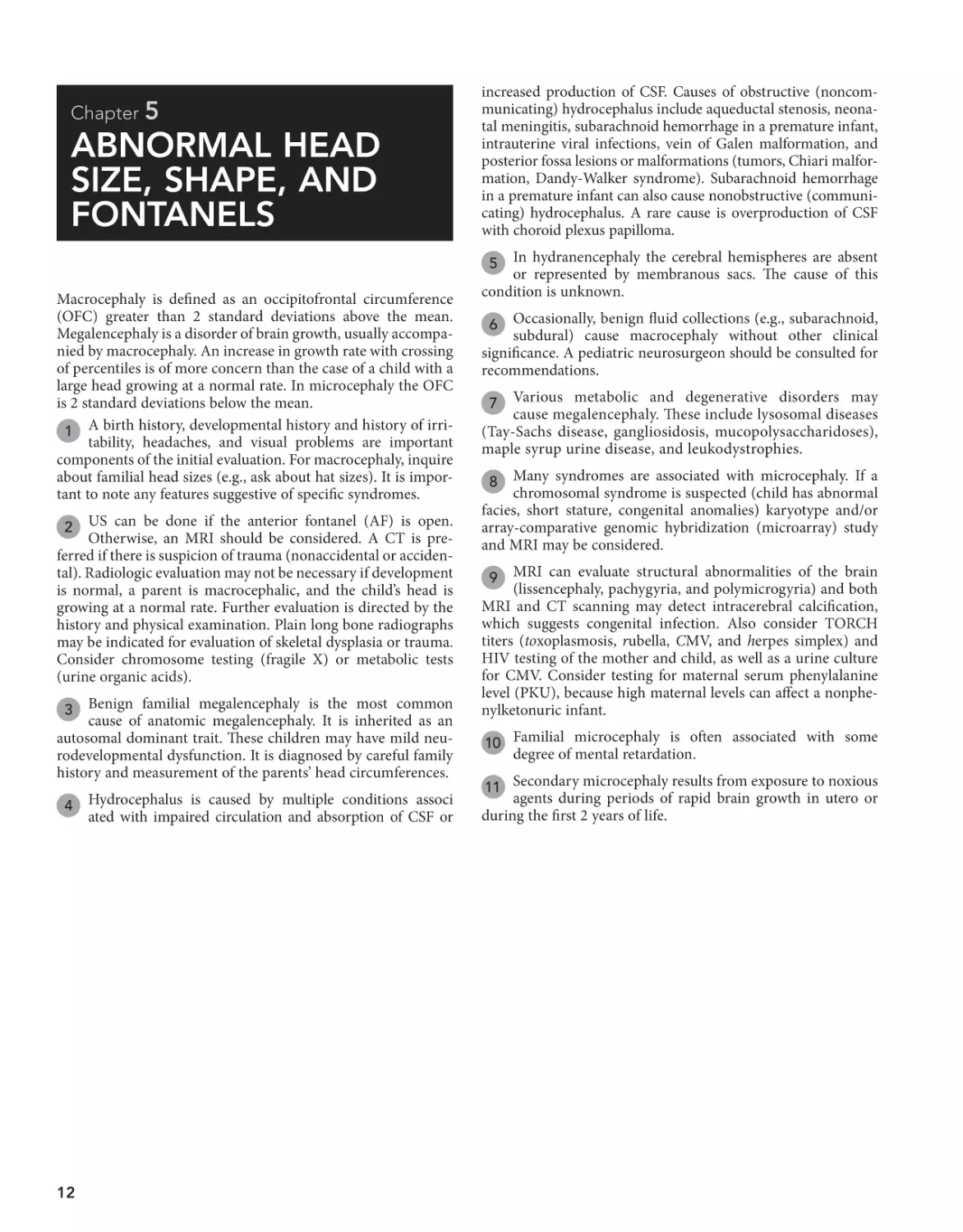
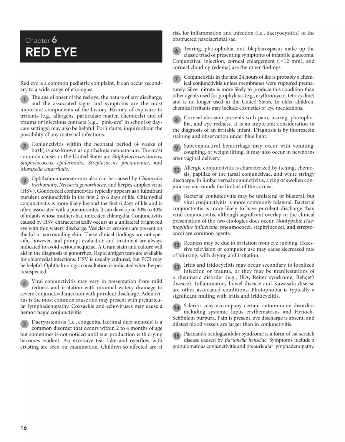
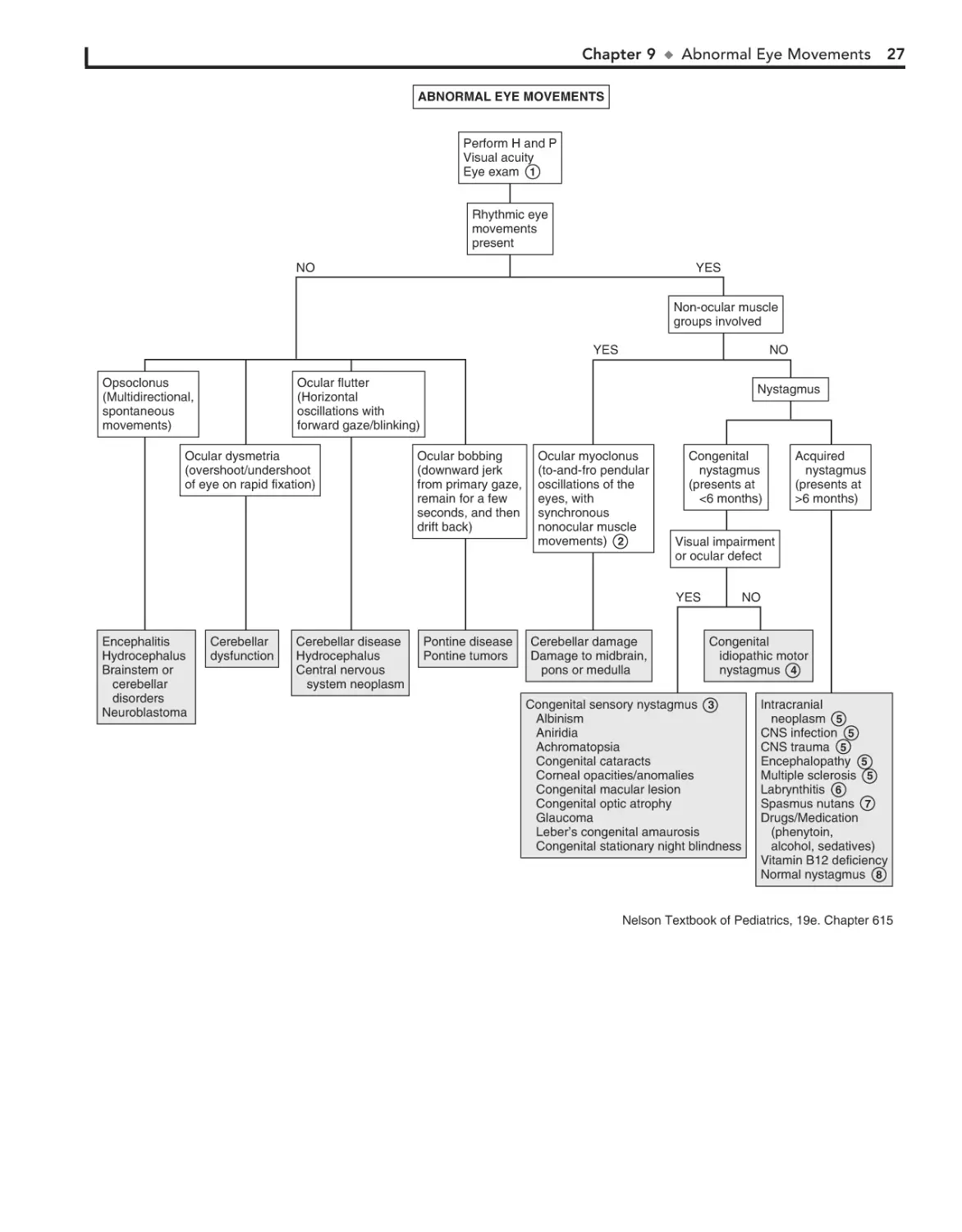


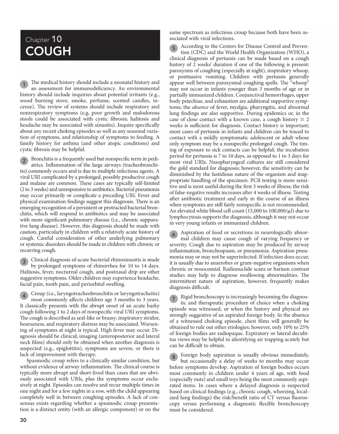
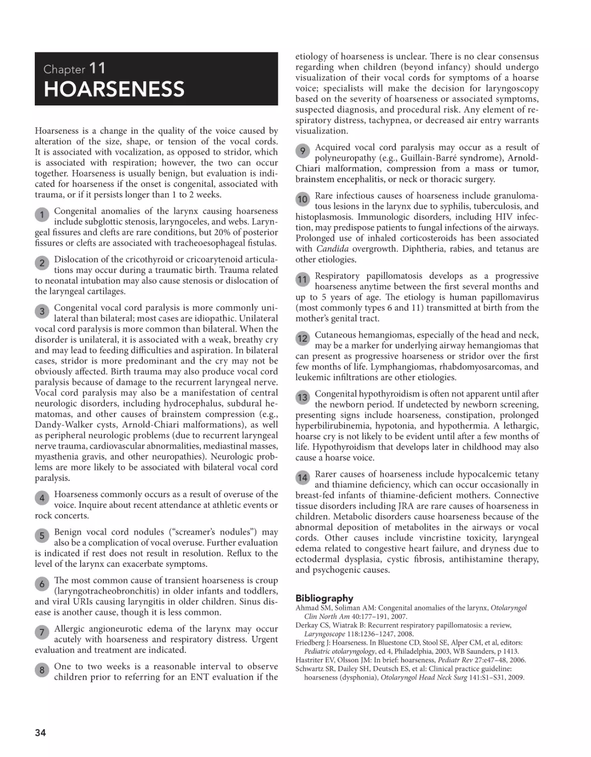
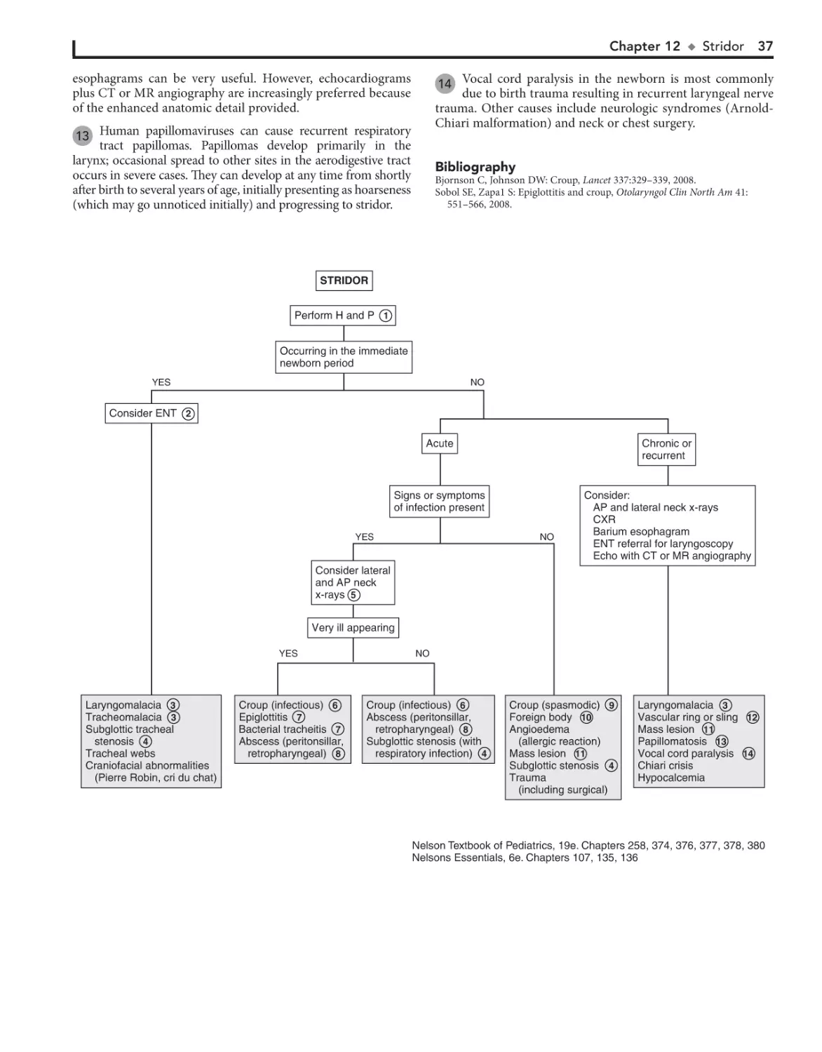
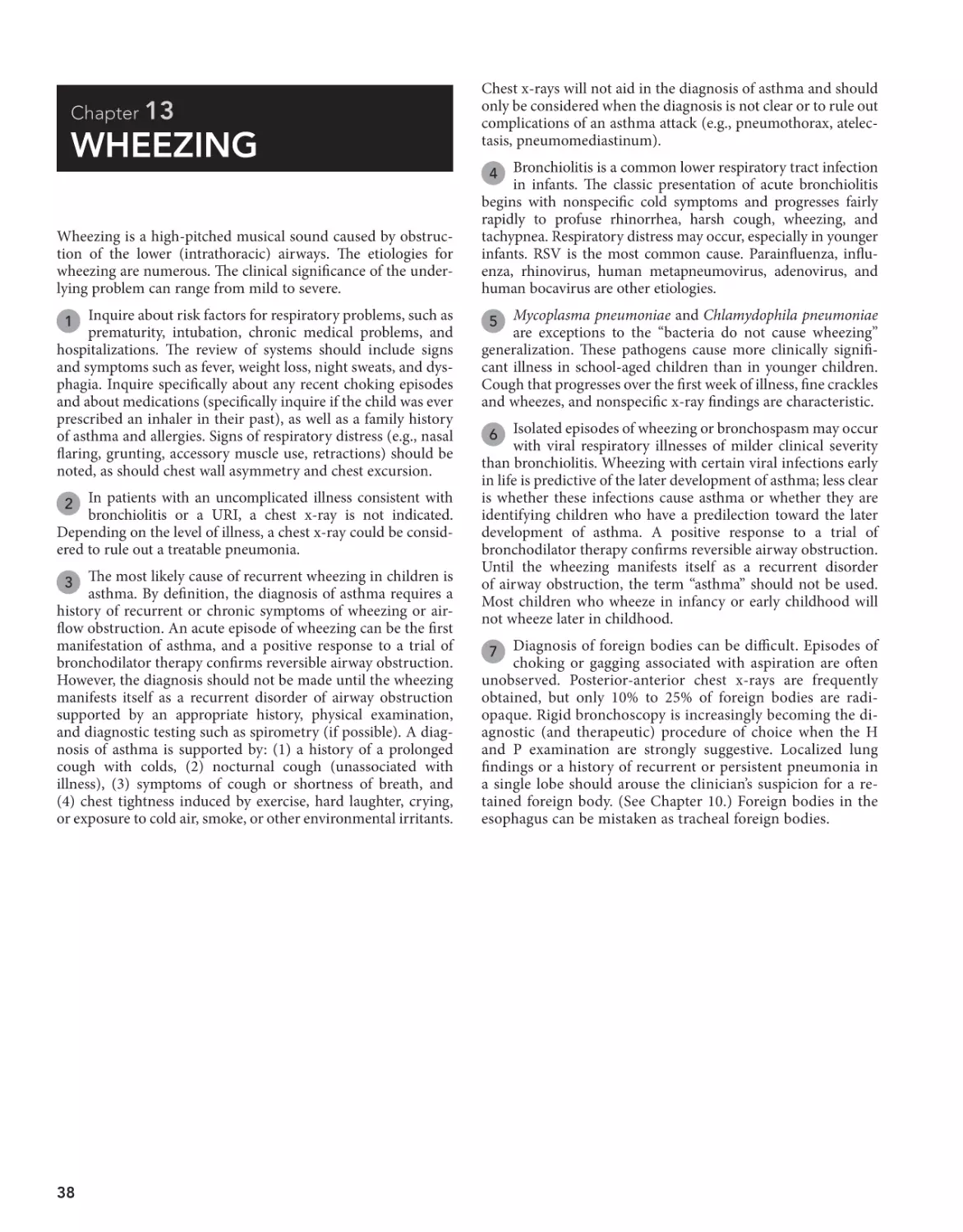
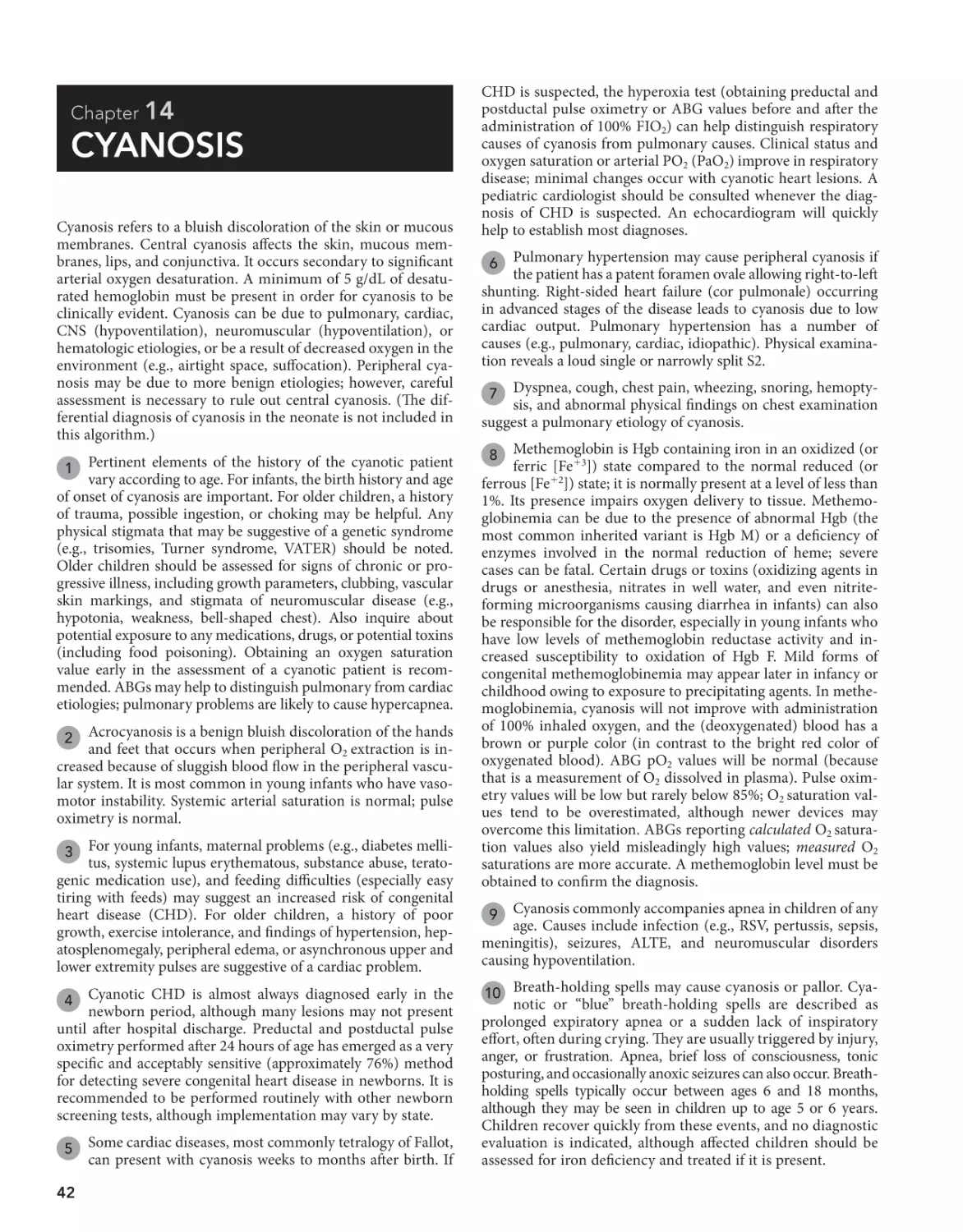
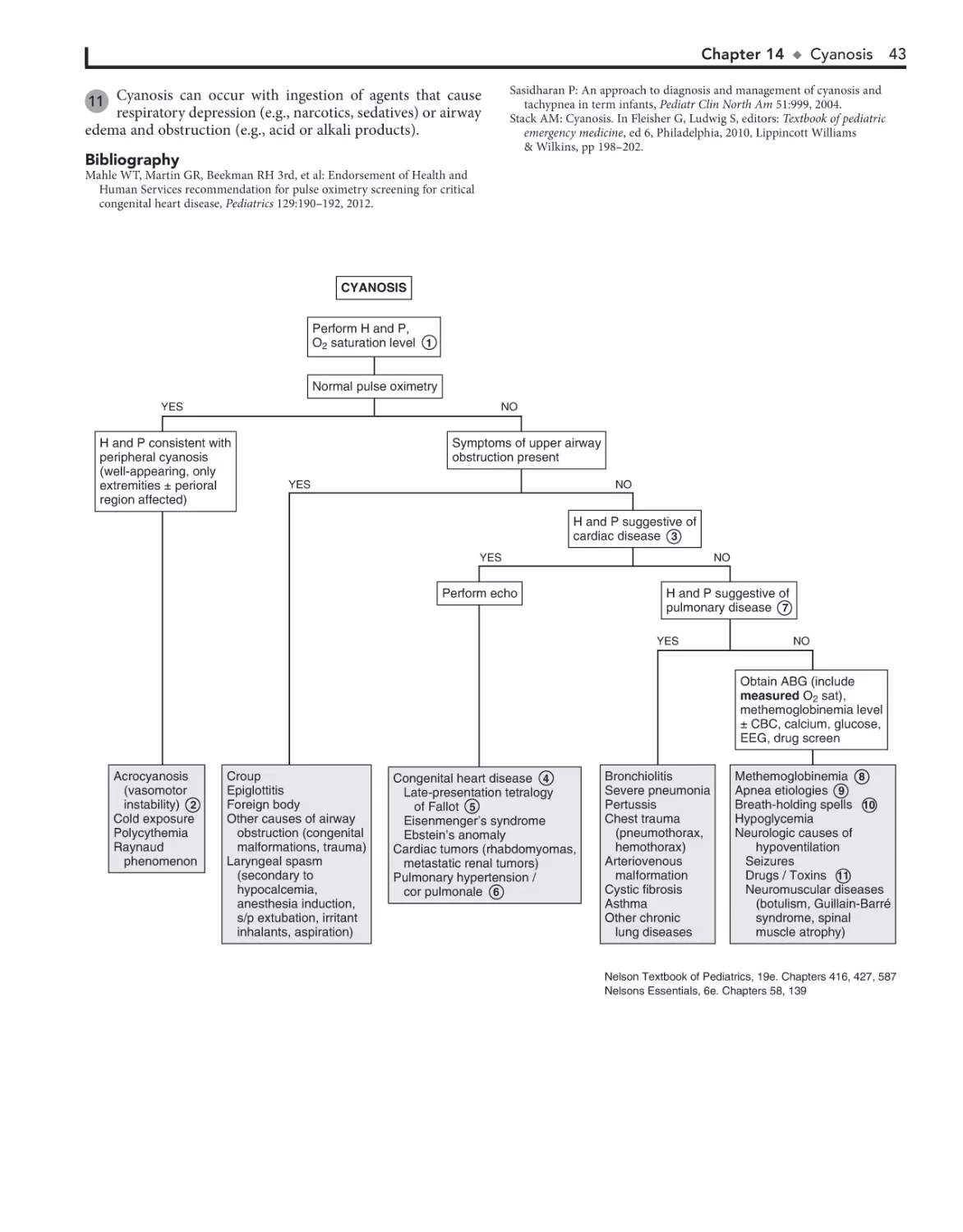
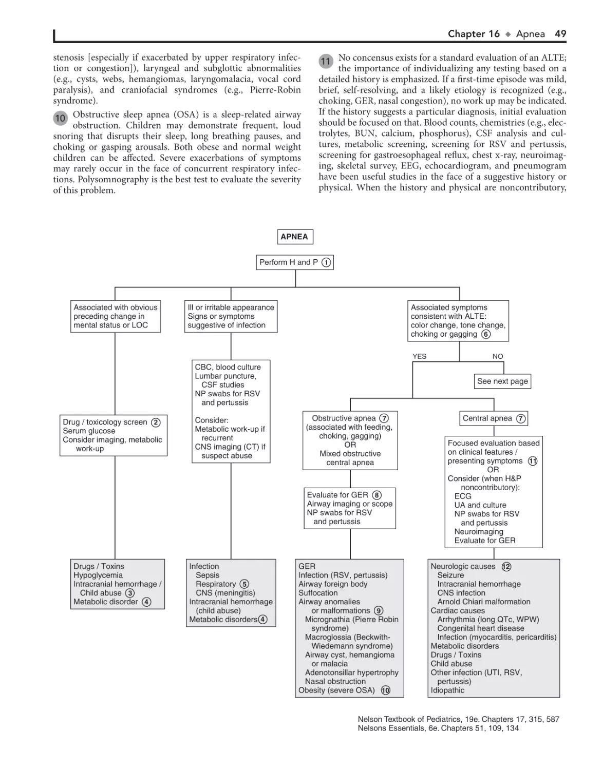

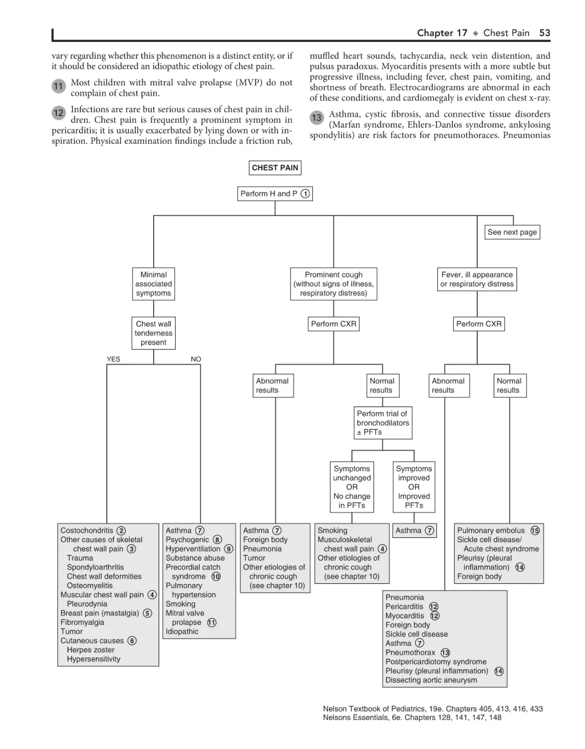

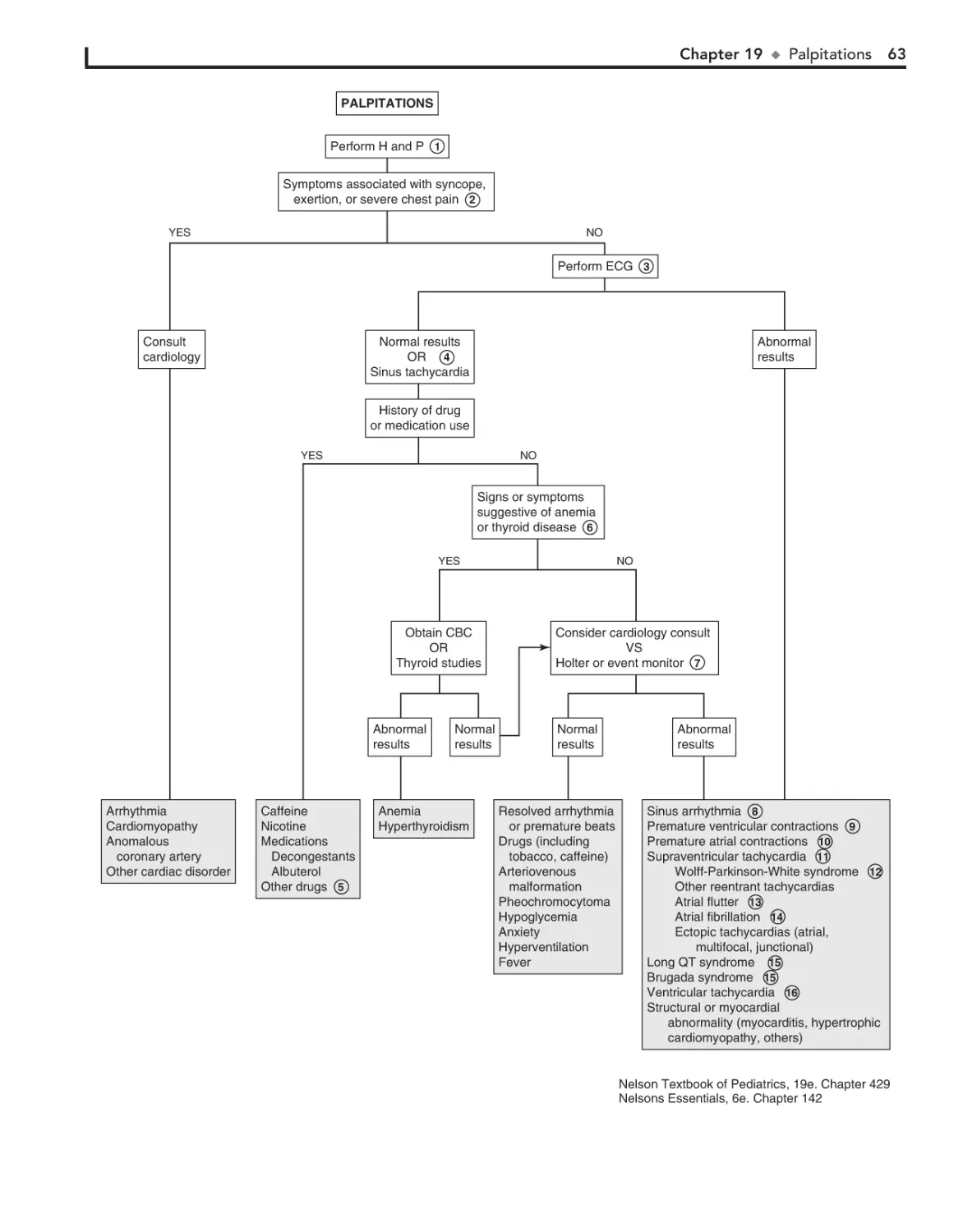

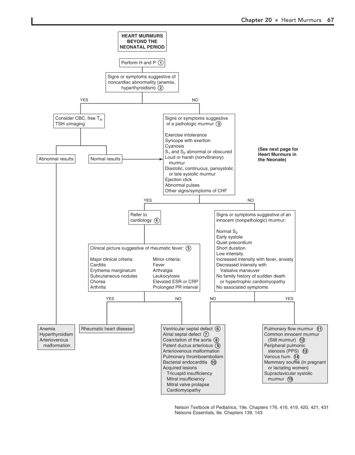


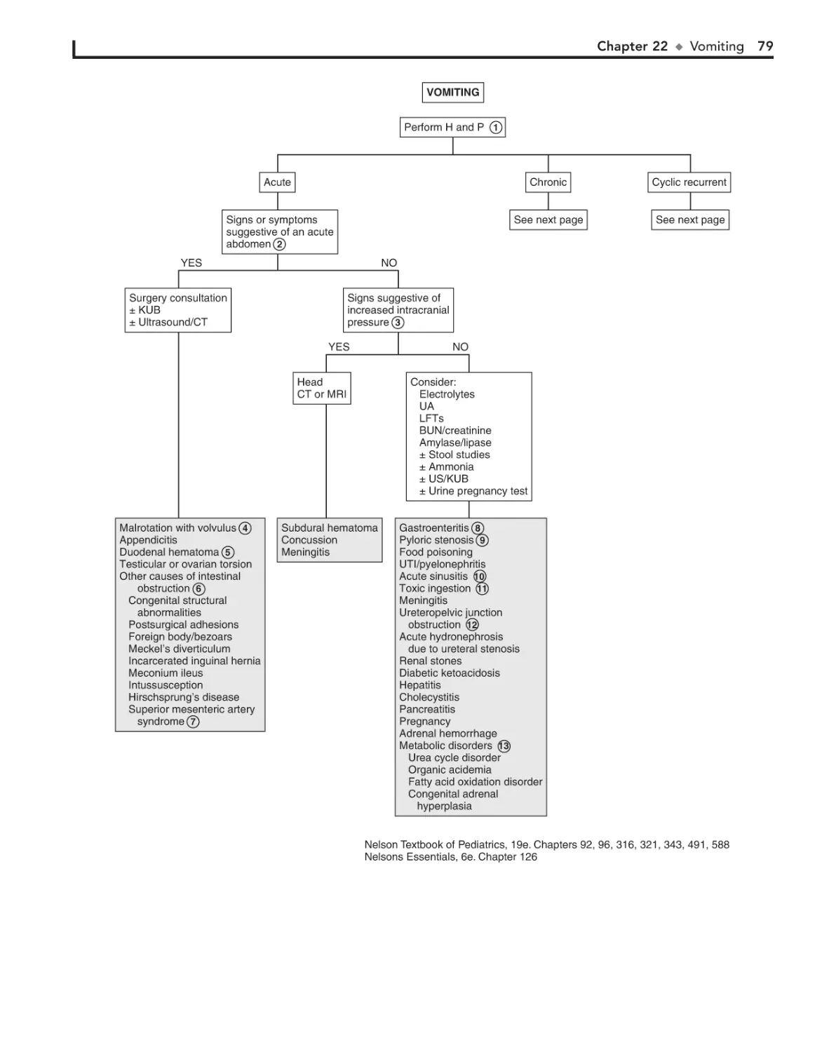
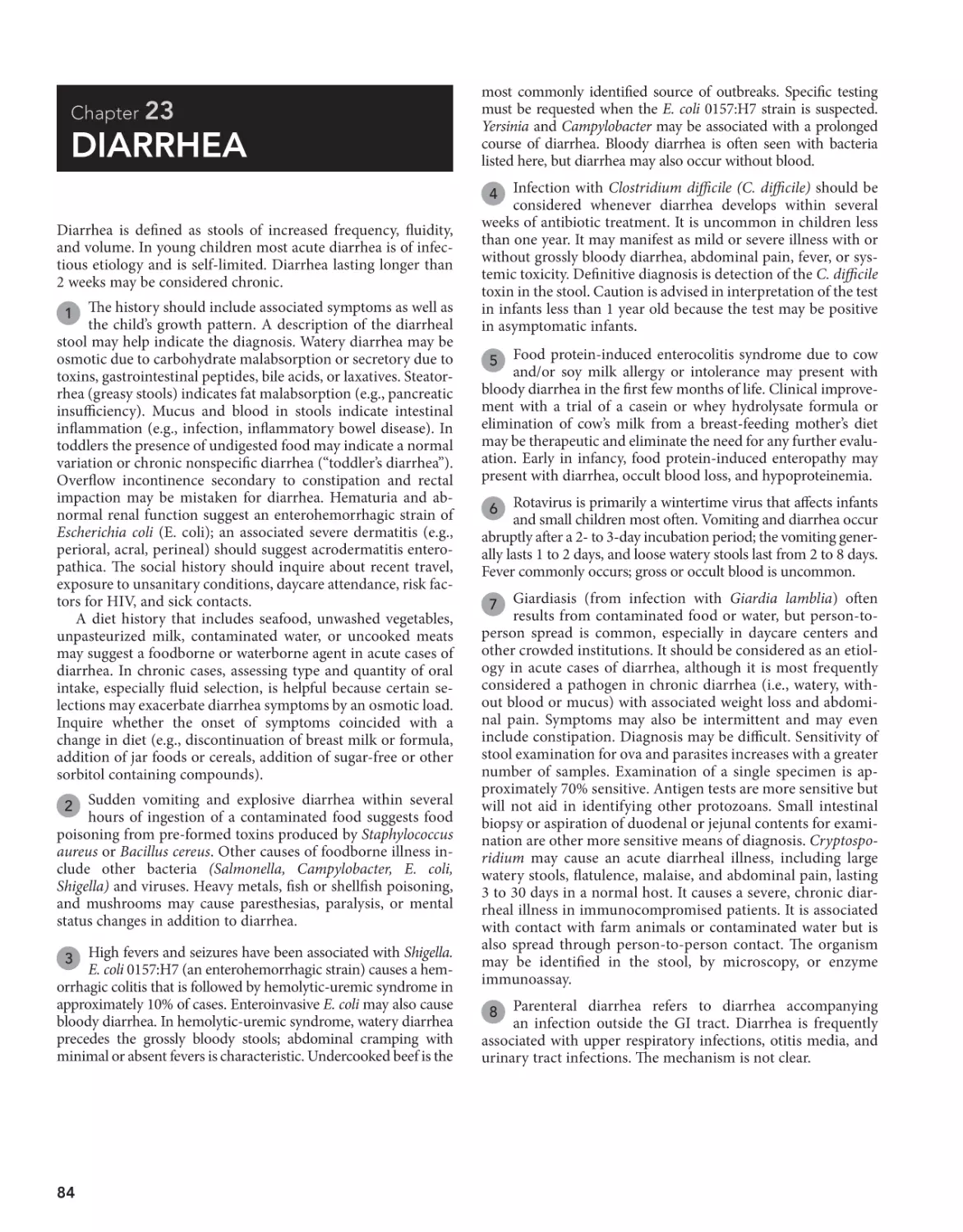
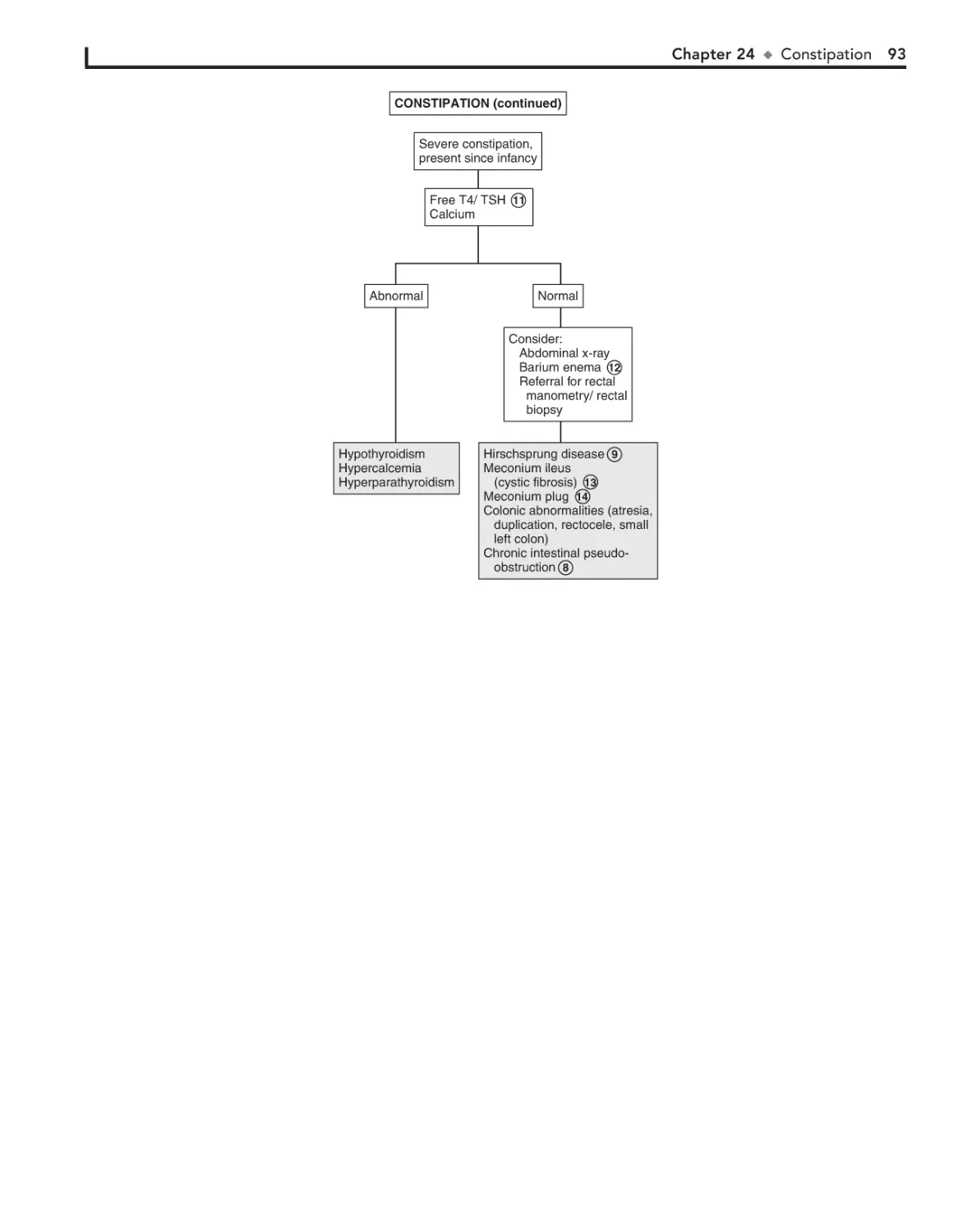
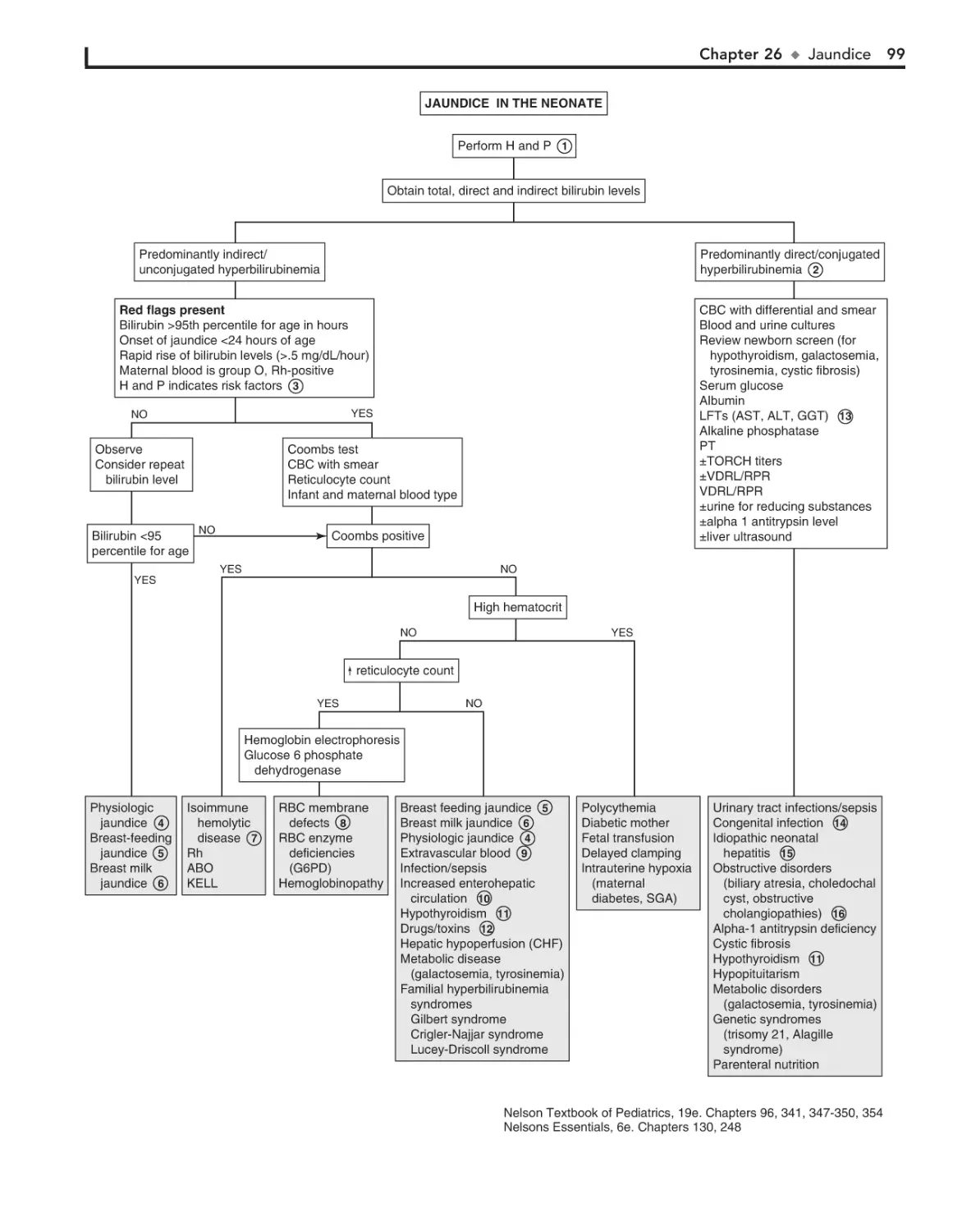
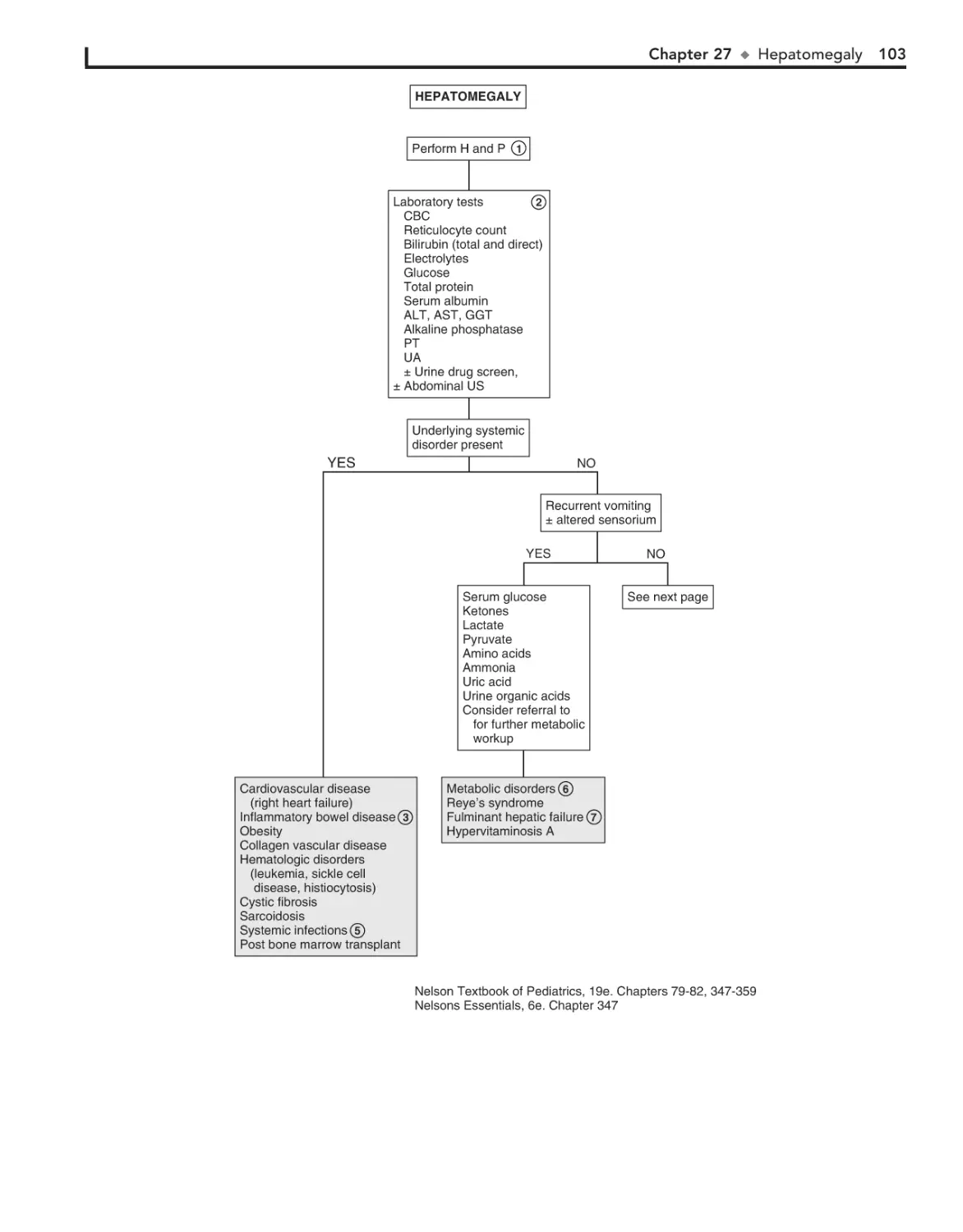
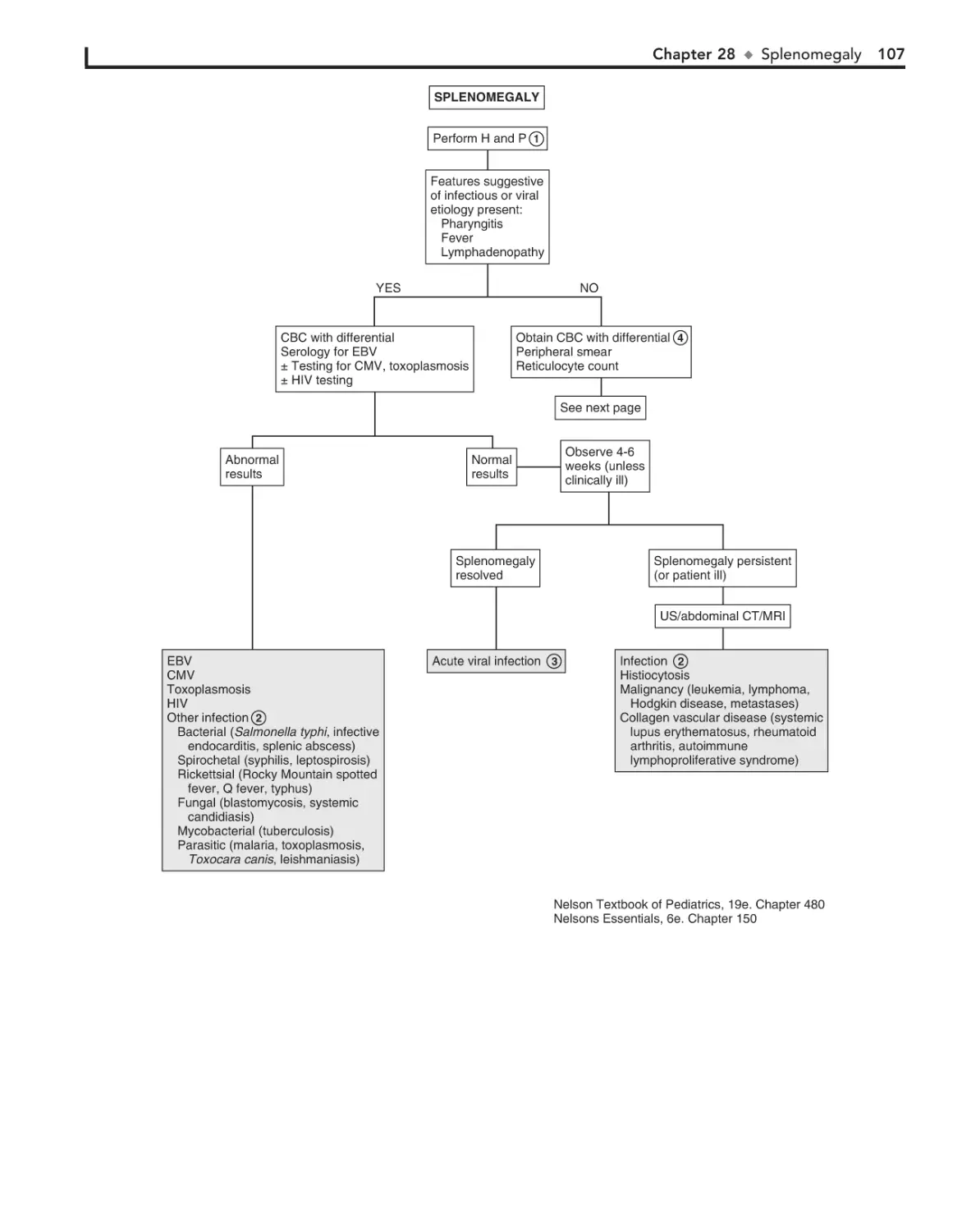
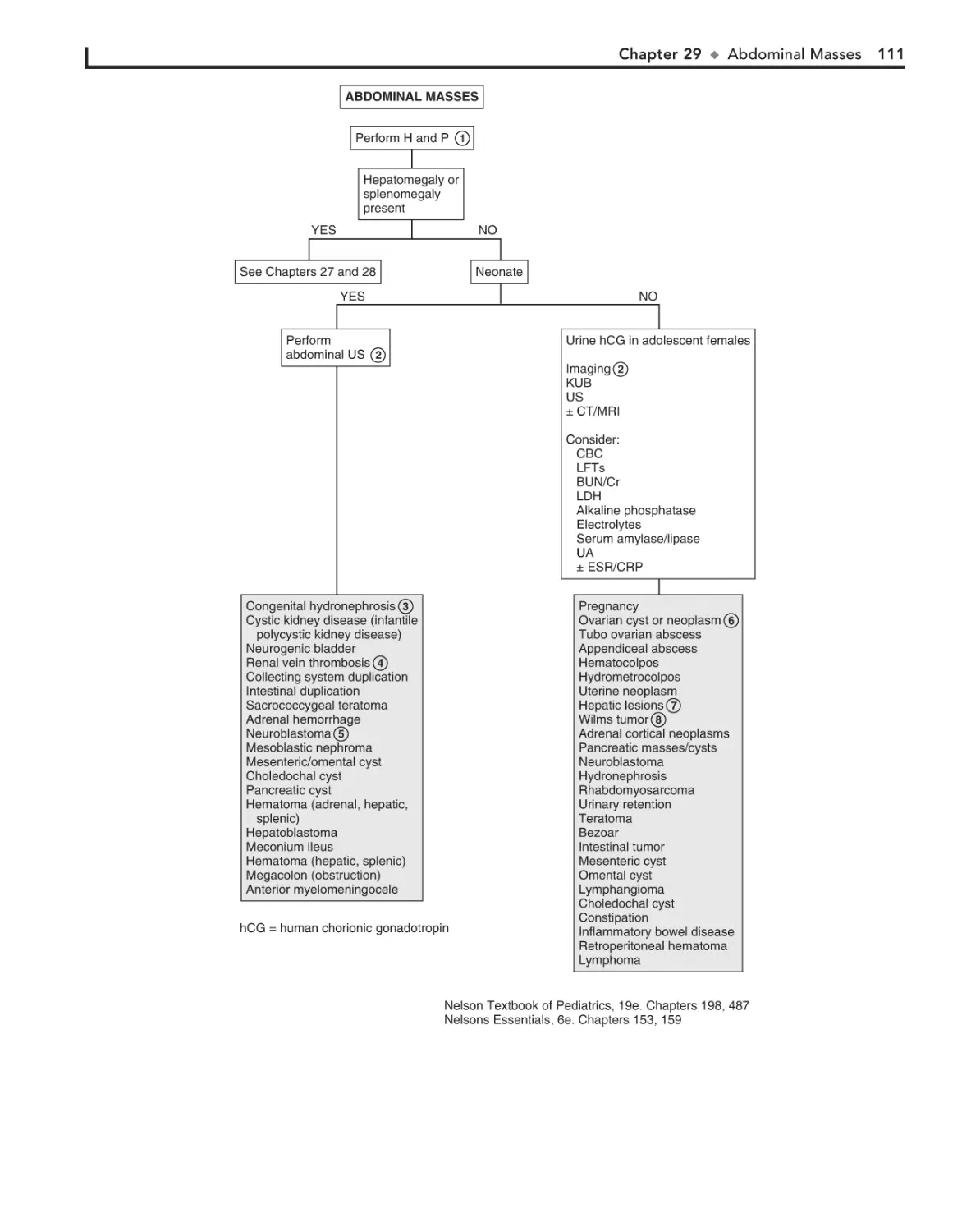


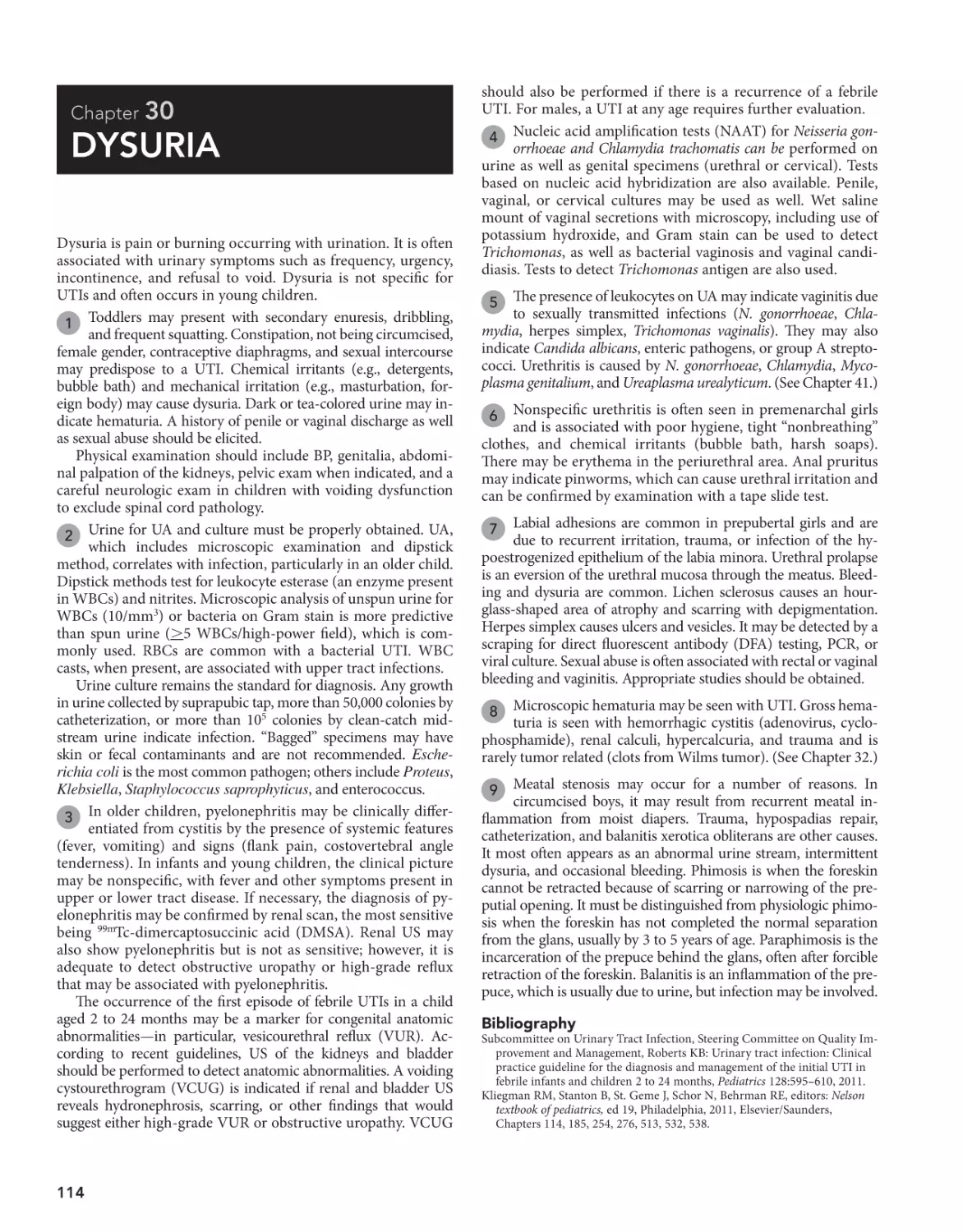
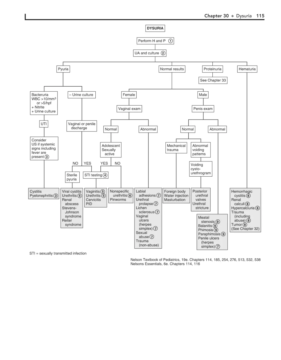


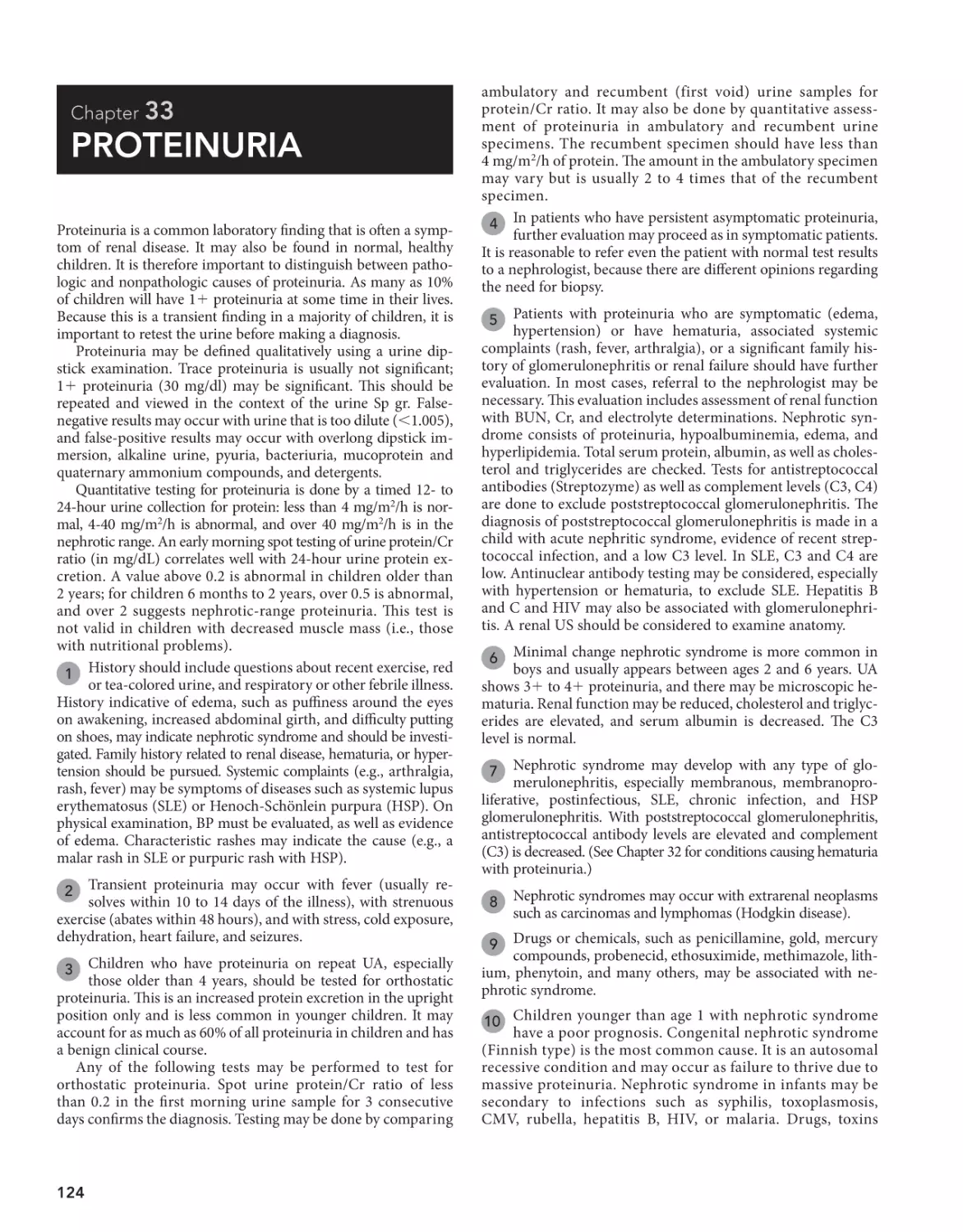
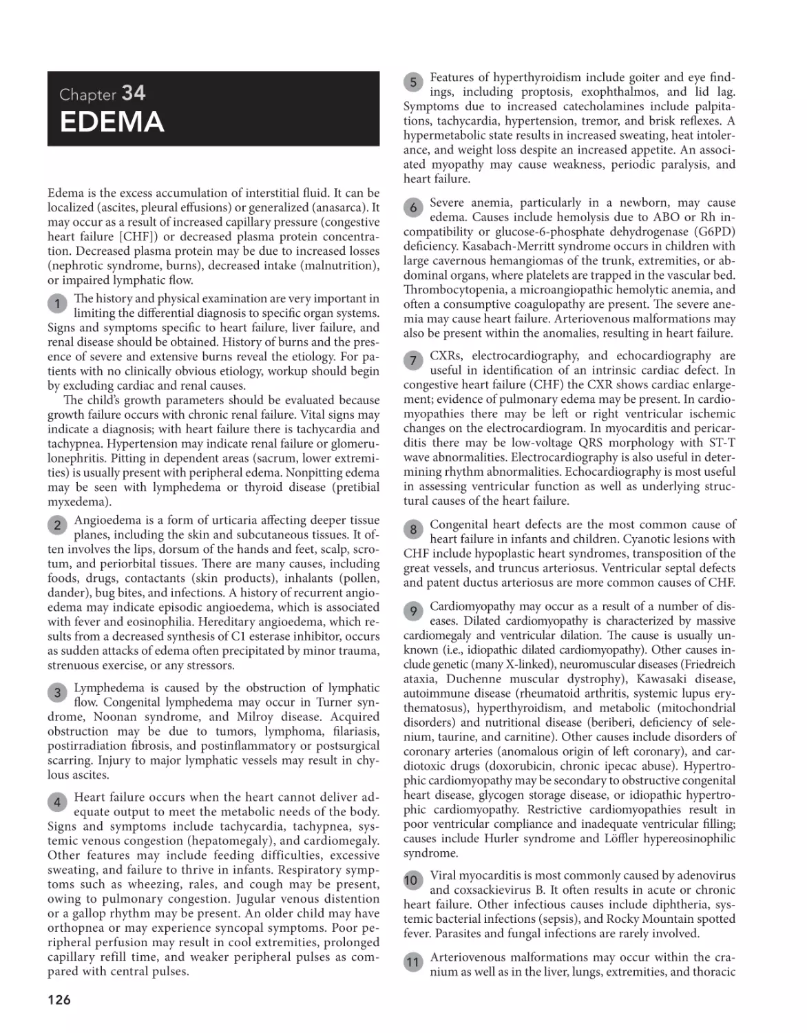

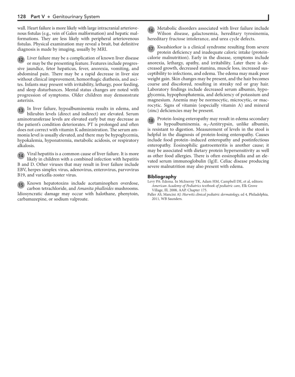


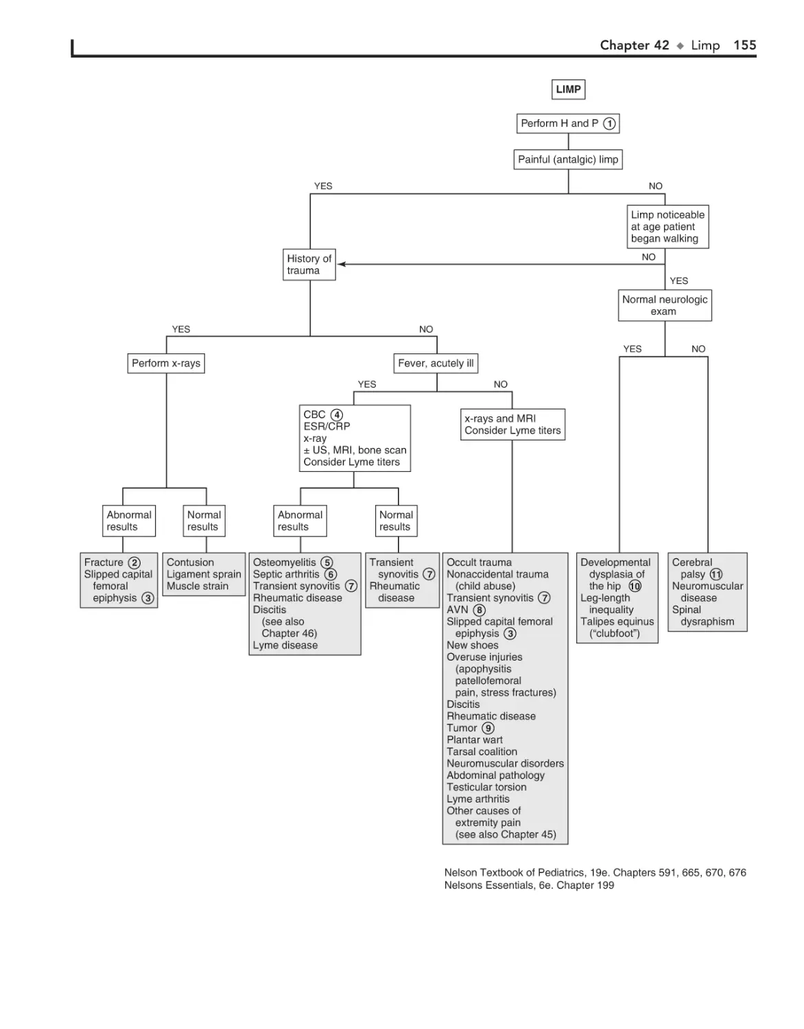

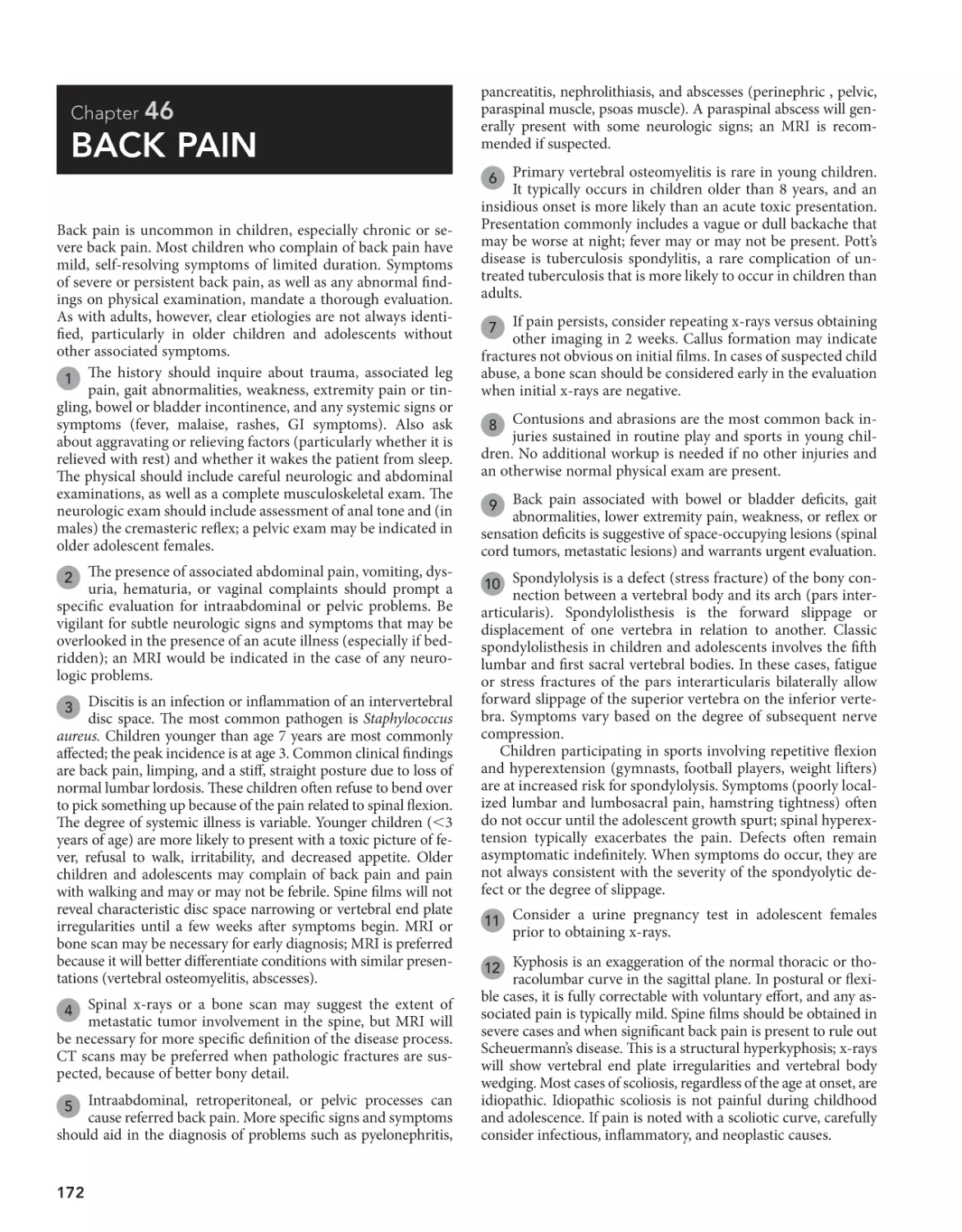
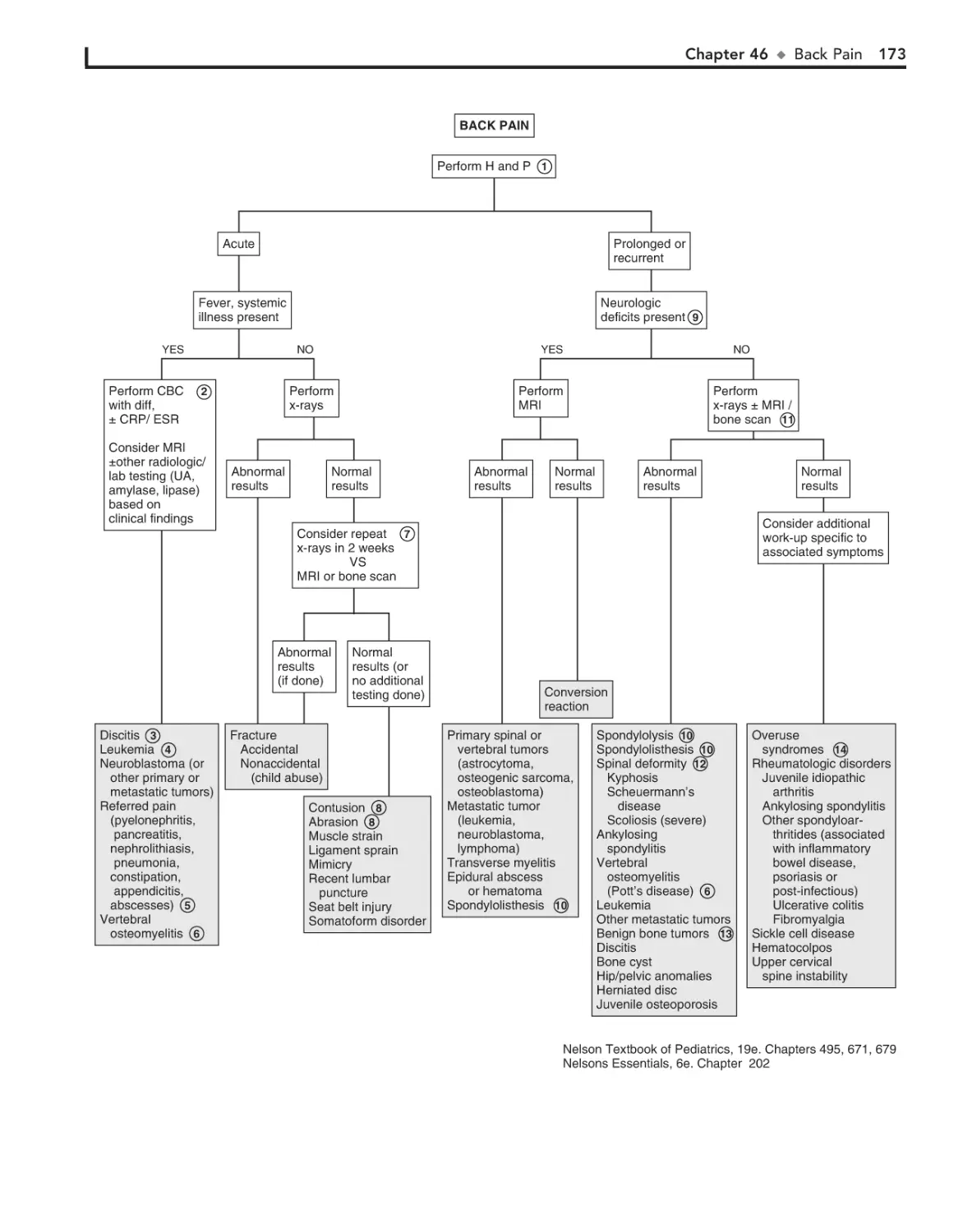

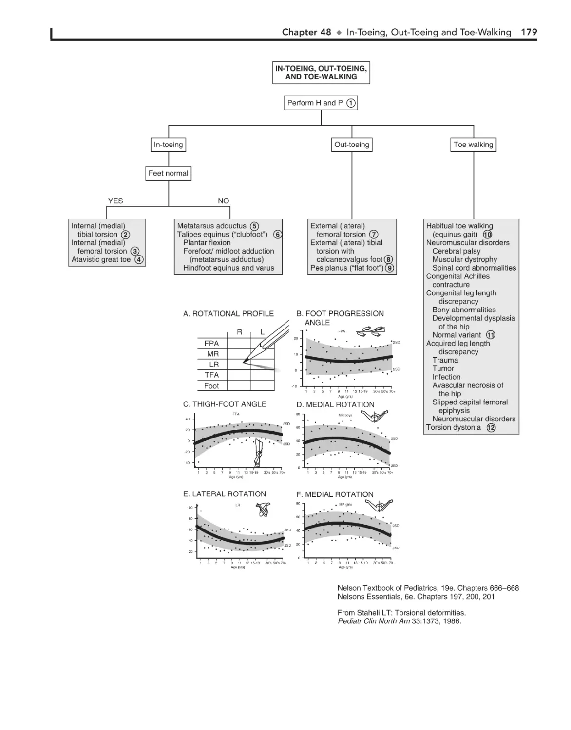


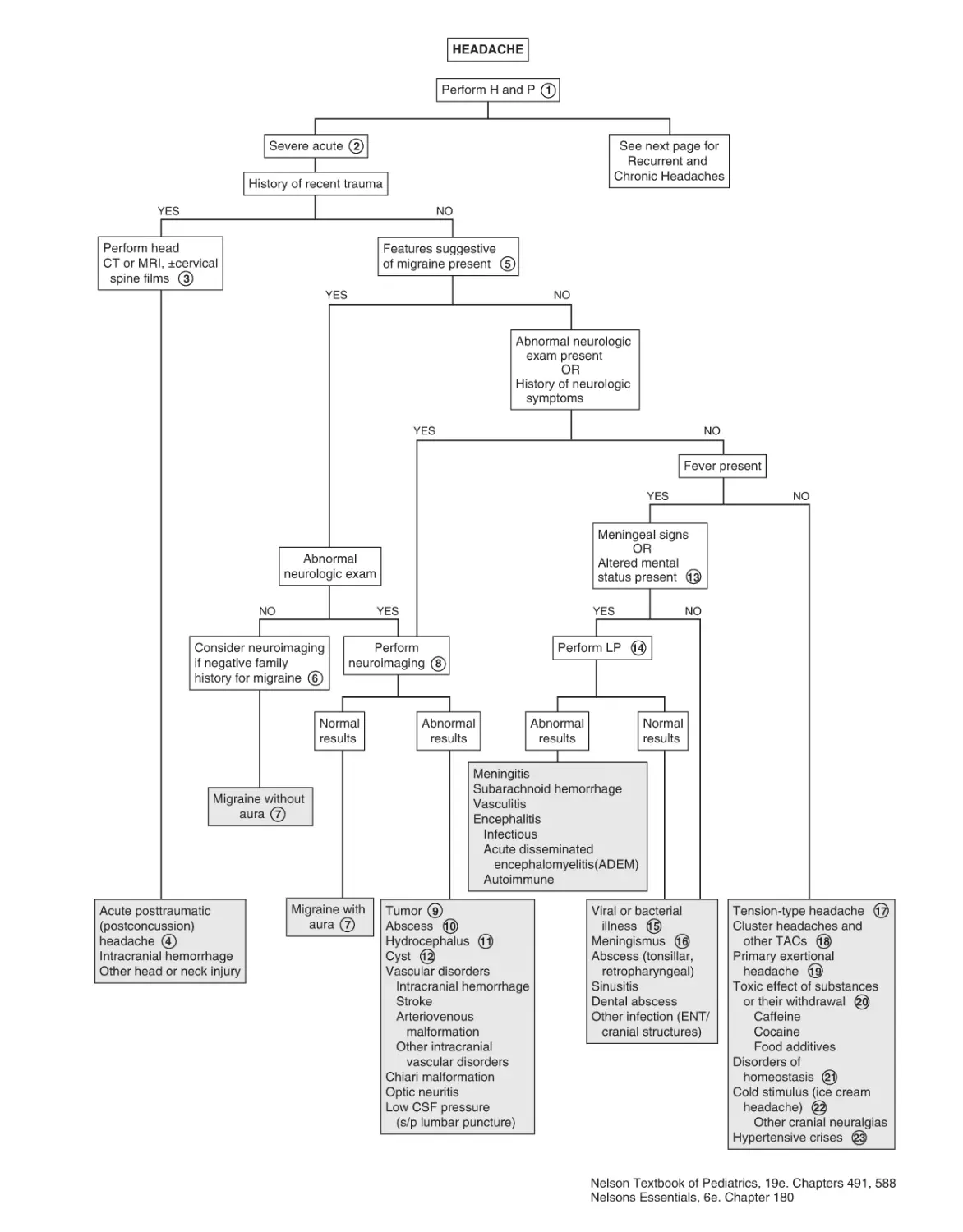
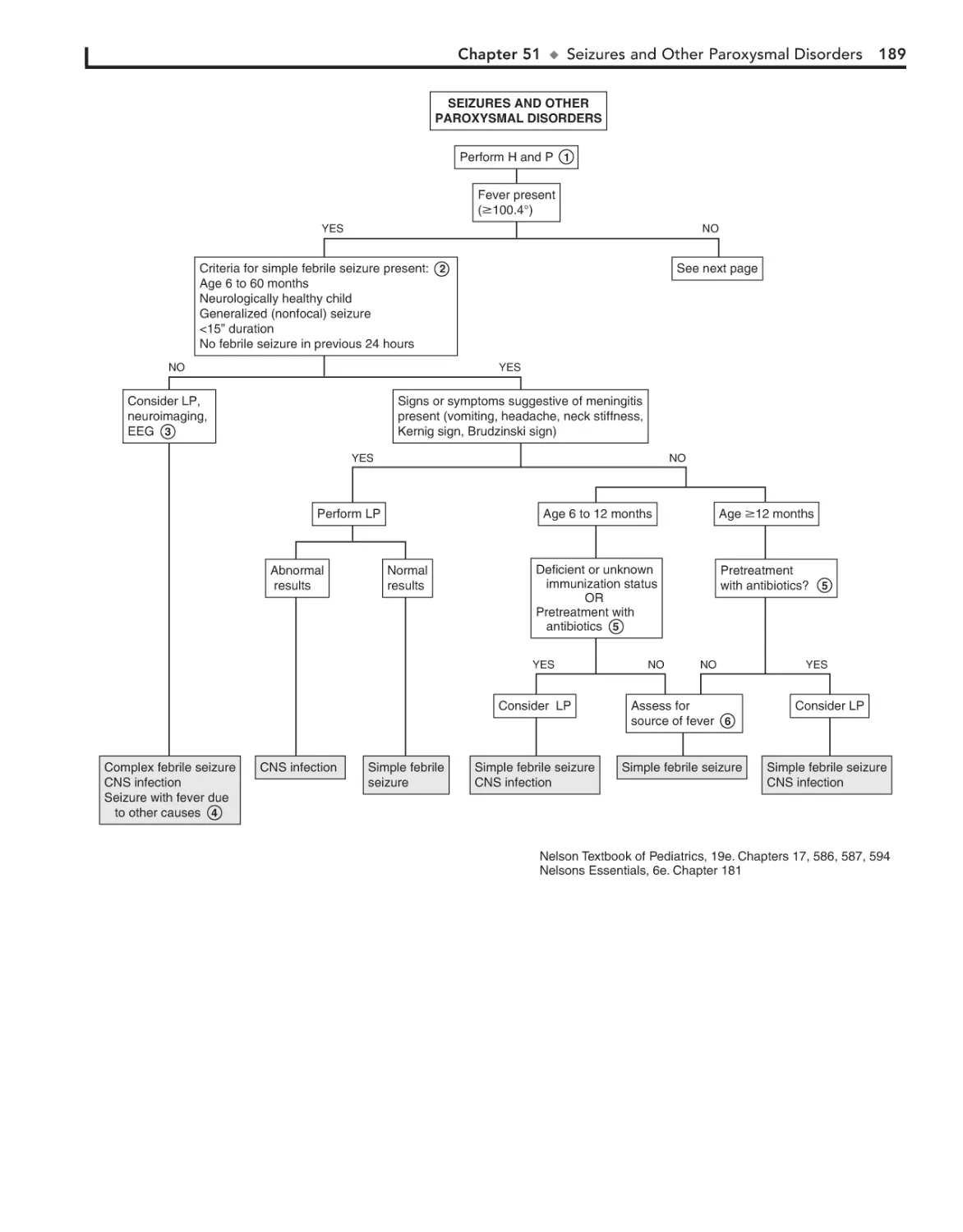
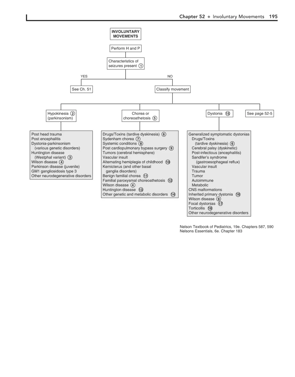

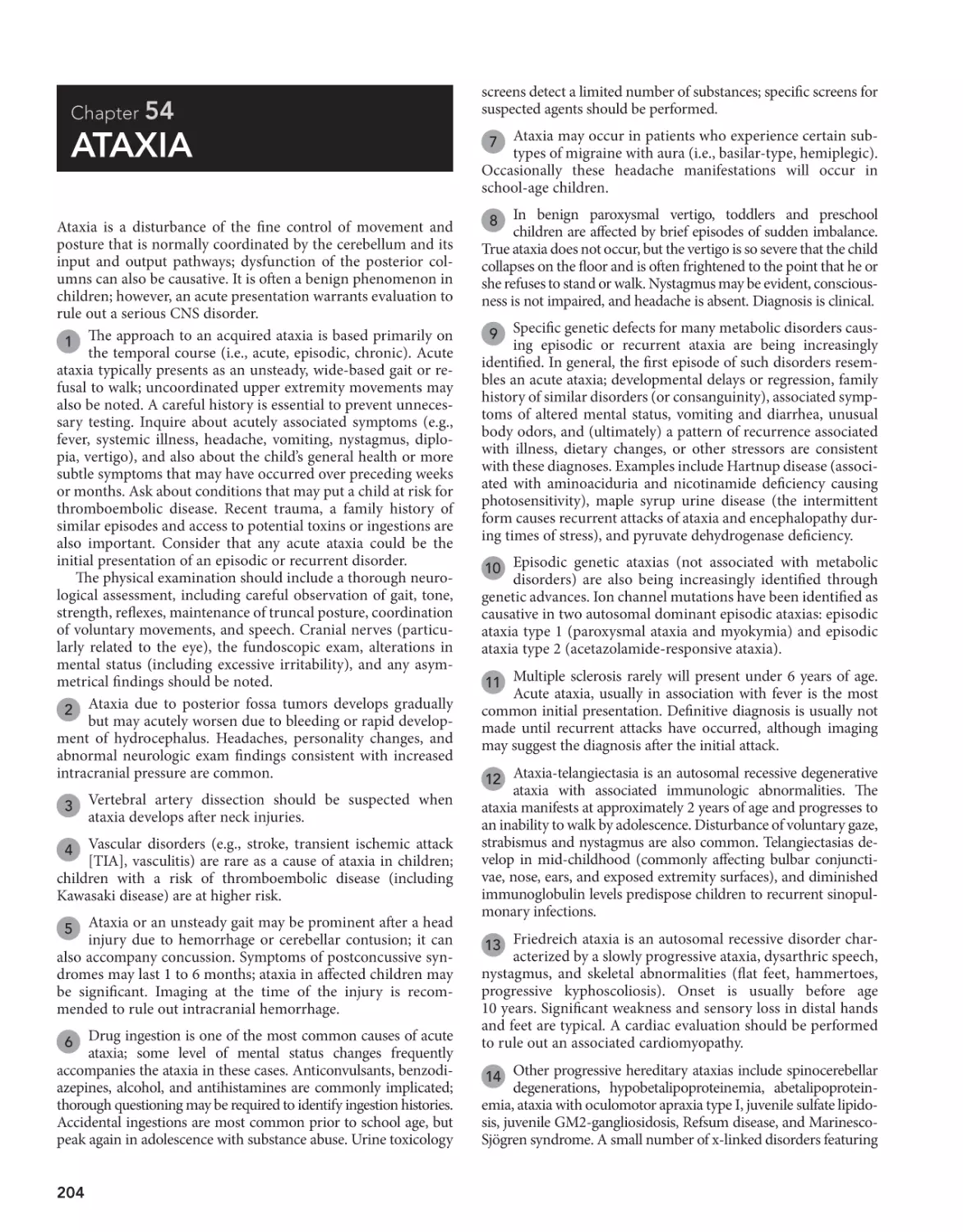
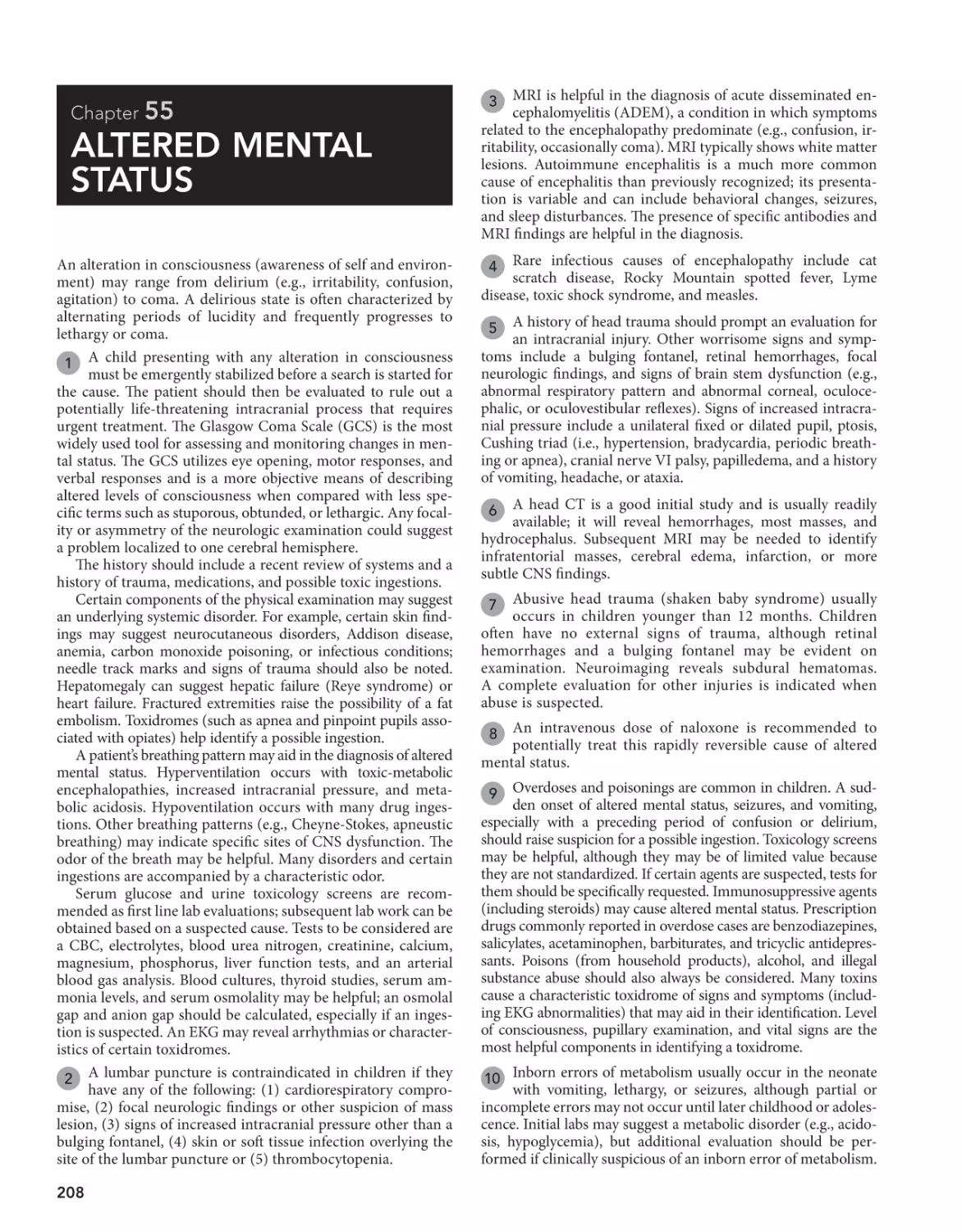


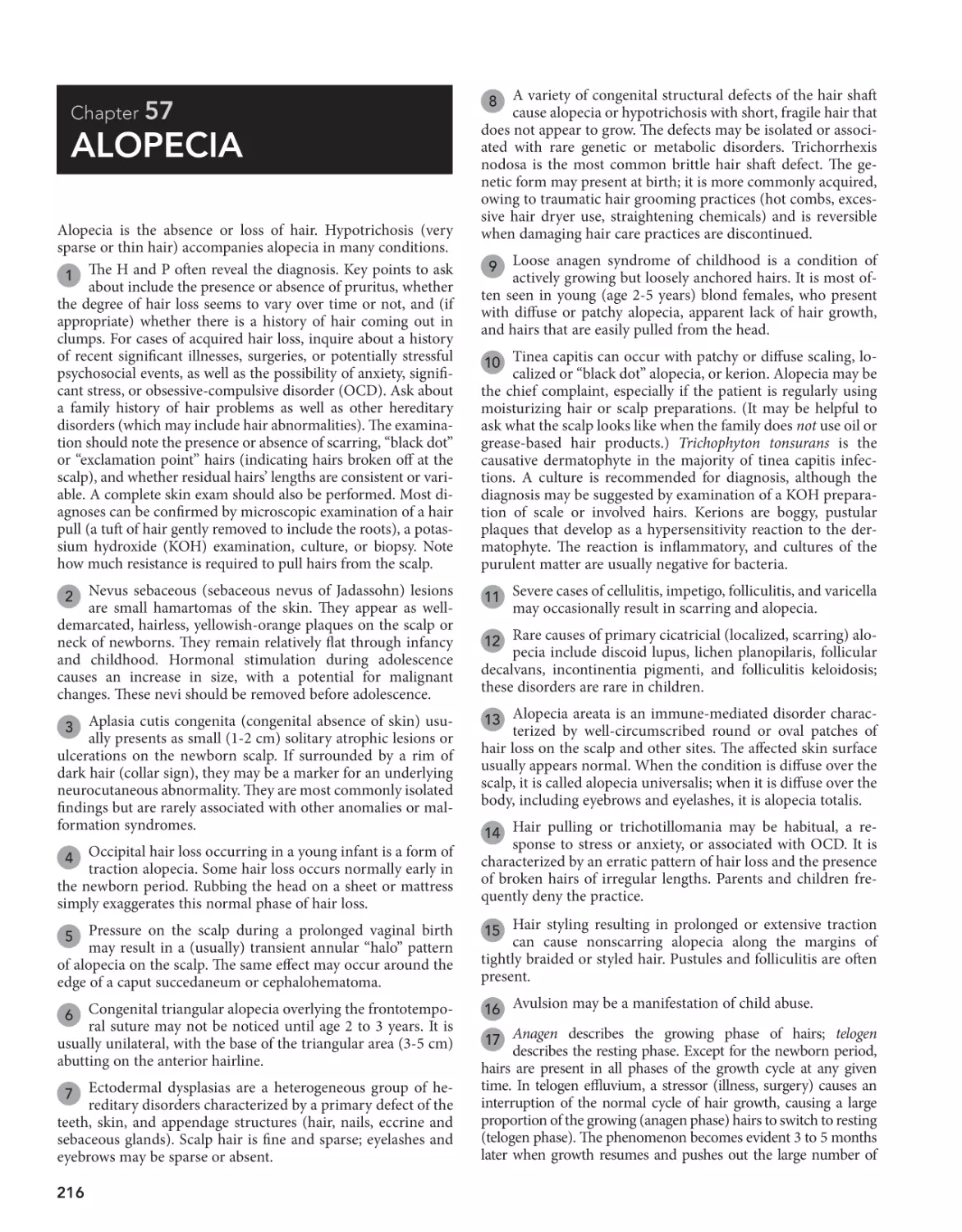

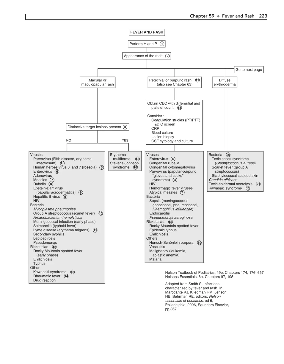


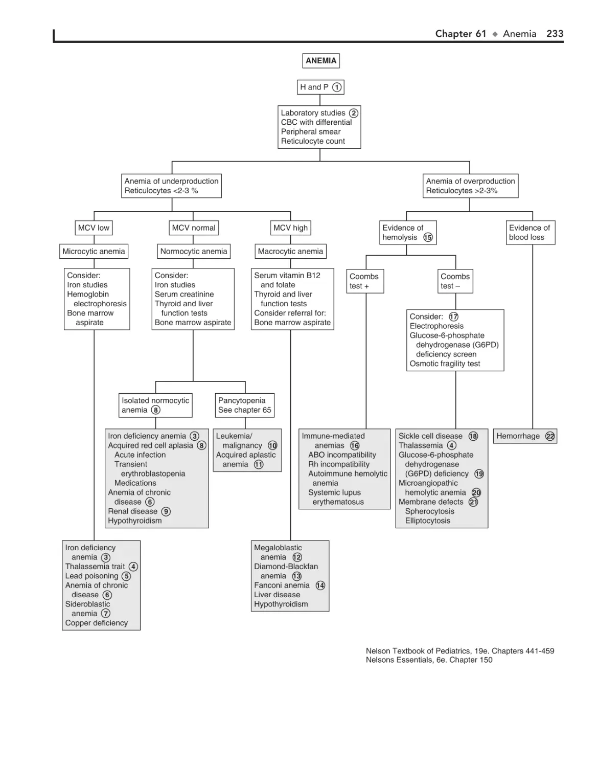


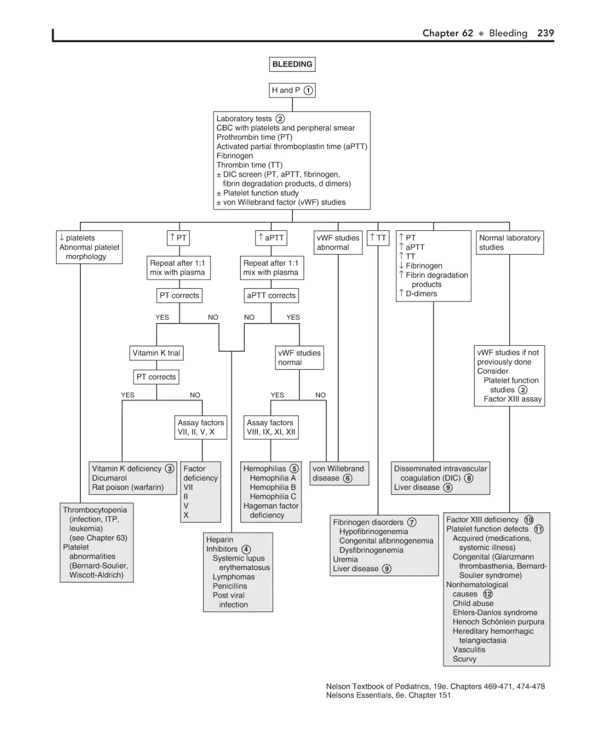
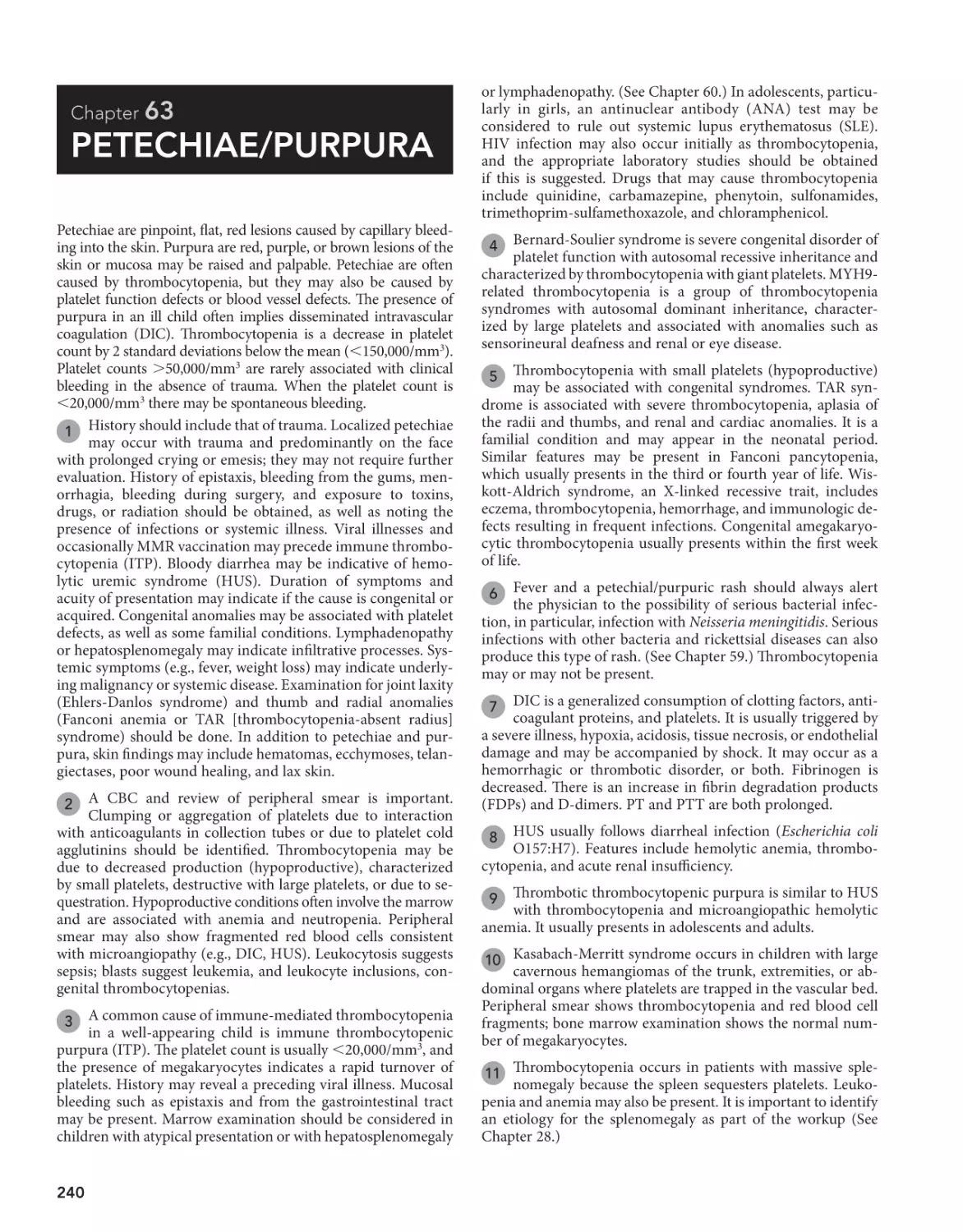


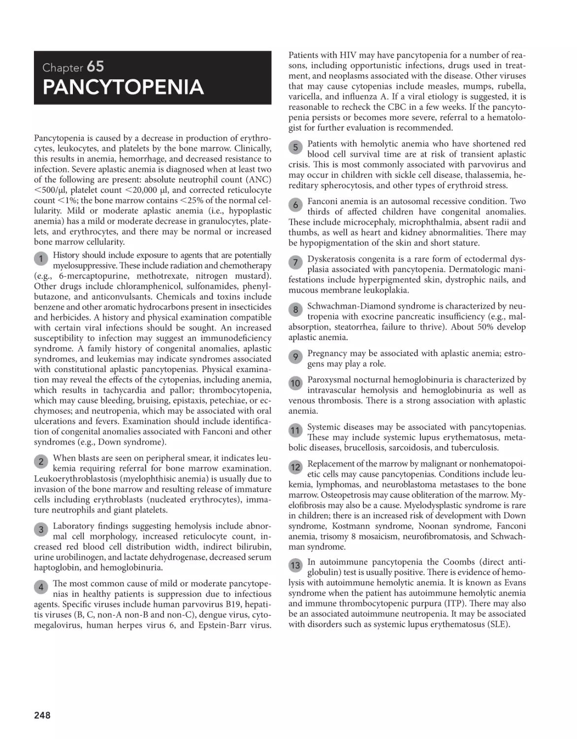
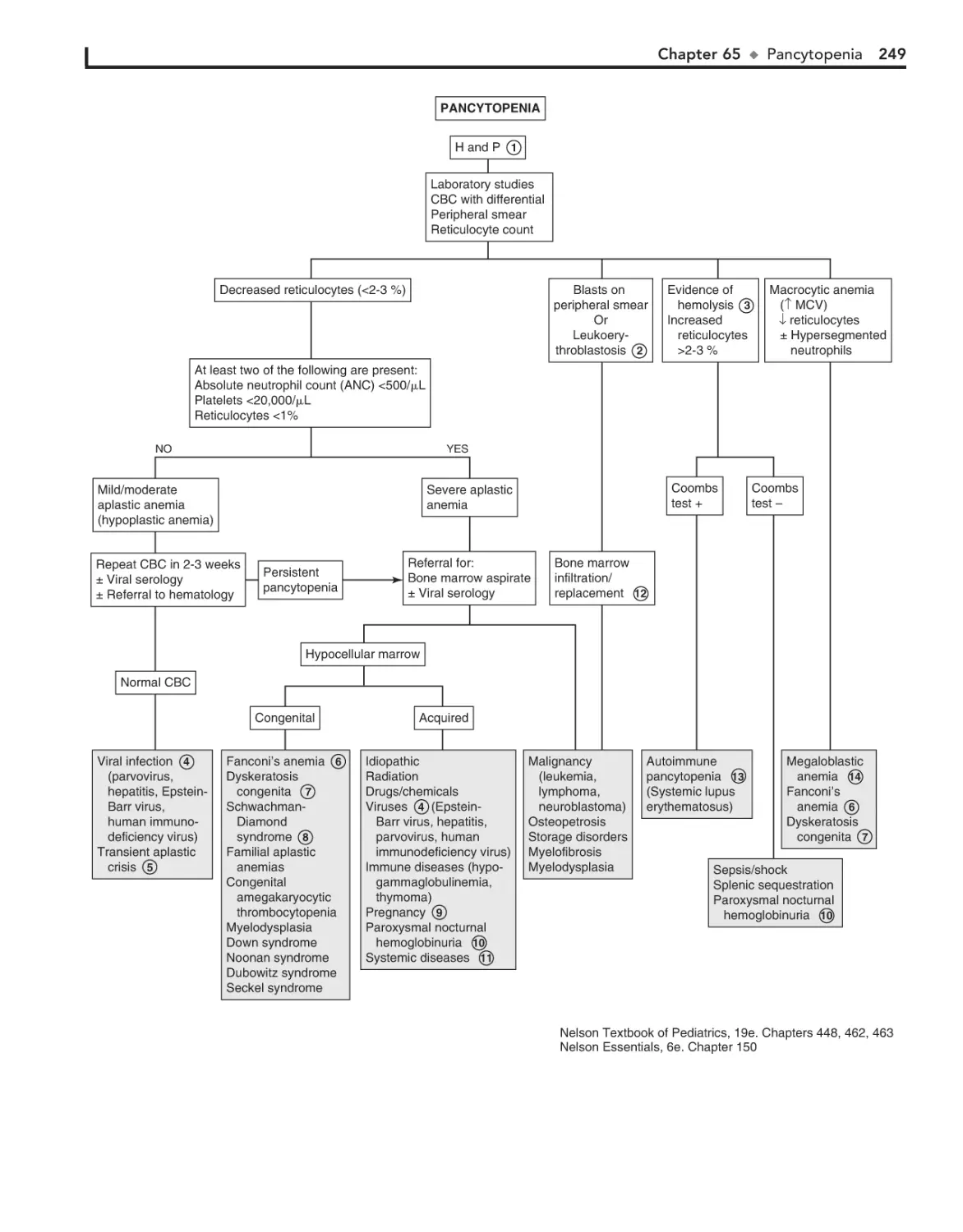

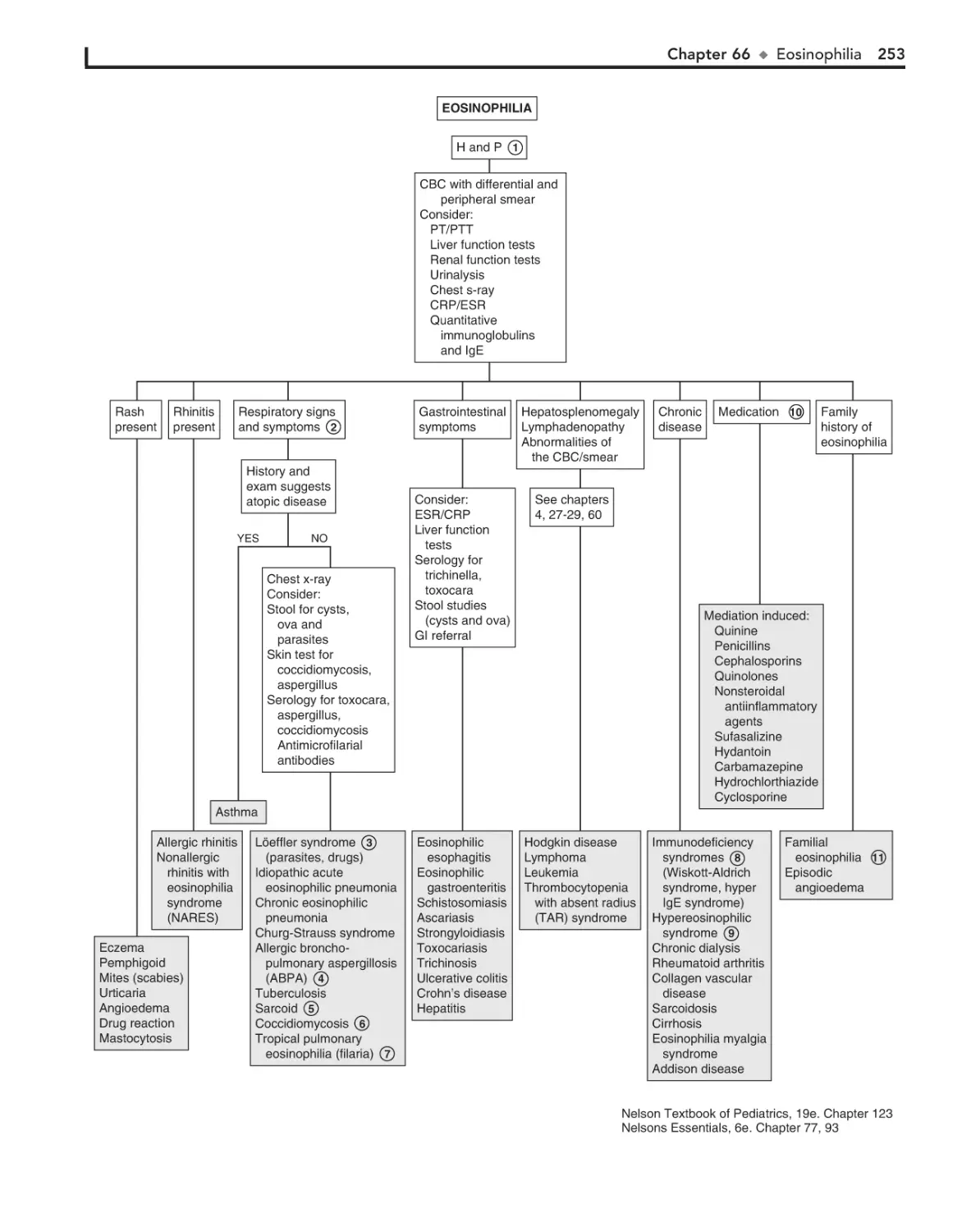


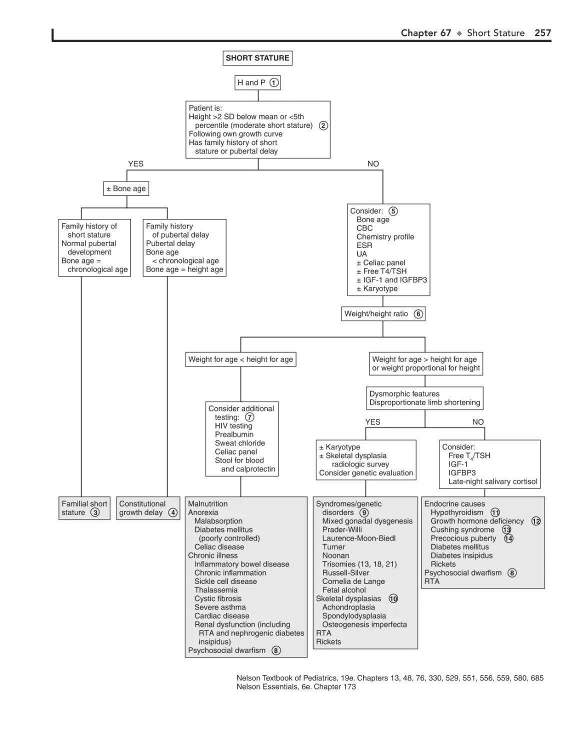
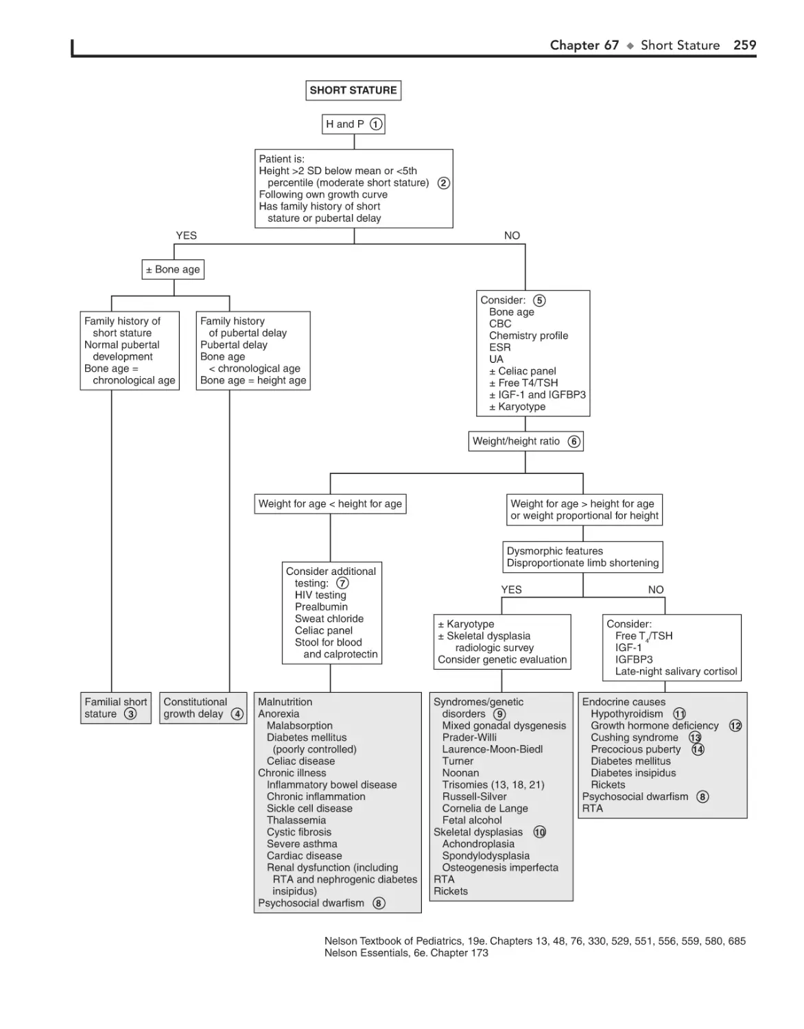

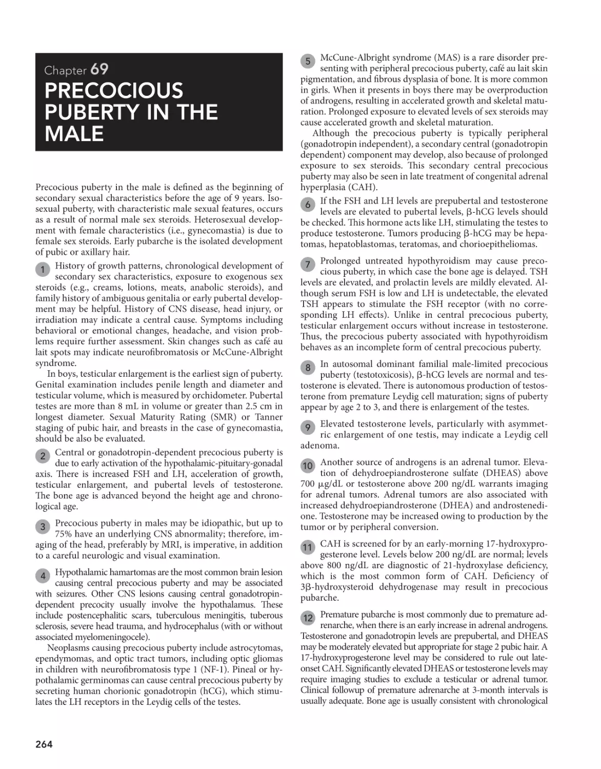
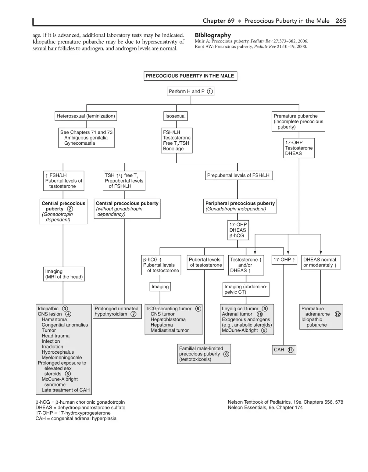
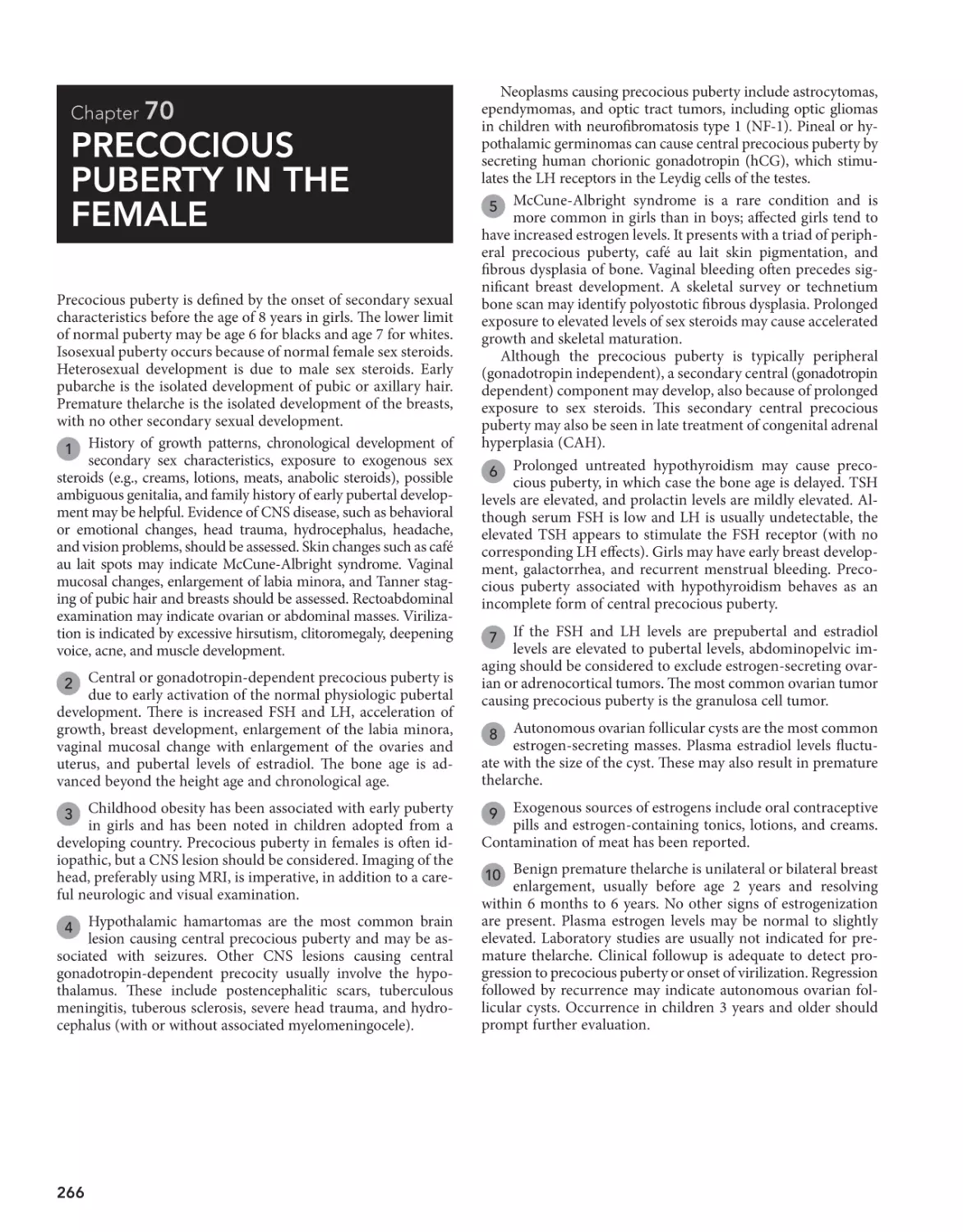
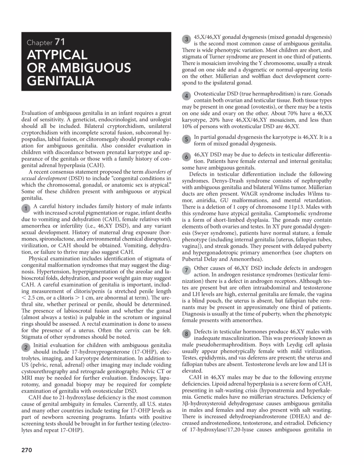
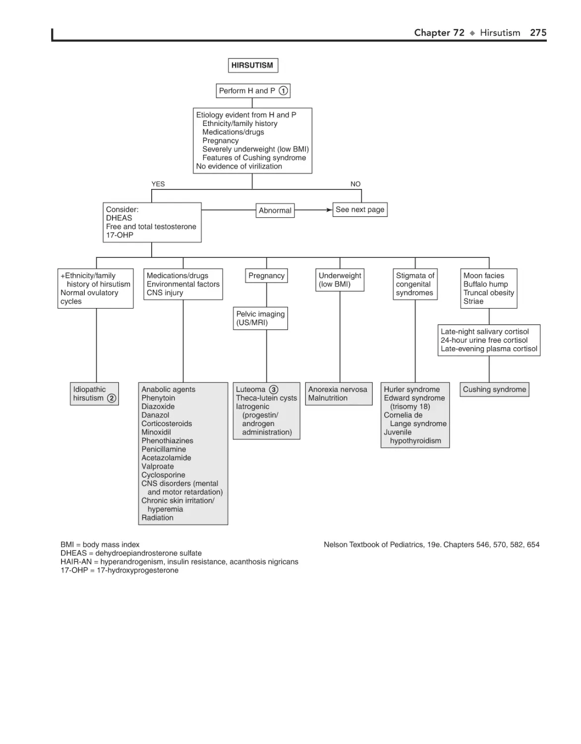
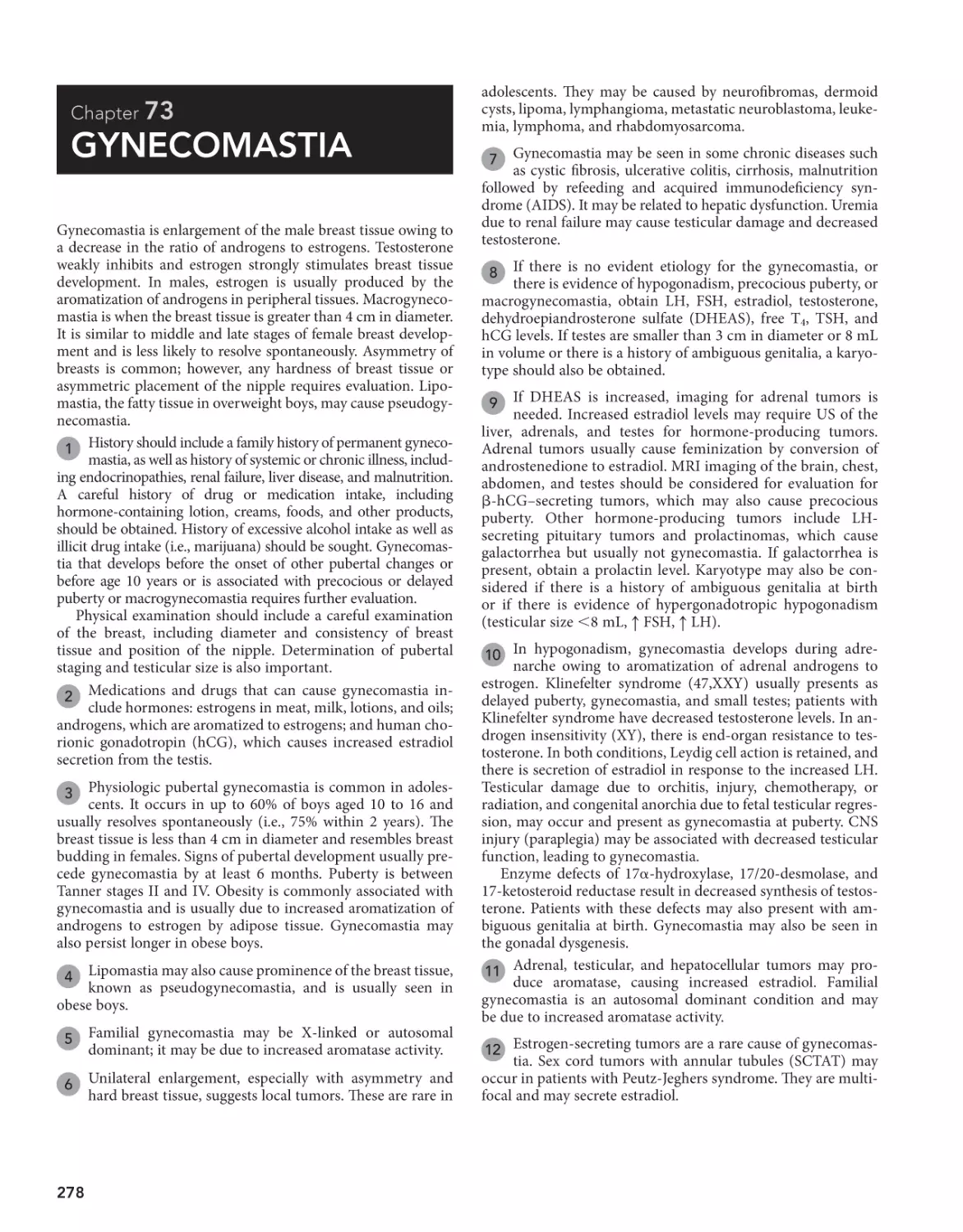
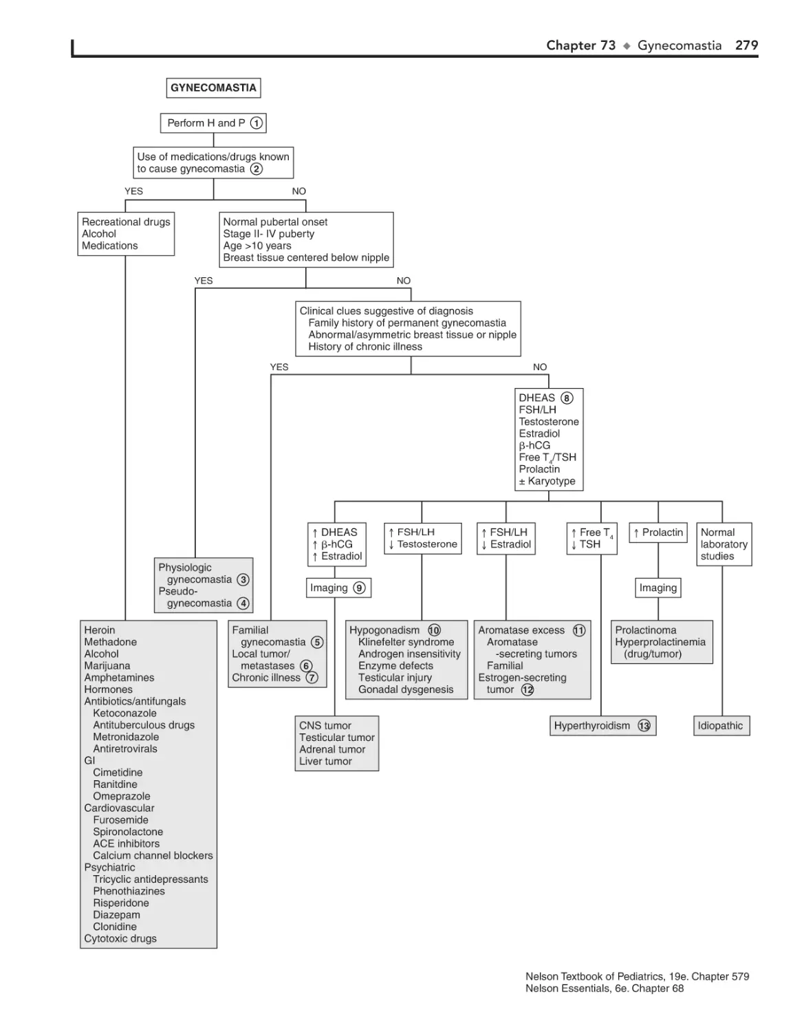

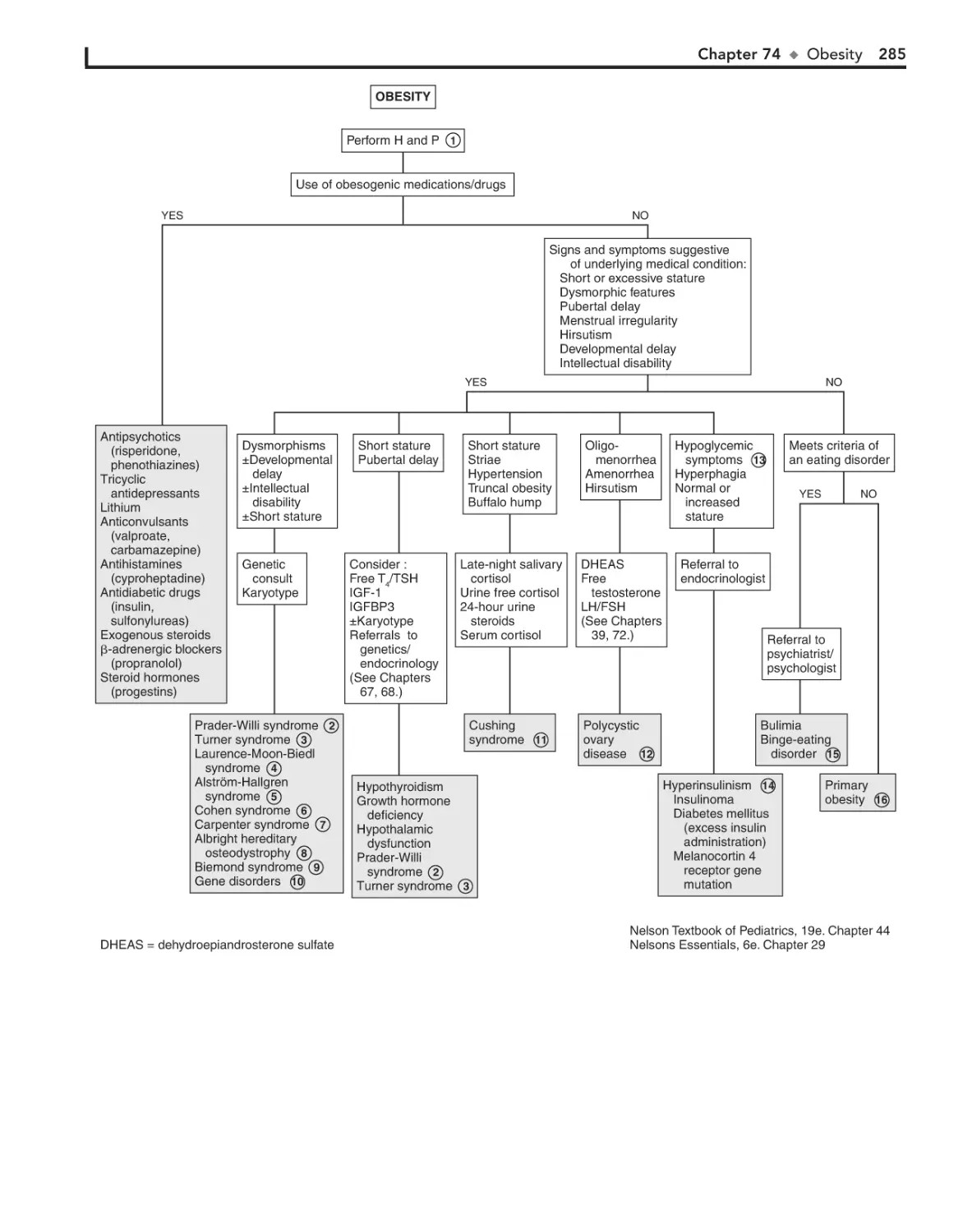


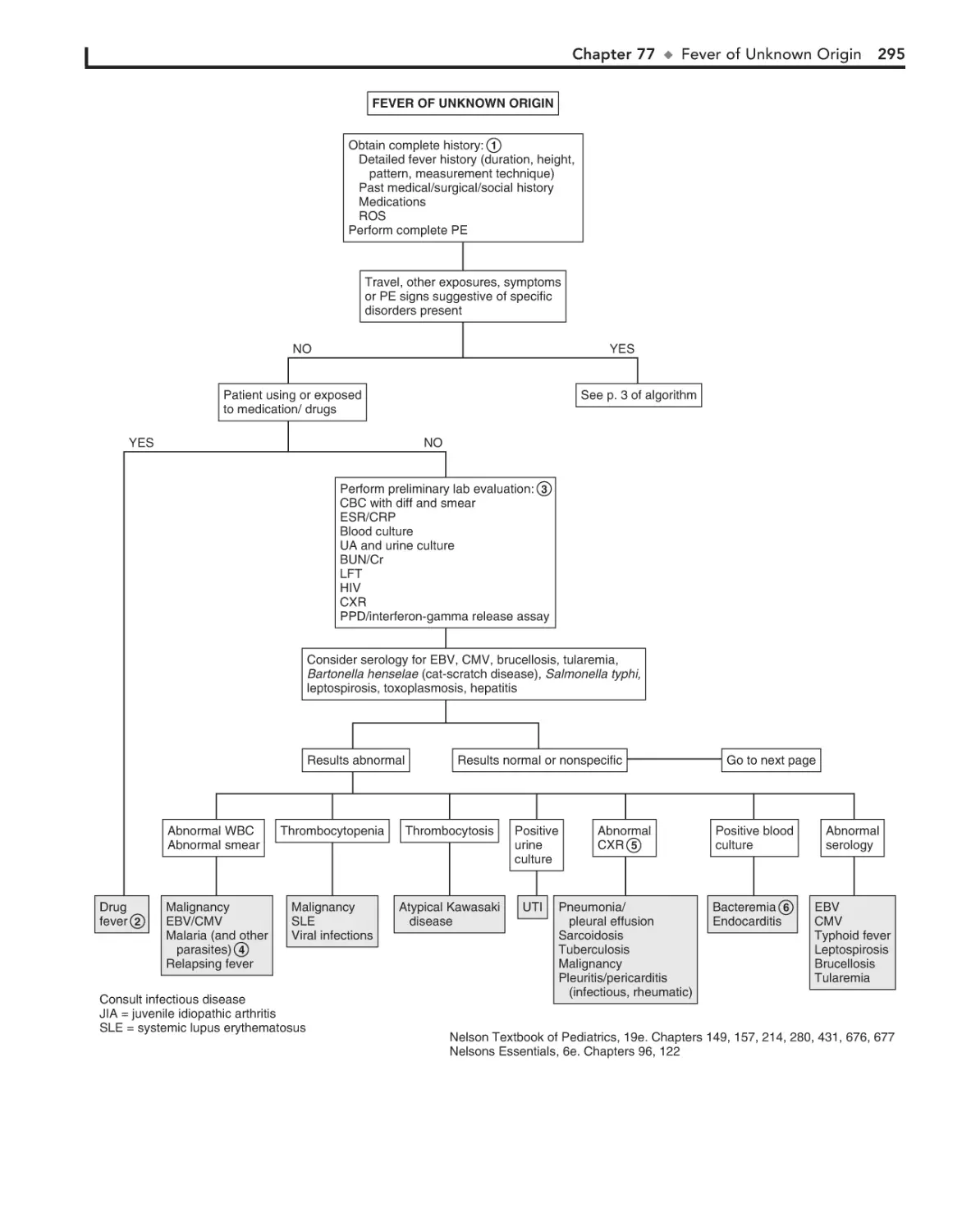

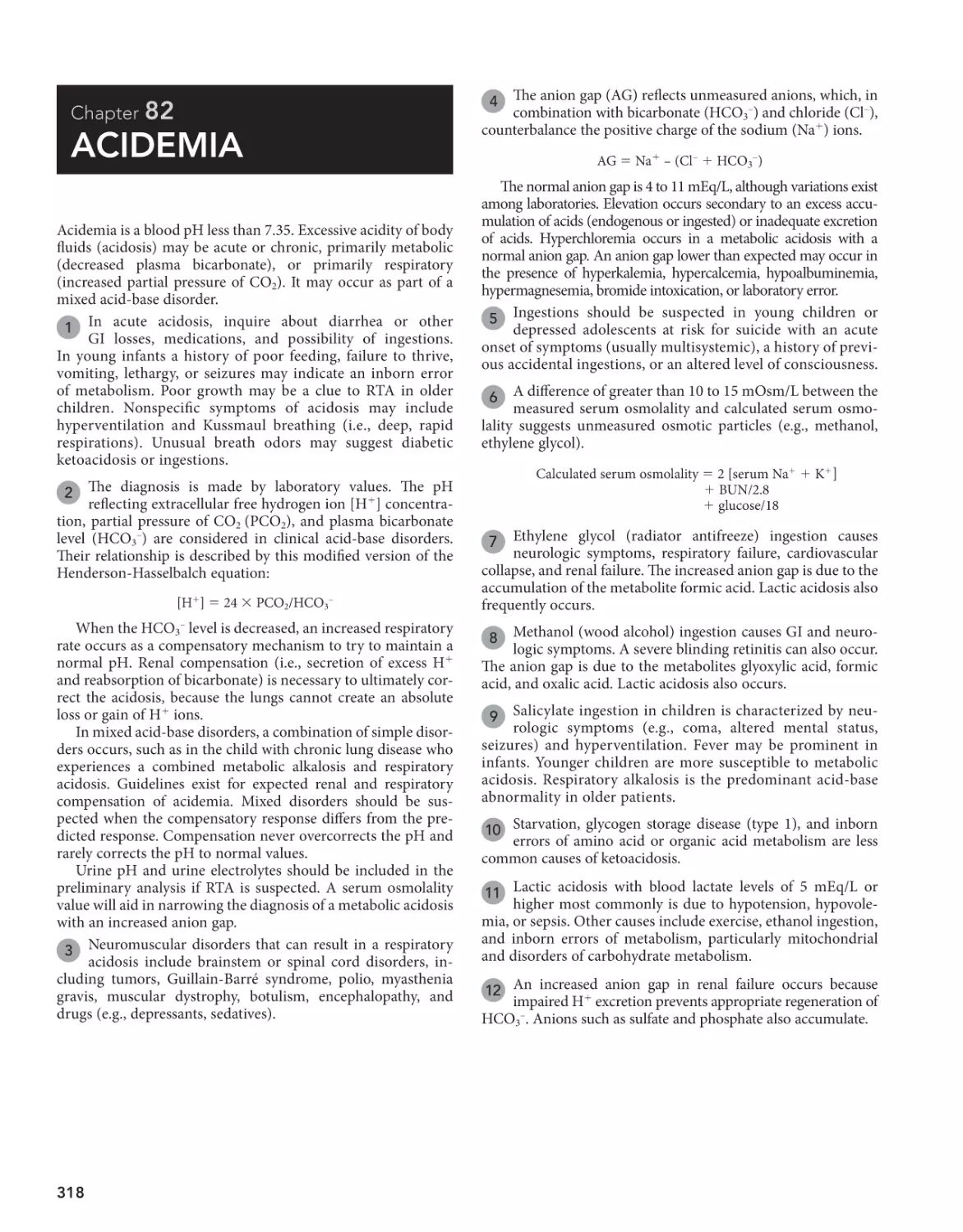
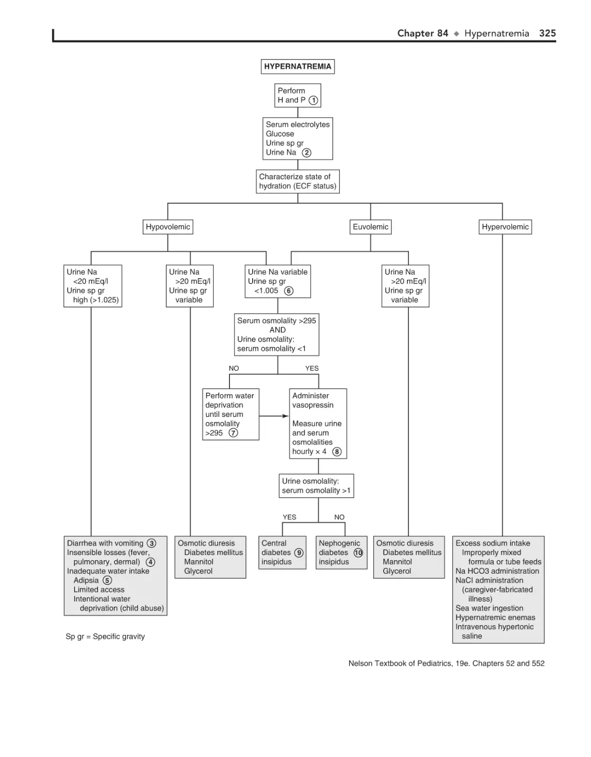
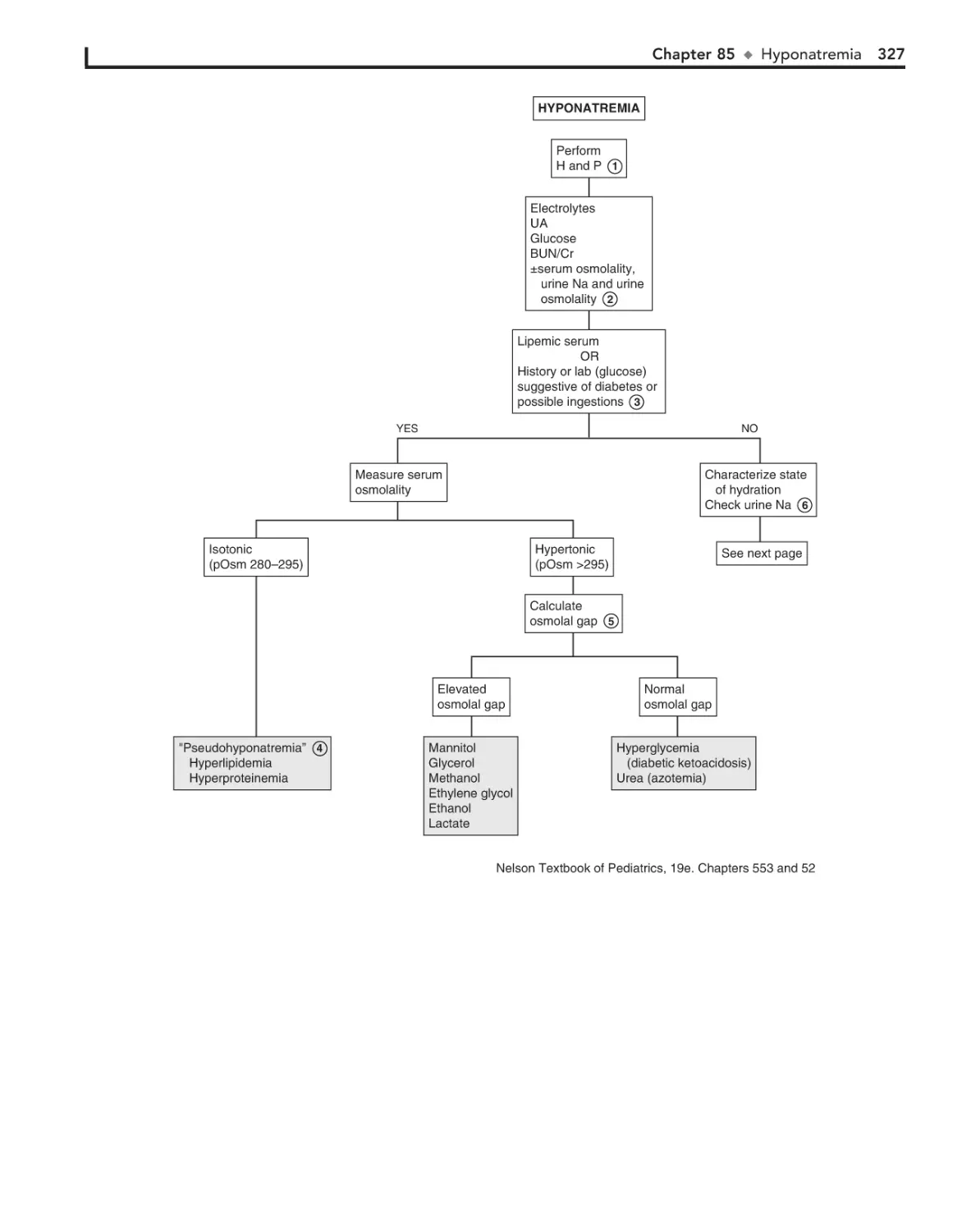
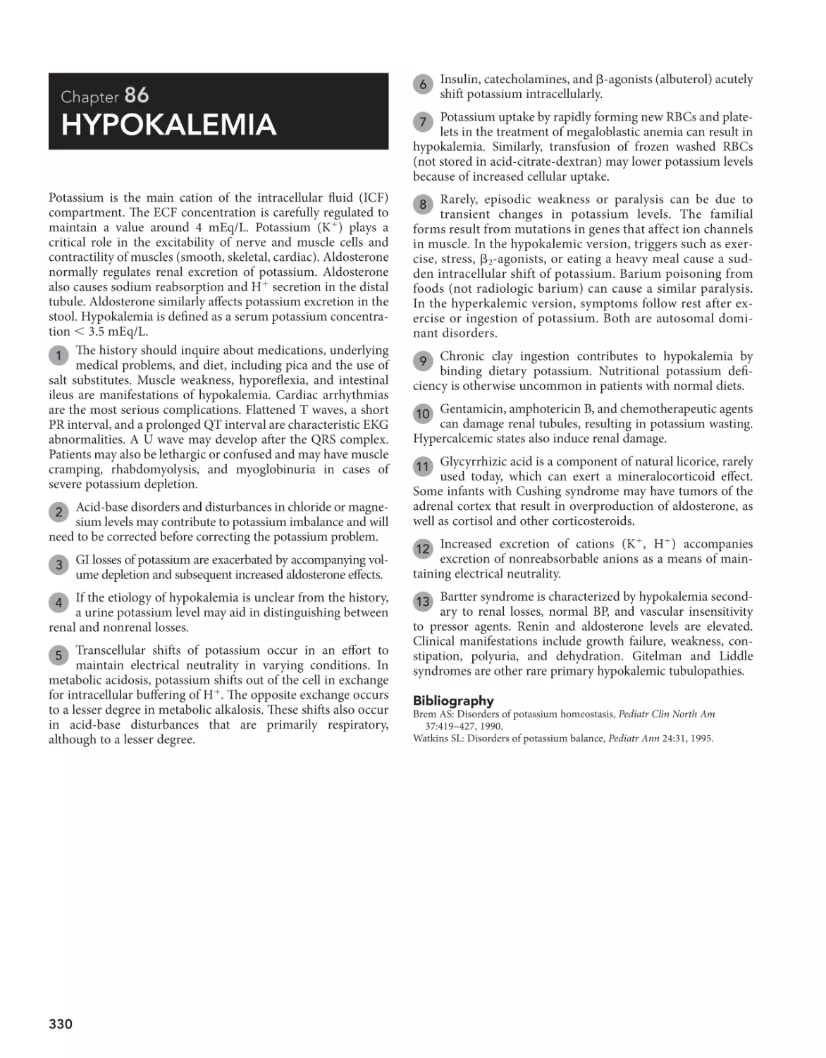
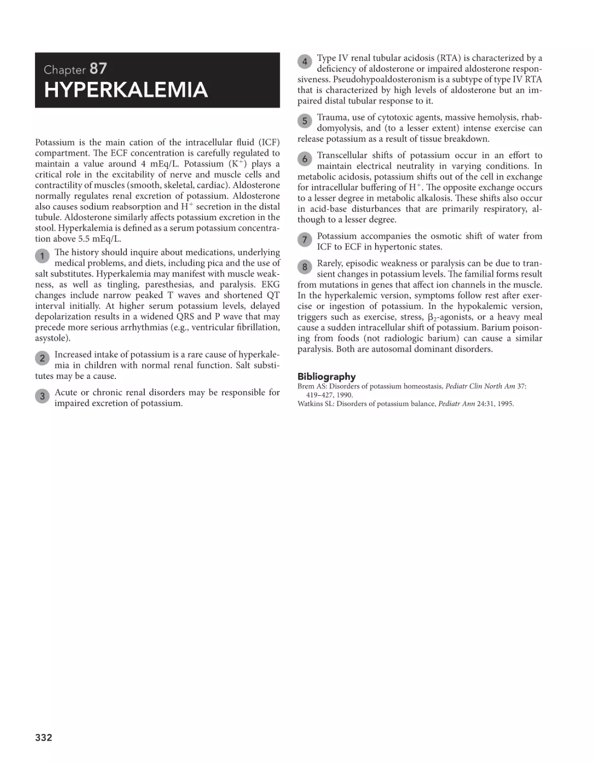
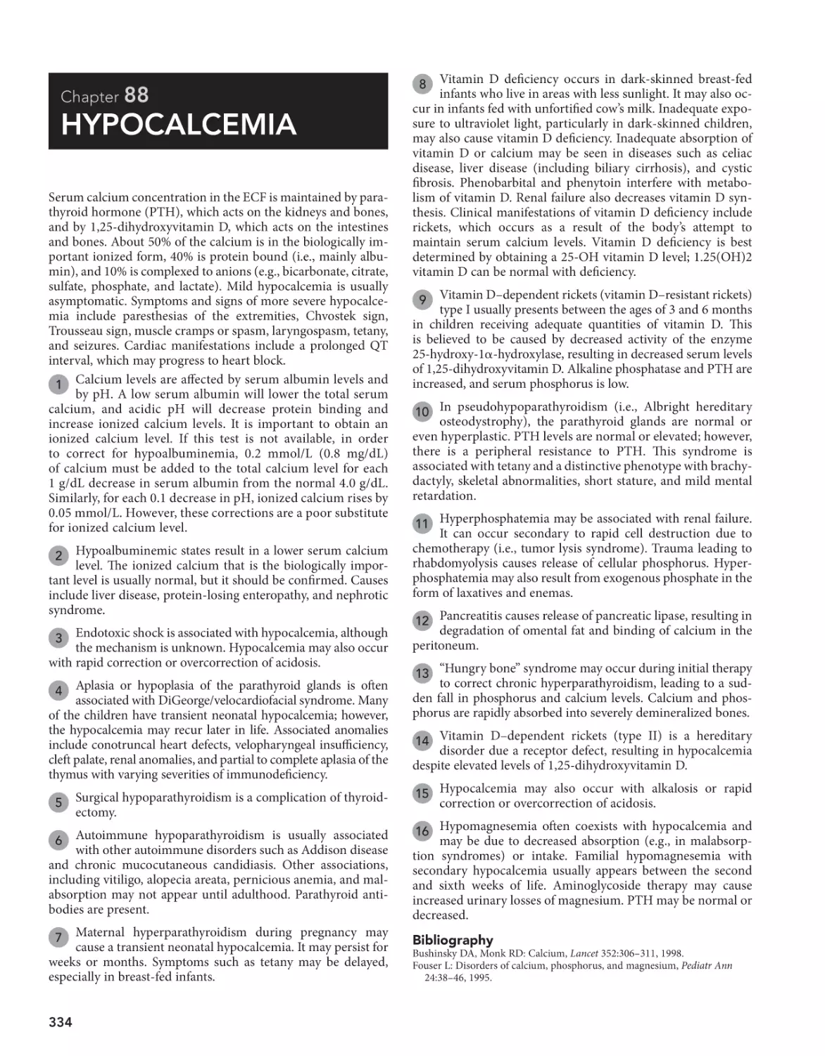
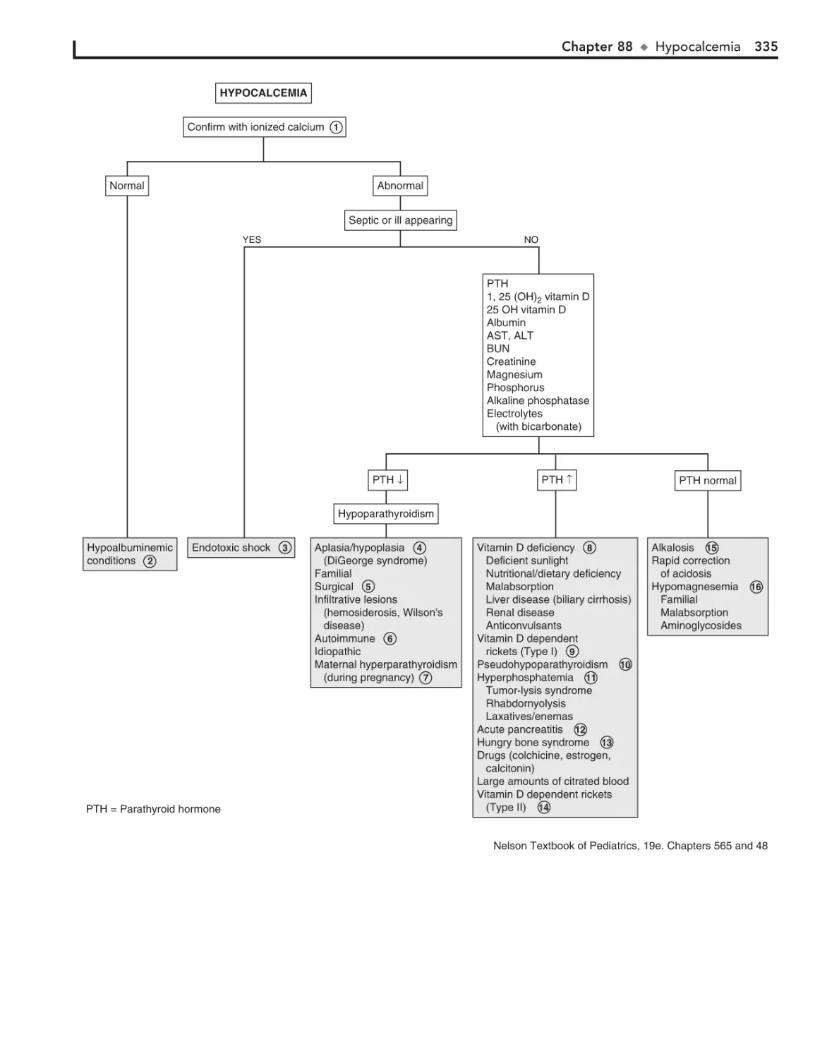


Author: Pomeranz J.A. Sabis S. Busey Sh.L. Kliegman R.M.
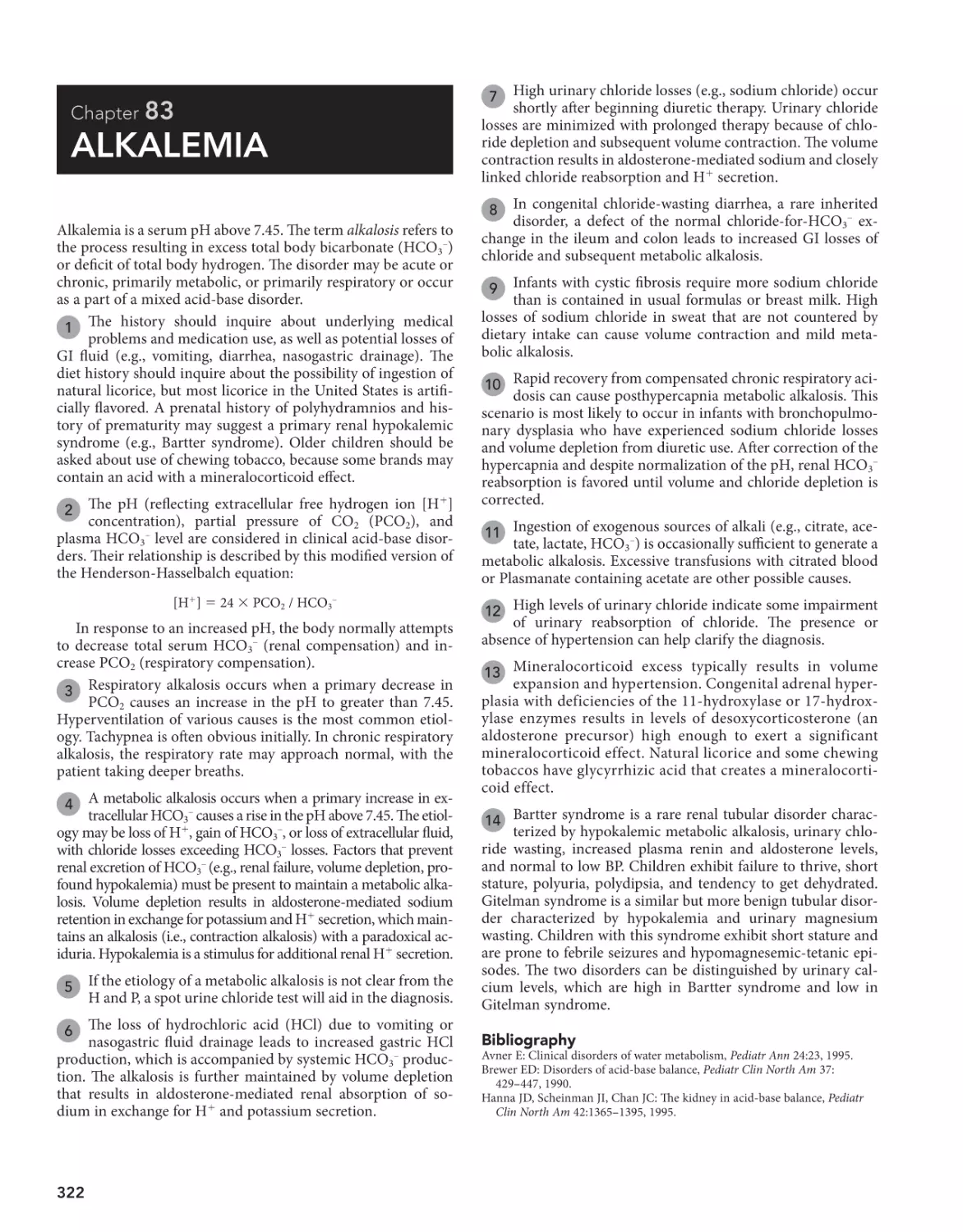
Tags: medicine pedagogy practical medicine practical guide pediatric decision-making
ISBN: 978-0-323-29854-4


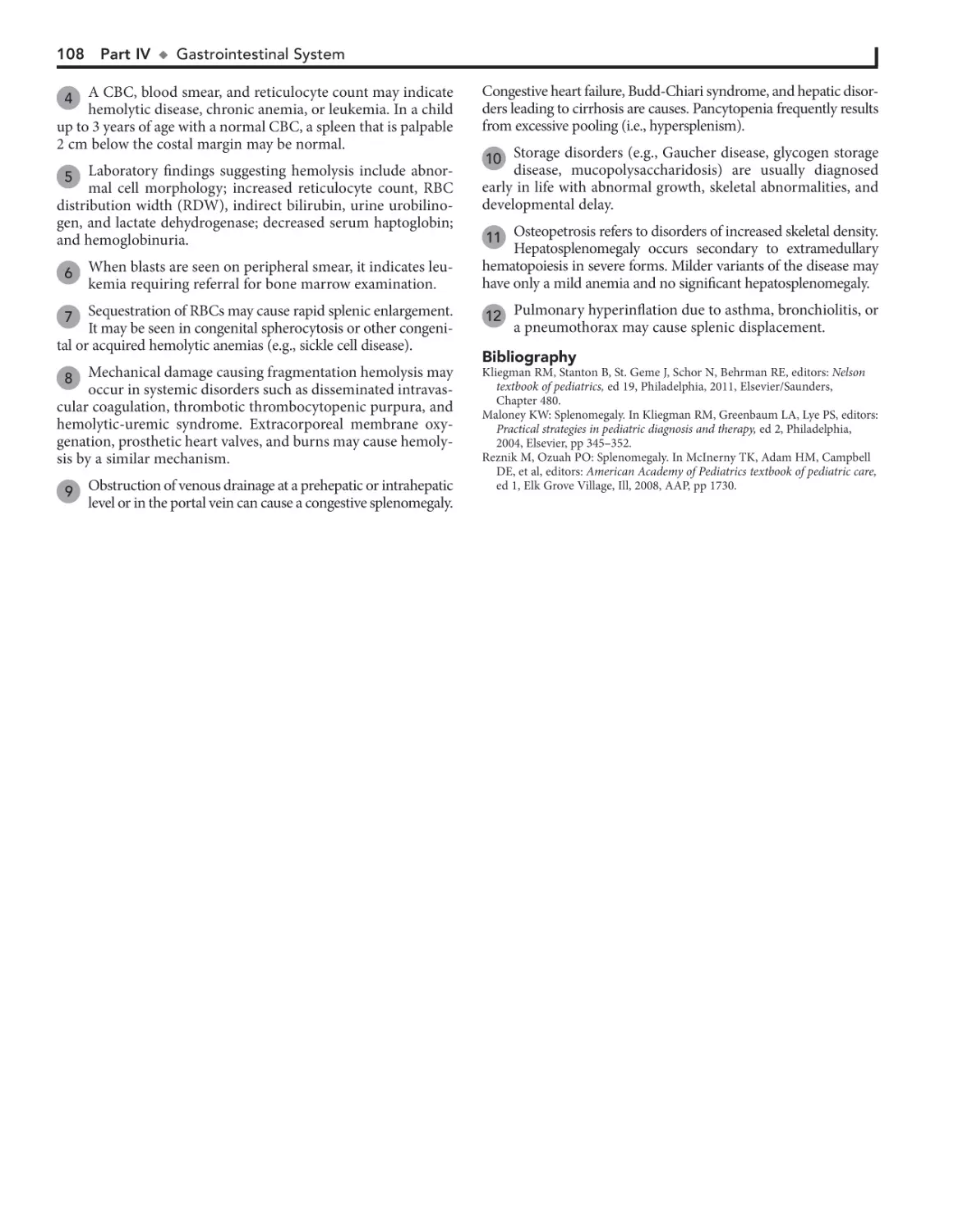
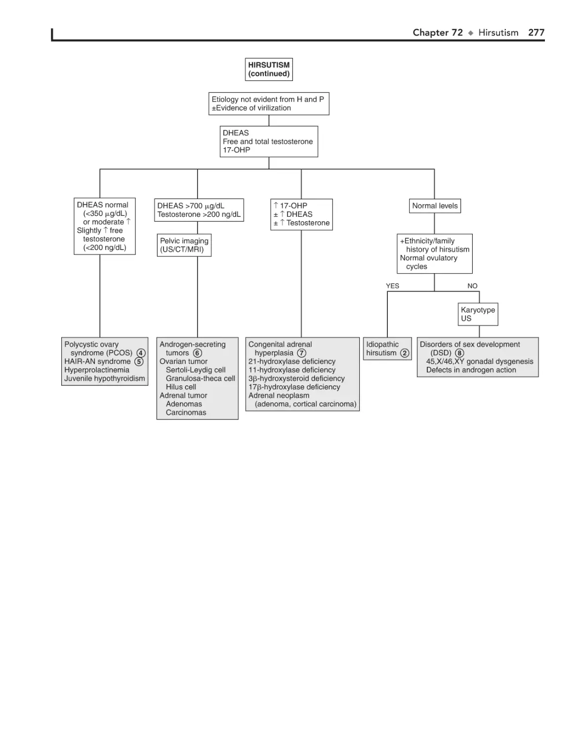
Text
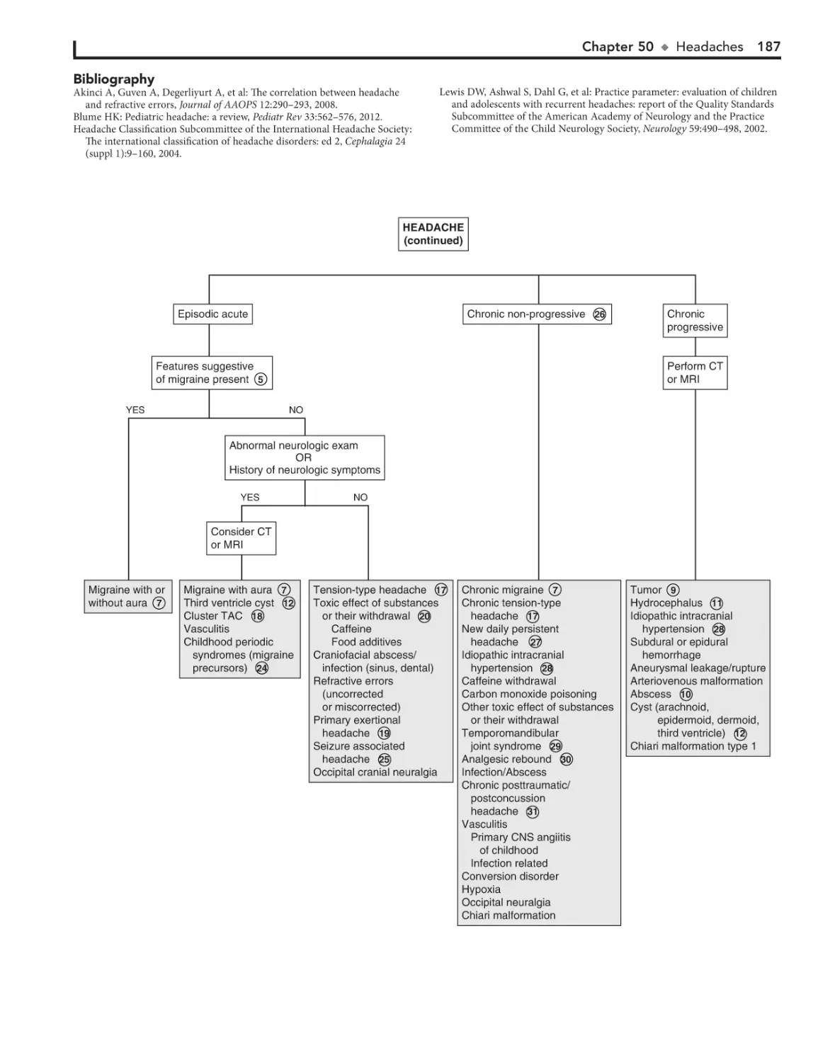
Don’t Forget Your Online Access to
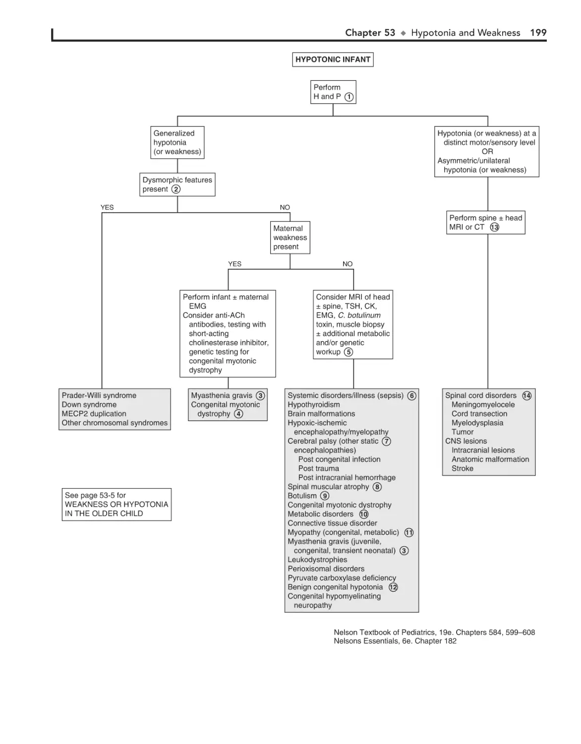
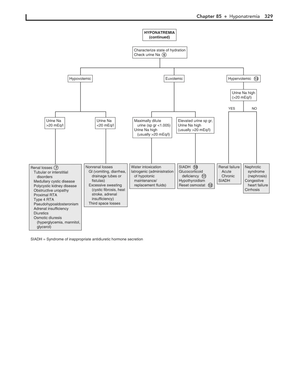
Mobile. Searchable. Expandable.
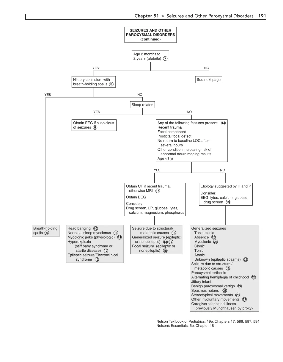
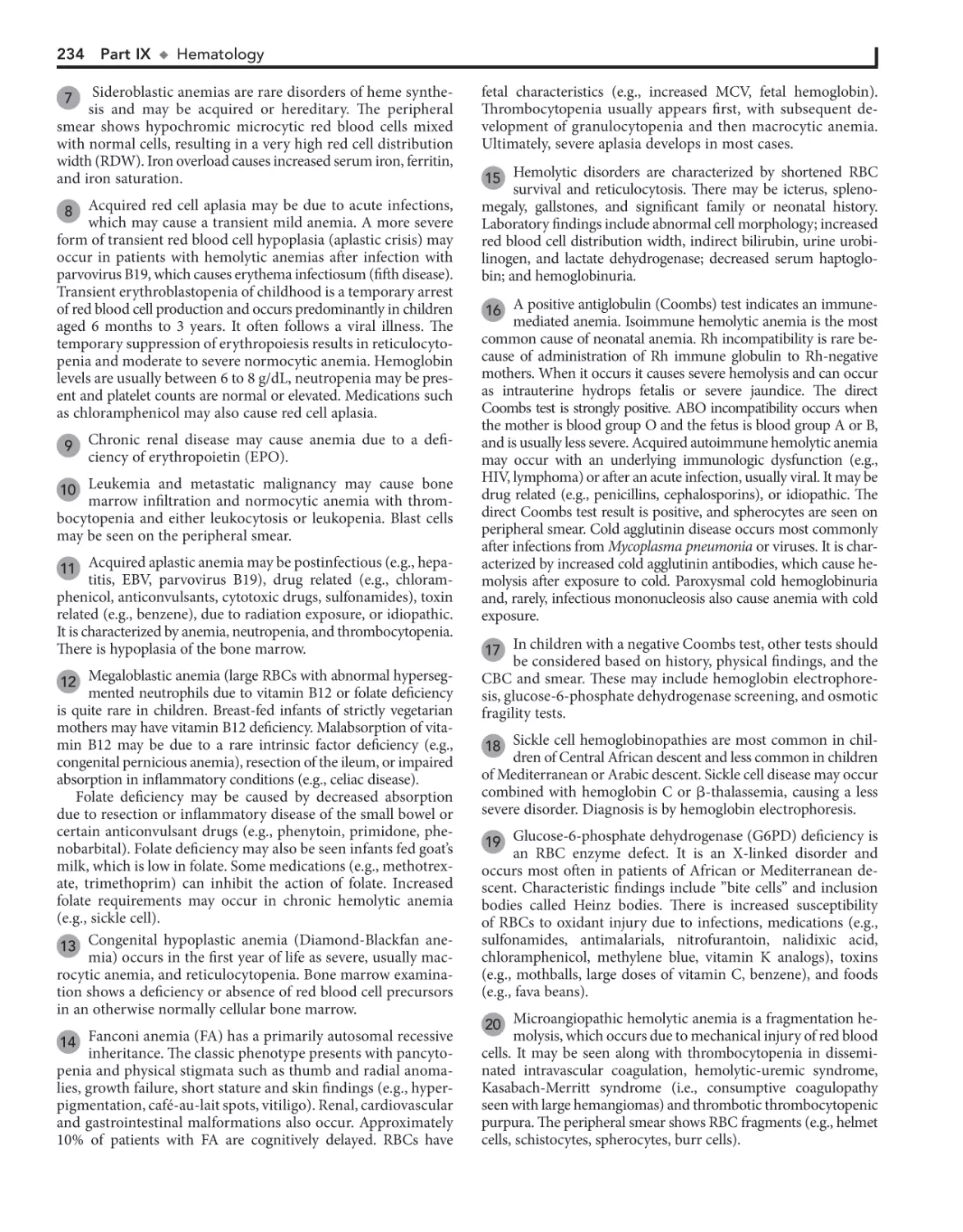
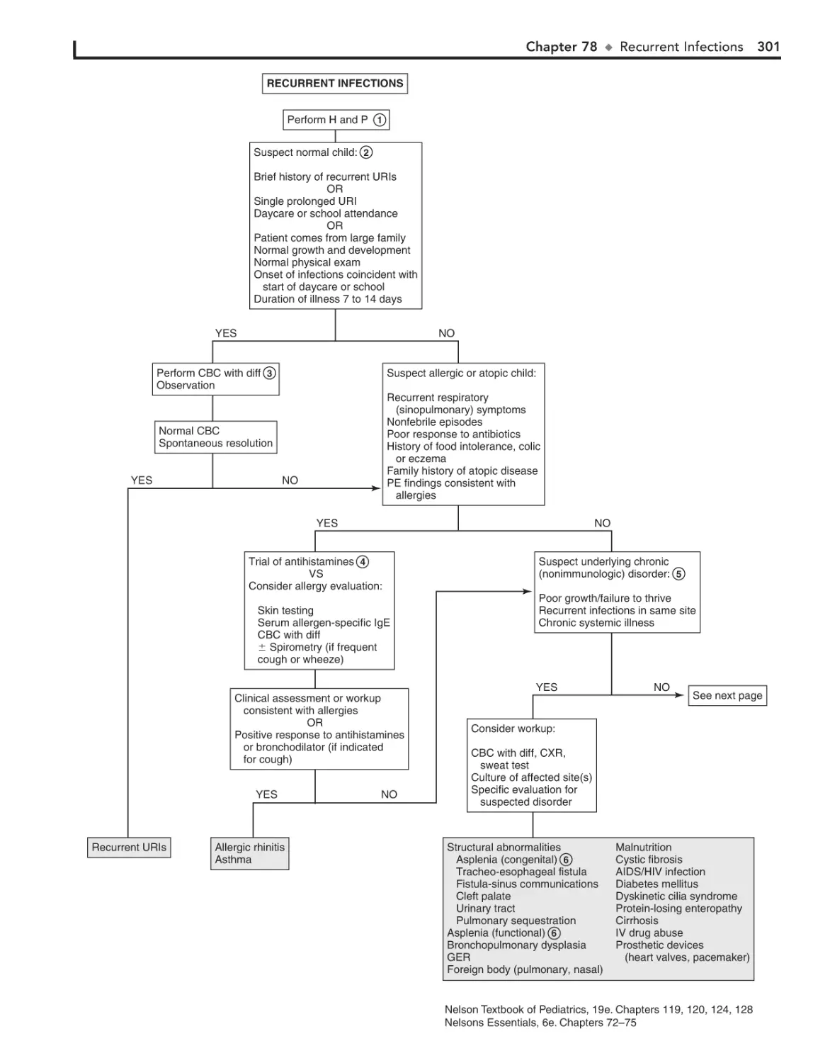
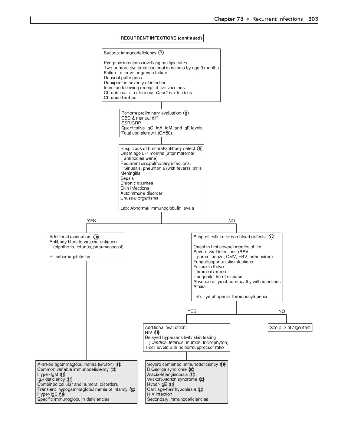
ACCESS it on any Internet-ready device
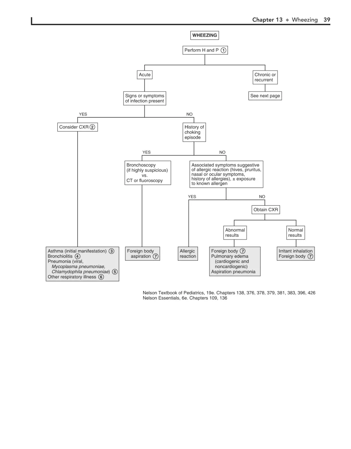
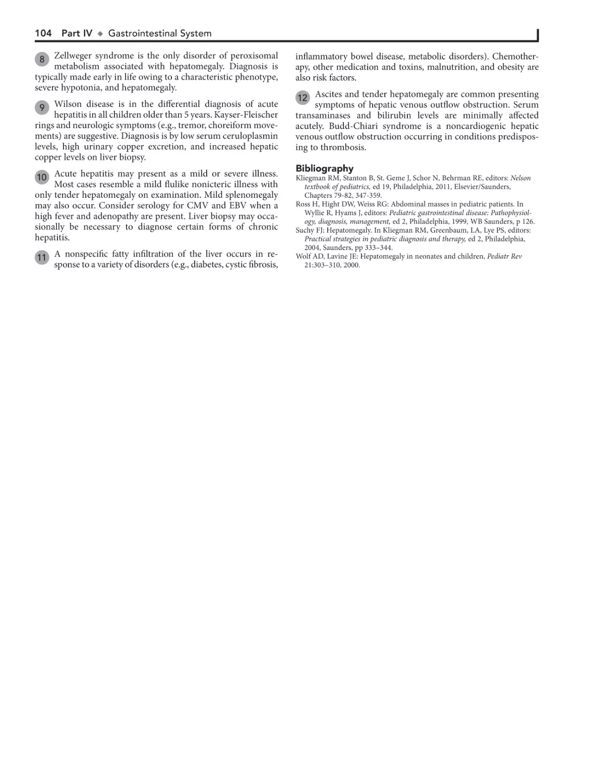
SEARCH all Expert Consult titles you own
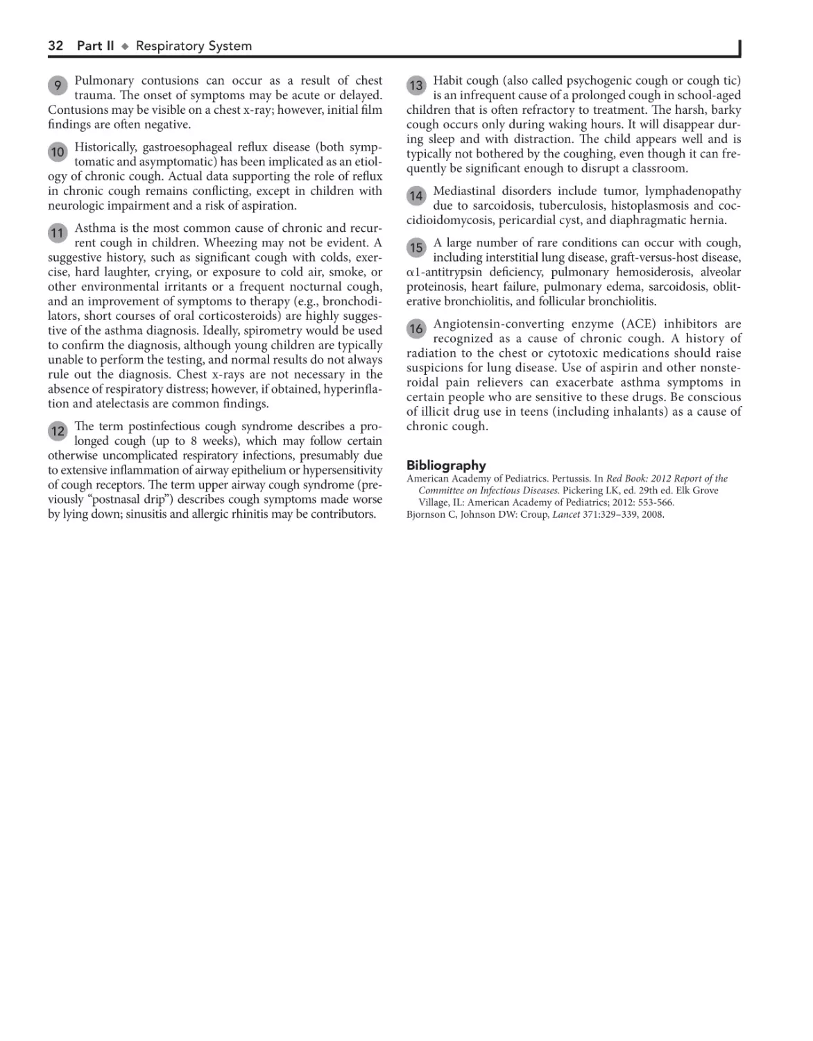
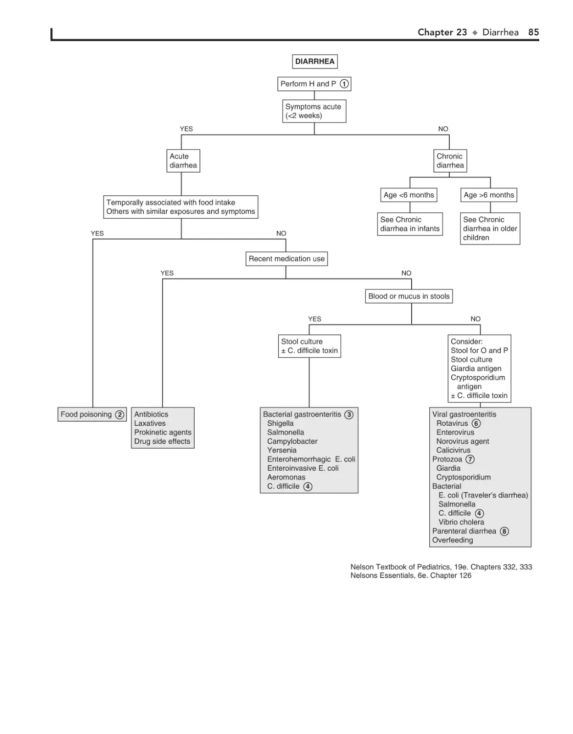
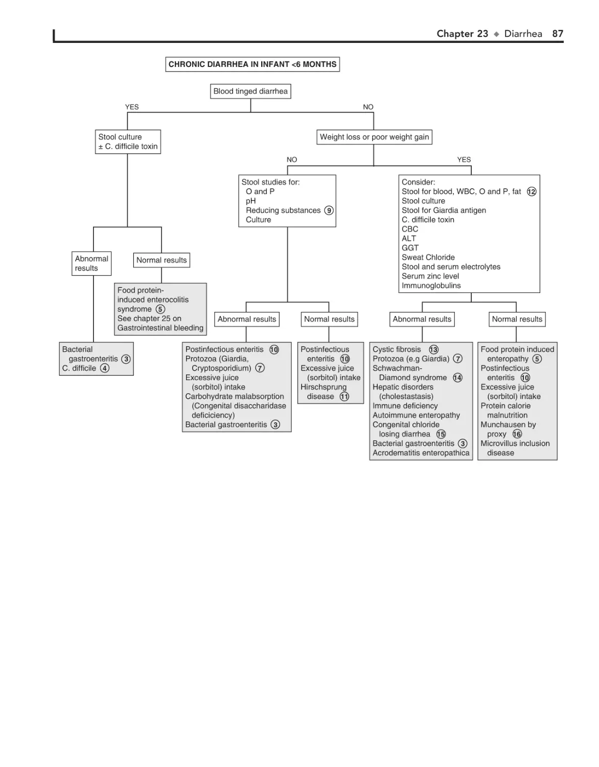
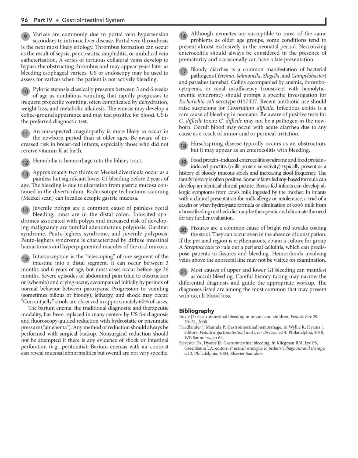
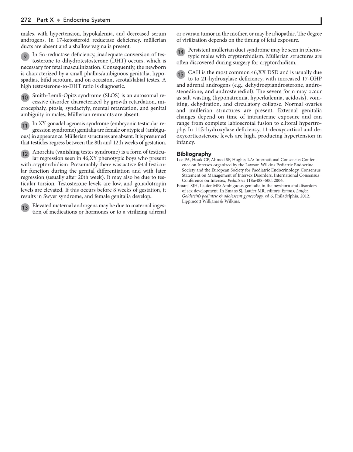
LINK to PubMed abstracts
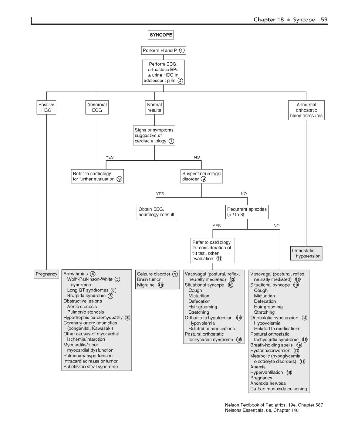
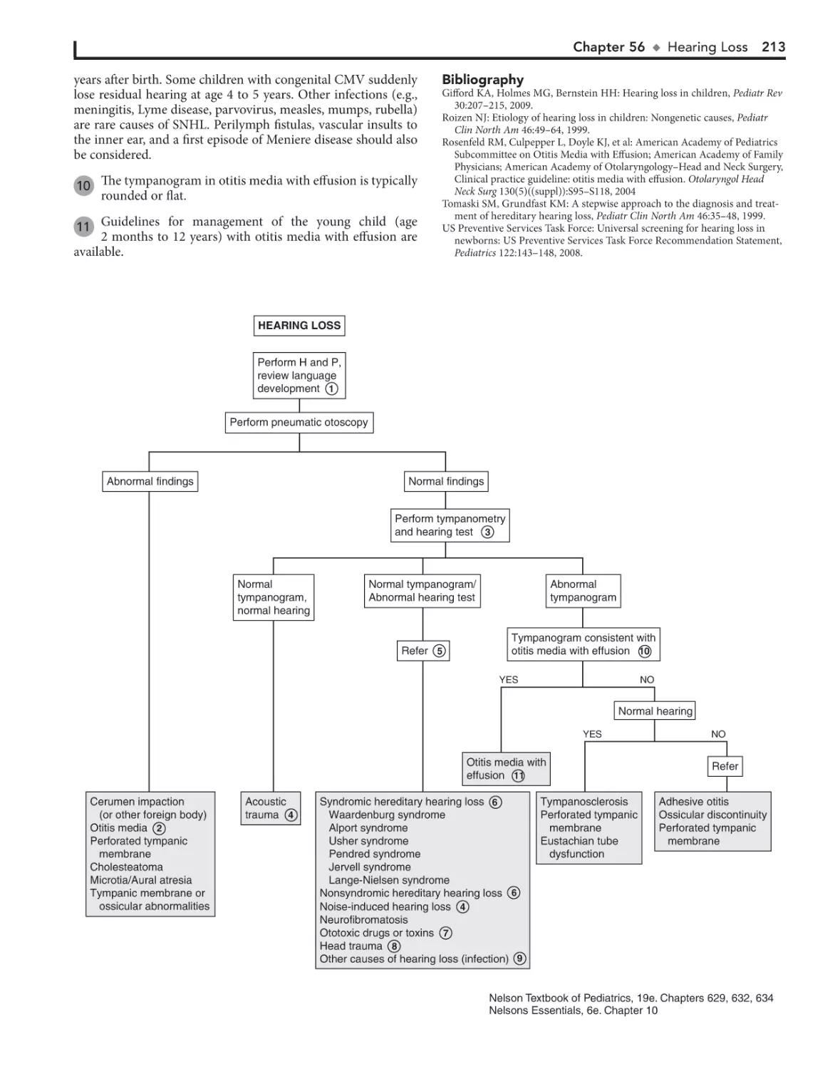
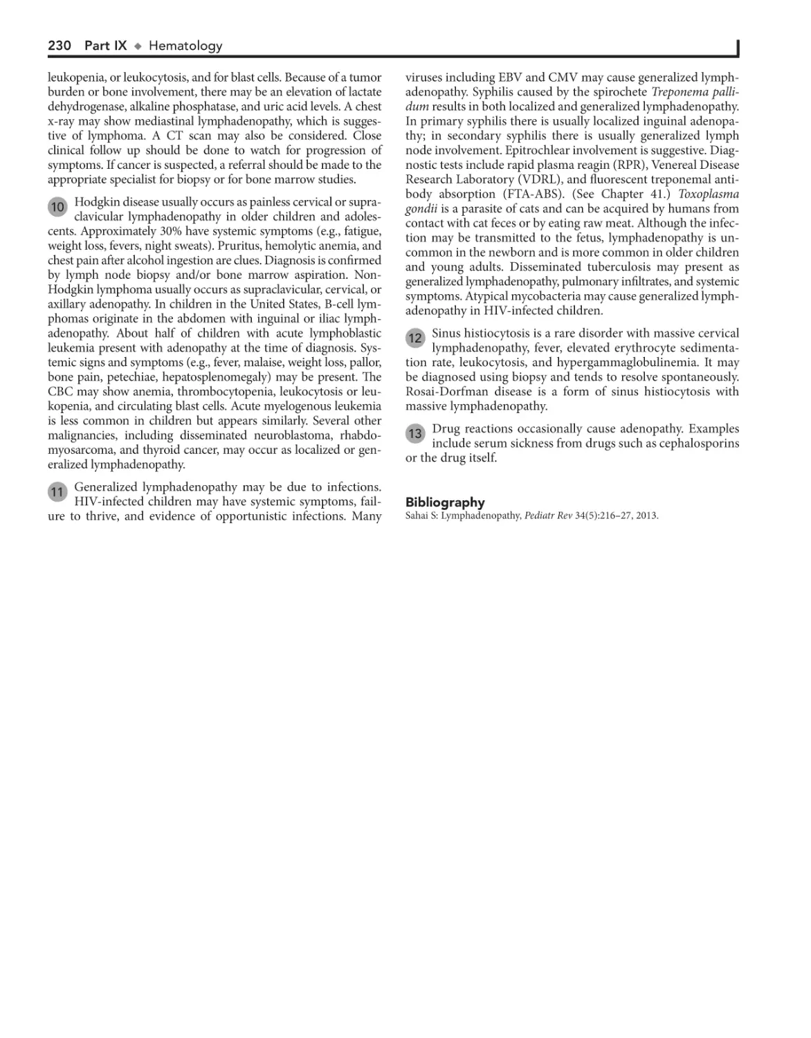
ALREADY REGISTERED?
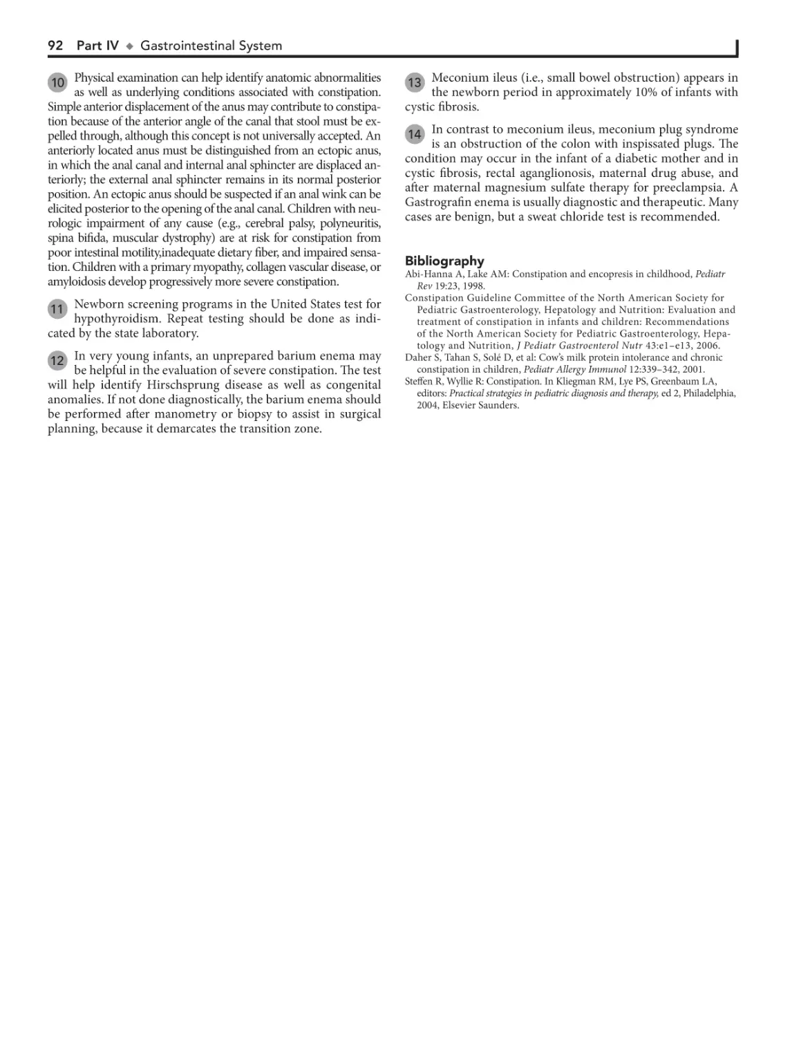
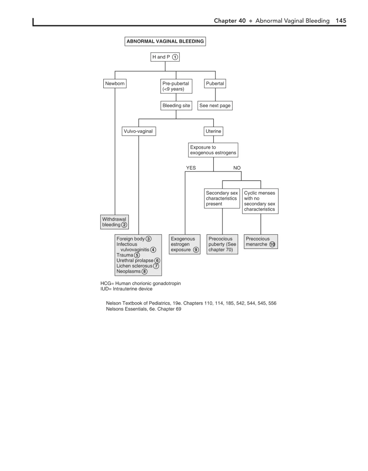
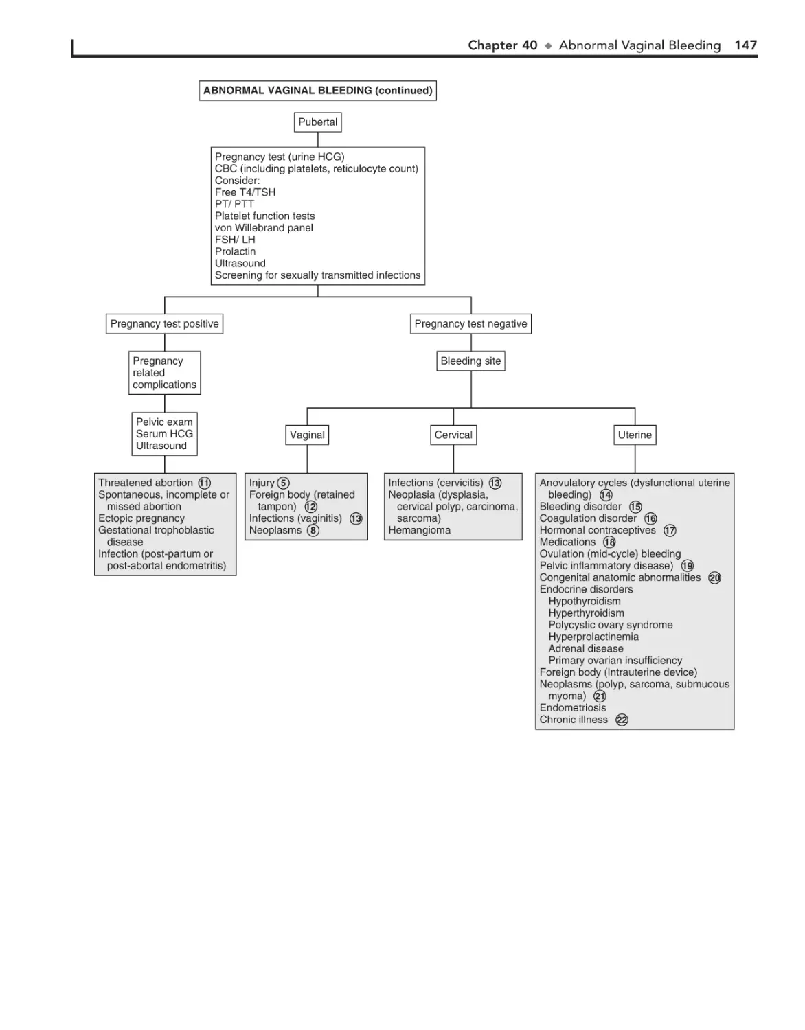
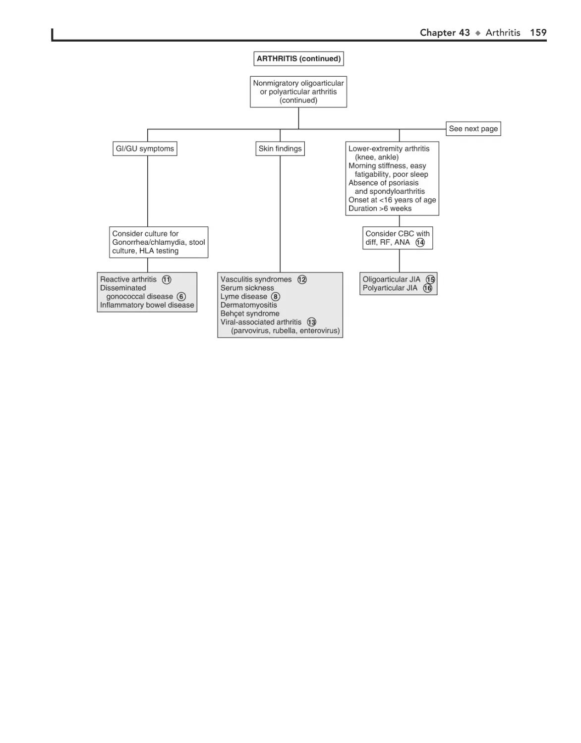
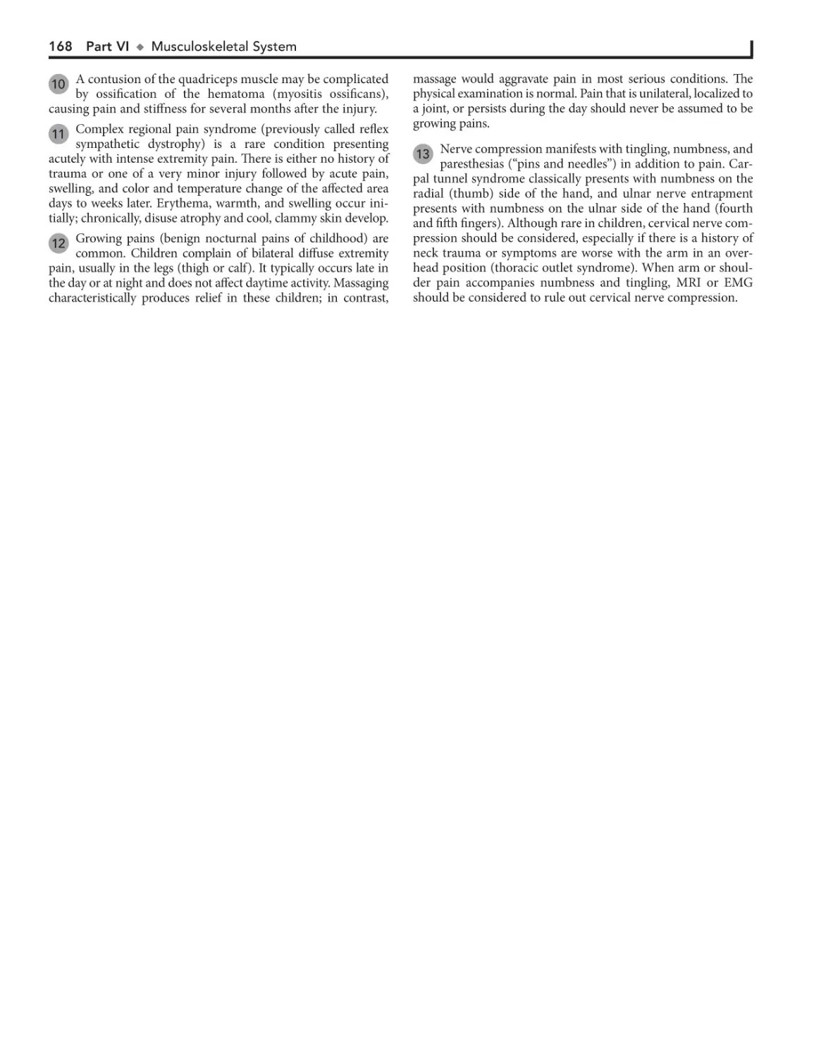
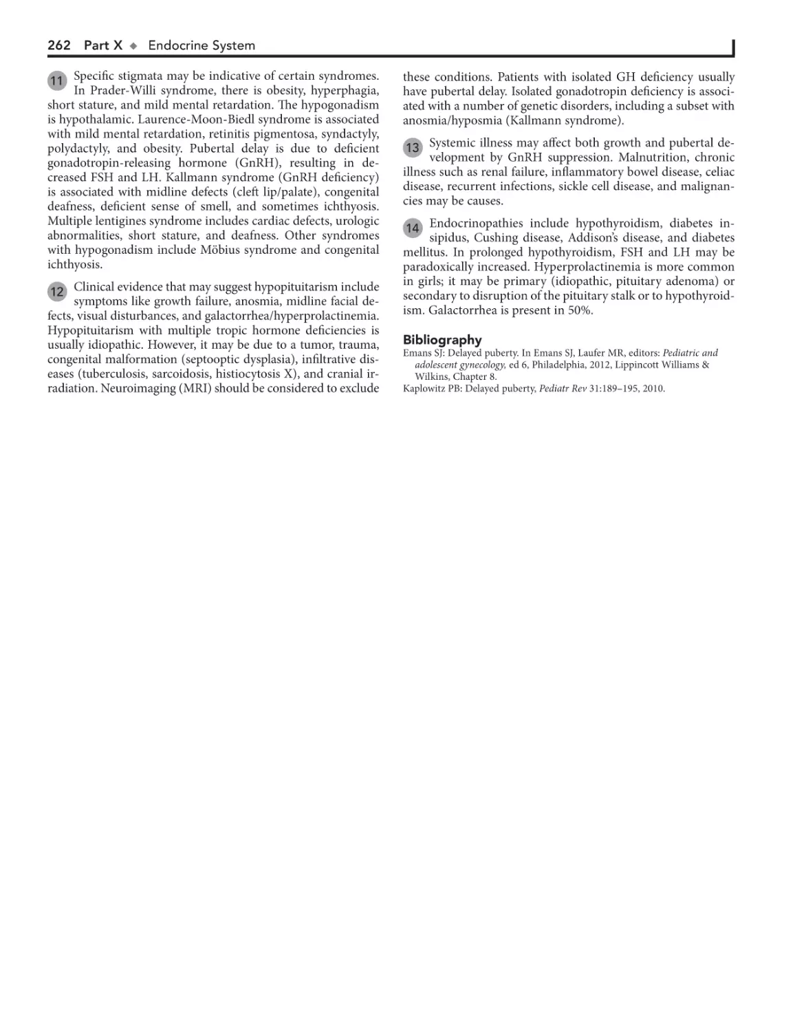
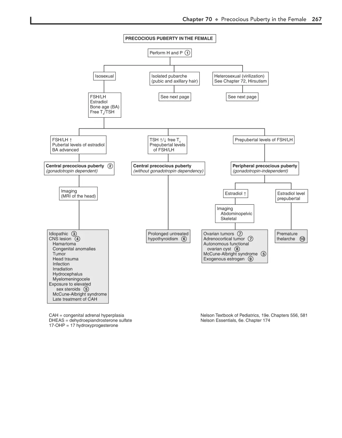
FIRST-TIME USER?
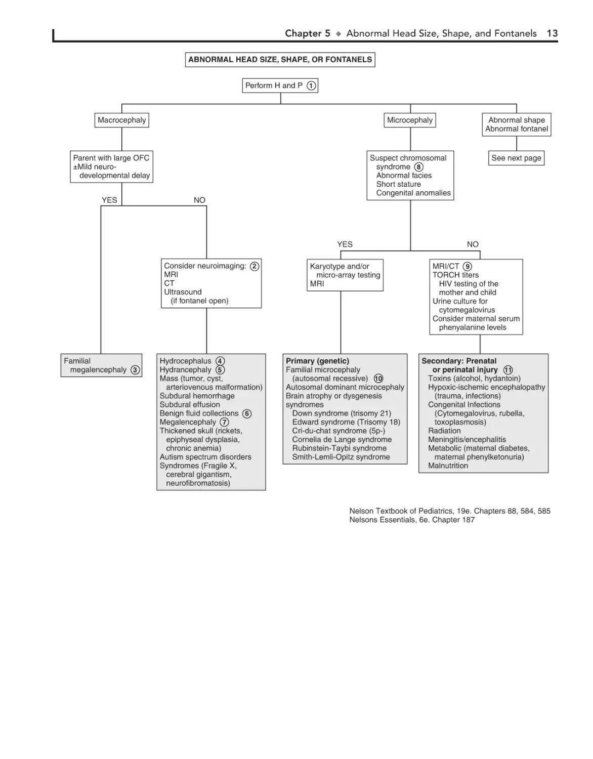
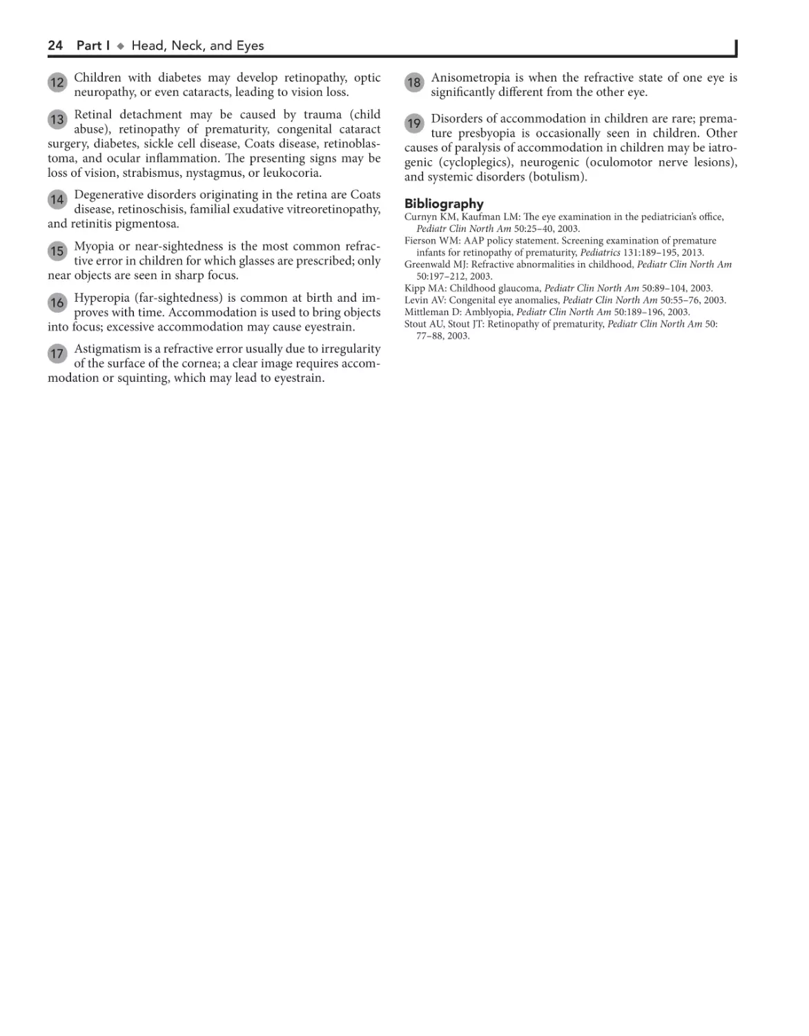
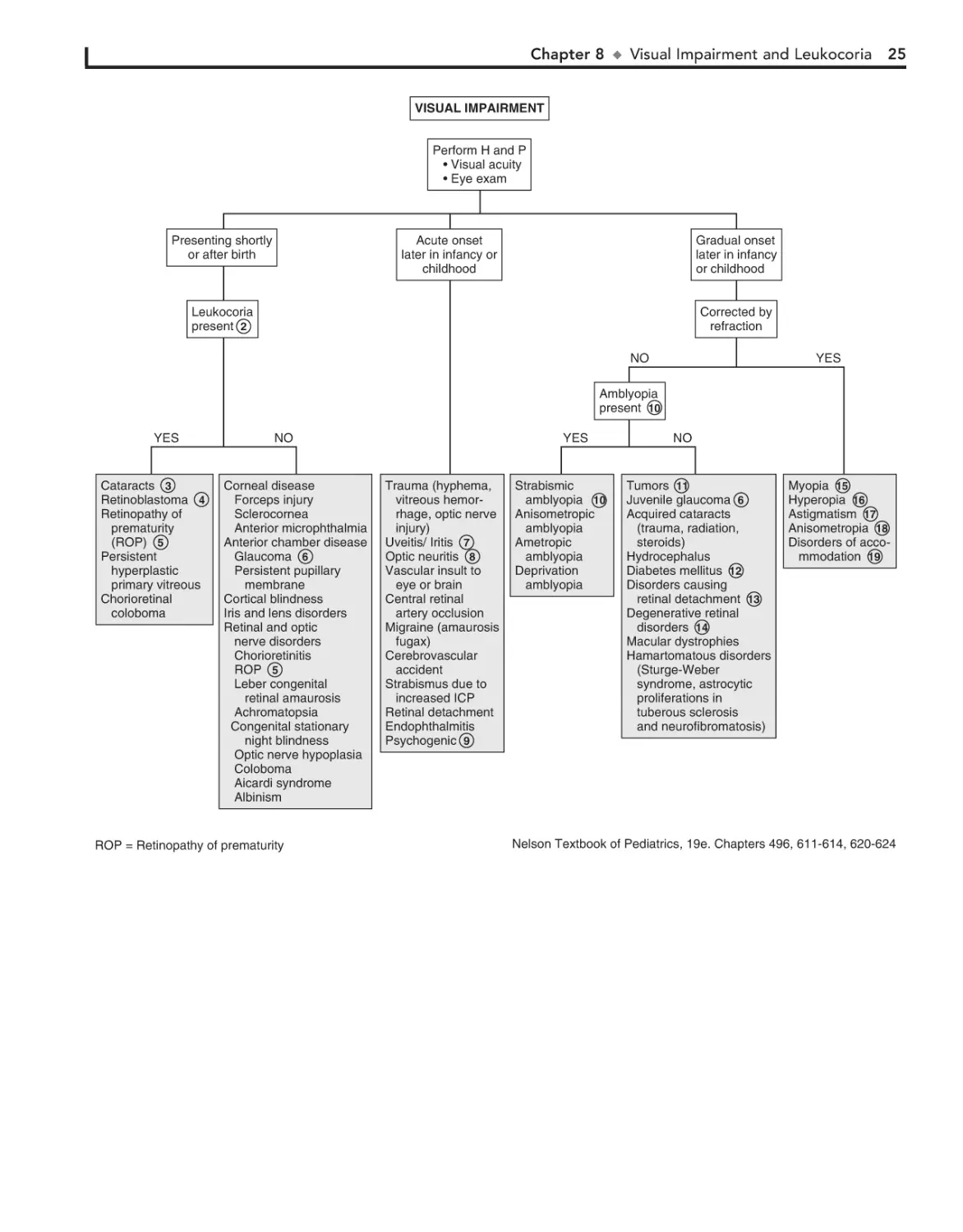
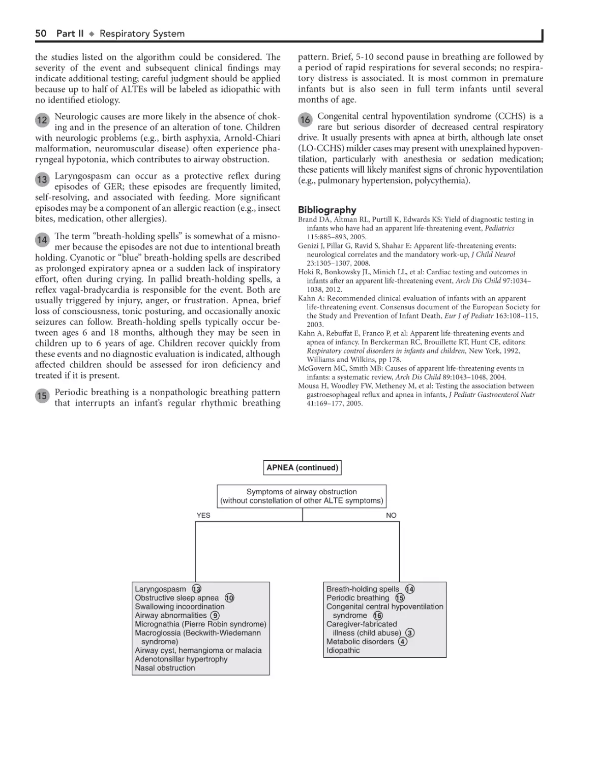
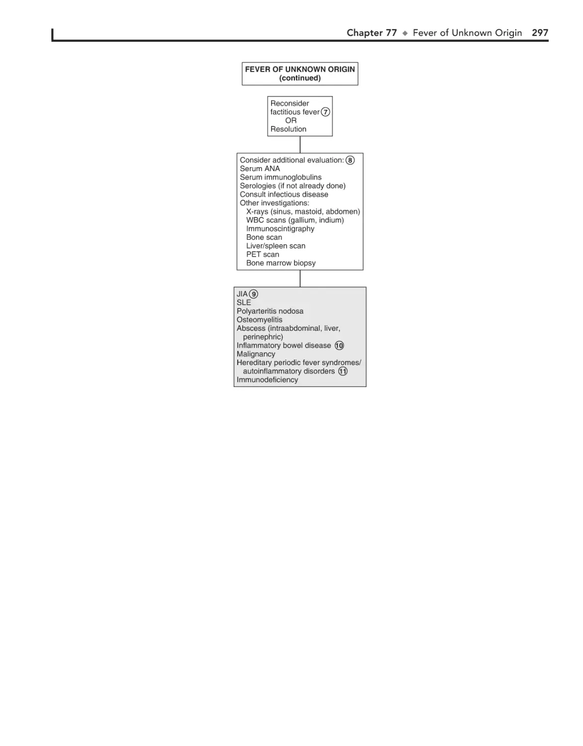
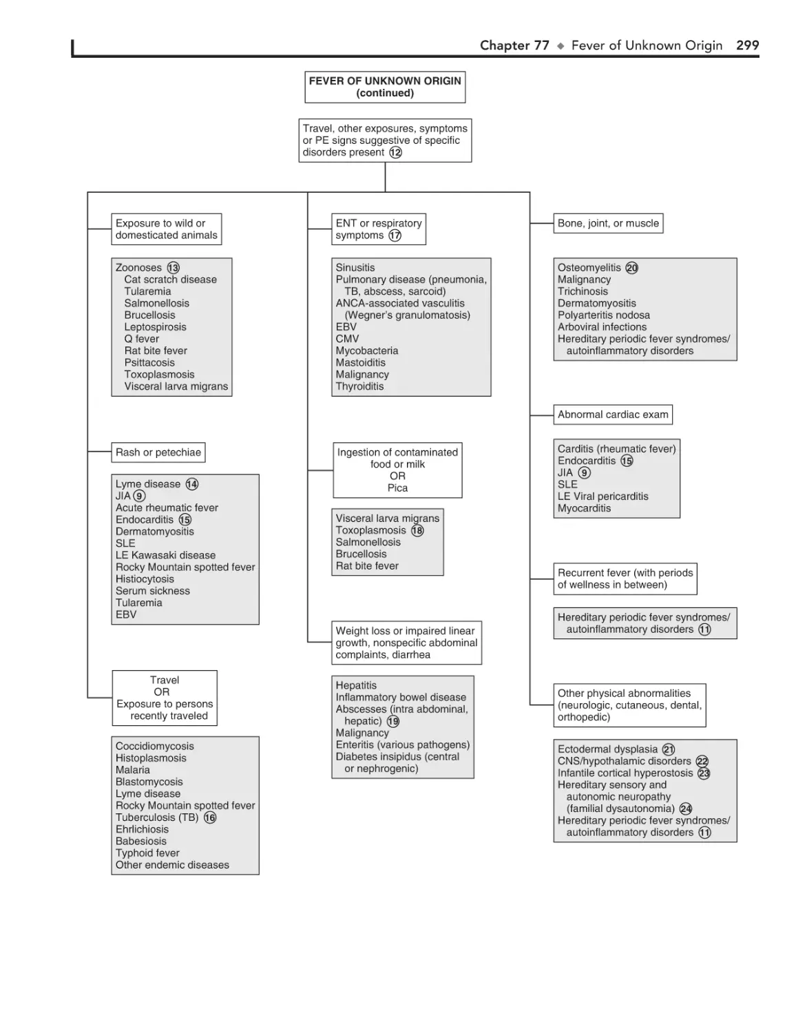
1. Log in at expertconsult.com
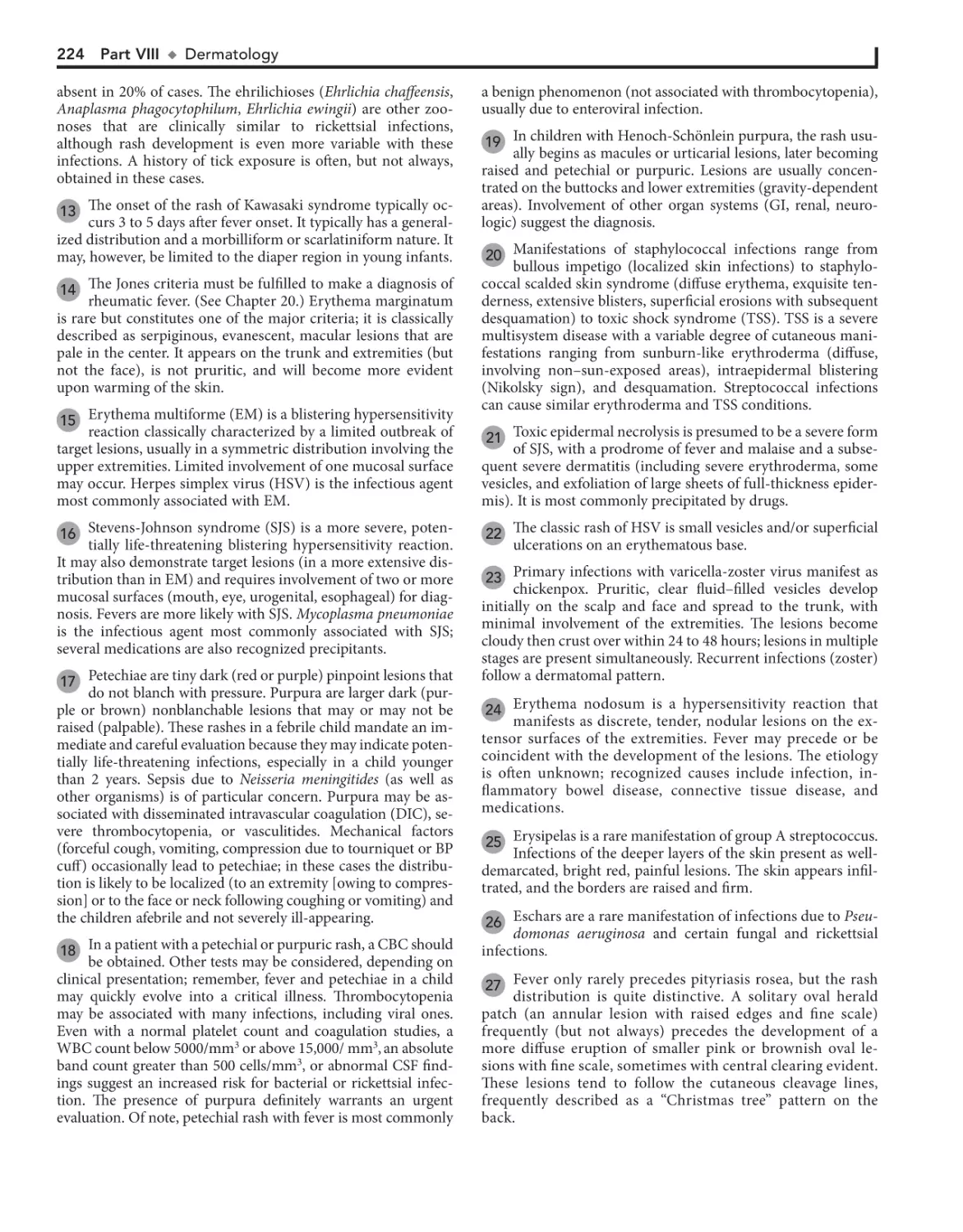
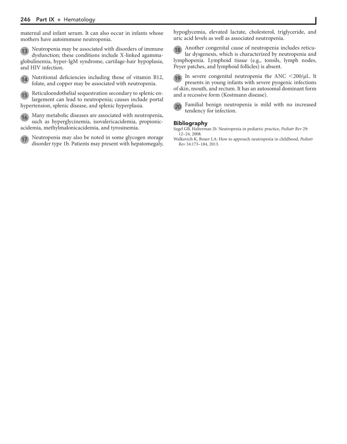
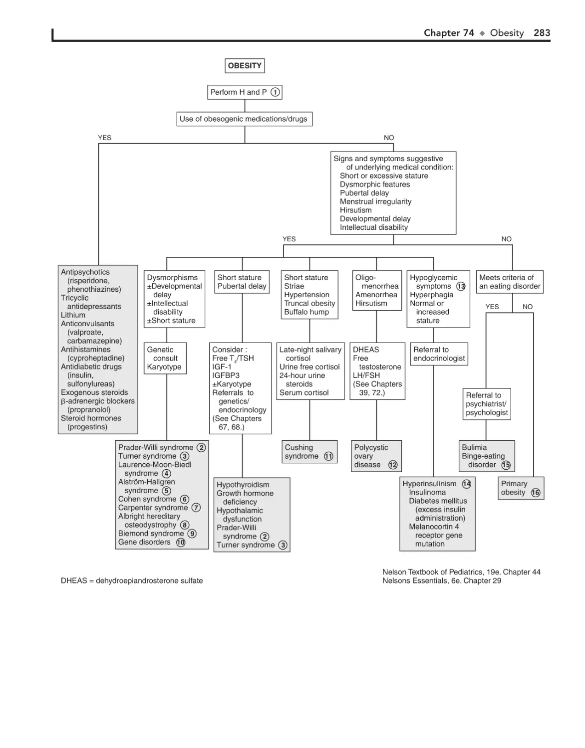
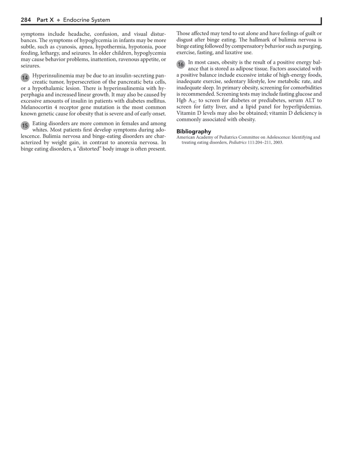
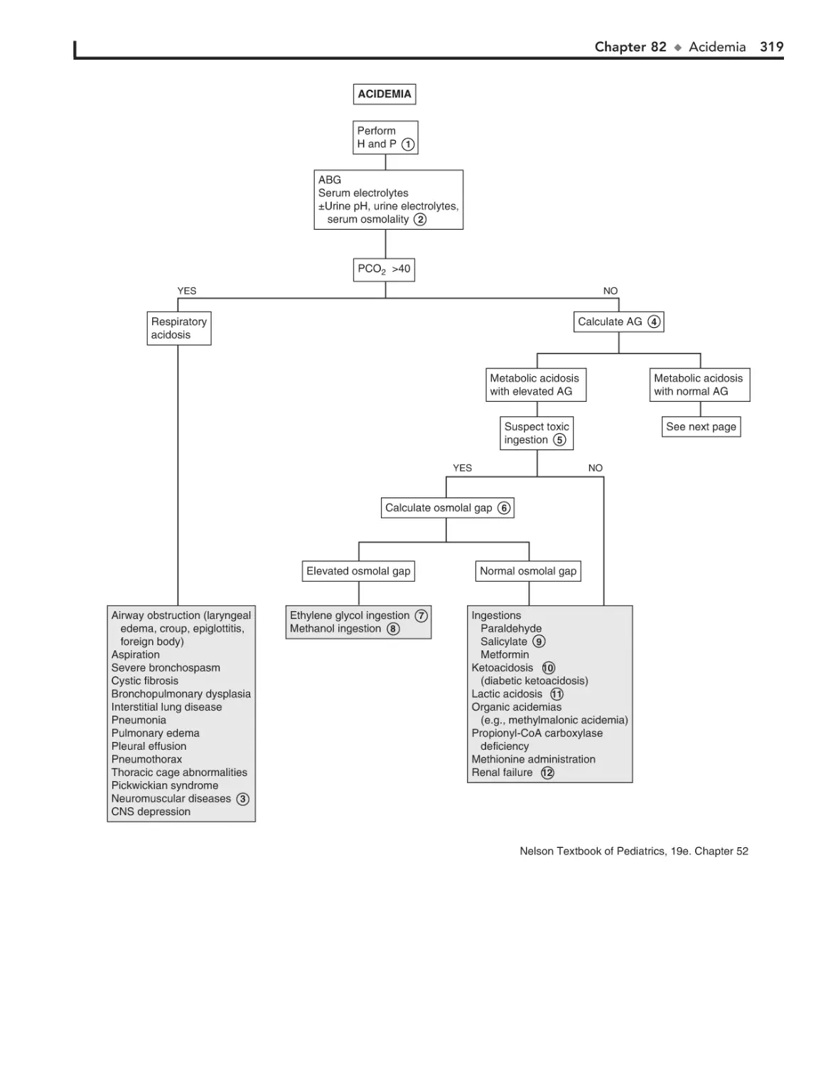
1. REGISTER
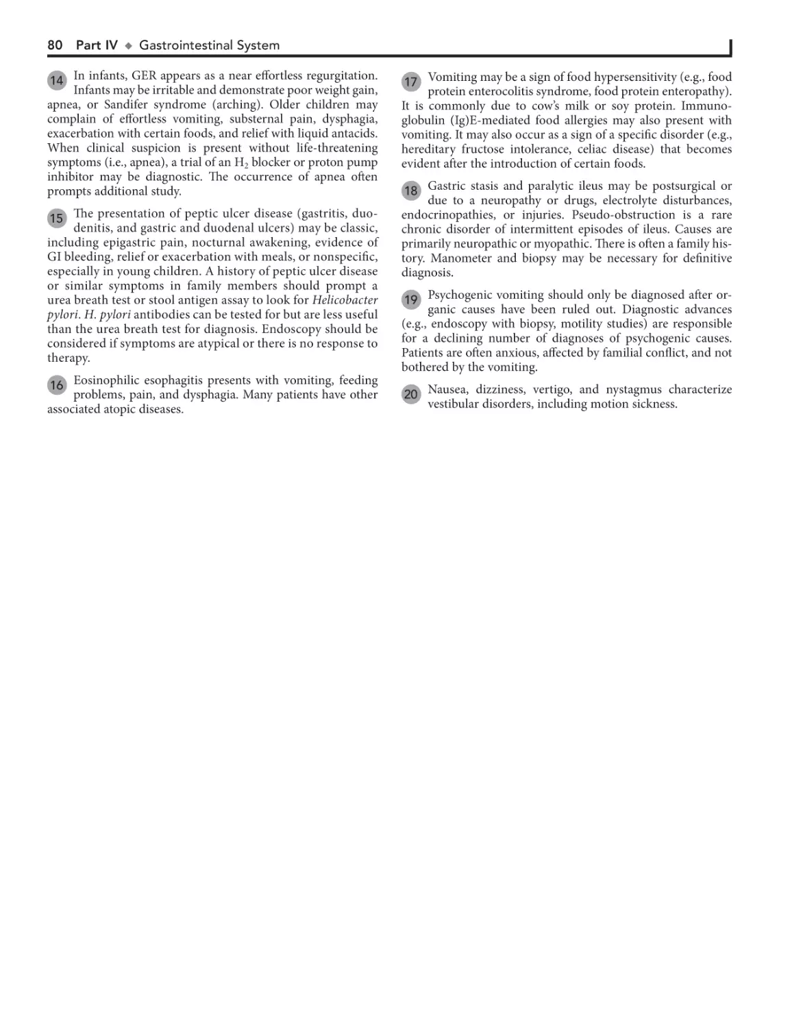
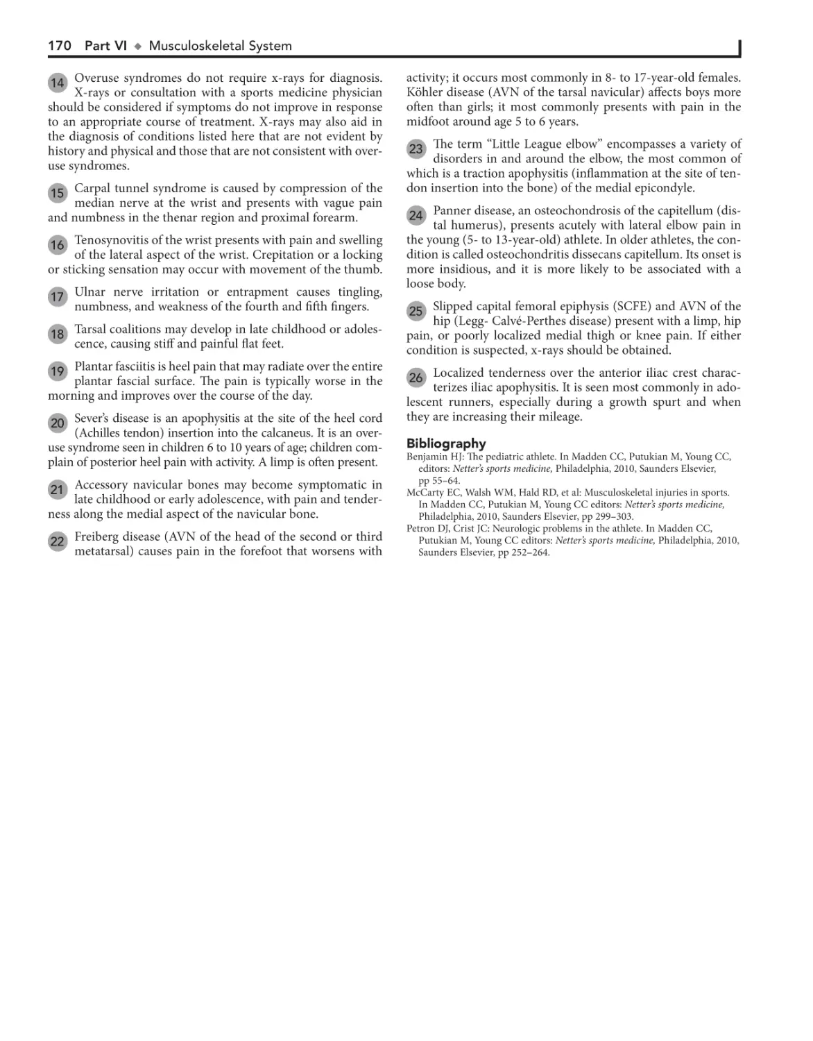
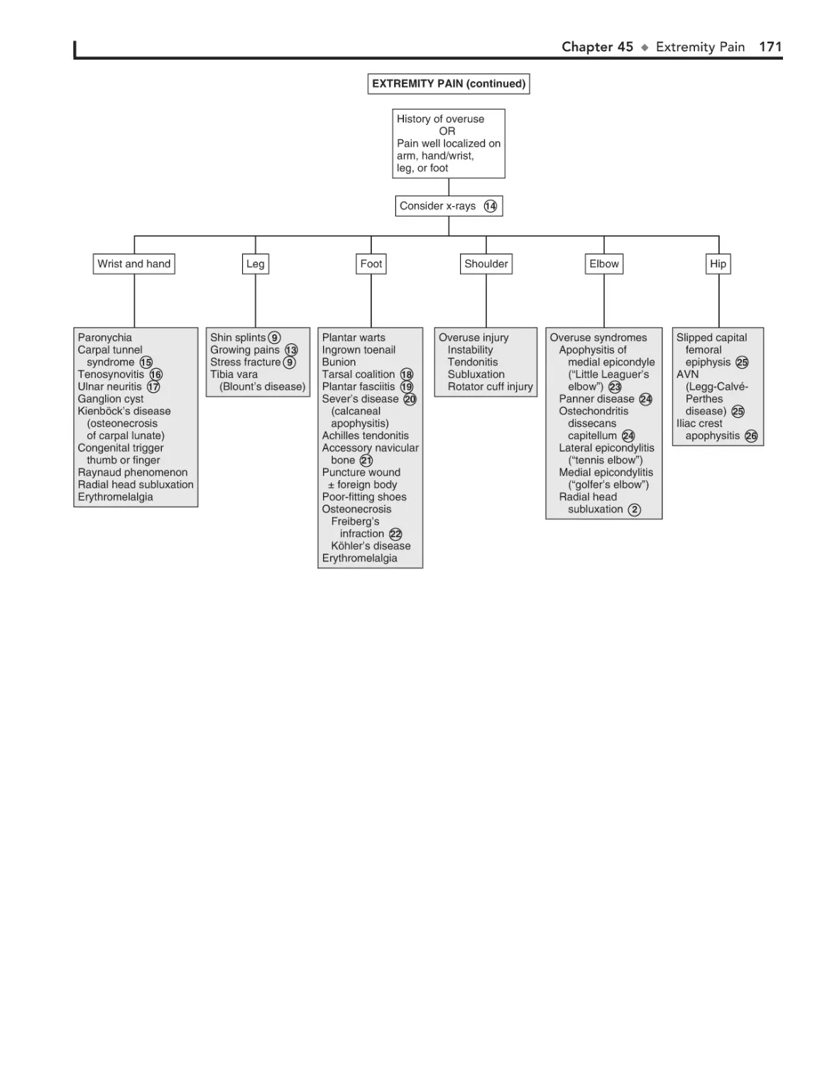
2. Scratch off your Activation Code below
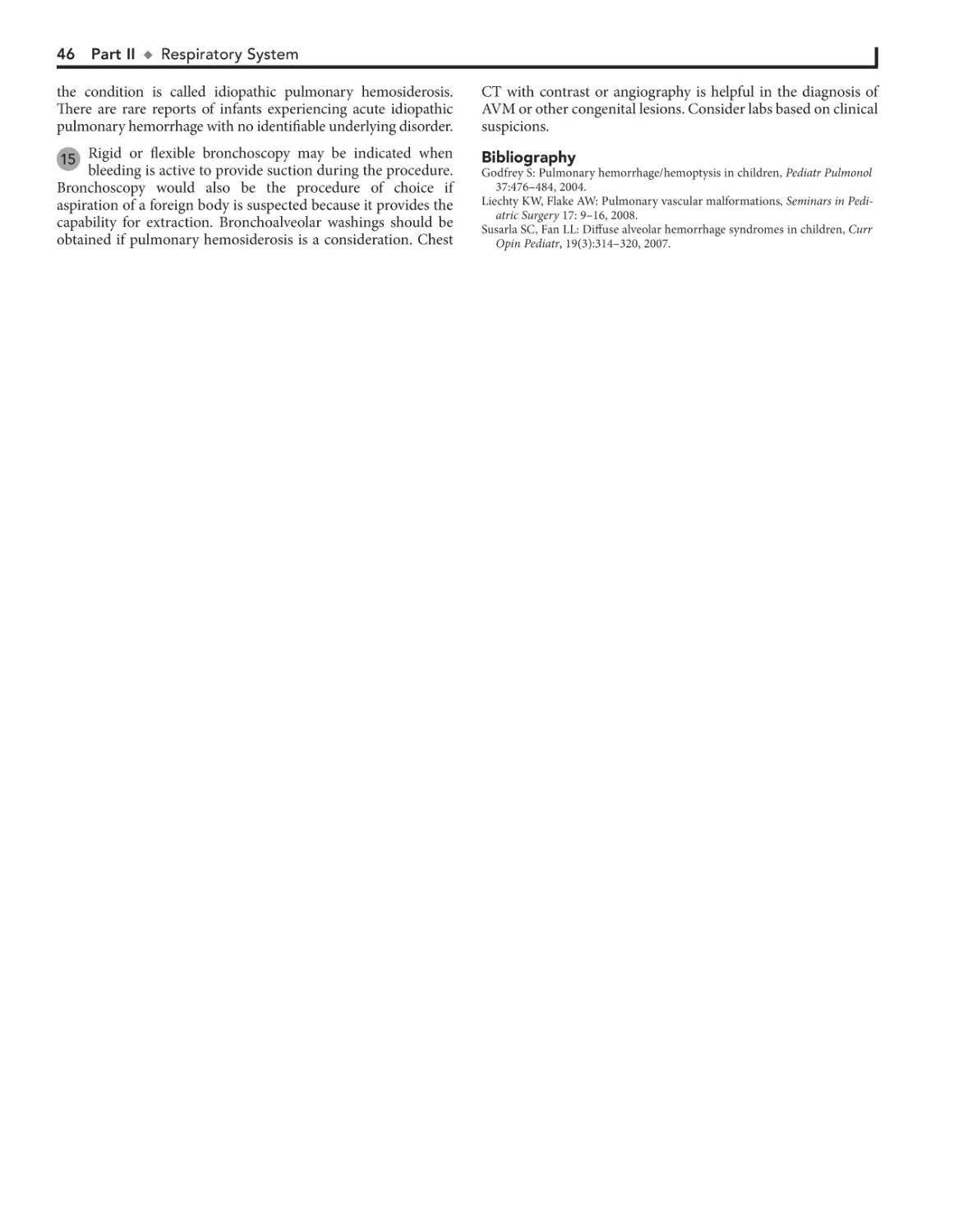
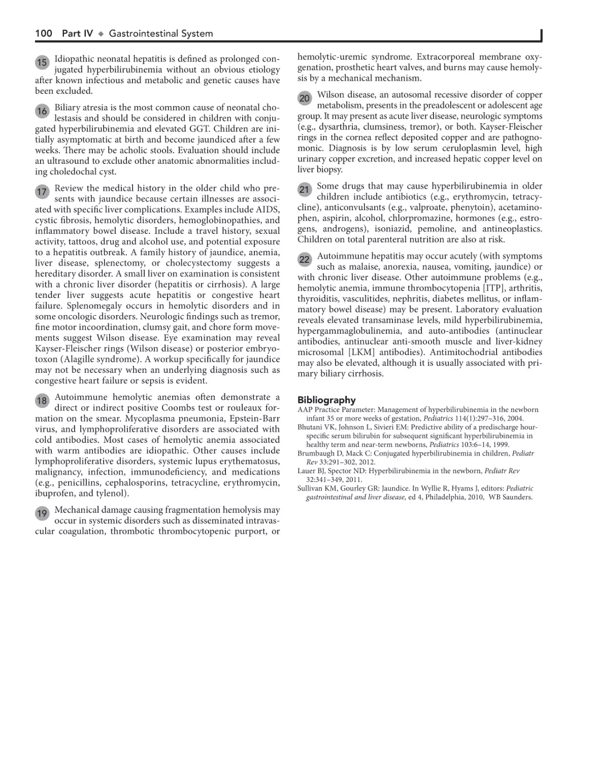
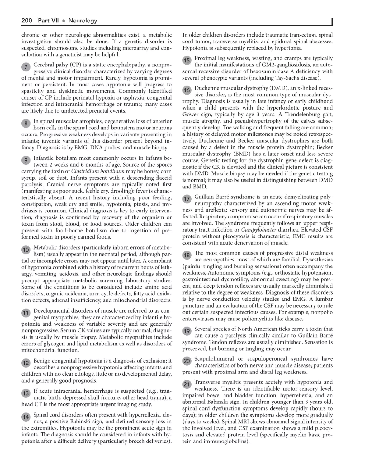
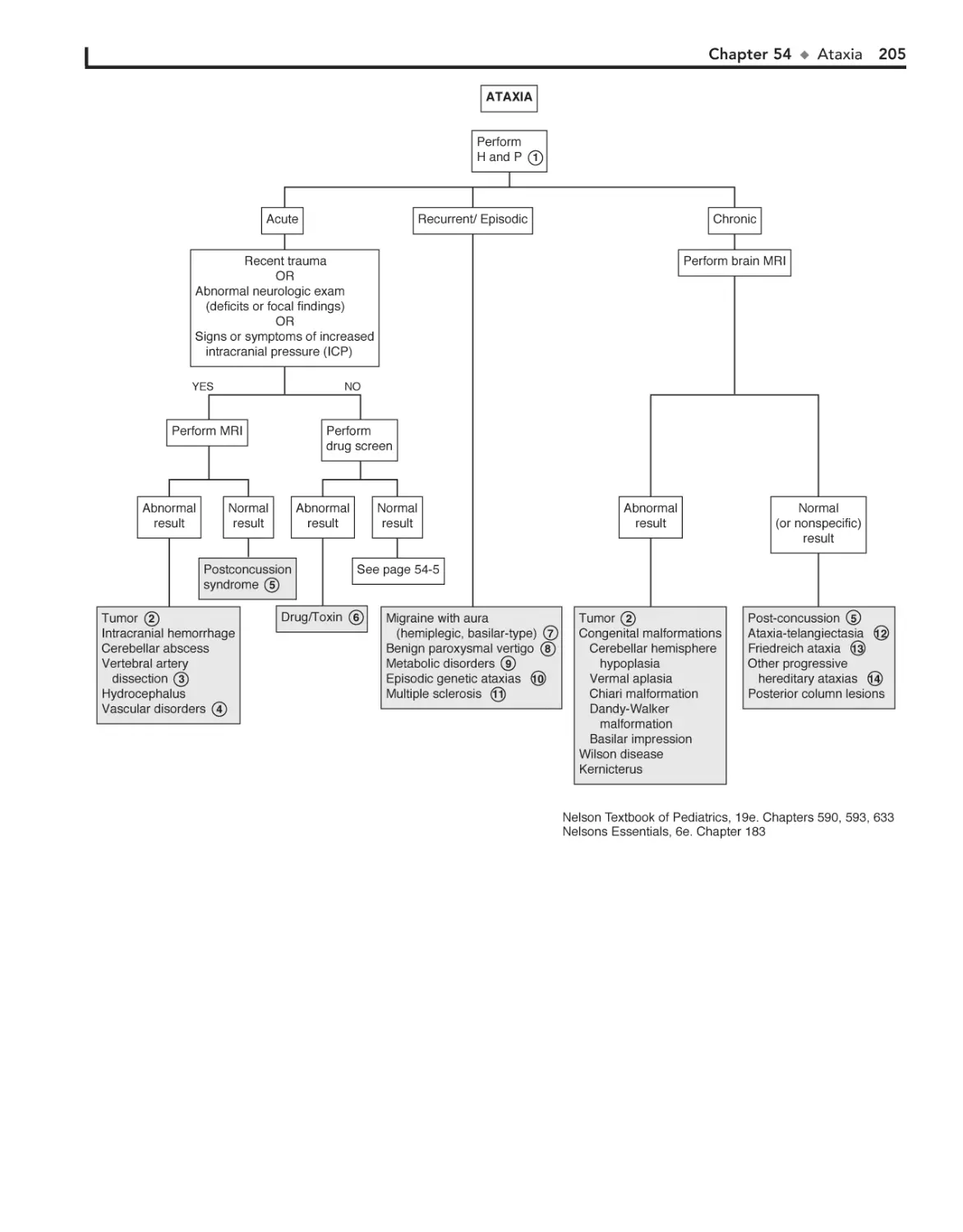
• Click “Register Now” at expertconsult.com
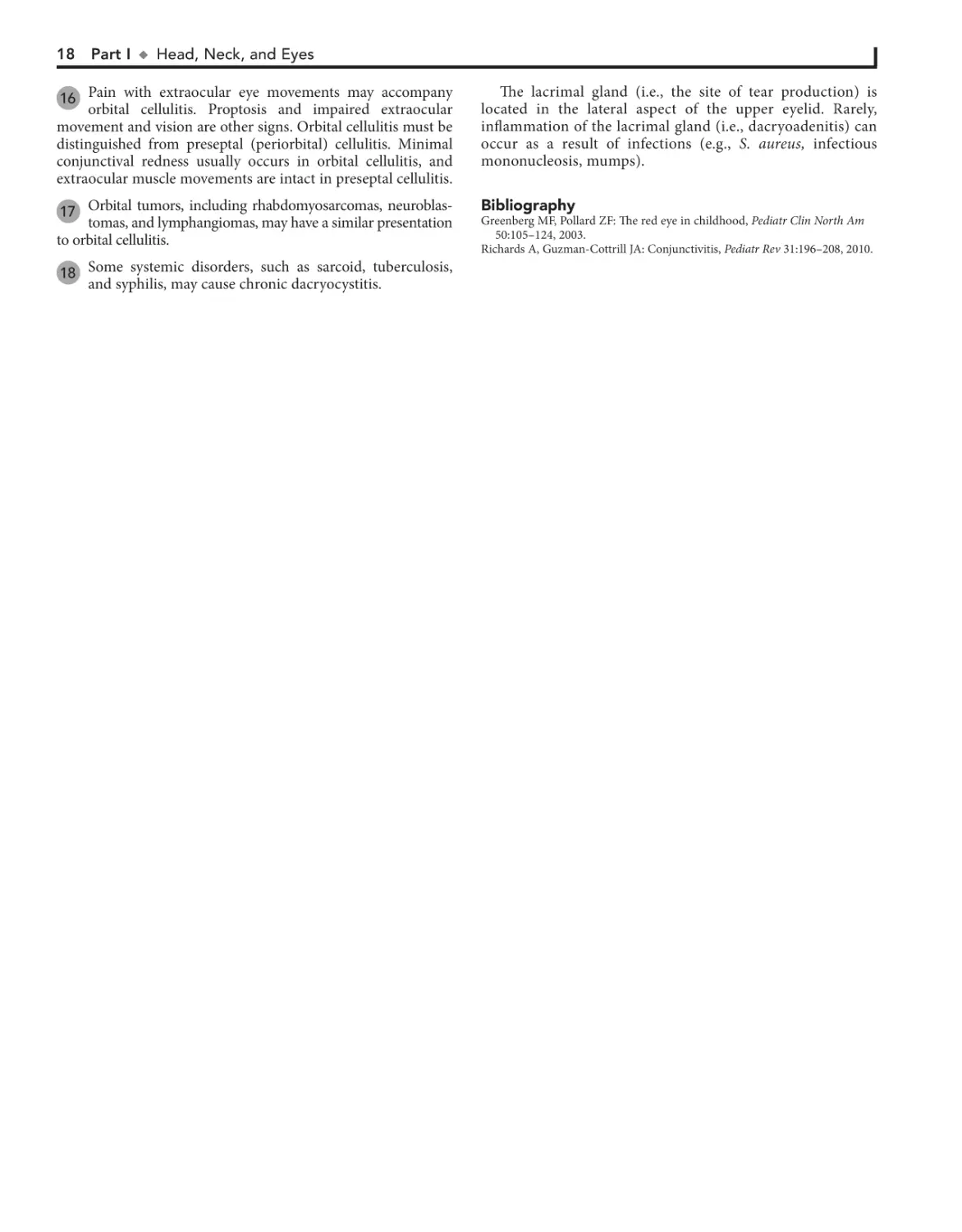
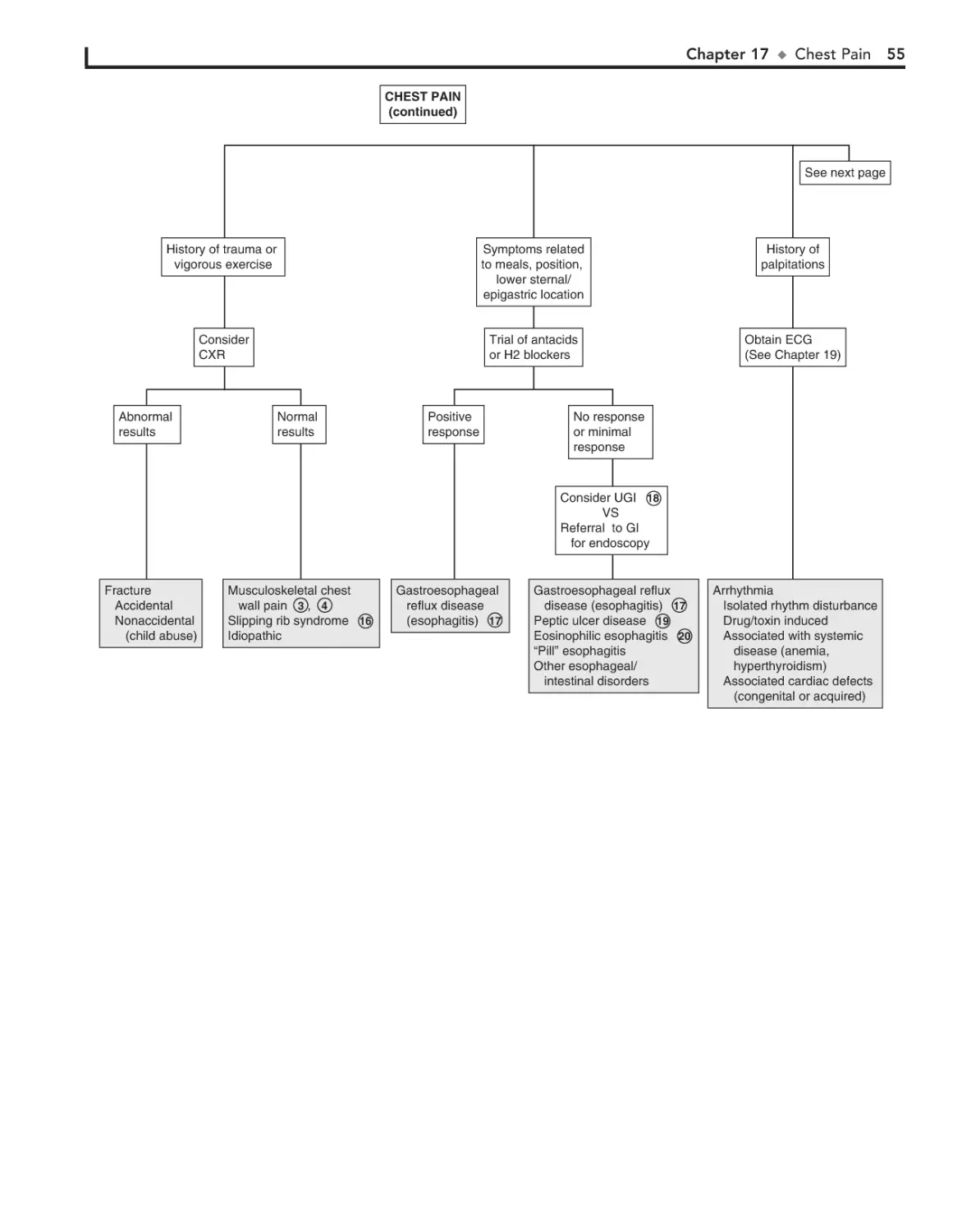
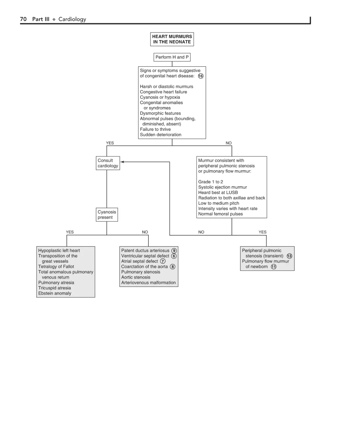
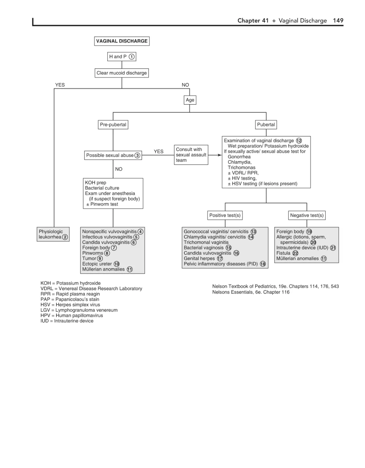
3. Enter it into the “Add a Title” box
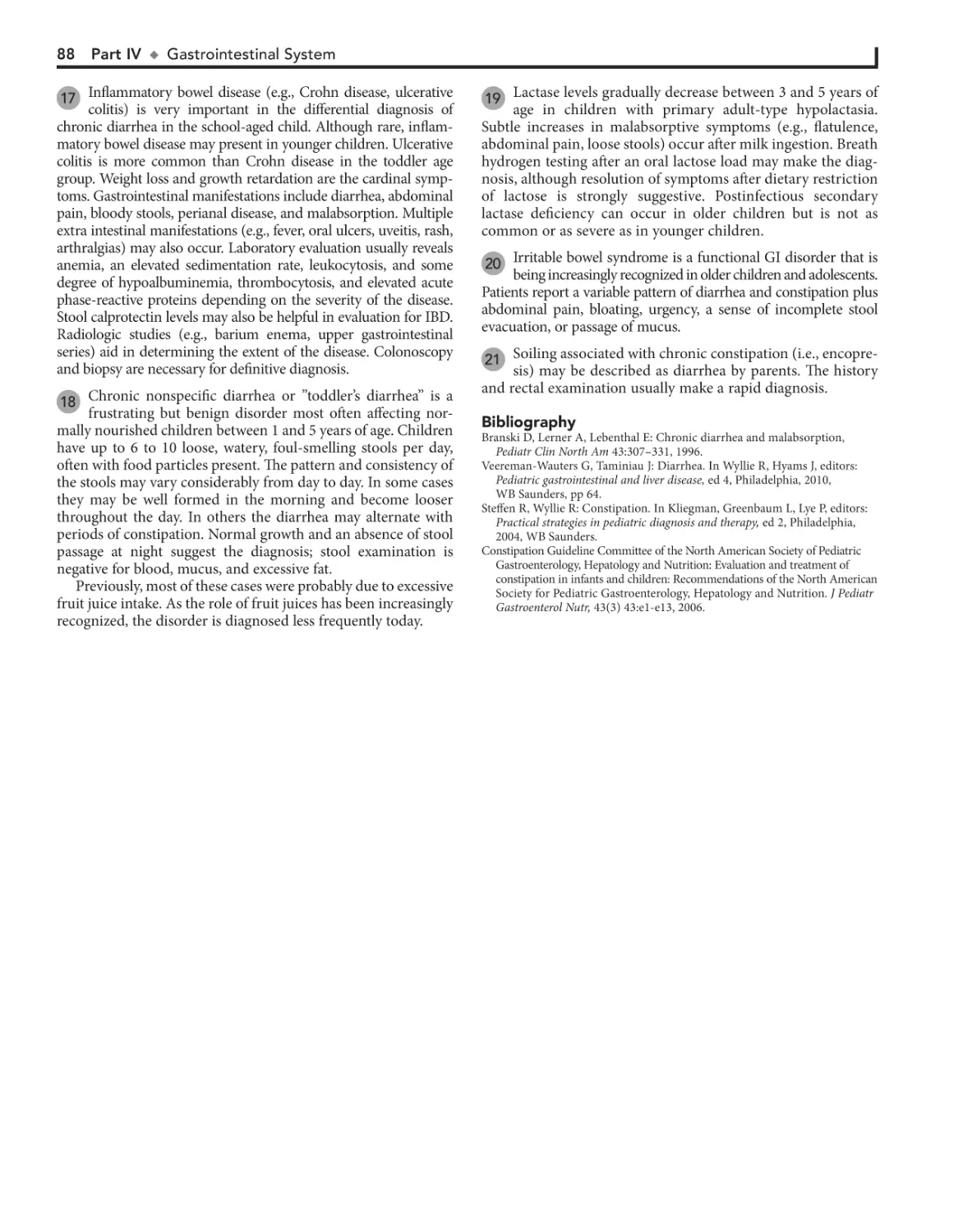
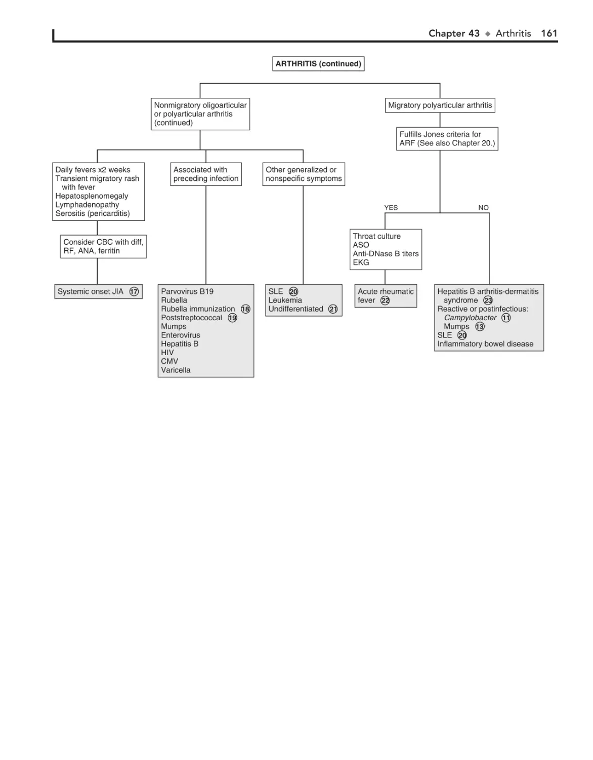
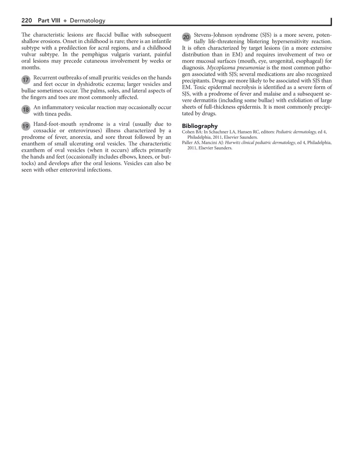
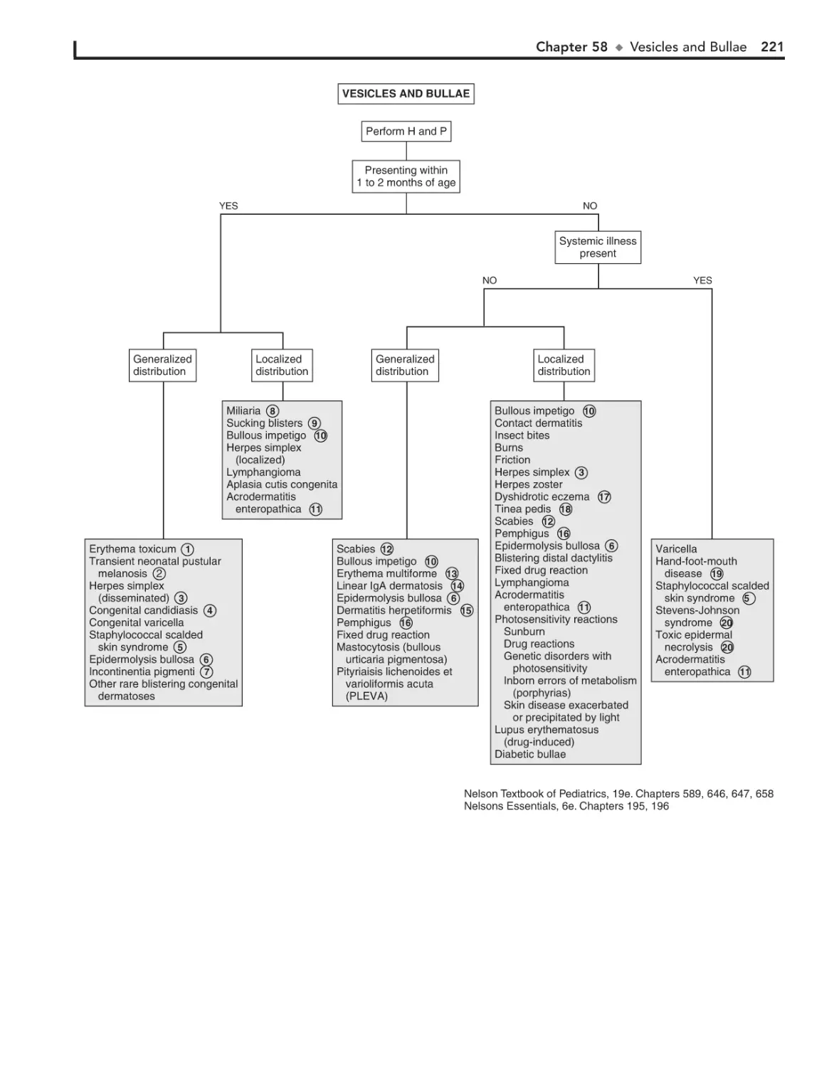
• Fill in your user information and click “Continue”
4. Click “Activate Now”
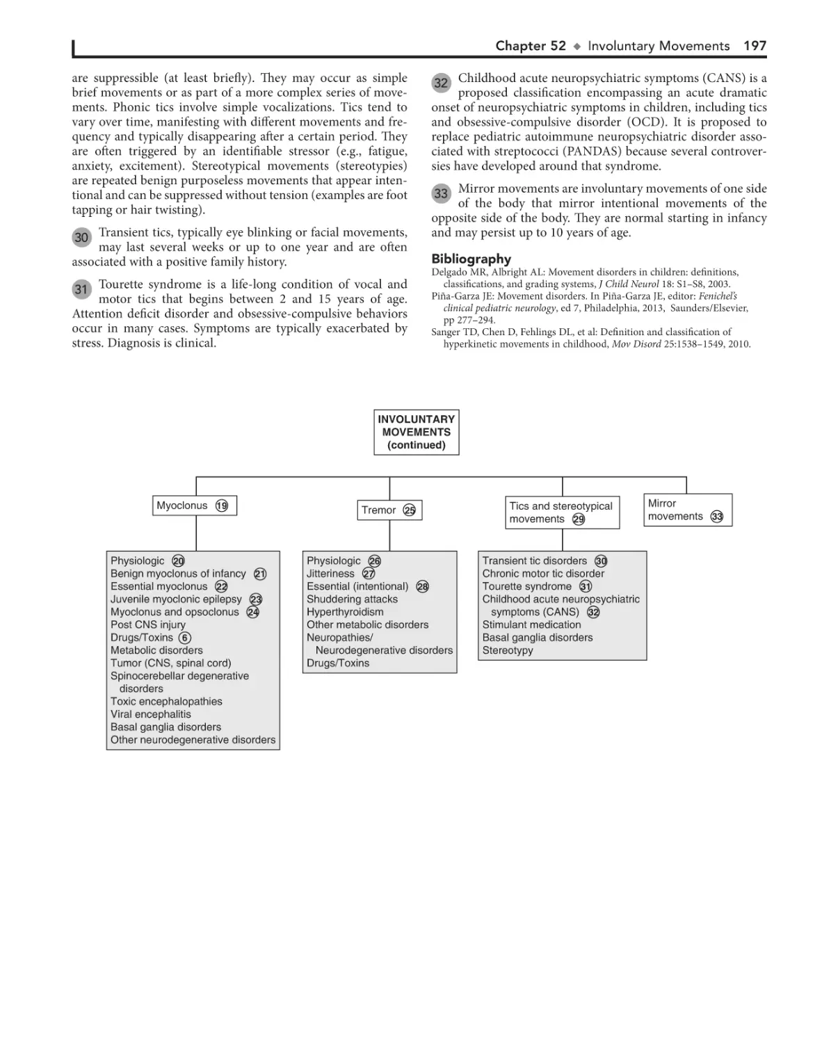
5. Click the title under “My Titles”
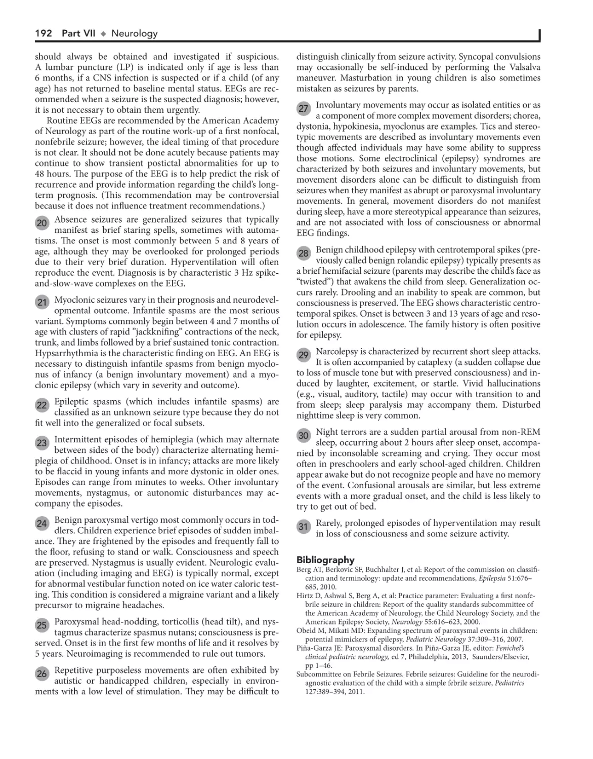
2. ACTIVATE YOUR BOOK
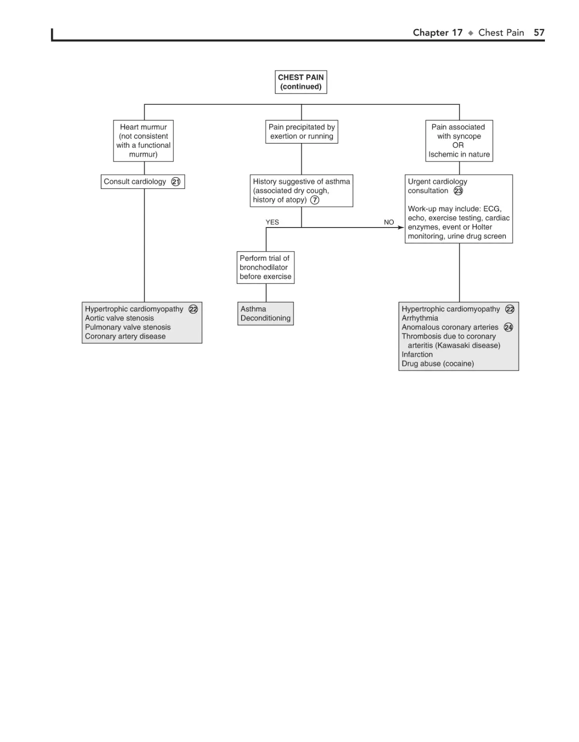
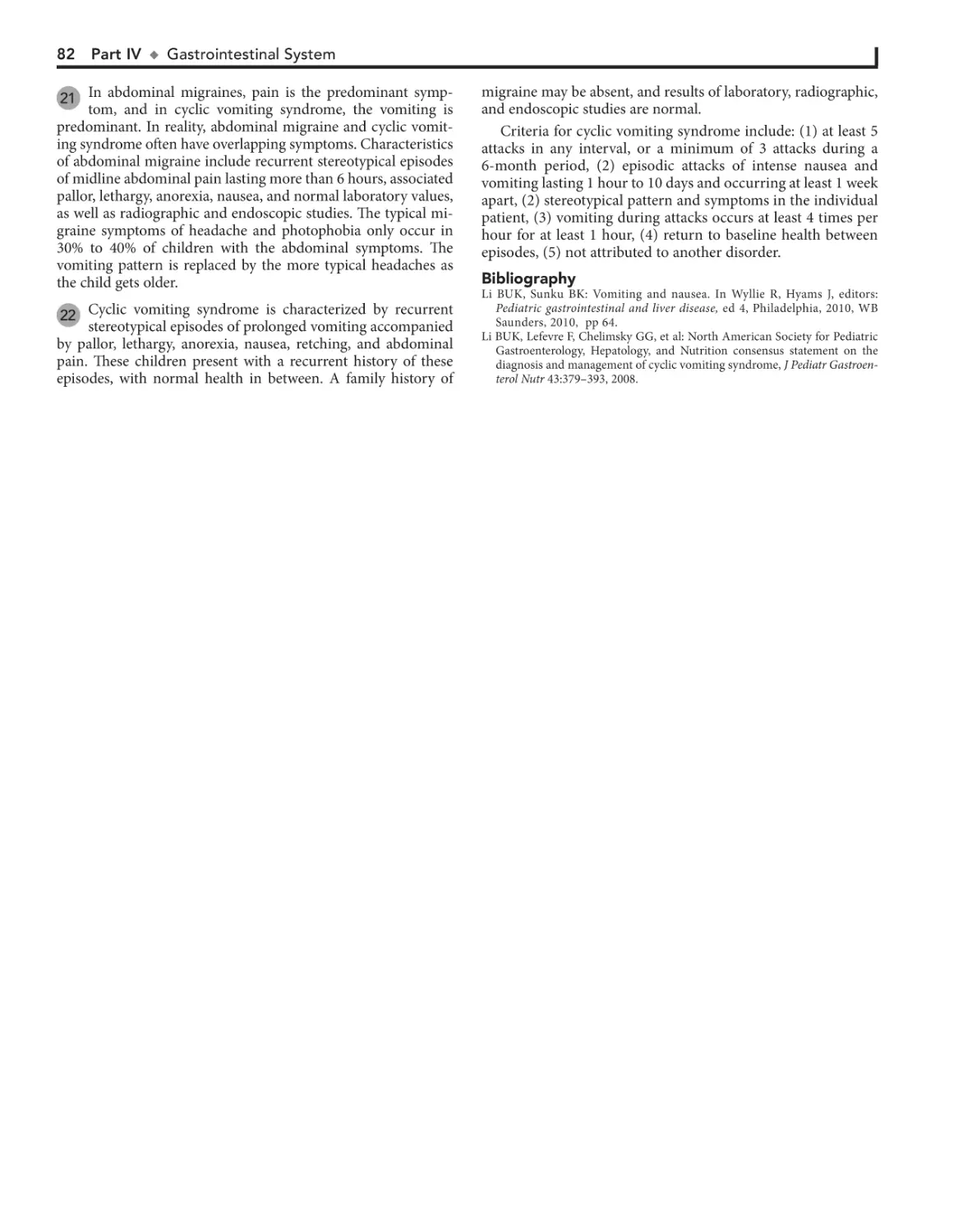
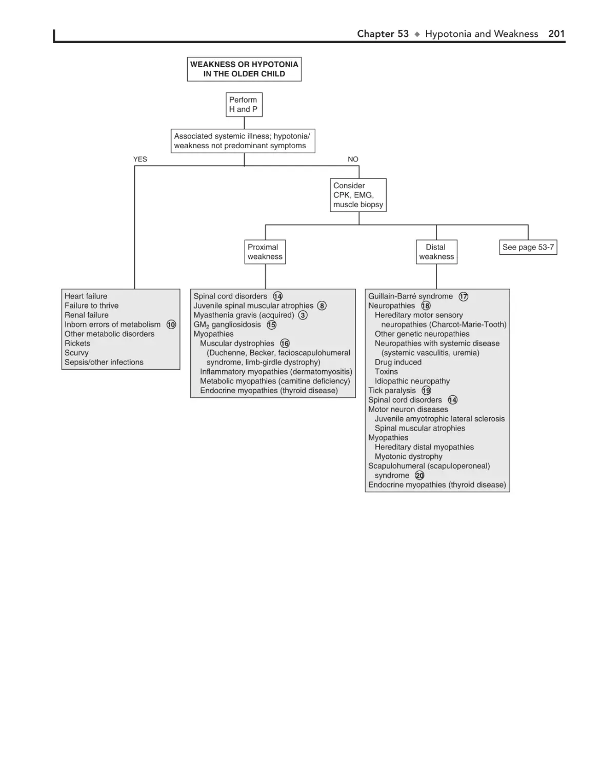
• Scratch off your Activation Code below
• Enter it into the “Enter Activation Code” box
• Click “Activate Now”
• Click the title under “My Titles”
For technical assistance:
email online.help@elsevier.com
call 800-401-9962 (inside the US)
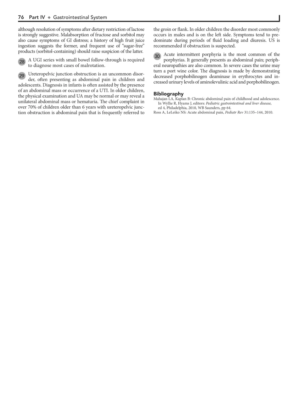
call +1-314-995-3200 (outside the US)
Activation Code
Pediatric
Decision-Making
Strategies
This page intentionally left blank
Pediatric
Decision-Making
Strategies
Second Edition
Albert J. Pomeranz, MD
Professor
Medical College of Wisconsin
Children’s Hospital of Wisconsin
Milwaukee, Wisconsin
Svapna Sabnis, MD
Associate Professor
Medical College of Wisconsin
Children’s Hospital of Wisconsin
Milwaukee, Wisconsin
Sharon L. Busey, MD
Associate Professor
Medical College of Wisconsin
Children’s Hospital of Wisconsin
Milwaukee, Wisconsin
Robert M. Kliegman, MD
Professor
Medical College of Wisconsin
Children’s Hospital of Wisconsin
Milwaukee, Wisconsin
1600 John F. Kennedy Blvd.
Ste 1800
Philadelphia, PA 19103-2899
PEDIATRIC DECISION-MAKING STRATEGIES, SECOND EDITION
ISBN: 978-0-323-29854-4
Copyright © 2016 by Saunders, an imprint of Elsevier Inc.
All rights reserved. No part of this publication may be reproduced or transmitted in any form or by any means,
electronic or mechanical, including photocopying, recording, or any information storage and retrieval system,
without permission in writing from the Publisher. Details on how to seek permission, further information about
the Publisher’s permissions policies and our arrangements with organizations such as the Copyright Clearance
Center and the Copyright Licensing Agency, can be found at our website: www.elsevier.com/permissions.
This book and the individual contributions contained in it are protected under copyright by the Publisher
(other than as may be noted herein).
Notices
Knowledge and best practice in this field are constantly changing. As new research and experience broaden
our understanding, changes in research methods, professional practices, or medical treatment may become
necessary.
Practitioners and researchers must always rely on their own experience and knowledge in evaluating
and using any information, methods, compounds, or experiments described herein. In using such information or methods they should be mindful of their own safety and the safety of others, including parties for
whom they have a professional responsibility.
With respect to any drug or pharmaceutical products identified, readers are advised to check the most
current information provided (i) on procedures featured or (ii) by the manufacturer of each product to be
administered, to verify the recommended dose or formula, the method and duration of administration, and
contraindications. It is the responsibility of practitioners, relying on their own experience and knowledge of
their patients, to make diagnoses, to determine dosages and the best treatment for each individual patient,
and to take all appropriate safety precautions.
To the fullest extent of the law, neither the Publisher nor the authors, contributors, or editors assume any
liability for any injury and/or damage to persons or property as a matter of products liability, negligence or
otherwise, or from any use or operation of any methods, products, instructions, or ideas contained in the
material herein.
Library of Congress Cataloging-in-Publication Data
Pomeranz, Albert J., author.
Pediatric decision-making strategies / Albert J. Pomeranz, Svapna Sabnis, Sharon L. Busey,
Robert M. Kliegman. -- 2nd edition.
p. ; cm.
Preceded by Pediatric decision-making strategies to accompany Nelson Textbook of Pediatrics,
16th ed. / Albert J. Pomeranz ... [et al.]. c2002.
Includes bibliographical references and index.
ISBN 978-0-323-29854-4 (pbk. : alk. paper)
I. Sabnis, Svapna, author. II. Busey, Sharon L., author. III. Kliegman, Robert, author. IV. Title.
[DNLM: 1. Pediatrics. 2. Diagnosis. WS 200]
RJ50.5
618.92’0075--dc23
2014038049
Senior Content Strategist: James Merritt
Content Development Specialist: Lisa Barnes
Publishing Services Manager: Anne Altepeter
Senior Project Manager: Cindy Thoms
Book Designer: Steve Stave
Printed in United States of America
Last digit is the print number: 9
8
7
6
5 4
3
2 1
To Kate and Emily, the greatest daughters a father could hope for
AP
To my loving and supportive family — my husband, Samir, and my sons, Rahul
and Nishant; my mother, Malavika Kapur, who always inspires me, and in memory
of my father, Ravinder Lal Kapur
SS
To Craig — for his endless patience and love
SLB
This page intentionally left blank
Preface
We are very pleased to be given the opportunity to produce a
second edition of Pediatric Decision-Making Strategies 12 years
after the original publication. The purpose and basic algorithmic format of the text has not changed, but each chapter
has been updated to reflect the latest medical information available. As with the original text, the purpose is to assist the student, house officer, and clinician in the evaluation of common
pediatric signs and symptoms and abnormal laboratory
findings. The algorithmic format provides a rapid and concise
stepwise approach to a diagnosis. The text accompanying each
algorithm helps to clarify certain approaches to diagnoses
and supplies additional useful information regarding various
medical conditions.
The information in the book is the most up to date available.
The literature has been extensively reviewed, and many of the
algorithms have been discussed with the appropriate specialists.
We believe that we have created algorithms that are accurate
and easy to follow. There is rarely a single acceptable approach
to any given problem, and not all diagnoses can fit neatly into
an algorithm. Even though the algorithms cannot be considered
all-inclusive, the goal is to facilitate a logical and efficient stepwise approach to reasonable differential diagnoses for the common clinical problems discussed. This task could not have been
completed without the generous help of many of the faculty
members of the Medical College of Wisconsin and Children’s
Hospital of Wisconsin.
vii
This page intentionally left blank
Acknowledgments
We wish to thank the many physicians and staff at the
Medical College of Wisconsin and Children’s Hospital of
Wisconsin who were asked a multitude of questions to ensure
the accuracy and completeness of this text. They have all been
extremely helpful and patient. We would like to extend special thanks to the following faculty members for their help:
Jay Nocton and James Verbsky for Musculoskeletal System;
Amanda Brandow for Hematology; Anoop Singh and Shanelle
Clark for Cardiology; Scott Van Why and Cynthia Pan for
Fluids and Electrolytes; Omar Ali and Patricia Donohoue
for Endocrine System; Alisha Mavis for Gastrointestinal
System; Lynn D’Andrea for Respiratory System; and Larry
Greenbaum of Emory School of Medicine for Fluids and
Electrolytes.
We also wish to thank Lisa Barnes and James Merritt at
Elsevier for their support and encouragement.
Special thanks to Kelsie Birschbach for her invaluable assistance in the manuscript preparation.
ix
This page intentionally left blank
Abbreviations
ABG
ALT
ALTE
ANA
AP
ARF
AST
AVN
BP
BUN
CBC
CMV
CNS
Cr
CRP
CSF
CT
CXR
DTP
EBV
ECF
ECMO
EEG
EKG
EMG
ENT
ESR
FSH
GER
GGT
GI
GU
H and P
HEENT
Hgb
arterial blood gases
alanine aminotransferase
apparent life-threatening event
antinuclear antibody
anteroposterior
acute rheumatic fever
aspartate aminotransferase
avascular necrosis
blood pressure
blood urea nitrogen
complete blood count
cytomegalovirus
central nervous system
creatinine
C-reactive protein
cerebrospinal fluid
computed tomography
chest x-ray
diphtheria-tetanus-pertussis
Epstein-Barr virus
extracellular fluid
extracorporeal membrane oxygenation
electroencephalogram
electrocardiogram
electromyogram
ear, nose, and throat
erythrocyte sedimentation rate
follicle-stimulating hormone
gastroesophageal reflux
-glutamyl transferase
gastrointestinal
genitourinary
history and physical
head, eyes, ears, nose, and throat
hemoglobin
HIV
I and D
ICP
IV
JRA
KUB
LFT
LH
LP
MRI
O&P
OM
PCR
PPD
PT
PTT
RBC
RF
RSV
RTA
SCIWORA
SI
Sp gr
s/p
T4
Td
TSH
UA
UGI
URI
US
UTI
WBC
human immunodeficiency virus
incision and drainage
intracranial pressure
intravenous
juvenile rheumatoid arthritis
kidney, ureter, bladder (x-ray study)
liver function test
luteinizing hormone
lumbar puncture
magnetic resonance imaging
ova and parasites
otitis media
polymerase chain reaction
purified protein derivative (of tuberculin)
prothrombin time
partial thromboplastin time
red blood cell
rheumatoid factor
respiratory syncytial virus
renal tubular acidosis
spinal cord injury in the absence of
radiographic abnormalities
sacroiliac
specific gravity
status post
thyroxine
tetanus-diphtheria toxoid
thyroid-stimulating hormone
urinalysis
upper gastrointestinal series
upper respiratory infection
ultrasound
urinary tract infection
white blood cell
xi
This page intentionally left blank
Contents
Part I
HEAD, NECK, AND EYES 1
1. Ear Pain 2
2. Rhinorrhea 4
3. Sore Throat 6
4. Neck Masses 8
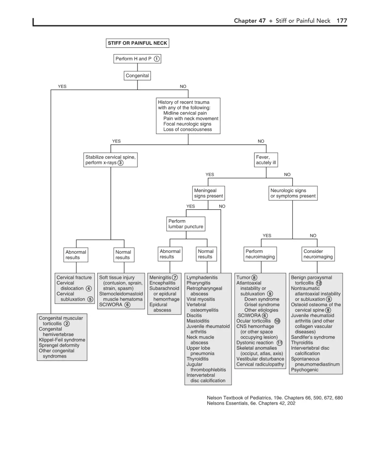
47. Stiff or Painful Neck 176
48. In-Toeing, Out-Toeing, and
Toe-Walking 178
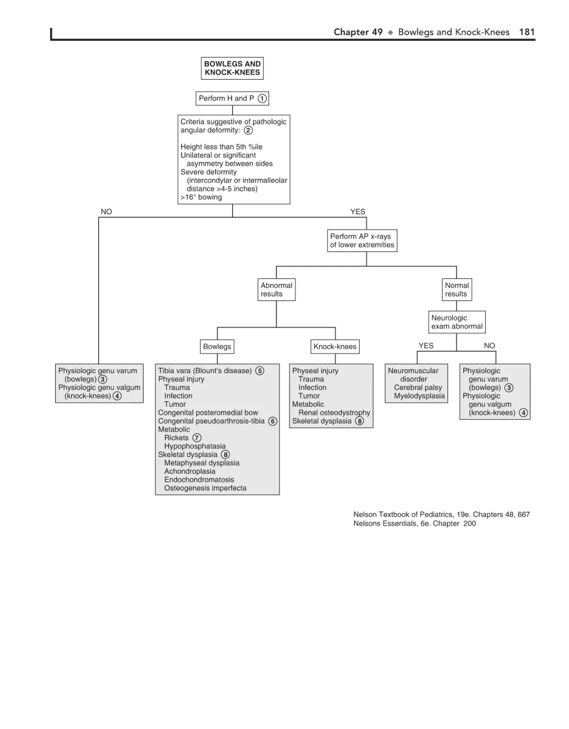
49. Bowlegs and Knock-Knees 180
5. Abnormal Head Size, Shape, and
Fontanels 12
6. Red Eye 16
7. Strabismus 20
8. Visual Impairment and Leukocoria 22
9. Abnormal Eye Movements 26
Part II
Part III
RESPIRATORY SYSTEM 29
10. Cough 30
11. Hoarseness 34
12. Stridor 36
13. Wheezing 38
14. Cyanosis 42
15. Hemoptysis 44
16. Apnea 48
Part VII
Part V
Part VI
215
57. Alopecia 216
58. Vesicles and Bullae 218
59. Fever and Rash 222
Part IX
HEMATOLOGY
Part X
ENDOCRINE SYSTEM 255
67. Short Stature 256
68. Pubertal Delay 260
CARDIOLOGY 51
17. Chest Pain 52
18. Syncope 58
19. Palpitations 62
GASTROINTESTINAL SYSTEM 71
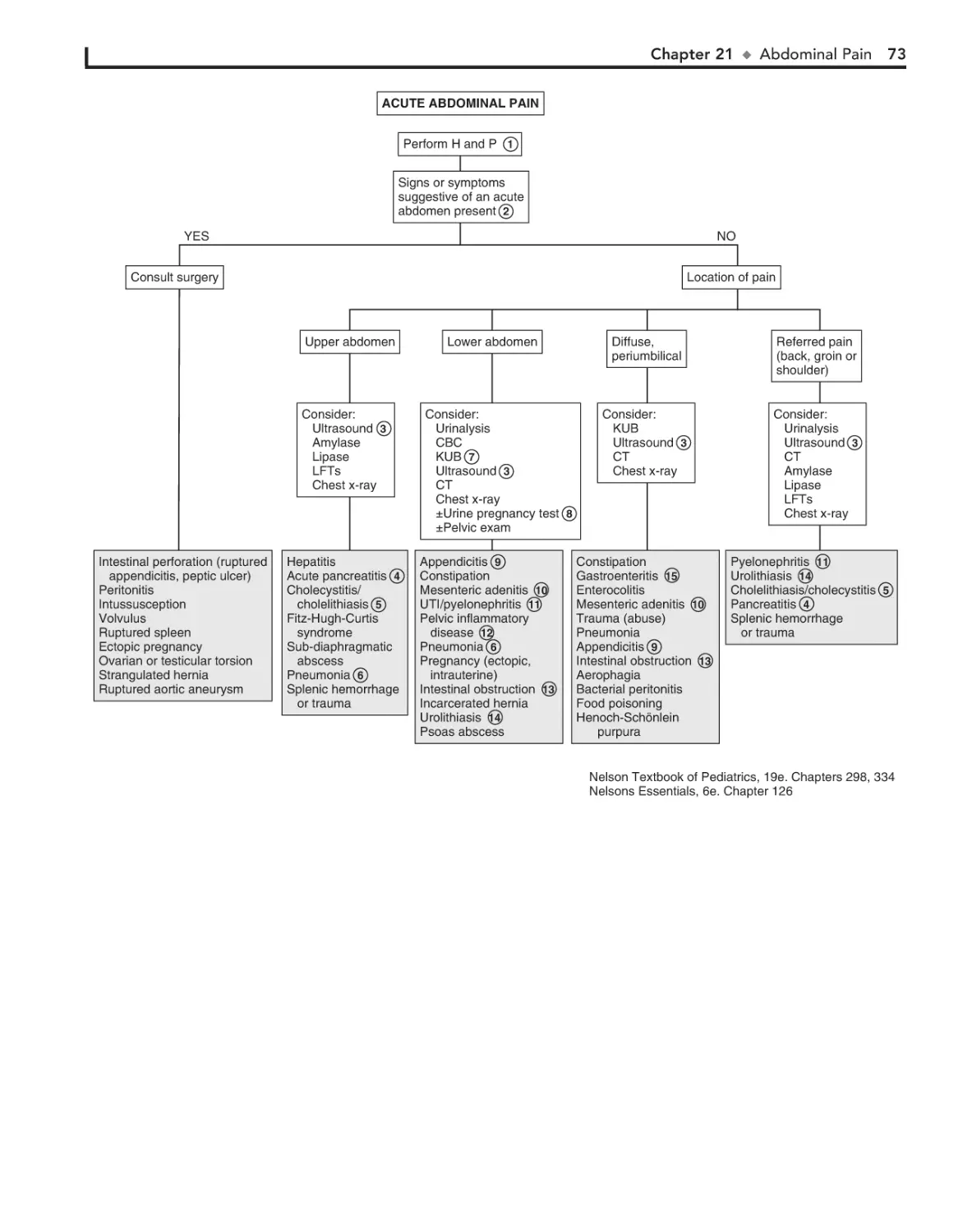
21. Abdominal Pain 72
22. Vomiting 78
23. Diarrhea 84
24. Constipation 90
25. Gastrointestinal Bleeding 94
26. Jaundice 98
27. Hepatomegaly 102
28. Splenomegaly 106
29. Abdominal Masses 110
GENITOURINARY SYSTEM 113
30. Dysuria 114
31. Enuresis 116
32. Red Urine and Hematuria 120
33. Proteinuria 124
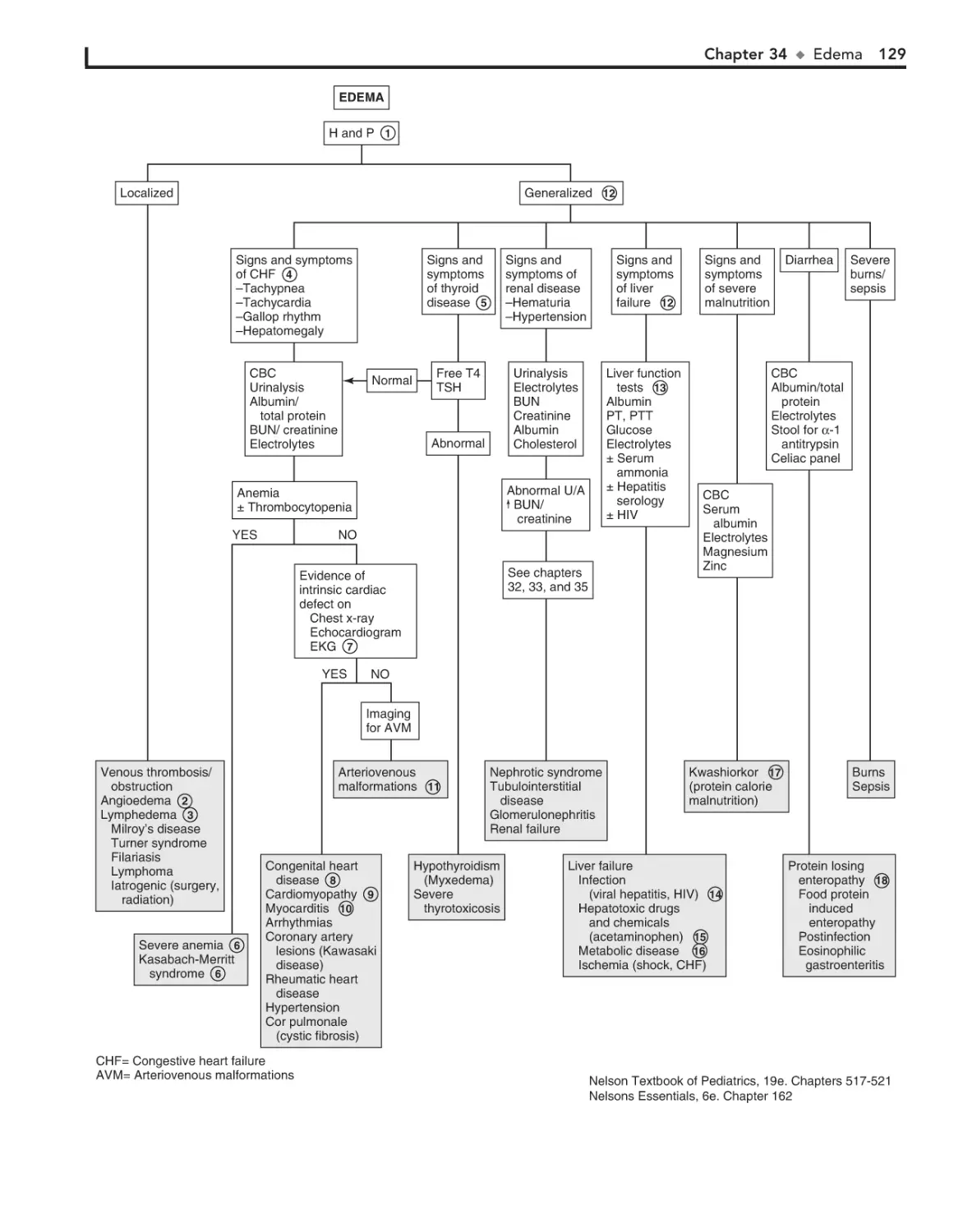
34. Edema 126
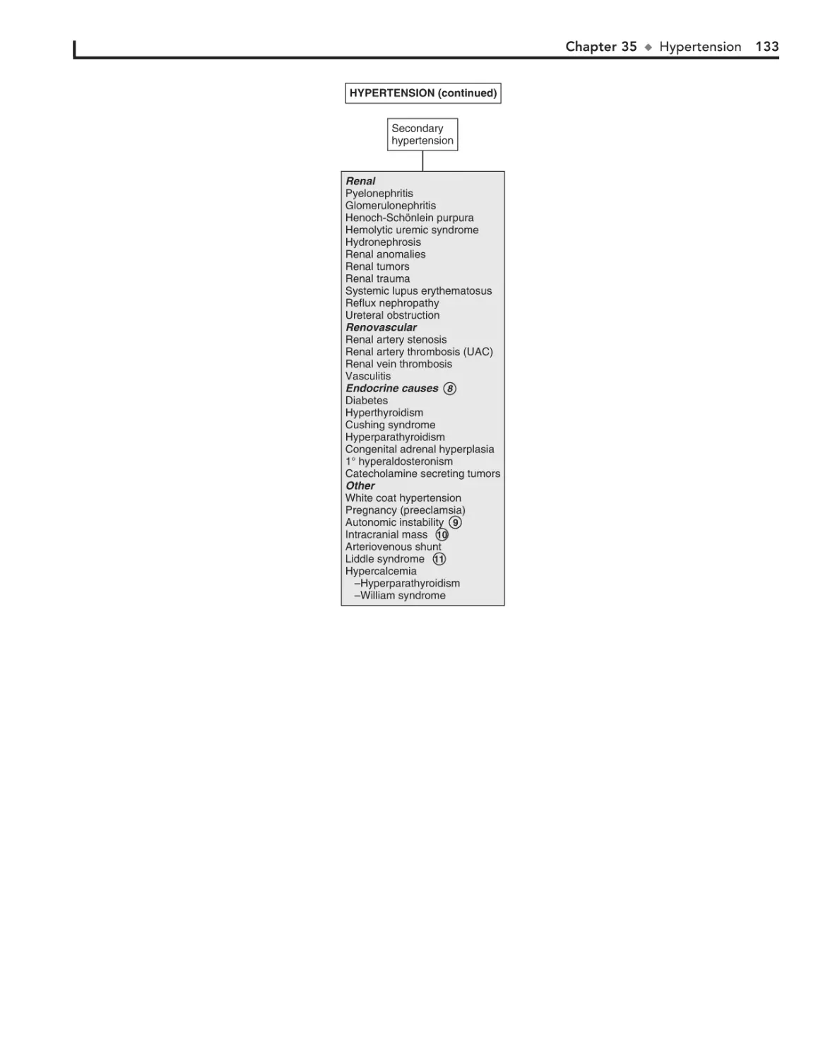
35. Hypertension 130
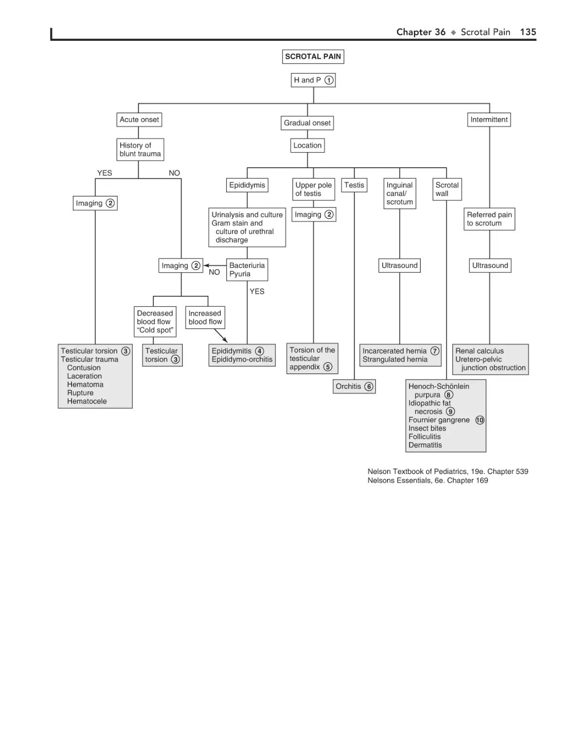
36. Scrotal Pain 134
37. Scrotal Swelling (Painless) 136
38. Dysmenorrhea 138
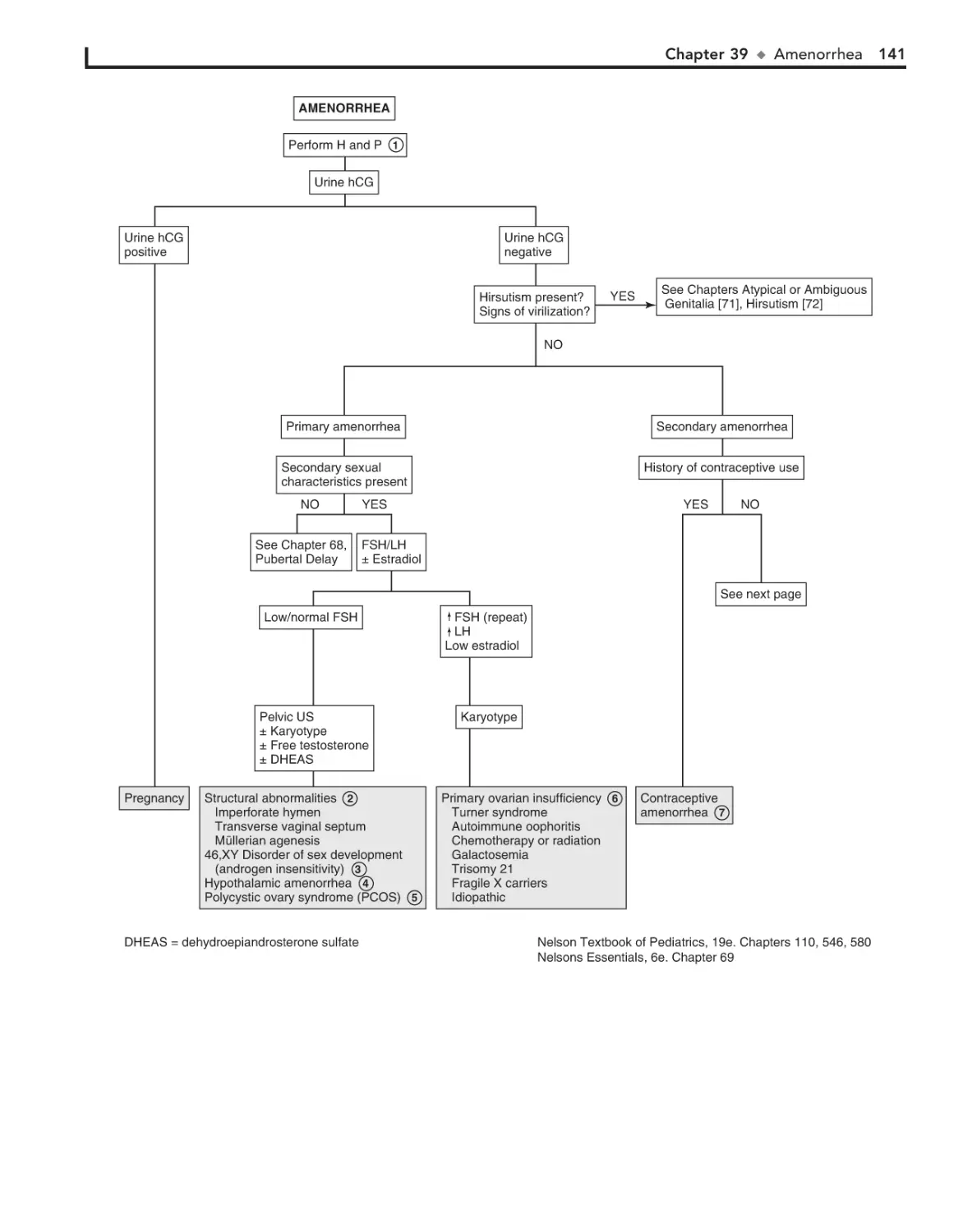
39. Amenorrhea 140
40. Abnormal Vaginal Bleeding 144
41. Vaginal Discharge 148
MUSCULOSKELETAL SYSTEM
42. Limp 152
43. Arthritis 156
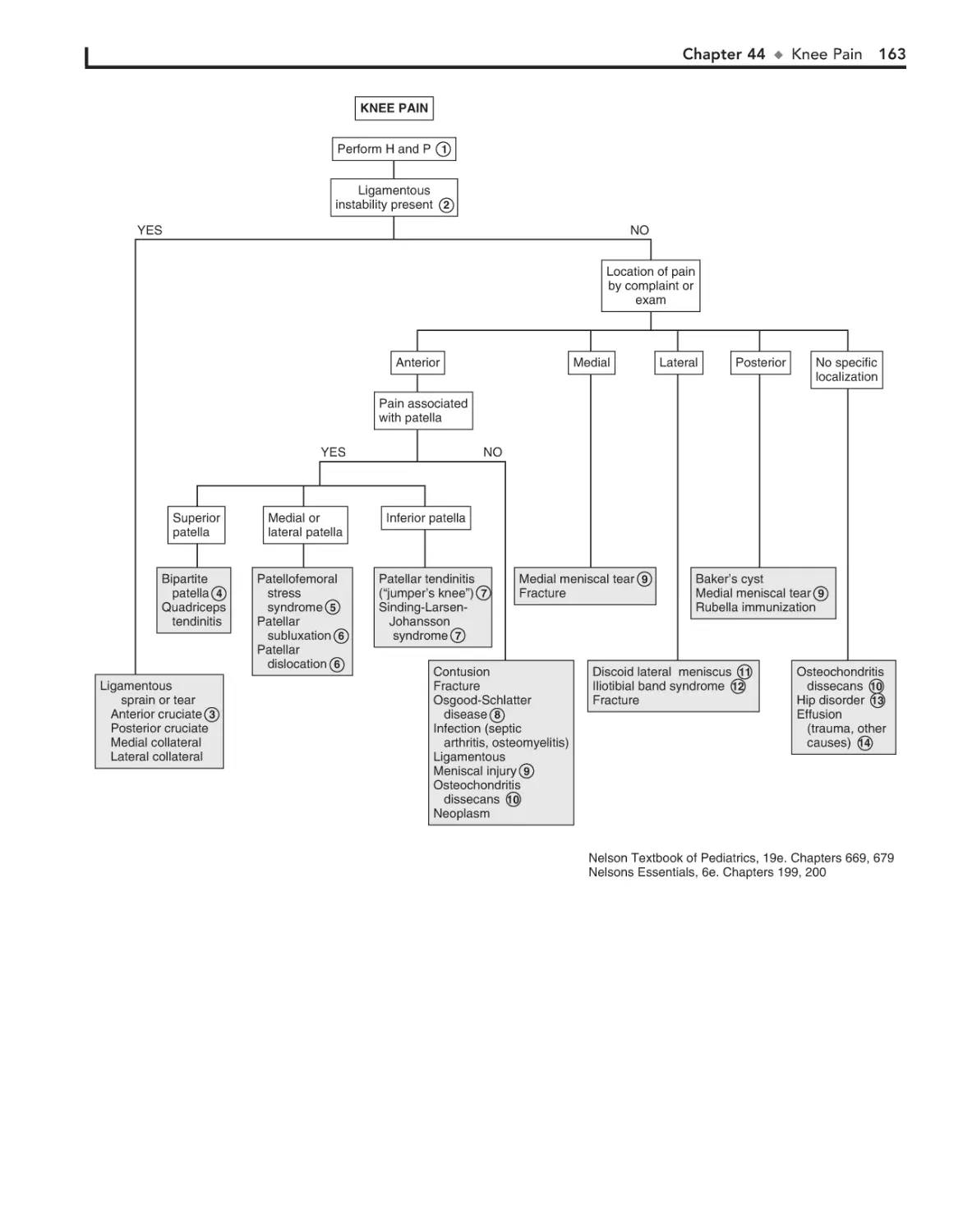
44. Knee Pain 162
45. Extremity Pain 166
46. Back Pain 172
183
50. Headaches 184
51. Seizures and Other Paroxysmal
Disorders 188
52. Involuntary Movements 194
53. Hypotonia and Weakness 198
54. Ataxia 204
55. Altered Mental Status 208
56. Hearing Loss 212
Part VIII DERMATOLOGY
20. Heart Murmurs 66
Part IV
NEUROLOGY
151
60.
61.
62.
63.
64.
65.
66.
69.
70.
71.
72.
73.
74.
75.
227
Lymphadenopathy 228
Anemia 232
Bleeding 236
Petechiae/Purpura 240
Neutropenia 244
Pancytopenia 248
Eosinophilia 252
Precocious Puberty in the Male 264
Precocious Puberty in the Female 266
Atypical or Ambiguous Genitalia 270
Hirsutism 274
Gynecomastia 278
Obesity 282
Polyuria 286
Part XI
GENERAL
289
Part XII
FLUIDS AND ELECTROLYTES
82. Acidemia 318
83. Alkalemia 322
84. Hypernatremia 324
85. Hyponatremia 326
86. Hypokalemia 330
87. Hyperkalemia 332
88. Hypocalcemia 334
89. Hypercalcemia 336
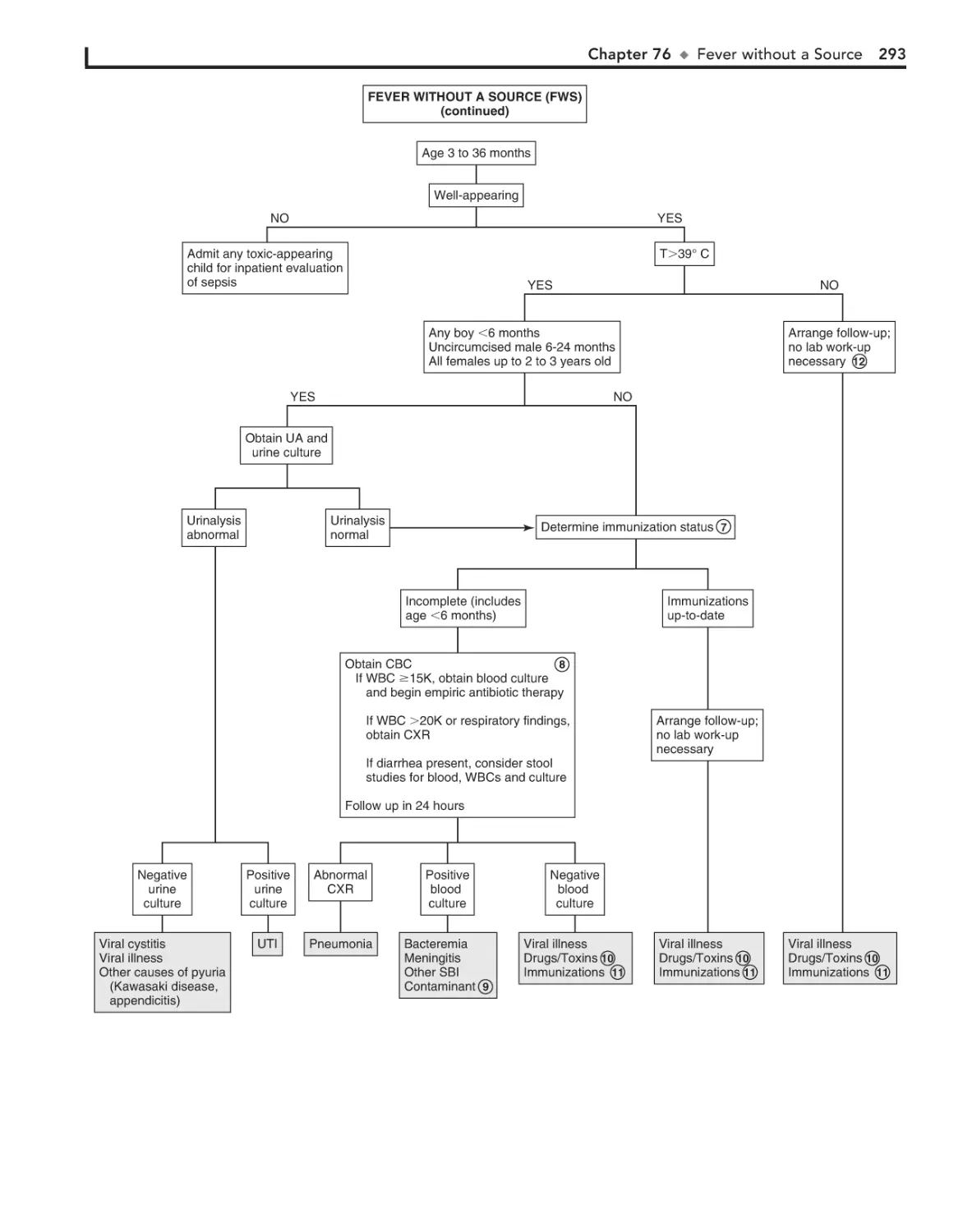
Fever without a Source 290
Fever of Unknown Origin 294
Recurrent Infections 300
Irritable Infant (Fussy or Excessively
Crying Infant) 306
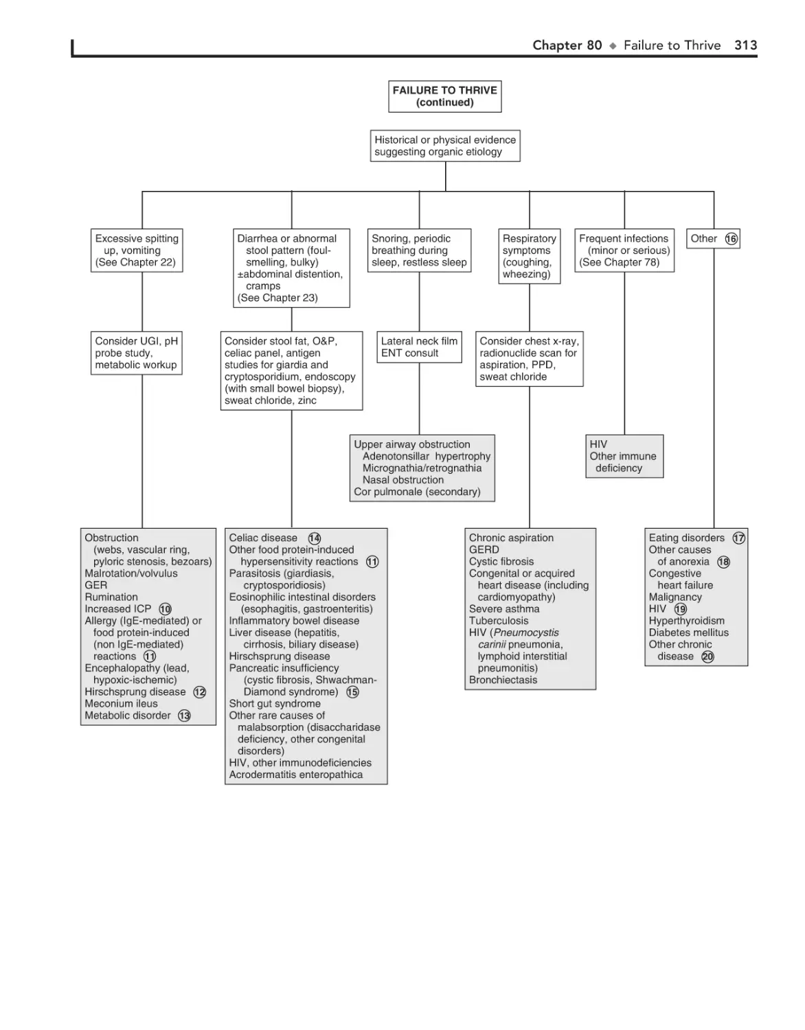
80. Failure to Thrive 310
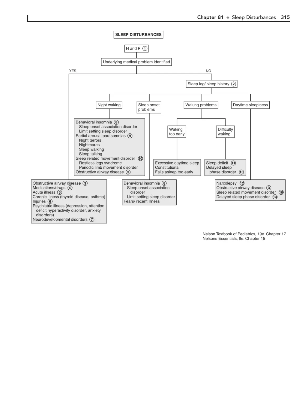
81. Sleep Disturbances 314
76.
77.
78.
79.
317
xiii
This page intentionally left blank
Head, Neck, and Eyes
PART
I
Chapter 1
EAR PAIN
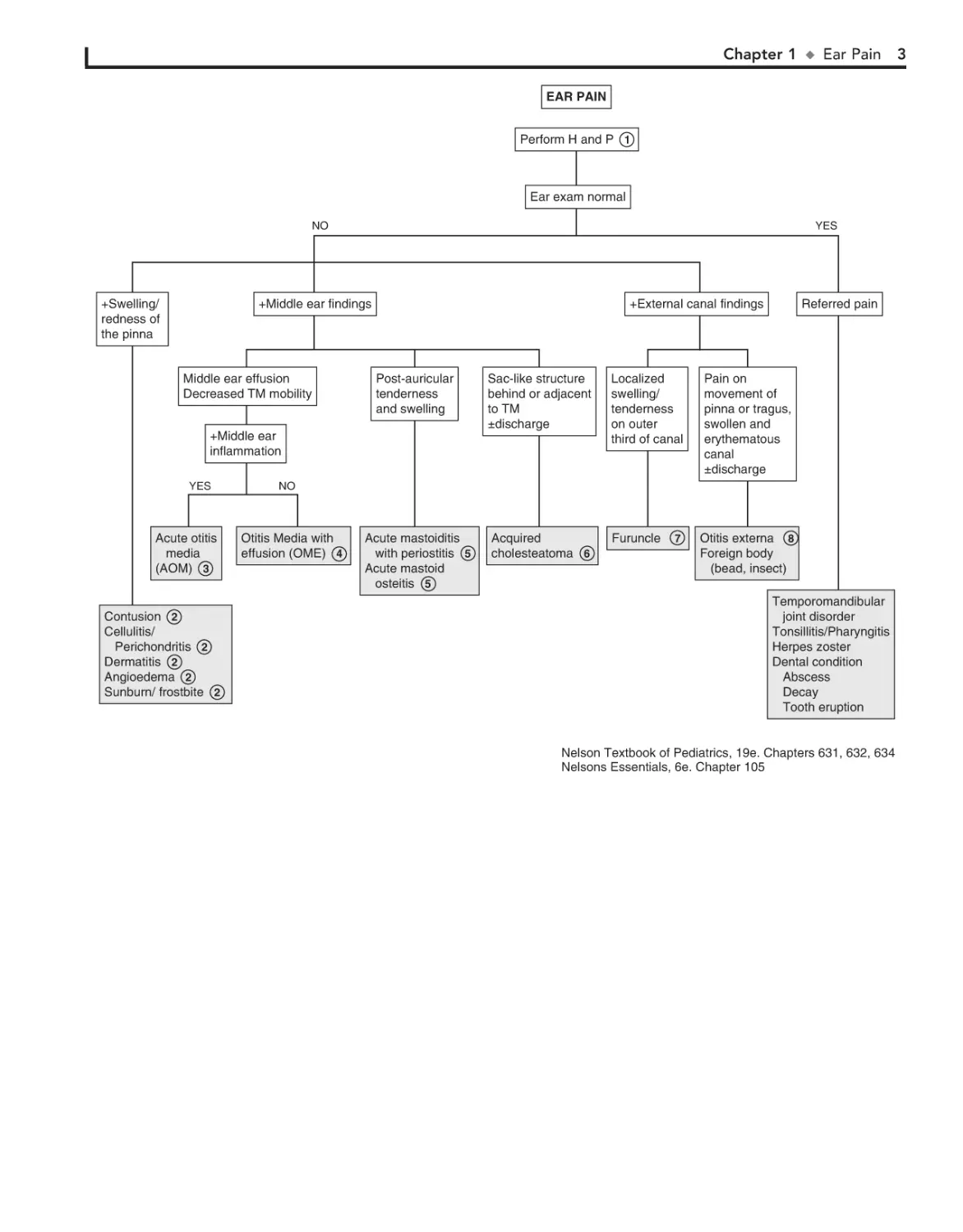
AOM. In general, OME should not be treated with antibiotics.
Mild discomfort or a feeling of “fullness” is not unusual. Diagnosis
can be aided by the use of tympanometry and acoustic reflectometry. These diagnostic tools determine the presence or absence of
effusion but not infection.
With periostitis, infection within the mastoid air cells has
spread to the periosteum that covers the mastoid process.
Further spread of infection results in osteitis, which involves
destruction of mastoid air cells and abscess formation. Resultant swelling is often severe enough to cause outward displacement of the pinna.
5
Ear pain is common, particularly in the first few years of life.
Acute otitis media (AOM) accounts for most cases. Over 80%
of children have at least one episode of AOM by the age of
3 years.
1 Signs of AOM may be nonspecific in the child younger
than age 2 (e.g., fever, irritability, vomiting). Ear tugging
is not a specific sign. AOM usually occurs with preceding or
concomitant upper respiratory symptoms. The presence of a
middle ear effusion is most accurately predicted by determining
altered mobility of the tympanic membrane (TM) with an
insufflator.
A swollen red auricle may be due to a contusion from
blunt trauma (e.g., wrestling or boxing). It is important to
recognize development of a hematoma with subperichondrial
collection of blood in order to correctly treat and prevent the
formation of a “cauliflower ear.” Perichondritis of the ear cartilage may also lead to deformity if untreated. Swelling of the
ear may be due to sunburn, frostbite, or an allergic reaction to
insect bites or contact irritants.
2
The diagnosis of AOM is usually made based on the presence of middle ear inflammation (i.e., redness, opacity,
and bulging of TM), middle ear effusion, and recent acute illness.
About two thirds of AOM episodes are a result of bacterial
infection. The major pathogens are nontypable Haemophilus
influenzae, Streptococcus pneumoniae, and Moraxella catarrhalis.
Inappropriate diagnosis of AOM contributes to the overuse of
antibiotics and the serious problem of antimicrobial resistance.
3
Otitis media with effusion (OME) is the presence of fluid in
the middle ear space without signs of inflammation or infection. It is commonly associated with URI or a successfully treated
4
2
A cholesteatoma is a collection of squamous cells in the
middle ear and should be suspected if retraction or
perforation of the TM with white caseous debris is noted. The
increasing size of the tumor results in destruction of the
middle ear and temporal bone, in addition to intracranial
spread.
6
The main clue to the diagnosis of a furuncle in the canal,
although uncommon, is the severe pain elicited when the
otoscope tip is placed in the canal. The canal appears generally
normal, except for the erythematous papule or pustule.
7
The ear canal is protected by cerumen, a waxy, waterrepellent coating. Excessive wetness or trauma or various
skin dermatoses (e.g., eczema) can disrupt this cerumen.
Frequent water exposure (e.g., swimming), hearing aids,
eczematous skin lesions, and aggressive use of cotton-tipped
swabs or other devices in the canal are risks for development of
otitis externa. Edema, erythema, and discharge are common.
Occasionally the disease is due to drainage from a perforated
tympanic membrane or to infection in the presence of tympanostomy tubes. The moist, irritant nature of the purulent
drainage results in superinfection from bacterial colonization.
Pathogens include Pseudomonas aeruginosa, Staphylococcus
aureus, other gram-negative organisms, and occasionally fungi.
8
Bibliography
American Academy of Pediatrics: Diagnosis and management of acute otitis
media, Pediatrics 113:1451–1465, 2004.
Chapter 1
u
Ear Pain 3
EAR PAIN
Perform H and P 1
Ear exam normal
NO
+Swelling/
redness of
the pinna
YES
+Middle ear findings
Middle ear effusion
Decreased TM mobility
+External canal findings
Post-auricular
tenderness
and swelling
Sac-like structure
behind or adjacent
to TM
±discharge
Localized
swelling/
tenderness
on outer
third of canal
Acute mastoiditis
with periostitis 5
Acute mastoid
osteitis 5
Acquired
cholesteatoma 6
Furuncle
+Middle ear
inflammation
YES
Acute otitis
media
(AOM) 3
Contusion 2
Cellulitis/
Perichondritis 2
Dermatitis 2
Angioedema 2
Sunburn/ frostbite 2
Referred pain
Pain on
movement of
pinna or tragus,
swollen and
erythematous
canal
±discharge
NO
Otitis Media with
effusion (OME) 4
7
Otitis externa 8
Foreign body
(bead, insect)
Temporomandibular
joint disorder
Tonsillitis/Pharyngitis
Herpes zoster
Dental condition
Abscess
Decay
Tooth eruption
Nelson Textbook of Pediatrics, 19e. Chapters 631, 632, 634
Nelsons Essentials, 6e. Chapter 105
Chapter 2
RHINORRHEA
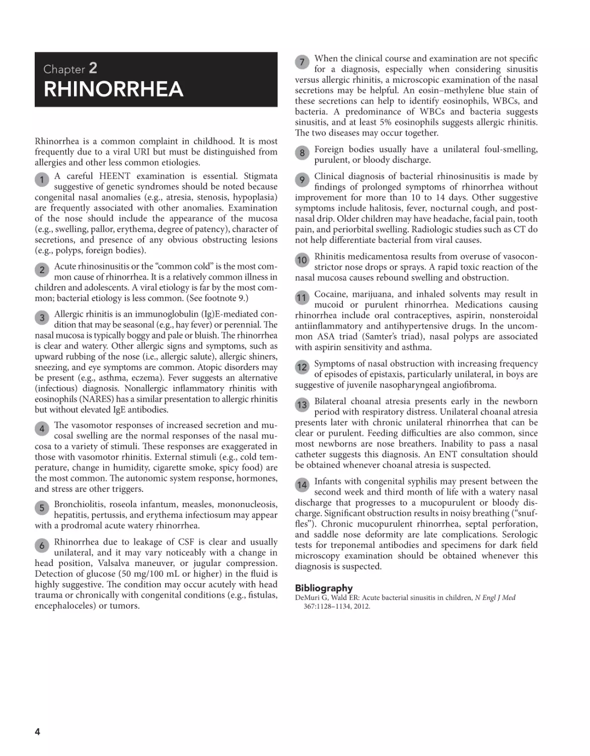
Rhinorrhea is a common complaint in childhood. It is most
frequently due to a viral URI but must be distinguished from
allergies and other less common etiologies.
1 A careful HEENT examination is essential. Stigmata
suggestive of genetic syndromes should be noted because
congenital nasal anomalies (e.g., atresia, stenosis, hypoplasia)
are frequently associated with other anomalies. Examination
of the nose should include the appearance of the mucosa
(e.g., swelling, pallor, erythema, degree of patency), character of
secretions, and presence of any obvious obstructing lesions
(e.g., polyps, foreign bodies).
Acute rhinosinusitis or the “common cold” is the most common cause of rhinorrhea. It is a relatively common illness in
children and adolescents. A viral etiology is far by the most common; bacterial etiology is less common. (See footnote 9.)
2
3 Allergic rhinitis is an immunoglobulin (Ig)E-mediated condition that may be seasonal (e.g., hay fever) or perennial. The
nasal mucosa is typically boggy and pale or bluish. The rhinorrhea
is clear and watery. Other allergic signs and symptoms, such as
upward rubbing of the nose (i.e., allergic salute), allergic shiners,
sneezing, and eye symptoms are common. Atopic disorders may
be present (e.g., asthma, eczema). Fever suggests an alternative
(infectious) diagnosis. Nonallergic inflammatory rhinitis with
eosinophils (NARES) has a similar presentation to allergic rhinitis
but without elevated IgE antibodies.
4 The vasomotor responses of increased secretion and mucosal swelling are the normal responses of the nasal mucosa to a variety of stimuli. These responses are exaggerated in
those with vasomotor rhinitis. External stimuli (e.g., cold temperature, change in humidity, cigarette smoke, spicy food) are
the most common. The autonomic system response, hormones,
and stress are other triggers.
5 Bronchiolitis, roseola infantum, measles, mononucleosis,
hepatitis, pertussis, and erythema infectiosum may appear
with a prodromal acute watery rhinorrhea.
Rhinorrhea due to leakage of CSF is clear and usually
unilateral, and it may vary noticeably with a change in
head position, Valsalva maneuver, or jugular compression.
Detection of glucose (50 mg/100 mL or higher) in the fluid is
highly suggestive. The condition may occur acutely with head
trauma or chronically with congenital conditions (e.g., fistulas,
encephaloceles) or tumors.
6
4
When the clinical course and examination are not specific
for a diagnosis, especially when considering sinusitis
versus allergic rhinitis, a microscopic examination of the nasal
secretions may be helpful. An eosin–methylene blue stain of
these secretions can help to identify eosinophils, WBCs, and
bacteria. A predominance of WBCs and bacteria suggests
sinusitis, and at least 5% eosinophils suggests allergic rhinitis.
The two diseases may occur together.
7
8
Foreign bodies usually have a unilateral foul-smelling,
purulent, or bloody discharge.
Clinical diagnosis of bacterial rhinosinusitis is made by
findings of prolonged symptoms of rhinorrhea without
improvement for more than 10 to 14 days. Other suggestive
symptoms include halitosis, fever, nocturnal cough, and postnasal drip. Older children may have headache, facial pain, tooth
pain, and periorbital swelling. Radiologic studies such as CT do
not help differentiate bacterial from viral causes.
9
10 Rhinitis medicamentosa results from overuse of vasocon-
strictor nose drops or sprays. A rapid toxic reaction of the
nasal mucosa causes rebound swelling and obstruction.
11 Cocaine, marijuana, and inhaled solvents may result in
mucoid or purulent rhinorrhea. Medications causing
rhinorrhea include oral contraceptives, aspirin, nonsteroidal
antiinflammatory and antihypertensive drugs. In the uncommon ASA triad (Samter’s triad), nasal polyps are associated
with aspirin sensitivity and asthma.
12 Symptoms of nasal obstruction with increasing frequency
of episodes of epistaxis, particularly unilateral, in boys are
suggestive of juvenile nasopharyngeal angiofibroma.
13 Bilateral choanal atresia presents early in the newborn
period with respiratory distress. Unilateral choanal atresia
presents later with chronic unilateral rhinorrhea that can be
clear or purulent. Feeding difficulties are also common, since
most newborns are nose breathers. Inability to pass a nasal
catheter suggests this diagnosis. An ENT consultation should
be obtained whenever choanal atresia is suspected.
14 Infants with congenital syphilis may present between the
second week and third month of life with a watery nasal
discharge that progresses to a mucopurulent or bloody discharge. Significant obstruction results in noisy breathing (“snuffles”). Chronic mucopurulent rhinorrhea, septal perforation,
and saddle nose deformity are late complications. Serologic
tests for treponemal antibodies and specimens for dark field
microscopy examination should be obtained whenever this
diagnosis is suspected.
Bibliography
DeMuri G, Wald ER: Acute bacterial sinusitis in children, N Engl J Med
367:1128–1134, 2012.
Chapter 2
u
Rhinorrhea 5
RHINORRHEA
Perform H and P 1
Chronic
(>10 days)
Acute
(<10 days)
Character of nasal
secretions 7
Purulent
or bloody
Nonpurulent
Prominent sneezing
Itchy eyes or nose
Allergic shiners
Nasal crease
Other signs of atopy
present
YES
Viral rhinosinusitis 2
Allergic rhinitis 3
Nonallergic inflammatory
rhinitis with eosinophils (NARES) 3
Vasomotor rhinitis or irritant rhinitis 4
Viral prodrome 5
CSF leak 6
Foreign body 8
Bacterial rhinosinusitis 9
Rhinitis medicamentosa 10
Nasal polyps
Drugs or medications 11
Juvenile nasopharyngeal
angiofibroma 12
Adenoidal hyperplasia
Choanal atresia or stenosis 13
Nasal tumor
Septal hematoma or abscess
Other infection (syphilis,
diphtheria) 14
Allergic rhinitis 13
Nonallergic rhinitis
with eosinophilis 3
NO
Vasomotor rhinitis or
irritant rhinitis 4
Rhinitis medicamentosa 10
Nasal polyps
Drugs or medications 11
CSF leak 6
Rhinitis of pregnancy (or menses)
Choanal atresia/stenosis 13
Septal deformity (congenital
or acquired)
Congenital malformation
Nasopharyngeal reflex or GER
Granulomatous disease
Ciliary dyskinesia
Immunodeficiency
Nasal tumor
Horner syndrome
Nelson Textbook of Pediatrics, 19e. Chapters 137, 210, 368, 371, 372
Nelsons Essentials, 6e. Chapters 102, 184
Chapter 3
SORE THROAT
The most common etiologies are rhinovirus, coronavirus, adenovirus, enterovirus, RSV, and metapneumovirus. Viral pharyngitis is usually gradual in onset with early signs of fever,
malaise, and anorexia generally preceding the sore throat.
7
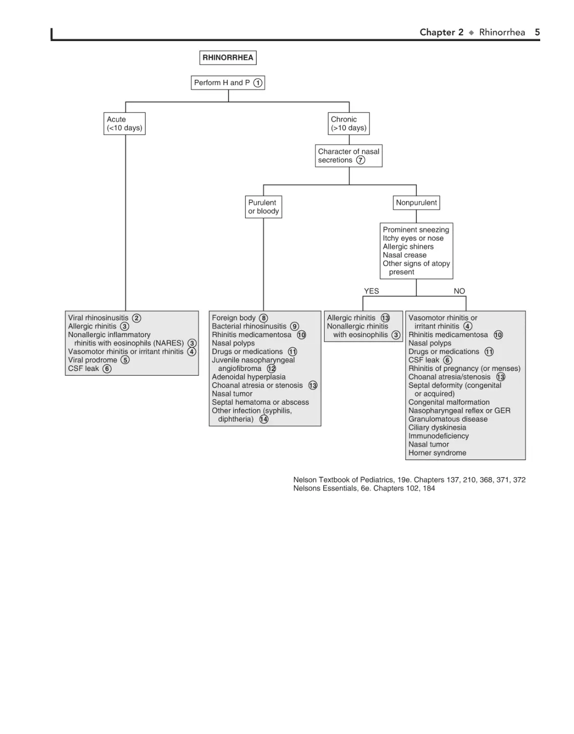
Most sore throats are benign, self-limiting viral illnesses. The
practitioner should always consider the likelihood of group A
b-hemolytic streptococcus (Streptococcus pyogenes), which is
important to identify and treat because of its potentially serious
complications. Other less common causes should be considered
when symptoms are worrisome or prolonged.
1 A history of exposure to a family member or classmate
with a cold or documented group A streptococcal infection is helpful. A history of sexual activity or abuse should raise
the suspicion for pharyngeal gonococcal infection. The degree
of pharyngeal inflammation is not always consistent with the
severity of the complaint. Tonsillar exudates are suggestive of
streptococcus but also of mononucleosis and adenovirus. Many
patients with streptococcal pharyngitis have only mild erythema without tonsillar enlargement or exudates. Small ulcers
or vesicles on the soft palate suggest a viral etiology.
Acute onset of illness with associated symptoms of stridor,
drooling, and air hunger or an unwillingness to recline suggests impending airway obstruction. The patient warrants emergent management for airway stabilization and treatment for
potentially life-threatening conditions such as epiglottitis and
retropharyngeal abscess. (See Chapter 12.) A lateral neck film
may be helpful but should be done only if the airway is stable.
2
Corynebacterium diphtheriae is a rare but serious cause of
pharyngitis. The disease is suggested by a systemic illness
and grayish membrane over the tonsils and pharyngeal walls. It
should be suspected in unimmunized persons or in persons
from underdeveloped countries. Culture of the organism and
confirmation of its toxin are necessary to confirm the diagnosis.
Soft tissue swelling and enlarged lymph nodes can cause a bullneck appearance.
3
Even when the clinical picture is highly suggestive of
streptococcal pharyngitis, laboratory confirmation is
strongly recommended. Rapid antigen detection tests (RST) are
highly specific, with sensitivities that are more variable. Throat
cultures are the standard for diagnosis whenever the RST
results are negative. The RST and the most commonly used
culture methods do not identify organisms other than group
A streptococcus. In cases in which another family member has
a positive culture finding, or in which a typical scarlatina rash
is present, group A streptococcus should still be considered
despite negative test results.
4
Group A streptococcal pharyngitis is most common between 5 and 11 years of age and unlikely under 3 years of
age. The occurrence of conjunctivitis, rhinitis, cough, and
hoarseness is more indicative of a virus than group A streptococcus. Significant diarrhea also makes streptococcal disease
unlikely. Some patients demonstrate the features of scarlet fever,
including circumoral pallor, strawberry tongue, and a red,
sandpaper-like scarlatina rash.
5
6
6
Viral pharyngitis is most commonly accompanied by
“common cold” symptoms such as rhinitis and cough.
Adenovirus may cause an exudative pharyngitis. Diarrhea
and conjunctivitis are also common.
Exudative pharyngitis is often a manifestation of infectious
mononucleosis. Patients can experience an abrupt onset of
fatigue, malaise, fever, and headache preceding the pharyngitis.
Hepatosplenomegaly and generalized lymphadenopathy are
common. Preadolescents tend to have milder symptoms than
adolescents and young adults. Atypical lymphocytosis is suggestive of the disorder, and a positive “Monospot” (heterophile antibody) test finding confirms EBV mononucleosis. The test is not
considered reliable in children younger than age 5 because of a
low titer of heterophile antibody. EBV serology should be used
in young patients or in patients with heterophile-negative cases.
CMV serology should also be considered because CMV causes
approximately 5% to 10% of cases.
8
9
Primary infection with HIV can also manifest with pharyngitis and a mononucleosis-like syndrome.
10 Arcanobacterium haemolyticum may cause a scarlet fever–
like illness but requires special culture methods. It is not
routinely sought in the evaluation of pharyngitis. Although non–
group A streptococci have been implicated in pharyngitis, they
cause a self-limiting illness, are not associated with complications, and require no treatment. Gonococcal pharyngeal infections are usually asymptomatic but can cause acute pharyngitis
with fever and cervical lymphadenitis.
11 Coxsackie A16 is responsible for hand-foot-mouth disease,
a characteristic outbreak of vesicles on the palms and soles,
with accompanying ulcerating vesicles throughout the oropharynx. Herpangina is a disorder characterized by fever and discrete
painful, vesicular lesions of the posterior pharynx. A variety of
enteroviruses cause herpangina, including enterovirus 71, although coxsackie A viruses are implicated most often.
12 Primary herpes simplex virus infection can cause gingivo-
stomatitis characterized by painful ulcerating vesicles in
the anterior portion of the oral cavity, including the lips. An
exudative tonsillitis may occur. Fevers and impaired fluid intake
are common. Herpetic gingivostomatitis may last up to 2 weeks.
13 Pharyngitis characterized by intense erythema but absent
tonsillar enlargement or exudate is an early finding in
measles. Fever, cough, coryza, conjunctivitis, and Koplik spots
(i.e., blue-white enanthema on buccal mucosa) suggest the diagnosis. These are followed by development of a maculopapular
rash that begins on the forehead then spreads downward. Laboratory criteria for diagnosis include positive serologic test for
measles immunoglobulin (Ig)M, seroconversion (a significant
rise in measles IgG), isolation of measles virus, or identification
by PCR of measles virus RNA from a clinical specimen (blood,
urine, or respiratory secretions).
14 Immunocompromised patients are at risk for fungal oro-
pharyngeal infections. Candida is the most common
pathogen. Diagnosis is made by examination of a specimen
treated with potassium hydroxide or by culture.
15 Agranulocytosis may manifest as pharyngitis with a white
or yellow exudate with underlying necrosis and ulceration.
Chapter 3
Bibliography
u
Sore Throat
Kenna MA: Sore throat in children. In Bluestone CD, Stool SE, Kenna MA, editors:
Pediatric otolaryngology, ed 4, Philadelphia, 2003, WB Saunders, pp 1120.
Gereige R, Cunill-De Sautu B: Throat infections, Pediatr Rev 32:459–468,
2011.
SORE THROAT
Perform H and P 1
Acute onset with respiratory distress
(stridor, drooling, dysphagia, trismus)
+fever 2
YES
NO
Signs suggestive of
streptococcal pharyngitis:
Relative absence of URI
symptoms
Acute onset
Fever
Tender anterior cervical
adenopathy
Tonsillar enlargement,
Associated headache or
abdominal pain
YES
RST
Throat culture 4
NO
Signs suggestive of viral infection:
Coryza, cough, congestion, diarrhea
–
YES
NO
Epiglottitis
Retropharyngeal/lateral
pharyngeal abscess
Peritonsillar abscess
Infectious mononucleosis
(massive tonsillar
hypertrophy)
Diphtheria 3
Group A
streptococcal
pharyngitis 5
NO
Oral mucosal lesions
+
7
Viral pharyngitis 6
Other viral syndromes
Adenovirus
(pharyngoconjunctival fever) 7
Infectious mononucleosis 8
HIV 9
Other bacterial pathogens 10
Other streptococci
(group C and group G)
Arcanobacterium haemolyticum
N. gonorrhoeae
Corynebacterium diphtheriae
YES
Coxsackie (hand-foot-mouth
disease, herpangina) 11
Herpes simplex
(gingivostomatitis) 12
Syndrome of periodic fever,
aphthous stomatitis,
pharyngitis, cervical
adenitis (PFAPA)
Measles 13
Immunodeficiency
(Candida) 14
Environmental irritants
Sinusitis
Allergic rhinitis
(post-nasal drip)
Agranulocytosis 15
Malignancy
Foreign body
GER
Nelson Textbook of Pediatrics, 19e. Chapters 176, 180, 238, 242, 244, 246, 254, 373
Nelsons Essentials, 6e. Chapter 103
Chapter 4
NECK MASSES
Most neck masses are benign, but it is important not to miss
rare malignant masses. A directed H and P examination allows
for successful diagnosis and, if necessary, referral for further
evaluation and treatment.
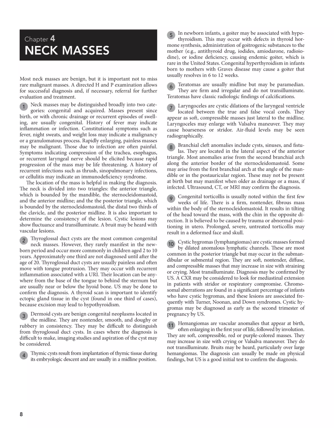
1 Neck masses may be distinguished broadly into two categories: congenital and acquired. Masses present since
birth, or with chronic drainage or recurrent episodes of swelling, are usually congenital. History of fever may indicate
inflammation or infection. Constitutional symptoms such as
fever, night sweats, and weight loss may indicate a malignancy
or a granulomatous process. Rapidly enlarging, painless masses
may be malignant. Those due to infection are often painful.
Symptoms indicating compression of the trachea, esophagus,
or recurrent laryngeal nerve should be elicited because rapid
progression of the mass may be life threatening. A history of
recurrent infections such as thrush, sinopulmonary infections,
or cellulitis may indicate an immunodeficiency syndrome.
The location of the mass is helpful in making the diagnosis.
The neck is divided into two triangles: the anterior triangle,
which is bounded by the mandible, the sternocleidomastoid,
and the anterior midline; and the the posterior triangle, which
is bounded by the sternocleidomastoid, the distal two thirds of
the clavicle, and the posterior midline. It is also important to
determine the consistency of the lesion. Cystic lesions may
show fluctuance and transilluminate. A bruit may be heard with
vascular lesions.
2 Thyroglossal duct cysts are the most common congenital
neck masses. However, they rarely manifest in the newborn period and occur more commonly in children aged 2 to 10
years. Approximately one third are not diagnosed until after the
age of 20. Thyroglossal duct cysts are usually painless and often
move with tongue protrusion. They may occur with recurrent
inflammation associated with a URI. Their location can be anywhere from the base of the tongue to behind the sternum but
are usually near or below the hyoid bone. US may be done to
confirm the diagnosis. A thyroid scan is important to identify
ectopic gland tissue in the cyst (found in one third of cases),
because excision may lead to hypothyroidism.
Dermoid cysts are benign congenital neoplasms located in
the midline. They are nontender, smooth, and doughy or
rubbery in consistency. They may be difficult to distinguish
from thyroglossal duct cysts. In cases where the diagnosis is
difficult to make, imaging studies and aspiration of the cyst may
be considered.
3
4
8
Thymic cysts result from implantation of thymic tissue during
its embryologic descent and are usually in a midline position.
In newborn infants, a goiter may be associated with hypothyroidism. This may occur with defects in thyroid hormone synthesis, administration of goitrogenic substances to the
mother (e.g., antithyroid drug, iodides, amiodarone, radioiodine), or iodine deficiency, causing endemic goiter, which is
rare in the United States. Congenital hyperthyroidism in infants
born to mothers with Graves disease may cause a goiter that
usually resolves in 6 to 12 weeks.
5
Teratomas are usually midline but may be paramedian.
They are firm and irregular and do not transilluminate.
Teratomas have classic radiologic findings of calcifications.
6
Laryngoceles are cystic dilations of the laryngeal ventricle
located between the true and false vocal cords. They
appear as soft, compressible masses just lateral to the midline.
Laryngoceles may enlarge with Valsalva maneuver. They may
cause hoarseness or stridor. Air-fluid levels may be seen
radiographically.
7
Branchial cleft anomalies include cysts, sinuses, and fistulas. They are located in the lateral aspect of the anterior
triangle. Most anomalies arise from the second branchial arch
along the anterior border of the sternocleidomastoid. Some
may arise from the first branchial arch at the angle of the mandible or in the postauricular region. These may not be present
at birth but may manifest when older as drainage or a mass, if
infected. Ultrasound, CT, or MRI may confirm the diagnosis.
8
Congenital torticollis is usually noted within the first few
weeks of life. There is a firm, nontender, fibrous mass
within the body of the sternocleidomastoid. It results in tilting
of the head toward the mass, with the chin in the opposite direction. It is believed to be caused by trauma or abnormal positioning in utero. Prolonged, severe, untreated torticollis may
result in a deformed face and skull.
9
10 Cystic hygromas (lymphangiomas) are cystic masses formed
by dilated anomalous lymphatic channels. These are most
common in the posterior triangle but may occur in the submandibular or submental region. They are soft, nontender, diffuse,
and compressible masses that may increase in size with straining
or crying. Most transilluminate. Diagnosis may be confirmed by
US. A CXR may be considered to look for mediastinal extension
in patients with stridor or respiratory compromise. Chromosomal aberrations are found in a significant percentage of infants
who have cystic hygromas, and these lesions are associated frequently with Turner, Noonan, and Down syndromes. Cystic hygromas may be diagnosed as early as the second trimester of
pregnancy by US.
11 Hemangiomas are vascular anomalies that appear at birth,
often enlarging in the first year of life, followed by involution.
They are soft, compressible, red or purple-colored masses. They
may increase in size with crying or Valsalva maneuver. They do
not transilluminate. Bruits may be heard, particularly over large
hemangiomas. The diagnosis can usually be made on physical
findings, but US is a good initial test to confirm the diagnosis.
Chapter 4
u
Neck Masses 9
NECK MASSES
Perform H and P 1
Anterior midline or
paramedian
Consider:
Free T4, TSH
Thyroid scan
Imaging (CT/ultrasound)
±Aspiration
Thyroglossal duct cyst 2
Dermoid cyst 3
Thymic cyst 4
Goiter 5
Congenital hyperthyroidism
Defect in thyroid hormone
synthesis
Maternal goitrogens
Iodide deficiency
Teratoma 6
Laryngocele 7
Congenital
Acquired
Location
Physical findings
See next page
Anterior triangle
Post auricular
Angle of the mandible
Anterior border of
sternocleidomastoid
±Fistula
Within the belly of the
sternocleidomastoid
Firm/solid
Imaging
Ultrasound
CT/MRI
Branchial
cleft cyst 8
Any location
Soft, spongy
Red/purplish color
Transilluminates
Imaging
Ultrasound
CT/MRI
Congenital muscular
torticollis 9
Cystic hygroma 10
Hemangioma 11
Nelson Textbook of Pediatrics, 19e. Chapters 240, 308, 494, 497, 559, 560, 561, 562, 614, 640, 642, 672
Nelsons Essentials, 6e. Chapter 175
10 Part I
u
Head, Neck, and Eyes
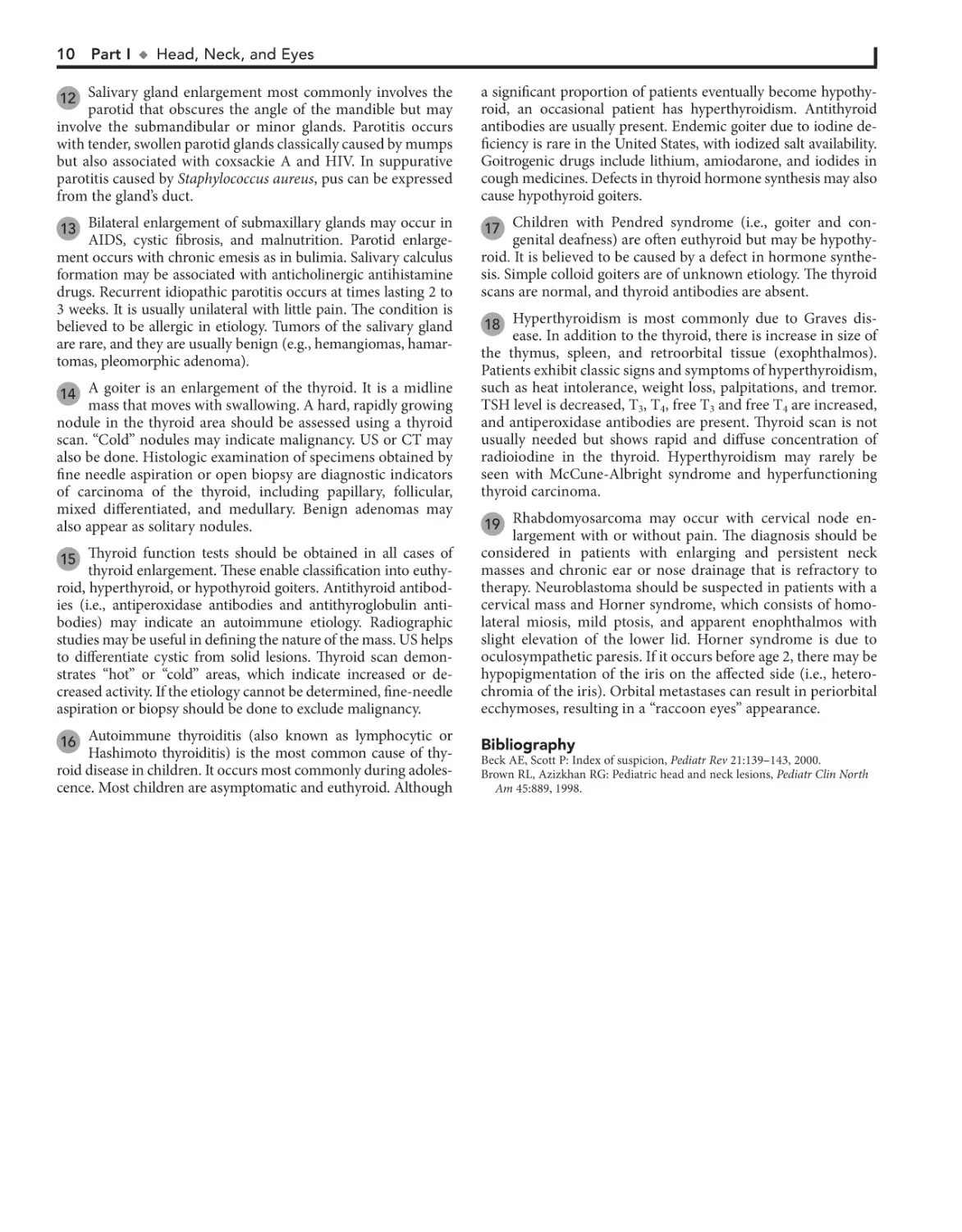
12 Salivary gland enlargement most commonly involves the
parotid that obscures the angle of the mandible but may
involve the submandibular or minor glands. Parotitis occurs
with tender, swollen parotid glands classically caused by mumps
but also associated with coxsackie A and HIV. In suppurative
parotitis caused by Staphylococcus aureus, pus can be expressed
from the gland’s duct.
a significant proportion of patients eventually become hypothyroid, an occasional patient has hyperthyroidism. Antithyroid
antibodies are usually present. Endemic goiter due to iodine deficiency is rare in the United States, with iodized salt availability.
Goitrogenic drugs include lithium, amiodarone, and iodides in
cough medicines. Defects in thyroid hormone synthesis may also
cause hypothyroid goiters.
13 Bilateral enlargement of submaxillary glands may occur in
17 Children with Pendred syndrome (i.e., goiter and con-
AIDS, cystic fibrosis, and malnutrition. Parotid enlargement occurs with chronic emesis as in bulimia. Salivary calculus
formation may be associated with anticholinergic antihistamine
drugs. Recurrent idiopathic parotitis occurs at times lasting 2 to
3 weeks. It is usually unilateral with little pain. The condition is
believed to be allergic in etiology. Tumors of the salivary gland
are rare, and they are usually benign (e.g., hemangiomas, hamartomas, pleomorphic adenoma).
14 A goiter is an enlargement of the thyroid. It is a midline
mass that moves with swallowing. A hard, rapidly growing
nodule in the thyroid area should be assessed using a thyroid
scan. “Cold” nodules may indicate malignancy. US or CT may
also be done. Histologic examination of specimens obtained by
fine needle aspiration or open biopsy are diagnostic indicators
of carcinoma of the thyroid, including papillary, follicular,
mixed differentiated, and medullary. Benign adenomas may
also appear as solitary nodules.
genital deafness) are often euthyroid but may be hypothyroid. It is believed to be caused by a defect in hormone synthesis. Simple colloid goiters are of unknown etiology. The thyroid
scans are normal, and thyroid antibodies are absent.
18 Hyperthyroidism is most commonly due to Graves dis-
ease. In addition to the thyroid, there is increase in size of
the thymus, spleen, and retroorbital tissue (exophthalmos).
Patients exhibit classic signs and symptoms of hyperthyroidism,
such as heat intolerance, weight loss, palpitations, and tremor.
TSH level is decreased, T3, T4, free T3 and free T4 are increased,
and antiperoxidase antibodies are present. Thyroid scan is not
usually needed but shows rapid and diffuse concentration of
radioiodine in the thyroid. Hyperthyroidism may rarely be
seen with McCune-Albright syndrome and hyperfunctioning
thyroid carcinoma.
19 Rhabdomyosarcoma may occur with cervical node en-
15 Thyroid function tests should be obtained in all cases of
thyroid enlargement. These enable classification into euthyroid, hyperthyroid, or hypothyroid goiters. Antithyroid antibodies (i.e., antiperoxidase antibodies and antithyroglobulin antibodies) may indicate an autoimmune etiology. Radiographic
studies may be useful in defining the nature of the mass. US helps
to differentiate cystic from solid lesions. Thyroid scan demonstrates “hot” or “cold” areas, which indicate increased or decreased activity. If the etiology cannot be determined, fine-needle
aspiration or biopsy should be done to exclude malignancy.
largement with or without pain. The diagnosis should be
considered in patients with enlarging and persistent neck
masses and chronic ear or nose drainage that is refractory to
therapy. Neuroblastoma should be suspected in patients with a
cervical mass and Horner syndrome, which consists of homolateral miosis, mild ptosis, and apparent enophthalmos with
slight elevation of the lower lid. Horner syndrome is due to
oculosympathetic paresis. If it occurs before age 2, there may be
hypopigmentation of the iris on the affected side (i.e., heterochromia of the iris). Orbital metastases can result in periorbital
ecchymoses, resulting in a “raccoon eyes” appearance.
16 Autoimmune thyroiditis (also known as lymphocytic or
Bibliography
Hashimoto thyroiditis) is the most common cause of thyroid disease in children. It occurs most commonly during adolescence. Most children are asymptomatic and euthyroid. Although
Beck AE, Scott P: Index of suspicion, Pediatr Rev 21:139–143, 2000.
Brown RL, Azizkhan RG: Pediatric head and neck lesions, Pediatr Clin North
Am 45:889, 1998.
Chapter 4
u
Neck Masses 11
NECK MASSES (continued)
Acquired
Location
Physical findings
Lymph node
involvement
Lateral neck
Obscuring angle of
the mandible
YES
Goiter 14
NO
Hard, solitary,
rapidly growing
nodule
Salivary gland
enlargement 12
YES
Inflammation
present
(warm, tender)
YES
Acute salivary
gland infection
Non-suppurative 12
Mumps
Coxsackie A
HIV
Suppurative
S. aureus
Lymphadenitis
Lymphadenopathy
(see Chapter 60
on Lymphadenopathy)
Enlarging and persistent
cervical mass with
any of the following:
Persistent drainage from
nose or ear which is
refractory to therapy
Horner syndrome
Iris heterochromia
“Raccoon eyes”
(periorbital ecchymosis)
Midline
Moves with swallowing
NO
Thyroid scan
Ultrasound
±Fine-needle
aspiration
or biopsy
MRI
Free T4, TSH 15
Anti-thyroid
antibodies
±Thyroid scan,
Imaging (Ultrasound/CT)
NO
Branchial
cleft cyst 8
Benign
adenoma 14
Carcinoma 14
Salivary calculus 13
Collagen-vascular disease
Recurrent emesis (bulimia) 13
Cystic fibrosis
Recurrent idiopathic parotitis 13
Mucocele
Tumor 13
Hypothyroid
Autoimmune
thyroiditis 16
Endemic goiter
(iodine
deficiency) 16
Goitrogenic
drugs 16
Defect in thyroid
hormone
synthesis 16
Thyroglossal
duct cyst 2
Euthyroid
Hyperthyroid
Pendred
syndrome 17
Simple colloid
goiter 17
Autoimmune
thyroiditis 16
Thyroglossal
duct cyst 2
Graves
disease 18
Autoimmune
thyroiditis 16
Rhabdomyosarcoma 19
Neuroblastoma 19
Nelsons Essentials, 6e. Chapter 175
Chapter 5
ABNORMAL HEAD
SIZE, SHAPE, AND
FONTANELS
increased production of CSF. Causes of obstructive (noncommunicating) hydrocephalus include aqueductal stenosis, neonatal meningitis, subarachnoid hemorrhage in a premature infant,
intrauterine viral infections, vein of Galen malformation, and
posterior fossa lesions or malformations (tumors, Chiari malformation, Dandy-Walker syndrome). Subarachnoid hemorrhage
in a premature infant can also cause nonobstructive (communicating) hydrocephalus. A rare cause is overproduction of CSF
with choroid plexus papilloma.
In hydranencephaly the cerebral hemispheres are absent
or represented by membranous sacs. The cause of this
condition is unknown.
5
Macrocephaly is defined as an occipitofrontal circumference
(OFC) greater than 2 standard deviations above the mean.
Megalencephaly is a disorder of brain growth, usually accompanied by macrocephaly. An increase in growth rate with crossing
of percentiles is of more concern than the case of a child with a
large head growing at a normal rate. In microcephaly the OFC
is 2 standard deviations below the mean.
1 A birth history, developmental history and history of irritability, headaches, and visual problems are important
components of the initial evaluation. For macrocephaly, inquire
about familial head sizes (e.g., ask about hat sizes). It is important to note any features suggestive of specific syndromes.
US can be done if the anterior fontanel (AF) is open.
Otherwise, an MRI should be considered. A CT is preferred if there is suspicion of trauma (nonaccidental or accidental). Radiologic evaluation may not be necessary if development
is normal, a parent is macrocephalic, and the child’s head is
growing at a normal rate. Further evaluation is directed by the
history and physical examination. Plain long bone radiographs
may be indicated for evaluation of skeletal dysplasia or trauma.
Consider chromosome testing (fragile X) or metabolic tests
(urine organic acids).
2
Benign familial megalencephaly is the most common
cause of anatomic megalencephaly. It is inherited as an
autosomal dominant trait. These children may have mild neurodevelopmental dysfunction. It is diagnosed by careful family
history and measurement of the parents’ head circumferences.
3
4 Hydrocephalus is caused by multiple conditions associ
ated with impaired circulation and absorption of CSF or
12
Occasionally, benign fluid collections (e.g., subarachnoid,
subdural) cause macrocephaly without other clinical
significance. A pediatric neurosurgeon should be consulted for
recommendations.
6
Various metabolic and degenerative disorders may
cause megalencephaly. These include lysosomal diseases
(Tay-Sachs disease, gangliosidosis, mucopolysaccharidoses),
maple syrup urine disease, and leukodystrophies.
7
Many syndromes are associated with microcephaly. If a
chromosomal syndrome is suspected (child has abnormal
facies, short stature, congenital anomalies) karyotype and/or
array-comparative genomic hybridization (microarray) study
and MRI may be considered.
8
MRI can evaluate structural abnormalities of the brain
(lissencephaly, pachygyria, and polymicrogyria) and both
MRI and CT scanning may detect intracerebral calcification,
which suggests congenital infection. Also consider TORCH
titers (toxoplasmosis, rubella, CMV, and herpes simplex) and
HIV testing of the mother and child, as well as a urine culture
for CMV. Consider testing for maternal serum phenylalanine
level (PKU), because high maternal levels can affect a nonphenylketonuric infant.
9
10 Familial microcephaly is often associated with some
degree of mental retardation.
11 Secondary microcephaly results from exposure to noxious
agents during periods of rapid brain growth in utero or
during the first 2 years of life.
Chapter 5
u
Abnormal Head Size, Shape, and Fontanels 13
ABNORMAL HEAD SIZE, SHAPE, OR FONTANELS
Perform H and P 1
Macrocephaly
Parent with large OFC
±Mild neurodevelopmental delay
YES
NO
Microcephaly
Abnormal shape
Abnormal fontanel
Suspect chromosomal
syndrome 8
Abnormal facies
Short stature
Congenital anomalies
See next page
YES
Familial
megalencephaly 3
Consider neuroimaging: 2
MRI
CT
Ultrasound
(if fontanel open)
Karyotype and/or
micro-array testing
MRI
Hydrocephalus 4
Hydrancephaly 5
Mass (tumor, cyst,
arteriovenous malformation)
Subdural hemorrhage
Subdural effusion
Benign fluid collections 6
Megalencephaly 7
Thickened skull (rickets,
epiphyseal dysplasia,
chronic anemia)
Autism spectrum disorders
Syndromes (Fragile X,
cerebral gigantism,
neurofibromatosis)
Primary (genetic)
Familial microcephaly
(autosomal recessive) 10
Autosomal dominant microcephaly
Brain atrophy or dysgenesis
syndromes
Down syndrome (trisomy 21)
Edward syndrome (Trisomy 18)
Cri-du-chat syndrome (5p-)
Cornelia de Lange syndrome
Rubinstein-Taybi syndrome
Smith-Lemli-Opitz syndrome
NO
MRI/CT 9
TORCH titers
HIV testing of the
mother and child
Urine culture for
cytomegalovirus
Consider maternal serum
phenyalanine levels
Secondary: Prenatal
or perinatal injury 11
Toxins (alcohol, hydantoin)
Hypoxic-ischemic encephalopathy
(trauma, infections)
Congenital Infections
(Cytomegalovirus, rubella,
toxoplasmosis)
Radiation
Meningitis/encephalitis
Metabolic (maternal diabetes,
maternal phenylketonuria)
Malnutrition
Nelson Textbook of Pediatrics, 19e. Chapters 88, 584, 585
Nelsons Essentials, 6e. Chapter 187
14 Part I
u
Head, Neck, and Eyes
12 Skull deformational malformations occur as the result of
an alteration of the normal forces (in utero, perinatal, or
postnatal) acting upon the growing cranium. Positional skull
deformity, or plagiocephaly (skull asymmetry), is the most
common type of deformational malformation. Its incidence has
increased because of the recommendations to place infants on
their backs while sleeping. Plagiocephaly is a benign condition
that must be distinguished from true cranial suture synostosis.
In plagiocephaly, sutures are open, and a frontal and temporal
prominence occurs on the same side as the flat occiput. Frontal
flattening occurs on the side opposite the flat occiput. Molding
can occur with breech presentation or as the neonate passes
though the birth canal; it resolves within a few weeks. Congenital muscular torticollis may restrict the infant’s range of
motion at the neck, leading to facial asymmetry and plagiocephaly. It is often not noticed in the newborn and is diagnosed
when the infant develops better head control. The diagnosis of
positional skull deformity is made based on H and P. Imaging
studies are rarely necessary and should only be considered in
refractory cases or children born with congenital deformities.
13 Cleidocranial dysostosis is a hereditary condition charac-
because of compensatory growth. Symmetric occipital flattening that is believed to be positional does not require imaging.
In craniosynostosis, there is often palpable ridging over the
fused sutures. The condition may occur as a primary isolated
disorder, which is most common, or as part of a syndrome.
Common associated disorders include Crouzon, Apert, and
Pfeiffer syndromes, congenital hyperthyroidism, and adrenal
hyperplasia.
16 Imaging is recommended except in the case of a crying
infant in whom the bulging resolves spontaneously or in
an infant with a clinical picture of meningitis. An LP should be
performed if meningitis is suspected.
17 Normal fullness occurs with crying in an infant with a
normal fontanel. It should be distinguished from true
bulging, such as occurs in hydrocephalic infants. Normally the
fontanel is pulsatile even when full due to crying. In hydrocephalus, the anterior fontanel is usually not visibly pulsatile.
Examination of the fontanel should be performed while the
infant is in a sitting position.
18 Transient unexplained benign bulging of the fontanel
terized by incomplete ossification of membranous bones,
including the cranium, clavicle, and pelvis. Cranial sutures are
often wide and contain wormian bones.
may occur in normal infants. This, however, should be a
diagnosis of exclusion.
14 The anterior fontanel averages 2.5 cm in diameter. Average age
Bibliography
of closure is between 7 and 19 months. As long as head growth
is normal and sutural ridging is absent, early closure is not a concern.
15 In true synostosis of a lambdoidal suture, frontal and
parietal bossing would occur on the opposite side
Kiesler K, Ricer R: The abnormal fontanel, Am Fam Physician 67:2547–2552,
2003.
Purugganan OH: Abnormalities in head size, Pediatr Rev 27:473–476, 2006.
Ridgway EB, Weiner HL: Skull deformities, Pediatr Clin North Am 51:
359–387, 2004.
Robin NH: Congenital muscular torticollis, Pediatr Rev 17:374–375, 1996.
Chapter 5
u
Abnormal Head Size, Shape, and Fontanels 15
ABNORMAL HEAD SIZE, SHAPE, OR FONTANELS (continued)
Abnormal shape
Abnormal fontanel
H and P suggestive of
benign skull deformity
Normal sutures
YES
NO
Large/delayed
closure
Frontal bossing
present
Congenital
syphilis
Rickets
Cleidocranial
dysostosis 13
Ectodermal
dysplasia
Lowe syndrome
Bulging
Sunken
Prominent
bony ridge
Fusion of the
suture(s)
Consider
imaging
X-ray/CT
Plagiocephaly
(positional skull
deformity) 12
Deformity due to
torticollis 12
Neonatal molding
(birth molding) 12
Small 14
Craniosynostosis 14
Craniosynostosis 15
Microcephaly
Congenital hypothyroidism
Skeletal dysplasias
(achondroplasia, cleidocranial
dysostosis)
Syndromes (Trisomies 13,
18, 21; Apert, Russell-Silver)
Osteogenesis Imperfecta
Prematurity syndromes
Hydrocephalus
Rickets
Congenital rubella
or syphilis
Dehydration
Crying (normal
fullness) 16
Hydrocephalus 17
Intraventricular
hemorrhage
Subdural hematoma
(shaken baby syndrome)
Infections (meningitis, roseola)
Metabolic (maple syrup urine
disease, galactosemia,
hypophosphatasia, urea
cycle enzyme defects)
Hypervitaminosis A
Lead encephalopathy
Benign 18
Chapter 6
RED EYE
Red eye is a common pediatric complaint. It can occur secondary to a wide range of etiologies.
1 The age of onset of the red eye, the nature of any discharge,
and the associated signs and symptoms are the most
important components of the history. History of exposure to
irritants (e.g., allergens, particulate matter, chemicals) and of
trauma or infectious contacts (e.g., “pink-eye” in school or daycare settings) may also be helpful. For infants, inquire about the
possibility of any maternal infections.
Conjunctivitis within the neonatal period (4 weeks of
birth) is also known as ophthalmia neonatorum. The most
common causes in the United States are Staphylococcus aureus,
Staphylococcus epidermidis, Streptococcus pneumoniae, and
Moraxella catarrhalis.
2
Ophthalmia neonatorum also can be caused by Chlamydia
trachomatis, Neisseria gonorrhoeae, and herpes simplex virus
(HSV). Gonococcal conjunctivitis typically appears as a fulminant
purulent conjunctivitis in the first 2 to 6 days of life. Chlamydial
conjunctivitis is more likely beyond the first 6 days of life and is
often associated with a pneumonitis. It can develop in 30% to 40%
of infants whose mothers had untreated chlamydia. Conjunctivitis
caused by HSV characteristically occurs as a unilateral bright red
eye with thin watery discharge. Vesicles or erosions are present on
the lid or surrounding skin. These clinical findings are not specific, however, and prompt evaluation and treatment are always
indicated to avoid serious sequelae. A Gram stain and culture will
aid in the diagnosis of gonorrhea. Rapid antigen tests are available
for chlamydial infections. HSV is usually cultured, but PCR may
be helpful. Ophthalmologic consultation is indicated when herpes
is suspected.
3
Viral conjunctivitis may vary in presentation from mild
redness and irritation with minimal watery drainage to
severe conjunctival injection with purulent discharge. Adenovirus is the most common cause and may present with preauricular lymphadenopathy. Coxsackie and echoviruses may cause a
hemorrhagic conjunctivitis.
4
Dacryostenosis (i.e., congenital lacrimal duct stenosis) is a
common disorder that occurs within 2 to 4 months of age
but sometimes is not noticed until tear production with crying
becomes evident. An excessive tear lake and overflow with
crusting are seen on examination. Children so affected are at
5
16
risk for inflammation and infection (i.e., dacryocystitis) of the
obstructed nasolacrimal sac.
Tearing, photophobia, and blepharospasm make up the
classic triad of presenting symptoms of infantile glaucoma.
Conjunctival injection, corneal enlargement (.12 mm), and
corneal clouding (edema) are the other findings.
6
Conjunctivitis in the first 24 hours of life is probably a chemical conjunctivitis unless membranes were ruptured prematurely. Silver nitrate is more likely to produce this condition than
other agents used for prophylaxis (e.g., erythromycin, tetracycline)
and is no longer used in the United States. In older children,
chemical irritants may include cosmetics or eye medications.
7
Corneal abrasion presents with pain, tearing, photophobia, and eye redness. It is an important consideration in
the diagnosis of an irritable infant. Diagnosis is by fluorescein
staining and observation under blue light.
8
Subconjunctival hemorrhage may occur with vomiting,
coughing, or weight lifting. It may also occur in newborns
after vaginal delivery.
9
10 Allergic conjunctivitis is characterized by itching, chemo-
sis, papillae of the tarsal conjunctivae, and white stringy
discharge. In limbal vernal conjunctivitis, a ring of swollen conjunctiva surrounds the limbus of the cornea.
11 Bacterial conjunctivitis may be unilateral or bilateral, but
viral conjunctivitis is more commonly bilateral. Bacterial
conjunctivitis is more likely to have purulent discharge than
viral conjunctivitis, although significant overlap in the clinical
presentation of the two etiologies does occur. Nontypable Haemophilus influenzae, pneumococci, staphylococci, and streptococci are common agents.
12 Redness may be due to irritation from eye rubbing. Exces-
sive television or computer use may cause decreased rate
of blinking, with drying and irritation.
13 Iritis and iridocyclitis may occur secondary to localized
infection or trauma, or they may be manifestations of
a rheumatic disorder (e.g., JRA, Reiter syndrome, Behçet’s
disease). Inflammatory bowel disease and Kawasaki disease
are other associated conditions. Photophobia is typically a
significant finding with iritis and iridocyclitis.
14 Scleritis may accompany certain autoimmune disorders
including systemic lupus erythematosus and HenochSchönlein purpura. Pain is present, eye discharge is absent, and
dilated blood vessels are larger than in conjunctivitis.
15 Parinaud’s oculoglandular syndrome is a form of cat scratch
disease caused by Bartonella henselae. Symptoms include a
granulomatous conjunctivitis and preauricular lymphadenopathy.
Chapter 6
Red Eye 17
u
RED EYE
Perform H and P 1
Conjunctival injection
Periorbital redness
and/or swelling
Neonate (<30 days)
Age >30 days
Eye drainage present
Eye drainage present
YES
YES
NO
Consider:
Gram stain and
culture
Chlamydia culture/
nucleic acid
amplification
tests (PCR)
Culture/PCR for
herpes simplex
if vesicles present
Bacterial
conjunctivitis 2
Sexually
transmitted
diseases 3
Viral
conjunctivitis 4
Dacryostenosis 5
See next page
NO
Allergic symptoms
associated (tearing,
eye rubbing)
History of trauma,
foreign body, chemical
exposure or irritation
Painful red eye
±Abnormal vision
±Photophobia
±Lacrimation
Eyelid redness/
irritation
Glaucoma 6
Keratitis (HSV)
Endophthalmitis
Iritis/iridocyclitis 13
Scleritis 14
Stevens-Johnson
syndrome
Bartonella
henselae 15
Blepharoconjunctivitis
HSV
Molluscum
contagiosum
Pubic lice
Consider fluorescein
staining and inspection
with Wood’s lamp
or slit lamp
Glaucoma 6
Viral conjunctivitis 4
Chemical
conjunctivitis 7
Corneal abrasion 8
Sub-conjunctival
hemorrhage 9
Viral conjunctivitis 4
Bacterial
conjunctivitis 11
STD (gonorrhea,
chlamydia)
Allergic
conjunctivitis 10
Limbal vernal
conjunctivitis
Chemical conjunctivitis
Corneal abrasion 8
Foreign body
Irritation/dry eyes 12
Sub-conjunctival
hemorrhage 9
Nelson Textbook of Pediatrics, 19e. Chapters 611, 614, 618, 619, 624, 625
Nelsons Essentials, 6e. Chapter 119
18 Part I
u
Head, Neck, and Eyes
16 Pain with extraocular eye movements may accompany
The lacrimal gland (i.e., the site of tear production) is
located in the lateral aspect of the upper eyelid. Rarely,
inflammation of the lacrimal gland (i.e., dacryoadenitis) can
occur as a result of infections (e.g., S. aureus, infectious
mononucleosis, mumps).
17 Orbital tumors, including rhabdomyosarcomas, neuroblas-
Bibliography
orbital cellulitis. Proptosis and impaired extraocular
movement and vision are other signs. Orbital cellulitis must be
distinguished from preseptal (periorbital) cellulitis. Minimal
conjunctival redness usually occurs in orbital cellulitis, and
extraocular muscle movements are intact in preseptal cellulitis.
tomas, and lymphangiomas, may have a similar presentation
to orbital cellulitis.
18 Some systemic disorders, such as sarcoid, tuberculosis,
and syphilis, may cause chronic dacryocystitis.
Greenberg MF, Pollard ZF: The red eye in childhood, Pediatr Clin North Am
50:105–124, 2003.
Richards A, Guzman-Cottrill JA: Conjunctivitis, Pediatr Rev 31:196–208, 2010.
Chapter 6
RED EYE (continued)
Periorbital redness
and/or swelling
Proptosis
Ophthalmoplegia
Vision abnormalities
YES
NO
Consider:
CT
Orbital cellulitis 16
Orbital tumors 17
Preseptal orbital cellulitis
Allergic reaction
Angioneurotic response
Insect bite/ sting
Chalazion
Hordeolum
Dacryocystitis 18
Dacryoadenitis 18
Ecchymoses/trauma
Vascular lesions
Hemangiomas
Nevus flammeus
Sturge-Weber syndrome
u
Red Eye 19
Chapter 7
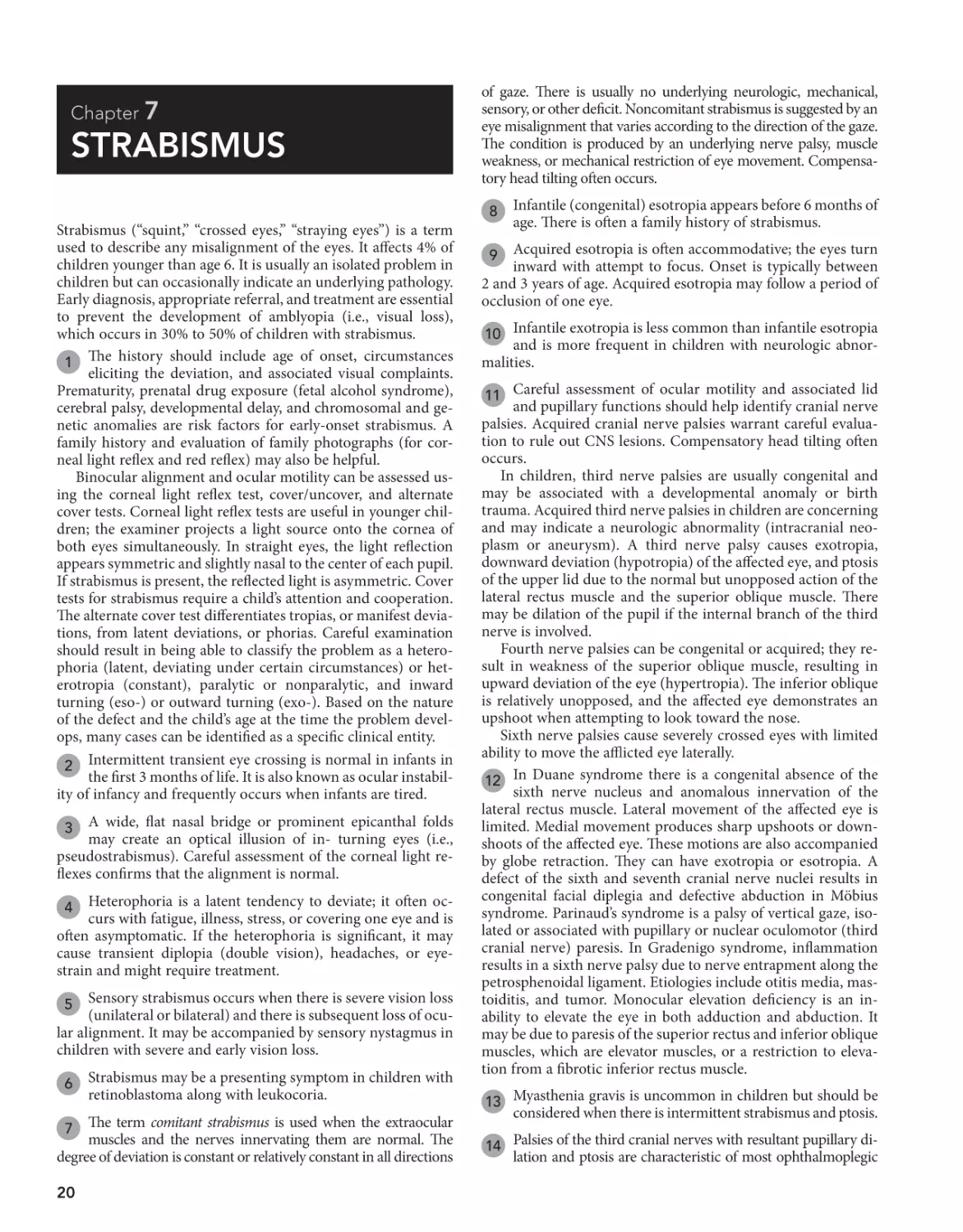
STRABISMUS
Strabismus (“squint,” “crossed eyes,” “straying eyes”) is a term
used to describe any misalignment of the eyes. It affects 4% of
children younger than age 6. It is usually an isolated problem in
children but can occasionally indicate an underlying pathology.
Early diagnosis, appropriate referral, and treatment are essential
to prevent the development of amblyopia (i.e., visual loss),
which occurs in 30% to 50% of children with strabismus.
1 The history should include age of onset, circumstances
eliciting the deviation, and associated visual complaints.
Prematurity, prenatal drug exposure (fetal alcohol syndrome),
cerebral palsy, developmental delay, and chromosomal and genetic anomalies are risk factors for early-onset strabismus. A
family history and evaluation of family photographs (for corneal light reflex and red reflex) may also be helpful.
Binocular alignment and ocular motility can be assessed using the corneal light reflex test, cover/uncover, and alternate
cover tests. Corneal light reflex tests are useful in younger children; the examiner projects a light source onto the cornea of
both eyes simultaneously. In straight eyes, the light reflection
appears symmetric and slightly nasal to the center of each pupil.
If strabismus is present, the reflected light is asymmetric. Cover
tests for strabismus require a child’s attention and cooperation.
The alternate cover test differentiates tropias, or manifest deviations, from latent deviations, or phorias. Careful examination
should result in being able to classify the problem as a heterophoria (latent, deviating under certain circumstances) or heterotropia (constant), paralytic or nonparalytic, and inward
turning (eso-) or outward turning (exo-). Based on the nature
of the defect and the child’s age at the time the problem develops, many cases can be identified as a specific clinical entity.
2 Intermittent transient eye crossing is normal in infants in
the first 3 months of life. It is also known as ocular instability of infancy and frequently occurs when infants are tired.
A wide, flat nasal bridge or prominent epicanthal folds
may create an optical illusion of in- turning eyes (i.e.,
pseudostrabismus). Careful assessment of the corneal light reflexes confirms that the alignment is normal.
3
Heterophoria is a latent tendency to deviate; it often occurs with fatigue, illness, stress, or covering one eye and is
often asymptomatic. If the heterophoria is significant, it may
cause transient diplopia (double vision), headaches, or eyestrain and might require treatment.
4
Sensory strabismus occurs when there is severe vision loss
(unilateral or bilateral) and there is subsequent loss of ocular alignment. It may be accompanied by sensory nystagmus in
children with severe and early vision loss.
5
6
Strabismus may be a presenting symptom in children with
retinoblastoma along with leukocoria.
The term comitant strabismus is used when the extraocular
muscles and the nerves innervating them are normal. The
degree of deviation is constant or relatively constant in all directions
7
20
of gaze. There is usually no underlying neurologic, mechanical,
sensory, or other deficit. Noncomitant strabismus is suggested by an
eye misalignment that varies according to the direction of the gaze.
The condition is produced by an underlying nerve palsy, muscle
weakness, or mechanical restriction of eye movement. Compensatory head tilting often occurs.
8
Infantile (congenital) esotropia appears before 6 months of
age. There is often a family history of strabismus.
Acquired esotropia is often accommodative; the eyes turn
inward with attempt to focus. Onset is typically between
2 and 3 years of age. Acquired esotropia may follow a period of
occlusion of one eye.
9
10 Infantile exotropia is less common than infantile esotropia
and is more frequent in children with neurologic abnormalities.
11 Careful assessment of ocular motility and associated lid
and pupillary functions should help identify cranial nerve
palsies. Acquired cranial nerve palsies warrant careful evaluation to rule out CNS lesions. Compensatory head tilting often
occurs.
In children, third nerve palsies are usually congenital and
may be associated with a developmental anomaly or birth
trauma. Acquired third nerve palsies in children are concerning
and may indicate a neurologic abnormality (intracranial neoplasm or aneurysm). A third nerve palsy causes exotropia,
downward deviation (hypotropia) of the affected eye, and ptosis
of the upper lid due to the normal but unopposed action of the
lateral rectus muscle and the superior oblique muscle. There
may be dilation of the pupil if the internal branch of the third
nerve is involved.
Fourth nerve palsies can be congenital or acquired; they result in weakness of the superior oblique muscle, resulting in
upward deviation of the eye (hypertropia). The inferior oblique
is relatively unopposed, and the affected eye demonstrates an
upshoot when attempting to look toward the nose.
Sixth nerve palsies cause severely crossed eyes with limited
ability to move the afflicted eye laterally.
12 In Duane syndrome there is a congenital absence of the
sixth nerve nucleus and anomalous innervation of the
lateral rectus muscle. Lateral movement of the affected eye is
limited. Medial movement produces sharp upshoots or downshoots of the affected eye. These motions are also accompanied
by globe retraction. They can have exotropia or esotropia. A
defect of the sixth and seventh cranial nerve nuclei results in
congenital facial diplegia and defective abduction in Möbius
syndrome. Parinaud’s syndrome is a palsy of vertical gaze, isolated or associated with pupillary or nuclear oculomotor (third
cranial nerve) paresis. In Gradenigo syndrome, inflammation
results in a sixth nerve palsy due to nerve entrapment along the
petrosphenoidal ligament. Etiologies include otitis media, mastoiditis, and tumor. Monocular elevation deficiency is an inability to elevate the eye in both adduction and abduction. It
may be due to paresis of the superior rectus and inferior oblique
muscles, which are elevator muscles, or a restriction to elevation from a fibrotic inferior rectus muscle.
13 Myasthenia gravis is uncommon in children but should be
considered when there is intermittent strabismus and ptosis.
14 Palsies of the third cranial nerves with resultant pupillary di-
lation and ptosis are characteristic of most ophthalmoplegic
Chapter 7
migraines. The eye muscle paralysis may last for a few weeks following a headache.
u
Strabismus 21
18 Excessive fibrosis and anomalous insertion of extraocular
15 Restrictive strabismus is due to mechanical forces such as
muscles results in ptosis and external ophthalmoplegia. Convergence on attempted upward gaze, divergence on attempted
downward gaze, and compensatory chin-up posturing are also
characteristic of congenital fibrosis syndrome.
16 Blunt trauma to the eye leading to a blowout fracture of
Bibliography
inflammation, edema, trauma, or congenital disorders resulting in fibrosis.
the orbit may cause strabismus due to muscle entrapment
as well as edema and hematoma of the muscle(s).
17 In Brown syndrome, an abnormality of the superior oblique
tendon results in an inability to elevate the eye in the medial position.
Magramm I: Amblyopia: Etiology, detection, and treatment, Pediatr Rev 13:
7–14, 1992.
Ticho BH: Strabismus, Pediatr Clin North Am 50:173–188, 2003.
Tingley DH: Vision screening essentials: Screening today for eye disorders in
the pediatric patient, Pediatr Rev 28:54–61, 2007.
STRABISMUS
H and P 1
Screening Tests
Red reflex test
Corneal light reflex
Cover/uncover test
Alternate cover test
±Visual acuity
test (>3 years)
Normal tests
Abnormal tests
Intermittently
abnormal tests
Deviation constant in
various directions of gaze
Absence of identifiable
neurologic, mechanical,
sensory deficits 7
Abnormal red reflex
and/or
Visual loss
YES
Intermittent
eye-crossing
in infants 2
Pseudostrabismus 3
Heterophorias 4
Retinoblastoma 5
Sensory
strabismus 6
(See Chapter 8
on visual
impairment and
leukocoria)
Infantile esotropia 8
Acquired esotropia 9
Infantile exotropia 10
NO
Paralytic 11
Cranial nerve palsy
(3rd, 4th, 6th)
Restrictive 15
Due to mechanical
forces
Syndromes (Duane
syndrome, Möbius
syndrome, Parinaud
syndrome, Gradenigo
syndrome, Monocular
elevation defect) 12
Congenital nerve palsy
Acquired nerve palsy
Trauma
Infection
CNS tumor or other lesion
Hydrocephalus
Post-viral
Myasthenia gravis 13
Ophthalmoplegic
migraine 14
Trauma 16
Brown syndrome 17
Congenital fibrosis
syndromes 18
Graves’
ophthalmopathy
Nelson Textbook of Pediatrics, 19e. Chapters 584, 614
Chapter 8
VISUAL IMPAIRMENT
AND LEUKOCORIA
When treating a child in whom there are visual concerns, consultation with a pediatric ophthalmologist or optometrist is
frequently required, especially for a very young infant.
1 A detailed description of the visual complaint is helpful, but
in infants and young children the history tends to be based
primarily on the observations of family and caretakers. For older
children, inquire about focal versus general blurring, double images, night vision, and abnormal sensations (e.g., spots, lines). The
birth history is an important component of the medical history
because asphyxia or trauma (i.e., forceps injury) may be contributory. Recent illness may aid in the diagnosis of sudden visual loss
caused by acute optic neuritis. A family history of neurocutaneous
disorders, metabolic disorders, cataracts, or other visual problems
may also be helpful. The examination should include an assessment for visual acuity using a Snellen chart or one designed for
preliterate children (e.g., tumbling E and picture tests). For infants
and toddlers, referral for visual assessment using behavioral responses may be necessary. Visual fields should also be assessed.
Examination of the pupils, ocular motility, and eye alignment is
necessary. A thorough ophthalmologic examination should be
done. Common findings that may be helpful in diagnosis include
strabismus, corneal opacity, leukocoria, and nystagmus.
Leukocoria (i.e., white pupillary reflex) may be due to abnormalities in the retina, lens, or vitreous. Leukocoria is
most common in the young infant, although it may occur with
problems that develop at a later age. It is the most common
presentation of retinoblastoma. Referral to an ophthalmologist
for a thorough diagnostic evaluation is always indicated.
2
Cataracts are opacity of the lens and are a common cause of
leukocoria. Common etiologies of cataracts include infections (e.g., rubella, toxoplasmosis, CMV), hereditary or metabolic
disorders, chromosomal disorders (e.g., trisomy 18, trisomy 13,
Turner syndrome), toxins, and trauma. They may also develop as
a result of an intraocular processes such as retinopathy of prematurity. Metabolic and endocrine diseases associated with cataracts
include galactosemia, galactokinase deficiency, hypoparathyroidism, Wilson disease, and juvenile-onset diabetes mellitus. Cataracts may also be seen in children of diabetic and prediabetic
mothers.
3
4
Retinoblastoma is a malignancy of the retina and is the
most common intraocular tumor in children.
5 Retinopathy of prematurity is a disease of developing retinal vasculature and occurs in premature infants. According
to the American Academy of Pediatrics screening guidelines,
infants with a birth weight of less than 1500 g or gestational age
of 30 weeks or less, and selected high-risk infants with birth
weight between 1500 and 2000 g or gestational age of over
30 weeks, should have retinal screening examinations.
22
Glaucoma is a progressive optic neuropathy associated with
elevated pressure within the eye. Congenital glaucoma is
present at birth, infantile glaucoma develops during the first
3 years of life, and juvenile glaucoma begins between 3 and
30 years of age. Glaucoma may be primary and due to an isolated
anomaly of the drainage apparatus of the eye (trabecular meshwork) or secondary with other associated ocular or systemic
abnormalities. In infantile glaucoma, signs and symptoms are
different from adult glaucoma, because the infant eye is pliable,
leading to enlargement of the cornea and globe. This is known
as buphthalmos or “ox-eye.” Only 30% of children with infantile
glaucoma demonstrate the classic triad of glaucoma: tearing,
photophobia, and blepharospasm (i.e., eyelid squeezing). Other
signs include corneal haziness, conjunctival injection, and visual
impairment.
6
Anterior uveitis involves inflammation of the iris and/or
ciliary body. It may be associated with JRA, sarcoidosis,
Kawasaki disease, Stevens-Johnson syndrome, viral infections
(e.g., herpes simplex, herpes zoster), syphilis, tuberculosis, and
many other conditions. Posterior uveitis involves the choroid
and often the retina. Causes include parasites (e.g., toxoplasmosis, toxocariasis), viruses (e.g., rubella, herpes simplex, HIV,
CMV), tuberculosis, and others.
7
Optic neuritis is an inflammation or demyelination of the
optic nerve, leading to acute vision loss. It may be due to
inflammatory diseases (e.g., systemic lupus erythematosus),
infections (e.g., HIV, syphilis, Lyme disease, meningitis), toxin
exposure (e.g., methanol, lead), or nutrient deficiency (e.g.,
B12). Multiple sclerosis is also a possible etiology but is more
common in adults.
8
Psychogenic vision loss (malingering, conversion reactions) is more likely to occur in older school-aged children.
Complaints of visual loss or blurring may be accompanied by
numerous other complaints (e.g., abnormal visual sensations,
diplopia, polyopia, painful eyes, headaches). Diagnosis is suggested by normal examination findings and behavioral “red
flags” such as inconsistent complaints in different situations,
positive responses to examiner’s suggestions of maneuvers to
improve vision, and inappropriate affect (e.g., indifferent, belligerent, overdramatic) during the examination.
9
10 Amblyopia is a decrease in visual acuity despite correction
of any refractive errors. It occurs in visually immature children and is due to the lack of a clear image projecting onto the
retina. When this occurs during the critical period of development in the first decade of life, amblyopia may occur. The unformed image can occur secondary to a strabismus, a difference
in refractive error between the eyes (anisometropic amblyopia),
a high refractive error in both eyes (ametropic amblyopia), and
image deprivation due to cataracts, corneal opacities, or occlusion (e.g., eye patch).
11 Optic gliomas are most commonly located in the optic
chiasm but may occur anywhere along the optic pathway.
They can occur with a variety of symptoms, including unilateral
vision loss, proptosis, bitemporal hemianopia, and eye deviation. Craniopharyngiomas may occur with visual loss, pituitary
dysfunction (e.g., diabetes insipidus, short stature, hypothyroidism), and increased ICP. Neuroimaging is indicated whenever a tumor is suspected.
Chapter 8
u
Visual Impairment and Leukocoria 23
VISUAL IMPAIRMENT
Perform H and P
• Visual acuity
• Eye exam
Presenting shortly
or after birth
Gradual onset
later in infancy
or childhood
Acute onset
later in infancy or
childhood
Leukocoria
present 2
Corrected by
refraction
NO
YES
Amblyopia
present 10
YES
Cataracts 3
Retinoblastoma 4
Retinopathy of
prematurity
(ROP) 5
Persistent
hyperplastic
primary vitreous
Chorioretinal
coloboma
NO
Corneal disease
Forceps injury
Sclerocornea
Anterior microphthalmia
Anterior chamber disease
Glaucoma 6
Persistent pupillary
membrane
Cortical blindness
Iris and lens disorders
Retinal and optic
nerve disorders
Chorioretinitis
ROP 5
Leber congenital
retinal amaurosis
Achromatopsia
Congenital stationary
night blindness
Optic nerve hypoplasia
Coloboma
Aicardi syndrome
Albinism
ROP = Retinopathy of prematurity
YES
Trauma (hyphema,
vitreous hemorrhage, optic nerve
injury)
Uveitis/ Iritis 7
Optic neuritis 8
Vascular insult to
eye or brain
Central retinal
artery occlusion
Migraine (amaurosis
fugax)
Cerebrovascular
accident
Strabismus due to
increased ICP
Retinal detachment
Endophthalmitis
Psychogenic 9
Strabismic
amblyopia 10
Anisometropic
amblyopia
Ametropic
amblyopia
Deprivation
amblyopia
NO
Tumors 11
Juvenile glaucoma 6
Acquired cataracts
(trauma, radiation,
steroids)
Hydrocephalus
Diabetes mellitus 12
Disorders causing
retinal detachment 13
Degenerative retinal
disorders 14
Macular dystrophies
Hamartomatous disorders
(Sturge-Weber
syndrome, astrocytic
proliferations in
tuberous sclerosis
and neurofibromatosis)
Myopia 15
Hyperopia 16
Astigmatism 17
Anisometropia 18
Disorders of accommodation 19
Nelson Textbook of Pediatrics, 19e. Chapters 496, 611-614, 620-624
24 Part I
u
Head, Neck, and Eyes
12 Children with diabetes may develop retinopathy, optic
18 Anisometropia is when the refractive state of one eye is
13 Retinal detachment may be caused by trauma (child
19 Disorders of accommodation in children are rare; prema-
neuropathy, or even cataracts, leading to vision loss.
significantly different from the other eye.
abuse), retinopathy of prematurity, congenital cataract
surgery, diabetes, sickle cell disease, Coats disease, retinoblastoma, and ocular inflammation. The presenting signs may be
loss of vision, strabismus, nystagmus, or leukocoria.
ture presbyopia is occasionally seen in children. Other
causes of paralysis of accommodation in children may be iatrogenic (cycloplegics), neurogenic (oculomotor nerve lesions),
and systemic disorders (botulism).
14 Degenerative disorders originating in the retina are Coats
Bibliography
disease, retinoschisis, familial exudative vitreoretinopathy,
and retinitis pigmentosa.
15 Myopia or near-sightedness is the most common refrac-
tive error in children for which glasses are prescribed; only
near objects are seen in sharp focus.
16 Hyperopia (far-sightedness) is common at birth and im-
proves with time. Accommodation is used to bring objects
into focus; excessive accommodation may cause eyestrain.
17 Astigmatism is a refractive error usually due to irregularity
of the surface of the cornea; a clear image requires accommodation or squinting, which may lead to eyestrain.
Curnyn KM, Kaufman LM: The eye examination in the pediatrician’s office,
Pediatr Clin North Am 50:25–40, 2003.
Fierson WM: AAP policy statement. Screening examination of premature
infants for retinopathy of prematurity, Pediatrics 131:189–195, 2013.
Greenwald MJ: Refractive abnormalities in childhood, Pediatr Clin North Am
50:197–212, 2003.
Kipp MA: Childhood glaucoma, Pediatr Clin North Am 50:89–104, 2003.
Levin AV: Congenital eye anomalies, Pediatr Clin North Am 50:55–76, 2003.
Mittleman D: Amblyopia, Pediatr Clin North Am 50:189–196, 2003.
Stout AU, Stout JT: Retinopathy of prematurity, Pediatr Clin North Am 50:
77–88, 2003.
Chapter 8
u
Visual Impairment and Leukocoria 25
VISUAL IMPAIRMENT
Perform H and P
• Visual acuity
• Eye exam
Presenting shortly
or after birth
Gradual onset
later in infancy
or childhood
Acute onset
later in infancy or
childhood
Leukocoria
present 2
Corrected by
refraction
NO
YES
Amblyopia
present 10
YES
Cataracts 3
Retinoblastoma 4
Retinopathy of
prematurity
(ROP) 5
Persistent
hyperplastic
primary vitreous
Chorioretinal
coloboma
NO
Corneal disease
Forceps injury
Sclerocornea
Anterior microphthalmia
Anterior chamber disease
Glaucoma 6
Persistent pupillary
membrane
Cortical blindness
Iris and lens disorders
Retinal and optic
nerve disorders
Chorioretinitis
ROP 5
Leber congenital
retinal amaurosis
Achromatopsia
Congenital stationary
night blindness
Optic nerve hypoplasia
Coloboma
Aicardi syndrome
Albinism
ROP = Retinopathy of prematurity
YES
Trauma (hyphema,
vitreous hemorrhage, optic nerve
injury)
Uveitis/ Iritis 7
Optic neuritis 8
Vascular insult to
eye or brain
Central retinal
artery occlusion
Migraine (amaurosis
fugax)
Cerebrovascular
accident
Strabismus due to
increased ICP
Retinal detachment
Endophthalmitis
Psychogenic 9
Strabismic
amblyopia 10
Anisometropic
amblyopia
Ametropic
amblyopia
Deprivation
amblyopia
NO
Tumors 11
Juvenile glaucoma 6
Acquired cataracts
(trauma, radiation,
steroids)
Hydrocephalus
Diabetes mellitus 12
Disorders causing
retinal detachment 13
Degenerative retinal
disorders 14
Macular dystrophies
Hamartomatous disorders
(Sturge-Weber
syndrome, astrocytic
proliferations in
tuberous sclerosis
and neurofibromatosis)
Myopia 15
Hyperopia 16
Astigmatism 17
Anisometropia 18
Disorders of accommodation 19
Nelson Textbook of Pediatrics, 19e. Chapters 496, 611-614, 620-624
Chapter 9
ABNORMAL EYE
MOVEMENTS
Abnormal eye movements may occur as a benign finding or in
association with other visual or ocular problems. They may,
however, indicate an acquired, more severe underlying neurologic problem.
1 The history should include an accurate description of the
eye movements, age of onset, and associated signs and
symptoms. Specifically inquire about visual acuity, color vision,
night vision, photophobia, abnormal head movements, tinnitus, and oscillopsia. Oscillopsia is a sensation of movement or
swinging of the visual field.
Careful examination should yield an accurate description of
the eye movements and other associated signs and symptoms.
Eye movements should be initially classified as rhythmic (swinging, pendulum-like) or nonrhythmic. The association of any
nonocular muscle movements should be discerned. Nystagmus
is defined as repetitive rhythmic oscillations of one or both eyes.
The waveform, direction, amplitude, frequency, and velocity of
oscillations further help to classify the pattern of nystagmus. It
should be noted whether the movements are symmetric or
asymmetric between the two eyes. Some patterns have diagnostic implications. For example, vertical nystagmus is associated
with posterior fossa lesions. Some drug-induced nystagmus may
occasionally be vertical.
Frequently, neuroimaging and sometimes more specialized
studies (e.g., electroretinogram, visual evoked potential test) are
necessary to rule out underlying etiologies.
2 Ocular myoclonus describes rhythmic oscillating eye
movements accompanied by nonocular movements of the
soft palate, tongue, face, pharynx, larynx, and diaphragm.
Congenital sensory nystagmus is commonly associated
with ocular abnormalities that lead to visual impairment.
It is the most common type of nystagmus in infants. It generally
occurs in children in the first 6 months of life and in children
3
26
with congenital or perinatal vision defects. It may also be observed in children who develop blindness in the first few years
of life. The cause is frequently evident on general or ophthalmoscopic examination. Electrophysiologic imaging may be
necessary to rule out certain causes (e.g., achromatopsia, congenital stationary night blindness, Leber’s congenital retinal
amaurosis) that are not evident on funduscopic examination.
(See Chapter 8.) Neuroimaging to rule out tumors is always
recommended when the cause is not evident.
Congenital idiopathic nystagmus typically appears in the
first 3 months of life and is associated with compensatory
head tilting. A thorough examination, neuroimaging, and electrophysiologic studies are usually necessary to rule out underlying ocular or neurologic disorders.
4
Neurologic disorders must always be excluded as a cause
of acquired nystagmus. Imaging should be considered
to exclude intracranial neoplasms. Other etiologies include
CNS infections, trauma, encephalopathy, and demyelination
disorders.
5
Labyrinthitis is often caused by viral illness and may be an
infrequent complication of otitis media. Manifestations
can include vertigo, ear pain, nausea, vomiting, hearing loss,
and nystagmus.
6
Spasmus nutans is usually a benign condition that occurs
as a combination of bilateral asymmetric nystagmus,
head nodding, and torticollis in the first years of life. The etiology is unknown. It generally resolves by age 3 years without
sequelae. Despite the benign nature of the condition, neuroimaging is recommended to rule out the possibility of a CNS
neoplasm.
7
Both children and adults can exhibit an occasional one
to two beats of lateral nystagmus on side gaze that is not
considered significant.
8
Bibliography
Curnyn KM, Kaufman LM: The eye examination in the pediatrician’s office,
Pediatr Clin North Am 50:25–40, 2003.
Ruttum MS: Eye disorders. In Kliegman RM, Lye PS, Greenbaum LA, editors:
Practical strategies in pediatric diagnosis and therapy, ed 2, Philadelphia,
2004, Elsevier Saunders. Chapter 43.
Thompson L, Kaufman LM: The visually impaired child, Pediatr Clin North Am
50:225–239, 2003.
Chapter 9
u
Abnormal Eye Movements 27
ABNORMAL EYE MOVEMENTS
Perform H and P
Visual acuity
Eye exam 1
Rhythmic eye
movements
present
NO
YES
Non-ocular muscle
groups involved
YES
Opsoclonus
(Multidirectional,
spontaneous
movements)
NO
Ocular flutter
(Horizontal
oscillations with
forward gaze/blinking)
Ocular dysmetria
(overshoot/undershoot
of eye on rapid fixation)
Nystagmus
Ocular bobbing
(downward jerk
from primary gaze,
remain for a few
seconds, and then
drift back)
Ocular myoclonus
(to-and-fro pendular
oscillations of the
eyes, with
synchronous
nonocular muscle
movements) 2
Congenital
nystagmus
(presents at
<6 months)
Visual impairment
or ocular defect
YES
Encephalitis
Hydrocephalus
Brainstem or
cerebellar
disorders
Neuroblastoma
Cerebellar
dysfunction
Cerebellar disease
Hydrocephalus
Central nervous
system neoplasm
Pontine disease
Pontine tumors
Cerebellar damage
Damage to midbrain,
pons or medulla
Acquired
nystagmus
(presents at
>6 months)
NO
Congenital
idiopathic motor
nystagmus 4
Congenital sensory nystagmus 3
Albinism
Aniridia
Achromatopsia
Congenital cataracts
Corneal opacities/anomalies
Congenital macular lesion
Congenital optic atrophy
Glaucoma
Leber’s congenital amaurosis
Congenital stationary night blindness
Intracranial
neoplasm 5
CNS infection 5
CNS trauma 5
Encephalopathy 5
Multiple sclerosis 5
Labrynthitis 6
Spasmus nutans 7
Drugs/Medication
(phenytoin,
alcohol, sedatives)
Vitamin B12 deficiency
Normal nystagmus 8
Nelson Textbook of Pediatrics, 19e. Chapter 615
This page intentionally left blank
Respiratory System
PART
II
Chapter 10
COUGH
The medical history should include a neonatal history and
an assessment for immunodeficiency. An environmental
history should include inquiries about potential irritants (e.g.,
wood burning stove, smoke, perfume, scented candles, incense). The review of systems should include respiratory and
nonrespiratory symptoms (e.g, poor growth and malodorous
stools could be associated with cystic fibrosis; halitosis and
headache may be associated with sinusitis). Inquire specifically
about any recent choking episodes as well as any seasonal variation of symptoms, and relationship of symptoms to feeding. A
family history for asthma (and other atopic conditions) and
cystic fibrosis may be helpful.
1
Bronchitis is a frequently used but nonspecific term in pediatrics. Inflammation of the large airways (tracheobronchitis) commonly occurs and is due to multiple infectious agents. A
viral URI complicated by a prolonged, possibly productive cough
and malaise are common. These cases are typically self-limited
(2 to 3 weeks) and unresponsive to antibiotics. Bacterial pneumonia
may occur primarily or complicate a preceding URI. Fever and
physical examination findings suggest this diagnosis. There is an
emerging recognition of a persistent or protracted bacterial bronchitis, which will respond to antibiotics and may be associated
with more significant pulmonary disease (i.e., chronic suppurative lung disease). However, this diagnosis should be made with
caution, particularly in children with a relatively acute history of
cough. Careful consideration of other underlying pulmonary
or systemic disorders should be made in children with chronic or
recurring cough.
2
Clinical diagnosis of acute bacterial rhinosinusitis is made
by prolonged symptoms of rhinorrhea for 10 to 14 days.
Halitosis, fever, nocturnal cough, and postnasal drip are other
suggestive symptoms. Older children may experience headache,
facial pain, tooth pain, and periorbital swelling.
3
Croup (i.e., laryngotracheobronchitis or laryngotracheitis)
most commonly affects children age 3 months to 3 years.
It classically presents with the abrupt onset of an acute barky
cough following 1 to 2 days of nonspecific viral URI symptoms.
The cough is described as seal-like or brassy; inspiratory stridor,
hoarseness, and respiratory distress may be associated. Worsening of symptoms at night is typical. High fever may occur. Diagnosis should be clinical; imaging (anteroposterior and lateral
neck films) should only be obtained when another diagnosis is
suspected (e.g., epiglottitis), symptoms are severe, or there is
lack of improvement with therapy.
Spasmodic croup refers to a clinically similar condition, but
without evidence of airway inflammation. The clinical course is
typically more abrupt and short-lived than cases that are obviously associated with URIs, plus the symptoms occur exclusively at night. Episodes can resolve and recur multiple times in
one night and for a few nights in a row, with the child appearing
completely well in between coughing episodes. A lack of consensus exists regarding whether a spasmodic croup presentation is a distinct entity (with an allergic component) or on the
4
30
same spectrum as infectious croup because both have been associated with viral infections.
5 According to the Centers for Disease Control and Prevention (CDC) and the World Health Organization (WHO), a
clinical diagnosis of pertussis can be made based on a cough
history of 2 weeks’ duration if one of the following is present:
paroxysms of coughing (especially at night), inspiratory whoop,
or posttussive vomiting. Children with pertussis generally
appear well between paroxysmal coughing spells. The “whoop”
may not occur in infants younger than 3 months of age or in
partially immunized children. Conjunctival hemorrhages, upper
body petechiae, and exhaustion are additional supportive symptoms; the absence of fever, myalgia, pharyngitis, and abnormal
lung findings are also supportive. During epidemics or, in the
case of close contact with a known case, a cough history 2
weeks is sufficient for diagnosis. Contact history is important;
most cases of pertussis in infants and children can be traced to
contact with a mildly symptomatic adolescent or adult whose
only symptom may be a nonspecific prolonged cough. The timing of exposure to sick contacts can be helpful; the incubation
period for pertussis is 7 to 10 days, as opposed to 1 to 3 days for
most viral URIs. Nasopharyngeal cultures are still considered
the gold standard for diagnosis; however, the sensitivity can be
diminished by the fastidious nature of the organism and inappropriate handling of the specimen. PCR testing is more sensitive and is most useful during the first 3 weeks of illness; the risk
of false-negative results increases after 4 weeks of illness. Testing
after antibiotic treatment and early in the course of an illness
when symptoms are still fairly nonspecific is not recommended.
An elevated white blood cell count (15,000 to 100,000/ml) due to
lymphocytosis supports the diagnosis, although it may not occur
in very young infants or immunized children.
Aspiration of food or secretions in neurologically abnormal children may cause cough of varying frequency or
severity. Cough due to aspiration may be produced by airway
inflammation, bronchospasm, or pneumonia. Aspiration pneumonia may or may not be superinfected. If infection does occur,
it is usually due to anaerobes or gram-negative organisms when
chronic or nosocomial. Radionuclide scans or barium contrast
studies may help to diagnose swallowing abnormalities. The
intermittent nature of aspiration, however, frequently makes
diagnosis difficult.
6
Rigid bronchoscopy is increasingly becoming the diagnostic and therapeutic procedure of choice when a choking
episode was witnessed, or when the history and physical are
strongly suggestive of an aspirated foreign body. In the absence
of a witnessed choking episode, chest films will generally be
obtained to rule out other etiologies; however, only 10% to 25%
of foreign bodies are radiopaque. Expiratory or lateral decubitus views may be helpful in identifying air trapping acutely but
can be difficult to obtain.
7
Foreign body aspiration is usually obvious immediately,
but occasionally a delay of weeks to months may occur
before symptoms develop. Aspiration of foreign bodies occurs
most commonly in children under 4 years of age, with food
(especially nuts) and small toys being the most commonly aspirated items. In cases where a delayed diagnosis is suspected
based on clinical findings (e.g., chronic cough, wheezing, localized lung findings) the risk/benefit ratio of CT versus fluoroscopy versus performing a diagnostic flexible bronchoscopy
must be considered.
8
Chapter 10
u
Cough 31
COUGH
Perform H and P 1
Acute
Chronic
(2 to 3 weeks)
or recurrent
Signs or symptoms
suggestive of infection:
Fever
Nasal discharge
See next page
YES
NO
Abnormal
chest exam
Normal
chest exam
Consider CXR
Suspect
uncomplicated URI:
Nasal irritation
and discharge
Sneezing
± Low-grade fever
Suspect foreign body aspiration:
Abrupt onset
History of choking episode
Localized lung findings
± toddler age group
YES
Consider CXR
vs.
Rigid bronchoscopy 7
YES
NO
See next
page
NO
Other signs or
symptoms
suggestive of
diagnosis
10–14 days
nasal
discharge
Sinus
tenderness
Facial pain
Bronchiolitis
Pneumonia
“Bronchitis” 2
URI
Acute bacterial
rhinosinusitis 3
Stridor
Barky quality
to cough
Nocturnal
worsening
of symptoms
Croup 4
Paroxysmal
posttussive
emesis
Whooping
quality
to cough
Neurologically
impaired
Prolonged
sputum
production
Rhonchi
Pertussis 5
Aspiration
pneumonia 6
Pneumonia
“Bronchitis”
(tracheobronchitis) 2
Foreign body 8
Nelson Texbook of Pediatrics, 19e. Chapters 138, 189, 315, 376, 377, 379, 383
Nelsons Essentials, 6e. Chapters 107, 133, 136
32 Part II
u
Respiratory System
Pulmonary contusions can occur as a result of chest
trauma. The onset of symptoms may be acute or delayed.
Contusions may be visible on a chest x-ray; however, initial film
findings are often negative.
9
10 Historically, gastroesophageal reflux disease (both symp-
tomatic and asymptomatic) has been implicated as an etiology of chronic cough. Actual data supporting the role of reflux
in chronic cough remains conflicting, except in children with
neurologic impairment and a risk of aspiration.
11 Asthma is the most common cause of chronic and recur-
rent cough in children. Wheezing may not be evident. A
suggestive history, such as significant cough with colds, exercise, hard laughter, crying, or exposure to cold air, smoke, or
other environmental irritants or a frequent nocturnal cough,
and an improvement of symptoms to therapy (e.g., bronchodilators, short courses of oral corticosteroids) are highly suggestive of the asthma diagnosis. Ideally, spirometry would be used
to confirm the diagnosis, although young children are typically
unable to perform the testing, and normal results do not always
rule out the diagnosis. Chest x-rays are not necessary in the
absence of respiratory distress; however, if obtained, hyperinflation and atelectasis are common findings.
12 The term postinfectious cough syndrome describes a pro-
longed cough (up to 8 weeks), which may follow certain
otherwise uncomplicated respiratory infections, presumably due
to extensive inflammation of airway epithelium or hypersensitivity
of cough receptors. The term upper airway cough syndrome (previously “postnasal drip”) describes cough symptoms made worse
by lying down; sinusitis and allergic rhinitis may be contributors.
13 Habit cough (also called psychogenic cough or cough tic)
is an infrequent cause of a prolonged cough in school-aged
children that is often refractory to treatment. The harsh, barky
cough occurs only during waking hours. It will disappear during sleep and with distraction. The child appears well and is
typically not bothered by the coughing, even though it can frequently be significant enough to disrupt a classroom.
14 Mediastinal disorders include tumor, lymphadenopathy
due to sarcoidosis, tuberculosis, histoplasmosis and coccidioidomycosis, pericardial cyst, and diaphragmatic hernia.
15 A large number of rare conditions can occur with cough,
including interstitial lung disease, graft-versus-host disease,
a1-antitrypsin deficiency, pulmonary hemosiderosis, alveolar
proteinosis, heart failure, pulmonary edema, sarcoidosis, obliterative bronchiolitis, and follicular bronchiolitis.
16 Angiotensin-converting enzyme (ACE) inhibitors are
recognized as a cause of chronic cough. A history of
radiation to the chest or cytotoxic medications should raise
suspicions for lung disease. Use of aspirin and other nonsteroidal pain relievers can exacerbate asthma symptoms in
certain people who are sensitive to these drugs. Be conscious
of illicit drug use in teens (including inhalants) as a cause of
chronic cough.
Bibliography
American Academy of Pediatrics. Pertussis. In Red Book: 2012 Report of the
Committee on Infectious Diseases. Pickering LK, ed. 29th ed. Elk Grove
Village, IL: American Academy of Pediatrics; 2012: 553-566.
Bjornson C, Johnson DW: Croup, Lancet 371:329–339, 2008.
Chapter 10
u
Cough 33
COUGH (continued)
Chronic
(2 to 3 weeks)
or
Recurrent
Acute cough (continued)
Signs or symptoms
suggestive of diagnosis
Any of the following present:
Failure to thrive
Frequent infections
Systemic symptoms
Diffuse lymphadenopathy
NO
YES
Signs or symptoms
suggestive of
diagnosis
YES
NO
Consider:
CXR
PPD
± chest CT
History
of trauma
Associated
with feeding
Reflux
symptoms
Environmental
exposures
Sneezing
Itchy eyes
Watery
rhinorrhea
Cough
exacerbated
by exercise
Frequent
nocturnal
cough
Positive family
history of
asthma
Prolonged
nasal
discharge
Sinus
tenderness
Pulmonary
contusion 9
Pulmonary
embolism
Aspiration
GER 10
Inhaled irritants
Hypersensitivity
pneumonitis
Allergic
rhinitis
Asthma 11
Acute bacterial
rhinosinusitis 3
Foreign body 8
Postinfectious cough
syndrome 12
Asthma
Vascular ring or
sling
Habit cough 13
External ear canal
irritation
Atypical pneumonia
(Mycoplasma
pneumoniae,
Chlamydophila
pneumoniae)
Mediastinal
disorders 14
Congestive
heart failure
Miscellaneous lung
disease 15
Medications 16
Tuberculosis
Obtain CXR
Consider:
Sweat chloride
PPD
UGI
HIV testing
Immunodeficiency
work-up
(See also
Chs. 60
and 78)
Atypical
pneumonia
Cystic fibrosis
HIV
Tuberculosis
GER 10
Immune
deficiency
Mediastinal
disorders 14
Dysmotile cilia
Pulmonary
sequestration
Bronchiectasis
Chapter 11
HOARSENESS
Hoarseness is a change in the quality of the voice caused by
alteration of the size, shape, or tension of the vocal cords.
It is associated with vocalization, as opposed to stridor, which
is associated with respiration; however, the two can occur
together. Hoarseness is usually benign, but evaluation is indicated for hoarseness if the onset is congenital, associated with
trauma, or if it persists longer than 1 to 2 weeks.
Congenital anomalies of the larynx causing hoarseness
include subglottic stenosis, laryngoceles, and webs. Laryngeal fissures and clefts are rare conditions, but 20% of posterior
fissures or clefts are associated with tracheoesophageal fistulas.
1
Dislocation of the cricothyroid or cricoarytenoid articulations may occur during a traumatic birth. Trauma related
to neonatal intubation may also cause stenosis or dislocation of
the laryngeal cartilages.
2
Congenital vocal cord paralysis is more commonly unilateral than bilateral; most cases are idiopathic. Unilateral
vocal cord paralysis is more common than bilateral. When the
disorder is unilateral, it is associated with a weak, breathy cry
and may lead to feeding difficulties and aspiration. In bilateral
cases, stridor is more predominant and the cry may not be
obviously affected. Birth trauma may also produce vocal cord
paralysis because of damage to the recurrent laryngeal nerve.
Vocal cord paralysis may also be a manifestation of central
neurologic disorders, including hydrocephalus, subdural hematomas, and other causes of brainstem compression (e.g.,
Dandy-Walker cysts, Arnold-Chiari malformations), as well
as peripheral neurologic problems (due to recurrent laryngeal
nerve trauma, cardiovascular abnormalities, mediastinal masses,
myasthenia gravis, and other neuropathies). Neurologic problems are more likely to be associated with bilateral vocal cord
paralysis.
3
Hoarseness commonly occurs as a result of overuse of the
voice. Inquire about recent attendance at athletic events or
rock concerts.
4
Benign vocal cord nodules (“screamer’s nodules”) may
also be a complication of vocal overuse. Further evaluation
is indicated if rest does not result in resolution. Reflux to the
level of the larynx can exacerbate symptoms.
5
The most common cause of transient hoarseness is croup
(laryngotracheobronchitis) in older infants and toddlers,
and viral URIs causing laryngitis in older children. Sinus disease is another cause, though it is less common.
etiology of hoarseness is unclear. There is no clear consensus
regarding when children (beyond infancy) should undergo
visualization of their vocal cords for symptoms of a hoarse
voice; specialists will make the decision for laryngoscopy
based on the severity of hoarseness or associated symptoms,
suspected diagnosis, and procedural risk. Any element of respiratory distress, tachypnea, or decreased air entry warrants
visualization.
Acquired vocal cord paralysis may occur as a result of
polyneuropathy (e.g., Guillain-Barré syndrome), ArnoldChiari malformation, compression from a mass or tumor,
brainstem encephalitis, or neck or thoracic surgery.
9
10 Rare infectious causes of hoarseness include granuloma-
tous lesions in the larynx due to syphilis, tuberculosis, and
histoplasmosis. Immunologic disorders, including HIV infection, may predispose patients to fungal infections of the airways.
Prolonged use of inhaled corticosteroids has been associated
with Candida overgrowth. Diphtheria, rabies, and tetanus are
other etiologies.
11 Respiratory papillomatosis develops as a progressive
hoarseness anytime between the first several months and
up to 5 years of age. The etiology is human papillomavirus
(most commonly types 6 and 11) transmitted at birth from the
mother’s genital tract.
12 Cutaneous hemangiomas, especially of the head and neck,
may be a marker for underlying airway hemangiomas that
can present as progressive hoarseness or stridor over the first
few months of life. Lymphangiomas, rhabdomyosarcomas, and
leukemic infiltrations are other etiologies.
13 Congenital hypothyroidism is often not apparent until after
the newborn period. If undetected by newborn screening,
presenting signs include hoarseness, constipation, prolonged
hyperbilirubinemia, hypotonia, and hypothermia. A lethargic,
hoarse cry is not likely to be evident until after a few months of
life. Hypothyroidism that develops later in childhood may also
cause a hoarse voice.
14 Rarer causes of hoarseness include hypocalcemic tetany
and thiamine deficiency, which can occur occasionally in
breast-fed infants of thiamine-deficient mothers. Connective
tissue disorders including JRA are rare causes of hoarseness in
children. Metabolic disorders cause hoarseness because of the
abnormal deposition of metabolites in the airways or vocal
cords. Other causes include vincristine toxicity, laryngeal
edema related to congestive heart failure, and dryness due to
ectodermal dysplasia, cystic fibrosis, antihistamine therapy,
and psychogenic causes.
6
Allergic angioneurotic edema of the larynx may occur
acutely with hoarseness and respiratory distress. Urgent
evaluation and treatment are indicated.
7
8
34
One to two weeks is a reasonable interval to observe
children prior to referring for an ENT evaluation if the
Bibliography
Ahmad SM, Soliman AM: Congenital anomalies of the larynx, Otolaryngol
Clin North Am 40:177–191, 2007.
Derkay CS, Wiatrak B: Recurrent respiratory papillomatosis: a review,
Laryngoscope 118:1236–1247, 2008.
Friedberg J: Hoarseness. In Bluestone CD, Stool SE, Alper CM, et al, editors:
Pediatric otolaryngology, ed 4, Philadelphia, 2003, WB Saunders, p 1413.
Hastriter EV, Olsson JM: In brief: hoarseness, Pediatr Rev 27:e47–48, 2006.
Schwartz SR, Dailey SH, Deutsch ES, et al: Clinical practice guideline:
hoarseness (dysphonia), Otolaryngol Head Neck Surg 141:S1–S31, 2009.
Chapter 11
u
Hoarseness 35
HOARSENESS
Perform H and P
Occurring in immediate
newborn period
YES
NO
History
suggestive
of overuse
YES
NO
Acute onset,
duration <1-2 weeks
YES
NO
Consider ENT referral
for laryngoscopy 8
Congenital anomalies
of larynx 1
Birth trauma to larynx 2
Vocal cord paralysis 3
Arnold-Chiari malformation
Congenital
Traumatic
Other neurologic etiology
Cri du chat syndrome
Overuse 4
Vocal cord
nodules 5
Croup 6
Laryngitis 6
Overuse 4
Smoking
Angioneurotic
edema 7
Allergy-related
postnasal drip
Post intubation
Laryngeal abscess
Pubertal voice changes
Trauma (including surgical)
Acquired vocal cord paralysis 9
Infection 10
Papillomatosis 11
Tumors (including hemangiomas) 12
Hypothyroidism 13
GER
Smoking
Other miscellaneous causes
(hypocalcemic tetany, neurologic) 3 , 14
Genetic syndromes
(Williams, De Lange, Farber)
Nelson Textbook of Pediatrics, 19e. Chapters 311, 376, 377, 382
Nelson Essentials, 6e. Chapters 107, 135
Chapter 12
STRIDOR
Stridor is a predominantly inspiratory harsh, medium-pitched
sound caused by obstruction in the upper (extrathoracic) airway. The most common cause is infectious croup.
Inquire about risk factors for respiratory problems, such as
prematurity, intubation, chronic medical problems, and
hospitalizations. The review of systems should include signs of
infection and any obvious aggravating factors (e.g., position,
agitation). Particularly important is a history of any recent
choking episodes, which would raise the suspicion of a foreign
body. Assess the child’s level of illness, apparent anxiety,
and respiratory distress (e.g., nasal flaring, grunting, accessory
muscle use, retractions). A careful skin exam for cutaneous
findings such as hemangiomas should be performed, particularly in infants in whom the stridor is gradually worsening
over time.
1
The most common cause of congenital stridor is laryngomalacia. If present early in the newborn period or
associated with significant dyspnea, ENT evaluation is necessary to rule out severe congenital laryngeal malformations that
may require acute intervention. Milder cases of laryngomalacia
do not require laryngoscopic diagnosis.
2
Laryngomalacia may be evident at birth but most commonly becomes notable at 2 to 4 weeks of age. Symptoms
are characteristically worse when the infant is supine or agitated
and typically resolve within the first year. Diagnosis is often
clinical but may be confirmed by laryngoscopy. Laryngomalacia
is often accompanied by tracheomalacia that may cause expiratory wheezing and cough as well as stridor.
3
Subglottic tracheal stenosis may be congenital or acquired.
The latter may result from prolonged endotracheal intubation and manifest gradually in the first year of life. Symptoms
are exaggerated in an acute respiratory illness. Recurrent croup
may occur at young ages in these children.
4
The indication for x-rays will be determined by clinical judgment based on the degree of respiratory distress and the
specific clinical presentation. A child with a clinical picture consistent with croup who is stable and responding well to therapy
may be managed without imaging. For a child with severe respiratory distress and a clinical picture suggesting bacterial tracheitis or
epiglottitis (e.g., high fever, anxiety, drooling, cyanosis, tripod
position), imaging should be delayed and airway protection prioritized. If epiglottitis is suspected, arrangements should be made
for immediate intubation in an operating room.
5
Infectious croup (laryngotracheobronchitis or laryngotracheitis) most commonly affects children age 3 months to
3 years. It classically presents with the abrupt onset of an acute
barky cough following 1 to 2 days of nonspecific viral URI symptoms. The cough is described as seal-like or brassy; inspiratory
stridor, hoarseness, and respiratory distress may be associated.
Worsening of symptoms at night is typical. High fever may
occur.
6
36
Epiglottitis is rare in developed countries because of widespread immunization against Haemophilus influenzae.
Classic features include rapid progression from a sore throat and
fever to severe respiratory distress, drooling, and dysphagia.
Patients tend to assume a tripod position, with their neck hyperextended.Immediate intubation in an operating room is recommended because of the high risk of complete airway obstruction.
Bacterial tracheitis should be suspected when an acute clinical
worsening occurs (e.g., respiratory distress, toxicity, lack of improvement with therapy) following a few days of milder illness.
(Drooling and the tripod position preference do not occur with
bacterial tracheitis.)
8 Abscesses (e.g., peritonsillar, retropharyngeal) usually develop as a complication of a pharyngitis or URI. Retropharyngeal abscesses are more common in ages greater than 5 years
and are more likely to present with neck pain, torticollis, hyperextension of the neck, and cervical adenopathy. Peritonsillar abscesses are more common in older children and adolescents and
are more likely to present with trismus (pain and difficulty with
opening the mouth), a muffled voice, and adenopathy; asymmetrical peritonsillar bulging may be evident on exam. Nonspecific
signs and symptoms may include sore throat, fever, adenopathy,
drooling, and refusal to move the neck.
7
Spasmodic croup is clinically similar to infectious croup
but without evidence of airway inflammation. The clinical
course is typically more abrupt and short-lived than cases that
are obviously associated with URIs, plus the symptoms occur
exclusively at night. Episodes can resolve and recur multiple
times in 1 night and for up to 4 nights in a row, with the child
appearing completely well in between coughing episodes. A
lack of consensus exists regarding whether a spasmodic croup
presentation is a distinct entity (with anallergic component) or
on the same spectrum as infectious croup, because both have
been associated with viral infections.
9
10 Stridor due to aspiration of a foreign body is generally an
acute symptom of an object lodged in the larynx or trachea.
Even if a choking or gagging episode was not observed, rigid
bronchoscopy is increasingly becoming the diagnostic (and therapeutic) procedure of choice when the H and P are strongly suggestive (e.g., acute respiratory distress, cough, stridor, inability to
speak). Posterior-anterior chest x-rays are frequently obtained,
but only 10% to 25% of foreign bodies are radiopaque. Aspiration
of objects small enough to reach the lower airways is more likely
to present with cough and wheezing. (See Chapter 10.)
11 Airway lesions (e.g., hemangioma, cyst, tumor) may cause
airway obstruction due to rapid enlargement (e.g., hemorrhage, infection). Congenital hemangiomas in a subglottic
location are rare but potentially life-threatening. Symptoms
usually develop between 1 and 2 months of age as gradual enlargement causes progressive airway obstruction. Cutaneous
hemangiomas, particularly on the head and neck, should raise
suspicion for this condition. Rarely, mediastinal lesions, thyroid
enlargement, or esophageal foreign bodies may cause stridor by
impinging on the larynx.
12 Vascular malformations, that compress the trachea or
esophagus usually present in infancy with stridor, cough,
or wheezing (or simply “noisy breathing” from a parent’s perspective) from birth. Symptoms are usually worsened by crying
and neck flexion; complete vascular rings may cause swallowing
difficulties. Chest x-rays may suggest the diagnosis; barium
Chapter 12
esophagrams can be very useful. However, echocardiograms
plus CT or MR angiography are increasingly preferred because
of the enhanced anatomic detail provided.
u
Stridor 37
14 Vocal cord paralysis in the newborn is most commonly
due to birth trauma resulting in recurrent laryngeal nerve
trauma. Other causes include neurologic syndromes (ArnoldChiari malformation) and neck or chest surgery.
13 Human papillomaviruses can cause recurrent respiratory
tract papillomas. Papillomas develop primarily in the
larynx; occasional spread to other sites in the aerodigestive tract
occurs in severe cases. They can develop at any time from shortly
after birth to several years of age, initially presenting as hoarseness
(which may go unnoticed initially) and progressing to stridor.
Bibliography
Bjornson C, Johnson DW: Croup, Lancet 337:329–339, 2008.
Sobol SE, Zapa1 S: Epiglottitis and croup, Otolaryngol Clin North Am 41:
551–566, 2008.
STRIDOR
Perform H and P 1
Occurring in the immediate
newborn period
YES
NO
Consider ENT 2
Acute
Chronic or
recurrent
Signs or symptoms
of infection present
Consider:
AP and lateral neck x-rays
CXR
Barium esophagram
ENT referral for laryngoscopy
Echo with CT or MR angiography
YES
NO
Consider lateral
and AP neck
x-rays 5
Very ill appearing
YES
Laryngomalacia 3
Tracheomalacia 3
Subglottic tracheall
stenosis 4
Tracheal webs
Craniofacial abnormalities
(Pierre Robin, cri du chat)
Croup (infectious) 6
Epiglottitis 7
Bacterial tracheitis 7
Abscess (peritonsillar,
retropharyngeal) 8
NO
Croup (infectious) 6
Abscess (peritonsillar,
retropharyngeal) 8
Subglottic stenosis (with
respiratory infection) 4
Croup (spasmodic) 9
Foreign body 10
Angioedema
(allergic reaction)
Mass lesion 11
Subglottic stenosis 4
Trauma
(including surgical)
Laryngomalacia 3
Vascular ring or sling 12
Mass lesion 11
Papillomatosis 13
Vocal cord paralysis 14
Chiari crisis
Hypocalcemia
Nelson Textbook of Pediatrics, 19e. Chapters 258, 374, 376, 377, 378, 380
Nelsons Essentials, 6e. Chapters 107, 135, 136
Chapter 13
WHEEZING
Wheezing is a high-pitched musical sound caused by obstruction of the lower (intrathoracic) airways. The etiologies for
wheezing are numerous. The clinical significance of the underlying problem can range from mild to severe.
Inquire about risk factors for respiratory problems, such as
prematurity, intubation, chronic medical problems, and
hospitalizations. The review of systems should include signs
and symptoms such as fever, weight loss, night sweats, and dysphagia. Inquire specifically about any recent choking episodes
and about medications (specifically inquire if the child was ever
prescribed an inhaler in their past), as well as a family history
of asthma and allergies. Signs of respiratory distress (e.g., nasal
flaring, grunting, accessory muscle use, retractions) should be
noted, as should chest wall asymmetry and chest excursion.
1
In patients with an uncomplicated illness consistent with
bronchiolitis or a URI, a chest x-ray is not indicated.
Depending on the level of illness, a chest x-ray could be considered to rule out a treatable pneumonia.
2
The most likely cause of recurrent wheezing in children is
asthma. By definition, the diagnosis of asthma requires a
history of recurrent or chronic symptoms of wheezing or airflow obstruction. An acute episode of wheezing can be the first
manifestation of asthma, and a positive response to a trial of
bronchodilator therapy confirms reversible airway obstruction.
However, the diagnosis should not be made until the wheezing
manifests itself as a recurrent disorder of airway obstruction
supported by an appropriate history, physical examination,
and diagnostic testing such as spirometry (if possible). A diagnosis of asthma is supported by: (1) a history of a prolonged
cough with colds, (2) nocturnal cough (unassociated with
illness), (3) symptoms of cough or shortness of breath, and
(4) chest tightness induced by exercise, hard laughter, crying,
or exposure to cold air, smoke, or other environmental irritants.
3
38
Chest x-rays will not aid in the diagnosis of asthma and should
only be considered when the diagnosis is not clear or to rule out
complications of an asthma attack (e.g., pneumothorax, atelectasis, pneumomediastinum).
Bronchiolitis is a common lower respiratory tract infection
in infants. The classic presentation of acute bronchiolitis
begins with nonspecific cold symptoms and progresses fairly
rapidly to profuse rhinorrhea, harsh cough, wheezing, and
tachypnea. Respiratory distress may occur, especially in younger
infants. RSV is the most common cause. Parainfluenza, influenza, rhinovirus, human metapneumovirus, adenovirus, and
human bocavirus are other etiologies.
4
Mycoplasma pneumoniae and Chlamydophila pneumoniae
are exceptions to the “bacteria do not cause wheezing”
generalization. These pathogens cause more clinically significant illness in school-aged children than in younger children.
Cough that progresses over the first week of illness, fine crackles
and wheezes, and nonspecific x-ray findings are characteristic.
5
Isolated episodes of wheezing or bronchospasm may occur
with viral respiratory illnesses of milder clinical severity
than bronchiolitis. Wheezing with certain viral infections early
in life is predictive of the later development of asthma; less clear
is whether these infections cause asthma or whether they are
identifying children who have a predilection toward the later
development of asthma. A positive response to a trial of
bronchodilator therapy confirms reversible airway obstruction.
Until the wheezing manifests itself as a recurrent disorder
of airway obstruction, the term “asthma” should not be used.
Most children who wheeze in infancy or early childhood will
not wheeze later in childhood.
6
Diagnosis of foreign bodies can be difficult. Episodes of
choking or gagging associated with aspiration are often
unobserved. Posterior-anterior chest x-rays are frequently
obtained, but only 10% to 25% of foreign bodies are radiopaque. Rigid bronchoscopy is increasingly becoming the diagnostic (and therapeutic) procedure of choice when the H
and P examination are strongly suggestive. Localized lung
findings or a history of recurrent or persistent pneumonia in
a single lobe should arouse the clinician’s suspicion for a retained foreign body. (See Chapter 10.) Foreign bodies in the
esophagus can be mistaken as tracheal foreign bodies.
7
Chapter 13
u
Wheezing 39
WHEEZING
Perform H and P 1
Acute
Chronic or
recurrent
Signs or symptoms
of infection present
See next page
YES
NO
Consider CXR 2
History of
choking
episode
YES
Bronchoscopy
(if highly suspicious)
vs.
CT or fluoroscopy
NO
Associated symptoms suggestive
of allergic reaction (hives, pruritus,
nasal or ocular symptoms,
history of allergies), ± exposure
to known allergen
YES
NO
Obtain CXR
Abnormal
results
Asthma (initial manifestation) 3
Bronchiolitis 4
Pneumonia (viral,
Mycoplasma pneumoniae,
Chlamydophila pneumoniae) 5
Other respiratory illness 6
Foreign body
aspiration 7
Allergic
reaction
Foreign body 7
Pulmonary edema
(cardiogenic and
noncardiogenic)
Aspiration pneumonia
Normal
results
Irritant inhalation
Foreign body 7
Nelson Textbook of Pediatrics, 19e. Chapters 138, 376, 378, 379, 381, 383, 396, 426
Nelson Essentials, 6e. Chapters 109, 136
40 Part II
u
Respiratory System
Recurrent aspiration as a cause of chronic respiratory
symptoms is most likely to occur in children with swallowing dysfunction, frequently associated with an underlying neurologic condition. Chronic aspiration may result in a diminished cough reflex, resulting in “silent aspiration.” Although
GER has been historically associated with chronic cough and
wheezing, evidence supporting this association is inconclusive.
8
Primary tracheomalacia and bronchomalacia are characterized by wheezing that is located more centrally then peripherally, and is not responsive to bronchodilators. The wheezing is
usually low pitched (not “musical”), expiratory, monophonic, and
typically becomes evident after 2 to 3 months of life, although
parents may describe the child as a “noisy breather” from birth.
Cough may or may not be significant. The defect is insufficient
cartilage to maintain airway patency throughout the breathing
cycle. The problem can affect the trachea or bronchi in isolation,
or it can affect the entire airway, including the larynx (laryngomalacia). (See Chapter 12.) Bronchomalacia can be bilateral or
unilateral (usually on the left). Diagnosis is often clinically based,
but it may be confirmed using bronchoscopy.
9
10 Primary ciliary dyskinesia (PCD) is a rare cause of wheez-
ing. Abnormal structure of the cilia leads to impaired
clearance of endobronchial secretions, resulting in chronic res
piratory infections (upper and lower tract). PCD may present in
the neonatal period with respiratory distress; the clinical manifestations in older children include chronic cough, recurrent
wheezing and recurrent otitis, rhinosinusitis, and lower respiratory tract infections. Poor feeding and failure to thrive may also
occur. Approximately 50% of patients with PCD have situs inversus totalis; when a patient with situs inverus totalis develops
chronic sinusitis and bronchiectasis, they are described as having Kartagener triad (or syndrome).
11 Clinical diagnosis of cystic fibrosis occurs less frequently
since newborn screening for cystic fibrosis has been implemented for all U.S. states. Although the screening is 95% sensitive,
it is only a screen—additional evaluation is always appropriate
when clinical suspicions are present.
12 Vocal cord dysfunction can mimic asthma by manifesting
as periodic “wheezing” and dyspnea that is unresponsive
to inhaled bronchodilators. During acute exacerbations, patients can experience dyspnea and stridor, but the pulmonary
gas exchange rate is normal. The high-pitched respiratory
sound created by vocal cord dysfunction is more correctly
classified as stridor, although it is transmitted throughout the
lung fields and is difficult to distinguish from peripheral
wheezing (although it is primarily inspiratory). Pulmonary
function tests during an episode demonstrate some degree of
extrathoracic obstruction. Pulmonary function tests and supportive clinical history are adequate to make the diagnosis.
Laryngoscopy (during an episode) may help to make the diagnosis in less clear cases. The disorder is most common in teenage girls, and underlying psychosocial stressors can often be
identified.
13 Several environmental contaminants including inorganic
dusts (e.g., talcum, asbestos, silica) and chemical fumes
can cause interstitial lung disease and wheezing. Disease due to
inhalation of organic dusts (particularly on farms or with exposure to birds) has been reported in children, although it is significantly more common in adults.
14 Chest x-ray findings may be suggestive in cases of vascular
malformations that compress the airway. The barium
esophagram (or UGI series) has been the traditional diagnostic
test and is diagnostic in most cases, although it will not define
the specific anatomy of a lesion. Barium studies are still frequently performed, although a CT or MR angiogram (where
available) will provide more precise anatomic details. Echocardiograms may visualize some vascular rings but will miss atretic
segments and do not reliably define anatomy; their value is to
rule out any accompanying cardiac defects. Other congenital
malformations that may present with recurrent wheezing or
stridor in young infants include tracheoesophageal fistulas, laryngeal or tracheal webs, stenoses, clefts, cysts, or atresias;
bronchoscopy would effectively diagnose all of these. Suspected
diagnosis, availability, radiation risk, and need for sedation
should all factor into the selection of a diagnostic test.
Bibliography
Weinberger M, Abu-Hasan M: Pseudo-asthma: when cough, wheezing, and
dyspnea are not asthma, Pediatrics 120:855–864, 2007.
Chapter 13
u
Wheezing 41
WHEEZING (continued)
Chronic or recurrent
H ± PE suggestive of asthma:
Recurrent episodes
Worse with exercise
Frequent nocturnal cough
Family history of asthma
YES
NO
Obtain CXR
Abnormal
results
Normal or
inconclusive
results
Signs or symptoms
suggestive of a
specific disorder
present
YES
Present
since birth
Recurrent
sinopulmonary
infections
Failure to thrive
NO
Asthma symptoms
unresponsive
to therapy
Exposure to
inhalants
Tachypnea,
history of
prematurity
Vocal cord
dysfunction 12
Allergic bronchopulmonary
aspergillosis
Inhalant reaction/
hypersensitivity 13
Neonatal chronic
lung disease
(bronchopulmonary
dysplasia)
Consider barium
esophagram, CT or
MR angiography,
echo, bronchoscopy 14
Sweat chloride
± referral for
evaluation of
ciliary function
Asthma 3
Tracheobronchomalacia 9
Congestive heart failure
Other causes of
pulmonary edema
Hilar adenopathy
(infection, tumor)
Other mass lesions
(tumor, hemangioma)
Foreign body 7
Recurrent aspiration 8
Bronchiolitis obliterans
Other interstitial disorders
Primary ciliary
dyskinesia 10
Cystic fibrosis 11
Bronchiectasis
Vascular ring
or sling
Other congenital
malformations
Foreign body 7
Chapter 14
CYANOSIS
Cyanosis refers to a bluish discoloration of the skin or mucous
membranes. Central cyanosis affects the skin, mucous membranes, lips, and conjunctiva. It occurs secondary to significant
arterial oxygen desaturation. A minimum of 5 g/dL of desaturated hemoglobin must be present in order for cyanosis to be
clinically evident. Cyanosis can be due to pulmonary, cardiac,
CNS (hypoventilation), neuromuscular (hypoventilation), or
hematologic etiologies, or be a result of decreased oxygen in the
environment (e.g., airtight space, suffocation). Peripheral cyanosis may be due to more benign etiologies; however, careful
assessment is necessary to rule out central cyanosis. (The differential diagnosis of cyanosis in the neonate is not included in
this algorithm.)
Pertinent elements of the history of the cyanotic patient
vary according to age. For infants, the birth history and age
of onset of cyanosis are important. For older children, a history
of trauma, possible ingestion, or choking may be helpful. Any
physical stigmata that may be suggestive of a genetic syndrome
(e.g., trisomies, Turner syndrome, VATER) should be noted.
Older children should be assessed for signs of chronic or progressive illness, including growth parameters, clubbing, vascular
skin markings, and stigmata of neuromuscular disease (e.g.,
hypotonia, weakness, bell-shaped chest). Also inquire about
potential exposure to any medications, drugs, or potential toxins
(including food poisoning). Obtaining an oxygen saturation
value early in the assessment of a cyanotic patient is recommended. ABGs may help to distinguish pulmonary from cardiac
etiologies; pulmonary problems are likely to cause hypercapnea.
1
Acrocyanosis is a benign bluish discoloration of the hands
and feet that occurs when peripheral O2 extraction is increased because of sluggish blood flow in the peripheral vascular system. It is most common in young infants who have vasomotor instability. Systemic arterial saturation is normal; pulse
oximetry is normal.
2
For young infants, maternal problems (e.g., diabetes mellitus, systemic lupus erythematous, substance abuse, teratogenic medication use), and feeding difficulties (especially easy
tiring with feeds) may suggest an increased risk of congenital
heart disease (CHD). For older children, a history of poor
growth, exercise intolerance, and findings of hypertension, hepatosplenomegaly, peripheral edema, or asynchronous upper and
lower extremity pulses are suggestive of a cardiac problem.
3
Cyanotic CHD is almost always diagnosed early in the
newborn period, although many lesions may not present
until after hospital discharge. Preductal and postductal pulse
oximetry performed after 24 hours of age has emerged as a very
specific and acceptably sensitive (approximately 76%) method
for detecting severe congenital heart disease in newborns. It is
recommended to be performed routinely with other newborn
screening tests, although implementation may vary by state.
4
5
42
Some cardiac diseases, most commonly tetralogy of Fallot,
can present with cyanosis weeks to months after birth. If
CHD is suspected, the hyperoxia test (obtaining preductal and
postductal pulse oximetry or ABG values before and after the
administration of 100% FIO2) can help distinguish respiratory
causes of cyanosis from pulmonary causes. Clinical status and
oxygen saturation or arterial PO2 (PaO2) improve in respiratory
disease; minimal changes occur with cyanotic heart lesions. A
pediatric cardiologist should be consulted whenever the diagnosis of CHD is suspected. An echocardiogram will quickly
help to establish most diagnoses.
Pulmonary hypertension may cause peripheral cyanosis if
the patient has a patent foramen ovale allowing right-to-left
shunting. Right-sided heart failure (cor pulmonale) occurring
in advanced stages of the disease leads to cyanosis due to low
cardiac output. Pulmonary hypertension has a number of
causes (e.g., pulmonary, cardiac, idiopathic). Physical examination reveals a loud single or narrowly split S2.
6
Dyspnea, cough, chest pain, wheezing, snoring, hemoptysis, and abnormal physical findings on chest examination
suggest a pulmonary etiology of cyanosis.
7
Methemoglobin is Hgb containing iron in an oxidized (or
ferric [Fe13]) state compared to the normal reduced (or
ferrous [Fe12]) state; it is normally present at a level of less than
1%. Its presence impairs oxygen delivery to tissue. Methemoglobinemia can be due to the presence of abnormal Hgb (the
most common inherited variant is Hgb M) or a deficiency of
enzymes involved in the normal reduction of heme; severe
cases can be fatal. Certain drugs or toxins (oxidizing agents in
drugs or anesthesia, nitrates in well water, and even nitriteforming microorganisms causing diarrhea in infants) can also
be responsible for the disorder, especially in young infants who
have low levels of methemoglobin reductase activity and increased susceptibility to oxidation of Hgb F. Mild forms of
congenital methemoglobinemia may appear later in infancy or
childhood owing to exposure to precipitating agents. In methemoglobinemia, cyanosis will not improve with administration
of 100% inhaled oxygen, and the (deoxygenated) blood has a
brown or purple color (in contrast to the bright red color of
oxygenated blood). ABG pO2 values will be normal (because
that is a measurement of O2 dissolved in plasma). Pulse oximetry values will be low but rarely below 85%; O2 saturation values tend to be overestimated, although newer devices may
overcome this limitation. ABGs reporting calculated O2 saturation values also yield misleadingly high values; measured O2
saturations are more accurate. A methemoglobin level must be
obtained to confirm the diagnosis.
8
Cyanosis commonly accompanies apnea in children of any
age. Causes include infection (e.g., RSV, pertussis, sepsis,
meningitis), seizures, ALTE, and neuromuscular disorders
causing hypoventilation.
9
10 Breath-holding spells may cause cyanosis or pallor. Cya-
notic or “blue” breath-holding spells are described as
prolonged expiratory apnea or a sudden lack of inspiratory
effort, often during crying. They are usually triggered by injury,
anger, or frustration. Apnea, brief loss of consciousness, tonic
posturing, and occasionally anoxic seizures can also occur. Breathholding spells typically occur between ages 6 and 18 months,
although they may be seen in children up to age 5 or 6 years.
Children recover quickly from these events, and no diagnostic
evaluation is indicated, although affected children should be
assessed for iron deficiency and treated if it is present.
Chapter 14
u
Cyanosis 43
Sasidharan P: An approach to diagnosis and management of cyanosis and
tachypnea in term infants, Pediatr Clin North Am 51:999, 2004.
Stack AM: Cyanosis. In Fleisher G, Ludwig S, editors: Textbook of pediatric
emergency medicine, ed 6, Philadelphia, 2010, Lippincott Williams
& Wilkins, pp 198–202.
11 Cyanosis can occur with ingestion of agents that cause
respiratory depression (e.g., narcotics, sedatives) or airway
edema and obstruction (e.g., acid or alkali products).
Bibliography
Mahle WT, Martin GR, Beekman RH 3rd, et al: Endorsement of Health and
Human Services recommendation for pulse oximetry screening for critical
congenital heart disease, Pediatrics 129:190–192, 2012.
CYANOSIS
Perform H and P,
O2 saturation level 1
Normal pulse oximetry
YES
NO
H and P consistent with
peripheral cyanosis
(well-appearing, only
extremities ± perioral
region affected)
Symptoms of upper airway
obstruction present
YES
NO
H and P suggestive of
cardiac disease 3
YES
Perform echo
NO
H and P suggestive of
pulmonary disease 7
YES
NO
Obtain ABG (include
measured O2 sat),
methemoglobinemia level
± CBC, calcium, glucose,
EEG, drug screen
Acrocyanosis
(vasomotor
instability) 2
Cold exposure
Polycythemia
Raynaud
phenomenon
Croup
Epiglottitis
Foreign body
Other causes of airway
obstruction (congenital
malformations, trauma)
Laryngeal spasm
(secondary to
hypocalcemia,
anesthesia induction,
s/p extubation, irritant
inhalants, aspiration)
Congenital heart disease 4
Late-presentation tetralogy
of Fallot 5
Eisenmenger’s syndrome
Ebstein’s anomaly
Cardiac tumors (rhabdomyomas,
metastatic renal tumors)
Pulmonary hypertension /
cor pulmonale 6
Bronchiolitis
Severe pneumonia
Pertussis
Chest trauma
(pneumothorax,
hemothorax)
Arteriovenous
malformation
Cystic fibrosis
Asthma
Other chronic
lung diseases
Methemoglobinemia 8
Apnea etiologies 9
Breath-holding spells 10
Hypoglycemia
Neurologic causes of
hypoventilation
Seizures
Drugs / Toxins 11
Neuromuscular diseases
(botulism, Guillain-Barré
syndrome, spinal
muscle atrophy)
Nelson Textbook of Pediatrics, 19e. Chapters 416, 427, 587
Nelsons Essentials, 6e. Chapters 58, 139
Chapter 15
HEMOPTYSIS
Hemoptysis, the expectoration of blood from the lower respiratory tract, is typically foamy bright red, mixed with sputum,
and has an alkaline pH. Respiratory infections, foreign body
aspiration, and bronchiectasis are among the most common
causes. It may be associated with coughing and in some cases
chest pain or a sensation of gurgling or warmth. Pulmonary
hemorrhage, particularly of a slow nature, may occur without
hemoptysis. Hemoptysis must be distinguished from blood
from the nasopharynx and the GI tract (hematemesis), which is
often dark red or brown with an acidic pH. Hematemesis is also
more likely to be associated with nausea or abdominal pain
than with coughing. Bleeding from epistaxis may result in
blood that is swallowed and coughed out.
The history should contain inquiries about associated
respiratory symptoms, epistaxis, foreign body aspiration,
recent procedures (e.g., tonsillectomy, laryngoscopy), and the
possibility of a bleeding disorder. Children with certain underlying conditions are predisposed to pulmonary hemorrhage
that, in some cases, can be severe. Children with cystic fibrosis
have the greatest risk of hemoptysis. Other at-risk disorders
include cardiac disease, hemoglobinopathies, connective tissue
disorders, coagulation abnormalities, and immunodeficiency
states. A chest x-ray (at minimum) and specialty consultation
should be urgently obtained when children with these conditions present with hemoptysis.
1
Lung contusions may occur without evidence of external
trauma. The chest x-ray may reveal a poorly defined density. Initial films, however, may appear normal.
2
If the child has spit up a minimal amount of blood, and if
the clinical picture is consistent with a nonthreatening,
self-limiting upper respiratory illness, it may not be necessary
to obtain a chest x-ray or perform any further evaluation.
Severe coughing may cause a small amount of hemoptysis in
even mild cases of viral respiratory infections (tracheobronchitis) because the mucosa is inflamed and friable.
3
Infection is a common cause of hemoptysis. Children who
have traveled internationally may be at risk for unusual
parasitic infections. Uncomplicated pneumonias are unlikely to
cause hemoptysis, but severe pneumonias, particularly in immunodeficient children, may result in hemoptysis due to
erosion of bronchial wall vessels. Severe viral pneumonias (e.g.,
advenovirus, measles) and tuberculosis are other risk factors for
hemoptysis.
4
Bleeding from a foreign body may appear weeks to months
after aspiration. Often there is no recall of a choking episode.
Organic foreign bodies are problematic because they cause an
inflammatory reaction that can result in significant bleeding. They
are radiolucent and typically yield only subtle x-ray findings, such
as air trapping or atelectasis. In some cases, bronchoscopy may
not be diagnostic if the object has worked its way into smaller
airways. CT may reveal the diagnosis, or diagnosis may be delayed
5
44
until the infected area is removed surgically. Sharp foreign bodies
may cause acute hemoptysis because of airway laceration.
Cystic fibrosis is the most common chronic condition
in children who experience hemoptysis. Bronchiectasis
(dilation and weakening of the airway wall) occurs secondary
to chronic inflammation and infection. Acute or chronic
hemoptysis, which is usually mild, occurs due to leakage of
these bronchial wall vessels. Anastomoses between pulmonary and bronchial arteries can occasionally result in significant bleeding. Coagulopathy due to vitamin K malabsorption
may also be present.
6
The most common vascular anomalies leading to hemoptysis are arteriovenous malformations (AVM). Many children with pulmonary AVM have hereditary hemorrhagic
telangiectasia (Osler-Weber-Rendu syndrome), an autosomal
dominant condition; 80% of these will experience only epistaxis
(not hemoptysis). Presentation is rare in childhood; a history of
recurrent epistaxis, a positive family history, and development
of mucocutaneous telangiectasias at puberty support this diagnosis. Large fistulas may be evident on chest x-ray; CT is the
gold standard for diagnosis. Airway hemangiomas, unilateral
pulmonary artery agenesis, and bronchial artery aneurysms are
less common vascular anomalies.
7
Autoimmune disorders, which characteristically involve
the lungs and the kidneys, are sometimes described as the
“pulmonary-renal syndromes.” Goodpasture syndrome (or
anti–glomerular basement membrane antibody disease) occurs
mostly in young adult males (rarely in children). Patients present
abruptly with pulmonary hemorrhage and nephritis, both of
which can be rapidly progressive and severe. Pathologically it is
characterized by immunoglobulin deposition on alveolar and
glomerular basement membranes, although diagnosis can be
made by the finding of anti–glomerular basement membrane
antibodies in the peripheral blood stream.
8
The term Wegener granulomatosis has been replaced by
granulomatosis with polyangiitis. Bleeding from cavitation
of pulmonary granulomas results in hemoptysis; specific diagnosis is based on the presence or absence of specific antinuclear
cytoplasmic antibodies. Microscopic polyangiitis and ChurgStrauss syndrome are other ANCA (antineutrophil cytoplasmic
antibodies) associated vasculitides that can result in pulmonary
hemorrhage.
9
10 The term pulmonary hemosiderosis describes the accumu-
lation of hemosiderin in the lungs that occurs when alveolar macrophages convert hemoglobin to hemosiderin when
alveolar hemorrhage occurs. Previous classification systems
for causes of pulmonary hemorrhage utilized primary and secondary causes of pulmonary hemosiderosis; newer systems
divide the causes of diffuse alveolar hemorrhage based on the
presence or absence of pulmonary capillaritis. There are a number of causes of diffuse alveolar hemorrhage (e.g., pulmonaryrenal syndromes, cardiac disease). The clinical manifestations
can be variable; patients may be asymptomatic, or they may
exhibit cough, wheezing, or dyspnea. They may or may not develop hemoptysis, or they may present with shock and respiratory failure from massive hemoptysis. Iron deficiency anemia
and poor growth are also common. Large numbers of hemosiderin-laden macrophages in gastric fluid, sputum, bronchial
washings, or lung biopsy specimens are diagnostic indicators.
Chapter 15
11 Other immune disorders include systemic lupus erythema-
tous, Henoch-Schönlein purpura, and polyarteritis nodosa.
Immunocompromised children (especially following a transplant
of solid organs or bone marrow) are also at risk for pulmonary
hemorrhage.
12 Secondary pulmonary hypertension due to congenital or
acquired heart lesions can lead to hemoptysis due to dilation and angiomatoid changes of small pulmonary arteries.
These changes develop slowly and present with hemoptysis in
adolescence or young adulthood.
u
Hemoptysis 45
13 Heiner syndrome describes a condition of pulmonary
hemosiderosis presumed to be due to cow’s milk hypersensitivity. The condition has historically been reported to include pulmonary infiltrates, GI bleeding, anemia, and failure to
thrive; it is characterized by high serum titers of precipitating
antibodies to cow’s milk proteins. There is, however, some controversy regarding the diagnosis because the role of the milk
precipitins is unclear.
14 When no additional organ systems are involved and inves-
tigation excludes other causes of pulmonary hemorrhage,
HEMOPTYSIS
Perform H and P 1
Recent trauma
or procedure
YES
NO
Coughing illness
Minimal hemoptysis
Clinically stable
YES
NO
Obtain CXR, CBC
± coagulation
studies, PFTs, UA
Abnormal
CXR result
Normal
CXR result
Perform CT or
bronchoscopy (15)
± drug screen, CO
level, ammonia
Penetrating chest wall trauma
External chest wall trauma
with pulmonary contusion 2
Endotracheal intubation
Vigorous suctioning of
endotracheal tube or
tracheostomy tube
Post-op complication
(bronchoscopy, lung biopsy)
Viral URI
(tracheobronchitis) 3
Pneumonia
Infection (pneumonia, lung
abscess, tuberculosis) 4
Foreign body 5
Congenital malformation
(pulmonary sequestration,
bronchogenic cyst)
Bronchiectasis (cystic fibrosis) 6
Vascular anomalies 7
Pulmonary renal syndromes
Goodpasture syndrome 8
Granulomatosis polyangiitis
(Wegener granulomatosis) 9
Other causes of diffuse
alveolar hemorrhage 10
Other immune disorders 11
Pulmonary hypertension
Other cardiac disease 12
Heiner syndrome 13
Idiopathic pulmonary
hemosiderosis 14
Pulmonary infarction
Tumor
Foreign body 5
Congenital malformation
(pulmonary sequestration,
bronchogenic cyst)
Vascular anomalies 7
Bronchitis (tracheobronchitis)
Other causes of diffuse
alveolar hemorrhage 10
Pulmonary embolus
Coagulopathy
Pulmonary hypertension
(primary)
Cardiac disease 12
Prematurity
Toxic inhalation (CO, NO2,
crack cocaine)
Hyperammonemia
Intracranial hemorrhage
Tuberous sclerosis
Factitious
Idiopathic
Nelson Textbook of Pediatrics, 19e. Chapters 161, 400, 401, 511, 642
Nelsons Essentials, 6e. Chapter 136
46 Part II
u
Respiratory System
the condition is called idiopathic pulmonary hemosiderosis.
There are rare reports of infants experiencing acute idiopathic
pulmonary hemorrhage with no identifiable underlying disorder.
CT with contrast or angiography is helpful in the diagnosis of
AVM or other congenital lesions. Consider labs based on clinical
suspicions.
15 Rigid or flexible bronchoscopy may be indicated when
Bibliography
bleeding is active to provide suction during the procedure.
Bronchoscopy would also be the procedure of choice if
aspiration of a foreign body is suspected because it provides the
capability for extraction. Bronchoalveolar washings should be
obtained if pulmonary hemosiderosis is a consideration. Chest
Godfrey S: Pulmonary hemorrhage/hemoptysis in children, Pediatr Pulmonol
37:476–484, 2004.
Liechty KW, Flake AW: Pulmonary vascular malformations, Seminars in Pediatric Surgery 17: 9–16, 2008.
Susarla SC, Fan LL: Diffuse alveolar hemorrhage syndromes in children, Curr
Opin Pediatr, 19(3):314–320, 2007.
Chapter 15
u
Hemoptysis 47
HEMOPTYSIS
Perform H and P 1
Recent trauma
or procedure
YES
NO
Coughing illness
Minimal hemoptysis
Clinically stable
YES
NO
Obtain CXR, CBC
± coagulation
studies, PFTs, UA
Abnormal
CXR result
Normal
CXR result
Perform CT or
bronchoscopy (15)
± drug screen, CO
level, ammonia
Penetrating chest wall trauma
External chest wall trauma
with pulmonary contusion 2
Endotracheal intubation
Vigorous suctioning of
endotracheal tube or
tracheostomy tube
Post-op complication
(bronchoscopy, lung biopsy)
Viral URI
(tracheobronchitis) 3
Pneumonia
Infection (pneumonia, lung
abscess, tuberculosis) 4
Foreign body 5
Congenital malformation
(pulmonary sequestration,
bronchogenic cyst)
Bronchiectasis (cystic fibrosis) 6
Vascular anomalies 7
Pulmonary renal syndromes
Goodpasture syndrome 8
Granulomatosis polyangiitis
(Wegener granulomatosis) 9
Other causes of diffuse
alveolar hemorrhage 10
Other immune disorders 11
Pulmonary hypertension
Other cardiac disease 12
Heiner syndrome 13
Idiopathic pulmonary
hemosiderosis 14
Pulmonary infarction
Tumor
Foreign body 5
Congenital malformation
(pulmonary sequestration,
bronchogenic cyst)
Vascular anomalies 7
Bronchitis (tracheobronchitis)
Other causes of diffuse
alveolar hemorrhage 10
Pulmonary embolus
Coagulopathy
Pulmonary hypertension
(primary)
Cardiac disease 12
Prematurity
Toxic inhalation (CO, NO2,
crack cocaine)
Hyperammonemia
Intracranial hemorrhage
Tuberous sclerosis
Factitious
Idiopathic
Nelson Textbook of Pediatrics, 19e. Chapters 161, 400, 401, 511, 642
Nelsons Essentials, 6e. Chapter 136
Chapter 16
APNEA
Apnea is the cessation of breathing for a period of time (age
dependent) or for any period if accompanied by pallor, cyanosis, bradycardia, and/or hypotonia. Apnea may be due to a
central origin, an element of airway obstruction or a combination of both elements. (The differential diagnosis of apnea in
neonates is extensive and includes many conditions unique to
the neonate. Neither neonatal apnea nor apnea of prematurity
is discussed here.)
When parents report that their child stopped breathing,
benign conditions such as periodic breathing or breathholding spells must be differentiated from more worrisome
etiologies. Apnea can be a manifestation of a variety of serious conditions, and in children it is frequently part of a
constellation of symptoms (including choking, gagging, or
change in color or tone) described as an apparent life-threatening event (ALTE).
1 Because of the broad differential diagnosis of apneic events,
there is no concensus regarding a standardized recommended
evaluation. A careful history and physical remain the critical diagnostic elements; multiple experts agree that subsequent testing
should be guided by the history and physical because at least
50% of ALTE cases will have an identifiable etiology. The history
for an apneic event should include any associated illness, the
relationship of the event to sleeping and eating, and the presence or
absence of associated symptoms, such as cyanosis, bradycardia,
altered level of consciousness, and posturing or abnormal tonicclonic movements. A birth and developmental history, a history
of previous similar events, and a family history inquiring about
apnea, ALTE, genetic disorders or infant deaths should be
obtained. A social history should ask about potentially toxic exposures, including drugs or medications in the home, tobacco smoke
exposure, and potential carbon monoxide exposure. Careful questioning should be done regarding whether any intervention was
needed and how quickly the child recovered from the event. For
infants who were sleeping, inquire about sleep position, bedding,
and coverings. The review of systems should include information
about symptoms of airway obstruction, including chronic mouth
breathing, noisy daytime respirations, snoring, and restlessness
during sleep. The physical examination should be complete with
careful attention to vital signs, head circumference (in infants),
signs of airway obstruction, skin findings for bruising or signs of
trauma, and facial dysmorphism. A careful ENT examination for
fresh blood in the nose or mouth is important (it may suggest
abuse). Dysmorphic features may be associated with craniofacial
syndromes predisposing to airway obstruction (e.g., Pierre-Robin
syndrome) or genetic or metabolic disorders. Airway hemangiomas are often associated with hemangiomas on the face, neck, or
upper trunk.
Urine toxicology screens are simple to perform and may
reveal a causative etiology for apnea. Barbiturates, salicylates, ipecac, boric acid, and cocaine are examples. Carbon
monoxide poisoning can also be an etiology. Neuroimaging
should be considered because child abuse is always part of the
differential diagnosis of apnea in children. Hypoglycemia is
2
48
likely to be the primary cause of apnea only in neonates, but it
may accompany serious disorders leading to apnea in older
children.
The possibility of child abuse should always be considered
when evaluating an apneic event. Inflicted neurotrauma,
deliberate suffocation, and poisoning attempts may be etiologies; a history of sudden infant death syndrome (SIDS) or
recurrent ALTEs (especially at a late age) in a family or a history
of recurrent events while in the care of a single caregiver could
be suspicious for caregiver-fabricated illness (previously called
Munchausen syndrome by proxy).
3
Congenital metabolic disorders (e.g., fatty acid oxidation
disorders, urea cycle defects) are being increasingly recognized. A history of symptom onset occurring in association
with fasting, altered mental status, recurrent episodes, a family
history of infant deaths, and an occurrence beyond one year of
age should raise suspicion for metabolic disorders. Serum glucose, ammonia, and pH should be obtained if suspicious of a
metabolic disorder; if possible, samples of blood and urine
should be obtained during the period of acute symptoms and
frozen for future testing, if indicated.
4
Respiratory infections frequently associated with apnea
include RSV (respiratory syncytial virus), bronchiolitis,
and pertussis. Both central and obstructive apnea can occur
with RSV bronchiolitis. In young infants, apnea can be the first
sign of RSV infection; there may be minimal pulmonary findings. Localized upper airway infections (e.g., tonsillitis, peritonsillar or retropharyngeal abscesses, croup, epiglottitis) can
result in obstructive apnea.
5
The term apparent life-threatening event (ALTE) describes
an acute event that appears frightening to the caregiver
and includes some combination of apnea, bradycardia, color
change (usually cyanosis or pallor, occasionally plethora), and
choking or gagging. The term replaces the previously used
“near-miss SIDS”; scientific studies about the two conditions
are difficult to compare because of the heterogeneity between
them, but no clear association between SIDS and ALTEs has
been demonstrated. ALTEs can be associated with a sleeping or
awake state, may or may not be associated with feeding, and
they are commonly reported to resolve quickly with intervention. Children frequently appear completely normal when they
present for evaluation, which is why the evaluation decisions
are so challenging.
6
Infants may appear to be choking or struggling to breathe
during an event, suggesting an element of airway obstruction (obstructive apnea); they may exhibit absence of any respiratory effort (central apnea), or a mixture of both may occur.
7
Gastroesophageal reflux (GER) has been implicated in
ALTEs, although its causal role is controversial since GER
is so common in infancy, plus no clear temporal association
between reflux events and apneic events has been demonstrated. Other etiologies should always be carefully considered,
even when GER appears to be temporally associated with an
event. Esophageal pH monitoring is a more sensitive diagnostic
test for GER than an upper gastrointestinal series (UGI) but is
still only useful in determining causality if an event is captured
during the study.
8
9
Airway problems that may lead to apneic events include
nasal obstruction (e.g., severe allergies, polyps, choanal
Chapter 16
stenosis [especially if exacerbated by upper respiratory infection or congestion]), laryngeal and subglottic abnormalities
(e.g., cysts, webs, hemangiomas, laryngomalacia, vocal cord
paralysis), and craniofacial syndromes (e.g., Pierre-Robin
syndrome).
10 Obstructive sleep apnea (OSA) is a sleep-related airway
obstruction. Children may demonstrate frequent, loud
snoring that disrupts their sleep, long breathing pauses, and
choking or gasping arousals. Both obese and normal weight
children can be affected. Severe exacerbations of symptoms
may rarely occur in the face of concurrent respiratory infections. Polysomnography is the best test to evaluate the severity
of this problem.
u
Apnea 49
11 No concensus exists for a standard evaluation of an ALTE;
the importance of individualizing any testing based on a
detailed history is emphasized. If a first-time episode was mild,
brief, self-resolving, and a likely etiology is recognized (e.g.,
choking, GER, nasal congestion), no work up may be indicated.
If the history suggests a particular diagnosis, initial evaluation
should be focused on that. Blood counts, chemistries (e.g., electrolytes, BUN, calcium, phosphorus), CSF analysis and cultures, metabolic screening, screening for RSV and pertussis,
screening for gastroesophageal reflux, chest x-ray, neuroimaging, skeletal survey, EEG, echocardiogram, and pneumogram
have been useful studies in the face of a suggestive history or
physical. When the history and physical are noncontributory,
APNEA
Perform H and P 1
Associated with obvious
preceding change in
mental status or LOC
Ill or irritable appearance
Signs or symptoms
suggestive of infection
Associated symptoms
consistent with ALTE:
color change, tone change,
choking or gagging 6
YES
CBC, blood culture
Lumbar puncture,
CSF studies
NP swabs for RSV
and pertussis
Drug / toxicology screen 2
Serum glucose
Consider imaging, metabolic
work-up
Consider:
Metabolic work-up if
recurrent
CNS imaging (CT) if
suspect abuse
See next page
Obstructive apnea 7
(associated with feeding,
choking, gagging)
OR
Mixed obstructive
central apnea
Evaluate for GER 8
Airway imaging or scope
NP swabs for RSV
and pertussis
Drugs / Toxins
Hypoglycemia
Intracranial hemorrhage /
Child abuse 3
Metabolic disorder 4
Infection
Sepsis
Respiratory 5
CNS (meningitis)
Intracranial hemorrhage
(child abuse)
Metabolic disorders 4
NO
GER
Infection (RSV, pertussis)
Airway foreign body
Suffocation
Airway anomalies
or malformations 9
Micrognathia (Pierre Robin
syndrome)
Macroglossia (BeckwithWiedemann syndrome)
Airway cyst, hemangioma
or malacia
Adenotonsillar hypertrophy
Nasal obstruction
Obesity (severe OSA) 10
Central apnea 7
Focused evaluation based
on clinical features /
presenting symptoms 11
OR
Consider (when H&P
noncontributory):
ECG
UA and culture
NP swabs for RSV
and pertussis
Neuroimaging
Evaluate for GER
Neurologic causes 12
Seizure
Intracranial hemorrhage
CNS infection
Arnold Chiari malformation
Cardiac causes
Arrhythmia (long QTc, WPW)
Congenital heart disease
Infection (myocarditis, pericarditis)
Metabolic disorders
Drugs / Toxins
Child abuse
Other infection (UTI, RSV,
pertussis)
Idiopathic
Nelson Textbook of Pediatrics, 19e. Chapters 17, 315, 587
Nelsons Essentials, 6e. Chapters 51, 109, 134
50 Part II
u
Respiratory System
the studies listed on the algorithm could be considered. The
severity of the event and subsequent clinical findings may
indicate additional testing; careful judgment should be applied
because up to half of ALTEs will be labeled as idiopathic with
no identified etiology.
pattern. Brief, 5-10 second pause in breathing are followed by
a period of rapid respirations for several seconds; no respiratory distress is associated. It is most common in premature
infants but is also seen in full term infants until several
months of age.
12 Neurologic causes are more likely in the absence of chok-
16 Congenital central hypoventilation syndrome (CCHS) is a
ing and in the presence of an alteration of tone. Children
with neurologic problems (e.g., birth asphyxia, Arnold-Chiari
malformation, neuromuscular disease) often experience pharyngeal hypotonia, which contributes to airway obstruction.
13 Laryngospasm can occur as a protective reflex during
episodes of GER; these episodes are frequently limited,
self-resolving, and associated with feeding. More significant
episodes may be a component of an allergic reaction (e.g., insect
bites, medication, other allergies).
14 The term “breath-holding spells” is somewhat of a misno-
mer because the episodes are not due to intentional breath
holding. Cyanotic or “blue” breath-holding spells are described
as prolonged expiratory apnea or a sudden lack of inspiratory
effort, often during crying. In pallid breath-holding spells, a
reflex vagal-bradycardia is responsible for the event. Both are
usually triggered by injury, anger, or frustration. Apnea, brief
loss of consciousness, tonic posturing, and occasionally anoxic
seizures can follow. Breath-holding spells typically occur between ages 6 and 18 months, although they may be seen in
children up to 6 years of age. Children recover quickly from
these events and no diagnostic evaluation is indicated, although
affected children should be assessed for iron deficiency and
treated if it is present.
15 Periodic breathing is a nonpathologic breathing pattern
that interrupts an infant’s regular rhythmic breathing
rare but serious disorder of decreased central respiratory
drive. It usually presents with apnea at birth, although late onset
(LO-CCHS) milder cases may present with unexplained hypoventilation, particularly with anesthesia or sedation medication;
these patients will likely manifest signs of chronic hypoventilation
(e.g., pulmonary hypertension, polycythemia).
Bibliography
Brand DA, Altman RL, Purtill K, Edwards KS: Yield of diagnostic testing in
infants who have had an apparent life-threatening event, Pediatrics
115:885–893, 2005.
Genizi J, Pillar G, Ravid S, Shahar E: Apparent life-threatening events:
neurological correlates and the mandatory work-up, J Child Neurol
23:1305–1307, 2008.
Hoki R, Bonkowsky JL, Minich LL, et al: Cardiac testing and outcomes in
infants after an apparent life-threatening event, Arch Dis Child 97:1034–
1038, 2012.
Kahn A: Recommended clinical evaluation of infants with an apparent
life-threatening event. Consensus document of the European Society for
the Study and Prevention of Infant Death, Eur J of Pediatr 163:108–115,
2003.
Kahn A, Rebuffat E, Franco P, et al: Apparent life-threatening events and
apnea of infancy. In Berckerman RC, Brouillette RT, Hunt CE, editors:
Respiratory control disorders in infants and children, New York, 1992,
Williams and Wilkins, pp 178.
McGovern MC, Smith MB: Causes of apparent life-threatening events in
infants: a systematic review, Arch Dis Child 89:1043–1048, 2004.
Mousa H, Woodley FW, Metheney M, et al: Testing the association between
gastroesophageal reflux and apnea in infants, J Pediatr Gastroenterol Nutr
41:169–177, 2005.
APNEA (continued)
Symptoms of airway obstruction
(without constellation of other ALTE symptoms)
YES
Laryngospasm 13
Obstructive sleep apnea 10
Swallowing incoordination
Airway abnormalities 9
Micrognathia (Pierre Robin syndrome)
Macroglossia (Beckwith-Wiedemann
syndrome)
Airway cyst, hemangioma or malacia
Adenotonsillar hypertrophy
Nasal obstruction
NO
Breath-holding spells 14
Periodic breathing 15
Congenital central hypoventilation
syndrome 16
Caregiver-fabricated
illness (child abuse) 3
Metabolic disorders 4
Idiopathic
Cardiology
PART
III
Chapter 17
CHEST PAIN
considered in athletes with repetitive upper extremity motions,
especially in the absence of a recognized acute traumatic event;
a bone scan should be considered if suspicion is high and x-rays
are negative. Chest wall deformities (pectus carinatum, pectus
excavatum) are rare causes of pediatric chest pain.
Muscular chest wall pain is common in weight-lifters, but
carrying heavy back packs, severe coughing, and sports
involving rotation or twisting can also be causative. Sharp pain
in the intercostal muscles can occur with infection due to coxsackie and other enteroviruses. This pain (historically called
pleurodynia or Bornholm disease) is sudden in onset, paroxysmal, and accompanied by fever and other systemic signs of enteroviral infection (e.g., vomiting, headache, sore throat).
Sometimes the illness exhibits a biphasic pattern with a
recurrence of the chest pain and fever several days after the
initial presentation.
4
Chest pain is common in children and adolescents. Despite the
degree of concern that it generates, the symptom is rarely associated with a serious cardiac problem. The epidemiology of
chest pain in youth is not well understood, although available
data suggest more cases are classified as idiopathic than are attributed to a more specific etiology (e.g., cardiac, respiratory).
Studies approximate between 1% and 10% of pediatric chest
pain cases are due to cardiac etiologies.
A properly done history and physical are often the only
tools required in the evaluation of pediatric chest pain.
Screening tests are not considered helpful unless specifically
indicated. Eliciting the patient’s or family’s concerns about their
complaint may be useful.
A medical history of asthma, sickle cell disease, collagen
vascular disease, or a recent coughing illness may be helpful.
Long-standing diabetes mellitus and chronic anemias are risk
factors for ischemic chest pain. Inquire about a history of Kawasaki disease, including the possibility of an undiagnosed case.
The review of systems should include inquiries about associated
acute and chronic symptoms and any precipitating factors. Inquire about choking episodes, recent trauma, and exercise or
activities that could cause pain from muscle strain or overuse.
It is critical to distinguish a history of exercise (that could cause
muscular chest wall pain) from exercise as a precipitating factor
(which may be consistent with ischemic pain and mandates an
urgent cardiac evaluation). Associated syncope is very worrisome and also mandates a cardiac evaluation. A medication
history could provide clues to a potentially causative etiology
(oral contraceptives) or the possibility of mucosal injury (e.g.,
tetracycline, NSAIDs); also investigate the possibility of substance abuse, especially cocaine and methamphetamine. Evaluation of psychosocial factors in the child’s life is very important.
Ask about school attendance and performance, relationships
with friends and family, and any current stresses or conflicts.
The family history should inquire about hypercholesterolemia, Marfan syndrome, and cardiomyopathy. A family history
of recurrent syncope or unexplained sudden death may suggest
hypertrophic cardiomyopathy or long QT syndrome. Heart
disease in an adult family member may provoke anxiety-related
chest pain in a younger person.
A complete thorough physical exam is necessary; focusing
on the chest exam may miss findings pertinent to a noncardiac
underlying cause of chest pain.
1
Costochondritis is pain due to inflammation of the costochondral joints (where the bony rib meets the costal cartilage). It is a common cause of chest pain in children and is
usually unilateral, sharp, transient in nature, and can be reproduced by palpation on examination. Tietze syndrome, a rare
form of costochondritis, affects a single chostochondral, costosternal or sternoclavicular joint and causes notable swelling,
tenderness, and warmth localized to the affected joint.
2
3
52
Other skeletal causes of chest pain include traumatic injury,
spondyloarthritis, and stress fractures. The latter should be
Early puberty may cause chest pain related to breast nodule
development in males and females. Other breast disorders
including infections, cystic disorders, pregnancy, and menstrual
swelling may cause chest pain in females.
5
Pain related to shingles (herpes zoster) may precede the
appearance of the rash. Children affected by hypersensitivity pain syndromes may complain of pain with light touch to
their chest wall, or even with wearing certain clothing; other
somatic complaints are typically present as well.
6
Chest pain is occasionally the initial presentation of
asthma. A history of nocturnal cough, atopy or a remote
history of bronchospasm may support the diagnosis. Bronchoconstriction is often reported by children as chest pain. Prolonged cough (due to acute exacerbations or poor control of
asthma) can lead to soreness of chest wall muscles. Asthma also
sometimes presents with a complaint of chest pain with running or exertion (with or without coughing). Chest x-ray findings are often normal, but may reveal hyperinflation, atelectasis,
or peribronchial thickening.
7
It is difficult to clearly define the role of psychological disorders in cases of pediatric chest pain because of inconsistent use of valid psychological assessment tools and differences
in research terminology in this area. Stress, anxiety, mood disorders, somatoform disorders, depression, and psychotic disorders have all been associated with chest pain; the validity of
these diagnoses is impacted by the use (or misuse or nonuse) of
appropriate psychological assessments. The psychological impact of organic causes of chest pain on patients is also poorly
defined, even though it is likely very relevant to patients and
families dealing with a serious medical diagnosis. Providers
must be cognizant of the importance of using valid assessments
to diagnose psychological disorders; psychogenic chest pain
should never be a diagnosis of exclusion.
8
Hyperventilation typically presents with rapid breathing,
dyspnea, anxiety, and sometimes with palpitations, chest
pain, paresthesias, lightheadedness, and confusion. Careful
evaluation often reveals anxiety or underlying psychological
concerns.
9
10 Precordial catch syndrome (Texidor twinge) is classically
described as a benign condition characterized by brief paroxysms of sharp, well-localized pains in the midsternal or precordial region. Episodes are brief (30 seconds to 3 minutes), selfresolving, and exacerbated by deep breathing. Expert opinions
Chapter 17
vary regarding whether this phenomenon is a distinct entity, or if
it should be considered an idiopathic etiology of chest pain.
u
Chest Pain 53
muffled heart sounds, tachycardia, neck vein distention, and
pulsus paradoxus. Myocarditis presents with a more subtle but
progressive illness, including fever, chest pain, vomiting, and
shortness of breath. Electrocardiograms are abnormal in each
of these conditions, and cardiomegaly is evident on chest x-ray.
11 Most children with mitral valve prolapse (MVP) do not
complain of chest pain.
12 Infections are rare but serious causes of chest pain in chil-
13 Asthma, cystic fibrosis, and connective tissue disorders
dren. Chest pain is frequently a prominent symptom in
pericarditis; it is usually exacerbated by lying down or with inspiration. Physical examination findings include a friction rub,
(Marfan syndrome, Ehlers-Danlos syndrome, ankylosing
spondylitis) are risk factors for pneumothoraces. Pneumonias
CHEST PAIN
Perform H and P 1
See next page
Minimal
associated
symptoms
Prominent cough
(without signs of illness,
respiratory distress)
Fever, ill appearance
or respiratory distress
Chest wall
tenderness
present
Perform CXR
Perform CXR
YES
NO
Abnormal
results
Normal
results
Abnormal
results
Normal
results
Perform trial of
bronchodilators
± PFTs
Costochondritis 2
Other causes of skeletal
chest wall pain 3
Trauma
Spondyloarthritis
Chest wall deformities
Osteomyelitis
Muscular chest wall pain 4
Pleurodynia
Breast pain (mastalgia) 5
Fibromyalgia
Tumor
Cutaneous causes 6
Herpes zoster
Hypersensitivity
Asthma 7
Psychogenic 8
Hyperventilation 9
Substance abuse
Precordial catch
syndrome 10
Pulmonary
hypertension
Smoking
Mitral valve
prolapse 11
Idiopathic
Asthma 7
Foreign body
Pneumonia
Tumor
Other etiologies of
chronic cough
(see chapter 10)
Symptoms
unchanged
OR
No change
in PFTs
Symptoms
improved
OR
Improved
PFTs
Smoking
Musculoskeletal
chest wall pain 4
Other etiologies of
chronic cough
(see chapter 10)
Asthma 7
Pulmonary embolus 15
Sickle cell disease/
Acute chest syndrome
Pleurisy (pleural
inflammation) 14
Foreign body
Pneumonia
Pericarditis 12
Myocarditis 12
Foreign body
Sickle cell disease
Asthma 7
Pneumothorax 13
Postpericardiotomy syndrome
Pleurisy (pleural inflammation) 14
Dissecting aortic aneurysm
Nelson Textbook of Pediatrics, 19e. Chapters 405, 413, 416, 433
Nelsons Essentials, 6e. Chapters 128, 141, 147, 148
54 Part III
u
Cardiology
due to certain pathogens (e.g., Pneumocystis jirovecii in immunodeficient conditions or staphylococcal, anaerobic gram negative pathogens) also predispose to the development of pneumothoraces. Healthy children may also develop spontaneous
pneumothorax; cocaine use is a risk factor. Forceful vomiting is
a rare cause of esophageal rupture causing pneumomediastinum (Boerhaave syndrome). Traumatic or iatrogenic causes
should also be considered.
14 Movement and deep breathing often aggravate the pain
associated with pleurisy (pleuritis) or pleural effusions.
Bacterial pneumonias are the most common cause of pleuritis
in children; collagen vascular disorders can also be causative.
15 Risk factors for venous thrombosis (e.g., oral contraceptives, recent abortion or surgery [especially cardiac], the
presence of a central venous line, immobilization, sepsis, hypercoagulable states, vascular malformations) should raise
suspicion for pulmonary emboli. Associated symptoms include
dyspnea, cough, hypoxia, and occasionally, hemoptysis. If emboli are suspected, appropriate labs and imaging (spiral CT or
pulmonary angiography) should be performed.
16 Slipping rib syndrome is characterized by pain along the
lower rib margin of the upper abdomen, sometimes associated with a slipping sensation and a popping or clicking
sound. Although a clear consensus on the cause of the pain is
lacking, a commonly presumed etiology is that trauma to the
eighth, ninth, or tenth rib causes a sprain-like injury, which
increases the mobility of the rib and allows impingement on an
intercostal nerve. Reproduction of the pain by hooking the fingers under the anterior costal margins and pulling the ribs forward is characteristic.
17 Symptoms of GERD (gastroesophageal reflux disease)
vary by age; common symptoms in older children and
adolescents are abdominal or substernal pain, vomiting or regurgitation, increased pain after meals or when recumbent, and
relief with antacids. A trial of empiric therapy is appropriate in
children with typical symptoms, although a positive response is
not confirmatory of GERD since spontaneous resolution of
symptoms (due to any cause) can occur.
18 The value of an UGI contrast study is to rule out other GI
disorders (e.g., esophageal abnormalities, intestinal obstruction); it will not diagnose GERD.
19 Peptic ulcer disease (PUD) frequently presents with a
chronic intermittent history of dull or aching pain and
often includes nighttime complaints. The pain may be epigastric or poorly localized abdominal pain; it may or may not be
relieved by antacids.
20 Eosinophilic esophagitis (EoE) is diagnosed by endoscopic
biopsies showing localized eosinophilic infiltrates of the
esophagus. The condition is being increasingly recognized in all
age groups; abdominal pain and vomiting are more common in
younger children and dysphagia, chest pain, and food impaction are more likely in adolescents. Other atopic diseases and
food allergies are commonly associated.
Chapter 17
u
Chest Pain 55
CHEST PAIN
(continued)
See next page
History of trauma or
vigorous exercise
Symptoms related
to meals, position,
lower sternal/
epigastric location
History of
palpitations
Consider
CXR
Trial of antacids
or H2 blockers
Obtain ECG
(See Chapter 19)
Abnormal
results
Normal
results
Positive
response
No response
or minimal
response
Consider UGI 18
VS
Referral to GI
for endoscopy
Fracture
Accidental
Nonaccidental
(child abuse)
Musculoskeletal chest
wall pain 3 , 4
Slipping rib syndrome 16
Idiopathic
Gastroesophageal
reflux disease
(esophagitis) 17
Gastroesophageal reflux
disease (esophagitis) 17
Peptic ulcer disease 19
Eosinophilic esophagitis 20
“Pill” esophagitis
Other esophageal/
intestinal disorders
Arrhythmia
Isolated rhythm disturbance
Drug/toxin induced
Associated with systemic
disease (anemia,
hyperthyroidism)
Associated cardiac defects
(congenital or acquired)
56 Part III
u
Cardiology
21 Consultation with a cardiologist is recommended because
of the potentially serious (ischemic) nature of chest pain
caused by severe obstructive lesions; it is also considered a more
cost-effective alternative to obtaining additional studies without
consultation.
22 Hypertrophic cardiomyopathy is a genetic disorder trans-
mitted in an autosomal dominant pattern, although a large
proportion of cases are considered de novo mutations. Classic
physical examination findings include a left ventricular lift and
a harsh systolic ejection murmur that is increased with any
maneuver that decreases venous return (Valsalva maneuver, rising from squatting to standing). As the development of hypertrophy is gradual over years, examination findings in children
may be limited to nonspecific murmurs; cardiac evaluation is
indicated whenever there is a known family history.
23 Unless suspected to be asthma, chest pain that is precipi-
tated by exercise or running or is associated with syncope
or palpitations warrants urgent cardiac consultation. Myocardial ischemia is rare in children overall, although an increasing
number of children are at risk due to advances in care and treatment of congenital and acquired (Kawasaki) heart disease. Unlike adults with atherosclerotic heart disease, children do not
experience classic angina-type pain (i.e., chest pressure or
squeezing sensation with radiation to neck, jaws or arms);
rather, the symptoms of myocardial ischemia in younger patients are nonspecific and include irritability, nausea, vomiting,
abdominal pain, failure to thrive, shock, syncope, seizures, or
sudden cardiac arrest. The presentation of congenital coronary
artery abnormalities may be subtle or abrupt with few identifiable risk factors. However, children with a history of heart surgery (e.g., repair of tetralogy of Fallot or transposition of the
great arteries), congenital heart conditions, or a history of Kawasaki disease warrant a higher threshold of awareness for risk
of ischemic chest pain.
24 Coronary artery anomalies are rare but can be associated
with severe ischemia. The physical examination may be
normal or may include tachypnea, tachycardia, pallor, diaphoresis, distant heart tones, a murmur consistent with mitral
regurgitation, or a gallop rhythm suggesting myocardial dysfunction. Echocardiogram and angiography are used in
diagnosis.
Bibliography
Eslick GD, Selbst SM: Pediatric chest pain. Preface. Pediatr Clin North Am
57(6):xiii-xiv, 2010.
Reddy S, Singh H: Chest pain in children and adolescents, Pediatr Rev 31:
e1-e9, 2010.
Chapter 17
u
Chest Pain 57
CHEST PAIN
(continued)
Heart murmur
(not consistent
with a functional
murmur)
Pain precipitated by
exertion or running
Consult cardiology 21
History suggestive of asthma
(associated dry cough,
history of atopy) 7
YES
Pain associated
with syncope
OR
Ischemic in nature
Urgent cardiology
consultation 23
NO
Work-up may include: ECG,
echo, exercise testing, cardiac
enzymes, event or Holter
monitoring, urine drug screen
Perform trial of
bronchodilator
before exercise
Hypertrophic cardiomyopathy 22
Aortic valve stenosis
Pulmonary valve stenosis
Coronary artery disease
Asthma
Deconditioning
Hypertrophic cardiomyopathy 22
Arrhythmia
Anomalous coronary arteries 24
Thrombosis due to coronary
arteritis (Kawasaki disease)
Infarction
Drug abuse (cocaine)
Chapter 18
SYNCOPE
may be indicated, however, whenever there is a history of an eating
disorder, malnutrition, a known or suspected endocrine abnormality (e.g., thyromegaly on physical examination), or a possible
pregnancy.
Depending on the symptomatology, further evaluation
may include a Holter monitor (24-hour ambulatory ECG),
an echocardiogram, or an exercise stress test. Cardiac catheterization and electrophysiologic studies with invasive monitoring
may be necessary in some severe cases.
3
Syncope is the temporary loss of consciousness and tone followed by a complete recovery after a brief period. It occurs due
to inadequate cerebral perfusion. It is a common complaint in
older children and adolescents (15% to 25% of children and
adolescents report at least one episode before adulthood) but is
unusual in children less than 6 years of age. Syncope is usually
benign in children but must be carefully addressed because it
may occasionally herald a life-threatening condition. When
recurrent, it can generate a significant amount of stress for a
patient and family. Presyncope is a constellation of symptoms
associated with the sense that one is about to pass out but without a loss of consciousness. Presyncope should be approached
in the same diagnostic manner as syncope.
1 When obtaining a history of the event, witnesses can be
especially helpful. Inquire about events (e.g., eating, voiding, stretching) and position (including a change in position)
prior to the episode, duration of loss of consciousness, associated symptoms (e.g., pallor, palpitations, headache, shortness of
breath, nausea, diaphoresis, amnesia, vision changes), and time
since the last meal. Environmental factors (e.g., warm temperature, crowded environment, noxious stimuli) may be helpful.
Syncope associated with exertion or exercise is ominous because it may indicate a serious cardiac etiology; a thorough
evaluation is always indicated. Syncope in the absence of presyncopal symptoms should be approached with a similar level
of concern.
A family history of sudden death, sudden infant death syndrome (SIDS), heart disease (including congenital), arrhythmias, deafness, seizures, and metabolic disorders may be helpful
in the diagnosis. Personal and family histories of prior episodes
of fainting are often obtained in cases of benign (vasovagal)
syncope. A menstrual history should be obtained in females to
investigate the possibility of pregnancy. The social history should
inquire about the possibility of ingestion or illicit drug use. Inquire about access to any potential toxins or medications,
including medications of other family members that might be
accessible. Diuretics, beta-blockers, other cardiac medications,
and tricyclic anti-depressants are medications that may lead to
syncopal events.
The physical examination findings are usually normal in
children who experience syncope. The examination should include a thorough neurologic examination, and the cardiac examination should be performed with the patient supine and
standing to rule out an obstructive lesion.
2 Assessment for orthostatic hypotension should be done by
obtaining blood pressure (and heart rate) after resting supine
for 5 minutes then after standing for 2 to 5 minutes. Most experts
recommend that all children with syncope should have an electrocardiogram (ECG) done with careful attention to the PR, QRS,
and QT/QTc intervals, abnormalities of which can suggest an
underlying conduction or electrolyte abnormality. The ECG also
indicates chamber enlargement or hypertrophy. Checking glucose
and electrolyte levels is usually not helpful, especially in children
who present for evaluation hours to days after the episode. Labs
58
Supraventricular tachycardia (SVT), ventricular tachycardia,
and heart block are the most common arrhythmias causing
syncope. Heart block can be congenital, postsurgical, acquired
(Lyme disease), or medication related. First- and second-degree
heart block are unlikely to cause syncope.
4
Wolff-Parkinson-White (WPW) syndrome is a primary
conduction abnormality that is characterized by an accessory conduction pathway that results in SVT. Atrial flutter may
also occur.
5
Long QT syndrome and Brugada syndrome are inherited
cardiac ion channel abnormalities with characteristic ECG
findings; both can cause lethal ventricular arrhythmias. The heterozygous form of congenital long QT (Romano-Ward) syndrome is
most common and milder than the homozygous form (JervellLange-Nielsen). The latter is also associated with congenital
deafness. Acquired long QT syndrome can occur secondary to myocarditis, mitral valve prolapse, electrolyte abnormalities, and medications (e.g., tricyclic antidepressants, antipsychotics, macrolide
antibiotics, organophosphates, antihistamines, antifungals).
6
Worrisome elements of the history include syncope that
either occurs in a recumbent position or is associated
with exercise, chest pain, or palpitations. A history of heart
disease (repaired or unrepaired) or a family history of unexplained death, drowning, hypertrophic cardiomyopathy
(HCM), long QT syndrome (or other arrhythmias), or pacemaker placement are worrisome. Patients with abnormal cardiac examination findings should also be referred for an urgent
cardiac evaluation.
7
Hypertrophic cardiomyopathy (HCM) is a rare but serious
cause of syncope. Subaortic hypertrophied myocardium
causes outflow tract obstruction; the subsequent murmur characteristically increases during a Valsalva maneuver and when a
patient rises from a squatting up to a standing position (both
maneuvers decrease preload). The condition can also cause arrhythmias leading to syncope. An evaluation is indicated whenever a murmur is present in a patient with syncope; a positive
family history should raise the level of suspicion because the inheritance risk is high.
9 Seizures are the most likely neurologic cause of syncope in
children; however, overall neurologic causes are rare in children and adolescents. A few tonic-clonic contractions are normal
in cases of vasovagal syncope. They should not be considered
true seizure activity. Characteristics that may help distinguish a
seizure from a syncopal event include a postictal phase, a rigid
(rather than limp) posture, a warm or flushed appearance (as opposed to pallor), and incontinence. Patients with seizures do not
experience presyncopal symptoms, and they are usually unconscious for a longer period. Loss of consciousness with syncope is
usually less than 1 minute. Seizures should also be suspected
when the loss of consciousness occurs in the supine position.
8
Chapter 18
u
Syncope 59
SYNCOPE
Perform H and P 1
Perform ECG,
orthostatic BPs
± urine HCG in
adolescent girls 2
Positive
HCG
Abnormal
ECG
Normal
results
Abnormal
orthostatic
blood pressures
Signs or symptoms
suggestive of
cardiac etiology 7
YES
NO
Refer to cardiology
for further evaluation 3
Suspect neurologic
disorder 9
YES
NO
Recurrent episodes
(>2 to 3)
Obtain EEG,
neurology consult
YES
NO
Refer to cardiology
for consideration of
tilt test, other
evaluation 11
Pregnancy
Arrhythmias 4
Wolff-Parkinson-White 5
syndrome
Long QT syndromes 6
Brugada syndrome 6
Obstructive lesions
Aortic stenosis
Pulmonic stenosis
Hypertrophic cardiomyopathy 8
Coronary artery anomalies
(congenital, Kawasaki)
Other causes of myocardial
ischemia/infarction
Myocarditis/other
myocardial dysfunction
Pulmonary hypertension
Intracardiac mass or tumor
Subclavian steal syndrome
Seizure disorder 9
Brain tumor
Migraine 10
Vasovagal (postural, reflex,
neurally mediated) 12
Situational syncope 13
Cough
Micturition
Defecation
Hair grooming
Stretching
Orthostatic hypotension 14
Hypovolemia
Related to medications
Postural orthostatic
tachycardia syndrome 15
Orthostatic
hypotension
Vasovagal (postural, reflex,
neurally mediated) 12
Situational syncope 13
Cough
Micturition
Defecation
Hair grooming
Stretching
Orthostatic hypotension 14
Hypovolemia
Related to medications
Postural orthostatic
tachycardia syndrome 15
Breath-holding spells 16
Hysteria/conversion 17
Metabolic (hypoglycemia,
electrolyte disorders) 18
Anemia
Hyperventilation 19
Pregnancy
Anorexia nervosa
Carbon monoxide poisoning
Nelson Textbook of Pediatrics, 19e. Chapter 587
Nelsons Essentials, 6e. Chapter 140
60 Part III
u
Cardiology
A rare migraine variant (basilar-type) can cause syncope. A
severe occipital headache and unilateral visual changes are
commonly associated; ataxia, vertigo, and vomiting may also occur.
10
11 Further evaluation may be indicated because frequent epi-
sodes of syncope are very distressing to a patient, even
when the etiology is benign. A tilt table evaluation may aid in
the diagnosis of syncope due to orthostatic intolerance (e.g.,
vasovagal, orthostatic hypotension, postural orthostatic tachycardia syndrome) and in the selection of therapy; there are,
however, limitations to its sensitivity, specificity, diagnostic yield,
and reproducibility.
12 Vasovagal syncope is also called postural, reflex, neurocar-
diogenic or neurally-mediated syncope, or simply “fainting”; some overlap in terminology is occurring with increasing
understanding of the pathophysiology of orthostasis (the body’s
adjustment to upright posture). It is the most common type of
syncope in normal children and adolescents; it occurs most frequently in the 15- to 19-year-old age group. A neurally-mediated
decline in blood pressure (the exact mechanism of which is
poorly understood) and heart rate are responsible for the transient decrease in cerebral blood flow leading to the syncopal
episode. Recognizable precipitating factors (rising to stand, a
prolonged period of standing, certain stressors like venipuncture, noxious stimuli, fasting, or a crowded location) and prodromal (presyncopal) autonomic symptoms (e.g., nausea, pallor,
diaphoresis) are characteristic of this type of noncardiac syncope. The absence of a prodromal or presyncopal sensation is
not consistent with a vasovagal etiology and should prompt
consideration of more serious etiologies.
Also, vasovagal syncope can occur after vigorous, usually prolonged exertion (such as at the end of a long competitive run) due
to a warm ambient temperature, venous pooling, and dehydration; it is distinct from “mid-stride” syncope, which should
prompt an immediate cardiac evaluation. Most of these cases have
a vasovagal (not cardiac) etiology, but sports participation should
be curtailed until a worrisome cardiac etiology has been ruled out.
13 Autonomic responses triggered by certain bodily functions or movements (e.g., cough, micturition, defecation,
hair grooming, stretching) can occasionally result in syncope;
the mechanism is presumed to be vasovagal in nature.
14 Orthostatic hypotension is defined as a persistent fall of 20
mmHg systolic and/or 10 mmHg diastolic blood pressure
within 3 minutes of assuming an upright position without moving the arms or legs. (Heart rates usually increase with standing
but are not part of the diagnostic criteria.) In contrast to vasovagal syncope, true orthostatic hypotension is due to an inadequate
vasoconstriction response to standing and will lead to syncope
without the typical presyncopal symptoms seen with vasovagal
syncope. Most cases in young people are nonneurogenic and
caused by medications or hypovolemia (e.g., hemorrhage, dehydration). Neurogenic orthostatic hypotension is a significant
disorder of the autonomic system and more likely to occur in
older patients or in association with serious medical conditions
(e.g., diabetes, amyloidosis, Parkinson’s disease).
15 Postural orthostatic tachycardia syndrome (POTS) de-
scribes posturally-induced symptoms (e.g., lightheadedness, palpitations, nausea) accompanied by an increased heart
rate of 30 bpm documented while obtaining blood pressures
for orthostatic hypotension. (Some decrease in blood pressure
may occur but is not part of the diagnostic criteria.) Recurrent
near syncope is most common, but frank syncope can occur.
Chronic fatigue and exercise intolerance are commonly present in this syndrome, which is most common in young women.
16 Breath-holding spells are the most common mechanism
of syncope in children younger than 6 years of age. Children who are startled or upset hold their breath in expiration,
collapse, and become cyanotic for a brief period.
17 Hysterical or conversion syncope is a diagnosis of exclusion.
It is most common in adolescents; they typically have an
audience for this event. Hemodynamic changes, sweating, pallor,
and subsequent psychological distress regarding the episode are
absent.
18 Hunger, weakness, sweating, agitation, and confusion may
accompany hypoglycemia or electrolyte disorders. Supine
position does not provide relief.
19 A history of preceding psychological distress, sensations of
shortness of breath, chest pain, visual changes, and numbness or tingling of the extremities may be reported in children
with syncope due to hyperventilation. The patient may be able
to reproduce the episode when requested to hyperventilate.
Bibliography
Fischer JW, Cho CS: Pediatric syncope: Cases form the emergency department, Emerg Med Clin N Am 28:501–516, 2010.
Lewis DA, Chala A: Syncope in the pediatric patient: The cardiologist’s perspective,
Pediatr Clin North Am 46:205–219, 1999.
Park MK: Pediatric cardiology for practitioners [electronic resource]. Philadelphia,
2008, Mosby/Elsevier.
Stewart JM: Common syndromes of orthostatic intolerance, Pediatrics 131:
968–980, 2013.
Strickberger SA, Benson DW, Biaggioni I, et al: AHA/ACCF scientific statement on the evaluation of syncope: from the American Heart Association
Councils on Clinical Cardiology, Cardiovascular Nursing, Cardiovascular
Disease in the Young, and Stroke, and the Quality of Care and Outcomes
Research Interdisciplinary Working Group; and the American College of
Cardiology Foundation in collaboration with the Heart Rhythm Society,
J Am Coll Cardiol 47(2):473–484, 2006.
Chapter 18
u
Syncope 61
SYNCOPE
Perform H and P 1
Perform ECG,
orthostatic BPs
± urine HCG in
adolescent girls 2
Positive
HCG
Abnormal
ECG
Normal
results
Abnormal
orthostatic
blood pressures
Signs or symptoms
suggestive of
cardiac etiology 7
YES
NO
Refer to cardiology
for further evaluation 3
Suspect neurologic
disorder 9
YES
NO
Recurrent episodes
(>2 to 3)
Obtain EEG,
neurology consult
YES
NO
Refer to cardiology
for consideration of
tilt test, other
evaluation 11
Pregnancy
Arrhythmias 4
Wolff-Parkinson-White 5
syndrome
Long QT syndromes 6
Brugada syndrome 6
Obstructive lesions
Aortic stenosis
Pulmonic stenosis
Hypertrophic cardiomyopathy 8
Coronary artery anomalies
(congenital, Kawasaki)
Other causes of myocardial
ischemia/infarction
Myocarditis/other
myocardial dysfunction
Pulmonary hypertension
Intracardiac mass or tumor
Subclavian steal syndrome
Seizure disorder 9
Brain tumor
Migraine 10
Vasovagal (postural, reflex,
neurally mediated) 12
Situational syncope 13
Cough
Micturition
Defecation
Hair grooming
Stretching
Orthostatic hypotension 14
Hypovolemia
Related to medications
Postural orthostatic
tachycardia syndrome 15
Orthostatic
hypotension
Vasovagal (postural, reflex,
neurally mediated) 12
Situational syncope 13
Cough
Micturition
Defecation
Hair grooming
Stretching
Orthostatic hypotension 14
Hypovolemia
Related to medications
Postural orthostatic
tachycardia syndrome 15
Breath-holding spells 16
Hysteria/conversion 17
Metabolic (hypoglycemia,
electrolyte disorders) 18
Anemia
Hyperventilation 19
Pregnancy
Anorexia nervosa
Carbon monoxide poisoning
Nelson Textbook of Pediatrics, 19e. Chapter 587
Nelsons Essentials, 6e. Chapter 140
Chapter 19
PALPITATIONS
Palpitations are sensations of the heart’s actions. They may be
described as rapid or slow, skipping or stopping, and regular or
irregular. Most cases are not due to serious cardiac etiologies.
The goal of the evaluation is to identify the small proportion of
patients who are at risk for serious cardiac disease.
An accurate description of the sensation may aid in the diagnosis. For children old enough to articulate the sensation,
racing, heart stopping or pausing, and skipping beats are common
descriptors. Inquire about the duration of symptoms, whether the
onset and termination of symptoms are subtle or abrupt, and the
factors associated with onset (e.g., such as exercise) or termination
(e.g., Valsalva maneuver). Infants may manifest nonspecific symptoms of irritability and poor feeding; some cases may progress to
congestive heart failure prior to identification of an abnormal
rhythm. When appropriate, consider instructing parents on how
to take the child’s pulse during future episodes.
Special attention should be paid to a history of structural
cardiac abnormalities or cardiac surgery because those factors
increase the risk of both arrhythmias and adverse outcomes associated with them. Certain medications can be responsible for
arrhythmias. Symptoms suggestive of endocrine disorders may
also indicate etiologies. A social history should investigate
stress levels, caffeine intake, and tobacco use. Familial disorders
that may be a cause of palpitations include Wolff-ParkinsonWhite syndrome, long QT syndrome (deafness is associated with
one of these inherited syndromes), and Kearns-Sayre syndrome
(e.g., retinal degeneration, ophthalmoplegia, muscle weakness).
1
An association of palpitations with syncope or exertion is
ominous and warrants urgent cardiac consultation to direct an evaluation for an underlying cardiac etiology. Other
worrisome history components include severe chest pain, a
family history of long QT syndrome, and unexplained or
aborted sudden death. If the history reveals any of these risk
factors, an urgent cardiac evaluation is recommended.
2
A resting electrocardiogram (ECG) may identify an arrhythmia or suggest a cardiac abnormality that could be
associated with an arrhythmia (e.g., cardiomyopathy, long QT
syndrome). It can also reveal abnormalities that may cause
symptoms other than palpitations (e.g., AV block, bradycardia).
When the complaint of palpitations is a minor one and is associated with a likely causative condition (e.g., fever, anxiety),
3
62
an ECG may not be necessary acutely; clinical judgment should
be applied.
Sinus tachycardia needs to be distinguished from supraventricular tachycardia (SVT). Both are narrow complex
tachycardias. Sinus tachycardia is characterized by a normal P-wave
axis, a gradual onset and termination, and a rate higher than the
age-specific upper limit of normal (usually less than 230 to
240 beats per minute [bpm]); variability in the heart rate is a
characteristic distinguishing it from SVT. Fever, pain, anemia, and
dehydration are common causes of sinus tachycardia.
4
When drugs are responsible for palpitations, the most
common mechanism is a transient increased heart rate,
but they may occasionally cause more serious rhythm disturbances. Medications associated with palpitations include tricyclic antidepressants, aminophylline, stimulants (for ADHD
treatment), and thyroid replacement medications. Drugs of
abuse (e.g., cocaine, amphetamines) can also cause cardiac side
effects ranging from benign palpitations to serious cardiac
arrhythmias.
5
Clinical characteristics of hyperthyroidism include goiter,
accelerated linear growth, failure to gain weight (or weight
loss), abnormal eyelid retraction, exophthalmos, tremor, and
weakness. Pallor on examination, a history of lethargy or easy
fatigability, excessive blood loss, or a diet history suggestive of
iron deficiency may be clues to anemia.
6
Holter monitoring is an extended rhythm recording (24 or
48 hours) recommended to attempt to capture an abnormal
rhythm when a patient experiences frequent symptoms. If events
are more intermittent, an incident or event recorder is preferable;
these require activation by the patient when symptoms develop.
7
Sinus arrhythmia is a normal variation of the heart rate
with a slowing during expiration and acceleration during
inspiration. Occasionally it can be prominent; careful auscultation will identify the relationship to the respiratory phases and
distinguish it from premature atrial contractions.
8
Premature ventricular complexes (PVCs) are common,
occurring in approximately 25% of the general population.
PVCs usually are followed by a compensatory pause preceding
the next beat. They are often asymptomatic, although patients
may describe a “skipped” beat followed by a strong beat or a
sensation of the “heart turning over.” Anxiety, fever, and certain
drugs, especially stimulants, may increase the occurrence of
PVCs. Although usually benign, a history of syncope, heart
disease, chest pain or other symptoms, a family history of sudden death, aggravation by exercise, frequent or prolonged runs,
or a nonuniform appearance of the QRS complex (suggesting a
multifocal origin) are worrisome characteristics and warrant
additional evaluation.
9
Chapter 19
u
Palpitations 63
PALPITATIONS
Perform H and P 1
Symptoms associated with syncope,
exertion, or severe chest pain 2
YES
NO
Perform ECG 3
Consult
cardiology
Normal results
OR 4
Sinus tachycardia
Abnormal
results
History of drug
or medication use
YES
NO
Signs or symptoms
suggestive of anemia
or thyroid disease 6
YES
Obtain CBC
OR
Thyroid studies
Abnormal
results
Arrhythmia
Cardiomyopathy
Anomalous
coronary artery
Other cardiac disorder
Caffeine
Nicotine
Medications
Decongestants
Albuterol
Other drugs 5
Normal
results
Anemia
Hyperthyroidism
NO
Consider cardiology consult
VS
Holter or event monitor 7
Normal
results
Resolved arrhythmia
or premature beats
Drugs (including
tobacco, caffeine)
Arteriovenous
malformation
Pheochromocytoma
Hypoglycemia
Anxiety
Hyperventilation
Fever
Abnormal
results
Sinus arrhythmia 8
Premature ventricular contractions 9
Premature atrial contractions 10
Supraventricular tachycardia 11
Wolff-Parkinson-White syndrome 12
Other reentrant tachycardias
Atrial flutter 13
Atrial fibrillation 14
Ectopic tachycardias (atrial,
multifocal, junctional)
Long QT syndrome 15
Brugada syndrome 15
Ventricular tachycardia 16
Structural or myocardial
abnormality (myocarditis, hypertrophic
cardiomyopathy, others)
Nelson Textbook of Pediatrics, 19e. Chapter 429
Nelsons Essentials, 6e. Chapter 142
64 Part III
u
Cardiology
10 Premature atrial contractions (PACs) are also benign rhythm
variations. They are usually asymptomatic; patients may occasionally complain of skipped beats or pauses in their heart rate.
11 Supraventricular tachycardias (SVTs) originate in the
atria, atrioventricular node, or one or more accessory
pathways between the atria and ventricles or within the AV
node (reentrant tachycardias). Reentrant tachycardias are characterized by an abrupt onset and cessation and tend to occur
when the patient is at rest. They demonstrate a narrow QRS
complex, an abnormal P-wave axis, and an unvarying rate that
usually exceeds 180 bpm. It may be as high as 300 bpm. Congenital heart disease and use of over-the-counter decongestants
are common causes of SVTs.
12 Wolff-Parkinson-White syndrome is characterized by an
SVT due to an accessory conduction pathway between the
atria and the ventricles, which manifests (between episodes of
SVT) with delta waves (a slow upstroke of the QRS) on the ECG.
13 Atrial rates of 250 to 300 bpm occur in atrial flutter (higher
rates can occur in infants); ventricular conduction can be
1:1 but some degree of heart block (2:1, 3:1) is more common, so
QRS complexes may appear normal. It usually occurs in children
with congenital heart disease, especially postoperatively, but may
occur in neonates with normal hearts. Onset and termination are
abrupt; the ECG shows atrial saw-toothed flutter waves.
14 In atrial fibrillation, chaotic atrial excitation at a rapid rate
(typically 350 to 400 bpm, sometimes higher) produces an
irregular ventricular rhythm. It usually occurs in the presence
of cardiac abnormalities. Otherwise healthy children experiencing atrial fibrillation should be evaluated for thyrotoxicosis,
pulmonary emboli, and pericarditis.
15 Long QT syndrome and Brugada syndrome are examples of
inherited cardiac ion channel abnormalities with characteristic ECG findings. Genetic defects have been identified for several variations of long QT syndrome; these conditions can cause
lethal ventricular arrhythmias. They may present with palpitations, syncope, drowning, or cardiac arrest. Calculation of the
corrected QT interval (QTc) corrects the interval for the patient’s
heart rate. Jervell-Lange-Nielsen is a congenital long QT syndrome associated with congenital deafness. Acquired long QT
syndrome can occur secondary to myocarditis, mitral valve prolapse, electrolyte abnormalities, and medications (e.g., tricyclic
antidepressants, antipsychotics, macrolide antibiotics, antihistamines, antifungals, cisapride, multiple cardiac medications).
16 Ventricular (wide QRS complex) tachycardia necessitates
prompt treatment. It may be asymptomatic in children
with normal hearts; children with structural heart disease are
more likely to be symptomatic.
Bibliography
Anderson BR, Vetter VL: Arrhythmogenic causes of chest pain in children,
Pediatr Clin North Am 57:1305–1330, 2010.
Biondi EA: Cardiac arrhythmias in children, Pedatri Rev 31:375–379, 2010.
Park MK: Cardiac arrhythmias. In Park MK, editor: Pediatric cardiology for
practitioners, 5e, St. Louis, 2008, Mosby.
Chapter 19
u
Palpitations 65
PALPITATIONS
Perform H and P 1
Symptoms associated with syncope,
exertion, or severe chest pain 2
YES
NO
Perform ECG 3
Consult
cardiology
Normal results
OR 4
Sinus tachycardia
Abnormal
results
History of drug
or medication use
YES
NO
Signs or symptoms
suggestive of anemia
or thyroid disease 6
YES
Obtain CBC
OR
Thyroid studies
Abnormal
results
Arrhythmia
Cardiomyopathy
Anomalous
coronary artery
Other cardiac disorder
Caffeine
Nicotine
Medications
Decongestants
Albuterol
Other drugs 5
Normal
results
Anemia
Hyperthyroidism
NO
Consider cardiology consult
VS
Holter or event monitor 7
Normal
results
Resolved arrhythmia
or premature beats
Drugs (including
tobacco, caffeine)
Arteriovenous
malformation
Pheochromocytoma
Hypoglycemia
Anxiety
Hyperventilation
Fever
Abnormal
results
Sinus arrhythmia 8
Premature ventricular contractions 9
Premature atrial contractions 10
Supraventricular tachycardia 11
Wolff-Parkinson-White syndrome 12
Other reentrant tachycardias
Atrial flutter 13
Atrial fibrillation 14
Ectopic tachycardias (atrial,
multifocal, junctional)
Long QT syndrome 15
Brugada syndrome 15
Ventricular tachycardia 16
Structural or myocardial
abnormality (myocarditis, hypertrophic
cardiomyopathy, others)
Nelson Textbook of Pediatrics, 19e. Chapter 429
Nelsons Essentials, 6e. Chapter 142
Chapter 20
HEART MURMURS
Most normal children (50% to 90%) have an audible heart murmur at some point prior to school age. The challenge to the practitioner is to ascertain which of these murmurs warrants additional evaluation. The clinical diagnosis of a normal or innocent
murmur should be made only in the presence of a normal history
and physical examination and characteristics consistent with a
normal murmur. Despite the easy availability of echocardiography, the history and physical examination remain the accepted
means of diagnosing normal murmurs. When the diagnosis of a
murmur is unclear, it is generally more cost-effective to refer to a
pediatric cardiologist than to order an echocardiogram.
The addition of preductal and postductal pulse oximetry
performed in newborn nurseries is recognized as a fairly
sensitive means of early identification of critical congenital heart
disease. If not recognized in the newborn nursery, serious cardiac disorders may present with heart murmurs and congestive
heart failure (CHF), the symptoms of which vary with age. In
young infants with CHF, the feeding history may reveal limited
intake, prolonged feeding times (but brief intervals between
feeds because the infant is still hungry), diaphoresis, tachypnea
or dyspnea, and possibly cyanosis with feeds. In older children,
exercise or exertion can be assessed by inquiring about level of
activity and tolerance to extended periods of play or activity
(e.g., biking, gym class, walking, climbing stairs). Inquiring
about how well older children keep up with their peers or siblings regarding exercise tolerance may also be helpful.
A history of fevers, lethargy, and recent dental work suggests
possible endocarditis. Chronic mouth breathing, snoring, or obstructive sleep apnea may be clues to pulmonary hypertension.
A maternal history of gestational diabetes, infection, and use of
certain drugs or medications may be risk factors for congenital
heart disease. A family history of sudden death, rheumatic fever,
sudden infant death syndrome, and structural cardiac defects in a
first-degree relative may be significant. A family history of hypertrophic cardiomyopathy is sufficient to mandate an echocardiogram because of the autosomal dominant pattern of inheritance.
When assessing the child with a murmur, a complete physical
examination is essential; growth parameters, peripheral edema,
and hepatomegaly can be signs of CHF or chronic disease. The
cardiac examination should include assessment of pulses, palpation of the precordium, auscultation, and blood pressures in
both arms (involvement of a subclavian artery [most commonly
the left] in a coarctation would cause a lower blood pressure in
the ipsilateral arm) and a leg. Lower extremity blood pressure is
usually 10-20 mmHg higher than upper extremity pressure. Diminished femoral pulses or a delay between the radial and femoral
pulses suggest coarctation of the aorta (the simple presence of a
femoral pulse does not rule out coarctation).
1
66
Noncardiac disorders that can cause a systolic outflow
murmur include anemia, hyperthyroidism, and arteriovenous malformations (AVM). Hyperthyroidism is suggested
by thyromegaly, tachycardia, a hyperdynamic precordium,
slightly bounding pulses, and mild hypertension. Except for
the thyromegaly, an AVM can cause similar findings; the examination may reveal localized continuous bruits over the
head, neck, or liver. Pulmonary AVMs are generally not associated with bruits or murmurs. Anemia may manifest as
pallor, tachycardia, exercise intolerance, or weakness.
2
A family history of sudden death or known hypertrophic
cardiomyopathy is also significant and mandates further
evaluation. Other physical examination findings that are worrisome include an abnormal rhythm, suprasternal thrill, prominent apical thrust, digital clubbing, wide or bounding pulses,
and absent or weak femoral pulses. Signs of systemic disease
(e.g., arthritis, fever, night sweats, embolic phenomena) should
prompt consideration of acquired conditions, such as rheumatic heart disease or endocarditis.
3
When the diagnosis of a murmur is unclear, referral to a
pediatric cardiologist is recommended; the severity of the
clinical picture should determine the urgency of the referral.
Discussion with a cardiologist may guide a clinician in obtaining studies (ECG, chest x-ray, echocardiogram) prior to a consultation visit, although postponing studies until a scheduled
consultation will generally be more cost-effective, especially in
an asymptomatic patient.
4
Rheumatic fever is an immunologically mediated inflammatory disorder following infection with group A streptococcus
(GAS). 50% to 60% of rheumatic fever patients will experience
carditis; in those cases, endocarditis (valvulitis) is universal and
pericarditis or myocarditis may also develop. Patients typically
present with murmurs of aortic or mitral regurgitation. The
modified Jones criteria are used for diagnosis. Evidence of recent
GAS disease (serologic or microbiologic) and either one major
and two minor criteria or two major criteria are necessary to make
the diagnosis. A history of fevers in the presence of a new or
changing heart murmur should raise the suspicion for both rheumatic fever and endocarditis.
5
Ventricular septal defects (VSDs) are the most common
congenital heart lesions. Symptoms depend on the size
of the defect and pulmonary vascular resistance. Most VSDs
are small and asymptomatic and close spontaneously in the
first year or two of life. The murmur is classically described
as a loud, usually holosystolic murmur with a harsh or blowing quality and is best heard at the left sternal border; a thrill
or lift may be palpable with moderate lesions. In neonates
the murmur may be heard best at the apex. Small defects
may have soft murmurs that become softer over time as the
lesion closes. In large defects, the left-to-right shunting increases over the first few weeks of life as pulmonary vascular
resistance falls. Clinical symptoms of congestive heart failure
develop gradually over this period. Referral for definitive
diagnosis is recommended.
6
Chapter 20
u
Heart Murmurs 67
HEART MURMURS
BEYOND THE
NEONATAL PERIOD
Perform H and P 1
Signs or symptoms suggestive of
noncardiac abnormality (anemia,
hyperthyroidism) 2
YES
NO
Signs or symptoms suggestive
of a pathologic murmur 3
Consider CBC, free T4,
TSH ±imaging
Abnormal results
Exercise intolerance
Syncope with exertion
Cyanosis
S1 and S2 abnormal or obscured
Loud or harsh (nonvibratory)
murmur
Diastolic, continuous, pansystolic
or late systolic murmur
Ejection click
Abnormal pulses
Other signs/symptoms of CHF
Normal results
YES
NO
Refer to
cardiology 4
Signs or symptoms suggestive of an
innocent (nonpathologic) murmur:
Normal S2
Early systole
Quiet precordium
Short duration
Low intensity
Increased intensity with fever, anxiety
Decreased intensity with
Valsalva maneuver
No family history of sudden death
or hypertrophic cardiomyopathy
No associated symptoms
Clinical picture suggestive of rheumatic fever: 5
Major clinical criteria:
Carditis
Erythema marginatum
Subcutaneous nodules
Chorea
Arthritis
YES
Anemia
Hyperthyroidism
Arteriovenous
malformation
Rheumatic heart disease
(See next page for
Heart Murmurs in
the Neonate)
Minor criteria:
Fever
Arthralgia
Leukocytosis
Elevated ESR or CRP
Prolonged PR interval
NO
NO
Ventricular septal defect 6
Atrial septal defect 7
Coarctation of the aorta 8
Patent ductus arteriosus 9
Arteriovenous malformation
Pulmonary thromboembolism
Bacterial endocarditis 10
Acquired lesions
Tricuspid insufficiency
Mitral insufficiency
Mitral valve prolapse
Cardiomyopathy
YES
Pulmonary flow murmur 11
Common innocent murmur
(Still murmur) 12
Peripheral pulmonic
stenosis (PPS) 13
Venous hum 14
Mammary soufflé (in pregnant
or lactating women)
Supraclavicular systolic
murmur 15
Nelson Textbook of Pediatrics, 19e. Chapters 176, 416, 419, 420, 421, 431
Nelsons Essentials, 6e. Chapters 139, 143
68 Part III
u
Cardiology
Atrial septal defects (ASDs) are usually asymptomatic in
infants and tend to be detected on routine physical examinations in the toddler or preschooler. They are characterized by
a hyperdynamic right ventricular impulse and a characteristic
fixed and widely split second heart sound. Murmurs are not
always audible, but large defects may manifest a mid-systolic
pulmonary flow murmur at the left upper sternal border or a
mid-diastolic rumble at the left mid or lower sternal border due
to increased flow over the tricuspid valve. When an ASD is
suspected, a cardiology evaluation is warranted to identify the
type of defect and the need for monitoring versus repair.
7
Coarctation of the aorta (COA) may present in infancy
with congestive heart failure and lower extremity hypoperfusion. Children not diagnosed in infancy can remain asymptomatic (even with severe coarctation) and often present with
hypertension later in childhood. The classic physical findings
are diminished or delayed arterial pulses in the lower extremities compared to the upper extremities, with corresponding
lower blood pressures in the lower extremities. A short systolic
murmur at the third or fourth left intercostal spaces may be
detected with transmission to the left infrascapular area or
neck. A systolic ejection click or suprasternal thrill is consistent
with a bicuspid aortic valve which occurs in 50% to 70% of
patients with COA.
8
A small patent ductus arteriosus (PDA) in infants may be
asymptomatic. Large ones tend to be symptomatic, causing
congestive heart failure and failure to thrive. Classic findings of
large PDA shunts are a wide peripheral pulse pressure (bounding pulses), a prominent apical impulse, a systolic thrill at the left
upper sternal border, and a continuous harsh machinery-like
murmur heard in the left infraclavicular region and at the left
upper sternal border. The murmur characteristically becomes
loudest during systole and softens during diastole. Large lesions
may also produce a mid-diastolic apical rumble. Referral to cardiology is indicated for definitive diagnosis and treatment. The
lesion is one of the cardiac anomalies frequently associated with
prenatal maternal rubella infection; it is also common in congenital hypothyroidism.
9
10 Infective endocarditis can occur in children with healthy
hearts but is most common in children with congenital heart
disease or rheumatic heart disease; other risk factors include any
valve malformation, patients who have had cardiac surgery, immunuosuppressed patients, patients with long-term intravascular
catheters and IV drug users. Cases may present acutely or insidiously with intermittent fevers (classically occurring in the afternoons) and vague symptoms of fatigue, myalgias, joint pain,
headache, and nausea or vomiting. New or changing murmurs,
splenomegaly, and petechiae are common. Echocardiography is
helpful in identifying vegetations, although results may be normal
early in the disease. Adding transesophageal echocardiography to
the transthoracic approach improves the diagnostic yield. Blood
cultures are necessary to identify the pathogen. Three to five cultures from separate sites are recommended. Laboratories should
be notified when endocarditis is suspected so that enriched media
and prolonged incubation times are used.
11 The normal turbulence of ejection into the pulmonic root
frequently generates an audible murmur of no hemodynamic significance. These murmurs are usually grade 1 to 3, short
systolic murmurs with a slightly grating (rather than vibratory)
quality. They are heard best over the left upper sternal border and
may or may not transmit to the neck. Clicks and thrills associated
with pulmonary valve stenosis are absent, and a normal S2 distinguishes it from an ASD. The murmur can be accentuated by full
exhalation and the supine position, and diminished by the upright posture and breath holding. It can occur in any age group
(including neonates) but is most likely in older children and adolescents.
12 Some sources are replacing the classic “Still murmur” term
with terms such as common innocent murmur, vibratory
innocent murmur, or classic vibratory murmur. These normal
murmurs are common in children (most commonly 3-7 years of
age). The non-radiating murmur is usually a low-grade short
systolic murmur heard best at the mid to lower left sternal border
and also towards the apex. It has a characteristic vibratory or
musical quality; commonly used descriptions include buzzing, a
vibrating tuning fork, or a twanging cello string. Rarely, it can be
surprisingly loud and ominous-sounding with transmission
throughout the precordium. It may be heard best with the bell of
a stethoscope, is usually loudest when the patient is supine and
tends to diminish with an upright or a sitting position. This murmur is also exacerbated by fever, anxiety, excitement, or exercise.
13 Peripheral pulmonary artery stenosis (PPS) presents with a
soft mid-systolic ejection murmur caused by flow through
narrow pulmonary artery branches, which angle sharply off the
main pulmonary artery. The murmur is loudest at the left upper
sternal border with good transmission to the axillae and back. It
generally disappears between 3 and 6 months of life as the pulmonary branches increase in size; persistence warrants cardiology
evaluation to rule out true stenosis or constriction of the pulmonary
arteries.
Venous hums are another common, normal murmur of
childhood. These are medium frequency continuous murmurs heard in the infraclavicular region (right more common
than left) and neck; their intensity increases slightly during diastole. The murmur typically is heard when the patient is sitting
or standing, and it diminishes or disappears in the supine position, when the patient turns his or her head far to one side, and
when gentle pressure is applied to the jugular veins in the neck.
They are presumed to be due to the turbulence created as the
internal jugular and subclavian veins enter the superior vena
cava. The main differential diagnoses are PDA and arteriovenous malformations.
14
15 A supraclavicular systolic murmur (supraclavicular bruit)
is a short systolic murmur heard best above the clavicles
with minimal radiation to the neck. It is distinguished from
aortic or pulmonary valve stenosis by the absence of an associated click and significant radiation to the neck. It is due to turbulence in the carotid arteries and may rarely be associated with
a palpable thrill.
Chapter 20
16 Congenital heart disease usually manifests in the first few
days or weeks of life, depending on the lesion. Preductal and
postductal pulse oximetry performed in newborn nurseries is
identifying many cases of critical cardiac disease prior to symptom
development. The development of a significant murmur in the
neonatal period accompanied by cyanosis or congestive heart failure warrants an urgent evaluation. Congestive heart failure may
manifest as poor feeding, disinterest in feeding, excessive fatigue,
diaphoresis, and tachypnea or dyspnea. In cyanotic heart defects,
u
Heart Murmurs 69
a sudden deterioration in the first few days of life occurs coincident
with the closing of the ductus. (See Chapter 14.) Many syndromes
(e.g., Down, Turner, Williams) have characteristic cardiac anomalies.
Bibliography
Menashe V: Heart murmurs. Pediatr Rev 28(4):e19–22, 2007.
Park MK: Physical examination. In Pediatric cardiology for practitioners,
St. Louis, 2008, Mosby.
70 Part III
u
Cardiology
HEART MURMURS
IN THE NEONATE
Perform H and P
Signs or symptoms suggestive
of congenital heart disease: 16
Harsh or diastolic murmurs
Congestive heart failure
Cyanosis or hypoxia
Congenital anomalies
or syndromes
Dysmorphic features
Abnormal pulses (bounding,
diminished, absent)
Failure to thrive
Sudden deterioration
YES
NO
Consult
cardiology
Murmur consistent with
peripheral pulmonic stenosis
or pulmonary flow murmur:
Grade 1 to 2
Systolic ejection murmur
Heard best at LUSB
Radiation to both axillae and back
Low to medium pitch
Intensity varies with heart rate
Normal femoral pulses
Cyanosis
present
YES
Hypoplastic left heart
Transposition of the
great vessels
Tetralogy of Fallot
Total anomalous pulmonary
venous return
Pulmonary atresia
Tricuspid atresia
Ebstein anomaly
NO
Patent ductus arteriosus 9
Ventricular septal defect 6
Atrial septal defect 7
Coarctation of the aorta 8
Pulmonary stenosis
Aortic stenosis
Arteriovenous malformation
NO
YES
Peripheral pulmonic
stenosis (transient) 13
Pulmonary flow murmur
of newborn 11
Gastrointestinal System
PART
IV
Chapter 21
ABDOMINAL PAIN
Abdominal pain is a symptom of multiple disorders; this chapter includes disorders in which it is a predominant symptom. In
children younger than 2 years, an organic cause of recurrent
pain is more likely than a nonorganic cause.
1 A history of the nature and progression of abdominal pain
is very helpful in arriving at a diagnosis but often difficult
to obtain, especially in young children. Young patients may assume a protective posture and protect themselves from movement or cough, which may exacerbate the pain. The history
should include a thorough review of symptoms, including a
complete medication and diet history.
A thorough unhurried physical examination is essential in
the evaluation of the child with abdominal pain. Inspection
may reveal pallor, distention, jaundice, or pain with movement.
A pelvic examination should be considered in any sexually active female (and may be helpful regardless of the patient’s sexual
history). A rectal examination should be performed unless the
diagnosis is obvious.
Certain underlying medical conditions predispose a child to
problems that may present primarily as abdominal pain. A
child with sickle cell anemia is at risk for vasoocclusive crises,
splenic sequestration, and cholelithiasis. Bacterial peritonitis
should be carefully considered with nephrotic syndrome or cirrhosis. Children with previous surgeries may have strictures or
adhesions that may cause obstructive symptoms.
2 The first challenge to the practitioner is to identify those
cases that may be surgical or life threatening. Certain historical and physical criteria suggest an acute or surgical problem and mandate immediate surgical consultation. Worrisome
signs and symptoms include sudden excruciating pain, point or
diffuse severe tenderness on examination, bilious vomiting, involuntary guarding, a rigid voluntary wall, and rebound tenderness. After ruling out potential emergencies, the chronicity and
location of the complaint should be considered to narrow the
diagnosis.
US is a useful diagnostic aid for suspected disorders related to the gallbladder, pancreas, and urinary tract and for
any abdominal mass. It provides a good assessment of the female reproductive organs and can also be used to visualize the
appendix.
3
Acute pancreatitis presents as an intense, steady, epigastric
and periumbilical pain that may radiate to the back. Bilious vomiting and fever may occur. Affected children look ill
and often assume a knee-to-chest posture while sitting or lying
on their side. The etiology may include trauma (including
abuse), infection, congenital anomalies, medications, or systemic disorders (e.g., cystic fibrosis, diabetes mellitus, hemoglobinopathies); children dependent on total parenteral nutrition
are also at risk due to gallstones. Diagnosis may be confirmed
by an elevated amylase or lipase level; amylase may be normal
initially (in 10% to 15% of cases); it normalizes sooner than lipase. US and CT are helpful imaging studies for confirming the
diagnosis and in followup.
4
72
Cholelithiasis is characterized by episodic severe right upper quadrant pain that may radiate to the angle of the
scapula or back. Risk factors include total parenteral nutrition,
hemolytic disease, and cholestatic liver disease. Patients appear
agitated and uncomfortable and may exhibit pallor, jaundice,
tachycardia, nausea, weakness, and diaphoresis. On examination the tenderness is localized deep in the right upper quadrant. If superficial tenderness is present, an accompanying
cholecystitis is suggested. Laboratory findings include elevated
direct bilirubin and serum alkaline phosphatase levels. US is the
preferred diagnostic imaging study.
5
Lower lobe pneumonia may present as abdominal pain
and occasional vomiting. Cough may or may not be significant. In these cases the abdominal examination is nonspecific, but the lung examination should suggest the diagnosis.
6
X-ray studies are of limited usefulness in the routine evaluation of abdominal pain. KUB views (upright and lateral)
are most likely to be helpful when suspected diagnoses include
intestinal obstruction, renal or biliary tract calculi, calcified fecaliths, or intestinal perforation (e.g., pneumoperitoneum, free
air). Ileus (i.e., diminished peristalsis in the absence of obstruction) occurs with infection, abdominal surgery, and metabolic
abnormalities; it is demonstrated as multiple air-fluid levels on
plain films. Plain films may also reveal large amounts of stool in
the colon (often an incidental finding; should not always be
presumed to be the cause of the pain).
7
Always consider obtaining a pregnancy test in an adolescent female with lower or diffuse abdominal pain. This
should be done prior to imaging (x-ray or CT).
8
Appendicitis is a difficult diagnosis. It often presents as a
nonspecific intermittent periumbilical pain. Nausea, anorexia, low-grade fever, and vomiting may occur. Diarrhea
(small volume) and urinary frequency or dysuria may also occur. The pain gradually intensifies and shifts to the right lower
quadrant (McBurney’s point), usually within several hours of
onset (but may take up to 3 days). Be aware that an atypical
location of the appendix may cause pain in sites other than the
right lower quadrant. The occurrence of emesis before pain
makes the diagnosis of appendicitis unlikely. The total WBC
count may be normal or elevated; a left shift is supportive of the
diagnosis. UA may be normal or reveal some RBCs and WBCs.
US or CT may aid in the diagnosis, although in many cases,
observation and serial examinations will be the most useful
diagnostic entities.
9
10 Mesenteric adenitis mimics appendicitis. Inflamed ab-
dominal lymph nodes occur as a result of viral (adenovirus, measles) or bacterial (Yersinia) infections; diagnosis may be
aided by accompanying upper respiratory symptoms, conjunctivitis, or pharyngitis. US or CT may make the diagnosis.
11 Acute unilateral back or flank pain, fever, dysuria, pyuria,
and urinary frequency suggest pyelonephritis. Usually UA
and culture is all that is needed to make a diagnosis. In certain
circumstances, US obtained acutely may suggest pyelonephritis
or identify obstructions or abscesses. A dimercaptosuccinic
acid (DMSA) scan is a more sensitive test for pyelonephritis but
is usually not needed if other diagnoses are being considered.
CT can also be helpful.
12 The minimal diagnostic criteria for pelvic inflammatory
disease include lower abdominal pain, adnexal tenderness,
Chapter 21
u
Abdominal Pain 73
ACUTE ABDOMINAL PAIN
Perform H and P 1
Signs or symptoms
suggestive of an acute
abdomen present 2
YES
NO
Location of pain
Consult surgery
Upper abdomen
Consider:
Ultrasound 3
Amylase
Lipase
LFTs
Chest x-ray
Intestinal perforation (ruptured
appendicitis, peptic ulcer)
Peritonitis
Intussusception
Volvulus
Ruptured spleen
Ectopic pregnancy
Ovarian or testicular torsion
Strangulated hernia
Ruptured aortic aneurysm
Hepatitis
Acute pancreatitis 4
Cholecystitis/
cholelithiasis 5
Fitz-Hugh-Curtis
syndrome
Sub-diaphragmatic
abscess
Pneumonia 6
Splenic hemorrhage
or trauma
Lower abdomen
Consider:
Urinalysis
CBC
KUB 7
Ultrasound 3
CT
Chest x-ray
±Urine pregnancy test 8
±Pelvic exam
Appendicitis 9
Constipation
Mesenteric adenitis 10
UTI/pyelonephritis 11
Pelvic inflammatory
disease 12
Pneumonia 6
Pregnancy (ectopic,
intrauterine)
Intestinal obstruction 13
Incarcerated hernia
Urolithiasis 14
Psoas abscess
Diffuse,
periumbilical
Referred pain
(back, groin or
shoulder)
Consider:
KUB
Ultrasound 3
CT
Chest x-ray
Consider:
Urinalysis
Ultrasound 3
CT
Amylase
Lipase
LFTs
Chest x-ray
Constipation
Gastroenteritis 15
Enterocolitis
Mesenteric adenitis 10
Trauma (abuse)
Pneumonia
Appendicitis 9
Intestinal obstruction 13
Aerophagia
Bacterial peritonitis
Food poisoning
Henoch-Schönlein
purpura
Pyelonephritis 11
Urolithiasis 14
Cholelithiasis/cholecystitis 5
Pancreatitis 4
Splenic hemorrhage
or trauma
Nelson Textbook of Pediatrics, 19e. Chapters 298, 334
Nelsons Essentials, 6e. Chapter 126
74 Part IV
u
Gastrointestinal System
or cervical motion tenderness. Additional diagnostic criteria
include fever (.38.3°C [101°F]), abnormal cervical or vaginal
discharge, elevated CRP or ESR, and positive cervical cultures
for Neisseria gonorrhoeae or Chlamydia trachomatis. The role of
US is not so much for definitive diagnosis as to identify tuboovarian abscesses, a common complication of pelvic inflammatory disease.
13 In intestinal obstruction, vomiting is usually a predomi-
nant symptom. Pain is a later finding and is crampy or
colicky, and rushes of high-pitched “tinkling” bowel sounds
may be noted. Plain x-ray films (KUB view) may reveal air-fluid
levels or distended bowel loops above the obstruction; free air
indicates an intestinal perforation. Contrast medium studies
may aid in definitive diagnosis if malrotation or volvulus, distal
small bowel obstruction, or intussusception is suspected.
14 Urolithiasis (i.e., kidney stones) presents as hematuria and
acute colicky abdominal, flank, or back pain. The pain may
radiate to the upper leg and groin. US and helical CT will detect
both radiopaque and radiolucent stones.
15 When acute gastroenteritis is suggested based on a clinical
presentation of vomiting and diarrhea preceding the complaint of diffuse abdominal pain and in the absence of any signs
or symptoms of an acute abdomen, no additional workup is
indicated. Parents should be counseled about worrisome signs
and symptoms and supportive measures.
16 Chronic or recurrent abdominal pain is defined by recur-
occasionally vomiting. One needs to rule out other diseases
such as gastritis, ulcer, and eosinophilic gastroenteritis. Diagnosis is based on the presence of these symptoms of peptic ulcer
disease in the presence of normal findings on upper endoscopy.
21 Inflammatory bowel disease is an important diagnosis to
exclude when faced with the child with chronic abdominal
pain. A thorough evaluation, including H and P and screening
laboratory studies, will often suggest the disorder. Additional
signs and symptoms that suggest inflammatory bowel disease
are anorexia, growth failure, perianal disease, hematochezia,
and diarrhea. The pain and diarrhea may awaken the child at
night. Supportive laboratory results include anemia, increased
ESR or CRP, thrombocytosis, hypoalbuminemia, heme-positive
stools, and elevated stool calprotectin levels. Contrast medium
studies (UGI series with small bowel follow-through) and barium enema may be helpful, although MRI enterography is often
used because of concerns about radiation. CT with oral contrast
can be helpful if an abscess is suspected. Endoscopy should be
done for definitive diagnosis.
22 Celiac disease, or gluten-sensitive enteropathy, is becom-
ing increasingly recognized as a cause of chronic abdominal pain. There is inflammation of the small intestine due to
exposure to dietary gluten. It classically presents with diarrhea,
steatorrhea, anemia, abdominal distention, failure to thrive, and
often with nonspecific abdominal complaints. The tissue transglutaminase (tTG) antibody enzyme-linked immunoassay is a
good screening test for celiac disease.
23 GER esophagitis in older children presents as substernal
rent or persistent bouts of pain that occur over a minimum
of 3 months and may or may not interfere with daily activities.
Chronic pain may be organic, nonorganic, or psychogenic.
pain, increased pain after meals or when recumbent, and
relief from pain with use of antacids.
17 If the diagnosis is in doubt, an abdominal film may help.
24 Peptic ulcer disease includes gastric and duodenal ulcers,
Constipation is the most common cause of chronic and
recurrent abdominal pain in children. It causes colonic distension and painful defecation. It may be functional or organic.
However, presence of stool does not always mean the pain is
from constipation.
18 Elements of the H and P examination that suggest an or-
ganic cause of abdominal pain include fever, weight loss or
growth deceleration, joint symptoms, emesis (especially if
blood or bile-stained), abnormal findings on physical examination (e.g., abdominal mass, perianal disease), and blood in the
stool or other abnormal results of laboratory studies. An organic cause should be considered for pain or diarrhea that
awakens a child from sleep, pain that is well localized away from
the umbilicus, and pain that is referred to the back, flank, or
shoulders.
19 Functional abdominal pain is the most common diagnosis
after organic causes have been ruled out. It describes a
pain that does not have a clear structural or biochemical basis
but is recognized as genuine pain. Characteristics include onset
at age older than 5 years, intermittent or episodic nature, periumbilical location, and a lack of association with activity, meals,
or bowel pattern. The physical examination is always normal
(although patients may appear tired or pale during episodes),
and results of laboratory studies are normal. The entity remains
a common yet poorly explained affliction of childhood.
20 Functional dyspepsia describes pain and discomfort in the
upper abdomen. It is characterized by epigastric pain accompanied by early satiety, bloating, belching, and nausea or
gastritis, and duodenitis. Unlike the classic adult presentation of epigastric pain, exacerbation with meals, and early
morning occurrence, in children the pain may be more diffuse
(epigastric or periumbilical) and unrelated to meals or time of
day. If clinical presentation is consistent with the diagnosis,
improvement with a trial of therapy is often diagnostic. If symptoms do not respond to treatment, a search for Helicobacter
pylori with a urea breath test, serum antibodies, or stool antigens may be indicated. However, endoscopy with biopsy is the
most reliable method.
25 Chronic pancreatitis is a rare cause of recurrent abdominal
pain in children. Children experience intermittent epigastric abdominal pain, often with associated nausea and vomiting; symptoms are frequently precipitated by a large meal or
stress. Serum lipase and amylase levels are not as likely to be
elevated as with acute cases. US and CT may aid in diagnosis;
x-ray studies may reveal calcifications consistent with chronic
pancreatitis.
26 Irritable bowel syndrome is characterized by abdominal
pain or discomfort and a variable defecation pattern.
Bloating, flatulence, and mucus may be present. Either diarrhea
or constipation may predominate in the disorder; the abdominal pain is usually relieved by defecation.
27 Lactose malabsorption causes symptoms of abdominal pain
and cramping, bloating, diarrhea, and excess flatulence. In
primary adult-type hypolactasia, symptoms may not develop until
3 to 5 years of age when lactase levels begin to decline. Breath hydrogen testing after an oral lactose load will make the diagnosis,
Chapter 21
u
Abdominal Pain 75
ABDOMINAL PAIN
(CHRONIC OR RECURRENT) 16
Perform H and P
Signs and symptoms suggestive of constipation 17
YES
NO
Consider:
Urinalysis
CBC
ESR
ALT, amylase, lipase
Go to Chapter 24 on Constipation
Signs and symptoms of
an organic etiology present 18
NO
YES
Weight loss
±Diarrhea
Consider:
CBC
ESR/CRP
Albumin
Tissue
transglutaminase
Stool for occult
blood
Calprotectin
KUB
UGI/SBFT
Endoscopy
Functional abdominal
pain 19
Functional
dyspepsia 20
Inflammatory
bowel
disease 21
Neoplasm
Eosinophilic
gastroenteritis
Celiac disease 22
Aggravated or
relieved by
eating
Distension,
loose stools,
diarrhea
Consider:
Amylase
Lipase
Ultrasound/
CT
UGI/SBFT
Endoscopy
Consider:
CBC
ESR/CRP
Tissue
transglutaminase
Stool studies:
-O and P
-Giardia and
Crypto antigen
-Occult blood
-Calprotectin
-Stool culture
Ultrasound/CT
UGI/SBFT
Colonoscopy
MRI enterography
Endoscopy
GER esophagitis 23
Peptic ulcer
disease 24
Functional
dyspepsia 20
Chronic
pancreatitis 25
Inflammatory bowel
disease 21
Irritable bowel
syndrome 26
Lactose
intolerance 27
Celiac disease 22
Parasites (Giarda,
Cryptosporidium)
Yersenia and other
bacteria
Amoebiasis
C. difficile
Associated
with menstrual
cycle
Non-specific signs
and symptoms
Consider:
CBC
ESR/CRP
Amylase
Lipase
LFTs
KUB
US/CT/MRI
UGI/SBFT
Endoscopy
Dysmenorrhea
Mittelschmerz
Hematocolpos
Endometriosis
Constipation 17
Irritable bowel syndrome 26
Eosinophilic esophagitis
Peptic ulcer disease 24
Ovarian masses
Malrotation with
intermittent volvulus 28
Hydronephrosis/
UPJ obstruction 29
Chronic pancreatitis 25
Meckel’s diverticulum
Abdominal migraine
Abdominal epilepsy
Lead poisoning
Parasites (Giardia)
Chronic hepatitis
Hepatobiliary or
pancreatic disorders
Familial Mediterranean fever
Nephrolithiasis
Porphyrias 30
Hereditary angioedema
Neoplasm
Trauma, tumor, infection of
vertebrae
76 Part IV
u
Gastrointestinal System
although resolution of symptoms after dietary restriction of lactose
is strongly suggestive. Malabsorption of fructose and sorbitol may
also cause symptoms of GI distress; a history of high fruit juice
ingestion suggests the former, and frequent use of “sugar-free”
products (sorbitol-containing) should raise suspicion of the latter.
the groin or flank. In older children the disorder most commonly
occurs in males and is on the left side. Symptoms tend to predominate during periods of fluid loading and diuresis. US is
recommended if obstruction is suspected.
28 A UGI series with small bowel follow-through is required
to diagnose most cases of malrotation.
porphyrias. It generally presents as abdominal pain; peripheral neuropathies are also common. In severe cases the urine may
turn a port wine color. The diagnosis is made by demonstrating
decreased porphobilinogen deaminase in erythrocytes and increased urinary levels of aminolevulinic acid and porphobilinogen.
29 Ureteropelvic junction obstruction is an uncommon disor-
der, often presenting as abdominal pain in children and
adolescents. Diagnosis in infants is often assisted by the presence
of an abdominal mass or occurrence of a UTI. In older children,
the physical examination and UA may be normal or may reveal a
unilateral abdominal mass or hematuria. The chief complaint in
over 70% of children older than 6 years with ureteropelvic junction obstruction is abdominal pain that is frequently referred to
30 Acute intermittent porphyria is the most common of the
Bibliography
Mahajan LA, Kaplan B: Chronic abdominal pain of childhood and adolescence.
In Wyllie R, Hyams J, editors: Pediatric gastrointestinal and liver disease,
ed 4, Philadelphia, 2010, WB Saunders, pp 64.
Ross A, LeLeiko NS: Acute abdominal pain, Pediatr Rev 31:135–144, 2010.
Chapter 21
u
Abdominal Pain 77
ABDOMINAL PAIN
(CHRONIC OR RECURRENT) 16
Perform H and P
Signs and symptoms suggestive of constipation 17
YES
NO
Consider:
Urinalysis
CBC
ESR
ALT, amylase, lipase
Go to Chapter 24 on Constipation
Signs and symptoms of
an organic etiology present 18
NO
YES
Weight loss
±Diarrhea
Consider:
CBC
ESR/CRP
Albumin
Tissue
transglutaminase
Stool for occult
blood
Calprotectin
KUB
UGI/SBFT
Endoscopy
Functional abdominal
pain 19
Functional
dyspepsia 20
Inflammatory
bowel
disease 21
Neoplasm
Eosinophilic
gastroenteritis
Celiac disease 22
Aggravated or
relieved by
eating
Distension,
loose stools,
diarrhea
Consider:
Amylase
Lipase
Ultrasound/
CT
UGI/SBFT
Endoscopy
Consider:
CBC
ESR/CRP
Tissue
transglutaminase
Stool studies:
-O and P
-Giardia and
Crypto antigen
-Occult blood
-Calprotectin
-Stool culture
Ultrasound/CT
UGI/SBFT
Colonoscopy
MRI enterography
Endoscopy
GER esophagitis 23
Peptic ulcer
disease 24
Functional
dyspepsia 20
Chronic
pancreatitis 25
Inflammatory bowel
disease 21
Irritable bowel
syndrome 26
Lactose
intolerance 27
Celiac disease 22
Parasites (Giarda,
Cryptosporidium)
Yersenia and other
bacteria
Amoebiasis
C. difficile
Associated
with menstrual
cycle
Non-specific signs
and symptoms
Consider:
CBC
ESR/CRP
Amylase
Lipase
LFTs
KUB
US/CT/MRI
UGI/SBFT
Endoscopy
Dysmenorrhea
Mittelschmerz
Hematocolpos
Endometriosis
Constipation 17
Irritable bowel syndrome 26
Eosinophilic esophagitis
Peptic ulcer disease 24
Ovarian masses
Malrotation with
intermittent volvulus 28
Hydronephrosis/
UPJ obstruction 29
Chronic pancreatitis 25
Meckel’s diverticulum
Abdominal migraine
Abdominal epilepsy
Lead poisoning
Parasites (Giardia)
Chronic hepatitis
Hepatobiliary or
pancreatic disorders
Familial Mediterranean fever
Nephrolithiasis
Porphyrias 30
Hereditary angioedema
Neoplasm
Trauma, tumor, infection of
vertebrae
Chapter 22
VOMITING
True vomiting is a forceful ejection of stomach or esophageal
contents from the mouth. It is often accompanied by nausea
and retching. Regurgitation is an effortless or near effortless
ejection and is not true emesis. Rumination is regurgitation
with rechewing of food.
1 Vomiting should be approached by first identifying
the pattern of vomiting. The review of systems should
include other abdominal, respiratory, and neurologic complaints. Inquire about diet and medication use. In cases of
chronic recurrent vomiting, the frequency is generally greater
than two episodes; children are generally not acutely ill and
vomit with a low intensity. In cyclic recurrent vomiting, episodes are infrequent (#2/week) but are characterized by acute
severity and illness and forceful vomiting occurring at a high
frequency (i.e., .4 to 6 times/h). Autonomic signs and symptoms such as pallor, lethargy, nausea, and abdominal pain are
frequently associated. Initially, chronic and cyclic vomiting
may appear to be acute problems until the pattern becomes
evident. It is important to exclude pregnancy in a sexually
active adolescent female.
When acute vomiting occurs in the context of an acute
abdomen, immediate surgical consultation should be obtained. Signs and symptoms of an acute abdomen include sudden severe pain, bilious vomiting, point or diffuse tenderness
on examination, diarrhea with abdominal distention, absent
bowel sounds, involuntary guarding, rebound tenderness, a
rigid abdomen, and pain with movement or cough.
2
Signs or symptoms suggestive of increased ICP include
early morning occurrence, progressive headaches, absence
of nausea, abnormal funduscopic examination, or a bulging
fontanel in infants. A few causes are listed in the algorithm.
3
Volvulus, the twisting of bowel on the mesentery, generally
occurs in the context of congenital intestinal malrotation.
Many cases of malrotation present as volvulus (e.g., bilious
vomiting, severe clinical toxicity) in the newborn period. Other
cases present many years later as intermittent vomiting.
4
Obstruction can occur at any level of the GI tract and can
appear as a surgical emergency or a chronic complaint of
abdominal pain or vomiting. Congenital lesions (e.g., esophageal stenosis, volvulus, duodenal webs, annular pancreas) usually occur acutely in the newborn period but may occur later if
the obstruction is partial. Many disorders (e.g., inflammatory
bowel disease, mucosal disease, postoperative adhesions) may
result in acquired obstructive lesions. Plain abdominal x-rays
are recommended initially. US, CT, or fluoroscopy may provide
a more definitive diagnosis but may not be necessary or recommended if a need for surgery has already been established.
5
Abdominal trauma occasionally results in an obstructive
duodenal hematoma. Child abuse and seat belt injuries are
recognized causes.
6
78
Superior mesenteric artery syndrome describes a condition of transient duodenal obstruction due to a trapping or
compression in the duodenum by the superior mesenteric artery anteriorly and the aorta posteriorly. Clinically, bilious
vomiting and epigastric pain occur and are relieved by a prone
or knee-chest position. The condition is most commonly seen
in cases of recent weight loss, lordosis, prolonged bed rest, or
body casting.
7
In a child with acute-onset, large-volume emesis, fever,
and/or diarrhea—a picture consistent with acute gastroenteritis—additional workup may not be necessary. With rotavirus in particular, vomiting often precedes diarrhea by 1 to 2
days. In other cases of acute severe vomiting, laboratory tests
and a UA may aid in the assessment of dehydration and possible
diagnosis. Further studies should be ordered based on suspected diagnoses.
8
Pyloric stenosis presents as nonbilious vomiting in the first
few weeks of life and progresses in frequency and intensity.
Clinical characteristics include projectile vomiting, later onset
of “coffee ground” emesis (hematemesis), and poor weight gain.
Patients are often dehydrated, with a metabolic alkalosis and
hypochloremia by the time they present. Diagnosis is by physical examination and by US or a UGI study that shows the
“string sign” of contrast medium through the narrowed pylorus.
9
10 Sinusitis may cause an acute, chronic, or cyclic pattern of
vomiting. Associated nausea, congestion, postnasal drip, and
early morning occurrence often preceded by coughing suggest the
diagnosis.
11 Mental status changes and abnormal respirations may indicate
ingestion of a toxic drug or poison.
12 Ureteropelvic junction obstruction results in hydronephrosis
during fluid loading and diuresis. Congenital cases appear as
an abdominal mass or UTI. Older children tend to present with
intermittent abdominal or flank pain and often with vomiting.
The physical examination and urinalysis may be normal or may
reveal a unilateral abdominal mass or hematuria. A history of
spontaneous resolution after several hours because of relief of the
renal pelvic distention as dehydration develops is suggestive. US
during an acute episode or after furosemide or an IV pyelogram
should help provide the diagnosis.
13 Most inborn errors of metabolism appear early in the new-
born period with vomiting and failure to thrive. Some
disorders occur as chronic or cyclic vomiting at later ages after
the addition of certain foods to the diet or in the context of
acute stresses or illnesses. These children may experience acute
intermittent episodes of vomiting accompanied by acidosis,
mental deterioration, and coma. There may be a family history
of the disorder or of unexplained mental retardation, failure to
thrive, or neonatal deaths.
Consider a metabol ic workup whenever neurologic symptoms (e.g., altered mental status, hypotonia, seizures, unexplained mental retardation), hepatosplenomegaly, or unusual
odors (e.g., from breath, urine, ear wax) are present. For a metabolic workup, blood and urine should be obtained during episodes of suggestive symptoms. Blood tests should include a
CBC, electrolytes, pH, glucose, ammonia, lactate, carnitine, acyl
carnitine, and serum amino acids. Urine should be analyzed for
ketones, reducing substances, organic acids, and amino acids.
Chapter 22
u
Vomiting
VOMITING
Perform H and P 1
Acute
Chronic
Cyclic recurrent
Signs or symptoms
suggestive of an acute
abdomen 2
See next page
See next page
YES
NO
Surgery consultation
± KUB
± Ultrasound/CT
Signs suggestive of
increased intracranial
pressure 3
YES
NO
Head
CT or MRI
Malrotation with volvulus 4
Appendicitis
Duodenal hematoma 5
Testicular or ovarian torsion
Other causes of intestinal
obstruction 6
Congenital structural
abnormalities
Postsurgical adhesions
Foreign body/bezoars
Meckel’s diverticulum
Incarcerated inguinal hernia
Meconium ileus
Intussusception
Hirschsprung’s disease
Superior mesenteric artery
syndrome 7
Consider:
Electrolytes
UA
LFTs
BUN/creatinine
Amylase/lipase
± Stool studies
± Ammonia
± US/KUB
± Urine pregnancy test
Subdural hematoma
Concussion
Meningitis
Gastroenteritis 8
Pyloric stenosis 9
Food poisoning
UTI/pyelonephritis
Acute sinusitis 10
Toxic ingestion 11
Meningitis
Ureteropelvic junction
obstruction 12
Acute hydronephrosis
due to ureteral stenosis
Renal stones
Diabetic ketoacidosis
Hepatitis
Cholecystitis
Pancreatitis
Pregnancy
Adrenal hemorrhage
Metabolic disorders 13
Urea cycle disorder
Organic acidemia
Fatty acid oxidation disorder
Congenital adrenal
hyperplasia
Nelson Textbook of Pediatrics, 19e. Chapters 92, 96, 316, 321, 343, 491, 588
Nelsons Essentials, 6e. Chapter 126
79
80 Part IV
u
Gastrointestinal System
14 In infants, GER appears as a near effortless regurgitation.
Infants may be irritable and demonstrate poor weight gain,
apnea, or Sandifer syndrome (arching). Older children may
complain of effortless vomiting, substernal pain, dysphagia,
exacerbation with certain foods, and relief with liquid antacids.
When clinical suspicion is present without life-threatening
symptoms (i.e., apnea), a trial of an H2 blocker or proton pump
inhibitor may be diagnostic. The occurrence of apnea often
prompts additional study.
15 The presentation of peptic ulcer disease (gastritis, duodenitis, and gastric and duodenal ulcers) may be classic,
including epigastric pain, nocturnal awakening, evidence of
GI bleeding, relief or exacerbation with meals, or nonspecific,
especially in young children. A history of peptic ulcer disease
or similar symptoms in family members should prompt a
urea breath test or stool antigen assay to look for Helicobacter
pylori. H. pylori antibodies can be tested for but are less useful
than the urea breath test for diagnosis. Endoscopy should be
considered if symptoms are atypical or there is no response to
therapy.
16 Eosinophilic esophagitis presents with vomiting, feeding
problems, pain, and dysphagia. Many patients have other
associated atopic diseases.
17 Vomiting may be a sign of food hypersensitivity (e.g., food
protein enterocolitis syndrome, food protein enteropathy).
It is commonly due to cow’s milk or soy protein. Immunoglobulin (Ig)E-mediated food allergies may also present with
vomiting. It may also occur as a sign of a specific disorder (e.g.,
hereditary fructose intolerance, celiac disease) that becomes
evident after the introduction of certain foods.
18 Gastric stasis and paralytic ileus may be postsurgical or
due to a neuropathy or drugs, electrolyte disturbances,
endocrinopathies, or injuries. Pseudo-obstruction is a rare
chronic disorder of intermittent episodes of ileus. Causes are
primarily neuropathic or myopathic. There is often a family history. Manometer and biopsy may be necessary for definitive
diagnosis.
19 Psychogenic vomiting should only be diagnosed after or-
ganic causes have been ruled out. Diagnostic advances
(e.g., endoscopy with biopsy, motility studies) are responsible
for a declining number of diagnoses of psychogenic causes.
Patients are often anxious, affected by familial conflict, and not
bothered by the vomiting.
20 Nausea, dizziness, vertigo, and nystagmus characterize
vestibular disorders, including motion sickness.
Chapter 22
u
VOMITING (continued)
Chronic
Signs suggestive of
increased intracranial
pressure 3
YES
NO
Head MRI
History suggesting peptic
ulcer disease
YES
NO
Consider trial of therapy
H2 blocker or proton pump
inhibitor
Symptoms
improved
Yes
Tumor
Subdural hematoma
Idiopathic intracranial
hypertension (pseudotumor
cerebri)
Chiari malformation
Hydrocephalus
Gastroesophageal reflux 14
Esophagitis
Peptic ucler disease 15
No
Consider:
UA
Chemistry profile
Electrolytes
H. pylori testing (stool
antigen, urea breath
test, serology)
Urine pregnancy test
in adolescent females
Endoscopy
Upper GI series
Abdominal ultrasound/CT
Gastroesophageal reflux 14
Peptic ulcer disease 15
Esophagitis
Eosinophilic esophagitis 16
Dyspepsia
Pyloric stenosis 9
Achalasia
Malrotation with volvulus 4
Intestinal obstruction 6
Hydronephrosis
Pregnancy
Food hypersensitivity/allergy 17
Overfeeding
Medications side effects
(NSAIDS, chemotherapy)
Toxins (lead)
Chronic sinusitis
Motility disorders 18
Metabolic disorders 13
Congenital adrenal hyperplasia
Secretory tumors
Rumination
Psychogenic 19
Bulimia/eating disorders
Vestibular disorders 20
Nelson Textbook of Pediatrics, 19e. Chapters 92, 96, 316, 321, 343, 491, 588
Nelsons Essentials, 6e. Chapter 126
Vomiting
81
82 Part IV
u
Gastrointestinal System
21 In abdominal migraines, pain is the predominant symp-
tom, and in cyclic vomiting syndrome, the vomiting is
predominant. In reality, abdominal migraine and cyclic vomiting syndrome often have overlapping symptoms. Characteristics
of abdominal migraine include recurrent stereotypical episodes
of midline abdominal pain lasting more than 6 hours, associated
pallor, lethargy, anorexia, nausea, and normal laboratory values,
as well as radiographic and endoscopic studies. The typical migraine symptoms of headache and photophobia only occur in
30% to 40% of children with the abdominal symptoms. The
vomiting pattern is replaced by the more typical headaches as
the child gets older.
22 Cyclic vomiting syndrome is characterized by recurrent
stereotypical episodes of prolonged vomiting accompanied
by pallor, lethargy, anorexia, nausea, retching, and abdominal
pain. These children present with a recurrent history of these
episodes, with normal health in between. A family history of
migraine may be absent, and results of laboratory, radiographic,
and endoscopic studies are normal.
Criteria for cyclic vomiting syndrome include: (1) at least 5
attacks in any interval, or a minimum of 3 attacks during a
6-month period, (2) episodic attacks of intense nausea and
vomiting lasting 1 hour to 10 days and occurring at least 1 week
apart, (2) stereotypical pattern and symptoms in the individual
patient, (3) vomiting during attacks occurs at least 4 times per
hour for at least 1 hour, (4) return to baseline health between
episodes, (5) not attributed to another disorder.
Bibliography
Li BUK, Sunku BK: Vomiting and nausea. In Wyllie R, Hyams J, editors:
Pediatric gastrointestinal and liver disease, ed 4, Philadelphia, 2010, WB
Saunders, 2010, pp 64.
Li BUK, Lefevre F, Chelimsky GG, et al: North American Society for Pediatric
Gastroenterology, Hepatology, and Nutrition consensus statement on the
diagnosis and management of cyclic vomiting syndrome, J Pediatr Gastroenterol Nutr 43:379–393, 2008.
Chapter 22
u
Vomiting
83
VOMITING (continued)
Cyclic recurrent
Associated with
menses
YES
NO
Consider:
Electrolytes
Glucose
BUN/Creatinine
Amylase/lipase
UA
ALT, GGT
Ultrasound
UGI series
Intense period of vomiting
lasting hours to days and
return to normal state for
weeks/months
± History of migraine
headaches
± Family history of
migraines
± Autonomic symptoms
(nausea, vomiting,
anorexia, pallor)
Pain
predominant
Vomiting
predominant
Meets criteria for
cyclic vomiting
syndrome 22
Abdominal
migraine 21
Cyclic vomiting
syndrome 22
Catamenial
(associated with
menstruation)
Attacks associated with:
Bilious emesis
Severe abdominal
pain/tenderness
Hematemesis
Attacks precipitated by:
Fasting
Intercurrent illness
High protein meal
Abnormal neurologic
exam
± Headache
Brain MRI
Consider:
Ultrasound/CT of
abdomen and
pelvis
Endoscopy
Obtain labs during
attack:
ALT, GGT, Amylase,
lipase ± urinary delta
aminolevulinic acid
and porphobiliniogen
Consider metabolic
workup at beginning
of attack:
Glucose
Electrolytes
Urine ketones
Lactate
Ammonia
Serum amino acids
Urine organic acids
± Plasma carnitine,
acylcarnitine
Intermittent bowel
obstruction
Gallbladder disease
Hepatitis
Pancreatitis
UPJ obstruction
Esophagitis
Acute intermittent
porphyria
Metabolic disorders 13
Urea cycle disorders
Amino acidemias
Organic acidemia
Fatty acid oxidation
disorder
Adrenal hyperplasia
Normal
Abnormal
Familial
dysautonomia
Self-induced
Munchausen’s
by proxy (ipecac)
Abdominal
migraine
Intracranial tumor
Chiari malformation
Hydrocephalus
Subdural hematoma
Idiopathic
intracranial
hypertension
(Pseudotumor
cerebri)
Nelson Textbook of Pediatrics, 19e. Chapters 92, 96, 316, 321, 343, 491, 588
Nelsons Essentials, 6e. Chapter 126
Chapter 23
DIARRHEA
most commonly identified source of outbreaks. Specific testing
must be requested when the E. coli 0157:H7 strain is suspected.
Yersinia and Campylobacter may be associated with a prolonged
course of diarrhea. Bloody diarrhea is often seen with bacteria
listed here, but diarrhea may also occur without blood.
Infection with Clostridium difficile (C. difficile) should be
considered whenever diarrhea develops within several
weeks of antibiotic treatment. It is uncommon in children less
than one year. It may manifest as mild or severe illness with or
without grossly bloody diarrhea, abdominal pain, fever, or systemic toxicity. Definitive diagnosis is detection of the C. difficile
toxin in the stool. Caution is advised in interpretation of the test
in infants less than 1 year old because the test may be positive
in asymptomatic infants.
4
Diarrhea is defined as stools of increased frequency, fluidity,
and volume. In young children most acute diarrhea is of infectious etiology and is self-limited. Diarrhea lasting longer than
2 weeks may be considered chronic.
The history should include associated symptoms as well as
the child’s growth pattern. A description of the diarrheal
stool may help indicate the diagnosis. Watery diarrhea may be
osmotic due to carbohydrate malabsorption or secretory due to
toxins, gastrointestinal peptides, bile acids, or laxatives. Steatorrhea (greasy stools) indicates fat malabsorption (e.g., pancreatic
insufficiency). Mucus and blood in stools indicate intestinal
inflammation (e.g., infection, inflammatory bowel disease). In
toddlers the presence of undigested food may indicate a normal
variation or chronic nonspecific diarrhea (“toddler’s diarrhea”).
Overflow incontinence secondary to constipation and rectal
impaction may be mistaken for diarrhea. Hematuria and abnormal renal function suggest an enterohemorrhagic strain of
Escherichia coli (E. coli); an associated severe dermatitis (e.g.,
perioral, acral, perineal) should suggest acrodermatitis enteropathica. The social history should inquire about recent travel,
exposure to unsanitary conditions, daycare attendance, risk factors for HIV, and sick contacts.
A diet history that includes seafood, unwashed vegetables,
unpasteurized milk, contaminated water, or uncooked meats
may suggest a foodborne or waterborne agent in acute cases of
diarrhea. In chronic cases, assessing type and quantity of oral
intake, especially fluid selection, is helpful because certain selections may exacerbate diarrhea symptoms by an osmotic load.
Inquire whether the onset of symptoms coincided with a
change in diet (e.g., discontinuation of breast milk or formula,
addition of jar foods or cereals, addition of sugar-free or other
sorbitol containing compounds).
1
Sudden vomiting and explosive diarrhea within several
hours of ingestion of a contaminated food suggests food
poisoning from pre-formed toxins produced by Staphylococcus
aureus or Bacillus cereus. Other causes of foodborne illness include other bacteria (Salmonella, Campylobacter, E. coli,
Shigella) and viruses. Heavy metals, fish or shellfish poisoning,
and mushrooms may cause paresthesias, paralysis, or mental
status changes in addition to diarrhea.
2
High fevers and seizures have been associated with Shigella.
E. coli 0157:H7 (an enterohemorrhagic strain) causes a hemorrhagic colitis that is followed by hemolytic-uremic syndrome in
approximately 10% of cases. Enteroinvasive E. coli may also cause
bloody diarrhea. In hemolytic-uremic syndrome, watery diarrhea
precedes the grossly bloody stools; abdominal cramping with
minimal or absent fevers is characteristic. Undercooked beef is the
3
84
Food protein-induced enterocolitis syndrome due to cow
and/or soy milk allergy or intolerance may present with
bloody diarrhea in the first few months of life. Clinical improvement with a trial of a casein or whey hydrolysate formula or
elimination of cow’s milk from a breast-feeding mother’s diet
may be therapeutic and eliminate the need for any further evaluation. Early in infancy, food protein-induced enteropathy may
present with diarrhea, occult blood loss, and hypoproteinemia.
5
Rotavirus is primarily a wintertime virus that affects infants
and small children most often. Vomiting and diarrhea occur
abruptly after a 2- to 3-day incubation period; the vomiting generally lasts 1 to 2 days, and loose watery stools last from 2 to 8 days.
Fever commonly occurs; gross or occult blood is uncommon.
6
Giardiasis (from infection with Giardia lamblia) often
results from contaminated food or water, but person-toperson spread is common, especially in daycare centers and
other crowded institutions. It should be considered as an etiology in acute cases of diarrhea, although it is most frequently
considered a pathogen in chronic diarrhea (i.e., watery, without blood or mucus) with associated weight loss and abdominal pain. Symptoms may also be intermittent and may even
include constipation. Diagnosis may be difficult. Sensitivity of
stool examination for ova and parasites increases with a greater
number of samples. Examination of a single specimen is approximately 70% sensitive. Antigen tests are more sensitive but
will not aid in identifying other protozoans. Small intestinal
biopsy or aspiration of duodenal or jejunal contents for examination are other more sensitive means of diagnosis. Cryptosporidium may cause an acute diarrheal illness, including large
watery stools, flatulence, malaise, and abdominal pain, lasting
3 to 30 days in a normal host. It causes a severe, chronic diarrheal illness in immunocompromised patients. It is associated
with contact with farm animals or contaminated water but is
also spread through person-to-person contact. The organism
may be identified in the stool, by microscopy, or enzyme
immunoassay.
7
Parenteral diarrhea refers to diarrhea accompanying
an infection outside the GI tract. Diarrhea is frequently
associated with upper respiratory infections, otitis media, and
urinary tract infections. The mechanism is not clear.
8
Chapter 23
u
Diarrhea 85
DIARRHEA
Perform H and P 1
Symptoms acute
(<2 weeks)
NO
YES
Acute
diarrhea
Chronic
diarrhea
Temporally associated with food intake
Others with similar exposures and symptoms
NO
YES
Age <6 months
Age >6 months
See Chronic
diarrhea in infants
See Chronic
diarrhea in older
children
Recent medication use
NO
YES
Blood or mucus in stools
NO
YES
Food poisoning 2
Antibiotics
Laxatives
Prokinetic agents
Drug side effects
Stool culture
± C. difficile toxin
Consider:
Stool for O and P
Stool culture
Giardia antigen
Cryptosporidium
antigen
± C. difficile toxin
Bacterial gastroenteritis 3
Shigella
Salmonella
Campylobacter
Yersenia
Enterohemorrhagic E. coli
Enteroinvasive E. coli
Aeromonas
C. difficile 4
Viral gastroenteritis
Rotavirus 6
Enterovirus
Norovirus agent
Calicivirus
Protozoa 7
Giardia
Cryptosporidium
Bacterial
E. coli (Traveler’s diarrhea)
Salmonella
C. difficile 4
Vibrio cholera
Parenteral diarrhea 8
Overfeeding
Nelson Textbook of Pediatrics, 19e. Chapters 332, 333
Nelsons Essentials, 6e. Chapter 126
86 Part IV
u
Gastrointestinal System
Commercial reagents (Clinitest) are available to test for reducing substances; pH testing with Nitrazine paper should
be performed on a fresh stool specimen. Positive reducing substances or a pH less than 5.5 suggests carbohydrate malabsorption. Be aware of two caveats: (1) the test for reducing substances
is only reliable when the child is being fed adequate amounts of
carbohydrates, and (2) sucrose is not a reducing sugar and must
be digested or split by bacteria to produce a positive test. Adding
hydrochloric acid before the analysis should have the same result.
9
10 Postinfectious enteritis after acute enteritis is a common
exocrine function. Many causes of chronic diarrhea demonstrate
an acidic pH or positive reducing substances consistent with
some element of carbohydrate malabsorption.
13 Cystic fibrosis is characterized by frequent large, foul-
smelling fatty stools not classic loose, watery diarrheal
stools. When accompanied by a history of recurrent respiratory
infections and failure to thrive, a sweat chloride test should be
done. Infants younger than 6 months tend to present with failure to thrive; diarrhea (steatorrhea) is more common in older
infants and toddlers.
cause of prolonged diarrhea. Low-grade mucosal injury is
responsible for the malabsorption. In young infants, a secondary lactase deficiency may be a contributing factor; in older
infants and children a hypocaloric, high-carbohydrate diet is
often responsible for the persistent malabsorption.
14 Pancreatic insufficiency, chronic neutropenia, and short
11 Approximately 10% of children with Hirschsprung disease
16 Immune-mediated damage of the small intestine in re-
develop enterocolitis. Historical ”red flags” include a history
of delayed passage of meconium, preceding constipation, Down
syndrome, and a positive family history. Absent stool on rectal
examination and immediate passage of stool after the rectal examination are suggestive. A rectal suction biopsy demonstrating
absent ganglion cells is necessary for diagnosis.
12 In chronic diarrhea a finding of leukocytes or occult blood
is more suggestive of inflammatory bowel disease than bacterial infection. A Sudan stain for fat will confirm steatorrhea but
does not specify whether the abnormality is of bowel, pancreatic,
or biliary origin. Stool elastase-1 can help evaluate for pancreatic
stature characterize Shwachman-Diamond syndrome.
15 Congenital chloride-losing diarrhea is a rare congenital dis-
order diagnosed by abnormally high levels of stool chloride.
sponse to gluten occurs in celiac disease (gluten-sensitive
enteropathy). Symptoms of malabsorption and failure to thrive
classically develop between 6 months and 3 years; symptom
onset follows the addition of cereal grains (e.g., wheat, oat,
barley, rye) to the diet. Diarrhea is often a late manifestation of
the disorder, which may also include failure to thrive, anorexia,
digital clubbing, anemia, abdominal distention, apathy, frequent fatty stools, and symptoms suggestive of specific nutrient
deficiencies. IgA anti-tissue transglutaminase immunoassay
(TTG) is a sensitive screening test. Biopsy is considered necessary for definitive diagnosis. Variable presentation has resulted
in cases being diagnosed in older children.
Chapter 23
u
Diarrhea 87
CHRONIC DIARRHEA IN INFANT <6 MONTHS
Blood tinged diarrhea
YES
NO
Stool culture
± C. difficile toxin
Weight loss or poor weight gain
NO
YES
Stool studies for:
O and P
pH
Reducing substances 9
Culture
Abnormal
results
Normal results
Food proteininduced enterocolitis
syndrome 5
See chapter 25 on
Gastrointestinal bleeding
Bacterial
gastroenteritis 3
C. difficile 4
Abnormal results
Postinfectious enteritis 10
Protozoa (Giardia,
Cryptosporidium) 7
Excessive juice
(sorbitol) intake
Carbohydrate malabsorption
(Congenital disaccharidase
deficiciency)
Bacterial gastroenteritis 3
Consider:
Stool for blood, WBC, O and P, fat 12
Stool culture
Stool for Giardia antigen
C. difficile toxin
CBC
ALT
GGT
Sweat Chloride
Stool and serum electrolytes
Serum zinc level
Immunoglobulins
Normal results
Abnormal results
Normal results
Postinfectious
enteritis 10
Excessive juice
(sorbitol) intake
Hirschsprung
disease 11
Cystic fibrosis 13
Protozoa (e.g Giardia) 7
SchwachmanDiamond syndrome 14
Hepatic disorders
(cholestastasis)
Immune deficiency
Autoimmune enteropathy
Congenital chloride
losing diarrhea 15
Bacterial gastroenteritis 3
Acrodematitis enteropathica
Food protein induced
enteropathy 5
Postinfectious
enteritis 10
Excessive juice
(sorbitol) intake
Protein calorie
malnutrition
Munchausen by
proxy 16
Microvillus inclusion
disease
88 Part IV
u
Gastrointestinal System
17 Inflammatory bowel disease (e.g., Crohn disease, ulcerative
colitis) is very important in the differential diagnosis of
chronic diarrhea in the school-aged child. Although rare, inflammatory bowel disease may present in younger children. Ulcerative
colitis is more common than Crohn disease in the toddler age
group. Weight loss and growth retardation are the cardinal symptoms. Gastrointestinal manifestations include diarrhea, abdominal
pain, bloody stools, perianal disease, and malabsorption. Multiple
extra intestinal manifestations (e.g., fever, oral ulcers, uveitis, rash,
arthralgias) may also occur. Laboratory evaluation usually reveals
anemia, an elevated sedimentation rate, leukocytosis, and some
degree of hypoalbuminemia, thrombocytosis, and elevated acute
phase-reactive proteins depending on the severity of the disease.
Stool calprotectin levels may also be helpful in evaluation for IBD.
Radiologic studies (e.g., barium enema, upper gastrointestinal
series) aid in determining the extent of the disease. Colonoscopy
and biopsy are necessary for definitive diagnosis.
18 Chronic nonspecific diarrhea or ”toddler’s diarrhea” is a
frustrating but benign disorder most often affecting normally nourished children between 1 and 5 years of age. Children
have up to 6 to 10 loose, watery, foul-smelling stools per day,
often with food particles present. The pattern and consistency of
the stools may vary considerably from day to day. In some cases
they may be well formed in the morning and become looser
throughout the day. In others the diarrhea may alternate with
periods of constipation. Normal growth and an absence of stool
passage at night suggest the diagnosis; stool examination is
negative for blood, mucus, and excessive fat.
Previously, most of these cases were probably due to excessive
fruit juice intake. As the role of fruit juices has been increasingly
recognized, the disorder is diagnosed less frequently today.
19 Lactase levels gradually decrease between 3 and 5 years of
age in children with primary adult-type hypolactasia.
Subtle increases in malabsorptive symptoms (e.g., flatulence,
abdominal pain, loose stools) occur after milk ingestion. Breath
hydrogen testing after an oral lactose load may make the diagnosis, although resolution of symptoms after dietary restriction
of lactose is strongly suggestive. Postinfectious secondary
lactase deficiency can occur in older children but is not as
common or as severe as in younger children.
20 Irritable bowel syndrome is a functional GI disorder that is
being increasingly recognized in older children and adolescents.
Patients report a variable pattern of diarrhea and constipation plus
abdominal pain, bloating, urgency, a sense of incomplete stool
evacuation, or passage of mucus.
21 Soiling associated with chronic constipation (i.e., encopre-
sis) may be described as diarrhea by parents. The history
and rectal examination usually make a rapid diagnosis.
Bibliography
Branski D, Lerner A, Lebenthal E: Chronic diarrhea and malabsorption,
Pediatr Clin North Am 43:307–331, 1996.
Veereman-Wauters G, Taminiau J: Diarrhea. In Wyllie R, Hyams J, editors:
Pediatric gastrointestinal and liver disease, ed 4, Philadelphia, 2010,
WB Saunders, pp 64.
Steffen R, Wyllie R: Constipation. In Kliegman, Greenbaum L, Lye P, editors:
Practical strategies in pediatric diagnosis and therapy, ed 2, Philadelphia,
2004, WB Saunders.
Constipation Guideline Committee of the North American Society of Pediatric
Gastroenterology, Hepatology and Nutrition: Evaluation and treatment of
constipation in infants and children: Recommendations of the North American
Society for Pediatric Gastroenterology, Hepatology and Nutrition. J Pediatr
Gastroenterol Nutr, 43(3) 43:e1-e13, 2006.
Chapter 23
u
Diarrhea 89
CHRONIC DIARRHEA IN INFANT >6 MONTHS
(>2 WEEKS DURATION)
Consider:
Stool for blood, pH, reducing
substances, O and P
Giardia antigen
Cryptosporidium antigen
Stool culture
C. difficile toxin
Normal results
Abnormal results
YES
NO
Weight loss or
poor weight gain
YES
NO
Consider:
Stool studies for WBC, fat,
Alpha-1 antitrypsin, elastase-1 12
CBC with differential
ALT/ GGT
ESR/ CRP
HIV testing
Sweat Chloride
IgA anti-tissue transglutaminase
immunoassay (TTG)
Serum immunoglobins
Serum zinc level (if rash present)
Bacterial gastroenteritis 3
Protozoa (Giardia) 7
C. difficile 4
Postinfectious enteritis 10
Disaccharidase deficiencies
Infammatory bowel disease 17
Post infectious enteritis 10
Congenital disaccharidase deficiencies
HIV
Cystic fibrosis
Schwachman-Diamond syndrome 14
Celiac disease 16
Immune deficiency
Food protein enteropathy 5
Hepatic disorders (cholestastasis)
Inflammatory bowel disease 17
Acrodematitis enteropathica
Protein calorie malnutrition
Intestinal telangectasias
Excessive juice (sorbitol) intake
Chronic nonspecific diarrhea
(toddler’s diarrhea) 18
Postinfectious enteritis 10
Lactose intolerance 19
Irritable bowel syndrome 20
Constipation with overflow
incontinence 21
Laxative abuse
Munchausen syndrome
(or by proxy) 16
Chapter 24
CONSTIPATION
Constipation is a very common pediatric problem. It can be
defined as delay or difficulty in passage of stools for more than
2 weeks, resulting in distress to the patient. Stools are often
painful or hard to pass. Functional constipation that is not due
to organic or anatomic causes is encountered most commonly.
Encopresis, also known as fecal incontinence, is fecal soiling
that occurs in the presence of chronic functional constipation.
1 When the constipation is severe and if it has been a longstanding problem since early infancy, it is necessary to rule
out an underlying organic disorder. Inquire specifically about
intermittent large stools, because some children with constipation will have a daily bowel movement but with incomplete
emptying and retention of a large stool mass. Parents may
describe stool withholding maneuvers of gluteal tightening and
posturing, which are sometimes interpreted as attempts to
strain or defecate. Occasionally, a parent will misinterpret the
signs of encopresis as diarrhea.
A diet history for fluid and fiber intake may be helpful. In
some infants, ingestion of large amounts of cow’s milk is associated with constipation. Concerns about possible abuse should
be addressed in the social history. Recent psychosocial changes
and stressors should also be explored. The family history may
reveal risk factors for Hirschsprung disease, such as the disorder itself or certain syndromes (e.g., trisomy 21, Waardenburg,
Williams).
A careful spine and neurologic examination should be done
to rule out spinal disorders that could be contributing to the
constipation. An anal wink elicited by stroking the perianal skin
with a sharp edge ensures normal sacral innervation. A digital
rectal examination may be helpful. Children with chronic constipation have a dilated rectal ampulla and a large, hard stool
mass unless they had a recent large bowel movement. Most
children with Hirschsprung disease will not have any palpable
stool in the first few centimeters of the anal canal.
2 If functional constipation is suspected, it is reasonable to
presumptively treat the patient with education, dietary
changes, and medication. Treatment of constipation consists of a
“clean out” of retained stool and a maintenance regimen of stool
softening and toileting practices to sustain evacuation and restore
normal rectal and colon tone. Attention to family dynamics and
the response of both the parents and child to the problem should
be addressed. If there is inadequate response or there is concern
for organic etiology, further investigation is suggested.
Situational constipation is usually short-lived and situational in response to a recent change or stress, such as starting daycare, travel, or the birth of a sibling. For some children,
the transition to all-day school and the associated loss of privacy
will contribute to withholding behaviors. Any condition causing
3
90
a decrease in the child’s normal activity level (e.g., illness, injury,
surgery) is also a risk factor for constipation. A change in diet,
especially starting cow’s milk, is constipating for some children.
Psychological factors or constitutional factors such as intrinsically slow motility can also exacerbate any cause of constipation.
In chronic cases, stool retention results in a vicious cycle of retained stool, painful defecation, resisting the urge to defecate,
further retaining of stool, and so on. These children are at risk
for encopresis (fecal incontinence).
Irritable bowel syndrome can be associated with constipation and diarrhea. Pain is often associated with change in
appearance or frequency of stooling. A response usually occurs
with improved dietary fiber.
4
Constipation can occur because of multiple classes of drugs,
including anticonvulsants, anticholinergics, antacids, ironcontaining medications, calcium channel blockers, psychotherapeutic drugs, and narcotic-containing medications. Toxins
may include metal intoxication (e.g., lead, arsenic, mercury)
and botulinum.
5
Laboratory studies are not normally contributory on a
routine basis unless there is some suggestion in the history
or physical examin ation of a metabolic disturbance. Congenital hypothyroidism is generally diagnosed through newborn screening programs, but the acquired form can occur at
any age.
6
Abdominal x-rays are not usually useful in the initial
evaluation of constipation. They may be considered in
determining if constipation is present at all. Rectal motility
studies (manometry) will demonstrate physiologic abnormalities related to defecation that may be primary (Hirschsprung
disease) or secondary (chronic constipation). The findings in
Hirschsprung disease are so characteristic that many centers are
now using manometry to establish the diagnosis. Manometry is
useful in children with constipation starting very early in life. It
may also be helpful in cases in which constipation has failed to
respond to a treatment regimen. In very young infants, a biopsy
may be preferred over manometry because the latter is technically difficult at very young ages. Further evaluation by a specialist is necessary to arrive at the remaining possible diagnoses.
7
Chronic intestinal pseudo-obstruction (or neuronal intestinal dysplasia) is a rare disorder of GI motility. It may
be congenital or acquired and may be due to a neuropathy or
myopathy or be idiopathic.
8
Hirschsprung disease (i.e., congenital aganglionic megacolon) typically occurs as delayed passage of meconium in
40% of affected infants, followed by lower intestinal obstruction
in young infants. Milder presentations include severe constipation since birth, narrow-caliber stools, abdominal distention,
and failure to thrive. Fecal soiling is almost unheard of in
Hirschsprung cases. Patients with short segment disease may
not present until older childhood, adolescence, or even adulthood. A rectal mucosal suction biopsy revealing an absence of
ganglion cells is often necessary for diagnosis of Hirschsprung
disease.
9
Chapter 24
u
Constipation 91
CONSTIPATION
Perform H and P 1
Severe
constipation,
present since
infancy
NO
YES
Any of the following present:
Excessive cow’s milk intake
Poor diet
Toilet training problems
Stress/ illness/ changes in routine
Retentive behaviors
Medications/ drugs/ toxins
YES
See next page
NO
Treatment
Education
Diet
± Medication
Abnormal
physical exam 10
Responds to
treatment
YES
NO
Consider: 6
Free T4/ TSH
Calcium
Lead
Electrolytes
Celiac panel (TTG)
Abnormal
Functional constipation 2
Behavioral/ situational
constipation (phobias,
abuse, coercive toilet
training) 3
Milk protein intolerance
Irritable bowel syndrome 4
Medications/ drugs/ toxins 5
Hypercalcemia
Lead toxicity
Hypothyroidism
Hypokalemia
Celiac disease
Normal
Consider
Consider:
Abdominal x-ray 7
Barium enema 7
Referral for rectal
manometry/ rectal
biopsy 7
Severe functional
constipation 3
Chronic intestinal
pseudo-obstruction 8
Hirschsprung disease 9
Muscular dystrophy
Acquired stricture
Anorectal lesions (fissures, hemorrhoids,
abscess, trauma)
Anorectal malformations (stenosis, anterior anal
displacement, ectopic anus, imperforate anus)
Abnormal abdominal musculature (prune belly,
gastroschisis)
Spinal cord lesions (tethered cord, spina bifida)
Neurologic (Botulism, cerebral palsy, myotonic
dystrophy)
Hirschsprung disease 9
Connective tissue disorders (SLE, scleroderma)
Nelson Textbook of Pediatrics, 19e. Chapters 96, 298, 324, 536
Nelsons Essentials, 6e. Chapter 126
92 Part IV
u
Gastrointestinal System
10 Physical examination can help identify anatomic abnormalities
as well as underlying conditions associated with constipation.
Simple anterior displacement of the anus may contribute to constipation because of the anterior angle of the canal that stool must be expelled through, although this concept is not universally accepted. An
anteriorly located anus must be distinguished from an ectopic anus,
in which the anal canal and internal anal sphincter are displaced anteriorly; the external anal sphincter remains in its normal posterior
position. An ectopic anus should be suspected if an anal wink can be
elicited posterior to the opening of the anal canal. Children with neurologic impairment of any cause (e.g., cerebral palsy, polyneuritis,
spina bifida, muscular dystrophy) are at risk for constipation from
poor intestinal motility,inadequate dietary fiber, and impaired sensation. Children with a primary myopathy, collagen vascular disease, or
amyloidosis develop progressively more severe constipation.
11 Newborn screening programs in the United States test for
hypothyroidism. Repeat testing should be done as indicated by the state laboratory.
12 In very young infants, an unprepared barium enema may
be helpful in the evaluation of severe constipation. The test
will help identify Hirschsprung disease as well as congenital
anomalies. If not done diagnostically, the barium enema should
be performed after manometry or biopsy to assist in surgical
planning, because it demarcates the transition zone.
13 Meconium ileus (i.e., small bowel obstruction) appears in
the newborn period in approximately 10% of infants with
cystic fibrosis.
14 In contrast to meconium ileus, meconium plug syndrome
is an obstruction of the colon with inspissated plugs. The
condition may occur in the infant of a diabetic mother and in
cystic fibrosis, rectal aganglionosis, maternal drug abuse, and
after maternal magnesium sulfate therapy for preeclampsia. A
Gastrografin enema is usually diagnostic and therapeutic. Many
cases are benign, but a sweat chloride test is recommended.
Bibliography
Abi-Hanna A, Lake AM: Constipation and encopresis in childhood, Pediatr
Rev 19:23, 1998.
Constipation Guideline Committee of the North American Society for
Pediatric Gastroenterology, Hepatology and Nutrition: Evaluation and
treatment of constipation in infants and children: Recommendations
of the North American Society for Pediatric Gastroenterology, Hepatology and Nutrition, J Pediatr Gastroenterol Nutr 43:e1–e13, 2006.
Daher S, Tahan S, Solé D, et al: Cow’s milk protein intolerance and chronic
constipation in children, Pediatr Allergy Immunol 12:339–342, 2001.
Steffen R, Wyllie R: Constipation. In Kliegman RM, Lye PS, Greenbaum LA,
editors: Practical strategies in pediatric diagnosis and therapy, ed 2, Philadelphia,
2004, Elsevier Saunders.
Chapter 24
CONSTIPATION (continued)
Severe constipation,
present since infancy
Free T4/ TSH 11
Calcium
Abnormal
Normal
Consider:
Abdominal x-ray
Barium enema 12
Referral for rectal
manometry/ rectal
biopsy
Hypothyroidism
Hypercalcemia
Hyperparathyroidism
Hirschsprung disease 9
Meconium ileus
(cystic fibrosis) 13
Meconium plug 14
Colonic abnormalities (atresia,
duplication, rectocele, small
left colon)
Chronic intestinal pseudoobstruction 8
u
Constipation 93
Chapter 25
GASTROINTESTINAL
BLEEDING
The history should include any previous GI problems (e.g.,
jaundice, liver disease, ulcers, GER, other GI hemorrhages),
blood transfusions, coagulopathies, and iron deficiency. A neonatal history of total parenteral nutrition, omphalitis, or umbilical
vein catheterization is a risk factor for portal vein thrombosis. For
infants, inquire about maternal idiopathic thrombocytopenic purpura. Aspirin, nonsteroidal antiinflammatory drugs (NSAIDs),
and probably corticosteroids may play a contributing role in the
development of gastritis.
A family history may be helpful in cases of suspected peptic
ulcer disease, inflammatory bowel disease (IBD), liver disease,
Meckel diverticulum, polyps, or milk allergy. Inquire about a
family history of coagulopathies and Hirschsprung disease. A
history of nosebleeds raises the possibility of swallowed blood
presenting as hematemesis. Hemoptysis may need to be ruled out
in cases in which severe coughing is present. Ask about ingestion
of undercooked meat, recent medications, and the possibility of
other ingestions (e.g., toxins, foreign bodies). Kool-Aid, gelatin,
food coloring, antibiotics, bismuth, beets, licorice, cranberries,
spinach, and blueberries can all mimic GI bleeding. Large iron
ingestions can cause hematemesis due to mucosal injury. Therapeutic doses of iron will cause black stools, but they will remain
negative for occult blood.
Examination of vital signs (heart rate, BP, orthostatics) is
important to ensure that bleeding does not require emergent
treatment for circulatory compromise. The growth curve may
reveal failure to thrive, suggestive of IBD. A thorough physical
exam is important to identify possible causes of bleeding. A
careful abdominal exam may note tenderness (seen with abdominal inflammatory processes), splenomegaly or ascites
(seen in portal hypertension), and a right lower quadrant mass
may suggest intussusception. A careful skin exam may reveal a
diagnosis; for example, hyperpigmented oral lesions (PeutzJeghers), petechiae (bleeding abnormality or disseminated intravascular coagulation), purpura (Henoch-Schönlein purpura,
hemolytic uremic syndrome), and jaundice or spider nevi (liver
disease). A rectal examination may reveal tags, fissures and fistulae (Crohn disease), or erythema with tenderness (group A
b-hemolytic streptococcal infection).
2 Confirming the presence of blood is important to avoid an
unnecessary evaluation. A stool test for occult blood must
be part of the initial evaluation. False-positive test results can occur in young women around the time of their menses and after
recent ingestion of rare red meat or fresh peroxidase-containing
foodstuffs such as broccoli, radishes, cauliflower, cantaloupe, or
turnips. Munchausen by proxy should be considered if a history
of significant bleeding is not supported by any documentation of
actual blood loss. With a breast-fed infant, if swallowed maternal
blood is suspected, definitive diagnosis can be made by an AptDowney test. Fecal leukocytes are consistent with an invasive
infectious organism or an inflammatory condition.
1
94
Hematemesis (i.e., vomiting of bright red blood or “coffee
grounds”) is generally associated with UGI hemorrhage
proximal to the ligament of Treitz. Consider passing a nasogastric tube to help in evaluation and management in case of persistent or severe UGI bleeding. Hematochezia (rectal passage of
bright red or maroon-colored blood mixed in with the stool) is
usually associated with lower GI or colonic bleeding, although
brisk UGI hemorrhages may also present in this way. Melena
(i.e., dark, tarry-appearing stool) represents bleeding from any
site above the ileocecal valve. Occult blood may originate from
upper or lower GI sources. Blood originating in the rectum
generally maintains a bright red color.
3
Significant GI bleeding warrants a CBC with platelet count,
differential, and reticulocyte count to assess for anemia. Microcytosis suggests chronic bleeding, and serial CBCs may help
assess severity of bleeding. If bleeding is significant or the history is
suggestive of a bleeding disorder, obtain at minimum a PT and
activated PTT. LFTs, electrolytes, Cr, and BUN may identify hepatic, renal, and metabolic problems. Hypoalbuminemia is consistent with long-standing liver dysfunction with portal hypertension,
IBD, or protein-losing enteropathies. In cases in which there was a
limited amount of bleeding and a clinical picture consistent with a
benign, self-limited condition (swallowed blood, Mallory-Weiss
tear), laboratory tests may not be indicated.
Upper endoscopy is successful in identifying UGI bleeding
sites in 80% to 90% of cases. Radiographic contrast studies are
less sensitive but may be used if endoscopy is not available.
They are contraindicated in cases of active bleeding. Small
bowel follow-through examinations may be helpful in identifying atretic lesions, strictures, and rotation abnormalities. Newer
endoscopic methods to image the small bowel include balloon
enteroscopy and capsule endoscopy. Bleeding scans can help
locate bleeding sites not accessible with endoscopy. Angiography is most useful when bleeding is moderately brisk, but it is
of limited value for lower GI hemorrhages.
5 Mallory-Weiss tears are common. Forceful recurrent vomiting causes a tear in the distal esophageal mucosa that results in a limited amount of bright red hematemesis. In light of
a stable clinical picture consistent with a self-limited vomiting
illness and no evidence of obstruction, a diagnostic workup is
not indicated.
4
Esophagitis as a complication of GER is a common cause
of GI bleeding in children. GER maybe present even in the
absence of regurgitation.
6
Gastritis may be due to caustic ingestions, viral infections,
Helicobacter pylori infection, radiation exposure, or bile
reflux. Frequent NSAID, cocaine, or alcohol use may cause gastritis in adolescents.
7
Peptic ulcer disease includes gastric and duodenal ulcers,
gastritis, and duodenitis. Gastritis and peptic ulcers are
common causes of UGI bleeding in sick or stressed neonates or
infants. In this young age group, they are more likely to present
acutely as perforation or hemorrhage. Less severe cases may
present as irritability, vomiting, and regurgitation. Ulcers in
older children and adolescents are more likely to have a history
of long-standing intermittent abdominal pain. Epigastric pain,
especially if nocturnal or relieved with meals, is suggestive of
peptic ulcer disease, although for children this “classic” constellation of symptoms is less reliable than for adults.
8
Chapter 25
u
Gastrointestinal Bleeding 95
GASTROINTESTINAL BLEEDING
Perform H and P 1
Confirmatory test for blood 2
Identify source of blood 3
Hematemesis/ Upper
gastrointestinal bleeding
Lower gastrointestinal
Swallowed blood
(non-gastrointestinal
source)
Melena
Hematochezia
Suspect Meckel
diverticulum
YES
NO
NO
4
Consider:
CBC with differential and platelets
PT/aPTT
LFTs
BUN/Cr
Referral for endoscopy/ imaging
Epistaxis
Swallowed maternal blood
Procedures (dental,
tonsillectomy)
Hemoptysis (see chapter 15)
Mallory Weiss tear 5
Esophagitis/ GE reflux 6
Gastritis 7
Peptic ulcer disease 8
Esophageal or gastric
varices 9
Foreign body
Intestinal malformations
(duplications, webs,
malrotation)
Volvulus
Vascular malformations
Tumors (leiomyoma)
Pyloric stenosis
(infants) 10
Hemorrhagic disease of
the newborn 11
Coagulopathies
Hemobilia 12
Bright red blood
in stools
Occult blood
loss 21
See next page
YES
Meckel scan
Meckel diverticulum 13
Nelson Textbook of Pediatrics, 19e. Chapters 298, 321, 323, 324, 326, 328, 330, 332, 337
Nelsons Essentials, 6e. Chapter 126
96 Part IV
u
Gastrointestinal System
Varices are commonly due to portal vein hypertension
secondary to intrinsic liver disease. Portal vein thrombosis
is the next most likely etiology. Thrombus formation can occur
as the result of sepsis, pancreatitis, omphalitis, or umbilical vein
catheterization. A series of tortuous collateral veins develop to
bypass the obstructing thrombus and may appear years later as
bleeding esophageal varices. US or endoscopy may be used to
assess for varices when the patient is not actively bleeding.
9
10 Pyloric stenosis classically presents between 3 and 6 weeks
of age as nonbilious vomiting that rapidly progresses to
frequent projectile vomiting, often complicated by dehydration,
weight loss, and metabolic alkalosis. The emesis may develop a
coffee-ground appearance and may test positive for blood. US is
the preferred diagnostic test.
11 An unsuspected coagulopathy is more likely to occur in
the newborn period than at older ages. Be aware of increased risk in breast-fed infants, especially those who did not
receive vitamin K at birth.
12 Hemobilia is hemorrhage into the biliary tract.
13 Approximately two thirds of Meckel diverticula occur as a
painless but significant lower GI bleeding before 2 years of
age. The bleeding is due to ulceration from gastric mucosa contained in the diverticulum. Radioisotope technetium scanning
(Meckel scan) can localize ectopic gastric mucosa.
14 Juvenile polyps are a common cause of painless rectal
bleeding; most are in the distal colon. Inherited syndromes associated with polyps and increased risk of developing malignancy are familial adenomatous polyposis, Gardner
syndrome, Peutz-Jeghers syndrome, and juvenile polyposis.
Peutz-Jeghers syndrome is characterized by diffuse intestinal
hamartomas and hyperpigmented macules of the oral mucosa.
15 Intussusception is the “telescoping” of one segment of the
intestine into a distal segment. It can occur between 3
months and 6 years of age, but most cases occur before age 36
months. Severe episodes of abdominal pain (due to obstruction
or ischemia) and crying occur, accompanied initially by periods of
normal behavior between paroxysms. Progression to vomiting
(sometimes bilious or bloody), lethargy, and shock may occur.
“Currant jelly” stools are observed in approximately 60% of cases.
The barium enema, the traditional diagnostic and therapeutic
modality, has been replaced in many centers by US for diagnosis
and fluoroscopy-guided reduction with hydrostatic or pneumatic
pressure (“air enema”). Any method of reduction should always be
performed with surgical backup. Nonsurgical reduction should
not be attempted if there is any evidence of shock or intestinal
perforation (e.g., peritonitis). Barium enemas with air contrast
can reveal mucosal abnormalities but overall are not very specific.
16 Although neonates are susceptible to most of the same
problems as older age groups, some conditions tend to
present almost exclusively in the neonatal period. Necrotizing
enterocolitis should always be considered in the presence of
prematurity and occasionally can have a late presentation.
17 Bloody diarrhea is a common manifestation of bacterial
pathogens (Yersinia, Salmonella, Shigella, and Campylobacter)
and parasites (ameba). Colitis accompanied by anemia, thrombocytopenia, or renal insufficiency (consistent with hemolyticuremic syndrome) should prompt a specific investigation for
Escherichia coli serotype 0157:H7. Recent antibiotic use should
raise suspicions for Clostridium difficile. Infectious colitis is a
rare cause of bleeding in neonates. Be aware of positive tests for
C. difficile toxin; C. difficile may not be a pathogen in the newborn. Occult blood may occur with acute diarrhea due to any
cause as a result of minor anal or perineal irritation.
18 Hirschsprung disease typically occurs as an obstruction,
but it may appear as an enterocolitis with bleeding.
19 Food protein–induced enterocolitis syndrome and food protein–
induced proctitis (milk protein sensitivity) typically present as a
history of bloody mucous stools and increasing stool frequency. The
family history is often positive. Some infants fed soy-based formula can
develop an identical clinical picture. Breast-fed infants can develop allergic symptoms from cow’s milk ingested by the mother. In infants
with a clinical presentation for milk allergy or intolerance, a trial of a
casein or whey hydrolysate formula or elimination of cow’s milk from
a breastfeeding mother’s diet may be therapeutic and eliminate the need
for any further evaluation.
20 Fissures are a common cause of bright red streaks coating
the stool. They can occur even in the absence of constipation.
If the perianal region is erythematous, obtain a culture for group
A Streptococcus to rule out a perianal cellulitis, which can predispose patients to fissures and bleeding. Hemorrhoids involving
veins above the anorectal line may not be visible on examination.
21 Most causes of upper and lower GI bleeding can manifest
as occult bleeding. Careful history-taking may narrow the
differential diagnosis and guide the appropriate workup. The
diagnoses listed are among the most common that may present
with occult blood loss.
Bibliography
Boyle JT: Gastrointestinal bleeding in infants and children, Pediatr Rev 29:
39–51, 2008.
Friedlander J, Mamula P: Gastrointestinal hemorrhage. In Wyllie R, Hyams J,
editors: Pediatric gastrointestinal and liver disease, ed 4, Philadelphia, 2010,
WB Saunders, pp 64.
Sylvester FA, Hyams JS: Gastrointestinal bleeding. In Kliegman RM, Lye PS,
Greenbaum LA, editors: Practical strategies in pediatric diagnosis and therapy,
ed 2, Philadelphia, 2004, Elsevier Saunders.
Chapter 25
u
Gastrointestinal Bleeding 97
Lower gastrointestinal bleeding
Hematochezia
Bright red blood
in stools
Occult blood
loss 21
Consider:
CBC, reticulocyte count
PT/PTT
ESR/CRP
Meckel scan
Colonoscopy 13
Other imaging (barium
enema/CT/ ultrasound)
Signs of colitis
(bloody diarrhea,
tenesmus)
Consider:
CBC, reticulocyte count
ESR/CRP
Stool exam for ova and
parasites
Meckel scan
Colonoscopy
Meckel diverticulum 13
Intestinal polyps 14
Intussusception 15
Volvulus
Incarcerated hernia
Necrotizing enterocolitis 16
Henoch-Schönlein purpura
Inflammatory bowel disease
Vascular malformations
Nodular lymphoid hyperplasia
Caregiver-fabricated illness
Intestinal malformations
(duplications, webs,
malrotation)
Tumors (leiomyoma)
YES
NO
Consider:
CBC, reticulocyte count
Stool cultures
PT/PTT
ESR/CRP
Colonoscopy (See
chapter 23)
Consider:
CBC, reticulocyte count
ESR/CRP
Colonoscopy
Infectious colitis 17
Hemolytic uremic syndrome
Necrotizing enterocolitis
Hirschsprung disease 18
Food protein-induced
enterocolitis syndrome 19
Inflammatory bowel disease
Fissures or hemorrhoids 20
Sexual abuse
Perianal streptococcal disease
Rectal prolapse
Intestinal polyp
Nodular lymphoid hyperplasia
Food protein-induced
enterocolitis syndrome 19
Food protein induced
proctocolitis 19
Inflammatory bowel disease
Celiac disease
Inflammatory bowel
disease
Polyposis
Meckel diverticulum
Vascular malformation
Infection (hookworm,
ascariasis)
Chapter 26
JAUNDICE
Breast-fed infants have an exaggerated physiologic jaundice
in the first week of life, with significantly higher bilirubin
levels than formula-fed infants in the first 5 days of life. This is due
to decreased caloric intake until adequate breast milk supply is
established with resulting increase in enterohepatic circulation.
5
”Breast milk” jaundice is jaundice occurring after the first
week of life. The mechanism is not clearly understood. To
confirm the diagnosis, breastfeeding may be discontinued for
24-48 hours to observe whether a decrease in bilirubin level
occurs.
6
Jaundice is the yellow discoloration of skin, sclerae, and other tissues caused by the deposition of bilirubin. The degree of jaundice
is related to the serum level of bilirubin and the degree of its deposition into the extravascular tissues. The most common source
of bilirubin is the increased breakdown of hemoglobin. Jaundice
may lead to kernicterus, which is a neurologic syndrome resulting
from the deposition of unconjugated (indirect) bilirubin in the
basal ganglia and brainstem nuclei. The toxic blood level for an
individual infant is unpredictable, but in general, kernicterus
typically occurrs only in infants with a bilirubin .20 mg/dL.
In older infants jaundice should be distinguished from carotenemia, a diffuse yellowish-orange skin discoloration caused
by ingestion of large amounts of carotene-containing foods
(e.g., carrots, squash).
1 Although most cases of neonatal jaundice are physiologic,
a careful history and physical examination are necessary to
rule out more serious disorders. The prenatal and birth history
should inquire about delivery complications, maternal infection, diabetes mellitus, and drug use. Oxytocin during labor is
associated with an increased risk of jaundice. A history of polyhydramnios suggests an intestinal obstruction. Other conditions resulting in a delayed passage of meconium (Hirschsprung
disease, cystic fibrosis) will also contribute to hyperbilirubinemia. Prematurity is a risk factor for hyperbilirubinemia that is
often compounded by delayed enteral feeds, parenteral nutrition, and perinatal insults due to hypoxia and acidosis. Vomiting, lethargy, poor feeding, and failure to thrive may suggest an
inborn error of metabolism. Breast-fed infants tend to have
higher and more prolonged unconjugated bilirubin levels. A
family history of jaundice, anemia, splenectomy, or cholecystectomy suggests a hereditary hemolytic disorder.
Detailed investigation is warranted in children with conjugated hyperbilirubinemia, defined as a conjugated bilirubin level .2.0 mg/dl or a conjugated fraction .20% of the
total bilirubin level. The evaluation should be directed at ruling
out infection, metabolic disorders, anatomic abnormalities,
and familial cholestatic syndromes.
2
Some risk factors for development of severe hyperbilirubinemia include prematurity, jaundice observed in first 24
hours of life, known blood group incompatibility or hemolytic
disease, sibling who required phototherapy, cephalohematoma,
bruising, poor breastfeeding, and East Asian race.
3
Nearly all newborns experience some rise in serum bilirubin levels owing to the relative immaturity of their hepatic
excretory function. The frequent occurrence of hyperbilirubinemia and jaundice—approximately one third of all newborns develop jaundice—has resulted in the term physiologic jaundice.
Levels higher than those used in the definition may occur
(i.e., exaggerated physiologic jaundice), but some evaluation
must be done to rule out more serious disorders before making
the diagnosis. A well-accepted AAP practice parameter exists for
the management of hyperbilirubinemia in the healthy term newborn. (See Bibliography.)
4
98
Isoimmune hemolytic disease occurs when maternal antibodies to the erythrocytes of the fetus cross the placenta
and cause destruction of the fetal red blood cells. Incompatibility of the Rh factor causes the most severe disease in progressive
pregnancies. ABO incompatibility causes less severe hemolysis.
On occasion, infants in the latter group will demonstrate a
negative or weakly positive direct Coombs test but the indirect
Coombs will be positive.
7
Red blood cell membrane defects include hereditary spherocytosis, hereditary elliptocytosis, infantile pyknocytosis,
hereditary stomatocytosis, and hereditary pyropoikilocytosis.
8
Extravascular blood results in increased bilirubin production. Examples include cephalohematoma, ecchymoses, occult hemorrhage as well as swallowed maternal blood.
9
10 Any condition causing obstruction or delayed passage of
meconium (e.g., Hirschsprung disease, meconium plug
syndrome) will increase enterohepatic circulation of bilirubin,
contributing to indirect hyperbilirubinemia and jaundice.
11 Prolonged indirect hyperbilirubinemia may be the earli-
est clinical manifestation of congenital hypothyroidism
as well as hypopituitarism. These may also cause conjugated
hyperbilirubinemia.
12 Oxytocin, excess vitamin K in premature infants, some
antibiotics (e.g., sulfonamides, ceftriaxone), and phenol
disinfectants are examples of drugs and toxins that may contribute to unconjugated hyperbilirubinemia.
13 Normal liver enzymes indicate that hepatic injury or biliary
tract disease is less likely. A significant elevation in elevated
gammaglutamyl transpeptidase (GGT) suggests biliary obstruction or intrahepatic cholestasis. Serum alkaline phosphatase is
also increased in relation to aminotransferases (i.e., AST and
ALT). In obstructive jaundice there is often prolonged prothrombin time that corrects with vitamin K administration due to
decreased absorption of fat-soluble vitamins. History of acholic
(clay colored) stools supports an obstructive cause. Elevation of
serum transaminases is caused by intrinsic hepatocellular disease.
With severe disease there may be impaired synthetic function
causing hypoalbuminemia and a prolonged prothrombin time
that does not correct with vitamin K. Hypoglycemia reflects hepatocellular damage; it indicates more severe disease and mandates
an urgent workup.
14 A congenital infection is suggested by intrauterine growth
retardation, microcephaly, and ophthalmologic abnormalities (e.g., cataracts, chorioretinitis, posterior embryotoxon).
Characteristic facies may suggest syndromes associated with
hyperbilirubinemia. Any of the “TORCH infections” (e.g., toxoplasmosis, rubella, cytomegalovirus, herpes virus, syphilis) may
cause growth retardation and cholestasis.
Chapter 26
u
Jaundice 99
JAUNDICE IN THE NEONATE
Perform H and P 1
Obtain total, direct and indirect bilirubin levels
Predominantly indirect/
unconjugated hyperbilirubinemia
Predominantly direct/conjugated
hyperbilirubinemia 2
Red flags present
Bilirubin >95th percentile for age in hours
Onset of jaundice <24 hours of age
Rapid rise of bilirubin levels (>.5 mg/dL/hour)
Maternal blood is group O, Rh-positive
H and P indicates risk factors 3
YES
NO
Observe
Consider repeat
bilirubin level
Coombs test
CBC with smear
Reticulocyte count
Infant and maternal blood type
Bilirubin <95
percentile for age
YES
CBC with differential and smear
Blood and urine cultures
Review newborn screen (for
hypothyroidism, galactosemia,
tyrosinemia, cystic fibrosis)
Serum glucose
Albumin
LFTs (AST, ALT, GGT) 13
Alkaline phosphatase
PT
±TORCH titers
±VDRL/RPR
VDRL/RPR
±urine for reducing substances
±alpha 1 antitrypsin level
±liver ultrasound
NO
Coombs positive
YES
NO
High hematocrit
NO
YES
reticulocyte count
YES
NO
Hemoglobin electrophoresis
Glucose 6 phosphate
dehydrogenase
Physiologic
jaundice 4
Breast-feeding
jaundice 5
Breast milk
jaundice 6
Isoimmune
hemolytic
disease 7
Rh
ABO
KELL
RBC membrane
defects 8
RBC enzyme
deficiencies
(G6PD)
Hemoglobinopathy
Breast feeding jaundice 5
Breast milk jaundice 6
Physiologic jaundice 4
Extravascular blood 9
Infection/sepsis
Increased enterohepatic
circulation 10
Hypothyroidism 11
Drugs/toxins 12
Hepatic hypoperfusion (CHF)
Metabolic disease
(galactosemia, tyrosinemia)
Familial hyperbilirubinemia
syndromes
Gilbert syndrome
Crigler-Najjar syndrome
Lucey-Driscoll syndrome
Polycythemia
Diabetic mother
Fetal transfusion
Delayed clamping
Intrauterine hypoxia
(maternal
diabetes, SGA)
Urinary tract infections/sepsis
Congenital infection 14
Idiopathic neonatal
hepatitis 15
Obstructive disorders
(biliary atresia, choledochal
cyst, obstructive
cholangiopathies) 16
Alpha-1 antitrypsin deficiency
Cystic fibrosis
Hypothyroidism 11
Hypopituitarism
Metabolic disorders
(galactosemia, tyrosinemia)
Genetic syndromes
(trisomy 21, Alagille
syndrome)
Parenteral nutrition
Nelson Textbook of Pediatrics, 19e. Chapters 96, 341, 347-350, 354
Nelsons Essentials, 6e. Chapters 130, 248
100
Part IV
u
Gastrointestinal System
15 Idiopathic neonatal hepatitis is defined as prolonged con-
jugated hyperbilirubinemia without an obvious etiology
after known infectious and metabolic and genetic causes have
been excluded.
hemolytic-uremic syndrome. Extracorporeal membrane oxygenation, prosthetic heart valves, and burns may cause hemolysis by a mechanical mechanism.
20 Wilson disease, an autosomal recessive disorder of copper
16 Biliary atresia is the most common cause of neonatal cholestasis and should be considered in children with conjugated hyperbilirubinemia and elevated GGT. Children are initially asymptomatic at birth and become jaundiced after a few
weeks. There may be acholic stools. Evaluation should include
an ultrasound to exclude other anatomic abnormalities including choledochal cyst.
metabolism, presents in the preadolescent or adolescent age
group. It may present as acute liver disease, neurologic symptoms
(e.g., dysarthria, clumsiness, tremor), or both. Kayser-Fleischer
rings in the cornea reflect deposited copper and are pathognomonic. Diagnosis is by low serum ceruloplasmin level, high
urinary copper excretion, and increased hepatic copper level on
liver biopsy.
17 Review the medical history in the older child who pre
21 Some drugs that may cause hyperbilirubinemia in older
sents with jaundice because certain illnesses are associated with specific liver complications. Examples include AIDS,
cystic fibrosis, hemolytic disorders, hemoglobinopathies, and
inflammatory bowel disease. Include a travel history, sexual
activity, tattoos, drug and alcohol use, and potential exposure
to a hepatitis outbreak. A family history of jaundice, anemia,
liver disease, splenectomy, or cholecystectomy suggests a
hereditary disorder. A small liver on examination is consistent
with a chronic liver disorder (hepatitis or cirrhosis). A large
tender liver suggests acute hepatitis or congestive heart
failure. Splenomegaly occurs in hemolytic disorders and in
some oncologic disorders. Neurologic findings such as tremor,
fine motor incoordination, clumsy gait, and chore form movements suggest Wilson disease. Eye examination may reveal
Kayser-Fleischer rings (Wilson disease) or posterior embryotoxon (Alagille syndrome). A workup specifically for jaundice
may not be necessary when an underlying diagnosis such as
congestive heart failure or sepsis is evident.
children include antibiotics (e.g., erythromycin, tetracycline), anticonvulsants (e.g., valproate, phenytoin), acetaminophen, aspirin, alcohol, chlorpromazine, hormones (e.g., estrogens, androgens), isoniazid, pemoline, and antineoplastics.
Children on total parenteral nutrition are also at risk.
18 Autoimmune hemolytic anemias often demonstrate a
Bibliography
direct or indirect positive Coombs test or rouleaux formation on the smear. Mycoplasma pneumonia, Epstein-Barr
virus, and lymphoproliferative disorders are associated with
cold antibodies. Most cases of hemolytic anemia associated
with warm antibodies are idiopathic. Other causes include
lymphoproliferative disorders, systemic lupus erythematosus,
malignancy, infection, immunodeficiency, and medications
(e.g., penicillins, cephalosporins, tetracycline, erythromycin,
ibuprofen, and tylenol).
19 Mechanical damage causing fragmentation hemolysis may
occur in systemic disorders such as disseminated intravascular coagulation, thrombotic thrombocytopenic purport, or
22 Autoimmune hepatitis may occur acutely (with symptoms
such as malaise, anorexia, nausea, vomiting, jaundice) or
with chronic liver disease. Other autoimmune problems (e.g.,
hemolytic anemia, immune thrombocytopenia [ITP], arthritis,
thyroiditis, vasculitides, nephritis, diabetes mellitus, or inflammatory bowel disease) may be present. Laboratory evaluation
reveals elevated transaminase levels, mild hyperbilirubinemia,
hypergammaglobulinemia, and auto-antibodies (antinuclear
antibodies, antinuclear anti-smooth muscle and liver-kidney
microsomal [LKM] antibodies). Antimitochodrial antibodies
may also be elevated, although it is usually associated with primary biliary cirrhosis.
AAP Practice Parameter: Management of hyperbilirubinemia in the newborn
infant 35 or more weeks of gestation, Pediatrics 114(1):297–316, 2004.
Bhutani VK, Johnson L, Sivieri EM: Predictive ability of a predischarge hourspecific serum bilirubin for subsequent significant hyperbilirubinemia in
healthy term and near-term newborns, Pediatrics 103:6–14, 1999.
Brumbaugh D, Mack C: Conjugated hyperbilirubinemia in children, Pediatr
Rev 33:291–302, 2012.
Lauer BJ, Spector ND: Hyperbilirubinemia in the newborn, Pediatr Rev
32:341–349, 2011.
Sullivan KM, Gourley GR: Jaundice. In Wyllie R, Hyams J, editors: Pediatric
gastrointestinal and liver disease, ed 4, Philadelphia, 2010, WB Saunders.
Chapter 26
u
Jaundice 101
JAUNDICE IN THE OLDER INFANT OR CHILD
(continued)
Perform H and P 17
Obtain total, direct and indirect bilirubin levels
Predominantly indirect/unconjugated
hyperbilirubinemia
Predominantly direct/conjugated
hyperbilirubinemia
CBC with smear
Reticulocyte count
Coombs test (Direct anti-globulin test)
LFTs (AST, ALT, GGT)
Alkaline phosphatase
PT
Serum glucose
Albumin
High Reticulocyte count
Laboratory studies suggestive of
biliary obstruction ( GGT) 13
YES
NO
Hemoglobin electrophoresis
Glucose 6 phosphate
dehydrogenase
±Coagulation studies (PT/PTT,
d dimer, fibrinogen)
RBC membrane defects 7
RBC enzyme deficiencies (G6PD)
Hemoglobinopathy
Sickle cell diseases
Thalassemias
Autoimmune hemolytic anemia 18
Fragmentation hemolysis (DIC) 19
Infection/sepsis
Drugs/toxins
Familial hyperbilirubinemia
syndromes
Gilbert syndrome
Crigler-Najjar syndrome
Lucey-Driscoll syndrome
Prolonged fasting
Portocaval shunt
NO
YES
Serologic tests for viral
hepatitis (A-E, EBV)
Antimitochondrial antibodies
Antinuclear antibodies
Antinuclear anti-smooth muscle
Liver-kidney microsomal (LKM)
antibodies
Serum levels of iron, transferrin,
and ferritin
Serum ceruloplasmin
Alpha-1 antitrypsin activity
Ultrasound
CT scan/Helical CT scan
Infectious hepatitis
Non-alcoholic steatohepatitis
Wilson disease 20
Alpha-1 antitrypsin deficiency
Drugs/toxins 21
Autoimmune hepatitis 22
Familial hyperbilirubinemia
syndromes
Rotor syndrome
Dubin-Johnson syndrome
Intrahepatic cholestasis
Cystic fibrosis
Veno-occlusive disease
Congestive heart failure
Neoplasm
Sickle cell disease with
intrahepatic sickling
Hemochromatosis
Gallstones
Primary sclerosing
cholangitis
Choledochal cysts
Neoplasm
Chapter 27
HEPATOMEGALY
The presence of a palpable liver does not always indicate
hepatomegaly. Liver size varies with age. It may be measured
by the extension of the liver edge below the costal margin, the
span of dullness to percussion, or by imaging. In children,
palpation of the liver edge more than 2 cm (.3.5 cm in newborns) below the right costal margin suggests liver enlargement. Liver span may be measured by percussing the upper
margin of dullness and by palpating the lower edge in the
right midclavicular line.
1 Inquire about a history of prolonged hyperbilirubinemia,
as well as underlying conditions that may contribute to
liver disease (e.g., blood transfusions). Ask about episodic
vomiting, associated neurologic changes, travel, and drug or
toxin ingestion. Metabolic disease is suggested by symptoms
of failure to thrive, vomiting, loss of developmental milestones, new seizures, or hypotonia. The social history should
include sexual activity, medication and drug use, and exposure to possible hepatitis outbreaks or jaundiced persons. A
family history of hepatic, neurologic, and psychiatric symptoms should be elicited, as well as a history of neonatal deaths.
Birth history may identify perinatally transmitted infections
(hepatitis B, toxoplasmosis, syphilis, CMV, rubella, herpes
simplex, HIV).
Cutaneous findings suggestive of liver disease include a
prominent abdominal venous pattern, palmar erythema, and
spider angiomas. Hepatomegaly is often associated with jaundice. (See Chapter 26 for additional diagnoses to consider.)
When splenomegaly is present as well, other diagnoses may be
considered. (See Chapter 28.) Specific signs may indicate syndromes. Coarse facial features may be seen with mucopolysaccharoidosis. Kayser-Fleischer rings are seen in Wilson disease.
102
Elevated transaminase levels provide nonspecific information about hepatocellular injury. Liver function is better assessed by serum albumin and prothrombin time because they rely
on the synthetic function of the liver. Increased levels of alkaline
phosphatase and GGT usually indicate cholestasis. Abdominal US
will aid in determining whether hepatomegaly is present.
2
Inflammatory bowel disease can be associated with hepatobiliary disease through a variety of mechanisms. These
include autoimmune (autoimmune hepatitis), inflammatory
(sclerosing cholangitis), medication toxicity (e.g., methotrexate), and related infections (hepatic abscess).
3
Nonalcoholic fatty liver disease is associated with obesity.
It may cause fatty liver and ultimately inflammation and
fibrosis (nonalcoholic steatohepatitis). It used to be an adult
disease but is increasingly described in children.
4
Patients with HIV/AIDS are prone to a variety of hepatobiliary disorders. Other infections that cause hepatosplenomegaly and anicteric hepatitis include cat-scratch disease, typhoid,
brucellosis, tularemia, syphilis, Lyme disease, leptospirosis, Rocky
Mountain spotted fever, Q fever, tuberculosis, and actinomycosis.
Fitz-Hugh–Curtis syndrome is a perihepatitis associated with pelvic inflammatory disease due to Neisseria gonorrhoeae or Chlamydia trachomatis.
5
Metabolic storage disorders and peroxisomal disorders
should be suspected in infants with hepatomegaly, hypotonia,
and loss of developmental milestones. These include amino acid
defects (e.g., tyrosinemia), glycogen storage disorders, galactosemia, mucopolysaccharidoses, and disorders of lipid metabolism. In
addition to the screening tests listed, consider referral for further
testing, liver biopsy, specific lymphocyte or urine assay, or bone
marrow examination, which may be necessary to diagnose certain
metabolic disorders. (See Chapter 22.)
6
Fulminant hepatic failure is the development of advanced
liver disease in the absence of previous liver disease. The
etiology includes infection, drug reactions, Reye syndrome, and
Wilson disease. Fulminant hepatic failure is unlikely in the absence of jaundice.
7
Chapter 27
u
HEPATOMEGALY
Perform H and P 1
Laboratory tests
2
CBC
Reticulocyte count
Bilirubin (total and direct)
Electrolytes
Glucose
Total protein
Serum albumin
ALT, AST, GGT
Alkaline phosphatase
PT
UA
± Urine drug screen,
± Abdominal US
Underlying systemic
disorder present
YES
NO
Recurrent vomiting
± altered sensorium
YES
Serum glucose
Ketones
Lactate
Pyruvate
Amino acids
Ammonia
Uric acid
Urine organic acids
Consider referral to
for further metabolic
workup
Cardiovascular disease
(right heart failure)
Inflammatory bowel disease 3
Obesity
Collagen vascular disease
Hematologic disorders
(leukemia, sickle cell
disease, histiocytosis)
Cystic fibrosis
Sarcoidosis
Systemic infections 5
Post bone marrow transplant
NO
See next page
Metabolic disorders 6
Reye’s syndrome
Fulminant hepatic failure 7
Hypervitaminosis A
Nelson Textbook of Pediatrics, 19e. Chapters 79-82, 347-359
Nelsons Essentials, 6e. Chapter 347
Hepatomegaly 103
104
Part IV
u
Gastrointestinal System
Zellweger syndrome is the only disorder of peroxisomal
metabolism associated with hepatomegaly. Diagnosis is
typically made early in life owing to a characteristic phenotype,
severe hypotonia, and hepatomegaly.
inflammatory bowel disease, metabolic disorders). Chemotherapy, other medication and toxins, malnutrition, and obesity are
also risk factors.
Wilson disease is in the differential diagnosis of acute
hepatitis in all children older than 5 years. Kayser-Fleischer
rings and neurologic symptoms (e.g., tremor, choreiform movements) are suggestive. Diagnosis is by low serum ceruloplasmin
levels, high urinary copper excretion, and increased hepatic
copper levels on liver biopsy.
symptoms of hepatic venous outflow obstruction. Serum
transaminases and bilirubin levels are minimally affected
acutely. Budd-Chiari syndrome is a noncardiogenic hepatic
venous outflow obstruction occurring in conditions predisposing to thrombosis.
8
9
10 Acute hepatitis may present as a mild or severe illness.
Most cases resemble a mild flulike nonicteric illness with
only tender hepatomegaly on examination. Mild splenomegaly
may also occur. Consider serology for CMV and EBV when a
high fever and adenopathy are present. Liver biopsy may occasionally be necessary to diagnose certain forms of chronic
hepatitis.
11 A nonspecific fatty infiltration of the liver occurs in re-
sponse to a variety of disorders (e.g., diabetes, cystic fibrosis,
12 Ascites and tender hepatomegaly are common presenting
Bibliography
Kliegman RM, Stanton B, St. Geme J, Schor N, Behrman RE, editors: Nelson
textbook of pediatrics, ed 19, Philadelphia, 2011, Elsevier/Saunders,
Chapters 79-82, 347-359.
Ross H, Hight DW, Weiss RG: Abdominal masses in pediatric patients. In
Wyllie R, Hyams J, editors: Pediatric gastrointestinal disease: Pathophysiology, diagnosis, management, ed 2, Philadelphia, 1999, WB Saunders, p 126.
Suchy FJ: Hepatomegaly. In Kliegman RM, Greenbaum, LA, Lye PS, editors:
Practical strategies in pediatric diagnosis and therapy, ed 2, Philadelphia,
2004, Saunders, pp 333–344.
Wolf AD, Lavine JE: Hepatomegaly in neonates and children, Pediatr Rev
21:303–310, 2000.
Chapter 27
u
Hepatomegaly 105
HEPATOMEGALY (continued)
Neurologic deterioration
OR
Loss of developmental
milestones
OR
Hypotonia
YES
NO
Refer for specific testing for
peroxisomal disorders
and storage disorders
Nonspecific symptoms of
fever, malaise, nausea,
abdominal discomfort
present
YES
Serology for hepatitis A–E
EBV, CMV
Serum ceruloplasmin
1-Antitrypsin level
ANA
Peroxisomal disorder
Zellweger syndrome 8
Lysosomal storage disease
Niemann-Pick disease
Gaucher disease
GM1 gangliosidosis
Mucopolysaccharidosis
(Hunter, Hurler syndromes)
Wilson disease 9
Glycogen storage diseases I and III
Acute hepatitis 10
Chronic hepatitis B, C, or D
Chronic drug-induced hepatitis
Autoimmune hepatitis
Wilson disease
1-Antitrypsin deficiency
Posttransfusion chronic viral
hepatitis
NO
Isolated hepatomegaly
(with or without splenomegaly)
Abdominal imaging
(US/CT)
± Referral for biopsy/
additional testing
Hepatic tumors
Fatty liver 11
Hepatic cysts
Hepatic abscess
Congenital hepatic fibrosis
Choledochal cyst
Caroli disease
Hepatic outflow obstruction
Budd-Chiari syndrome 12
Cholesterol ester storage disease
Niemann-Pick type B
Gaucher disease (adult form)
Tyrosinemia
Total parenteral nutrition
Downward displacement of normal liver
(pulmonary hyperinflation,
pneumothorax, retroperitoneal mass,
subdiaphragmatic abscess)
Reidel lobe
Chapter 28
SPLENOMEGALY
In children, a palpable spleen may or may not be enlarged, because the volume of the spleen may be relatively larger compared with the volume of the abdomen. As a child grows, the
absolute and relative size of the spleen decreases. Up to 15% of
newborns, 10% of children, and 5% of adolescents have palpable spleens. A splenic edge felt more than 2 cm below the left
costal margin is usually abnormal. A persistently palpable
spleen may be normal, but some workup is necessary before
making this conclusion. A careful H and P will usually suggest
the most likely diagnosis and guide the workup.
1 A neonatal history of an umbilical catheter is a risk factor
for portal vein thrombosis and subsequent venous obstruction. A history of surgery or blood transfusions may be a risk
factor for certain bloodborne infections or thrombosis. Hepatic
disease of any cause that results in portal hypertension can result
in splenomegaly. Certain ethnic backgrounds suggest a risk of
certain disorders, mostly hemolytic or storage disorders. People
of Mediterranean or South Asian descent are at risk for thalassemia and glucose-6-phosphate dehydrogenase (G6PD) deficiency. An African ethnicity is a risk factor for sickle cell disease
and G6PD deficiency, and an Ashkenazi Jewish ancestry is a risk
factor for certain storage disorders, including Gaucher disease.
106
A family history of jaundice, anemia, cholecystectomy, or splenectomy is suggestive of a hemolytic disorder.
A review of systems positive for anemia, failure to thrive or
weight loss, night sweats, lethargy, bruising, bony abnormalities,
or respiratory symptoms may suggest an underlying systemic
disorder. A travel history should be obtained. Sudden splenomegaly in a child with sickle cell disease suggests acute splenic
sequestration, a life-threatening condition. The abdominal examination should include attention to the liver and the possibility
of ascites and other abdominal masses. Attention to the characteristic notch on the medial or inferior border of the spleen may
help identify it, although other nodular masses may be present.
Pain occurs secondary to stretching of the splenic capsule and
may occur as left upper quadrant pain or referred pain to the left
shoulder. Pain on palpation suggests the capsule has been
stretched acutely, such as in an acute infection or hemolysis.
Examples of other infections that may cause splenomegaly
include spirochetal, rickettsial, parasitic, fungal, mycobacterial, and protozoal (i.e., malaria). Congenital infections (e.g.,
CMV infection, herpes simplex virus infection, toxoplasmosis,
rubella) also result in splenomegaly.
2
Viral infection is the most common cause of splenomegaly in children. In the absence of hemolysis and other worrisome symptoms, a period of observation is acceptable, with
additional workup recommended if the splenomegaly persists
for 4 to 6 weeks. In the presence of a mononucleosis syndrome,
identifying the cause may be reassuring to the physician or family but will not affect the management of the disorder.
3
Chapter 28
u
Splenomegaly 107
SPLENOMEGALY
Perform H and P 1
Features suggestive
of infectious or viral
etiology present:
Pharyngitis
Fever
Lymphadenopathy
YES
NO
CBC with differential
Serology for EBV
± Testing for CMV, toxoplasmosis
± HIV testing
Obtain CBC with differential 4
Peripheral smear
Reticulocyte count
See next page
Abnormal
results
Observe 4-6
weeks (unless
clinically ill)
Normal
results
Splenomegaly
resolved
Splenomegaly persistent
(or patient ill)
US/abdominal CT/MRI
EBV
CMV
Toxoplasmosis
HIV
Other infection 2
Bacterial (Salmonella typhi, infective
endocarditis, splenic abscess)
Spirochetal (syphilis, leptospirosis)
Rickettsial (Rocky Mountain spotted
fever, Q fever, typhus)
Fungal (blastomycosis, systemic
candidiasis)
Mycobacterial (tuberculosis)
Parasitic (malaria, toxoplasmosis,
Toxocara canis, leishmaniasis)
Acute viral infection 3
Infection 2
Histiocytosis
Malignancy (leukemia, lymphoma,
Hodgkin disease, metastases)
Collagen vascular disease (systemic
lupus erythematosus, rheumatoid
arthritis, autoimmune
lymphoproliferative syndrome)
Nelson Textbook of Pediatrics, 19e. Chapter 480
Nelsons Essentials, 6e. Chapter 150
108
Part IV
u
Gastrointestinal System
A CBC, blood smear, and reticulocyte count may indicate
hemolytic disease, chronic anemia, or leukemia. In a child
up to 3 years of age with a normal CBC, a spleen that is palpable
2 cm below the costal margin may be normal.
Congestive heart failure, Budd-Chiari syndrome, and hepatic disorders leading to cirrhosis are causes. Pancytopenia frequently results
from excessive pooling (i.e., hypersplenism).
5 Laboratory findings suggesting hemolysis include abnormal cell morphology; increased reticulocyte count, RBC
distribution width (RDW), indirect bilirubin, urine urobilinogen, and lactate dehydrogenase; decreased serum haptoglobin;
and hemoglobinuria.
disease, mucopolysaccharidosis) are usually diagnosed
early in life with abnormal growth, skeletal abnormalities, and
developmental delay.
4
6 When blasts are seen on peripheral smear, it indicates leukemia requiring referral for bone marrow examination.
Sequestration of RBCs may cause rapid splenic enlargement.
It may be seen in congenital spherocytosis or other congenital or acquired hemolytic anemias (e.g., sickle cell disease).
7
Mechanical damage causing fragmentation hemolysis may
occur in systemic disorders such as disseminated intravascular coagulation, thrombotic thrombocytopenic purpura, and
hemolytic-uremic syndrome. Extracorporeal membrane oxygenation, prosthetic heart valves, and burns may cause hemolysis by a similar mechanism.
8
9
Obstruction of venous drainage at a prehepatic or intrahepatic
level or in the portal vein can cause a congestive splenomegaly.
10 Storage disorders (e.g., Gaucher disease, glycogen storage
11 Osteopetrosis refers to disorders of increased skeletal density.
Hepatosplenomegaly occurs secondary to extramedullary
hematopoiesis in severe forms. Milder variants of the disease may
have only a mild anemia and no significant hepatosplenomegaly.
12 Pulmonary hyperinflation due to asthma, bronchiolitis, or
a pneumothorax may cause splenic displacement.
Bibliography
Kliegman RM, Stanton B, St. Geme J, Schor N, Behrman RE, editors: Nelson
textbook of pediatrics, ed 19, Philadelphia, 2011, Elsevier/Saunders,
Chapter 480.
Maloney KW: Splenomegaly. In Kliegman RM, Greenbaum LA, Lye PS, editors:
Practical strategies in pediatric diagnosis and therapy, ed 2, Philadelphia,
2004, Elsevier, pp 345–352.
Reznik M, Ozuah PO: Splenomegaly. In McInerny TK, Adam HM, Campbell
DE, et al, editors: American Academy of Pediatrics textbook of pediatric care,
ed 1, Elk Grove Village, Ill, 2008, AAP, pp 1730.
Chapter 28
u
Splenomegaly 109
SPLENOMEGALY (continued)
Obtain CBC with differential 4
Peripheral smear
Reticulocyte count
Abnormal results
Suggestive of
hemolytic disorder 5
(see also Chapter 61)
Atypical lymphocytes
Normal results
Blast cells
present 6
Obtain serology for EBV,
toxoplasmosis, CMV
RBC enzyme deficiencies
(G6PD deficiency)
Hemoglobinopathies 7
Erythrocyte membrane
disorders (hereditary
spherocytosis, elliptocytosis)
Autoimmune hemolytic anemia
Drug-induced hemolytic anemia
Leukemia
Fragmentation hemolysis 8
Systemic lupus erythematosus
Paroxysmal nocturnal
hemoglobinuria
EBV
CMV
Toxoplasmosis
Leukemia
Lymphoma
Perform abdominal US
± CT/MRI
Abnormal
results
Normal
results
Congestive disorder 9
Neoplasm
Storage disease 10
Other infections
Collagen vascular disease
Histiocytosis
Extramedullary hematopoiesis
(osteopetrosis, myelofibrosis) 11
Hematoma
Cysts/pseudocysts
Hamartomas
Polysplenia syndrome
Hemangiomas
Lymphangiomas
Normal
displacement 12
Chapter 29
ABDOMINAL MASSES
Abdominal masses represent a varied group of entities, many of
which are age and gender specific. Because the abdominal cavity allows considerable room for growth, there may be few or
nonspecific symptoms.
1 For infants, a perinatal and birth history may reveal risk
factors for certain disorders. Results of a prenatal US may
provide a diagnosis before postnatal imaging. A thorough review of symptoms and social history, including a sexual history,
recent travel, and infectious contacts, should be obtained.
The abdominal examination should note the location, size,
shape, texture, mobility, and tenderness of the mass. The location will aid in determining which organ is most likely affected.
Hepatosplenomegaly is the cause of more than half of childhood abdominal masses. (See Chapters 27 and 28.) A normal
liver is nontender with a sharp edge and is palpated in the right
upper quadrant 1 to 2 cm below the right costal margin. The
normal spleen is usually nonpalpable, although it may be felt in
the left upper quadrant with a round edge. The liver and spleen
move with respirations. Renal masses usually extend downward
from the kidney location, do not tend to cross the midline, and
do not move with respiration. Abdominal distention due to ascites must be distinguished from abdominal distention due to a
mass. Ascites generally causes bulging flanks and dullness to
percussion in the flanks. The fluid shifts with movement of the
patient and causes a percussion wave or shifting dullness. In
males the external genitalia should be assessed, particularly for
a left-sided varicocele, which may be associated with a Wilms
tumor. In females with a lower abdominal mass, an assessment
should be made to rule out an imperforate hymen.
Although imaging studies have the highest yield in the
evaluation of abdominal masses, laboratory evaluation should
be considered in certain cases. A urine pregnancy test should
110
be obtained in all adolescent females with a lower abdominal mass. A CBC, LFTs, BUN/Cr, serum electrolytes, serum
amylase and lipase, as well as UA may screen for other
abdominal problems. LDH and alkaline phosphatase may be
clues to malignancy.
2 Abdominal US is a safe, noninvasive method for determining whether a mass is cystic or solid. It is the preferred
diagnostic test in neonates and is also appropriate in older
children.
Hydronephrosis is the most common cause of an abdominal mass in the neonate. It is due to obstruction of the
urinary outflow tract. In male infants, posterior urethral valves
are the most common cause of hydronephrosis.
3
In infants, a history of polycythemia, dehydration, diabetic
mother, asphyxia, sepsis, or coagulopathy are risk factors
for renal vein thrombosis. Hematuria, hypertension, and
thrombocytopenia are often present.
4
5
Neuroblastoma is one of the most common malignancies
in infants.
Ovarian lesions may appear as a mass or pain due to torsion, adhesions, hemorrhage, or rupture. Some ovarian
tumors occur as precocious puberty owing to the production of
estrogen.
6
The etiology of a hepatic mass includes tumors, hemangiomas, cysts, and abscesses. Adenoma, focal nodular hyperplasia, and hamartomas can occur as solitary lesions. Malignant
hepatic tumors include hepatoblastoma and hepatocellular
carcinoma.
7
8
Wilms tumor is the second most common malignancy in
childhood.
Bibliography
Gauderer MWL, Chandler JC: Abdominal masses. In Kliegman RM, Greenbaum
LA, Lye PS, editors: Practical strategies in pediatric diagnosis and therapy, ed 2,
Philadelphia, 2004, Elsevier, pp 383–394.
Kliegman RM, Stanton B, St. Geme J, Schor N, Behrman RE, editors: Nelson
textbook of pediatrics, ed 19, Philadelphia, 2011, Elsevier/Saunders,
Chapters 298, 487.
Chapter 29
u
Abdominal Masses 111
ABDOMINAL MASSES
Perform H and P 1
Hepatomegaly or
splenomegaly
present
YES
NO
See Chapters 27 and 28
Neonate
YES
NO
Urine hCG in adolescent females
Perform
abdominal US 2
Imaging 2
KUB
US
± CT/MRI
Consider:
CBC
LFTs
BUN/Cr
LDH
Alkaline phosphatase
Electrolytes
Serum amylase/lipase
UA
± ESR/CRP
Congenital hydronephrosis 3
Cystic kidney disease (infantile
polycystic kidney disease)
Neurogenic bladder
Renal vein thrombosis 4
Collecting system duplication
Intestinal duplication
Sacrococcygeal teratoma
Adrenal hemorrhage
Neuroblastoma 5
Mesoblastic nephroma
Mesenteric/omental cyst
Choledochal cyst
Pancreatic cyst
Hematoma (adrenal, hepatic,
splenic)
Hepatoblastoma
Meconium ileus
Hematoma (hepatic, splenic)
Megacolon (obstruction)
Anterior myelomeningocele
hCG = human chorionic gonadotropin
Pregnancy
Ovarian cyst or neoplasm 6
Tubo ovarian abscess
Appendiceal abscess
Hematocolpos
Hydrometrocolpos
Uterine neoplasm
Hepatic lesions 7
Wilms tumor 8
Adrenal cortical neoplasms
Pancreatic masses/cysts
Neuroblastoma
Hydronephrosis
Rhabdomyosarcoma
Urinary retention
Teratoma
Bezoar
Intestinal tumor
Mesenteric cyst
Omental cyst
Lymphangioma
Choledochal cyst
Constipation
Inflammatory bowel disease
Retroperitoneal hematoma
Lymphoma
Nelson Textbook of Pediatrics, 19e. Chapters 198, 487
Nelsons Essentials, 6e. Chapters 153, 159
This page intentionally left blank
Genitourinary System
PART
V
Chapter 30
DYSURIA
Dysuria is pain or burning occurring with urination. It is often
associated with urinary symptoms such as frequency, urgency,
incontinence, and refusal to void. Dysuria is not specific for
UTIs and often occurs in young children.
1 Toddlers may present with secondary enuresis, dribbling,
and frequent squatting. Constipation, not being circumcised,
female gender, contraceptive diaphragms, and sexual intercourse
may predispose to a UTI. Chemical irritants (e.g., detergents,
bubble bath) and mechanical irritation (e.g., masturbation, foreign body) may cause dysuria. Dark or tea-colored urine may indicate hematuria. A history of penile or vaginal discharge as well
as sexual abuse should be elicited.
Physical examination should include BP, genitalia, abdominal palpation of the kidneys, pelvic exam when indicated, and a
careful neurologic exam in children with voiding dysfunction
to exclude spinal cord pathology.
2 Urine for UA and culture must be properly obtained. UA,
which includes microscopic examination and dipstick
method, correlates with infection, particularly in an older child.
Dipstick methods test for leukocyte esterase (an enzyme present
in WBCs) and nitrites. Microscopic analysis of unspun urine for
WBCs (10/mm3) or bacteria on Gram stain is more predictive
than spun urine (.5 WBCs/high-power field), which is commonly used. RBCs are common with a bacterial UTI. WBC
casts, when present, are associated with upper tract infections.
Urine culture remains the standard for diagnosis. Any growth
in urine collected by suprapubic tap, more than 50,000 colonies by
catheterization, or more than 105 colonies by clean-catch midstream urine indicate infection. “Bagged” specimens may have
skin or fecal contaminants and are not recommended. Escherichia coli is the most common pathogen; others include Proteus,
Klebsiella, Staphylococcus saprophyticus, and enterococcus.
3 In older children, pyelonephritis may be clinically differentiated from cystitis by the presence of systemic features
(fever, vomiting) and signs (flank pain, costovertebral angle
tenderness). In infants and young children, the clinical picture
may be nonspecific, with fever and other symptoms present in
upper or lower tract disease. If necessary, the diagnosis of pyelonephritis may be confirmed by renal scan, the most sensitive
being 99mTc-dimercaptosuccinic acid (DMSA). Renal US may
also show pyelonephritis but is not as sensitive; however, it is
adequate to detect obstructive uropathy or high-grade reflux
that may be associated with pyelonephritis.
The occurrence of the first episode of febrile UTIs in a child
aged 2 to 24 months may be a marker for congenital anatomic
abnormalities—in particular, vesicourethral reflux (VUR). According to recent guidelines, US of the kidneys and bladder
should be performed to detect anatomic abnormalities. A voiding
cystourethrogram (VCUG) is indicated if renal and bladder US
reveals hydronephrosis, scarring, or other findings that would
suggest either high-grade VUR or obstructive uropathy. VCUG
114
should also be performed if there is a recurrence of a febrile
UTI. For males, a UTI at any age requires further evaluation.
4 Nucleic acid amplification tests (NAAT) for Neisseria gonorrhoeae and Chlamydia trachomatis can be performed on
urine as well as genital specimens (urethral or cervical). Tests
based on nucleic acid hybridization are also available. Penile,
vaginal, or cervical cultures may be used as well. Wet saline
mount of vaginal secretions with microscopy, including use of
potassium hydroxide, and Gram stain can be used to detect
Trichomonas, as well as bacterial vaginosis and vaginal candidiasis. Tests to detect Trichomonas antigen are also used.
The presence of leukocytes on UA may indicate vaginitis due
to sexually transmitted infections (N. gonorrhoeae, Chlamydia, herpes simplex, Trichomonas vaginalis). They may also
indicate Candida albicans, enteric pathogens, or group A streptococci. Urethritis is caused by N. gonorrhoeae, Chlamydia, Mycoplasma genitalium, and Ureaplasma urealyticum. (See Chapter 41.)
5
Nonspecific urethritis is often seen in premenarchal girls
and is associated with poor hygiene, tight “nonbreathing”
clothes, and chemical irritants (bubble bath, harsh soaps).
There may be erythema in the periurethral area. Anal pruritus
may indicate pinworms, which can cause urethral irritation and
can be confirmed by examination with a tape slide test.
6
Labial adhesions are common in prepubertal girls and are
due to recurrent irritation, trauma, or infection of the hypoestrogenized epithelium of the labia minora. Urethral prolapse
is an eversion of the urethral mucosa through the meatus. Bleeding and dysuria are common. Lichen sclerosus causes an hourglass-shaped area of atrophy and scarring with depigmentation.
Herpes simplex causes ulcers and vesicles. It may be detected by a
scraping for direct fluorescent antibody (DFA) testing, PCR, or
viral culture. Sexual abuse is often associated with rectal or vaginal
bleeding and vaginitis. Appropriate studies should be obtained.
7
Microscopic hematuria may be seen with UTI. Gross hematuria is seen with hemorrhagic cystitis (adenovirus, cyclophosphamide), renal calculi, hypercalcuria, and trauma and is
rarely tumor related (clots from Wilms tumor). (See Chapter 32.)
8
Meatal stenosis may occur for a number of reasons. In
circumcised boys, it may result from recurrent meatal inflammation from moist diapers. Trauma, hypospadias repair,
catheterization, and balanitis xerotica obliterans are other causes.
It most often appears as an abnormal urine stream, intermittent
dysuria, and occasional bleeding. Phimosis is when the foreskin
cannot be retracted because of scarring or narrowing of the preputial opening. It must be distinguished from physiologic phimosis when the foreskin has not completed the normal separation
from the glans, usually by 3 to 5 years of age. Paraphimosis is the
incarceration of the prepuce behind the glans, often after forcible
retraction of the foreskin. Balanitis is an inflammation of the prepuce, which is usually due to urine, but infection may be involved.
9
Bibliography
Subcommittee on Urinary Tract Infection, Steering Committee on Quality Improvement and Management, Roberts KB: Urinary tract infection: Clinical
practice guideline for the diagnosis and management of the initial UTI in
febrile infants and children 2 to 24 months, Pediatrics 128:595–610, 2011.
Kliegman RM, Stanton B, St. Geme J, Schor N, Behrman RE, editors: Nelson
textbook of pediatrics, ed 19, Philadelphia, 2011, Elsevier/Saunders,
Chapters 114, 185, 254, 276, 513, 532, 538.
Chapter 30
u
Dysuria 115
DYSURIA
Perform H and P
1
UA and culture 2
Pyuria
Normal results
Proteinuria
Hematuria
See Chapter 33
Bacteruria
WBC >10/mm3
or >5/hpf
+ Nitrite
+ Urine culture
– Urine culture
UTI
Vaginal or penile
discharge
Consider
US if systemic
signs including
fever are
present 3
Male
Vaginal exam
Penis exam
Abnormal
Normal
Adolescent
Sexually
active
NO
Cystitis
Pyelonephritis 3
Female
YES
YES
Sterile
pyuria
STI testing 4
Viral cystitis
Urethritis 5
Renal
abscess
StevensJohnson
syndrome
Reiter
syndrome
Vaginitis 5
Urethritis 5
Cervicitis
PID
Normal
Mechanical
trauma
NO
Abnormal
Abnormal
voiding
patterns
Voiding
cystourethrogram
Nonspecific
urethritis 6
Pinworms
Labial
adhesions 7
Urethral
prolapse 7
Lichen
sclerosus 7
Vaginal
ulcers
(herpes
simplex) 7
Sexual
abuse 7
Trauma
(non-abuse)
Foreign body
Water injection
Masturbation
Posterior
urethral
valves
Urethral
stricture
Meatal
stenosis 9
Balanitis 9
Phimosis 9
Paraphimosis 9
Penile ulcers
(herpes
simplex) 7
Hemorrhagic
cystitis 8
Renal
calculi 8
Hypercalciuria 8
Trauma
(including
abuse) 8
Tumor 8
(See Chapter 32)
STI = sexually transmitted infection
Nelson Textbook of Pediatrics, 19e. Chapters 114, 185, 254, 276, 513, 532, 538
Nelsons Essentials, 6e. Chapters 114, 116
Chapter 31
ENURESIS
Enuresis is urinary incontinence at an age when most children
are continent. Nocturnal enuresis, the most common form, is
the involuntary passage of urine during sleep. Diurnal enuresis
is the unintended leakage of urine when awake in a child old
enough to maintain bladder control. Primary nocturnal enuresis refers to a child who has never been continent at night and
is older than age 5 years. Secondary enuresis refers to a child
who was successfully toilet trained for at least 3 to 6 months and
becomes incontinent once again. It is often related to stress
(new sibling, school trauma, physical or sexual abuse).
Monosymptomatic nocturnal enuresis with no associated
daytime symptoms of urgency, frequency, or daytime enuresis
is usually physiologic and occurs at least monthly in approximately 20% of 5-year-olds and in 10% of 6-year-olds.
1 It is most important to distinguish between monosymptomatic nocturnal enuresis (which is usually benign) and
diurnal enuresis (which may have an organic cause). UTI is
often associated with enuresis, as well as constipation and encopresis. The timing of the wetting should be determined. Children with overactive bladder (pediatric unstable bladder) may
have “squatting,” urinary frequency, and urgency and may also
have symptoms of attention deficit hyperactivity disorder
(ADHD). A history of holding urine until the last minute or
enuresis associated with giggling, laughing, coughing, straining, or physical activity may indicate the cause. Polyuria and
polydipsia may indicate diabetes mellitus or diabetes insipidus.
Neurologic symptoms or signs, as well as midline abnormalities, may indicate an underlying neurologic disorder associated
with a neurogenic bladder. In children with nocturnal enuresis,
a history of snoring and mouth breathing may indicate sleep
apnea. Hypertension or growth failure may indicate chronic
renal disease. A careful GU examination should be done to look
for meatal stenosis, labial fusion, or other abnormalities. In
patients with urethral obstruction, the bladder and kidneys may
be enlarged.
All children with enuresis should also have a complete UA
with microscopic examination and culture when indicated. (See Chapter 30.) Glucosuria may indicate diabetes mellitus. A first morning urine sample with specific gravity above
1.015 to 1.020 excludes diabetes insipidus. In children with incontinence associated with dysuria, frequency, urgency, and
foul-smelling urine, the UA and culture may indicate a UTI.
(See Chapter 30.) RBCs may be seen with urethral obstruction
or hydronephrosis, and glucosuria may be due to diabetes mellitus. Hematuria may be noted in children with hypercalcuria or
sickle cell disease or trait.
2
Constipation is often associated with bladder dysfunction,
because anorectal and lower urinary tract function are interrelated. This relationship between abnormal bowel and bladder function is known as dysfunctional elimination syndrome.
3
116
Neurogenic bladder may develop secondary to a lesion of
the central or peripheral nervous system. A careful neurologic examination should be included, assessing strength, tone,
sensation and reflexes of the lower extremities, and anal wink.
The lumbosacral spine should be examined for hair tufts,
dimples, masses, or other skin findings that might reveal spinal
dysraphism. The voiding cystourethrogram demonstrates a
trabeculated bladder with a “Christmas tree” or “pine cone”
appearance. MRI should be done to look for spinal cord abnormalities when the cause of the neurogenic bladder has not
been determined.
4
In children who have leakage of urine after voiding, a careful examination may indicate labial fusion in which there
is retention of urine behind the fused labia. In some girls, especially obese or preschool-aged girls who do not open the labia
when voiding, there may be “reflux” of the urine into the vagina, which later leaks out. Some girls who have postvoid
dribble syndrome may feel a sense of wetness after voiding lasting for a few minutes, although there is no evidence of urine.
This is believed to be due to detrusor “after contractions.”
5
Urethral obstruction may appear as abnormal urinary symptoms such as dribbling, poor stream, needing to push, or
weak thin stream. It may be congenital (e.g., posterior urethral
valves, stricture, urethral diverticula) or acquired, owing to development of a stricture due to infection (Neisseria gonorrhoeae
urethritis) or trauma (traumatic catheterization, urethral foreign
body.
6
In dysfunctional voiding, there is inability of the urethral
sphincter and pelvic floor musculature to relax during
voiding. Hinman syndrome (detrusor-sphincter dyssynergia) is
an extreme form of this in a child without neurologic abnormalities. It is also known as nonneurogenic neurogenic bladder.
Children with this syndrome have a staccato stream and a decreased urinary flow rate, day and night wetting, recurrent
UTIs, constipation, and encopresis. Imaging shows a trabeculated bladder, a significant amount of residual urine after voiding, and may show vesicourethral reflux, upper urinary tract
dilation, and renal scarring.
7
Overactive bladder (urge syndrome, pediatric unstable bladder)
is a common cause of daytime wetting. Affected children
may have daytime and nighttime wetting, frequency, and urgency,
as well as squatting behavior, which is a characteristic symptom.
The squatting is an attempt to suppress detrusor contractions,
which can last more than a minute.
8
Giggle incontinence is associated with laughing and is
common in girls. The entire bladder empties, in contrast to
stress incontinence, in which a small amount of urine leaks owing to increased intraabdominal pressure. Common causes of
stress incontinence are coughing, straining, or physical activity.
Another common cause of wetting is micturition deferral. It is
common in preschool-aged children who are engrossed in activities. This may increase the risk for UTI as well.
9
10 Ectopic ureter is a rare congenital anomaly. Incontinence
occurs when the ureter is inserted distal to the external
sphincter. It is more common in girls, and there is constant
dripping of urine.
Chapter 31
u
Enuresis
ENURESIS
Perform H and P
1
UA 2
UA
normal
UA
abnormal
Daytime urinary incontinence
Nocturnal enuresis
See next page
Hard stool/
fecal
incontinence
Abnormal neurologic exam
Midline abnormalities
Abnormal voiding
patterns
Wets after
voiding
Abnormal
urinary
stream
Squatting
Frequency
Urgency
VCUG
MRI spine
Consider:
VCUG
US of GU
system
(before &
after voiding)
Labial fusion 5
Postvoid dribble
syndrome
Vaginal reflux
of urine
Constipation 3
Neurogenic
bladder 4
Cerebral palsy
Myelomeningocele
Tethered cord
Spinal cord tumors
Caudal regression
syndrome
Sacral agenesis
IVP = Intravenous pyelogram
VCUG = Voiding cystourethrogram
Urethral obstruction 6
Posterior urethral
valves
Meatal stenosis
Strictures
Urinary diverticuli
Dysfunctional
voiding 7
(including Hinman
syndrome)
Consider:
US of GU
system
Associated
activities
Giggling
Playing
Exercise/
coughing/
straining
Waiting
too long to
urinate
Polyuria
Polydipsia
Always
wet
US
VCUG
Renal scan
Consider:
US of GU
system
Overactive
bladder 8
Diabetes
insipidus
Diabetes
mellitus
Giggle
incontinence 9
Stress
incontinence
Voiding
postponement
Ectopic
ureter 10
UTI
Diabetes
mellitius
Diabetes
insipidus
Hydronephrosis
Hypercalcuria
Sickle cell
disease
Nelson Textbook of Pediatrics, 19e. Chapters 21, 537
Nelsons Essentials, 6e. Chapter 14
117
118
Part V
u
Genitourinary System
11 A history of dysuria, frequency, urgency, daytime enuresis,
13 Monosymptomatic primary nocturnal enuresis is a com-
polydipsia, polyuria, CNS injury, constipation or encopresis, constant wetness, neurologic signs or symptoms, or abnormal urine stream may indicate an organic cause and prompt
further evaluation, as in the case of diurnal enuresis. Nocturnal
enuresis is rarely due to an organic etiology.
mon problem. It is more common in boys and often shows
a familial pattern. These children have a normal examination
(including careful neurologic exam), no associated daytime
symptoms, and have normal findings on UA.
12 In children with sleep apnea, suggested by severe snoring
Graham KM, Levy JB: Enuresis, Pediatr Rev 30:165–173, 2009.
Kliegman RM, Stanton B, St. Geme J, Schor N, Behrman RE, editors: Nelson
textbook of pediatrics, ed 19, Philadelphia, 2011, Saunders, Chapters 21, 537.
Neveus T, Eggert P, Evans J, et al: Evaluation of and treatment for monosymptomatic enuresis: A standardization document from the International
Children’s Continence Society, J Urol 183:441–447, 2010.
and obligatory mouth breathing, wetting may occur during
sleep apnea. A lateral neck x-ray may be helpful to document
large adenoids, and a sleep study to evaluate for obstructive sleep
apnea. Referral to an ENT specialist is recommended.
Bibliography
Chapter 31
ENURESIS (continued)
Nocturnal enuresis
History or exam suggestive for organic etiology 11
YES
NO
Suspect obstructive
sleep apnea 12
YES
Consider:
ENT referral
Sleep study
Lateral neck x-ray
Obstructive
sleep apnea
Monosymptomatic enuresis
(no associated daytime
symptoms – urgency,
frequency, daytime enuresis)
NO
NO
YES
Follow algorithm
for diurnal enuresis
Monosymptomatic
primary nocturnal
enuresis 13
u
Enuresis
119
Chapter 32
RED URINE AND
HEMATURIA
Red or brown urine may indicate hematuria and possible renal
disease. Hematuria is a common finding on UA. Microscopic
hematuria is defined as more than 5 RBCs per high-power field
in the sediment of freshly voided urine. Gross hematuria is visible to the naked eye. The presence of RBCs in the urine must
be confirmed because there are many conditions other than
hematuria causing red or brown discoloration of the urine.
1 History should include urinary symptoms such as dysuria,
frequency, and urgency, as well as flank or abdominal pain.
A history of exercise or trauma, including a foreign body, catheterization, or sexual/physical abuse, may indicate the cause of
the hematuria. A medication, drug, and dietary history should
be obtained. The history should include oliguria and hypertension, as well as systemic illnesses often associated with renal
disease (e.g., arthritis, respiratory illness). Family history should
include renal abnormalities, hematuria, deafness, renal failure,
hypertension, nephrolithiasis, sickle cell disease or trait, dialysis,
or renal transplant. BP must be obtained. Physical examination
should focus on the GU system and joints and on identifying
any abdominal mass or a rash.
A positive reagent strip (dipstick) in the absence of
RBCs indicates the presence of Hgb or myoglobin. Hemoglobinuria occurs with hemolysis. It may occur in hemolytic anemias, hemolytic-uremic syndrome, mismatched
transfusions, freshwater drowning, septicemia, and paroxysmal nocturnal hemoglobinuria. It is also associated with
carbon monoxide, fava beans, venoms, mushrooms, naphthalene, quinine, and many other substances. A CBC with smear
will often show fragmented cells, and the reticulocyte count
may be elevated.
Myoglobinuria occurs with rhabdomyolysis after viral myositis and in children with inborn errors of energy metabolism,
often after exercise. The clinical picture as well as elevated
muscle enzyme levels may aid in distinguishing myoglobinuria
from hematuria. If needed, Hgb and myoglobin may be measured in the urine.
3 Microscopic hematuria is often found on routine screening. If the child is asymptomatic, with normal BP and no
proteinuria, the UA should be repeated at least 2 to 3 times over
2 to 3 months. If followup UA is normal, a diagnosis of isolated
asymptomatic hematuria is made. If proteinuria is present, the
evaluation is the same as for gross hematuria (see algorithm). If
the child is symptomatic (hypertension, edema) and has proteinuria and a family history of deafness or kidney disease, a
nephrologist should be consulted.
2
Gross hematuria can be localized to the upper or lower
urinary tract. Upper tract bleeding causes brown, smoky,
or tea-colored urine. Proteinuria suggests glomerular involvement. (See Chapter 33.) Dysmorphic RBCs due to passage
through the glomerular basement membrane also indicate upper tract involvement. Lower tract bleeding is bright red, may
4
120
have clots, rarely contains significant amounts of protein, and
shows isomorphic RBC morphology.
Symptomatic gross hematuria may be due to renal disease.
UTIs commonly cause gross hematuria and can be diagnosed by a positive urine culture when bacterial in origin.
Hemorrhagic cystitis is often caused by adenovirus. Nephrolithiasis is associated with renal colic, positive family history, and
UA that may show crystals as well as hematuria. Premature infants who received furosemide may have nephrocalcinosis.
Stones can be diagnosed by using IV pyelography, spiral CT, or
US. X-rays may not detect radiolucent stones. Hypercalciuria,
even without the presence of a stone, may cause abdominal or
flank pain, dysuria, and hematuria. Meatal stenosis with ulceration, trauma due to catheterization, and sexual abuse may
cause hematuria. Abdominal and renal traumas are also causes
and require abdominal CT scan with IV contrast medium enhancement. Injury to the bladder and posterior urethra may be
associated with pelvic fractures and may be diagnosed by retrograde urethrography.
5
Idiopathic hypercalciuria most often occurs as persistent
microscopic hematuria or as recurrent gross hematuria or
dysuria. A calcium-to-Cr ratio above 0.2 is suggestive. If this is
present, a 24-hour urine collection for calcium should be obtained. Autosomal dominant polycystic kidney disease often
appears as gross hematuria. Symptoms may begin in childhood
but more often occur in adulthood. Renal and bladder tumors
may rarely occur as hematuria. Arteriovenous malformations of
the kidney may present as gross hematuria because of rapid
transit of blood down the ureter; localization of bleeding may
require cystoscopy or angiography. Stress hematuria occurs after exercise. It is painless and of short duration; there is no
proteinuria. Patients with benign familial hematuria (thin basement membrane nephropathy) have an excellent prognosis but
must be followed. It has an autosomal dominant inheritance.
Renal biopsy shows thinning of the glomerular basement membrane on electron microscopy. Nutcracker syndrome is due to
the compression of the distal segment of the left renal vein between the superior mesenteric artery and the aorta. Papillary
necrosis may result in hematuria in patients with sickle cell
disease or trait.
6
Acute postinfectious glomerulonephritis occurs 4 days to
3 weeks after a febrile illness just with hematuria, but also
with oliguria, edema, and hypertension. Group A streptococcal
infection causing either pharyngitis or impetigo is the most
common cause. Laboratory findings include a decrease in C3
and C4 levels and laboratory evidence of a preceding group A
streptococcal infection (Streptozyme, antistreptolysin, antihyaluronidase, anti–DNase B titers). Hematuria from immunoglobulin (Ig)A nephropathy appears within 48 hours of a URI.
Microscopic hematuria may be present between episodes. Alport syndrome is associated with a family history of renal disease, deafness, and hematuria.
Henoch-Schönlein purpura (HSP) may appear as abdominal
pain, joint pain, and lower extremity rash (palpable purpura).
In addition to hematuria, proteinuria may also be present.
There is no specific laboratory test for HSP; however, serum
IgA levels may be elevated. In tuberous sclerosis, hematuria
may be due to associated renal cysts and angiomyolipomas.
Systemic infections such as bacterial endocarditis and shunt
infections may be associated with hematuria and proteinuria.
Nephrotic syndrome may be associated with hematuria,
7
Chapter 32
u
Red Urine and Hematuria 121
RED URINE/HEMATURIA
H and P 1
Urinalysis
Heme positive
Heme negative
RBCs > 5/hpf
See next page
NO
YES
Serum creatine kinase
Urine for hemoglobin
and myoglobin
Microscopic
hematuria 3
Gross
hematuria 4
Proteinuria
present
Suspect upper urinary tract source:
Cola/ brown urine
Proteinuria 100 mg/dL or 2+
RBC casts/ deformed RBCs
NO
YES
Dysuria
present
YES
Urine
culture
NO
NO
Repeat
urinalysis x2
Normal
YES
NO
Pursue
evaluation for
gross
hemturia 4
YES
Consider:
Serum electrolytes, calcium,
creatinine
Urine calcium: creatinine ratio
Urine culture (if dysuria)
CBC/PT/PTT
Sickle screen
Imaging (ultrasound/ CT/ KUB)
Cystoscopy
Urinalysis of family members
Diagnosis
apparent from
evaluation 5
NO
CBC with differential
Electrolytes
Serum Calcium
BUN/ Creatinine
Complement (C3, C4)
Anti-streptococcal antibodies
Cholesterol
ANA
Tests for group A
streptococci (if indicated)
Rapid antigen detection test
Throat culture
YES
Hemoglobinuria 2
Hemolysis
Hemolytic anemia
Hemolytic uremic
syndrome
Myoglobinuria 2
Viral myositis
Rhabdomyolysis
UTI= Urinary tract infection
UTI
Isolated
asymptomatic
hematuria
Hypercalciuria 6
Polycystic kidney disease 6
Sexual abuse
Trauma (including catheterization)
Benign familial hematuria (thin basement
membrane nephropathy) 6
Cystitis (including hemorrhagic cystitis)
Urethritis
Urolithiasis
Renal/ bladder tumor 6
Heavy exercise
Hydronephrosis
Meatal stenosis
Hemoglobinopathies (sickle cell) 6
Renal vein thrombosis
Arteriovenous malformations 6
Hemangiomas of the bladder
Coagulopathy
Cyclophosphamide
Nutcracker syndrome 6
Factitious
Hemoglobinopathies (sickle cell)
Post-infectious
glomerulonephritis 7
IgA nephropathy 7
Alport syndrome 7
Henoch-Schönlein
purpura 7
Systemic lupus
erythematosus
Vasculitides
Nephrotic syndrome
Hemolytic uremic
syndrome
Tubulointerstitial
nephritis (TIN) 7
Hemolytic uremic
syndrome
Nephrotic syndrome
Goodpasture’s syndrome
Tuberous sclerosis
Systemic infections
(endocarditis, shunt
infections) 7
Nelson Textbook of Pediatrics, 19e. Chapter 503–516
Nelsons Essentials, 6e. Chapter 163
122
Part V
u
Genitourinary System
particularly in focal segmental sclerosis and membranoproliferative glomerulonephritis. Tubulointerstitial nephritis is
often associated with penicillins, cephalosporins, sulfonamides,
rifampin, tetracyclines, nonsteroidal antiinflammatory drugs,
furosemide, thiazides, heavy metals, and others. It may also
be associated with other diseases, such as systemic lupus
erythematosus.
Bibliography
Kliegman RM, Stanton BF, St. Geme J, Schor N, Behrman RE, editors:
Nelson textbook of pediatrics, ed 19, Philadelphia, 2011, Elsevier Saunders.
Chapters 503–516.
McInerny TK, Adam HM, Campbell DE, et al, editors: American Academy of
Pediatrics textbook of pediatric care, Elk Grove Village, Ill, 2009, American
Academy of Pediatrics, pp 1566–1569.
Pan CG: Evaluation of gross hematuria, Pediatr Clin North Am 53:401–412, 2006
Chapter 32
u
Red Urine and Hematuria 123
RED URINE/HEMATURIA (continued)
Heme negative
Pink, red,
cola-colored, burgundy
Dark brown, black
Porphyrinuria (see chapter 21)
Drug/food ingestion
Aminopyrine
Anthrocyanin
Azo dyes
Beets
Blackberries
Chloroquine
Deferoxamine mesylate
Ibuprofen
Methyldopa
Nitrofurantoin
Phenazopyridine
Phenolphthalein
Pyridium
Red food color
Rifampin
Rhodamine B
Sulfasalazine
Urates
Alkaptonuria
Homogentisic aciduria
Methemoglobinemia
Tyrosinosis
Drug/food ingestion
Alanine
Cascara
Resorcinol
Thymol
Chapter 33
PROTEINURIA
Proteinuria is a common laboratory finding that is often a symptom of renal disease. It may also be found in normal, healthy
children. It is therefore important to distinguish between pathologic and nonpathologic causes of proteinuria. As many as 10%
of children will have 11 proteinuria at some time in their lives.
Because this is a transient finding in a majority of children, it is
important to retest the urine before making a diagnosis.
Proteinuria may be defined qualitatively using a urine dipstick examination. Trace proteinuria is usually not significant;
11 proteinuria (30 mg/dl) may be significant. This should be
repeated and viewed in the context of the urine Sp gr. Falsenegative results may occur with urine that is too dilute (,1.005),
and false-positive results may occur with overlong dipstick immersion, alkaline urine, pyuria, bacteriuria, mucoprotein and
quaternary ammonium compounds, and detergents.
Quantitative testing for proteinuria is done by a timed 12- to
24-hour urine collection for protein: less than 4 mg/m2/h is normal, 4-40 mg/m2/h is abnormal, and over 40 mg/m2/h is in the
nephrotic range. An early morning spot testing of urine protein/Cr
ratio (in mg/dL) correlates well with 24-hour urine protein excretion. A value above 0.2 is abnormal in children older than
2 years; for children 6 months to 2 years, over 0.5 is abnormal,
and over 2 suggests nephrotic-range proteinuria. This test is
not valid in children with decreased muscle mass (i.e., those
with nutritional problems).
1 History should include questions about recent exercise, red
or tea-colored urine, and respiratory or other febrile illness.
History indicative of edema, such as puffiness around the eyes
on awakening, increased abdominal girth, and difficulty putting
on shoes, may indicate nephrotic syndrome and should be investigated. Family history related to renal disease, hematuria, or hypertension should be pursued. Systemic complaints (e.g., arthralgia,
rash, fever) may be symptoms of diseases such as systemic lupus
erythematosus (SLE) or Henoch-Schönlein purpura (HSP). On
physical examination, BP must be evaluated, as well as evidence
of edema. Characteristic rashes may indicate the cause (e.g., a
malar rash in SLE or purpuric rash with HSP).
Transient proteinuria may occur with fever (usually resolves within 10 to 14 days of the illness), with strenuous
exercise (abates within 48 hours), and with stress, cold exposure,
dehydration, heart failure, and seizures.
2
Children who have proteinuria on repeat UA, especially
those older than 4 years, should be tested for orthostatic
proteinuria. This is an increased protein excretion in the upright
position only and is less common in younger children. It may
account for as much as 60% of all proteinuria in children and has
a benign clinical course.
Any of the following tests may be performed to test for
orthostatic proteinuria. Spot urine protein/Cr ratio of less
than 0.2 in the first morning urine sample for 3 consecutive
days confirms the diagnosis. Testing may be done by comparing
3
124
ambulatory and recumbent (first void) urine samples for
protein/Cr ratio. It may also be done by quantitative assessment of proteinuria in ambulatory and recumbent urine
specimens. The recumbent specimen should have less than
4 mg/m2/h of protein. The amount in the ambulatory specimen
may vary but is usually 2 to 4 times that of the recumbent
specimen.
4 In patients who have persistent asymptomatic proteinuria,
further evaluation may proceed as in symptomatic patients.
It is reasonable to refer even the patient with normal test results
to a nephrologist, because there are different opinions regarding
the need for biopsy.
Patients with proteinuria who are symptomatic (edema,
hypertension) or have hematuria, associated systemic
complaints (rash, fever, arthralgia), or a significant family history of glomerulonephritis or renal failure should have further
evaluation. In most cases, referral to the nephrologist may be
necessary. This evaluation includes assessment of renal function
with BUN, Cr, and electrolyte determinations. Nephrotic syndrome consists of proteinuria, hypoalbuminemia, edema, and
hyperlipidemia. Total serum protein, albumin, as well as cholesterol and triglycerides are checked. Tests for antistreptococcal
antibodies (Streptozyme) as well as complement levels (C3, C4)
are done to exclude poststreptococcal glomerulonephritis. The
diagnosis of poststreptococcal glomerulonephritis is made in a
child with acute nephritic syndrome, evidence of recent streptococcal infection, and a low C3 level. In SLE, C3 and C4 are
low. Antinuclear antibody testing may be considered, especially
with hypertension or hematuria, to exclude SLE. Hepatitis B
and C and HIV may also be associated with glomerulonephritis. A renal US should be considered to examine anatomy.
5
Minimal change nephrotic syndrome is more common in
boys and usually appears between ages 2 and 6 years. UA
shows 31 to 41 proteinuria, and there may be microscopic hematuria. Renal function may be reduced, cholesterol and triglycerides are elevated, and serum albumin is decreased. The C3
level is normal.
6
Nephrotic syndrome may develop with any type of glomerulonephritis, especially membranous, membranoproliferative, postinfectious, SLE, chronic infection, and HSP
glomerulonephritis. With poststreptococcal glomerulonephritis,
antistreptococcal antibody levels are elevated and complement
(C3) is decreased. (See Chapter 32 for conditions causing hematuria
with proteinuria.)
7
8
Nephrotic syndromes may occur with extrarenal neoplasms
such as carcinomas and lymphomas (Hodgkin disease).
Drugs or chemicals, such as penicillamine, gold, mercury
compounds, probenecid, ethosuximide, methimazole, lithium, phenytoin, and many others, may be associated with nephrotic syndrome.
9
10 Children younger than age 1 with nephrotic syndrome
have a poor prognosis. Congenital nephrotic syndrome
(Finnish type) is the most common cause. It is an autosomal
recessive condition and may occur as failure to thrive due to
massive proteinuria. Nephrotic syndrome in infants may be
secondary to infections such as syphilis, toxoplasmosis,
CMV, rubella, hepatitis B, HIV, or malaria. Drugs, toxins
Chapter 33
u
Proteinuria
Bibliography
(e.g., mercury), or SLE may also be causes. Syndromes associated with nephrotic syndrome in infants include nail patella
syndrome, Lowe syndrome, congenital brain malformations,
and Drash syndrome (e.g., Wilms tumor, nephropathy, and
genital abnormalities).
Kliegman RM, Stanton BF, St. Geme J, et al, editors: Nelson textbook of pediatrics, ed 19, Philadelphia, 2011, Elsevier Saunders. Chapters 517–521.
Mahesh S, Woroniecki RP: Proteinuria. In McInerny TK, Adam HM, Campbell DE, et al, editors: American Academy of Pediatrics textbook of pediatric
care, Elk Grove Village, Ill, 2008, AAP. Chapter 210.
PROTEINURIA
H and P 1
Patient symptomatic
Hypertension
Edema
Hematuria
Systemic complaints
–Rash
–Fever
–Arthralgia
NO
History of:
Fever
Exercise
Cold exposure
Dehydration
Stress
Seizure
Heart failure
YES
YES
Laboratory 5
Electrolytes
Albumin
Cholesterol
BUN/creatinine
Complement (C3, C4)
Antistreptococcal antibodies
± ANA, Hepatitis B and C
serology, HIV testing
Renal ultrasound
NO
Referral to nephrologist
First morning void
–Repeat urinalysis
–Urine protein:
creatinine ratio (P/Cr)
Repeat urinalysis
negative for
protein
No protein
Urine P/Cr <0.2
Protein +
Urine P/Cr >0.2
Test for orthostatic
proteinuria 3
Repeat P/Cr 1–2 weeks later
Or
Quantitative measurement
of proteinuria
Positive
Negative
Urine P/Cr >0.2
Or
Urine protein >4
mg/m2/hour
Persistent
asymptomatic
proteinuria 4
Transient/functional
proteinuria 2
Orthostatic
proteinuria
RTA= Renal tubular acidosis
ANA= Antinuclear antibody
125
Glomerulonephritis
Nephrotic syndrome
Minimal change 6
Idiopathic
IgA nephropathy
SLE
Vasculitis
Glomerulonephritis 7
Tumors 8
Drugs/ toxins 9
Congenital 10
Hepatitis B
Hepatitis C
Tubulointerstitial disease
Reflux nephropathy
Tubular toxins (drugs,
heavy metals, antibiotics)
Cystinosis
Wilson disease
Lowe syndrome
Proximal RTA
Acute tubular necrosis
Hypokalemia
Cystic diseases
Infections (Hepatitis B,
Hepatitis C, adenovirus)
Nelson Textbook of Pediatrics, 19e. Chapters 517–521
Nelsons Essentials, 6e. Chapter 162
Chapter 34
EDEMA
Edema is the excess accumulation of interstitial fluid. It can be
localized (ascites, pleural effusions) or generalized (anasarca). It
may occur as a result of increased capillary pressure (congestive
heart failure [CHF]) or decreased plasma protein concentration. Decreased plasma protein may be due to increased losses
(nephrotic syndrome, burns), decreased intake (malnutrition),
or impaired lymphatic flow.
1 The history and physical examination are very important in
limiting the differential diagnosis to specific organ systems.
Signs and symptoms specific to heart failure, liver failure, and
renal disease should be obtained. History of burns and the presence of severe and extensive burns reveal the etiology. For patients with no clinically obvious etiology, workup should begin
by excluding cardiac and renal causes.
The child’s growth parameters should be evaluated because
growth failure occurs with chronic renal failure. Vital signs may
indicate a diagnosis; with heart failure there is tachycardia and
tachypnea. Hypertension may indicate renal failure or glomerulonephritis. Pitting in dependent areas (sacrum, lower extremities) is usually present with peripheral edema. Nonpitting edema
may be seen with lymphedema or thyroid disease (pretibial
myxedema).
2 Angioedema is a form of urticaria affecting deeper tissue
planes, including the skin and subcutaneous tissues. It often involves the lips, dorsum of the hands and feet, scalp, scrotum, and periorbital tissues. There are many causes, including
foods, drugs, contactants (skin products), inhalants (pollen,
dander), bug bites, and infections. A history of recurrent angioedema may indicate episodic angioedema, which is associated
with fever and eosinophilia. Hereditary angioedema, which results from a decreased synthesis of C1 esterase inhibitor, occurs
as sudden attacks of edema often precipitated by minor trauma,
strenuous exercise, or any stressors.
Lymphedema is caused by the obstruction of lymphatic
flow. Congenital lymphedema may occur in Turner syndrome, Noonan syndrome, and Milroy disease. Acquired
obstruction may be due to tumors, lymphoma, filariasis,
postirradiation fibrosis, and postinflammatory or postsurgical
scarring. Injury to major lymphatic vessels may result in chylous ascites.
3
Heart failure occurs when the heart cannot deliver adequate output to meet the metabolic needs of the body.
Signs and symptoms include tachycardia, tachypnea, systemic venous congestion (hepatomegaly), and cardiomegaly.
Other features may include feeding difficulties, excessive
sweating, and failure to thrive in infants. Respiratory symptoms such as wheezing, rales, and cough may be present,
owing to pulmonary congestion. Jugular venous distention
or a gallop rhythm may be present. An older child may have
orthopnea or may experience syncopal symptoms. Poor peripheral perfusion may result in cool extremities, prolonged
capillary refill time, and weaker peripheral pulses as compared with central pulses.
4
126
Features of hyperthyroidism include goiter and eye findings, including proptosis, exophthalmos, and lid lag.
Symptoms due to increased catecholamines include palpitations, tachycardia, hypertension, tremor, and brisk reflexes. A
hypermetabolic state results in increased sweating, heat intolerance, and weight loss despite an increased appetite. An associated myopathy may cause weakness, periodic paralysis, and
heart failure.
5
Severe anemia, particularly in a newborn, may cause
edema. Causes include hemolysis due to ABO or Rh incompatibility or glucose-6-phosphate dehydrogenase (G6PD)
deficiency. Kasabach-Merritt syndrome occurs in children with
large cavernous hemangiomas of the trunk, extremities, or abdominal organs, where platelets are trapped in the vascular bed.
Thrombocytopenia, a microangiopathic hemolytic anemia, and
often a consumptive coagulopathy are present. The severe anemia may cause heart failure. Arteriovenous malformations may
also be present within the anomalies, resulting in heart failure.
6
CXRs, electrocardiography, and echocardiography are
useful in identification of an intrinsic cardiac defect. In
congestive heart failure (CHF) the CXR shows cardiac enlargement; evidence of pulmonary edema may be present. In cardiomyopathies there may be left or right ventricular ischemic
changes on the electrocardiogram. In myocarditis and pericarditis there may be low-voltage QRS morphology with ST-T
wave abnormalities. Electrocardiography is also useful in determining rhythm abnormalities. Echocardiography is most useful
in assessing ventricular function as well as underlying structural causes of the heart failure.
7
Congenital heart defects are the most common cause of
heart failure in infants and children. Cyanotic lesions with
CHF include hypoplastic heart syndromes, transposition of the
great vessels, and truncus arteriosus. Ventricular septal defects
and patent ductus arteriosus are more common causes of CHF.
8
Cardiomyopathy may occur as a result of a number of diseases. Dilated cardiomyopathy is characterized by massive
cardiomegaly and ventricular dilation. The cause is usually unknown (i.e., idiopathic dilated cardiomyopathy). Other causes include genetic (many X-linked), neuromuscular diseases (Friedreich
ataxia, Duchenne muscular dystrophy), Kawasaki disease,
autoimmune disease (rheumatoid arthritis, systemic lupus erythematosus), hyperthyroidism, and metabolic (mitochondrial
disorders) and nutritional disease (beriberi, deficiency of selenium, taurine, and carnitine). Other causes include disorders of
coronary arteries (anomalous origin of left coronary), and cardiotoxic drugs (doxorubicin, chronic ipecac abuse). Hypertrophic cardiomyopathy may be secondary to obstructive congenital
heart disease, glycogen storage disease, or idiopathic hypertrophic cardiomyopathy. Restrictive cardiomyopathies result in
poor ventricular compliance and inadequate ventricular filling;
causes include Hurler syndrome and Löffler hypereosinophilic
syndrome.
9
Viral myocarditis is most commonly caused by adenovirus
and coxsackievirus B. It often results in acute or chronic
heart failure. Other infectious causes include diphtheria, systemic bacterial infections (sepsis), and Rocky Mountain spotted
fever. Parasites and fungal infections are rarely involved.
10
11 Arteriovenous malformations may occur within the cra-
nium as well as in the liver, lungs, extremities, and thoracic
Chapter 34
u
Edema 127
EDEMA
H and P 1
Localized
Generalized 12
Signs and symptoms
of CHF 4
–Tachypnea
–Tachycardia
–Gallop rhythm
–Hepatomegaly
CBC
Urinalysis
Albumin/
total protein
BUN/ creatinine
Electrolytes
Normal
Signs and
symptoms
of thyroid
disease 5
Signs and
symptoms of
renal disease
–Hematuria
–Hypertension
Signs and
symptoms
of liver
failure 12
Free T4
TSH
Urinalysis
Electrolytes
BUN
Creatinine
Albumin
Cholesterol
Liver function
tests 13
Albumin
PT, PTT
Glucose
Electrolytes
± Serum
ammonia
± Hepatitis
serology
± HIV
Abnormal
Abnormal U/A
BUN/
creatinine
Anemia
± Thrombocytopenia
YES
NO
See chapters
32, 33, and 35
Evidence of
intrinsic cardiac
defect on
Chest x-ray
Echocardiogram
EKG 7
YES
Signs and
symptoms
of severe
malnutrition
Diarrhea
Severe
burns/
sepsis
CBC
Albumin/total
protein
Electrolytes
Stool for α-1
antitrypsin
Celiac panel
CBC
Serum
albumin
Electrolytes
Magnesium
Zinc
NO
Imaging
for AVM
Venous thrombosis/
obstruction
Angioedema 2
Lymphedema 3
Milroy’s disease
Turner syndrome
Filariasis
Lymphoma
Iatrogenic (surgery,
radiation)
Severe anemia 6
Kasabach-Merritt
syndrome 6
Arteriovenous
malformations 11
Congenital heart
disease 8
Cardiomyopathy 9
Myocarditis 10
Arrhythmias
Coronary artery
lesions (Kawasaki
disease)
Rheumatic heart
disease
Hypertension
Cor pulmonale
(cystic fibrosis)
CHF= Congestive heart failure
AVM= Arteriovenous malformations
Nephrotic syndrome
Tubulointerstitial
disease
Glomerulonephritis
Renal failure
Hypothyroidism
(Myxedema)
Severe
thyrotoxicosis
Kwashiorkor 17
(protein calorie
malnutrition)
Liver failure
Infection
(viral hepatitis, HIV) 14
Hepatotoxic drugs
and chemicals
(acetaminophen) 15
Metabolic disease 16
Ischemia (shock, CHF)
Burns
Sepsis
Protein losing
enteropathy 18
Food protein
induced
enteropathy
Postinfection
Eosinophilic
gastroenteritis
Nelson Textbook of Pediatrics, 19e. Chapters 517-521
Nelsons Essentials, 6e. Chapter 162
128
Part V
u
Genitourinary System
wall. Heart failure is more likely with large intracranial arteriovenous fistulas (e.g., vein of Galen malformation) and hepatic malformations. They are less likely with peripheral arteriovenous
fistulas. Physical examination may reveal a bruit, but definitive
diagnosis is made by imaging, usually by MRI.
12 Liver failure may be a complication of known liver disease
or may be the presenting feature. Features include progressive jaundice, fetor hepaticus, fever, anorexia, vomiting, and
abdominal pain. There may be a rapid decrease in liver size
without clinical improvement, hemorrhagic diathesis, and ascites. Infants may present with irritability, lethargy, poor feeding,
and sleep disturbances. Mental status changes are noted with
progression of symptoms. Older children may demonstrate
asterixis.
13 In liver failure, hypoalbuminemia results in edema, and
bilirubin levels (direct and indirect) are elevated. Serum
aminotransferase levels are elevated early but may decrease as
the patient’s condition deteriorates. PT is prolonged and often
does not correct with vitamin K administration. The serum ammonia level is usually elevated, and there may be hypoglycemia,
hypokalemia, hyponatremia, metabolic acidosis, or respiratory
alkalosis.
14 Viral hepatitis is a common cause of liver failure. It is more
likely in children with a combined infection with hepatitis
B and D. Other viruses that may result in liver failure include
EBV, herpes simplex virus, adenovirus, enterovirus, parvovirus
B19, and varicella-zoster virus.
15 Known hepatotoxins include acetaminophen overdose,
carbon tetrachloride, and Amanita phalloides mushrooms.
Idiosyncratic damage may occur with halothane, phenytoin,
carbamazepine, or sodium valproate.
16 Metabolic disorders associated with liver failure include
Wilson disease, galactosemia, hereditary tyrosinemia,
hereditary fructose intolerance, and urea cycle defects.
17 Kwashiorkor is a clinical syndrome resulting from severe
protein deficiency and inadequate caloric intake (proteincalorie malnutrition). Early in the disease, symptoms include
anorexia, lethargy, apathy, and irritability. Later there is decreased growth, decreased stamina, muscle loss, increased susceptibility to infections, and edema. The edema may mask poor
weight gain. Skin changes may be present, and the hair becomes
coarse and discolored, resulting in streaky red or gray hair.
Laboratory findings include decreased serum albumin, hypoglycemia, hypophosphatemia, and deficiency of potassium and
magnesium. Anemia may be normocytic, microcytic, or macrocytic. Signs of vitamin (especially vitamin A) and mineral
(zinc) deficiencies may be present.
18 Protein-losing enteropathy may result in edema secondary
to hypoalbuminemia. a1-Antitrypsin, unlike albumin,
is resistant to digestion. Measurement of levels in the stool is
helpful in the diagnosis of protein-losing enteropathy. Causes
include food protein–induced enteropathy and postinfectious
enteropathy. Eosinophilic gastroenteritis is another cause; it
may be associated with dietary protein hypersensitivity as well
as other food allergies. There is often eosinophilia and an elevated serum immunoglobulin (Ig)E. Celiac disease producing
severe malnutrition may also present with edema.
Bibliography
Levy PA: Edema. In McInerny TK, Adam HM, Campbell DE, et al, editors:
American Academy of Pediatrics textbook of pediatric care, Elk Grove
Village, Ill, 2008, AAP. Chapter 175.
Paller AS, Mancini AJ: Hurwitz clinical pediatric dermatology, ed 4, Philadelphia,
2011, WB Saunders.
Chapter 34
u
Edema 129
EDEMA
H and P 1
Localized
Generalized 12
Signs and symptoms
of CHF 4
–Tachypnea
–Tachycardia
–Gallop rhythm
–Hepatomegaly
CBC
Urinalysis
Albumin/
total protein
BUN/ creatinine
Electrolytes
Normal
Signs and
symptoms
of thyroid
disease 5
Signs and
symptoms of
renal disease
–Hematuria
–Hypertension
Signs and
symptoms
of liver
failure 12
Free T4
TSH
Urinalysis
Electrolytes
BUN
Creatinine
Albumin
Cholesterol
Liver function
tests 13
Albumin
PT, PTT
Glucose
Electrolytes
± Serum
ammonia
± Hepatitis
serology
± HIV
Abnormal
Abnormal U/A
BUN/
creatinine
Anemia
± Thrombocytopenia
YES
NO
See chapters
32, 33, and 35
Evidence of
intrinsic cardiac
defect on
Chest x-ray
Echocardiogram
EKG 7
YES
Signs and
symptoms
of severe
malnutrition
Diarrhea
Severe
burns/
sepsis
CBC
Albumin/total
protein
Electrolytes
Stool for α-1
antitrypsin
Celiac panel
CBC
Serum
albumin
Electrolytes
Magnesium
Zinc
NO
Imaging
for AVM
Venous thrombosis/
obstruction
Angioedema 2
Lymphedema 3
Milroy’s disease
Turner syndrome
Filariasis
Lymphoma
Iatrogenic (surgery,
radiation)
Severe anemia 6
Kasabach-Merritt
syndrome 6
Arteriovenous
malformations 11
Congenital heart
disease 8
Cardiomyopathy 9
Myocarditis 10
Arrhythmias
Coronary artery
lesions (Kawasaki
disease)
Rheumatic heart
disease
Hypertension
Cor pulmonale
(cystic fibrosis)
CHF= Congestive heart failure
AVM= Arteriovenous malformations
Nephrotic syndrome
Tubulointerstitial
disease
Glomerulonephritis
Renal failure
Hypothyroidism
(Myxedema)
Severe
thyrotoxicosis
Kwashiorkor 17
(protein calorie
malnutrition)
Liver failure
Infection
(viral hepatitis, HIV) 14
Hepatotoxic drugs
and chemicals
(acetaminophen) 15
Metabolic disease 16
Ischemia (shock, CHF)
Burns
Sepsis
Protein losing
enteropathy 18
Food protein
induced
enteropathy
Postinfection
Eosinophilic
gastroenteritis
Nelson Textbook of Pediatrics, 19e. Chapters 517-521
Nelsons Essentials, 6e. Chapter 162
Chapter 35
HYPERTENSION
It is currently recommended that all children older than age 3
have routine BP screening. Children younger than 3 with other
medical conditions should also have their BP checked. These
include those with a history of prematurity, congenital heart
disease, renal disease, solid organ transplant, cancer, medications known to raise BP, other illnesses associated with hypertension (HTN), or evidence of increased ICP. It is important to
use an appropriate-size cuff whose width measures 40% of the
circumference of the arm, and the cuff bladder length should
cover 80% to 100% of the arm circumference. It is also important to use standardized methods of determining systolic (onset
of Korotkoff sounds) and diastolic BPs (disappearance of
sounds) on auscultation. Use of automated devices is acceptable
in newborns and infants when auscultation may be difficult and
in settings that require continuous monitoring, such as an intensive care unit. Abnormal BPs from automated devices should
be confirmed by auscultation.
BP measurement is complicated by the fact that there is
wide fluctuation of BP through the day, as well as with activity, stress, and other factors. BP tables that are adjusted for
height should be used to assess for HTN. Guidelines in this
chapter are based on the report from the National High
Blood Pressure Education Program Working Group on High
Blood Pressure in Children and Adolescents. Normal BP is
when both systolic and diastolic pressures are less than the
90th percentile for age and sex. Pre-HTN is defined as average systolic or diastolic pressures between the 90th and 95th
percentiles. HTN is defined as average systolic or diastolic
BP that is above the 95th percentile for age and sex, measured on 3 or more occasions. Stage 1 HTN is when BP is
between the 95th and 99th percentiles plus 5 mmHg; in stage
2 HTN the BP is above the 99th percentile plus 5 mmHg. If
Stage 1 HTN is asymptomatic and without target organ damage, this allows time for evaluation before starting treatment;
in stage 2 HTN, prompt evaluation and pharmacologic
therapy is necessary.
Although there are ethnic differences in BP, they are not
believed to be clinically relevant. A patient with BP above the
95th percentile in a physician’s office or clinic but who is normotensive outside a clinical setting has “white-coat hypertension.” Ambulatory BP monitoring (ABPM) is usually required
to make this diagnosis.
1 History should include ingestion of medications and toxins, as well as tobacco use. Symptoms such as abdominal
pain, dysuria, frequency, nocturia, enuresis, hematuria, and
edema may indicate a renal cause. In infants, growth failure, irritability, and feeding problems may be symptoms of HTN. Joint
pain or swelling may be due to collagen vascular diseases.
Weight loss, sweating, and pallor may be due to a catecholaminesecreting tumor. Muscle cramps or weakness and constipation
may be seen with the hypokalemia associated with hyperaldosteronism. Menstrual disorders, hirsutism, and virilization may
indicate forms of congenital adrenal hyperplasia (CAH) associated with HTN. A neonatal history of umbilical artery line
130
placement can result in renal artery embolization, leading to
HTN. History of prolonged loud snoring may identify sleeprelated causes of HTN.
On physical examination, pallor and edema may indicate a
renal cause. A careful skin examination should identify characteristic findings such as cafe-au-lait spots (neurofibromatosis);
tubers, ash leaf spots (tuberous sclerosis); hirsutism (congenital
adrenal hyperplasia); malar rash (lupus); and purpura (HenochSchönlein purpura). Retinal examination may show changes
secondary to chronic HTN. Thyromegaly may be found with
hyperthyroidism. There may be a heart murmur with absent or
decreased femoral pulses in aortic coarctation, and tachyarrhythmias with pheochromocytomas. It is therefore important
to always measure BP in all four extremities. Signs of heart failure
may be present with chronic or severe HTN. Bruits may indicate
aortic or renal arterial disease. Abdominal examination may
identify enlarged kidneys. There may be neurologic deficits or
Bell palsy with chronic HTN. Stigmata of syndromes such as
Cushing syndrome (buffalo hump, striae, moon face, truncal
obesity, hirsutism), Turner syndrome (short stature, webbed
neck, shield chest, low hairline), and Williams syndrome (elfin
facies, poor growth, retardation) should be identified. A complete
family history may be helpful in identifying primary or secondary
(e.g., polycystic kidney disease) HTN.
2 Coarctation of the aorta may occur at any point from the
arch to the bifurcation. It is more common in males but is
also associated with Turner syndrome. The classic features include
decreased or absent lower extremity pulses and lower extremity
BP less than upper extremity BP; a short systolic murmur may be
heard along the left sternal border, as well as an interscapular systolic murmur over the region of the coarctation.
Malignant HTN is a marked elevation of BP, which may be
associated with retinal changes on funduscopy, congestive
heart failure, or facial palsy. Hypertensive encephalopathy may
occur as nausea, vomiting, altered mental status, visual disturbances, seizures, or stroke. The patient requires emergent diagnosis and management.
3
The initial evaluation includes measurement of serum
BUN, Cr, electrolytes, CBC, UA and renal US. From 75%
to 80% of secondary HTN in children is due to renal disease;
therefore, initial workup is directed toward detection of renal
problems. UTIs may be associated with an obstructive lesion.
Hematuria and/or proteinuria may indicate an underlying renal
disease. (See Chapters 32 and 33.) A CBC may reveal anemia,
which is often associated with chronic renal disease, or a microangiopathic hemolytic anemia associated with hemolyticuremic syndrome. Determination of BUN and Cr levels may
provide evidence of chronicity of HTN and renal involvement.
Electrolytes may be helpful in the diagnosis of hyperaldosteronism
(i.e., low potassium).
4
Consider further evaluation for comorbidities (lipid profile, fasting glucose, polysomnography when indicated). A
fasting lipid profile should be considered as part of the assessment, because elevated BP in adults is known to accelerate the
development of coronary artery disease. Polysomnography is
indicated when there is snoring and obstructive sleep apnea is
suspected. Evaluation of HTN should also include assessment
of target organs. This includes funduscopic examination of the
retina and echocardiography. Retinal examination may show
arteriolar narrowing and arteriovenous nicking. Hemorrhages
5
Chapter 35
u
Hypertension 131
HYPERTENSION
H and P 1
Acute, severe onset
YES
NO
Ingestion of
drugs/ medications
YES
NO
Stage I hypertension
Lifestyle
changes
Confirm BP (Repeat over 3 visits)
4 extremity manual
BP measurement
Drug testing
as indicated
Upper extremity BP >
lower extremity BP
YES
NO
Repeat BP
in 6 months
Consider
work-up if
still elevated
Echocardiogram
“Malignant
hypertension”
Hypertensive
encephalopathy 3
Emergent
treatment and
comprehensive
investigation
Cocaine
Bath salts
Oral contraceptives
Sympathomimetic agents
(appetite suppressants,
nasal decongestants)
Amphetamines
ADHD medications
Phencyclidine
Corticosteroids and ACTH
Licorice (glycyrrhizic acid)
Lead, mercury, cadmium,
thallium
Vitamin D intoxication
adrenergic agonists
Theophylline
Coarctation of
the aorta 2
Stage 2 hypertension
Prehypertension
CBC 4
BUN/ creatinine
Electrolytes, calcium
Urinalysis
Renal ultrasound
Consider
Evaluation for co-morbidity 5
–Fasting lipid panel
–Fasting glucose
–Polysomnography
Evaluation for target-organ damage 5
–Echocardiogram
–Retinal exam
Further evaluation as indicated (stage 2,
prepubertal age, findings specific to underlying
condition) 6
–Free T4, TSH
–Ambulatory BP monitoring
–Plasma renin
–Renovascular imaging
–Plasma and urine catecholamines
–Plasma and urinary steroids
–Urine pregnancy test (if suspected)
Normal
Abnormal
Primary
hypertension 7
Secondary
hypertension
See next page
UAC= Umbilical artery catheter
ACTH= Adrenocorticotropic hormone
Nelson Textbook of Pediatrics, 19e. Chapter 439
Nelsons Essentials, 6e. Chapter 166
132
Part V
u
Genitourinary System
and exudates are more likely in adults. Echocardiography is
used to detect left ventricular hypertrophy.
Other studies are performed if secondary HTN is suspected. A more extensive evaluation is suggested for
prepubertal children (usually ,10 years of age), those with
stage 2 HTN, and those with H and P suggesting a specific underlying cause. Plasma renin is low in mineralocorticoidrelated disease (with hypokalemia); renin levels may be elevated
in renal artery stenosis. Plasma and urine catecholamines
should be obtained in patients with symptoms of catecholamine
excess (headache, sweating, tachycardia) or those who are at
risk of pheochromocytoma (patients with neurofibromatosis).
Screening for renovascular disease is performed in patients with
stage 2 HTN if no other cause is identified or if there are predisposing risk factors (history of umbilical artery catheterization,
neurofibromatosis, or abdominal bruit). A renal scan may be
helpful in determining renal scars (from recurrent UTIs) and
ischemic areas secondary to renovascular disease. In young
children with HTN, renovascular disease is a common cause
and further testing may be needed. Other imaging methods
include MR angiography, Doppler flow studies, 3-dimensional
CT, or renal angiography. Appropriate referral of children with
renal or renovascular disease should be considered for further
evaluation. Voiding cystourethrography is used in diagnosis of
reflux nephropathy.
6
is characterized by virilization in females and early puberty in
males. In 17-hydroxylase deficiency, there is hypokalemia and
suppression of renin and aldosterone. Affected males are unvirilized and may appear phenotypically female with female external genitalia and a blind vagina; affected females have failure of
pubertal sexual development and primary amenorrhea.
Low plasma renin activity and elevated aldosterone levels
may indicate mineralocorticoid excess.
It is important to note that random plasma renin activity that
is not profiled against urine sodium (“spot” or 24-hour urinary
excretion) may not be specific or sensitive. Primary hyperaldosteronism is characterized by HTN, hypokalemia, and suppressed plasma renin activity. Elevated plasma renin activity
may indicate renovascular disease and should prompt renal
referral for further evaluation. Children with catecholaminesecreting tumors (pheochromocytoma) usually have sustained
HTN. If plasma or urine catecholamine levels are increased,
imaging of the adrenal glands should be considered in the diagnosis of pheochromocytoma. Neuroblastomas and ganglioneuromas may also produce catecholamines.
9 Intermittent HTN may be present in patients with autonomic instability (e.g., Guillain-Barré syndrome, burns,
poliomyelitis, Stevens-Johnson syndrome, porphyria). There
maybe episodic increases in urinary catecholamine excretion.
10 In children with H and P indicating raised ICP (e.g., head-
In older children and adolescents with mild HTN, normal
screening laboratory tests, and no evidence of end-organ
damage, essential or primary HTN may be considered. This is
especially the case in children with a positive family history.
ache, vomiting, papilledema, neurologic changes), cranial
imaging should be considered to rule out an intracranial mass.
Other conditions associated with raised ICP are intracranial
hemorrhage and brain injury.
In children with features of Cushing syndrome and a
negative history of exogenous corticosteroids, further hormonal and imaging studies may be considered. In children with
symptoms of hyperthyroidism and systolic HTN, thyroxine and
thyroid-stimulating hormone may be measured. Hypercalcemia, which may be associated with hyperparathyroidism, may
cause HTN.
CAH due to 11-hydroxylase deficiency or 17-hydroxlase
deficiency may both result in HTN. 11-Hydroxylase deficiency
11 Liddle syndrome is an autosomal dominant condition
7
8
characterized by HTN and hypokalemia. Renin is suppressed and aldosterone levels are low.
Bibliography
Brady TM: Hypertension, Pediatr Rev 33:541–552, 2012.
National High Blood Pressure Education Program Working Group on High
Blood Pressure in Children and Adolescents: The fourth report on the diagnosis, evaluation, and treatment of high blood pressure in children and adolescents, Pediatrics 114:555–576, 2004.
Chapter 35
HYPERTENSION (continued)
Secondary
hypertension
Renal
Pyelonephritis
Glomerulonephritis
Henoch-Schönlein purpura
Hemolytic uremic syndrome
Hydronephrosis
Renal anomalies
Renal tumors
Renal trauma
Systemic lupus erythematosus
Reflux nephropathy
Ureteral obstruction
Renovascular
Renal artery stenosis
Renal artery thrombosis (UAC)
Renal vein thrombosis
Vasculitis
Endocrine causes 8
Diabetes
Hyperthyroidism
Cushing syndrome
Hyperparathyroidism
Congenital adrenal hyperplasia
1° hyperaldosteronism
Catecholamine secreting tumors
Other
White coat hypertension
Pregnancy (preeclamsia)
Autonomic instability 9
Intracranial mass 10
Arteriovenous shunt
Liddle syndrome 11
Hypercalcemia
–Hyperparathyroidism
–William syndrome
u
Hypertension 133
Chapter 36
SCROTAL PAIN
Painful scrotal swelling requires urgent evaluation to rule out
conditions such as testicular torsion or incarcerated inguinal
hernias, which require immediate surgical management.
1 To determine the cause of the painful scrotum, the history
should include the duration of pain (acute, chronic, or intermittent), radiation of pain to other areas, any other associated
pain symptoms, and whether the pain is associated with exercise
or trauma (suggesting testicular torsion). GU (dysuria, frequency, hematuria, penile discharge), abdominal, and systemic
symptoms may be helpful in diagnosis. Inquire about sexual
activity, UTIs, sexually transmitted diseases, and renal stones.
On examination, pubertal development should be assessed.
Careful examination of the GU area should include the inguinal canals and spermatic cord, testis and epididymis, and position of testis within the scrotum. Changes in the scrotal skin
and presence or absence of the cremasteric reflex should be
noted.
2 Imaging studies are helpful in the diagnosis of painful
scrotal swelling. Color Doppler US allows differentiation
of torsion from inflammation and can differentiate testicular
blood flow from scrotal wall flow. In torsion of the testicular
appendix, as opposed to testicular torsion, testicular blood flow
will be normal or increased. In prepubertal boys, Doppler signal may not be demonstrated, owing to small testicular size. The
99mc
Tc-pertechnetate testicular flow scan is also used to differentiate ischemia (as in testicular torsion), which appears as a
“cold spot,” from inflammation of the testis, which causes normal or increased uptake of the radionuclide dye. Neither test is
100% accurate. Both require clinical correlation and depend on
the skill of the radiologist.
3 Testicular torsion is the cause in one third of cases of painful
scrotum. It is a surgical emergency because of risk of gonadal
loss. It usually occurs between the ages of 10 and 18 years and is
most often associated with a predisposing anatomic abnormality
(“bell-clapper” deformity). It also may be associated with trauma.
It occurs rarely in the neonatal period. Testicular torsion usually
presents with sudden onset of pain, swelling, and tender enlargement of the testis, which may be high-riding with an abnormal
transverse lie. The cremasteric reflex is usually absent. There may
be referred pain to the groin or abdomen and associated nausea
or vomiting. In most cases, H and P can help to make the diagnosis. Imaging studies show reduced blood flow.
4
134
Epididymitis is most common in sexually active adolescents and is due to retrograde spread of a urethral infection,
most often due to Chlamydia, Mycoplasma, and Neisseria gonorrhoeae. In prepubertal boys, it is usually associated with a UTI
secondary to a structural abnormality of the lower GU tract and
involves Escherichia coli and other gram-negative organisms. In
addition to gradual onset of testicular pain, there may be symptoms of a UTI and urethral discharge. On examination there is
scrotal edema, erythema, warmth, and tenderness. The cremasteric reflex is usually preserved, and there may be a reactive
hydrocele. When the inflammation involves the testis it is
known as epididymoorchitis. The Prehn sign, which is the relief
of pain with elevation of the testis, may be suggestive of epididymitis or orchitis. UA often shows pyuria or bacteriuria. A
urine culture should be obtained and Gram stain and culture of
a urethral discharge. If diagnosis is not definitive, imaging may
be used to show increased blood flow to the testis and rule out
torsion.
Torsion of the testicular appendix is most common between ages 7 and 12 years. There is gradual onset of testicular pain and swelling with a 3- to 5-mm, tender, indurated mass
on the upper pole of the testis. If the mass is visible through the
scrotal skin, this is known as the “blue dot” sign. Imaging may
show increased flow due to hyperemia of the testis.
5
Orchitis is rarely an isolated infection. It is usually associated with viral infection: mumps, coxsackievirus, varicella,
or dengue.
6
Incarcerated hernias occur as painful irreducible swellings
in the groin or scrotum. Strangulation results from compromised vascular supply.
7
Henoch-Schönlein purpura is a systemic vasculitis that
may present as a purpuric rash or tense edema over the
scrotum. There may be swelling and tenderness of the testis.
8
Idiopathic fat necrosis involves acute painful swelling of
the scrotum due to necrosis of intrascrotal fat, the etiology
of which is unknown.
9
10 Fournier gangrene of the scrotum is a form of necrotizing
fasciitis, occurs rarely in children, and when present is
usually associated with severe diaper rash, insect bites, circumcision, or perianal skin abscess. Organisms involved are Staphylococcus aureus, Streptococcus, Bacteroides fragilis, E. coli, and
Clostridium welchii. There is acute scrotal swelling with redness
and tenderness, as well as systemic symptoms of fever, chills,
and septicemia.
Bibliography
Kass EJ, Lundak B: The acute scrotum, Pediatr Clin North Am 44:1251, 1997.
Palmer LS: Scrotal pain and swelling. In McInerny TK, Adam HM, Campbell
DE, et al, editors: American Academy of Pediatrics textbook of pediatric care,
Elk Grove Village, Ill, 2008, AAP. Chapter 216.
Kliegman RM, Stanton BF, St. Geme J, et al, editors: Nelson textbook of pediatrics,
ed 19, Philadelphia, 2011, Elsevier Saunders. Chapter 539.
Chapter 36
u
Scrotal Pain 135
SCROTAL PAIN
H and P 1
Acute onset
Gradual onset
History of
blunt trauma
Location
YES
Intermittent
NO
Epididymis
Upper pole
of testis
Urinalysis and culture
Gram stain and
culture of urethral
discharge
Imaging 2
Testis
Inguinal
canal/
scrotum
Imaging 2
Imaging 2
NO
Scrotal
wall
Referred pain
to scrotum
Ultrasound
Ultrasound
Incarcerated hernia 7
Strangulated hernia
Renal calculus
Uretero-pelvic
junction obstruction
Bacteriuria
Pyuria
YES
Decreased
blood flow
“Cold spot”
Testicular torsion 3
Testicular trauma
Contusion
Laceration
Hematoma
Rupture
Hematocele
Testicular
torsion 3
Increased
blood flow
Epididymitis 4
Epididymo-orchitis
Torsion of the
testicular
appendix 5
Orchitis 6
Henoch-Schönlein
purpura 8
Idiopathic fat
necrosis 9
Fournier gangrene 10
Insect bites
Folliculitis
Dermatitis
Nelson Textbook of Pediatrics, 19e. Chapter 539
Nelsons Essentials, 6e. Chapter 169
Chapter 37
SCROTAL SWELLING
(PAINLESS)
Hernias, hydroceles, varicoceles, and spermatoceles are the most
common causes of painless scrotal swelling. A testicular mass
may be malignant; therefore, the swelling should be carefully
localized by examination and, if necessary, by ultrasound (US).
1 Physical examination should be done with the patient in
the upright and supine positions. Communicating hydroceles, hernias, and varicoceles are accentuated in the upright
position and the Valsalva maneuver. Transillumination of the
scrotum is used to distinguish solid from cystic lesions. When
swelling cannot be adequately assessed on physical examination,
or if the testis is not palpable within a cystic mass, US evaluation
is necessary.
Acute idiopathic scrotal wall edema is a rare cause of
acute scrotal swelling in boys between 4 and 7 years old.
There may be minimal itching and a waddling gait. There is
unilateral or bilateral scrotal wall edema; however, the testicles
are not affected. The etiology is unknown but suspected to be
allergic.
2
Henoch-Schönlein purpura is a systemic vasculitis that
may present as a purpuric rash or tense edema over the
scrotum. There may be swelling of the testis.
3
Solid extratesticular masses may arise from the epididymis,
spermatic cord, and scrotal wall. The most common benign
lesion is spermatic cord lipoma. Fibromas, leiomyomas, lymphangiomas, adrenal rest tumors, and dermoid cysts are rare. Tumors of the epididymis are usually benign, the most common
being the adenomatoid tumor. Paratesticular rhabdomyosarcoma is the most common paratesticular malignancy, with peak
incidence between ages 2 and 5 years; metastasis occurs early.
4
136
Hernias and hydroceles are the most common scrotal/
inguinal masses. Hernias are most common in premature
infants and low-birth-weight infants. A hydrocele is a smooth
and nontender collection of fluid in the tunica vaginalis. Transillumination confirms the presence of fluid. Noncommunicating
hydroceles are present in 1% to 2% of male neonates. Communicating hydroceles often persist. There is increasing scrotal
swelling during the day, with decrease in size overnight. Hernias may be associated. Hydroceles may also be reactive
secondary to torsion, epididymitis, or tumor. If the testis is abnormal on palpation or if it is not adequately palpated, US is
needed to rule out an underlying condition. Hematoceles are
filled with blood; they are rare and may indicate intraabdominal bleeding. Varicoceles are dilated, elongated veins of the
pampiniform plexus, located posterosuperior to the testis, usually on the left side. They resemble a “bag of worms” and are
most common in adolescents. They may cause hypotrophy of
the testicle and impaired fertility. Spermatoceles and epididymal cysts occur in the rete testis, efferent ductule, or epididymis
and contain sperm. On occasion, US may be necessary for definitive diagnosis.
5
Testicular tumors occur as a painless scrotal mass, with
secondary hydrocele in 10% to 15%. Pain may occur with
torsion or hemorrhage into the tumor. Germ cell tumors are
most common. Tumor marker a fetoprotein is elevated in 80%
of yolk sac tumors, whereas b-human chorionic gonadotropin
is elevated in teratocarcinomas. Gonadal stromal tumors may
produce hormones causing signs and symptoms of precocious
puberty and gynecomastia. Lymphomas and leukemia may
metastasize to the testis.
6
Testicular microlithiasis may rarely occur as testicular
enlargement. This is diagnosed on US and may be associated with subsequent development of testicular malignancies.
7
Bibliography
Kaplan GW: Scrotal swelling in children, Pediatr Rev 21:311–314, 2000.
Palmer LS: Scrotal pain and swelling. In McInerny TK, Adam HM, Campbell
DE, et al, editors: American Academy of Pediatrics textbook of pediatric care,
Elk Grove Village, Ill, 2008, AAP. Chapter 216.
Chapter 37
u
Scrotal Swelling (Painless) 137
PAINLESS SCROTAL SWELLING
H and P 1
Extra-testicular
Scrotal wall edema/
swelling
Idiopathic scrotal
edema 2
Insect bites
Henoch-Schönlein
purpura 3
Testicular
Solid mass
Cystic mass
Ultrasound
± Ultrasound
± Referral
to urology
Soft tissue tumors 4
Adenomatoid tumor 4
Paratesticular
rhabdomyosarcoma 4
Inguinal hernia 5
Hydrocele
Hematocele
Varicocele
Spermatocele and
epididymal cysts
Dependent
edema
Ultrasound
± Serum tumor markers
( HCG,
fetoprotein)
Nephrotic syndrome
Protein losing
enteropathy
Liver failure (See
chapter 34 on
Edema)
Testicular tumor 6
Testicular microlithiasis 7
Nelson Textbook of Pediatrics, 19e. Chapter 539
Chapter 38
DYSMENORRHEA
Dysmenorrhea is defined as crampy lower abdominal or low
back pain associated with menstruation. It is characterized into
primary dysmenorrhea and secondary dysmenorrhea.
1 History should include onset of symptoms and whether
dysmenorrhea started with menarche. Timing of pain during periods, history of sexual activity, and presence of vaginal
discharge should be noted. It is important to obtain a history of
disruption of daily activity and response to medications to determine the extent of investigation and treatment required. An
abdominopelvic examination may reveal the cause in an older
or sexually active adolescent. In a younger virginal adolescent,
a rectoabdominal examination or US may be adequate. A rectal
examination should also be done.
Primary dysmenorrhea has no clinically detected pelvic
pathology. It is due to uterine contractions caused by prostaglandins produced by the premenstrual secretory endometrium and occurs only with ovulatory cycles. It begins with the
onset of the menstrual period and lasts from a few hours to
days. Cramping may be associated with nausea, vomiting, diarrhea, and headache. The pelvic examination is normal but is
usually not clinically indicated.
If the clinical presentation is consistent with primary dysmenorrhea, it is reasonable to do a trial of therapy. Prostaglandin synthetase inhibitors are effective when given before
a menstrual period (or shortly after it begins). Hormonal
contraceptives (oral, vaginal ring, contraceptive patch) may
2
138
also be helpful and may be considered, particularly for girls
who need contraception.
3 Secondary dysmenorrhea is associated with underlying
pathology. New onset of symptoms in a sexually active
teen may be due to a complication of pregnancy or pelvic inflammatory disease (PID). (For further discussion on cervicitis
and PID, see Chapter 41.) If there is a positive pregnancy test
finding, US evaluation may be needed to exclude an ectopic
pregnancy or miscarriage.
Onset of symptoms with menarche when cycles are usually
anovulatory may be due to müllerian tract abnormalities
with partial outflow obstruction. Pelvic examination, US, or
laparoscopy may be needed to evaluate outlet obstruction.
There may be partial obstruction of menstrual flow, causing
cyclic dysmenorrhea with accumulation of menstrual fluid, resulting in hematocolpos, hematometra, or hematosalpinx, depending on the level of the obstruction.
4
Chronic pelvic pain that is worse during a period may be due
to endometriosis and may be diagnosed using laparoscopy.
Endometriosis is the presence of endometrial tissue outside the
normal intrauterine cavity. Unlike in adults, in adolescents the
pelvic examination may be normal or there may be minimal
tenderness.
5
6
Psychogenic dysmenorrhea may be related to negative
sexual experiences, such as child abuse or rape.
Bibliography
Dinerman LM, Joffe A: Dysmenorrhea. In McInerny TK, Adam HM, Campbell
DE, et al, editors: American Academy of Pediatrics textbook of pediatric care,
ed 1, Elk Grove Village, Ill, 2009, AAP, p 1461.
Kliegman RM, Stanton BF, St. Geme J, Schor N, Behrman RE, editors:
Nelson textbook of pediatrics, ed 19, Philadelphia, 2011, Elsevier/Saunders.
Chapter 110.
Chapter 38
u
Dysmenorrhea 139
DYSMENORRHEA
Perform H and P 1
Classical symptoms of
primary dysmenorrhea 2
Normal physical exam
Responds
to therapy
Atypical symptoms
Secondary
dysmenorrhea 3
Onset, timing
Characteristics
of pain
NO
YES
Pain during or
immediately
after menses
Sexually active
± Vaginal discharge
Pain began
with menarche
Pain occurs with
onset of bleeding
Chronic pelvic pain
worse during menses
Increasing severity of
pain
Pregnancy test
US
US
+
US
–
Cervical cultures
± US
Partial vaginal
outflow obstruction
Normal
Abnormal
Consider referral
for laparoscopy if
symptoms >6
months
Primary
dysmenorrhea 2
Miscarriage
Ectopic pregnancy
Pelvic inflammatory
disease
Cervicitis (See
Chapter 41,
Vaginal Discharge.)
Imperforate hymen 4
Rudimentary uterine horn
Endometriosis 5
Postinfectious
scarring
Idiopathic
Psychogenic 6
Tumor
Ovarian cyst
Foreign body
Pelvic inflammatory
disease
Nelson Textbook of Pediatrics, 19e. Chapter 110
Nelsons Essentials, 6e. Chapter 69
Chapter 39
AMENORRHEA
Amenorrhea is the absence of menstrual periods. Primary amenorrhea occurs when there is no menstrual period by age 15 years,
or no signs of puberty as well as menses by age 13 years. Secondary
amenorrhea occurs when a previously menstruating female has
no menstrual bleeding for at least 3 to 6 months. Oligomenorrhea
is when there is more than 6 weeks between menstrual cycles or
fewer than 9 periods annually.
1 History should include pubertal development and menstrual
patterns in secondary amenorrhea. Pregnancy is the first
consideration in an adolescent with secondary amenorrhea, but
should always be considered as a possible cause even in primary
amenorrhea. Information on sexual history including sexual abuse
and use of hormonal contraceptives should be carefully elicited.
Obtaining a history of weight change, anorexia, stress, athletic
participation, and abnormal eating patterns (anorexia, bulimia) is
important to the diagnosis of amenorrhea. History should also
include chronic illness, infections, medications, and substance
abuse. Family history should include gynecologic problems, age at
the onset of puberty and menses, and fertility history of the mother
and other female relatives. On physical examination, it is important to assess BP, nutritional status (body mass index [BMI]), and
growth parameters. The presence of congenital anomalies may
identify syndromes associated with amenorrhea (e.g., Turner syndrome). Galactorrhea is often associated with amenorrhea; acne,
hirsutism, and other signs of possible virilization should be identified. Features suggestive of CNS disease (e.g., headache, visual
disturbances, and midline facial defects) should also be noted. A
careful examination of the reproductive tract is useful in identifying anatomic defects and assessing sexual maturity.
Congenital structural abnormalities such as imperforate hymen and transverse vaginal septum may obstruct menstrual
outflow. A history of cyclic pain may be present, and a midline
lower abdominal mass (hematocolpos/hematometra) may be
palpated. Müllerian agenesis (Mayer-Rokitansky-Küster-Hauser
syndrome) is characterized by an absent or shallow vagina with
an absent cervix and uterus. Gonadal function and secondary
sexual development are normal, but urinary tract and skeletal
anomalies may be present. Although US is very useful, MRI or
laparoscopy may be necessary to define anatomic abnormalities.
2
140
46,XY disorder of sex development (androgen insensitivity
syndrome), previously known as testicular feminization,
occurs in phenotypic females who are chromosomally XY but
lack androgen receptors. External genitalia appear female, but
the vagina is shallow and testes are intraabdominal. At puberty,
breasts develop owing to gonadal estrogens; axillary and pubic
hair is absent. LH is increased, FSH is usually normal.
3
Hypothalamic dysfunction leading to amenorrhea is a diagnosis of exclusion. It is caused by suppression of gonadotropinreleasing hormone pulsatile secretion and is most commonly
associated with chronic illness associated with undernutrition
(Crohn disease, celiac disease), stress, excessive exercise, or
weight loss and with eating disorders. The female athlete triad
consists of disordered eating, amenorrhea, and low bone mass.
Withdrawal bleeding may occur with a progestin challenge (see
note 9).
4
Polycystic ovary syndrome is characterized by oligomenorrhea or amenorrhea and evidence of hyperandrogenism,
either clinical or laboratory. Laboratory evidence may include
increased free testosterone and dehydroepiandrosterone sulfate
(DHEAS) levels, as well as increased ratio of LH to FSH. Diagnostic criteria vary among experts. In chronic anovulation,
withdrawal bleeding occurs with a progestin challenge (prolactin and TSH levels are normal). Polycystic ovary syndrome is a
common cause (see note 9).
5
Primary ovarian insufficiency (premature ovarian failure)
is also known as hypergonadotropic hypogonadism. FSH
is elevated and estradiol is low. Patients with possible Turner
syndrome need a karyotype determination. Stigmata of Turner
syndrome include short stature, pigmented nevi, high-arched
palate, low hairline, shield chest, ptosis, cutis laxa, pterygium
colli, shortened fourth metacarpals, cubitus valgus, heart murmurs, nail changes, and deformed ears.
Autoimmune disease may cause primary ovarian insufficiency. Other associated conditions include myasthenia gravis,
idiopathic thrombocytopenic purpura, rheumatoid arthritis,
vitiligo, and autoimmune hemolytic anemia. Ovarian failure may
result from chemotherapy or from irradiation. Gonadotropinsecreting adenomas are not associated with amenorrhea.
6
If there is history of contraceptive use (birth control pills
or long-acting implantable or injectable progestins),
amenorrhea may be attributed to the suppression of ovulation
in the progestin-dominated hormonal environment. Menstrual
cycles should revert to normal within 6 months of stopping
birth control pills and by 12 months after the last injection of
medroxyprogesterone (Depo-Provera).
7
Chapter 39
u
Amenorrhea 141
AMENORRHEA
Perform H and P 1
Urine hCG
Urine hCG
positive
Urine hCG
negative
Hirsutism present?
Signs of virilization?
YES
See Chapters Atypical or Ambiguous
Genitalia [71], Hirsutism [72]
NO
Primary amenorrhea
Secondary amenorrhea
Secondary sexual
characteristics present
History of contraceptive use
NO
See Chapter 68,
Pubertal Delay
YES
YES
NO
FSH/LH
± Estradiol
See next page
Pregnancy
Low/normal FSH
FSH (repeat)
LH
Low estradiol
Pelvic US
± Karyotype
± Free testosterone
± DHEAS
Karyotype
Structural abnormalities 2
Imperforate hymen
Transverse vaginal septum
Müllerian agenesis
46,XY Disorder of sex development
(androgen insensitivity) 3
Hypothalamic amenorrhea 4
Polycystic ovary syndrome (PCOS) 5
DHEAS = dehydroepiandrosterone sulfate
Primary ovarian insufficiency 6
Turner syndrome
Autoimmune oophoritis
Chemotherapy or radiation
Galactosemia
Trisomy 21
Fragile X carriers
Idiopathic
Contraceptive
amenorrhea 7
Nelson Textbook of Pediatrics, 19e. Chapters 110, 546, 580
Nelsons Essentials, 6e. Chapter 69
142
Part V
u
Genitourinary System
Adolescent girls with hyperprolactinemia may present
with amenorrhea or delayed puberty and often with
galactorrhea. Cranial imaging (MRI) is recommended to
evaluate for pituitary or hypothalamic tumors or disease (craniopharyngioma, prolactinoma, sarcoidosis). Galactorrhea
with normal or mildly elevated prolactin levels may be secondary to nipple stimulation and chest wall irritation or trauma.
Hyperprolactinemia may be due to drugs including antipsychotics, methyldopa, amitriptyline, benzodiazepines, cocaine,
and metoclopramide.
8
Estrogen status may be evaluated by progestin challenge,
because estrogen levels are not always reliable. Estrogen
status may also be confirmed by vaginal smear or the presence
of abundant watery cervical mucus. For a progestin challenge, a
5- to 10-day course of oral medroxyprogesterone acetate or a
single dose of intramuscular progesterone is given. If bleeding
9
occurs within 2 weeks of treatment, it implies a functional
uterus and outflow tract and an endometrium that has been
exposed to estrogen. The amount of bleeding is roughly proportional to the amount and duration of prior estrogen exposure.
10 If there is no bleeding, it usually implies a low-estrogen
state or hypoestrogenic amenorrhea. Rarely, it may be that
the uterus cannot bleed secondary to uterine scarring caused by
prior dilatation and curettage or severe uterine infections (Asherman syndrome). This is rare in teenagers. It is important to
rule out pregnancy. If there is any question, the pregnancy test
must be repeated.
Bibliography
Emans SJH, Laufer MR: Amenorrhea in the adolescent. In Emans SJ, Laufer
MR, editors: Emans, Laufer, Goldstein’s pediatric & adolescent gynecology,
ed 6, Philadelphia, 2012, Lippincott Williams & Wilkins, pp 138–158.
Chapter 39
u
Amenorrhea 143
AMENORRHEA (continued)
Secondary amenorrhea
History of contraceptive use
NO
FSH/LH
Free T4/ TSH
Prolactin
Estradiol
FSH (repeat)
LH
Low estradiol
TSH
Free T4
Free T4
TSH
Karyotype
Prolactin
Consider
cranial MRI
Low/normal FSH, LH
Evaluation of estrogen status 9
Progestin challenge
Normal withdrawal bleeding
Normal estradiol
YES
NO
Free testosterone
DHEAS
Primary ovarian
insufficiency 6
Turner syndrome
Autoimmune
oophoritis
Chemotherapy or
radiation
Galactosemia
Trisomy 21
Fragile X carriers
Idiopathic
Hypothyroidism
Hyperthyroidism
Hyperprolactinemia 8
Idiopathic
CNS tumors
(prolactinoma,
craniopharyngioma)
Drugs
PCOS 5
Hypothalamic
amenorrhea 4
Hypothalamic amenorrhea 4
CNS tumors
Asherman syndrome 10
Chapter 40
ABNORMAL
VAGINAL BLEEDING
In prepubertal-age girls without cyclic bleeding and with
no signs of puberty, a vulvovaginal source is most common. Vaginal bleeding is more predictive of a foreign body than
a vaginal discharge. The possibility of sexual abuse must be
considered. The foreign object may be visualized in the kneechest position, but if not, examination using anesthesia may be
required.
3
Infectious vulvovaginitis usually appears as a discharge,
but bleeding may be present. The most common organisms obtained on culture are group A streptococci, Shigella, and
mixed organisms. The presence of gonococci, Chlamydia, or
Trichomonas should prompt evaluation for sexual abuse.
4
Normal menstrual cycles range from 3 to 7 days. Periodic menstrual bleeding occurring more frequently than every 21 days,
greater than every 45 days, or lasting longer than 7 days requires
evaluation. With excessive blood loss, iron deficiency anemia may
develop. Variations in menstrual cycles may include menorrhagia
(normal intervals, excessive flow and duration of bleeding), metrorrhagia (irregular intervals), polymenorrhea (intervals # 21 days),
oligomenorrhea (,6 menses/yr), and intermenstrual bleeding.
1 The age of the patient is important, as well as any history of
abuse or trauma, including sexual abuse. A history of any foreign body, including intrauterine devices (IUDs) and tampons in
older girls, should be elicited. In girls who have reached menarche, a
detailed menstrual history including date of menarche and menstrual pattern should be obtained. Increased clots may signify an
abnormality. A sexual history (sexually transmitted disease, sexual
partners) is important, as well as any use of hormonal contraception.
Exposure to medications, including exogenous estrogens, anticoagulants, and platelet inhibitors, may be a cause of bleeding. Abdominal
pain or vaginal discharge may indicate infections. Review of systems
should include stress, weight change, and chronic diseases. On examination, the site of bleeding should be carefully assessed. An examination of external genitalia must be done (vaginal digital exam if
possible) to identify anatomic abnormalities, and a pelvic examination performed when indicated for sexually active patients. Orthostatic BPs may be helpful in cases of heavy bleeding.
2 In the newborn, a small amount of endometrial bleeding
may occur secondary to withdrawal from relatively high
fetal estrogen levels.
144
Vulvovaginal trauma is usually caused by straddle injuries
and less commonly by vaginal penetration and tearing
from forced leg abduction; always consider the possibility of
sexual abuse.
5
6
If a mass is visualized, consider urethral prolapse, which
appears as red, friable, often necrotic tissue at the urethra.
In lichen sclerosus, the vulvar skin becomes thin and
parchment-like (classically in an hourglass pattern around
the introitus and anus) and therefore susceptible to bleeding
from minor trauma. The diagnosis may be confirmed by biopsy.
7
Neoplasms include hemangiomas, polyps, and sarcoma
botryoides (a grapelike mass protruding from the vagina). Malignancies are uncommon (adenocarcinoma and
rhabdomyosarcoma).
8
Exogenous exposures to estrogens may occur from ingestion of birth control pills, foods, and beauty products. It
has been hypothesized that plastics may contain estrogen-like
components.
9
10 Precocious menarche is a rare form of incomplete preco-
cious puberty with cyclic menstruation but no other secondary sexual characteristics. There may be a slight increase in
serum estrogen levels; gonadotropin levels are prepubertal.
Chapter 40
u
Abnormal Vaginal Bleeding 145
ABNORMAL VAGINAL BLEEDING
H and P 1
Newborn
Pre-pubertal
(<9 years)
Pubertal
Bleeding site
See next page
Vulvo-vaginal
Uterine
Exposure to
exogenous estrogens
YES
NO
Secondary sex
characteristics
present
Cyclic menses
with no
secondary sex
characteristics
Precocious
puberty (See
chapter 70)
Precocious
menarche 10
Withdrawal
bleeding 2
Foreign body 3
Infectious
vulvovaginitis 4
Trauma 5
Urethral prolapse 6
Lichen sclerosus 7
Neoplasms 8
Exogenous
estrogen
exposure 9
HCG= Human chorionic gonadotropin
IUD= Intrauterine device
Nelson Textbook of Pediatrics, 19e. Chapters 110, 114, 185, 542, 544, 545, 556
Nelsons Essentials, 6e. Chapter 69
146
Part V
u
Genitourinary System
11 In pubertal-age girls, first exclude pregnancy. Complica-
tions of pregnancy such as miscarriage or ectopic pregnancy may appear as abnormal bleeding. If this is suggested,
pelvic examination, quantitative serum human chorionic gonadotropin (hCG) levels, and US are helpful. If the US shows no
ectopic pregnancy or there is low suspicion of an ectopic pregnancy, serial hCG testing is needed to document complete
abortion or to determine if repeat US or culdocentesis is
needed.
12 If the bleeding site is the vagina, the cause may be injury
or laceration, abuse, or foreign body (e.g., retained tampon, contraceptive sponge). Foul discharge may be present.
Cancer is uncommon. Localization of the bleeding site may
require anesthesia and possible referral to a gynecologist.
13 Infections causing vaginitis and cervicitis include chla-
mydia, gonorrhea, and trichomoniasis, herpes simplex,
and human papillomavirus. (See Chapter 41.)
14 Menstrual cycles in adolescents are often “normally” an-
ovulatory in the early years. This is believed to be due to
the immaturity of the hypothalamic-pituitary axis. Problems
occur when the negative feedback of estrogen does not occur,
producing a steady state of estrogen, FSH, and LH, as in
chronic anovulation. Constant levels of estrogen result in persistent endometrial stimulation and irregular heavy bleeding
when the endometrium cannot be sustained (i.e., abnormal
uterine bleeding).
15 Bleeding disorders may be due to thrombocytopenia.
disorders, factor deficiencies, liver dysfunction, and vitamin K
deficiency. Consultation with a hematologist may be needed for
further testing. (See Chapter 62.)
17 Oral contraceptive pills and injectable or implantable pro-
gestin contraceptives may be associated with abnormal
bleeding.
18 Medications include those with hormonal effects (e.g.,
estrogens, progestins, androgens, prolactin, antipsychotics,
spironolactone) and those with anticoagulant effects (e.g., warfarin, heparin, aspirin).
19 Infections such as pelvic inflammatory disease (PID) may
cause uterine bleeding. PID includes diseases of the upper
genital tract such as endometritis, salpingitis, tuboovarian abscess, and pelvic peritonitis involving multiple organisms.
Causes include Neisseria gonorrhoeae, Chlamydia trachomatis,
and endogenous flora (streptococci, anaerobes, gram-negative
bacilli). In addition, Gardnerella vaginalis, Haemophilus influenzae, enteric gram-negative rods, and Streptococcus agalactiae, CMV, Mycoplasma hominis, Ureaplasma urealyticum, and
Mycoplasma genitalium may also be associated.
20 Congenital partial obstruction of the hemivagina or uter-
ine horn can result in uterine bleeding.
21 Neoplasms include fibroids (submucous myoma), endo-
metrial polyps, malignant tumors, or estrogen-producing
ovarian tumors.
22 Chronic diseases include diabetes, renal disease, and sys-
Causes include idiopathic thrombocytopenic purpura
(ITP), hypersplenism, aplastic anemia, and (rarely) systemic
diseases such as leukemia. (See Chapter 62.)
temic lupus erythematosus. They may affect menstruation
by affecting ovulation or normal coagulation. Tuberculosis can
rarely cause a local endometrial infection.
16 Coagulation disorders often cause abnormal uterine bleed-
Bibliography
ing, especially the more severe bleeding that results in anemias requiring transfusions. PT, PTT, platelet function tests, and
von Willebrand factor screening should be obtained to test for
von Willebrand disease (a common etiology), platelet function
Emans SJH, Laufer MR: Abnormal vaginal bleeding in the adolescent. In
Emans SJ, Laufer MR, editors: Emans, Laufer, Goldstein’s pediatric &
adolescent gynecology, ed 6, Philadelphia, 2012, Lippincott Williams &
Wilkins.
Gray SH: Menstrual disorders, Pediatr Rev 34:6–18, 2013.
Chapter 40
u
Abnormal Vaginal Bleeding 147
ABNORMAL VAGINAL BLEEDING (continued)
Pubertal
Pregnancy test (urine HCG)
CBC (including platelets, reticulocyte count)
Consider:
Free T4/TSH
PT/ PTT
Platelet function tests
von Willebrand panel
FSH/ LH
Prolactin
Ultrasound
Screening for sexually transmitted infections
Pregnancy test positive
Pregnancy test negative
Pregnancy
related
complications
Bleeding site
Pelvic exam
Serum HCG
Ultrasound
Threatened abortion 11
Spontaneous, incomplete or
missed abortion
Ectopic pregnancy
Gestational trophoblastic
disease
Infection (post-partum or
post-abortal endometritis)
Vaginal
Cervical
Uterine
Injury 5
Foreign body (retained
tampon) 12
Infections (vaginitis) 13
Neoplasms 8
Infections (cervicitis) 13
Neoplasia (dysplasia,
cervical polyp, carcinoma,
sarcoma)
Hemangioma
Anovulatory cycles (dysfunctional uterine
bleeding) 14
Bleeding disorder 15
Coagulation disorder 16
Hormonal contraceptives 17
Medications 18
Ovulation (mid-cycle) bleeding
Pelvic inflammatory disease) 19
Congenital anatomic abnormalities 20
Endocrine disorders
Hypothyroidism
Hyperthyroidism
Polycystic ovary syndrome
Hyperprolactinemia
Adrenal disease
Primary ovarian insufficiency
Foreign body (Intrauterine device)
Neoplasms (polyp, sarcoma, submucous
myoma) 21
Endometriosis
Chronic illness 22
Chapter 41
VAGINAL DISCHARGE
If there is possible sexual abuse, vaginal secretions must be
cultured to exclude an STI.
6 Candidal vulvovaginitis in a prepubertal girl is uncommon
but may occur after use of oral antibiotics.
Foreign bodies (often retained toilet tissue) in the vagina
cause a foul-smelling brown or bloody discharge. If the
foreign body cannot be visualized, examination using anesthesia may be needed.
7
Vaginal discharge is a common but nonspecific sign in female
adolescents. It should prompt consideration of sexually transmitted disease in sexually active girls.
1 Menstrual history, including changes in cycle or new onset
of dysmenorrhea, may be helpful. A history of symptoms
including odor, color, amount of discharge, and pruritus should
be obtained. Associated illnesses (HIV, diabetes) and medications such as antibiotics and oral contraceptives may be contributing factors. Sexual history should include recent sexual
encounters, number of partners, contraceptive and condom
use, dyspareunia, history of sexually transmitted infection
(STI), and pregnancies if any. Sexual abuse should be considered, particularly in prepubertal girls with vaginal discharge.
Most discharge in this age group is not sexually acquired.
Examination includes careful inspection of the vulva, vagina,
and introitus for any abnormalities, including bruises, lacerations,
excoriations, and vesicles, as well as character of the vaginal discharge. Pelvic examination (if necessary) in prepubertal girls may
be done using a Huffman speculum. The Pederson speculum is
slightly larger and may be used in an adolescent. Rashes may be
seen with STIs, as well as joint and other constitutional symptoms.
2 Physiologic leukorrhea is a whitish mucoid discharge that
occurs in newborns and adolescents. In pubertal adolescents it starts before menarche and may decrease with onset of
menses or may continue for a few years. During the middle of
the menstrual cycle, it is copious and clear; it is scant and sticky
during the second half of the cycle. Wet preparation shows epithelial cells and no abnormal findings. If the patient is sexually
active, an STI must be ruled out.
All cases with suspected sexual abuse require direct cultures for Neisseria gonorrhoeae and Chlamydia, because
results of antigen detection tests may not be admissible in court.
Referral to persons with expertise in evaluation of sexually
abused children (sexual assault team) is indicated. It is preferable that the physical exam be performed by a sexual abuse team
if possible. Culture for gonorrhea and chlamydia should be
obtained because it is considered the gold standard. Nucleic
acid antigen tests should also be obtained because of increased
yield; also, some state laws will accept them as evidence. Wet
mount needs to be done, looking for Trichomonas and bacterial
vaginosis. Any lesion suspicious for herpes should be cultured.
Also test for HIV and syphilis.
3
In the prepubertal girl, the vulvar mucosa is thin and more
susceptible to irritation and inflammation. Poor hygiene, use
of irritant soaps, bubble baths, and “nonbreathing” underwear
may be associated with nonspecific or irritant vulvovaginitis.
4
5 Infectious vulvovaginitis may be due to fecal or respiratory
pathogens, including Escherichia coli, group A Streptococcus, Staphylococcus aureus, Haemophilus influenzae, Shigella,
and Yersinia enterocolitica. A bloody vaginal discharge may be
seen with Shigella or group A streptococcal infections.
148
Anal pruritus may indicate pinworms and can be diagnosed using a tape test. Pinworms are more common in
younger children.
8
Neoplasms are a rare cause of discharge. Examples include
rhabdomyosarcoma, as well as adenosis and adenocarcinoma
in girls whose mothers were exposed to diethylstilbestrol during
pregnancy.
9
10 In girls, an ectopic ureter may drain into the vagina (25%)
and rarely into the cervix or uterus.
11 Müllerian anomalies may also present with vaginal dis-
charge—for example, in a patient with a transverse vaginal
septum with a small opening.
12 The appearance of secretions may help in diagnosis; Can-
dida causes a thick, whitish, curdy discharge, and Trichomonas has a yellow frothy discharge. A mucopurulent discharge
with evidence of cervicitis may be seen with Chlamydia trachomatis, N. gonorrhoeae, and herpes.
Microscopic examination of secretions may help provide the
diagnosis. Trichomonads are motile flagellated organisms. Clue
cells (i.e., epithelial cells coated with refractile bacteria) are seen
with bacterial vaginosis. Potassium hydroxide (KOH) reveals
pseudohyphae in Candida. A positive “whiff ” test, due to release
of amine odor, is present with bacterial vaginosis and sometimes
with Trichomonas. A nucleic acid amplification test (NAAT) is the
preferred test for detection of N. gonorrhoeae and C. trachomatis
and can be performed on urethral or cervical specimens as well as
urine. Other methods for detection of C. trachomatis include enzyme immunoassay, ligase chain reaction, PCR, and culture. HIV
and syphilis testing should be considered in all sexually active
girls. If herpes simplex virus (HSV) is suspected, cell culture and
PCR testing are preferred for a patient presenting with active lesions. HSV serologic tests can be used to diagnose a patient with
a history of vesicular genital lesions, and to diagnose a past HSV
infection or current infection in a patient with an atypical lesions.
13 Gonorrheal infections may also involve the pharynx, rectum, and joints; disseminated infection may occur.
14 Chlamydial infections usually cause vaginal discharge,
dysuria, or frequency. Endometritis with menorrhagia or
metrorrhagia may be present. Perihepatitis (Fitz-Hugh–Curtis
syndrome) may occur with gonorrhea or chlamydial infection;
these are also responsible for most pelvic inflammatory disease
(PID).
15 Bacterial vaginosis occurs secondary to replacement of the
normal hydrogen peroxide–producing vaginal flora (Lactobacillus) by overgrowth of anaerobic microorganisms, as well
as Gardnerella vaginalis, Ureaplasma, and Mycoplasma. Although bacterial vaginosis is not categorized as an STI, sexual
activity is associated with increased frequency of vaginosis.
Chapter 41
u
Vaginal Discharge 149
VAGINAL DISCHARGE
H and P 1
Clear mucoid discharge
YES
NO
Age
Pubertal
Pre-pubertal
Possible sexual abuse 3
NO
KOH prep
Bacterial culture
Exam under anesthesia
(if suspect foreign body)
± Pinworm test
Physiologic
leukorrhea 2
Nonspecific vulvovaginitis 4
Infectious vulvovaginitis 5
Candida vulvovaginitis 6
Foreign body 7
Pinworms 8
Tumor 9
Ectopic ureter 10
Müllerian anomalies 11
KOH = Potassium hydroxide
VDRL = Venereal Disease Research Laboratory
RPR = Rapid plasma reagin
PAP = Papanicolaou’s stain
HSV = Herpes simplex virus
LGV = Lymphogranuloma venereum
HPV = Human papillomavirus
IUD = Intrauterine device
YES
Consult with
sexual assault
team
Examination of vaginal discharge 12
Wet preparation/ Potassium hydroxide
If sexually active/ sexual abuse test for
Gonorrhea
Chlamydia,
Trichomonas
± VDRL/ RPR,
± HIV testing,
± HSV testing (if lesions present)
Positive test(s)
Negative test(s)
Gonococcal vaginitis/ cervicitis 13
Chlamydia vaginitis/ cervicitis 14
Trichomonal vaginitis
Bacterial vaginosis 15
Candida vulvovaginitis 16
Genital herpes 17
Pelvic inflammatory diseases (PID) 18
Foreign body 19
Allergic (lotions, sperm,
spermicidals) 20
Intrauterine device (IUD) 21
Fistula 22
Müllerian anomalies 11
Nelson Textbook of Pediatrics, 19e. Chapters 114, 176, 543
Nelsons Essentials, 6e. Chapter 116
150
Part V
u
Genitourinary System
16 Candida albicans is the most common fungal infection of the
vagina; other Candida species may be involved. Diabetes mellitus, pregnancy, and use of antibiotics are predisposing factors.
17 Herpes appears initially as vesicles and pustules associated
with vaginal discharge and dysuria, followed by ulceration
of the lesions, with associated constitutional symptoms. Tender
inguinal lymphadenopathy may be present.
18 PID includes inflammatory disorders of the female upper
genital tract. These may include endometritis, salpingitis,
tuboovarian abscess, and pelvic peritonitis, often in combination. C. trachomatis and N. gonorrhoeae are most commonly
involved. However, other pathogens may also be associated,
such as anaerobes, G. vaginalis, H. influenzae, enteric gramnegative rods, Streptococcus agalactiae, CMV, Mycoplasma hominis, Ureaplasma urealyticum, and Mycoplasma genitalium.
PID is difficult to diagnose because of the wide variation in
the symptoms and signs. Many females with PID have subtle or
mild symptoms, resulting in many unrecognized cases. Healthcare providers should consider the possibility of PID in young
sexually active females presenting with vaginal discharge and/
or abdominal pain.
19 Foreign bodies may also cause discharge in pubertal-age
girls. Often it is a retained tampon.
20 Other causes of vaginitis include allergic vulvovaginitis
caused by soaps, douches, contraceptive gels or creams,
and (rarely) sperm.
21 Chronic discharge may be due to an intrauterine device
string.
22 Rectovaginal and anovaginal fistulae may be seen with
Crohn disease and rarely with obstetric trauma (more often seen in developing countries with inadequate obstetric
care). Vesicovaginal fistula may also be due to obstetric trauma.
Bibliography
Emans SJH, Laufer MR: Vulvovaginal complaints in the adolescent. In Emans SJ,
Laufer MR, editors: Emans, Laufer, Goldstein’s pediatric & adolescent
gynecology, ed 6, Philadelphia, 2012, Lippincott Williams & Wilkins, pp 305–324.
Emans SJH, Laufer MR: Sexually transmitted infections: Chlamydia, gonorrhea, pelvic inflammatory disease, and syphilis. In Emans SJ, Laufer MR,
editors: Emans, Laufer, Goldstein’s pediatric & adolescent gynecology, ed 6,
Philadelphia, 2012, Lippincott Williams & Wilkins, pp 325–348.
Sugar NF, Graham EA: Common gynecologic problems in prepubertal girls,
Pediatr Rev 27:213–222, 2006.
Musculoskeletal System
PART
VI
Chapter 42
LIMP
biopsy or abscess aspiration. If the x-ray is negative and clinical
suspicion is high, US may aid in detecting joint effusions, especially of the hip. If an effusion is present, joint aspiration is
recommended to rule out a septic arthritis. MRI is the preferred
diagnostic test when osteomyelitis is suspected.
Septic arthritis is an infection confined to the capsule of a
joint. Infection within the bone is osteomyelitis; secondary
spread to involve the joint may accompany it. Both disorders
may present with localized pain and tenderness in an acutely ill
child. Osteomyelitis may also occur subacutely with prolonged
pain and limp but without fevers or systemic complaints. Bone
changes on x-ray may not become evident for 7 to 10 days. Soft
tissue changes may be evident earlier, plus films are usually
obtained to rule out trauma and tumor initially. MRI is the
preferred choice for diagnostic imaging if available. Bone scans
are also sensitive and specific early in the clinical course if MRI
is not available.
5
An abnormal gait in a child may be due to pain, weakness, torsional deformity, or a musculoskeletal disorder.
1 A birth history and developmental history are particularly
important for problems noticed early or around the time a
child begins walking. Prematurity and birth complications are
risk factors for hypoxic brain damage (e.g., cerebral palsy). For
older children, inquire about a history of trauma and systemic
signs and symptoms (fever, rash, generalized weakness, weight
loss) that may suggest infections or rheumatic disorders. Always be conscious of the possibility of child abuse (nonaccidental trauma).
The musculoskeletal examination should include careful attention to all joints and the spine. The hip exam is particularly
important because hip problems are a common cause of limping, plus hip pathology frequently causes referred pain to the
knee. Careful observation of the gait, ideally over a distance
such as a long hallway, is critical. Having the child adequately
undressed is also necessary for a good evaluation. In a painful
gait, the child’s stance phase on the affected limb is shortened;
trauma, infection, and rheumatic disorders are the most likely
etiologies in these cases. Congenital and neuromuscular disorders are more likely to present with a painless (Trendelenburg)
gait, which indicates proximal muscle weakness or hip instability. The stance phase is equal from side to side in these cases, but
the child tends to shift their weight over the involved side for
balance. Bilateral involvement produces a waddling gait.
2 Children are more at risk for epiphyseal fractures than ligamentous sprains because their ligaments are generally stronger
than the adjacent growth plates. Because x-rays may be normal or
show only physeal widening, the diagnosis is often clinical. Consultation should always be considered when in doubt.
Slipped capital femoral epiphysis (SCFE) is the most common hip disorder in adolescents presenting with pain or
an abnormal gait. In this condition, a failure of the physis
(growth plate) leads to posterior displacement of the metaphysis (femoral neck) relative to the epiphysis (femoral head). In
many cases, an undiagnosed chronic slip is diagnosed after it is
acutely worsened by trauma. A careful history often elicits
chronic complaints of pain, subtle limp, or self-imposed activity
restrictions. Examination reveals limited internal rotation of
the affected hip and an out-toed, painful gait. AP and frog-leg
lateral radiographs are routinely recommended to make the
diagnosis; the latter should be performed with caution because
of the potential risk for further displacement of the slip. Bilateral views are recommended because the condition is bilateral
in 25% of cases at initial presentation. Conditions like hypothyroidism, pituitary disorders, and renal osteodystrophy can impact bone ossification and increase the risk of developing SCFE.
3
When infection or inflammation is suspected, laboratory
tests (CBC, ESR, CRP) may be helpful but are nonspecific.
Blood cultures yield a positive result in about 50% of cases of
osteomyelitis; rates may be higher with specimens from a bone
4
152
Septic arthritis is a medical emergency requiring prompt
diagnosis and treatment. Patients typically present with
fever, malaise, refusal to walk, and localized joint pain, most
commonly knee or hip. Examination reveals erythema, warmth,
swelling, and pain with passive motion. US evaluation may suggest the diagnosis by demonstrating an effusion, but joint aspiration is mandated to confirm (or rule out) the diagnosis. (See
Chapter 43.)
6
Acute transient synovitis (previously called “toxic synovitis”) is one of the most common causes of hip pain and
limping in children, usually between 3 and 8 years of age. The
etiology is not well defined; it is described as a nonspecific inflammatory condition, although viral, allergic, and traumatic
mechanisms have all been suggested. Children present with
unilateral hip pain, a painful limp, and slightly restricted abduction and internal rotation. Fevers may be present, but rarely do
patients present with the acute toxicity suggestive of septic arthritis. The diagnosis is one of exclusion; the most important
diagnosis to exclude is a septic arthritis. Several recent studies
have focused on identifying factors that help distinguish a septic joint (which requires emergent treatment) from a transient
synovitis (which is managed conservatively). The more of these
factors that are present, the greater the likelihood of a septic
joint: fever above 38.5 C°, an elevated CRP, an elevated ESR,
refusal to bear weight, and an elevated WBC. With transient
synovitis, laboratory results are usually normal or suggest a
mild inflammatory process. X-rays are usually normal or may
show a slightly widened medial joint space or accentuated pericapsular shadow. Rarely, US and aspiration to rule out septic
arthritis may be necessary when a child presents acutely with
pain and fever; children presenting after 1 or 2 days of symptoms may usually be managed more conservatively.
7
AVN (or Legg-Calvé-Perthes disease) is an ischemic necrosis of the femoral head resulting in a bony deformity of the
femoral head. Children between 2 and 12 years of age are affected, with a peak incidence between 4 and 8 years of age; boys
are affected more than girls. Children commonly demonstrate a
limp before complaining of pain. Pain complaints are usually associated with activity; the pain may be located in the groin or
referred to the anteromedial thigh or knee. Common physical
findings include slightly restricted abduction and internal rotation; over time, hip flexion contractures, atrophy of the leg muscles, and a leg-length discrepancy may be evident due to disuse.
8
Chapter 42
u
Limp 153
LIMP
Perform H and P 1
Painful (antalgic) limp
YES
NO
Limp noticeable
at age patient
began walking
NO
History of
trauma
YES
Normal neurologic
exam
YES
NO
YES
Perform x-rays
YES
NO
CBC 4
ESR/CRP
x-ray
± US, MRI, bone scan
Consider Lyme titers
Abnormal
results
Fracture 2
Slipped capital
femoral
epiphysis 3
Normal
results
Contusion
Ligament sprain
Muscle strain
NO
Fever, acutely ill
Abnormal
results
Osteomyelitis 5
Septic arthritis 6
Transient synovitis 7
Rheumatic disease
Discitis
(see also
Chapter 46)
Lyme disease
x-rays and MRI
Consider Lyme titers
Normal
results
Transient
synovitis 7
Rheumatic
disease
Occult trauma
Nonaccidental trauma
(child abuse)
Transient synovitis 7
AVN 8
Slipped capital femoral
epiphysis 3
New shoes
Overuse injuries
(apophysitis
patellofemoral
pain, stress fractures)
Discitis
Rheumatic disease
Tumor 9
Plantar wart
Tarsal coalition
Neuromuscular disorders
Abdominal pathology
Testicular torsion
Lyme arthritis
Other causes of
extremity pain
(see also Chapter 45)
Developmental
dysplasia of
the hip 10
Leg-length
inequality
Talipes equinus
(“clubfoot”)
Cerebral
palsy 11
Neuromuscular
disease
Spinal
dysraphism
Nelson Textbook of Pediatrics, 19e. Chapters 591, 665, 670, 676
Nelsons Essentials, 6e. Chapter 199
154
Part VI
u
Musculoskeletal System
Night pain is characteristic of both benign and malignant
primary or metastatic tumors. Characteristic x-ray findings
usually suggest the diagnosis of benign bone tumors; intervention
(biopsy, removal, monitoring) will be required for many of them.
9
If not diagnosed in infancy, developmental dysplasia of
the hip (DDH) will usually present as a limp, waddling
gait, or leg-length discrepancy after the child starts walking. On
physical exam, hip abduction will be limited, a positive Galeazzi
sign (knees at different levels with hips flexed when child lying
supine) may be evident, and lumbar lordosis may be present
(due to altered hip mechanics).
10
Spastic diplegia is the most common type of cerebral
palsy. Affected children are delayed in crawling and walking. The condition is characterized by toe-walking and a painless, waddling (Trendelenburg) gait. Examination reveals increased muscle tone, spasticity, hyperactive deep tendon
reflexes, tight heel cords, and persistent pathologic reflexes.
11
Bibliography
Caird MS, Flynn JM, Leung YL, et al: Factors distinguishing septic arthritis
from transient synovitis of the hip in children. A prospective study, J Bone
Joint Surg Am 88:1251–1257, 2006.
Perry DC, Bruce C: Evaluating the child who presents with an acute limp,
BMJ 341:c4250, 2010.
Chapter 42
u
Limp 155
LIMP
Perform H and P 1
Painful (antalgic) limp
YES
NO
Limp noticeable
at age patient
began walking
NO
History of
trauma
YES
Normal neurologic
exam
YES
NO
YES
Perform x-rays
YES
NO
CBC 4
ESR/CRP
x-ray
± US, MRI, bone scan
Consider Lyme titers
Abnormal
results
Fracture 2
Slipped capital
femoral
epiphysis 3
Normal
results
Contusion
Ligament sprain
Muscle strain
NO
Fever, acutely ill
Abnormal
results
Osteomyelitis 5
Septic arthritis 6
Transient synovitis 7
Rheumatic disease
Discitis
(see also
Chapter 46)
Lyme disease
x-rays and MRI
Consider Lyme titers
Normal
results
Transient
synovitis 7
Rheumatic
disease
Occult trauma
Nonaccidental trauma
(child abuse)
Transient synovitis 7
AVN 8
Slipped capital femoral
epiphysis 3
New shoes
Overuse injuries
(apophysitis
patellofemoral
pain, stress fractures)
Discitis
Rheumatic disease
Tumor 9
Plantar wart
Tarsal coalition
Neuromuscular disorders
Abdominal pathology
Testicular torsion
Lyme arthritis
Other causes of
extremity pain
(see also Chapter 45)
Developmental
dysplasia of
the hip 10
Leg-length
inequality
Talipes equinus
(“clubfoot”)
Cerebral
palsy 11
Neuromuscular
disease
Spinal
dysraphism
Nelson Textbook of Pediatrics, 19e. Chapters 591, 665, 670, 676
Nelsons Essentials, 6e. Chapter 199
Chapter 43
ARTHRITIS
Arthralgia is joint pain, with or without physical findings. Arthritis is joint swelling or the presence of two or more of the
following: joint pain with motion, decreased range of motion, or
warmth or erythema overlying a joint. The degree of joint pain
and diminished function accompanying arthritis is variable.
1 Although arthritis typically presents with pain and physical findings, occasionally joint findings (swelling, redness,
warmth) occur without any associated pain or restricted motion. In the neonate, clinical manifestations of arthritis are
nonspecific: poor feeding, irritability, pseudoparalysis of an
extremity. In children, morning stiffness or stiffness after prolonged periods of inactivity are common. They may also experience referred pain. Hip disease may be referred to the knees,
and pelvic pain may be referred to the back, hip, or anterior
thigh. Arthritis may also present as a limp. (See Chapter 42,
Limp.)
The history should include a detailed description of joint
symptoms, including the affected joints, duration, severity, and
pattern of symptoms. Recent illness, travel, trauma, immunizations,
and medications are also key history elements. A thorough review of systems is important because rheumatic disorders can
affect virtually all organ systems. Inquire specifically about
weight loss, anorexia, and fevers, as well as a history of sexual
activity. Assess the impact of the complaint on sleep and usual
activities. Bone pain severe enough to cause nighttime awakening, especially in association with more constitutional symptoms (fevers, weight loss), should raise suspicion for possible
malignancy.
A generalized physical exam should assess for signs of acute
infection. Overall growth parameters, ocular findings (uveitis),
lymphadenopathy, hepatosplenomegaly, and cutaneous findings could all yield clues to a possible diagnosis. The physical
exam should include observation of the gait, a comprehensive
joint exam, and a focused exam of each affected joint, ideally
using the contralateral side (if unaffected) for comparison. A
careful discernment between extraarticular and intraarticular
swelling must be made. Adjacent extremities should be assessed
for bony tenderness and muscle atrophy.
2 Clinical findings of fever and refusal to bear weight are
more predictive of a septic joint than a nonbacterial etiology, as are an increased CRP, ESR, and WBC. The more of these
findings that are present, the greater the risk of a septic joint.
Acute transient synovitis (see Chapter 42, Limp) usually
presents with a less fulminant onset than a septic arthritis. Joint aspiration may rarely be necessary to distinguish
between the two conditions if evaluated in the acute period of
symptom onset. If performed, synovial fluid analysis will yield
a negative Gram stain and culture. (Acute transient synovitis
does not typically meet the definition of arthritis, but it does
need to be considered in the evaluation of a painful joint or
limp.)
3
4
156
An acutely inflamed single joint needs to be aspirated if
there is any concern about a potential bacterial infection
(septic joint). Blood cultures should be obtained if concerned
about a septic joint; testing for Neisseria gonorrhoeae (urine,
rectal, throat) should be performed if the patient is sexually active. Imaging can be at the preference of the consultant performing the aspiration. Synovial fluid should be sent for Gram
stain and culture, including inoculation on solid media and in
aerobic blood culture bottles. The latter may increase the yield
of Kingella kingae; if available, PCR testing for that pathogen is
even more sensitive. The Gram stain is particularly important,
since synovial fluid cultures are often negative. Analysis of synovial fluid for cell count, protein, and glucose will be elevated
in both infectious and noninfectious causes of arthritis; very
high cell counts (.50,000-100,000 cells/mm3) usually indicate
infection.
The most common agents of septic arthritis in children
and adolescents are Staphylococcus aureus, group A streptococcus, and Streptococcus pneumoniae. K. kingae is being increasingly recognized as an etiology, particularly in children
under 4 to 5 years of age. Salmonella is an important cause in
patients with sickle cell disease; N. gonorrhoeae needs to be
considered in sexually active patients; Neisseria meningitidis,
Pseudomonas aeruginosa (as a sequelae to a puncture wound
through a tennis shoe), and tuberculosis are rare causes. Group
B streptococcus and gram-negative enteric rods can be causative in neonates. Haemophilus influenzae type B, once the most
common cause of septic joints in young children, is now uncommon owing to immunization.
5
Disseminated gonococcal disease should be considered in
sexually active adolescents, although a monoarticular arthritis is a less common presentation of this disease. It more
commonly occurs as a tenosynovitis causing polyarthralgia
(primarily of the wrists, hands, and fingers) plus fever, chills,
and a characteristic rash. A monoarticular arthritis is less likely
to be accompanied by systemic symptoms and typically involves the knee. The GU tract, rectum, or pharynx are also usually infected.
6
Careful review of the history is the most important next
step in the evaluation. The lab evaluation should be approached thoughtfully; most causes of arthritis in children have
no specific laboratory findings, and the risk of slightly abnormal
findings with little clinical significance is high. Labs may support a diagnosis but rarely lead to one; referral to a specialist
may be a more appropriate and cost-effective diagnostic maneuver than an extensive lab and imaging workup.
7
Lyme disease titers should be obtained in patients with
risk factors such as exposure to an endemic area (northeastern, mid-Atlantic, and north-central United States). The
clinical presentation of Lyme disease can be highly variable.
Early symptoms can include flulike symptoms of variable severity and erythema migrans (an expanding annular rash); less
common are facial nerve palsies, meningitis, and cardiac problems.
Arthritis is generally a late finding, however, and frequently a
history for earlier clinical symptoms is negative. When arthritis
occurs, it can be monoarticular or oligoarticular. When clinically
suspicious of Lyme disease, obtain an enzyme-linked immunosorbent assay (ELISA) followed by a confirmatory Western blot
test if the ELISA is positive or equivocal.
8
Spondyloarthropathy (or spondyloarthritis) traditionally referred to rheumatic diseases that were RF negative, frequently HLA-B27 positive, and involved the spine
9
Chapter 43
u
Arthritis 157
ARTHRITIS
Perform H and P 1
Monoarticular
Oligoarticular
(≤4 joints)
Migratory
Concern for septic joint: 2
Acute onset
Rapidly progressive
Fever
Joint warmth, erythema
NO
Review clinical
characteristics,
associated signs
and symptoms 7
NO
YES
See next
page
YES
Obtain blood culture 4
Consider:
CBC with diff
ESR/CRP
±Testing for
Neisseria gonorrhoeae
Imaging (x-ray, US)
Consult orthopedics or
radiology for joint
aspiration
Abnormal
results
Acute transient
synovitis 3
Polyarticular
Normal or
inconclusive
results
Septic arthritis (including
N. gonorrhoeae) 5 , 6
Leukemia/tumor
Hemarthrosis
Trauma
Coagulopathy (hemophilia)
DIP = distal interphalangeal joint
ELISA = enzyme-linked immunosorbent assay
JIA = juvenile idiopathic arthritis
SLE = systemic lupus erythematosus
Exposure to endemic
Lyme region,
supportive clinical
characteristics
Associated back pain
(spine or SI joint)
Enthesitis
Morning stiffness
Loss of lumbosacral
flexion
Family history of
disease associated
with HLA-B27
Psoriasis
Dactylitis, DIP joint
involvement
Nail pitting
Onycholysis
Family history
of psoriasis
ELISA
If + ELISA, do
confirmatory
Western blot
Consider
HLA-B27, ANA
Consider
HLA-B27, ANA
Lyme disease 8
Enthesitis-related
arthritis (ERA) 9
Juvenile ankylosing
spondylitis 9
Inflammatory bowel
disease
Psoriatic arthritis 10
Nelson Textbook of Pediatrics, 19e. Chapters 147, 149, 150, 151, 176, 185, 214, 677
Nelsons Essentials, 6e. Chapters 89, 118
158
Part VI
u
Musculoskeletal System
(especially the SI joints), large joints of the lower extremities,
and entheses (where ligaments, tendons and joint capsules
insert into bone). The current preferred classification system
(per the International Leagues for the Associations for Rheumatology [ILAR]) uses the term enthesitis-related arthritis
(ERA) to include patients with both arthritis and enthesitis
(the Achilles tendon and plantar fascia are common sites) or
with enthesitis and two of the following additional characteristics: SI joint tenderness or lumbosacral pain, the presence of
HLA-B27, onset of arthritis in males older than age 6 years,
uveitis, or a first-degree relative with an HLA-B27–associated
disease. Children meeting ILAR criteria for ERA may ultimately progress to meet criteria for juvenile ankylosing spondylitis or inflammatory bowel disease, although this progression is not predictable. In juvenile ankylosing spondylitis,
lower extremity arthritis (commonly including the hip) and
severe enthesitis (especially at the foot, ankle, and knee)
typically precede axial and SI joint involvement. X-rays can
confirm sacroiliitis, but it may take years to get to this point.
The arthritis associated with inflammatory bowel disease is
polyarticular, affecting both small and large joints; it can also
be migratory. Its severity usually reflects the degree of intestinal inflammation.
occurring with Campylobacter is more likely to be migratory.
Reactive arthritis in not included in the ILAR classification
system.
12 Arthritis is a frequent component of vasculitis syndromes,
including Henoch-Schönlein purpura, polyarteritis nodosa, Wegener’s granulomatosis, and Takayasu arteritis. Characteristic skin findings occur with many of the disorders.
13 Arthritis and arthralgias have been associated with many
viral infections, including rubella, parvovirus, EBV, CMV,
varicella, and influenza. The joint symptoms may occur during
the course of the infection or as a postinfectious reaction.
Symptoms usually resolve within 6 weeks. The arthritis occurring with mumps is more likely to be migratory.
14 The term juvenile idiopathic arthritis (JIA) encompasses
Arthritis may precede the development of psoriasis. It is
most commonly an asymmetric oligoarthritis; both large and
small joints can be affected, including the distal interphalangeal
joints. Dactylitis (swollen “sausage-shaped” digits), nail pitting,
and onycholysis are other characteristics. A positive ANA will
occur more commonly than a positive HLA-B27 result. RF is
negative.
all forms of chronic juvenile arthritis. The current preferred classification system (per ILAR) includes the disorders
previously classified as JRA as well as the previously mentioned
ERAs and psoriatic arthritis. For all of these disorders, the onset
of arthritis must occur before 16 years of age and last a minimum of 6 weeks to meet the diagnostic criteria. (The term
“rheumatoid” was removed to eliminate the association with
adult seropositive rheumatoid arthritis, which is typically very
distinct from childhood arthritis.) If clinical suspicion is high
for JIA, labs may support the diagnosis. Hematologic abnormalities reflect the degree of inflammation. RF is present in 5%
to 10% of polyarthritis; ANA may be positive in oligoarthritis
or polyarthritis. The presence of ANA indicates an increased
risk of uveitis. Both RF and ANA may be transiently elevated in
the presence of acute infections, so results may be misleading if
the clinical diagnosis is not suggestive.
11 Reactive arthritis refers to a monoarthritis or oligoarthri-
15 Oligoarthritis (#4 joints) is classified as persistent JIA if
10 Psoriatic arthritis is included in the ILAR classification.
tis, usually affecting the lower extremities, following (by
weeks or months) a GI or GU infection (Salmonella, Shigella,
Yersinia enterocolitica, Campylobacter jejuni, Cryptosporidium
parvum, Giardia intestinalis, Chlamydia trachomatis, and Ureaplasma have all been implicated). Enthesitis, dactylitis, cutaneous
manifestations (balanitis, vulvitis, oral lesions, keratoderma
blennorrhagica), and systemic symptoms of fever, malaise, and
fatigue may occur; the arthritis is usually short-lived. Patients
who are HLA-B27 positive have a higher risk of developing a
chronic arthritis, especially spondyloarthritis. The arthritis
no additional joint involvement occurs over time; if additional joint involvement does occur, it is extended oligoarticular JIA. Affected joints are mostly lower large joints (knees and
ankles); hip involvement is very rare and should suggest another diagnosis (spondyloarthropathy or nonrheumatologic
cause).
16 Polyarthritis includes involvement of 5 or more joints in up-
per and lower extremities in the first 6 months of symptoms.
It is further classified based on the presence or absence of RF.
Chapter 43
u
Arthritis 159
ARTHRITIS (continued)
Nonmigratory oligoarticular
or polyarticular arthritis
(continued)
See next page
GI/GU symptoms
Skin findings
Consider CBC with
diff, RF, ANA 14
Consider culture for
Gonorrhea/chlamydia, stool
culture, HLA testing
Reactive arthritis 11
Disseminated
gonococcal disease 6
Inflammatory bowel disease
Lower-extremity arthritis
(knee, ankle)
Morning stiffness, easy
fatigability, poor sleep
Absence of psoriasis
and spondyloarthritis
Onset at <16 years of age
Duration >6 weeks
Vasculitis syndromes 12
Serum sickness
Lyme disease 8
Dermatomyositis
Behçet syndrome
Viral-associated arthritis 13
(parvovirus, rubella, enterovirus)
Oligoarticular JIA 15
Polyarticular JIA 16
160
Part VI
u
Musculoskeletal System
17 Children with systemic-onset JIA (SoJIA) experience
short-lived high fevers once to twice daily for a minimum of 2 weeks, often accompanied by a characteristic
salmon-colored macular rash. Visceral involvement (hepatosplenomegaly, lymphadenopathy, serositis) is very common and may precede the arthritis, although arthritis
(which is usually polyarticular and can involve the hip, cervical spine, and temporomandibular joint) is necessary for
the diagnosis. Labs typically reflect an anemia of chronic
disease and generally very high inflammatory markers (including ferritin), WBCs, and platelet counts. RF and ANA
are typically absent.
18 Arthralgias and arthritis in knees and hands may develop
10 to 28 days after immunization with rubella vaccine.
This reaction occurs most commonly in postpubertal females.
An associated rash may occur.
19 Poststreptococcal arthritis is an oligoarticular nonmigra-
tory arthritis that affects mostly lower joints after infection
with group A streptococcus. These cases do not fulfill the Jones
criteria for acute rheumatic fever. Controversy exists over
whether poststreptococcal arthritis is a distinct entity or an incomplete form of acute rheumatic fever.
20 In children, systemic lupus erythematosus (SLE) most
commonly presents with nonspecific symptoms (malaise,
fatigue); small-joint arthralgias and arthritis are frequently
present but overlooked in the early phases of the disease. Adults
classically experience a migratory arthritis.
21 One of the shortcomings of the ILAR system is that chil-
dren do experience arthritis that does not fit into one of
the classifications (or fits into more than one). The course or
progression of these children is still unable to be predicted.
22 The polyarthritis that constitutes one of the Jones criteria
for the diagnosis of rheumatic fever is migratory and characterized by extreme tenderness, redness, and swelling of affected joints. It typically affects larger joints (knees, ankles,
wrists, elbows) and rarely affects the spine, hands, or hips. This
arthritis classically responds very quickly to treatment with
aspirin. Arthralgias (in the absence of arthritis) constitute a
minor criterion for the diagnosis of rheumatic fever.
23 Hepatitis B arthritis-dermatitis syndrome clinically mim-
ics serum sickness, with an urticarial rash and symmetric
migratory polyarthritis.
Bibliography
Berard R: Approach to the child with joint inflammation, Pediatr Clin North Am
59:245–262, 2012.
Caird MS, Flynn JM, Leung YL, et al: Factors distinguishing septic arthritis
from transient synovitis of the hip in children: A prospective study, J Bone
Joint Surg Am 88:1251–1257, 2006.
Chapter 43
u
Arthritis 161
ARTHRITIS (continued)
Nonmigratory oligoarticular
or polyarticular arthritis
(continued)
Migratory polyarticular arthritis
Fulfills Jones criteria for
ARF (See also Chapter 20.)
Daily fevers x2 weeks
Transient migratory rash
with fever
Hepatosplenomegaly
Lymphadenopathy
Serositis (pericarditis)
Associated with
preceding infection
Other generalized or
nonspecific symptoms
YES
Throat culture
ASO
Anti-DNase B titers
EKG
Consider CBC with diff,
RF, ANA, ferritin
Systemic onset JIA 17
NO
Parvovirus B19
Rubella
Rubella immunization 18
Poststreptococcal 19
Mumps
Enterovirus
Hepatitis B
HIV
CMV
Varicella
SLE 20
Leukemia
Undifferentiated 21
Acute rheumatic
fever 22
Hepatitis B arthritis-dermatitis
syndrome 23
Reactive or postinfectious:
Campylobacter 11
Mumps 13
SLE 20
Inflammatory bowel disease
Chapter 44
KNEE PAIN
Knee pain can be an indicator of problems related to the distal femur, proximal tibia, hip, or the knee itself. If arthritis is
suspected (warmth or swelling of joint, limited motion, multiple joint involvement, or systemic symptoms), refer to
Chapter 43.
1 Knee pain in children can be due to acute problems or
chronic processes. A thorough history and examination
should include the specific location of the pain, duration of the
complaint, and (if relevant) relationship to and mechanism of
trauma. It is important to clarify descriptions like “locking,”
“catching,” or “giving out”; have the patient specify whether the
knee was truly “stuck” in a position (requiring some type of
manipulation by the patient to release it), if the joint was unable
to be flexed or extended beyond a certain point, or if they sensed
the joint was simply unstable versus if it truly buckled on them.
Range of motion may be affected simply by pain or by intraarticular damage. Forced range of motion should never be
attempted on exam. Bilateral knee evaluation allows comparison for symmetry and better detection of subtle findings.
Asymmetry of the thigh musculature suggests a chronic problem with resultant muscular atrophy. A careful examination of
the hips is critical in the evaluation of knee pain, especially in
the absence of trauma; abnormalities could indicate a primary
hip disorder.
2 A careful knee exam should include maneuvers (anterior
drawer, Lachman, lateral pivot shift, Apley compression,
McMurray) that can aid in identifying ligamentous and meniscal problems. Proficiency in performing these maneuvers is essential to doing a reliable exam. Mild injuries are likely to
demonstrate pain on ligamentous testing in the absence of laxity or instability. Younger children are at greater risk for fractures than ligament sprains, because the physes (growth plates)
are weaker than ligaments.
A history of a twisting injury, a “popping” sensation, and
rapid development of an effusion (swelling) suggest an
anterior cruciate ligament (ACL) injury. Children presenting
later may complain of their knee frequently “giving out.” Evaluation of a suspected ACL injury should include x-rays to rule
out associated avulsion fractures. Because ACL tears are often
associated with meniscal injury in children, referral for further
evaluation (arthroscopy or MRI) is recommended.
3
A bipartite patella is the result of secondary ossification
centers in the patella failing to fuse to the primary ossification center. Most are asymptomatic, but occasionally pain may
occur with sports, especially with jumping, or climbing stairs.
Symptoms are usually unilateral, and the examination typically
reveals pain at the superolateral pole of the patella. X-rays are
diagnostic.
5 Patellofemoral stress syndrome (also known as patellofemoral pain syndrome or patellofemoral dysfunction)
comprises a spectrum of disorders characterized by anterior
knee pain with no clearly definable cause. The term is typically
applied to a pain syndrome experienced by adolescents, often
4
162
after a change in activity level. Pain is vaguely described; it may
be around the knee or behind the patella. It is exacerbated by
climbing stairs, squatting, running, and after sitting with the
knee flexed for an extended period. The pain is frequently bilateral, and examination may be normal or reveal medial patellar
tenderness or pain with patellofemoral compression. Diagnosis
is usually clinical. Specialty consultation or x-rays may be indicated in atypical or prolonged symptoms that do not respond to
therapy. Significant pain or guarding with attempted lateral or
medial displacement of the patella (positive apprehension test)
suggests a more serious disorder of chronic or recurrent patellar
subluxation or dislocation.
Certain congenital conditions (high-riding patella, shallow intercondylar notch, genu valgum deformity) may
predispose children to patellar misalignment significant enough
to produce recurrent subluxation or dislocation. Significant
pain or guarding with attempted lateral or medial displacement
(positive apprehension sign) suggests subluxation. In patellar
dislocation, the knee is usually locked in approximately 45 degrees of flexion.
6
Sinding-Larsen-Johansson syndrome is an apophysitis at
the site of insertion of the patellar tendon into the inferior
pole of the patella. It is an overuse syndrome that most frequently affects volleyball and basketball players. Pain is localized to the inferior pole of the patella and is aggravated by activity. Diagnosis is usually clinical. Patients with similar complaints
and tenderness over the patellar ligament near but not involving
the bony attachments are most likely to have patellar tendinitis
(“jumper’s knee”).
7
Osgood-Schlatter disease is an apophysitis at the site of insertion of the patellar tendon into the tibial tuberosity. It is
an overuse syndrome common in adolescent athletes who are
undergoing a growth spurt. Examination reveals tenderness and
swelling at the tibial tubercle and exacerbation of pain with resisted knee extension. Symptoms are commonly bilateral, although one side may be more symptomatic than the other.
Patients complain of worsening pain with flexion activities (running, jumping, kneeling, climbing stairs). Typical x-ray findings
are soft tissue swelling and occasionally avulsed bony spicules
over the tibial tuberosity, although x-rays are not usually indicated when the condition is bilateral. X-rays should be obtained
when the pain is unilateral, not located directly over the tibial
tuberosity, and when cases are unresponsive to treatment.
8
Meniscal injuries are more common in adolescents than in
younger age groups. They are most commonly sports injuries due to a twisting motion that occurs when the knee is flexed
and the foot is firmly planted on the ground. Medial meniscal
tears are more common than lateral ones. The injury may or
may not be evident acutely; sometimes they present later with
complaints of vague pain, recurrent effusions, stiffness, “giving
out,” clicking, and sometimes locking. Examination reveals
joint-line tenderness and occasionally a small effusion. X-rays
are not helpful; MRIs are the preferred diagnostic entity.
9
10 Osteochondritis dissecans (OCD) develops after a small
area of bone (most commonly in the medial femoral condyle) becomes avascular. The necrotic fragment (with the articular cartilage overlying it) partially or completely separates
from the long bone. Patients complain of nonspecific pain, usually located around the patella and associated with activity. Effusion is sometimes present. Locking or popping can occur
Chapter 44
u
Knee Pain 163
KNEE PAIN
Perform H and P 1
Ligamentous
instability present 2
YES
NO
Location of pain
by complaint or
exam
Anterior
Medial
Lateral
Posterior
No specific
localization
Pain associated
with patella
YES
Superior
patella
Bipartite
patella 4
Quadriceps
tendinitis
Ligamentous
sprain or tear
Anterior cruciate 3
Posterior cruciate
Medial collateral
Lateral collateral
Medial or
lateral patella
Patellofemoral
stress
syndrome 5
Patellar
subluxation 6
Patellar
dislocation 6
NO
Inferior patella
Patellar tendinitis
(“jumper’s knee”) 7
Sinding-LarsenJohansson
syndrome 7
Medial meniscal tear 9
Fracture
Contusion
Fracture
Osgood-Schlatter
disease 8
Infection (septic
arthritis, osteomyelitis)
Ligamentous
Meniscal injury 9
Osteochondritis
dissecans 10
Neoplasm
Baker’s cyst
Medial meniscal tear 9
Rubella immunization
Discoid lateral meniscus 11
Iliotibial band syndrome 12
Fracture
Osteochondritis
dissecans 10
Hip disorder 13
Effusion
(trauma, other
causes) 14
Nelson Textbook of Pediatrics, 19e. Chapters 669, 679
Nelsons Essentials, 6e. Chapters 199, 200
164
Part VI
u
Musculoskeletal System
when the bony fragment completely separates into the joint
space. X-rays with a notch view are helpful in diagnosis; MRI
will better define the cartilaginous involvement.
11 Discoid lateral meniscus is a congenital variant of the lateral
meniscus. Patients typically present in late childhood or
adolescence with vague complaints of pain and an audible pop or
snap with flexion. Examination reveals a palpable bulge at the
lateral joint line when the knee is flexed. Standing x-rays may
show a widened lateral joint space, flattening of the lateral femoral condyle, or cupping of the lateral aspect of the tibial plateau.
MRI or arthroscopy may be necessary for definitive diagnosis.
12 Iliotibial band syndrome is an overuse syndrome that
causes lateral knee pain in runners.
13 Poorly localized knee pain accompanied by an abnormal
hip examination (restricted or painful internal rotation,
abduction or flexion) suggests a primary hip disorder such as
AVN or a slipped capital femoral epiphysis (SCFE).
14 Effusions are most likely to occur in association with a trau-
matic injury. Immediate development of an effusion after an
injury usually indicates hemarthrosis. Effusions may develop
slowly (2 to 3 days) after an injury or may present intermittently
owing to an intracapsular injury (meniscal tear), overuse, or a
rheumatoid process. Septic arthritis should be considered when
a knee is acutely painful, warm, and swollen, especially if the
patient is febrile or toxic; immediate aspiration is essential when
septic arthritis is suspected. Aspiration may also be necessary for
the diagnosis of chronic or recurrent knee effusions.
Bibliography
Baxter WR: Sports medicine in the growing child. In Morrissy R, Weinstein S,
editors: Lovell & Winter’s pediatric orthopaedics, ed 6, Philadelphia, 2006,
Lippincott Williams & Wilkins, pp 1384–1429.
Koh J, section editor: Knee and lower leg. In Sarwark JF, editor: Essentials
of musculoskeletal care, ed 3, Rosemont, Ill, 2010, American Academy of
Orthopaedic Surgeons, pp 976.
Walsh WM, McCarty EC, Madden CC: Knee injuries. In Madden CC,
Putukian M, Young CC, editors: Netter’s sports medicine, Philadelphia,
2010, Saunders Elsevier, pp 417–428.
Chapter 44
u
Knee Pain 165
KNEE PAIN
Perform H and P 1
Ligamentous
instability present 2
YES
NO
Location of pain
by complaint or
exam
Anterior
Medial
Lateral
Posterior
No specific
localization
Pain associated
with patella
YES
Superior
patella
Bipartite
patella 4
Quadriceps
tendinitis
Ligamentous
sprain or tear
Anterior cruciate 3
Posterior cruciate
Medial collateral
Lateral collateral
Medial or
lateral patella
Patellofemoral
stress
syndrome 5
Patellar
subluxation 6
Patellar
dislocation 6
NO
Inferior patella
Patellar tendinitis
(“jumper’s knee”) 7
Sinding-LarsenJohansson
syndrome 7
Medial meniscal tear 9
Fracture
Contusion
Fracture
Osgood-Schlatter
disease 8
Infection (septic
arthritis, osteomyelitis)
Ligamentous
Meniscal injury 9
Osteochondritis
dissecans 10
Neoplasm
Baker’s cyst
Medial meniscal tear 9
Rubella immunization
Discoid lateral meniscus 11
Iliotibial band syndrome 12
Fracture
Osteochondritis
dissecans 10
Hip disorder 13
Effusion
(trauma, other
causes) 14
Nelson Textbook of Pediatrics, 19e. Chapters 669, 679
Nelsons Essentials, 6e. Chapters 199, 200
Chapter 45
EXTREMITY PAIN
than the surrounding ligaments, tendons, and joint capsule.
These injuries may result from excessive force or repetitive microtrauma. Be aware of the possibility of child abuse when unsuspected fractures are detected.
Mild to moderate muscle aches (myalgias) are common
with many viral infections; lab values (if obtained) are
generally normal or consistent with a viral illness. A more
severe viral-related myositis presents typically with severe
calf pain and difficulty or refusal to walk 5 to 7 days after the
acute onset of viral (most commonly influenza type B) symptoms. Labs reveal elevated muscle enzymes and possibly
myoglobinuria.
5
This chapter is devoted to extremity pain other than joint problems. (See Chapters 42, 43, 44 for complaints better characterized as limp, arthritis, or knee pain.) Muscle pain (myalgia) is a
nonspecific finding in many conditions; evaluation of other aspects of the history and physical (joint pain, weakness, toxin
exposure, systemic complaints, or other physical findings) may
be more useful in narrowing a differential diagnosis.
1 Be aware of the possibility of unobserved trauma as well
as intentional trauma (child abuse), particularly in the
preverbal child. Inquire specifically about the lifting of a child
by the child’s extended arm, which can cause a radial head
subluxation.
Radial head subluxation (“nursemaid’s elbow”) is most
commonly the result of a preschool-aged child being lifted
upward by an extended arm. This upward traction causes the
annular ligament to partially slip off the radial head. (Technically, it is a subluxation of the ligament rather than the radius.)
The injury is only mildly painful, but the child is reluctant to
use the arm. They may prefer holding the arm splinted close to
the body, often giving the impression of wrist pain. The history
is generally the key to diagnosis; x-ray findings are nonspecific
but films should be considered to rule out other injuries when
the history is unclear.
2
“Burners” or “stingers” are transient neurologic injuries of
the brachial plexus due to compression or traction. Transient severe unilateral pain extends from the shoulder to the
fingertips. Accompanying weakness and numbness may occur.
These injuries occur most commonly in football players and
wrestlers. If bilateral or affecting the lower extremities, spinal
cord injury must be considered.
3
Children and adolescents are more susceptible to physeal
(growth plate) injury than ligament sprains or diaphyseal
(shaft) fractures because physes in growing children are weaker
4
166
Adolescents with juvenile fibromyalgia often experience
headaches, fatigue, sleep problems, anxiety, and a subjective sensation of joint swelling in addition to localized areas of
muscle tenderness; most experts consider a 3-month duration
necessary for diagnosis.
6
Infection within the bone is osteomyelitis; it may present
acutely with localized pain and tenderness in an illappearing child or subacutely with prolonged pain and limp
without fevers or systemic complaints. Bone changes on x-ray
may not become evident for 7 to 10 days. MRI is the preferred
choice for diagnostic imaging if available. Bone scans are also
sensitive and specific early in the clinical course if MRI is not
available.
7
Night pain is especially characteristic of both benign and
malignant primary bone and metastatic tumors. Pain in
the absence of local tenderness is another clue.
8
Shin splints (medial tibial stress syndrome) are the most
common overuse injury of the lower leg. They present with
diffuse tenderness along the lower third or half of the medial tibia;
the pain onset is initially toward the end of a period of exercise but
with progression will be present throughout activity. Imaging is
not necessary for diagnosis. Consider x-rays only if a stress fracture is suspected; pain due to stress fractures will be localized,
more severe, and present throughout activity. Swelling may or
may not be associated. Radiographs will be normal in shin splints;
they may also be normal until after 3 to 4 weeks of symptoms due
to a stress fracture. MRI has replaced bone scan as the preferred
imaging for suspected stress fractures in most centers.
9
Chapter 45
u
Extremity Pain 167
EXTREMITY PAIN
Perform H and P 1
History of
trauma
Systemic symptoms present
(fever, irritability, fatigue,
headache, sleep
disturbances) ± swelling
Swelling present
OR
Pain persistent >5-7 days
(in the absence of obvious
systemic symptoms)
Consider x-ray
Consider:
CBC with diff
Blood culture
ESR or CRP
UA
Muscle enzymes
X-ray
MRI
± Bone scan
Obtain x-ray
of affected
area
Normal
results
Abnormal
results
Normal
results
Normal
results
Abnormal
results
Go to next page
Abnormal
results
Obtain CBC
with diff and
ESR or CRP
Normal
results
Abnormal
results
Observe for
1 week
Consider MRI
vs bone scan
Abnormal
results
Contusion
Sprain or strain
Radial head
subluxation 2
Burners or
stingers 3
Fracture 4
Dislocation
Myalgia 5
Myositis
Fibromyalgia 6
Myositis 5
(±rhabdomyolysis)
Sickle cell crisis
Osteomyelitis 7
Bone tumor 8
Leukemia
Neuroblastoma
Osteomyelitis 7
Bone tumor 8
Fracture
(intentional vs
unintentional) 4
Shin splits 9
Stress fracture 9
Leukemia
NO
Normal
results
Contusion
Symptoms
resolved after
1 week
YES
Contusion
Fracture
(intentional vs
unintentional) 4
Dislocation
Osteomyelitis 7
Bone tumor 8
Myositis ossificans
Leukemia
Neuroblastoma
Nelson Textbook of Pediatrics, 19e. Chapters 162, 250, 495, 666, 673, 679
Nelsons Essentials, 6e. Chapters 92, 293
168
Part VI
u
Musculoskeletal System
10 A contusion of the quadriceps muscle may be complicated
by ossification of the hematoma (myositis ossificans),
causing pain and stiffness for several months after the injury.
11 Complex regional pain syndrome (previously called reflex
sympathetic dystrophy) is a rare condition presenting
acutely with intense extremity pain. There is either no history of
trauma or one of a very minor injury followed by acute pain,
swelling, and color and temperature change of the affected area
days to weeks later. Erythema, warmth, and swelling occur initially; chronically, disuse atrophy and cool, clammy skin develop.
12 Growing pains (benign nocturnal pains of childhood) are
common. Children complain of bilateral diffuse extremity
pain, usually in the legs (thigh or calf). It typically occurs late in
the day or at night and does not affect daytime activity. Massaging
characteristically produces relief in these children; in contrast,
massage would aggravate pain in most serious conditions. The
physical examination is normal. Pain that is unilateral, localized to
a joint, or persists during the day should never be assumed to be
growing pains.
13 Nerve compression manifests with tingling, numbness, and
paresthesias (“pins and needles”) in addition to pain. Carpal tunnel syndrome classically presents with numbness on the
radial (thumb) side of the hand, and ulnar nerve entrapment
presents with numbness on the ulnar side of the hand (fourth
and fifth fingers). Although rare in children, cervical nerve compression should be considered, especially if there is a history of
neck trauma or symptoms are worse with the arm in an overhead position (thoracic outlet syndrome). When arm or shoulder pain accompanies numbness and tingling, MRI or EMG
should be considered to rule out cervical nerve compression.
Chapter 45
u
Extremity Pain 169
EXTREMITY PAIN (continued)
Erythema or
swelling present
± Fever
History
suggestive of
growing pains 12
Central papule
or punctum visible
YES
Insect bite
Puncture wound
(superinfected)
Foreign body
(± cellulitis)
Numbness or
tingling present
Consider x-ray,
specialty consultation
(neurology, orthopedics)
for EMG ± MRI
NO
Cellulitis
Contact dermatitis
Complex regional pain
syndrome 11
Insect bite
Growing pains 12
Nerve compression 13
Carpal tunnel syndrome
Ulnar nerve entrapment
Cervical disc herniation
Thoracic outlet syndrome
Go to next page
170
Part VI
u
Musculoskeletal System
14 Overuse syndromes do not require x-rays for diagnosis.
X-rays or consultation with a sports medicine physician
should be considered if symptoms do not improve in response
to an appropriate course of treatment. X-rays may also aid in
the diagnosis of conditions listed here that are not evident by
history and physical and those that are not consistent with overuse syndromes.
15 Carpal tunnel syndrome is caused by compression of the
median nerve at the wrist and presents with vague pain
and numbness in the thenar region and proximal forearm.
16 Tenosynovitis of the wrist presents with pain and swelling
of the lateral aspect of the wrist. Crepitation or a locking
or sticking sensation may occur with movement of the thumb.
17 Ulnar nerve irritation or entrapment causes tingling,
numbness, and weakness of the fourth and fifth fingers.
18 Tarsal coalitions may develop in late childhood or adoles-
cence, causing stiff and painful flat feet.
19 Plantar fasciitis is heel pain that may radiate over the entire
plantar fascial surface. The pain is typically worse in the
morning and improves over the course of the day.
20 Sever’s disease is an apophysitis at the site of the heel cord
(Achilles tendon) insertion into the calcaneus. It is an overuse syndrome seen in children 6 to 10 years of age; children complain of posterior heel pain with activity. A limp is often present.
21 Accessory navicular bones may become symptomatic in
late childhood or early adolescence, with pain and tenderness along the medial aspect of the navicular bone.
22 Freiberg disease (AVN of the head of the second or third
metatarsal) causes pain in the forefoot that worsens with
activity; it occurs most commonly in 8- to 17-year-old females.
Köhler disease (AVN of the tarsal navicular) affects boys more
often than girls; it most commonly presents with pain in the
midfoot around age 5 to 6 years.
23 The term “Little League elbow” encompasses a variety of
disorders in and around the elbow, the most common of
which is a traction apophysitis (inflammation at the site of tendon insertion into the bone) of the medial epicondyle.
24 Panner disease, an osteochondrosis of the capitellum (dis-
tal humerus), presents acutely with lateral elbow pain in
the young (5- to 13-year-old) athlete. In older athletes, the condition is called osteochondritis dissecans capitellum. Its onset is
more insidious, and it is more likely to be associated with a
loose body.
25 Slipped capital femoral epiphysis (SCFE) and AVN of the
hip (Legg- Calvé-Perthes disease) present with a limp, hip
pain, or poorly localized medial thigh or knee pain. If either
condition is suspected, x-rays should be obtained.
26 Localized tenderness over the anterior iliac crest charac-
terizes iliac apophysitis. It is seen most commonly in adolescent runners, especially during a growth spurt and when
they are increasing their mileage.
Bibliography
Benjamin HJ: The pediatric athlete. In Madden CC, Putukian M, Young CC,
editors: Netter’s sports medicine, Philadelphia, 2010, Saunders Elsevier,
pp 55–64.
McCarty EC, Walsh WM, Hald RD, et al: Musculoskeletal injuries in sports.
In Madden CC, Putukian M, Young CC editors: Netter’s sports medicine,
Philadelphia, 2010, Saunders Elsevier, pp 299–303.
Petron DJ, Crist JC: Neurologic problems in the athlete. In Madden CC,
Putukian M, Young CC editors: Netter’s sports medicine, Philadelphia, 2010,
Saunders Elsevier, pp 252–264.
Chapter 45
u
Extremity Pain 171
EXTREMITY PAIN (continued)
History of overuse
OR
Pain well localized on
arm, hand/wrist,
leg, or foot
Consider x-rays 14
Wrist and hand
Leg
Foot
Shoulder
Elbow
Hip
Paronychia
Carpal tunnel
syndrome 15
Tenosynovitis 16
Ulnar neuritis 17
Ganglion cyst
Kienböck’s disease
(osteonecrosis
of carpal lunate)
Congenital trigger
thumb or finger
Raynaud phenomenon
Radial head subluxation
Erythromelalgia
Shin splints 9
Growing pains 13
Stress fracture 9
Tibia vara
(Blount’s disease)
Plantar warts
Ingrown toenail
Bunion
Tarsal coalition 18
Plantar fasciitis 19
Sever’s disease 20
(calcaneal
apophysitis)
Achilles tendonitis
Accessory navicular
bone 21
Puncture wound
± foreign body
Poor-fitting shoes
Osteonecrosis
Freiberg’s
infraction 22
Köhler’s disease
Erythromelalgia
Overuse injury
Instability
Tendonitis
Subluxation
Rotator cuff injury
Overuse syndromes
Apophysitis of
medial epicondyle
(“Little Leaguer’s
elbow”) 23
Panner disease 24
Ostechondritis
dissecans
capitellum 24
Lateral epicondylitis
(“tennis elbow”)
Medial epicondylitis
(“golfer’s elbow”)
Radial head
subluxation 2
Slipped capital
femoral
epiphysis 25
AVN
(Legg-CalvéPerthes
disease) 25
Iliac crest
apophysitis 26
Chapter 46
BACK PAIN
pancreatitis, nephrolithiasis, and abscesses (perinephric , pelvic,
paraspinal muscle, psoas muscle). A paraspinal abscess will generally present with some neurologic signs; an MRI is recommended if suspected.
Primary vertebral osteomyelitis is rare in young children.
It typically occurs in children older than 8 years, and an
insidious onset is more likely than an acute toxic presentation.
Presentation commonly includes a vague or dull backache that
may be worse at night; fever may or may not be present. Pott’s
disease is tuberculosis spondylitis, a rare complication of untreated tuberculosis that is more likely to occur in children than
adults.
6
Back pain is uncommon in children, especially chronic or severe back pain. Most children who complain of back pain have
mild, self-resolving symptoms of limited duration. Symptoms
of severe or persistent back pain, as well as any abnormal findings on physical examination, mandate a thorough evaluation.
As with adults, however, clear etiologies are not always identified, particularly in older children and adolescents without
other associated symptoms.
1 The history should inquire about trauma, associated leg
pain, gait abnormalities, weakness, extremity pain or tingling, bowel or bladder incontinence, and any systemic signs or
symptoms (fever, malaise, rashes, GI symptoms). Also ask
about aggravating or relieving factors (particularly whether it is
relieved with rest) and whether it wakes the patient from sleep.
The physical should include careful neurologic and abdominal
examinations, as well as a complete musculoskeletal exam. The
neurologic exam should include assessment of anal tone and (in
males) the cremasteric reflex; a pelvic exam may be indicated in
older adolescent females.
The presence of associated abdominal pain, vomiting, dysuria, hematuria, or vaginal complaints should prompt a
specific evaluation for intraabdominal or pelvic problems. Be
vigilant for subtle neurologic signs and symptoms that may be
overlooked in the presence of an acute illness (especially if bedridden); an MRI would be indicated in the case of any neurologic problems.
2
Discitis is an infection or inflammation of an intervertebral
disc space. The most common pathogen is Staphylococcus
aureus. Children younger than age 7 years are most commonly
affected; the peak incidence is at age 3. Common clinical findings
are back pain, limping, and a stiff, straight posture due to loss of
normal lumbar lordosis. These children often refuse to bend over
to pick something up because of the pain related to spinal flexion.
The degree of systemic illness is variable. Younger children (,3
years of age) are more likely to present with a toxic picture of fever, refusal to walk, irritability, and decreased appetite. Older
children and adolescents may complain of back pain and pain
with walking and may or may not be febrile. Spine films will not
reveal characteristic disc space narrowing or vertebral end plate
irregularities until a few weeks after symptoms begin. MRI or
bone scan may be necessary for early diagnosis; MRI is preferred
because it will better differentiate conditions with similar presentations (vertebral osteomyelitis, abscesses).
3
Spinal x-rays or a bone scan may suggest the extent of
metastatic tumor involvement in the spine, but MRI will
be necessary for more specific definition of the disease process.
CT scans may be preferred when pathologic fractures are suspected, because of better bony detail.
4
Intraabdominal, retroperitoneal, or pelvic processes can
cause referred back pain. More specific signs and symptoms
should aid in the diagnosis of problems such as pyelonephritis,
5
172
If pain persists, consider repeating x-rays versus obtaining
other imaging in 2 weeks. Callus formation may indicate
fractures not obvious on initial films. In cases of suspected child
abuse, a bone scan should be considered early in the evaluation
when initial x-rays are negative.
7
Contusions and abrasions are the most common back injuries sustained in routine play and sports in young children. No additional workup is needed if no other injuries and
an otherwise normal physical exam are present.
8
Back pain associated with bowel or bladder deficits, gait
abnormalities, lower extremity pain, weakness, or reflex or
sensation deficits is suggestive of space-occupying lesions (spinal
cord tumors, metastatic lesions) and warrants urgent evaluation.
9
10 Spondylolysis is a defect (stress fracture) of the bony con-
nection between a vertebral body and its arch (pars interarticularis). Spondylolisthesis is the forward slippage or
displacement of one vertebra in relation to another. Classic
spondylolisthesis in children and adolescents involves the fifth
lumbar and first sacral vertebral bodies. In these cases, fatigue
or stress fractures of the pars interarticularis bilaterally allow
forward slippage of the superior vertebra on the inferior vertebra. Symptoms vary based on the degree of subsequent nerve
compression.
Children participating in sports involving repetitive flexion
and hyperextension (gymnasts, football players, weight lifters)
are at increased risk for spondylolysis. Symptoms (poorly localized lumbar and lumbosacral pain, hamstring tightness) often
do not occur until the adolescent growth spurt; spinal hyperextension typically exacerbates the pain. Defects often remain
asymptomatic indefinitely. When symptoms do occur, they are
not always consistent with the severity of the spondyolytic defect or the degree of slippage.
11 Consider a urine pregnancy test in adolescent females
prior to obtaining x-rays.
12 Kyphosis is an exaggeration of the normal thoracic or tho-
racolumbar curve in the sagittal plane. In postural or flexible cases, it is fully correctable with voluntary effort, and any associated pain is typically mild. Spine films should be obtained in
severe cases and when significant back pain is present to rule out
Scheuermann’s disease. This is a structural hyperkyphosis; x-rays
will show vertebral end plate irregularities and vertebral body
wedging. Most cases of scoliosis, regardless of the age at onset, are
idiopathic. Idiopathic scoliosis is not painful during childhood
and adolescence. If pain is noted with a scoliotic curve, carefully
consider infectious, inflammatory, and neoplastic causes.
Chapter 46
u
Back Pain 173
BACK PAIN
Perform H and P 1
Acute
Prolonged or
recurrent
Fever, systemic
illness present
YES
Perform CBC
with diff,
± CRP/ ESR
Neurologic
deficits present 9
NO
Perform
x-rays
2
Consider MRI
±other radiologic/
lab testing (UA,
amylase, lipase)
based on
clinical findings
YES
Abnormal
results
NO
Perform
MRI
Normal
results
Abnormal
results
Perform
x-rays ± MRI /
bone scan 11
Normal
results
Abnormal
results
Consider additional
work-up specific to
associated symptoms
Consider repeat 7
x-rays in 2 weeks
VS
MRI or bone scan
Abnormal
results
(if done)
Discitis 3
Leukemia 4
Neuroblastoma (or
other primary or
metastatic tumors)
Referred pain
(pyelonephritis,
pancreatitis,
nephrolithiasis,
pneumonia,
constipation,
appendicitis,
abscesses) 5
Vertebral
osteomyelitis 6
Normal
results (or
no additional
testing done)
Fracture
Accidental
Nonaccidental
(child abuse)
Contusion 8
Abrasion 8
Muscle strain
Ligament sprain
Mimicry
Recent lumbar
puncture
Seat belt injury
Somatoform disorder
Normal
results
Conversion
reaction
Primary spinal or
vertebral tumors
(astrocytoma,
osteogenic sarcoma,
osteoblastoma)
Metastatic tumor
(leukemia,
neuroblastoma,
lymphoma)
Transverse myelitis
Epidural abscess
or hematoma
Spondylolisthesis 10
Spondylolysis 10
Spondylolisthesis 10
Spinal deformity 12
Kyphosis
Scheuermann’s
disease
Scoliosis (severe)
Ankylosing
spondylitis
Vertebral
osteomyelitis
(Pott’s disease) 6
Leukemia
Other metastatic tumors
Benign bone tumors 13
Discitis
Bone cyst
Hip/pelvic anomalies
Herniated disc
Juvenile osteoporosis
Overuse
syndromes 14
Rheumatologic disorders
Juvenile idiopathic
arthritis
Ankylosing spondylitis
Other spondyloarthritides (associated
with inflammatory
bowel disease,
psoriasis or
post-infectious)
Ulcerative colitis
Fibromyalgia
Sickle cell disease
Hematocolpos
Upper cervical
spine instability
Nelson Textbook of Pediatrics, 19e. Chapters 495, 671, 679
Nelsons Essentials, 6e. Chapter 202
174
Part VI
u
Musculoskeletal System
ones are osteoid osteoma, osteoblastoma, and eosinophilic
granuloma. Presentation is typically a prolonged period of back
pain (especially at night) that eventually evolves to stiffness and
(rarely) a painful scoliosis or mild neurologic defects.
and soft tissues (muscles, tendons, bursae, cartilage) are susceptible to overuse injuries. Gymnasts, dancers, weight lifters,
and football players are particularly prone to overuse injuries
of the back. Carrying a heavy book bag or backpack may cause
back pain.
14 Overuse syndromes are more common in athletic adoles-
Bibliography
13 Benign tumors of the spine are rare. The most common
cents than younger children. The stress of normal physiologic activity results in microtrauma, which typically resolves.
Overuse injuries result when repetitive activity without adequate conditioning or rest prohibits this resolution. Both bone
Gurd DP: Back pain in the young athlete, Sports Med Arthrosc 19:7–16, 2011.
Corneli HN: Pain—back. In Fleisher G, Ludwig S, editors: Textbook of pediatric emergency medicine, ed 6, Philadelphia, 2010, Lippincott Williams &
Wilkins, pp 429–433.
Chapter 46
u
Back Pain 175
BACK PAIN
Perform H and P 1
Acute
Prolonged or
recurrent
Fever, systemic
illness present
YES
Perform CBC
with diff,
± CRP/ ESR
Neurologic
deficits present 9
NO
Perform
x-rays
2
Consider MRI
±other radiologic/
lab testing (UA,
amylase, lipase)
based on
clinical findings
YES
Abnormal
results
NO
Perform
MRI
Normal
results
Abnormal
results
Perform
x-rays ± MRI /
bone scan 11
Normal
results
Abnormal
results
Consider additional
work-up specific to
associated symptoms
Consider repeat 7
x-rays in 2 weeks
VS
MRI or bone scan
Abnormal
results
(if done)
Discitis 3
Leukemia 4
Neuroblastoma (or
other primary or
metastatic tumors)
Referred pain
(pyelonephritis,
pancreatitis,
nephrolithiasis,
pneumonia,
constipation,
appendicitis,
abscesses) 5
Vertebral
osteomyelitis 6
Normal
results (or
no additional
testing done)
Fracture
Accidental
Nonaccidental
(child abuse)
Contusion 8
Abrasion 8
Muscle strain
Ligament sprain
Mimicry
Recent lumbar
puncture
Seat belt injury
Somatoform disorder
Normal
results
Conversion
reaction
Primary spinal or
vertebral tumors
(astrocytoma,
osteogenic sarcoma,
osteoblastoma)
Metastatic tumor
(leukemia,
neuroblastoma,
lymphoma)
Transverse myelitis
Epidural abscess
or hematoma
Spondylolisthesis 10
Spondylolysis 10
Spondylolisthesis 10
Spinal deformity 12
Kyphosis
Scheuermann’s
disease
Scoliosis (severe)
Ankylosing
spondylitis
Vertebral
osteomyelitis
(Pott’s disease) 6
Leukemia
Other metastatic tumors
Benign bone tumors 13
Discitis
Bone cyst
Hip/pelvic anomalies
Herniated disc
Juvenile osteoporosis
Overuse
syndromes 14
Rheumatologic disorders
Juvenile idiopathic
arthritis
Ankylosing spondylitis
Other spondyloarthritides (associated
with inflammatory
bowel disease,
psoriasis or
post-infectious)
Ulcerative colitis
Fibromyalgia
Sickle cell disease
Hematocolpos
Upper cervical
spine instability
Nelson Textbook of Pediatrics, 19e. Chapters 495, 671, 679
Nelsons Essentials, 6e. Chapter 202
Chapter 47
STIFF OR PAINFUL
NECK
Most causes of neck pain and stiffness in children are benign;
however, potentially life-threatening conditions (meningitis, cervical spine fracture) must always be considered in the evaluation.
1 The history should clarify the onset, duration, and nature of
the complaint and include a comprehensive review of systems. The physical examination should note stiffness and range
of motion of the neck (lateral movement and flexion-extension)
and the specific nature and location of the pain (muscle spasm,
muscle or bone tenderness). The differential diagnosis for a child
who exhibits full mobility of their neck, even if it is painful, differs from that of a child whose range of motion is limited.
Torticollis or “wry neck” describes a condition in which the
infant’s or child’s neck is tilted to one side and rotated to the
opposite side. It is the primary manifestation of congenital
torticollis, but it can also be a symptom of several other problems (infection, CNS neoplasm, structural abnormalities, neurologic disorders). A thorough neurologic examination including mental status, cranial nerve involvement, upper extremity
pain or weakness, and cerebellar function is important.
2 Congenital muscular torticollis appears in the first several
weeks of life. The head is tilted toward a shortened sternocleidomastoid muscle; a fibrotic mass is frequently palpable in the
muscle belly. Other signs of intrauterine mechanical deformation
(plagiocephaly, facial asymmetry, foot deformities, developmental
hip dysplasia) are frequently present. Plain x-rays of the cervical
spine to rule out congenital vertebral abnormalities should be
obtained in the absence of any of these associated clinical findings or if there is no response to a stretching program.
A suspected cervical spine injury should be treated in the
acute setting with cervical spine immobilization until appropriate x-rays can be taken.
3
The most common cervical spine subluxation involves the
atlantoaxial (C1-C2) joint. It may result from mild as well
as severe trauma. Symptoms can include neck pain, sternocleidomastoid muscle tenderness, and torticollis. Symptoms of
spinal cord compression are rare in children.
4
Although clavicular fractures are usually indicated by localized pain and tenderness, the predominant presenting
symptom can occasionally be torticollis due to spasm of the
sternocleidomastoid muscle.
5
The ligamentous laxity and hypermobility of the cervical
spine and a potentially narrow spinal canal in young children place them at risk for spinal cord injury without radiographic abnormalities (SCIWORA). These injuries are due to
stretching or distortion of the spinal cord or nerve roots. Careful history-taking is essential because initial neurologic deficits
(weakness, paresthesias) may be transient, resolving shortly after the injury then recurring minutes to days later. MRI is indicated when suspected.
6
176
In meningitis, meningeal signs (nuchal rigidity, Kernig
and Brudzinski signs) are usually accompanied by systemic signs of illness, including fever, altered consciousness,
poor feeding, headache, and possibly seizures. Classic meningeal signs are not always present in meningitis, particularly in
children younger than 18 to 24 months. With bacterial meningitis, CSF analysis usually reveals a low glucose level of usually
less than 40 mg/dL, a high protein level, a neutrophilic leukocytosis, and a positive Gram stain.
7
Tumor of any origin (nerve, muscle, bone) may present
acutely with a stiff neck, owing to swelling or nerve compression. Nocturnal pain may indicate an osteoid osteoma.
Diagnosis is by x-ray or MRI.
8
Hypermobility or instability of the occipitoatlantal or the
atlantoaxial joints due to ligamentous hyperlaxity occurs
in up to 60% of children with Down syndrome. These children
are at an increased risk for spinal cord injury. Patients may be
asymptomatic or have slowly progressive neurologic symptoms
including neck pain, clumsiness or increased falling, change in
tone, weakness, sensory deficits, or changes in bowel or bladder
control. The Special Olympics currently require cervical spine
imaging for patients with Down syndrome prior to participation in certain activities that may carry an increased risk of
spinal injury. Otherwise, routine cervical spine imaging for asymptomatic patients with Down syndrome is currently not
recommended as a routine part of health maintenance by the
American Academy of Pediatrics. Families, however, need to be
continually educated regarding worrisome signs and symptoms
(as above) that are suggestive of spinal cord impingement and
would warrant urgent evaluation.
Grisel syndrome is a rare mild atlantoaxial subluxation that
occurs in children without other risk factors for subluxation
(Down syndrome, connective tissue disorders, rheumatoid arthritis) and in the absence of trauma. It is presumed to be due
to inflammation (related to pharyngitis or URI), causing increased laxity of the cervical spinal ligaments.
Other etiologies include rare congenital conditions (Marfan
syndrome, Klippel-Feil syndrome, os odontoideum, Morquio
syndrome).
10 Eye problems (nystagmus, superior oblique muscle weakness, strabismus) may result in compensatory neck stiffness and torticollis in infants.
9
11 Neuroimaging may not be indicated if a drug effect is sus-
pected. Several antipsychotic and antiemetic medications
(most commonly haloperidol, prochlorperazine, and metoclopramide) can produce acute dystonic reactions within days of
exposure. Oculogyric crisis is a dystonic reaction characterized
by torticollis and involuntary deviation and fixation of the gaze,
usually upward.
12 Benign paroxysmal torticollis is characterized by torticol-
lis of variable duration that may or may not be accompanied by symptoms of pallor, agitation, ataxia, or vomiting. The
onset is in the first year of life, and consciousness is not impaired. The family history is usually positive for migraine.
Bibliography
American Academy of Pediatrics: Health supervision for children with Down
syndrome, Pediatrics 128:393–406, 2011.
Tzimenatos L, Vance C, Kuppermann N: Neck stiffness. In Fleisher G, Ludwig S,
editors: Textbook of pediatric emergency medicine, ed 6, Philadelphia,
2010, Lippincott Williams & Wilkins, pp 392–401.
Chapter 47
u
Stiff or Painful Neck 177
STIFF OR PAINFUL NECK
Perform H and P 1
Congenital
YES
NO
History of recent trauma
with any of the following:
Midline cervical pain
Pain with neck movement
Focal neurologic signs
Loss of consciousness
YES
NO
Stabilize cervical spine,
perform x-rays 3
Fever,
acutely ill
YES
NO
Meningeal
signs present
YES
Neurologic signs
or symptoms present
NO
Perform
lumbar puncture
YES
Abnormal
results
Cervical fracture
Cervical
dislocation 4
Cervical
subluxation 5
Congenital muscular
torticollis 2
Congenital
hemivertebrae
Klippel-Feil syndrome
Sprengel deformity
Other congenital
syndromes
Normal
results
Soft tissue injury
(contusion, sprain,
strain, spasm)
Sternocleidomastoid
muscle hematoma
SCIWORA 6
Abnormal
results
Meningitis 7
Encephalitis
Subarachnoid
or epidural
hemorrhage
Epidural
abscess
Normal
results
Lymphadenitis
Pharyngitis
Retropharyngeal
abscess
Viral myositis
Vertebral
osteomyelitis
Discitis
Mastoiditis
Juvenile rheumatoid
arthritis
Neck muscle
abscess
Upper lobe
pneumonia
Thyroiditis
Jugular
thrombophlebitis
Intervertebral
disc calcification
Perform
neuroimaging
Tumor 8
Atlantoaxial
instability or
subluxation 9
Down syndrome
Grisel syndrome
Other etiologies
SCIWORA 6
Ocular torticollis 10
CNS hemorrhage
(or other space
occupying lesion)
Dystonic reaction 11
Skeletal anomalies
(occiput, atlas, axis)
Vestibular disturbance
Cervical radiculopathy
NO
Consider
neuroimaging
Benign paroxysmal
torticollis 12
Nontraumatic
atlantoaxial instability
or subluxation 9
Osteoid osteoma of the
cervical spine 8
Juvenile rheumatoid
arthritis (and other
collagen vascular
diseases)
Sandifer’s syndrome
Thyroiditis
Intervertebral disc
calcification
Spontaneous
pneumomediastinum
Psychogenic
Nelson Textbook of Pediatrics, 19e. Chapters 66, 590, 672, 680
Nelsons Essentials, 6e. Chapters 42, 202
Chapter 48
IN-TOEING, OUTTOEING, AND TOEWALKING
In-toeing and out-toeing are common and generally benign
rotational deformities in children. Some positional deformities will be noted before walking age. The rotational variation
can occur anywhere between the hip and the foot. Toe walking is frequently a benign condition but warrants careful
assessment.
Version is the normal degree of rotation or twisting of a
limb segment. Femoral anteversion is normal. More specifically, the distal aspect of the femur (femoral condyles) is normally rotated medially (twisted anteriorly) relative to the
proximal aspect (the femoral head and neck). Femoral anteversion is greatest at birth and decreases (via normal bony remodeling) gradually until age 8 or 9 years to 15 to 20 degrees of
anteversion (which is normal). The term torsion is defined as
version that is in excess of 2 standard deviations of the norm.
The distal aspect of the tibia is normally rotated medially relative to the proximal aspect (tibial torsion), resulting in a bowed
or in-toed appearance of the lower extremity. This appearance
is usually exaggerated in the infant owing to in utero positioning. (The use of this term torsion in the literature is somewhat
inconsistent. The term is applied to excess femoral anteversion
[internal femoral torsion], yet internal tibial “torsion” is considered normal at birth.)
1 The history for a child presenting with a gait problem
should include the age at onset, a birth and developmental history, progression of the problem and any exacerbating
factors (fatigue, running). Also inquire about a family history
of rotational disorders and skeletal disorders (including vitamin D–resistant rickets). In addition to a careful spine, lower
extremity, and neurologic examination, a rotational profile
(next page) is very helpful in the evaluation of torsional deformities. The graphs show the mean and 2 standard deviations
of the expected degrees of the foot progression angle, thigh
foot angle, femoral medial rotation (for boys and girls) and
femoral lateral rotation according to age; it can frequently be
reassuring to families to see that their child’s gait is within
normal limits.
2 Internal (medial) tibial torsion is the most common cause
of in-toeing in children younger than 3 years. It is usually
bilateral; if unilateral and associated with a bowing (varus) deformity, pathologic skeletal abnormalities need to be considered. Normal bony remodeling typically decreases the degree of
tibial torsion to the normal expected range by early school age.
Internal (medial) femoral torsion (also referred to as excessive femoral anteversion) is the most common cause of
in-toeing in children older than 2 years. Females are affected
3
178
more often than males. In addition to “W” sitting, affected children frequently exhibit generalized ligamentous laxity.
An atavistic great toe (or dynamic hallucis abductus)
“searches” or “wanders” medially when walking and gives
the impression of a dynamic in-toeing process. The abnormality
is not evident at rest.
4
Metatarsus adductus is commonly presumed to be an effect
of in utero molding (compression) on the developing foot;
subtle orthopedic abnormalities (muscle imbalance, subluxation)
may also be factors. The forefoot is adducted and occasionally
supinated relative to the normal midfoot and hindfoot, making
the foot appear C-shaped (with the medial border concave and
the lateral border convex). Assessment of the mobility of the deformity is very important; feet that can be manually corrected to
neutral (or overcorrected) are mild cases and may be treated with
stretching and observed. If the deformity is fixed and does not
actively or passively correct, referral for orthopedic consultation
is indicated.
5
The exam abnormalities of clubfoot or talipes equinovarus
include plantar flexion (cavus), metatarsus adductus of the
forefoot and midfoot, and equinus and varus of the hindfoot.
6
External (lateral) femoral torsion is an occasional cause of
bilateral out-toeing. Unilateral cases in older children require assessment for a slipped capital femoral epiphysis.
7
External (lateral) tibial torsion is less common than medial
tibial torsion. It usually occurs in conjunction with a calcaneovalgus foot, a positional deformity due to intrauterine malposition, characterized by dorsiflexion and mild eversion of the
ankle. A more serious underlying problem (posteromedial bow
of the tibia, vertical talus) may rarely be associated.
8
9 Flat feet (“pes planus”) may give the impression of out-toeing
when the patient is standing. It is important to distinguish flexible (in which the arch reappears when on tiptoes) from rigid flat
feet; the latter is more likely to be problematic and require treatment.
10 Habitual (or idiopathic) toe walking may be a normal find-
ing until 3 years of age. Neurologic examination and range
of motion (passive dorsiflexion beyond 15 degrees) are normal
in these cases. Assessment to rule out other causes is indicated
beyond age 3 or if acquired after the patient has been walking
normally for a period of time.
11 A small amount of length-leg discrepancy is normal. Most
children easily compensate for a discrepancy between
1 and 2 cm.
12 Torsion dystonia (dystonia musculorum deformans) is a
movement disorder that begins in childhood. Toe walking
is observed owing to intermittent unilateral posturing (extension and rotation) of a lower extremity, followed by progression
to generalized involvement.
Bibliography
Lincoln TL, Suen PW: Common rotational variations in children, J Am Acad
Orthop Surg 11:312–320, 2003.
Scherl SA: Common lower extremity problems in children, Pediatr Rev 25:52,
2004.
Smith BG: Lower extremity disorders in children and adolescents, Pediatr Rev
30:287, 2009.
Chapter 48
u
In-Toeing, Out-Toeing and Toe-Walking 179
IN-TOEING, OUT-TOEING,
AND TOE-WALKING
Perform H and P 1
In-toeing
Out-toeing
Toe walking
External (lateral)
femoral torsion 7
External (lateral) tibial
torsion with
calcaneovalgus foot 8
Pes planus (“flat foot”) 9
Habitual toe walking
(equinus gait) 10
Neuromuscular disorders
Cerebral palsy
Muscular dystrophy
Spinal cord abnormalities
Congenital Achilles
contracture
Congenital leg length
discrepancy
Bony abnormalities
Developmental dysplasia
of the hip
Normal variant 11
Acquired leg length
discrepancy
Trauma
Tumor
Infection
Avascular necrosis of
the hip
Slipped capital femoral
epiphysis
Neuromuscular disorders
Torsion dystonia 12
Feet normal
YES
Internal (medial)
tibial torsion 2
Internal (medial)
femoral torsion 3
Atavistic great toe 4
NO
Metatarsus adductus 5
Talipes equinus (“clubfoot”) 6
Plantar flexion
Forefoot/ midfoot adduction
(metatarsus adductus)
Hindfoot equinus and varus
B. FOOT PROGRESSION
ANGLE
A. ROTATIONAL PROFILE
R
L
FPA
20
FPA
MR
LR
TFA
Foot
2SD
10
2SD
0
-10
1
C. THIGH-FOOT ANGLE
3
5
7
30’s 50’s 70+
D. MEDIAL ROTATION
TFA
80
MR boys
40
2SD
20
0
9 11 13 15-19
Age (yrs)
2SD
-20
60
2SD
40
20
-40
2SD
0
1
3
5
7
9 11 13 15-19
Age (yrs)
30’s 50’s 70+
E. LATERAL ROTATION
3
5
7
9 11 13 15-19
Age (yrs)
30’s 50’s 70+
F. MEDIAL ROTATION
80
LR
100
1
MR girls
60
80
2SD
60
2SD
40
2SD
20
40
2SD
20
0
1
3
5
7
9 11 13 15-19
Age (yrs)
30’s 50’s 70+
1
3
5
7
9 11 13 15-19
Age (yrs)
30’s 50’s 70+
Nelson Textbook of Pediatrics, 19e. Chapters 666–668
Nelsons Essentials, 6e. Chapters 197, 200, 201
From Staheli LT: Torsional deformities.
Pediatr Clin North Am 33:1373, 1986.
Chapter 49
BOWLEGS AND
KNOCK-KNEES
lower extremities should be done to localize the deformity and
identify any obvious bony abnormalities. Failure to follow a
normal developmental sequence, rapid progression of the deformity, family history of a pathologic condition, and physical
findings suggestive of an underlying abnormality (neurofibromatosis, arthritis, infection) all warrant further evaluation.
Genu varum (physiologic bowlegs) is usually a combination of a normal varus angulation of the knee and internal
tibial torsion that is normal in the infant and toddler. Neutralization by 2 to 3 years of age is the norm. Persistence beyond
age 3 or a severe case (.4 to 5 inches intercondylar distance,
16 degrees or greater tibial femoral angle) warrants investigation to rule out a pathologic cause.
3
Angular deformities such as bowlegs and knock-knees are
common complaints. Spontaneous resolution is the rule for
most cases. It is important to know when to be concerned
about a potentially pathologic diagnosis when treating an
angular deformity.
Angular alignment of the lower extremities at birth is typically varus (bowlegged) at the knee, although parents often do
not comment on this alignment until a child starts standing.
Normal bony remodeling occurs over the first 2 years of life,
with typical resolution of this physiologic genu varus (bowlegs)
to a near-neutral position. Subsequent development between
the third and fourth year of life normally results in a valgus
alignment at the knees (genu valgus or knock-knees), which
gradually neutralizes to the normal adult alignment of mild
valgus by age 7 or 8 years.
1 The history should clearly elucidate the nature of the
complaint, age at onset, progression, and any previous
treatment. A complete medical (including a review of growth
parameters), developmental (particularly the age when the
child started walking), and family history should be part of
the evaluation. Include a birth history and feeding history
(breastfeeding, unusual diets) and inquire about medications
(some anticonvulsants can impact vitamin D and phosphate).
It may also be helpful to ask parents to clarify their exact
concerns and fears about the child’s appearance and future
prognosis, because education and reassurance about the expected course of angular deformities is often all that is
needed.
The physical assessment should include careful measurements and observation of the gait. A rotational profile should be
done because rotational deformities can contribute to the appearance of genu varum or valgum. (See Chapter 48.) Joints
should be assessed for laxity and swelling suggesting arthritis.
Extremities should be assessed for asymmetry suggesting bony
abnormalities (congenital absence of bones, hypoplasia). Caféau-lait spots or neurofibromas may indicate neurofibromatosis.
Any pain or tenderness on exam should be noted.
2 Gentle reassurance of parental concerns and education
about the expected course of these conditions are often all
that is necessary. Certain indicators, such as short stature and
significant asymmetry, do indicate further workup for a cause
other than physiologic. If any worrisome prognosticators are
present, preliminary x-ray (standing AP) evaluation of the
180
Physiologic genu valgum (knock-knees) develops as the
natural overcorrection of physiologic genu varus and occurs between the third and fourth years of life. Resolution generally occurs between 5 and 8 years of age.
4
Asymmetric growth of the tibial growth plate (due to abnormal ossification) results in tibia vara (Blount’s disease),
characterized by severe progressive bowing. Infantile onset (age
1 to 3 years) is most common in overweight toddlers, AfricanAmericans, early walkers, and children with a family history of
Blount’s disease; most (80%) have bilateral involvement. A later
juvenile onset (age 4 to 10 years) is more likely to affect overweight African-American males. Bilateral involvement is less
frequent in this age group (50%), and pain (rather than deformity) is more likely to be the primary complaint. The condition
can also develop in adolescents. X-rays show medial metaphyseal irregularity, “beaking” of the medial tibial epiphysis, and
wedging of the proximal epiphysis; specific radiographic criteria exist to characterize the stage of disease.
5
Congenital pseudoarthrosis of the tibia appears early in
infancy as an anterolateral bowing of the tibia; 50% of affected children have neurofibromatosis. (The condition affects
10% of all patients with neurofibromatosis.)
6
Rickets may be nutritional (vitamin D or calcium deficiency), genetic (vitamin D resistance) or due to hypophosphatasia (renal disorders, nutritional). A history of prolonged
breastfeeding without adequate vitamin D supplementation is
common in children with rickets. A widened physis and “frayed”
appearance of the metaphysis are common radiologic findings in
rickets.
7
Bony dysplasias may be characterized by metaphyseal, diaphyseal, epiphyseal, or physeal plate abnormalities. Children with these conditions are more likely to be small for their
age and have a later age of onset of walking.
8
Bibliography
Schoenecker PL, Rich MM: The lower extremity. In Morrissy R, Weinstein S,
editors: Lovell & Winter’s pediatric orthopaedics, ed 6, Philadelphia, 2006,
Lippincott Williams & Wilkins, pp 1158–1211.
Chapter 49
u
Bowlegs and Knock-Knees 181
BOWLEGS AND
KNOCK-KNEES
Perform H and P 1
Criteria suggestive of pathologic
angular deformity: 2
Height less than 5th %ile
Unilateral or significant
asymmetry between sides
Severe deformity
(intercondylar or intermalleolar
distance >4-5 inches)
>16° bowing
NO
YES
Perform AP x-rays
of lower extremities
Abnormal
results
Normal
results
Neurologic
exam abnormal
Bowlegs
Physiologic genu varum
(bowlegs) 3
Physiologic genu valgum
(knock-knees) 4
Tibia vara (Blount’s disease) 5
Physeal injury
Trauma
Infection
Tumor
Congenital posteromedial bow
Congenital pseudoarthrosis-tibia 6
Metabolic
Rickets 7
Hypophosphatasia
Skeletal dysplasia 8
Metaphyseal dysplasia
Achondroplasia
Endochondromatosis
Osteogenesis imperfecta
Knock-knees
Physeal injury
Trauma
Infection
Tumor
Metabolic
Renal osteodystrophy
Skeletal dysplasia 8
YES
Neuromuscular
disorder
Cerebral palsy
Myelodysplasia
NO
Physiologic
genu varum
(bowlegs) 3
Physiologic
genu valgum
(knock-knees) 4
Nelson Textbook of Pediatrics, 19e. Chapters 48, 667
Nelsons Essentials, 6e. Chapter 200
This page intentionally left blank
Neurology
PART
VII
Chapter 50
HEADACHES
Headaches are common and usually benign. Parents often seek
medical attention for a child because they are concerned about
the potential of a serious or life-threatening cause of the headaches. Most headache complaints will be primary headache
disorders; evaluation is essential to rule out more worrisome
secondary causes of headache.
1 A thorough history should inquire about the quality, pattern, and progression of the complaint as well as inciting
factors, recent trauma, response to medications, and associated
visual or sensory (or other neurologic) disturbances. Social and
emotional issues should be reviewed; teens should typically be
interviewed in private. The impact of the headaches on a child’s
visual activity should be elicited. A thorough family history
should inquire about headaches (particularly migraines), other
neurologic problems, and general medical conditions that could
cause secondary headaches. A headache diary may be helpful in
characterizing headache patterns and identifying associated
symptoms and triggers.
Older children are often able to describe their headache;
nonverbal young children may become irritable, rock, rub their
eyes or head, vomit, or seek a darkened environment.
When intracranial pressure (ICP) is increased (due to hydrocephalus, mass lesions, edema, or hemorrhage), headaches
are frequently characterized by ”red flags” such as pain that is
present in the morning or awakens the patient from sleep and
then remits with being upright; increased pain with cough,
straining, or position change; altered sensorium or personality,
or a patient’s description of ”the worst headache of my life.”
In addition to a thorough neurologic examination, the physical examination should include a blood pressure evaluation, a
thorough ENT examination (including dentition), and an assessment of visual acuity. Worrisome physical findings include
abnormal neurologic signs, meningeal signs, papilledema, and
altered sensorium.
2 Migraines are the most likely recurrent headache disorder
to present with a first acute severe headache. Other chronic
or recurrent headache disorders will more commonly present
for evaluation after repeated episodes (even if they have not
quite reached the diagnostic 3 month duration criteria) but may
need to be considered in the evaluation of any first severe acute
headache.
3 Magnetic resonance imaging is generally preferred for identifying structural lesions, infection, inflammation, and ischemia. Computed tomography (CT) is preferred in acute cases of
suspected trauma or fracture. Imaging choice should consider
that head CTs carry a higher radiation risk whereas MRIs are
more costly, time-consuming, and they require sedation.
Headaches developing within 7 days of head trauma with
signs or symptoms of concussion are defined as acute posttraumatic headache.
4
5 Migraines are classically characterized by a positive family history for migraines, associated nausea, vomiting or abdominal
184
pain, sensitivity to light and sound, an aura (scotomata, blurriness,
visual distortions, sensory changes) or other transient neurologic
disturbances. Relief with sleep is common. Triggers (e.g., stress, fatigue, illness) can often be identified. Migraines can be unilateral or
bilateral in children, and the pain is typically characterized by a
pounding or pulsing quality. (Auras, a longer headache duration,
and a unilateral location become more common with increasing
age.) Neurologic deficits (e.g., hemiplegia, vertigo, visual changes)
may occur with some of the less common subtypes.
6 Many experts consider the absence of a family history for
migraines as a risk factor for a space-occupying intracranial lesion in children with acute headaches. Be conscious that
migraine presentations can vary between family members, and
headaches are often unrecognized as “migraines” by families;
detailed questioning may be required. Most experts recommend neuroimaging for the child who has migrainous headaches with an absolutely negative family history of migraine or
its equivalent (e.g., motion sickness, cyclic vomiting).
7 The second edition of the International Classification of
Headache Disorders provides detailed diagnostic criteria
for several subtypes of migraines. Diagnosis of a migraine disorder requires at least two episodes meeting the diagnostic criteria. The classifications include migraines with and without
aura (including hemiplegia and visual disturbances) as well as
childhood periodic syndromes.
8 Neuroimaging (MRI is preferable if available) is warranted
when the neurologic examination is abnormal or unusual
neurologic features occur during the migraine, when a child has
headaches that awaken him or her from sleep or that are present
on first awakening (then remit with upright posture), or are very
brief in duration occurring only with cough or bending over.
9 Brain tumors present with symptoms due to increased in-
tracranial pressure (ICP) or focal destruction or invasion
of brain tissue; presentations vary according to tumor type,
location and patient age. The increased intracranial pressure
typically causes headaches that may wake a child at night, are
typically worse in the morning, are aggravated by coughing or
bending over, and are frequently accompanied by vomiting;
papilledema, sixth nerve palsy, altered level of consciousness,
and gait instability may also be noted on examination.
10 An increased risk of brain abscesses is present in children
with immunodeficiency disorders, right-to-left cardiac
shunts (especially tetralogy of Fallot), chronic ENT infections
(ear, sinus, mastoid, dental), penetrating head injuries, soft
tissue infections of the face or scalp, and infected VP shunts.
11 Hydrocephalus is most likely to cause headaches in older
children (whose cranial sutures are closing). Headaches
are generalized; they may be acute or gradual in onset and mild
or severe in quality, depending on how rapidly the hydrocephalus is progressing.
12 Third ventricle cysts may function as a ball-valve mechanism
causing intermittent severe (“thunderclap”) headaches due
to transient increases in ICP. Other cysts (e.g., arachnoid, epidermoid, dermoid) may present with progressive headaches or
seizures.
13 Neurologic abnormalities, altered mental status, or menin-
geal signs (e.g., nuchal rigidity, Kernig, and Brudzinski
signs) warrant an evaluation to rule out meningitis.
HEADACHE
Perform H and P 1
Severe acute 2
See next page for
Recurrent and
Chronic Headaches
History of recent trauma
YES
NO
Perform head
CT or MRI, ±cervical
spine films 3
Features suggestive
of migraine present 5
NO
YES
Abnormal neurologic
exam present
OR
History of neurologic
symptoms
NO
YES
Fever present
NO
YES
Meningeal signs
OR
Altered mental
status present 13
Abnormal
neurologic exam
NO
YES
Consider neuroimaging
if negative family
history for migraine 6
Perform
neuroimaging 8
Normal
results
Migraine without
aura 7
Acute posttraumatic
(postconcussion)
headache 4
Intracranial hemorrhage
Other head or neck injury
YES
Migraine with
aura 7
NO
Perform LP 14
Abnormal
results
Abnormal
results
Normal
results
Meningitis
Subarachnoid hemorrhage
Vasculitis
Encephalitis
Infectious
Acute disseminated
encephalomyelitis(ADEM)
Autoimmune
Tumor 9
Abscess 10
Hydrocephalus 11
Cyst 12
Vascular disorders
Intracranial hemorrhage
Stroke
Arteriovenous
malformation
Other intracranial
vascular disorders
Chiari malformation
Optic neuritis
Low CSF pressure
(s/p lumbar puncture)
Viral or bacterial
illness 15
Meningismus 16
Abscess (tonsillar,
retropharyngeal)
Sinusitis
Dental abscess
Other infection (ENT/
cranial structures)
Tension-type headache 17
Cluster headaches and
other TACs 18
Primary exertional
headache 19
Toxic effect of substances
or their withdrawal 20
Caffeine
Cocaine
Food additives
Disorders of
homeostasis 21
Cold stimulus (ice cream
headache) 22
Other cranial neuralgias
Hypertensive crises 23
Nelson Textbook of Pediatrics, 19e. Chapters 491, 588
Nelsons Essentials, 6e. Chapter 180
186
Part VII
u
Neurology
14 The risk of increased ICP must be considered before per-
forming a lumbar punture (LP), although this risk in children
is low. There are no specific guidelines regarding imaging before
performing an LP in children; clinical judgment should take into
consideration factors such as the presence of papilledema, focal
neurologic deficits, a history of hydrocephalus, a VP shunt, or history of neurosurgery or cranial space-occupying lesion.
15 Headache often accompanies fever of any cause. Head-
aches can frequently be caused by systemic infections
(e.g., influenza, sepsis) and extracranial infections (e.g., ear,
eye, mastoid), as well as cranial infections like meningitis and
encephalitis.
16 Meningismus describes symptoms consistent with menin-
geal irritation but due to infection of other head and neck
structures rather than meningitis.
17 Tension-type headaches are the most common headaches
experienced by children. They are typically described as
having a “squeezing” (as opposed to “pounding”) quality and a
band-like (nonfocal) distribution. They can be episodic or
chronic, they tend to be less severe, and they have a shorter
duration than migraines. Triggers (e.g., stressors, fatigue) can
frequently be identified. Neurologic examination is always normal, mild sensitivity to sound or light may occur; in chronic
cases, mild nausea may occur as well.
18 Trigeminal autonomic cephalagias (TACs) are characterized
by headaches and parasympathetic autonomic features
and are rare in children. Like all headache classifications, they
can be acute or chronic. Cluster headaches are comprised of
acute severe attacks of unilateral pain around the temple or eye
with associated ipsilateral rhinorrhea, sweating, eye redness,
tearing, eyelid swelling, pinpoint pupils, or ptosis. Others in this
category (e.g., paroxysmal hemicrania, short-lasting unilateral
neuralgiform headache attacks with conjunctival injection and
tearing) present with similar, but less severe, symptoms. Neuroimaging is usually recommended in these cases to rule out the
possibility of a more serious cause.
19 A primary exertional headache can be triggered by exercise
or exertion in people who do not suffer from migraines. It
can last from minutes to hours and is clearly precipitated by
exercise. Imaging to exclude an intracranial hemorrhage is recommended for the first occurrence of this headache type.
20 The International Headache Classification Disorder-II
encompasses headaches attributed to exposure to or withdrawal from a variety of substances within a single classification. Examples include medications, drugs of abuse, carbon
monoxide, caffeine, and food additives. Definitive diagnosis
cannot be made until significant improvement is documented
after the exposure to the substance has ended.
21 Disorders of homeostasis encompass problems with blood
gases (hypoxia), volume distributions, blood pressure, and
endocrine disorders. Examples include headaches occurring
due to altitude change, hypertensive crises, or fasting. Evaluation should be based on clinical presentation.
22 Stimulus or irritation of various branches of the cranial
nerves cause pain in the areas that those branches innervate. Cold stimulus (e.g., ice cream) and compression (e.g., tight
headgear or helmet) are causes; trigeminal, glossopharyngeal,
and occipital neuralgia patterns are recognized.
23 No convincing association between mild and moderate
hypertension and headaches has been demonstrated; an
acute severe headache may occur with a rare hypertensive crisis.
24 Headache may be a component of childhood periodic
syndromes, which include cyclic vomiting, abdominal
migraine, and benign paroxysmal vertigo. Neurologic findings
noted during episodes (e.g., nystagmus, visual disturbances) are
completely absent between episodes. These syndromes are
considered childhood precursors of migraine.
25 Migraine-type symptoms during (hemicranias epileptic)
and after seizures are a well-recognized entity.
26 Chronic headaches are defined as occurring on more than
15 days per month for a period of at least 3 months.
27 The category of new daily persistent headache (NDPH)
encompasses new headaches without an identifiable cause
(in particular, medication overuse) that are occurring daily from
within 3 days of their onset. This type of headache is usually bilateral with mild to moderate severity; it may have features of
either migraine or tension-type headaches, but no more than
one of the following characteristics: photophobia, phonophobia,
mild nausea. The abrupt onset is critical to making this diagnosis (which can only be applied after 3 months of headache); if a
patient cannot describe this type of onset, this term should not
be applied.
28 Idiopathic intracranial hypertension (previously pseudotu-
mor cerebri) can present acutely but is more commonly
diagnosed as a cause of progressive headache. It is most common
in young obese women. There may be a history of an enlarged
blind spot and constricted visual fields. Papilledema is usually
present, and a sixth nerve palsy is common; otherwise, the
physical examination is typically normal. Vomiting is infrequent.
Neuroimaging is normal; however, it must precede an LP in order
to rule out intracranial processes. The CSF studies are normal
except for an increased opening pressure, and the LP typically
provides relief of the headache. Intracranial hypertension can
also occur secondary to a multitude of etiologies, including
metabolic, infectious, hematologic, and drug-related causes.
29 Temporomandibular joint (TMJ) syndrome causes pain
localized to the temporomandibular joint. It is aggravated
by chewing motions and associated with a clicking sound.
30 Medication-overuse causes headaches in patients taking
analgesics daily or on most days; it often complicates other
headache types. The headache occurs due to a vicious cycle of
pain and analgesic use (sometimes inappropriately dosed) followed by more pain and more analgesic use as the effect wears
off. The definitive diagnosis cannot be made until improvement
is documented within two months of stopping the medication
overuse (the term “probably medication-overuse” headaches is
appropriate until then).
31 Posttraumatic or postconcussion headaches are described
as chronic when they persist beyond 3 months. Chronic
posttraumatic headaches frequently occur as part of a broader
posttraumatic syndrome, which includes more problems with
balance, sleep, cognitive function, mood, etc. A neck injury
(e.g., whiplash) may cause similar chronic symptoms. Cervical
spine films with or without neuroimaging should be done
acutely in the case of whiplash; subsequent neuroimaging may
rarely be indicated to rule out a chronic subdural hematoma.
Chapter 50
Bibliography
Akinci A, Guven A, Degerliyurt A, et al: The correlation between headache
and refractive errors, Journal of AAOPS 12:290–293, 2008.
Blume HK: Pediatric headache: a review, Pediatr Rev 33:562–576, 2012.
Headache Classification Subcommittee of the International Headache Society:
The international classification of headache disorders: ed 2, Cephalagia 24
(suppl 1):9–160, 2004.
u
Headaches 187
Lewis DW, Ashwal S, Dahl G, et al: Practice parameter: evaluation of children
and adolescents with recurrent headaches: report of the Quality Standards
Subcommittee of the American Academy of Neurology and the Practice
Committee of the Child Neurology Society, Neurology 59:490–498, 2002.
HEADACHE
(continued)
Episodic acute
Chronic non-progressive 26
Features suggestive
of migraine present 5
YES
Chronic
progressive
Perform CT
or MRI
NO
Abnormal neurologic exam
OR
History of neurologic symptoms
YES
NO
Consider CT
or MRI
Migraine with or
without aura 7
Migraine with aura 7
Third ventricle cyst 12
Cluster TAC 18
Vasculitis
Childhood periodic
syndromes (migraine
precursors) 24
Tension-type headache 17
Toxic effect of substances
or their withdrawal 20
Caffeine
Food additives
Craniofacial abscess/
infection (sinus, dental)
Refractive errors
(uncorrected
or miscorrected)
Primary exertional
headache 19
Seizure associated
headache 25
Occipital cranial neuralgia
Chronic migraine 7
Chronic tension-type
headache 17
New daily persistent
headache 27
Idiopathic intracranial
hypertension 28
Caffeine withdrawal
Carbon monoxide poisoning
Other toxic effect of substances
or their withdrawal
Temporomandibular
joint syndrome 29
Analgesic rebound 30
Infection/Abscess
Chronic posttraumatic/
postconcussion
headache 31
Vasculitis
Primary CNS angiitis
of childhood
Infection related
Conversion disorder
Hypoxia
Occipital neuralgia
Chiari malformation
Tumor 9
Hydrocephalus 11
Idiopathic intracranial
hypertension 28
Subdural or epidural
hemorrhage
Aneurysmal leakage/rupture
Arteriovenous malformation
Abscess 10
Cyst (arachnoid,
epidermoid, dermoid,
third ventricle) 12
Chiari malformation type 1
Chapter 51
SEIZURES AND
OTHER PAROXYSMAL
DISORDERS
A seizure is a paroxysmal disturbance in brain function that
manifests as an alteration in motor activity, level of consciousness, or autonomic function. Seizures and seizure disorders were
traditionally classified according to whether the manifestations
were generalized or focal and whether consciousness was preserved or impaired. Advances in neuroscience that allow specific, objective diagnostic criteria for seizures and seizure disorders
have led to revision of the terminology and classification system
of these entities; the current system emphasizes diagnosis based
on objective, measurable criteria. The traditional definition of
status epilepticus is the occurrence of prolonged or recurrent
seizures (without a return to consciousness) for 30 minutes or
longer. A number of nonepileptic paroxysmal disorders occur in
childhood and must be distinguished from epileptic seizures.
A description of the event is the most valuable part of the
evaluation because physical findings are rare and diagnostic studies may not be conclusive. Indeed, the description of the
event is generally going to be the key to discerning whether the
event was likely a seizure versus a nonepileptic event; a video
recording of the event can be extremely helpful in this regard.
Inquire about the occurrence of an aura, preceding mood or
behavioral changes, a detailed description of the event including motor, verbal, and autonomic changes (e.g., pupillary dilatation, drooling, incontinence, pallor, vomiting), as well as a
history of any inciting events (e.g., trauma, fever, crying). Obtain the medical history, including a birth and developmental
history. Inquire about medication use and potential ingestions
of toxic substances. In children with a known seizure disorder,
specifically ask about medication compliance. Neurocutaneous
lesions (e.g., café-au-lait spots, ash leaf macules) and vascular
lesions should be noted on the physical examination.
1
Seizures meeting the criteria for simple febrile seizures are
generally benign events with excellent long-term prognoses.
Two to five percent of the general population will experience a
(usually simple) febrile seizure; of those, approximately 30% will
2
188
have a recurrence. The risk of subsequent epilepsy following a
simple febrile seizure is low and affected by various predictors
such as age of occurrence, duration of fever, height of fever, family history, etc. Complex febrile seizures are defined as focal or
prolonged (longer than 15 minutes) or occurring in a flurry of
repetitive episodes during the febrile illness. Genetic factors are
being increasingly recognized as contributing factors in complex
febrile seizures. (Remember, children younger than 6 months of
age do not meet the criteria for a simple febrile seizure and
should not be evaluated along this pathway.)
The evaluation of complex febrile seizures should be individualized based on the presentation. A very prolonged
seizure, a focal seizure (characteristic of herpes simplex virus
[HSV] encephalitis), or an abnormal neurologic examination
should prompt a thorough evaluation for a CNS infection. The
diagnostic test of choice for herpes simplex virus encephalitis is
a PCR for HSV performed on a spinal fluid specimen; tests
should also be obtained for bacterial and viral etiologies of
meningitis. MRI should be obtained for all children with a focal
seizure. An EEG should be obtained acutely if concerned about
febrile status epilepticus.
3
By definition, the term complex febrile seizure applies to
children between 6 months and 60 months of age. Outside
of that age group, seizures with fever can be due to multiple
other causes (e.g., HHV6, Shigella, immunizations).
4
A lumbar puncture is not routinely recommended for a
child age 6-12 months (who is well appearing and fully immunized) who has experienced a simple febrile seizure because
their risk of having bacterial meningitis is extremely low. Less
straightforward is the recommendation for performing an LP on
a child who has had antibiotics (by any route) in the days preceding the seizure or whose immunization status is unknown or
deficient (particularly for Hib and pneumococcus, which are
known pathogens for bacterial meningitis). Most of the studies
regarding outcomes of febrile seizures have been done on children with high immunization rates. Because the outcomes of
febrile seizures for children with incomplete or unknown immunization status or any pretreatment with antibiotics is unknown, an LP is recommended as an option for these children.
5
Any labs obtained, particularly CBCs, should be directed
towards evaluating the source of the fever, not to evaluate
the seizure. (Of note, when considering bacteremia as the
source of the fever, the incidence of bacteremia is the same in
febrile children under 24 months of age, regardless of whether
they have a febrile seizure or not.)
6
Chapter 51
u
Seizures and Other Paroxysmal Disorders 189
SEIZURES AND OTHER
PAROXYSMAL DISORDERS
Perform H and P 1
Fever present
( 100.4°)
NO
YES
Criteria for simple febrile seizure present: 2
Age 6 to 60 months
Neurologically healthy child
Generalized (nonfocal) seizure
<15” duration
No febrile seizure in previous 24 hours
NO
See next page
YES
Consider LP,
neuroimaging,
EEG 3
Signs or symptoms suggestive of meningitis
present (vomiting, headache, neck stiffness,
Kernig sign, Brudzinski sign)
NO
YES
Age 6 to 12 months
Perform LP
Abnormal
results
Normal
results
Deficient or unknown
immunization status
OR
Pretreatment with
antibiotics 5
YES
Complex febrile seizure
CNS infection
Seizure with fever due
to other causes 4
CNS infection
Simple febrile
seizure
Age
NO
12 months
Pretreatment
with antibiotics? 5
NO
YES
Consider LP
Assess for
source of fever 6
Consider LP
Simple febrile seizure
CNS infection
Simple febrile seizure
Simple febrile seizure
CNS infection
Nelson Textbook of Pediatrics, 19e. Chapters 17, 586, 587, 594
Nelsons Essentials, 6e. Chapter 181
190
Part VII
u
Neurology
Evaluation of infants younger than 2 months of age with
seizures requires consideration of causes mostly unique to
the neonatal age group; that approach to neonatal seizures is
not covered in this chapter.
7
Breath-holding spells should be distinguished from seizures
before proceeding with an evaluation for seizures. The term
“breath-holding spells” is somewhat of a misnomer as the episodes are not due to intentional breath holding. Cyanotic or “blue”
breath-holding spells are described as prolonged expiratory apnea
or a sudden lack of inspiratory effort, often during crying. In pallid breath-holding spells, a reflex vagal-bradycardia is responsible
for the event, usually following a minor injury. Both can be triggered by injury, anger, or frustration. Apnea, brief loss of consciousness, tonic posturing, and occasionally anoxic seizures can
follow. Breath-holding spells typically occur between ages 6 and
18 months, although they may be seen in children up to 6 years.
Children recover quickly from these events, and no diagnostic
evaluation is indicated. However, affected children should be assessed for iron deficiency, which should be treated if it is present.
8
The presence of autonomic symptoms, altered level of consciousness, lack of suppression with gentle restraint, and a
history of CNS insult or injury should raise suspicion of seizures. A nonepileptic event is suggested by abrupt resolution
with return to full level of consciousness (LOC) or when the
movement ceases abruptly when children can be aroused from
sleep. Older children experiencing seizures may additionally
manifest abnormal vocalizations, incontinence, or a change in
mood or behavior preceding the event; they may subsequently
be able to describe an aura or other preictal symptoms.
9
10 Head banging (jactatio capitis nocturna) is a common behav-
ior of rhythmic to-and-fro movements of the head and body.
Children have no memory of this behavior, which typically occurs
as they are going to sleep. It typically resolves by 5 years of age.
11 Myoclonus refers to a brief involuntary muscle “jerk”; its
clinical significance varies significantly based on whether it is
occurring as an isolated involuntary movement or as a component
of a more complex epilepsy syndrome or movement disorder. It is
common and usually benign in sleeping infants (neonatal sleep
myoclonus); random myoclonic jerks can be normal (physiologic)
in people of all ages during sleep. In infants, the condition can be
distinguished from seizures based on it occurring only during
sleep and ceasing when the infant wakes up, as well as the absence
of any autonomic symptoms. It resolves by 2 to 3 months of age.
League Against Epilepsy [ILAE] in their 2010 report on terminology to reduce the imprecise use of the term “syndrome” in previous classification systems; not all epilepsy meets criteria for an
electroclinical syndrome.) Etiologies of epilepsy can be genetic,
structural/metabolic, or unknown. (Currently, the advances in
neuroscience are making the descriptions and diagnosis of seizures a dynamic work in progress!) Epilepsy syndromes may be
attributed to an identifiable problem (cerebral or metabolic abnormality); if not, they are labeled as idiopathic (the term cryptogenic
is no longer used) and presumed to have a genetic basis.
14 Neuroimaging should be performed emergently if there is
a risk of a serious condition requiring immediate treatment
(e.g., intracranial hemorrhage, brain swelling, space occupying
lesion); head trauma occurring in close proximity to the seizure,
associated with any loss of consciousness or change in mental
status, persistent headache, or vomiting would meet this criteria.
Imaging should also be considered for children who experience
a focal seizure and children with known conditions that predispose them to abnormal neuroimaging studies (e.g., developmental
delays of unclear etiology, sickle cell disease, bleeding disorders,
hydrocephalus, cerebral vascular disease, malignancy, HIV infection, hemihypertrophy or travel to an area endemic for cysticercosis). Neuroimaging should also be considered when a child
is found unconscious and it is not clear whether trauma or seizure caused the loss of consciousness.
15 MRI is the preferred imaging study in the evaluation of
seizures because of its increased sensitivity relative to CT.
A CT, however, can be adequate to detect emergent treatable
conditions; it is the preferred mode of imaging when acute
trauma has occurred.
16 Seizures may occur secondary to an acute problem or as a
manifestation of epilepsy (genetic cause). The classification of “structural/metabolic” causes includes trauma, infection
and metabolic disorders (most commonly abnormalities of sodium, calcium and glucose) as well as drugs or toxins.
17 Isolated seizures (with or without a recognizable cause)
with no subsequent sequelae can occur.
18 Focal seizures are presumed to begin in one cerebral hemi-
disease) may have nocturnal myoclonus, stiffening upon
awakening, an exaggerated startle reflex, and occasionally apnea. A few children may continue to experience an exaggerated
startle response with stiffening and falling throughout life.
sphere (in contrast to generalized seizures which are believed to begin in both hemispheres at the same time). In focal
seizures, the degree of impairment in the level of consciousness
can be variable. Manifestations may include focal motor signs,
automatisms (semi-purposeful movements), or autonomic (including somatosensory) symptoms. EEGs reveal focal epileptiform discharges. Neuroimaging is indicated in all children with
focal seizures to rule out anatomic lesions. (The terms “partial
simple” and “partial complex” seizures have been abandoned by
the ILAE due to the imprecise definitions of the terms.)
13 The term epilepsy defines a disorder in which there is a per-
19 Based on the American Academy of Neurology 2001 state-
12 Infants with hyperekplexia (stiff baby syndrome or startle
sistent underlying abnormality of the brain, which can generate seizure activity; the term applies after the occurrence of two
or more unprovoked seizures and is classified based on a number
of features, including age at onset, cognitive and developmental
antecedents and consequences, neurological examination, EEG
characteristics, triggering factors, and sleep patterns. Epilepsy as a
diagnosis should be distinguished from an electroclinical (epilepsy) syndrome. Electroclinical syndromes are clinical entities of
a specific complex of signs and symptoms comprising a distinct
clinical disorder. (This term was proposed by the International
ment, no lab tests are indicated in children older than
6 months of age with a first nonfebrile seizure who has returned
to baseline and has no clinical history suggesting an etiology
(e.g., trauma, dehydration, toxin ingestion, stigmata suggestive
of rickets). If the child has not returned to baseline and there is
a history suggesting a problem (e.g., vomiting, dehydration),
labs pertinent to the evaluation of the suspected problem may
be appropriate; a sodium level for children less than 6 months,
calcium and blood glucose levels are the most likely to be abnormal (AAN 2000). A careful history for risk of toxic ingestion
Chapter 51
u
Seizures and Other Paroxysmal Disorders 191
SEIZURES AND OTHER
PAROXYSMAL DISORDERS
(continued)
Age 2 months to
2 years 7
2 years (afebrile) 7
YES
NO
History consistent with
breath-holding spells 8
YES
See next page
NO
Sleep related
YES
NO
Obtain EEG if suspicious
of seizures 9
Any of the following features present: 14
Recent trauma
Focal component
Postictal focal defect
No return to baseline LOC after
several hours
Other condition increasing risk of
abnormal neuroimaging results
Age <1 yr
YES
Obtain CT if recent trauma,
otherwise MRI 15
Obtain EEG
Consider:
Drug screen, LP, glucose, lytes,
calcium, magnesium, phosphorus
Breath-holding
spells 8
Head banging 10
Neonatal sleep myoclonus 11
Myoclonic jerks (physiologic) 11
Hyperkplexia
Hyperekplexia
(stiff baby syndrome or
startle disease) 12
Epileptic seizure/Electroclinical
syndrome 13
Seizure due to structural/
metabolic causes 16
Generalized seizure (epileptic
or nonepileptic) 13 17
Focal seizure (epileptic or
nonepileptic) 18
NO
Etiology suggested by H and P
Consider:
EEG, lytes, calcium, glucose,
drug screen 19
Generalized seizures
Tonic-clonic
Absence 20
Myoclonic 21
Clonic
Tonic
Atonic
Unknown (epileptic spasms) 22
Seizure due to structural/
metabolic causes 16
Paroxysmal torticollis
Alternating hemiplegia of childhood 23
Jittery infant
Benign paroxysmal vertigo 24
Spasmus nutans 25
Stereotypical movements 26
Other involuntary movements 27
Caregiver fabricated illness
(previously Munchhausen by proxy)
Nelson Textbook of Pediatrics, 19e. Chapters 17, 586, 587, 594
Nelsons Essentials, 6e. Chapter 181
192
Part VII
u
Neurology
should always be obtained and investigated if suspicious.
A lumbar puncture (LP) is indicated only if age is less than
6 months, if a CNS infection is suspected or if a child (of any
age) has not returned to baseline mental status. EEGs are recommended when a seizure is the suspected diagnosis; however,
it is not necessary to obtain them urgently.
Routine EEGs are recommended by the American Academy
of Neurology as part of the routine work-up of a first nonfocal,
nonfebrile seizure; however, the ideal timing of that procedure
is not clear. It should not be done acutely because patients may
continue to show transient postictal abnormalities for up to
48 hours. The purpose of the EEG is to help predict the risk of
recurrence and provide information regarding the child’s longterm prognosis. (This recommendation may be controversial
because it does not influence treatment recommendations.)
20 Absence seizures are generalized seizures that typically
manifest as brief staring spells, sometimes with automatisms. The onset is most commonly between 5 and 8 years of
age, although they may be overlooked for prolonged periods
due to their very brief duration. Hyperventilation will often
reproduce the event. Diagnosis is by characteristic 3 Hz spikeand-slow-wave complexes on the EEG.
21 Myoclonic seizures vary in their prognosis and neurodevel-
opmental outcome. Infantile spasms are the most serious
variant. Symptoms commonly begin between 4 and 7 months of
age with clusters of rapid ”jackknifing” contractions of the neck,
trunk, and limbs followed by a brief sustained tonic contraction.
Hypsarrhythmia is the characteristic finding on EEG. An EEG is
necessary to distinguish infantile spasms from benign myoclonus of infancy (a benign involuntary movement) and a myoclonic epilepsy (which vary in severity and outcome).
22 Epileptic spasms (which includes infantile spasms) are
classified as an unknown seizure type because they do not
fit well into the generalized or focal subsets.
distinguish clinically from seizure activity. Syncopal convulsions
may occasionally be self-induced by performing the Valsalva
maneuver. Masturbation in young children is also sometimes
mistaken as seizures by parents.
27 Involuntary movements may occur as isolated entities or as
a component of more complex movement disorders; chorea,
dystonia, hypokinesia, myoclonus are examples. Tics and stereotypic movements are described as involuntary movements even
though affected individuals may have some ability to suppress
those motions. Some electroclinical (epilepsy) syndromes are
characterized by both seizures and involuntary movements, but
movement disorders alone can be difficult to distinguish from
seizures when they manifest as abrupt or paroxysmal involuntary
movements. In general, movement disorders do not manifest
during sleep, have a more stereotypical appearance than seizures,
and are not associated with loss of consciousness or abnormal
EEG findings.
28 Benign childhood epilepsy with centrotemporal spikes (pre-
viously called benign rolandic epilepsy) typically presents as
a brief hemifacial seizure (parents may describe the child’s face as
“twisted”) that awakens the child from sleep. Generalization occurs rarely. Drooling and an inability to speak are common, but
consciousness is preserved. The EEG shows characteristic centrotemporal spikes. Onset is between 3 and 13 years of age and resolution occurs in adolescence. The family history is often positive
for epilepsy.
29 Narcolepsy is characterized by recurrent short sleep attacks.
It is often accompanied by cataplexy (a sudden collapse due
to loss of muscle tone but with preserved consciousness) and induced by laughter, excitement, or startle. Vivid hallucinations
(e.g., visual, auditory, tactile) may occur with transition to and
from sleep; sleep paralysis may accompany them. Disturbed
nighttime sleep is very common.
30 Night terrors are a sudden partial arousal from non-REM
23 Intermittent episodes of hemiplegia (which may alternate
between sides of the body) characterize alternating hemiplegia of childhood. Onset is in infancy; attacks are more likely
to be flaccid in young infants and more dystonic in older ones.
Episodes can range from minutes to weeks. Other involuntary
movements, nystagmus, or autonomic disturbances may accompany the episodes.
sleep, occurring about 2 hours after sleep onset, accompanied by inconsolable screaming and crying. They occur most
often in preschoolers and early school-aged children. Children
appear awake but do not recognize people and have no memory
of the event. Confusional arousals are similar, but less extreme
events with a more gradual onset, and the child is less likely to
try to get out of bed.
24 Benign paroxysmal vertigo most commonly occurs in tod-
31 Rarely, prolonged episodes of hyperventilation may result
dlers. Children experience brief episodes of sudden imbalance. They are frightened by the episodes and frequently fall to
the floor, refusing to stand or walk. Consciousness and speech
are preserved. Nystagmus is usually evident. Neurologic evaluation (including imaging and EEG) is typically normal, except
for abnormal vestibular function noted on ice water caloric testing. This condition is considered a migraine variant and a likely
precursor to migraine headaches.
25 Paroxysmal head-nodding, torticollis (head tilt), and nys-
tagmus characterize spasmus nutans; consciousness is preserved. Onset is in the first few months of life and it resolves by
5 years. Neuroimaging is recommended to rule out tumors.
26 Repetitive purposeless movements are often exhibited by
autistic or handicapped children, especially in environments with a low level of stimulation. They may be difficult to
in loss of consciousness and some seizure activity.
Bibliography
Berg AT, Berkovic SF, Buchhalter J, et al: Report of the commission on classification and terminology: update and recommendations, Epilepsia 51:676–
685, 2010.
Hirtz D, Ashwal S, Berg A, et al: Practice parameter: Evaluating a first nonfebrile seizure in children: Report of the quality standards subcommittee of
the American Academy of Neurology, the Child Neurology Society, and the
American Epilepsy Society, Neurology 55:616–623, 2000.
Obeid M, Mikati MD: Expanding spectrum of paroxysmal events in children:
potential mimickers of epilepsy, Pediatric Neurology 37:309–316, 2007.
Piña-Garza JE: Paroxysmal disorders. In Piña-Garza JE, editor: Fenichel’s
clinical pediatric neurology, ed 7, Philadelphia, 2013, Saunders/Elsevier,
pp 1–46.
Subcommittee on Febrile Seizures. Febrile seizures: Guideline for the neurodiagnostic evaluation of the child with a simple febrile seizure, Pediatrics
127:389–394, 2011.
Chapter 51
u
Seizures and Other Paroxysmal Disorders 193
SEIZURES AND OTHER
PAROXYSMAL DISORDERS
(continued)
Age >2 years
(afebrile)
Sleep related 8
YES
NO
Any of the following features present: 14
Recent trauma
Focal component
Postictal focal defect
No return to baseline LOC after
several hours
Other condition increasing risk of
abnormal neuroimaging results
Obtain EEG if suspicious
of seizures 9
YES
NO
Obtain CT if recent trauma,
otherwise MRI 15
H&P
Etiology suggested by H
and P
Obtain EEG
Consider:
Drug screen, glucose, lytes,
calcium, magnesium, phosphorus
Benign childhood epilepsy with
centrotemporal spikes 28
Narcolepsy/cataplexy 29
29
Night terrors 30
Myoclonic jerks 11
Head banging
Sleepwalking
Confusional arousals 30
Seizure due to structural/
metabolic causes 16
Generalized seizure (epileptic
or nonepileptic) 13 17
Focal seizure (epileptic or
nonepileptic) 18
Consider:
EEG, lytes, calcium, glucose,
drug screen 19
Generalized seizures
Tonic-clonic
Absence 20
Myoclonic 21
Clonic
Tonic
Atonic
Seizure due to structural/metabolic causes 16
Syncope
Breath-holding spells 8
Cataplexy 29
Torticollis (paroxysmal or spasmodic)
Hyperekplexia (startle disease) 12
Alternating hemiplegia of childhood 23
Myoclonus 11
Hyperventilation 31
Drug reactions (dystonia)
Tic disorders
Benign paroxysmal vertigo 24
Migraine variant
Spasmus nutans 25
Stereotypic movements 26
Other involuntary movements 27
Psychogenic seizures
Caregiver-fabricated illness
(previously Munchhausen by proxy)
Nelson Textbook of Pediatrics, 19e. Chapters 17, 586, 587, 594
Nelsons Essentials, 6e. Chapter 181
Chapter 52
INVOLUNTARY
MOVEMENTS
Involuntary movements can be the primary or secondary manifestation of numerous neurologic disorders; they can also be
benign. Hyperkinetic movements are much more common
than hypokinetic movements (parkinsonism) in children. Classification has historically been difficult because of ambiguous or
overlapping terminology, plus affected children commonly
demonstrate more than one type of disordered movement. The
Task Force on Childhood Movement Disorders published a
consensus statement in 2010 proposing definitions for hyperkinetic movements recognized in children based on the best
available evidence. Hyperkinetic movements are defined as
unwanted or excess movements.
1 Distinguishing movement disorders from seizures is often
the first diagnostic challenge because many movement
disorders are also paroxysmal. Characteristics suggestive of
seizures include: (1) symptoms that persist or worsen during
sleep, (2) brief, nonstereotypical movements, (3) altered level of
consciousness, and (4) accompanying epileptiform activity on
EEG. An EEG should be performed whenever a seizure disorder is suspected. If seizures are deemed unlikely, identifying or
classifying the type of abnormal movement is the next step in
narrowing the differential diagnosis. Videotaping the abnormal
movements can be an extremely helpful diagnostic aid. Once
the movement has been classified, evaluation is based on the
suspected diagnosis for these disorders: imaging, medication
trials, electromyography, or genetic testing may be indicated.
Hypokinesia or parkinsonism (e.g., bradykinesia, rigidity,
tremor, abnormal posture) is rare in childhood. It may occur
in children affected by rare genetic or neurodegenerative disorders.
2
Huntington disease in children is more likely to manifest
bradykinesia or dystonia rather than chorea (more likely
in adults). A juvenile onset occurs often in the Westphal variant
of Huntington disease.
3
Wilson disease (hepatolenticular degeneration) is an autosomal recessive disorder characterized by liver failure and
neurologic symptoms; children are more likely to present with
the former. Neurologic symptoms (e.g., dystonia, dysarthria,
chorea, rigidity, postural abnormalities, tremors, drooling) develop with disease progression. Diagnosis is by elevated serum
ceruloplasmin and increased urinary copper excretion; liver
biopsy determines the extent of the disease. Kayser-Fleischer
rings (yellow-brown rings around the cornea due to copper
deposition in the Descemet membrane) develop when the
disease has progressed to include neurologic symptoms. When
present, they are pathognomonic for the disease.
4
Chorea is a sequence of discrete random involuntary movements or fragments of involuntary movements. These movements tend to occur in a jerky flow of rapid ongoing motions that
can make distinguishing the distinct start and end point of individual movements difficult. They are not reliably associated with
5
194
an intentional movement (but intentional movement can worsen
them), and children often try to disguise the movements by incorporating them into a more purposeful movement (“parakinesias”). They are not predictable or stereotypical of any particular
body region and cannot be voluntarily suppressed with relaxation;
children are frequently described as fidgety or hyperactive. Chorea affecting the shoulder or hip joints is called “ballism” and
frequently manifests as large-amplitude flailing motions of the
limbs. Affected children frequently are usually unable to maintain
a voluntary posture (e.g., tongue protrusion, hand grip).
The movement of athetosis is a slow, smooth, continuous
writhing motion that prevents a child from maintaining a stable
posture. It tends to affect a particular body region; distal (as
opposed to proximal) extremities are more likely to be involved,
plus the face, neck, and trunk can be affected. It can be worsened by intentional movement but also appears at rest. In children, athetosis rarely occurs in isolation; it frequently coexists
with chorea (choreoathetosis), most commonly in a specific
form of cerebral palsy (dyskinetic) in which dystonia is typically
a predominant finding as well.
A variety of drugs can induce hyperkinetic movements. An
acute dystonic reaction typically occurs very early in the
course of medication use; later reactions can also occur. The correct classification of tardive dyskinesia is unclear; it may be a
subtype of chorea. Other authors classify it as a dystonia or a mimicker of motor tics. It refers to a drug-induced syndrome of orofacial movements (e.g., tongue protrusion, lip smacking, puckering,
grimacing). Neuroleptics and antiemetics (dopamine antagonists)
are the most common causes. Chorea is more likely to occur with
the abrupt discontinuation of a dopamine antagonist.
6
Sydenham chorea is an infrequent neurologic component of
acute rheumatic fever. The onset is usually insidious, occurring several weeks to months after an acute group A b-hemolytic
streptococcal infection and may be accompanied by emotional
lability and hypotonia. The chorea is usually asymmetric,
although involvement of bilateral metacarpophalangeal joints
producing a “piano-playing” effect is also commonly reported.
Acute and convalescent antistreptolysin O titers may confirm a
recent strep infection, but the diagnosis of Sydenham chorea is
clinical (negative titers do not exclude it). If suspected, a cardiac
evaluation is essential to rule out rheumatic carditis.
7
Systemic disorders that may occasionally cause chorea include hyperthyroidism, hypoparathyroidism, systemic lupus
erythematosus, and pregnancy. Encephalitis and Lyme disease are
other rare causes.
8
Severe choreoathetosis is seen in a small percentage of
children after bypass surgery (“post pump”) for congenital
heart disease. Diagnosis is clinical.
9
10 Choreoathetosis and ataxia are late findings of alternating
hemiplegia of childhood (attacks of flaccid hemiplegia
with nystagmus, dystonia, and tonic spells).
11 Benign hereditary (familial) chorea is an autosomal
dominant disorder appearing early in childhood with
mild but continuous (not episodic or paroxysmal) chorea.
Intention tremor, dysarthria, hypotonia, and athetosis may
also be present. Development may be delayed, but intelligence
is normal. Diagnosis is clinical. The family history may be
overlooked if incomplete expression of the disorder occurs in
parents.
Chapter 52
u
Involuntary Movements 195
INVOLUNTARY
MOVEMENTS
Perform H and P
Characteristics of
seizures present 1
YES
NO
Classify movement
See Ch. 51
See page 52-5
Hypokinesia 2
(parkinsonism)
Chorea or
choreoathetosis 5
Dystonia 15
Post head trauma
Post encephalitis
Dystonia-parkinsonism
(various genetic disorders)
Huntington disease
(Westphal variant) 3
Wilson disease 4
Parkinson disease (juvenile)
GM1 gangliosidosis type 3
Other neurodegenerative disorders
Drugs/Toxins (tardive dyskinesia) 6
Sydenham chorea 7
Systemic conditions 8
Post cardiopulmonary bypass surgery 9
Tumors (cerebral hemisphere)
Vascular insult
Alternating hemiplegia of childhood 10
Kernicterus (and other basal
ganglia disorders)
Benign familial chorea 11
Familial paroxysmal choreoathetosis 12
Wilson disease 4
Huntington disease 13
Other genetic and metabolic disorders 14
Generalized symptomatic dystonias
Drugs/Toxins
(tardive dyskinesia) 6
Cerebral palsy (dyskinetic)
Post-infectious (encephalitis)
Sandifer’s syndrome
(gastroesophageal reflux)
Vascular insult
Trauma
Tumor
Autoimmune
Metabolic
CNS malformations
Inherited primary dystonia 16
Wilson disease 9
Focal dystonias 17
Torticollis 18
Other neurodegenerative disorders
Nelson Textbook of Pediatrics, 19e. Chapters 587, 590
Nelsons Essentials, 6e. Chapter 183
196
Part VII
u
Neurology
12 Familial paroxysmal choreoathetosis is characterized by par-
20 Physiologic myoclonus can be associated with sleep (at
oxysmal attacks of choreoathetosis, dystonia, or ballismus
(flailing extremities). Episodes are short lived and may be triggered by startle or sudden movement or change in position.
onset, during sleep, and when waking up); this is termed
sleep or nocturnal myoclonus. It can also be associated with
anxiety.
13 Huntington disease is an autosomal dominant neurode-
21 Benign myoclonus of infancy is characterized by clusters
generative disorder of the basal ganglia, which rarely pre
sents in childhood. Rigidity and dystonia are the most common
pediatric manifestations, although chorea, mental deterioration, behavioral problems, and seizures may also occur.
of jerks of the head, neck, and arms. These nonepileptic
events are distinguished from the more ominous infantile myoclonic spasms by cessation at about 3 months of age, a normal
EEG, and subsequent normal development.
14 Other disorders that may include choreoathetosis include
22 Essential myoclonus is a chronic condition of jerking
ataxia-telangiectasia (which may manifest with chorea
without ataxia), Fahr disease, pantothenate kinase-associated
neurodegeneration (previously Hallervorden-Spatz disease),
neuroacanthocytosis disorders, and Lesch-Nyhan syndrome.
(focal, segmental, generalized) that may be sporadic or
familial. Facial, trunk, and proximal muscles are typically affected, and no other neurologic problems are associated. Diagnosis is clinical. EEGs and imaging studies are normal.
15 The definition of dystonia is “a movement disorder in
23 Juvenile myoclonic epilepsy (previously Janz syndrome)
which involuntary sustained or intermittent muscle contractions cause twisting and repetitive movements, abnormal
postures, or both”; the term “torsion spasm” has also been used
to describe this movement disorder. The movements can be brief
or prolonged, they can be triggered by attempted movements
(often only specific ones), they tend to occur in a particular pattern (resulting in identifiable postures) for a given child, and
(with the exception of those associated with seizures) they are
absent during sleep. Previously cocontraction (simultaneous
contracture of agonist and antagonist muscles) was identified as
a component of dystonia. Currently cocontraction is not believed to be common in dystonia; when it does occur, it is suspected to be an element (possibly voluntary) of compensation.
16 Several primary inherited dystonias are identified. Pri-
mary torsion dystonia (previously dystonia musculorum
deformans) is an autosomal dominant disorder in children that
typically begins with a focal dystonia (e.g., arm, leg, or larynx)
and progresses to generalized involvement; one particular mutation is more common in the Ashkenazi Jewish population.
Dopa-responsive dystonia (Segawa syndrome) is an autosomal
dominant disorder characterized by dopamine deficiency;
symptoms demonstrate a diurnal variation, with worsening
over the course of the day and a dramatic response to levodopa.
17 Focal dystonias involve a specific body region; examples
include writer’s cramp, blepharospasm, torticollis, and opisthotonus. Blepharospasm (spasmodic eye closing) in children is
often drug induced, although it occasionally occurs due to other
causes of dystonia; it needs to be distinguished from tics.
18 When torticollis is associated with dystonia in the face or
limbs, an MRI is indicated to rule out intracranial and
cervical spine disorders.
19 Myoclonus describes a series of involuntary, repeated
shock-like unidirectional muscle jerks. The sudden muscle
contraction (positive myoclonus) may be followed by a period of
muscle relaxation (negative myoclonus). The movements may
occur in a random (multifocal) or generalized pattern; they may
be rhythmic (myoclonus tremor) with fast and slow phases (correlating with contraction and relaxation); they can diminish but
do not necessarily disappear during sleep, and movement may
induce or worsen them. Myoclonus can be distinguished from
tics because there is no preceding urge or suppressibility. It is
more likely to signify a more ominous disorder in children than
adults, although it can also be benign in children.
is one of the most common epilepsies of childhood. The
onset is typically in adolescence, and it is characterized by myoclonic movements, generalized tonic-clonic seizures, and
absence seizures. The myoclonic jerks are frequently the first
manifestation; they are most prominent in the morning (causing the patient to drop things) but are often ignored and
diagnosis is delayed until a generalized seizure occurs. Characteristic EEG findings are bilateral, symmetrical spike and
polyspike-and-wave discharges of 3.5 Hz to 6 Hz.
24 Myoclonus and opsoclonus may occur together as a syn-
drome with or without ataxia. The condition is characterized by opsoclonus (flurries of conjugate eye movements)
and severe myoclonic jerking of the trunk and head. It may
occur as an idiopathic disorder, due to encephalitis, or as a
paraneoplastic disorder, most commonly associated with
neuroblastoma.
25 Tremor is an involuntary, rhythmic continuous oscillatory
(to-and-fro) movement about a joint axis. The speed and
the direction of the movement is the same in both directions.
Depending on when it is most noticeable, a tremor may be designated as “rest”, “postural”, or “action”. “Intention tremor”
implies a worsening tremor as an intentional movement approaches its target; it is associated with cerebellar dysfunction.
26 A low level physiologic tremor is normal in all people. It may
be exacerbated by stress, anxiety, and certain medications.
27 Jitteriness occurs in response to a stimulus and is common
in normal full-term infants, lasting for a few weeks. Etiologies that should be considered include hypoxic-ischemic encephalopathy, drug withdrawal, hypoglycemia, hypomagnesemia, hypocalcemia, and intracranial hemorrhage. In contrast to
seizures, eye deviations and altered respiratory patterns do not
occur with jitteriness. Normal jitteriness can be stopped by
gently touching and flexing the moving limb.
28 Essential (familial) tremor is an inherited condition that
affects only the limb being used. It may manifest simply as
incoordination in young children; with time it is more clearly
associated with action. The rhythmicity and the absence of
worsening toward the end of an intentional movement distinguish it from cerebellar dysfunction (dysmetria).
29 Motor tics are distinctly recognizable purposeless move-
ments or movement fragments that are characteristically
associated with a preceding urge to perform the movement and
Chapter 52
are suppressible (at least briefly). They may occur as simple
brief movements or as part of a more complex series of movements. Phonic tics involve simple vocalizations. Tics tend to
vary over time, manifesting with different movements and frequency and typically disappearing after a certain period. They
are often triggered by an identifiable stressor (e.g., fatigue,
anxiety, excitement). Stereotypical movements (stereotypies)
are repeated benign purposeless movements that appear intentional and can be suppressed without tension (examples are foot
tapping or hair twisting).
30 Transient tics, typically eye blinking or facial movements,
may last several weeks or up to one year and are often
associated with a positive family history.
31 Tourette syndrome is a life-long condition of vocal and
motor tics that begins between 2 and 15 years of age.
Attention deficit disorder and obsessive-compulsive behaviors
occur in many cases. Symptoms are typically exacerbated by
stress. Diagnosis is clinical.
u
Involuntary Movements 197
32 Childhood acute neuropsychiatric symptoms (CANS) is a
proposed classification encompassing an acute dramatic
onset of neuropsychiatric symptoms in children, including tics
and obsessive-compulsive disorder (OCD). It is proposed to
replace pediatric autoimmune neuropsychiatric disorder associated with streptococci (PANDAS) because several controversies have developed around that syndrome.
33 Mirror movements are involuntary movements of one side
of the body that mirror intentional movements of the
opposite side of the body. They are normal starting in infancy
and may persist up to 10 years of age.
Bibliography
Delgado MR, Albright AL: Movement disorders in children: definitions,
classifications, and grading systems, J Child Neurol 18: S1–S8, 2003.
Piña-Garza JE: Movement disorders. In Piña-Garza JE, editor: Fenichel’s
clinical pediatric neurology, ed 7, Philadelphia, 2013, Saunders/Elsevier,
pp 277–294.
Sanger TD, Chen D, Fehlings DL, et al: Definition and classification of
hyperkinetic movements in childhood, Mov Disord 25:1538–1549, 2010.
INVOLUNTARY
MOVEMENTS
(continued)
Myoclonus 19
Physiologic 20
Benign myoclonus of infancy 21
Essential myoclonus 22
Juvenile myoclonic epilepsy 23
Myoclonus and opsoclonus 24
Post CNS injury
Drugs/Toxins 6
Metabolic disorders
Tumor (CNS, spinal cord)
Spinocerebellar degenerative
disorders
Toxic encephalopathies
Viral encephalitis
Basal ganglia disorders
Other neurodegenerative disorders
Tremor 25
Physiologic 26
Jitteriness 27
Essential (intentional) 28
Shuddering attacks
Hyperthyroidism
Other metabolic disorders
Neuropathies/
Neurodegenerative disorders
Drugs/Toxins
Tics and stereotypical
movements 29
Transient tic disorders 30
Chronic motor tic disorder
Tourette syndrome 31
Childhood acute neuropsychiatric
symptoms (CANS) 32
Stimulant medication
Basal ganglia disorders
Stereotypy
Mirror
movements 33
Chapter 53
HYPOTONIA AND
WEAKNESS
Weakness is generally a complaint of the older child. Young
infants are more likely to present with hypotonia than weakness. Although the two frequently occur together, they are not
synonymous. Active tone is physiologic resistance to movement; passive tone is range of motion around joints. Hypotonia
is a reduction in joint resistance to passive range of motion.
Weakness relates to strength of (or power generated by) muscle.
Any component of the nervous system may be responsible.
Brain disorders are more common in hypotonic infants, whereas
neuromuscular disorders are more likely in older children.
1 When considering a floppy infant, signs or symptoms suggestive of a cerebral disorder include seizures, impaired
level of consciousness, poor feeding, jitteriness, dysmorphic
features, organ malformations, fisting of hands, brisk deep
tendon reflexes, clonus, and autonomic dysfunction. Pertinent
history includes perinatal events (including a history of drug or
teratogen exposure), intrauterine movements, birth weight, and
a family history of infant deaths or neuromuscular disorders.
A history of traumatic or precipitous birth may be a risk factor
for intracranial hemorrhage. The degree and distribution of the
hypotonia and weakness are significant to the diagnosis. Hypotonic infants manifest significant joint hyperextensibility (scarf
sign) and abnormal postural reflexes (e.g., traction response,
axillary suspension, ventral suspension). They usually have
markedly diminished spontaneous movements. The persistence
of primitive reflexes (e.g., Moro, tonic neck reflex) in the absence of spontaneous movements is suggestive of a cerebral
disorder; the absence of deep tendon reflexes is more suggestive
of a peripheral (motor neuron unit) disorder. Occasionally,
characteristic facies or physical stigmata will suggest the diagnosis (e.g., Down syndrome, Prader-Willi syndrome).
A feeding and developmental history is relevant in the assessment of older infants and toddlers. For the older child presenting with weakness, inquire about fatigability, falling, school
(cognitive) performance, and the possibility of ingestions, as
well as a family history. In toddlers and older children, strength
can be assessed by observation of various tasks (e.g., standing
on one foot, running, climbing stairs, rising to stand from a
sitting or lying position [Gower sign]).
2 Hypotonia and weakness are almost universal findings in
infants with Down syndrome. The strength improves, but
the hypotonia persists as they grow older. Infants with PraderWilli syndrome present with marked hypotonia, poor suck and
feeding difficulties in early infancy. Characteristic phenotypic
features (e.g., narrow bifrontal diameter, almond-shaped palpebrae, short stature, small hands and feet), hypogonadism,
pathologic food seeking behaviors and obesity become evident
later in childhood. Weakness improves in these children,
although the hypotonia persists. MECP2 duplication is an
x-linked condition causing infantile hypotonia, progressive
spasticity, recurrent respiratory infections, and seizures.
198
Myasthenia gravis is a spectrum of disorders characterized
by easy fatigability of muscles due to either an immunemediated neuromuscular blockade or a disorder of the motor
endplate. Transient neonatal myasthenia gravis occurs due to
transfer of maternal antibodies from an affected mother to her
fetus; affected infants can demonstrate hypotonia, poor feeding,
and even respiratory insufficiency, which will resolve without
further sequelae as the abnormal antibodies disappear. The juvenile form of myasthenia gravis is an acquired automimmune
disorder that occurs due to the presence of anti-acetylcholine
receptor antibodies; it may begin in late infancy or childhood.
Congenital myasthenia gravis syndromes are rare hereditary
deficiencies of motor endplate acetylcholinesterase production
or function (including defects of its receptors). Ptosis and extraocular muscle weakness are the most common symptoms.
Rapid fatigue of muscles with worsening symptoms as the day
progresses is characteristic. Diagnosis is confirmed by characteristic electromyography (EMG) findings. Anti-acetylcholine
antibodies are only present in the immune-mediated versions
(not in the congenital forms), and their presence is inconsistent
in neonates born to mothers affected with myasthenia gravis.
Rapid symptomatic improvement with a short-acting cholinesterase inhibitor (edrophonium or prostigmine methylsulfate) is
a clinical diagnostic test; it should be performed only in infants
and children meeting specific criteria and only in a setting with
critical care support available.
3
In congenital myotonic dystrophy, severe hypotonia,
weakness, swallowing and sucking difficulties, congenital
joint contractures, and sometimes respiratory insufficiency are
evident at birth; these are usually infants born to mothers with
symptomatic myotonic dystrophy. In childhood-onset myotonic dystrophy, myotonia (a disturbance of muscle relaxation)
may be the first symptom. It may precede the distal weakness by
several years. Facial weakness is also characteristic. EMG findings are characteristic later in childhood, and genetic testing is
available for definitive diagnosis; diagnosis of the neonatal
form, however, is usually based on clinical findings (often supported by a family history).
4
Creatine kinase (CK) is released by damaged or degenerating muscle fibers. Electromyography measures the electric potentials during various states of muscle contractions and
may identify certain types of muscle diseases. Muscle biopsy
can distinguish between neurogenic and myopathic processes,
and histochemical studies will identify specific metabolic myopathies. More specialized molecular and biochemical testing
may be necessary when metabolic disorders or progressive
encephalopathies are suggested. The evaluation for a metabolic
disorder should include blood for a CBC, electrolytes, pH,
glucose, ammonia, lactate, acylcarnitine profile, and amino
acids and urine for ketones, reducing substances, organic
acids, and carnitine. Neurologic and/or genetic consultation
should be considered for specific recommendations for testing
based on clinical suspicions.
5
Systemic disorders may cause hypotonia due to a disturbance of cerebral function. The onset may be acute or insidious. Depending on the clinical picture, laboratory studies to
assess serum electrolytes, renal and thyroid function, and to rule
out infection should be considered. A lumbar puncture (LP) and
cerebral spinal fluid (CSF) evaluation may be necessary to rule
out certain suspected infectious causes. If symptoms have been
6
Chapter 53
u
Hypotonia and Weakness 199
HYPOTONIC INFANT
Perform
H and P 1
Generalized
hypotonia
(or weakness)
Hypotonia (or weakness) at a
distinct motor/sensory level
OR
Asymmetric/unilateral
hypotonia (or weakness)
Dysmorphic features
present 2
YES
NO
Perform spine ± head
MRI or CT 13
Maternal
weakness
present
YES
Perform infant ± maternal
EMG
Consider anti-ACh
antibodies, testing with
short-acting
cholinesterase inhibitor,
genetic testing for
congenital myotonic
dystrophy
Prader-Willi syndrome
Down syndrome
MECP2 duplication
Other chromosomal syndromes
See page 53-5 for
WEAKNESS OR HYPOTONIA
IN THE OLDER CHILD
Myasthenia gravis 3
Congenital myotonic
dystrophy 4
NO
Consider MRI of head
± spine, TSH, CK,
EMG, C. botulinum
toxin, muscle biopsy
± additional metabolic
and/or genetic
workup 5
Systemic disorders/illness (sepsis) 6
Hypothyroidism
Brain malformations
Hypoxic-ischemic
encephalopathy/myelopathy
Cerebral palsy (other static 7
encephalopathies)
Post congenital infection
Post trauma
Post intracranial hemorrhage
Spinal muscular atrophy 8
Botulism 9
Congenital myotonic dystrophy
Metabolic disorders 10
Connective tissue disorder
Myopathy (congenital, metabolic) 11
Myasthenia gravis (juvenile,
congenital, transient neonatal) 3
Leukodystrophies
Perioxisomal disorders
Pyruvate carboxylase deficiency
Benign congenital hypotonia 12
Congenital hypomyelinating
neuropathy
Spinal cord disorders 14
Meningomyelocele
Cord transection
Myelodysplasia
Tumor
CNS lesions
Intracranial lesions
Anatomic malformation
Stroke
Nelson Textbook of Pediatrics, 19e. Chapters 584, 599–608
Nelsons Essentials, 6e. Chapter 182
200
Part VII
u
Neurology
chronic or other neurologic abnormalities exist, a metabolic
investigation should also be done. If a genetic disorder is
suspected, chromosome studies including microarray and consultation with a geneticist may be helpful.
In older children disorders include traumatic transection, spinal
cord tumor, transverse myelitis, and epidural spinal abscesses.
Hypotonia is subsequently replaced by hypertonia.
Cerebral palsy (CP) is a static encephalopathy, a nonprogressive clinical disorder characterized by varying degrees
of mental and motor impairment. Rarely, hypotonia is prominent or persistent. In most cases hypotonia will progress to
spasticity and dyskinetic movements. Commonly identified
causes of CP include perinatal hypoxia or asphyxia, congenital
infection and intracranial hemorrhage or trauma; many cases
are likely due to undetected prenatal events.
the initial manifestations of GM2-gangliosidosis, an autosomal recessive disorder of hexosaminidase A deficiency with
several phenotypic variants (including Tay-Sachs disease).
7
In spinal muscular atrophies, degenerative loss of anterior
horn cells in the spinal cord and brainstem motor neurons
occurs. Progressive weakness develops in variants presenting in
infants; juvenile variants of this disorder present beyond infancy. Diagnosis is by EMG, DNA probes, and muscle biopsy.
8
Infantile botulism most commonly occurs in infants between 2 weeks and 6 months of age. Source of the spores
carrying the toxin of Clostridium botulinum may be honey, corn
syrup, soil or dust. Infants present with a descending flaccid
paralysis. Cranial nerve symptoms are typically noted first
(manifesting as poor suck, feeble cry, drooling); fever is characteristically absent. A recent history including poor feeding,
constipation, weak cry and smile, hypotonia, ptosis, and mydriasis is common. Clinical diagnosis is key to early intervention; diagnosis is confirmed by recovery of the organism or
toxin from stool, blood, or food sources. Older children can
present with food-borne botulism due to ingestion of preformed toxin in poorly canned foods.
9
10 Metabolic disorders (particularly inborn errors of metabo-
lism) usually appear in the neonatal period, although partial or incomplete errors may not appear until later. A complaint
of hypotonia combined with a history of recurrent bouts of lethargy, vomiting, acidosis, and other neurologic findings should
prompt appropriate metabolic screening laboratory studies.
Some of the conditions to be considered include amino acid
disorders, organic acidemia, urea cycle defects, fatty acid oxidation defects, adrenal insufficiency, and mitochondrial disorders.
11 Developmental disorders of muscle are referred to as con-
genital myopathies; they are characterized by infantile hypotonia and weakness of variable severity and are generally
nonprogressive. Serum CK values are typically normal; diagnosis is usually by muscle biopsy. Metabolic myopathies include
errors of glycogen and lipid metabolism as well as disorders of
mitochondrial function.
12 Benign congenital hypotonia is a diagnosis of exclusion; it
15 Proximal leg weakness, wasting, and cramps are typically
16 Duchenne muscular dystrophy (DMD), an x-linked reces-
sive disorder, is the most common type of muscular dystrophy. Diagnosis is usually in late infancy or early childhood
when a child presents with the hyperlordotic posture and
Gower sign, typically by age 3 years. A Trendelenburg gait,
muscle atrophy, and pseudohypertrophy of the calves subsequently develop. Toe walking and frequent falling are common;
a history of delayed motor milestones may be noted retrospectively. Duchenne and Becker muscular dystrophies are both
caused by a defect in the muscle protein dystrophin; Becker
muscular dystrophy (BMD) has a later onset and less severe
course. Genetic testing for the dystrophin gene defect is diagnostic if the CK is elevated and the clinical picture is consistent
with DMD. Muscle biopsy may be needed if the genetic testing
is normal; it may also be useful in distinguishing between DMD
and BMD.
17 Guillain-Barré syndrome is an acute demyelinating poly-
neuropathy characterized by an ascending motor weakness and areflexia; sensory and autonomic nerves may be affected. Respiratory compromise can occur if respiratory muscles
are involved. The syndrome frequently follows an upper respiratory tract infection or Campylobacter diarrhea. Elevated CSF
protein without pleocytosis is characteristic; EMG results are
consistent with acute denervation of muscle.
18 The most common causes of progressive distal weakness
are neuropathies, most of which are familial. Dysesthesias
(painful tingling and burning sensations) often accompany the
weakness. Autonomic symptoms (e.g., orthostatic hypotension,
gastrointestinal dysmotility, abnormal sweating) may be present, and deep tendon reflexes are usually markedly diminished
relative to the degree of weakness. Diagnosis of these disorders
is by nerve conduction velocity studies and EMG. A lumbar
puncture and an evaluation of the CSF may be necessary to rule
out certain suspected infectious causes. For example, nonpolio
enteroviruses may cause poliomyelitis-like disease.
19 Several species of North American ticks carry a toxin that
can cause a paralysis clinically similar to Guillain-Barré
syndrome. Tendon reflexes are usually diminished. Sensation is
preserved, but burning or tingling may occur.
20 Scapulohumeral or scapuloperoneal syndromes have
describes a nonprogressive hypotonia affecting infants and
children with no clear etiology, little or no developmental delay,
and a generally good prognosis.
characteristics of both nerve and muscle disease; patients
present with proximal arm and distal leg weakness.
13 If acute intracranial hemorrhage is suspected (e.g., trau-
weakness. There is an identifiable motor-sensory level,
impaired bowel and bladder function, hyperreflexia, and an
abnormal Babinski sign. In children younger than 3 years old,
spinal cord dysfunction symptoms develop rapidly (hours to
days); in older children the symptoms develop more gradually
(days to weeks). Spinal MRI shows abnormal signal intensity of
the involved level, and CSF examination shows a mild pleocytosis and elevated protein level (specifically myelin basic protein and immunoglobulins).
matic birth, depressed skull fracture, other head trama), a
head CT is the most appropriate urgent imaging study.
14 Spinal cord disorders often present with hyperreflexia, clo-
nus, a positive Babinski sign, and defined sensory loss in
the extremities. Hypotonia may be the prominent acute sign in
infants. The diagnosis should be considered in infants with hypotonia after a difficult delivery (particularly breech deliveries).
21 Transverse myelitis presents acutely with hypotonia and
Chapter 53
u
Hypotonia and Weakness 201
WEAKNESS OR HYPOTONIA
IN THE OLDER CHILD
Perform
H and P
Associated systemic illness; hypotonia/
weakness not predominant symptoms
YES
NO
Consider
CPK, EMG,
muscle biopsy
Proximal
weakness
Heart failure
Failure to thrive
Renal failure
Inborn errors of metabolism 10
Other metabolic disorders
Rickets
Scurvy
Sepsis/other infections
Spinal cord disorders 14
Juvenile spinal muscular atrophies 8
Myasthenia gravis (acquired) 3
GM2 gangliosidosis 15
Myopathies
Muscular dystrophies 16
(Duchenne, Becker, facioscapulohumeral
syndrome, limb-girdle dystrophy)
Inflammatory myopathies (dermatomyositis)
Metabolic myopathies (carnitine deficiency)
Endocrine myopathies (thyroid disease)
Distal
weakness
See page 53-7
Guillain-Barré syndrome 17
Neuropathies 18
Hereditary motor sensory
neuropathies (Charcot-Marie-Tooth)
Other genetic neuropathies
Neuropathies with systemic disease
(systemic vasculitis, uremia)
Drug induced
Toxins
Idiopathic neuropathy
Tick paralysis 19
Spinal cord disorders 14
Motor neuron diseases
Juvenile amyotrophic lateral sclerosis
Spinal muscular atrophies
Myopathies
Hereditary distal myopathies
Myotonic dystrophy
Scapulohumeral (scapuloperoneal)
syndrome 20
Endocrine myopathies (thyroid disease)
202
Part VII
u
Neurology
22 In periodic paralysis, patients experience episodes of
severe weakness, often followed by partial or complete
paralysis. Episodes are usually related to hypokalemia or hyperkalemia and may be primary (genetically transmitted) or
secondary due to endocrine, renal, or gastrointestinal causes of
hypokalemia or hyperkalemia.
Bibliography
Peredo DE, Hannibal MC: The floppy infant: Evaluation of hypotonia, Pediatr
Rev 30:e66–76, 2009.
Piña-Garza JE: The hypotonic infant. In Piña-Garza JE, editor: Fenichel’s
clinical pediatric neurology, ed 7, Philadelphia, 2013, Saunders/Elsevier,
pp 147–169.
Piña-Garza JE: Flaccid limb weakness in childhood. In Piña-Garza JE, editor:
Fenichel’s clinical pediatric neurology, ed 7, Philadelphia, 2013, Saunders/
Elsevier, pp 170–194.
Chapter 53
u
Hypotonia and Weakness 203
WEAKNESS OR HYPOTONIA
IN THE OLDER CHILD
(continued)
Weakness correlates with
specific spinal level
Generalized
weakness
Obtain MRI of spine
Acute onset
Chronic, gradual onset
Changes in
sensation present
YES
NO
Consider CPK,
EMG, muscle
biopsy, lumbar
puncture
Spinal tumor
Spinal abscess
Transverse myelitis 21
Guillain-Barré syndrome 17
Tick paralysis 19
Acute infectious myositis
Infection
Enterovirus
West Nile virus
Tick paralysis 19
Neuromuscular blockage
Neuromuscular blocking agent
(prolonged use)
Organophosphate poisoning
Botulism 9
Corticosteroid use (high dose)
Acute intermittent porphyria
Hereditary tyrosinemia
Periodic paralysis 22
Metabolic disorders 10
Hyperthyroidism
Congenital syndromes
Prader-Willi syndrome
Down syndrome
Soto syndrome
Angelman syndrome
Noonan syndrome
Rett syndrome
Smith-Lemli-Opitz syndrome
Congenital hypomyelinating
neuropathy
Chapter 54
ATAXIA
Ataxia is a disturbance of the fine control of movement and
posture that is normally coordinated by the cerebellum and its
input and output pathways; dysfunction of the posterior columns can also be causative. It is often a benign phenomenon in
children; however, an acute presentation warrants evaluation to
rule out a serious CNS disorder.
1 The approach to an acquired ataxia is based primarily on
the temporal course (i.e., acute, episodic, chronic). Acute
ataxia typically presents as an unsteady, wide-based gait or refusal to walk; uncoordinated upper extremity movements may
also be noted. A careful history is essential to prevent unnecessary testing. Inquire about acutely associated symptoms (e.g.,
fever, systemic illness, headache, vomiting, nystagmus, diplopia, vertigo), and also about the child’s general health or more
subtle symptoms that may have occurred over preceding weeks
or months. Ask about conditions that may put a child at risk for
thromboembolic disease. Recent trauma, a family history of
similar episodes and access to potential toxins or ingestions are
also important. Consider that any acute ataxia could be the
initial presentation of an episodic or recurrent disorder.
The physical examination should include a thorough neurological assessment, including careful observation of gait, tone,
strength, reflexes, maintenance of truncal posture, coordination
of voluntary movements, and speech. Cranial nerves (particularly related to the eye), the fundoscopic exam, alterations in
mental status (including excessive irritability), and any asymmetrical findings should be noted.
2 Ataxia due to posterior fossa tumors develops gradually
but may acutely worsen due to bleeding or rapid development of hydrocephalus. Headaches, personality changes, and
abnormal neurologic exam findings consistent with increased
intracranial pressure are common.
3
Vertebral artery dissection should be suspected when
ataxia develops after neck injuries.
Vascular disorders (e.g., stroke, transient ischemic attack
[TIA], vasculitis) are rare as a cause of ataxia in children;
children with a risk of thromboembolic disease (including
Kawasaki disease) are at higher risk.
4
Ataxia or an unsteady gait may be prominent after a head
injury due to hemorrhage or cerebellar contusion; it can
also accompany concussion. Symptoms of postconcussive syndromes may last 1 to 6 months; ataxia in affected children may
be significant. Imaging at the time of the injury is recommended to rule out intracranial hemorrhage.
5
Drug ingestion is one of the most common causes of acute
ataxia; some level of mental status changes frequently
accompanies the ataxia in these cases. Anticonvulsants, benzodiazepines, alcohol, and antihistamines are commonly implicated;
thorough questioning may be required to identify ingestion histories.
Accidental ingestions are most common prior to school age, but
peak again in adolescence with substance abuse. Urine toxicology
6
204
screens detect a limited number of substances; specific screens for
suspected agents should be performed.
Ataxia may occur in patients who experience certain subtypes of migraine with aura (i.e., basilar-type, hemiplegic).
Occasionally these headache manifestations will occur in
school-age children.
7
In benign paroxysmal vertigo, toddlers and preschool
children are affected by brief episodes of sudden imbalance.
True ataxia does not occur, but the vertigo is so severe that the child
collapses on the floor and is often frightened to the point that he or
she refuses to stand or walk. Nystagmus may be evident, consciousness is not impaired, and headache is absent. Diagnosis is clinical.
8
Specific genetic defects for many metabolic disorders causing episodic or recurrent ataxia are being increasingly
identified. In general, the first episode of such disorders resembles an acute ataxia; developmental delays or regression, family
history of similar disorders (or consanguinity), associated symptoms of altered mental status, vomiting and diarrhea, unusual
body odors, and (ultimately) a pattern of recurrence associated
with illness, dietary changes, or other stressors are consistent
with these diagnoses. Examples include Hartnup disease (associated with aminoaciduria and nicotinamide deficiency causing
photosensitivity), maple syrup urine disease (the intermittent
form causes recurrent attacks of ataxia and encephalopathy during times of stress), and pyruvate dehydrogenase deficiency.
9
10 Episodic genetic ataxias (not associated with metabolic
disorders) are also being increasingly identified through
genetic advances. Ion channel mutations have been identified as
causative in two autosomal dominant episodic ataxias: episodic
ataxia type 1 (paroxysmal ataxia and myokymia) and episodic
ataxia type 2 (acetazolamide-responsive ataxia).
11 Multiple sclerosis rarely will present under 6 years of age.
Acute ataxia, usually in association with fever is the most
common initial presentation. Definitive diagnosis is usually not
made until recurrent attacks have occurred, although imaging
may suggest the diagnosis after the initial attack.
12 Ataxia-telangiectasia is an autosomal recessive degenerative
ataxia with associated immunologic abnormalities. The
ataxia manifests at approximately 2 years of age and progresses to
an inability to walk by adolescence. Disturbance of voluntary gaze,
strabismus and nystagmus are also common. Telangiectasias develop in mid-childhood (commonly affecting bulbar conjunctivae, nose, ears, and exposed extremity surfaces), and diminished
immunoglobulin levels predispose children to recurrent sinopulmonary infections.
13 Friedreich ataxia is an autosomal recessive disorder char-
acterized by a slowly progressive ataxia, dysarthric speech,
nystagmus, and skeletal abnormalities (flat feet, hammertoes,
progressive kyphoscoliosis). Onset is usually before age
10 years. Significant weakness and sensory loss in distal hands
and feet are typical. A cardiac evaluation should be performed
to rule out an associated cardiomyopathy.
14 Other progressive hereditary ataxias include spinocerebellar
degenerations, hypobetalipoproteinemia, abetalipoproteinemia, ataxia with oculomotor apraxia type I, juvenile sulfate lipidosis, juvenile GM2-gangliosidosis, Refsum disease, and MarinescoSjögren syndrome. A small number of x-linked disorders featuring
Chapter 54
u
Ataxia 205
$7$;,$
3HUIRUP
+DQG3
$FXWH
5HFXUUHQW(SLVRGLF
&KURQLF
3HUIRUPEUDLQ05,
5HFHQWWUDXPD
25
$EQRUPDOQHXURORJLFH[DP
GHILFLWVRUIRFDOILQGLQJV
25
6LJQVRUV\PSWRPVRILQFUHDVHG
LQWUDFUDQLDOSUHVVXUH ,&3
<(6
12
3HUIRUP05,
$EQRUPDO
UHVXOW
3HUIRUP
GUXJVFUHHQ
1RUPDO
UHVXOW
$EQRUPDO
UHVXOW
3RVWFRQFXVVLRQ
V\QGURPH
7XPRU
,QWUDFUDQLDOKHPRUUKDJH
&HUHEHOODUDEVFHVV
9HUWHEUDODUWHU\
GLVVHFWLRQ
+\GURFHSKDOXV
9DVFXODUGLVRUGHUV
1RUPDO
UHVXOW
$EQRUPDO
UHVXOW
1RUPDO
RUQRQVSHFLILF
UHVXOW
7XPRU
&RQJHQLWDOPDOIRUPDWLRQV
&HUHEHOODUKHPLVSKHUH
K\SRSODVLD
9HUPDODSODVLD
&KLDULPDOIRUPDWLRQ
'DQG\:DONHU
PDOIRUPDWLRQ
%DVLODULPSUHVVLRQ
:LOVRQGLVHDVH
.HUQLFWHUXV
3RVWFRQFXVVLRQ
$WD[LDWHODQJLHFWDVLD
)ULHGUHLFKDWD[LD
2WKHUSURJUHVVLYH
KHUHGLWDU\DWD[LDV
3RVWHULRUFROXPQOHVLRQV
6HHSDJH
'UXJ7R[LQ
0LJUDLQHZLWKDXUD
KHPLSOHJLFEDVLODUW\SH
%HQLJQSDUR[\VPDOYHUWLJR
0HWDEROLFGLVRUGHUV
(SLVRGLFJHQHWLFDWD[LDV
0XOWLSOHVFOHURVLV
1HOVRQ7H[WERRNRI3HGLDWULFV
H&KDSWHUV
1HOVRQV(VVHQWLDOVH&KDSWHU
206
Part VII
u
Neurology
ataxia are also recognized. The majority of these conditions will be
diagnosed clinically or via specific blood or genetic tests; neuroimaging is usually not contributory. Any of the acute recurrent ataxias
(e.g., Hartnup disease, maple syrup urine disease) may eventually
become progressive without return to baseline between attacks.
15 Labryinthitis can complicate acute otitis media, sinusitis,
mastoiditis or bacterial meningitis. Severe vertigo, vomiting,
nystagmus and hearing loss may also be present.
16 Acute cerebellar ataxia (acute postinfectious cerebellitis) is
the most common acute ataxia in children (approximately
40% of all cases). The etiology is generally a postinfectious cerebellar demyelination, which is presumed to be the result of an
autoimmune reaction following infection. (Immunization has
been suspected of inciting this condition, but evidence does not
support this theory.) A preceding illness (5 to 21 days) is identified in most cases; varicella is no longer the most commonly
identified infectious trigger due to universal varicella immunization. The onset is acute ataxia and truncal instability with maximal severity at the onset; occasionally a child is unable to walk
at all at the onset. Head titubation (bobbing), tremor, dysmetria,
and ocular abnormalities may also occur, but mental status is
always normal and reflexes are preserved. The diagnosis is one
of exclusion. A drug screen is probably the most appropriate test
to be done acutely to rule out unsuspected ingestions; other testing serves primarily to rule out other etiologies. As improvement should begin within a few days in acute cerebellar ataxia,
imaging should be performed if rapid improvement of the ataxia
does not occur. If a lumbar puncture is performed, findings are
usually normal or may show mild pleocytosis with an increased
CSF protein level. Most children will fully recover.
17 Acute disseminated encephalomyelitis (acute postinfectious
demyelinating encephalomyelitis) is another postinfectious,
immune-mediated condition with a more severe presentation than
acute cerebellar ataxia, including altered mental status, seizures
(which may progress to status epilepticus), and multifocal neurologic defects. MRI will show foci of demyelination and an LP, if
performed, will reveal normal findings or may show mild pleocytosis with an increased CSF protein level. Repeated episodes may
suggest multiple sclerosis.
18 The Miller-Fisher syndrome is considered a variant of Guillain-
Barré syndrome, although it is also suspected to be a variant
of brainstem encephalitis. The cranial nerves are affected by the
immune-mediated demyelination in this condition. Ataxia, ophthalmoplegia, and areflexia occur after an infectious illness (particularly Campylobacter gastroenteritis). Vertical gaze is characteristically affected while horizontal gaze is typically preserved.
The areflexia aids in distinguishing this from acute postinfectious
ataxia. CSF examination results vary based on the timing relative
to symptom onset; an elevated protein without an increased cell
count is characteristic later in the course.
19 Ataxia and cranial nerve dysfunction characterize brain-
stem encephalitis; mental status changes, hemiparesis and
respiratory irregularities can also occur. Epstein-Barr virus,
Listeria, and enterovirus (type 71) have been identified as etiologies. MRI will show increased signal intensity in the brainstem. Evaluation of the CSF shows pleocytosis, normal glucose
levels, and normal or slightly elevated protein levels. Brainstem
auditory evoked response testing indicates abnormalities within
the brainstem; EEGs will be normal. Neuroimaging will yield
nonspecific findings but will rule out other etiologies.
20 Opsoclonus-myoclonus syndrome is characterized by
opsoclonus, myoclonus, ataxia, and encephalopathy. It may
occur as a postinfectious entity, but is clearly recognized as a
paraneoplastic cerebellar syndrome and should prompt an investigation for neuroblastoma or (rarely) other neoplasms.
21 Acute episodic ataxia (”pseudoataxia”) may rarely be the
only clinical manifestation of nonconvulsive seizure activity.
EEG is diagnostic.
Bibliography
Friday JH: Ataxia. In Fleisher G, Ludwig S, editors: Textbook of pediatric
emergency medicine, ed 6, Philadelphia, 2010, Lippincott Williams &
Wilkins, pp 164–167.
Piña-Garza JE, Ataxia: In Piña-Garza JE, editor: Fenichel’s clinical pediatric
neurology, ed 7, Philadelphia, 2013, Saunders/Elsevier, pp 215–235.
Ryan MM, Engle EC: Topical review: Acute ataxia in childhood, J Child Neurol
18:309–316, 2003.
Chapter 54
u
Ataxia 207
ATAXIA
(continued)
Associated OM, sinusitis,
severe vertigo ± nystagmus
or hearing loss present
YES
NO
Recent viral
illness, normal
sensorium, afebrile
YES
NO
Consider MRI, LP
Perform MRI
Abnormal
result
Normal
result
Perform LP
Abnormal
result
Normal
result
Review family history
Consider EEG, BAER
Acute
labyrinthitis 15
Acute cerebellar
ataxia 16
Tumor 2
Cerebellar astrocytoma
Cerebellar hemangioblastoma
(von Hippel-Lindau)
Ependymoma
Medulloblastoma
Supratentorial
Intracranial hemorrhage
Acute disseminated
encephalomyelitis 17
Multiple sclerosis 11
Meningitis
Miller-Fisher syndrome 18
(Guillain-Barré)
Multiple sclerosis 11
Acute cerebellar ataxia 16
Acute cerebellar ataxia 16
Neuroblastoma (opsoclonusmyoclonus syndrome) 20
Brain stem encephalitis 19
Pseudoataxia (epileptic) 21
Conversion reaction
Hereditary or other
neurodegenerative
disorders
Tick paralysis
Chapter 55
ALTERED MENTAL
STATUS
An alteration in consciousness (awareness of self and environment) may range from delirium (e.g., irritability, confusion,
agitation) to coma. A delirious state is often characterized by
alternating periods of lucidity and frequently progresses to
lethargy or coma.
1 A child presenting with any alteration in consciousness
must be emergently stabilized before a search is started for
the cause. The patient should then be evaluated to rule out a
potentially life-threatening intracranial process that requires
urgent treatment. The Glasgow Coma Scale (GCS) is the most
widely used tool for assessing and monitoring changes in mental status. The GCS utilizes eye opening, motor responses, and
verbal responses and is a more objective means of describing
altered levels of consciousness when compared with less specific terms such as stuporous, obtunded, or lethargic. Any focality or asymmetry of the neurologic examination could suggest
a problem localized to one cerebral hemisphere.
The history should include a recent review of systems and a
history of trauma, medications, and possible toxic ingestions.
Certain components of the physical examination may suggest
an underlying systemic disorder. For example, certain skin findings may suggest neurocutaneous disorders, Addison disease,
anemia, carbon monoxide poisoning, or infectious conditions;
needle track marks and signs of trauma should also be noted.
Hepatomegaly can suggest hepatic failure (Reye syndrome) or
heart failure. Fractured extremities raise the possibility of a fat
embolism. Toxidromes (such as apnea and pinpoint pupils associated with opiates) help identify a possible ingestion.
A patient’s breathing pattern may aid in the diagnosis of altered
mental status. Hyperventilation occurs with toxic-metabolic
encephalopathies, increased intracranial pressure, and metabolic acidosis. Hypoventilation occurs with many drug ingestions. Other breathing patterns (e.g., Cheyne-Stokes, apneustic
breathing) may indicate specific sites of CNS dysfunction. The
odor of the breath may be helpful. Many disorders and certain
ingestions are accompanied by a characteristic odor.
Serum glucose and urine toxicology screens are recommended as first line lab evaluations; subsequent lab work can be
obtained based on a suspected cause. Tests to be considered are
a CBC, electrolytes, blood urea nitrogen, creatinine, calcium,
magnesium, phosphorus, liver function tests, and an arterial
blood gas analysis. Blood cultures, thyroid studies, serum ammonia levels, and serum osmolality may be helpful; an osmolal
gap and anion gap should be calculated, especially if an ingestion is suspected. An EKG may reveal arrhythmias or characteristics of certain toxidromes.
2 A lumbar puncture is contraindicated in children if they
have any of the following: (1) cardiorespiratory compromise, (2) focal neurologic findings or other suspicion of mass
lesion, (3) signs of increased intracranial pressure other than a
bulging fontanel, (4) skin or soft tissue infection overlying the
site of the lumbar puncture or (5) thrombocytopenia.
208
MRI is helpful in the diagnosis of acute disseminated encephalomyelitis (ADEM), a condition in which symptoms
related to the encephalopathy predominate (e.g., confusion, irritability, occasionally coma). MRI typically shows white matter
lesions. Autoimmune encephalitis is a much more common
cause of encephalitis than previously recognized; its presentation is variable and can include behavioral changes, seizures,
and sleep disturbances. The presence of specific antibodies and
MRI findings are helpful in the diagnosis.
3
Rare infectious causes of encephalopathy include cat
scratch disease, Rocky Mountain spotted fever, Lyme
disease, toxic shock syndrome, and measles.
4
A history of head trauma should prompt an evaluation for
an intracranial injury. Other worrisome signs and symptoms include a bulging fontanel, retinal hemorrhages, focal
neurologic findings, and signs of brain stem dysfunction (e.g.,
abnormal respiratory pattern and abnormal corneal, oculocephalic, or oculovestibular reflexes). Signs of increased intracranial pressure include a unilateral fixed or dilated pupil, ptosis,
Cushing triad (i.e., hypertension, bradycardia, periodic breathing or apnea), cranial nerve VI palsy, papilledema, and a history
of vomiting, headache, or ataxia.
5
A head CT is a good initial study and is usually readily
available; it will reveal hemorrhages, most masses, and
hydrocephalus. Subsequent MRI may be needed to identify
infratentorial masses, cerebral edema, infarction, or more
subtle CNS findings.
6
Abusive head trauma (shaken baby syndrome) usually
occurs in children younger than 12 months. Children
often have no external signs of trauma, although retinal
hemorrhages and a bulging fontanel may be evident on
examination. Neuroimaging reveals subdural hematomas.
A complete evaluation for other injuries is indicated when
abuse is suspected.
7
An intravenous dose of naloxone is recommended to
potentially treat this rapidly reversible cause of altered
mental status.
8
Overdoses and poisonings are common in children. A sudden onset of altered mental status, seizures, and vomiting,
especially with a preceding period of confusion or delirium,
should raise suspicion for a possible ingestion. Toxicology screens
may be helpful, although they may be of limited value because
they are not standardized. If certain agents are suspected, tests for
them should be specifically requested. Immunosuppressive agents
(including steroids) may cause altered mental status. Prescription
drugs commonly reported in overdose cases are benzodiazepines,
salicylates, acetaminophen, barbiturates, and tricyclic antidepressants. Poisons (from household products), alcohol, and illegal
substance abuse should also always be considered. Many toxins
cause a characteristic toxidrome of signs and symptoms (including EKG abnormalities) that may aid in their identification. Level
of consciousness, pupillary examination, and vital signs are the
most helpful components in identifying a toxidrome.
9
10 Inborn errors of metabolism usually occur in the neonate
with vomiting, lethargy, or seizures, although partial or
incomplete errors may not occur until later childhood or adolescence. Initial labs may suggest a metabolic disorder (e.g., acidosis, hypoglycemia), but additional evaluation should be performed if clinically suspicious of an inborn error of metabolism.
Chapter 55
u
Altered Mental Status 209
ALTERED MENTAL STATUS
Stabilize 1
Perform H and P,
determine Glasgow coma score
Obtain initial labs, including blood
glucose and urine toxicology screen
Clinical symptoms/
signs of infection and
NO suspicion
of head trauma
Signs and symptoms
suggestive of head trauma
OR
Increased intracranial pressure
OR
Focal neurological findings 5
Abnormal initial lab results
OR
Nonfocal exam
Suspected seizure
Perform lumbar puncture
±head
± headCT
CT(if
(ifsuspect
suspect
brain abscess) 2
Obtain
neuroimaging 6
Suspect narcotic ingestion
Perform EEG
±anticonvulsant
±
anticonvulsant
levels 14
13
YES
NO
Administer
intravenous
naloxone 87
Meningitis
Sepsis
Brain abscess
Encephalitis 3
Acute disseminated
encephalomyelitis
(ADEM)
Infectious
Autoimmune
Other infections 4
Subdural hemorrhage
Intracranial hemorrhage/
Abusive
abusive head
head trauma
trauma 7
Cerebral edema (due to
head trauma)
Tumor
Arteriovenous malformation
Aneurysm
Hydrocephalus (including
VP shunt malfunction)
Brain abscess
Vascular insult (stroke,
embolism, vasculitis)
vasculitis)
Hypoglycemia
Drug/ Toxiningestion
Drug/toxin
ingestion 98
Dehydration
Diabetic ketoacidosis
Other metabolic disorders 10
9
Arrhythmia
Migraine with aura (acute
confusional, familial hemiplegic)
Intussusception 11
10
11
Carbon monoxide 12
Hypertensive, hypoxic-ischemic
and other encephalopathies 12
11
Reye syndrome 13
12
Narcotic
ingestion/
overdose
Seizure
Postictal state
Nelson Textbook of Pediatrics, 19e. Chapters 60, 62, 63, 584
Nelsons Essentials, 6e. Chapter 184
210
Part VII
u
Neurology
The evaluation for a metabolic disorder should include blood for
a CBC, electrolytes, pH, glucose, ammonia, lactate, acylcarnitine
profile, and amino acids, plus urine for ketones, reducing
substances, and organic acids. A patient or family history of recurrent episodes of lethargy, vomiting, personality changes, or
frequent hospitalizations should raise suspicion for a metabolic
disorder and prompt an appropriate laboratory evaluation.
Neurologic and/or genetic consultation should be considered
for specific recommendations for testing based on clinical
suspicions.
In older infants and children, altered mental status may be due
to electrolyte and other metabolic or endocrine abnormalities
(e.g., diabetic ketoacidosis, hypernatremia, hyponatremia, hypocalcemia, hypercalcemia, adrenal disorders, thyroid disorders).
11 Symptoms of apathy and lethargy may initially predominate in children with intussusception before progression
to obvious abdominal pain and bloody stools occurs.
conditions. Rarer causes of encephalopathy include Hashimoto’s
encephalopathy (associated with high levels of antithyroid antibodies), acute hepatic failure (due to drugs or viral hepatitis),
uremia (acute or chronic, due to renal failure), burn encephalopathy, hypomagnesia, hyperalimentation, thiamine deficiency,
and rheumatologic diseases (systemic lupus erythematosus,
Behcet’s disease). Psychiatric conditions rarely cause coma or
stupor in children; adolescents may rarely present with psy
chosomatic symptoms (feigning unresponsiveness).
12 Hypertensive encephalopathy will be suggested by an ele-
14 In a child with a known seizure disorder, be sure to obtain
vated blood pressure and possibly elevated renal function
tests and proteinuria. Children with chronic cardiopulmonary
disorders (e.g., cystic fibrosis, congenital heart disease, neuromuscular disorders) can experience an insidious onset of changes
in mental status due to gradually declining arterial oxygen concentrations. Choking, drowning, or suffocation will cause acute
anoxia encephalopathy; other causes to consider are severe anemia, severe methemoglobinemia, carbon monoxide poisoning,
and hypothermia or hyperthermia due to extreme environmental
13 The incidence of Reye syndrome, a mitochondrial encepha-
lopathy, has significantly declined due to the decreased use of
aspirin in children. It is characterized by an acute onset of vomiting, combativeness, and mental status changes ranging from delirium to coma. Hepatic enzyme levels and serum ammonia levels
are elevated; hypoglycemia, metabolic acidosis, and cerebral
edema may occur. The syndrome typically is associated with a
preceding viral infection (e.g., varicella, influenza B, influenza A).
anticonvulsant levels.
Bibliography
Nelson DS: Coma and altered level of consciousness. In Fleisher G, Ludwig S,
editors: Textbook of pediatric emergency medicine, ed 6, Philadelphia, 2010,
Lippincott Williams & Wilkins, pp 176–186.
Piña-Garza JE: Altered mental status. In Piña-Garza JE, editor: Fenichel’s
clinical pediatric neurology, ed 7, Philadelphia, 2013, Saunders/Elsevier,
pp. 47–75.
Chapter 55
u
Altered Mental Status 211
ALTERED MENTAL STATUS
Stabilize 1
Perform H and P,
determine Glasgow coma score
Obtain initial labs, including blood
glucose and urine toxicology screen
Clinical symptoms/
signs of infection and
NO suspicion
of head trauma
Signs and symptoms
suggestive of head trauma
OR
Increased intracranial pressure
OR
Focal neurological findings 5
Abnormal initial lab results
OR
Nonfocal exam
Suspected seizure
Perform lumbar puncture
±head
± headCT
CT(if
(ifsuspect
suspect
brain abscess) 2
Obtain
neuroimaging 6
Suspect narcotic ingestion
Perform EEG
±anticonvulsant
±
anticonvulsant
levels 14
13
YES
NO
Administer
intravenous
naloxone 87
Meningitis
Sepsis
Brain abscess
Encephalitis 3
Acute disseminated
encephalomyelitis
(ADEM)
Infectious
Autoimmune
Other infections 4
Subdural hemorrhage
Intracranial hemorrhage/
Abusive
abusive head
head trauma
trauma 7
Cerebral edema (due to
head trauma)
Tumor
Arteriovenous malformation
Aneurysm
Hydrocephalus (including
VP shunt malfunction)
Brain abscess
Vascular insult (stroke,
embolism, vasculitis)
vasculitis)
Hypoglycemia
Drug/ Toxiningestion
Drug/toxin
ingestion 98
Dehydration
Diabetic ketoacidosis
Other metabolic disorders 10
9
Arrhythmia
Migraine with aura (acute
confusional, familial hemiplegic)
Intussusception 11
10
Carbon monoxide 12
11
Hypertensive, hypoxic-ischemic
and other encephalopathies 12
11
Reye syndrome 13
12
Narcotic
ingestion/
overdose
Seizure
Postictal state
Nelson Textbook of Pediatrics, 19e. Chapters 60, 62, 63, 584
Nelsons Essentials, 6e. Chapter 184
Chapter 56
HEARING LOSS
Hearing loss can be conductive or sensorineural (or mixed) due
to problems along the peripheral auditory pathway (i.e., external
ear, middle ear, inner ear, and the auditory nerve). Conductive
hearing loss is usually due to problems in the external and
middle ear; sensorineural hearing loss (SNHL) is usually due to
inner ear and auditory nerve problems. Screening of all newborn infants has been recommended by the U.S. Preventive Task
Force since 2001 because early detection of and intervention for
hearing loss are clearly beneficial to language development, and
there has been minimal risk associated with the screening and
treatment for the condition. Many cases of hearing loss, however, are progressive and may not be detected at birth. Children
with acquired or late-onset hearing loss may present with a more
subtle problem, such as poor school performance. Sometimes
children with effusion or eustachian tube dysfunction present
with a complaint of hearing loss. The role of the primary care
practitioner is to identify the hearing loss and make appropriate
referrals for comprehensive evaluation and treatment. Identifying hearing loss as early as possible is critical to minimizing the
adverse effects on speech and language and school performance.
Perinatal risk factors for hearing loss include congenital
infections, craniofacial abnormalities, birthweight ,1500
grams, hyperbilirubinemia requiring exchange transfusion, low
Apgar scores (,4 at 5 minutes and ,6 at 10 minutes), ototoxic
medications (gentamicin), and mechanical ventilation (especially for more than 5 days) or ECMO. Prenatal exposure to
retinoids, cisplatin, and certain toxins (e.g., alcohol, mercury,
quinine) have also been associated with hearing loss. For older
children, inquire about noise exposure, trauma, and toxic ingestions or exposures. A family history of hearing loss, a history of
bacterial meningitis, and characteristics of syndromes associated with hearing loss are risk factors at any age.
A family history positive for hearing loss developing under age
30 years, kidney abnormalities, different colored eyes, a white forelock of hair, night blindness, cardiac arrhythmias, or sudden cardiac
death should raise suspicion for hereditary causes of hearing loss.
On the physical examination, carefully assess the head and
neck for head shape, abnormal hair findings (white forelock),
branchial cleft remnants, and abnormal ear findings. Eye placement and color should be noted; microphthalmia or retinitis
could suggest a congenital infection. Skin and neurologic examinations are also important; any characteristics suggestive of
genetic syndromes should be noted.
A history of absent or delayed language milestones is significant
in the evaluation of hearing loss. Some general ”red flags” suggesting language delays include (1) not startling to loud sounds by
3 months, (2) not vocalizing by 6 months, (3) not localizing speech
or other sounds by 9 months, (4) not babbling multiple sounds or
syllables by 12 months, (5) not saying ”mama” or ”dada” specifically
by 13 months, (6) less than 50% of speech understandable by
1
212
24 months and (7) not following a one-step command by 13 to
15 months.
2 Mild conductive hearing loss is common with otitis media
and normally improves with the resolution of the effusion.
Small tympanic membrane perforations have little effect on
hearing, but large perforations may.
Tympanometry provides information about tympanic
membrane compliance and middle ear pressure. It is most
helpful in identifying middle ear effusions and perforations of
the tympanic membrane (TM). Before age 4 months, the excessive compliance of the ear canal limits the usefulness of the test.
Newborn screening programs rely on otoacoustic emissions
(OAE) testing and auditory brainstem evoked response (ABR)
tests to evaluate infant hearing. The advantage of ABR testing
over OAE is its potential to identify an auditory neuropathy.
Once a child can cooperate, pure tone audiometry with bone
and air conduction results is recommended; most children can
be reliably tested by this method by age 4 years. Referral for
testing via other methods (depending on the age of the child)
may be indicated if a child is too young to complete pure tone
audiometry or if they are unable to cooperate with the testing.
4 A temporary shift in hearing threshold after exposure to
potentially injurious sounds can precede permanent noiseinduced hearing loss (NIHL). NIHL has been attributed to high
levels of continuous noise (e.g., music, recreational vehicles,
power tools) and high intensity sounds of short duration (e.g.,
gunfire, firecrackers).
3
Referral to a multidisciplinary center is ideal to provide
evaluation and treatment by audiology, otolaryngology,
and speech pathology. Genetics consultation may be helpful
since approximately half of sensorineural hearing loss cases are
associated with a genetic etiology. Genetics can aid in providing
a specific diagnosis, prognostic factors, and counseling on associated risks and conditions.
5
Genetics are estimated to be a factor in approximately 50%
of cases of SNHL; two thirds of the cases are not associated
with any syndrome. Genetic counseling is becoming an increasingly important part of the evaluation of hearing loss as advances in genetics are identifying mitochondrial disorders and
mutations associated with an increased susceptibility to deafness. Approximately 80% of cases of SNHL are inherited as autosomal recessive traits. Hearing impairment associated with
some genetic disorders may not develop until later in childhood.
6
Effects of ototoxic drugs may not appear for up to
6 months after exposure to the drug. Aminoglycosides,
loop diuretics, and chemotherapy agents (especially cisplatin)
are the most common offenders. Quinine, lead ,and arsenic
have also been identified as causes of hearing loss.
7
Both conductive and sensorineural hearing loss have been
reported in children who experience head trauma due to
temporal bone fractures or inner ear concussion; spontaneous
resolution usually occurs.
8
9
Hearing loss due to congenital cytomegalovirus (CMV),
toxoplasmosis, and syphilis may not appear until months to
Chapter 56
years after birth. Some children with congenital CMV suddenly
lose residual hearing at age 4 to 5 years. Other infections (e.g.,
meningitis, Lyme disease, parvovirus, measles, mumps, rubella)
are rare causes of SNHL. Perilymph fistulas, vascular insults to
the inner ear, and a first episode of Meniere disease should also
be considered.
10 The tympanogram in otitis media with effusion is typically
rounded or flat.
11 Guidelines for management of the young child (age
2 months to 12 years) with otitis media with effusion are
available.
u
Hearing Loss 213
Bibliography
Gifford KA, Holmes MG, Bernstein HH: Hearing loss in children, Pediatr Rev
30:207–215, 2009.
Roizen NJ: Etiology of hearing loss in children: Nongenetic causes, Pediatr
Clin North Am 46:49–64, 1999.
Rosenfeld RM, Culpepper L, Doyle KJ, et al: American Academy of Pediatrics
Subcommittee on Otitis Media with Effusion; American Academy of Family
Physicians; American Academy of Otolaryngology–Head and Neck Surgery,
Clinical practice guideline: otitis media with effusion. Otolaryngol Head
Neck Surg 130(5)((suppl)):S95–S118, 2004
Tomaski SM, Grundfast KM: A stepwise approach to the diagnosis and treatment of hereditary hearing loss, Pediatr Clin North Am 46:35–48, 1999.
US Preventive Services Task Force: Universal screening for hearing loss in
newborns: US Preventive Services Task Force Recommendation Statement,
Pediatrics 122:143–148, 2008.
HEARING LOSS
Perform H and P,
review language
development 1
Perform pneumatic otoscopy
Abnormal findings
Normal findings
Perform tympanometry
and hearing test 3
Normal
tympanogram,
normal hearing
Normal tympanogram/
Abnormal hearing test
Abnormal
tympanogram
Refer 5
Tympanogram consistent with
otitis media with effusion 10
YES
NO
Normal hearing
YES
Otitis media with
effusion 11
Cerumen impaction
(or other foreign body)
Otitis media 2
Perforated tympanic
membrane
Cholesteatoma
Microtia/ Auralatresia
Microtia/Aural
atresia
Tympanic membrane or
ossicular abnormalities
Acoustic
trauma 4
Syndromic hereditary hearing loss 6
Waardenburg syndrome
Alport syndrome
Usher syndrome
Pendred syndrome
Jervell syndrome
Lange-Nielsen syndrome
Nonsyndromic hereditary hearing loss 6
Noise-induced hearing loss 4
Neurofibromatosis
Ototoxic drugs or toxins 7
Head trauma 8
Other causes of hearing loss (infection) 9
Tympanosclerosis
Perforated tympanic
membrane
Eustachian tube
dysfunction
NO
Refer
Adhesive otitis
Ossicular discontinuity
Perforated tympanic
membrane
Nelson Textbook of Pediatrics, 19e. Chapters 629, 632, 634
Nelsons Essentials, 6e. Chapter 10
This page intentionally left blank
Dermatology
PART
VIII
Chapter 57
ALOPECIA
Alopecia is the absence or loss of hair. Hypotrichosis (very
sparse or thin hair) accompanies alopecia in many conditions.
1 The H and P often reveal the diagnosis. Key points to ask
about include the presence or absence of pruritus, whether
the degree of hair loss seems to vary over time or not, and (if
appropriate) whether there is a history of hair coming out in
clumps. For cases of acquired hair loss, inquire about a history
of recent significant illnesses, surgeries, or potentially stressful
psychosocial events, as well as the possibility of anxiety, significant stress, or obsessive-compulsive disorder (OCD). Ask about
a family history of hair problems as well as other hereditary
disorders (which may include hair abnormalities). The examination should note the presence or absence of scarring, “black dot”
or “exclamation point” hairs (indicating hairs broken off at the
scalp), and whether residual hairs’ lengths are consistent or variable. A complete skin exam should also be performed. Most diagnoses can be confirmed by microscopic examination of a hair
pull (a tuft of hair gently removed to include the roots), a potassium hydroxide (KOH) examination, culture, or biopsy. Note
how much resistance is required to pull hairs from the scalp.
Nevus sebaceous (sebaceous nevus of Jadassohn) lesions
are small hamartomas of the skin. They appear as welldemarcated, hairless, yellowish-orange plaques on the scalp or
neck of newborns. They remain relatively flat through infancy
and childhood. Hormonal stimulation during adolescence
causes an increase in size, with a potential for malignant
changes. These nevi should be removed before adolescence.
2
Aplasia cutis congenita (congenital absence of skin) usually presents as small (1-2 cm) solitary atrophic lesions or
ulcerations on the newborn scalp. If surrounded by a rim of
dark hair (collar sign), they may be a marker for an underlying
neurocutaneous abnormality. They are most commonly isolated
findings but are rarely associated with other anomalies or malformation syndromes.
3
A variety of congenital structural defects of the hair shaft
cause alopecia or hypotrichosis with short, fragile hair that
does not appear to grow. The defects may be isolated or associated with rare genetic or metabolic disorders. Trichorrhexis
nodosa is the most common brittle hair shaft defect. The genetic form may present at birth; it is more commonly acquired,
owing to traumatic hair grooming practices (hot combs, excessive hair dryer use, straightening chemicals) and is reversible
when damaging hair care practices are discontinued.
8
Loose anagen syndrome of childhood is a condition of
actively growing but loosely anchored hairs. It is most often seen in young (age 2-5 years) blond females, who present
with diffuse or patchy alopecia, apparent lack of hair growth,
and hairs that are easily pulled from the head.
9
10 Tinea capitis can occur with patchy or diffuse scaling, lo-
calized or “black dot” alopecia, or kerion. Alopecia may be
the chief complaint, especially if the patient is regularly using
moisturizing hair or scalp preparations. (It may be helpful to
ask what the scalp looks like when the family does not use oil or
grease-based hair products.) Trichophyton tonsurans is the
causative dermatophyte in the majority of tinea capitis infections. A culture is recommended for diagnosis, although the
diagnosis may be suggested by examination of a KOH preparation of scale or involved hairs. Kerions are boggy, pustular
plaques that develop as a hypersensitivity reaction to the dermatophyte. The reaction is inflammatory, and cultures of the
purulent matter are usually negative for bacteria.
11 Severe cases of cellulitis, impetigo, folliculitis, and varicella
may occasionally result in scarring and alopecia.
12 Rare causes of primary cicatricial (localized, scarring) alo-
pecia include discoid lupus, lichen planopilaris, follicular
decalvans, incontinentia pigmenti, and folliculitis keloidosis;
these disorders are rare in children.
13 Alopecia areata is an immune-mediated disorder charac-
terized by well-circumscribed round or oval patches of
hair loss on the scalp and other sites. The affected skin surface
usually appears normal. When the condition is diffuse over the
scalp, it is called alopecia universalis; when it is diffuse over the
body, including eyebrows and eyelashes, it is alopecia totalis.
14 Hair pulling or trichotillomania may be habitual, a re-
Occipital hair loss occurring in a young infant is a form of
traction alopecia. Some hair loss occurs normally early in
the newborn period. Rubbing the head on a sheet or mattress
simply exaggerates this normal phase of hair loss.
sponse to stress or anxiety, or associated with OCD. It is
characterized by an erratic pattern of hair loss and the presence
of broken hairs of irregular lengths. Parents and children frequently deny the practice.
Pressure on the scalp during a prolonged vaginal birth
may result in a (usually) transient annular “halo” pattern
of alopecia on the scalp. The same effect may occur around the
edge of a caput succedaneum or cephalohematoma.
can cause nonscarring alopecia along the margins of
tightly braided or styled hair. Pustules and folliculitis are often
present.
Congenital triangular alopecia overlying the frontotemporal suture may not be noticed until age 2 to 3 years. It is
usually unilateral, with the base of the triangular area (3-5 cm)
abutting on the anterior hairline.
17 Anagen describes the growing phase of hairs; telogen
4
5
6
Ectodermal dysplasias are a heterogeneous group of hereditary disorders characterized by a primary defect of the
teeth, skin, and appendage structures (hair, nails, eccrine and
sebaceous glands). Scalp hair is fine and sparse; eyelashes and
eyebrows may be sparse or absent.
7
216
15 Hair styling resulting in prolonged or extensive traction
16 Avulsion may be a manifestation of child abuse.
describes the resting phase. Except for the newborn period,
hairs are present in all phases of the growth cycle at any given
time. In telogen effluvium, a stressor (illness, surgery) causes an
interruption of the normal cycle of hair growth, causing a large
proportion of the growing (anagen phase) hairs to switch to resting
(telogen phase). The phenomenon becomes evident 3 to 5 months
later when growth resumes and pushes out the large number of
Chapter 57
resting hairs, causing a sudden diffuse loss of hair. Other inciting
stressors are medications, febrile illnesses, crash diets, anesthesia,
childbirth, endocrine disorders, and severe stress.
18 Anagen effluvium (toxic alopecia) is acute severe hair loss
due to abnormal interruption of the anagen phase. It typically is due to radiation or chemotherapy.
19 Androgenetic (male pattern) baldness typically begins as
hair thinning at the anterior scalp line. It can begin in late
childhood or adolescence and can affect both males and females.
u
Alopecia 217
20 Other nutritional disorders resulting in thin or absent hair
include kwashiorkor (severe protein/amino acid deficiency), marasmus (severe caloric malnutrition), iron deficiency,
gluten-sensitive enteropathy, essential fatty acid deficiency, and
biotinidase deficiency.
Bibliography
Paller AS, Mancini AJ: Hurwitz clinical pediatric dermatology, ed 4,
Philadelphia, 2011, Elsevier Saunders.
Cohen BA: In Schachner LA, Hansen RC, editors: Pediatric dermatology:
Expert consult, ed 4, Philadelphia, 2011, Elsevier Saunders.
ALOPECIA
Perform
H and P 1
Congenital
(or onset in first few months of life)
Localized
Late onset or acquired
Localized
Diffuse
Associated lesions
or scarring
YES
Birthmark
Nevus sebaceous 2
Epidermal nevus
Hemangioma
Aplasia cutis congenita 3
Amniotic band syndrome
Incontinentia pigmenti
Diffuse
Associated lesions,
inflammation or scarring
NO
YES
Neonatal occipital
hair loss 4
Halo scalp ring 5
Congenital triangular
alopecia of scalp 6
Sutural alopecia
(Hallermann-Streiff
syndrome)
Tinea capitis (kerion) 10
Post-infectious 11
Trauma
Burns or radiation injury
Other causes of primary
cicatrical (scarring)
alopecia 12
Ectodermal dysplasia 7
Hair shaft defects 8
Loose anagen syndrome 9
Atrichia with papules
Congenital hypothyroidism
Acrodermatitis enteropathica
Biotinidase deficiency
Progeria
Other rare congenital
syndromes
NO
Alopecia areata 13
Trichotillomania 14
Traction alopecia 15
Avulsion (intentional
hair pulling) 16
Acquired hair shaft
defects (hair
damage) 8
Alopecia totalis or
universalis 13
Telogen effluvium 17
Anagen effluvium
(toxic alopecia) 18
Endocrine disorders
Hypothyroidism
Hypopituitarism
Hypoparathyroidism
Hyperthyroidism
Androgenetic baldness 19
Acrodermatitis enteropathica
(zinc deficiency)
Other nutritional disorders 20
Nelson Textbook of Pediatrics, 19e. Chapters 640, 641, 654
Nelsons Essentials, 6e. Chapter 193
Chapter 58
VESICLES AND
BULLAE
Vesicles are small (,0.5 cm) fluid-filled lesions; bullae are
larger vesicles (.0.5-1 cm). Fluid may be clear or hemorrhagic.
Vesiculobullous lesions (blisters) may be infectious or noninfectious. Noninfectious lesions may be spontaneous or induced
by trauma or friction. Most vesiculobullous disorders are rare
in children but need to be considered in chronic blistering conditions. Many are clinically indistinguishable from each other;
skin biopsy, immunofluorescence, and electron microscopy are
often necessary to make the definitive diagnosis.
The usefulness of this algorithm is limited (without photographs) to categorizing potential diagnoses based on some
broad clinical criteria. A more specific dermatology reference
will often be necessary to confirm suspected diagnoses.
1 Erythema toxicum (ET) is a common, benign, self-limited
neonatal rash. Tiny papules, vesicles, or pustules are present on a blotchy erythematous macular base; the rash can be
present on any surface except for the palms and soles. The onset
generally is in the first few days of life (occasionally later), and
remission is usually by 2 weeks. If microscopically examined,
the lesions show an accumulation of eosinophils within the
pilosebaceous apparatus.
Transient neonatal pustular melanosis is another benign,
self-limited neonatal rash. Small, fragile vesicles or pustules are noted at birth and can be present on any surface. The
lesions rupture quickly, leaving a characteristic rim of scale and
hyperpigmented macule that gradually fades. Histologic exam
shows primarily neutrophils.
2
A generalized vesicular exanthem can occur in disseminated herpes simplex virus (HSV) infection of the newborn. Lesions associated with localized infections (involving
only skin, eyes, and mouth) may be subtle, but diagnosis is
critical because localized disease in these infants can progress to
encephalitis or disseminated disease. PCR tests and cultures are
diagnostic options for HSV; Tzanck smears are of limited usefulness because they are not very sensitive, plus will not distinguish between HSV and varicella.
3
4 Congenital candidiasis typically manifests as diffuse papules and pustules, rarely bullae.
Staphylococcal scalded skin syndrome is an exfoliative
dermatitis characterized by diffuse tender erythema, flaccid bullae, sheets of desquamating skin, and a positive Nikolsky
sign (blistering elicited by light touch). The face, neck, groin,
and axillae are most commonly affected.
5
Epidermolysis bullosa (EB) constitutes a group of inherited
blistering disorders. The clinical severity, age of presentation, and level/site of skin cleavage vary. Blistering may be spontaneous or in response to pressure or trauma. Most variants become evident in the neonatal period. The most common variant
(EB simplex of the hands and feet [Weber-Cockayne syndrome])
may not manifest until adolescence or young adulthood, with
6
218
blisters developing on the hands and feet after significant trauma
or friction, especially in hot weather conditions.
Incontinentia pigmenti is a hereditary disorder with multisystem involvement; it mainly affects females. Cutaneous
manifestations develop in the first 2 weeks of life, with red
streaks and crops of vesicles or bullae developing on the trunk
or in a characteristic linear distribution on the extremities. Evolution to verrucous lesions followed by characteristic pigmentation changes subsequently occurs, usually by 4 months of age.
Blisters may recur later with febrile illnesses. The hair, eyes,
CNS, and teeth are also affected.
7
Miliaria or heat rash may be characterized by tiny (1-2 mm)
clear, thin-walled vesicles without any associated redness
(miliaria crystallina) or with larger (2-4 mm) vesicles or papules
with associated erythema (miliaria rubra). Areas occluded
by heavy clothing or affected by sunburn are most commonly
affected.
8
Sucking blisters are presumably due to in utero sucking
on the affected area. They may be located on the dorsal
surfaces of the forearm, hands, or fingers.
9
10 The lesions of bullous impetigo are flaccid bullae that rupture
easily, leaving superficial erosions. This form of impetigo
is much less common than the nonbullous form (crusted lesions).
In the newborn, the diaper area is most commonly affected.
11 The skin lesions of acrodermatitis enteropathica can be
vesiculobullous or psoriasiform in nature. They are likely
to be found in acral, oral, and perineal locations.
12 Vesicles are a more common manifestation of scabies in
infants and young children than in older children. They
are most likely to be found on the palms and soles and in the
axillae and groin. Severe pruritus is characteristic.
13 Erythema multiforme (EM) is a blistering hypersensitivity
reaction classically characterized by target lesions. Target
lesions are well-demarcated round or oval macules with distinct
“rings”—outermost erythema surrounding a whitish ring and
then a central dusky blue/gray or blistered center; the typical size
ranges from 1 to 3 cm. A self-limited outbreak of target lesions
occurs in EM; the outbreak is usually symmetric and involving
primarily the upper extremities. Limited involvement of one
mucosal surface may occur. Many types of infectious agents
have been associated with EM; HSV is the most common.
14 Linear immunoglobulin (Ig)A disease (chronic bullous der-
matosis of children) usually occurs in the preschool years.
The disorder is characterized by a widespread eruption of large
sausage-shaped bullae with a variable degree of pruritus. The inguinal region, lower trunk, buttocks, legs, and tops of the feet are
most commonly affected, but the bullae may develop anywhere.
Sometimes the bullae develop in an annular or rosette-like
configuration surrounding a central crust (“cluster of jewels”).
15 In dermatitis herpetiformis, outbreaks of clusters of small
papules and vesicles occur in a symmetric distribution on
the extensor surfaces, lower trunk, and buttocks. The outbreaks
are extremely pruritic and tender. Hemorrhagic bullae on the
palms and soles occasionally occur. The disorder is associated
with a gluten-sensitive enteropathy.
16 The term pemphigus encompasses a group of chronic
blistering disorders with variable severity and prognoses.
Chapter 58
u
Vesicles and Bullae 219
VESICLES AND BULLAE
Perform H and P
Presenting within
1 to 2 months of age
YES
NO
Systemic illness
present
NO
Generalized
distribution
Localized
distribution
Generalized
distribution
Localized
distribution
Miliaria 8
Sucking blisters 9
Bullous impetigo 10
Herpes simplex
(localized)
Lymphangioma
Aplasia cutis congenita
Acrodermatitis
enteropathica 11
Erythema toxicum 1
Transient neonatal pustular
melanosis 2
Herpes simplex
(disseminated) 3
Congenital candidiasis 4
Congenital varicella
Staphylococcal scalded
skin syndrome 5
Epidermolysis bullosa 6
Incontinentia pigmenti 7
Other rare blistering congenital
dermatoses
YES
Scabies 12
Bullous impetigo 10
Erythema multiforme 13
Linear IgA dermatosis 14
Epidermolysis bullosa 6
Dermatitis herpetiformis 15
Pemphigus 16
Fixed drug reaction
Mastocytosis (bullous
urticaria pigmentosa)
Pityriaisis lichenoides et
varioliformis acuta
(PLEVA)
Bullous impetigo 10
Contact dermatitis
Insect bites
Burns
Friction
Herpes simplex 3
Herpes zoster
Dyshidrotic eczema 17
Tinea pedis 18
Scabies 12
Pemphigus 16
Epidermolysis bullosa 6
Blistering distal dactylitis
Fixed drug reaction
Lymphangioma
Acrodermatitis
enteropathica 11
Photosensitivity reactions
Sunburn
Drug reactions
Genetic disorders with
photosensitivity
Inborn errors of metabolism
(porphyrias)
Skin disease exacerbated
or precipitated by light
Lupus erythematosus
(drug-induced)
Diabetic bullae
Varicella
Hand-foot-mouth
disease 19
Staphylococcal scalded
skin syndrome 5
Stevens-Johnson
syndrome 20
Toxic epidermal
necrolysis 20
Acrodermatitis
enteropathica 11
Nelson Textbook of Pediatrics, 19e. Chapters 589, 646, 647, 658
Nelsons Essentials, 6e. Chapters 195, 196
220
Part VIII
u
Dermatology
The characteristic lesions are flaccid bullae with subsequent
shallow erosions. Onset in childhood is rare; there is an infantile
subtype with a predilection for acral regions, and a childhood
vulvar subtype. In the pemphigus vulgaris variant, painful
oral lesions may precede cutaneous involvement by weeks or
months.
17 Recurrent outbreaks of small pruritic vesicles on the hands
and feet occur in dyshidrotic eczema; larger vesicles and
bullae sometimes occur. The palms, soles, and lateral aspects of
the fingers and toes are most commonly affected.
18 An inflammatory vesicular reaction may occasionally occur
with tinea pedis.
19 Hand-foot-mouth syndrome is a viral (usually due to
coxsackie or enteroviruses) illness characterized by a
prodrome of fever, anorexia, and sore throat followed by an
enanthem of small ulcerating oral vesicles. The characteristic
exanthem of oval vesicles (when it occurs) affects primarily
the hands and feet (occasionally includes elbows, knees, or buttocks) and develops after the oral lesions. Vesicles can also be
seen with other enteroviral infections.
20 Stevens-Johnson syndrome (SJS) is a more severe, poten-
tially life-threatening blistering hypersensitivity reaction.
It is often characterized by target lesions (in a more extensive
distribution than in EM) and requires involvement of two or
more mucosal surfaces (mouth, eye, urogenital, esophageal) for
diagnosis. Mycoplasma pneumoniae is the most common pathogen associated with SJS; several medications are also recognized
precipitants. Drugs are more likely to be associated with SJS than
EM. Toxic epidermal necrolysis is identified as a severe form of
SJS, with a prodrome of fever and malaise and a subsequent severe dermatitis (including some bullae) with exfoliation of large
sheets of full-thickness epidermis. It is most commonly precipitated by drugs.
Bibliography
Cohen BA: In Schachner LA, Hansen RC, editors: Pediatric dermatology, ed 4,
Philadelphia, 2011, Elsevier Saunders.
Paller AS, Mancini AJ: Hurwitz clinical pediatric dermatology, ed 4, Philadelphia,
2011, Elsevier Saunders.
Chapter 58
u
Vesicles and Bullae 221
VESICLES AND BULLAE
Perform H and P
Presenting within
1 to 2 months of age
YES
NO
Systemic illness
present
NO
Generalized
distribution
Localized
distribution
Generalized
distribution
Localized
distribution
Miliaria 8
Sucking blisters 9
Bullous impetigo 10
Herpes simplex
(localized)
Lymphangioma
Aplasia cutis congenita
Acrodermatitis
enteropathica 11
Erythema toxicum 1
Transient neonatal pustular
melanosis 2
Herpes simplex
(disseminated) 3
Congenital candidiasis 4
Congenital varicella
Staphylococcal scalded
skin syndrome 5
Epidermolysis bullosa 6
Incontinentia pigmenti 7
Other rare blistering congenital
dermatoses
YES
Scabies 12
Bullous impetigo 10
Erythema multiforme 13
Linear IgA dermatosis 14
Epidermolysis bullosa 6
Dermatitis herpetiformis 15
Pemphigus 16
Fixed drug reaction
Mastocytosis (bullous
urticaria pigmentosa)
Pityriaisis lichenoides et
varioliformis acuta
(PLEVA)
Bullous impetigo 10
Contact dermatitis
Insect bites
Burns
Friction
Herpes simplex 3
Herpes zoster
Dyshidrotic eczema 17
Tinea pedis 18
Scabies 12
Pemphigus 16
Epidermolysis bullosa 6
Blistering distal dactylitis
Fixed drug reaction
Lymphangioma
Acrodermatitis
enteropathica 11
Photosensitivity reactions
Sunburn
Drug reactions
Genetic disorders with
photosensitivity
Inborn errors of metabolism
(porphyrias)
Skin disease exacerbated
or precipitated by light
Lupus erythematosus
(drug-induced)
Diabetic bullae
Varicella
Hand-foot-mouth
disease 19
Staphylococcal scalded
skin syndrome 5
Stevens-Johnson
syndrome 20
Toxic epidermal
necrolysis 20
Acrodermatitis
enteropathica 11
Nelson Textbook of Pediatrics, 19e. Chapters 589, 646, 647, 658
Nelsons Essentials, 6e. Chapters 195, 196
Chapter 59
FEVER AND RASH
high fever lasting for 3 to 5 days, followed by a short-lived
(hours to 1 to 3 days), nonspecific, morbilliform, rose-colored
rash that appears as defervescence occurs. The etiology can be
human herpesviruses 6 or 7.
Enteroviruses are a common cause of petechial rashes. The
patients usually do not appear ill and may not require extensive testing. Close followup, however, is essential.
6
Fever and rash are components of many disease processes, most
of which are benign self-limited conditions. Rarely, this combination of symptoms may herald a life-threatening illness, so it is
essential to narrow the differential diagnosis with a careful history and physical. Causes of fever and rash include infections,
vasculitides, and hypersensitivity disorders. Laboratory tests
should be ordered according to the presumptive diagnosis based
on the history and physical. Many rashes are pathognomonic for
certain diseases (varicella), and testing may not be indicated.
1 The history should include the characteristics of the rash,
presence of pruritus or pain or tenderness, appearance in
relationship to the fever, and the evolution and progression of the
rash. A history of ill contacts, recent travel, exposures (pets, wildlife, insects), medications (including the possibility of IV drug
use), and sexual activity should be obtained. Past medical history
should be reviewed, and a history of any prodromal or associated
symptoms (abdominal pain, rash, headaches) obtained. Examination should include a general assessment of the patient to determine the severity of the illness, including vital signs and height
of fever. Tachycardia and tachypnea in a patient with fever and
rash may indicate sepsis, particularly if there is altered mental
status. The development of hypotension may indicate septic
shock.
Note the distribution of the rash and lesion morphology
and color, as well as the presence and characteristics of any
enanthems (eruptions on mucosal surfaces). The term macular
describes flat lesions, papular describes raised or palpable lesions, and the term morbilliform (classically used to describe
the rash of measles) describes coalescence of maculopapular
lesions into a diffuse or sheet-like distribution. Another important characteristic is whether a rash blanches with pressure or
not. (Reddish lesions that blanch are due to dilated capillaries;
nonblanchable lesions imply RBCs have leaked out of the blood
vessels [extravasation]). Distribution may be generalized or localized, symmetric or asymmetric, centripetal (more lesions on
the trunk, less on the extremities and face), or centrifugal (more
lesions on the distal extremities and face, less on the trunk). The
characteristics of the rash and associated symptoms frequently
suggest a diagnosis.
2
Target lesions are well-demarcated round or oval macules
with distinct “rings”—a rim of outer erythema surrounding a whitish ring and then a central dusky blue/gray or bullous
center; the typical size ranges from 1 to 3 cm.
3
Fifth disease (also called erythema infectiosum) is caused
by human parvovirus B19. The rash starts with a “slapped
cheek” appearance with circumoral pallor, followed 1 to 4 days
later by a more diffuse maculopapular rash that becomes lacy or
reticular in appearance as it fades. A less common manifestation of parvovirus B19 is a petechial rash in a papular-purpuric
“gloves and socks” acral distribution.
4
5
222
The classic presentation of roseola (also called roseola infantum, exanthema subitum, or sixth disease) is an acute
Although rarely seen today in countries with good immunization practices, several distinctive clinical findings can aid
in the diagnosis of measles. Initial prodromal symptoms are the
three “C’s”—cough, conjunctivitis, and coryza—which last for a
few days before the development of high fever and the characteristic morbilliform exanthem that erupts (and subsequently fades)
in a head-to-toe pattern. It affects the trunk more than the extremities and may sometimes be petechial or hemorrhagic.
Atypical or modified measles are milder cases that may develop
in a child with partial protection (transplacental antibody in
young infants, vaccination before 1 year of age, or recipients of
immunoglobulin). Koplik spots (a white or bluish-white dot-like
enanthem found on the buccal mucosa near the lower molars)
are pathognomonic for measles when noted; they develop during
the prodromal phase but are present for only a brief period of
time (12 to 24 hours).
7
The classic rubella (German measles) rash consists of discrete pink macular lesions that appear initially on the face
and spread in a head-to-toe progression. They tend to coalesce
in the truncal region and remain as discrete macules on the
extremities. The facial rash is typically faded by the time the
rash reaches the extremities.
8
Papular acrodermatitis (also called Gianotti-Crosti syndrome) is a characteristic outbreak of discrete, flat-topped,
dark or dusky papules, usually 1 to 10 mm in size. Crops of lesions erupt symmetrically on the face, buttocks, and extensor
surfaces of limbs; palms and soles can be affected. Low-grade
fever may or may not occur. It is a recognized reaction to immunizations and viral infections. EBV is the most commonly
associated virus; hepatitis B, coxsackie A16, CMV, and parainfluenza are others.
9
10 Group A streptococcus (GAS) is associated with several
characteristic skin findings. Scarlatina (scarlatiniform rash
or scarlet fever) describes a diffuse, fine papular, “ashy” or
“sandpaper” rash that tends to develop initially on the neck and
upper chest. The rash may be concentrated in creases (axillae,
antecubital fossae, inguinal), where it takes on a linear petechial
appearance (Pastia’s lines). Subsequent desquamation of the
rash, particularly on the hands and feet, is characteristic in both
treated and untreated GAS infections.
11 A typical erythema migrans rash is pathognomonic for
Lyme disease. It begins as a red macule or papule at the site
of the tick bite and expands to an average diameter of 15 cm. It
may be a uniform erythematous macule or demonstrate central
clearing (target lesion).
12 The rash of rickettsial infections is often a late develop-
ment in the illness course. The typical rash of Rocky
Mountain spotted fever (RMSF) due to Rickettsia rickettsii begins as a blanchable macular rash that initially develops on the
wrists and ankles then spreads centrally and includes the palms,
soles, trunk, and face. The macules frequently evolve into petechial (and sometimes purpuric) lesions. Rash may be entirely
Chapter 59
u
Fever and Rash 223
FEVER AND RASH
Perform H and P
1
Appearance of the rash
2
Go to next page
Macular or
maculopapular rash
Petechial or purpuric rash 17
(also see Chapter 63)
Diffuse
erythroderma
Obtain CBC with differential and
platelet count 18
Distinctive target lesions present 3
NO
Viruses
Parvovirus (Fifth disease, erythema
infectiosum) 4
Human herpes virus 6 and 7 (roseola) 5
Enterovirus 6
Adenovirus
Measles 7
Rubella 8
Epstein-Barr virus
(papular acrodermatitis) 9
Hepatitis B virus 9
HIV
Bacteria
Mycoplasma pneumoniae
Group A streptococcus (scarlet fever) 10
Arcanobacterium hemolyticus
Meningococcal infection (early phase)
Salmonella (typhoid fever)
Lyme disease (erythema migrans) 11
Secondary syphilis
Leptospirosis
Pseudomonas
Rickettsiae 12
Rocky Mountain spotted fever
(early phase)
Ehrlichiosis
Typhus
Other
Kawasaki syndrome 13
Rheumatic fever 14
Drug reaction
YES
Erythema
multiforme 15
Stevens-Johnson
syndrome 16
Consider :
Coagulation studies (PT/PTT)
±DIC screen
CRP
Blood culture
Lesion biopsy
CSF cytology and culture
Viruses
Enterovirus 6
Congenital rubella
Congenital cytomegalovirus
Parvovirus (papular-purpuric
“gloves and socks”
syndrome) 4
HIV
Hemorrhagic fever viruses
Atypical measles 7
Bacteria
Sepsis (meningococcal,
gonococcal, pneumococcal,
Haemophilus influenzae)
Endocarditis
Pseudomonas aeruginosa
Rickettsiae 12
Rocky Mountain spotted fever
Epidemic typhus
Ehrlichiosis
Others
Henoch-Schönlein purpura 19
Vasculitis
Malignancy (leukemia,
aplastic anemia)
Malaria
Bacteria 20
Toxic shock syndrome
(Staphylococcus aureus)
Scarlet fever (group A
streptococcus)
Staphylococcal scalded skin
Candida albicans
Toxic epidermal necrolysis 21
Kawasaki syndrome 13
Nelson Textbook of Pediatrics, 19e. Chapters 174, 176, 657
Nelsons Essentials, 6e. Chapters 97, 195
Adapted from Smith S: Infections
characterized by fever and rash. In
Marcdante KJ, Kliegman RM, Jenson
HB, Behrman RE, editors: Nelson
essentials of pediatrics, ed 6,
Philadelphia, 2006, Saunders Elsevier,
pp 367.
224
Part VIII
u
Dermatology
absent in 20% of cases. The ehrilichioses (Ehrlichia chaffeensis,
Anaplasma phagocytophilum, Ehrlichia ewingii) are other zoonoses that are clinically similar to rickettsial infections,
although rash development is even more variable with these
infections. A history of tick exposure is often, but not always,
obtained in these cases.
13 The onset of the rash of Kawasaki syndrome typically oc-
curs 3 to 5 days after fever onset. It typically has a generalized distribution and a morbilliform or scarlatiniform nature. It
may, however, be limited to the diaper region in young infants.
14 The Jones criteria must be fulfilled to make a diagnosis of
rheumatic fever. (See Chapter 20.) Erythema marginatum
is rare but constitutes one of the major criteria; it is classically
described as serpiginous, evanescent, macular lesions that are
pale in the center. It appears on the trunk and extremities (but
not the face), is not pruritic, and will become more evident
upon warming of the skin.
15 Erythema multiforme (EM) is a blistering hypersensitivity
a benign phenomenon (not associated with thrombocytopenia),
usually due to enteroviral infection.
19 In children with Henoch-Schönlein purpura, the rash usu-
ally begins as macules or urticarial lesions, later becoming
raised and petechial or purpuric. Lesions are usually concentrated on the buttocks and lower extremities (gravity-dependent
areas). Involvement of other organ systems (GI, renal, neurologic) suggest the diagnosis.
20 Manifestations of staphylococcal infections range from
bullous impetigo (localized skin infections) to staphylococcal scalded skin syndrome (diffuse erythema, exquisite tenderness, extensive blisters, superficial erosions with subsequent
desquamation) to toxic shock syndrome (TSS). TSS is a severe
multisystem disease with a variable degree of cutaneous manifestations ranging from sunburn-like erythroderma (diffuse,
involving non–sun-exposed areas), intraepidermal blistering
(Nikolsky sign), and desquamation. Streptococcal infections
can cause similar erythroderma and TSS conditions.
reaction classically characterized by a limited outbreak of
target lesions, usually in a symmetric distribution involving the
upper extremities. Limited involvement of one mucosal surface
may occur. Herpes simplex virus (HSV) is the infectious agent
most commonly associated with EM.
of SJS, with a prodrome of fever and malaise and a subsequent severe dermatitis (including severe erythroderma, some
vesicles, and exfoliation of large sheets of full-thickness epidermis). It is most commonly precipitated by drugs.
16 Stevens-Johnson syndrome (SJS) is a more severe, poten-
22 The classic rash of HSV is small vesicles and/or superficial
tially life-threatening blistering hypersensitivity reaction.
It may also demonstrate target lesions (in a more extensive distribution than in EM) and requires involvement of two or more
mucosal surfaces (mouth, eye, urogenital, esophageal) for diagnosis. Fevers are more likely with SJS. Mycoplasma pneumoniae
is the infectious agent most commonly associated with SJS;
several medications are also recognized precipitants.
17 Petechiae are tiny dark (red or purple) pinpoint lesions that
do not blanch with pressure. Purpura are larger dark (purple or brown) nonblanchable lesions that may or may not be
raised (palpable). These rashes in a febrile child mandate an immediate and careful evaluation because they may indicate potentially life-threatening infections, especially in a child younger
than 2 years. Sepsis due to Neisseria meningitides (as well as
other organisms) is of particular concern. Purpura may be associated with disseminated intravascular coagulation (DIC), severe thrombocytopenia, or vasculitides. Mechanical factors
(forceful cough, vomiting, compression due to tourniquet or BP
cuff) occasionally lead to petechiae; in these cases the distribution is likely to be localized (to an extremity [owing to compression] or to the face or neck following coughing or vomiting) and
the children afebrile and not severely ill-appearing.
18 In a patient with a petechial or purpuric rash, a CBC should
be obtained. Other tests may be considered, depending on
clinical presentation; remember, fever and petechiae in a child
may quickly evolve into a critical illness. Thrombocytopenia
may be associated with many infections, including viral ones.
Even with a normal platelet count and coagulation studies, a
WBC count below 5000/mm3 or above 15,000/ mm3, an absolute
band count greater than 500 cells/mm3, or abnormal CSF findings suggest an increased risk for bacterial or rickettsial infection. The presence of purpura definitely warrants an urgent
evaluation. Of note, petechial rash with fever is most commonly
21 Toxic epidermal necrolysis is presumed to be a severe form
ulcerations on an erythematous base.
23 Primary infections with varicella-zoster virus manifest as
chickenpox. Pruritic, clear fluid–filled vesicles develop
initially on the scalp and face and spread to the trunk, with
minimal involvement of the extremities. The lesions become
cloudy then crust over within 24 to 48 hours; lesions in multiple
stages are present simultaneously. Recurrent infections (zoster)
follow a dermatomal pattern.
24 Erythema nodosum is a hypersensitivity reaction that
manifests as discrete, tender, nodular lesions on the extensor surfaces of the extremities. Fever may precede or be
coincident with the development of the lesions. The etiology
is often unknown; recognized causes include infection, inflammatory bowel disease, connective tissue disease, and
medications.
25 Erysipelas is a rare manifestation of group A streptococcus.
Infections of the deeper layers of the skin present as welldemarcated, bright red, painful lesions. The skin appears infiltrated, and the borders are raised and firm.
26 Eschars are a rare manifestation of infections due to Pseu-
domonas aeruginosa and certain fungal and rickettsial
infections.
27 Fever only rarely precedes pityriasis rosea, but the rash
distribution is quite distinctive. A solitary oval herald
patch (an annular lesion with raised edges and fine scale)
frequently (but not always) precedes the development of a
more diffuse eruption of smaller pink or brownish oval lesions with fine scale, sometimes with central clearing evident.
These lesions tend to follow the cutaneous cleavage lines,
frequently described as a “Christmas tree” pattern on the
back.
Chapter 59
Bibliography
Bell LM, Newland JG: Fever and rash. In Bergelson JM, Shah SS, Zaoutis TE,
editors: Pediatric infectious diseases: Requisites, ed 1, Philadelphia, 2008,
Mosby-Elsevier, pp 251.
u
Fever and Rash 225
Thompson ED, Herzog KD: Fever and rash. In Zaoutis LB, Chiang VW, editors: Comprehensive pediatric hospital medicine, ed 1, Philadelphia, 2007,
Elsevier, pp 329.
FEVER AND RASH
(continued)
Vesicular, bullous,
or pustular rash
(also see Chapter 58)
Urticarial rash
Erythema
nodosum 24
YES
“Christmas tree”
distribution
Eschar 26
Erythema
marginatum
Distinctive target lesions present 3
NO
Unique or distinctive rashes
Erysipelas
Erythema
migrans
Consider:
Viral PCR (for HSV)
Viral culture
DFA (for HSV or varicella-zoster)
Gram stain and culture of the lesion
Viruses
Herpes simplex 22
Varicella zoster 23
Coxsackie virus A
and B (hand-footmouth syndrome)
Bacteria 20
Staphylococcal
scalded skin
Staphylococcal
bullous impetigo
Group A streptococcal
impetigo
Other
Toxic epidermal
necrolysis 21
Stevens-Johnson
syndrome 16
Rickettsialpox
Viruses
Epstein-Barr virus
Hepatitis B virus
HIV
Bacteria
Mycoplasma
pneumoniae
Group A
streptococci
Drug reaction
Henoch-Schönlein
purpura 19
Erythema
multiforme 15
Stevens-Johnson
syndrome 16
Rheumatic
fever 14
Multiple etiologies
(strep, IBD,
sarcoid, TB)
Group A
streptococcus
Pseudomonas
aeruginosa
Aspergillosis
Mucormycosis
Lyme disease
Pityriasis
rosea 27
11
25
Adapted from Smith S: Infections
characterized by fever and rash.
In Marcdante KJ, Kliegman RM,
Jenson HB, Behrman RE, editors:
Nelson essentials of pediatrics, ed 6,
Philadelphia, 2006, Saunders Elsevier,
pp 367.
This page intentionally left blank
Hematology
PART
IX
Chapter 60
LYMPHADENOPATHY
often with hepatosplenomegaly. Bacterial causes include group A
b-hemolytic streptococci or oral anaerobes such as Fusobacterium. Scalp infections such as tinea capitis may cause occipital
lymphadenopathy.
If there is pharyngitis associated with cervical adenitis, a
throat culture or rapid streptococcal antigen detection test
may be done to help diagnose group A b-hemolytic streptococci.
Viral causes include EBV, herpes simplex, and enteroviruses.
3
Lymphadenopathy is the presence of one or more enlarged
lymph nodes measuring .1 cm in diameter for axillary and cervical nodes, .1.5 cm for inguinal nodes and .0.5 cm for epitrochlear lymph nodes. It may be due to (1) reactive lymphadenopathy, a common and normal function of lymph nodes
characterized by hyperplasia in response to antigenic stimuli;
(2) lymphadenitis, an inflammatory response to bacteria or their
products, accompanied by erythema, warmth, and tenderness;
(3) malignancy by primary origin in the node or secondary to
metastases; and (4) rare lipid storage disorders.
1 A good history is essential. The age of the child may indicate
the cause. Adenopathy in neonates may be due to infections
in utero (e.g., cytomegalovirus, syphilis, toxoplasmosis, human
immunodeficiency virus [HIV]). In toddlers, adenopathy is usually due to either focal infections that drain to the affected node or
systemic viral infections. Malignancy is more likely to be a cause
of lymphadenopathy in older children. Immunodeficiency may
predispose children to opportunistic infections or malignancies.
Certain medications (e.g., procainamide, sulfasalazine, phenytoin,
or tetracycline) may cause adenopathy. Family or exposure history
may suggest infections such as HIV, syphilis, tuberculosis, group
A b-hemolytic streptococci, or mononucleosis. Birth and travel
history may indicate exposure to endemic infections (e.g., tuberculosis, histoplasmosis), as well as consumption of infected foods
(e.g., Brucella, Mycobacterium from unpasteurized milk products). Diagnostic clues may be revealed by social history (e.g.,
socioeconomic status or ethnicity), family diet (e.g., consumption
of raw meats), and presence of family pets (e.g., cat-scratch disease, toxoplasmosis from kitty litter). Adolescents must be asked
about sexual activity, risk factors for HIV, and exposure to sexually
transmitted diseases (e.g., syphilis). Lymphogranuloma venereum
may cause inguinal lymphadenopathy. An acute onset may suggest infection, whereas an insidious onset accompanied by systemic symptoms (e.g., anorexia, weight loss, fevers, night sweats)
suggests Hodgkin disease.
On physical examination, all areas that may be involved must be
palpated, including cervical, preauricular and postauricular, axillary, epitrochlear, inguinal, and supraclavicular. Location of the
node may be helpful in diagnosis, whether lymphadenopathy is
localized or generalized. Localized lymphadenopathy often indicates involvement in the area of lymphatic drainage. Supraclavicular lymphadenopathy is usually a red flag for mediastinal tumors or
infections or for metastatic abdominal tumors. Palpation of the
nodes is helpful, with erythema, warmth, and tenderness indicating adenitis. Tender, nonerythematous, soft nodes may indicate a
viral or a systemic infection. Firm or hard, rubbery, nontender
nodes may indicate infiltrating tumors. Hard, matted, fixed, nontender nodes indicate tumor or fibrosis after acute infection.
2 Reactive adenopathy is usually a transient response to infections of the upper respiratory tract or skin. Pharyngeal infections commonly cause cervical lymphadenopathy. Common viral
agents include adenovirus, parainfluenza, influenza, rhinovirus,
and enterovirus. Cytomegalovirus (CMV) and Epstein-Barr virus
(EBV) may cause a localized or generalized lymphadenopathy,
228
Acute onset, unilateral adenitis is usually bacterial in origin and may form an abscess. Bacterial infections of skin
and soft tissue (e.g., abscess, cellulitis, erysipelas, and fasciitis)
are primarily caused by group A b-hemolytic streptococci or
Staphylococcus aureus.
4
Subacute or chronic adenitis may be due to mycobacterial
infections. Tuberculosis is increasing in incidence in children and is usually associated with hilar adenopathy, with the
lungs as the primary source of the infection. A positive tuberculin
skin test and a chest x-ray may confirm tuberculosis. Induration
of .15 mm is considered positive in a child older than 4 years
with no risk factors. In children with known contacts with tuberculosis, or who are clinically suspected to have tuberculosis, on
immunosuppressive therapy, or who have immunosuppressive
conditions, including HIV infection, induration .5 mm is considered positive. Children at increased risk of disseminated disease or increased environmental exposure to tuberculosis are
considered positive at .10 mm induration. Interferon-gamma
release assays may also be used in children 5 years.
5
Cervical adenitis is usually due to atypical mycobacteria,
primarily Mycobacterium avium-intracellulare, M. kansasii,
M. scrofulaceum, and M. marinum. Diagnosis is determined
using acid-fast staining and culture of the excised node or by
fine-needle aspiration. An indeterminate tuberculin skin test
with 5 to 9 mm of induration suggests infection with atypical
mycobacteria. With modern methods of milk pasteurization,
M. bovis, a previously common cause of cervical adenitis, is
rarely seen.
6
Cat-scratch disease, caused by a gram-negative bacillus,
Bartonella henselae, occurs after exposure to a scratch or
bite of a cat, with development of a papule at the site of trauma,
followed in 7 to 14 days by regional lymphadenitis, usually axillary. Other symptoms include low-grade fever and malaise. The
lymph nodes usually regress spontaneously within several
weeks. Some 10% may have a purulent drainage that is culture
negative. Serologic tests, such as indirect fluorescent antibody
(IFA), enzyme-linked immunosorbent assay (ELISA), IgM
tests, or polymerase chain reaction (PCR) testing are useful in
making the diagnosis. If needed, diagnosis can be confirmed by
biopsy of the node showing granulomas, central necrosis, and
organisms seen on Warthin-Starry silver stain.
7
Kawasaki disease is determined clinically in children by
noting 5 consecutive days of high fever accompanied by at
least four of the following five conditions: cervical lymphadenopathy, oral mucosal erythema, conjunctivitis without exudates,
rash, and extremity changes, such as edema and desquamation in
the absence of known causes of these signs. The lymph node is
typically single and large but is not consistently present.
8
If any of the ”red flags” are present, malignancy is suspected
and the necessary evaluation should be done. This may include examination of the CBC for anemia, thrombocytopenia,
9
Chapter 60
u
Lymphadenopathy
229
LYMPHADENOPATHY
H and P 1
Location
Generalized
Localized
(See page 60-5)
Nontender, small nodes
Associated URI, skin
or dental infection
Warm, tender nodes
± Fever
± Erythema
Chronic >4 weeks
Signs and symptoms indicating
possible malignancy 9
Supraclavicular node(s)
Fixed/matted
Progressive, painless enlargement
Signs of airway obstruction
Prolonged constitutional symptoms
(fever, night sweats, weight loss)
Adenitis
NO
Acute onset
(<7 days)
Tests for group A streptococci
Rapid antigen detection test
Throat culture
± CBC
± “Monospot”
± EBV titers
± Viral polymerase chain reaction
Reactive
Adenopathy 2
Group A -hemolytic
streptococci
Adenovirus
EBV
CMV
Herpes simplex I, VI
Enterovirus
Sub-acute/
chronic onset
(>1-3 weeks)
Suppurative or
abscess
Unilateral 4
Pharyngitis 3
Consider:
CBC
Blood culture
Incision and drainage
Aspiration
Staphylococcus
aureus
Group A -hemolytic
Anaerobes
Pasturella multicoda
Yersenia pestis
LGV
Syphilis
Kawasaki disease 8
URI = Upper respiratory infection
EBV = Epstein-Barr virus
CMV = Cytomegalovirus
LDH = Lactate dehydrogenase
VDRL = Venereal Disease Research Laboratory
RPR = Rapid plasma reagin
PPD = Purified protein derivative (tuberculosis skin test)
PCR = Polymerase chain reaction
YES
Consider
CBC
ESR
CRP
PPD/ Quantiferon
Chest x-ray
Test for group
A streptococci
PCR/serology for EBV,
CMV, toxoplasmosis,
B. henselae, HIV
Echocardiogram
CBC
CRP
ESR
LDH
Alkaline phosphatase
Uric acid
Imaging (chest x-ray/
ultrasound/CT)
EBV
CMV
Toxoplasmosis
M. tuberculosis 5
Atypical mycobacterium 6
S. aureus
Group A -hemolytic
streptococci
Cat scratch disease 7
Herpes simplex
Enteroviruses
Kawasaki disease 8
Hodgkin disease 10
Non-Hodgkin lymphoma
Leukemia
Neuroblastoma
Rhabdomyosarcoma
Thyroid cancer
Consider referral for:
Biopsy
Bone marrow studies
Nelson Textbook of Pediatrics, 19e. Chapters 159, 160, 195, 201, 207, 210, 244,
246, 268, 282, 490, 501, 658
Nelsons Essentials, 6e. Chapter 99
230
Part IX
u
Hematology
leukopenia, or leukocytosis, and for blast cells. Because of a tumor
burden or bone involvement, there may be an elevation of lactate
dehydrogenase, alkaline phosphatase, and uric acid levels. A chest
x-ray may show mediastinal lymphadenopathy, which is suggestive of lymphoma. A CT scan may also be considered. Close
clinical follow up should be done to watch for progression of
symptoms. If cancer is suspected, a referral should be made to the
appropriate specialist for biopsy or for bone marrow studies.
10 Hodgkin disease usually occurs as painless cervical or supra-
clavicular lymphadenopathy in older children and adolescents. Approximately 30% have systemic symptoms (e.g., fatigue,
weight loss, fevers, night sweats). Pruritus, hemolytic anemia, and
chest pain after alcohol ingestion are clues. Diagnosis is confirmed
by lymph node biopsy and/or bone marrow aspiration. NonHodgkin lymphoma usually occurs as supraclavicular, cervical, or
axillary adenopathy. In children in the United States, B-cell lymphomas originate in the abdomen with inguinal or iliac lymphadenopathy. About half of children with acute lymphoblastic
leukemia present with adenopathy at the time of diagnosis. Systemic signs and symptoms (e.g., fever, malaise, weight loss, pallor,
bone pain, petechiae, hepatosplenomegaly) may be present. The
CBC may show anemia, thrombocytopenia, leukocytosis or leukopenia, and circulating blast cells. Acute myelogenous leukemia
is less common in children but appears similarly. Several other
malignancies, including disseminated neuroblastoma, rhabdomyosarcoma, and thyroid cancer, may occur as localized or generalized lymphadenopathy.
11 Generalized lymphadenopathy may be due to infections.
HIV-infected children may have systemic symptoms, failure to thrive, and evidence of opportunistic infections. Many
viruses including EBV and CMV may cause generalized lymphadenopathy. Syphilis caused by the spirochete Treponema pallidum results in both localized and generalized lymphadenopathy.
In primary syphilis there is usually localized inguinal adenopathy; in secondary syphilis there is usually generalized lymph
node involvement. Epitrochlear involvement is suggestive. Diagnostic tests include rapid plasma reagin (RPR), Venereal Disease
Research Laboratory (VDRL), and fluorescent treponemal antibody absorption (FTA-ABS). (See Chapter 41.) Toxoplasma
gondii is a parasite of cats and can be acquired by humans from
contact with cat feces or by eating raw meat. Although the infection may be transmitted to the fetus, lymphadenopathy is uncommon in the newborn and is more common in older children
and young adults. Disseminated tuberculosis may present as
generalized lymphadenopathy, pulmonary infiltrates, and systemic
symptoms. Atypical mycobacteria may cause generalized lymphadenopathy in HIV-infected children.
12 Sinus histiocytosis is a rare disorder with massive cervical
lymphadenopathy, fever, elevated erythrocyte sedimentation rate, leukocytosis, and hypergammaglobulinemia. It may
be diagnosed using biopsy and tends to resolve spontaneously.
Rosai-Dorfman disease is a form of sinus histiocytosis with
massive lymphadenopathy.
13 Drug reactions occasionally cause adenopathy. Examples
include serum sickness from drugs such as cephalosporins
or the drug itself.
Bibliography
Sahai S: Lymphadenopathy, Pediatr Rev 34(5):216–27, 2013.
Chapter 60
u
Lymphadenopathy
Generalized lymphadenopathy
Signs and symptoms indicating
possible malignancy 11
Supraclavicular node(s)
Fixed/matted
Progressive, painless enlargement
Signs of airway obstruction
Prolonged constitutional symptoms
(fever, night sweats, weight loss)
NO
Systemic symptoms
± Rash
± Fever
± Hepatosplenomegaly
± Arthritis/arthralgia
YES
History of drugs/medications
(e.g., phenytoin, phenobarbital,
carbamazepine, isoniazid,
aspirin, penicillin, tetracycline,
cephalosporins, sulfonamides
Consider:
CBC
CRP
ESR
ANA
VDRL/RPR
PPD
Chest x-ray
Viral serology
HIV test
Lyme titers
CMV/EBV titers or PCR
Toxoplasma titers
± Imaging (ultrasound/CT)
± Biopsy
Infectious 11
Viral (EBV, CMV, HHV-6, parvovirus B19, HIV,
HSV, rubella)
Bacterial (syphilis, lyme disease, salmonella,
brucellosis, disseminated tuberculosis)
Other infections (toxocara, histioplasmosis,
malaria, toxoplasmosis)
Sinus histiocytosis 12
Immunologic (serum sickness, SLE, autoimmune
lymphoproliferative disease, autoimmune
hemolytic anemia)
Storage diseases (Gaucher, Niemann-Pick)
Sarcoidosis
CBC
CRP
ESR
LDH
Alkaline phosphatase
Uric acid
Imaging (chest x-ray/
ultrasound/CT)
Consider referral for:
Biopsy
Bone marrow studies
Drug reaction 13
Hodgkin disease 10
Non-Hodgkin lymphoma
Leukemia
Neuroblastoma
Rhabdomyosarcoma
Thyroid cancer
231
Chapter 61
ANEMIA
Anemia is defined as a decrease in hemoglobin concentration of
more than 2 standard deviations below the mean. Normal values of hemoglobin and hematocrit vary with age and gender so
it is important to use age and sex adjusted norms. Race may also
influence norms; African American children have a normal
hemoglobin that may be ,0.5 g/dl less than white or Asian
children. Adolescent females often have lower hemoglobin than
males of the same age range. A term infant has a normal hemoglobin level of 15 to 21 g/dl, followed by a physiologic nadir of
9.5 to 10 g/dl around 2 months of age. This is exaggerated in
premature infants.
History should include iron sources in the diet. Infants with
excessive intake (.24 oz per day) of cow’s milk or low-iron
formula are at risk. Allergy to cow’s milk may also cause occult
gastrointestinal blood losses. Goat’s milk is associated with folate
deficiency, and vitamin B12 deficiency occurs in those on macrobiotic diets. Increased iron requirements occur as a result of
increased menstrual blood losses, pregnancy, and in prematurity.
Impaired absorption of iron may be associated with malabsorptive syndromes such as inflammatory bowel disease or celiac
disease. Pica may suggest lead poisoning. A neonatal history of
hyperbilirubinemia may indicate a congenital hemolytic anemia,
especially if there is a family history of anemia, splenectomy, or
cholecystectomy. Sickle cell anemia is more common in those of
African descent. Medications may lead to hemolysis, as in glucose-6-phosphate dehydrogenase (G6PD) deficiency. Infections
and chronic disease are also associated with anemia. Travel history may reveal infections such as malaria.
In general, anemia (unless acute) may be asymptomatic until
the hemoglobin level is less than 7 to 8 g/dl. Clinical features
can include pallor, fatigue, irritability, and decreased exercise
tolerance. A flow murmur may be present. Severe anemia may
cause tachypnea, tachycardia, and ultimately heart failure.
Chronic anemia may affect growth. Chronic hemolytic anemias
may cause expansion of bone marrow with prominent cheek
bones, frontal bossing, and dental malocclusion. Splenomegaly
may be present. Lymphadenopathy and hepatosplenomegaly
occur with infiltrative disease of the bone marrow. Examination
may also reveal signs of systemic disease. A careful skin examination may reveal bruising or purpura.
1
Laboratory testing should include a CBC including red
blood cell indices, a review of the peripheral blood smear,
and a reticulocyte count. Anemias may be classified based on red
blood cell size by using the mean corpuscular volume (MCV) to
categorize anemias into microcytic, normocytic, and macrocytic.
Anemias may also be categorized based on reticulocyte
counts, which are affected by the underlying cause. Low reticulocyte counts are seen with decreased production of red blood cells
and increased reticulocyte counts with hemolysis or blood loss.
In patients with moderate to severe anemia, this may appear
elevated. It is therefore expressed as the corrected reticulocyte
count (reticulocyte count 3 hemoglobin/normal hemoglobin for
2
232
age). The reticulocyte count is expressed as a percent of the total
number of RBCs.
Examination of the peripheral blood smear is also important
and may reveal the diagnosis. Hypersegmentation of polymorphonuclear cells and macrocytosis indicate megaloblastic disease.
Other findings may include spherocytes (spherocytic anemia,
immune-mediated hemolytic anemia), sickle cells (sickle cell
anemia), and Howell-Jolly bodies (asplenia). Blister or bite cells
are seen in Glucose-6-phosphate dehydrogenase (G6PD) deficiency. Target cells may be seen in patients with iron deficiency,
hemoglobinopathies, and thalassemia. Intraerythrocytic parasites may be seen on peripheral smear in patients with malaria.
If necessary, bone marrow examination provides a definitive
diagnosis in sideroblastic anemia, aplastic anemias, and malignancies.
3 The American Academy of Pediatrics recommends universal screening for anemia at age 1 year with determination of
hemoglobin concentration. In addition, selective screening is
recommended at any age for those with additional risk factors
(e.g., prematurity or low birth weight, exclusive breast feeding
beyond 4 months of age, early weaning to cow’s milk).
Iron deficiency is the most common cause of anemia in children. If it is suggested by history and CBC (decreased hemoglobin, increased red blood cell distribution width and low MCV),
a therapeutic trial of iron may be considered (3 to 6 mg/kg/day
of elemental iron). Iron-deficiency anemia may be normocytic
in the early stages. An increase in hemoglobin of 1 g/dl within
2 to 4 weeks confirms the diagnosis. Laboratory confirmation
of iron-deficiency anemia by a low transferrin saturation (low
serum iron, high total iron-binding capacity), and low ferritin
may be considered in children who are at low risk and have
unexplained iron deficiency.
4 Thalassemias are a group of genetic disorders of globin production. b-thalassemia trait is most common in children of
Mediterranean or African descent. The red blood cell distribution
width is normal, and hemoglobin A2 is increased. Children with
b-thalassemia major (Cooley anemia) present during infancy
with severe anemia, increased reticulocyte count, and features
of bone marrow expansion. Children with b-thalassemia trait
are asymptomatic. a-thalassemia may occur as an asymptomatic
carrier state; as a trait with mild anemia and microcytosis; as
hemoglobin H disease with a moderate hemolytic anemia, microcytosis, reticulocytosis, and splenomegaly; or as a-thalassemia
major, which is generally incompatible with life. Hemoglobin
H and a-thalassemia major disease usually occur in Asians.
a-thalassemia is usually a diagnosis of exclusion because DNA
sequencing is rarely done outside a laboratory setting.
5
Microcytic anemia is associated with lead poisoning (plumbism) as iron deficiency enhances the absorption of lead.
Anemia of chronic disease may be microcytic or normocytic, with increased erythrocyte sedimentation rate and
decreased serum iron and total iron binding capacity (TIBC).
The ferritin may be increased due to an inflammatory state. Because ferritin is an acute phase reactant and may be influenced
by concurrent infection or inflammation, some experts recommend also obtaining a C-reactive protein (CRP) test. Anemia of
chronic disease (ACD) is also known as “anemia of inflammation.” It may be seen with systemic infections (e.g., HIV, osteomyelitis), autoimmune disorders (e.g., systemic lupus erythematosus, inflammatory bowel disease), and with malignancies.
6
Chapter 61
u
Anemia 233
ANEMIA
H and P 1
Laboratory studies 2
CBC with differential
Peripheral smear
Reticulocyte count
Anemia of underproduction
Reticulocytes <2-3 %
Anemia of overproduction
Reticulocytes >2-3%
MCV low
MCV normal
MCV high
Microcytic anemia
Normocytic anemia
Macrocytic anemia
Consider:
Iron studies
Serum creatinine
Thyroid and liver
function tests
Bone marrow aspirate
Serum vitamin B12
and folate
Thyroid and liver
function tests
Consider referral for:
Bone marrow aspirate
Consider:
Iron studies
Hemoglobin
electrophoresis
Bone marrow
aspirate
Isolated normocytic
anemia 8
Pancytopenia
See chapter 65
Iron deficiency anemia 3
Acquired red cell aplasia 8
Acute infection
Transient
erythroblastopenia
Medications
Anemia of chronic
disease 6
Renal disease 9
Hypothyroidism
Leukemia/
malignancy 10
Acquired aplastic
anemia 11
Iron deficiency
anemia 3
Thalassemia trait 4
Lead poisoning 5
Anemia of chronic
disease 6
Sideroblastic
anemia 7
Copper deficiency
Evidence of
blood loss
Evidence of
hemolysis 15
Coombs
test +
Coombs
test –
Consider: 17
Electrophoresis
Glucose-6-phosphate
dehydrogenase (G6PD)
deficiency screen
Osmotic fragility test
Immune-mediated
anemias 16
ABO incompatibility
Rh incompatibility
Autoimmune hemolytic
anemia
Systemic lupus
erythematosus
Sickle cell disease 18
Thalassemia 4
Glucose-6-phosphate
dehydrogenase
(G6PD) deficiency 19
Microangiopathic
hemolytic anemia 20
Membrane defects 21
Spherocytosis
Elliptocytosis
Hemorrhage 22
Megaloblastic
anemia 12
Diamond-Blackfan
anemia 13
Fanconi anemia 14
Liver disease
Hypothyroidism
Nelson Textbook of Pediatrics, 19e. Chapters 441-459
Nelsons Essentials, 6e. Chapter 150
234
Part IX
u
Hematology
Sideroblastic anemias are rare disorders of heme synthesis and may be acquired or hereditary. The peripheral
smear shows hypochromic microcytic red blood cells mixed
with normal cells, resulting in a very high red cell distribution
width (RDW). Iron overload causes increased serum iron, ferritin,
and iron saturation.
7
8 Acquired red cell aplasia may be due to acute infections,
which may cause a transient mild anemia. A more severe
form of transient red blood cell hypoplasia (aplastic crisis) may
occur in patients with hemolytic anemias after infection with
parvovirus B19, which causes erythema infectiosum (fifth disease).
Transient erythroblastopenia of childhood is a temporary arrest
of red blood cell production and occurs predominantly in children
aged 6 months to 3 years. It often follows a viral illness. The
temporary suppression of erythropoiesis results in reticulocytopenia and moderate to severe normocytic anemia. Hemoglobin
levels are usually between 6 to 8 g/dL, neutropenia may be present and platelet counts are normal or elevated. Medications such
as chloramphenicol may also cause red cell aplasia.
9
Chronic renal disease may cause anemia due to a deficiency of erythropoietin (EPO).
10 Leukemia and metastatic malignancy may cause bone
marrow infiltration and normocytic anemia with thrombocytopenia and either leukocytosis or leukopenia. Blast cells
may be seen on the peripheral smear.
11 Acquired aplastic anemia may be postinfectious (e.g., hepa-
titis, EBV, parvovirus B19), drug related (e.g., chloramphenicol, anticonvulsants, cytotoxic drugs, sulfonamides), toxin
related (e.g., benzene), due to radiation exposure, or idiopathic.
It is characterized by anemia, neutropenia, and thrombocytopenia.
There is hypoplasia of the bone marrow.
12 Megaloblastic anemia (large RBCs with abnormal hypersegmented neutrophils due to vitamin B12 or folate deficiency
is quite rare in children. Breast-fed infants of strictly vegetarian
mothers may have vitamin B12 deficiency. Malabsorption of vitamin B12 may be due to a rare intrinsic factor deficiency (e.g.,
congenital pernicious anemia), resection of the ileum, or impaired
absorption in inflammatory conditions (e.g., celiac disease).
Folate deficiency may be caused by decreased absorption
due to resection or inflammatory disease of the small bowel or
certain anticonvulsant drugs (e.g., phenytoin, primidone, phenobarbital). Folate deficiency may also be seen infants fed goat’s
milk, which is low in folate. Some medications (e.g., methotrexate, trimethoprim) can inhibit the action of folate. Increased
folate requirements may occur in chronic hemolytic anemia
(e.g., sickle cell).
13 Congenital hypoplastic anemia (Diamond-Blackfan anemia) occurs in the first year of life as severe, usually macrocytic anemia, and reticulocytopenia. Bone marrow examination shows a deficiency or absence of red blood cell precursors
in an otherwise normally cellular bone marrow.
14 Fanconi anemia (FA) has a primarily autosomal recessive
inheritance. The classic phenotype presents with pancytopenia and physical stigmata such as thumb and radial anomalies, growth failure, short stature and skin findings (e.g., hyperpigmentation, café-au-lait spots, vitiligo). Renal, cardiovascular
and gastrointestinal malformations also occur. Approximately
10% of patients with FA are cognitively delayed. RBCs have
fetal characteristics (e.g., increased MCV, fetal hemoglobin).
Thrombocytopenia usually appears first, with subsequent development of granulocytopenia and then macrocytic anemia.
Ultimately, severe aplasia develops in most cases.
15 Hemolytic disorders are characterized by shortened RBC
survival and reticulocytosis. There may be icterus, splenomegaly, gallstones, and significant family or neonatal history.
Laboratory findings include abnormal cell morphology; increased
red blood cell distribution width, indirect bilirubin, urine urobilinogen, and lactate dehydrogenase; decreased serum haptoglobin; and hemoglobinuria.
16 A positive antiglobulin (Coombs) test indicates an immune-
mediated anemia. Isoimmune hemolytic anemia is the most
common cause of neonatal anemia. Rh incompatibility is rare because of administration of Rh immune globulin to Rh-negative
mothers. When it occurs it causes severe hemolysis and can occur
as intrauterine hydrops fetalis or severe jaundice. The direct
Coombs test is strongly positive. ABO incompatibility occurs when
the mother is blood group O and the fetus is blood group A or B,
and is usually less severe. Acquired autoimmune hemolytic anemia
may occur with an underlying immunologic dysfunction (e.g.,
HIV, lymphoma) or after an acute infection, usually viral. It may be
drug related (e.g., penicillins, cephalosporins), or idiopathic. The
direct Coombs test result is positive, and spherocytes are seen on
peripheral smear. Cold agglutinin disease occurs most commonly
after infections from Mycoplasma pneumonia or viruses. It is characterized by increased cold agglutinin antibodies, which cause hemolysis after exposure to cold. Paroxysmal cold hemoglobinuria
and, rarely, infectious mononucleosis also cause anemia with cold
exposure.
17 In children with a negative Coombs test, other tests should
be considered based on history, physical findings, and the
CBC and smear. These may include hemoglobin electrophoresis, glucose-6-phosphate dehydrogenase screening, and osmotic
fragility tests.
18 Sickle cell hemoglobinopathies are most common in chil-
dren of Central African descent and less common in children
of Mediterranean or Arabic descent. Sickle cell disease may occur
combined with hemoglobin C or b-thalassemia, causing a less
severe disorder. Diagnosis is by hemoglobin electrophoresis.
19 Glucose-6-phosphate dehydrogenase (G6PD) deficiency is
an RBC enzyme defect. It is an X-linked disorder and
occurs most often in patients of African or Mediterranean descent. Characteristic findings include ”bite cells” and inclusion
bodies called Heinz bodies. There is increased susceptibility
of RBCs to oxidant injury due to infections, medications (e.g.,
sulfonamides, antimalarials, nitrofurantoin, nalidixic acid,
chloramphenicol, methylene blue, vitamin K analogs), toxins
(e.g., mothballs, large doses of vitamin C, benzene), and foods
(e.g., fava beans).
20 Microangiopathic hemolytic anemia is a fragmentation he-
molysis, which occurs due to mechanical injury of red blood
cells. It may be seen along with thrombocytopenia in disseminated intravascular coagulation, hemolytic-uremic syndrome,
Kasabach-Merritt syndrome (i.e., consumptive coagulopathy
seen with large hemangiomas) and thrombotic thrombocytopenic
purpura. The peripheral smear shows RBC fragments (e.g., helmet
cells, schistocytes, spherocytes, burr cells).
Chapter 61
21 Membrane defects include hereditary spherocytosis and
elliptocytosis. Presenting features are anemia, jaundice,
and splenomegaly. Diagnosis is confirmed by an osmotic
fragility test.
22 A significant acute or subacute blood loss leads to anemia.
The RBC morphology and size remain normal. It may require
3 to 5 days to produce an elevated reticulocyte count. Transfusion
may be required to replace large blood losses when there are clinical
u
Anemia 235
manifestations such as fatigue, lightheadedness, tachycardia,
dyspnea, or heart failure.
Bibliography
Baker RD, Greer FR: Diagnosis and prevention of iron deficiency and irondeficiency anemia in infants and young children (0-3 years of age), Pediatrics
126(5):1040–1050, 2010.
Janus J, Moerschel SK: Evaluation of anemia in children, Am Fam Physician
81(12):1462–1471, 2010.
Chapter 62
BLEEDING
Bleeding disorders are caused by a disturbance in the normal
hemostasis. Components of the hemostatic mechanism include
platelets, anticoagulant proteins, procoagulant proteins, and
components of the vessel walls. The first stage of hemostasis
consists of formation of “platelet plug” followed by activation of
coagulation pathways leading to formation of a fibrin clot.
1 History should include age and acuity at onset of bleeding,
triggers, whether bleeding was immediate or delayed, and
whether it was prolonged or exaggerated. Severity of bleeding
must be determined, such as nosebleeds requiring cautery or
packing or surgeries requiring transfusions. It is very important
to differentiate bruising due to child abuse. Perinatal history
should include details regarding bruising or petechiae, bleeding
with circumcision, cephalhematoma, CNS or gastrointestinal
bleeding, unexplained anemia or jaundice, or bleeding after
cord separation. A history of vitamin K administration and
maternal drugs should be obtained. In adolescent girls a history
of dysfunctional uterine bleeding would be important. A de
tailed family history should be obtained, including maternal
obstetric history of bleeding. For X-linked disorders (e.g., he
mophilias) a family history of male relatives on the maternal
side should be solicited, and history of consanguinity for auto
somal recessive conditions (Factor XIII deficiency). Medica
tions such as aspirin, other nonsteroidal anti-inflammatory
drugs (NSAIDs), anticoagulants, antibiotics, and anticonvul
sants may be associated with bleeding. The bleeding site may in
dicate the etiology. Acute mucocutaneous bleeding may indicate
idiopathic thrombocytopenic purpura. Generalized bleeding
may be associated with disseminated intravascular coagulation
(DIC), vitamin K deficiency, liver disease, or uremia. Mucosal
bleeding, petechiae, and bruising may indicate disorders of
platelets, blood vessels, or von Willebrand disease. Bleeding
into soft tissues, muscles and joints, or excessive bleeding with
surgery may be due to deficiency of coagulation factors (e.g.,
hemophilia). Various findings on physical examination may be
helpful in determining the etiology. Heart murmurs may indi
cate endocarditis; arthropathy is suggestive of hemophilia; joint
laxity is seen with Ehlers-Danlos syndrome; and thumb or
radial anomalies are seen with Fanconi anemia or thrombocy
topenia-absent radius (TAR) syndrome. Hepatosplenomegaly
may indicate liver disease; lymphadenopathy may indicate a
malignancy. Skin findings include hematomas, petechiae, ec
chymoses, telangiectasia, purpura, poor wound healing, lax
skin, and varicose veins.
2 The critical evaluation for a bleeding disorder should begin
with a few specific laboratory tests. A CBC including a
platelet count may reveal thrombocytopenia or anemia. A pe
ripheral smear can exclude pseudo-thrombocytopenia due to
platelet clumping. Platelet morphology may indicate the diagno
sis; platelets are small in Wiscott Aldrich syndrome and giant
platelets are seen in Bernard Soulier syndrome. Prothrombin
time (PT) and activated partial thromboplastin time (aPTT) are
dependent on all coagulation factors except factor XIII. A clot
ting factor deficiency or the presence of an inhibitor can prolong
236
PT and aPTT. Testing for an inhibitor is done by mixing one part
of patient plasma with one part pooled plasma; if the PT or
aPTT corrects, there is a factor deficiency; if it does not correct,
an inhibitor is present. Inhibitors include anticoagulants (e.g.,
heparin), autoantibodies against clotting factors, and lupus type
anticoagulants. Fibrinogen function is measured by thrombin
time or fibrinogen activity. In von Willebrand disease, the PTT
may be prolonged, but testing should be considered if mucocu
taneous bleeding disorder is suspected because screening labo
ratory results may be normal in von Willebrand disease. Testing
may include a quantitative assay for von Willebrand factor
(vWF) antigen, testing for vWF activity (ristocetin cofactor
activity), testing for plasma factor VIII activity, determination
of vWF structure (vWF multimers), and a platelet count. If the
patient is taking medications that might interfere with hemo
stasis, all tests should be done or repeated after stopping the
medication. Bleeding time indirectly measures platelet number,
function and platelet-vessel wall interaction; it has low sensitiv
ity. Platelet function studies may include PFA-100 as an initial
screen for platelet function abnormalities but there are problems
with sensitivity and specificity. If platelet function abnormality is
seriously being considered, platelet aggregation studies are
needed.
Vitamin K deficiency is common in neonates, malnour
ished children, those receiving broad-spectrum antibiot
ics, and children with cholestatic liver disease. Hemorrhagic
disease of the newborn is rarely seen anymore because of pro
phylactic administration on Vitamin K in the newborn nursery.
Vitamin K deficiency may present as generalized bleeding into
skin, gastrointestinal tract, and central nervous system. Dicu
marol and rat poison (superwarfarin) also act by affecting vita
min K and, therefore, also vitamin K-dependent factors II, VII,
IX, and X.
3
Inhibitors may be directed against a specific coagulation
factor or a reaction site in the coagulation pathway. In
hibitors against specific factors usually affect factors VIII, IX, or
XI, causing prolonged PTT with normal PT. Lupus anticoagu
lant is directed against a reaction site and causes prolonged PT
and PTT; it is more likely to cause thrombosis than bleeding.
4
Deficiency of factor VIII is hemophilia A, and that of fac
tor IX is hemophilia B or Christmas disease. These are
transmitted as X-linked traits; therefore, family history of bleed
ing in males in the mother’s family should be obtained. Hemo
philia A or B usually occur as bleeding into muscles or joints
and easy bruising. Because factor VIII does not cross the pla
centa, bleeding may occur during the neonatal period. Factor
XI deficiency, or hemophilia C, has autosomal recessive trans
mission and milder symptoms than A and B. Factor XII or
Hageman factor deficiency causes prolonged PTT but is usually
asymptomatic.
5
The most common cause of lifelong symptoms of mucocuta
neous bleeding is von Willebrand disease due to deficiency
of vWF, which is prevalent in approximately 1% of children. In
heritance is typically autosomal dominant. The vWF is a protein
required for platelet plug formation; it is also a carrier for factor
VIII. In severe deficiency of vWF there is also deficient factor
VIII. PTT and bleeding time are often, but not always, abnormal,
and so screening for vWF should always be considered when
evaluating bleeding disorders. The PTT corrects with 1:1 mixing
with pooled plasma. In addition, stress, medications, trauma, and
difficult venipuncture may increase vWF levels. Levels of vWF are
6
Chapter 62
u
Bleeding 237
BLEEDING
H and P 1
Laboratory tests 2
CBC with platelets and peripheral smear
Prothrombin time (PT)
Activated partial thromboplastin time (aPTT)
Fibrinogen
Thrombin time (TT)
± DIC screen (PT, aPTT, fibrinogen,
fibrin degradation products, d dimers)
± Platelet function study
± von Willebrand factor (vWF) studies
↓ platelets
Abnormal platelet
morphology
↑ PT
↑ aPTT
Repeat after 1:1
mix with plasma
Repeat after 1:1
mix with plasma
PT corrects
aPTT corrects
YES
NO
NO
Vitamin K trial
vWF studies
abnormal
↑ TT
↑ PT
↑ aPTT
↑ TT
↓ Fibrinogen
↑ Fibrin degradation
products
↑ D-dimers
YES
vWF studies if not
previously done
Consider
Platelet function
studies 2
Factor XIII assay
vWF studies
normal
PT corrects
YES
Vitamin K deficiency 3
Dicumarol
Rat poison (warfarin)
Thrombocytopenia
(infection, ITP,
leukemia)
(see Chapter 63)
Platelet
abnormalities
(Bernard-Soulier,
Wiscott-Aldrich)
NO
YES
Assay factors
VII, II, V, X
Assay factors
VIII, IX, XI, XII
Factor
deficiency
VII
II
V
X
Hemophilias 5
Hemophilia A
Hemophilia B
Hemophilia C
Hageman factor
deficiency
Heparin
Inhibitors 4
Systemic lupus
erythematosus
Lymphomas
Penicillins
Post viral
infection
Normal laboratory
studies
NO
von Willebrand
disease 6
Disseminated intravascular
coagulation (DIC) 8
Liver disease 9
Fibrinogen disorders 7
Hypofibrinogenemia
Congenital afibrinogenemia
Dysfibrinogenemia
Uremia
Liver disease 9
Factor XIII deficiency 10
Platelet function defects 11
Acquired (medications,
systemic illness)
Congenital (Glanzmann
thrombasthenia, BernardSoulier syndrome)
Nonhematological
causes 12
Child abuse
Ehlers-Danlos syndrome
Henoch Schönlein purpura
Hereditary hemorrhagic
telangiectasia
Vasculitis
Scurvy
Nelson Textbook of Pediatrics, 19e. Chapters 469-471, 474-478
Nelsons Essentials, 6e. Chapter 151
238
Part IX
u
Hematology
dependent on blood type and may be influenced by age. Interpre
tation of tests requires a qualified laboratory and often the assis
tance of a hematologist.
Thrombin time measures the last step in the clotting cas
cade, the conversion of fibrinogen to fibrin. Thrombin
time is prolonged when fibrinogen levels are low (hypofibrino
genemia or afibrinogenemia), with dysfunctional fibrinogen
(dysfibrinogenemia), or due to factors, which interfere with fi
brin formation (e.g., heparin, fibrin split products). Congenital
afibrinogenemia is a rare disorder. Although the blood is inco
agulable, hemorrhage is rarely spontaneous and usually occurs
with trauma or surgery.
7
DIC is a generalized consumption of clotting factors, anti
coagulant proteins, and platelets. It is usually triggered by
a severe illness, hypoxia, acidosis, tissue necrosis, and endothe
lial damage, and may be accompanied by shock. It may appear
as a hemorrhagic or thrombotic disorder or both.
8
Liver failure affects all coagulation factors except factor
VIII. In both DIC and liver failure there is prolonged PT,
PTT, and thrombin time and decreased fibrinogen. Fibrin deg
radation products are elevated in DIC and are normal or ele
vated in liver failure. Platelets are decreased in DIC but may be
normal or decreased with liver failure.
9
10 Factor XIII deficiency often presents in infancy as bleeding
after separation of the umbilical stump and later as intra
cranial, gastrointestinal, and intra-articular bleeding. Results of
routine coagulation studies are normal.
11 Platelet function defects may also be due to medications
(e.g., aspirin, NSAIDs, alcohol, high doses of penicillin,
valproate) as well as systemic illness (e.g., uremia, liver disease).
Severe congenital platelet defects present shortly after birth;
these include Glanzmann thrombasthenia (deficiency of a fi
brinogen receptor) with normal platelet counts and BernardSoulier syndrome (deficiency of the von Willebrand receptor)
with mild thrombocytopenia. If other tests are normal and
examination does not suggest other nonhematological etiolo
gies (see Footnote 12), platelet aggregation studies should be
considered to evaluate for other less severe platelet function
defects.
12 Many nonhematological conditions can mimic bleeding
disorders. Significant bruising or bleeding with normal
laboratory studies should prompt consideration of child
abuse. Disorders of the vessel walls or their supporting tissues
may have a similar presentation as a bleeding disorder but
coagulation studies are usually normal. Petechiae and purpu
rae may be caused by an underlying vasculitis (HenochSchönlein purpura) or disorders affecting collagen structures
(e.g., Ehlers-Danlos syndrome, scurvy). Hereditary hemor
rhagic telangiectasia may present with epistaxis or gastroin
testinal bleeding.
Bibliography
Journeycake JM, Buchanan GR: Coagulation disorders, Pediatr Rev 24(3):
83–91, 2003.
Sarnaik A, Kamat D, Kannikeswaran N: Diagnosis and management of
bleeding disorder in a child, Clin Pediatrics 49(5):422–431, 2010.
Sharathkumar AA, Pipe SW: Bleeding disorders. Pediatr Rev 29(4):
121–130, 2008.
Chapter 62
u
Bleeding 239
BLEEDING
H and P 1
Laboratory tests 2
CBC with platelets and peripheral smear
Prothrombin time (PT)
Activated partial thromboplastin time (aPTT)
Fibrinogen
Thrombin time (TT)
± DIC screen (PT, aPTT, fibrinogen,
fibrin degradation products, d dimers)
± Platelet function study
± von Willebrand factor (vWF) studies
↓ platelets
Abnormal platelet
morphology
↑ PT
↑ aPTT
Repeat after 1:1
mix with plasma
Repeat after 1:1
mix with plasma
PT corrects
aPTT corrects
YES
NO
NO
Vitamin K trial
vWF studies
abnormal
↑ TT
↑ PT
↑ aPTT
↑ TT
↓ Fibrinogen
↑ Fibrin degradation
products
↑ D-dimers
YES
vWF studies if not
previously done
Consider
Platelet function
studies 2
Factor XIII assay
vWF studies
normal
PT corrects
YES
Vitamin K deficiency 3
Dicumarol
Rat poison (warfarin)
Thrombocytopenia
(infection, ITP,
leukemia)
(see Chapter 63)
Platelet
abnormalities
(Bernard-Soulier,
Wiscott-Aldrich)
NO
YES
Assay factors
VII, II, V, X
Assay factors
VIII, IX, XI, XII
Factor
deficiency
VII
II
V
X
Hemophilias 5
Hemophilia A
Hemophilia B
Hemophilia C
Hageman factor
deficiency
Heparin
Inhibitors 4
Systemic lupus
erythematosus
Lymphomas
Penicillins
Post viral
infection
Normal laboratory
studies
NO
von Willebrand
disease 6
Disseminated intravascular
coagulation (DIC) 8
Liver disease 9
Fibrinogen disorders 7
Hypofibrinogenemia
Congenital afibrinogenemia
Dysfibrinogenemia
Uremia
Liver disease 9
Factor XIII deficiency 10
Platelet function defects 11
Acquired (medications,
systemic illness)
Congenital (Glanzmann
thrombasthenia, BernardSoulier syndrome)
Nonhematological
causes 12
Child abuse
Ehlers-Danlos syndrome
Henoch Schönlein purpura
Hereditary hemorrhagic
telangiectasia
Vasculitis
Scurvy
Nelson Textbook of Pediatrics, 19e. Chapters 469-471, 474-478
Nelsons Essentials, 6e. Chapter 151
Chapter 63
PETECHIAE/PURPURA
Petechiae are pinpoint, flat, red lesions caused by capillary bleeding into the skin. Purpura are red, purple, or brown lesions of the
skin or mucosa may be raised and palpable. Petechiae are often
caused by thrombocytopenia, but they may also be caused by
platelet function defects or blood vessel defects. The presence of
purpura in an ill child often implies disseminated intravascular
coagulation (DIC). Thrombocytopenia is a decrease in platelet
count by 2 standard deviations below the mean (,150,000/mm3).
Platelet counts .50,000/mm3 are rarely associated with clinical
bleeding in the absence of trauma. When the platelet count is
,20,000/mm3 there may be spontaneous bleeding.
1 History should include that of trauma. Localized petechiae
may occur with trauma and predominantly on the face
with prolonged crying or emesis; they may not require further
evaluation. History of epistaxis, bleeding from the gums, menorrhagia, bleeding during surgery, and exposure to toxins,
drugs, or radiation should be obtained, as well as noting the
presence of infections or systemic illness. Viral illnesses and
occasionally MMR vaccination may precede immune thrombocytopenia (ITP). Bloody diarrhea may be indicative of hemolytic uremic syndrome (HUS). Duration of symptoms and
acuity of presentation may indicate if the cause is congenital or
acquired. Congenital anomalies may be associated with platelet
defects, as well as some familial conditions. Lymphadenopathy
or hepatosplenomegaly may indicate infiltrative processes. Systemic symptoms (e.g., fever, weight loss) may indicate underlying malignancy or systemic disease. Examination for joint laxity
(Ehlers-Danlos syndrome) and thumb and radial anomalies
(Fanconi anemia or TAR [thrombocytopenia-absent radius]
syndrome) should be done. In addition to petechiae and purpura, skin findings may include hematomas, ecchymoses, telangiectases, poor wound healing, and lax skin.
A CBC and review of peripheral smear is important.
Clumping or aggregation of platelets due to interaction
with anticoagulants in collection tubes or due to platelet cold
agglutinins should be identified. Thrombocytopenia may be
due to decreased production (hypoproductive), characterized
by small platelets, destructive with large platelets, or due to sequestration. Hypoproductive conditions often involve the marrow
and are associated with anemia and neutropenia. Peripheral
smear may also show fragmented red blood cells consistent
with microangiopathy (e.g., DIC, HUS). Leukocytosis suggests
sepsis; blasts suggest leukemia, and leukocyte inclusions, congenital thrombocytopenias.
2
A common cause of immune-mediated thrombocytopenia
in a well-appearing child is immune thrombocytopenic
purpura (ITP). The platelet count is usually ,20,000/mm3, and
the presence of megakaryocytes indicates a rapid turnover of
platelets. History may reveal a preceding viral illness. Mucosal
bleeding such as epistaxis and from the gastrointestinal tract
may be present. Marrow examination should be considered in
children with atypical presentation or with hepatosplenomegaly
3
240
or lymphadenopathy. (See Chapter 60.) In adolescents, particularly in girls, an antinuclear antibody (ANA) test may be
considered to rule out systemic lupus erythematosus (SLE).
HIV infection may also occur initially as thrombocytopenia,
and the appropriate laboratory studies should be obtained
if this is suggested. Drugs that may cause thrombocytopenia
include quinidine, carbamazepine, phenytoin, sulfonamides,
trimethoprim-sulfamethoxazole, and chloramphenicol.
Bernard-Soulier syndrome is severe congenital disorder of
platelet function with autosomal recessive inheritance and
characterized by thrombocytopenia with giant platelets. MYH9related thrombocytopenia is a group of thrombocytopenia
syndromes with autosomal dominant inheritance, characterized by large platelets and associated with anomalies such as
sensorineural deafness and renal or eye disease.
4
Thrombocytopenia with small platelets (hypoproductive)
may be associated with congenital syndromes. TAR syndrome is associated with severe thrombocytopenia, aplasia of
the radii and thumbs, and renal and cardiac anomalies. It is a
familial condition and may appear in the neonatal period.
Similar features may be present in Fanconi pancytopenia,
which usually presents in the third or fourth year of life. Wiskott-Aldrich syndrome, an X-linked recessive trait, includes
eczema, thrombocytopenia, hemorrhage, and immunologic defects resulting in frequent infections. Congenital amegakaryocytic thrombocytopenia usually presents within the first week
of life.
5
Fever and a petechial/purpuric rash should always alert
the physician to the possibility of serious bacterial infection, in particular, infection with Neisseria meningitidis. Serious
infections with other bacteria and rickettsial diseases can also
produce this type of rash. (See Chapter 59.) Thrombocytopenia
may or may not be present.
6
DIC is a generalized consumption of clotting factors, anticoagulant proteins, and platelets. It is usually triggered by
a severe illness, hypoxia, acidosis, tissue necrosis, or endothelial
damage and may be accompanied by shock. It may occur as a
hemorrhagic or thrombotic disorder, or both. Fibrinogen is
decreased. There is an increase in fibrin degradation products
(FDPs) and D-dimers. PT and PTT are both prolonged.
7
HUS usually follows diarrheal infection (Escherichia coli
O157:H7). Features include hemolytic anemia, thrombocytopenia, and acute renal insufficiency.
8
Thrombotic thrombocytopenic purpura is similar to HUS
with thrombocytopenia and microangiopathic hemolytic
anemia. It usually presents in adolescents and adults.
9
10 Kasabach-Merritt syndrome occurs in children with large
cavernous hemangiomas of the trunk, extremities, or abdominal organs where platelets are trapped in the vascular bed.
Peripheral smear shows thrombocytopenia and red blood cell
fragments; bone marrow examination shows the normal number of megakaryocytes.
11 Thrombocytopenia occurs in patients with massive sple-
nomegaly because the spleen sequesters platelets. Leukopenia and anemia may also be present. It is important to identify
an etiology for the splenomegaly as part of the workup (See
Chapter 28.)
Chapter 63
u
Petechiae/Purpura 241
PETECHIAE/PURPURA
H and P 1
CBC with platelet count 2
Peripheral smear
Thrombocytopenia
Normal platelet count
Ill
Well
Congenital
YES
Large platelets
Hemoglobin and
WBC normal
Congenital
NO
YES
Large platelets
± Anemia
Congenital
Consider:
DIC screen
PT (prothrombin time)
aPTT (activated partial
thromboplastin time)
FDP (fibrin
degradation products)
Fibrinogen
D-dimers
YES
Giant platelet
disorders 4
Bernard-Soulier
syndrome
MYH9-related
disease
Immune-mediated 3
Immune
thrombocytopenic
purpura (ITP)
Systemic lupus
erythematosus
HIV
Drug induced
Neonatal
autoimmune
Small platelets
Small platelets
± Hepatosplenomegaly
± Adenopathy
± Anemia
± Leukopenia
NO
Consider
platelet
function
studies 14
NO
Medications
(sulfonamides,
chloramphenicol)
Toxins (benzene)
Radiation
Congenital
thrombocytopenias 5
TAR syndrome
Wiskott-Aldrich syndrome
Fanconi anemia
Congenital
amegakaryocytic anemia
Infection 6
(meningococcemia, Rocky
Mountain spotted fever)
Disseminated intravascular
coagulation 7
Hemolytic uremic syndrome 8
Thrombotic thrombocytopenic
purpura 9
Kasabach-Merritt syndrome 10
Sequestration 11
Abnormal platelet
function 15
Platelet aggregation defects
Glanzmann thrombasthenia
Disorders of blood vessels
Ehlers-Danlos syndrome 16
Hereditary hemorrhagic
telangiectasia
Aplastic syndromes
(see chapter 65) 12
Malignancy 13
Storage disease 13
Viral infections 17
Prolonged crying or emesis
Valsalva (on face only)
Child abuse
Abnormal platelet function
(toxins, drugs,
systemic illness) 18
Disorders of blood vessels
(Henoch-Schönlein
purpura,vasculitis,
scurvy) 19
Nelson Textbook of Pediatrics, 19e. Chapters 477, 478
Nelson Essentials, 6e. Chapter 151
242
Part IX
u
Hematology
12 With aplastic syndromes (congenital and acquired aplastic
16 In Ehlers-Danlos syndrome, features include joint laxity,
anemias) there is thrombocytopenia with small platelets,
leukopenia, and anemia. (See Chapter 65.)
hyperelastic skin and poor wound healing, often ecchymoses is present and occasionally petechiae.
13 Infiltration of the marrow by malignant cells or due to
17 A number of viral infections also occur as petechial rashes.
storage disorders interferes with platelet production.
Malignancies may include acute lymphoblastic leukemia,
lymphomas, histiocytosis X, as well as metastatic tumors
(e.g., neuroblastoma). Other findings may include adenopathy, hepatosplenomegaly, masses, as well as other abnormalities of the peripheral smear. A bone marrow is diagnostic.
14 Platelet function studies may include PFA-100 as an initial
screen for platelet function abnormalities, but there are
problems with sensitivity and specificity. If platelet function
abnormality is seriously being considered, platelet aggregation
studies are needed. Bleeding time indirectly measures platelet
number and function and platelet-vessel wall interaction, and
has low sensitivity.
15 Congenital platelet function defects include Glanzmann
thrombasthenia (deficiency of a fibrinogen receptor).
Less severe platelet function defects may have more subtle
presentation and may require platelet aggregation studies.
Most common of these is enterovirus infection, but rubella,
measles, varicella, Ebstein-Barr virus (EBV), cytomegalovirus
(CMV), herpes (HSV), and HIV may also be involved.
18 Acquired platelet function defects may be acquired second-
ary to medications (aspirin, nonsteroidal antiinflammatory
drugs, alcohol, high doses of penicillin, valproate, and others) as
well as systemic illness (e.g., uremia, liver disease).
19 Vasculitic disorders occur as hemorrhagic lesions of skin
and mucous membranes and symptoms relating to involved organ systems. Coagulation studies are usually normal.
Henoch-Schönlein purpura (HSP) presents as petechiae, palpable purpura over the buttocks and lower extremities, as well
as arthritis, abdominal pain, and glomerulonephritis.
Bibliography
Consolini DM: Thrombocytopenia in infants and children. Pediatr Rev
32(4):135–151, 2011.
Chapter 63
u
Petechiae/Purpura 243
PETECHIAE/PURPURA
H and P 1
CBC with platelet count 2
Peripheral smear
Thrombocytopenia
Normal platelet count
Ill
Well
Congenital
YES
Large platelets
Hemoglobin and
WBC normal
Congenital
NO
YES
Large platelets
± Anemia
Congenital
Consider:
DIC screen
PT (prothrombin time)
aPTT (activated partial
thromboplastin time)
FDP (fibrin
degradation products)
Fibrinogen
D-dimers
YES
Giant platelet
disorders 4
Bernard-Soulier
syndrome
MYH9-related
disease
Immune-mediated 3
Immune
thrombocytopenic
purpura (ITP)
Systemic lupus
erythematosus
HIV
Drug induced
Neonatal
autoimmune
Small platelets
Small platelets
± Hepatosplenomegaly
± Adenopathy
± Anemia
± Leukopenia
NO
Consider
platelet
function
studies 14
NO
Medications
(sulfonamides,
chloramphenicol)
Toxins (benzene)
Radiation
Congenital
thrombocytopenias 5
TAR syndrome
Wiskott-Aldrich syndrome
Fanconi anemia
Congenital
amegakaryocytic anemia
Infection 6
(meningococcemia, Rocky
Mountain spotted fever)
Disseminated intravascular
coagulation 7
Hemolytic uremic syndrome 8
Thrombotic thrombocytopenic
purpura 9
Kasabach-Merritt syndrome 10
Sequestration 11
Abnormal platelet
function 15
Platelet aggregation defects
Glanzmann thrombasthenia
Disorders of blood vessels
Ehlers-Danlos syndrome 16
Hereditary hemorrhagic
telangiectasia
Aplastic syndromes
(see chapter 65) 12
Malignancy 13
Storage disease 13
Viral infections 17
Prolonged crying or emesis
Valsalva (on face only)
Child abuse
Abnormal platelet function
(toxins, drugs,
systemic illness) 18
Disorders of blood vessels
(Henoch-Schönlein
purpura,vasculitis,
scurvy) 19
Nelson Textbook of Pediatrics, 19e. Chapters 477, 478
Nelson Essentials, 6e. Chapter 151
Chapter 64
NEUTROPENIA
There are many drugs that cause neutropenia, including
chemotherapeutic drugs. A partial list is presented in the
algorithm. An idiosyncratic reaction generally affects only neutrophils; other cell lines are usually unaffected.
4
The most common cause of transient neutropenia in children
is viral infection. Viruses commonly causing neutropenia include hepatitis A and B, respiratory syncytial virus, influenza virus
types A and B, measles, rubella, and varicella. It may also occur in
early stages of infectious mononucleosis. Leukopenia may be seen
in patients with HIV infection. This may be due to the virus or due
to antiviral drugs.
5
Neutropenia is an absolute decrease in the number of circulating neutrophils in the blood. It varies with age and race. In
whites, the lower limit for normal absolute neutrophil counts
(ANC 5 neutrophils 1 bands) is 1000 cells/µl in infants between 2 weeks and 1 year of age, and 1500 cells/µl in children
older than 1 year. In blacks, the lower limits may be 200 to 600
cells/µl less than whites. The severity of neutropenia is helpful
in predicting an increased likelihood of pyogenic infection.
Mild neutropenia ranges from 1000 to 1500 cells/µl; moderate
neutropenia ranges from 500 to 1000 cells/µl and severe neutropenia is ,500 cells/µl.
1 History of any recent or recurrent infections, mouth ulcers
and drug exposure should be obtained. This history should
include fever, which may be a sign of infection. Infections may
be both a cause and a consequence of neutropenia. The duration and severity of the neutropenia is important in determining infection risk. Severe neutropenia with counts ,500 cells/µl
is associated with the greatest risk of pyogenic infections, including cellulitis, abscesses (including perirectal), furuncles,
pneumonia, septicemia, as well as oral infections such as stomatitis, gingivitis, and periodontitis. A detailed family history
should include familial neutropenia and recurrent infections.
A family history of short stature, dwarfism, skeletal abnormalities, and albinism may suggest congenital conditions associated
with neutropenia. A careful physical examination is important
in locating any sites of occult infection. The usual signs of infection (e.g., erythema, warmth) may not be present because of
the neutropenia. Examination should also include evaluation
for pallor indicating anemia, petechiae suggesting thrombocytopenia, lymphadenopathy, hepatosplenomegaly, and any other
signs of underlying disease.
Although any severe infection may result in neutropenia,
cyclic neutropenia and chronic neutropenia need to be
excluded with twice weekly WBC counts for 6 to 8 weeks. Reticulocyte, platelet, and other leukocyte counts may also cycle.
2
3 In patients with fever and neutropenia, particularly if the
child is ill appearing, cultures of blood, urine, and any suspected sites of infection should be done. Antibody responses,
antigen detection tests, and polymerase chain reaction (PCR)
methods may also be useful. In patients undergoing chemotherapy, cultures should also be obtained from central venous
lines. These should include aerobic and anaerobic bacteria as
well as fungi. In chronically infected sites, mycobacterial and
anaerobic cultures are recommended. If diarrhea is present, obtain stool cultures for bacteria, viruses, and parasites. Clostridium difficile toxin should be sought. Tests for certain viruses may
be considered in specific instances; for example, herpes cell culture and PCR testing from vesicular skin lesions or PCR testing
for respiratory viruses in children with respiratory symptoms. It
is important to note that mild neutropenia in a child with a febrile viral-appearing illness and without a history of recurrent
significant infections may not need further evaluation.
244
There may be neutropenia associated with typhoid, paratyphoid, tuberculosis, brucellosis, tularemia, and rickettsial
infections. In patients with an immunodeficiency, commonly
cultured organisms include Staphylococcus aureus, coagulasenegative staphylococci, and gram-negative organisms, including E. coli and Klebsiella pneumoniae. Candida may also be
cultured.
6
7
Severe malnutrition seen in anorexia nervosa and marasmus may also cause neutropenia.
Cyclic neutropenia is a congenital condition with regular
fluctuation in neutrophil counts. The nadir occurs approximately every 21 days, often to an absolute neutrophil
count of ,200 cells/µl. During the neutropenic period there
may be fever, oral ulcers, gingivitis, periodontitis, and pharyngitis with lymphadenopathy. More serious infections such as
mastoiditis or pneumonia may also occur.
8
Congenital neutropenias include those associated with
phenotypic abnormalities. Chédiak-Higashi syndrome is
characterized by oculocutaneous albinism. There is an increased susceptibility to infection due to neutropenia, as well as
defective function of the remaining neutrophils. Dyskeratosis
congenita is associated with nail dystrophy, leukoplakia, and
reticulated hyperpigmentation of the skin. Shwachman syndrome is characterized by dwarfism, growth failure, skeletal
abnormalities, and exocrine pancreatic insufficiency, causing
diarrhea, weight loss, and failure to thrive. Cartilage-hair hypoplasia features neutropenia with short-limbed dwarfism and
fine hair.
9
10 Autoimmune neutropenia may be associated with other
autoimmune diseases (e.g., systemic lupus erythematosus,
autoimmune hemolytic anemia, and thrombocytopenia). It
may also be associated with infection (e.g., infectious mononucleosis, HIV), malignancy (e.g., leukemia, lymphoma), and
drugs. Antineutrophil antibodies may be present on testing;
Coombs testing may identify associated hemolytic conditions.
11 Autoimmune neutropenia of infancy usually presents be-
tween ages 5 to 15 months. Patients usually have severe
neutropenia with an ANC ,500/µL, however the total WBC
count is usually normal. Diagnosis is usually by the presence of
antineutrophil antibodies, but multiple screenings may be
needed to detect these, and avoid the need for bone marrow
studies.
12 Immune neonatal neutropenia is similar to Rh-hemolytic
anemia. It occurs due to maternal sensitization caused by
fetal neutrophil antigens. The neutropenia may last for weeks
and as long as 6 months. Antineutrophil antibodies are found in
Chapter 64
u
Neutropenia
245
NEUTROPENIA
H and P 1
CBC with differential
Peripheral smear 2
Pancytopenia
YES
NO
Drugs
Chemotherapy
Radiation
YES
NO
Ill appearing/febrile
YES
NO
Recheck in 2-4 weeks depending on absolute neutrophil count (ANC)
If still neutropenic check WBC counts twice weekly for 6-8 weeks 2
WBC count
normalizes
Cyclical changes
in WBC counts
Neutropenia persists
Chronic neutropenia
Consider: 3
Bacterial, viral
or fungal
cultures/ PCR
Phenotypic abnormalities present
YES
NO
Anti-neutrophil antibodies
Coombs test
Immunoglobulins (IgA, IgG, IgM)
Abnormal
See Chapter 65
Drug induced neutropenia 4
Anticonvulsants (phenytoin,
carbamazepine)
Antibiotics (chloramphenicol,
penicillins, sulfonamides)
Analgesics (ibuprofen,
indomethacin)
Antithyroid drugs
(propylthiouracil)
Cardiovascular drugs
(quinidine)
Sedatives (phenothiazines)
Chemotherapy
Hypoglycemic agents
(chlorpropamide)
Radiation
Consider:
B12 and folate levels
Copper levels
HIV testing
Bone marrow
examination
Viral
suppression 5
Infection
Viral infection 5
Bacterial infection 6
Congenital/acquired
neutropenia with
superimposed infection
Severe malnutrition 7
Normal
Cyclic
neutropenia 8
Chédiak-Higashi
syndrome 9
Dyskeratosis
congenita 9
Schwachman’s
syndrome 9
Cartilage-hair
hypoplasia 9
Autoimmune
neutropenia 10
Autoimmune
neutropenia
of infancy 11
Immune neonatal
neutropenia 12
Neutropenia
associated
with immune
disorders 13
Nutritional
deficiencies 14
Splenic
sequestration 15
Metabolic
disease 16
Glycogen storage
disease 17
Reticular
dysgenesis 18
Severe congenital
neutropenia 19
Benign familial
neutropenia 20
HIV infection
Nelson Textbook of Pediatrics, 19e. Chapters 81, 118, 125
Nelson Essentials, 6e. Chapter 74
246
Part IX
u
Hematology
maternal and infant serum. It can also occur in infants whose
mothers have autoimmune neutropenia.
hypoglycemia, elevated lactate, cholesterol, triglyceride, and
uric acid levels as well as associated neutropenia.
13 Neutropenia may be associated with disorders of immune
18 Another congenital cause of neutropenia includes reticu-
dysfunction; these conditions include X-linked agammaglobulinemia, hyper-IgM syndrome, cartilage-hair hypoplasia,
and HIV infection.
lar dysgenesis, which is characterized by neutropenia and
lymphopenia. Lymphoid tissue (e.g., tonsils, lymph nodes,
Peyer patches, and lymphoid follicles) is absent.
14 Nutritional deficiencies including those of vitamin B12,
19 In severe congenital neutropenia the ANC ,200/µL. It
folate, and copper may be associated with neutropenia.
15 Reticuloendothelial sequestration secondary to splenic en-
largement can lead to neutropenia; causes include portal
hypertension, splenic disease, and splenic hyperplasia.
16 Many metabolic diseases are associated with neutropenia,
such as hyperglycinemia, isovalericacidemia, propionicacidemia, methylmalonicacidemia, and tyrosinemia.
17 Neutropenia may also be noted in some glycogen storage
disorder type 1b. Patients may present with hepatomegaly,
presents in young infants with severe pyogenic infections
of skin, mouth, and rectum. It has an autosomal dominant form
and a recessive form (Kostmann disease).
20 Familial benign neutropenia is mild with no increased
tendency for infection.
Bibliography
Segel GB, Halterman JS: Neutropenia in pediatric practice, Pediatr Rev 29:
12–24, 2008.
Walkovich K, Boxer LA: How to approach neutropenia in childhood, Pediatr
Rev 34:173–184, 2013.
Chapter 64
u
Neutropenia
247
NEUTROPENIA
H and P 1
CBC with differential
Peripheral smear 2
Pancytopenia
YES
NO
Drugs
Chemotherapy
Radiation
YES
NO
Ill appearing/febrile
YES
NO
Recheck in 2-4 weeks depending on absolute neutrophil count (ANC)
If still neutropenic check WBC counts twice weekly for 6-8 weeks 2
WBC count
normalizes
Cyclical changes
in WBC counts
Neutropenia persists
Chronic neutropenia
Consider: 3
Bacterial, viral
or fungal
cultures/ PCR
Phenotypic abnormalities present
YES
NO
Anti-neutrophil antibodies
Coombs test
Immunoglobulins (IgA, IgG, IgM)
Abnormal
See Chapter 65
Drug induced neutropenia 4
Anticonvulsants (phenytoin,
carbamazepine)
Antibiotics (chloramphenicol,
penicillins, sulfonamides)
Analgesics (ibuprofen,
indomethacin)
Antithyroid drugs
(propylthiouracil)
Cardiovascular drugs
(quinidine)
Sedatives (phenothiazines)
Chemotherapy
Hypoglycemic agents
(chlorpropamide)
Radiation
Consider:
B12 and folate levels
Copper levels
HIV testing
Bone marrow
examination
Viral
suppression 5
Infection
Viral infection 5
Bacterial infection 6
Congenital/acquired
neutropenia with
superimposed infection
Severe malnutrition 7
Normal
Cyclic
neutropenia 8
Chédiak-Higashi
syndrome 9
Dyskeratosis
congenita 9
Schwachman’s
syndrome 9
Cartilage-hair
hypoplasia 9
Autoimmune
neutropenia 10
Autoimmune
neutropenia
of infancy 11
Immune neonatal
neutropenia 12
Neutropenia
associated
with immune
disorders 13
Nutritional
deficiencies 14
Splenic
sequestration 15
Metabolic
disease 16
Glycogen storage
disease 17
Reticular
dysgenesis 18
Severe congenital
neutropenia 19
Benign familial
neutropenia 20
HIV infection
Nelson Textbook of Pediatrics, 19e. Chapters 81, 118, 125
Nelson Essentials, 6e. Chapter 74
Chapter 65
PANCYTOPENIA
Pancytopenia is caused by a decrease in production of erythrocytes, leukocytes, and platelets by the bone marrow. Clinically,
this results in anemia, hemorrhage, and decreased resistance to
infection. Severe aplastic anemia is diagnosed when at least two
of the following are present: absolute neutrophil count (ANC)
,500/µl, platelet count ,20,000 µl, and corrected reticulocyte
count ,1%; the bone marrow contains ,25% of the normal cellularity. Mild or moderate aplastic anemia (i.e., hypoplastic
anemia) has a mild or moderate decrease in granulocytes, platelets, and erythrocytes, and there may be normal or increased
bone marrow cellularity.
1 History should include exposure to agents that are potentially
myelosuppressive. These include radiation and chemotherapy
(e.g., 6-mercaptopurine, methotrexate, nitrogen mustard).
Other drugs include chloramphenicol, sulfonamides, phenylbutazone, and anticonvulsants. Chemicals and toxins include
benzene and other aromatic hydrocarbons present in insecticides
and herbicides. A history and physical examination compatible
with certain viral infections should be sought. An increased
susceptibility to infection may suggest an immunodeficiency
syndrome. A family history of congenital anomalies, aplastic
syndromes, and leukemias may indicate syndromes associated
with constitutional aplastic pancytopenias. Physical examination may reveal the effects of the cytopenias, including anemia,
which results in tachycardia and pallor; thrombocytopenia,
which may cause bleeding, bruising, epistaxis, petechiae, or ecchymoses; and neutropenia, which may be associated with oral
ulcerations and fevers. Examination should include identification of congenital anomalies associated with Fanconi and other
syndromes (e.g., Down syndrome).
When blasts are seen on peripheral smear, it indicates leukemia requiring referral for bone marrow examination.
Leukoerythroblastosis (myelophthisic anemia) is usually due to
invasion of the bone marrow and resulting release of immature
cells including erythroblasts (nucleated erythrocytes), immature neutrophils and giant platelets.
2
Laboratory findings suggesting hemolysis include abnormal cell morphology, increased reticulocyte count, increased red blood cell distribution width, indirect bilirubin,
urine urobilinogen, and lactate dehydrogenase, decreased serum
haptoglobin, and hemoglobinuria.
3
4 The most common cause of mild or moderate pancytopenias in healthy patients is suppression due to infectious
agents. Specific viruses include human parvovirus B19, hepatitis viruses (B, C, non-A non-B and non-C), dengue virus, cytomegalovirus, human herpes virus 6, and Epstein-Barr virus.
248
Patients with HIV may have pancytopenia for a number of reasons, including opportunistic infections, drugs used in treatment, and neoplasms associated with the disease. Other viruses
that may cause cytopenias include measles, mumps, rubella,
varicella, and influenza A. If a viral etiology is suggested, it is
reasonable to recheck the CBC in a few weeks. If the pancytopenia persists or becomes more severe, referral to a hematologist for further evaluation is recommended.
Patients with hemolytic anemia who have shortened red
blood cell survival time are at risk of transient aplastic
crisis. This is most commonly associated with parvovirus and
may occur in children with sickle cell disease, thalassemia, hereditary spherocytosis, and other types of erythroid stress.
5
Fanconi anemia is an autosomal recessive condition. Two
thirds of affected children have congenital anomalies.
These include microcephaly, microphthalmia, absent radii and
thumbs, as well as heart and kidney abnormalities. There may
be hypopigmentation of the skin and short stature.
6
Dyskeratosis congenita is a rare form of ectodermal dysplasia associated with pancytopenia. Dermatologic manifestations include hyperpigmented skin, dystrophic nails, and
mucous membrane leukoplakia.
7
Schwachman-Diamond syndrome is characterized by neutropenia with exocrine pancreatic insufficiency (e.g., malabsorption, steatorrhea, failure to thrive). About 50% develop
aplastic anemia.
8
9
Pregnancy may be associated with aplastic anemia; estrogens may play a role.
10 Paroxysmal nocturnal hemoglobinuria is characterized by
intravascular hemolysis and hemoglobinuria as well as
venous thrombosis. There is a strong association with aplastic
anemia.
11 Systemic diseases may be associated with pancytopenias.
These may include systemic lupus erythematosus, metabolic diseases, brucellosis, sarcoidosis, and tuberculosis.
12 Replacement of the marrow by malignant or nonhematopoi-
etic cells may cause pancytopenias. Conditions include leukemia, lymphomas, and neuroblastoma metastases to the bone
marrow. Osteopetrosis may cause obliteration of the marrow. Myelofibrosis may also be a cause. Myelodysplastic syndrome is rare
in children; there is an increased risk of development with Down
syndrome, Kostmann syndrome, Noonan syndrome, Fanconi
anemia, trisomy 8 mosaicism, neurofibromatosis, and Schwachman syndrome.
13 In autoimmune pancytopenia the Coombs (direct anti-
globulin) test is usually positive. There is evidence of hemolysis with autoimmune hemolytic anemia. It is known as Evans
syndrome when the patient has autoimmune hemolytic anemia
and immune thrombocytopenic purpura (ITP). There may also
be an associated autoimmune neutropenia. It may be associated
with disorders such as systemic lupus erythematosus (SLE).
Chapter 65
u
Pancytopenia 249
PANCYTOPENIA
H and P 1
Laboratory studies
CBC with differential
Peripheral smear
Reticulocyte count
Decreased reticulocytes (<2-3 %)
Blasts on
peripheral smear
Or
Leukoerythroblastosis 2
Evidence of
hemolysis 3
Increased
reticulocytes
>2-3 %
Macrocytic anemia
(↑ MCV)
↓ reticulocytes
± Hypersegmented
neutrophils
At least two of the following are present:
Absolute neutrophil count (ANC) <500/ L
Platelets <20,000/ L
Reticulocytes <1%
NO
YES
Mild/moderate
aplastic anemia
(hypoplastic anemia)
Coombs
test +
Severe aplastic
anemia
Repeat CBC in 2-3 weeks
± Viral serology
± Referral to hematology
Persistent
pancytopenia
Referral for:
Bone marrow aspirate
± Viral serology
Coombs
test –
Bone marrow
infiltration/
replacement 12
Hypocellular marrow
Normal CBC
Congenital
Viral infection 4
(parvovirus,
hepatitis, EpsteinBarr virus,
human immunodeficiency virus)
Transient aplastic
crisis 5
Fanconi’s anemia 6
Dyskeratosis
congenita 7
SchwachmanDiamond
syndrome 8
Familial aplastic
anemias
Congenital
amegakaryocytic
thrombocytopenia
Myelodysplasia
Down syndrome
Noonan syndrome
Dubowitz syndrome
Seckel syndrome
Acquired
Idiopathic
Radiation
Drugs/chemicals
Viruses 4 (EpsteinBarr virus, hepatitis,
parvovirus, human
immunodeficiency virus)
Immune diseases (hypogammaglobulinemia,
thymoma)
Pregnancy 9
Paroxysmal nocturnal
hemoglobinuria 10
Systemic diseases 11
Malignancy
(leukemia,
lymphoma,
neuroblastoma)
Osteopetrosis
Storage disorders
Myelofibrosis
Myelodysplasia
Autoimmune
pancytopenia 13
(Systemic lupus
erythematosus)
Megaloblastic
anemia 14
Fanconi’s
anemia 6
Dyskeratosis
congenita 7
Sepsis/shock
Splenic sequestration
Paroxysmal nocturnal
hemoglobinuria 10
Nelson Textbook of Pediatrics, 19e. Chapters 448, 462, 463
Nelson Essentials, 6e. Chapter 150
250
Part IX
u
Hematology
14 Megaloblastic anemia (large RBCs with abnormal hyper-
segmented neutrophils due to vitamin B12 or folate deficiency is rare in children. Neutropenia and thrombocytopenia
may be present, particularly in patients with long-standing and
severe deficiencies.
Vitamin B12 deficiency may be seen in breast-fed infants of
vegan mothers or with malabsorption due to a rare intrinsic
factor deficiency (e.g., congenital pernicious anemia), resection
of the ileum, or impaired absorption in inflammatory conditions (e.g., celiac disease).
Folate deficiency may be caused by decreased absorption
due to resection or inflammatory disease of the small bowel or
certain anticonvulsant drugs (e.g., phenytoin, primidone, phenobarbital). Folate deficiency may occur in infants fed goat’s
milk, which is low in folate). Some medications (e.g., methotrexate, trimethoprim) can inhibit the action of folate. Increased
folate requirements may occur in chronic hemolytic anemia
(e.g., sickle cell).
Bibliography
Sills RH, Deters A: Pancytopenia. In Sills RH, editor: Practical Algorithms
in Pediatric Hematology and Oncology, Basel, Switzerland, 2003, Karger,
pp 12–13.
Chapter 65
u
Pancytopenia 251
PANCYTOPENIA
H and P 1
Laboratory studies
CBC with differential
Peripheral smear
Reticulocyte count
Decreased reticulocytes (<2-3 %)
Blasts on
peripheral smear
Or
Leukoerythroblastosis 2
Evidence of
hemolysis 3
Increased
reticulocytes
>2-3 %
Macrocytic anemia
(↑ MCV)
↓ reticulocytes
± Hypersegmented
neutrophils
At least two of the following are present:
Absolute neutrophil count (ANC) <500/ L
Platelets <20,000/ L
Reticulocytes <1%
NO
YES
Mild/moderate
aplastic anemia
(hypoplastic anemia)
Coombs
test +
Severe aplastic
anemia
Repeat CBC in 2-3 weeks
± Viral serology
± Referral to hematology
Persistent
pancytopenia
Referral for:
Bone marrow aspirate
± Viral serology
Coombs
test –
Bone marrow
infiltration/
replacement 12
Hypocellular marrow
Normal CBC
Congenital
Viral infection 4
(parvovirus,
hepatitis, EpsteinBarr virus,
human immunodeficiency virus)
Transient aplastic
crisis 5
Fanconi’s anemia 6
Dyskeratosis
congenita 7
SchwachmanDiamond
syndrome 8
Familial aplastic
anemias
Congenital
amegakaryocytic
thrombocytopenia
Myelodysplasia
Down syndrome
Noonan syndrome
Dubowitz syndrome
Seckel syndrome
Acquired
Idiopathic
Radiation
Drugs/chemicals
Viruses 4 (EpsteinBarr virus, hepatitis,
parvovirus, human
immunodeficiency virus)
Immune diseases (hypogammaglobulinemia,
thymoma)
Pregnancy 9
Paroxysmal nocturnal
hemoglobinuria 10
Systemic diseases 11
Malignancy
(leukemia,
lymphoma,
neuroblastoma)
Osteopetrosis
Storage disorders
Myelofibrosis
Myelodysplasia
Autoimmune
pancytopenia 13
(Systemic lupus
erythematosus)
Megaloblastic
anemia 14
Fanconi’s
anemia 6
Dyskeratosis
congenita 7
Sepsis/shock
Splenic sequestration
Paroxysmal nocturnal
hemoglobinuria 10
Nelson Textbook of Pediatrics, 19e. Chapters 448, 462, 463
Nelson Essentials, 6e. Chapter 150
Chapter 66
EOSINOPHILIA
Sarcoidosis is a chronic granulomatous disease affecting
primarily the lungs; however, it may affect any organ system. Pulmonary involvement includes parenchymal infiltrates,
military nodules, and hilar and paratracheal lymphadenopathy.
Pulmonary function test findings show a restrictive pattern.
5
Coccidioidomycosis is caused by Coccidioides immitis; it is
endemic in California (San Joaquin valley), central and
southern Arizona, and southwestern Texas. It may occur as a
primary infection. Symptoms may include fever, rash, cough,
chest pain, anorexia, malaise, and, occasionally, hemoptysis. A
majority of cases are asymptomatic. Results of a chest examination are usually normal; however, chest x-ray findings may be
significant for consolidation or pleural effusion. A residual cavity may develop in an area of consolidation. Diagnosis of coccidioidomycosis may be done using skin tests or serology.
Negative skin test results do not exclude coccidioidal infection.
Skin tests cannot distinguish recent from old infections.
6
Eosinophilia is most commonly associated with exposure to an
antigen, but the etiology may often be unclear. Normal absolute
eosinophil counts are usually ,450 cells/µL. Eosinophilia may
be mild (500 to 1500 cells/µL), moderate (1500 to 5000 cells/µL),
or severe (.5000 cells/µL).
1 History should include exposure to drugs that may cause
hypersensitivity. Many rashes are associated with eosinophilia. These may be infectious or allergic. Travel history,
particularly to tropical countries, is helpful in the diagnosis of
parasitic infections. Exposure to cats and dogs may also be
associated with parasites. Respiratory signs and symptoms
(e.g., wheezing, cough, rales, and rhonchi) may indicate the
presence of asthma or allergic rhinitis. If atopic disease is
not suggested, a careful search for other causes is indicated.
Gastrointestinal signs and symptoms such as weight loss, diarrhea, and failure to thrive may suggest either parasite infestation or chronic gastrointestinal disease associated with
eosinophilia. A detailed history of medications and nutritional supplements should be obtained from patients with
eosinophilia. Hematologic and oncologic conditions may also
be associated with eosinophilia, as well as many chronic diseases. A family history of atopic disease (e.g., asthma, allergic
rhinitis, and atopic dermatitis) should be obtained. Eosinophilia may be a familial condition. Because eosinophilia is
associated with so many conditions, the algorithm focuses on
more common pediatric causes.
If respiratory symptoms are present and asthma is not suggested, a chest x-ray should be obtained looking for eosinophilia associated lung diseases. Serologic tests for Toxocara
may be done in children with exposure to pets, specifically cats
or dogs. Stool studies for cysts and ova may reveal parasite
infestation. Serologic and skin tests for aspergillosis and
coccidioidomycosis may be considered in specific cases.
2
Löeffler syndrome is a transient allergic response to antigens, usually parasites or drugs. It is characterized by
pulmonary infiltrates, which may resemble miliary tuberculosis, as well as by paroxysmal episodes of coughing, dyspnea,
and pleurisy, usually without fever. Eosinophilia may be
as high as 70%. The most common parasites causing Löeffler
syndrome are Toxocara (T. canis and T. cati), Ascaris lumbricoides, Strongyloides stercoralis, and hookworm. Echinococcus
granulosus may produce dyspnea, cough, and hemoptysis
due to hydatid cysts in the lungs. Drugs that may cause
Löeffler syndrome include aspirin, penicillin, sulfonamides,
and imipramine.
3
Allergic bronchopulmonary aspergillosis (ABPA) is a hypersensitivity reaction characterized by recurrent bronchospasm, transient pulmonary infiltrates, and bronchiectasis. It
occurs in children with chronic pulmonary disease (e.g., asthma,
cystic fibrosis). Skin testing as well as testing for antibodies to
Aspergillus antigen may aid in the diagnosis.
4
252
Tropical pulmonary eosinophilia is caused by filarial infection of the lymph nodes and lungs. Signs and symptoms
include cough, dyspnea, fever, weight loss, and fatigue; there
may be rales and rhonchi on chest examination. Chest x-ray
shows increased bronchovascular markings, discrete opacities,
or diffuse miliary lesions. Hepatosplenomegaly and generalized
lymphadenopathy may be present. Laboratory findings include
eosinophilia (.2000/mm3), increased IgE levels, and high
titers of antifilarial antibodies.
7
Eosinophilia is seen with many immunodeficiency
syndromes. Wiskott-Aldrich syndrome is an X-linked
recessive syndrome presenting as eczema, thrombocytopenia,
and susceptibility to infections. Hyperimmunoglobulinemia
E (hyper-IgE) syndrome is characterized by recurrent staphylococcal abscesses involving the skin, lungs, and joints.
8
Hypereosinophilic syndrome is a rare disorder characterized by sustained overproduction of eosinophils and signs
and symptoms of organ involvement, including the heart, skin,
liver, spleen, gastrointestinal tract, brain, and lungs. Although
many cases are idiopathic, recent advances have determined
pathogenesis.
9
10 Medications are often associated with eosinophilia; some-
times this is asymptomatic but it may also involve specific
organs. Asymptomatic eosinophilia may occur with quinine,
penicillins, cephalosporins, or quinolones. NSAIDs and nitrofurantoin may cause pulmonary involvement with eosinophilia.
Cephalosporins may cause interstitial nephritis and eosinophilia. Drug reaction with eosinophilia and systemic symptoms
(DRESS syndrome) can occur with multiple medications (e.g.,
carbamazepine, hydrochlorothiazide, cyclosporine)
11 Familial eosinophilia is usually benign, with no associated
symptoms. Episodic angioedema is a familial syndrome
associated with eosinophilia. Features include recurrent episodes of fever, urticaria, and angioedema.
Bibliography
Nutman TB: Evaluation and differential diagnosis of marked, persistent eosinophilia, Immunol Allergy Clin North Am 27(3):529–549, 2007.
Chapter 66
u
Eosinophilia 253
EOSINOPHILIA
H and P 1
CBC with differential and
peripheral smear
Consider:
PT/PTT
Liver function tests
Renal function tests
Urinalysis
Chest s-ray
CRP/ESR
Quantitative
immunoglobulins
and IgE
Rash
present
Respiratory signs
and symptoms 2
Rhinitis
present
History and
exam suggests
atopic disease
YES
NO
Chest x-ray
Consider:
Stool for cysts,
ova and
parasites
Skin test for
coccidiomycosis,
aspergillus
Serology for toxocara,
aspergillus,
coccidiomycosis
Antimicrofilarial
antibodies
Gastrointestinal
symptoms
Consider:
ESR/CRP
Liver function
tests
Serology for
trichinella,
toxocara
Stool studies
(cysts and ova)
GI referral
Hepatosplenomegaly
Lymphadenopathy
Abnormalities of
the CBC/smear
Chronic
disease
Medication 10
Family
history of
eosinophilia
See chapters
4, 27-29, 60
Mediation induced:
Quinine
Penicillins
Cephalosporins
Quinolones
Nonsteroidal
antiinflammatory
agents
Sufasalizine
Hydantoin
Carbamazepine
Hydrochlorthiazide
Cyclosporine
Asthma
Allergic rhinitis
Nonallergic
rhinitis with
eosinophilia
syndrome
(NARES)
Eczema
Pemphigoid
Mites (scabies)
Urticaria
Angioedema
Drug reaction
Mastocytosis
Löeffler syndrome 3
(parasites, drugs)
Idiopathic acute
eosinophilic pneumonia
Chronic eosinophilic
pneumonia
Churg-Strauss syndrome
Allergic bronchopulmonary aspergillosis
(ABPA) 4
Tuberculosis
Sarcoid 5
Coccidiomycosis 6
Tropical pulmonary
eosinophilia (filaria) 7
Eosinophilic
esophagitis
Eosinophilic
gastroenteritis
Schistosomiasis
Ascariasis
Strongyloidiasis
Toxocariasis
Trichinosis
Ulcerative colitis
Crohn’s disease
Hepatitis
Hodgkin disease
Lymphoma
Leukemia
Thrombocytopenia
with absent radius
(TAR) syndrome
Immunodeficiency
syndromes 8
(Wiskott-Aldrich
syndrome, hyper
IgE syndrome)
Hypereosinophilic
syndrome 9
Chronic dialysis
Rheumatoid arthritis
Collagen vascular
disease
Sarcoidosis
Cirrhosis
Eosinophilia myalgia
syndrome
Addison disease
Familial
eosinophilia 11
Episodic
angioedema
Nelson Textbook of Pediatrics, 19e. Chapter 123
Nelsons Essentials, 6e. Chapter 77, 93
This page intentionally left blank
Endocrine System
PART
X
Chapter 67
SHORT STATURE
Short stature is defined as height less than 2 to 2.5 standard
deviations (SD) below the mean for age. However, decreasing
growth velocity is very important regardless of absolute height.
It is important to distinguish normal (constitutional or familial)
short stature from that due to a medical problem. Short stature
must also be distinguished from failure to thrive (FTT), with
associated poor weight gain. Because of the importance our
culture places on height for males, boys are more likely to be
brought to medical attention for this complaint. Many factors
such as parental height and growth patterns, pubertal status,
ethnicity, nutritional status, chronic illnesses, and emotional
and psychological effects may influence stature. Infants of very
low birth weight and small for gestational age (SGA) for various
reasons (chromosomal disorders or syndromes, infections, or
maternal alcohol use) are more likely to be of shorter stature.
1 History and physical examination should rule out any obvious dysmorphisms, syndromes, or diseases that may be
associated with short stature. Birth history should include
height and weight, prenatal exposures and illnesses, as well as
perinatal problems. Prolonged jaundice, hypoglycemia, and
small phallus suggest growth hormone (GH) deficiency; puffy
extremities suggest Turner syndrome.
A careful analysis of the growth curve is essential. Growth
velocity and growth patterns must be plotted. Weight-forheight ratio is also helpful in determining the cause of short
stature. The target (mid-parental) should be determined. For
boys: mid–parental height 5 [(maternal height 1 13 cm) 1
paternal height]/2. For girls: mid–parental height 5 [(paternal
height 1 13 cm) 1 maternal height]/2. A height range can then
be calculated; 8.5 cm below and above the mid–parental height
represent the 3rd to 97th percentile for the child. The ratio of
upper to lower body segment (U/L) ratio can differentiate
whether short stature is proportionate (involving both trunk
and lower extremities) or disproportionate. The U/L ratio is
decreased in skeletal dysplasias involving the spine. It is increased in dysplasias involving long bones and also in precocious puberty.
A short child who remains along his or her growth percentile may have familial or constitutional delay. Progressive deviation below the curve suggests a congenital disorder (Turner
syndrome, GH deficiency). Developmental delay is associated
with syndromes such as Prader-Willi syndrome. Weight for age
that is less than height for age suggests malnutrition or chronic
illness. A history of cranial irradiation for CNS malignancies
suggests GH deficiency. Midline defects may be associated with
hypopituitarism.
2 A child with a family history of short stature or pubertal
delay who is staying along his or her growth curve may be
followed clinically.
A child with familial short stature has normal growth velocity with growth curves below but parallel to the normal
growth curve and has normal pubertal development. These children have no endocrine disorder or systemic illness and have a
family history of short stature. Bone age may be considered and
3
256
is normal. The final adult height is short but within targeted
range based on parental height. If there is deviation from the
growth curve, further evaluation is needed.
Constitutional delay of growth and adolescence is a normal variation, recognized more commonly in boys. There
is pubertal delay, delayed bone age, and delayed growth spurt,
with subsequent attainment of normal adult height. Bone age
usually correlates with height age. There is often also a family
history of delayed puberty (usually the father). Laboratory test
results are normal. It may be difficult to distinguish these children from those with mild GH defects, chronic disease, or
central hypogonadism.
4
If the child has decelerating growth patterns or is significantly short (#2 SD), further evaluation should be guided
by the clinical findings. Bone age radiography (BA) is used to
assess skeletal maturity. A chronic illness may be suggested by
the results of a CBC, ESR, chemistry profile, and UA. Hypothyroidism can be detected by obtaining free T4 and TSH levels.
Suspicion of Turner syndrome in girls with unexplained short
stature requires a karyotype. GH deficiency may be screened for
by obtaining an insulin-like growth factor (IGF)-1 and IGF
binding protein 3 (IGFBP3); however, the definitive test involves GH levels in response to pharmacologic stimulation.
Further evaluation by an endocrinologist is required.
5
We suggest using a weight/height ratio to organize a differential diagnosis for a child with short stature. This is an
inexact method, and it is not a formal means of classification.
The specific laboratory tests ordered should be based on the
findings of the H and P. In general, however, weight for age less
than height for age may indicate chronic illness or malnutrition,
and weight for age greater than height for age may indicate
endocrine disorders or genetic disorders and syndromes.
6
Children with weight disproportionately lower than height
may be malnourished, malabsorbing, or have specific
nutritional deficiencies (e.g., rickets). Chronic illness may be a
cause. Screening laboratory tests (e.g., ESR, CBC, UA, and
chemistry profile) may indicate the diagnosis. RTA may result
in FTT and short stature. A low serum bicarbonate on the
chemistry profile may be the clue. Further evaluation reveals a
normal anion gap metabolic acidosis. (See Chapter 82.) If the
reason is not evident, the evaluation should also include tests to
exclude celiac disease (tissue transglutaminase, antigliadin antibodies, immunoglobulin [Ig]A), and testing for HIV should be
considered when appropriate. Cystic fibrosis testing should be
considered when newborn screen results are not available.
7
Emotional deprivation may retard growth and mimic hypopituitarism. The condition is known as psychosocial
dwarfism. The mechanism is poorly understood, but the child
may have functional hypopituitarism with low levels of IGF-1.
Growth velocity is slow, and bone age is less than the chronological age. The child has short stature, and weight may be proportional to height or decreased compared to height. Puberty
may be normal or premature. Close observation may reveal disturbed mother-child or family interactions. These children grow
normally once they are removed from the poor environment.
8
Genetic syndromes or chromosomal abnormalities may be
associated with short stature. If features of a particular
syndrome are present, karyotype should be obtained; a comprehensive lab evaluation may not be necessary, and genetics
9
Chapter 67
u
Short Stature 257
SHORT STATURE
H and P 1
Patient is:
Height >2 SD below mean or <5th
percentile (moderate short stature)
Following own growth curve
Has family history of short
stature or pubertal delay
YES
2
NO
± Bone age
Family history of
short stature
Normal pubertal
development
Bone age =
chronological age
Consider: 5
Bone age
CBC
Chemistry profile
ESR
UA
± Celiac panel
± Free T4/TSH
± IGF-1 and IGFBP3
± Karyotype
Family history
of pubertal delay
Pubertal delay
Bone age
< chronological age
Bone age = height age
Weight/height ratio
Weight for age < height for age
Consider additional
testing: 7
HIV testing
Prealbumin
Sweat chloride
Celiac panel
Stool for blood
and calprotectin
Familial short
stature 3
Constitutional
growth delay 4
Malnutrition
Anorexia
Malabsorption
Diabetes mellitus
(poorly controlled)
Celiac disease
Chronic illness
Inflammatory bowel disease
Chronic inflammation
Sickle cell disease
Thalassemia
Cystic fibrosis
Severe asthma
Cardiac disease
Renal dysfunction (including
RTA and nephrogenic diabetes
insipidus)
Psychosocial dwarfism 8
6
Weight for age > height for age
or weight proportional for height
Dysmorphic features
Disproportionate limb shortening
YES
± Karyotype
± Skeletal dysplasia
radiologic survey
Consider genetic evaluation
Syndromes/genetic
disorders 9
Mixed gonadal dysgenesis
Prader-Willi
Laurence-Moon-Biedl
Turner
Noonan
Trisomies (13, 18, 21)
Russell-Silver
Cornelia de Lange
Fetal alcohol
Skeletal dysplasias 10
Achondroplasia
Spondylodysplasia
Osteogenesis imperfecta
RTA
Rickets
NO
Consider:
Free T4/TSH
IGF-1
IGFBP3
Late-night salivary cortisol
Endocrine causes
Hypothyroidism 11
Growth hormone deficiency
Cushing syndrome 13
Precocious puberty 14
Diabetes mellitus
Diabetes insipidus
Rickets
Psychosocial dwarfism 8
RTA
12
Nelson Textbook of Pediatrics, 19e. Chapters 13, 48, 76, 330, 529, 551, 556, 559, 580, 685
Nelson Essentials, 6e. Chapter 173
258
Part X
u
Endocrine System
may be consulted for specific tests. The more common syndromes include Turner syndrome, 45,X/46,XY mixed gonadal
dysgenesis, Down syndrome, and other trisomies. Turner syndrome should always be considered in short girls, because
sometimes short stature may be the only feature. In mixed gonadal dysgenesis, puberty is delayed or absent and bone age is
slightly delayed compared to chronological age. Syndromes
such as Prader-Willi and Laurence-Moon-Biedl syndromes
may present as hypogonadism, obesity, and mental retardation.
10 Bone dysplasias include skeletal dysplasias and osteochon-
drodysplasias. They usually present with short stature and
abnormal body proportions (predominantly with short limbs
or short trunks). Genetic consultation may be helpful.
11 Hypothyroidism may be congenital or acquired. Growth
velocity is slow and bone age is delayed compared to
chronological age. Children with Turner syndrome, Down syndrome, Klinefelter syndrome, or diabetes mellitus are also at
risk for autoimmune hypothyroidism. Free T4 is decreased and
TSH is increased with acquired hypothyroidism. Low TSH and
free T4 suggest a hypothalamic/pituitary defect.
12 GH deficiency can be congenital or acquired. There may be
isolated GH deficiency or panhypopituitarism. Congenital
deficiencies are idiopathic, associated with midline defects (e.g.,
septooptic dysplasia, cleft palate, single central incisor), or inherited. The infants tend to be slightly smaller at birth (most are
appropriate for gestational age); have hypoglycemia, jaundice,
and micropenis; and show poor postnatal linear growth.
Acquired GH deficiency may be due to CNS injury, infection,
irradiation, or tumor. Bone age is delayed, and IGF-1 and
IGFBP3 levels are often low. Levels are influenced by age,
puberty, and nutrition. Further evaluation often requires referral
to an endocrinologist for GH testing and testing for other
pituitary hormone deficiencies (TSH, corticotropin, prolactin,
gonadotropin deficiencies, or diabetes insipidus). Laron dwarfism features end-organ resistance to GH, with elevated GH
levels and low levels of IGF-1 and IGFBP3.
13 Cushing syndrome is due to excessive levels of glucocorti-
coids, which may be exogenous (i.e., high doses of oral/
topical corticosteroids) or endogenous due to excess corticotropin from pituitary tumors or ectopic production. Examination
reveals an obese child who often has plethora, moon facies, buffalo hump, striae, acne, and hypertension. Hyperpigmentation
occurs when excess corticotropin is present. Significant virilization may indicate an adrenal tumor. Growth velocity is slow,
and bone age is delayed relative to chronological age.
14 Precocious puberty is characterized by early acceleration
of growth, with advanced bone age. This is followed by
early closure of epiphyses, which stops growth, leading to short
stature. (See Chapters 69 and 70.)
Bibliography
Rose SR, Vogiatzi MG, Copeland KC: A general pediatric approach to evaluating a short child, Pediatr Rev 26:410–420, 2005.
Chapter 67
u
Short Stature 259
SHORT STATURE
H and P 1
Patient is:
Height >2 SD below mean or <5th
percentile (moderate short stature)
Following own growth curve
Has family history of short
stature or pubertal delay
YES
2
NO
± Bone age
Family history of
short stature
Normal pubertal
development
Bone age =
chronological age
Consider: 5
Bone age
CBC
Chemistry profile
ESR
UA
± Celiac panel
± Free T4/TSH
± IGF-1 and IGFBP3
± Karyotype
Family history
of pubertal delay
Pubertal delay
Bone age
< chronological age
Bone age = height age
Weight/height ratio
Weight for age < height for age
Consider additional
testing: 7
HIV testing
Prealbumin
Sweat chloride
Celiac panel
Stool for blood
and calprotectin
Familial short
stature 3
Constitutional
growth delay 4
Malnutrition
Anorexia
Malabsorption
Diabetes mellitus
(poorly controlled)
Celiac disease
Chronic illness
Inflammatory bowel disease
Chronic inflammation
Sickle cell disease
Thalassemia
Cystic fibrosis
Severe asthma
Cardiac disease
Renal dysfunction (including
RTA and nephrogenic diabetes
insipidus)
Psychosocial dwarfism 8
6
Weight for age > height for age
or weight proportional for height
Dysmorphic features
Disproportionate limb shortening
YES
± Karyotype
± Skeletal dysplasia
radiologic survey
Consider genetic evaluation
Syndromes/genetic
disorders 9
Mixed gonadal dysgenesis
Prader-Willi
Laurence-Moon-Biedl
Turner
Noonan
Trisomies (13, 18, 21)
Russell-Silver
Cornelia de Lange
Fetal alcohol
Skeletal dysplasias 10
Achondroplasia
Spondylodysplasia
Osteogenesis imperfecta
RTA
Rickets
NO
Consider:
Free T4/TSH
IGF-1
IGFBP3
Late-night salivary cortisol
Endocrine causes
Hypothyroidism 11
Growth hormone deficiency
Cushing syndrome 13
Precocious puberty 14
Diabetes mellitus
Diabetes insipidus
Rickets
Psychosocial dwarfism 8
RTA
12
Nelson Textbook of Pediatrics, 19e. Chapters 13, 48, 76, 330, 529, 551, 556, 559, 580, 685
Nelson Essentials, 6e. Chapter 173
Chapter 68
PUBERTAL DELAY
Pubertal delay is defined as absence of development of secondary sexual characteristics (breast budding in girls, testicular
enlargement in boys) by age 13 in girls and by age 14 in boys. It
is related to abnormality of the hypothalamic-pituitary-gonadal
axis. Pubic and axillary hair development are due to adrenal
androgens and may be present in children with pubertal delay.
For discussion of girls with some sexual maturation but with
primary amenorrhea, please see Chapter 39.
1 History must include infections, chronic illness, endocrinopathies, trauma, chemotherapy, irradiation, CNS disorders, and syndromes. Growth patterns and growth velocity
should be evaluated. Severe growth retardation may indicate
growth hormone (GH) deficiency (see Chapter 67, Short
Stature). History of heights of family members should be
obtained. Also history of pubertal onset, menarche, and fertility
of female relatives may indicate familial disorders such as
pubertal delay, androgen insensitivity, and congenital adrenal
hyperplasia. It is also important to obtain a history of gonadal
tumors, autoimmune endocrine disorders, inborn errors of
metabolism, and fragile X syndrome.
Examination includes careful measurements of height,
weight, arm span, and body mass index (BMI). Patterns of
growth (velocity) and sexual maturity rating (Tanner stage)
should be assessed. Body proportions may be helpful; with GH
deficiency, height age is less than weight age. Eunuchoidal proportions (arm span . height, long legs) are noted with hypogonadism. Androgen effects include phallic growth, increased
testicular size, sexual hair, voice change, increased stature and
muscle mass, hairline recession, and body odor. In girls, excessive androgens may lead to acne, hirsutism, or clitoromegaly.
Estrogen effects include vaginal cornification/discharge, breast
development, uterine size, and onset of menarche 2 to 2.5 years
after breast budding. A careful CNS examination should include identification of midline facial defects and tests for olfaction (i.e., anosmia/hyposmia in Kallmann syndrome) as well as
visual acuity. The presence of gynecomastia and galactorrhea
should be noted. Skin examination includes café au lait spots
(neurofibromatosis), tanning (adrenal insufficiency), and ichthyosis (congenital ichthyosis, Kallmann syndrome). Stigmata
of specific syndromes may be identified on initial exam (e.g.,
Turner, fragile X, Prader-Willi, etc.).
2 Plasma FSH and LH levels can be used to classify the causes
of pubertal delay into hypogonadotropic (gFSH/LH)
or hypergonadotropic (hFSH/LH) hypogonadism. Bone age
assessment, estradiol or testosterone levels, and prolactin and
thyroid studies may be considered.
Stigmata of gonadal dysgenesis syndromes (e.g., Turner syndrome) include short stature, pigmented nevi, high-arched
palate, low hairline, shield chest, ptosis, cutis laxa, pterygium coli
(webbed neck), shortened fourth metacarpals, cubitus valgus,
heart murmurs, nail changes, and deformed ears. There may be a
history of congenital lymphedema. The 45,X karyotype is most
3
260
common, but girls who are not diagnosed until their teens may
have fewer examination findings and mosaicism (45,X/46,XX)
may be present.
Mixed gonadal dysgenesis is a common cause of ambiguous
genitalia in newborns. The karyotype usually shows mosaicism (45,X/46,XY). Patients often have a female phenotype and
features of Turner syndrome. Some patients have no signs of
masculinization, whereas in others there may be prepubertal
clitoromegaly, with further virilization occurring at puberty.
4
Klinefelter syndrome is the most common cause of testicular failure; the karyotype is usually 47,XXY; mosaics are
seen in 20%. The patients are usually tall and thin, testes are
small, there is mild mental retardation, and gynecomastia may
be present.
5
In pure gonadal dysgenesis, variants include XX or XY
karyotype, with normal stature and streak gonads. In XY
gonadal dysgenesis (Swyer syndrome), children have a female
phenotype, but at pubertal age, breast development and menarche fail to occur.
6
7
Noonan syndrome has similar features to Turner syndrome but a normal karyotype.
The genetic male with androgen insensitivity (i.e., 46,XY
disorder of sex development, testicular feminization) appears phenotypically female, with primary amenorrhea, breast
development, and absence of pubic hair. There is wide variation
in the degree of androgen resistance. Levels of LH are usually
elevated, but FSH levels may be normal because estrogen from
the testes may suppress it.
8
A cause of primary ovarian failure may be autoimmune
oophoritis, which is associated with autoimmune endocrinopathies (thyroiditis, Addison disease, diabetes) and may be
screened for with antiovarian antibodies. Resistant ovary syndrome is due to defects in the FSH or LH receptors. Premature
gonadal failure may be associated with galactosemia, myotonic
dystrophy, and fragile X–associated disorders. Anorchia is the
absence of testes and must be distinguished from bilateral cryptorchidism. If testes cannot be palpated in boys with normal external genitalia, they have cryptorchidism. The testes are usually
undescended or retractile, but rarely no testes are found even
after investigation. This syndrome is known as “vanishing testes,” congenital anorchia, or testicular regression syndrome. In
cryptorchidism, owing to compromised Leydig cell function,
normal testosterone levels may only be achieved with elevated
FSH and LH levels. Irradiation or chemotherapy may also cause
gonadal injury. Enzyme defects include 17-hydroxylase deficiency and 17-ketosteroid reductase deficiency, which result in
hypertension, adrenal insufficiency, and deficiency of androgens
and estrogens. The environmental influences that have been
implicated in testicular dysgenesis syndrome include environmental chemicals that may act as endocrine disruptors (bisphenol A, phthalates), pesticides, phytoestrogens, and other chemicals. However, the evidence is only suggestive, not conclusive.
9
10 Constitutional delay is the most common cause of delayed
puberty and is brought to medical attention more often in
boys. There is a family history of delayed puberty, a consistent
growth velocity, and a normal examination. Usually the bone
age equals height age, which is less than the chronological age.
Chapter 68
u
Pubertal Delay 261
PUBERTAL DELAY
Perform H and P 1
FSH/LH 2
Consider:
Bone age
Estradiol (females)
Testosterone (males)
Prolactin
Free T4/TSH
Hypergonadotropic hypogonadism
B FSH/LH
Hypogonadotropic hypogonadism
Low/normal FSH/LH (prepubertal levels)
Karyotype
Family history of delayed puberty
Bone age = height age
YES
Abnormal
NO
Normal
Constitutional
delay 10
Stigmata of specific
syndromes 11
YES
NO
CBC
ESR/CRP
Chemistry profile with electrolytes
UA
Free T4/TSH
IGF-1/IGBP3
Consider:
Celiac screen (tissue transglutaminase, IgA)
HIV
Stool for occult blood and calprotectin
Clinical evidence of
hypopituitarism 12
YES
Turner
syndrome 3
Mixed gonadal
dysgenesis 4
Klinefelter
syndrome 5
Pure gonadal dysgenesis 6
Noonan syndrome 7
Androgen insensitivity
(46,XY disorder of sex
development) 8
Premature gonadal failure 9
Autoimmune oophoritis
Resistant ovary syndrome
Galactosemia
Myotonic dystrophy
Fragile X syndrome/carriers
Postinfectious (mumps)
Gonadal injury
(surgery, torsion)
Gonadal toxins
(radiation/chemotherapy)
Congenital anorchia
(vanishing testes)
Bilateral cryptorchidism
Enzyme defects
Testicular dysgenesis
syndrome
NO
Cranial imaging (MRI)
Specific syndromes 11
Prader-Willi
Laurence-Moon-Biedl
Kallmann
Multiple lentigenes
Möbius syndrome
Congenital ichthyosis
Hypopituitarism 12
Idiopathic
Tumors
(craniopharyngioma,
adenoma, prolactinoma)
Congenital malformations
Radiation therapy
postinfectious
Head trauma
Hydrocephalus
Isolated growth
hormone deficiency
Isolated gonadotropin
deficiency (Kallmann
syndrome)
Chronic illness
(inflammatory
bowel disease) 13
Endocrinopathies 14
Malnutrition
Anorexia nervosa/
eating disorders
Excessive exercise
Depression
Stress
Substance abuse
Nelson Textbook of Pediatrics, 19e. Chapters 76, 551, 577, 580, 582
Nelson Essentials, 6e. Chapter 174
262
Part X
u Endocrine
System
11 Specific stigmata may be indicative of certain syndromes.
In Prader-Willi syndrome, there is obesity, hyperphagia,
short stature, and mild mental retardation. The hypogonadism
is hypothalamic. Laurence-Moon-Biedl syndrome is associated
with mild mental retardation, retinitis pigmentosa, syndactyly,
polydactyly, and obesity. Pubertal delay is due to deficient
gonadotropin-releasing hormone (GnRH), resulting in decreased FSH and LH. Kallmann syndrome (GnRH deficiency)
is associated with midline defects (cleft lip/palate), congenital
deafness, deficient sense of smell, and sometimes ichthyosis.
Multiple lentigines syndrome includes cardiac defects, urologic
abnormalities, short stature, and deafness. Other syndromes
with hypogonadism include Möbius syndrome and congenital
ichthyosis.
12 Clinical evidence that may suggest hypopituitarism include
symptoms like growth failure, anosmia, midline facial defects, visual disturbances, and galactorrhea/hyperprolactinemia.
Hypopituitarism with multiple tropic hormone deficiencies is
usually idiopathic. However, it may be due to a tumor, trauma,
congenital malformation (septooptic dysplasia), infiltrative diseases (tuberculosis, sarcoidosis, histiocytosis X), and cranial irradiation. Neuroimaging (MRI) should be considered to exclude
these conditions. Patients with isolated GH deficiency usually
have pubertal delay. Isolated gonadotropin deficiency is associated with a number of genetic disorders, including a subset with
anosmia/hyposmia (Kallmann syndrome).
13 Systemic illness may affect both growth and pubertal de-
velopment by GnRH suppression. Malnutrition, chronic
illness such as renal failure, inflammatory bowel disease, celiac
disease, recurrent infections, sickle cell disease, and malignancies may be causes.
14 Endocrinopathies include hypothyroidism, diabetes in-
sipidus, Cushing disease, Addison’s disease, and diabetes
mellitus. In prolonged hypothyroidism, FSH and LH may be
paradoxically increased. Hyperprolactinemia is more common
in girls; it may be primary (idiopathic, pituitary adenoma) or
secondary to disruption of the pituitary stalk or to hypothyroidism. Galactorrhea is present in 50%.
Bibliography
Emans SJ: Delayed puberty. In Emans SJ, Laufer MR, editors: Pediatric and
adolescent gynecology, ed 6, Philadelphia, 2012, Lippincott Williams &
Wilkins, Chapter 8.
Kaplowitz PB: Delayed puberty, Pediatr Rev 31:189–195, 2010.
Chapter 68
u
Pubertal Delay 263
PUBERTAL DELAY
Perform H and P 1
FSH/LH 2
Consider:
Bone age
Estradiol (females)
Testosterone (males)
Prolactin
Free T4/TSH
Hypergonadotropic hypogonadism
B FSH/LH
Hypogonadotropic hypogonadism
Low/normal FSH/LH (prepubertal levels)
Karyotype
Family history of delayed puberty
Bone age = height age
YES
Abnormal
NO
Normal
Constitutional
delay 10
Stigmata of specific
syndromes 11
YES
NO
CBC
ESR/CRP
Chemistry profile with electrolytes
UA
Free T4/TSH
IGF-1/IGBP3
Consider:
Celiac screen (tissue transglutaminase, IgA)
HIV
Stool for occult blood and calprotectin
Clinical evidence of
hypopituitarism 12
YES
Turner
syndrome 3
Mixed gonadal
dysgenesis 4
Klinefelter
syndrome 5
Pure gonadal dysgenesis 6
Noonan syndrome 7
Androgen insensitivity
(46,XY disorder of sex
development) 8
Premature gonadal failure 9
Autoimmune oophoritis
Resistant ovary syndrome
Galactosemia
Myotonic dystrophy
Fragile X syndrome/carriers
Postinfectious (mumps)
Gonadal injury
(surgery, torsion)
Gonadal toxins
(radiation/chemotherapy)
Congenital anorchia
(vanishing testes)
Bilateral cryptorchidism
Enzyme defects
Testicular dysgenesis
syndrome
NO
Cranial imaging (MRI)
Specific syndromes 11
Prader-Willi
Laurence-Moon-Biedl
Kallmann
Multiple lentigenes
Möbius syndrome
Congenital ichthyosis
Hypopituitarism 12
Idiopathic
Tumors
(craniopharyngioma,
adenoma, prolactinoma)
Congenital malformations
Radiation therapy
postinfectious
Head trauma
Hydrocephalus
Isolated growth
hormone deficiency
Isolated gonadotropin
deficiency (Kallmann
syndrome)
Chronic illness
(inflammatory
bowel disease) 13
Endocrinopathies 14
Malnutrition
Anorexia nervosa/
eating disorders
Excessive exercise
Depression
Stress
Substance abuse
Nelson Textbook of Pediatrics, 19e. Chapters 76, 551, 577, 580, 582
Nelson Essentials, 6e. Chapter 174
Chapter 69
PRECOCIOUS
PUBERTY IN THE
MALE
Precocious puberty in the male is defined as the beginning of
secondary sexual characteristics before the age of 9 years. Isosexual puberty, with characteristic male sexual features, occurs
as a result of normal male sex steroids. Heterosexual development with female characteristics (i.e., gynecomastia) is due to
female sex steroids. Early pubarche is the isolated development
of pubic or axillary hair.
1 History of growth patterns, chronological development of
secondary sex characteristics, exposure to exogenous sex
steroids (e.g., creams, lotions, meats, anabolic steroids), and
family history of ambiguous genitalia or early pubertal development may be helpful. History of CNS disease, head injury, or
irradiation may indicate a central cause. Symptoms including
behavioral or emotional changes, headache, and vision problems require further assessment. Skin changes such as café au
lait spots may indicate neurofibromatosis or McCune-Albright
syndrome.
In boys, testicular enlargement is the earliest sign of puberty.
Genital examination includes penile length and diameter and
testicular volume, which is measured by orchidometer. Pubertal
testes are more than 8 mL in volume or greater than 2.5 cm in
longest diameter. Sexual Maturity Rating (SMR) or Tanner
staging of pubic hair, and breasts in the case of gynecomastia,
should be also be evaluated.
2 Central or gonadotropin-dependent precocious puberty is
due to early activation of the hypothalamic-pituitary-gonadal
axis. There is increased FSH and LH, acceleration of growth,
testicular enlargement, and pubertal levels of testosterone.
The bone age is advanced beyond the height age and chronological age.
Precocious puberty in males may be idiopathic, but up to
75% have an underlying CNS abnormality; therefore, imaging of the head, preferably by MRI, is imperative, in addition
to a careful neurologic and visual examination.
3
4 Hypothalamic hamartomas are the most common brain lesion
causing central precocious puberty and may be associated
with seizures. Other CNS lesions causing central gonadotropindependent precocity usually involve the hypothalamus. These
include postencephalitic scars, tuberculous meningitis, tuberous
sclerosis, severe head trauma, and hydrocephalus (with or without
associated myelomeningocele).
Neoplasms causing precocious puberty include astrocytomas,
ependymomas, and optic tract tumors, including optic gliomas
in children with neurofibromatosis type 1 (NF-1). Pineal or hypothalamic germinomas can cause central precocious puberty by
secreting human chorionic gonadotropin (hCG), which stimulates the LH receptors in the Leydig cells of the testes.
264
McCune-Albright syndrome (MAS) is a rare disorder presenting with peripheral precocious puberty, café au lait skin
pigmentation, and fibrous dysplasia of bone. It is more common
in girls. When it presents in boys there may be overproduction
of androgens, resulting in accelerated growth and skeletal maturation. Prolonged exposure to elevated levels of sex steroids may
cause accelerated growth and skeletal maturation.
Although the precocious puberty is typically peripheral
(gonadotropin independent), a secondary central (gonadotropin
dependent) component may develop, also because of prolonged
exposure to sex steroids. This secondary central precocious
puberty may also be seen in late treatment of congenital adrenal
hyperplasia (CAH).
6 If the FSH and LH levels are prepubertal and testosterone
levels are elevated to pubertal levels, b-hCG levels should
be checked. This hormone acts like LH, stimulating the testes to
produce testosterone. Tumors producing b-hCG may be hepatomas, hepatoblastomas, teratomas, and chorioepitheliomas.
5
Prolonged untreated hypothyroidism may cause precocious puberty, in which case the bone age is delayed. TSH
levels are elevated, and prolactin levels are mildly elevated. Although serum FSH is low and LH is undetectable, the elevated
TSH appears to stimulate the FSH receptor (with no corresponding LH effects). Unlike in central precocious puberty,
testicular enlargement occurs without increase in testosterone.
Thus, the precocious puberty associated with hypothyroidism
behaves as an incomplete form of central precocious puberty.
7
In autosomal dominant familial male-limited precocious
puberty (testotoxicosis), b-hCG levels are normal and testosterone is elevated. There is autonomous production of testosterone from premature Leydig cell maturation; signs of puberty
appear by age 2 to 3, and there is enlargement of the testes.
8
Elevated testosterone levels, particularly with asymmetric enlargement of one testis, may indicate a Leydig cell
adenoma.
9
10 Another source of androgens is an adrenal tumor. Eleva-
tion of dehydroepiandrosterone sulfate (DHEAS) above
700 mg/dL or testosterone above 200 ng/dL warrants imaging
for adrenal tumors. Adrenal tumors are also associated with
increased dehydroepiandrosterone (DHEA) and androstenedione. Testosterone may be increased owing to production by the
tumor or by peripheral conversion.
11 CAH is screened for by an early-morning 17-hydroxypro-
gesterone level. Levels below 200 ng/dL are normal; levels
above 800 ng/dL are diagnostic of 21-hydroxylase deficiency,
which is the most common form of CAH. Deficiency of
3b-hydroxysteroid dehydrogenase may result in precocious
pubarche.
12 Premature pubarche is most commonly due to premature ad-
renarche, when there is an early increase in adrenal androgens.
Testosterone and gonadotropin levels are prepubertal, and DHEAS
may be moderately elevated but appropriate for stage 2 pubic hair. A
17-hydroxyprogesterone level may be considered to rule out lateonset CAH. Significantly elevated DHEAS or testosterone levels may
require imaging studies to exclude a testicular or adrenal tumor.
Clinical followup of premature adrenarche at 3-month intervals is
usually adequate. Bone age is usually consistent with chronological
Chapter 69
age. If it is advanced, additional laboratory tests may be indicated.
Idiopathic premature pubarche may be due to hypersensitivity of
sexual hair follicles to androgen, and androgen levels are normal.
u
Precocious Puberty in the Male 265
Bibliography
Muir A: Precocious puberty, Pediatr Rev 27:373–382, 2006.
Root AW: Precocious puberty, Pediatr Rev 21:10–19, 2000.
PRECOCIOUS PUBERTY IN THE MALE
Perform H and P 1
Heterosexual (feminization)
Isosexual
See Chapters 71 and 73
Ambiguous genitalia
Gynecomastia
FSH/LH
Testosterone
Free T4/TSH
Bone age
B FSH/LH
Pubertal levels of
testosterone
Central precocious
puberty 2
(Gonadotropin
dependent)
Premature pubarche
(incomplete precocious
puberty)
17-OHP
Testosterone
DHEAS
TSH B/? free T4
Prepubertal levels
of FSH/LH
Prepubertal levels of FSH/LH
Central precocious puberty
(without gonadotropin
dependency)
Peripheral precocious puberty
(Gonadotropin-independent)
17-OHP
DHEAS
-hCG
-hCG B
Pubertal levels
of testosterone
Imaging
(MRI of the head)
Pubertal levels
of testosterone
Imaging
Idiopathic 3
CNS lesion 4
Hamartoma
Congential anomalies
Tumor
Head trauma
Infection
Irradiation
Hydrocephalus
Myelomeningocele
Prolonged exposure to
elevated sex
steroids 5
McCune-Albright
syndrome
Late treatment of CAH
Prolonged untreated
hypothyroidism 7
-hCG = -human chorionic gonadotropin
DHEAS = dehydroepiandrosterone sulfate
17-OHP = 17-hydroxyprogesterone
CAH = congenital adrenal hyperplasia
Testosterone B
and/or
DHEAS B
17-OHP B
DHEAS normal
or moderately B
Imaging (abdominopelvic CT)
hCG-secreting tumor
CNS tumor
Hepatoblastoma
Hepatoma
Mediastinal tumor
6
Premature
adrenarche
Idiopathic
pubarche
Leydig cell tumor 9
Adrenal tumor 10
Exogenous androgens
(e.g., anabolic steroids)
McCune-Albright 5
Familial male-limited
precocious puberty 8
(testotoxicosis)
12
CAH 11
Nelson Textbook of Pediatrics, 19e. Chapters 556, 578
Nelson Essentials, 6e. Chapter 174
Chapter 70
PRECOCIOUS
PUBERTY IN THE
FEMALE
Precocious puberty is defined by the onset of secondary sexual
characteristics before the age of 8 years in girls. The lower limit
of normal puberty may be age 6 for blacks and age 7 for whites.
Isosexual puberty occurs because of normal female sex steroids.
Heterosexual development is due to male sex steroids. Early
pubarche is the isolated development of pubic or axillary hair.
Premature thelarche is the isolated development of the breasts,
with no other secondary sexual development.
1 History of growth patterns, chronological development of
secondary sex characteristics, exposure to exogenous sex
steroids (e.g., creams, lotions, meats, anabolic steroids), possible
ambiguous genitalia, and family history of early pubertal development may be helpful. Evidence of CNS disease, such as behavioral
or emotional changes, head trauma, hydrocephalus, headache,
and vision problems, should be assessed. Skin changes such as café
au lait spots may indicate McCune-Albright syndrome. Vaginal
mucosal changes, enlargement of labia minora, and Tanner staging of pubic hair and breasts should be assessed. Rectoabdominal
examination may indicate ovarian or abdominal masses. Virilization is indicated by excessive hirsutism, clitoromegaly, deepening
voice, acne, and muscle development.
Central or gonadotropin-dependent precocious puberty is
due to early activation of the normal physiologic pubertal
development. There is increased FSH and LH, acceleration of
growth, breast development, enlargement of the labia minora,
vaginal mucosal change with enlargement of the ovaries and
uterus, and pubertal levels of estradiol. The bone age is advanced beyond the height age and chronological age.
2
Childhood obesity has been associated with early puberty
in girls and has been noted in children adopted from a
developing country. Precocious puberty in females is often idiopathic, but a CNS lesion should be considered. Imaging of the
head, preferably using MRI, is imperative, in addition to a careful neurologic and visual examination.
3
Hypothalamic hamartomas are the most common brain
lesion causing central precocious puberty and may be associated with seizures. Other CNS lesions causing central
gonadotropin-dependent precocity usually involve the hypothalamus. These include postencephalitic scars, tuberculous
meningitis, tuberous sclerosis, severe head trauma, and hydrocephalus (with or without associated myelomeningocele).
4
266
Neoplasms causing precocious puberty include astrocytomas,
ependymomas, and optic tract tumors, including optic gliomas
in children with neurofibromatosis type 1 (NF-1). Pineal or hypothalamic germinomas can cause central precocious puberty by
secreting human chorionic gonadotropin (hCG), which stimulates the LH receptors in the Leydig cells of the testes.
5 McCune-Albright syndrome is a rare condition and is
more common in girls than in boys; affected girls tend to
have increased estrogen levels. It presents with a triad of peripheral precocious puberty, café au lait skin pigmentation, and
fibrous dysplasia of bone. Vaginal bleeding often precedes significant breast development. A skeletal survey or technetium
bone scan may identify polyostotic fibrous dysplasia. Prolonged
exposure to elevated levels of sex steroids may cause accelerated
growth and skeletal maturation.
Although the precocious puberty is typically peripheral
(gonadotropin independent), a secondary central (gonadotropin
dependent) component may develop, also because of prolonged
exposure to sex steroids. This secondary central precocious
puberty may also be seen in late treatment of congenital adrenal
hyperplasia (CAH).
6 Prolonged untreated hypothyroidism may cause precocious puberty, in which case the bone age is delayed. TSH
levels are elevated, and prolactin levels are mildly elevated. Although serum FSH is low and LH is usually undetectable, the
elevated TSH appears to stimulate the FSH receptor (with no
corresponding LH effects). Girls may have early breast development, galactorrhea, and recurrent menstrual bleeding. Precocious puberty associated with hypothyroidism behaves as an
incomplete form of central precocious puberty.
If the FSH and LH levels are prepubertal and estradiol
levels are elevated to pubertal levels, abdominopelvic imaging should be considered to exclude estrogen-secreting ovarian or adrenocortical tumors. The most common ovarian tumor
causing precocious puberty is the granulosa cell tumor.
7
Autonomous ovarian follicular cysts are the most common
estrogen-secreting masses. Plasma estradiol levels fluctuate with the size of the cyst. These may also result in premature
thelarche.
8
Exogenous sources of estrogens include oral contraceptive
pills and estrogen-containing tonics, lotions, and creams.
Contamination of meat has been reported.
9
10 Benign premature thelarche is unilateral or bilateral breast
enlargement, usually before age 2 years and resolving
within 6 months to 6 years. No other signs of estrogenization
are present. Plasma estrogen levels may be normal to slightly
elevated. Laboratory studies are usually not indicated for premature thelarche. Clinical followup is adequate to detect progression to precocious puberty or onset of virilization. Regression
followed by recurrence may indicate autonomous ovarian follicular cysts. Occurrence in children 3 years and older should
prompt further evaluation.
Chapter 70
u
Precocious Puberty in the Female 267
PRECOCIOUS PUBERTY IN THE FEMALE
Perform H and P 1
Isosexual
Isolated pubarche
(pubic and axillary hair)
FSH/LH
Estradiol
Bone age (BA)
Free T4/TSH
See next page
FSH/LH B
Pubertal levels of estradiol
BA advanced
Central precocious puberty
(gonadotropin dependent)
2
Heterosexual (virilization)
See Chapter 72, Hirsutism
See next page
TSH B/? free T4
Prepubertal levels
of FSH/LH
Prepubertal levels of FSH/LH
Central precocious puberty
(without gonadotropin dependency)
Peripheral precocious puberty
(gonadotropin-independent)
Imaging
(MRI of the head)
Estradiol B
Estradiol level
prepubertal
Imaging
Abdominopelvic
Skeletal
Idiopathic 3
CNS lesion 4
Hamartoma
Congenital anomalies
Tumor
Head trauma
Infection
Irradiation
Hydrocephalus
Myelomeningocele
Exposure to elevated
sex steroids 5
McCune-Albright syndrome
Late treatment of CAH
CAH = congenital adrenal hyperplasia
DHEAS = dehydroepiandrosterone sulfate
17-OHP = 17 hydroxyprogesterone
Prolonged untreated
hypothyroidism 6
Ovarian tumors 7
Adrenocortical tumor 7
Autonomous functional
ovarian cyst 8
McCune-Albright syndrome
Exogenous estrogen 9
Premature
thelarche 10
5
Nelson Textbook of Pediatrics, 19e. Chapters 556, 581
Nelson Essentials, 6e. Chapter 174
268
Part X
u
Endocrine System
11 Premature adrenarche is the most common form of iso-
lated precocious puberty, with an early increase in adrenal
androgens. In addition to bone age, it is reasonable to obtain
a 17-hydroxyprogesterone level to detect mild CAH due to
21-hydroxylase deficiency. Bone age is usually consistent with
chronological age in premature adrenarche. Estradiol and gonadotropin levels are prepubertal, and dehydroepiandrosterone
sulfate (DHEAS) may be moderately elevated (appropriate for
stage 2 pubic hair). Girls with premature adrenarche are at
increased risk for hyperandrogenism and polycystic ovary
syndrome. Idiopathic premature pubarche may be due to
hypersensitivity of sexual hair follicles to androgen.
If bone age is advanced or signs of virilization are present,
further testing is needed.
12 Virilization requires evaluation for androgen-producing
adrenal or ovarian tumors, CAH, or exogenous androgen
exposure, particularly in female athletes. CAH is screened for
by an early-morning 17-hydroxyprogesterone level. Levels
below 200 ng/dL are normal; levels above 800 ng/dL are diagnostic of 21-hydroxylase deficiency, which is the most common
form of CAH. Bone age is greater than the chronological age.
Deficiency of 3b-hydroxysteroid dehydrogenase may also result
in precocious pubarche.
13 Elevation of DHEAS above 700 mg/dL or testosterone
above 200 ng/dL warrants imaging for adrenal or ovarian
tumors. Bone age is usually greater than chronological age.
Bibliography
Muir A: Precocious puberty, Pediatr Rev 27:373–382, 2006.
Root AW: Precocious puberty, Pediatr Rev 21:10–19, 2000.
Chapter 70
u
Precocious Puberty in the Female 269
PRECOCIOUS PUBERTY IN THE FEMALE
Perform H and P
Isosexual
Isolated pubarche
(pubic and axillary hair)
Heterosexual (virilization)
See Chapter 72, Hirsutism
See previous page
DHEAS
Free testosterone
17-OHP
Bone age (BA)
DHEAS normal
or moderately B
BA = CA
B 17-OHP
BA > CA
DHEAS >700 µg/dL
Testosterone >200 ng/dL
BA > CA
Abdominopelvic imaging (CT)
Premature adrenarche
Idiopathic pubarche
11
CAH
12
Adrenal tumor 7
Ovarian tumor 7
Exogenous androgens
(e.g., anabolic steroids)
Chapter 71
ATYPICAL
OR AMBIGUOUS
GENITALIA
45,X/46,XY gonadal dysgenesis (mixed gonadal dysgenesis)
is the second most common cause of ambiguous genitalia.
There is wide phenotypic variation. Most children are short, and
stigmata of Turner syndrome are present in one third of patients.
There is mosaicism involving the Y chromosome, usually a streak
gonad on one side and a dysgenetic or normal-appearing testis
on the other. Müllerian and wolffian duct development correspond to the ipsilateral gonad.
3
Ovotesticular DSD (true hermaphroditism) is rare. Gonads
contain both ovarian and testicular tissue. Both tissue types
may be present in one gonad (ovotestis), or there may be a testis
on one side and ovary on the other. About 70% have a 46,XX
karyotype, 20% have 46,XX/46,XY mosaicism, and less than
10% of persons with ovotesticular DSD are 46,XY.
4
Evaluation of ambiguous genitalia in an infant requires a great
deal of sensitivity. A geneticist, endocrinologist, and urologist
should all be included. Bilateral cryptorchidism, unilateral
cryptorchidism with incomplete scrotal fusion, subcoronal hypospadias, labial fusion, or clitoromegaly should prompt evaluation for ambiguous genitalia. Also consider evaluation in
children with discordance between prenatal karyotype and appearance of the genitals or those with a family history of congenital adrenal hyperplasia (CAH).
A recent consensus statement proposed the term disorders of
sexual development (DSD) to include “congenital conditions in
which the chromosomal, gonadal, or anatomic sex is atypical.”
Some of these children present with ambiguous or atypical
genitalia.
1 A careful history includes family history of male infants
with increased scrotal pigmentation or rugae, infant deaths
due to vomiting and dehydration (CAH), female relatives with
amenorrhea or infertility (i.e., 46,XY DSD), and any variant
sexual development. History of maternal drug exposure (hormones, spironolactone, and environmental chemical disruptors),
virilization, or CAH should be obtained. Vomiting, dehydration, or failure to thrive may also suggest CAH.
Physical examination includes identification of stigmata of
congenital malformation syndromes that may suggest the diagnosis. Hypertension, hyperpigmentation of the areolae and labioscrotal folds, dehydration, and poor weight gain may suggest
CAH. A careful examination of genitalia is important, including measurement of clitoris/penis (a stretched penile length
, 2.5 cm, or a clitoris . 1 cm, are abnormal at term). The urethral site, whether perineal or penile, should be determined.
The presence of labioscrotal fusion and whether the gonad
(almost always a testis) is palpable in the scrotum or inguinal
rings should be assessed. A rectal examination is done to assess
for the presence of a uterus. Often the cervix can be felt.
Stigmata of other syndromes should be noted.
2 Initial evaluation for children with ambiguous genitalia
should include 17-hydroxyprogesterone (17-OHP), electrolytes, imaging, and karyotype determination. In addition to
US (pelvic, renal, adrenal) other imaging may include voiding
cystourethrography and retrograde genitography. Pelvic CT or
MRI may be needed for further evaluation. Endoscopy, laparotomy, and gonadal biopsy may be required for complete
examination of genitalia with ovotesticular DSD.
CAH due to 21-hydroxylase deficiency is the most common
cause of genital ambiguity in females. Currently, all U.S. states
and many other countries include testing for 17-OHP levels as
part of newborn screening programs. Infants with positive
screening tests should be brought in for further testing (electrolytes and repeat 17-OHP).
270
5
In partial gonadal dysgenesis the karyotype is 46,XY. It is a
form of mixed gonadal dysgenesis.
46,XY DSD may be due to defects in testicular differentiation. Patients have female external and internal genitalia;
some have ambiguous genitals.
Defects in testicular differentiation include the following
syndromes. Denys-Drash syndrome consists of nephropathy
with ambiguous genitalia and bilateral Wilms tumor. Müllerian
ducts are often present. WAGR syndrome includes Wilms tumor, aniridia, GU malformations, and mental retardation.
There is a deletion of 1 copy of chromosome 11p13. Males with
this syndrome have atypical genitalia. Camptomelic syndrome
is a form of short-limbed dysplasia. The gonads may contain
elements of both ovaries and testes. In XY pure gonadal dysgenesis (Swyer syndrome), patients have normal stature, a female
phenotype (including internal genitalia [uterus, fallopian tubes,
vagina]), and streak gonads. They present with delayed puberty
and hypergonadotropic primary amenorrhea (see chapters on
Pubertal Delay and Amenorrhea).
6
Other causes of 46,XY DSD include defects in androgen
action. In androgen resistance syndromes (testicular feminization) there is a defect in androgen receptors. Although testes are present but are often intraabdominal and testosterone
and LH levels are high, external genitalia are female, the vagina
is a blind pouch, the uterus is absent, but fallopian tube remnants may be present in approximately one third of patients.
Diagnosis is usually at the time of puberty, when the phenotypic
female presents with amenorrhea.
7
Defects in testicular hormones produce 46,XY males with
inadequate masculinization. This was previously known as
male pseudohermaphroditism. Boys with Leydig cell aplasia
usually appear phenotypically female with mild virilization.
Testes, epididymis, and vas deferens are present; the uterus and
fallopian tubes are absent. Testosterone levels are low and LH is
elevated.
CAH in 46,XY males may be due to the following enzyme
deficiencies. Lipoid adrenal hyperplasia is a severe form of CAH,
presenting in salt-wasting crisis (hyponatremia and hyperkalemia. Genetic males have no müllerian structures. Deficiency of
3b-hydroxysteroid dehydrogenase causes ambiguous genitalia
in males and females and may also present with salt wasting.
There is increased dehydroepiandrosterone (DHEA) and decreased androstenedione, testosterone, and estradiol. Deficiency
of 17-hydroxylase/17,20-lyase causes ambiguous genitalia in
8
Chapter 71
u
Atypical or Ambiguous 271
ATYPICAL OR AMBIGUOUS GENITALIA
(DISORDERS OF SEX DEVELOPMENT[DSD])
Perform H and P 1
Consider: 2
Imaging (US)
Karyotype
17-hydroxyprogesterone (17-OHP)
Electrolytes
Gonad palpable/
present on US
YES
NO
Müllerian structures present on imaging
(fallopian tubes, uterus)
See next page
YES
Karyotype:
45,X/46,XY mosaic
45,X/46,XY gonadal dysgenesis
(mixed gonadal dysgenesis
[MGD]) 3
DHEA = dehydroepiandrosterone
DHT = dihydrotestosterone
NO
Karyotype:
46,XX/46,XY mosaic
Ovotesticular DSD 4
(true hermaphroditism)
Karyotype:
46,XY
Karyotype:
46,XY
Consider
Testosterone
FSH/LH
Consider
Testosterone
Estradiol
FSH/LH
Androstenedione
DHEA
DHT
Ovotesticular DSD 4
Partial gonadal dygenesis
(MGD with XY) 5
Defects in testicular differentiation 6
Denys-Drash syndrome
WAGR syndrome
Campomelic syndrome
Swyer syndrome
(pure gonadal dysgenesis)
Defects in androgen action
Androgen insensitivity syndromes 7
Disorders of testosterone
synthesis 8
Leydig cell aplasia
Congenital adrenal hyperplasia
Defects in androgen action
5- reductase deficiency 9
Androgen insensitivity
syndromes 7
Smith-Lemli-Opitz
syndrome 10
Nelson Textbook of Pediatrics, 19e. Chapters 338, 538, 539, 569, 570, 582
Nelsons Essentials, 6e. Chapter 177
272
Part X
u
Endocrine System
males, with hypertension, hypokalemia, and decreased serum
androgens. In 17-ketosteroid reductase deficiency, müllerian
ducts are absent and a shallow vagina is present.
9 In 5a-reductase deficiency, inadequate conversion of testosterone to dihydrotestosterone (DHT) occurs, which is
necessary for fetal masculinization. Consequently, the newborn
is characterized by a small phallus/ambiguous genitalia, hypospadias, bifid scrotum, and on occasion, scrotal/labial testes. A
high testosterone-to-DHT ratio is diagnostic.
or ovarian tumor in the mother, or may be idiopathic. The degree
of virilization depends on the timing of fetal exposure.
14 Persistent müllerian duct syndrome may be seen in pheno-
typic males with cryptorchidism. Müllerian structures are
often discovered during surgery for cryptorchidism.
15 CAH is the most common 46,XX DSD and is usually due
gression syndrome) genitalia are female or atypical (ambiguous) in appearance. Müllerian structures are absent. It is presumed
that testicles regress between the 8th and 12th weeks of gestation.
to to 21-hydroxylase deficiency, with increased 17-OHP
and adrenal androgens (e.g., dehydroepiandrosterone, androstenedione, and androstenediol). The severe form may occur
as salt wasting (hyponatremia, hyperkalemia, acidosis), vomiting, dehydration, and circulatory collapse. Normal ovaries
and müllerian structures are present. External genitalia
changes depend on time of intrauterine exposure and can
range from complete labioscrotal fusion to clitoral hypertrophy. In 11b-hydroxylase deficiency, 11-deoxycortisol and deoxycorticosterone levels are high, producing hypertension in
infancy.
12 Anorchia (vanishing testes syndrome) is a form of testicu-
Bibliography
10 Smith-Lemli-Opitz syndrome (SLOS) is an autosomal re-
cessive disorder characterized by growth retardation, microcephaly, ptosis, syndactyly, mental retardation, and genital
ambiguity in males. Müllerian remnants are absent.
11 In XY gonadal agenesis syndrome (embryonic testicular re-
lar regression seen in 46,XY phenotypic boys who present
with cryptorchidism. Presumably there was active fetal testicular function during the genital differentiation and with later
regression (usually after 20th week). It may also be due to testicular torsion. Testosterone levels are low, and gonadotropin
levels are elevated. If this occurs before 8 weeks of gestation, it
results in Swyer syndrome, and female genitalia develop.
13 Elevated maternal androgens may be due to maternal inges-
tion of medications or hormones or to a virilizing adrenal
Lee PA, Houk CP, Ahmed SF, Hughes LA: International Consensus Conference on Intersex organized by the Lawson Wilkins Pediatric Endocrine
Society and the European Society for Paediatric Endocrinology. Consensus
Statement on Management of Intersex Disorders. International Consensus
Conference on Intersex, Pediatrics 118:e488–500, 2006.
Emans SJH, Laufer MR: Ambiguous genitalia in the newborn and disorders
of sex development. In Emans SJ, Laufer MR, editors: Emans, Laufer,
Goldstein’s pediatric & adolescent gynecology, ed 6, Philadelphia, 2012,
Lippincott Williams & Wilkins.
Chapter 71
u
Atypical or Ambiguous 273
ATYPICAL OR AMBIGUOUS GENITALIA
(DISORDERS OF SEX DEVELOPMENT)
(continued)
No gonad palpable/present on US
Müllerian structures present on imaging
(fallopian tubes, uterus)
NO
YES
Karyotype:
46,XY
Consider:
Testosterone
FSH/LH
Karyotype:
46,XX
Normal 17-OHP
↑ 17-OHP
± Abnormal electrolytes
Consider:
11-deoxycortisol
DHEA
XY gonadal agenesis syndrome 11
Anorchia (vanishing testes syndrome) 12
Elevated maternal androgens 13
Persistent müllerian duct syndrome 14
Congenital adrenal hyperplasia 15
21-hydroxylase deficiency
11 -hydroxylase deficiency
Chapter 72
HIRSUTISM
Hirsutism implies an excessive growth of body hair in women and
children in an adult male pattern. It is often due to androgen
excess. Hirsutism is an increased density of terminal hair (coarse,
adult type) and must be differentiated from hypertrichosis.
Hypertrichosis is an increase in vellus hair (i.e., the downy hair
seen in prepubertal years) and is often associated with drugs,
malignancy, and anorexia. Hirsutism must also be distinguished
from virilization (masculinization), which includes findings of
increased muscle mass, voice changes, and clitoral enlargement in
girls. Sexual hair grows in response to sex steroids. It grows on the
face, lower abdomen, anterior thighs, chest, breasts, pubic area,
and axilla. Androgens, especially testosterone, stimulate growth
of sexual hair, whereas estrogens have an opposite effect. Other
hormones (e.g., thyroxine, prolactin) also affect hair growth.
1 Increased androgen production usually causes hirsutism,
acne, and increased oiliness of the skin. In extreme cases it
causes virilization or masculinization, with male baldness patterns, clitoromegaly, deepening voice, increased muscle mass,
274
and male body habitus. The age and rapidity of onset may indicate the etiology of the hirsutism. Rapid onset of virilization
may indicate a tumor. Early onset of hirsutism is often seen with
congenital adrenal hyperplasia (CAH). Amenorrhea or galactorrhea may indicate hyperprolactinemia. A history of CNS
problems such as head trauma or encephalitis should be sought.
Medications may at times be responsible and should be reviewed. A family history of hirsutism, polycystic ovary syndrome (PCOS), CAH, diabetes, hyperinsulinism, and infertility
should be obtained. Careful examination should include the
degree of virilization, and a search for thyromegaly, abdominal
or pelvic masses, and skin changes (e.g., acanthosis nigricans or
any chronic skin irritation such as from a cast). Features
of PCOS include hirsutism, obesity, and oligomenorrhea.
Presence of striae and a buffalo hump may indicate Cushing
syndrome.
Idiopathic or familial hirsutism is seen in certain geographic
areas, ethnic groups, or families. It is probably due to increased sensitivity of the skin’s hair apparatus to normal levels of
androgens, increased conversion of testosterone to dihydrotestosterone, or an excess of hair follicles. These women ovulate
regularly and have normal menses and normal androgen levels.
2
Virilization during pregnancy may indicate a luteoma,
which is an exaggerated response of ovarian stroma to
chorionic gonadotropin; this regresses postpartum.
3
Chapter 72
u
Hirsutism 275
HIRSUTISM
Perform H and P 1
Etiology evident from H and P
Ethnicity/family history
Medications/drugs
Pregnancy
Severely underweight (low BMI)
Features of Cushing syndrome
No evidence of virilization
YES
Consider:
DHEAS
Free and total testosterone
17-OHP
+Ethnicity/family
history of hirsutism
Normal ovulatory
cycles
Medications/drugs
Environmental factors
CNS injury
NO
Abnormal
Pregnancy
See next page
Underweight
(low BMI)
Stigmata of
congenital
syndromes
Moon facies
Buffalo hump
Truncal obesity
Striae
Pelvic imaging
(US/MRI)
Late-night salivary cortisol
24-hour urine free cortisol
Late-evening plasma cortisol
Idiopathic
hirsutism 2
Anabolic agents
Phenytoin
Diazoxide
Danazol
Corticosteroids
Minoxidil
Phenothiazines
Penicillamine
Acetazolamide
Valproate
Cyclosporine
CNS disorders (mental
and motor retardation)
Chronic skin irritation/
hyperemia
Radiation
Luteoma 3
Theca-lutein cysts
Iatrogenic
(progestin/
androgen
administration)
BMI = body mass index
DHEAS = dehydroepiandrosterone sulfate
HAIR-AN = hyperandrogenism, insulin resistance, acanthosis nigricans
17-OHP = 17-hydroxyprogesterone
Anorexia nervosa
Malnutrition
Hurler syndrome
Edward syndrome
(trisomy 18)
Cornelia de
Lange syndrome
Juvenile
hypothyroidism
Cushing syndrome
Nelson Textbook of Pediatrics, 19e. Chapters 546, 570, 582, 654
276
Part X
u
Endocrine System
PCOS is the most common cause of hirsutism in adolescents. This is a spectrum of clinical disease that is ill
defined, especially in adolescents. Features include persistent
anovulation, irregular menstrual cycles, hirsutism, and obesity. On US evaluation the ovaries may appear normal or have
a number of small cysts (25%). There is an increased sensitivity to gonadotropin-releasing hormones, resulting in an
increase in LH levels and LH:FSH ratios. The elevated LH
causes an increase in ovarian androgen production and a
decrease in sex hormone–binding globulin (SHBG), resulting
in an overall increase in free androgen, including testosterone
and dehydroepiandrosterone sulfate (DHEAS). If anovulation or galactorrhea are present, consider a prolactin level
and thyroid function tests to exclude hyperprolactinemia and
hypothyroidism.
4
Hyperandrogenism (HA) is often associated with insulin
resistance (IR) and acanthosis nigricans (AN), otherwise
known as the HAIR-AN syndrome. The mechanism of this
syndrome is unknown; however, there is a defect in membrane
insulin receptors. Testing for hyperinsulinism is therefore indicated in women with persistent anovulation, android obesity,
and acanthosis nigricans.
5
6
Elevation of DHEAS above 700 mg/dL (suggesting adrenal
tumors), testosterone levels over 200 ng/dL (ovarian tumors),
or history of rapid onset of virilizing symptoms warrants imaging
for adrenal or ovarian tumors.
Late-onset adrenal hyperplasia is screened for by an earlymorning 17-hydroxyprogesterone level. Levels below 200
ng/dL are normal; levels above 800 ng/dL are diagnostic of
21-hydroxylase deficiency, which is the most common form of
CAH. DHEAS and testosterone levels are also increased. Levels
over 200 ng/dL require corticotropin testing for 3b-hydroxysteroid dehydrogenase and 11b-hydroxylase deficiency.
7
If there is a history of ambiguous genitalia, consider
karyotype testing to identify incomplete androgen insensitivity and mixed gonadal dysgenesis. Females with 45,X/46,XY
mixed gonadal dysgenesis with Y-containing mosaics, or
males with defects in androgen action (partial androgen insensitivity, 5a-reductase deficiency) who are phenotypically
female, may develop signs of androgen stimulation at puberty.
US may be needed to identify an intraabdominal gonad (testis
or dysgenetic gonad). (See Chapter 71, Atypical or Ambiguous
Genitalia.)
8
Bibliography
Emans SJH, Laufer MR: Androgen abnormalities in the adolescent girl. In Emans
SJ, Laufer MR, editors: Emans, Laufer, Goldstein’s pediatric & adolescent
gynecology, ed 6, Philadelphia, 2012, Lippincott Williams & Wilkins.
Chapter 72
u
Hirsutism 277
HIRSUTISM
(continued)
Etiology not evident from H and P
±Evidence of virilization
DHEAS
Free and total testosterone
17-OHP
DHEAS normal
(<350
g/dL)
or moderate ↑
Slightly ↑ free
testosterone
(<200 ng/dL)
DHEAS >700
g/dL
Testosterone >200 ng/dL
↑ 17-OHP
± ↑ DHEAS
± ↑ Testosterone
Normal levels
Pelvic imaging
(US/CT/MRI)
+Ethnicity/family
history of hirsutism
Normal ovulatory
cycles
YES
NO
Karyotype
US
Polycystic ovary
syndrome (PCOS) 4
HAIR-AN syndrome 5
Hyperprolactinemia
Juvenile hypothyroidism
Androgen-secreting
tumors 6
Ovarian tumor
Sertoli-Leydig cell
Granulosa-theca cell
Hilus cell
Adrenal tumor
Adenomas
Carcinomas
Congenital adrenal
hyperplasia 7
21-hydroxylase deficiency
11-hydroxylase deficiency
3 -hydroxysteroid deficiency
17 -hydroxylase deficiency
Adrenal neoplasm
(adenoma, cortical carcinoma)
Idiopathic
hirsutism 2
Disorders of sex development
(DSD) 8
45,X/46,XY gonadal dysgenesis
Defects in androgen action
Chapter 73
GYNECOMASTIA
Gynecomastia is enlargement of the male breast tissue owing to
a decrease in the ratio of androgens to estrogens. Testosterone
weakly inhibits and estrogen strongly stimulates breast tissue
development. In males, estrogen is usually produced by the
aromatization of androgens in peripheral tissues. Macrogynecomastia is when the breast tissue is greater than 4 cm in diameter.
It is similar to middle and late stages of female breast development and is less likely to resolve spontaneously. Asymmetry of
breasts is common; however, any hardness of breast tissue or
asymmetric placement of the nipple requires evaluation. Lipomastia, the fatty tissue in overweight boys, may cause pseudogynecomastia.
1 History should include a family history of permanent gynecomastia, as well as history of systemic or chronic illness, including endocrinopathies, renal failure, liver disease, and malnutrition.
A careful history of drug or medication intake, including
hormone-containing lotion, creams, foods, and other products,
should be obtained. History of excessive alcohol intake as well as
illicit drug intake (i.e., marijuana) should be sought. Gynecomastia that develops before the onset of other pubertal changes or
before age 10 years or is associated with precocious or delayed
puberty or macrogynecomastia requires further evaluation.
Physical examination should include a careful examination
of the breast, including diameter and consistency of breast
tissue and position of the nipple. Determination of pubertal
staging and testicular size is also important.
2 Medications and drugs that can cause gynecomastia include hormones: estrogens in meat, milk, lotions, and oils;
androgens, which are aromatized to estrogens; and human chorionic gonadotropin (hCG), which causes increased estradiol
secretion from the testis.
Physiologic pubertal gynecomastia is common in adolescents. It occurs in up to 60% of boys aged 10 to 16 and
usually resolves spontaneously (i.e., 75% within 2 years). The
breast tissue is less than 4 cm in diameter and resembles breast
budding in females. Signs of pubertal development usually precede gynecomastia by at least 6 months. Puberty is between
Tanner stages II and IV. Obesity is commonly associated with
gynecomastia and is usually due to increased aromatization of
androgens to estrogen by adipose tissue. Gynecomastia may
also persist longer in obese boys.
3
Lipomastia may also cause prominence of the breast tissue,
known as pseudogynecomastia, and is usually seen in
obese boys.
4
5
Familial gynecomastia may be X-linked or autosomal
dominant; it may be due to increased aromatase activity.
6 Unilateral enlargement, especially with asymmetry and
hard breast tissue, suggests local tumors. These are rare in
278
adolescents. They may be caused by neurofibromas, dermoid
cysts, lipoma, lymphangioma, metastatic neuroblastoma, leukemia, lymphoma, and rhabdomyosarcoma.
Gynecomastia may be seen in some chronic diseases such
as cystic fibrosis, ulcerative colitis, cirrhosis, malnutrition
followed by refeeding and acquired immunodeficiency syndrome (AIDS). It may be related to hepatic dysfunction. Uremia
due to renal failure may cause testicular damage and decreased
testosterone.
7
If there is no evident etiology for the gynecomastia, or
there is evidence of hypogonadism, precocious puberty, or
macrogynecomastia, obtain LH, FSH, estradiol, testosterone,
dehydroepiandrosterone sulfate (DHEAS), free T4, TSH, and
hCG levels. If testes are smaller than 3 cm in diameter or 8 mL
in volume or there is a history of ambiguous genitalia, a karyotype should also be obtained.
8
If DHEAS is increased, imaging for adrenal tumors is
needed. Increased estradiol levels may require US of the
liver, adrenals, and testes for hormone-producing tumors.
Adrenal tumors usually cause feminization by conversion of
androstenedione to estradiol. MRI imaging of the brain, chest,
abdomen, and testes should be considered for evaluation for
b-hCG–secreting tumors, which may also cause precocious
puberty. Other hormone-producing tumors include LHsecreting pituitary tumors and prolactinomas, which cause
galactorrhea but usually not gynecomastia. If galactorrhea is
present, obtain a prolactin level. Karyotype may also be considered if there is a history of ambiguous genitalia at birth
or if there is evidence of hypergonadotropic hypogonadism
(testicular size ,8 mL, h FSH, h LH).
9
10 In hypogonadism, gynecomastia develops during adre-
narche owing to aromatization of adrenal androgens to
estrogen. Klinefelter syndrome (47,XXY) usually presents as
delayed puberty, gynecomastia, and small testes; patients with
Klinefelter syndrome have decreased testosterone levels. In androgen insensitivity (XY), there is end-organ resistance to testosterone. In both conditions, Leydig cell action is retained, and
there is secretion of estradiol in response to the increased LH.
Testicular damage due to orchitis, injury, chemotherapy, or
radiation, and congenital anorchia due to fetal testicular regression, may occur and present as gynecomastia at puberty. CNS
injury (paraplegia) may be associated with decreased testicular
function, leading to gynecomastia.
Enzyme defects of 17a-hydroxylase, 17/20-desmolase, and
17-ketosteroid reductase result in decreased synthesis of testosterone. Patients with these defects may also present with ambiguous genitalia at birth. Gynecomastia may also be seen in
the gonadal dysgenesis.
11 Adrenal, testicular, and hepatocellular tumors may produce aromatase, causing increased estradiol. Familial
gynecomastia is an autosomal dominant condition and may
be due to increased aromatase activity.
12 Estrogen-secreting tumors are a rare cause of gynecomas-
tia. Sex cord tumors with annular tubules (SCTAT) may
occur in patients with Peutz-Jeghers syndrome. They are multifocal and may secrete estradiol.
Chapter 73
u
Gynecomastia 279
GYNECOMASTIA
Perform H and P 1
Use of medications/drugs known
to cause gynecomastia 2
YES
NO
Recreational drugs
Alcohol
Medications
Normal pubertal onset
Stage II- IV puberty
Age >10 years
Breast tissue centered below nipple
YES
NO
Clinical clues suggestive of diagnosis
Family history of permanent gynecomastia
Abnormal/asymmetric breast tissue or nipple
History of chronic illness
YES
NO
DHEAS 8
FSH/LH
Testosterone
Estradiol
-hCG
Free T4/TSH
Prolactin
± Karyotype
Physiologic
gynecomastia 3
Pseudogynecomastia 4
Heroin
Methadone
Alcohol
Marijuana
Amphetamines
Hormones
Antibiotics/antifungals
Ketoconazole
Antituberculous drugs
Metronidazole
Antiretrovirals
GI
Cimetidine
Ranitdine
Omeprazole
Cardiovascular
Furosemide
Spironolactone
ACE inhibitors
Calcium channel blockers
Psychiatric
Tricyclic antidepressants
Phenothiazines
Risperidone
Diazepam
Clonidine
Cytotoxic drugs
B DHEAS
B -hCG
B Estradiol
BFSH/LH
?Testosterone
BFSH/LH
? Estradiol
BFree T4
? TSH
Imaging 9
Familial
gynecomastia 5
Local tumor/
metastases 6
Chronic illness 7
Hypogonadism 10
Klinefelter syndrome
Androgen insensitivity
Enzyme defects
Testicular injury
Gonadal dysgenesis
CNS tumor
Testicular tumor
Adrenal tumor
Liver tumor
BProlactin
Normal
laboratory
studies
Imaging
Aromatase excess 11
Aromatase
-secreting tumors
Familial
Estrogen-secreting
tumor 12
Prolactinoma
Hyperprolactinemia
(drug/tumor)
Hyperthyroidism 13
Idiopathic
Nelson Textbook of Pediatrics, 19e. Chapter 579
Nelson Essentials, 6e. Chapter 68
280
Part X
u
Endocrine System
13 Gynecomastia is seen in a third of patients with thyrotoxi-
cosis. It is primarily due to increased androstenedione,
which is aromatized to estradiol. Hyperthyroidism also alters
the androgen-to-estrogen ratio by increasing bound androgen
and decreasing free testosterone.
Bibliography
Diamantopoulos SY: Gynecomastia and premature thelarche: A guide for
practitioners, Pediatr Rev 28:57–68, 2007.
Chapter 73
u
Gynecomastia 281
GYNECOMASTIA
Perform H and P 1
Use of medications/drugs known
to cause gynecomastia 2
YES
NO
Recreational drugs
Alcohol
Medications
Normal pubertal onset
Stage II- IV puberty
Age >10 years
Breast tissue centered below nipple
YES
NO
Clinical clues suggestive of diagnosis
Family history of permanent gynecomastia
Abnormal/asymmetric breast tissue or nipple
History of chronic illness
YES
NO
DHEAS 8
FSH/LH
Testosterone
Estradiol
-hCG
Free T4/TSH
Prolactin
± Karyotype
Physiologic
gynecomastia 3
Pseudogynecomastia 4
Heroin
Methadone
Alcohol
Marijuana
Amphetamines
Hormones
Antibiotics/antifungals
Ketoconazole
Antituberculous drugs
Metronidazole
Antiretrovirals
GI
Cimetidine
Ranitdine
Omeprazole
Cardiovascular
Furosemide
Spironolactone
ACE inhibitors
Calcium channel blockers
Psychiatric
Tricyclic antidepressants
Phenothiazines
Risperidone
Diazepam
Clonidine
Cytotoxic drugs
B DHEAS
B -hCG
B Estradiol
BFSH/LH
?Testosterone
BFSH/LH
? Estradiol
BFree T4
? TSH
Imaging 9
Familial
gynecomastia 5
Local tumor/
metastases 6
Chronic illness 7
Hypogonadism 10
Klinefelter syndrome
Androgen insensitivity
Enzyme defects
Testicular injury
Gonadal dysgenesis
CNS tumor
Testicular tumor
Adrenal tumor
Liver tumor
BProlactin
Normal
laboratory
studies
Imaging
Aromatase excess 11
Aromatase
-secreting tumors
Familial
Estrogen-secreting
tumor 12
Prolactinoma
Hyperprolactinemia
(drug/tumor)
Hyperthyroidism 13
Idiopathic
Nelson Textbook of Pediatrics, 19e. Chapter 579
Nelson Essentials, 6e. Chapter 68
Chapter 74
OBESITY
Obesity among children is increasing. It is commonly defined by
using the body mass index (BMI), which is a proxy for direct measurement of body fat. BMI 5 weight in kg / (height in meters)2.
Children older than 2 years with a BMI above the 95th percentile
meet the criterion for obesity, and those with a BMI from the
85th to 95th percentiles are in the overweight range. Adults
with a BMI of 30 or above meet the criterion for obesity, and
those with a BMI of 25 to 30 fall in the overweight range. These
children are at greater risk for obesity as an adult, which is associated with an increased risk of diabetes, hypertension, and
atherosclerosis.
1 Children should be carefully evaluated for any disorder that
may be associated with obesity. These disorders are rare and
are usually associated with specific signs and symptoms. A drug
history should be obtained because certain medications are associated with obesity. A history of headaches, visual changes, or
CNS injuries or infections may indicate a hypothalamic cause.
Short stature and delayed sexual development are often present. In
girls, hirsutism, amenorrhea, or oligomenorrhea may suggest an
underlying condition. Striae, buffalo hump, or truncal obesity, as
well as acne and hypertrichosis, are present in Cushing syndrome.
Dysmorphic features associated with syndromes should also be
noted. Developmental delay is often a feature of these syndromes.
Hyperinsulinemia should be considered in patients with hyperphagia who have an excessive increase in stature. Acanthosis nigricans is often present in obese children and has a strong association with insulin resistance. The presence of a goiter indicates
thyroid disease. It is important to differentiate bigger or stockier
children with larger skeletal frames from those who are obese.
Prader-Willi syndrome is associated with transient neonatal
hypotonia, developmental delay, and intellectual disability.
There is a hypogonadotropic hypogonadism and associated short
stature. Feeding problems may occur during infancy, and extreme hyperphagia may occur in childhood and adolescence. The
hands and feet are small. Strabismus is often present. FISH 15q11
microdeletion is present in 70% of cases.
2
Turner syndrome should be suspected in short females,
particularly if there is a history of pubertal delay and
amenorrhea. Features of this syndrome include webbed neck,
low posterior hairline, small mandible, prominent ears, epicanthal folds, high-arched palate, broad chest with wide-spaced
nipples, cubitus valgus, and hyperconvex fingernails. Diagnosis
may be made by chromosome analysis.
3
Laurence-Moon-Biedl (Bardet-Biedl) syndrome is characterized by truncal obesity and retinal dystrophy/retinitis
pigmentosa with progressive visual impairment. Other features
include intellectual disability, digital anomalies (polydactyly,
syndactyly), hypogenitalism, and nephropathy.
4
Alström-Hallgren syndrome is associated with nerve deafness,
diabetes mellitus, and retinal degeneration with blindness,
cataracts, and small testes in males.
5
282
Cohen syndrome is characterized by truncal obesity, hypotonia, muscle weakness, and mild intellectual disability.
Characteristic craniofacial features include a high nasal bridge,
maxillary hypoplasia, downslanting palpebral fissures, higharched palate, short philtrum, strabismus, small jaw, open
mouth, and prominent maxillary incisors. In addition, narrow
hands and feet, short metacarpals and metatarsals, simian
crease, hyperextensible joints, lumbar lordosis, and mild scoliosis are often present.
6
Carpenter syndrome is characterized by brachycephaly
with craniosynostosis, lateral displacement of inner canthi
and apparent exophthalmos, flat nasal bridge, low-set ears, retrognathism, and high-arched palate. The extremities may show
brachydactyly, syndactyly, and polydactyly. Intellectual disability is present.
7
Albright hereditary osteodystrophy (i.e., pseudohypoparathyroidism type I) occurs as short stature, round face,
short metacarpals and metatarsals, mental retardation, cataracts, coarse skin, brittle hair and nails, as well as hypocalcemia
and hyperphosphatemia.
8
9
Biemond syndrome presents with cognitive impairment,
coloboma of the iris, hypogonadism and polydactyly.
10 Gene disorders known to result in obesity including FTO
(fat mass and obesity) and INSIG2 (insulin-induced
gene 2) mutations, leptin or leptin receptor deficiency, and
pro-opiomelanocortin deficiency.
11 Cushing syndrome is characterized by truncal obesity, with
hypertension caused by high levels of cortisol arising from
the adrenal cortex. Other features include “moon facies,” plethora, hirsutism, buffalo hump, and striae. Prolonged exogenous
administration of corticotropin or corticosteroids can cause
similar features, referred to as “cushingoid” appearance. There
may be signs of virilization, including hirsutism, acne, deepening of the voice, and clitoral enlargement in girls. Growth impairment occurs except when significant virilization is present,
resulting in a period of normal or increased growth. Delayed
puberty, amenorrhea, and oligomenorrhea in girls past menarche may occur. Laboratory findings include elevated evening
cortisol levels (loss of normal diurnal rhythm with decreased
evening cortisol levels) and increased urinary excretion of free
cortisol and 17-hydroxycorticosteroids.
12 The classic polycystic ovaries syndrome is characterized by
obesity, hirsutism, and secondary amenorrhea, with bilaterally enlarged polycystic ovaries. Laboratory findings are variable;
total testosterone level is usually normal, but serum free testosterone level is often elevated and sex hormone–binding globulin
level decreased. There is often an increased ratio of LH to FSH.
Dehydroepiandrosterone sulfate (DHEAS) levels are normal or
moderately elevated. Hyperandrogenism is often associated with
insulin resistance and acanthosis nigricans. Testing for hyperinsulinism (i.e., fasting blood insulin and glucose) may be considered in women with persistent anovulation, android obesity, or
acanthosis nigricans.
13 Hypoglycemic symptoms, particularly with rapid drop in
glucose levels, may be due to activation of the autonomic
nervous system and include perspiration, tachycardia, pallor,
tremulousness, weakness, hunger, nausea, and vomiting. With a
slower decrease in glucose level or prolonged hypoglycemia,
Chapter 74
u
Obesity 283
OBESITY
Perform H and P 1
Use of obesogenic medications/drugs
YES
NO
Signs and symptoms suggestive
of underlying medical condition:
Short or excessive stature
Dysmorphic features
Pubertal delay
Menstrual irregularity
Hirsutism
Developmental delay
Intellectual disability
YES
Antipsychotics
(risperidone,
phenothiazines)
Tricyclic
antidepressants
Lithium
Anticonvulsants
(valproate,
carbamazepine)
Antihistamines
(cyproheptadine)
Antidiabetic drugs
(insulin,
sulfonylureas)
Exogenous steroids
-adrenergic blockers
(propranolol)
Steroid hormones
(progestins)
Dysmorphisms
±Developmental
delay
±Intellectual
disability
±Short stature
Genetic
consult
Karyotype
Prader-Willi syndrome 2
Turner syndrome 3
Laurence-Moon-Biedl
syndrome 4
Alström-Hallgren
syndrome 5
Cohen syndrome 6
Carpenter syndrome 7
Albright hereditary
osteodystrophy 8
Biemond syndrome 9
Gene disorders 10
DHEAS = dehydroepiandrosterone sulfate
Short stature
Pubertal delay
Consider :
Free T4/TSH
IGF-1
IGFBP3
±Karyotype
Referrals to
genetics/
endocrinology
(See Chapters
67, 68.)
NO
Short stature
Striae
Hypertension
Truncal obesity
Buffalo hump
Oligomenorrhea
Amenorrhea
Hirsutism
Hypoglycemic
symptoms 13
Hyperphagia
Normal or
increased
stature
Late-night salivary
cortisol
Urine free cortisol
24-hour urine
steroids
Serum cortisol
DHEAS
Free
testosterone
LH/FSH
(See Chapters
39, 72.)
Referral to
endocrinologist
Cushing
syndrome 11
Polycystic
ovary
disease 12
Hypothyroidism
Growth hormone
deficiency
Hypothalamic
dysfunction
Prader-Willi
syndrome 2
Turner syndrome 3
Meets criteria of
an eating disorder
YES
NO
Referral to
psychiatrist/
psychologist
Bulimia
Binge-eating
disorder 15
Hyperinsulinism 14
Insulinoma
Diabetes mellitus
(excess insulin
administration)
Melanocortin 4
receptor gene
mutation
Primary
obesity 16
Nelson Textbook of Pediatrics, 19e. Chapter 44
Nelsons Essentials, 6e. Chapter 29
284
Part X
u
Endocrine System
symptoms include headache, confusion, and visual disturbances. The symptoms of hypoglycemia in infants may be more
subtle, such as cyanosis, apnea, hypothermia, hypotonia, poor
feeding, lethargy, and seizures. In older children, hypoglycemia
may cause behavior problems, inattention, ravenous appetite, or
seizures.
14 Hyperinsulinemia may be due to an insulin-secreting pancreatic tumor, hypersecretion of the pancreatic beta cells,
or a hypothalamic lesion. There is hyperinsulinemia with hyperphagia and increased linear growth. It may also be caused by
excessive amounts of insulin in patients with diabetes mellitus.
Melanocortin 4 receptor gene mutation is the most common
known genetic cause for obesity that is severe and of early onset.
15 Eating disorders are more common in females and among
whites. Most patients first develop symptoms during adolescence. Bulimia nervosa and binge-eating disorders are characterized by weight gain, in contrast to anorexia nervosa. In
binge eating disorders, a “distorted” body image is often present.
Those affected may tend to eat alone and have feelings of guilt or
disgust after binge eating. The hallmark of bulimia nervosa is
binge eating followed by compensatory behavior such as purging,
exercise, fasting, and laxative use.
16 In most cases, obesity is the result of a positive energy bal-
ance that is stored as adipose tissue. Factors associated with
a positive balance include excessive intake of high-energy foods,
inadequate exercise, sedentary lifestyle, low metabolic rate, and
inadequate sleep. In primary obesity, screening for comorbidities
is recommended. Screening tests may include fasting glucose and
Hgb A1C to screen for diabetes or prediabetes, serum ALT to
screen for fatty liver, and a lipid panel for hyperlipidemias.
Vitamin D levels may also be obtained; vitamin D deficiency is
commonly associated with obesity.
Bibliography
American Academy of Pediatrics Committee on Adolescence: Identifying and
treating eating disorders, Pediatrics 111:204–211, 2003.
Chapter 74
u
Obesity 285
OBESITY
Perform H and P 1
Use of obesogenic medications/drugs
YES
NO
Signs and symptoms suggestive
of underlying medical condition:
Short or excessive stature
Dysmorphic features
Pubertal delay
Menstrual irregularity
Hirsutism
Developmental delay
Intellectual disability
YES
Antipsychotics
(risperidone,
phenothiazines)
Tricyclic
antidepressants
Lithium
Anticonvulsants
(valproate,
carbamazepine)
Antihistamines
(cyproheptadine)
Antidiabetic drugs
(insulin,
sulfonylureas)
Exogenous steroids
-adrenergic blockers
(propranolol)
Steroid hormones
(progestins)
Dysmorphisms
±Developmental
delay
±Intellectual
disability
±Short stature
Genetic
consult
Karyotype
Prader-Willi syndrome 2
Turner syndrome 3
Laurence-Moon-Biedl
syndrome 4
Alström-Hallgren
syndrome 5
Cohen syndrome 6
Carpenter syndrome 7
Albright hereditary
osteodystrophy 8
Biemond syndrome 9
Gene disorders 10
DHEAS = dehydroepiandrosterone sulfate
Short stature
Pubertal delay
Consider :
Free T4/TSH
IGF-1
IGFBP3
±Karyotype
Referrals to
genetics/
endocrinology
(See Chapters
67, 68.)
NO
Short stature
Striae
Hypertension
Truncal obesity
Buffalo hump
Oligomenorrhea
Amenorrhea
Hirsutism
Hypoglycemic
symptoms 13
Hyperphagia
Normal or
increased
stature
Late-night salivary
cortisol
Urine free cortisol
24-hour urine
steroids
Serum cortisol
DHEAS
Free
testosterone
LH/FSH
(See Chapters
39, 72.)
Referral to
endocrinologist
Cushing
syndrome 11
Polycystic
ovary
disease 12
Hypothyroidism
Growth hormone
deficiency
Hypothalamic
dysfunction
Prader-Willi
syndrome 2
Turner syndrome 3
Meets criteria of
an eating disorder
YES
NO
Referral to
psychiatrist/
psychologist
Bulimia
Binge-eating
disorder 15
Hyperinsulinism 14
Insulinoma
Diabetes mellitus
(excess insulin
administration)
Melanocortin 4
receptor gene
mutation
Primary
obesity 16
Nelson Textbook of Pediatrics, 19e. Chapter 44
Nelsons Essentials, 6e. Chapter 29
Chapter 75
POLYURIA
Polyuria is an excessive urine volume, usually over 2 L/m2/day.
It may be associated with increased thirst and drinking (polydipsia) and may be accompanied by nocturia or enuresis. Some
conditions may appear as increased frequency of urination
without increased volume (e.g., UTIs, urge syndrome). They
may be difficult to distinguish from true polyuria by history
alone and are therefore included in the algorithm.
1 History may include polyphagia, polydipsia, and weight
loss, which may indicate diabetes mellitus (DM). Children
may be prone to infections with Candida or pyogenic skin infections. Medications, heavy metals, and toxins may cause renal injury resulting in decreased reabsorption of glucose. Ingestion of
substances that may produce osmotic diuresis, such as mannitol,
glycerol, urea, and radiologic contrast materials, should be noted.
It is important to review fluid intake patterns. Children with
psychogenic polydipsia often drink more during the day. Infants with polyuria due to diabetes insipidus (DI) often have
failure to thrive and episodes of severe dehydration. There may be
hyperthermia, irritability, vomiting, and constipation. They
may be severely dehydrated with sunken fontanel, doughy skin
turgor, and hypotension. Children with central DI do not perspire and may have anorexia. DI secondary to a CNS lesion may
present with associated visual changes, sexual precocity, growth
failure, and short stature. It is important to ask about a history
of brain surgery or injury, because these may cause central DI.
A weak urine stream may suggest obstructive uropathy.
2 UA revealing nitrite, leukocyte esterase, WBCs, and often
bacteria suggests infection. (See Chapter 30.) The presence
of glucose, with or without ketones, suggests DM; the presence
of protein or blood may indicate renal disease. (See Chapters
32, 33.) A urine Sp gr above 1.020 makes DI unlikely. Urine and
serum osmolality may be needed for complete evaluation.
DM is characterized by glucosuria and hyperglycemia, with
a corresponding increase in serum and urine osmolality.
The polyuria is due to an osmotic diuresis. Type 1, or insulindependent DM is more common in children. In early stages there
may be vomiting, dehydration, and polyuria. In the later stages
there may be Kussmaul respirations, severe abdominal pain, and
CNS changes, leading ultimately to coma. In addition to glucosuria and hyperglycemia, there is ketonuria, ketonemia, and a
metabolic acidosis. Type 2 or non–insulin-dependent DM is
more common in adults, but it may be seen in older children and
adolescents, particularly with obesity or family history. Ketosis is
infrequent. An oral glucose tolerance test and Hgb A1C can confirm the diagnosis of type 2 DM. Secondary diabetes may be seen
with cystic fibrosis or ingestion of drugs or poisons (e.g., the rat
poison Vacor). Certain genetic syndromes may also be associated
with DM, including Prader-Willi syndrome. Autoimmune diseases (e.g., Hashimoto thyroiditis, multiple endocrine deficiency
syndrome) may also be associated with type 1 diabetes.
3
286
Renal glucosuria may be a congenital disorder or associated with Fanconi syndrome and other renal tubular disorders affecting renal absorption of glucose. Fanconi syndrome
may be secondary to multiple myeloma, medications, and
heavy metals. A transient glucosuria may also occur with stressful events, with or without mild hyperglycemia. This finding
may indicate a decreased capacity for insulin secretion, and
patients may need closer followup and glucose tolerance testing
for DM.
4
The water deprivation test is usually performed after referral
to a nephrologist. It is useful in differentiating DI from psychogenic DI.
5
DI is characterized by low urine Sp gr (usually , 1.005), low
urine osmolality, and normal serum osmolality when hydration is adequate. With water restriction or deprivation, the
serum sodium increases (as well as serum osmolality), whereas
the patient remains unable to concentrate urine. The ratio of
urine to plasma osmolality is less than 1.0. This test should be
conducted in a controlled setting and discontinued if the body
weight decreases by more than 3%.
6
Primary polydipsia is caused by an increase in water intake. It may be due to compulsive water drinking, which
causes suppression of vasopressin secretion and results in a
large volume of hypo-osmolar urine. The polyuria decreases at
night as polydipsia stops during sleep. Polydipsia may also be
seen in patients with psychiatric illnesses. Its etiology is unclear
and may be associated with certain medications. Phenothiazines may cause a sensation of dry mouth, leading to increased
fluid intake.
7
In nephrogenic DI, the kidney does not respond to antidiuretic hormone. It may be a primary condition (X-linked
recessive), which usually appears in male infants as polyuria,
polydipsia, and hypernatremic dehydration. Secondary DI may
be seen in conditions causing a loss of medullary concentrating
gradient, such as renal failure, tubular defects, and obstructive
uropathy. Diseases such as sickle cell disease may cause renal
damage and often may be associated with isosthenuria (urine
Sp gr 5 1.010). Drugs (e.g., lithium) or metabolic diseases (e.g.,
hypokalemia, hypercalcemia) may decrease the effect of antidiuretic hormone on the tubule causing DI.
8
Any lesion affecting the neurohypophyseal unit may cause
central DI. These include suprasellar and chiasmatic tumors (e.g., craniopharyngiomas, optic gliomas, germinomas).
Infections (encephalitis) as well as infiltrative processes (leukemia, sarcoidosis, tuberculosis, histiocytosis, actinomycosis)
may also be causes. Wolfram syndrome is associated with insulin-dependent DM, DI, optic atrophy, deafness, and neurogenic
bladder. Imaging (CT/MRI) may be considered to exclude brain
tumors, injuries, or infiltrative processes. In patients with accompanying growth failure or short stature, pituitary tests and
thyroid function tests may be considered. A radioimmunoassay
for vasopressin is available. Low levels indicate central DI.
9
Bibliography
Saborio P, Tipton GA, Chan JCM: Diabetes insipidus, Pediatr Rev 21:122–129,
2000.
Chapter 75
Polyuria 287
u
POLYURIA
Perform H and P 1
UA 2
Abnormal
Normal
Urine Sp gr 1.020
Glycosuria
Serum glucose
Electrolytes, bicarbonate
± BUN, Cr
±Urine and serum osmolality
±Calcium
+ WBC,
+Leukocyte esterase
+Nitrite
Proteinuria/hematuria
(See Chapters 32, 33.)
NO
YES
Able to concentrate a.m.
urine to 1.020
Urine culture
positive
NO
YES
Laboratory
Urine and serum osmolality
Electrolytes
BUN
Cr
Calcium
↑ Serum glucose
↑ Urine and
serum osmolality
±Ketonuria
↓ Urine osmolality
↑ Normal serum
osmolality
Hypernatremia
Normal blood glucose
↑ Urine osmolality
↓ Urine osmolality
Normal serum
osmolality
Hyponatremia/
normal Na
Consider referral
to nephrology
Water deprivation
test 5
↑ Serum osmolality
Urine Sp gr <1.010
↓ Urine osmolality
↑ Urine osmolality
Normal serum
osmolality
Urine Sp gr >1.020
Diabetes insipidus 6
Administer DDAVP (vasopressin)
Diabetes mellitus 3
Type 1, insulin
dependent
Type 2,–non-insulin
dependent
Secondary diabetes
(cystic fibrosis,
drugs/toxins)
No response
Urine osmolality low
(<200 mOsm/kg)
UTI
(See Chapter 30.)
Renal glucosuria 4
Stress
Benign glycosuria
Fanconi syndrome
Renal tubular disorders
Nephrogenic diabetes insipidus 8
Primary nephrogenic
diabetes insipidus
Renal failure
Renal tubular defects
(Bartter syndrome)
Obstructive uropathy
Cystic diseases
Interstitial nephritis
Nephrocalcinosis
Systemic disease
(sickle cell, sarcoidosis)
Drug induced (lithium)
Metabolic (hypercalcemia,
hypokalemia)
Urine osmolality
>450 mOsm/kg
Urine osmolality
>750 mOsm/kg
Primary polydipsia 7
Psychogenic
Medications
Central diabetes insipidus 9
CNS tumors
Encephalitis
Histiocytosis
Tuberculosis
Actinomycosis
Sarcoidosis
Leukemia
CNS injury/surgery
Wolfram syndrome
Septo-optic dysplasia
Idiopathic
Overactive bladder
(See Chapter 31,
Enuresis.)
Nelson Textbook of Pediatrics, 19e. Chapter 552
Nelsons Essentials, 6e. Chapters 35, 161
This page intentionally left blank
General
PART
XI
Chapter 76
FEVER WITHOUT
A SOURCE
The most commonly used definition of fever is a rectal temperature of 38°C (100.4°F) or above. The terms fever without a
source (FWS) and fever without localizing signs (FWLS) are used
to describe fevers of a duration of less than 1 week that developed acutely in a previously well child in whom no likely cause
for the fever is evident after a thorough H and P.
Prior to the development of the conjugated Haemophilus influenza type b (Hib) and pneumococcal vaccines, management
guidelines (Boston, Rochester, Philadelphia) for fever in young
children were aimed at identifying those at risk for bacteremia
in order to reduce their risk of a subsequent serious bacterial
infection (SBI) (meningitis, sepsis, osteomyelitis, septic arthritis,
UTIs, pneumonias, bacterial enteritis). In the current era of
widespread use of these conjugated vaccines, the risk of occult
bacteremia in well-appearing children has been significantly
reduced, diminishing the usefulness of such guidelines. Consensus remains strong, however, for the management of febrile infants younger than 3 months old, particularly those younger
than 28 days old. A similar consensus exists regarding children
who do not look well; any child who appears significantly ill or
toxic should be hospitalized for further evaluation and treatment regardless of their age or immunization status.
1 A rectal temperature is the best method of assessment of
body core temperature and is recommended when evaluating infants younger than 3 months of age; axillary and tympanic
membrane temperature measurements are not recommended.
Infants with fever documented at home by a reliable caretaker
should be treated the same as if the fever were documented by a
healthcare worker. The role of bundling in the elevation of body
temperature is controversial; bundling and a warm ambient
temperature may contribute to an elevation in skin temperature
more so than rectal temperature in young infants, but elevation
into a febrile range is unlikely. Some studies suggest an infant
with a normal temperature obtained 15 to 30 minutes after unbundling can be considered afebrile; others do not recommend
attributing temperatures in the febrile range (especially temperatures .38.5°C) to bundling.
The birth history, including duration of ruptured membranes
and history of maternal infections (group B strep, sexually transmitted
290
infections) may be helpful, particularly in very young infants. A history of exposure to sick contacts and any use of medication (antibiotics, antipyretics) should also be elicited. A medical history of
sickle cell disease, immunodeficiency, congenital heart disease, a
central venous line, or malignancy is significant and will alter the
approach to a febrile child. Individualized clinical judgment should
be used when assessing fever in these high-risk children.
2 If pleocytosis is present in the CSF or if there is a history
of maternal herpes simplex (HSV) infection, consider
adding acyclovir to the empirical treatment regimen.
Urine should be obtained by either catheterization or suprapubic aspiration to avoid the risk of contamination. The
addition of viral testing (culture, PCR) to CSF studies can aid in
the identification of infants at low risk for SBIs; infants with an
identified viral infection are at very low risk for bacterial infections. Stool should be examined for WBCs when diarrhea is present; culture should be sent if the microscopic analysis cannot be
performed.
3
Criteria established prior to the use of the current Hib and
pneumococcal vaccines to identify infants as “low risk” for
an SBI continue to be useful in the management of infants 29 to
90 days of age. A WBC of 15,000/mm3 is generally considered
the upper limit of values considered low risk for SBI (although
,20, 000/mm3 has also been proposed as a low-risk value, as
has a WBC between 5000/mm3 and 15,000/mm3). A negative
leukocyte esterase on urine dipstick and microscopic analysis
showing fewer than 5 WBCs per high-power field and no bacteria on Gram stain suggest minimal risk for UTI.
4
Some protocols may defer an LP and CSF studies if other
labs indicate low risk for SBI; however, a strong consensus
supports always obtaining CSF if empiric antibiotics are going
to be administered. Definition of low risk CSF findings range
between ,5 to 10 WBC/mm3 with negative Gram stains. The
recommended empiric antibiotic therapy in young infants is
ceftriaxone (50 mg/kg IM). In addition to fulfilling low-risk lab
criteria, reliable follow-up in 24 hours should be ensured before
planning outpatient treatment while culture results are pending. Parents should be instructed in the importance of urgent
follow up whenever a child’s clinical status seems to worsen.
5
Well-appearing infants with an abnormal UA in the face of
normal blood tests (normal absolute neutrophil count and
band counts) can probably be safely treated with outpatient
parental antibiotics for a presumed UTI. Children with a suspected UTI who appear severely ill or are vomiting should be
hospitalized for IV antibiotic therapy. Beyond 2 months of age,
oral therapy for suspected UTIs has been proven to be as efficacious as parenteral therapy.
6
Chapter 76
u
Fever without a Source 291
FEVER WITHOUT A SOURCE (FWS)
Perform H and P 1
0 to 28 days
of age, T 38°C
(100.4°F)
29 to 90 days
of age
3 months to
36 months
Well-appearing
No focus of infection on
exam
No history of prematurity
No recent antibiotic use
Go to next page
NO
Admit to hospital for
sepsis evaluation
(blood, urine, CSF
cultures) and empiric
antibiotic treatment 2
YES
CBC with diff
3
Blood culture
UA and culture
CXR if any respiratory symptoms
LP and CSF studies
Stool studies for blood and WBCs
if diarrhea present
Normal or “low risk” lab results: 4
WBC
5K or
15-20K
Absolute band count
1500 or I:T
0.2
UA normal
Stool with
5WBCs/HPF
CSF normal (if obtained)
Any abnormal lab results:
WBC
5K or
15-20K
Absolute band count
1500
or I:T
0.2
UA abnormal
CSF pleocytosis
Arrange f/u in 24 hours 5
Consider empiric antibiotics
(do LP before administering)
Consider admission to hospital
for antibiotic treatment 6
UTI
Bacteremia/sepsis
Meningitis (bacterial or viral,
including herpes)
Osteomyelitis
Bacterial enteritis
Viral infection
Nelson Textbook of Pediatrics, 19e. Chapters 169, 170
Nelsons Essentials, 6e. Chapter 96
292
Part XI
u
General
A child who has received the primary series of 3 of a
conjugated pneumococcal vaccine (PCV7 or PCV13) and
a complete primary series of a conjugated H. influenzae vaccine
(2 or 3 doses, depending on the product) is considered completely immunized. Because the primary series of pneumococcal
vaccine cannot be completed until 6 months, all infants younger
than 6 months are considered incompletely immunized.
7
8 Guidelines for evaluating fever in children who are incompletely immunized include using criteria for identifying
risk of occult bacteremia, as well as clinical judgment. Reliability of parental followup should always be considered in management decisions for these children.
When blood cultures are positive for pathogens other
than Streptococcus pneumoniae, the likelihood of a contaminant needs to be carefully considered. Neisseria meningitidis, H. influenzae type b, and Staphylococcus aureus are likely
pathogens, and those children should be (if not already)
admitted for a full sepsis evaluation and treatment. Other
organisms may or may not be pathogens; if a contaminant is
suspected and the child is well-appearing, continued outpatient observation may be appropriate.
9
10 Fever can rarely be caused by medications in children; al-
though the onset can occur within days of administration
of a drug, this etiology is usually not considered until the fever
has been present for a more prolonged period. Anticonvulsants, allopurinol (and other chemotherapy agents), nitrofurantoin (and other antibiotics), and drugs with anticholinergic
(atropine, antihistamines) and sympathomimetic (cocaine,
amphetamines) effects are among the more common agents
causing fever due to a variety of mechanisms (including, but
not limited to, hypersensitivity). Other symptoms may or may
not be associated.
11 Vaccine reactions may be a source of fever. Mild to moder-
ate fever can occur after diphtheria and tetanus toxoids and
acellular pertussis (DTaP) vaccines; extremely high fevers
(.40.5°C) that were occasionally seen after administration of the
previously used DTP whole-cell preparation are extremely rare
following administration of the currently available acellular preparation. Fever occurs in approximately 5% to 15% of children
within 5 to 12 days of receiving the measles, mumps, and rubella
(MMR) vaccine; those receiving the combination MMR and
varicella vaccine (MMRV) are more likely (22%) to develop fever.
12 A fever below 39°C in children 3 months of age or older is
most likely to be a viral infection. It is reasonable in a wellappearing 3- to 36-month-old child to forego any lab evaluation
and observe closely, assuming a reliable means of followup
(phone, transportation) has been ensured. Careful instructions
should be given to parents to return if the fever persists more
than 2 to 3 days.
Many children with FWS are in the prodromal phase of an
infectious illness and develop more specific signs or symptoms
within several hours to days of the initial fever onset. In some
diseases, such as Rocky Mountain spotted fever, leptospirosis,
and measles, the fever may precede more specific signs by up to
3 days. In illnesses such as roseola, viral hepatitis, infectious
mononucleosis, typhus, typhoid fever, and Kawasaki disease,
a longer interval may occur between fever onset and more
specific findings.
Bibliography
American Academy of Pediatrics: In Pickering LK, editor: Red book: 2012
report of the Committee on Infectious Diseases, ed 29, Elk Grove Village, Ill,
2012, AAP.
Baraff LJ, Bass JW, et al: Practice guidelines for the management of infants and
children 0 to 36 months of age with fever without source, Pediatrics 92:
1, 1993.
Cheng TL, Partridge JC: Effect of bundling and high environmental temperature on neonatal body temperature, Pediatrics 92:238, 1993.
Grover G, Berkowitz CD, Thompson M, et al: The effects of bundling on
infant temperature, Pediatrics 945:669, 1994.
Schnadower D, Kuppermann N, Macias CG, et al; American Academy of
Pediatrics Pediatric Emergency Medicine Collaborative Research Committee:
Febrile infants with urinary tract infections at very low risk for adverse events
and bacteremia, Pediatrics 126:1074, 2010.
Subcommittee on Urinary Tract Infection, Steering Committee on Quality
Improvement and Management: Urinary tract infection: Clinical practice
guideline for the diagnosis and management of the initial UTI in febrile
infants and children 2 to 24 months, Pediatrics 128:595, 2011.
Chapter 76
u
Fever without a Source 293
FEVER WITHOUT A SOURCE (FWS)
(continued)
Age 3 to 36 months
Well-appearing
NO
YES
T 39° C
Admit any toxic-appearing
child for inpatient evaluation
of sepsis
YES
NO
Arrange follow-up;
no lab work-up
necessary 12
Any boy 6 months
Uncircumcised male 6-24 months
All females up to 2 to 3 years old
YES
NO
Obtain UA and
urine culture
Urinalysis
abnormal
Urinalysis
normal
Determine immunization status 7
Incomplete (includes
age 6 months)
Immunizations
up-to-date
Obtain CBC
8
If WBC
15K, obtain blood culture
and begin empiric antibiotic therapy
If WBC
20K or respiratory findings,
obtain CXR
If diarrhea present, consider stool
studies for blood, WBCs and culture
Arrange follow-up;
no lab work-up
necessary
Follow up in 24 hours
Negative
urine
culture
Positive
urine
culture
Abnormal
CXR
Positive
blood
culture
Negative
blood
culture
Viral cystitis
Viral illness
Other causes of pyuria
(Kawasaki disease,
appendicitis)
UTI
Pneumonia
Bacteremia
Meningitis
Other SBI
Contaminant 9
Viral illness
Drugs/Toxins 10
Immunizations 11
Viral illness
Drugs/Toxins 10
Immunizations 11
Viral illness
Drugs/Toxins 10
Immunizations 11
Chapter 77
FEVER OF
UNKNOWN ORIGIN
The initial definition of fever of unknown origin (FUO) was proposed for adult patients in 1961 to provide a basis for data comparison across study populations; less consensus (from both a
research and a practical standpoint) exists regarding a definition
for children. One of the most commonly used definitions of FUO
in pediatrics is a history of fever greater than 38.3°C (101°F) for
8 or more days in a child for whom an initial evaluation (inpatient or outpatient), including history and physical and preliminary lab evaluation, does not suggest a probable etiology of the
fever. Extremely rare disorders can present with FUO in children,
but the symptom is far more likely to be due to an uncommon
presentation of a common illness. Infections are the leading cause
of FUO in children, followed by connective tissue disorders, then
neoplasm. The outcome of children with undiagnosed FUO is
variable. Some without a diagnosis after a preliminary laboratory
evaluation will eventually be diagnosed; no diagnosis, however,
will be established for at least 10% to 20%. In general, the outcome for children with FUO is better than for adults with FUO.
The evaluation of FUO should be based on factors specific
to each case, such as the age of a patient and their ability to
report specific complaints, underlying medical conditions, and
exposures. The history is by far the most essential component of
an FUO evaluation. Requesting a family to keep a fever diary
may be helpful. Fever patterns may be described as intermittent
(when the temperature returns to normal at least once daily),
remittent (temperature fluctuates but never normalizes), or
sustained (minimal fluctuation); these categorizations, however,
can be blurred by the use of antipyretics. A relapsing fever pattern (fever resolves for 1 or more days then recurs) can be characteristic of a few specific problems (malaria, Lyme disease); a
pattern of recurrent fevers for more than 6 months is suggestive of
metabolic defects, temperature dysregulation due to a CNS abnormality, or immunodeficiencies (particularly periodic disorders
like cyclic neutropenia). The possibility of factitious fever should
always be considered.
A surgical history (dental, abdominal, cardiac) may indicate
a risk of endocarditis or abscesses. Ethnic background may be a
clue to a very limited number of etiologies (nephrogenic diabetes insipidus, familial Mediterranean fever, familial dysautonomia). A complete review of systems is important; be sure to
include GI, musculoskeletal (including joint complaints), and
risk factors for HIV.
The social history should include exposures to ill contacts,
animals (domestic, farm, wild), insect or tick bites, lake or well
water, and travel. Inquire whether any travel prophylaxis was
recommended and/or taken and about exposure to people who
have traveled; artifacts, rocks, or soil that have been brought
from distant areas could serve as vectors. Exposure to or ingestion of medications (including topicals, medications of household members, and illicit substances) and foods (game meat,
raw meat, raw shellfish, unpasteurized milk) as well as dirt (or
other pica) could be significant.
1
294
The PE should particularly note growth parameters, skin
findings (rash, petechiae, sparse hair/eyebrows/eyelashes, bites
or punctum), the ophthalmologic exam (conjunctivitis, abnormal corneal or pupillary reflexes, funduscopic abnormalities),
ENT findings (nasal discharge, sinus tenderness, tooth abnormalities, smooth tongue, pharyngeal injection, gingival hypertrophy, dental abscesses), and a thorough musculoskeletal exam
(arthritis, bone or muscle tenderness). Attempts should be
made to examine a child while febrile to assess for the appearance of a rash with fever and the presence or absence of sweating. (Absence of sweating could indicate hereditary sensory and
autonomic neuropathy or ectodermal dysplasia.) Sexually active females should always have a pelvic examination to assess
for pelvic inflammatory disease (PID) and pelvic abscesses. A
rectal exam should be performed to assess for an abscess or
tumor, and stool should always be checked for occult blood.
Fever can be a reaction to several types of medication owing
to a variety of mechanisms (not just allergy). Discontinuation of the drug followed by disappearance of the fever is suggestive of drug fever, although sometimes fever persists days to
weeks as a result of slow excretion of the drug. Anticonvulsants,
allopurinol (and other chemotherapy agents), nitrofurantoin
(and other antibiotics), phenothiazines, drugs with anticholinergic effects (atropine, antihistamines) and sympathomimetic effects (cocaine, epinephrine, amphetamines) are among the more
common causative agents.
2
The extent of urgency of the evaluation depends on the
degree of illness in the child. Hospitalization is not usually
necessary to evaluate FUO. It does offer the advantage, however, of close observation and following up on every lead.
Blood cultures should be obtained aerobically unless anaerobic
infection is suspected. Repeated cultures may be necessary to
diagnose certain conditions such as endocarditis, osteomyelitis, or deep-seated abscesses causing bacteremia. Specific media need to be requested if particular infections are suspected
(tularemia, leptospirosis, Yersinia); serology or PCR may be the
preferred diagnostic method for some suspected pathogens.
CXRs are commonly recommended in the initial testing for
FUO; x-rays of sinuses, mastoids, and the GI tract should be
performed selectively based on the clinical picture. Skin testing
for tuberculosis (TB) with a PPD test is usually recommended.
Both the CBC and inflammatory markers like ESR and CRP
are often abnormal but nonspecific. Extreme WBC values (WBC
.10K/mm3, ANC ,500/mm3) are suggestive of bacterial infections; EBV infections are characterized by atypical lymphocytosis. An ESR greater than 100 mm/h warrants consideration
of malignancy, autoimmune disease, Kawasaki disease, TB,
and vasculitis. Lower values are nonspecific indicators of
inflammation and may be more useful in monitoring disease
course, whereas normal values should raise suspicion for factitious fever. CRP is similarly nonspecific; it normalizes more
quickly than an ESR.
3
Malaria may be transmitted by a mosquito vector from an
infected person (e.g., someone who has traveled in an
endemic area) to another person. It can also be transmitted by
contaminated syringe needles and blood. In persons who took
appropriate antimalarial drugs while traveling, the disease may
not manifest until several months later. The organism can be identified on blood smears. Trypanosomiasis and babesiosis are other
parasites that can be identified on a blood smear. Splenomegaly is
usually present; a relapsing fever pattern is characteristic.
4
Chapter 77
u
Fever of Unknown Origin 295
FEVER OF UNKNOWN ORIGIN
Obtain complete history: 1
Detailed fever history (duration, height,
pattern, measurement technique)
Past medical/surgical/social history
Medications
ROS
Perform complete PE
Travel, other exposures, symptoms
or PE signs suggestive of specific
disorders present
NO
YES
Patient using or exposed
to medication/ drugs
See p. 3 of algorithm
YES
NO
Perform preliminary lab evaluation: 3
CBC with diff and smear
ESR/CRP
Blood culture
UA and urine culture
BUN/Cr
LFT
HIV
CXR
PPD/interferon-gamma release assay
Consider serology for EBV, CMV, brucellosis, tularemia,
Bartonella henselae (cat-scratch disease), Salmonella typhi,
leptospirosis, toxoplasmosis, hepatitis
Results abnormal
Drug
fever 2
Results normal or nonspecific
Abnormal WBC
Abnormal smear
Thrombocytopenia
Thrombocytosis
Malignancy
EBV/CMV
Malaria (and other
parasites) 4
Relapsing fever
Malignancy
SLE
Viral infections
Atypical Kawasaki
disease
Consult infectious disease
JIA = juvenile idiopathic arthritis
SLE = systemic lupus erythematosus
Abnormal
CXR 5
Positive blood
culture
Abnormal
serology
Pneumonia/
pleural effusion
Sarcoidosis
Tuberculosis
Malignancy
Pleuritis/pericarditis
(infectious, rheumatic)
Bacteremia 6
Endocarditis
EBV
CMV
Typhoid fever
Leptospirosis
Brucellosis
Tularemia
Positive
urine
culture
UTI
Go to next page
Nelson Textbook of Pediatrics, 19e. Chapters 149, 157, 214, 280, 431, 676, 677
Nelsons Essentials, 6e. Chapters 96, 122
296
5
Part XI
u
General
The finding of hilar adenopathy on CXR should raise suspicion for sarcoidosis, TB, or malignancy.
In cases of FUO, identification of bacteremia will usually
indicate a more specific infection. For example, Streptococcus viridans is frequently associated with endocarditis, and
Staphylococcus aureus, group A streptococci, pneumococcus,
and Kingella kingae with bone and joint infections.
6
The possibility of factitious fever needs to be considered; it
may be intentional (caregiver-fabricated illness, previously
known as Munchausen syndrome by proxy) or unintentional
(inappropriate heating of the thermometer or skin or mucosal
surface before taking the temperature). Factitious fevers should
be suspected when no recognizable circadian rhythm of the
fevers can be identified; also be suspicious if vasoconstriction,
sweating, tachypnea, and tachycardia are not associated with
the fever.
7
Initial lab results, additional historical information or evolution of exam findings may help guide subsequent evaluation if the preliminary evaluation has not identified an etiology.
An ANA level will be positive in systemic lupus erythematosus
(SLE) but is not likely to be positive in systemic-onset juvenile
idiopathic arthritis (JIA). Be aware that 30% of children without
rheumatic disease will also have a positive ANA, so more
specific immunologic testing (dsDNA or anti-Smith Abs) is
recommended when a diagnosis of SLE is suspected. (It should
definitely be obtained if there is a family history of lupus.) Elevated immunoglobulin levels may occur in systemic-onset JIA,
certain immunodeficiencies, and HIV infection. Advanced imaging studies may identify tumors or abscesses, but cost, risk,
and expected yield need to be considered; consultation with an
8
infectious disease specialist should be considered prior to obtaining additional imaging or more specialized testing at this
point in the evaluation in order to avoid a cycle of unfounded
additional testing based on slightly abnormal but nonspecific
results.
JIA is usually only diagnosed after an extended period of
observation and evolution of more characteristic symptoms. The systemic form is most likely to present as an FUO
with an intermittent fever pattern; arthritis is necessary for definitive diagnosis but may not develop for months. Serology
may remain negative for an extended period. A characteristic
salmon-colored rash frequently accompanies the fever. A slit
lamp examination performed by an ophthalmologist may reveal
uveitis.
9
10 Fever is more common in children than adults with in-
flammatory bowel disease. Evaluation should be considered in cases of prolonged FUO, even without specific GI
symptoms (and especially if weight loss or impaired growth has
occurred or if there is anemia, an elevated ESR, or occult blood
in the stool).
11 Persons of Mediterranean descent (Armenians, Arabs,
Sephardic Jews) may inherit familial Mediterranean fever,
characterized by episodic fevers and abdominal pain. Ulster
Scots may be at risk for nephrogenic diabetes insipidus, persons
of Jewish descent may inherit familial dysautonomia, and
Asians (particularly females) are at risk for Kikuchi-Fujimoto
disease (a necrotizing lymphadenitis). Several other inherited
(usually autosomal dominant) autoinflammatory disorders are
recognized; they present with a spectrum of clinical manifestations in addition to fever.
Chapter 77
FEVER OF UNKNOWN ORIGIN
(continued)
Reconsider
factitious fever 7
OR
Resolution
Consider additional evaluation: 8
Serum ANA
Serum immunoglobulins
Serologies (if not already done)
Consult infectious disease
Other investigations:
X-rays (sinus, mastoid, abdomen)
WBC scans (gallium, indium)
Immunoscintigraphy
Bone scan
Liver/spleen scan
PET scan
Bone marrow biopsy
JIA 9
SLE
Polyarteritis nodosa
Osteomyelitis
Abscess (intraabdominal, liver,
perinephric)
Inflammatory bowel disease 10
Malignancy
Hereditary periodic fever syndromes/
autoinflammatory disorders 11
Immunodeficiency
u
Fever of Unknown Origin 297
298
Part XI
u
General
12 The algorithm lists diagnoses suggested by particular elements
of the history and physical. Repeating the history is important
because parents and patients frequently remember historical elements that may be contributory. Physical findings often evolve.
13 Food outbreaks of salmonellosis have increased over the
last few decades, likely because of processes related to mass
food production and the use of antimicrobials in food animals.
Salmonella is transmitted via food products and animals, including domestic animals (cats, dogs, reptiles, rodents, amphibians). Enteritis is the primary presentation of Salmonella typhi;
nontyphoidal strains may cause GI as well as extraintestinal
infections. Symptoms are frequently nonspecific, and repeated
blood and stool cultures or serology should lead to a diagnosis.
Leptospirosis is one of the most common zoonoses in the
world, and it frequently causes a biphasic illness. It is transmitted
by contact with infected animals or soil or water contaminated
with their urine; rats are the most common source of infection
for humans, but many domestic and feral animals can be vectors. More cases are being recognized in urban and suburban
areas, and occupational exposure is not always associated.
Brucellosis is uncommon in industrialized countries, but
exposure to unpasteurized dairy products (especially goat’s
milk cheese) is a risk factor; symptoms are nonspecific, but the
classic triad of fever, joint involvement, and hepatosplenomegaly generally occurs in most cases. Ingestion of squirrel or rabbit meat may be a risk factor for tularemia (Francisella tularensis); fever and lymph node involvement (including ulceration)
are the most common of a variety of symptoms that can occur.
Regional lymphadenitis is the most common symptom of catscratch disease (Bartonella henselae); scratches from kittens are
more likely to transmit infection.
14 Lyme disease (due to Borrelia burgdorferi) is frequently diagnosed following the appearance of the characteristic erythema migrans rash (although the onset can also be more indolent, with nonspecific symptoms). A relapsing fever pattern is
characteristic. Transmission is via bites from infected ticks that
attach themselves to white-tailed deer; endemic U.S. areas are
the Northeast and Midwest. A two-fold serologic testing process (enzyme-linked immunosorbent assay [ELISA] followed
by confirmatory Western blot testing for positive or equivocal
results) is recommended for diagnosis.
15 The onset of bacterial endocarditis may be fulminant or
insidious; children with a preexisting cardiac lesion are at
highest risk. A new or changing heart murmur, splenomegaly,
and petechiae are common findings. Late findings in untreated
cases may include Osler’s nodes, Janeway lesions, and splinter
hemorrhages of the nail beds. Repeated blood cultures (both
aerobic and anaerobic should be obtained) may be negative;
echocardiography may initially be negative.
16 In children, FUO is less likely due to pulmonary TB and
more likely due to nonpulmonary TB (disseminated infection or infection of the liver, peritoneum, pericardium, or GU
tract). A high index of suspicion based on a history of possible
contacts should initiate further investigation of liver and bone
marrow if CXR, skin testing, and lab testing are negative or inconclusive. Up to 50% of children with disseminated disease
will demonstrate no response to their initial PPD. Results of
interferon gamma release assays may also be negative in disseminated disease.
17 A CBC, CXR, and sinus CT or x-ray should be obtained
when patients have complaints of prolonged upper or
lower respiratory symptoms, regardless of the severity of the
complaints. EBV and CMV can both cause mononucleosis-type
syndromes with lymphadenopathy and pharyngitis; nontubercular mycobacteria may cause lymphadenitis.
18 Toxoplasmosis (due to the protozoan Toxoplasma gondii)
is most often acquired by ingestion of undercooked meat
that contains oocysts (or by ingestion of any matter contaminated with oocytes excreted by infected cats). Cervical and
supraclavicular adenopathy is usually present.
19 A history of abdominal surgery is a risk factor for intraab-
dominal abscesses. Liver abscesses are occasionally found
in the normal host, although they are more common in the immunocompromised patient. Blood cultures and LFTs are often
normal, although hepatomegaly and right upper quadrant tenderness are usually evident on careful exam. Imaging may be
necessary for diagnosis.
20 Osteomyelitis is often, but not always, clinically evident
based on exam findings. Nonextremity infections (pelvis,
vertebrae) are more likely to present as FUO, especially in
younger children. Advanced imaging (bone scan, MRI) is most
likely to aid in the diagnosis.
21 Fever repeatedly occurring in warm environments and the
absence of sweating while febrile suggest ectodermal dysplasia (or factitious fever). Dental defects and sparse hair occur
in the ectodermal dyplasias.
22 Severe brain damage may result in dysfunction of thermo-
regulation. Rarely, neurologically intact children have fevers
due to central dysfunction.
23 Infantile cortical hyperostosis (Caffey disease) is suggested
by swelling of soft tissues over the face and jaws, cortical
thickening of long and flat bones, and tenderness over affected
bony areas. The condition often becomes evident in the first
year of life; persistent fevers are common.
24 Both hypothermia and extreme fevers occur in hereditary
sensory and autonomic neuropathy (familial dysautonomia).
Excessive sweating, insensitivity to pain, ataxia, and progressive
scoliosis are also characteristic.
Bibliography
Lorin MI, Feigin RD: Fever without localizing signs and fever of unknown origin.
In Cherry JD, Harrison G, Kaplan S, et al, editors: Textbook of pediatric infectious diseases, ed 6, Philadelphia, 2013, Elsevier Saunders, pp 837.
Long SS, Edwards KM: Prolonged, recurrent, and periodic fever syndromes. In
Long SS, Pickering LK, Prober CG, editors: Principles and practice of pediatric infectious diseases, ed 4, Philadelphia, 2012, Elsevier Saunders, pp 117.
Chapter 77
u
Fever of Unknown Origin 299
FEVER OF UNKNOWN ORIGIN
(continued)
Travel, other exposures, symptoms
or PE signs suggestive of specific
disorders present 12
Exposure to wild or
domesticated animals
ENT or respiratory
symptoms 17
Bone, joint, or muscle
Zoonoses 13
Cat scratch disease
Tularemia
Salmonellosis
Brucellosis
Leptospirosis
Q fever
Rat bite fever
Psittacosis
Toxoplasmosis
Visceral larva migrans
Sinusitis
Pulmonary disease (pneumonia,
TB, abscess, sarcoid)
ANCA-associated vasculitis
(Wegner’s granulomatosis)
EBV
CMV
Mycobacteria
Mastoiditis
Malignancy
Thyroiditis
Osteomyelitis 20
Malignancy
Trichinosis
Dermatomyositis
Polyarteritis nodosa
Arboviral infections
Hereditary periodic fever syndromes/
autoinflammatory disorders
Abnormal cardiac exam
Rash or petechiae
Lyme disease 14
JIA 9
Acute rheumatic fever
Endocarditis 15
Dermatomyositis
SLE
LE Kawasaki disease
Rocky Mountain spotted fever
Histiocytosis
Serum sickness
Tularemia
EBV
Ingestion of contaminated
food or milk
OR
Pica
Visceral larva migrans
Toxoplasmosis 18
Salmonellosis
Brucellosis
Rat bite fever
Weight loss or impaired linear
growth, nonspecific abdominal
complaints, diarrhea
Travel
OR
Exposure to persons
recently traveled
Coccidiomycosis
Histoplasmosis
Malaria
Blastomycosis
Lyme disease
Rocky Mountain spotted fever
Tuberculosis (TB) 16
Ehrlichiosis
Babesiosis
Typhoid fever
Other endemic diseases
Hepatitis
Inflammatory bowel disease
Abscesses (intra abdominal,
hepatic) 19
Malignancy
Enteritis (various pathogens)
Diabetes insipidus (central
or nephrogenic)
Carditis (rheumatic fever)
Endocarditis 15
JIA 9
SLE
LE Viral pericarditis
Myocarditis
Recurrent fever (with periods
of wellness in between)
Hereditary periodic fever syndromes/
autoinflammatory disorders 11
Other physical abnormalities
(neurologic, cutaneous, dental,
orthopedic)
Ectodermal dysplasia 21
CNS/hypothalamic disorders 22
Infantile cortical hyperostosis 23
Hereditary sensory and
autonomic neuropathy
(familial dysautonomia) 24
Hereditary periodic fever syndromes/
autoinflammatory disorders 11
Chapter 78
RECURRENT
INFECTIONS
Recurrent infections are a frequent complaint, but only rarely
are they caused by primary disorders of the immune system.
More likely explanations for recurrent infections are frequent
benign URIs, respiratory allergies, or a single prolonged infection in an otherwise normal child. Primary immunodeficiencies are rare; secondary immunodeficiencies due to infection,
drugs, malnutrition, or protein loss are more common.
1 Data gathered from the H and P should help categorize
a patient as probably well, probably allergic, chronically
ill, or probably immunodeficient. Newborn screening results
should be noted because an increasing number of states are
screening for severe combined immunodeficiency (SCID).
Obtain a detailed history of the frequency and nature of the
recurrent infections. High fevers and purulent secretions suggest bacterial infections; these cases are more likely to warrant
consideration of an immune deficiency. Recurrent symptoms of
a single site suggest allergy or local structural problems (anatomic obstruction, foreign bodies).
For young infants, an in-depth birth history should be obtained, including exposure to maternal infections (herpes simplex, HIV), a history of delayed umbilical cord separation
(consistent with leukocyte adhesion deficiency) and risk factors
for HIV. If the child was premature, inquire about associated
complications (bronchopulmonary dysplasia, blood transfusions). Inquire about any chronic medical problems, conditions
requiring indwelling equipment (catheters, shunts, prosthetic
devices), and conditions disrupting the integrity of mucocutaneous barriers (dermal sinus tracks, burns, surgical wounds).
Some autoimmune disorders (endocrine, rheumatic) can include primary immunodeficiencies as well.
The family history should inquire specifically about immunodeficiency disorders, as well as allergic disease, unexplained
infant deaths, and risk factors for HIV in family members. The
social history should inquire about risk factors for HIV in the
patient plus exposure to environmental irritants (smoke, other
fumes), animals, chemicals, and school or daycare attendance.
Inquire about travel and any changes in the child’s routine that
may expose the child to new allergens or infectious contacts.
A complete physical exam is essential; signs of chronic disease (lung abnormalities, clubbing, impaired growth), cutaneous
300
abnormalities, and findings suggestive of allergic disease (transverse nasal crease, allergic “shiners,” posterior pharyngeal cobblestoning, swollen pale nasal mucosa) should all be carefully
noted. Lymphadenopathy suggests chronic disease; absent
lymph tissue (tonsils, lymph nodes, no thymus on CXR) suggest
a congenital (lymphocyte) immune defect.
2 The majority of children with a complaint of recurrent
infections are normal healthy children who are experiencing frequent URIs. Children who have a large family or attend
daycare will experience 6 to 10 viral infections annually, mostly
URIs and GI infections, until age 3 to 5 years. A minimal
workup, if any, is usually all that is necessary.
Rarely, the CBC may reveal an unsuspected malignancy or
a WBC abnormality (number, morphology). Further
workup should be based on specific results.
3
Many children with recurrent respiratory symptoms have
allergies; usually the diagnosis is made clinically. Although usually unnecessary, antigen-specific immunoglobulin
panels (as-IgE) and skin testing are the most sensitive tests to
identify allergens. Other labs are not recommended; both
eosinophilia and total IgE levels are nonspecific (up to half of
allergic patients have normal IgE levels).
4
A portion of children presenting with recurrent infections
have an underlying chronic nonimmunologic problem.
Structural or anatomic defects (cleft palate, congenital heart
disease, pulmonary sequestration) are risk factors for recurrent
local infections. Malnutrition, chronic illnesses, and proteinlosing enteropathies alter immune function. Growth may or
may not be normal, depending on the specific condition. Suggestive findings on physical examination include abnormal lung
examination, digital clubbing, abdominal distention, hepatosplenomegaly, muscle wasting, and pallor. Secondary defects of
the immune system, including the complement system, may
develop owing to problems such as HIV infection, splenic dysfunction, malignancy, immunosuppression (due to chemotherapy or transplantation), and various causes of neutropenia.
5
Congenital asplenia may occur as part of a syndrome in
association with congenital heart disease; functional asplenia occurs in children with sickle cell disease. Howell-Jolly
bodies and pitted or pocked erythrocytes on the peripheral
blood smear suggest asplenia. Asplenic children are at especially high risk for infection by pneumococci and Haemophilus
influenzae as well as salmonellae, Staphylococcus aureus,
gram-negative enteric bacilli, and meningococci. Children
who have undergone surgical splenectomy are also at increased risk of serious bacterial infections, although their risk
may be slightly lower because of the previous development of
opsonizing antibodies.
6
Chapter 78
u
Recurrent Infections 301
RECURRENT INFECTIONS
Perform H and P 1
Suspect normal child: 2
Brief history of recurrent URIs
OR
Single prolonged URI
Daycare or school attendance
OR
Patient comes from large family
Normal growth and development
Normal physical exam
Onset of infections coincident with
start of daycare or school
Duration of illness 7 to 14 days
YES
NO
Perform CBC with diff 3
Observation
Suspect allergic or atopic child:
Recurrent respiratory
(sinopulmonary) symptoms
Nonfebrile episodes
Poor response to antibiotics
History of food intolerance, colic
or eczema
Family history of atopic disease
PE findings consistent with
allergies
Normal CBC
Spontaneous resolution
YES
NO
YES
NO
Trial of antihistamines 4
VS
Consider allergy evaluation:
Skin testing
Serum allergen-specific IgE
CBC with diff
Spirometry (if frequent
cough or wheeze)
Clinical assessment or workup
consistent with allergies
OR
Positive response to antihistamines
or bronchodilator (if indicated
for cough)
YES
Recurrent URIs
Allergic rhinitis
Asthma
NO
Suspect underlying chronic
(nonimmunologic) disorder: 5
Poor growth/failure to thrive
Recurrent infections in same site
Chronic systemic illness
YES
NO
See next page
Consider workup:
CBC with diff, CXR,
sweat test
Culture of affected site(s)
Specific evaluation for
suspected disorder
Structural abnormalities
Asplenia (congenital) 6
Tracheo-esophageal fistula
Fistula-sinus communications
Cleft palate
Urinary tract
Pulmonary sequestration
Asplenia (functional) 6
Bronchopulmonary dysplasia
GER
Foreign body (pulmonary, nasal)
Malnutrition
Cystic fibrosis
AIDS/HIV infection
Diabetes mellitus
Dyskinetic cilia syndrome
Protein-losing enteropathy
Cirrhosis
IV drug abuse
Prosthetic devices
(heart valves, pacemaker)
Nelson Textbook of Pediatrics, 19e. Chapters 119, 120, 124, 128
Nelsons Essentials, 6e. Chapters 72–75
302
Part XI
u
General
A small percentage of children presenting with recurrent
infections will have an underlying immunodeficiency; the
true incidence is difficult to determine. Recurrent pneumonias
are a common complaint, although children with immunodeficiencies usually experience infections of different sites and severity. In general, children with disorders of antibodies, the
complement system, or phagocytic cells have recurrent infections with encapsulated bacteria; these children may exhibit
normal growth and development. Failure to thrive, opportunistic infections, and serious infections from common viral pathogens early in life are more consistent with T-cell disorders in
children.
7
An initial CBC with manual differential, CRP, and/or ESR
and quantitative immunoglobulins constitute a practical
initial evaluation for suspected immunodeficiencies; the results
can be helpful in focusing the subsequent workup. Consideration of the age-appropriate CBC values is important. A normal
ESR would make chronic bacterial or fungal infections less
likely. Urgent consultation with immunology should be considered whenever the clinical picture and preliminary workup is
suspicious of an immune disorder; providers need to be conscious of the possibility of lab artifact and the importance of not
administering IVIG until blood for all appropriate lab tests has
been drawn.
8
In B-cell or antibody production defects, the onset of frequent infections tends to occur after age 5 to 7 months
when protective maternal antibody titers are waning. Infections
tend to be caused by encapsulated organisms (Streptococcus
pneumoniae, H. influenzae type b, group A streptococci); fungal
and viral infections are less common, although severe enteroviral infections (encephalitis) do occur. Other clinical characteristics consistent with B-cell defects include reduced/absent
tonsils and lymph nodes, which occur in X-linked agammaglobulinemia and X-linked hyper-IgM syndrome; in contrast,
enlarged lymph nodes and spleen occur in common variable
immunodeficiency (CVID) and autosomal recessive hyperIgM syndrome. Cutaneous abnormalities (absent eyebrows,
thin hair, severe eczema, refractory candidal infections after
4 months of age, petechiae, vitiligo, recurrent warts, severe molluscum) may also be clues to antibody production defects. Abnormal lung findings may suggest pneumonia or bronchiectasis
(suggesting chronic pulmonary infections); digital clubbing
may also occur.
9
10 The test for titers to vaccine antigens will assess response
to protein (tetanus-diphtheria) and polysaccharide (pneumococcal) antigens. Repeat immunization and followup titers
are generally recommended when titers are low. Response patterns need to be interpreted with consideration of patient age
(children ,age 2 years do not respond well to polysaccharide
antigens). Global immune deficiencies are suggested by lack of
response to both proteins and polysaccharides; a poor response
to polysaccharide antigens only suggests less severe selective
(IgG subclass) deficiencies. Isohemagglutinins (antibodies to
RBC antigens) are natural antipolysaccharide antibodies; their
absence also indicates a defect of antibody production, but they
are not reliably present in the first 1 to 2 years of life (and they
are not present at all in children with an AB blood type). One
caveat to remember when evaluating antibody titers is that
protein-losing states (enteropathy, nephrosis) and corticosteroid use may cause low IgG levels even though antibody production capacity is normal.
11 X-linked agammaglobulinemia (Bruton agammaglobulinemia)
is an X-linked recessive disorder of severe hypogammaglobulinemia or agammaglobulinemia. Affected infants usually
present with recurrent bacterial infections (OM, sinusitis, pneumonia, meningitis) around age 6 months. Occasionally a child
may remain asymptomatic for a longer period (up to 2 years).
These children have marked hypoplasia of lymphoid tissue on
physical examination.
12 Common variable immunodeficiency is a group of disorders
characterized by severe hypogammaglobulinemia. It is often
associated with a sprue-like syndrome with diarrhea, proteinlosing enteropathies, and failure to thrive. Chronic respiratory
infections are the most common manifestation; Giardia infection
is also common. These children have low circulating levels of IgG,
IgM, and IgA but normal or increased numbers of B cells.
13 The hyper-IgM syndromes encompass a number of ge-
netically and clinically heterogeneous disorders characterized by a defect that prevents B cells from switching from the
production of IgM to other antibody classes (IgG, IgA, IgE).
Most are characterized by absent or low IgG and IgA and normal or increased IgM.
14 IgA deficiency is the most common primary immunodefi-
ciency. Affected children may be asymptomatic or have
recurrent respiratory, GI, and urogenital tract infections; autoimmune disease (celiac disease, systemic lupus erythematosus)
may also be associated. Four types of IgG subclass deficiencies
are recognized, but the clinical significance of specific deficiencies is unclear, and subclass testing is not routinely recommended. The total IgG level is usually normal.
15 It is normal for infants to experience a variable physiologic
hypogammaglobulinemia between 4 and 9 months of life.
When prolonged or severe, recurrent viral and pyogenic infections may occur. The number of B cells and T cells is normal,
and the infants respond appropriately to immunizations even
though their immunoglobulin levels remain low until age 12 to
36 months.
16 Patients with hyper-IgE syndromes have characteristic facial
features and suffer from recurrent “cold” (without inflammation) abscesses, pneumonias, and skin infections starting in
infancy. IgE levels are extremely high, and T cells and phagocytic function are also affected.
17 Because B-cell function influences T-cell function, T-cell
disorders will always be accompanied by some degree of
B-cell dysfunction. Infections tend to be due to organisms that
are typically considered opportunistic (viruses, fungi, protozoa)
or less virulent and typically begin within the first several
months of life. Other clinical features that should raise the suspicion for T-cell dysfunction include cutaneous abnormalities
(absence of hair/nails/sweating, neonatal rashes, severe diaper
rash, oral candidiasis, eczema, telangiectasias, petechiae), absence of lymphadenopathy with infections, and severe reactions
(infections) in response to live virus vaccines. Tetany and abnormal facies may be present with DiGeorge syndrome.
18 HIV infection must be ruled out before primary disorders
of T cells can be diagnosed.
19 SCID syndromes are a group of disorders of severe abnor-
malities in B- and T-cell function. Abnormal development
of the thymus results in a small thymus, absent or low T cells,
Chapter 78
u
Recurrent Infections 303
RECURRENT INFECTIONS (continued)
Suspect immunodeficiency: 7
Pyogenic infections involving multiple sites
Two or more systemic bacterial infections by age 9 months
Failure to thrive or growth failure
Unusual pathogens
Unexpected severity of infection
Infection following receipt of live vaccines
Chronic oral or cutaneous Candida infections
Chronic diarrhea
Perform preliminary evaluation: 8
CBC & manual diff
ESR/CRP
Quantitative IgG, IgA, IgM, and IgE levels
Total complement (CH50)
Suspicious of humoral/antibody defect: 9
Onset age 5-7 months (after maternal
antibodies wane)
Recurrent sinopulmonary infections:
Sinusitis, pneumonia (with fevers), otitis
Meningitis
Sepsis
Chronic diarrhea
Skin infections
Autoimmune disorder
Unusual organisms
Lab: Abnormal immunoglobulin levels
YES
Additional evaluation: 10
Antibody titers to vaccine antigens
(diphtheria, tetanus, pneumococcal)
Isohemagglutinins
NO
Suspect cellular or combined defects: 17
Onset in first several months of life
Severe viral infections (RSV,
parainfluenza, CMV, EBV, adenovirus)
Fungal/opportunistic infections
Failure to thrive
Chronic diarrhea
Congenital heart disease
Absence of lymphadenopathy with infections
Ataxia
Lab: Lymphopenia, thrombocytopenia
YES
Additional evaluation:
HIV 18
Delayed hypersensitivity skin testing
(Candida, tetanus, mumps, trichophyton)
T-cell levels with helper/suppressor ratio
X-linked agammaglobulinemia (Bruton) 11
Common variable immunodeficiency 12
Hyper-IgM 13
IgA deficiency 14
Combined cellular and humoral disorders
Transient hypogammaglobulinemia of infancy 15
Hyper-IgE 16
Specific immunoglobulin deficiencies
Severe combined immunodeficiency 19
DiGeorge syndrome 20
Ataxia-telangiectasia 21
Wiskott-Aldrich syndrome 22
Hyper-IgE 10
Cartilage-hair hypoplasia 23
HIV infection
Secondary immunodeficiencies
NO
See p. 3 of algorithm
304
Part XI
u
General
and lymphopenia (,2500/mm3 in children ,5 years old) from
birth. Growth is initially normal, but wasting typically occurs
after symptom onset in the first few months of life; recurrent
pneumonias, OM, failure to thrive, chronic diarrhea, and severe
candidal (and other opportunistic) infections are common. Infections are characteristically not accompanied by lymphadenopathy. The addition of a test for SCID to newborn screening
panels in an increasing number of states appears to be decreasing the morbidity and mortality for identified infants who receive early treatment.
20 Abnormal development of the branchial pouches (result-
ing in thymic hypoplasia) is the underlying defect in DiGeorge syndrome. The parathyroid glands, face, ears, aortic
arch, and heart are also affected. Diagnosis is usually made early
in life based on the congenital heart defects. The immunodeficiency is related to inadequate thymic function. The total lymphocyte count may range from very low to normal, but T-cell
levels are usually depressed. Immunoglobulin levels may be
normal, but antibody response to antigens is abnormal.
21 Ataxia-telangiectasia is an autosomal recessive disorder
characterized by immunodeficiency, neurologic dysfunction, endocrine abnormalities, oculocutaneous telangiectasias,
recurrent sinopulmonary infections, and a high rate of malignancy. The presenting symptom is usually ataxia beginning between 3 and 6 years of age. Varying degrees of both B-cell and
T-cell dysfunction occur; absence of IgA is the most common
immune abnormality, and IgE is also usually low.
22 Wiskott-Aldrich syndrome is an X-linked recessive disor-
der characterized by abnormal lymphocytes, platelets, and
phagocytes. Patients experience severe eczema, bleeding (due
to thrombocytopenia), and recurrent infections with encapsulated bacteria and opportunistic pathogens. Immunoglobulin
levels vary. IgG is usually slightly low or normal, IgA and IgE
are elevated, and IgM is decreased. Patients demonstrate abnormal
responses to some antigens but no response to polysaccharide
antigens (pneumococcal vaccine, H. influenzae type b vaccine).
T-cell levels and the helper/suppressor ratio are normal, but
their function is abnormal.
23 Cartilage-hair hypoplasia (short-limbed dwarfism) is an auto-
somal recessive immunodeficiency with skeletal abnormalities
and sparse, unpigmented hair. Both B- and T-cell functions are affected; the immunodeficiency worsens with time.
24 The approach to neutropenia needs to consider produc-
tion, destruction, and function of neutrophils. Specific
testing can identify disorders of motility and chemotaxis, adhesion, microbicidal activity, and specific granule defects.
25 Leukocyte adhesion deficiency is characterized by neutro-
philia but diminished inflammatory responses. These patients
are susceptible to infections with S. aureus, enteric gram-negative
pathogens and opportunistic pathogens. Impaired migration and
phagocytic function result in infections without typical signs of inflammation (erythema, warmth, swelling). The newborn history
will frequently reveal delayed separation of the umbilical cord.
26 Chronic granulomatous disease is a rare disorder of the
microbicidal function of neutrophils. Catalase-positive
microorganisms (S. aureus, Serratia marcescens) are ingested
but not killed, owing to a defect in oxidative function. Recurrent
pneumonias, skin infections, osteomyelitis, and lymphadenitis
are the most common manifestations; focal infections are more
common than sepsis. Granuloma development, occasionally
with obstructive complications, can occur. Onset may occur
early in infancy or as late as young adulthood.
27 Chédiak-Higashi syndrome is characterized by partial albi-
nism, neutropenia, and abnormal giant cytoplasmic granules in multiple tissues. Recurrent infections of the skin, respiratory tract, and mucous membranes typically occur secondary to
a defect in neutrophil degranulation. Bleeding time is usually
prolonged. The presence of large melanosomes in melanocytes
can confirm the diagnosis. Progressive neuropathies and a tendency to life-threatening histiocytosis can also occur. Chemotactic function is abnormal when assayed in control serum.
28 The complement system acts synergistically with the other
components of the immune system to enhance the immune
response to infectious agents. Any systemic infection with neisseria
(N. meningitides, N. gonorrhea) should prompt an evaluation of the
complement system. CH50 screens the classical pathway. Secondary
complement disorders are common in autoimmune disorders.
29 Congenital deficiencies of all the components of the com-
plement cascade have been described; a deficiency of C1
esterase inhibitor is associated with hereditary angioedema.
Appropriate specimen handling is critical to obtaining accurate
results in complement testing.
Bibliography
Ballow M: Approach to the patient with recurrent infections, Clin Rev Allergy
Immunol 34:129–140, 2008.
Bonilla FA, Bernstein IL, Khan DA, et al: Practice parameter for the diagnosis
and management of primary immunodeficiency, Ann Allergy Asthma Immunol 94:S1–S63, 2005.
Immune Deficiency Foundation: Diagnostic and clinical care guidelines for primary immunodeficiency diseases, ed 2, Towson, Md, 2009, IDF.
Slatter M, Cant AJ, Arkwright PD, et al: Clinical features that identify children
with primary immunodeficiency diseases, Pediatrics 127:810, 2011.
Chapter 78
u
Recurrent Infections 305
RECURRENT INFECTIONS (continued)
Suspect phagocytic cell disorder:
Skin infections (abscesses, cellulitis)
Lymphadenopathy/lymphadenitis
Deep-seated infections (osteomyelitis,
hepatic or pulmonary abscesses)
Pulmonary aspergillosis
Recurrent pneumonia
Oral lesions (gingivitis, ulcers)
Abscesses due to unusual pathogens
History of delayed umbilical cord separation
Poor wound healing
Open sores with scant pus
Unexplained systemic symptoms (fever,
malaise, fatigue)
Lab: Abnormal ANC (high or low)
YES
NO
See Chapter 64 if neutropenia present 24
Suspect complement disorder: 28
Refer to immunology for CD11/CD18
expression (to exclude LAD) and a
respiratory burst assay
Systemic neisserial infection
(sepsis, meningitis)
Recurrent infections with encapsulated
bacteria (OM, bacteremia, pneumonia)
Lab: Abnormal CH50
CH50 (classical pathway) 29
If abnormal, refer for testing to identify
specific complement components
Leukocyte adhesion deficiency 25
Chronic granulomatous disease 26
Chédiak-Higashi syndrome 27
Cyclic neutropenia
Other phagocytic disorders
Complement disorders
Complement deficiency
Chapter 79
IRRITABLE INFANT
(Fussy or Excessively
Crying Infant)
Intussusception typically presents with a sudden onset of
paroxysmal colicky episodes of pain. Vomiting is common; affected infants and toddlers are initially normal between
episodes and gradually become weaker and more lethargic.
Most (≈60%) will pass stool with gross blood and mucous (“currant jelly”), but this may not occur for up to 1 or 2 days after
symptom onset. Abdominal US followed by enema
(air, saline, water-soluble contrast) is the preferred diagnostic
and therapeutic approach.
4
Test stool for occult blood in fussy infants with nonspecific GI symptoms. Mild cases of protein-induced enterocolitis (non–IgE-mediated hypersensitivity or food allergy)
may present with very nonspecific symptoms (spitting, slightly
loose stools, fussy). Fecal occult blood is frequently positive.
Consider a trial of a hypoallergenic formula if suspicious of
the diagnosis. Consider allergy consultation for long-term
recommendations.
5
(Background: The semantics of traditional medical teachings were
such that the term “irritable infant” implied an infant needed to
be evaluated for a serious or life-threatening condition [e.g., meningitis]. Current literature uses terms such as “fussy” or “excessively crying” to describe less severe clinical presentations with less
critical connotations.)
Parents are frequently concerned by what they consider to be
excessive crying or fussiness in an infant without an obvious
apparent reason. Most children presenting with this history will
be normal or have a non-serious medical condition, but a very
small proportion (5%) will have a serious medical disorder. The
challenge to the practitioner is to identify those infants who are
affected by a serious organic or life-threatening disorder. This
algorithm includes common conditions that are frequently
overlooked, but it should not be considered all inclusive.
(Febrile infants or those presenting with respiratory distress
or other significant symptoms will be evaluated and managed
according to their clinical presentation.)
1 A thorough history and physical will yield a diagnosis in a
majority of crying infants. The history should include the
fever history, birth history (including newborn screen results), a
past medical history, a thorough review of symptoms, plus a social, developmental, and feeding history. A thorough unclothed
physical examination is essential; be particularly vigilant with the
otolaryngologic, ophthalmologic, cutaneous, and musculoskeletal
exams. (Include palpation of long bones and careful examination
of digits in the musculoskeletal exam.) Growth parameters should
be noted. Labs or imaging based on positive elements of the history and physical may be indicated; be aware that the yield of diagnostic tests not suggested by the history and physical is very low.
Judgment must be used in determining whether certain
subtle or minor physical findings (abrasions, insect bites,
stomatitis) are causative. In cases of severe or persistent irritability,
follow-up and possibly additional evaluation may be necessary.
2
Tourniquet syndrome refers to a thin filamentous material
(hair, thread) that has wrapped around an appendage and
is causing vascular compromise. Careful examination of digits
is essential; fingers, toes, penis, and even the uvula have been
reported as affected sites. An area of well-demarcated discoloration or swelling on a distal appendage is suspicious; be aware
that accompanying edema and duskiness can obscure the strangulating item, making diagnosis difficult. Rapid diagnosis is
necessary to minimize morbidity. Surgical intervention is frequently needed, especially for cases of penile strangulation.
3
306
Hand-foot syndrome (dactylitis) is often the first clinical
manifestation of sickle cell disease. Infants present with
irritability and painful, usually symmetric, swelling of the
hands and feet.
6
If the initial history and physical examination do not suggest a diagnosis, consider the ability to console the infant
during the evaluation. An infant who can be consoled during the
initial period of observation and has normal results on physical
examination is not likely to have a serious illness. Follow-up
should be recommended within 24 hours for any infant being
evaluated for excessive fussiness or crying, even if consolable or
a diagnosis seems evident; sometimes an alternative or definitive
diagnosis may become evident within 1 or 2 days of the initial
presentation. In addition, parents should be counseled carefully
about worrisome signs and symptoms and reasons to follow up
sooner.
7
Appropriate age (most infants start teething around age
6 months), evidence of erupting teeth, increased salivation, and relief with chewing on a cold object suggest teething.
8
Normal crying may be reported as excessive by inexperienced or stressed parents. Normally, crying increases from
birth until about 6 weeks of age, with an average maximum of
2.5 to 3 hours per day. The crying tends to be concentrated between 3 and 11 pm. A wide range of normal variation in crying
time is recognized. An acute change in a child’s crying pattern
is more worrisome than the actual duration.
9
10 Mild fussiness is associated with several of the standard
childhood vaccines; local soreness at the injection site is
generally considered the cause. Significant irritability related to
vaccines is unusual; other etiologies should be considered.
11 A common definition of colic is recurrent episodes of
excessive crying or fussiness at predictable times of the
day, usually the evening, lasting more than 3 hours, and occurring on more than 3 days per week in otherwise normal infants.
Onset is usually around 3 weeks of age, and the condition typically resolves by 3 to 4 months of age. Parental education and
reassurance are necessary and important.
Chapter 79
u
Irritable Infant 307
IRRITABLE INFANT
Perform H and P 1
Diagnosis suggested
by physical exam 2
YES
NO
Consolable during time of
observation, looks well 7
YES
NO
Evidence of tooth
eruption, symptoms
compatible with
teething 8
See next page
YES
NO
First episode?
YES
NO
Observe for 24 hours
Irritability resolved?
NO
Tourniquet syndrome 3
Foreign body (eye)
OM
M Teething
Stomatitis
Corneal abrasion
Congestive heart failure
Intussusception 4
Other causes of acute abdomen
Testicular torsion
Food protein–induced enterocolitis 5
Tachyarrhythmias
Hand-foot syndrome (sickle cell disease) 6
“Shaken baby syndrome”/Child abuse
Septic arthritis/osteomyelitis
Anal fissure (constipation)
Encephalitis/meningitis
Pseudotumor cerebri
Bite (insect, spider)
Incarcerated inguinal hernia
Teething 8
See next page
YES
History/symptoms compatible with
colic:
Normal growth and development
History of similar episodes
Predominantly evening occurrence
Normal between episodes
NO
Normal infant 9
Vaccine reaction 10
Teething 8
Constipation
Overstimulation
Viral illness
YES
Colic 11
Normal infant 9
Constipation
Nelson Textbook of Pediatrics, 19e. Chapters 145, 325, 456
Nelsons Essentials, 6e. Chapter
Chapters 11, 84
308
Part XI
u
General
12 A UA and culture are the most likely diagnostic tests to
yield a positive result. Other testing has a very low yield
unless the history and physical suggests an indication for it.
13 Abstinence syndromes may occur in infants born to sub-
stance-abusing mothers; withdrawal symptoms include
irritability, vomiting, diarrhea, hypertonicity, poor feeding, and
sleep problems. Withdrawal usually occurs in the first week of
life but may be delayed up to 2 to 3 weeks if the mother was
using methadone. Beyond the neonatal period, breastfeeding
infants may experience fussiness and irritability due to transfer
of maternal drugs (decongestants, caffeine, nicotine, cocaine).
Environmental toxins (carbon monoxide) may rarely be a cause
of nonspecific infant fussiness.
14 The risk/benefit ratio of performing additional testing
must be weighed against the option of simply ensuring
follow-up and reassessment in 24 hours. Very rarely will an
infant with persistent or severe irritability of unclear cause warrant hospitalization for observation and additional evaluation.
15 A head CT will diagnose intracranial hemorrhage (suspi-
cious of child abuse) or other causes of increased ICP.
Meningitis needs to be considered in cases of severe or inconsolable irritability; some infants will present afebrile. A head
CT should be obtained before performing an LP in these
infants to rule out increased ICP.
16 Acidosis, hypernatremia, hypocalcemia, and hypoglyce-
mia are rare but serious causes of irritability in infants.
Inborn errors of metabolism should be considered when there
is associated vomiting, neurologic symptoms, failure to thrive,
or a positive family history, including unexplained neonatal
deaths.
Bibliography
Freedman SB, Al-Harthy N, Thull-Freedman J: The crying infant: Diagnostic
testing and frequency of serious underlying disease, Pediatrics 123:841–848,
2009.
Herman M, Le A: The crying infant, Emerg Med Clin North Am 25:1137–1159,
2007.
Pawel BB, Henretig FM: Crying and colic in early infancy. In Fleisher G,
Ludwig S, editors: Textbook of pediatric emergency medicine, ed 6,
Philadelphia, 2010, Lippincott Williams & Wilkins, pp 203–205.
Chapter 79
u
Irritable Infant 309
IRRITABLE INFANT (continued)
Inconsolable or poorly consolable
during time of observation
OR
Irritability persistent/recurring
after 24 hours
Obtain UA and urine culture 12
Consider:
CBC, stool guaiac/fecal occult
blood, urine drug screen
Normal results
(or not performed)
Abnormal results
(if labs performed)
History of GI symptoms
(vomiting, looser stools)
blood in stools (micro or macro)
YES
Trial of
hypoallergenic
formula
Symptoms
improved
UTI/ pyelonephritis
Other serious bacterial infection
Osteomyelitis/discitis
Septic arthritis
Meningitis
Intussusception 4
Enteritis (allergic, infectious)
Drugs/toxins (including abstinence syndrome) 13
Sickle cell disease (pain crisis)
Anemia
Constipation/fissures
Milk protein–induced
enterocolitis 5
Symptoms
not improved
NO
Perform CBC, stool guaiac/fecal
occult blood, urine
drug screen if not already done
Consider additional workup if
diagnosis not evident 14
VERSUS
Follow up in 24 hours:
Skeletal survey or bone scan
Fluorescein eye stain
Head CT 15
LP (do CT prior)
Ophthalmology consult
EKG
UGI/endoscopy
In-house observation
Intussusception 4
Enteritis (allergic, infectious)
Child abuse (intracranial
hemorrhage, fractures)
Brain tumor
Pseudotumor cerebri
Drugs/toxins (including abstinence
syndrome) 13
Sickle cell disease (pain crisis) 6
Serious bacterial infection
Osteomyelitis/discitis
Septic arthritis
Meningitis
Metabolic disorders (hyponatremia,
hypoglycemia, hypocalcemia) 16
Encephalitis
Anemia
GERD
Supraventricular tachycardia
Anomalous coronary artery
(myocardial ischemia)
Parental anxiety/normal infant
Chapter 80
FAILURE TO THRIVE
Failure to thrive (FTT) describes a condition of poor growth as
a result of inadequate calories in infants and young children. A
precise definition is lacking, as is a recommended uniform approach to the problem. The “organic” versus “nonorganic” etiology dichotomy has been replaced by the recognition that most
cases of FTT are due to inadequate nutrition secondary to an
often complex combination of biological and environmental
factors. Rarely is an underlying serious organic disorder contributory. Broader consideration of the multifactorial aspect of
FTT is occurring; simply ruling out an organic etiology is typically a costly (and misguided) approach to evaluation of FTT.
Definitions of FTT include failing to grow at a rate consistent
with expected standards, the deceleration of growth velocity
with subsequent crossing of at least 2 major percentile lines, a
weight less than the 3rd percentile for age (which, by definition,
includes 3% of the population), weight less than 80% of ideal
weight for age, and a weight/height ratio of less than the 3rd to
5th percentile. Weight generally drops off before length or head
circumference in malnutrition, but be aware that the weight/
length ratio may normalize in prolonged cases of malnutrition
(after linear growth is affected). The lack of a clear definition to
help distinguish between normal variants of growth (constitutional growth delay, short stature due to extreme prematurity)
and FTT challenges the clinician because the latter warrants
intervention; its impact extends to developmental and behavioral outcomes, not just physical growth.
1 Careful, nonjudgmental observation of the parent-child
interaction is important. Sensitive questions about how
difficult the child is to take care of, the parents’ impression of
the problem, and the relationship and shared responsibilities
between the parents should be asked. Postpartum depression
can affect bonding and have a negative impact on feeding interactions and increase the risk of neglect. FTT due to inadequate
intake may be related to financial limitations of the parents.
A complete history, including a thorough review of systems,
and physical examination should be performed. Inquire specifically about risk factors for HIV infection and travel to or
from a developing country. A family history of growth patterns
(short stature, constitutional growth delay) may also be helpful.
Accurate plotting of serial measurements of height, weight,
and head circumference is essential in the evaluation of FTT. The
World Health Organization (WHO) growth charts for children
younger than age 2 years are preferred over the Centers for Disease Control and Prevention growth charts; they identify a lower
proportion of underweight children. Growth charts for premature infants are available; after 1 to 2 months of age (beyond the
values on the premature growth chart), correction for gestational
age for weight should be made until 24 months, for head circumference until 18 months, and for length/height until 40 months by
accounting for gestation-adjusted age on the standard WHO
growth charts. It is important to ensure standing height is not
documented as recumbent length on growth charts (“we are all
longer than we are tall”). The age of onset of growth failure
310
should be noted; poor growth since birth suggests a prenatal or
congenital etiology (chromosomal disorder, congenital infection,
teratogen), whereas problems starting after the initiation of solid
foods could be a clue to oral-motor dysfunction or celiac disease.
2 Most causes of developmental delay can result in FTT.
Even mild neurologic disabilities can significantly impact
a child’s ability to achieve adequate caloric intake. A small head
that predates the FTT suggests a prenatal insult, such as
hypoxic-ischemic encephalopathy or a congenital viral infection, including HIV. Dysmorphic features suggest chromosomal abnormalities. Prenatal exposures to anticonvulsants or
alcohol are also risk factors for developmental delay and FTT.
Other diagnoses to consider include neuromuscular disorders,
congenital syndromes, and metabolic disorders. Special consideration is required regarding the expected growth and caloric
needs of children with more severe disabilities. Subcutaneous
fat (triceps skinfold measurement) may be a more appropriate
growth assessment than growth chart parameters for some of
these children.
Oral-motor dysfunction is common in children with developmental delay of any severity. The condition may be subtle
and tends to manifest when a child begins taking more textured
solid foods. Evaluation by a speech pathologist, including a
video fluoroscopic swallow study (VFSS), may be necessary.
3
Although organic causes of FTT are more the exception
than the rule, a careful history and physical will usually
suggest an underlying medical problem if present. Any disease,
depending on its severity and chronicity, may produce FTT.
4
An in-depth diet and feeding history should include dietary content (food types, volume, preparation) as well as
inquiries about how easy the child is to feed—food aversion,
parental anxiety, or misconceptions about feeding cues or appropriate nutrition can all contribute to inadequate caloric intake. Asking parents to provide a 24-hour dietary recall or a food
diary for 2 to 3 days is often helpful. Quantify water, milk, and
juice intake. Ask about formula preparation and substitution
and the process of feeding and mealtimes in the household. For
example, ask about whether everyone in the family eats their
meals together and whether any distractions are present during
meals. Nutritional intervention should begin at the time of the
assessment. Nutritional education and counseling should be
provided to parents, ideally with the assistance of a nutritionist.
5
Other psychosocial factors contributing to inadequate caloric intake include family mental health disorders, substance abuse, and other family chaos (homelessness, intimate
partner violence, unemployment). Neglect can occur when
parents perceive a child as having a difficult temperament or
being poorly responsive when feeding.
6
There is no recognized ideal panel of screening tests for
FTT. Ideally tests should be considered according to the
suspected disorder; the algorithm lists a reasonable selection for
when no clinical diagnosis is suspected. Review of the newborn
screen should provide results of screening for cystic fibrosis and
congenital hypothyroidism. Most newborn screening processes
use a combination of immunoreactive trypsinogen levels and
limited DNA testing to identify children with cystic fibrosis;
confirmatory sweat analysis is recommended when results are
abnormal. This approach is about 95% sensitive.
7
Chapter 80
Failure to Thrive 311
u
FAILURE TO THRIVE
(AND WEIGHT LOSS)
Perform H and P, accurate
documentation of serial measurements
1
Signs or symptoms suggestive of
developmental delay or
neuromuscular disorder present
YES
NO
Carefully review
feeding history
Assess caloric intake
Consider TORCH titers,
speech pathology consult
Signs or symptoms
suggestive of an
organic etiology present
NO
4
YES
Carefully review feeding history 5
and technique, psychosocial issues
Nutritional intervention
Diagnosis
apparent
(See next page)
Diagnosis
not apparent
CBC, lead 7
Blood chemistry profile
UA and culture
Albumin and serum protein
Celiac panel (anti-tTG and IgA)
Review newborn screen results
Consider sweat chloride, free T4, TSH
Normal results
Abnormal results
Continue nutritional intervention 8
Consider referral to a multidisciplinary
feeding program VS Inpatient observation
Diagnosis
apparent
Diagnosis
not apparent
Further consider
organic disorders
(See next page)
Developmental delay
(multiple etiologies) 2
Inadequate calorie intake
Oral-motor dysfunction 3
Inadequate intake
Breastfeeding problems
Improper formula preparation
Improper feeding technique
Insufficient food offered (food
insecurity, inappropriate parental
knowledge or expectations )
Aversion (pain, sensory)
Other psychosocial factors 6
9
Renal disease
Obstructive uropathy
Reflux nephropathy
Renal tubular acidosis
Glomerulonephritis
Liver disease
Celiac disease
Cystic fibrosis
Other causes of
malabsorption
Nelson Textbook of Pediatrics, 19e. Chapters 149, 157, 214, 280, 431, 676, 677
Nelsons Essentials, 6e. Chapters 96, 122
312
Part XI
u
General
If a cause is not apparent after a thorough history and
physical and preliminary laboratory evaluation, and no
improvement has occurred with initial nutritional interventions, involvement with a more intensive multidisciplinary
team is ideal (if available). Therapists, nutritionists, social workers, speech pathologists, and psychologists or developmental
specialists can all make valuable contributions to the management of a child with FTT, regardless of the cause; the availability
of such comprehensive teams is, however, limited in most
places. Hospitalization is indicated in cases of suspected abuse
or neglect for the purpose of ensuring patient safety, in extreme
cases of malnutrition in which a child is at risk for refeeding
syndrome, and when more intense surveillance is needed because outpatient evaluation and intervention has not resulted in
any improvement. The benefits of hospitalization are limited by
short stays (due to payer mandates) and the multiple variables
an inpatient setting creates. Long-term outpatient and in-home
therapies are preferred options.
8
When a period of multidisciplinary involvement and observation still fail to improve growth or suggest an etiology, further consideration should be given to organic disorders,
especially those that may present with subtle symptoms. Assessing for malabsorption disorders is reasonable because they may
occur without obvious diarrhea. Also remember that GER can
be present without obvious spitting up or vomiting.
9
10 If GI causes are not identified in a child with a history of
vomiting, neuroimaging should also be considered, since
increased ICP due to a variety of etiologies (including abusive
head trauma) may be the cause.
11 Food protein–induced syndromes (enterocolitis, proctocoli-
tis, enteropathy) are non–immunoglobulin (Ig)E-mediated
hypersensitivity reactions that can manifest as vomiting and/or
diarrhea, with a wide range of clinical severity and usually
begin in infancy.
12 Children with Hirschsprung disease may have a history of
delayed passage of meconium, Down syndrome, or positive family history. Most cases will be diagnosed in the first days
of life with abdominal distention, vomiting, and constipation.
Occasionally, older children will present with FTT, abdominal
distention, and constipation. A few will demonstrate intermittent diarrhea. Absent stool on rectal examination and immediate passage of stool and gas after the rectal examination are also
suggestive. A rectal suction biopsy demonstrating absent ganglion cells is necessary for diagnosis.
13 Inborn errors of metabolism in the neonate are typically
severe and life threatening. The infants are usually normal
at birth and often rapidly deteriorate over the hours to days after their birth. Signs and symptoms are nonspecific and include
vomiting, lethargy, poor feeding, convulsions, and coma. These
infants are often presumed to have sepsis. The physical examination may be normal or may reveal signs related to the CNS,
hepatomegaly, or occasionally, a characteristic odor that may
aid in diagnosis.
A high index of suspicion is necessary and should be increased by a history of consanguinity and/or a family history of
neonatal deaths. For a metabolic workup, blood and urine
should be obtained during episodes of suggestive symptoms.
Blood tests should include a CBC, electrolytes (calculate anion
gap), ABG, glucose, ammonia, lactate, carnitine, acylcarnitine,
and serum amino acids. Urine should be analyzed for ketones,
reducing substances, organic acids, and carnitine. Consider
amylase or lipase, particularly in older children who also complain of abdominal pain.
Mild variants of most inborn errors do occur and have an
insidious presentation. Be suspicious when there is a history
of unexplained mental retardation, seizures, an unusual odor
(especially if manifest during an acute illness), or a family
history of metabolic disorders or unexplained infant deaths.
Some children will experience intermittent episodes of severe
illness, including unexplained vomiting, acidosis, and mental
deterioration or coma.
14 Celiac disease is the most common food protein–induced
enteropathy. It typically presents as intestinal symptoms
(diarrhea, bloating, FTT, irritability, poor appetite) in the first
2 years of life after the introduction of gluten-containing pro
ducts (wheat, rye, barley, other grains) into the diet. Anti–tissue
transglutaminase (anti-tTG) antibody is the preferred screening test in most institutions; an IgA endomysial (EMA) antibody titer is a reasonable alternative. Be aware that anti-tTG
may be absent in 10% of children younger than age 2 with celiac
disease. Checking total serum IgA is also recommended because IgA deficiency is common in celiac disease; if present,
additional serology or intestinal biopsy may be necessary for
diagnosis.
15 Cystic fibrosis may manifest at birth with meconium
ileus or later with steatorrhea, FTT, and cough. Not all
cases develop pancreatic insufficiency; of those that do, the
malabsorption may not be evident until after the neonatal
period. Shwachman syndrome is characterized by pancreatic
insufficiency and chronic neutropenia.
16 Even the most exhaustive efforts and thorough evaluation
may occasionally fail to determine the cause of a case of
FTT. Maintaining involvement and recurrent evaluation over
time may be the key to diagnosis.
17 Eating disorders should always be carefully considered in
adolescents, regardless of associated complaints.
18 In children without other obvious associated disorders,
iatrogenic causes of anorexia (drug therapy, unpalatable
therapeutic diets) and depression should be considered.
19 It is important to inquire about risk factors for HIV be-
cause it may present only as FTT with lymphadenopathy
and developmental delay. Associated infections are not always
present.
20 Nearly any chronic disease can lead to FTT secondary either
to altered intake, increased needs, or excessive irritability.
Bibliography
Gahagan S: Failure to thrive: A consequence of undernutrition, Pediatr Rev
27:e1, 2006.
Jaffe AC: Failure to thrive: Current clinical concepts, Pediatr Rev 32:100, 2011.
Schwartz D: Failure to thrive: An old nemesis in the new millennium, Pediatr Rev
21:257, 2000.
Chapter 80
u
Failure to Thrive 313
FAILURE TO THRIVE
(continued)
Historical or physical evidence
suggesting organic etiology
Excessive spitting
up, vomiting
(See Chapter 22)
Diarrhea or abnormal
stool pattern (foulsmelling, bulky)
±abdominal distention,
cramps
(See Chapter 23)
Snoring, periodic
breathing during
sleep, restless sleep
Respiratory
symptoms
(coughing,
wheezing)
Frequent infections
(minor or serious)
(See Chapter 78)
Consider UGI, pH
probe study,
metabolic workup
Consider stool fat, O&P,
celiac panel, antigen
studies for giardia and
cryptosporidium, endoscopy
(with small bowel biopsy),
sweat chloride, zinc
Lateral neck film
ENT consult
Consider chest x-ray,
radionuclide scan for
aspiration, PPD,
sweat chloride
Upper airway obstruction
Adenotonsillar hypertrophy
Micrognathia/retrognathia
Nasal obstruction
Cor pulmonale (secondary)
Obstruction
(webs, vascular ring,
pyloric stenosis, bezoars)
Malrotation/volvulus
GER
Rumination
Increased ICP 10
Allergy (IgE-mediated) or
food protein-induced
(non IgE-mediated)
reactions 11
Encephalopathy (lead,
hypoxic-ischemic)
Hirschsprung disease 12
Meconium ileus
Metabolic disorder 13
Celiac disease 14
Other food protein-induced
hypersensitivity reactions 11
Parasitosis (giardiasis,
cryptosporidiosis)
Eosinophilic intestinal disorders
(esophagitis, gastroenteritis)
Inflammatory bowel disease
Liver disease (hepatitis,
cirrhosis, biliary disease)
Hirschsprung disease
Pancreatic insufficiency
(cystic fibrosis, ShwachmanDiamond syndrome) 15
Short gut syndrome
Other rare causes of
malabsorption (disaccharidase
deficiency, other congenital
disorders)
HIV, other immunodeficiencies
Acrodermatitis enteropathica
Chronic aspiration
GERD
Cystic fibrosis
Congenital or acquired
heart disease (including
cardiomyopathy)
Severe asthma
Tuberculosis
HIV (Pneumocystis
carinii pneumonia,
lymphoid interstitial
pneumonitis)
Bronchiectasis
Other 16
HIV
Other immune
deficiency
Eating disorders 17
Other causes
of anorexia 18
Congestive
heart failure
Malignancy
HIV 19
Hyperthyroidism
Diabetes mellitus
Other chronic
disease 20
Chapter 81
SLEEP DISTURBANCES
Sleep problems or disturbances are a common concern of parents. They may be classified as dyssomnias (disturbance in
amount or timing of sleep), parasomnias (abnormal sleep behaviors such as nightmares and night terrors), and disruptions
due to other physical or mental conditions. It is important to
understand the normal maturation of the sleep cycle of newborns, infants, and children. In the first year, infants often wake
many times during the night, but they may not always cry. The
average newborn sleeps about 16.5 hours a day, with relatively
undifferentiated sleep cycles. Sleep/wake cycles become more
clearly established by 3 to 4 months of age, with an increasing
proportion of nighttime sleep. However, there is a great deal of
individual variation. In evaluating sleep disorders, it is important to identify inappropriate parental expectations, excessive
parental anxiety or stress, as well as parental behaviors that may
reinforce the sleep problems.
1 History and physical examination should first of all identify any underlying organic etiology for the sleep disturbance. This may be easier to identify in cases of an acute change
in sleep due to illness or injury. A drug and medication history
should include caffeine-containing beverages and over-thecounter medications such as cold preparations. Maternal behaviors and attachment as well as psychosocial stress have an
important role in sleep problems. Perinatal factors such as birth
asphyxia and prematurity contribute to increased wakefulness.
Children with developmental delay and autism commonly have
sleep problems. The sleeping environment (e.g., temperature,
noise, television in room) should also be assessed.
A screening tool (BEARS) is helpful in determining causes
of sleep disturbances. It includes five sleep domains, each with
trigger questions: Bedtime problems, Excessive daytime sleepiness, Awakenings during the night, Regularity and duration of
sleep, and Snoring.
Snoring may indicate obstructive sleep apnea; other symptoms may include choking and gasping. A history of repetitive
kicking movements may suggest periodic leg movement disorders. An overnight sleep study may be indicated if history suggests obstructive sleep apnea, periodic leg movement disorders,
or excessive daytime sleepiness.
2 If possible, parents should be asked to keep a sleep log.
This should include the following information: the time
the child awoke in the morning, the time and duration of daytime naps, the time the child is put to bed at night, the time the
child fell asleep, the time(s) awakened at night, and what the
parent did. It is also important to review sleep practices and
influences. The term sleep hygiene is used to describe habits
acquired for readying for, falling, and staying asleep. Bedtime
settling practices create sleep associations and include rituals
and routines used, such as bedtime stories or songs, rocking, or
comforting. The child’s temperament and ability to self-soothe
(e.g., thumb sucking, transitional objects) are important in the
ability to fall asleep and maintain sleep. Feeding practices play
an important role. Breast-fed infants may be more likely to
314
wake up for feeding. Bed sharing is controversial. In countries
where bed sharing is common, it does not seem to be associated
with sleep problems; studies in the United States suggest an association. Bed sharing is unsafe for younger children and has
been associated with sudden infant death syndrome (SIDS) and
other sleep-related causes of infant deaths. One must also determine whether the bed sharing arrangement is due to the parents’ beliefs or a response to a sleep problem.
Obstructive sleep apnea is due to recurring episodes of
upper airway obstruction and is associated with hypoxemia and hypercapnia. There may be a history of snoring and
mouth breathing, with episodes of apnea. It is often associated
with airway obstruction due to hypertrophy of the tonsils and
adenoids. Sleep apnea is associated with obesity and craniofacial abnormalities (micrognathia, Pierre-Robin syndrome). It
may result in daytime hypersomnia and behavior problems. An
overnight polysomnogram (sleep study) is helpful in making
the diagnosis of obstructive sleep apnea.
3
Stimulant medications or caffeine-containing products are
known to cause sleep problems. Additionally, corticosteroids may disrupt sleep. Some medications such as antihistamines and anticonvulsants may cause excessive sleepiness during
the day.
4
Sleep problems may be due to acute illness, such as otitis
media, URI, and UTI. Other possible causes include teething, food allergy, atopic disease, GER, and diaper dermatitis.
5
6
Injuries such as corneal abrasion, occult fracture, and
those secondary to child abuse should be identified.
Children with neurodevelopmental disorders can have
subclinical or nocturnal seizures, thereby causing sleep
disruptions. Children with blindness, cognitive delay, autism
spectrum disorders, and some chromosomal syndromes (fragile X) often have sleep disturbances.
7
Behavioral insomnia of childhood includes the sleep-onset
association type and the limit-setting type. Sleep-onset association disorder describes a child who requires certain activities to fall asleep, such as rocking, singing, playing, or feeding
and does not develop the ability to self-soothe. Some associations may be helpful in preparing the child for bed (reading);
others may create problems (rocking) if the child tends to wake
up and require the same behavior repeated to fall asleep.
Trained night feeders are babies who have a prolonged need for
nighttime feeding. Beyond 6 months of age, most night feeding
may be considered a learned behavior. It is present in approximately 5% of children aged 6 months to 3 years. It may be considered a form of sleep-onset association disorder, because the
child requires food or drink to return to sleep.
Bedtime struggles such as stalling and refusing to go to bed
are more common in preschoolers and older children. They are
often due to parental difficulties in setting limits such as consistent bedtimes and enforcing bedtime rules. The child’s temperament plays an important role, with struggles for autonomy and
attention-seeking behaviors further complicating the situation.
Problems may begin after 6 to 9 months of age with the acquisition of developmental skills such as object permanence, with
consequent separation anxiety and the ability to pull to stand,
which the child may use to avoid going to bed. Difficulty settling to sleep may occur during an acute illness and may persist
after recovery.
8
Chapter 81
u
Sleep Disturbances 315
SLEEP DISTURBANCES
H and P 1
Underlying medical problem identified
YES
NO
Sleep log/ sleep history 2
Night waking
Sleep onset
problems
Behavioral insomnia 8
Sleep onset association disorder
Limit setting sleep disorder
Partial arousal parasomnias 9
Night terrors
Nightmares
Sleep walking
Sleep talking
Sleep related movement disorder 10
Restless legs syndrome
Periodic limb movement disorder
Obstructive airway disease 3
Obstructive airway disease 3
Medications/drugs 4
Acute illness 5
Chronic illness (thyroid disease, asthma)
Injuries 6
Psychiatric illness (depression, attention
deficit hyperactivity disorder, anxiety
disorders)
Neurodevelopmental disorders 7
Waking problems
Daytime sleepiness
Waking
too early
Difficulty
waking
Excessive daytime sleep
Constitutional
Falls asleep too early
Sleep deficit 11
Delayed sleep
phase disorder 13
Behavioral insomnia 8
Sleep onset association
disorder
Limit setting sleep disorder
Fears/ recent illness
Narcolepsy 12
Obstructive airway disease 3
Sleep related movement disorder 10
Delayed sleep phase disorder 13
Nelson Textbook of Pediatrics, 19e. Chapter 17
Nelsons Essentials, 6e. Chapter 15
316
Part XI
u
General
Parasomnias or disorders of arousal include night terrors
and nightmares, as well as sleep walking and talking. Night
terrors occur early in the night during partial arousal from deep
non–rapid eye movement (non-REM) sleep. The child may cry
or scream and show signs of agitation; the child may appear
disoriented or confused and seems unaware of the parent’s presence. He or she may be difficult to arouse and on awakening
usually has no memory of the event. This occurs most commonly in children aged 2 to 6 years. Nightmares usually occur
later during the night during REM sleep. The child usually remembers the dream vividly, seems upset on waking, but can be
comforted by the parent. The peak age at onset of nightmares is
3 to 5 years, but they can occur at any age, presumably even in
preverbal children. Sleep walking (somnambulism) occurs, like
sleep terrors, during non-REM sleep. The most common age at
onset is 4 to 8 years of age. Safety of the child is the primary
concern with this disorder. Sleep talking (somniloquy) is not
specific to any stage of sleep and has no clinical significance
except that it may occur during nightmares or night terrors.
9
10 Restless legs syndrome (RLS) is a disorder characterized by
uncomfortable sensations in the lower extremities, accompanied by a strong urge to move the legs. RLS is a clinical diagnosis, and the patient is aware of the symptoms. With periodic
limb movement disorder (PLMD), there are periodic repetitive
limb jerks occurring at frequent intervals. These movements
usually occur during sleep and involve the leg, with extension of
the big toe and dorsiflexion of the ankle. The patient is unaware
of these movements. The diagnosis may be confirmed by overnight
polysomnography. Both RLS and PLMD may be associated with
low serum iron levels.
11 A child who has difficulty waking up in the morning often
has a sleep deficit. This is most common in adolescents
because of increased activities and demands on their time.
There is often an increased physiologic need for sleep at this
time as well.
12 Narcolepsy is a rare disorder with a characteristic irre-
sistible daytime sleepiness. It may be associated with
cataplexy (i.e., sudden loss of muscle tone with maintained
consciousness) and rarely with hallucinations and sleep
paralysis. It is rare in children; however, 25% of adults with
narcolepsy report initial presentation during adolescence.
Confirmation of the diagnosis requires referral to a sleep laboratory for polysomnography.
13 Delayed sleep phase disorder is due to a disorder of circa-
dian rhythms and is common in adolescents. There is late
sleep onset with subsequent delayed wakening time. This causes
difficulty adjusting to normal school or work times.
Bibliography
AAP Task Force on Sudden Infant Death Syndrome: SIDS and other sleeprelated infant deaths: Expansion of recommendations for a safe infant
sleeping environment, Pediatrics 128:e1341–e1367, 2011.
Bhargave S: Diagnosis and management of common sleep problems in children,
Pediatr Rev 32:91–99, 2011.
Owens JA, Dalzell J: Use of the ‘BEARS’ sleep screening tool in a pediatric
residents’ continuity clinic: A pilot study, Sleep Med 6:63–69, 2005.
Fluids and Electrolytes
PART
XII
ACIDEMIA
Acidemia is a blood pH less than 7.35. Excessive acidity of body
fluids (acidosis) may be acute or chronic, primarily metabolic
(decreased plasma bicarbonate), or primarily respiratory
(increased partial pressure of CO2). It may occur as part of a
mixed acid-base disorder.
1 In acute acidosis, inquire about diarrhea or other
GI losses, medications, and possibility of ingestions.
In young infants a history of poor feeding, failure to thrive,
vomiting, lethargy, or seizures may indicate an inborn error
of metabolism. Poor growth may be a clue to RTA in older
children. Nonspecific symptoms of acidosis may include
hyperventilation and Kussmaul breathing (i.e., deep, rapid
respirations). Unusual breath odors may suggest diabetic
ketoacidosis or ingestions.
The pH
2 The diagnosis is made by laboratory values.
reflecting extracellular free hydrogen ion [H1] concentration, partial pressure of CO2 (PCO2), and plasma bicarbonate
level (HCO3–) are considered in clinical acid-base disorders.
Their relationship is described by this modified version of the
Henderson-Hasselbalch equation:
[H1] 5 24 3 PCO2/HCO3–
When the HCO3– level is decreased, an increased respiratory
rate occurs as a compensatory mechanism to try to maintain a
normal pH. Renal compensation (i.e., secretion of excess H1
and reabsorption of bicarbonate) is necessary to ultimately correct the acidosis, because the lungs cannot create an absolute
loss or gain of H1 ions.
In mixed acid-base disorders, a combination of simple disorders occurs, such as in the child with chronic lung disease who
experiences a combined metabolic alkalosis and respiratory
acidosis. Guidelines exist for expected renal and respiratory
compensation of acidemia. Mixed disorders should be suspected when the compensatory response differs from the predicted response. Compensation never overcorrects the pH and
rarely corrects the pH to normal values.
Urine pH and urine electrolytes should be included in the
preliminary analysis if RTA is suspected. A serum osmolality
value will aid in narrowing the diagnosis of a metabolic acidosis
with an increased anion gap.
3 Neuromuscular disorders that can result in a respiratory
acidosis include brainstem or spinal cord disorders, including tumors, Guillain-Barré syndrome, polio, myasthenia
gravis, muscular dystrophy, botulism, encephalopathy, and
drugs (e.g., depressants, sedatives).
318
The anion gap (AG) reflects unmeasured anions, which, in
combination with bicarbonate (HCO3–) and chloride (Cl–),
counterbalance the positive charge of the sodium (Na1) ions.
4
Chapter 82
AG 5 Na1 – (Cl– 1 HCO3–)
The normal anion gap is 4 to 11 mEq/L, although variations exist
among laboratories. Elevation occurs secondary to an excess accumulation of acids (endogenous or ingested) or inadequate excretion
of acids. Hyperchloremia occurs in a metabolic acidosis with a
normal anion gap. An anion gap lower than expected may occur in
the presence of hyperkalemia, hypercalcemia, hypoalbuminemia,
hypermagnesemia, bromide intoxication, or laboratory error.
5 Ingestions should be suspected in young children or
depressed adolescents at risk for suicide with an acute
onset of symptoms (usually multisystemic), a history of previous accidental ingestions, or an altered level of consciousness.
A difference of greater than 10 to 15 mOsm/L between the
measured serum osmolality and calculated serum osmolality suggests unmeasured osmotic particles (e.g., methanol,
ethylene glycol).
6
Calculated serum osmolality 5 2 [serum Na1 1 K1]
1 BUN/2.8
1 glucose/18
7 Ethylene glycol (radiator antifreeze) ingestion causes
neurologic symptoms, respiratory failure, cardiovascular
collapse, and renal failure. The increased anion gap is due to the
accumulation of the metabolite formic acid. Lactic acidosis also
frequently occurs.
Methanol (wood alcohol) ingestion causes GI and neurologic symptoms. A severe blinding retinitis can also occur.
The anion gap is due to the metabolites glyoxylic acid, formic
acid, and oxalic acid. Lactic acidosis also occurs.
8
Salicylate ingestion in children is characterized by neurologic symptoms (e.g., coma, altered mental status,
seizures) and hyperventilation. Fever may be prominent in
infants. Younger children are more susceptible to metabolic
acidosis. Respiratory alkalosis is the predominant acid-base
abnormality in older patients.
9
10 Starvation, glycogen storage disease (type 1), and inborn
errors of amino acid or organic acid metabolism are less
common causes of ketoacidosis.
11 Lactic acidosis with blood lactate levels of 5 mEq/L or
higher most commonly is due to hypotension, hypovolemia, or sepsis. Other causes include exercise, ethanol ingestion,
and inborn errors of metabolism, particularly mitochondrial
and disorders of carbohydrate metabolism.
anion gap in renal failure occurs because
12 An increased
1
impaired H excretion prevents appropriate regeneration of
HCO3–. Anions such as sulfate and phosphate also accumulate.
Chapter 82
u
Acidemia 319
ACIDEMIA
Perform
H and P 1
ABG
Serum electrolytes
±Urine pH, urine electrolytes,
serum osmolality 2
PCO2 >40
YES
NO
Respiratory
acidosis
Calculate AG 4
Metabolic acidosis
with elevated AG
Metabolic acidosis
with normal AG
Suspect toxic
ingestion 5
See next page
YES
NO
Calculate osmolal gap 6
Elevated osmolal gap
Airway obstruction (laryngeal
edema, croup, epiglottitis,
foreign body)
Aspiration
Severe bronchospasm
Cystic fibrosis
Bronchopulmonary dysplasia
Interstitial lung disease
Pneumonia
Pulmonary edema
Pleural effusion
Pneumothorax
Thoracic cage abnormalities
Pickwickian syndrome
Neuromuscular diseases 3
CNS depression
Ethylene glycol ingestion 7
Methanol ingestion 8
Normal osmolal gap
Ingestions
Paraldehyde
Salicylate 9
Metformin
Ketoacidosis 10
(diabetic ketoacidosis)
Lactic acidosis 11
Organic acidemias
(e.g., methylmalonic acidemia)
Propionyl-CoA carboxylase
deficiency
Methionine administration
Renal failure 12
Nelson Textbook of Pediatrics, 19e. Chapter 52
320
Part XII
u
Fluids and Electrolytes
13 Hyperkalemia can develop without the effect of aldoste-
rone. It may be due to primary aldosterone deficiency or
result from acquired kidney disease, resulting in low renin levels.
14 Type IV RTA is characterized by a deficiency of or
impaired distal tubular response to aldosterone.
15 In pseudohypoaldosteronism, both renin and aldosterone
levels are elevated, but the response of the distal tubule to
the aldosterone is impaired.
16 Certain medications (e.g., potassium-sparing diuretics,
nonsteroidal antiinflammatory drugs, b-blocking agents,
angiotensin-converting enzyme inhibitors, cyclosporine) can
cause a functional hypoaldosteronism.
17 Urine pH should be measured on a fresh urine specimen.
Serum electrolytes should be collected at the same time for
the HCO3– level. The urine specimen can be taken at any time
but should be obtained while the patient is still acidotic.
The urine pH reflects only free H1 (,1% of the total H1
excreted). It is often helpful to consider it in conjunction with a
measure of net acid excretion, primarily urine ammonium concentration. Synthesis of ammonia (NH3) and its subsequent
excretion as ammonium (NH41) are increased in most cases of
systemic acidosis. The urinary anion gap is used to estimate the
production of ammonium ion:
Urine AG 5 (Urine Na1 1 Urine K1) – Urine Cl–
The lower or more negative the gap, the greater the acid
secretion in the form of NH41.
18 RTA is characterized by an inability to adequately acidify
the urine in the presence of a normal anion gap (hyperchloremic) acidosis. It may occur primarily or in association
with multiple inherited disorders or acquired systemic disorders. In the distal and proximal subtypes, volume contraction
results in an aldosterone-mediated hypokalemia.
In distal (type I or classic) RTA, a permanent deficiency of
H1 secretion by the distal tubules results in wasting of a portion
of the filtered load of HCO3–. The HCO3– wasting persists even
in the presence of severe systemic acidosis; urine pH is never
more acidic than 5.8. The urine anion gap is a small or low
negative value.
In proximal RTA (type II or HCO3– wasting) a defect in the
proximal tubular reabsorption of HCO3– results in a higher load
of HCO3– being presented to the distal tubule. In mild acidosis,
this excess HCO3– is lost in the urine because its amount
exceeds the capacity of the distal tubule for reabsorption in the
normal distal tubule. In more severe acidosis a lower amount of
HCO3– is filtered and is able to be reabsorbed in the normal
distal tubule. Because distal acidification mechanisms are intact
in proximal RTA, urine pH will become more acidic (pH ,5.5)
under these circumstances. The urine anion gap is a high negative value, regardless of the severity or duration of the acidosis.
Accompanying hypokalemia can be severe.
–
3 -containing
19 Rapid volume expansion with non–HCO
–
solutions can cause transient small HCO3 losses in the
urine.
20 Premature infants and neonates with renal function that is
normal for maturation may demonstrate a mild metabolic
acidosis due to a transient reduced threshold for HCO3– reabsorption. This normally corrects by 4 to 6 weeks in the premature infant and by 3 weeks in the term infant.
–
21 Increased HCO3 losses in diarrhea can contribute to a
metabolic acidosis.
Bibliography
Brewer ED: Disorders of acid-base balance, Pediatr Clin North Am 37:
429–447, 1990.
Hanna JD, Scheinman JI, Chan JC: The kidney in acid-base balance, Pediatr
Clin North Am 42:1365–1395, 1995.
Chapter 82
u
Acidemia 321
ACIDEMIA
(continued)
Metabolic acidosis
with normal AG
Hyperkalemia
present 13
YES
NO
Obtain urine pH
±Urinary AG 17
Type IV RTA 14
Adrenal disorders
Addison disease
Congenital hyperplasia
Primary hypoaldosteronism
Obstructive uropathy
Pyelonephritis
Interstitial nephritis
Diabetes mellitus
Nephrosclerosis
Pseudohypoaldosteronism 15
Drugs 16
Chronic renal failure
AG = Anion gap
RTA = Renal tubular acidosis
PCO2 = Partial pressure of CO2
Urine pH >5.5
Urine pH 5.5
Distal RTA
(type I) 18
Proximal RTA
(in mild acidosis) 18
Carbonic anhydrase inhibitors
(acetazolamide)
Dilutional acidosis 19
Late metabolic acidosis
of prematurity 20
Diarrhea 21
Other bicarbonate losses (tube or fistula
drainage, ureteral diversion, ileal-ileo pouch)
Proximal RTA
(in severe acidosis) 18
Ingestion of chloride-containing compounds
HCI
NH4CI
Arginine HCI
Lysine HCI
Anion exchange resins (cholestyramine)
Hyperalimentation
Carbonic anhydrase inhibitors (acetazolamide)
Chapter 83
ALKALEMIA
Alkalemia is a serum pH above 7.45. The term alkalosis refers to
the process resulting in excess total body bicarbonate (HCO3–)
or deficit of total body hydrogen. The disorder may be acute or
chronic, primarily metabolic, or primarily respiratory or occur
as a part of a mixed acid-base disorder.
1 The history should inquire about underlying medical
problems and medication use, as well as potential losses of
GI fluid (e.g., vomiting, diarrhea, nasogastric drainage). The
diet history should inquire about the possibility of ingestion of
natural licorice, but most licorice in the United States is artificially flavored. A prenatal history of polyhydramnios and history of prematurity may suggest a primary renal hypokalemic
syndrome (e.g., Bartter syndrome). Older children should be
asked about use of chewing tobacco, because some brands may
contain an acid with a mineralocorticoid effect.
The pH (reflecting extracellular free hydrogen ion [H1]
concentration), partial pressure of CO2 (PCO2), and
plasma HCO3– level are considered in clinical acid-base disorders. Their relationship is described by this modified version of
the Henderson-Hasselbalch equation:
2
[H1] 5 24 3 PCO2 / HCO3–
In response to an increased pH, the body normally attempts
to decrease total serum HCO3– (renal compensation) and increase PCO2 (respiratory compensation).
3 Respiratory alkalosis occurs when a primary decrease in
PCO2 causes an increase in the pH to greater than 7.45.
Hyperventilation of various causes is the most common etiology. Tachypnea is often obvious initially. In chronic respiratory
alkalosis, the respiratory rate may approach normal, with the
patient taking deeper breaths.
A metabolic alkalosis occurs when a primary increase in extracellular HCO3– causes a rise in the pH above 7.45. The etiology may be loss of H1, gain of HCO3–, or loss of extracellular fluid,
with chloride losses exceeding HCO3– losses. Factors that prevent
renal excretion of HCO3– (e.g., renal failure, volume depletion, profound hypokalemia) must be present to maintain a metabolic alkalosis. Volume depletion results in aldosterone-mediated sodium
retention in exchange for potassium and H1 secretion, which maintains an alkalosis (i.e., contraction alkalosis) with a paradoxical aciduria. Hypokalemia is a stimulus for additional renal H1 secretion.
4
5
If the etiology of a metabolic alkalosis is not clear from the
H and P, a spot urine chloride test will aid in the diagnosis.
The loss of hydrochloric acid (HCl) due to vomiting or
nasogastric fluid drainage leads to increased gastric HCl
production, which is accompanied by systemic HCO3– production. The alkalosis is further maintained by volume depletion
that results in aldosterone-mediated renal absorption of sodium in exchange for H1 and potassium secretion.
6
322
High urinary chloride losses (e.g., sodium chloride) occur
shortly after beginning diuretic therapy. Urinary chloride
losses are minimized with prolonged therapy because of chloride depletion and subsequent volume contraction. The volume
contraction results in aldosterone-mediated sodium and closely
linked chloride reabsorption and H1 secretion.
7
In congenital chloride-wasting diarrhea, a rare inherited
disorder, a defect of the normal chloride-for-HCO3– exchange in the ileum and colon leads to increased GI losses of
chloride and subsequent metabolic alkalosis.
8
Infants with cystic fibrosis require more sodium chloride
than is contained in usual formulas or breast milk. High
losses of sodium chloride in sweat that are not countered by
dietary intake can cause volume contraction and mild metabolic alkalosis.
9
10 Rapid recovery from compensated chronic respiratory aci-
dosis can cause posthypercapnia metabolic alkalosis. This
scenario is most likely to occur in infants with bronchopulmonary dysplasia who have experienced sodium chloride losses
and volume depletion from diuretic use. After correction of the
hypercapnia and despite normalization of the pH, renal HCO3–
reabsorption is favored until volume and chloride depletion is
corrected.
sources of alkali (e.g., citrate, ace11 Ingestion of exogenous
–
tate, lactate, HCO3 ) is occasionally sufficient to generate a
metabolic alkalosis. Excessive transfusions with citrated blood
or Plasmanate containing acetate are other possible causes.
12 High levels of urinary chloride indicate some impairment
of urinary reabsorption of chloride. The presence or
absence of hypertension can help clarify the diagnosis.
13 Mineralocorticoid excess typically results in volume
expansion and hypertension. Congenital adrenal hyperplasia with deficiencies of the 11-hydroxylase or 17-hydroxylase enzymes results in levels of desoxycorticosterone (an
aldosterone precursor) high enough to exert a significant
mineralocorticoid effect. Natural licorice and some chewing
tobaccos have glycyrrhizic acid that creates a mineralocorticoid effect.
14 Bartter syndrome is a rare renal tubular disorder charac-
terized by hypokalemic metabolic alkalosis, urinary chloride wasting, increased plasma renin and aldosterone levels,
and normal to low BP. Children exhibit failure to thrive, short
stature, polyuria, polydipsia, and tendency to get dehydrated.
Gitelman syndrome is a similar but more benign tubular disorder characterized by hypokalemia and urinary magnesium
wasting. Children with this syndrome exhibit short stature and
are prone to febrile seizures and hypomagnesemic-tetanic episodes. The two disorders can be distinguished by urinary calcium levels, which are high in Bartter syndrome and low in
Gitelman syndrome.
Bibliography
Avner E: Clinical disorders of water metabolism, Pediatr Ann 24:23, 1995.
Brewer ED: Disorders of acid-base balance, Pediatr Clin North Am 37:
429–447, 1990.
Hanna JD, Scheinman JI, Chan JC: The kidney in acid-base balance, Pediatr
Clin North Am 42:1365–1395, 1995.
Chapter 83
u
Alkalemia 323
ALKALEMIA
Perform
H and P 1
Blood gas
Electrolytes
Creatinine
±spot urine
chloride 2
PCO2 <40
YES
NO
Respiratory
alkalosis 3
Metabolic alkalosis 4
Obtain spot
urine chloride 5
Urine chloride
>10 mEq/l
Urine chloride
<10 mEq/l
Hypertension
present
YES
Psychogenic/anxiety
Fever
Sepsis
High altitude
Pneumonia
Pulmonary emboli
Congestive heart failure
with hypoxemia
CNS disorders
Liver failure
Salicylates, other medications
Mechanical ventilation
PCO2 = Partial pressure of CO2
Vomiting 6
Pyloric stenosis
Nasogastric drainage 6
Diuretics (prolonged use) 7
Congenital chloride losing
diarrhea 8
Laxative abuse
Low chloride intake
(cystic fibrosis) 9
Resolving chronic
hypercapnia 10
Exogenous alkali 11
Excess mineralocorticoid effect 13
Primary aldosteronism
Renal artery stenosis
Renin-secreting tumors
Cushing's syndrome
Congenital adrenal
hyperplasia (17-hydroxylase
deficiency and 11 -hydroxylase
deficiency)
Licorice or chewing tobacco
Liddle's syndrome
NO
Bartter's syndrome 14
Gitelman's syndrome 14
Diuretics (recent use) 7
Magnesium depletion
Nelson Textbook of Pediatrics, 19e. Chapter 52
Chapter 84
HYPERNATREMIA
Sodium (Na1) is the primary cation of the ECF compartment
and is the major osmotically active solute in the ECF. Osmolality is the measurement of the number of solute particles in a
unit of volume. It can be measured or estimated by the formula:
Calculated serum osmolality 5 2 [serum Na1 1 K1]
1 BUN/2.8
1 glucose/18
The body responds to changes in osmolality by increasing or
suppressing thirst and antidiuretic hormone (ADH) release.
1 Signs and symptoms of hypernatremia are nonspecific.
Thirst will be increased as long as the brain’s thirst centers
are intact. Excessive thirst to the point it disrupts sleep or play
and enuresis may be a clue to diabetes. Neurologic symptoms
(e.g., irritability, lethargy, confusion, seizures) may be present
in severe cases.
Inquire about volume losses (diarrhea, vomiting), oral intake, and urine output. Specifically ask about formula preparation and the possibility of excessive salt intake (e.g., of table salt
or sea water). Polyuria and polydipsia may suggest diabetes
mellitus (DM) or insipidus (DI). Infants with fever, very lowbirth-weight infants, and children with cystic fibrosis or heat
stroke are at risk for hypernatremia because of excessive (hypotonic) sweat losses. The medical history should also inquire
about renal disease and CNS disease or hemorrhage, which may
be a risk factor for central DI.
2 The approach to hypernatremia is best initiated with an
assessment of the patient’s volume status. Hypovolemia
manifests as lethargy, dry mucous membranes, and decreased
skin turgor. Infants will exhibit decreased tearing and a sunken
fontanel. Tachycardia, orthostatic hypotension, and oliguria are
also common. Fever may occur as both a cause and an effect of
hypernatremic dehydration. The signs of ECF volume losses are
less pronounced in hypernatremic dehydration than in hyponatremic dehydration.
Patients appear to have a normal volume status (euvolemia)
when total body water is decreased in the presence of normal or
near-normal sodium content. This situation occurs secondary
to inadequate water intake or solute-free water losses. These
water losses may be extrarenal or renal (DI). Extrarenal losses
will produce hypertonic urine. Urine sodium may be variable.
Renal losses result in hypotonic urine.
Hypervolemia occurs when total body water is essentially
normal in the presence of increased total body sodium. Manifestations of excess ECF volume include edema and congestive
heart failure.
3 Diarrhea is most likely to lead to hyponatremic or
isonatremic dehydration; hypernatremic dehydration is
likely if fluid intake is low (vomiting, anorexia), fever is present
324
(increased free water insensible losses), or hypertonic fluids are
being given.
Insensible water losses from the respiratory tract occur
secondary to hyperventilation or respiratory distress. Dermal losses are common in infants placed on a radiant warmer
and in settings of increased ambient temperature.
4
Although adipsia is rare, it can occur as a primary entity or
secondary to hypothalamic lesions, hydrocephalus, or
head trauma.
5
Hypotonic urine and polyuria in the presence of hypernatremia suggest DI. DI, which presents with polyuria and
polydipsia, represents an inability to concentrate urine because
of either a lack of ADH production (central DI) or renal resistance to ADH (nephrogenic DI). DI is an inability to effectively
conserve urinary water. Hypernatremia in DI most often occurs
when access to water is restricted. Because young children do
not usually have control over their fluid intake, most pediatric
cases of DI occur as hypovolemia.
6
A water deprivation test should be carefully monitored in
the hospital and should be done during daytime hours
rather than overnight. During water deprivation, the body
weight should not be allowed to decline by more than 3%.
7
The response to intramuscular aqueous vasopressin
(0.1-0.2 units/kg) will help confirm the diagnosis of DI
and distinguish between central and renal causes.
8
Central DI is probably the diagnosis if, after administration of vasopressin, the urine/serum ratio becomes greater
than 1. Psychogenic polydipsia usually does not result in hypernatremia. If the administration of vasopressin produces a
higher urinary concentration than dehydration does, DI is the
diagnosis regardless of the level of urinary concentration. Psychogenic polydipsia usually does not result in hypernatremia.
Central DI may be primary (familial or nonfamilial) or secondary to head trauma or several disease states (suprasellar or
intrasellar tumors, granulomatous disease, histiocytosis, CNS
infection or hemorrhage). ADH release can also be inhibited by
stress (e.g., pain from surgery or trauma) and certain drugs and
medications (e.g., a-adrenergic agonists, alcohol, opiate antagonists, phenytoin, clonidine) and carbon monoxide poisoning.
The disorder has also been associated with cleft lip and palate.
10 Nephrogenic DI may be congenital or secondary to multiple types of renal disease. Males with primary nephrogenic DI are likely to have a significant history of polyuria,
polydipsia, and previous episodes of hypernatremic dehydration. Females with a primary defect have milder symptoms and
tend to be diagnosed later in life. It can also be caused by drugs
or medications (e.g., lithium, demeclocycline, methoxyflurane,
amphotericin B, cyclophosphamide, propoxyphene, cisplatin,
angiographic dyes, osmotic diuretics), hypokalemia, and hypercalcemia.
9
Bibliography
Avner E: Clinical disorders of water metabolism, Pediatr Ann 24:23, 1995.
Conley SB: Hypernatremia, Pediatr Clin North Am 37:365–372, 1990.
Chapter 84
u
Hypernatremia
325
HYPERNATREMIA
Perform
H and P 1
Serum electrolytes
Glucose
Urine sp gr
Urine Na 2
Characterize state of
hydration (ECF status)
Hypovolemic
Urine Na
<20 mEq/l
Urine sp gr
high (>1.025)
Euvolemic
Urine Na
>20 mEq/l
Urine sp gr
variable
Hypervolemic
Urine Na
>20 mEq/l
Urine sp gr
variable
Urine Na variable
Urine sp gr
<1.005 6
Serum osmolality >295
AND
Urine osmolality:
serum osmolality <1
NO
Perform water
deprivation
until serum
osmolality
>295 7
YES
Administer
vasopressin
Measure urine
and serum
osmolalities
hourly × 4 8
Urine osmolality:
serum osmolality >1
YES
Diarrhea with vomiting 3
Insensible losses (fever,
pulmonary, dermal) 4
Inadequate water intake
Adipsia 5
Limited access
Intentional water
deprivation (child abuse)
Sp gr = Specific gravity
Osmotic diuresis
Diabetes mellitus
Mannitol
Glycerol
Central
diabetes 9
insipidus
NO
Nephogenic
diabetes 10
insipidus
Osmotic diuresis
Diabetes mellitus
Mannitol
Glycerol
Excess sodium intake
Improperly mixed
formula or tube feeds
Na HCO3 administration
NaCI administration
(caregiver-fabricated
illness)
Sea water ingestion
Hypernatremic enemas
Intravenous hypertonic
saline
Nelson Textbook of Pediatrics, 19e. Chapters 52 and 552
Chapter 85
HYPONATREMIA
Sodium (Na1) is the primary cation of the ECF compartment
and is the major osmotically active solute in the ECF. Osmolality is the measurement of the number of solute particles in a
unit of volume. It can be measured or estimated by the formula:
Calculated serum osmolality 5 2 [serum Na1 1 K1]
1 BUN/2.8
1 glucose/18
The body responds to changes in osmolality by increasing or
suppressing thirst and antidiuretic hormone (ADH) release.
1 The history should inquire about GI losses (e.g., vomiting,
diarrhea). Ask about fluid intake, urine output, medications, and possibility of a toxic ingestion. For infants, inquire
specifically about formula preparation and amount of free water
ingested. Ingestions should be suspected in young children
aged 1 to 5 years with an acute onset of symptoms, a history of
previous accidental ingestions, neurologic symptoms, or an
unusual breath odor. Signs of hyponatremia usually manifest
when the sodium level falls rapidly below 120 mEq/L. Signs and
symptoms may include apathy, anorexia, nausea, vomiting,
altered mental status, and seizures. Musculoskeletal symptoms
include cramps and weakness.
If the preliminary laboratory evaluation reveals a significantly elevated BUN and Cr value not believed to be
caused by dehydration, a pediatric nephrologist should be
consulted for further evaluation of presumed renal failure.
2
3
326
Hyponatremia most commonly occurs in conjunction
with a hypotonic state. The presence of lipemic serum or
clinical clues suggestive of diabetes mellitus (e.g., polyuria,
polydipsia, weight loss, hyperglycemia) or a possible ingestion
should prompt consideration of nonhypotonic states.
Older methods of electrolyte measurement determined
sodium in mEq/L of plasma, as opposed to plasma water,
which could yield an artificially low sodium value (pseudohyponatremia) in the presence of hyperlipidemia (nephrotic
syndrome) or hyperproteinemia, which is rare in children.
Newer methods use ion-selective electrodes to measure serum
sodium activity directly in plasma water and do not produce
this artifact.
4
Hyponatremia occurs in a hypertonic setting when
an osmotically active solute has been added to the ECF.
A difference of greater than 10 to 15 mOsm/L between the
measured serum osmolality and the calculated serum osmolality is suggestive of a nonglucose solute in the ECF and
may be the first clue to certain ingestions (e.g., of methanol,
ethanol).
5
Assessment of the patient’s overall volume status is essential in the evalua tion of hyponatremia.
Hypovolemia (dehydration) manifests as lethargy, dry mucous membranes, and decreased skin turgor. Infants will exhibit
decreased tearing and a sunken fontanel. Tachycardia, orthostatic hypotension, and oliguria are also common.
Patients appear to have a normal volume status (euvolemia)
when total body sodium is normal or near normal in the
presence of a slight excess of body water. Most patients presenting with euvolemic hyponatremia have a syndrome of
inappropriate antidiuretic hormone secretion (SIADH).
Although characterized as euvolemic, many of these patients
will have a slightly increased but clinically unimportant ECF
volume.
Hypervolemia occurs when total body water is increased
to a greater degree than total body sodium in either edemaforming states or renal failure.
6
Chapter 85
u
Hyponatremia
HYPONATREMIA
Perform
H and P 1
Electrolytes
UA
Glucose
BUN/Cr
±serum osmolality,
urine Na and urine
osmolality 2
Lipemic serum
OR
History or lab (glucose)
suggestive of diabetes or
possible ingestions 3
YES
NO
Measure serum
osmolality
Characterize state
of hydration
Check urine Na 6
Hypertonic
(pOsm >295)
Isotonic
(pOsm 280–295)
See next page
Calculate
osmolal gap 5
Elevated
osmolal gap
"Pseudohyponatremia” 4
Hyperlipidemia
Hyperproteinemia
Mannitol
Glycerol
Methanol
Ethylene glycol
Ethanol
Lactate
Normal
osmolal gap
Hyperglycemia
(diabetic ketoacidosis)
Urea (azotemia)
Nelson Textbook of Pediatrics, 19e. Chapters 553 and 52
327
328
Part XII
u
Fluids and Electrolytes
Renal disorders causing sodium wasting include nephritis,
medullary cystic disease, polycystic kidney disease, and
obstructive uropathies. Premature infants may have limited
reabsorption of sodium, leading to salt wasting.
In proximal renal RTA, reduced proximal tubular reabsorption of bicarbonate results in an obligatory loss of sodium.
Type IV RTA is caused by either a lack of aldosterone or an
insensitivity to it (i.e., pseudohypoaldosteronism), resulting in
increased renal losses of sodium.
8 Conditions in which isotonic fluid translocates to a “third
space” include burns, pancreatitis, muscle trauma, peritonitis, and ascites.
7
Water intoxication as a cause of hyponatremia occurs in
children receiving IV fluids in the presence of some level
of impaired water excretion. For instance, infants are less efficient at excreting water and therefore are at higher risk for water
intoxication. Infants younger than 6 months of age fed excessive
amounts of water may also develop hyponatremia and seizures.
Rare causes include tap water enemas and swallowed swimming pool water. Psychogenic polydipsia is most likely to occur
in mentally disturbed patients.
9
10 The syndrome of SIADH is characterized by a sustained or
intermittent secretion of ADH that is inappropriate based
on the volume status and serum osmolality. The urine osmolality
is generally higher than the serum osmolality and higher than
expected for the degree of hyponatremia. The urine sodium is
usually higher than expected for the degree of hyponatremia in
SIADH. The diagnosis is one of exclusion and should only be
made in the presence of normal renal, adrenal, pituitary, and
thyroid function, and in the absence of hypovolemia, dehydration, and edema. The condition can be due to numerous causes,
including CNS disorders, pulmonary disorders, tumors, and
medications. Postoperative pain and stress are also causes.
11 In glucocorticoid deficiency, ADH release is not maxi-
mally suppressed. The condition resembles SIADH, except
that it will respond to exogenous glucocorticoid.
12 A reset osmostat is a variant of SIADH affecting chroni-
cally ill children. The plasma osmolality level at which
ADH release occurs is reset downward, so that these patients
have chronic hyponatremia. Water loading decreases ADH
secretion, and sodium loading increases ADH secretion and
concentrates the urine.
13 Hypervolemia occurs when total body water is increased
to a greater degree than total body sodium. Patients usually have an impaired ability to excrete water and may have
decreased intravascular volume. A manifestation of excess ECF
volume is peripheral edema, as noted in cirrhosis and heart
failure. All cases are further complicated by an increased secretion of ADH, which leads to further water retention.
Bibliography
Avner E: Clinical disorders of water metabolism, Pediatr Ann 24:23, 1995.
Berry PL, Belsha CW: Hyponatremia, Pediatr Clin North Am 37:351–363,
1990.
Trachtman H: Sodium and water homeostasis, Pediatr Clin North Am
42:1343–1363, 1995.
Chapter 85
u
Hyponatremia
329
HYPONATREMIA
(continued)
Characterize state of hydration
Check urine Na 6
Hypovolemic
Euvolemic
Hypervolemic 13
Urine Na high
(>20 mEq/l)
YES
Urine Na
>20 mEq/l
Urine Na
<20 mEq/l
Maximally dilute
urine (sp gr <1.005)
Urine Na high
(usually >20 mEq/l)
Elevated urine sp gr,
Urine Na high
(usually >20 mEq/l)
Renal losses 7
Tubular or interstitial
disorders
Medullary cystic disease
Polycystic kidney disease
Obstructive uropathy
Proximal RTA
Type 4 RTA
Pseudohypoaldosteronism
Adrenal insufficiency
Diuretics
Osmotic diuresis
(hyperglycemia, mannitol,
glycerol)
Nonrenal losses
Gl (vomiting, diarrhea,
drainage tubes or
fistulas)
Excessive sweating
(cystic fibrosis, heat
stroke, adrenal
insufficiency)
Third space losses
Water intoxication
Iatrogenic (administration
of hypotonic
maintenance/
replacement fluids)
SIADH 10
Glucocorticoid
deficiency 11
Hypothyroidism
Reset osmostat 12
SIADH = Syndrome of inappropriate antidiuretic hormone secretion
Renal failure
Acute
Chronic
SIADH
NO
Nephrotic
syndrome
(nephrosis)
Congestive
heart failure
Cirrhosis
Chapter 86
HYPOKALEMIA
Potassium is the main cation of the intracellular fluid (ICF)
compartment. The ECF concentration is carefully regulated to
maintain a value around 4 mEq/L. Potassium (K1) plays a
critical role in the excitability of nerve and muscle cells and
contractility of muscles (smooth, skeletal, cardiac). Aldosterone
normally regulates renal excretion of potassium. Aldosterone
also causes sodium reabsorption and H1 secretion in the distal
tubule. Aldosterone similarly affects potassium excretion in the
stool. Hypokalemia is defined as a serum potassium concentration , 3.5 mEq/L.
1 The history should inquire about medications, underlying
medical problems, and diet, including pica and the use of
salt substitutes. Muscle weakness, hyporeflexia, and intestinal
ileus are manifestations of hypokalemia. Cardiac arrhythmias
are the most serious complications. Flattened T waves, a short
PR interval, and a prolonged QT interval are characteristic EKG
abnormalities. A U wave may develop after the QRS complex.
Patients may also be lethargic or confused and may have muscle
cramping, rhabdomyolysis, and myoglobinuria in cases of
severe potassium depletion.
Acid-base disorders and disturbances in chloride or magnesium levels may contribute to potassium imbalance and will
need to be corrected before correcting the potassium problem.
2
6
Insulin, catecholamines, and b-agonists (albuterol) acutely
shift potassium intracellularly.
Potassium uptake by rapidly forming new RBCs and platelets in the treatment of megaloblastic anemia can result in
hypokalemia. Similarly, transfusion of frozen washed RBCs
(not stored in acid-citrate-dextran) may lower potassium levels
because of increased cellular uptake.
7
Rarely, episodic weakness or paralysis can be due to
transient changes in potassium levels. The familial
forms result from mutations in genes that affect ion channels
in muscle. In the hypokalemic version, triggers such as exercise, stress, b2-agonists, or eating a heavy meal cause a sudden intracellular shift of potassium. Barium poisoning from
foods (not radiologic barium) can cause a similar paralysis.
In the hyperkalemic version, symptoms follow rest after exercise or ingestion of potassium. Both are autosomal dominant disorders.
8
Chronic clay ingestion contributes to hypokalemia by
binding dietary potassium. Nutritional potassium deficiency is otherwise uncommon in patients with normal diets.
9
10 Gentamicin, amphotericin B, and chemotherapeutic agents
can damage renal tubules, resulting in potassium wasting.
Hypercalcemic states also induce renal damage.
11 Glycyrrhizic acid is a component of natural licorice, rarely
used today, which can exert a mineralocorticoid effect.
Some infants with Cushing syndrome may have tumors of the
adrenal cortex that result in overproduction of aldosterone, as
well as cortisol and other corticosteroids.
1
1
12 Increased excretion of cations (K , H ) accompanies
GI losses of potassium are exacerbated by accompanying volume depletion and subsequent increased aldosterone effects.
excretion of nonreabsorbable anions as a means of maintaining electrical neutrality.
If the etiology of hypokalemia is unclear from the history,
a urine potassium level may aid in distinguishing between
renal and nonrenal losses.
13 Bartter syndrome is characterized by hypokalemia second-
3
4
Transcellular shifts of potassium occur in an effort to
maintain electrical neutrality in varying conditions. In
metabolic acidosis, potassium shifts out of the cell in exchange
for intracellular buffering of H1. The opposite exchange occurs
to a lesser degree in metabolic alkalosis. These shifts also occur
in acid-base disturbances that are primarily respiratory,
although to a lesser degree.
5
330
ary to renal losses, normal BP, and vascular insensitivity
to pressor agents. Renin and aldosterone levels are elevated.
Clinical manifestations include growth failure, weakness, con
stipation, polyuria, and dehydration. Gitelman and Liddle
syndromes are other rare primary hypokalemic tubulopathies.
Bibliography
Brem AS: Disorders of potassium homeostasis, Pediatr Clin North Am
37:419–427, 1990.
Watkins SL: Disorders of potassium balance, Pediatr Ann 24:31, 1995.
Chapter 86
u
Hypokalemia 331
HYPOKALEMIA
Perform
H and P 1
Electrolytes
Calcium
Magnesium
BUN/Cr
±urine K+ 2
History of vomiting, diarrhea
or GI tubes or fistulas
YES
NO
Check urine K+ 4
Gl losses 3
Vomiting
Diarrhea
Gastric suctioning
Intestinal fistulas
Laxative abuse
Villous adenoma
RTA = Renal tubular acidosis
Urine K+
<15 mEq/l
Urine K+
>15 mEq/l
Transcellular shifts 5
Alkalosis (metabolic or respiratory)
Insulin or glucose therapy 6
Catecholamine (stress, beta-agonist
therapy for asthma) 6
Treatment of anemia (megaloblastic) 7
Blood transfusion (washed RBCs) 7
Hypothermia
Periodic paralysis-hypokalemic 8
Nutritional deficiency 9
Prolonged, excessive sweating
Renal tubular damage (nephritis,
toxins, hypercalcemia) 10
Diuretics
Excess mineralocorticoid effect 11
Primary hyperaldosteronism
Licorice
Cushing's syndrome
Volume depletion (vomiting)
Nonreabsorbable anions (penicillins,
bicarbonate, ketones) 12
Renal tubular disorders (RTA,
Fanconi's, Bartter's, Gitelman's
or Liddle's syndromes) 13
Hypomagnesemia
Nelson Textbook of Pediatrics, 19e. Chapter 52
Chapter 87
HYPERKALEMIA
Type IV renal tubular acidosis (RTA) is characterized by a
deficiency of aldosterone or impaired aldosterone responsiveness. Pseudohypoaldosteronism is a subtype of type IV RTA
that is characterized by high levels of aldosterone but an impaired distal tubular response to it.
4
Trauma, use of cytotoxic agents, massive hemolysis, rhabdomyolysis, and (to a lesser extent) intense exercise can
release potassium as a result of tissue breakdown.
5
Potassium is the main cation of the intracellular fluid (ICF)
compartment. The ECF concentration is carefully regulated to
maintain a value around 4 mEq/L. Potassium (K1) plays a
critical role in the excitability of nerve and muscle cells and
contractility of muscles (smooth, skeletal, cardiac). Aldosterone
normally regulates renal excretion of potassium. Aldosterone
also causes sodium reabsorption and H1 secretion in the distal
tubule. Aldosterone similarly affects potassium excretion in the
stool. Hyperkalemia is defined as a serum potassium concentration above 5.5 mEq/L.
1 The history should inquire about medications, underlying
medical problems, and diets, including pica and the use of
salt substitutes. Hyperkalemia may manifest with muscle weakness, as well as tingling, paresthesias, and paralysis. EKG
changes include narrow peaked T waves and shortened QT
interval initially. At higher serum potassium levels, delayed
depolarization results in a widened QRS and P wave that may
precede more serious arrhythmias (e.g., ventricular fibrillation,
asystole).
Increased intake of potassium is a rare cause of hyperkalemia in children with normal renal function. Salt substitutes may be a cause.
2
3
332
Acute or chronic renal disorders may be responsible for
impaired excretion of potassium.
Transcellular shifts of potassium occur in an effort to
maintain electrical neutrality in varying conditions. In
metabolic acidosis, potassium shifts out of the cell in exchange
for intracellular buffering of H1. The opposite exchange occurs
to a lesser degree in metabolic alkalosis. These shifts also occur
in acid-base disturbances that are primarily respiratory, although to a lesser degree.
6
7
Potassium accompanies the osmotic shift of water from
ICF to ECF in hypertonic states.
Rarely, episodic weakness or paralysis can be due to transient changes in potassium levels. The familial forms result
from mutations in genes that affect ion channels in the muscle.
In the hyperkalemic version, symptoms follow rest after exercise or ingestion of potassium. In the hypokalemic version,
triggers such as exercise, stress, b2-agonists, or a heavy meal
cause a sudden intracellular shift of potassium. Barium poisoning from foods (not radiologic barium) can cause a similar
paralysis. Both are autosomal dominant disorders.
8
Bibliography
Brem AS: Disorders of potassium homeostasis, Pediatr Clin North Am 37:
419–427, 1990.
Watkins SL: Disorders of potassium balance, Pediatr Ann 24:31, 1995.
Chapter 87
u
Hyperkalemia 333
HYPERKALEMIA
Perform
H and P 1
Electrolytes
Calcium
Magnesium
BUN/Cr
±urine K+, CBC
History of IV infusions,
K+ containing oral medications
or blood tranfusions
YES
NO
Difficult blood draw (hemolysis)
OR
Extreme thrombocytosis
OR
Extreme leukocytosis
YES
NO
Suspect medication
side effect
YES
Increased intake 2
Pseudohyperkalemia
(spurious lab value)
ACE = Angiotensin-converting enzyme
RTA = Renal tubular acidosis
NO
K+ sparing diuretics
ACE inhibitors
Digitalis
Beta blockers
Arginine hydrochloride
Succinylcholine
Heparin
Renal failure 3
Hypovolemia
Hypoaldosteronism
Type IV RTA 4
Pseudohypoaldosteronism
Adrenal insufficiency
Transcellular shift
Increased tissue catabolism 5
Acidosis (metabolic and
acute respiratory) 6
Hypertonicity (hyperglycemia,
hypernatremia, mannitol) 7
Periodic paralysis-hyperkalemic 8
Nelson Textbook of Pediatrics, 19e. Chapter 52
Chapter 88
HYPOCALCEMIA
Serum calcium concentration in the ECF is maintained by parathyroid hormone (PTH), which acts on the kidneys and bones,
and by 1,25-dihydroxyvitamin D, which acts on the intestines
and bones. About 50% of the calcium is in the biologically important ionized form, 40% is protein bound (i.e., mainly albumin), and 10% is complexed to anions (e.g., bicarbonate, citrate,
sulfate, phosphate, and lactate). Mild hypocalcemia is usually
asymptomatic. Symptoms and signs of more severe hypocalcemia include paresthesias of the extremities, Chvostek sign,
Trousseau sign, muscle cramps or spasm, laryngospasm, tetany,
and seizures. Cardiac manifestations include a prolonged QT
interval, which may progress to heart block.
1 Calcium levels are affected by serum albumin levels and
by pH. A low serum albumin will lower the total serum
calcium, and acidic pH will decrease protein binding and
increase ionized calcium levels. It is important to obtain an
ionized calcium level. If this test is not available, in order
to correct for hypoalbuminemia, 0.2 mmol/L (0.8 mg/dL)
of calcium must be added to the total calcium level for each
1 g/dL decrease in serum albumin from the normal 4.0 g/dL.
Similarly, for each 0.1 decrease in pH, ionized calcium rises by
0.05 mmol/L. However, these corrections are a poor substitute
for ionized calcium level.
2 Hypoalbuminemic states result in a lower serum calcium
level. The ionized calcium that is the biologically important level is usually normal, but it should be confirmed. Causes
include liver disease, protein-losing enteropathy, and nephrotic
syndrome.
Vitamin D deficiency occurs in dark-skinned breast-fed
infants who live in areas with less sunlight. It may also occur in infants fed with unfortified cow’s milk. Inadequate exposure to ultraviolet light, particularly in dark-skinned children,
may also cause vitamin D deficiency. Inadequate absorption of
vitamin D or calcium may be seen in diseases such as celiac
disease, liver disease (including biliary cirrhosis), and cystic
fibrosis. Phenobarbital and phenytoin interfere with metabolism of vitamin D. Renal failure also decreases vitamin D synthesis. Clinical manifestations of vitamin D deficiency include
rickets, which occurs as a result of the body’s attempt to
maintain serum calcium levels. Vitamin D deficiency is best
determined by obtaining a 25-OH vitamin D level; 1.25(OH)2
vitamin D can be normal with deficiency.
8
Vitamin D–dependent rickets (vitamin D–resistant rickets)
type I usually presents between the ages of 3 and 6 months
in children receiving adequate quantities of vitamin D. This
is believed to be caused by decreased activity of the enzyme
25-hydroxy-1a-hydroxylase, resulting in decreased serum levels
of 1,25-dihydroxyvitamin D. Alkaline phosphatase and PTH are
increased, and serum phosphorus is low.
9
10 In pseudohypoparathyroidism (i.e., Albright hereditary
osteodystrophy), the parathyroid glands are normal or
even hyperplastic. PTH levels are normal or elevated; however,
there is a peripheral resistance to PTH. This syndrome is
associated with tetany and a distinctive phenotype with brachydactyly, skeletal abnormalities, short stature, and mild mental
retardation.
11 Hyperphosphatemia may be associated with renal failure.
It can occur secondary to rapid cell destruction due to
chemotherapy (i.e., tumor lysis syndrome). Trauma leading to
rhabdomyolysis causes release of cellular phosphorus. Hyperphosphatemia may also result from exogenous phosphate in the
form of laxatives and enemas.
12 Pancreatitis causes release of pancreatic lipase, resulting in
Endotoxic shock is associated with hypocalcemia, although
the mechanism is unknown. Hypocalcemia may also occur
with rapid correction or overcorrection of acidosis.
degradation of omental fat and binding of calcium in the
peritoneum.
4 Aplasia or hypoplasia of the parathyroid glands is often
associated with DiGeorge/velocardiofacial syndrome. Many
of the children have transient neonatal hypocalcemia; however,
the hypocalcemia may recur later in life. Associated anomalies
include conotruncal heart defects, velopharyngeal insufficiency,
cleft palate, renal anomalies, and partial to complete aplasia of the
thymus with varying severities of immunodeficiency.
to correct chronic hyperparathyroidism, leading to a sudden fall in phosphorus and calcium levels. Calcium and phosphorus are rapidly absorbed into severely demineralized bones.
3
5
Surgical hypoparathyroidism is a complication of thyroidectomy.
Autoimmune hypoparathyroidism is usually associated
with other autoimmune disorders such as Addison disease
and chronic mucocutaneous candidiasis. Other associations,
including vitiligo, alopecia areata, pernicious anemia, and malabsorption may not appear until adulthood. Parathyroid antibodies are present.
6
Maternal hyperparathyroidism during pregnancy may
cause a transient neonatal hypocalcemia. It may persist for
weeks or months. Symptoms such as tetany may be delayed,
especially in breast-fed infants.
7
334
13 “Hungry bone” syndrome may occur during initial therapy
14 Vitamin D–dependent rickets (type II) is a hereditary
disorder due a receptor defect, resulting in hypocalcemia
despite elevated levels of 1,25-dihydroxyvitamin D.
15 Hypocalcemia may also occur with alkalosis or rapid
correction or overcorrection of acidosis.
16 Hypomagnesemia often coexists with hypocalcemia and
may be due to decreased absorption (e.g., in malabsorption syndromes) or intake. Familial hypomagnesemia with
secondary hypocalcemia usually appears between the second
and sixth weeks of life. Aminoglycoside therapy may cause
increased urinary losses of magnesium. PTH may be normal or
decreased.
Bibliography
Bushinsky DA, Monk RD: Calcium, Lancet 352:306–311, 1998.
Fouser L: Disorders of calcium, phosphorus, and magnesium, Pediatr Ann
24:38–46, 1995.
Chapter 88
u
Hypocalcemia 335
HYPOCALCEMIA
Confirm with ionized calcium 1
Abnormal
Normal
Septic or ill appearing
YES
NO
PTH
1, 25 (OH)2 vitamin D
25 OH vitamin D
Albumin
AST, ALT
BUN
Creatinine
Magnesium
Phosphorus
Alkaline phosphatase
Electrolytes
(with bicarbonate)
PTH ↓
PTH ↑
PTH normal
Vitamin D deficiency 8
Deficient sunlight
Nutritional/dietary deficiency
Malabsorption
Liver disease (biliary cirrhosis)
Renal disease
Anticonvulsants
Vitamin D dependent
rickets (Type I) 9
Pseudohypoparathyroidism 10
Hyperphosphatemia 11
Tumor-lysis syndrome
Rhabdornyolysis
Laxatives/enemas
Acute pancreatitis 12
Hungry bone syndrome 13
Drugs (colchicine, estrogen,
calcitonin)
Large amounts of citrated blood
Vitamin D dependent rickets
(Type II) 14
Alkalosis 15
Rapid correction
of acidosis
Hypomagnesemia 16
Familial
Malabsorption
Aminoglycosides
Hypoparathyroidism
Hypoalbuminemic
conditions 2
Endotoxic shock
PTH = Parathyroid hormone
3
Aplasia/hypoplasia 4
(DiGeorge syndrome)
Familial
Surgical 5
Infiltrative lesions
(hemosiderosis, Wilson's
disease)
Autoimmune 6
Idiopathic
Maternal hyperparathyroidism
(during pregnancy) 7
Nelson Textbook of Pediatrics, 19e. Chapters 565 and 48
Chapter 89
HYPERCALCEMIA
due to ectopic production of PTH; however, ectopic production
of PTH-related peptide (PTHrP) is more common.
Hypercalcemia is present in 10% of children with Williams
syndrome; the cause is unknown. Features of the syndrome include feeding difficulties, elfin facies, growth delay,
gregarious personality, supravalvular aortic stenosis, renovascular disease, and developmental delay.
4
Serum calcium concentration in the ECF is maintained by parathyroid hormone (PTH) and 1,25-dihydroxyvitamin D. PTH
acts on the kidneys and bones and stimulates the production of
1,25-dihydroxyvitamin D, which acts on the intestines and
bones. About 50% of the calcium is in the biologically important ionized form, 40% is protein bound (mainly albumin), and
10% is complexed to anions (bicarbonate, citrate, sulfate, phosphate, and lactate). Calcium levels are affected by serum albumin levels and pH. An elevated serum albumin concentration
will appear to raise the total serum calcium concentration; an
alkaline pH will increase protein binding and decrease ionized
calcium levels. It is important to obtain an ionized calcium
level.
Mild hypercalcemia is usually asymptomatic. With more
severe hypercalcemia there may be neurologic features ranging
from drowsiness to depression, stupor, and coma. GI manifestations such as constipation, nausea, vomiting, anorexia, and ulcers may occur. Hypercalciuria leads to nephrogenic diabetes
insipidus, resulting in polyuria. Other renal effects of hypercalcemia include nephrolithiasis and nephrocalcinosis.
1 Primary hyperparathyroidism is caused by excessive production of PTH, owing to adenoma or hyperplasia. Incidence may be sporadic or may occur as part of the multiple
endocrine neoplasia (MEN) syndromes, with involvement of
the pancreas and the anterior pituitary. Hyperparathyroidism–
jaw tumor syndrome is characterized by parathyroid adenomas
and fibro-osseous jaw tumors. Patients may also have polycystic
kidney disease, renal hamartomas, and Wilms tumor.
In familial hypocalciuric hypercalcemia (familial benign
hypercalcemia), the patients are usually asymptomatic and
PTH levels are inappropriately normal. The calcium-to-Cr
clearance ratio is decreased in spite of the hypercalcemia.
Secondary hyperparathyroidism is increased production
of PTH in response to hypocalcemia, as in chronic renal
failure. If this persists for a prolonged period, the glands begin
to autonomously produce PTH even after the underlying reason for the hypocalcemia has been corrected, as after renal
transplant. This is known as tertiary hyperparathyroidism and
results in hypercalcemia.
10 Jansen-type metaphyseal chondrodysplasia is a rare genetic
2
3
336
Hypercalcemia may occur in association with malignancies
(e.g., neuroblastoma, leukemia, renal tumors). This may be
5
Idiopathic hypercalcemia of infancy manifests during the first
year of life with failure to thrive and hypercalcemia. There is
increased absorption of calcium. Levels of 1,25-dihydroxyvitamin D
may be normal or elevated. PTH and phosphorus levels are normal.
6
7 Ectopic production of vitamin D may occur with granuloma-
tous disease, such as sarcoidosis (30%-50%) and tuberculosis. It
may rarely occur with tumors. The excessive 1,25-dihydroxyvitamin D
suppresses the production of PTH.
Hypervitaminosis A results in excessive bone resorption.
Thiazide diuretics cause increased renal calcium reabsorption. Milk-alkali syndrome is caused by the consumption of
large amounts of calcium-containing nonabsorbable antacids,
leading to hypercalcemia, alkalemia, nephrocalcinosis, and renal
insufficiency.
8
9 Malignancy may cause hypercalcemia. Most commonly it is
secondary to production of PTH-related peptide but can also
be caused by bone lysis, tumor production of 1,25-dihydroxyvitamin D,
or ectopic PTH. In thyrotoxicosis, PTH is suppressed. Hypercalcemia
is due to excessive bone resorption caused by the thyroid hormone.
Patients undergoing dialysis may develop hypercalcemia, especially
if they receive calcium supplements or are dialyzed against a highcalcium bath.
disorder with features of short-limbed dwarfism. Circulating
levels of PTH and PTHrP are undetectable. The hypercalcemia
is severe but asymptomatic.
Bibliography
Bushinsky DA, Monk RD: Calcium, Lancet 352:306–311, 1998.
Fouser L: Disorders of calcium, phosphorus, and magnesium. Pediatr Ann
24:38–46, 1995.
Chapter 89
u
Hypercalcemia
337
HYPERCALCEMIA
Obtain
PTH
Phosphorus
PTH ↑
Primary
hyperparathyroidism 1
PTH normal
PTH ↓
Stigmata of
Williams syndrome
1,25 (OH)2
vitamin D
Ectopic PTH
production 3
Undetectable
PTH
Increased
1,25 (OH)2
vitamin D
Normal
1,25 (OH)2
vitamin D
Excessive intake
Medications
Tertiary
hyperparathyroidism 2
YES
Sporadic (adenoma/
hyperplasia)
Hereditary
MEN I
MEN II
Hyperparathyroidism–
jaw tumor syndrome
Malignancies 9
Post renal
transplant
PTH = parathyroid hormone
MEN = multiple endocrine neoplasia
NO
YES
Subcutaneous fat
necrosis
Familial hypocalciuric
hypercalcemia 5
Hypophosphatemia
Idiopathic
hypercalcemia
of infancy 6
Lithium
Williams
syndrome 4
NO
YES
Ectopic production of
vitamin D 7
Tumors
Sarcoid, other
granulomatous
diseases
Hypervitaminosis D
latrogenic
Excessive food
fortification
Food fads
NO
Immobilization
Malignancy 9
Thyrotoxicosis
Dialysis
osteomalacia
Hypophosphatemia
Hypervitaminosis A 8
Thiazides
Milk-alkali syndrome
Jansen-type
metaphyseal
chondrodysplasia 10
Nelson Textbook of Pediatrics, 19e. Chapter 564
This page intentionally left blank
Index
A
Abdominal masses, 110–112, 111f
Abdominal pain, 71–77
acute, 73f
signs and symptoms of, 72, 78
chronic or recurrent, 74, 75f
ABO incompatibility, 233f, 234
Abortion, abnormal vaginal bleeding and, 146, 147f
Abrasions, back injuries and, 172, 173f
Abscess
back pain and, 172, 173f
headaches and, 184, 185f, 187f
liver, 297f, 298, 299f
retropharyngeal, 6, 7f, 36, 37f
apnea from, 48
Abstinence syndromes, 308
Accommodation, 24
disorders of, 23f, 24
Acetaminophen, hyperbilirubinemia from, 100
Acidemia, 317–321, 321f
Acidosis, 318
hyperventilation and, 208
irritable infant and, 308
lactic, 318, 321f
renal tubular, 256, 257f, 320
type IV, 332, 333f
Acquired immunodeficiency syndrome (AIDS)
hepatobiliary disorders and, 100, 102
parotitis and, 10
Acrocyanosis, 42, 43f
Acrodermatitis enteropathica, 84, 87f, 219f
Actinomycosis
central diabetes insipidus and, 286, 287f
hepatosplenomegaly from, 102
Activated partial thromboplastin time (aPTT), 236
Acute otitis media. see Otitis media
Adenocarcinoma, diethylstilbestrol and, 148
Adenoma
Leydig cell, 264, 265f
in neck, 10, 11f
primary hyperparathyroidism from, 336
Adenopathy, 228
hilar, on CXR, 296
reactive, 228, 229f
Adenosis, vaginal discharge and, 148
Adenovirus, 223f
adenopathy and, 229f
conjunctivitis and, 16
hemoptysis from, 44
myocarditis from, 126
pharyngitis and, 6, 7f
Adipsia, 324, 325f
Adrenarche, premature, 264–265, 267f, 268, 269f
Afibrinogenemia, congenital, 238
Agammaglobulinemia, X-linked, 302, 303f
b-Agonists, and potassium cellular shifts, 330
Agranulocytosis, 6, 7f
Airway
inflammation, 30
lesions, 36
obstruction, 6
problems, apnea from, 48–49, 49f, 50f
Albright hereditary osteodystrophy, 282, 283f, 334
Albumin, serum calcium and, 334
Albuterol, and potassium cellular shifts, 330
Alcohol
failure to thrive and, 310
gynecomastia and, 278, 279f
Hodgkin disease and, 230
Alcohol (Continued)
hyperbilirubinemia from, 100
platelet function defects and, 238
and short stature, 256
Aldosterone, 330
Alkalemia, 322–323, 323f
Alkaline phosphatase, and cholestasis, 102
Alkalosis
metabolic, 322, 323f
respiratory, 322, 323f
Allergic bronchopulmonary aspergillosis (ABPA), 252, 253f
Alopecia, 215–217, 217f
Alport syndrome, 120, 121f, 123f, 213f
Alström-Hallgren syndrome, 282, 283f
Altered mental status, 208–211, 209f
Amanita phalloides mushrooms, hepatotoxicity of, 128
Amblyopia, 20, 22, 23f
Amegakaryocytic thrombocytopenia, congenital, 240, 241f
Amenorrhea, 140–143, 141f, 143f
testicular feminization and, 270
Aminoglycosides, hypomagnesemia and, 334
Amphetamines, palpitations from, 62
Amphotericin B, renal toxicity of, 330
Anagen effluvium, 217, 217f
Analgesics
headaches from, 186
neutropenia and, 245f
Androgen
hirsutism and, 274
hyperbilirubinemia from, 100
maternal, ambiguous genitalia and, 272
Androgen insensitivity, 260, 261f, 278
hirsutism and, 276
syndrome, 140, 141f
Androgen resistance syndromes, 270, 271f, 273f
Anemia, 232–235, 233f, 248–250
aplastic, 248
acquired, 233f, 234
autoimmune hemolytic, 100, 101f, 233f, 234
autoimmune neutropenia and, 244
and blood loss, 235
of chronic disease, 232, 233f
Fanconi, 233f, 234
hemolytic
aplastic crisis and, 234
Hodgkin disease and, 230
isoimmune, 233f, 234
microangiopathic, 233f, 234
and pancytopenia, 248
history for, 232
hypoplastic, 233f, 234, 248–250
of inflammation, 232
iron deficiency, 232, 233f
laboratory testing for, 232
megaloblastic, 233f, 234, 249f, 250
treatment of, hypokalemia and, 330, 331f
screening for, 232
severe, and edema, 126, 127f
sickle cell, abdominal pain and, 72–76
sideroblastic, 233f, 234
systolic outflow murmur and, 66, 67f
thalassemia trait as, 232, 233f
Angioedema, 126, 127f
episodic, 252, 253f
Angiotensin-converting enzyme (ACE) inhibitors
cough and, 32
functional hypoaldosteronism from, 320, 333f
Anion gap (AG), 318, 321f
urinary, 320
339
340 Index
Anisometropia, 23f, 24
Anorchia, 260, 261f, 272
Anorexia, 312, 313f
amenorrhea and, 140
Anorexia nervosa, and neutropenia, 244
Anovulatory cycles, 146, 147f
Antacids, and hypercalcemia, 336
Anterior cruciate ligament injury, 162, 163f
Antibiotics
acute diarrhea and, 84, 85f
and bleeding, 236
Candida and, 150
gastrointestinal bleeding and, 94, 96
neutropenia and, 245f
Anticoagulants, and bleeding disorders, 236, 238
Anticonvulsants
and bleeding, 236
failure to thrive and, 310
hyperbilirubinemia from, 100
neutropenia and, 245f
pancytopenia and, 248
Antidiuretic hormone, 324
Antiemetics, and tardive dyskinesia, 194
Anti-glomerular basement membrane antibody disease, 44
Antineoplastics, hyperbilirubinemia from, 100
Antineutrophil antibodies, 244–245
Antineutrophil cytoplasmic antibodies (ANCA), 44
Antithyroid drugs, neutropenia and, 245f
a1-Antitrypsin deficiency, cough and, 32
Aorta, coarctation of, 66, 67f, 68, 70f, 130, 131f
heart murmur with, 130–132
Apnea, 48–50, 49f
cyanosis and, 42, 43f, 48
in infants, 48
neurologic causes of, 49f, 50
obstructive sleep, 49, 49f, 50f, 314, 315f
pulmonary hypertension and, 66
sleep, and enuresis, 118
Apophysitis, iliac, 170–171, 171f
Apparent life-threatening event (ALTE), 48
Appendicitis, 72, 73f
Arcanobacterium haemolyticum, 6, 7f
Arrhythmia, 58, 59f, 62, 63f
sinus, 62, 63f
sinus tachycardia as, 62
ventricular tachycardia as, 64
Arthralgia, 156
Arthritis, 156–161, 157f, 159f, 161f
enthesitis-related, 156–158, 157f
juvenile idiopathic, 158, 295f, 296, 297f, 299f
systemic-onset, 160, 161f
monoarticular, 156
poststreptococcal, 160, 161f
psoriatic, 157f, 158
reactive, 158, 159f
septic, 152, 153f, 156, 157f
knee pain and, 164
vasculitis syndromes and, 158
Ascites, abdominal distention from, 110
Aspergillosis, allergic bronchopulmonary, 252
Aspirin, 94
and bleeding, 236
cough and, 32
Löeffler syndrome and, 252
platelet function defects and, 237f
Reye syndrome and, 210
rhinorrhea and, 4
Asplenia, congenital, 300, 303f
Asthma, 32, 33f, 252
chest pain and, 52, 53f, 57f
pneumothorax and, 53–54
wheezing and, 38, 39f, 41f
Astigmatism, 23f, 24
Ataxia, 204–207, 205f, 207f
acute cerebellar, 206, 207f
Ataxia-telangiectasia, 204, 205f, 303f, 304
Atherosclerosis, obesity and, 282–284
Atlantoaxial joints instability, 176, 177f
Atrial fibrillation, 63f, 64
Atrial flutter, 63f, 64
Atrial septal defects, 67f, 68, 70f
Audiometry, 212–213
Autoantibodies, as inhibitors, 236
B
Back pain, 172–175, 173f
Bacteremia, 290, 291f, 292, 293f
blood culture and, 294, 295f
Balanitis, 114, 115f
“Ballism,” 194
Bardet-Biedl syndrome, 282, 283f
Barium enema, 96
Barium esophogram, 40
Bartter syndrome, 322, 323f, 330, 331f
BEARS screening tool, for sleep disturbances, 314
Bernard-Soulier syndrome, 236, 238, 240
Biemond syndrome, 282, 283f
Biliary atresia, 100
Bismuth, vs. gastrointestinal bleeding, 94
Bladder
neurogenic, 116, 117f, 119f
overactive, 116, 117f, 119f
Bleeding disorders, 146, 147f, 236–239, 237f
Blisters, sucking, 218, 219f
b-Blocking agents, functional hypoaldosteronism from, 320
Blood
extravascular, 98, 99f
factor XIII deficiency of, 237f, 238
red cell membrane defects, jaundice and, 98, 99f, 101f
Blood pressure
coarctation of aorta and, 66
hypertension and, 130
Blood tests
for metabolic disorders, 78
for streptococcal infection, 120, 121f, 123f
Blount’s disease, 180, 181f
“Blue dot” sign, 134
Body mass index, 282–284
Bone(s)
accessory navicular, 170, 171f
age of, precocious puberty in females and, 266, 267f
precocious puberty in males and, 264
stature and, 256, 257f, 258
tumor in, 166, 167f
Botulism, 199f, 200, 203f
respiratory acidosis and, 318
Bowlegs, 180–182, 181f
Brain lesions, and precocious puberty, 266, 267f
Branchial cleft anomalies, 8, 9f
Breath-holding spells, 42, 50, 50f, 190
in syncope, 59f, 60
Bronchiectasis, 44, 45f
Bronchiolitis, 38, 39f, 41f
apnea from, 48
cough and, 32
respiratory syncytial virus, 48
rhinorrhea and, 4
Bronchitis, 30, 31f
Bronchomalacia, wheezing in, 40
Bronchoscopy, 30
and foreign body aspiration, 30
for hemoptysis, 44, 46
for wheezing, 38, 39f, 41f
Bronchospasm, 30, 38
Brown syndrome, 21, 21f
Brucellosis, 295f, 298, 299f
hepatosplenomegaly from, 102
and pancytopenia, 248
Brugada syndrome, 58, 59f
palpitations from, 63f, 64
Bruton agammaglobulinemia, 302
Budd-Chiari syndrome, 104
splenomegaly and, 108
Index
Bulimia
amenorrhea and, 140
parotid enlargement and, 10, 11f
Bulimia nervosa, 283f, 284
Bullae, 218–221, 219f
Buphthalmos, 22
“Burners,” 166, 167f
C
Caffeine, irritable infant and, 308
Caffey disease, 298
Camptomelic syndrome, 270
Campylobacter, arthritis and, 158
Candida, 6, 7f, 223f
antibiotics and, 150
diabetes mellitus and, 150
hoarseness and, 34
vaginal discharge and, 148
Carbamazepine, neutropenia and, 245f
Carbon monoxide
headaches from, 186
hemoglobinuria and, 120
irritable infant and, 308
Cardiac arrhythmia, hypokalemia and, 330
Cardiac disease, with hemoptysis, 44
Cardiomyopathy, 126, 127f
by electrocardiogram, 126
hypertrophic, 56, 57f
syncope from, 58, 59f
Cardiovascular drugs, and neutropenia, 245f
Care-giver fabricated illness, 48, 50f
Carpal tunnel syndrome, 168–169, 169f, 170, 171f
Carpenter syndrome, 282, 283f
Cataplexy, 192, 193f
Cataracts, 22, 23f
Catecholamines, and potassium cellular shifts, 330, 331f
Cat-scratch disease, 228, 230
hepatosplenomegaly from, 102
Ceftriaxone, dosage of, 290
Celiac disease, 74, 75f, 86–87, 89f, 311f, 312, 313f
calcium and, 334
diarrhea and, 312, 313f
IgA deficiency and, 302
pubertal delay and, 262
Cellulitis, orbital, 18, 19f
Cephalosporins, autoimmune hemolytic anemia and, 234
Cerebellitis, postinfectious, 206, 207f
Cerebral palsy (CP), 153f, 154–153, 200
neuromuscular disorder and, 181f
toe walking and, 179f
Cerebrospinal fluid (CSF), in rhinorrhea, 4, 5f
Cerumen, 2
Cervical spine, subluxation of, 176, 177f
Cervicitis, 146, 147f
Chédiak-Higashi syndrome, 244, 245f, 304
recurrent infection with, 304, 305f
Chemicals, and nephrotic syndrome, 124
Chemotherapeutic agents
neutropenia and, 245f
pancytopenia and, 248
renal toxicity of, 330
Chest pain, 51–57, 53f, 55f, 57f
asthma and, 52, 53f, 57f
cyanosis and, 42
hyperventilation in, 52, 53f
muscular, 52, 53f, 55f
myocardial ischemia and, 56
psychogenic, 52, 53f
skeletal causes of, 52, 53f, 55f
Child abuse
apnea and, 48, 49f
avulsion as, 216, 217f
Chlamydia, conjunctivitis and, 16
Chlamydia trachomatis
in pelvic inflammatory disease, 72–73, 102
vaginal discharge and, 148
Chlamydial infections, vaginal discharge and, 148, 149f
Chlamydophila pneumoniae, 38, 39f
Chloramphenicol
neutropenia and, 245f
pancytopenia and, 248
red cell aplasia and, 234
Chlorpromazine, hyperbilirubinemia from, 100
Choanal atresia, bilateral, 4, 5f
Cholecystitis, 72, 73f
Cholelithiasis, 72, 73f
Cholesteatoma, 2, 3f, 213f
Chondrodysplasia, Jansen-type metaphyseal, 336, 337f
Chorea
benign hereditary (familial), 194–195, 195f
Sydenham, 194, 195f
Choreoathetosis, 194, 195f
Christmas disease, 236
Chronic renal disease, and anemia, 233f, 234
Churg-Strauss syndrome, 44
Cirrhosis
calcium and, 334
eosinophilia and, 253f
peritonitis with, 72–76
splenomegaly and, 108
Clinitest reagent, reducing substance test from, 86
Clostridium difficile
acute diarrhea and, 84, 87f
bloody diarrhea and, 96
“Clubfoot,” 178, 179f
Coagulation disorders, abnormal vaginal bleeding and, 146, 147f
Coagulopathy, 95f, 96
Coats disease, 24
Cocaine
irritable infant and, 308
palpitations from, 62
pneumothorax and, 53–54, 57f
rhinorrhea from, 4
Coccidioidomycosis, 252, 253f
cough and, 32
Cohen syndrome, 282, 283f
Cold agglutinin disease, 234
Colic, definition of, 306, 307f
Colitis
infectious, gastrointestinal bleeding and, 96, 97f
ulcerative, 88, 253f
Complement system, disorders of, 304
Complex regional pain syndrome, 168, 169f
Computed tomography, for intracranial hemorrhage diagnosis, 308
Congenital adrenal hyperplasia, 322
ambiguous genitalia and, 272–273
hirsutism and, 275f, 276
hypertension and, 130
precocious puberty in females and, 266–268, 267f
precocious puberty in males and, 257f, 264–265
screening for, 276
Congenital central hypoventilation syndrome (CCHS), 50, 50f
Congenital fibrosis syndrome, 21, 21f
Congenital heart disease, 42, 43f, 69–70, 126, 127f
cyanosis and, 42, 69–70
risk factors for, 66
severe choreoathetosis and, 194
signs of, 70f
supraventricular tachycardias and, 64
Conjunctivitis
allergic, 16, 17f
bacterial, 16, 17f
chemical, 16, 17f
limbal vernal, 16, 17f
mesenteric adenitis with, 72
viral, 16, 17f
Connective tissue disorder(s), 253f
hypotonia and, 199f
pneumothorax and, 53–54
Constipation, 90–93, 91f, 93f, 117f, 119f, 307f, 309f
encopresis as, 90
functional, 90
situational, 90
341
342 Index
Constitutional delay
and pubertal delay, 260, 261f
short stature and, 256, 257f
Constitutional growth delay, 310
Contraceptives
abnormal vaginal bleeding and, 146, 147f
amenorrhea and, 140–141, 141f
hormonal, for dysmenorrhea, 138–139
Contusion
back injuries and, 172, 173f
pulmonary, 32, 33f
hemoptysis with, 44
of quadriceps, 168
Conversion syncope, 59f, 60
Cooley anemia, 232
Coombs test, 244
for anemia, 234
for autoimmune pancytopenia, 248
Copper deficiency, and neutropenia, 246
Corneal abrasion, 16, 17f
Coronary artery anomalies, 56–57, 57f
Corticosteroids, 94
hirsutism and, 275f
Corynebacterium diphtheriae, 6, 7f
Costochondritis, 52, 53f
Cough, 29–33, 31f, 33f
habit, 32, 33f
psychogenic, 32
Cow’s milk, and anemia, 232
Coxsackie virus, 6, 7f, 225f
conjunctivitis and, 16
myocarditis from, 126
parotitis and, 10
Cramping, during menstruation, 138
Cranial nerve palsy, strabismus and, 20
Craniopharyngioma, 22–23
central diabetes insipidus and, 286
Craniosynostosis, 14, 15f
Creatine kinase (CK), 198
Crohn’s disease, 88, 253f
Croup, 30, 31f, 36, 37f
apnea from, 48
hoarseness and, 34, 35f
Cryptorchidism, bilateral, 260, 261f
Cryptosporidiosis, 313f
Cryptosporidium, acute diarrhea and, 84, 85f, 87f
Cushing disease, and pubertal delay, 262
Cushing syndrome, 257f, 258, 275f, 282, 283f
aldosterone and, 330, 331f
Cushing triad, 208
Cyanosis, 42–43, 43f, 48
Cyclic vomiting syndrome, 82–83, 83f
Cyclosporine, functional hypoaldosteronism from, 320
Cystic fibrosis, 30, 311f, 312, 313f
alkalosis and, 322, 323f
aspergillosis and, 252
calcium and, 334
hemoptysis and, 44, 45f
hepatomegaly and, 104
meconium ileus in, 92, 93f
pancreatitis and, 72
parotitis and, 10
pneumothoraces and, 53–54
secondary diabetes and, 286, 287f
steatorrhea and, 86, 87f
wheezing and, 40–39, 41f
Cystitis, hemorrhagic, 114, 115f, 120
Cyst(s)
autonomous ovarian follicular, 266, 267f
branchial cleft, 8, 9f
dermoid, 8, 9f
gynecomastia and, 278
headaches from, 184, 187f
epididymal, 136
pericardial, cough and, 32
third ventricle, 184, 185f, 187f
thymic, 8, 9f
thyroglossal duct, 8, 9f, 11f
Cytomegalovirus (CMV), 223f
adenopathy from, 228, 229f
hearing loss and, 212–213
microcephaly and, 12
in pancytopenia, 248, 249f
petechiae and, 242
D
Dacryoadenitis, 18–19, 19f
Dacryocystitis, 16, 19f
Dacryostenosis, 16, 17f
Dactylitis, 158
Deafness. see also Hearing loss
Alport syndrome and, 120
Kallmann syndrome and, 262
long QT syndrome and, 58
multiple lentigines syndrome and, 262
Decongestants
irritable infant and, 308
supraventricular tachycardias and, 64
Dehydration, 324, 326
Dehydroepiandrosterone sulfate (DHEAS), 264, 268
Delayed language milestones, 212
Delayed sleep phase disorder, 315f, 316
Delirium, 208
Dengue virus, in pancytopenia, 248, 249f
Denys-Drash syndrome, 270, 271f, 273f
Dermatitis, diarrhea and, 84
Dermatitis herpetiformis, 218–219, 219f
Dermatosis, linear immunoglobulin (Ig) A, 218, 219f
Developmental delay, failure to thrive and, 310, 311f
Developmental dysplasia of hip (DDH), 154
Diabetes insipidus, 286, 287f, 324
central, 286, 324, 325f
dehydration and, 324
hypovolemia as, 324
nephrogenic, 286, 324, 325f
pubertal delay and, 262
urinalysis for, 116
water deprivation test for, 286
Diabetes mellitus, 286, 287f
abnormal vaginal bleeding and, 146–147
autoimmune hypothyroidism and, 258
failure to thrive and, 313f
hepatomegaly and, 104
hyperinsulinemia and, 283f, 284
obesity and, 282–284
pancreatitis and, 72
and pubertal delay, 262
Diamond-Blackfan anemia, 233f, 234
Diarrhea, 84–89, 85f
bloody, 96
chloride-losing, congenital, 86
chloride-wasting, congenital, 322, 323f
chronic, 84, 86, 87f, 88, 89f
dermatitis and, 84
failure to thrive and, 313f
hypernatremia and, 324, 325f
parenteral, 84–85, 85f
“toddler’s,” 88
Dicumarol, 236
Diethylstilbestrol, and adenocarcinoma, 148
DiGeorge syndrome, 302, 303f, 334, 335f
Dimercaptosuccinic acid, pyelonephritis test by, 72
Diphtheria
hoarseness and, 34
myocarditis from, 126
Diphtheria-tetanus-pertussis vaccine, fever and, 292
Diplegia, spastic, 154–153
Discitis, 172, 173f
Disorders of sexual development (DSD), 270
Disseminated intravascular coagulation (DIC), 237f, 238, 240, 241f
Diuretics
alkalemia from, 322, 323f
potassium-sparing, functional hypoaldosteronism from, 320, 333f
in syncope, 58
thiazide
hypercalcemia and, 336
Index
Down syndrome
atlantoaxial instability and, 176, 177f
autoimmune hypothyroidism and, 256–257
hypotonia and weakness and, 198, 199f, 203f
pancytopenia and, 248, 249f
Doxorubicin, and cardiomyopathy, 126
Drugs
abnormal vaginal bleeding and, 146, 147f
acquired aplastic anemia and, 234
altered mental status and, 208, 209f
antidiuretic hormone inhibition from, 324
apnea from, 48, 49f
ataxia from, 204
and congenital nephrotic syndrome, 124–125, 125f
constipation and, 90, 91f
dystonic reactions from, 176, 177f
and eosinophilia, 252
fever and, 292, 293f, 294, 295f
functional hypoaldosteronism from, 320, 321f, 333f
glucose-6-phosphate dehydrogenase (G6PD) deficiency and, 234
goiter and, 8
gynecomastia and, 278
hearing loss and, 212
hirsutism and, 275f
hyperkalemia and, 332
Löeffler syndrome and, 252
long QT syndrome and, 58
maternal, irritable infant and, 308
nephrogenic diabetes insipidus from, 324
and nephrotic syndrome, 124, 125f
neutropenia and, 244, 245f
ototoxic, 212
palpitations and, 62, 63f
pancytopenia and, 248
platelet function defects and, 238
rhinorrhea and, 4, 5f
syncope and, 58
thrombocytopenia from, 240, 241f
tubulointerstitial nephritis and, 120–123
Duane syndrome, 20, 21f
Duchenne muscular dystrophy (DMD), 200, 201f
Dwarfism, psychosocial, 256, 257f
Dysautonomia, familial, 298–299, 299f
Dysfunctional elimination syndrome, and enuresis, 116, 117f, 119f
Dyskeratosis congenita, 244, 245f, 248, 249f
Dyskinesia, tardive, 194, 195f
Dysmenorrhea, 138–139, 139f
Dysmetria, ocular, 27f
Dysostosis, cleidocranial, 14, 15f
Dyspepsia, functional, 74, 75f
Dysplasia. see also Camptomelic syndrome
bony, 180–181, 258
developmental, of hip, 153f, 154
ectodermal, 216, 217f, 298, 299f see also Dyskeratosis congenita
Dyspnea, cyanosis and, 42
Dyssomnias, 314
Dystonia, 195f, 196
dopa-responsive, 196
focal, 195f, 196
torsion, 178–179, 179f, 196
torticollis and, 196
Dystrophy
congenital myotonic, 198, 199f, 201f
muscular, 200, 201f
respiratory acidosis and, 318
toe walking and, 179f
reflex sympathetic, 168
Dysuria, 113–115, 115f
E
Ear pain, 1–3, 3f
Eating disorders, 312, 313f
Echoviruses, conjunctivitis and, 16
Edema, 126–129, 127f
acute idiopathic scrotal wall, 136, 137f
angioneurotic, hoarseness and, 34, 35f
congestive heart failure related, hoarseness and, 34
pulmonary, cough and, 32
Edrophonium, for myasthenia gravis, 198
Effusions, knee pain and, 163f, 164
Ehlers-Danlos syndrome, 236, 241f, 242
Electrocardiogram, 58
Electrocardiography
atrial flutter by, 64
cardiomyopathy by, 126
myocarditis by, 126
sinus tachycardia by, 62
supraventricular tachycardias by, 64
Electroencephalography (EEG)
absence seizures by, 192
juvenile myoclonic epilepsy by, 196
seizures and, 188
Electromyography (EMG), 198
Elliptocytosis, 235
Emboli, pulmonary, chest pain from, 53f, 54
Embolism
altered mental status and, 209f
fat, fractures and, 208
Encephalitis
altered mental status and, 209f
brainstem, 206, 207f
central diabetes insipidus and, 286, 287f
headaches and, 185f
herpes simplex virus, seizures and, 188
Encephalopathies
altered mental status and, 209f
hypertensive, 130, 131f, 209f
hyperventilation and, 208
hypotonia and, 199f
static, 199f, 200
Encopresis, 90
Endocarditis, 223f
bacterial, 295f, 298, 299f
blood culture and, 294
hematuria and, 120–123, 121f, 123f
infective, 67f, 68
risk factors for, 66
Endocrinopathies, pubertal delay and, 261f, 262
Endometriosis, dysmenorrhea and, 138, 139f
Endometritis, vaginal discharge and, 148
Endoscopy, for gastrointestinal bleeding, 94–96
Endotoxic shock, hypocalcemia and, 334, 335f
Enteritis
bacterial, 291f
postinfectious, 86, 87f, 89f
Enterocolitis
necrotizing, 96, 97f
protein-induced, 306, 307f
Enteropathy
gluten-sensitive, 74, 86–87
protein-losing, 127f, 128
Enterovirus, 223f
petechiae and, 242
petechial rashes and, 222
weakness and, 203f
Enuresis, 116–119, 117f, 119f
diurnal, 116, 118
nocturnal, 116
monosymptomatic primary, 118–119
Eosin-methylene blue, in rhinorrhea, 4
Eosinophilia, 252–254, 253f
Epidermal necrolysis, toxic, 219f, 220, 225f
Epidermolysis bullosa (EB), 218, 219f
Epididymitis, 134, 135f
Epididymoorchitis, 134, 135f
Epiglottitis, 6, 7f, 36, 37f
apnea from, 48
Epilepsy, 190. see also Seizures
Epstein-Barr virus (EBV), 223f, 225f. see also Pancytopenia
adenopathy and, 228, 229f
in autoimmune hemolysis, 100
fever and, 295f, 299f
liver infection and, 128
in pancytopenia, 248, 249f
petechiae and, 242
Equinus gait, 179f
343
344 Index
Erysipelas, 224, 225f
Erythema infectiosum, 234
rhinorrhea and, 4
Erythema multiforme (EM), 218, 219f, 225f, 253f
Erythema toxicum (ET), 218, 219f
Erythroblastopenia, transient, 233f, 234
Erythromycin, hyperbilirubinemia from, 100
Escherichia coli
colitis and, 96
enterohemorrhagic strain of, 84, 85f
epididymitis and, 134
hematuria and, 84
in hemolytic-uremic syndrome, 240
Esophagitis, 94, 95f
eosinophilic, 54, 55f
gastrointestinal bleeding and, 94
GER, 74, 75f
Esotropia
acquired, 20, 21f
infantile, 20, 21f
Estrogen
abnormal vaginal bleeding and, 144, 145f
effects of, 260–262
exogenous sources of, 266
gynecomastia and, 278
hyperbilirubinemia from, 100
ovarian tumors and, 110, 266
precocious puberty in females and, 266
progestin challenge and, 142
Ethylene glycol, 318, 321f
Evans syndrome, 248
Exotropia, infantile, 20, 21f
Extremity pain, 166–171, 167f, 169f, 171f
Eye movements, abnormal, 26–28, 27f
Eye problems, stiff neck and, 176
F
Failure to thrive, 310–313, 311f, 313f
Familial exudative vitreoretinopathy, 24
Familial gynecomastia, 278, 279f
Familial hypocalciuric hypercalcemia, 336, 337f
Familial Mediterranean fever, 296–297
Fanconi anemia (FA), 233f, 234, 248, 249f
Fanconi pancytopenia, 240, 241f
Fasciitis, plantar, 170, 171f
Female athlete triad, 140
Femoral torsion
external (lateral), 178, 179f
internal (medial), 178, 179f
Ferritin, and anemia, 232
Fever, 222–226, 223f, 225f, 290
bacterial infection and, 290, 291f, 293f
factitious, 296
in infant younger than 28 days, 290, 291f
neutropenia and, 244
sickle cell disease and, 290–292
of unknown origin, 294–300, 295f, 297f, 299f
vaccine reactions and, 292, 293f
without source, 289–293, 291f, 293f
Fibrinogen, function of, 236, 237f
Fibromyalgia, juvenile, 166, 167f
Fissures, 96, 97f
Fistula, vaginal discharge and, 149f, 150
Fitz-Hugh-Curtis syndrome, 102
gonorrhea and, 148
Flat feet, 178, 179f
Fluoroscopy, and foreign body aspiration, 30
Folate deficiency, 234
neutropenia and, 246
pancytopenia and, 250
Follicle stimulating hormone (FSH) levels, 260
Food poisoning, 84, 85f
Food protein-induced enterocolitis syndrome, 96, 97f
Food protein-induced syndromes, 312, 313f
Foreign body
in rhinorrhea, 4, 5f
vaginal bleeding from, 144, 145f, 147f
vaginal discharge from, 148, 149f
Foreign body aspiration, 30, 31f, 33f
hemoptysis with, 44, 45f
stridor due to, 36, 37f
wheezing from, 38, 39f, 41f
Fournier gangrene, of scrotum, 134–135, 135f
Fractures
clavicular, 176
epiphyseal, 152, 153f
extremities, fat embolism and, 208
knee, 162, 163f
physeal, 166, 167f
stress, 166–167, 167f, 171f
Freiberg disease, 170, 171f
Friedreich ataxia, 204, 205f
Fructose, malabsorption of, 74–76
Fructose intolerance, hereditary, 80
Furosemide
nephrocalcinosis and, 120
for ureteropelvic junction obstruction, 78
Furuncle, in ear, 2, 3f
G
Gait, abnormal, 152
Galactorrhea, 140, 142
Galeazzi sign, 154
Gangliosidosis, megalencephaly and, 12
Gangrene, Fournier, of scrotum, 134–135, 135f
Gardner syndrome, 96
Gastritis, 94, 95f
Gastroenteritis
acute, 73f, 74, 78, 79f
eosinophilic, 128
Gastroesophageal reflux, 32, 80, 81f
apnea from, 48, 49f
chest pain and, 54, 55f
esophagitis, 74, 75f
failure to thrive and, 313f
gastrointestinal bleeding with, 94, 95f
hoarseness and, 34, 35f
wheezing from, 40
Gastrografin enema, for meconium plug syndrome, 92–93
Gastrointestinal bleeding, 94–97, 95f
fissures in, 96
lower, 97f
in neonates, 96
Gastrointestinal tract
obstruction of, 78
pseudo-obstruction of, 80, 90
Gaucher disease, 106
Gene disorders, 282, 283f
Genitalia, ambiguous, 260, 270–273
maternal androgen and, 272
mixed gonadal dysgenesis and, 270, 271f, 273f
Gentamicin, renal toxicity of, 330
Genu valgum, 180, 181f
Genu varum, 180, 181f
Germ cell tumors, 136
Germinomas, central diabetes insipidus and, 286
Giardiasis, 84, 313f
Gingivostomatitis, 6, 7f
Gitelman syndrome, 322, 323f, 330, 331f
Glanzmann thrombasthenia, platelet function defects and, 238
Glasgow Coma Scale (GCS), 208
Glaucoma, 22, 23f
infantile, 16, 17f, 22
Glioma, optic, 22–23
central diabetes insipidus and, 286
Glomerulonephritis
hypertension and, 133f
postinfectious, 120, 121f, 123f, 124, 125f
Glucose-6-phosphate dehydrogenase (G6PD) deficiency, 106, 232,
233f, 234
Glucosuria, renal, 286, 287f
Glycerol, osmotic diuresis and, 286
Glycogen storage disorder type 1b, and neutropenia, 245f, 246
Goiter, 8, 9f, 10, 11f
Gonadal dysgenesis, 260, 261f
ambiguous genitalia and, 270
Index
Gonadal dysgenesis (Continued)
hirsutism and, 275f, 276
partial, 270
short stature and, 256–257, 257f
Gonadal stromal tumors, 136
Gonadotropin deficiency, 262
Gonococcal disease, arthritis and, 156, 159f
Gonococcus
conjunctivitis and, 16
pharyngeal infection and, 6, 7f
Gonorrheal infections, vaginal discharge and, 148, 149f
Goodpasture syndrome, 44, 45f
Gradenigo syndrome, 20, 21f
Graft-versus-host disease, cough and, 32
Granulomatosis with polyangiitis, 44, 45f
Granulomatous disease, chronic, 304, 305f
Graves disease, hyperthyroidism in, 10, 11f
Grisel syndrome, 176
Growing pains, 168, 169f, 171f
Growth hormone (GH) deficiency, 256, 257f, 258, 260
Guillain-Barré syndrome, 200, 201f, 203f
respiratory acidosis and, 318
Gynecomastia, 278–281, 279f
H
H2 blocker, for gastroesophageal reflux, 80, 81f
Hageman factor deficiency, 236, 237f
HAIR-AN (hyperandrogenism, insulin resistance, acanthosis
nigricans) syndrome, 275f, 276
Hallermann-Streiff syndrome, 217f
Hand-foot syndrome, 306, 307f
Hand-foot-mouth syndrome, 219f, 220
Hartnup disease, 204
Hashimoto thyroiditis, diabetes mellitus and, 286
b-hCG levels, 264, 265f
Head, size and shape of, abnormal, 12–15, 13f, 15f
Head banging, 190, 191f, 193f
Headaches, 183–187, 185f, 187f
Hearing loss, 212–214, 213f
Heart block, 58
Heart failure
congestive, 126
cough and, 32, 33f
hypertension and, 130–132
weakness and, 201f
Heart murmurs, 66–70, 67f, 70f
aortic coarctation and, 130–132
innocent signs of, 67f, 68
in neonate, 68, 70f
pathological signs in, 67f
pheochromocytomas and, 130–132
Heiner syndrome, 45, 45f
Hemangioma, 8–9, 9f
hoarseness and, 34, 35f
stridor and, 36
Hematemesis, 44–46, 94
Hematochezia, 94
Hematoma
obstructive duodenal, 78, 79f
ossification of, 168
Hematopoiesis, extramedullary, 108, 109f
Hematuria, 114, 120–123, 121f, 123f
and enuresis, 116
Hemiplegia, alternating, 191f, 192, 193f
Hemivagina, congenital partial obstruction of, 146
Hemobilia, 95f, 96
Hemoglobinuria, 120, 121f, 123f
paroxysmal cold, 234
paroxysmal nocturnal, 248, 249f
Hemolysis, fragmentation, 100, 101f, 108, 109f
Hemolytic disorders, 233f, 234
family history and, 98, 106
hemoglobinuria and, 120
isoimmune, 98, 99f
Hemolytic-uremic syndrome (HUS), 84, 240, 241f
hypertension and, 133f
Hemophilia, 236, 237f
Hemoptysis, 45f
cyanosis and, 42
Hemorrhage, 237f, 238
subconjunctival, 16, 17f
Hemosiderosis, pulmonary, 44, 45f
cough and, 32
idiopathic, 45–46, 45f
Henoch-Schönlein purpura, 45, 120–123, 121f, 123f, 134, 135f, 136,
137f, 224, 241f, 242
arthritis and, 158
hypertension and, 133f
and proteinuria, 124
Hepatic mass, 110, 111f
Hepatitis, 292
acute, 104, 105f
autoimmune, 100–101
fever and, 299f
and glomerulonephritis, 124
idiopathic neonatal, 99f, 100
in liver failure, 128
and neutropenia, 244
rash and, 223f, 225f
rhinorrhea and, 4
Hepatitis B arthritis-dermatitis syndrome, 160–161, 161f
Hepatitis viruses, in pancytopenia, 248, 249f
Hepatomegaly, 102–105, 103f, 105f
Hepatosplenomegaly, abdominal masses and, 110
Hepatotoxins, and edema, 127f, 128
Hermaphroditism, 270, 271f, 273f
causes of, 270
Hernias
diaphragmatic, cough and, 32
incarcerated, 134, 135f
scrotal, 136, 137f
Herpangina, 6
Herpes simplex virus, 219f, 225f
adenopathy and, 229f
conjunctivitis and, 16
diagnostic options for, 218
in dysuria, 114, 115f
encephalitis, seizures and, 188
liver infection and, 128
microcephaly and, 12
oral lesions and, 6, 7f
petechiae and, 242
splenomegaly and, 106
vaginal discharge and, 148, 149f
Herpes zoster, 219f
in chest pain, 52, 53f
Heterophoria, 20, 21f
Hinman syndrome, 116, 117f, 119f
Hip exam, for limping, 152–153
Hirschsprung disease, 86, 90–91, 91f, 92, 93f
as bleeding enterocolitis, 96, 97f
failure to thrive and, 312, 313f
Hirsutism, 274–277, 275f
idiopathic or familial, 274, 275f
Histiocytosis
central diabetes insipidus and, 286, 287f
sinus, 230, 231f
Histoplasmosis
cough and, 32
hoarseness and, 34
Hoarseness, 34–35, 35f
Hodgkin disease, 230, 231f
adenopathy and, 228–231
Holter monitoring, 62
Hormones, hyperbilirubinemia from, 100
Horner syndrome, 10–11
Human herpes virus, in pancytopenia, 249f
Human immunodeficiency virus (HIV), 223f, 225f
adenopathy from, 228, 229f
autoimmune hemolytic anemia and, 234
failure to thrive and, 310, 313f
and glomerulonephritis, 124
hoarseness and, 34
in pancytopenia, 248
parotitis and, 10
345
346 Index
Human immunodeficiency virus (Continued)
petechiae and, 242
pharyngitis and, 6, 7f
thrombocytopenia from, 240, 241f
tuberculin skin test with, 228
Human papillomavirus, hoarseness and, 34, 37
“Hungry bone” syndrome, 334, 335f
Huntington disease, 194, 195f
Hydranencephaly, 12, 13f
Hydroceles, scrotal, 136
Hydrocephalus, 12, 13f, 15f, 184, 185f, 187f, 324
altered mental status and, 209f
ataxia and, 204, 205f
Hydronephrosis, 110
congenital, 111f
hypertension and, 133f
Hydroxylase deficiency, 132
Hygroma, cystic, 8, 9f
Hymen, imperforate, amenorrhea and, 140, 141f
Hyperbilirubinemia
conjugated, 98
prolonged indirect, 98
Hypercalcemia, 336–338, 337f
hypertension and, 132
nephrogenic diabetes insipidus from, 286, 287f, 336
Hypercalciuria, 120, 121f, 123f
Hypercapnea, 42
Hypereosinophilic syndrome, 252, 253f
Hyper-IgE syndromes, 302, 303f
Hyper-IgM syndromes, 302, 303f
Hyperimmunoglobulinemia, 252, 253f
Hyperinsulinemia, 283f, 284
Hyperkalemia, 320, 332–333, 333f
Hyperkyphosis, 172–173
Hypernatremia, 324–325, 325f
irritable infant and, 308
Hyperopia, 23f, 24
Hyperostosis, infantile cortical, 298, 299f
Hyperoxia test, 42
Hyperparathyroidism
maternal, during pregnancy, 334, 335f
primary, 336, 337f
Hyperparathyroidism-jaw tumor syndrome, 336
Hyperphosphatemia, 334, 335f
Hyperpigmentation, 258
Hyperprolactinemia, 262
amenorrhea and, 142
hirsutism and, 274, 275f
Hyperproteinemia, 326
Hypersensitivity pain syndromes, 52, 53f
Hypertension, 130–133, 131f, 133f
alkalemia and, 322
headaches and, 186, 187f
hyperthyroidism and, 130–132
malignant, 130
neutropenia and, 246
obesity and, 282–284
portal vein
splenomegaly and, 106
varices and, 96
postinfectious glomerulonephritis and, 120
primary, 131f, 132
pulmonary
clues to, 66
cyanosis and, 42, 43f
renal artery thrombosis and, 133f
retinal examination for, 130–132
secondary, 45
Hyperthyroidism, 10, 126
chest pain from, 55f
clinical characteristics of, 62, 63f
gynecomastia and, 279f, 280
hypertension and, 130–132
systolic outflow murmur and, 66, 67f
tremor and, 197f
Hypertropia, 20
Hyperventilation, 52, 53f
syncope and, 59f, 60
Hypervitaminosis A, 336, 337f
Hypervolemia, 326–328
Hypoalbuminemia, and edema, 128
Hypoalbuminemic states, 334, 335f
Hypocalcemia, 334–335, 335f, 336
hoarseness and, 34, 35f
irritable infant and, 308
Hypoglycemia, 59f, 60
apnea from, 48, 49f
irritable infant and, 308
jaundice and, 98
Hypogonadism
gynecomastia and, 278, 279f
hypergonadotropic, 140
Hypokalemia, 330–331, 331f
nephrogenic diabetes insipidus from, 286, 287f
Hypokinesia, 194, 195f
Hypomagnesemia, 334, 335f
Hyponatremia, 326–329, 327f
Hypoparathyroidism
autoimmune, 334, 335f
surgical, 334, 335f
Hypopituitarism, 261f, 262
Hypoplasia
cartilage-hair, 244, 245f, 303f, 304
thymic, 304
Hypothalamic dysfunction, amenorrhea and, 140, 141f
Hypothyroidism, 257f, 258
alopecia and, 217f
hoarseness and, 34, 35f
hypotonia and, 199f
precocious puberty in females and, 266, 267f
precocious puberty in males and, 264, 265f
pubertal delay and, 262
Hypotonia, 198–203, 199f, 201f, 203f
Hypotrichosis, 216–217
Hypotropia, 20
Hypovolemia, 324
Hypoxia, and headaches, 186, 187f
Hysterical syncope, 59f, 60
I
Ibuprofen, and neutropenia, 245f
Idiopathic fat necrosis, 134, 135f
Idiopathic hypercalcemia of infancy, 336, 337f
IgA (immunoglobulin A) deficiency, 302, 303f
IgA (immunoglobulin A) nephropathy, 120, 121f, 123f
IgG (immunoglobulin G) subclass deficiencies, 302
Ileus, 72
meconium, 92, 93f
paralytic, 80
Iliotibial band syndrome, 163f, 164
Imipramine, and Löeffler syndrome, 252
Immune thrombocytopenic purpura (ITP), 240, 241f
Immunization, fever from, 293f
Immunodeficiency
common variable, 302, 303f
recurrent infection and, 302
severe combined, 302–303, 303f
Immunoglobulins, evaluation of, 302
Impetigo, 225f
bullous, 218, 219f, 225f
Incontinence, giggle, 116, 117f, 119f
Incontinentia pigmenti, 218, 219f
Increased intracranial pressure, signs and symptoms of, 78, 81f
Indomethacin, and neutropenia, 245f
Infant, irritable, 306–309, 307f, 309f
Infantile spasms, 192
Infections, 26, 27f, 30, 223f
adenopathy and, 228
arthritis and, 158, 161f
autoimmune hemolytic anemia and, 234
blood culture and, 294
in bone, 166
central nervous system, seizures and, 188
cyanosis and, 42
eosinophilia and, 252, 253f
fever without source and, 290, 291f, 292, 293f
Index
Infections (Continued)
hearing loss and, 212, 213f
hemoptysis with, 44, 45f
hepatosplenomegaly from, 102, 103f
hoarseness and, 34, 35f
hypotonia and, 198–199, 201f, 203f
jaundice and, 98–99, 99f
liver failure and, 128
microcephaly and, 12
myocarditis from, 126
nephrotic syndrome from, 124–125
neutropenia and, 244, 245f
pancreatitis from, 72
pancytopenia and, 248, 249f
rash with, 240, 241f
recurrent, 262, 300–305, 303f
asplenia in, 300
immunodeficiency in, 302
respiratory, apnea from, 48, 49f
splenomegaly and, 106, 107f
streptococcal, and hematuria, 120
vesicles and, 220
viral
arthritis and, 158, 159f
Reye syndrome and, 210
and transient neutropenia, 244
Inflammatory bowel disease, 74, 75f, 88, 89f
arthritis and, 156–158, 157f
fever and, 296, 297f, 299f
hepatomegaly and, 102, 103f
jaundice and, 100
pubertal delay and, 261f, 262
vitamin B12 deficiency and, 234
Influenza virus, and neutropenia, 244
Inhibitors, and coagulation, 236, 237f
Insensible water losses, 324, 325f
Insomnia, behavioral, 314, 315f
Insulin, and potassium cellular shifts, 330, 331f
Intestinal obstruction, 73f, 74
pseudo-obstruction, 90, 91f
In-toeing, 178–179, 179f
Intracranial pressure (ICP)
computed tomography for, 308
headaches and, 184, 186
hyperventilation and, 208
increased, 312, 313f
signs of, 208
Intrauterine device, vaginal discharge and, 149f, 150
Intussusception, 96, 97f, 306, 307f, 309f
Involuntary movements, 194–197, 195f, 197f
Iodide, goiter and, 10
Ipecac, cardiomyopathy from, 126
Iridocyclitis, 16, 17f
Iritis, 16, 17f
Iron deficiency, 232–234, 233f
Iron therapy, gastrointestinal bleeding and, 94
Irritable bowel syndrome, 74, 75f, 88, 89f
constipation and, 90, 91f
Isoniazid, hyperbilirubinemia from, 100
Isovalericacidemia, 246
J
Jactatio capitis nocturna, 190, 191f, 193f
Jansen-type metaphyseal chondrodysplasia, 336, 337f
Jaundice, 98–101, 101f
“breast milk,” 98, 99f
breast-feeding, 98, 99f
fulminant hepatic failure and, 102
obstructive, 98
physiologic, 98, 99f
vitamin K, 98
Jitteriness, 196, 197f
“Jumper’s knee,” 162, 163f
K
Kallmann syndrome, pubertal delay and, 261f, 262
Kasabach-Merritt syndrome, 126, 127f, 234, 240, 241f
Kawasaki disease, 228–229, 229f, 292
fever and, 295f
Kayser-Fleischer rings, 100, 194
Kearns-Sayre syndrome, 62
Ketoacidosis, 318, 321f
Kidney stones, 73f, 74
Klinefelter syndrome, 260, 261f, 278, 279f
autoimmune hypothyroidism and, 258
gynecomastia and, 260, 278
Knee pain, 162–165, 163f
Knock knees, 180–182, 181f
Köhler disease, 170, 171f
Koplik spots, 222
Kostmann disease, 246
Kwashiorkor, and edema, 128
Kyphosis, 172–173, 173f
L
Labial adhesions, 114
Labial fusion, 116, 117f, 119f
Labyrinthitis, 26, 27f, 206
Lactose malabsorption, 74–76, 75f
Laryngoceles, 8, 9f
Laryngomalacia, 36, 37f
Laryngoscopy, 40
Laryngospasm, 50, 50f
Laryngotracheitis, 30, 36
Laryngotracheobronchitis, 30, 36
hoarseness and, 34
Larynx, congenital anomalies of, hoarseness and, 34, 35f
Laurence-Moon-Biedl syndrome, 261f, 262, 282, 283f
Lead, 232, 233f
Legg-Calvé-Perthes disease, 152, 170, 171f
Length-leg discrepancy, 178, 179f
Leptospirosis, 295f, 298, 299f
hepatosplenomegaly from, 102
Leukemia, 230, 231f
anemia with, 233f, 234
autoimmune neutropenia and, 244
central diabetes insipidus and, 286, 287f
gynecomastia and, 278
hypercalcemia and, 336
and pancytopenia, 248
Leukocoria, 22
strabismus and, 20
Leukocyte adhesion deficiency, 304, 305f
Leukodystrophies, megalencephaly and, 12
Leukoerythroblastosis, 248
Leukopenia, and human immunodeficiency virus
(HIV), 244
Leukorrhea, physiologic, 148, 149f
Licorice, natural, 322, 330, 331f
Liddle syndrome, 132–133, 330, 331f
Limbal vernal conjunctivitis, 16, 17f
Limp, 151–155, 153f
Linear immunoglobulin (Ig) A disease, 218, 219f
Lipoma
gynecomastia and, 278
spermatic cord, 136
Lissencephaly, 12
Lithium, nephrogenic diabetes insipidus from, 286, 287f
“Little League elbow,” 170, 171f
Liver
disease of
gynecomastia and, 278
nonalcoholic fatty, 102
failure of, 127f, 128 see also Hepatitis
bleeding disorders with, 237f, 238
fulminant, 102, 103f
function of, clinical measures of, 102
size varies with age, 102
Löeffler syndrome, 252, 253f
Long QT syndrome, 58, 59f
palpitations from, 63f, 64
Loose anagen syndrome, 216, 217f
Lumbar puncture
contraindications to, 208
seizures and, 188–189
347
348 Index
Lung disease, interstitial, cough and, 32, 33f
Lupus erythematosus, 219f
Luteinizing hormone, 260
Luteoma, 274, 275f
Lyme disease, 156–157, 157f, 159f, 223f, 298, 299f
hepatosplenomegaly from, 102
rash of, 222, 225f
Lymphadenopathy, 227–231, 229f, 231f
cough and, 32, 33f
Lymphangioma, 8, 18, 219f
gynecomastia and, 278
hoarseness and, 34
Lymphedema, 126, 127f
Lymphogranuloma venereum, 228
Lymphoma
autoimmune hemolytic anemia and, 234
autoimmune neutropenia and, 244
bleeding disorders and, 237f
gynecomastia and, 278
non-Hodgkin, 230, 231f
and pancytopenia, 248
Lymphoproliferative disorders, in autoimmune hemolytic anemia, 100
Lysosomal diseases, megalencephaly and, 12
M
Macrocephaly, 12–14
Magnetic resonance imaging
for headaches, 184
seizures and, 190
Malaria, 294, 295f, 299f
Malformation(s). see also Camptomelic syndrome
arteriovenous
altered mental status and, 209f
headaches and, 185f, 187f
heart failure and, 126–127
hemoptysis and, 44
systolic outflow murmur and, 66
brain, hypotonia and, 199f
congenital
ataxia and, 205f
hypopituitarism and, 262
deformational, 14
vascular, 36–37, 37f
Malignancy
anemia with, 233f, 234
autoimmune neutropenia and, 244
on CXR, 296
fever and, 295f, 299f
hypercalcemia and, 336, 337f
platelet disorders and, 241f, 242
pubertal delay and, 262
signs and symptoms of, 231f
Mallory-Weiss tears, 94, 95f
Malrotation, 75f, 76
with volvulus, 78, 79f, 81f
Mannitol, osmotic diuresis and, 286
Manometry, 90, 91f, 93f
Maple syrup urine disease, megalencephaly and, 12
Marijuana, rhinorrhea from, 4
Mass(es). see Tumor(s); specific anatomic site
Mastalgia, 53f
Mayer-Rokitansky-Küster-Hauser syndrome, 140
McCune-Albright syndrome (MAS), 264, 265f, 266, 267f
Measles, 6, 7f, 222, 223f, 292
hemoptysis from, 44
petechiae and, 242
rhinorrhea and, 4
Measles-mumps-rubella vaccine, and fever, 292
Meatal stenosis
dysuria and, 114, 115f
hematuria and, 120
Meckel diverticula, 95f, 96, 97f
Meconium plug syndrome, 92–93, 93f
Medications. see Drugs
Medroxyprogesterone, amenorrhea and, 140–141
Megacolon, congenital aganglionic, 90–91
Megalencephaly, 12–14, 13f
Menarche, precocious, 144–145, 145f
Meningitis, 176, 177f
apnea and, 49f
ataxia and, 207f
bulging fontanel and, 14
headaches and, 184–185, 185f
hearing loss and, 212
in irritable infant, 308, 309f
management of
in febrile infants 0 to 28 days, 291f
in febrile infants 29 to 90 days, 291f
Meniscal injuries, knee pain and, 162, 163f
Meniscus, discoid lateral, 163f, 164
Menorrhagia, 144–147
Menstrual cycles, 144–147
6-Mercaptopurine, 248
Mesenteric adenitis, 72, 73f
Mesenteric artery syndrome, superior, 78, 79f
Metabolic disorders
altered mental status and, 208–209, 209f
congenital, apnea and, 48, 49f, 50f
failure to thrive and, 310, 313f
hypotonia and, 199f, 200, 203f
and liver failure, 128
nephrogenic diabetes insipidus from, 286, 287f
and pancytopenia, 248
Metabolic storage disorders, hepatomegaly and, 102
Metabolism
errors of, 78
inborn errors of, 312
Metatarsus adductus, 178, 179f
Methanol, 318, 321f
Methemoglobin, 42
Methemoglobinemia, 42, 43f
Methotrexate, pancytopenia and, 248, 250
Metrorrhagia, 144–147
Microcephaly, 12, 13f, 15f
Microlithiasis, testicular, 136–137, 137f
Migraine, 184, 185f, 186, 187f
abdominal, 82, 83f
basilar-type, 59f, 60, 204, 205f
ophthalmoplegic, 20–21, 21f
Milk-alkali syndrome, 336
Miller-Fisher syndrome, 206, 207f
Mineralocorticoid, 322, 323f
Mirror movements, 197
Mitral valve prolapse, 53, 53f
long QT syndrome and, 58
Möbius syndrome, 20, 21f
pubertal delay and, 261f, 262
Mononucleosis, 6, 106, 292
anemia and, 234
rhinorrhea and, 4
Monosymptomatic primary nocturnal enuresis, 118–119
Movement disorders, 194
Mucopolysaccharidoses, megalencephaly and, 12
Müllerian agenesis, 140, 141f
Müllerian anomalies, vaginal discharge and, 148, 149f
Müllerian tract abnormalities, dysmenorrhea and, 138
Multiple lentigines syndrome, 261f, 262
Mumps, arthritis and, 158, 161f
Murmurs. see Heart murmurs
Muscle pain, 166–171
Myalgias, 166, 167f
Myasthenia gravis, 20, 21f, 198, 199f, 201f
Mycoplasma pneumoniae, 38, 39f
Myelitis, transverse, 200–201, 203f
Myelodysplastic syndrome, and pancytopenia, 248
Myelofibrosis, and pancytopenia, 248
Myocarditis, 53, 53f
causes of, 126, 127f
by electrocardiogram, 126
long QT syndrome and, 58
viral, and edema, 126
Myoclonus, 190, 193f, 196, 197f
ocular, 26, 27f
Index
Myoclonus and opsoclonus syndrome, 196, 197f
Myoglobinuria, 120, 121f, 123f
Myopia, 23f, 24
Myositis, viral-related, 166, 167f
Myositis ossificans, 168
N
Naloxone, 208, 209f
Narcolepsy, 192, 193f, 315f, 316
Narcotics, cyanosis and, 43
Nasopharyngeal angiofibroma, juvenile, 4, 5f
Navicular bones, accessory, 170, 171f
Neck
masses of, 8–11
stiff, 176–177, 177f
Neisseria gonorrhoeae, in pelvic inflammatory disease, 72–73, 102
Neisseria meningitidis infection, 240, 241f
Neoplasms
abnormal vaginal bleeding and, 145f, 146, 147f
intracranial, 26, 27f
Nephrolithiasis, 120, 121f, 123f
back pain and, 172, 173f
Nephrotic syndrome, 124, 326
congenital, 124–125
and glomerulonephritis, 124
hematuria with, 120–123, 121f, 123f
minimal change, 124, 125f
peritonitis with, 72–76
proteinuria and, 124
Nerve compression, 168–169, 169f
Neuralgia, occipital, 186, 187f
Neuritis, optic, 22, 23f
headaches and, 185f
Neuroblastoma, 10–11, 11f, 18
abdominal masses and, 110, 111f
hypercalcemia and, 336
lymphadenopathy and, 229f, 231f
metastatic, gynecomastia and, 278
opsoclonus and, 196
in pancytopenia, 248
Neurofibromas, gynecomastia and, 278
Neurofibromatosis
congenital pseudoarthrosis of tibia and, 180
hearing loss and, 213f
Neurogenic bladder, 111f
Neuroleptics, tardive dyskinesia and, 194
Neurologic disorders, and acquired nystagmus, 26
Neuropathies, and distal weakness, 200, 201f
Neutropenia, 244–247, 245f
autoimmune, 244, 245f
of infancy, 244
chronic, 244
congenital, 244
cyclic, 244, 245f
and disorders of immune dysfunction, 246
familial benign, 245f, 246
immune neonatal, 244–245, 245f
and pancytopenia, 248
secondary defects of immune system and, 300
Nevus sebaceous lesions, 216, 217f
Nicotine, irritable infant and, 308
Night pain, 154
Night terrors, 192, 193f, 315f, 316–315
Nightmares, 315f, 316–315
Nitrogen mustard, and pancytopenia, 248
Noise-induced hearing loss (NIHL), 212, 213f
Nonallergic inflammatory rhinitis with eosinophils (NARES), 4, 5f
Non-Hodgkin lymphoma, 230, 231f
Nonsteroidal antiinflammatory drugs (NSAIDs), 94
functional hypoaldosteronism from, 320
platelet function defects and, 237f
Noonan syndrome, 260, 261f
Nucleic acid amplification test, for vaginal discharge, 148
Nucleic acid antigen tests, vaginal discharge and, 148
“Nursemaid’s elbow,” 166
Nutcracker syndrome, 120, 121f, 123f
Nutrition, parenteral, cholelithiasis and, 72
Nystagmus, 26
acquired, 26, 27f
congenital
idiopathic, 26, 27f
sensory, 26, 27f
O
Obesity, 49f, 282–285, 283f
in gynecomastia, 278
and precocious puberty in females, 266
Occipitoatlantal joints instability, 176
Ocular bobbing, 27f
Ocular flutter, 27f
Ocular instability of infancy, 20, 21f
Oculogyric crisis, 176
Oligoarthritis, asymmetric, 158
Oligomenorrhea, 140, 144–147
Omphalitis, thrombus formation and, 96
Oophoritis, autoimmune, 260, 261f
Ophthalmia neonatorum, 16
Ophthalmic disorders, 16, 20–21, 21f, 22–25, 23f, 26–28, 27f
Opsoclonus, 27f, 196
Oral-motor dysfunction, 310, 311f
Orchitis, 134, 135f
Orthostatic hypotension, and syncope, 59f, 60
Oscillopsia, 26
Osgood-Schlatter disease, 162, 163f
Osler-Weber-Rendu syndrome, 44
Osteochondritis dissecans (OCD), 162–163, 163f
Osteochondrosis of capitellum, 170, 171f
Osteodystrophy, Albright hereditary, 282, 283f, 334
Osteoma, osteoid, neck pain and, 176, 177f
Osteomyelitis, 152, 153f, 166, 167f, 297f, 298, 299f
blood culture and, 294
treatment of, 291f
vertebral, 172, 173f
Osteopetrosis, 108, 109f
Osteoporosis, in pancytopenia, 248
Otitis media, 2, 3f, 212, 213f
diarrhea and, 84–85
with effusion, 2, 3f
labyrinthitis and, 26
by tympanogram, 213
Out-toeing, 178–179, 179f
Ovarian cyst, 110, 111f
Overnight polysomnogram, 314
Overuse syndromes, 170, 171f
back pain and, 173f, 174
Oxytocin, and jaundice, 98
P
Pachygyria, 12
Pain
abdominal, 71–77
acute, 73f
signs and symptoms of, 72
chronic or recurrent, 74, 75f
associated with menstruation, 138
back, 172–175, 173f
chest, 51–57, 53f, 55f, 57f
asthma and, 52, 53f, 57f
hyperventilation in, 52, 53f
muscular, 52, 53f
myocardial ischemia and, 56
skeletal causes of, 52
chronic, 74
ear, 1–3, 3f
extremity, 166–171, 167f, 169f, 171f
knee, 162–165, 163f
from limp, 152
neck, 176–177, 177f
patellofemoral, 162
psychogenic, 74
scrotal, 134–135
Palpitations, 62–65, 63f
349
350 Index
Pancreatitis, 334, 335f
acute, 72, 73f
back pain and, 172, 173f
chronic, 74–75, 75f
thrombus formation and, 96
Pancytopenia, 108, 248–251, 249f
Panner disease, 170, 171f
Papillomavirus, hoarseness and, 34, 37
Paralysis, hyperkalemic, 332, 333f
Paraphimosis, 114, 115f
Parasomnia, 314, 315f, 316–315
Parathyroid hormone (PTH), 334, 335f
Parinaud’s oculoglandular syndrome, 16
Parinaud’s syndrome, 20, 21f
Parkinsonism, 194, 195f
Parotitis, 10, 11f
Paroxysmal torticollis, benign, 176–177, 177f
Parvovirus, 223f
in pancytopenia, 248, 249f
Patella
bipartite, 162, 163f
dislocation of, 162, 163f
subluxation of, 162, 163f
tendinitis of, 162, 163f
Patellofemoral stress syndrome, 162, 163f
Patent ductus arteriosus, 67f, 68, 70f
congestive heart failure from, 126
Pelvic inflammatory disease, 73f
abnormal vaginal bleeding and, 146, 147f
chlamydial infections and, 148
diagnostic criteria for, 72–73
dysmenorrhea and, 138
vaginal discharge and, 149f, 150
Pemoline, hyperbilirubinemia from, 100
Pemphigus, 218–220, 219f
Pendred syndrome, 10, 11f
Penicillins
autoimmune hemolytic anemia and, 234
bleeding disorders and, 237f
Löeffler syndrome and, 252
neutropenia and, 245f
Peptic ulcer disease, 54, 55f, 74, 75f, 80, 81f
bleeding with, 94, 95f
Pericarditis, 53, 53f
by electrocardiogram, 126
Perihepatitis, 102, 148
Periodic breathing, 50, 50f
Periodic limb movement disorder (PLMD), 315f, 316
Periodic paralysis, 202–203, 203f
Periostitis, 2, 3f
Peripheral blood smear, and anemia, 232
Peritonitis, 73f
cirrhosis and, 72–76
Peroxisomal disorders, hepatomegaly and, 102, 105f
Pertussis, 30, 31f
apnea from, 48, 49f
cyanosis and, 43f
rhinorrhea and, 4
“Pes planus,” 178, 179f
Petechiae, 224, 240–243, 241f
Peutz-Jeghers syndrome, 96
pH
in alkalemia, 322
serum calcium and, 334
Pharyngitis
cervical adenitis with, 228, 229f
exudative, 6
mesenteric adenitis with, 72
streptococcal, 6, 7f
viral, 6, 7f, 228
Phenobarbital, and vitamin D, 334
Phenothiazines, 275f
neutropenia and, 245f
Phenylbutazone, and pancytopenia, 248
Phenytoin, 275f
hyperbilirubinemia from, 100
neutropenia and, 245f
and vitamin D, 334
Pheochromocytoma, hypertension and, 132–133
Phimosis, 114, 115f
Pinworms, 114
vaginal discharge and, 148, 149f
Plagiocephaly, 14, 15f
Plasma renin activity, hypertension and, 132–133
Platelet function defects, 237f, 238, 242
Pleocytosis, 290
Pleurisy, 53f, 54
Pleuritis, 53f, 54
Pleurodynia, 52, 53f
Pneumonia, 39f
bacterial, pleuritis from, 54
cough from, 30, 31f, 33f
failure to thrive and, 313f
hemoptysis and, 44, 45f
lower lobe, abdominal pain and, 72, 73f
treatment for, 293f
Pneumothorax, 52, 53f
Polyangiitis, microscopic, 44
Polyarteritis nodosa, 45
Polyarthritis, 158, 160–159
Polycystic kidney disease, 120, 121f, 123f
Polycystic ovary syndrome, 140, 141f, 275f, 276, 282, 283f
Polydipsia, primary, 286, 287f
Polymenorrhea, 144–147
Polymicrogyria, 12
Polyneuropathy, 34
Polyposis, juvenile, and gastrointestinal bleeding, 96
Polyps
juvenile, 96
nasal, 4, 5f
Polyuria, 286–288, 287f
Porphyria, 219f
acute intermittent, 75f, 76
weakness and, 203f
“Post nasal drip,” 32
Postconcussive syndromes, ataxia and, 204, 205f
Postinfectious cough syndrome, 32, 33f
Postural orthostatic tachycardia syndrome (POTS), 59f, 60
Potassium, 332
Potassium hydroxide
in alopecia test, 216
in Candida test, 148
Pott’s disease, 172, 173f
Prader-Willi syndrome, 198, 199f, 203f, 282, 283f
diabetes mellitus and, 286
pubertal delay and, 261f, 262
short stature and, 256–258
Precocious puberty, in male, 264–265, 265f
Precordial catch syndrome, 52–53, 53f
Pregnancy
abnormal vaginal bleeding and, 146
amenorrhea and, 140
aplastic anemia and, 248, 249f
hirsutism and, 274, 275f
Prehn sign, 134
Prehypertension, 130, 131f
Premature atrial contractions, 63f, 64–63
Premature ventricular complexes, 62, 63f
Prematurity, 310
acidosis of, 320, 321f
hyperbilirubinemia and, 98
retinopathy of, 22, 23f
Presyncope, 58–60
Primary ciliary dyskinesia (PCD), 40, 41f
Proctitis, food protein-induced, 96
Progestin, amenorrhea and, 142
Prolactinoma
galactorrhea and, 278
gynecomastia and, 279f
Propylthiouracil, and neutropenia, 245f
Prostaglandin synthetase inhibitors, for dysmenorrhea, 138–139
Proteinosis, alveolar, cough and, 32
Proteinuria, 120, 124–125, 125f
asymptomatic, 124
orthostatic, 124
transient, 124
Index
Prothrombin time (PT), 98, 236
Proton pump inhibitor, for gastroesophageal reflux, 80, 81f
Pruritus, vaginal discharge and, 148
Pseudoarthrosis, congenital, 180, 181f
“Pseudoataxia,” 206–207, 207f
Pseudohypoaldosteronism, 320, 321f, 328, 332
Pseudohyponatremia, 326, 327f
Pseudohypoparathyroidism, 282, 334, 335f
Pseudomonas aeruginosa, 224, 225f
Pseudostrabismus, 20, 21f
Pseudotumor cerebri, 186
Psoriasis, arthritis and, 158
Psychogenic dysmenorrhea, 138–139, 139f
Ptosis, 20
Pubertal delay, 260–263, 261f
Puberty, precocious, 257f, 258
female, 266–269, 267f, 269f
central, 266
gonadotropin-dependent, 264
Pulmonary artery stenosis, peripheral, 67f, 68, 70f
Pulmonary hyperinflation, 108
Pulmonary-renal syndromes, 44, 45f
Pulse pressure, patent ductus arteriosus and, 68
Purpura, 224, 240–243, 241f
Pyelonephritis, 72, 73f, 114
back pain and, 172, 173f
hypertension and, 133f
Pyloric stenosis, 78, 79f
neonatal bleeding and, 95f, 96
Q
Q fever, hepatosplenomegaly from, 102
QT syndrome, long, 58
palpitations from, 64
Quinidine, neutropenia and, 245f
Quinine, and hemoglobinuria, 120
R
Rabies, hoarseness and, 34
Radial head, subluxation of, 166, 167f, 171f
Radiation, cough and, 32
Radiography, 30
asthma by, 32
foreign body aspiration and, 36
gastrointestinal bleeding and, 94–96
hereditary hemorrhagic telangiectasia by, 44
lung contusion by, 44
pulmonary contusion by, 32
stridor and, 36
Radiology, contrast materials for, osmotic diuresis
from, 286
Rapid antigen detection tests (RST), 6
Rash, 222–226, 223f, 225f
Rectal temperature, 290
Red blood cells
membrane defects, jaundice and, 98, 99f, 101f
sequestration of, splenomegaly and, 108
washed, transfusion of, 330, 331f
Red cell aplasia, acquired, 233f, 234
Red eye, 16–19
Red urine, 120–123, 121f, 123f
Redness, of the eye, 16
5a-Reductase, deficiency of, 271f, 272, 273f
Reflex nephropathy, hypertension and, 133f
Regurgitation, 78–83
Renal disease
abnormal vaginal bleeding and, 146–147
hypertension and, 130, 133f
Renal disorders, hyperkalemia and, 332, 333f
Renal failure
anion gap in, 318, 321f
gynecomastia and, 278
hypotonia and, 201f
nephrogenic diabetes insipidus and, 287f
pubertal delay and, 262
vitamin D synthesis and, 334
Renovascular disease, hypertension and, 132
Reset osmostat, 328
Resistant ovary syndrome, 260, 261f
Respiratory papillomatosis, 34
Respiratory syncytial virus, 38
apnea from, 48, 49f
and neutropenia, 244
Restless legs syndrome (RLS), 315f, 316
Reticular dysgenesis, neutropenia from, 245f, 246
Reticulocyte counts, and anemia, 232
Retina, degenerative disorders of, 23f, 24
Retinal detachment, 23f, 24
Retinitis pigmentosa, 24
Retinoblastoma, 22, 23f
strabismus and, 20, 21f
Retinopathy of prematurity, 22, 23f
Retinoschisis, 24
Reye syndrome, 209f, 210
Rh incompatibility, 234
Rhabdomyosarcoma, 10–11, 11f, 18
gynecomastia and, 278
hoarseness and, 34
lymphadenopathy and, 231f
paratesticular, 136, 137f
vaginal discharge and, 148
Rheumatic fever, 66, 225f
arthritis and, 160–159, 161f
Jones criteria for, 224
Sydenham chorea and, 194
Rheumatoid arthritis
cardiomyopathy from, 126
eosinophilia and, 253f
juvenile, hoarseness and, 34
Rhinitis, allergic, 4, 5f
Rhinitis medicamentosa, 4, 5f
Rhinorrhea, 4–5, 5f
Rhinosinusitis, 4
bacterial, acute, 30, 31f
Rickets, 334, 335f
Rickettsial infections, rash of, 222–223
Rocky Mountain spotted fever, 223f, 292
hepatosplenomegaly from, 102
myocarditis from, 126
thrombocytopenia and, 279f
Roseola infantum, rhinorrhea and, 4
Rotational profile, 179f
Rotavirus, 84, 85f
Rubella, 223f
arthritis and, 158, 159f, 160, 161f
microcephaly and, 12
and neutropenia, 244
petechiae and, 242
splenomegaly and, 106
S
Salicylate, 318, 321f
acidemia from, 318
Salivary calculus, 10, 11f
Salmonella, 223f
arthritis and, 156
Salmonella typhi, 295f, 298
Sarcoid, dacryocystitis and, 18
Sarcoidosis, 252, 253f
central diabetes insipidus and, 286
cough and, 32
on CXR, 295f, 296
diabetes insipidus and, 287f
hypercalcemia and, 336
hypopituitarism and, 262
and pancytopenia, 248
Sarcoma botryoides, 144
Scabies, 218, 219f, 253f
Scapulohumeral/scapuloperoneal syndromes, 200, 201f
Scarlet fever, 223f
Scheuermann’s disease, back pain and, 172–173, 173f
Schistosomiasis, 253f
Schwachman-Diamond syndrome, 248, 249f
SCIWORA, stiff neck and, 176, 177f
Scleritis, 16, 17f
351
352 Index
Sclerosis
multiple, ataxia and, 207f
tuberous, 120–123, 121f, 123f
Sclerosus, lichen, 144, 145f
Scoliosis, 172–173, 173f
Scrotal pain, 134–135, 135f
Scrotal swelling, painless, 136–137, 137f
Sebaceous nevus of Jadassohn, 216, 217f
Secondary hyperparathyroidism, 336, 337f
Sedatives
cyanosis and, 43
neutropenia and, 245f
respiratory acidosis and, 318
Segawa syndrome, 196
Seizures, 188–193, 189f, 191f, 193f
absence, 191f, 192, 193f
acute episodic ataxia in, 206–207
characteristics of, 194
cyanosis and, 43f
febrile, 188, 189f
headaches and, 186, 187f
hyperventilation and, 192–193, 193f
myoclonic, 191f, 192, 193f
repetitive purposeless movements and, 192
syncope and, 58, 59f
Sepsis
apnea and, 49f
jaundice and, 100
myocarditis from, 126
neonatal petechiae and, 240
thrombus formation and, 96
Sever’s disease, 170, 171f
Sexual abuse, dysuria and, 114, 115f
Sexual activity
dysmenorrhea and, 138
hepatomegaly and, 102
jaundice and, 100
pharyngitis and, 6
Sexual history
abnormal vaginal bleeding and, 144
amenorrhea and, 140
vaginal discharge and, 148
Sexually transmitted infections, dysuria and, 114
Shaken baby syndrome, 208
Shigella, vaginal discharge and, 148
Shin splints, 166–167, 167f, 171f
Shingles, 52
Shwachman-Diamond syndrome, 86, 87f, 89f, 244, 313f
Sickle cell disease, 106–108, 232, 234
congenital asplenia and, 300
fever and, 290–292
infant irritability and, 306, 309f
nephrogenic diabetes insipidus from, 286, 287f
pubertal delay and, 262
transient aplastic crisis with, 248, 249f
“Silent aspiration,” 40
Silver nitrate, conjunctivitis and, 16
Sinding-Larsen-Johansson syndrome, 162, 163f
Sinus histiocytosis, 230, 231f
Sinusitis, 30
headaches and, 185f
vomiting with, 78, 79f
Skeletal dysplasia, 181f, 257f, 258
Skull, abnormalities of, 14
Sleep disturbances, 314–316, 315f
Sleep hygiene, 314, 315f
Slipped capital femoral epiphysis (SCFE), 152, 153f, 164,
170, 171f
Slipping rib syndrome, 54, 55f
Smith-Lemli-Opitz syndrome (SLOS), 272–271
Snoring, 314
cyanosis and, 42
hypertension and, 130
Sodium, 326
Sodium wasting, renal disorders causing, 328
Solvents, inhaled, rhinorrhea from, 4
Somnambulism, 315f, 316–315
Sorbitol, malabsorption of, 74–76
Sore throat, 6–7, 7f
Spasmus nutans, 26, 27f, 191f, 192, 193f
Spermatoceles, 136
Spherocytosis, 235
transient aplastic crisis with, 248, 249f
Spinal cord disorders, hypotonia and, 199f, 200, 201f
Spinal muscular atrophies, 199f, 200, 201f
Splenomegaly, 106–109, 107f, 109f
Spondylitis
juvenile ankylosing, 156–158, 157f
tuberculous, 172
Spondyloarthritis, 156–158
Spondyloarthropathy, 156–158
Spondylolisthesis, 172, 173f
Spondylolysis, 172, 173f
Stance phase, 152–153
Staphylococcal scalded skin syndrome, 218, 219f, 223f, 225f
Staphylococcus aureus
discitis from, 172
parotitis and, 10
Startle disease, 190, 191f, 193f
Stature, short, 255–259, 257f
Status epilepticus, 188–193
Stevens-Johnson syndrome (SJS), 220, 225f
Stiff neck, 176–177, 177f
“Still murmur,” 67f, 68
Stimulant medications, for sleep disturbances, 314, 315f
“Stingers,” 166, 167f
Storage disorders, 108, 109f
Strabismus, 20–21, 21f
comitant, 20
restrictive, 21
sensory, 20, 21f
Streptococcus
adenopathy and, 228, 229f
arthritis and, 160
gastrointestinal fissures and, 96
rash and, 223f, 225f
Stridor, 36–37, 37f
Subconjunctival hemorrhage, 16, 17f
Subglottic stenosis, 36, 37f
Sulfonamides
Löeffler syndrome and, 252
neutropenia and, 245f
pancytopenia and, 248
thrombocytopenia from, 241f
Supraclavicular bruit, 67f, 68
Supraclavicular systolic murmur, 67f, 68
Supraventricular tachycardia, 63f, 64
syncope from, 58
Wolff-Parkinson-White syndrome and, 58
Swyer syndrome, 260
Sydenham chorea, 194, 195f
Syncope, 58–61, 59f
Syndrome of inappropriate antidiuretic hormone (SIADH), 328
Synostosis, cranial suture, 14
Synovitis, transient, 152, 153f, 156, 157f
Syphilis, 228
adenopathy and, 229f, 230
dacryocystitis and, 18
hearing loss and, 212–213
hepatosplenomegaly from, 102
hoarseness and, 34
rash and, 223f
rhinorrhea and, 4, 5f
Systemic lupus erythematosus
abnormal vaginal bleeding and, 146–147
arthritis and, 160, 161f
autoimmune neutropenia and, 244
bleeding disorders and, 237f, 238
cardiomyopathy from, 126
fever and, 295f, 297f
hematuria and, 120–123
hemolytic anemia and, 100
hypertension and, 133f
IgA deficiency and, 302
Index
Systemic lupus erythematosus (Continued)
and pancytopenia, 248
pulmonary hemorrhage and, 45
T
Tachycardia
sinus, 62
ventricular, 63f, 64
Talipes equinus, in-toeing and, 179f
Tarsal coalitions, 170, 171f
Tay-Sachs disease, megalencephaly and, 12
99mc
Tc-pertechnetate testicular flow scan, for scrotal pain, 134
Teething, 306, 307f
Telangiectasia
ataxia, 204, 205f, 304
hereditary hemorrhagic, 44
Telogen effluvium, 216–217, 217f
Temporomandibular joint (TMJ) syndrome, and headaches, 186, 187f
Tendinitis, patellar, 162, 163f
Tenosynovitis
gonococcal disease and, 156
of wrist, 170, 171f
Teratomas, 8, 9f
precocious puberty in males and, 264, 265f
Testicular differentiation, defects in, 270, 271f, 273f
Testicular feminization, 140, 260, 261f
Testicular torsion, 134
XY gonadal agenesis syndrome, 272
Testotoxicosis, 264, 265f
Tetanus, hoarseness and, 34
Tetracyclines, hyperbilirubinemia from, 100
Tetralogy of Fallot, cyanosis and, 42, 43f
Texidor twinge, 52–53
Thalassemia, 106, 232, 233f
transient aplastic crisis with, 248, 249f
Thelarche, premature, 266, 267f
Thiamine deficiency, hoarseness and, 34
Thrombocytopenia, 240–242
abnormal vaginal bleeding and, 146
autoimmune neutropenia and, 244
and edema, 126
and pancytopenia, 248–250
Thrombocytopenia-absent radius (TAR) syndrome, 236, 240,
241f, 253f
Thrombosis
portal vein
splenomegaly and, 106
varices and, 96
renal artery, hypertension and, 133f
renal vein, 110, 111f
venous, risk factors for, 54
Thrombotic thrombocytopenic purpura, 240, 241f
Thyroglossal duct cysts, 8, 9f, 11f
Thyroiditis, 10, 11f
Hashimoto, diabetes mellitus and, 286
Thyrotoxicosis, gynecomastia and, 280
Tibia vara, 180, 181f
Tibial stress syndrome, medial, 166–167
Tibial torsion, 178, 179f
Tick paralysis, 201f, 203f
Tics, 196–197, 197f
Tietze syndrome, 52
Tinea capitis, 216, 217f
adenopathy and, 228
Toe, atavistic great, 178, 179f
Toe walking, 178–179, 179f
Torsion, 178–179, 179f
rotational profile and, 178, 179f
Torsion dystonia, 178–179, 179f, 196
Torsion of the testicular appendix, 134, 135f
Torticollis, 8, 9f, 176–177, 195f, 196
congenital muscular, 176, 177f
dystonia and, 196
Torticollis, paroxysmal, 191f, 193f
Tourette syndrome, 197
Tourniquet syndrome, 306, 307f
Toxocara, 252, 253f
Toxoplasmosis, 298, 299f
hearing loss and, 212–213
microcephaly and, 12
splenomegaly and, 106
Tracheitis, 36, 37f
Tracheobronchitis, 30, 31f
hemoptysis with, 45f
Tracheomalacia, wheezing in, 40, 41f
Transfusion
for blood loss, 235
and potassium cellular shifts, 330
Transient erythroblastopenia of childhood, 234
Trauma
acute pancreatitis and, 72, 73f
birth, and hoarseness, 34, 35f
blunt, strabismus and, 21, 21f
intentional, 166
and petechiae, 240
vulvovaginal, 144, 145f
Tremor, 196, 197f
Trichinosis, 253f
Trichomonas, vaginal discharge and, 148
Trimethoprim, and pancytopenia, 250
Tuberculosis, 228, 295f, 296, 299f
adenopathy and, 228, 229f
central diabetes insipidus and, 286, 287f
cough and, 32, 33f
dacryocystitis and, 18
disseminated, 230
failure to thrive and, 313f
hemoptysis from, 44
hepatosplenomegaly from, 102
hoarseness and, 34
hypopituitarism and, 262
nonpulmonary, 298
and pancytopenia, 248
skin test for, 228
Tubulointerstitial nephritis, 120–123, 121f, 123f
Tularemia, hepatosplenomegaly from, 102
Tumor(s)
adenomatoid, 136, 137f
adenopathy and, 228–231
adrenal, 275f, 276, 278, 279f
altered mental status and, 209f
ataxia and, 204, 205f, 207f
back pain and, 172, 173f, 174
bone, 166, 167f
central diabetes insipidus and, 286, 287f
estrogen-secreting, 278, 279f
gynecomastia and, 278, 279f
headaches and, 184, 185f, 187f
hirsutism and, 274
hypopituitarism and, 262
hypotonia and, 199f, 203f
night pain and, 153f, 154
orbital, 18, 19f
ovarian, 266, 267f, 275f, 276
precocious puberty in females and, 266, 267f
precocious puberty in males and, 264
renal, hypercalcemia and, 336
respiratory acidosis and, 318
stiff neck and, 176, 177f
testicular, 136, 137f
Turner syndrome, 256–257, 260, 261f, 282, 283f
amenorrhea and, 140, 141f
autoimmune hypothyroidism and, 258
Tympanometry, 212
Tympanosclerosis, 213f
Typhoid fever, 223f, 292, 295f, 299f
hepatosplenomegaly from, 102
U
Ulcer, peptic, 54, 74, 75f
bleeding with, 94
Ulnar nerve irritation/entrapment, 169f, 170, 171f
353
354 Index
Ultrasound
abdominal, 110
as abdominal diagnostic aid, 72
color Doppler, for scrotal pain, 134
Umbilical vein catheterization, thrombus formation and, 96
Upper GI series, 80
Urea, osmotic diuresis and, 286
Ureter, ectopic, 116–117, 117f, 119f
vaginal discharge and, 148, 149f
Ureteral obstruction, hypertension and, 133f
Ureteropelvic junction obstruction, 76
Urethra
obstruction of, 116, 117f, 119f
prolapse of, 114, 144, 145f
Urethritis, nonspecific, 114
Urinary incontinence, 116, 117f, 119f
Urinary tract infection, 114
diarrhea and, 84–85
enuresis and, 116
hematuria and, 120, 121f, 123f
treatment of, 290–291, 291f, 293f
Urine
pH of, acidemia and, 320
red, 120–123, 121f, 123f
toxicology, apnea and, 48
Urine culture, 114
Urine pregnancy test, 110
Urine tests, for metabolic disorders, 78
Urolithiasis, 73f, 74
Uveitis, 22, 23f
V
Vaccine
infant irritability and, 306, 307f
reactions from, fever and, 292
Vaginal bleeding, abnormal, 144–147, 145f, 147f
Vaginal discharge, 148–150, 149f
chronic, 150
Vaginal septum, transverse, amenorrhea and, 140, 141f
Vaginitis, 146, 147f
Vaginosis, bacterial, 148–149, 149f
Valproate, hyperbilirubinemia from, 100
Varicella, 219f, 225f
acute cerebellar ataxia and, 206
and neutropenia, 244
petechiae and, 242
Varices, 95f, 96
Varicoceles, 136
Vasculitis, 223f, 241f, 242
altered mental status and, 209f
headaches and, 185f, 187f
syndromes, arthritis and, 158, 159f
Vasopressin, in diagnosis of diabetes insipidus, 324, 325f
Vasovagal syncope, 58, 59f, 60
Velocardiofacial syndrome, 334
Venous hums, 67f, 68
Ventricular septal defects, 66–67, 67f, 70f
Vertigo, benign paroxysmal, 191f, 192, 193f, 204
Vesicles, 218–221, 219f
Vincristine toxicity, hoarseness and, 34
Vision loss, 24
psychogenic, 22, 23f
Vitamin B12 deficiency, 234
neutropenia and, 246
and pancytopenia, 250
Vitamin D deficiency, 334, 335f
Vitamin K deficiency, 236, 237f
Vitreoretinopathy, familial exudative, 24
Vocal cords
dysfunction of, 40–39, 41f
paralysis of, 37, 37f
paralysis of, hoarseness and, 34, 35f
Volvulus, 78
Vomiting, 78–83, 79f, 81f, 83f
abdominal migraine and, 82
acute abdomen and, 78
alkalosis from, 322, 323f
chronic recurrent, 78
cyclic recurrent, 78
psychogenic, 80
von Willebrand disease, 236–237, 237f
Vulvar mucosa, 148
Vulvovaginitis, 148, 149f
allergic, 149f, 150
candidal, 148, 149f
infectious, 144, 145f
W
WAGR (Wilms tumor, aniridia, GU malformations, and mental
retardation) syndrome, 270
Warfarin, 236
Water intoxication, 328, 329f
Weakness, 198–203, 199f, 201f, 203f
Weber-Cockayne syndrome, 218
Wegener granulomatosis, 44, 45f
Wheezing, 38–41, 39f, 41f
cyanosis and, 42
“Whiff ” test, for bacterial vaginosis, 148
“White-coat hypertension,” 130
Williams syndrome, 336, 337f
Wilms tumor, 110, 111f
Wilson disease, 100, 104, 105f, 194, 195f
fulminant hepatic failure and, 102
hypokinesia and, 195f
jaundice and, 100
Wiskott-Aldrich syndrome, 236, 240, 241f, 252, 253f
recurrent infection with, 303f, 304
Withdrawal bleeding, 144, 145f
Wolff-Parkinson-White syndrome
palpitations from, 63f, 64
supraventricular tachycardia, 58, 59f
Wolfram syndrome, 286, 287f
“Wry neck,” 176–177
X
X-ray
abdominal pain and, 72
chest, hilar adenopathy on, 296
Z
Zellweger syndrome, 104, 105f
Smarter search. Faster answers.
Smarter, Faster Search
for Better Patient Care
Unlike a conventional search engine, ClinicalKey
is specifically designed to serve doctors by providing
three core components:
1
Comprehensive
Content
The most current,
evidence-based
answers available
for every medical
and surgical
specialty.
2
Trusted
Answers
Content supplied
by Elsevier, the
world’s leading
provider of health
and science
information.
3
Unrivaled Speed
to Answer
Faster, more relevant
clinical answers, so
you can spend less
time searching and
more time caring for
patients.
Start searching with ClinicalKey today!
Visit ClinicalKey.com for more information
and subscription options.