/


Author: Omender S. Juneja D.
Tags: medicine biology toxicology practical medicine critical care toxicology
Year: 2019


Text
Principles and Practice
of
Critical Care Toxicology
Prelims.indd 1
19-01-2019 11:44:02
Prelims.indd 2
19-01-2019 11:44:02
Principles and Practice
of
Critical Care Toxicology
Editors
Omender Singh
md fccm
Director
Institute of Critical Care Medicine
Max Superspecialty Hospital
Saket, New Delhi, India
Deven Juneja
dnb fnb edic fccp ficcm fccm
Associate Director
Institute of Critical Care Medicine
Max Superspecialty Hospital
Saket, New Delhi, India
Foreword
Farhad N Kapadia
JAYPEE BROTHERS MEDICAL PUBLISHERS
The Health Sciences Publisher
New Delhi | London | Panama
Prelims.indd 3
19-01-2019 11:44:02
Jaypee Brothers Medical Publishers (P) Ltd
Headquarters
Jaypee Brothers Medical Publishers (P) Ltd
4838/24, Ansari Road, Daryaganj
New Delhi 110 002, India
Phone: +91-11-43574357
Fax: +91-11-43574314
Email: jaypee@jaypeebrothers.com
Overseas Offices
J.P. Medical Ltd
83 Victoria Street, London
SW1H 0HW (UK)
Phone: +44 20 3170 8910
Fax: +44 (0)20 3008 6180
Email: info@jpmedpub.com
Jaypee-Highlights Medical Publishers Inc
City of Knowledge, Bld. 235, 2nd Floor
Clayton, Panama City, Panama
Phone: +1 507-301-0496
Fax: +1 507-301-0499
Email: cservice@jphmedical.com
Jaypee Brothers Medical Publishers (P) Ltd
Bhotahity, Kathmandu, Nepal
Phone: +977-9741283608
Email: kathmandu@jaypeebrothers.com
Website: www.jaypeebrothers.com
Website: www.jaypeedigital.com
© 2019, Jaypee Brothers Medical Publishers
The views and opinions expressed in this book are solely those of the original contributor(s)/author(s) and do not necessarily
represent those of editor(s) of the book.
All rights reserved. No part of this publication may be reproduced, stored or transmitted in any form or by any means, electronic,
mechanical, photocopying, recording or otherwise, without the prior permission in writing of the publishers.
All brand names and product names used in this book are trade names, service marks, trademarks or registered trademarks of
their respective owners. The publisher is not associated with any product or vendor mentioned in this book.
Medical knowledge and practice change constantly. This book is designed to provide accurate, authoritative information about
the subject matter in question. However, readers are advised to check the most current information available on procedures
included and check information from the manufacturer of each product to be administered, to verify the recommended dose,
formula, method and duration of administration, adverse effects and contraindications. It is the responsibility of the practitioner
to take all appropriate safety precautions. Neither the publisher nor the author(s)/editor(s) assume any liability for any injury and/
or damage to persons or property arising from or related to use of material in this book.
This book is sold on the understanding that the publisher is not engaged in providing professional medical services. If such
advice or services are required, the services of a competent medical professional should be sought.
Every effort has been made where necessary to contact holders of copyright to obtain permission to reproduce copyright
material. If any have been inadvertently overlooked, the publisher will be pleased to make the necessary arrangements at the
first opportunity. The CD/DVD-ROM (if any) provided in the sealed envelope with this book is complimentary and free of cost.
Not meant for sale.
Inquiries for bulk sales may be solicited at: jaypee@jaypeebrothers.com
Principles and Practice of Critical Care Toxicology
First Edition : 2019
ISBN: 978-93-5270-674-7
Prelims.indd 4
19-01-2019 11:44:02
Dedicated to
Our lovely and enthusiastic colleagues
for their motivation and support throughout this endeavor.
Our organizations of practice
for providing us state of the art infrastructure and resources,
and above all we extend profound gratefulness
to our patients for their faith in us.
Prelims.indd 5
19-01-2019 11:44:02
Prelims.indd 6
19-01-2019 11:44:02
Contributors
Akhilesh Singh md
Ashwinkumar Patel msc
Senior Consultant
Institute of Critical Care Medicine
Max Superspecialty Hospital
Saket, New Delhi, India
Consultant, Unipath Specialty Laboratory Ltd
Ahmedabad, Gujarat, India
Amit Goel md edic
Senior Consultant
Institute of Critical Care Medicine
Max Superspecialty Hospital
Saket, New Delhi, India
Anish Gupta md fnb edic
Senior Consultant
Institute of Critical Care Medicine
Max Superspecialty Hospital
Saket, New Delhi, India
Anjali Chaudhari md
Fellow Critical Care
Institute of Critical Care Medicine
Max Superspecialty Hospital
Saket, New Delhi, India
Anna L Condella md
Consultant
Harvard Affiliated Emergency Medicine Residency
Brigham and Women’s Hospital/Massachusetts
General Hospital
Boston, Massachusetts, USA
Aruna Dewan md
Director
CEARCH (Centre for Education, Awareness and Research
on Chemicals and Health)
Head, Toxicology Division
Unipath Specialty Laboratory Ltd
Ahmedabad, Gujarat, India
Arvinth Soundarrajan mbbs mem
Registrar
Department of Emergency Medicine
Meenakshi Mission Hospital and Research Center
Madurai, Tamil Nadu, India
Ashish Bhalla md ficcm
Additional Professor
Department of Internal Medicine, Postgraduate Institute of
Medical Education and Research (PGIMER)
Chandigarh, India
Prelims.indd 7
Bharat Jagiasi md idccm
Head
Critical Care, Terna Specialty Hospital
and Research Center
Mumbai, Maharashtra, India
Desh Deepak md fnb edic
In-Charge Critical Care
Medeor Hospital, VPS Healthcare
Dubai, UAE
Deven Juneja dnb fnb edic fccp ficcm fccm
Associate Director
Institute of Critical Care Medicine
Max Superspecialty Hospital
Saket, New Delhi, India
Dinesh Chaudhari md
Associate Consultant
Department of Neurosciences
Indraprastha Apollo Hospital
New Delhi, India
Dinesh Khullar mbbs md dm
Professor and Head
Department of Nephrology
Max Superspecialty Hospital
Saket, New Delhi, India
Edward W Boyer md phd
Director of Academic Development
Department of Emergency Medicine
Brigham and Women’s Hospital
Associate Professor
Harvard Medical School
Boston, Massachusetts, USA
Farah Dadabhoy md
Consultant, Harvard Affiliated
Emergency Medicine Residency Program
Boston, Massachusetts, USA
Gunjan Chanchalani md fnb idccm ifccm edic
Chief Intensivist
Nanavati Superspecialty Hospital
Mumbai, Maharashtra, India
19-01-2019 11:44:02
viii
Principles and Practice of Critical Care Toxicology
Ipe Jacob pgdip fam med ctccm
Nipun Verma md dm
Fellow, Department of Intensive Care
Columbia Asia Referral Hospital
Bengaluru, Karnataka, India
Assistant Professor
Department of Hepatology
PGI, Chandigarh, India
Jamshed Nayer md
Omender Singh md fccm
Assistant Professor
Department of Emergency Medicine
All India Institute of Medical Sciences (AIIMS)
New Delhi, India
Director
Institute of Critical Care Medicine
Max Superspecialty Hospital
Saket, New Delhi, India
Jeetendra Sharma md ifccm
Ponniah Thirumalai Kolundu Subramanian md
Director and Head, Critical Care
Artemis Hospital
Gurugram, Haryana, India
Head
Department of Internal Medicine
Chennai Medical College Hospital and Research Center
Trichy, Tamil Nadu, India
Jitendra Shukla md dnb
Resident, Department of Medicine
Maulana Azad Medical College
and associated Hospitals
New Delhi, India
JV Peter md dnb mams fracp fjficm fcicm ficcm
Professor
Department of Critical Care
Medical Intensive Care Unit
Christian Medical College
Vellore, Tamil Nadu, India
Lalit Kumar mbbs
Resident, Department of Medicine
Maulana Azad Medical College and associated Hospitals
New Delhi, India
Michael E Nelson md ms
Consultant, Department of Emergency Medicine
Division of Toxicology
Cook County Hospital
Chicago, Illinois, USA
Mohit Mathur md idccm edic
Principal Consultant
Department of Critical Care Medicine
Max Hospital
Gurugram, Haryana, India
Mradul Kumar Daga md frcp fccp
Prelims.indd 8
Pradeep Rangappa
dnb fjficm edic fcicm pgdipecho mba ficcm pgdmle
Consultant Intensivist
Department of Intensive Care
Columbia Asia Referral Hospital
Bengaluru, Karnataka, India
Prakash K Khernar md idccm ifccm
Consultant
Critical Care
Metro Hospital
Faridabad, Haryana, India
Prashant Nasa md fnb edic
Specialist, Critical Care Medicine
Head Prevention and Control of Infection
Department of Critical Care Medicine
NMC Specialty Hospital, Dubai, UAE
Prashant Singh md
Associate Consultant
Institute of Critical Care Medicine
Max Superspecialty Hospital
Saket, New Delhi, India
Pravin Amin md fccm
Consultant, Critical Care
Bombay Hospital
Mumbai, Maharashtra, India
Director Professor
Department of Medicine and
Intensive Care Medical ICU
Maulana Azad Medical College and associated Hospitals
New Delhi, India
Rajesh Chawla md ficcm fccm
Neha Sharma md
Ravi Jain md fnb
Senior Resident
Department of Forensic Medicine and Toxicology
All India Institute of Medical Sciences (AIIMS)
New Delhi, India
Associate Consultant
Department of Critical Care Medicine
Max Hospital
Gurugram, Haryana, India
Senior Consultant
Department of Pulmonary Medicine and Critical Care
Indraprastha Apollo Hospital
New Delhi, India
19-01-2019 11:44:02
Contributors
Rohit Yadav da idccm
Subramanian Senthilkumaran
Senior Consultant
Institute of Critical Care Medicine
Max Superspecialty Hospital
Saket, New Delhi, India
md dip a&e fccm msc phd
Head, Department of Emergency and Critical Care
Manian Medical Center
Erode, Tamil Nadu, India
Sahil Bagai md dm
Sudhir K Gupta mbbs (Gold Medalist) md dnb mnams
Associate Consultant
Department of Nephrology
Max Superspecialty Hospital
Saket, New Delhi, India
Sanjay V Patne md
Director and In-Charge of Critical Care Unit
JJ Plus Hospital
Aurangabad, Maharashtra, India
Saswati Sinha md edic
Consultant, Critical Care and
Emergency Department
Advanced Medicare and
Research Institute (AMRI) Hospitals
Kolkata, West Bengal, India
Shivakumar Mutnal md idccm ifccm
Registrar
Department of Intensive Care
Columbia Asia Referral Hospital
Bengaluru, Karnataka, India
Shivangi Khanna md
Fellow, Critical Care Medicine
Artemis Hospital
Gurugram, Haryana, India
Shreya Singh md
Assistant Professor
Department of Medical Microbiology
Postgraduate Institute
Chandigarh, India
Shweta Gupta da dnb mnams fccp
Specialist and In-Charge of Pulmonology
Dr Baba Saheb Ambedkar Hospital
New Delhi, India
Subhash Kumar Todi md mrcp
Head
Critical Care and Emergency Department
Advanced Medicare and
Research Institute (AMRI) Hospitals
Kolkata, West Bengal, India
Prelims.indd 9
ix
Professor and Head
Department of Forensic Medicine and Toxicology
All India Institute of Medical Sciences (AIIMS)
New Delhi, India
Suneel Kumar Garg md fnb edic fccp
Senior Consultant, Institute of Critical Care Medicine
Max Superspecialty Hospital
Saket, New Delhi, India
Supradip Ghosh dnb edic
Director
Department of Critical Care Medicine
Fortis-Escorts Hospital
Faridabad, Haryana, India
Timothy B Erickson md
Associate Professor
Division of Medical Toxicology
Department of Emergency Medicine
Brigham and Women’s Hospital
Harvard Medical School
Harvard Humanitarian Initiative
Boston, Massachusetts, USA
Vijay Kumar Agarwal md idccm ifccm
Director
Critical Care Medicine and Academic Programs
Senior Consultant
Pulmonology and Sleep Medicine
Metro Hospital, Faridabad, Haryana, India
Vikas Suri md
Associate Professor
Department of Internal Medicine
Postgraduate Institute of Medical
Education and Research (PGIMER)
Chandigarh, India
Vinay Amin mbbs
Mumbai, Maharashtra, India
Yash Javeri da idccm
Director
Apex Healthcare Consortium
New Delhi, India
19-01-2019 11:44:02
Prelims.indd 10
19-01-2019 11:44:02
Foreword
It gives me great pleasure to review this book Principles and Practice of Critical Care Toxicology by Dr Omender Singh
and colleagues. I have been following his work and his presentations on the critical care aspects of toxicology for more
than 10–15 years and it is befitting that he has brought out this book with like-minded and vastly experienced colleagues.
The authors include international and national experts in the field of toxicology, and the content and depth of the
material in each subject only confirm the expertise of the authors and editors.
The material is presented in a format, which is easy to read and digest. Despite having a fair bit of basic sciences, this is
ultimately a practical bedside book. Readers will easily pick-up the salient aspects of each specific poisoning syndrome
and clinicians and ICU residents will be able to use it as a manual to follow when dealing with these patients. I also
personally found some of the material novel, specifically the use of Lipid Emulsion Therapy (LET) and the role of
Extracorporeal Membrane Oxygenation (ECMO) in acute toxicity. Readers like me, who do a lot of critical care, but only
deal infreqeuntly with poisonings, will find this a very useful update on recent advances in the field.
The book includes poisoning management ranging from common household poisons to drug overdose, industrial,
agricultural, chemical warfare and nerve agent poisonings, from ABC of poisoning to extracorporeal and emerging
treatment modalities (LET). I recommend this book be part of each ICU, library and emergency room. The staff of these
departments will find it a comprehensive, pragmatic and useful resource.
Farhad N Kapadia
MD MRCP DA (UK) EDIC (European Diploma of Intensive Care
European Society of Intensive Care Medicine)
FRCP (Royal College of Physicians, Edinburgh)
Consultant Physician and Intensivist
PD Hinduja Hospital
Mumbai, Maharashtra, India
Prelims.indd 11
19-01-2019 11:44:02
Prelims.indd 12
19-01-2019 11:44:02
Preface
Omender Singh
Deven Juneja
From the beginning of written history, poisons and their effects have been well described. Paracelsus (1493–1541)
correctly noted that ‘‘All substances are poisons; there is none which is not a poison. The right dose differentiates a
poison…’’ As life in the modern era has become more complex, so has the study of poisons and their treatments.
Critical care toxicology is a field of medicine dedicated to the evaluation and treatment of poisoned and envenomated
patients. This book to us is about our zeal to practice critical care toxicology and improve outcomes of poisoned patients
in the era of complex poisonings with almost negligible evidence-based guidelines in the field of medical toxicology.
Over the past decade, the field of Medical Toxicology has grown in conjunction with the emergence of new
pharmaceuticals, new drugs of abuse, chemicals within the workplace, environmental toxins and agents of terrorism.
The clinical outcome of patients poisoned by a specific agent depends largely on the quality of care delivered within
the first few hours in the emergency and critical care setting. This book will highlight the nuances of clinical toxicology
with in-depth and most recent management of poisoning. It will serve physicians working in the fields of critical care
medicine, emergency medicine, internal medicine and other allied specialties involved in the management of criticallyill poisoned patients.
We have made a conscious effort to incorporate the expertise and experience of clinical toxicologists from diverse
specialties from across the globe, to author relevant chapters. We commend the authors for providing a single resource
that covers a broad spectrum of toxicologic emergencies in a succinct, clinically relevant, and cutting-edge manner.
We are confident that this book will be a useful resource for clinicians, leading to improved outcomes of critically-ill
poisoned patients.
We wish to thank Jaypee Brothers Medical Publishers, New Delhi, India, to bring our skills and ideas to a wide audience,
and we are thankful for their collaboration.
Editors
Prelims.indd 13
19-01-2019 11:44:02
Prelims.indd 14
19-01-2019 11:44:02
Acknowledgments
I owe an enormous gratitude towards many individuals; some of them deserve a special mention. Especially my parents
Late Sh. Surendra Pal Singh and my mother Smt. Singari Devi, my wife Rimple Singh and my lovely children Sofiya and
Nikshay for allowing me to spend time for this book, which was rightfully theirs. My teacher Dr Farhad Kapadia, and my
inspirational mentors Dr Pervez Ahmed and Dr NK Pandey for their guidance and support. I would also like to extend
my gratitude to Ms Poonam Joshi for her unconditional support.
Omender Singh
To my parents, Dr BK Juneja and Mrs Kumud Juneja, for being my source of inspiration and encouragement. To my
wonderful wife, Payal and my lovely daughters Aanya and Arayna for being my support and tolerating me and my career.
Deven Juneja
Prelims.indd 15
19-01-2019 11:44:02
Prelims.indd 16
19-01-2019 11:44:02
Contents
Section 1 General Management of Poisoning or Overdose
1. Approach to a Poisoned Patient. . . . . . . . . . . . . . . . . . . . . . . . . . . . . . . . . . . . . . . . . . . . . . . . . . . . . . . . . . . . . . . . . . . 1
Omender Singh, Prashant Nasa, Deven Juneja
Initial Resuscitation and Assessment 3 Disability and Decontamination 5 Toxidromes 9
Laboratory Testing of Poisoning 14 Admission to Intensive Care Unit 15 Antidotes 15
Advanced Life Support Considerations 16
2. Laboratory Testing in Poisoning. . . . . . . . . . . . . . . . . . . . . . . . . . . . . . . . . . . . . . . . . . . . . . . . . . . . . . . . . . . . . . . . . . 19
Aruna Dewan, Ashwin Patel
Approach to Laboratory Tests in Acute Poisoning 19 Toxicology Tests at Centre for Education,
Awareness and Research on Chemicals and Health Laboratory (CEARCH, Ahmedabad) 21
3. Acid-base Disorders in Poisoning . . . . . . . . . . . . . . . . . . . . . . . . . . . . . . . . . . . . . . . . . . . . . . . . . . . . . . . . . . . . . . . . 26
Pradeep Rangappa, Shivakumar Mutnal, Ipe Jacob
Acid-base Disturbances 26 Metabolic Acidosis 26 Osmolar Gap 28 Metabolic Alkalosis 29
Acute Respiratory Acidosis 29 Acute Respiratory Alkalosis 29
4. Antidotes. . . . . . . . . . . . . . . . . . . . . . . . . . . . . . . . . . . . . . . . . . . . . . . . . . . . . . . . . . . . . . . . . . . . . . . . . . . . . . . . . . . . . . . 31
Omender Singh, Anish Gupta
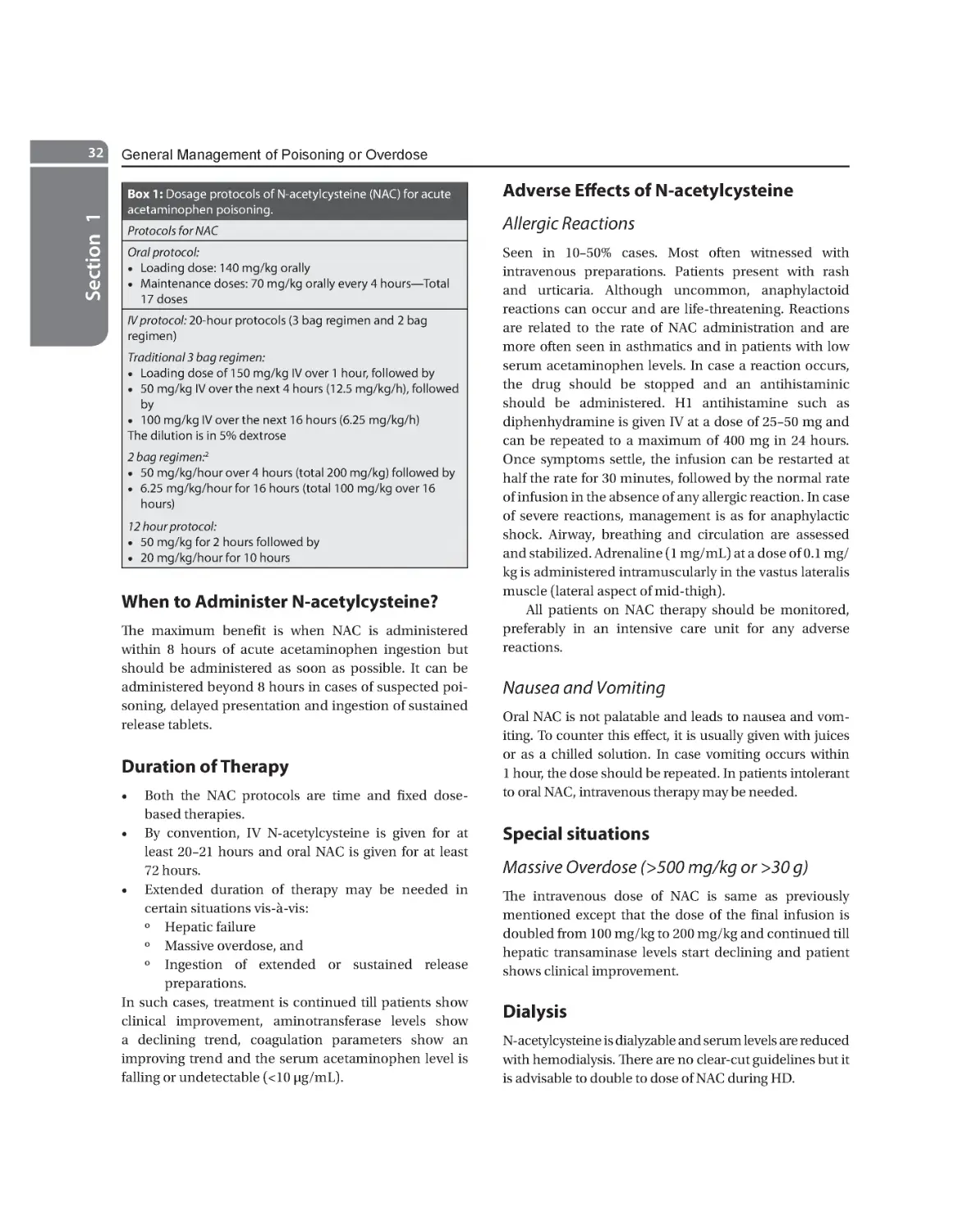
N-Acetylcysteine 31 Atropine 33 Botulinum Antitoxin-Heptavalent 34
Botulinum Immuno Globulin (Intravenous Human) (Babybig) 35 Bromocriptine 35
Activated Charcoal 36 Calcium 37 Calcium Disodium EDTA (CaNa2 EDTA) 37
Dantrolene 38 Deferoxamine 39 Dimercaprol 39 Digoxin-specific Antibody Fragments 40
Flumazenil 40 Fomepizole (4-Methylpyrazole) 41 Folic Acid (Vitamin B9) 42 Glucagon 42
High-dose Insulin Therapy 42 Hydroxocobalamin 43 Hyperbaric Oxygen 43 Methylene Blue 44
Naloxone 44 Penicillamine 45 Physostigmine 46 Phytonadione (Vitamin K1) 46
Pralidoxime (2-Pam) 47 Protamine Sulfate 48 Pyridoxine (Vitamin B6) 49 Sodium Nitrite 50
Sodium Thiosulfate 51 Succimer [Dimercaptosuccinic Acid (DMSA)] 51 Silymarin 52
Sodium Bicarbonate 52 Leucovorin (Folinic Acid) 53 Levocarnitine 54 Octreotide 54
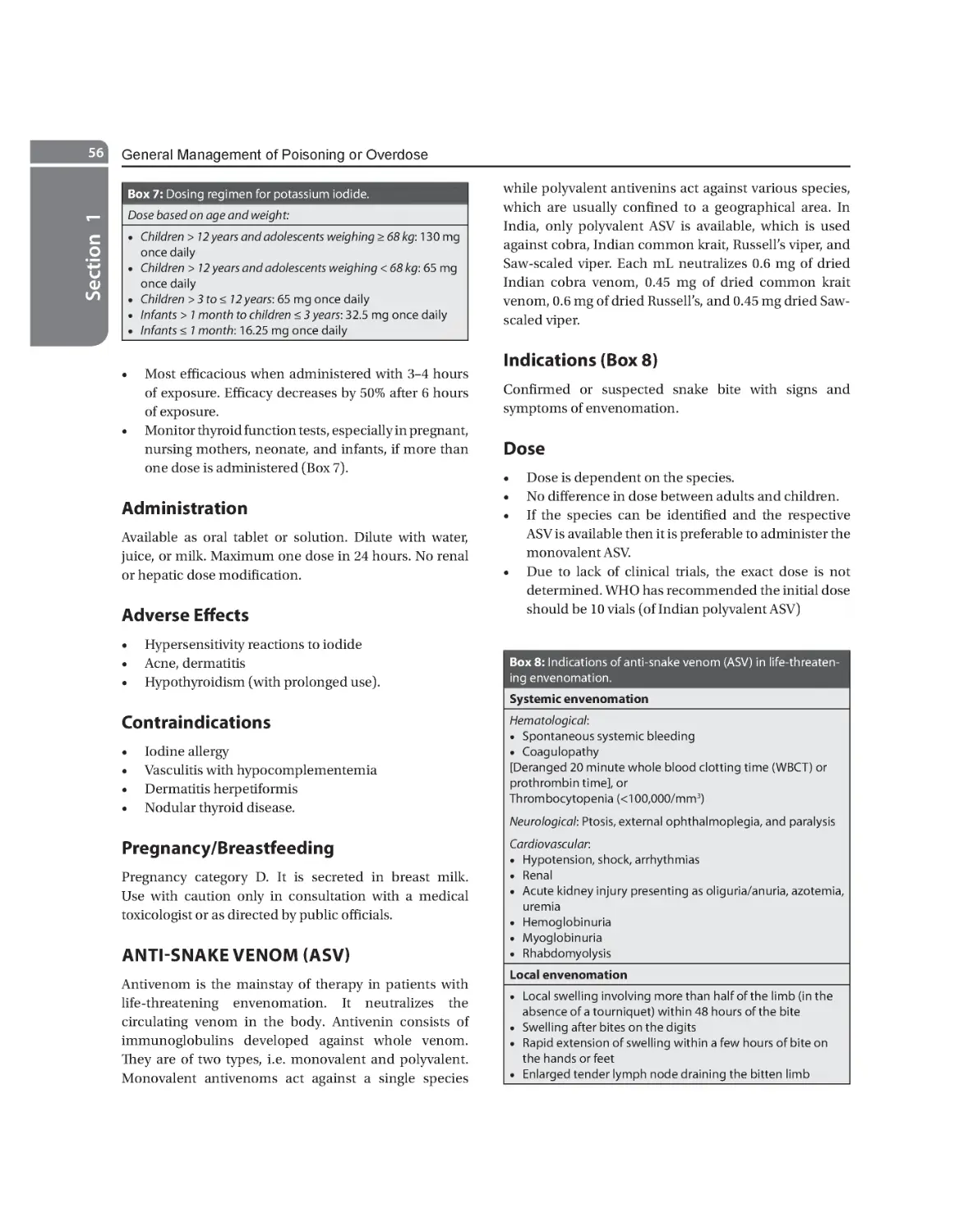
Thiamine (Vitamin B1) 55 Potassium Iodide 55 Anti-Snake Venom (ASV) 56
5. Lipid Emulsion Therapy . . . . . . . . . . . . . . . . . . . . . . . . . . . . . . . . . . . . . . . . . . . . . . . . . . . . . . . . . . . . . . . . . . . . . . . . . 60
Omender Singh, Deven Juneja
Mechanism of Action 60 Lipid Preparations 61 Dose and Delivery 61
Adverse Effects 62 Indications of Use 63
6. Forensic Toxicology for the Critical Care Specialist . . . . . . . . . . . . . . . . . . . . . . . . . . . . . . . . . . . . . . . . . . . . . . . . 68
Sudhir K Gupta, Neha Sharma
Definitions 68 Laws Relating to Poisons 68 Legal Provisions Pertaining to
Poisoning 69 Circumstances of Poisoning 70 Duties of a Medical Practitioner in a Patient of
Suspected Poisoning 70 Evidence Collection and Preservation of Samples in a Patient of Suspected
Poisoning 71 Important Points for Dispatch of Sample for Chemical Analysis 72 Algorithm of
Management of a Patient with Suspected Poisoning 72 Toxidrome Approach 72 List of Common
Poisons and their Antidotes 72 Reason for Negative Chemical Analysis Report in a Patient with Positive
History of Poisoning 72
Prelims.indd 17
19-01-2019 11:44:03
xviii
Principles and Practice of Critical Care Toxicology
Section 2 Drugs of Abuse
7. Central Nervous System Depressants: Overdose and Management. . . . . . . . . . . . . . . . . . . . . . . . . . . . . . . . . . 77
Mradul Kumar Daga, Lalit Kumar, Jitendra Shukla
Common Central Nervous System Depressants: Mechanism of Action and Uses 77
Central Nervous System Depressant Overdose and its Management 78 Management of
Sedative Hypnotic Overdose and Role of Flumazenil 79 Opioid Overdose and its Management 80
Cannabinoids Toxicity 80 Gamma-Hydroxybutyric Acid Overdose 80 Party Drugs 80
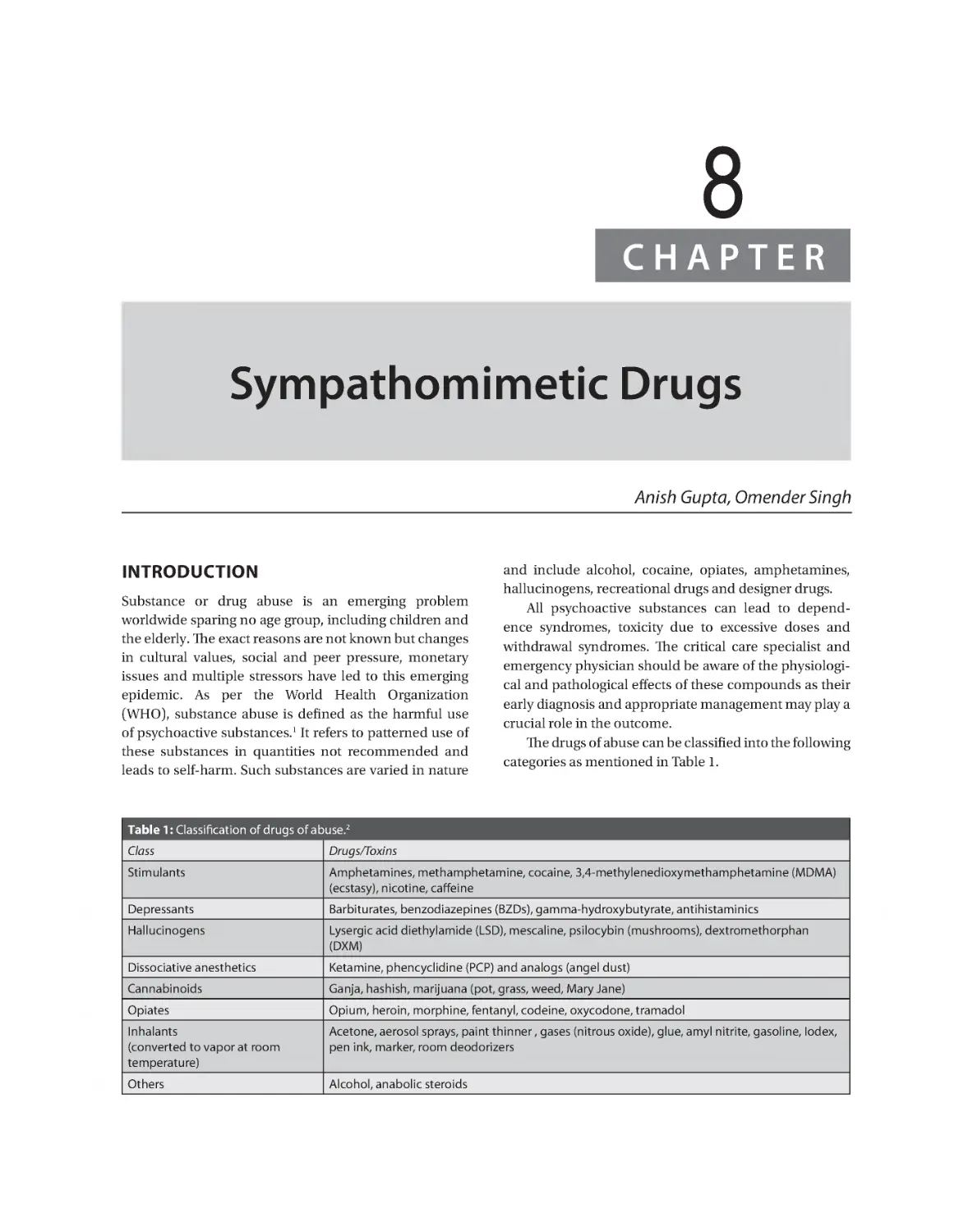
8. Sympathomimetic Drugs . . . . . . . . . . . . . . . . . . . . . . . . . . . . . . . . . . . . . . . . . . . . . . . . . . . . . . . . . . . . . . . . . . . . . . . . 84
Anish Gupta, Omender Singh
Central Nervous System Stimulants 85 Hallucinogens 87
Dissociative Anesthetics 90
9. Cocaine Intoxication . . . . . . . . . . . . . . . . . . . . . . . . . . . . . . . . . . . . . . . . . . . . . . . . . . . . . . . . . . . . . . . . . . . . . . . . . . . . 93
Omender Singh, Anish Gupta
Pathophysiology 93 Clinical Features 93 Diagnosis 95
Management 95 Prognosis 97
10. Newer Drugs of Abuse. . . . . . . . . . . . . . . . . . . . . . . . . . . . . . . . . . . . . . . . . . . . . . . . . . . . . . . . . . . . . . . . . . . . . . . . . . . 99
Omender Singh, Rajesh Chawla, Deven Juneja
Substituted Cathinones or “Bath Salts” 99 Synthetic Cannabinoids 101
Substituted Phenethylamines (2C Drugs) 102 Piperazines 102 Tryptamines 103
Kratom 103 Salvia 103 Hawaiian Baby Woodrose 104 Desomorphine 104
Section 3 CNS Toxins
11. Toxins-induced Seizures. . . . . . . . . . . . . . . . . . . . . . . . . . . . . . . . . . . . . . . . . . . . . . . . . . . . . . . . . . . . . . . . . . . . . . . . 109
Dinesh Chaudhari, Anjali Chaudhari, Omender Singh
Toxin-induced Seizures 109 Status Epilepticus 115
12. Toxic Alcohols . . . . . . . . . . . . . . . . . . . . . . . . . . . . . . . . . . . . . . . . . . . . . . . . . . . . . . . . . . . . . . . . . . . . . . . . . . . . . . . . . 118
Michael E Nelson, Farah Dadabhoy, Timothy B Erickson
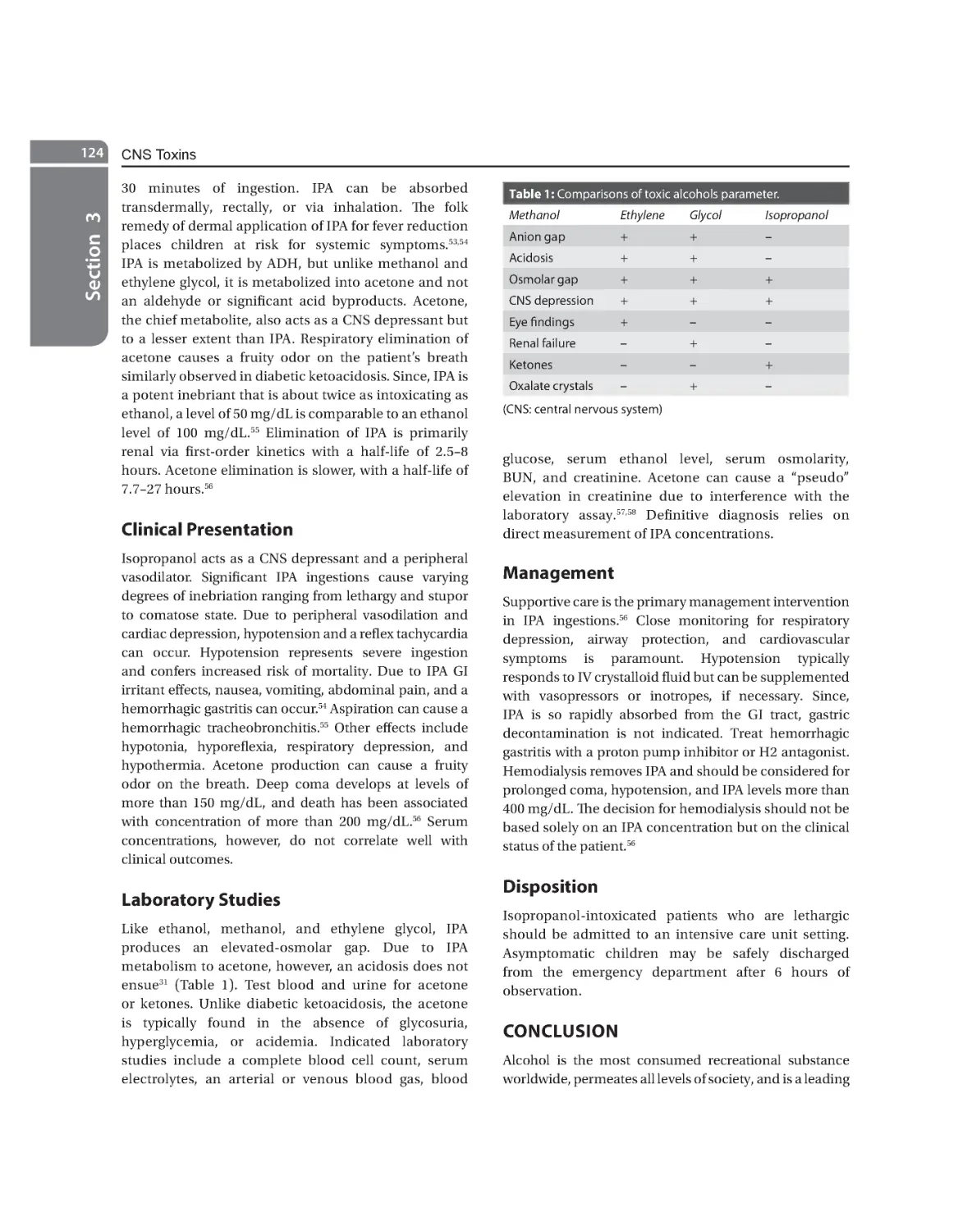
Ethanol 118 Methanol 120 Ethylene Glycol 122 Isopropyl Alcohol 123
13.
Botulism . . . . . . . . . . . . . . . . . . . . . . . . . . . . . . . . . . . . . . . . . . . . . . . . . . . . . . . . . . . . . . . . . . . . . . . . . . . . . . . . . . . . . . 128
Anjali Chaudhari, Deven Juneja
Epidemiology 128 Classification 128 Pathogenesis 129 Clinical Presentation 129
Differential Diagnosis 130 Diagnosis 130 Treatment 131 Prognosis 131
Prevention 131
14. Anticonvulsant Overdose. . . . . . . . . . . . . . . . . . . . . . . . . . . . . . . . . . . . . . . . . . . . . . . . . . . . . . . . . . . . . . . . . . . . . . . 133
Rohit Yadav, Deven Juneja
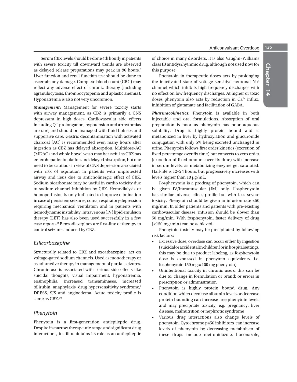
Classification 133 Management of Antiepileptic Drugs Overdose Based on Mechanism of Action 134
General Points to Remember in Management of Anticonvulsants Overdose 141
Prelims.indd 18
19-01-2019 11:44:03
Contents
xix
Section 4 Pulmonary Toxins
15. Approach to Respiratory Failure . . . . . . . . . . . . . . . . . . . . . . . . . . . . . . . . . . . . . . . . . . . . . . . . . . . . . . . . . . . . . . . . 145
Suneel Kumar Garg, Shweta Gupta
Pathogenesis 145 Approach to Respiratory Failure 146 Specific Management 148
16. Inhalational Poisoning . . . . . . . . . . . . . . . . . . . . . . . . . . . . . . . . . . . . . . . . . . . . . . . . . . . . . . . . . . . . . . . . . . . . . . . . . 151
Suneel Kumar Garg, Shweta Gupta
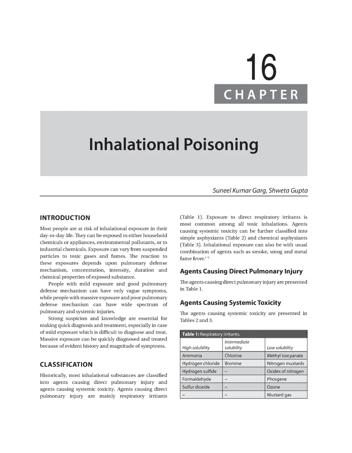
Classification 151 Epidemiology 152 Pathogenesis 152 Agents Causing Direct Pulmonary
Injury 153 Agents Causing Systemic Toxicity 154 Combination Agents 157 Chemical Warfare
and Riot Control Agents 158
17. Carbon Monoxide Poisoning. . . . . . . . . . . . . . . . . . . . . . . . . . . . . . . . . . . . . . . . . . . . . . . . . . . . . . . . . . . . . . . . . . . . 162
Ashish Bhalla, Vikas Suri, Jamshed Nayer
Sources of Carbon Monoxide 162 Mechanism of Carbon Monoxide Toxicity 162
Clinical Features 162 Diagnosis 163 Treatment 163
Section 5 Cardiac Toxins
18. Poisoning Induced Circulatory Failure. . . . . . . . . . . . . . . . . . . . . . . . . . . . . . . . . . . . . . . . . . . . . . . . . . . . . . . . . . . 167
Omender Singh, Desh Deepak
Epidemiology 167 Pathophysiology 167 Management 168 Future Therapies 171
19. Aluminum Phosphide Poisoning. . . . . . . . . . . . . . . . . . . . . . . . . . . . . . . . . . . . . . . . . . . . . . . . . . . . . . . . . . . . . . . . 174
Ashish Bhalla
Pathophysiology and Toxicodynamics 174 Clinical Presentation 175 Diagnosis 175
Laboratory Workup 175 Treatment 175 Prognosis 176
20. Beta-blocker and Calcium Channel Blocker Overdose . . . . . . . . . . . . . . . . . . . . . . . . . . . . . . . . . . . . . . . . . . . . 178
Saswati Sinha, Subhash Kumar Todi
Pharmacology 178 Clinical Features 178 Management Principles 179
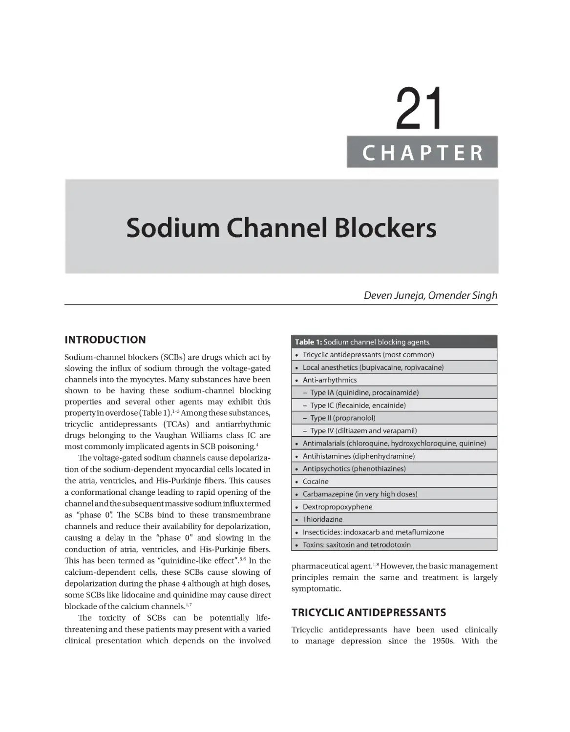
21.
Sodium Channel Blockers . . . . . . . . . . . . . . . . . . . . . . . . . . . . . . . . . . . . . . . . . . . . . . . . . . . . . . . . . . . . . . . . . . . . . . 185
Deven Juneja, Omender Singh
Tricyclic Antidepressants 185 Antihistamine (Diphenhydramine) 190
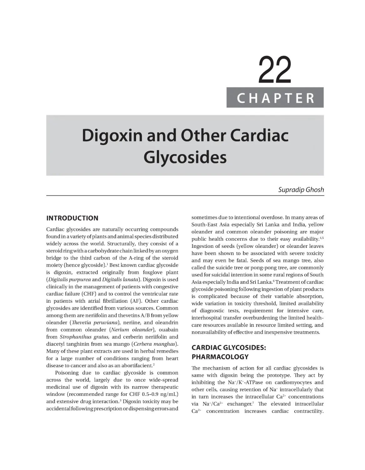
22. Digoxin and Other Cardiac Glycosides. . . . . . . . . . . . . . . . . . . . . . . . . . . . . . . . . . . . . . . . . . . . . . . . . . . . . . . . . . . 194
Supradip Ghosh
Cardiac Glycosides: Pharmacology 194 Clinical Features 195
Management 195
Section 6 Gastrointestinal and Liver Toxins
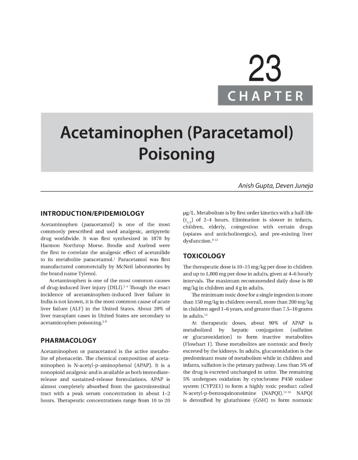
23. Acetaminophen (Paracetamol) Poisoning . . . . . . . . . . . . . . . . . . . . . . . . . . . . . . . . . . . . . . . . . . . . . . . . . . . . . . . 203
Anish Gupta, Deven Juneja
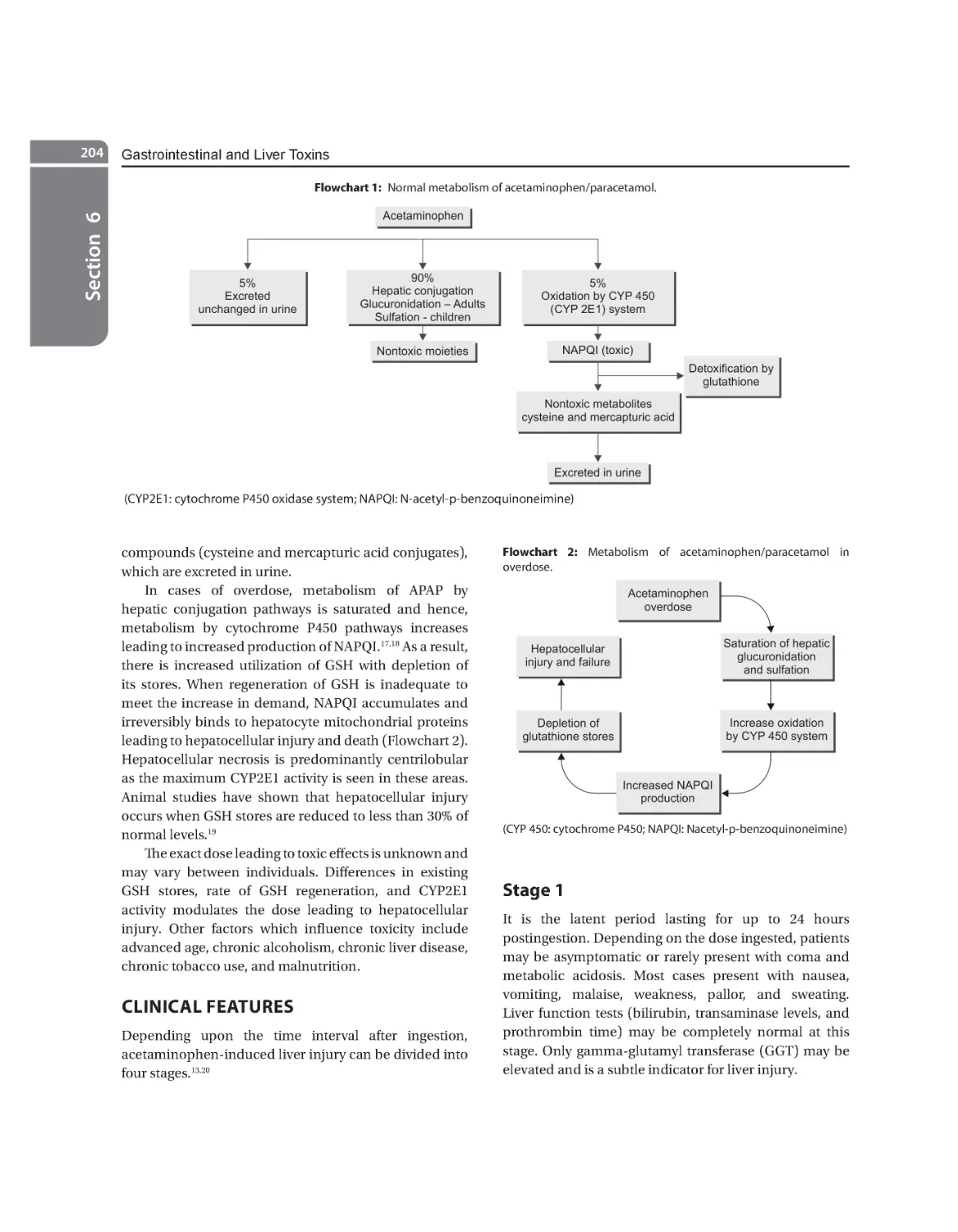
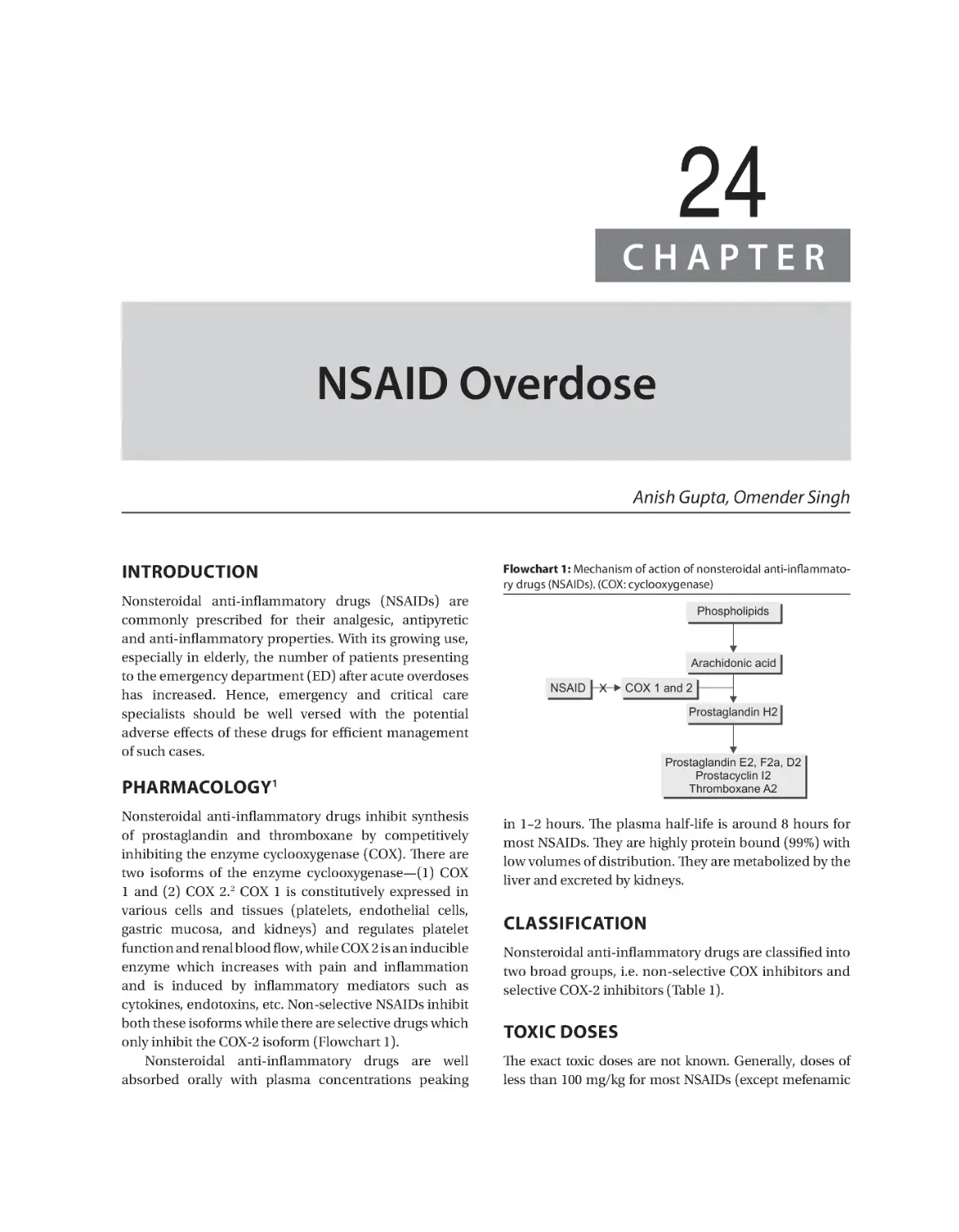
Introduction/Epidemiology 203 Pharmacology 203 Toxicology 203 Clinical Features 204
Diagnosis 205 Management 206 Special Cases 208 Prognosis/Outcome 208
Prelims.indd 19
19-01-2019 11:44:03
xx
Principles and Practice of Critical Care Toxicology
24. NSAID Overdose. . . . . . . . . . . . . . . . . . . . . . . . . . . . . . . . . . . . . . . . . . . . . . . . . . . . . . . . . . . . . . . . . . . . . . . . . . . . . . . 210
Anish Gupta, Omender Singh
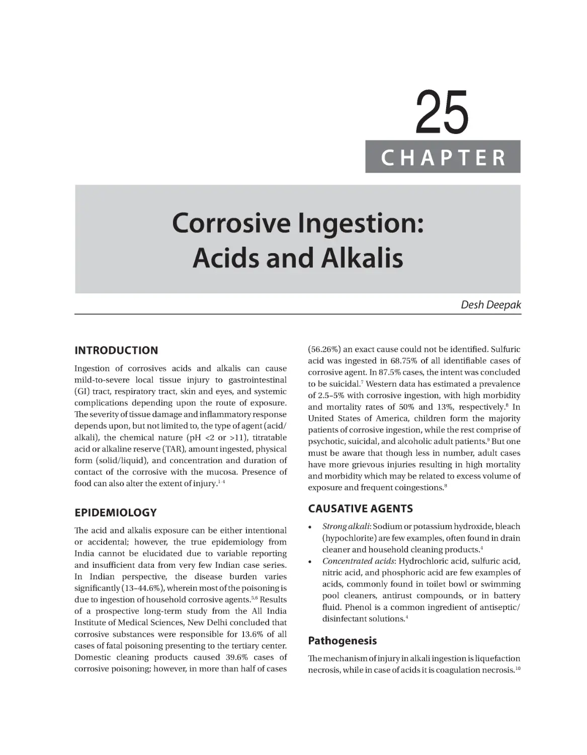
Pharmacology 210 Classification 210 Toxic Doses 210 Toxic Features 211 Diagnosis 211
Management 211 Salicylates 212
25. Corrosive Ingestion: Acids and Alkalis. . . . . . . . . . . . . . . . . . . . . . . . . . . . . . . . . . . . . . . . . . . . . . . . . . . . . . . . . . . 218
Desh Deepak
Epidemiology 218 Causative Agents 218 Common Corrosive Agents 222
Section 7 Hematological Toxins
26. Warfarin and Superwarfarin Toxicity . . . . . . . . . . . . . . . . . . . . . . . . . . . . . . . . . . . . . . . . . . . . . . . . . . . . . . . . . . . . 227
Mohit Mathur
Brief History of Warfarin and Superwarfarins 227 Mechanism of Action 228 Epidemiology 228
Approach Considerations 229 Clinical Presentation and Evaluation 229
Laboratory Investigations 230 Treatment 230
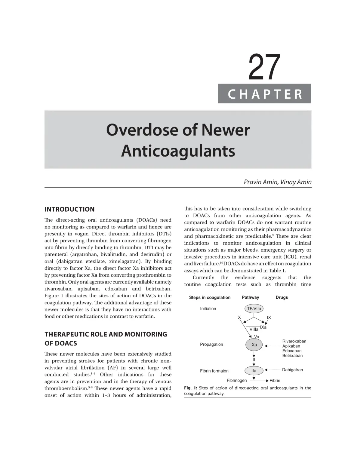
27. Overdose of Newer Anticoagulants . . . . . . . . . . . . . . . . . . . . . . . . . . . . . . . . . . . . . . . . . . . . . . . . . . . . . . . . . . . . . . 236
Pravin Amin, Vinay Amin
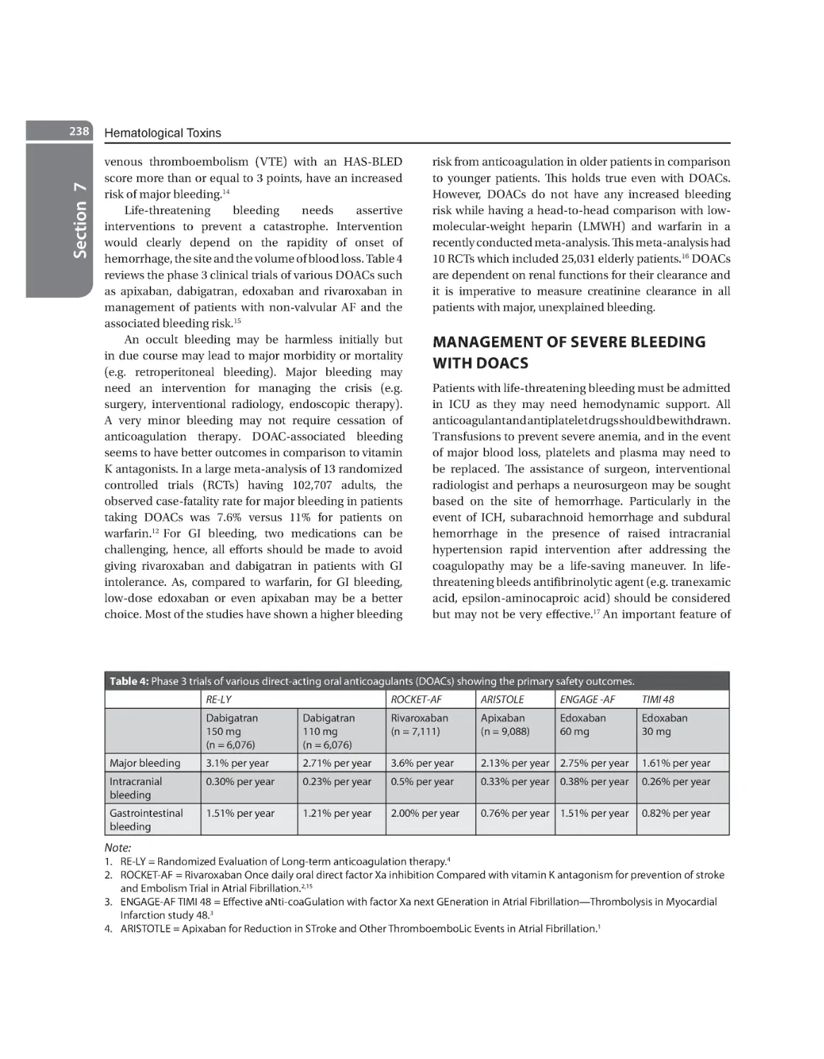
Therapeutic Role and Monitoring of DOACS 236 Bleeding with DOACS 237
Management of Severe Bleeding with DOACS 238 Agent-specific Interventions for Reversals 239
28. Dyshemoglobinemias . . . . . . . . . . . . . . . . . . . . . . . . . . . . . . . . . . . . . . . . . . . . . . . . . . . . . . . . . . . . . . . . . . . . . . . . . . 242
Jeetendra Sharma, Shivangi Khanna
Carboxyhemoglobinemia 242 Methemoglobinemia 247 Cyanide Toxicity 250
Sulfhemoglobinemia 251
Section 8 Renal Toxins and Extracorporeal Therapies
29. Approach to Toxin Induced Acute Renal Failure. . . . . . . . . . . . . . . . . . . . . . . . . . . . . . . . . . . . . . . . . . . . . . . . . . 257
Sahil Bagai, Dinesh Khullar
Clinical Features 257 Mechanisms of Injury 258 Agents Causing Acute Kidney Injury 258
Heavy Metal Poisoning and AKI 259 Envenomation and Bites Causing AKI 260
Approach to AKI with Poisoning 261 General Management of AKI 261
Prognosis of Patients who had AKI 262
30. Extracorporeal Therapies: General Principles . . . . . . . . . . . . . . . . . . . . . . . . . . . . . . . . . . . . . . . . . . . . . . . . . . . . 264
Deven Juneja, Omender Singh
Factors Affecting Dialyzability 264 Types of Extracorporeal Toxin Removal 265
Issues with Extracorporeal Toxin Removal 269
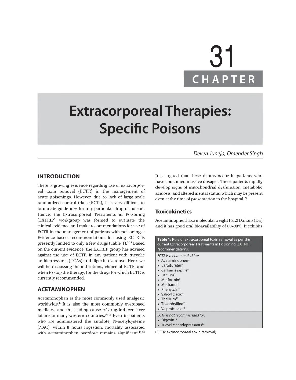
31. Extracorporeal Therapies: Specific Poisons . . . . . . . . . . . . . . . . . . . . . . . . . . . . . . . . . . . . . . . . . . . . . . . . . . . . . . 274
Deven Juneja, Omender Singh
Acetaminophen 274 Barbiturates 275 Carbamezapine 276 Lithium 277
Metformin 277 Methanol 278 Phenytoin 279 Salicylic Acid 280 Thallium 281
Theophylline 282 Valproic Acid 283
Prelims.indd 20
19-01-2019 11:44:03
Contents
xxi
32. Extracorporeal Membrane Oxygenation . . . . . . . . . . . . . . . . . . . . . . . . . . . . . . . . . . . . . . . . . . . . . . . . . . . . . . . . . 288
Anna L Condella, Edward W Boyer
Brief History 288 Mechanics of Extracorporeal Membrane Oxygenation 289
Indications for Extracorporeal Membrane Oxygenation 289 Pharmaceutical Overdoses 290
Nonpharmaceutical Poisoning 290 Pediatric Considerations 291
Section 9 Pesticides and Rodenticides
33. Organophosphorus . . . . . . . . . . . . . . . . . . . . . . . . . . . . . . . . . . . . . . . . . . . . . . . . . . . . . . . . . . . . . . . . . . . . . . . . . . . . 295
JV Peter
Clinical Presentation 295 Determinants of Toxicity of Organophosphate
Compounds 295 Management of Poisoning 296
34. Carbamates and Newer Insecticides. . . . . . . . . . . . . . . . . . . . . . . . . . . . . . . . . . . . . . . . . . . . . . . . . . . . . . . . . . . . . 303
Vijay Kumar Agarwal, Prakash K Khernar, Amit Goel
Carbamates 303 Pyrethroids 307 Organophosphate-Pyrethroid
Combination 309
35.
Herbicides Poisoning: Paraquat and Diquat. . . . . . . . . . . . . . . . . . . . . . . . . . . . . . . . . . . . . . . . . . . . . . . . . . . . . . 312
Amit Goel, Omender Singh
Chemistry 312 Mechanism of Toxicity 312 Clinical Presentation 314
Management 315 Outcome 317
36. Organochlorines. . . . . . . . . . . . . . . . . . . . . . . . . . . . . . . . . . . . . . . . . . . . . . . . . . . . . . . . . . . . . . . . . . . . . . . . . . . . . . . 320
Ravi Jain, Omender Singh
Pharmacology 320 Pathophysiology and Mechanism of Toxicity 322 Approach to Patient 323
Biochemical Effects of Toxins 323 Differential Diagnosis 324 Treatment of Organochlorine
Pesticide Poisoning 325
37.
Rodenticides. . . . . . . . . . . . . . . . . . . . . . . . . . . . . . . . . . . . . . . . . . . . . . . . . . . . . . . . . . . . . . . . . . . . . . . . . . . . . . . . . . . 330
Sanjay V Patne, Subramanian Senthilkumaran
Classification of Rodenticides 330 Biochemistry 330
Pathophysiology 331 Clinical Features 331 Clinical Manifestations 332
Workup 332 Treatment 332
Section 10 Miscellaneous Toxicities
38. Heavy Metal Poisoning . . . . . . . . . . . . . . . . . . . . . . . . . . . . . . . . . . . . . . . . . . . . . . . . . . . . . . . . . . . . . . . . . . . . . . . . . 339
Prashant Nasa
Pathophysiology 340 General Principles of Assessment and Management 340
Specific Metals 342
39. Envenomation: Snake, Scorpion, and Spider . . . . . . . . . . . . . . . . . . . . . . . . . . . . . . . . . . . . . . . . . . . . . . . . . . . . . 348
Subramanian Senthilkumaran, Ponniah Thirumalai Kolundu Subramanian
Types of Snakes 348 Toxic Effects of Snake Venom 348 Pathophysiology 348
Clinical Features 349 Diagnosis 349 Investigations 350 Management of Snake Bite 350
Indications for Antivenom 351 Adverse Reaction to Antivenom 351 Management in Special
Situations 352 Scorpion Stings 352 Hymenoptera Stings 354
Prelims.indd 21
19-01-2019 11:44:03
xxii
Principles and Practice of Critical Care Toxicology
40. Plant Poisonings. . . . . . . . . . . . . . . . . . . . . . . . . . . . . . . . . . . . . . . . . . . . . . . . . . . . . . . . . . . . . . . . . . . . . . . . . . . . . . . 357
Arvinth Soundarrajan, Subramanian Senthilkumaran, Ponniah Thirumalai Kolundu Subramanian
Indian Poisonous Plants 357
41. Mushroom Poisoning. . . . . . . . . . . . . . . . . . . . . . . . . . . . . . . . . . . . . . . . . . . . . . . . . . . . . . . . . . . . . . . . . . . . . . . . . . . 367
Nipun Verma, Ashish Bhalla, Shreya Singh
Background 367 Pathophysiology 367 Epidemiology 368 Clinical Presentation 369
Differential Diagnoses 370 Laboratory Studies 370
Treatment of Mushroom Poisoning 371
Prognosis 372
42. Methotrexate and Other Chemotherapeutic Agents Toxicity. . . . . . . . . . . . . . . . . . . . . . . . . . . . . . . . . . . . . . . 374
Akhilesh Singh, Omender Singh
Chemotherapeutic Drug-associated Toxicity 377 Extravasation of
Chemotherapeutic Agents 382
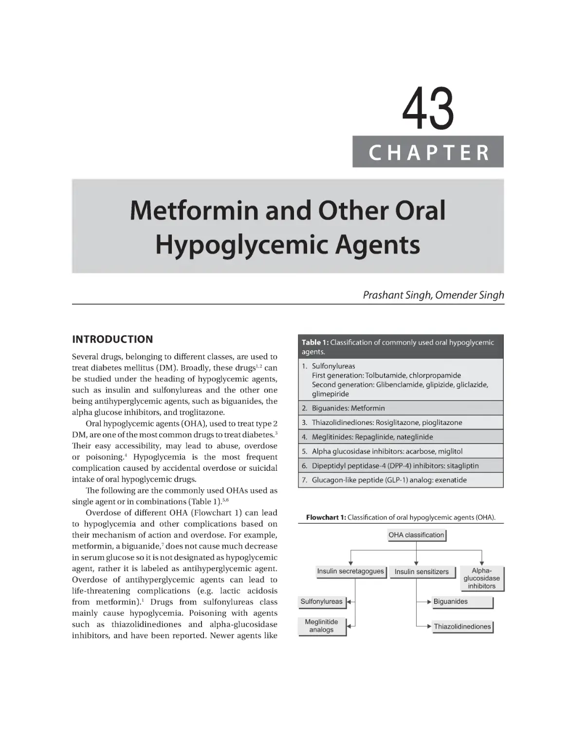
43. Metformin and Other Oral Hypoglycemic Agents. . . . . . . . . . . . . . . . . . . . . . . . . . . . . . . . . . . . . . . . . . . . . . . . . 386
Prashant Singh, Omender Singh
Biguanides (Metformin) 387 Sulfonylureas 388 Meglitinides 390
Alpha-Glucosidase Inhibitors 391 Dipeptidyl Peptidase-4 Inhibitors 391 Serum Glucose
Transporter-2 Inhibitors 391
44. Chemical and Biological Warfare. . . . . . . . . . . . . . . . . . . . . . . . . . . . . . . . . . . . . . . . . . . . . . . . . . . . . . . . . . . . . . . . 395
Yash Javeri, Bharat Jagiasi, Gunjan Chanchalani
Chemical Warfare 396 Biological Warfare 398
Index. . . . . . . . . . . . . . . . . . . . . . . . . . . . . . . . . . . . . . . . . . . . . . . . . . . . . . . . . . . . . . . . . . . . . . . . . . . . . . . . . . . . . . . . . . 403
Prelims.indd 22
19-01-2019 11:44:03
SECTION
1
General Management of
Poisoning or Overdose
1. Approach to a Poisoned Patient
Omender Singh, Prashant Nasa, Deven Juneja
2. Laboratory Testing in Poisoning
Aruna Dewan, Ashwin Patel
3. Acid-base Disorders in Poisoning
Pradeep Rangappa, Shivakumar Mutnal, Ipe Jacob
4. Antidotes
Omender Singh, Anish Gupta
5. Lipid Emulsion Therapy
Omender Singh, Deven Juneja
6. Forensic Toxicology for the Critical Care Specialist
Sudhir K Gupta, Neha Sharma
Ch-1.indd 1
17-01-2019 12:25:59
Ch-1.indd 2
17-01-2019 12:26:00
1
CHAPTER
Approach to a Poisoned Patient
Omender Singh, Prashant Nasa, Deven Juneja
INTRODUCTION
The patients with suspected poisoning are commonly
encountered by acute medicine and critical care
physicians. The poisoning in these cases may be
accidental or intentional (suicidal or homicidal). Any
chemical available can be potentially toxic to humans,
provided the quantity is large. The high index of suspicion
is warranted to identify these patients early especially
when direct history of intoxication is not available. The
knowledge and recognition of a specific toxidrome is
critical, but one has to be aware of pitfalls like symptoms
either may be nonspecific or masked (e.g. intracranial
hemorrhage in cocaine poisoning).
The physical examination should be focused in case
of critical condition and comprehensives once the patient
is stabilized. The vital signs and level of consciousness is
paramount in assessing the degree of toxicity. Initial vitals
assessment by emergency staff or other paramedic must
include respiratory rate, heart rate, blood pressure, and
temperature; are of utmost importance as clinical signs
may change by treatment. The patient neurological state
(level of consciousness), pupils size, reaction to light,
and presence of seizures may provide clues regarding the
identity of the ingested poison.
The management approach in these patients is based
on rapid and early diagnosis, decontamination and
prevent further exposure, appropriate specific treatment
while providing multiorgan supportive care. We have
divided this chapter into following sections:
Ch-1.indd 3
A.
B.
C.
D.
E.
Initial resuscitation and assessment
Toxidromes
Laboratory testing in poisoning
Advanced cardiac life support concerns in poisoning
Antidotes.
INITIAL RESUSCITATION AND
ASSESSMENT
Most of the patients with poisoning respond well with
early general intensive supportive measures with close
watch on red flag signs. The supportive measures can be
similar to any critically ill patient with “ABCD” approach
(airway, breathing, circulation) however D in poisoning
is different and means disability and decontamination.
In case patient has an altered level of consciousness, the
priority is airway management and cervical spine must
be immobilized till it is clear of any injury. The initial
resuscitation of patient is similar to any critical patient
with systematic approach to evaluate and identify a
life-threatening problem, and treat the problem before
proceeding to next. The ABCD approach usually
employed in critical care or emergency is useful.
Airway
Airway patency should be assessed in all cases unless a
conscious-oriented patient without signs of upper airway
obstruction. In case of altered sensorium or patient
with stridor, hoarseness and other signs of upper airway
17-01-2019 12:26:00
Section 1
4
General Management of Poisoning or Overdose
obstruction, airway should be secured with maneuver
like headtilt-chinlift or may need airway adjuncts.
Endotracheal intubation may be required in these patients
for airway protection and patency. The other indications
are acute respiratory failure, high supplemental oxygen
in case of carbon monoxide poisoning.1 The urine or
blood toxicology screening should be obtained before
any sedatives or hypnotics are administered. Some toxins
(acid or alkali ingestion) require special care during airway
management and involve an expert help in such cases.
Whenever endotracheal intubation is required in patients
with unknown poisoning, rapid sequence induction (RSI)
using short-acting sedatives and paralytics is generally
preferred as time from last meal is either unknown or
erroneous. The succinylcholine is the neuromuscular
paralytic agent of choice; however, it should be avoided in
suspected organophosphorus poisoning as duration may
be prolonged in view of decreased acetylcholinesterase
levels. The intubation is necessary for most patients
with above indications, but if possible intubation in
aspirin (difficult to attain hyperventilation) and gammahydroxybutyrate (rapid recovery) poisoning can be
avoided or delayed unless the patients present with clinical
or blood gas evidence of respiratory failure.1
The patients where intubation may be required
for correction of hypoxemia or other presentation of
respiratory failure should be identified and managed
early (Table 1).
Table 1: Drugs/poisons causing hypoxemia.
Aspiration pneumonia
•• Poisons/drugs causing altered
mental status
Bronchospasm
••
••
••
••
••
Cardiogenic pulmonary
edema
•• Antiarrhythmics β-blockers
•• Tricyclic antidepressants
•• Calcium channel blockers
(Verapamil)
Cytopathic (cellular)
hyoxia
••
••
••
••
Hypoventilation
•• Alcohols (ethanol, methanol,
ethylene glycol)
•• Botulinum toxin
•• Barbiturates
•• Benzodiazepines
•• Opioids
•• Sedative/hypnotics
•• Snake bite
•• Neuromuscular blockade
•• Tricyclic antidepressants
•• Tetanus
Hemorrhage (Alveolar)
••
••
••
••
••
••
••
Noncardiogenic
pulmonary edema
•• Toxic inhalation gases (e.g.
hydrocarbons, phosgene
•• Cocaine
•• Ethylene glycol
•• Opioids
•• Salicylates
Pneumothorax
•• Cocaine
•• IV drug abuse
•• Kerosene
Gases (inert)
••
••
••
••
Breathing
If breathing or respiratory effort is inadequate beside
the endotracheal intubation, respiratory support is also
required. Endotracheal intubation besides altered level
of consciousness to protect airway is indicated in acute
respiratory failure. The supplemental oxygen in case of
poisoning like carbon monoxide poisoning is additional
indication.
The patient’s oxygen saturation (SpO2) with standard
bedside pulse oximetry can be misleading in poisoning
with dyshemoglobinemias (e.g. carbon monoxide
poisoning), where co-oximeter should be used to identify
abnormal hemoglobins and true SpO2. The target SpO2 is
94–99% for most of the poisoning except in some poisoning
where high SpO2 is associated with oxygen-mediated
toxicity, e.g. chlorine gas, paraquat and diquat.2 In these
patients lowest possible PaO2 (~50–60 mm Hg) and SpO2
(~90–93%) should be targeted essential to prevent tissue
hypoxia and in order to avoid oxygen-mediated toxicity.3
If standard lung-protective invasive ventilation
strategy is acceptable for most of the poisoning except
in salicylates where any acidosis due to hypoventilation
during intubation and/or lung protective ventilation may
cause clinical deterioration.4,5
Ch-1.indd 4
β-blockers
Cocaine
Organophosphates
Drugs resulting in aspiration
Drugs associated with
myocardial depression (cardiac
asthma)
Hydrogen sulfide
Carbon monoxide
Cyanide
Methemoglobinemia
Cocaine
Anticoagulants
Thrombolytics
Amiodarone
Nitrofurantoin
Penicillamine
Toluene
Carbon dioxide
Methane
Nitrogen
Propane
17-01-2019 12:26:00
Approach to a Poisoned Patient
Circulation
Bradycardia and/or Hypotension: Bradycardia is mainly
seen with β-blockers and calcium-channel blockers but
sometimes significant enough to require temporary
pacing especially if associated with hemodynamic
instability (Table 3).
• Hypotension/Bradycardia—calcium-channel
blockers, β-blockers, digoxin, aluminum phosphide
(celphos) insecticide
• Hypertension/Tachycardia—Sympathomimetics,
cocaine.
The cause of hypotension in a patient with poisoning
can be multifactorial which includes hypovolemia, poison
causing direct myocardial depression, arrhythmias,
Table 2: Agents causing hypotension and hypertension.
Hypotension (SCAR)
Hypertension (SCAN)
Sedative/hypnotics
Sympathomimetics
Calcium-channel blockers
Cocaine
Antidepressants,
antihypertensives
Anticholinergics and
amphetamines
Rodenticides (e.g. arsenic,
cyanide)
Nicotine
Table 3: Drugs causing bradycardia and tachycardia.
Ch-1.indd 5
Bradycardia
Tachycardia
Calcium-channel blockers
Anticholinergics
β-blockers
Amphetamines
Class 1A and 1C
antiarrhythmics
Antihistaminics
Tricyclic antidepressants
Carbon monoxide
Digoxin
Cyanide poisoning
Lithium
Clonidine
Metoclopramide
Cocaine
Opioids
Cyclic antidepressants
Organophosphates and
carbamates
Hydralazine
Physostigmine
Quinidine
Chapter 1
The patients with poisoning may present with hypotension
or hypertension, brady- or tachyarrhythmias (Tables 2
and 3). The continuous monitoring of electrocardiogram
along with blood preesure and heart is thus vital for all
these patients.
and/or profound systemic vasodilation. The patients with
intoxication of sympathomimetic drugs, anticholinergics,
ergot derivatives, cocaine and withdrawal of nicotine,
alcohol, and sedatives instead present with hypertension.
The initial resuscitation must include peripheral venous
access using two large bore cannula (16- or 18-gauge)
with targeted fluid resuscitation and/or vasopressors
or inotropes if required for correction of hypotension.
The treatment of hypertension is goal-oriented and
may depend on factors like inciting agent, severity and
associated complications.6
5
DISABILITY AND DECONTAMINATION
The target-oriented neurological assessment in poisoned
patients presenting with altered mental status is must
(Box 1). In patients presenting with focal neurological
deficit an imaging may be required to rule out any
structural brain injury. There are simple bedside scores
like Glasgow Coma Score or Alert/Verbal/Painful/
Unresponsive (AVPU) score which can be used to assess
consciousness and need to protect the airways. However
neither has been validated and found to predict prognosis
of the poisoned patient.3
Seizure is common neurological finding along with
pupillary abnormalities. The assessment of pupil may
help to suspect some poison along with other systemic
findings (Table 4).
Box 1: Poisoning present with altered level of unconsciousness (LETHARGIC).
L: metals like lead, lithium
E: Alcohol like ethanol, ethylene glycol
T: Tricyclic antidepressants, antipsychotics
H: Heroin, hydrogen sulfide, hypoglycemics
A: Arsenic, antidepressants, anticonvulsants, antihistamines
R: Rohypnol (sedative hypnotics), risperidone
G-GHB
I: Isoniazid, insulin
C: Carbon monoxide, cyanide, clonidine
Table 4: Agents causing pupillary abnormalities.
Miosis (SCOP)
Mydriasis (SAD)
Methemoglobinemia
Sedative-hypnotics
Sympathomimetics
Phencyclidine,
phenothiazines
Pseudoephedrine
Cholinergics,carbamates
(organophosphates)
Anticholinergics
Opiates
Drug withdrawal
Theophylline
Phenothiazines (antipsychotics)
17-01-2019 12:26:00
Section 1
6
General Management of Poisoning or Overdose
The “coma cocktail” traditionally included dextrose
(1 ampule of D50% IV), oxygen (5–10 L/min), flumazenil
(0.2 mg IV), naloxone (2 mg IV), and thiamine (100 mg
IV) and was advocated in unknown poisoning with
unconsciousness and coma. This is helpful in prehospital
to avoid intubation and to treat common causes of altered
level of consciousness. The later studies have found use
of flumazenil in this cocktail was counterproductive.
The flumazenil was used for diagnosis of intentional
benzodiazepine overdose. However empirical fluma
zenil can increase the risk of seizures and agitation by
benzodiazepine withdrawal in chronic abuse.7 It can
also stimulate seizure, ventricular tachycardia in mixed
overdose, e.g. amitriptyline, chloral hydrate.7-10
In a very large meta-analysis with more than 900
patients treated with flumazenil in emergency room,
the number of patients needs to be treated for a very
severe adverse events (e.g. arrhythmia and seizure)
and for any adverse event (e.g. vomiting, agitation, and
dysphoria) were 50 and 6.2 respectively.11 The patients
given flumazenil has higher risk of seizures especially
those on chronic treatment or abusing benzodiazepines,
pre-existing seizure disorder or traumatic head injury,
and patients who have been co-overdosed with tricyclic
antidepressants.12 In light of available evidence on
flumazenil epileptogenic effect, chances of precipitating
an acute withdrawal, and some studies showed
supportive management for benzodiazepine overdose
is as effective; the use of empirical flumazenil is reduced
in recent years and is currently recommended for highly
selective subgroup of patients.13,14
The empirical naloxone can also precipitate opioid
withdrawal or vomiting in nonopioid cause of coma.
Fortunately with short half-life of naloxone, in case
withdrawal is seen, symptoms wear off in 1–2 hours.
However, the higher mortality of the opioids overdose
over the benzodiazepines in view of respiratory
depression still continue to recommend its use but dose
titration should be to respiration rather to wakefulness.14
The administration of empirical thiamine is considered
with history or suspicion of chronic alcohol abuse and
in patients with severe malnutrition. There is a small
risk of severe anaphylaxis with intravenous thiamine.
Oxygen should be target to SpO2 95–99%. The “coma
cocktail” can thus be revisited into more evidence based
(Table 5).14
Decontamination
The evidence-based literature regarding proper
decontamination methods are limited. There is paucity
about the approved agents and there therapeutic
indications and most of the principles have been taken
from warfare fields and radiation accident protocols.
Healthcare workers should use appropriate personal
protective equipments (PPEs) like splash resistant
goggles, gloves and gowns, while decontaminated patients
with unknown poisoning to prevent any dermal or eye
exposure. The decontamination is avoided in prehospital
setting if adequate PPEs and other decontamination
equipment are not available. The decontamination is
effective only if done early and should not be delayed
pending identification of the definitive offending agent.
Dermal and Eye Decontamination
In poisoning with possible dermal route, the patients’
clothings should be removed and a mild soap, and
copious amount of lukewarm water should be used for
decontamination. The body temperature should be
monitored to avoid hypothermia, strong detergents,
or hot water however should be avoided. In case of eye
contamination, decontamination should be done with
copious irrigation of normal saline solution and periodic
monitoring of pH under supervision of ophthalmologists.3
Table 5: Revised “coma cocktail”.
Ch-1.indd 6
Agents
Indications
Dosage recommended
Dextrose
Capillary blood glucose less than 50 mg/dL
50 mL of 50% dextrose IV
Oxygen
Pulse oximetry (SpO2 less than 92–94%)
At 5–10 L/min
Naloxone
Bradypnea (± miosis)
40–200 µg IV bolus, repeat every 2–3 minutes till
respiratory depression improves or maximum dose 10 mg
whichever is earlier
Thaimine
In alcoholic or malnourished patients for prevention of
precipitation of Wernicke encephalopathy
100 mg IV/IM
17-01-2019 12:26:00
Approach to a Poisoned Patient
Gastrointestinal Decontamination
Cathartics: It can decrease the absorption of substances
by its prokinetic effect and their rapid expulsion of
poisons from the GI tract. There are broadly two types
of osmotic cathartics: saccharide based (sorbitol) or
saline based (magnesium citrate, magnesium sulfate
and sodium sulfate). The mechanism of action is mainly
useful for slowly absorbed poisons. Based on recent
evidence, the current policy statement of AACT and
EAPCCT on cathartics, there is no definite indication for
them in poisoning. Besides there are contraindications
such as corrosives, ileus or intestinal obstruction,
recent abdominal trauma or surgery, and intestinal
perforation. Magnesium cathartics is contraindications
with renal failure, renal insufficiency, or heart block.
Cathartics is to be avoided in elderly or the very young
(<1 year of age) and hypotensive patients or electrolyte
imbalances.16
Gastric lavage: It is used for removal of unabsorbed
poison and to small extent decrease absorption of
ingested substances for over 200 years. The technique has
been described using a wide bore, orogastric tube (16–18
gauge) with patient position trendelenburg position
(head down) and left lateral decubitus position. The
lavage is then performed with approximately 250 mL (or
around 10 mL/kg in pediatric patients) of water or saline
followed by aspiration. The procedure is repeated until
the aspirated solution is clear of any particulate matter.3,6
The gastric lavage is associated with complications
sunch as aspiration, esophageal perforation, epistaxis,
hypothermia. It is not indicated in patients with nontoxic
overdoses or combative and uncooperative patients and
contraindicated in suspected corrosive substance or a
Ch-1.indd 7
Chapter 1
Gastrointestinal (GI) decontamination can be
simply defined as measures to prevent or reduce the
absorption of an ingested substance. The process of
GI decontamination has evolved significantly over the
last three decades and focus is now on minimally or
less invasive techniques. This has happened partially
because of recent research showed lack of benefit with
many techniques of GI decontamination and in some
cases serious complications with these procedures.
There have been updation of many position statements
by the American Academy of Clinical Toxicology (AACT)
and the European Association of Poison Centers and
Clinical Toxicologists (EAPCCT) on various techniques
of GI decontamination.15
volatile hydrocarbon poisoning (kerosene) as may cause
aspiration associated lung injury.3,6
In recent policy statement of AACT and EAPCCT
the evidence supporting for gastric lavage routinely in
poisoned patients is weak. If physicians on their clinical
judgment decide to use gastric lavage it should be done
under supervision with preference for activated charcoal
or only observation over gastric lavage.17
7
Ipecac syrup: Ipecac-induced emesis is less traumatic
than gastric lavage, and is therefore being still used
in prehospital settings or in pediatric patients. Ipecac
is most useful if administered immediately after
ingestion with effectiveness decreases rapidly to only
30–40% removal rate even 1 hour after ingestion. The
Ipecac is contraindicated in unconscious patients;
seizures, poisoning with corrosives, and petroleum
products are absolute contraindications due to risk
of aspiration and lung injury.3,6 The Ipecac use is
benign with complications relatively uncommon and
easily treatable like diarrhea and/or vomiting. Serious
complications are reported, e.g. Mallory–Weiss tears,
pneumomediastinum, and aspiration pneumonia
but are very rare.3 In case of Ipecac use, the activated
charcoal should only be given after 1–2 hours. The
current policy statement of AACT and EAPCCT
on Ipecac-induced emesis is insufficient evidence
supporting its use after poison ingestion.18 The Ipecac
if considered should be administered early (within 60
minutes) in a patient who has consumed significant
toxic dose and no altered consciousness.
Activated charcoal: This is an inert, nontoxic, powerful,
and nonspecific substance which produces irreversible
bonds to many intraluminal drugs and thus may interfere
with their absorption. The process of activation includes
steam heating and chemical treatment, where the surface
area of charcoal is increased and available for adsorption.
The activated charcoal can create a diffusion gradient
between blood and gut, and can secondarily decreased
serum drug levels of absorbed drug a process referred to
as “GI dialysis” (Table 6). Charcoal can either be sole GI
decontaminating agent or can be administered after both
gastric lavage and Ipecac-induced emesis. The activated
charcoal is generally well-tolerated with complications
which are infrequent. The usual contraindications of all
GI decontaminants like altered state of consciousness
and/or unprotected airway, protracted vomiting, and
intestinal obstruction or perforation too applies to
17-01-2019 12:26:00
Section 1
8
General Management of Poisoning or Overdose
Table 6: Recommendations for use of multiple doses of
activated charcoal.
Indicated (supported
by evidence)
Unclear (Neither
supported or refuted
by evidence)
Controversial
(insufficient
supporting
evidence
Carbamazepine
Phenytoin
Salicylates
Dapsone
Disopyramide
Phenobarbital
Amitriptyline
Quinine
Dextropropoxyphene
Theophylline
Digitoxin
Sotalolol
activated charcoal. Few uncommon side effects like
aspiration pneumonia bronchiolitis obliterans, ARDS,
and death has been reported in literature.3,7,19 The ideal
dose should give a charcoal-to-drug ratio of 10:1. The
dose required in children is 0.5–1 g/kg body weight, while
in adults a fixed single dose of 50–100 g is indicated.19,20
The charcoal can be administered with cathartics
(magnesium sulfate, magnesium citrate) to avoid its
constipation effect. There are few drugs where charcoal
is not found effective (Box 2).
Single-dose charcoal: The routine administration of
single-dose activated charcoal is not indicated in
poisoned patient. It should be considered only in a
patient with significant toxic dose and when presented
within the first hour of the event.
Mutidose-activated charcoal: The multiple dose is initial
dose of 50–100 g (pediatric 10–25 g) followed by repeated
frequency of hourly, 2 hourly, or 4 hourly at equivalent
dose of 12.5 g/h. This repeated dose is the main principle
behind GI dialysis. The evidence for multiple doses of
activated charcoal in decreased morbidity or mortality
is lacking and therefore routine administration is not
recommended in the poisoned patient.20-22 Indications
for administration of multi-dose activated charcoal (any
of the following criteria):
• Intake of the poison exceeds the capacity to be
adsorbed by a single dose.
• Drugs with significant enterohepatic circulation: To
prevent the reabsorption by enterohepatic circulation
of the active substance, metabolite, or drug conjugate.
• Intoxication by drugs with sustained release.
• Poisoning by drugs that decrease GI transit
(anticholinergics, tricyclic antidepressants, opioids,
and phenothiazine).
Ch-1.indd 8
Box 2: Drugs not absorbed by activated charcoal.
Potassium, pesticides like organophosphates and carbamates
Hydrocarbons
Alcohol, acids
Iron
Lithium
Solvents
Whole bowel irrigation (WBI): The WBI works on the
principle of preventing absorption of ingested matter
by inducing a liquid stool through use of an osmotically
balanced solvent [e.g. polyethylene glycol electrolyte
solution (PEG-ES)].19 In current policy statement
by AACT and EAPCTT, in view of lack of sufficient
evidence, WBI was not recommended as a routine GI
decontamination method and can be considered only in
certain situations.23
Indications where WBI can be considered:19
• Sustained release products, medicines (like
potassium chloride)
• Body Stuffers/Packers
• Drugs where activated charcoal does not work (heavy
metals—iron, lithium or lead foreign body)
• Ingestion whole transdermal patches like fentanyl,
clonidine, etc.
Contraindications for WBI:
• Bowel perforations
• Bowel obstruction, ileus
• Significant GI bleeding
• Unprotected airway
• Hypotensive patient
• Protracted vomiting
• Signs of leakage of illicit drug packets.
Renal elimination: Forced diuresis and urine alkalaniza
tion are the most used methods of renal elimination.3,19
Forced diuresis: The factors which decide renal excretion
of a substance are glomerular filtration rate (GFR), tubular
secretion if any and passive tubular reabsorption. The
GFR depends on molecular weight, protein-binding and
volume of distribution of a substance inside the body. The
substance with larger volume of distribution, high degree
of protein binding and/or higher molecular weight will
have only a small fraction available for filtration and,
therefore, forced diuresis will not be helpful. The efficacy
of this technique has not been found in limited number
of toxins (Table 7).
17-01-2019 12:26:00
Approach to a Poisoned Patient
Box 3: The various techniques of extracorporeal toxin
removal.
Forced diuresis
Urine alkalinization
Intermittent hemodialysis (IHD)
Cyclophosphamide
2,4-Dichlorophenoxyacetic acid
Sustained low-efficiency dialysis (SLED)
Thallium
Chloropropamide
Intermittent hemofiltration (IHF) and hemodiafiltration (IHDF)
Isoniazid
Diflunisal
Continuous renal replacement therapy (CRRT)
Meprobamate
Fluoride
Hemoperfusion (HP)
5-fluorouracil, cisplatin
Methotrexate
Therapeutic plasma exchange (TPE)
Fluoride, iodide, bromides
Phenobarbital
Exchange transfusion
Barium, chromium
Salicylates
Peritoneal dialysis (PD)
Ethylene glycol
Sulfonamides
Albumin dialysis
Salicylates
Lithium
Cerebrospinal fluid exchange
Urine alkalinization: It works on the principle of increased
elimination of substance in urine by altering urine pH
to cause increased ionized form of active substance and
which decreases its tubular reabsorption. The drug should
have mainly renal elimination, high volume of distribution,
and high protein-binding. The alkalnization is done using
20–35 mEq/L of bicarbonate diluted in 5% dextrose with
half-normal saline with target of urine output at 3–6 mL/
kg/h and urine pH 7.5–8.5. Hypokalemia is the most
common complication and tetany because of alkalosis
and intracellular calcium shift is reported very rarely. The
most definitive indication is for salicylate poisoning is that
it can be used for other poisioning (Table 7), the evidence
supporting its use in them is insufficient.24
Urine acidification: Urine acidification in weak bases
toxicity was tried for their enhanced renal excretion. The
ammonium chloride or ascorbic acid can be used for
acidification and is tried for poisoning like amantadine,
amphetamine, quinidine, or phencyclidine poisoning.
The procedure is now obsolete because of only moderate
elimination and significant complications (metabolic
acidosis).24
Extracorporeal elimination of toxins: Extracorporeal
treatments encompass heterogeneous modalities of
treatments for either endogenous or exogenous poisons.
The various techniques have been used for extracorporeal
elimination of toxins (Box 3).
Principles for Selection of Extracorporeal
Therapy
Screening of patients: After the initial stabilization of
a poisoned patient, a detailed assessment is required
Ch-1.indd 9
Chapter 1
Table 7: Toxins which can be removed through renal
elimination.
9
Extracorporeal membrane oxygenation (ECMO)
Emergency cardiopulmonary bypass
to determine the need and modality of extracorporeal
therapy (ECTR). There is lack of randomized controlled
trials comparing ECTR to conservative conventional
management because of various issues like ethical,
cost, unbiased randomization and infrastructure.
Even the observation studies are uncommon and very
heterogeneous. In absence of evidence-based medicine,
the need of ECTR is to be decided by crude measures like
dose of poison, availability of any antidote, condition of
the finally whether availability and expertise of ECTR is
present in the hospital. Time bound decisions is critical
too as most studies show effectiveness of ECTR when
initiated early. In cases where risk of mortality (aluminum
phosphide, paraquat, salicylates) or irreversible injury
(blindness with methanol poisoning) is high, the decision
tilts in favor of ECTR based on cost-effectiveness ratio
(Tables 8 and 9). In other scenarios where either effective
antidotes are available or morbidity is not much severe
(e.g. methanol poisoning without acidosis), decision of
ECTR should be taken case-by-case basis.25
Toxicokinetic considerations: There are four critical
determinants which can be used to identify the success
of ECTR in enhance poison removal: (1) molecular
weight, (2) degree of protein binding, (3) any endogenous
clearance, and (4) poison extracellular volume of
distribution.
TOXIDROMES
The detailed and targeted history from patients or
accompanying attendants is essential to understand
17-01-2019 12:26:00
10
General Management of Poisoning or Overdose
Section 1
Table 8: Critical toxicokinetic considerations for various modalities of ECTR.
HD
HF
HP
Albumin dialysis
PD
Diffusion
Convection
Adsorption on
Diffusion/
Convection
Diffusion Separation Centrifugation/
Separation/
Convection
Molecular
Low-flux:
Weight (cut off ) 1000 Da
High-flux:
11,000 Da
40,000
Da
5,000–10,000 Da
MARS/SPAD: 60,000 <500 Da
Da, Prometheus:
%100,000 D
No
restriction
1,300,000 Da
Degree of
<80%
protein binding
<80%
<90%
Likely high
No
restriction
No restriction
Principle
mechanics
VD
Low
Likely
low
ET
TPE
Very low
(ET: exchange transfusion; HD: hemodialysis; HF: hemofiltration; HP: hemoperfusion; MARS: molecular adsorbent recirculating system;
PD: peritoneal dialysis; SPAD: single pass albumin dialysis; TPE: therapeutic plasma exchange; VD: volume of distribution)
Table 9: Indications of dialysis and hemoperfusion.
Hemodialysis
Hemoperfusion
Plasmapheresis
ECMO
Methanol
Theophylline
Tricyclic antidepressants
Amiodarone
Ethylene glycol
Phenobarbital
Thyroxine
β-blocker
Boric acid
Phenytoin
Heavy metals
Calcium-channel blockers
Salicylates
Carbamazepine
Theophylline
Opioids
Lithium
Paraquat
Ethyl dibromide
Organophosphorous
Glutethimide
Paraquat
Tricyclic antidepressants
(ECMO: extracorporeal membrane oxygenation)
the timeline of symptoms and/or withdrawal state. The
history should not be limited to but also include the
amount of substance consumed, time since the last
exposure (acute versus chronic), amount taken, and
route of administration (i.e. ingestion, intravenous,
and inhalation). The history must include prescription
and nonprescription drugs, nonallopathic like herbal,
ayurvedic products and empty bottles/containers if any
found. The vial “pill count” to ascertain the number of
consumed pills can be helpful to assess the amount. The
history from the patient and/or attendant may not always
be reliable. The medical record of any previous medical
exposure before admission, and data from emergency
medical staff is vital and should be registered. Initial
vital signs, presenting state, neurological assessment
(autonomic excitability, peripheral reflexes, and
cognition affection) should be noted, along with previous
medication history. The subsequent change in vital signs
during the hospital course should be included in decision
making about diagnosis of either a new toxin or effect of
Ch-1.indd 10
treatment process and/or withdrawal from chronic used
substances.
The different clinical and laboratory features can
be used for a clinical diagnosis of suspected group of
poisoning or a particular toxidrome. The toxidrome is a
constellation of symptoms and signs, laboratory results
and/or ECG changes which can guide to a specific class
of poisons and thus subsequent management.3,19,26
The common toxidromes encountered based on
clinical settings and their associated class of poisons
are discussed in Table 10. The specific toxicologic
syndromes, or toxidromes, are helpful in narrowing the
wide-list of differential diagnosis to a specific class of
poisons and guide subsequent management. They may
not be useful for a diagnosis of individual drug but can
guide immediate class specific management.
While toxidromes are useful in emergency after
initial resuscitation to identify possible class of drug, it
is important to understand their limitations.19,26 Firstly,
the toxidromes can have several overlapping feature,
17-01-2019 12:26:01
Approach to a Poisoned Patient
11
Table 10: Toxidromes on basis of clinical features.
Drugs
Clinical features
Anticholinergics
Belladona alkaloids and synthetic
congeners (Atropine, ipratropium,
scopolamine, homatropine, tropicamide),
antihistamines, antispasmodics, tricyclic
antidepressants, phenothiazines,
antiparkinsonian agents, psychedelic
mushroom
“Hot as a hare (hyperpyrexia), blind as a bat (mydriasis), dry
as a bone (dry skin, eyes and mucosa), red as a beet (flushed
face), mad as a wet hen (delirium)”
Sympathomimetics
Beta-adrenergic agonists, monoamine
oxidase inhibitors (MAOIs), amphetamines,
decongestants (pseudoephedrine,
ephedrine), phenylephrine cocaine
Nausea, vomiting, abdominal pain, agitation,
hallucinations, mydriasis, tachycardia, hypertension,
arrhythmias, hyperthermia, and diaphoresis
Cholinergics
Organophosphates, carbamate
insecticides, cholinesterase inhibitors
SLUDGE: HyperSalivation, Lacrimation, Urinary
incontinence, Diarrhea, Gastrointestinal cramps, Emesis
(SLUDGE), bradycardia, diaphoresis, miosis, bronchorrhea,
pulmonary edema, bronchospasm, weakness, muscle
fasciculations, paralysis
Opioids
Hydromorphone, fentanyl, morphine,
propoxyphene, codeine, heroin
Sedation, miosis, decreased bowel sounds, respiratory
depression, bradycardia, hypothermia (mild)
Sedatives/hypnotics
Benzodiazepines, nonbenzodiazepine
GABA agonists, barbiturates, ethanol,
chloral hydrate, ethchlorvynol,
meprobamate
CNS depression, hyporeflexia, respiratory depression,
hypothermia, hypotension, and bradycardia (mild)
Neuroleptics
Chlorpromazine, promethazine,
prochlorperazine, fluphenazine,
perphenazine, haloperidol, olanzapine,
quetiapine
Hypotension, arrhythmias, oculogyric crisis, trismus,
dystonia, ataxia, parkinsonism, neuroleptic malignant
syndrome, anticholinergic manifestations
Serotonergics
Selective serotonin reuptake inhibitors,
Akathisia, tremor, agitation, hyperthermia, hypertension,
tricyclic antidepressants, MAOIs, buspirone, diaphoresis, hyper-reflexia, clonus, lower extremity
tramadol, fentanyl, synthetic stimulants
muscular hypertonicity, and diarrhea
Hyperthermia, mydriasis, dry skin and mucous membrane,
altered mental status, delirium, hallucinations, tachycardia,
anhidrosis, decrease bowel sounds, and urinary retention
Chapter 1
Toxidrome
(CNS: central nervous system; GABA: gamma-aminobutyric acid)
i.e. the sympathomimetic agents can have findings
similar to anticholinergic baring few exception like
sympathomimetic agents produce diaphoresis, on
the other hand warm, dry, and flushed skin is seen
with anticholinergics. Secondly, the findings seen in
individual toxidromes may not be all present and can
be altered by inter-individual variability, comorbid
conditions, and coingestants or polypharmacy. Thirdly,
the individual agents in a class may not have one or more
toxidrome findings, i.e. pethidine produces mydriasis
instead of miosis seen with other opiates.
Laboratory Toxidromes
Osmolol gap
The serum osmole gap (OG) can be used as an important
bedside laboratory test in evaluating poisoned patients.
Ch-1.indd 11
Serum osmolality (mOsm/kg) or osmolarity (mOsm/L)
can be measured (Osmmae) using osmometer, which
works on principle of freezing point depression and
calculated (Osmcal) using sodium, blood urea nitrogen
(serum urea) and glucose.
Osmcal: 2(sodium) + (urea nitrogen)/2.8 or blood urea/6
+ (glucose)/18
Osmotically active substances that are reported in
milligrams per deciliter (e.g. urea nitrogen and glucose),
are converted using a conversion factor. The conversion
factor for urea nitrogen is 2.8 and for glucose 18. In case
of unusual osmotically active substances like ethanol
and toxical alcohol, the equation will be:
Osmcal: 2(sodium) + (urea nitrogen)/2.8 + (glucose)/18 +
(ethanol)/4.6+ (methanol)/3.2 +(ethylene glycol)/6.2 +
(isopropanol)/6.0.
17-01-2019 12:26:01
Section 1
12
General Management of Poisoning or Overdose
The serum OG can then be calculated using Osmmae and
Osmcal. The Osmmae is in units of osmolality (milliosmoles
per kilogram) and the calculated form is in units of
osmolarity (milliosmoles per liter), however for clinical
purpose this is considered insignificant and OG can be
written in any unit.
OG = Osmmae – Osmcal
An increase in the OG is a marker of presence of an
osmotically active substance in the blood. There are
however limitations dependent on the baseline OG
and time when test was done in relation to substance
ingestion. An OG of 10 has been conventionally defined
as normal. Many studies have debated that this so-called
normal value has revealed a wide range from –9 to +20
mOsmol/kg. The OG helps in identification of poisoning
with abnormal osmotic active substance, however there
is wide variation in acceptable normal (Table 11). The
patient with OG of 9 mOsm/kg—near-normal value (10
mOsm/kg), but if this patient had ingested a toxic alcohol
and had baseline (preingestion) OG of –5 mOsm/kg, the
patient’s OG has to be raised by 14 mOsm/kg to reach
9 mOsm/kg, which in certain poisoning like ethylene
glycol is equivalent to a toxic level of 86.8 mg/dL. In
other patients where there is delay in testing of OG, the
poison is already metabolized where metabolites will not
influence the OG because being anions they will displace
bicarbonate and will double the serum sodium. Finally,
the contribution of OG is dependent on molecular weight
of active compound. Compounds with larger molecular
weights contribute less to the osmolal gap and hence less
significant OG (e.g. ethylene glycol).26-29
The OG interpretation should be done with above
limitations and is used as an adjunct to clinical decision
making. The positive value has significance and leads
to further investigation; however, a “normal” osmole
gap should be interpreted with caution, appropriate
therapy should be initiated on suspicion pending
confirmation of serum levels of suspected poison.
Anion Gap
The basic electrolytes (sodium, potassium, chloride
and bicarbonate) should be done in all patients with
poisoning. Whenever metabolic acidosis or low serum
bicarbonate is seen an anion gap must be calculated
using the equation:
Anion gap (AG) = Na+ – (Cl– + HCO3–)
Ch-1.indd 12
Table 11: Agents with high Osmolol gap.
Methanol
Methanol, mannitol
Ethylene glycol
Uremia
Diabetic ketoacidosis,
diuretics
Diabetic ketoacidosis
Isopropyl alcohol
Propylene glycol, propranolol
Ethanol
Ischemia, isopropanolol
Lactic acidosis
Ethylene glycol, ethanol
Salicylates, starvation ketoacidosis,
shock
Essentially the AG is a difference of major cations and
major known anions in body. In case of poisoning
multiple substance acting as anions (e.g. sulfate,
phosphate, or organic anions) cannot be measured
and may contribute to AG. The anion gap thus helps in
measuring or suspecting these “unmeasured” ions. The
normal range for the anion gap is 8–16 mEq/L.19,26 The
unmeasured anions may be endogenous (e.g. lactate)
or exogenous (e.g. salicylate) and a common cause have
been enumerated (Table 12).
The increased anion gap metabolic acidosis should
identify the individual cause as hypoperfusion and
resultant lactic acidosis may cause elevation of AG.
This AG, however, may improve after adequate initial
resuscitation and supportive care including hydration
and oxygenation. If, AG metabolic acidosis worsen or
does not improve despite adequate initial supportive
care, the other causes of high AG metabolic acidosis
should be checked especially toxic alcohols (Table 12).
Electrocardiogram Toxidromes
There are wide varieties of electrocardiogram (ECG)
changes that can be seen in a poisoned patient. However,
many of them are either nonsignificant or multiple
unrelated drugs overdose may have common ECG
effects. The ECG changes can be broadly divided into two
toxidromes (depending on predominant mechanism of
channel blockade): QT prolongation (cardiac potassium
channel blockade) and QRS prolongation (sodium
channel blockade).
QT prolongation: This is a relative common finding in drug
overdoses (around 3% of all noncardiac prescriptions
may cause QT prolongation). The primary mechanism is
efflux potassium channel blockade causing prolongation
17-01-2019 12:26:01
Approach to a Poisoned Patient
13
Table 12: Causes of anion gap metabolic acidosis (Mnemonic: METAL ACID GAP or MUDPILES)
Decreased anion gap
Methanol, metformin, massive overdoses
Methanol
Increased unmeasured cation
(potassium, calcium, magnesium)
Ethylene glycol
Uremia
Acute lithium intoxication
Toluene
Diabetic ketoacidosis
Elevated IgG (myeloma in alcoholic;
cationic paraprotein)
Alcoholic ketoacidosis
Paraldehyde, phenformin
Unmeasured decreased anion
(bromide, iodide, lithium)
Lactic acidosis
Iron, inhalants (i.e. carbon monoxide, cyanide,
toluene), isoniazid, ibuprofen
Hypoalbuminemia
Acetaminophen (large overdoses)
Lactic acidosis
Drugs (Polymyxin B)
Cyanide, carbon monoxide, colchicine
Ethylene glycol, ethanol (alcoholic) ketoacidosis
Analytical artifact (Hypernatremia,
hyperlipidemia)
Isoniazid, iron, ibuprofen
Salicylates, starvation ketoacidosis,
sympathomimetics
Chapter 1
Anion gap metabolic acidosis
(Mnemonic: METAL ACID GAP or MUDPILES)
Diabetic ketoacidosis
Generalized seizure-producing toxins
Acetylsalicylic acid or other salicylates
Paraldehyde, phenformin
of repolarization manifesting on ECG as QT prolongation
and T or U wave abnormalities. The abnormal prolonged
repolarization actually produces inward depolarization
current (early after depolarization) which may promote
an auto-trigging causing re-entry phenomenon and
hence malignant polymorphic ventricular tachycardia
(torsades de pointes variant) (Table 13). QT prolongation
is independently associated with higher mortality.27
QT interval is calculated from the Q wave of the QRS
complex, till to the end of the T wave. QT interval is
influenced by many factors including patient’s age, sex,
and heart rate. The calculation of the QT interval should
include patient’s heart rate which is known as corrected
QT interval (QTc) using the Bazett formula (QTc = QT/
RR1/2). Significant QT prolongation is considered when
the QTc interval is greater than 440 milliseconds (msec)
in men and 460 msec in women with torsades seen
mostly with values greater than 500 msec. There is not
only inter-drug but also inter-individual variation for
arrhythmia for a given QT interval. The drugs commonly
associated with QT prolongation are mentioned in
(Table 13).
QRS prolongation: Cardiac sodium channels are
usually present in the cell membrane and involved in
cellular depolarization. The drugs which may block
Ch-1.indd 13
Table 13: Drugs causing QT prolongation.
Cardiac drugs
Class 1A, IC and III antiarrhythmics.
(Amiodarone, sotalol, disopyramide,
dofetilide, procainamide, quinidine)
Antidepressants Tricyclic antidepressants, selective
serotonin reuptake inhibitors, lithium
Antipsychotics
Haloperidol, quetiapine, chlorpromazine,
thioridazine, risperidone, droperidol
Antihistaminics
Loratadine, terfenadine, diphenhydramine,
astemizole
Antimicrobials
Clarithromycin, erythromycin,
fluoroquinolones, fluconazole,
voriconazole, pentamidine, quinine,
chloroquine
Others
Arsenic, hydroxychloroquine, tacrolimus,
methadone, halofantrine
sodium channel basically bind to the transmembrane
side and decrease the availability of number of
channels for depolarization. This effect is considered
traditionally as membrane stabilizing effect, but in
toxic concentrations, it may cause slowing of upslope of
depolarization and manifesting as wide QRS complex
on ECG. Due to progressive QRS prolongation, the
difference of ventricular and supraventricular rhythms
17-01-2019 12:26:01
Section 1
14
General Management of Poisoning or Overdose
Table 14: Drugs causing QRS prolongation on
electrocardiogram.
Cardiac drugs
Class 1A and 1C antiarrhythmics
(Quinidine, disopyramide, procainamide,
flecainide, propafenone), propranolol,
verapamil, diltiazem
Antipsychotics
Citalopram, phenothiazines
Antidepressants
Tricyclic antidepressants
Anticonvulsants
Carbamazepine
Others
Chloroquine, quinine, cocaine,
amantadine
becomes difficult; and finally, produces a sine wave
pattern or, even asystole. The sodium channel blockers
can also produce various conduction blocks, or a
re-entry causing ventricular tachycardia or ventricular
fibrillation. Bradyarrhythmias are also rarely seen in
clinical practice because of associated anticholinergic or
sympathomimetic properties of some drugs. The drugs
causing QRS prolongation may cause blockade of other
channels like calcium influx or potassium efflux channels
(Table 14). The prominent R’ wave in lead aVR as well as
the deep S wave in lead I is pathognomonic of tricyclic
antidepressants cardiotoxicity.19,26
These drugs are however one common management,
the administration of hypertonic saline or sodium
bicarbonate in case of QRS prolongation of more than
120 msec. The patients with prolonged QRS interval and
hemodynamic stability can be treated empirically with
1–2 mEq/kg of sodium bicarbonate.
LABORATORY TESTING OF POISONING
A basic laboratory panel of investigations should be
obtained in all poisoned patients:
• Complete blood count
• Serum electrolytes
• Blood urea nitrogen and creatinine
• Blood glucose and bicarbonate level
• Liver functions test
• Arterial blood gases
• ECG
• A pregnancy test in a female of child-bearing age,
unless proven otherwise
• The anion gap, serum osmolality, and osmolality
gap
• Chest and plain abdominal X-ray
Ch-1.indd 14
•
The gastric content testing is controversial and has
no effect on clinical decision making; is required
only for regulatory requirements and/or medicolegal
considerations.
Specific Investigations
Sample for urine toxicology screening for common drugs
must be taken before giving any sort of sedation to these
patients. Common urine test available in hospital.
• Cocaine
• Opiates
• Barbiturates
• Amphetamines
• Propoxyphene
• Phencyclidine (PCP)
• Tricyclic antidepressants.
In general, urine toxicology screens are mere
screening tests and have low clinical utility than do
serum assays. The urine toxicology has poor correlation
to clinical signs and symptoms, also has low sensitivity
and specificity for clinical diagnosis. There are many
point-of-care urine assays (with the exception of that
for the cocaine metabolite) available which have poor
specificity with high false positive rate and require clinical
interpretation. There are also assays which have principle
of antibody binding to drug metabolites. The assays are
however influenced by time of ingestion, fat solubility of
poison, and associated drugs consumed. There are some
drugs in which urine assays may be positive many days
after use and can be unrelated to current clinical picture.
The review of current drug usage is important. The routine
urine drug testing has been proved of little impact of on
patient management and should be discouraged.30-33
On the other hand, few drugs of abuse are not
detected on routine urine drug screen, e.g. gammahydroxybutyrate (GHB), fentanyl, and ketamine. There
are many false positive and false negatives seen in
some urine assays [EMIT (DuPont Medical Products,
Wilmington, DE, USA) and TDx (Abbott Laboratories,
North Chicago, IL, USA) urine immunoassays], e.g. false
positive benzodiazepine in patients who have ingested
nonsteroidal anti-inflammatory drug like oxaprozin.26,34
The urine assays results are thus to be used with
clinical judgment and other suggestive laboratory,
and other investigations like ECG but not in isolation.
The serum toxicology assays are more specific to drug
intoxication and to be sent in case of high index of
suspicion/history of drug overdose or abuse (Table 15).
17-01-2019 12:26:01
Approach to a Poisoned Patient
Drugs
Toxins
Salicylate
Iron
Lithium
Arsenic
Theophylline
Lead
Valproic acid
Ethyl alcohol
Carbamazepine
Methyl alcohol
Digoxin
Aluminum
Phenobarbital (if urine barbiturates are
positive)
Phenytoin
Acetaminophen (paracetamol)
Digitalis
•
•
Ch-1.indd 15
The cholinesterase level for organophosphorus
poisoning: Specific levels of cholinesterase can guide
treatment
Oxygen saturation gap (SaO2 – SpO2 ): An oxygen
saturation gap is diagnosed when more than a 5%
difference between arterial blood gas (SaO2) saturation
and saturation measured by CO-oximetry (SpO2). An
elevated oxygen saturation gap is commonly found
in carbon monoxide, methemoglobinemia, cyanide,
and hydrogen sulfide poisoning.
•
•
•
•
Tricyclic or phenothiazine overdose manifesting
anticholinergic signs, neurologic abnormalities, QRS
duration more than 0.12 s, or QT more than 0.5 s
Administration of pralidoxime in organophosphate
toxicity
Antivenom administration in envenomation
Need for continuous infusion of naloxone.
Chapter 1
Table 15: Drugs/toxins which can be measured by serum
levels.
15
ANTIDOTES
Most poisoned patients require intensive supportive
care and have an uneventful recovery but in some cases
an antidote can change the outcome. Antidotes are
substances which either may antagonize the toxic effect
or minimize the effect by competitive or noncompetitive
action to primary drug. They can have their own toxicity
and are indicated only in specific clinical circumstances.
With the exception of naloxone, the role of other antidotes
in unknown poisoning is very limited. The clinician
should be familiar with the types of antidotes, their
indications, and the availability with adequate stocking
case of emergency (Table 16). Although antidotes can
be ‘‘life-saving’’ but their use is very limited in bedside
toxicology and in a minority of poisonings.35,36
Those that should be immediately available within
ICU/emergency department:3,5,12
Table 16: Drugs/poisons and their antidotes.
Agent
Antidote
ADMISSION TO INTENSIVE CARE UNIT
Benzodiazepines
Flumazenil
Beta-blockers
Glucagon, high-dose insulin
The indication to intensive care unit (ICU) admission
includes any of the following:19,26
• Respiratory depression (PaCO2 > 45 mm Hg)
• Emergency intubation
• Seizures
• Cardiac arrhythmia (QT prolongation, preferably
corrected QTc)
• QRS duration more than 0.12 ms
• Second- or third-degree atrioventricular block
• Systolic BP less than 80 mm Hg
• Unresponsiveness to verbal stimuli
• Glasgow Coma Scale score less than 12
• Need for emergency dialysis, hemoperfusion, or
extracorporeal membrane oxygenation
• Increasing metabolic acidosis
• Pulmonary edema induced by toxins (including
inhalation) or drugs
Bupivacaine
Intralipid
Cyanide
Cyanocobalamin/sodium thiosulfate
Digoxin
Fab
Ethylene glycol
Ethyl alcohol, fomepizole
Isoniazid
Pyridoxine
Methanol
Ethyl alcohol, fomepizole
Methemoglobinemia Methylene blue, vitamin C (ascorbic
acid)
Organophosphate
Atropine, pralidoxime
Opiates
Naloxone
Lead
Dimercaprol, BAL
Valproate
Carnitine
Iron
Desferrioxamine
Heparin
Protamine
Warfarin
Vitamin K
17-01-2019 12:26:01
Section 1
16
General Management of Poisoning or Overdose
•
•
•
•
•
•
•
•
•
•
•
•
•
•
•
Acetylcysteine
Activated charcoal
Atropine
Pralidoxime
Calcium gluconate/calcium chloride
Hydroxocobalamin
Diazepam
Flumazenil
Glucagon
Glyceryl trinitrate
Methylene blue
Naloxone
Sodium bicarbonate
Sodium nitrite
Sodium thiosulfate.
ADVANCED LIFE SUPPORT
CONSIDERATIONS
Advanced life support is an important terminal event in
a critically ill-poisoned patient. The patient may develop
cardiac arrest on initial presentation, or may have out of
hospital cardiac arrest and admitted after or with ongoing
resuscitation.
The focused assessment along with standard
resuscitation as per American Heart Association
guidelines is important. The resuscitation must include
consideration of poisoning and events preceding
to cardiac arrest which may help in identifying the
precipitating cause and specific management. There
are antidotes or toxin-specific interventions that are
recommended besides standard resuscitation during
resuscitation from cardiac arrest.19,26,36,37
A simple mnemonic for special consideration to
poisoning can be:
R—Resuscitation and risk assessment
I—Investigations
D—Decontamination
E—Enhanced elimination
A—Antidotes if indicated.
Resuscitation
•
Ch-1.indd 16
The airway obstruction or drugs/poisons with
inhalation toxicity should be considered and when
hypoxia is the precipitating cause—early intubation
and ventilation with 100% oxygen should be
considered
•
100% oxygen is recommended for certain poisoning
like cyanide, carbon monoxide
• Hyperventilation is to be avoided in resuscitation,
however, may be considered in cases with salicylate
and cyanide poisoning.
• 100% oxygen and hydroxocobalamin, with or without
sodium thiosulfate, is recommended for cyanide
poisoning
• Cardiac toxicity by certain drugs will need specific
management, e.g. high-dose insulin therapy, IV
glucagon, calcium for refractory shock with β-blocker
and/or calcium-channel blocker toxicity. Digoxinspecific antibodies for digoxin toxicity causing
bradyarrhythmia and cardiac arrest
• Sodium bicarbonate can be considered in case of
cardiac arrest due to tricyclic antidepressant overdose
• No role of naloxone and standard life support should
be followed in case of in opioid overdose causing
cardiac arrest
• Atropine consideration and (at higher doses) for
organophosphorous poisoning
• Role of prolonged cardiopulmonary resuscitation
(CPR) and extracorporeal membrane oxygenation
(ECMO) for poisoning patient with cardiac arrest also
need to mention and have been evidenced as salvage
measure in few case reports.
Intravenous fat emulsion (IFE) has been suggested as
a probably beneficial therapy in the management of local
anesthetic overdose. It has also been tried in several other
poisoning with varied results. The exact mechanism of
action of IFE in poisoning is unknown, it is postulated to
mediate antidote activity or act by compartmentalization
of the offending xenobiotic into lipid phase, and hence
moving it away from its target receptors (Table 17). As per
one strategy, 20% emulsion is given in bolus at 1.5 mL/kg,
and can be repeated 1–2 more times in case of persistent
cardiac arrest. This can be followed by an infusion at 0.25
mL/kg/min for 30–60 minutes.19,38,39
CONCLUSION
An approach to unknown poisoning is based on standard
principles of management in Intensive care like securing
airway, breathing and circulation. The approach may
be general but a index of suspicion and knowledge of
toxidromes may be helpful in diagnosis. The lack of
availability of antidotes for all poisoning, and lack of
clear recommendations on reduction of absorption and
enhanced eliminations make the management difficult.
17-01-2019 12:26:01
Approach to a Poisoned Patient
17
Table 17: Drugs which may benefit by use of intravenous fat emulsion.
Possible benefit (weak evidence)
Local anesthetics: Bupivacaine, mepivacaine,
ropivacaine, levobupivacaine, prilocaine,
lignocaine, lidocaine
Anti-epileptics: Carbamazepine, lamotrigine
Chapter 1
Probable benefit (stronger evidence)
Anti-psychotics: Chlorpromazine, haloperidol, olanzapine, quetiapine
Anti-histamine: Diphenhydramine
Barbiturates: Pentobarbital, phenobarbital, thiopental
Beta-blockers: Atenolol, carvedilol, metoprolol, nebivolol, propranolol
Calcium-channel blockers: Amlodipine, diltiazem, felodipine, nifedipine, verapamil
Disease-modifying anti-rheumatic drug: Hydroxychloroquine
Tricyclic antidepressants: Amitriptyline, clomipramine, dosulepin, dothiepin,
doxepin, imipramine
Other anti-depressants: Bupropion, venlafaxine
Others: Baclofen, cocaine, endosulfan, flecainide, propanone
There is increasing trends of poisoning with multiple
substances and chronic drug abuse which should be
considered while using general and specific therapies.
KEY POINTS
•
•
•
•
•
•
•
•
Keep a high index of suspicion for intoxication
Initiate initial resuscitation based on ABCDE
approach
Gastric lavage and aspiration if required should be
done within 1 hour
Obtaining a detailed history is vital in diagnosing
poisoning
General supportive measures and measures to
reduce absorption and enhance elimination should
be instituted immediately
Apart from routine investigations, an ABG and
specific investigations like drug levels should be sent,
as per the suspected poisoning
Specific therapy or antidotes, if available, should be
started early
Most of these patients would require ICU care and
monitoring.
REFERENCES
1. Lopes AT, Manso C. Paraquat and diquat: mechanisms
of toxicity. Acta Med Port. 1989;2(1):35-9.
2. Gawarammana IB, Buckley NA. Medical management of
paraquat ingestion. Br J Clin Pharmacol. 2011;72(5):74557.
Ch-1.indd 17
3. Erickson TB, Thompson TM, Lu JJ. The approach to the
patient with an unknown overdose. Emerg Med Clin
North Am. 2007;25(2):249-81.
4. Bora K, Aaron C. Pitfalls in salicylate toxicity. Am J Emerg
Med. 2010;28(3):383-4.
5. Boyle JS, Bechtel LK, Holstege CP. Management of the
critically poisoned patient. Scand J Trauma, Resuscit
Emerg Med. 2009;17:29.
6. Mokhlesi B, Leiken JB, Murray P, et al. Adult toxicology
in critical care: part I: general approach to the intoxicated
patient. Chest. 2003;123(2):577-92.
7. Ngo AS, Anthony CR, Samuel M, et al. Should a
benzodiazepine antagonist be used in unconscious
patients presenting to the emergency department?
Resuscitation. 2007;74:27-37.
8. Marchant B, Wray R, Leach A, et al. Flumazenil causing
convulsions and ventricular tachycardia. BMJ (Clinical
research ed.). 1989;299:860.
9. Burr W, Sandham P, Judd A. Death after flumazenil. BMJ
(Clinical research ed.). 1989;298:1713.
10. Short TG, Maling T, Galletly DC. Ventricular arrhythmia
precipitated by flumazenil. BMJ (Clinical research ed.)
1988;296:1070-71.
11. Penninga EI, Graudal N, Ladekarl MB, et al. Adverse
events associated with flumazenil treatment for the
management of suspected benzodiazepine intoxication:
A systematic review with meta-analyses of randomised
trials. Basic Clin Pharmacol Toxicol. 2016;118(1):
37-44.
12. Dart RC, Borron SW, Caravati EM, et al. Expert consensus
guidelines for stocking of antidotes in hospitals that
provide emergency care. Ann Emerg Med. 2009;54:
386-94.
13. Seger DL. Flumazenil–Treatment or toxin. J Toxicol–Clin
Toxicol. 2004;42:209-16.
17-01-2019 12:26:01
Section 1
18
Ch-1.indd 18
General Management of Poisoning or Overdose
14. Sivilotti ML. Flumazenil, naloxone and the ‘coma cocktail’.
Br J Clin Pharmacol. 2016;81(3):428-36.
15. Caravati EM, Mégarbane B. Update of position papers on
gastrointestinal decontamination for acute overdose. Clin
Toxicol. 2013;51(3):127.
16. American Academy of Clinical Toxicology and European
Association of Poisons Centres and Clinical Toxicologists,
Position Paper: Cathartics. J Toxicol: Clin Toxicol.
2004;42(3):243-53.
17. Benson BE, Hoppu K, Troutman WG, et al. Position
paper update: gastric lavage for gastrointestinal
decontamination.
Clin
Toxicol
(Philadelphia).
2013;51(3):140-6.
18. Höjer J, Troutman WG, Hoppu K, et al. Position paper
update: ipecac syrup for gastrointestinal decontamination.
Clin Toxicol (Phila). 2013;51(3):134-9.
19. Thompson TM, Theobald J, Lu J, et al. The general approach
to the poisoned patient. Dis Mon. 2014;60(11):509-24.
20. Chyka PA, Seger D, Krenzelok EP, et al. Position paper:
Single-dose activated charcoal. Clin Toxicol (Phila).
2005;43(2):61-87.
21. Tenenbein M. Multiple doses of activated charcoal: time
for reappraisal II. Ann Emerg Med. 2003;42(4):597-8.
22. Bonilla-Velez J, Marin-Cuero DJ. The Use of Activated
Charcoal for Acute Poisonings. Int J Med Students.
2017;5(1):45-52.
23. Thanacoody R, Caravati EM, Troutman B, et al. Position
paper update: whole bowel irrigation for gastrointestinal
decontamination of overdose patients. Clin Toxicol
(Phila). 2015;53(1):5-12.
24. Ghannoum M, Gosselin S. Enhanced poison elimination
in critical care. Adv Chronic Kidney Dis. 2013;20(1):94-101.
25. Ghannoum M, Roberts DM, Hoffman RS, et al. A
stepwise approach for the management of poisoning with
extracorporeal treatments. Semin dial. 2014;27(4):362-70.
26. Holstege CP, Borek HA. Toxidromes. Crit Care Clin.
2012;28(4):479-98.
27. Glaser DS. Utility of the serum osmol gap in the diagnosis
of methanol or ethylene glycol ingestion. Ann Emerg
Med. 1996;27(3):343-6.
28. Eder AF, McGrath CM, Dowdy YG, et al. Ethylene glycol
poisoning: toxicokinetic and analytical factors affecting
laboratory diagnosis. Clin Chem. 1998;44(1):168-77.
29. Haviv YS, Rubinger D, Zamir E, et al. Pseudo-normal
osmolal and anion gaps following simultaneous ethanol
and methanol ingestion. Am J Nephrol. 1998;18(5):436-8.
30. Schade Hansen C, Pottegård A, Ekelund U, et al.
Association between QTc prolongation and mortality
in patients with suspected poisoning in the emergency
department: a transnational propensity score matched
cohort study. BMJ Open. 2018;8(7):e020036.
31. Kellermann AL, Fihn SD, LoGerfo JP, et al. Impact of
drug screening in suspected overdose. Ann Emerg Med.
1987;16(11):1206-16.
32. Brett A. Toxicologic analysis in patients with drug
overdose. Arch Intern Med. 1988;148(9):2077.
33. Bast RP, Helmer SD, Henson SR, et al. Limited utility of
routine drug screening in trauma patients. South Med J.
2000;93(4):397-9.
34. Camara PD, Audette L, Velletri K, et al. False-positive
immunoassay results for urine benzodiazepine in patients
receiving oxaprozin (Daypro). Clin Chem. 1995;41(1):
115-6.
35. Smollin CG. Toxicology: pearls and pitfalls in the use
of antidotes. Emerg Med Clin North Am. 2010;28(1):
149-61.
36. Marraffa JM, Cohen V, Howland MA. Antidotes for
toxicological emergencies: a practical review. Am J
Health Syst Pharm. 2012;69(3):199-212.
37. Lavonas EJ, Drennan IR, Gabrielli A, et al. Part 10: Special
Circumstances of Resuscitation: 2015 American Heart
Association Guidelines Update for Cardiopulmonary
Resuscitation and Emergency Cardiovascular Care.
Circulation. 2015;132(18 Suppl 2):S501-18.
38. American College of Medical Toxicology. ACMT
position statement: interim guidance for the use of lipid
resuscitation therapy. J Med Toxicol. 2011;7:81-2.
39. Lam SH, Majlesi N, Vilke GM. Use of Intravenous Fat
Emulsion in the Emergency Department for the Critically
Ill Poisoned Patient. J Emerg Med. 2016;51(2):203-14.
17-01-2019 12:26:01
2
CHAPTER
Laboratory Testing in Poisoning
Aruna Dewan, Ashwin Patel
INTRODUCTION
Poisoning, whether acute or chronic, is rampant in
India.1-4 Chronic poisonings may not be easy to recognize,
but acute poisonings are seen and treated at every level
of health-care facility. The reason for acute poisoning in
adolescents and adults is mostly intentional (suicidal)
and accidental poisoning is more common in children.
Experienced physicians can often diagnose the type of
poisoning from a brief clinical examination even when
definite history is difficult to obtain. For confirmation,
relatives can be asked to bring empty containers, drug
strips or provide images of same on mobile applications.
Clinical laboratory investigations available in
hospital emergency laboratories may offer useful leads
toward the suspected toxic agent.5 Marked and sustained
hypoglycemia may be due to overdose of antidiabetic
drugs. Respiratory acidosis and hypoxia often occurs
with central nervous system (CNS) depressants. Elevated
osmolar gap accompanied by anion gap acidosis suggests
poisoning by methanol or ethylene glycol. To confirm the
diagnosis and start a definite line of treatment, toxicology
tests are needed.
APPROACH TO LABORATORY TESTS IN
ACUTE POISONING
Toxicology tests will vary according to clinical suspicion
and severity of poisoning. If a patient is not in acute
Ch-02.indd 19
distress, immediate toxicology testing may not be
needed. But there are instances:
• When a young person is found unconscious
and brought to emergency by unknown persons or
police
• Clinical picture is of poisoning but relatives deny the
same
• Poisoning is a differential diagnosis in a critically ill
patient.
Such cases of “Unknown or Suspected Poisoning”
pose a challenge and a dialog between the
clinician and the analytical toxicologist (if available)
becomes necessary to decide the appropriate plan of
action.
One or more of the following tests may be carried out
based on the clinical picture.
Qualitative Tests or Toxicology Screens
These tests can be done to detect the presence of some
common pharmaceuticals, drugs of abuse, few chemicals
or their metabolites. Tests are routinely carried out
on urine samples and few can also be done on gastric
lavage samples. Testing of gastric contents may be done
when the treating doctor wants to confirm whether he is
dealing with a harmless ingestion or a similar look-alike
highly toxic substance [mercury tablets vs. aluminum
phosphide (AIP tablet)].
17-01-2019 12:31:28
20
General Management of Poisoning or Overdose
Section 1
Urine Drug Screens
These screens are often ordered for differential diagnosis
when a patient presents with nonspecific clinical
features, such as drowsiness, coma or convulsions.
As a routine, nine-drug panel cassettes are used
which include: amphetamine, barbiturates, benzodi
azepines, cocaine, opiates, marijuana [tetrahydrocan
nabinol (THC)], methamphetamine, phencyclidine and
methadone.
Limitations of urine drug screens: These include:
• A negative screen does not rule out the possibility of
poisoning
• Even therapeutic amounts of some drugs such as
opioids and benzodiazepines, may be detected if
they are within the cut-off limits
• False negatives may be seen with synthetic opioids
like oxycodone and fentanyl in the opiate assay
• A positive urine drug screen should be interpreted
very carefully and medication history should be
available (Table 1).
A variety of multidrug test panels (Rapid ResponseTM)
are now becoming available. They offer customized
combinations of any 2–12 drugs from those listed
below for screening of urine samples. This can give a
wider choice for emergency drug screens provided the
manufacturer supplies information about cross-reacting
drugs.
Drugs included: 6-monoacetylmorphine (6-MAM) (her
oin metabolite), acetaminophen, alcohol, amphetamines,
barbiturates, benzodiazepines, buprenorphine, cocaine,
cotinine, ecstasy [3,4-methylenedioxy methamphetamine
(MDMA)], 2-ethylidene-1,5-dimethyl-3,3-diphenylpyrro
lidine [EDDP (methadone metabolite)], ethyl glucuronide
(EtG) (ethanol metabolite), fentanyl, ketamine, lysergic
acid diethylamide (LSD), marijuana (THC), methadone,
methamphetamine, methaqualone, methylphenidate,
methylphenidate metabolite, morphine, opiates, oxyco
done, phencyclidine, propoxyphene, synthetic marijuana
(K2), tramadol, tricyclic antidepressants and zolpidem.
Quantitative Tests
In these tests, level of a drug or its metabolite is measured
in blood samples. Quantitative blood tests are needed
only for those drugs for which blood levels will predict
subsequent toxicity or guide specific therapy. At times,
it is more practical to rely on other biomarkers of toxic
effect such as cholinesterase (ChE) activity or coagulation
profile, rather than on the concentration of the toxicant
itself.
The National Academy of Clinical Biochemistry has
prepared a set of guidelines for two tiers of laboratory
tests that should be available for poisoning patients in
emergency departments. These guidelines are based on
the recommendations of an expert panel of analytical
toxicologists and emergency physicians specializing in
clinical toxicology.9
Stat Quantitative Serum Toxicology
Assays Recommended to Support an
Emergency Department (Tier I Tests)
•
Drugs: Acetaminophen (paracetamol), lithium,
salicylates, iron, theophylline, valproic acid,
carbamazepine, digoxin, phenobarbital
Table 1: False positives in urine drug screens.6-8
Drug
Drugs which may give false-positive results
Amphetamine/
methamphetamine
Chlorpromazine, fluoxetine, phentermine, phenylephrine, promethazine, propranolol/labetalol,
pseudoephedrine, ranitidine, tricyclic antidepressants
Phencyclidine
Ibuprofen, imipramine, ketamine, lamotrigine, meperidine, tricyclic antidepressants, tramadol
Methadone
Chlorpromazine, quetiapine ,tricyclic antidepressants venlafaxine
Opiates
Loperamide, quinine, quinolones, rifampin, tricyclic antidepressants, verapamil
Benzodiazepines
Sertraline, verapamil
Marijuana (THC)
Ibuprofen, naproxen, pantoprazole/PPIs, promethazine
Barbiturates
Ibuprofen, naproxen, phenytoin
Cocaine
Fluconazole, ibuprofen, naproxen
(PPIs: proton-pump inhibitors; THC: tetrahydrocannabinol)
Ch-02.indd 20
17-01-2019 12:31:29
Laboratory Testing in Poisoning
Advanced Toxicology Tests (Tier II Tests)
In situations where the patient is not improving and
no definite agent has been identified, an experienced
toxicologist (if available) should be consulted before
a second tier of more advanced and costly tests is
recommended.9 Advanced methods such as GC with
mass spectrometry (GC-MS) and liquid chromatographytandem mass spectrometry (LC-MS-MS) can identify
drugs and their metabolites and measure unknown
substances at minuscule levels12-17 in all types of
biological samples. Their use in emergency toxicology
is restricted because of high cost of the instruments and
need for highly trained personnel and long turnaround
time (TAT).
Most of the hospitals do not have laboratories which
can do such tests and patient’s specimens may be sent to
different reference laboratories or a regional toxicology
laboratory. Forensic toxicology laboratories are capable
of general unknown screening (GUS) or systematic
toxicological analysis (STA) and can be approached.
Ch-02.indd 21
Analysis by GC is limited to apolar, volatile,
and thermally stable compounds such as ethanol,
methanol, ethylene glycol, etc. Gas chromatography–
mass spectrometry (GC-MS) is an analytical method
that combines the features of GC and MS to identify
different substances within a test sample. It has built-in
library of standardized spectra of a large number of
drugs and chemicals which can be matched with
unknown samples.12,13 However, GC-MS cannot detect
polar, thermally labile, and nonvolatile compounds. To
overcome these limitations, high-performance liquid
chromatography with diode array detector (HPLCDAD) system has been found to be useful for screening
a wide range of compounds including common drugs
encountered in poisoning cases like benzodiazepines,
hypnotics, analgesics, psychotropic and recreational
drugs.17
It would be ideal to have a reliable technique for GUS
to cover a broad spectrum of toxicologically relevant
substances, in one analytical run.18 The combinations of
MS with suitable chromatographic procedures are the
methods of choice. In recent years, such techniques are
being applied for identification of novel psychoactive
substances.
Chapter 2
Ethanol, methanol, ethylene glycol: In acute
intoxications, a simple breath analyzer test may be
appropriate for alcohol estimation. EtG, which is a
metabolite of ethanol, can remain positive for 3–4
days in urine samples. Methanol and ethylene glycol
are to be estimated by gas chromatography (GC)
• Methemoglobin, carboxyhemoglobin: Methemo
globin and carboxyhemoglobin can be easily
determined by a CO-oximeter.
Some of the above mentioned drugs like lithium,
carbamazepine, valproic acid and digoxin can be
estimated on automated calibrated instruments used in
laboratories which carry out routine investigations.
Since many developing countries do not have
facilities for analytical toxicology, World Health
Organization has published a manual entitled “Basic
Analytical Toxicology”.10 This book has been compiled by
a group of international experts and is available free on
the web.11 The purpose of this book is to provide practical
guidelines for clinical laboratories to develop simple
analytical tests. A variety of qualitative and quantitative
tests for drugs and chemicals with detailed instructions
are covered in this manual. Laboratory staff, with some
training, can select and standardize tests which are
relevant to their area to provide an analytical toxicology
service to emergency physicians.
•
21
Toxicology Tests in Chronic Poisoning
Chronic poisoning is often seen due to environmental or
occupational exposures to heavy metals like lead, arsenic,
or mercury because these toxic metals bioaccumulate in
the body. Source of lead exposure can be from leaded
paint, lead mining and smelting operations, informal
recycling activities, leaded water pipes, traditional
medicines and cosmetics.19 Blood lead is considered as
the standard biomarker of lead exposure and toxicity.20,21
Large scale arsenic poisoning has occurred in
Bangladesh and West Bengal due to high levels of arsenic
in well water.22,23 Metal elements can be measured by
atomic absorption spectrophotometer (AAS) or ion
coupled plasma atomic emission spectrometry (ICP-MS).
TOXICOLOGY TESTS AT CENTRE FOR EDUCATION, AWARENESS AND RESEARCH
ON CHEMICALS AND HEALTH LABORATORY (CEARCH, AHMEDABAD)
Based on the type of poisonings prevalent in the
area, Centre for Education, Awareness and Research
17-01-2019 12:31:29
Section 1
22
General Management of Poisoning or Overdose
on Chemicals and Health (CEARCH) Toxicology
laboratory24 has stand
ardized some tests useful in
clinical toxicology. Only those tests which are relevant,
economical and have a quicker TAT, and those which
make a difference in emergency management of
poisoning, have been developed. They have been
adapted from standard textbooks and peer-reviewed
journals and standardized by employing tech
niques
like thin-layer chromatography (TLC) or color tests for
qualitative analysis and UV-Visible spectrophotometry/
HPLC for quantitative analysis.
The laboratory provides 24 × 7 service (excluding
tests on HPLC) and carries out the tests after discussion
with the clinician. Any significant results are conveyed
by telephone so that physician can confidently start the
specific treatment. Hard copies of the reports are sent at
the earliest possible.
Qualitative Tests
Nine-drug Screen from Urine
Screening for nine drugs of abuse mentioned earlier
is carried out on urine samples by using lateral flow
immunoassay cassettes. Comments on cross-reactivity
are mentioned in the report when any of the drugs is
found positive.
Color Tests for Some Common
Drugs from Urine
Tests mentioned below are described in detail in
the book “Basic Analytical Toxicology”.10 They can
be standardized in any laboratory with common
equipment and easily available reagents. It is especially
important to look for presence of paracetamol or
salicylates in urine samples. These drugs are often
present in the commonly available over-the-counter
(OTC) preparations and can produce severe toxicity
which can be treated if recognized early.
Paracetamol (o-cresol reagent): Hydrolysis of the urine
sample is done by boiling with concentrated hydrochloric
acid giving p-aminophenol, which is conjugated with
o-cresol to form a strongly colored dye. A strong blue to
blue-black color indicate positive result.
Ch-02.indd 22
under acidic conditions. Color ranging from red, orange,
violet or blue indicates the presence of phenothiazines.
Imipramine and related compounds (Forrest reagent):
Mixing Forrest Reagent with urine sample gives greenblue color. Yellow-to-green color deepening through
dark green to blue indicates the presence of imipramine
and related compounds.
A positive color test for paracetamol or salicylates
in urine is followed by quantitative estimation of these
drugs in blood using spectrophotometer.
Detection of paraquat: Paraquat is an extremely toxic
herbicide. It can be detected from urine or gastric lavage
samples with sodium dithionite reagent. If the test is
strongly positive (blue or blue-black color) in a urine
sample 4 hours after ingestion, it is indicative of sufficient
absorption and a poor prognosis.
Differentiation between mercury tablets (grain
preservative) and phosphides from gastric lavage
sample: Silver nitrate test will give positive result with
phosphide.
Clinical implication: Some clinicians utilize this test to
increase their confidence in making diagnosis and to rule
out toxic phosphide ingestion.
Elemental mercury is not absorbed orally and
patient will be clinically normal whereas in aluminum
phosphide poisoning, patient is often critically ill and in
shock. Presence of mercury in lavage samples can also be
confirmed by Reinsch test.
Detection of common steroids: Steroids (prednisolone,
methylprednisolone, betamethasone, dexamethasone,
etc.) in unknown drugs or powders are detected
by TLC.
Quantitative Tests
These tests are done mainly on UV-Visible spectropho
tometer or HPLC.
Tests on UV-Visible Spectrophotometer
Each test needs 2–3 mL blood collected in ethylenedi
aminetetraacetic acid (EDTA) and brought to the
laboratory in cold condition at the earliest. TAT for the
tests is 1–2 hours.
Salicylates (Trinder reagent): Violet color with Trinder
reagent indicates the presence of salicylates.
Cholinesterase Levels
Phenothiazines (FPN reagent): The test is based on the
reaction of phenothiazine compounds with ferric ion
This test has been modified at CEARCH laboratory and
therefore is being described in detail. Pesticide poisoning
17-01-2019 12:31:29
Laboratory Testing in Poisoning
Method Used at CEARCH Toxicology
Laboratory for Estimation of Plasma and Red
Blood Cell Cholinesterase
It is based on Ellman's method25 and has been modified
in CEARCH lab by one of the authors (Ashwin Patel).
The principle of the method is to measure the rate of
production of thiocholine using acetylthiocholine as the
substrate. The enzyme activity is estimated by UV-VIS
spectrophotometer measuring the increase of yellow
color at 412 nm produced from thiocholine when it reacts
with dithiobisnitrobenzoate (DTNB) ion. For plasma
ChE, only one set of measurement is done from plasma.
For RBC ChE, it involves the dilution of an aliquot of
Ch-02.indd 23
blood in normal saline and separation of supernatant by
centrifugation. Cholinesterase levels are estimated in the
whole blood and supernatant in two different steps using
the above substrate. RBC ChE activity is derived from the
difference between whole blood and the supernatant
levels.
Both tests can be carried out from blood collected
in EDTA. The results of tests carried out at CEARCH
laboratory have been found to correlate very well with
the diagnosis and severity of OP poisoning.
• Reference value for plasma ChE in blood: 2,900–5,800
U/L
• Reference value for RBC ChE in blood: 1,700–3,500
U/L.
The above reference values, in normal individuals,
have been established by us at CEARCH laboratory.
Chapter 2
is the commonest poisoning in India and many South
East Asian countries.1,2 ChE activities in blood are the
standard biochemical biomarker for diagnosis after
potential exposure to organophosphorus (OP) or
carbamate pesticides and nerve agents. Chlorpyrifos is
an OP insecticide and is commonly used as household
antitermite agent. Easy access to this pesticide makes
it a very common agent for intentional poisoning.24
Other OP pesticides such as monocrotophos,
dimethoate, quinalphos, phorate, etc. are mostly used
for destroying agricultural pests. The toxicity of OP and
carbamate pesticides is due to inhibition of the enzyme
acetylcholinesterase (AChE) which is present at central
and peripheral nerve synapses.
Plasma/serum cholinesterase which is also referred
to as butyrylcholinesterase (BChE) or pseudocholinest
erase (EC 3.1.1.8) can be measured in clinical labora
tories using easily available kits. A normal plasma ChE
may be enough to rule out OP or carbamate exposure or
poisoning. However, at times very-low plasma ChE levels
may be seen without any clinical signs and symptoms
typical of cholinergic poisoning. This has been specially
observed in poisoning due to formulations containing
20% chlorpyrifos (personal observation). Even when
plasma ChE levels are very low, there is no miosis,
bradycardia, or increased secretions. In these patients,
red blood cell (RBC) ChE levels are mostly in the normal
range and thus correlate very well with the clinical
picture.
Red blood cells cholinesterase also known as AChE
or true ChE (EC 3.1.1.7.) is a better indicator of toxicity
and severity of OP and carbamate pesticide poisoning.
However, kits are not easily available for estimation of
this enzyme.
23
Blood methemoglobin levels: Acute methemoglobinemia is commonly encountered in the industries which
manufacture dyes and dye intermediates26 or can be due
to a number of drugs.27
Principle: Methemoglobin levels are estimated by
spectrophotometry using sodium azide and potassium
ferricyanide reagent. Methemoglobin reacts with sodium
azide to convert methemoglobin to cyanmethemoglobin
which is measured at 630 nm.
Serum salicylate levels:
Principle: Spectrophotometry using Trinder reagent—
ferric ion of Trinder reagent reacts with the phenolic ring
of salicylate to form blue-to-purple complex which is
measured at 540 nm.
Serum paracetamol (acetaminophen) levels: Paraceta
mol is a widely used analgesic. Toxicity can occur due to
miscalculation of dose in children or intentional overdose.28
In many countries, acetaminophen toxicity has become
the most common cause of acute liver failure.29 Because
of delayed toxicity, it is important to measure paracetamol
level even in the absence of clinical symptoms.
Specimen requirement: About 3 mL blood, plain or
anticoagulated with EDTA. Blood collection should be
done at least 4 hours after ingestion.
Principle: Protein from serum is precipitated with
trichloroacetic acid. To the protein free filtrate, HCl and
sodium nitrite solution are added for diazotization then,
excess nitrous acid is removed by ammonium sulfamate
and absorbance of azo dye is measured at 570 nm in
alkaline condition.
17-01-2019 12:31:29
Section 1
24
General Management of Poisoning or Overdose
Interpretation of results: Blood level of paracetamol at
specific times after the ingestion can be plotted on the
Rumack-Matthew nomogram. If 4 hours after ingestion,
paracetamol level is more than 150 µg/mL (150 mg/L),
it is an indication of possible toxicity and need for
N-acetylcysteine therapy (Fig. 1).30
Blood lead levels: (By using Lead-Care II Analyzer and
lead test kits):
Technique: The Lead-Care II relies on electrochemistry
(anodic stripping voltammetry) and a unique sensor
to detect lead in whole blood. The blood is mixed with
treatment reagent to lyse the red blood cells making the
lead available for detection. This is applied on the sensor
to display the result as µg/dL.
Reportable range: Lead, 3.3–65 μg/dL. This method can
measure blood lead only up to 65 μg/dL. Levels above
this need to be sent to reference laboratories to get the
exact value using AAS or ICP-MS.
Tests Based on High-performance
Liquid Chromatography
High-performance liquid chromatography is considered
a highly sensitive and accurate technique for estimation
of many drugs and toxic substances. It can be used in
toxicology as well as for therapeutic drug monitoring
(TDM).
Some of the HPLC techniques have been standardized
and validated at CEARCH laboratory. These include
estimation of blood levels of following drugs for TDM as
well as some cases of overdose:
• Antifungal drugs: Voriconazole, posaconazole
• Antiepileptic drugs or their metabolites: Phenobar
bitone, phenytoin, carbamazepine, and its epoxide,
oxcarbazepine and its monohydroxy metabolite,
levetiracetam
• Antibiotic: Vancomycin
• Anticancer drug: Methotrexate
Turnaround time: Six hours.
CONCLUSION
This chapter describes some of the relevant tests useful
in poisoning cases in India and mentions advanced tests
which can be developed. The CEARCH laboratory has
made a beginning by providing dedicated toxicology
services to local hospitals in Ahmedabad and surrounding
areas. Method for RBC ChE estimation developed at
CEARCH has proved very useful to determine the severity
of OP poisoning.
KEY POINTS
•
•
•
•
Laboratory tests can play a vital role in the diagnosis
and management of poisoned patients
Newly available drug screens for a large number of
drugs may be tried for emergency cases
Toxicology laboratories which can undertake STAT or
Tier I investigations, should ideally be available in the
emergency laboratory
Emergency laboratories can use the manual available
at http://www.who.int/ipcs/publications/training_
poisons/basic_analytical_tox/en/index11.html for
developing some common toxicology tests.
REFERENCES
Fig. 1: Semilogarithmic plot of plasma acetaminophen levels versus
time.
Source: Rumack BH, Matthew H. Acetaminophen poisoning and toxicity. Pediatrics. 1975;55:871-6.
Ch-02.indd 24
1. Murali R, Bhalla A, Singh D, et al. Acute pesticide
poisoning: 15 years experience of a large North-West
Indian hospital. Clin Toxicol (Phila). 2009;47:35-8.
2. Jesslin J, Adepu R, Churi S. Assessment of prevalence
and mortality incidences due to poisoning in a South
Indian tertiary care teaching hospital. Indian J Pharm
Sci. 2010;72:587-91.
3. Indu TH, Raja D, Ponnusankar S. Toxicoepidemiology
of acute poisoning cases in a secondary care hospital in
17-01-2019 12:31:29
Laboratory Testing in Poisoning
5.
6.
7.
8.
9.
10.
11.
12.
13.
14.
15.
Ch-02.indd 25
16. Michely JA, Maurer HH. A multianalyte approach to
help in assessing the severity of acute poisoningsDevelopment and validation of a fast LC-MS/MS
quantification approach for 45 drugs and their relevant
metabolites with one-point calibration. Drug Test Anal.
2018;10:164-76.
17. Elliott SP, Hale KA. Applications of an HPLC-DAD drugscreening system based on retention indices and UV
spectra. J Anal Toxicol. 1998;22:279-89.
18. Stefan S. A general unknown screening for drugs and
toxic compounds in human serum. Thesis submitted to
the Faculty of Natural Sciences of the University of Basel,
Switzerland for a Ph. D. Degree, Basel, 2005;1-108.
19. Dargan PI, Gawarammana IB, Archer JRH, et al. Heavy
metal poisoning from Ayurvedic traditional medicines:
an emerging problem? Int J Environ Health.
2008;2(3/4):463-74.
20. Needleman H. Lead poisoning. Annu Rev Med.
2004;55:209-22.
21. Karri SK, Saper RB, Kales SN. Lead encephalopathy
due to traditional medicines. Curr Drug Saf. 2008;3(1):
54-9.
22. Guha Mazumder DN, Chakraborty AK, Ghosh A, et al.
Chronic arsenic toxicity from drinking tube-well water
in rural West Bengal. Bull World Health Organ. 1988;66:
499-506.
23. Guha Mazumder DN, Ghosh A, Majumdar KK, et al.
Arsenic contamination of ground water and its health
impact on population of district of Nadia, West Bengal,
India. Indian J Comm Med. 2010;35(2):331-8.
24. http://www.cearch.in
25. Ellman GL, Courtney KD, Andres V Jr, et al. A new and
rapid colorimetric determination of acetylcholinesterase
activity. Biochem Pharmacol. 1961;7:88-95.
26. Dewan
A,
Patel
AB,
Saiyed
HN.
Acute
methemoglobinemia—A common occupational hazard
in an industrial city in western India. J Occup Health.
2001;43:168-71.
27. D’sa SR, Victor P, Jagannati M, et al. Severe
methemoglobinemia due to ingestion of toxicants. Clin
Toxicol. 2014;52:897-900.
28. Woolley D, Woolley A (Eds). Practical Toxicology:
Evaluation, Prediction, and Risk, 3rd edition. London:
CRC Press; 2017. pp. 330.
29. Lee WM. Acute liver failure. Semin Respir Crit Care Med.
2012;33(1):36-45.
30. Rumack BH, Matthew H. Acetaminophen poisoning and
toxicity. Pediatrics. 1975;55:871-6.
Chapter 2
4.
rural South India: A five-year analysis. J Postgrad Med.
2015;61:159-62.
Maharani B, Vijayakumari N. Profile of poisoning cases in
a tertiary care Hospital, Tamil Nadu, India. J App Pharm
Sci. 2013;3(1):91-4.
In: Olson KR, Anderson IB, Benowitz NL, Blanc PD, Clark
RF, Kearny TE, Osterloh JD, (Eds). Poisoning and Drug
Overdose, 5th edition. New York, NY: McGraw Hill Lange;
2007.
Moeller KE, Lee KC, Kissack JC. Review on urine drug
screening: Practical Guide for Clinicians. Mayo Clinic
Proceedings. 2008;83:66-76.
Saitman A, Park HD, Fitzgerald RL. False-positive
interferences of common urine drug screen
immunoassays: a review. J Anal Toxicol. 2014;38(7):38796.
Mdaware. Urine drug screen false positive (Table). [online]
Available from: http://mdaware.blogspot.com/2016/04/
urine-drug‐screen‐false‐positive‐table.html.
Wu AH, McKay C, Broussard LA, et al. National Academy
of Clinical Biochemistry Laboratory Medicine Practice
guidelines: Recommendations for the use of laboratory
tests to support poisoned patients who present to the
emergency department. Clin Chem. 2003;49(3):357-79.
Flanagan RJ, Braithwaite RA, Brown SS, Widdop B, de
Wolff FA (Eds). Basic Analytical Toxicology. Geneva:
World Health Organization; 1995.
World Health Organization. Basic Analytical Toxicology.
[online] Available from http://www.who.int/ipcs/
publications/training_poisons/basic_analytical_tox/en/
index11.html [Accessed October, 2018].
Moffat AC, Osselton MD, Widdop B, Watts JO (Eds).
Clarke’s Analysis of Drugs and Poisons, 4th edition,
1st volume. Chicago, London: Pharmaceutical Press;
2011.
Flanagan RJ, Taylor AA, Watson ID, Whelpton R, editors.
Fundamentals of Analytical Toxicology. Chichester: John
Wiley & Sons; 2008.
Meyer GM, Weber AA, Maurer HH. Development and
validation of a fast and simple multi-analyte procedure
for quantification of 40 drugs relevant to emergency
toxicology using GC-MS and one-point calibration. Drug
Test Anal. 2014;6(5):472-81.
Mueller DM, Duretz B, Espourteille FA, et al. Development
of a fully automated toxicological LC-MS(n) screening
system in urine using online extraction with turbulent
flow chromatography. Anal Bioanal Chem. 2011;400(1):
89-100.
25
17-01-2019 12:31:29
3
CHAPTER
Acid-base Disorders in Poisoning
Pradeep Rangappa, Shivakumar Mutnal, Ipe Jacob
INTRODUCTION
In most critically ill patients biochemical and metabolic
abnormalities are common and acid-base status is an
essential modality in monitoring these parameters.1 In
cases of poisoning, these abnormalities are seen either
due to metabolic pathways that are directly affected
by the poison or due to organ dysfunction. Blood gas
derangement might be due to many drugs and toxins
and should be ruled out in any patient presenting with
abnormal blood gases to the hospital, failing which
irreversible organ damage and death can occur.2
ACID-BASE DISTURBANCES
Blood gas disturbances may arise and fluctuate in short
periods of time and need to be recognized and treated
early, necessitating the facility for immediate blood gas
analysis. Understanding a blood gas report is essential
to prevent further impairment of intermediary cell
metabolism and organ dysfunction.
METABOLIC ACIDOSIS
The algorithm for analysis of metabolic acidosis is
described in Flowchart 1.
Etiology of Metabolic Acidosis
Metabolic acidosis may arise from one or more of the
following mechanisms:2,3
Ch-03.indd 26
•
•
•
•
•
As a secondary response to respiratory alkalosis
Addition of acid (e.g. HCl)
Endogenous (e.g. lactate, ketones) or exogenous (e.g.
salicylate, ethylene glycol, methanol) generation of
H+
Excess hydrogen from dietary proteins remaining
unexcreted by kidneys [types 1 and 4 renal tubular
acidosis (RTA)]
Gastrointestinal tract (diarrhea) or kidney (type 2
RTA) wasting of bicarbonates.
Analysis of Metabolic Acidosis
Calculating the anion gap is the first step in the analysis
of metabolic acidosis and differentiates between the
various causes. The normal anion gap ranges from 4 to
12. Metabolic acidosis with an anion gap in this range is
defined as a normal anion gap metabolic acidosis. On the
other hand, metabolic acidosis with an anion gap above
12 is defined as a high anion gap metabolic acidosis.3
High Anion Gap Metabolic Acidosis
Secondary to Poisoning
The most common causes of metabolic acidosis with
elevated anion gap can be kept in mind with the popular
mnemonic “GOLDMARK”.
• G—glycols (ethylene and propylene glycol)
• O—oxoproline
• L—lactate
17-01-2019 12:33:59
Acid-base Disorders in Poisoning
27
Flowchart 1: Algorithm for analysis of metabolic acidosis.
Chapter 3
(AG: anion gap; BUN: blood urea nitrogen; Cl: chlorine;
CO3: carbonate; HCO3: bicarbonate K: potassium; Na: sodium;
RTA: renal tubular acidosis.)
•
•
•
•
•
D—D-lactate
M—methanol
A—aspirin
R—renal failure (uremia)
K—ketoacidosis.
Eliciting a good clinical history is important as this
may point toward a specific cause. Lactic acidosis is seen
in the majority of cases. Other common causes include
ketoacidosis and uremia, although the latter is not seen
unless serum creatinine rises to 5–6 mg/dL or above.
After ruling out these causes by specific tests and history
if it is inconclusive, then patient should be investigated
for toxic alcohols (ethylene glycol and methanol). If
suspected, either fomepizole (alcohol dehydrogenase
inhibitor) or ethanol has to be administered without
delay as the laboratory tests may take several days.
It should be noted that the above list does not include
causes such as nonsteroidal anti-inflammatory drugs
(NSAIDs), vasoactive drugs, numerous mitochondrial
poisons and systemic toxins (aluminum phosphide).
Lactic Acidosis10
Metabolic acidosis with a high lactate may be divided
into two types:
Ch-03.indd 27
1. Type A (associated with hypoperfusion or hypoxemia)
and
2. Type B (usually due to toxins, inborn errors of
metabolism or underlying disease).
However, there may be instances of overlap where
both type A and type B are seen, for example, when
some toxins such as iron lead to tissue hypoxemia or
hypoperfusion which in turn causes elevation of lactate.
In this regard, the basal cause of an increased lactate
should be investigated—either due to excess generation
of lactate or the failure to clear it. Majority cases of lactic
acidosis related to toxicology are secondary to increased
production of lactate.
Toxicological Causes of Increased
Serum Lactate
Increased serum lactate in toxicology scenarios can be
attributed to any of the following mechanisms:
• Toxins leading to seizures: Isoniazid (INH),
bupropion, camphor and theophylline
• Toxins that increase oxygen demand above supply:
Iron, acetaminophen and b-agonists
• Localized vasoconstriction and hypoperfusion leading
to tissue hypoxemia: Nucleoside reverse transcriptase
17-01-2019 12:34:00
Section 1
28
General Management of Poisoning or Overdose
•
•
•
inhibitors, cyanide, hydrogen sulfide, carbon
monoxide, cyanide, propofol and rotenone
Interference with oxygen utilization: Cocaine, ergots,
amphetamines and cathinones
Direct metabolism of a toxin to lactate: Propylene
glycol
Failure to excrete lactate: Metformin/phenformin
and ethanol.
Ketones11
In toxicology, ketosis can be attributed to isopropanol
intoxication and alcoholic ketoacidosis (AKA). Ketosis
is also caused by valproate and salicylate overdose.
Amongst these, only alcohol and salicylate excess may
result in a metabolic acidosis. Alcohol dehydrogenase
breaks down isopropanol into acetone, which is a ketone
body. This is not metabolized any further and may
accumulate in the blood to cause the “ketosis without
acidosis” associated with acute and isolated isopropanol
ingestions.
Alcoholics may develop high anion gap acidosis
and trace ketones and generally respond rapidly to
administration of thiamine, dextrose and fluids. Those
who do not improve should be investigated for toxic
alcohol ingestion without receiving specific antidote or
concurrent ethanol toxicity.
Toxic Alcohols
11
Unexplained high anion gap metabolic acidosis without
accompanying ketoacidosis, uremia or lactic acidosis
bears consideration of poisonings by certain toxic
alcohols. Such acidosis may be caused either metabolism
of neutral compounds to strong acids or rarely by
direct ingestion of acids. In methanol and ethylene
glycol poisonings, the parent compound is oxidized to
aldehyde and then acid by hepatic alcohol and aldehyde
dehydrogenase, respectively. The acids thus formed are
formic and glycolic acid and are nonmeasured anions,
thus causing an increased anion gap. Formic acid may
injure the retina and cause blindness. Ethylene glycol
forms two metabolites—(1) glycolate, which leads to
metabolic acidosis and (2) oxalate, which is nephrotoxic.
In cases of late presentation after ingestion of the poison,
findings of visual loss or renal failure may point towards
diagnosis.
The clinical course in toxic alcohol poisoning is
characterized by worsening metabolic acidosis. Serum
levels of toxic alcohols should be obtained in such
Ch-03.indd 28
patients. However, these tests are usually outsourced by
most hospitals and results may be delayed. When a clear
history is unavailable, a decision about therapy should
be made looking at corroborative evidence obtained at
the time of admission like serum osmolality and ethanol
levels.
OSMOLAR GAP12
The osmolar gap is defined as the difference between the
measured osmolality and the calculated osmolality and
may be used to indicate poisoning by various alcohols.
Calculated osmolality is measured by the following
formula:
Osmcalc = 2 × (Na+) + Glucose/18 + BUN/2.8
The normal osmolar gap ranges from –14 to 10. This
is a wide range and glycol (methanol or ethylene) toxicity
cannot be ruled out with an osmolar gap in this range.
Additionally, when the alcohol gets metabolized, the
osmolar gap reduces and the anion gap increases. On
the other hand, a slightly increased osmolar gap does not
imply poisoning by a toxic alcohol. Lactic acidosis and
alcoholic ketoacidosis may lead to a very high osmolar
gap. A very high osmolar gap (40–60 Osm) usually implies
poisoning by a toxic alcohol.
Normal Anion Gap Metabolic Acidosis11
The mechanisms whereby a normal anion gap metabolic
acidosis is generated are limited and include:
• RTA inducing toxins (Table 1)
• Diarrhea-inducing toxins: Cholestyramine, Erythro
mycin, metoclopramide, methotrexate, antineoplas
tics, magnesium citrate, lactose, mannitol and
stimulant laxatives
• Intake of an absorbable acid: Hydrochloric acid
• Carbonic anhydrase inhibitors: Acetazolamide and
topiramate
• Toluene and ethylene glycol
• Dilutional acidosis.
Overdoses with the drugs listed in Table 1 are not
common. Only a prolonged usage of these drugs leads
to metabolic acidosis. Carbonic anhydrase inhibitors
like acetazolamide and some nephrotoxic drugs like
amphotericin B, may cause RTA secondary to urinary
bicarbonate loss. Volatile solvents, particularly toluene,
may be abused, leading to renal injury and loss of
potassium and bicarbonate, which presents with
hypokalemic paralysis and acidosis.13
17-01-2019 12:34:00
Acid-base Disorders in Poisoning
Table 1: Selected toxins causing renal tubular acidosis (RTA).
Type 2
(Proximal)
Type 4
(Hypoaldesteronism)
Amphotericin B
Cadmium
Ifosfamide
Lithium carbonate
Antiretroviral
therapy
Lead
Angiotensinconverting enzyme
inhibitors
Angiotensin receptor
antagonists
Eplerenone
Toluene
Ifosfamide
NSAIDs
Zoledronate
Pentamidine
Spironolactone
Trimethoprim
(NSAIDs: nonsteroidal anti-inflammatory drugs.)
In such scenarios, the underlying etiology of the acidosis
must be treated. Treating acidosis by administering large
boluses of sodium bicarbonate only gives a temporary
benefit and may also lead to intracellular acidosis,
extracellular fluid (ECF) overload and pulmonary edema.
METABOLIC ALKALOSIS11
There are a number of basic mechanisms that may lead to
metabolic alkalosis:
• Secondary to respiratory acidosis
• Administration of bicarbonate
• Chloride loss
• Excess of mineralocorticoid due to therapeutic
causes or pathological causes (renin/aldosteroneproducing tumors, renal artery stenosis and Bartter
syndrome).
These various causes may be differentiated by urinary
chloride.
Metabolic alkalosis is indicated by blood gas analysis
showing the following findings: pH >7.45 or H+ < 35 nmol/
liter with raised bicarbonate (>25 mmol/liter) and a base
excess (> +2.3 mmol/liter). Usually bicarbonate in excess
of 25 mmol/liter is excreted in the urine. A persistent
metabolic alkalosis may result from renal retention
of bicarbonate. This is seen in hyperaldosteronism in
which there is continuous excretion of hydrogen ions
from the renal tubules. This occurs most commonly due
to ECF volume depletion, which in turn activates the
renin angiotensin system (RAS) leading to secondary
hyperaldosteronism resulting in increased proximal
tubule reabsorption of sodium and bicarbonate and
increased excretion of hydrogen and potassium from the
distal tubule. This is referred to as a “saline-responsive”
Ch-03.indd 29
Chapter 3
Type 1
(Distal)
or “contraction” metabolic alkalosis,14 where there
is loss of water without loss of bicarbonate. Alkalosis
results because the pCO2 is kept within normal limits
by ventilation. This alkalosis may be seen in poisonings
resulting in dehydration (typically due to diuretics) and
can be corrected by normal saline infusion.
Newer studies suggest that the saline-responsive
metabolic alkalosis is caused by chloride depletion rather
than volume loss. Hence this condition could be referred
to as “chloride depletion alkalosis”. Pendrin, a chloride/
bicarbonate exchanger, is crucial to the development of
this form of alkalosis. In both animal and human studies,
acidosis caused by furosemide and dietary chloride
restriction has been corrected by repletion of chloride
even when there is volume loss and sodium depletion.15
29
ACUTE RESPIRATORY ACIDOSIS11
Acute respiratory acidosis [high pCO2 (45 mm Hg) and nor
mal standard bicarbonate] results from hypoventilation,
the common toxicological causes of which are intoxication
with sedative—hypnotics and opioids. Usually, well before
respiratory acidosis shows up on an acid blood gas report,
the underlying hypoventilation would have been noted
clinically and rectified. However, it should be noted that
hypoventilation may even occur when tidal volumes are
inadequate, although the respiratory rate may be normal.
In such conditions, there is decreased minute ventilation
and proportionally more dead space ventilation, leading
to type II respiratory failure.
Overdose of sedatives (opiates, tricyclic antidepres
sants and now uncommonly barbiturates) may lead to
depressed brainstem respiratory reflexes and hypoventi
lation, resulting in a respiratory acidosis. This may also
be seen when a mix of sedatives and alcohol is taken
together.16,17 There may be accompanying tissue hypoxia
from poor gas exchange leading to metabolic acidosis
and type A lactic acidosis.
In the field of chemical warfare, certain choking
agents may be deployed. These are inhalational gases
like chlorine, phosgene and nitrogen oxides which may
cause pulmonary edema and military smokes which may
lead to respiratory acidosis.18 Associated hypoxemia with
this again leads to type A lactic acidosis.
ACUTE RESPIRATORY ALKALOSIS11
Respiratory alkalosis is usually seen as a compensation
for a metabolic acidosis, although the pH never rises
above 7.40. It is seen in overdose of salicylates, aspirin
and theophylline.
17-01-2019 12:34:00
Section 1
30
General Management of Poisoning or Overdose
Salicylates are complicated metabolic toxins which
may cause a characteristic acid-base abnormality
that varies according to the delay in presentation. If
they present early following an overdose, patients will
generally exhibit a primary respiratory alkalosis. This is
secondary to direct stimulation of the medulla, which
will in turn cause increase in respiratory rate and tidal
volume, as a compensation for impaired oxygenation
from pulmonary toxins. This stage may be not be seen
in children who lack the ventilatory reserve which is
present in the adults. As toxicity progresses, patients
may have metabolic alkalosis secondary to vomiting
and metabolic acidosis, cause of which is generally
multifactorial.
Similarly, aspirin and theophylline overdose may
stimulate the respiratory center in central nervous system
resulting in undue hyperventilation, respiratory alkalosis
and hypocapnia.
Hyperventilation Secondary to
Impaired Oxygenation
Several drugs may lead to pulmonary fibrosis or
pulmonary edema causing impaired gas exchange.18
There is a resulting tachypnea and hyperpnea. Severe
impairment of oxygenation may also result in a
concomitant lactic acidosis from anaerobic metabolism.
CONCLUSION
Acid-base analysis is essential in treating cases of poisoning.
A handful of tests such as blood biochemistry, arterial blood
gas analysis, lactate, ketones, serum osmolality, serum
ethanol levels, blood ammonia and urinary electrolytes
and pH can aid in narrowing down the differential
diagnosis and help with a focused therapy. Acid-base
status is reflective of the clinical pathophysiology of several
toxicological processes and its better understanding
implies a better understanding of the disease.
KEY POINTS
•
•
•
Ch-03.indd 30
Acid-base status and its analysis is essential in
diagnosing and managing most of the critically ill
patients
Metabolic acidosis being the most common, having
an algorithmic approach will help in narrowing down
the diagnosis and treating them
The most common causes of high anion gap acidosis
like lactic acidosis, ketoacidosis and uremia are
•
•
excluded then poisoning by toxic alcohols should be
given a priority
A very high osmolar gap (40–60 osm) is almost always
a poisoning by the toxic alcohol
Acid-base status reflects pathophysiology of many
diseases/toxicological processes and understanding
it better entails a better understanding of the
disease.
REFERENCES
1. Adrogue HJ, Madias NE. Management of life-threatening
acid-base disorders. First of two parts. N Engl J Med.
1998;338(1):26-34.
2. Gluck SL. Acid-base. Lancet. 1998;352:474-9.
3. Berend K, de Varies AP, Gans RO. Physiological Approach
to Assessment of Acid–Base Disturbances. N Engl J Med.
2014;371:1434-45.
4. Emmett M, Narins RG. Clinical use of the anion gap. Med
Baltim. 1977;56:38-54.
5. Hatherill M, Waggie Z, Purves L, et al. Correction of the
anion gap for albumin in order to detect occult tissue
anions in shock. Arch Dis Child. 2002;87:526-9.
6. Enger E. Acidosis, gaps and poisonings. Acta Med Scand.
1982;212:1-3.
7. Rastegar A. Use of the DeltaAG/DeltaHCO3-ratio in the
diagnosis of mixed acid-basedisorders. J Am Soc Nephrol.
2007;18:2429-31.
8. Lynd LD, Richardson KJ, Purssell RA, et al. An evaluation
of the osmole gap as a screening test for toxic alcohol
poisoning. BMC Emerg Med. 2008;8:5.
9. Krahn J, Khajuria A. Osmolality gaps: diagnostic accuracy
and long-term variability. Clin Chem. 2006;52:737-9.
10. Kraut JA, Madias NE. Lactic Acidosis. N Engl J Med.
2014;371;24.
11. Wiener SW. Toxicologic Acid-Base Disorders. Emerg Med
Clin N Am. 2014;32:149-65.
12. Hoffman RS, Smilkstein MJ, Howland MA, et al. Osmol
gaps revisited: normal values and limitations. J Toxicol
Clin Toxicol. 1993;31:81-93.
13. Jone CM, Wu AH. An unusual case of toluene-induced
metabolic acidosis. Clin Chem. 1988;34:2596-9.
14. Garella S, Chang BS, Kahn SI. Dilution acidosis and
contraction alkalosis: review of a concept. Kidney Int.
1975;8:279-83.
15. Luke RG, Galla JH. It is chloride depletion alkalosis,
not contraction alkalosis. J Am Soc Nephrol. 2012;23:
204-7.
16. Takeda S, Eriksson LI, Yamamoto Y, et al. Opioid action
on respiratory neuron activity of the isolated respiratory
network in newborn rats. Anesthesiology. 2001;95:
740-9.
17. Mullen WH. Toxin-related noncardiogenic pulmonary
edema. PCCSU article. Am Coll Chest Physicians.
2013.
18. Tenney SM, Miller RM. The respiratory and circulatory
action of salicylate. Am J Med. 1955;19:498-508.
17-01-2019 12:34:00
4
CHAPTER
Antidotes
Omender Singh, Anish Gupta
INTRODUCTION
•
Antidotes are substances which counteract and reverse
the effects of poisons and toxins. Despite advances
in medical science antidotes are available for a small
number of toxins and poisons. However, whenever
available they can be life-saving.
Antidotes act by various mechanisms to alleviate the
effects of poisons (Table 1).
•
N-ACETYLCYSTEINE1
Acute Paracetamol Poisoning
Specific antidote for paracetamol (acetaminophen)
overdose/poisoning.
Mechanism of action:
• N-acetylcysteine (NAC) is a glutathione (GSH)
precursor which increases GSH stores
Both oral and intravenous routes can be used with no
difference in efficacy. Both protocols are United States:
the Food and Drug Administration (US-FDA) approved
(Box 1).
•
Directly binds to N-acetyl-p-benzoquinone imine
(NAPQI)
Increases metabolism of acetaminophen by sulfation
pathway
Antioxidant effect—reduces free radical generation
and injury
Dose
Table 1: Mechanism of action of antidotes.
Mechanism
Examples
Chelation
Antidotes combine with the poison to form inert nontoxic
complexes, thus preventing their absorption and adverse effects
Deferoxamine, D-penicillamine, sodium calcium edetate,
digoxin specific antibody fragments, pralidoxime,
protamine
Detoxification of poison
Certain antidotes prevent conversion of the poison to its toxic
metabolite and hence prevent adverse effects
Fomepizole, ethanol, N-acetyl cysteine, methylene blue, sodium
thiosulfate
Compete with effective receptor sites
Oxygen, naloxone, phytomenadione
Block the receptors through which toxic effects are mediated
Oxygen, glucagon, atropine.
Increase elimination
Ch-04.indd 31
17-01-2019 12:42:45
General Management of Poisoning or Overdose
Box 1: Dosage protocols of N-acetylcysteine (NAC) for acute
acetaminophen poisoning.
Section 1
32
Protocols for NAC
Oral protocol:
•• Loading dose: 140 mg/kg orally
•• Maintenance doses: 70 mg/kg orally every 4 hours—Total
17 doses
IV protocol: 20-hour protocols (3 bag regimen and 2 bag
regimen)
Traditional 3 bag regimen:
•• Loading dose of 150 mg/kg IV over 1 hour, followed by
•• 50 mg/kg IV over the next 4 hours (12.5 mg/kg/h), followed
by
•• 100 mg/kg IV over the next 16 hours (6.25 mg/kg/h)
The dilution is in 5% dextrose
2 bag regimen:2
•• 50 mg/kg/hour over 4 hours (total 200 mg/kg) followed by
•• 6.25 mg/kg/hour for 16 hours (total 100 mg/kg over 16
hours)
12 hour protocol:
•• 50 mg/kg for 2 hours followed by
•• 20 mg/kg/hour for 10 hours
When to Administer N-acetylcysteine?
The maximum benefit is when NAC is administered
within 8 hours of acute acetaminophen ingestion but
should be administered as soon as possible. It can be
administered beyond 8 hours in cases of suspected poi
soning, delayed presentation and ingestion of sustained
release tablets.
Duration of Therapy
•
Both the NAC protocols are time and fixed dosebased therapies.
• By convention, IV N-acetylcysteine is given for at
least 20–21 hours and oral NAC is given for at least
72 hours.
• Extended duration of therapy may be needed in
certain situations vis-à-vis:
ºº Hepatic failure
ºº Massive overdose, and
ºº Ingestion of extended or sustained release
preparations.
In such cases, treatment is continued till patients show
clinical improvement, aminotransferase levels show
a declining trend, coagulation parameters show an
improving trend and the serum acetaminophen level is
falling or undetectable (<10 μg/mL).
Ch-04.indd 32
Adverse Effects of N-acetylcysteine
Allergic Reactions
Seen in 10–50% cases. Most often wit
nessed with
intravenous preparations. Patients present with rash
and urticaria. Although uncommon, anaphy
lactoid
reactions can occur and are life-threatening. Reactions
are related to the rate of NAC administration and are
more often seen in asthmatics and in patients with low
serum acetaminophen levels. In case a reaction occurs,
the drug should be stopped and an antihistaminic
should be administered. H1 antihistamine such as
diphenhydramine is given IV at a dose of 25–50 mg and
can be repeated to a maximum of 400 mg in 24 hours.
Once symptoms settle, the infusion can be restarted at
half the rate for 30 minutes, followed by the normal rate
of infusion in the absence of any allergic reaction. In case
of severe reactions, management is as for anaphylactic
shock. Airway, breathing and circulation are assessed
and stabilized. Adrenaline (1 mg/mL) at a dose of 0.1 mg/
kg is administered intramuscularly in the vastus lateralis
muscle (lateral aspect of mid-thigh).
All patients on NAC therapy should be monitored,
preferably in an intensive care unit for any adverse
reactions.
Nausea and Vomiting
Oral NAC is not palatable and leads to nausea and vomiting. To counter this effect, it is usually given with juices
or as a chilled solution. In case vomiting occurs within
1 hour, the dose should be repeated. In patients intolerant
to oral NAC, intravenous therapy may be needed.
Special situations
Massive Overdose (>500 mg/kg or >30 g)
The intravenous dose of NAC is same as previously
mentioned except that the dose of the final infusion is
doubled from 100 mg/kg to 200 mg/kg and continued till
hepatic transaminase levels start declining and patient
shows clinical improvement.
Dialysis
N-acetylcysteine is dialyzable and serum levels are reduced
with hemodialysis. There are no clear-cut guidelines but it
is advisable to double to dose of NAC during HD.
17-01-2019 12:42:46
Antidotes
Pregnancy/Breastfeeding
ATROPINE3
It is an anticholinergic agent which inhibits the action
of acetylcholine on parasympathetic sites and acts as a
competitive antagonist of acetylcholine at muscarinic
receptors. Atropine has no action on nicotine receptors.
The indications of atropine are summarized in Table 2.
We will be discussing role of atropine in organophos
phates poisoning.
Indications
Chapter 4
N-acetylcysteine is a pregnancy category B drug. It can be
administered in pregnancy at usual doses as described
for adults. It is not known if acetylcysteine is excreted
in breast milk. Acetylcysteine is cleared off the body in
about 30 hours. Nursing mothers can resume feeding 30
hours after the last dose.
leading to cholinergic toxicity. Respiratory failure is the
most common cause of death in OP poisoning and is
secondary to muscarinic (bronchospasm, bronchorrhea,
arrhythmias, and hypotension) and nicotinic effects
(weakness and paralysis).
Atropine competitively inhibits the action of
acetylcholine by competing with Ach for the muscarinic
effects thus alleviating the cholinergic symptoms.
There is no standard dosing regimen of atropine for
OP poisoning. The commonly followed dosing protocol
is mentioned in the subsequent Box 2.
Other routes of administration: Atropine can be
administered by intraosseous route or via endotracheal
tube.
33
Intraosseous administration: The dose is 1:1 with
IV dose.
Table 2: Indications of atropine.
Organophosphate and Carbamate
Poisoning (OPC)4
These compounds bind to the enzyme acetylcholinester
ase and inhibit its activity. As a result, there is excess
acetylcholine (Ach) at the neuromuscular junction
General
Off-label
•• Bradycardia
•• Preanesthetic agent
•• Organophosphorus
compounds poisoning
•• Nerve agent poisoning
•• Beta-blocker poisoning
•• Rapid sequence
intubation
•• Stress echocardiography
(chronotropic effect)
•• Mushroom poisoning
(antimuscarinic effects)
Box 2: Dosing schedule for atropine in organophosphate (OP) poisoning.
Dose of Atropine
Adults
Loading dose:
Initial dose is 1–3 mg IV (depending on the severity)
Initial bolus dose can be 5 mg IV (in severe cases)
Double the dose every 3–5 minutes till atropinization is achieved
Continue bolus doses of atropine till the
}
•• Heart rate > 80 beats/min
•• Systolic blood pressure > 80 mm Hg and Atropinization
•• Chest is clear of secretions
Tachycardia and pupillary dilatation (mydriasis) are no longer considered appropriate markers for atropinization as they can be
confounded by hypoxia, hypovolemia and sympathetic overactivity.
Maintenance dose:
•• About 10–20% of the total loading dose is started as an infusion to be administered over 1 hour
•• Monitor for inadequate or excessive atropine dosing. In cases with inadequate dosing the cholinergic signs and symptoms may
re-emerge while in excessive dosing the patient may become confused, agitated, hyperpyrexic and develop constipation and
urinary retention
In cases of atropine toxicity it is advisable to stop the infusion for about an hour and let the symptoms abate before restarting the
infusion.
Children (<12 years)
Loading dose: 0.05 mg/kg IV
Maintenance dose: 10–20 % of total loading dose and is usually in the range of 0.02–0.05 mg/kg/hour
Monitoring for features of cholinergic toxicity (suboptimal dosing) or atropine toxicity (excessive dosing)
Ch-04.indd 33
17-01-2019 12:42:46
Section 1
34
General Management of Poisoning or Overdose
Endotracheal tube (ETT) administration: The dose is
2–3 times the IV dose diluted with 5 mL of normal saline
and administered via ET. This is followed by five positive
pressure breaths.
Nerve Agent Poisoning
Atropine can be used for nerve agent poisoning.
However, the total dose required is lesser as compared
to that required for organophosphorus compounds and
depends on the severity of clinical features.
Mild features: Dose is 0.05 mg/kg by IV, IM or IO route
(maximum 2 mg/dose).
Moderate features: Dose is 0.05 mg/kg (maximum
4 mg/dose).
Repeat every 3–5 minutes with a target of decrease in
bronchial secretions and bronchospasm. Pralidoxime is
administered along with atropine and will be discussed
elsewhere.
Severe features: Dose is 0.1 mg/kg by IM route only
(maximum 6 mg/dose). Dose can be repeated and
titrated as per clinical end points.
Cardiac Glycoside Poisoning
Atropine has a role in the management of symptomatic
bradycardia and high-degree AV-block due to cardiac
glycoside toxicity and has been used with moderate
success. The dose of atropine used is as per the 2010
ALCS/PALS guidelines for symptomatic bradycardia.
Adults: A bolus of 0.5 mg IV. The dose can be repeated
every 5 minutes to a maximum of 3 mg.
Children: Bolus dose 0.02 mg/kg IV (minimum 0.1 mg,
maximum 0.5 mg), repeated up to maximum 0.04 mg/kg
(not to exceed 3 mg).
Beta-blocker and Calcium Channel
Blocker Poisoning
Atropine can be used for mild cases of β-blocker and
calcium blocker induced bradycardia but is usually
ineffective. The doses are similar to that mentioned for
cardiac glycoside toxicity.
Pregnancy/Breastfeeding
Pregnancy category B/C. Atropine crosses the placenta.
No adverse events reported in animal studies. Atropine
may suppress lactation. Use with caution.
Ch-04.indd 34
BOTULINUM ANTITOXIN-HEPTAVALENT
It is an equine-derived antitoxin which is effective against
7 Clostridium botulinum antitoxins namely A, B, C, D, E,
F and G (hence the name heptavalent). The total volume
in a vial varies from 10 mL to 20 mL. It is used for the
treatment of botulism.
Botulism5
Dose
The dose is based on the age and weight of the patient.
For infants less than 1 year of age, 10% of a single vial
is to be administered irrespective of weight.
For patients 1–17 years of age dose is calculated
based on weight of patient and varies from 20% to 100%
of the vial. A minimum of 20% is to be used. The following
formulas are used for calculating the dose.
≤30 kg: Percentage (%) of adult dose to be administered
= Weight (kg) × 2
>30 kg: Percentage (%) of adult dose to be administered
= Weight (kg) + 30
For patients more than 17 year of age one vial is
administered.
There are no dose modifications in patients with
renal or liver impairment.
Administration
The frozen antitoxin is thawed in a water bath at a
temperature of 37°C. The vial should be inspected for
any particulate matter, impurities and discoloration. Do
not shake the vial and avoid any foaming. The antitoxin
is diluted with normal saline (0.9%) to a ratio of 1:10. In
some cases a premixed bag may be stored but should be
used within 8–10 hours.
A hypersensitivity testing should be performed
before starting the infusion. An intradermal test can
be performed with 0.02 mL of 1:1,000 saline diluted
antitoxin. A positive test is defined as a wheal with
erythema of more than 3 mm than the control test with
normal saline. The test is read at 15–20 minutes.
The antitoxin is administered by a slow IV infusion
via an infusion pump at a dose of 0.5 mL/min for the first
30 minutes. In case there are no reactions the infusion is
doubled every 30 minutes to a maximum of 2 mL/min.
Epinephrine, antihistamines and crash cart should be
kept available and accessible in case of dire emergencies.
17-01-2019 12:42:46
Antidotes
Contraindications
Adverse Effects
Pregnancy/Breastfeeding
The common adverse effects are headache, chills, rash,
urticaria, pruritis, fever, nausea, and anaphylaxis.
Only for use in infants less than 1 year. Do not administer
to pregnant and breastfeeding women.
Pregnancy/Breastfeeding
BROMOCRIPTINE
Not known if the toxin crosses the placenta or is excreted
in breast milk. Nursing mothers may have to discontinue
feeding during therapy.
It is an ergot alkaloid with dopamine D2 receptor agonist
activity. It activates the postsynaptic dopamine receptors
in the tuberoinfundibular and nigrostriatal pathways,
thereby decreasing prolactin secretions and increasing
coordinated motor control.
BOTULINUM IMMUNO GLOBULIN
(INTRAVENOUS HUMAN) (BABYBIG)6
It provides antibodies from immunized adults for use in
infant botulism (<1 year of age) caused by toxin type A
and B. The dosage schedule is discussed in Box 3.
Administration
•
•
•
•
Preferably administer via an infusion pump with a
low-volume tubing.
Always infuse through a separate IV line.
Do not administer if solution is turbid.
No dose modifications in renal/hepatic impairment.
•
•
•
•
•
•
Parkinson’s disease
Hyperprolactinemia
Acromegaly
Diabetes mellitus—not routine recommendation
Neuroleptic malignant syndrome (off-label use)
Neuroleptic Malignant Syndrome7
•
•
•
Exact frequency is not defined. All systems can be
affected and are more related to infant botulism rather
than the immunoglobulin.
For allergic reactions, anaphylaxis the infusion should
be slowed or temporarily stopped. Keep epinephrine
handy for severe acute allergic reactions.
•
•• Single dose 50 mg/kg as intravenous infusion.
•• Always verify dose with manufacturer’s drug insert as the
dose may vary with each manufactured lot.
•• Start infusion at 25 mg/kg/hour for the first 15 minutes, then
•• Increase the infusion to a maximum rate of 50 mg/kg/hour
(if well tolerated).
•• The infusion should be completed in about 4 hours (if no
adverse reaction).
Hypersensitivity or previous allergic reaction to
immune globulin or any component of the formulation
Selective IgA deficiency.
Uses
Adverse Effects
Box 3: Dose of botulinum immunoglobulin.
Ch-04.indd 35
•
Chapter 4
Pediatric patients (1–17 years): Start the infusion at
0.01 mL/kg/min for the first 30 minutes. In case of no
reactions, increase the rate by 0.01 mL/kg/min every 30
minutes to a maximum rate of 0.03 mL/kg/min.
35
•
Used for moderate to severe cases. Evidence
supporting its use is minimal
Dose—2.5 mg via PO route 3–4 times per day.
Maximum dose 45 mg/day
Continue treatment till NMS is controlled. Pediatric
dose is unknown
Administer with food to alleviate gastrointestinal
distress
No renal/hepatic dose modifications.
Adverse Effects
Orthostatic hypotension and syncope are commonly
seen during initiation and escalation of therapy.
Most adverse effects are seen after prolonged use.
Cardiac valvular fibrosis, pleural/pericardial effusions
and fibrosis (pleural, pulmonary and retroperitoneal)
have been reported.
Increased risk for melanoma especially in patients
with Parkinson’s disease.
CNS depression, hallucinations and loss of impulse
control have all been reported.
17-01-2019 12:42:46
Section 1
36
General Management of Poisoning or Overdose
Use with caution in tasks requiring mental activity.
Discontinuation of therapy or dose reduction may be
needed in some cases.
•
Pregnancy/Breastfeeding
•
•
•
Pregnancy risk factor category B. It crosses placenta
but data has not shown to increase the risk of birth
defects. Preferable to discontinue bromocriptine
once pregnancy is confirmed.
Contraindicated during breastfeeding as it is known
to inhibit lactation.
ACTIVATED CHARCOAL
It is commonly known as the “universal antidote”. It is
the most frequently used substance for gastrointestinal
decontamination. It acts by the mechanism of adsorption
wherein it binds various ingested drugs and toxins and
prevents their systemic absorption. However, not all
drugs/toxins can be adsorbed by charcoal and hence
the term universal antidote may be a misnomer. It can
be administered as both single-dose activated charcoal
(SDAC) and multiple-dose activated charcoal (MDAC).
MDAC is helpful for adsorbing toxins/drugs with high
enterohepatic circulation. The efficacy of activated
charcoal depends upon the lag time between toxin
ingestion and AC administration. The more the lag time
the lesser is the efficacy.
Dose
Previously, the recommended dosing was a ratio of 10:1
for charcoal: poison.8 However, in most cases the amount
of poison ingested is unknown making this approach
challenging and impractical.9
The usual dose is 1 g/kg to a maximum for 50 g for a
single dose.9
MDAC—the initial dose is up to 50 g followed by
25–50 g every 4 hours.
Administration
•
•
Ch-04.indd 36
Activated charcoal is unpalatable. Use flavoring or
thickening agents (e.g. chocolate syrup, concentrated
fruit juice, bentonite, carboxymethyl cellulose) to
increase its palatability. However, these may reduce
the ability of activated charcoal to adsorb poisons.
Always check for the presence of bowel sounds before
administration.
•
Intravenous antiemetics may be required to reduce
the risk of vomiting.
Shake the activated charcoal container thoroughly
before administration.
Rinse the container with a small quantity of water
to ensure that the patient has received all of the
activated charcoal.10
Indications
Used in cases of poisoning wherein:
• Severe systemic toxicity is anticipated
• No effective antidote is available
• A modified release or extended release formulation
is ingested
• Absence of contraindications (such as decreased
mental status, poor airway reflexes precluding to an
unprotected airway, uncooperative patient, ileus or
intestinal obstruction).
It is important to note that AC is not effective in cases
of poisoning with hydrocarbons, caustic acids and alkali,
lithium and iron.
Administration of SDAC is not mandatory and
decision is clinical and based on the risk: benefit ratio.
The maximal benefit of activated charcoal is when it is
administered within 1 hour of poison ingestion.
Safety
Activated charcoal is considered safe as it has no systemic
absorption. Risks include pulmonary aspiration, bezoar
formation and intestinal obstruction.
Contraindications
As per the American Academy of Clinical Toxicology
(AACT) and the European Association of Poisons
Centers and Clinical Toxicologists (EAPCCT) the
contraindications to the use of AC are:
• Intestinal obstruction
• Anatomically abnormal GI tract
• Risk of GI hemorrhage or perforation
• Unprotected airway (poor airway reflexes, depressed
level of consciousness, Glasgow coma scale < 8).
Pregnancy and Breastfeeding
Activated charcoal has no systemic adsorption after
oral administration. Hence, use during pregnancy and
breastfeeding is not expected to result in significant
17-01-2019 12:42:46
Antidotes
CALCIUM
Calcium is mainly used in body for modulation of nerve
and muscle function. It plays a role in various metabolic
and chemical reactions in the body.
Indications
•
•
•
•
Hypocalcemia
Calcium blocker overdose/poisoning (off label use)
Beta-blocker overdose/poisoning (off label use)
Hydrofluoric acid exposure/burns (off label use).
Calcium Channel Blocker Poisoning
(Off Label)12
Indications: Causing hypotension and conduction dis
turbances.
Shock not responding to standard therapy:
• Bolus
ºº Calcium gluconate (10%): 0.6 mL/kg. Repeat as
needed
ºº Calcium chloride (10%): 0.2 mL/kg
• Infusion
ºº Calcium gluconate (10%): 0.6–1.5 mL/kg/hour
or
ºº Calcium chloride (10%): 0.2–0.5 mL/kg/hour.
Titrate the infusion rate to achieve adequate
hemodynamic response (i.e. improvement in blood
pressure and cardiac contractility).
Monitor serum ionized calcium levels. Goal is to
maintain ionized calcium at twice normal only in lifethreatening situations. Avoid hypercalcemia. Infusions
should be administered only via a central venous
access.
Beta-blocker Poisoning (Off-label)
•
•
•
Ch-04.indd 37
Hypotension or conduction disturbances not
responding to standard therapy
Optimal dose has not been established.
The bolus dose and infusion are same as men
tioned above for calcium channel blocker (CCB)
poisoning.
Hydrofluoric Acid Burns/Exposure
Calcium gluconate provides calcium ions which complex
with free fluoride ions and thus prevent or reduce toxicity
and correct fluoride-induced hypocalcemia.
Used as various formulations:
• Topical treatment: Calcium gluconate gel (2.5%) to
be applied within 3 hours of exposure.
• Subcutaneous dose: 0.5 mL/cm2 of 5–10% calcium
gluconate to be infiltrated 0.5 cm away from the
margin of the injured tissue. Endpoint of therapy
is relief of pain. Never use calcium chloride for
subcutaneous injection.
• Intra-arterial: To be performed only by an expert
accustomed with the technique. A clinical toxicologist
should be consulted prior to administration. Meticulous
care needs to be taken to prevent extravasation.
Dilute 10–20 mL of 10% calcium gluconate in 50 mL
of normal saline or 5% dextrose and infuse into the artery
supplying the affected area. Give the infusion slowly over
4 hours.
• Inhalation: A 2.5% nebulization solution is used for
nebulization. Mix 1.5 mL of 10% calcium gluconate
solution with 4.5 mL NS.
Chapter 4
fetal exposure. In general antidotes if clearly indicated
should be administered to a pregnant woman
and should not be withheld because of the fear of
teratogenicity.11
37
Note: Ocular exposure is treated with saline or sterile
water irrigation. Calcium is not used in the irrigation
fluid for cleaning eyes.
CALCIUM DISODIUM EDTA
(CaNa2EDTA)13,14
It is a chelating agent used in the treatment of lead
poisoning and cadmium poisoning (off label). The
heavy metal ions displace calcium and form calcium as
nonionizing soluble complexes. These complexes are
readily excreted in urine.
Indications
Lead Poisoning
Adults
Therapy recommended when blood lead levels > 50 μg/
dL with symptoms or Blood levels > 100 μg/dL with or
without symptoms Repeat doses may be required but
should be administered after 2–3 days depending on
blood lead levels. The dosing regimens are mentioned in
Table 3.
17-01-2019 12:42:46
38
General Management of Poisoning or Overdose
Section 1
Table 3: Dosing regimen of edetate calcium disodium (CaNa2EDTA).
Blood lead levels and symptoms
Dose
<70 μg/dL + asymptomatic
1000 m2/day for 5 days (IV, IM)
>70 μg/dL + symptomatic
Treat with both CaNa2EDTA and dimercaprol
Begin treatment with CaNa2EDTA followed by dimercaprol
IM, IV: 1,000 mg/m2/day or 25–50 mg/kg/day for 5 days; a maximum dose of 3,000 mg has been
suggested
Lead encephalopathy
Dimercaprol added to treatment regimen
Begin treatment with CaNa2EDTA with the second dimercaprol dose
1,500 mg/m2/day or 50–75 mg/kg/day for 5 days (maximum dose—3,000 mg)
Lead nephropathy
Modify dosing regimen due to decrease in creatinine clearance
Regimen may need to be repeated monthly till lead levels are reduced to an acceptable level
Creatinine: 2–3 mg/dL: 500 mg/m2 every 24 hours x 5 days
Creatinine: 3–4 mg/dL: 500 mg/m2 every 48 hours for 3 doses
Creatinine: >4 mg/dL: 500 mg/m2 once weekly
Administration: Both intravenous (IV) and intramuscular
(IM) routes can be used but intravenous route is preferred.
IM route is preferred when cerebral edema is present.
IV infusion: Administer as a diluted solution over 8–12
hours or continuously over 24 hours.
For IM injection: Divide the daily dose into 2–3 equal
doses and administer by deep IM injection 8–12 hours
apart. Procaine hydrochloride or lidocaine may be added
to minimize pain at injection site. When dimercaprol is
also used inject into a separate site.
Dose reduction in renal disease. No dose modification
in hepatic disease.
Pregnancy/breastfeeding: It is a pregnancy category B
drug. No well-controlled studies in pregnant women.
Chelation therapy should be initiated with blood
lead levels more than or equal to 45 μg/dL. If blood levels
are more than 70 μg/dL or clinical features suggest lead
encephalopathy, chelation therapy should be initiated
irrespective of trimester.
Nursing mothers with blood lead levels more than or
equal to 40 μg/dL should avoid breastfeeding. Calcium
supplementation may reduce the amount of lead in
breast milk.
Cadmium poisoning (off-label): May be beneficial in
some cases. Chelation is effective only up to 24–48 hours
postexposure.
Therapy is for 5 days with a dose of about 75 mg/kg/
day divided into 3–6 doses. Total dose for 5 days should
not exceed 500 mg/kg. A repeat course may be considered
based on blood cadmium levels after 2 days.
Ch-04.indd 38
DANTROLENE
It is a skeletal muscle relaxant used for the treatment
of malignant hyperthermia and neuroleptic malignant
syndrome (off label). It prevents the release of calcium
from the sarcoplasmic reticulum in the skeletal muscle
and thus decreases myoplasmic calcium concentrations.
Malignant Hyperthermia15
The Malignant Hyperthermia Association of the United
States (MHAUS) recommendation is followed.
Dantrolene is administered at a dose of 2.5 mg/kg by
intravenous route.
Dose may be repeated every 5–15 minutes till
symptoms abate or a cumulative dose of 10 mg/kg is
reached.
Post-crisis Management
As per MHAUS protocol, administer 1 mg/kg every 4–6
hours (route not specified) or a continuous IV infusion of
0.25 mg/kg/hour for at least 24 hours.
Administration
Doses are administered as rapid IV push during crisis.
Follow-up doses are administered over at least 1 minute.
Adverse Effects
•
Dantrolene is hepatotoxic and is reported at variable
drug doses.
17-01-2019 12:42:46
Antidotes
•
•
It is a pregnancy category C drug. Deferoxamine is not
known to cross the placenta or excreted in breast milk.
Use with caution.
Pregnancy/Breastfeeding
DIMERCAPROL
It is a pregnancy category C drug. Dantrolene crosses
placenta and small amounts are also present in breast
milk.
It is also called British anti-Lewisite (BAL). It is used for
the treatment of arsenic, gold and mercury poisoning. It
provides sulfhydryl groups which combine with heavy
metals to form nontoxic soluble chelates which are
excreted in urine. The dosing regimens of dimercaprol is
mentioned in Box 4.
Premedication with a H1 histamine antagonist such
as diphenhydramine is recommended.
DEFEROXAMINE
It is a chelating agent that forms complexes with ferric
ions to form ferrioxamine, which is excreted by the
kidneys. 100 mg of deferoxamine binds to 8.5 mg of free
circulating elemental iron (85 mg per 1,000 mg dose). It
does not bind to iron from transferrin or hemoglobin. It
reduces the cytoplasmic free iron levels and thus free ironinduced disruption of mitochondrial cell membranes
and enzyme systems. Ferrioxamine produces a pink- to
red- or orange-colored urine. It is the drug of choice for
iron poisoning.
Iron Poisoning16
•
•
•
•
•
•
•
Severe intoxication with serum iron more than or
equal to 500 μg/dL irrespective of symptoms.
Severe symptomatic toxicity characterized by coma,
metabolic acidosis, shock, gastrointestinal bleeding
and cardiovascular collapse.
Initial dose is 5 mg/kg/hour, which is gradually
titrated up to 15 mg/kg/hour if well tolerated.
Repeat dose of 500 mg every 4 hours can be
administered for a maximum of two doses.
Subsequent doses are administered at 4–12 hour
intervals to a maximum of 6 g in 24 hours.
Optimal duration of therapy is unknown. Therapeutic
endpoints include serum iron level less than
350 μg/dL and resolution of signs of toxicity.
Intravenous route is preferred. Rarely, intramuscular
route may be used but is not recommended.
Dose Modification
No dose modification in hepatic disease. Contrain
dicated in severe renal disease or anuric patients17
(Table 4).
Ch-04.indd 39
Pregnancy and Breastfeeding
Chapter 4
•
Risk of hepatotoxicity is higher in women (>35 years)
and elderly patients.
Begin treatment with the lowest possible effective
dose.
Monitor liver function tests routinely.
39
Dose Modification
No renal dose modification. It is contraindicated in
hepatic dysfunction except in hepatocellular injury
secondary to arsenic.
Table 4: Dose modification of deferoxamine in renal dysfunction.
Creatinine
clearance
>50 mL/min
Dose
No
modification adjustment
10–50 mL/
min
or
CRRT
<10 mL/min
hemodialysis
Peritoneal
dialysis
Give
25–50% of
dose
Contraindicated
Box 4: Dosing regimens of British anti-Lewisite (BAL) in arsenic, gold and mercury poisoning.
Arsenic or gold poisoning (Mild): 2.5 mg/kg every 6 hours for 2
days, then every 12 hours for 1 day, followed by once daily
for 10 days
Arsenic or gold poisoning (Severe): 3 mg/kg every 4 hours for 2
days, then every 6 hours for 1 day, followed every 12 hours
for 10 days
Inorganic mercury poisoning: 5 mg/kg initially, followed by
2.5 mg/kg 1–2 times/day for 10 days
Lead poisoning13 (No guideline or recommendation at present):
3–5 mg/kg IM followed by 3–4 mg/kg along with
CaNa2EDTA every 4–6 hours for 3–5 days. Lower doses (2–3
mg/kg) may be used in the absence of encephalopathy
Note: Urine alkalization stabilizes the dimercaprol-metal
complex, and is recommended during chelation therapy to
prevent kidney damage.
All injections are administered deep intramuscular.
17-01-2019 12:42:46
40
General Management of Poisoning or Overdose
Section 1
Adverse Effects
•
•
British anti-Lewisite is nephrotoxic. Maintain alkaline urine pH.
Use with caution in G6PD patients as it can lead to
hemolysis.
Pregnancy and Breastfeeding
Pregnancy category C drug. Not known if secreted
in breast milk. Use with caution and only if benefits
outweigh risks.
DIGOXIN-SPECIFIC ANTIBODY
FRAGMENTS
They are purified preparations of the Fab portion
of the IgG anti-digoxin antibodies which are derived
from immunized sheep. They bind to free digoxin to
form immune complexes which are freely excreted
in urine. As the free digoxin levels decrease in blood,
digoxin dissociates from the sodium-potassium ATPase
channel (Na-K-ATPase) along the concentration gradient
thereby alleviating toxic symptoms. Hemodialysis is an
ineffective treatment modality as the digoxin-immune
fragment complexes are of large molecular size.
DigiFab is one of the commercial preparations and each
vial contains 40 mg of Fab fragments which binds 0.5 mg of
digoxin. It lacks the Fc portion of the antibody and hence
allergic reactions are uncommon. However, individuals
with allergies to sheep or papaya extracts (papain), which
are used to cleave the antibody, are at risk.
Fab is used for the treatment of digitalis poisoning
associated with life-threatening arrhythmias (Tachyar
rhythmias—VT/VF, bradyarrhythmias—heart block,
symptomatic bradycardia), hemodynamic instability
and hyperkalemia.
It is also used for other cardiac glycoside toxicities
such as oleander, quill and toad venom.
For acute toxicity the dose is calculated by
various methods depending upon the clinical scenario
(Table 5).18,19
Chronic Digoxin Ingestion
Lower dose of digoxin-Fab is used as the total body
digoxin load is lower. Usually half the normal dose is
given. One way is to administer 1 vial of Fab and observe
for clinical response. If no response is seen in 1 hour a
second ampule is given.
Nonpharmacological Cardiac
Glycoside Poisoning
The serum concentrations do not correlate well and
cannot be used to calculate the dose of Fab. Dose is 10
vials of Fab initially which is repeated if clinical response
is inadequate.
Administration
•
•
Administer slowly over 30 minutes in all cases
Administer by slow IV push in cases of cardiac arrest
or in whom arrest is imminent.
FLUMAZENIL20
It is an antidote used for the treatment of benzodiazepine
poisoning and reversal of benzodiazepine induced
sedation. It is an imidazobenzodiazepine derivative and
is a competitive benzodiazepine receptor antagonist.
It inhibits the activity at the benzodiazepine receptor
site on the GABA/benzodiazepine receptor complex.
Flumazenil does not antagonize the effect of drugs
Table 5: FAB dose for acute digitalis toxicity.
Digoxin level, or
Amount ingested unknown
Amount of Digoxin ingested known but
Digoxin concentration unknown
Digoxin concentration known
Empirical treatment
10 vials of DigiFab
Repeat if no clinical response
Calculate total body load (TBL)
TBL for digoxin =
Dose in mg x 0.8
(0.8 – refers to bioavailability of digoxin and is a constant)
No. of vials = TBL/0.5
(round off to nearest whole no.)
Number of vials =
(Digoxin conc. ng/mL) (Wt kg)
100
or
Digitoxin (ng/mL) × Wt (kg) 1,000
(FAB: antibody fragments; conc: concentration; Wt: weight))
Ch-04.indd 40
17-01-2019 12:42:46
Antidotes
Benzodiazepine Poisoning
Initial dose is 0.2 mg over 30 seconds
Response (regain of consciousness)
is inadequate after 30 seconds
Repeat 0.3 mg over 30 seconds after 1 minute
Response inadequate
Repeat 0.5 mg over 30 seconds at 1-minute intervals
Maximum cumulative dose: 3 mg (usual total dose: 1 to
3 mg)
For patients with partial response at total dose of
3 mg, additional doses up to a total dose of 5 mg may be
required. If no response after a total of 5 mg consider
alternative diagnosis or effect of coingestants.
Monitor for at least 2 hours for sedation, respiratory
depression, benzodiazepine withdrawal, and residual
effects of benzodiazepines.
Although not FDA approved, IV infusion of
0.1–1 mg/hour may be used in certain cases of re-sedation.
Always administer into a large vein with a freely
running intravenous fluid.
Dose modification is not needed for the initial
doses in patients with hepatic impairment. However,
subsequent doses may need to be reduced or frequency
decreased. No renal dose modification required.
Ethylene Glycol and Methanol
Poisoning21,22
Begin treatment immediately.
Dose
Loading dose of 15 mg/kg, followed by 10 mg/kg 12
hourly for 2 days, followed by 15 mg/kg every 12 hours,
until
• Ethylene glycol or methanol concentrations are less
than 20 mg/dL or
• Patient is asymptomatic
• Acidosis is resolved.
Dose modification in hemodialysis23 (Table 6)
Dose modification in liver disease: Fomepizole is
metabolized in the liver. However, no dose modification
has been described in hepatic impairment.
Adverse Effect
Table 6: Dosing schedule of fomepizole in patients on
hemodialysis (HD).
A notable adverse effect is seizures. They are mostly
seen in patients on long-term benzodiazepine for
sedation or in cases with signs of tricyclic antidepressant
overdose. Keep a benzodiazepine at bedside in case of
seizure.
Before
Pregnancy and Breastfeeding
Pregnancy category C drug. Avoid used during labor and
delivery. Not known if secreted in breast milk. Use with
caution.
FOMEPIZOLE (4-METHYLPYRAZOLE)
Fomepizole is a competitive inhibitor of the enzyme
alcohol dehydrogenase which catalyzes the metabolism
Ch-04.indd 41
of ethanol, ethylene glycol, and methanol. Methanol is
metabolized to formaldehyde by alcohol dehydrogenase
and by a series of subsequent reactions to formic acid.
Formic acid leads to metabolic acidosis and visual
impairment in methanol poisoning. Similarly, ethylene
glycol is metabolized to glycoaldehyde and finally
oxidized to form glycolate and oxalate. These compounds
lead to metabolic acidosis and kidney damage. Hence,
fomepizole is used for the treatment of ethylene glycol
and ethanol poisoning.
Chapter 4
affecting GABA-ergic neurons and does not reverse the
effects of opioids.
41
Determine when the last dose was given
If given <6 hours: No additional dose required
If ≥6 hours: Administer the next dose prior
to HD
During Administer fomepizole every 4 hours
or
Loading dose of 10–20 mg/kg followed by infusion
at 1–1.5 mg/kg/hour
After
Determine when the last dose was given
Last dose < 1 hour prior to completion—No
additional dose
Last dose 1–3 hour prior to completion—Administer
half the scheduled dose
Last dose > 3 hour prior to completion—Administer
full scheduled dose after completion
Maintenance dose when dialysis is stopped is 10–15
mg/kg every 12 hours.
17-01-2019 12:42:46
Section 1
42
General Management of Poisoning or Overdose
Role of hemodialysis in toxic alcohol poisoning:
• In severe cases hemodialysis may be required along
with antidote. Hemodialysis should be considered as
an adjunct to fomepizole.
• Indications of hemodialysis are renal failure,
refractory metabolic acidosis and high alcohol
concentrations more than 50 mg/dL.
Administration
All IV doses should be administered by slow infusion
over 30 minutes. Do not administer as a bolus injection
or in undiluted form.
Dose less than or equal to 5 mg administered
undiluted over more than or equal to 1 minute or
dilute in 50 mL of NS or D5W and infuse over 30
minutes.
Pregnancy/Breastfeeding
Folic acid crosses placenta and is present in breast milk.
Requirements of folic acid increase during pregnancy
and supplementation is necessary.
GLUCAGON
Pregnancy category C drug. Not known whether it is
secreted in breast milk.
Glucagon is a hormone which increases cyclic AMP
production by stimulating adenylate cyclase. This
increases glycogenolysis and gluconeogenesis in the
liver leading to an increase in blood sugar. It also induces
relaxation of smooth muscles especially in the stomach
and gut. Glucagon also has a positive chronotropic
and inotropic effect in the heart which is mediated
by glucagon receptors. This mechanism makes it a
treatment option for β-blocker or CCB poisoning induced
myocardial depression.25 It is used as an adjunct in cases
not responding to standard therapy.
FOLIC ACID (VITAMIN B9)
Dose
Folic acid is used as a cofactor for various metabolic
reactions in the body. It is essential for purine and
pyrimidine synthesis, nucleoprotein synthesis, maturation
of RBCs and production of WBCs and platelets.
It has an off-label use for methanol poisoning22 as
it increases the metabolism of formic acid to nontoxic
metabolites. It is used as an adjunct and should not be
used as the sole agent for treatment.
A bolus dose of 0.05–0.15 mg/kg (5–10 mg) is given over
1 minute followed by an infusion of 3–5 mg/hour (or 0.05–
0.1 mg/kg/hour). Titrate infusion to clinical response
and a target mean arterial pressure (MAP) of 65 mm Hg.12
No hepatic/renal dose adjustments.
It is contraindicated in patients with pheochromocy
toma, insulinoma, and glucagonoma. No adverse effect
has been reported in pregnancy and breastfeeding.
Dose
Pregnancy/Breastfeeding
It can be administered by both IV and oral route.24
• Intravenous: 50–70 mg every 4 hours
• Oral: 50 mg every 3–4 hours
Continue treatment till both methanol and formic
acid are undetectable.
For dialysis patients administer the dose after
completion of dialysis irrespective of timing of last dose.
No reported adverse events. Use with caution.
Adverse Effects
•
•
Hypersensitivity to fomepizole or components is
contraindication to its use.
Nausea and headache are common adverse effects.
Pregnancy/Breastfeeding
Administration
•
Ch-04.indd 42
•
Administered by oral, IV, IM, and SC routes. Oral
route preferred.
HIGH-DOSE INSULIN THERAPY
High-dose insulin therapy (HDIT) is used for the
treatment of β-blocker and CCB toxicity refractory to
standard therapy.26
The exact mechanism of action is unknown. Both
β-blockers and CCBs interfere with fatty acid metabolism
in myocytes. This leads to insulin resistance and glucose
dependence for functioning in the myocardial cells. In
addition, secretion of insulin from beta cells of pancreas
17-01-2019 12:42:47
Antidotes
Box 5: Protocol for high-dose insulin therapy.
Bolus dose: 1 unit/kg of regular, short-acting insulin by IV
route followed by
Continuous infusion: 0.5 units/kg/hour IV
Titrate infusion till hypotension is rectified or to a maximum
dose of 10 units/kg/hour is reached. Hemodynamic response
may be delayed for 30–60 minutes
Pregnancy category C. Unknown if secreted in breast
milk. Advised to avoid nursing in lieu of unknown
adverse effects in infant.
HYPERBARIC OXYGEN
is inhibited. Hence, high-dose insulin is of help in such
cases. Since, the dose of insulin administered is very
large the risk of hypoglycemia is very high (Box 5).
The benefits of hyperbaric oxygen therapy (HBOT) are
based on the relationship between volume, pressure and
concentration of gas. HBOT helps to increase oxygen
content and delivery, reduces the size of gas bubbles,
improves wound healing and antagonizes the effects of
carbon monoxide poisoning.29
Patients are exposed to 100% oxygen under supraatmospheric conditions. This reduces the half-life of
carboxyhemoglobin (COHb), from approximately 90
minutes to approximately 30 minutes and also increases
the amount of dissolved oxygen from approximately 0.3–
6.0 mL/dL. Thus, oxygen delivery to tissues is increased.
Pregnancy/Breastfeeding
Carbon Monoxide Poisoning30,31
Insulin is present in breast milk and is detected in cord
blood. It is recommended for treatment in pregnant and
nursing mothers. Watch for hypoglycemia.
No clear-cut recommendation for use of HBOT as
evidence is mixed.
The greatest benefit is achieved if administered
within 6 hours. Suggested in cases where:
• Carbon monoxide level > 25% or >20% in a pregnant
patient
• Metabolic acidosis (pH < 7.1)
• Unconscious or comatose state
• Evidence of end-organ damage.
Maintain euglycemia by a continuous IV infusion of 5–10%
dextrose (0.5–1 g of dextrose/kg/hour)
Monitor glucose levels every 15–30 minutes initially and then
every 1 hour until several hours after completion of therapy
Monitor potassium levels every 30 minutes initially and then
1–2 hourly. Maintain potassium in low normal range.
Magnesium supplementation may be needed in some cases.
HYDROXOCOBALAMIN
It is a water soluble vitamin used for the treatment of
cyanide poisoning. It is a precursor to cyanocobalamin
(vitamin B12). Cyanocobalamin acts as a coenzyme
for carbohydrate and fat metabolism and protein
synthesis. In the presence of cyanide each molecule of
hydroxocobalamin binds with one cyanide ion forming
cyanocobalamin, which is excreted in urine.27,28
Dose
•
•
•
•
•
Single dose 70 mg/kg (maximum 5 g) as an infusion
over 15 minutes.
May repeat an additional dose of 5 g.
Maximum cumulative dose is 10 g.
No hepatic/renal dose modifications.
No contraindications have been reported.
Adverse Effects
Can cause urine and skin discoloration which can persist
for up to 5 weeks. Headache and hypertension are
commonly reported.
Ch-04.indd 43
Pregnancy and Breastfeeding
Chapter 4
High-dose insulin therapy
43
Dose
The optimal number of treatment sessions, duration and
treatment pressure are not clearly defined.
Normally each treatment session consists of admin
istering oxygen at a pressure of 2.5–3 ATM for 60–100
minutes.
Depending on response, further treatment sessions
are recommended. The contraindications to the use of
HBOT are mentioned in Table 7.
Complications
•
•
•
Middle ear barotrauma—most common adverse effect
Sinus squeeze/sinus barotrauma—second most
common effect
Reversible myopia
17-01-2019 12:42:47
General Management of Poisoning or Overdose
Section 1
44
Table 7: Contraindication to hyperbaric oxygen therapy (HBOT).
•
Contraindications
Absolute
Relative
•
Obstructive airway diseases
Asymptomatic pulmonary blebs or bullae
on Chest X-ray
Upper respiratory or sinus infections
Recent ear surgery
Recent thoracic surgery
Uncontrolled fever
Claustrophobia
Renal/Hepatic Dose Modifications
Untreated
pneumothorax
•
•
•
Pulmonary barotrauma—rare
Oxygen toxicity
Seizures.
METHYLENE BLUE
It is a phenothiazine derivative used for its vasoplegic
and antidotal properties.
Uses32
•
•
•
•
Methemoglobinemia (drug-induced/acquired)
Cyanide toxicity
Ifosfamide-induced encephalopathy
Vasoplegia syndrome.
Dose
Methemoglobinemia
•
•
•
1–2 mg/kg over 5–30 minutes
Repeat dose after 1 hour, if levels more than 30%
Glucose-6-phosphate dehydrogenase (G6PD) is
essential for methylene blue to function.
• If no response after two doses of methylene blue, do
not give more.
At low concentrations, it accelerates the conversion
of methemoglobin to hemoglobin; however, at high
concentrations, it has opposite effects.
No dose modifications.
Administration
Routes: Oral, intravenous (IV):
• IV—administer slowly as undiluted injection over
5–30 minutes. Use only 5% dextrose for dilution. For
infusions, always use central line.
• Oral: Administer with juice to mask the taste.
Topical: Used as a diagnostic aid and is sprayed or
applied directly to the affected mucosa.
Do not administer subcutaneously or intrathecally.
Adverse Effects
•
•
•
Encephalopathy may improve spontaneously and
treatment is not always required.
50 mg PO every 4–8 hours until symptoms improve.
•
•
Ch-04.indd 44
No validated dosing regimens
Single dose of 1.5–2 mg/kg over 20–60 minutes, or
G6PD deficiency: Use with caution or avoid as it can
precipitate hemolysis.
Concomitant drugs: Avoid in patients on selective
serotonin reuptake inhibitor (SSRI), serotonin
and norepinephrine reuptake inhibitors (SNRI),
monoamine oxidase inhibitors (MAO) inhibitors as
fatal serotonin syndrome may be precipitated.
Pregnancy/Breastfeeding
•
•
Pregnancy category X
Not known, if secreted in breast milk. Preferable to
avoid feeding for up to 8 days after completion of
therapy.
NALOXONE
•
Vasoplegia
•
•
Discoloration of body fluids (urine—blue green)
Dizziness, chest pain, nausea, and vomiting.
Special Populations
Ifosfamide-induced Encephalopathy
•
Bolus dose of 1 mg/kg followed by an infusion of
0.5–1 mg/kg/hour.
Methylene blue inhibits nitric oxide synthase and
guanylate cyclase enzyme thus increasing vascular
tone.
•
It is pure opioid antagonist. It is a competitive
antagonist of μ, κ, and σ opiate receptors in the
central nervous system. It has maximum affinity for
μ receptor.
Used for the treatment of opioid overdose/poisoning.
17-01-2019 12:42:47
Antidotes
Pregnancy/Breastfeeding
•
•
•
•
•
•
•
0.4–2 mg by IV, intramuscular (IM), and subcutaneous
(SC) route.
May repeat dose every 2–3 minutes.
Use a lower initial dose (0.1–0.2 mg) in patients with
opioid dependence to avoid acute withdrawal.
After reversal, may readminister dose at later intervals
(20–60 minutes) depending on the type of opioid.
If no response is achieved after a total of 10 mg,
consider other causes of respiratory depression.
Endotracheal route (off-label) may be used in
some cases. Dose is 2–2.5 times the initial IV dose
(i.e. 0.8–5 mg).
Continuous Infusion (Off-label Dosing)34
Uses
•
•
•
•
•
•
Long-acting opioids (e.g. methadone)
Sustained release product, and
Symptomatic body packers after initial naloxone
response
Dose is usually two-thirds of the initial effective
naloxone dose
Administer ½ the initial dose to maintain serum
levels of naloxone followed by infusion
Titrate infusion rate to ensure adequate ventilation as
well as prevent withdrawal.
Other Routes of Administration
Nebulization
Dilute 3 mg of naloxone with 3 mg of normal saline
for nebulization. The usual nebulization masks are
used.
Intranasal Spray
•
•
•
•
•
•
Place the patient in the supine position
Tilt the head back
Alternate nostrils with each dose
Turn patient sideways after administration
Each container contains a single intranasal spray, do
not reuse
Use a new container with each administration.
Renal/Hepatic Dose Modification
No renal or hepatic dose modifications.
Ch-04.indd 45
•
Pregnancy category C. Naloxone is not recommended
in pregnant women except is life-threatening
emergency as it crosses the placenta and may
precipitate opioid withdrawal in the fetus.
Unknown, if naloxone is secreted in breast milk.
However, opioids may be present in breast milk and
may be transferred to the nursing infant.
Chapter 4
Dose33
45
PENICILLAMINE
It is a chelating agent, which forms soluble complexes
with lead, copper, mercury, and other heavy metals.
These complexes are excreted in urine. Penicillamine
is not US-FDA approved for lead chelation therapy. It is
no longer recommended for arsenic or mercury
toxicity.35
It is recommended for chelation in the treatment of
lead poisoning as third-line agent.
Lead Poisoning
Dose
•
•
•
•
Chelation therapy indicated, if blood lead level is
more than 45 µg/dL in children and more than 70 µg/
dL in adults.
Oral: 900–1,500 mg/day in three divided doses for
1–2 weeks, then 750 mg/day.
Start with a small dose and increase it gradually.
Continue treatment till blood lead concentrations are
less than 60 µg/dL or urinary lead excretion less than
500 µg/L for 2 consecutive months.
Renal/Hepatic Dose Modification
•
•
Dose adjustment required in renal impairment.
Avoid if creatinine clearance (CrCl < 50 mL/min).
Penicillamine is dialyzable. In chronic kidney
disease, dose reduction to 250 mg three times per
week is advisable.
No hepatic dose modification.
Administration
•
•
Oral doses less than or equal to 500 mg may be
administered as a single dose
Doses more than 500 mg should be given in divided
doses
17-01-2019 12:42:47
46
General Management of Poisoning or Overdose
Section 1
•
Administer on an empty stomach (1 hour before or 2
hours after meals) and do not administer with other
drugs, milk, antacids, and zinc-containing products
or at least 2 hours apart from iron-containing
products.
Adverse Effects
•
•
High rate of adverse reactions of 33%.
Reactions include nausea, vomiting, leukopenia,
thrombocytopenia, aplastic anemia, nephrotic syn
drome, proteinuria, incontinence, teratogenicity.
Pregnancy/Breastfeeding
Pregnancy category D. Birth defects (congenital cutis
laxa) have been reported. Unknown if secreted in breast
milk. Contraindicated during feeding.
PHYSOSTIGMINE
It is an inhibitor of the enzyme acetylcholinesterase
thereby potentiating the effects of acetylcholine. It is used
for reversing the effects of anticholinergic toxidrome.36
Dose
Dose is 0.5–2 mg. Administer as a slow IV push no
faster than 1 mg/min. May repeat every 10–30 minutes
to achieve adequate response. It may be given by
intramuscular route.
Rapid administration can lead to bradycardia,
respiratory distress, or seizures.
Pregnancy/Breastfeeding
May be used in pregnant, if benefits outweigh risks.
Unknown, if secreted in breast milk.
PHYTONADIONE (VITAMIN K1)
It is a fat-soluble vitamin, which promotes the synthesis
of clotting factors (II, VII, IX, and X) in the liver.
Indications
•
•
•
Warfarin Toxicity
Dose:
Usual doses are 2.5–10 mg given by oral or intravenous
route as a single dose. Dose may be repeated in 12–48
hours based on INR values. Anticoagulation reversal
begins within 4–6 hours after intravenous administration
and about 24 hours after oral administration. Dosing
regimens may be based on the levels of INR and the
presence or absence of bleeding (Table 8).
Intracranial Hemorrhage in Association with
Vitamin K Antagonists (Off-label Use)
•
•
Renal/Hepatic Dose Modification
No dose modification.
Contraindications
•
•
•
•
•
Gastrointestinal or genitourinary obstruction
Asthma
Gangrene
Cardiovascular disease
Coadministration of choline esters and depolarizing
neuromuscular-blocking agents (e.g. succinylcho
line).
Adverse Effects
It may cause cholinergic side effects—salivation, lacri
mation, urination, diaphoresis, emesis, bronchorrhea.
Ch-04.indd 46
Warfarin toxicity leading to coagulopathy
Intracranial hemorrhage in association with vitamin
K antagonists
Hypoprothrombinemia.
Intravenous vitamin K 10 mg and four factor PCC, if
INR more than 1.4
Monitor INR levels. If values more than or equal to
1.4 within 24–48 hours a repeat dose of Vitamin K
(10 mg) is given.39
Hypoprothrombinemia Secondary to Drugs
(Other than Coumarin Derivatives) or Factorslimiting Absorption or Synthesis
•
•
•
Oral: 2.5–25 mg (rarely up to 50 mg)
Intravenous: Initial: 2.5–25 mg (rarely up to 50 mg).
Measure INR after 6–8 hours and repeat dose, if needed.
Coagulopathy secondary to acute liver failure: IV
vitamin K 5–10 mg (at least one dose).40
Anticoagulant Rodenticide Poisoning41
•
•
High doses of vitamin K are administered.
In adults, 25–50 mg is administered PO two to four
times per day. In children, 0.4 mg/kg per dose is
administered two to four times per day.
17-01-2019 12:42:47
Antidotes
hepatic dose modifications. Use with caution in patients
on mechanical heart valves.
INR and
bleeding
Adverse Effects
Dose
<4.5
No bleeding
Hold next dose of warfarin
Frequently monitor INR
Restart warfarin at a low dose when INR is
near normal range37
4.5–10
No bleeding
Hold warfarin
Against routine use of vitamin K as per ACCP
guidelines (2012)38
Monitor INR frequently
Some recommend a low dose of vitamin K
(oral 1–2.5 mg), if risk factors for bleeding are
present
INR > 10
No bleeding
Hold warfarin
Administer oral vitamin K (dose not specific
but 2.5–10 mg may be given) as per the 2012
ACCP guidelines
Monitor INR and give additional vitamin K as
necessary (Hirsh, 2008)
If minor
bleeding
at any INR
elevation
Hold warfarin
Administer vitamin K 2.5–5 mg PO
Monitor INR
Repeat doses may be necessary, if INR
correction is incomplete
If major
bleeding
at any INR
elevation
Administer IV vitamin K 5–10 mg and four
factor Prothrombin complex concentrate
(PCC) as per 2012 ACCP guidelines
Hold warfarin
(ACCP: American College of Chest Physicians)
•
•
INR levels are monitored 6–8 hourly and repeat doses
are administered based on values.
Alternatively, IV 10 mg two to four times per day is
administered in those unable to take oral medication.
Note: High doses of vitamin K (>10 mg) lead to warfarin
resistance for more than 1 week. During this time period,
heparin (UFH or LMWH) may be needed till the INR
responds.42
Administration
Oral route is preferred. If parenteral routes are used,
intravenous route is preferred. Subcutaneous and
intramuscular routes are not commonly used.
Intravenous administration should be slow with
infusion not exceeding 1 mg/min. If dilution is required,
it should be done in 50 mL normal saline and given via
infusion pump over at least 20 minutes. No renal or
Ch-04.indd 47
Chest pain, flushing, tachycardia, hypotension, dizziness,
and anaphylactoid reaction.
Chapter 4
Table 8: Dosing regimens for vitamin K based on international normalized ratio (INR) levels and evidence of bleeding.
47
Specific Concerns
Aluminum toxicity: Certain parenteral forms contain
aluminum, which may accumulate in patients with
renal dysfunction or those receiving high doses of
vitamin K. Premature neonates are at high risk. Highaluminum exposures of more than 4–5 µg/kg/day
leads to neurological and bone toxicity.
• Benzyl alcohol and derivatives: Certain preparations
contain benzyl alcohol, which in large amounts
(≥99 mg/kg/day) have been associated with a fatal
“gasping syndrome” especially in neonates. The
“gasping syndrome” consists of abnormal respirations
(gasping), metabolic acidosis, neurological dysfunction
(convulsions, intracranial hemorrhage), hypotension,
and cardiovascular collapse.43
• Polyoxyethylated castor oil: Can lead to hypersensi
tivity reactions.
• Polysorbate 80 (Tweens): Can cause delayed
hypersensitivity reactions in certain individuals.
Thrombocytopenia, pulmonary dysfunction, and
kidney and hepatic failure have been reported in
premature neonates.
•
Pregnancy/Breastfeeding
Vitamin K crosses placenta and is present in breast milk.
The dietary requirements are same for pregnant and
nonpregnant and breastfeeding and nonbreastfeeding
women. It is advisable to use preservative-free
preparations in breastfeeding women.
PRALIDOXIME (2-PAM)
It is used for the treatment of organophosphate poisoning.
It reactivates the enzyme cholinesterase that had been
rendered inactivated by organophosphate compound. It
removes the phosphoryl group from the active site of the
enzyme thus reactivating cholinesterase activity.
Organophosphate Poisoning
Always administered in conjunction with atropine.
Response to atropine needs to be established before
17-01-2019 12:42:47
General Management of Poisoning or Overdose
Table 9: Dosing regimen for organophosphate (OP) poisoning.
Section 1
48
Intravenous route:44-46
Loading dose: 30 mg/kg
(maximum: 2,000 mg) or 2,000
mg to be administered over
15–30 minutes
followed by:
Maintenance dose of 8–10 mg/
kg/hour (maximum: 650 mg/
hour or 500 mg/hour)
Rate not to exceed 200 mg/min
Intramuscular route:
Depending on the
symptoms, 600 mg is
administered by IM route
Doses are repeated every
15 minutes to a maximum
of 1,800 mg
administering PAM. Dosing regimen is discussed in
Table 9.
Adverse Effects
•
•
Tachycardia, hypertension, cardiac arrest, dizziness,
drowsiness, and apnea.
No absolute contraindications to its use.
Special Situations
•
•
•
•
Carbamate poisoning: PAM is not indicated for
carbamate poisoning as acetylcholinesterase is
weakly affected by carbamates.
Nonanticholinesterase poisoning: PAM is not indi
cated for the treatment of poisoning with substances
(phosphorus, inorganic phosphates, or organophos
phates) without anticholinesterase activity.
Myasthenia gravis: Use with caution as it may
precipitate a myasthenic crisis.
Renal dysfunction: Use with caution in patients with
renal impairment; dosage modification is required.
Pregnancy/Breastfeeding
Pregnancy category C. Unknown, if secreted in breast
milk. Use with caution.
PROTAMINE SULFATE
Protamine is a positively charged alkaline molecule
while heparin is a negatively charged strongly acidic
molecule. Protamine forms a stable complex with
heparin with no anticoagulant activity. However, it
only partially reverses the antifactor Xa activity in the
presence of LMWH. It is important to emphasize that at
Ch-04.indd 48
high doses, protamine itself has a weak anticoagulant
activity.47
Uses
•
•
•
Heparin/LMWH overdose
Neutralization of heparin
Intracranial hemorrhage associated with heparin/
LMWH.
Heparin Overdose
One mg of protamine neutralizes 100 units of
unfractionated heparin. Amount of protamine to be
administered depends upon the time elapsed since
heparin administration. Table 10 describes the dose of
protamine needed for heparin overdose with respect to
time of administration.
Low-molecular-weight Heparin Overdose
Protamine does not completely neutralize factor Xa
activity (maximum 60–75%). The doses of protamine to
neutralize various formulations of LMWH are described
in Box 6.
Table 10: Neutralization dose of protamine for intravenous
(IV) heparin overdosage.48
Time since
administration
Dose of protamine (mg) to neutralize
100 units of heparin
Immediate
1–1.5
30–60 minutes
0.5–0.75
>2 hours
0.25–0.375
Box 6: Neutralization dose of protamine for low-molecularweight heparins.
Enoxaparin:
•• Less than or equal to 8 hours—1 mg protamine for 1 mg
enoxaparin
•• More than 8 hours or if aPTT is prolonged after 2–4 hours
of the first dose—0.5 mg of protamine for every 1 mg of
enoxaparin administered
Dalteparin/tinzaparin:
•• 1 mg protamine for each 100 anti-Xa units of dalteparin or
tinzaparin
•• If PTT prolonged after 2–4 hours of the first dose or in cases
with persistent bleeding: Repeat dose of 0.5 mg for each
100 anti-Xa units of dalteparin or tinzaparin
(aPTT: activated partial thromboplastin time; PTT: partial
thromboplastin time)
17-01-2019 12:42:47
Antidotes
•
Recommendations from the Neurocritical Care Society/
Society of Critical Care Medicine (NCS/SCCM):39
•
Unfractionated heparin-mediated:
• One mg protamine for every 100 units of heparin
administered in the previous 2–3 hours; maximum
single dose is 50 mg.
• A repeat dose of 0.5 mg protamine for every 100 units
of heparin, if aPTT is prolonged after 2–4 hours
• Also reverse prophylactic SC heparin when aPTT is
prolonged.
LMWH-mediated (full therapeutic dose):
• LMWH for prophylaxis (i.e. not a full therapeutic
dose), the NCS/SCCM guidelines suggest against
reversal.
• Dose is same as mentioned in Box 6.
Unknown amount of heparin:
Neutralization is based on the activated coagulation time
(ACT) (Table 11).
Pregnancy/Breastfeeding
•
Pregnancy category C. Unknown, if secreted in breast
milk. Use with caution.
PYRIDOXINE (VITAMIN B6)
It is a water soluble vitamin. It is essential for protein, fat,
and carbohydrate metabolism, synthesis of GABA in CNS
and release of glycogen from storage sites.49
Indications
•
•
•
Isoniazid-induced seizures
Ethylene glycol poisoning
False morel toxicity.
Administration
Isoniazid-induced Seizures50
Only for IV use. Administer by slow IV infusion (50 mg
over 10 minutes). Rapid infusion can cause hypotension.
No renal or hepatic dose modifications.
Dose of pyridoxine is equal to the amount of isoniazid
ingested. Maximum dose of pyridoxine is 5 g. The calcu
lated dose is administered at the rate of 0.5–1 g/min till
seizures stop or maximum dose is reached. If seizures stop
before the total dose is administered then the remaining
dose can be administered over 4–6 hours. A repeat dose
may be administered after 5–10 minutes, if seizures
persist. If amount of isoniazid ingested is unknown then
5 g is administered at 0.5–1 g/min.
Adverse Effects
•
Hypersensitivity reactions: Hypotension, cardiovas
cular collapse, noncardiogenic pulmonary edema,
pulmonary vasoconstriction, and pulmonary
hypertension. Patients with diabetes on neutral
protamine Hagedorn (NPH) insulin are at high risk.
Other risk factors include high doses of protamine,
fish allergy, and severe left ventricular dysfunction.
Table 11: Neutralization dose of protamine based on
activated coagulation time (ACT) for unknown quantity of
heparin administered.
ACT
Dose
≤150 seconds
No protamine
300–500 seconds
Dose of 1.2 mg/kg protamine IV
(maximum 50 mg) over 15 minutes
ACT is not available, and Administer 25–50 mg protamine IV
time of administration
as an initial dose (maximum 50 mg)
is unknown
Monitor aPTT levels frequently
(aPTT: activated partial thromboplastin time)
Ch-04.indd 49
Heparin rebound associated with bleeding has been
reported. Typically seen after 8–9 hours of protamine
administration.
Infusion-related reactions: Rapid administration can
cause hypotension and anaphylactoid reactions.
Chapter 4
Intracranial Hemorrhage Associated with
Heparin/LMWH
49
Ethylene Glycol Toxicity
Pyridoxine is used as an adjunct and is not the sole
therapy. The mechanism of benefit is unclear but
pyridoxine is said to increase the formation of glycine,
which is a nontoxic metabolite. Dose is 100 mg/day till
clinical features of toxicity abate.
False Morel Toxicity51
They are gyromitrin-containing mushrooms, which can
lead to seizures. Pyridoxine is administered at 25 mg/kg
over 15–30 minutes. Repeat doses may be needed, if
seizures persist.
17-01-2019 12:42:47
50
General Management of Poisoning or Overdose
Section 1
Administration
Table 12: Dose on sodium nitrite solution based on hemoglobin levels.
Administered by oral and parenteral routes (IV and IM).
Parenteral routes are preferred in acute toxicity. No renal
or hepatic dose modification.
Hemoglobin level (g/dL)
Adverse Effects
•
•
•
•
Peripheral neuropathy: Long-term administration
with doses more than 2 g/day.
Hypersensitivity reactions
Ataxia, paresthesias
Aluminum toxicity: Parenteral preparations contain
aluminum, which can lead to aluminum toxicity.
Pregnancy/Breastfeeding
Pregnancy category A and is present in breast milk.
Maternal requirements increase during pregnancy and
breastfeeding.
SODIUM NITRITE
•
•
Used for the treatment of cyanide poisoning.28,52
Sodium nitrite promotes the formation of
methemoglobin, which combines with the cyanide
ion to form cyanomethemoglobin. Thus, cytochrome
oxidase if free for aerobic metabolism to continue.
Dose
Sodium nitrite is given in conjunction with sodium
thiosulfate.
• Always administer sodium nitrite first.
• Usual dose is 0.2 mL/kg (usually 300 mg; 10 mL of a
3% solution).
Since administration of sodium nitrite causes
methemoglobinemia, some authorities suggest dosing
based on hemoglobin levels, especially in patients with
pre-existing comorbidities, wherein oxygen delivery is
hampered. Such cases include and are not restricted to
ischemic heart disease, cardiomyopathies, chronic lung
diseases, etc.
Table 12 is used for dose calculation based on
hemoglobin levels.53
If symptoms of cyanide toxicity recur, half the original
dose of sodium nitrite and sodium thiosulfate can be
administered.
Monitor for methemoglobin levels. If levels exceed
30% withhold sodium nitrite.
7
0.19
8
0.22
9
0.25
10
0.27
11
0.3
12
0.33
13
0.36
14
0.39
Administration
Administered as a slow IV injection (2.5–5 mL/min)
followed by immediate administration of sodium
thiosulfate. Slow the infusion rate if hypotension occurs.
Special Cases
•
•
•
Ch-04.indd 50
Dose (3% sodium nitrite
solution)
•
•
•
Anemia: Anemic patients tend to form more
methemoglobin. Dose reduction is required. Refer to
Table 12 for appropriate doses.
Glucose-6-phosphate dehydrogenase deficiency:
Sodium nitrite can lead to hemolysis in G6PD
patients. Alternative treatment options may need
to be explored. Monitor for an acute drop in
hemoglobin and hematocrit. Exchange transfusion
may be required.
Renal impairment: Sodium nitrite is excreted renally.
Risk for adverse events may be increased.
Elderly: Use cautiously as they usually have decreased
renal function.
Fire victims: Fire victims may present with both cya
nide and carbon monoxide poisoning. Administration
of sodium nitrite in such cases may worsen tissue
hypoxia by promoting methemoglobinemia, which
decreases the O2-carrying capacity of hemoglobin.
Hydroxocobalamin is the agent of choice in such
cases for cyanide toxicity. Sodium nitrite (and
methemoglobinemia induction) is contraindicated
till carbon monoxide levels decline to normal levels.
Sodium thiosulfate may be used alone for cyanide
toxicity but hydroxocobalamin is preferred.
17-01-2019 12:42:47
Antidotes
Adverse Effects
•
Hypotension can occur during administration.
Maintain euvolemia.
Methemoglobinemia.
Pregnancy/Breastfeeding
•
Pregnancy category C. Teratogenic effects have been
observed. Fetal methemoglobin reductase levels
are lower than that in adults and hence they are at
increased risk of nitrite-induced prenatal hypoxia.
• Unknown, if sodium nitrite is excreted in breast milk.
Use with caution.
Note: Sodium nitrite is also used for hydrogen sulfide
poisoning as an off-label medication. The dose remains
the same as previously mentioned for cyanide poisoning.
Dose
Hypotension can occur with rapid administration.
Nausea, vomiting, altered sensorium, and prolonged
bleeding time have been reported. No absolute
contraindications to its use. Hypersensitivity to sulfite
should not preclude its use.
Lead poisoning (adults)
Treat, if lead levels more than 50 µg/dL with
symptoms, or
• Levels more than or equal to 100 µg/dL with/without
symptoms
• Available only as oral capsule
• Dose for adults is not clearly defined
• 10 mg/kg/dose (maximum 500 mg) may be used.
Administer 500 mg TDS for 5 days followed by
500 mg BD for 14 days
• Lead may be released from storage sites leading to a
rebound increase in the blood levels and subsequent
toxicity
• Succimer may be repeated, but at a 2-week
interval.
There are no recommended dosing regimens for
arsenic and mercury poisoning. The same treatment
course for lead poisoning may be used.
No dose modifications in renal impairment.
However, succimer is dialyzable but the chelates are not.
No hepatic dose modification required. In patients with
history of or pre-existing liver disease, liver function
test may need to be monitored frequently due to risk of
transaminitis.
Dose modification is required in cases of succimer
toxicity leading to neutropenia. If absolute neutrophil
count is less than 1,200 mm3, succimer should not be
administered. Succimer may be re-initiated cautiously
once ANC is more than 1,500 mm3..
Pregnancy/Breastfeeding
Administration
Pregnancy category C. No teratogenic effect has been
reported in animal studies. Unknown, if excreted in
breast milk. Use with caution.
It is available only in capsule form. If patient is unable to
swallow the capsule, the contents can be sprinkled on
food or mixed with a fruit drink.
SODIUM THIOSULFATE
Sodium thiosulfate is used in conjunction with sodium
nitrite for cyanide poisoning. It serves as a sulfur donor
for the formation of thiocyanate. Thiocyanate is less toxic
than cyanide.28,52
Dose
•
•
•
•
Administered immediately after sodium nitrite
Dose is 12.5 g (25% solution). Half the dose may be
repeated, if symptoms recur
The total dose is administered by slow IV infusion
over 10–30 minutes. Infusion rate may need to be
decreased, if hypotension occurs.
No hepatic or renal dose modifications. Use with
caution in patients with renal dysfunction as risk of
adverse events is increased.
Adverse Effects
Ch-04.indd 51
Succimer is an analog of dimercaprol. Succimer binds to
heavy metals to form water-soluble chelates, which are
easily excreted renally.
It is used for lead poisoning in adults, mercury, and
arsenic poisoning.13,54
(Note: All indications are off label). Off-label treatment
decisions should be made after consultation with a
toxicologist and on a case-by-case basis.
Chapter 4
•
SUCCIMER [DIMERCAPTOSUCCINIC ACID
(DMSA)]
51
•
•
17-01-2019 12:42:47
52
General Management of Poisoning or Overdose
Section 1
Adverse Effects
•
•
•
Raised hepatic transaminases
Arrhythmias
Neutropenia.
Pregnancy/Breastfeeding
Adverse events were observed in animal reproduction
studies. Unknown, if excreted in breast milk. Use with
caution.
SILYMARIN
Silymarin and silibinin are flavonoids. Their exact
mechanism of action is unknown.55 They have antioxidant
properties and regulate the intracellular levels of
glutathione. They have membrane stabilizing properties,
which prevent hepatotoxic substances from entering
hepatocytes. They also stimulate rRNA synthesisinduced liver regeneration and inhibit transformation of
stellate cells into myofibroblasts and subsequent fibrosis
and cirrhosis. Free radical scavenging is said to be the key
player among the above mechanisms.
Uses
Hepatotoxicity secondary to:
• Mushroom poisoning (Amanita phalloides)
• Ethanol
• Paracetamol
• Carbon tetrachloride.
Amanita Phalloides (Mushroom Poisoning)
Silymarin prevents amanitin (toxic principle) from
entering the hepatocytes and thus has hepatoprotective
effects. It also inhibits the effects of tumor necrosis factor
and lipid peroxidation.
Dose
Doses range from 280 mg/day to 800 mg/day. Exact
doses have not been studied. Suggested dose is 140 mg
three times/day.
Adverse Effects
•
Ch-04.indd 52
Allergic reactions, anaphylaxis, abdominal pain,
bloating, rash, and pruritis.
•
Use with caution or avoid in patients with
hypersensitivity to chrysanthemums, daisies,
marigolds, etc.
Pregnancy/Breastfeeding
Pregnancy category is unknown. Not known whether
secreted in breast milk.
SODIUM BICARBONATE
Sodium bicarbonate diffuses to sodium and bicarbonate
ion. The bicarbonate ion neutralizes hydrogen ion raising
blood and urinary pH.
Uses
•
•
•
•
Tricyclic antidepressants (TCA) poisoning/toxicity
Salicylate poisoning/toxicity
Methotrexate toxicity
Toxic alcohol poisoning.
TCA Poisoning/Toxicity56
Indicated with:
• Hypotension
• Widened QRS complex more than 100 ms
• Ventricular arrhythmia.
Probable benefit is secondary to increase in blood
pH levels and extracellular sodium concentrations.
Alkalosis prevents drug (TCA) dissociation and thus
prevents binding to sodium channels. Also, increase
in extracellular sodium increases the electrochemical
gradient across cardiac cell membranes and thus
attenuates TCA-induced blockade of sodium channels.
Dose:
• Sodium bicarbonate 1–2 mEq/kg
• Given by rapid IV administration.
One approach used is administration of 150 mL of
8.4% sodium bicarbonate as a bolus. Depending on the
response, the dose may be repeated after 5 minutes.
Continuous ECG monitoring is mandatory during
therapy to look for narrowing of QRS complex, reduced
amplitude of R wave in lead aVR and resolution of
arrhythmia. Once QRS complex narrows, a continuous
infusion is started. 150 mEq of NaHCO3 is mixed with
1 L of 5% dextrose and started at 250 mL/hour. Sodium
bicarbonate therapy is tapered when ECG changes
resolve, which may take days to weeks. It is suggested
17-01-2019 12:42:47
Antidotes
Salicylate Poisoning57
Alkalinization therapy (both serum and urine) is the
mainstay of treatment of salicylate poisoning.
Salicylic acid is a weak acid, which dissociates at
equilibrium to form hydrogen ion and salicylate ion:
H+ + Sal– ↔ HSal
Alkalosis shifts the equation to the left thus reducing
salicylic acid levels. As plasma salicylic acid levels reduce,
the salicylic acid in the brain diffuses down the concen
tration gradient into the extracellular fluid. As CNS levels
reduce, the equation shifts to the right further forming
salicylic acid. This further diffuses down the concentration
gradient attenuating the toxic effects of salicylic acid. Urine
alkalinization also increases tubular excretion.
Dose:
• Bolus dose 1–2 mEq/kg followed by infusion
• For infusion, dilute 150 mEq of sodium bicarbonate
in 1 L of 5% dextrose and initiate at around
150–250 mL/hour. Titrate infusion rate to target a
urine pH of 7.5–8 and a serum pH of 7.50–7.55.
Note: Respiratory alkalosis is not a contraindication
to alkalinization therapy.
Alkalinization is discontinued when:
• Serum salicylate levels less than 40 mg/dL
• Metabolic acidosis is resolved
• Symptoms resolved with normal respiratory rate and
effort
• Monitor ABG and serum salicylate levels every 1–2
hours and urine pH every 1 hour after discontinuation
of alkalinization.
Methotrexate Toxicity
Methotrexate precipitates in acidic urine. Alkalinization
increases solubility and prevents precipitation in renal
tubules.
Sodium bicarbonate (150 mEq in 1 L of 5% dextrose)
is started at an infusion of 125–150 mL/hour. Target
urine pH more than 7. Sodium bicarbonate may be
Ch-04.indd 53
administered by IV bolus at 1 mEq/kg every 4–6 hours or
as oral tablets (650 mg) 2–5 tablets every 2–4 hours.
Toxic Alcohol (Methanol and Ethylene Glycol)
Poisoning:21,22 Sodium bicarbonate may be used to treat
refractory life-threatening metabolic acidosis. It may
help to improve hemodynamic parameters but is used
only as an adjunct and as a short term rescue therapy.
Dose is 1 mEq/kg IV bolus. Dose is repeated depending
on blood pH levels.
Chapter 4
to taper infusion rate by 25% every hour for 4 hours
and monitor for widening of QRS complex. In case QRS
widens administer a bolus dose followed by original
infusion rate.
Monitor arterial blood pH levels (goal 7.5–7.55).
Watch for hypernatremia, fluid overload, metabolic
alkalosis, and hypokalemia.
53
LEUCOVORIN (FOLINIC ACID)
Leucovorin calcium is the reduced form of folic acid. It
restores folate stores by competing with methotrexate for
transport sites and intracellular binding sites.
Indications
•
•
Methotrexate toxicity
Methanol poisoning.
Methotrexate Toxicity58
•
•
•
Dose is equal to or more than the dose of methotrexate.
Usual dose is 10–25 mg/m2 6 hourly
Higher doses (100 mg/m2 3–6 hourly) are used in the
following cases:
ºº Serum creatinine increase more than 50% at 24
hours post-MTX administration
ºº 24 hour MTX level more than 5 micromolar
ºº 48 hour MTX level more than 0.9 micromolar.
Goal is to increase serum concentration of leucovorin
higher than MTX and reduce MTX level less than
0.01 micromolar.
Methanol Toxicity21
Formic acid is the toxic metabolite in methanol poisoning.
10-tetrahydrofolate dehydrogenase metabolizes formic
acid to carbon dioxide and water. Leucovorin serves as
a source of tetrahydrofolate thus helping in eliminating
formic acid.
Dose is 1 mg/kg IV (maximum 50 mg) every 4–6
hours. It is administered over ½ to 1 hour.
Administration
•
Administered by IM and IV routes. Oral preparations
are also available. Never to be administered
intrathecally. Never to be administered concurrently
17-01-2019 12:42:48
Section 1
54
General Management of Poisoning or Overdose
•
•
with MTX. It is usually started 24 hours after MTX
administration. Due to calcium content never to
infuse at a rate faster than 160 mg/min.
No hepatic or renal dose modification.
Contraindicated in pernicious anemia and vitamin
B12 deficiency states.
Pregnancy/Breastfeeding
Pregnancy category C. Leucovorin is the active form of
folate and is routinely recommended in pregnancy and
breastfeeding.
LEVOCARNITINE
Levocarnitine is a dietary supplement, which works
as a carrier molecule for long-chain fatty acids (LCFA)
in the mitochondria. It is principally used for the
treatment of carnitine deficiency. One off-label use
is acute valproic acid toxicity.59 The exact mechanism
of action in valproic acid toxicity is unknown but it
is said to increase beta-oxidation of valproic acid
whereby toxic metabolite and ammonia production
decrease.
Dose60,61
•
•
•
•
Bolus dose: 100 mg/kg IV followed by
Infusion: 50 mg/kg to be infused over 30 minutes
every 8 hours
Continue therapy till ammonia levels decrease
or patient shows clinical improvement.
In some studies, lower maintenance doses of 15 mg/kg
every 4–6 hours have been used.
Administration
•
•
•
•
•
Ch-04.indd 54
Available as oral solution and IV preparation.
Oral solution: Only for oral use. Consume slowly
after dissolution in a drink. Administer doses every
3–4 hours. This route is not usually used for acute
valproic acid toxicity.
Parenteral preparation: IV preparation to be
administered either as a bolus or slowly over 15–30
minutes.
For hemodialysis patients administer after each
dialysis session in the venous return line.
No renal or hepatic dose modification for oral/ IV
carnitine. Use with caution.
Adverse Effects
•
•
Hypersensitivity reactions to both oral and IV
formulations, especially in dialysis patients.
Use with caution in patients with seizures as it can
precipitate seizures.
Pregnancy/Breastfeeding
No teratogenic effects observed in animal studies. Use
with caution.
OCTREOTIDE
It is a somatostatin analog, which mimics endogenous
somatostatin. It inhibits the secretion of serotonin, gastrin,
VIP, insulin, secretin, motilin and pancreatic polypeptide.
Uses
•
•
•
•
•
•
Acromegaly
Carcinoid
VIPomas
Diarrhea (chemotherapy induced)
Decompensated chronic liver disease: Esophageal
varices, hepatorenal syndrome
Sulfonylurea-induced hypoglycemia.
Sulfonylurea-induced Hypoglycemia
The exact indications and the dosage are not clearly
defined. However, it is used in patients when hypoglycemia
is not responding to standard therapy with dextrose.62,63
Dose:
• SC: Administer 50 µg every 6 hours. Repeat doses on
the basis of blood glucose concentrations.
• IV: A bolus dose of 50 µg is given, infusions up to
125 µg/hour have been used
• Levels of octreotide peak in about 1 hour. Always
administer glucose with octreotide
• Monitor blood sugar levels and for symptoms of
hypoglycemia after stopping octreotide for at least 24
hours
• No role in hypoglycemia secondary to biguanides
(metformin) and insulin.
Administration
•
It is administered by subcutaneous (SC), intravenous
push (over 3 minutes), and intravenous infusion (over
17-01-2019 12:42:48
Antidotes
•
Adverse Effects
•
•
•
•
•
•
Hypersensitivity reactions
Hypothyroidism—suppresses
thyroid-stimulating
hormone (TSH) secretion.
Pancreatitis—alters absorption of fats
Sinus bradycardia, palpitations, and chest pain
Abdominal pain, dyspepsia, and bloating
Abnormal Schilling’s test—with long-term use.
Pregnancy/Breastfeeding
Pregnancy category B. Octreotide crosses placenta and
is present in breast milk. No teratogenic effects reported
from use in case reports. The Endocrine Society suggests
using short-acting octreotide prior to planned pregnancy.
Avoid long-acting and depot formulations.
THIAMINE (VITAMIN B1)
It is a water-soluble vitamin, which is essential for
carbohydrate metabolism.
Indications
•
•
•
Wernicke’s encephalopathy
Thiamine deficiency (Beriberi)
Ethylene glycol toxicity.
•
•
•
IV thiamine 100 mg once a day till signs and symptoms
of toxicity resolve
It is used as an adjunct and is not the sole form of
therapy
In ethylene glycol toxicity, thiamine is said to
increase the formation of glycine, which is a nontoxic
metabolite.
Administration
It is available in oral and parenteral forms. Parenteral
forms are administered by IV or IM routes. For doses
up to 100 mg, administer over 5 minutes while for doses
exceeding 100 mg extended infusion times are preferred.
Preferable to administer in large proximal veins to
prevent local injection reactions. No renal or hepatic
dose modifications.
Adverse Effects
•
•
Hypersensitivity reactions, anaphylaxis, angioedema.
Flushing, restlessness.
Note: Thiamine should always be administered before
dextrose or symptoms of acute thiamine deficiency may
be precipitated.
Pregnancy/Breastfeeding
Pregnancy category A. Being water soluble, it crosses
placenta and is also present in breast milk.
POTASSIUM IODIDE
It is used an antidote during nuclear radiation
emergencies for thyroid block. It blocks the uptake of
radioactive iodine by the thyroid gland and thereby
reduces the risk of thyroid cancer.
Wernicke’s Encephalopathy
Dose66
•
•
•
•
•
•
Ch-04.indd 55
64,65
Ethylene Glycol Toxicity
High-dose IV thiamine (200–500 thrice daily) for 7
days followed by
250 mg IV/IM once per day for 3–5 days followed by
Oral thiamine 30 mg twice per day or 100 mg/day for
7–14 days followed by
100 mg orally once daily.
Always administer thiamine prior to parenteral
glucose.
Chapter 4
•
15–30 minutes) routes. Only in emergency situations,
it is administered as IV bolus. For SC administration,
use the smallest quantity and rotate the injection site.
Administer injection only after bringing to room
temperature.
No renal dose adjustments. However, in patients
on dialysis, the clearance of octreotide is reduced
by 50% and dose modification may be needed. No
hepatic dose adjustments but the half-life (t1/2) and
clearance are reduced in cirrhotics.
55
•
•
Oral potassium iodide 130 mg per day for 10–14 days
or as directed by public officials.
It may have to be continued till the risk of exposure
has reduced or other safety measures have been
instituted.
It is also to be administered to pregnant and lactating
women. However, use the lowest possible dose due to
risk of hypothyroidism in the neonate.
17-01-2019 12:42:48
56
General Management of Poisoning or Overdose
Box 7: Dosing regimen for potassium iodide.
Section 1
Dose based on age and weight:
•• Children > 12 years and adolescents weighing ≥ 68 kg: 130 mg
once daily
•• Children > 12 years and adolescents weighing < 68 kg: 65 mg
once daily
•• Children > 3 to ≤ 12 years: 65 mg once daily
•• Infants > 1 month to children ≤ 3 years: 32.5 mg once daily
•• Infants ≤ 1 month: 16.25 mg once daily
•
•
Most efficacious when administered with 3–4 hours
of exposure. Efficacy decreases by 50% after 6 hours
of exposure.
Monitor thyroid function tests, especially in pregnant,
nursing mothers, neonate, and infants, if more than
one dose is administered (Box 7).
while polyvalent antivenins act against various species,
which are usually confined to a geographical area. In
India, only polyvalent ASV is available, which is used
against cobra, Indian common krait, Russell’s viper, and
Saw-scaled viper. Each mL neutralizes 0.6 mg of dried
Indian cobra venom, 0.45 mg of dried common krait
venom, 0.6 mg of dried Russell’s, and 0.45 mg dried Sawscaled viper.
Indications (Box 8)
Confirmed or suspected snake bite with signs and
symptoms of envenomation.
Dose
Administration
•
•
•
Available as oral tablet or solution. Dilute with water,
juice, or milk. Maximum one dose in 24 hours. No renal
or hepatic dose modification.
•
Adverse Effects
•
•
•
Hypersensitivity reactions to iodide
Acne, dermatitis
Hypothyroidism (with prolonged use).
Dose is dependent on the species.
No difference in dose between adults and children.
If the species can be identified and the respective
ASV is available then it is preferable to administer the
monovalent ASV.
Due to lack of clinical trials, the exact dose is not
determined. WHO has recommended the initial dose
should be 10 vials (of Indian polyvalent ASV)
Box 8: Indications of anti-snake venom (ASV) in life-threatening envenomation.
Systemic envenomation
Contraindications
•
•
•
•
Iodine allergy
Vasculitis with hypocomplementemia
Dermatitis herpetiformis
Nodular thyroid disease.
Pregnancy/Breastfeeding
Pregnancy category D. It is secreted in breast milk.
Use with caution only in consultation with a medical
toxicologist or as directed by public officials.
ANTI-SNAKE VENOM (ASV)
Antivenom is the mainstay of therapy in patients with
life-threatening envenomation. It neutralizes the
circulating venom in the body. Antivenin consists of
immunoglobulins developed against whole venom.
They are of two types, i.e. monovalent and polyvalent.
Monovalent antivenoms act against a single species
Ch-04.indd 56
Hematological:
•• Spontaneous systemic bleeding
•• Coagulopathy
[Deranged 20 minute whole blood clotting time (WBCT) or
prothrombin time], or
Thrombocytopenia (<100,000/mm3)
Neurological: Ptosis, external ophthalmoplegia, and paralysis
Cardiovascular:
•• Hypotension, shock, arrhythmias
•• Renal
•• Acute kidney injury presenting as oliguria/anuria, azotemia,
uremia
•• Hemoglobinuria
•• Myoglobinuria
•• Rhabdomyolysis
Local envenomation
•• Local swelling involving more than half of the limb (in the
absence of a tourniquet) within 48 hours of the bite
•• Swelling after bites on the digits
•• Rapid extension of swelling within a few hours of bite on
the hands or feet
•• Enlarged tender lymph node draining the bitten limb
17-01-2019 12:42:48
Antidotes
•
For vasculotoxic snake bites, doses may need to be
repeated depending on the whole blood clotting
time. For neuroparalytic snake bites, a single infusion
of 20 vials is administered.
Box 10: Adverse reactions to anti-snake venom (ASV).
•
•
It is usually administered by intravenous route (IV).
Intramuscular route may also be used but absorption
is erratic. In severe life-threatening situations with
difficult IV access, intraosseous route may also be
used.
ASV is either diluted (normal saline) or administered
after reconstitution without dilution. To be
administered slowly over 30–60 minutes.
Local administration of ASV at the site of bite is not
recommended. During treatment with ASV, a number
of parameters are monitored (Box 9).
Treatment
•• Stop ASV infusion
•• Injection adrenaline 0.5 mg (1:1,000 dilution) IM for adult
and 0.01 mg/kg for children
•• Injection chlorpheniramine maleate 10 mg IV
•• Injection hydrocortisone 100 mg IV
•• Injection ranitidine—50 mg IV
Pyrogenic reactions
•• Develop within 1–2 hours
•• Present with fever, chills, and rigors. Febrile convulsions
may be precipitated in children
•• These reactions are caused by contamination with
pyrogens during manufacturing
Adverse Effects
Treatment
•• Oral paracetamol SOS
•• External cooling
•• IV fluids
A significant number of individuals (>10%) develop
adverse reactions to ASV. The adverse effects are divided
into early (within hours) anaphylactic reactions, pyrogenic
reactions, and late (5 days or more) reactions. Box 10
describes the type of adverse reactions and their treatment.
Late (serum sickness) reactions
•• Develops within 1–12 days with an average of 7 days
•• Present with fever, nausea, vomiting, itching, urticaria,
myalgias, arthralgias, lymphadenopathy, mononeuritis
multiplex, proteinuria with immune complex nephritis and,
encephalopathy.
Contraindications
•
•
No absolute contraindications to ASV
Use with caution in patients with history of atopy,
asthma, and previous reaction to horse/sheep
serum-containing products.
CONCLUSION
Antidotes are available for a multitude of drugs, poisons,
and toxins. In depth knowledge and understanding of
Box 9: Monitoring parameters for response to anti-snake
venom (ASV).
Response to ASV
•• General improvement in patient well-being
•• Stoppage of systemic bleeding (usually takes in 15–30
minutes)
•• Resolution of coagulopathy (measured by 20 WBCT)—
usually takes 3–9 hours
•• Resolution of hypotension, bradycardia, and arrhythmias
•• Improvement in neurological symptoms
(WBCT: whole blood clotting time)
Ch-04.indd 57
Chapter 4
Early anaphylactic reactions
•• Develops within 2–3 hours. Not true allergic reactions (i.e.
not IgE mediated) and are complement mediated
•• Present with itching, dry cough, urticaria, nausea, vomiting,
and abdominal pain
•• Life-threatening anaphylactic shock is seen only in a
minority of patients.
Administration
•
57
Treatment
•• Oral antihistamine
•• Steroid (prednisolone 5 mg every 6 hours may be required
in unresponsive cases)
the specific poison will help to utilize the antidote in
the most effective way. When in doubt, always consult a
medical toxicologist or a poison control center.
KEY POINTS
•
•
•
Antidotes act by various mechanisms
Patient-tailored approach is necessary
Whenever an antidote is available for a specific
poison, it should be administered.
REFERENCES
1. Green JL, Heard KJ, Reynolds KM, et al. Oral
and intravenous acetylcysteine for treatment of
acetaminophen toxicity: A systematic review and metaanalysis. West J Emerg Med. 2013;14(3):218-26.
17-01-2019 12:42:48
Section 1
58
Ch-04.indd 58
General Management of Poisoning or Overdose
2. Wong A, Graudins A. Simplification of the standard threebag intravenous acetylcysteine regimen for paracetamol
poisoning results in a lower incidence of adverse drug
reactions. Clin Toxicol (Phila). 2016;54(2):115-9.
3. Michael E, Nick AB, Peter E, et al. Management of acute
organophosphorus pesticide poisoning. The Lancet.
2008;371(9612):597-607.
4. Abedin MJ, Sayeed AA, Basher A, et al. Open-label
randomized clinical trial of atropine bolus injection versus
incremental boluses plus infusion for organophosphate
poisoning in Bangladesh. J Med Toxicol. 2012;8(2):
108-17.
5. Hill SE, Iqbal R, Cadiz CL, et al. Foodborne botulism
treated with heptavalent botulism antitoxin. Ann
Pharmacother. 2013;47(2):e12.
6. Food and Drug Administration. BabyBIG (BIG-IV)
(botulism immune globulin) [prescribing information].
Westlake Village, CA: Baxter Healthcare Corporation;
2015.
7. Strawn JR, Keck PE Jr, Caroff SN. Neuroleptic malignant
syndrome. Am J Psychiatry. 2007;164(6):870-6.
8. Gude AB, Hoegberg LC, Angelo HR, et al. Dosedependent adsorptive capacity of activated charcoal
for gastrointestinal decontamination of a simulated
paracetamol overdose in human volunteers. Basic Clin
Pharmacol Toxicol. 2010;106(5):406-10.
9. Chyka PA, Seger D, Krenzelok EP, et al. American
Academy of Clinical Toxicology; European Association
of Poisons Centres and Clinical Toxicologists. Position
Paper: Single-Dose Activated Charcoal, Clin Toxicol
(Phila). 2005;43(2):61-87.
10. Krenzelok EP, Lush RM. Container residue after the
administration of aqueous activated charcoal products.
Am J Emerg Med. 1991;9(2):144-6.
11. Bailey B. Are there teratogenic risks associated with
antidotes used in the acute management of poisoned
pregnant women? Birth Defects Res A Clin Mol Teratol.
2003;67(2):133-40.
12. Vanden Hoek TL, Morrison LJ, Shuster M, et al. Part 12:
cardiac arrest in special situations: 2010 American
Heart Association Guidelines for Cardiopulmonary
Resuscitation and Emergency Cardiovascular Care.
Circulation. 2010;122(18 Suppl 3):S829-61.
13. Kosnett MJ, Wedeen RP, Rothenberg SJ, et al.
Recommendations for medical management of adult lead
exposure. Environ Health Perspect. 2007;115(3):463-71.
14. Howland M. Antidotes in depth: edetate calcium
disodium (CaNa2EDTA). In: Hoffman RS, Howland MA,
Lewin NA (Eds). Goldfrank’s Toxicologic Emergencies,
10th edition. New York, NY: McGraw-Hill Companies,
Inc; 2015.
15. Malignant Hyperthermia Association of the United States
(MHAUS). (2011). Annual Report. [online] Available from
https://www.mhaus.org/about/financial-reports/20112012-annual-report/. [Accessed December, 2018].
16. Sheth S. Iron chelation: an update. Curr Opin Hematol.
2014;21(3):179-85.
17. Aronoff GR, Bennett WM, Berns JS, et al. Drug Prescribing
in Renal Failure: Dosing Guidelines for Adults and
Children, 5th edition. Philadelphia, PA: American College
of Physicians; 2007. p. 116.
18. Chan BS, Buckley NA. Digoxin-specific antibody fragments
in the treatment of digoxin toxicity. Clin Toxicol (Phila).
2014;52(8):824-36.
19. Bayer MJ. Recognition and management of digitalis
intoxication: implications for emergency medicine. Am
J Emerg Med. 1991;9(2 Suppl 1):29-32; discussion 33-4.
20. An H, Godwin J. Flumazenil in benzodiazepine overdose.
CMAJ. 2016;188(17-18):E537.
21. Barceloux DG, Bond GR, Krenzelok EP, et al. American
Academy of Clinical Toxicology practice guidelines on the
treatment of methanol poisoning. J Toxicol Clin Toxicol.
2002;40(4):415-46.
22. Barceloux DG, Krenzelok EP, Olson K, et al. American
Academy of Clinical Toxicology Practice Guidelines on
the Treatment of Ethylene Glycol Poisoning. Ad Hoc
Committee. J Toxicol Clin Toxicol. 1999;37(5):537-60.
23. Jobard E, Harry P, Turcant A, et al. 4-Methylpyrazole and
hemodialysis in ethylene glycol poisoning. J Toxicol Clin
Toxicol. 1996;34(4):373-7.
24. Zakharov S, Pelclova D, Navratil T, et al. Fomepizole versus
ethanol in the treatment of acute methanol poisoning:
Comparison of clinical effectiveness in a mass poisoning
outbreak. Clin Toxicol (Phila). 2015;53(8):797-806.
25. Bailey B. Glucagon in beta-blocker and calcium channel
blocker overdoses: a systematic review. J Toxicol Clin
Toxicol. 2003;41(5):595-602.
26. Olson KR. What is the best treatment for acute
calcium channel blocker overdose? Ann Emerg Med.
2013;62(3):259-61.
27. Anseeuw K, Delvau N, Burillo-Putze G, et al. Cyanide
poisoning by fire smoke inhalation: a European expert
consensus. Eur J Emerg Med. 2013;20(1):2-9.
28. Reade MC, Davies SR, Morley PT, et al. Review article:
management of cyanide poisoning. Emerg Med Australas.
2012;24(3):225-38.
29 Gill AL, Bell CN. Hyperbaric oxygen: its uses, mechanisms
of action and outcomes. QJM. 2004;97(7):385-95.
30. Ernst A, Zibrak JD. Carbon monoxide poisoning. N Engl
J Med. 1998;339(22):1603-8.
31. Rose JJ, Wang L, Xu Q, et al. Carbon Monoxide Poisoning:
Pathogenesis, Management, and Future Directions of
Therapy. Am J Respir Crit Care Med. 2017;195(5):596-606.
32. Clifton J 2nd, Leikin JB. Methylene blue. Am J Ther.
2003;10(4):289-91.
33. Kampman K, Jarvis M. American Society of Addiction
Medicine (ASAM) National Practice Guideline for the Use
of Medications in the Treatment of Addiction Involving
Opioid Use. J Addict Med. 2015;9(5):358-67.
34. Goldfrank L, Weisman RS, Errick JK, et al. A dosing
nomogram for continuous infusion intravenous naloxone.
Ann Emerg Med. 1986;15(5):566-70.
35. Aronoff GR, Bennett WM, Berns JS, et al. Drug Prescribing
in Renal Failure: Dosing Guidelines for Adults and
17-01-2019 12:42:48
Antidotes
37.
38.
39.
40.
41.
42.
43.
44.
45.
46.
47.
48.
Ch-04.indd 59
49. Howland MA. Antidotes in Depth: Pyridoxine. In:
Flomenbaum NE, Goldfrank LR, Hoffman RS (Eds).
Goldfrank’s Toxicologic Emergencies, 10th edition. New
York, NY: McGraw-Hill Companies Inc.; 2015. pp. 872-5.
50. Morrow LE, Wear RE, Schuller D, et al. Acute isoniazid
toxicity and the need for adequate pyridoxine supplies.
Pharmacotherapy. 2006;26(10):1529-32.
51. Lheureux P, Penaloza A, Gris M. Pyridoxine in clinical
toxicology: a review. Eur J Emerg Med. 2005;12(2):78-85.
52. Gracia R, Shepherd G. Cyanide poisoning and its
treatment. Pharmacotherapy. 2004;24(10):1358-65.
53. Berlin CM Jr. The treatment of cyanide poisoning in
children. Pediatrics. 1970;46(5):793-6.
54. Kosnett MJ. The role of chelation in the treatment of arsenic
and mercury poisoning. J Med Toxicol. 2013;9(4):347-54.
55. Valenzuela A, Garrido A. Biochemical bases of the
pharmacological action of the flavonoid silymarin and of
its structural isomer silibinin. Biol Res. 1994;27(2):105-12.
56. Hoffman JR, Votey SR, Bayer M, et al. Effect of hypertonic
sodium bicarbonate in the treatment of moderate-tosevere cyclic antidepressant overdose. Am J Emerg Med.
1993;11(4):336-41.
57. Proudfoot AT, Krenzelok EP, Vale JA. Position Paper
on urine alkalinization. J Toxicol Clin Toxicol. 2004;
42(1):1-26.
58. Widemann BC, Adamson PC. Understanding and
managing methotrexate nephrotoxicity. Oncologist.
2006;11(6):694-703.
59. Perrott J, Murphy NG, Zed PJ. L-carnitine for acute
valproic acid overdose: a systematic review of published
cases. Ann Pharmacother. 2010;44(7-8):1287-93.
60. Russell S. Carnitine as an antidote for acute valproate
toxicity in children. Curr Opin Pediatr. 2007;19(2):
206-10.
61. Howland MA. Antidotes in depth: L-carnitine. In:
Nelson L, Lewin N, Howland MA, Hoffman R, Goldfrank
L, Flomenbaum N (Eds). Goldfrank’s Toxicologic
Emergencies, 9th edition. New York, NY: McGraw-Hill
Companies, Inc.; 2011. p. 711.
62. Barkin JA, Block HM, Mendez PE. Octreotide: a
novel therapy for refractory sulfonylurea-induced
hypoglycemia. Pancreas. 2013;42(4):722-3.
63. Dougherty PP, Klein-Schwartz W. Octreotide’s role in the
management of sulfonylurea-induced hypoglycemia. J
Med Toxicol. 2010;6(2):199-206.
64. Latt N, Dore G. Thiamine in the treatment of Wernicke
encephalopathy in patients with alcohol use disorders.
Intern Med J. 2014;44(9):911-5.
65. Hoffman RS. Antidotes in depth: thiamine. In:
Flomenbaum NE, Goldfrank LR, Hoffman RS, (Eds).
Goldfrank’s Toxicologic Emergencies, 10th edition. New
York, NY: McGraw-Hill Companies, Inc.; 2015.
66. American Academy of Pediatrics Committee on
Environmental Health. Radiation disasters and children.
Pediatrics. 2003;111(6 Pt 1):1455-66.
Chapter 4
36.
Children, 5th edition. Philadelphia, PA: American College
of Physicians; 2007. p. 104.
Howland M. Antidotes in depth: physostigmine
salicylate. In: Hoffman RS, Howland MA, Lewin NA (Eds).
Goldfrank’s Toxicologic Emergencies, 10th edition. New
York, NY: McGraw-Hill; 2015.
Patriquin C, Crowther M. Treatment of warfarinassociated coagulopathy with vitamin K. Expert Rev
Hematol. 2011;4(6):657-65; quiz 666-7.
Guyatt GH, Akl EA, Crowther M, et al. Executive
summary: Antithrombotic Therapy and Prevention of
Thrombosis, 9th edition. American College of Chest
Physicians Evidence-Based Clinical Practice Guidelines.
Chest. 2012;141(2 Suppl):7S-47S.
Frontera JA, Lewin JJ, Rabinstein AA, et al. Guideline for
Reversal of Antithrombotics in Intracranial Hemorrhage:
A Statement for Healthcare Professionals from the
Neurocritical Care Society and Society of Critical Care
Medicine. Neurocrit Care. 2016;24(1):6-46.
Lee WM, Stravitz T, Larson AM. (2011). AASLD
Position Paper: The management of acute liver
failure: update 2011. [online] Available from http://
aasld.org/sites/default/files/guideline_documents/
AcuteLiverFailureUpdate201journalformat1.pdf.
[Accessed December, 2018].
Watt BE, Proudfoot AT, Bradberry SM, et al. Anticoagulant
rodenticides. Toxicol Rev. 2005;24(4):259-69.
Ansell J, Hirsh J, Hylek E, et al. Pharmacology and
management of the vitamin K antagonists: American
College of Chest Physicians Evidence-Based Clinical
Practice Guidelines (8th Edition). Chest. 2008;133
(6 Suppl):160S-198S.
Centers for Disease Control (CDC). Neonatal deaths
associated with use of benzyl alcohol—United States.
MMWR Morb Mortal Wkly Rep. 1982;31(22):290-1.
Howland MA. Antidotes in Depth-Pralidoxime. In:
Hoffman RS, Howland MA, Lewin NA, Nelson LS,
Goldfrank LR (Eds). Goldfrank’s Toxicologic Emergencies,
10th edition. New York, NY: McGraw Hill; 2015.
pp. 1419-34.
Roberts DM, Aaron CK. Management of acute
organophosphorus pesticide poisoning. BMJ. 2007;334
(7594):629-34.
World Health Organization (WHO); United Nations
Environment Programme (UNEP). (2006). Sound
management of pesticides and diagnosis and treatment
of pesticide poisoning: a resource tool. [online] Available
from http://www.who.int/whopes/recommendations/
IPCSPesticide_ok.pdf?ua=1.
[Accessed
December,
2018].
Pai M, Crowther MA. Neutralization of heparin activity.
Handb Exp Pharmacol. 2012;(207):265-77.
Caravati EM. Protamine sulfate. In: Dart RC (Ed). Medical
Toxicology, 3rd edition. Philadelphia, PA: Lippincott
Williams and Wilkins; 2004. pp. 243-4.
59
17-01-2019 12:42:48
5
CHAPTER
Lipid Emulsion Therapy
Omender Singh, Deven Juneja
INTRODUCTION
There are several indications for use of intravenous
lipids in critically ill patients. In these patients, they are
mostly prescribed as nutritional supplement to provide
for the essential fatty acids and calories. Presently, their
clinical indications have expanded and they are regularly
being used as an antidote in patients with lipophilic
drug overdose.1 Other indications for lipids include as
a carrier vehicle for drug delivery and for attenuation of
reperfusion injury.1
The Association of Anesthetists of Great Britain
and Ireland (AAGBI), in their 2007 guidelines for the
management of local anesthetic systemic toxicity (LAST)
have recommended the use of lipid emulsion therapy
(LET). They even recommended that lipid emulsions
should be easily available at all places in the hospital
where potentially toxic doses of local anesthetics (LAs)
may get administered.2
Several studies have suggested the utility of LET in
the management of severe LAST and it has become an
accepted therapy. It is now increasingly been used in
patients with less severe form of LAST also.3-6 Moreover,
its role as an antidote, in the management of other
drug overdoses has now been recognized and it has
been used successfully in the overdoses of several
antiepileptic, cardiovascular (CV), and psychotropic
medications.7-11 As per the recent estimates, there are
Ch-05.indd 60
reports of LET being tried in 65 unique substances, with
varying results.12 However, in these patients, its utility is
largely restricted to patients with cardiac arrest or lifethreatening arrhythmias who are not responding to the
standard medical therapies.13,14
As is true for several other therapies used in the
management of poisoning and overdose, there is a dearth
of large randomized control trials (RCTs) assessing
the efficacy of LET in these patients. Hence, all the
recommendations presently are based on case reports.
Patient registries and larger trials are needed before this
therapy can be applied more regularly.
MECHANISM OF ACTION
Several mechanisms of action for LET have been
suggested as the exact mechanism is unknown. These
proposed theories include the following:
The Lipid Sink Theory
This is the most widely accepted mechanism of action
in the management of acute poisonings. As per this
theory, when large volumes of lipids are infused
intravenously, they act as a “lipid sink”, where lipophilic
drugs are accumulated and absorbed.5 This removes
large quantities of these potentially harmful drugs from
their target sites and hence reduce their harmful effects
17-01-2019 12:44:03
Lipid Emulsion Therapy
Fatty Acid Metabolism Theory
The myocardium utilizes fatty acids for production
of energy in the normal aerobic conditions. These
fatty acids generate 80–90% of the cardiac adenosine
triphosphate (ATP).15 LAs inhibit the metabolism of
fatty acids by the myocardium and hence interferes with
energy production. Large volumes of lipids provide the
myocardium with an alternate source of energy, thereby,
reducing the harmful effects of the LA and restoring the
normal function of the myocardium.16
Ion Channel Modulation Theory
High-dose lipid emulsion causes increase in the serum
fatty acid levels which affect the functioning of the sodium
and calcium ion channels present in the myocardium.17,18
This theory also suggests that LET may also have a direct
cardiotonic effect as it affects the carnitine transport
which improves fatty acid utilization by the myocardium
causing a positive inotropic effect and improved cardiac
function.19,20 This effect may especially explain the utility
of LET in resuscitation of patients in cardiac arrest
situation.21
Ch-05.indd 61
water.23 Generally, lipid emulsions incorporating fish and
olive oils, which are commonly prescribed as parenteral
nutrition, are used less frequently as antidotes.
These different emulsions are prescribed for different
indications when used for providing parenteral nutrition.
But for its use as an antidote in the management of
LAST, there are no guidelines which suggest use of one
formulation over the other. There are reports which
suggest that long-chained triglycerides based lipid
emulsions may be 2.5 times more efficient when used
as an antidote in the management of LAST.24 But other
reports have failed to show similar efficacy.25 As most of
the literature pertains to use of 20% lipid formulation, this
preparation is mostly recommended for use in patients
with LAST.26
Chapter 5
and development of complications. This theory may
also explain the reversal of CV and neurological
complications associated with overdose of other lipid
soluble drugs.
61
DOSE AND DELIVERY
Initial Management
As with any other patient requiring acute care, the initial
management of any patient with LAST includes airway,
breathing and circulation (ABC) of resuscitation.
Treatment of Seizures
Seizures are a common complication or presentation
of LAST. Benzodiazepines are the drugs of choice for
management of these seizures.
Nitric Oxide Synthase Inhibition
Initiation of Lipid Emulsion Therapy
This theory proposes that the release of nitric oxide may
contribute to the hypotension associated with LAST. By
inhibiting endothelial nitric oxide synthase, LET may
reduce the nitric oxide induced vasodilation and hence
may improve hypotension.22
A bolus dose is recommended, 1.5 mL/kg of 20% lipid
emulsion intravenously over 1 minute, which should be
followed by a maintenance dose. The bolus dose of LET
may be repeated up to two times, with 5-minute intervals,
if adequate circulation is not restored.26
LIPID PREPARATIONS
Maintenance Therapy
Numerous lipid preparations have been used clinically
for different indications. These preparations have
different lipid concentrations and the lipid source.
Preparations of 10%, 20%, and 30% lipid solutions are
available commercially in different unit doses ranging
from 100–1,000 mL. The most commonly prescribed
and studied lipid formulation is the 20%, which contains
20% soybean oil, 1.2% egg yolk phospholipids and 2.25%
glycerine.23 It can be given easily through a peripheral
line as its osmolality is approximately 350 mOsm/kg
Infusion of 20% lipid emulsion should be initiated at a
rate of 0.25 mL/kg/min. The infusion rate can be doubled
(up to 0.50 mL/kg/min), if there is no hemodynamic
improvement after 5 minutes of initiation of therapy.
The end points of infusion therapy are less clear. The
American College of Medical Toxicology (ACMT)
recommends that the infusion should be continued for
at least 60 minutes.27 However, longer regimens of up to
6.5 hours have also been tried, without any significant
side effects.28 It is generally accepted to continue the
17-01-2019 12:44:03
Section 1
62
General Management of Poisoning or Overdose
infusion for at least 10 minutes till after achieving the
hemodynamic stability.
It should be kept in mind that the total dose of lipids
should not exceed 10 mL/kg over 30 minutes for 20%
lipid emulsion, as the rate of complications may increase
beyond this dose.26
As per the AAGBI guidelines recommendations, the
maximum total dose should not exceed 12 mL/kg, in the
management of LAST.2
ADVERSE EFFECTS
Lipid therapy, be it for parenteral nutrition or as an anti
dote, may be associated with a few adverse effects.29 As
the dose used as an antidote is much higher, higher com
plications rates may be expected. However, most of the
reports have suggested that LET may be used effectively
and safely in most of the patients.30 But still, awareness of
potential side effects associated with this therapy should
be there. The associated complications may be divided
into immediate and delayed complications.31
Immediate Adverse Effects
These include pyrogenic reactions and fat overload. The
commonly associated side effects of LET may be related
to administration site contamination and irritation
of the veins secondary to other fluids coinfused with
lipid emulsion. Nausea, vomiting, headache, dizziness,
somnolence, dyspnea, hyperthermia, diaphoresis,
allergic reactions and pain at injection site are other
side effects which may occur in the immediate initiation
period.23 Hence, it becomes imperative that the patients
be monitored closely for development of any allergic
reactions especially during the initial infusion.
Acute Pancreatitis
It is a rare but dreaded side effect associated with use of
LET.32 This side effect seems to be dose related and the
risk is highest in those patients who receive several doses
or a prolonged lipid infusion.
The exact mechanism for the development of acute
pancreatitis secondary to hypertriglyceridemia is not
known. However, it is suggested that when triglycerides
are hydrolyzed by pancreas to form free fatty acid, it
results in free radical formation which may further lead to
inflammation causing pancreatitis.36,37 It is believed that
serum triglyceride levels of >1,000 mg/dL are necessary to
cause acute pancreatitis.38 It is, therefore, recommended
to regularly monitor serum triglyceride levels in patients
on high dose of lipids. It is recommended that if the
patient develops hypertriglyceridemia (triglyceride levels
>400 mg/dL), then the dose of LET should be reduced,
but if the serum triglyceride levels exceed 1,000 mg/dL,
then LET should be completely stopped.39
Acute Lung Injury
Cases of acute lung injury have been reported after
the initiation of LET which may lead to acute hypoxia.
However, other causes of hypoxia, in these patients,
must also be kept in mind. These causes include acute
bronchospasm after a rapid lipid infusion and fat or
pulmonary embolism.35
Delayed Adverse Effects
Fat Accumulation
These adverse effects may be dose related and include:
This may occur especially when large doses or
prolonged lipid infusion is administered. This may
clinically manifest as hemolytic anemia, fat embolism or
hyperlipidemia.31,35,37 Fat accumulation may also cause
steatosis, cholestasis and gallbladder sludge.
Interference with Laboratory Studies
The use of high-dose lipids in the form of LET
obviously leads to lipemia which may make the blood
sample analysis difficult. Several commonly used
tests like complete blood cell counts, serum glucose,
electrolytes, creatinine, albumin, total protein, alanine
aminotransferase, bilirubin, creatine kinase, amylase
and lipase may get affected. Lipemia may also affect the
analysis of arterial blood gases. Hence, the possibility of
Ch-05.indd 62
this interference should always be kept in mind when
the patient is on LET and interpretation of the laboratory
tests results should be done with caution. Moreover, it
should also be kept in mind that this interference may
persist even up to 24 hours after stopping the LET.32-35
Recurring Toxic Effects
Several reports of delayed or recurring toxicity have been
reported following LET. These effects may appear even
after 24 hours of stopping lipid infusion and may exhibit
as worsening of sensorium, development of seizures,
17-01-2019 12:44:03
Lipid Emulsion Therapy
Miscellaneous
Other rare side effects which have been reported to be
associated with use of LET include deep vein thrombosis,
acute renal failure, and digital amputation. LET use may
also cause derangement in liver function tests, coagu
lopathy, thrombocytopenia, leukopenia, and spleen and
liver enlargement. These effects are generally mild and
reversible on stopping the therapy. Nevertheless, cau
tion must be exercised when LET is used in patients with
pregnancy, underlying severe liver or pulmonary dis
ease, coagulation disorders, or anemia.23
INDICATIONS OF USE
Local Anesthetic Systemic Toxicity
We use LAs regularly in our day-to-day clinical
practice. Even though the side effects are rare, there is
always a potential for severe toxicity if these agents are
inadvertently given systemically, rather than locally.
Systemic administration may lead to LAST, which has a
reported incidence of 7.5–20 cases per 10,000 peripheral
blockades performed.40 The reported incidence of LAST
is slightly lower, 4 out of 10,000, when LAs are used for
epidural anesthesia.40
The initial symptoms of LAST are secondary to cen
tral nervous system (CNS) involvement as LAs first sup
press the inhibiting pathways in the brain. Initial clinical
symptoms may include metallic taste, slurred speech,
visual changes, light headedness, altered sensorium and
seizures. These symptoms may worsen with increasing
serum levels of the LA involved. Later in the course, the
excitatory pathways get affected leading to inhibition of
respiration, apnea and coma.
Cardiovascular toxicity may exhibit clinically
as development of arrhythmias and myocardial
depression, secondary to inhibition of sinoatrial and
atrioventricular node conduction. This may manifest on
an electrocardiogram (ECG) as prolonged PR, widening
of QRS complex and atrioventricular blocks of varying
degrees. Patient may develop bradycardias and re-entrant
tachyarrhythmias along with life-threatening ventricular
tachycardia or fibrillation. These CV manifestations
might be refractory to general supportive treatment.
Presence of CV symptoms at the time of presentation
Ch-05.indd 63
indicates severe toxicity and poor outcomes.41
Several factors may determine the clinical severity of
LAST. These include:42
• Speed of administration of LA
• Site of LA administration
• Total dose of LA given
• Protein binding of the involved LA
• Route of administration of LA (intravenous or intraarterial)
• Coadministration of epinephrine.
The systemic absorption of LA depends on the
vascularity of the site of injection. If the LA involved is
highly protein bound, it reduces the serum concentration
of the free drug and hence systemic toxicity.42 Moreover,
epinephrine is sometimes coadministered to reduce
bleeding, which causes local vasoconstriction and
therefore inhibit the absorption and hence, systemic
levels of LA.41
If a patient with LAST develops cardiac arrest, it
may be refractory to standard resuscitative measures,
more so if the involved agent is a long-acting LA like
bupivacaine.41,43 In such a clinical scenario, before the
use of LET, the only available therapeutic option was
cardiopulmonary bypass.
Lipid emulsion therapy has been in clinical use for
more than a decade now with the first reported human
case in LAST being published in 2006.44 This report
described a case of 58-year-old male who had developed
seizures followed by asystolic cardiac arrest after being
administered with 20 mL of 1.5% mepivacaine and
20 mL of 0.5% bupivacaine. He was administered 20%
lipid emulsion at a rate of 0.5 mL/kg/min for 2 hours,
when he failed to respond to standard resuscitative
measures. He showed remarkable recovery without
development of any side effects.44
The American Society of Regional Anesthesia (ASRA)
has recommended the use of LET in the management
of patients with severe LAST as an adjunct to airway
management and good cardiopulmonary resuscitation
(CPR).45 They have further stated that LET may be helpful
in reducing the toxicity of LA by providing a “lipid sink”
and hence reducing the LA concentration in the cardiac
tissue and improve cardiac contractility, conduction and
also the coronary perfusion.45
It is argued that early initiation of LET may be
more beneficial and may be associated with better
patient outcomes. As CNS symptoms appear before CV
symptoms, the current evidence suggests initiating LET
before the patient develops severe CV symptoms or
Chapter 5
arrhythmias, or even cardiac arrest. Hence, every
patient should be monitored for development of such
complications even after stopping LET.36
63
17-01-2019 12:44:03
Section 1
64
General Management of Poisoning or Overdose
cardiac asystole. It is also important to understand that
in the presence of hypoxia, the deleterious effects of
LET may be more pronounced.46 In addition, underlying
acidosis may also affect the binding of lipids with LA,
thereby reducing its clinical efficacy.47
As per the current evidence, it seems prudent to keep
stocks of 20% lipid emulsions in all the areas, within the
hospital, where LAs are routinely used. These should
include at least the operation theaters, emergency
rooms and intensive care units (ICUs). As 20% lipid
can be kept at room temperature (<25°C), it can easily
become a standard component of resuscitation trolleys
or crash carts. As per the ASRA recommendations, lipid
emulsions should be stored as 500 mL bags, because
these are more likely to be sufficient for resuscitation of
an adult patient.26
Toxicities Other than Local Anesthetics
It was postulated that LET may be beneficial in patients
with other lipophilic drug toxicities as it may be useful in
removing the offending agent from its site of activity by
providing a “lipid sink”. Hence, the antidotal role of LET
was expanded and it has been tried in various other drug
overdoses too including b-blockers, calcium channel
blockers (CCBs), herbicides, parasiticides, tricyclic
antidepressants (TCAs) and several other psychotropic
drugs (Box 1), with varying efficacy.12
Certain group of drugs like b-blockers, CCBs and
TCAs share many properties including lipophilic nature,
with LA.48 Hence, it was postulated that the principles of
“lipid sink” should apply to these drugs too and LET may
be beneficial in these drug overdoses.
Lipid emulsion therapy has been tried in other drug
toxicities for a long time with the first successful case
being published in 2008.49 The authors reported a case
of a 17-year-old female who suffered from cardiac arrest
after administration of a large dose of bupropion and
lamotrigine. Twenty percent lipid emulsion was initiated
after a prolonged unsuccessful resuscitative effort lasting
for 70 minutes. She showed a dramatic response within
1 minute of LET administration and achieved return of
spontaneous circulation and showed remarkable clinical
recovery with no observed side effects.49
In view of the expanding clinical applications
regarding LET, it is generally accepted that conduction
of RCTs might not be possible for evaluating its efficacy.
Therefore, the Lipid Injection for the Purpose of Antidotal
Effect in lipophilic Medicine IntoxiCation (LIPAEMIC)
study group was formed in 2009 with the objective of
initiating a prospective registry enrolling all the patients
in whom LET is being used. The results from this “LIPID
REGISTRY” were published in 2014 in which they
reported use of LET in 48 patients from 61 centers across
the world.30 The results showed that the LET was being
primarily used in patients with non-LA toxicities and
presence of neurological symptoms were the primary
indication for starting LET. Use of LET led to improvement
of 2 points in Glasgow Coma Score (GCS) in patients with
neurological dysfunction and a significant improvement
in blood pressure, in patients with hemodynamic
instability, within 30 minutes of initiation. The reported
side effects were few with only one patient developing
serious adverse event and two patients developing minor
side effects. The authors concluded that LET can be used
safely and effectively as an antidote in patients with LA
and non-LA drug toxicities.30
The last decade witnessed growing interest in the use
of LET as an antidotal therapy with publication of many
systematic reviews. In addition, several clinical associations
Box 1: Drugs which may benefit by use of lipid emulsion therapy.12
Probable benefit
•• All local anesthetics: Bupivacaine, mepivacaine, ropivacaine, levobupivacaine, prilocaine, lignocaine, lidocaine
Possible benefit
•• Antiepileptics: Carbamazepine, lamotrigine
•• Antipsychotics: Chlorpromazine, haloperidol, olanzapine, quetiapine
•• Antihistamine: Diphenhydramine
•• Barbiturates: Pentobarbital, phenobarbital, thiopental
•• Beta-blockers: Atenolol, carvedilol, metoprolol, nebivolol, propranolol
•• Calcium channel blockers: Amlodipine, diltiazem, felodipine, nifedipine, verapamil
•• Disease-modifying antirheumatic drug: Hydroxychloroquine
•• Tricyclic antidepressants: Amitriptyline, clomipramine, dosulepin, dothiepin, doxepin, imipramine
•• Other antidepressants: Bupropion, venlafaxine
•• Others: Baclofen, cocaine, endosulfan, flecainide, propanone
Ch-05.indd 64
17-01-2019 12:44:03
Lipid Emulsion Therapy
CONCLUSION
Lipid emulsion therapy is being increasingly used
in management of drug overdoses. Presently, it is
recommended for management of severe toxicities
related to LA overdose. However, its role is expanding, and
it should be considered in the management of patients
with severe hemodynamic compromise secondary to
overdose of lipophilic cardiotoxic drugs, especially when
other standard therapies have proven to be ineffective.
Because of potentially life-threatening nature of LAST,
lipid emulsions should be readily available in hospital
areas where LAs are routinely used.
KEY POINTS
•
Ch-05.indd 65
Lipid emulsion therapy is an effective and safe
therapeutic option which can be used in the
management of LAST
•
•
Lipid emulsion therapy should also be considered
in cases of suspected or proven drug overdoses
if the patient is having CV collapse or refractory
shock, not responding to standard resuscitative
measures
The therapeutic role of LET is unclear in patients with
less severe drug toxicities.
Chapter 5
have also released their guidelines and recommendations
on use of LET.20,50-53 The largest systematic analysis on the
use of LET has been recently published by a workgroup
established by the American Academy of Clinical
Toxicology. Their primary aim was to analyze the available
clinical evidence on the efficacy of LET in patients with
non-LA drug poisoning.12 Their analysis included 203
articles (141 human studies and 62 animal studies). Even
though, most of these articles were case reports, they also
included three human RCTs and one observational study.
However, because of the low quality of evidence, they
reported that the efficacy of LET in the management of
various non-LA poisonings remains heterogenous.12
As per the current evidence, the LET has been
accepted as a standard of care in the management of
severe toxicity associated with LA. However, more clini
cal evidence is required, before it can be recommended
regularly in the management of non-LA toxicities. But in
patients who develop severe hemodynamic compromise,
with a background of non-LA toxicity, LET must be con
sidered especially when standard resuscitation measures
have not been effective. Even in cases where drug over
dose is suspected, but the offending agent is unknown
and the standard measures have been unsuccessful, LET
must be considered strongly to improve patient outcomes.
There is dearth of data, as far as the dosing regimens are
concerned, in patients with non-LA toxicities. However,
it is safe to presume that the regimens recommended for
LAST should be effective in these patients also.
65
REFERENCES
1. Buys M, Scheepers PA, Levin AI. Lipid emulsion therapy:
non-nutritive uses of lipid emulsions in anaesthesia and
intensive care. SAJAA. 2015;21(5):124-30.
2. The Association of Anaesthetists of Great Britain and
Ireland. Guidelines for the management of severe local
anaesthetic toxicity. August 2007. [online] Available
from
https://www.aagbi.org/sites/default/files/la_
toxicity_2010_0.pdf [Assessed July, 2018].
3. Neal JM, Mulroy MF, Weinberg GL. American Society
of Regional Anesthesia and Pain Medicine checklist for
managing local anesthetic systemic toxicity: 2012 version.
Reg Anesth Pain Med. 2012;37(1):16-8.
4. Vanden Hoek TL, Morrison LJ, Shuster M, et al. Part 12:
Cardiac arrest in special situations: 2010 American Heart
Association guidelines for cardiopulmonary resuscitation
and emergency cardiovascular care. Circulation.
2010;122(18 Suppl 3):S829-S861.
5. Weinberg GL. Lipid emulsion infusion: resuscitation for
local anesthetic and other drug overdose. Anesthesiology.
2012;117(1):180-7.
6. Picard J, Ward SC, Zumpe R, et al. Guidelines and the
adoption of ‘lipid rescue’ therapy for local anaesthetic
toxicity. Anaesthesia. 2009;64:122-5.
7. Brent J. Poisoned patients are different-sometimes fat is
a good thing. Crit Care Med. 2009;37:1157-8.
8. Picard J, Harrop-Griffiths W. Lipid emulsion to treat
drug overdose: past, present and future. Anaesthesia.
2009;64:119-21.
9. Jamaty C, Bailey B, Larocque A, et al. Lipid emulsions
in the treatment of acute poisoning: a systematic review
of human and animal studies. Clin Toxicol (Phila).
2010;48:1-27.
10. Cave G, Harvey M. Intravenous lipid emulsion as antidote
beyond local anesthetic toxicity: a systematic review.
Acad Emerg Med. 2009;16:815-24.
11. Cave G, Harvey M, Willers J, et al. LIPAEMIC Report:
Results of clinical use of intravenous lipid emulsion in
drug toxicity reported to an online lipid registry. J Med
Toxicol. 2014;10:133-42.
12. Levine M, Hoffman RS, Lavergne V, et al. Systematic
review of the effect of intravenous lipid emulsion therapy
for non-local anesthetics toxicity. Clin Toxicol (Phila).
2016;54:194-221.
13. Jamaty C, Bailey B, Larocque A, et al. Lipid emulsions
in the treatment of acute poisoning: a systematic review
17-01-2019 12:44:03
Section 1
66
General Management of Poisoning or Overdose
14.
15.
16.
17.
18.
19.
20.
21.
22.
23.
24.
25.
26.
27.
28.
29.
30.
Ch-05.indd 66
of human and animal studies. Clin Toxicol (Phila).
2010;48:1-27.
Rothschild L, Bern S, Oswald S, et al. Intravenous lipid
emulsion in clinical toxicology. Scand J Trauma Resusc
Emerg Med. 2010;18:51.
Collins-Nakai RL, Noseworthy D, Lopaschuk GD.
Epinephrine increases ATP production in hearts by
preferentially increasing glucose metabolism. Am J
Physiol. 1994;267:H1862-71.
Partownavid P, Umar S, Li J, et al. Fatty acid oxidation
and calcium homeostasis are involved in the rescue of
bupivacaine-induced cardiotoxicity by lipid emulsion in
rats. Crit Care Med. 2012;40(8):2431-7.
Stehr SN, Ziegler JC, Pexa A, et al. The effects of lipid
infusion on myocardial function and bioenergetics in
l-bupivacaine toxicity in the isolated rat heart. Anesth
Analg. 2007;104(1):186-92.
Mottram AR, Valdivia CR, Makielski JC. Fatty acids
antagonize bupivacaine induced I(Na) blockade. Clin
Toxicol (Phila). 2011;49(8):729-33.
Weinberg GL, Palmer JW, VadeBoncouer TR, et al.
Bupivacaine inhibits acylcarnitine exchange in cardiac
mitochondria. Anesthesiology. 2000;92(2):523-8.
Waring WS. Intravenous lipid administration for druginduced toxicity: a critical review of the existing data.
Expert Rev Clin Pharmacol. 2012;5(4):437-44.
Cave G, Harvey MG. Should we consider the infusion of
lipid emulsion in the resuscitation of poisoned patients?
Crit Care. 2014;18:457.
Ok SH, Sohn JT, Baik JS, et al. Lipid emulsion reverses
levobupivacaine-induced responses in isolated rat aortic
vessels. Anesthesiology. 2011;114:293-301.
Intralipid® 20% (a 20% I.V. fat emulsion in Excel® container)
[package insert]. Deerfield, IL: Baxter Healthcare
Corporation; 2006.
Mazoit JX, Le Guen R, Beloeil H, et al. Binding of
long-lasting local anesthetics to lipid emulsions.
Anesthesiology. 2009;110:380-6.
Candela D, Louart G, Bousquet PJ, et al. Reversal of
bupivacaine-induced cardiac electrophysiologic changes
by two lipid emulsions in anesthetizes and mechanically
ventilated piglets. Anesth Analg. 2010;10:1473-9.
Neal JM, Bernards CM, Butterworth JF 4th, et al. ASRA
practice advisory on local anesthetic systemic toxicity.
Reg Anesth Pain Med. 2010;35:152-61.
American College of Medical Toxicology. ACMT
position statement: interim guidance for the use of lipid
resuscitation therapy. J Med Toxicol. 2011;7:81-2.
Fettiplace MR, Akpa BS, Rubinstein I, et al. Confusion
about infusion: rational volume limits for intravenous
lipid emulsion during treatment of oral overdoses. Ann
Emerg Med. 2015;66:185-8.
Levine M, Skolnik AB, Ruha AM, et al. Complications
following antidotal use of intravenous lipid emulsion
therapy. J Med Toxicol. 2014;10:10-4.
Cave G, Harvey M, Willers J, et al. LIPAEMIC Report:
Results of clinical use of intravenous lipid emulsion in
31.
32.
33.
34.
35.
36.
37.
38.
39.
40.
41.
42.
43.
44.
45.
46.
47.
drug toxicity reported to an online lipid registry. J Med
Toxicol. 2014;10:133-42.
Turner-Lawrence DE, Kearns W II. Intravenous fat
emulsion; a potential novel antidote. J Med Toxicol.
2008;4:109-14.
Bucklin MH, Gorodetsky RM, Wiegand TJ. Prolonged
lipemia and pancreatitis due to extended infusion of lipid
emulsion in bupropion overdose. Clin Toxicol (Phila).
2013;51(9):896-8.
Smith NA. Possible side effects of lipid rescue therapy.
Anaesthesia. 2010;65(2):210-1.
Grunbaum AM, Gilfix BM, Gosselin S, et al. Analytical
interferences resulting from intravenous lipid emulsion.
Clin Toxicol (Phila). 2012;50(9):812-7.
Geib AJ, Liebelt E, Manini AF; Toxicology Investigators’
Consortium (ToxIC). Clinical experience with intravenous
lipid emulsion for drug-induced cardiovascular collapse.
J Med Toxicol. 2012;8(1):10-4.
Levine M, Brooks DE, Franken A, et al. Delayed-onset
seizure and cardiac arrest after amitriptyline overdose,
treated with intravenous lipid emulsion therapy.
Pediatrics. 2012;130:e432-e438.
Lekka ME, Liokatis S, Nathanali C, et al. The impact of
intravenous fat emulsion administration in acute lung
injury. Am J Respir Crit Care Med. 2004;169:638-44.
Tsuang W, Navaneethan U, Ruis L, et al.
Hypertriglyceridemic pancreatitis: presentation and
management. Am J Gastroenterol. 2009;104:984-91.
Adolph M, Heller AR, Koch T, et al. Lipid emulsions.
Guidelines on parenteral nutrition. Ger Med Sci.
2009;18:7-23.
Manavi MV. Lipid infusion as a treatment for local anes
thetic toxicity: a literature review. AANA J. 2010;78:
69-78.
Cox B, Durieux ME, Marcus MA. Toxicity of local
anaesthetics. Best Pract Res Clin Anaesthesiol.
2003;17(1):111-36.
Faccenda KA, Finucane BT. Complications of regional
anaesthesia: incidence and prevention. Drug Safety.
2001;24(6):413-42.
Weinberg GL, Massad MG. Metabolic modulation
for cardiac protection. Expert Rev Cardiovasc Ther.
2007;5(2):135-8.
Rosenblatt MA, Abel M, Fischer GW, et al. Successful
use of a 20% lipid emulsion to resuscitate a patient
after a presumed bupivacaine-related cardiac arrest.
Anesthesiology. 2006;105:217-8.
Weinberg GL. Treatment of local anesthetic systemic
toxicity (LAST). Reg Anesth Pain Med. 2010;35:
188-93.
Harvey M, Cave G, Kazemi A. Intralipid infusion
diminishes return of spontaneous circulation following
hypoxic cardiac arrest in rabbits. Anesth Analg.
2009;108:1163-8.
Mazoit JX, Le Guen R, Beloeil H, et al. Binding of
long-lasting local anesthetics to lipid emulsions.
Anesthesiology. 2009;110:380-6.
17-01-2019 12:44:03
Lipid Emulsion Therapy
Ch-05.indd 67
51. Leelach R, Bern S, Oswald S, et al. Intravenous lipid
emulsion in clinical toxicology. Scand J Trauma Resusc
Emerg Med. 2010;18:51-9.
52. Jamaty C, Bailey B, Larocque A, et al. Lipid emulsions in
the treatment of acute poisoning: a systematic review of
human and animal studies. Clin Toxicol. 2010;48:1-27.
53. Cave G, Harvey M, Graudins A. Intravenous lipid
emulsion as antidote: a summary of published human
experience. Emerg Med Australas. 2011;23:123-41.
Chapter 5
48. Leskiw U, Weinberg GL. Lipid resuscitation for local
anesthetic toxicity: is it really lifesaving? Curr Opin
Anaesthesiol. 2009;22:667-71.
49. Sirianni AJ, Osterhoudt KC, Calello DP, et al. Use of lipid
emulsion in the resuscitation of a patient with prolonged
cardiovascular collapse after overdose of bupropion and
lamotrigine. Ann Emerg Med. 2008;51:412-5.
50. Harvey M, Cave G. Intravenous lipid emulsion as antidote
beyond local anesthetic toxicity: a systematic review.
Acad Emerg Med. 2009;16:815-24.
67
17-01-2019 12:44:03
6
CHAPTER
Forensic Toxicology for the Critical
Care Specialist
Sudhir K Gupta, Neha Sharma
INTRODUCTION
As per World Health Organization (WHO), poisoning
occurs when people drink, eat, breathe, inject or touch
enough of a hazardous substance (poison) to cause
illness or death. Some poisons can cause illness or
injury in very small amounts. When a poisoned patient
is brought to hospital, the primary duty of the doctor
is to save the life of patient by decontamination of the
ingested/injected/inhaled poison by antidote, gastric
lavage and supportive management. But the doctor also
has a legal responsibility toward his patient, by marking
the case as a medicolegal case (MLC) and by providing
information to police.
DEFINITIONS
Toxicology: The science that deals with the source, the
physical and chemical properties, the physiological
action, the detection, the estimation and the treatment
of ill effects resulting from the consumption or
administration of poisonous substances. It can be
divided into two divisions:
1. Forensic toxicology: The science dealing with
medicolegal aspects of the harmful effects of the
chemicals on the human body
2. Clinical toxicology: Deals with the clinical diagnosis
of poisoning and management of sign and symptoms.
Poison: A poison may be defined as any substance
which, if introduced into or brought into contact with a
Ch-06.indd 68
living body, produces ill health, disease or death. If such
a substance, even if it is nontoxic, is administered with
the intention of killing or causing injury to a person, the
person who administered it is punishable under law.
Drugs: Natural or synthetic substances, which are used
to treat human diseases and have physiological or
psychological effect on the consumer.
Overdose: Accidental or intentional ingestion of a drug or
other substance which leads to harmful effects over the
body.
Lethal dose (fatal dose): Lethal dose (LD) for a poison
is the dose which can cause death of an individual. It is
generally considered as minimum LD, i.e. the dose fatal
to 50% of animals (LD 50).
LAWS RELATING TO POISONS
Many therapeutic substances are poisonous to human
body. Statutory restrictions on the import, export,
manufacture, possession and sale of drugs are necessary
to prevent dangers arising from abuse. Various laws have
been made and amended from time to time.
Drugs and Cosmetics Act (1940,
Amended in 1964)
The most important provision of the Drugs Act of 1940
is that no person shall import or exhibit for sale, or
distribute patent or proprietary medicines, unless there
17-01-2019 12:44:49
Forensic Toxicology for the Critical Care Specialist
medical or scientific purpose. The punishment term may
range from minimum 6 months to a maximum imprisonment of 20 years and fine as decided by the court of law.
There is also scope for enhanced punishment for repeat
offences which includes even death penalty.1,2
Drugs and Cosmetics Rules (1945)
The treating doctor should be aware of these legal
provisions, to be able to act accordingly:
• Section 175 Indian Penal Code (IPC): Medical
practitioner is legally bound to give information about
MLCs including poisoning cases to an authorized
public servant or investigating officer whenever the
latter summons him.
• Section 176 IPC: Omission to give notice or
information to public servant by person legally
bound to give it. Punishment is simple imprisonment
for a term which may extend to 6 months
• Section 201 IPC: Causing disappearance of evidence
of offence, or giving false information to screen
offender. Punishment is imprisonment for a term
which may extend to 7 years
• Section 202 IPC: Intentional omission to give
information of offence by person bound to inform.
Punishment is imprisonment for a term which may
extend to 6 months
• Section 284 IPC: Negligent conduct with respect to
poisonous substance in his possession as is sufficient
to guard against any probable danger to human life
from such poisonous substance, shall be punished
with imprisonment of either description for a term
which may extend to 6 months, or with fine
• Section 299 IPC: Culpable homicide including that
caused through administration of some poisonous
substance
• Section 300 IPC: Murder including that caused
through administration of poisonous substance with
the intention of causing death
• Section 328 IPC: Causing hurt by means of poison,
etc. with intent to commit an offence. Whoever
administers to or causes to be taken by any person
any poison or any stupefying, intoxicating or
unwholesome drug, or other thing with intent to
cause hurt to such person, or with intent to commit or
to facilitate the commission of an offence or knowing
it to be likely that he will thereby cause hurt, shall be
punished with imprisonment of either description
The rules were framed to implement under the Drugs
and Cosmetics Act 1940 by the Central Government to
regulate import of drugs into India, the functions and
procedures of the Central Drugs Laboratory, and the
manufacture, distribution, and sale of drugs in India.
These rules have classified all therapeutic drugs into
various schedules as follows:
• Schedule C: Biological products
• Schedule E: Poisons
• Schedule F: Vaccines and sera
• Schedule G: Hormonal preparations
• Schedule H: Drugs to be sold only on prescriptions
• Schedule J: List of diseases, drugs for which should
not be advertised
• Schedule L: Antibiotics (like schedule H drugs to be
sold only on prescription)
• Schedule O: Standards to be followed with regard to
disinfectant fluid
• Schedule S: Standards to be followed with regard to
cosmetics and allied products
• Schedule X: Barbiturates and other sedatives.
Narcotic Drugs and Psychotropic
Substances Act (1985, Amended in 1988,
2001 and 2004)
The Act was made to consolidate and amend the law relating to narcotic drugs, to make stringent provisions for the
control and regulation of operations relating to narcotic
drugs and psychotropic substances. As per this Act, a
narcotic drug could be an opiate, cannabis or cocaine.
The term psychotropic substance is with reference to
mind altering drugs such as lysergic acid diethylamide
(LSD), phencyclidine, amphetamines, barbiturates,
methaqualone, benzodiazepines, mescaline, psilocybin
and designer drugs. The Act imposes complete prohibition on the cultivation, manufacture, sale, purchase use,
or transport of any of the mentioned drugs except for
Ch-06.indd 69
LEGAL PROVISIONS PERTAINING
TO POISONING
Chapter 6
is displayed on its label or container either the true
formula or the list of ingredients in it in a manner readily
intelligible to members of the medical profession. The Act
further empowers the Central Government to prescribe
conditions of packing of bottles, packages and other
containers of imported drugs and to prescribe maximum
proportion of any poisonous substances contained in
any imported drug.
69
17-01-2019 12:44:49
Section 1
70
General Management of Poisoning or Overdose
for a term which may extend to 10 years, and shall
also be liable to fine.3
Administration of drug causing poisoning is a criminal
offence on the basis of intent:
• To kill
• To cause injury
• Stupefying for commission of a crime
• Causing abortion.
CIRCUMSTANCES OF POISONING
Poisoning can be homicidal, accidental or suicidal.
Suicidal Poisoning
This is the most common manner among poisoning
cases due to easy availability of certain household and
agricultural poisons. In recent times, many instances of
doctors committing suicides injecting some medicinal
drugs and anesthetic agents, like thiopental have come
to light.
Homicidal Poisoning
A medical practitioner must keep in mind the homicidal
manner of poisoning and should keep an eye on the
visitors and relatives of the patient in case of suspicion.
Homicidal poisoning can be acute or chronic, i.e.
chronically given in small doses leading to chronic disease
and death. Some poison mimic signs and symptoms of a
natural disease and cause confusion among the doctor
regarding its diagnosis.
Accidental Poisoning
Though the manner of poisoning is rare but there is sharp
rise in the number of cases due to accidental poisoning
especially inhalation poisons, e.g. carbon dioxide,
hydrogen sulfide, etc. Bhopal gas tragedy is a well-known
example of accidental poisoning. Few other examples
are workers dying in abandoned wells or gutters due to
the inhalation of poisonous gases, insecticides spraying
leading to death of farmers/workers, suffering from bite
of a poisonous snake, etc. It is also a common manner
among children who carelessly take some easily available
household poison or some organic plant poison. The
accidental poisoning usually takes place as a result of the
carelessness/negligence leading to death or damage to
body.
Ch-06.indd 70
DUTIES OF A MEDICAL PRACTITIONER IN
A PATIENT OF SUSPECTED POISONING
Medical Duties
The first and the foremost duty of a doctor is to save
the life of a patient. He should find out the nature of
poisoning by looking for symptoms and signs carefully
so that he can apply appropriate treatment immediately.
Legal Duties
When a doctor is called to attend a suspected case of
poisoning, he must exercise certain precautions.
• Preliminaries of the patient like name, sex, age,
address, name of attendant, time of admission and
address should be noted properly
• A registered medical practitioner is bound to inform
all such cases to police and under Section 39 of
Criminal Procedure Code, if a private practitioner
is convinced regarding the homicidal manner of
poisoning, then he must inform police. However,
it is advisable to inform all the cases of poisoning
to the police as a doctor may not always be right to
differentiate manner of the case. This is also in the
interest of the practitioner and the investigating
agencies, as it will avoid unnecessary complications
• He should avoid giving any opinion whether verbal
or in writing on a mere suspicion. If proved wrong,
action for damages can be brought against him
• Doctor must notify the health authority in case he
comes across cases of food poisoning involving
several people at same time from same source of
a public eating places like hotel, cafeteria, hostel
canteen, restaurant, etc. or at a mass eating place
such as at a wedding party dinner or any such other
group/festival occasions eating in common place
• Doctor must make every attempt to collect, preserve
and dispatch all the suspected materials (vomited
material, urine, feces and suspected food or
medicine) to the Chemical Examiner. In the event of
noncompliance to this, the doctor is held responsible
for the loss or disappearance of evidence and
punishable (Section 201 IPC)
• To arrange for recording dying declaration, if the
patient is about to die. However, if there is any delay
in arrival of magistrate, and death is imminent, doctor
himself can record the dying declaration [Section 32,
Clause 1 of Indian Evidence Act (IEA)]
17-01-2019 12:44:49
Forensic Toxicology for the Critical Care Specialist
•
Special attention must be given to the following:
• The character of symptoms and time of their occurrence
• If related to taking food, drink or medicines
• If the signs and symptoms ceased or increased in
severity with time
• If the patient was apparently healthy before suspected
poisoning incidence
• If symptoms appeared after taking food, if yes
whether he had taken similar food previously and
with what effects
• Any suspicion on kith and kin for poisoning.4,5
EVIDENCE COLLECTION AND
PRESERVATION OF SAMPLES IN A
PATIENT OF SUSPECTED POISONING
Proper sampling and evidence collection is of utmost
importance in reference to forensic toxicology. It is
essential for the screening, confirmation, interpretation
by chemical analysis and also in negligible cases of
reanalysis. So, the sample collection and its preservations
need to be fool proof, as it not only provides a definitive
diagnosis for treatment but also holds a crucial position
for medicolegal provision. In a live patient, the collection
and preservation comes under the duty of the treating
doctor, which refers to whosoever treats the patient
either in emergency department or in intensive care
and is engaged in removal of unabsorbed and absorbed
poison along with supportive and resuscitative care. So,
along with treatment they must preserve samples. There
is limitation to the samples to be collected from living
than from nonliving as from dead we can collect variety
of samples for analytical detection. It is a challenge for
the doctor to collect sample in order to detect unknown
substance. The doctors are recommended to collect and
preserve the following sample properly and immediately
while treating the patient.
Gastric Lavage
Irrigation of stomach is indicated both medically and
legally when the patient who has ingested poison by
Ch-06.indd 71
whatever manner (suicidal/homicidal/accidental) in
large quantity comes to the casualty. However, it is not
indicated in certain poisonings like corrosives (except
carbolic acid), unconscious patients, and strychnine
poisoning. The first wash samples with plain lukewarm
water or isotonic salt solutions should be preserved and
send for chemical analysis as a whole in a container
which should be duly sealed, labeled and handed over
to the police officer for further analysis. The containers
recommended are preferably disposable hard plastic and
labeling requires complete preliminaries of the patient for
easy tracking.6
Chapter 6
•
If the victim dies, withhold issuing of the death
certificate and arrange for the medicolegal autopsy
examination at the earliest
Doctor must always maintain a detailed written
record of every case of poisoning treated by him and
kept under safe custody labeling MLC
71
Urine
It is probably the best sample to be collected for
comprehensive screening of drugs, poisons and their
metabolites, as these get accumulated in urine in high
concentrations. A minimum 20 mL of sample is sufficient
for the chemical analysis. It should be collected in a glass
vial and fluoride as preservative may be added but not
necessarily.
Blood
It is the sample most preferred for quantification and
interpretation of concentration of drug/poison and their
metabolites. In addition to above, blood samples are also
useful in detection of gaseous poisons, e.g. carbon monox
ide, hydrogen sulfide, etc. which should be covered on the
top with paraffin wax to avoid exposure to atmosphere. A
minimum of 10–20 mL of blood is sufficient to collect for
sampling and it should be collected in a glass tube prefer
ably made of amber glass to avoid photodegeneration and
preserved with sodium fluoride at a concentration of 2%
weight by volume or 20 mL of blood in 10 mg/mL of sodium
fluoride. An additional 5 mL of blood should be preserved
in potassium ethylenediaminetetraacetic acid (EDTA) vials
in case of suspected fluoride poisoning. The vial should be
sealed and labeled and kept at the temperature on 4°C or a
frozen fraction may be maintained for a better analysis and
stability.
Hair
Sample of hair is one of the most important sample
and evidence in cases of delayed poisoning or chronic
poisoning cases. Hair (100–200 mg) should be collected
from the posterior region of head by cutting near to the
scalp or plucking. These should be tied together, covered
17-01-2019 12:44:49
Section 1
72
General Management of Poisoning or Overdose
in an aluminum foil, wrapped in an envelope and stored
in room temperature till analysis.
ALGORITHM OF MANAGEMENT OF A
PATIENT WITH SUSPECTED POISONING
Nails
The management of a patient with suspected poisoning
is shown in Flowchart 1.
Just like hair sample, nail samples are helpful in
detection of poisoning which is chronic in nature, as in
heavy metals. Nails should be trimmed in living and the
nail clippings should be collected in an aluminum foil
or parchment paper, properly wrapped in an envelope,
sealed, labeled and stored in room temperature till
analysis.7-9
IMPORTANT POINTS FOR DISPATCH OF
SAMPLE FOR CHEMICAL ANALYSIS
•
•
•
•
•
•
•
Ch-06.indd 72
All the containers and vials should be sealed correctly,
pasted with a label containing reference number of
the case, date, name of the patient, nature of material,
the nature of the preservative and signature of the
Medical Officer
It is advisable to take three copies of the label
using carbon paper. The original of the label is cut
out and pasted on the bottles. One copy is sent to
the Chemical Examiner along with the materials.
The second copy is kept in the office for future
reference
The labels are affixed on the containers and tightly
closed with the screw cap, which are then wrapped
with paper
The Medical Officer should have a metal seal showing
the identity. Sealing wax is applied at all junctions
If sealing is not done, it will not be accepted by the
Chemical Examiner. The entire procedure must be
done in such a manner that the container cannot be
opened without breaking the seals
Medical Officer should send the sample along
with the requisite form which should be made in
three copies (chemical analysis, police and office
reference) along with a report containing brief
history and relevant particulars of the case including
details of symptoms, differential diagnosis and
detail of any high-risk case [hepatitis B, human
immunodeficiency virus (HIV)]
The material objects are forwarded to the Chemical
Examiner through police. The Medical Officer should
maintain a register showing the details of materials
forwarded to the Chemical Examiner.9
TOXIDROME APPROACH
The word “toxidrome” is a combination of the words
“toxic” and “syndrome”. Toxidromes are a specific group
of signs and symptoms that are caused by overdose of
certain medications or chemicals. Proper recognition,
identification and assessment lead to diagnosis and
treatment. Common toxidromes experienced in day-today life are according to Table 1.4
LIST OF COMMON POISONS AND
THEIR ANTIDOTES
Antidotes (Table 2) are substances which counteract the
effects of poisons and are divided into three groups—
physical (mechanical), chemical and physiological.10
REASON FOR NEGATIVE CHEMICAL
ANALYSIS REPORT IN A PATIENT WITH
POSITIVE HISTORY OF POISONING
There may be instances where in spite of positive history
of consumption of a specific poison by the patient and
Flowchart 1: Management of a patient with suspected poisoning.
17-01-2019 12:44:49
Forensic Toxicology for the Critical Care Specialist
73
Table 1: Common toxidromes experienced in day-to-day life.
Drugs/medicines
Mental status
Pupils
Signs
Antidote
Anticholinergic
Atropine, dhatura
Hypervigilant,
agitated, delirium,
hallucination, coma
Mydriasis
Raised temperature, blood
pressure, heart rate, respiratory
rate, dryness, flushed dry
mucous, urinary retention
Physostigmine
Sympathomimetic
Cocaine,
amphetamines,
caffeine
Hypervigilant,
agitated,
hallucinations,
paranoid
Mydriasis
Raised temperature, blood
pressure, heart rate, respiratory
rate, tremors, seizures, hyperreflexia
b-blockers
Opioid
Heroin, morphine,
methadone
CNS depression,
coma
Miosis
Decreased temperature, blood
pressure, heart rate, respiratory
rate, pulmonary edema,
hyporeflexia
Naloxone
Cholinergic
Organophosphorus,
insecticide,
pilocarpine
Confusion, coma
Miosis
Raised heart rate, blood
pressure, decreased heart rate,
respiratory rate
Muscarinic: Diarrhea,
diaphoresis, urination, bronchial
secretions, emesis, lacrimation,
lethargic, salivation
Nicotinic: Weakness tremors,
fasciculation, seizures
Atropine, oximes
(oximes are
contraindicated
in carbamates)
Hallucinogenic
Phencyclidine,
lysergic acid
diethylamide,
amphetamines,
designer drugs
Hallucinations,
depersonalization,
agitation
Mydriasis
Raised temperature, blood
pressure, heart rate, respiratory
rate, nystagmus
Supportive
Sedative hypnotic/
alcoholic
Barbiturates, ethyl
alcohol
Sedation,
hyporeflexia, coma,
confusion, stupor
Miosis
Decreased temperature, blood
pressure, heart rate, respiratory
rate, diplopia, paresthesia,
slurred speech
Supportive
Chapter 6
Toxidrome
(CNS: central nervous system)
presence of typical signs and symptoms, the chemical
analysis report may be negative for the poison. There can
be several reasons like:11-13
• Improper collection
• Inadequate collection
• Improper preservation of the sample
• Elimination of the poison
• Chemical disintegration of the poison in the sample,
e.g. morphine, anesthetic agents
• Presence of metabolites and residues in analysis
• Poisons not detected in routine examination,
e.g. sulfmethemoglobin, solvents, radioactive
compounds, calcium channel blockers, etc.
• Lack of suitable chemical test
• Negligible amount of poison in samples
• Difficult extraction
Ch-06.indd 73
•
•
Tampering of the samples
Specific poison which are difficult to detect by
chemical analysis, e.g. thallium, polonium, insulin,
heroine, snake venom, photolabile poisons, etc.
CONCLUSION
Critical care specialist needs to be updated about the
management of patient with alleged poisoning not
only medically but also medicolegally. The foremost
duty of the doctor is to save the life of patient but we
must be aware of the forensic aspects of medicine and
toxicology. Suitable samples to be collected preserved
and handed over to investigating authorities. In case the
patient dies, no death certificate should be issued and
the police informed regarding the death to follow further
proceedings.
17-01-2019 12:44:50
74
General Management of Poisoning or Overdose
Section 1
Table 2: Poisons and their antidotes.
Poisons
Antidotes
Organophosphorus compounds
Pralidoxime, atropine
Digitalis
Digoxin-specific Fab antibody
Heavy metals
Chelating agents
Methyl alcohol, ethylene glycol
Ethyl alcohol, fomepizole
Benzodiazepines
Flumazenil
Local anesthetics
Intravenous lipid emulsion
Acetaminophen
N-acetyl cysteine
Opioid
Naloxone
Nitrites, sulfonamides
Methylene blue
Carbon monoxide
Hyperbaric oxygen
Cyanide
Sodium nitrite, sodium thiosulfate
Sodium channel blocker
Sodium bicarbonate
Anticoagulant rodenticides
Vitamin K1
Insulin, oral hypoglycemics
Dextrose
Snake venom
Antisnake venom
Tricyclic antidepressants
Sodium bicarbonate
KEY POINTS
•
•
•
•
•
•
Any unconscious patient presenting to emergency
department could be suspected to have consumed
poison
Doctor’s role is to try to save the patient’s life and also
to perform medicolegal documentation
Toxidrome approach should be applied while
managing a patient with poisoning
Antidote, if available, should be promptly
administered
Police must be informed about all patients with
suspected poisoning
Death certificate should not be issued in any
suspected poisoning cases.
REFERENCES
1. Matiharan K, Patnaik AK. Modi’s Medical Jurisprudence
and Toxicology, 2nd section, 23rd edition, 5th reprint.
Nagpur: Lexis Nexis; 2010. pp. 22-43.
2. Vij K. Textbook of Forensic Medicine and Toxicology:
Principles and Practice, 5th edition. New Delhi: Elsevier;
2011. pp. 446-7.
3. Justice Thomas KT, Rashid MA. The Indian Penal Codeas amended by the Criminal Law (Amendment) Act,
2013, 34th edition. Lexis Nexis; 2014. pp. 336-81.
Ch-06.indd 74
4. Aggarwal A. Textbook of Forensic Medicine and
Toxicology, 1st edition. New Delhi: Avichal Publishing
Company; 2014. pp. 543-80.
5. Bhullar DS. Role of toxicologist in the management of
poisoning cases in the casualty department. J Karnataka
Medicolegal Soc. 2002;2:23-4.
6. Pathak AK, Rathod B, Mahajan A. Significance of gastric
lavage in viscera of death due to poisoning. JIAFM.
2013;35(1):7-9.
7. Levine B. Principles of Forensic Toxicology. Washington,
DC: American Association for Clinical Chemistry; 2006.
8. Society of Forensic Toxicologists/American Academy
of Forensic Sciences. Forensic Toxicology Laboratory
Guidelines (2006). [online] Available from www.soft-tox.
org [Accessed October, 2018].
9. Jaiswal AK, Millo T. Handbook of Forensic Analytical
Toxicology. New Delhi: Jaypee Brothers; 2014. pp. 450-62.
10. Pillay VV. Textbook of Forensic Medicine and Toxicology,
17th edition. Paras Medical Publication; 2016. pp. 470-96.
11. Giroud C, Mangin P. Drug assay and interpretation of
results. In: Payne-James, Busuttil A, Smock W (Eds).
Forensic Medicine: Clinical and Pathological Aspects.
London: Greenwich Medical Media Ltd; 2003. pp. 609-22.
12. Yadav A, Gupta SK, Prasad KB, et al. Death is due to
poisoning: Negative viscera report intricacies thereof.
Indian Police Journal. 2015;216-27.
13. Malik Y, Chaliha RR, Malik P, et al. Toxicology unit in
Department of Forensic Medicine emphasis from a study
from North East India. JIAFM. 2012;34(4):23-7.
17-01-2019 12:44:50
SECTION
2
Drugs of Abuse
7. Central Nervous System Depressants: Overdose and Management
Mradul Kumar Daga, Lalit Kumar, Jitendra Shukla
8. Sympathomimetic Drugs
Anish Gupta, Omender Singh
9. Cocaine Intoxication
Omender Singh, Anish Gupta
10. Newer Drugs of Abuse
Omender Singh, Rajesh Chawla, Deven Juneja
Ch-07.indd 75
17-01-2019 12:45:53
Ch-07.indd 76
17-01-2019 12:45:53
7
CHAPTER
Central Nervous System
Depressants: Overdose and
Management
Mradul Kumar Daga, Lalit Kumar, Jitendra Shukla
INTRODUCTION
Central nervous system (CNS) depressants are a class
of drug that produce global decline in functions of the
nervous system to produce sedation and in larger doses
coma. These include the barbiturates, benzodiazepine
(BZD), i.e. sedative and hypnotics, opioids and a wide
variety of agents like gamma-hydroxybutyric acid
(GHB). Cannabinoids on the other hand, have both CNS
excitatory as well as inhibitory properties. A sedative is
prescribed to calm down the subject and alleviate anxiety
while hypnotic induces sleep.1 Opioids are used mainly
for pain relief and anesthesia while cannabinoids are
active compounds present in the hemp plant Cannabis
indica used for recreational purposes. These drugs are
easily available to the general population especially in
settings where their sale and prescription is not regulated
and hence have abuse potential.
A population-based retrospective study done
in United Kingdom estimated that for every million
prescriptions of BZD 3–7.9 deaths occur as a result of
overdose.2 In 2013, BZD prescription overdose was found
in 31% of the estimated 22,767 deaths in a study conducted
in United States.3 Their toxicity is rarely fatal, if adequate
timely measures are taken during management.4 In 2015,
globally 122,000 people died because of opioid toxicity.
In United States, the number of deaths due to opioids
Ch-07.indd 77
prescribed for medical illness was 20,100 while number
of deaths due to heroin overdose was 13,000. Another
astonishing thing was the fact that opioids caused more
than 60% of drug overdose deaths.5,6 According to Drug
Use Epidemiology in 2017, marijuana overdose although
commonly due to widespread use, did not cause any
mortality in United States.
In this review, we will discuss the commonly used
CNS depressants, their mechanism of action, uses and
how a patient with toxicity presents in the emergency
along with its management. Finally, a brief discussion on
party drugs will be done.
COMMON CENTRAL NERVOUS SYSTEM
DEPRESSANTS: MECHANISM OF ACTION
AND USES
Barbiturates
•
Barbiturates are derived from barbituric acid
and include phenobarbitone, thiopentone and
methohexitone. They act on gamma-aminobutyric
acid (GABA) BZD receptor chloride channel
complex to potentiate the GABAergic inhibition by
increasing the duration of chloride channel opening.
They are lipid soluble and well absorbed from the
gastrointestinal tract
17-01-2019 12:45:53
78
Drugs of Abuse
Section 2
•
Phenobarbitone is used in the management of status
epilepticus while thiopentone is useful as anesthetic
agent.
Benzodiazepines
•
•
•
•
Benzodiazepines include diazepam, midazolam,
alprazolam, flurazepam, lorazepam, oxazepam and
chlordiazepoxide
Benzodiazepines increase the pre- and postsynaptic
inhibition by acting on a specific BZD receptor,
which is an integral part of the GABAA receptor
chloride channel complex in the ascending reticular
formation and limbic system
They have overshadowed the barbiturates due to their
high therapeutic index, lower incidence of side effects
like respiratory and cardiovascular depression, low
potential for abuse with less drug seeking behavior
and the availability of the antidote, flumazenil in case
of overdose
Commonly used BZDs include diazepam which
is used as anxiolytic, hypnotic, muscle relaxant,
anesthetic and antiepileptic in status epilepticus.
Alprazolam and flurazepam are commonly employed
as anxiolytic and hypnotic.
Nonbenzodiazepine Hypnotics
•
•
•
These include zolpidem, zopiclone and zaleplon
These compounds differ in structure from BZD but
still they act like agonist on alpha-1 unit containing
BZD receptors to produce hypnotic and amnesic
action but only weak antianxiety, muscle relaxant
and anticonvulsant effects
These are used for short-term treatment of insomnia
or as a replacement strategy in chronic insomniacs
with BZD seeking behavior.
Opioid
•
•
Ch-07.indd 78
•
The commonly used opioids include morphine,
oxycodone, hydrocodone, fentanyl, naloxone and
naltrexone
Opioids act on the opioid receptors in the CNS and
other tissues of the body. There are three types of
opioid receptors: the mu (1, 2 and 3), kappa, delta and
opioid-like receptor-1 to produce a variety of effects.
All these receptors are G protein coupled receptors
which act at GABAergic transmission. Some opioids
may act as agonist at one receptor while antagonist at
other receptors
Opioids are mainly used for pain relief predominantly
chronic pain in malignancies. Other uses include
as cough suppressants and also for diarrhea and
dyspnea in advanced cases of chronic obstructive
pulmonary disease (COPD) and cancer.7-9
Cannabinoids
•
•
•
The cannabis plant contains more than 400 active
substances but the most important ones include
tetrahydrocannabinol, cannabidiol and cannabivarin
Cannabinoids act on the CB receptor which is of
two types (type 1 and type 2). CB1 receptor is found
predominantly in the brain and is responsible for the
psychological effects of tetrahydrocannabinol while
CB2 receptor is found on the cells that make up the
immune system
Cannabinoids are mainly used for recreational
purposes.
Gamma-hydroxybutyric Acid
•
•
•
•
It is a psychoactive drug which acts on the GHB
receptor and also a weak agonist at the GABAB receptor
Due to its quick onset of action of 5–15 minutes, it
has been used as a general anesthetic, along with for
treatment of cataplexy, narcolepsy, fibromyalgia and
alcoholism
Gamma-hydroxybutyric acid uses include abuse by
athletes to enhance their performance and may be
detected in urine samples
Due to its colorless and odorless nature with the ability
to dissolve easily in drinks, it is used as date rape drug.
Its street names are G, liquid G and liquid X.
CENTRAL NERVOUS SYSTEM
DEPRESSANT OVERDOSE AND ITS
MANAGEMENT
Sedative Hypnotics
•
•
Since barbiturates are less commonly used, the
main chunk of overdose cases is due to BZD. Patient
presents within hours of ingestion
Central nervous system symptoms predominate the
clinical picture with the patient looking intoxicated,
17-01-2019 12:45:53
Central Nervous System Depressants: Overdose and Management
•
•
•
•
Ch-07.indd 79
circulation) care to the patient should not be delayed
due to any cause.
MANAGEMENT OF SEDATIVE HYPNOTIC
OVERDOSE AND ROLE OF FLUMAZENIL
•
•
•
•
•
A case of sedative hypnotic overdose is managed by
frequent observation of the patients vital signs to look
for any early deterioration, use of Glasgow coma scale
scoring, intravenous (IV) fluid administration and
maintenance of airway including artificial ventilation
if the need arises like in cases of respiratory depression
or pulmonary aspiration.17 Hypotension is managed
by aggressive fluid therapy, although occasionally
patient may require ionotropic support. Bradycardia
is managed by atropine
A thorough gastric decontamination with activated
charcoal should be performed in a patient with
suspected sedative hypnotic overdose, especially
if the patient has undertaken a cocktail of CNS
depressants and that too if they present within first
4 hours of ingestion. Both gastric lavage and bowel
irrigation are not recommended at present18
For barbiturate overdose, urinary alkalinization with
sodium bicarbonate may be beneficial. The optimum
urinary pH which needs to be achieved is >7.5 and
urine output should be more than 2 mL/kg/min.
Forced diuresis is unlikely to have any benefit in a
case of BZD overdose due to high-lipid solubility
along with high volume of distribution.19 Similarly,
there is no role of hemodialysis
Supportive measures are always given priority than
specific antidotes like flumazenil which may only be
beneficial in pure BZD overdose or in patients who
are not on chronic BZD intake which constitute less
than 10% of overdose cases19
Flumazenil is contraindicated in patients who are
on BZD for long term, patients with toxicity due to
drugs which may lower the seizure threshold and
patients which have a wide complex tachycardia
in electrocardiogram (ECG), or patients showing
anticholinergic symptoms.20-22 It is given in a dose
of 0.1–0.2 mg IV over 30–60 seconds, and repeated
every 1–2 minutes to a total of 1–2 mg. The half-life of
flumazenil is less than 1 hour, so multiple doses may be
needed in patients or in such a scenario it may be given
as a continuous infusion at a rate of 0.3–1 mg/hour
for a duration of 3–6 hours.23,24
Chapter 7
•
sleepy, slurring of speech, loss of recent memory,
i.e. amnesia, loss of balance and impaired motor
function leading to ataxia and hypotonia. Patients
attendant may also give history of frequent vomiting
episodes.10,11 Some patients may present with
hyperexcitability features as anxiety, delirium,
hallucinations and aggression. This paradoxical
stimulation is especially seen in flurazepam
overdose
With increased dose, respiratory depression may
occur and patient lands up in coma which is life
threatening. Patient may aspirate and features of
cardiovascular collapse may manifest leading to
cardiac arrest and death. These symptoms typically
last for 12–36 hours post-overdose11
Pure BZD overdose is rarely fatal and features of
severe toxicity is seen when these drugs are combined
with other CNS depressants like alcohol and opioids
to make a cocktail for recreational purpose, suicidal
intent or accidental overdose. Combination of heroin
and sedatives is particularly dangerous
Many studies done in the past have established
that different BZD have different levels of toxicity.
In 1993, a British study analyzed the deaths of
patients who died because of drug overdose and
concluded that flurazepam followed by temazepam
had higher mortality rates per million prescriptions
of the drug when they were compared with the then
other available BZD.12 An Australian study done
in 1995 conclude that oxazepam had less toxicity
and sedative potential than temazepam.13 Another
Australian study done in 2004 which analyzed
overdose admissions during 1987–2002 found out
that alprazolam was more toxic than diazepam.14
Zopiclone was found out to be having a comparable
toxicity profile with other BZD in a study done in New
Zealand in 200315
Blood levels of any particular BZD is not helpful
in predicting the clinical course or outcome.15 To
diagnose a case of BZD overdose, a careful clinical
history focusing on the amount of drug ingested, time
at which drug was taken along with history of intake
of other concomitant drugs is enough. This should be
followed by a detailed neurological examination16
A possibility of suspected case of suicidal intent
should be noted and patient should always be
examined for other evidences of any type of body
harm. However, the ABC (airway, breathing and
79
17-01-2019 12:45:53
Section 2
80
Drugs of Abuse
OPIOID OVERDOSE AND
ITS MANAGEMENT
•
•
•
•
•
•
•
Opioid toxicity is the leading cause of death due to
drug overdoses. The onset of symptoms depends on
the route, with IV route showing early toxicity
Opioid overdose manifests as nausea, vomiting,
sedation, drowsiness, respiratory depression and
urinary retention. Patients are usually unconscious
and pupils are pin point
Those who survive the period of early toxicity may
develop late complications of drug overdose like
pulmonary edema, rhabdomyolysis, compartment
syndrome and permanent neurological sequelae
Opioids dependence, injectable drug user, patients
with chronic illness and mood disorders are more
likely to consume more dose of drug and hence are
especially at risk. Use with other CNS depressants is
particularly dangerous
Like BZD, patient with toxicity are managed
aggressively with focus on basic life support care
otherwise death will ensue. Adequate hydration
therapy must be given
Naloxone is given to all patients of opioids overdose
as an antidote.25,26 Naloxone is a competitive
antagonist at all the opioids receptor site. It is given
as 0.4–0.8 mg IV every 2–3 minutes to a maximum of
10 mg dose
It should be realized that most of the deaths are due
to overprescription of these agents and there should
be national level programs that monitor the drug
prescription rates as well as may also decrease doctor
shopping which is done by addicted patients.
CANNABINOIDS TOXICITY
•
•
•
Ch-07.indd 80
Mortality due to cannabis overdose is very rare as
the lethal dose of cannabis is much higher than the
usuaul dose taken
The psychological effects of cannabinoids that one
gets on smoking or ingesting the drug include feeling
of high, euphoria, anxiety, increased creativity,
increased libido and ataxia. Auditory, visual
hallucinations, depersonalization and derealization
may occur with very high doses
The physiological effects include tachycardia, dry
mouth, constipation, reddening of eyes, decreased
intraocular pressure and feeling of relaxation of
muscle and joints
•
•
The basic life supportive measures should be
provided to the patients till they are symptomatic
Special care needs to be taken with regards to
predisposition to arrhythmia and ECG monitoring
should be done along with serial measurements of
creatinine kinase.
GAMMA-HYDROXYBUTYRIC
ACID OVERDOSE
•
Gamma-hydroxybutyric acid overdose is a difficult
condition to treat due to its multiple effects on the
body
• Gamma-hydroxybutyric acid acts like a CNS
depressant at higher doses and as a stimulant at lower
doses. Thus euphoria, disinhibition, enhanced libido
and empathogenic state is seen at lower dose, while
nausea, dizziness, drowsiness, visual disturbance,
breathing difficult, amnesia, unconsciousness and
death are seen at higher doses
• A dose of 3,500 mg produces unconsciousness while
a dose of 7,000 mg causes respiratory depression,
bradycardia, and cardiac arrest. Its toxicity is increased
when taken with other CNS depressants especially
alcohol which may have a lethal combination. Zvosec
et al. investigated 226 deaths due to GHB of which
213 deaths were due to cardiorespiratory arrest and
13 were due to accident-related cause27
• The management of GHB overdose is supportive
only
• Regular ECG monitoring to look for bradycardia and
life-threatening bradycardia should be managed by
atropine
• Convulsions are managed by BZDs like diazepam.
BZDs act on GABAA which is not the site of action of
GHB and hence dual depression of CNS is rare
• A new drug called SCH-50911 is being used as
antidotes in experimental animals.
The common CNS depressants and their fatal doses
are summarized in Table 1.
PARTY DRUGS
•
•
Party drugs also known as rave drug, club drug are
commonly used these days mainly in discotheques,
night-clubs, house parties and rave parties
These include MDMA (3,4-methylenedioxymethamphetamine),
2C-B
(2,5-dimethoxy-4-
17-01-2019 12:45:53
Central Nervous System Depressants: Overdose and Management
81
Table 1: Common central nervous system depressants and their fatal dose.
Therapeutic dose used for specific indication
Available strength in market
Usual lethal dose
Phenobarbitone
15–18 mg/kg IV for status epilepticus
15 mg, 30 mg, 60 mg in tablet
30–130 mg/mL in IV form
5,000–10,000 mg
1–3 mg/kg/day oral for seizures
100–200 mg PO for insomnia
Thiopentone
50–75 mg for induction of anesthesia
Available as a powder, to be dissolved in the
reconstitution fluid
1,500 mg
Alprazolam
0.25–0.5 mg TDS for anxiety
0.25 mg, 0.5 mg, 1 mg, 2 mg as tablet
1 mg/mL as oral syrup
7,500 mg
0.5 mg TDS for panic disorder
Diazepam
1 mg/mL and 5 mg/mL as IV, 5 mg/ml as IM
2 mg, 5 mg, 10 mg as oral tablet
3,000–4,000 mg
Midazolam
70–80 µg/kg 30–60 minutes before surgery
for sedation
2 mg/mL as syrup
1 mg/mL and 5 mg/mL for IV/IM injection
3,000–4,000 mg
15 mg, 30 mg oral tablet
3,300 mg
Chapter 7
Drug
300–350 µg/kg IV for anesthetic purpose
Flurazepam
15–30 mg PO for insomnia
Morphine
10–20 mg PO q4hr, 5–10 mg q4hr, 2.5–5 mg 15 mg, 30 mg, 60 mg, 100 mg, 200 mg as oral
q 3–4hr for pain relief
tablet/capsules
0.5 mg/mL, 1 mg/mL, 2 mg/mL, 4 mg/mL, 10
mg/mL as IV or IM injection
250 mg
Hydrocodone
10–90 mg capsule used in chronic pain*
10 mg, 15 mg, 30 mg, 45 mg, 60 mg, 90 mg as
oral tablet
90 mg
Oxycodone
10–30 mg divided 6 hourly for pain relief†
5 mg, 10 mg, 15 mg, 20 mg, 30 mg as oral tablet 80 mg
Fentanyl
50–100 µg slow IV/IM 30–60 minutes prior
to surgery
0.05 mg/mL as IV injection
80 mg
Increase dose to up to 2–20 µg/kg/dose
followed by 1–2 µg/kg/h infusion for major
surgery
not available in medicine
not used in India
(IM: intramuscular; IV: intravenous; PO: per oral; q6hr: every 6 hours; TDS: three times a day)
*
†
•
Ch-07.indd 81
bromophenethylamine) and inhalants (nitrous
oxide and poppers), stimulants (amphetamine and
cocaine), depressants/sedatives (GHB, Rohypnol),
and psychedelic and hallucinogenic drugs [LSD and
dimethyltryptamine (DMT)]
The MDMA, also commonly known as ecstasy and
molly, is synthetic compound which affects CNS and
has stimulant and hallucinogenic action leading to
sense of increased energy, pleasure and emotional
warmth. The effect lasts for 3–6 hours and in higher
doses leads to nausea, vomiting, blurring of vision,
sweating and depression. Treatment is supportive
only. The blood levels of 0.1–0.25 mg/L are seen in
patients who take drug for recreational purpose, while
most fatal cases have blood levels of 0.5–10 mg/L.
•
•
•
The use of these drugs is illegal in many countries,
but is commonly used in night parties
Party drugs are commonly used in mixed form
as additive to other drugs and alcohol leading to
difficulty in identifying the exact offending drug, thus
difficult to treat and are often fatal
The treatment is supportive in form of correction of
hydration level, vital monitoring and observation, in
absence of antidote.
CONCLUSION
Central nervous system depressants are prescribed to a
large number of patients for their medicinal indications.
Overdosage is mostly due suicidal intent or recreational
17-01-2019 12:45:53
Section 2
82
Drugs of Abuse
purpose. Children, elderly and frail persons are
susceptible to severe toxicity. A combination of drugs
is most dangerous. The management of overdose is
generally supportive with a focus on airway breathing
and circulation. A thorough gastric decontamination is
advised, if patients present early. Respiratory compromise
should be suspected early and patient should be put on
mechanical ventilation to salvage the life.
Flumazenil use should be restricted to patients who
may actually benefit from it like in case of pure BZD
overdose or patients who are not on its chronic use.
Patients with opioid toxicity may benefit from naloxone
and it should be given to all patients with suspected
opioid overdose.
Whatever the antidote available, life saving measures
especially protection of airway, breathing and circulation
strategy are the cornerstone of management.
KEY POINTS
•
•
•
•
•
•
•
Central nervous system depressants overdose is a
medical emergency and timely intervention may
save a life
Airway, breathing and circulation stabilization is the
most important cornerstone of management
Detailed history about the possible drugs involved
along with a complete neurological examination is
the most important diagnostic test
Do a thorough gastric decontamination, but avoid
gastric lavage and whole bowel irrigation
Respiratory depression in a case of sedative hypnotic
overdose is dangerous and mechanical ventilation
must be initiated in its presence
Overenthusiastic use of flumazenil may actually do
more harm than benefiting the patient; give it only in
cases of pure BZDs overdose or those who are BZD
naïve
Naloxone should be given in all suspected cases of
opioid overdose.
REFERENCES
1. Taylor S, McCracken CF, Wilson KC, et al. Extent and
appropriateness of benzodiazepine use. Results from an
elderly urban community. Br J Psychiatry. 1998;173(5):
433-8.
2. Buckley NA, McManus PR. Changes in fatalities due
to overdose of anxiolytic and sedative drugs in the UK
(1983–1999). Drug Saf. 2004;27:135-41.
Ch-07.indd 82
3. Bachhuber MA, Hennessy S, Cunningham CO, et al.
Increasing benzodiazepine prescriptions and overdose
mortality in the United States, 1996-2013. Am J Public
Health. 2016;106(4):686-8.
4. Ngo AS, Anthony CR, Samuel M, et al. Should a
benzodiazepine antagonist be used in unconscious
patients presenting to the emergency department?
Resuscitation. 2007;74(1):27-37.
5. GBD 2015 Mortality and Causes of Death, Collaborators.
(8 October 2016). Global, regional, and national life
expectancy, all-cause mortality, and cause-specific
mortality for 249 causes of death, 1980-2015: a systematic
analysis for the Global Burden of Disease Study 2015.
Lancet. 2016;388(10053):1459-544.
6. American Society of Addiction Medicine. Opioid
Addiction 2016 Facts & Figures. Rockville: ASAM; 2016.
7. Gallagher R. The use of opioids for dyspnea in advanced
disease. CMAJ. 2011;183(10):1170.
8. Wiseman R, Rowett D, Allcroft P, et al. Chronic refractory
dyspnoea--evidence based management. Aust Fam
Physician. 2013;42(3):137-40.
9. Rocker G, Horton R, Currow D, et al. Palliation of
dyspnoea in advanced COPD: revisiting a role for opioids.
Thorax. 2009;64:910-5.
10. Wiley CC, Wiley JF. Pediatric benzodiazepine ingestion
resulting in hospitalization. J Toxicol Clin Toxicol.
1998;36(3):227-31.
11. Gaudreault P, Guay J, Thivierge RL, et al. Benzodiazepine
poisoning. Clinical and pharmacological considerations
and treatment. Drug Saf. 1993;6(4):247-65.
12. Serfaty M, Masterton G. Fatal poisonings attributed
to benzodiazepines in Britain during the 1980s. Br J
Psychiatry. 1993;163(3):386-93.
13. Buckley NA, Dawson AH, Whyte IM, et al. Relative toxicity
of benzodiazepines in overdose. BMJ. 1995;310(6974):21921.
14. Isbister GK, O’Regan L, Sibbritt D, et al. Alprazolam
is relatively more toxic than other benzodiazepines in
overdose. Br J Clin Pharmacol. 2004;58(1):88-95.
15. Reith DM, Fountain J, McDowell R, et al. Comparison of
the fatal toxicity index of zopiclone with benzodiazepines.
J Toxicol Clin Toxicol. 2003;41(7):975-80.
16. Perry HE, Shannon MW. Diagnosis and management
of opioid- and benzodiazepine-induced comatose
overdose in children. Current Opinion in Pediatrics.
1996;8(3):243-7.
17. Rumpl E, Prugger M, Battista HJ, et al. Short latency
somatosensory evoked potentials and brain-stem auditory
evoked potentials in coma due to CNS depressant drug
poisoning. Preliminary observations. Electroencephalogr
Clin Neurophysiol. 1988;70(6):482-9.
18. el-Khordagui LK, Saleh AM, Khalil SA. Adsorption of
benzodiazepines on charcoal and its correlation with
in vitro and in vivo data. Pharmaceutica Acta Helvetiae.
1987;62(1):28-32.
19. Whyte IM. Benzodiazepines. Medical Toxicology.
Philadelphia: Williams & Wilkins; 2004. pp. 811-22.
17-01-2019 12:45:54
Central Nervous System Depressants: Overdose and Management
Ch-07.indd 83
management of suspected benzodiazepine intoxication:
a systematic review with meta-analyses of randomised
trials. Basic Clin Pharmacol Toxicol. 2016;118:37-44.
25. Etherington J, Christenson J, Innes G, et al. Is early
discharge safe after naloxone reversal of presumed opioid
overdose. CJEM. 2000;2(3):156-62.
26. Leo B, Scott CB, Alex HK. Closing death’s door: Action
steps to facilitate emergency opioid drug overdose
reversal in the United States. SSRN Electronic Journal.
2009.
27. Zvosec DL, Smith SW, Porrata T, et al. Case series of 226
gamma hydroxybutyrate associated deaths: lethal toxicity
and trauma. Am J Emerg Med. 2011;29(3):319-32.
Chapter 7
20. Nelson LH, Flomenbaum N, Goldfrank LR, et al.
Antidotes in depth: Flumazenil. Goldfrank’s Toxicologic
Emergencies, 8th edition. New York: McGraw-Hill; 2006.
pp. 1112-7.
21. Spivey WH. Flumazenil and seizures: analysis of 43 cases.
Clin Ther. 1992;14(2):292-305.
22. Marchant B, Wray R, Leach A, et al. Flumazenil
causing convulsions and ventricular tachycardia. BMJ.
1989;299(6703):860.
23. Weinbroum AA, Flaishon R, Sorkine P, et al. A riskbenefit assessment of flumazenil in the management of
benzodiazepine overdose. Drug Saf. 1997;17(3):181-96.
24. Penninga EI, Graudal N, Ladekarl MB, et al. Adverse
events associated with flumazenil treatment for the
83
17-01-2019 12:45:54
8
CHAPTER
Sympathomimetic Drugs
Anish Gupta, Omender Singh
INTRODUCTION
Substance or drug abuse is an emerging problem
worldwide sparing no age group, including children and
the elderly. The exact reasons are not known but changes
in cultural values, social and peer pressure, monetary
issues and multiple stressors have led to this emerging
epidemic. As per the World Health Organization
(WHO), substance abuse is defined as the harmful use
of psychoactive substances.1 It refers to patterned use of
these substances in quantities not recommended and
leads to self-harm. Such substances are varied in nature
and include alcohol, cocaine, opiates, amphetamines,
hallucinogens, recreational drugs and designer drugs.
All psychoactive substances can lead to depend
ence syndromes, toxicity due to excessive doses and
withdrawal syndromes. The critical care specialist and
emergency physician should be aware of the physiologi
cal and pathological effects of these compounds as their
early diagnosis and appropriate management may play a
crucial role in the outcome.
The drugs of abuse can be classified into the following
categories as mentioned in Table 1.
Table 1: Classification of drugs of abuse.2
Ch-08.indd 84
Class
Drugs/Toxins
Stimulants
Amphetamines, methamphetamine, cocaine, 3,4-methylenedioxymethamphetamine (MDMA)
(ecstasy), nicotine, caffeine
Depressants
Barbiturates, benzodiazepines (BZDs), gamma-hydroxybutyrate, antihistaminics
Hallucinogens
Lysergic acid diethylamide (LSD), mescaline, psilocybin (mushrooms), dextromethorphan
(DXM)
Dissociative anesthetics
Ketamine, phencyclidine (PCP) and analogs (angel dust)
Cannabinoids
Ganja, hashish, marijuana (pot, grass, weed, Mary Jane)
Opiates
Opium, heroin, morphine, fentanyl, codeine, oxycodone, tramadol
Inhalants
(converted to vapor at room
temperature)
Acetone, aerosol sprays, paint thinner , gases (nitrous oxide), glue, amyl nitrite, gasoline, Iodex,
pen ink, marker, room deodorizers
Others
Alcohol, anabolic steroids
17-01-2019 12:47:46
Sympathomimetic Drugs
Amphetamines (Ma-huang, Khat,
Propylhexedrine, Meow)
Amphetamine is a central nervous system (CNS) stimulant
belonging to the phenethylamine group. Historically, it
was used as a nasal decongestant and weight loss drug.
Then, it was a common agent used by military personnel
to increase alertness and reduce fatigue, but eventually
came into vogue for its euphoric effects.
Being lipophilic amphetamines have large volumes of
distribution and can readily cross the blood-brain barrier.3
The t½ varies from 2 hours to 24 hours and depends on the
compound consumed. They are metabolized in the liver
and are excreted in urine. Urinary excretion is primarily
pH dependent as amphetamines are alkaline in nature.
Acidic urine enhances excretion.4
The chemical structure of amphetamines resembles
epinephrine, norepinephrine and to a certain extent
dopamine. Hence, the clinical features of amphetamine
intoxication are secondary to sympathomimetic effects
and action on dopaminergic pathways. The constellation
of signs and symptoms of amphetamine intoxication
form a part of the sympathomimetic toxidrome.4
However, a number of coingestants may aggravate
or blunt the classic clinical features of amphetamine
intoxication. The commonly abused substances with
amphetamines are alcohol, cocaine, cannabis and
3,4-methylenedioxymethamphetamine (MDMA).5
The clinical effects are secondary to stimulation of
a- and b-adrenergic receptors. Studies have also shown
a role of amphetamine in competitively inhibiting the
enzyme monoamine oxidase (MAO) which oxidatively
metabolizes epinephrine and norepinephrine. As a
result, the effects of these adrenergic neurotransmitters
are prolonged with their resultant systemic effects.
The toxic dose of amphetamine varies on the type
of user as well as the dose consumed. Naïve users may
develop toxicity at doses of 1 mg/kg while in chronic
abusers the toxic doses may be higher.
Central nervous system: Since amphetamines easily
cross the blood-brain barrier, neurological symptoms
are common. A neuropsychiatric syndrome is described
wherein cases present with anxiety, agitation, violent and
aggressive behavior, confusion, hallucinations, tremors,
myoclonus and seizures.7 Hyponatremia with altered
mental status and cerebral edema has been reported in
some cases.8
Other systems: Amphetamines can cause mydriasis
and weight loss. Skin tracks with thrombophlebitis,
local abscess and cellulitis, can occur after intravenous
use.9 Dyselectrolytemias (hypokalemia/hyperkalemia,
hypermagnesemia and hyponatremia) and high anion
gap metabolic acidosis can occur in severe cases. Renal
ischemia leading to acute kidney injury and acute tubular
necrosis have also been reported.10
Tolerance can develop in chronic abusers with
physical or psychological dependence. Abusers may
present with mental impairment, increased sleep,
emotional instability and loss of weight and appetite.
Diagnosis
Amphetamine is routinely detected in urine toxicology
screen. However, urine assays should be analyzed with
caution as false-positive results are seen with selegiline,
pseudoephedrine and bupropion.11 At present, there
is no specific test to diagnose amphetamine toxicity.
Serum drug levels can be measured but are cumbersome
and time consuming thus mitigating their role in acute
intoxication.
The diagnosis is made on the basis of history of
particular drug abuse and clinical features suggestive of
sympathomimetic toxidrome.
The management of amphetamine toxicity is similar
to methamphetamine intoxication and is discussed in
the following section.
The cardiovascular and nervous systems are commonly
involved.
Methamphetamine (Sisa, Speed, Crank,
Crystal, Chalk, Tina, Gak, Poor Man’s
Coke, Redneck)
Cardiovascular: Stimulation of the cardiac b-receptors
leads to adrenergic effects with increase in heart rate,
Methamphetamine (similar to amphetamine) is a sym
pathomimetic amine belonging to phenethylamine group.
Clinical Features
Ch-08.indd 85
palpitations and blood pressure. There is associated
bronchodilatation, anxiety and tremulousness. Patients
present with chest pain, diaphoresis and arrhythmias.
Myocardial ischemia, dysfunction and cases of myocar
ditis have been reported with acute intoxication.6
Chapter 8
CENTRAL NERVOUS SYSTEM
STIMULANTS
85
17-01-2019 12:47:46
Section 2
86
Drugs of Abuse
It differs slightly from amphetamine in chemical structure with a second methyl group in the carbon chain. It
exists as two isomers dextro- and levo-isomer. These features increase the lipophilicity of methamphetamine as
compared to amphetamine.
The mechanism of toxicity is similar to amphetamine.
However, unlike amphetamine, methamphetamine does
not have direct adrenergic effects. Methamphetamine
enters the presynaptic nerve terminals and displaces
epinephrine, norepinephrine, dopamine and serotonin
from their vesicles into the cytosol. As the cytosolic
concentration of these amines increases they diffuse out
of the neuron into the synaptic spaces and bind to their
respective postsynaptic receptors. Methamphetamine
also inhibits the reuptake of these amines by inactivating
transporter systems. This leads to surge in sympathetic
stimulation.4
Methamphetamine is absorbed by oral, intravenous,
intramuscular, rectal and vaginal routes. Peak plasma
concentrations vary depending on the route of
administration and ranges from 5 minutes for intranasal
to 2–3 hours for oral route. The t½ is 12–36 hours.12
Toxic Features
Methamphetamine intoxication has features similar to
sympathomimetic toxidrome. The commonly involved
systems are cardiovascular, respiratory, neurological and
gastrointestinal.
Cardiovascular: Methamphetamine causes dosedependent increase in heart rate and blood pressure.
Acute coronary syndrome occurs rarely. Valvular
dysfunction can occur and is secondary to serotonergic
effects. Sudden cardiovascular collapse has been
reported with severe intoxications.13
Central nervous system: Choreiform movements are
typical and secondary to abnormal dopaminergic
transmission.14 Focal neurological deficits, seizures
and hyperactive delirium are commonly seen in acute
intoxication. A variety of psychiatric symptoms, i.e.
anxiety, hallucinations, paranoia, psychosis and suicidal
ideation have been reported but mainly in chronic
abusers.15
Respiratory: Tachypnea and breathlessness are often
seen at presentation. Acute pulmonary edema and
pulmonary hypertension may be the presenting features.
Pneumonia, pneumothorax, pneumomediastinum,
pulmonary hemorrhage and acute respiratory
Ch-08.indd 86
distress syndrome, have all been reported with acute
methamphetamine intoxication.16,17
Meth mouth: A typical finding in chronic
methamphetamine abusers. It is extensive tooth decay
and distortion secondary to poor dental hygiene,
repeated grinding of teeth and decreased salivation.
Others: Vomiting, diarrhea and abdominal pain are
seen in some cases. Methamphetamine can cause bowel
ischemia especially in cases of body packing.18 Abuse
during pregnancy can lead to placental insufficiency and
abruptio placenta.19 “Track marks” may be present in
chronic intravenous abusers.
Diagnosis
Diagnosis is similar to that mentioned in
amphetamine intoxication. A patient presenting with a
sympathomimetic toxidrome and a positive assay for the
drug in urine is diagnosed as a case of methamphetamine
intoxication. A number of drugs may present with
clinical features similar to methamphetamine toxicity
and include cocaine, phencyclidine derivatives, MAO
inhibitors, salicylates, lithium, tricyclic antidepressants
and synthetic cathinones, etc. Some nontoxicologic
conditions like cerebral hemorrhage, thyrotoxicosis,
pheochromocytoma and hyperthermic disorders (heat
stroke) can mimic methamphetamine intoxication.
Baseline investigations include complete hemogram,
kidney and liver function tests and blood glucose
levels. An electrocardiogram (ECG) should be done
in all cases to rule out conduction abnormalities. A
pregnancy screening test should be done in all women of
childbearing age.
A
urine
toxicology
screen
can
detect
methamphetamine. The test is not specific and has
false-positive and false-negative results.11 Bupropion,
benzphetamine, selegiline and nasal inhalers (containing
L-desoxyephedrine) may yield false-positive results.20,21
Management
One specific feature to methamphetamine intoxication is
that patients may become severely agitated and violent
without any provocation. The principal strategy for
management of patients includes prevention and control
of agitation and hyperthermia.
Airway management: The indications for endotracheal
intubation are severely intoxicated patients with
17-01-2019 12:47:46
Sympathomimetic Drugs
Sedation: Severely agitated patients can pose a threat to
themselves and others. Control of violent behavior is of
utmost importance. Benzodiazepines are the drugs of
choice. Midazolam 3–5 mg or lorazepam 2–6 mg can be
given IV. The doses can be repeated every 5–10 minutes
until patient is sedated or agitation is controlled. In some
cases, a second benzodiazepine may need to be added if
poor control is achieved with the first one. Antipsychotic
medications are administered as an adjunct when high
doses of benzodiazepines are unable to achieve control
of psychotic symptoms. Butyrophenones (haloperidol)
are the preferable agents but can prolong QT interval and
precipitate fatal arrhythmias.22,23
Avoid sole physical restraints: Agitated patients solely
with physical restraints are at risk of sudden cardiac
death. This is explained by the fact that struggle against
restraints by these patients leads to isometric muscle
contractions which can lead to hyperthermia and
lactic acidosis which may be life-threatening. Always
use chemical sedation in combination with physical
restraints and discontinue them as soon as possible.
Control of tachycardia and hypertension: Tachycardia
is usually well tolerated. However, in patients with
coronary artery disease, it can precipitate acute coronary
syndrome. Benzodiazepines reduce the heart rate by
decreasing central catecholamine release. The doses are
the same as mentioned above. Calcium channel blockers
(diltiazem 15–30 mg TDS) can be used for rate control in
addition to benzodiazepines. Avoid pure b-blockers and
combined a- and b-blockers as they increase the risk of
vasoconstriction.24
Hypertension is a common finding in intoxicated
patients and usually responds to sedation with
benzodiazepines. However, in some cases, intravenous
antihypertensives may be needed. Preferable agents
include nitroprusside, nitroglycerin and phentolamine.
Ch-08.indd 87
Avoid b-blockers during the acute phase as they can lead
to unopposed alpha agonistic actions.
Hyperthermia: Temperature should be controlled
aggressively with external and in some extreme cases
internal cooling methods. Benzodiazepines are
often used to reduce temperature but in some cases
neuromuscular paralysis may be warranted. Antipyretics
have no role in the management of methamphetamineinduced hyperthermia.
Chapter 8
hyperthermia, severe agitation not responding to
benzodiazepines and antipsychotics, muscle rigidity and
worsening acidosis. Nondepolarizing neuromuscular
blocking agents (rocuronium, vecuronium) are preferred
for paralysis. Succinylcholine is contraindicated as
it can precipitate life-threatening hyperkalemia in
hyperthermic and severely agitated patients. Some
authors recommend giving soda bicarbonate prior
to endotracheal intubation to target a pH > 7.1. This
recommendation is not based on any randomized
controlled trials.
87
Gastric decontamination: Activated charcoal has no
routine role as methamphetamine is not commonly
consumed orally but is smoked, inhaled or injected.
Activated charcoal helps in cases of body packing or
stuffing.
Urine acidification: No role at present. It was used in the
past to increase elimination.
HALLUCINOGENS
A hallucinogen is a substance which alters mood, thought
process and sensory perception. Hallucinogens occur
naturally or are artificially synthesized. The first synthetic
hallucinogen is lysergic acid diethylamide (LSD). It was
synthesized by Albert Hofmann in 1938 and is considered
the prototype hallucinogen.25 Hallucinogens are abused
for their psychedelic effects. Two common terms are
used to describe the hallucinogenic effects in individuals
namely “Trip” and “Flashback”. A Trip refers to acute toxic
effects of hallucinogens and can be a good or a bad trip.
Flashback refers to the recurrence of toxic symptoms after
the acute phase has worn off.
Pharmacology
The exact mechanisms of effects are unknown but
a common feature to all is their ability to bind to
5-hydroxytryptamine (5-HT) 2A receptors.26,27 Various
neurotransmitters have been implicated in the
hallucinogenic process namely serotonin, dopamine and
glutamate. A unique hallucinogen is Salvinorin A, which
has k-opioid agonistic activity.28 Most hallucinogens
are administered by oral route and have different
pharmacokinetics depending upon the substance
consumed.
Features of Intoxication
The most common presenting features are neuropsychi
atric. The clinical manifestations are euphoria, altered
17-01-2019 12:47:46
Section 2
88
Drugs of Abuse
mental status (impaired perception of time), distorted
perception of sensory stimuli and heightened experi
ence of mystical and spiritual feelings. A peculiar feature
is blending of senses called synesthesia. Individuals may
hear objects or see sounds. Tachycardia, hypertension
and hyperthermia are commonly observed. Negative
symptoms include fear, anxiety attacks, panic episodes
and sense of impending doom. Although uncommon
some may present with frank psychosis.
A typical manifestation of hallucinogens (LSD,
MDMA) is Serotonin Syndrome which is characterized by
a triad of altered mentation, myoclonus and autonomic
hyperactivity.
Lysergic Acid Diethylamide (Lysergide)
It is an indole alkylamine and a potent hallucinogen.
Generally, it is consumed orally as a pill, capsule or
liquid. Commonly available as small blotting paper
strips impregnated with liquid LSD, where each strip is
equivalent to one dose. The usual recreational doses are
20–80 μg. It produces psychedelic effects for 8–12 hours.29
Individuals have impaired perception of time, visual
illusions or hallucinations, blurred vision, euphoria,
synesthesia and depersonalization. Life-threatening
toxicity leading to cardiovascular collapse is uncommon
and is reported with ingestions >400 μg.30 Most cases of
death are due to impaired judgment leading to accidents
and not due to LSD toxicity. It is metabolized in the liver
and excreted mainly in bile. It can be detected in urine
for 1–2 days post-ingestion.
Dextromethorphan
Dextromethorphan (DXM) is a common component
of over-the-counter (OTC) cough and cold remedies.
DXM abuse is most commonly observed in adolescents
and young adults. Although it is commonly abused for
recreational purposes, suicidal attempts have also been
reported. In children and infants, toxicity is usually
accidental (e.g. miscalculation of dose for viral illness).
DXM is frequently coingested with ethanol and history
of all possible coingestants should be elicited. DXM
poisoning is usually secondary to a single large ingestion
but chronic use can also lead to toxicity.
Dextromethorphan is a D-isomer of codeine
analog, levorphanol and is chemically D-3-methoxy-Nmethylmorphine. The addictive and sedative properties
of DXM are considered to be negligible. However,
Ch-08.indd 88
cases of drug dependence have been reported. The
therapeutic dose is 15–30 mg given three to four times
per day (maximum 120 mg/day). At therapeutic doses,
DXM causes tachycardia, dry mouth and decreases
concentration (anticholinergic syndrome). At high doses,
it produces a dissociative state called “robo-tripping”,
“robo-copping”, “robo-ing” or “going pharming”.31 The
dissociative effects and adrenergic effects are secondary
to antagonism of N-methyl-D-aspartate (NMDA)
receptors. DXM also has some action on serotonergic
receptors (5-HT2) and hence can precipitate serotonin
syndrome in abusers.32,33 The ease of access has made it
a commonly used recreational drug and is referred by
adolescents as a “legal high”.
Dextromethorphan toxicity occurs in a dosedependent manner. Toxic symptoms become manifest at
doses greater than 1.5 mg/kg. There are different stages of
toxicity which are called as “plateaus” amongst abusers
(Table 2).34
Mechanism of toxicity of DXM has been shown in
Flowchart 1.
Toxic Manifestations
In acute overdose, DXM produces tachycardia, mydriasis, euphoria, hallucinations, agitation, dissociative
symptoms and in extreme cases coma. Tachycardia is a
consistent finding. The absence of tachycardia rules out
the possibility of DXM poisoning except in cases where
patients are on atrioventricular node blocking agents
(b-blockers, calcium channel blockers). Agitation can
be severe leading to rhabdomyolysis and metabolic
acidosis.31,35
Serotonin syndrome can be precipitated in large
ingestions or concomitant ingestion of serotonergic
drugs (MAO-inhibitors, tramadol, linezolid).36
Respiratory depression, coma and dystonic reactions
have been reported in infants and children.35
The OTC medications do not always contain
DXM as the sole agent. There are additional drugs in
Table 2: Various stages (plateaus) of toxicity of
dextromethorphan poisoning.
1st Plateau
100–200 mg
Tachycardia, hypertension,
diaphoresis, mydriasis
2nd Plateau
200–300 mg
Hallucinations, euphoria
3rd Plateau
300–600 mg
Dissociative symptoms
4 Plateau
600 mg
Coma, unresponsiveness
th
17-01-2019 12:47:46
Sympathomimetic Drugs
89
Flowchart 1: Mechanism of toxicity of dextromethorphan.
Chapter 8
combination namely antihistaminics, decongestants
(pseudoephedrine, phenylephrine) and acetaminophen.
Overdose may lead to toxic features of these co-ingestants
in addition to those of DXM.
Antihistaminics block H1 receptors and toxicity
is manifested as anticholinergic poisoning with
tachycardia, hypertension, mydriasis, hyperthermia,
warm flushed skin, urinary retention and delirium.31
Excess of decongestants (a-1 adrenergic agonist) can
lead to headache, hypertension with reflex bradycardia
or tachycardia. Acetaminophen can lead to acute or
delayed liver cell failure. Acetaminophen poisoning is
discussed in detail in a separate chapter.
Bromism: The salt of DXM found in OTC medications is
DXM hydrobromide. In chronic abusers, bromide toxicity
may occur and manifest as fatigue, headache, memory
loss and ataxia. An elevated chloride concentration and
a negative anion gap metabolic acidosis should raise the
concern for bromism.37
Diagnosis
Dextromethorphan toxicity/poisoning is diagnosed on
the basis of history and clinical features. Typical cases
are young adults with sudden change in behavior with
gait disturbances, visual hallucinations, tachycardia and
hypertension.
Routinely available urine drug assay tests do not
detect DXM as they use immunoassay technology.38
However, tests which use the liquid chromatography
technique may detect DXM but may give false-positive
results for phencyclidine and derivatives.39 Quantitative
and qualitative assays use gas chromatography or mass
Ch-08.indd 89
spectroscopy and are for reference laboratories. DXM
levels in blood are usually performed for forensic purposes.
Psilocybin
It is an hallucinogen belonging to the tryptamine group.40
They are also known as “magic mushrooms” or “shrooms”.
Recreational doses vary from 10 mg to 50 mg. Other
similar tryptamines include a-methyltryptamine, N,Ndimethyltryptamine and 5-methoxy-dimethyltryptamine
(foxy methoxy). The neuropsychiatric effects of
all tryptamines are similar to LSD. In addition,
gastrointestinal symptoms (nausea, vomiting, diarrhea)
are frequently present. Serotonin syndrome is a common
presentation as tryptamines have structural similarity to
serotonin.
Management of Hallucinogenic
Intoxication
Most patients who present with acute intoxication with
hallucinogens have self-limited toxic features and only
need supportive care. The main goal of care is to limit the
sensory input as intoxicated patients distort and augment
the sensory input.41,42 Patients should be nursed in a calm
and quiet environment with frequent monitoring of vital
parameters.42
Patients occasionally may present with serious
symptoms. Assessment and stabilization of airway,
breathing and circulation is first done in such patients.
Benzodiazepines (midazolam, diazepam, lorazepam)
are the first-line agents for psychomotor agitation.
Benzodiazepines should be administered preferably by
17-01-2019 12:47:46
Section 2
90
Drugs of Abuse
the intravenous route. Neuroleptic medications are added
for those with psychotic features despite supportive care
and optimal therapy with benzodiazepines.43 Intravenous
haloperidol 2–5 mg is used to abate the psychotic
symptoms. The minimal possible dose should be used. It is
imperative to monitor such patients as neuroleptic agents
by itself can lead to multiple adverse effects (neuroleptic
malignant syndrome, broad QRS complex).
The role of activated charcoal is limited, since hallucinogens are rapidly absorbed from the gastrointestinal
tract. Activated charcoal is given only in cases which present within 1 hour of presentation or there is a history of
coingestion with other drugs/toxins.
Control of Hyperthermia
Cooling methods should be instituted promptly.
Evaporative cooling is the best modality as it is
noninvasive and effective. Some experts are of the
opinion that patients with severe hyperthermia (>40°C)
should be intubated and paralyzed as it reduces muscle
activity. No routine role of antipyretics (paracetamol) as
there is no change in the temperature set point in the
hypothalamus.
Role of Urinary Acidification
Acidification of urine has been used in the past to increase
the excretion of certain hallucinogens, in particular LSD
which is a weak base. However, the effect was minimal
with a propensity to cause harm. At present, urinary
acidification is not recommended to treat intoxication
with hallucinogens.
Antidotal Therapy
At present, there is no specific antidote for any of the
hallucinogens and treatment is symptomatic and
supportive.
Naloxone
Naloxone is an opioid receptor antagonist. Hence, it helps
reverse respiratory depression and coma in patients
with DXM poisoning.44 It is administered at a dose of
0.1 mg/kg to a maximum dose of 2 mg. All patients on
naloxone will need hospital admission and invasive
monitoring. Patients may not respond to the initial
doses and repeat doses may be needed. Total doses may
approach 10 mg.
Ch-08.indd 90
DISSOCIATIVE ANESTHETICS
Phencyclidine (Angel Dust, Embalming
Fluid, Killer Weed, Peace Pill)
It is a synthetic arycycloalkylamine and is a dissociative
anesthetic.45,46 It has properties similar to ketamine. At
low doses, it has non-narcotic anesthetic effects while
at higher doses it causes cataplexy. Phencyclidine acts
by noncompetitively inhibiting NMDA receptors,47
inhibiting the uptake of biogenic amines (epinephrine,
norepinephrine and dopamine)48 and binding to sigma
opioid receptors.49 The recreational doses vary from 1
mg to 6 mg and is available as powder, tablets, crystals
and liquid. Toxicity depends on the dose and route
of administration. Symptoms commence at a dose
of 0.05 mg/kg. Distortion of body image occurs at 0.1
mg/kg and there are reports of catatonic stupor at 10
mg.46 Neurological symptoms are the most common.
Symptoms may wax or wane and include agitation,
bizarre behavior or in some rare cases coma. In one
case series, the neurological features are named
as “acute brain syndrome” with defining features
as disorientation, confusion, amnesia and lack of
judgement.50 Hallucinations (visual, auditory and tactile)
have been reported. Psychosis is seen in a significant
number of patients. Nystagmus (horizontal, vertical or
rotatory) is a common toxic manifestation.51 Urine and
serum phencyclidine levels can be measured. Usually
qualitative tests are performed as quantitative levels do
not correlate with toxic features.51 Gas chromatography
is used for confirmation but is not routinely used. Falsepositive tests are seen with ketamine, DXM, venlafaxine
and tramadol. Management includes initial assessment
and stabilization of airway, breathing and circulation.
The mainstay of management is control of psychomotor
agitation and benzodiazepines are used for the same.
Butyrophenones are added as adjuncts in uncontrolled
cases.52 Gastrointestinal decontamination with activated
charcoal has a limited role. Urine acidification and
charcoal hemoperfusion have no role.
Ketamine (Street Names K, Special K, Kit
Kat, Vitamin K, Ket and Super K)
Ketamine, like phencyclidine, is an arylcycloalkylamine
used as an induction and maintenance agent in general
anesthesia. It is a dissociative anesthetic agent with
hallucinogenic properties. It is an NMDA receptor
antagonist with sympathomimetic properties. It also has
17-01-2019 12:47:46
Sympathomimetic Drugs
CONCLUSION
Sympathomimetic drugs are compounds which mimic
the endogenous agonists of the sympathetic nervous
system, directly or indirectly. Most of these are CNS
stimulants, with possible abuse potential, and the
ability to induce tolerance and/or physical dependence.
Overdose resembles a sympathomimetic toxidrome,
with features like hyperactivity, anxiety, hyper-reflexia,
hyperthermia, altered mentation, hypertension,
etc. Commonly abused ones include amphetamine,
cocaine, and MDMA. Management of overdose is mostly
supportive and symptomatic.
KEY POINTS
•
•
•
•
•
Ch-08.indd 91
The clinical features of amphetamine and metham
phetamine overdose resemble a sympathomimetic
toxidrome
The cornerstone of management of CNS stimulant
intoxication is prevention and control of agitation
and hyperthermia. Benzodiazepines are the mainstay
of therapy
Hallucinogens produce a trip or flashback
Dextromethorphan produces dose-related toxicity
with different stages called as “plateaus”
Bromide toxicity can be seen with DXM overdose
•
•
Naloxone can be used for management of respiratory
depression in DXM toxicity
“Acute brain syndrome” is seen with phencyclidine
toxicity.
REFERENCES
Chapter 8
some opioid receptor activity. Its main site of action is the
thalamocortical projection system. The therapeutic dose
is based on patients’ age and type of procedure. Usual
doses are up to 5 mg/kg.53 Recreational doses vary from 75
mg to 300 mg and depend on the route of administration.
Some
analogs
of
ketamine
(methoxetamine,
methoxyketamine and tiletamine) called Designer drugs
have gained popularity due to ease of availability.54 In
small doses, it has hallucinogenic effects and decreases
alertness, alters sensory perception and causes ataxia
and nystagmus.55 In overdose, it can cause severe
psychomotor agitation and fear. At therapeutic doses, it
causes increase in heart rate and blood pressure while in
large doses cardiac effects are unpredictable. Ketamine
can precipitate acute coronary syndrome as it increases
myocardial oxygen consumption.56 It is important to
emphasize that ketamine is commonly abused with other
illicit drugs like cocaine, amphetamines and MDMA.57
There is no definitive laboratory test for ketamine and
diagnosis is based on history and clinical features. There
is no specific antidote for ketamine intoxication and
management is mainly supportive.
91
1. World Health Organization. (2018). Substance abuse.
[online] Available from http://www.who.int/topics/
substance_abuse/en/ [Accessed October, 2018].
2. Nahas GG. A pharmacological classification of drugs of
abuse. Bull Narc. 1981;33(2):1-19.
3. Baselt RC. Disposition of Toxic Drugs and Chemicals in Man,
7th edition, Foster City: Biomedical Publications; 2004.
4. Chiang WK. Amphetamines. In: Goldfrank LR (Ed).
Goldfrank’s Toxicologic Emergencies, 9th edition, New
York: McGraw-Hill; 2011. p. 1078.
5. Winstock AR, Mitcheson LR, Deluca P, et al. Mephedrone,
new kid for the chop? Addiction. 2011;106(1):154-61.
6. Nicholson PJ, Quinn MJ, Dodd JD. Headshop heartache:
acute mephedrone ‘meow’ myocarditis. Heart.
2010;96(24):2051-2.
7. Durham M. Ivory wave: the next mephedrone? Emerg
Med J. 2011;28(12):1059-60.
8. Wood DM, Davies S, Puchnarewicz M, et al. Recreational
use of mephedrone (4-methylmethcathinone, 4-MMC)
with associated sympathomimetic toxicity. J Med Toxicol.
2010;6(3):327-30.
9. Dorairaj JJ, Healy C, McMenamin M, et al. The untold
truth about “bath salt” highs: a case series demonstrating
local tissue injury. J Plast Reconstr Aesthet Surg.
2012;65(2):e37-41.
10. Adebamiro A, Perazella MA. Recurrent acute kidney
injury following bath salts intoxication. Am J Kidney Dis.
2012;59(2):273-5.
11. elSohly MA, Jones AB. Drug testing in the workplace:
could a positive test for one of the mandated drugs be for
reasons other than illicit use of the drug? J Anal Toxicol.
1995;19:450.
12. Meredith CW, Jaffe C, Ang-Lee K, et al. Implications of
chronic methamphetamine use: a literature review. Harv
Rev Psychiatry. 2005;13(3):141-54.
13. Hawley LA, Auten JD, Matteucci MJ, et al. Cardiac
complications of adult methamphetamine exposures. J
Emerg Med. 2013;45(6):821-7.
14. Sperling LS, Horowitz JL. Methamphetamine-induced
choreoathetosis and rhabdomyolysis. Ann Intern Med.
1994;121(12):986.
15. Zweben JE, Cohen JB, Christian D, et al. Psychiatric
symptoms in methamphetamine users. Am J Addict.
2004;13(2):181-90.
16. Wells SM, Buford MC, Braseth SN, et al. Acute inhalation
exposure to vaporized methamphetamine causes lung
injury in mice. Inhal Toxicol. 2008;20(9):829-38.
17. Schaiberger PH, Kennedy TC, Miller FC, et al. Pulmonary
hypertension associated with long-term inhalation of
“crank” methamphetamine. Chest. 1993;104:614.
17-01-2019 12:47:46
Section 2
92
Ch-08.indd 92
Drugs of Abuse
18. Johnson TD, Berenson MM. Methamphetamine-induced
ischemic colitis. J Clin Gastroenterol. 1991;13(6):687-9.
19. Oro AS, Dixon SD. Perinatal cocaine and methamphetamine
exposure: maternal and neonatal correlates. J Pediatr.
1987;111(4):571-8.
20. Nixon AL, Long WH, Puopolo PR, et al. Bupropion
metabolites produce false-positive urine amphetamine
results. Clin Chem. 1995;41(6 Pt 1):955-6.
21. Cody JT, Valtier S. Detection of amphetamine and
methamphetamine
following
administration
of
benzphetamine. J Anal Toxicol. 1998;22(4):299-309.
22. Richards JR, Derlet RW, Duncan DR. Methamphetamine
toxicity: treatment with a benzodiazepine versus a
butyrophenone. Eur J Emerg Med. 1997;4(3):130-5.
23. Delbridge TR, Yealy DM. Wide complex tachycardia.
Emerg Med Clin North Am. 1995;13:903.
24. Paratz ED, Cunningham NJ, MacIsaac AI. The cardiac
complications of methamphetamines. Heart Lung Circ.
2016;25(4):325-32.
25. Hoffman A. LSD: My Problem Child. Ott J (Trans), Saline,
MI: McNaughton and Gunn; 2005.
26. Nichols
DE.
Hallucinogens.
Pharmacol
Ther.
2004;101(2):131-81.
27. Fantegrossi WE, Murnane KS, Reissig CJ. The behavioral
pharmacology of hallucinogens. Biochem Pharmacol.
2008;75(1):17-33.
28. Roth BL, Baner K, Westkaemper R, et al. Salvinorin A:
a potent naturally occurring nonnitrogenous kappaopioid selective agonist. Proc Natl Acad Sci USA.
2002;99(18):11934-9.
29. Dolder PC, Schmid Y, Steuer AE, et al. Pharmacokinetics
and pharmacodynamics of lysergic acid diethylamide in
healthy subjects. Clin Pharmacokinet. 2017;56(10):1219-30.
30. Klock JC, Boerner U, Becker CE. Coma, hyperthermia and
bleeding associated with massive LSD overdose. West J
Med. 1974;120(3):183-8.
31. Kirages TJ, Sule HP, Mycyk MB. Severe manifestations of
Coricidin intoxication. Am J Emerg Med. 2003;21:473-5.
32. Carr BC. Efficacy, abuse, and toxicity of over-the-counter
cough and cold medicines in the pediatric population.
Curr Opin Pediatr. 2006;18(2):184-8.
33. Weinbroum AA, Rudick V, Paret G, et al. The role of
dextromethorphan in pain control. Can J Anaesth.
2000;47(6):585-96.
34. Logan BK, Yeakel JK, Goldfogel G, et al. Dextromethorphan
abuse leading to assault, suicide, or homicide. J Forensic
Sci. 2012;57(5):1388-94.
35. Paul IM, Reynolds KM, Kauffman RE, et al. Adverse events
associated with pediatric exposures to dextromethorphan.
Clin Toxicol (Phila). 2017;55(1):25-32.
36. Ganetsky M, Babu KM, Boyer EW. Serotonin syndrome
in dextromethorphan ingestion responsive to propofol
therapy. Pediatr Emerg Care. 2007;23(11):829-31.
37. Ng YY, Lin WL, Chen TW, et al. Spurious hyperchloremia
and decreased anion gap in a patient with dextromethorphan bromide. Am J Nephrol. 1992;12(4):268-70.
38. Boyer EW. Dextromethorphan abuse. Pediatr Emerg
Care. 2004;20(12):858-63.
39. Schier J. Avoid unfavorable consequences: dextrometho
rphan can bring about a false-positive phencyclidine
40.
41.
42.
43.
44.
45.
46.
47.
48.
49.
50.
51.
52.
53.
54.
55.
56.
57.
urine drug screen. J Emerg Med. 2000;18(3):
379-81.
Halpern JH. Hallucinogens and dissociative agents
naturally growing in the United States. Pharmacol Ther.
2004;102(2):131-8.
Solursh LP, Clement WR. Use of diazepam in
hallucinogenic drug crises. JAMA. 1968;205(9):644-5.
Taylor RL, Maurer JI, Tinklenberg JR. Management of “bad
trips” in an evolving drug scene. JAMA. 1970;213(3):422-5.
Spain D, Crilly J, Whyte I, et al. Safety and effectiveness
of high-dose midazolam for severe behavioural
disturbance in an emergency department with suspected
psychostimulant-affected patients. Emerg Med Australas.
2008;20(2):112-20.
Schneider SM, Michelson EA, Boucek CD, et al.
Dextromethorphan poisoning reversed by naloxone. Am
J Emerg Med. 1991;9(3):237-8.
Pali MJ, Tharratt RS, Albertson TE. Phencyclidine and its
congeners. In: Brent J, Wallace KL, Burkhart KK, et al.
(Eds). Critical Care Toxicology, 1st edition, Philadelphia:
Mosby; 2005.
Aniline O, Pitts FN Jr. Phencyclidine (PCP): a review and
perspectives. Crit Rev Toxicol. 1982;10(2):145-77.
Javitt DC, Zukin SR. Recent advances in the
phencyclidine model of schizophrenia. Am J Psychiatry.
1991;148(10):1301-8.
Akunne HC, Reid AA, Thurkauf A, et al. [3H]1-[2-(2thienyl)cyclohexyl]piperidine labels two high-affinity
binding sites in human cortex: further evidence for
phencyclidine binding sites associated with the biogenic
amine reuptake complex. Synapse. 1991;8(4):289-300.
Wolfe SA Jr, De Souza EB. Sigma and phencyclidine
receptors in the brain-endocrine-immune axis. NIDA Res
Monogr. 1993;133:95-123.
McCarron MM, Schulze BW, Thompson GA, et al.
Acute phencyclidine intoxication: clinical patterns,
complications, and treatment. Ann Emerg Med.
1981;10(6):290-7.
Barton CH, Sterling ML, Vaziri ND. Phencyclidine
intoxication: clinical experience in 27 cases confirmed
by urine assay. Ann Emerg Med. 1981;10(5):243-6.
Giannini AJ, Nageotte C, Loiselle RH, et al. Comparison
of chlorpromazine, haloperidol and pimozide in the
treatment of phencyclidine psychosis: DA-2 receptor
specificity. J Toxicol Clin Toxicol. 1984-1985;22(6):
573-9.
Miller RD. Miller’s Anesthesia, 6th edition, New York:
Elsevier Churchill Livingstone; 2005.
Ward J, Rhyee S, Plansky J, et al. Methoxetamine: a novel
ketamine analog and growing health-care concern. Clin
Toxicol (Phila). 2011;49(9):874-5.
Green SM, Li J. Ketamine in adults: what emergency
physicians need to know about patient selection and
emergence reactions. Acad Emerg Med. 2000;7(3):278-81.
Weiner AL, Vieira L, McKay CA, et al. Ketamine abusers
presenting to the emergency department: a case series.
J Emerg Med. 2000;18(4):447-51.
Wood DM, Nicolaou M, Dargan PI. Epidemiology of
recreational drug toxicity in a nightclub environment.
Subst Use Misuse. 2009;44(11):1495-502.
17-01-2019 12:47:46
9
CHAPTER
Cocaine Intoxication
Omender Singh, Anish Gupta
INTRODUCTION
Coca is the oldest known stimulant with history dating
back to 3000 BC when ancient Incas used to chew coca
leaves for spiritual and religious purposes. It is one of
the most common illegal drugs used for recreational
purposes in the world. Cocaine is derived from the leaves
of coca plant (Erythroxylum coca) and was first isolated
by Albert Niemann (German chemist) in 1859.1 Cocaine
is the principal alkaloid in cocoa leaves and exists in the
chemical form called benzoylmethylecgonine.2
Cocaine abuse is commonly seen in adolescents and
young adults (age 15–35 years).3 Unemployed men are
the predominant abusers. It is used to increase vitality,
alertness, social disinhibition, euphoria and to decrease
fatigue.4-6 A term called “total body orgasm” is used for
this intensely pleasurable experience.4
Cocaine is consumed by various ways including
oral, intravenous (IV), insufflation, inhalational,
rectal and vaginal routes. The most common route is
nasal insufflation by snorting, sniffing or blowing. It is
coingested with a variety of substances such as opiates,
alcohol, benzodiazepines (BZDs) and cannabinoids.
It rapidly passes through the nasal mucosa and enters
the blood stream. The half-life (t1/2) of cocaine in blood
is 1–2 hours. It is metabolized to various compounds
namely benzoylecgonine (BE), ecgonine methyl ester,
norcocaine, meta-hydroxy BE (mOH-BE) and parahydroxy BE (p-OH-BE).7-9 These metabolites have longer
Ch-09.indd 93
t1/2 and can be spotted in urine for many days after
administration. Cocaine ingested along with alcohol
produces a metabolite, cocaethylene, which has longer
t1/2 and similar toxic effects as cocaine.10,11
PATHOPHYSIOLOGY
The effects of cocaine are due to sodium channel
blockade, increased concentration of excitatory amino
acids and inhibition of reuptake of biogenic amines.12,13
Cocaine inhibits the recovery of neuronal and cardiac
sodium channels and slows sodium current. It increases
the levels of excitatory neurotransmitters such as
glutamate and aspartate in the brain.13 It also blocks the
reuptake of biogenic amines like serotonin, epinephrine,
norepinephrine and dopamine. Increased levels of these
catecholamines lead to stimulation of alpha- and betareceptors with their consequent physiological effects.
CLINICAL FEATURES
Cocaine abuse produces dose-dependent toxicity leading
to multisystem involvement, predominantly affecting the
cardiovascular and central nervous system (CNS).
Cardiovascular
Cocaine has a positive chronotropic and inotropic effect
on the heart causing an increase in heart rate and blood
pressure, leading to increased myocardial oxygen demand.
17-01-2019 12:49:07
Section 2
94
Drugs of Abuse
Cardiotoxicity occurs secondary to coronary vasocon
striction and accelerated atherosclerosis.14,15 Vasospasm
is primarily alpha receptor mediated. Cocaine also
stimulates platelet aggregation and production of
plasminogen activator inhibitor 1, thus increasing
thrombus formation.16 These effects can lead to
myocardial ischemia and in severe cases acute
coronary syndrome. It is estimated that about 5% of
cases presenting to the triage have ischemia. Acute
intoxication can also cause tachyarrhythmias (both
supraventricular and ventricular) or conduction blocks
which occur secondary to myocardial ischemia or direct
myocardial toxicity. The most common arrhythmias are
supraventricular (paroxysmal atrial tachycardia, atrial
fibrillation, sinus tachycardia). Rarely, aortic dissection
and rupture have been reported with cocaine abuse.
Central Nervous System
Cocaine is a stimulant which increases arousal, alertness,
vigilance and provides a sense of euphoria or exultation.
Intoxication can lead to headache, psychomotor
agitation, hallucinations, seizures, coma, intracranial
(IC) bleeds and focal neurological deficits.13 Increased
levels of excitatory amines and neurotransmitters and
vasoconstriction are implicated in the pathogenesis
of CNS effects. Seizures occur in 3–4% cases.17 Focal
neurological effects occur secondary to intracerebral
hemorrhage or ischemia. It is important to note that IC
bleeds are more common than ischemic strokes.
Hyperthermia
Hyperthermia is commonly seen and is associated with
a high mortality to a tune of 30%.18 It occurs secondary
to agitation and concurrent peripheral vasoconstriction.
As a result, there is increased heat production with
inadequate heat loss. Severe cases may present with
rhabdomyolysis with elevated creatine kinase (CK),
myoglobin levels, hyperkalemia, hypocalcemia,
metabolic and lactic acidosis and acute kidney injury.
Respiratory
The respiratory effects are secondary to the mode
of cocaine administration and direct effects on the
respiratory airways and parenchyma. Sniffing, snorting
or inhalational cocaine can lead to pneumothorax
and pneumomediastinum.19,20 It can precipitate
Ch-09.indd 94
bronchospasm or exacerbate pre-existing airway
disease. Some forms of cocaine (crack) need high
temperatures to be smoked. The heated fumes can
lead to pharyngeal and laryngeal burns. Though
uncommon pulmonary infarction can occur secondary
to vasospasm and accelerated atherosclerosis. Crack
lung is a serious complication of inhalational cocaine
use. Patients present with dyspnea, tachypnea, chest
pain, hemoptysis and acute respiratory distress
syndrome.21,22
Gastrointestinal
Cocaine increases gastric acid formation leading to
peptic ulcer disease and gastrointestinal perforations.23
It can lead to local ischemia in gut causing intestinal
infarction and ischemic colitis.
Body Packer Syndrome
Body packers are individuals who swallow large quanti
ties of prepackaged drugs for the purpose of smuggling.
They are referred to by several names like couriers, mules
or internal carriers.24 In contrast body stuffers swallow
smaller quantities of drugs.
Ocular
Cocaine causes pupillary dilatation secondary to
sympathetic stimulation. Pupillary reaction to light is
preserved. It can lead to loss of vision and can precipitate
acute angle closure glaucoma.25
Effects of Adulterants
Levamisole and Clenbuterol are common adulterants
found in cocaine. Levamisole causes agranulocytosis,
leukoencephalopathy, cutaneous vasculitis and
necrosis.26,27 Clenbuterol has beta-agonistic activity
and can cause tachycardia, palpitations, arrhythmias,
hyperglycemia and hypokalemia.28
Pregnancy
Cocaine readily crosses the placenta and is secreted in
breast milk. It can lead to fetal malformations, mental
retardation, delayed milestones and development with
possibility of drug dependence in neonates.29 It can
lead to abruptio placenta and subsequent antepartum
hemorrhage.
17-01-2019 12:49:08
Cocaine Intoxication
DIAGNOSIS
Cardiovascular Findings
•
•
•
Ch-09.indd 95
Electrocardiography (ECG): Obtain an ECG in
all patients with chest pain following cocaine
abuse. Rule out ST-elevation myocardial infarction
(STEMI), supraventricular tachycardias, ventricular
tachycardias and ventricular fibrillation. QT
prolongation and broad QRS complex type may be
seen secondary to sodium channel blockade. Brugada
like ECG pattern (ST elevation in V1–V4) and early
repolarization changes may be seen in some patients
Cardiac troponins: Troponin T and I to be measured
in all cases with cardiovascular symptoms. Troponin
I is more specific than CK-MB for diagnosis of cocaine
induced ACS
Two-dimensional echocardiography (2D ECHO):
Transthoracic echo should be performed in patients
with chest pain to rule out findings suggestive of
acute coronary syndrome. A detailed assessment is
Radiology
Chapter 9
Cocaine and its metabolites can be detected in various
body fluids and tissues. They include urine (most
common), blood, saliva and hair.30 Benzoylecgonine
is the major metabolite of cocaine. The time frame for
detection of cocaine is shorter than for benzoylecgonine.
Rapid point-of-care tests are available which can detect
cocaine metabolites in urine for up to 10 days depending
on the quantity consumed.31 Normally the metabolites are
detectable for up to 72 hours after cocaine consumption.
However, it is important to understand that detection of
benzoylecgonine confirms cocaine abuse but does not
establish acute toxicity. Concordance between clinical
features and laboratory assays is important to establish
a firm diagnosis.
Specialized techniques such as gas chromatographymass spectrometry (GC-MS) are used for detection of
these metabolites.
Blood levels of cocaine and its metabolites are not
routinely measured. Cocaine can be detected in blood
for 12 hours while benzoylecgonine can be detected for
48 hours after acute intoxication. The blood levels do
not correlate well with severity of symptoms and have a
limited role.32 Testing of sweat and hair for cocaine has
no role in acute intoxication as they have long detection
periods. Hair samples have the longest detection period
and can test positive for years after cocaine consumption
while sweat samples may test positive for weeks.33
done to rule out regional wall motion abnormality,
valvular regurgitation, vegetations on valves (injection drug abusers), septal defects and to calculate
ejection fraction.
95
A chest X-ray (CXR) should be performed in all
patients presenting with acute intoxication. A widened
mediastinum with chest pain may be suggestive of aortic
dissection which may warrant further investigation and
emergent treatment. Bilateral infiltrates on CXR with
hypoxia may suggest acute respiratory distress syndrome
(crack lung). An X-ray of the abdomen should be done in
cases with suspicion of body packing/stuffing which may
show multiple radiopaque packets.
Routine investigation such as complete hemogram,
renal function test, liver function test and blood sugar
levels should be done in all cases. Blood/serum ethanol,
acetaminophen and salicylate levels should be measured
as these are common coingestants.
Any patient who presents to emergency room
(ER) with unexplained tachycardia, hypertension
and hyperthermia and history of sympathomimetic
toxidrome, cocaine intoxication should be rule out.
MANAGEMENT
General Management
The general approach to and the management of a
patient with poisoning is discussed elsewhere. Supple
mental oxygen is administered as per target oxygen satu
ration. Protect airway, optimize ventilation and maintain
adequate circulation. During endotracheal intubation it
is best to avoid succinylcholine for neuromuscular paral
ysis. Succinylcholine and cocaine are both metabolized
by plasma cholinesterase, hence, coadministration may
either increase cocaine related toxicity or prolong the
effects of succinylcholine. It is preferable to use nond
epolarizing neuromuscular blocking agent. The preferred
induction agents are BZDs, etomidate and propofol.
Specific Management
Cardiovascular Toxicity
Cardiovascular symptoms account for the majority of
hospital emergency department visits with chest pain
accounting for about 40% cases.34
17-01-2019 12:49:08
Section 2
96
Drugs of Abuse
Benzodiazepines are the first-line agents to alleviate
cardiovascular symptoms. It is given to anxious and agi
tated patients. BZDs reduce sympathetic flow and hence
decrease tachycardia and hypertension. IV lorazepam
can be given in 1 mg aliquots until sedation is achieved.
Chest pain
Oxygen is administered to all hypoxic patients to maintain
oxygen saturation (SpO2) more than 94%. Chest pain
and hypertension can be managed with nitroglycerine
(NTG). NTG is given sublingually at a dose of 0.4 mg. It
can be repeated at 5 minutes intervals for a maximum of
three doses. For persistent chest pain an IV infusion of
NTG can be started and titrated to effect (relief of pain).
Calcium channel blockers (IV diltiazem, IV verapamil,
IV nicardipine) are administered as adjunctive therapy in
patients with refractory symptoms despite optimal dose
of NTG.
Phentolamine is an alpha-antagonist. It helps
reduce coronary artery vasospasm. It is given in bolus
doses of 1–2 mg every 5–15 minutes. It is administered
in patients with chest pain and hypertension refractory
to BZDs.35
Acute Coronary Syndrome
Aspirin is administered to all patients with cocaine
induced acute coronary syndrome except in cases of sus
pected aortic dissection. The initial dose of aspirin is simi
lar to that recommended by AHA for ACS, i.e. 162–325 mg.
Role of Beta-blockers
The major difference in management of cocaine induced
myocardial ischemia/infarction is the contraindication
to the use of beta-blockers, especially in cases when
cocaine abuse is within less than 24 hours.36 Beta-blockers
can lead to unopposed alpha-adrenergic stimulation,
which can cause hypertension and coronary arterial
vasoconstriction, further exacerbating myocardial
ischemia and infarction.37 The use of combined alpha/
beta-blockers (e.g. labetalol) is controversial with
inconclusive evidence with regard to their safety and
efficacy. Some authors believe that labetalol with mixed
alpha and beta-blocking properties (beta:alpha = 3:1 for
oral and 7:1 for IV preparation) may be useful.
In case beta-blockers need to be given, they should
be given along with an alpha antagonist or a vasodilator.
Ch-09.indd 96
Patients should be monitored under close observation
with frequent hemodynamic monitoring and ECGs.
The management of patients with non-NSTEMI or
STEMI is similar to AHA guidelines for management of
ACS. Primary percutaneous coronary intervention (PCI)
is preferred over fibrinolytic therapy in cocaine induced
acute MI as rate of ICH is higher in patients who received
fibrinolytic therapy for cocaine induced MI. Also, young
individuals have early repolarization changes (J point
elevation) on ECG which may mimic STEMI and lead to
inadvertent administration of fibrinolytic therapy.38 For
primary PCI bare metal stents are considered superior to
drug eluting stents as patients are at a greater risk of stent
thrombosis39 and long-term compliance with antiplatelet
therapy is a matter of concern.
Management of Arrhythmias40
The etiology of arrhythmias in acute intoxication is mul
tifactorial. Blockade of sodium and potassium channels,
catecholamine excess and acute coronary syndrome
have been implicated in the etiology of arrhythmias.
Sodium channel blockade is manifested as
prolonged QRS duration while potassium channel
blockade prolongs QT interval. Hypertonic sodium
bicarbonate is administered if the QRS complex is
substantially prolonged or patient has tachyarrhythmias
(ventricular arrhythmias). The optimal dose is not clear
but usually a bolus of 1–2 mEq/kg is given followed by
intermittent boluses or as an infusion. There is no fixed
duration of therapy. Sodium bicarbonate is continued
till hemodynamic parameters are optimized and QRS
duration is decreased or normalized. Infusions can be
discontinued if the pH rises above 7.5. If bicarbonate
therapy fails to correct arrhythmias, lidocaine
1–1.5 mg/kg is administered as a bolus followed by
continuous infusion at the rate 1–3 mg/min. Lidocaine
is a class 1B antiarrhythmic agent. All class 1A and
1C antiarrhythmic are contraindicated for treating
arrhythmias. The role of amiodarone is unclear and
unsupported at present. Electrolyte disturbances such
as hypokalemia and hypomagnesemia should be
corrected.
Hypertension
Raised blood pressure is commonly seen with cocaine
intoxication. There are no cocaine specific blood pressure
goals. In case of hypertensive emergencies, the blood
17-01-2019 12:49:08
Cocaine Intoxication
Crack Lung
Management includes symptomatic and supportive
care. Maintain adequate oxygenation. Patient may
need ventilatory support (noninvasive or invasive) for
hypoxemia and to reduce work of breathing.
Body Packers and Body Stuffers
Symptomatic patients are treated with gastrointestinal
decontamination with activated charcoal or whole bowel
irrigation.41 Activated charcoal is given at a dose of 1 g/kg
(maximum 50 g) every 4 hours. Whole bowel irrigation
can be done using polyethylene glycol until the gut is clear
of all packets. Avoid oil-based laxatives as they can lead
to rupture of the packets.42 Endoscopic management may
be needed to remove the packets. Surgery with emergent
removal of packets is recommended for symptomatic
patients or those with intestinal obstruction. The
management of cocaine induced toxicity is same as
mentioned earlier.
Neuropsychiatric Symptoms
Benzodiazepines are the drugs of choice for cocaine
induced psychomotor agitation. BZDs can lead to
respiratory depression and hypotension and patients
need to be monitored closely. Midazolam (1–2 mg),
diazepam (5–10 mg IV every 3–5 min) or lorazepam (1
mg every 3–5 min) can be administered.
Hyperthermia
Aggressive cooling is recommended. Target temperatures
should be achieved within 30 minutes. The target core
body temperature is less than 102°F. External cooling
with cooling blankets, ice packs, immersion in ice water
or evaporative cooling can be used. Internal cooling with
cold IV fluids, cold gastric lavage or cold inspired oxygen
can be used for severe cases. If there is evidence of
rhabdomyolysis then regular CPK monitoring and fluid
resuscitation with urinary alkalinization (if CPK >5,000)
is recommended.
Ch-09.indd 97
PROGNOSIS
The overall prognosis is good. However, patients with
myocardial infarction (MI) secondary to cocaine
are prone for more complications as compared to
patients with MI without cocaine. Timely and prompt
management may help alleviate adverse events.
Chapter 9
pressure should be lowered rapidly but not exceeding
an initial fall of 25% in mean arterial pressure. BZDs are
first-line medications. Phentolamine, NTG, and sodium
nitroprusside can be added to BZDs in case target goals
are not achieved. Nonselective beta-blockers may be
used with caution.
97
CONCLUSION
Cocaine is one of the most common causes of drug related
emergency department visits. Over the years, a number
of adulterants have been mixed with cocaine, which,
if in excess, can lead to significant deleterious effects.
Knowledge of these adulterants is imperative to improve
patient care. Also, every visit to the ED should be used as
an opportunity to counsel patients to seek treatment and
help for drug abuse. In addition, laws should be enforced
strongly to prevent smuggling and illicit use of cocaine.
KEY POINTS
•
•
•
•
•
The toxic effects of cocaine are due to blockade
of sodium channels, increased concentration of
excitatory amino acids and inhibition of reuptake of
biogenic amines
Levamisole and clenbuterol are two common adulterants found in cocaine which can produce toxic effects
Benzoylecgonine is the main metabolite of cocaine
and is easily detected in urine
Benzodiazepines are the first-line agents to alleviate
cardiovascular symptoms
Beta-blockers are contraindicated in the management
of cocaine induced MI. The role of mixed alpha- and
beta-blockers (labetalol) is not clearly established.
REFERENCES
1. Grzybowski A. The history of cocaine in medicine and
its importance to the discovery of the different forms of
anaesthesia. Klin Oczna. 2007;109(1-3):101-5.
2. Damodaran S. Cocaine and beta-blockers: the paradigm.
Eur J Intern Med. 2010;21(2):84-6.
3. Center for Behavioral Health Statistics and Quality. 2015
National Survey on Drug Use and Health: Detailed Tables.
Rockville: Substance Abuse and Mental Health Services
Administration; 2016.
4. Angrist, B. Clinical effects of central nervous system
stimulants: a selective update. In: Engel J, Oreland L,
Ingvar DH (Eds). Brain Reward Systems and Abuse. New
York: Raven Press; 1987. pp. 109-27.
17-01-2019 12:49:08
Section 2
98
Ch-09.indd 98
Drugs of Abuse
5. Baselt RC. Drug Effects on Psychomotor Performance.
Foster City: Biomedical Publications; 2001.
6. Fischman MW, Foltin RW. Cocaine self-administration
research: implications for rational pharmacotherapy.
In: Higgins ST, Katz JL (Eds). Behavior, Pharmacology,
and Clinical Applications. San Diego: Cocaine Abuse
Academic Press; 1998. pp. 181-207.
7. Warner A, Norman AB. Mechanisms of cocaine hydrolysis
and metabolism in vitro and in vivo: a clarification. Ther
Drug Monit. 2000;22(3):266-70.
8. Cone EJ. Pharmacokinetics and pharmacodynamics of
cocaine. J Anal Toxicol. 1995;19(6):459-78.
9. Gorelick DA. Pharmacokinetic strategies for treatment
of drug overdose and addiction. Future Med Chem.
2012;4(2):227-43.
10. Pennings EJ, Leccese AP, Wolff FA. Effects of concurrent
use of alcohol and cocaine. Addiction. 2002;97(7):773-83.
11. Baker J, Jatlow P, Pade P, Ramakrishnan V. Acute cocaine
responses following cocaethylene infusion. Am J Drug
Alcohol Abuse. 2007;33(4):619-25.
12. Tella SR, Schindler CW, Goldberg SR. Cocaine:
cardiovascular effects in relation to inhibition of
peripheral neuronal monoamine uptake and central
stimulation of the sympathoadrenal system. J Pharmacol
Exp Ther. 1993;267(1):153-62.
13. Smith JA, Mo Q, Guo H, et al. Cocaine increases
extraneuronal levels of aspartate and glutamate in the
nucleus accumbens. Brain Res. 1995;683(2):264-9.
14. Afonso L, Mohammad T, Thatai D. Crack whips the heart:
a review of the cardiovascular toxicity of cocaine. Am J
Cardiol. 2007;100:1040-3.
15. Lange RA, Cigarroa RG, Yancy CW Jr, et al. Cocaineinduced coronary-artery vasoconstriction. N Engl J Med.
1989;321(23):1557-62.
16. Moliterno DJ, Lange RA, Gerard RD, et al. Influence of
intranasal cocaine on plasma constituents associated
with endogenous thrombosis and thrombolysis. Am
J Med. 1994;96:492-6.
17. Koppel BS, Samkoff L, Daras M. Relation of cocaine use
to seizures and epilepsy. Epilepsia. 1996;37(9):875-8.
18. Marzuk PM, Tardiff K, Leon AC, et al. Ambient
temperature and mortality from unintentional cocaine
overdose. JAMA. 1998;279(22):1795-800.
19. Maeder M, Ullmer E. Pneumomediastinum and bilateral
pneumothorax as a complication of cocaine smoking.
Respiration. 2003;70(4):407.
20. Kloss BT, Broton CE, Rodriguez E. Pneumomediastinum
from nasal insufflation of cocaine. Int J Emerg Med.
2010;3(4):435-7.
21. Forrester JM, Steele AW, Waldron JA, et al. Crack lung:
an acute pulmonary syndrome with a spectrum of
clinical and histopathologic findings. Am Rev Respir Dis.
1990;142(2):462-7.
22. Ettinger NA, Albin RJ. A review of the respiratory effects
of smoking cocaine. Am J Med. 1989;87(6):664-8.
23. Chander B, Aslanian HR. Gastric Perforations Associated
With the Use of Crack Cocaine. Gastroenterol Hepatol
(NY). 2010;6(11):733-5.
24. Traub SJ, Hoffman RS, Nelson LS. Body packing—the
internal concealment of illicit drugs. N Engl J Med.
2003;349:2519.
25. Hoffman RS, Reimer BI. “Crack” cocaine-induced
bilateral amblyopia. Am J Emerg Med. 1993;11(1):35-7.
26. Gross RL, Brucker J, Bahce-Altuntas A, et al. A novel
cutaneous vasculitis syndrome induced by levamisolecontaminated cocaine. Clin Rheumatol. 2011;30(10):
1385-92.
27. Chang A, Osterloh J, Thomas J. Levamisole: a dangerous
new cocaine adulterant. Clin Pharmacol Ther.
2010;88(3):408-11.
28. Centers for Disease Control and Prevention (CDC).
Atypical reactions associated with heroin use—five states,
January-April 2005. MMWR Morb Mortal Wkly Rep.
2005;54:793.
29. Addis A, Moretti ME, Ahmed Syed F, et al. Fetal effects
of cocaine: an updated meta-analysis. Reprod Toxicol.
2001;15(4):341-69.
30. Verstraete AG. Detection times of drugs of abuse in blood,
urine, and oral fluid. Ther Drug Monit. 2004;26(2):200-5.
31. Preston KL, Epstein DH, Cone EJ, et al. Urinary elimination
of cocaine metabolites in chronic cocaine users during
cessation. J Anal Toxicol. 2002;26(7):393-400.
32. Blaho K, Logan B, Winbery S, et al. Blood cocaine
and metabolite concentrations, clinical findings, and
outcome of patients presenting to an ED. Am J Emerg
Med. 2000;18(5):593-8.
33. Gambelunghe C, Rossi R, Ferranti C, et al. Hair analysis
by GC/MS/MS to verify abuse of drugs. J Appl Toxicol.
2005;25(3):205-11.
34. Brody SL, Slovis CM, Wrenn KD. Cocaine-related medical
problems: consecutive series of 233 patients. Am J Med.
1990;88(4):325-31.
35. Lange RA, Hillis LD. Cardiovascular complications of
cocaine use. N Engl J Med. 2001;345(5):351-8.
36. Hoffman RS. Cocaine and beta-blockers: should the
controversy continue? Ann Emerg Med. 2008;51(2):
127-9.
37. Richards JR, Hollander JE, Ramoska EA, et al. β-Blockers,
Cocaine, and the Unopposed α-Stimulation Phenomenon.
J Cardiovasc Pharmacol Ther. 2017;22(3):239-49.
38. Hollander JE, Wilson LD, Leo PJ, et al. Complications from
the use of thrombolytic agents in patients with cocaine
associated chest pain. J Emerg Med. 1996;14(6):731-6.
39. McKee SA, Applegate RJ, Hoyle JR, et al. Cocaine use
is associated with an increased risk of stent thrombosis
after percutaneous coronary intervention. Am Heart J.
2007;154(1):159-64.
40. Hoffman RS. Treatment of patients with cocaine-induced
arrhythmias: bringing the bench to the bedside. Br J Clin
Pharmacol. 2010;69(5):448-57.
41. Tomaszewski C, McKinney P, Phillips S, et al. Prevention
of toxicity from oral cocaine by activated charcoal in
mice. Ann Emerg Med. 1993;22(12):1804-6.
42. Traub SJ, Hoffman RS, Nelson LS. Body packing--the
internal concealment of illicit drugs. N Engl J Med.
2003;349(26):2519-26.
17-01-2019 12:49:08
10
CHAPTER
Newer Drugs of Abuse
Omender Singh, Rajesh Chawla, Deven Juneja
INTRODUCTION
Drugs which are abused are the drugs which are easily
accessible and popular with peer groups. Hence, the
drugs which are commonly abused differ according to
the geographic location and the social demographics.
These drugs also differ depending on the age of the users.
Historically, the drugs most commonly abused included
marijuana, cocaine and amphetamine. However,
increasing legislative measures made these drugs less
easily available. Hence, “street pharmacists” had to come
up with novel drugs, by slightly altering the chemical
structure of these drugs, and producing drugs which
are not illegal. These “designer drugs” or “legal highs”
have become very popular, especially among the young
population because of their ease of availability and lack
of any legislative restrictions. These drugs are readily
available at retail shops, street dealers and even on the
Internet. They are sold under various street or generic
names which make their identification difficult.
SUBSTITUTED CATHINONES OR
“BATH SALTS”
Synthetic analogs of cathinone are termed as substituted
cathinones. Cathinone is a monoamine alkaloid which
is naturally present in the “khat” plant.1 These drugs
are derived from phenethylamines and have a beta-
Ch-10.indd 99
keto group on the side chain. Certain cathinones, like
pyrovalerone and diethylpropion, have been used
medically and bupropion is even recommended for
smoking cessation.
They were introduced as recreational drugs in the
mid-2000s and have become very popular, especially
among the youths. These were initially marketed as “legal
highs” and are still legal in several countries. However,
more and more legislative changes are being made
to change their legal status and get these drugs under
government’s control. Subtle changes in their chemical
structure are also common, as there is no standardization
of their manufacturing process. They are commercially
sold under different street names each having subtle
differences in their basic chemical structure (Table 1).2,3
Modes of Administration
These drugs are commercially available as powders, and
can be administered orally, by snorting or intravenously.
The typical dose consumed varies according to the type
of cathinone derivative and the route of administration.
The typical dose of mephedrone or methylone ranges
between 50 mg and 100 mg for first time users but it may
go up to 150–250 mg in experienced users.4 Drug doses
are generally smaller when used as insufflation, with the
typical dose ranging between 15 mg and 25 mg going up
to 75–125 mg.4
17-01-2019 12:50:48
Drugs of Abuse
Table 1: Common street/brand names for substances containing synthetic cathinones and cannabinoids.2,3
Section 2
100
Synthetic cathinones
Synthetic cannabinoids
Bath salts
Bubbles
Cloud 9
Energy-1
Explosion
Hurricane Charlie
Impact
Ivory wave
Khat
MCAT
Meow meow
NRG-1
Red dove
Scarface
Vanilla sky
White dove
White lightning
White rush
Aroma
Aztec fire
Aztec thunder
Banana cream nuke
Black mamba
Blaze
Bliss
Blueberry posh
Bombay blue
Bonzai
Buddha-blue
C-Liquid
Chaos mint/original/cherry
Chill out
Clover spring
Eclipse
Egypt
Exclusive original/cherry/mint
Fake weed
Genie
Happy tiger incense
Herbal e-liquid
K2
Krypton
Moon rocks
Mr Smiley
Red merkury
Sensation vanilla/orange/blackberry
Smoke
Spice
Spice arctic synergy
Spice diamond
Spice gold
Spice silver
Spicy XXX
Tai fun blackberry/orange/vanilla
Yucatan fire
Zohai
Zen
Zen ultra
Recreational Effects
These agents act by releasing the catecholamines and
preventing their reuptake in the central nervous system
(CNS) and the peripheral nervous system (PNS). These
drugs have a CNS stimulatory and entactogenic effects
leading to desirable effects like empathy, increased
libido, reduced inhibition, euphoria, enhancement
of sensory senses, increased sociability, and energy
levels.1 After recreational use, serum levels of cathinone
derivatives generally remain below 0.1 mg/L. However, in
Ch-10.indd 100
patients with acute toxicity, these levels may dramatically
increase but these levels may not correlate with clinical
picture or prognosis.
Adverse/Toxic Effects
The adverse effects associated with these drugs involve
the CNS and cardiovascular system and patients
may present with agitation, hallucinations, paranoia,
seizures, tachycardia, and hypertension.1 “Bath salts”
abuse may even lead to multiorgan failure.5 Patients with
severe toxicity may present with signs and symptoms
of excessive serotonin activity, like metabolic acidosis,
hyperthermia, and rhabdomyolysis. These drugs have
been shown to induce a strong craving and have an
addictive potential in most users.6
Laboratory Analysis
Apart from the basic laboratory investigations, cardiac
markers, creatinine kinase and urinary myoglobin levels
must be done to rule out rhabdomyolysis.7 The commonly
available urine toxicology screens fail to detect most of
the cathinones derivatives. However, urine toxicology
screening should be done to rule out coingestions.
Recently, several analytical methods have been
developed to analyze the constituents of these drugs.
Immunoassay screening methods which are employed
to detect methamphetamines, are generally not
effective in detecting cathinone derivatives.8 Gas-liquid
chromatography-mass spectrometry (GC-MS) remains
the method of choice which can be used to rapidly
identify cathinones derivatives in powders.9,10 Liquid
chromatography-mass spectrometry (LC-MS) has also
been used to identify cathinones in urine, oral fluids,
blood, postmortem tissues, and even dried blood spots
useful in forensic toxicology.11,12 Presently, it is the only
approved technique to identify and quantify cathinones
derivatives in biological fluids.
Other newer techniques which may be employed
include direct analysis in real time mass spectrometry
(DART-MS) or portable mass-spectrometer equipped
with desorption electrospray ionization-mass spectrom
etry (DESI-MS).13
Clinical Management
There is no specific antidote available for these agents and
clinical experience with their overdose is also limited.
Hence, most of the literature related to management of
17-01-2019 12:50:48
SYNTHETIC CANNABINOIDS
Another newer classes of psychoactive drugs which
have become increasingly popular over the last decade
are synthetic cannabinoids (SCs). SCs were developed
in the 1970s and SCs like nabilone or dronabinol
have also been used therapeutically as antiemetic, in
the treatment of chemotherapy-induced nausea and
vomiting and as orexigenics for the management of
anorexia. Their analgesic property has also been utilized
in the management of neuropathic pain associated with
multiple sclerosis.
Pharmacologically, SCs bind to CB1 and/or CB2
receptors. CB1 receptor agonism is responsible for
the recreational effects of the SCs. Some SCs bind to
these receptors more strongly than others and even
natural cannabinoids, which may result in more potent,
unpredictable and sometimes even dangerous effects.
They are also promoted as “legal highs” as they mimic
the euphoric effects of cannabis and are not illegal to use
in many countries. American studies have reported use of
SCs in 1.4–10% of population studies14,15 with increasing
incidence among the adolescents, especially males living
in urban areas.16
Modalities of Administration
They are commonly sprayed on natural herbs and
marketed as “herbal incense” or “herbal smoking blends”.17
These are typically sold in metal foil sachets mixed with
dried vegetable matter. These are available easily at
convenience stores or internet and marked as “not for
human consumption” and sold under different street
names like “K2”, “spice” and “black mamba” (Table 1).2,3
Newer formulations of SCs are also available in the
solution form to be used in e-cigarettes. Ingestion of SCs
is rarely reported and other modes of administration like
nasal insufflation and intravenous injection are even
more rare.18-20
Ch-10.indd 101
101
The psychoactive effects of SCs are similar to cannabis.
Users may feel a sense of euphoria, calmness, relaxation,
lowering of inhibitions, disorientation, and a sense of
altered perception. These effects generally appear within
a few minutes after inhalation and last for 2–6 hours.17
Chapter 10
acute toxicity has been extrapolated from data related to
cocaine or amphetamine overdose.
Early identification and management of complica
tions like seizures, hyponatremia, hyperthermia and
dysrhythmias is vital in improving patient outcomes.
Benzodiazepines may be used to control agitation
and seizures. Generally, use of dopamine blocking
antipsychotic drugs should be avoided as they may
exacerbate thermoregulatory disturbances.1
Newer Drugs of Abuse
Recreational Effects
Adverse/Toxic Effects
Use of SCs is associated with several adverse effects,
which are predominantly neurological (61.9%) or
cardiovascular (43.5%).21 In a large case series, the most
commonly reported adverse effect was tachycardia
(41.6%), followed by drowsiness/lethargy (24.3%),
nausea/vomiting (21.6%), agitation/irritability (18.5%),
and hallucinations/delusions (10.8%). Even though
the adverse effects were not life threatening in majority
of patients, a large proportion of these patients (61%)
had adverse effects serious enough to require medical
intervention.22,23 Seizures are also commonly reported
among these patients and serious effects like stroke and
acute myocardial infarction have also been reported
after “K2” abuse.24,25 SCs have also shown to have
addictive potential and patients may develop symptoms
of agitation, tachycardia, irritability, anxiety, and mood
swings after acute withdrawal.26
Diagnostic Tests
Routine laboratory tests include random blood sugar
(RBS), complete blood counts (CBCs), liver function test
(LFT), kidney function test (KFT), coagulation studies,
and cardiac markers. Electrocardiogram (ECG) and
electromyogram (EMG) may also be required in some
patients, depending upon their clinical presentation.
Urine toxicology screen does not detect SCs but may be
useful in detecting other possible coingestions.
Immunochemistry
The immunoassay screening methods generally
employed to detect cannabis are unable to detect SCs.
Hence, specialized enzyme-linked immunosorbent
assays (ELISAs) or homogeneous enzyme immunoassays
have been recently developed for detection of SCs in
urinary samples.27-29
Other laboratory tests like gas-liquid chromatography
(LC) along with electron impact mass spectrometry (GCMS) may also be utilized to rapidly identify and quantify
17-01-2019 12:50:48
Section 2
102
Drugs of Abuse
SCs in herbal and powder materials.30 These are the only
methods which can be utilized to rapidly identify and
quantify SCs in biological specimens like urine, serum,
blood, oral fluid, or hair.31
Clinical Management
There is no typical toxidrome associated with SC
toxicity. Clinical picture may further be complicated by
different mixes of SC compounds which are available
on the market. As there is limited data related to
management of patients with SCs toxicity, treatment is
generally guided based on the data extrapolated from
the experiences with management of cannabis. There is
no specific antidote for management of SC toxicity and
hence, general management includes early aggressive
care with intravenous fluids along with management
and prevention of complications. Patients presenting
with withdrawal symptoms, may require treatment with
benzodiazepines like diazepam and antipsychotics drugs
like quetiapine.32
SUBSTITUTED PHENETHYLAMINES
(2C DRUGS)
Recent years have witnessed appearance of several
new synthetic derivatives of amphetamine. These
substituted phenethylamines also share their basic
structure with catecholamines, synthetic cathinones,
and many other drugs and are called 2C class of drugs.33,34
Examples of these drugs include 3,4-methylenedioxy-Nhydroxyamphetamine (MDOH) and 4,5-methylenedioxy3-methoxy-phenethylamine (MMDPEA).35
Their use is common in dance clubs and rave parties
with a reported use of 17.6% for 2C-B and 11.2% for 2C-I
drugs.36 These are generally available in tablet or powder
forms.37 Party revelers seeking ecstasy are frequently
exposed to 2C drugs as contaminants or substitutes.
Most of the users tend to be young males and most of
them have a history of polydrug use.38
Clinical Effects
Phenethylamines exhibit both stimulatory and
hallucinogenic properties. Users may develop nausea,
vomiting, diarrhea, headaches, dizziness, hallucinations,
body aches, depression, and confusion.33,34,38 Seizures
and serotonin syndrome with hyperthermia have also
been reported after ingestion of 2C drugs.39
Ch-10.indd 102
Clinical Management
There is no specific test which is recommended for
phenethylamine ingestion as these drugs cannot be
detected using standard laboratory tests or urine
toxicology screening. However, GC/MS or LC-MS/
MS or thin-layer chromatography (TLC) may be useful
in confirming the exposure.40,41 Treatment consists of
symptom-based goal-directed care as there is no specific
antidote for 2C drug toxicity.34
PIPERAZINES
These are fully synthetic recreational drugs which were
initially developed as antihelminthic drugs and were
also used clinically as antidepressants.40 There are two
main types of piperazines which are used for recreational
purposes; these are benzylpiperazines (BZPs) and
phenylpiperazines.33
They are commonly sold as mixtures of multiple
piperazine derivatives or in combinations with other
drugs of abuse especially ecstasy or amphetamines and
form an active ingredient of “party pills”. These are fre
quently marketed as “legal” alternatives to amphetamine
and ecstasy [3,4-methylenedioxy methamphetamine
(MDMA)] and sold with street names like “Legal E” and
“Herbal ecstasy”. These are commonly abused by younger
males and can be purchased on the Internet. Rather, pip
erazines form the most common active ingredient found
in recreational drugs purchased on the Internet.42
It is sold in pills or powder form.43 Dosage, time to
onset and duration of effects differ among different
piperazines. The typical dosage for 1-BZP, the most
commonly used piperazine range from 50 mg to 250 mg,
and its effects may last for 6–8 hours.40
Piperazines are generally consumed orally, but
may also be consumed by sniffing. Their effects are
unpredictable and may be dangerous, even after
consumption of small doses.44
Clinical Effects
The effects produced by piperazines are similar to that
produced by amphetamine, albeit with lower intensity.
Apart from the desirable recreational euphoriant and
stimulant effects, patients with toxicity may present
with nausea, vomiting, tremors, insomnia, anxiety,
headaches, dizziness, palpitations, breathlessness,
confusion, hallucinations and paranoia. Prolonged QT
17-01-2019 12:50:48
Newer Drugs of Abuse
Clinical Management
No commercially available immunoassay is useful in
detecting piperazines. However, use of piperazines may
cause these tests to be falsely positive for amphetamines.
Routinely employed urine toxicology screens are also
negative. Tests like GC-MS, LC-MS, TLC may be employed
to confirm the exposure to piperazines but they are not
useful in clinical situations as they are neither easily
available nor economically viable.41,45
TRYPTAMINES
Tryptamines are a heterogeneous group of compounds
which are derivatives of the amino acid tryptophan. All
of these have hallucinogenic properties but some even
have stimulant properties.46 Lysergic acid diethylamide
(LSD), which also belongs to this group, is the most
hallucinogenic compound known.46 These compounds
are known to cause euphoria, increase creativity,
enhance libido, and increase sensory perceptions.47
Tryptamines occurring naturally include serotonin,
melatonin, bufotenine, and psilocin/psilocybin.48
Naturally occurring tryptamines can only be eaten or
drunk, whereas, synthetic tryptamines can be smoked,
sniffed or injected also.
Patients may present with tachycardia, hypertension,
agitation, psychosis, excited delirium and mydriasis.
These compounds may also lead to rhabdomyolysis and
acute renal failure.
KRATOM
It is a naturally occurring compound, extracted from
a Southeast Asian tree [Mitragyna speciosa (Korth)].
Its stimulant, euphoric and analgesic properties were
recognized even in late 1800s, when it was used by
manual laborers of these regions.49 Several other
beneficial properties have now been recognized
including anti-inflammatory, antidiarrheal, antipyretic,
antihypertensive, hypoglycemic, and procirculatory
Ch-10.indd 103
properties.2 Moreover, kratom also has been used for
opium withdrawal and to enhance sexual prowess.2,49
Because of these properties, it has gained popularity
among the young population and is increasingly sold
over the Internet.50
Its main recreational use is because of its
hallucinogenic properties. Lower doses may produce
cocaine like simulant effects. Kratom is generally smoked
or ingested after brewing in tea. The time of onset of
effects is generally 5 minutes and their duration is around
1 hour, but these properties are dose dependent.
Due to its multiple effects, patients may present with
signs and symptoms suggestive of sympathomimetic
or opioid effects or even opioid withdrawal. Long-term
users may become addicted to kratom and may develop
symptoms of kratom withdrawal, which is clinically
similar to opioid withdrawal. Rarely patients may
develop life-threatening complications like generalized
tonic-clonic seizures.51 There are no specific tests for the
diagnosis and the management is generally supportive as
there is no specific antidote.
Chapter 10
interval, seizures, hyperthermia, rhabdomyolysis, renal
failure, disseminated intravascular coagulation and
hyponatremia are potentially serious adverse effects of
piperazine toxicity. Because of the serotonergic effects
of piperazines, patients may be at risk of developing
serotonin syndrome, especially if there is coingestion of
other serotonergic agents.40
103
SALVIA
Salvia is a member of the mint family and is native
to Mexico. There are hundreds of species but Salvia
divinorum is clinically most relevant species. Historically,
salvia has been used in religious ceremonies and has
been known to have hallucinogenic properties.52 It can
be smoked, or ingested after brewing in tea. Plant leaves
can also be chewed to give the desired effects and is
commonly sold in convenient stores or on the Internet
as prepackaged crushed leaves. It is also sold under the
street names of “mystic sage”, “magic mint”, and “Sally D”.
Salvia causes serotonin receptor agonism, which is
responsible for its hallucinogenic effects.
It can be smoked, ingested or chewed. Traditionally,
salvia is smoked like marijuana, with deep inhalation
with valsalva.53 Buccal absorption may stimulate kappaopioid receptors which may clinically manifest as
pseudohallucinations, perceptual distortions, and an
altered sense of self and environment.
There is rapid onset of effects, ranging from 30 seconds
to 10 minutes, depending on the route of exposure and
these effects last for only 30 minutes.52,53 The effects are
brief but intense, and may be further exaggerated in
younger users. Some of them have even reported having
out of body experiences, visual distortions of body image,
and hearing colors.52
17-01-2019 12:50:48
104
Drugs of Abuse
Section 2
As the effects are short lived, most of the users do
not seek treatment and hence, experience with medical
management is very limited.54 However, the most com
monly reported adverse symptoms include tachycardia,
hypertension, giddiness, confusion, disorientation, hal
lucinations, and a flushed sensation. The only medical
management required in most of the cases is benzodiaz
epine administration, for symptom control.54
HAWAIIAN BABY WOODROSE
Lysergamide (LSA) is a naturally occurring ergot alkaloid
and is the chief psychoactive component of the seeds of
the morning glory (Ipomoea violacea) and Hawaiian baby
woodrose (Argyreia nervosa). Its chemical structure is
similar to that of synthetic lysergamide or LSD and has an
abuse potential because of its hallucinogenic properties.
Convenience stores and online websites account
for the majority of sale of these seeds across the United
States and Europe.55 The seeds from the woodrose may
be eaten directly or in extract form after being soaked in
water. Generally, five to ten seeds are ingested to have the
desired effects, which roughly correspond to a dose of
2–5 mg of LSA. The resultant hallucinogenic effects may
last for 4–6 hours.56
The desired recreational effects are similar to that
of other hallucinogenic agents, with users experiencing
euphoria, happiness, altered perceptions of colors and
textures, and enhanced mood. However, users may also
experience untoward clinical effects like tachycardia,
hypertension, nausea, vomiting, anxiety, vertigo,
mydriasis, sedation, and a sense of derealization.56-58 After
prolonged use, users may also have depression, feelings
of loneliness, depression, and suicidal tendency.58
DESOMORPHINE
Desomorphine is a newer opioid analog which is
pharmacologically similar to heroin. It is commonly known
as “krokodil/crocodile” because of the characteristic skin
lesions it causes in the users. It was introduced in the
United States in 1932, and promoted as an alternative to
morphine.59 However, it is increasingly being abused for
recreational purposes now.60
It is a µ-receptor agonist and has analgesic effects
which are ten times stronger than morphine. The onset
of action is rapid, within 1–2 minutes, and duration of
action is also shorter (1–2 hours) than morphine. These
properties increase its abuse potential and also chance
for addiction and withdrawal.61,62
Ch-10.indd 104
Desomorphine is easy to synthesize and hence, it is
produced widely at in-home laboratories at low cost with
easy to procure chemicals, further increasing its abuse
potential.59 This also increases the variability in chemical
composition and may cause unpredictable clinical effects.
Its adverse effects profile is similar to other opioids.
Intravenous injections may cause damage to injection
site and may also lead to more systemic effects like
bacteremia, osteomyelitis, meningitis and other organ
dysfunction. Due to potentially serious side effects, the
reported survival from its first use is 2 years.62
There is limited data regarding the specific
management. The basic management principles are
similar to that of other opioid toxicities. Naloxone
administration may be attempted but caution must be
exerted to prevent symptoms of opioid withdrawal.
CONCLUSION
Recent years have witnessed a renewed interest in
chemically synthesizing several recreational drugs as
“legal” alternatives to commonly abused stimulants like
cocaine and amphetamine/methamphetamine. These
drugs are increasingly being marketed as “designer
drugs” or “legal highs” and are being targeted to young
population. Additionally, their increasing popularity and
lack of legislative controls have further fuelled process
of introducing subtle chemical modifications leading
to increase in diversity of these agents. Physicians
dealing with toxicology patients are very likely to
encounter patients who have consumed these drugs.
However, because of lack of any specific toxidrome or
laboratory tests, diagnosis may be difficult. As the basic
management principles remain the same (Box 1), general
understanding of broad clinical classification of these
drugs becomes important to understand the clinical
implications of their abuse.
KEY POINTS
•
•
•
Drugs of abuse post a serious challenge for the treating
physicians as they keep evolving by manipulation of
basic chemical structures of various compounds.
Understanding of individual names and their
underlying chemical formulations may not be possible.
Hence, a general understanding of broad clinical
classification of these drugs becomes important to
understand the clinical implications of their abuse.
Abuse of majority of these newer synthetic drugs
cause psychoactive and sympathomimetic effects.
17-01-2019 12:50:49
Newer Drugs of Abuse
Box 1: Common features of newer drugs of abuse.
•• Newer drugs are frequently introduced by making
subtle changes in the basic structure to avoid legislative
measures
•• Most of the users are young males, with a history of
polydrug use
•• Available under different street names at convenience
stores, rave parties, dance clubs and over the Internet
•• No toxidrome, and coingestions may further complicate
clinical picture
•• Difficult to diagnose, routine urine toxicology screens do
not detect these compounds
•• There is no specific antidote and hence, treatment is mainly
symptomatic
•
•
The side effects may sometimes be serious and are
often unpredictable.
The general management consists of aggressive
resuscitation and symptom-based goal-directed
supportive care.
REFERENCES
1. Prosser JM, Nelson LS. The toxicology of bath salts: a review
of synthetic cathinones. J Med Toxicol. 2012;8:33-42.
2. Rosenbaum CD, Carreiro SP, Babu KM. Here today, gone
tomorrow and back again? A review of herbal marijuana
alternatives (K2, Spice), synthetic cathinones (bath
salts), kratom, Salvia divinorum, methoxetamine, and
piperazines. J Med Toxicol. 2012;8(1):15-32.
3. Nelson ME, Bryant SM, Aks SE. Emerging drugs of abuse.
Emerg Med Clin North Am. 2014;32(1):1-28.
4. Forrester MB. Synthetic cathinone exposures reported
to Texas poison centers. Am J Drug Alcohol Abuse.
2012;38(6):609-15.
5. Borek HA, Holstege CP. Hyperthermia and multior
gan failure after abuse of “bath salts” containing
3,4-methylenedioxypyrovalerone. Ann Emerg Med.
2012;60(1):103-5.
6. Brunt TM, Poortman A, Niesink RJ, et al. Instability of the
ecstasy market and a new kid on the block: mephedrone.
J Psychopharmacol. 2011;25(11):1543-7.
7. Mas-Morey P, Visser MH, Winkelmolen L, et al. Clinical
toxicology and management of intoxications with
synthetic cathinones (“bath salts”). J Pharm Pract.
2013;26(4):353-7.
8. Ojanperä LA, Heikman PK, Rasanen IJ. Urine analysis
of 3,4-methylenedioxypyrovalerone in opioid-dependant
patients by gas chromatography-mass spectrometry. Ther
Drug Monitoring. 2011;33:257-63.
9. Brandt SD, Freeman S, Sumnall HR, et al. Analysis of
NRG ‘legal highs’ in the UK: identification and formation
of novel cathinones. Drug Test Anal. 2011;3(9):569-75.
Ch-10.indd 105
Chapter 10
•• Legal to use in many countries and hence are marketed as
“legal highs”
10. McDermott SD, Power JD, Kavanagh P, et al. The analysis
of substituted cathinones. Part 2: an investigation into the
phenylacetone-based isomers of 4-methylmethcathinone
and N-ethylcathinone. Forensic Sci Int. 2011;212(1-3):13-21.
11. Wyman JF, Lavins ES, Engelhart D, et al. Postmortem
tissue distribution of MDPV following lethal intoxication
by “bath salts”. J Anal Toxicol. 2013;37(3):182-5.
12. O’Byrne PM, Kavanagh PV, McNamara SM, et al. Screening
of stimulants including designer drugs in urine using
a liquid chromatography tandem mass spectrometry
system. J Anal Toxicol. 2013;37(2):64-73.
13. Lesiak AD, Musah RA, Cody RB, et al. Direct analysis in
real time mass spectrometry (DART-MS) of “bath salt”
cathinone drug mixtures. Analyst. 2013;138(12):3424-32.
14. Wohlfarth A, Scheidweiler KB, Castaneto M, et al.
Urinary prevalence, metabolite detection rates, temporal
patterns and evaluation of suitable LC-MS/MS targets to
document synthetic cannabinoid intake in US military
urine specimens. Clin Chem Lab Med. 2015;53(3):423-34.
15. Palamar JJ, Acosta P. Synthetic cannabinoid use in a
nationally representative sample of US high school
seniors. Drug Alcohol Depend. 2015;149:194-202.
16. Martz G, Tankersley W, Mekala HM, et al. Rates of
synthetic cannabinoid use in adolescents admitted to
a treatment facility. Prim Care Companion CNS Disord.
2018;20(5).17m02265.
17. Auwärter V, Dresen S, Weinmann W, et al. ‘Spice’ and
other herbal blends: harmless incense or cannabinoid
designer drugs? J Mass Spectrum. 2009;44(5):832-7.
18. Lapoint J, James LP, Moran CL, et al. Severe toxicity
following synthetic cannabinoid ingestion. Clin Toxicol
(Phila). 2011;49(8):760-4.
19. Lonati D, Buscaglia E, Papa P, et al. MAM-2201
(analytically confirmed) intoxication after “Synthacaine”
consumption. Ann Emerg Med. 2014;64(6):629-32.
20. Debruyne D, Le Boisselier R. Emerging drugs of abuse:
current perspectives on synthetic cannabinoids. Subst
Abuse Rehabil. 2015;6:113-29.
21. Forrester MB, Kleinschmidt K, Schwarz E, et al. Synthetic
cannabinoid exposures reported to Texas poison centers.
J Addict Dis. 2011;30(4):351-8.
22. Forrester MB. Adolescent synthetic cannabinoid
exposures reported to Texas poison centers. Pediatr
Emerg Care. 2012;28(10):985-9.
23. Hoyte CO, Jacob J, Monte AA, et al. A characterization
of synthetic cannabinoid exposures reported to the
National Poison Data System in 2010. Ann Emerg Med.
2012;60(4):435-8.
24. Freeman MJ, Rose DZ, Myers MA, et al. Ischemic stroke
after use of the synthetic marijuana “spice”. Neurology.
2013;81(24):2090-3.
25. McKeever RG, Vearrier D, Jacobs D, et al. K2—not the
spice of life; synthetic cannabinoids and ST elevation
myocardial infarction: a case report. J Med Toxicol.
2015;11(1):129-31.
26. Palmer S. Synthetic cannabinoid JWH-018 and psychosis:
an explorative study. Drug Alcohol Depend. 2011;117
(2-3):152-7.
27. Arnston A, Ofsa B, Lancaster D, et al. Validation of a novel
immunoassay for the detection of synthetic cannabinoids
105
17-01-2019 12:50:49
Drugs of Abuse
Section 2
106
28.
29.
30.
31.
32.
33.
34.
35.
36.
37.
38.
39.
40.
41.
42.
43.
44.
Ch-10.indd 106
and metabolites in urine specimens. J Anal Toxicol.
2013;37(5):284-90.
Mohr A, Ofsa B, Keil A, et al. Enzyme-linked immuno
sorbent assay (ELISA) for the detection of use of the
synthetic cannabinoid agonists UR-144 and XLR-11 in
human urine. J Anal Toxicol. 2014;38(7):427-31.
Barnes AJ, Young S, Spinelli E, et al. Evaluation of a
homogenous enzyme immunoassay for the detection
of synthetic cannabinoids in urine. Forensic Sci Int.
2014;241:27-34.
Simolka K, Lindigkeit R, Schiebel HM, et al. Analysis
of synthetic cannabinoids in “spice-like” herbal highs:
snapshot of the German market in summer 2011. Anal
Bioanal Chem. 2012;404(1):157-71.
Kneisel S, Speck M, Moosmann B, et al. LC/ESI-MS/MS
method for quantification of 28 synthetic cannabinoids
in neat oral fluid and its application to preliminary
studies on their detection windows. Anal Bioanal Chem.
2013;405(14):4691-706.
MacFarlane V, Christie G. Synthetic cannabinoid
withdrawal: a new demand on detoxification services.
Drug Alcohol Rev. 2015;34(2):147-53.
Hill SL, Thomas SH. Clinical toxicology of newer
recreational drugs. Clin Toxicol. 2011;49:705-19.
Dean BV, Stellpflug SL, Burnett AM, et al. 2C or not 2C:
phenethylamine designer drug review. J Med Toxicol.
2013;9:172-8.
Freeman S, Alder JF. Arylethylamine psychotropic
recreational drugs: a chemical perspective. Eur J Med
Chem. 2002;37(7):527-39.
Winstock AR, Mitcheson LR, Deluca P, et al. Mephedrone,
new kid for the chop? Addiction. 2010;106:154-61.
Caudevilla-Galligo F, Riba J, Ventura M, et al. 4-Bromo2,5-dimethoxyphenethylamine (2C-B): presence in
the recreational drug market in Spain, pattern of use
and subjective effects. J Psychopharmacol. 2012;26:
1026-35.
Sanders B, Lankenau SE, Bloom JJ, et al. “Research
chemicals”: tryptamine and phenethylamine use among
high-risk youth. Subst Use Misuse. 2008;43:389-402.
Bosak A, LoVecchio F, Levine M. Recurrent seizures and
serotonin syndrome following “2C-I” ingestion. J Med
Toxicol. 2013;9:196-8.
Schep LJ, Slaughter RJ, Vale JA, et al. The clinical
toxicology of the designer “party pills” benzylpiperazine
and trifluoromethylphenylpipeazine. Clin Toxicol.
2011;49:131-41.
Arbo MD, Bastos ML, Carmo HF. Piperazine compounds
as drugs of abuse. Drug Alcohol Depend. 2012;122:17485.
Davies S, Wood DM, Smith G, et al. Purchasing ‘legal
highs’ on the Internet—is there consistency in what you
get? QJM. 2010;103:489-93.
Wikström
M,
Holmgren
P,
Ahlner
J.
A2
(N-benzylpiperazine) a new drug of abuse in Sweden. J
Anal Toxicol. 2004;28(1):67-70.
Gee P, Gilbert M, Richardson S, et al. Toxicity from the
recreational use of 1-benzylpiperazine. Clin Toxicol
(Phila). 2008;46:802-7.
45. Gee P, Jerram T, Bowie D. Multiorgan failure from
1-benyzlpiperazine ingestion—legal high or lethal high?
Clin Toxicol (Phila). 2010;48:230-3.
46. Baumann MH, Partilla JS, Lehner KR, et al. Powerful
cocaine-like actions of 3,4-methylenedioxypyrovalerone
(MDPV), a principal constituent of psychoactive “bath
salts” products. Neuropsychopharmacology. 2013;38:55262.
47. Dargan P, Wood D. Novel Psychoactive Substances:
Classification, Pharmacology and Toxicology. Cambridge,
MA: Academic Press; 2013.
48. Hohmann N, Mikus G, Czock D. Effects and risks associated
with novel psychoactive substances: mislabeling and
sale as bath salts, spice, and research chemicals. Dtsch
Arztebl Int. 2014;111:139-47.
49. Shellard EJ. Ethnopharmacology of Kratom and the
Mitragyna alkaloids. J Ethnopharmacol. 1989;25:123-4.
50. Boyer EW, Babu KM, Macalino GE, et al. Self-treatment
of opioid withdrawal with a dietary supplement, Kratom.
Am J Addict. 2007;16:352-6.
51. Nelson JL, Lapoint J, Hodgman MJ, et al. Seizures and
coma following Kratom (Mitragyna speciosa Korth)
exposure. J Med Toxicol. 2010;6:424-6.
52. Kelly BC. Legally tripping: a qualitative profile of Salvia
divinorum use among young adults. J Psychoactive
Drugs. 2011;43:46-54.
53. Baggott MJ, Erowid E, Erowid F, et al. Use patterns and
self-reported effects of Salvia divinorum: an internetbased survey. Drug Alcohol Depend. 2010;111:250-6.
54. Rais V, Seefeld A, Cantrell L, et al. Salvia divinorum:
exposures reported to a statewide poison control system
over 10 years. J Emerg Med. 2011;40:643-50.
55. Schmidt MM, Sharma A, Schifano F, et al. “Legal highs”
on the net: evaluation of UK-based websites, products
and product information. Forensic Sci Int. 2011;206:92-7.
56. Klinke HB, Muller IB, Steffenrud S, et al. Two cases of
lysergamide intoxication by ingestion of seeds from
Hawaiian baby woodrose. Forensic Sci Int. 2010;197:e1-5.
57. Kremer C, Paulke A, Wunder C, et al. Variable adverse
effects in subjects after ingestion of equal doses of
Argyreia nervosa seeds. Forensic Sci Int. 2012;214: e6-8.
58. Juszczak GR, Swiergiel AH. Recreational use of
D-lysergamide from the seeds of Argyreia nervosa,
Ipomoea tricolor, Ipomoea violacea, and Ipomoea
purpurea in Poland. J Psychoactive Drugs. 2013;45:79-93.
59. Grund J-PC, Latypov A, Harris M. Breaking worse: the
emergence of krokodil and excessive injuries among
people who inject drugs in Eurasia. Int J Drug Policy.
2013;24:265-74.
60. DESOMORPHINE
(Dihydrodesoxymorphine;
dihydrodesoxymorphine-D; Street Name: Krokodil,
Crocodil). Available from: http://www.deadiversion.
usdoj.gov/drug_chem_info/desomorphine.pdf. Accessed
November 21, 2018.
61. Thekkemuriyi DV, John SG, Pillai U. “Krokodil”—a
designer drug from across the Atlantic, with serious
consequences. Am J Med. 2014;127:e1-2.
62. Gahr M, Freudenmann RW, Hiemke C, et al. Desomorphine
goes “crocodile.” J Addict Dis. 2012;31:407-12.
17-01-2019 12:50:49
SECTION
3
CNS Toxins
11. Toxin-induced Seizures
Dinesh Chaudhari, Anjali Chaudhari, Omender Singh
12. Toxic Alcohols
Michael E Nelson, Farah Dadabhoy, Timothy B Erickson
13. Botulism
Anjali Chaudhari, Deven Juneja
14. Anticonvulsant Overdose
Rohit Yadav, Deven Juneja
Ch-11.indd 107
17-01-2019 14:43:03
Ch-11.indd 108
17-01-2019 14:43:03
11
CHAPTER
Toxin-induced Seizures
Dinesh Chaudhari, Anjali Chaudhari, Omender Singh
INTRODUCTION
Conceptual definition of an “epileptic seizure” is
abnormal and excessive neuronal activity in the brain
leading to transient signs and/or neuronal symptoms.1
The International League Against Epilepsy (ILAE)
has defined refractory seizures as drug-resistant
seizures which occur when a patient fails to remain
seizure free with two class of antiepileptic medications
(appropriately chosen for the patient’s seizure type
and tolerated well by the patient).2 The new definition
of status epilepticus (SE) proposed by ILAE is: failure
of seizure termination mechanisms or initiation of
mechanisms leading to abnormally, prolonged seizures
(after time point t1). The long-term consequences, such
as neuronal injury and neuronal death occur later
(after time point t2). It is a conceptual definition of two
operational dimensions: first is the seizure length (t1),
beyond which it is considered to be continuous seizure
activity.3 The time of ongoing seizure activity leading to
long-term consequences is the second dimension (t2).
For a convulsive SE, both time points (t1 at 5 min and
t2 at 30 min) are based on clinical research and animal
experiments. Refractory SE is defined as SE which is
refractory to two intravenous antiepileptic drugs (AEDs),
one of which is a benzodiazepine.4 Super-refractory
status epilepticus (SRSE) is SE that goes on for 24 hours
or more, even after the use of intravenous anesthetic
Ch-11.indd 109
drugs, including SE cases which recur on weaning of
anesthesia.5 Various toxins and drugs can potentially
lead to such refractory seizures.
TOXIN-INDUCED SEIZURES
Amongst the important causes of seizures is toxic
chemicals exposure. These toxins cause abnormal
hyperactivity in the brain. A lot of household chemicals
can precipitate seizures when exposed at higher doses,
but the major culprit still remains waste from industries,
pesticide use in agriculture and occupational hazards.6
Epidemiology
Incidence of toxin related seizures still remains
unknown. In the studies done so far on patients with first
convulsive seizure, suggest that 8.5% patients with age
above 25 years and around 11% patients with age above
60 years have a toxin-related etiology. For patients with
age of 40–65 years, 24% of them have toxic, metabolic or
vascular etiology for seizure.7
Pathophysiology
Toxin-induced seizures occur due to disturbances
in excitation-inhibition coupling mechanisms. The
balance is maintained by excitatory neurotransmitters
[e.g. glutamate, N-methyl-D-aspartate (NMDA)] and
17-01-2019 14:43:03
Section 3
110
CNS Toxins
inhibitory neurotransmitters [e.g. gamma-aminobutyric
acid (GABA)]. Reduction in the levels of inhibitory
neurotransmitters or their receptor function, or increase
in excitatory activity, can cause seizures. Certain drugs
that reduce levels of GABA [e.g. isoniazid (INH)] can
precipitate seizures. A sudden withdrawal of drugs like
ethanol having inhibitory functions can precipitate
withdrawal seizures.8
Electrolyte abnormalities causing seizures include
hyponatremia, hypernatremia, hypomagnesemia, hypo
calcemia, hypoglycemia, and hyperglycemia.9 There are
certain toxins such as cyanide and carbon monoxide (act
ing as hypoglycemic agents) that may result in increased
excitatory amino acids (EAA) levels and precipitate sei
zure. Oral hypoglycemic agents such as sulfonylureas can
also precipitate seizures via hypoglycemia, compounds
such as 3,4-Methylenedioxymethamphetamine (MDMA)
via hyponatremia and salicylates by causing cerebral
edema.10
Various Molecular Mechanisms
Associated with Toxin-induced Seizures
GABA and NMDA Receptors
When stimulated, GABA receptors modulate chloride
ion flux, inhibiting membrane depolarization.11 On
the contrary GABA antagonism or deficiency increases
membrane depolarization and reduces seizure thresholds.
Certain withdrawal seizures attributed to ethanol, sedative
hypnotics, and baclofen also happen due to loss of GABA
mediated inhibition.12 Hydrazines like INH or Gyromitra
species of mushrooms block the enzyme pyridoxal
5-phosphate, which is a cofactor in the synthesis of GABA
hence resulting in its deficiency. Under this circumstance
use of benzodiazepines and barbiturates would not be
useful due to insufficient levels of GABA to exert their
effects. Pyridoxine (vitamin B6) is therefore the treatment
of choice in seizures of hydrazine toxicity, and is proposed
to be useful in theophylline toxicity as well.7
The excitatory amino acid (glutamate) after binding
to the glutamate receptors can cause influx of sodium,
resulting in depolarization and excessive neuronal
excitation that may result in seizures.10
Disturbances of Ion Flux
Depolarization is caused by the opening of Na+ channels
and voltage-gated K+ channels are essential in the
Ch-11.indd 110
repolarization and hyperpolarization. Seizures result
due to a disequilibrium, and shifting from a given
state of homeostasis. Deviation from this homeostasis
in any direction can cause seizure. It could be due to
increased sodium channel opening or blockade, or
simply because of the changes in the cholinergic tone.
Local anesthetics (LA) is one such example which can
be possibly used for the treatment of SE due to its virtue
of Na+ channel blockade and precipitate seizures in the
overdose. Similarly carbamazepine, which is used as an
anticonvulsant in therapeutic doses but may precipitate
seizures in overdoses.12 Various toxins that induce seizures
via ion flux mechanism are mentioned in Table 1.7
Adenosine Antagonism
Adenosine receptors are located in central nervous
system (CNS). Presynaptic adenosine receptors reduce
the release of glutamate and postsynaptic receptors
directly inhibit excitatory pathways. Therefore drugs
such as theophylline and caffeine which are adenosine
antagonists can reduce seizure threshold.8 Seizures
occurring due to theophylline or caffeine poisoning
which are not responding to benzodiazepines are also
not likely to respond to phenytoin as well, but may
respond to barbiturates.7
Other Biogenic Amines
Other neurotransmitters known to be playing a role
in seizure induction include serotonin, acetylcholine
(ACh), norepinephrine, dopamine, and histamine.
Ethanol withdrawal causes increased norepinephrine
release and can precipitate seizures due to autonomic
overstimulation.12 Cholinergic overstimulation caused
by carbamates, organophosphates and nerve gas may
also result in seizures.13
Table 1: Examples of toxins that induce seizure via ion flux
mechanism.
1
Sodium channel openers
Pyrethroids, ciguatoxin
2
Sodium-channel blockers
Phenytoin, lidocaine,
quinidine
3
Potassium-channel openers
Barium, apamin (bee
venom)
4
Potassium channel blockers
Aminopyridine
5
Beta-blockers
Propranolol
17-01-2019 14:43:03
Toxin-induced Seizures
Clinical Presentation and
Differential Diagnosis
To differentiate between drug and toxin-induced
seizures from other causes is difficult unless there is a
pertaining history of overdose. A thorough history of the
medications patient has access to should be assessed.
In case of focal seizures, seizure is unlikely to be drug
related if there is no history of altered sensorium or
postictal period.10 Certain clinical scenarios may help in
narrowing down the differential diagnosis. Patients with
psychiatric disorders may have overdosed on their own
medications. Patients with a history of exposure to wild
plants or mushrooms may have unknowingly ingested
water hemlock or Gyromitra.12 Sympathomimetic
toxidrome preceding the seizure is suggestive of
substance abuse with cocaine or amphetamines.
Seizures with wide QRS interval on electrocardiogram
(ECG) may be secondary to tricyclic antidepressants,
venlafaxine, propoxyphene, diphenhydramine, etc. If a
patient presents with seizure and has recent history of
Mantoux test or antitubercular treatment, INH is likely
to be the cause for seizure.12 Presentation of seizure
as myoclonic activity with or without altered mental
status may be caused by strychnine14 and serotonin
syndrome.15
Drugs Causing Seizures
Drugs causing seizures are broadly classified as
psychotropic and nonpsychotropic agents. Psychotropic
drugs include antipsychotics, antidepressants, and
antiepileptics. Few examples of nonpsychotropic drugs
are narcotics, anticholinergics, methylxanthines, and
other miscellaneous drugs (Table 2).16
Chapter 11
The organophosphates and carbamates are
acetylcholinesterase inhibitors (the hydrolyzing enzyme
for the neurotransmitter ACh that degrades it into choline
and acetic acid) causing overstimulation of cholinergic
receptors. The mainstay of treatment is atropine and
pralidoxime.
111
Toxins Causing Seizures
List of seizurogenic chemicals includes toxic industrial
chemicals, pesticides, and natural toxins (Table 3).6
Seizure Causing Chemicals which
Modulate Ion Channels
Organochlorines [e.g. dichlorodiphenyltrichloroethane
(DDT), pyrethroids] interrupt the functions of sodium
channels, leading to repetitive firing of the nerve
impulse causing tremors and seizures.17 No specific
antidotes are available for organochlorine pesticides. So
benzodiazepines are their first-line therapy and rest is
supportive management.18
Natural Agents
Certain natural agents are notorious for causing
seizures. Water hemlock (Cicuta douglasii) is a highly
toxic plant containing cicutoxin, a neurotoxin that
causes intractable seizures and has no specific antidote
(Table 4).19 The cytotoxic mushroom Gyromitra
esculenta contains gyromitrins, a family of hydrazines,
structurally similar to INH. Ingestion may cause
Table 2: Common drugs causing seizures.
Class of drugs
Examples
Analgesics
Tramadol, mefenamic acid, meperidine, propoxyphene, salicylates
Antiepileptics
Carbamazepine, phenytoin, vigabatrin, lamotrigine
Drug abuse
Amphetamines, cocaine, MDMA
Antidepressants
and antipsychotics
SSRI, venalafaxine, TCA, lithium, citalopram, clonazepine, olanzepine, haloperidol, quetiapine
Withdrawal
Ethanol, baclofen, sedative-hypnotics
Natural substances
Mushrooms (Gyromitra esculenta), ephedra, datura, water hemlock
Cellular asphyxiants
Cyanide, carbon monoxide, hydrogen sulfide
Miscellaneous agents
Methylxanthines, isoniazid, camphor, chloroquines, quinines, iron, and asphyxiants
(MDMA: 3,4-methylenedioxymethamphetamine; SSRI: selective serotonin re-uptake inhibitors; TCA: tricyclic antidepressants)
Ch-11.indd 111
17-01-2019 14:43:04
112
CNS Toxins
Section 3
Table 3: Chemical agents causing seizures.
Mode of action
Chemical class
Examples
Over stimulation of cholinergic receptors
Organophosphates and carbamates
Parathion, carbaryl, aldicarb, chlorpyrifos
Warfare and nerve gas agents
Sarin, soman
Pyrethroid pesticides and
organochlorine
DDT, permethrin
Biotoxins
Scorpion toxin
Glutamate receptor activation
Industrial chemicals
Cyanides, azides
Glycine receptor inhibition
Pesticides
Strychinine
GABA receptor inhibition
Pesticides
Tetramethylenedisulfotetramine, Lindane
Action on voltage-gated sodium channels
(DDT: dichloro-diphenyl-trichloroethane; GABA: gamma amino butyric acid)
Table 4: Examples of some specific antidotes.
Agents
Specific antidotes
Arsenic, lead
BAL/EDTA
INH, theophylline, hydrazines, gyrometria mushroom
Pyridoxine
OP insecticides, nerve gas agents
Atropine/pralidoxime
Thallium
Prussian blue
Carbon monoxide
Hyperbaric oxygen
Insulin, OHA agents
Dextrose
Sulfonylureas
Octreotide
Salicylates, lithium, theophylline
Hemodialysis
(BAL: British anti-Lewisite; EDTA: ethylenediaminetetraacetic acid; INH: isoniazid; OHA: oral hypoglycemic agents; OP: organophosphate)
seizures, delayed gastrointestinal symptoms and renal
or hepatic failure.20 Picrotoxin (a toxic plant compound)
a noncompetitive antagonist of the GABA receptor has
been used in models to study seizures.21 Strychnine is
a proconvulsant because of its antagonistic activity at
glycine receptors, mostly in the spinal cord, causing
hyperactivity of both sensory and motor functions.
Strychnine poisoning usually requires an aggressive
airway protection, anticonvulsant treat
ment, and
sometimes even neuromuscular blockade.22
Heavy Metals
Exposure to heavy metals like arsenic, thallium and lead,
leads to multiple toxic effects. Presentation may include
severe neurologic and gastrointestinal symptoms, includ
ing confusion, encephalopathy, delirium or seizures.23
Investigations
As a part of routine work up, blood sugar levels, complete
metabolic panel, serum electrolytes and a 12 lead ECG
Ch-11.indd 112
should be done. Other important investigations which
should be done are:
• Urinary toxicology panel is done to look for common
drugs of abuse like cocaine, amphetamines, opioids,
cannabinoids (marijuana), etc.
• Computed tomography (CT) of head should be
selectively done if any focal neurological deficit is
present along with seizures.
• Electroencephalograph (EEG) may help to know if
there is any underlying structural brain lesion. It is
also indicated when there is doubt about presence of
seizure activity.7
Treatment of Drug- and Toxin-associated
Seizures
The first and foremost attention is paid to the supportive
management that includes airway protection and
mechanical ventilation, hemodynamic stabilization with
the blood pressure and heart rate monitoring. Bedside
blood glucose and arterial blood gas (ABG) measurement
with the core body temperature monitoring as mentioned
17-01-2019 14:43:04
Toxin-induced Seizures
hyperthermia, hypoxia, hypercarbia, rhabdomyolysis,
lactic acidosis, and aspiration of gastric contents. A
permanent neurological sequelae is also likely due to
uncontrolled seizure activity.8
Flowchart 1: Management algorithm for toxin-induced seizures.
Ch-11.indd 113
Chapter 11
in Flowchart 1. Majority of drug and toxin-induced
seizures present as generalized tonic-clonic motor
seizures and are often self-limited. But a prolonged
convulsive activity can lead to complications such as,
113
17-01-2019 14:43:04
114
CNS Toxins
Section 3
Hyperthermia is a dreaded complication of SE
and needs urgent intervention to prevent death and
serious end-organ damage. If the muscle contractions
persist despite initial anticonvulsant therapy and the
temperature exceeds above 40°C, then neuromuscular
relaxants may be used with external cooling measures.12
EEG monitoring is recommended to monitor response to
anticonvulsants.
Benzodiazepines
Benzodiazepines are the first-line drugs used for
anticonvulsant therapy in drug and toxin-induced
seizures.8 Intravenous lorazepam is the preferred
benzodiazepine, used in the dosage of 2 mg/min
up to 4 mg and can be repeated once at 10 minutes
if needed. Diazepam is used in the dose of 5–10 mg
intravenous in adults repeated every 10 minutes if
needed up to a maximum of 30 mg.7 If there is difficulty
securing an intravenous access during the emergency,
then midazolam (more water-soluble) can be given
intramuscularly (IM) as it is more readily absorbed by
this route.24 According to a Cochrane review and a large
randomized controlled trials (RCTs) for SE, intravenous
lorazepam was found to be better than intravenous
phenytoin or intravenous diazepam for cessation of
SE.25,26 As a life-saving measure, high doses of midazolam
have also been used to terminate seizures refractory to
other agents.
Barbiturates
Barbiturates are the second-line of anticonvulsants if
patient does not respond to benzodiazepines. Amongst
barbiturates, phenobarbital is the drug of choice, given
in the doses of 15–20 mg/kg intravenous (bolus), rate not
exceeding 1 mg/kg/min. A repeat dose of 5–10 mg/kg
can be given after 10 minutes. Barbiturates are effective
in fluvoxamine-associated seizures not responding to
benzodiazepines and phenytoin.27 Experimental studies
have also reported it to be more effective than phenytoin
in prevention and treatment of theophylline-induced
seizures.28 The other barbiturates include pentobarbital,
thiopental, and secobarbital. They have higher potency,
better lipid solubility, and more rapid onset as compared
to phenobarbital. However they also carry a greater
risk of respiratory depression and hypotension. The
combination of benzodiazepines and barbiturates
together or in succession should be used with caution
Ch-11.indd 114
due to the risk of respiratory depression. These two drugs
work synergistically, where benzodiazepines increase
the frequency and barbiturates increase the duration of
GABA chloride channel opening.29
Propofol
Propofol is an alternative second-line anticonvulsant for
drug-induced seizures. It is an intravenous anesthetic
agent with synergistic effects with benzodiazepines
and barbiturates.28 The dose for ceasing SE is 2–5 mg/kg
(higher than the dose used for sedation) and the patient
may require endotracheal intubation with mechanical
ventilation. The harmful effects of high-dose propofol
infusions include hypertriglyceridemia, propofol
infusion syndrome, and neuroexcitation. Prolonged
propofol infusion can result in propofol infusion
syndrome manifesting as hypotension, bradycardia,
rhabdomyolysis, and metabolic acidosis.8
Miscellaneous Anticonvulsant Agents
Pyridoxine is the ideal drug for seizures induced by INH
toxicity. Empirically dose of 5 g intravenously (70 mg/kg
in pediatric) is given if the ingested dose of INH is not
known. But, if the amount of drug ingested is known,
then it is gram for gram replacement with pyridoxine. It
has also been used in poisonings by theophylline where
it acts by increasing GABA concentrations. Phenytoin
acts on the voltage-dependent sodium channels and
increases the threshold for membrane depolarization
that curtails the seizure activity.7 However, it has been
shown to be ineffective in terminating withdrawal
seizures in a number of cases,30 and may even be
harmful in cases of theophylline, lidocaine or tricyclic
antidepressants toxicity.31 As a last resort there may be a
role of inhalational anesthetic agents such as isoflurane in
case of refractory SE.7 An upcoming drug whose definite
role has still not been proved is valproic acid. Drugs
like ketamine are also under evaluation for treatment
of refractory SE. Its efficacy has been proved in cases of
tetramine poisoning and where benzodiazepines and
thiopental failed to control the seizures.32
Other Treatment Modalities
Patients undergoing gastrointestinal decontamination
procedures should have a protected airway. Activated
charcoal (dose of 1 g/kg) may be considered for agents
17-01-2019 14:43:04
STATUS EPILEPTICUS
Drugs that may precipitate SE either by lowering the
threshold or by increasing the clearance of antiepileptic
drugs are enumerated in Table 5.
Complications of Status Epilepticus
Prolonged seizure activity can lead to several
complications. Respiratory complications include
apnea, respiratory failure, and hypoxia with risk for
ventilator-associated pneumonia, adult respiratory
distress syndrome (ARDS), atelectasis, and neurogenic
pulmonary edema. Metabolically patient has a risk
Table 5: Category of drugs with examples.
Category of drugs
Examples
Antiepileptics
Tiagabine, levetiracetam,
valproate, lamotrigine,
carbamazepine, pregablin,
and phenytoin
Antipsychotics
Clozapine, lithium,
chlorpromazine
Tricyclic antidepressants
Clomipramine, imipramine
Tetracyclic antidepressants
Maprotiline, amoxapine
Monocyclic antidepressant
Bupropion
Selective serotonin reuptake
inhibitor (SSRI)
Sertraline, paroxetine,
fluoxetine
Recreational drugs
Alcohol withdrawal,
amphetamines, cocaine,
MDMA (ectasy)
Antibiotics
Carbapenems—imipenem
Cephalosporins—
ceftazidime, cefepime
Quinolones—ciprofloxacin,
oflaoxacin, alatrofloxacin
Macrolides—clarithromycin
Isoniazid
Metronidazole
(MDMA: 3,4-methylenedioxymethamphetamine)
Ch-11.indd 115
115
for hyperkalemia, hyperglycemia then hypoglycemia,
volume depletion.
Other complications include cerebral edema with
raised intracranial pressure, hypertension, arrhythmias,
myocardial dysfunction, stress ulcers with gastrointestinal
bleed, renal failure with rhabdomyolysis, disseminated
intravascular coagulation, venous stasis, and possible
thrombosis. It is important to detect these potentially lifethreatening early and institute prompt treatment.33
Chapter 11
that are known to adsorb to it such as methylxanthines
or carbamazepine. The airway should be protected for
the risk of aspiration. Bowel irrigation may be done in
cases of heavy metal toxicity, sustained release drugs,
body stuffers and packers. Not recommended in cases
of hemodynamic instability, active seizures or ileus.
Hemodialysis can enhance the elimination of drugs such
as salicylates, lithium, and methylxanthines.12
Toxin-induced Seizures
Refractory Status Epilepticus (RSE)
About 30% of the patients with SE may show resistant to
the standard treatment with the first-line anticonvulsant
agents of benzodiazepines and phenytoin. The patients
of refractory status epilepticus (RSE), not responding to
first line of AEDs are unlikely to respond to an alternate
drug from the same category. If seizures fail to terminate
after administration of lorazepam and fosphenytoin
in appropriate dosages, a provisional diagnosis of RSE
can be made. These patients usually need intensive
care unit (ICU) care for monitoring and more rigorous
treatment with assisted mechanical ventilation.
Recently a randomized trial concluded the efficacy
of valproate in terminating generalized convulsive
SE (GCSE) refractory to phenytoin in 15 out of 19
(79%) of patients.34 It has a desirable safety profile as a
nonsedating drug and can be used (in the doses of 30
mg/kg) in patients with cardiorespiratory impairment
with do not ventilate status. Other relatively newer AEDs
include IV levetiracetam and oral topiramate. These
two drugs are gaining popularity for their nonsedating
properties and hence can be tried without ventilator
assistance. Intravenous preparation of levetiracetam
has been used safely up to a max dosage of 3,000 mg
given over 15 mins. Oral topiramate in the doses of
300–1,600 mg/day when administered via nasogastric
tube have also been found to be effective in terminating
RSE.33
Pharmacological treatment algorithm after failure
of first and second-line anticonvulsants is as described
in Flowchart 2. In GCSE, intravenous anesthetics are
usually recommended (treatment pathway 1). But a third
nonanesthetic agent such as phenobarbital or valproic
acid can also be used before using the intravenous
anesthetics (treatment pathway 2). In nonconvulsive
status epilepticus (NCSE), pathway 2 is recommended
and anesthetics are preferably avoided.35
17-01-2019 14:43:04
116
CNS Toxins
Section 3
Flowchart 2: Treatment algorithm for refractory status epilepticus.35
CONCLUSION
Certain chemicals are highly toxic and have deleterious
effects on nervous system. A wide variety of such agents
lead to seizures. A thorough clinical history regarding
exposure to such agents and physical examination is
probably more useful than diagnostic testing.
KEY POINTS
•
•
•
•
•
Ch-11.indd 116
In this modern era of medicine, all critical care
physicians must have an awareness and basic
knowledge about the drugs and toxins with
seizurogenic potential.
Majority of toxin-induced seizures respond to
anticonvulsants.
Serum blood glucose levels, ABG, and serum
electrolytes should be checked on presentation.
Use of pyridoxine needs to be considered when INHrelated seizure is suspected.
In cases of NCSE nonanesthetic agents such as
valproic acid may be preferred over anesthetic agents
for termination of seizures.
•
Certain newer agents such as topiramate and
levetiracetam are still under study for treatment of
drug-induced RSE.
REFERENCES
1. Fisher RS, Acevedo C, Arzimanoglou A, et al. ILAE
official report: a practical clinical definition of epilepsy.
Epilepsia. 2014;55(4):475-82.
2. Kwan P, Arzimanoglou A, Berg AT, et al. Definition of drug
resistant epilepsy: Consensus proposal by the ad hoc Task
Force of the ILAE Commission on Therapeutic Strategies.
Epilepsia. 2010;51(6):1069-77.
3. Trinka E, Cock H, Hesdorffer D, et al. A definition and
classification of status epilepticus—Report of the ILAE
task force on classification of status epilepticus. Epilepsia.
2015;56(10):1515-23.
4. Rossetti AO, Lowenstein DH. Management of refractory
status epilepticus in adults: Still more questions than
answers. Lancet Neurol. 2011;10(10):922-30.
5. Dubey D, Kalita J, Misra UK. Status epilepticus: Refractory
and super-refractory. Neurol India. 2017;65:S12-7.
6. Jett DA. Chemical toxins that cause seizures.
Neurotoxicology. 2012;33(6):1473-5.
7. Sharma AN, Hoffman RJ. Toxin-related seizures. Emerg
Med Clin North Am. 2011;29(1):125-39.
17-01-2019 14:43:04
Toxin-induced Seizures
8. Chen HY, Albertson TE, Olson KR. Treatment of drug-induced
9. Delanty N, Vaughan CJ, French JA. Medical causes of
seizures. Lancet. 1998;352(9125):383-90.
10. Pooja HV, Anup UP. Drugs implicated in seizures and its
management. J Pharmacol Clin Res. 2017;3(2):555-607.
11. Fountain NB, Lothman EW. Pathophysiology of status
epilepticus. J Clin Neurophysiol. 1995;12(4):326-42.
12. Wills B, Erickson T. Drug- and toxin-associated seizures.
Med Clin N Am. 2005;89(6):1297-321.
13. Yokoyama H, Sato M, Iinuma K, et al. Centrally acting
histamine H1 antagonists promote the development
of amygdala kindling in rats. Neurosci Lett. 1996;217
(2-3):194-6.
14. Shadnia S, Moiensadat M, Abdollahi M. A case of acute
strychnine poisoning. Vet Hum Toxicol. 2004;46(2):76-9.
15. Walker IA, Slovis CM. Lidocaine in the treatment of
status epilepticus. Acad Emerg Med. 1997;4(9):918-22.
16. Franson KL, Hay DP, Neppe V, et al. Drug-induced
seizures in the elderly. Causative agents and optimal
management. Drugs Aging. 1995;7(1):38-48.
17. Chang LW, Dyer RS. Handbook of Neurotoxicology. New
York: Marcel Dekker; 1995.
18. Bradberry SM, Cage SA, Proudfoot AT, et al. Poisoning
due to pyrethroids. Toxicol Rev. 2005;24(2):93-106.
19. From the Centers for Disease Control and Prevention.
Water hemlock poisoning—Maine, 1992. JAMA.
1994;271(19):1475.
20. Michelot D, Toth B. Poisoning by Gyromitra esculenta—a
review. J Appl Toxicol. 1991;11(4):235-43.
21. Baden DG, Bourdelais AJ, Jacocks H, et al. Natural and
derivative brevetoxins: historical background, multiplicity,
and effects. Environ Health Perspect. 2005;113(5):
621-5.
22. Philippe G, Angenot L, Tits M, et al. About the toxicity
of some Strychnos species and their alkaloids. Toxicon.
2004;44(4):405-16.
Ch-11.indd 117
23. Toxicologic profiles ATSDR. [online] Available from http://
www.atsdr.cdc.gov/toxprofiles. [Accessed Nov., 2004].
24. Silbergleit R, Durkalski V, Lowenstein D, et al.
Intramuscular versus intravenous therapy for prehospital
status epilepticus. N Engl J Med. 2012;366(7):591-600.
25. Prasad M, Krishnan PR, Sequeira R, et al. Anticonvulsant
therapy for status epilepticus. Cochrane Database Syst
Rev. 2014;9:CD003723.
26. Treiman DM, Meyers PD, Walton NY, et al. A comparison
of four treatments for generalized convulsive status
epilepticus. Veteran Affairs Status Epilepticus Cooperative
Study Group. N Engl J Med. 1998;339(12):792-8.
27. Wood DM, Rajalingam Y, Greene SL, et al. Status
epilepticus following intentional overdose of fluvoxamine:
a case report with serum fluvoxamine concentration. Clin
Toxicol. 2007;45(7):791.
28. Blake KV, Massey KL, Hendeles L, et al. Relative efficacy
of phenytoin and phenobarbital for the prevention of
theophylline-induced seizures in mice. Ann Emerg Med.
1988;17(10):1024-8.
29. Tunnicliff G. Basis of the antiseizure action of phenytoin.
Gen Pharmacol. 1996;27(7):1091-7.
30. Rathlev NK, D’Onofrio G, Fish SS, et al. The lack of efficacy
of phenytoin in the prevention of recurrent alcoholrelated seizures. Ann Emerg Med. 1994;23(5):513-8.
31. Callaham M, Schumaker H, Pentel P. Phenytoin
prophylaxis of cardiotoxicity in experimental amitriptyline
poisoning. J Pharmacol Exp Ther. 1988;245(1):216-20.
32. Chau CM, Leung AKH, Tan IKS. Tetramine poisoning.
Hong Kong Med J. 2005;11(6):511-4.
33. Cherian A, Thomas SV. Status epilepticus. Ann Indian
Acad Neurol. 2009;12(3):140-53.
34. Misra UK, Kalita J, Patel R. Sodium valproate vs
phenytoin in status epilepticus: a pilot study. Neurology.
2006;67(2):340-2.
35. Holtkamp M. The anaesthetic and invasive care of status
epilepticus. Curr Opin Neurol. 2007;20:188-93.
Chapter 11
seizures. Br J Clin Pharmacol. 2016;81(3):412-9.
117
17-01-2019 14:43:04
12
CHAPTER
Toxic Alcohols
Michael E Nelson, Farah Dadabhoy, Timothy B Erickson
ETHANOL
Ethanol is the most commonly used recreational
substance worldwide and consumption per capita is
increasing. In the United States, ethanol is the third
leading cause of preventable death and alcoholism
permeates all levels of society. The widespread incidence
and deleterious effects of alcoholism are well-known
to the critical care clinician. Almost all societies that
consume alcohol show related health and social
problems.1 The effects of acute and chronic alcoholism
not only affect the individual drinker but also have farreaching implications for the family, community, and
workplace. Ethanol consumption is responsible for
nearly 4% of global mortality, 5% of disability-adjusted
life-years (DALY) lost due to premature death, and is
projected to take on increasing importance over time.2,3
Alcohol contributes to 88,000 deaths and $249 billion in
societal costs annually in the United States.4 Additionally
in the United States, at least 24–31% of emergency
department (ED) patients meet National institute alcohol
abuse and Alcoholism (NIAAA) criteria for “at-risk”
drinking.3 At-risk drinking is defined as an average of
15 or more standard drinks per week or 5 or more on an
occasion for men and 8 or more drinks weekly or 4 or
more on an occasion for women as well as people older
than 65 years of age.5 The lifetime prevalence of alcohol
Ch-12.indd 118
use disorder (AUD) in the general population is 18%, and
of dependence is 12%.6 AUD consists of loss of control
of alcohol intake with compulsive alcohol use and a
negative emotional state, if not using alcohol. AUD also
has varying degrees of severity from mild to severe based
on criteria as outlined in the diagnostic and statistical
manual of mental disorders (DSM). AUD is common in
all developed countries and is more prevalent in men
than in women, with lower but still substantial rates in
developing countries. The prevalence of AUD is higher in
specialized populations, affecting about 40% of patients
presenting to the emergency department and 60% of
trauma patients.7
In the United States, alcohol is the recreational
substance most commonly used by youth. “Binge
drinking” tendencies (five or more consecutive drinks
on one occasion) have been reported in 7.3–21% of 9th
through 12th graders, respectively, and up to 32% of
college students.5,6 Intermittent ethanol exposure in
adolescence is associated with lasting changes in the
adult brain that can increase the risk for alcohol use
disorder and dependence as well as all the complications
of alcoholism.7
In China, high school pooled male drinking rate in
a recent meta-analysis was 36.5%, while middle school
male drinking rate was 23.6% over 30 days.8 In East
Africa, the prevalence of alcohol use in secondary school
17-01-2019 13:43:38
Toxic Alcohols
Pharmacokinetics and Pathophysiology
Ethanol undergoes hepatic metabolism via two metabolic
pathways: alcohol dehydrogenase and the microsomal
ethanol oxidizing system (MEOS). Alcohol dehydrogenase
(ADH) is the major metabolic pathway and the rate-limiting
step in converting ethanol to acetaldehyde. Acetaldehyde
is then metabolized by aldehyde dehydrogenase (ALDH)
into acetic acid, which is then converted to water and
carbon dioxide. With consumption of large quantities of
ethanol, the cytochrome P450 2E1 enzyme contributes to
ethanol metabolism. In general, nontolerant individuals
metabolize ethanol at 10–25 mg/dL/hour, and those with
tolerance can metabolize it up to a rate of 30 mg/dL/hour.12
Children may ingest large amounts of ethanol in relation
to their body weight, resulting in rapid development of
high blood concentrations. In children, the ability to
metabolize ethanol is diminished because of immature
hepatic ADH activity.
Clinical Presentation
Ethanol is a selective central nervous system (CNS)
depressant at low concentrations and a general depressant
at high concentrations. Initially, ethanol produces
euphoria and loss of inhibition, which progresses to lack
of coordination, ataxia, slurred speech, gait disturbances,
drowsiness, and ultimately stupor and coma. The
intoxicated patient may demonstrate a flushed face,
dilated pupils, excessive sweating, gastrointestinal (GI)
distress, hypoventilation, hypothermia, and hypotension.
Death from respiratory depression may occur at
serum ethanol concentrations more than 500 mg/dL.
Convulsions and death have been reported in children
with acute ethanol intoxication owing to alcohol-induced
hypoglycemia.
Hypoglycemia results from inhibition of hepatic
gluconeogenesis and is most common in chronic
malnourished alcoholics and children younger than
5 years. It does not appear to be directly related to the
Ch-12.indd 119
quantity of ethanol ingested and it is not uniformly
present in all cases of pediatric ethanol ingestion.13
Laboratory Studies
In symptomatic patients who have suspected ethanol
intoxication, the most critical laboratory tests are the
serum ethanol and glucose concentrations.14 Although
blood ethanol concentrations roughly correlate with
clinical signs, the physician must treat patients based
on their clinical status, not the absolute level as tolerant
individuals will require higher concentrations to achieve
CNS depression.15 If the ethanol level does not correlate
with the clinical picture (such as a comatose patient with a
low serum ethanol concentration), the intensivist should
consider coingestants or other causes of altered mental
status. If patients have experienced fluid losses, measure
serum electrolytes and assess their acid-base status.
Chapter 12
students was 33%, with problem drinking ranging from
3% to 15%.9 According to a 2-year prospective study in
Norway, 46% of poisonings in 8- to 15-year-old children
involved ethanol.10 In parts of India, adolescent drinking
has dramatically increased over the last two decades, with
estimates being as high as 74% for those born between
1981 and 1985.11
119
Management
The mainstay of treatment for most patients with acute
ingestions of ethanol is supportive care and allowing
the patient to metabolize the ethanol until of no longer
clinically intoxicated. Attention is directed toward
management of the patient’s airway, circulation, and
blood sugar status. If hypoglycemia is present, administer
2–4 mL/kg of D25W in children less than 50 kg, and 1 amp
of D50W to those adults more than 50 kg. Serial glucose
levels are followed to detect recurrent hypoglycemia. In
obtunded patients, naloxone 0.01 mg/kg intravenous (IV)
push can be given for suspected opiate co-intoxication.
Gastric decontamination is not indicated in ethanolpoisoned patients because ethanol is absorbed rapidly
from the stomach. Hemodialysis does increase ethanol
clearance by three to four times and may be considered
in massive ethanol ingestions with significant metabolic
disturbances and in patients who do not respond to
conventional therapy although this is rarely needed.
Disposition
Patients with significantly altered mental status following
acute ethanol ingestion should be admitted or observed
for monitoring of respiratory and blood sugar status and
for management of fluids and electrolytes. Asymptomatic
patients may be discharged home with reliable caretakers.
Patients should be referred for counseling in an alcohol
17-01-2019 13:43:38
Section 3
120
CNS Toxins
addiction program if a recurrent pattern of problematic
ethanol use is suspected.
METHANOL
Methanol is present in a variety of substances found
around the home and workplace, including paint
solvents, gasoline additives, air or brake line antifreeze,
canned-heat products, windshield washer fluid, and
embalming fluid, and is manufactured as an intermediate
in many chemical reactions. It is also known as wood
alcohol, as it was distilled from wood in the 1920s to
1930s prohibition era.15 Despite vast knowledge about
the deleterious effects of methanol, it is still implicated
in many recent mass poisonings, including Norway
(2002–2004), Estonia 2007, Libya (2013), Iran (2013), and
Kenya (2014).16-18 According to Dawn news, the largest
English newspaper, there were 317 deaths in Pakistan
over the last 13 years due to suspected methanol
contamination.19 The largest outbreak was documented
in Toba Tek Singh in 2016 with 42 deaths.20 In India, over
the last three decades, methanol has been implicated in
over 2000 fatalities. As recently as 2015, it is estimated
that 80–100 people died after consuming toxic methanol
in Mumbai alone.
Pharmacokinetics and Pathophysiology
Methanol is rapidly absorbed following ingestion, with
an average absorption half-life of 5 minutes. Peak serum
concentration can be reached as early as 30–60 minutes
after ingestion. As with ethanol, methanol is primarily
metabolized by hepatic ADH. Its immediate metabolite
is formaldehyde, which is rapidly metabolized (half-life
of 1–2 minutes) to formic acid. The half-life of methanol
at toxic concentrations may be as long as 24 hours,
but in the presence of ADH inhibition (such as with
fomepizole or coingested ethanol), it extends upward of
50 hours.21-23 Elimination of methanol is mainly via zeroorder kinetics, but at low concentrations can have firstorder metabolism.21,24,25 Methanol is harmless; however,
its metabolite, formic acid, is extremely toxic. Formic
acid inhibits mitochondrial cytochrome oxidase thus
impairing oxidative metabolism and promotes lactic
acidosis.
It affects the optic disc of the retina due to low levels
of mitochondria and cytochrome oxidase.26 Formic
acid combines with tetrahydrofolate (THF) and is
metabolized into water and carbon dioxide. Fatalities
Ch-12.indd 120
have been reported after ingestion of as little as 15 mL
of a 40% methanol solution, although 30 mL is generally
considered a minimal lethal dose. Ingestion of only 10 mL
can lead to blindness. Adults have survived ingestions of
500 mL.
Clinical Presentation
The onset of symptoms following methanol ingestion
varies from 1 hour to 72 hours. Initial symptoms are
similar to ethanol intoxication and include mental status
depression, inebriation, ataxia, slurred speech, and
potential gastric irritation. The classic triad of methanol
poisoning consists of visual complaints, abdominal pain,
and metabolic acidosis. The accumulation of formic acid
leads to the delayed development of ophthalmologic
signs and symptoms. Reported visual disturbances
and findings include blurred vision (“snowstorm”
appearance), photophobia, constricted visual fields,
blindness, hyperemia of the optic disk, retinal edema,
and reduced pupillary light response. The outcomes of
patients who develop blindness cannot be predicted, as
partial visual recovery can occur.27,28
Patients typically complain of nausea and vomiting
and can experience GI bleeding and acute pancreati
tis. Unlike other alcohols, these patients often lack the
odor of ethanol on their breath and can have a clear
sensorium. Methanol toxicity should be suspected
in patients with altered mental status and metabolic
acidosis of unclear etiology, especially if they have
complaints involving vision.29 Prognosis correlates
with degree of acidosis, time from ingestion to
presentation, and timing to initiation of treatment.16,30
Necrosis of the putamen and subcortical white matter
can lead to permanent neurologic complications
such as a Parkinson-like extrapyramidal syndrome,
polyneuropathy, and cognitive deficits.31,32
Laboratory Studies
Definitive diagnosis requires direct measurement
of the serum methanol concentration. Additional
recommended studies include a complete blood cell
count, serum electrolytes and blood glucose, serum
ethanol, lipase, blood urea nitrogen (BUN), and serum
creatinine, a urinalysis, and a blood gas. If the type of toxic
alcohol ingested is unclear, a serum ethylene glycol is also
recommended along with the methanol concentration.
Classically, methanol-intoxicated patients develop an
17-01-2019 13:43:38
Toxic Alcohols
AG = (Na+) – (Cl– + HCO3–) (N = 8±4 mEq/L).
The presence of an elevated-osmolar gap can suggest
methanol ingestion but is not specific for methanol (or
ethylene glycol, discussed later). The osmolar gap is the
difference between the measured and calculated serum
osmolarities. An elevated osmolar gap indicates the
presence of an unmeasured osmotically active substance
in the serum. The formula for calculating the serum
osmolarity is:
Calculated Osm (mOsm/kg) = 2(Na+) + glucose/18 +
BUN/2.8
Normally, the difference between the measured
and calculated serum osmolarities is less than 10
mOsm. Additional causes of an elevated-osmolar gap
include ethanol, ethylene glycol, and isopropanol.
Nontoxicological causes of an elevated-osmolar gap
include diabetic ketoacidosis, alcoholic ketoacidosis,
renal failure, critical illness, and multiple organ failure.
A normal osmolar gap does not rule-out toxic alcohol
poisoning32,33 because the toxic alcohol may have been
metabolized prior to patient presentation. Additionally,
patients may have different baseline osmolarities
resulting in a wide range of osmolar gap in the population
from −15 mOsm to +10 mOsm. Thus, if a person begins
with a negative osmolar gap, they could have a “normal”
calculated osmolar gap with significant toxic alcohol
ingestion. Generally, peak methanol concentrations
of less than 20 mg/dL do not produce toxicity but peak
levels of more than 50 mg/dL indicate significant risk.15
Ocular effects occur at levels of more than 100 mg/dL, and
fatalities have been reported in untreated victims with
levels of more than 150 mg/dL.32 Regardless of measured
methanol concentration, comatose presentation and
pH less than 7.0 are strong predictors of morbidity and
mortality.30
Management
Due to the rapid absorption of alcohols from the GI
tract, gastric decontamination is not indicated. Initial
management consists of standard supportive care and
fluid resuscitation. The main priorities in methanol or
ethylene glycol poisonings are correction of acidosis,
Ch-12.indd 121
inhibition of toxic acid generation, and elimination of
the parent alcohol and toxic metabolites. If a significant
ingestion of methanol (or ethylene glycol) is likely,
begin empiric treatment with the IV ADH inhibitor
fomepizole prior to confirmatory laboratory tests, thus
inhibiting the generation of toxic metabolites.34,35 The
initial loading dose of fomepizole is 15 mg/kg followed by
10 mg/kg every 12 hours. After 48 hours of treatment with
fomepizole, increase dosing to 15 mg/kg every 12 hours,
as repeated administration of fomepizole induces its own
metabolism via the cytochrome P4502E1 enzyme.36,37
Specific indications for fomepizole therapy include
serum methanol (or ethylene glycol) concentrations
of more than or equal to 20 mg/dL, history of ingestion
with osmolar gap greater than 10 mOsm/L, or suspected
ingestion with acidemia (pH <7.30).38 Fomepizole
therapy should be continued until the methanol or
ethylene glycol concentration is less than 20 mg/dL.
If fomepizole is unavailable (which is generally the
case in low-middle income countries), then ethanol, a
preferential substrate of ADH, may be administered.39
Fomepizole is preferable due to its safety profile and ease
of administration compared to ethanol.40 To prevent toxic
metabolite formation, ethanol levels are maintained
between 100 mg/dL and 150 mg/dL. An IV solution of 10%
ethanol in 5% dextrose in water (D5W) is optimal, with
a loading dose of 0.6 g/kg. A simplified approximation
of the loading dose is 1 mL/kg of 10% diluted absolute
ethanol. Close monitoring of the ethanol level every
1–2 hours is necessary in order to titrate the infusion to
maintain the serum ethanol within the desired range.
If IV ethanol preparations are unavailable, oral ethanol
therapy can be instituted. Hypoglycemia is a potential
complication of ethanol therapy in young children,
thus serum glucose concentrations must be closely
monitored. Additional adverse effects of ethanol include
CNS and respiratory depression, hypotension, phlebitis,
nausea, and vomiting.40 Unlike ethanol, fomepizole does
not cause CNS depression and hypoglycemia.
Additional therapies include IV sodium bicarbonate
for pH less than 7.3. Correction of acidosis may improve
ophthalmologic symptoms.13,36 Bicarbonate may be
given as bolus 1–2 mEq/kg or infusion at 150 mEq/L
in 5% dextrose at 1.5–2 times maintenance fluid rate
until no longer acidemic. Folate, the active form of folic
acid, is a coenzyme in the metabolic step converting
the toxic metabolite formic acid to CO2 and H2O and
can be administered to the methanol-poisoned patient.
Chapter 12
elevated anion gap (AG) metabolic acidosis, although
this may not be present, if the patient presents before a
significant quantity of formic acid has been generated.33
The AG is calculated using the equation:
121
17-01-2019 13:43:38
Section 3
122
CNS Toxins
Give 1 mg/kg up to 50 mg of folate intravenously every
4–6 hours until the acidosis is corrected and methanol
concentrations fall below 20 mg/dL.15,16
Hemodialysis removes methanol and formic acid.
Indications for dialysis include visual impairment,
metabolic acidosis (pH <7.3), renal failure, electrolyte
imbalance, and methanol concentration of more than
50 mg/dL (with or without clinical signs or symptoms).
Hemodialysis can be discontinued once acid-base
disturbances are corrected and methanol concentrations
are less than 20 mg/dL. Since, fomepizole and ethanol
are readily dialyzed, dose adjustments are required.
For fomepizole, the recommendation is to increase
the frequency of dosing to every 4 hours during
hemodialysis.15
Disposition
Any patient who is comatose and has abnormal vital
signs, visual complaints, metabolic acidosis, or high
methanol concentrations requires admission to an
intensive care unit. Asymptomatic patients without
evidence of acidosis and with a methanol level of less
than 10 mg/dL may be discharged after observation in
the emergency department.
ETHYLENE GLYCOL
Ethylene glycol is an odorless, sweet-tasting compound
that is found in antifreeze products, coolants,
preservatives, industrial solvents, hydraulic brake fluids,
paints, glycerin substitutes, and certain paints and
cosmetics.
Clinical Presentation
The clinical effects of ethylene glycol poisoning classically
are divided into three stages: Stage I (acute neurologic
stage) occurs within the first 12 hours of ingestion with
CNS symptoms similar to that experienced with ethanol
and methanol, including slurred speech, nystagmus,
ataxia, vomiting, lethargy, and coma. Unique to ethylene
glycol, convulsions, myoclonic jerks, and tetanic
contractions may occur because of hypocalcemia. As
in methanol poisoning, patients can have an anion gap
acidosis with an elevated-osmolar gap. In approximately
one-third of cases, calcium oxalate crystals will be found
in the urine (Fig. 1).
Stage II (cardiopulmonary stage) occurs within
12–24 hours after ingestion and is characterized by
rapidly progressive tachypnea, cyanosis, metabolic
acidosis, pulmonary edema, adult respiratory distress
syndrome, and cardiomegaly. Death is most common
during this stage. Stage III (renal stage) occurs 2 to 3 days
after ingestion and is heralded by flank pain, oliguria,
proteinuria, anuria, and renal failure.
Like methanol, delayed neurologic sequelae can
occur with bulbar palsy, diplopia, nystagmus, facial
droop, dysphagia, hearing loss, and autonomic nerve
dysfunction.47,48 Poor prognostic factors include
severe acidosis, renal failure, seizures, comatose state,
hyperkalemia, and a delay to treatment.47,48 Also, like
methanol, the degree of acidosis does predict mortality
outcomes as well as morbidity, particularly the
development of renal insufficiency.33,41,49 Ethylene glycol
Pharmacokinetics and Pathophysiology
Ethylene glycol undergoes rapid absorption from the
GI tract, and initial signs of intoxication may occur
as early as 30 minutes after ingestion. It undergoes
hepatic metabolism via ADH to form glycolaldehyde.
Glycolaldehyde is metabolized by ALDH into glycolic
acid, which is then metabolized to glyoxylate and oxalic
acid. Pyridoxime and thiamine act as cofactors in the
metabolism of glyoxylate. Half-life of ethylene glycol
ranges from 3 hours to 8.6 hours but can increase up
to 20 hours with ADH inhibition.41-43 Oxalic acid can
combine with calcium to form calcium oxalate crystals
that precipitate in the proximal renal tubules and lead to
acute tubular necrosis and renal failure.44, 45 Additionally,
hypocalcemia can occur due to this reaction.46
Ch-12.indd 122
Fig. 1: Calcium oxalate crystals found with ethylene glycol
poisoning.
17-01-2019 13:43:38
Toxic Alcohols
Laboratory Studies
Definitive diagnosis requires direct measurement
of serum ethylene glycol concentrations. Additional
recommended studies include complete blood cell
count, serum electrolytes, blood glucose, serum ethanol,
calcium, creatine kinase, an arterial or venous blood gas,
BUN, serum creatinine, serum osmolarity, and urinalysis
for crystals, protein, and blood. If the specific type of
toxic alcohol ingested is unclear, a serum methanol level
should also be measured in addition to the ethylene
glycol concentration. Calculate both anion and osmolar
gaps (as for methanol ingestion). Compared to other
alcohols, ethylene glycol’s contribution to the osmolar
gap is relatively small, and toxic serum ethylene glycol
concentrations will only cause an 8- to 10- mOsm
elevation.39 Therefore, an elevated-osmolar gap can
suggest ethylene glycol exposure, but a normal gap
does not exclude it. Because of the potential for severe
cardiopulmonary toxicity, a chest radiograph and an
electrocardiogram are recommended. Peak serum
ethylene glycol concentrations less than 20 mg/dL do not
cause significant toxicity.41,47
Management
Many of the management principles of ethylene glycol
are similar to methanol. Gastric decontamination is not
indicated because ethylene glycol is rapidly absorbed
from the GI tract. Implement standard supportive care
and treat seizures with benzodiazepines and barbiturates
as needed.
Correct acidosis (pH <7.3) with sodium bicarbonate
boluses or continuous infusion, as it may increase urinary
excretion of ethylene glycol and delay calcium oxalateinduced renal failure.33 Start ADH blockade as soon as
possible with fomepizole 15 mg/kg loading dose followed
by 10 mg/kg every 12 hours for a total of 48 hours. After
48 hours, increase the dose to 15 mg/kg every 12 hours
due to fomepizole inducing its own metabolism.37,38 If
fomepizole is not available, administer ethanol as for
methanol (see previous discussion). Since, ethylene
glycol has a shorter half-life than methanol, certain cases
Ch-12.indd 123
can be managed with fomepizole alone,41,47,51,52 as long as
there is no renal compromise or acidosis.
Hemodialysis effectively removes ethylene glycol
and its metabolites. Indications for dialysis include
metabolic acidosis (pH <7.3), renal failure, electrolyte
imbalance, and ethylene glycol concentrations of
more than 50 mg/dL. Hemodialysis can be discontinued
once acid-base disturbances are corrected and ethylene
glycol concentrations are less than 20 mg/dL. Since,
fomepizole and ethanol are readily dialyzed, dose
adjustments are required.
Monitor serum calcium levels, and if the patient
develops clinical signs of hypocalcemia, treat with 10%
calcium gluconate. Calcium replacement is not indicated
for hypocalcemia alone, since this will encourage the
formation of calcium oxalate crystals. In addition,
thiamine and pyridoxine (vitamin B6) act as cofactors in
ethylene glycol metabolism and can be administered but
have not been proven to be effective in changing clinical
outcomes.36 Give thiamine 0.25–0.5 mg/kg to maximum
100 mg and pyridoxine 1–2 mg/kg to maximum 50 mg
every 6 hours.
Chapter 12
poisoning is possible in any inebriated patient lacking
an odor of ethanol who has severe acidosis, oxalate
crystalluria, hematuria, or renal failure. This diagnosis
should be considered in a patient with a metabolic
acidosis of an unclear etiology.50
123
Disposition
Any patient with altered mental status, metabolic acidosis,
high ethylene glycol serum concentration, or evidence
of renal dysfunction should be admitted to a pediatric
intensive care unit. Asymptomatic patients without
acidosis and an ethylene glycol serum concentration of
less than 10 mg/mL can be discharged with close out
patient follow-up.
ISOPROPYL ALCOHOL
Isopropyl alcohol (isopropanol, IPA) is a common solvent
and disinfectant with CNS-depressant properties similar
to ethanol. The majority of exposures (up to 90%) occur
in children younger than 6 years. Exposure to IPA occurs
more frequently in children than methanol or ethylene
glycol ingestions. Toxicity results from both accidental
and intentional ingestions, as well as inhalation and
dermal exposures in patients given “rubbing alcohol”
sponge baths for fever.
Pharmacokinetics and Pathophysiology
Isopropanol is rapidly absorbed across the gastric
mucosa, with acute intoxication occurring within
17-01-2019 13:43:39
Section 3
124
CNS Toxins
30 minutes of ingestion. IPA can be absorbed
transdermally, rectally, or via inhalation. The folk
remedy of dermal application of IPA for fever reduction
places children at risk for systemic symptoms.53,54
IPA is metabolized by ADH, but unlike methanol and
ethylene glycol, it is metabolized into acetone and not
an aldehyde or significant acid byproducts. Acetone,
the chief metabolite, also acts as a CNS depressant but
to a lesser extent than IPA. Respiratory elimination of
acetone causes a fruity odor on the patient’s breath
similarly observed in diabetic ketoacidosis. Since, IPA is
a potent inebriant that is about twice as intoxicating as
ethanol, a level of 50 mg/dL is comparable to an ethanol
level of 100 mg/dL.55 Elimination of IPA is primarily
renal via first-order kinetics with a half-life of 2.5–8
hours. Acetone elimination is slower, with a half-life of
7.7–27 hours.56
Clinical Presentation
Isopropanol acts as a CNS depressant and a peripheral
vasodilator. Significant IPA ingestions cause varying
degrees of inebriation ranging from lethargy and stupor
to comatose state. Due to peripheral vasodilation and
cardiac depression, hypotension and a reflex tachycardia
can occur. Hypotension represents severe ingestion
and confers increased risk of mortality. Due to IPA GI
irritant effects, nausea, vomiting, abdominal pain, and a
hemorrhagic gastritis can occur.54 Aspiration can cause a
hemorrhagic tracheobronchitis.55 Other effects include
hypotonia, hyporeflexia, respiratory depression, and
hypothermia. Acetone production can cause a fruity
odor on the breath. Deep coma develops at levels of
more than 150 mg/dL, and death has been associated
with concentration of more than 200 mg/dL.56 Serum
concentrations, however, do not correlate well with
clinical outcomes.
Laboratory Studies
Like ethanol, methanol, and ethylene glycol, IPA
produces an elevated-osmolar gap. Due to IPA
metabolism to acetone, however, an acidosis does not
ensue31 (Table 1). Test blood and urine for acetone
or ketones. Unlike diabetic ketoacidosis, the acetone
is typically found in the absence of glycosuria,
hyperglycemia, or acidemia. Indicated laboratory
studies include a complete blood cell count, serum
electrolytes, an arterial or venous blood gas, blood
Ch-12.indd 124
Table 1: Comparisons of toxic alcohols parameter.
Methanol
Ethylene
Glycol
Isopropanol
Anion gap
+
+
–
Acidosis
+
+
–
Osmolar gap
+
+
+
CNS depression
+
+
+
Eye findings
+
–
–
Renal failure
–
+
–
Ketones
–
–
+
Oxalate crystals
–
+
–
(CNS: central nervous system)
glucose, serum ethanol level, serum osmolarity,
BUN, and creatinine. Acetone can cause a “pseudo”
elevation in creatinine due to interference with the
laboratory assay.57,58 Definitive diagnosis relies on
direct measurement of IPA concentrations.
Management
Supportive care is the primary management intervention
in IPA ingestions.56 Close monitoring for respiratory
depression, airway protection, and cardiovascular
symptoms is paramount. Hypotension typically
responds to IV crystalloid fluid but can be supplemented
with vasopressors or inotropes, if necessary. Since,
IPA is so rapidly absorbed from the GI tract, gastric
decontamination is not indicated. Treat hemorrhagic
gastritis with a proton pump inhibitor or H2 antagonist.
Hemodialysis removes IPA and should be considered for
prolonged coma, hypotension, and IPA levels more than
400 mg/dL. The decision for hemodialysis should not be
based solely on an IPA concentration but on the clinical
status of the patient.56
Disposition
Isopropanol-intoxicated patients who are lethargic
should be admitted to an intensive care unit setting.
Asymptomatic children may be safely discharged
from the emergency department after 6 hours of
observation.
CONCLUSION
Alcohol is the most consumed recreational substance
worldwide, permeates all levels of society, and is a leading
17-01-2019 13:43:39
Toxic Alcohols
KEY POINTS
•
•
Ch-12.indd 125
Methanol poisoning classically presents with
metabolic acidosis, visual disturbance, and multiorgan system failure.
Ethylene glycol poisoning classically presents
with metabolic acidosis, renal failure, and multiorgan
system failure.
•
•
•
•
Isopropanol may cause CNS depression and
hypotension but does not cause metabolic
acidosis.
All alcohols can produce an osmolar gap, but the
absence of an osmolar gap does not exclude toxic
alcohol ingestion.
Fomepizole is the antidote for ethylene glycol and
methanol poisoning, and it should be administered if
there is strong clinical concern for toxicity.
Hemodialysis is indicated for toxic alcohol poisonings
not responsive to supportive measures and antidote
therapy or in patients with severe metabolic acidosis
and/or end organ damage.
Chapter 12
cause of preventable morbidity and mortality. In children,
ethanol overdose may result in coma and hypoglycemia.
Methanol and ethylene glycol can result in severe cellular
dysfunction resulting in significant complications and
mortality, if unrecognized and untreated. Methanol is
commonly found in gasoline additives, air or brake line
antifreeze, canned-heat products, and windshield washer
fluid. Methanol poisoning is manifested by metabolic
acidosis, visual disturbance, and potential multiorgan
system failure. Ethylene glycol is a compound that is
found in antifreeze products, coolants, preservatives,
industrial solvents and hydraulic brake fluids. Ethylene
glycol poisoning is manifested by metabolic acidosis,
renal failure, and potential multiorgan system failure.
Isopropyl alcohol is a common solvent and disinfectant
with CNS-depressant properties similar to ethanol.
Isopropyl alcohol will result in medical complications
but has lower mortality outcomes than methanol or
ethylene glycol. Isopropanol may also cause GI bleeding
and hypotension but generally does not cause metabolic
acidosis. All alcohols can produce an osmolar gap but the
absence of an osmolar gap does not exclude toxic alcohol
ingestion.
Definitive diagnosis relies on direct measurement of
specific concentrations, however inhibition of alcohol
dehydrogenase should be initiated promptly, if there is
strong clinical suspicion for ingestion of methanol or
ethylene glycol. Fomepizole is the only Food and Drug
Administration (FDA) approved antidote for ethylene
glycol and methanol poisoning however in the absence
of access to fomepizole, ADH inhibition can be achieved
with ethanol. Hemodialysis effectively removes the
parent alcohol and its metabolites. It is indicated for
toxic alcohol poisoning not responsive to supportive
and antidote therapy or in patients with evidence
of end-organ damage or severe acidosis. Despite
widespread understanding of these substances and their
management, significant complications and outbreaks
still continue to occur, thus the clinician should consider
these poisons in the appropriate clinical setting.
125
REFERENCES
1. Rehm J, Mathers C, Popova S, et al. Global burden
of disease and injury and economic cost attributable
to alcohol use and alcohol use disorders. Lancet.
2009;373(9682):2223-33.
2. Zhang M, Wu R, Jiang J, et al. The presence of hepatitis
B core antibody is associated with more advanced liver
disease in alcoholic patients with cirrhosis. Alcohol.
2013;47(7):553-8.
3. Trillo AD, Merchant RC, Baird JR, et al. Sex differences
in alcohol misuse and estimated blood alcohol
concentrations among emergency department patients:
implications for brief interventions. Acad Emerg Med.
2012;19(8):924-33.
4. Centers for Disease Control and Prevention (2018). Fact
Sheets – Alcohol Use and Your Health [online] Available
from: https://www.cdc.gov/alcohol/fact-sheets/alcoholuse.htm. [Accessed November 2018].
5. Friedmann PD. Clinical practice. Alcohol use in adults.
N Engl J Med. 2013;368(4):365-73.
6. Kann L, McManus T, Harris WA, et al. Youth Risk
Behavior Surveillance—United States, 2017. MMWR
Surveill Summ. 2018;67(SS-8):1-114.
7. Maldonado JR, Sher Y, Ashouri JF, et al. The “Prediction
of alcohol withdrawal severity scale” (PAWSS): systematic
literature review and pilot study of a new scale for the
prediction of complicated alcohol withdrawal syndrome.
Alcohol. 2014;48(4):375-90.
8. Feng Y, Newman IM. Estimate of adolescent alcohol
use in China: a meta-analysis. Arch Public Health.
2016;74(45):1-14.
9. Francis JM, Grosskurth H, Changalucha J, et al. Systematic
review and meta-analysis: prevalence of alcohol use
among young people in eastern Africa. Trop Med Int
Health. 2014;19(4):476-88.
10. Rajka T, Heyerdahl F, Hovda K, et al. Acute child
poisonings in Oslo: A 2 year prospective study. Acta
Pediatr. 2007;96(9):1355-9.
11. Pilai A, Nayak MB, Greenfield TK, et al. Adolescent
drinking onset and it adult consequences among men:
17-01-2019 13:43:39
CNS Toxins
Section 3
126
12.
13.
14.
15.
16.
17.
18.
19.
20.
21.
22.
23.
24.
25.
26.
27.
28.
Ch-12.indd 126
a population based study from India. J Epidemiol
Community Health. 2014; 68(10):922-7.
Jones AW. Evidence-based survey of the elimination
rates of ethanol from blood with applications in forensic
casework. Forensic Sci Int. 2010;200(1-3):1-20.
Minera G, Robinson E. Accidental acute alcohol
intoxication in infants: review and case report. J Emerg
Med. 2014;47(5):524-6.
Erickson T, Brent J. Toxic Alcohols. In: Erickson T, Ahren
W, Aks S, Baum C, Ling L (Eds). Pediatric Toxicology
Diagnosis and Management of the Poisoned Child,
1st edition. New York, NY: McGraw-Hill; 2005.
pp. 326-32.
Barceloux DG, Bond RG, Krenzelok EP, et al. American
Academy of Clinical Toxicology Ad Hoc Committee:
AACT practice guidelines on the treatment of methanol
poisoning. J Toxicol Clin Toxicol. 2002;40(4):415-46.
Zakharov S, Pelclova D, Urban P, et al. Czech mass
methanol outbreak 2012: epidemiology, challenges, and
clinical features. Clin Toxicol. 2014;52:1013-24.
Rostrup M, Edwards JK, Abukalish M, et al. The methanol
poisoning outbreaks in Libya 2013 and Kenya 2014. PLoS
One. 2016;11(3):1-10.
Hassanian-Moghaddam H, Nikfarjam A, Mirafzal A,
et al. Methanol mass poisoning in Iran: role of case
finding in outbreak management. J Public Health (Oxf ).
2015;37(2):354-9.
Palatnick W, Redman LW, Sitar DS, et al. Methanol
half-life during ethanol administration: Implications for
management of methanol poisoning. Ann Emerg Med.
1995;26:202-7.
Hovda KE, Andersson KS. Methanol and formate
kinetics during treatment with fomepizole. Clin Toxicol.
2005;43:221-7.
Brent J, McMartin K, Phillips S, et al. Methylpyrazole for
Toxic Alcohols Study Group: Fomepizole for the treatment
of methanol poisoning. N Engl J Med. 2001;344:424-9.
Haffner HT, Besserer K, Graw M, et al. Methanol
elimination in non-alcoholics: inter- and intraindividual
variation. Forensic Sci Int. 1997;86:69-76.
Wu AB, Kelly T, McKay C, et al. Definitive identification
of an exceptionally high methanol concentration in an
intoxication of a surviving infant: methanol metabolism
by first order elimination kinetics. J Forensic Sci.
1995;40:315-20.
Sharpe JA, Hostovsky M, Bilbao JM, et al. Methanol
optic neuropathy: a histopathological study. Neurol.
1982;32:1093-100.
Erickson T. Toxic alcohol poisoning: when to suspect and
keys to diagnosis. Consultant. 2000;40:1845-56.
Sanaei-Zadeh H, Zamani N, Shadnia S. Outcomes of
visual disturbances following methanol poisoning. Clin
Toxicol. 2011;49:102-7.
Brent J, Lucas M, Kulig K, et al. Methanol poisoning in a
6 week old infant. J Pediatr. 1991;118:644-6.
Paasma R, Hovda KE, Hassanian-Moghaddam H, et al. Risk
factors related to poor outcome after methanol poisoning
29.
30.
31.
32.
33.
34.
35.
36.
37.
38.
39.
40.
41.
42.
43.
44.
45.
46.
and the relation between outcome and antidotes – a
multicenter study. Clin Toxicol. 2012;50(9):823-31.
Reddy NJ, Sudini M, Lewis LD. Delayed neurological
sequelae from ethylene glycol, diethylene glycol and
methanol poisonings. Clin Toxicol. 2010;48:967-73.
Paasma R, Hovda KE, Jacobsen D. Methanol poisoning
and long term sequelae—a six years follow-up after a
large methanol outbreak. BMC Clin Pharmacol. 2009;9:5.
Kraut J, Kraut I. Toxic alcohol ingestions: clinical features,
diagnosis and management. Clin J Am Soc Nephrol.
2008;3(1):208-25.
Hoffman RS, Smilkstein MJ, Howland MA, et al. Osmol
gaps revisited: normal values and limitations. Clin Tox.
1993;31(1):81-93.
Glaser DS. Utility of the serum osmol gap in the diagnosis
of methanol or ethylene gylcol ingestion. Ann Emerg
Med. 1996;27(3):343-6.
Liu JJ, Daya MR, Carrasquill O, et al. Prognostic factors in
patients with methanol poisoning. J Toxicol Clin Toxicol.
1998;36:175.
Brent J, McMartin K, Phillips S, et al. Fomepizole for
the treatment of methanol poisoning. N Engl J Med.
2001;344:424-9.
Mégarbane B, Borron SW, Baud FJ. Current recommendations for treatment of severe toxic alcohol poisonings.
Intensive Care Med. 2005;31:189-95.
Brent J. Fomepizole for ethylene glycol and methanol
poisoning. N Engl J Med. 2009;360(21):2216-23.
Jacobsen D, McMartin KE. Antidotes for methanol
and ethylene glycol poisoning. J Toxicol Clin Toxicol.
1997;35:127.
Lepik KJ, Levy AR, Sobolev BG, et al. Adverse drug
events associated with antidotes for methanol and
ethylene gylcol poisoning: a comparison of ethanol and
fomepizole. Ann Emerg Med. 2009;53(4):439-50.
Barceloux DG, Krenzelor E, Olson K, et al. American
Academy of Clinical Toxicology Ad Hoc Committee:
Guidelines on the treatment of ethylene glycol poisoning.
J Toxicol Clin Toxicol. 1999;37:537-60.
Sivilotti MLA, Burns MJ, McMartin KE, et al. Toxicokinetics
of ethylene glycol during fomepizole therapy:
implications for management. Ann Emerg Med. 2000;
36(2):114-25.
Levine M, Curry SC, Ruha AM, et al. Ethylene glycol
elimination kinetics and outcomes in patients managed
without hemodialysis. Ann Emerg Med. 2012;59(6):
527-31.
McMartin K. Are calcium oxalate crystals involved in
the mechanism of acute renal failure in ethylene glycol
poisoning? Clin Toxicol. 2009;47:859-69.
Guo C, Cenac TA, Li Y, et al. Calcium oxalate, and not
other metabolites, is responsible for the renal toxicity of
ethylene glycol. Toxicol Lett. 2007;173:8-16.
Kraut JA, Mullins ME. Toxic alcohols. NEJM.
2018;378(3):270-80.
Kruse JA. Methanol and ethylene glycol intoxication. Crit
Care Clin. 2012;28:661-711.
17-01-2019 13:43:39
Toxic Alcohols
Ch-12.indd 127
52. Brent J. Fomepizole for the treatment of pediatric ethylene
and diethylene glycol, butoxyethanol, and methanol
poisonings. Clin Toxicol. 2010;48(5):401-6.
53. Boyer EW, Mejia M, Woolf A, et al. Severe ethylene
glycol ingestion treated without hemodialysis. Pediatrics.
2001;107(1):172-4.
54. Dyer S, Mycyk MB, Ahrens WR, et al. Hemorrhagic gastritis
from topical isopropanol exposure. Ann Pharmacother.
2002;36(11):1733-5.
55. McFadden SW, Haddow JE. Coma produced by topical
application of isopropanol. Pediatrics. 1969;43(4):622-3.
56. Trummel J, Ford M, Austin P. Ingestion of unknown
alcohol. Ann Emerg Med. 1996;27:368-74.
57. Slaughter RJ, Mason RW, Beasley DMG, et al. Isopropanol
poisoning. Clin Toxicol. 2014;52:470-8.
58. Hawley PC, Falko JM. “Pseudo” renal failure after isopropyl
alcohol intoxication. South Med J. 1982;75(5):630-1.
Chapter 12
47. Rahman SS, Kadakia S, Balsam L, et al. Autonomic
dysfunction as a delayed sequelae of acute ethylene
glycol ingestion. J Med Toxicol. 2012;8:124-9.
48. Hylander B, Kjellstrand CM. Prognostic factors and
treatment of severe ethylene glycol intoxication. Intensive
Care Med. 1996;22(6):546-52.
49. Lung DD, Kearney TE, Brasiel JA, et al. Predictors of
death and prolonged renal insufficiency in ethylene
glycol poisoning. J Intensive Care Med. 2015;30(5):
270-7.
50. Coulter CV, Farquhar SE, McSherry CM, et al. Methanol
and ethylene glycol acute poisonings – predictors of
mortality. Clin Toxicol. 2011;49:900-6.
51. Woolf AD, Wynshaw-Boris A, Rinaldo P, et al. Intentional
infantile ethylene glycol poisoning presenting as an
inherited metabolic disorder. J Pediatr. 1992;120:
421-4.
127
17-01-2019 13:43:39
13
CHAPTER
Botulism
Anjali Chaudhari, Deven Juneja
INTRODUCTION
Botulism is a naturally occurring potentially lethal
paralytic illness causing descending flaccid paralysis. It
results from the action of a potent neurotoxin produced
by Clostridium botulinum which is an anerobic, spore
forming, Gram-positive rod. These spores are found in
dust, soil, and aquatic sediments. Botulinum toxin has
been investigated as a biologic agent since the time of
World War II. It is known to be the most lethal substance,
with a lethal dose (LD50) of 1 ng/kg body weight.1 There
are eight known serotypes of botulism, A to G, and Ca
and Cb subgroups. All most all human botulism cases are
attributed to toxin types A, B, and E. Types C and D cause
outbreaks in birds and animals. No human disease has
been documented so far by type Cb or type G.2
EPIDEMIOLOGY
Occurrence of food botulism dates back to late 1700s
in the countries of Europe. Back in those days it was
mainly associated with the sausage production and
consumption. Presently the highest incidence is
found in Republic of Georgia and Asia, attributed to
improper food handling. US has a low incidence rate
with maximum percentage of 70–75% of infant botulism
followed by foodborne botulism 20–25%.3 A total of 139
cases of food botulism were reported between 2001 and
2007 in US, with neurotoxin type A being the culprit in
Ch-13.indd 128
more than half of the cases. Incidence of infant botulism
throughout the world has been mostly implicated to
honey consumption. Incidences of wound botulism have
been on a rise, especially after intravenous injection use
of black tar heroin. Confirmed cases have been largely
under reported in India due to lack of diagnostic enzymelinked immunosorbent assay (ELISA) kits. Hence, mostly
mouse neutralization test and molecular tests such as
polymerase chain reaction (PCR) are done.4
CLASSIFICATION
Classically three forms of disease have been described:
1. Foodborne botulism, caused by ingestion of
preformed toxin, usually occurs after consumption
of inadequately cooked food, home canned food
or fermented meat. More than half of the cases are
attributed to type A toxin.5 Normally adults can
consume the spores (e.g. raw vegetables) without
any effects. If the food is contaminated, the spores
have the capacity of germinating under anaerobic
conditions producing the toxin. Standard cooking
does not kill C. botulinum spores (which requires
heating at a specific temperature and pressure), but
the toxins are heat labile and temperature sensitive.6
2. Wound botulism, caused by wound contamination
with Clostridium spores, which germinate and
multiply under anerobic conditions producing toxin.
17-01-2019 13:45:02
Botulism
Adult-type Infant Botulism (Classification
Undetermined)
A subtype identified by Centers for Disease Control and
Prevention (CDC). Includes cases in noninfantile age
group for which no vehicle is identified.7
Inadvertent Botulism
This type although rare can occur in patients receiving
treatment with injections of botulinum toxin for dystonia
and similar disorders. In December 1989 Food and Drug
Administration (FDA) approved crystalline botulinum
toxin type A for treatment of spasmodic muscle
disorders.6 Both the types of toxins, i.e. botox (type A
toxin) and myobloc (type B toxin) have been used to treat
spastic myopathies.
PATHOGENESIS
Botulinum toxin majorly acts on the cholinergic
nerve terminal. It acts by binding to the presynaptic
acetylcholine containing vesicles, inhibiting its release.
The toxin has a large molecular mass (150,000 d),
rendering its entry into the blood-brain barrier difficult,
therefore its manifestations and symptoms are mostly
restricted to peripheral nervous system only. It can
also inhibit cholinergic transmission at sympathetic
and parasympathetic ganglia, and at parasympathetic
postganglionic sites.8
The botulinum toxin is made up of a light and heavy
chain linked by a disulfide bond which is taken up into the
presynaptic nerve ending by the process of endocytosis.
The light chain after separating free from the heavy
chain attacks on the SNARE (soluble N-ethylmaleimide
sensitive fusion protein attachment receptor) protein
inside the neuron. This SNARE protein is involved in the
release of acetylcholine into the synaptic cleft. Hence,
the disruption of SNARE protein complex inhibits the
muscle contraction by inhibiting acetylcholine release
causing flaccid paralysis.9
Chapter 13
Approximately 80% cases of this class are attributed
to toxin type A and 20% to toxin type B.5 Seen in
intravenous drug abusers and postsurgical cases.
3. Intestinal colonization botulism, usually seen in
infants (rarely in adults as intestinal toxemia) is a
result of colonization of the infant’s colon. Multiple
proved factors have been associated with the
development of this form of botulism including age
(2–4 months) and ingestion of honey.2
129
CLINICAL PRESENTATION
Usually the disease in case of foodborne botulism begins
with the prodromal symptoms such as nausea and
vomiting (diarrhea usually not seen) that appear within
18–36 hours of ingestion of contaminated food.10 This is
followed by the constellation of neurological symptoms
that begin with the involvement of ocular muscles
resulting in diplopia, ptosis followed by facial muscle
paralysis, muscles of mastication and swallowing and
then the larger groups of muscles of limbs. It is a classical
descending type of symmetrical flaccid paralysis and
many patients present with signs of bulbar palsies to
emergency room. If the illness goes undiagnosed and
untreated it may involve intercostal muscles with other
muscles of respiration including the diaphragm resulting
in respiratory failure and need for mechanical ventilation.
The bulbar palsy presents with motor involvement of
cranial nerves with signs and symptoms of blurring
of vision, diplopia, nystagmus, dilated pupils, sore
throat, dysphagia, dysphonia, diminished gag reflexes
(Table 1).
The gastrointestinal symptoms are absent in case of
wound botulism and incubation period is longer (4–14
days).2 In both the types, patient is notably afebrile
and features of inflammation and markers of systemic
infection such as leukocytosis is usually absent (unless
a superimposed secondary infection).9,11 There is usually
Table 1: Signs and symptoms of botulism.6
Ch-13.indd 129
Gastrointestinal
Nausea, vomiting, pain abdomen, constipation
Cranial nerves
Ptosis, diplopia, blurring of vision, difficulty lifting head, dysarthria, dysphagia, dysphonia, facial
weakness
Peripheral nerve system
Respiratory muscle weakness, weakess of limb muscles, loss of deep tendon reflexes, ataxia
Autonomic nervous system
Postural hypotension, paralytic ileus, dry mouth, urinary incontinence/retention, and respiratory failure.
17-01-2019 13:45:02
Section 3
130
CNS Toxins
no sensory or central neurological involvement seen.
Patients generally have an intact consciousness status
unless there is hypoxic injury secondary to respiratory
failure. However patients may show features of autonomic
dysfunction such as dry mouth or fixed dilated pupils.6
Infant botulism occurs 95% times in babies younger
than 6 months of age.12 Presentation includes poor
sucking and swallowing, lethargic, weak cry, hypotonic
floppy appearance with inability to hold the head upright.
Constipation is also a remarkable feature.13
DIFFERENTIAL DIAGNOSIS
Common causes of motor and bulbar neuropathies that
may mimic botulism include myasthenia gravis, Miller
Fisher variant of Guillain-Barré syndrome, paralytic shell
fish poisoning, tick paralysis, Lambert-Eaton syndrome
and poliomyelitis (Table 2).
DIAGNOSIS
The first basic assessment to aid in clinical diagnosis
includes motor and cranial nerve examination where
features of cranial neuropathy with loss of deep tendon
reflexes may be identified. Classical botulism usually
spares the cognitive and sensory system. Initial panel of
investigations includes complete blood count (absence
of leukocytosis), renal function test, and electrolytes
(normal electrolytes levels). A suspicion of impending
respiratory failure mandates arterial blood gas (ABG)
analysis and repeated assessment of vital capacity.14
For toxin assays adequate sample such as serum,
stools, gastric aspirate, enema fluid, swabs, wound
aspirates and suspected food contents should be sent in
an anaerobic transport medium, ideally before initiation
of the antitoxin treatment, to the concerned public health
authorities. Early growth of Gram-positive bacillus with
oval-shaped subterminal spores and β-hemolytic activity
on the anaerobic culture media confirms the diagnosis.15
The other tests include mouse bioassay (most sensitive),
ELISA and PCR.
The electromyography (EMG) findings include small
M-wave amplitudes. Repeated nerve stimulation at low
frequency (3 Hz) shows decremental response whereas at
high frequency of 20–50 Hz shows incremental response
in M-wave amplitude (as compared to progressive
decline in action potential in case of myasthenia gravis).16
Meanwhile awaiting the culture reports imaging studies
that include computed tomography (CT) scan and
Table 2: Differential diagnosis of botulism.
Condition
Clinical features
Diagnostic studies
Myasthenia gravis
Bilateral ptosis, bulbar palsies without
sensory findings
Single fiber electromyography, edrophonium
(Tensilon) testing (often falsely positive in
botulism as well), acetylcholine receptor
antibodies
Miller Fisher variant of GBS
History of fever, ocular muscular
weakness, ataxia, descending paralysis
CSF study (albumin cytological dissociation,
nerve conduction studies, anti GQ1b
antibodies
Poliomyelitis
History of fever, asymmetric paralysis
Isolation of virus (culture)
Tick paralysis
Proximal large group of muscles
involved, loss of DTR, ascending type of
paralysis
Travel history, evidence or history of tick bite
(transmitting tick may still be found attached
to the body)
Lambert-Eaton syndrome
Autonomic symptoms (salivation,
Electromyography, nerve conduction studies
erectile dysfunction), repetitive muscle
contraction on electromyography shows
improvement in the muscle weakness
Shell fish poisoning
Perioral numbness, paresthesia,
respiratory failure
History of ingestion (<1 hour of incubation
period)
Stroke syndromes
Motor weakness, altered conscious may
be present
Imaging (CT scan, MRI)
(CT: computed tomography; CSF: cerebrospinal fluid; DTR: deep tendon reflexes; GBS: Guillain-Barré syndrome; MRI: magnetic
resonance imaging)
Ch-13.indd 130
17-01-2019 13:45:02
TREATMENT
All the patients presenting with suspected signs and
symptoms of botulism should be hospitalized and kept
under observation for monitoring signs of impending
respiratory failure. Treatment consists of general
supportive measures and antitoxin administration.
Collection of ABG samples, repeated monitoring of
pulmonary vital capacity and clinical evaluation of upper
airway integrity will predict for the need of intubation
and mechanical ventilation. Other tests which may aid
in predicting the need for mechanical ventilation include
pulse oximetry, negative inspiratory force and end tidal
capnography.
Although botulinum antitoxin cannot reverse the
paralysis, early administration (as early as possible) of the
antitoxin plays a crucial role in halting the progression
of disease. In case of strong clinical suspicion, antitoxin
administration should not be delayed, awaiting results
of diagnostic tests. The antitoxin acts by binding
with the neurotoxin and preventing its action on the
neuromuscular junction. Two formulations of the toxin,
trivalent, and heptavalent are available for use. Currently,
CDC recommends the use of trivalent antitoxin
[containing 7,500 international units (IU) type A, 5,500 IU
type B, and 8,500 IU type E antitoxins] in a single 10 mL
vial, diluted to 1:10 in normal saline and administered
as slow intravenous infusion over 30 minutes.3 For
adults and children more than 1 year of age, equine
serum antitoxin is used which carries a risk of allergic
reactions and anaphylaxis, therefore a slow and diluted
administration is advocated. For infants botulism, a
human derived immunoglobulin (BabyBIG) is available
as an intravenous preparation. HBAT (heptavalent
botulinum antitoxin, antitoxin under investigation) may
eventually replace the other available antitoxins since it
is effective for all known toxin types. It has been shown
to shorten the course of illness and reduce mortality in
some studies.9
No antibiotics are recommended for foodborne or
infant botulism. Their use is restricted for treatment of
secondary infections amongst which aminoglycosides
Ch-13.indd 131
131
and clindamycin are contraindicated because of the
potential to exacerbate the neuromuscular blockade.3
Antibiotic use may be recommended in case of wound
botulism (although not proven by clinical trials so far).
Recommended antibiotics include penicillin G (3 million
units intravenous 4th hourly) or metronidazole (500 mg
intravenous 8th hourly).17 Wound care and surgical
debridement is strongly recommended in cases of wound
botulism.
Chapter 13
magnetic resonance imaging (MRI) can be performed
to rule out any cerebrovascular accident, bleed, space
occupying lesions or brainstem lesion. A lumbar puncture
may be performed in suspicions of infective etiology
and Guillain-Barré syndrome (high cerebrospinal fluid
protein counts).
Botulism
Intensive Care Unit
Intensive care unit (ICU) admission is required for
airway protection, mechanical ventilatory support,
fluid and nutritional support till the normal muscle
functions return. At the time of ICU admission first step
is the assessment and securing of airway, breathing, and
circulation. Patients with impending respiratory failure
need intensive care monitoring of respiratory rate, pulse
oximetry, ABGs with serial measurements of forced vital
capacity (FVC <15 mL/kg may need respiratory support,
FVC < 12 mL/kg has high chance of needing mechanical
ventilation).6 Activated charcoal may be administered,
if there are no contraindications otherwise. Activated
charcoal can absorb serotype toxin A and may even
be given after some time delay to act on the remnant
bacteria and spores in the gut.
Ventilator dependence is high, weaning in these
patients may be slow and these patients may often require
tracheostomy. Even after years of recovery, diminished
respiratory functions may persist in some patients.14
Physical rehabilitation with rigorous limb physiotherapy
and muscle strengthening exercises play a crucial role in
early recovery.9
PROGNOSIS
Certain factors are associated with poorer outcomes,
these include delay in administration of antitoxin (more
than 24 hours), advanced age (above 60 years), and
type A toxin. These patients may have a longer hospital
stay, increased need for mechanical ventilation, and
higher mortality rates.9,18,19
PREVENTION
There is no pre-exposure or postexposure prophylaxis
(for asymptomatic individuals) available for the public.3
The best method for killing the spores is heating the food
17-01-2019 13:45:02
Section 3
132
CNS Toxins
to 121°C under a pressure of 15–20 lb/in2 for 20 minutes,
however the toxin can be destroyed by standard heating
to 80°C for 30 minutes, or 100°C for 10 minutes.9
CONCLUSION
Although rare, the disease is potentially fatal and can
mimic other illnesses making the diagnosis difficult.
Critical care and emergency specialists should familiarize
themselves with the disease as many of these patients can
present with respiratory failure and need for mechanical
ventilation and an early recognition and administration
of treatment can considerably improve the outcome and
prognosis.
KEY POINTS
•
•
•
•
Early clinical diagnosis on the basis of history and
physical examination plays a crucial role.
Classical presentation of botulism includes acute
flaccid symmetric descending paralysis with cranial
nerves involvement and sparing of cognitive and
sensory functions. Disease marks the absence of any
inflammatory evidence (fever, leukocytosis usually
not seen).
Early administration of the antitoxin on the basis of
clinical suspicion is advocated to halt the progression
of the disease (paralysis) in the early stages. Antitoxin
does not release the toxin already bound to the
presynaptic receptors therefore cannot reverse the
paralysis that has already occurred.
Antibiotics play a role only in cases of wound
botulism.
REFERENCES
1. Arnon SS, Schechter R, Inglesby TV, et al. Botulism
toxin as a biological weapon: medical and public health
management. JAMA. 2001;285(8):1059-70.
2. Mohanty SD, Chaudhry R. Botulism: An update. J Med
Microbiol. 2001;19(2):35-43.
3. CDC. (2008). Botulism: Information and Guidance for
Clinicians. Centers for Disease Control and Prevention.
https://www.sfcdcp.org/wp-content/uploads/2018/01/
Botulism-Binder-Chapter.2008.FINAL-id312.pdf
Ch-13.indd 132
4. Chaudhry R. Botulism: a diagnostic challenge. Ind J Med
Res. 2011;134(1):10-2.
5. Shapiro RL, Hatheway CL, Swerdlow DL. Botulism in the
United States: a clinical and epidemiologic review. Ann
Intern Med. 1998;129(3):221-8.
6. Wenham T, Cohen A. Botulism. Continuing Education in
Anaesthesia. Crit Care Pain. 2008;8(1):21-5.
7. Dowell VR. Botulism and tetanus: selected epidemiologic
and microbiologic aspects. Rev Infect Dis. 1984;6 Suppl
1:S202-7.
8. Simpson LL. Identification of the major steps in botulinum
toxin action. Annu Rev Pharmacol Toxicol. 2004;44:
167-93.
9. Horowitz BZ. Botulinum toxin. Crit Care Clin.
2005;21(4):825-39.
10. Thwaites CL. Botulism and tetanus. Medicine. 2014;42(1):
11-3.
11. Werner SB, Passaro D, McGee J, et al. Wound botulism
in California, 1951-1998: recent epidemic in heroin
injectors. Clin Infect Dis. 2000;31(4):1018-24.
12. Arnon SS. Infant botulism. In: Rudolph AM, Hoffman JIE,
Rudolph CD (Eds). Rudolph’s Pediatrics, 20th edition.
New York: Appleton & Lange; 1996. pp. 555-8.
13. Arnon SS, Midura TF, Clay SA, et al. Infant botulism:
epidemiological, clinical, and laboratory aspects. JAMA.
1977;237:1946-51.
14. Wilcox PG, Andolfatto G, Fairbarn MS, et al. Long-term
follow-up of symptoms, pulmonary function, respiratory
muscle strength, and exercise performance after botulism.
Am Rev Respir Dis. 1989;139(1):157-63.
15. Caya JG, Agni R, Miller JE. Clostridium botulinum and the
clinical laboratorian: a detailed review, including biologic
warfare ramifications of botulinum toxin. Arch Path Lab
Med. 2004;128(6):653-62.
16. Padua L, Aprile I, Monaco ML, et al. Neurophysiological
assessment in the diagnosis of botulism: usefulness of
single-fiber EMG. Muscle Nerve. 1999;22(10):1388-92.
17. American Academy of Pediatrics. Botulism and infant
botulism (Clostridium botulinum). In: Kimberlin DW,
Brady MT, Jackson MA, Long SS (Eds). Red Book: 2015
Report of the Committee on Infectious Diseases, 30th
edition. American Academy of Pediatrics, Elk Grove
Village, IL; 2015. p. 294.
18. Tacket CO, Shandera WX, Mann JM, et al. Equine
antitoxin use and other factors that predict outcome
in type A foodborne botulism. Am J Med. 1984;76(5):
794-8.
19. Woodruff BA, Griffin PM, McCroskey LM, et al. Clinical
and laboratory comparison of botulism from toxin types
A, B, and E in the United States 1975-1988. J Infect Dis.
1992;166(6):1281-3.
17-01-2019 13:45:02
14
CHAPTER
Anticonvulsant Overdose
Rohit Yadav, Deven Juneja
INTRODUCTION
Antiepileptics/anticonvulsants are group of drugs used
to prevent seizure activity. In between 1939 and 1980
first-generation of antiepileptic drugs were developed.
Second- and third- generations of antiepileptic drugs
were developed after 1993, with a better safety profile.1
Most of the antiepileptic drugs exhibit their anticonvul
sant effect by acting on sodium channels, calcium
channels, and potassium channels; or by affecting
gamma-aminobutyric acid (GABA) activity. Mechanism
of action of the few of the newer antiepileptic drugs is not
well known. The first-generation antiepileptic drugs have
well established serum levels that correlate with acute
toxicity and may help in guiding treatment. Second- and
third-generation drugs do not have established serum
levels and serum levels are not useful to guide therapy.
Newer drugs have better safety profile compared
to old antiepileptic drugs and very few cases of serious
complications or death are reported in the published
human data. Despite of narrow therapeutic window
and drug interactions due to enzyme induction, older
antiepileptic drugs are still used as monotherapy or as
first-line drugs because of their better potency.
Most of the cases of anticonvulsant overdose are not
serious, and present with altered mental status, requiring
supportive care and monitoring for a few hours with
Ch-14.indd 133
special attention to airway protection. Serious cases are
due to major central nervous system (CNS) depression
requiring mechanical ventilation, cardiotoxicity of
sodium channel blocking drugs and seizure induced by
anticonvulsant drugs.
CLASSIFICATION
Antiepileptic drugs are subdivided as per generations,
with newer generations having fewer drug interactions
and a better safety profile (Table 1). These drugs are also
classified into different groups, as given in Table 2.
Table 1: Classification of antiepileptic drugs as per different
generations of drugs.
First-generation
Second-generation
Third-generation
Phenobarbital
Phenytoin
Primidone
Carbamazepine
Valproic acid
Ethosuximide
Oxcarbazepine
Lamotrigine
Gabapentin
Pregabalin
Vigabatrin
Topiramate
Tiagabine
Zonisamide
Levetiracetam
Rufinamide
Felbamate
Eslicarbazepine
Lacosamide
Retigabine
17-01-2019 13:46:44
134
CNS Toxins
Section 3
Table 2: Classification as per groups.
Barbiturates
Phenobarbitone
Deoxybarbiturate
Primidone
Hydantoin
Phenytoin
Fosphenytoin
Iminostilbene
Carbamazepine
Oxcarbazepine
Succinimide
Ethosuximide
Aliphatic carboxylic
Valproic acid
Divalproex
Benzodiazepines
Clonazepam
Diazepam
Lorazepam
Clobazam
Phenyltriazine
Lamotrigine
Cyclic GABA analog
Gabapentin
Newer drugs
Vigabatrin
Topiramate
Tiagabine
Zonisamide
Levetiracetam
(GABA: gamma-aminobutyric acid)
MANAGEMENT OF ANTIEPILEPTIC
DRUGS OVERDOSE BASED ON
MECHANISM OF ACTION
Drugs that Act on Voltage-dependent
Sodium Channels
Carbamazepine
Carbamazepine (CBZ) is used in the management of
simple and complex partial seizures, trigeminal neuralgia
and bipolar disorders. CBZ act by inhibiting voltagegated sodium channels (prolonging inactivated state) like
phenytoin. It also shows lithium like effects, hence, it is
used in management of bipolar disorders. CBZ is more CNS
depressant then phenytoin. FDA recommends screening
of HLA-B*1502 allele before starting therapy in patients of
Asian ancestry in view of increased risk of Stevens–Johnson
syndrome (SJS) and toxic epidermal necrolysis (TEN).2
Pharmacokinetics: Oral absorption is erratic as it is
lipophilic drug. Peak level is achieved in 4–8 hours but
may be delayed up to 24 hours. Protein binding is 75%
and it is metabolized mainly in liver by oxidation to active
metabolite, 10,11-epoxy CBZ and also by hydroxylation
Ch-14.indd 134
and conjugation to produce inactive metabolite. CBZ
shows autoinduction mechanism during chronic use, its
normal half-life of 20–40 hours is decreased to 10-20 hours,
due to induction of self metabolism. Autoinduction is
because of induction of cytochrome p450, which increases
metabolism of other drugs also.
Clinical features of overdose: Carbamazepine overdose
presents predominantly with neurologic, cardiovascular
and anticholinergic symptoms. Due to erratic absorption
of CBZ, initial presentation is usually with symptoms
of lethargy and tachycardia. CNS effects range from
drowsiness to coma depending on severity of toxicity.
CBZ shows cyclical coma with fluctuating consciousness.
Dysmetria and ataxia are commonly present. Tachycardia
is common in CBZ overdose. Hypotension presents
in severe overdose and is related to direct negative
inotropic effects of CBZ. Self-limiting seizures may occur
in overdose but may progress to status epilepticus. Few
case reports have described myoclonus, hypotonia and
hypertonia in patients with overdose.3,4 Anticholinergic
effects include hyperthermia, absent bowel sounds and
urinary retention. Nystagmus and mydriasis are also
frequent presenting symptoms.
Chronic use of CBZ may present with leukopenia,
agranulocytosis and aplastic anemia. Hyponatremia
has been associated with chronic therapy. It is usually
asymptomatic and is detected as an incidental laboratory
finding. One case report described CBZ-induced
hyponatremia leading to seizure.5 Hyponatremia is due
to increased sensitivity of renal tubules for antidiuretic
hormone, increased expression of aquaporin-2 in inner
medullary collecting duct and due to alteration in sensitivity
of hypothalamic osmoreceptors.5,6 Hyponatremia related
to CBZ is considered a form of syndrome of inappropriate
antidiuretic hormone secretion (SIADH). Drug reaction
with eosinophilia and systemic symptoms (DRESS)
syndrome, SJS and TEN have also been found to be
associated with chronic use.
Laboratory workup: Serum levels of CBZ well correlate
with overdose. Normal therapeutic levels are between
8 and 12 mg/L, but in patients with multiple antiepileptic
use, the therapeutic range may be deceased to 8–10
mg/L. CBZ overdose can be classified in four stages as
per CBZ levels:7
1. Potentially catastrophic relapse if levels <11 mg/L
2. Disorientation and ataxia if levels >11–15 mg/L
3. Combativeness and hallucinations if levels 15–25 mg/L
4. Convulsions and coma if levels >25 mg/L.
17-01-2019 13:46:44
Management: Management for severe toxicity starts
with airway management, as CBZ is primarily a CNS
depressant in high doses. Cardiovascular side effects
including QT prolongation, hypotension and arrhythmias
are rare, and should be managed with fluid boluses and
supportive care. Gastric decontamination with activated
charcoal (AC) is recommended even many hours after
ingestion as CBZ has delayed absorption. Multidose-AC
(MDAC) and whole bowel wash may be useful as CBZ has
enterohepatic circulation and delayed absorption, but one
need to be cautious in view of CNS depression associated
with risk of aspiration in patients with unprotected
airway and ileus due to anticholinergic effect of CBZ.
Sodium bicarbonate may be useful in cardio toxicity due
to sodium channel inhibition by CBZ. Hemodialysis or
hemoperfusion is only indicated to improve elimination
in case of persistent seizures, coma, respiratory depression
requiring mechanical ventilation and in patients with
hemodynamic instability. Intravenous (IV) lipid emulsion
therapy (LET) has also been used successfully in a few
case reports.9 Benzodiazepines are first-line of therapy to
control seizures induced by CBZ.
Eslicarbazepine
Structurally related to CBZ and oxcarbazepine, act on
voltage-gated sodium channels. Used as monotherapy or
as adjunctive therapy in management of partial seizures.
Chronic use is associated with serious side effects like
suicidal thoughts, visual impairment, hyponatremia,
eosinophilia, increased transaminases, increased
bilirubin, anaphylaxis, drug hypersensitivity syndrome/
DRESS, SJS and angioedema. Acute toxicity profile is
same as CBZ.10
Phenytoin
Phenytoin is a first-generation antiepileptic drug.
Despite its narrow therapeutic range and significant drug
interactions, it still maintains its role as an antiepileptic
Ch-14.indd 135
135
of choice in many disorders. It is also Vaughn–Williams
class IB antidysrhythmic drug, although not used now for
this purpose.
Phenytoin in therapeutic doses acts by prolonging
the inactivated state of voltage sensitive neuronal Na+
channel which inhibits high frequency discharges with
no effect on low frequency discharges. At higher or toxic
doses phenytoin also acts by reduction in Ca2+ influx,
inhibition of glutamate and facilitation of GABA.
Chapter 14
Serum CBZ levels should be done 4th hourly in patients
with severe toxicity till downward trends are observed
as delayed release preparations may peak in 96 hours.8
Liver function and renal function test should be done to
ascertain any damage. Complete blood count (CBC) may
reflect any adverse effect of chronic therapy (including
agranulocytosis, thrombocytopenia and aplastic anemia).
Hyponatremia is also not very uncommon.
Anticonvulsant Overdose
Pharmacokinetics: Phenytoin is available in both
injectable and oral formulations. Absorption of oral
preparation is poor as phenytoin has poor aqueous
solubility. Drug is highly protein bound and is
metabolized in liver by hydroxylation and glucuronide
conjugation with only 5% being excreted unchanged in
urine. Phenytoin follows first order kinetics (excretion of
fixed percentage over fix time) but converts to zero order
(excretion of fixed amount over fix time) with increase
in serum levels, as metabolizing enzyme get saturated.
Half-life is 12–24 hours, but progressively increases with
levels higher than 10 µg/mL.
Fosphenytoin is a prodrug of phenytoin, which can
be given IV/intramuscular (IM) only. Fosphenytoin
has similar adverse effect profile but with less severe
toxicity. Phenytoin should be given in infusion rate <50
mg/min. In older patients and patients with pre-existing
cardiovascular disease, infusion should be slower than
50 mg/min. With fosphenytoin, faster delivery of drug
(<150 mg/min) can be achieved.
Phenytoin toxicity may be precipitated by following
risk factors:
• Excessive dose; overdose can occur either by ingestion
(suicidal or accidental in children) or in hospital settings,
this may be due to product labeling, as fosphenytoin
dose is expressed in phenytoin equivalents, i.e.
fosphenytoin 150 mg = 100 mg phenytoin)
• Unintentional toxicity in chronic users, this can be
due to, change in formulation or brand; or errors in
prescription or administration
• Phenytoin is highly protein bound drug. Any
condition which decrease albumin levels or decrease
protein bounding can increase free phenytoin levels
and may precipitate toxicity, e.g. pregnancy, liver
disease, malnutrition or nephrotic syndrome
• Various drug interactions also change levels of
phenytoin. Cytochrome p450 inhibitors can increase
levels of phenytoin by decreasing metabolism of
these drugs include metronidazole, fluconazole,
17-01-2019 13:46:44
CNS Toxins
cimetidine and amiodarone. Cytochrome p450
inducers (barbiturates, alcohol, rifampicin and
CBZ) can decrease levels of phenytoin. Cessation
of these drugs without adjusting dose of phenytoin
can precipitate toxicity. Valproic acid (VPA) and
sulfonylureas can precipitate phenytoin toxicity by
displacing it from protein binding site.
Section 3
136
Clinical features of overdose: Phenytoin-related
poisoning is rarely fatal. Injectable phenytoin-related
toxicity is mainly due to fast infusion (>50 mg/min).
Severe toxicity related to tablet form is rare, but few cases
of life-threatening bradycardia are reported with chronic
use of phenytoin oral preparation.11
A review article of 32 trials and 10 case reports
concluded, phenytoin cardiotoxicity is mainly related to
fast IV administration, with no mortality being reported
in 32 trials and mortality was only reported in a few
case reports in patients with pre-existing cardiovascular
comorbid conditions or metabolic derangements.12
With mild-to-moderate toxicity (serum levels
<40 mg/L) common symptoms are nausea, vomiting, ataxia
and nystagmus. Rapid IV administration is associated
with bradyarrhythmia, hypotension and rarely asystole.
Coexisting hyponatremia may contribute to cardiotoxic
effects of phenytoin. Symptoms do not correlate with
serum levels but serum levels >40 mg/L are associated
with prominent CNS symptoms including coma.
Purple glove syndrome is associated with IV
phenytoin administration. Patient develops blister,
pain and peripheral edema of the hand receiving IV
phenytoin, usually within 24 hours. Mechanism is not well
understood, many proposed mechanisms correlate it with
extravasation of phenytoin with irritation of soft tissues
due to added propylene glycol and sodium hydroxide
which could subsequently lead to vasoconstriction,
microthrombus formation and edema. But in many case
reports extravasation was not present and also in many
cases after extravasation patients did not develop purple
glove syndrome. It is suggested to use central venous
access if available or large bore cannulas to avoid purple
glove syndrome. Management based on few case reports
includes; elevation of affected hand, application of heat,
soft tissue massage, and nitroglycerine patch to local area
or brachial plexus block. Surgical intervention might be
required in few cases.13
Drug reaction with eosinophilia and systemic
symptoms is associated with phenytoin treatment.
It is a potentially life-threatening hypersensitivity
Ch-14.indd 136
condition with mortality of approximately 10%. DRESS
usually present after 2 months of starting treatment.
Few idiosyncratic reactions are also associated with
phenytoin which includes TEN and SJS.14
Laboratory workup: Phenytoin toxicity should be
suspected in any patient on oral phenytoin, presenting
with dysrhythmias and having; hypoalbuminemia,
hyperbilirubinemia and severe electrolyte imbalance.7
Laboratory workup includes serum levels of
phenytoin, for confirmation of diagnosis. Normal
levels are 10–20 mg/L, levels >30 mg/L are associated
with clinical toxicity and levels >100 mg/L are usually
lethal. Liver function test, as liver dysfunction increases
duration of toxicity. Serum albumin levels, as low levels
will cause increase in free fraction of the drug. As free
drug levels are not measured, this reflects discrepancy
in serum level and severity of toxicity. Free phenytoin
reference range is 1–2.5 mg/L. Serum levels of phenytoin
for hypoalbuminemia can be corrected with Sheiner–
Tozer formula.15
Adjusted phenytoin concentration = measured total
concentration/[(0.2 × albumin) + 0.1]
Serial ECG to be done as bradyarrhythmia, wide
QRS complexes with ventricular dysrhythmias,
atrioventricular block or sinus arrest with junctional or
ventricular escape are associated with IV phenytoin.
Management: Management depends on symptoms, with
severe toxicity and altered mental status, airway protection
is of prime importance. Gastric decontamination with
AC is recommended with dose of 1 g/kg (maximum 50
g) with recent ingestion (within few hours). MDAC is also
considered by many authors in view of enterohepatic
circulation of drug, even with IV toxicity.16 Whole bowel
wash or gastric lavage is not indicated.
Cardiovascular effects are usually associated with
rapid injections of phenytoin. Bradyarrhythmias are
rarely associated with hypotension and respond well to
IV fluid boluses. If bradyarrhythmia with hypotension
persists, atropine, epinephrine and dopamine are firstline therapies. Successful use of temporary transvenous
pacing is also reported in cases of refractory bradycardia.7
Other etiologies should also be ruled out in patients
with persistent symptomatic bradycardia. Wide QRS
and QT interval are usually self-limiting and stopping
of infusion is the only treatment required in most of the
patients. For wide QRS complex more than 100 ms or
ventricular dysrhythmias, sodium bicarbonate 2 mmol/kg
17-01-2019 13:46:44
Anticonvulsant Overdose
Lacosamide
Lacosamide acts on voltage-dependent sodium
channels. Oral lacosamide is used as monotherapy or
adjunctive therapy for focal-onset seizures. Lacosamide is
available as tablet, solution and injectable formulations.
Oral absorption is 100%. Excretion is renal and by
biotransformation.
Fast IV administration of lacosamide is associated with
bradycardia. Dose-dependent PR interval prolongation
and atrial flutter and fibrillations are observed. Severe
cases with third-degree heart blocks are also reported.
Management of overdose consists of gastric
decontamination and sodium bicarbonate used as firstline therapy in presence of cardiovascular complications.
Hemodialysis can be considered in severe toxicity.18
Lamotrigine
Lamotrigine is a broad spectrum antiepileptic, which
act by prolonging sodium channel inactivation and also
inhibit voltage gated sodium channels. Lamotrigine also
acts as serotonin reuptake inhibitor, this property make
patient on risk of serotonin syndrome if given with other
serotonin reuptake inhibitors.19 Initially introduced for
refractory seizures but now also used as monotherapy.
Pharmacokinetics: Lamotrigine oral absorption is good
with 98% bioavailability, peak concentrations reached
after 1–3 hours of oral ingestion. Lamotrigine is 55%
protein bound, with volume of distribution (Vd) of 0.9–1.2
L/kg. Metabolized completely in liver by glucuronidation
to produce inactive 2N-glucuronide metabolite, which
has complete renal excretion. Half-life is 24–30 hours,
Ch-14.indd 137
but reduced in patients on combination therapy with
phenytoin, CBZ or phenobarbitone. On the contrary,
patients on VPA therapy have increased levels due to
inhibition of glucuronidation.20
Clinical features of overdose: Lamotrigine overdose
presents with prominent CNS symptoms such as
oculogyric crisis, dystonia, nystagmus, ataxia, slurred
speech and hypertonia. Abnormal movements like
choreoathetoid movements and choreiform dyskinesia
also have been described in case reports. Being a sodium
channel blocker lamotrigine toxicity also presents with
cardiac manifestation. This includes sinus tachycardia,
wide QRS, QT prolongation, left or right bundle branch
block and complete heart block.20 Few cases of lamotrigine
associated acute pancreatitis have been also reported.21
SJS is associated with use of lamotrigine, presents early
in course of treatment if patient is also taking valproate.20
Chapter 14
IV repeated every 2 minute is indicated till perfusion
rhythm is restored, however, multiple doses may be
required. Defibrillation could be tried but is rarely helpful.
Consider IV lignocaine 1.5 mg/kg, if pH more than 7.5
and patient is having dysrhythmias. Cardiovascular
toxicity is thought to be associated with propylene glycol,
a diluent used in IV formulation of phenytoin, as cardiac
toxicity is not frequent with fosphenytoin which does not
contain propylene glycol.
Seizures related to high dose phenytoin toxicity are
controlled with benzodiazepines. Extracorporeal therapy
to remove phenytoin is not routinely indicated but EXTRIP
group recommends use of intermittent hemodialysis or
hemoperfusion in selected patients with severe toxicity,
coma or prolonged incapacitating ataxia.17
137
Management: Management is mainly symptomatic, AC
administration is recommended for acute intoxication.
Sodium bicarbonate can be used if QRS more than
100 ms, to maintain pH 7.45–7.55. Seizures, if present,
can be treated with benzodiazepines. Extracorporeal
therapies are not well studied in lamotrigine toxicity.
Many case reports have described effective role of LET
in lamotrigine overdose-related cardiac or neurologic
toxicity refractory to conventional therapy.22
Rufinamide
Rufinamide is a triazole derivative, which acts by prolonging inactive state of sodium channels. Used in Lennox–
Gastaut syndrome. Oral absorption is slow. Chronic use
is associated with few serious side effects like status epilepticus, suicidal behavior, leukopenia, drug hypersensitivity syndrome/DRESS, SJS and short QT interval. Acute
overdose presents with mainly CNS symptoms, not very
serious. Management is supportive only.
Zonisamide
Zonisamide acts by inhibiting voltage-gated sodium
channels. Zonisamide is also a weak carbonic anhydrase
inhibitor. Symptoms of overdose include diminished
breathing, loss of consciousness, hypotension,
electrolyte abnormality and bradycardia with wide QRS
in ECG. Management includes AC, supportive care and
sodium bicarbonate for wide QRS complexes more than
100 ms.
17-01-2019 13:46:44
Section 3
138
CNS Toxins
Drugs that Affect Calcium Current
Benzodiazepine
Ethosuximide
Benzodiazepines are covered in detail in Chapter 7 on
sedatives and hypnotics.
Ethosuximide is a succimide derivative whose clinical
effect is through thalamocortical system by suppressing
T-type calcium channels. Its overdose or toxicity is not
very common, as ethosuximide is prescribed only for
absent seizures. Ethosuximide toxicity is mainly as
coingestion.
Pharmacokinetics: Ethosuximide is very well absorbed
in gastrointestinal (GI) tract and has very low protein
binding and Vd of 0.67 L/kg. Metabolism is mainly
hepatic with 10–20% of renal excretion.
Clinical features of overdose: Acute overdoses mainly
present with nausea, vomiting and altered mental status.
Coma with respiratory depression is a rare presentation.
Succinimide derivative mesuximide overdose follow
a biphasic profile, initial comatose state followed by
improved alertness and then after 24 hours again relapse
into coma. Metabolite N-desmethyl methsuximide
is responsible for relapse of coma.23 Severe toxicity is
associated with CNS symptoms, respiratory depression
and coma.
Laboratory work up: The therapeutic range is
40–100 mg/L, but levels do not correlate with clinical
effect. Plasma concentrations are not readily available.
Management: Management starts with ABC, mainly
comprise of airway protection, supportive care and
monitoring. Consider AC 1 mg/kg (50 g maximum)
if patient presents within 1 hour of ingestion. Gastric
lavage is not recommended for patients with altered
mental status. Consider intubation for airway protection
in patients not able to protect airway or with respiratory
depression. Benzodiazepines are indicated for seizure
management. Forced diuresis or plasma exchange is
not effective. In cases of serious toxicity of ethosuximide
hemodialysis should be considered. For mesuximide
charcoal hemoperfusion may be effective in removing
N-desmethyl metabolite.24
Drugs that Affect Gamma-aminobutyric
Acid Activity
Barbiturates—Phenobarbitone
Barbiturates are covered in detail in Chapter 7 on
sedatives and hypnotics.
Ch-14.indd 138
Vigabatrin
Vigabatrin is a newer anticonvulsant which inhibits
GABA transaminase, and hence increases synaptic
concentration of GABA. Its clinical use is limited due to
visual impairment. Used in refractory focal seizures and
infantile spasms in children with tuberous sclerosis.
Gastrointestinal absorption is 100% and it has got
negligible protein binding and Vd of 1.1 L/kg. There is
no hepatic metabolism and excreted 95% in urine. Peak
plasma concentration is reached in 2 hours.
Retinal toxicity is a major concern for use of
vigabatrin, mechanism of toxicity is unknown. Acute
toxicity is rare due to limited use of this drug. Not much
data is available for acute toxicity, hence symptomatic
and supportive care is only warranted.
Drugs that Affect Glutamate Receptors
Perampanel
Perampanel is a noncompetitive antagonist of AMPA
receptors. Used in partial seizure or as an adjunct in tonicclonic seizure. Common features of perampanel toxicity
include dysarthria, fatigue, stupor and disorientation.
Symptoms may persist for 48 hours. Management is
supportive only.
Drugs with Multiple Mechanism of Action
Valproate (Sodium Valproate)
Valproic acid indicated as monotherapy or adjunctive
therapy for complex partial seizures, absence seizure,
and many other disorders like migraine prophylaxis or
bipolar mania.
Mechanism of action: Valproic acid with multiple
mechanism of action consists broad clinical effectiveness:
(i) Block voltage-dependent Na+ channel, (ii) Weaker
action against T-type Ca++ channels, and (iii) increases
concentration of GABA through different mechanism. VPA
indirectly increases levels of GABA in CNS by increasing
glutamic acid decarboxylase (GDA) and by inhibiting
GABA transaminase, this action is different from action of
17-01-2019 13:46:44
Anticonvulsant Overdose
Pharmacokinetics: Valproic acid comes in different
preparations; syrup, capsule, enteric coated delayed
release and extended release tablets. Oral preparations
are rapidly absorbed from GI tract and peak serum
concentrations achieved in 1–4 hours with nonenteric
coated preparations. Enteric coated tablets exhibit delayed
peak serum concentrations (Cmax) up to 20 hours.
Valproic acid follows first order kinetics. VPA is highly
protein bound with small Vd (0.13–0.23 L/kg), most of
the drug is available in extracellular space in protein
bound form. Persons with hypoalbuminemia have high
unbound drug concentrations despite normal total drug
levels. With levels in therapeutic range VPA is 80–90%
protein bound but as concentration rises, more drug is
in unbound form. Only 15% fraction of VPA is protein
bound, when concentration is more than 1,000 mg/L.
This reflects in significant clinical toxicity and use of
extracorporeal treatment in severe toxicity.
Valproic acid is primarily metabolized in the liver
through glucuronide conjugation, mitochondrial
β-oxidation and to lesser extent by omega-oxidation.
Omega-oxidation is responsible to produce toxic
metabolites. With chronic and high-dose therapy
fraction of omega-oxidation increases, this increases
risk of dose dependent and idiosyncratic hepatotoxicity,
and metabolic and neurologic adverse effects. VPA
metabolites are not measured in VPA levels. So it is
possible to have ongoing VPA toxicity due to metabolites
despite of normal VPA levels. Less than 5% is excreted in
urine unchanged. The elimination half-life is 10–15 hours.
It may be increased in neonates, liver disease and in
overdose. VPA also decreases GI motility.
Ch-14.indd 139
Clinical features of overdose: History must be taken
for time of ingestion, preparation of drug (syrup, delay
or extended release), any chronic therapy of VPA, any
other prescribed drugs or alcohol for coingestions. Time
to onset of symptoms after overdose depends on type of
preparations, with nonenteric coated drugs showing fast
onset of symptoms of less than 1 hour, but enteric coated
tablets result in delayed symptoms onset.
Most of VPA overdose presents with only altered
mental status and no other serious manifestation and
follow a benign course but toxicity could range from
multiorgan dysfunction, coma to even death.
Therapeutic dose is 10–15 mg/kg/day, with maximum
dose up to 60 mg/kg/day, with acceptable therapeutic
levels between 50 and 100 mg/L. Clinically significant
toxicity rarely occurs at dose <200 mg/kg. VPA levels do
not correlate well with symptoms, but many studies have
described symptoms with serum levels.26
• With levels <450 mg/L or dose <400 mg/kg, common
symptoms are only drowsiness and confusion
• Levels 450–850 mg/L or dose 400–800 mg/dL
correlates with severe toxicity which includes coma
• Levels >850 mg/L or dose >1,000 mg/kg associated
with life-threatening complications like, hypotension,
respiratory depression requiring intubation and
ventilator support, coma and metabolic acidosis.
Cerebral edema is documented with VPA toxicity
within 48–72 hours. Cerebral edema is due to
accumulation of the 2-EN-VPA metabolite (half-life
43 hours) in the brain and is responsible for prolonged
coma. Hyperammonemia caused by VPA is not always
associated with liver dysfunction and is believed due
to two mechanisms; first due to propionic acid (a
metabolite of VPA) and second because of relative
carnitine deficiency, which favors omega-oxidation of
VPA. Other manifestations are alopecia (high/chronic
dose), tremors, chorea, miosis, nystagmus and rarely
renal failure. Chronic therapy may be associated with
non-dose-related (idiosyncratic) toxicity, which includes
hepatotoxicity, hyperammonemia, pancreatitis, alopecia,
thrombocytopenia, leukopenia and anemia.27
Chapter 14
hypnotics-sedatives which enhance postsynaptic GABA
action. Like phenytoin and CBZ, VPA also blocks voltage
dependent Na channels, but this action is not thought to
be important mechanism for antiepileptic activity and
also not responsible for toxic effects.
Valproic acid alters fatty-acid metabolism, betaoxidation and urea cycle. This leads to hyperammonemia,
metabolic derangements, hepatitis and pancreatitis.
Long-term treatment could lead to impaired reproductive
functions, this could be related to oxidative stress as
improvement with antioxidants demonstrated in animal
studies.25 VPA associated fatal hepatotoxicity is seen in
individuals with Alpers–Huttenlocher syndrome (AHS), due
to mutations in mitochondrial DNA polymerase gamma
(POLG). VPA depletes carnitine level which leads to fatty
liver. In therapeutic dose VPA causes minimal sedation.
139
Laboratory workup: Laboratory findings are
hypernatremia,
hypocalcemia,
hyperammonemia,
hyperosmolality and high anion gap metabolic acidosis.
VPA levels should be done every 2–4 hourly till peak is
achieved and levels starts falling. CT head is not routinely
indicated in VPA overdose, but to be done in patient with
focal neurological deficit, persistently depressed mental
17-01-2019 13:46:44
Section 3
140
CNS Toxins
functions for more than 12 hours and high ammonia
levels with encephalopathy.
Management: Management starts with ABC, consists
of mainly airway protection in view of altered mental
status, decontamination, supportive care and more
definite therapies which include hemodialysis and
hemoperfusion. Decontamination with AC single dose of
1 g/kg (maximum 50 g) is recommended if patient present
within 2 hours of ingestion. AC not to be administered to
patients with CNS depression and in those who are not
able to protect airway.
Multidose activated charcoal is recommended by many
authors in view of delayed gastric emptying by VPA and also
for enteric coated delayed/extended release preparations,
but based on review of literatures it is not recommended,
as no clinical benefit of MDAC therapy in VPA overdose.28
Whole bowel irrigation, gastric lavage and force diuresis
are also not recommended for decontamination.
Naloxone is reported to reverse CNS depression by
VPA. Naloxone can be considered in patients without
history of chronic opioid use. It can be given IV/IM/SC/
inhalational route; through endotracheal route it is least
effective. Start with low dose of 0.04 mg IV, followed by
incremental doses every 2 minutes till single dose of 2 mg
achieved or reversal of symptoms, then increments of 2
mg given till reversal of symptoms or cumulative dose of
10 mg.
Carnitine is recommended in various literature
for VPA induced hepatotoxicity (VHT), VPA-induced
hyperammonemic encephalopathy (VHE) and levels >450
mg/L. Although there is not very strong recommendation
in favor of carnitine but with good safety profile, it could
be used in above mentioned conditions. It comes in oral
and IV formulations. In acute toxicity IV preparations of
carnitine should be used. L-carnitine is given 100 mg/kg
(not more than 6 g) IV over 30 minutes, followed by 50
mg/kg (not more than 3 g) IV every 8th hourly.29
As in acute toxicity with high dose, unbound portion
of drug increases so hemodialysis or hemoperfusion
help to decease drug levels. Based on recommendations
by EXTRIP work group:30 Intermittent hemodialysis is
preferred modality, if not available both hemoperfusion
and CRRT are acceptable.
Topiramate
Topiramate (TPM) is used in cases of refractory cases
of focal or generalized tonic-clonic seizure and also
Ch-14.indd 140
approved to be used as initial monotherapy for newly
diagnosed focal or generalized tonic-clonic seizure. The
FDA has approved its use in children younger than 2
years of age. TPM act by multiple mechanisms, it inhibits
sodium channels like phenytoin, enhances GABA
activity by postsynaptic effect and antagonizes glutamate
receptors. TPM is a weak carbonic anhydrase inhibitor
and can cause non-anion gap metabolic acidosis, this
effect is usually clinically not significant.
Pharmacokinetics: Topiramate is rapidly absorbed
orally, with half-life around 24 hours. TPM is completely
excreted in urine.
Clinical features of overdose: Symptoms of acute toxicity
predominantly includes drowsiness, lethargy, vertigo,
agitation and confusion. Echolalia (repetition of words)
and oral-buccal dyskinesia is also reported. Respiratory
and CNS depression requiring intubation is rare.
Laboratory workup: Metabolic derangement may be
seen within hours of ingestion which includes nonanion gap metabolic acidosis with hyperchloremia and
hypokalemia (2.0–3.2 mEq/L). The metabolic acidosis
may persist for days.
Management: Activated charcoal is recommended
for patient presenting within 1 hour of ingestion.
Electrolytes measurement with blood gas analysis is
important. Hyperchloremic metabolic acidosis should
be treated with IV sodium bicarbonate 1–2 mEq/kg, as
sodium bicarbonate therapy may reverse antiepileptic
effect of TPM, bicarbonate therapy should be given
in intensive care unit only. Hemodialysis is helpful
in severe neurological manifestations and metabolic
abnormalities not responding to standard care.
Drugs with Other Mechanism of Action
Levetiracetam
Levetiracetam is a new antiepileptic drug, mechanism of
action is not well known. Levetiracetam is used to treat
partial complex seizures. Levetiracetam available in both
injectable and oral formulations. Use of levetiracetam is
increasing in view of fewer interactions and better safety
profile.
Overdose of levetiracetam is usually not associated
with serious symptoms. Common symptoms are agitation,
irritability, transient leukopenia thrombocytopenia,
diminished deep tendon reflex and drowsiness.
17-01-2019 13:46:44
Anticonvulsant Overdose
•
•
Gabapentin/Pregabalin
Gabapentin/pregabalin both are GABA derivative,
but do not act as GABA agonist. Gabapentin works
with multiple mechanisms to enhance GABA release.
Gabapentin is a first-line drug for refractory partial
seizures, mostly used as an adjunctive antiepileptic
drug. Also it is effective for pain due to diabetic
neuropathy, postherpetic neuralgia and prophylaxis
of migraine. Oral absorption is good and excreted
unchanged in urine with a half-life of 6 hours.
Gabapentin/pregabalin rarely causes serious toxicity
but serious outcome like coma is also reported in patient
with chronic renal failure.31 Common symptoms are
myoclonus, lethargy and hypoglycemia.
Management: It includes AC and supportive care.
Few case reports show faster elimination of drug with
hemodialysis.31,32
GENERAL POINTS TO REMEMBER IN
MANAGEMENT OF ANTICONVULSANTS
OVERDOSE
•
•
•
•
•
•
Ch-14.indd 141
Most of the anticonvulsant drugs have suicidal
tendency as an adverse effect
Finger stick test for random blood sugar should be
done for every patient with altered mental status. If
random blood sugar <60 mg/dL, give IV thiamine 100
mg then 50 mL dextrose 50% solution
Activated charcoal, if indicated, should only be given
after ensuring a secured airway but intubation should
not be done only for giving AC
Always consider possibility of coingestion of other
antiepileptic drugs, in all antiepileptic overdoses, as
many of patient are on multidrug therapy.
Urine toxicology screen should be done in all patients
and pregnancy test should be done in all females of
child bearing age
During history taking always check timing of
ingestion, amount of drug, formulation (delayed/
extended release, useful for use of MDAC)
•
•
Neuroimaging should be considered in patient with
prolonged altered mental status, to rule out other
etiologies
Activated charcoal should be given in all antiepileptic
drugs overdose presenting within 1 hour of ingestion
and may be given after this period in patients with
suspected overdose of CBZ, phenytoin and valproate.
Dose of AC is 1 g/kg, but the dose should not exceed
50 g
Benzodiazepines are drugs of choice in seizures due
to antiepileptic overdose. IV lorazepam 0.1 mg/kg up
to maximum 4 mg or IV midazolam 0.2 mg/kg up to
maximum of 10 mg may be given
Extracorporeal therapy should be considered in
selected patients (covered in detail, elsewhere in the
book).
Chapter 14
With severe toxicity it can cause respiratory depression as
per few case reports.
Management consists of gastric decontamination
with AC and supportive care. Few case reports suggest
hemodialysis can increase rate of elimination.
141
CONCLUSION
In conclusion, old anticonvulsants are more potent,
so despite of many drug interactions and serious side
effects they still manage to be used as first-line drugs in
many clinical conditions. Old anticonvulsants are widely
used in monotherapy, hence effect of overdose and
management is well studied. Newer antiepileptic drugs
have less drug interactions and better safety profile.
Serious or fatal toxicities are also rarely reported with
these agents. When reported, most of the cases of these
newer drugs are along with coingestion of some other
drugs. Management consists of supportive care only.
KEY POINTS
•
•
No antidote available for any anticonvulsant drugs,
but few first-line therapies are well documented,
like sodium bicarbonate for cardiotoxicity of sodium
channel blocking drugs, benzodiazepines for
antiepileptics- induced seizures, and extracorporeal
therapy for selected drugs
As most of anticonvulsants show CNS symptoms
when taken in overdose, so management of airway
and repeated assessment is very important.
REFERENCES
1. Tintinalli JE, Stapczynski JS, Ma OJ, et al. Tintinalli’s
Emergency Medicine: A Comprehensive Study Guide,
7th edition. New York: McGraw-Hill Companies; 2011.
pp. 1277-82.
17-01-2019 13:46:44
CNS Toxins
Section 3
142
Ch-14.indd 142
2. Tangamornsuksan W, Chaiyakunapruk N, Somkrua R,
et al. Relationship between the HLA-B*1502 allele and
carbamazepine-induced Stevens-Johnson syndrome and
toxic epidermal necrolysis: a systematic review and metaanalysis. JAMA Dermatol. 2013;149:1025-32.
3. O’Neal W Jr, Whitten KM, Baumann RJ, et al. Lack of
serious toxicity following carbamazepine overdosage.
Clin Pharm. 1984;3:545.
4. Fisher RS, Cysyk B. A fatal overdose of carbamazepine:
case report and review of literature. J Toxicol Clin Toxicol.
1988;26:477.
5. Holtschmidt-Taschner B, Soyka M. Hyponatremiainduced seizure during carbamazepine treatment. World
J Biol Psychiatry. 2007;8:51-3.
6. de Braganca AC, Moyses ZP, Magaldi AJ. Carbamazepine
can induce kidney water absorption by increasing
aquaporin 2 expression. Nephrol DialTransplant.
2010;25:3840-5.
7. Unei H, Ikeda H, Murakami T, at al. Detoxication
treatment for carbamazepine and lithium overdose.
Yakugaku Zasshi. 2008;128:165-70.
8. Graudins A, Peden G, Dowsett RP. Massive overdose with
controlled-release carbamazepine resulting in delayed
peak serum concentrations and life-threatening toxicity.
Emerg Med (Fremantle). 2002;14:89-94.
9. Baek WS. Intravenous Lipid Emulsion as an Immediate
Antidote for Carbamazepine Toxicity: Tick-Tock Tick-Tock
Tick-Tock! Sci J Neurol Neurosurg. 2015;1:001-002.
10. Thompson J, Powell J, Ovakim D. Intentional overdose of
the novel anti-epileptic drug eslicarbazepine presenting
with recurrent seizures and ventricular dysrhythmias.
CJEM. 2017;1-4.
11. Su CM, Kung CT, Wang YC, et al. Life-threatening
cardiotoxicity due to chronic oral phenytoin overdose.
Neurol India. 2009;57:200-2.
12. Guldiken B, Rémi J, Noachtar S. Cardiovascular adverse
effects of phenytoin. J Neurol. 2016;263:861-70.
13. Burneo JG, Anandan JV, Barkley GL. A prospective study
of the incidence of the purple glove syndrome. Epilepsia.
2001;42:1156-9.
14. Roujeau JC. Clinical heterogeneity of drug hypersensitivity.
Toxicology. 2005;209:123-9.
15. Tobler A, Hösli R, Mühlebach S, et al. Free
phenytoin assessment in patients: measured versus
calculated blood serum levels. Int J Clin Pharm. 2016;38(2):
303-9.
16. Skinner CG, Chang AS, Matthews AS, et al. Randomized
controlled study on the use of multiple-dose activated
charcoal in patients with supratherapeutic phenytoin
levels. Clin Toxicol (Phila). 2012;50:764-9.
17. Anseeuw K, Mowry JB, Burdmann EA, et al. Extracorporeal
Treatment in Phenytoin Poisoning: Systematic Review
and Recommendations from the EXTRIP (Extracorporeal
18.
19.
20.
21.
22.
23.
24.
25.
26.
27.
28.
29.
30.
31.
32.
Treatments in Poisoning) Workgroup. Am J Kidney Dis.
2016;67:187-97.
Bauer S, David Rudd G, Mylius V, et al. Lacosamide
intoxication in attempted suicide. 2010;17:549-51.
Kotwal A, Cutrona S. Serotonin syndrome in the setting
of lamotrigine, aripiprazole, and cocaine use. Case Rep
Med. 2015;2015:769531.
Alabi A, Todd A, Husband A, et al. Safety profile of
lamotrigine in overdose. Ther Adv Psychopharmacol.
2016;6:369-81.
Nwogbe B, Ferié J, Smith H, et al. Significant lamotrigine
overdose associated with acute pancreatitis. J R Soc Med.
2009;102:118-9.
Castanares-Zapatero D, Wittebole X, Huberlant V, et al.
Lipid emulsion as rescue therapy in lamotrigine overdose.
J Emerg Med. 2012;42:48-51.
Rumack BH. POISINDEX(R) Information System
Micromedex, Inc., Englewood, CO, 2017; CCIS Volume
172, edition expires May, 2017. In: Hall AH, Rumack BH
(Eds). TOMES(R) Information System Micromedex, Inc.,
Englewood, CO, 2017; CCIS Volume 172, edition expires
May, 2017.
Thomson/Micromedex. Drug Information for the Health
Care Professional. Volume 1, Greenwood Village, CO.
2007. p. 272.
Ourique GM, Saccol EM, Pês TS, et al. Protective
effect of vitamin E on sperm motility and oxidative
stress in valproic acid treated rats. Food Chem Toxicol.
2016;95:159-67.
Tank JE, Palmer BF. Simultaneous “in series” hemodialysis
and hemoperfusion in the management of valproic acid
overdose. Am J Kidney Dis. 1993;22:341-4.
Gram L, Bentsen KD. Valproate: an updated review. Acta
Neurol Scand. 1985;72:129-39.
Vale J, Krenzelok EP, Barceloux VD. Position statement
and practice guidelines on the use of multi-dose activated
charcoal in the treatment of acute poisoning. American
Academy of Clinical Toxicology; European Association of
Poisons Centres and Clinical Toxicologists. J Toxicol Clin
Toxicol. 1999;37:731-51.
Perrott J, Murphy NG, Zed PJ. L-carnitine for acute
valproic acid overdose: a systematic review of published
cases. Ann Pharmacother. 2010;44:1287-93.
Ghannoum M, Laliberté M, Nolin TD, et al. Extracorporeal
treatment for valproic acid poisoning: systematic review
and recommendations from the EXTRIP workgroup. Clin
Toxicol (Phila). 2015;53:454-65.
Dogukan A, Aygen B, Berilgen MS, et al. Gabapentininduced coma in a patient with renal failure. Hemodial
Int. 2006;10:168-9.
Yoo L, Matalon D, Hoffman RS et al. Treatment of
pregabalin toxicity by hemodialysis in a patient with
kidney failure. Am J Kidney Dis. 2009;54:1127-30.
17-01-2019 13:46:45
SECTION
4
Pulmonary Toxins
15. Approach to Respiratory Failure
Suneel Kumar Garg, Shweta Gupta
16. Inhalational Poisoning
Suneel Kumar Garg, Shweta Gupta
17. Carbon Monoxide Poisoning
Ashish Bhalla, Vikas Suri, Jamshed Nayer
Ch-15.indd 143
17-01-2019 13:48:32
Ch-15.indd 144
17-01-2019 13:48:32
15
CHAPTER
Approach to Respiratory Failure
Suneel Kumar Garg, Shweta Gupta
INTRODUCTION
Respiratory failure is a syndrome in which the respiratory
system fails to serve its function, i.e. oxygenation and/or
carbon dioxide elimination, resulting in either hypoxemia
(type-1), hypercarbia (type-2), or mixed respiratory failure.
Poisoning and drugs overdose can present with either of
these respiratory failure (Table 1). Exposure to inhala
tional agents (respiratory irritants, simple and chemical
asphyxiants, etc.), cardiac drugs, anticancer drugs, and
drug overdose predominantly lead to type-1 and type-2
respiratory failure respectively. Sometimes, the respira
tory failure may be the only clue to make a diagnosis of
poisoning or drug overdose in case of unknown exposure.
Mortality is very high for patients who present with
poisoning or drug overdose with respiratory failure.
Urgent resuscitation of these patients require rescue from
the source, decontamination, oxygen, airway, and/or
ventilation.
The prognosis and clinical course of these patients,
who present with respiratory failure due to poisoning
or drug overdose, depend largely on the quality of the
care delivered in the prehospital to emergency room to
intensive care unit (ICU).
PATHOGENESIS
Pathogenesis of respiratory failure is different for different
substances or drugs. Human beings have an efficient
pulmonary system which can be disrupted in several
Ch-15.indd 145
ways. Pulmonary injury can manifest acutely, subacutely
or chronically. Respiratory irritants can cause regional
pulmonary injury which includes direct cellular injury
and/or destruction of type-1 and 2 pneumocytes leading
to type-1 respiratory failure. Site and extent of injury to
respiratory tract depends upon particle size, chemical
properties and solubility of respiratory irritants. These
agents cause varied extent of injury in upper and lower
airways that can involve the pulmonary parenchyma,
pleura,
airways,
pulmonary
vascular
system,
mediastinum and the neuromuscular system leading to
respiratory failure. Degree of injury also depends upon
host factors and environmental factors.1-5
Simple asphyxiants and chemical asphyxiants can
lead to displacement of oxygen from inspired air or
interfere with oxygen transport to the tissues or inhibit
cellular oxygen utilization respectively, leading to type-1
respiratory failure.
Combination agents can cause respiratory failure
by multiple mechanisms because of mixture of multiple
gases and particulate matter. Chemical warfare agents
primarily affect nervous system and respiratory system
and lead to bronchospasm, wheezing, muscle paralysis
leading to type-1 and/or type-2 respiratory failure.
Some poisons and most of the drug overdose
directly suppress the nervous system and predispose
the patients to life threatening type-II respiratory failure
progressing to mixed respiratory failure. Many times
it is a manifestation of the aspiration pneumonia that
17-01-2019 13:48:32
146
Pulmonary Toxins
Section 4
Table 1: Common substances and drugs causing respiratory failure (RF).
Substances/drugs
Type of RF
Comments
Respiratory irritants
type-1
Regional pulmonary injury
Simple asphyxiants
type-1
Simple displacement of oxygen from the inspired air
Chemical asphyxiants
type-1
Either interfere with oxygen transport to the tissues or inhibit
cellular oxygen utilization
Combination agents
type-1
Multiple mechanism because of mixture of multiple gases and
particulate matter
Chemical warfare agents
type-1 and/or
type-2
Primarily affect nervous system and respiratory system
Sedatives-hypnotics
type-2/mixed
Central nervous system depression
Opioids
type-2/mixed
Central nervous system depression
Antiarrhythmic drugs
type-1
Cardiogenic pulmonary edema
Organophosphates, ACE inhibitors, cocaine,
β-blockers
type-1
Bronchospasm
Anticoagulants, cocaine cyclophosphamide
type-1
Alveolar hemorrhage
Methotrexate, nitrofurantoin, hydralazine
type-1
Pleural effusion
Amiodarone, amphotericin-B, bleomycin,
aspirin
type-1
Interstitial lung disease
Cocaine, bleomycin, carmustine, retinoic
acid
type-1
Pneumothorax
Cyclophosphamide
type-1
Pleural thickening
Asbestos, silica, beryllium, paraquat,
type-1
Unique pulmonary toxins
Neuroparalytic snake bite
type-2/mixed
Central nervous system depression
(ACE: angiotensin converting enzyme)
results from the overdose of the drugs, but there is mixed
respiratory failure. Various other drugs can affect the
lung parenchyma and/or pleural space leading to type-1
respiratory failure.
Flowchart 1: Approach and management of patients with
poisoning.
APPROACH TO RESPIRATORY FAILURE
Most important steps in making diagnosis and
management of poisoning and/or drug overdose are a
good clinical history, physical examination, appropriate
laboratory tests for identification of suspected toxin
along with protocolized treatment plan (Flowchart 1).
History
As poisoning patients may be unreliable in terms of
providing accurate information, especially in cases
of attempted suicide or illicit drug abuse,6,7 family
members, friends, or family physician may provide
actual information. A good clinical history should
Ch-15.indd 146
include information about the poison or drug, exact
time, and place of ingestion or exposure and route
of administration with quantity. All efforts should be
made to know the intention of the poisoning or drug
overdose.
17-01-2019 13:48:32
Approach to Respiratory Failure
Clinical Examination
Laboratory Evaluation
In general, laboratory analyses are not very helpful in
establishing the diagnosis of poisoning and/or drugs
induced respiratory failure. Arterial blood gas (ABG)
with co-oximetry [carboxyhemoglobin (carboxyHb),
cyanohemoglobin (cynHb), and methemoglobinemia
(MethHb)] is a valuable tool and reveals the severity
Ch-15.indd 147
Chapter 15
Respiratory irritants can cause inflammation of the
oropharynx and upper respiratory tract leading to
dyspnea, wheezing, upper airway obstruction, hoarseness
of voice, laryngospasm, stridor and pulmonary edema
leading to type-1 respiratory failure. These gases can also
cause long-term lung damage with pulmonary fibrosis
and chronic wheezing.8-11
Simple asphyxiants can alter oxygen delivery and
lead to hypoxia and type-1 respiratory failure. There can
be decrease in mental alertness, loss of consciousness,
and death, with decreasing oxygen concentration.12
Chemical asphyxiants impair tissue oxygen delivery
by shifting the oxyhemoglobin dissociation curve to the
left, leading to type-1 respiratory failure. Patients can
present with headache, dizziness, nausea, vomiting,
altered mental status, tachypnea, and hypertension.
With increasing concentration of asphyxiants, patients
can become dyspneic, can have cardiac dysrhythmia,
ischemia, hypotension, syncope, and seizures. Patients
can even die with high concentration of asphyxiants.13
Smoke causes type-1 respiratory failure by causing
damage to whole respiratory system as it contains
multiple gases and particulate matter. Patients exposed
to smoke can present with cough, shortness of breath,
and hoarseness or noisy breathing.
Chemical warfare agents primarily affect nervous
system and respiratory system, leading to type-1 and/or
type-2 respiratory failure.8,14 There can be varied
presentation from upper and lower airway inflammation,
pneumonitis, coughing, inspiratory stridor, wheezing,
laryngospasm to noncardiogenic pulmonary edema.
Sedatives, hypnotics, and opioids cause respiratory
depression leading to type-2 respiratory failure. Because
of respiratory depression, patients can also have
aspiration leading to type-1 respiratory failure.
The patient may have crackles in the case of
pulmonary edema, wheezes in the case of bronchospasm,
and decreased breath sounds in pleural effusion.
of hypoxemia, type of respiratory failure and arterial
oxygen saturation gap. Arterial oxygen saturation gap is
classically seen with carbon monoxide (CO) poisoning
and methHb. ABG will also reveal abnormally high venous
oxygen saturation in poisoning with hydrogen sulfide
and cyanide. Chest radiographs are typically obtained.
However, high resolution computed tomography (CT)
scanning is more sensitive than chest radiography for
defining the radiographic abnormalities.15
All efforts should be made to identify the toxidrome
and subsequently the offending toxin. Acute physiology
and chronic health evaluation (APACHE) and simplified
acute physiologic score (SAPS-II) scores are not reliable
for outcome prediction in patients with poisoning. More
specific scores have been developed but have not yet
been thoroughly validated in the patients with poisoning,
e.g. Toxscore and poisoning severity score (PPS).
147
Management
Patients who present with respiratory failure due to
poisoning or drug overdose have poor prognosis.
Prehospital and emergency room care is very important
in determining the outcome of these patients. All patients
presenting with respiratory failure due to poisoning or
drug overdose must be admitted to the hospital. Some
patients are required to admit in ICU, if they are following
criteria for ICU admission (Table 2).16
Table 2: Criteria for ICU admission.
1. Severe/refractory hypoxemia
2. Respiratory depression (pCO2 > 45 mm Hg)
3. Pulmonary edema
4. Shock
5. Cardiac arrhythmias
6. Reduced GCS/inability to protect the airway (GCS < 12)
7. Seizures
8. Worsening metabolic acidosis
9. Severe electrolyte abnormalities
10. Need for continuous infusion of antidote and its
monitoring
11. Need for dialysis or hemoperfusion
12. Need for extracorporeal membrane oxygenation (ECMO)
13. Requirement for exchange transfusion
(GCS: Glasgow coma scale; ICU: intensive care unit; pCO2: partial
pressure of carbon dioxide)
17-01-2019 13:48:32
Section 4
148
Pulmonary Toxins
As with all critical patients, resuscitation should start
from airway, breathing, circulation, decontamination
and elimination (ABCDE). All patients presenting with
type-1 respiratory failure should receive supplemental
oxygen to target oxygen saturation. These patients
may also require noninvasive or invasive mechanical
ventilation. Patients with sedatives and hypnotics
overdose usually present with type-2 respiratory
failure and have high risk of aspiration. Emergency
physician should keep a low threshold for airway
protection for these patients. In the contrary, a high
threshold of intubation should be kept in poisoning
or drug exposure with short acting agents, i.e. gammahydroxybutyrate (GHB).17 Oxygen therapy may actually
aggravate toxicity in certain poisoning, e.g. paraquat,
diquat. Special emphasis should be given to identify
the toxin, where specific therapies, e.g. antidote may
improve respiratory failure (Table 3).
Gastric lavage should be done within an hour
in patients presenting with drug overdose. Special
emphasis should be given for potential benefit versus risk
of aspiration with gastric lavage.18 Whenever indicated,
activated charcoal should always be use as soon as
possible after gastric lavage.19
Few studies suggest the possible role including pre
vention of early deaths with continuous venovenous
hemodiafiltration with or without charcoal hemoperfu
sion in patients presenting with respiratory failure due
Table 3: Antidotes for specific poison or drug.
Poison/Drug
Antidote/Intervention
Respiratory irritants
Oxygen
Carbon monoxide
100% oxygen, hyperbaric
oxygen
Cyanide
Nitrites, thiosulfate,
hydroxocobalamin
Chemical warfare agents
Atropine, pralidoxime, PAM
autoinjectors
Benzodiazepine
Flumazenil
Opioid
Naloxone
β-blockers
Glucagon, insulin
Anticoagulants
Vitamin K1, FFP, prothrombin
complex
Methotrexate
Folinic acid
Antiplatelets
Platelets transfusion
Neuroparalytic snake bite
Antisnake venom
(FFP: fresh frozen plasma; PAM: pralidoxime)
Ch-15.indd 148
to poisoning or drug overdose, e.g. paraquat poisoning.
This modality has a role by promoting hemodynamic
stability which is a major cause of death in paraquat
poisoning.20
Patients with refractory hypoxemia should receive
venovenous extracorporeal membrane oxygenation
(ECMO) while patients with refractory shock and
reversible cardiotoxicity should be given venoarterial
ECMO. ECMO is most useful for poisoning with
respiratory failure where the primary lung insult is
reversible. There are anecdotal case reports where ECMO
has been used as a rescue therapy in different poisoning
with respiratory failure, e.g. organophosphorus
poisoning21 and CO poisoning, etc.
SPECIFIC MANAGEMENT
Drugs and Respiratory Failure
Specific antidote is available for cyanide, i.e. amyl nitrite,
sodium nitrite, and sodium thiosulfate. Amyl nitrite
is used as an emergency drug. It is available in 0.3 mL
solution and used as inhalation over 15–30 seconds.
It can be repeated until intravenous sodium nitrite is
available. Sodium nitrite is used intravenously 300 mg or
10 mg/kg over 2–5 minutes. Dose of sodium thiosulfate
is 12.5 g to be given intravenously over 10 minutes.
Hydroxocobalamin is used as infusion over 15 minutes
in dose of 70 mg/kg (usually 5 g).
Flumazenil is available as an antidote for
benzodiazepines for reversing respiratory failure in
acute overdose only. Patient with chronic overdose can
have seizure after flumazenil.22,23 Dose of flumazenil
is 0.2 mg intravenous over 15–30 seconds followed by
0.3 mg over 30 seconds followed by 0.5 mg over 30
seconds at 1 minute intervals to a total dose of 3 mg/hour.
Dose may be repeated at 20-minute interval, in the event
of resedation; not to exceed 1 mg at any one time and no
more than 3 mg/hour. Sedation is unlikely to be because
of benzodiazepines, if no response to flumazenil after
5 minutes. There is no additional benefit of flumazenil
dosed beyond 3 mg.24
Naloxone can be used for complete or partial
reversal of opioid-induced sedation, hypotension, and
respiratory depression.25 Dose is 0.04–0.4 mg intravenous
or intramuscular or subcutaneous initially. It can be
repeated until desired response achieved. Consider other
causes of respiratory depression, if desired response not
observed after 0.8 mg total dose.26
17-01-2019 13:48:32
Approach to Respiratory Failure
CONCLUSION
Poisoning and drugs overdose can present with either
type-1 or type-2 respiratory failure. Pathogenesis of
respiratory failure is different for different substances/
drugs. Good clinical history, physical examination and
appropriate laboratory tests are the most important step
in making diagnosis and management of poisoning and/
or drug overdose. Management consist of ABCDE and
specific management for particular drugs and/or poison.
KEY POINTS
•
•
•
Ch-15.indd 149
Poisoning and drugs overdose can present with
either type-1 or type-2 respiratory failure.
The prognosis and clinical course of these patients
depend largely on the quality of the care delivered in
the prehospital to emergency room to ICU.
History and physical examination are the most
important step in making diagnosis and management
•
•
•
of respiratory failure due to poisoning or drug
overdose.
Arterial blood gas (ABG) should always be done with
co-oximetry.
ABCDE remains the main stay of initial treatment
and need to be modified in certain poisoning and
overdose where oxygen therapy may aggravate
toxicity.
Specific therapy, i.e. antidote should be used
whenever feasible as it may actually improve
respiratory failure dramatically.
Chapter 15
Initial antidote for chemical warfare agents is
atropine. Usual dose in adults is 2–4 mg or more,
to be given intravenously or intramuscularly. It is
used to antagonize the excess acetylcholine that
accumulates at nerve endings in the absence of
functional anticholinesterase. Respiratory depression
can be partially reversed, but not muscle paralysis.27,28
Pralidoxime should be administered to complement
the actions of atropine. Usual adult dose of pralidoxime
is 1–2 g intravenously.29,30 It can be repeated twice or
thrice over a period of not more than 48 hours, if muscle
weakness persists.
In battle situations, fast administration of antidotes
becomes the main priority. This can be done by autoinjector syringes which allow the administration of the
antidotes by the victim itself or by a colleague.31,32
Antidote for beta-blocker overdose may include
glucagon which can enhance myocardial contractility,
heart rate, and atrioventricular conduction. Usual
dose of glucagon is 50 mg/kg intravenously loading,
followed by a continuous infusion of 1–15 mg/hours
titrated to patient response.33 High-dose insulin
infusion has been reported to improve outcomes
in beta-blocker poisoning by promoting positive
ionotropic effect. Currently, recommended regimen is
a 1 U/kg bolus followed by continuous infusion of 1–10
U/kg/hour.34
149
REFERENCES
1. Singh O, Javeri Y, Juneja D, et al. Profile and outcome
of patients with acute toxicity admitted in intensive care
unit: Experiences from a major corporate hospital in
urban India. Indain J Anaesth. 2011;55(4):370-4.
2. Kulling P, Persson H. Role of the Intensive care unit in
the management of the poisoned patient. Med Toxicol.
1986;1(5):375-86.
3. Mathru M, McDaniel LB. Primum non nocere. Is the
therapy worse than the disease? Chest. 1994;105:1634-6.
4. Musshoff F, Hagemier L, Kirschbaum K, et al. Two cases
of suicide by asphyxiation due to helium and argon.
Forensic Sci Int. 2012;223:e27-30.
5. Dunford JV, Lucas J, Vent N, et al. Asphyxiation due to
dry ice in a walk-in freezer. J Emerg Med. 2009;36:353-6.
6. Wright N. An assessment of the unreliability of the history
given by self-poisoned patients. Clin Toxicol. 1980; p. 381.
7. Singh O, Nasa P. General poisoning management. In:
Chawla R, Todi S (Eds). ICU Protocols. A Stepwise
Approach. London: Springer; 2012.
8. ARSDR (2006). Agency for Toxic Substances and Disease
Registry [online] Available from http:// www.atsdr.cdc.
gov/. [Accessed November 2018].
9. Çımrın AH. İnhalasyona Bağlı Akciğer Zedelenmesi. In:
Ekim N, Türktaş H (Eds). Göğüs Hastalıkları Acilleri.
Ankara: Bilimsel Tıp Yayınevi; 2000. pp. 107-17.
10. Glazer CS. Acute Inhalational Injury. In: Hanley ME,
Welsh CH (Eds). Current Diagnosis & Treatment in
Pulmonary Medicine. International Edition. New York:
Mc-Graw Hill; 2003. pp. 354-60.
11. Miller K, Chang A. Acute inhalation injury. Emerg Med
Clin North Am. 2003;21:533-57.
12. DeBehnke DJ, Hilander SJ, Dobler DW, et al. The
hemodynamic and arterial blood gas response to
asphyxiation: a canine model of pulseless electrical
activity. Resuscitation. 1995;30:169-75.
13. Gracia R, Shepherd G: Cyanide poisoning and its
treatment. Pharmacotherapy 2004; 24:1358-1365.
14. Newman LS, Gottschall EB. Toxic Inhalational Lung
Injury. In: Albert RK, Spiro SG, Jett JR (Eds). Clinical
Respiratory Medicine. 2nd edition. Philadelphia: Mosby;
2004: pp.759-64.
17-01-2019 13:48:32
Pulmonary Toxins
Section 4
150
Ch-15.indd 150
15. Tamura M, Saraya T, Fujiwara M, et al. High resolution
computed tomography findings for patients with drug
induced pulmonary toxicity, with special reference to
hypersensitivity pneumonitis like patterns in gemcitabine
induced cases. Oncologist. 2013;18(4):45-49.
16. Brett AS, Rothschild N, Gray R, et al. Predicting the clinical
course in intentional drug overdose: Implications for use
of the intensive care unit. Arch Intern Med. 1987;147:
133-7.
17. O’Connell T, Kaye L, Plosay JJ. 3d Gamma-hydroxybutyrate
(GHB): A newer drug of abuse. Am Fam physician.
2000;62:2478-83.
18. Vale JA. American Academy of Clinical Toxicology,
European Association of Poison Control Centres and
Clinical Toxicologists. Position Statement: Gastric lavage.
J Toxicol Clin Toxicol. 1997;35:711.
19. Chyka PA, Seger D. Position statement: single-dose
activated charcoal. American Academy of Clinical
Toxicology, European Association of Poisons Centres and
Clinical Toxicologists. J Toxicol Clin Toxicol. 1997;35:721
20. Sandhu JS, Dhiman A, Mahajan R, et al. Outcome of
paraquat poisoning - a five year study. Indian J Nephrol.
2003;13:64-8.
21. Yosri M, Attia AA, Abdelbary M, et al. Veno-venous
Extracorporeal Membrane Oxygenation in a case of
organophosphorus poisoning. Egyptian J Crit Care Med.
2016;4(1):43-6.
22. Longmire AW, Seger DL. Topics in clinical pharmacology:
Flumazenil, a benzodiazepine antagonist. Am J Med Sci.
1993;306:49.
23. Mordel A, Winkler E, Almog S, et al. Seizures after
flumazenil administration in a case of combined
benzodiazepine and tricyclic antidepressant overdose.
Crit Care Med. 1992;20:1733.
24. Spivey WH, Roberts JR, Derlet RW. A clinical trial of
escalating doses of flumazenil for reversal of suspected
25.
26.
27.
28.
29.
30.
31.
32.
33.
34.
benzodiazepine overdose in the emergency department.
Ann Emerg Med. 1993;22:1813.
Handal KA, Schauben JL, Salamone FR. Naloxone. Ann
Emerg Med. 1983;12:438.
Albertson TE, Dawson A, de Latorre F, et al. Tox-ACLS:
Toxicologic oriented advanced cardiac life support. Ann
Emerg Med. 2001;37:S78.
Stares JE. Medical protection against chemical-warfare
agents. In: Stares J (Ed). Medical Protection Against
Chemical-Warfare Agents. Stockholm: Almqvist and
Wiksell International; 1976. pp. 157-66.
Taylor P. Anticholinesterase agents. In: Gilnan AG,
Goodman LS, Gilman A (Eds). Goodman and Gilman’s,
The Pharmacological Basis of Therapeutics. New York:
Macmillan Publishing Co. 1980. pp. 100-19.
Gosselin RE, Smith RP, Hodge HC, et al. Clinical
Toxicology of Commercial Products, 5th edition. Williams
and Willdns, Baltimore, MD, 1984. pp. I-2, II-236, III-49,
III-338-40.
Barr SJ. Chemical warfare agents. Top Emergency Med.
1985;7(1):62-70.
Jokanovic M, Stojiljkovic MP. Current understanding of
the application of pyridinium oximes as cholinesterase
reactivators in treatment of organophosphate poisoning.
Eur. J Pharmacol. 2006;553:10.
McDonough JH, Shih TM. In: Marrs TC, Maynard RL,
Sidell FR (Eds). Chemical Warfare Agents-Toxicology and
Treatment, 2nd edition. West Sussex: John Wiley & Sons
Ltd. 2007. pp. 287-303.
Peterson CD, Leeder JS, Sterner S. Glucagon therapy
for beta-blocker overdose. Drug Intell Clin Pharm.
1984;18(5):394-8.
Engebretsen KM, Kaczmarek KM, Morgan J, et al. Highdose insulin therapy in beta-blocker and calcium channelblocker poisoning. Clin Toxicol (Phila). 2011;49(4):
277-83.
17-01-2019 13:48:33
16
CHAPTER
Inhalational Poisoning
Suneel Kumar Garg, Shweta Gupta
INTRODUCTION
Most people are at risk of inhalational exposure in their
day-to-day life. They can be exposed to either household
chemicals or appliances, environmental pollutants, or to
industrial chemicals. Exposure can vary from suspended
particles to toxic gases and fumes. The reaction to
these exposures depends upon pulmonary defense
mechanism, concentration, intensity, duration and
chemical properties of exposed substance.
People with mild exposure and good pulmonary
defense mechanism can have only vague symptoms,
while people with massive exposure and poor pulmonary
defense mechanism can have wide spectrum of
pulmonary and systemic injuries.
Strong suspicion and knowledge are essential for
making quick diagnosis and treatment, especially in case
of mild exposure which is difficult to diagnose and treat.
Massive exposure can be quickly diagnosed and treated
because of evident history and magnitude of symptoms.
CLASSIFICATION
Historically, most inhalational substances are classified
into agents causing direct pulmonary injury and
agents causing systemic toxicity. Agents causing direct
pulmonary injury are mainly respiratory irritants
Ch-16.indd 151
(Table 1). Exposure to direct respiratory irritants is
most common among all toxic inhalations. Agents
causing systemic toxicity can be further classified into
simple asphyxiants (Table 2) and chemical asphyxiants
(Table 3). Inhalational exposure can also be with usual
combination of agents such as smoke, smog and metal
fume fever.1-5
Agents Causing Direct Pulmonary Injury
The agents causing direct pulmonary injury are presented
in Table 1.
Agents Causing Systemic Toxicity
The agents causing systemic toxicity are presented in
Tables 2 and 3.
Table 1: Respiratory irritants.
High solubility
Intermediate
solubility
Low solubility
Ammonia
Chlorine
Methyl isocyanate
Hydrogen chloride
Bromine
Nitrogen mustards
Hydrogen sulfide
–
Oxides of nitrogen
Formaldehyde
–
Phosgene
Sulfur dioxide
–
Ozone
–
–
Mustard gas
17-01-2019 13:50:55
152
Pulmonary Toxins
Table 2: Simple asphyxiants.
Section 4
Heavier than air
Argon
Butane
Carbon dioxide
Ethane
Propane
Lighter than air
Acetylene
Ethylene
Methane
Neon
Nitrogen
Table 3: Chemical asphyxiants.
Inhibiting oxygen transport
Carbon monoxide (CO)
Hydrogen sulfide
Methemoglobin (MehHb) inducer
Oxides of nitrogen
Inhibiting cellular oxygen
utilization
Acrylonitrile
Cyanide (CN)
Phosphine
–
EPIDEMIOLOGY
Inhalation of household chemicals or appliances,
environmental pollutants or industrial exposure may
cause either irritant lung injury, or asphyxiation, or other
systemic effects. Exposure can vary from accidental
spills and explosions to fires. There were 5,500 and
6,000 exposures to chlorine gas in the US in 2012 and
2013 respectively. Exposures to carbon monoxide (CO)
in 2012 and 2013 were 14,300 and 13,600 respectively.
These data were reported by the American Association
of Poison Control Centers in 2014.6,7 In India, most of the
cases are not informed or documented so incidence and
prevalence as well as potential degree of the health effects
produced by inhalational agents are not easy to estimate.
Particle size, chemical properties and solubility of
respiratory irritants determine the site and extent of the
injury to respiratory tract. Gases with large particle size
(>10 micron) and high water solubility, intermediate
particle size (5–10 micron) and intermediate water
solubility and small particle size (<5 micron) and low
water solubility produce upper airway, middle airway
and lower airway symptoms, respectively (Fig. 1).
Host factors and environmental factors also play a
role in determining the degree of injury. Important host
factors with worst outcome are elderly, allergic, smoker
and patients with underlying lung debilitating illness.
Important environmental factors with worst outcome are
high concentration and long duration of exposure in a
closed space. 1-4,9
They can disrupt the gas exchange system in several
ways. They can injure the alveolar epithelial cells and
capillary endothelium, which can lead to reactive
airway dysfunction syndrome (RADS). Interstitial airway
components are also at risk. Acute exposure to airway
irritants can lead to reversible bronchoconstriction,
while with chronic exposure macrophage-initiated or
macrophage-mediated inflammatory response plays a
major role.10
Reactive Airway Dysfunction Syndrome
Reactive airway dysfunction syndrome often associated
with exposure to high concentrations of some water-
PATHOGENESIS
Pathogenesis is different for different inhalational sub
stances causing direct pulmonary injury and systemic
toxicity.
Respiratory irritants can cause regional pulmonary
injury as pulmonary system has the greatest potential
surface area of exposure. Children are more susceptible
than adults because of greater concentration time
exposure.8 Water-soluble compounds can irritate the
mucosal surfaces and cell barrier system from the nares
to the alveoli. Injury can be exaggerated by retrograde
transport of irritants by cilia lining the epithelium via an
intact mucociliary action. There can also be direct cellular
injury with destruction of type 1 and 2 pneumocytes
leading to pulmonary edema.
Ch-16.indd 152
Fig. 1: Distribution of the irritant gases and the site of injury in the
respiratory tract according to their particle size and water solubility.
S ource: Gorguner M, Akgun M. Acute inhalation injury. Eurasian J
Med. 2010;42(1):28-35.
17-01-2019 13:50:55
Inhalational Poisoning
Respiratory irritants are the water-soluble substances,
react with water at the mucosal surface of the airways
generating acids, alkali or other reactive compounds,
which cause direct tissue injury.
airflow obstruction, interstitial fibrosis and impaired gas
exchange. The mortality rate can be up to 40% with severe
exposure. Less soluble gases slowly hydrolyze in water,
cause predominantly damage in lower respiratory tract.
Signs and symptoms include shortness of breath, longterm lung damage with pulmonary fibrosis and chronic
wheezing.1-3,9
Silo filler’s disease is an occupational disease
which mostly affects farmers and results from inhaling
fumes of nitrogen dioxide given off by moist silage such
as fresh corn or grains. Signs and symptoms depend
upon concentration and duration of exposure. Most
symptomatic exposures are mild and self-limiting as with
other respiratory irritants. There can be sudden death
from asphyxiation and pulmonary edema. The condition
can recur 10–14 days after temporarily resolve, even
without further exposure.14
Chemical suicide is rare but growing phenomenon.
Victims often taped Hazmat warning signs on the window
of their cars. A variety of toxic gases can be produced by
mixing household chemicals and quickly reach lethal
levels in the enclosed space of a car. Most common gas
produced is hydrogen sulfide or disulfide. Signs and
symptoms depend upon concentration and duration
of exposure. Victims rapidly become unconscious and
apneic, the so called “slaughterhouse sledgehammer
effect”. Management is mainly supportive.
Inhalation of some gases can also cause
hypersensitivity pneumonitis by triggering an allergic
response. In some people, inhalation over a long period
of time may result in chronic bronchitis. Also, inhalation
of some chemicals can be carcinogenic, e.g. arsenic
compounds and hydrocarbons.
Signs and Symptoms
Diagnosis
Signs and Symptoms depend upon concentration and
duration of exposure. Soluble gases cause inflammation
of the skin, oropharynx, upper respiratory tract and con
junctivitis with mild exposure. With moderate exposure,
there can be burning or sometimes edema of the skin,
nose and oropharynx. Individuals can have dyspnea,
wheezing, chest tightness and upper airway obstruction.
With severe exposure person can suffer from corneal
ulceration, blindness, upper airway obstruction, hoarseness of voice, laryngospasm, stridor, pulmonary edema
and burns on skin or mucous membrane of respiratory tract. Individuals often suffer residual chronic lung
dis
ease such as persistent bronchitis, bronchiectasis,
Important clue to make a diagnosis is a relevant
history of exposure. Signs and symptoms depend on
concentration and duration of exposure. Pulse oximetry
is of utmost importance. A chest X-ray can be useful to
diagnose pulmonary edema or bronchiolitis. Computed
tomography is especially helpful when people have
symptoms with normal chest X-ray.
AGENTS CAUSING DIRECT
PULMONARY INJURY
Respiratory Irritants
Ch-16.indd 153
Chapter 16
soluble airway irritants. It can be considered as a
category of occupational asthma, but in contrast to
asthma, it can develop abruptly without a latency period
and also it is not associated with Immunoglobulin E
mediated histamine release. It is provoked by exposure
to small concentrations of the same agent or others. It is
manifested as bronchospasm, bronchoconstriction and
decreased airflow.
Most important diagnostic clue is the onset of
symptoms within 24 hours of significant exposure to
suspected water-soluble irritant without a competing
pulmonary diagnosis.
Agents causing systemic toxicity are classified as
either simple asphyxiants or chemical asphyxiants based
on their mechanism of toxicity. Simple asphyxiants
decrease oxygen delivery by simply displacing oxygen
in inspired air, results in hypoxemia. They cause
various organ dysfunctions because of hypoxemia.
Chemical asphyxiants either changes oxygen affinity
for hemoglobin and therefore interfere with oxygen
transport system, e.g. methemoglobin (MetHb) and CO
or inhibiting cellular oxygen utilization and cause tissue
hypoxia, e.g. cyanide (CN).11-13
153
Treatment
Mainstay of treatment is to remove the victim from expo
sure followed by a primary and secondary assessment.
Supportive treatment starts from decontamination,
17-01-2019 13:50:55
Section 4
154
Pulmonary Toxins
oxygen therapy, nebulized bronchodilators and inhaled
or systemic steroids if needed. Irrigate the eyes with water
for 15–20 minutes. Patients have to be kept in observation
for need for early airway protection. Mechanical
ventilation may be needed. Cricothyroidotomy may be
required in more severe cases.
Prevention
Exposure to household chemicals or appliances can
be prevented by keeping all household chemicals in
a well-ventilated area and discontinue to use them
whenever there is first sign of discomfort. Appliances
should be cleaned and serviced properly. Exposure to
environmental pollutants can be prevented by wearing
mask while going out. Air purifier can also be used, if
parts per million (ppm) of pollutants is very high. It can
be helpful to prevent exposure to industrial exposure by
adopting protocol for running industries.
There are numerous accidents noted in the history
with release of irritant gases and its impact on masses
requiring acute care (Table 4).
AGENTS CAUSING SYSTEMIC TOXICITY
Simple Asphyxiants
These substances, mostly inert gases or vapors, can alter
oxygen delivery by simple displacing oxygen from the
Table 4: Historical accidents with irritant gases.
Chemical
Source
Properties
Injuries
Major incidences
Ammonia
Agriculture
(mostly fertilizers),
pharmaceuticals
manufacturing and
household cleaning
products
Highly water soluble,
colorless, sharp and
pungent odor
Upper airways
burns
Punjab, India
Massive explosion in huge ammonia gas cylinder
(21/02/2018)
Fatalities–3, injuries–11
Punjab, India
Gas tanker leak (13/06/2015)
Fatalities–5, injuries–>100
Shanghai, China
Leakage of anhydrous ammonia (31/08/2013)
Fatalities–15, injuries–41
Minot, North Dakota
Freight train derailment (18/01/2002)
Fatalities–1, injuries–333
Shenxian, Shandong Province, China
Spilling from a fertilizer plant (08/07/2002)
Fatalities–13, injuries–11
Dakar, Senegal
Explosion of ammonia tank (24/03/1992)
Fatalities–116, injuries–1150
Cartagena, Colombia (09/12/1977)
Fatalities–21, injuries–30
Houston, USA
Rupture of ammonia tank (11/05/1976)
Fatalities–6, injuries–178
Potchefstroom, Natal, South Africa
Major release from a fertilizer plant (13/07/1973)
Fatalities–18, injuries–65
Contd…
Ch-16.indd 154
17-01-2019 13:50:55
Inhalational Poisoning
155
Contd…
Source
Properties
Injuries
Major incidences
Chlorine
Household and
hospital cleaners,
bleaching agents,
water or sewage
disinfectant
Intermediate water
solubility, dense and
yellow green gas with
irritating odor
Upper and
lower airway
inflammation,
pneumonitis and
noncardiogenic
pulmonary
edema
Southwestern Iranian province of Khuzestan
Gas leak (12/08/2017)
Injuries–475
Nigeria, West Africa
Explosion of a chlorine gas storage tank at a water
treatment plant (25/07/2015)
Fatalities–8, injuries–100
Chapter 16
Chemical
Graniteville, South Carolina
Freight train derailment (05/01/2005)
Fatalities–9, injuries–529
Texas, USA
Freight train met with an accident (28/06/2004)
Fatalities–3, injuries–44
Missouri, USA
Flex hose of a railroad tanker car ruptured
(14/08/2002)
Fatalities–67
Toulouse, France
Explosion in a fertilizer plant (21/09/2001)
Fatalities–30, injuries–2500
Wieltje North-east of Ypres in Belgian Flanders
Used by German Army on British troops, during
World War-I as chemical warfare in combination
with phosgene gas (19/12/1915)
Fatalities–120, injuries-1,069
Phosgene
Pesticide and
other chemical
manufacture
Low water solubility
Upper airway,
Lake Charles, LA
inflammation and Leak from a chemical plant (10/09/1984)
pneumonitis
Fatalities–1, injuries–40
Wieltje North-east of Ypres in Belgian Flanders
Used by German Army on British troops, during
World War-I as chemical warfare in combination
with phosgene gas (19/12/1915)
Fatalities–120, injuries–1,069
Methyl
isocyanate
(MIC)
Pesticide and
other chemical
manufacture
Low water solubility,
colorless, and tearing
agent
Upper and
lower airway
inflammation
inspired air, when present in high concentrations. In
low concentration, they do not have much physiological
effect. Decrease oxygen availability leads to central
nervous system and cardiac dysfunction.11-13 Death
occurs with very high concentration of these agents.
Ch-16.indd 155
Bhopal, MP, India
Gas leak from a pesticide plant (02/12/1984)
Fatalities-19,787, injuries–55,8125
Signs and Symptoms
Signs and symptoms depend upon concentration and
duration of exposure. Hypoxia results in autonomic
stimulation and cerebral symptoms. There can be
17-01-2019 13:50:55
Section 4
156
Pulmonary Toxins
decrease mental alertness, loss of consciousness and
death, when the oxygen concentration falls below 16%,
10% and 6%, respectively after the exposure to simple
asphyxiants.24
Diagnosis
Relevant history of exposure, myriad spectrum of
complaints, group of victims and rapid resolution of
symptoms on removal from exposure is an important
clue for diagnosis. Pulse oximetry again is of utmost
importance. Radiology dose not have much importance
to make a diagnosis.
Treatment
Rapid removal away from asphyxiant is as important
as treatment as rescuer can become a second victim.
Second most important aspect of treatment is
prevention of hypoxemia.25 Supportive treatment
involves oxygen therapy and ventilation, if needed.
Patients can also require fluid resuscitation, if
hemodynamically unstable. All victims should be kept
in close observation. Patients with prolonged hypoxia
should be observed for the delayed development of
posthypoxic ischemic encephalopathy.
There are accidents noted in the history with release
of simple asphyxiants and its impact on masses requiring
acute care (Table 5).
more places of hemoglobin tetramer, e.g. methemoglobin
(MetHb) and CO.
There are another type of chemical asphyxiants,
which are mitochondrial toxins and interfere with
cellular utilization of oxygen leading to abnormally high
venous oxygen saturation, e.g. hydrogen sulfide, CN, etc.
Occupational exposures and fires are the most
common sources of inhalational injuries. Working in
confined spaces is hazardous to workers.
Signs and Symptoms
Chemical asphyxiants shift the oxyhemoglobin (OxyHb)
dissociation curve to the left, impairing tissue oxygen
delivery and use. Mild symptoms of asphyxia include
headache, dizziness, nausea, vomiting, altered mental
status, tachypnea and hypertension. More severe
symptoms range from dyspnea, cardiac dysrhythmia,
ischemia, hypotension, syncope, seizure and even
death.27
Diagnosis
Pulse
oximetry
is
unreliable
in
detecting
carboxyhemoglobin (carboxyHb) or MetHb. Arterial
blood gas with CO-oximetry is diagnostic, which shows
hypoxemia, elevated carboxyHb, MetHb and metabolic
acidosis. It is useful to identify the arterial oxygen
saturation gap.
Chemical Asphyxiants
Treatment
These are the substances, which either interfere with
oxygen transport to the tissues by the red cells or
inhibiting cellular oxygen utilization. Each hemoglobin
tetramer has four binding sites for the oxygen. Chemical
asphyxiants replace the oxygen binding site at one or
All victims should be kept in close observation.
Supportive treatment involves removal from exposure,
administer highest concentration of available oxygen
irrespective of pulse oximetry. Elective intubation
and ventilation should be done in case of fire with
Table 5: Historical accident with simple asphyxiant.
Ch-16.indd 156
Chemical
Source
Properties
Injuries
Major incidences
Carbon
dioxide
•• Produced by the action of
acidified water on limestone
•• By-product of fermentation of
sugar in the brewing of beer,
whisky and other alcoholic
beverages
•• From thermal decomposition
of limestone
Colorless, odorless,
sharp, acidic odor at
high concentration
Hypoxia by
displacing oxygen in
inspired air
Lake Nyos, Cameroon, Africa
Limnic eruption (21/08/1986)
Fatalities → 1700,
Injuries—thousands of livestock
up to 12 miles away
17-01-2019 13:50:55
Inhalational Poisoning
COMBINATION AGENTS
Fire Smoke
Smoke has multiple toxic gases, particulate matter as
well as heated gas. Smoke contains irritant gases, simple
asphyxiants as well as chemical asphyxiants. Particulate
matter is formed from incomplete combustion of organic
material and is usually less than 0.5 μm in size. Because
of its nature, smoke causes damage to whole respiratory
system.28-30 Main reason for smoke injuries is closedspace fires. Individuals who become expose to smoke
in a closed space become drowsy, leading to more
inhalation as they are neither able to protect themselves
nor they are able to run away from the place. Damage to
respiratory system is mainly because of irritant gases and
heat. Thermal injuries typically limited to upper airways.
Lower airway injuries are mainly because of steam
inhalation. Simple asphyxiants can displace oxygen
from alveoli and lead to central nervous system and
cardiac dysfunction.11-13 Chemical asphyxiants, mainly
CO and CN, generated from fires are important cause of
morbidity and mortality. In fact, most common source of
CN poisoning in humans is from incomplete combustion
of material containing nitrogen, e.g. plastic, vinyl, wool
or silk.
Signs and Symptoms
Signs and symptoms depend mainly upon close versus
open space, fire concentration and duration of exposure.
Victims in close space are more prone for devastating
injuries. They can present with cough, shortness of
breath, hoarseness or noisy breathing and headache
Ch-16.indd 157
(because of CO). Victims can also have eye damage,
decrease alertness, and skin changes (pale and bluish
due to lack of oxygen, bright red due to CO poisoning and
burns). Seizure and coma are also possible after smoke
inhalation. Clinician should be alert by looking at soot in
the nose or throat.
Treatment
Chapter 16
smoke inhalation. Oxygen reverses the hypoxemia and
accelerates the elimination of asphyxiants. It reverses
hypoxia, competes with CO for Hb binding, and promotes
carboxyHb dissociation. It shortens carboxyHb halflife from 5 to 6 hours on room air to 40–90 minutes on
100% oxygen. Use of hyperbaric oxygen therapy (HBO)
has been shown to be controversial, but it should be
considered for severe CO poisoning. HBO further
reduces the half-life of carboxyHb to 15–30 minutes.
Treatment of methemoglobinemia requires methylene
blue. CN toxicity requires amyl nitrite, sodium nitrite
and sodium thiosulfate.
Chemical asphyxiants are also described in details in
other chapter in this textbook.
157
Treatment begins with airway, breathing, and circulation.
Carboxyhb and MetHb levels are to be checked and
treated accordingly. Oxygen should be providing to all
victims irrespective of saturation keeping in the mind of
CO poisoning and methemoglobinemia. HBO should be
used to treat CO poisoning. There must be a low threshold
for airway protection as with time airway can become
difficult. Antibiotics may be given to treat or prevent an
infection. Treatment with hydroxocobalamin and/or
sodium thiosulfate is appropriate in the management of
suspected CN poisoning.
Smog
Smog is mixture of smoke and fog in the air. It is a kind
of air pollution. Smog results from large amounts of
burning of wood and coal together especially crop in an
area and the emissions from vehicles and industries. It is
caused by a mixture of smoke and ozone, sulfur dioxide,
nitrogen dioxide and CO. Smog is a problem in winters in
India, especially in Delhi NCR. In Delhi, it was declared
as a medical emergency in 2017.
Signs and Symptoms
Signs and symptoms depend mainly upon particle size
in smog. Victims present with shortness of breath, pain
when inhaling deeply, wheezing, coughing and acute
respiratory distress. Special risk groups are patients
having pre-existing obstructive lung disease or any
other respiratory diseases. There is also increased
cardiovascular mortality because of increased incidences
of acute coronary syndrome.31,32 Victims also become
more susceptible to infection as there is drying of mucous
membrane of nose and throat.
Treatment
The best treatment is prevention by wearing mask and
not to get exposed to the affected area. Some victims can
require oxygen and other supportive treatment. Hospital
17-01-2019 13:50:56
Section 4
158
Pulmonary Toxins
admissions and deaths increase with increasing levels of
ozone.
hydration and symptomatic treatment with analgesics
may be needed.
Metal Fume Fever
CHEMICAL WARFARE AND RIOT
CONTROL AGENTS
Also known as Monday morning fever, is classically
associated with inhalation of zinc oxide, aluminum
oxide and magnesium oxide, which are produced as by
products in the fumes that result when certain metals
are heated. Welders are commonly exposed to the metal
fumes. Cadmium fumes inhalation under poor ventilation
is typical during welding or metal smelting operations.
Mercury inhalation risk is mainly with metal reclamation
processes. Exposure to heated fluoropolymers causes
polymer fume fever while exposure to high amounts of
endotoxin leads to organic dust toxic syndrome.
Signs and Symptoms
Victim can present with chills, fever, malaise and
myalgia. Typical onset is 4–8 hours after inhalation of
fumes or dust. Victim can also present with cough or
mid dyspnea. Symptoms of a more severe metal toxicity
may also include a burning sensation in the body, shock,
oliguria, seizures, vomiting, watery or bloody diarrhea. It
requires a prompt medical attention. Metal fume fever is
very characteristic in terms of lessening of symptoms with
ongoing exposure. Symptoms reappear after a period of
time away from exposure. This is classically described as
Monday morning fever.33
Diagnosis
It is essential to take good environmental and/or
occupational exposure history to make the correct
diagnosis as victims usually present with vague
complaints. The diagnosis is based primarily upon
a history of exposure to metal oxide fumes. Physical
symptoms vary among persons exposed. Patients may
present with respiratory findings on examination, e.g.
wheezing or crackles. Chest X-ray abnormalities may also
be present. White blood cell counts are usually elevated.
Urine, blood and skin zinc levels may be elevated.
In 2006, approximately 700 metal fume exposures
were reported to the United States Poison control center.
Chemical Warfare and Riot Control Agents are called as
weapons of mass destruction. This is one of the curse
used by mankind against humanity. These are synthetic
chemicals, which can be used in any form, especially gas,
liquid or aerosol. These agents are extremely toxic. Initial
agents used were primarily gases, such as mustard gas,
phosgene and chloropicrin, but later on organophosphate
pesticides based systemic toxins also have been used.
Classification
There are many ways to classify the chemical warfare
agents. Based on physiological effects on human beings,
they are classified as follows:
• Nerve agents, e.g. sarin, soman and tabun
• Vesicants (blistering agents), e.g. mustards and
arsenicals
• Bloods agents (cyanogenic agents), e.g. CNs
• Choking agents (pulmonary agents), e.g. chlorine,
phosgene and diphosgene
• Riot
control
agents
(tear
gases),
e.g.
1-chloroacetophenone (CN), 2-chlorobenzylidene
malononitrile (CS) and dibenz [b, f ]-1,4-oxazepine
(CR)
• Psychotomimetic agents, e.g. lysergic acid
diethylamide (LSD) and marijuana.
Signs and Symptoms
They primarily affect nervous system and respiratory
system.1,4 Signs and symptoms are described in Table 6
with individual historical chemical warfare agents.
Riot control agents (crowd control agents and
tear gases) are different as compared to chemical
warfare agents. These agents cause immediate mucous
membrane irritation. Chloroacetophenone and orthochlorobenzamalonitrile are the most common agents
worldwide.
Treatment
Treatment
This is a self-limited disease. Clinical manifestations
resolve spontaneously within 12–48 hours. Bed rest,
Treatment depends upon type of chemical warfare agent.
Nerve agents are organophosphorus group and require
Ch-16.indd 158
17-01-2019 13:50:56
Inhalational Poisoning
159
Chapter 16
Table 6: Historical incidences with chemical warfare agents.
Chemical
Source
Clinical findings
Management
Major incidences
Nerve agents:
•• Cyclosarin
•• Sarin
•• Soman
•• Tabun
Organophosphorus
Fasciculations, coma,
sudden collapse,
paralysis and apnea
•• Mild effects: Atropine
•• Moderate effects:
Atropine and pralidoxime
•• Severe effects: Atropine,
pralidoxime, and a
benzodiazepine
•• PAM Auto injectors (if
available)
Syria war (2013)
Mustard compounds
(e.g. nitrogen mustards
and sulfur mustards)
Respiratory irritant
Upper and lower
airway inflammation
pneumonitis
•• Decontamination is the
key to treatment
•• 100% oxygen and
ventilator management
World War-1
Hydrogen chloride and
hydrogen fluoride
Noncardiogenic
pulmonary edema
Coughing, inspiratory
stridor, wheezing and
laryngospasm
•• Fresh air
•• Provide humidified
oxygen
•• Inhaled epinephrine for
stridor or upper airway
obstruction
•• Pulmonary toilet
•• Bronchoscopy for severe
upper airway obstruction
World war-1
Phosgene
Respiratory irritant
or noncardiogenic
pulmonary edema
Upper airway
inflammation and
pneumonitis
•• Fresh air
•• Provide humidified
oxygen
As chemical warfare
in combination
with chlorine gas
(19/12/1915)
Fatalities–120,
injuries–1069
Iran Iraq war (19791988)
Wieltje Northeast
of Ypres in Belgian
Flanders Used by
German Army on British
troops, during World
War-I
Chlorine
Ch-16.indd 159
Respiratory irritant
Upper and lower
airway inflammation,
pneumonitis and
noncardiogenic
pulmonary edema
•• Fresh air
•• Provide humidified
oxygen
In Syria (2014) Kafr Zita
chemical attack
Used several times
by insurgents in
the Iraqi insurgency
(2003-11)
Wieltje north-east
of Ypres in Belgian
Flanders Used by
German Army on British
troops, during World
War-I as chemical
warfare in combination
with phosgene gas
(19/12/1915)
fatalaties–120,
injuries–1069
17-01-2019 13:50:56
Section 4
160
Pulmonary Toxins
atropine for mild effect, atropine and pralidoxime
(PAM) for moderate effect and atropine, PAM, and a
benzodiazepine for severe effects. There is emergency
role of PAM auto-injectors also, if available.
Mustard compounds, hydrogen chloride, hydrogen
fluoride, and phosgene are respiratory irritant and require
removal of exposure, decontamination, and oxygen
therapy. Mechanical ventilation can also be required.
Chemical warfare and riot control agents are also
described in details in other chapter in this textbook.
CONCLUSION
Most people are at risk of inhalation exposure in
their day to day life. Most inhalation substances are
classified into agents causing direct pulmonary injury
and systemic toxicity. In India, most of the cases
are not informed or documented. Pathogenesis is
different for different inhalation substances causing
direct pulmonary injury and systemic toxicity. Strong
suspicion and knowledge is essential for making quick
diagnosis and treatment.
KEY POINTS
•
•
•
•
•
•
•
Exposure to inhalation substances is not unusual in
day-to-day life
Strong suspicion and vast knowledge are needed for
making a quick diagnosis and treatment
Diagnosis is straightforward in the setting of a history
of exposure
Treatment is mainly symptomatic
When history of exposure is not clear or it is difficult
to identify the agent, treatment should be focused
on presenting symptoms and signs referable to a
particular airway component
An assessment should be made to identify the
substance based on history and clinical presentation,
e.g. respiratory irritants, simple asphyxiants,
chemical asphyxiants, combination agents, etc.
Once an assessment or differential diagnosis is made,
treatment should focus on supportive care and
particular organ dysfunction.
REFERENCES
1. Agency for Toxic Substances and Disease Registry. (2018)
Agency for Toxic Substances and Disease Registry.
[online] Available from: http:// www.atsdr.cdc.gov/
[Accessed October, 2018].
Ch-16.indd 160
2. Çımrın AH. İnhalasyona Bağlı Akciğer Zedelenmesi. In:
Ekim N, Türktaş H (Eds). Göğüs Hastalıkları Acilleri.
Ankara: Bilimsel Tıp Yayınevi; 2000. pp. 107-17.
3. Glazer CS. Acute Inhalational Injury. In: Hanley ME,
Welsh CH (Eds). Current Diagnosis & Treatment in
Pulmonary Medicine, International Ed. New York:
McGraw Hill; 2003. pp. 354-60.
4. Newman LS, Gottschall EB. Toxic Inhalational Lung
Injury. In: Albert RK, Spiro SG, Jett JR (Eds). Clinical
Respiratory Medicine second Edition. Philadelphia:
Mosby; 2004: pp. 759-64.
5. Rosenstock L. Acute inhalational injury. In: Textbook
of Clinical Occupational and Environmental Medicine.
Philadelphia: WB Saunders Co;1994. pp. 236-7.
6. Mowry JB, Spyker DA, Brooks DE, et al. 2014 Annual
Report of the American Association of Poison Control
Centers’ National Poison Data System (NPDS): 32nd
Annual Report. Clin Toxicol. 2015;53(10):962-1147.
7. Mowry JB, Spyker DA, Cantilena LR Jr, et al. 2013 Annual
Report of the American Association of Poison Control
Centers’ National Poison Data System (NPDS): 31st
Annual Report. Clin Toxicol. 2014;52(10):1032-283.
8. Burri PH. Structural aspects of postnatal lung
development–alveolar formation and growth. Biol
Neonate. 2006;89:313-22.
9. Miller K, Chang A. Acute inhalation injury. Emerg Med
Clin North Am. 2003;21:533-57.
10. Hashimoto D, Chow A, Noizat C, et al. Tissue-resident
macrophages self-maintain locally throughout adult life
with minimal contribution from circulating monocytes.
Immunity. 2013;38:792-804.
11. Musshoff F, Hagemier L, Kirschbaum K, et al. Two cases
of suicide by asphyxiation due to helium and argon.
Forensic Sci Int. 2012;223:e27-30.
12. Dunford JV, Lucas J, Vent N, et al. Asphyxiation due
to dry ice in a walk-in freezer. J Emerg Med. 2009;36:
353-6.
13. Gill JR, Ely SF, Hua Z. Environmental gas displacement:
three accidental deaths in the workplace. Am J Forensic
Med Pathol. 2002;23:26-30.
14. Douglas WW, Hepper NGG, Colby TV. Silo-filler’s disease.
Mayo Clin Proc. 1989;64:291-304.
15. OGP. (2010). Risk assessment data directory. Report No.
434-17. [online] Available from: http://www.ogp.org.uk/
pubs/434-17.pdf [Accessed October, 2018].
16. National Transportation Safety Board. (2002). Derailment
of Canadian Pacific railway freight train 292-16 and
subsequent release of anhydrous ammonia near Minot,
North Dakota. Railroad accident report NTSB/RAR-04/01.
Washington, DC: National Transportation Safety Board;
2002. [online] Available from: http://www.ntsb.gov/doclib/
reports/2004/RAR0401. pdf [Accessed October, 2018].
17. The Times of India. (2015). Five dead, 100 injured in
Punjab ammonia gas tanker leak. [online] Available
from:
http://timesofindia.indiatimes.com/india/Fivedead-100-injured-in-Punjab-ammonia-gas-tanker-leak/
articleshow/47651388.cms [Accessed October, 2018].
17-01-2019 13:50:56
Inhalational Poisoning
Ch-16.indd 161
24. DeBehnke DJ, Hilander SJ, Dobler DW, et al. The
hemodynamic and arterial blood gas response to
asphyxiation: a canine model of pulseless electrical
activity. Resuscitation. 1995;30:169-75.
25. Rorison DG, McPherson SJ. Acute toxic inhalations.
Emerg Med Clin North Am. 1992;10:409-35.
26. Baxter PF, Kapila M, Mfonfu D. Lake Nyos disaster,
Cameroon, 1986: the medical effects of large
scale emission of carbon dioxide. BMJ. 1989;298:
1437-41.
27. Gracia R, Shepherd G. Cyanide poisoning and its
treatment. Pharmacotherapy. 2004;24:1358-65.
28. Ainsile G. Inhalational injuries produced by smoke and
nitrogen dioxide. Respir Med. 1993;87:169-74.
29. Gu TL, Liou SH, Hsu CH, et al. Acute health hazards of
fire fighters after fighting a department store fire. Indust
Health. 1996;34:13-23.
30. Alarie Y. Toxicity of fire smoke. Crit Rev Toxicol.
2002;32:259-89.
31. Samoli E, Nastos PT, Paliatsos AG, et al. Acute effects of
air pollution on pediatric asthma exacerbation: evidence
of association and effect modification. Environ Res.
2011;111:418-24.
32. Raaschou-Nielsen O, Andersen ZJ, Jensen SS, et al. Traffic
air pollution and mortality from cardiovascular disease
and all causes: a Danish cohort study. Environ Health.
2012;11:60.
33. Wong A, Greene S, Robinson J. Metal fume fever: a case
review of calls made to the Victorian Poisons Information
Centre. Aust Fam Physician. 2012;41:141-4.
Chapter 16
18. Wikipedia. (2018). German Phosgene attack (19 December
1915). [online] Available from: https://en.wikipedia.org/
wiki/ First_German_phosgene_attack_on_British_troops
[Accessed October, 2018].
19. The 100 largest losses 1972-2011. (2011) Large property
damage losses in hydrocarbon industry, 22nd edition.
[online]
Available
from:
https://usa.marsh.com/
Portals/9/Documents/100_Largest_Losses2011.pdf
[Accessed October, 2018].
20. National Transportation Safety Board. (2005) Collision of
Norfolk Southern freight train 192 with standing Norfolk
Southern local train P22 with subsequent hazardous materials release: Graniteville, South Carolina January 6, 2005.
Railroad accident report NTSB/RAR-05/04. Washington,
DC: National Transportation Safety Board; 2005.
[online] Available from: https://www.ntsb.gov/doclib/
reports/2005/RAR0504. pdf [Accessed October, 2018].
21. Occupational Safety & Health Administration (OSHA).
(2013). Accident: 14259261–inhalation of phosgene gas.
Report ID: 0625700 – event date: 09/10/1984. Washington,
DC: US Department of Labor, Occupational Safety &
Health Administration. [online] Available from: https://
www.osha.gov/pls/imis/accidentsearch.accident_
detail?id514259261 [Accessed October, 2018].
22. EPA air toxics Web site. (2013). Methyl isocyanate.
Environmental Protection Agency. [online] Available
from: http://www.epa.gov/ttnatw01/hlthef/methylis.html
[Accessed October, 2018].
23. Dhara VR, Dhara R. The Union Carbide disaster in
Bhopal: a review of health effects. Arch Environ Health.
2002;57:391-404.
161
17-01-2019 13:50:56
17
CHAPTER
Carbon Monoxide Poisoning
Ashish Bhalla, Vikas Suri, Jamshed Nayer
INTRODUCTION
Carbon monoxide (CO) is a colorless, tasteless clear gas,
with a density of 0.97 that of air.
SOURCES OF CARBON MONOXIDE
Sources include burning fuel, engine exhaust, gas/
coal-heater emissions, smoke from accidental fires and
fumes, steel foundries, pulp paper mills/formaldehydeproducing plants. Indoor burning of charcoal/animal
dung produces accidental CO intoxication.1 Incomplete
combustion in gas geysers installed in bathrooms have
emerged as another important cause. Poisoning can also
result from inhaling solvents, and paint removers.
MECHANISM OF CARBON
MONOXIDE TOXICITY
Carbon monoxide has 250 times greater affinity for
binding to hemoglobin as compared to oxygen. It
displaces oxygen from oxyhemoglobin (HbO2) and
transforms it into carboxyhemoglobin (HbCO). There
is resultant decreased in oxygen carrying capacity. It
shifts the oxygen dissociation curve to left. This results
in oxygen nonavailability to the tissues.2 At cellular level,
it acts as tissue poison as it competes with oxygen for
other hemoproteins such as myoglobin, peroxidase,
Ch-17.indd 162
catalase and cytochromes. Thus oxygen dependent
organs, like brain and heart, become dysfunctional from
CO intoxication. It activates inflammatory processes
resulting in neurological damage. They contribute to
the development of systemic inflammatory response
syndrome (SIRS) and delayed neurological sequelae
(DNS).3
CLINICAL FEATURES
The clinical presentation is due to hypoxic damage
to the heart and brain. The extent is dependent on
concentration, duration of exposure and the level of
activity of the victim at the time of exposure.
Acute Poisoning
Headache is the earliest symptom of mild to moderate
toxicity. This is followed by dizziness, nausea, dyspnea
and loss of muscle control. Severely intoxicated patient
will have cherry pink color of the skin, nails and
mucosa. Cherry red discoloration is rare and found
only in severe CO poisoning, it heralds poor outcome.4
Bright red retinal veins, flame shaped retinal hemorrhages
or papilledema on fundoscopy examination can be
seen. In severe cases hypotension, bradycardia,
bradypnea and noncardiogenic pulmonary edema may
develop.5
17-01-2019 13:51:53
Carbon Monoxide Poisoning
Carboxyhemoglobin
level
Clinical features
< 10%
Very few mainly headache
10–20%
Dyspnea on exertion, lethargy, nausea,
chest pain, tightness around forehead
20–30%
Headache, irritability, fatigue, nausea,
vomiting, disturbed judgment, vision
and vertigo
40–50%
Severe headache, confusion and
collapse
50–60%
Same as above + syncope, CheyneStroke breathing, fits
60–70%
Coma, seizures, respiratory failures and
death, weak, thready pulse and slow
breathing
80%
Rapidly fatal
Neurologic symptoms include fatigue, malaise, or
flulike symptoms, nausea, vomiting, lethargy, somnolence,
difficulty in concentration, and memory, emotional
liability, dizziness, paresthesias and weakness. Severe
symptoms are stroke, coma, seizure, and respiratory arrest.
Cardiovascular symptoms include ischemic chest
pain, palpitations from dysrhythmias, mottled skin, poor
capillary refill, hypotension, and cardiac arrest (Table 1).6
The symptoms and signs of CO toxicity are
exacerbated by circumstances that increase neurologic
and myocardial oxygen demand.
Subacute Poisoning
Secondary injury from ischemia imposed by CO poison
ing leads to various symptoms which manifest within
a few days of exposure. These include peripheral
neuropathy, skin lesions (bullae and purpuric spots),
rhabdomyolysis, renal failure, non-cardiogenic pulmonary
edema, multiorgan failure, disseminated intravascular
coagulation, circulatory shock and renal damage in
addition to above mentioned features of acute toxicity.
Glycosuria and albuminuria are often present.
About 40% patients with significant CO exposure, may
experience DNS after apparent recovery. DNS is more frequently seen in patients with prolonged exposure, old age
and in patients who initially presented with severe symptoms like coma. Chronic headache, generalized weakness, aphasia, apraxia, disorientation, hallucinations,
Ch-17.indd 163
extra-pyramidal syndrome, gait disturbances and incontinence are important delayed manifestations.7 Chronic
long-term low dose exposure to CO can result in development of atherosclerosis and polycythemia.
DIAGNOSIS
Diagnosis of acute CO poisoning is usually based
on suggestive history supported by relevant clinical
examination. The diagnosis of chronic CO intoxication
and related sequelae is difficult.
Standard pulse oximetry is not adequate screening
tool for CO exposure, as it does not differentiate HbCO
from HbO2. A normal PaO2 value in venous blood in
presence of hypoxia will suggest the diagnosis.8
Eight-wavelength pulse oximeters capable of
measuring HbCO and methemoglobin are being
developed. HbCO is estimated by spectrophotometry bit
this facility may be available in select laboratories only.
A useful bedside test for HbCO can be performed
by taking 10 mL of distilled water + 1 mL of 5% NaOH
and adding 1 mL of patient’s blood. Straw yellow color
indicates HbCO less than 20%. If the color remains pink,
it indicates HbCO is greater than 20%.
Signs and symptoms of CO injury correlate reasonably
well with on the scene CO-oximetry determination of
HbCO levels. However, in the emergency department
(ED) predictive value of HbCO measured by CO-oximetry
levels falls considerably. American College of Emergency
Physicians (ACEP) recommends against using pulse
CO-oximetry to diagnose CO toxicity in patients with
suspected acute CO poisoning.9
One must perform blood counts, renal functions,
metabolic profile, urinalysis, lactate, creatine kinase,
ECG, cardiac enzymes and coagulation profile at the
time of presentation to estimate organ damage. Imaging
studies like chest radiography, CT and MRI scans may be
done to assess brain damage.
Chapter 17
Table 1: Symptoms of acute CO poisoning based on blood
carboxyhemoglobin levels.
163
TREATMENT
Prehospital Care
Secure airway, breathing, and circulation in all the seriously
ill or injured victim with presumed CO intoxication. The
patient should be removed promptly from the source of
carbon monoxide exposure. The rescuers must take proper
precautions before trying to rescue victims, especially
from burning buildings, as they are also at risk.
17-01-2019 13:51:53
164
Pulmonary Toxins
Section 4
Hospital Care
Patients with mild symptoms (nausea, headache,
weakness, or flu-like malaise) should receive 100 percent
oxygen for a period of about 4 hours. they should be
periodically assessed for complications. The half-life of
HbCO is approximately 3–4 hours while breathing room
air, 60 min wile breathing 100% NBO, and 15–23 min with
100% hyperbaric oxygen (HBO) at 2.5–2.8 atmospheres
of pressure.10
A routine patient with mild symptoms will require
oxygen for 4–6 hours or until HbCO level becomes
less than 5%. Noninvasive continuous positive airway
pressure (CPAP) ventilation using a tight mask and an
inspired fraction of oxygen (FiO2) of 100% can be effective
in moderate to severe CO poisoning.11 Although there is
insufficient evidence to support the use of hyperbaric
oxygen for treatment of patients with carbon monoxide
poisoning, but it should be tried in severe cases and
in patients not showing signs of clinical improvement
after 4 hours of normobaric oxygen (NBO) therapy.12
Major symptoms (any history of loss of consciousness,
profound dip in blood pressure, amnesia, or myocardial
ischemia) are indications for initial HBO treatment.
Dramatic improvement is usually noted in symptoms
and signs. However, it is important to use high flow
100% oxygen if HBO is not available. If the patients
need to be referred to an institute with HBO facility,
make sure that the patient receives 100% oxygen during
transit.
Coexisting cyanide toxicity should be suspected in
patients rescued from burning buildings without nay
response to conventional therapy.
CONCLUSION
Carbon monoxide (CO) is a colorless, odorless gas, so
the exposure may not be noted by the victim. Diagnosis
should be suspected in unconscious patients with
possible exposure to vehicle exhausts or gas/coal/wood
fires, especially in enclosed spaces. Removal of patient
from the scene, 100% oxygen and/or hyperbaric oxygen
will help. Patients surviving the acute insult may end up
with residual neurological sequel.
KEY POINTS
•
Ch-17.indd 164
Headache is the earliest symptom, followed by
dizziness, nausea, dyspnea and loss of muscle
control. In severe cases hypotension, bradycardia,
•
•
•
•
•
bradypnea and noncardiogenic pulmonary edema
may develop.
Cherry red discoloration is rare and found only in
severe CO poisoning, it heralds poor outcome.
Pulse CO-oximetry is not reliable and should not
be used to diagnose CO toxicity in patients with
suspected acute CO poisoning.
Noninvasive CPAP ventilation with FiO2 of 100% can
be effective in moderate to severe CO poisoning.
Hyperbaric oxygen can be tried in severe cases and
in patients not showing signs of clinical improvement
after 4 hours of normobaric oxygen therapy.
Delayed neurological sequelae is more frequently
seen in patients with prolonged exposure, old age and
those presenting with severe symptoms like coma.
REFERENCES
1. Raub JA, Mathieu-Nolf M, Hampson NB, et al. Carbon
monoxide poisoning – a public health perspective.
Toxicology. 2000;145:1-14.
2. Piantadosi C. Diagnosis and treatment of carbon monoxide
poisoning. Resp Care Clin North Am. 1999;5:183-202.
3. Hardy KR, Thom SR. Pathophysiology and treatment
of carbon monoxide poisoning. J Toxicol Clin Toxicol.
1994;32:613-29.
4. Hampson NB, Piantadosi CA, Thom SR, et al. Practice
recommendations in the diagnosis, management, and
prevention of carbon monoxide poisoning. Am J Respir
Crit Care Med. 2012;186:1095-101.
5. Burney RE, Wu SC, Nemiroff MJ. Mass carbon monoxide
poisonings: clinical effects and results of treatment in 184
victims. Ann Emerg Med. 1982;11:394-9.
6. Penney DG. Hemodynamic response to carbon monoxide.
Environ Health Perspec. 1988;77:121-30.
7. Smith JS, Brandon S. Morbidity from acute carbon
monoxide poisoning at three-year follow-up. BMJ.
1973;1:318-21.
8. Hampson NB. Pulse oximetry in severe carbon monoxide
poisoning. Chest. 1998;114:1036-41.
9. Wolf SJ, Maloney GE, Shih RD, et al. Clinical Policy:
Critical Issues in the Evaluation and Management of
Adult Patients Presenting to the Emergency Department
with Acute Carbon Monoxide Poisoning. Ann Emerg
Med. 2017; 69:98-107.e6.
10. Pace N, Stajman E, Walker EL. Acceleration of carbon
monoxide elimination in man by high pressure oxygen.
Science 1950; 111:652-4.
11. Roth D, Mayer J, Schreiber W, et al. Acute carbon monoxide
poisoning treatment by non-invasive CPAP-ventilation,
and by reservoir face mask: Two simultaneous cases. Am
J Emerg Med. 2018 36 (9):1718.
12. Buckley NA, Juurlink DN, Isbister G, et al. Hyperbaric
oxygen for carbon monoxide poisoning. Cochrane
Database Syst Rev. 2011;CD002041.
17-01-2019 13:51:54
SECTION
5
Cardiac Toxins
18. Poisoning Induced Circulatory Failure
Omender Singh, Desh Deepak
19. Aluminum Phosphide Poisoning
Ashish Bhalla
20. Beta-blocker and Calcium Channel Blocker Overdose
Saswati Sinha, Subhash Kumar Todi
21. Sodium Channel Blockers
Deven Juneja, Omender Singh
22. Digoxin and Other Cardiac Glycosides
Supradip Ghosh
Ch-18.indd 165
17-01-2019 15:01:06
Ch-18.indd 166
17-01-2019 15:01:06
18
CHAPTER
Poisoning Induced
Circulatory Failure
Omender Singh, Desh Deepak
INTRODUCTION
Intensive care physicians managing critically ill
poisoning patients with circulatory shock is a challenging
scenario as most part of the management is derived
from other guidelines of hemodynamic resuscitation
and surviving sepsis campaign.1 As contrast to well
researched recommendations in surviving sepsis
campaign for management of sepsis and septic shock,
management of poisoning patients follows the general
principles of shock management with special emphasis
on toxin identification, decontamination, prescription
of antidote and enhancing toxin or drug elimination
along with other supportive management.2 Most data
guiding management of poisoning is biased with positive
case reports and series often favoring unconventional
treatment modalities.2
EPIDEMIOLOGY
Arrhythmias, cardiogenic shock and cardiac arrest
are often the presenting and persisting symptoms
with exposure of certain toxins such as cardiovascular
drugs. Epidemiology of these events is varying and
unpredictable, but some studies have suggested up to
17% incidence in hospitalized toxicology patients.3 Of
the cardiovascular drugs induced causes of poisoning,
b-blockers and calcium channel blockers (CCBs) are the
leading culprits with high mortality rates of up to 60%.4
Ch-18.indd 167
List of poison or drugs associated with cardiovascular
instability is exhaustive, not inclusive (Boxes 1 and 2).
PATHOPHYSIOLOGY
Poisoning patients are acted upon by variety of overlap
ping mechanisms leading to cardiogenic, hypovolemic
or distributive shock. Oxygen supply and demand mis
match causing myocardial depression or injury is often
induced by excessive central or peripheral receptors
stimulation leading to intense tachyarrhythmias,
hypertension, agitation, hyperthermia and inhibition of
oxidative phosphorylation. Beta-adrenergic antagonism
and calcium channel blockade causes decrease in
cardiac contractility and systemic vascular resistance
(SVR) and very often various arrhythmias leading to
Box 1: Agents that may lead to bradycardia.5
•• Beta-receptor blockers
•• Calcium channel blockers
•• Alpha 2-receptor agonists (e.g. clonidine,
dexmedetomidine)
•• Cardioactive glycosides (e.g. digoxin, fox glow, yellow
oleander)
•• Cholinergic agents (e.g. organophosphate compounds,
carbamates)
•• Potassium channel blockers (type III antiarrhythmics) (e.g.
sotalol, amiodarone)
•• Others like aluminum phosphide
17-01-2019 15:01:06
168
Cardiac Toxins
Box 2: Agents that may lead to tachycardia.5
Section 5
•• Sodium channel blockers such as tricyclic antidepressants,
type 1 antiarrhythmics, local anesthetics, chloroquine,
quinine
•• Other pharmaceuticals (e.g. theophylline)
•• Drugs of abuse (e.g. amphetamines, cocaine)
•• Cardioactive glycosides (e.g. digoxin, fox glow, yellow
oleander)
•• Natural toxins (e.g. aconitine, night-shadow plants)
•• Potassium channel blockers (type III antiarrhythmics) (e.g.
sotalol, amiodarone)
•• Noradrenaline reuptake inhibitors (e.g. venlafaxine)
•• Antiepileptics (carbamazepine, phenytoin)
•• Phenothiazines (thioridazine)
decrease in cardiac output (CO). Often patient can
have normal to high CO with low SVR. Several toxins
have arrhythmogenic properties due to acquired QT
prolongation, potassium channel blockade, and altered
calcium influx or efflux. In addition, there can be relative
or substantial third spacing leading to hypovolemic
shock.5
MANAGEMENT
History and physical examination is similar to any other
critically ill patients with special emphasis on revisiting
the history again from relatives, previous medical records
and circumstantial evidence. Follow standard airway,
breathing and circulation approach of emergency
management. Specific antidotes are available for very
few agents, but if available, they must be administered.
Poisoning patient often requires unconventional
therapeutic modalities which are mostly suggested based
on case reports and case series in the absence of strong
data based on randomized controlled trials (RCTs).6
Specific antidotes have been discussed in other chapters.
Attending intensive care physicians should contact
toxicologist or toxicology nodal center as soon as
possible in all suspected or confirmed cases of poisoning,
especially if patients presents with hemodynamic
instability or otherwise.
Cardiopulmonary Resuscitation
Cardiac arrest is to be managed as per Advanced Cardiac
Life Support (ACLS) guidelines. But these patients often
require prolonged efforts with special consideration
to possible toxins.6,7 Current research is citing some
role of automated mechanical compression devices
Ch-18.indd 168
(like auto pulse) in cardiac arrest situations and it may
be particularly helpful in poisoning patients where
neurological outcome is expectedly good such as
coexisting hypothermia. No general recommendations
can be made as of now but it may be worth trying as there
has been good outcome with prolonged resuscitation
efforts in few case reports often with extracorporeal life
support (ECLS).8,9
Fluid Management
As derived from sepsis guidelines, start rapid
administration of 30 mL/kg intravenous crystalloids
for hypotension. After fluid administration, start
vasopressors to keep a mean arterial pressure (MAP)
65 mm Hg for persistent hypotension. Continued fluid
therapy is guided by improvement in dynamic parameters
with fluid challenges while safe guarding against the fluid
overload. Assessment by echocardiography to assess type
of shock is an essential step, especially if patient does not
respond to initial therapy. Current literature favors use of
dynamic indices such as passive leg raising, institution
of prespecified fluid boluses with measurement of stroke
volume (SV) or CO increase by 10–15% from baseline or
improvement in indices of stroke volume variation (SVV),
or pulse pressure variation (PPV), due to respiratory
variation in chest cavity with mechanical ventilation.1,10
Vasopressors
There is no evidence to suggest one vasopressor over
other in case of poisoning, unlike in sepsis patients.
There is some evidence from series of case reports which
suggest beneficial outcomes with norepinephrine in
patients with tricyclic antidepressant (TCA) overdose.11
Adrenergic agonist agents, despite being potent
vasopressors, may be insufficient to treat hypotension
in certain scenarios such as b-blocker overdose, due
to overwhelming blockade of b-adrenergic receptors.
Same has been observed with CCBs toxicity. At present,
there is insufficient evidence to support high dose
vasopressors because of the potential adverse effect with
high doses.6 The 4-aminopyridine and metaraminol have
been successfully used in CCB poisonings suggesting
importance of understanding physiological effects of
poisons in rationalizing the appropriate supportive
therapy.12 Vasopressin or analogs acting through v1
receptors decrease norepinephrine dosages while
avoiding side effects. However, they themselves can affect
17-01-2019 15:01:06
Adjunctive Therapies
Hemodynamically unstable and cardiac arrested
poisoning patient requires unconventional or adjunctive
therapies along with standard resuscitative protocols.
Such measures may include supportive medications
bypassing the blocked receptors (glucagon) or
supporting the depressed organ till toxin is eliminated or
metabolized (extracorporeal therapies).13 Though basic
principles remain same but therapies have variation
depending on whether patient’s predominant presenting
symptom is bradycardia or tachycardia, with or without
hypotension.
Therapies for Patients Presenting
with Bradyarrhythmia
Bradycardia algorithm of ACLS guidelines is a useful
guide in most scenarios. In this section, we will discuss
some other therapies which have resulted in good
outcomes as per anecdotal reports. It is suggested
to consider these along with standard algorithm of
resuscitation.6
Atropine is standard inclusion of bradycardia
treatment. It is a drug of choice to reverse bradycardia
induced by organ phosphorus poisoning. But bradycardia
induced by CCBs or b-blocker is seldom responsive
to atropine alone. More often, it is considered as
temporizing measure followed by epinephrine infusion,
dopamine infusion and transvenous or transcutaneous
pacing.6,13
Calcium chloride should be given to patients with
hypotension and bradycardia due to suspected CCB or
b-blocker poisoning. Calcium chloride in boluses of 20
mL (10%) every 2–5 minutes is given to achieve stable
heart rate and blood pressure.13
Glucagon
Glucagon increases the myocardial contractility by
increasing intracellular calcium pool by b-adrenergic
receptors independent mechanism. It has been used
successfully in b-blocker and CCBs poisoning. Till date,
there is no substantial evidence in the form of RCTs
Ch-18.indd 169
169
barring few case reports.14 It is given in dose of 3–10 mg
intravenously (or 0.05–0.15 mg/kg) bolus followed by an
infusion of 3–5 mg/h (or 0.05–0.1 mg/kg/h); infusion is
titrated to achieve adequate clinical response.6
It has been incorporated in several algorithms as it
is a useful agent in drug-induced severe bradycardia or
anaphylaxis refractory to epinephrine in patients who
are on chronic b-blocker therapy.15
Chapter 18
poor skin and splanchnic perfusion with thrombogenic
complications. There is an evidence of benefit in severe
vasodilatory shock induced by CCBs and angiotensin
receptor antagonist overdosage or toxicity.12
Poisoning Induced Circulatory Failure
High-dose Insulin Euglycemia Therapy
High-dose insulin therapy is consistent with improved
hemodynamics in calcium channel and b-blockers
toxicity compared to glucagon with whom both success
and failures have been reported. There is no head-to-head
comparison of insulin versus glucagon. Beta-blockers
and CCBs cause direct myocardial depression and their
effect is pronounced with metabolic derangements of
hyperglycemia, insulin deficiency and acidotic state.
Insulin supplementation counters b-blocker and CCB
mediated insulin deficiency improves inotropic state of
myocardium and overall perfusion.5 Point worth to note
is that there is no RCT, but it has shown consistent effects
in toxin-induced cardiac depression.16 Various regimens
have been tried with common mode is of giving high-dose
insulin with continuous concentrated dextrose solution
to maintain euglycemia with simultaneous monitoring
to avoid hypoglycemia and electrolyte disturbances
(Box 3).5,17
Lipid Emulsion Infusion
Twenty percent emulsion of free fatty acids mixture is
standard treatment for local anesthetics bupivacaine
Box 3: High dose insulin euglycemia regimen.5
•• Bolus of 1 U/kg of regular human insulin
•• Give 0.5 g/kg of dextrose IV if blood glucose is >300–400
mg/dL (16.6–22.2 mmol/L); the dextrose bolus is
withheld
•• An infusion of regular insulin should follow the bolus,
starting at 1 U/kg/h and titrated up to 2 U/kg/h or higher if
no improvement is evident after 15–30 min
•• A continuous dextrose infusion beginning at 0.5 g/kg/h
should also be started
•• Glucose should be monitored every half hour for the first
4-h, then 1–2 hourly. It is titrated to maintain euglycemia
•• Monitor electrolytes especially potassium at least 8–12
hourly
(IV: intravenous)
17-01-2019 15:01:06
Section 5
170
Cardiac Toxins
toxicity. Over the period, it is being recognized as rescue
for several other lipophilic cardiotoxic drugs including
b-blocker and CCB.5,18
It is postulated that lipid emulsion traps the lipophilic
toxin making them unavailable to produce end organ
toxic effects.13,19 Other mechanism cited is antagonizing
calcium channels blockade and providing free fatty acid
as energy source in insulin deficiency state induced by
b-blocker and CCB.5
When indicated, a proposed regimen in cardiac
arrest includes a 1.5 mL/kg bolus of 20% lipid emulsion
followed by a 0.25–0.5 mL/kg/min infusion5 (Box 4).
Currently 20% lipid emulsion is accepted as rescue
therapy in patients of refractory hypotension induced by
cardiotoxic drugs (Box 4).5
Therapies for Patients Presenting
with Tachyarrhythmia
Toxicology patients presenting with tachycardia
management is guided as ACLS algorithm. Therapy
is mainly supportive with correction of electrolytes,
correction of fluid deficits, support of blood pressure
often requiring cardioversion, and defibrillation with
and without antiarrhythmic agents.
Hypertonic Sodium Bicarbonate
It is considered as first choice in patients presenting
with wide complex tachyarrhythmia with or without
hypotension in case of suspected drug overdose or
poisoning causing sodium channel block such as TCAs.
Direct competitive antagonism of sodium channel
blockade, serum alkalization increases drug binding to
plasma proteins and decreased binding at end receptors
hence resulting in controlling the toxicity and hypotension.
It is given in aliquots of 1–2 mmol/kg intravenously in
Box 4: Lipid emulsion regimen.5
•• Bolus of 1.5 mg/kg ideal body weight of 20% ILE to be
infused over 1 min followed by an infusion of 0.25 mL/kg/
min
•• The bolus dose can be repeated up to two times for
refractory cardiovascular collapse and the infusion
dose can be doubled to 0.5 mL/kg/min for persistent
hypotension
•• The infusion should be continued for 10 min after
stabilization of hemodynamics. The maximum dose should
not exceed 10 mL/kg over the first 30 min
(ILE: intravenous lipid emulsion)
Ch-18.indd 170
repeated doses to achieve stable blood pressure and
rhythm is achieved. Care should be taken to avoid
severe alkalemic pH of >7.5 as it compromises
perfusion.13,20,21
Severe toxicity often warrants extracorporeal thera
pies to augment toxin removal by various forms of renal
replacement therapy such as hemodialysis, continuous
renal replacement therapies and hemadsorption often
with extracorporeal membrane oxygenation (ECMO) and
intra-aortic balloon pump (IABP). These are applicable
for any patients of cardiovascular failure presenting
with bradyarrhythmia or tachyarrhythmias with
compromised perfusion.13 Renal replacement therapies
have been discussed elsewhere.
Mechanical Supportive Devices
Cardiac assist devices, cardiac pacing, IABP, cardiopul
monary bypass and ECMO should be considered in
refractory hypotension depending upon local availability
and expertise. Experts recommend above said therapies
in toxicology patients as most toxin are likely to be
eliminated from the body with supportive therapy.
Optimizing perfusion in such cases by means of these
supportive therapies may ultimately lead to reversal of
severe cardiovascular depression and hence improved
outcomes. Extracorporeal support therapies are often
combined with hemodialysis, plasmapheresis and other
potential techniques for enhanced removal of toxins.12,13,22
Intra-aortic Balloon Pump
Intra-aortic balloon pump has been used as adjunct to
augment CO in overdose with quinidine, propranolol,
dextropropoxyphene, antihistamines, verapamil, atenolol,
and organophosphate poisoning.12 It is not recommended
in ACLS algorithms of cardiac arrest as IABP does not
augment CO in no flow situations.21 Indication to use
IABP is refractory shock of cardiogenic origin without any
significant rhythm disturbances. Shock trial established
the role of IABP in case of acute myocardial infarction,
but in recent years its use has declined in view of early
institution of revascularization therapy but it still holds
promise in poisoning settings where other advance
supportive such as ECMO is unavailable.21
Cardiopulmonary Pulmonary Bypass
It is a highly invasive and labor-intensive procedure
which requires opening the chest cavity and major vessels
17-01-2019 15:01:07
Poisoning Induced Circulatory Failure
Extracorporeal Life Support
Currently, ECMO is considered much safer technique
compared to cardiopulmonary bypass as it involves
arterial and venous cannulation without any sternotomy
though it is not without complications like vessel injuries,
hemorrhage and risk of limb ischemia are the important
ones to note from several enlisted in the literature. Major
advantage of ECMO is provision of blood flows in range
of 1.5 to 6 liter per minute which is adequate to maintain
perfusion in all kind of hemodynamically unstable
patients. There are case reports of successful use of
ECMO in imipramine, desipramine, carbamazepine,
propranolol, acebutolol, disopyramide, quinidine,
flecainide, verapamil, diltiazem, carbamate, paroxetine,
digoxin and chloroquine poisoning.12,21
In a retrospective cohort analysis, patients of severe
shock or cardiac arrest due to poisoning were analyzed
for use of ECLS versus with patients who were treated
with conventional treatment. Patient treated with ECLS
had significant lesser mortality compared to conven
tional modalities even after adjusting for confounders in
multivariate analysis. Surviving of ECLS-treated patients
(12/14; 86%) was significantly higher compared to the
conventionally treated group (23/48; 48%).23 Growing
body of evidence points toward better survival of cardiac
arrest patient in ECLS support treated patients.24
Though it is difficult to make recommendations, but
it seems to be a good tool in experts hands in patients
of severe poisoning where organ support is immensely
important to rescue patient.
FUTURE THERAPIES
Biventricular Devices
There is a case report where biventricular assist device
was placed in a case of refractory myocardial dysfunction
in scombroid poisoning. Its role in resource poor settings
seems to be uncertain especially in acute setting. But
with advent of percutaneously placed ventricular device,
we may see change in management suggestions.
Ch-18.indd 171
Methylene Blue
Methylene blue is rescue drug which may be used in
persistent vasodilatory shock in cases of sepsis and
anaphylaxis. There have been reports of its successful
use in CCBs toxicity by inhibition of nitric oxide release
by inhibiting cyclic guanosine monophosphate (cGMP)
production since methylene blue is a cGMP inhibitor.
At present dose, indications and potential side effects in
this population is unclear. Therefore, it is used in same
doses as in anaphylaxis and vasopressors resistant septic
shock. It is given as 1 mg/kg intravenously of 1% solution
as bolus. It can be repeated once more. In future, it may
add to armamentarium against CCB toxicity.25
Chapter 18
cannulation with potential complications of hemorrhage
and pericardial tamponade, etc. It is possible only in
operating room unlike ECMO, IABP, or cardiac pacing
which are possible at bedside in intensive care units. It
has also been used in few cases of aconitine, diltiazem,
and verapamil poisoning.12,22
171
Terlipressin
Terlipressin is a potent synthetic vasopressin analog
which can be used as intermittent dosing pattern
of 6-hour duration. It is currently accepted therapy
for variceal bleed and hepatorenal syndrome. Since
its action is independent of adrenergic receptors, it
has been used in several cases of CCB, angiotensin
converting enzyme inhibitor and TCA poisoning where
normal vasopressors were ineffective. It is used in dosage
of 1–2 mg intravenously as bolus then repeated dosage
4–6 hourly.26-28 It can be considered as rescue to maintain
hemodynamics in appropriate settings.
High-dose Insulin Therapy
An observational retrospective study done over 3-year
period studied high-dose insulin as primary intervention
with an attempt to avoid vasopressors. They included 12
patients of b-blocker, CCB, coingestions, polyingestions
and TCAs. They concluded vasopressors are ineffective
and often harmful in cardiotoxic shock. They also
concluded that insulin act as inotrope and vasodilator.
Concept of high-dose insulin as preferential therapy
needs further evaluation to come to conclusions as
current retrospective study was too small, heterogeneous
and several therapies were instituted together making it
unreliable to draw any significant conclusions.29
High-dose Insulin and Intravenous Lipid
Emulsion Therapy
Combination of these two modalities for calcium
channel and b-blocker toxicity was instituted in two
17-01-2019 15:01:07
Section 5
172
Cardiac Toxins
heterogeneous cases with several confounding factors.
Since both agents act differently, it seems worth
consideration in poisoning agents of high lipid solubility.
Lipids sequester the toxin while high dose of insulin
provide inotropic support. Further lipids and free fatty
acids act as substrate for high-dose insulin.30
Since medicine is ever expanding branch with new
challenges in toxicology, clinical medicine innovative
technique consideration while supporting with rescue
therapies is current rationalized approach in poisoning
patients.
CONCLUSION
Management of the hypotensive critically ill poisoning
patient requires vigilant and aggressive supportive therapy to support hemodynamics often requiring extreme
measures to maximize improved patient outcome. Above
mentioned therapies should be considered in cases of
refractory hypotension in poisoning patients.
KEY POINTS
•
•
•
•
•
Start with standard resuscitation approach
In cardiac arrest, ACLS algorithm is to be followed
with special concern for toxins
Once patients remains unresponsive to initial
therapy, emergency physician should get in touch
with toxicologist and intensive care physician at
earliest
Institute adjunctive therapies based on patient
presenting symptoms of tachycardia with
hypotension or bradycardia with hypotension along
with history and examination
If patient remains hypotensive but have probability of
good outcome, offer them extracorporeal therapies
especially ECMO as it will give time to decide and
search for appropriate antidote or elimination agent
or technique.
REFERENCES
1. Rhodes A, Evans LE, Alhazzani W, et al. Surviving Sepsis
Campaign: International Guidelines for Management
of Sepsis and Septic Shock: 2016. Intensive Care Med.
2017;43(3):304-77.
2. Levine M, Brooks DE, Truitt CA, et al. Toxicology in the
ICU: part 1: general overview and approach to treatment.
Chest. 2011;140(3):795-806.
Ch-18.indd 172
3. Manini AF, Nelson LS, Stimmel B, et al. Incidence of
adverse cardiovascular events in adults following drug
overdose. Acad Emerg Med. 2012;19(7):843-9.
4. DeWitt CR, Waksman JC. Pharmacology, pathophysiology
and management of calcium channel blocker and betablocker toxicity. Toxicol Rev. 2004;23(4):223-38.
5. Jang DH, Spyres MB, Fox L, et al. Toxin-induced cardio
vascular failure. Emerg Med Clin N Am. 2014;32:79-102
6. Vanden Hoek, Morrison LJ, Shuster M, et al. Part
12: Cardiac arrest in special situations. Circulation.
2010;2:S852-861.
7. Givens ML, O’Connell E. Toxicologic issues during
cardiopulmonary resuscitation. Curr Opin Crit Care.
2007;13(3):287-93.
8. Piacentini A, Volonte’ M, Rigamonti M, et al. Successful
prolonged mechanical CPR in a severely poisoned
hypothermic patient: a case report. Case Rep Emerg Med.
2012;2012:381798.
9. Gillart T, Loiseau S, Azarnoush K, et al. Resuscitation after
three hours of cardiac arrest with severe hypothermia
following a toxic coma. Ann Fr Anesth Reanim.
2008;27(6):510-3.
10. Levy MM, Evans LE, Rhodes A, et al. The Surviving
Sepsis Campaign Bundle: 2018 Update. Crit Care Med.
2018;46(6):925-92.
11. Teba L, Schiebel F, Dedhia HV, et al. Beneficial effect
of norepinephrine in the treatment of circulatory shock
caused by tricyclic antidepressant overdose. Am J Emerg
Med. 1988;6(6):566-8.
12. Eyer F. Hypotension and shock in the poisoned patient.
In: Brent J, McMartin K, Phillips S (Eds). Critical
Care Toxicology. Switzerland: Springer International
Publishing; 2015.
13. Gunja N, Graudins A. Management of cardiac arrest
following poisoning. Emerg Med Australas. 2011;23(1):
16-22.
14. Bailey B. Glucagon in beta-blocker and calcium channel
blocker overdoses: a systematic review. J Toxicol Clin
Toxicol. 2003;41:595-602.
15. Love JN, Sachdeva DK, Bessman ES, et al. A potential
role for glucagon in the treatment of drug-induced
symptomatic bradycardia. Chest. 1998;114(1):323-6.
16. Holger JS, Stellpflug SJ, Cole JB, et al. High-dose insulin:
a consecutive case series in toxin-induced cardiogenic
shock. Clin Toxicol (Phila). 2011;49:653-8.
17. Holger JS, Engebretsen KM, Marini JJ. High dose insulin in
toxic cardiogenic shock. Clin Toxicol (Phila). 2009;47:303-7.
18. Geib AJ, Liebelt E, Manini AF. Clinical experience
with intravenous lipid emulsion for drug-induced
cardiovascular collapse. J Med Toxicol. 2012;8:10-4.
19. Sirianni AJ, Osterhoudt KC, Calello DP, et al. Use of lipid
emulsion in the resuscitation of a patient with prolonged
cardiovascular collapse after overdose of bupropion and
lamotrigine. Ann Emerg Med. 2008;51:412-5, 15 e1.
20. Jamaty C, Bailey B, Larocque A, et al. Lipid emulsions
in the treatment of acute poisoning: a systematic review
17-01-2019 15:01:07
Poisoning Induced Circulatory Failure
22.
23.
24.
25.
Ch-18.indd 173
26. Moolenaar DL, van der Oord BM, Manten A. Use of
terlipressin in amitriptyline overdose. Neth J Crit Care.
2011;15(2):72-80.
27. Ragot C, Gerbaud E, Boyer A. Terlipressin in refractory
shock induced by diltiazem poisoning . Am J Emerg Med.
2017;35(7):1032.e1-1032.e2.
28. Samanta S, Samanta S, Baronia AK, et al. Ramipril
poisoning rescued by naloxone and terlipressin. Saudi J
Anaesth. 2014;8(2):311-2.
29. Holger JS, Stellpflug SJ, Cole JB, et al. High-dose insulin:
A consecutive case series in toxin-induced cardiogenic
shock. Clin Toxicol. 2011;49:653-8.
30. Doepker B, Healy W, Cortez E, et al. High-dose insulin
and intravenous lipid emulsion therapy for cardiogenic
shock induced by intentional calcium-channel blocker
and beta-blocker overdose: A case series. J Emerg Med.
2014;46:486-90.
Chapter 18
21.
of human and animal studies. Clin Toxicol (Phila).
2010;48(1):1-27.
Baud FJ, Megarbane B, Deye N, et al. Clinical review:
aggressive management and extracorporeal support for
drug-induced cardiotoxicity. Crit Care. 2007;11(2):207.
Massetti M, Bruno P, Babatasi G, et al. Cardiopulmonary
bypass and severe drug intoxication. J Thorac Cardiovasc
Surg. 2000;120(2):424-5.
Masson R, Colas V, Parienti JJ, et al. A comparison of
survival with and without extracorporeal life support
treatment for severe poisoning due to drug intoxication.
Resuscitation. 2012;83(11):1413-7.
Daubin C, Lehoux P, Ivascau C, et al. Extracorporeal life
support in severe drug intoxication: a retrospective cohort
study of seventeen cases. Crit Care. 2009;13(4):R138.
Jang DH, Nelson LS, Hoffman RS, et al. Methylene blue
for distributive shock: A potential new use of an old
antidote. J Med Toxicol. 2013;9:242-9.
173
17-01-2019 15:01:07
19
CHAPTER
Aluminum Phosphide Poisoning
Ashish Bhalla
INTRODUCTION
Phosphides (aluminum, zinc, calcium and magnesium)
are used worldwide as grain fumigants and are highly
efficacious. Low cost, easy availability and practically
no residue makes them ideal grain fumigants. On the
contrary, easy availability has led to them being misused
as agents of self-harm.
Aluminum phosphide (AlP) is available as greenish
gray tablet of 3 g which contains 56% AlP and 44%
ammonium carbonate. Each tablet is capable of
liberating 1 g of phosphine gas when it comes in contact
with moisture. It is also formulated in India as pellets
and granules. Change in the formulation as granulated
powder in pouches (10 g) is one of the important reasons
for marked decrease in the mortality after overdose.
The release of phosphine (PH3) after contact of the
tablet or granules with moisture or gastric acid results
in toxicity. Pure phosphine is a colorless and odorless
gas. Garlicky odor is attributed to impurities such as
substituted phosphines, diphosphines and arsine
phosphine.1
PATHOPHYSIOLOGY AND
TOXICODYNAMICS2-4
Phosphine gas is liberated when metal phosphides come
in contact with water in the mucosal membranes and/or
gastric acid in the stomach.
Ch-19.indd 174
AlP + 3H2O = Al (OH)3 + PH3
AlP + 3HCl = AlCl3 + PH3
There is rapid absorption from mucosa and alveolar
lining. Symptoms usually appear within 30 minutes
after ingestion. Nontoxic residues like phosphate and
hypophosphite are excreted and can be detected in urine
or feces.
Phosphine is a cellular toxin, like cyanide. The
exact mechanism is not clear, but evidence suggests
that it inhibits mitochondrial respiration by inhibiting
mitochondrial respiratory chain. This clinically
manifests as tissue hypoxia and resultant metabolic
acidosis.
Phosphine causes generation of highly reactive free
radicals which result is widespread damage to cellular
membranes. This interferes with protein synthesis and
enzymatic function leading to cell death and organ
failure.
Toxic Dose
A concentration of 50 ppm can lead to immediate
threat to life, and exposure over 600 ppm for 30 minutes
is generally fatal. Accidental exposure to higher
concentrations has been reported in grain silos, ship
hulls and warehouses. Death has been reported
following ingestion of as little as 4 g of zinc phosphide
or 500 mg of AlP.
17-01-2019 15:06:40
Aluminum Phosphide Poisoning
CLINICAL PRESENTATION1,5-9
DIAGNOSIS
The diagnosis of toxicity is mainly clinical. High index
of suspicion with history of ingestion is important. The
classic triad of hypotension, severe metabolic acidosis,
and cardiac arrhythmia should raise the suspicion
of phosphine toxicity. Garlicky odor on the breath is
hallmark but may be difficult to appreciate.
The diagnosis can be confirmed by detection of
phosphine in exhaled air or stomach aspirate using the
silver nitrate test. Phosphine in breath can be detected
using phosphine detector tubes and gas chromatography
but is technically challenging and expensive.
LABORATORY WORKUP
Severe metabolic acidosis on arterial blood gas (ABG),
in the presence of hypotension and arrhythmia should
Ch-19.indd 175
Chapter 19
The clinical features depend on the route, the dose and
the interval between ingestion and presentation. Toxicity
following inhalation is difficult to diagnose clinically. The
exposure may not be evident but the smell of rotten eggs or
a garlic-like odor may be noted by patients. After exposure
to gas cough and breathlessness are generally reported,
gastrointestinal (GI) symptoms like nausea and vomiting
are common. Chest tightness, dizziness, diplopia,
weakness, paresthesias and tremors are also reported
after ingestion but may be observed after large exposure
to phosphine gas. Some patients may report hematemesis
at presentation due to ulceration in esophagus. These
patients can go on to develop esophageal strictures later.
In severe poisoning, GI manifestations are severe, and
refractory hypotension, tachypnea and shock invariably
develops faster. Oliguria and adult respiratory distress
syndrome are common in severely hypotensive patients.
Cardiac dysrhythmias are a hallmark and all kinds of
rhythm disturbances may be noted. If the patient survives
the acute phase, signs of capillary leak may develop.
Other complications like disseminated intravascular
coagulation, intravascular hemolysis, fulminant hepatic
and heart failure have been reported.
The majority of deaths occur within the first
12–24 hours after ingestion. Refractory cardiogenic shock
and dysrhythmias are responsible for mortality. Deaths
after 24 hours after exposure are likely related to acute
respiratory distress syndrome (ARDS), liver failure, renal
failure or other complications.
raise suspicion of the poisoning. ABG analysis reveals
metabolic acidosis with respiratory alkalosis.
Investigations are aimed at detecting organ system
dysfunction. Chest X-ray, electrocardiogram (ECG) and
routine biochemistry is ordered to rule out ARDS, aspiration
pneumonitis, dysrhythmias, electrolyte abnormality and
acute kidney injury. In patients with moderate to severe
toxicity arrhythmia can be observed on cardiac monitor.
ECG findings of diffuse ST-segment elevation/depression
are suggestive of toxic myocarditis.10 Arrhythmias are
evident in 10–20% patients. Conduction blocks at the
level of sinus node, atrioventricular node, bundle of His
or ventricle are common.10 Bed side echocardiography
can be particularly useful in detecting hypokinesia of left
ventricle (LV), LV dilatation and low ejection fraction.
Serum electrolytes, blood sugar, liver, and kidney
function tests are done at baseline and need to be
repeated periodically. Hypokalemia can be dangerous
and may potentiate arrhythmia.
Magnesium levels are variable and do not help in
guiding the clinical management.
175
TREATMENT
Early recognition and management of poisoning is
essential. Young patients presenting with severe metabolic
acidosis and hypotension with high central venous
pressure (CVP) should be suspected of having significant
toxicity. The aim of therapy is to reverse metabolic acidosis
and treat shock to maintain organ perfusion.
Gastric Decontamination
With Potassium permanganate (1:10,000), is known to
oxidize phosphine and may be beneficial if done within
the first hour of ingestion. However, there is no evidence
that gastric lavage improves outcome in poisoned
patients. Lavage may be associated with complications
including in an agitated or obtunded patient and should
be carefully planned after securing the airway.
It is difficult to retard phosphine absorption by any
means. Administration of activated charcoal has not
shown any benefit, although a theoretical possibility of
phosphine adsorption is strong.
Administration of coconut oil/paraffin is potentially a
risk factor for aspiration in an agitated/comatose patient,
it cannot be recommended.
In the absence of any specific antidote, management
remains largely supportive. Ventilation for respiratory
distress, management of electrolyte abnormalities and
17-01-2019 15:06:40
Section 5
176
Cardiac Toxins
recognition/management of hypoglycemia is important.
Severe acidosis is an important cause of significant
myocardial depression and needs to be reversed.
The primary aim is early recognition and aggressive
treatment of shock. Administration of fluids has not
improved outcomes and should be avoided. One
must use intravenous fluids judiciously under strict
monitoring of CVP or inferior vena cava (IVC) diameter.
Norepinephrine and vasopressin help maintain blood
pressure and improve peripheral perfusion. Dopamine
or dobutamine administration is not recommended as
they may induce cardiac dysrhythmias.
Administration of steroids does not result in improved
mortality and is not recommended.
Magnesium sulfate has been used in to prevent
dysrhythmias and has antioxidant effects to combat
free radical mediated injury. Although dysrhythmia
improves yet, mortality is not affected. Amiodarone has
been tried but causes severe hypotension, therefore
should be avoided.
Early and effective management of metabolic
acidosis can be accomplished with the administration of
sodium bicarbonate infusion. Aim is to maintain serum
bicarbonate level at 18–20 mmol/L and pH more than
7.1. In refractory acidosis, hemodialysis can be initiated,
however, it may worsen the hypotension. Continuous
venovenous hemofiltration (CVVH) may result in lesser
hemodynamic compromise, thus can be used.
N-acetylcysteine (NAC) has shown conflicting results
in two different human studies hence, presently cannot be
recommended.11,12 Glucose-insulin and potassium (GIK)
infusion administered to produce hyperinsulinemic eugly
cemia has shown encouraging results. It benefits poisoned
patients by providing energy, restoring calcium influx and
increasing myocardial contractility. It is a promising new
intervention and has shown no significant adverse effects.
Hemodynamic support to failing heart with an intraaortic balloon pump and extracorporeal membrane
oxygenation (ECMO) has shown benefit. These therapies
have significant drawbacks, including high cost, limited
availability. If available, these modalities may be tried.
In the case of an inhalational exposure to phosphine
gas, removal from the source and administration of
oxygen has been sufficient therapy in the majority of
the cases. The remainder of treatment is guided by the
clinical condition of the patient.
PROGNOSIS
Mortality is variable, ranging between 37% and 100%,
depending on the severity.5,8,9,11 Consumption of fresh
Ch-19.indd 176
tablet, severe metabolic acidosis, refractory hypotension,
and ARDS are associated with increased mortality in
ALP poisoning. A large number of clinical, biochemical
parameters and a critical care scoring systems have been
evaluated as predictors of mortality in AlP poisoning.
However, low arterial pH (<7.2), low sodium bicarbonate
levels, hypotension (systolic blood pressure <90 mm Hg)
and altered mental status have been observed by many
authors as predictors of poor prognosis.
CONCLUSION
Aluminum phosphide poisoning remains a major
cause of mortality in patients attempting self harm
in India. The diagnosis is clinical and management is
generally supportive. Recently, insulin euglycemia and
extracorporeal membrane oxygenation (ECMO) has
shown promising results. However, lack of conclusive
evidence and high cost are important deterrents in
recommending these interventions as standard of care.
KEY POINTS
•
•
•
•
•
•
Aluminum phsophide is an excellent, safe and cheap
rhodenticide
It is very effective in getting rid of pests as phosphine
leaves no toxic residue on grain
It is an important agent used by patients contemplating
self harm. Easy availability and low cost makes it as
one of the most sought after agent for self harm
Ingestion is the commonest mode but increasingly
inhalation of toxic gas is being reported worldwide.
Diagnosis is mainly clinical. Gastrointestinal
symptoms followed by hypotension and metabolic
acidosis are classical symptoms
Treatment is mainly aimed at supporting the
failing heart along with other supportive treatment.
Magnesium sulfate, sodium bicarbonate and
inotropic support for heart are the mainstay of
treatment. Insulin euglycemia and ECMO are
important emerging therapies.
REFERENCES
1. Gupta S, Ahlawat SK. Aluminium phosphide poisoning: a
review. J Toxicol Clin Toxicol. 1995;33:19-24.
2. Chugh SN, Mittal A, Seth S, et al. Lipid peroxidation in
acute aluminium poisoning. J Assoc Physicians India.
1995;43:265-6.
3. Dua R, Gill KD. Aluminum phosphide exposure:
implications on rat brain lipid peroxidation and
17-01-2019 15:06:41
Aluminum Phosphide Poisoning
5.
6.
7.
8.
Ch-19.indd 177
9. Shadnia S, Mehrpour O, Soltaninejad S. A simplified acute
physiology score in the prediction of acute aluminum
phosphide poisoning outcome. Indian J Medical Sci.
2010;64:532-5.
10. Chugh SN, Chugh K, Ram S, et al. Electrocardiographic
abnormalities in aluminium phosphide poisoning
with special reference to its incidence, pathogenesis,
mortality and histopathology. J Indian Med Assoc. 1991;
89:32-5.
11. Bhalla A, Jyotinath P, Singh S, et al. Antioxidant therapy in
patients with severe aluminium phosphide poisoning—a
pilot study. Indian J Crit Care Med. 2017;21(12):
836-40.
12. Tehrani H, Halvaie Z, Shadnia S, et al. Protective effects
of N acetyl cysteine on aluminium phosphide induced
oxidative stress in acute human poisoning. Clin Toxicol
(Phila). 2013;51(1):23-8.
Chapter 19
4.
anti-oxidant defence system. Pharmacol Toxicol.
2001;89:315-9.
Dua R, Gill KD. Effect of aluminium phosphide exposure
on kinetic properties of cytochrome oxidase and
mitochondrial energy metabolism in rat brain. Biochim
Biophys Acta. 2004;1674:4-11.
Murali R, Bhalla A, Singh D, et al. Acute poisoning: 15
year experience of a large north-west Indian hospital. Clin
Toxicol (Phila). 2009;47:35-8.
Singh S, Dilawari JB, Vashist R, et al. Aluminium phosphide
ingestion. Br Med J. 1985;290:1110-1.
Singh S, Bhalla A, Verma SK et al. Cytochrome-c oxidase
inhibition in 26 aluminum phosphide poisoned patients.
Clin Toxicol. 2006:44(2):155-8.
Siwach SB, Yadav DR, Arora B, et al. Acute aluminium
phosphide poisoning. An epidemiological, clinical and
histopathological study. J Assoc Physc India. 1988;36:594-6.
177
17-01-2019 15:06:41
20
CHAPTER
Beta-blocker and Calcium Channel
Blocker Overdose
Saswati Sinha, Subhash Kumar Todi
INTRODUCTION
Beta-blockers (BBs) and calcium channel blockers
(CCB) are widely used for a myriad of conditions such
as hypertension, ischemic heart disease, arrhythmias
and several other disorders. Although overdose with
these drugs form a minority of all poisonings, these often
cause severe systemic toxicity and a high mortality.1
They often present as bradycardia, hypotension, and
myocardial depression and some CCBs cause profound
vasodilatory shock. Management follows the similar
principles and requires a multimodal approach guided
by careful hemodynamic assessment to ascertain the
pathophysiology thereby guiding choice of the most
appropriate therapy.
3. Beta 3: It is found in the heart and adipose tissue
and activation causes depressed contractility and
mediates thermogenesis.2
Calcium Channel Blockers
PHARMACOLOGY
There are two major categories of CCBs.
1. Dihydropyridines: Block L-type calcium channels in
peripheral vasculature, e.g. nifedipine, amlodipine,
felodipine, nicardipine and isradipine which are
potent vasodilators but have negligible effect on
cardiac contractility or AV conduction.
2. Nondihydropyridines: Block L-type calcium channels
in myocardium, e.g. verapamil and diltiazem
and depress cardiac contractility and also affect
conduction and pacemaker cells but are weak
vasodilators (Table 1).
Beta-blockers
CLINICAL FEATURES
There are three types of beta receptors:
1. Beta 1: It is primarily found in cardiac muscle
and activation mediates increase in heart rate,
atrioventricular (AV) conduction, contractility and
decreases refractoriness of AV node
2. Beta 2: It is present in bronchial and peripheral
smooth muscles predominantly and activation
causes bronchodilation and vasodilation. They are
also found in cardiac muscle
History
Ch-20.indd 178
It should include:
• The agent—immediate or sustained release
• Possibility of coingestion of other drugs
• Timing of ingestion
• Pre-existing cardiac disorder.
Identification of overdose with sustained-release pre
parations will help decide the role of gastrointestinal (GI)
17-01-2019 15:07:45
Beta-blocker and Calcium Channel Blocker Overdose
179
Table 1: Common clinical effects in calcium channel blocker and beta-blocker toxicity.
AV conduction
QRS duration
Cardiac
contractility
Systemic vascular
resistance
Side effects
Beta-blocker
Decreased
Slow
1st, 2nd, 3rd
degree AV
block
Unchanged
Decreased
Unchanged or
increased
Seizures, coma,
hypoglycemia
BB with MSA
Decreased
Same as BB
Unchanged or
increased
Decreased
Same as BB
Same as BB
Verapamil/diltiazem
Decreased
Asystole
Unchanged
Decreased
Increased or
decreased
Hyperglycemia
Dihydropyridine CCB
Unchanged or
increased
Uncommon
Unchanged
Unchanged or
increased
Decreased
Seizures, lactic
acidosis
Chapter 20
Heart rate
(AV: atrioventricular; BB: beta-blocker; CCB: calcium channel blocker; MSA: membrane stabilizing activity)
decontamination. Specific agents like membranestabilizing agents may predict certain dysrhythmias like
ventricular arrhythmias, e.g. propranolol, sotalol.
•
Physical Examination
•
Most common features include hypotension and
bradycardia except in case of dihydropyridine CCBs
like nifedipine, which might cause reflex tachycardia.
However in severe overdose, even nifedipine might cause
hypotension with bradycardia progressing to cardiogenic
shock.
Mental status often remains clear even in the face of
profound hypotension especially in CCB overdose likely
due to their neuroprotective effect. However in case of
severe shock, mental status impairment might occur
leading to seizures, delirium and coma, especially with
lipid soluble drugs like propranolol which diffuse readily
across the blood brain barrier.
Beta-blockers overdose might present with
hypoglycemia and bronchospasm. CCB overdose might
present with hyperglycemia due to inhibition of calcium
mediated insulin release.
•
Laboratory Evaluation
•
Ch-20.indd 179
Electrocardiography (ECG): Findings are bradycardia,
PR prolongation, and tachycardia in dihydropyridine
CCB; QRS prolongation in membrane-stabilizing
agents; and QTc prolongation in case of sotalol. In
severe poisoning, ECG may show severe dysrhythmia
and may progress to systole
•
Capillary blood glucose: Hypoglycemia in BB,
hyperglycemia in CCBs
Biochemistry: Blood urea, creatinine, electrolytes,
calcium
Chest X-ray in case of clinical features of pulmonary
edema
Serum lactate predict life-threatening poisoning (>3
mmol/L—poor prognosis).3
MANAGEMENT PRINCIPLES
Patients with BB or CCB overdose who present in shock
need to be managed in the intensive care unit. Overdose
with sustained-release preparations often have delayed
toxicity and will need to be observed in a monitored
unit for a period of 24 hours, even if asymptomatic on
admission.
Gastrointestinal Decontamination
Gastrointestinal decontamination should be instituted
in all patients with overdose of immediate-release
preparations. Patients should receive one dose of
activated charcoal (50 g; 1 g/kg in children) within 4 hours
of ingestion. In case of depressed mental status, activated
charcoal should be avoided to prevent aspiration unless
the airway is protected.
If the overdose involves confirmed or suspected
sustained-release preparations, whole bowel irrigation
(WBI) using polyethylene glycol-based preparations4
at the rate of 2 L/h (500 mL/h in children) until rectal
effluent is clear5,6 should be used before symptom onset as
17-01-2019 15:07:45
Section 5
180
Cardiac Toxins
hypotension and bradycardia coincides with depressed
GI function and ileus and WBI should be avoided at this
stage.
Initial Treatment and Resuscitation
As for all critically ill patients, initial assessment and
stabilization of the airway and breathing is the priority.
In hypotensive patients, initial fluid resuscitation of 1–2 L
of crystalloid bolus may be administered, examining
closely for volume overload.
If patient has symptomatic bradycardia or
conduction disturbances, intravenous (IV) atropine
maybe administered 0.5 mg every 3–5 minutes (total
dose—3 mg). It may not be effective but can be tried as it
is readily available and inexpensive.7,8
Emergent point of care echocardiography should
be performed to try and ascertain the underlying
mechanism of shock. Invasive assessment of cardiac
output and hemodynamic parameters using pulmonary
artery catheter or pulse contour cardiac output (PiCCO)
measurement maybe indicated in patients with shock.
This will usually guide the choice of the most appropriate
vasoactive agent.
Specific Therapeutic Interventions
Although several specific interventions exist and can
be implemented serially and next intervention can
be chosen based on the response, often the severely
poisoned are so unstable that several interventions might
need to be instituted simultaneously.
The interventions include:
• Intravenous glucagon
• Intravenous calcium
• Intravenous high-dose insulin—dextrose
• Vasopressors
• Intravenous lipid emulsion (ILE)
• Pacemaker
• Extracorporeal therapies.
Intravenous Glucagon
Glucagon produces positive inotropic and chronotropic
actions on cardiac muscle mediated through
glucagon receptor which increases cyclic adenosine
monophosphate (cAMP) independent of beta-adrenergic
receptors. It improves the heart rate but has no significant
effect on blood pressure.9
Ch-20.indd 180
Onset of action takes a few minutes and persists
for 10–15 minutes.10 Due to the short half-life, an
infusion need to be started following a loading dose.
The recommended loading dose is 5–10 mg followed by
1–10 mg/h. Adverse effects include nausea, vomiting,
and tachyphylaxis. Glucagon might not be available in
sufficient amounts and is expensive. Hence, other agents
like high-dose Insulin therapy and vasopressors are
preferred over glucagon.
Intravenous Calcium
Intravenous calcium is recommended as a first-line
agent in CCB and also BB toxicity based on improvement
in blood pressure and cardiac output in case series.11-14 It
does not affect the heart rate. Both calcium chloride and
calcium gluconate may be used.
Calcium chloride: About 10 mL of a 10% solution (1 g)
should be administered via a central venous catheter. It
may be repeated every 10–20 minutes to a total of 3 gram
or an infusion of 0.2–0.4 mL/kg/h.
Calcium gluconate: It can be administered via a secure
peripheral vein. Initial dose of 30 mL of 10% solution (3 g)
followed by infusion of 0.6–1.2 mL/kg/h.11
Serial monitoring of serum calcium concentrations is
advised to prevent iatrogenic severe hypercalcemia.
Calcium infusions alone are often insufficient and
require combination with high-dose insulin therapy and
vasopressors to improve the hemodynamics.
High-dose Insulin Euglycemic Therapy
High-dose insulin euglycemic therapy (HIET) is an
exciting development in the treatment of severe CCB and
BB overdose. There are several postulated mechanisms
by which insulin improves cardiac contractility.
Under normal conditions, myocardial cells
primarily derive energy from free fatty acid oxidation.
However, in shock the energy substrate changes to
carbohydrate metabolism.15,16 In CCB induced shock,
the cardiac myocytes cannot utilize carbohydrates due
to hypoinsulinemia (inhibition of calcium-mediated
insulin secretion) and an acquired insulin resistance.17
Moreover, the low cardiac output reduces further
the delivery of glucose and insulin which potentiates the
cycle of hypodynamic shock.18 Hence, the shock is not
only due to calcium channel antagonism but is also due
to deranged carbohydrate metabolism.19
17-01-2019 15:07:45
Beta-blocker and Calcium Channel Blocker Overdose
toxicity from bradyarrhythmias, conduction delays and
negative inotropy, while in CCB it can be due to direct
cardiac toxicity and/or vasodilatation. Identification
of the predominant pathophysiology should guide the
choice of vasopressor or inotropic agent.
In BB and CCB poisoning, the doses required might
be very high and response might be unpredictable.26-28 No
single catecholamine has been found to be superior.28 The
goal should be to improve tissue perfusion and not just
achieve a blood pressure target. Norepinephrine seems the
most rational initial choice due to its strong alpha1 agonist
(increase systemic vascular resistance) and moderate
beta1 agonist (increase contractility) activity. Adrenaline
infusion has both positive chronotropic and inotropic
effects but in large doses can cause hyperglycemia,
lactic acidosis and insulin resistance without improving
tissue perfusion thereby worsening tissue ischemia.19
Dobutamine, dopamine, and isoproterenol are associated
with risk of paradoxical hypotension due to vasodilatation
due to beta2 receptor stimulation. Vasopressin use in
isolation has been discouraged due to its lack of efficacy
and worsening survival in animal studies.29 Vasopressin
can cause reduced cardiac output and limb ischemia and
should never be used in doses exceeding 0.04 IU/min.
It may be used as adjunct in severe vasodilatory shock
unresponsive to other catecholamine.30
Vasopressor Therapy
Intravenous lipid emulsion was used initially in the
management of local anesthetic associated systemic
toxicity which was later found to be effective in cases of
intoxication with other lipophilic drugs.32,33 It has been
Vasopressor selection is guided by the type of shock.
Shock in BB poisoning can be due to direct cardiac
Ch-20.indd 181
Chapter 20
High-dose insulin euglycemic therapy increases
the transport of glucose, lactate, and oxygen into the
myocardial cells.19 Insulin also increases inotropy
mediated by phosphatidylinositol 3-kinase (PI3K).20 It
increases endothelial nitric oxide (NO) synthase activity
thereby causing vasodilatation of peripheral arterioles
in coronary, systemic and pulmonary vasculature and
improves overall microcirculatory flow.21
High-dose insulin euglycemic therapy has become
a first-line intervention in severely poisoned patient
especially if the patient has myocardial dysfunction
(depressed contractility on echocardiography or cardiac
index is less than 2.2 L/min/m2). The onset of effect usually
takes 15–60 minutes and failure of this effect in case series
has been seen when it is instituted late. Hence, it should
be started early when a large poisoning is suspected even
before the hemodynamic instability is profound.22,23
A wide variation of dosage has been used. A bolus
dose of 1 unit/kg followed by an infusion of 0.5 unit/kg/h
has shown to improve hemodynamics.20,24 Uptitration of
dose up to 10 units/kg/h have demonstrated benefit. The
dose needs to be titrated to a systolic BP of more than or
equal to 100 mm Hg. Supplementation of IV dextrose (10–
50%) is needed to maintain euglycemia. If no response is
seen in 30–60 minutes, the dose needs to be increased.
However, the glucose requirement does not necessarily
increase with increased doses of insulin.
While preparing insulin infusion, the concentration
should be prepared at 10 units/mL in order to avoid fluid
overload due to high doses required.
Side effects of HIET include hypoglycemia and
hypokalemia and blood glucose monitoring (every
15–30 min) for initial 4 hours until the blood glucose
has stabilized between 100 and 200 mg/dL for at least
4 hours and then hourly thereafter is recommended.
Serum potassium should be monitored every hour
for the first 6 hours and then 4–6 hourly with a target
serum potassium more than 3 mEq/L. Magnesium and
phosphorus levels also need to be monitored.
There are no standardized guidelines to guide the
duration of therapy and it has been used up to 96 hours.
Blood glucose levels need to be monitored closely for
24 hours after stopping the insulin infusion as the insulin
levels might remain high even after the infusion has ceased.25
181
Other Therapies
Phosphodiesterase inhibitors: These agents (amrinone,
milrinone and enoximone) increase intracellular
cAMP independent of beta-adrenergic receptors
thereby improving inotropy. However, they also act on
vascular smooth muscle causing vasodilatation and
hypotension.30 Due to limited experience in poisoning,
these can be considered as third-line agents.
Levosimendan: It sensitizes myocardial troponin C to
calcium and improves contractility. In animal models, it
has improved cardiac output but does not increase blood
pressure.31 There is no clinical evidence to support its
use, yet in BB and CCB overdose.
Intravenous Lipid Emulsion
17-01-2019 15:07:45
Section 5
182
Cardiac Toxins
recommended more as a rescue therapy for CCB and
BB toxicity. A positive hemodynamic response might be
evident within an hour of administration or even after
several hours of therapy.
Although the exact mechanism of how ILE is effective
in drug toxicity is unclear, several possibilities include
“lipid sink” which is intravascular sequestration of
the toxic drug to a intravascular lipid phase thereby
decreasing the free blood concentration of the drug. This
leads to redistribution of the drug from the tissues thereby
reducing the drug concentration in the target tissues.34
ILE might also provide a source of energy for the cardiac
myocytes. They may also improve the contractility by
increasing intracellular calcium levels. ILE has also been
shown to alleviate ischemia reperfusion injury.
The dose suggested is 1.5 mL/kg of a 20% lipid emul
sion followed by 0.25 mL/kg/min for 30–60 minutes with
US Food and Drug Administration (FDA) recommending
the maximum dose at 12.5 mL/kg in 24 hours.35 Adverse
effects include pancreatitis, lung injury, lipemic serum
interfering with biochemical tests and possibility of
paradoxically increasing absorption of toxic drugs from
the GI tract.
Pacemaker
Pacing may be considered if there is unstable bradycardia
or high-grade conduction disturbances if shock persists
after the institution of first-line therapies like calcium,
vasopressors and high-dose insulin therapies. Due to
ease and ready availability, transcutaneous pacing might
be attempted first and converted to transvenous later if
effective.
Methylene Blue
Methylene blue has been used as rescue in the treat
ment of recalcitrant vasodilatory shock—the proposed
mechanism being inhibition of NO–cyclic guanosine
monophosphate (GMP) pathway and inhibiting NO
synthesis which might be of benefit as certain CCBs like
amlodipine enhance NO synthesis. The dose is 1–2 mg/ kg
as a single injection. Adverse effects include vomiting,
hemolytic anemia in higher doses, and serotonin toxicity
in case of coingestion of serotonergic drugs.36 But,
latest expert recommendations do not recommend this
treatment (Table 2).
Table 2: Dosing recommendations for specific agents in calcium channel blocker and beta-blocker toxicity.
Ch-20.indd 182
Agent
Recommended dosing
Remarks
Glucagon
•• Loading dose: 5–10 mg (50 µg/kg in children)
•• Infusion: 1–10 mg/h
•• Monitor glucose levels
•• Might cause nausea and vomiting
Calcium
•• Calcium chloride 10%: 10 mL may be repeated
every 10–20 minutes to total 3 g or infusion
0.2–0.4 mL/kg/hour
•• Calcium gluconate 10%: 30 mL or infusion
0.6–1.2 mL/kg/h
•• Administer slowly
•• Keep ionized calcium upper level of normal
•• Calcium chloride only through central vein
Insulin—dextrose
•• Initial bolus 1 unit/kg followed by infusion 0.5
units/kg/h. Start 10–50% dextrose 0.5 g/kg/
hour. Insulin dose can be increased till 10 units/
kg/h
•• Target systolic blood pressure of 100 mm Hg
•• Regular monitoring of blood glucose (initially
q 30 min)
•• Target sugar: 100–200 mg/dL
•• Monitor electrolytes
Vasopressors
•• Norepinephrine most suitable initial choice
•• Vasopressin: Dose not more than 0.04 units/
min
•• Very high doses may be needed
Intravenous lipid emulsion
•• 1.5 mL/kg of 20% lipid emulsion over 1 min
followed by 0.25 mL/kg/min infusion. Two more
boluses of 1.5 mL/kg or increasing infusion to
0.5 mL/kg/min (maximum dose 10 mL/kg)
•• Look for response within 5 min
Methylene blue
•• 1–2 mg/kg single dose
•• Vomiting and hemolytic anemia are adverse
effects
17-01-2019 15:07:46
Beta-blocker and Calcium Channel Blocker Overdose
Severe CCB and BB overdose account for around twothirds of all deaths due to overdose with cardiotoxic
medications. A small subset of patients who develop
shock refractory to conventional therapies might
benefit from extracorporeal therapies.37 Extracorporeal
therapies allow time for drugs to be redistributed or
metabolized till the heart function recovers but are
ineffective in refractory vasodilatory shock. Isolated case
reports of successful management of severely poisoned
cases with continuous venovenous hemodiafiltration
(CVVHDF) with charcoal hemoperfusion have been
reported.38
Expert recommendations now have recognized
the utility of venoarterial–extracorporeal membrane
oxygenation (ECMO) in the management of severely
poisoned patients who are in cardiogenic shock or
mixed shock with a significant cardiogenic component.39
Complications include local bleeding, limb ischemia and
issues related to nuances of the procedure. However, as
expertise and familiarity with ECMO increases, timely
initiation has shown to provide a survival benefit especially
in patients refractory to maximal medical therapy.40
CONCLUSION
Management of life-threatening overdose with CCB
and BBs is challenging. Since the pathophysiology
of shock which occurs could be multifactorial, a
multimodal approach to management is required based
on careful hemodynamic assessment. Therapy must be
individualized and multiple simultaneous interventions
might be required to achieve the best possible outcome.
The key lies in timely identification of nonresponders
and institute extracorporeal therapies as salvage before
organ failure and cardiac arrest sets in.
KEY POINTS
•
•
Ch-20.indd 183
Severe cases of BB and CCB overdose carry risk
of high mortality especially in sustained-release
ingestions when the peak toxicity might be delayed
and need monitoring for at least 24 hours, even if
asymptomatic at presentation
Decontamination should be considered in patients
who present within an hour of presentation making
sure there is no risk of aspiration
•
•
•
•
•
•
Shock due to these ingestions is multifactorial with
varying degrees of myocardial depression, dysrhyth
mias and vasodilatation. Careful hemodynamic
monitoring is the key to guide therapy
High-dose
insulin
euglycemia
therapy
is
recommended as a first-line therapy to be instituted
early when myocardial depression is suspected.
Norepinephrine is the usual first-line vasopressor
Intravenous calcium, glucagon can confer some
beneficial effects but are insufficient by themselves
Pacemaker should be considered in case of unstable
bradycardia or high-grade AV block
Intravenous lipid emulsions should be considered
as a rescue therapy in patients refractory to first-line
therapies like calcium, vasopressors and high-dose
insulin therapy
Timely identification of shock refractory to maximal
medical therapy especially with a cardiogenic
component should be considered for timely initiation
of VA-ECMO.
Chapter 20
Extracorporeal Therapies (Extracorporeal
Life Support)
183
REFERENCES
1. DeWitt CR, Waksman JC. Pharmacology, pathophysiology
and management of calcium channel blocker and
b-blocker toxicity. Toxicol Rev. 2004;23:223-38.
2. Love JN, Howell JM, Litovitz TL, et al. Acute beta blocker
overdose: factor associated with the development
of cardiovascular morbidity. J Toxicol Clin Toxicol.
2000;38(3):275-81.
3. Mégarbane B, Deye N, Malissin I, et al. Usefulness of
the serum lactate concentration for predicting mortality
in acute beta-blocker poisoning. Clin Toxicol (Phila).
2010;48(10):974-8.
4. Thanacoody R, Caravati EM, Troutman B, et al. Position
paper update: whole bowel irrigation for gastrointestinal
decontamination of overdose patients. Clin Toxicol
(Phila). 2015;53(1):5-12.
5. Tenenbein M. Position statement: whole bowel irrigation.
American Academy of Clinical Toxicology; European
Association of Poisons Centres and Clinical Toxicologists.
J Toxicol Clin Toxicol. 1997;35(7):753-62.
6. Tenenbein M, Cohen S, Sitar DS. Whole bowel irrigation
as a decontamination procedure after acute drug
overdose. Arch Intern Med. 1987;147(5):905-7.
7. Gay R, Algeo S, Lee R, et al. Treatment of verapamil
toxicity in intact dogs. J Clin Invest. 1986;77:1805-11.
8. Strubelt O. Antidotal treatment of the acute cardiovascular
toxicity of verapamil. Acta Pharmacol Toxicol (Copenh).
1984;55:231-7.
9. Bailey B. Glucagon in beta-blocker and calcium channel
blocker overdoses: a systematic review. J Toxicol Clin
Toxicol. 2003;41:595-602.
17-01-2019 15:07:46
Cardiac Toxins
Section 5
184
Ch-20.indd 184
10. Parmley WW, Glick G, Sonnenblick EH. Cardiovascular
effects of glucagon in man. N Engl J Med. 1968;279:12-7.
11. Konca C, Yildizdas RD, Sari MY, et al. Evaluation of
children poisoned with calcium channel blocker or beta
blocker drugs. Turk Arch Ped. 2013;138-144.
12. Howarth DM, Dawson AH, Smith AJ, et al. Calcium
channel blocking drug overdose: An Australian series.
Hum Exp Toxicol. 1994;13:161-6.
13. Ramoska EA, Spiller HA, Winter M, et al. A one-year
evaluation of calcium channel blocker overdoses: Toxicity
and treatment. Ann Emerg Med. 1993;22:196-200.
14. Henry M, Kay MM, Viccellio P. Cardiogenic shock
associated with calcium-channel and beta blockers:
Reversal with intravenous calcium chloride. Am J Emerg
Med. 1985;3:334-6.
15. Enyeart JJ, Price WA, Hoffman DA, et al. Profound
hyperglycemia and metabolic acidosis after verapamil
overdose. J Am Coll Cardiol. 1983;2:1228-31.
16. Kline JA, Leonova E, Raymond RM. Beneficial myocardial
metabolic effects of insulin during verapamil toxicity in
the anesthetized canine. Crit Care Med. 1995;23:1251-63.
17. Devis G, Somers G, Ban Obberghan E, et al. Calcium
antagonists and islet function: inhibition of insulin
release by verapamil. Diabetes. 1975;24:547-51.
18. Kern R. Are we ready to utilize insulin-glucose as routine
therapy for calcium channel blocker toxicity? Int J Med
Toxicol. 1998;1:23-7.
19. Kline JA, Leonova E, Williams TC, et al. Myocardial
metabolism during graded intraportal verapamil infusion
in awake dogs. J Cardiovasc Pharmacol. 1996;27:719-26.
20. Lewinski von D, Bruns S, Walther S, et al. Insulin
causes [Ca2+]i-dependent and [Ca2+]i- independent
positive inotropic effects in failing human myocardium.
Circulation. 2005;111:2588-95.
21. Bechtel LK, Haverstick DM, Holstege CP. Verapamil
toxicity dysregulates the phosphatidylinositol 3-kinase
pathway. Acad Emerg Med. 2008;15:368-74.
22. Engebretsen KM, Kaczmarek KM, Morgan J, et al. Highdose insulin therapy in beta-blocker and calcium channelblocker poisoning. Clin Toxicol (Phila). 2011;49:277-83.
23. Espinoza TR, Bryant SM, Aks SE. Hyperinsulin therapy
for calcium channel antagonist poisoning: a seven-year
retrospective study. Am J Ther. 2013;20:29-31.
24. Greene SL, Gawarammana I, Wood DM, et al. Relative
safety of hyperinsulinaemia/euglycaemia therapy in
the management of calcium channel blocker overdose:
a prospective observational study. Intensive Care Med.
2007;33:2019-24.
25. Birnbaum K, Olson KR, Anderson IB, et al. Poisoning &
Drug Overdose, 5th edition. New York, NY: McGraw-Hill;
2007. pp. 461-3.
26. Palatnick W, Jelic T. Emergency department management
of calcium-channel blocker, beta blocker, and digoxin
toxicity. Emerg Med Pract. 2014;16:1-19.
27. Levine M, Curry SC, Padilla-Jones A, et al. Critical care
management of verapamil and diltiazem overdose with
a focus on vasopressors: a 25-year experience at a single
center. Ann Emerg Med. 2013;62:252-8
28. Kerns W. Management of beta-adrenergic blocker and
calcium channel antagonist toxicity. Emerg Med Clin
North Am. 2007;25:309-31.
29. Barry JD, Durkovich D, Cantrell L, et al: Vasopressin
treatment of verapamil toxicity in the porcine model. J
Med Toxicol. 2005;1:3-10.
30. Hollenberg SM. Vasoactive drugs in circulatory shock.
Am J Respir Crit Care Med. 2011;183:847-55.
31. Ajiro Y, Hagiwara N, Katsube Y, et al. Levosimendan
increases L-type Ca(2+) current via phosphodiesterase-3
inhibition in human cardiac myocytes. Eur J Pharmacol.
2002;435:27-33.
32. Cave G, Harvey M. Intravenous lipid emulsion as antidote
beyond local anesthetic toxicity: a systematic review.
Acad Emerg Med. 2009;16:815-24.
33. Ozcan MS, Weinberg G. Intravenous lipid emulsion for
the treatment of drug toxicity. J Intensive Care Med.
2014;29:59-70.
34. Weinberg G. Lipid rescue resuscitation from local
anaesthetic cardiac toxicity. Toxicol Rev. 2006;25:
139-45.
35. Neal JM, Bernards CM, Butterworth JF, et al. ASRA
practice advisory on local anesthetic systemic toxicity.
Reg Anesth Pain Med. 2010;35:152-61.
36. Ramsay RR, Dunford C, Gillman PK. Methylene blue
and serotonin toxicity: inhibition of monoamine oxidase
A (MAO A) confirms a theoretical prediction. Br J
Pharmacol. 2007;152:946-51.
37. Albertson TE, Dawson A, de Latorre F, et al. TOX-ACLS:
Toxicologic-oriented advanced cardiac life support. Ann
Emerg Med. 2001(4 Suppl);37:S78-90.
38. Nasa P, Singh A, Juneja D, et al. Continuous venovenous
hemodiafiltration along WITH Charcoal hemoperfusion
for the management of life threatening lercanidipine
and amlodipine overdose. Saudi J Kidney Dis Transpl.
2014;25(6):1255-8.
39. St-Onge M, Anseeuw K, Cantrell FL, et al. Experts
consensus recommendations for the management of
calcium channel blocker poisoning in adults. Crit Care
Med. 2017;45:e306-e315.
40. Vignesh C, Kumar M, Venkataraman R, et al.
Extracorporeal membrane oxygenation in drug overdose:
A clinical case series. Indian J Crit Care Med. 2018;22:
111-5.
17-01-2019 15:07:46
21
CHAPTER
Sodium Channel Blockers
Deven Juneja, Omender Singh
INTRODUCTION
Sodium-channel blockers (SCBs) are drugs which act by
slowing the influx of sodium through the voltage-gated
channels into the myocytes. Many substances have been
shown to be having these sodium-channel blocking
properties and several other agents may exhibit this
property in overdose (Table 1).1–3 Among these substances,
tricyclic antidepressants (TCAs) and antiarrhythmic
drugs belonging to the Vaughan Williams class IC are
most commonly implicated agents in SCB poisoning.4
The voltage-gated sodium channels cause depolariza
tion of the sodium-dependent myocardial cells located in
the atria, ventricles, and His-Purkinje fibers. This causes
a conformational change leading to rapid opening of the
channel and the subsequent massive sodium influx termed
as “phase 0”. The SCBs bind to these transmembrane
channels and reduce their availability for depolarization,
causing a delay in the “phase 0” and slowing in the
conduction of atria, ventricles, and His-Purkinje fibers.
This has been termed as “quinidine-like effect”.5,6 In the
calcium-dependent cells, these SCBs cause slowing of
depolarization during the phase 4 although at high doses,
some SCBs like lidocaine and quinidine may cause direct
blockade of the calcium channels.1,7
The toxicity of SCBs can be potentially lifethreatening and these patients may present with a varied
clinical presentation which depends on the involved
Ch-21.indd 185
Table 1: Sodium channel blocking agents.
•• Tricyclic antidepressants (most common)
•• Local anesthetics (bupivacaine, ropivacaine)
•• Anti-arrhythmics
–– Type IA (quinidine, procainamide)
–– Type IC (flecainide, encainide)
–– Type II (propranolol)
–– Type IV (diltiazem and verapamil)
•• Antimalarials (chloroquine, hydroxychloroquine, quinine)
•• Antihistamines (diphenhydramine)
•• Antipsychotics (phenothiazines)
•• Cocaine
•• Carbamazepine (in very high doses)
•• Dextropropoxyphene
•• Thioridazine
•• Insecticides: indoxacarb and metaflumizone
•• Toxins: saxitoxin and tetrodotoxin
pharmaceutical agent.1,8 However, the basic management
principles remain the same and treatment is largely
symptomatic.
TRICYCLIC ANTIDEPRESSANTS
Tricyclic antidepressants have been used clinically
to manage depression since the 1950s. With the
17-01-2019 15:09:08
Section 5
186
Cardiac Toxins
development of newer antidepressants, their use has
reduced over the years. However, these agents are still
being used in the management of several other clinical
conditions like chronic or neuropathic pain, major
depressive disorder, obsessive-compulsive disorders
(OCDs), attention deficit hyperactivity disorder (ADHD),
and nocturnal enuresis.9-11 Commonly used TCAs are
given in Table 2.
Their antidepressant effect is due to inhibition of
presynaptic serotonin reuptake and norepinephrine.
There is also competitive inhibition of muscarinic
and alpha-adrenergic receptors along with histamine
inhibition, along with antagonism of the gammaaminobutyric acid type A (GABAA) receptors. TCAs also
have type IA antiarrhythmic properties secondary to
blockade of cardiac sodium channels.
Tricyclic antidepressants have been recognized as
one of the most common agents ingested in cases of
self- poisoning. Rather, they have been shown to be the
second most common cause of fatal drug overdose, after
analgesics.12,13 Among the various TCAs, dothiepin and
amitriptyline have comparatively higher toxicity and are
most commonly involved in fatal overdoses and dothiepin,
is more like to cause seizures as compared to other TCAs.14,15
concentration, up to 40–200 times more than the plasma,
in the myocardium and the brain.17 TCAs also have a
high endogenous clearance, and undergo first-pass
metabolism in the liver producing metabolites which
are ultimately excreted in the urine.17,18 The primary drug
and its active metabolites, both undergo enterohepatic
recirculation, which also prolongs the elimination time.
Clinical Features
There is a significant morbidity and mortality associated
with TCA overdose. TCAs are responsible for more than
50% mortality associated with antidepressant overdose.19
The clinical features associated with TCA toxicity may be
classically divided into anticholinergic, cardiovascular
and central nervous system effects (Table 3).
Anticholinergic Effects
These effects are common in TCA toxicity and can even
aid in making a diagnosis. These effects are generally
Table 3: Clinical features of tricyclic antidepressant toxicity.
Anticholinergic effects
Blurred vision
Dilated pupils
Dry flushed skin
Dry mouth
Hyperthermia
Ileus
Mydriasis
Myoclonic twitching
Toxic megacolon
Urinary retention
Cardiovascular system
Asystole
Cardiogenic shock
Heart block
Hypotension
Prolonged PR/QRS/QT
Sinus tachycardia
ST/T wave changes
Tachycardia
Vasodilatation
Ventricular fibrillation/tachycardia
Central nervous system
Agitation
Coma
Delirium
Drowsiness
Ophthalmoplegia
Pyramidal signs
Respiratory depression
Rigidity
Seizures
Toxicokinetics
Tricyclic antidepressants have a narrow therapeutic index
and hence toxicity is common. Even though TCAs are
absorbed rapidly from the gastrointestinal tract (GIT), the
time to achieve peak drug level is delayed because TCAs
cause decreased GIT motility due to their anticholinergic
effects.16 TCAs have a large volume of distribution
(Vd) and are highly protein bound. They achieve high
Table 2: Commonly used tricyclic antidepressants.
Amitriptyline
Amoxapine
Clomipramine
Desipramine
Dothiepin
Doxepin
Imipramine
Maprotiline
Nortriptyline
Protriptyline
Trimipramine
Ch-21.indd 186
17-01-2019 15:09:08
Sodium Channel Blockers
Cardiovascular Effects
Cardiovascular effects like tachycardia, hypotension,
and peripheral vasodilation are secondary to
muscarinic and alpha-adrenergic blockade. Among
the cardiovascular effects, sinus tachycardia is most
commonly reported. It may be secondary to inhibition
of norepinephrine reuptake or due to the anticholinergic
properties of TCA.
The TCAs inhibit the sodium current leading
to slowing of depolarization of the cardiac action
potential. This further causes delay in propagation of
depolarization through the myocardium, as well as
the conducting tissue, leading to prolongation of the
QRS complex and the PR/QT intervals predisposing
the patient to development of cardiac arrhythmias.22
This sodium channel inhibition in the myocardial cells
may cause depressed contractility23,24 which along with
reduction in peripheral resistance, may contribute
to hypotension. The reported incidence of serious
arrhythmias is low25 but hypotension may occur in up
to 50% of the patients.26
Death is generally secondary to cardiac complica
tions like AV conduction disturbances, wide complex
arrhythmias, and myocardial depression. Death gener
ally occurs within the first few hours of consumption
and many patients die even before they reach the
hospital.27,28
Diagnosis
There is no specific toxidrome to aid in diagnosis of TCA
toxicity. Apart from the basic laboratory tests including
complete blood counts, liver and kidney function
tests, other tests may also be required, especially the
electrocardiogram (ECG) and the arterial blood gas
(ABG) analysis.
Among the metabolic complications, acidosis is most
commonly present. Hypotension leading to reduced
tissue perfusion and hyperlactatemia, along with
respiratory depression, contribute to acidosis which is
mixed acidosis having both respiratory and metabolic
components.31 Hypokalemia may also be present in
several patients.32
Electrocardiography
On obtaining an ECG, hallmarks of TCA sodium
channel blockade may be identified. The principal
change in the ECG is widening of the QRS complex.
The QRS complexes may rarely take the pattern of
bundle branch blocks.33 In cases of severe toxicity,
the widening of QRS may become profound making it
difficult to differentiate it from supraventricular and
ventricular rhythms.
The common ECG changes associated with SCBs
toxicity are given in Table 4.34 Even though sinus
tachycardia is more common because of anticholinergic
Table 4: Electrocardiogram changes associated with sodium
channel blockade.
Sinus tachycardia
Interventricular conduction delay—prolonged QRS >100 ms
in lead II
Right axis deviation of the terminal QRS:
•• Terminal R wave > 3 mm in aVR
•• R/S ratio > 0.7 in aVR
Tall R wave in aVR
Intraventricular conduction delay
Central Nervous System Effects
QT prolongation
Coma is common in these patients and may be present
in up to 17%29 of patients. Incidence of coma is more than
50% among patients with fatal outcomes.27 Seizures are
relatively rare complications and the reported incidence
of seizures among TCA toxicity patients admitted in
intensive care units (ICUs) is 6.2%.30
Brugada pattern
•• Type 1: Coved ST-segment elevation more than 2 mm in
more than 1 of V1–V3 followed by a negative T wave
•• Type 2: Saddleback ST-segment elevated by >1 mm
•• Type 3: ST-segment elevated by >1 mm
Ch-21.indd 187
Chapter 21
mild but potentially serious complications like toxic
megacolon and intestinal perforation have been also
been reported.20,21
Due to its antihistaminic and anticholinergic pro
perties, patients with TCA overdose may develop altered
mental status which may present as agitation, sedation,
delirium, or coma. Other anticholinergic symptoms
include tachycardia, dry flushed skin, hyperthermia,
mydriasis, ileus, and urinary retention. Patients may
also develop seizures due to anticholinergic and GABAA
antagonism.
187
Abnormal QRS morphology (deep slurred S wave in I and aVL)
Sinus bradycardia
17-01-2019 15:09:08
Section 5
188
Cardiac Toxins
and/or adrenergic effects of TCAs, sinus bradycardia
may also occur due to the slowing of depolarization
in the sinoatrial (SA) node. Presence of bradycardia
is an indicator of severe sodium channel blockade
which overshadows the chronotropic response
secondary to muscarinic antagonism.1 Other SCB
agents like class IC antiarrhythmic drugs, which do
not have these antimuscarinic effects, are more likely
to cause bradyarrhythmias, including junctional or
ventricular escape rhythms and may ultimately cause
asystole.35,36
Treatment
Presence of arrhythmias, seizures, hypotension or
altered mental status will require ICU admission.
Among these symptoms, life-threatening arrhythmias
are rare findings, but seizures, coma, and hypotension
may frequently complicate clinical picture.18,25,31
However, it may be challenging to predict which
patient may have a poor outcome. Even though, a dose
of 5–20 mg/kg is considered to be a toxic dose, it may
not correlate with clinical outcomes.28,37 In addition,
predictors of poor outcomes are not clearly defined.
Even serum concentrations do not correlate with the
ECG findings.
Electrocardiogram changes have been shown to be
better predictor of clinical outcomes than serum TCA
concentration. In particular, duration of QRS or an R
wave of more than 3 mm in aVR can predict development
of ventricular arrhythmia and seizures.38,39 Duration of
QRS more than 100 ms has been shown to be predictive
of seizures and duration more than 160 ms predictive of
ventricular tachycardia.39
Most of the major complications of TCA toxicity
occur within 6 hours of its consumption40,41 and late
complications are rare.42,43
General Measures
Gastric lavage or aspiration can be attempted if the
patient presents within 1 hour of consumption of TCAs.
Activated charcoal may be useful in decreasing the
GIT absorption of TCAs and should be attempted after
ensuring airway protection. Multiple-dose activated
charcoal (MDAC) has also been shown to be effective as
the GIT absorption of TCAs may be slow and there may
be significant enterohepatic recirculation in cases of
overdose.44,45
Ch-21.indd 188
Sodium Bicarbonate
Sodium bicarbonate is indicated in the treatment of
TCA toxicity. Sodium bicarbonate may be effective in
resolving hypotension and QRS prolongation in majority
of patients.46 It may improve hypotension by volume
and sodium loading and it also improves conduction of
the myocardium by sodium loading and induction of
alkalosis.47Alkalosis may potentially benefit by increasing
the protein binding of the drug, thereby reducing its
effective concentration and may also alter the charge of
the TCA-receptor complex.48,49
Similarly, in patients on mechanical ventilation,
alkalosis by hyperventilation may also be effective
in reversing hypotension and improving myocardial
conduction.50 However, combined use of hyperventilation
and sodium bicarbonate can lead to profound alkalosis
and result in poorer outcomes, and hence should be
avoided.51
Sodium bicarbonate is effective even in the absence
of acidosis52 and hence, it remains the mainstay of
treatment in patients with TCA toxicity, especially
those who present with life-threatening complications
like fluid-unresponsive hypotension, seizures or ECG
findings of prolonged QRS (>100 ms), prominent R waves
in aVR or ventricular arrhythmias.53 However, there are
certain adverse effects associated with prolonged sodium
bicarbonate treatment which include, fluid overload,
hypocalcemia, hypokalemia and impairment of oxygen
delivery by leftward shifting of the oxyhemoglobin
dissociation curve.
The exact dose and length of therapy with hypertonic
saline is not well-defined. However, it is generally
accepted to administer a bolus dose of 1 mEq/kg of
hypertonic sodium bicarbonate followed by an infusion
of 15–20 mEq/hr. This dose may be effective in obtaining
and maintaining a target pH of 7.50–7.60, without
causing any significant rise in serum sodium levels.
Caution must be exercised if this therapy is combined
with hyperventilation, and it is imperative to avoid severe
alkalemia (serum pH >7.60).54
Antiarrhythmic Treatment
Specific antiarrhythmic drugs should be avoided as cor
rection of acidosis and hypotension is sufficient to reduce
the cardiotoxic effects of TCAs in most patients. Even when
they are required, certain antiarrhythmic drugs should not
be used as they may exacerbate cardiotoxic effects of TCAs.
17-01-2019 15:09:08
Hypotension
Hypotension may be secondary to reduced vascular
resistance, myocardial depression or arrhythmias.
Inotropic agents may be indicated if hypotensive is not
responding to aggressive fluid therapy. Ideally, a pure
alpha receptor agonist ought to be used to prevent
the unopposed alpha stimulation caused by TCAs.60
Noradrenaline has been shown to be superior to
dopamine for management of shock in these patients,
and is commended as the agent of choice.61
In patients with profound or persistent hypotension
“rescue” measures like glucagon therapy,62 lidocaine and
magnesium sulfate,58 intra-aortic balloon pump (IABP),
extracorporeal life support,63 and lipid emulsion therapy
(LET),64 may all be tried.
Central Nervous System Complications
Seizures are common but are mostly self-limiting.
benzodiazepines may be given to control seizures.37
However, flumazenil should not be used in patients with
altered mental status as it may precipitate seizures.
Intubation and Mechanical Ventilation
Reduced consciousness or respiratory depression
may require intubation for airway protection. Before
Ch-21.indd 189
189
intubation, it is vital to obtain an ECG to check for signs
of severe toxicity. In patients with widened QRS, large
terminal R wave in aVR and prolonged QT, sodium
bicarbonate 1–2 mmol/kg should be given before
attempting intubation, as intubation may worsen
acidosis, exacerbating TCA toxicity. Post-intubation,
patient should be hyperventilated to achieve alkalosis
and once pH is above 7.5, sodium bicarbonate may no
longer be required.
Chapter 21
Drugs belonging to class 1A like quinidine, procainamide,
and disopyramide and class 1C like flecainide also prolong
cardiac depolarization similar to TCAs. Similarly, drugs
belonging to class 3 like bretylium and amiodarone, which
also prolong the QT interval, should be avoided, as they
may also predispose to arrhythmias.
Lignocaine (lidocaine) has been shown to be
effective in the management of frequent ventricular
ectopics secondary to TCA overdose.26 Phenytoin, which
is a class 1B antiarrhythmic agent, is another drug which
has shown some efficacy in management of ventricular
arrhythmias in TCA toxicity.55
Alpha-blockers have also been shown to be useful in
managing these arrhythmias. As they reduce myocardial
contractility, they may precipitate hypotension and
hence should be used with caution.56
Even cardioversion and defibrillation may not be
successful in managing refractory ventricular arrhythmias
with hemodynamic compromise. Other drugs which
have been tried, with varying success rates, include
glucagon,57magnesium sulfate,58 and physostigmine.59
Sodium Channel Blockers
Cardiac Arrest
Patients who have severe toxicity may develop cardiac
arrest. Resuscitative attempts should not be stopped
until the patient is intubated, sodium bicarbonate
is given and a target pH of more than 7.5 is achieved.
Recovery has been reported even after prolonged
resuscitation lasting for 3–5 hours after cardiac
arrest.65,66 This may be explained by the fact that there is
redistribution of TCA during this time, which reduces its
effective concentration and effect on the myocardium.
This same rationale may also be applicable when
LET is used in patients with refractory cardiac arrest,
not responding to standard measures. As rescue
therapy, external cardiac pacing may be attempted
if heart rate is below 50/min and IABP may be tried
if there is cardiogenic shock without any rhythm
disturbances.67
Specific Antibody Fragments
Specific antibody fragments have been devised for TCAs
and studied in animal models.68 However, their clinical
utility is limited due to requirement for large amounts,
high cost, and renal toxicity.
Extracorporeal Therapies
Owning to their small molecular mass (between 200
Da and 400 Da), TCAs were considered to be easily
dialyzable. However, their large volume of distribution,
high protein binding, and good intrinsic clearance
make extracorporeal therapies (ECT) less viable for
managing TCA poisoning. ECT like hemodialysis,
hemoperfusion, and plasmapheresis have all been
tried for severely poisoned TCA patients, with
varying results.69–72 However, ECTs are currently not
indicated in the management of any patient with TCA
toxicity.73
17-01-2019 15:09:08
Section 5
190
Cardiac Toxins
ANTIHISTAMINE (DIPHENHYDRAMINE)
Diagnosis
Diphenhydramine is a first generation antihistaminic
agent acting on the H1 receptors. It also has anticholinergic
properties. Other modes of action for diphenhydramine
include potentiation of opioid receptors, increasing the
dopamine concentration, and modulation of serotonin
function.74
Diphenhydramine is commonly used to treat
allergies. Its other uses include management of common
cold, insomnia, tremor in parkinsonism, and nausea. It is
generally taken orally but injectable preparations are also
available. Overdose with diphenhydramine is relatively
common because of its easy availability as an over-thecounter drug. Accidental overdose is especially common
among the elderly and children.
Diagnosis is based on history and clinical presentation as
utility of serum levels is limited. ECG may show features of
sodium channel blockade. QT interval may be prolonged
and patients may develop cardiac arrhythmias including
torsades de pointes.81 ECG may also show wide complex
tachycardia6,82 and rarely patients may also develop the
Toxicokinetics
In general, antihistamines are highly lipid soluble and
protein bound (98%). They also exhibit large Vd, up to
30 L/kg. Peak effect occurs after 2 hours of oral
consumption and the effects may last up to 7 hours. They
are primarily metabolized in the liver, except for drugs
like cetirizine, levocetirizine and fexofenadine, which are
excreted and eliminated by the kidneys.
Clinical Features
Patients with diphenhydramine toxicity generally
present with classical features of anticholinergic toxicity.
In cases of mild diphenhydramine overdose, the primary
symptoms are typical of antimuscarinic toxidrome
and patient may present with tachycardia, urinary
retention, dry skin and mucosa, mydriasis, and
reduced bowel sounds. Patients with moderate-tosevere overdose, may also have central antimuscarinic
features including agitation, delirium, hallucinations,
altered mental status or coma.75
Sodium channel blockade may occur in cases
of severe poisoning and these patients may present
with cardiovascular symptoms, secondary to delay
in intraventricular conduction, similar to Vaughan
Williams 1A anti-arrhythmics.6,76 Sodium channel
blockade may also cause neurological symptoms like
seizures. Diphenhydramine toxicity has also been shown
to be associated with rhabdomyolysis.77–79
Acute severe poisoning with diphenhydramine may
prove to be fatal within 2–18 hours of consumption.
Death generally occurs because of cardiovascular
complications secondary to sodium channel blockade.80
Ch-21.indd 190
Brugada pattern.83,84
Rapid urine toxicology screens are unable to detect
diphenhydramine, but may show false positive test for
methadone.85 Diphenhydramine can be detected in
the body fluids like blood, plasma, or serum by using
techniques like gas chromatography-mass spectrometry
(GC-MS).86 However, clinical utility of this test is limited
because it is expensive and not widely available, but may
be employed for legal investigations.
Treatment
Any patient with suspected or proven overdose of
diphenhydramine, above 7.5 mg/kg in children under
6 years or 300 mg for older children and adults, should be
managed in hospital. Treatment is generally symptomatic
as there is no specific antidote.
Anticholinergic symptoms, like severe delirium and
tachycardia, may be managed with physostigmine.87
However, physostigmine may be contraindicated
in patients with asthma or other pulmonary diseases,
seizures, and in those with intraventricular conduction
delay (widened QRS, bradycardia).88,89
Benzodiazepines may be required to manage
seizures, agitation or psychosis. For management of
symptoms secondary to sodium channel blockade,
sodium bicarbonate remains the mainstay of therapy.
Patients with severe poisoning, not responding to
standard measures may be managed with LET.90
Several factors may influence the outcomes of these
patients. These include underlying comorbid conditions,
total dose ingested and any co-ingestion. Most of the
patients show full recovery but patients at extremes
of age may develop multiorgan failure if a large dose is
consumed and may have poor outcomes.
CONCLUSION
Toxicity with SCB agents especially TCAs is common
and may be potentially life-threatening. Cardiac
complications are the common causes of morbidity and
17-01-2019 15:09:08
Sodium Channel Blockers
KEY POINTS
•
•
•
•
•
Toxicity with SCB agents is common and may be
potentially life threatening.
Most of the complications occur in the first few hours
of ingestion.
Sodium bicarbonate therapy is indicated along with
fluid resuscitation and other supportive measures.
Hyperventitation must be done in patients on
mechanical ventilation to achieve alkalosis but target
pH should be maintained around 7.50.
Death is generally due to cardiac complications.
REFERENCES
1. Kolecki PF, Curry SC. Poisoning by sodium channel
blocking agents. Crit Care Clin. 1997;13:829-48.
2. Silver K, Dong K, Zhorov BS. Molecular Mechanism
of Action and Selectivity of Sodium Channel Blocker
Insecticides. Curr Med Chem. 2017;24(27):2912-24.
3. Pratheepa V, Vasconcelos V. Binding and Pharmacokinetics
of the Sodium Channel Blocking Toxins (Saxitoxin and the
Tetrodotoxins). Mini Rev Med Chem. 2017;17(4):320-7.
4. Brubacher J. Bicarbonate therapy for unstable
propafenone-induced wide complex tachycardia. CJEM.
2004;5:349-56.
5. Kyle DJ, Ilyin VI. Sodium channel blockers. J Med Chem.
2007;50(11):2583-8.
6. Sharma AN, Hexdall AH, Chang EK, et al.
Diphenhydramine-induced wide complex dysrhythmia
responds to treatment with sodium bicarbonate. Am J
Emerg Med. 2003; 21(3):212-5.
7. Létienne R, Vié B, Le Grand B. Pharmacological
characterisation of sodium channels in sinoatrial
node pacemaking in the rat heart. Eur J Pharmacol.
2006;530(3):243-9.
8. Henry JA, Cassidy SL. Membrane stabilising activity: a
major cause of fatal poisoning. Lancet 1986;1(8495):1414-7.
9. Koszewska I, Rybakowski JK. Antidepressant-induced
mood conversions in bipolar disorder: a retrospective
study of tricyclic versus non-tricyclic antidepressant
drugs. Neuropsychobiology. 2009;59(1):12-6.
Ch-21.indd 191
10. Gillman PK. Tricyclic antidepressant pharmacology and
therapeutic drug interactions updated. Br J Pharmacol.
2007;151(6):737-48.
11. Hejazi RA, Reddymasu SC, Namin F, et al. Efficacy of
tricyclic antidepressant therapy in adults with cyclic
vomiting syndrome: a two-year follow-up study. J Clin
Gastroenterol. 2010;44(1):18-21.
12. Obafunwa JO, Busuttil A. Deaths from substance
overdose in the Lothian and Borders region of Scotland
(1983-1991). Hum Exp Toxicol. 1994;13:401-6.
13. Coleridge J, Cameron PA, Drummer OH, et al.
Survey of drug related deaths in Victoria. Med J Aust.
1992;157(7):459-62.
14. Henry JA, Alexander CA, Sener EK. Relative mortality from
overdose of antidepressants. BMJ 1995;310(6974):221-4.
15. Buckley NA, Dawson AH, Whyte IM, et al. Greater
toxicity in overdose of dothiepin than of other tricyclic
antidepressants. Lancet 1994;343(8890):159-62.
16. Jarvis MR. Clinical pharmacokinetics of tricyclic antidepressant overdose. Psychopharmacol Bull. 1991;27(4):
541-50.
17. Krishel S, Jackimczyk K. Cyclic antidepressants, lithium,
and neuroleptic agents. Pharmacology and toxicology.
Emerg Med Clin North Am. 1991;9(1):53-86.
18. Kerr GW, McGuffie AC, Wilkie S. Tricyclic antidepressant
overdose: a review. Emerg Med J. 2001;18(4):236-241.
19. Mowry JB, Spyker DA, Cantilena LR Jr, et al. 2012 Annual
Report of the American Association of Poison Control
Centers’ National Poison Data System (NPDS): 30th
Annual Report. Clin Toxicol (Phila). 2013;51:949-1229.
20. McMahon AJ. Amitriptyline overdose complicated by
intestinal pseudo-obstruction and caecal perforation.
Postgrad Med J. 1989;65(770):948-9.
21. Ross JP, Small TR, Lepage PA. Imipramine overdose com
plicated by toxic megacolon. Am Surg. 1998;64(3):242-4.
22. Brennan FJ. Electrophysiological effects of imipramine
and doxepin on normal and depressed cardiac purkinje
fibers. Am J Cardiol. 1980;46(4):599-606.
23. Marshall JB, Forker AD. Cardiovascular effects of tricyclic
antidepressant drugs: therapeutic usage, overdose,
and management of complications. Am Heart J.
1982;103(3):401-14.
24. Taylor DJ, Braithwaite RA. Cardiac effects of tricyclic
antidepressant medication: a preliminary study of
nortriptyline. Br Heart J. 1978;40:1005-9.
25. Hulten BA, Heath A. Clinical aspects of tricyclic poisoning.
Acta Med Scand. 1983; 213(4):275-8.
26. Langou RA, Van Dyke C, Tahan SR, et al. Cardiovascular
manifestations of tricyclic antidepressant overdose. Am
Heart J. 1980;100(4):458-64.
27. Callaham M, Kassel D. Epidemiology of fatal tricyclic
antidepressant ingestion: implications for management.
Ann Emerg Med. 1985;14(1):1-9.
28. Woolf AD, Erdman AR, Nelson LS, et al. Tricyclic
antidepressant poisoning: an evidence based consensus
guideline for out-of-hospital management. Clin Toxicol
(Phila). 2007;45(3):203-33.
Chapter 21
mortality among these patients. Most of the complications
occur in the first few hours of ingestion, and hence it
is imperative to make a prompt diagnosis and institute
an early aggressive care. Sodium bicarbonate therapy
is indicated along with fluid resuscitation and other
supportive measures to improve patient outcomes. In
patients on mechanical ventilation, hyperventilation
should be done to achieve the target pH of around 7.50.
Presently, there is no role for ECT in management of TCA
overdose.
191
17-01-2019 15:09:08
Cardiac Toxins
Section 5
192
Ch-21.indd 192
29. Starkey IR, Lawson AA. Poisoning with tricyclic and
related antidepressants—a ten year review. Q J Med.
1980; 49(193):33-49.
30. Lipper B, Bell A, Gaynor B. Recurrent hypotension
immediately after seizures in nortriptyline overdose. Am
J Emerg Med. 1994;12(4):452-3.
31. Thorstrand C. Clinical features in poisonings by tricyclic
antidepressants with special reference to the ECG. Acta
Med Scand. 1976;199(5):337-44.
32. Strom J, Sloth Madsen P, Nygaard Nielsen N, et al. Acute
self-poisoning with tricyclic antidepressants in 295
consecutive patients treated in an ICU. Acta Anaesthesiol
Scand. 1984;28(6):666-70.
33. Snider RD. Case report: Left bundle branch blockade:
rare complication of citalopram overdose. J S C Med
Assoc. 2001;97(9):380-2.
34. Mehta N, Alexandrou N. Tricyclic Antidepressant
Overdose and Electrocardiographic Changes. J of
Emergency Medicine. 2000;18(4): 463-4.
35. Koppel C, Oberdisse U, Heinemeyer G. Clinical course
and outcome in class IC antiarrhythmic overdose.
J Toxicol Clin Toxicol. 1990;28(4):433-44.
36. Kim SY, Benowitz NL. Poisoning due to class 1A
antiarrhythmic drugs. Quinidine, procainamide and
disopyramide. Drug Saf. 1990;5(6):393-420.
37. Crome P. Poisoning due to tricyclic antidepressant
overdosage: clinical presentation and treatment. Med
Toxicol. 1986;1(4):261-85.
38. Boehnert MT, Lovejoy FH, Jr. Value of the QRS duration
versus the serum drug level in predicting seizures and
ventricular arrhythmias after an acute overdose of tricyclic
antidepressants. N Engl J Med. 1985;313(8):474-9.
39. Liebelt EL, Francis PD, Woolf AD. ECG lead aVR versus
QRS interval in predicting seizures and arrhythmias in
acute tricyclic antidepressant toxicity. Ann Emerg Med.
1995; 26(2):195-201.
40. Tokarski GF, Young MJ. Criteria for admitting patients
with tricyclic antidepressant overdosage. J Emerg Med.
1988;6(2):121-4.
41. Banahan BF Jr, Schelkun PH. Tricyclic antidepressant
overdosage: conservative management in a community
hospital with cost-saving implications. J Emerg Med.
1990;8(4):451-4.
42. Pentel P, Sioris L. Incidence of late arrhythmias following
TCA overdose. Clin Toxicol. 1981;18(5):543-8.
43. Fasoli RA, Glauser FL. Cardiac arrhythmias and
ECG abnormalities in TCA overdose. Clin Toxicol.
1981;18(2):155-63.
44. Crome P, Dawling S, Braithwaite RA, et al. Effect of
activated charcoal on absorption of nortriptyline. Lancet.
1977;2(8050):1203-5.
45. Swartz C, Sherman A. The treatment of tricyclic
antidepressant overdose with repeated charcoal. J Clin
Psychopharmacol. 1984;4(6):336-40.
46. Hoffman JR, Votey SR, Bayer M, et al. Effect of hypertonic
sodium bicarbonate in the treatment of moderate to
severe cyclic antidepressant overdose. Am J Emerg Med.
1993;11(4):336-41.
47. Blackman K, Brown SG, Wilkes GJ. Plasma alkalinization
for tricyclic antidepressant toxicity: a systematic review.
Emerg Med. 2001;13(2):204-10.
48. Bradberry SM, Thanacoody HK, Watt BE, et al.
Management of the cardiovascular complications of
tricyclic antidepressant poisoning: role of sodium
bicarbonate. Toxicol Rev. 2005;24(3):195-204.
49. Hagerman GA, Hanashiro PK. Reversal of tricyclic-anti
depressant induced cardiac conduction abnormalities by
phenytoin. Ann Emerg Med. 1981;10(2):82-6.
50. Kingston ME. Hyperventilation in tricyclic antidepressant
poisoning. Crit Care Med. 1979;7(12):550-1.
51. Wrenn K, Smith BA, Slovis CM. Profound alkalemia during
treatment of tricyclic antidepressant overdose: a potential
hazard of combined hyperventilation and intravenous
bicarbonate. Am J Emerg Med. 1992;10(6):553-5.
52. Brown TC. Sodium bicarbonate and tricyclic
antidepressant poisoning. Lancet. 1977;1(8007):375.
53. Albertson TE, Dawson A, de Latorre F, et al. International
Liaison Committee on R: TOX-ACLS: toxicologic-oriented
advanced cardiac life support. Ann Emerg Med. 2001;37(4
Suppl): S78-90.
54. Di Grande A, Giuffrida C, Narbone G, et al. Management
of sodium-channel blocker poisoning: the role of
hypertonic sodium salts. Eur Rev Med Pharmacol Sci.
2010;14(1): 25-30.
55. Boehnert M, Lovejoy F. The effect of phenytoin on cardiac
conduction and ventricular arrhythmias in acute tricyclic
antidepressant overdose. Vet Hum Toxicol. 1985;28:297.
56. Freeman JW, Mundy GR, Beattie RR, et al. Cardiac
abnormalities in poisoning with tricyclic antidepressant.
Br Med J. 1969;2(5657):610-1.
57. Sener EK, Gabe S, Henry J. Response to glucagon in
imipramine overdose. J Toxicol Clin Toxicol. 1995;33(1):
51-3.
58. Knudsen K, Heath A. Effects of self poisoning with
maprotiline. Br Med J (Clin Res Ed). 1984;288 (6417):
601-3.
59. Buchman AL, Dauer J, Geiderman J. The use of vasoactive
agents in the treatment of refractory hypotension seen in
tricyclic antidepressant overdose. J Clin Psychopharmacol.
1990;10:409-13.
60. Teba L, Scheibel F, Dedhia H, et al. Beneficial effect of
norepinephrine in the treatment of circulatory shock
caused by tricyclic antidepressant overdose. Am J Emerg
Med. 1988;6(6):566-8.
61. Boyd R, Ghosh A. Towards evidence based emergency
medicine: best BETS from the Manchester General
Infirmary. Glucagon in tricyclic overdose. Emerg Med J.
2003;20(3):266-7.
62. Knudsen K, Abrahamsson J. Magnesium sulphate in
the treatment of ventricular fibrillation in amitriptyline
poisoning. Eur Heart J. 1997;18(5):881-2.
63. Daubin C, Quentin C, Goulle JP, et al. Refractory shock
and asystole related to tramadol overdose. Clin Toxicol.
2007;45(8):961-4.
64. Jamaty C, Bailey B, Larocque A, et al. Lipid emulsions
in the treatment of acute poisoning: a systematic review
17-01-2019 15:09:08
Sodium Channel Blockers
66.
67.
68.
69.
70.
71.
72.
73.
74.
75.
76.
77.
Ch-21.indd 193
78. Coco TJ, Klasner AE. Drug-induced rhabdomyolysis. Curr
Opin Pediatr. 2004;16(2):206-10.
79. Emadian SM, Caravati EM, Herr RD. Rhabdomyolysis:
a rare adverse effect of diphenhydramine overdose. Am
J Emerg Med. 1996;14(6):574-6.
80. Brunton L, Chabner B, Knollmann B. Chapter 32:
Histamine, Bradykinin, and Their Antagonists. In: Brunton
L (Ed). Goodman & Gilman’s The Pharmacological Basis
of Therapeutics, 12th edition. New York City: McGraw
Hill; 2011. pp. 242-5.
81. Khalifa M, Drolet B, Daleau P, et al. Block of potassium
currents in guinea pig ventricular myocytes and
lengthening of cardiac repolarization in man by the
histamine H1 receptor antagonist diphenhydramine. J
Pharmacol Exp Ther. 1999;288(2):858-65.
82. Cole JB, Stellpflug SJ, Gross EA, et al. Wide complex
tachycardia in a pediatric diphenhydramine overdose
treated with sodium bicarbonate. Pediatr Emerg Care.
2011;27(12):1175-7.
83. Lopez-Barbeito B, Lluis M, Delgado V, et al.
Diphenhydramine overdose and Brugada sign. Pacing
Clin Electrophsiol. 2005;28(7):730-2.
84. Levine M, Lovecchio F. Diphenhydramine-induced
Brugada pattern. Resuscitation. 2010;81(4):503-4.
85. Rogers SC, Pruitt CW, Crouch DJ, et al. Rapid urine
drug screens: diphenhydramine and methadone crossreactivity. Pediatr Emerg Care. 2010;26(9):665-6.
86. Pragst F. Chapter 13: High performance liquid
chromatography in forensic toxicological analysis. In:
Smith RK, Bogusz MJ (Eds). Forensic Science (Handbook
of Analytical Separations), 2nd edition. Amsterdam:
Elsevier Science; 2007; 6:471.
87. Manning B. Chapter 18: Antihistamines. In: Olson K (Ed).
Poisoning & Drug Overdose, 6th edition. New York City:
McGraw-Hill; 2012.
88. Gao M, Sato M, Ikegaya Y. Machine Learning-based
Prediction of Seizure-inducing Action as an Adverse
Drug Effect. Yakugaku Zasshi. 2018;138(6):809-13.
89. Leybishkis B, Fasseas P, Ryan KF. Doxylamine overdose
as a potential cause of rhabdomyolysis. Am J Med Sci.
2001;322(1):48-9.
90. Abdi A, Rose E, Levine M. Diphenhydramine overdose
with intraventricular conduction delay treated with
hypertonic sodium bicarbonate and iv lipid emulsion.
West J Emerg Med. 2014;15(7):855-8.
Chapter 21
65.
of human and animal studies. Clin Toxicol (Phila).
2010;48(1):1-27.
Orr DA, Bramble MG. Tricyclic antidepressant poisoning
and prolonged external cardiac massage during asystole.
Br Med J (Clin Res Ed). 1981. 24;283(6299):1107-8.
Baud FJ, Megarbane B, Deye N, et al. Clinical review:
aggressive management and extracorporeal support for
drug-induced cardiotoxicity. Crit Care. 2007;11(2):207.
Southall DP, Kilpatrick SM. Imipramine poisoning:
survival of a child after prolonged cardiac massage. Br
Med J. 1974;4 (5943):508.
Pentel PR, Scarlett W, Ross CA, et al. Reduction of
desipramine cardiotoxicity and prolongation of survival
in rats using polyclonal drug specific antibody Fab
fragments. Ann Emerg Med. 1995;126(3):334-41.
Ozayar E, Degerli S, Gulec H. Hemodiafiltration: a novel
approach for treating severe amitriptyline intoxication.
Toxicol Int. 2012;19(3):319-21.
Sari I, Turkcuer I, Erurker T, et al. Therapeutic plasma
exchange in amitriptyline intoxication: case report and
review of the literature. Transfus Apher Sci. 2011;45(2):183-5.
Mutlu M, Karaguzel G, Bahat E, et al. Charcoal
hemoperfusion in an infant with supraventricular
tachycardia and seizures secondary to amitriptyline
intoxication. Hum Exp Toxicol. 2011;30(3):254-6.
Winchester JF, Boldur A, Oleru C, Kitiyakara C. Use of
dialysis and hemoperfusion in treatment of poisoning.
In: Daugirdas JT, Blake PG, Ing TS (Eds). Handbook of
Dialysis. Philadelphia, PA: Lippincott Williams & Wilkins;
2007: 300-20.
Yates C, Galvao T, Sowinski KM, et al. EXTRIP workgroup.
Extracorporeal treatment for tricyclic antidepressant
poisoning: recommendations from the EXTRIP
Workgroup. Semin Dial. 2014;27(4):381-9.
Rollstin AD, Seifert SA. Acetaminophen/diphenhydr
amine overdose in profound hypothermia. Clin Toxicol
(Phila). 2013;51(1):50-3.
Levine M, Brooks DE, Truitt CA, et al. Toxicology in the
ICU: Part 1: general overview and approach to treatment.
Chest. 2011;140(3):795-806.
Kuo CC, Huang RC, Lou BS. Inhibition of Na(+) current
by diphenhydramine and other diphenyl compounds:
molecular determinants of selective binding to the
inactive channels. Mol Pharmacol. 2000;57(1):135-43.
Ramchandran K, Sirop P. Rare complications of
diphenhydramine toxicity. Conn Med. 2008;72(2):79-82.
193
17-01-2019 15:09:09
22
CHAPTER
Digoxin and Other Cardiac
Glycosides
Supradip Ghosh
INTRODUCTION
Cardiac glycosides are naturally occurring compounds
found in a variety of plants and animal species distributed
widely across the world. Structurally, they consist of a
steroid ring with a carbohydrate chain linked by an oxygen
bridge to the third carbon of the A-ring of the steroid
moiety (hence glycoside).1 Best known cardiac glycoside
is digoxin, extracted originally from foxglove plant
(Digitalis purpurea and Digitalis lanata). Digoxin is used
clinically in the management of patients with congestive
cardiac failure (CHF) and to control the ventricular rate
in patients with atrial fibrillation (AF). Other cardiac
glycosides are identified from various sources. Common
among them are neriifolin and thevetins A/B from yellow
oleander (Thevetia peruviana), neriine, and oleandrin
from common oleander (Nerium oleander), ouabain
from Strophanthus gratus, and cerberin neriifolin and
diacetyl tanghinin from sea mango (Cerbera manghas).
Many of these plant extracts are used in herbal remedies
for a large number of conditions ranging from heart
disease to cancer and also as an abortifacient.2
Poisoning due to cardiac glycoside is common
across the world, largely due to once wide-spread
medicinal use of digoxin with its narrow therapeutic
window (recommended range for CHF 0.5–0.9 ng/mL)
and extensive drug interaction.3 Digoxin toxicity may be
accidental following prescription or dispensing errors and
Ch-22.indd 194
sometimes due to intentional overdose. In many areas of
South-East Asia especially Sri Lanka and India, yellow
oleander and common oleander poisoning are major
public health concerns due to their easy availability.4,5
Ingestion of seeds (yellow oleander) or oleander leaves
have been shown to be associated with severe toxicity
and may even be fatal. Seeds of sea mango tree, also
called the suicide tree or pong-pong tree, are commonly
used for suicidal intention in some rural regions of South
Asia especially India and Sri Lanka.6 Treatment of cardiac
glycoside poisoning following ingestion of plant products
is complicated because of their variable absorption,
wide variation in toxicity threshold, limited availability
of diagnostic tests, requirement for intensive care,
interhospital transfer overburdening the limited healthcare resources available in resource limited setting, and
nonavailability of effective and inexpensive treatments.
CARDIAC GLYCOSIDES:
PHARMACOLOGY
The mechanism of action for all cardiac glycosides is
same with digoxin being the prototype. They act by
inhibiting the Na+/K+-ATPase on cardiomyocytes and
other cells, causing retention of Na+ intracellularly that
in turn increases the intracellular Ca2+ concentrations
via Na+/Ca2+ exchanger.7 The elevated intracellular
Ca2+ concentration increases cardiac contractility.
17-01-2019 15:10:25
Digoxin and Other Cardiac Glycosides
Ch-22.indd 195
curve, which suggests that there might be variability in
bioavailability.9
CLINICAL FEATURES
Clinical features of cardiac glycosides poisoning are
often nonspecific and include GI symptoms like nausea,
vomiting, pain abdomen, diarrhea, or generalized
weakness or drowsiness. More specific symptoms
like visual changes, like green or yellow discoloration
is reported only rarely that too after chronic digoxin
toxicity. Cardiac toxicities account for most deaths
and may present with almost any type of arrhythmias
with the notable exception of atrial tachyarrhythmias
with fast ventricular rate (FVR).3 Sinus bradycardia,
ventricular premature complexes (VPCs), and AV
block are the most common arrhythmias described.
With more severe toxicity potentially fatal arrhythmias
like ventricular tachycardia (sometimes characteristic
bidirectional ventricular tachycardia), ventricular
fibrillation, advanced heart block, and asystole may
ensue. Interestingly “Reverse Tick Sign” seen commonly
in patients on digoxin is not a feature of toxicity.3 Another
characteristic finding in severe digoxin poisoning is
refractory hyperkalemia.
In a prospective trial including 79 patients, admitted
to a district hospital in Sri Lanka following deliberate selfingestion of yellow oleander, the most common reported
symptoms were abdominal pain and vomiting.4 Most
patients with abnormal electrocardiogram presented
with bradyarrhythmia, mostly sinus bradycardia, AV node
dysfunction, and sinus arrest or exit block. Forty-three
percent of patients presented or developed second or
third degree AV block. The time taken for progression and
resolution of cardiotoxicity was not uniform, reflecting
the variable pharmacokinetics. Like a patient had sinus
rhythm for initial 3 days after which he developed second
degree heart block. In a retrospective study from the
Indian state of Kerala, 57.8% of patients admitted with
sea mango seed intoxication, had bradyarrhythmias
including Mobitz type 2 block and complete AV block.6
Chapter 22
Accumulation of Na+ and Ca2+ in the intracellular space
causes partial membrane depolarization that may
increase automaticity and risk of tachyarrhythmias.
Digoxin may also increase the vagal tone, reducing the
activity of sinus node and prolong the conduction in the
atrioventricular (AV) node which may lead to bradycardia.
Several other mechanisms of action have also been
reported, including endocytosis of Na+/K+-ATPase
(adenosine triphosphatase), blockage of voltage-gated
Na+ channels, and intracellular signal transduction
activation. Subtle differences may be observed between
different cardiac glycosides that can affect their toxicity
and response to therapy. For example, insulin seems to
inhibit the effect of digoxin on Na+/K+-ATPase in the rat
model but has no effect on ouabain, possibly explained
by their different binding region.8
Depending upon the type of formulation, absorption
of digoxin is variable and after absorption distribution to
tissues takes up to 6 hours. These variability in absorption,
time taken for distribution and also time-dependent
binding to Na+/K+-ATPase explain the delayed onset of
digoxin’s effect. In addition, digoxin undergoes extensive
enterohepatic circulation resulting in mean elimination
half-life as long as 7.5 days.7 Digoxin is mainly eliminated
from the body by renal clearance. Hence, a higher serum
digoxin levels, for a given dose, may be seen in individuals
with kidney dysfunction. Serum digoxin concentration,
when measured postdistribution (after at least 6 hours
of acute poisoning) does reflect the likelihood of toxicity.
Digoxin is a substrate for P-glycoprotein which is
located in various body organs like the bile ducts, small
intestine, and the kidneys. Amiodarone, clarithromycin,
quinidine, and verapamil, which are potent inhibitors
of P-glycoprotein, may decrease renal tubular secretion
and also increase gastrointestinal (GI) absorption of
digoxin which may cause increased serum digoxin
levels.7 In addition to digoxin concentration, certain
other conditions like hypokalemia, hypomagnesemia,
hypercalcemia, acid-base disturbances, and myocardial
ischemia may increase the sensitivity to digoxin.3
Pharmacological data on other cardiac glycosides are
limited. Pharmacological effects of cardiac glycosides
following yellow oleander seeds are variable because of
the erratic absorption (which may extend to more than
50 hours after ingestion) and depends on whether the
seed was crushed or was taken as a whole.4,7 The severity
of cardiotoxicity, does not correlate well with the number
of seeds ingested or area under the concentration-time
195
MANAGEMENT
Given the similarity in structure, mechanism of action
and toxicity profile between different cardiac glycosides
and in the absence of large data from other cardiac
glycosides, treatments of cardiac glycosides poisoning
are frequently extrapolated from the literature which is
17-01-2019 15:10:25
Section 5
196
Cardiac Toxins
available for digoxin poisoning. However, recently an
increasing number of clinical trials are being reported
which have assessed the effects of various treatment
modalities in yellow oleander poisoning.
When feasible, all patients acutely poisoned with
cardiac glycosides should be admitted to an intensive
care unit (ICU) for continuous cardiac monitoring. Serum
digoxin concentration should be measured and a higher
serum level correlates with a more severe poisoning.
However, the correlation between digoxin concentration
and severity of clinical manifestations is poor, reflecting
the distribution kinetics of digoxin. As most digoxin
assays are based on enzyme-linked immunosorbent
assay (ELISA) platform with potential for crossreactivity
with other cardiac glycosides, this can also be used for
detecting the presence of nondigoxin cardiac glycosides
(e.g. from oleander).9 Serum K+, Mg2+, and Ca2+ levels
must be measured initially and repeated periodically.
Toxic symptoms secondary to digoxin poisoning
generally manifests inside 6 hours of last dose for
both acute and chronic toxicity. If a patient remains
asymptomatic during this period, electrocardiogram
does not show any brady- or tachyarrhythmia, serum K+ is
within normal range, and serum digoxin level is less than
2 ng/mL, patient may be discharged home.7 However,
patients may develop a delayed onset of toxicity from
yellow oleander; it is recommended to monitor these
patients in a hospital setup for at least 72 hours after
ingestion.4,7
General Management
A single dose of activated charcoal (50–100 g) must be
given to all patients with suspected or proven acute
ingestion of a potentially toxic exposure, irrespective of
the time of exposure.7 Multiple-dose activated charcoal
(MDAC) is also recommended as it can potentially
enhance the elimination of digoxin by interrupting
enterohepatic circulation. In a retrospective review of 39
patients with serum digoxin level more than 2.5 ng/mL,
treatment with MDAC was found to enhance digoxin
elimination by 78% compared to nontreated patients.10
In a study from Sri Lanka, 401 patients of yellow oleander
toxicity were administered a single dose of activated
charcoal at the time of admission and were then divided
randomly to either 50 g of activated charcoal every 6 hourly
for a period of 3 days or placebo groups.11 Treatment
with MDAC could significantly reduce the mortality rate
at discharge compared to placebo [2.5% vs. 8%; 95%
Ch-22.indd 196
confidence interval (CI) 0.6–10.3; coefficient of correlation
(p) = 0.025]. MDAC was also found to be safe and well
tolerated. However, in a subsequent larger randomized
controlled trial (RCT) of 4,632 patients from Sri Lanka,
MDAC was compared against single-dose activated
charcoal or no activated charcoal, in patients presenting
with all types of self-poisoning including 1,647 patients of
yellow oleander toxicity.12 Mortality rate was not different
between single-dose activated charcoal group, MDAC
group, and no charcoal group; this effect was found to be
irrespective of the type of poison ingested. Considering
its safety and tolerability found in all studies and possible
benefit, MDAC should be considered in every patient
presenting with cardiac glycoside poisoning, unless it
is contraindicated because of the presence of repeated
vomiting or paralytic ileus.
Correction of Electrolytes
Hypokalemia is corrected with supplemental potassium
until a level of more than 4 mEq/L is achieved.
Hyperkalemia is corrected by using insulin-glucose,
bicarbonate, or exchange resins and hemodialysis
in refractory cases or in the presence of renal failure.
Use of calcium salts should be avoided because of
the theoretical risk of inducing asystole or malignant
arrhythmias (as intracellular calcium is already very
high).7,13 Beta-agonists should be avoided for the
correction of hyperkalemia as they may precipitate
tachyarrhythmias.13 There are controversies regarding
correction of hypocalcemia and better avoided in cases
of cardiac glycoside poisoning.13 Hypomagnesemia is
common in chronic poisoning, most likely due to the
frequent use of diuretics in these patients. Correction of
magnesium level may reduce myocardial irritability and
improves conduction.
Management of Bradyarrhythmias
Atropine antagonizes vagal inhibition produced by
cardiac glycosides, increasing heart rate and is used as
the first-line agent for symptomatic bradycardia with
hypotension.7 Usual dose is 0.6–1 mg; however, doses
up to 2–3 mg have also been used. Higher doses may
produce delirium and hyperthermia especially in hot
tropical climate.
Temporary cardiac pacing may be an option in
symptomatic bradyarrhythmias especially when
antidigoxin Fab is not available. Pacing may be associated
17-01-2019 15:10:25
Digoxin and Other Cardiac Glycosides
Management of Tachyarrhythmias
In symptomatic supraventricular tachycardia, intra
venous (IV) phenytoin can be used (100 mg bolus may
be repeated in 5 minutes).13 In rare patients with AF or
flutter with rapid ventricular response following cardiac
glycoside poisoning, phenytoin will not be useful and in
them esmolol may be considered. The recommended
dose is a bolus of 0.5 mg/kg IV to be given over 1 minute
which should be followed by a continuous infusion at the
rate of 0.05 mg/kg/min.13
In ventricular arrhythmias, lidocaine in a dose of 1.5
mg/kg IV bolus, to be followed by a continuous infusion
at the rate of 2.5–4 mg/min in adults and 50 µg/kg/min in
children, or phenytoin (100 mg IV bolus every 5 minutes,
until the arrhythmia gets controlled or up to a maximum
of 1 g in adults or 20 mg/kg in children) may be used
especially when antidigoxin Fab is not available.13 Both
of these drugs are able to reduce ventricular excitability
without worsening AV conduction. In selected cases
refractory to lidocaine or phenytoin, IV amiodarone may
be considered.13 Beta-blockers may precipitate complete
AV block and should be used with extreme caution in
ventricular tachyarrhythmias. If used, IV esmolol with
a short elimination half-life of 9 minutes is possible
choice.13 Magnesium (1.5 g administered intravenously
over 20 minutes) may be useful in some refractory cases.13
Electrical cardioversion is generally ineffective in car
diac glycoside poisoning and may precipitate irrevers
ible cardiac arrest. Cardioversion should be reserved for
patients who develop ventricular dysrhythmias refractory
to other treatments. Cardioversion, if required, should be
done using low energy levels (20–100 J).7,13
Antidigoxin Fab
Antidigoxin Fab exhibits high affinity for binding digoxin
and can remove it from its binding site on Na+/K+-ATPase.
Ch-22.indd 197
It was first used by Smith and colleagues in 1976, for
successfully treating a patient with a history of ingestion
of 22.5 mg digoxin, after all conventional treatment
failed.15 Since then antidigoxin Fab (Fab) has come into
wide use for treating both acute and chronic poisoning
with digoxin. Because Fab can bind to other cardiac
glycosides also, it has been used in the management of
poisonings with other nondigoxin cardiac glycosides,
especially yellow oleander.16 The biggest deterrent to
its extensive use is the high cost (~US$750 per 40 mg
vial) and nonavailability of the drug in many countries
including India.
In a recent systemic review, use of Fab has been
shown to be both safe and efficacious in treatment
of digoxin poisoning.17 The antibody is utilized more
often in the management of acute rather than chronic
digoxin toxicity. It has been shown that the levels of free
digoxin rapidly become negligible within a few minutes
after Fab administration, with a time to reversal of
digoxin toxicity within 30–45 minutes. Overall the
response to digoxin Fab has varied from 80–90% to
50% with better success rate in acute poisoning. A
possible explanation for this apparent poor efficacy
of the antibody in chronic digoxin poisoning could be
substantial comorbid illnesses like cardiac or renal
dysfunction and coprescription of medi
cations like
angiotensin converting enzyme inhibitors, calciumchannel blockers, beta-blockers, or aldoster
one
18
antagonists. Indications for prescribing antidigoxin
Fab are in patients with life-threatening brady-/
tachyarrhythmias, hyperkalemia (serum potassium
levels above 6 mmol/L), or absence of hemodynamic
stability with an increased serum digoxin levels (>2 ng/L
or 2.6 nmol/L).17 Suggested dosage of antidigoxin Fab is
40 mg (1 vial) as bolus, to be repeated after 1 hour if the
patient continues to remain symptomatic or sooner if
the patient becomes clinically unstable. Usually 40–120
mg (1–3 vials) is adequate for reverting the toxicity.17
In a single-center study from Sri Lanka, 66 patients
of yellow oleander poisoning with significant cardiac
toxicity were randomized to receive either 1,200 mg of Fab
or placebo.18 Reversal of arrhythmias, increase in heart
rate, and reduction in serum K+ level was significantly
higher in antibody treated patients. Dose of antibody in
yellow oleander toxicity is generally higher partly due to
failure to quantify the toxin burden from history and in
vitro assays and also due to lower crossreactivity between
Fab and other cardiac glycosides.
Chapter 22
with complications including precipitating ventricular
fibrillation and death.7 In an older study, the rate of
complication was as high as 36% with a fatality rate of
13%. Pacing failed to prevent life-threatening arrhythmias
in 23% of cases, in comparison to 8% in patients where
antidigoxin Fab was used.14 Use of temporary pacing
is also limited by logistical issues, including the need
for procedural expertize and advanced facilities for
monitoring which are seldom available in rural regions
and in many developing countries.
197
17-01-2019 15:10:25
198
Cardiac Toxins
Section 5
Extracorporeal Treatment
Because of its high volume of distribution (Vd), hemo
dialysis (HD) is not effective in the treatment of digoxin
toxicities. However, successful use of extracorporeal treat
ment like resin hemoperfusion is reported as anecdotal
case reports in the literature.19 Extracorporeal Treatments
in Poisoning (EXTRIP) workgroup has recently reviewed
the efficacy of extracorporeal treatment in digoxin toxi
city and concluded that digoxin is only slightly dialyzable
and extracorporeal treatment is not likely to improve the
prognosis of patients with digoxin poisoning irrespective
of the use of Fab.20 The workgroup recommended against
the use of extracorporeal treatment even in patients
with severe digoxin toxicity irrespective of availability or
administration of Fab.20
CONCLUSION
Cardiac glycosides poisoning is common especially in
resource limited rural areas of South-East Asia. Despite
availability of effective treatment, nonavailability of
antidigoxin Fab and high treatment cost is the biggest
barrier to the delivery of this therapy.21 In future,
availability of affordable treatment for poisoned patients
including cardiac glycosides should be the priority for
health care agencies and delivery systems.
KEY POINTS
•
•
•
•
•
Ch-22.indd 198
Cardiac glycosides poisoning is common and
includes digoxin and various widely available plant
toxins
Classic manifestations of cardiac glycosides
poisoning include GI/visual disturbances, potentially
life-threatening
brady-/tachyarrhythmias,
and
hyperkalemia
The MDAC should be considered in all patients
presenting with cardiac glycoside poisoning to
interrupt enterohepatic circulation
Antidigoxin Fab is the treatment of choice for all
potentially life-threatening toxic manifestations
including
dysrhythmias,
hyperkalemia,
and
hemodynamic instability with high serum digoxin
levels, but limited by cost and nonavailability in
many countries including India
Other supportive measures include cautious use
of selective antiarrhythmic drugs and correction of
dyselectrolytemia especially hypo- and hyperkalemia.
•
IV calcium gluconate should be avoided because of
the theoretical risk of precipitating arrhythmias
As of today, extracorporeal treatment is not
indicated in the management of cardiac glycoside
poisoning.
REFERENCES
1. Kanji S, MacLean RD. Cardiac glycoside toxicity: more
than 200 years and counting. Crit Care Clin. 2012;28:
527-35.
2. Prasad A, Krishnaveni K, Neha KA, et al. A review on
management of common oleander and yellow oleander
poisoning. World J Pharm Pharm Sci. 2016;5:493-503.
3. Pincus M. Management of digoxin toxicity. Aust Prescr.
2016;39:18-20.
4. Eddleston M, Ariaratnam CA, Meyer WP, et al. Epidemic
of self-poisoning with seeds of the yellow oleander tree
(Thevetia peruviana) in northern Sri Lanka. Trop Med Int
Health. 1999;4:266-73.
5. Bose TK, Basu RK, Biswas B, et al. Cardiovascular
effects of yellow oleander ingestion. J Indian Med Assoc.
1999;97:407-10.
6. Renymol B, Sasidharan PD, Ambili NR. Study on
clinical profile and predictors of mortality in Cerbera
odollam poisoning. Indian J Crit Care Med. 2018;22:
431-4.
7. Roberts DM, Gallapatthy G, Dunuwille A, et al.
Pharmacological treatment of cardiac glycoside
poisoning. Br J Clin Pharmacol. 2016;81:488-95.
8. Oubaassine R, Weckering M, Kessler L, et al. Insulin
interacts directly with Na(+)/K(+)ATPase and protects
from digoxin toxicity. Toxicology. 2012;299:1-9.
9. Roberts DM, Southcott E, Potter JM, et al.
Pharmacokinetics of digoxin cross-reacting substances in
patients with acute yellow oleander (Thevetia peruviana)
poisoning, including the effect of activated charcoal. Ther
Drug Monit. 2006;28:784-92.
10. Ibanez C, Carcas AJ, Frias J, et al. Activated charcoal
increases digoxin elimination in patients. Int J Cardiol.
1995;48:27-30.
11. de Silva HA, Fonseka MMD, Pathmeswaran A, et al.
Multiple-dose activated charcoal for treatment of yellow
oleander poisoning: a single-blind, randomized, placebocontrolled trial. Lancet. 2003;361:1935-8.
12. Eddleston M, Juszczak E, Buckley NA, et al. Ox-Col
Poisoning Study collaborators. Multiple-dose activated
charcoal in acute self-poisoning: a randomised controlled
trial. Lancet. 2008;371:579-87.
13. Nogué S, Cino J, Civeira E, et al. Digitalis poisoning:
the basis for treatment with antidigoxin antibodies.
Emergencias. 2012;24:462-75.
14. Taboulet P, Baud FJ, Bismuth C, et al. Acute digitalis
intoxication—is pacing still appropriate? J Toxicol Clin
Toxicol. 1993;31:261-73.
17-01-2019 15:10:25
Digoxin and Other Cardiac Glycosides
Ch-22.indd 199
ingestion of yellow oleander: a randomised controlled
trial. Lancet. 2000;355:967-71.
19. Juneja D, Singh O, Bhasin A, et al. Severe suicidal digoxin
toxicity managed with resin hemoperfusion: A case
report. Indian J Crit Care Med. 2012;16:231-3.
20. Mowry JB, Burdmann EA, Anseeuw K, et al. Extracorporeal
treatment for digoxin poisoning: systematic review and
recommendations from the EXTRIP Workgroup. Clin
Toxicol. 2016;(2):103-14.
21. Eddleston M, Senarathna L, Mohamed F, et al. Deaths due
to absence of an affordable antitoxin for plant poisoning.
Lancet. 2003;362:1041-4.
Chapter 22
15. Smith TW, Haber E, Yeatman L, et al. Reversal of advanced
digoxin intoxication with Fab fragments of digoxin-specific
antibodies. N Engl J Med.1976;294:797-800.
16. Chan BSH, Buckley NA. Digoxin-specific antibody
fragments in the treatment of digoxin toxicity. Clin
Toxicol. 2014;52:824-36.
17. Chan BS, Isbister GK, O’Leary M, et al. Efficacy and
effectiveness of anti-digoxin antibodies in chronic digoxin
poisonings from the DORA study (ATOM-1). Clin Toxicol.
2016;54:488-94.
18. Eddleston M, Rajapakse S, Rajakanthan K, et al. Anti‐
digoxin Fab fragments in cardiotoxicity induced by
199
17-01-2019 15:10:25
Ch-22.indd 200
17-01-2019 15:10:25
SECTION
6
Gastrointestinal and
Liver Toxins
23. Acetaminophen (Paracetamol) Poisoning
Anish Gupta, Deven Juneja
24. NSAID Overdose
Anish Gupta, Omender Singh
25. Corrosive Ingestion: Acids and Alkalis
Desh Deepak
Ch-23.indd 201
17-01-2019 15:11:27
Ch-23.indd 202
17-01-2019 15:11:27
23
CHAPTER
Acetaminophen (Paracetamol)
Poisoning
Anish Gupta, Deven Juneja
INTRODUCTION/EPIDEMIOLOGY
Acetaminophen (paracetamol) is one of the most
commonly prescribed and used analgesic, antipyretic
drug worldwide. It was first synthesized in 1878 by
Harmon Northrop Morse. Brodie and Axelrod were
the first to correlate the analgesic effect of acetanilide
to its metabolite paracetamol.1 Paracetamol was first
manufactured commercially by McNeil laboratories by
the brand name Tylenol.
Acetaminophen is one of the most common causes
of drug-induced liver injury (DILI).2-4 Though the exact
incidence of acetaminophen-induced liver failure in
India is not known, it is the most common cause of acute
liver failure (ALF) in the United States. About 20% of
liver transplant cases in United States are secondary to
acetaminophen poisoning.5-8
PHARMACOLOGY
Acetaminophen or paracetamol is the active metabo
lite of phenacetin. The chemical composition of aceta
minophen is N-acetyl-p-aminophenol (APAP). It is a
nonopioid analgesic and is available as both immediaterelease and sustained-release formulations. APAP is
almost completely absorbed from the gastrointestinal
tract with a peak serum concentration in about 1–2
hours. Therapeutic concentrations range from 10 to 20
Ch-23.indd 203
µg/L. Metabolism is by first order kinetics with a half-life
(t1/2) of 2–4 hours. Elimination is slower in infants,
children, elderly, coingestion with certain drugs
(opiates and anticholinergics), and pre-existing liver
dysfunction.9-12
TOXICOLOGY
The therapeutic dose is 10–15 mg/kg per dose in children
and up to 1,000 mg per dose in adults, given at 4–6 hourly
intervals. The maximum recommended daily dose is 80
mg/kg in children and 4 g in adults.
The minimum toxic dose for a single ingestion is more
than 150 mg/kg in children overall, more than 200 mg/kg
in children aged 1–6 years, and greater than 7.5–10 grams
in adults.13
At therapeutic doses, about 90% of APAP is
metabolized by hepatic conjugation (sulfation
or glucuronidation) to form inactive metabolites
(Flowhart 1). These metabolites are nontoxic and freely
excreted by the kidneys. In adults, glucuronidation is the
predominant route of metabolism while in children and
infants, sulfation is the primary pathway. Less than 5% of
the drug is excreted unchanged in urine. The remaining
5% undergoes oxidation by cytochrome P450 oxidase
system (CYP2E1) to form a highly toxic product called
N-acetyl-p-benzoquinoneimine (NAPQI).14-16 NAPQI
is detoxified by glutathione (GSH) to form nontoxic
17-01-2019 15:11:27
204
Gastrointestinal and Liver Toxins
Section 6
Flowchart 1: Normal metabolism of acetaminophen/paracetamol.
(CYP2E1: cytochrome P450 oxidase system; NAPQI: N-acetyl-p-benzoquinoneimine)
compounds (cysteine and mercapturic acid conjugates),
which are excreted in urine.
In cases of overdose, metabolism of APAP by
hepatic conjugation pathways is saturated and hence,
metabolism by cytochrome P450 pathways increases
leading to increased production of NAPQI.17,18 As a result,
there is increased utilization of GSH with depletion of
its stores. When regeneration of GSH is inadequate to
meet the increase in demand, NAPQI accumulates and
irreversibly binds to hepatocyte mitochondrial proteins
leading to hepatocellular injury and death (Flowchart 2).
Hepatocellular necrosis is predominantly centrilobular
as the maximum CYP2E1 activity is seen in these areas.
Animal studies have shown that hepatocellular injury
occurs when GSH stores are reduced to less than 30% of
normal levels.19
The exact dose leading to toxic effects is unknown and
may vary between individuals. Differences in existing
GSH stores, rate of GSH regeneration, and CYP2E1
activity modulates the dose leading to hepatocellular
injury. Other factors which influence toxicity include
advanced age, chronic alcoholism, chronic liver disease,
chronic tobacco use, and malnutrition.
CLINICAL FEATURES
Depending upon the time interval after ingestion,
acetaminophen-induced liver injury can be divided into
four stages.13,20
Ch-23.indd 204
Flowchart 2: Metabolism of acetaminophen/paracetamol in
overdose.
(CYP 450: cytochrome P450; NAPQI: Nacetyl-p-benzoquinoneimine)
Stage 1
It is the latent period lasting for up to 24 hours
postingestion. Depending on the dose ingested, patients
may be asymptomatic or rarely present with coma and
metabolic acidosis. Most cases present with nausea,
vomiting, malaise, weakness, pallor, and sweating.
Liver function tests (bilirubin, transaminase levels, and
prothrombin time) may be completely normal at this
stage. Only gamma-glutamyl transferase (GGT) may be
elevated and is a subtle indicator for liver injury.
17-01-2019 15:11:27
Acetaminophen (Paracetamol) Poisoning
Stage 2
Stage 3
This stage extends from the 3rd to 4th day postingestion.
Aminotransferase levels peak at this stage signifying the
stage of maximal hepatocellular injury. Most patients
(irrespective of aminotransferase levels) progress to
recovery with treatment. Maximal deaths occur during
this stage. This is secondary to refractory metabolic
acidosis, coagulopathy, cerebral edema, arrhythmias,
and renal failure.
Chapter 23
This stage signifies the onset of liver injury with features
typical of hepatitis. It typically extends between 24 to
48 hours postingestion. Symptoms include abdominal
pain, nausea, vomiting, fatigue, and malaise. Clinical
examination may reveal hepatomegaly with right
upper quadrant tenderness. Liver function tests reveal
elevated aminotransferase levels. Serum bilirubin
levels and prothrombin time may be normal or slightly
elevated. Although not commonly seen, depending
on the extent of hepatocellular injury patients may
present with signs and symptoms of liver cell failure
(encephalopathy, coagulopathy, hypoglycemia, and
acidosis). Renal dysfunction can occur in more than 50%
cases of hepatic failure and is secondary to acute tubular
necrosis.21,22 The overall incidence of acute kidney injury
is approximately 2% in all cases of acetaminophen
poisoning. It manifests as an increase in creatinine and
blood urea nitrogen levels and recovers spontaneously
within 4 weeks.
reason for ingestion (suicidal, accidental). It is important
to emphasize whether the drug was taken as a single large
dose or multiple doses were consumed over a period of
time.
The risk of hepatotoxicity can be predicted
by correlating the time of ingestion to the serum
acetaminophen concentration. Studies have failed to
show a correlation between amount of drug ingested
and its serum concentration and hence is not used to
predict the risk of hepatotoxicity.23,24 All patients with
history of acetaminophen overdose should get serum
acetaminophen level measured 4 hours after the time
of ingestion. In situations when the exact history of
time of ingestion is not available, a serum APAP level
should be measured on admission and repeated 4 hours
later. The serum APAP levels help predict toxicity after
acute drug ingestion. The modified Rumack–Matthew
nomogram is used to assess for the risk of toxicity
(Fig. 1).
If the APAP value between 4 to 24 hours of ingestion
falls on or above the nomogram line, then the patient is
at risk of hepatotoxicity and is a candidate for antidotal
therapy. It is important to note that if the value falls just
below the nomogram line, there is no need for antidote
to be given as the risk for liver injury is minimal.25
The treatment line in the modified nomogram
has a 25% safety margin as compared to the original
nomogram. This safety margin helps eliminate variations
in acetaminophen measurements by different techniques
or laboratories.25
205
Stage 4
This is the stage of recovery and extends from day 4 to up
to 2 weeks. Patients with mild toxicity commonly recover
by 5th or 6th day of ingestion while more serious cases
may take some weeks for recovery. Irrespective of the level
of toxicity and the time taken for revival, patients who
survive usually regain normal liver function. At present,
there are no known cases with persistent abnormal liver
function after paracetamol poisoning.
DIAGNOSIS
A detailed history should be taken with respect to
the dose of drug taken, the duration of time between
ingestion and presentation to a medical facility, and the
Ch-23.indd 205
Fig. 1: Rumack–Matthew nomogram (diagrammatic representation,
values are not to scale).
17-01-2019 15:11:28
Section 6
206
Gastrointestinal and Liver Toxins
Drawbacks of Rumack–Matthew
Nomogram
It can be used only for acute ingestions and has no role
for chronic drug consumptions. If the exact time of
ingestion is not known, it could be misleading and the
worst value should be considered to decide on therapy.
The role of nomogram in predicting risk following acute
overdose of sustained release preparations in unclear.
In cases when the patient presents after 24 hours and
the APAP level is below the nomogram line, the risk of
hepatotoxicity cannot be ruled out.
Role of Repeat Testing of N-acetyl
Para-aminophenol Levels
There is no routine need for serial measurements of
APAP levels. A single APAP level is sufficient to plan
therapy. However, in certain exceptional cases a repeat
measurement may be warranted viz exact time of
ingestion unknown, ingestion of an extended release
(ER) formula, and high levels of APAP at baseline with
possibility of detectable levels on completion of therapy.
MANAGEMENT
A two-pronged approach is used for treating acetami
nophen overdose namely, general care (gastric decon
tamination and organ support) and specific therapy
(antidotal treatment).
General Care
The general approach to all toxicities and poisonings
is same with assessment of airway, breathing, and
circulation. Patients should be evaluated for a patent
airway with adequate breathing and oxygenation.
Hemodynamic assessment with optimization of
intravascular volume status and circulation is warranted.
All patients should be admitted in a critical care unit for
monitoring of vital parameters and hemodynamic status.
Gastric Decontamination
Gastric decontamination with activated charcoal is
recommended for all patients who present within 1–2
hours (maximum up to 4 hours) of APAP overdose. A
dose of 1 g/kg (maximum 50 g) orally is recommended.
Studies have shown that patients who received activated
charcoal prior to N-acetyl cysteine (NAC) therapy had
lesser chances of liver injury.26,27
Ch-23.indd 206
Specific Therapy (Antidote)
The basis of antidote therapy is regeneration of GSH as its
depletion leads to liver injury. At present, NAC is used for
treatment of APAP overdose.
There are various mechanisms by which NAC helps
to prevent hepatotoxicity.
N-acetyl cysteine is converted to cysteine intracellu
larly, which is a GSH precursor and increases GSH stores.
NAC has sulfhydryl groups which can directly detoxify
APAP. It increases APAP metabolism by the sulfation
pathway and has a direct antioxidant effect reducing radi
cal species mediated cellular injury and destruction.
There are two approved treatment protocols [oral and
intravenous (IV)] of NAC therapy.
Oral N-acetyl Cysteine Protocol28
This is a 72-hour course. NAC is given as 140 mg/kg
loading dose followed by 70 mg/kg every 4 hours for
17 doses. NAC can induce vomiting and hence is given
with cold juices, soft drinks, or as a chilled solution.
Alternatively, it can be diluted to form a 10% liquid. If
vomiting occurs within 1 hour of any dose, a repeat dose
needs to be given. Most patients who present within 8–10
hours of ingestion are adequately treated with oral NAC
therapy.
Intravenous Therapy
3 bag regimen: This regimen consists of a loading dose of
NAC at 150 mg/kg over 15 minutes. This is followed by
infusion of 50 mg/kg over 4 hours and then 100 mg/kg
over 16 hours. A total of 300 mg/kg is administered over a
period of 20–21 hours.29 NAC is dissolved in 5% dextrose
to be administered as an infusion.
2 bag regimen: This regimen was introduced as it is said
to reduce the risk of anaphylactoid reactions. In this
regimen, NAC is administered at 50 mg/kg/hour over 4
hours (total 200 mg/kg) followed by an infusion of 6.25
mg/kg/hour for 16 hours (total 100 mg/kg over 16 hours).
There are no differences with respect to efficacy of
oral or IV therapy.30,31 Intravenous therapy is preferred
in those with severe vomiting, hepatic failure, and
contraindication to oral therapy (ileus).
Indications of N-acetyl Cysteine Therapy
•
Serum APAP concentration measured at 4 hours
or more, after an acute drug overdose, is above the
treatment line in the modified Rumack–Matthew
nomogram.
17-01-2019 15:11:28
Acetaminophen (Paracetamol) Poisoning
•
Unknown time of ingestion
History of acetaminophen ingestion with evidence of
liver injury
Delayed presentation, i.e. more than 24 hours after
ingestion, with biochemical evidence of liver injury.
Adverse Effects of N-acetyl Cysteine
Vomiting and anaphylaxis are the most common adverse
effects of oral and IV NAC, respectively. Vomiting is
predominantly related to the palatability of NAC. This is
managed by diluting it in juice, cola, or covering the cup.
Antiemetics can be given to prevent vomiting.
Anaphylaxis is seen in 10–15% cases of patients
receiving IV NAC therapy. Presentation varies from
flushing, pruritis, urticaria and angioedema, and
anaphylactic shock. It is advisable to stop NAC infusion in
cases of hypersensitivity reactions and to restart when the
reaction subsides. Antihistaminics (diphenhydramine),
glucocorticoids, and epinephrine (intramuscular) may be
needed in cases of systemic anaphylaxis. In some cases,
patients may need to be switched to oral NAC therapy.
Duration of Therapy
The United States Food and Drug Administration (USFDA)
has approved two protocols which are time based (20–21
hours for IV therapy and 72 hours for oral therapy).
Extracorporeal Therapy
Acetaminophen can be removed by hemodialysis.
However, there is no routine role for hemodialysis in the
treatment of acetaminophen poisoning. Some experts
recommend hemodialysis in addition to NAC therapy for
patients with massive drug ingestion and lactic acidosis.32
This is not supported by any randomized controlled
trials. Also, hemodialysis removes NAC and hence,
some are of the opinion that the dose of NAC should be
doubled in such cases. However, in either case if the need
arises early initiation of hemodialysis is recommended.
The indications of extracorporeal therapy in patients
with paracetamol poisoning are discussed in a separate
chapter.
Chapter 23
•
•
207
Liver Transplant
About 20% cases of liver transplant in United States are
secondary to acetaminophen overdose. Acetaminophen
overdose can lead to fulminant hepatic failure, defined as
hepatic encephalopathy and coagulopathy with jaundice
in patients without pre-existing liver disease.33 The
commonly used criteria for liver transplantation in cases
of fulminant hepatic failure include, the United Network
for Organ Sharing (UNOS) criteria and King’s College
criteria (Table 1).34,35
The UNOS criteria34 for liver transplant in cases
of acetaminophen-induced liver failure includes the
following parameters—age of patient, life expectancy
without transplant, presence of fulminant hepatic
failure without pre-existing liver disease, coagulopathy,
mechanical ventilatory support, and need for dialysis.
The King’s College criteria35 for liver transplant in
acetaminophen-induced ALF includes the following
parameters, i.e. arterial lactate level, serum creatinine,
coagulopathy, acidosis, and presence of hepatic
encephalopathy.
Cimetidine
Cimetidine was once considered as a treatment option
for acetaminophen poisoning as it inhibits cytochrome
P450 activity. Even though animal studies supported the
role of high dose cimetidine in APAP poisoning, there are
no trials at present supporting its role in humans.36
Table 1: The United Network for Organ Sharing (UNOS) and King’s College criteria for liver transplantation in
acetaminophen-induced liver failure.
UNOS criteria
Ch-23.indd 207
King’s College criteria
Age > 18 years
Strong consideration for orthotopic liver transplant (OLT)
Life expectancy <7 days without transplant
Arterial lactate >3.5 mmol/L after early fluid resuscitation
Fulminant hepatic failure without pre-existing liver disease and
one of the following:
Mechanical ventilatory support, Dialysis, Continuous
venovenous hemofiltration (CVVH), Continuous venovenous
hemodiafiltration (CVVHDF), INR > 2
List for OLT:
•• pH <7.3, or
•• Arterial lactate >3 after adequate fluid resuscitation
List for OLT if within 24 hours all of the following are present:
•• Creatinine >3.4 mg/dL
•• INR >6.5
•• Hepatic encephalopathy (grade 3 or 4)
17-01-2019 15:11:28
208
Gastrointestinal and Liver Toxins
Section 6
SPECIAL CASES
Hepatic failure
Only IV NAC therapy is recommended for patients with
hepatic failure. The dosing regimen is same as previously
mentioned. The only exception is that the final infusion
of 100 mg/kg over 16 hours (6.25 mg/kg/h) is continued
till hepatic encephalopathy resolves, international
normalized ratio (INR) is less than 2, or the patient
undergoes liver transplant.37,38
•
•
•
Pregnancy
Acetaminophen readily crosses the placenta and
maternal overdose can lead to fetal hepatic necrosis and
death.39,40 The treatment protocols for NAC therapy are
same as previously mentioned except that IV therapy is
preferred so as to reduce the risk of vomiting. Early NAC
therapy also helps prevent miscarriage and fetal demise.39
In rare cases when fetus is delivered in a poisoned
patient, IV NAC therapy should be administered to both
the mother and the fetus.41
PROGNOSIS/OUTCOME
The prognosis and outcome after acetaminophen
overdose are usually good if NAC is administered
within 8 hours of ingestion irrespective of the serum
acetaminophen concentration.42,43 Studies are ongoing
to identify biomarkers which may help identify those at
risk of hepatocellular injury following acetaminophen
overdose.33,44,45
CONCLUSION
Acetaminophen is one of the most common causes of
acute liver failure in the world. Even though N-acetyl
cysteine is an available antidote for acetaminophen
induced liver failure, a substantial portion of the cases
may need liver transplantation. The conventional
regimens of NAC are suitable for most cases but some
may need longer therapy. A patient tailored approach
is recommended for all patients. Since acetaminophen
is available as an over the counter (OTC) medication,
strict rules and regulations should be imposed by the
government to limit access to individuals.
KEY POINTS
•
Ch-23.indd 208
The minimum toxic dose for a single large ingestion
is more than 150 mg/kg in children, more than
•
200 mg/kg in children aged 1–6 years, and greater
than 7.5–10 g in adults
The risk of toxicity is assessed by measuring
acetaminophen levels at 4 hours postingestion and
plotting it on the Rumack–Matthew nomogram
The Rumack–Matthew nomogram is not useful
in all cases and exceptions are unknown time of
ingestion, multiple drug consumptions, overdose
with extended or sustained release formulations, and
patients presenting 24 hours after ingestion
The only antidote for acetaminophen toxicity is NAC.
Both oral and intravenous routes have equal efficacy.
Patients who receive NAC within 8 hours of ingestion
usually have a good outcome irrespective of the
serum acetaminophen concentration
Patients not improving with NAC therapy may need
liver transplant. The UNOS and King’s college criteria
are commonly used for the same.
REFERENCES
1. Brodie BB, Axelrod J. The fate of acetophenetidin
(phenacetin) in man and methods for the estimation
of acetophenetidin and its metabolites in biological
material. J Pharmacol Exp Ther. 1949;97:58-67.
2. Bunchorntavakul C, Reddy KR. Acetaminophen-related
hepatotoxicity. Clin Liver Dis. 2013;17(4):587-607.
3. Clark R, Fisher JE, Sketris IS, et al. Population prevalence
of high dose paracetamol in dispensed paracetamol/
opioid prescription combinations: an observational
study. BMC Clin Pharmacol. 2012;12:11.
4. Yoon E, Babar A, Choudhary M, et al. Acetaminopheninduced hepatotoxicity: a comprehensive update. J Clin
Transl Hepatol. 2016;4(2):131-42.
5. Watson WA, Litovitz TL, Klein-Schwartz W, et al. 2003
annual report of the American Association of Poison
Control Centers Toxic Exposure Surveillance System. Am
J Emerg Med. 2004;22(5):335-404.
6. Lee WM. Acetaminophen and the United States Acute
Liver Failure Study Group: lowering the risks of hepatic
failure. Hepatology. 2004;40(1):6-9.
7. Ostapowicz G, Fontana RJ, Schiødt FV, et al. Results of
a prospective study of acute liver failure at 17 tertiary
care centers in the United States. Ann Intern Med.
2002;137(12):947-54.
8. Lee WM. Acute liver failure in the United States. Semin
Liver Dis. 2003;23(3):217-26.
9. Burke A, Smyth E, Fitzgerald GA. Analgesic-antipyretic
and antiinflammatory agents; pharmacotherapy of gout.
In: Brunton LL, Lazo JS, Parker KL (Eds). Goodman and
Gilman’s The Pharmacological Basis of Therapeutics.
11th edition. New York: McGraw Hill; 2006.
10. Linden CH, Rumack BH. Acetaminophen overdose.
Emerg Med Clin North Am. 1984;2(1):103-19.
11. Peterson RG, Rumack BH. Pharmacokinetics of
acetaminophen in children. Pediatrics. 1978;62:877-9.
17-01-2019 15:11:28
Acetaminophen (Paracetamol) Poisoning
Ch-23.indd 209
29. Prescott LF, Park J, Ballantyne A, et al. Treatment
of paracetamol (acetaminophen) poisoning with
N-acetylcysteine. Lancet. 1977;2(8035):432-4.
30. Schwarz E, Cohn B. Is intravenous acetylcysteine more
effective than oral administration for the prevention of
hepatotoxicity in acetaminophen overdose? Ann Emerg
Med. 2014;63(1):79-80.
31. Green JL, Heard KJ, Reynolds KM, et al. Oral
and intravenous acetylcysteine for treatment of
acetaminophen toxicity: A systematic review and metaanalysis. West J Emerg Med. 2013;14(3):218-26.
32. Antoine DJ, Dear JW, Lewis PS, et al. Mechanistic
biomarkers provide early and sensitive detection of
acetaminophen-induced acute liver injury at first
presentation to hospital. Hepatology. 2013;58(2):777-87.
33. Lee WM, Stravitz RT, Larson AM. Introduction to the
revised American Association for the Study of Liver
Diseases Position Paper on acute liver failure 2011.
Hepatology. 2012;55(3):965-7.
34. Organ procurement and transplantation network. Policies.
[online] Available from https://optn.transplant.hrsa.gov/
governance/policies. [Accessed September, 2018].
35. O’Grady JG, Alexander GJM, Hayllar KM, et al. Early
indicators of prognosis in fulminant hepatic failure.
Gastroenterology. 1989;97(2):439-45.
36. Burkhart KK, Janco N, Kulig KW, et al. Cimetidine as
adjunctive treatment for acetaminophen overdose. Hum
Exp Toxicol. 1995;14:299.
37. Fontana RJ. Acute liver failure including acetaminophen
overdose. Med Clin North Am. 2008;92(4):761-94.
38. Keays R, Harrison PM, Wendon JA, et al. Intravenous
acetylcysteine in paracetamol induced fulminant
hepatic failure: a prospective controlled trial. BMJ.
1991;303(6809):1026-9.
39. Riggs BS, Bronstein AC, Kulig K, et al. Acute
acetaminophen overdose during pregnancy. Obstet
Gynecol. 1989;74(2):247-53.
40. Haibach H, Akhter JE, Muscato MS, et al. Acetaminophen
overdose with fetal demise. Am J Clin Pathol.
1984;82(2):240-2.
41. Wang PH, Yang MJ, Lee WL, et al. Acetaminophen
poisoning in late pregnancy. A case report. J Reprod Med.
1997;42:367-71.
42. Smilkstein MJ, Bronstein AC, Linden C, et al.
Acetaminophen overdose: a 48-hour intravenous
N-acetylcysteine treatment protocol. Ann Emerg Med.
1991;20(10):1058-63.
43. Smilkstein MJ, Knapp GL, Kulig KW, et al. Efficacy of
oral N-acetylcysteine in the treatment of acetaminophen
overdose. Analysis of the national multicenter study
(1976 to 1985). N Engl J Med. 1988;319(24):1557-62.
44. Wong A, Graudins A. Risk prediction of hepatotoxicity
in paracetamol poisoning. Clin Toxicol (Phila).
2017;55(8):879-92.
45. Wong A, Sivilotti ML, Graudins A. Accuracy of the
paracetamol aminotransferase multiplication product to
predict hepatotoxicity in modified release paracetamol
overdose. Clin Toxicol (Phila). 2017;55(5):346-351.
Chapter 23
12. Andreasen PB, Hutters L. Paracetamol (acetaminophen)
clearance in patients with cirrhosis of the liver. Acta Med
Scand Suppl. 1979;624:99-105.
13. Prescott LF. Paracetamol overdosage. Pharmacological
considerations and clinical management. Drugs.
1983;25(3):290-314.
14. Corcoran GB, Mitchell JR, Vaishnav YN, et al. Evidence
that acetaminophen and N-hydroxyacetaminophen
form a common arylating intermediate, N-acetyl-p
benzoquinoneimine. Mol Pharmacol. 1980;18(3):536-42.
15. McGill MR, Jaeschke H. Metabolism and disposition of
acetaminophen: recent advances in relation to hepatotox
icity and diagnosis. Pharm Res. 2013;30(9):2174-87.
16. Manyike PT, Kharasch ED, Kalhorn TF, et al.
Contribution of CYP2E1 and CYP3A to acetaminophen
reactive metabolite formation. Clin Pharmacol Ther.
2000;67(3):275-82.
17. Slattery JT, Wilson JM, Kalhorn TF, et al. Dose-dependent
pharmacokinetics of acetaminophen: evidence of
glutathione depletion in humans. Clin Pharmacol Ther.
1987;41(4):413-8.
18. Jaeschke H, Williams CD, Ramachandran A, et al.
Acetaminophen hepatotoxicity and repair: the role of
sterile inflammation and innate immunity. Liver Int.
2012;32(1):8-20.
19. Mitchell JR, Thorgeirsson SS, Potter WZ, et al.
Acetaminophen-induced hepatic injury: protective role
of glutathione in man and rationale for therapy. Clin
Pharmacol Ther. 1974;16(4):676-84.
20. McBride PV, Rumack BH. Acetaminophen intoxication.
Semin Dial. 1992;5:292.
21. Mazer M, Perrone J. Acetaminophen-induced nephrotox
icity: pathophysiology, clinical manifestations, and man
agement. J Med Toxicol. 2008;4(1):2-6.
22. Wilkinson SP, Moodie H, Arroyo VA, et al. Frequency of
renal impairment in paracetamol overdose compared
with other causes of acute liver damage. J Clin Pathol.
1977;30:141-3.
23. Read RB, Tredger JM, Williams R. Analysis of factors
responsible for continuing mortality after paracetamol
overdose. Hum Toxicol. 1986;5:201-6.
24. Ambre J, Alexander M. Liver toxicity after acetaminophen
ingestion. Inadequacy of the dose estimate as an index of
risk. JAMA. 1977;238:500-1.
25. Rumack BH, Matthew H. Acetaminophen poisoning and
toxicity. Pediatrics. 1975;55(6):871-6.
26. Spiller HA, Krenzelok EP, Grande GA, et al. A prospective
evaluation of the effect of activated charcoal before oral
N-acetylcysteine in acetaminophen overdose. Ann Emerg
Med. 1994;23(3):519-23.
27. Buckley NA, Whyte IM, O’Connell DL, et al. Activated
charcoal reduces the need for N-acetylcysteine treatment
after acetaminophen (paracetamol) overdose. J Toxicol
Clin Toxicol. 1999;37(6):753-7.
28. Rumack BH, Peterson RC, Koch GG, et al. Acetaminophen
overdose. 662 cases with evaluation of oral
acetylcysteine treatment. Arch Intern Med. 1981;141(3
Spec No):380-5.
209
17-01-2019 15:11:28
24
CHAPTER
NSAID Overdose
Anish Gupta, Omender Singh
INTRODUCTION
Flowchart 1: Mechanism of action of nonsteroidal anti-inflammatory drugs (NSAIDs). (COX: cyclooxygenase)
Nonsteroidal anti-inflammatory drugs (NSAIDs) are
commonly prescribed for their analgesic, antipyretic
and anti-inflammatory properties. With its growing use,
especially in elderly, the number of patients presenting
to the emergency department (ED) after acute overdoses
has increased. Hence, emergency and critical care
specialists should be well versed with the potential
adverse effects of these drugs for efficient management
of such cases.
PHARMACOLOGY1
Nonsteroidal anti-inflammatory drugs inhibit synthesis
of prostaglandin and thromboxane by competitively
inhibiting the enzyme cyclooxygenase (COX). There are
two isoforms of the enzyme cyclooxygenase—(1) COX
1 and (2) COX 2.2 COX 1 is constitutively expressed in
various cells and tissues (platelets, endothelial cells,
gastric mucosa, and kidneys) and regulates platelet
function and renal blood flow, while COX 2 is an inducible
enzyme which increases with pain and inflammation
and is induced by inflammatory mediators such as
cytokines, endotoxins, etc. Non-selective NSAIDs inhibit
both these isoforms while there are selective drugs which
only inhibit the COX-2 isoform (Flowchart 1).
Nonsteroidal anti-inflammatory drugs are well
absorbed orally with plasma concentrations peaking
Ch-24.indd 210
in 1–2 hours. The plasma half-life is around 8 hours for
most NSAIDs. They are highly protein bound (99%) with
low volumes of distribution. They are metabolized by the
liver and excreted by kidneys.
CLASSIFICATION
Nonsteroidal anti-inflammatory drugs are classified into
two broad groups, i.e. non-selective COX inhibitors and
selective COX-2 inhibitors (Table 1).
TOXIC DOSES
The exact toxic doses are not known. Generally, doses of
less than 100 mg/kg for most NSAIDs (except mefenamic
17-01-2019 15:13:18
NSAID Overdose
Table 2: Adverse effects of nonsteroidal anti-inflammatory
drugs (NSAIDs).
Non-selective
COX inhibitors
System
Symptoms and signs
Gastrointestinal
Nausea, vomiting, abdominal pain,
dyspepsia, bleeding, peptic ulcer disease
Renal5,6
Acute kidney injury, renal papillary
necrosis (rare), accelerated hypertension
Cardiovascular
Hypotension, cardiovascular collapse
myocardial infarction, arrhythmias
Neurological7,8
Headaches, disorientation, drowsiness,
ataxia, nystagmus, seizures, coma,
psychosis, aseptic meningitis
Selective COX 2
inhibitors
•• Salicylates: Aspirin
•• Acetic acid derivatives: Diclofenac,
Etodolac, Sulindac, Indomethacin
•• Propionic acid derivatives: Naproxen,
Ibuprofen, Ketoprofen, Fenoprofen
•• Fenamate derivatives: Mefenamic acid
•• Naphthylalkanone derivatives:
Nabumetone
•• Enolic acid derivatives: Piroxicam
Celecoxib, Rofecoxib, Valdecoxib
(COX: cyclooxygenase)
acid and phenylbutazone) do not cause toxicity while doses
greater than 400 mg/kg are associated with significant
toxicity.3
TOXIC FEATURES
Always try to elicit a history of the name of drug, dose
ingested, time since ingestion and whether any other
drugs were coingested.
Acute overdose of NSAIDs is usually well tolerated
and does not cause serious adverse effects. Patients
may be asymptomatic or present with non-specific
symptoms. The most common complaints are nausea,
vomiting, abdominal pain, drowsiness, blurred vision,
and giddiness.4
The various adverse effects of overdose are mentioned
in Table 2.
DIAGNOSIS
Diagnosis is based mainly by eliciting a positive history of
drug ingestion. Routine workup consisting of CBC, renal
function tests, blood glucose levels and ECG should be
done. All women of child bearing age should undergo a
pregnancy test. Measuring NSAID levels in blood has no
role in management as symptoms correlate poorly with
blood concentration.12
MANAGEMENT
As in any case of drug overdose or poisoning it is
important to ensure a patent airway and optimize
breathing and circulation. However, respiratory
depression and hypotension are not commonly seen with
Ch-24.indd 211
Chapter 24
Table 1: Classification of nonsteroidal anti-inflammatory
drugs (NSAIDs) on the basis of mechanism of action.
211
Hematological9,10 Aplastic anemia, agranulocytosis
bleeding
Respiratory11
Bronchospasm, pulmonary infiltrates with
eosinophilia
Allergies/
anaphylaxis
Urticaria, angioedema, pruritus,
anaphylactic shock
Acid base
abnormalities
High anion gap metabolic acidosis
NSAID overdose. It is advisable to monitor all acute cases
of poisoning in a critical care unit. Asymptomatic cases
with ingestions of less than 100 mg/kg can be discharged
under the supervision of a caregiver and if follow-up is
possible. This is not valid for ingestions with mefenamic
acid and phenylbutazone as their exact toxic doses are
unknown and they can lead to seizures or a host of other
adverse effects. Hence, such patients should be admitted
and monitored for at least 24 hours.
Gastrointestinal decontamination with activated
charcoal for patients presenting within 2 hours of
ingestion is suggested. Dose of charcoal is 1 g/kg.
NSAIDs have a low enterohepatic circulation and
multiple doses of activated charcoal (MDAC) may be
ineffective in such cases. Studies have not shown a
beneficial effect of MDAC in acute overdose, and it is
not recommended.13
There is no specific antidote for NSAIDs. Treatment
is mainly symptomatic and supportive. Fluids
should be administered to maintain intravascular
volume. Crystalloids are recommended for the same.
Metabolic acidosis should be corrected. Role of sodium
bicarbonate for correction of acidosis is not clear and is
not recommended.3,12 Benzodiazepines are used to treat
seizures. Hypothermia should be corrected with active
warming techniques.
17-01-2019 15:13:19
Section 6
212
Gastrointestinal and Liver Toxins
Role of Extracorporeal Therapy
Toxicity
Nonsteroidal anti-inflammatory drugs are highly
protein bound and hence hemodialysis (HD) may be
ineffective for acute toxicity. There are case reports
wherein plasmapheresis was used to treat a patient
with phenylbutazone overdose and ECMO was used
for ibuprofen ingestion. However, the exact role
of plasmapheresis and extracorporeal membrane
oxygenation in cases of NSAID overdose is not clear and
should be individualized.14,15
Ingestion of large quantities of aspirin leads to saturation
of the metabolic pathways and overwhelms the normal
protective mechanisms against salicylates.22 As protein
binding sites and hepatic detoxification mechanisms get
saturated, the free drug levels rise in the body leading to
toxic side effects.
Fatal dose is considered 10–30 g in adults and 3 g
in children. There is poor correlation between serum
salicylate concentrations and signs and symptoms of
toxicity. The normal therapeutic range for salicylates is
10–30 mg/dL.18,19
SALICYLATES
Salicylates are compounds found in various over the
counter (OTC) medications such as aspirin, antacids,
ointments, salicylic acid, oil of wintergreen, etc.
Salicylates belong to the group of NSAIDs and are
non-selective inhibitors of the enzyme COX. They are
available in various formulations such as oral tablets,
rectal suppositories, and topical preparations. We will be
discussing salicylate toxicity with aspirin as the prototype
drug.
Aspirin acts at various cellular sites and centers
producing multiple systemic effects. Aspirin inhibits
COX with consequent decrease in production of
prostaglandins and thromboxanes leading to platelet
dysfunction and gastric mucosal injury.16,17 It stimulates
the medullary respiratory center and the chemoreceptor
trigger zone leading to tachypnea and respiratory
alkalosis and nausea and vomiting respectively. It
also uncouples oxidative phosphorylation leading to
metabolic and lactic acidosis.18,19
Pharmacology
Aspirin is a weak acid and at standard therapeutic doses
aspirin is rapidly absorbed from the gut with serum
levels peaking within 1 hour (except in cases of delayed
gastric emptying such as pylorospasm, ingestion of
enteric coated and sustained or delayed release tablets
or use of suppositories).1,20,21 Aspirin is rapidly converted
to salicylic acid in the body. About 90% is protein
bound and is mainly metabolized by glycination (75%)
in the liver to produce salicyluric acid, salicylphenolic
glucuronide, salicyl acyl glucuronide, and gentisic acid.
Salicyluric acid is less toxic than salicylic acid and is
excreted in urine. A small amount of aspirin (10%) is
excreted unchanged in urine.
Ch-24.indd 212
Toxic Manifestations
Initial presentation is nonspecific. Patients present with
tachycardia, tachypnea, and hyperthermia. Nausea,
vomiting, tinnitus, vertigo, and diarrhea are commonly
observed in the early stages. Aspirin causes nausea and
vomiting by stimulating the medullary chemoreceptor
trigger zone, direct irritation of the gastric mucosa and
disruption of the mucosal layer. Tinnitus is commonly
seen and can be accompanied with altered sound
perception and transient hearing loss.23 These symptoms
can occur at therapeutic concentrations and tend to
resolve spontaneously.
Neurological
The common manifestations are lethargy, agitation,
restlessness, and confusion. Some patients may present
with seizures. Coma is a rare manifestation and is seen
in severe cases. The CNS effects of salicylates are sec
ondary to direct toxicity, neuroglycopenia and cerebral
edema.24,25 Salicylates specifically lower CNS glucose
concentrations and neuroglycopenia can occur despite
normal serum glucose concentrations.24,25
Acid Base Disorders
Salicylates stimulate the medullary respiratory center
leading to tachypnea, hyperpnea and hyperventilation
with consequent respiratory alkalosis.19,26 Both the rate
and depth of respiration are increased. Hyperpnea is
an early sign of salicylate intoxication that may help
establish a diagnosis.
Salicylic acid, being a weak acid does not directly
alter the pH. Uncoupling of oxidative phosphorylation
leads to accumulation of organic acids (lactic acid,
17-01-2019 15:13:19
NSAID Overdose
Others
Salicylates can lead to noncardiogenic pulmonary
edema and acute lung injury especially in elderly and
with chronic intoxications.
Arrhythmias are common and are secondary to
fluid and electrolyte derangements and direct effect
on membrane permeability of cardiac myocytes.27
Hypotension is secondary to hypovolemia caused by
poor intake, hyperthermia, vomiting, and diuresis.28
Thrombocytopenia may be seen but is not of clinical
significance.29 Hemorrhage is a rare manifestation. Large
doses can lead to hepatotoxicity with signs and symptoms
of liver failure and coagulopathy.
Investigations
Serum Salicylate Levels
The serum salicylate levels are measured using
spectrophotometric analysis.30 Serum lipids interfere
with spectrophotometry and ultracentrifugation should
be performed prior to testing the sample.31 Therapeutic
concentrations range between 10 mg/dL to 30 mg/dL.
There is no correlation between serum levels and
toxic manifestations, but values above 40 mg/dL are
associated with toxicity. In patients with clinical signs
of toxicity, serum salicylate levels should be measured
every 2 hours, till two successive measurements show a
decreasing trend or the value reaches below 40 mg/dL or
the patient shows improvement in clinical condition.
Ch-24.indd 213
Earlier the Done nomogram was used to predict
the risk of toxicity depending on the serum salicylate
concentrations. The nomogram is no longer used as it
had a low predictive value.32
It is important to understand that in some cases
(pylorospasm, ingestion of extended release formulations)
the serum salicylate levels may not rise for a couple of
hours and laboratory values should be interpreted with
caution in patients fitting the clinical picture.33,34 Always
interpret the serum salicylate values in the context of
patient’s clinical status and acid base status (pH levels).
Chapter 24
ketoacids) causing metabolic acidosis.26 A high anion
gap metabolic acidosis is commonly observed. Thus,
patients with salicylate intoxication present commonly
with a primary respiratory alkalosis or a mixed acid base
disorder (respiratory alkalosis and metabolic acidosis).
Respiratory acidosis and pure metabolic acidosis are
uncommon findings and when present should prompt
suspicion of other coingestants. The severity of salicylate
poisoning can be graded into three categories based
on the serum pH levels and acid base abnormality.
Mild poisoning is characterized by a pH more than 7.45
and a pure respiratory alkalosis, moderate poisoning
is characterized by a normal pH (7.35–7.45) and a
mixed acid base disorder (respiratory alkalosis and
metabolic acidosis) while severe poisoning is depicted
by a subnormal pH (< 7.35) and metabolic acidosis ±
respiratory alkalosis.
213
Ferric Chloride Spot Test
The ferric chloride helps detect salicylate in urine.35 A
10% solution of ferric chloride is used and it turns urine
purple in presence of salicylate. However, the test has
limitations as a positive result only signifies exposure to
salicylate and does not imply overdose. False positive
results are seen with acetoacetic acid, phenylpyruvic
acid, phenothiazines and phenylbutazone.
Serum Electrolytes
Alkalinization can lead to hypokalemia and hence,
potassium levels should be monitored frequently and
corrected aggressively. Hypokalemia leads to absorption
of potassium via H+/K+ exchanger in the distal tubule. The
secretion of H+ ion interferes with urinary alkalinization
which is an important aspect of therapy.
Renal Function Tests
Aspirin is excreted by the kidneys; creatinine and blood
urea nitrogen levels should be measured at baseline and
repeated daily. Acute kidney injury and renal failure may
prompt need for dialysis in such patients.
ABG
The most common primary acid base abnormality is
respiratory alkalosis. A primary respiratory alkalosis
and metabolic acidosis can also be seen. A high anion
gap acidosis is commonly observed. However, a normal
anion gap acidosis does not rule out salicylate toxicity.
Isolated metabolic acidosis is rare and should prompt
evaluation of other causes of poisoning. Hyperlactatemia
is a common finding as salicylates uncouple oxidative
phosphorylation and increase anaerobic metabolism
leading to increase in lactate production.
17-01-2019 15:13:19
Section 6
214
Gastrointestinal and Liver Toxins
Liver Function Tests and
Coagulation Parameters
In rare cases of salicylate poisoning, transaminitis
(elevated AST and ALT) and coagulopathy (raised
prothrombin time and raised international normalized
ratio) may be present.
of thought advise sodium bicarbonate administration
prior to intubation so as to maintain an alkalotic pH
(7.45–7.5).
The ventilatory settings should be adjusted to
maintain a high minute ventilation either by maintaining
a high respiratory rate (preferably patients pre-intubation
rate) or/and a high tidal volume.
Diagnosis
Circulation
The diagnosis is established on the basis of history,
clinical findings, acid base abnormalities, and elevated
serum salicylate levels. Always interpret the serum
salicylate values in context to the blood pH and patients’
clinical condition. Decreasing values are not always
indicative of improvement especially if patient shows
clinical deterioration.
Hypotension is a common finding in salicylate toxicity.
Hypotension occurs secondary to increased insensible
fluid losses (fever, respiratory losses from tachypnea),
vomiting, natriuresis and systemic vasodilatation.38
Aggressive fluid resuscitation should be instituted early
in the absence of contraindications. Vasopressors may
be added in fluid unresponsive patients. No specific
vasopressor is recommended.
Management
The management of salicylate poisoning is plagued
with complexities. Since, there is no specific antidote
for salicylate poisoning treatment is mainly supportive.
Management begins with stabilizing and optimizing the
ABCs (airway, breathing and circulation).
Airway and Breathing
Oxygen should be administered to all patients to
maintain saturation above 92%. Endotracheal intubation
in an aspirin poisoned patient is dangerous and has
resulted in worsening of clinical condition and in severe
cases even death.36,37 It should be done only in cases
of hypoventilation, respiratory failure (as determined
by ABG), acute respiratory distress syndrome, and
deteriorating neurological status. Aspirin stimulates the
respiratory center in the medulla causing an increase
in minute ventilation by increasing both the respiratory
rate (RR) and tidal volume (TV). Increase in minute
ventilation leads to respiratory alkalosis by decreasing
carbon dioxide levels (PaCO2) which acts as a protective
mechanism and traps the salicylate ions in the blood.
During endotracheal intubation, the administration of
sedative and neuromuscular blockers leads to apnea
with resultant respiratory acidosis. This effect blunts the
protective effect of alkalosis leading to redistribution and
crossover of nonionized salicylate into the brain. As a
result, there is increase concentration of salicylate in the
brain with consequent increase in toxicity. Some schools
Ch-24.indd 214
Gastrointestinal Decontamination
Activated charcoal (AC) should be given to all patients
who present within 2 hours of ingestion. The dose is 1 g/kg
(maximum 50 gram). AC can be administered beyond
2 hours in patients with ingestion of enteric coated
tablets, delayed gastric emptying (pylorospasm) and
bezoar formation. Multiple dose activated charcoal
(MDAC) and whole bowel irrigation has shown
to decrease salicylate levels in patients who have
ingested enteric coated tablets or sustained release
preparations.39,40
Supplemental Glucose
All patients with altered mental status should receive
supplemental glucose irrespective of serum glucose
concentrations. Neuroglycopenia is seen in aspirin poisoned patients despite normal serum glucose levels.25,41
At present there is no robust evidence as to what level
the blood glucose should be maintained. However, it is
preferable to maintain serum glucose levels in the high
normal range. Serum glucose levels should be monitored
frequently (every 1–2 hours) till patient shows clinical
progress.
Alkalinization of Serum and Urine
Urine and serum alkalinization are central to the
management of patients with salicylate or aspirin
17-01-2019 15:13:19
NSAID Overdose
Salicylic acid ↔ Salicylate ion + H+
Increase in the systemic pH (alkalemia) helps reduce
toxicity by two mechanisms. As pH rises the equation
shifts to the right, thus causing salicylic acid to diffuse
down the concentration gradient from the CNS into
the blood stream. As a result, the CNS concentration of
salicylic acid decreases. This leads to a leftward shift of
the equation with increased formation of salicylic acid in
the CNS. This further leads to diffusion of the acid into the
blood stream thus alleviating the toxic effects of aspirin.
Sodium bicarbonate is administered as an intravenous
bolus dose of 1–2 mEq/kg followed by an infusion at the
rate of 25–50 mEq/hour. A typical preparation consists of
150 mEq of sodium bicarbonate in 1 liter of 5% dextrose
solution. Potassium chloride (20–40 mEq/L) can be
added to this preparation as alkalinization leads to
intracellular shift of potassium leading to hypokalemia.
The infusion is titrated to target a urine pH of 7.5–8 and
a urine output of 2–3 mL/kg/hour. Urinary alkalinization
in turn increases salicylate excretion by more than tenfold. Salicylate ion is excreted in the proximal convoluted
tubule. Similar to the mechanism stated earlier, alkaline
urine also causes dissociation of salicylic acid to salicylate
ion thereby preventing back diffusion into the systemic
circulation and increasing elimination.
During alkalinization therapy serum potassium
should be monitored frequently and maintained in the
high normal range as reclamation of potassium in the
kidneys in exchange for hydrogen ions may hamper
urinary alkalinization.
Alkalinization therapy is not a contraindication despite
a primary respiratory alkalosis.42 In salicylate poisoning,
respiratory alkalosis leads to bicarbonaturia. Bicarbonate
deficit also occurs secondary to high anion gap metabolic
acidosis seen in salicylate poisoning. Hence, alkalinization
therapy is administered irrespective of alkalemia. There
is no specific target serum pH to be achieved but it is
advisable to maintain serum pH less than 7.6. Urine pH
should be maintained between 7.5 and 8.
Fluids
Intravenous fluids are administered to maintain adequate
intravascular volume and urine output. Urine output is
usually targeted in the high normal range, between 1 mL/
kg/hour to 2 mL/kg/hour.28 There is no routine role of
Ch-24.indd 215
diuretics to induce forced diuresis as salicylate excretion
depends upon pH and not urinary flow rate.42
Carbonic Anhydrase Inhibitor
Carbonic anhydrase inhibitors (acetazolamide) alkalinize
urine by reducing bicarbonate reabsorption and thus
increases salicylate excretion. However, bicarbonate loss
reduces serum pH which in turn potentiates salicylate
movement into the CNS and exacerbates toxicity. At
present there is no recommendation for the use of
acetazolamide in salicylate poisoning.44
Chapter 24
poisoning.42,43 Salicylic acid is a weak acid and at steady
state it exists in equilibrium as shown.
215
Hemodialysis
Hemodialysis is an effective modality to remove salicylate
from blood. In earlier days charcoal hemoperfusion was
used for salicylate removal but with the use of HD its
need has reduced. Hemodialysis is a better modality as it
not only removes salicylate but also corrects electrolyte,
acid-base and fluid abnormalities. It is important to note
that continuous renal replacement therapy (CRRT) is
considered inferior to hemodialysis for removal of salicylic
acid and is not routinely recommended.45-47 Hemodialysis
achieves faster clearance of salicylic acid and correction
of fluid, electrolyte and acid base disorder as compared to
CRRT. It is preferable to use a high bicarbonate dialysate
solution for HD. Peritoneal dialysis is also not effective at
removal of salicylate and is seldom used. The indications
of dialysis18,45 are summarized in Box 1.
There are no absolute cut-off serum salicylate values
for the initiation of hemodialysis. However, in patients
with acute toxicity and serum salicylate levels greater
than 100 mg/dL and in patients with chronic toxicity
along with signs and symptoms of toxicity and values
of 40 mg/dL or greater, consideration of dialysis is
warranted. The need for dialysis is in principle governed
by the patients’ clinical condition. It is always advisable
to initiate dialysis early so as to improve outcome.
Box 1: Indications for hemodialysis.
••
••
••
••
••
••
••
••
Altered mental status, coma, seizures, cerebral edema
Pulmonary edema, acute respiratory distress syndrome
Acute kidney injury
Refractory acidosis (pH < 7.2)
Markedly elevated serum salicylate concentrations
Hyperthermia
Deteriorating clinical condition despite alkalinization therapy
Contraindication to sodium bicarbonate (renal failure,
pulmonary edema)
17-01-2019 15:13:19
216
Gastrointestinal and Liver Toxins
Section 6
Glycine
Glycine or N-glycylglycine can be administered orally to
poisoned patients. Since approximately 75% of salicylic
acid is metabolized by glycination to non-toxic metabolites
(salicyluric acid), theoretically supplemental glycine
may help reduce salicylic acid levels.47 At present there is
limited data to support its role for treatment of salicylate
poisoning.
Prognosis
Salicylates are one of the most commonly prescribed and
used drugs worldwide. As per data from the American
Association of Poison Control Center, National Poison
Data System (NPDS),48 analgesics were the most commonly abused substances in adults more than 20 years
old with approximately 24% deaths due to intoxication
with aspirin or aspirin-combination drugs. Overall, the
mortality from acute overdose is about 1% while in those
with chromic intoxication it is higher to the tune of 25%.
CONCLUSION
NSAIDs' are amongst the most common class of drugs
prescribed to treat pain and inflammation. Over the
counter availability and chronic abuse have made it a
significant contender for drug overdose and toxicity.
Since, there is no specific antidote available, management
is mainly symptomatic and supportive. Patients should
be educated about the side effects and toxicity of NSAIDs
as self overdosing is the common cause of toxicity.
Government should frame laws regarding over the counter
availability of such drugs to limit its widespread abuse.
KEY POINTS
•
•
•
•
Ch-24.indd 216
The exact toxic doses are unknown. As a norm doses
less than 100 mg/kg (except mefenamic acid and
phenylbutazone) are not considered to be associated
with toxicity and doses more than 400 mg/kg are
always associated with toxicity
There is no specific antidote for NSAID poisoning
and even MDAC is ineffective for NSAIDs
In salicylates toxicity, the primary acid base
abnormality is respiratory alkalosis as it stimulates
the respiratory center in the medulla. A high anion
gap metabolic acidosis is commonly observed
Alkalinization of serum and urine are the mainstays
for management of salicylate poisoning. Target
•
urine pH of 7.5–8. Respiratory alkalosis is not a
contraindication to alkalinization therapy
Keep a high threshold for endotracheal intubation as
it blunts the protective effect of respiratory alkalosis
and can increase mortality.
REFERENCES
1. Hoffman RS, Howland MA, Lewin NA, et al. Goldfrank’s
Toxicologic Emergencies, 10th edition. New York:
McGraw-Hill; 2014.
2. Parente L, Perretti M. Advances in the pathophysiology of
constitutive and inducible cyclooxygenases: two enzymes
in the spotlight. Biochem Pharmacol. 2003;65(2):153-9.
3. Volans G, Monaghan J, Colbridge M. Ibuprofen overdose.
Int J Clin Pract. 2003;(135):54-60.
4. Smolinske SC, Hall AH, Vandenberg SA, et al. Toxic effects
of nonsteroidal anti-inflammatory drugs in overdose. An
overview of recent evidence on clinical effects and doseresponse relationships. Drug Saf. 1990;5(4):252-74.
5. Gambaro G, Perazella MA. Adverse renal effects of
anti-inflammatory agents: evaluation of selective and
nonselective cyclooxygenase inhibitors. J Intern Med.
2003;253(6):643-52.
6. Griffin MR, Yared A, Ray WA. Nonsteroidal
antiinflammatory drugs and acute renal failure in elderly
persons. Am J Epidemiol. 2000;151(5):488-96.
7. Sánchez-Hernandez MC, Delgado J, Navarro AM, et al.
Seizures induced by NSAID. Allergy. 1999;54(1):90-1.
8. Vale JA, Meredith TJ. Acute poisoning due to nonsteroidal anti-inflammatory drugs. Clinical features and
management. Med Toxicol. 1986;1(1):12-31.
9. Risks of agranulocytosis and aplastic anemia. A first
report of their relation to drug use with special reference
to analgesics. The International Agranulocytosis and
Aplastic Anemia Study. JAMA. 1986;256(13):1749-57.
10. Nelson L, Shih R, Hoffman R. Aplastic anemia induced by
an adulterated herbal medication. J Toxicol Clin Toxicol.
1995;33(5):467-70.
11. Goodwin SD, Glenny RW. Nonsteroidal antiinflammatory drug-associated pulmonary infiltrates with
eosinophilia. Review of the literature and Food and Drug
Administration Adverse Drug Reaction reports. Arch
Intern Med. 1992;152(7):1521-4.
12. Hall AH, Smolinske SC, Stover B, et al. Ibuprofen overdose
in adults. J Toxicol Clin Toxicol. 1992;30(1):23-37.
13. Position statement and practice guidelines on the
use of multi-dose activated charcoal in the treatment
of acute poisoning. American Academy of Clinical
Toxicology; European Association of Poisons Centres and
Clinical Toxicologists. J Toxicol Clin Toxicol. 1999;37(6):
731-51.
14. Marciniak KE, Thomas IH, Brogan TV, et al. Massive
ibuprofen overdose requiring extracorporeal membrane
oxygenation for cardiovascular support. Pediatr Crit Care
Med. 2007;8(2):180-2.
17-01-2019 15:13:19
NSAID Overdose
Ch-24.indd 217
34.
35.
36.
37.
38.
39.
40.
41.
42.
43.
44.
45.
46.
47.
48.
following a single salicylate ingestion. Ann Pharmacother.
2004;38:1186.
Drummond R, Kadri N, St-Cyr J. Delayed salicylate toxicity
following enteric-coated acetylsalicylic acid overdose: a
case report and review of the literature. CJEM. 2001;3:44.
Hoffman RJ, Nelson LS, Hoffman RS. Use of ferric chloride
to identify salicylate-containing poisons. J Toxicol Clin
Toxicol. 2002;40(5):547-9.
Greenberg MI, Hendrickson RG, Hoffman M. Deleterious
effects of endotracheal intubation in salicylate poisoning.
Ann Emerg Med. 2003;41(4):583-4.
Stolbach AI, Hoffman RS, Nelson LS. Mechanical
ventilation was associated with acidemia in a case
series of salicylate-poisoned patients. Acad Emerg Med.
2008;15(9):866-9.
Leatherman JW, Schmitz PG. Fever, hyperdynamic shock,
and multiple-system organ failure. A pseudo-sepsis
syndrome associated with chronic salicylate intoxication.
Chest. 1991;100(5):1391-6.
Kirshenbaum LA, Mathews SC, Sitar DS, et al. Does
Multiple-Dose Charcoal Therapy Enhance Salicylate
Excretion? Arch Intern Med. 1990;150(6):1281-3.
Kirshenbaum LA, Mathews SC, Sitar DS, et al. Wholebowel irrigation versus activated charcoal in sorbitol for
the ingestion of modified-release pharmaceuticals. Clin
Pharmacol Ther. 1989;46(3):264-71.
Kuzak N, Brubacher JR, Kennedy JR. Reversal of salicylateinduced euglycemic delirium with dextrose. Clin Toxicol
(Phila). 2007;45(5):526-9.
Proudfoot AT, Krenzelok EP, Vale JA. Position Paper on
urine alkalinization. J Toxicol Clin Toxicol. 2004;42(1):1-26.
Prescott LF, Balali-Mood M, Critchley JA, et al. Diuresis
or urinary alkalinisation for salicylate poisoning? Br Med
J (Clin Res Ed). 1982;285(6352):1383-6.
Sweeney KR, Chapron DJ, Brandt JL, et al. Toxic interaction
between acetazolamide and salicylate: case reports and
a pharmacokinetic explanation. Clin Pharmacol Ther.
1986;40(5):518-24.
Juurlink DN, Gosselin S, Kielstein JT, et al. Extracorporeal
Treatment for Salicylate Poisoning: Systematic Review
and Recommendations From the EXTRIP Workgroup.
Ann Emerg Med. 2015;66(2):165-81.
Goodman JW, Goldfarb DS. The role of continuous renal
replacement therapy in the treatment of poisoning.
Semin Dial. 2006;19(5):402-7.
Patel DK, Ogunbona A, Notarianni LJ, et al. Depletion
of plasma glycine and effect of glycine by mouth on
salicylate metabolism during aspirin overdose. Hum Exp
Toxicol. 1990;9(6):389-95.
Mowry JB, Spyker DA, Brooks DE, et al. 2014 Annual
Report of the American Association of Poison Control
Centers’ National Poison Data System (NPDS): 32nd
Annual Report. Clin Toxicol (Phila). 2015;53(10):962-1147.
Chapter 24
15. Geith S, Renner B, Rabe C, et al. Ibuprofen plasma
concentration profile in deliberate ibuprofen overdose
with circulatory depression treated with therapeutic
plasma exchange: a case report. BMC Pharmacol Toxicol.
2017;18(1):81
16. Vane JR, Botting RM. The mechanism of action of aspirin.
Thromb Res. 2003;110(5-6):255-8.
17. Cadavid AP. Aspirin: the Mechanism of Action Revisited
in the Context of Pregnancy Complications. Front
Immunol. 2017;8:261.
18. O’Malley GF. Emergency department management of the
salicylate-poisoned patient. Emerg Med Clin North Am.
2007;25(2):333-46.
19. Hill JB. Salicylate intoxication. N Engl J Med.
1973;288(21):1110-3.
20. Pierce RP, Gazewood J, Blake RL Jr. Salicylate poisoning
from enteric-coated aspirin. Delayed absorption may
complicate management. Postgrad Med. 1991;89(5):61-4.
21. Wortzman DJ, Grunfeld A. Delayed absorption following
enteric-coated aspirin overdose. Ann Emerg Med
1987:434-36.
22. Garella S. Extracorporeal techniques in the treatment of
exogenous intoxications. Kidney Int. 1988:735-54.
23. Cazals Y. Auditory sensori-neural alterations induced by
salicylate. Prog Neurobiol. 2000;62(6):583-631.
24. Rauschka H, Aboul-Enein F, Bauer J, et al. Acute cerebral
white matter damage in lethal salicylate intoxication.
Neurotoxicology. 2006;28(1):33-7
25. Thurston JH, Pollock PG, Warren SK, et al. Reduced
brain glucose with normal plasma glucose in salicylate
poisoning. J Clin Invest. 1970;49(11):2139-45.
26. Gabow PA, Anderson RJ, Potts DE, et al. Acid-Base
Disturbances in the Salicylate-Intoxicated Adult. Arch
Intern Med. 1978;138(10):1481-4.
27. Kent K, Ganetsky M, Cohen J, et al. Non-fatal ventricular
dysrhythmias associated with severe salicylate toxicity.
Clin Toxicol (Phila). 2008;46(4):297-9.
28. Temple AR. Acute and chronic effects of aspirin toxicity
and their treatment. Arch Intern Med. 1981;141(3):364-9.
29. Visentin GP, Liu CY. Drug Induced Thrombocytopenia.
Hematol Oncol Clin North Am. 2007;21(4):685.
30. Okuno S, Wada Y. Measurement of serum salicylate levels
by solid-phase extraction and desorption/ionization
on silicon mass spectrometry. J Mass Spectrom.
2005;40(8):1000-4.
31. Charlton NP, Lawrence DT, Wallace KL. Falsely elevated
salicylate levels. J Med Toxicol. 2008;4(4):310-1.
32. Dugandzic RM, Tierney MG, Dickinson GE, et al.
Evaluation of the validity of the Done nomogram in the
management of acute salicylate intoxication. Ann Emerg
Med. 1989;18(11):1186-90.
33. Rivera W, Kleinschmidt KC, Velez LI, et al. Delayed
salicylate toxicity at 35 hours without early manifestations
217
17-01-2019 15:13:19
25
CHAPTER
Corrosive Ingestion:
Acids and Alkalis
Desh Deepak
INTRODUCTION
Ingestion of corrosives acids and alkalis can cause
mild-to-severe local tissue injury to gastrointestinal
(GI) tract, respiratory tract, skin and eyes, and systemic
complications depending upon the route of exposure.
The severity of tissue damage and inflammatory response
depends upon, but not limited to, the type of agent (acid/
alkali), the chemical nature (pH <2 or >11), titratable
acid or alkaline reserve (TAR), amount ingested, physical
form (solid/liquid), and concentration and duration of
contact of the corrosive with the mucosa. Presence of
food can also alter the extent of injury.1-4
(56.26%) an exact cause could not be identified. Sulfuric
acid was ingested in 68.75% of all identifiable cases of
corrosive agent. In 87.5% cases, the intent was concluded
to be suicidal.7 Western data has estimated a prevalence
of 2.5–5% with corrosive ingestion, with high morbidity
and mortality rates of 50% and 13%, respectively.8 In
United States of America, children form the majority
patients of corrosive ingestion, while the rest comprise of
psychotic, suicidal, and alcoholic adult patients.9 But one
must be aware that though less in number, adult cases
have more grievous injuries resulting in high mortality
and morbidity which may be related to excess volume of
exposure and frequent coingestions.8
EPIDEMIOLOGY
CAUSATIVE AGENTS
The acid and alkalis exposure can be either intentional
or accidental; however, the true epidemiology from
India cannot be elucidated due to variable reporting
and insufficient data from very few Indian case series.
In Indian perspective, the disease burden varies
significantly (13–44.6%), wherein most of the poisoning is
due to ingestion of household corrosive agents.5,6 Results
of a prospective long-term study from the All India
Institute of Medical Sciences, New Delhi concluded that
corrosive substances were responsible for 13.6% of all
cases of fatal poisoning presenting to the tertiary center.
Domestic cleaning products caused 39.6% cases of
corrosive poisoning; however, in more than half of cases
•
Ch-25.indd 218
•
Strong alkali: Sodium or potassium hydroxide, bleach
(hypochlorite) are few examples, often found in drain
cleaner and household cleaning products.4
Concentrated acids: Hydrochloric acid, sulfuric acid,
nitric acid, and phosphoric acid are few examples of
acids, commonly found in toilet bowl or swimming
pool cleaners, antirust compounds, or in battery
fluid. Phenol is a common ingredient of antiseptic/
disinfectant solutions.4
Pathogenesis
The mechanism of injury in alkali ingestion is liquefaction
necrosis, while in case of acids it is coagulation necrosis.10
17-01-2019 15:14:28
Corrosive Ingestion: Acids and Alkalis
Cardiovascular System
Clinical Presentation3,10,12,15
Early
Initial signs and symptoms can be very deceptive;
hence, all patients need observation for at least 24 hours.
Severe signs and symptoms suggest presence of severe
injury.
•
Central Nervous System
•
•
•
Ch-25.indd 219
Altered sensorium (especially with coingestion of
alcohol)
Hypotension due to hypovolemia is common due
to repeated vomiting, third space losses, and rarely
hemorrhage. Sepsis secondary to perforation peritonitis,
mediastinal perforation, and mediastinitis can also cause
hemodynamic instability.
Chapter 25
Alkali such as ammonia or sodium hydroxide causes
rapidly progressive damage extending through the layers
of tissues, till it is buffered by tissue fluids. Ninety to
hundred percent of alkali injury cases involve esophagus
and gastrium. In up to 30% cases, duodenal involvement
has been observed.11 In acute phase (first 3–4 days),
tissues are very fragile and friable with frequent ulcers
and slough formation. In the next phase (up to 2 weeks),
granulation tissue and fibrosis sets in and eventually
healing occurs in 4–12 weeks, often requiring intervention
to prevent stricture formations.10,12
Acid injury usually causes limited damage due to
coagulation of superficial tissue protein and blood vessels
forming a protective layer limiting the progression.
Nonetheless, tissue damage can still be extensive if
volume of ingestion is high or if liquid preparations are
ingested. Acids cause instantaneous superficial damage
with tissue reaction and hence, they are extremely
painful. Being painful, they are deterrent for further
ingestion. An injury caused by acid usually heals by an
eschar formation.12,13
Sweeping generalizations that alkali causes more
injury than acid is not accurate, as corrosive injury
depends upon several physiochemical properties
of the chemical and its variable interaction with the
human body. In an isolated study, which compared
one particular acid ingestion (glacial acetic acid)
versus alkali ingestion, it was found that patients
who have ingested glacial acetic acid were sicker,
developed more complications like perforation and
sepsis, eventually requiring intensive care support
and had worst outcome. Early endoscopy graded
extent of injury was the best predictor of anticipated
complication and mortality.14 Grading systems
Kikendall and Zargar classification similar to burns
grading system are used which are based on early
endoscopic evaluation.9,11
219
Gastrointestinal Tract
Severe pain can occur due to ulcers on lips, oropharynx,
and esophagus along with diffuse gastritis. Patients can
present with complaints of dysphagia/odynophagia,
retrosternal chest pain, epigastric pain, retching, and
emesis.
Initial presentation can be of acute abdomen due to
GI perforation and/or peritonitis.
Respiratory Tract
•
•
Excessive salivation, hoarseness, expectoration,
hematemesis
Dyspnea
due
to
laryngospasm,
chemical
pneumonitis, pulmonary edema, esophageal
perforation causing pneumomediastinum.
Skin
•
Pain, erythema, vesicle formation
Eye and Adnexa
•
•
Acutely, eye involvement may present as severe pain,
epiphora, foreign body sensation, blurred vision,
corneal epithelial defects, conjunctival congestion
Limbal ischemia leads to corneal opacity, severe dry
eye, symblepharon, etc.
Complications12
Necrosis of tissue with initial contact of corrosive
agents causing intense pain. Airway edema and
obstruction is a deadly complication associated
with oropharyngeal injury often involving upper
respiratory tract
First 24–72 hours is the crucial period where
perforation and ulceration can happen
Fibrosis and scarring set in around 14–21 days
postexposure
17-01-2019 15:14:28
220
Gastrointestinal and Liver Toxins
Other acute complications: Shock, renal failure,
sepsis, disseminated intravascular coagulation, acute
respiratory distress syndrome.
Section 6
•
Late
•
•
Strictures start forming in few weeks postexposure
Esophageal and gastric carcinoma formation take
few years to decades to develop following injury
Skin scars
Corneal opacity, dry eye, symblepharon.
•
•
24 hours and an early endoscopy to assess the severity of
injury.14
Intensive care unit: Patients with large amounts
of corrosive ingestion or the ones with endoscopic
esophageal damage with grades 2B or greater (Zargar
classification) should always be admitted in an intensive
care unit to avoid/manage acute life-threatening
conditions. The cases in which reliable history is in
question, patient should be hospitalized, and an early
endoscopy needs to be done.14
Investigations
General Management3,9,10,12
See Box 1.
Basic management should be started simultaneously
with ABCD (airway, breathing, circulation, disability
evaluation, and decontamination) approach.
• Airway protection: In the presence of hypoxemia
with altered sensorium, especially with signs and
symptoms of upper airway obstruction, an airway
should be secured (especially in view of planned
endoscopy). Severely symptomatic patient with
respiratory distress/stridor/hoarse voice should be
intubated early. Always consider rapid sequence
intubation and use short-acting agents. If time
permits, take samples of serum and urine beforehand,
for toxicology screening, to avoid confusion with
anesthetic agent used in intubation
• Breathing should be optimized with appropriate
supportive devices of oxygen delivery such as nasal
cannula or face mask to maintain a SpO2 of more than
95%. Avoid noninvasive ventilation, as there is a high
risk of conversion of minor injury to perforation. If
required, go for invasive ventilation
• Monitor consciousness, heart rate, electrocardio
graphy (ECG) rhythm, blood pressure, plethysmo
graphic saturation, respiratory rate, and urinary
output. Obtain peripheral venous access and start
guided fluid therapy. Patient may need arterial line or
central line placement and monitoring of advanced
parameters
• Keep patient nil by mouth until evaluation has been
done by endoscopy to grade the injury and delineate
the management plan. Never insert nasoenteric tube
blindly
• Local lesions of skin and eye should be irrigation with
copious amount of sterile water for at least 5 minutes.
Details have been discussed at the end of this
chapter
Management3,9,10,12
Management of corrosive injury is not well studied in the
randomized controlled trials. Most of its guidance comes
from standard textbooks, few case series, and animal
data.
Triaging
Based on detailed clinical history and examination,
patient should be triaged either to ward or intensive care
unit.
Ward or emergency room: Asymptomatic patients
where history is very reliable need observation for at least
Box 1: Investigations to be done in case of corrosive agents
poisoning.
Investigations3,10,12
••
••
••
••
••
••
••
••
••
Complete blood count
Renal function test, liver function test, serum electrolytes
Coagulation profile, blood grouping
Arterial blood gas, anion gap, serum osmolality, osmolar
gap, lactate
Serum/urine toxicology screen
ECG, chest X-ray, abdominal X-ray, ultrasound abdomen
CT thorax and abdomen*
Endoscopic ultrasound†
Barium studies‡
* Sensitivity and specificity of CT has been found to be more
than that of endoscopic grading system in predicting the
development of esophageal strictures.16
†
May be helpful in predicting stricture formation.17
‡
Not indicated in acute settings, but in late presentations they
can be used to assess strictures.10
(CT: computed tomography; ECG: electrocardiography)
Ch-25.indd 220
17-01-2019 15:14:28
Corrosive Ingestion: Acids and Alkalis
•
•
•
•
Upper Gastrointestinal Endoscopy
The indications and contraindications of upper GI
endoscopy are given in Table 1.10,12
Complications of endoscopy are rare, but they
include perforation, ulceration, bleeding, and
infection.
Endoscopic Classification
Upper Gastrointestinal Endoscopy-guided
Management
•
•
•
•
•
•
•
Classifications given by Zargar and Kikendall are currently
being followed to grade esophageal injury which help in
prediction of subsequent clinical progression.9,11 To keep
discussion simple, only Zargar system is presented in
Table 2.
•
Endoscopy should be done to evaluate severity of
lesions according to the Zargar grading system.
In grade 1 or 2A lesions, start liquid diet early and
then switch patients to a regular soft diet based on
acceptance, gradually, by the end of 10th day.10
Place endoscopic-guided nasoenteric feeding tube in
grade 2B or 3 lesions. After 48 hours of observation in
grade 2B, oral liquids can be started cautiously only if
the patient can swallow saliva and should further be
observed for any signs of leak.10
Grade 3 lesions require close monitoring for any
signs of perforation. They require long-term support
with tube feedings (naso-oroenteric, gastrostomy,
and jejunostomy). These patients need follow-up for
screening and treatment for strictures.10
Give proton pump inhibitor or H2 blocker.
Nutritional management is an important aspect
of corrosive ingestion as it directly affects GI tract.
As discussed above, based on endoscopy, an
appropriate route for feeds and type of diet can be
decided based on severity of lesions. Surgical cases
require gastrostomy/jejunostomy for nutritional
management.
Role of systemic steroids to prevent stricture
formations is debatable. With current evidence it is
not recommended.20
No role of prophylactic antibiotics. But should be
administered in case of GI perforation.
Chapter 25
•
Do not use emetics agents (they can cause more
harm than benefit by inducing perforation)
Do not use neutralizing agents. Theoretically, it may
help but neutralization produces heat, which can be
damaging by itself. Currently, there are no general
recommendations for neutralization18
Oral pharyngeal examination to be done. Even if the
oropharynx is clear of any burns, it does not guarantee
an absence of perforation or injury in other areas of
GI tract12
Upper GI endoscopy should be performed early (with
in first 24–48 hours) in all cases, to evaluate the extent
of damage and plan further management14
In the presence of perforation, surgical management
is imperative. Severe caustic injuries causing
tracheobronchial injury or severe esophageal injuries
may even require emergency surgeries.19
221
Table 1: Indications and contraindications of upper gastrointestinal endoscopy.
Indications
Absolute contraindications
All patients within first 24 hours of corrosive exposure, as
sometimes sign and symptoms appear late
Gastrointestinal perforation
Delayed endoscopy to be done 3 weeks after exposure
for evaluation and stricture dilation, if needed
Hemodynamic instability (stabilize the patient first, then perform
endoscopy)
Respiratory distress or suggestive of glottic edema. Intubate the patient
first, then continue with endoscopy
Relative contraindications
Between 72 hours to 3 weeks of corrosive exposure (risk > benefit)
Uncorrected coagulopathy (unless severe and obvious bleeding
patient)
Ch-25.indd 221
17-01-2019 15:14:28
222
Gastrointestinal and Liver Toxins
Section 6
Table 2: Zargar classification.11
•
Grade 0
Normal mucosa
Grade 1
Edema and erythema of the mucosa
Grade 2a
Hemorrhage, erosions, blisters, superficial ulcers
Grade 2b
Circumferential lesions
Grade 3a
Focal deep-gray or brownish-black ulcers
Grade 3b
Extensive deep-gray or brownish-black ulcers
Grade 4
Perforation
Experimental/newer
techniques:
Prophylactic
esophageal stenting is not recommended.21 Although
with the currently available self-expanding plastic
stents which are easily retrievable and cause less tissue
reactions, options have increased.22 In a pediatric
study of severe caustic esophageal injury, the use
of early (prophylactic) dilatation did not eliminate
stricture formation completely, but strictures
resolved more easily with early bougienage.23
Surgical Management
•
•
Patients in intensive care having signs and symptoms
of severe abdominal pain, abdominal hypertension,
respiratory distress, ascites, pleural effusion,
metabolic acidosis in blood gas analysis with
hyperlactemia should be evaluated for perforation
peritonitis or mediastinitis necessitating emergency
surgical intervention, involving thoracic, abdominal,
or combined approach with or without laparoscopy.
Procedures range from stenting, esophagectomy,
leakage repair to gastric or colonic interposition.12,24
Stricture management: Strictures which failed
to resolve with endoscopic dilation and medical
management need surgical repair with resection and
anastomosis with or without repair.24
COMMON CORROSIVE AGENTS
Hydrofluoric Acid
It is present in home rust removers, metal cleaners, and
electronic manufacturing.
Solutions of concentrations varying from 5% to 50%
exposure cause deep burns as compared to small area of
exposure. Inhalation exposure with fumes can also cause
acute lung injury. Fluoride ions penetrate and precipitate
magnesium and calcium insoluble salts causing
Ch-25.indd 222
hypocalcemia, and hypomagnesia. Electrolyte imbalance
and direct cardiotoxicity by fluoride ions causes
arrhythmias which can result in death. Initial treatment
involves copious irrigation with water followed by local
calcium gluconate 2.5% gel application. Aggressive
supportive therapy along with intravenous calcium and
magnesium replacement is the mainstay of treatment.25,26
Phenol (Phenyl)
Exposure can be oral and dermal. From mild symptoms
ranging from nausea, vomiting, diarrhea and abdominal
pain; these patients can have severe corrosive injury to the
mouth, throat, esophagus, and stomach with bleeding,
perforation, scarring, or stricture formation as potential
sequelae. These patients can have agitation, seizures, or
coma with hypotension or dysrhythmia due to free plasma
concentration of phenol. Decontamination with removal
of clothing, flushing body with water, then brushing
ethyl alcohol for dermal exposure is an important
consideration. Treatment is mainly supportive.27
White Phosphorus (Fire Crackers)
Exposure can cause dermal burns by combined oxidative
and thermal injury. Post burns, these patients are likely
to have life-threatening electrolyte abnormalities,
i.e. hypocalcemia, hyperphosphatemia, and often
hepatic necrosis requiring supportive intensive care
management. Calcium and phosphorus levels should
be monitored frequently in first 72 hours for appropriate
management. Dermal wounds should be thoroughly
washed with water and then wrapped in saline-soaked
gauzes. Visible particles should be picked up cleanly
with forceps. These patients often require repeated
surgical debridement to remove all particles. Surgical
management is continued following the principles of
burns management.28
Hydrochloric Acid (Toilet Cleaner)
Accidental oral exposure of undiluted hydrochloric acid
in large quantity results in diffuse upper GI damage.
Dermal exposure needs flushing with copious amount
of water while oral ingestion requires urgent endoscopy
within 12 hours to ascertain the damage and manage the
complications. Aggressive intensive care support and
surgical intervention is needed in case of complications
of perforation in thoracic or abdominal cavity.29
17-01-2019 15:14:29
Corrosive Ingestion: Acids and Alkalis
Injury is mainly after inhalation exposure, but it often
follows ingestion. Patient can have symptoms of
oropharyngeal, epigastric, and retrosternal discomfort.
Even GI perforations have been reported up to 48 hours
after ingestion. Due to its extreme water solubility, it can
cause widespread pulmonary damage often requiring
intubation and ventilation. Decontaminate with copious
water irrigation. Rinse mouth thoroughly with water.
Arrange for endoscopic evaluation with supportive
management.30
List of alkalis and acids causing systemic toxicity
is exhaustive; therefore, one should get in touch with
toxicology centers to guide treatment in individual
exposures. Agency for toxic substances and disease
registry guidelines can be used to guide management in
individual corrosive exposures.31
Irrigation/Decontamination
There are no set recommendations, but in acute
presentations irrigation with copious amount of water
is suggested. Alkali injury may require longer duration
of irrigation. In case of eye involvement, 15–30 minutes
of irrigation is suggested. For irrigation, generally sterile
water is suggested.
Exception to Irrigation with Water
•
•
•
Dry lime: Contains calcium oxide which reacts with
water to form calcium hydroxide which is a stronger
alkali. Brushing off solid particles to be done
Metals: React with water to form hazardous
by-products. Remove solid particles with dry forceps.
Repeated application of mineral oil to be done to
remove smaller particles
Phenol: Not soluble in water. Polyethylene glycol
sponging is needed for its removal.
Follow-up
•
•
Ch-25.indd 223
Explain the need for regular follow-up to the patient
and be on a look out for the sequelae and plan their
management. Printed material for patient education
may provide significant help
Long-term
follow-up
by
gastroenterologist,
dermatology, and ophthalmology is often required.
CONCLUSION
Corrosive injury is common problem with varied
presentations where timely management with early
endoscopy, grade assessment, nutritional support and
early involvement of gastroenterologist to plan long-term
management is prudent.
Chapter 25
Anhydrous Ammonia (Agriculture
Fertilizers and Industrial Refrigerant)
223
KEY POINTS
•
•
•
•
•
Perform early endoscopy in first 24 hours, as initial
signs and symptoms are often misleading
Absence of oropharyngeal injury does not rule out
esophageal or gastric involvement
Hemodynamic stability and secure airway is a priority
over upper GI endoscopy
Always contact designated regional poison control
center for guidance on treatment
Moderate-to-severe injury patients should be
observed, evaluated, and managed in intensive care
units with advanced monitoring capabilities, along
with round the clock back up of gastroenterologist,
surgical, and intervention specialties.
REFERENCES
1. Goldman LP, Weigert JM. Corrosive substance ingestion:
a review. Am J Gastroenterol. 1984;79:85-90.
2. Wasserman RL, Ginsburg CM. Caustic substance injuries.
J Pediatr. 1985;107(2):169-74.
3. Brent J, Burkhart K, Dargan P. Critical Care Toxicology:
Diagnosis and Management of the Critically Poisoned
Patient, 1st edition. St Louis: Mosby: 2005. pp. 1035-44.
4. Goldfrank LR. Caustics. Goldfrank’s Toxicologic
Emergencies, 8th edition. New York: McGraw-Hill; 2006.
5. Patil A, Peddawad R, Verma V, et al. Profile of acute
poisoning cases treated in a tertiary care hospital: a study
in Navi Mumbai. APJMT. 2014;3(1):36-40.
6. Singh O, Javeri Y, Juneja D, et al. Profile and outcome
of patients with acute toxicity admitted in intensive care
unit: Experiences from a major corporate hospital in
urban India. Indian J Anaes. 2011;55:370-4.
7. Swain R, Behera C, Gupta S. Fatal corrosive ingestion:
A study from South and South-East Delhi, India (20052014). Medicine, Science and Law. 2016;56(4):252-7.
8. Contini S, Scarpignato C. Caustic injury of the upper
gastrointestinal tract: a comprehensive review. World J
Gastroenterol. 2013;19(25):3918-30.
9. Kikendall JW. Caustic ingestion injuries. Gastroenterol
Clin North Am. 1991;20(4):847-57.
10. Chibishev A, Pereska Z, Chibisheva V, et al. Corrosive
poisonings in adults. Materia Socio Medica.
2012;24(2):125-30.
17-01-2019 15:14:29
Gastrointestinal and Liver Toxins
Section 6
224
Ch-25.indd 224
11. Zargar SA, Kochhar R, Nagi B, et al. Ingestion of
strong corrosive alkalis: spectrum of injury to upper
gastrointestinal tract and natural history. Am J
Gastroenterol.1992;87(3):337-41.
12. Raghu R, Naik R, Vadivelan M. Corrosive poisoning.
Indian Journal of Clinical Practice. 2012;23(3):131-4.
13. Fisher RA, Eckhauser ML, Radivoyevitch M. Acid
ingestion in an experimental model. Surg Gynecol Obstet.
1985;161(1):91-9.
14. Poley JW, Steyerberg EW, Kuipers EJ, et al. Ingestion
of acid and alkaline agents: outcome and prognostic
value of early upper endoscopy. Gastrointest Endosc.
2004;60(3):372-7.
15. Gaudreault P, Parent M, McGuigan MA, et al. Predictability
of esophageal injury from signs and symptoms: a
study of caustic ingestion in 378 children. Pediatrics.
1983;71(5):767-70.
16. Ryu HH, Jeung KW, Lee BK, et al. Caustic injury: can CT
grading system enable prediction of esophageal stricture?
Clin Toxicol (Phila). 2010;48(2):137-42.
17. Kamijo Y, Kondo I, Kokuto M, et al. Miniprobe
ultrasonography for determining prognosis in corrosive
esophagitis. Am J Gastroenterol. 2004;99(5):851-4.
18. Penner GE. Acid ingestion: toxicology and treatment.
Ann Emerg Med.1980;9(7):374-9.
19. Chirica M, Resche-Rigon M, Bongrand NM, et al. Surgery
for caustic injuries of the upper gastrointestinal tract. Ann
Surg. 2012;256(6):994-1001.
20. Fulton JA, Hoffman RS. Steroids in second degree caustic
burns of the esophagus: a systematic pooled analysis of
fifty years of human data: 1956-2006. Clin Toxicol (Phila).
2007;45(4):402-8.
21. Mills LJ, Estrera AS, Platt MR. Avoidance of esophageal
stricture following severe caustic burns by the use of an
intraluminal stent. Ann Thorac Surg. 1979;28:60-5.
22. Evrard S, Le Moine O, Lazaraki G, et al. Self-expanding
plastic stents for benign esophageal lesions. Gastrointest
Endosc. 2004;60(6):894-900.
23. Tiryaki T, Livanelioğlu Z, Atayurt H. Early bougienage for
relief of stricture formation following caustic esophageal
burns. Pediatr Surg Int. 2005;21(2):78-80.
24. Zhou JH, Jiang YG, Wang RW, et al. Management of
corrosive esophageal burns in 149 cases. J Thorac
Cardiovasc Surg. 2005;130:449-55.
25. Sanz-Gallén P, Nogué S, Munné P, et al. Hypocalcaemia
and hypomagnesaemia due to hydrofluoric acid. Occup
Med. 2001;51:294-5.
26. Yamaura K, Kao B, Limori E, et al. Recurrent ventricular
tachyarrhythmias associated with QT prolongation
following hydrofluoric acid burns. J Toxicol Clin Toxicol.
1997;35(3):311-3.
27. Spiller HA, Quadrani-Kushner DA, Cleveland P. A five
year evaluation of acute exposures to phenol disinfectant
(26%). J Toxicol Clin Toxicol. 1993;3192:307-13.
28. Barillo DJ, Cancio LC, Goodwin CW. Treatment of white
phosphorus and other chemical burn injuries at one burn
center over a 51-year period. Burns. 2004;30(5):448-52.
29. Koschny R, Herceg M, Stremmel W, et al. Fatal course of
a suicidal intoxication with hydrochloric acid. Case Rep
Gastroenterol. 2013;7(1):89-96.
30. Amshel CE, Fealk MH, Phillips BJ, et al. Anhydrous
ammonia burns case report and review of the literature.
Burns. 2000;26:493.
31. https://www.atsdr.cdc.gov/ [Assessed June, 2018].
17-01-2019 15:14:29
SECTION
7
Hematological Toxins
26. Warfarin and Superwarfarin Toxicity
Mohit Mathur
27. Overdose of Newer Anticoagulants
Pravin Amin, Vinay Amin
28. Dyshemoglobinemias
Jeetendra Sharma, Shivangi Khanna
Ch-26.indd 225
17-01-2019 15:15:17
Ch-26.indd 226
17-01-2019 15:15:17
26
CHAPTER
Warfarin and Superwarfarin
Toxicity
Mohit Mathur
INTRODUCTION
Derivatives of coumarin have broad ranging applications,
therapeutically as anticoagulants and commercially as
rodenticides. In this chapter, we will be discussing various
aspects of toxicity due to warfarin and their longer acting
version superwarfarins. Toxicity due to warfarin, which
has been one of the most widely used oral anticoagulant
agent, can occur due to intentional overdose but can also
occur unintentionally in patients already on warfarin with
increased serum drug levels due to factors like change
in diet, liver dysfunction, initial dosing phase, and most
importantly because of drug interactions with agents
(e.g. propranolol, piroxicam, amiodarone, erythromycin,
omeprazole, and fluconazole) which displace warfarin
from their protein binding sites.1 Superwarfarins are used
commercially as rodenticides and toxicity because of
their use is almost always intentional.2,3 .The United States
Environmental Protection Agency in its label review
manual for pesticides has categorized (on the basis of
lethal dose 50) anticoagulant rodenticide poisons which
include warfarin and superwarfarins as “less toxic” with
signal word “caution” to be mentioned on the product
label.4
BRIEF HISTORY OF WARFARIN AND
SUPERWARFARINS
Bishydroxycoumarin (dicoumarol) was identified in 1939
as the agent responsible for the deaths in cattle due to
Ch-26.indd 227
a sudden hemorrhagic disorder that resulted from the
ingestion of spoiled silage from sweet clover.5 Sweet clover
contains a harmless ingredient coumarin which upon
getting spoiled/spoilt gets converted to dicoumarol. Nine
years later, warfarin was introduced as a more powerful and
synthetic derivative of bishydroxycoumarin. Warfarin was
an acronym for Wisconsin Alumni Research Foundation,
(which helped the research and obtained the patent)
with the suffix “arin” derived from its parent molecule
coumarin. It was developed to be used as a rodenticide
only and its therapeutic potential as an anticoagulant was
realized after an army inductee survived suicidal ingestion
of massive amounts of it in the year 1952. Interestingly, it
was abused even before its therapeutic use was realized.
The United States Food and Drug Administration approved
it in the year 1954 and its earliest recipient was the then
US President Eisenhower who received it when he had
myocardial infarction in the year 1955.6
After nearly 20 years of rampant use of warfarin as
a very successful rodenticide, rodent strains started to
acquire resistance to warfarin by the way of mutations
in VKORC1 gene, which resulted in decreased binding
to warfarin. This led to the search for more powerful
derivatives and as a result superwarfarins were developed
by attaching phenyl rings to the 4-hydroxycoumarin
moiety which increases their hydrophobicity and
resultant tissue accumulation plus retention. Examples
of superwarfarins include bromadiolone, brodifacoum,
diphenadione, chlorophacinone, and pindone out of
which difenacoum and brodifacoum are most commonly
17-01-2019 15:15:17
Section 7
228
Hematological Toxins
available.7 Difenacoum was commercially available
in United Kingdom in 1970s. Some of the available
rodenticide brands in India containing superwarfarins
are racumin sure from Bayer (contains bromadiolone)
and D-con (contains brodifacoum). Superwarfarins are
also referred to as second generation warfarins or longacting anticoagulant rodenticides (LAARs).
MECHANISM OF ACTION
As we proceed further, it is important to understand the
mechanism of action of warfarin (Fig. 1). Coagulation
factors II, VII, IX, and X along with anticoagulation factors
proteins C and S are synthesized in liver in the inactive
form. Carboxylation of the glutamic acid residues of
these factors is required to convert them to their active
form. The process of carboxylation is catalyzed by the
enzyme gamma-glutamyl carboxylase. This process of
carboxylation also requires reduced vitamin K (KH2)
which gets converted to its oxidized or epoxide form (KO)
in the process. For carboxylation process to continue
uninterrupted, KH2 must be continuously regenerated
from KO and this process of regeneration is catalyzed by
the enzyme vitamin K1 2,3-epoxid reductase. Warfarin
inhibits this enzyme and therefore prevents regeneration
of KH2, leading to interruption in the synthesis of
active form of clotting factors II, VII, IX, and X and the
anticoagulant proteins C and S.8-10
Toxicokinetics and Toxicodynamics
When it comes to toxicokinetics of an acute overdose of
these drugs, the literature is limited. Warfarin follows
first order elimination and has a half-life of 21.7 hours11
and the anticoagulant effect lasts for about 5–7 days after
a single dose of warfarin.12 In contrast, superwarfarins
follow zero order saturation-dependent kinetics and
have much more prolonged half-lives of greater than
20 days with anticoagulant effect lasting from weeks to
months.13 There is a case report of brodifacoum overdose
where the half-life was found to be 56 days.14 Oral
bioavailability of both is nearly 100% with significantly
high protein binding to the extent of 97%. Ingestion is the
most common mode of toxicity, but dermal absorption is
also a possible route and case reports of talcum powder
contaminated with warfarin leading to fatal epidemic in
neonates has been described.15,16
In terms of their ability to antagonize vitamin K,
superwarfarins are 100 times more powerful due to their
lipophilic nature causing them to be retained in liver for
longer duration.17 Because of being 100-fold stronger
and having significant prolonged duration of action,
superwarfarins have completely replaced warfarin as
rodenticides.
Warfarin is metabolized in liver by cytochrome P-450
isoenzyme and metabolites are excreted in bile.12,18-20
Patients with liver disease can have unpredictable
half-lives and clinical effect due to varying rates of
metabolism. Some of the metabolites are excreted
through kidneys, hence possibility of prolonged effects in
patients with renal dysfunction cannot be ruled out. The
dose in which warfarin becomes toxic is very variable. A
onetime ingestion in a dose as high as 10–20 mg rarely
causes serious manifestations; however, repeated intake
of normal doses can produce toxicity especially if taken
along with drugs which interact with it or in the presence
of hepatic dysfunction, low albumin states, or bleeding
diathesis. On the other hand, superwarfarins by the virtue
of being extremely potent can produce profound toxicity
even with single ingestion of dose as small as 1 mg.
EPIDEMIOLOGY
Fig. 1: Mechanism of action of warfarin.
Ch-26.indd 228
Anticoagulant rodenticides constitute the major cause
for rodenticide poisoning exposures in the developed
world.21,22 In the developing world, unintentional
inadvertent ingestion of a single, small dose is very
common. As per data published by American Association
17-01-2019 15:15:17
Warfarin and Superwarfarin Toxicity
APPROACH CONSIDERATIONS
History
As in any other poisoning, obtaining detailed and
accurate history is of paramount importance. Intent
of ingestion, the specific agent consumed, amount,
timing, coingestion of other toxins/drugs, pre-existing
diseases, and detailed medication history (medications
can decrease or increase the metabolism of warfarin)
are the important aspects. The product label should be
sought, and the regional poison control center should
be contacted to obtain more details about the product
and anticipated complications. Intentional overdose
is usually obvious from initial history but may need to
be suspected in cases of unexplained coagulopathy.
Munchausen syndrome or Munchausen syndrome by
proxy in child abuse cases where in these drugs have
been used are well described in literature.
CLINICAL PRESENTATION
AND EVALUATION
The only major symptom of significance is bleeding which
can be external but more problematic and noteworthy is
internal bleeding. Intracranial hemorrhage and massive
gastrointestinal (GI) hemorrhage are the two most
catastrophic presentations but fortunately are not very
common. Frank hematuria with abdominal or flank
pain is the most common type of significant bleeding
associated with warfarin overdose.18,24-26 Major bleeding
can happen in almost any site like within spinal cord,
pericardium, within liver parenchyma, retroperitoneal
region, pulmonary or adrenal region, but these are
uncommon. Common and less threatening bleeding
manifestations include superficial ecchymosis, epistaxis,
bleeding from gums, subconjunctival hemorrhage, or
vaginal bleeding. Those having major hemorrhage may
Ch-26.indd 229
present with signs of shock while those with intracranial
hemorrhage may present with altered sensorium with
signs of raised intracranial pressure. Occult internal
bleeding makes scenario even more challenging. A
digital rectal examination may help in locating occult
GI hemorrhage. Due to certain peculiar toxicokinetic
aspects of warfarin and superwarfarins, acute ingestion
poses certain clinical challenges:
• Delay in presentation: Prolongation of prothrombin
time/international normalized ratio (PT/INR)
happens after a delay of nearly 12–24 hours
because previously formed and already circulating
clotting factors are not affected by warfarin and
these must first undergo normal catabolism
before the anticoagulation becomes evident.18
Due to varying degradation half-lives of each of
these (factor II—60 hours, factor VII—4–6 hours,
factor IX—24 hours, and factor X—48–72 hours),
the time of onset of anticoagulation varies widely
and cases of suspected or confirmed overdose
should be frequently followed-up for coagulation
abnormalities for a period of at least 36–48 hours
from the time of ingestion. A normal PT/INR even
48–72 hours after ingestion practically rules out
significant ingestion.
• Sometimes PT/INR may be prolonged as the levels
of factors with short half-lives decrease below 30%
(in particular factor VII), but full anticoagulant
effect or bleeding is not evident for days beyond
INR prolongation, due to the long half-lives of other
factors (particularly factor II). No direct correlation
can be drawn between any specific value of raised
INR and bleeding episodes. Even with INR more than
10, major bleeding episodes are not very common.
• Prolonged duration and rebound toxicity: Because of
their prolonged duration of action, superwarfarins
can produce prolonged coagulopathy lasting for many
months and recurrent bleeding is possible.24,27 Cases
have been reported wherein patients with warfarin
toxicity in whom INR was normalized with vitamin K
treatment, were readmitted with bleeding events and
raised INR more than 3 days after discharge.
• Concurrent acute alcohol intoxication may decrease
hepatic metabolism of warfarin whereas chronic
alcohol use might increase the metabolism.
• Even though bleeding is the most common presen
tation, we should also be aware about a paradoxi
cal sounding complication, which is thrombosis.
Chapter 26
of Poison Control Centers (AAPCC) for the year 2016 in
United States, a total of 1,532 cases of warfarin exposure
were reported. Cases of unintentional exposure leading to
toxicity were 1,326 (87%) and the remaining cases were of
intentional toxicity. Most of the cases occurred in persons
older than 19 years of age. Major outcomes were reported
in only 11 cases and no mortality happened. Occupational
exposure in workers exposed in the manufacturing and
using of rodenticides has been reported, especially when
precautions are not appropriate.16,23
229
17-01-2019 15:15:17
230
Section 7
Hematological Toxins
•
Warfarin produces a transient paradoxical hyperco
agulable state after beginning of treatment as it also
reduces the activity of anticoagulant proteins C and
S. Proteins C and S have half–lives of 8 and 30 hours
approximately. Because of their shorter half-lives,
balance shifts in favor of clotting in the initial phase
of beginning of treatment and later as clotting factors
are inhibited, anticoagulant state starts. Majority of
cases of poisoning due to warfarin and superwarfarin
have reported bleeding as the presentation but
thrombosis has also been reported and clinicians
should be aware of this uncommon presentation
also.28-30
Drug and food item (including herbal products)
interactions: The list of drugs which have significant
interaction with warfarin is exhaustive and the most
important ones are shown in Table 1. It is important
to realize that every drug which patient was taking
previously or has coingested or is about to receive
after ingestion for management of toxicity, should
be screened for possible interaction. Warfarin due
to its significant protein binding and metabolism
in liver by cytochrome P-450 system has significant
interactions with other drugs and many food items.
LABORATORY INVESTIGATIONS
Serum levels of warfarin are not routinely available and
are not deemed necessary or useful to guide clinical
management of acute overdoses. Superwarfarin
qualitative screen and quantitative testing for
brodifacoum and difenacoum [by normal-phase high
performance liquid chromatography (HPLC) with
diode array detection] are available though not in
India and may be of utility only to confirm exposure
where ingestion is suspicious or more importantly
for estimating the duration of therapy with vitamin K.
Baseline and thereafter 24–48 hourly measurements of
PT and INR are the most effective manner of quantifying
anticoagulation.31 As previously discussed, PT/INR
may not be prolonged until 24–48 hours postingestion.
Serum levels of vitamin K—dependent clotting factors
(II, VII, IX, and X) can be obtained and decreased levels
are helpful in confirming suspected poisonings though
their utility in clinical management is not much.32
Measurement of vitamin K1 and vitamin K1 2,3-epoxide
levels (by reversed-phase HPLC with postcolumn
derivetization and fluorescence detection) can specify
the degree of inhibition of carboxylation but are more
Ch-26.indd 230
for research purposes only. Vitamin K1 2,3-epoxide
levels also assume importance in occupational chronic
exposure in workers, because they remain abnormal for
many months even after cessation of exposure and can
serve as long-term markers of exposure as compared to
standard coagulation parameters.23,33
Ancillary studies may be required in those with any
evidence of bleeding on examination or coagulopathy
on testing. Complete blood count followed by frequent
hemoglobin estimations should be done as per bleeding
status. Sample for blood typing and screening (crossmatching) may be drawn in cases of intentional ingestion
of toxic doses. Urine analysis for microscopic hematuria
or stool for guaiac may not be of much benefit. If
coingestion is suspected, then sample for acetaminophen
levels and toxicology screen should also be obtained.
Further evaluation including imaging and endoscopies
may be performed depending on the specific site of
bleeding and clinical features.
Role of mixing studies comes when there is a
dilemma between anticoagulation caused by warfarin or
superwarfarins and that caused by antifactory antibodies
or factor inhibitor like lupus anticoagulant.34 In case
of warfarin or superwarfarin induced anticoagulation,
mixing patients serum with normal serum in equal
quantity will restore deranged PT to normal level.
TREATMENT
A dose of less than 1 mg of superwarfarin is considered
nontoxic and does not warrant medical evaluation but
in most cases of anticoagulant rodenticide ingestion, it
is difficult to determine the exact amount ingested and
hence amount cannot be relied upon to assess toxicity.
Indications for immediate medical evaluation are
intentional ingestion (e.g. self-harm, misuse, abuse, or
malicious intent), symptomatic patients, or high dose/
unknown amount ingestion. These patients should be
admitted for observation after initial evaluation. Even
though there are few small case series that document
some instances of coagulopathy in children or older adults
who have ingested nontoxic amounts of anticoagulant
rodenticides and who are not on warfarin therapy,35 still
there is no strong evidence to suggest any laboratory
assessment or observation in such patients and is not
advisable because no major instances of bleeding have
been found to happen with ingestion of up to one packet
of anticoagulant rodenticide.36,37
17-01-2019 15:15:17
Warfarin and Superwarfarin Toxicity
231
Table 1: Drugs which have significant interaction with warfarin.
Chapter 26
Drugs
Increase INR (potentiates)
Decrease INR (inhibits)
Cardiac
•• Amiodarone
•• Diltiazem
•• Quinidine
•• Propranolol
•• Propafenone
•• Cholesterol-lowering agents (e.g. gemfibrozil, clofibrate,
fenofibrate, fluvastatin, lovastatin, rosuvastatin, simvastatin)
•• Sulfinpyrazone
•• Ropinirole
Cardiac
•• Cholestyramine
•• Bosentan
Anti-inflammatory, analgesics
•• Phenylbutazone
•• Piroxicam
•• Acetylsalicylic acid
•• Acetaminophen
•• Celecoxib
•• Dextropropoxyphene
•• Tramadol
Anti-inflammatory, analgesics
•• Mesalamine
GI drugs
•• Cimetidine
•• Omeprazole
GI drugs
•• Sucralfate
CNS drugs
•• Alcohol with liver disease
•• Citalopram
•• Entacapone
•• Sertraline
•• Disulfiram
•• Chloral hydrate
•• Fluvoxamine
•• Phenytoin (biphasic with later inhibition)
CNS drugs
•• Enzyme-inducing antiepileptic drugs
(e.g. carbamazepine, barbiturates,
chlordiazepoxide
•• Phenytoin (mixed effects described)
Antimicrobials
•• Amoxicillin, amoxicillin-clavulanate
•• Doxycycline
•• Cephalosporins
•• Fluoroquinolones (e.g. ciprofloxacin, levofloxacin, moxifloxacin,
norfloxacin)
•• Macrolides (e.g. azithromycin, erythromycin, clarithromycin)
•• Metronidazole
•• Trimethoprim-sulfamethoxazole
•• Isoniazid
•• Azole antifungals [e.g. fluconazole, miconazole (oral),
voriconazole]
Antimicrobials
•• Dicloxacillin
•• Griseofulvin
•• Nafcillin
•• Rifampin
•• Ritonavir
•• Ribavirin
Chemotherapeutic agents, antimetabolites
•• Capecitabine, gemcitabine
•• Paclitaxel
•• Fluorouracil (5-FU)
•• Imatinib
•• Tamoxifen
Chemotherapeutic agents, antimetabolites
•• Mercaptopurine
•• Azathioprine
Others
•• Allopurinol
•• Androgens (e.g. methyltestosterone, oxandrolone, testosterone)
•• Tolterodine
•• Zileuton
Others
•• Vitamin K
•• Raloxifene
Contd…
Ch-26.indd 231
17-01-2019 15:15:17
232
Hematological Toxins
Section 7
Contd…
Food
Increase INR (potentiates)
Decrease INR (inhibits)
•• Cranberry juice
•• Alcohol
•• Ginkgo biloba, dong quai, fenugreek, chamomile (increases
bleeding risk independent of PT/INR)
•• Mango
•• Grapefruit juice
•• Fish oil
•• Boldo fenugreek, guilinggao, danshen, dong quai, lycium
barbarum
•• Saint John’s wort
•• Large amounts of avocado
•• Kale, spinach, Brussels sprouts, collards,
mustard greens, chard, broccoli, asparagus
•• Green tea
•• Soymilk
•• Ginseng
(CNS: central nervous system; GI: gastrointestinal; INR: international normalized ratio; PT: prothrombin time)
If the patient is seeking medical attention within
1 hour of ingestion, then activated charcoal (AC) should
be considered, if there is no risk for aspiration. Between
1 and 2 hours of ingestion, AC can still be considered,
more so in cases of coingestion with agents which
delay gastric emptying. Beyond 2 hours, AC is unlikely
to prevent absorption and is not recommended. Data
is not supportive of techniques like multiple-dose
activated charcoal (MDAC), or hemodialysis to enhance
elimination of these compounds and hence they are not
recommended.38 There are few small studies in which
cholestyramine was found to enhance elimination of
warfarin or superwarfarin from GI tract, but further
evaluation is needed before any recommendation can be
made.39,40
Gastric lavage is of limited utility in any poisoning
more so if rapid administration of AC can be accomplished
and has no role here also. Whole bowel irrigation is also
not supported by evidence to be any benefit. Any other
coingestion should be actively sought and treated. Also
imperative is to check the possible interaction of every
drug being administered to the patient as many drugs
can increase or decrease the metabolism of warfarin/
superwarfarins.
Assessment of hemodynamic status along with
usual supportive measures become more important
when the patient is presenting late (24–48 hours after
ingestion) with bleeding manifestations or with signs of
hemodynamic compromise implying occult bleeding.
Depending on the site and severity of bleeding, relevant
treatment should be instituted like airway support and
antiedema measures for intracranial hemorrhage or
endoscopic in consultation with relevant specialist.
Further treatment can be decided as per these three
clinical situations:
Ch-26.indd 232
1. Absence of coagulopathy: Patients who continue
to have normal coagulation parameters even at 48
hours after ingestion are not found to be at risk for
any major bleeding or coagulopathy. Case reports of
rebound bleeding beyond 3 days in case of warfarin
overdose are there; however, they are too few to be
of any significance and these patients can be safely
discharged. There is no role of prophylactic vitamin K
therapy in these patients as it is not needed, and it
may mask the onset of anticoagulation.
2. Presence of coagulopathy but no active bleeding:
Varies as per the molecule:
• Superwarfarins: If INR is less than 4, then only
observation and serial monitoring is required.
If INR is more than 4, then oral vitamin K1
(phytomenadione or phytonadione) should be
preferred over intravenous (IV) vitamin K1, if
available. Vitamin K3 (or menadione) is not directly
active as a coenzyme and therefore less effective in
reversing anticoagulation. Intravenous vitamin K1
carries significant risk of anaphylaxis and therefore
should be used cautiously. If patients develop
anaphylaxis, then infusion should be immediately
stopped, and diphenhydramine should be
administered. Intravenous vitamin K should be
given slowly in infusion over 30 minutes. It should
be reserved for patients with life-threatening or
serious bleeding. Oral vitamin K1 has very good
bioavailability and is recommended when there
is no life-threatening bleeding. Subcutaneous or
intramuscular route is not preferred as it may lead
to hematoma formation in anticoagulated patients.
In various case reports, doses of oral vitamin K1
varying from 15 mg/day to 600 mg/day for 30–200
days have been used but a dose of 10 mg to start
17-01-2019 15:15:18
Warfarin and Superwarfarin Toxicity
Ch-26.indd 233
operative intervention and intracranial hematoma
size reduction did improve; however, no mortality
benefit could be demonstrated for either PCC or
rFVIIa over FFP.47,48 Packed red blood cell should be
transfused as per blood loss. Even though vitamin K1
does not have any immediate effect, it should be given
by slow IV infusion in a dose of 10 mg. Consultation
with hematologist should be sought, wherever
possible.
In cases of ingestion with suicidal intent, psychiatric
evaluation should be obtained, and patient should be
kept under close supervision.
Chapter 26
with in adults is reasonable.20,24-26 Consultation
with hematologist should be obtained for longterm dosing and monitoring. Monitoring for up to
several weeks may be required.24
• Acute warfarin ingestion: If INR is less than 9, then
the risk of bleeding has not been found to be very
high. If no further warfarin exposure is expected,
then nothing more needs to be done apart from
discontinuing warfarin. If there is any other factor
which makes these patients prone to high risk
of bleeding, then oral vitamin K1 in the dose of
1–2.5 mg can be given. If INR is more than 9, then
give oral vitamin K1 in the dose of 2.5–5 mg and
reassess INR after 24 hours. Vitamin K therapy
may need to be repeated after 24 hours, if INR
remains elevated. Management of patients with
toxicity due to chronic warfarin use is beyond the
scope of this chapter.
3. Presence of coagulopathy with active bleeding:
Patients presenting with life-threatening bleeding like
intracranial, genitourinary, or GI hemorrhage require
immediate reversal which cannot be achieved with
vitamin K therapy. These patients should be managed
with 4-factor prothrombin complex concentrate (PCC)
in a dose of 25–50 international units/kg (maximum
dose 2,000 international units).41,43 It may be repeated
after 12 hours. If 4-factor PCC is not available, then
3-factor PCC should be used. If both are not available,
then fresh frozen plasma (FFP) should be infused18,41,42
in a dose of 15–30 mL/kg (approximately 4 units in a
normal adult). Historically, FFP was the first line of
treatment in warfarin toxicity with severe bleeding.
FFP transfusion may need to be given again as the
factors in FFP are cleared from circulation more
rapidly as compared to warfarin or superwarfarin
remaining in the tissues. Even though utility of
PCC is limited by high cost, it offers certain definite
advantages over FFP. It does not require thawing or
blood group typing and hence valuable time can be
saved. It results in rapid reversal of coagulopathy as
compared to FFP. It mitigates the risk of transfusion
reactions, infectious disease transmission, and
volume overload. Recombinant factor VIIa (rFVIIa)
has also been found to rapidly correct INR within
hours in patients of warfarin toxicity.44-46 It may be
considered in situations where PCC is not readily
available. An appropriate dose is 20 µg/kg. In the few
studies available, secondary end points like time to
233
Long-term Follow-up
Risk of rebound toxicity should be explained at the time
of discharge49 and patients should be instructed to avoid
activities which can predispose to bleeding events. In
case of superwarfarin poisoning, prolonged vitamin
K1 therapy extending up to weeks may be required.50
Patients may need to be given vitamin K1 daily in the
dose of 50–200 mg and need to complete full course
must be emphasized to the patient.49 If patient was
taking warfarin previously for any medical indication,
then heparin can be used if continued anticoagulation is
imperative. It must be noted that if patient has received
high doses of vitamin K, then they may become resistant
to warfarin for several days leaving patients exposed to a
procoagulant state.
CONCLUSION
Most of the times, warfarin and superwarfarin toxicity
lead to minor clinical manifestations and do not require
reversal with vitamin K therapy. It is important to be
aware about the indications for reversal therapy either
immediately with PCC, rFVIIa, or FFP or gradually
with vitamin K. A thorough understanding of the
mechanism of action and interactions with other drugs
is also imperative to better manage the toxicity. Need for
long-term follow-up with coagulation studies is also a
derivation of this thorough understanding.
KEY POINTS
•
Vitamin K is a cofactor for the synthesis of active
form of clotting factors II, IV, IX, and X, and warfarin
acts as a vitamin K antagonist thereby producing
anticoagulation
17-01-2019 15:15:18
234
Hematological Toxins
Section 7
•
•
•
•
Superwarfarins are longer acting and more powerful
versions of warfarin. Because of these attributes,
they have replaced warfarin as the anticoagulant
rodenticide
Life-threatening bleeding complications include
intracranial, genitourinary, or GI hemorrhage
and should be managed with urgent reversal of
anticoagulation
In most of the situations without life-threatening
bleeding, reversal using vitamin K is adequate
Risk of rebound toxicity in case of warfarin and pro
longed need for vitamin K therapy in superwarfarin
toxicity should always be contemplated.
REFERENCES
1. Haemostasis and Thrombosis Task Force for the British
Committee for standards in Haematology. Guidelines
on oral anticoagulation; 3rd edition. Br J Haematol.
1998;101:374-87.
2. Anderson IB. Warfarin and related rodenticides. In: Olson
KR (Ed). Poisoning and Drug Overdose, 6th edition. New
York: The McGraw-Hill Companies; 2012. pp. 409-11.
3. Chen BC, Su M. Antithrombotics. In: Hoffman RS,
Howland MA, Lewin NA, Nelson LS, Goldfrank LR (Eds).
Goldfrank’s Toxicologic Emergencies, 10th edition. New
York,: McGraw-Hill Education; 2015.
4. United States Environmental Protection Agency. Label
review manual, Office of Pesticide Programs (Chapter
7: Precautionary statements). New York: United States
Environmental Protection Agency; 2018.
5. Hogg K. Blood coagulation and anticoagulant, fibrinolytic,
and antiplatelet drugs. Goodman & Gilman’s: The
Pharmacological Basis of Therapeutics, 13th edition. New
York, NY: McGraw-Hill education; 2018.
6. Dhungat JP. Discovery of anticoagulant warfarin. J Assoc
Physicians India. 2017;65(7):115.
7. Hadler MR, Shadbolt RS. Novel 4-hydroxycoumarin
anticoagulants active against resistant rats. Nature.
1975;253:275-7.
8. Hanley JP. Warfarin reversal. J Clin Pathol. 2004;57:
1132-9.
9. Butler AC, Tait RC. Management of oral anticoagulantinduced intracranial haemorrhage. Blood Rey. 1998;12:3544.
10. Pindur G, Morsdort S. The use of prothrombin complex
concentrates in the treatment of hemorrhages induced
by oral anticoagulation. Thromb Res. 1999;95:S57-S61.
11. Hackett LP, Ilett KF, Chester A. Plasma warfarin
concentrations after a massive overdose. Med J Aust.
1985;142:642-3.
12. Yip L. Anticoagulant rodenticides. In: Dart RC,
Caravati EM, McGuigan MA (Eds). Medical Toxicology.
Philadelphia, PA: Lippincott Williams & Wilkins;
2004. p. 1497.
Ch-26.indd 234
13. O’Reilly RA, Aggeler PM, Leong LS. Studies on the
coumarin anticoagulant drugs: The pharmacodynamics
of warfarin in man. J Clin Invest. 1963;42:1542-51.
14. Olmos V, Lopez CM. Brodifacoum poisoning with
toxicokinetic data. Clin Toxicol (Phila). 2007;45(5):487-9.
15. Martin-Bouyer G, Khanh NB, Linh PD, et al. Epidemic of
haemorrhagic disease in Vietnamese infants caused by
warfarin-contaminated talcs. Lancet. 1983;1:230-2.
16. Spiller HA, Gallenstein GL, Murphy MJ. Dermal
absorption of a liquid diphacinone rodenticide causing
coagulopathy. Vet Hum Toxicol. 2003;45:313-4.
17. Gebauer M. Synthesis and structure-activity relationships
of novel warfarin derivatives. Bioorg Med Chem.
2007;15:2414-20.
18. Watt BE, Proudfoot AT, Bradberry SM, et al. Anticoagulant
rodenticides. Toxicol Rev. 2005;24:259-69.
19. Lai M, Ewald M. Anticoagulants. In: Shannon MW,
Borron SW, Burns MJ (Eds). Haddad and Winchester’s
Clinical Management of Poisoning and Drug Overdose.
Saunders; 2007. p. 1051.
20. Berny PJ, de Oliveira LA, Videmann B, et al. Assessment
of ruminal degradation, oral bioavailability, and toxic
effects of anticoagulant rodenticides in sheep. Am J Vet
Res. 2006;67:363-71.
21. Dawson A, Garthwaite D. Rodenticide usage by
local authorities in Great Britain. In: Department for
Environment (Ed). Pesticide Usage Survey Report 185,
F.a.R.A. York UK; 2001.
22. Bronstein AC, Spyker DA, Cantilena LR Jr, et al. 2011
Annual report of the American Association of Poison
Control Centers’ National Poison Data System (NPDS):
29th Annual Report. Clin Toxicol (Phila). 2012;50:9111164.
23. Svendsen SW, Kolstad HA, Steesby E. Bleeding problems
associated with occupational exposure to anticoagulant
rodenticides. Int Arch Occup Environ Health. 2002;75:
515-7.
24. Bruno GR, Howland MA, McMeeking A, et al. Longacting anticoagulant overdose: brodifacoum kinetics
and optimal vitamin K dosing. Ann Emerg Med. 2000;36:
262-7.
25. Hollinger BR, Pastoor TP. Case management and plasma
half-life in a case of brodifacoum poisoning. Arch Intern
Med. 1993;153:1925-8.
26. Stanton T, Sowray P, McWaters D, et al. Prolonged
anticoagulation with long-acting coumadin derivative:
Case report of a brodifacoum poisoning with
pharmacokinetic data. Blood. 1988;73:310a.
27. Watts RG, Castleberry RP, Sadowski JA. Accidental
poisoning with a superwarfarin compound (brodifacoum)
in a child. Pediatrics. 1990;86:883-7.
28. De Paula EV, Montalvao SA, Madureira PR, et al.
Simultaneous bleeding and thrombosis in superwarfarin
poisoning. Thromb Res. 2009;123:637-9.
29. Chua JD, Friedenberg WR. Superwarfarin poisoning.
Arch Intern Med. 1998;158:1929-32.
30. Kruse JA, Carlson RW. Fatal rodenticide poisoning with
brodifacoum. Ann Emerg Med. 1992;21:331-6.
17-01-2019 15:15:18
Warfarin and Superwarfarin Toxicity
Ch-26.indd 235
42.
43.
44.
45.
46.
47.
48.
49.
50.
system in emergency patients treated with warfarin
overdose. Ann Hematol. 2003;82:121-3.
Zupancić-Salek S, Kovacević-Metelko J, Radman I.
Successful reversal of anticoagulant effect of superwarfarin
poisoning with recombinant activated factor VII. Blood
Coagul Fibrinolysis. 2005;16:239-44.
Schulman S, Furie B. How I treat poisoning with vitamin
K antagonists. Blood. 2015;125(3):438-42.
Deveras RA, Kessler CM. Reversal of warfarin-induced
excessive anticoagulation with recombinant human
factor VIIa. Ann Intern Med. 2002;137(11):884-8.
Ilyas C, Beyer GM, Dutton RP, et al. Recombinant factor
VIIa for warfarin-associated intracranial bleeding. J Clin
Anesth. 2008;20(4):276-9.
Nishijima DK, Dager WE, Schrot RJ, et al. The efficacy
of factor VIIa in emergency department patients with
warfarin use and traumatic intracranial hemorrhage.
Acad Emerg Med. 2010;17(3):244-51.
Huttner HB, Schellinger PD, Hartmann M, et al.
Hematoma growth and outcome in treated neurocritical
care patients with intracerebral hemorrhage related
to oral anticoagulant therapy: comparison of acute
treatment strategies using vitamin K, fresh frozen
plasma, and prothrombin complex concentrates. Stroke.
2006;37(6):1465-70.
Kalina M, Tinkoff G, Gbadebo A, et al. A protocol for
the rapid normalization of INR in trauma patients with
intracranial hemorrhage on prescribed warfarin therapy.
Am Surg. 2008;74(9):858-61.
Berling I, Mostafa A, Grice JE, et al. Warfarin poisoning
with delayed rebound toxicity. J Emerg Med. 2017;52
(2):194-6.
Tsutaoka BT, Miller M, Fung SM, et al. Superwarfarin and
glass ingestion with prolonged coagulopathy requiring
high-dose vitamin K1 therapy. Pharmacotherapy.
2003;23(9):1186-9.
Chapter 26
31. Caravati EM, Erdman AR, Scharman EJ, et al. Long-acting
anticoagulant rodenticide poisoning: an evidence-based
consensus guideline for out-of-hospital management.
Clin Toxicol (Phila). 2007;45(1):1-22.
32. Miller MA, Levy PD, Hile D. Rapid identification of
surreptitious brodifacoum poisoning by analysis of
vitamin K-dependent factor activity. Am J Emerg Med.
2006;24(3):383.
33. Fristedt B, Sterner N. Warfarin intoxication from
percutaneous absorption. Arch Environ Health.
1965;11:205-8.
34. Spahr JE, Maul JS, Rodgers GM. Superwarfarin poisoning:
a report of two cases and review of the literature. Am J
Hematol. 2007;82(7):656-60.
35. Su M. Anticoagulants. In: Nelson LS, Lewin NA,
Howland MA, Hoffman RS (Eds). Goldfrank’s Toxicologic
Emergencies, 9th edition. New York: McGraw HIll
Medical; 2011. p. 861.
36. Watt BE, Proudfoot AT, Bradberry SM, et al. Anticoagulant
rodenticides. Toxicol Rev. 2005; 24:259-69.
37. Ingels M, Lai C, Tai W, et al. A prospective study of
acute, unintentional, pediatric superwarfarin ingestions
managed without decontamination. Ann Emerg Med.
2002;40:73-8.
38. Lai M, Ewald M. Anticoagulants. In: Shannon MW,
Borron SW, Burns MJ (Eds). Haddad and Winchester’s
Clinical Management of Poisoning and Drug Overdose.
Saunders; 2007. pp. 1051.
39. Jähnchen E, Meinertz T, Gilfrich HJ, et al. Enhanced
elimination of warfarin during treatment with
cholestyramine. Br J Clin Pharmacol. 1978;5:437-40.
40. Renowden S, Westmoreland D, White JP, et al. Oral
cholestyramine increases elimination of warfarin after
overdose. Br Med J (Clin Res Ed). 1985;291:513-4.
41. Yasaka M, Oomura M, Ikeno K, et al. Effect of prothrombin
complex concentrate on INR and blood coagulation
235
17-01-2019 15:15:18
27
CHAPTER
Overdose of Newer
Anticoagulants
Pravin Amin, Vinay Amin
INTRODUCTION
The direct-acting oral anticoagulants (DOACs) need
no monitoring as compared to warfarin and hence are
presently in vogue. Direct thrombin inhibitors (DTIs)
act by preventing thrombin from converting fibrinogen
into fibrin by directly binding to thrombin. DTI may be
parenteral (argatroban, bivalirudin, and desirudin) or
oral (dabigatran etexilate, ximelagatran). By binding
directly to factor Xa, the direct factor Xa inhibitors act
by preventing factor Xa from converting prothrombin to
thrombin. Only oral agents are currently available namely
rivaroxaban, apixaban, edoxaban and betrixaban.
Figure 1 illustrates the sites of action of DOACs in the
coagulation pathway. The additional advantage of these
newer molecules is that they have no interactions with
food or other medications in contrast to warfarin.
this has to be taken into consideration while switching
to DOACs from other anticoagulation agents. As
compared to warfarin DOACs do not warrant routine
anticoagulation monitoring as their pharmacodynamics
and pharmacokinetic are predictable.9 There are clear
indications to monitor anticoagulation in clinical
situations such as major bleeds, emergency surgery or
invasive procedures in intensive care unit (ICU), renal
and liver failure.10 DOACs do have an effect on coagulation
assays which can be demonstrated in Table 1.
Currently the evidence suggests that the
routine coagulation tests such as thrombin time
THERAPEUTIC ROLE AND MONITORING
OF DOACS
These newer molecules have been extensively studied
in preventing strokes for patients with chronic nonvalvular atrial fibrillation (AF) in several large well
conducted studies.1-4 Other indications for these
agents are in prevention and in the therapy of venous
thromboembolism.5-8 These newer agents have a rapid
onset of action within 1–3 hours of administration,
Ch-27.indd 236
Fig. 1: Sites of action of direct-acting oral anticoagulants in the
coagulation pathway.
17-01-2019 15:23:04
Overdose of Newer Anticoagulants
237
Chapter 27
Table 1: Comparison between different direct-acting oral anticoagulants.
aPTT
TT, dTT
ECT
Anti-FXa assays
PT
INR
Dabigatran
Rivaroxaban
Apixaban
Edoxaban
?
(aPTT: activated partial thromboplastin time; dTT: diluted thrombin time; ECT: ecarin clotting time; INR: international normalized ratio;
PT: prothrombin time; TT: thrombin time)
(TT), prothrombin time (PT), and activated partial
thromboplastin time (aPTT) should only be used to
corroborate the DOACs anticoagulation effect rather
than to modify the dosages. Dabigatran definitely
prolongs PT-international normalized ratio (INR) but has
poor sensitivity. Its correlation with aPTT is curvilinear,
hence these test are not advocated as monitoring tools.11
In the same way, PT and aPTT are also prolonged by all
the factor Xa inhibitors, but at a lower sensitivity. TT
may be very sensitive for the estimation of dabigatran
activity but, in case the plasma is diluted, TT may be used
to monitor the drug.11 A more accurate method is to use
ecarin clotting time (ECT) for dabigatran and techniques
to determine antifactor Xa activity for the factor Xa
inhibitors (Table 2).11
BLEEDING WITH DOACS
The incidence of bleeding with anticoagulants remains
a major risk factor contributing to associated morbidity
and mortality. In comparison to warfarin, the chances
of fatal [intracerebral hemorrhage (ICH)] intracranial
bleeding with DOACs are clearly lower.12 The extent
and the site of bleeding would validate the degree of
response in a clinical setting. An occult bleed in the
gastrointestinal (GI) tract may seem innocuous, but
could progress to a life-threatening lower GI bleed. A
major bleeding like GI bleed, hemoptysis, epistaxis,
severe menorrhagia, retroperitoneal bleeding, etc. may
need rapid interventions. A serious bleeding may be
linked with substantial blood loss necessitating blood
transfusion. ICH or bleeding into compartments may be
life-threatening. Across various trials and meta-analysis
morbidity and mortality was worse with warfarin as
Ch-27.indd 237
Table 2: Methods to monitor direct-acting oral
anticoagulants (DOACs).
DOAC
Ideal method
During crisis
Dabigatran
1. Ecarin clotting time
2. Diluted thrombin time
aPTT
Rivaroxaban
Anti-factor Xa
PT
Apixaban
Anti-factor Xa
PT
Edoxaban
Anti-factor Xa
-
(aPTT: activated partial thromboplastin time; PT: prothrombin
time)
Table 3: The HAS-BLED risk criteria.
HAS-BLED risk criteria
Score
Hypertension
1
Abnormal renal or liver function (1 point each)
1 or 2
Stroke
1
Bleeding
1
Labile INR
1
Elderly (e.g. age >65 years)
1
Drugs or alcohol (1 point each)
1 or 2
(HAS-BLED: hypertension, abnormal renal/liver function, stroke,
bleeding history or predisposition, labile INR, elderly, drugs/
alcohol concomitantly)
compared to DOACs.12,13 Certain risk stratification
tools such as HAS-BLED score (Table 3) incorporates
parameters such as age, comorbidities (e.g. renal and
liver disease, hypertension, malignancy, etc.), previous
bleeding, and anemia are utilized to assess the bleeding
risk in patients on anticoagulants. Patients with acute
17-01-2019 15:23:04
Section 7
238
Hematological Toxins
venous thromboembolism (VTE) with an HAS-BLED
score more than or equal to 3 points, have an increased
risk of major bleeding.14
Life-threatening
bleeding
needs
assertive
interventions to prevent a catastrophe. Intervention
would clearly depend on the rapidity of onset of
hemorrhage, the site and the volume of blood loss. Table 4
reviews the phase 3 clinical trials of various DOACs such
as apixaban, dabigatran, edoxaban and rivaroxaban in
management of patients with non-valvular AF and the
associated bleeding risk.15
An occult bleeding may be harmless initially but
in due course may lead to major morbidity or mortality
(e.g. retroperitoneal bleeding). Major bleeding may
need an intervention for managing the crisis (e.g.
surgery, interventional radiology, endoscopic therapy).
A very minor bleeding may not require cessation of
anticoagulation therapy. DOAC-associated bleeding
seems to have better outcomes in comparison to vitamin
K antagonists. In a large meta-analysis of 13 randomized
controlled trials (RCTs) having 102,707 adults, the
observed case-fatality rate for major bleeding in patients
taking DOACs was 7.6% versus 11% for patients on
warfarin.12 For GI bleeding, two medications can be
challenging, hence, all efforts should be made to avoid
giving rivaroxaban and dabigatran in patients with GI
intolerance. As, compared to warfarin, for GI bleeding,
low-dose edoxaban or even apixaban may be a better
choice. Most of the studies have shown a higher bleeding
risk from anticoagulation in older patients in comparison
to younger patients. This holds true even with DOACs.
However, DOACs do not have any increased bleeding
risk while having a head-to-head comparison with lowmolecular-weight heparin (LMWH) and warfarin in a
recently conducted meta-analysis. This meta-analysis had
10 RCTs which included 25,031 elderly patients.16 DOACs
are dependent on renal functions for their clearance and
it is imperative to measure creatinine clearance in all
patients with major, unexplained bleeding.
MANAGEMENT OF SEVERE BLEEDING
WITH DOACS
Patients with life-threatening bleeding must be admitted
in ICU as they may need hemodynamic support. All
anticoagulant and antiplatelet drugs should be withdrawn.
Transfusions to prevent severe anemia, and in the event
of major blood loss, platelets and plasma may need to
be replaced. The assistance of surgeon, interventional
radiologist and perhaps a neurosurgeon may be sought
based on the site of hemorrhage. Particularly in the
event of ICH, subarachnoid hemorrhage and subdural
hemorrhage in the presence of raised intracranial
hypertension rapid intervention after addressing the
coagulopathy may be a life-saving maneuver. In lifethreatening bleeds antifibrinolytic agent (e.g. tranexamic
acid, epsilon-aminocaproic acid) should be considered
but may not be very effective.17 An important feature of
Table 4: Phase 3 trials of various direct-acting oral anticoagulants (DOACs) showing the primary safety outcomes.
RE-LY
ROCKET-AF
ARISTOLE
ENGAGE -AF
TIMI 48
Edoxaban
60 mg
Edoxaban
30 mg
Dabigatran
150 mg
(n = 6,076)
Dabigatran
110 mg
(n = 6,076)
Rivaroxaban
(n = 7,111)
Apixaban
(n = 9,088)
Major bleeding
3.1% per year
2.71% per year
3.6% per year
2.13% per year 2.75% per year
1.61% per year
Intracranial
bleeding
0.30% per year
0.23% per year
0.5% per year
0.33% per year 0.38% per year
0.26% per year
Gastrointestinal
bleeding
1.51% per year
1.21% per year
2.00% per year
0.76% per year 1.51% per year
0.82% per year
Note:
1. RE-LY = Randomized Evaluation of Long-term anticoagulation therapy.4
2. ROCKET-AF = Rivaroxaban Once daily oral direct factor Xa inhibition Compared with vitamin K antagonism for prevention of stroke
and Embolism Trial in Atrial Fibrillation.2,15
3. ENGAGE-AF TIMI 48 = Effective aNti-coaGulation with factor Xa next GEneration in Atrial Fibrillation—Thrombolysis in Myocardial
Infarction study 48.3
4. ARISTOTLE = Apixaban for Reduction in STroke and Other ThromboemboLic Events in Atrial Fibrillation.1
Ch-27.indd 238
17-01-2019 15:23:04
Overdose of Newer Anticoagulants
Agent
Factors
Dosage and route
3-factor PCC
II, IX, X
50 U/kg, IV
May repeat after 12 hr
4-factor PCC
II, VII, IX, X,
Proteins C and S
50 U/kg, IV;
Stat dose
management in major bleeds is to try drug removal from
GI tract and circulation. Oral administration of activated
charcoal may be attempted in order to eliminate any
unabsorbed dabigatran etexilate present in the GI tract,
especially if the ingestion has taken place within the
last 2 hours. Studies in patients with renal impairment
have shown that hemodialysis may be used to remove
active dabigatran from circulation.18-21 In warfarininduced bleeding, nonspecific hemostatic products such
as fresh frozen plasma (FFP) and prothrombin complex
concentrate (PCC) replace clotting factors and along with
the addition of vitamin K.22 As exogenous clotting factors
are also inhibited by DOACs, FFP may not successfully
work against DOAC-mediated anticoagulation.23
Unactivated PCCs and activated PCCs (aPCCs) both
include clotting factors filtered from human plasma
and are different in their composition [e.g. 3-factor and
4-factor (Table 5)].
In a recent study, out of 84 adult patients who received
a fixed dose of a 4-factor PCC (25 units/kg) for the reversal
of rivaroxaban or apixaban for a major bleeding event,
69% achieved hemostasis.24 Recombinant activated factor
VII (rFVIIa) is ineffective in treating DOAC-associated
bleeding.25,26
AGENT-SPECIFIC INTERVENTIONS
FOR REVERSALS
Specific reversal agents are presently available in order
to control life-threatening bleeding in DOAC-treated
patients and improve outcomes. These reversal agents
need to be administered during serious bleeding or
due to overdosage and in the perioperative period to
attain hemostasis. A monoclonal antibody fragment
idarucizumab especially targets and reverses the effect
of dabigatran. Studies have shown that within a few
minutes of idarucizumab administration, complete
reversal of the anticoagulation caused by dabigatran may
occur.27,28 Idarucizumab has approximately 350 times
Ch-27.indd 239
more affinity than thrombin to bind dabigatran. After an
intravenous dose of 5-g idarucizumab, the percentage
reversal of patients diluted TT or ECT within 4 hours was
100%.29 Andexanet has also been reported to reverse the
anticoagulation secondary to apixaban and rivaroxaban
use in older healthy individuals, within a few minutes
of its administration, with no apparent clinical adverse
effects.30 Andexanet , administered as a 2-hour infusion,
has shown to significantly decrease the activity of antifactor Xa in patients who have life-threatening bleeds
secondary to factor Xa inhibitors, and hemostasis could
be achieved in 79% of these individuals.31 In a pilot study
to bind DTIs, factor Xa inhibitors using ciraparantag,
heparins (including LMWH and fondaparinux), was able
to reverse their anticoagulant properties.32 FXa(I16L),
has been shown to immediately restore hemostasis
in case of anticoagulation secondary to use of DOAC.
FXaI16L improved clotting and reduced blood loss after
rivaroxaban therapy, and the effect of rivaroxaban
and dabigatran has also been shown to get reversed in
vitro.33
Chapter 27
Table 5: Agents used for major direct-acting oral
anticoagulants (DOACs)-induced bleeding.
239
CONCLUSION
Direct oral anticoagulants reversibly inhibit coagulation
factors and exhibit a half-life which is shorter than that
of warfarin. Current agents available in therapeutics are
DTIs like dabigatran and the direct factor Xa inhibitors
which include apixaban, betrixaban, edoxaban, and
rivaroxaban. The bleeding risk with DOACs is lesser
than warfarin, but major serious bleeding has been
documented with these newer agents. In the presence of
major bleeding due to DOACs the offending drug should
be withdrawn, component therapy should be transfused
aptly, appropriate interventions like endoscopy,
interventional radiology and surgery may need to be
implemented. Specific antidotes to these newer agents
such as idarucizumab and andexanet, though expensive
are available and should be used to attain hemostasis.
KEY POINTS
•
•
•
The DOACs need no monitoring as compared to
warfarin and hence are presently in vogue
In comparison to warfarin, the chances of fatal
intracranial bleeding with DOACs are clearly lower
In life-threatening bleeds antifibrinolytic agents
like tranexamic acid, and epsilon-aminocaproic acid
should be considered but may not be very effective.
17-01-2019 15:23:04
Section 7
240
Hematological Toxins
•
•
Non-specific hemostatic products such as FFP and
PCC may be used to replace clotting factors along
with addition of vitamin K
Hemodialysis may be used to remove active
dabigatran from circulation
Specific reversal agents, like idarucizumab and
Andexanet, may be used to control life-threatening
bleeding and improve outcomes.
REFERENCES
1. Granger CB, Alexander JH, McMurray JJ, et al. Apixaban
versus warfarin in patients with atrial fibrillation. N Engl
J Med. 2011;365(11):981-92.
2. Patel MR, Mahaffey KW, Garg J, et al. Rivaroxaban versus
warfarin in nonvalvular atrial fibrillation. N Engl J Med.
2011;365(10):883-91.
3. Giugliano RP, Ruff CT, Braunwald E, et al. Edoxaban
versus warfarin in patients with atrial fibrillation. N Engl
J Med. 2013;369(22):2093-104.
4. Connolly SJ, Ezekowitz MD, Yusuf S, et al. Dabigatran
versus warfarin in patients with atrial fibrillation. N Engl
J Med. 2009;361(12):1139-51.
5. Schulman S, Kearon C, Kakkar AK, et al. Dabigatran
versus warfarin in the treatment of acute venous
thromboembolism. N Engl J Med. 2009;361(24):2342-52.
6. Agnelli G, Buller HR, Cohen A, et al. Oral apixaban for
the treatment of acute venous thromboembolism. N Engl
J Med. 2013;369(9):799-808.
7. Hokusai VT, Buller HR, Decousus H, et al. Edoxaban
versus warfarin for the treatment of symptomatic venous
thromboembolism. N Engl J Med. 2013;369(15):1406-15.
8. Investigators E, Bauersachs R, Berkowitz SD, et al. Oral
rivaroxaban for symptomatic venous thromboembolism.
N Engl J Med. 2010;363(26):2499-510.
9. Miyares MA, Davis K. Newer oral anticoagulants: a review
of laboratory monitoring options and reversal agents
in the hemorrhagic patient. Am J Health Syst Pharm.
2012;69(17):1473-84.
10. Favaloro EJ, Lippi G, Koutts J. Laboratory testing of
anticoagulants: the present and the future. Pathology.
2011;43(7):682-92.
11. Baglin T, Keeling D, Kitchen S. Effects on routine
coagulation screens and assessment of anticoagulant
intensity in patients taking oral dabigatran or rivaroxaban:
guidance from the British Committee for Standards in
Haematology. Br J Haematol. 2012;159(4):427-9.
12. Chai-Adisaksopha C, Hillis C, Isayama T, et al. Mortality
outcomes in patients receiving direct oral anticoagulants:
a systematic review and meta-analysis of randomized
controlled trials. J Thromb Haemost. 2015;13(11):2012-20.
13. Inohara T, Xian Y, Liang L, et al. Association of Intracerebral
Hemorrhage Among Patients Taking Non-Vitamin K
Antagonist vs Vitamin K Antagonist Oral Anticoagulants
With In-Hospital Mortality. JAMA. 2018;319(5):463-73.
Ch-27.indd 240
14. Kooiman J, van Hagen N, Iglesias Del Sol A, et al. The
HAS-BLED Score identifies patients with acute venous
thromboembolism at high risk of major bleeding
complications during the first six months of anticoagulant
treatment. PLoS One. 2015;10(4):e0122520.
15. Patel MR, Hellkamp AS, Fox KA. Point-of-Care Warfarin
Monitoring in the ROCKET AF Trial. N Engl J Med.
2016;374(8):785-8.
16. Sardar P, Chatterjee S, Chaudhari S, et al. New oral
anticoagulants in elderly adults: evidence from a
meta-analysis of randomized trials. J Am Geriatr Soc.
2014;62(5):857-64.
17. Frontera JA, Lewin JJ 3rd, Rabinstein AA, et al.
Guideline for Reversal of Antithrombotics in Intracranial
Hemorrhage: a Statement for Healthcare Professionals
from the Neurocritical Care Society and Society of Critical
Care Medicine. Neurocrit Care. 2016;24(1):6-46.
18. Getta B, Muller N, Motum P, et al. Intermittent
haemodialysis and continuous veno-venous dialysis are
effective in mitigating major bleeding due to dabigatran.
Br J Haematol. 2015;169(4):603-4.
19. Chai-Adisaksopha C, Hillis C, Lim W, et al. Hemodialysis
for the treatment of dabigatran-associated bleeding: a
case report and systematic review. J Thromb Haemost.
2015;13(10):1790-8.
20. Khadzhynov D, Wagner F, Formella S, et al. Effective
elimination of dabigatran by haemodialysis. A phase
I single-centre study in patients with end-stage renal
disease. Thromb Haemost. 2013;109(4):596-605.
21. Wanek MR, Horn ET, Elapavaluru S, et al. Safe use of
hemodialysis for dabigatran removal before cardiac
surgery. Ann Pharmacother. 2012;46(9):e21.
22. Holbrook A, Schulman S, Witt DM, et al. Evidence-based
management of anticoagulant therapy: Antithrombotic
Therapy and Prevention of Thrombosis, 9th ed: American
College of Chest Physicians Evidence-Based Clinical
Practice Guidelines. Chest. 2012;141(2 Suppl):e152Se84S.
23. Ageno W, Gallus AS, Wittkowsky A, et al. Oral
anticoagulant therapy: Antithrombotic Therapy and
Prevention of Thrombosis, 9th ed: American College
of Chest Physicians Evidence-Based Clinical Practice
Guidelines. Chest. 2012;141(2 Suppl):e44S-e88S.
24. Majeed A, Agren A, Holmstrom M, et al. Management
of rivaroxaban- or apixaban-associated major bleeding
with prothrombin complex concentrates: a cohort study.
Blood. 2017;130(15):1706-12.
25. Xu Y, Schulman S, Dowlatshahi D, et al. Direct Oral
Anticoagulant- or Warfarin-Related Major Bleeding:
Characteristics, Reversal Strategies, and Outcomes From
a Multicenter Observational Study. Chest. 2017;152(1):
81-91.
26. Arellano-Rodrigo E, Lopez-Vilchez I, Galan AM,
et al. Coagulation Factor Concentrates Fail to Restore
Alterations in Fibrin Formation Caused by Rivaroxaban or
Dabigatran in Studies With Flowing Blood From Treated
Healthy Volunteers. Transfus Med Rev. 2015;29(4):242-9.
17-01-2019 15:23:04
Overdose of Newer Anticoagulants
Ch-27.indd 241
30. Siegal DM, Curnutte JT, Connolly SJ, et al. Andexanet Alfa
for the Reversal of Factor Xa Inhibitor Activity. N Engl J
Med. 2015;373(25):2413-24.
31. Connolly SJ, Milling TJ Jr, Eikelboom JW, et al. Andexanet
Alfa for Acute Major Bleeding Associated with Factor Xa
Inhibitors. N Engl J Med. 2016;375(12):1131-41.
32. Galliazzo S, Donadini MP, Ageno W. Antidotes for the
direct oral anticoagulants: What news? Thromb Res.
2018;164(Suppl 1):S119-S23.
33. Thalji NK, Ivanciu L, Davidson R, et al. A rapid
pro-hemostatic approach to overcome direct oral
anticoagulants. Nat Med. 2016;22(8):924-32.
Chapter 27
27. Pollack CV Jr, Reilly PA, Eikelboom J, et al. Idarucizumab
for Dabigatran Reversal. N Engl J Med. 2015;373(6):
511-20.
28. Glund S, Stangier J, Schmohl M, et al. Safety, tolerability,
and efficacy of idarucizumab for the reversal of
the anticoagulant effect of dabigatran in healthy
male volunteers: a randomised, placebo-controlled,
double-blind phase 1 trial. Lancet. 2015;386(9994):
680-90.
29. Pollack CV Jr. Evidence supporting idarucizumab for the
reversal of dabigatran. Am J Emerg Med. 2016;34(11S):
33-8.
241
17-01-2019 15:23:04
28
CHAPTER
Dyshemoglobinemias
Jeetendra Sharma, Shivangi Khanna
INTRODUCTION
Dyshemoglobinemias are a group of disorders in
which the hemoglobin (Hb) molecule is functionally
altered and becomes incapable of carrying oxygen. The
commonly found functional alterations of Hb molecule
are carboxyhemoglobin (COHb), methemoglobin, and
sulfhemoglobin. COHb is formed as a result of carbon
monoxide (CO) poisoning and is considered as an
environmental emergency.
CARBOXYHEMOGLOBINEMIA
Approximately 50,000 people every year are affected
by CO poisoning in the United States.1 The clinical
spectrum of CO poisoning ranges from headache and
dizziness to coma and death.2 Long-term neurocognitive
sequelae occur in 15–40% of affected people.1 Out of the
moderately to severely affected patients, approximately
one-third suffer from cardiac problems like arrhythmias,
systolic dysfunction of left ventricle, and myocardial
infarction (MI). Mortality rate ranges from 1% to 3% of
affected individuals.1
Etiology and Pathogenesis
Carbon monoxide poisoning occurs as a result of
routine activities, fire hazards, or large scale disasters
(hurricanes, floods, or storms). Apart from sources like
faulty furnaces, improperly ventilated flame-based
Ch-28.indd 242
heating sources, and automobile exhausts, endogenous
sources have been suggested like sepsis and hemolytic
anemia but they are rarely of clinical relevance.3,4 The
World Health Organization (WHO) holds that CO levels
in excess of 6 parts-per million (ppm) can be potentially
deleterious over a period of time5 (Flowchart 1).
Carbon monoxide binds to Hb and forms COHb.
Exposure to as low as 10 ppm of CO can lead to detectable
COHb levels of more than 2% in blood. COHb more than
2% in nonsmokers and more than 10% in smokers is
considered to be abnormal.6
Mechanism of Toxicity
Hemoglobin Binding
The CO binds to the ferrous heme containing proteins
with high affinity. The Hb binds 250 times more avidly
to CO as compared to oxygen.7 The binding of CO to Hb
leads to stabilization of the R state of the Hb molecule,
thereby increasing the affinity of oxygen binding to other
sites on the Hb tetramer. This leads to decreased tissue
delivery of oxygen and leftward shift of oxyhemoglobin
dissociation curve (Haldane effect).
Mitochondrial Dysfunction and
Free Radical Generation
The CO binds to ferrous heme in the active site of
cytochrome oxidase, thus the oxidative phosphorylation
17-01-2019 15:40:12
Dyshemoglobinemias
243
Flowchart 1: Causes of carbon monoxide poisoning.
Chapter 28
(CO: carbon monoxide)
is shut down in the mitochondria. The CO binding affinity
of cytochrome oxidase is three-fold when compared to
oxygen; therefore, increased binding occurs in hypoxic
situations. As the oxidative phosphorylation is shut
down, other electron carriers in the electron transport
chain continue to shuttle electrons, leading to generation
of free radicals like superoxide, leading to further damage
to tissues and organs.8-10
Platelet Activation and Mediating
Inflammation
The CO causes activation of platelets by displacement
of nitric oxide (NO) from its surface. The displaced
NO binds to superoxide radical leading to generation
of peroxynitrite which further inhibits mitochondrial
function and causes activation of more platelets.11-13
Activated platelets lead to degranulation of neutrophils leading to release of myeloperoxidase (MPO). This
MPO then amplifies inflammation by causing activation,
adhesion, and degranulation of more neutrophils and
release of proteases. These proteases cause generation of
Ch-28.indd 243
reactive oxygen species (ROS) by oxidation of xanthine
dehydrogenase to xanthine oxidase in the endothelium.
MPO and ROS cause lipid peroxidation which forms
adducts with myelin basic protein, leading to lymphocyte
response and microglia activation. Thus, this mechanism
of MPO activation and ROS generation is responsible for
delayed neurological sequelae.
Mechanism of Brain Ischemia
Carbon monoxide poisoning leads to decrease in
oxygen delivery and shut down of mitochondrial
oxidative phosphorylation. This results in ischemic brain
injury by excitotoxic mechanisms, ionic imbalance,
depolarization, and oxidative stress. Reduction in
adenosine triphosphate (ATP) production leads to
activation of proteases and lipases, which further causes
mitochondrial membrane depolarization, cell death,
and release of neurotransmitters, especially glutamate.
Glutamate activates the N-methyl, D-aspartate receptors,
leading to cellular dysfunction and apoptosis14-16
(Flowchart 2).
17-01-2019 15:40:12
244
Hematological Toxins
Section 7
Clinical Features and Diagnosis
Carbon monoxide poisoning is characteristically
diagnosed by the clinical triad of symptoms consistent
with CO poisoning, history of recent CO exposure, and
elevated COHb levels in blood. However, these criteria
are not strict as many patients are found unconscious
Flowchart 2: Pathophysiology of carbon monoxide poisoning.
(CO: carbon monoxide; Hb: hemoglobin; HIF-1α: hypoxia-inducible factor 1-alpha; iNOS: nitric oxide synthase; MPO: myeloperoxidase; NMDA:
N-methyl-D-aspartate; NO: nitric oxide)
Ch-28.indd 244
17-01-2019 15:40:13
Dyshemoglobinemias
Table 1: Signs and symptoms of carbon monoxide poisoning.
Carboxyhemoglobin and Smoking
Generally, the COHb level in smokers is around 3–5%.
For each pack of cigarettes smoked each day, the
COHb level rises by 2.5%. In heavy smokers, especially
with underlying lung pathology, the COHb levels can
rarely rise to as high as 10%. A COHb level of 3–4% in
nonsmokers and 10% in smokers is considered to be well
above the normal expected range.17
The symptoms of CO poisoning range from headache,
nausea, vomiting, dizziness, altered mentation, chest
pain, dyspnea, and loss of consciousness. The common
practice of looking for “cherry red” discoloration of skin
for diagnosis of CO poisoning is not actually without
downfalls. The concept as described by Hoppe in 1857,18
was that COHb is actually a brighter shade of red when
compared to oxyhemoglobin. The blood color in the
capillaries determines the skin color. Therefore, it is
reasonable to believe that the skin color of an individual
with elevated CO levels might change to bright red. When
skin color of individuals dying with CO poisoning was
measured using reflectance spectrophotometry, it was
seen that less than one-third actually had a “cherry red”
color to their skin. Chronic lower level exposure presents
as chronic fatigue, paresthesias, vertigo, abdominal
pain, diarrhea, polycythemia, and recurrent infections
(Table 1).
Flowchart 3: Triad for diagnosis of carbon monoxide poisoning.
(CO: carbon monoxide; h/o: history of )
Ch-28.indd 245
Severity
Signs and symptoms
Mild
Fatigue, malaise, headache, dizziness,
disorientation, confusion, blurred vision,
nausea, vomiting
Moderate
Ataxia, syncope, tachypnea, dyspnea,
palpitations, chest pain, rhabdomyolysis
Severe
Hypotension, arrhythmias, MI, coma,
respiratory depression, seizures, cardiogenic
pulmonary edema
Chapter 28
and make history unobtainable. Also, caution should
be exercised while excluding CO poisoning in areas of
chronic low level exposure of CO, where measurement
of ambient CO levels can aid in diagnosis. Evidence of
elevated COHb levels in blood confirms the diagnosis
due to suspected exposure (Flowchart 3).
245
(MI: myocardial infarction)
Cardiovascular Effects
Decreased oxygen delivery due to CO poisoning leads
to compensation in the form of increased cardiac
contractility and oxygen extraction until all the
compensatory mechanisms are exhausted. This along
with propensity of CO to cause thrombosis and direct
binding of CO to myofilaments increases the risk of MI
even in patients with normal cardiovascular physiology.
Inhibition of oxidative phosphorylation and decreased
availability of ATP alters the calcium gradients, increases
intracellular calcium concentration and sensitivity of
myofilaments to calcium, leading to a hyperadrenergic
state. Most commonly found abnormality in CO poisoning
is prolongation of QT interval. CO is also reported to
increase the late component of inward sodium current by
increasing NO levels. Therefore, NO synthase inhibitors
like L-NAME (N omega-nitro-L-arginine methyl ester)
and sodium channel inhibitor like ranolazine can be
used for management of arrhythmias and prolonged QT
interval in CO poisoning.19
Neurological Effects
Carbon monoxide poisoning can lead to development
of long-term neurologic sequelae, related to brain
injury. Symptoms like impairment of memory, cognitive
dysfunction, anxiety, depression, and vestibular or motor
effects can present within 6 weeks, more commonly in
younger individuals (< 36 years) and more than 24 hours
of CO exposure. Even low level of exposure is associated
with long-term effects that do not resolve after the source
of CO is removed, indicating brain injury. Magnetic
resonance imaging (MRI) in CO poisoned patients most
commonly demonstrates white matter hyperintensities
17-01-2019 15:40:13
Section 7
246
Hematological Toxins
(WMH) particularly in the periventricular area and
hippocampal atrophy. WMH in centrum semiovale are
associated with cognitive defects. Thalamus, putamen,
and caudate nucleus may also be involved, appearing
as asymmetric hyperintense lesions in T2 weighted and
fluid attenuation inversion recovery (FLAIR) images.
Computerized tomography (CT) scans demonstrate
bilateral symmetric attenuation. In very severe cases,
the brainstem and cerebellum may be involved. Delayed
posthypoxic leukoencephalopathy can occur in CO
poisoning due to impaired cellular metabolism and
direct myelotoxicity, observed as diffuse WMH in T2
weighted images. Neuronal loss and apoptosis may
occur in CO poisoned patients with or without long-term
sequelae, seen as sulcal widening and increased size of
the ventricles, disproportionate to the patient’s age.20-22
Management of Carbon
Monoxide Poisoning
Immediate management of a victim of CO poisoning
is removal of the patient from the source of CO,
administration of 100% oxygen, and transfer to a hospital
where more definitive management can be done.
Carbon Monoxide Poisoning
and Oxygen Therapy
Without oxygen, the elimination half time of CO is
approximately 320 minutes on room air. Administration
of oxygen by tight-fitting mask (normobaric oxygen
therapy—NBO2) decreases the elimination half-life to 74
minutes. Hyperbaric oxygen therapy (HBO2) can further
reduce the elimination half time to approximately 20–30
minutes (Fig. 1).23
The administration of NBO2 should begin as soon as
the patient is received to a medical facility, well before
the diagnosis is confirmed. It was recommended to
add carbon dioxide (CO2) to oxygen (O2) for increasing
alveolar ventilation. However, the ventilatory response
varies among individuals, so addition of CO2 may be
risky as it can add to the acidosis, already present in
individuals who are retaining CO2 due to ventilatory
depression caused by CO poisoning.
The major goal of HBO2 is to prevent long-term
neurocognitive sequelae. In case hyperbaric oxygen
is not available, normobaric 100% oxygen should be
given until the COHb levels fall to less than 3% and the
Ch-28.indd 246
Fig. 1: Decay of carboxyhemoglobin with oxygen therapy. (COHb:
carboxyhemoglobin)
symptoms of CO poisoning abate, which typically takes
around 6 hours. All patients should be considered as
potential candidates of HBO2 therapy, especially those
with loss of consciousness, neurological deficits, severe
metabolic acidosis, ischemic cardiac changes, and with
COHb more than 25%. It is also reasonable to repeat
HBO2 therapy in persistently symptomatic patients. The
maximum numbers of HBO2 treatments are not clearly
recommended, but it is considered appropriate to allow
a maximum of three treatments to such patients.
If CO poisoning is believed to be intentional, a
toxicology screen should be conducted in such patients. If
the mental status changes appear to be disproportionate
to the level of CO exposure suspected, coingestion of
substances should be ruled out. Blood alcohol levels
should be measured at the least in such patients.
Cyanide poisoning should be suspected in CO
poisoned patients caught in house fires, especially
if the arterial blood gas (ABG) shows a pH less than
7.20 and a plasma lactate of more than 10 mmol/L.
Empiric management of cyanide poisoning should
start in such patients at the earliest.24 They should
receive immediate parenteral administration of 5 g
hydroxocobalamin over 15 minutes with repeat dosing
up to 15 g. Pediatric patients should receive 70 mg/kg
hydroxocobalamin, repeated twice if necessary.
Fortunately, hydroxocobalamin has very few side effects
in smoke inhaled patients.
All patients treated for accidental CO poisoning
should be followed-up for at least 4–6 weeks as
17-01-2019 15:40:13
Dyshemoglobinemias
METHEMOGLOBINEMIA
Methemoglobinemia is said to occur when the red blood
cells (RBCs) contain methemoglobin levels higher than
1%. Methemoglobin has iron in the oxidized state (Fe+3),
rendering it useless for carrying oxygen and causing a
functional state of anemia. Methemoglobin also causes
leftward shift of oxyhemoglobin dissociation curve,
leading to decreased delivery of oxygen to the tissues.27,28
Etiology and Pathogenesis
The Hb molecule is made up of four globin chains and
each of these is attached to a heme group which contains
iron in the form of Fe+2, i.e. the reduced form. This form
of iron can share an electron with oxygen, leading to
formation of oxyhemoglobin. As the oxygen is released
from oxyhemoglobin molecule into the tissues, the iron
is again reduced to Fe+2 form. If the Hb molecule loses its
electron and the iron atom becomes oxidized, it does not
have electrons to bind to the oxygen molecule, reducing
its capacity to carry oxygen. This may occur in times of
oxidative stress.
Normally, methemoglobin levels are maintained
to less than 1% with the help of two mechanisms, the
first being the hexose monophosphate shunt pathway
in the RBCs (utilizes glutathione for reduction) and the
second one is through the action of diaphorase I and II
enzymes. The diaphorase I and II pathways utilize NADH
and NADPH, respectively to reduce methemoglobin.
Diaphorase I pathway is responsible for reduction
of 95–99% of normally produced methemoglobin.
Cytochrome b5 reductase plays a major role in
transferring electrons from NADH to methemoglobin
in this pathway. The diaphorase II pathway has a minor
role to play in normal individuals. It utilizes glutathione
production and glucose 6 phosphate dehydrogenase
(G6PD) for reduction of methemoglobin. It is a major
pathway for methemoglobin reduction in patients
with congenital cytochrome b5 reductase deficiency.
This NADPH dependent diaphorase II pathway can
be accelerated to approximately five times of normal
Ch-28.indd 247
Fig. 2: Metabolic process depicting reduction of methemoglobin.
(Meth Hb: methemoglobin NAD: nicotinamide adenine dinucleotide)
Chapter 28
long-term neurological effects like memory disturbance,
depression, anxiety, vestibular problems, and inability
to calculate can develop after acute management of CO
poisoning.25 All survivors of intentional CO poisoning
should be followed-up by a psychiatrist as rates of
completed suicides are very high in such patients.26
247
activity by exogenous substances like methylene blue
(Fig. 2).
Classification
Congenital
•
•
•
•
•
Type I: Cytochrome b5 reductase deficiency is
detectable only in RBCs. Homozygotes are cyanotic
but asymptomatic, while heterozygotes may develop
symptomatic methemoglobinemia after exposure to
certain drugs or toxins. It has autosomal recessive
inheritance
Type II: Cytochrome b5 reductase deficiency is
generalized and is detectable in all tissues. It presents
as severe, lethal and progressive neurological deficits
like microcephaly and mental retardation along
with methemoglobinemia. The affected individuals
usually die at a very early age
Type III: Cytochrome b5 reductase deficiency is
limited only to the hematopoietic cells and clinically
resembles type I
Type IV: It occurs due to deficiency of a cofactor and
resembles type I clinically
Hemoglobin M disease: The inheritance pattern is
autosomal dominant in which the tyrosine residue
on the Hb molecule is replaced with histidine, which
causes displacement of the heme group, thereby
allowing oxidation of the ferrous iron to ferric
state and forms a functionally impaired Hb. Three
phenotypic variants are observed according to the
globin chain affected
ĦĦ Alpha-chain variant: causes persistent neonatal
cyanosis
ĦĦ Beta-chain variant: does not cause cyanosis till
fetal Hb has reduced till several months after birth
ĦĦ Gamma-chain variant: causes transient neonatal
cyanosis which resolves after the level of fetal Hb
is reduced.
17-01-2019 15:40:13
248
Hematological Toxins
Section 7
•
Hemoglobin E disease (HbE): HbE beta thalassemia
is associated with higher methemoglobin levels,
especially in those patients who have undergone
previous splenectomy.
Table 2: Drugs causing methemoglobinemia.
Categories
Drugs
Analgesics
••
••
••
••
Acetaminophen
Fentanyl
Phenazopyridine
Celecoxib
Antimicrobials
••
••
••
••
••
••
Cotrimoxazole
Dapsone
Nitrofurantoin
Clofazimine
Phenazopyridine
Sulfonamides
Antimalarials
•• Quinine
•• Chloroquine
•• Primaquine
Antimycobacterial drugs
•• Rifampicin
Anticonvulsants
•• Phenobarbitone
•• Phenytoin
•• Sodium valproate
Vasodilators
••
••
••
••
••
Nitroglycerine
Isosorbide dinitrate
Amyl nitrite
Nitric oxide
Sodium nitroprusside
Local anesthetics
••
••
••
••
••
Benzocaine
Prilocaine
Lidocaine
Bupivacaine
EMLA
Miscellaneous
••
••
••
••
••
Metoclopramide
Oral hypoglycemics
Flutamide
Riluzole
Methylene blue
Acquired
•
•
•
•
•
Occupational exposures: Nitrates, aniline, nitrobenzene
Household exposures: Shoe polishes, shoe dyes con
taining aniline, perfumes, flavoring essence, nitrite
contamination of water
Premature infants and infants less than 4 months are
at an increased risk because NADH reductase levels
are low at birth and reach adult levels at 4 months.
Higher gastric pH in infants favors proliferation of
bacteria leading to increased conversion of nitrate to
nitrite. Acute gastroenteritis, leading to loss of stool
bicarbonate, causes metabolic acidosis and further
impairs the activity of NADH reductase enzyme
Intensive care unit (ICU) hemodialysis
Drug exposures: Table 2 includes drugs which can
cause methemoglobinemia.
Anti-infective agents
Clinical Features
Signs and symptoms of methemoglobinemia are given in
Table 3.
Diagnosis
•
•
•
•
Ch-28.indd 248
Investigations to rule out hemolysis should be
performed. These include complete blood count
(CBC), reticulocyte count, peripheral smear review,
lactate dehydrogenase (LDH), bilirubin, haptoglobin,
and Heinz body preparation
Investigations to rule out organ failure: Liver function
tests, electrolytes, and renal function tests
Investigations for detecting hereditary cause of
methemoglobinemia: Hemoglobin electrophoresis to
detect hemoglobin M (HbM) and deoxyribonucleic
acid (DNA) sequencing of globin chain can be
performed for diagnosis of HbM disease. Specific
enzyme assays for NADH reductase and cytochrome
b5 reductase levels in specific cells like platelets,
fibroblasts, or granulocytes can be performed
Measurement of blood levels of nitrite and specific
toxins should be done in suspected cases. The
treatment is usually started before these levels are
obtained.
(EMLA: eutectic mixture of local anesthetics)
Table 3: Signs and symptoms of methemoglobinemia29-31.
Meth Hb
levels (%)
Signs and symptoms
<3
None
3–10
Blue/slate gray skin
10–20
Cyanosis, chocolate-brown blood
20–50
Breathlessness, headache, dizziness, syncopal
attacks
50–70
Tachypnea, lactic acidosis, seizures,
arrhythmias, CNS depression, coma
> 70
Death
(CNS: central nervous system, Meth Hb: methemoglobin)
17-01-2019 15:40:13
Dyshemoglobinemias
•
•
•
Management
Early establishment of diagnosis and administration of
treatment is imperative. Although, it may be necessary
to conduct an array of investigations to rule out cardiac
and pulmonary causes of cyanosis in ambiguous cases.
Once the diagnosis is confirmed, the offending agent or
oxidizing substance should be withdrawn and oxygen
supplementation should start at the earliest.
Patients with asymptomatic cyanosis can be
discharged after an observation period of about 6 hours,
only if the offending agent has been withdrawn and
is not known to cause rebound methemoglobinemia.
Symptomatic patients with high levels of methemoglobin
should be admitted to the hospital.
Ch-28.indd 249
As soon as the diagnosis is confirmed, a search for
etiology should commence either by obtaining blood
levels or by gastric lavage. Removal of the offending
agent and monitoring of blood methemoglobin levels
is sometimes all that is required in patients with low
methemoglobin levels. Treatment is advisable in patients
having methemoglobin levels 20% or higher and also at
lower methemoglobin levels, if there are comorbidities
present or evidence of end organ dysfunction.
Methylene blue 1–2 mg/kg (up to a total of
50 mg in adults, adolescents, and older children)
as a 1% solution in intravenous (IV) saline over 3–5
minutes is recommended for symptomatic patients.
Administration may be repeated at 1 mg/kg every 30
minutes as necessary to control symptoms. A maximum
dose of 7 mg/kg can be given, as methylene blue at
higher dose can itself cause methemoglobinemia. It is
contraindicated in G6PD deficiency, as it can lead to
hemolysis in absence of G6PD. It should be used with
caution in patients already on serotonergic psychiatric
drugs, as it can precipitate serotonergic syndrome by
increasing serotonin levels in brain by monoamine
oxidase-A inhibition. Methylene blue is ineffective
in HbM disease, diaphorase II deficiency, and in
sulfhemoglobinemia.32-35
Exchange transfusion or HBO2 can be used
in G6PD deficient patients who are severely
symptomatic and unresponsive to methylene blue.
Automated RBC exchange, also called RBC apheresis
or erythrocytapheresis is preferred over exchange
transfusion of whole blood if available. The patient’s
RBC are removed and replaced by packed red blood
cell units. A bedside hemoglobinometer may be used
for continuous monitoring of Hb in the patient. The
advantage of RBC apheresis is that the replacement RBCs
do not have to be reconstituted in AB plasma, which is
an expensive blood product and is required for exchange
transfusion. Hyperbaric oxygen allows oxygenation to
continue through dissolved plasma oxygen, while the Hb
carrier is ineffective.
Infants who have methemoglobinemia due to
metabolic acidosis should be treated with fluids
and bicarbonate. Dextrose-containing fluids should
be preferred as NADPH dependent clearance of
methemoglobin requires glucose to function.
Patients with mild chronic methemoglobinemia
due to enzyme deficiencies may be treated with oral
medications: methylene blue 100–300 mg/day (may
Chapter 28
•
Bedside tests: To detect if the dark blood is due to
methemoglobinemia, 100% oxygen can be bubbled
through it in a tube. In methemoglobinemia, the
blood remains dark in spite of the oxygen exposure.
Another method is to place one to two drops of
blood on a white paper and blowing oxygen over
it. Deoxygenated Hb changes to bright red, while
methemoglobin remains brown.
Arterial blood gas: Falsely elevated saturation of
oxygen is observed in ABG with methemoglobinemia.
Detection of a “saturation gap”, i.e. difference
between saturation observed by pulse oximetry
and ABG is a useful clue. The PaO2 is unaffected in
methemoglobinemia.
Oximetry: Pulse oximetry is unreliable in cases of
methemoglobinemia as methemoglobin offers optical
interference by absorbing light at both the wavelengths
used for pulse oximetry, i.e. 660 and 940 nm. Therefore,
the saturations observed plateau at 85%. Co-oximetry
should be used wherever possible as it uses four
wavelengths of light and can differentiate between
methemoglobin, oxy-, and deoxyhemoglobin. The
presence of methylene blue interferes with detection
of methemoglobin with co-oximetry; therefore, it
cannot be reliably used if treatment has already been
started for methemoglobinemia.
Potassium cyanide (KCN) test: This test can be
used to differentiate between methemoglobin and
sulfhemoglobin. Methemoglobin reacts with cyanide
to form cyanmethemoglobin which has a bright
red color, while sulfhemoglobin does not react with
cyanide.
249
17-01-2019 15:40:13
Section 7
250
Hematological Toxins
produce blue urine), ascorbic acid 200–500 mg/day, and
riboflavin 20 mg/day.
Cimetidine can be tried for dapsone-induced
methemoglobinemia to prevent further formation of its
metabolite. N-acetylcysteine is not currently an approved
treatment for methemoglobinemia, although it is shown
to reduce methemoglobin levels.
CYANIDE TOXICITY
Etiology and Pathogenesis
Accidental poisoning is caused by inhalation of hydrogen
cyanide gas or absorption of cyanide containing solutions
or gas through the skin. It can also result from prolonged
infusions of certain drugs like sodium nitroprusside.
Prolonged ingestion of certain foods containing cyanide
like bitter almonds, cassava root, and apricot seeds can
result in cyanide poisoning. The most common source of
cyanide exposure is smoke inhalation from industrial or
residential fires.36,37
Mechanism of Toxicity
Cyanide causes toxicity with inhibition of enzymes
like cytochrome oxidase, xanthine oxidase, carbonic
anhydrase, and glutamate dehydrogenase. By inhibition
of the cytochrome oxidase system, cyanide inhibits
the electron cytochrome chain that is responsible for
production of ATP. The aerobic oxidation is thereby
shut down, but production of pyruvate is continued
by glycolysis, which is eventually converted to lactate.
The energy production, therefore, becomes inefficient.
Systemic acidosis occurs not because of the rising lactate
levels, but due to overproduction of hydrogen ions by
cellular production of ATP without normal balancing
with oxidative phosphorylation.38
Cyanide toxicity affects the most ATP dependent
organs first, like heart and brain. Cardiac cytochrome
oxidase is particularly sensitive to cyanide. Inhibition of
superoxide dismutase and glutathione reductase may
further increase the injury by generation of free radicals.
Neurotoxicity is caused by stimulation of N-methyl
D-aspartate receptors and release of glutamate,
eventually enhancement of intracellular calcium release,
leading to cell death. Cyanide also inhibits glutamate
decarboxylase, the enzyme responsible for production of
gamma-aminobutyric acid (GABA). Loss of GABA action
leads to increased risk of seizures.39
Ch-28.indd 250
Clinical Features
Clinical features of cyanide poisoning depend upon the
route of exposure, type of cyanide involved, and the dose.
Rapidity of onset of symptoms is maximum with gaseous
exposure, followed by soluble salt, insoluble salt, and
cyanogens.
Exposure to high levels of vapor can lead to
(Table 4): Exposure to lower vapor concentrations and
liquid exposure can cause anxiety, vertigo, weakness,
trembling, headache and loss of consciousness. Cyanogen
chloride exposure causes skin and mucous membrane
irritation, lacrimation, rhinorrhea, bronchorrhea, cough,
chest tightness, and pulmonary edema. Higher doses
can cause similar systemic symptoms as HCN exposure.
Another important finding is the bitter almond odor
of cyanide, but unfortunately it is recognized by only
40–60% of the population. The characteristic skin color of
a cyanide poisoned patient is cherry red due to reduced
consumption of oxygen, leading to arterialization of
venous blood, but the patient can be found cyanosed
with prolonged respiratory failure and shock.
Lethal dose: Orally administered HCN to adult, lethal
dose is estimated to be 50–100 mg, and for KCN, about
150–250 mg.40
Diagnosis
The most important diagnostic test in cyanide poisoning
is an ABG analysis. High lactates and high anion gap
metabolic acidosis are the two most common and
universal findings in these patients. Both of these
can result from various clinical effects of cyanide like
seizures, respiratory failure, cardiac failure, shock, and
catecholamine release. A plasma lactate concentration of
more than 8 mmol/L was found to be 94% sensitive and
70% specific in predicting a blood cyanide concentration
of more than 1.0 mg/L in a study conducted by Baud et al.
Table 4: Symptoms of cyanide poisoning after exposure to
cyanide.
Symptoms
Duration after exposure
Hyperpnea and hypertension
15 seconds
Loss of consciousness
30 seconds
Convulsions
30–45 seconds
Respiratory arrest
3–5 minutes
Bradycardia, hypotension, and
cardiac arrest
5–8 minutes
17-01-2019 15:40:13
Dyshemoglobinemias
Management
Prehospital care of suspected cyanide poisoning victims
includes removal from the source of cyanide exposure,
removal of the clothing, and decontamination with
soap and water as the first step. Aggressive airway
management and administration of high flow oxygen
should be commenced at the earliest. Continuous cardiac
monitoring should begin and advanced cardiac life
support (ACLS) protocols be followed for dysrhythmias.
Decontamination with activated charcoal (1 g/kg) should
be done in cases of oral ingestion. The gastric aspirate
can be potentially hazardous and can cause secondary
contamination. Administration of cyanide antidotes
should begin as soon as possible, without waiting for
Table 5: Signs and symptoms of cyanide poisoning according
to blood cyanide levels.42
Ch-28.indd 251
Signs and symptoms
Cyanide levels
Flushing and tachycardia
0.5–1 mg/L
Obtundation
1–2.5 mg/L
Coma
2.5–3 mg/L
Death
3 mg/L
laboratory confirmation of the diagnosis in strongly
suspected cases.
Cyanide Antidotes
Cyanide antidotes are discussed in Table 6.
Delayed effects of cyanide toxicity like lung injury,
acute respiratory distress syndrome (ARDS), and
neurological sequelae can complicate the recovery
course. Patients can develop delayed neurologic
sequelae, days to months after the initial poisoning.
Parkinsonian symptoms including bradykinesia,
dysarthria, rigidity, and ataxia can occur with injury to
the putamen and globus pallidus and the cerebral cortex
and cerebellum (as seen on neuroimaging). Treatment
with dopaminergic agents has yielded significant clinical
improvement in some patients but is often of no benefit.
Chapter 28
in residential fire victims and pure cyanide intoxications.
Exclusion of patients treated with catecholamines
increased the specificity.41 As lactate levels can be
increased in various other diseases, the lactate levels
are a useful screening tool only in patients with strongly
suspected cyanide exposure. Measurement of serial
lactate levels can predict the need for repeat dosing and
the effectiveness of antidotal therapy.
Other more specific tests include measurement
of thiocyanate in urine (which is a useful marker of
exposure) and whole blood cyanide concentration assay.
Cyanide typically concentrates in RBCs; therefore, whole
blood cyanide concentration is most commonly used
as a diagnostic test. Blood cyanide concentration does
correlate well with the cyanide levels (Table 5).
Postmortem measurement of tissue cyanide levels in
liver and heart may be more accurate than blood cyanide
levels.
Many other nonspecific findings like elevated mixed
venous oxygen saturation, hyperglycemia, and ST
segment shortening resulting in origin of T wave over
the R wave on an electrocardiogram (ECG) can indicate
toward cyanide poisoning.
251
SULFHEMOGLOBINEMIA
Sulfhemoglobinemia is abnormal attachment of sulfur
atom to Hb molecule, rendering it nonfunctional for
oxygen transport. It is caused by exposure to sulfur
containing drugs like sulfonamides, sulfasalazine,
phenazopyridines, overdose of sumatriptan, dapsone,
cotrimoxazole, etc. Patients usually present with
cyanosis. It has been shown that less sulfhemoglob
in (0.5 g/dL) is needed to cause cyanosis as compared
to methemoglobin (1.5 g/dL) and deoxygenated Hb
(5 g/dL). The condition usually resolves by itself as
affected RBCs get destroyed after completion of their life
span, but multiple blood transfusions may be required in
extreme cases.43,44
CONCLUSION
Dyshemoglobinemias are a group of disorders caused
by functional alteration of the Hb molecule, thereby
reducing the oxygen carrying capacity. Prompt diagnosis
and management of individual disorders is imperative
for reducing morbidity and mortality and obtaining a
favorable long-term neuropsychological outcome.
KEY POINTS
•
Low threshold for suspicion of CO poisoning should
be kept in patients at high risk of CO exposure and
prompt administration of oxygen should start as
soon as it is suspected
17-01-2019 15:40:14
252
Hematological Toxins
Section 7
Table 6: Cyanide antidotes.
Antidote
Mechanism of action
Administration
Adverse effects
Amyl nitrite
•• Methemoglobin (eventually
•• Before IV access: 1 ampule (0.3 mL) of
•• Reduced oxygen
cyanmethemoglobin)
amyl nitrite broken over a handkerchief
carrying capacity of
formation
and held in front of the patient’s mouth
blood
•• Vasodilatation (increase blood
for 15 seconds and removed for 15
•• Hypotension
flow to liver and increased
seconds. This is repeated till IV access
detoxification)
can be secured and IV sodium nitrite
can be given. Alternatively, it can be
given through a ventilation system or
in aerosolized form
Sodium nitrite
•• Methemoglobin (eventually
•• Adults: 300 mg (10 mL of a 3%
cyanmethemoglobin)
solution) or 10 mg/kg IV for 3–5
formation
minutes (2.5–5 mL/min)
•• Vasodilatation (increase blood •• Children: 6–8 mL/m2 or 2 mL/kg (not to
flow to liver and increased
exceed 10 mL)
detoxification)
•• Reduced oxygen
carrying capacity of
blood
•• Hypotension
Sodium thiosulfate
•• Additional sulfur donor to
facilitate cyanide metabolism
by enzymes like rhodanese,
3-mercaptopyruvate
sulfurtransferase, and
thiosulfate reductase
•• Adults: 12.5 g (1 ampule) in 50 mL NS
IV over 30 minutes
•• Children: 7 g/m2 (not to exceed 12.5 g)
•• None significant
Hydroxocobalamin
•• Binds cyanide intracellularly
to form cyanocobalamin
•• Adults: 5 g IV over 15 minutes (one
dose to be repeated as needed)
•• Children: 70 mg/kg
•• Hypertension
•• Transient discoloration
of secretions
4-Dimethylaminophenol
•• Methemoglobin formation
•• 3–5 mg/kg IV
•• Reduced oxygen
carrying capacity of
blood
•• Necrosis at injection
site (specially with IM
injections)
Dicobalt edetate
•• Directly binds cyanide
•• 300 mg (in 20% dextrose, 20 mL—1
ampule) IV. To be repeated for two
doses if no recovery occurs
•• Cardiovascular toxicity,
angioedema
(IM: intramuscular; IV: intravenous)
•
•
•
•
Ch-28.indd 252
All patients with CO poisoning should be followed-up
for 4–6 weeks after recovery and evaluated for longterm neuropsychological complications
Methemoglobinemia should be suspected in patients
having a history of drug overdose or exposure to
dyes and chemicals, presenting with nonspecific
symptoms along with cyanosis
Careful history taking and evaluation for G6PD
deficiency should be done in patients who are to
receive methylene blue
Prompt initiation of exchange transfusion or RBC
apheresis is a useful alternative, wherever the local
facilities permit
•
Cyanide poisoning should be strongly suspected in
victims of residential fires and prompt resuscitation
measures should be undertaken. Early administration
of cyanide antidotes is lifesaving in such cases.
REFERENCES
1. Rose JJ, Wang L, Xu Q, et al. Carbon Monoxide Poisoning:
Pathogenesis, Management, and Future Directions of
Therapy. Am J Respir Crit Care Med. 2017;195(5):596-606.
2. Hampson NB, Dunn SL; members of the UHMS/CDC
CO Poisoning Surveillance Group. Symptoms of acute
carbon monoxide poisoning do not correlate with the
initial carboxyhemoglobin level. Undersea Hyperb Med.
2012;39:657-65.
17-01-2019 15:40:14
Dyshemoglobinemias
Ch-28.indd 253
19. Dallas ML, Yang Z, Boyle JP, et al. Carbon monoxide
induces cardiac arrhythmia via induction of the late Na1
current. Am J Respir Crit Care Med. 2012;186:648-56.
20. Mimura K, Harada M, Sumiyoshi S, et al. Long-term
follow-up study on sequelae of carbon monoxide
poisoning; serial investigation 33 years after poisoning [in
Japanese]. Seishin Shinkeigaku Zasshi. 1999;101:592-618.
21. Weaver LK, Valentine KJ, Hopkins RO. Carbon monoxide
poisoning risk factors for cognitive sequelae and the
role of hyperbaric oxygen. Am J Respir Crit Care Med.
2007;176:491-7.
22. Parkinson RB, Hopkins RO, Cleavinger HB, et al. White
matter hyperintensities and neuropsychological outcome
following carbon monoxide poisoning. Neurology.
2002;58:1525-32.
23. Weaver LK, Hopkins RO, Chan KJ, et al. Hyperbaric
oxygen for acute carbon monoxide poisoning. N Engl J
Med. 2002;347:1057-67.
24. Baud FJ. Cyanide: critical issues in diagnosis and
treatment. Hum Exp Toxicol. 2007;26:191-291.
25. Pages B, Planton M, Buys S, et al. Neuropsychological
outcome after carbon monoxide exposure following
a storm: a case–control study. BMC Neurol. 2014;
14:153.
26. Hampson NB, Hauff NM, Rudd RA. Increased long-term
mortality among survivors of acute carbon monoxide
poisoning. Crit Care Med. 2009;37:1941-7.
27. Mansouri
A,
Lurie
AA.
Concise
review:
methemoglobinemia. Am J Hematol. 1993;42(1):7-12.
28. Curry S. Methemoglobinemia. Ann Emerg Med.
1982;11(4):214-21.
29. Goldfrank’s Toxicologic emergencies, 9th edition. 2010.
pp. 1698-710.
30. Wright RO, Lewander WJ, Woolf AD. Methemoglobinemia:
etiology, pharmacology, and clinical management Ann
Emerg Med. 1999;34:646-56.
31. Trapp L, Will J. Acquired methemoglobinemia revisited.
Dent Clin N Am. 2010;54:665-75.
32. Goluboff N, Wheaton R. Methylene blue induced cyanosis
and acute hemolytic anemia complicating the treatment
of methemoglobinemia. J Pediatr. 1961;58:86-9.
33. Price D. Methemoglobinemia. In: Goldfrank LR,
Flomenbaum NE, Lewin NA, Weisman RS, Howland MA,
Hoffman RS (Eds). Goldfrank’s Toxicologic Emergencies,
6th edition. Old Tappan, NJ: Appleton & Lange; 1998. pp.
1507-23.
34. Harvey JW, Keitt AS. Studies of the efficacy and
potential hazards of methylene blue therapy in anilineinduced methaemoglobinaemia. Br J Haematol.
1983;54:29-41.
35. Roigas H, Zoellner E, Jacobasch G, et al. Regulatory
factors in methylene blue catalysis in erythrocytes. Eur
J Biochem. 1970;12:24-30.
36. Gracia R, Shepherd G. Cyanide poisoning and its
treatment. Pharmacotherapy. 2004;24(10):1358-65.
37. Curry SC, LoVecchio FA. Hydrogen cyanide and inorganic
cyanide salts. In: Sullivan JB, Krieger GR (Eds). Clinical
Chapter 28
3. Omaye ST. Metabolic modulation of carbon monoxide
toxicity. Toxicology. 2002;180(2):139-50.
4. Bauer I, Pannen BH. Bench-to-bedside review: Carbon
monoxide–from mitochondrial poisoning to therapeutic
use. Crit Care. 2009;13(4):220.
5. Penney D, Benignus V, Kephalopoulos S, et al. Carbon
monoxide. WHO Guidelines for Indoor Air Quality:
Selected Pollutants. Geneva: WHO; 2010.
6. Hampson NB, Piantadosi CA, Thom SR, et al. Practice
recommendations in the diagnosis, management, and
prevention of carbon monoxide poisoning. Am J Respir
Crit Care Med. 2012;186:1095-101.
7. Hall J. Guyton and Hall Textbook of Medical Physiology.
Philadelphia, PA: Saunders/Elsevier; 2010.
8. Gnaiger E, Lassnig B, Kuznetsov A, et al. Mitochondrial
oxygen affinity, respiratory flux control and excess
capacity of cytochrome c oxidase. J Exp Biol. 1998;201:
1129-39.
9. Wald G, Allen DW. The equilibrium between cytochrome
oxidase and carbon monoxide. J Gen Physiol. 1957;40:593608.
10. Lo Iacono L, Boczkowski J, Zini R, et al. A carbon
monoxide–releasing molecule (CORM-3) uncouples
mitochondrial respiration and modulates the production
of reactive oxygen species. Free Radic Biol Med.
2011;50:1556-64.
11. Thom SR, Ohnishi ST, Ischiropoulos H. Nitric oxide
released by platelets inhibits neutrophil B2 integrin
function following acute carbon monoxide poisoning.
Toxicol Appl Pharmacol. 1994;128:105-10.
12. Thom SR, Xu YA, Ischiropoulos H. Vascular endothelial
cells generate peroxynitrite in response to carbon
monoxide exposure. Chem Res Toxicol. 1997;10:
1023-31.
13. Thom SR, Bhopale VM, Han ST, et al. Intravascular
neutrophil activation due to carbon monoxide poisoning.
Am J Respir Crit Care Med. 2006;174:1239-48.
14. Kim I, Xu W, Reed JC. Cell death and endoplasmic
reticulum stress: disease relevance and therapeutic
opportunities. Nat Rev Drug Discov. 2008;7:1013-30.
15. Piantadosi CA, Zhang J, Levin ED, et al. Apoptosis
and delayed neuronal damage after carbon monoxide
poisoning in the rat. Exp Neurol. 1997;147:103-14.
16. Ishimaru H, Katoh A, Suzuki H, et al. Effects of N-methylD-aspartate receptor antagonists on carbon monoxideinduced brain damage in mice. J Pharmacol Exp Ther.
1992;261:349-52.
17. Radford EP, Drizd TA. Blood carbon monoxide levels
in persons 3–74 years of age: United States, 1976–80.
Advance data from Vital and Health Statistics, No. 76.
DHHS Publ. No. (PHS) 82-1250. Hyattsville, MD: National
Center for Health Statistics; Office of Health Research,
Statistics, and Technology; Public Health Service; U.S.
Department of Health and Human Services; 1982.
18. Hoppe F. Uber die Einwirkung des Kohlenoxydgases auf
das Hämatoglobulin. Virchows Arch Pathol Anat Physiol
Klin Med. 1857;11:288.
253
17-01-2019 15:40:14
Hematological Toxins
Section 7
254
Ch-28.indd 254
Environmental Health and Toxic Exposures, 2nd edition.
Philadelphia, PA: Lippincott Williams & Wilkins; 2001.
pp. 705-16.
38. Curry SC, LoVecchio FA. Hydrogen cyanide and inorganic
cyanide salts. In: Sullivan JB Krieger GR (Eds). Clinical
Environmental Health and Toxic Exposures, 2nd edition.
Philadelphia, PA: Lippincott Williams & Wilkins; 2001.
pp. 705-16.
39. Tursky T, Sajter V. The influence of potassium cyanide
poisoning on the gamma-aminobutyric acid level in rat
brain. J Neurochem. 1962;9:519-23.
40. Ballantyne B. The forensic diagnosis of acute cyanide
poisoning. In: Ballantyne B (Eds). Forensic Toxicology.
Bristol: Wright Publication; 1974. pp. 99-113.
41. Baud FJ, Borron SW, Megarbane B, et al. Value
of lactic acidosis in the assessment of the severity of
acute cyanide poisoning. Crit Care Med. 2002;30(9):
2044-50.
42. Baskin SI, Brewer TG. Cyanide poisoning. In: Sidell FR,
Takefuji ET, Franz DR (Eds). Medical Aspects of Chemical
and Biological Warfare. Washington, DC: Office of the
Surgeon General; 1997. pp. 271-86.
43. Gopalachar AS, Bowie VL, Bharadwaj P. Phenazopyridineinduced sulfhemoglobinemia. Ann Pharmacother.
2005;39(6):1128-30.
44. Flexman AM, Del Vicario G, Schwarz SK. Dark green
blood in the operating theatre. Lancet. 2007;369(9577):
1972.
17-01-2019 15:40:14
SECTION
8
Renal Toxins and
Extracorporeal Therapies
29. Approach to Toxin Induced Acute Renal Failure
Sahil Bagai, Dinesh Khullar
30. Extracorporeal Therapies: General Principles
Deven Juneja, Omender Singh
31. Extracorporeal Therapies: Specific Poisons
Deven Juneja, Omender Singh
32. Extracorporeal Membrane Oxygenation
Anna L Condella, Edward W Boyer
Ch-29.indd 255
17-01-2019 15:43:55
Ch-29.indd 256
17-01-2019 15:43:55
29
CHAPTER
Approach to Toxin Induced
Acute Renal Failure
Sahil Bagai, Dinesh Khullar
INTRODUCTION
Acute kidney injury (AKI) is defined by an increase of
more than 0.3 mg/dL serum creatinine within 48 hours
occurring as a result of an injury that causes a functional
or structural change in the kidney.1 AKI occurs in
approximately 5% of hospitalized patients and about 30%
of intensive care unit (ICU) patients. AKI is a frequent
accompaniment in critical care patients, more so in
patients with poisonings. Even modest degrees of AKI
can increase death rates by approximately fivefold.2
Poisonings and envenomation may lead to AKI
through several different mechanisms. Some of the
toxins may result in hemolysis or rhabdomyolysis, which
may further lead to pigment-induced AKI. Presence of
other factors like hypotension or infection may further
contribute to AKI.
Indian studies have shown an incidence of AKI around
5.6% in patients with poisoning and envenomation, with
paraquat and snakebites as being common causes of AKI
in these patients.3
Common poisoning causing rhabdomyolysis include
opioids, benzodiazepines, and antipsychotics (Table 1).
Tubular injury often occurs from a combination of
vasoconstriction affecting kidney perfusion and direct
cellular toxicity or, may be of immunological origin such
as in case of interstitial nephritis.2
Amongst common poisonings that are of interest to
a critical care nephrologists are namely snake venoms,
Ch-29.indd 257
Table 1: Common toxins associated with rhabdomyolysis.4
Amphetamine
Antipsychotics
Benzodiazepines
Beta-blockers
Carbon monoxide
Clonidine
Gabapentin
Methanol
Opioids
Organophosphates
Snake venom
Tricyclic antidepressants (TCA)
bee and wasp sting bite, paraphenylene diamine
(PPD) and other aniline substance, organophosphorus
(OP) compounds, copper sulfate, datura, aluminum
phosphide, paraquat, methyl alcohol, ethyl alcohol,
lithium poisoning, heavy metals, and rodenticides.
CLINICAL FEATURES
Patients with AKI who land up in ICU with history of
accidental or abuse poisoning mostly present with
oliguria, anuria or nonoliguric AKI and associated
manifestations of poisoning, i.e. shock, tachypnea,
17-01-2019 15:43:55
Section 8
258
Renal Toxins and Extracorporeal Therapies
vomiting, pain abdomen, bleeding, etc.5 Patients with
bleeding from orifices may have fang marks on the body
or may have marks of hornet bite stings. Patients with
alcohol intake can have fruit like odor from mouth. These
are clues which may assist in diagnosis of poisoning.
MECHANISMS OF INJURY
Most of the toxic agents directly injure S1 segment of
the proximal tubules, leading to acute tubular necrosis
(ATN). ATN can also be seen indirectly if S3 segment
gets involved secondarily to hypotension in any critically
sick patient. In some cases rhabdomyolysis, pigment
cast nephropathy has been other forms of renal injury
reported after poisonings.
AGENTS CAUSING ACUTE KIDNEY
INJURY (TABLE 2)
Paraphenylenediamine
Hair dyes and fortified “henna” used for tattooing are
common source of PPD. Contact with photocopying and
printing inks can result in occupational exposure. PPD can
affect gastrointestinal tract, liver, brain, and kidneys. ATN
and pigment cast nephropathy are pathophysiological
mechanisms causing AKI. In 1982, two cases of PPD were
reported from a center in India.6
Organophosphorus Poisonings
They are available as pesticides and herbicides. These
toxins inhibit enzyme acetylcholine esterase, resulting in
accumulation of acetylcholine in synaptic junctions. This
uninhibited action of acetylcholine leads to cholinergic
crises which predispose kidneys to ischemic ATN. As per
one study OP poisoning was associated with a six times
higher risk of AKI.7
Datura
It is a poison from flowering plant. Scopolamine,
hyoscyamine, and atropine are active ingredients mainly
in seeds and flowers. Renal injury occurs indirectly
because of the cholinergic crisis which results in ischemic
ATN. Interstitial edema and direct tubular insult can also
occur in datura poisoning.8
Rodenticides
It is commonly used in the developing world for homicide
purpose. Active metabolic compounds are brodifacoum,
Table 2: Poisoning causing acute kidney injury.
Poisoning
Active compound
Source
Mechanism of renal injury
Paraphenylenediamine
Paraphenylenediamine
Hair dyes, fortified henna
ATN, pigment nephropathy
Methanol
Methyl alcohol
Industrial solvents
ATN
Organophosphorus
Acetylcholine
Pesticides, herbicides
Ischemic ATN
Paraquat
Superoxide radical
Herbicide
Direct cellular damage, toxic ATN
Copper sulfate
Copper sulfate
Pesticides, herbicides
ATN, pigment nephropathy
Datura
Hyoscyamine, sclopamine
Seeds
Ischemic ATN, interstitial edema
Rodenticide
Brodifacoum, diphacinone,
warfarin
Rodenticide
ATN, pigment nephropathy
Benzodiazipines ingestion
Benzodiazipines ingestion
Snake/hornet bite
Phospholipase A2
Lead poisoning
Lead
Paints
Fanconi syndrome
Arsenic
Ar
Insecticides
Proximal RTA, CIN
Cadmium
Cd2+
Batteries
Nephrolithiasis, CIN, proximal
tubular injury
Lithium
Li2+
Drugs
DI, CIN
2+
Lorazepam
Ischemia reperfusion, ATN
ATN, ACN, pigment cast
nephropathy
(ACN: acute cortical necrosis; ATN: acute tubular necrosis; CIN: chronic interstitial nephritis; DI: diabetes insipidus)
Ch-29.indd 258
17-01-2019 15:43:56
Approach to Toxin Induced Acute Renal Failure
Copper Sulfate (CuSO4)
It is a blue and odorless salt used as fungicides, herbicides,
and insecticides.9 Absorption occurs via gastrointestinal
tract, lungs, and skin. Neurological symptoms such as
delerium, coma, convulsion, hypotension, respiratory
failure, pallor or jaundice point toward systemic toxicity.
Pigment nephropathy and ATN are most likely reasons
for AKI.10
Propylene Glycol Toxicity
Lorazepam and diazepam use propylene glycol as solvent
which can cause AKI. Midazolam does not require this
solvent so does not cause AKI. It is also characterized
by hyperosmolarity and a high anion gap metabolic
acidosis. It can occur with normal doses and renal
function, but it is usually associated with dosages above
the recommended range of 0.1 mg/kg/h and/or renal
impairment. An osmolar gap more than 10 mmoles/L
suggests that the serum propylene glycol concentration
is high enough to cause toxicity.11 Treatment consists of
avoiding offending agent and, if severe, dialysis.
Barbiturates
They are the agents used in ICU setting for delirious
patients. Toxic serum levels of phenobarbital are greater
than 3 mg/dL, and coma begins to appear at levels of
6 mg/dL. Multidose activated charcoal should be consi
dered as first-line therapy and alkalinization of the urine
may also be useful in the case of long-acting barbiturates.
Hemodialysis (HD) should be contemplated when coma is
prolonged, especially when complications of coma, such as
pneumonia, refractory hypotension persist. There is, however, no evidence that HD will improve overall survival.
Long-extended duration dialysis is recommended by a few.
Toxic Alcohols
Unexplained metabolic acidosis accompanied by
increased anion and osmolal gaps is classical of toxic
alcohols. Although methanol and ethyl alcohol are
themselves harmless, methanol metabolite formate and
ethylene metabolite glycolate, glyoxylate, and oxalate
respectively, accumulate following large ingestions.
Ch-29.indd 259
Above plasma levels of approximately 20 mg/dL
(approximately 6 mmol/L of methanol or 3 mmol/L of
ethylene glycol), these metabolites can cause specific
end-organ damage.12 History of use of automotive
coolant or antifreeze, windshield wiper fluid, solvents
points to toxicity by these alcohols.
Mild central nervous system (CNS) depression
can be seen as a result of any alcohol intake but coma,
seizures, hyperpnea (Kussmaul-Kien respirations)
and hypotension all signify intake of toxic dosages of
alcohol. An afferent pupillary defect is an ominous sign
of advanced methanol poisoning. Retinal sheen due to
retinal edema, and hyperemia of the optic disk are the
eye changes observed with methanol toxicity.
Methanol or ethylene glycol toxicity is delayed when
ethanol is coingested. Ethylene glycol metabolism can
lead to cranial nerve palsies in addition to oliguria and
hematuria.
Chapter 29
warfarin, and bromadiolone. Rapid blood loss after
coagulopathy can lead to ischemic ATN and hemoglobin
pigments can also cause tubular blockade leading to AKI.8
259
Paraquat Poisoning
Paraquat is a nonselective herbicide that is cheap and
easily available. Paraquat gets concentrated in cells
and undergoes redox cycling leading to production of
superoxide radical which causes direct cellular damage
or can lead to formation of other reactive oxygen species
and nitrite radicals.13 Multiorgan failure can ensue in
hours or days. The organs most affected are those with
high blood flow and oxygen tension, particularly the
lungs, heart, kidneys, and liver.
Acute kidney injury suggests significant paraquat
poisoning and may occur due to paraquat-induced
ATN or volume depletion. Impaired renal function is
associated with increased mortality.14 Best extracorporeal
therapy is hemoperfusion but the same is required to be
done within 4 hours. Paraquat poisoning is associated
with very high mortality.
HEAVY METAL POISONING AND AKI
Kidney has a unique ability to reabsorb and accumulate
divalent metals making it susceptible to heavy metals.
The extent of renal damage is determined the nature,
dose, route, and duration of exposure.
Lead Poisoning
Acute renal manifestation of lead nephropathy is proxi
mal tubular dysfunction (Fanconi syndrome). The lead
17-01-2019 15:43:56
Section 8
260
Renal Toxins and Extracorporeal Therapies
gets reabsorbed in the proximal tubular cells and
affects the renal vasculature. Proximal tubular cells
when affected show presence of intranuclear inclusions
composed of a lead-protein complex. Grossly kidneys
appear smaller with characteristic microscopic picture of
acellular interstitial nephritis. Glomeruli are spared and
vessels demonstrate medial thickening and narrowing of
the lumen. Immunofluorescence is negative.
Chronic lead nephropathy has presentation similar
to hypertensive nephropathy but peripheral neuropathy,
basophilic stippling and perivascular cerebellar
calcification are features differentiating it from the latter.
Cadmium
Cadmium is used in the manufacture of glass and alloys.
Most common site affected is proximal tubule. Kidney
involvement occurs in the form of proximal tubular
dysfunction, nephrolithiasis, and chronic interstitial
nephritis. The mechanism of injury is largely unknown.
History of exposure with elevated urinary betamicroglobulin and cadmium levels (> 7 µg/g creatinine)
are diagnostic. Discontinuation of exposure does not
seem to limit the renal progression.
Arsenic
It is used in insecticides and weed killers. It usually
damages nerves and skin but rarely can affect kidneys.
Proximal renal tubular acidosis (RTA) and chronic
interstitial fibrosis are lesions usually seen with arsenic
exposure. High urinary arsenic levels are diagnostic.
Lithium
It is a therapeutic option in cases of bipolar disorder.
Lithium decreases intracellular inositol, which helps in
mood stabilization. It has a good bioavailability. Lithium
is handled by kidneys similar to sodium. Lithium is
associated with chronic renal injury which manifests as
diabetes insipidus, and chronic interstitial nephritis. The
therapeutic safe range for lithium is between 0.8 mEq/L
and 1.2 mEq/L and severe toxicity is seen at levels more
than or equal to 3.5 mEq/L. Lithium low molecular
weight, negligible protein binding and small volume of
distribution makes it easily dialyzable. Therefore, HD is
the treatment of choice for severe lithium toxicity.
It is recommended to use longer duration of HD to
minimize the rebound in serum lithium levels.15
Ch-29.indd 260
ENVENOMATION AND BITES
CAUSING AKI
Snake bites account for significant morbidity and
mortality in Asia and Africa. The risk of snakebite is
maximum during the rainy season and after floods due
to displacement of snakes from their burrows. Most
venomous snakes belong to one of the two families,
Elapidae and Viperidae.
Elapidae
Elapid snakes are found widely throughout tropical and
subtropical regions of Asia and Africa. Common names
include cobras, kraits. Shorter fangs (compared to body
size), a less triangular head with a more subtle transition
from head to body and a larger scale pattern on the head
distinguish them from Viperidae family. They can affect
kidneys by causing rhabdomyolysis or ATN.
Viperidae
Viperid snakes have a similar geographical distribution to
elapid snakes. Vipers (e.g. Russell, carpet, saw-scaled) and
adders are some snakes of this family. Snakes belonging
to the Viperidae family typically have folding, long fangs,
triangular heads with an abrupt transition to the body,
and smaller numerous scales on the head. They can also
cause rhabdomyolysis, and/or acute kidney injury.
Management of snakebite requires supportive
treatment plus definitive treatment in the form of
polyvalent antivenom. AKI can occur as a result of toxins
and lead to toxic ATN or secondary to rhabdomyolysis or
disseminated intravascular coagulation.16 Independent
risk factors for AKI include age less than 12 years, a delay
in administering antivenom by more than 2 hours, and a
elevated creatinine kinase (> 2,000 U/L).17
To prevent rhabdomyolysis related renal injury, it is
recommended that rapid infusion of isotonic saline is
done to establish urine output of 200–300 mL/h along
with antisnake venom. Antivenom may prevent the
development of muscle toxicity but does not appear to
reverse established rhabdomyolysis.
The urine output should be monitored closely, since
oliguria in patients who have been adequately volume
repleted usually indicates renal failure. Short-term
dialysis may be required. Rarely the renal damage may be
permanent as a result of bilateral renal cortical necrosis
requiring long-term management for renal failure.
17-01-2019 15:43:56
Wasps and bees stings can cause multiorgan involvement.
AKI can occur due to coagulopathy.18 Toxins such as
phospholipase A2 and hyaluronidase present in the
venom play a pathogenic role. Patients classically present
with oliguria, microhematuria, and hypertension.
Intravascular hemolysis, rhabdomyolysis or shock can
cause AKI. Rarely acute cortical necrosis can be seen in
these patients.19,20
APPROACH TO AKI WITH POISONING
Management requires an extensive evaluation. It starts
with confirmation of poisoning to identification of
the agent, severity, and prediction of toxicity. Therapy
comprises of supportive care, preventing absorption,
and enhancing elimination.
Renal involvement increases the mortality in these
patients and requires meticulous care.
GENERAL MANAGEMENT OF AKI
Life-threatening fluid and electrolyte abnormalities
should be identified and addressed immediately. Few
emergent conditions requiring aggressive management
include:
• Fluid overload;
• Hyperkalemia (serum potassium > 5.5 mEq/L) or a
rapidly increasing serum potassium;
• Signs of uremia, such as pericarditis, or an otherwise
unexplained decline in mental status; and
• Severe metabolic acidosis (pH < 7.1).
All these listed conditions eventually would require
one or other form of dialysis but in view of high mortality
and morbidity associated, medical therapy must ensue
until a definitive therapy can be started.
Volume Assessment
Assessment of intravascular volume in all AKI patients
is pivotal for it having bearing on overall mortality. Even
modest amount of fluid overload results in increased
mortality.21
• Volume depletion: Patients presenting with profuse
vomiting and diarrhea, hypotension, tachycardia
and/or oliguria should be given liberal fluids. This
fluid challenge can prevent progression of AKI.
This is especially true for certain etiologies such as
Ch-29.indd 261
•
261
rhabdomyolysis. In the absence of hemorrhagic
shock, all patients with poisoning should be started
on intravenous crystalloids preferably normal saline
rather than colloids.22
Volume overload: This is only true indication of giving
diuretics in patients with AKI, else diuretics are not
recommended in patients with AKI.
Chapter 29
Hornet Bite
Approach to Toxin Induced Acute Renal Failure
Hyperkalemia
It is a common and life-threatening complication of AKI.
It is especially seen more in oliguric patients who are
catabolic or have evidence of active cellular breakdown,
such as rhabdomyolysis. Medical management must be
done in all patients to keep serum potassium levels to less
than 5.5 mEq/L. Dialysis is always considered in patients
who have hyperkalemia resistant to medical therapy.
Metabolic Acidosis
The excretion of acid and regeneration of bicarbonate
is impaired in the setting of a low glomerular filtration
rate (GFR) resulting in metabolic acidosis. Acidosis is
a myocardial depressant and hence associated with
increased mortality. Sodium bicarbonate and dialysis
are modes to correct acidosis. In patients with volume
overload and/or oligoanuria, dialysis is offered upfront
as treatment for acidosis.
Nutritional Support
All patients with poisoning admitted to ICU are catabolic
and have high energy and protein requirement. Protein
energy wasting is common among critically ill patients
with AKI and contributes to mortality. Average calorie
requirement for patient with any stage of AKI is usually
20–30 Kcal/kg/day. Protein requirement increases
with worsening staging of AKI. Whereas nondialysis
patients with only mild to moderate illness require only
0.8–1.2 g/kg/day, critically ill patients or patients who are
on dialysis generally require 1.2–1.5 g/kg/day or more.23
Renal Replacement Therapy
Indications where extracorporeal therapies may be
helpful are:
• Ingestion of a poison whose elimination can be
enhanced.
• Failure of a patient to respond to maximal supportive
care.
17-01-2019 15:43:56
262
Renal Toxins and Extracorporeal Therapies
Section 8
•
The clinical course is predicted to be complicated
based on the nature and/or concentration of the toxin,
impaired clearance of the toxin or is complicated by
comorbid illness, concomitant severe electrolyte or
other laboratory derangements.
Table 3: Elimination of drugs using urinary alkalinization.
Sulfonamides
Fluoride
2,4-Dichlorophenoxyacetic acid (herbicide)
Chlorpropamide
Hemodialysis
Hemodialysis is rarely needed in the care of poisoned
patients, although HD is most common used extracor
poreal modality for poison elimination.24
Hemodialysis is most useful in removing toxins with
the following characteristics:
• Low molecular weight (< 500 Daltons);
• Small volume of distribution (< 1 L/kg);
• Low degree of protein-binding;
• High water solubility;
• Low endogenous clearance (< 4 mL/min/kg); and
• High dialysis clearance relative to total body
clearance.
Indications and techniques of extracorporeal therapy
in poisoning are discussed in detail in a separate chapter.
Urinary Alkalization
The urinary excretion is enhanced by altering the urinary
pH. Altering the pH converts a lipid-soluble intact acid
or base into the charged salt, making diffusion into the
kidney impossible.
Drugs where urinary alkalinization can be
helpful:
• They are predominantly eliminated unchanged by
the kidney;
• They are distributed primarily in the extracellular
fluid compartment;
• They are minimally protein-bound; and
• They are weak acids.
Drugs that can be eliminated using urinary
alkalinization are in Table 3.25
Urine alkalinization is contraindicated in patients
with established or incipient renal failure, pulmonary
edema, and cerebral edema.
PROGNOSIS OF PATIENTS WHO HAD AKI
Patients who develop AKI are at increased risk to develop
chronic kidney disease in future. There is emerging
recognition that even minor short-term changes in serum
creatinine are associated with increased mortality.26
Ch-29.indd 262
Salicylates
Methotrexate
Barbiturates
•• Phenobarbital
•• Barbital
CONCLUSION
Acute kidney injury (AKI) is a common occurrence in
patients with poisoning. Meticulous evaluation and early
management is the need of the hour in managing critical
patients who present with poisoning and AKI. Extracorporeal therapies are effective in poisoning induced AKI.
KEY POINTS
•
•
•
•
Kidney injury due to toxins results in a sizeable
number of ICU admissions.
Vasoconstriction, direct cellular toxicity or immuno
logical injury is the most common pathogenic
mechanism implicated in toxin-related AKI.
Patients with impaired kidney function, diabetes
mellitus, cardiovascular diseases, and advanced age
are at increased risk.
Most of the cases of toxin-related AKI can be managed
with meticulous management of the volume status.
REFERENCES
1. Malhotra R, Kashani KB, Macedo E, et al. A risk prediction
score for acute kidney injury in the intensive care unit.
Nephrol Dial Transplant. 2017;32(5):814-22.
2. Evenepoel P. Acute toxic renal failure. Best Pract Res Clin
Anaesthesiol. 2004;18(1):37-52.
3. Siva Kumar DK, Karthikeyan M. Study of clinical profile
and outcome of acute kidney injury in acute poisoning
and envenomation. Int J Adv Med. 2018;5(2):249-56.
4. Mousavi SR, Vahabzadeh M, Mahdizadeh A, et al.
Rhabdomyolysis in 114 patients with acute poisonings.
J Res Med Sci. 2015;20(3):239-43.
5. Hamdouk MI, Abdelraheem MB, Taha AA, et al.
Paraphenylenediamine hair dye poisoning. In: De Broe
ME, Porter GA, Bennett WM, Deray G (Eds). Clinical
Nephrotoxins: Renal Injury from Drugs and Chemicals,
3rd edition. New York: Springer; 2008. pp. 671-9.
17-01-2019 15:43:56
Approach to Toxin Induced Acute Renal Failure
Ch-29.indd 263
17. Pinho FM, Zanetta DM, Burdmann EA. Acute renal
failure after Crotalus durissus snakebite: a prospective
survey on 100 patients. Kidney Int. 2005;67(2):659-67.
18. Barr SE. Allergy to Hymenoptera stings: a review of the
world literature: 1953-1970. Ann Allergy. 1971;29(2):4966.
19. Dhanapriya J, Dinesh kumar T, Sakthirajan R, et al.
Wasp sting-induced acute kidney injury. Clin Kidney J.
2016;9(2):201-4.
20. Kim YO, Yoom SA, Kim KJ, et al. Severe rhabdomyolysis
and acute renal failure due to multiple wasp stings.
Nephrol Dial Transplant. 2003;18(6):1235.
21. Bellomo R, Cass A, et al. Renal Replacement Therapy
Study Investigators, An observational study fluid balance
and patient outcomes in the Randomized Evaluation of
Normal vs. Augmented Level of Replacement Therapy
trial. Crit Care Med. 2012;40(6):1753-60.
22. Finfer S, Bellomo R, Boyce N, et al. A comparison of
albumin and saline for fluid resuscitation in the intensive
care unit. N Engl J Med. 2004;350(22):2247-56.
23. Fouque D, Kalantar-Zadeh K, Kopple J, et al. A proposed
nomenclature and diagnostic criteria for protein-energy
wasting in acute and chronic kidney disease. Kidney Int.
2008;73(4):391-8.
24. Patel N, Bayliss GP. Developments in extracorporeal
therapy for the poisoned patient. Adv Drug Deliv Rev.
2015;90:3-11.
25. Proudfoot AT, Krenzelok EP, Vale JA. Position paper on
urine alkalinization. J Toxicol Clin Toxicol. 2004;42(1):1-26.
26. Garella S. Extracorporeal techniques in the treatment of
exogenous intoxications. Kidney Int. 1988;33(3):735-54.
Chapter 29
6. Chugh KS, Malik GH, Singhal PC. Acute renal failure
following paraphenylene diamine [hair dye] poisoning:
Report of two cases. J Med. 1982;13(1-2):131-7.
7. Lee FY, Chen WK, Lin CL, et al. Organophosphate
Poisoning and Subsequent Acute Kidney Injury Risk. A
Nationwide Population-Based Cohort Study. Medicine.
2015;94(47):e2107.
8. Naqvi R. Acute kidney injury from different poisonous
substances. World J Nephrol. 2017;6(3):162-7.
9. Oldenquist G, Salem M. Parenteral copper sulfate
poisoning causing acute renal failure. Nephrol Dial
Transplant. 1999;14(2):441-3.
10. Mortazavi F, Javid AJ. Acute renal failure due to
copper sulfate poisoning: a case report. Iran J Pediat.
2009;19(1):75-8.
11. Barnes BJ, Gerst C, Smith JR, et al. Osmol gap as
a surrogate marker for serum propylene glycol
concentrations in patients receiving lorazepam for
sedation. Pharmacotherapy. 2006;26(1):23-33.
12. Kerns W 2nd, Tomaszewski C, McMartin K, et al. Formate
kinetics in methanol poisoning. J Toxicol Clin Toxicol.
2002;40(2):137-43.
13. Suntres ZE. Role of antioxidants in paraquat toxicity.
Toxicology. 2002;180(1):65-77.
14. Pawan M. Acute kidney injury following paraquat
poisoning in India. Iran J Kidney Dis. 2013;7(1):64-6.
15. Amdisen A. Clinical features and management of lithium
poisoning. Med Toxicol Adverse Drug Exp. 1988;3(1):
18-32.
16. Chugh KS. Snake-bite-induced acute renal failure in
India. Kidney Int. 1989;35(3):891-907.
263
17-01-2019 15:43:56
30
CHAPTER
Extracorporeal Therapies:
General Principles
Deven Juneja, Omender Singh
INTRODUCTION
Extracorporeal removal of toxins is not a new concept.
Surprisingly, extracorporeal therapies have been used in
removal of exogenous substances much before they were
recognized as standard therapies for the management of
patients with chronic renal disease. The first published
case of successful use of hemodialysis (HD) was more
than 100 years ago.1 Although this report involved an
overdose of salicylate in a dog, extracorporeal toxin
removal (ECTR) is being increasingly used in humans
now.
The mainstay of management of patients with
poisoning is general supportive care and use of
antidotes, if available. Nonetheless, all attempts to reduce
absorption and enhance elimination of the poison
must be undertaken, to improve patient outcomes.
Measures to enhance elimination may be broadly
classified as corporeal and extracorporeal methods.
Corporeal measures basically occur in the body and
on the other hand, extracorporeal measures occur
outside the body, mostly using an extracorporeal circuit.
Examples of corporeal measures include multiple-dose
activated charcoal (MDAC), forced diuresis and urinary
alkalinization. These measures are easy to employ and are
routinely used in the management of poisoned patients.
Extracorporeal toxin removal is still largely
underutilized as evidenced by the fact that during a
Ch-30.indd 264
20-year period till 2005, only 19,351 cases of use of ECTR
were reported in United States.2 Another more recent
report stated that out of more than 600,000 cases of
poisoning reported in various American hospitals, ECTR
was employed in mere 0.1% of intoxications.3 Its utility is
not only limited by lack of availability, but also because
of lack of understanding of these techniques. Hence, a
better understanding of the type of poisons which can
be dialyzed and the types of ECTR available, is essential
to improve the utility of this mode of therapy in the
management of poisoned patients.
FACTORS AFFECTING DIALYZABILITY
Understandably, not all poisons can be dialyzed.
Hence, before considering ECTR for any patient with
poisoning, it is imperative to assess the dialyzability
of the toxin involved. Several factors have been
shown to affect the dialyzability of toxins, these
include—molecular weight (MW), protein binding,
lipid solubility, endogenous clearance and volume of
distribution (Vd).4
Molecular Weight
Molecular weight of the involved toxin plays an
important role in determining if ECTR can be
employed and if useful, which mode of ECTR may
17-01-2019 16:59:12
Extracorporeal Therapies: General Principles
protein-binding toxins. In addition, plasmapheresis is
also a good option for ECTR as it is effective in removal of
highly protein bound drugs.
Protein Binding
Endogenous Clearance
Poisons binding to proteins are classically considered to
be nondialyzable, as protein-poison complex is generally
bigger than the pore size of the dialysis membranes and
hence, diffusion of solute across the membranes is not
possible. Even convective removal is ineffective as the
poison is bound to nonultrafilterable plasma proteins.
So, highly protein bound drugs are not considered for
ECTR. However, in case of overdose, protein binding
sites may become saturated which may increase the
serum concentration of the free poison which can be
dialyzed.4 Drugs like valproate and salicylate, which
are otherwise protein-bound, are therefore dialyzable
in patients with overdose. Other drugs like phenytoin,
which is also protein bound, has small binding constant
and hence, it easily dissociates from its binding site
making it available for ECTR.5 Other factors can also
influence the amount of protein binding and proportion
of free drug. Hypoalbuminemia leads to reduced
availability of proteins for drug binding and uremic
acidosis also reduces the sites for binding of acidic toxins
like phenytoin, salicylates and warfarin.6
Examples of highly protein-bound drugs are
arsenic, calcium channel blockers (CCBs), diazepam,
phenytoin and nonsteroidal anti-inflammatory drugs
(NSAIDs) including salicylates, thyroxine and tricyclic
antidepressants (TCAs). Examples of toxins with low
protein binding include alcohols, aminoglycosides, and
lithium. Alterations in protein binding may become
clinically relevant in cases of drugs like lithium and
digoxin, which have a narrow therapeutic index.7
Hemoperfusion (HP) can be effective in such cases as
the charcoal or resin adsorbent can easily bind with these
It is suggested that the extracorporeal clearance of
the poison must be at least 30% of the total clearance
for it to have any significant contribution in toxin
removal.9 Hence, drugs like cocaine, which otherwise
has a small size and is not significantly protein bound,
is not considered to be dialyzable as it has got a high
endogenous clearance.
Ch-30.indd 265
Lipid Solubility
Highly lipophilic drugs are also not ideal for ECTR
removal as they tend to spread widely in the different
body compartments. Hence, conventional HD might not
be able to achieve adequate clearance for these drugs
and HP may be a better option. As these agents are widely
distributed in the body, they also have a tendency to get
released in the intravascular space, leading to “rebound”
increase in the serum concentrations.8
Chapter 30
be more effective. HD removes the toxins mainly
by diffusion across the semipermeable membrane.
Higher MW of the toxin prevents its free passage across
this membrane and adversely affects its dialyzability.
Lower MW toxins (<500 D), easily diffuse through the
pores and even low-flux dialysis filters may be effective
in their removal. Most of the toxins have low MW and
hence, HD may be employed in most of the cases. The
large MW solutes (>1,000 D), are unable to pass freely
through these pores, and hence, are not dialyzable
using conventional filters in HD. For such toxins, high
efficiency or high flux filters or convective clearance
may be more effective.4
265
Volume of Distribution
As extracorporeal therapies only remove the poisons
present in the intravascular compartment, drugs with
high Vd are not considered to be dialyzable.4 However,
if the ECTR is initiated before the poison gets fully
distributed into the deep compartments, it still might be
able to achieve significant drug clearance (e.g. thallium).
Examples of drugs with high Vd include β-blockers,
barbiturates, CCBs, chloroquine, colchicines, digoxin,
phenothiazines, quinidine, strychnine and TCAs.
Examples of drugs with small Vd include alcohols,
aminoglycosides, lithium, paracetamol, salicylates and
theophylline.
TYPES OF EXTRACORPOREAL
TOXIN REMOVAL
Several extracorporeal therapies have been tried, with
varied success rates, for removal of toxins. These include,
intermittent HD (IHD), HP, sustained low-efficiency
dialysis (SLED), intermittent hemofiltration (IHF)
or hemodiafiltration, continuous renal replacement
therapy (CRRT), therapeutic plasma exchange (TPE),
17-01-2019 16:59:12
Section 8
266
Renal Toxins and Extracorporeal Therapies
exchange transfusion, peritoneal dialysis (PD), albumin
dialysis, cerebrospinal fluid (CSF) exchange (CFE) and
extracorporeal life support like extracorporeal membrane
oxygenation (ECMO).10
Intermittent Hemodialysis
This mode of dialysis is based on the diffusion process
in which the solutes move from greater to lesser
concentration, across a semipermeable membrane. It
was classically used in clearance of small molecules
(<500 D), but with the availability of modern filters, it
may be useful in removal of larger molecules also. It may
also aid in rapid correction of acid–base and electrolyte
abnormalities and the combined process of ultrafiltration
may help in fluid removal also.
Currently, IHD is the most commonly prescribed
mode of extracorporeal therapy not only for the
management of patients with end-stage renal disease
(ESRD), and acute kidney injury (AKI) but also for
poisoning.2 It has also been shown to be at least 30%
cheaper than any other form of ECTR.11 These properties
of wide availability, ease of operation, lower rates of
complications, and relatively low cost make it the
most widely used mode of ECTR (Table 1).2,11,12 Other
advantages of IHD include rapid poison clearance and its
ability to concomitantly treat electrolyte and acid-base
abnormalities.2
The removal of poison depends on the dialyzer
surface area, the porosity of the membrane, and the flow
rates of the blood and the dialysate.13 The drugs, which
are most amenable for removal with HD are those with
a low MW, small Vd, low protein binding and low lipid
solubility. Examples of such drugs include ethylene
glycol, methanol, salicylates and theophylline.
Table 1: Advantages and disadvantages of hemodialysis.
Ch-30.indd 266
Advantages
Disadvantages
••
••
••
••
••
••
•• Less effective for highly
protein bound poisons
•• Less effective for
poisons with large
molecular weights
•• Less effective for highly
lipophilic poisons
•• May cause
hemodynamic
instability
Widely/easily available
Easy to operate
Lower complication rates
Lower operational costs
Rapid poison clearance
Concomitantly treat metabolic
disorders
Hemoperfusion
The basic principle behind this form of ECTR is use of
adsorbent columns, made of activated charcoal or an
ion exchange resin, to adsorb the toxins. Use of activated
charcoal over charcoal, reduces hypersensitivity
reactions and increases pore size and surface area for
adsorption. Ion exchange resins adsorbent columns
contain XAD 4 amberlite (Jafron Biomedical HA 230)
which may provide better clearance as compared to the
earlier columns containing XAD 2.14 These HP cartridges
may require priming with 2–3 L of normal saline or
dextrose, before initiating HP and can be performed
using the standard HD machines. However, it can also be
done using the standard CRRT machines.15,16
Drug clearance in HP is dependent on several factors
like type of adsorptive column (charcoal vs. resin),
surface area of the filter, column’s mass and its pore size,
and configuration.14 Resins columns may offer better
clearance than charcoal columns for several toxins like
amanita A and B, bromine, barbiturates, carbamazepine,
camphor, diazepam, digoxin, metamizole, methaqua
lone, oxyphenbutazone, paraoxon, parathion, phenylb
utazone, pine oil, procainamide, propyphenazone,
selenium, TCAs, valproic acid and vasopressors.14,17
Charcoal HP may be better for some other drugs
like acetaminophen, salicylates, chloroquine, diquat
and methotrexate. However, certain other drugs like
adriamycin, aminophenazone, disopyramide, mercury,
paraquat and theophylline are not affected by the type
of column used and have similar clearance with charcoal
and resin adsorbents.14
Molecular-weight dose not effect toxin removal
by HP and there is no absolute cut-off for MW for
performing HP, unlike HD. Only clearance of poisons
with very high MW, exceeding 5,000 Da, may get
affected when using HP.18 The poison elimination with
HP is also not affected by the serum concentration
of the poison, as the clearance is not dependent on
the concentration gradient. Clearance of lipophilic
toxins is favored, by HP. Resins columns exhibit even
better adsorption and clearance for lipophilic drugs,
as compared to charcoal columns.14 Although being
used less commonly now, HP has got some distinct
advantages over HD (Table 2).14
The efficacy of HP is limited by the tendency of HP
columns to get saturated with plasma proteins and
cellular debris.19 This may occur between 2 and 6 hours
of initiating HP. Hence, it is recommended to change the
17-01-2019 16:59:12
Extracorporeal Therapies: General Principles
Table 2: Advantages and disadvantages of hemoperfusion.
Disadvantages
•• More effective for highly
protein bound poisons
•• More effective for poisons
with large molecular weights
•• More effective for poisons
with high lipid solubility
•• Concentration independent
poison clearance
•• Rapid poison clearance
•• Limited availability
•• May cause hemodynamic
instability
•• Higher complication
rates
•• Higher operational costs
•• Not effective in
managing concomitant
metabolic abnormalities
•• Saturation of columns
HP column after every 3–4 hours. However, for most of
the poisons, a single session of 3–4 hours is sufficient.
The clearance efficiency of HP may be enhanced by
putting multiple columns in series. Even simultaneous
use of HP and HD in series has also been shown to be
superior to any of these two techniques used alone.20,21
It is generally accepted that the blood should pass
through the HD filter before the HP column, as this will
delay the saturation of the HP column and increase its
life and efficiency.
Complications associated with HP are generally sec
ondary to nonspecific adsorption of various biological
components to the HP column. These include leuko
penia, thrombocytopenia, hypocalcemia, hypophos
phatemia and hypofibrinogenemia. Leukopenia and
thrombocytopenia are common and HP may cause up
to 50% reduction in the number of white blood cells and
platelets.22-24 Electrolyte abnormalities and hypoglycemia
are generally unpredictable, hence a close watch on all
these parameters have to be kept.24 Complication rates
have generally reduced after use of modern advanced
columns.25
Intermittent Hemofiltration and
Hemodiafiltration
Intermittent hemofiltration is a mode of extracorporeal
therapy, which relies solely on convection only. On
the other hand, intermittent hemodiafiltration (IHDF)
combines the use of both convection and diffusion.
In convection, the solvent and solutes move across a
semipermeable membrane according to the pressure
gradient, also called solvent drag.
The efficacy of toxin removal is affected by the
degree of binding to proteins, pore size of the dialyzer
Ch-30.indd 267
Chapter 30
Advantages
membranes, and the ability of the solute to pass through
the membrane by convection (ultrafiltration and sieving
coefficients). The clearance of these convection-based
techniques is similar to IHD in regard to protein-binding
and Vd. However, these techniques may allow clearance
of molecules of higher MW, as high as 40,000 Daltons,
as compared to IHD.26,27 This property may make these
techniques more favorable in ECTR, but their clinical
utility is diminshed by limited availability, higher
technical requirements and lack of experience.28 The
poisons for which HF may be useful include hirudin,
lithium, methanol, methotrexate, procainamide,
thallium and vancomycin.29
267
Continuous Renal Replacement Therapy
Continuous renal replacement therapies may be further
classified as continuous venovenous hemofiltration
(CVVH), continuous venovenous HD (CVVHD) and
continuous venovenous hemodiafiltration (CVVHDF).
These newer modalities are generally performed
in intensive care units (ICUs) in hemodynamically
unstable patients who cannot tolerate high-efficiency
intermittent treatments. The basic principles of dialysis
and convection also apply to these CRRT techniques
but these techniques remove fluid and solutes
gradually over a prolonged time as the flow rates of
blood and effluent are significantly lower. As a result,
the clearance of poisons also occurs over a prolonged
time.30
Continuous renal replacement therapy is generally
not favored because of inferior drug clearance as
compared to HD or HP, higher costs and need for
expertise and special equipment. However, CRRT may
have an advantage in patients who are hemodynamically
unstable and in management of poisons which have a
tendency to show “rebound phenomenon” like poisons
with large Vd, and high tissue binding. These drugs
include lithium, methotrexate and procainamide.31
It may also be useful in patients with or at risk of
developing cerebral edema. CRRT has also been tried
in management of patients with paraquat, thallium and
methanol toxicity.29
Sustained Low-efficiency Dialysis
Sustained low-efficiency dialysis, sustained lowefficiency daily dialysis (SLEDD), and prolonged
intermittent renal replacement therapy (PIRRT) are all
17-01-2019 16:59:12
Section 8
268
Renal Toxins and Extracorporeal Therapies
hybrid techniques developed with an aim to combine
advantages of both IHD and CRRT techniques. The
duration of therapy, and dialysate (QD) and blood
(QB) flow rates are also in between to that of IHD and
CRRT. This technique may offer hemodynamic stability
comparable to that of CRRT and clearance comparable
to that of IHD. Generally, the duration of treatment is
around 6–12 hours, blood flow rates of 200–300 mL/
min and dialysate flow rates of 300 mL/min. Another
advantage of SLED is that it can be performed using most
of the modern dialysis machines used for IHD.
Small solute clearance of SLED and CRRT is also
comparable.32,33 However, CRRT may offer better middle
and large molecule clearance owning to its prolonged
duration and use of additional convective clearance.33,34
Use of modern high-flux filters and using both diffusive
and convective techniques by performing sustained
low-efficiency diafiltration (SLED-f ), the clearance
of middle and large molecules may be considerably
improved.35 However, utility of SLED in managing
poisoning patients is limited by lack of data and facilities
to perform SLED-f are not widely available.28,36,37
Nonetheless, SLED may be used as an alternative to
IHD, especially in hemodynamically unstable patients
not tolerating IHD.
Therapeutic Plasma Exchange or
Plasmapheresis
The process of extracorporeal separation of plasma from
the cellular components of blood is termed as TPE. In
plasma exchange, plasma is replaced by fresh frozen
plasma (FFP), but when other products like albumin are
used for replacing plasma, it is termed plasmapheresis.
The process of plasma exchange can be performed
either by centrifugation or filtration. In centrifugation, the
plasma is separated from the other blood components by
gravity and density, inside a centrifuge. In the filtration
process, blood is made to pass through one (single)
or two filters (cascade filtration) with large pores. The
efficacy of both these procedures is comparable as per
the amount of plasma protein cleared per unit of plasma
exchanged. The blood flow rates may differ slightly in
these two procedures with rates of 100–150 mL/min
in centrifugation,38 compared to 100–200 mL/min in
plasmafiltration.39 Typically, around 30 mL/min of
plasma is removed in a single exchange, but with modern
techniques, this may go up to 50–60 mL/min.40,41
Ch-30.indd 268
Generally, one session lasts for around 2–4 hours.42
Even though TPE can be performed using peripheral
venous access, use of central catheters increases its
efficacy by improving blood flows rates and clearance.38
The number of plasma volume exchanged determines
the clearance capacity of TPE.43 However, for removal of
poisons, it is generally believed that there is no additional
benefit after two plasma volumes exchange. Hence, as per
the American Society for Apheresis (ASFA) guidelines,
it is recommended to perform one–to-two total plasma
volumes exchanges per day till the resolution of clinical
symptoms and the release of poison from tissues
becomes insignificant.44
Therapeutic plasma exchange is particularly suited
to remove large45 and highly protein bound poisons46 as
it can essentially remove all substances from the plasma,
even proteins. Plasmapheresis has been mostly used in
the management of mushroom (Amanita phalloides)
poisoning. Amanita phalloides is associated with high
mortality (25–50%) and hence all attempts to reduce
absorption and early elimination must be made.47
Gastrointestinal lavage, followed by decontamination using
activated charcoal and early use of ECTR, may be beneficial.
As it is highly protein bound, HP and plasmapheresis may
be attempted and maximum benefits may be expected,
if ECTR is initiated within 36 hours of ingestion. Early
removal of amatoxins using plasmapheresis has been
shown to reduce mortality to as low as 4.8%.48
Plasmapheresis is also effective in removing Lthyroxine, which is highly protein bound, verapamil,
diltiazem, carbamazepine, organophosphorus com
pounds and theophylline.46,49 Plasmapheresis has been
used as adjunctive therapy in addition to antidigoxin
antibodies in digitalis intoxication in patients with renal
failure to prevent the rebound caused by dissociation of
the digoxin-antidigoxin complexes.50 Plasmapheresis has
also been shown to be effective in reducing mortality in
patients with ethylene dibromide poisoning, especially,
if employed within 24 hours of its ingestion.51
Another indication for plasmapheresis may include
management of toxicities, which have been complicated
by massive hemolysis (like sodium chlorate toxicity) or
methemoglobinuria.
However, it has significantly lower clearance capacity
as compared to IHD, IHF or HP.43 As TPE also eliminates
red blood cells (RBCs) and free hemoglobin patients
may develop anemia.52 Other associated complications
are immunosuppression, bleeding, and hypersensitivity
17-01-2019 16:59:12
Peritoneal Dialysis
In this dialysis mode, the dialysate is infused in the
peritoneal cavity of the patient through a peritoneal
catheter. In this technique, the peritoneal membrane
functions as the dialyzer and the solute moves from
the blood to the dialysate based on the concentration
gradient, by diffusion. The clearance capacity in PD
depends on the molecular mass of the solute, dialysate
flow rate, surface area of the peritoneum, and the
hemodynamic status of the patient, with hypotensive
patients having poorer clearance rates.2,53
Poison removal by PD may be improved by adding
dextrose to the dialysis fluid which may increase the
convective gradient and may enhance removal of larger
MW compounds by ultrafiltration. In addition, by
alkalinizing or acidifying the PD fluid, elimination of
certain poisons may be improved by the phenomenon of
ion trapping. Addition of albumin to the PD fluid has also
shown to increase the removal of protein bound poisons
like barbiturates.54
Due to its limited clearance capacity, PD is rarely used
in the management of poisoned patients.2,7,55 Presently,
PD is only indicated for toxin removal in patients who
are already on PD for management of ESRD, are having
only mild symptoms and no other mode of ECTR is
available. Other indications of PD may be in pediatric
poisoning, where HD may be difficult to perform or in
hemodynamically unstable patients when CRRT is not
available.
Extracorporeal Liver Assist Devices
Extracorporeal liver assist device (ELAD) or albumin
dialysis is a technique to replace the function of the liver
in patients with severe cirrhosis or fulminant liver failure, mostly employed as a bridge to liver transplantation.
There are different types of albumin dialysis, which are
commercially available—molecular adsorbent recirculating system (MARS),56 single pass albumin dialysis
(SPAD)57 and the Prometheus system.56
The principle behind albumin dialysis is that when
albumin is added to the dialysate it leads to formation
Ch-30.indd 269
269
of protein-binding disequilibrium, which causes the
unbound drug in the blood to cross and bind to the
albumin on the dialysate side. Hence, ELAD may be
advantageous in removal of protein-bound toxins.58
However, the clearance capacity of these modes is
significantly lesser than IHD and hence ELAD does not
seem to offer any clinical advantage. Moreover, high cost,
limited availability and unpredictable efficacy further
limits its utility.57,59
Chapter 30
reactions secondary to replaced plasma proteins or
albumin. In addition, as most of the commonly implicated
poisons have small or medium molecular size, the utility
of TPE is further limited. Presently, TPE is indicated only
when other modes of ECTR are not available or feasible.
Extracorporeal Therapies: General Principles
Exchange Transfusion
Exchange transfusion is a technique in which therapeutic
apheresis is performed and the patient’s RBCs are
separated from other components of the blood. These
RBCs are replaced with colloids or normal donor RBCs
or sometimes even both. A single exchange generally
removes around two-thirds of the circulating RBCs.
Exchange transfusion is generally indicated to remove
pathogenic factors associated with RBCs and hence
is useful in the management of diseases like severe
malaria, sickle cell disease or babesiosis. In toxicology,
it has been used in the management of severe hemolysis
associated with arsine gas inhalation60 and removal
of certain drugs, which are highly bound to RBCs
(cyclosporine and tacrolimus).10 Exchange transfusion
has also been used to treat methemoglobinemia
associated with toxic exposure to drugs like dapsone,
sodium nitrite, propranolol and aniline.10 As it is simple
to use in infants, it may be used for managing toxic
overdoses of drugs like theophylline, barbiturates and
salicylates in infants.
Cerebrospinal Fluid Exchange
Cerebrospinal fluid exchange has been rarely performed
in the management of life-threatening neurological com
plications of certain toxins like methotrexate, particularly
after an inadvertent intrathecal administration.61,62 In this
procedure, CSF is drained through a ventricular catheter
and a sterile solution consisting of albumin and sodium
chloride is inserted into the lumbar subarachnoid space
as a replacement.
ISSUES WITH EXTRACORPOREAL
TOXIN REMOVAL
Apart from understanding the toxicokinetics of the
poison and the intricacies of the various modes of ECTR,
17-01-2019 16:59:12
Section 8
270
Renal Toxins and Extracorporeal Therapies
it is also important to understand several other issues
which may affect the decision to initiate or continue
ECTR.
Lack of Trials
Conducting clinical trials for poisoning patients is
considered very difficult due to numerous reasons
like difficult to obtain consent, heterogeneous nature
of the poisons and patients, and relatively low
mortality associated with poisonings.4,63 As a result, most
of the data available is from the case reports or case
series.63
Obsolete Technology
Technology associated with extracorporeal therapies
had made significant advancements. Use of high-flux
and high-efficiency dialysis filters has become the
norm. These membranes have much superior molecular
cutoff values (10,000 Da vs. 500 Da), have larger surface
areas (2.5 m2 vs. 0.5 m2), and have much improved
ultrafiltration coefficients (50–90 vs. 5 mL/h/mm Hg),
in comparison to the previously used cuprophane
membranes. Use of newer catheters and better dialysis
machines tolerating higher blood flows have allowed
better poison clearance. However, much of the current
evidence is based on studies or case reports published
decades ago using older filters and obsolete technology.
Basing current recommendations on these reports is not
feasible.
Rebound Phenomenon
Certain toxins, especially those which have a large Vd,
tend to move out of the intravascular compartment, only
to return back after some time. Hence, the drug levels
may rise again after some time of completion of ECTR
therapy, leading to worsening of clinical symptoms, and
poor outcomes. CRRT may be an option in such patients
as it is performed over a prolonged period, and any toxin
which diffuses back in the intravascular space overtime,
will get removed.
Early End-organ Damage
Certain poisons like paraquat have all the physical
characteristics, which may characterize them to
Ch-30.indd 270
be highly dialyzable. However, unless the ECTR is
initiated very early after its ingestion, it cannot have
any meaningful impact on outcome, because paraquat
can rapidly cause severe pulmonary damage.64,65
Hence, in the management of toxicology patients, what
really matters is the removal of poison from the target
organ, rather than from the plasma for example cardiac
muscles for digoxin, central nervous system (CNS)
for lithium and lung parenchyma for paraquat poisoning.
Withdrawal Symptoms
Overdose of certain therapeutic drugs (valproic acid,
phenytoin and theophylline) will lead to high serum
drug levels and toxic symptoms. However, when ECTR
is applied, the drugs levels suddenly reduce, sometimes
to subtherapeutic levels, leading to withdrawal
symptoms.
CONCLUSION
Early resuscitation and organ support remain the
mainstay of management of poisoning patients.
ECTR may play a crucial role in managing certain
patient subgroups with severe intoxications and may
prove life-saving. However, before initiating ECTR, a
thorough assessment of patient’s clinical status, poison’s
characteristics, complexities of available modes of ECTR
and risk evaluation should be done. The algorithm in
Flowchart 165 may aid in choosing the right mode of
ECTR. Hence, it is imperative to use ECTR in the correct
clinical setting to optimize outcomes and minimize
complications.
KEY POINTS
•
•
•
•
Extracorporeal toxin removal is a valuable therapeutic
modality for management of patients with severe
poisonings
Ideally, a toxin with low MW, protein binding, fat
solubility and Vd is amenable for ECTR
Guidelines may not be available to guide therapy in
all cases
The characteristics of the suspected poison and
understanding of the intricacies of the available
modes of ECTR will help in choosing the right patient
and the right mode of ECTR.
17-01-2019 16:59:12
Extracorporeal Therapies: General Principles
271
Flowchart 1: Algorithm to choose the right mode of extracorporeal toxin removal.
Chapter 30
(ECTR: extracorporeal toxin removal)
REFERENCES
1. Abel JJ, Rowntree LG, Turner BB. On the removal of
diffusible substances from the circulating blood by
dialysis. Tans Assoc Am Physicians. 1913;58:51-4.
2. Holubek WJ, Hoffman RS, Goldfarb DS, et al. Use of
hemodialysis and hemoperfusion in poisoned patients.
Kidney Int. 2008;74:1327-34.
3. Mowry JB, Spyker DA, Cantilena LR Jr, et al. 2012 Annual
Report of the American Association of Poison Control
Centers’ National Poison Data System (NPDS): 30th
Annual Report. Clin Toxicol (Phila). 2013;51:949-1229.
4. Ghannoum M, Nolin TD, Lavergne V, et al; EXTRIP
workgroup. Blood purification in toxicology: nephrology’s
ugly duckling. Adv Chronic Kidney Dis. 2011;18(3):160-6.
5. Ghannoum M, Troyanov S, Ayoub P, et al. Successful
hemodialysis in a phenytoin overdose: case report and
review of the literature. Clin Nephrol. 2010;74:59-64.
6. Lam YW, Banerji S, Hatfield C, et al. Principles of drug
administration in renal insufficiency. Clin Pharmacokinet.
1997;32:30-57.
7. Pond SM. Extracorporeal techniques in the treatment of
poisoned patients. Med J Aust. 1991;154:617-22.
8. Orlowski JM, Hou S, Leikin JB. Extracorporeal removal
of drugs and toxins. In: Ford M, Delaney KA, Ling L,
Erickson T. (Eds). Clinical toxicology, 1st edition. St
Louis, MO: WB Saunders Company; 2001. pp. 43-50.
Ch-30.indd 271
9. Maher JF, Schreiner GE. The dialysis of poisons and
drugs. Trans Am Soc Artif Intern Organs. 1968;14:440-53.
10. Ouellet G, Bouchard J, Ghannoum M, et al. Available
extracorporeal treatments for poisoning: overview and
limitations. Semin Dial. 2014;27(4):342-9.
11. Bouchard J, Lavergne V, Roberts DM, et al. Availability
and cost of extracorporeal treatments for poisonings
and other emergency indications: a worldwide survey.
Nephrol Dial Transplant. 2017;32(4):699-706.
12. Tyagi PK, Winchester JF, Feinfeld DA. Extracorporeal
removal of toxins. Kidney Int. 2008;74:1231-3.
13. Winchester JF. Dialysis and hemoperfusion in poisoning.
Adv Ren Replace Ther. 2002;9:26-30.
14. Ghannoum M, Bouchard J, Nolin TD, et al. Hemoperfusion
for the treatment of poisoning: technology, determinants
of poison clearance, and application in clinical practice.
Semin Dial. 2014;27(4):350-61.
15. Nasa P, Singh A, Juneja D, et al. Continuous venovenous
hemodiafiltration along with charcoal hemoperfusion
for the management of life-threatening lercanidipine
and amlodipine overdose. Saudi J Kidney Dis Transpl.
2014;25(6):1255-8.
16. Garg SK, Goyal PK, Kumar R, et al. Management of
life-threatening calcium channel blocker overdose with
continuous veno-venous hemodiafiltration with charcoal
hemoperfusion. Indian J Crit Care Med. 2014;18(6):
399-401.
17-01-2019 16:59:12
Renal Toxins and Extracorporeal Therapies
Section 8
272
Ch-30.indd 272
17. Juneja D, Singh O, Bhasin A, et al. Severe suicidal digoxin
toxicity managed with resin hemoperfusion: A case
report. Indian J Crit Care Med. 2012;16(4):231-3.
18. Cohan SL, Winchester JF, Gelfand MC. Treatment of
intoxication with charcoal hemadsorption. Drug Metab
Rev. 1982;13:681-93.
19. Hampel G, Widdop B, Goulding R. Adsorptive capacities
of hemoperfusion devices in clinical use. Artif Organs.
1978;2:363-6.
20. Dehua G, Daxi J, Honglang X, et al. Sequential hemo
perfusion and continuous venovenous hemofiltration
in treatment of severe tetramine poisoning. Blood Purif.
2006;24:524-30.
21. Bentley C, Kjellstrand CM. The treatment of severe drug
intoxication with charcoal hemoperfusion in series with
hemodialysis. J Dial. 1979;3:337-48.
22. Mamdani B, Dunea G, Siemsen AW. Long-term
hemoperfusion with coated activated charcoal. Clin
Toxicol. 1980;17:543-6.
23. Mydlik M, Bucek J, Derzsiova K, et al. Influence of
charcoal haemoperfusion on platelet count in acute
poisoning and during regular dialysis treatment. Int Urol
Nephrol. 1981;13:387-9.
24. Koffler A, Bernstein M, LaSette A, et al. Fixed-bed
charcoal hemoperfusion. Treatment of drug overdose.
Arch Intern Med. 1978;138:1691-4.
25. Haapanen EJ. Hemoperfusion in acute intoxication.
Clinical experience with 48 cases. Acta Med Scand Suppl.
1982;668:76-81.
26. Ward RA, Schmidt B, Hullin J, et al. A comparison of
on-line hemodiafiltration and high-flux hemodialysis:
a prospective clinical study. J Am Soc Nephrol.
2000;11:2344-50.
27. Ahrenholz PG, Winkler RE, Michelsen A, et al. Dialysis
membrane-dependent removal of middle molecules
during hemodiafiltration: the beta2-microglobulin/
albumin relationship. Clin Nephrol. 2004;62:21-8.
28. Bailey AR, Sathianathan VJ, Chiew AL, et al. Comparison of
intermittent haemodialysis, prolonged intermittent renal
replacement therapy and continuous renal replacement
haemofiltration for lithium toxicity: a case report. Crit
Care Resusc. 2011;13:120-2.
29. Mendonca S, Gupta S, Gupta A. Extracorporeal
management of poisonings. Saudi J Kidney Dis Transpl.
2012;23(1):1-7.
30. Kim Z, Goldfarb DS. Continuous renal replacement
therapy does not have a clear role in the treatment of
poisoning. Nephron Clin Pract. 2010;115:c1-6.
31. Leblanc M, Raymond M, Bonnardeaux A, et al. Lithium
poisoning treated by high-performance continuous
arteriovenous and venovenous hemodiafiltration. Am J
Kidney Dis. 1996;27:365-72.
32. Berbece AN, Richardson RM. Sustained low-efficiency
dialysis in the ICU: cost, anticoagulation, and solute
removal. Kidney Int. 2006;70:963-8.
33. Liao Z, Zhang W, Hardy PA, et al. Kinetic comparison
of different acute dialysis therapies. Artif Organs.
2003;27:802-7.
34. Thanacoody RH. Extracorporeal elimination in acute
valproic acid poisoning. Clin Toxicol (Phila). 2009;47:
609-16.
35. Marshall MR. Dialytic management of acute kidney
injury and intensive care unit nephrology. In: Floege JJR,
Feehally J (Eds). Comprehensive Clinical Nephrology,
4th edition. St. Louis: Elsevier Saunders Company; 2010.
pp. 843-52.
36. Fiaccadori E, Maggiore U, Parenti E, et al. Sustained lowefficiency dialysis (SLED) for acute lithium intoxication.
Nephrol Dial Transplant. 2008;24:329-32.
37. Lund B, Seifert SA, Mayersohn M. Efficacy of sustained
low-efficiency dialysis in the treatment of salicylate
toxicity. Nephrol Dial Transplant. 2005;20:1483-84.
38. Okafor C, Kalantarinia K. Vascular access considerations
for therapeutic apheresis procedures. Semin Dial.
2012;25:140-4.
39. Tan HK, Hart G. Plasma filtration. Ann Acad Med
Singapore. 2005;34:615-24.
40. Ibrahim RB, Liu C, Cronin SM, et al. Drug removal
by plasmapheresis: an evidence-based review.
Pharmacotherapy. 2007;27:1529-49.
41. Kaplan AA, Bailey RA, Kew CE, et al. High flux plasma
exchange using a modified rotating membrane system.
ASAIO J. 1996;42:957-60.
42. Madore F. Plasmapheresis technical aspects and
indications. Crit Care Clin. 2002;18(2):375-92.
43. Jones JS, Dougherty J. Current status of plasmapheresis
in toxicology. Ann Emerg Med. 1986;15:474-82.
44. Szczepiorkowski ZM, Winters JL, Bandarenko N, et al.
Apheresis Applications Committee of the American
Society for A. Guidelines on the use of therapeutic
apheresis in clinical practice–evidence-based approach
from the Apheresis Applications Committee of the
American Society for Apheresis. J Clin Apher. 2010;25:
83-177.
45. Solomon A, Fahey JL. Plasmapheresis therapy in
macroglobulinemia. Ann Intern Med. 1963;58:789-800.
46. Jha S, Waghdhare S, Reddi R, et al. Thyroid storm due
to inappropriate administration of a compounded
thyroid hormone preparation successfully treated with
plasmapheresis. Thyroid. 2012;22:1283-6.
47. Nenov D, Nenov K. Therapeutic apheresis in exogenous
poisoning and in myeloma. Nephrol Dial Transplant.
2001;16 (Suppl 6):101-2.
48. Jander S, Bischoff J. Treatment of Amanita phalloides
poisoning: I. Retrospective evaluation of plasmapheresis
in 21 patients. Ther Apher. 2000;4:303-7.
49. Nenov VD, Marinov P, Sabeva J, et al. Current applications
of plasmapheresis in clinical toxicology. Nephrol Dial
Transplant. 2003;18 (Suppl 5):v56-8.
50. Rabetoy GM, Price CA, Findlay JW, et al. Treatment of
digoxin intoxication in a renal failure patient with digoxin
specific antibody fragments and plasmapheresis. Am J
Nephrol. 1990;10:518-21.
51. Pahwa N, Bharani R, Jain M, et al. Therapeutic plasma
exchange: an effective treatment in ethylene dibromide
poisoning cases. J Clin Apher. 2013;28(5):374-7.
17-01-2019 16:59:13
Extracorporeal Therapies: General Principles
Ch-30.indd 273
60.
61.
62.
63.
64.
65.
by albumin-supplemented dialysate during modeled
continuous hemodialysis. Nephrol Dial Transplant.
2009;24:231-8.
Romeo L, Apostoli P, Kovacic M, et al. Acute arsine
intoxication as a consequence of metal burnishing
operations. Am J Ind Med. 1997;32:211-6.
Jardine LF, Ingram LC, Bleyer WA. Intrathecal leucovorin
after intrathecal methotrexate overdose. J Pediatr Hematol
Oncol. 1996;18:302-4.
Finkelstein Y, Zevin S, Raikhlin-Eisenkraft B, et al.
Intrathecal methotrexate neurotoxicity: clinical correlates
and antidotal treatment. Environ Toxicol Pharmacol.
2005;19:721-5.
Singh O, Javeri Y, Juneja D, et al. Profile and outcome
of patients with acute toxicity admitted in intensive care
unit: Experiences from a major corporate hospital in
urban India. Indian J Anaesth. 2011;55(4):370-4.
Hampson EC, Pond SM. Failure of haemoperfusion and
haemodialysis to prevent death in paraquat poisoning. A
retrospective review of 42 patients. Med Toxicol Adverse
Drug Exp. 1988;3:64-71.
Ghannoum M, Hoffman RS, Gosselin S, et al. Use
of extracorporeal treatments in the management of
poisonings. Kidney Int. 2018;94(4):682-8.
Chapter 30
52. Meert KL, Ellis J, Aronow R, et al. Acute ammonium
dichromate poisoning. Ann Emerg Med. 1994;24:
748-50.
53. Manley HJ, Bridwell DL, Elwell RJ, et al. Influence of
peritoneal dialysate flow rate on the pharmacokinetics
of cefazolin. Perit Dial Int. 2003;23:469-74.
54. Exaire E, Trevino-Becerra A, Monteon F. An overview
of treatment with peritoneal dialysis in drug poisoning.
Contrib Nephrol. 1979;17:39-43.
55. Lee CS, Peterson JC, Marbury TC. Comparative
pharmacokinetics of theophylline in peritoneal dialysis
and hemodialysis. J Clin Pharmacol. 1983;23:274-80.
56. Krisper P, Stauber RE. Technology insight: artificial
extracorporeal liver support–how does Prometheus
compare with MARS? Nat Clin Pract Nephrol. 2007;3:
267-76.
57. Krisper P, Stadlbauer V, Stauber RE. Clearing of toxic
substances: are there differences between the available
liver support devices? Liver Int. 2011;31(Suppl 3):5-8.
58. Sen S, Ratnaraj N, Davies NA, et al. Treatment of phenytoin
toxicity by the molecular adsorbents recirculating system
(MARS). Epilepsia. 2003;44:265-7.
59. Churchwell MD, Pasko DA, Smoyer WE, et al.
Enhanced clearance of highly protein-bound drugs
273
17-01-2019 16:59:13
31
CHAPTER
Extracorporeal Therapies:
Specific Poisons
Deven Juneja, Omender Singh
INTRODUCTION
There is growing evidence regarding use of extracorpor
eal toxin removal (ECTR) in the management of
acute poisonings. However, due to lack of large scale
randomized control trials (RCTs), it is very difficult to
formulate guidelines for any particular drug or poison.
Hence, the Extracorporeal Treatments in Poisoning
(EXTRIP) workgroup was formed to evaluate the
clinical evidence and make recommendations for use of
ECTR in the management of patients with poisonings.1
Evidence-based recommendations for using ECTR is
presently limited to only a few drugs (Table 1).2-14 Based
on the current evidence, the EXTRIP group has advised
against the use of ECTR in any patient with tricyclic
antidepressants (TCAs) and digoxin overdose. Here, we
will be discussing the indications, choice of ECTR, and
when to stop the therapy, for the drugs for which ECTR is
currently recommended.
ACETAMINOPHEN
Acetaminophen is the most commonly used analgesic
worldwide.15 It is also the most commonly overdosed
medicine and the leading cause of drug-induced liver
failure in many western countries.16-18 Even in patients
who are administered the antidote, N-acetylcysteine
(NAC), within 8 hours ingestion, mortality associated
with acetaminophen overdose remains significant.19,20
Ch-31.indd 274
It is argued that these deaths occur in patients who
have consumed massive dosages. These patients rapidly
develop signs of mitochondrial dysfunction, metabolic
acidosis, and altered mental status, which may be present
even at the time of presentation to the hospital.21
Toxicokinetics
Acetaminophen has a molecular weight 151.2 Daltons (Da)
and it has good oral bioavailability of 60–90%. It exhibits
Table 1: Role of extracorporeal toxin removal as per the
current Extracorporeal Treatments in Poisoning (EXTRIP)
recommendations.
ECTR is recommended for:
•• Acetaminophen2
•• Barbiturates3
•• Carbamezapine4
•• Lithium5
•• Metformin6
•• Methanol7
•• Phenytoin8
•• Salicylic acid9
•• Thallium10
•• Theophylline11
•• Valproic acid12
ECTR is not recommended for:
•• Digoxin13
•• Tricyclic antidepressants14
(ECTR: extracorporeal toxin removal)
17-01-2019 15:53:21
Extracorporeal Therapies: Specific Poisons
Role of Extracorporeal Toxin Removal
N-acetylcysteine is the standard therapy which is
sufficient for the management of most of the patients
with acetaminophen overdose. Rarely, when NAC is not
available or the serum acetaminophen levels are very high,
in patients with massive ingestions, ECTR may be required.
ECTR may also be useful in correcting the associated
metabolic acidosis and removal of toxic metabolites like
N-acetyl-p-benzoquinone imine (NAPQI). However,
ECTR should not be used, only on the basis of the reported
or suspected ingested dose or serum acetaminophen
levels, especially if NAC is administered (Table 2).2
Table 2: Extracorporeal toxin removal in the management of
acetaminophen toxicity.2
Indications for ECTR:
In the presence of any of the following:
•• Serum acetaminophen levels above 1,000 mg/L (6,620
µmol/L) and NAC is not administered
•• Patient presenting with altered mental status, metabolic
acidosis, elevated lactate, and serum acetaminophen
levels above 700 mg/L (4,630 µmol/L) and NAC is not
administered
•• Even if NAC is administered but the patient presents with
altered mental status, metabolic acidosis, elevated lactate,
and serum acetaminophen levels are above 900 mg/L
(5,960 µmol/L)
Cessation of extracorporeal toxin removal:
•• ECTR should be continued till sustained clinical
improvement is apparent
Choice of ECTR:
•• IHD is the preferred mode of ECTR
•• Intermittent hemoperfusion (HP) or continuous renal
replacement therapies (CRRTs) may be used as alternatives
•• Exchange transfusion may be a valid alternative in
neonates
(ECTR: extracorporeal toxin removal; IHD: intermittent
hemodialysis; NAC: N-acetylcysteine)
Ch-31.indd 275
Current evidence suggests that NAC is also dialyzable
and up to 50% of the drug may get removed if intermittent
hemodialysis (IHD) is being performed. Hence, to
maintain the same concentration of NAC, it is imperative
not only to continue NAC infusion during ECTR, but to
increase the dose accordingly.23
Chapter 31
low protein binding of 10–30%, which does not change
even in cases of overdose.22 Its volume of distribution (Vd)
is around 0.8–1.0 L/kg and the acceptable therapeutic
range is 8–20 mg/L (55–133 µmol/L).
Patients with massive ingestions (above 500 mg/kg)
may develop altered consciousness and metabolic
acidosis with hyperlactemia, within 12 hours of ingestion
and generally before development of any biochemical or
clinical evidence of liver toxicity. Serum acetaminophen
levels in these patients may be much higher than the
therapeutic levels, often above 750 mg/L (5,000 µmol/L).
275
BARBITURATES
Barbiturates are a group of sedative-hypnotic drugs
which have been used as anxiolytics, hypnotics, and
anticonvulsants. However, because of their potential for
addiction and overdose, they have been largely replaced
by benzodiazepines in routine clinical practice. They
are broadly classified as long acting (phenobarbital,
mephobarbital) and short acting depending on their
pharmacokinetic properties. Short-acting agents have
been further classified into ultra-short (thiopental
sodium, methohexital sodium), short (secobarbitone,
hexobarbitone, pentobarbitone, cyclobarbitone), and
intermediate-acting (allobarbital, pentohexital sodium)
agents.24
Toxicokinetics
Short-acting agents are lipid soluble, more protein
bound, and have a more rapid onset but shorter duration
of action. Short-acting barbiturates are metabolized
primarily by the liver.25 On the other hand, long-acting
barbiturates are less lipid soluble, less protein bound,
have smaller Vd, and are mainly excreted as active drugs
by the kidneys.25
Long-term users may develop tolerance to the
sedative-hypnotic effects but respiratory depression
may still develop if the serum drug concentration is high.
These patients are also at risk of developing symptoms of
drug withdrawal, if serum levels are rapidly reduced after
ECTR.24
Role of Extracorporeal Toxin Removal
Extracorporeal toxin removal is recommended in patients
with severe long-acting barbiturate toxicity (Table 3).3
Because of their larger Vd and greater lipid solubility,
short-acting barbiturates are cleared less effectively by
ECTR. Urine alkalinization also does not increase the
elimination of these short-acting barbiturates.
Extracorporeal toxin removal may also be
instrumental in reducing the duration of coma, intensive
17-01-2019 15:53:21
Renal Toxins and Extracorporeal Therapies
Section 8
276
Table 3: Extracorporeal toxin removal in the management of
barbiturate toxicity.3
Table 4: Extracorporeal toxin removal in the management of
carbamazepine toxicity.4
Indications for ECTR:
Indications for ECTR:
In the presence of any of the following:
•• Any symptom suggestive of severe poisoning
ºº Prolonged coma (present or expected)
ºº Shock
ºº Respiratory depression requiring mechanical ventilation
•• Lack of clinical response to MDAC treatment
•• Rising or persistently high serum barbiturate, in spite of
MDAC treatment
In the presence of any of the following
•• Multiple seizures refractory to standard therapy
•• Life-threatening dysrhythmias
Choice of ECTR:
•• IHD is the preferred mode
•• HP or CRRT are acceptable alternatives
Cessation of ECTR:
•• When there is apparent clinical improvement
(CRRT: continuous renal replacement therapy; ECTR:
extracorporeal toxin removal; HP: hemoperfusion; IHD:
intermittent hemodialysis; MDAC: multiple-dose activated
charcoal)
care unit (ICU) and hospital length of stay (LOS),
duration on mechanical ventilation, and associated
complications like pneumonia, cardiorespiratory
compromise, and renal failure. However, measures like
multiple-dose activated charcoal (MDAC), which may be
useful in increasing elimination of barbiturates, should
be continued even during ECTR.26
CARBAMEZAPINE
Carbamazepine is used for the management of patients
with bipolar disorder, neuropathic pain, seizure disorder,
and hyperactivity. It has a narrow therapeutic index;
hence, chances of overdose are high. Even though the
associated mortality with carbamazepine overdose is
low, it is associated with high morbidity, prolonged
ICU and hospital stay, and requirement for invasive
mechanical ventilation.27
Toxicokinetics
Carbamazepine is highly protein bound; up to 70–80%
and this percentage remains high even in case of
overdose. It is highly lipophilic and has a low Vd of 0.8–1.4
L/kg. It is primarily metabolized by the cytochrome P450
(CYP) enzymes in the liver, and only a minor proportion
(1–3%) is excreted unchanged in the urine.
The therapeutic serum levels of carbamazepine lies
in the range of 4–12 mg/L (17–51 μmol/L) and clinically
Ch-31.indd 276
ECTR should also be considered in the presence of:
•• Prolonged coma or respiratory depression requiring
mechanical ventilation
•• Persistently high or increasing serum carbamazepine levels,
in spite of MDAC and other supportive therapeutic measures
Cessation of ECTR:
•• Presence of apparent clinical improvement or
•• Serum carbamazepine levels less than 10 mg/L (42 µmol/L)
Choice of ECTR:
•• IHD is the preferred mode of ECTR
•• Intermittent hemoperfusion or CRRT are acceptable
alternatives
(CRRT: continuous renal replacement therapy; ECTR:
extracorporeal toxin removal; IHD: intermittent hemodialysis;
MDAC: multiple-dose activated charcoal)
significant toxicity has been reported when serum levels
go above 40 mg/L (169 μmol/L), but symptoms may
occur at lower serum concentrations too.27 The mortality
rate associated with carbamazepine toxicity is around
13% and the reported lethal dose is around 23.6 gram.27
Role of Extracorporeal Toxin Removal
There is no specific antidote for carbamazepine toxicity,
but most of the patients can be successfully managed
with supportive care, fluid resuscitation, organ support,
and treatment of seizures with benzodiazepines. MDAC
has been shown to enhance elimination and improve
patient outcome;28 hence, it is recommended for
managing patients with life-threatening ingestions.26
However, its use may be limited in patients with reduced
bowel motility, and those with unprotected airways.29
As per the current evidence, ECTR should be
considered in patients with severe carbamazepine
poisoning (Table 4).4 The effect of ECTR on mortality
may be unclear, but ECTR may be useful in reducing
the duration of mechanical ventilation, LOS in ICU,
and the related costs.30 As the presence of seizures
and dysrhythmias may signify severe toxicity and are
associated with poor clinical outcomes, presence of
these symptoms should be considered for initiation of
ECTR.27
17-01-2019 15:53:21
Extracorporeal Therapies: Specific Poisons
LITHIUM
Lithium is a commonly prescribed psychiatric medicine
which is used in the management of manic phase of
depressive disorders. However, it has got a narrow
therapeutic index. Hence, there is always a potential
for overdose. Lithium overdose may be life-threatening
and there are limited treatment options to manage such
patients. In addition, prolonged high serum levels increase
the risk of chronic neurological damage. Hence, it becomes
imperative to reduce the serum levels immediately.
Toxicokinetics
Lithium has good oral bioavailability and distributes
widely in the total body water. Its initial Vd is 0.5 L/kg but
it gradually increases to 0.7–0.9 L/kg.34 It does not bind to
serum proteins and is excreted entirely unchanged in the
urine. Its half-life is 12–27 hours, but it may get prolonged
to up to 58 hours in elderly patients or in those who take
lithium chronically.35
Role of Extracorporeal Toxin Removal
Studies have shown that lithium is easily dialyzable and
most of the reports have shown clinical improvement
in patients with lithium overdose, whenever ECTR has
been used. In addition, hemodialysis has shown to
reduce serum lithium levels at a much higher rate than
that cleared by the kidneys. The EXTRIP workgroup
has recommended ECTR as a tool to manage lithium
overdose as it is highly dialyzable (Table 5).5
The clinical decision regarding initiation of ECTR
should be taken considering the serum lithium levels,
renal function, pattern of lithium toxicity, clinical status
of the patient, and the availability of the extracorporeal
therapies. The association between serum lithium
levels and clinical toxicity is controversial. However, it
Ch-31.indd 277
Table 5: Extracorporeal toxin removal in the management of
lithium toxicity.5
Indications for ECTR:
In the presence of any of the following:
•• Impaired renal function with serum lithium levels more
than 4.0 mEq/L
•• Presence of life-threatening symptoms like reduced
consciousness, seizures, or dysrhythmias irrespective of
serum lithium levels
•• Serum lithium levels more than 5.0 mEq/L
•• Patient is confused
•• If the expected time to reduce serum lithium levels below
1.0 mEq/L with standard management is more than 36 hours
Chapter 31
Carbamazepine is mostly protein bound; hence,
hemoperfusion (HP) is considered to be the most effective
mode for performing ECTR. Both resin and charcoal
columns may be used for this purpose.31,32 However, its
utility may be limited by associated complications and
restricted availability. Other techniques, especially IHD
with modern filters may also be successful in removing
carbamazepine,33 and it is currently recommended as the
modality of choice.4 Irrespective of the ECTR modality
used, MDAC should be continued even during ECTR.4
277
ECTR should be stopped when:
•• Serum lithium levels are reduced below 1.0 mEq/L
•• There is apparent clinical improvement
•• After at least 6 hours of ECTR session, if serum lithium
levels are unavailable
Choice of ECTR:
•• Intermittent hemodialysis is the preferred mode
•• CRRT is an acceptable alternative
(CRRT: continuous renal replacement therapy; ECTR:
extracorporeal toxin removal)
is generally agreed that if the serum lithium levels are
above 5.0 mEq/L, ECTR should be considered as the risk
of toxicity is high in such patients. In addition, lithium
is more amenable for removal when serum levels are
high. As kidney is the sole organ involved in lithium
removal, ECTR should also be considered in patients
with impaired renal function.
Charcoal HP has been shown to be useless in the
treatment of patients with lithium overdose as charcoal
does not adsorb lithium.36,37 As the clearances obtained
with peritoneal dialysis (PD) are very poor, it is therefore
not recommended.5
After termination of the ECTR session, serum lithium
levels should be monitored for at least next 12 hours to
decide regarding the use of subsequent ECTR sessions.5
METFORMIN
Metformin is a commonly used biguanide oral
hypoglycemic agent which is prescribed for management
of patients with noninsulin dependent diabetes mellitus
(NIDDM). It acts by inhibiting gluconeogenesis,
facilitating cellular glucose uptake, and by decreasing
insulin resistance. However, metformin overdose may
lead to severe toxicity and may be associated with high
mortality rates.
17-01-2019 15:53:21
278
Renal Toxins and Extracorporeal Therapies
Section 8
Toxicokinetics
Metformin is a small molecule with a molecular weight
of 165 Da and is not protein bound. It has a Vd of 1–5
L/kg. Metformin is eliminated from the body largely
unchanged by the kidneys. Hence, its elimination
decreases with reduction in glomerular filtration rate.38
Metformin-associated Lactic Acidosis
Metformin-associated lactic acidosis (MALA) has been
described as serum lactate levels above 5 mmol/L along
with arterial pH below 7.35, associated with metformin
exposure.39
It is further classified into two specific categories:
incidental or chronic MALA and intentional or acute MALA.
Chronic MALA is a result of accumulation of metformin
along with alteration of production and/or clearance of
lactate. On the other hand, acute MALA, also known as
metformin-induced lactic acidosis (MILA), occurs after an
acute overdose of metformin leading to lactic acidosis.40-42
As metformin is primarily eliminated by the kidneys,
renal dysfunction is a major contributing factor of
metformin toxicity. Other factors like hypotension,
dehydration, sepsis, ischemia, and liver impairment,
which lead to increased production or impaired clearance
of lactates, may also contribute to lactic acidosis.
Role of Extracorporeal Toxin Removal
Aggressive resuscitation and organ support remains the
mainstay of initial therapy for metformin toxicity, as
there is no specific antidote. In spite of early aggressive
care, the associated mortality remains high in the range
of 30–50%.43,44
The ECTR is recommended in the management of
patients with severe metformin poisoning (Table 6).6
Intermittent hemodialysis is preferred over
continuous renal replacement therapy (CRRT) as lactate
clearance may be superior with IHD. Lactate clearance
may also be enhanced with use of higher effluent rates45
and high-flux/high-efficiency dialyzers.46 As metformin
is not protein bound, HP47 or plasma exchange48 does not
offer any advantage over IHD or CRRT, and are also not
very effective in correcting acid-base abnormalities.49
METHANOL
Consumption of contaminated illicit or homemade
alcoholic beverages is a common cause of methanol
Ch-31.indd 278
Table 6: Extracorporeal toxin removal in the management of
metformin toxicity.6
Indications for ECTR:
Presence of any of the following:
•• Serum lactate levels above 20 mmol/L
•• Blood pH ≤ 7.0
•• Failure of standard therapy
ECTR should also be considered if:
•• Serum lactate levels are between 15 mmol/L and 20
mmol/L
•• Blood pH is between 7.0 and 7.1
•• In the presence of shock, impaired renal function, liver
failure, or reduced consciousness
Cessation of ECTR:
•• When serum lactate levels are below 3 mmol/L and blood
pH is above 7.35
Choice of ECTR:
•• IHD with bicarbonate buffer is the preferred mode
•• CRRT is a valid alternative
(CRRT: continuous renal replacement therapy; ECTR:
extracorporeal toxin removal; IHD: intermittent hemodialysis)
poisoning and it is associated with substantial morbidity
and mortality.50,51 If the therapy is delayed or inadequate,
fatality rate may exceed 40% and those who survive, may
develop severe visual impairment and/or motor and
cognitive disorders.52,53
Toxicokinetics
Hepatic alcohol dehydrogenase (ADH) first metabolizes
methanol to formaldehyde and then to formic acid.
Formic acid is considered to be the major toxic
compound. Ethanol and fomepizole are the prescribed
antidotes which are competitive inhibitors of ADH and
prevent metabolism of methanol to its toxic components.
Molecular weight of methanol is 32.04 g/mol and that
of formic acid is 46.03 g/mol, and they are both water
soluble. Methanol has low protein binding and low Vd
of 0.6–0.8 L/kg.54 The toxic adult dose is 30 mL or 30 g,55
and a dose of 60 mL may be lethal.56 Serum methanol
levels above 200 mg/L (9.4 mmol/L) are considered to be
toxic.57
Role of Extracorporeal Toxin Removal
Literature suggests that use of fomepizole, in patients
presenting early, with normal renal function and
absence of any visual or neurological dysfunction, may
17-01-2019 15:53:21
Extracorporeal Therapies: Specific Poisons
Table 7: Extracorporeal toxin removal in the management of
methanol toxicity.7
Indications for ECTR:
Presence of any of the following:
•• Severe methanol poisoning, as evidenced by presence of
any of the following:
ºº Neurological symptoms like coma, seizures, or new
vision deficits
ºº Metabolic acidosis, serum pH ≤ 7.15, or persistent
metabolic acidosis in spite of adequate supportive
measures and antidotes or presence of high anion gap
>24 mmol/L
•• Serum methanol concentration
ºº More than 700 mg/L (21.8 mmol/L), if fomepizole
therapy is available
ºº More than 600 mg/L (18.7 mmol/L), if ethanol treatment
is being given
ºº More than 500 mg/L (15.6 mmol/L), if ADH blocker is not
being given
ºº Osmolal/osmolar gap may be used, if methanol
concentration is not available
•• Impaired renal function
Choice of ECTR:
•• IHD is the preferred mode
•• CRRT may be used, if patient is hemodynamically unstable
or if IHD is unavailable
Cessation of therapy:
•• ECTR may be discontinued when the methanol
concentration is less than 200 mg/L (6.2 mmol/L) and the
patient shows clinical improvement
(CRRT: continuous renal replacement therapy; ECTR:
extracorporeal toxin removal; IHD: intermittent hemodialysis)
Ch-31.indd 279
or inadequate ADH inhibition secondary to insufficient
dose of antidotes (ethanol or fomepizole).
As tests to measure serum methanol levels may not
be widely available, other tests like osmolal/osmolar gap
(OG) have also been recommended. OG correlates well
with serum methanol levels and it has been shown that
an OG of 20 mOsm/kg H2O gap corresponds to methanol
levels of 20 mmol/L (641 mg/L). Hence, OG levels
around 25–30 mOsm/kg H2O have been suggested as
the criteria for initiating ECTR based on the current
evidence.63,64
Further, some authors have suggested that the
base deficit may better reflect the severity of metabolic
acidosis, and hence a base deficit of more than 15
mmol/L should be criteria for initiating ECTR.58
The antidotes used in the management of methanol
toxicity, ethanol, and fomepizole are also dialyzable.
Hence, their levels might get reduced to subtherapeutic
levels when ECTR is initiated. Therefore, it is suggested
that their maintenance dosage should at least be doubled
initially and then should be titrated as per the clinical
response.65 If fomepizole, is used as an antidote, it is
advisable to give a loading dose of 15 mg/kg, which should
be followed by an infusion at rate of 1–1.5 mg/kg/h, or to
repeat the loading dose of 15 mg/kg every 4th hourly.58
Empirically, a duration of 8 hours for IHD and 18
hours for CRRT, has been suggested on the basis of halflife of formate. Other laboratory parameters have also
been suggested as markers for cessation of ECTR therapy.
These include resolution of acidemia along with an OG
level below 20 mOsm/kg H2O on two samples taken at an
interval of at least 1 hour.66
Chapter 31
obviate the need for ECTR.58,59 However, ECTR may
be instrumental in preventing toxicity and facilitating
clinical recovery, especially reversal of neurotoxicity,
and hence is indicated in the treatment of patients with
severe toxicity (Table 7).7
Both, clinical presentation and laboratory tests may
aid in identifying patients which may benefit from ECTR.
Presence of clinical symptoms like coma, seizures, or
new vision defects signifies severe methanol toxicity.60,61
In addition, laboratory parameters like admission
serum pH levels correlate with clinical outcomes and
arterial pH below 7.0 may be associated with high
mortality rates.61 However, correction of acidemia may
require large doses of sodium bicarbonate which may
increase the risk of complications like hypernatremia,
tetany, or volume overload.62 Metabolic acidosis may
persist in some patients in spite of bicarbonate therapy,
which may suggest ingestion of massive dose of methanol,
279
PHENYTOIN
Phenytoin, a hydantoin derivative, is a commonly
prescribed drug for treatment of tonic-clonic,
psychomotor, and neurosurgery-associated seizures.67
Overdose of phenytoin is common, although rarely
fatal with a reported mortality of 3.5%.68 Oral overdose
may manifest with cerebellar and vestibular effects,
like nausea, vomiting, multidirectional nystagmus,
dizziness, and ataxia.69 However, severe overdose may
lead to seizures, coma, and respiratory depression.69
Intravenous overdose produces a similar clinical picture,
but cardiotoxicity can also occur which may manifest
with arrhythmias, hypotension, and bradycardia.70
However, these side effects are largely attributed to the
diluent (propylene glycol) rather than the drug itself.71
17-01-2019 15:53:21
Section 8
280
Renal Toxins and Extracorporeal Therapies
Fosphenytoin is a prodrug of phenytoin, which is
more water soluble and has reduced local side effects. Its
overdose, may also present with a similar clinical picture
and patients may present with seizures, drowsiness/
lethargy, hypotension and ataxia. Patients may also
develop cardiovascular symptoms like hypotension,
bradyarrhythmias and other conduction abnormalities.72
Toxicokinetics
The molecular mass of phenytoin is 252 Da and it is
extensively protein bound (around 90%). Even in cases
of overdose, this proportion decreases only slightly to
75–80%.73 The absorption of the oral formulation may
be slow and variable; hence, the clinical effects may be
unpredictable in cases of overdose.
Phenytoin has a low Vd of around 0.6–0.8 L/kg. It
is primarily metabolized by the hepatic CYP enzyme
system and less than 1% is excreted unchanged in the
urine. The therapeutic range of phenytoin is 10–20 mg/L
(39.6–79.2 mmol/L). In therapeutic doses, its half-life is
1.5–3 hours and for extended release preparations, it is
around 4–12 hours. However, in case of an overdose, the
half-life may become excessively prolonged even beyond
100 hours.74 This may result in prolonged toxicity and
extended hospitalization.
Role of Extracorporeal Toxin Removal
As there is no specific antidote, the management of such
patients is largely supportive. Even use of MDAC is not
routinely advocated in all patients.26 As per the current
recommendations, phenytoin is considered to be
moderately dialyzable, and ECTR is a reasonable option
in the management of selected cases of severe poisoning
(Table 8).8 As the mortality associated with phenytoin
overdose is small, the primary indication of ECTR would
be to reduce morbidity associated with incapacitating or
prolonged ataxia, and coma. ECTR may be instrumental
in rapid elimination of phenytoin from the blood, which
will reduce its concentration from the cerebrospinal fluid
(CSF), thereby reducing its central nervous system (CNS)
toxicity. However, ECTR should not be initiated solely
based on suspected dose of phenytoin ingested or serum
phenytoin concentration.
As phenytoin is largely protein bound, HP and
therapeutic plasma exchange are expected to be most
efficient for removing phenytoin. Historically, IHD has
not shown to be effective in removal of phenytoin75,76
Ch-31.indd 280
Table 8: Extracorporeal toxin removal in the management of
phenytoin toxicity.8
Indications for ECTR:
Presence of any of the following:
•• Expected or present prolonged coma
•• ECTR is also a reasonable option in patients with prolonged
incapacitating ataxia
Cessation of ECTR:
•• ECTR should be continued till there is apparent clinical
improvement
Choice of ECTR:
•• IHD is the preferred mode
•• HP is an acceptable alternative
(ECTR: extracorporeal toxin removal; HP: hemoperfusion; IHD:
intermittent hemodialysis)
but with use of modern high-efficiency filters, especially
in the presence of conditions like kidney dysfunction
and hypoalbuminemia, which reduce protein binding,
IHD may be effective.77 Addition of a charcoal column,
in series after dialysis filter, has also been suggestive to
improve phenytoin clearance.78
SALICYLIC ACID
Acetylsalicylic acid (aspirin) is the most commonly used
salicylate but the term “salicylates” also includes other
forms like methyl salicylate, sodium salicylate, and
bismuth subsalicylate.
Aspirin is a common cause of overdose and every
year several deaths are reported related to aspirin
overdose.16,79
Toxicokinetics
Acetylsalicylic acid is a small organic acid which has a
molecular mass of 180 Da. It is highly protein bound (90%),
but the protein binding sites may get saturated and the
protein binding can get reduced to as low as 30% in patients
with overdose. Its Vd is low (0.2 L/kg), and the therapeutic
range is between 0.4 mmol/L to 1.8 mmol/L (5–25 mg/dL).
Toxic dose is considered to be above 150 mg/kg and the
reported lethal dose is more than 500 mg/kg.80,81
Role of Extracorporeal Toxin Removal
Patients with overdose develop metabolic acidosis with
hyperlactatemia. Multiple organ dysfunction may ensue,
17-01-2019 15:53:21
Extracorporeal Therapies: Specific Poisons
Table 9: Extracorporeal toxin removal in the management of
salicylate toxicity.9
Indications for ECTR:
Presence of any of the following:
•• Serum salicylate levels above 7.2 mmol/L (100 mg/dL)
•• Serum salicylate levels above 6.5 mmol/L (90 mg/dL) in the
presence of impaired renal function
•• Presence of clinical symptoms suggestive of severe toxicity
ºº Altered mental status
ºº New onset hypoxia requiring supplemental oxygen
•• ECTR is also recommended, if there is failure of standard
therapy in the presence of:
ºº Serum salicylate levels above 6.5 mmol/L (90 mg/dL)
ºº Serum salicylate levels above 5.8 mmol/L (80 mg/dL), in
the presence of renal failure
ºº Serum pH less than 7.2
Cessation of therapy:
•• There is apparent clinical improvement and serum
salicylate levels below 1.4 mmol/L (19 mg/dL) or ECTR has
been conducted for at least 4–6 hours, in absence of serum
salicylates levels
Choice of ECTR:
•• IHD is the preferred mode
•• HP or CRRT are valid alternatives
•• Exchange transfusion may be tried in neonates
(CRRT: continuous renal replacement therapy; ECTR:
extracorporeal toxin removal; HP: hemoperfusion; IHD:
intermittent hemodialysis)
Ch-31.indd 281
good clearance of salicylates.85 Although CRRT has been
tried in the management of salicylate overdose, clearance
of salicylate remains significantly inferior as compared
to hemodialysis or HP.86 As per the recommendations,
intravenous bicarbonate should be continued even during
ECTR.9
Chapter 31
but death typically occurs due to cerebral edema which
occurs because of high salicylate levels in the CNS. CNS
entry of salicylates is facilitated by low serum pH, hence, it
is vital to maintain normal pH in such patients.82,83
Any signs or symptoms suggestive of CNS
involvement may suggest severe toxicity. Patients may
also develop acute respiratory distress syndrome (ARDS)
due to salicylate toxicity and hence, oxygen requirement,
which may be a harbinger of early ARDS, may also signify
severe toxicity.
Treatment of patients with salicylate overdose is
mainly supportive. However, bicarbonate therapy is an
important component of therapy as it maintains normal
serum pH and also causes alkaluria, which helps in
urinary salicylate excretion, especially when urinary pH
is above 7.5.84
Extracorporeal toxin removal has been recommended
in the management of patients with severe salicylate
poisoning (Table 9).9 It not only rapidly reduces the
concentration of the circulating salicylate but also helps
in correction of acidemia, which in turn prevents entry of
salicylate in the CSF. Both hemodialysis and HP provide
281
THALLIUM
Historically, thallium salts have been used as medicinal
agents for management of ringworm infections and as
rodenticides.87,88 Presently, they are primarily used for
radioactive contrast, in electric lighting, manufacture
of optical lenses, and extreme cold thermometers.
However, in certain parts of the world, they are still used
as rodenticides and hence, poisonings are still reported.89
Thallium is highly toxic and may lead to serious morbidity.
Even a dose as low as 6 mg/kg may be fatal.87
Toxicokinetics
Thallium has a molecular mass of 204.4 Da (g/mol), and
has a large Vd (3–10 L/kg) and extensive enterohepatic
recirculation. These factors increase its half-life to 2–4
days and sometimes even up to 15 days.90,91 It does not
bind to proteins and has an excellent bioavailability of
90–100%.90 It is mostly excreted unchanged in the bile
and feces (51%) and urine (26%).92
Role of Extracorporeal Toxin Removal
The treatment of patients with thallium toxicity is
supportive. Orogastric lavage may be done if massive
ingestion is suspected and the patient presents within 1–2
hours of ingestion. Activated charcoal is indicated as it
has high absorptive capacity for thallium.93 Prussian blue
has also been tried orally as an ion exchanger which may
be effective in enhancing fecal elimination of thallium.93
Dose-effect relationship of thallium ingestion is
unclear.94 Initial symptoms, like gastrointestinal symptoms
and tachycardia, are nonspecific. Other symptoms like
ascending painful neuropathy and alopecia, which may
suggest thallium overdose, develop later and by that
time it is already late to initiate ECTR. Signs suggesting
CNS involvement, like seizures, confusion, or coma, are
indicators of poor overall outcome.
Based on the current evidence, ECTR is
recommended for management of patients with severe
thallium poisoning (Table 10).10 However, because of its
17-01-2019 15:53:22
Renal Toxins and Extracorporeal Therapies
Table 10: Extracorporeal toxin removal in the management
of thallium toxicity.10
Section 8
282
Indications for ECTR:
•• Suspected thallium exposure based on history or clinical
features
•• Serum thallium levels above 1.0 mg/L
•• ECTR should also be considered if serum thallium levels are
between 0.4 mg/L and 1.0 mg/L
Cessation of ECTR:
•• ECTR should be continued till serum thallium levels have
remained below 0.1 mg/L for at least 72 hours
Choice of ECTR:
•• IHD is the preferred mode of ECTR
•• Intermittent hemoperfusion or CRRT can be used as
alternatives
(CRRT: continuous renal replacement therapy; ECTR:
extracorporeal toxin removal; IHD: intermittent hemodialysis)
high Vd, thallium cannot be substantially removed from
the circulation with any kind of ECTR. CNS distribution
of thallium takes around 24 hours90,91 and hence, it is
reasonable that if ECTR is initiated before this time
period, it may be beneficial in reducing CNS toxicity
and improving outcomes. Therefore, when indicated,
ECTR should preferably be started within 24–48 hours of
thallium exposure. As thallium is primarily excreted by
the kidneys, presence of any renal dysfunction should
lower the threshold for initiating ECTR.
Traditionally, HP has been considered to be the most
efficient mode of ECTR for removing thallium. However,
with modern ECTR techniques and high-flux filters, even
IHD may be equally efficient. Even when ECTR is initiated,
other therapies aimed at enhancing elimination of thallium,
like MDAC and Prussian blue, should be continued.
THEOPHYLLINE
Theophylline is a methylxanthine compound derived
from the plants. It is similar to other methylxanthine
compounds like caffeine, paraxanthine, and theobromine.
Theophylline is commonly prescribed as a bronchodilator
in the management of patients with asthma and chronic
obstructive pulmonary disease (COPD). It is available
in both oral and intravenous (aminophylline; 85% of
anhydrous theophylline) formulations.
Toxicokinetics
Theophylline has a low molecular weight of 180 Da and
is moderately protein bound (40–60%). Protein binding
Ch-31.indd 282
may be further reduced in patients with uremia and
cirrhosis.95 It has excellent oral bioavailability of up to
90% and a low Vd of approximately 0.5 L/kg.
Theophylline is primarily metabolized in the liver by
the CYP enzymes and hence, its metabolism is affected
by factors which induce (smoking, phenytoin, and
phenobarbital) or inhibit (cimetidine and macrolides)
the CYP enzymes.96,97 Only a small proportion of
theophylline (<10%) is excreted unchanged by the
kidneys.98
The half-life of theophylline is 8–11 hours, but in
patients with overdose, it exhibits zero-order elimination
which markedly increases its half-life. Its therapeutic
range is 5–15 mg/L (28–83 mmol/L) and toxicity may
develop if serum concentration increases beyond 25
mg/L (139 mmol/L). The reported toxic dose is more
than 15 mg/kg; whereas, a dose above 100 mg/kg may
prove to be lethal.
Role of Extracorporeal Toxin Removal
Theophylline toxicity is a potentially life-threatening
medical emergency. Even though several studies have
suggested a relationship between serum theophylline
levels99,100 and clinical symptoms, especially in patients
with acute poisoning, this relationship remains
controversial.101
Presence of symptoms like shock, intractable seizures,
and life-threatening dysrhythmias are suggestive of
severe toxicity and are associated with high mortality.97,102
Chronic exposure to theophylline, especially in extremes
of age, is associated with more toxicity and poorer
outcomes.
As there is no specific antidote, the management of
patients with theophylline poisoning remains supportive.
MDAC is recommended as it enhances elimination of
theophylline,26 but its utility may be limited by intractable
vomiting103 which is commonly present in these patients,
especially in patients with massive overdose.104
Extracorporeal toxin removal is recommended in
the management of patients with severe theophylline
poisoning (Table 11).11 Use of ECTR with modes like HP
or hemodialysis may enhance theophylline clearance
from the body by several folds105,106 and may also
augment its removal from the CNS.107 ECTR especially
hemodialysis, may also aid in correcting acidosis and
hyperthermia.
Previous studies had shown that HP may provide
best clearance. However, use of high-efficiency modern
17-01-2019 15:53:22
Extracorporeal Therapies: Specific Poisons
Indications of ECTR:
•• Serum theophylline levels above 100 mg/L (555 µmol/L) in
patients with acute exposure
•• Presence of severe complications like seizures, lifethreatening dysrhythmias, or shock
•• Rising serum theophylline levels or clinical deterioration in
spite of optimal therapy
ECTR should also be considered if:
•• Serum theophylline levels are above 60 mg/L (333 µmol/L)
in patients with chronic exposure
•• Extremes of age (<6 months or >60 years) and serum
theophylline above 50 mg/L (278 µmol/L) in presence of
chronic exposure
•• Contraindications/nonavailability of gastrointestinal
decontamination
Cessation of ECTR:
•• Apparent clinical improvement or
•• Serum theophylline levels below 15 mg/L (83 µmol/L)
Choice of ECTR:
•• IHD is the preferred mode of ECTR
•• Hemoperfusion and CRRT are valid alternatives
•• Exchange transfusion is an acceptable alternative in
neonates
(CRRT: continuous renal replacement therapy; ECTR:
extracorporeal toxin removal; IHD: intermittent hemodialysis)
filters has improved efficiency of IHD in the recent years.
Further, it is suggested that use of HP and hemodialysis
filters in series may provide the best clearance of
theophylline.108 It is also recommended that MDAC
therapy should be continued even during ECTR as it may
provide an additive effect to ECTR.11
VALPROIC ACID
Valproic acid is widely prescribed for the management of
partial and generalized seizure disorders.
Toxicokinetics
It has a small molecular mass (144 Da) and has saturable
plasma protein binding. As a result, at therapeutic
doses when serum concentrations are below 100 mg/L,
94% valproic acid is protein bound. However, after an
overdose when serum concentrations rise, plasma
proteins get oversaturated and the concentration of free
active fraction increases. When serum concentrations
reach above 1,000 mg/L, less than 15% valproic acid is
protein bound.
Ch-31.indd 283
Valproic acid is largely metabolized in the liver by
glucuronide conjugation and only a small proportion of
it is excreted in the urine unchanged.109 Its elimination
half-life also increases dramatically to more than 30 hours
after an overdose as compared to 12 hours at therapeutic
concentrations.110
Data suggests that the serum valproic acid level
correlates with severity of toxicity. Serum concentrations
between 50 and 100 mg/L are considered to be in the
therapeutic range. Patients may manifest clinical signs of
CNS depression at levels above 200 mg/L with sedation,
ataxia, and lethargy.111 If serum levels rise beyond 400
mg/kg, patients may exhibit signs of severe valproic acid
poisoning with coma, respiratory depression, cerebral
edema, hemodynamic instability, and high mortality
rates.111
Chapter 31
Table 11: Extracorporeal toxin removal in the management
of theophylline toxicity.11
283
Role of Extracorporeal Toxin Removal
The mainstay of treatment remains early aggressive and
supportive care. L-carnitine has been commended as an
antidote for management of patients with valproic acid
overdose and hyperammonemic encephalopathy.112
Studies have shown that neither urine alkalinization nor
MDAC are useful in increasing elimination of valproic
acid in poisoned patients and hence, these therapies are
not currently recommended.26,113
In patients with valproic acid toxicity, ECTR may
be helpful in reducing the duration of coma, reducing
the need for invasive mechanical ventilation, and also
prevent development of cerebral edema;12 and hence,
it is indicated in the management of severe valproic
acid toxicity (Table 12).12 IHD may also have an added
advantage of correcting the associated acidemia and
rapid elimination of ammonia. Hyperammonemia is
often an accompanied complication of valproic acid
toxicity which may contribute to cerebral edema and
poor outcomes. As ammonia distributes in total body
water, IHD has been shown to be the most efficient mode
for ammonia removal. CRRT may be preferred in patients
with suspected or documented cerebral edema, as it may
have less impact on intracranial pressures.114
There are certain complications specific to use of
ECTR in patients with valproic acid toxicity. Withdrawal
seizures can occur if the serum valproic acid levels fall
below the therapeutic range, in an epileptic patient.115
Intracranial pressure may also increase during ECTR in
patients with or at risk of cerebral edema.116
17-01-2019 15:53:22
Renal Toxins and Extracorporeal Therapies
Table 12: Extracorporeal toxin removal in the management
of valproic acid toxicity.12
Section 8
284
Indications for ECTR:
•• ECTR is indicated if any of the following is present:
ºº Serum valproic acid levels more than 1,300 mg/L (9,000
mmol/L)
ºº Cerebral edema or shock
•• ECTR should also be considered in patients with valproic
acid toxicity if any of the following is present:
ºº Serum valproic acid levels more than 900 mg/L (6,250
mmol/L)
ºº Need for invasive mechanical ventilation because of
coma or respiratory depression
ºº Acute hyperammonemia
ºº Blood pH ≤ 7.10
ECTR should be stopped when:
•• Apparent clinical improvement or serum valproic acid levels
in therapeutic range of 50–100 mg/L (350–700 mmol/L)
Choice of ECTR:
•• IHD is the preferred mode of ECTR
•• HP and CRRT are valid alternatives
(CRRT: continuous renal replacement therapy; ECTR:
extracorporeal toxin removal; HP: hemoperfusion; IHD:
intermittent hemodialysis)
CONCLUSION
Based on the current evidence, ECTR is recommended
in management of patients with severe toxicity of
the following drugs: acetaminophen, barbiturates,
carbamazepine,
lithium,
metformin,
methanol,
phenytoin, salicylic acid, thallium, theophylline,
and valproic acid. Presently, the EXTRIP group has
recommended against the use of ECTR in patients with
overdose of TCAs and digoxin. However, use of ECTR is
still evolving and future studies may expand its role in the
management of more poisonings. Hence, it is imperative
to have a clear understanding of the pharmacokinetics
and pharmacodynamics of the drugs involved and the
utility and shortcomings of the ECTR before it is applied
in any poisoning patient. If applied in the correct clinical
scenario, it may prove life-saving.
KEY POINTS
•
Ch-31.indd 284
Extracorporeal toxin removal may be clinically
useful in selected patients with severe poisoning
with acetaminophen, barbiturates, carbamazepine,
lithium, metformin, methanol, phenytoin, salicylic
acid, thallium, theophylline, and valproic acid
•
•
•
•
Extracorporeal toxin removal is currently not
recommended in any patient with TCAs and digoxin
overdose
Hemodialysis may be used as the modality of choice
for performing ECTR
Continuous renal replacement therapy may be used,
as an alternative, especially in hemodynamically
unstable patients
If indicated, ECTR should be initiated as early as
technically possible and preferably within 24 hours
of exposure to the drug.
REFERENCES
1. Ghannoum M, Nolin TD, Lavergne V, et al.; EXTRIP
workgroup. Blood purification in toxicology: nephrology’s
ugly duckling. Adv Chronic Kidney Dis. 2011;18(3):160-6.
2. Gosselin S, Juurlink DN, Kielstein JT, et al.; Extrip
Workgroup. Extracorporeal treatment for acetaminophen
poisoning: recommendations from the EXTRIP
workgroup. Clin Toxicol (Phila). 2014;52(8):856-67.
3. Mactier R, Laliberté M, Mardini J, et al.; EXTRIP Workgroup.
Extracorporeal treatment for barbiturate poisoning:
recommendations from the EXTRIP Workgroup. Am J
Kidney Dis. 2014;64(3):347-58.
4. Ghannoum M, Yates C, Galvao TF, et al.; EXTRIP
workgroup. Extracorporeal treatment for carbamazepine
poisoning: systematic review and recommendations
from the EXTRIP workgroup. Clin Toxicol (Phila).
2014;52(10):993-1004.
5. Decker BS, Goldfarb DS, Dargan PI, et al.; EXTRIP
Workgroup. extracorporeal treatment for lithium
poisoning: Systematic review and recommendations
from the EXTRIP Workgroup. Clin J Am Soc Nephrol.
2015;10(5):875-87.
6. Calello DP, Liu KD, Wiegand TJ, et al.; Extracorporeal
Treatments in Poisoning Workgroup. Extracorporeal
Treatment for Metformin Poisoning: Systematic Review
and Recommendations from the Extracorporeal
Treatments in Poisoning Workgroup. Crit Care Med.
2015;43(8):1716-30.
7. Roberts DM, Yates C, Megarbane B, et al.; EXTRIP Work
Group. Recommendations for the role of extracorporeal
treatments in the management of acute methanol
poisoning: a systematic review and consensus statement.
Crit Care Med. 2015;43(2):461-72.
8. Anseeuw K, Mowry JB, Burdmann EA, et al.; EXTRIP
Workgroup. Extracorporeal Treatment in Phenytoin
Poisoning: Systematic Review and Recommendations
from the EXTRIP (Extracorporeal Treatments in
Poisoning) Workgroup. Am J Kidney Dis. 2016;67(2):
187-97.
9. Juurlink DN, Gosselin S, Kielstein JT, et al.; EXTRIP
Workgroup. Extracorporeal Treatment for Salicylate
Poisoning: Systematic Review and Recommendations
17-01-2019 15:53:22
Extracorporeal Therapies: Specific Poisons
11.
12.
13.
14.
15.
16.
17.
18.
19.
20.
21.
22.
23.
24.
Ch-31.indd 285
25. Roberts DM, Buckley NA. Enhanced elimination in acute
barbiturate poisoning—a systematic review. Clin Toxicol
(Phila). 2011;49(1):2-12.
26. Vale JG, Krenzelok EP, Barceloux VD. Position statement
and practice guidelines on the use of multi-dose activated
charcoal in the treatment of acute poisoning. American
Academy of Clinical Toxicology; European Association of
Poisons Centres and Clinical Toxicologists. J Toxicol Clin
Toxicol. 1999;37(6):731-51.
27. Schmidt S, Schmitz-Buhl M. Signs and symptoms of
carbamazepine overdose. J Neurol. 1995;242:169-73.
28. Brahmi N, Kouraichi N, Thabet H, et al. Influence of
activated charcoal on the pharmacokinetics and the
clinical features of carbamazepine poisoning. Am J
Emerg Med. 2006;24:440-3.
29. Graudins A, Peden G, Dowsett RP. Massive overdose with
controlled-release carbamazepine resulting in delayed
peak serum concentrations and life-threatening toxicity.
Emerg Med (Fremantle). 2002;14:89-94.
30. Askenazi DJ, Goldstein SL, Chang IF, et al. Management
of a severe carbamazepine overdose using albu
min enhanced continuous venovenous hemodialysis.
Pediatrics. 2004;113:406-9.
31. Ghannoum M, Bouchard J, Nolin TD, et al. Hemoperfusion
for the treatment of poisoning: technology, determinants
of poison clearance, and application in clinical practice.
Semin Dial. 2014;27:350-61.
32. Ghannoum M, Roberts DM, Hoffman RS, et al. A
stepwise approach for the management of poisoning with
extracorporeal treatments. Semin Dial. 2014;27:362-70.
33. Bouchard J, Roberts DM, Roy L, et al. Principles and
operational parameters to optimize poison removal with
extracorporeal treatments. Semin Dial. 2014;27:371-80.
34. Meltzer H. Antipsychotic agents and lithium. In: Katsung
BG, Masters SB, Trevor AJ (Eds). Basic and Clinical
Pharmacology, 12th edition. New York: McGraw-Hill
Medical; 2012. pp. 501-20.
35. Okusa MD, Crystal LJ. Clinical manifestations and
management of acute lithium intoxication. Am J Med.
1994;97:383-9.
36. Unei H, Ikeda H, Murakami T, et al. Detoxication
treatment for carbamazepine and lithium overdose.
Yakugaku Zasshi. 2008;128:165-70.
37. Favin FD, Klein-Schwartz W, Oderda GM, et al. In vitro
study of lithium carbonate adsorption by activated
charcoal. J Toxicol Clin Toxicol. 1988;26:443-50.
38. Sirtori CR, Franceschini G, Galli-Kienle M, et al.
Disposition of metformin (N,N-dimethylbiguanide) in
man. Clin Pharmacol Ther. 1978;24:683-93.
39. Luft D, Deichsel G, Schmülling RM, et al. Definition of
clinically relevant lactic acidosis in patients with internal
diseases. Am J Clin Pathol. 1983;80:484-9.
40. Vecchio S, Protti A. Metformin-induced lactic acidosis:
No one left behind. Crit Care. 2011;15:107.
41. Arroyo AM, Walroth TA, Mowry JB, et al. The MALAdy
of metformin poisoning: Is CVVH the cure? Am J Ther.
2010;17:96-100.
42. Aghabiklooei A, Mostafazadeh B, Shiva H. A fatal case of
metformin intoxication. Pak J Med Sci. 2011;27:943-4.
Chapter 31
10.
from the EXTRIP Workgroup. Ann Emerg Med.
2015;66(2):165-81.
Ghannoum M, Nolin TD, Goldfarb DS, et al.;
Extracorporeal Treatments in Poisoning Workgroup.
Extracorporeal Treatment for Thallium Poisoning:
Recommendations from the EXTRIP Workgroup. Clin J
Am Soc Nephrol. 2012;7(10):1682-90.
Ghannoum M, Wiegand TJ, Liu KD, et al.; EXTRIP
Workgroup. Extracorporeal treatment for theophylline
poisoning: systematic review and recommendations
from the EXTRIP workgroup. Clin Toxicol (Phila).
2015;53(4):215-29.
Ghannoum M, Laliberté M, Nolin TD, et al. on behalf
of the EXTRIP Workgroup. Extracorporeal treatment
for valproic acid poisoning: Systematic review and
recommendations from the EXTRIP workgroup. Clin
Toxicol. 2015;53:454-65.
Mowry JB, Burdmann EA, Anseeuw K, et al.; EXTRIP
Workgroup. Extracorporeal treatment for digoxin
poisoning: systematic review and recommendations
from the EXTRIP Workgroup. Clin Toxicol (Phila).
2016;54(2):103-14.
Yates C, Galvao T, Sowinski KM, et al.; EXTRIP
Workgroup. Extracorporeal treatment for tricyclic
antidepressant poisoning: recommendations from the
EXTRIP Workgroup. Semin Dial. 2014;27(4):381-9.
Josephy PD. The molecular toxicology of acetaminophen.
Drug Metab Rev. 2005;37:581-94.
Mowry JB, Spyker DA, Cantilena LR Jr, et al. 2012 Annual
Report of the American Association of Poison Control
Centers’ National Poison Data System (NPDS): 30th
Annual Report. Clin Toxicol (Phila). 2013;51:949-1229.
Leise MD, Poterucha JJ, Talwalkar JA. Drug-induced liver
injury. Mayo Clin Proc. 2014;89:95-106.
Lopez AM, Hendrickson RG. Toxin-induced hepatic
injury. Emerg Med Clin North Am. 2014;32:103-25.
Bourdeaux C, Bewley J. Death from paracetamol overdose
despite appropriate treatment with N-acetylcysteine.
Emerg Med J. 2007;24:e31.
Schwartz EA, Hayes BD, Sarmiento KF. Development of
hepatic failure despite use of intravenous acetylcysteine
after a massive ingestion of acetaminophen and
diphenhydramine. Ann Emerg Med. 2009;54:421-3.
Shah AD, Wood DM, Dargan PI. Understanding lactic
acidosis in paracetamol (acetaminophen) poisoning. Br
J Clin Pharmacol. 2011;71:20-8.
Milligan TP, Morris HC, Hammond PM, et al. Studies
on paracetamol binding to serum proteins. Ann Clin
Biochem. 1994;31:492-6.
Grunbaum AM, Kazim S, Ghannoum M, et al.
Acetaminophen and n-acetylcysteine dialysance during
hemodialysis for massive ingestion. Clin Toxicol.
2013;51:270-1.
Mihic S, Harris R. Chapter 17: Hypnotics and sedatives.
In: Brunton LL, Chabner BA, Knollmann BC (Eds).
Goodman & Gilman’s The Pharmacological Basis of
Therapeutics, 12th edition. New York, NY: McGraw-Hill;
2011. pp. 457-80.
285
17-01-2019 15:53:22
Renal Toxins and Extracorporeal Therapies
Section 8
286
Ch-31.indd 286
43. Peters N, Jay N, Barraud D, et al. Metformin-associated
lactic acidosis in an intensive care unit. Crit Care.
2008;12:R149.
44. Biradar V, Moran JL, Peake SL, et al. Metformin-associated
lactic acidosis (MALA): Clinical profile and outcomes in
patients admitted to the intensive care unit. Crit Care
Resusc. 2010;12:191-5.
45. Liu Y, Ouyang B, Chen J, et al. Effects of different doses in
continuous veno-venous hemofiltration on plasma lactate
in critically ill patients. Chin Med J (Engl). 2014;127:
1827-32.
46. Akoglu H, Akan B, Piskinpasa S, et al. Metforminassociated lactic acidosis treated with prolonged
hemodialysis. Am J Emerg Med. 2011;29:575.e3-575.e5.
47. Guo PY, Storsley LJ, Finkle SN. Severe lactic acidosis treated
with prolonged hemodialysis: Recovery after massive
overdoses of metformin. Semin Dial. 2006;19:80-3.
48. Turkcuer I, Erdur B, Sari I, et al. Severe metformin
intoxication treated with prolonged haemodialyses and
plasma exchange. Eur J Emerg Med. 2009;16:11-3.
49. Ouellet G, Bouchard J, Ghannoum M, et al. Available
extracorporeal treatments for poisoning: Overview and
limitations. Semin Dial. 2014;27:342-9.
50. Bronstein AC, Spyker DA, Cantilena LR Jr, et al. 2011
Annual report of the American Association of Poison
Control Centers’ National Poison Data System (NPDS):
29th Annual Report. Clin Toxicol (Phila). 2012;50:911-1164.
51. Paasma R, Hovda KE, Hassanian-Moghaddam H, et al.
Risk factors related to poor outcome after methanol
poisoning and the relation between outcome and
antidotes—A multicenter study. Clin Toxicol (Phila).
2012;50:823-31.
52. Paasma R, Hovda KE, Tikkerberi A, et al. Methanol mass
poisoning in Estonia: Outbreak in 154 patients. Clin
Toxicol (Phila). 2007;45:152-7.
53. Hovda KE, Hunderi OH, Tafjord AB, et al. Methanol
outbreak in Norway 2002-2004: Epidemiology, clinical
features and prognostic signs. J Intern Med. 2005;258:
181-90.
54. Graw M, Haffner HT, Althaus L, et al. Invasion and
distribution of methanol. Arch Toxicol. 2000;74:313-21.
55. Gonda A, Gault H, Churchill D, et al. Hemodialysis for
methanol intoxication. Am J Med. 1978;64:749-58.
56. Jacobsen D, McMartin KE. Methanol and ethylene glycol
poisonings. Mechanism of toxicity, clinical course,
diagnosis and treatment. Med Toxicol. 1986;1:309-34.
57. Kostic MA, Dart RC. Rethinking the toxic methanol level.
J Toxicol Clin Toxicol. 2003;41:793-800.
58. Hovda KE, Jacobsen D. Expert opinion: Fomepizole
may ameliorate the need for hemodialysis in methanol
poisoning. Hum Exp Toxicol. 2008;27:539-46.
59. Mégarbane B, Borron SW, Trout H, et al. Treatment of
acute methanol poisoning with fomepizole. Intensive
Care Med. 2001;27:1370-8.
60. Kute VB, Godara SM, Shah PR, et al. Hemodialysis for
methyl alcohol poisoning: A single-center experience.
Saudi J Kidney Dis Transpl. 2012;23:37-43.
61. Liu JJ, Daya MR, Carrasquillo O, et al. Prognostic factors
in patients with methanol poisoning. J Toxicol Clin
Toxicol. 1998;36:175-81.
62. Bennett IL Jr, Cary FH, Mitchell GL Jr, et al. Acute methyl
alcohol poisoning: A review based on experiences in an
outbreak of 323 cases. Medicine (Baltimore). 1953;32:
431-63.
63. Waring WS, Ho C, Warner M. Significance of the osmolal
gap in suspected ethylene glycol poisoning. Clin Toxicol.
2010;48(5):401-6.
64. Krasowski MD, Wilcoxon RM, Miron J. A retrospective
analysis of glycol and toxic alcohol ingestion: Utility of
anion and osmolal gaps. BMC Clin Pathol. 2012;12:1.
65. In: Nelson LS, Lewin NA, Howland MA, et al (Eds).
Goldfrank’s Toxicologic Emergencies, 9th Edition. New
York: McGraw-Hill; 2010.
66. Hunderi OH, Hovda KE, Jacobsen D. Use of the osmolal
gap to guide the start and duration of dialysis in methanol
poisoning. Scand J Urol Nephrol. 2006;40:70-4.
67. Woodbury DM, Kemp JW. Pharmacology and
mechanisms of action of diphenylhydantoin. Psychiatr
Neurol Neurochir. 1971;74(2):91-115.
68. Case log counts (generic). Enterprise Reports, National
Poison Data System, American Association of Poison
Control Centers. Available from http://www.aapcc.org/
data-system/. [Accessed October, 2018].
69. Kutt H, Winters W, Kokenge R, et al. Diphenylhydantoin
metabolism, blood levels, and toxicity. Arch Neurol.
1964;11:642-8.
70. Narcy P, Zorza G, Taburet AM, et al. Severe poisoning
with intravenous phenytoin in the newborn. Value
of peritoneal dialysis [in French]. Arch Fr Pediatr.
1990;47(8):591-3.
71. Pillai U, Hothi J, Bhat Z. Severe propylene glycol
toxicity secondary to use of anti-epileptics. Am J Ther.
2014;21(4):e106-e109.
72. Watson WA, Litovitz TL, Rodgers GC Jr, et al. 2002 Annual
report of the American Association of Poison Control
Centers Toxic Exposure Surveillance System. Am J Emerg
Med. 2003;21(5):353-421.
73. Kawasaki C, Nishi R, Uekihara S, et al. Charcoal
hemoperfusion in the treatment of phenytoin overdose.
Am J Kidney Dis. 2000;35(2):323-6.
74. Brandolese R, Scordo MG, Spina E, et al. Severe phenytoin
intoxication in a subject homozygous for CYP2C9*3. Clin
Pharmacol Ther. 2001;70(4):391-4.
75. Rubinger D, Levy M, Roll D, et al. Inefficiency of
haemodialysis in acute phenytoin intoxication. Br J Clin
Pharmacol. 1979;7(4):405-7.
76. Adler DS, Martin E, Gambertoglio JG, et al. Hemodialysis
of phenytoin in a uremic patient. Clin Pharmacol Ther.
1975;18(1):65-9.
77. Churchwell MD, Pasko DA, Smoyer WE, et al. Enhanced
clearance of highly protein-bound drugs by albumin
supplemented dialysate during modeled continuous
hemodialysis. Nephrol Dial Transplant. 2009;24(1):231-8.
78. De Schoenmakere G, De Waele J, Terryn W, et al.
Phenytoin intoxication in critically ill patients. Am J
Kidney Dis. 2005;45(1):189-92.
17-01-2019 15:53:22
Extracorporeal Therapies: Specific Poisons
Ch-31.indd 287
99. Sessler CN. Theophylline toxicity: clinical features of 116
consecutive cases. Am J Med. 1990;88:567-76.
100. Hall KW, Dobson KE, Dalton JG, et al. Metabolic
abnormalities associated with intentional theophylline
overdose. Ann Intern Med. 1984;101:457-62.
101. Aitken ML, Martin TR. Life-threatening theophylline
toxicity is not predictable by serum levels. Chest.
1987;91:10-4.
102. Olson KR, Benowitz NL, Woo OF, et al. Theophylline
overdose: acute single ingestion versus chronic repeated
overmedication. Am J Emerg Med. 1985;3:386-94.
103. Amitai Y, Lovejoy FH, Jr. Characteristics of vomiting
associated with acute sustained release theophylline
poisoning: Implications for management with oral
activated charcoal. J Toxicol Clin Toxicol. 1987;25:539-54.
104. Sessler CN, Glauser FL, Cooper KR. Treatment of
theophylline toxicity with oral activated charcoal. Chest.
1985;87:325-9.
105. Park GD, Spector R, Roberts RJ, et al. Use of hemoperfusion
for treatment of theophylline intoxication. Am J Med.
1983;74:961-6.
106. Shannon M. Predictors of major toxicity after theophylline
overdose. Ann Intern Med. 1993;119:1161-7.
107. Perrin C, Debruyne D, Lacotte J, et al. Treatment of
caffeine intoxication by exchange transfusion in a
newborn. Acta Paediatr Scand. 1987;76:679-81.
108. Hootkins R, Lerman MJ, Thompson JR. Sequential and
simultaneous “in series” hemodialysis and hemoperfusion
in the management of theophylline intoxication. J Am
Soc Nephrol. 1990;1:923-6.
109. Thanacoody RH. Extracorporeal elimination in acute
valproic acid poisoning. Clin Toxicol (Phila). 2009;47:60916.
110. Franssen EJ, van Essen GG, Portman AT, et al. Valproic acid
toxicokinetics: Serial hemodialysis and hemoperfusion.
Ther Drug Monit. 1999;21:289-92.
111. Isbister GK, Balit CR, Whyte IM, et al. Valproate overdose:
A comparative cohort study of self-poisonings. Br J Clin
Pharmacol. 2003;55:398-404.
112. Perrott J, Murphy NG, Zed PJ. L-carnitine for acute
valproic acid overdose: a systematic review of published
cases. Ann Pharmacother. 2010;44:1287-93.
113. Farrar HC, Herold DA, Reed MD. Acute valproic acid
intoxication: enhanced drug clearance with oral-activated
charcoal. Crit Care Med. 1993;21:299-301.
114. Richardson D, Bellamy M. Intracranial hypertension
in acute liver failure. Nephrol Dial Transplant. 2002;17:
23-7.
115. Gubensek J, Buturovic-Ponikvar J, Ponikvar R,
et al. Hemodiafiltration and high-flux hemodialysis
significantly reduce serum valproate levels inducing
epileptic seizures: case report. Blood Purif. 2008;26:37980.
116. Lin CM, Lin JW, Tsai JT, et al. Intracranial pressure
fluctuation during hemodialysis in renal failure patients
with intracranial hemorrhage. Acta Neurochir Suppl.
2008;101:141-4.
Chapter 31
79. Mowry JB, Spyker DA, Cantilena LR Jr, et al. 2013 Annual
report of the American Association of Poison Control
Centers’ National Poison Data System (NPDS): 31st
annual report. Clin Toxicol (Phila). 2014;52:1032-283.
80. Levy G. Pharmacokinetics of salicylate elimination in
man. J Pharm Sci. 1965;54:959-67.
81. Hollister L, Levy G. Some aspects of salicylate distribution
and metabolism in man. J Pharm Sci. 1965;54:1126-9.
82. Hill JB. Experimental salicylate poisoning: observations
on the effects of altering blood pH on tissue and plasma
salicylate concentrations. Pediatrics. 1971;47:658-65.
83. Hill JB. Salicylate intoxication. N Engl J Med.
1973;288:1110-3.
84. Temple AR. Acute and chronic effects of aspirin toxicity
and their treatment. Arch Intern Med. 1981;141:364-9.
85. Jacobsen D, Wiik-Larsen E, Bredesen JE. Haemodialysis
or haemoperfusion in severe salicylate poisoning? Hum
Toxicol. 1988;7:161-3.
86. French LK, McKeown NJ, Hendrickson RG. Continuous
renal replacement therapy for salicylate overdose
in a patient with multisystem trauma. Clin Toxicol.
2011;49:515-627.
87. Munch JC, Ginsburg HM, Nixon C. The 1932 thallotoxicosis
outbreak in California. J Am Med Assoc. 1933;100:1315-9.
88. Chamberlain PH, Stavinoha WB, Davis H, et al. Thallium
poisoning. Pediatrics. 1958;22:1170-82.
89. Saha A, Sadhu HG, Karnik AB, et al. Erosion of nails
following thallium poisoning: A case report. Occup
Environ Med. 2004;61:640-2.
90. de Groot G, van Heijst AN. Toxicokinetic aspects of
thallium poisoning. Methods of treatment by toxin
elimination. Sci Total Environ. 1988;71:411-8.
91. Atkins HL, Budinger TF, Lebowitz E, et al. Thallium-201
for medical use. Part 3: Human distribution and physical
imaging properties. J Nucl Med. 1977;18:133-40.
92. Sharma AN, Nelson LS, Hoffman RS. Cerebrospinal
fluid analysis in fatal thallium poisoning: Evidence for
delayed distribution into the central nervous system. Am
J Forensic Med Pathol. 2004;25:156-8.
93. Hoffman RS, Stringer JA, Feinberg RS, et al. Comparative
efficacy of thallium adsorption by activated charcoal,
Prussian blue, and sodium polystyrene sulfonate. J
Toxicol Clin Toxicol. 1999;37:833-7.
94. Suwelack B, Muller C, Welling U, et al. A case of
potentially lethal thallium intoxication. Comparison of
various elimination procedures. Trace Elem Electrolytes.
1994;11:51-4.
95. Blouin RA, Bauer LA, Bustrack JA, et al. Theophylline
hemodialysis clearance. Ther Drug Monit. 1980;2:221-3.
96.Antoniou T, Gomes T, Mamdani MM, et al. Ciprofloxacin
induced theophylline toxicity: a population-based study.
Eur J Clin Pharmacol. 2011;67:521-6.
97. Gaudreault P, Guay J. Theophylline poisoning.
Pharmacological
considerations
and
clinical
management. Med Toxicol. 1986;1:169-91.
98. Levy G, Koysooko R. Renal clearance of theophylline in
man. J Clin Pharmacol. 1976;16:329-32.
287
17-01-2019 15:53:22
32
CHAPTER
Extracorporeal Membrane
Oxygenation
Anna L Condella, Edward W Boyer
INTRODUCTION
Extracorporeal membrane oxygenation (ECMO) is an
advanced supportive therapy for patients experiencing
or in imminent danger of cardiopulmonary collapse.
Though ECMO has been used in cases of poisoning for
over 20 years,1 it remains a rare therapy relative to the
number of poisoned patients.2 In this chapter, we will
review the use of ECMO as a therapy including its history
and mechanics, as well as complications, and then
discuss its application in poisoned patients.
BRIEF HISTORY
Extracorporeal membrane oxygenation, also referred
to as extracorporeal life support (ECLS), is the process
of cannulating a patient’s vasculature, circulating the
patient’s blood outside of the body through artificial
membranes that allow for gas exchange, and finally
returning the patient’s now-oxygenated blood to the
body. In some cases, circulatory support (mechanical
assistance of blood flow) is also provided. As a therapy,
ECMO has been used in clinical practice since the 1970s
and was developed from short-term cardiopulmonary
bypass commonly used during cardiac surgery. In its
early years, ECMO had high complication rates and
showed no significant survival benefit over conventional
Ch-32.indd 288
therapy.3 Its use remained largely in neonatal and
pediatric patients, including those with poisoning.4-6
As technology improved, however, there was renewed
interest in using ECMO as a supportive therapy outside
the surgical or perisurgical adult patient, especially
in patients with acute respiratory distress syndrome
(ARDS).7
A landmark clinical trial in 2009, known as the
CESAR (Conventional ventilatory support vs ECMO
for Severe Adult Respiratory failure) trial, showed that
patients with ARDS randomized to consideration of
ECMO rather than conventional mechanical ventilation
had improved survival without severe disability at
6 months. In addition, it provided data which suggested
that choosing ECMO for these patients would even be
cost-effective.8 Since this study, ECMO has continued
to gain traction as a viable supportive therapy in a
variety of life-threatening illnesses, including those
which cause circulatory collapse such as cardiac
arrest or cardiogenic shock.9 The Extracorporeal Life
Support Organization (ELSO) is an international
organization that was created with a goal of maintaining
comprehensive data on the use of ECMO and providing
clinical guidelines for this complex, resource-intensive
therapy (Extracorporeal Life Support: The ELSO Red
Book).10
17-01-2019 15:54:33
Extracorporeal Membrane Oxygenation
Modalities
Extracorporeal membrane oxygenation has two
modalities, veno venous (VV) or veno arterial (VA).
VV-ECMO draws blood from a patient’s venous system
for extracorporeal oxygenation and removal of carbon
dioxide, and then reintroduces it into the venous
system. VV-ECMO is optimally used in patients who
have pulmonary failure but still maintain adequate
cardiac function to support circulation independent of
mechanical assistance. In contrast, VA-ECMO removes
blood from a patient’s venous system and returns it to
the arterial system, bypassing the heart and directly
supporting circulation with a mechanical pump as well
as oxygenation and carbon dioxide removal. VA-ECMO
is used in patients with cardiac failure that leads to an
inability to support circulation independent of the ECMO
circuit. Since most poisoned patients who receive ECMO
do so for cardiac arrest or cardiogenic shock, VA-ECMO
is the more common modality in poisoned patients.
Cannulation
The process of initiating ECMO is often referred to as
“cannulation,” but that is only part of the process. At a
minimum, the team consists of an ECMO surgeon who
will perform cannulation and a perfusionist who will
manage the circuit. The circuit itself consists of the extracorporeal oxygenation membrane system (which both filters and pumps the blood), specialized cannulae between
the unit and the patient, and the patient’s vasculature
itself. While technique can vary, in all cases the surgeons
place the cannulae through femoral and/or internal jugular access and confirm with radiographs their positions
in the venous and arterial system, respectively. The circuit is filled with normal saline and the patient placed on
heparin to prevent clotting. Once the circuit is complete,
a perfusionist monitors its parameters to ensure there is
adequate flow of blood oxygenation, and ventilation.11
Complications
Both forms of ECMO are associated with complications
including renal failure, limb ischemia, intracranial
hemorrhage, bleeding at the cannula site and mechanical
Ch-32.indd 289
breakdowns of the circuit itself. In VV-ECMO, complications
are less common, and have a lower effect on overall
mortality.12 VA-ECMO carries a higher rate of complications,
in part due to the potential for arterial ischemia. One
weighted meta-analysis found a pooled rate of kidney
injury requiring renal replacement therapy higher than
40%.13,14 Complications resulting in permanent disability
were less common but still significant, with recent estimates
of patients on VA-ECMO having a 15.2% rate of neurologic
sequelae and 3.12% rate of lower extremity amputation.13
It is important to consider these complications in
context. Patients on VA-ECMO by definition have severe
underlying illness, which independent of VA-ECMO may
result in permanent disability. As with any intervention,
the potential risks of VA-ECMO are relative to the likely
sequelae of the patient’s disease process.
Chapter 32
MECHANICS OF EXTRACORPOREAL
MEMBRANE OXYGENATION
289
INDICATIONS FOR EXTRACORPOREAL
MEMBRANE OXYGENATION
For most practice environments, the decision to involve
ECMO is both clinically and logistically challenging.
This decision is made more difficult by the evolving
availability and cost-effectiveness of ECMO. Institutionspecific protocols for ECMO initiation are important
guidelines for any provider.
From a clinical standpoint, the broad consensus
is that ECMO is indicated in patients who have or are
threatened with cardiopulmonary collapse, but for whom
the underlying disease process is likely reversible. For
example, guidelines published by ELSO suggest ECMO is
indicated in any patient with “acute severe heart or lung
failure with high mortality risk despite optimal therapy,”10
where the risk of mortality is 80% or higher. The guidelines
also caution that ECMO is relatively contraindicated in
patients for whom there is no reasonable chance of survival
(futility), or in patients who are likely to have severe
disability. Due to the need for anticoagulation, for example,
patients with intracranial hemorrhage are like to have far
worse outcomes if placed on ECMO. Poisoned patients, as
we will discuss further, have a favorable condition in this
respect; they are limited only by the extent of the damage
the toxin has caused prior to reversal or removal.2,15
From a logistical standpoint, ECMO is highly
resource-intensive, requiring a team of specialists
and equipment to rapidly intervene in a coordinated
manner. Even if the patient is already in an ECMO-
17-01-2019 15:54:33
Section 8
290
Renal Toxins and Extracorporeal Therapies
capable hospital, establishing a circuit requires either
an operating room or a hospital room large enough
to establish multiple sterile fields and maneuver large
appliances around the patient’s bed. Furthermore, once
the circuit is established, the team may have to contend
with the need to transfer the patient to an appropriate
intensive care unit for continued management. Despite
these difficulties, ECMO has been successfully applied
in many challenging practice environments.16 Recent
trends have seen ECMO in a growing number of settings,
including outside the hospital, as associated mortality for
patients on ECMO decreases.17,18
Even when clinically indicated and logistically feasible, ECMO may not be possible for reasons of resource
allocation or institutional exclusion. Some patient factors
that are correlated with poor outcomes such as advanced
age, severe sepsis, or severe brain injury, e.g, might be
considered relative contraindications.15 Furthermore,
the associated cost of ECMO—and discussion over the
appropriate allocation of this resource—continues to
evolve.
PHARMACEUTICAL OVERDOSES
Many cases reported in medical literature for poisoned
patients treated with ECMO are now adult intentional
pharmaceutical overdoses, primarily of cardiac or
psychiatric drugs and frequently involving multiple
drugs.11,19,20 A relatively early study of poisoned adults on
ECMO reviewed all patients from 1997 to 2007 at a single
center who were placed on ECMO for drug overdose.
Most patients had ingested cardiotoxic drugs. Seven
were cannulated following refractory cardiac arrest, and
ten for refractory cardiogenic shock. This amounted to
17 patients, 13 of whom survived to hospital discharge
(survival rate 76%) and 11 who had no significant cerebral
performance deficits.11 A larger 2012 retrospective
cohort analysis of poisoned patients at two academic
institutions where one center preferentially performed
ECMO (referred to as ECLS). A comparison of patients
treated with ECMO versus conventional therapies found
significantly improved survival rate in ECMO (86%
compared to 48% in conventional therapies).20
A literature review by Johnson et al. in 2013 identified
30 published cases patients with cardiotoxic drug
poisoning treated with ECMO. Of these, 66% (20) patients
survived without any serious sequelae. A striking feature
of this review is the variety of drugs in the literature—
single-agent poisonings included verapamil, flecainide,
Ch-32.indd 290
diltiazem, ibuprofen and bupropion, with each of the
patients with these overdoses survived to full recovery.19
Overall, while randomized clinical trials or metaanalyses for ECMO in patients with pharmaceutical
poisoning have not been performed, a growing body of
evidence exists that ECMO has an important role in the
critically ill overdose patient. Just as intensivists learned to
consider mechanical ventilation for patients in imminent
respiratory failure, and emergency physicians have
learned to consider the need for cardiac catheterization in
patients with a ST-elevation myocardial infarction, so may
more and more toxicologists learn to consider the need for
ECMO early in the course of massive cardiotoxic ingestion.
Some complications of ECMO must be considered
especially in poisoned patients. One of these is the
administration of intravenous lipid emulsion, a treatment
often given to patients with ingestion of cardiogenic
drugs. Evidence suggests that patients on intralipid and
VA-ECMO have high rates of blood clot formation and
fat deposition in the circuit;21 this evidence, however, is
limited by the small number of overall cases where the
two have intersected.9
NONPHARMACEUTICAL POISONING
Extracorporeal membrane oxygenation for patients
with nonpharmaceutical poisoning has been described
in even fewer case reports. This should not, however,
be considered a suggestion that ECMO would be less
effective in nonpharmaceutical poisoning. In fact,
this application of ECMO may have even more robust
evidence because many nonpharmaceutically poisoned
patients suffer isolated respiratory failure and may
require only VV-ECMO. As detailed above, this form
of ECMO carries lower complication rates and has
randomized clinical data supporting its use in patients
with respiratory failure.
Carbon monoxide poisoning is one form of common
nonpharmaceutical poisoning now with multiple case
reports of survival to full recovery.22-24 Another recent case
report detailed the survival of a patient with inhalation
injury from zinc chloride (a common ingredient in
smoke bombs), following VV-ECMO.25 VV-ECMO was
also successfully used as a bridge to lung transplant for
a patient poisoned with paraquat, a common herbicide
with high mortality in cases of inhalation injury and no
antidote.26,27 Patients with other toxic inhalations which
lead to inhalation injury and respiratory failure have the
potential to benefit from VV-ECMO (Table 1).28-30
17-01-2019 15:54:33
Extracorporeal Membrane Oxygenation
Agent
Symptoms of poisoning
Likely ECMO modality
Potential duration of treatment
Calcium channel blocking agents
Cardiovascular collapse
VA-ECMO
4–6 days31
Beta-adrenergic blocking agents
Cardiovascular collapse
VA-ECMO
4–6 days20
Tricyclic antidepressants
Cardiovascular collapse
VA-ECMO
1–4 days32,33
Colchicine ingestion
Cardiovascular collapse
VA-ECMO
4–14 days34,35
Paraquat inhalation
Respiratory failure
VV-ECMO
7–14 days26,27
Carbon monoxide inhalation
Respiratory failure and cardiovascular VV-ECMO or VA-ECMO 7–10 days22,36
collapse
Cyanide/undifferentiated smoke
inhalation
Respiratory failure
VV-ECMO
Chapter 32
Table 1: Summary of poisoning agents and their likely extracorporeal membrane oxygenation modality, with potential duration of
treatment based on limited available evidence.
291
10–14 days37,38
(ECMO: extracorporeal membrane oxygenation; VA: venoarterial; VV: venovenous)
PEDIATRIC CONSIDERATIONS
As with many medications, devices, and interventions,
ECMO has not been thoroughly tested in pediatric
patients. Although its utility remains unclear, a 2016
review of all ECMO cases in patients reported to the
American College of Medical Toxicology Investigators
Consortium (ACMT-ToxIC) registry found two children
were placed on VA-ECMO for carbon monoxide poisoning
and survived.2 ECMO has also been used unsuccessfully
in the treatment of arsenic poisoning in a 4-month-old
infant, demonstrating the range of patient ages in which
ECMO may be applied.5
CONCLUSION
Extracorporeal membrane oxygenation has been used
as a supportive therapy in poisoned patients for many
years and has good outcomes for the majority of patients.
Although it remains a relatively rare intervention with
limited evidence, all clinicians with a poisoned patient
in high probability of cardiopulmonary collapse should
consider ECMO as a matter of course. Poisoned patients
have higher recoverability and are often younger in
age, representing a favorable population for aggressive
intervention with ECMO. As ECMO continues to
evolve in technology and availability, so the number of
poisoned patients who receive this intervention should
increase.
KEY POINTS
•
Ch-32.indd 291
Extracorporeal membrane oxygenation is an
advanced resource-intensive therapy indicated in
•
•
•
patients with cardiopulmonary collapse due to a
reversible cause
Case reports and series have shown positive outcomes
in the use of ECMO for poisoned patients who are
threatened with or experience cardiopulmonary
collapse
Venoarterial–ECMO is most commonly used in
pharmaceutical overdoses, while VV-ECMO is more
appropriate in inhalation injury
Extracorporeal membrane oxygenation continues to
develop as a technology and should be considered
early in the course of any poisoned patient requiring
aggressive supportive therapy.
REFERENCES
1. Holzer M, Sterz F, Schoerkhuber W, et al. Successful
resuscitation of a verapamil-intoxicated patient with
percutaneous cardiopulmonary bypass. Crit Care Med.
1999;27(12):2818-23.
2. Wang GS, Levitan R, Wiegand TJ, et al. Extracorporeal
membrane oxygenation (ECMO) for severe toxicological
exposures: Review of the Toxicology Investigators
Consortium (ToxIC). J Med Toxicol. 2016;12(1):95-9.
3. Morris AH, Wallace CJ, Menlove RL, et al. Randomized
clinical trial of pressure-controlled inverse ratio
ventilation and extracorporeal CO2 removal for adult
respiratory distress syndrome. Am J Respir Crit Care Med.
1994;149(2 Pt 1):295-305.
4. Chyka PA. Benefits of extracorporeal membrane
oxygenation for hydrocarbon pneumonitis. J Toxicol Clin
Toxicol. 1996;34(4):357-63.
5. Lai MW, Boyer EW, Kleinman ME, et al. Acute arsenic
poisoning in two siblings. Pediatrics. 2005;116(1):249-57.
6. Marciniak KE, Thomas IH, Brogan TV, et al. Massive
ibuprofen overdose requiring extracorporeal membrane
17-01-2019 15:54:34
Renal Toxins and Extracorporeal Therapies
Section 8
292
7.
8.
9.
10.
11.
12.
13.
14.
15.
16.
17.
18.
19.
20.
21.
Ch-32.indd 292
oxygenation for cardiovascular support. Pediatr Crit Care
Med. 2007;8(2):180-2.
Cavarocchi NC. Introduction to extracorporeal membrane
oxygenation. Crit Care Clin. 2017;33(4):763-6.
Peek GJ, Mugford M, Tiruvoipati R, et al. Efficacy and
economic assessment of conventional ventilatory
support versus extracorporeal membrane oxygenation
for severe adult respiratory failure (CESAR): a multicentre
randomised controlled trial. Lancet. 2009;374(9698):
1351-63.
Tramm R, Ilic D, Davies AR, et al. Extracorporeal
membrane oxygenation for critically ill adults. Cochrane
Database Syst Rev. 2015;1:CD010381.
Extracorporeal Life Support Organization. ELSO
Guidelines for Cardiopulmonary Extracorporeal Life
Support( Version 1.3 edition). Ann Arbor: ELSO; 2013.
Daubin C, Lehoux P, Ivascau C, et al. Extracorporeal life
support in severe drug intoxication: a retrospective cohort
study of seventeen cases. Crit Care. 2009;13(4):R138.
Vaquer S, de Haro C, Peruga P, et al. Systematic review
and meta-analysis of complications and mortality of
veno-venous extracorporeal membrane oxygenation
for refractory acute respiratory distress syndrome. Ann
Intensive Care. 2017;7(1):51.
Cheng R, Hachamovitch R, Kittleson M, et al.
Complications of extracorporeal membrane oxygenation
for treatment of cardiogenic shock and cardiac arrest: a
meta-analysis of 1,866 adult patients. Ann Thorac Surg.
2014;97(2):610-6.
Zangrillo A, Landoni G, Biondi-Zoccai G, et al. A metaanalysis of complications and mortality of extracorporeal
membrane oxygenation. Crit Care Resusc. 2013;15(3):
172-8.
de Lange DW, Sikma MA, Meulenbelt J. Extracorporeal
membrane oxygenation in the treatment of poisoned
patients. Clin Toxicol (Phila). 2013;51(5):385-93.
Fagnoul D, Combes A, De Backer D. Extracorporeal
cardiopulmonary resuscitation. Curr Opin Crit Care.
2014;20(3):259-65.
Karagiannidis C, Brodie D, Strassmann S, et al.
Extracorporeal membrane oxygenation: evolving
epidemiology and mortality. Intensive Care Med.
2016;42(5):889-96.
Reynolds JC, Grunau BE, Elmer J, et al. Prevalence,
natural history, and time-dependent outcomes of a multicenter North American cohort of out-of-hospital cardiac
arrest extracorporeal CPR candidates. Resuscitation.
2017;117:24-31.
Johnson NJ, Gaieski DF, Allen SR, et al. A review of
emergency cardiopulmonary bypass for severe poisoning
by cardiotoxic drugs. J Med Toxicol. 2013;9(1):54-60.
Masson R, Colas V, Parienti JJ, et al. A comparison of
survival with and without extracorporeal life support
treatment for severe poisoning due to drug intoxication.
Resuscitation. 2012;83(11):1413-7.
Buck ML, Ksenich RA, Wooldridge P. Effect of infusing
fat emulsion into extracorporeal membrane oxygenation
circuits. Pharmacotherapy. 1997;17(6):1292-5.
22. McCunn M, Reynolds HN, Cottingham CA, et al.
Extracorporeal support in an adult with severe carbon
monoxide poisoning and shock following smoke
inhalation: a case report. Perfusion. 2000;15(2):169-73.
23. Teerapuncharoen K, Sharma NS, Barker AB, et al.
Successful treatment of severe carbon monoxide
poisoning and refractory shock using extracorporeal
membrane oxygenation. Respir Care. 2015;60(9):e155-60.
24. Baran DA, Stelling K, McQueen D, et al. Pediatric venoveno extracorporeal membrane oxygenation rescue from
carbon monoxide poisoning. Pediatr Emerg Care. 2018.
25. Chian CF, Wu CP, Chen CW, et al. Acute respiratory
distress syndrome after zinc chloride inhalation: survival
after extracorporeal life support and corticosteroid
treatment. Am J Crit Care. 2010;19(1):86-90.
26. Kumar H, Singh VB, Meena BL, et al. Paraquat poisoning:
A case report. J Clin Diagn Res. 2016;10(2):OD10-1.
27. Tang X, Sun B, He H, et al. Successful extracorporeal
membrane oxygenation therapy as a bridge to sequential
bilateral lung transplantation for a patient after severe
paraquat poisoning. Clin Toxicol (Phila). 2015;53(9):
908-13.
28. Walker PF, Buehner MF, Wood LA, et al. Diagnosis and
management of inhalation injury: an updated review.
Crit Care. 2015;19:351.
29. Chacko J, Jahan N, Brar G, et al. Isolated inhalational
injury: Clinical course and outcomes in a multidisciplinary
intensive care unit. Indian J Crit Care Med. 2012;16(2):
93-9.
30. Kumar A, Chaudhari S, Kush L, et al. Accidental
inhalation injury of phosgene gas leading to acute
respiratory distress syndrome. Indian J Occup Environ
Med. 2012;16(2):88-9.
31. St-Onge M, Dube PA, Gosselin S, et al. Treatment for
calcium channel blocker poisoning: a systematic review.
Clin Toxicol (Phila). 2014;52(9):926-44.
32. Kerr GW, McGuffie AC, Wilkie S. Tricyclic antidepressant
overdose: a review. Emerg Med J. 2001;18(4):236-41.
33. Williams JM, Hollingshed MJ, Vasilakis A, et al.
Extracorporeal circulation in the management of severe
tricyclic antidepressant overdose. Am J Emerg Med.
1994;12(4):456-8.
34. Finkelstein Y, Aks SE, Hutson JR, et al. Colchicine
poisoning: the dark side of an ancient drug. Clin Toxicol
(Phila). 2010;48(5):407-14.
35. Boisrame-Helms J, Rahmani H, Stiel L, et al. Extracorporeal
life support in the treatment of colchicine poisoning. Clin
Toxicol (Phila). 2015;53(8):827-9.
36. Rose JJ, Wang L, Xu Q, et al. Carbon monoxide poisoning:
Pathogenesis, management, and future directions of
therapy. Am J Respir Crit Care Med. 2017;195(5):596-606.
37. Dries DJ, Endorf FW. Inhalation injury: epidemiology,
pathology, treatment strategies. Scand J Trauma Resusc
Emerg Med. 2013;21:31.
38. Thompson JT, Molnar JA, Hines MH, et al. Successful
management of adult smoke inhalation with
extracorporeal membrane oxygenation. J Burn Care
Rehabil. 2005;26(1):62-6.
17-01-2019 15:54:34
SECTION
9
Pesticides and Rodenticides
33. Organophosphorus
JV Peter
34. Carbamates and Newer Insecticides
Vijay Kumar Agarwal, Prakash K Khernar, Amit Goel
35. Herbicides Poisoning: Paraquat and Diquat
Amit Goel, Omender Singh
36. Organochlorines
Ravi Jain, Omender Singh
37. Rodenticides
Sanjay V Patne, Subramanian Senthilkumaran
Ch-33.indd 293
17-01-2019 16:14:12
Ch-33.indd 294
17-01-2019 16:14:12
33
CHAPTER
Organophosphorus
JV Peter
INTRODUCTION
Organophosphate (OP) poisoning continues to be a major
problem in developing countries, particularly in India. It
is reported that there is one farmer suicide in India every
35 minutes and that the suicide rates are grossly underreported.1 This alarming trend has continued for several
years despite many welfare measures initiated by the
Government of India. On the other hand, in the hospital
setting, better understanding of the clinical features
and management of poisoned patients, particularly OP
poisoning, has resulted in a reduction in the mortality
of OP poisoning from over 50% in the 1980s (personal
communication) to less than 5% recently.2 This article
will focus mainly on the management of OP poisoning.
CLINICAL PRESENTATION
Patients with OP or organocarbamate poisoning present
with the cholinergic toxidrome.3 The cholinergic
toxidrome is summarized by the acronym SLUDGE
which denotes Salivation, Lacrimation, Urination,
Defecation, Gastrointestinal dysfunction and Emesis.3,4
Clinically, it is impossible to distinguish between OP
and organocarbamate poisoning or between different
types of OP compounds, although certain compounds
with certain properties tend to have slightly different
courses in hospital. A suppressed cholinesterase activity
Ch-33.indd 295
(butyryl—cholinesterase, pseudocholinesterase and
red cell cholinesterase) of less than 25% is taken as
supportive evidence of significant poisoning with an OP
or carbamate compound.5
DETERMINANTS OF TOXICITY OF
ORGANOPHOSPHATE COMPOUNDS
Broadly, OP compounds are classified as dimethyl and
diethyl OPs.6 Recently, other classes such as S-alkyl
OPs have been described in human poisoning.7 Various
characteristics determine toxicity in OP poisoning. This
includes the volume of poison ingested, the class of the
compound (dimethyl, diethyl and S-alkyl), the World
Health Organization (WHO) pesticide hazard class (class
I, II or III), solubility characteristics (fat soluble and water
soluble), chemical nature (thion and oxon) and possibly
other yet undetermined patient factors that dictate
distribution and elimination of the pesticide following
ingestion.6 The common OP compounds implicated in
deliberate self harm (DSH) in India are summarized in
Table 1 and classified based on the type of compound,
WHO class of the compound8 and chemical nature of the
OP compounds.
Classification is based on the class of compound
(dimethyl, diethyl, or S-alkyl), the WHO pesticide hazard
classification (I, II or III) as well as the chemical nature of
the compound (oxon and thion).
17-01-2019 16:14:13
296
Pesticides and Rodenticides
Section 9
Table 1: Classification of the common organophosphate compounds implicated in deliberate self harm in India.
WHO
classification
Class I
Class of compound
Dimethyl
Diethyl
S-alkyl
Chemical nature
••
••
••
••
–
–
Oxon
•• Phorate
•• Triazophos
•• Parathion
–
Thion
•• Profenofos
Oxon
Monocrotophos
Phosphamidon
Oxydemeton-methyl
Dichlorvos
•• Methyl Parathion
Class II
•• Dimethoate
•• Fenthion
•• Phenthoate
•• Quinalphos
•• Chlorpyrifos
•• Ethion
–
Thion
Class III
•• Acephate
–
–
Oxon
•• Malathion
–
–
Thion
(WHO: World Health Organization)
MANAGEMENT OF POISONING
The management of OP poisoning can be broadly
classified as supportive therapy and specific therapy.
The components of supportive and specific therapy are
summarized in Table 2.
Table 2: Components of supportive and specific therapy in
acute organophosphate poisoning.
Supportive therapy
Specific therapy
ABC—support of airway,
breathing and circulation
Reduction of poison load
Antidotal therapy
Supportive Therapy
Management of
complications related to
poison
Airway, Breathing and Circulation of a
Critically Ill Patient
Management of
complications related to
hospitalization*
Countering the clinical effect
of poison on organ systems
•
Ch-33.indd 296
Airway: Patients with OP poisoning often present with
the inability to protect the airway. This is due to reduced
consciousness which may be due to the poison,
coingestion of other neurodepressive substances such
as alcohol or secondary to cortical dysfunction due
to trauma (as a result of a fall), hypoxia, hypotension
and other metabolic effects of the poison. The airway
may be compromised by aspiration of vomitus and
secretions, foreign body,9 oral structures (tongue falling
back) or traumatic intubation, further compounding
the problem. It is important that the airway is secured
in this situation. The airway is cleared of secretions by
suctioning. An oral airway may be inserted to keep the
airway open, if the cause of the obstructed airway is
the tongue falling back. Maneuvers such as the triple
maneuver,10 which involves chin-lift, jaw thrust and
head-tilt may be deployed while preparing to secure
the airway. The patient is preoxygenated during this
Measures to eliminate or
enhance elimination of the
poison
* Would include ventilator related complications, nosocomial
infections.
(ABC: airway, breathing and circulation)
time with bag and mask ventilation and an assessment
of the anticipated difficulty of the airway is undertaken
simultaneously.11 If difficult airway is anticipated, it
may be prudent to get the anesthetist to be on standby.
A rapid sequence induction (RSI) for intubation is
preferred in these patients. Succinylcholine is generally
avoided for neuromuscular blockade for intubation
since clearance of this drug may be delayed due to
acetylcholinesterase inhibition by the OP compound.12
Nondepolarizing neuromuscular blocking drugs such
as rocuronium may be used for intubation.
17-01-2019 16:14:13
Organophosphorus
•
Management of Complications
Related to the Poison
•
Ch-33.indd 297
Intermediate syndrome: About one-fifth to one-half
of patients with OP poisoning develop profound
neuromuscular weakness within 48–72 hours of
DSH, termed as intermediate syndrome. These
patients manifest weakness of proximal muscles,
neck muscles and respiratory muscles13 requiring
intubation at the onset of weakness or continue to
require mechanical ventilation following resolution
of the cholinergic phase and the onset of intermediate
syndrome. Treatment is supportive with mechanical
ventilatory support and the muscle weakness is
self-limiting lasting for 1–2 weeks with complete
resolution usually.
Electromyography (EMG) is characterized typically
by neuromuscular transmission defects.16,17 Patients with
moderate muscle weakness have an initial decrementincrement pattern at high rates of stimulation, which
progresses to decrement-increment patterns at
intermediate and low frequency.17 Further progression
of muscle weakness results in decrement-increment
response and repetitive fade patterns.17
• Organophosphate coma: Coma is seen in the acute
phase of OP poisoning.4 Altered conscious state
is multifactorial as described earlier. Delayed OP
encephalopathy and coma occur in some patients
between 4 and 7 days after poisoning.18,19 Patient may
either develop an encephalopathy like picture or go
into profound coma with clinical features consistent
with brain death. The clinical clue to this brain death
mimic is pin-point pupils as opposed to fixed dilated
pupils in brain death. A computed tomography
(CT) scan or magnetic resonance imaging (MRI)
may be done to rule out structural changes in the
brain; however, these are often negative and do
not contribute to the management, unless there
was profound hypoxia or hypotension at any time
that could contribute to hypoxic brain injury.
Electroencephalography
shows
bihemispheric
slowing18 implying that the patient is not brain dead.
This phenomenon lasts 3–5 days and spontaneously
resolves. The coma is thought to be due to saturation
of central receptors by the OP compound over time
and the delay is attributed to redistribution of the
compound from the lipid tissue.18,19 Clinicians should
be aware of this problem and manage appropriately
and counsel the patient’s family that this is generally
a transient phenomenon. No specific treatment
is required. However, some patients may need
increased atropine dose during this period due to
resurgence of cholinergic symptoms.
Chapter 33
•
Breathing: The breathing compound is also impaired
in acute OP poisoning. Both type I and type II
respiratory failure are observed in OP poisoning.
Type I respiratory failure is contributed by V/Q
mismatch as a result of shunting or dead space and
type II respiratory failure due to hypoventilation as
a result of central effects (neurodepressive effect of
the poison, hypoxia and metabolic) compounded by
peripheral effects (neuromuscular dysfunction) and
increased work of breathing and resultant fatigue
due to increase in airway resistance (compromised
airway) and reduced compliance (bronchorrhea and
aspiration). In a patient whose breathing is severely
compromised, intubation and ventilation needs to be
done immediately. However, in borderline patients,
support of oxygenation, atropinization and measures
to open airway may help prevent intubation. Atropine
helps by drying the respiratory secretions, clearing the
airway, improving the Glasgow Coma Scale (GCS) by its
central effects on the brain and improving respiratory
function by its effect on the neuromuscular junction.13
There is no evidence to suggest that atropine should
be withheld till the hypoxia is corrected.14 Noninvasive
ventilation may be tried in the subset of patients who
are able to protect their airway. However, there are no
studies on the role of noninvasive ventilation in OP
poisoning. Often airway compromise and reduced
GCS preclude the use of noninvasive ventilation in this
setting.
Circulation: Patients with OP poisoning present with
bradycardia and hypotension. These respond well to
rapid atropinization.15 Hypotension is also managed
with fluid resuscitation and if refractory, vasoactive
agents may be used judicially. Atropinization and
targets will be discussed in the specific therapy
section below. In a subset of patients who are
refractory to high dose atropine therapy (target
heart rate not achieved), our experience (personal
communication) suggests that a small dose of
adrenaline as an infusion (1–2 μg/min) improves
hemodynamics with a reduction in the dose of
atropine required to maintain heart rate.
297
17-01-2019 16:14:13
298
Pesticides and Rodenticides
Section 9
•
•
Extrapyramidal manifestations: Some patients
with severe OP poisoning develop discomforting
extrapyramidal manifestations during the second
week of hospitalization.20-23 The manifestations include
dystonia,rigidityandtremors.20 Thesesymptomsareselflimiting and resolve within 1–2 weeks of occurrence.20
The extrapyramidal manifestations may be managed
with the use of centrally acting anticholinergics such
as trihexyphenidyl,24 amantadine,25 and dopamine
agonists such as bromocriptine.24
Other complications: Other complications related to
the poison per se include cerebellar manifestations,26
diaphragmatic paralysis27 and late onset laryngeal
dysfunction resulting in failed extubation.28
Management of Complications
Related to Hospitalization
Patients with OP poisoning appear to be more prone
for infection (personal communication). There is
some evidence that OP compounds may interfere with
leukocyte function,29 which may explain the increased
propensity to develop infections when compared with
other agents of DSH. The need for prolonged mechanical
ventilation as well as the need for organ support
also predisposes the patient to ventilator-associated
pneumonia (VAP), catheter-associated blood stream
infections (CRBSI) as well as catheter-associated urinary
tract infection (CAUTI), although the latter is not as
frequently seen. These are managed as per standard
protocols of management of infections, keeping in mind
the need to look at local antimicrobial sensitivity patterns
to guide antibiotic therapy.
Ventilator associated complications such as
barotrauma are managed as per standard guidelines.
Patients requiring renal replacement therapy (RRT) are
supported by dialysis.
Specific Therapy
Reduction of Poison Load
This involves skin and gut decontamination. Skin
decontamination is an important aspect and should not
be neglected. The contaminated clothes are removed and
the patient washed with soap and water. Care should be
taken by the health personnel to avoid contamination
as there are reports of healthcare providers developing
cholinergic symptoms as a result of such exposure.30
Ch-33.indd 298
Cholinesterase sponges have been applied on the skin
in experimental animals; however, they have not been
shown to be of benefit.
Gut decontamination is undertaken by forced emesis,
if the patient is awake. Gastric lavage and activated
charcoal along with a cathartic (e.g. sorbitol) may be used
if the patient presents early in the course of poisoning.
Studies on gastric lavage are limited and the position
paper on gastric lavage suggests that the risk and harm
may outweigh the benefits.31 In OP poisoning, where
there is increased gastric motility, the gastric residue may
not be sufficiently high, particularly if the time interval
between ingestion and lavage exceeds 2 hours. In
those patients who have a compromised airway, lavage
should only be done (if contemplated) after securing the
airway. Activated charcoal has been assessed in a large
randomized trial from Sri Lanka.32 The study did not show
any benefit of multidose activated charcoal on outcomes
in acute OP poisoning.32
Antidotal Therapy
Atropine is the mainstay of antidotal therapy in acute OP
poisoning. Rapid atropinization is recommended with
an initial dose of 2-mg bolus with doubling of atropine
dose every 2–5 minutes till atropinization.15 Mandatory
atropine targets that should be achieved are heart rate
more than 100/min, systolic blood pressure (BP) more
than 90 mm Hg and clear lung fields.6 The heart rate
targets may be lowered on subsequent days. Since a
low heart rate, low BP and flooded lungs are potentially
life-threatening, rapid atropinization is recommended.
Other targets of atropinization that are monitored in
clinical practice include pupil size (mid-position pupils)
and bowel sounds (which should just be present). Once
atropinization is achieved, target heart rate and BP may
be maintained with atropine infusion. There is some
evidence that atropine administration as an infusion
may be preferred over bolus doses.33 In patients who have
atropine toxicity or are allergic to atropine, glycopyrrolate
may be used. One study showed that both these drugs are
equivalent although glycopyrrolate is more expensive
than atropine.34 Atropine may also have beneficial effects
in reversing the central neurological manifestations seen
early in the course of poisoning.4
Oximes are nucleophilic agents that have the ability
to bind to OP compound and lift them off from the
OP-cholinesterase site. However, several clinical trials35-42
and meta-analyses43-45 have failed to show a convincing
17-01-2019 16:14:13
Organophosphorus
form. Studies suggest that a single dose in mice achieves
therapeutic concentration for 4 days.47, 48 In the meantime
studies have used fresh frozen plasma (FFP) as a source
of cholinesterase. Although it appears that cholinesterase
levels improve with FFP therapy,49 this has not translated
to meaningful improvements in clinical outcome in the
trials that have used this therapy.49,50 Albumin has also
been tried as a bioscavenger in one trial.49 There was
no significant clinical benefit. However, the trials were
underpowered to detect a clinically significant effect.
Chapter 33
benefit of oximes in acute OP poisoning in humans.
Although one study from Pune46 showed benefit of
pralidoxime in acute poisoning of moderate severity who
presented to hospital within 2 hours of poisoning, these
results have not been replicated in other studies. Some
methodological problems (exclusion of sick patient in
the Pune study) and ground level realities in DSH (late
presenters) suggest that oximes may not be beneficial
in human OP poisoning. Reasons for failure of oximes
in OP poisoning include mega-dose intoxications, late
administration of the antidote, aging characteristics
and potential toxicity of the antidote.43 In the absence of
evidence, the use of oximes may be restricted to very early
presenters (<2 h) with diethyl OP poisoning of moderate
toxicity; even in this group, the evidence is not strong and
consistent.
Another antidotal therapy that has been investigated
is bioscavenger therapy. Bioscavenger therapy involves
the use of enzymes such as cholinesterases to sequester
highly toxic OP compounds. They are viewed as “anti-OP”
agents rather than the OP compound being viewed
as anticholinergic agents.47,48 Purified human butyrylcholinesterase is an attractive possibility since it has
a long shelf life of more than 2 years in the lyophilized
299
Countering the Effects of the Poison
Atropine successfully counters the cardiovascular
effects of the poison by reversing the bradycardia and
hypotension. Atropine, as mentioned earlier, is also
able to counteract some of the central and peripheral
neurological effects of the poison. Several other
adjuncts have been used in clinical trials to try and
reverse the clinical effects of the poison.51 They include
magnesium,52 calcium channel blockers,52 diazepam,51
clonidine51,53 and N-acetyl cysteine.51,54 The various
studies on these agents and the recommendations are
summarized in Table 3. Of the agents, only magnesium
Table 3: Therapies used to counteract the effects of the poison.
Therapy
Physiological basis
Human studies
Recommendations
Magnesium
Potential for reversing
neuromuscular junction
effects; improve skeletal
muscle CMAP; cardioprotective effect51
Eight comparative studies summarized
in a meta-analysis;52 reasonably well
tolerated; some studies showed clinical
benefit, some no benefit; pooled odds
ratio benefit for mortality and ventilation
Methodological weakness and risk of
bias are limitations; magnesium could
be considered as an adjunct; more trials
required
Calcium
Blocking calcium
channel blocker channels thereby
reducing acetylcholine
release52
No human studies; studies on rats52
reduced lethality (nimodipine) and
protection against muscle fasciculation
and convulsions (verapamil)
Need more animal studies before human
studies are planned; potential to reduce
blood pressure may be a limitation
Diazepam
Used for control of
seizures and beneficial
when given early51
No human studies; pretreatment with
diazepam in animal models decrease
toxicity51
Use diazepam or other newer
benzodiazepines (e.g. midazolam) for
control of seizures
Clonidine
Dose-related inhibition
of soman induced
cardiovascular channels51
One phase II trial of 48 patients;53; drop in
blood pressure after third dose in 42%; no
difference in mortality
Side effects preclude use
N-acetyl
cysteine
Attenuation of
generation of free
radicals and alterations in
antioxidant status51
One trial in humans involving 46 patients;
less atropine requirements; no other
outcome benefits; no major adverse
effects54
Need further trials before routine use in
humans OP poisoning
Note: In addition to the above agents, adenosine agonist51 have been tried in animal models; however, there are no studies in humans.
(CMAP: compound muscle action potential; OP: organophosphorus)
Source: : Reproduced in modified form from a table that will be published in “Case-based Review in Critical Care Medicine: A
comprehensive preparatory book for the examinee.”
Ch-33.indd 299
17-01-2019 16:14:13
Section 9
300
Pesticides and Rodenticides
appears promising in human OP poisoning. Eight studies
have been summarized in a meta-analysis.51 Pooled
results suggest benefit for mortality and ventilation.51
Larger randomized trials on magnesium are required
for confirmation. However, there does not appear to be a
major limitation to its use in patients since the dose used
in these studies are replacement doses usually used in
the critically ill on a routine basis. A dose effect may also
need to be studied keeping in mind that higher doses of
magnesium may by itself worsen muscle weakness.
benefits (reduced suicide burden; better utilization of
healthcare resources). As we wait for that to happen, it
is important that clinicians have adequate knowledge
on the clinical presentation and management of OP
poisoning so that the outcome of patients presenting to
hospital with DSH will be improved.
KEY POINTS
•
Enhancing Elimination
Two methods of enhancing elimination of the poison
have been employed and include alkalinization51,55 and
hemoperfusion.56-58 The possible beneficial effects of
alkalinization are enhanced pesticide clearance, volume
expansion, improved tissue perfusion and effect on
neuromuscular function.51 Five studies have explored
the role of alkalinization and this has been summarized
in a meta-analysis.55 There was marked heterogeneity
in the included studies and a trend toward lower dose
of atropine requirement was observed in the study. The
evidence is, however, insufficient for recommendation
for routine use in human OP poisoning.
Hemoperfusion has also been tried in OP poisoning,
the basis being the extracorporeal removal of toxins.
There have been three recent trials56-58 on this topic.
The results have been variable with use of less atropine,
shorter time to improve GCS, and mortality improvement
in some studies.56-58 Cost and resource availability
preclude widespread use of this treatment. The absence
of properly conducted randomized trials also limits the
recommendation of hemoperfusion for routine use in OP
poisoning.
CONCLUSION
The morbidity and mortality due to OP poisoning can
only be reduced by a multipronged strategy. This would
involve the participation of various stakeholders from
the government (to bring out legislation to ban the more
toxic pesticides), the industry (pesticide manufacturer, to
bring out formulations that are less harmful to humans),
and healthcare professionals (to reduce the mortality
when patients present with DSH). Such a strategy
implemented well is likely to result in societal benefits
(lives saved), economic benefits (increased productivity
of the individual; cost of treatment) and healthcare
Ch-33.indd 300
•
•
•
The main components of management of a patient
with poisoning are supportive treatment (airway,
breathing and circulation) and specific antidotal
therapy (atropine)
Multiple factors contribute to compromised airway
and breathing in OP poisoning; intubation and
ventilation may be required
The circulatory component is best managed with
atropine therapy, fluid resuscitation and judicious
use of vasoactive agents, if the patient is refractory to
atropine and fluids
The role of adjuncts in OP poisoning requires further
study.
REFERENCES
1. Rediff.com. (2018). Sainath P: How states fudge the data
on declining farmer suicides. [online] Available from:
http://www.rediff.com/news/column/p-sainath-howstates-fudge-the-data-on-farmer-suicides/20140801.htm
[Accessed October, 2018]
2. Peter JV, John G. Management of acute organophosphorus
pesticide poisoning. Lancet. 2008;371:2170.
3. Holstege CP, Borek HA. Toxidromes. Crit Care Clin.
2012;28:479-98.
4. Peter JV, Sudarsan TI, Moran JL. Clinical features of
organophosphate poisoning: a review of different
classification systems and approaches. Indian J Crit Care
Med. 2014;18:735-45.
5. Peter JV, Cherian AM. Organic insecticides. Anaesth
Intensive Care. 2000;28:11-21.
6. Peter JV, Jerobin J, Nair A, et al. Clinical profile and
outcome of patients hospitalized with dimethyl and
diethyl organophosphate poisoning. Clin Toxicol (Phila).
2010;48:916-23.
7. Eddleston M, Worek F, Eyer P, et al. Poisoning with the
S-alkyl organophosphorus insecticides profenofos and
prothiofos. QJM. 2009;102:785-92.
8. Peter JV. Author response to Letter to the Editor
entitled, “Is there a relationship between the WHO
hazard classification of organophosphate pesticide and
outcomes in suicidal human poisoning with commercial
organophosphate
formulations?”
Regul
Toxicol
Pharmacol. 2010;57:339-40.
17-01-2019 16:14:13
Organophosphorus
Ch-33.indd 301
25.
26.
27.
28.
29.
30.
31.
32.
33.
34.
35.
36.
37.
38.
39.
after acute organophosphate poisoning. Neurol India.
2006;54:207-9.
Kalyanam B, Narayana S, Kamarthy P. A rare neurological
complication of acute organophosphorous poisoning.
Toxicol Int. 2013;20:189-91.
Fonseka MM, Medagoda K, Tillakaratna Y, et al. Selflimiting cerebellar ataxia following organophosphate
poisoning. Hum Exp Toxicol. 2003;22:107-9.
Rivett K, Potgieter PD. Diaphragmatic paralysis after
organophosphate poisoning. A case report. S Afr Med J.
1987;72:881-2.
Indudharan R, Win MN, Noor AR. Laryngeal paralysis
in organophosphorous poisoning. J Laryngol Otol.
1998;112:81-2.
Hermanowicz A, Kossman S. Neutrophil function and
infectious disease in workers occupationally exposed to
phophoorganic pesticides: role of mononuclear-derived
chemotactic factor for neutrophils. Clin Immunol
Immunopathol. 1984:33:13-22.
Geller RJ, Singleton KL, Tarantino ML, et al. Nosocomial
poisoning associated with emergency department
treatment of organophosphate toxicity–Georgia 2000. J
Toxicol Clin Toxicol. 2001;39:109-11.
Benson BE, Hoppu K, Troutman WG, et al. Position
paper update: gastric lavage for gastrointestinal
decontamination. Clin Toxicol. 2013;51:140-6.
Eddleston M, Juszczak E, Buckley NA, et al. Multiple-dose
activated charcoal in acute self-poisoning: a randomised
controlled trial. Lancet. 2008;371:579-87.
Abedin MJ, Sayeed AA, Basher A, et al. Open label
randomized clinical trial of atropine bolus injection versus
incremental boluses plus infusion for organophosphate
poisoning in Bangladesh. J Med Toxicol. 2012:8:108-17.
Bardin PG, Van Eeden SF. Organophosphate poisoning.
Grading the severity and comparing treatment between
atropine and glycopyrrolate. Crit Care Med. 1990;18:
956-60.
Cherian AM, Peter JV, Samuel J, et al. Effectiveness
of P2AM (PAM–Pralidoxime) in the treatment of
organophosphate poisoning (OPP). A randomized,
double-blind placebo-controlled clinical trial. J Assoc
Physicians India. 1997;45:22-4.
Johnson S, Peter JV, Thomas K, et al. Evaluation of two
treatment regimens of pralidoxime (1 gm single bolus vs.
12 gm infusion) in the management of organophosphorus
poisoning. A randomized double-blind controlled clinical
trial. J Assoc Physicians India. 1996;44:529-31.
Cherian MA, Roshini C, Visalakshi J, et al. Biochemical
and clinical profile after organophosphate poisoning–a
placebo-controlled trial using pralidoxime. J Assoc.
Physicians India. 2005;53:427-31.
De Silva HJ, Wijewickrema A, Senanayake N. Does
pralidoxime affect outcome of management in acute
organophosphate poisoning? Lancet. 1992;339:1136-8.
Abdollahi M, Jafaria A, Jalali N, et al. A new approach
to the efficacy of oximes in the management of acute
organophosphorus poisoning. Ir J Med Sci. 1995;20:105-9.
Chapter 33
9. Jose R, Chacko B, Iyyadurai R, et al. Polythene
predicament. J Emerg Med. 2012;43:e31-3.
10. Matten EC, Shear T, Vender JS. Nonintubation
management of the airway: airway maneuvers and mask
ventilation. In: Hagberg CA (Ed). Benumof and Hagberg’s
Airway Management, 3rd Edition. Philadelphia; Elsevier;
2013. pp. 324-39.
11. El-Ganzouri AR, McCarthy RJ, Tuman KJ, et al. Preoperative
airway assessment: predictive value of a multivariate risk
index. Anesth Analg. 1996;82:1197-204.
12. Sener EB, Ustun E, Kocamanoglu S, et al. Prolonged
apnea following succinylcholine administration in
undiagnosed acute organophosphate poisoning; Acta
Anaesthesiol Scand. 2002;46:1046-8.
13. Wadia RS, Sadagopan C, Amin RB, et al. Neurological
manifestations
of
organophosphorus
insecticide
poisoning. J Neurol Neurosurg Psychiatry. 1974;37:
841-7.
14. Konickx LA, Bingham K, Eddleston M. Is oxygen required
before atropine administration in organophosphorus or
carbamate pesticide poisoning?–A cohort study. Clin
Toxicol (Phila). 2014;52:531-7.
15. Eddleston M, Buckley NA, Checketts H, et al. Speed of
initial atropinisation in significant organophosphorus
pesticide poisoning–a systematic comparison of
recommended regimens. J Toxicol Clin Toxicol.
2004;42:865-75.
16. Jayawardane P, Senanayake N, Buckley NA, et al.
Electrophysiological correlates of respiratory failure in
acute organophosphate poisoning: Evidence for different
roles of muscarinic and nicotinic stimulation. Clin Toxicol
(Phila). 2012;50:250-3.
17. Jayawardane P, Senanayake N, Dawson A. Electro
physiological correlates of intermediate syndrome
following acute organophosphate poisoning. Clin Toxicol
(Phila). 2009;47:193-205.
18. Peter JV, Prabhakar AT, Pichamuthu K. Delayed-onset
encephalopathy and coma in acute organophosphate
poisoning in humans. Neurotoxicology. 2008;29:335-42.
19. Peter JV, Prabhakar AT, Pichamuthu K. In-laws, insecticide-and a mimic of brain death. Lancet. 2008;371(9612):22.
20. Kuzhiyelil KR, Mathew V, Zachariah A, et al.
Extrapyramidal effects of acute organophosphate
poisoning. Clin Toxicol. 2016;54:259-65.
21. Senanayake N, Sanmuganathan PS. Extrapyramidal man
ifestations complicating organophosphorus insecticide
poisoning. Hum Exp Toxicol. 1995;14:600-4.
22. Hsieh BH, Deng JF, Ger J, et al. Acetylcholinesterase
inhibition and the extrapyramidal syndrome: a review of
the neurotoxicity of organophosphate. Neurotoxicology.
2001;22:423-7.
23. Brahmi N, Gueye PN, Thabet H, et al. Extrapyramidal
syndrome as a delayed and reversible complication of
acute dichlorvos organophosphate poisoning. Vet Hum
Toxicol. 2004;46:187-9.
24. Goel D, Singhal A, Srivastav RK, et al. Magnetic resonance
imaging changes in a case of extra-pyramidal syndrome
301
17-01-2019 16:14:13
Pesticides and Rodenticides
Section 9
302
Ch-33.indd 302
40. Chugh SN, Aggarwal N, Dabla S, et al. Comparative
evaluation of atropine alone and atropine with pralidoxime
(PAM) in the management of organophosphorus
poisoning. JIACM. 2005;6:33-7.
41. Balali-Mood M, Shariat M. Treatment of organophosphate
poisoning. Experience of nerve agents and acute
pesticide poisoning on the effect of oximes. J Physiol
Paris. 1998;92:375-8.
42. Eddleston M, Eyer P, Worek F, et al. Pralidoxime in acute
organophosphorus insecticide poisoning –a randomized
controlled trial. PLoS Med. 2009;6:e1000104.
43. Peter JV, Moran JL, Graham PL. Advances in the
management of organophosphate poisoning. Expert
Opin Pharmacother. 2007;8:1451-64.
44. Peter JV, Moran JL, Graham P. Oxime therapy and
outcome in human organophosphate poisoning: an
evaluation using meta-analytic techniques. Crit Care
Med. 2006;34:502-10.
45. Buckley NA, Eddleston M, Li Y, et al. Oximes for
acute organophosphate pesticide poisoning. Cochrane
Database Syst Rev. 2011;16:CD005085.
46. Pawar KS, Bhoite RR, Pillay CP, et al. Continuous
pralidoxime infusion versus repeated bolus injection
to treat organophosphorus insecticide poisoning:
a randomized controlled trial. Lancet. 2006;368:
2136-41.
47. Doctor BP, Saxena A. Bioscavengers for the protection
of humans against organophosphate toxicity. Chem Biol
Interact. 2005;157–158:167-71.
48. Saxena A, Sun W, Luo C, et al. Human serum
butyrylcholinesterase: in vitro and in vivo stability,
pharmacokinetics, and safety in mice. Chem Biol Interact.
2005;157–158:199-203.
49. Pichamuthu K, Jerobin J, Nair A, et al. Bioscavenger
therapy for organophosphate poisoning–an open
labelled pilot randomized trial comparing fresh frozen
plasma or albumin with saline in acute organophosphate
50.
51.
52.
53.
54.
55.
56.
57.
58.
poisoning in humans. Clin Toxicol (Phila). 2010;48:
813-9.
Guven M, Sungur M, Eser B, et al. The effects of fresh
frozen plasma on cholinesterase levels and outcomes in
patients with organophosphate poisoning. J Toxicol Clin
Toxicol. 2004;42:617-23.
Peter JV, Moran JL, Pichamuthu K, et al. Adjuncts and
alternatives to oxime therapy in organophosphate poisoning–is there evidence of benefit in human poisoning?
A review. Anaesth Intensive Care. 2008;36:339-50.
Brvar M, Chan MY, Dawson AH, et al. Magnesium
sulfate and calcium channel blocking drugs as antidotes
for acute organophosphorus insecticide poisoning–a
systematic review and meta-analysis. Clin Toxicol (Phila).
2018;56:725-36.
Perera PM, Jayamanna SF, Hettiarachchi R, et al. A
phase II clinical trial to assess the safety of clonidine
in acute organophosphorus pesticide poisoning. Trials.
2009;10:73.
El-Ebiary AA, Elsharkawy RE, Soliman NA, et al.
N-acetylcysteine in acute organophosphorous pesticide
poisoning; a randomized, clinical trial. Basic Clin
Pharmacol Toxicol. 2016;119:222-7.
Roberts D, Buckley NA. Alkalinisation for organophosphorus pesticide poisoning. Cochrane Database Syst Rev.
2005;1:CD004897
Liang MJ, Zhang Y. Clinical analysis of penehyclidine
hydrochloride combined with hemoperfusion in the
treatment of acute severe organophosphorus pesticide
poisoning. Genet Mol Res. 2015;14:4914-9.
Dong H, Weng YB, Zhen GS, et al. Clinical emergency
treatment of 68 patients with severe organophosphorus
poisoning and prognosis analysis after rescue. Medicine
(Baltimore). 2017;96:e7237.
Bo L. Therapeutic efficacies of different hemoperfusion
frequencies in patients with organophosphate poisoning.
Eur Rev Med Pharmacol Sci. 2014;18:3521-3.
17-01-2019 16:14:13
34
CHAPTER
Carbamates and Newer
Insecticides
Vijay Kumar Agarwal, Prakash K Khernar, Amit Goel
INTRODUCTION
In the developing world, the toxins that are most
commonly consumed are insecticides and a pesticide—
aluminum phosphide.1,2 Pesticides are classified by
chemical type into various categories: organochlorines,
organophosphates, carbamates, and pyrethroids. Their
designated use further classifies them into herbicides,
insecticides, fungicides, rodenticides, and fumigants.
Carbamates are a recent addition to the insecticide
family, and are relatively less toxic to humans because
they reversibly inhibit the enzyme acetylcholinesterase
(AChE), unlike irreversible inhibitors such as
organochlorines and organophosphates. Carbamates—
although similar in action to organophosphate
compounds—reversibly inhibit AChE to cause a severe
cholinergic crisis, albeit for a shorter duration, and their
enzyme inhibition is rapidly reversed compared to that
by organophosphate compounds.1,2
CARBAMATES
Epidemiology
The annual incidence of acute pesticide poisoning in
developed countries is approximately 18/100,000,3 and
the numbers are ten times higher in the developing
world.4 In the Indian scenario, pesticides containing
aluminum phosphide are the major cause of self-
Ch-34.indd 303
ingested poisoning.5 Rural populations, which are
predominantly agrarian, have easy access to these
toxins and bear the major burnt of toxicity. The
unavailability of especialty services such as securing
emergency airway access as well as inadequately
trained staff and equipment to deal with these very sick
patients complicate the clinical resolution of carbamate
toxicity. In addition, the lack of appropriate government
guidelines to ban these highly toxic compounds (WHO
Class I toxins) further constitute major reasons for the
very high mortality rates associated with carbamate
toxicity in India. A cross-sectional survey of 631 farmers
from southern India showed that approximately 70%
of farmers who apply pesticides do so by themselves,
and 75% of these apply highly hazardous chemicals;
moreover, 88% do not use any form of protection when
applying pesticides.6
Pharmacokinetics
Carbamates are chemical esters of N-methylcarbamic
acid, with variable toxicity based on whether the attached
group is a phenol or an alcohol. Pesticide and insecticide
compounds such as Baygon (propoxur), Sevin (carbaryl),
Furadan (carbofuran), and Posse (carbosulfan) are
the major agents causing clinical toxicity.7 Moreover,
depending on toxicity, concentration, and designated
use, carbamates can be classified as suitable for “general”
17-01-2019 16:16:06
Section 9
304
Pesticides and Rodenticides
or “restricted” use. Various commercially available
carbamate compounds, are presented in Table 1.
All carbamates are well absorbed through all primary
routes of exposure, including contact with intact skin
and mucosa, inhalation, and ingestion. Being lipophilic,
they are usually evenly distributed throughout the
body and have a high volume of distribution. Delayed
effects of toxicity are not uncommon in obese patients
because of the tendency of carbamates to undergo fat
deposition. Carbamates undergo first-pass metabolism.
Following exposure to large doses of carbamates, this
effect is overwhelmed with resultant hepatotoxicity. In
experimental animals, subchronic and chronic exposure
typically resulted in greater hepatic involvement. Hepatic
conjugation with sulfur and glucuronidation forms the
major pathway of metabolism for carbamates, and the
nontoxic, water-soluble metabolites of carbamates then
undergo renal excretion. Carbamates, especially carbaryl
insecticides, are hepatic enzyme inducers through
phase-I isoforms of CYP2C9. They have been shown
to reduce the effect of pentobarbital in experimental
animals. In addition, inhibition of human hepatic phase-I
metabolism through hepatic microsomal enzymes and
CYP1A2 has been reported.8
Mechanism of Action
Carbamates and organophosphates share a single
mechanism of action. Carbamates are reversible AChE
inhibitors that act by carbamylating the esteratic site
of the enzyme AChE, which is responsible for the
breakdown of endogenous acetylcholine (ACh) secreted
at the postsynaptic junction following nerve stimulation.
Inhibition of AChE leads to the accumulation of ACh at
the postsynaptic junction, causing excess stimulatory
activity at muscarinic, ganglionic, skeletal muscle, and
central nervous system (CNS) sites with exaggeration of
their respective effects.7 The AChE present in plasma and
red blood cells (RBCs) is inhibited too, although related
symptoms remain largely unknown. However, there are
fewer CNS symptoms with carbamates because of their
lower ability to permeate through the blood-brain barrier
(BBB).9 Symptoms and signs of carbamate toxicity vary
based on their site of stimulation (Table 2).
Despite carbamates being classically described as
reversible AChE inhibitors, a compound such as aldicarb
[2-methyl-2-(methylthio) propionaldehyde] can have
extreme toxicity because its reported LD is much
smaller—an oral LD50 of 1.0 mg/kg in rat and a dermal
LD50 of 20 mg/kg in rabbits.10
Aldicarb
Aldicarb, a white crystalline powder, may be dispersed
thorough contaminated food or water although
occupational exposure is not uncommon. Exposure
via oral or dermal routes can produce extreme toxicity,
Table 2: Characteristics of carbamate poisoning by site of
stimulation.
Muscarinic features
••
••
••
••
••
••
••
••
••
••
Bronchospasm
Bronchorrhea
Miosis
Lacrimation
Urination
Diarrhea
Hypotension
Bradycardia
Vomiting
Salivation
Nicotinic features
••
••
••
••
Tachycardia
Mydriasis
Hypertension
Sweating
CNS features
••
••
••
••
Confusion
Agitation
Coma
Respiratory failure
Neuromuscular features
•• Muscle weakness
•• Paralysis
•• Fasciculation
Table 1: Carbamate compounds, brands, and lethal doses—
generic name.
Ch-34.indd 304
Brand names
LD50 (mg/kg)
Aldicarb
Temik, Sanacarb
0.5–1.5
Bendiocarb
Ficam, Garvox, Seedox, Dycarb,
Multamat
34–156
Carbaryl
Sevin, Cabramec, Efaryl, Karl
250–850
Carbofuran
Furadan, Agrofuran, Carbodan,
Carbosip, Cekufuran, Chinufur,
Furacarb, Terrafuran
~8
Methomyl
Lannate, Dunet, Methavin,
Methomex, Methosan, Nudrin,
Pilarmate, Sathomin
17–24
Propoxur
Baygon, Unden, Mitoxur, Proper 40–50
Aminocarb
Matacil
50
Isoprocarb
Etrofolan, Isso, Mipcin
450
Pirimicarb
Aphox, Pirimor, Pilly, Pirimisct
100–200
17-01-2019 16:16:07
Carbamates and Newer Insecticides
discontinuation of exposure. Individuals with prior longterm exposure to methomyl and suppressed AChE levels
although asymptomatic may manifest signs of severe
toxicity on acute exposure to low doses of methomyl.11
Bendiocarb
Propoxur—a white to cream-colored crystalline solid—
is a highly toxic compound that is commonly marketed
in combination with other organophosphates. Propoxur
is routinely used in antimalarial mass fumigation.
Personnel involved in propoxur dissemination exhibit a
significant decrease in AChE levels, and marked recovery
is noted on cessation of exposure. Thus far, no adverse
cumulative toxicity effect has been reported.11
An odorless, solid, white crystal with moderate
toxicity, bendiocarb is absorbed through all normal
routes, although dermal absorption constitutes the
most rapid route causing toxicity. Without persistent
exposure, symptoms usually reverse within 24 hours.
CNS symptoms of headache, blurred vision, twitching,
giddiness, and confusion are most common, besides
characteristic features of a typical cholinergic crisis.
Severe poisoning can lead to bronchospasm, respiratory
arrest, and, sometimes, death.11
Carbaryl
Carbaryl is a moderate-to-severe toxicity general-use
pesticide. Odorless, and available as a grayish-white
powdered compound, it is absorbed though the oral,
dermal, and inhalational routes. Toxicity symptoms
resemble those of other carbamates. Intentional
ingestion may cause death. Long-term high-dose
exposure can cause neurological toxicity and, in a few
cases, immunological abnormalities.11
Carbofuran
Carbofuran is available as an odorless, white, crystalline
powder; the granular form was banned to protect birds
because it resembles food grains, and ingestion by small
birds can kill them. A liquid form is in use as a restricteduse pesticide formulation. Carbofuran’s ChE-inhibiting
effect is short-term and reversible. Death may result due
to respiratory failure following high-dose intentional
self-ingestion of carbofuran.11
Methomyl
A white, crystalline solid with a slight sulfurous odor,
methomyl can produce severe toxicity if ingested orally.
Toxicity symptoms and clinical management is the
same as for other carbamates. Long-term exposure
to methomyl may lead to covert inhibition of AChE
which then results in flu-like symptoms, reversible on
Ch-34.indd 305
Propoxur
Chapter 34
with rapid symptom onset (≤15 minutes) followed by
reduction of symptom intensity by 12 hours. Very high
doses can cause respiratory paralysis and, consequently,
death. Long-term low-level exposure to aldicarb may
cause immunosuppression, but does not have significant
adverse effects.11
305
Clinical Features
Clinically, acute toxicity from carbamates cannot be
differentiated from that caused by organophosphates.
The acute cholinergic phase is characterized by a typical
cholinergic toxidrome—diarrhea, urination, miosis/
muscle weakness, bronchorrhea, bradycardia, emesis,
lacrimation, and salivation/sweating (Dumbbells)—which
results from overstimulation caused by ACh accumulation
at synapses of the CNS and autonomic nervous system as
well as at neurosynaptic junctions in skeletal muscles.12,13
Plasma cholinesterase is simultaneously inhibited,
although it has no clinical significance beyond the
diagnosis of carbamate poisoning. The resolution of the
cholinergic crisis is usually followed by an intermediate
syndrome presenting within 24–96 hours of carbamates
exposure or, at times, as a mixed “overlap syndrome”.
A new-onset muscle weakness involving respiratory
muscles, signs of bulbar paralysis, and proximal limb
weakness involving the neck muscles may be seen. This
intermediate syndrome and delayed neuropathy is rarely
observed with carbamate toxicity.12,13
Diagnosis
A diagnosis of carbamate toxicity is mainly based on
clinical findings and a history of exposure to a toxic
compound. The characteristic petroleum-like odor
of carbamate compounds, together with signs of a
cholinergic toxidrome, is enough for one to initiate
immediate therapy. In cases where there is some
ambiguity, an atropine challenge is intravenously
administered with 0.6–1.2 mg atropine (in children
17-01-2019 16:16:07
Section 9
306
Pesticides and Rodenticides
<12 years, 0.01 mg/kg); a resultant increase in the heart
rate by 20–25 beats per minute usually rules out poisoning
or may be indicative of mild toxicity.14
Laboratory evaluation of serum samples may not
generate very reliable results, because the carbamateinhibited AChE may recover spontaneously both in
vitro and in vivo. Therefore, blood sampling to check for
possibly lower levels of AChE activity should be done
early, within 2–3 hours of carbamate intoxication.
Another method to confirm carbamate toxicity,
which may sometimes be required for medicolegal
purposes, is testing of urine samples. A carefully preserved
urine sample can be tested for unique metabolites of
carbamates even later. Although facilities for such a
complex analysis may not be widely available, such testing
may be of value for determining alpha-naphthol from
carbaryl, isopropoxyphenol from propoxur, carbofuran
phenol from carbofuran, and aldicarb sulfone, sulfoxide,
and nitrile from aldicarb.
Management
The basic principles for the management of all
pesticide intoxications are the same. The primary goal
of management is to reduce the toxin load in the body
by reducing its absorption, enhancing elimination, and
neutralizing retained toxins.13-15
An early priority is the management of ABC’s—
the protection of airways, breathing, and circulation
in all severely intoxicated patients. Patients who seem
obtunded, delirious, with altered mentation, and/or
any respiratory compromise should be considered for
early airway management. Endotracheal intubation is
undertaken with rapid sequential induction and applied
Sellick’s maneuver. Succinylcholine can be used for
rapid induction of paralysis; however, it can prolong
the neuromuscular blocking effect. Nondepolarizing
agents are safe for use. Patients with carbamate toxicity
may pose challenges to adequate ventilation secondary
to complications such as bronchorrhea, bronchospasm,
or aspiration of gastric content. In all such cases,
atropine dosing, or dose escalation, is necessary to dry
up respiratory secretions. High ventilatory pressure
with high positive end-expiratory pressure (PEEP),
limiting the plateau pressure to 30 cm H2O, is needed for
adequate oxygenation. Ventilated patients should receive
appropriate sedation and adequate analgesia.
Furthermore, organ-supportive therapy—that is,
management of seizures with benzodiazepines such
Ch-34.indd 306
as diazepam, midazolam, or lorazepam; therapy for
hypotension or hypertension; avoidance of hepatotoxic
medications in cases with hepatic enzyme dysfunction;
and, rarely, support for acute pancreatitis—may be
required. Lorazepam (adults, 2–4 mg IV and, children <12
years, 0.05–0.1 mg/kg IV, respectively over 2–5 minutes;
maximum ≤8 mg for 12 hours) is increasingly used as a
first-line agent for seizure control.
Given the ability of carbamates to permeate intact
skin, individuals involved in direct care of patients with
carbamate toxicity should use rubber gloves as latex/
vinyl gloves are ineffective against this agent. Universal
precautions of wearing cap, mask, protective goggles,
rubber gloves, and rubber gown during initial handling
of such patients are mandatory.
To minimize carbamate absorption, the patient
should be distanced from the source of the toxin. This
includes removal, bagging, and eventual discarding of
any clothing as well as bathing and shampooing of the
patient, with particular attention to skin folds, fingernails,
postauricular regions, and the eyes where there may be
remnants of toxic substances. Soap with 30% ethanol
helps to hydrolyze carbamate compounds in an alkaline
environment. The eyes should be washed with copious
amounts of clean water for at least 15 minutes; in case of
persistent irritation, refer for an ophthalmic examination.
Gastric lavage is the most controversial therapy in
carbamate poisoning. A secured airway and stability
of vital parameters constitute a priority before lavage,
especially in patients at risk of respiratory distress. A
few studies from Asia,16 especially China, recommend
repeated and extended lavage with saline or water for
48–72 hours because the aspirated fluid had significant
traces of toxic material; however, these studies had
significant methodological flaws, and meta-analysis that
included these studies were inconclusive.5 Activated
charcoal may be used in patients presenting within an
hour of exposure.
Antidote
Atropine is a time-tested, lifesaving antidote in pesticide
poisoning. It provides symptomatic relief by acting as an
antagonist at muscarinic receptors against the excess ACh
at end-organ plates. A reversible ACh inhibitor, multiple
dosing may be required until tissue concentrations
of toxicants that inhibit AChE are reduced. However,
atropine has adverse effects because, being a tertiary
amine, it can achieve significant concentration in the
17-01-2019 16:16:07
Carbamates and Newer Insecticides
Ch-34.indd 307
inhibitors that lead to spontaneous restoration of AChE
activity; aging is an unlikely phenomenon for them.
The WHO-recommended PAM dose20 for organo
phosphate intoxication is 30–45 mg/kg IV bolus over
20–30 minutes followed by an 8–12 mg/kg/hour infusion.
However, a Cochrane review21 found insufficient evi
dence to justify this recommendation.
Oxime use in carbamate poisoning remains
controversial for a few reasons: carbamates have shorter
duration of activity; reversible AChE inhibitors allow
spontaneous regeneration of AChE activity; and oximes
have variable affinity for the organophosphate-AChE
complex. This does not, however, mean carbamate
toxicity is nonfatal. Carbamates, in large doses, can cause
prolonged inhibition and significant reduction in AChE
levels. Oxime use in this scenario is justifiable. Moreover,
oximes are recommended in mixed organophosphatecarbamate poisoning, or in poisoning with unknown
pesticides presenting with initial muscarinic symptoms.13
Obidoxime is administered as a 250-mg loading dose,
followed by a 750-mg infusion every 24 hours. Therapy
can be continued until 12–24 hours after clinical recovery
or absence of atropine requirement or ventilatory support
for 7 days, whichever is later.
Some reasons for failure of oxime therapy are:
inadequacy of fixed-dose regimens to neutralize
supralethal doses; different half-lives of aging processes
in different insecticides; and limited spectrum of oxime
activity. Until there is more concrete evidence, oxime
use in carbamate poisoning should be continued when
clinical dilemma exists as to the specific insecticide
causing toxicity, to derive benefits of early therapy
initiation. The isolated use of PAM without atropine is
unjustified.
Chapter 34
CNS, whereas carbamates, being quaternary compounds,
penetrate poorly into the BBB. In severe toxicity, atropine
could cause greater CNS effects before it adequately
neutralizes the peripheral actions of carbamates.
Glycopyrrolate, a quaternary ammonium compound,
does not pass through the BBB and, therefore, has fewer
CNS side effects;17 moreover, it has better control of
secretions and less tachycardia.18 An initial dose of 1.8–
3.0 mg (children <12 years, 0.02 mg/kg) atropine as an
intravenous (IV) bolus is followed by repeated dosing
every 3–5 minutes until signs of atropinization (clear
breath sounds, dry mouth, and absence of pulmonary
secretions) appear; then, a last bolus of atropine is
administered, followed by an hourly infusion comprising
10–20% of the total dose required for atropinization.19
Tachycardia, dilated pupils, sweating, flushing,
and blood pressure changes may be unreliable as
signs of atropinization because they may be caused by
simultaneously ongoing pathology. Oral secretions may
be unreliable in patients who are intubated, unconscious,
or with an oropharyngeal airway. In recovering, stable
patients, chest secretions are a reliable guide for tapering
the atropine dose. Oral ingestion of liquid carbamates
may induce early signs of pulmonary edema from
aspiration of hydrocarbon fumes that is unresponsive
to atropine therapy; this should be treated as an acute
respiratory syndrome. Patients on atropine infusion
should be monitored for signs for atropine toxicity such
as absence of bowel sounds, fever, and confusion. The
infusion should be stopped, and only restarted at 80% of
the earlier infusion dose after the fever subsides.
The second-line antidotes are oximes, including
pralidoxime
(PAM),
diacetylmonoxime
(DAM),
obidoxime, and others. Recommendations for oxime
use are controversial; however, their unique mode of
action—the reactivation of inhibited AChE—validates
their continued use. AChE has two binding sites—the
esteratic site occupied by carbamate compounds and
the anionic site that binds oximes. Oximes thus attached
form a new bond with the carbamyl atom of carbamates
and diffuse away as oxime-carbamyl, leaving behind
active AChE. With extended duration of the AChE and
carbamyl complex, the carbamyl of carbamates loses
the alkyl group, incapacitating it from binding with
oximes—a process known as “aging”. Considerable
individual variation exists in the intoxicant aging process,
with resultant concerns on oxime effectiveness. Most
carbamate compounds are reversible, short-acting AChE
307
PYRETHROIDS
The chemical substances used to reduce, stop, kill or
repel any pest including insects, rodents and weeds
to microorganisms are known as pesticide. Among
the pesticides, pyrethroids are relatively less toxic to
mammals in contrast to organophosphorus insecticides.
This property has led to their increased used around the
world.
We have staked our health by increasing the
concentration of these pesticides in our food and
environment indirectly by using them more in modern
agriculture for increasing the productivity. Further they
are being combined with other pesticides especially
17-01-2019 16:16:07
Section 9
308
Pesticides and Rodenticides
organophosphorus to overcome emerging pyrethroid
resistance in agricultural pests, but they end up
potentiating the detrimental toxic effects of each other
for human lives.
Pyrethrins are found in the flower heads of Chrysan
themum cinerariaefolium. This is a natural insecticide
and gets rapidly decomposed by sunlight. In 1949, first
synthetic analogue allethrin was produced and they
were named as pyrethroids. On the basis of structure and
efficacy, they are further divided into type I and II. The
common pyrethroids are as listed in Table 3.
They are widely used both commercially—for grain
storage, in poultry pens, on dogs and cats to control lice
and fleas22 and against household pests like mosquitoes,
houseflies, and cockroaches.23
Mechanism of Action
Pyrethroids are about 2,250 times more toxic to insects
compared to mammals because insects have more
sensitive sodium channel, smaller body size and lower
body temperature.24 Moreover, mammals have tough
skin with less absorption and swift metabolism to
nontoxic metabolites.25,26 Rapid detoxification occurs by
ester hydrolysis.
Piperonyl butoxide, a component of sesame oil
is added to pyrethroids as it is synergist, prevents
development of resistance and inhibits its degradation.
The main effects of pyrethroids are on sodium
and chloride channels. Nerve and muscle are the
principal targets of pyrethroid toxicity. Pyrethroids
delay the closure of voltage-sensitive sodium channels
in mammalian and invertebrate neuronal membranes.
This permits prolong sodium influx, a so-called sodium
“tail current”27 and repetitive train of action potentials
leading finally to conduction block. The “amplitude”
of the tail current depends on number of sodium
channels that have been modified by the pyrethroid
concentration.
Especially type II pyrethroids have an action on
voltage-dependent chloride channels, they decrease
chloride channel current in nerves, muscles and salivary
glands leading to salivation and myotonia.28 Both type
I and type II pyrethroids have been proposed to have
proconvulsant action via benzodiazepine receptors.
Toxicokinetics
Absorption of pyrethroids depends on the exposure
site—transdermally—1.2–1.5%, Orally—20–60%. The
half-life is 8–10 hours,29 due to distribution to fat. They
are metabolized rapidly in the liver by oxidation and are
excreted mainly as metabolites in urine but a proportion
is excreted unchanged in feces.
Clinical Features24
Pyrethroids are mostly available commercially mixed
with organic solvents and variable mix of other pesticides.
Signs and symptoms of toxicities depend on route of
exposure of toxin.
Skin
Most common adverse effect on the skin is paresthesia,
as the cutaneous sensory nerve fibers are hyperactive
even to the very low concentration of pyrethroids.
Other complaints can be pruritus, tingling or pricking,
erythema, burning and blisters formation.
Table 3: Commonly used pyrethroids.
Ch-34.indd 308
Type I
Type II
Cyclopropanecarboxylic ester structure
Alpha-cyano-cyano group at the benzylic
carbon atom
Alpha-cyano-phenylacetic
3-phenoxybenzyl esters
••
••
••
••
••
••
••
••
••
••
••
••
••
••
••
••
••
••
••
••
••
••
Allethrin
Bioallethrin
Bifenthrin
Permethrin
d-Phenothrin
Prallethrin
Resmethrin
Bioresmethrin
Tefluthrin
Tetramethrin
Cyfluthrin
Cyhalothrin
λ-Cyhalothrin
Cypermethrin
α-Cypermethrin
Deltamethrin
Fenpropathrin
Fenvalerate
Esfenvalerate
Flucythrinate
Flumethrin
Tau-fluvalinate
17-01-2019 16:16:07
Carbamates and Newer Insecticides
Inhalational
Ingestion
Their ingestion can lead to nausea, vomiting, sore throat,
mouth ulceration, dysphagia, and abdominal pain.
Injection
Necrotizing fasciitis develops at the site of subcutaneous
injection.
Systemic
Systemic manifestations are less common and occur only
after ingestion. They may present with nausea, vomiting,
dizziness, headache, fatigue, palpitations, chest tightness
and blurred vision.30
Convulsions and coma30 may be the main lifethreatening property of pyrethroid poisoning. They
may develop within few minutes after ingestion of
considerable amount and continue for days to weeks.
Alveolar dysfunction occurs via disruption of
epithelium and spreading of infiltrates manifesting as
acute respiratory distress syndrome (ARDS).
Cardiotoxicity manifests with electrocardiography
(ECG) changes—ST and T wave changes, arrhythmiastachycardia, ventricular ectopics and rarely sinus
bradycardia.30 These may persist for days to week
depending on the amount of ingested poison.
Management
Systemic toxicity is less common but would require
intensive care admission, mechanical ventilation
and other life-supporting treatment for ARDS, status
epilepticus and multiorgan dysfunction. Most of the
patients exposed to pyrethroids require only skin or
eye decontamination and symptomatic and supportive
measures.
As most of these preparations have solvents which
may increase the risk of aspiration pneumonia, gastric
lavage is better avoided. Administration of active charcoal
50–100 g within 1 hour of ingestion of potentially toxic
amount in an adult may be considered.
Ch-34.indd 309
Chapter 34
Dust or aerosol droplets of pyrethroids are inhaled.
Inhalation may result in nasal and respiratory irritation
manifesting with cough, dyspnea, increased nasal
secretions and sneezing.
For paresthesias, topical application of dl-αtocopherol acetate (vitamin E) reduced the severity of
skin reactions by blockade of the pyrethroid-induced
sodium tail current.31
Benzodiazepines should be considered for status
epilepticus as most of the conventional antiepileptics are
inferior in controlling seizures. Muscle fasciculations are
also better treated with diazepam. Anticholinergic can
be used for hypersalivation.32 Beta-blocker—propranolol
has been used for toxic tremors.
Ivermectin increases chloride currents and
pentobarbital acts centrally as a membrane stabilizer
as well as on chloride channels and helps in reducing
choreoathetosis.
309
ORGANOPHOSPHATE-PYRETHROID
COMBINATION
Organophosphate-pyrethroid combinations like cyper
methrin-ethion, deltamethrin-triazophos, and deltame
thrin-chlorpyrifos are being used to overcome emerging
pyrethroid resistance in agricultural pests.22
Pyrethroids are usually safe, however in combination
with organophosphate which themselves have very
high mortality, toxicity is increased due to inhibition
of detoxification of pyrethroid by organophosphate.26
Though final adverse effect profile—organophosphate
toxicity versus pyrethroid toxicity depends on the mix
ratio of the two components.33-35
CONCLUSION
Characteristics signs and symptoms of cholinergic
toxidromes facilitate easy diagnosis in cases of toxicity.
The availability of the specific antidotes atropine and
PAM adds to the therapeutic success rate following
carbamate poisoning. Carbamates, less toxic than their
predecessor organophosphates, have been recently
introduced into the market with an aim to reduce the
use of organophosphates and minimize overall animal
toxicity. However, this aim has not been met because the
majority of toxicity is reported from rural, undeveloped
parts of the world where the availability of these highly
toxic compounds is common. Several pesticides banned
in other countries are still being used in India and are
sold at higher-than-recommended concentrations.
Furthermore, the changing socioeconomic equation
has resulted in an increase in cases of self-ingestion. In
carbamate poisoning, initial airway management plays a
17-01-2019 16:16:07
Section 9
310
Pesticides and Rodenticides
crucial role; but unfortunately, the lack of this facility in
this part of the world apparently contributes to the high
mortality rates associated with organophosphate and
carbamate toxicities.
KEY POINTS
•
•
•
•
Carbamate toxicity is easily diagnosed from clinical
presentation because of its similarity with the toxicity
caused by organophosphate compounds and the
signs and symptoms of cholinergic toxidromes
(Dumbbells)
Ineffective early airway management and late
nosocomial infection are the major factors
contributing to mortality
Atropine is the first-line therapeutic agent; however,
excessive use and possible toxicity should be avoided;
oximes, as part of early therapy, should be used until
there is more specific research evidence
Most of the mechanisms of pyrethroids toxicity are
clear, but management of patients who are severely
intoxicated is poorly understood. Most common
complaint is paresthesia after dermal exposure and
systemic manifestations are less common and only
occur after ingestion.
REFERENCES
1. Gupta S, Ahlawat SK. Aluminum phosphide poisoning—a
review. J Toxicol Clin Toxicol. 1995;33:19-24.
2. Eddleston M. Patterns and problems of deliberate selfpoisoning in the developing world. Q J Med. 2000;93:
715-31.
3. Calvert GM, Plate DK, Das R, et al. Acute occupational
pesticide-related illness in the US, 1998–1999: surveillance
findings from the SENSOR-pesticides program. Am J Ind
Med. 2004;45:14-23.
4. Eddleston M, Sudarshan K, Senthilkumaran M, et al.
Patterns of hospital transfer for self-poisoned patients in
rural Sri Lanka? Implications for estimating the incidence
of self-poisoning acts in the developing world. Bull World
Health Organ. 2006;84:276-82.
5. Li Y, Tse ML, Gawarammana I, et al. Systematic review
of controlled clinical trials of gastric lavage in acute
organophosphorus pesticide poisoning. Clin Toxicol.
2009;47:179-92.
6. Chitra GA, Muraleedharan VR, Swaminathan T, et al. Use
of pesticides and its impact on health of farmers in south
India. Int J Occup Environ Health. 2006;12:228-3.
7. Erdman AR. Pesticides—insecticides. In: Dart RC (Ed).
Medical Toxicology, 3rd edition. Philadelphia: Lippincott
Williams and Wilkins; 2003. pp. 1487-92.
Ch-34.indd 310
8. Hodgson E, Levi PE. Metabolism of pesticides. In: Krieger
RI, Hayes WJ, Laws ER (Eds). Handbook of Pesticide
Toxicology. San Diego: Academic Press; 2001. pp. 531-62.
9. Saadeh AM, al-Ali MK, Farsakh NA, et al. Clinical and
sociodemographic features of acute carbamate and
organophosphate poisoning: a study of 70 adult patients
in North Jordan. J Toxicol Clin Toxicol. 1996;34(1):45-51.
10. Dikshith TS, Diwan PV. Industrial Guide to Chemical and
Drug Safety. Philadelphia: Wiley-IEEE; 2003. p. 185.
11. Dikshith TS, Diwan PV. Industrial Guide to Chemical and
Drug Safety. Philadelphia: Wiley-IEEE; 2003. pp. 184-97.
12. Eddleston M, Buckley NA, Dawson AH. Management of
acute organophosphorus pesticide poisoning. Lancet.
2008;371:597-607.
13. Rosman Y, Makarovsky I, Bentur Y, et al. Carbamate
poisoning: treatment recommendations in the setting of
a mass casualties event. Am J Emerg Med. 2009;27(9):
1117-24.
14. Eddleston M, Dawson A, Karalliedde L, et al. Early
management after self-poisoning with organophosphorus
or carbamate pesticide: a treatment protocol for junior
doctors. Crit Care. 2004;8(6):R391-7.
15. Kalantri SP, Jajoo S. Protocol for management of pesticide
poisoning. J Occup Environ Med. 2012;16:93-8.
16. Wadia RS. Treatment of organophosphate poisoning.
Indian J Crit Care Med. 2003,7:85-7.
17. Brown JH, Taylor P. Muscarinic receptor agonists
and antagonists. In: Hardman JG, Limbird LE (Eds).
Goodman and Gilman’s the Pharmacological Basis of
Therapeutics, 10th edition. New York: McGraw-Hill; 2001.
pp. 155-73.
18. Mirakhur RK, Dundee JW. Glycopyrrolate: pharmacology
and clinical uses. Anaesthesia. 1983;38:1195-203.
19. Aaron CK. Organophosphates and carbamates. In: Ford
MD, Delaney KA, Ling LJ, Erickson T (Eds). Clinical
Toxicology. Philadelphia: WB Saunders Company; 2001.
pp. 819-28.
20. WHO International Programme on Chemical Safety.
Poisons information monograph G001. Organophosphorus
pesticides. Geneva: World Health Organization; 1999.
21. Buckley NA, Eddleston M, Li Y, et al. Oximes for
acute organophosphate pesticide poisoning. Cochrane
Database Syst Rev. 2011;2:CD005085.
22. International Programme on Chemical Safety (IPCS).
Environmental Health Criteria 97, Deltamethrin. Geneva:
World Health Organization; 1990.
23. Matsunaga T, Makita TM, Higo A, et al. Studies on
prallethrin, a new synthetic pyrethroid for indoor
applications. I. The insecticidal activities of prallethrin.
Jpn J Sanit Zool. 1987;38:219-23.
24. Bradberry SM, Cage SA, Proudfoot AT, et al. Poisoning
due to pyrethroids. Toxicol Rev. 2005;24:93-106.
25. Narahashi T. Neuronal ion channels as the target sites of
insecticides. Pharmacol Toxicol. 1996;78:1-14.
26. Bradbury SP, Coats JR. Comparative toxicology of the
pyrethroid insecticides. Rev Environ Contam Toxicol.
1989;108:133-77.
17-01-2019 16:16:07
Carbamates and Newer Insecticides
Ch-34.indd 311
32. Ram JS, Kumar SS, Jayarajan A, Kuppuswamy G.
Continuous infusion of high doses of atropine in the
management of organophosphorus compound poisoning.
J Assoc Physicians India. 1991;39:190-3.
33. Tripathi M, Pandey R, Ambesh SP, et al. A mixture
of organophosphate and pyrethroid intoxication
requiring intensive care unit admission: a diagnostic
dilemma and therapeutic approach. Anaesth Analg.
2006;103:410-2.
34. Iyyadurai R, Peter JV, Immanuel S, et al. Organophosphatepyrethroid combination pesticides may be associated with
increased toxicity in human poisoning compared to either
pesticide alone. Clin Toxicol (Phila). 2014;52(5):538-41.
35. Gupta B, Kerai S, Khan I. Organophosphate-pyrethroid
combined poisoning may be associated with prolonged
cholinergic symptoms compared to either poison alone.
Indian J Anaesth. 2018;62:903-5.
Chapter 34
27. Miyamoto J, Kaneko H, Tsuji R, et al. Pyrethroids, nerve
poisons: how their risks to human health should be
assessed. Toxicol Lett. 1995;82-83:933-40.
28. Ray DE, Sutharsan S, Forshaw PJ. Actions of pyrethroid
insecticides on voltage-gated chloride channels in
neuroblastoma cells. Neurotoxicology. 1997;18:755-60.
29. Anadón A, Martinez-Larrañaga MR, FernandezCruz ML, et al. Toxicokinetics of deltamethrin and its
4′-HO-metabolite in the rat. Toxicol Appl Pharmacol.
1996;141:8-16.
30. He F, Wang S, Liu L, et al. Clinical manifestations and
diagnosis of acute pyrethroid poisoning. Arch Toxicol.
1989;63:54-8.
31. Song JH, Narahashi T. Selective block of tetramethrinmodified
sodium
channels
by
(+/-)-alphatocopherol (vitamin E). J Pharmacol Exp Ther. 1995;275:
1402-11.
311
17-01-2019 16:16:07
35
CHAPTER
Herbicides Poisoning:
Paraquat and Diquat
Amit Goel, Omender Singh
INTRODUCTION
Developing countries have many major public health
issues including pesticides poisoning. The approximate
estimate death in Asia pacific region is about 280,000–
320,000 per year.1,2
Paraquat is most widely used bipyridyl agent and
is an essential component of weed control in many
agricultural regions, horticulture and forestry. India like
many other countries reports this as leading pesticide
killer.3,4
Paraquat poisoning is a global problem. Trinidad,
Tobago and Samoa reported 63–76% suicide death in
late nineties all attributed to paraquat self-poisoning.5-8
Even developed countries like England and Wales have
reported 56% deaths due to paraquat out of all pesticides
ingestion between 1945 and 1989.7,8
As per the American Association of Poison Control
Centers’ National Poison Data System, paraquat was
responsible for greater number of deaths than any other
pesticide in 2008.9 Almost all European countries have
banned paraquat because of high mortality associated
with it.
Paraquat is inherently toxic and there is no accepted
and effective treatment guidelines for the poisoning.
Most of the treatment options are extrapolated from
small, uncontrolled animal studies. The treatment
given is either supportive care alone or combinations of
Ch-35.indd 312
various supportive therapies. These supportive therapies
include hemoperfusion (HP) and hemodialysis (HD),
immunomodulation and antioxidant therapy. However,
even the best of centers have the overall mortality greater
than 50%.
CHEMISTRY
Paraquat (1,1’-dimethyl-4,4’-bipyridylium) is bisquaternary ammonium compound with a molecular
formula C12H14N2.10 It is usually synthesized as dichloride
salt which is colorless hygroscopic, nonvolatile freely
water soluble crystalline solid. Clay soil and anionic
surfactants adsorb it strongly and rapidly inactivate it.
Diquat (1,1’-ethylene-2,2’-bipyridylium) is related
structurally to paraquat. Both consists of a bipyridyl ring
structure and are found as divalent cation linked with
anions such as bromide and chloride.1
MECHANISM OF TOXICITY
Paraquat
Oxidative Stress and Generation
of Free Radicals
Paraquat undergoes redox cycling by many enzymes
systems and generates reactive oxygen species (ROS).
17-01-2019 16:16:47
Herbicides Poisoning: Paraquat and Diquat
Oxidative Stress and its Effects
Lipid peroxidation: Paraquat causes lipid peroxidation
via free radicals. They oxidize polyunsaturated fatty acids
via extracting hydrogen atoms from them. Widespread
lipid peroxidation is the key initial pathophysiological
process that affects cell functions and may also activate
apoptosis, following paraquat poisoning.15,16
Mitochondrial toxicity: Mitochondria have complex
I (NADH-ubiquinone oxidoreductase) which reduces
paraquat and forms superoxide. Paraquat causes
membrane depolarization, uncoupling and matrix
swelling via Ca2+-dependent transport systems.17
Oxidation of NADPH: Cellular defense against
oxidative stress is impaired in response to paraquatinduced oxidized NADPH (e.g. reduction in glutathione
production).
Activation of nuclear factor-kappa B (NF-kB):
Normal cells have NF-kB in dormant stage attached to
an inhibitory protein [inhibitor kBa (I-kBa)]. Reactive
oxygen free radicals stimulate NF-kB by phosphorylating
I-kBa. Activated NF-kB sets up inflammatory reaction
in the cells by inducing target genes in the nucleus.
This leads to attraction of inflammatory cells, platelet
aggregation and fibrogenesis.18
Apoptosis: Paraquat induced generation of ROS
and stimulation of NF-kB leads to apoptosis by
deoxyribonucleic acid (DNA) and nuclear damage.19
Ch-35.indd 313
Pathological Processes
The above explained multiple mechanisms are
synergistic and usually occur simultaneously. The most
likely explanation why no targeted therapy works when
given after poisoning is this multiplicity of pathways.
Most of the other modalities of treatment target other
less specific and more common unreasonable processes
like inflammation.
Chapter 35
These include nicotinamide adenine dinucleotide
phosphate (NADPH), NADH-ubiquinone oxidoreductase,
cytochrome P450 reductase, nitric oxide (NO) synthase
and xanthine oxidase.10-13 Monocation radical (PQ+) is
generated when paraquat undergoes metabolism via
above, which rapidly reoxidized to bis-cation radical
(PQ2+) and further generates superoxide (O2-) inside the
cell. NADP donates while O2 accepts electron in this
reaction. Further, hydroxyl free radical (OH) is generated
via Fenton reaction in the presence of iron.
Paraquat induces Nitric Oxide (NO) production from
L-arginine by NO synthase enzyme system. When NO
combines with superoxide, peroxynitrite (ONOO-) is
generated which is a nitrating intermediate and a very
strong oxidant. These strongly active oxygen and nitrite
species are highly toxic to most of the organs. Lungs have
strong affinity to paraquat and show very severe toxicity
as they take it up against a concentration gradient.14
313
Lungs: Paraquat toxicity has its major impact over lungs.
Alveolar epithelium remains the most affected part
in the lungs. The manifestations are acute alveolitis,
diffuse alveolar collapse, congested vessels, apoptosis,
attachment of polymorphonuclear leukocytes and
activated platelets to the vascular endothelium.20
Paraquat toxicity to lungs manifests into three
overlapping phases. In the acute “destructive phase,”
both type I and type II pneumocytes develop swelling,
vacuolation and disruption of the endoplasmic
reticulum and mitochondria. Sloughing of the alveoli
leads to pulmonary edema. This initial phase is followed
by proliferative phase wherein the mononuclear
profibroblasts fill the alveolar space and mature into
fibroblasts in next 7–10 days. Lung fibrosis finally ensues.21
Kidneys: The proximal convoluted tubules develop large
vacuolation resulting in to necrosis.22
Liver: Paraquat poisoning can lead to mitochondrial
damage and degeneration of rough and smooth
endoplasmic reticulum in liver. These signs of
hepatocellular injury may manifest within few days.
Toxicokinetics of Paraquat
Paraquat has rapid but incomplete absorption in
gut upon ingestion. It is rapidly distributed to highly
vascular organs, e.g. lung, liver, kidney, muscle. Within
12–24 hours after ingestion, kidneys excrete most of the
absorbed paraquat rapidly. Paraquat has a high volume
of distribution. Two-compartment model can be used
to understand plasma paraquat concentration. Central
compartment shows time-dependent elimination.23,24
As the organs that are responsible for bioavailability and
elimination shows toxic effects of poisoning, the kinetic
parameters become nonlinear. With increasing dose, gut
and liver develop toxicity.
In severe poisoning, renal clearance declines rapidly
after a few hours. This leads to slow clearance of paraquat
distributed to the deeper compartments over days
17-01-2019 16:16:47
Section 9
314
Pesticides and Rodenticides
to weeks. Based upon days of ingestion, the elimination
half-life varies from hours to days. Type II pneumocyte
actively takes up paraquat against a concentration
gradient24 which behaves like a third “toxic effect”
compartment which has even slower elimination rate
than the other deep compartments.
with proximal tubular dysfunction. Renal failure mostly
presents with microscopic hematuria, proteinuria,
glycosuria, phosphaturia aminoaciduria, excessive
leakage of sodium and urate.
Diquat
CLINICAL PRESENTATION
Most of the affected patients have cough, hemoptysis,
dyspnea. Rarely pneumothorax, pleural effusion and
pulmonary edema may occur. Ingestion of substantial
amount of paraquat may lead to development of acute
respiratory distress syndrome and its manifestations like
decreased PaO2, gas diffusion and vital capacity. Chest
X-ray is normal initially but patchy infiltration occurs
progressing to complete opacification of one or both the
lungs. Paraquat survivors usually show restrictive type of
pulmonary dysfunction.28
Paraquat
Cardiovascular
Skin
Initially they have sinus tachycardia. Ventricular
tachycardia, nonspecific T wave changes, intraventricular
conduction defects may be seen lately. Terminal event
may be hypotension, sinus bradycardia and cardiac
arrest.
It is a strong oxidizing agent, and exerts its poisonous
effects by forming free radicals via a single electron
addition. NADPH and cytochrome P450 catalyze this
process. In contrast to paraquat, lungs do not uptake it
actively and it has five times shorter half-life in lung than
that of paraquat.
Normally external epithelium of the skin is a strong
barricade. Excessive inhalation of aerosolized paraquat
may cause stomatitis, epistaxis, sore throat and headache.
Erythema, eruptions, boil and ulceration of skin may
occur.25
Eyes exposure to concentrated paraquat may cause
inflammation, ulceration to conjunctiva and cornea
and diminution of vision. Although healing is slow but
recovery is complete.26
Pulmonary
Neurological
Paraquat poisoning leads to cerebral edema manifesting
with ataxia, convulsion and facial paresis and finally
comma.
Gastrointestinal Tract
Endocrine
As a result of local irritation paraquat leads to nausea
vomiting and diarrhea. Ulceration of tongue (paraquat
tongue) and mouth, sloughing of oropharyngeal mucosa,
dysphagia. Severe cases may lead to perforation of
esophagus27 and mediastinitis, surgical emphysema,
pneumothorax, pleural effusion. Liver is often affected
leading to icterus hepatomegaly. Paraquat induced
pancreatitis and its complications may cause pain in
abdomen.
Paraquat leads to adrenal cortical necrosis particularly in
severely poisoned patient with multiorgan failure.
Renal
Renal dysfunction manifests as oliguric or nonoliguric
failure and is mostly attributed to acute tubular necrosis.
It becomes evident in 1–2 days. In some cases, glomerular
and tubular bleeding is seen and others may present
Ch-35.indd 314
Hematological
Polymorphonuclear leukocytosis is commonly seen with
erythrocyte aplasia leading to normochromic anemia
and hemolytic anemia. Metabolic derangements like
raised creatine phosphokinase, hypocalcemia, metabolic
acidosis are also commonly seen.
The clinical symptoms of paraquat ingestion rely
upon the ingested quantity29 and patients can be
categorized into three classes:30
1. Mild poisoning: About 20 mg paraquat ion per
kilogram of body weight. These patients are
usually asymptomatic or may have gastrointestinal
17-01-2019 16:16:47
Herbicides Poisoning: Paraquat and Diquat
abdomen, paralytic ileus. Mild self-resolving liver
dysfunction has been reported.
Diquat
MANAGEMENT
Skin
Patient initially presents to the triage, and from there
after quick resuscitation they should be shifted to critical
care unit for further management and support.
Intact skin is an effective barrier against damage and
absorption. Corrosive injury leads to burning and
hemorrhagic ulceration of oral mucosa. Inhalation
of aerosolized diquat may cause epistaxis and throat
irritation. Eyes exposure may lead to conjunctivitis
and corneal scarring. Nails growth is affected following
contact and shedding occurs on prolonged exposure.
Gastrointestinal Tract
Diquat may cause severe and extensive mucosal
ulceration. It can lead to vomiting, diarrhea and pain
Ch-35.indd 315
Renal
Nephrotoxicity ranging from proteinuria to acute renal
failure has been seen developing as early as 1-hour.
Hypovolemia decreased renal perfusion and direct toxic
effect of diquat leads to acute tubular necrosis.31
Chapter 35
symptoms like vomiting and diarrhea, but usually
fully recover.
2. Moderate-to-severe poisoning: About 20–40 mg
paraquat ion per kilogram of body weight. These
patients usually develop severe caustic lesions in the
gastrointestinal tract, progressive pulmonary fibrosis
and acute renal failure. Deaths occur in 2–3 weeks,
from severe respiratory failure.
3. Fulminant poisoning: More than 40 mg paraquat ion
per kilogram of body weight. Ingestion in large quantity
(>50–100 mL of 20% ion w/v) leads to fulminant organ
failure affecting all the vital organs. These patients
usually present with desaturation, hypoperfusion and
metabolic acidosis. Multiple organ failure resulting in
death occurs in hours to days.
Two main organs (kidneys and lungs) are usually
affected with ingestion of smaller quantities of paraquat.
They develop toxicity over the next 2–6 days leading to
“moderate-to-severe” poisoning. Renal failure develops
quite rapidly. However, as lung accumulates paraquat,
they suffer maximum cell damage. The lung involvement
has two phases: inflammation leading to acute alveolitis
in 1–3 days and subsequent to this is secondary fibrosis.
Patient typically presents with worsening respiratory
functions due to decreased gas exchange in next 3–7 days.
Ultimately in next 4–5 weeks severe hypoxia occurs due
to ongoing fibrosis, leading to death. Liver dysfunction
occurs in most of these patients. Mortality in this group
remains very high (>50%).
However, the usual mode of death is neither renal nor
liver damage and survivors usually do not show any longterm dysfunctions of these organs.
315
Pulmonary
Diquat leads to development of pneumonia, pulmonary
edema. Respiratory failure requiring mechanical
ventilation may be there but pulmonary fibrosis seen in
paraquat poisoning is not reported.
Cardiovascular
Life-threatening cardiac arrhythmias may be precipitated
recurrent attacks of ventricular fibrillation, ventricular
tachycardia.31
Neurological
Diquat poisoning can lead to development of grand mal
seizures, status epilepticus, pontine bleed and coma. The
development of aggressive behavior accompanied the
complication of intracerebral bleed.
Hematological
Pancytopenia, thrombocytopenia and leukopenia have
been seen.
Resuscitation
Standard guideline and protocols of resuscitation
(assessment and management of airway, breathing
and circulation) should be used. Vomiting can result
in aspiration, metabolic acidosis and compromise the
airway. Acute alveolitis may further lead to tachypnea
and hypoxia. However, as oxygen supplementation may
further worsen oxidative stress, so it should be avoided
in mild to moderate hypoxia.32 Accept peripheral oxygen
17-01-2019 16:16:47
Section 9
316
Pesticides and Rodenticides
saturation (SpO2) approximately 88–92%. An arterial
blood gas and chest X-ray should be done routinely for
all the patients.
Hypotension is mostly because of hypovolemia in
the beginning and should be managed with repeated
fluid boluses as required. Initially good urine output is
required. However, fluid balance monitoring should be
done judiciously as urine output starts lowering in first
24 hours and finally renal failure develops.
Patients generally remain alert and awake. In case of
impaired consciousness, a strong suspicion of abuse of
additional drugs (e.g. alcohol) or severe toxicity should
be ruled out. This is due to hypotension, hypoxia or severe
acidosis. Moreover, most of the resuscitative measures
are futile in case of severe poisoning.
Confirmation and Risk Assessment
Systemic paraquat toxicity can be established by doing
a simple bedside test. It uses bicarbonate and sodium
dithionite. In an alkaline medium, sodium dithionite
reduces paraquat to a blue radical and diquat to bluegreen radical.
A high urine paraquat concentration (>1 mg/L)
which indirectly reflects plasma level is a marker of grave
outcome. So for confirmation and prognostication plasma
levels should be done.33 Plasma samples can be subjected
to similar colorimetric technique used for urine.
To offer prediction of outcome from as early as
4 hours till 200 hours after ingestion certain nomograms
and formulae for plasma paraquat concentrations have
been derived.34,35 They all have been shown to perform
well in predicting death.36 Plasma concentrations help
in decisions making and prognostication. They are
useful but not urgent or essential as they do not direct
management interventions.
Clinical and laboratory features may also be used for
prognostication. Faster the systemic signs and symptoms
of poisoning (e.g. hypotension, severe hypoxia, acidosis
and low Glasgow Coma Scale) develop worse the
prognosis is. Progressive worsening renal dysfunction,
chest radiograph and gastrointestinal symptoms are all
adverse prognostic signs. “Burning sensation” in the skin
has also been observed as a very poor prognostic sign.
Investigations
Apart from paraquat concentrations, hematology (full
blood count) and biochemical parameters including
Ch-35.indd 316
serum electrolytes and kidney and hepatic function tests
should be done every day. A chest X-ray should be done
to rule out pneumomediastinum, pneumothorax or lung
fibrosis. To assess present (early lung fibrosis) and longterm lungs condition, a computed tomography (CT)
scan chest is helpful. If patient has pain abdomen and
hyperglycemia, rule out acute pancreatitis.
Gastrointestinal Decontamination
As paraquat is a lethal poison with no established
antidote, a single dose of activated charcoal or Fuller’s
earth following gastric lavage is advocated for consenting
patients after protecting the airway, within 1 hour of
ingestion of paraquat.37,38 Recommended dosage of
Fuller’s earth:
• Adults and children over 12 years: About 100–150 g
• Children under 12 years: About 2 g/kg body weight.
However, none of these procedures have established
clinical benefit in paraquat poisoning.39
Clearance Enhancement: Hemodialysis/
Hemoperfusion
Hemodialysis (HD) and hemoperfusion (HP) are being
done as a routine treatment at several places.40,41 However,
advantage of it is short lived. Firstly, most paraquat is
eliminated rapidly as the kidneys get rid of it with very high
clearance rate in the first 6–12 hours and relatively modest
additional amount will be cleared by using extracorporeal
therapies. Secondly, there is very short period when the
enhanced clearance will affect lung concentration. A
swine model has shown, clinical benefit only when HP was
initiated within 2 hours postingestion.23 Initiating HD/HP
2–4 hours after ingestion can reduce plasma paraquat
concentration but that taken up by the lungs is reduced
negligibly and hence is unlikely to change overall
outcome. The subsequent clearance of accumulated
paraquat from the lung is minimally dependent on the
plasma concentration. Subsequently performed clinical
studies have supported these conclusions.
Case fatality rate reported after routinely doing HP/
HD has not changed and is still reported to be over
50%.40,41
However, the most important decision in beginning
the treatment is early initiation and the choice of
technique is secondary. Experimental models have
shown, clearance by HD is static but that of HP is high
initially but falls rapidly after about 90 minutes.42
17-01-2019 16:16:47
Herbicides Poisoning: Paraquat and Diquat
Other Treatment Options
Immunosuppression
Paraquat initiates an acute inflammatory response
leading to lung fibrosis and death. Interference of this
with immunosuppressants may inhibit the processes.
Study suggests that the repeated cyclophosphamide
and methylprednisolone pulses when combined with
continuous dexamethasone are found to have mortality
benefit in treating severely paraquat-poisoned patients
with predicted mortality 50–90%.43-45 Dexamethasone has
shown a significantly reduced paraquat accumulation in
the lungs. The ultimate value of these agents is not known,
although some degree of benefit has been observed when
given early in the course of treatment.34
Antioxidants
There are no clear human studies and data to support
their role. Following therapeutic preparations have been
tried:
Vitamin E
Vitamin E works by membrane stabilization, oxygen free
radical scavenging, inhibiting the activation of NF-kB.46,47
Vitamin C
Vitamin C is an antioxidant and works by neutralizing
free radicals.48,49 High doses of vitamin C have been used
in paraquat poisoning and have been shown to enhance
the total antioxidant status of patient.50
N-Acetylcysteine
N-acetylcysteine (NAC) has shown multiple beneficial
mechanisms—scavenging of ROS, increasing glutathione
and reducing inflammation, lipid peroxidation and
apoptosis. NAC restores a critical antioxidant defense,
glutathione.51,52 Studies has shown NAC to increase
glutathione in alveolar type II pneumocytes exposed to
paraquat.53
Deferoxamine
Reactive oxygen species are generated through Fenton
reaction in the presence of iron. Iron enhances toxicity
Ch-35.indd 317
Salicylic Acid
Salicylic acid acts as anti-inflammatory, antioxidant,
hydroxyl radical scavenger55 inhibits the activation of
NF-kB,57 and lipid peroxidation.58,59
Salicylic acid has pleiotropic effects but human
studies are lacking.
Chapter 35
As paraquat is a lethal poison with grave outcome
having no established treatment guidelines, supportive
treatment is done either alone or in combination as
follows.
of paraquat. Deferoxamine (DFO) has been shown to be
protective experimentally;54-56 however, human studies
are lacking to support DFO in paraquat poisoning.
317
Novel Therapies
Anti-C5a antibodies (IFX-1) have also been used as
effective therapy for inflammatory responses induced by
paraquat.60
Other Therapies
Lung irradiation has been proposed for inhibiting
fibroblastic proliferation, but clinical role of it is
uncertain.61 Lung transplantation is of theoretical interest
to a few patients only.30
Clinical Monitoring and Ongoing Care
Treatment of paraquat intoxication is based on three
main tenets: (1) some patients are treatable; (2) early
treatment initiation is necessary; (3) renal protection is
the cornerstone of early treatment. We recommend the
following treatment modality: extracorporeal elimination
(HP), intravenous antioxidant administration (NAC,
glutathione), diuresis with fluid and short-term
cytotoxic drug treatment (steroid pulse therapy,
cyclophosphamide).
OUTCOME
The mortality rate varies according to the amount
ingested: greater than 60 mL, about 100%; 40–50 mL,
more than 90%; 20–40 mL, 50–60%; 10–20 mL, 10–20%;
less than 10 mL, less than 1%. The cause of death
also varies in accordance with the amount ingested.
Individuals ingesting greater than 100 mL fall in to
“sudden death” group (death within the first 24 h),
and cardiac arrest is the most frequent cause of death.
Individuals in the “rapid progress” group (death within
7 days) ingest 50–100 mL, and the most common cause
of death is respiratory failure, with or without acute renal
failure. And those in the “slow progress” group (death
17-01-2019 16:16:47
Section 9
318
Pesticides and Rodenticides
within 2–4 weeks) ingest 15–40 mL, and death occurs in
all due to respiratory failure.62
A patient is defined as a “survivor” if he or she survives
greater than 3 months after poison ingestion with stable
vital signs and stable lung function. It is extremely rare
for a patient to die 3 months later, secondary to paraquator diquat-related problems.
CONCLUSION
Paraquat (bipyridyl agent) is a leading cause of death
from pesticide poisoning with a mortality >50%.
Lungs are affected the most as they take up poison
against a concentration gradient. There are no proven
treatment or antidote available. Oxygen supplementation
may further worsen oxidative stress, so it should be
avoided in mild to moderate hypoxia. Extracorporeal
elimination hemodialysis (HD)/hemoperfusion (HP),
should be considered early (within 1–2 hours)
KEY POINTS
•
•
•
Bipyridyl agents are lethal poison with grave outcome
Oxidative stress and generation of free radicals are
the mainstay of toxicity
There are no established treatment guidelines
REFERENCES
1. Jeyaratnam J. Acute pesticide poisoning: a major global
health problem. World Health Stat Q. 1990;43:139-44.
2. Eddleston M, Phillips MR. Self-poisoning with pesticides.
BMJ. 2004;328:42-4.
3. Eddleston M. Patterns and problems of deliberate selfpoisoning in the developing world. QJM. 2000;93:715-31.
4. Dawson AH, Eddleston M, Senarathna L, et al. Acute
human lethal toxicity of agricultural pesticides: a
prospective cohort study. PLoS Med. 2010;7:e1000357.
5. Hutchinson G, Daisley H, Simeon D, et al. High rates of
paraquat-induced suicide in southern Trinidad. Suicide
Life Threat Behav. 1999;29:186-91.
6. Bourke T. Suicide in Samoa. Pac Health Dialog. 2001;
8:213-9.
7. Dargan P, Shiew C, Greene S, et al. Paraquat poisoning:
caution in interpreting prognosis based on plasma
paraquat concentration. Clin Toxicol. 2006;44:762.
8. Casey P, Vale JA. Deaths from pesticide poisoning in
England and Wales: 1945–1989. Hum Exp Toxicol.
1994;13:95-101.
9. Bronstein AC, Spyker DA, Cantilena LR Jr, et al. 2008
annual report of the American Association of Poison
Control Centers’ National Poison Data System (NPDS):
26th annual report. Clin Toxicol (Phila). 2009;47:911-1084.
Ch-35.indd 318
10. Yang W, Tiffany-Castiglioni E. The bipyridyl herbicide
paraquat induces proteasome dysfunction in human
neuroblastoma SH-SY5Y cells. J Toxicol Environ Health
A. 2007;70:1849-57.
11. Castello PR, Drechsel DA, Patel M. Mitochondria are
a major source of paraquat-induced reactive oxygen
species production in the brain. J Biol Chem. 2007;282:
14186-93.
12. Bonneh-Barkay D, Reaney SH, Langston WJ, et al. Redox
cycling of the herbicide paraquat in microglial cultures.
Brain Res Mol Brain Res. 2005;134:52-6.
13. Adam A, Smith LL, Cohen GM. An assessment of the role
of redox cycling in mediating the toxicity of paraquat and
nitrofurantoin. Environ Health Perspect. 1990;85:113-7.
14. Rannels DE, Kameji R, Pegg AE, et al. Spermidine uptake
by type II pneumocytes: interactions of amine uptake
pathways. Am J Physiol. 1989;257 (Pt 1):L346-53.
15. Yasaka T, Okudaira K, Fujito H, et al. Further studies of
lipid peroxidation in human paraquat poisoning. Arch
Intern Med. 1986;146:681-5.
16. Kurisaki E. Lipid peroxidation in human paraquat
poisoning. J Toxicol Sci. 1985;10:29-33.
17. Costantini P, Petronilli V, Colonna R, et al. On the effects
of paraquat on isolated mitochondria. Evidence that
paraquat causes opening of the cyclosporin A-sensitive
permeability transition pore synergistically with nitric
oxide. Toxicology. 1995;99:77-88.
18. Schoonbroodt S, Piette J. Oxidative stress interference
with the nuclear factor-kappa B activation pathways.
Biochem Pharmacol. 2000;60:1075-83.
19. Denicola A, Radi R. Peroxynitrite and drug-dependent
toxicity. Toxicology. 2005;208:273-88.
20. Dinis-Oliveira RJ, Sousa C, Remiao F, et al. Sodium
salicylate prevents paraquat-induced apoptosis in the rat
lung. Free Radic Biol Med. 2007;43:48-61.
21. Smith P, Heath D, Kay JM. The pathogenesis and structure
of paraquat-induced pulmonary fibrosis in rats. J Pathol.
1974;114:57-67.
22. Fowler BA, Brooks RE. Effects of the herbicide paraquat
on the ultrastructure of mouse kidney. Am J Pathol.
1971;63:505-20.
23. Pond SM, Rivory LP, Hampson EC, et al. Kinetics of toxic
doses of paraquat and the effects of hemoperfusion in the
dog. J Toxicol Clin Toxicol. 1993;31:229-46.
24. Gaudreault P, Karl PI, Friedman PA. Paraquat and
putrescine uptake by lung slices of fetal and newborn
rats. Drug Metab Dispos. 1984;12:550-2.
25. Sagar GR. Uses and usefulness of Paraquat. Hum Toxicol.
1987;6:7-11.
26. Joyce M. Ocular damage caused by paraquat. Br J
Ophthalmol. 1969;53:688-90.
27. Smith LL, Wright A. Effective treatment for paraquat
poisoning for rat and its relevance to treatment of
paraquat poisoning in man. BMJ. 1974;4:569-71.
28. Yamashita M, Ando Y. A long-term follow-up of lung
functions in survivor of paraquat poisoning. Hum Exp
Toxicol. 2000;19:99-103.
17-01-2019 16:16:48
Herbicides Poisoning: Paraquat and Diquat
Ch-35.indd 319
46. Suntres ZE, Shek PN. Liposomal alpha-tocopherol
alleviates the progression of paraquat-induced lung
damage. J Drug Target. 1995;2:493-500.
47. Suzuki YJ, Packer L. Inhibition of NF-kappa B DNA
binding activity by alpha-tocopheryl succinate. Biochem
Mol Biol Int. 1993;31:693-700.
48. Minakata K, Suzuki O, Saito S, et al. Effect of dietary
paraquat on a rat mutant unable to synthesize ascorbic
acid. Arch Toxicol. 1996;70:256-8.
49. Perla V, Perrin NA, Greenlee AR. Paraquat toxicity in
a mouse embryonic stem cell model. Toxicol In Vitro.
2008;22:515-24.
50. Hong SY, Hwang KY, Lee EY, et al. Effect of vitamin C on
plasma total antioxidant status in patients with paraquat
intoxication. Toxicol Lett. 2002;126:51-9.
51. Yeh ST, Guo HR, Su YS, et al. Protective effects of
N-acetylcysteine
treatment
post-acute
paraquat
intoxication in rats and in human lung epithelial cells.
Toxicology. 2006;223:181-90.
52. Hoffer E, Shenker L, Baum Y, et al. Paraquat-induced
formation of leukotriene B4 in rat lungs: modulation by
N-acetylcysteine. Free Radic Biol Med. 1997;22:567-72.
53. Hoffer E, Baum Y, Tabak A, et al. N-acetylcysteine increases
the glutathione content and protects rat alveolar type II
cells against paraquat-induced cytotoxicity. Toxicol Lett.
1996;84:7-12.
54. Kohen R, Chevion M. Paraquat toxicity is enhanced by
iron and reduced by desferrioxamine in laboratory mice.
Biochem Pharmacol. 1985;34:1841-3.
55. Arouma O, Halliwell B. The iron-binding and hydroxyl
radical scavenging action of anti-inflammatory drugs.
Xenobiotica. 1988;18:459-70.
56. Silverman FP, Petracek PD, Fledderman CM, et al.
Salicylate activity. 1. Protection of plants from paraquat
injury. J Agric Food Chem. 2005;53:9764-8.
57. Katerinaki E, Haycock JW, Lalla R, et al. Sodium salicylate
inhibits TNF-alpha-induced NF-kappa B activation, cell
migration, invasion and ICAM-1 expression in human
melanoma cells. Melanoma Res. 2006;16:11-22.
58. Maharaj DS, Saravanan KS, Maharaj H, et al.
Acetaminophen and aspirin inhibit superoxide anion
generation and lipid peroxidation, and protect against
1-methyl-4-phenyl pyridinium-induced dopaminergic
neurotoxicity in rats. Neurochem Int. 2004;44:355-60.
59. Furst R, Blumenthal SB, Kiemer AK, et al. Nuclear
factor-kappa B-independent anti-inflammatory action of
salicylate in human endothelial cells: induction of heme
oxygenase-1 by the c-jun N-terminal kinase/activator
protein-1 pathway. J Pharmacol Exp Ther. 2006;318:389-94.
60. Sun S, Jiang Y, Wang R, et al. Treatment of paraquatinduced lung injury with an anti-C5a antibody: potential
clinical application. Crit Care Med. 2018;46:e419-25.
61. Talbot AR, Barnes MR. Radiotherapy for the treatment of
pulmonary complicationS of paraquat poisoning. Hum
Toxicol. 1988;7:325-32.
62. Gil HW, Hong JR, Jang SH, et al. Diagnostic and
therapeutic approach for acute paraquat intoxication.
J Korean Med Sci. 2014;29:1441-9.
Chapter 35
29. Bismuth C, Hall AH. Paraquat Poisoning: Mechanisms,
Prevention, and Treatment. New York: Marcel Dekker
Inc.; 1995.
30. Vale JA, Meredith TJ, Buckley BM. Paraquat poisoning:
clinical features and immediate general management.
Hum Toxicol. 1987;6(1):41-7.
31. Vanholder R, Colardyn F. Diquat intoxication: report of two
cases and review of literature. Am J Med. 1981;70:1267-71.
32. Hoet PH, Demedts M, Nemery B. Effects of oxygen
pressure and medium volume on the toxicity of paraquat
in rat and human type II pneumocytes. Hum Exp Toxicol.
1997;16:305-10.
33. Koo JR, Yoon JW, Han SJ, et al. Rapid analysis of plasma
paraquat using sodium dithionite as a predictor of
outcome in acute paraquat poisoning. Am J Med Sci.
2009;338:373-7.
34. Proudfoot AT, Stewart MS, Levitt T, et al. Paraquat
poisoning:
significance
of
plasma
paraquat
concentrations. Lancet. 1979;2:330-2.
35. Hart TB, Nevitt A, Whitehead A. A new statistical
approach to the prognostic significance of plasma
paraquat concentrations. Lancet. 1984;2:1222-3.
36. Senarathna L, Eddleston M, Wilks MF, et al. Prediction
of outcome after paraquat poisoning by measurement
of the plasma paraquat concentration. QJM. 2009;102:
251-9.
37. Vale JA. Position statement: gastric lavage. American
Academy of Clinical Toxicology; European Association
of Poisons Centers and Clinical Toxicologists. J Toxicol
Clin Toxicol. 1997;35:711-9.
38. Vale JA, Kulig K. Position paper: gastric lavage. J Toxicol
Clin Toxicol. 2004;42:933-43.
39. Eddleston M, Juszczak E, Buckley NA, et al. Multiple-dose
activated charcoal in acute self-poisoning: a randomised
controlled trial. Lancet. 2008;371:579-87.
40. Koo JR, Kim JC, Yoon JW, et al. Failure of continuous
venovenous hemofiltration to prevent death in paraquat
poisoning. Am J Kidney Dis. 2002;39:55-9.
41. Hong SY, Yang JO, Lee EY, et al. Effect of haemoperfusion
on plasma paraquat concentration in vitro and in vivo.
Toxicol Ind Health. 2003;19:17-23.
42. Van de Vyver FL, Giuliano RA, Paulus GJ, et al.
Hemoperfusion-hemodialysis ineffective for paraquat
removal in life-threatening poisoning? J Toxicol Clin
Toxicol. 1985;23:117-31.
43. Lin JL, Lin-Tan DT, Chen KH, et al. Repeated pulse
of methylprednisolone and cyclophosphamide with
con
tinuous dexamethasone therapy for patients with
severe paraquat poisoning. Crit Care Med. 2006;34(2):
368-73.
44. Lin JL, Leu ML, Liu YC, et al. A prospective clinical trial of
pulse therapy with glucocorticoid and cyclophosphamide
in moderate to severe paraquat-poisoned patients. Am J
Respir Crit Care Med. 1999;159:357-60.
45. Dinis-Oliveira RJ, Remiao F, Duarte JA, et al. P-glycoprotein
induction: an antidotal pathway for paraquatinduced lung toxicity. Free Radic Biol Med. 2006;41:
1213-24.
319
17-01-2019 16:16:48
36
CHAPTER
Organochlorines
Ravi Jain, Omender Singh
INTRODUCTION
Agriculture is the most important economic activity
in India and contributes to major amount of country’s
gross domestic product. Fertilizers, pesticides played
an important role in the Green Revolution and their
use is on continuous rise since last five decades. Key
studies in late 1980s have suggested that developing
countries consumes around 80% of all pesticides in
world.1 Several legal, regulatory, and public awareness
issues persist in developing world societies that leads to
high level of agricultural chemicals exposure, and their
target population is usually the first degree users of these
chemicals, the agriculturists.2
There are many types of pesticides in use but,
organochlorines (OCs) forms a major chemical class of
pesticide in developing countries in Asia as they have
low cost with broad spectrum of activity. Organochlorine
pesticides (OCPs) amount for approximately 40% of all
pesticides in use.3-5
Organochlorine pesticides are multipurpose chlori
nated hydrocarbon chemicals. OCPs slowly disintegrate
and tend to accumulate in animals in form of fat components, and so OCPs were termed as long-range atmospheric
transport (LRAT) organic compounds and listed as persistent
organic pollutants (POPs) by the Stockholm Convention.6
Among OCPs, dichlorodiphenyltrichloroethane
(DDT) and hexachlorocyclohexane (HCH) were widely
Ch-36.indd 320
used in India in both agriculture and community
medicine for pest and vector control. But environmental
concerns lead to cessation of their use in agriculture
in late 1990s, and now it is restricted for community
health use for vector control according to World Health
Organization (WHO) guidelines.7,8
Although chronic exposure is a larger public health
concern, however acute exposure is actually a matter of
concern for critical care physicians. Toxic level exposure
of pesticides is one of the primary occupational hazards
among developing world farmers and related industry
workers even nonoccupational exposure is also noticed
to be of major concerns in nonrelated populations in
developing world because of high level of environmental
contamination.9-11
Pesticide poisoning amounted to around one million
deaths per annum all across the world in late 1990s.12
OCs are second most common agricultural pesticide
agents after aluminum phosphide, which are responsible
for human toxicities in India. Agricultural pesticides are
responsible for total of 12.8% toxicity incidences in India.13
PHARMACOLOGY
Table 1 lists pharmacological classification of OCPs. Table
2 provides concise summary of commonly available
OCPs, their toxic lethal dose, common use, WHO
classification based on LD50 and half-life in environment.
17-01-2019 19:33:12
Organochlorines
321
Table 1: Organochlorine pesticides can be separated in five groups of compounds.
Compounds
1.
DDT and its congeners (e.g. dicofol, methoxychlor)
2.
Hexachlorocyclohexane (i.e. benzene hexachloride) and isomers (e.g. lindane, gamma-hexachlorocyclohexane)
3.
Cyclodienes (e.g. aldrin, chlordane, dieldrin, endosulfan, endrin, heptachlor, isobenzan)
4.
Chlordecone, kelevan and mirex
5.
Toxaphene
Chapter 36
S. No.
(DDT: dichlorodiphenyltrichloroethane)
Table 2: Major organochlorine pesticides, toxic dose*, main uses, WHO classification based on LD50**, half-life in environment.14
WHO classification
based on rat oral
dose LD50
Half-life in
environment
S No.
Chemical name
Toxic dose LD50
Use
1.
Aldrin
Rat
Oral: 39–60 mg/kg
Dermal: 100 mg/kg
Mouse
Oral: 44 mg/kg
Insecticide
2.
Chlordane
Rat
Oral: 200–700 mg/kg
Dermal: 530–690 mg/
kg
Mice
Oral: 145–430 mg/kg
Dermal: 153 mg/kg
Insecticide
Moderately
hazardous
High persistence
half-life: 10 years
3.
Dichlorodiphenyltrichloroethane
(DDT)
Rat
Oral: 113–130 mg/kg
Dermal: 2,510 mg/kg
Mice
Oral: 150–300 mg/kg
Acaricide
Insecticide
Moderately
hazardous
High persistence
half-life:
2–15 years
4.
1,1-dichloro-2,2 bis
(p-chlorophenyl)ethane
(DDD)
Rat
Oral: 4,000 mg/kg
Insecticide
Acute hazard is
unlikely
High persistence
half-life:
5–10 years
5.
Dichloro-diphenyl
dichloroethane
(DDE)
Rat
Oral: 800–1,240 mg/kg
Insecticide
6.
Dicofol
Rat
Oral: 684–1,495 mg/kg
Rabbit
Oral: 1,810 mg/kg Dermal:
2.1 g/kg
Acaricide
Moderately
hazardous
Moderate
persistence
Half-life: 60 days
7.
Dieldrin
Rat
Oral: 46 mg/kg
Dermal: 50–120 mg/kg
Mouse
Oral: 38–77 mg/kg
Insecticide
Highly hazardous
High persistence
half-life: 9
months
8.
Endosulfan
Rat
Oral: 18–220 mg/kg
Dermal: 74 mg/kg
Rabbits
Dermal: 200–359 mg/kg
Insecticide
Highly hazardous
Moderate
persistence
half-life:
35–150 days
Highly hazardous
Slightly hazardous
Moderate
persistence
half-life:
4–7 years
High persistence
half-life:
10 years
Contd…
Ch-36.indd 321
17-01-2019 19:33:12
322
Pesticides and Rodenticides
Section 9
Contd…
WHO classification
based on rat oral
dose LD50
Half-life in
environment
S No.
Chemical name
Toxic dose LD50
Use
9.
Endrin
Rat
Oral: 3 mg/kg
Dermal: 15 mg/kg
Mouse
Oral: 1.37g/kg
Intravenous: 2,300 g/kg
Avicide
insecticide
10.
Heptachlor
Rat
Oral: 40–220 mg/kg
Dermal: 119–320 mg/kg
Mouse
Oral: 30–68 mg/kg
11.
Lindane
Rat
Oral: 88–270 mg/kg
Mouse
Oral: 59–246 mg/kg
Acaricide
Insecticide
Rodenticide
Moderately
hazardous
High persistence
half-life:
15 months
12.
Methoxychlor
Rat
Oral: 5,000–6,000 mg/kg
Mice
Oral: 2,000 mg/kg
Insecticide
Hazard is unlikely
High persistence
half-life: <120
days acute
13.
Mirex
Rat
Oral: 600–740 mg/kg
Insecticide
Acute hazard is
unlikely
High persistence
half-life: 10 years
14.
Toxaphene (Camphechlor)
C10H10Cl8
Rat
Oral: 80–293 mg/kg
Dogs: 25 mg/kg
Acaricide
Insecticide
Slightly hazardous
Moderate
persistence
half-life 11 Years
Insecticide
Highly hazardous
Moderate
persistence
half-life:
1 Day to 12 Years
High persistence
Highly – Moderately half-life: 2 years
hazardous
*Lethal dose: Pesticides are toxic compounds that have killing activity against particular targeted pests, hence it becomes unethical
to test their lethal dose in humans. They have been tested in various other test animals for calculation of lethal dose in humans and
expressed as LD50.
**LD50: It is the dose of active compound needed to kill 50% of test animal population. Smaller LD50 confers to more toxic compound.15
PATHOPHYSIOLOGY AND MECHANISM
OF TOXICITY
Organochlorine pesticides can be toxic to humans if
exposure is in from of ingestion, inhalation, and skin
contact of agent directly or by food contaminated with
them.16 OCPs are well absorbed orally, whereas skin
or inhalational absorption is variable. The toxic effect
of these agents depends on molecular size, volatility,
lipid solubility, and central nervous system (CNS)
effects.17
Very high fat solubility of OCP can lead to their
accumulation in brain, kidney and endocrinal glands,
apart from liver and adipose tissue. OCPs distribution to
adipose tissues can be considered as self-detoxification.
Ch-36.indd 322
Occasionally fat mobilization because of internal
metabolic processes can lead to release of significant
concentration in blood circulation and produce
toxicological CNS effects.18
Chlorinated
hydrocarbon
insecticides
are
metabolized mainly in liver by microsomal enzymes
system. These can be hydroxylated, dehydrochlorinated
(lindane) or can be converted to stable epoxides
(dieldrin from endrin). Some of these agents are
eliminated by glutathione conjugation or glucuronides
formation.
Most of these agents are usually excreted in bile
and through feces later on. Their metabolites can
be eliminated through kidney, especially after
glucuronidation.
17-01-2019 19:33:12
Organochlorines
Mechanism of Toxicity
On the basis of mechanism of action and primary
symptoms these agents can be divided in two groups—
(1) the DDT like compounds and (2) the chlorinated
alicyclics. Their mechanism of action is slightly different.22
• The DDT like compounds works on axonal membrane
of nervous system. At axonal membrane they prevent
voltage gated sodium channel gate closure after membrane depolarization, thus cause continuous sodium
leak and leads to a more negative “after potential” with
hyperexcitability of the nerve. This leakage causes
repeated discharges in the neuron either spontaneously or after a single stimulus, thus explains hyperexcitability of neurons. Their cardiac toxicity is attributed
to high levels of circulating catecholamines23
• Rest 3 groups of Chlorinated cyclodienes lead to
depressed CNS activity after a prolong exposure
of 2–8-hour, this is followed by hyperexcitability,
tremors, and then seizures. They probably bind
and block the gamma-aminobutyric acid (GABA)
chloride ionophore complex, thus leading to reduced
flow of chloride ions into the nerve, they can lead to
intracellular calcium accumulation by inhibiting
inward calcium ion flux, and may also inhibit calcium
and magnesium adenosine triphosphatase (ATPase)
this further lead to sustained release of excitatory
neurotransmitters24-31
ºº Some different preparations of these agents like
inhalational vapours, liquid forms can be rapidly
absorbed from gastrointestinal (GI) tract, skin,
and alveolar mucosa, these can lead to hypoxia
atelectasis, chemical pneumonitis and in severe
cases may lead to acute respiratory distress
syndrome (ARDS), lung tissue necrosis, etc.
is not very clear in many intentional poisoning cases and
every effort to identify the poison should be made, like
wise retrieval of empty packets.
A comprehensive history and clinical examination
is very helpful in managing various aspects of patient’s
management. One should try to obtain original
containers and empty packets to review the poison and
save samples for testing.
Package details varies by local laws of every country,
as in India it is mandatory to display the actual content
with strength on package and sign/symptoms related
to exposure, followed by first aid and antidotes for that
particular agent.
For further details and help one can contact regional
poison information center.
BIOCHEMICAL EFFECTS OF TOXINS
Major biological effects of common OCPs have described
in Table 3. Onset of symptoms is usually abrupt after
acute exposure, can involve various organ systems as
follows.
Neurological Effects
As aforementioned in “mechanism section”, OCPs cause
CNS depression followed by excitation, lead to confusion,
delirium, hallucinations especially visual or auditory,
lower seizure threshold.
These patient can have seizures with trivial stimulus,
muscle spasms, fasciculation are common after
exposure.32,33,35-38,43,45
Some of the OCPs (endosulfan, etc.) have been
reported to precipitate refractory and super refractory
status epilepticus that customarily have fatal outcome.47
Cardiovascular Effects
Arrhythmias, and acute cardiovascular symptoms
predominates acute DDT exposure due to high level of
circulatory catecholamine.35
Gastrointestinal Effects
APPROACH TO PATIENT
Nausea, vomiting, abdominal pain, diarrhea may present
with ingestion of OCP toxins.
History
Respiratory Effects
As for any poison’s exposure, poison exposure history
is the most vital information and has a pivotal role in
patient’s management with OCPs poisoning. But history
Acute respiratory distress and shortness of breath are the
initial symptoms, this further lead to hypoxia, tachypnea,
ARDS like illness after inhalational exposure.46
Ch-36.indd 323
Chapter 36
Half-life of these agents remains very high and
presented separately in Table 1. The fat solubility
accounts for prolong systemic effects, longer half-life of
OCPs in toxicity.19 Due to property of bioaccumulation
contaminated food items became an important source
for chronic exposure and long-term complications.20,21
323
17-01-2019 19:33:12
324
Pesticides and Rodenticides
Section 9
Table 3: Biological effects of organochlorine pesticides.14
S No.
Chemical name
Biochemical effect in humans
References
1.
Aldrin and Dieldrin
Nervous system injury, reproductive, immunity deficits, carcinogenic effects,
musculoskeletal and hematological effects (muscular twitching and tremors,
aplastic anemia)
(32)
2.
Chlordane
Neuromuscular effects (ike tremors, in coordination) confusion and convulsions
(33)
3.
DDE/BHC
Dermatological symptoms to exposed surfaces Itching, eczema, psoriasis, skin
rashes, leucoderma
(34)
4.
DDT
Oral irritation, neuromuscular and neuropsychiatric symptoms (e.g. fatigue,
tremors, muscular weakness, in coordination, hyper excitability, anorexia,
dizziness, headache, confusion, convulsion)
(35)
5.
Diazinon
Blurring of vision, neuropsychiatric symptoms (e.g. anxiety and restlessness,
memory and affect disorders, and delirium) and acute pancreatitis
(36-38)
6.
Endosulfan
Neuropsychiatric symptoms (e.g. tremors, confusion, convulsion) and leukopenia,
adverse effects on immune system
Affects male reproductive system (like as, sperm count, semen quality, altered
sperm morphology) and defects in male sex hormones, nucleic acid damages and
mutation
(39-41)
7.
Lindane
Injury to renal, hepatic, nervous and reproductive system and decreased immunity, (42-45)
and also have teratogenic and carcinogenic effect
8.
Polychlorinated biphenyls Memory and neurological deficits
(PCB)
(45)
9.
Pentachlorophenol
(46)
Oral cavity and upper airway inflammation and tracheobronchitis, hematological
effects (aplastic anemia), injury to renal and hepatic systems, immunity-related
disorder, topical irritation to eye, oropharyngeal mucosa and exposed skin
(BHC: benzene hexachloride; DDE: dichlorodiphenyldichloroethylene; DDT: dichlorodiphenyltrichloroethane)
Secondary lung injury may occur after aspiration of
vomited contents or gustatory secretion pooling during
neurological compromise.
Dermatologic Effects
Itching, psoriasis, eczema, leucoderma, skin rashes,
excoriation after skin exposure.34
Hematologic Effects
Anemia, neutropenia, immunodeficiency have been
reported with certain specific agents exposure. They may
be related to development of blood dyscrasias, aplastic
anemia, neutropenia, leukemia.35,40,42,46
Other
Chronic long-term exposure of OCPs have been linked to
nausea, anorexia, headaches visual disturbances, fatigue,
muscle fasciculation/twitching.
Organochlorine pesticides have xenoestrogen like
effect on thyroxin, to reduced synthesis and increased
degradation in humans and animal models. Some
researchers have found strong dose-response relationship
Ch-36.indd 324
between OCPs and diabetes prevalence. Male hormonal
disturbances, defective spermatogenesis and reproductive
toxicities have been reported with selected agents.48,49
Long-term nephrotoxicity, hepatotoxicity, and
increased propensity to have breast and prostate cancers
were also linked to exposure of OCPs.50
Negative effect of long-term exposure is also observed
in the form of child developmental abnormalities,
learning disabilities.51
DIFFERENTIAL DIAGNOSIS
A broad differential diagnosis from the following list
should always be considered whenever there is lack
of clear history of exposure of OCP. Relative poisoning
toxidrome helps in identifying other intoxications.
List of Differentials
•
•
•
•
•
Central nervous system stimulant poisoning
Camphor toxicity
Strychnine toxicity
Picrotoxin toxicity
Hypoxemia
17-01-2019 19:33:12
Organochlorines
Idiopathic epilepsy
Arsenic toxicity
Hydrocarbon toxicity
Toluene toxicity
Toxicity, organophosphate and carbamate.
Work-up and Diagnostic Consideration
Exposure history is the most important information
during work up of suspected toxicity patient.
Laboratory Studies
Patient should be evaluated for routine laboratory test
that include rapid bedside glucose testing, Electrolytes
(hyperkalemia), Complete blood counts (leukocytosis,
pancytopenia), kidney function tests (azotemia, raised
creatinine levels), liver function tests (Transaminitis
and hyperbilirubinemia), coagulation profile (prolong
coagulation tests) creatine phosphokinase (CPK),
arterial or venous blood gas analysis, (pulmonary
increased Aa gradient, hypoxemia, metabolic acidosis),
serum lactate measurements, urine pregnancy testing,
urinalysis, etc.
Screening for toxicology panels in serum and urine for
common toxicants, particularly testing for paracetamol
and acetyl-salicylic acid levels, if intentional coexposure
is suspected.
Electrocardiography (sinus rhythm disturbances,
QTc prolongation, ST segment changes) should be done.
Chest radiography indicated in aspiration and lung injury
cases.
Neuroimaging/diagnostic lumbar puncture/electro
encephalography (EEG) should be considered to rule out
specific nervous system causes of seizures and altered
sensorium.
One should always investigate other possible
toxicants in unreliable history or possibility of coexposure
with other pesticides (e.g. organophosphorus pesticides:
identified by cholinergic toxidrome and plasma and red
cell cholinesterase activity testing).
Chlorinated hydrocarbon levels can be identified
in blood by gas-liquid chromatographic examination
(can be measured, but not clinically helpful and it is not
routinely available).
Gas chromatography and analytical studies of
adipose tissues, breast milk, serum and urine can be
considered for documentation of exposure only, they do
not have any significant clinical implication. Blood levels
Ch-36.indd 325
correlate more with acute toxicity, while levels found
in adipose tissue and breast milk reflect long-term and
historic exposure.52
Chromatography method can detect much lower
concentration, that may be present in general population
because of long-term accumulation of OCP in food
chain. These positive results cannot be linked with acute
toxicity in humans, therefore raised blood OCP levels
does not justify diagnosis of acute toxicity. In United
States general population tissues concentration data is
available with Centre for Disease Control Biomonitoring
Program and can be used to interpret chromatography
results.53
Chapter 36
•
•
•
•
•
325
Forensic Examination
Chromatographic brain analytical studies are warranted
for postmortem evaluation for suspected deaths due
to acute exposure or chronic environmental pollution,
because severity of toxicity correlates with brain
concentration of these insecticides.
TREATMENT OF ORGANOCHLORINE
PESTICIDE POISONING
Prehospital/Emergency Room Care
Safety of emergency medical service (EMS) provider/
emergency room (ER) staff is to be ensured at all
times. Patient and EMS personal may be at high risk of
continuous and concomitant exposure of toxins so early
environmental control is necessary in all cases.
Immediate removal of clothing and body wash with
soap and water limits exposure through contact skin,
and prevent continued absorption. For massive exposure
and multiple cases help from HAZMAT (hazardous
materials) team should be taken.
Induction of emesis is not recommended in case of
ingestion of OCPs because patient may aspirate vomitus
if develop drowsiness or depressed sensorium.
Assessment and maintenance of ABC (airway,
breathing and circulation) with other vital signs during
prehospital EMS care can save many lives.
Rapid sequence intubation is usually a practice
standard while managing a toxicology case in EMS and
ER.
Control of airway and breathing gives liberty for use
of sedative and anticonvulsant medication in future
course of hospital stay.
17-01-2019 19:33:12
Section 9
326
Pesticides and Rodenticides
After acute exposure patient may have cardiac
dysrhythmia as a result of high circulating levels of
catecholamine. Beta-blockers can be used to blunt this
response. For hypotension phenylephrine is suggested
vasopressor.
Other arrhythmias should be managed according
to uniform cardiac arrhythmia management protocols
(American or European Cardiac Life Support
Algorithms).
For neurologic symptoms in prehospital phase,
traditional protocol for seizure control is to be followed
as such—first line: benzodiazepine, second line:
antiepileptic medicines and third line: anesthetic
agents. If patient is already intubated and paralyzed then
frequent EEG monitoring to be done as soon as possible
after intensive care unit (ICU) admission.
Hospital Specific Care
Every patient with OCPs exposure should be monitored/
observed in hospital for sufficient lengths, as delayed
development of signs and symptoms may be there,
attributed to long t1/2 of OCP compounds.
After adequate environmental control and ABC
management, further specific management is started.
Digestive decontamination: Induction of emesis is
not recommended as chances of aspiration are very high,
and so role of naso-/orogastric tube is also controversial,
as it may induce vomiting. Any lavage should be carried
out with utmost care especially in case of liquid poison
ingestion. Airway should always be secured before
lavage.
Antidotes: No specific antidotes are available for
OCPs poisoning.
Use of activated charcoal is recommended to absorb
toxin present in the gut and also to enhances their fecal
elimination and to minimizes systemic absorption.
Activated charcoal has been used as an emergency
measure when toxin is not known. Drug absorption
efficacy is 100–1,000 mg/g of activated charcoal. Usual
dose is 0.5–1.0 g/kg enterally one time only within 30
minutes of intake, but it can be used up to 1–2 hours of
chemical intake.
Multiple dose activated charcoal (MDAC) used in
10–20 g/ q2–4 hours enterally can exert “sink effect”
regardless of route of exposure by creating a concentration
gradient in GI circulation.
Activated charcoal use should be individualized and
risk to benefit should be titrated in each case, as risk of
Ch-36.indd 326
aspiration is very high if patient develops any seizures.
Aqueous preparation is usually preferred as sorbitol
based charcoal can cause more nausea, vomiting,
diarrhea.54
Cholestyramine is a nonabsorbable anion exchange
resin and can be used for binding and to reduce reab
sorption of bound drugs in GI tract, thus it can enhance
elimination of toxicant in body.55 Cholestyramine
interferes with enterohepatic circulation of lipophilic
chemicals. About 4 g of cholestyramine enterally every
6–12 hours to be taken with feeds only.56
Other methods to reduce absorption have also been
tried with limited success, sucrose polymer (olestra),
whole bowel irrigation were tried, but their use should
be individualized to patients need.57
Forced dieresis, dialysis hemoperfusion or filtration
is not very useful for toxin elimination.
Seizures are the main neurological symptom and
sign of toxicity with OCP poisoning.
These patients can have status epilepticus very
commonly (~33% reported in retrospective studies), thus
continuous EEG monitoring is recommended for sedated
and paralyzed patient on ventilator. Refractory seizures
carries poor neurological prognosis with fatal outcome,
hence termination of seizure should be attempted with
standard treatment algorithms for status epilepticus.
Although standard epilepsy medications are not seen to
be working well in retrospective studies, these seizures
can be controlled with drugs acting on GABA chloride
ionophore complex (e.g. benzodiazepines, barbiturates,
intravenous anesthetic agents).47
Other supportive care should be started as and when
required during the course of treatment.
Lung protective ventilation strategy as per ARDS
net trial protocols and latest evidence based practice is
recommended for patients on ventilator.
For arrhythmias beta-blockers (propranolol/
esmolol) remain the mainstay of therapy. Phenylephrine
(pure alpha adrenergic agonist) can be used for fluid
refractory hypotension. Phenylephrine has an advantage
of having low myocardium stimulatory properties.
Like as, for any liver necrosis or elevated liver
enzymes N-acetylcysteine can be tried for prevention of
irreversible drug-induced liver injury (DILI).
Avoid nephrotoxic agents and dehydration to prevent
any kidney injury, seizure episodes will lead to raised CPK
levels and it should be treated with optimum hydration
and monitoring strategy.
17-01-2019 19:33:13
CONCLUSION
Organochlorine compounds are among the most
widely used pesticide chemicals, out of which DDT
and HCH are banned for agricultural use and now
used exclusively for public health purposes. Acute OCP
toxicity usually manifest as CNS excitation and related
neurological symptoms, whereas chronic exposure
largely leads to chronic nonspecific general symptoms
(nausea, headache, vomiting etc.), blood dyscrasias and
malignancies. History of exposure is the most important
tool for diagnosis as many other intoxicants share the
similar toxicology profile. In every case of OCP toxicity
the key of management remain decontamination,
general supportive care and treatment of seizures.
Although epidemiologically acute OCP toxicity
is not of that big magnitude but their tendency for
slow degradation, long range atmospheric transport,
bioaccumulation and subsequent chronic toxicity is
major public health concern, and suitable stringent laws
should be in place for their open use.
KEY POINTS
•
•
•
Ch-36.indd 327
Organochlorine pesticides are some of the most
commonly used pesticide in developing countries,
although their use is banned for agricultural use in
most of the developed countries
Organochlorine pesticides are metabolized slowly
in body and in environment and have a very high
t1/2, thus have a property of bioaccumulation in food
chain and long range atmospheric transport, and so
these have been classified as POPs
Other neurotoxin ingestion remains main differential
diagnosis if exposure history is in doubt. Gas-liquid
chromatography in the tissue samples, is rarely
needed for clinical diagnosis of OCP toxicity, but
•
•
327
remains a valuable test for community or forensic
medicine studies
In absence of known antidote for OCPs, activated
charcoal and cholestyramine remain the main stay of
initial care to reduce absorption while other general
supportive care remains the corner stone of therapy
Although not reported well, seizures due to OCP
ingestion is best controlled by drugs acting on GABA
ionophore complex.
Chapter 36
Prophylactic antibiotic use for aspiration pneumonia
is not recommended and remains controversial.
After stabilization in hospital and control of
immediate issues, follow-up care should be explained
and arranged for possible long-term sequelae.
A psychiatrist consultation should always be taken
for intentional/suicidal poisoning episodes, and patient
health education session should be arranged as required.
For every toxicity case local guidelines for legal
information and notification to regional poison
information center should be followed.
Organochlorines
REFERENCES
1. WHO. Public health impact of pesticides used in
agriculture. Geneva, Switzerland: World Health
Organization; 1990.
2. Jong HK, Smith A. Distribution of organochlorine
pesticides in soils from South Korea. Chemosphere.
2001;43(2):137-40.
3. FAO. Proceedings of the Asia Regional Workshop, Regional
Office for Asia and the Pacific, Bangkok, Thailand. Rome:
Food and Agriculture Organization of the United Nations
(FAO); 2005.
4. Gupta PK. Pesticide exposure—Indian scene. Toxicology.
2004;198:83-90.
5. Lallas P. The Stockholm Convention on persistent organic
pollutants. Am J Int Law. 2001;95:692-708.
6. United Nations Environment Programme. (2009).
Report of the Conference of the Parties of the
Stockholm Convention on Persistent Organic Pollutants
on the Work of its Fourth Meeting UNEP/POPS/
COP4/38. [online] Available from: http://chm.pops.
int/TheConvention/ConferenceoftheParties/Meetings/
COP4/COP4D ocuments/tabid/531/D efault.aspx
[Accessed October, 2018].
7. Jit S, Dadhwal M, Kumari H, et al. Evaluation of
hexachlorocyclohexane contamination from the last
lindane production plant operating in India. Environ Sci
Poll Res. 2011;18(4):586-97.
8. UNEP. National Implementation Plans Stockholm
Convention on Persistent Organic Pollutants (POPs)
(Government of India), Secretariat of the Stockholm
Convention. 2011.
9. Wasseling C, Aragon A, Castillo L, et al. Hazardous
pesticides in Central America. Int J Occupat Environ
Health. 2001;7(4):287-94.
10. Konradsen F, Van der Hoek, Cole W, et al. Reducing acute
poisoning in developing countries—options for restricting
the availability of pesticides. Toxicology. 2003;192:249-61.
11. Coronado GD, Thompson B, Strong L, et al. Agricultural
task and exposure to organophosphate pesticides among
farm workers. Environ Health Perspect. 2004;112:142-7.
12. Environews Forum. Killer environment. Environ Health
Perspect. 1999;107:A62.
13. Srivastava A, Peshin SS, Kaleekal T et al. Human &
Experimental. An epidemiological study of poisoning
17-01-2019 19:33:13
Pesticides and Rodenticides
Section 9
328
14.
15.
16.
17.
18.
19.
20.
21.
22.
23.
24.
25.
26.
27.
28.
29.
Ch-36.indd 328
cases reported to the National Poisons Information
Centre, All India Institute of Medical Sciences, New
Delhi. Toxicology. 2005;24:279-85.
Jayaraj R, Megha P, Sreedev P. Organochlorine pesticides,
their toxic effects on living organisms and their fate in the
environment. Interdiscip Toxicol. 2016;9(3-4):90-100.
Hock WK, Lorenz ES. (2006). Pesticide safety fact sheet:
toxicity of pesticides. Penn State College of Agricultural
Sciences Agricultural Research and Cooperative
Extension Pesticide Education. [online] Available from:
https://extension.psu.edu/downloadable/download/
sample/sample_id/678/ [Accessed October, 2018].
Reigart JR, Roberts JR. Recognition and Management
of Pesticide Poisonings, 5th edition. Washington DC:
Environmental Protection Agency; 1999.
Bhalla M, Thami GP. Reversible neurotoxicity after an
overdose of topical lindane in an infant. Pediatr Dermatol.
2004;(5):59-79.
COT. Committee on the Toxicity of Chemicals in
Food, Consumer Products and the Environment. Risk
Assessment of Mixtures of Pesticides and Similar
Substances. Food Standards Agency, UK; 2002.
Mortensen ML. Management of acute childhood
poisonings caused by selected insecticides and herbicides.
Pediatr Clin North Am. 1986;33(2):421-45.
Nicolopoulou-Stamati P, Pitsos M. The impact of
endocrine disrupters on the female reproductive system.
Hum Reprod Update. 2001;7(3):323-30.
Hall RH. A new threat to public health: organochlorines
and food. Nutr Health. 1992;8: 33-43.
Joy RM. The effects of neurotoxicants on kindling and
kindled seizures. Fundament Appl Toxicol. 1985;5(1):
41-65.
Ishikawa Y, Charalambous P, Matsumura F. Modification
by pyrethroids and DDT of phosphorylation activities
of rat brain sodium channel. Biochem Pharmacol.
1989;38:2449-57.
Casida JE, Lawrence LJ. Structure–activity correlations
for interactions of bicyclophosphorus esters and some
polychlorocycloalkane and pyrethroid insecticides with
brain-specific t-butyl bi cyclophosphorthionate receptor.
Environ Health Perspect. 1985;61:123-32.
Cole LM, Casida JE. (1986). Polychlorocycloalkane
insecticide-induced convulsions in mice in relation to
disruption of the GABA-regulated chloride ionophore.
Life Sci. 1986;39:1855-62.
Narahashi T. Neuronal Ion Channels as the Target Sites
of Insecticides. Pharmacol Toxicol. 1996;18:1-14.
Huang J, and Casida JE. Role of cerebellar granule cellspecific GABA(A) receptor subtype in the differential
sensitivity of [3H] ethynylbicycloorthobenzoate binding
to GABA mimetics. Neurosci Lett. 1997;225:85-8.
Ratra GS, Kamita SG, Casida JE. Role of human GABA
(A) receptor beta-3 subunit in insecticide toxicity. Toxicol
Appl Pharmacol. 2001;172:233-40.
Ratra GS, Erkkila BE, Weiss DS, et al. Unique insecticide
specificity of human homomeric [rho]1 GABAC receptor.
Toxicol Lett. 2002;129:47-53.
30. Karatas AD, Aygun D, Baydin A. Characteristics of
endosulfan poisoning: a study of 23 cases. Singapore Med
J. 2006;47(12):1030-2.
31. Roberts DM, Dissanayake W, Rezvi Sheriff MH, et al.
Refractory status epilepticus following self-poisoning
with the organochlorine pesticide endosulfan. J Clin
Neurosci. 2004;11(7):760-2.
32. USEPA. Health Effects Support Document for Aldrin/
Dieldrin, U.S. Environmental Protection Agency, Office of
Water (4304T). Washington, DC: Health and Ecological
Criteria Division; 2003.
33. Agency for Toxic Substances and Disease Registry.
Chlordane (1997). In: ATSDR Toxicological Profiles on
CD-ROM. Boca Raton: Lewis Publishers; 1997.
34. Subramaniam K, Solomon Jebakumar RD. (2006).
Organochlorine pesticides BHC and DDE in human
blood in and around Madurai, India. Indian J Clin
Biochem. 2006;21(2):169-72.
35. Klaassen CD, Amdur MO, Doull J. Casarett & Doull’s
Toxicology: The Basic Science of Poisons, 5th edition.
New York: McGraw-Hill; 1996.
36. Reigert JR, Roberts JR. Organophosphate Insecticides.
Recognition and Management of Pesticide Poisonings,
5th edition. U. S. Environmental Protection Agency,
Office of Prevention, Pesticides and Toxic Substances,
Office of Pesticide Programs, U.S. Government Printing
Office. 1999;5:34-40.
37. Wagner SL. Diagnosis and treatment of organophosphate
and carbamate intoxication. Hum Health Effects
Pesticides 1997;12:239-49.
38. USEPA. Environmental Risk Assessment for Diazinon.
Washington, DC:U.S. Environmental Protection Agency,
Office of Prevention, Pesticides and Toxic Substances,
Office of Pesticide Programs, U.S. Government Printing
Office; 2000.
39. Pandey N, Gundevia F, Prem AS, et al. Studies on the
genotoxicity of endosulfan, an organochlorine insecticide
in mammalian germ cells. Mutat Res. 1990;242(1):1-7.
40. Susan S, Sania P. Endosulfan—a review of its toxicity and
its effects on the endocrine system. Canada: World Wild
Life Fund; 1999.
41. Singh N, Sharma A, Dwivedi P. Citrinin and endosulfan
induced teratogenic effects in Wistar rats. J Appl Toxicol.
2007;27(2):143-51.
42. Sahoo DK, Roy A, Chainy GB. (2008). Protective effect
of vitamin E and curcumin on L-thyroxine-induced
rat testicular oxidative stress. Chem Biol Interact.
2008;176:21-8.
43. Bano M, Bhatt DK. Ameliorative effect of a combination
of vitamin-E, vitamin-C α-lipoic acid and stilbene
resveratrol on lindane induced toxicity in mice olfactory
lobe and cerebrum. Indian J Exp Biol. 2010;8:48-150.
44. Vijaya Padma V, Sowmya P, Arun FT, et al. Protective
effect of gallic acid against lindane induced toxicity in
experimental rats. Food Chem Toxicol. 2011;49:991-8.
45. Jacobson JL, Jacobson SW. Intellectual Impairment in
Children Exposed to Polychlorinated Biphenyls in Utero.
N Engl J Med 1996;335(11):783-9.
17-01-2019 19:33:13
Organochlorines
Ch-36.indd 329
52. Frank R, Braun HE. Organochlorine residues in bird
species collected dead in Ontario 1972-1988. Bull Environ
Contam Toxicol. 1990;44(6):932-9.
53. Centers for Disease Control and Prevention. National
Report on Human Exposure to Environmental
Chemicals. [online] Available from: http://www.cdc.gov/
exposurereport/ [Accessed October, 2018].
54. Morgan DP, Dotson TB, Lin LI. Effectiveness of
activated charcoal, mineral oil, and castor oil in limiting
gastrointestinal absorption of a chlorinated hydrocarbon
pesticide. Clin Toxicol. 1977;11(1):61-70.
55. Kassner JT, Maher TJ, Hull KM, et al. Cholestyramine
as an adsorbent in acute lindane poisoning: a murine
model. Ann Emerg Med. 1993;22(9):1392-7.
56. Cohn WJ, Boylan JJ, Blanke RV, et al. Treatment of
chlordecone (Kepone) toxicity with cholestyramine.
Results of a controlled clinical trial. N Engl J Med.
1978;298(5):243-8.
57. Mutter LC, Blanke RV, Jandacek RJ, et al. Reduction in the
body content of DDE in the Mongolian gerbil treated with
sucrose polyester and caloric restriction. Toxicol Appl
Pharmacol. 1988;92(3):4283-5.
Chapter 36
46. Agency for Toxic Substances and Disease Registry
(ATSDR). Toxicological Profile for Pentachlorophenol
(Update) (Draft). Atlanta, GA: Public Health Service, U.S.
Department of Health and Human Services; 1999.
47. Moon JM, Chun BJ. Acute endosulfan poisoning: a
retrospective study. Hum Exp Toxicol. 2009;28(5):309-16.
48. Lee DH, Lee IK, Song K, et al. A strong dose-response
relation between serum concentrations of persistent
organic pollutants and diabetes: Results from the
National Health and Examination Survey, 1999–2002.
Diabetes Care. 2006;29:1638-44.
49. Mnif W, Hassine AI, Bouaziz A, et al. Effect of Endocrine
Disruptor Pesticides: a Review. Int J Environ Res Public
Health. 2011;8:2265-303.
50. Xu X, Dailey AB, Talbott EO, et al. Associations of serum
concentrations of organochlorine pesticides with breast
cancer and prostate cancer in U.S. adults. Environ Health
Perspect. 2010;118:60-6.
51. Lee D, Jacobs DR, Porta M. Association of serum
concentrations of persistent organic pollutants with the
prevalence of learning disability and attention deficit
disorder. J Epidemiol Community Health. 2007;61:591-6.
329
17-01-2019 19:33:13
37
CHAPTER
Rodenticides
Sanjay V Patne, Subramanian Senthilkumaran
INTRODUCTION
Rodenticides are the category of pest control chemicals
intended to kill rodents. These are heterogeneous group
of compounds that exhibit markedly different toxicities
to humans and rodents. They are among the most toxic
substances kept at homes. Even though, rodenticides
are utilized worldwide. Specific types of rodenticide
exposures critical care physicians commonly encounter
vary regionally.
CLASSIFICATION OF RODENTICIDES
Rodenticides1 are broadly classified into:
1. Non-anticoagulant compounds
2. Anticoagulant compounds.
The important classification of anticoagulant
compound derivatives, and nature and toxicity of nonanticoagulant compounds is given in Tables 1 and 2,
respectively.
BIOCHEMISTRY
•
Ch-37.indd 330
Anticoagulants: Anticoagulant rodenticides are
tasteless, odorless or foul smelling pending upon the
agent. Most exposures are oral; however, they have
been reported through the dermal and inhalational
routes. Specifically, brodifacoum is lipid soluble,
concentrated in the liver, is approximately 100 times
more potent than warfarin and long lasting with
clinical effects that can range from days to months.
Barium: Barium rodenticides no longer are available
for sale in many countries but old product may
remain. Barium carbonate as a powder may be
inhaled and cause acute paralysis.
•
Table 1: Non-anticoagulant compounds: Nature and toxicity.
Nature of the compound
Nature of toxicity
••
••
••
••
••
••
••
••
••
••
Highly toxic
Arsenic
Barium carbonate
Strychnine
Thallium sulfate
Organic phosphorus compounds (OPC)
Yellow phosphorus
Zinc phosphide
Aluminum phosphide
Vacor
Tetramine
•• Cholecalciferol
•• α-Naphthylthiourea (ANTU)
Moderate toxicity
Table 2: Anticoagulant compound derivatives and generic
name.
Derivative
Generic name
Coumarin derivatives
•• First generation: Warfarin
•• Second generation: Brodifacoum,
coumatetralyl, Difenacoum
Indandiones
•• Diphacinone, Chlorophacinone
17-01-2019 16:32:40
Rodenticides
•
•
•
PATHOPHYSIOLOGY
•
•
•
•
Ch-37.indd 331
Anticoagulants:
Warfarin
and
warfarin-like
anticoagulants disrupt enzymes in the liver. These
rodenticides inhibit liver vitamin K reductases, which
are essential to endogenous activation of hepatically
synthesized clotting factors II, VII, IX and X and
proteins C, S and Z. Bleeding from the coagulopathy
may occur when factor concentrations decline to less
than 25–30% of baseline levels.
ANTU: The toxicity of ANTU results from its active
metabolites. Lung reduced nicotinamide adenine
dinucleotide phosphate-dependent cytochrome
P450 enzymes seem to generate these injurious
metabolites. Glutathione depletion seems to
exacerbate the toxicity.
Bromethalin: Bromethalin uncouples oxidative
phosphorylation and interrupts nerve impulse
conduction.
Barium: After stimulating muscle, produces a
depolarizing neuromuscular blockade. Potassium is
shifted intracellularly along with blockage of cellular
potassium channel efflux.2
•
•
•
Phosphine: The mechanisms of phosphine toxicity
are not completely understood; however, it may block
cytochrome c and a oxidases. Free radical generation
and lipid peroxidation also seem to have a role.
Strychnine: Strychnine acts on the central nervous
system as a competitive antagonist at glycine
receptors. It also seems to block the action of GABA
in spinal interneurons, although this inhibition is not
as potent as that with glycine.
Yellow phosphorous: It produces an exothermic
reaction releasing phosphoric acid that causes direct
tissue damage due to the production of free radicals
against organic molecules. This, in turn, will bring
about changes in ribosomal function and protein
synthesis, failure of regulation of blood glucose and
fatty degeneration of multiple organs. The reason for
an increased predilection to cause liver toxicity is,
however, not fully understood.
Chapter 37
•
Phosphides: Zinc and aluminum phosphide are the
most commonly available products. Usually are
found as powders or pellets. In the presence of water
and gastric acid, the metal is released and phosphine
gas is produced. This gas may have a garlic odor.
Strychnine: Strychnine is an extract from the seed
of Strychnos nux-vomica. It is a bitter-tasting,
odorless white powder. Most products contain 0.5%
strychnine.
Tetramine: The dose of tetramine which kills 50%
of mammals (LD50) is 0.1–0.3 mg/kg and doses of
7.0–10.0 mg are considered lethal in humans.
Tetramine is approximately 100 times more toxic
to humans than potassium cyanide and might be a
more powerful human convulsant than strychnine.
Yellow phosphorus: It can get absorbed through
skin, mucus membrane, respiratory and
gastrointestinal epithelium. After absorption, it is
distributed to all tissues, particularly the liver and
the peak level is reached after 2–3 hours of toxic oral
ingestion. Bile salts are important for absorption
of phosphorus. Because of water content and low
oxygen tension, phosphorus remains stable in gut
for longer period.
331
CLINICAL FEATURES
The important clinical manifestations of other nonanticoagulant rodenticides are as follows:
• Arsenic: Cardiovascular collapse (VT, Torsade de
pointes), garlic odor, dysphagia, nausea vomiting,
bloody diarrhea
• Thallium sulfate: Polyneuritis, alopecia, alteration in
blood pressure
• Organophosphate compounds: “SLUDGE” (salivation,
lacrimation, urination, defecation, gastrointestinal
upset and emesis)
• Yellow phosphorous: Oral burns, abdominal pain,
gastrointestinal (GI) bleed, hepatotoxicity and
“smoking” luminescent stool
• Vacor: (yellow, yellow-green powder, peanut odor)
Hyperglycemia, ketosis and neuropathies
• Cholecalciferol: Hypercalcemia, osteomalacia and
calcifications
• Red squill: Digoxin-like action
• Norbormide: Vasoconstriction with ischemia
• Strychnine:
Muscular
spasms,
twitches,
hypersensitivity to stimuli and convulsions
• Yellow phosphorus: The clinical effects of acute
poisoning are divided into three stages. Stage 1—the
initial GI stage is characterized by vomiting, nausea,
diarrhea, and abdominal pain, which occur within
the first 24 hours after ingestion. Laboratory tests
are almost normal during this period. During this
17-01-2019 16:32:40
Pesticides and Rodenticides
Section 9
332
•
stage, sudden death may occur; it may be that the
ingestion of a very large amount can directly result
in cardiovascular arrhythmia and collapse within
the first 24 hours. Stage 2: (1–4 days)—is essentially
a symptom-free period, but liver enzyme levels are
elevated and toxic hepatitis begins to spread. Stage
3—can end in acute liver failure and acute renal
failure with metabolic derangements, encephalo
pathy, coagulopathy, arrhythmia, cardiogenic shock
and abnormal liver tests. In short, this is a multiorgan
failure; the third stage occurred between 4 days and
7 days. Patient’s progress to the third stage because of
the systemic effects of high-dose phosphorus after it
has been absorbed3
Anticoagulant rodenticides: They are toxic to virtually
every organ/system in the body and the signs and
symptoms may vary from asymptomatic to active
bleeding.
WORKUP
Most patients seek medical attention many days after
ingestion with evidence of coagulopathy. In the acute
phase, the international normalized ratio (INR) and
prothrombin time (PT) should be reassessed every
6 hours and repeated if prolongation is observed until the
level plateaus. In suspected long-acting anticoagulation
overdose, twice-daily INR evaluation for 2 days is
essential to identify most patients at risk of coagulopathy.
Assessment of glycemic status, electrolytes, liver function
test and arterial blood gas analysis are warranted
depending on the clinical picture. The presence of super
warfarin can be estimated by special blood assays.
TREATMENT
•
Super Warfarin
The evolution of the warfarin resistance in rats has
resulted in the development of a second-generation of
anticoagulant rodenticides known as super warfarin.
They have no role in human therapeutic anticoagulation.
Brodifacoum
It is the most common active ingredient in commercially
available rodenticides usually found in a 0.005%
concentration. It is used in agricultural and urban
settings for rodent control and available in the forms
such as solid, granular and pellet baits. It acts by blocking
the activation of vitamin K. When vitamin K is not
regenerated, the clotting factors II, VII, IX and X cannot
be activated (extrinsic pathway) and coagulopathy
develops.
CLINICAL MANIFESTATIONS
The clinical manifestations after ingestion range
from being asymptomatic to active bleeding such as
hematuria, vaginal bleeding, hematemesis, melena, soft
tissue bruising, epistaxis, hemoptysis, hemarthrosis,
retroperitoneal and intracranial hemorrhage. The
coagulopathy may last weeks to months and can be
associated with significant overall blood loss. Toxic
hepatitis has been reported after 3–4 days even with
single exposure.
Ch-37.indd 332
•
Gastric lavage: Gastric decontamination can be
employed within 6 hours in cases of rodenticide
poisoning but gastric lavage with water is
contraindicated in case of phosphide compound
poisoning because it liberates highly toxic phosphine
gas when it comes in contact either with water or with
hydrochloric acid in the stomach. The lavage can be
done with Potassium permanganate (1:10,000) or with
coconut oil. The exact mechanism by which coconut
oil reduces the toxicity of phosphides is unknown but
most probably it forms a protective layer around the
gastric mucosa, thereby preventing the absorption of
phosphine gas. Secondly, it helps in diluting the HCl
and again inhibiting the breakdown of phosphide4
Role of blood transfusion in rodenticide poisoning:
In the case of hemorrhagic shock, active bleeding,
impaired oxygen transport or emergency surgery,
blood transfusion with fresh whole blood from
related donor is indicated, as it contains both cellular
components and coagulation factors. Fresh frozen
plasma is used in life-threatening coagulopathy
secondary to super warfarin toxicity as it immediately
reverts by replacing active vitamin K-dependent
coagulation factors.
Antidote for Super Warfarin Poisoning5
(Flowchart 1)
Vitamin K
Vitamin K1 (phytonadione) is the specific antidote
for super warfarin poisoning. It helps in the hepatic
17-01-2019 16:32:40
Rodenticides
Chapter 37
Flowchart 1: Algorithm for management of super warfarin poisoning.
(Used from Chapter Toxicity due to Anticoagulants with permission from the editor)
333
Source: Toxicity due to Anticoagulants. In: Suresh David (Ed). Text Book of Emergency Medicine.
(INR: international normalized ratio; FFP: fresh frozen plasma)
synthesis of coagulation factors II, VII, IX, and X. Vitamin
K2 (menaquinone) or Vitamin K3 (menadione) are not
effective and reliance on treatment with these or their
analogs can lead to serious consequences.
Preferred route: Vitamin K1 is available in several
formulations such as oral, subcutaneous, intramuscular
or intravenous. In super warfarin poisoning intravenous
injection of vitamin K1 is preferred, as it produces imme
diate response, but there is a risk of non-allergic, nonIgE mediated anaphylaxis. Early oral therapy is equally
effective in patients with symptomatic coagulopathy and
who is not on activated charcoal. Subcutaneous vitamin
K1 should be avoided as its absorption is unpredictable.
Likewise, intramuscular vitamin K1 may be discouraged
as it may cause intramuscular hemorrhage.
Administering intravenous vitamin K1: Vitamin K1
injection (Phytonadione Injectable Emulsion, USP)
Ch-37.indd 333
is supplied as 1 mL ampoule at the concentration of
10 mg/mL. The required dose may be diluted with 100
mL of 0.9% sodium chloride injection or 5% dextrose
injection and it’s infused over 30–60 minutes, not
exceeding 1 mg per minute.
•
Possible adverse reactions: Rarely, intravenous
administration of vitamin K can produce severe,
shock-like reactions. Deaths have been reported
after intravenous and intramuscular administration.
These reactions are generally as a result of too rapid
administration (> 1 mg/minute).
Optimum dose of vitamin K: A starting dose of 100 mg
of vitamin K1 may be given per day in four divided
doses. The optimal dosage regimen thereafter remains
unclear but should be titrated to correct the INR. The
daily vitamin K dose required may be up to 600 mg
depending on the severity of coagulopathy. The doses
17-01-2019 16:32:40
334
Pesticides and Rodenticides
Section 9
should aim to return INR to therapeutic, not normal
concentrations.
Vitamin K1 should be administered continuously at
high doses and for prolonged periods of time; for example,
100 mg daily for many days, weeks and even months. This
can vary from 2 months to a year depending on the halflife of the compound ingested and the severity. Initially
parenteral vitamin K1 is often indicated and oral route
may be preferred if long-term administration is required.
Table 3: Indications for ICU admission in rodenticide poisoning.
Agent
Indication
Long-acting
anticoagulants
Life-threatening hemorrhage
ANTU
Pulmonary edema
Barium, Bromethalin
Ventilatory management, severe
hypokalemia
Refractory seizure
Prophylactic vitamin K1: The administration of
prophylactic vitamin K1 in an asymptomatic patient
immediately after exposure to super warfarin with no
laboratory evidence of coagulopathy is not indicated, as
this may mask the onset of anticoagulant effects in the
few patients who do require prolonged treatment and
follow-up care.
Phosphides
Hypotension, cardiac arrhythmias,
coma, status
epilepticus
PNU
Autonomic neuropathy with
hypotension, encephalopathy,
ketoacidosis
Fluoroacetates,
fluoroacetamide
Cardiac dysrhythmias, coma, status
epilepticus
In case of unintentional single ingestions of super
warfarin: It is often seen in children who ingest a small
dose of a warfarin-based rodenticide, INR prolongation
is unusual. A diet rich in vitamin K such as spinach,
Brussels sprouts and broccoli may be encouraged.
Guidelines do not exist regarding exact dose of
N-Acetyl Cysteine (NAC) in yellow phosphorous so
routinely intravenous NAC that was administered was
according to the recommended clinical practice guide
lines for the treatment of acetaminophen overdose—
150 mg/kg of NAC in 200 mL of 5% dextrose over 15–60
minutes and then 50 mg/kg in 500 mL of 5% dextrose
over 4 hours followed by 100 mg/kg in 1000 mL of 5%
dextrose over 16 hours. A cumulative dose of 300 mg/kg
of injection NAC was administered over 21 hours.6
The indications for ICU admission in rodenticide
poisoning are given in Table 3.
Strychnine, Tetramine
Status epilepticus or severe muscle
spasms with ventilator support,
cardiac dysrhythmias, severe
acidosis. Myocardial ischemia,
status
Seizure
Yellow phosphorus
Acidosis, Alerted sensorium, Elevated
liver enzymes and cardiovascular
instability
CONCLUSION
Rodenticide compounds are variable and hence, it
becomes imperative to ensure the compound involved,
before therapy is initiated. Baseline PT/INR must be
obtained and FFP transfusion may be required in
patients with serious bleeding. High dose vitamin K1,
remains the therapy of choice, which may be required for
a prolonged period. However, patient must be observed
for development of any allergic reaction. Recombinant
factor VIIa (rFVIIa) or prothrombin complex concentrate
(PCC), may also be used, if available. Associated
co-ingestions and co-morbidities must be managed
actively. Extracorporeal removal may enhance recovery
Ch-37.indd 334
(ANTU: α-naphthylthiourea; PNU: N-3-pyridylmethyl-N’-pnitrophenylurea)
from rodenticide poisoning. Patient should be evaluated
for suicidal intent.
KEY POINTS
•
•
•
•
•
•
•
Avoid drugs that may enhance bleeding or decrease
metabolism of the anticoagulant
Respiratory compromise can be multifactorial
and may develop quickly or can be insidious for
rodenticides
Acidosis and cardiovascular instability are common
after exposure to heavy doses of rodenticides
Lack of data on transfer of super warfarin compounds
across the placenta or excretion in breast milk
Avoid the false reassurances of the asymptomatic
phase in yellow phosphorus
Early administration of intravenous NAC must be
considered
Potassium replacement alone reverses paralysis in
barium poisoning.
17-01-2019 16:32:41
Rodenticides
REFERENCES
Ch-37.indd 335
Chapter 37
1. Flomenbaum NE, Frank G, Lewis R. Hoffman
pesticides: an overview with a focus on principles
and rodenticide. In: Gold frank (Ed). Text Book of
Toxicological Emergencies, 8th edition. New York:
McGraw-Hill; 2006. pp. 1470-9.
2. Rhyee SH, Heard K. Acute barium toxicity from
ingestion of “snake” fireworks. J Med Toxicol.
2009;5(4):209-13.
3. Ravikanth R, Sandeep S, Philip B. Acute yellow phosphorus poisoning causing fulminant hepatic failure
with parenchymal hemorrhages and contained duo-
denal perforation. Indian J Crit Care Med. 2017;21:
238-42.
4. Senthilkumaran S, Ananth C, Menezes RG, et al.
Aluminium phosphide poisoning: Need for revised
treatment guidelines. Indian J Anaesth. 2015;59:831-2.
5. Senthilkumaran S. Toxicity due to anticoagulants. In:
Suresh David (Ed). Text Book of Emergency Medicine,
1st edition. Philadelphia: Lippincott Williams &
Wilkins; 2012. pp. 2053-66.
6. Kafeel SI, Chandrasekaran VP, Eswaran V. Role of
N-acetyl cystine in outcome of patients with YP
poisoning-An observational study. Natl J Emerg Med.
2012;1:35-40.
335
17-01-2019 16:32:41
Ch-37.indd 336
17-01-2019 16:32:41
SECTION
10
Miscellaneous Toxicities
38. Heavy Metal Poisoning
Prashant Nasa
39. Envenomation: Snake, Scorpion, and Spider
Subramanian Senthilkumaran,
Ponniah Thirumalai Kolundu Subramanian
40. Plant Poisonings
Arvinth Soundarrajan, Subramanian Senthilkumaran,
Ponniah Thirumalai Kolundu Subramanian
41. Mushroom Poisoning
Nipun Verma, Ashish Bhalla, Shreya Singh
42. Methotrexate and Other Chemotherapeutic
Agents Toxicity
Akhilesh Singh, Omender Singh
43. Metformin and Other Oral Hypoglycemic
Agents
Prashant Singh, Omender Singh
44. Chemical and Biological Warfare
Yash Javeri, Bharat Jagiasi, Gunjan Chanchalani
Ch-38.indd 337
17-01-2019 16:34:56
Ch-38.indd 338
17-01-2019 16:34:56
38
CHAPTER
Heavy Metal Poisoning
Prashant Nasa
INTRODUCTION
Heavy metals are ubiquitous in our environment and
some of them are naturally occurring elements of earths’
crust; they existed before any form of life on earth and
expected to continue beyond humans’ survival. In recent
years, these have become increasing ecological and
global public health concern because of environmental
contamination through activities like use in industries,
metal-mining and smelting, domestic use, and
agricultural activities.1,2 Heavy metal toxicity in acute
medicine is an uncommon diagnosis, yet if remain
undiagnosed may contribute to significant morbidity and
mortality.2,3 Which metals are considered as heavy metals?
The definition of heavy metals used in literature varies.
Heavy metals literally mean metals with density heavier
than water. The two common criteria for classification
of heavy metals are either based on atomic weight or
on specific density (specific density of >5 g/cm3).2 Some
other manuscripts have used “heavy metal” as a general
term comprising of all those metals and/or semimetals
with linkage to environmental contamination and/or
potential toxicity to human beings.3-5 All heavy metals are
toxic is not true, e.g. copper and selenium are pivotal in
physiological and metabolic functions and commonly
called as trace elements but are potentially toxic at high
levels of exposure.4 The International Union of Pure
and Applied Chemistry in its report has recommended
Ch-38.indd 339
to stop using this terminology.5 The human exposure
to these metals is in different forms and may be some
of this exposure is innocuous. The toxicity depends on
factors like the type of metal, route, total dose, duration
of exposure, chemical form of metal, and whether the
exposure was acute or chronic.6 There are patients related
factors too which can influence toxicity like age, young
children are more susceptible to lead toxicity; diet, heavy
metals may be contaminants of dietary supplement;
and lifestyle, cobalt toxicity related cardiomyopathy
in alcoholics (Beers drinker’s cardiomyopathy). The
toxicity of these metals is commonly chronic and very
rarely involves acute services of health care. The critical
care or acute care physicians are involved in direct care
of these patients only with either acute toxicity (iron or
lead—intentional or unintentional ingestion) or less
often in initial diagnosis of a patient presenting with
multiorgan dysfunction where high index of suspicion
based on previous experience or knowledge of the signs
and symptoms may give a clue. These metals have also
been implicated as potential carcinogenic by various
international health agencies such as the World Health
Organization, the Environmental Protection Agency
of the United States, and the European Food Safety
Authority.4,7 In this chapter, we discussed arsenic, lead,
mercury, cadmium, and chromium which are important
to public health.
17-01-2019 16:34:56
340
Miscellaneous Toxicities
Section 10
PATHOPHYSIOLOGY
Whatever the mode of entry of heavy metals to the body
ingestion or inhalation of metallic dust and fumes,
the blood is main medium of its initial action and
transport to different organs in body.8 The common
mechanism proposed for heavy metal toxicity in blood
is binding to oxygen, nitrogen, and sulfhydryl groups of
proteins (Fig. 1). This result in alterations of enzymatic
activity and induce synthesis of metal binding proteins,
metalloproteinases. These molecules then bind to
heavy metals using thiol-ligands and transport metals
throughout the body and thus multiorgan toxicity.6-8
Almost all organ systems are involved by the toxicity;
however, there is predilection of certain organ systems
like nervous system, liver, kidney, and hematopoietic
system depending on pharmacokinetics of involved metal
the duration (chronicity) and extent of the exposure, and
the age of the patient.4,6,8 The oxidative stress produced by
these metals produce oxidation stress which may induce
DNA damage, protein modification, lipid peroxidation,
and others.
United States (US) Environmental Protection Agency
(EPA) and International Agency for Research on Cancer
has classified these metals either “known” or “probable”
human carcinogens.2,7,9
GENERAL PRINCIPLES OF ASSESSMENT
AND MANAGEMENT
The history of exposure where it is available is the most
specific clue to diagnosis. The history should be detail
in case of potential heavy metal toxicity and should
include, but not limited to occupational exposures,
dietary, lifestyle including recreational activities, and
area of staying. However, in few cases it is very difficult
to identify the source as symptoms are delayed after
exposure.
The acute toxicity is mostly seen in case of occupational
or accidental setting. The inhalational heavy metal
toxicity called as “metal fume fever (MFF)” or “Monday
morning fever” or, “Zinc shakes’’ is usually a self-limiting
syndrome seen in industrial workers who are exposed
to metal oxide fumes.9,10 Though seen first with zinc
oxide but may occur with other metals like magnesium,
cobalt, and copper oxide too.11 In acute heavy exposure,
presentation may be like flu like with nausea, vomiting,
cough, dyspnea, diarrhea, and abdominal pain with the
symptoms starts as early as 3 hours.
The assessment of the patients includes evaluation of
different organs for toxicity. The cardiac (cardiomyopathy,
dysrhythmias), renal (acute tubular necrosis,
acidosis), respiratory (acute interstitial pneumonia,
bronchiolitis obliterans organizing pneumonia), and
brain (encephalopathy) involvement may be seen
in acute toxicity. The chronic toxicity, however, may
involve any organ of body with predilection to nervous,
hematopoietic, integumentary, and gastrointestinal
(GI) system. The knowledge of toxidromes seen with
heavy metal toxicity may raise an index of suspicion and
clinical expert in toxicology should be involved for timely
diagnosis and management (Table 1).
The specific laboratory testing for individual metal
should be undertaken in case of suspicion after a liaison
with laboratory staff. As these tests are infrequently
ordered and/or mostly send to a nodal laboratory out of the
hospital, the instructions on method of collection, type, and
quantity of sample, precautions during transport, and time
taken for reporting should be followed; the interpretation
Fig. 1: Mechanism of heavy metal toxicity. (GSH: glutathione reductase; GST: glutathione S-transferase; H2O2: hydrogen peroxide; ROS: reactive
oxygen species; SOD: superoxide dismutase)
Ch-38.indd 340
17-01-2019 16:34:56
Heavy Metal Poisoning
341
Table 1: Clinical features and diagnosis of heavy metals toxicity.
Acute toxicity
Chronic toxicity
Arsenic
Gastrointestinal: Nausea, vomiting,
profound diarrhea
Skin: Hypopigmentation/hyperkeratosis Arsenic concentration in 24-hour
urine:
Neurological: Encephalopathy,
•• ≥ 50 μg/L urine
ascending peripheral neuropathy
•• 100 μg/g creatinine
Cancer: Lung, urinary bladder, skin
Diabetes
Neurological: Encephalopathy,
painful neuropathy
Others: Hemolysis, acute kidney
injury, mutisystem organ failure,
prolonged QT syndrome
Lead
Gastrointestinal: Nausea, vomiting
Neurological: Headache, seizures,
ataxia, encephalopathy
Diagnosis
Neurological: Encephalopathy,
nephropathy, foot-drop/wrist-drop
Hematological: Anemia
Reproductive system: Decrease sperm
count, infertility miscarriages, stillbirth
Mercury
Elemental (vapor): Metal fume fever,
erosive bronchitis, bronchiolitis
Metal fume fever—pneumonitis
Blood lead level: For symptomatic
patients
•• Children: Blood level >5 μg/dL,
chelation therapy above 45 μg/dL
•• Adults : ≥70 μg/dL
Acrodynia, tremors, erethism,
behavioral changes, insomnia, amnesia
Blood levels more than for acute
exposure 10 μg/L (whole blood);
20 μg/L (24-hour urine)
Nephrotoxicity: Fanconi syndrome like
presentation
Blood level: ≥5 μg/L
Urine: ≥15 μg/g creatinine
Inorganic salts (ingestion): Caustic
gastroenteritis
Cadmium
Chapter 38
Metal
Bones: Osteomalacia, osteoporosis
Cancer: Lung is the most definitively
established site
Others: Adrenals, testes, and the
hemopoietic system
Chromium
Ingestion: GI ulcers hemorrhage,
hemolysis, acute renal failure
Ingestion: Anemia, infertility, GI ulcers,
dermatitis, skin ulcers
Inhalation: Rhinitis, rhinorrhea,
asthma
Inhalation: Nasal ulcers, septal
perforation, pulmonary fibrosis, lung
cancer
No clear reference standard with
urine and blood levels are send for
diagnosis
(GI: gastrointestinal)
of results may also require help of a toxicology expert.12
Hair analysis is usually unreliable and avoid for diagnosis.
The interpretation of metal concentration in urine samples
results should be done in relation to the timing of ingestion
and collection of sample.
The important component of management for metal
toxicity is the termination of source of exposure and if
possible decontamination. Activated charcoal does not
bind metals and thus is of limited usefulness in cases of
metal ingestion. In case of acute exposure or in case of
serious multiorgan failure, initial focus should be on good
supportive care. General principles of toxicity should be
applied like: airway patency and protection, mechanical
ventilation in case of impending or overt respiratory
failure, identify and treatment of cardiomyopathy and
dysrhythmias, circulatory support with replacement of
fluid and electrolytes, and monitor and support organ
Ch-38.indd 341
dysfunction. Chelating agents, if available, are definitive
treatment for heavy metal toxicity which bind metals into
stable compounds and either make them less toxic and/or
enhance their excretion.13-15 Chelation is rarely indicated
in the acute poisoning except in lead encephalopathy.
The chelation therapy for suspected or confirmed metal
exposures should be made in consultation with a medical
toxicologist or physician expert in toxicology. The use
of chelating agents depends on the metal involved and
the clinical circumstances. The most commonly used
chelating agents are dimercaprol [British anti-Lewisite
(BAL)], ethylenediamine tetraacetic acid (EDTA),
succimer [dimercaptosuccinic acid (DMSA)], and
penicillamine. These agents basically supply sulfhydryl
groups which help in displacement of heavy metals from
biological proteins and finally elimination from the body
(Table 2).14,15
17-01-2019 16:34:56
342
Miscellaneous Toxicities
Section 10
Table 2: Chelating drugs.
Chemical name
Common name
Metabolism
Metal chelated
2,3-bis (sulfanyl) butanedioic
acid
Dimercaptosuccinic acid
(DMSA)
Urine (>90% as DMSA-cysteine
disulfide conjugate)
Lead, arsenic, mercury,
cadmium, silver, copper
Sodium 2,3-bis (sulfanyl)
propane-1-sulfonate
Dimercaptopropane sulfonate
(DMPS)
Urine (>84%)
Mercury, arsenic, copper, lead,
cadmium, silver
2-[2-[bis(carboxymethyl)amino] Ethylenediaminetetraacetic
ethyl-(carboxymethyl)amino]
acid (EDTA)
acetic acid
Not metabolized, excreted
unchanged
Lead, cadmium, zinc
(2S)-2-amino-3-methyl-3sulfanylbutanoic acid
Penicillamine
Urine excretion as disulfides
Copper, arsenic, zinc, mercury,
lead
2,3-bis(sulfanyl)propan-1-ol
Dimercaprol; British AntiLewisite (BAL)
Urine excretion unchanged
Arsenic, gold, mercury, lead (in
combination with CaNa2 EDTA)
SPECIFIC METALS
Arsenic
Arsenic is a lethal and ubiquitous heavy metal in our
environment (20th most abundant element in earth’s
crust).16 Arsenic toxicity is either accidental (industrial
source, contaminated water, wine, pesticides) or from
malicious source (suicidal or homicidal).13,16 The dietary
history especially herbal, Ayurvedic, and nutritional
supplements is important because of potential
contamination. Recently, arsenic is gaining interest for
its medicinal interest in hematological malignancies
(acute promyelocytic leukemias, multiple myeloma,
myelodysplastic syndromes) and even some resistant
solid blood tumors.17,18
Mechanism of Toxicity
Arsenic in inorganic form is potentially more toxic than
its organic form. Trivalent inorganic arsenic binds the
sulfydryl group of enzymes in citric acid cycle causing
inhibition of pyruvate dehydrogenase and its conversion
to acetyl coenzyme A, and hence affecting cell respiration
(Fig. 1). It also blocks the production of glutathione and
produces oxidative stress. Pentavalent forms are relatively
less toxic and resemble inorganic phosphates which cause
uncoupling of oxidative phosphorylation by forming
dimercaptosuccinic acid adenosine diphosphate (ADP)arsenate form instead of adenosine triphosphate (ATP).19,20
Clinical Features
Acute toxicity on ingestion present like infectious severe
diarrhea, vomiting, and may complicate with cardiac
Ch-38.indd 342
dysrhythmias and shock. Inhalation route with gaseous
form manifests as flu-like symptoms like headache,
abdominal pain, nausea, vomiting, but can complicate
with hemolysis, hemoglobinuria, acute kidney injury,
and even death. Chronic exposure predominantly
involves skin, hematological, and nervous system. “Mees”
line (white bands on nails of fingers or toes), alopecia,
hyperkeratotic dermatitis, peripheral neuropathy,
ascending paralysis (like Guillain-Barré syndrome),
and rarely liver and renal involvement is seen. Arsenic
is potential carcinogenic as per US-EPA and associated
with cancers of lung, urinary bladder and skin.9,21
Diagnosis
The hematological changes seen with chronic exposure
are macrocytic anemia, elevated eosinophils, and
basophilic stippling of red blood cell (RBC), while
hemolytic anemia can be seen in acute exposure. The
blood levels of arsenic may be false negative as arsenic is
rapidly cleared from the blood. Arsenic is predominantly
excreted in urine and 24-hour urine collection for total
arsenic excretion can be diagnostic (Table 1). The urine
levels can also be used to monitor the response to
therapy.22
Treatment
In acute poisoning, the treatment is mainly supportive.
The GI decontamination with activated charcoal has
no role; whole bowel irrigation with polyethylene
glycol, however, has shown to decrease absorption.23
Hemodialysis is found to be useful in some cases and can
be considered in acute course.24 The dialysis, however,
17-01-2019 16:34:56
Heavy Metal Poisoning
Lead
Lead is another ubiquitous heavy metal in our
environment with no biological role in humans. The
human exposure is mainly occupational and rarely
accidental: those living in certain developing countries,
lower socio-economic strata, and those exposed to lowergrade gasoline, vehicles exhausts, paints, plumbing pipes,
ceramic glazes, storage batteries, medical equipment,
and lead faucets.2,3,8 Children are more exposed to lead
by hand-to-mouth activities in contaminated soils, paint
chips, and toys made of lead.8 Lead is most common
environmental illness in children in United States.
According to Centre of Disease Control and Prevention
(CDC) surveillance, around half a million children aged
1–5 years have lead level above toxic range of 5 µg/dL
(reference range by CDC).26
Mechanism
The lead toxicity is similar to other heavy metals, by
causing increased production of reactive oxygen species
(ROS) [hydroperoxide, hydrogen peroxide (H2O2),
and singlet oxygen] and interference with generation
of antioxidants. Lead inhibits glutathione and other
antioxidants like superoxide dismutase and catalase.
This causes intracellular oxidative stress which in turn
causes cell membrane damage by lipid peroxidation. ROS
may also cause damage to structural cellular proteins,
nucleic acids, and eventually apoptosis (Fig. 1). There is
another mechanism of lead toxicity by ability to replace
biologicals ions like Ca2+, Mg2+, and Na+ by lead ions.
These important ions are involved in many physiological
processes like intra- and intercellular signaling, protein
folding, maturation, apoptosis, cellular transportation,
enzyme regulation, and release of neurotransmitters.27
Clinical Features
The presentation of lead toxicity depends on form of
lead (organic, inorganic, or metallic), age of exposure,
duration, and time of exposure. As mentioned earlier,
children are more susceptible to lead toxicity because of
increased exposure and intestinal absorption in GI tract.
Ch-38.indd 343
Acute exposure is mainly occupational at work place and
involves predominantly GI and neurological systems
with loss of appetite, nausea, colicky abdominal pain,
headache, hallucinations, vertigo, fatigue, sleeplessness,
and rarely renal dysfunction. Chronic exposure has mainly
hematological and neurological manifestations. After
absorption from GI tract, 99% of the lead is bound to the
hemoglobin portion of RBCs and via the vascular system
reaches and deposited into liver, kidney, bone, and hair.
Microcytic anemia with basophilic stippling of RBCs is
not characteristic but commonly seen.2,3,8,27 Lead toxicity
can cause tubular dysfunction and glomerular sclerosis.28
Motor neuropathy (wrist or foot drop) with sensory
sparing is seen in adults. Lead exposure during pregnancy
can cross placental barrier and even found in breast milk.
In children, lead exposure may cause mental retardation,
birth defects, psychosis, autism, allergies and dyslexia.8,27
Chapter 38
does not alter outcome and has no role in hemolysis. The
chelation therapy is useful for acute toxicity with agents
like BAL, DMSA and dimercaptopropane sulfonate
(DMPS) with latter as drug of choice.15,25
343
Diagnosis
Blood lead levels (BLL) more than 10 µg/dL is diagnostic.
Symptoms, however, develop at higher exposure with
pediatrics BLL more than or equal to 45 µg/dL and in
adults more than or equal to 70 µg/dL.29 Free erythrocyte
protoporphyrin (FEP) level correlates with microcytic
anemia. Liver and renal function tests to identify organ
dysfunction are adjuvant tests.13
Treatment
The most effective treatment is to stop further exposure.
As per US Occupational Safety and Health Administration
(OSHA), the permissible exposure limit is 50 µg/m3 for
an 8-hour shift. The employees should be mandatory
abstained from work if the average of their last 3 BLLs
is 50 µg/dL or higher. For children with BLL more than
10 µg/dL, community-based intervention involving
nutritional, education, and environment modification
should be undertaken. Chelation therapy is indicated
for both acute and chronic exposure. Oral DMSA are
first-line agents. Calcium disodium EDTA and BAL are
parenteral chelating agents which can be used in cases
of encephalopathy. With very high BLL (>100 µg/dL),
combination of BAL and EDTA can be used.15,30
Mercury
Mercury is toxic to humans in any of its form—elemental,
organic, or inorganic. Methyl mercury, organic form
17-01-2019 16:34:56
Section 10
344
Miscellaneous Toxicities
of mercury, with fish as major source is most deadly
of all.
The mercury poisoning is usually through ingestion
via contaminated food, injection seen in dental care
procedures (using amalgams in endodontics), accidental
(mercury thermometers, and sphygmomanometer),
occupational— inhalational or absorption through skin
(e.g. mining), and others (using fluorescent light bulbs and
batteries).2,3,8 The last century had seen at least two major
public health disasters of mercury poisoning. The infamous
Minamata Bay leak of mercury into water and other in
Niigata, both in Japan, contaminating fish and other sea
food with more than 2,000 and 700 victims, respectively.31,32
Pathophysiology
Mercury is potential toxic to any organ.
Organic mercury: Organic mercury which is available
in three forms—aryl-, short-, and long-chain alkyl
compounds, is readily absorbed from GI tract, because
of high lipid solubility and corrosive properties.
Noteworthy, methylmercury (an aryl form) is high lipid
soluble, rapidly absorbed from GI tract (90–95%), and
cross blood–brain barrier and placenta too. It freely
distributes all over the body with deposition in brain,
kidney, liver, hair, and skin. In blood, it gets oxidized to
inorganic mercury which in turn bind to phosphoryl,
carboxyl, and sulfhydryl groups interfering many
cellular functional and metabolic pathways. Inorganic
mercury results in oxidative stress, interference in
DNA transcription, interference with heme synthesis,
damages cell membrane integrity, and neurotransmitter
disruption. The mechanism of mercury poisoning on the
nervous system is by free radical production, inhibiting
protein synthesis and repair and accumulation of neural
excitotoxins (serotonin, aspartate, and glutamate).2,3,8,33,34
Inorganic mercury: It is also available in three forms—
elemental or vapor, metallic, and mercurial. Elemental
mercury (vapor) is available in liquid but easily vaporizes
at room temperature and thus explains toxicity by
inhalation (80% of cases). The inorganic mercury, as
explained above, binds with sulfhydryl and selenohydryl,
and damages the tertiary and quaternary structure of
proteins.8,33,34
Other inorganic forms are available as mercuric salt
with batteries as main source; skin is major port of entry,
only 10% is absorbed enterally. The poor CNS penetration
is attributed to its poor lipid solubility. The kidneys are
Ch-38.indd 344
the predominant site of accumulation and excretion and
hence causing significant renal damage.33,34
Clinical Features
Acute elemental mercury (vapor) exposure via inhalation
can lead to MFF and respiratory failure by causing
erosive bronchitis and bronchiolitis. Low-grade chronic
exposure causes neurological symptoms as vapor
can cross blood–brain barrier and can cause tremors,
erethism (irritability, excitability, anxiety, insomnia,
and social withdrawal), personality and behavioral
changes, insomnia, memory loss, and in severe cases
even delirium and hallucinations. Dermal exposure by
elemental mercury can cause allergic features known
as acrodynia (pink disease). It causes erythema of the
palms and soles, hands and feet, pruritus, desquamating
rash, and even alopecia. The systemic manifestations can
also be there which includes diaphoresis, tachycardia,
hypertension, photophobia, irritability, anorexia,
insomnia, constipation, or diarrhea by corrosive action
on upper GI tract.8,33,34
Mercuric salts have mainly intestinal toxicity—
abdominal pain, bloody diarrhea, and even shock.
Acute renal injury develops in acute toxicity survivors.
Subacute or chronic inorganic mercury intoxication has
predominantly GI symptoms, and even neurologic and
renal dysfunction. Chronic mercury toxicity promotes
atherogenesis by lipid peroxidation and oxidative stress
and is associated with coronary artery disease, stroke,
and peripheral arterial disease.35
Organic mercury especially methylmercury after
oxidation to inorganic mercury has neurotoxicity and
dermal toxicity. The neurological features predominate
with neuropsychiatric features, ataxia, visual loss,
hearing loss, neuropathy, paralysis, and even death.34
Diagnosis
The blood, hair, and urine mercury levels are used
for diagnosis of toxicity but none of them correlate
with total body burden. Urine levels above 10–20 µg/L
are significant. Urine levels are good indicator of
inorganic and elemental mercury exposure, however,
are unreliable for organic mercury (methylmercury)
because of its elimination mainly in feces. Blood level
are diagnostic for acute methylmercury exposure. It
binds to erythrocytes with half-life of 44 days.34 Hair and
nail levels are used for both acute and chronic exposure
17-01-2019 16:34:56
Heavy Metal Poisoning
Treatment
In acute poisoning, emergency supportive care for airway,
breathing, and circulation is standard especially with
respiratory obstruction or failure seen with inhalation
of elemental mercury and the ingestion of corrosive
inorganic mercury. Activated charcoal can be used for
GI decontamination for both organic and inorganic
mercury. Gastric lavage can be considered for organic
mercury. Chelating agents are indicated for symptomatic
patients and/or positive blood or urine levels. BAL is
traditionally recommended chelating agent and newer
studies even support oral agents like DMSA.34,35
Cadmium
The cadmium exposure from environmental has
increased over the years from burning of fossil fuel, metal
mining and refining, use and development of phosphate
fertilizers, food stocks from contaminated ground soil,
and waste burning. Occupational exposure of cadmium
is seen in industries involved in battery production,
electroplating, and production of polyvinyl plastics and
paints. Smoking is a major source of cadmium with levels
in blood four to five times in smokers than in nonsmokers.
Mechanism of Toxicity
Cadmium is distributed all over the body after
absorption. Around 60% is deposited in liver and kidney
with very prolonged clearance half-life of over 25 years.
Cadmium binds to sulfhydryl group-containing protein
creating oxidative stress which affects cell proliferation,
differentiation, and apoptosis (Fig. 1). The other
mechanisms include competitive interference with zinc
or magnesium causing interference in heme production,
binds to mitochondrial, and inhibits cellular respiration
and phosphorylation.3,8,37,38
followed by bone, liver, and placenta. Nephrotoxicity
in seen mainly with occupational and environmental
exposure of cadmium and produces a picture like
Fanconi
syndrome
(glucosuria,
aminoaciduria,
phosphaturia, hypercalciuria, polyuria, and decreased
buffering capacity).37,38 Cadmium effects skeletal system
by impairing vitamin D metabolism in kidney, decrease
gut absorption of calcium, and direct effect of cadmium
on bones and derangement of collagen metabolism,
which can cause osteomalacia and/or osteoporosis.
Itai-itai disease (first seen in Japan) is most severe
form of cadmium toxicity characterized by osteomalacia,
osteoporosis, renal tubular dysfunction, and anemia.39
Cadmium is linked epidemiologically to chronic vascular
diseases like hypertension, coronary artery, peripheral
vascular disease, and diabetes by its effect on vascular
intima media thickness, oxidative stress, and increased
lipid peroxidation.37,38 Cadmium is also linked to male
infertility by impairing spermatogenesis, sperm quality,
and even decreases libido by decreasing testosterone
level. In females, it inhibits the ovarian function and
increases the rate of spontaneous abortions. Cadmium
is carcinogenic, with lung being most established
site and may be involved in lung, adrenals, testes,
liver, hematopoietic system, bladder, and stomach
malignancies.4,8,37,38
Diagnosis
Blood and urine levels are used to diagnose toxicity
of cadmium level. The reference range for diagnosis
in urine is 15 μg/g creatinine and blood 5 μg/L. Urine
β2-microglobulin is marker of nephrotoxicity.13
Treatment
Acute poisoning is rare and need similar approach as
any other heavy metal. Activated charcoal cannot absorb
cadmium and only GI irrigation may help. Among
chelating agents, EDTA can significantly increase urinary
elimination.38 However, combination of chelating agents
are more effective in cadmium toxicity with DMSA and
monoisoamyl-DMSA is most optimal regimen.2,37,38
Clinical Features
Chromium
The manifestations of cadmium toxicity depend on route,
quantity, and rate of exposure. Kidneys are the chief organ
for toxicity owing to its deposition in proximal tubule
Chromium in nature exists in its oxidation state (valence
state) ranging chromium (II) to chromium (VI).2 The two
most stable state are chromium (III) (ferrochromite) and
Ch-38.indd 345
Chapter 38
especially methylmercury. A hair value of above1.2 µg/g
is diagnostic. Use of chelating agents for provocation in
order to improve urine diagnostic levels is not routinely
recommended and may be harmful.13,36
345
17-01-2019 16:34:57
Section 10
346
Miscellaneous Toxicities
chromium (VI) while elemental chromium or chromium
(0) is unstable and does not exist naturally.2,3 The
environmental contamination [predominant chromium
(VI)] is mainly coming from industrial source (metal
processing, leather tanning, stainless steel welding,
and dyes and pigments especially ferrochrome and
chrome pigment production).2,40 Chromium (III) which
is ubiquitous in environment are less toxic, because of
no GI absorption and has a biological role in metabolism
of glucose, fat, and protein metabolism by its effect on
insulin action. Chromium (VI) is a major public health
concern because of toxicity and mainly industrial
exposure.2,3,8 According to US-OSHA, maximum level of
exposure for chromium in workplace is 5 µg/m3, for an
8-hour time-weighted average.41
Mechanism and Clinical Features
Chromium (VI) is very powerful oxidizing agent. It is
corrosive and inhalation is main port of entry followed by
GI tract and skin, can easily pass through cell membranes
and cause intracellular free radical production.
Chromium (VI) after reaching its target cells is reduced
by glutathione reductase (GSH), in presence of hydrogen
peroxide and ascorbic acid, to produce unstable
oxidative intermediates [chromium (V), (IV), hydroxyl
radicals] and finally to chromium (III). As chromium III
cannot enter target organs/cell, the amount and rate of
entry of chromium IV into these cells/organs ultimately,
decide its toxicity.2 The corrosive nature causes nasal
ulcers, rhinitis, rhinorrhea, asthma, nasopharyngeal
pruritus, and on chronic exposure may cause perforation
of nasal septa, pneumoconiosis, and restrictive lung
disease. The ingestion and systemic absorption of
chromium (VI) can cause GI ulcers, anemia, infertility
(sperm damage), renal tubular damage, liver necrosis,
and male reproductive system damage. The skin effect
seen with dichromates and chromium (VI) includes
dermatitis and ulcers seen mainly with occupational
exposure. Intracellular oxidative stress can cause DNA
damage, chromosomal abnormalities, DNA adducts, and
alterations in replication and transcription of DNA. This
explains its carcinogenic potential with increased risk of
lung, nasal, and nasopharyngeal cancers.2,3,8
Diagnosis
Urine and blood levels are mainly send for diagnosis.
Hair samples are not useful because of high risk of
environmental contamination.
Ch-38.indd 346
Treatment
In acute, high-level chromium exposure, the treatment
is mainly supportive and symptomatic. In case of
nephrotoxicity, urine alkalinization may help initially
and gastric lavage in case of ingestion. Ten percent EDTA
ointment is useful for dermatitis and might facilitate
removal of chromate scabs. In case of chronic exposure,
the main treatment is removal of patient from further
exposure. Chelating agents are not useful in chromium
toxicity.2,14,15
CONCLUSION
Heavy metal toxicity is increasingly becoming a major
public health problem. The patients presenting in
emergency with acute toxicity is uncommon; however, a
focused history of exposure and knowledge of toxidromes
seen with heavy metals and early expert consultation is
key to their management. A close liaison with regional
toxic center is pivotal in interpretation of laboratory
results and recent evidence-based recommendations in
management of such patients.
KEY POINTS
•
•
•
•
•
Heavy metal poisoning is uncommon but incidence is
increasing due to human-environmental interactions
with urbanization and industrialization
Diagnosis requires focused history of potential metal
exposure and high index of suspicion in patients with
abnormal multisystem presentations
Acute management is mainly supportive, along with
stopping further exposure, decontamination and
elimination, if possible
Long-term toxicity is multi-system and it varies as
per the specifics of metal and management again is
supportive with little role of chelating agents in few
metals
Many of the metals are potentially carcinogenic. Its
knowledge, serial follow-up and close contact with
the regional poison center is essential for proper
management.
REFERENCES
1. Bradl H. Heavy Metals in the Environment: Origin,
Interaction and Remediation, 6th volume. London:
Academic Press; 2002.
2. Tchounwou PB, Yedjou CG, Patlolla AK, et al. Heavy metal
toxicity and the environment. EXS. 2012;101:133-64.
17-01-2019 16:34:57
Heavy Metal Poisoning
Ch-38.indd 347
23.
24.
25.
26.
27.
28.
29.
30.
31.
32.
33.
34.
35.
36.
37.
38.
39.
40.
41.
and test utilization in the assessment of arsenic toxicity.
Am J Clin Pathol. 2014;142(2):184-9.
Lech T, Trela F. Massive acute arsenic poisonings.
Forensic Sci Int. 2005;151(2-3):273-7.
Duenas-Laita A, Perez-Miranda M, Gonzalez-Lopez MA,
et al. Acute arsenic poisoning. Lancet. 2005;365(9475):1982.
Andersen O, Aaseth J. A review of pitfalls and progress
in chelation treatment of metal poisonings. J Trace Elem
Med Biol. 2016;38:74-80.
Betts KS. CDC Updates guidelines for children’s lead
exposure. Environ Health Perspect. 2012;120(7):a268.
Flora G, Gupta D, Tiwari A. Toxicity of lead: A review with
recent updates. Interdiscip Toxicol. 2012;5(2):47-58.
Evans M, Fored CM, Nise G, et al. Occupational lead
exposure and severe CKD: a population-based casecontrol and prospective observational cohort study in
Sweden. Am J Kidney Dis. 2010;55(3):497-506.
CDC. US Department of Health and Human Services,
National Institute for Occupational Safety and Health;
2013. Adult Blood Lead Epidemiology and Surveillance
(ABLES). Available online as: https://www.cdc.gov/
niosh/topics/ables/default.html
Lin JL, Ho HH, Yu CC. Chelation therapy for patients
with elevated body lead burden and progressive renal
insufficiency. A randomized, controlled trial. Ann Intern
Med. 1999;130(1):7-13.
Mostafalou S, Abdollahi M. Environmental pollution by
mercury and related health concerns: Renotice of a silent
threat. Arh Hig Rada Toksikol. 2013;64:179-81.
Grandjean P, Satoh H, Murata K, et al. Adverse effects
of methylmercury: environmental health research
implication. Environ Health Perspect. 2010;118:1137-45.
Bernhoft RA. Mercury toxicity and treatment: a review of
the literature. J Environ Public Health. 2012;2012:460508.
Rafati-Rahimzadeh M, Rafati-Rahimzadeh M, Kazemi S,
et al. Current approaches of the management of mercury
poisoning: need of the hour. Daru. 2014;22:46.
Park JD, Zheng W. Human exposure and health effects
of inorganic and elemental mercury. J Prev Med Publ
Health. 2012;45:344-52.
Risher JF, Amler SN. Mercury exposure: evaluation and
intervention the inappropriate use of chelating agents
in the diagnosis and treatment of putative mercury
poisoning. Neurotoxicology. 2005;26(4):691-9.
Rafati Rahimzadeh M, Rafati Rahimzadeh M, Kazemi
S, et al. Cadmium toxicity and treatment: An update.
Caspian J Intern Med. 2017;8(3):135-45.
Bernhoft RA. Cadmium toxicity and treatment. Sci World
J. 2013;2013:394652.
Umemura T, Wako Y. Pathogenesis of osteomalacia in
Itai-itai Disease. J Toxicol Pathol. 2006;19:69-74.
Agency for Toxic Substances and Disease Registry
(ATSDR). Toxicological Profile for Chromium. Atlanta,
GA: U.S. Department of Health and Human Services,
Public Health Service; 2008.
Occupational exposure to hexavalent chromium. Final rule.
Occupational Safety and Health Administration (OSHA),
Department of Labor. Fed Regist. 2006;71(39):10099-385.
Chapter 38
3. Longo DL. Chapter e49: Heavy metal poisoning. In:
Howard Hu (Ed). Harrison’s Principles of Internal
Medicine, 18th edition. New York: The McGraw-Hill; 2012.
4. Dorne JL, Kass GE, Bordajandi LR, et al. Human risk
assessment of heavy metals: principles and applications.
Met Ions Life Sci. 2011;8:27-60.
5. Duffus, JH. “Heavy metals”—a meaningless term? (IUPAC
Technical Report). Pure Appl Chem. 2002;74:793-807.
6. Sue YJ. Mercury. In: Hoffman RS, Howland MA, Lewin NA,
Nelson LS, Goldfrank LR (Eds). Goldfrank’s Toxicologic
Emergencies, 10th edition. New York, NY: McGraw-Hill
Education; 2015. pp. 1334-44.
7. Koedrith P, Kim H, Weon JI, et al. Toxicogenomic
approaches for understanding molecular mechanisms of
heavy metal mutagenicity and carcinogenicity. Int J Hyg
Environ Health. 2013;216(5):587-98.
8. Jaishankar M, Tseten T, Anbalagan N, et al. Toxicity,
mechanism and health effects of some heavy metals.
Interdiscip Toxicol. 2014;7(2):60-72.
9. Wu X, Cobbina SJ, Mao G, et al. Review of toxicity
and mechanisms of individual and mixtures of heavy
metals in the environment. Environ Sci Pollut Res Int.
2016;23(9):8244-59.
10. Malaguarnera M, Drago F, Malaguarnera G, et al. Metal
fume fever. Lancet. 201329;381(9885):2298.
11. Greenberg MI, Vearrier D. Metal fume fever and polymer
fume fever. Clin Toxicol (Phila). 2015;53(4):195-203.
12. Gordon T, Fine JM. Metal fume fever. Occup Med.
1993;8(3):504-17.
13. Keil DE, Berger-Ritchie J, McMillin GA. Testing for
toxic elements: A focus on arsenic, cadmium, lead, and
mercury. Lab Medicine. 2011;42:735-42.
14. Gummin DD, Mowry JB, Spyker DA, et al. 2016 Annual
Report of the American Association of Poison Control
Centers’ National Poison Data System (NPDS): 34th
Annual Report. Clin Toxicol (Phila). 2017;55(10):1072-252.
15. Sears ME. Chelation: Harnessing and enhancing
heavy metal detoxification—a review. Sci World J.
2013;2013:219840.
16. Mandal BK, Suzuki KT. Arsenic round the world: a review.
Talanta. 2002;58(1):201-35.
17. Emadi A, Gore SD. Arsenic trioxide-An old drug
rediscovered. Blood Rev. 2010;24(4-5):191-9.
18. Lo-Coco F, Cicconi L, Breccia M. Current standard
treatment of adult acute promyelocytic leukaemia. Br J
Haematol. 2016;172(6):841-54.
19. Luna AL, Acosta-Saavedra LC, Lopez-Carrillo L, et al.
Arsenic alters monocyte superoxide anion and nitric
oxide production in environmentally exposed children.
Toxicol Appl Pharmacol. 2010;245(2):244-51.
20. Balakumar P, Kaur J. Arsenic exposure and cardiovascular
disorders: an overview. Cardiovasc Toxicol. 2009;9(4):
169-76.
21. Cohen SM, Arnold LL, Beck BD, et al. Evaluation of the
carcinogenicity of inorganic arsenic. Crit Rev Toxicol.
2013;43(9):711-52.
22. Hackenmueller SA, Strathmann FG. Total arsenic
screening prior to fractionation enhances clinical utility
347
17-01-2019 16:34:57
39
CHAPTER
Envenomation: Snake,
Scorpion, and Spider
Subramanian Senthilkumaran, Ponniah Thirumalai Kolundu Subramanian
INTRODUCTION
•
•
There are about 3,000 species of snakes are found
in the world and of them about 600 are venomous,
mainly distributed in warm tropical regions1
Global burden of snake bite is estimated to be about
421,000 envenomations and about 20,000 deaths
each year.2 About 50,000 people die of snake bite
every year in India.3
•
•
•
TYPES OF SNAKES
Venomous snakes belong to two families—
1. Family Elapidae: Cobras, Kraits, and Coral Snakes. It
has a subfamily:
I. Hydrophidae: Sea snakes
2. Family Viperidae: The family of Viperidae has two
subfamilies:
I. Viperidae (Russell’s viper, saw scaled viper)
II. Crotalinae (Pit vipers, hump-nosed vipers and
green pit vipers).
TOXIC EFFECTS OF SNAKE VENOM
•
Ch-39.indd 348
Snake venom is not a single component, but is
highly complex mixture of a variety of components
enzymatic and nonenzymatic proteins, and toxins
with varying actions, which can work together
having different toxicities; cytotoxic, neurotoxic,
hemorrhagic, procoagulant and anticoagulant,
nephrotoxicity and myotoxicity, etc.4
Variation of venom composition of the same species
may be due to geographical, seasonal or snake
specific reasons, e.g. Russell’s viper
Quantity of venom injected at a bite is very variable,
depending on the species and size of the snake and
the mechanical efficiency of the bite
For whatever the reason, a proportion of bites by
venomous snakes do not result in envenoming,
called dry bites.
PATHOPHYSIOLOGY
•
•
•
Elapidae venom has predominately neurotoxins
which causes paralysis of muscles by blocking the
neuromuscular junction. These could be either
postsynaptic (Elapidae) or presynaptic (Elapidae and
some Viperidae)
Some components of the neurotoxins have either
direct or indirect action on the brain causing
alteration of the level of consciousness
Viperidae venom is rich in procoagulant enzymes
and leads to coagulopathy. Hemorrhagins (zinc
metalloproteinases) damage the endothelial lining
of blood vessels causing spontaneous bleeding and
direct red cell damage that produces intravascular
hemolysis
17-01-2019 16:37:32
Envenomation: Snake, Scorpion, and Spider
•
•
•
Coagulopathy:
bleeding, etc.
•
•
Russell’s Viper
Sea Snakes
•
•
•
•
•
Cobra
•
•
•
Local swelling, blistering, tissue necrosis
Neurotoxicity: Ptosis, external ophthalmoplegia,
dysphagia, respiratory paralysis, limb and muscle
weakness, etc.
No coagulopathy.
•
•
•
Neurotoxicity: Ptosis, external ophthalmoplegia,
dysphagia, respiratory paralysis, limb muscle
weakness, etc.
No local effects/envenoming
No coagulopathy.
Saw-scaled Viper
•
•
Acute rhabdomyolysis, trismus, myoglobinuria,
hyperkalemia, acute kidney injury
Neuromuscular paralysis, respiratory failure.
Gross swelling and pain in the bitten limb and painful
lymphadenopathy
DIAGNOSIS
•
•
•
•
Local bleeding and swelling
gum
Green Pit Viper
•
Krait
and
Local swelling, hemorrhagic blisters, necrosis
Coagulopathy: Incoagulable blood and gum bleeding,
red urine, etc.
Acute kidney injury.
•
•
blood
Hump-nosed Viper
CLINICAL FEATURES (TABLE 1)
Local swelling
Neurotoxicity: Ptosis, external ophthalmoplegia,
dysphagia, paralysis, etc.
Coagulopathy: Incoagulable blood, hematuria,
hematemesis, gum bleeding, etc.
Myotoxicity: Myalgia, rhabdomyolysis
Acute kidney injury.
Incoagulable
Chapter 39
Hemolytic and myolytic phospholipase A2 produce
rhabdomyolysis and myoglobinuria causing
secondary renal failure and red cell membrane
damage leading to hemolysis
Local tissue injury occurs at the site of bite due to
proteases, hyaluronidases and other enzymatic
effects of the venom.
349
If the dead/live snake is brought, it can be identified
Otherwise, the species responsible can be inferred
from the clinical syndrome of symptoms and signs.
Since we do not have any venom detections kits at the
point of care so, the syndromic approach is useful5
Circumstantial evidence is also help in krait bite—
sleeping on the floor in a rural house, presenting with
abdominal pain and neurotoxicity6
It is supported by evidence of incoagulable blood
detected by 20 minutes whole blood clotting test
(20WBCT)—this is a very useful and informative
bedside tests requires only a new, clean, dry, glass
tube or bottle
Point-of-care INR testing devices should not be used
for snake bite cases to diagnose venom-induced
consumption coagulopathy.7
Table 1: Clinical aspects and therapeutic response of snake bite.
Feature
Cobras
Kraits
Russell’s viper
Saw scaled viper
Hump-nosed viper
Local pain/tissue damage
YES
NO
YES
YES
YES
Ptosis/neurological signs
YES
YES
YES
NO
NO
Hemostatic abnormalities
NO
NO
YES
YES
YES
Renal complications
NO
NO
YES
NO
YES
Response to neostigmine
YES
NO
NO
NO
NO
Response to ASV
YES
YES
YES
YES
NO
(ASV: antisnake venom)
Ch-39.indd 349
17-01-2019 16:37:33
350
Miscellaneous Toxicities
Section 10
INVESTIGATIONS
20-minute Whole Blood Clotting Test
•
This is very useful bedside test to find out whether
any evidence of coagulopathy
• In Southeast Asian region, incoagulable blood is
diagnostic of a viper bite and rules out an elapid bite
• If there is any doubt, repeat the test in duplicate,
including a “control” ( blood from a healthy person)
Warning! If the tube or bottle used for the test is not made
of ordinary glass, or if it has been used before and cleaned
with detergent, its wall may not stimulate clotting of the
blood sample in the usual way and test will be invalid.
• Depending on the availability, other investigations
should include a full blood count with platelet,
coagulation studies including d-dimer, FDP, PT, APTT
and biochemical tests including creatine kinase. A urine
analysis is helpful for detecting blood or myoglobin.
MANAGEMENT OF SNAKE BITE
(FLOWCHART 1)
First Aid Treatment
•
The four main steps involved in sequence in the
correct first aid can be remembered by the mnemonic,
Do it R.I.G.H.T.
ºº R = Reassure the patient.
(75% of snake bites are from a venomous
species and that even if it is a venomous snake
bite, on average only 50% of such bites actually
envenomate, the rest are called “dry” bites)
ºº I = Immobilize without compression.
(Immobilize in the same way as a fractured limb.
Do not allow the victim to walk, but should be
carried. Do not apply any compression in the form
of tight ligatures; their use can increase necrosis)
ºº G. H. = Get to Hospital fast.
(Traditional remedies have no proven benefit in
treating snake bite).
ºº T= Tell the doctor.
(Any systemic symptoms such as drooping eyelids,
double vision, dribbling and any taste of blood in
the victim’s mouth or developing bruising at the
bite site that manifest on the way to hospital)
Most traditional first aid methods should be discouraged,
they do more harm than good!
• Analgesia
• Antisnake venom
• Anticholinesterase therapy
• Tetanus prophylaxis if needed
• Local wound care
• Antibiotic should be considered in case of cellulitis
or necrosis.
Flowchart 1: Clinical pathway: Snake bite management.
Ch-39.indd 350
17-01-2019 16:37:33
Envenomation: Snake, Scorpion, and Spider
Antisnake Venom Treatment
•
•
•
Antivenom is the only specific antidote to snake
venom
The most important decision in the management of a
snake bite victim is whether or not to give antivenom
Antivenom treatment carries the risk of severe
adverse reactions and in most countries it is costly
and may be in limited supply
Polyvalent antivenin manufactured in India will not
neutralize venom of pit vipers.8
•
•
•
INDICATIONS FOR ANTIVENOM
Antivenom treatment is recommended if and when a
patient with proven or suspected snake bite develops one
or more of signs of systemic envenoming (Box 1).9-11
•
Box 1: Indications for antivenom.
Systemic envenoming
•• Hemostatic abnormalities:
ºº Spontaneous systemic bleeding (clinical)
ºº Coagulopathy or thrombocytopenia (laboratory)
•• Neurotoxic signs:
ºº Ptosis
•
Skin/conjunctival hypersensitivity testing does not
reliably predict early or late antivenom reactions and
is not recommended
There is no absolute contraindication to antivenom
treatment, but patients who have reacted to equine
serum in the past and those with a strong history of
atopic diseases should be given antivenom only if
they have signs of systemic envenoming
It may be monospecific or polyspecific antivenom
should be given by the intravenous route only
Intravenous infusion: Reconstituted freeze, dried
or liquid antivenom is diluted in approximately
200–300 mL of isotonic saline and infused at a
constant rate over a period of 1 hour and repeated as
on need
Snakes inject the same dose of venom into children
and adults. Children must therefore be given the
same dose of antivenin as adults. But, volume of
the infusion should be adjusted according to the
body weight and underlying diseases. Correct dose
of antivenin, given promptly, is needed to save
children
Saving the mother’s life is the priority in pregnancy.
In this regard the mother should not be deprived of
any treatment that is indicated.
Chapter 39
•
•
351
ºº External ophthalmoplegia
ºº Paralysis (clinical)
•• Cardiovascular abnormalities:
ºº Hypotension
ºº Shock
ºº Cardiac arrhythmia (clinical)
ºº Abnormal ECG
•• Acute renal failure:
ºº Oliguria/anuria (clinical)
ºº Rising blood creatinine/urea (laboratory)
•• Hemoglobin-/myoglobinuria:
ºº Dark brown urine (clinical)
ºº Urine dipsticks, hyperkalemia (laboratory)
Local envenoming
•• Local swelling involving more than half of the bitten limb
(in the absence of a tourniquet)
•• Swelling after bites on the digits (toes and especially fingers)
•• Rapid extension of swelling (for example beyond the wrist
or ankle within a few hours of bites on the hands or feet)
•• Development of an enlarged tender lymph node draining
the bitten limb
(ECG: electrocardiogram)
Ch-39.indd 351
ADVERSE REACTION TO ANTIVENOM
Anaphylaxis and anaphylactoid reaction is a major
obstacle for the treatment of the antivenom and may be
life threatening. In case of anaphylaxis, then:
• Stop antivenom
• About 0.5 mg of 1:1,000 adrenaline will be given IM
for adults. Children are given 0.01 mg/kg body weight
of adrenaline IM
• For long-term protection against anaphylactoid
reaction, hydrocortisone, H1 and H2 blockers will be
administered IV
• A second dose of 0.5 mg of adrenaline 1:1,000 IM is
given after 10–15 minutes if the patient’s condition
has not improved or is worsening
• This can be repeated for a third and final occasion
but in the vast majority of reactions, two doses of
adrenaline will be sufficient
• If there is hypotension or hemodynamic instability,
IV fluids should be given
• The ASV can be restarted slowly for 10–15 minutes
after the patient had improved, under close moni
toring. Then the normal drip rate should be resumed.
17-01-2019 16:37:33
Section 10
352
Miscellaneous Toxicities
End Point for Antivenin
Rehabilitation
•
Physical and psychological rehabilitation are needed
for patients who have had complications and recovered
following a long hospital stay.
•
Spontaneous systemic bleeding usually stops within
15–30 minutes
Neurotoxic envenoming of the postsynaptic type
will begin to improve as early as 30 minutes after
antivenom, but usually takes several hours.
Criteria for Giving More Antivenom
•
•
•
If the blood remains incoagulable as measured by
20WBCT 6 hours after the initial dose of antivenom,
the same dose has to be repeated. It is based on the
time taken for the liver to restore coagulable levels
of fibrinogen and other clotting factors is 3–9, mean
6 hours
In patients who continue to bleed briskly, the dose of
antivenom should be repeated within 1–2 hours
In case of deteriorating neurotoxicity or
cardiovascular signs, the initial dose of antivenom
should be repeated after 1–2 hours.
SCORPION STINGS
Scorpion stings are acute life-threatening medical
emergency which are common in rural areas of many
tropical and subtropical regions of the world.
Epidemiology
•
•
•
MANAGEMENT IN SPECIAL SITUATIONS
•
•
•
•
•
•
When no antivenom is available: Conservative
approach/plasmapheresis
Neurotoxic envenoming with respiratory paralysis:
Assisted ventilation
Hemostatic abnormalities: Transfusion of clotting
factors and platelets, fresh frozen plasma
Renal failure: Dialysis
Dark brown urine (myoglobinuria or hemoglobinuria):
Correct hypovolemia, and acidosis and consider a
single early infusion of mannitol
Severe local envenoming: Local necrosis, intracom
partmental syndromes—surgical intervention may
be needed but the risks of surgery in a patient with
consumption coagulopathy, thrombocytopenia and
enhanced fibrinolysis must be balanced against the
life-threatening complications of local envenoming.
Disposition
•
•
Ch-39.indd 352
Patients without signs of envenomation should
still be admitted for observation for 24 hours due
to the potential delay in onset of findings of venom
poisoning
Any snake bite victim with signs or symptoms of
envenomation should be admitted to the hospital.
•
Out of 1,500 scorpion species known to exist, about
30 are of medical importance
In India, 86 species of scorpions have been identified;
Mesobuthus tamulus and Palamneus gravimanus are
of medical importance12
Even though millions of scorpion stings occur
annually, most cases are minor, with localized pain
and minimal systemic involvement. However, severe
envenomation is a most important health issues in
South Asia, Middle East and North Africa
While the incidence of scorpion stings is higher in
adults, the severity of envenomation is considerably
greater in children, in whom the mortality is up to ten
times higher than in adults.13
Toxicity of Venom
•
•
•
Venom is deposited deep to subcutaneous tissue
after sting and it is almost completely absorbed
from sting site within 7–8 hours (70% of maximum
concentration of venom in the blood reached within
15 min of sting)
Several toxins have been identified in scorpion
venoms, most of which are small peptide toxins
that target voltage-dependent ion channels and
toxins alter these channels, leading to prolonged
depolarization and, hence, neuronal excitation14
The toxins that have the greatest medical significance
are the scorpion α-toxins which cause massive
endogenous release of the catecholamines as well as
other vasoactive peptide hormones.
Pathophysiology
•
Scorpion venom delays the closing of neuronal
sodium channels, resulting in “autonomic storm”
17-01-2019 16:37:33
Envenomation: Snake, Scorpion, and Spider
•
•
Clinical Manifestations
Despite the variety of different scorpion species exist,
majority of them produce similar neurotoxic excitation.
However, Centruroides and Parabuthus scorpions are
associated primarily with neuromuscular toxicity,
whereas severe envenomation from androctonus,
buthus, and mesobuthus scorpions is associated with
cardiovascular toxicity.
Local Manifestations
•
•
•
•
•
Severe excruciating local pain
Swelling, redness, local increase in temperature and
regional lymph node involvement
Local edema, urticaria, fasciculation, and spasm of
underlying muscles are rarely seen due to serotonin
Positive tap test is present (on tapping increase in
paresthesia occurs) in some patients
The stings from the H. lepturus scorpion of Iran do
not cause immediate severe pain. The majority of
cases are characterized by erythema, purpuric and
bullous lesions that resolve, but in about 20% of cases
there is delayed localized necrosis that develops over
hours or days. The syndrome appears to be similar to
that associated with bites by loxosceles spiders.
Systemic Manifestations
•
•
•
Ch-39.indd 353
Systemic symptoms may develop within minutes,
but may be delayed as much as 24 hours. Features of
autonomic nervous system excitation are transient
cholinergic and prolonged adrenergic stimulation
Hypertension is common and occurs early in
response to sympathetic stimulation
Prolonged massive release of catecholamines causes
restlessness, piloerection, marked tachycardia,
mydriasis, hyperglycemia, hypertension, toxic
myocarditis, cardiac failure and pulmonary edema
All forms of electrocardiogram abnormalities are
documented
• Priapism may occur secondary to cholinergic
stimulation
• Vomiting and abdominal pain are common after
scorpion stings
• Hemiplegia and other neurological lesions have
been attributed to fibrin deposition resulting from
disseminated intravascular coagulation
• Acute pancreatitis has been reported with Leiurus
quinquestriatus and Tityus species
• Systematic features of H. lepturus scorpion sting are
direct hemolysis with hemoglobinuria, and acute
kidney injury that often warrants for dialysis.
On basis of clinical manifestations scorpion enveno
mation is graded into four grades.15 Ideally, clinical
grading should direct treatment.
• Grade I: Local pain and paresthesia without any
systemic manifestation
• Grade II: Patient with pain and paresthesia distant
from the site of sting with or without tachycardia and
without cardiorespiratory signs
• Grade III: Patients with peripheral circulatory failure,
cardiovascular and respiratory manifestations
• Grade IV: Patient with central nervous system
manifestation and multisystem involvement.
•
Chapter 39
•
owing to sudden outpouring of endogenous
catecholamines into the circulation
Parasympathetic effects are less severe compared to
sympathetic effects
The combination of sympathetic excitation and
the release of catecholamine in plasma cause the
majority of the severe systemic effects
The severity of scorpion sting depends on the victim’s
age, the season, and the time between sting and
treatment.
353
Differential Diagnosis
Tachycardia, excessive secretions, wheezing, and
respiratory distress accompanying serious scorpion
stings may be mistaken for asthma, airway obstruction
from a foreign body or poisoning with cholinergic agents
such as organophosphate insecticides.
Investigations
There are no specific diagnostic investigations
recommended for scorpion stings. Investigations
should focus on potential complications of scorpion
envenomation.
Treatment
•
•
Mild pain can be alleviated by application of ice packs
over the site of sting and with oral analgesics
Severe excruciating local pain that does not respond to
oral analgesia can be transiently relieved by ring block
17-01-2019 16:37:33
Section 10
354
Miscellaneous Toxicities
•
•
•
•
•
•
•
•
•
•
•
using combination of lignocaine and bupivacaine
which can give prolonged relief from pain
Prazosin is pharmacological and physiological
antidote to scorpion venom actions. It totally reverses
the metabolic and hormonal effects of alpha receptor
stimulation16
Prazosin (plain tablet not sustained release form)
is administered orally as 1 mg in adults (children
30 μg/kg)
Repeat prazosin in the same dose after 3 hours
depending on the clinical response and later every
6 hours (not exceeding 5 mg total in a day) till the
extremities are warm, dry and the peripheral veins
are visible easily
Prazosin can be given irrespective of blood pressure
provided there is no hypovolemia
Dobutamine is indicated for hypotension due to
cardiogenic shock
Nitroglycerin is used to treat pulmonary edema, where
as other vasodilators like hydralazine, captopril,
nifedipine, sodium nitroprusside, clonidine are not
recommended because of potential adverse effects
like sympathetic stimulation and reflex tachycardia
Atropine used to reverse severe bradycardia
associated with hypotension and excessive sweating
or salivation can cause an autonomic storm with
transient cholinergic stimulation followed by
sustained adrenergic hyperactivity, hence it is
contraindicated
Neuromuscular incoordination, sympathetic agita
tion, and seizures are alleviated with administration
of benzodiazepine
The use of antivenom for scorpion stings remains
controversial
Commercially prepared antivenins are available in
several countries for some of the most dangerous
species. The dose is 5–25 mL of antivenom diluted
in two to three volumes of isotonic saline to be given
intravenously over an hour
If there is no significant improvement, further doses
of antivenom can be given17 (total dose of antivenom
required is 30–100 mL in severe envenomation).
•
•
•
HYMENOPTERA STINGS
•
•
•
Ch-39.indd 354
Grade I and II envenomations after a short
observation period (3–4 h after sting occurred) for
progression of symptoms
Hymenoptera stings pose great hazards in tropics and
it is the most important venomous insects known to
humans
More fatalities result from stings by these insects than
by stings or bites by any other arthropod.
Entomology
•
•
•
The Hymenoptera families of medical interest include:
Apidae (honeybees), Bombidae (bumblebees),
Vespidae (wasps, hornets and yellow jackets), and
Formicidae (ants)
The Bombidae and Vespidae have stingers that
remain functionally intact after a sting, resulting in
their ability to inflict multiple stings
Yellow jackets may attack without provocation and
are the most common cause of insect-induced
anaphylactic reactions.
Toxicity of Venom
•
•
•
•
•
Disposition
Grade III and IV envenomations require admission to
ICU if they have not received antivenom, or have any
evidence of a complication
Encourage patient to return for progression of
symptoms
Patients suspected of severe envenomation should be
hospitalized for at least 24 hours and closely observed
for cardiovascular and neurological sequelae.
Hymenoptera venom contains several components
such as proteins, enzymes, and vasoactive amines
Melittin is the major component of honeybee venom
that can cause degranulation of basophils and mast
cells
Fire-ant venom is composed primarily of a
transpiperidine alkaloid that causes tissue
necrosis
Honeybee venom is immunochemically distinct.
However, yellow jacket and hornet venoms have a
high degree of cross-reactivity
The dose of venom delivered per sting may vary
from none to the entire contents of the venom
gland. It has been estimated that about 1,500 stings
would be required to deliver a lethal dose of
hymenoptera venom for a nonallergic adult who
weighs 70 kg.18
17-01-2019 16:37:33
Envenomation: Snake, Scorpion, and Spider
Investigations
•
•
•
•
•
Hymenoptera venom contains different peptide
antigens that all may trigger allergic reactions
The most well-known allergic reaction is the type I
anaphylactic or immediate hypersensitivity reaction
and this is similar to immunoglobulin E (IgE)
mediated allergic reactions
Delayed reactions develop several days to a week after
the sting and these reactions are non-IgE mediated
Although most deaths result from immunologic
mechanisms, some are from direct toxicity.
•
Treatment
•
•
Clinical Manifestations
The patient may present with local or systemic signs of
envenomation.
•
Local Manifestations
•
•
•
Local reactions are common, resulting in swelling,
erythema, and a burning sensation at the sting site,
vesiculation and blisters, itching and a sensation of
warmth
These reactions are benign, resolve within hours to
days, and are not predictive of systemic reactions to
subsequent stings.
•
•
•
•
Systemic Manifestations
•
•
•
There is no correlation between systemic reaction
and the number of stings
Vomiting, diarrhea, hypotension, syncope, angio
edema, bronchospasm, laryngospasm, rhabdomyo
lysis, coagulopathy and death
The complications associated with severe
hymenoptera envenomation include cerebral and
myocardial infarction.
Delayed Reaction
It is believed to be immune complex mediated consists
of serum sickness-like features appearing 5–14 days after
a sting.
Differential Diagnosis
•
•
Ch-39.indd 355
Extensive local reactions must be differentiated from
an infectious cellulitis
Anaphylactic reactions may be due to variety of
drugs, foods, and environmental allergens.
There are no specific diagnostic investigations
recommended
Creatine phosphokinase, coagulation profile, ECG
and renal function should be checked in severe cases
of multiple stings.
Chapter 39
Pathophysiology
355
•
•
The single bee sting injury does not require any
treatment
The bee stingers often remain embedded in the
patient’s skin, and these have to be removed as soon
as possible
Rapid removal of the stinger by any means (the
method is unimportant) is most effective in
minimizing envenomation19
Large local reactions usually respond well to a short
course of antihistamines, analgesics, H2 blocker, and
steroids
Nebulized salbutamol may be considered for
treatment of bronchospasm
Renal function should be closely monitored and
symptomatic treatment instituted20
Provide tetanus prophylaxis if appropriate
In cases of multiple wasp stings, secondary infections
should be anticipated and appropriate antibiotic
prophylaxis to cover skin flora
Establish airway, breathing, and circulation to
provide adequate airway, ventilation and perfusion
Envenomated patients suffering anaphylaxis should
receive an intramuscular epinephrine.
Disposition
•
•
•
•
Minimal isolated local reaction can be discharged
Life-threatening reaction requires 24 hours of
observation
Intensive care unit admission is required if there is
worsening of symptoms, airway compromise and
hemodynamic instability.
Provide prescriptions for Epi-Pen to patients
discharged after presenting with life-threatening
reactions to bee stings.
CONCLUSION
•
Differentiating between the biting species either as
venomous or nonvenomous based on the bite marks
is useless
17-01-2019 16:37:33
356
Miscellaneous Toxicities
Section 10
•
•
•
•
•
If the captured or killed snake be brought to the ED
with the victim, identification of the species should
be carried out carefully as there is always an inherent
danger in this practice as they can even envenomate
when they are dead
If patient received antivenom, monitor for delayed
serum sickness
Relying on the absence of a visible lesion at the site of
the sting to rule out scorpion envenomation
Patients experiencing a systemic reaction to wasp
or bee stings should be referred to an allergy
specialist
Patient on beta-blockers who are resistant to
epinephrine, glucagon can be administered.
KEY POINTS
•
•
Antivenom is the mainstay of therapy for poisonous
snake bite and there is no role for test dose
The shorter the interval between envenomation
and the onset of symptoms, the more severe is the
morbidity and mortality
REFERENCES
1. Warrell DA. WHO/SEARO guidelines for the clinical
management of snake bites in the Southeast Asian
Region. Trop Med Pub Heath. 1999;30:1.
2. Warrell DA. Snake bite. Lancet. 2010;375:77-88.
3. Mohapatra B, Warrell DA, Suraveera W, et al. Snake bite
mortality in India. A National representative mortality
survey. PLoS Neglected Trop Dis. 2011;5(4):e1018.
4. Kasturiratne A1, Wickremasinghe AR, de Silva N, et al.
The global burden of snake bite: a literature analysis and
modeling based on regional estimates of envenoming
and deaths. PLoS Med. 2008;5:e218.
5. Ariaratnam CA, Sheriff MH, Arambepola C, et al.
Syndromic approach for treatment of snake bite in Sri
Lanka: based on results of a prospective national hospitalbased survey of patients envenomed by identified snakes.
Am J Trop Med Hyg. 2009;81(4):725-31.
6. Ariaratnam CA, Sheriff MH, Theakston RD, et al.
Distinctive Epidemiologic and Clinical Features of
Ch-39.indd 356
7.
8.
9.
10.
11.
12.
13.
14.
15.
16.
17.
18.
19.
20.
Common Krait (Bungarus caeruleus) Bites in Sri Lanka.
Am J Trop Med Hyg. 2008;79(3);458-62.
Senthilkumaran S, David SS, Jena NN, et al. Limitations
and consumer aspects of point-of-care in snake
envenomation. Emerg Med Australas. 2014;26(2):208.
Ariaratnam CA, Thuraisingam V, Kularatne SA, et al.
Frequent and potentially fatal envenoming by humpnosed pit vipers (Hypnale hypnale and H. nepa) in Sri
Lanka: lack of effective antivenom. Transac Royal Soc
Trop Med Hyg. 2008;102:1120-6.
Senthilkumaran S, Khamis A, Manikam R, et al. Snakebite
and severe hypertension: looking for the Holy Grail.
Indian J Crit Care Med. 2014;18:186.
Senthilkumaran
S,
Meenakshisundaram
R,
Thirumalaikolundusubramanian P, et al. Cardiac toxicity
following cobra envenomation. Clin Toxicol (Phila).
2012;50(9):862.
Senthilkumaran S, Balamurugan N, Menezes RG, et al.
Snake bite and brain death—cause for caution? Am J
Emerg Med. 2013;31:625-6.
Isbister GK, Bawaskar HS. Scorpion envenomation. N
Engl J Med. 2014;371(5):457-63.
Mahadevan S, Rameshkumar R. Systemic manifestations
in children with scorpion sting envenomation: how to
manage? Indian J Pediatr.. 2015;82(6):497-8.
Chippaux JP. Emerging options for the management of
scorpion stings. Drug Des Devel Ther. 2012;6:165-73.
Bawaskar HS, Bawaskar PH. Prazosin in management of
cardiovascular manifestations of scorpion sting. Lancet.
1986;1(8479):510-1.
Bawaskar HS, Bawaskar PH. Prazosin therapy and
scorpion envenomation. J Assoc Physicians India.
2000;48(12):1175-80.
Bawaskar HS, Bawaskar PH. Efficacy and safety of
scorpion antivenom plus prazosin compared with
prazosin alone for venomous scorpion (Mesobuthus
tamulus sting: randomised open label clinical trial. BMJ.
2011;341:c7136.
Goddard J. Physician’s guide to arthropods of medical
importance, 4th edition. Boca Raton: CRC Press; 2003.
pp. 4.
Balamurgan N, Senthilkumaran S, Thirumalaikolundu
subramanian P. Mass envenomation by honey bee-speed
thrills. J Emerg Trauma Shock. 2010;3:420-1.
Senthilkumaran S, Ananth C, Benita F, et al. Acute kidney
injury in wasp sting-do early bicarbonate and mannitol
make a difference? Indian J Crit Care Med. 2014;18:701-2.
17-01-2019 16:37:33
40
CHAPTER
Plant Poisonings
Arvinth Soundarrajan, Subramanian Senthilkumaran, Ponniah Thirumalai Kolundu Subramanian
INTRODUCTION
Plant kingdom is large and has many thousands of plants.
India has a rich and varied flora. The rural community
in general is dependent on their farms and gardens for
food, and are occasionally get poisoned when they fail to
identify toxic plants.
Children are especially at risk, as these are accessible
and attractive, and consume plant materials without
realizing its toxic effects.1 Suicide using poisonous plants
is fairly common in India, especially in rural areas, but
homicide is rare.2 Use of poisonous plants in herbal
remedies, and “traditional medicines” are other causes
of poisoning. Some of the more common poisonous
plants in relation to its action along with examples seen
in India are furnished in Table 1.3-6
ones accidently and are susceptible for this poisoning
mostly.
The phytotoxin present in the seed is Abrin, a protein
considered as deadliest poison colloquially known as
ricin.9
Abrin inhibits elongation factors (EF) such as EF1
and EF2, and thereby prevents protein synthesis and
leads to death. It also induces endothelial damage
which increases capillary permeability and fluid leakage
resulting in tissue edema. The parenteral form of the
toxin is used for homicidal poisoning and inhalational
Table 1: Category of poisonous plants with examples.
Category of poisonous
plants
Examples
Irritant plants
Crab’s eyes, castor, colocynth,
croton, glory lily, marking nut, May
apple, red pepper, rosary pea
Some of the dangerously toxic plants most commonly
implicated in poisoning in India are listed in Table 2.7,8
Cardiotoxic plants
Aconite, autumn Crocus, common
oleander, yellow oleander, suicide
tree
Abrus precatorius
Neurotoxic plants
Calotropis, cassava, chickling pea,
datura, strychnos
The plant A. precatorius (Crab’s eyes, Crow beards,
jequirity bean, Rosary bean) grows in many parts of India
and other tropical countries.
The seeds of the plant are colorful and attract
children. Hence, they tend to eat crushed and broken
Hepatotoxic plants
Neem
Miscellaneous toxic
plants/plant products
Areca nut, Cleistanthus collinus,
physic nut
INDIAN POISONOUS PLANTS
Ch-40.indd 357
17-01-2019 16:41:32
358
Miscellaneous Toxicities
Section 10
Table 2: Dangerously poisonous Indian plants most often implicated in poisoning.
Plant
Main toxic principle
Usual fatal dose
Ricinus communis (Castor)
Ricin
8–10 seeds
Abrus precatorius (Jequirity)
Abrin
1–3 seeds
Nerium oleander (Common oleander)
Oleandrin
5–15 leaves, 15 g root
Thevetia peruviana (Yellow oleander)
Thevetin
8–10 seeds, 15–20 g root
Cerbera odollam (Suicide tree)
Cerberin
1 kernel
Datura fastuosa (Thorn apple)
Atropine, hyoscine
50–100 seeds
Strychnos nuxvomica
Strychnine
1–3 g seeds
Cleistanthus collinus (Oduvan)
Cleistanthin A and B
A few leaves
Amanita phalloides (Death cap mushroom)
Amanitin, phalloidin
1 mushroom
form in bioterrorism.10,11 The estimated fatal dose of abrin
varies from species to species and for human being is
0.1–1 µg/kg body weight.
Clinical Features and Management
Ingestion of whole seed produces minimal or no
symptoms due to protection by the hard outer layer.
Any crack in the outer shell of the seeds leads to release
of abrin which is poorly absorbed and presents with
features of acute gastroenteritis. The symptoms and signs
are nausea, vomiting, abdominal pain, diarrhea with or
without blood, hypovolemia, hypotension, shock, and
death.
Tachycardia, fever, headache, dilated pupils,
irritability, hallucination, drowsiness, weakness, tetany,
tremors, seizures, cerebral edema, dysrhythmias,
flushing of skin, liver failure, and renal failure may
manifest. Treatment is symptomatic and supportive.
Ricinus communis (Castor)
•
•
•
•
Ch-40.indd 358
The castor or castor oil plant R. communis
(Euphorbiaceae: spurges) is also called mole bean,
moy bean, or palma christi (Fig. 1)
It is an ornamental plant found in many parts of
India and the oil extracted from its seeds is also used
medicinally as a purgative and as a lubricant for
engines
In India, castor seed extract is used systemically or
topically in folk medicine to stimulate breast milk
production
Its main active principle is ricin, a toxalbumin, which
is used in chemical warfare, as an experimental
Fig. 1: Ricinus communis plant.
•
•
antitumor and immunosuppressive agent, and to
poison moles
Castor seeds (Fig. 2) are harmless when ingested
whole, since their outer coating resists digestion,
but if the seeds are crushed or chewed before being
swallowed, ricin is released
The pulp of the seed contains glycoproteins, which
cause allergic dermatitis, rhinitis, and asthma in
sensitized individuals.
Clinical Features and Management
•
•
Gastrointestinal (GI) symptoms like colicky
abdominal pain, vomiting, and diarrhea
Hemorrhagic gastritis and dehydration were seen in
severe cases
17-01-2019 16:41:32
Plant Poisonings
359
Chapter 40
Fig. 2: Castor seeds.
•
•
•
Fig. 3: Citrullus colocynthis.
Delayed central nervous system toxicity may occur,
especially involving the cranial nerves. Hematuria
and acute renal and hepatic failure may develop
Parenteral injection of ricin can be fatal in doses as
low as 1 mg/kg body weight
Treatment is symptomatic and supportive.
Citrullus colocynthis (Colocynth)
•
•
The colocynth C. colocynthis (Cucurbitaceae: gourds
or cucurbits) grows wild all over India (Fig. 3)
The dried fruit pulp of colocynth is used as a purgative
by rural people and the root is used in traditional
system for jaundice, rheumatism, and constipation.
Clinical Features and Management
•
•
Poisoning causes vomiting, diarrhea, hypotension,
and shock
Treatment is symptomatic and supportive.
Fig. 4: Croton tiglium.
Clinical Features and Management
•
•
Croton tiglium (Croton)
•
•
•
•
Ch-40.indd 359
C. tiglium (Euphorbiaceae: spurges) grows well in
Assam, Bengal, and the Western Ghats (Fig. 4)
Its seeds, oil, and root extract are used as a drastic
purgative in folk medicine
Plants of this family contain strongly irritant diterpene
esters
The stem, leaves, and seeds are mostly toxic,
containing crotin (toxalbumin) and crotonoside
(glycoside).
•
•
Skin contact with the latex or chewing the stem causes
erythema, swelling, and blistering after 2–8 hours
Ingestion results in burning pain in the upper GI
tract, vomiting, tenesmus, watery or blood-stained
diarrhea, hypotension, collapse, coma, and death
Drinking cold milk may alleviate the GI irritation
Treatment is symptomatic and supportive.
Semecarpus anacardium (Marking Nut)
•
The marking nut tree is a member of the
Anacardiaceae, a family that includes cashew,
mango, and poison ivy, and grows well in many parts
of India (Fig. 5)
17-01-2019 16:41:32
Miscellaneous Toxicities
Section 10
360
Fig. 5: Semecarpus anacardium.
Fig. 6: Podophyllum species.
•
•
•
•
The black, oily juice of the nut is used by dhobis
(washermen) in India to make markings on the
clothes before washing
Extracts of the toxic nut, which contains semecarpol
and bhilawanol, are used in folk medicine for various
ailments, and the bruised nut is inserted into the
vagina as an abortifacient
The fatal dose ranges from 5–8 nuts.
Clinical Features and Management
•
•
•
•
•
•
•
Skin contact with the acrid juice results in irritation,
inflammation, vesication, and ulceration
Ingestion causes blistering of the mouth and GI
distress
In severe cases, there is vomiting, abdominal pain,
diarrhea, hypotension, tachycardia, delirium, and
coma
Pupils may be dilated
Decontamination of skin with soap and water
Milk may ameliorate GI symptoms
Treatment is symptomatic and supportive.
Podophyllum Species (May Apple)
•
•
Ch-40.indd 360
Podophyllum spp. (Berberidaceae: barberries),
including the May apple grow well in the hilly regions
of Sikkim, Uttar Pradesh, Punjab, Himachal Pradesh,
and Kashmir (Fig. 6)
Resins of this plant are used as keratolytic agents, as
topical treatment of condylomata acuminata (genital
warts) and also in homoeopathy
•
•
The most toxic parts are the leaves and rhizomes
which contain at least 50% of podophyllotoxin
(lignans and flavonols)
Podophyllum and podophyllotoxin have effects
similar to colchicine and vinblastine: antimitosis and
inhibition of axoplasmic transport protein
The toxins interfere with ribonucleic acid (RNA)
and deoxyribonucleic acid (DNA) synthesis, and
tricarboxylic acid cycle enzymes.
Clinical Features and Management
•
•
•
•
•
•
Both ingestion and dermal application cause toxicity.
About 30 minutes to several hours after ingestion
patient develops nausea, vomiting, abdominal
pain, and diarrhea, followed by fever, tachypnea,
tachycardia, hypotension, ataxia, dizziness, lethargy,
peripheral neuropathy, confusion, and altered
sensorium
Seizures may occur
Thrombocytopenia and leukopenia are common
Polyneuropathy generally appears in about a week
and progresses for 2–3 months
Consumption of Chinese herbal products containing
extracts of podophyllum has caused neuropathies
and encephalopathy12
Treatment is symptomatic and supportive.
Capsicum Species (Red Pepper)
•
Common Indian species include Capsicum annuum
and C. frutescens (Solanaceae: nightshades) (Fig. 7).
17-01-2019 16:41:33
•
•
•
•
•
Clinical Features and Management
•
•
Cutaneous exposure causes a burning, stinging, pain,
and occupational handling of chillies causes “chilli
burns” or “Hunan hand”
Ocular exposure causes intense pain, lacrimation,
conjunctivitis, and blepharospasm, while inhalation
or aspiration of chilli powder causes “chilli workers’
cough”
Fig. 7: Capsicum species.
Ch-40.indd 361
•
•
•
•
361
Ingestion results in nausea, vomiting, burning
pain, salivation, abdominal cramps, and “burning”
diarrhea
Topical decontamination is carried out with liberal
amounts of water and application of local and
systemic analgesics
Symptoms resulting from ingestion are relieved by
sips of cool water or crushed ice
Treatment is symptomatic and supportive.
Chapter 40
•
It is also known as chilly, chilli pepper, cayenne
pepper, cherry pepper, cluster pepper, Christmas
pepper, and cone pepper
Its long tapering fruits, which become red when
ripe, contain small, flat yellowish seeds which can
be mistaken for datura seeds. Serious poisoning
can result from mistaken identity. The differences
between chilli and datura seeds are provided in
Table 3
Capsicum fruit and seeds are very popular in Indian
cuisine
In traditional medicine, it is used as an appetite
stimulant and carminative
The active principle is capsaicin, which is used in the
treatment of neuralgia and diabetic neuropathy
Capsicum vanillyl acids are irritants which deplete
substance P from nerve terminals resulting in local
swelling and pain due to dilatation of blood vessels,
and intense excitation of sensory nerve endings,
followed by relative insensitivity, the basis for the use
of capsaicin in analgesic creams
Accidental aspiration of pepper can be fatal.
Inhalation has been used as a method of homicide.
Plant Poisonings
Cerbera odollam (Suicide Tree)
•
•
•
•
The suicide tree C. odollam (Apocynaceae: dogbanes),
also known as the Kerala suicide nut, ordeal tree,
pong-pong, or othalanga (Fig. 8), is endemic to South
Asia, growing well in South India, especially in Kerala
In Kerala, it is said to be responsible for about 50% of
plant poisoning and 10% of all cases of poisoning
Its fruits resemble unripe (green) mangoes
The seeds are used in folk medicine as emetocathartics and the kernel of the fruit contains the
cardiac glycoside cerberin, which blocks cardiac
calcium ion channels.
Table 3: Differences between chilli and datura seeds.
Chilli seed
Datura seed
Small
Large
Yellow
Brown
Rounded and smooth
Reniform and pitted
Pungent odor
Odorless
Pungent taste
Bitter taste
Fig. 8: Cerbera odollam.
17-01-2019 16:41:33
Section 10
362
Miscellaneous Toxicities
Clinical Features and Management
Clinical Features and Management
•
•
•
•
•
Manifestations of poisoning and treatment are the
same as for oleanders
Since it is difficult to detect at autopsy, and its flavor
can be masked with strong spices, this is a fairly
common agent of homicide and suicide in some
parts of South India
Accidental poisoning may result from confusion with
unripe mango
Treatment is symptomatic and supportive.
Calotropis Species
•
•
•
•
•
•
•
•
•
The calotropis, madar, crown flower, giant milkweed,
swallow wort, or Sodom apple (Calotropis procera,
C. gigantea—Apocynaceae, Asclepiadoideae: milk
weeds) grows wild throughout India (Fig. 9)
It is used by rural practitioners to treat many ailments
The milky juice of the plant was referred to as
“vegetable mercury”, as it was said to be effective in
the treatment of syphilis
According to Ayurveda, the dried whole plant is a
good tonic, expectorant, and anthelmintic
The powdered root is used in asthma, bronchitis, and
dyspepsia
The leaves are said to be useful in the treatment of
arthralgia, swellings, and intermittent fevers
The flowers are bitter, and are used as a digestive,
astringent, and anthelmintic
Calotropis is also used in homoeopathic medicines
All parts of the plant, especially leaves and juice,
contain calotoxin, calotropin, and uscharin.
Fig. 9: Calotropis species.
Ch-40.indd 362
•
•
•
Contact of the juice with the skin results in intense
irritation with redness, swelling, and vesication, and
contact with eyes results in severe conjunctivitis
It has been used by malingerers to simulate skin
injury or conjunctivitis
Ingestion of leaves or juice causes burning pain and
vomiting
Treatment is symptomatic and supportive.
Lathyrus sativus (Chickling Pea)
•
•
•
The chickling pea, bluesweet pea, grass pea, or Indian
pea L. sativus (Fabaceae/Leguminosae: legumes,
peas, beans, pulses) grows well in Madhya Pradesh,
Bihar, Uttar Pradesh, West Bengal, and Punjab
(Fig. 10)
The seeds (“kesari dal” in Hindi) are used as a cheap
substitute for costlier lentils by the rural people in
these states
The
toxic
principle
is
β-N-oxalyl-L-α,β13
diaminopropionic acid (ODAP).
Clinical Features and Management
•
•
•
•
Chronic intake of kesari dal leads to the development
of lathyrism, characterized by gradually progressive
bilateral spastic paraparesis
Prodromal symptoms include cramps, prickling
sensation, and nocturnal calf pain
There are exaggerated tendon reflexes and extensor
plantar responses
Complete spastic paraplegia may result eventually
Fig. 10: Lathyrus sativus.
17-01-2019 16:41:33
Plant Poisonings
•
Azadirachta indica (Neem)
•
•
•
The neem or margosa tree A. indica (Meliaceae:
mahoganies) grows well in most parts of India and
has been introduced into Africa and other tropical
countries (Fig. 11)
Various parts of the tree as well as the oil have been
used in the treatment of a wide variety of ailments
Seed oil (margosa oil) contains aflatoxins and leaves
contain stearic, oleic, palmitic, and linoleic acids.
•
•
Clinical Features and Management
•
•
•
Clinical Features and Management
•
•
•
•
Ingestion of neem leaf extract or an excess of
margosa oil results in hepatotoxicity with vomiting,
drowsiness, and encephalopathy
Metabolic acidosis is often present
Convulsions,
myocarditis
(with
ventricular
fibrillation), and pancreatitis have occurred
There is no specific antidote available, and gastric
lavage is not recommended for neem oil poisoning.
The management is primarily symptomatic and
supportive.
•
The areca or betel nut A. catechu (Arecaceae/Palmae:
palms) grows well in Kerala, Karnataka, Goa, Assam,
and West Bengal (Fig. 12)
Fig. 11: Azadirachta indica.
Ch-40.indd 363
Arecoline is a bronchoconstrictor and can cause
exacerbation of bronchospasm in asthmatic patients
who chew betel nut
The phenolic volatile oil present in betel nut is an
alkaloid which produces cocaine-like reactions
Treatment is symptomatic and supportive.
Cleistanthus collinus (Oduvan)
•
•
•
Areca catechu (Areca or Betel Nut)
In India it is called “supari” and is chewed alone or as
pan, a mixture of supari, lime, and other ingredients
(with or without tobacco) wrapped in betel leaf (Piper
betel)
Of the toxic principles in leaf and nut, arecoline is
cholinergic and arecaidine is probably carcinogenic.
Chapter 40
There is no specific treatment. Once it develops,
analgesics, generous diet, and large doses of vitamin
B complex may help.
363
C. collinus (Phyllanthaceae, formerly Euphorbiaceae)
grows wild in dry hills of various parts of India from
Himachal Pradesh to Bihar and southwards into
Peninsular India (Fig. 13)14
There are many names for this plant in different
Indian languages such as Oduvanthalai/Nillipalai
(Tamil Nadu and Puducherry); Kadishe (Andhra
Pradesh); Karlajuri (West Bengal); and Garari (Hindispeaking states of India)
All parts of the plant contain a multitude of toxic
glycosides and arylnaphthalene lignan lactones—
cleistanthin A and B, collinusin, and diphyllin
(“oduvin”).
Fig. 12: Areca catechu.
17-01-2019 16:41:33
Miscellaneous Toxicities
Section 10
364
Fig. 13: Cleistanthus collinus.
Fig. 14: Jatropha curcas.
Clinical Features and Management
•
•
•
•
•
•
•
•
•
•
Vomiting, epigastric pain, breathlessness, visual dis
turbances (clouding/blurring/colored vision), gid
diness, drowsiness, fever, tachycardia, hypotension,
and/or respiratory arrest
Adult respiratory distress syndrome, distal
renal tubular acidosis, and shock secondary to
inappropriate vasodilatation may also occur
Electrocardiogram (ECG) changes may include QTc
prolongation and nonspecific ST-T changes
Rarely, a myasthenic crisis-like syndrome requiring
assisted ventilation can occur due to C. collinus
poisoning, which is said to respond to treatment with
neostigmine
Gastric lavage before 2 hours is beneficial
The mainstay of C. collinus poisoning management is
monitoring and correction of electrolyte imbalance,
namely hypokalemia and metabolic acidosis as well
as supportive
The precise role of cardiac pacing in the setting of
C. collinus poisoning is a matter of conjecture as the
evidence for preventing deaths through its use is
inadequate
N-acetylcysteine, L-cysteine, melatonin, and thiolcontaining compounds have all been suggested as possible antidotes for management of C. collinus toxicity.
Jatropha curcas (Physic Nut)
•
Ch-40.indd 364
The black seed of J. curcas (Euphorbiaceae: spurges)
is known as “physic nut” or “purging nut” (Fig. 14)
It contains curcan oleic acid. Apart from its use as a
laxative, the seed oil is applied to painful joints
The seeds possess the toxalbumin curcin.
Clinical Features and Management
•
•
•
The crude oil of the plant when applied externally
causes irritation, and when ingested causes severe
diarrhea
Ingestion of the seeds results in salivation, sweating,
abdominal pain, diarrhea, weakness, and muscle
twitching. Jatropha poisoning is generally not fatal in
humans, although it may be so in animals
Treatment is symptomatic and supportive.
Yellow Oleander
Poisoning due to deliberate self-harm with the seeds
of yellow oleander (Thevetia peruviana) results in
significant morbidity and mortality each year in South
Asia (Fig. 15).15
Yellow oleander seeds contain highly toxic cardiac
glycosides including thevetins A and B and neriifolin.
Important epidemiological and clinical differences
exist between poisoning due to yellow oleander and
digoxin.16
Clinical Features and Management
Presents with wide variety of bradyarrhythmias and
tachyarrhythmias following ingestion.
• Continuous ECG monitoring for at least 24 hours is
necessary to detect arrhythmias
17-01-2019 16:41:33
Plant Poisonings
365
Chapter 40
Fig. 15: Yellow oleander.
•
•
•
•
•
Correction of dehydration with normal saline is
necessary, and antiemetics are used rarely to control
severe vomiting
Hypokalemia worsens toxicity due to cardiac
glycosides, and hyperkalemia is life threatening. Both
must be corrected
Intravenous (IV) calcium increases the risk of cardiac
arrhythmias and is not recommended in treating
hyperkalemia
Lidocaine is the preferred antiarrhythmic in the
management of tachyarrhythmia
Digoxin-specific antibody fragments remain the only
proven therapy for yellow oleander poisoning.
Gloriosa superba
Gloriosa superba is a plant of the family Colchicaceae (Fig.
16) and that grows wild in several parts of South India.
It is recognized as state flower of Tamil Nadu. This plant
commonly called as flame lily, climbing lily, creeping lily,
glory lily, gloriosa lily, tiger claw, and fire lily. It is called
as kalihari (Hindi) and nabhi and nagati gadda (Telugu).
The tubers of this plant have been found to contain several
alkaloids such as colchicine, gloriosine, superbine, and
salicylic acid and used in traditional medicine. Acute
intoxication associated with the ingestion of this plant is
indistinguishable from colchicine toxicity.17
Clinical Features and Management
•
Ch-40.indd 365
There are three progressive phases of poisoning:
1. 10–24 hours after ingestion—GI phase mimicking
gastroenteritis
Fig. 16: Gloriosa superba.
•
•
•
•
2. 24 hours to 7 days after ingestion—phase of
multiorgan dysfunction
3. In untreated patients, death results from rapidly
progressive multiorgan failure, involving bone
marrow suppression, kidney and liver failure,
acute respiratory distress syndrome, arrhythmias
and cardiovascular collapse, and neuromuscular
involvement.
Recovery typically occurs within a few weeks of
ingestion, but with rebound leukocytosis and
alopecia
The first step of treatment is timely GI decontamination
with activated charcoal for massive and recent
ingestions may warrant gastric lavage
Administration of granulocyte colony-stimulating
factor might help in combating hematological cell
deficiency
Fab fragment antibodies for colchicine poisoning
have been used; it is not commercially available.
CONCLUSION
Accidental poisoning with some of these plants or plant
products may occur among inhabitants of rural areas,
dependent on their farms and gardens for food, due to
mistakes in identifying toxic plants, with children being
at particular risk. Contamination of foodstuffs and the
use of poisonous plants in traditional or folk medicine
are other causes of poisoning. In the present context,
recognition and management of few plant poisoning
seen in India are described.
17-01-2019 16:41:33
Section 10
366
Miscellaneous Toxicities
Some of the plant sap material causes allergic
dermatitis in some susceptible individuals in 10–30
minutes and gets cleared within 10 days invariably. It
is characterized by redness, papules, vesicles, bullae,
or linear streaking. Skin decontamination is carried out
by pouring liberal amounts of water and washing the
patient. This shall be carried out immediately so as to
avoid entry of toxins into the system. Always check the
undersurface of finger nails and clean them with soap and
water in order to prevent the entry of left out materials in
the finger nails, which continue to spread the antigen to
other parts of the body. Check for mild dermatitis which
may require external steroid cream and antihistamine
tablets. If the skin lesions are more and severe or delayed
onset of skin reactions require oral steroids which shall
be tapered of over 2–3 weeks.
Supportive treatment includes gut decontamination
if arrives within 30–60 minutes, oral administration
of charcoal, IV fluids, vasopressors, antiarrhythmic
agents, and benzodiazepines and/or barbiturates for
control of seizures, and urine alkalization by IV sodabicarbonate. Overall, it is worth to identify the plant or
get information from poison control centers. Monitor
airway, breathing, and circulation Also, watch for
metabolic parameters and provide appropriate care.
If patients do not develop any symptoms and/or signs
within 6 hours after exposure to plant toxins, toxicity is
unlikely.
KEY POINTS
•
•
•
•
Plant poisoning may be accidental, suicidal or
homicidal
Apart from systemic toxicity, patients may also
develop local allergic skin reactions
It may be useful to identify the plant or get information
from poison control centers
If patients do not develop any signs or symptoms
within 6 hours after exposure, toxicity is unlikely.
REFERENCES
1. Ghorani-Azam A, Sepahi S, Riahi-Zanjam B, et al. Plant
toxins and acute medicinal plant poisoning in children:
a systematic literature review. J Res Med Sci. 2018;23:26.
Ch-40.indd 366
2. Nagesh KR, Menezes RG, Rastogi P, et al Suicidal plant
poisoning with Colchicum autumnale. J Forensic Leg
Med. 2011;18(6):285-7.
3. Farthing K. Poisonous plants. Lab Medicine.
1996;22(4):200-3.
4. Welch KD, Panter KE, Gardner DR, et al. The good and
the bad of poisonous plants: an introduction to the
USDA-ARS poisonous plant research laboratory. J Med
Toxicol. 2012;8:153-9.
5. Diaz JH. Poisoning by herbs and plants: rapid toxidromic
classification and diagnosis. Wilderness Environ Med.
2016;27:136-52.
6. Senthilkumaran
S,
Meenakshisundaram
R,
Thirumalaikolundusubramanian
P.
Chapter
5:
Plant toxins and the heart. In: Ramachandran R,
Thirumalaikolundusubramanian P (Eds). The Heart and
Toxins, 1st edition. USA: Academic Press (Elsevier); 2016.
pp. 151-9.
7. Pillay VV. Common Indian poisonous plants. In: Warrell
DA, Cox TM, Firth JD (Eds). Oxford Textbook of Medicine.
United Kingdom: Oxford University Press; 2010. Chapter
re-evaluated on July 20th 2015.
8. Poisonous plants. [online] Available from: https://www.
emedmd.com/content/common-indian-poisonousplants. [Accessed August, 2018].
9. Dickers KJ, Bradberry SM, Rice P, et al. Abrin poisoning.
Toxicol Rev. 2003;22(3):137-42.
10. Roxas-Duncan VI, Smith LA. Of beans and beads: ricin
and abrin in bioterrorism and biodefense. J Bioterr
Biodef. 2012;S2:002. [online] Available from: http://dx.doi.
org/10.4172/2157-2526.S2-002. [Accessed October, 2018].
11. Krenzelok EP, Mrvos R. Friends and foes in the plant
world: A profile of plant ingestions and fatalities. Clin
Toxicol (Phila). 2011;49(3):142-9.
12. Lee BK, Kim JH, Jung JW, et al. Myristicin-induced
neurotoxicity in human neuroblastoma SK-N-SH cells.
Toxicol Lett. 2005;157(1):49-56.
13. Rao SL, Adiga PR, Sarma PS. The isolation and
characterization of β-N-Oxalyl-L-α,β-diaminopropionic
acid: A neurotoxin from the seeds of Lathyrus sativus.
Biochemistry. 1964;3:432-6.
14. Chrispai A. Cleistanthus collinus poisoning. J Emerg
Trauma Shock. 2012;5(2):160-6.
15. Rajapake S. Management of yellow oleander poisoning.
Clin Toxicol (Phila). 2009;47(3):206-12.
16. Senthilkumaran
S,
Saravanakumar
S,
Thirumalaikolundusubramanian P. Cutaneous absorption
of oleander: Fact or fiction. J Emerg Trauma Shock.
2009;2:43-5.
17. Senthilkumaran S, Balamurugan N, Rajesh N, et al. Hard
facts about loose stools-Massive alopecia in Gloriosa
superba poisoning. Int J Trichology. 2011;3:126-7.
17-01-2019 16:41:34
41
CHAPTER
Mushroom Poisoning
Nipun Verma, Ashish Bhalla, Shreya Singh
BACKGROUND
PATHOPHYSIOLOGY
Fleshy fruiting bodies of certain higher fungi are
commonly denoted as mushrooms.1 Mushrooms are
the source of food and article of trade across many
cultures throughout the globe.1 Thousands of species
of mushrooms have been identified worldwide and
around 100 of which are poisonous and 15–20 are fatal
to humans.2 People in India are known to consume
nearly 283 species of wild mushrooms3 and around 100
of such species are poisonous to humans.4 Accidental
consumption of poisonous mushrooms belonging to
specific genera may lead to fatal outcomes including
liver, renal and multiorgan failures.4
The first record of fatal poisoning was described in
a family consisting of mother, daughter and two sons
in Euripides (456–450 BC).5 Majority cases (95%) of
mushroom poisoning occur due to misidentification and
accidental consumption of poisonous mushrooms. In
around 5% cases, the poisoning results from intentional use
of mushrooms for their mind-altering properties.4 Severity
of mushroom poisoning varies from mild gastrointestinal
(GI) illness to severe liver or multiorgan failure(s) and
death.1,4 The severity depends on the type of mushroom
consumed, quantity of toxin delivered, delay in treatment,
genetic makeup of mushroom and host susceptibility.
Processing, storing, boiling, freezing the food does not
seem to affect the toxicity of poisonous mushrooms.4
The reported poisonous species of mushroom in India
includes Amanita species, and some members of the
Galerina, Lepiota, and Conocybe genera6,7 and Omphalo
tus olivascens, Mycena pura and Chlorophyllum molyb
dites.3,8,9 Amanita phalloides forms the dominant cause
of mushroom poisoning worldwide and as well as in
India.1,2 The toxins10,11 incriminating the damage include
cyclopeptides (i.e. amatoxins, phallotoxins, virotoxins),
gyromitrins, orellanine, muscarine, psilocybin, musci
mol/ibotenic acid, coprine, direct central neurotoxins,
nephrotoxins, myotoxins, immunoactive toxins, hemo
lytic toxins and GI irritants.
The amatoxins, gyromitrins are hepatotoxic,
orellanine is nephrotoxic, gyromitrins are epileptogenic,
muscarine, psilocybin, muscimol, and ibotenic acid
are neurotoxic and coprine induces the disulfiram-like
reaction when combined with alcohol.
• Cyclopeptides include amatoxins12 (high toxicity),
phallotoxins (medium toxicity), and virotoxins (no
toxicity). Amatoxins are the most common cause
of hepatotoxicity related to mushroom poisoning.
Amanita, Galerina, Lepiota, and Conocybe genera
of mushrooms produce these toxins. Five types
of amatoxins are known, the two most important
ones include alpha-amanitin and beta-amanitin.
Amanitins are heat resistant, water soluble and
Ch-41.indd 367
17-01-2019 16:44:21
Miscellaneous Toxicities
Section 10
368
•
•
•
•
Ch-41.indd 368
alcohol soluble. They are rapidly absorbed in
enterohepatic circulation, have limited protein
binding. They inhibit eukaryotic RNA polymerase II
and protein synthesis, thus interrupting transcription
in humans resulting in decreased mRNA, leading
eventually to cell death. The effects are maximum
metabolically active organs like liver and kidney.
These are excreted in the urine and may be detected
in the vomitus and feces. Hepatocellular damage is
presumably caused by the formation of free radical
intermediates. Examples of amatoxins containing
mushrooms include A. phalloides (death cap),
Amanita virosa (destroying angel), Amanita verna
(fool’s mushroom), and Galerina autumnalis
(autumn skullcap). Phallotoxins are not absorbed
and are toxic to cell membrane of enterocytes leading
to initial diarrhea like illness.
Gyromitrins are the toxins released by Gyromitra
esculenta, Gyromitra ambigua, Gyromitra gigas,
Gyromitra infula and Verpa bohemica genera of
mushrooms. The route of intoxication may be oral
or inhalation. These are metabolized to hepatotoxic
hydrazine, a derivative, which inhibits cytochrome
enzymes, induces oxidative damage, and depletes
glutathione stores. It reduces γ-aminobutyric acid
(GABA)-mediated neurotransmission and may lead
to neurotoxicity resulting in seizures, hemolysis,
methemoglobinemia and renal failure.
Orellanine is the toxin released by Cortinarius mushroom (webcap). These are widely distributed across the
globe including Europe, Japan and North America. The
toxin is colorless, crystalline and causes nephrotoxicity
(tubulotoxic) by itself or after conversion into orelline.
These mushrooms also contain cortinarin A, B, and C
which are nephrotoxic.
Affected liver and intestines show fatty degeneration
and/or severe inflammation.
Psilocybin and psilocin are the toxins released by
Psilocybe, Panaeolus, Gymnopilus, Copelandia, Con
ocybe, Psathyrella, and Pluteus genera of mushrooms.
The toxins are serotoninergic and cause psychedelic
effects analogous to lysergic acid.
Ibotenic acid and muscimol are released by Amanita
muscaria (fly agaric) and Amanita pantherina
(panther cap) mushrooms. Ibotenic acid is a
glutamate agonist in the central nervous system (CNS)
and causes hallucinations. It may be metabolized
to muscimol which is GABA agonist. There may be
•
•
•
•
•
anticholinergic and muscarinic symptoms due to
potent substances in these mushrooms.
Muscarines are released by Inocybe and Clitocybe
mushrooms. The action of muscarine is peripheral
nervous system and results in typical symptoms of
muscarine poisoning.
Coprine is the toxin released by Coprinus
atramentarius (inky cap) mushroom. Its metabolite
(1-aminocyclopropanol) may block acetaldehyde
dehydrogenase, and, results in accumulation of
acetaldehyde upon consuming alcohol, therefore
resulting in a reaction analogous to disulfiram. The
effect may persist up to 72 hours after consumption.
Involutin is released by Paxillus involutus mushrooms. It is GI toxic and leads to pain abdomen,
nausea, vomiting, diarrhea followed by immune
complex-mediated reactions like hemolytic anemia
with hemoglobinuria, oliguria, anuria, and acute
renal failure.
Lycoperdons are released in the spores of puffball
mushrooms, which upon inhalation may lead to
allergic response and pneumonitis.
Gastrointestinal toxins are there in the variety of
mushrooms like C. molybdites (green gill), Ompha
lotus illudens (jack-o’-lantern), Boletus piperatus
(peppery bolete), and Agaricus arvensis (horse
mushroom), among many others.
EPIDEMIOLOGY
The fruiting stage of mushrooms tends to occur in the
spring and fall of winters and the cases of poisoning
tends to rise during this time.1,13 These tend to occur in
epidemics or sporadically and hence the exact incidence
is unknown. The ingestion of poisonous mushrooms
is accidental in 95% cases and intentional cases
represent less than 5%.4,6,14,15 Accidental cases arise due
to misidentification of poisonous mushrooms.14 Many
outbreaks have been reported from various parts of India
and abroad.16-18
Mortality/Morbidity
People in extremes of ages and poor baseline health
are prone to high morbidity and mortality. Early
diagnosis and treatment may alter the natural history
of intoxication. Severe liver injury due to Amanita
has variable spontaneous survival. Majority patients
with mild toxicity survive but some series have quoted
17-01-2019 16:44:21
CLINICAL PRESENTATION
History of consumption of mushrooms is paramount
importance while eliciting the history of GI intoxication
along with multiorgan involvements.4 Since, the removal
of toxins may alter the course and identification of
ingested mushroom may help target-specific therapies.
Time of ingestion, duration elapsed past symptoms,
amount of mushroom consumed, the intake by other
members of society and their symptoms must be looked
very carefully.
Mushrooms are best classified by the physiologic and
clinical effects of their poisons.1,4,13 The traditional timebased classification of mushrooms into an early/low
toxicity group and a delayed/high toxicity group may be
inadequate. Additionally, many mushroom syndromes
develop soon after ingestion. Out of these, Amanita
poisoning forms the most common form of toxicity and
cause of concern.
369
The third phase of Amanita poisoning (i.e. the
hepatorenal syndrome) is characterized by jaundice,
hypoglycemia, coma, and multiorgan and system failure
followed by death in 50–90% of patients. With therapy,
mortality may be well below 10%.1 The course of amatoxin
poisoning typically lasts 6–8 days in adults and 4–6 days
in children.18
Chapter 41
mortality of 40–60% with amatoxins related acute liver
failure (ALF).17
Mushroom Poisoning
Gyromitrin Poisoning
The clinical presentation often mimics Amanita
poisoning.6 After an initial lag phase of 6–10 hours,
patient develops headache, abdominal pain, vomiting
and diarrhea. Young and elderly patients and especially
those on isoniazid develop hydrazine metabolites related
neurotoxicity manifested as dizziness, confusion, seizures
or coma. After inhalation, the symptoms of neurotoxicity
may arise as early as 2 hours. Other manifestations
include hemolysis, renal failure and rarely liver failure.
Generally, recovery starts 2 days after symptom onset
and may be delayed for 5–6 days. The course may be fatal
in 2–4% cases.
Amanita Poisoning12
Orellanine Poisoning19
Classically, clinical manifestations occur in four stages
as described in Table 1. After initial lag phase ranging
from 0 hour to 24 hours after ingestion, patients may
develop GI symptoms which may last 6–24 hours
and then a phase of convalescence is followed by
hepatic and renal failures. Finally, patient lands up
in multiorgan failures and death in 50–90% cases.
However, sometimes GI symptoms may present earlier
than 6 hours, making the differentiation between
amatoxin poisoning and other benign mushroom
exposure difficult.14
Adequate history is often clue to diagnosis and
may be confirmed by an experienced mycologist and
toxicological analysis. In the absence of mushroom for
analysis, the diagnosis is largely clinical and can best be
probable.4,6
The clinical symptoms start with minor GI symptoms
lasting 24–48 hours after ingestion. Thereafter, a
convalescent phase develops ranging from 3 days to
3 weeks. Then, patient develops polyuria with intense
thirst leading on to renal failure. Renal replacement
therapy is required in up to half the patients. Other
symptoms include myalgias, muscle cramps, headache
and seizures. Overall, the course may be fatal in 15%
cases.
Psilocybin Poisoning20
The symptoms starts rapidly with hallucinations and
generally subside within 2 hours. The course is generally
benign and there is preserved consciousness, which is
in contrast to ibotenic acid poisoning. Case reports of
Table 1: Clinical manifestations—four stages of Amanita poisoning.
Ch-41.indd 369
Phases
Onset from ingestion
Symptoms and signs
Stage 1
Lag phase 0–24 hours
Asymptomatic
Stage 2
Gastrointestinal phase 6–24 hours
Nausea, vomiting, crampy abdominal pain, and severe secretory diarrhea
Stage 3
Apparent convalescence 24–72 hours
Asymptomatic, worsening of hepatic and renal function indices
Stage 4
Acute liver failure 4–9 days
Hepatic and renal failure → multiorgan failure → death
17-01-2019 16:44:22
Section 10
370
Miscellaneous Toxicities
fatal poisoning have been described in small children
after massive consumption in whom hallucinations are
followed by fever, seizures, coma and death.
Muscarine Poisoning
21
The symptoms arise typically 15–30 minutes of ingestion
and include hypersalivation, hyperperspiration, and
hyperlacrimation. Patients may develop abdominal pain,
nausea, diarrhea, blurred vision, and labored breathing
after large consumption. The course is generally benign
and self-limiting in 2–3 hours and fatal in rarity.
Ibotenic Acid/Muscimol Poisoning22
The symptoms arise 1–2 hours of ingestion. Children
present with glutaminergic symptoms in the form
of illusions, delirium, hyperactivity, excitability, and
seizures. Adults, usually present with GABAergic/
muscimol effects in the form of dizziness, increased
sleepiness and dysphoria. Adults may have fluctuating
sensorium with periods of drowsiness and hyperactive
delirium. The course is fatal in children with large
amounts of intoxication within 12 hours. On the other
hand, the course in adults is generally benign and rarely
fatal.
Coprine Poisoning23
The symptoms usually arise similar to disulfiram reaction
if the patient consumes alcohol within 2 hours of
consumption of mushroom. Typically symptoms include
nausea, vomiting, headache, chest pain, flushing, and
diaphoresis. These may last for 2–3 hours and fatality is
extremely rare.
Miscellaneous Gastrointestinal Poisons
Nonspecific GI symptoms including diarrhea, vomiting
and nausea may occur after consuming poisonous
mushrooms. The associated electrolyte and metabolic
derangements that may rarely result in mortality.
DIFFERENTIAL DIAGNOSES
The common differentials include ALF due to viral
hepatitis, acute renal failure due to any reason, adrenal
crisis, encephalopathy due to sepsis or liver failure,
food poisoning, acute bacterial or viral gastroenteritis,
isoniazid toxicity, disulfiram toxicity, ALF, hallucinogens
Ch-41.indd 370
and septic/hemorrhagic shock. Again, the correct and
adequate history is often clue to diagnosis.
LABORATORY STUDIES
Complete hemogram specifically looking for hemolysis
as in gyromitrin poisoning, anemia due to renal failure in
orellanine, leukocytosis due to sepsis. Serum electrolyte
imbalances, acid-base disorders and dehydration may
arise due to massive diarrhea, vomiting, renal failure,
and sepsis. Arterial blood gas analysis may demonstrate
hypoxia and metabolic acidosis. Hypocalcemia is seen in
orellanine-induced renal failure, gyromitrin and amatoxin
poisoning. Hypophosphatemia may occur with amatoxin
and gyromitrin poisoning, especially in children.
Hypoglycemia may occur in severe diarrhea or liver failure,
e.g. Amanita and gyromitrin poisoning, electrolytes:
electrolyte disturbances, such as hypokalemia, may occur
in patients with severe gastroenteritis. Renal failure may
arise due to multifactorial reasons and amatoxin and
gyromitrin toxicity itself. Higher serum creatinine levels
are associated with poor prognosis in Amanita poisoning.
Liver function tests will reveal elevated bilirubin, alanine
aminotransferase (ALT), lactate dehydrogenase, albumin
and international normalized ratio (INR) especially
in amatoxin and gyromitrin ingestions. Elevated
serum ammonia is seen cases with ALF and fatal in
60%. Methemoglobinemia may be seen in gyromitrin
poisoning and psilocybin poisoning. Elevated creatine
phosphokinase (CPK) levels are a manifestation of
rhabdomyolysis and may be diagnosed with elevated
CPK levels as in with Tricholoma and Russula species.
Toxicological analysis to rule out paracetamol,
benzodiazepines, opiates, barbiturates and alcohol
poisoning is generally required. Electrocardiogram may
be required to identify cardiac rhythm abnormalities,
electrolyte imbalances and orellanine poisoning.
Imaging Studies
Chest X-ray may reveal bilateral reticulonodular infiltrates
as seen in puffball-induced allergic bronchoalveolitis. A
computed tomography (CT) scan of the brain is indicated
in all patients with encephalopathy in order to rule out
structural disease or a bleed.
Renal ultrasound may show enlarged kidneys in
orellanine poisoning.
Mushroom identification: Botanical identification of
fungus is the most reliable confirmatory data required
17-01-2019 16:44:22
Mushroom Poisoning
Liver biopsy may be done in patients, if transplant is
contemplated. It may reveal diffuse hepatocellular
necrosis in gyromitrin toxicity and fatty degeneration
of the liver, with extensive central zone necrosis and
centrilobular hemorrhage in Amanita poisoning.25
Renal biopsy may reveal interstitial nephritis in
gyromitrin poisoning and loss of brush border, tubular
vacuolization, necrosis, dedifferentiation of proximal
tubules and binding in orellanine poisoning.26
TREATMENT OF MUSHROOM
POISONING
Supportive treatment forms the backbone of manage
ment of mushroom poisoning with multiorgan involve
ment.4,27 The approach needs to be aggressive, rapid and
accurate. The most important step after maintaining
circulation, airway and breathing is GI decontamination.
The efficacy of lavage is maximum in initial 1 hour of
ingestion beyond which the role is controversial. Multiple
doses of activated charcoal should be administered
daily for 3–4 days by oral route regardless of the time
of presentation. It aims to inhibit the absorption of
existing toxins in GI tract and blocks the enterohepatic
circulation of toxins. Endotracheal intubation is required
for patients in grade III–IV hepatic encephalopathy,
hypoxia, hypercarbia, acidosis, shock. Blood transfusions
may be required for blood loss with target hemoglobin
of 7–9 g/dL. Circulatory support may be required with
intravenous crystalloids and colloids with vasopressors in
order to maintain mean arterial pressures above 70 mm
Hg. Frequent blood sugar monitoring and correction is
necessary. Cerebral edema associated with ALF should be
managed with head end elevation to 30°, hyperventilation
with target PCO2 25–35 mm Hg, mannitol boluses, 3%
hypertonic saline to maintain serum sodium up to 145–
155 mEq/L, osmolality below 450 mOsm/kg. Finally,
propofol and anticonvulsants may be necessary to bring
down intracranial pressure (ICP) and prevent seizures.27
Methemoglobinemia, after gyromitrin or psilocybin
poisoning is treated with intravenous methylene
blue. Hemolysis and hemoglobinuria associated with
gyromitrin toxicity requires aggressive intravenous
hydration and adequate urine output to prevent renal
Ch-41.indd 371
failure. Agitation and hyperactive delirium is treated
with benzodiazepines. The correctable etiologies of
agitation like hypoxia, hypovolemia, and shock should
be correctly identified and corrected. Anticholinergic
symptoms should be treated with benzodiazepines and
rarely require physostigmine. Muscarinic symptoms
may be treated with atropine infusions. Severe
disulfiram-like reactions can be treated with fomepizole
(4-methylpyrazole), if available. Renal failure, refractory
electrolyte abnormalities, volume overload and
metabolic derangements may require hemodialysis.
Early hemoperfusion may prevent renal failure in
patients with orellanine poisoning. Fulminant hepatic
failure should be treated aggressively. The decision for
liver transplantation28 should be taken early in the course
of disease within 3–4 days of admission. King’s College
Hospital,29 Ganzert’s criteria30 (prothrombin index ≤25%
with serum creatinine ≥106 μmol/L between day 3–10
of ingestion) and symptom onset within 8 hours or
prothrombin index (PTI) at day 4 of ingestion below 10%
or INR greater than 6.028 may guide the decision for liver
transplantation. Patients with severe liver injury (ALT
>10 times and INR >2.0 without encephalopathy) should
be referred immediately to a center with a facility of liver
transplantation.
Chapter 41
for diagnosis. However, according to a report, mushroom
could not be identified in 82.7% cases.24 This should not
be insisted upon to make a diagnosis.
371
Specific Therapies
Amatoxin Poisoning7
In addition to supportive treatment with airway, breathing and circulation, GI decontamination, the following
modalities have been shown to improve outcomes in
amatoxin poisoning.
Intravenous benzylpenicillin31 and silibinin32 (extract of
milk thistle): Both acts to reduce the uptake of amatoxins
by hepatocytes. Silibinin has been shown to improve
survival in patients with Amanita poisoning especially
given within 48 hours of symptoms.33,34
Cimetidine: It inhibits cytochrome P-450 enzyme and
may prevent the uptake of amatoxins by the oxidase
system. N-acetylcysteine:35 it repletes the glutathione
stores and helps scavenge the free radical damage
induced by amatoxins.
Plasma exchange with molecular absorbent regenerating
system (MARS)36,37 and Prometheus38 has recently shown
survival benefit in patients with Amanita-associated liver
failure.
17-01-2019 16:44:22
Section 10
372
Miscellaneous Toxicities
Gyromitrin Poisoning6
•
Seizures are generally controlled by antiepileptics like
benzodiazepines. Pyridoxine supplementation that
replete the GABA in brain; usually reduces the occurrence
of seizures.
REFERENCES
PROGNOSIS
The prognosis depends on the amount consumed,
age of the patient, time of presentation and severity
at presentation. Generally, with adequate and timely
supportive and specific care, the survival has improved.39
In amatoxin poisoning, the survival has risen from 40% to
80–90%.40,41 In gyromitrin poisoning, course is generally
benign and death is rare. In orellanine poisoning, a
short time to symptom onset portends poor prognosis.
Generally, the course is self-limiting and improves with
supportive care. Fatality is reported in up to 15% cases. In
psilocybin poisoning, prognosis is generally good.
CONCLUSION
Mushroom poisoning may occur due to consumption on
nonedible toxic mushrooms. The type, amount and the
interval between ingestion and onset of symptoms are
important parameters for management. Identification
of mushrooms may be difficult and should not delay
management. Treatment is generally supportive. Follow
the principles of Airway, Breathing, Circulation and
Decontamination (ABCD). Multidose-activated charcoal
and GI purging may help in reducing toxicity and hence,
may be attempted. Education regarding the poisonous
nature of wild mushrooms may act as a deterrent to
mushroom foraging and ingestion.
KEY POINTS
•
•
•
•
•
Ch-41.indd 372
Mushroom poisoning occurs due to consumption on
nonedible toxic mushrooms
Most benign exposures have early-onset GI symptoms
whereas Amanita toxicity generally has a lag period
The type, amount and the interval between ingestion
and onset of symptoms are important parameters for
management
Identification of mushrooms may be difficult and
should not delay management
Multidose-activated charcoal and GI purging help
reduce toxicity but care must be taken to avoid
dyselectrolytemia
Patients ingesting coprine-containing mushrooms
should be educated regarding the interaction with
alcohol.
1. Diaz JH. Amatoxin-containing mushroom poisonings:
species, toxidromes, treatments, and outcomes.
Wilderness Environ Med. 2018;29(1):111-8.
2. Verma N, Bhalla A, Kumar S, et al. Wild mushroom
poisoning in North India: case series with review of
literature. J Clin Exp Hepatol. 2014;4:361-5.
3. Datta A, Choudhary M, Devi R, et al. Diversity of
wild edible mushrooms in Indian subcontinent and
its neighboring countries. Rec Adv Biol Med. 2015;1:
69-79..
4. Diaz JH. Syndromic diagnosis and management of
confirmed mushroom poisonings. Crit Care Med.
2005;33:427-36.
5. Jha S. (2012). Recent scenario in diversity, distribution
and applied value of macrofungi: a review. [online]
Available
from
https://www.researchgate.net/
publication/221947197_Recent_scenario_in_diversity_
distribution_and_applied_value_of_macrofungi_a_
review. [Accessed November, 2018].
6. Karlson-Stiber C, Persson H. Cytotoxic fungi: an overview.
Toxicon. 2003;42:339-49.
7. Enjalbert F, Rapior S, Nouguier-Soule J, et al. Treatment
of amatoxin poisoning: 20-year retrospective analysis. J
Toxicol Clin Toxicol. 2002;40:715-57.
8. Kumar M, Kaviyarasan V. Few common poisonous
mushrooms of Kolli hills, South India. Mycotaxon.
1976;3:363.
9. Sarma TC, Sarma I, Patiri BN. Wild edible mushrooms
used by some ethnic tribes of Western Assam. The
Bioscan. 2010;3:613-25.
10. Berger KJ, Guss DA. Mycotoxins revisited: Part I. J Emerg
Med. 2005;28:53-62.
11. Berger KJ, Guss DA. Mycotoxins revisited: Part II. J Emerg
Med. 2005;28:175-83.
12. Mas A. Mushrooms, amatoxins and the liver. J Hepatol.
2005;42:166-9.
13. Diaz JH. Evolving global epidemiology, syndromic
classification, general management, and prevention
of unknown mushroom poisonings. Crit Care Med.
2005;33:419-26.
14. Colak S, Kandis H, Afacan MA, et al. Assessment of
patients who presented to the emergency department with
mushroom poisoning. Hum Exp Toxicol. 2015;34:725-31.
15. Trabulus S, Altiparmak MR. Clinical features and
outcome of patients with amatoxin-containing mushroom
poisoning. Clin Toxicol (Phila). 2011;49:303-10.
16. Sharma J, Malakar M. (2013). An expressive study of
mushroom poisoning cases in Lakhimpur district of
Assam. [online] Available from https://www.researchgate.
net/publication/280642953_An_expressive_study_of_
17-01-2019 16:44:22
Mushroom Poisoning
18.
19.
20.
21.
22.
23.
24.
25.
26.
27.
28.
Ch-41.indd 373
29. O’Grady JG, Alexander GJ, Hayllar KM, et al. Early
indicators of prognosis in fulminant hepatic failure.
Gastroenterology. 1989;97:439-45.
30. Ganzert M, Felgenhauer N, Zilker T. Indication of liver
transplantation following amatoxin intoxication. J
Hepatol. 2005;42:202-9.
31. Ganzert M, Felgenhauer N, Schuster T, et al. Amanita
poisoning—comparison of silibinin with a combination
of silibinin and penicillin. Dtsch Med Wochenschr.
2008;133:2261-7.
32. Lacombe G, St-Onge M. Towards evidence-based
emergency medicine: best BETs from the Manchester
Royal Infirmary. BET 1: Silibinin in suspected amatoxincontaining mushroom poisoning. Emerg Med J.
2016;33:76-7.
33. Abenavoli L, Izzo AA, Milic N, et al. Milk thistle (Silybum
marianum): a concise overview on its chemistry,
pharmacological, and nutraceutical uses in liver diseases.
Phytother Res. 2018;32:2202-13.
34. Hruby K, Csomos G, Fuhrmann M, et al. Chemotherapy of
Amanita phalloides poisoning with intravenous silibinin.
Hum Toxicol. 1983;2:183-95.
35. Montanini S, Sinardi D, Pratico C, et al. Use of
acetylcysteine as the life-saving antidote in Amanita
phalloides (death cap) poisoning. Case report on 11
patients. Arzneimittelforschung. 1999;49:1044-7.
36. Pillukat MH, Schomacher T, Baier P, et al. Early initiation
of MARS® dialysis in Amanita phalloides-induced acute
liver injury prevents liver transplantation. Ann Hepatol.
2016;15:775-87.
37. Zhang J, Zhang Y, Peng Z, et al. Experience of treatments
of Amanita phalloides-induced fulminant liver failure
with molecular adsorbent recirculating system and
therapeutic plasma exchange. ASAIO J. 2014;60:407-12.
38. Bergis D, Friedrich-Rust M, Zeuzem S, et al. Treatment of
Amanita phalloides intoxication by fractionated plasma
separation and adsorption (Prometheus®). J Gastrointestin
Liver Dis. 2012;21:171-6.
39. Saviuc PF, Danel VC, Moreau PA, et al. Erythromelalgia
and mushroom poisoning. J Toxicol Clin Toxicol.
2001;39:403-7.
40. Karvellas CJ, Tillman H, Leung AA, et al. Acute liver
injury and acute liver failure from mushroom poisoning
in North America. Liver Int. 2016;36:1043-50.
41. Giannini L, Vannacci A, Missanelli A, et al. Amatoxin poisoning: a 15-year retrospective analysis and follow-up evaluation of 105 patients. Clin Toxicol (Phila). 2007;45:539-42.
Chapter 41
17.
Mushroom_poisoning_cases_in_Lakhimpur_district_of_
Assam. [Accessed November, 2018].
Bronstein AC, Spyker DA, Cantilena LR Jr, et al. 2009
annual report of the American Association of Poison
Control Centers’ National Poison Data System (NPDS):
27th annual report. Clin Toxicol (Phila). 2010;48:
979-1178.
Jan MA, Siddiqui TS, Ahmed N, et al. Mushroom
poisoning in children: clinical presentation and outcome.
J Ayub Med Coll Abbottabad. 2008;20:99-101.
Danel VC, Saviuc PF, Garon D. Main features of
Cortinarius spp. poisoning: a literature review. Toxicon.
2001;39:1053-60.
Peden NR, Bissett AF, Macaulay KE, et al. Clinical
toxicology of “magic mushroom” ingestion. Postgrad Med
J. 1981;57:543-5.
George P, Hegde N. Muscarinic toxicity among family
members after consumption of mushrooms. Toxicol Int.
2013;20:113-5.
Moss MJ, Hendrickson RG. Toxicity of muscimol and
ibotenic acid containing mushrooms reported to a
regional poison control center from 2002-2016. Clin
Toxicol (Phila). 2018:1-5.
Haberl B, Pfab R, Berndt S, et al. Case series: alcohol
intolerance with coprine-like syndrome after consumption
of the mushroom Lepiota aspera (Pers.:Fr.) Quel., 1886
(Freckled Dapperling). Clin Toxicol (Phila). 2011;49:
113-4.
Mowry JB, Spyker DA, Cantilena LR Jr, et al. 2012 annual
report of the American Association of Poison Control
Centers’ National Poison Data System (NPDS): 30th
annual report. Clin Toxicol (Phila). 2013;51:949-1229.
Bartoloni St Omer F, Giannini A, Botti P, et al. Amanita
poisoning: a clinical-histopathological study of 64
cases of intoxication. Hepatogastroenterology. 1985;32:
229-31.
Myler RK, Lee JC, Hopper J Jr. Renal tubular necrosis
caused by mushroom poisoning. Renal biopsy findings
by electron microscopy and use of peritoneal dialysis in
treatment. Arch Intern Med. 1964;114:196-204.
Wendon J, Cordoba J, Dhawan A, et al. EASL Clinical
Practical Guidelines on the management of acute
(fulminant) liver failure. J Hepatol. 2017;66:1047-81.
Escudie L, Francoz C, Vinel JP, et al. Amanita phalloides
poisoning: reassessment of prognostic factors and
indications for emergency liver transplantation. J Hepatol.
2007;46:466-73.
373
17-01-2019 16:44:22
42
CHAPTER
Methotrexate and Other
Chemotherapeutic Agents Toxicity
Akhilesh Singh, Omender Singh
INTRODUCTION
With rise in cancer over the last few decades, the
probability to encounter patients with chemotherapeutic
drug-induced toxicity has also increased.1 Most
chemotherapeutic drugs target cells which multiply
rapidly and target nucleic acids as well as their precursors
that synthesize rapidly during cell division. Apart from
oncological indications chemotherapeutic agents are
used for treatment of other disorders as well [e.g. uses of
methotrexate (MTX)].
The properties which differentiate chemotherapeutic
agents are the severity and incidence of toxic effects which
may occur at therapeutic doses. Chemotherapeutic drugs
have the lowest therapeutic indices, that may often lead
to multisystem toxicity.2 These toxicity can be acute and
latent. These properties increase risk of morbidity and
mortality associated with treatment.
The management of chemotherapy-induced toxicity
consists of discontinuation and/or reduction in their
dose, use of alternative drugs or their analogs, use of
cytoprotective drugs, or growth factors.2
Methotrexate Toxicity
Methotrexate is an analog of folate, an antimetabolite
chemotherapeutic drug exhibiting antiproliferative, antiinflammatory, and immunomodulating properties. It is
clinically used in treating malignancies, dermatological
Ch-42.indd 374
and rheumatic disorders, and also in termination of
gestation.3 It can be administered intravenously (IV),
intramuscularly, intrathecally, and orally. Dosing of MTX
is varied in view of its multiple indications.4
Methotrexate toxicity may be idiosyncratic and/
or dose dependent. The toxic profile of MTX depends
on the dose administered. MTX containing regimens
may be categorized as low (<50 mg/m2), intermediate
(50–500 mg/m2), or high-dose HD-MTX (>500 mg/m2),
depending on its dose.
Serious adverse events and high mortality have been
reported with medical and patient errors.5 Acute MTX
toxicity may present as pancytopenia, gastrointestinal
(GI), mucositis, hepatotoxicity, pulmonary toxicity,
neurotoxicity, and acute renal failure.6,7
Methotrexate Metabolism
Methotrexate undergoes metabolism in the liver,
intracellularly to polyglutamated forms.8 Hydrolase
enzymes convert the polyglutamated forms back to
MTX, thereby increasing intracellular half-life of MTX,
leading to increased MTX levels despite extracellular
removal.8
Pharmacokinetics of Methotrexate
Regardless of serum concentration, about 50% MTXprotein binding occurs.9 MTX kinetics depends
18-01-2019 13:45:18
Methotrexate and Other Chemotherapeutic Agents Toxicity
Methotrexate’s Mechanism of Action
Methotrexate competitively inhibits dihydrofolate reductase so that FH2 and active FH4 cannot be generated
from folate, neither existing FH2 can be recycled. MTX
metabolites inhibit thymidylate synthase, thus blocking
thymidine synthesis. Hence, thymidylate and purine biosynthesis is reduced leading to halt in DNA synthesis and
cells can no longer divide.
Rapidly proliferating cells depend on nucleotide
creation for DNA replication and RNA synthesis are most
affected by MTX (e.g. malignancies, lymphoblasts, GI
tract, bone marrow, synovial macrophages, and epithelia).
Methotrexate Poisoning with
“Therapeutic Dosing”
Patients with deranged kidney function are at highest risk
of developing MTX toxicity, as MTX is mainly excreted by
kidneys; however, increased concentrations are seen in
the absence of preexisting renal disease. Administration
of nephrotoxic agents, including IV contrast, can also
induce MTX toxicity.13 Acute renal failure and tubular
necrosis may occur due to precipitation of the metabolites
or MTX itself in the renal tubules.14
Clinical Manifestations
Signs and symptoms of acute MTX toxicity are based on
extent and severity of organ involvement.
Ch-42.indd 375
Hematologic Toxicity
High-dose MTX therapy is often associated with
hematotoxicity.15 It presents with thrombocytopenia
followed by a rapidly progressive leukoneutropenia.16
Leukopenia is observed after 1–3 weeks.17
With low-dose MTX, hematologic toxicity is low.18
In patients with rheumatoid arthritis (RA) being treated
with MTX, the prevalence of hematologic toxicity
is about 3% and pancytopenia around 1.4%.19 High
incidence of pancytopenia is seen with advanced age,
hypoalbuminemia, folic acid deficiency, concomitant
infections, dehydration, and co-administration of drugs.20,21
Basis of therapy includes discontinuation of MTX,
granulocyte colony-stimulating factor (G-CSF), and
methylprednisolone.22
Chapter 42
significantly upon its dosage and route of administration.
Absorption of MTX plateau is achieved between 25 mg
and 50 mg on oral administration10 and at doses less
than 30 mg, almost complete intestinal uptake occurs,
whereas plasma MTX concentration is less than 10%
when MTX dose of approximately more than 80 mg
is administered.11 In the kidneys, elimination of MTX
occurs by glomerular filtration, tubular reabsorption,
and secretion.11
Certain drugs like sulfonamides, penicillin, non
steroidal anti-inflammatory drugs (NSIADs), salicylates,
probenecid, benzimidazoles, and barbiturates may
increase serum MTX concentration. Impairment of
glomerular is the prime reason for increased MTX
elimination filtration time.11
Exposure 1.7 times of an equivalent oral dose from
bulk cerebrospinal fluid (CSF) absorption into systemic
circulation, can result due to intrathecal administration
of MTX.12
375
Nephrotoxicity
Acute renal failure is generally secondary to acute tubular
necrosis caused by MTX (2–4%)23 and may be severe
and formidable. Precipitation of MTX or its metabolites
in the renal tubules may lead to nephrotoxicity24,25 due
to obstruction and reduction in renal clearance and
prolongation of MTX high levels. MTX may also affect
tubular epithelium directly leading to vasoconstriction
of the afferent arteriole.16,26
Methotrexate and its metabolites are not easily
soluble in acidic urine.27 Hence, increasing the urinary
pH may result in greater solubility of the MTX and its
metabolites, reducing their harmful effects. Clinically,
patients with acute renal failure may be asymptomatic.28
Pulmonary Toxicity
Pulmonary toxicity may present with acute interstitial
pneumonitis, interstitial fibrosis, pleuritis, pleural effusions, bronchiolitis obliterans organizing pneumonia
(BOOP), noncardiogenic pulmonary edema, or pulmonary nodules and is seen in up to 10% of patients.29
Tapering of steroids, use of chemotherapeutic agents
(cyclophosphamide or bleomycin) increase the risk of
pulmonary toxicity. Old age, diabetes, disease-modifying
antirheumatic drugs (DMARDs), and hypoalbuminemia
are few risk factors associated with pulmonary toxicity in
oral low-dose MTX therapy.30
Neurotoxicity
Methotrexate therapy may cause acute, subacute, and
chronic neurotoxicity and is associated with intrathecal
or IV administration of MTX.31 Leukoencephalopathy is
18-01-2019 13:45:18
Section 10
376
Miscellaneous Toxicities
the most common acute neurologic manifestation27 that
presents with insomnia, confusion, agitation, seizure, and
coma and is diagnosed by magnetic resonance imaging.32
Intrathecal administration of MTX may cause headache,
nausea, vomiting, and aseptic meningitis.33 Aminophylline
or leucovorin is indicated for MTX neurotoxicity.34,35
Gastrointestinal Toxicity
Methotrexate causes variety of GI disorders. Patients may
present with abdominal pain, vomiting, and diarrhea.
Long-term MTX is associated with hepatotoxicity.36
Supplementation of folic acid 1 mg/day or folinic
acid 2.5 mg/week leads to decreased in incidence of
aminotransferases elevation.37
highest risk for MTX toxicity. Plasma MTX levels should
be measured at 24 hours, 48 hours, and 72 hours after
initiation of the infusion.
To avoid MTX toxicity, levels should be above 10 µM
at 24 hours, 1 µM at 48 hours, and 0.15 µM at 72 hours.23
Management of Methotrexate Toxicity
Targets involved in managing acute MTX toxicity are
broadly based on:
• Elimination of MTX from the body
• Antidotal strategies [folinic acid (leucovorin), thymidine, and glucarpidase]
• Organ-specific care.
Mucocutaneous Toxicity
Elimination of Methotrexate from Body
(Hydration and Urine Alkalization)
High-dose MTX can manifest with cutaneous toxicity.
Clinical manifestations may include oral mucosa
ulcerations, photosensitivity, multiform erythema,
urticaria, and vasculitis.38,39 Low-dose MTX can also
induce cutaneous small-vessel vasculitis in patients with
collagen vascular disease.40
Maintaining adequate hydration and urine alkalization:
To prevent intratubular precipitation of MTX, aggressive
hydration should be maintained. Brisk diuresis (>60
mL/h) by administration of IV saline is necessary for
MTX elimination.23,27
Diagnostic Evaluation
Patient presenting with suspected signs and symptoms of
MTX toxicity must be admitted in a critical care units and
reverse barrier nursing protocol should be instituted.
Routine blood investigations—complete hemogram,
renal function test liver function test, and serum MTX
levels should be done. For patients with pulmonary and
neurological symptoms, chest radiography and/or brain
imaging is needed.
Renal Functions
As MTX is excreted mainly by the kidneys, it is necessary
to assess kidney functions. MTX dose adjustment is
necessary for patients with impaired renal functions.
For creatinine clearance (CrCl) between 30 mL/min
and 60 mL/min, dose of MTX has to be reduced by 50%
and for CrCl between 10 mL/min and 30 mL/min, dose
has to be reduced by 75%.23
Monitoring Plasma Methotrexate Concentration
Methotrexate levels should be monitored for MTX-HD
therapy. The aim is to identify patients who are at
Ch-42.indd 376
Maintaining alkaline urine pH: MTX and its metabolite
7-OH-MTX, have 20 and 12-fold increased solubility,
respectively when pH increases from 5 to 7.23,41 Precipitation
of MTX and 7-OH-MTX in renal tubules occurs when pH
is lower than 5.0.23 It is important to initiate MTX infusion
when the urine pH is more than or equal to 7.0 and to
maintain it, until plasma MTX levels are less than 0.1 µM.
Drainage of third-space fluids: Third-space fluids lead to a
prolonged MTX plasma half-life leading to risk of toxicity,
hence drainage of third-space fluid is recommended to
prevent toxicity.23
Folinic Acid—Leucovorin Rescue Therapy
Folinic acid or leucovorin is antidote of choice for MTX
toxicity. In 1948, leucovorin (5-formyl-FH4, folinic acid,
citrovorum factor, calcium folinate) was first identified42
and was reported to effectively reverse MTX toxicity, that
was resistant to folate rescue.43,44
Administration of leucovorin is necessary to avoid
the metabolic blockage imposed by MTX. Within 24–36
hours of initiation of, MD-MTX infusion, leucovorin
rescue should be started. Commonly administered doses
of leucovorin rescue varies between 10 mg/m2 and 15 mg/
m2, 6 hourly until plasma MTX levels are below 0.2 µM.23
18-01-2019 13:45:18
Role for Extracorporeal Drug Removal
Patients with worsening renal functions are at high risk
of developing MTX toxicity and high-flux hemodialysis
Ch-42.indd 377
377
or charcoal hemoperfusion has been reported to be
useful.53,54 Repeat dialysis sessions may be required as
MTX redistribution from the cellular compartment may
result in a rebound increase in MTX levels.2
Peritoneal dialysis is not useful as it has minimal
affect on serum MTX concentrations because its MTX
clearance occurs only in the first hour of exchange.55
Chapter 42
Oral dose of leucovorin is 20–40 mg and is
bioequivalent to IV administration because of decreased
absorption of the inactive form and presystemic intestinal
metabolism. Active intestinal transport is saturated at
doses more than 40 mg PO.23
Recommended doses of 100 mg/m2 or 1,000 mg/m2
every 6 hours and as high as 10 g/day are indicated for
MTX toxicity. Reversal of toxicity is not seen in patients
with MTX concentrations more than 100 µmol/L.23,45
In patients receiving MTX for nonmalignancy
causes—leucovorin should be continued till the serum
MTX concentration is 10 nmol/L (0.01 µmol/L). In
patients receiving MTX as chemotherapy, leucovorin
should be continued till MTX levels are 50–100 nmol/L
(0.05–0.1 µmol/L).23,46
Leucovorin infusion must not exceed 160 mg/min
because of high calcium content (0.004 mEq calcium
per mg of leucovorin), intrathecal administration of
leucovorin is contraindicated.47 In case, more than 10
mg/m2 is required to be administered, leucovorin should
be dissolved in sterile water.
Thymidine was available as a rescue agent
(thymidylate salvage) in the starting of year 1978.48,49
Thymidine is no longer available as a therapy for MTX
toxicity.
Glutamate carboxypeptidase (CPDG2, carboxypepti
dase G2, and glucarpidase) is an antidote approved by
the US Food and Drug Administration (USFDA) in 2012.
It is indicated for patients who have delayed MTX elimi
nation or acute kidney injury (AKI) and plasma MTX
concentrations above 0.1 mmol/L.50
Recommended dose is 50 units/kg IV to be
administered over 5 minutes. Within 15 minutes of its
administration, serum MTX concentrations have been
shown to get reduced by 95–99%.2
Methotrexate metabolites have a tendency to crossreact with mostly available MTX immunoassays, thus to
determine actual serum MTX levels, high-performance
liquid chromatography should be used. CPDG2 has
a 15-fold higher affinity for MTX than for leucovorin
although affinity for folate is similar.51
In cases of inadvertent IT-MTX administration,
intrathecal CPDG2 has been administered with complete
patient recovery.52
Methotrexate and Other Chemotherapeutic Agents Toxicity
Avoiding Drug Interactions
Co-administration of drugs that may either displace
MTX from serum proteins and/or reduce MTX clearance
may lead to increased MTX toxicity. Interactions are
commonly seen with trimethoprim and sulfamethoxazole
and NSAIDs.56,57 Alteration in elimination of MTX are also
reported with pyrazoles, aminoglycosides, probenecid,
some penicillin, macrolides, omeprazole,58 piperacillin,59
and amphotericin B.60
Role of Folate
As MTX blocks the enzyme responsible for folate utilization and activation, thus it is not an effective antidote.23
CHEMOTHERAPEUTIC DRUGASSOCIATED TOXICITY
Use and evolvement of chemotherapeutic drugs has
significantly increased in past 2 decades and so has the
toxicity associated with them. Due to disease complexity,
a great majority of these patients61 are on multidrug
regimens. To manage severe toxicity, termination of
exposure is often crucial. The toxicity onset depends
on the dosage and the nature of the drug involved.
Subsequently, these agents cause varied generalized as
well as organ-specific toxicities.
Chemotherapeutic agents are categorized into
various classes depending upon their chemical structure
and mechanism of action (Table 1).62
Neurological Toxicity
Central and peripheral neurotoxicity are encountered
with chemotherapeutic toxicity.63 Common neurotoxic
agents are vincristine, ifosfamide, and cisplatin.64 Other
agents are enlisted in the Table 2.
Ifosfamide and cisplatin toxicity may present as
altered mental status due to nonconvulsive status
epilepticus.64,65
18-01-2019 13:45:18
Miscellaneous Toxicities
Table 1: Classification of chemotherapeutic agents.
Class
Commonly used agents
Alkylating agents
Busulfan, dacarbazine
Nitrogen mustards:
Chlorambucil,
cyclophosphamide,
ifosfamide, and melphalan
Platinoids: Cisplatin,
carboplatin, oxaliplatin,
procarbazine, and
temozolomide
Antimetabolites
Methotrexate
Purine analogs: Fludarabine,
mercaptopurine, and
pentostatin
Pyrimidine analogs:
Capecitabine, cytarabine
5-Fluorouracil (5-FU),
Gemcitabine
Antimitotics
Taxanes: Docetaxel, paclitaxel
Vinca alkaloids: Vincristine,
vindesine, and vinorelbine
Antibiotics
Anthracycline: Doxorubicin,
daunorubicin, idarubicin,
dactinomycin, and bleomycin
Monoclonal antibodies
L-asparaginase, rituximab,
cetuximab, and trastuzumab
Protein kinase inhibitor
Erlotinib, gefitinib, and
dasatinib
Selective estrogen receptor
Tamoxifen
modulator
Topoisomerase inhibitors
Camptothecins: Irinotecan,
topotecan
Epipodophyllotoxins
Etoposide, teniposide
Section 10
378
Table 2: Chemotherapeutic neurotoxins.
Toxicity
Drugs
Cerebrovascular accident, TIA Bevacizumab, cisplatin, and
erlotinib
Acute cerebellar syndrome
Cytarabine, 5-FU
with ataxia
Encephalopathy
Cisplatin, fludarabine, 5-FU,
ifosfamide, methotrexate,
and vincristine
Seizures
Busulfan, temozolomide,
vinblastine, and chlorambucil
Sagittal sinus thrombosis
L-asparaginase
Hemiparesis
Methotrexate
Parkinsonism-like syndrome
L-asparaginase
Peripheral neuropathy
Fludarabine, vincristine,
taxanes, and cisplatin
Reversible posterior
Cytarabine, cisplatin,
leukoencephalopathy
bevacizumab, and
gemcitabine
(5-FU: 5-fluorouracil; TIA: transient ischemic attack)
Cardiovascular Toxicity
Anthracyclines, specifically doxorubicin, cause cardiac
toxicity in the acute, subacute, and chronic setting. DNA
disruption and myocyte death due to generation of
hydroxyl radicals are main cause of cardiotoxicity.68
Table 3 enlists the cardiovascular toxicities associated
with chemotherapeutic drugs.68-70
Management
Vincristine-associated neurotoxicity presents with
diverse neurologic symptoms from peripheral neuropathy,
focal cranial nerve palsies to encephalopathy. Route of
drug administration is also determinants of neurologic
complications. Accidental intrathecal administration
can be lethal, although have delayed presentation.
Peripheral neuropathy is also seen with paclitaxel,
cisplatin, and oxaliplatin.
Management
Supportive care is the mainstay in management of
chemotherapy-induced neurologic toxicity after the
exclusion of infectious causes.
Antidote for neurotoxicity: Methylene blue.66,67
Dose: Methylene blue 1% solution IV (initial dose of
50 mg in adults).
Ch-42.indd 378
The management of cardiovascular toxicity is similar to
the management from cardiogenic and vascular causes.
Anthracycline exposure should be avoided with patients
with anthracycline-associated cardiotoxicity.
Dexrazoxane is the antidote, which inhibits
doxorubicin-induced generation of hydroxyl radicals.71
Incidence of anthracycline-associated congestive heart
failure (CHF) and left ventricular dysfunction is reduced
in patients receiving co-administration of dexrazoxane
with doxorubicin chemotherapy.72,73
Hematopoietic Toxicity
Bone marrow suppression is common side effect of
chemotherapeutic agents and is associated with serious
complications and may warrant early termination of
chemotherapy (Table 4).
18-01-2019 13:45:19
Methotrexate and Other Chemotherapeutic Agents Toxicity
Table 3: Cardiovascular toxicities.
Drugs
Hypotension
Etoposide, fludarabine, MTX,
paclitaxel, and procarbazine
Myocarditis
Cyclophosphamide
Myocardial necrosis
Cyclophosphamide
Pericarditis/pericardial
effusion
Bleomycin, busulfan,
cyclophosphamide, cytarabine,
MTX, and dasatinib
Pericardial fibrosis
Bleomycin
QT-prolongation
Dasatinib, sorafenib, and lapatinib
Sudden cardiac death
Cetuximab
Thromboembolic
complications
Bevacizumab, cisplatin,
gemcitabine, erlotinib,
L-asparaginase, tamoxifen, MTX,
and sunitinib
Cardiac ischemia
Bevacizumab, cisplatin, docetaxel,
fludarabine, 5-FU, paclitaxel,
rituximab, vincristine, and
vinblastine
CHF, LV dysfunction
Anthracyclines (daunorubicin,
doxorubicin, and epirubicin)
Cisplatin, cyclophosphamide (high
dose), cytarabine, and docetaxel
Paclitaxel, mitomycin, and sunitinib
Dysrhythmias
Cisplatin, cyclophosphamide (high
doses), and daunorubicin
Doxorubicin, 5-FU, fludarabine,
paclitaxel, and rituximab
Hypertension
Cisplatin, bevacizumab, etoposide,
vincristine, vinblastine, and
vinorelbine
Capillary leak syndrome Gemcitabine
(CHF: congestive heart failure; 5-FU: 5-fluorouracil; MTX:
methotrexate)
Chapter 42
Toxicity
Neutropenia is single most important doselimiting complications of chemotherapy. Vinorelbine
administration may cause granulocytopenia.
Cyclophosphamide, MTX, and mitomycin C are
frequently associated with leukopenia. Procarbazine and
fludarabine toxicity may lead to hemolytic anemia.
Thrombotic angiopathy may manifest as thrombotic
thrombocytopenic purpura (TTP) or hemolytic uremic
syndrome (HUS). The drugs most commonly associated
with causing HUS are mitomycin C and gemcitabine.74,75
Other agents which have also been associated with
caus
ing thrombotic angiopathy include bleomycin,
carbopl
atin, cisplatin, daunorubicin, tamoxifen,
alemtuzumab, bevacizumab, erlotinib, sunitinib, and
imatinib.76,77
L-asparaginase therapy may lead to depression of
clotting factors. Dasatinib toxicity may cause thrombocy
topenia and platelet dysfunction.78
379
Management
The management of hematopoietic toxicity is supportive.
For stimulation of bone marrow cell lineage, recombinant
hematopoietic growth factors, e.g. granulocytemacrophage colony-stimulating factor (GM-CSF) are
indicated.79 Treatment may include transfusion of blood
and blood products, factor repletion, and symptomatic
care. Source of infection should be ruled out in febrile
patients. In patients with severe hemolysis, hematologist
consultation should be sought and plasma exchange
should be considered.79
Pulmonary Toxicity
Pulmonary toxicity associated with chemotherapy has
low incidence (<10%) and it is challenging as it mimics
metastatic lung or infective manifestation.
Table 4: Hematopoietic toxicity.
Ch-42.indd 379
Busulfan
Epirubicin
Pentostatin
Capecitabine
Etoposide
Procarbazine
Carboplatin
Fludarabine
Rituximab
Chlorambucil
5-fluorouracil (5-FU)
Temozolomide
Cisplatin
Gemcitabine
Thioguanine
Dacarbazine
Idarubicin
Vinblastine
Dasatinib
Methotrexate
Vinorelbine
Daunorubicin
Paclitaxel
Docetaxel
Mercaptopurine
18-01-2019 13:45:19
380
Miscellaneous Toxicities
Section 10
Most common chemotherapeutic pulmonary toxin is
bleomycin.80 Patients with bleomycin toxicity may develop
hypersensitivity reaction, nodular changes, interstitial
pneumonitis, pleural effusion, pulmonary edema, BOOP,
and progressive pulmonary fibrosis (Table 5).
Busulfan, melphalan, and chlorambucil toxicity are
associated with high mortality. Common pulmonary
toxicities and causative agents are enlisted in Table 6.
Management
The treatment involves discontinuation of the causative
agent, steroids, antibiotics, and supportive care. Pulmo
nary fibrosis may have a prolonged course and may show
a poor response to conventional treatment. Oxygen
therapy may worsen bleomycin-associated pulmonary
toxicity, and hence, it should be administered cautiously
Table 5: Chemotherapeutic pulmonary toxins.
Toxicity
Drugs
BOOP
Bleomycin, cetuximab,
chlorambucil,
cyclophosphamide, MTX, and
mitomycin C
Bronchospasm
Vincristine, pentostatin
Hemoptysis
Bevacizumab
Hypersensitive pneumonitis
Cetuximab, fludarabine, MTX,
paclitaxel, and vinblastine
Interstitial pneumonitis
Bleomycin, cetuximab,
chlorambucil, and
cyclophosphamide
Dactinomycin, docetaxel,
erlotinib, gefitinib, and
gemcitabine
Mitomycin C, MTX, paclitaxel,
and vinorelbine
Noncardiogenic pulmonary
edema
Docetaxel, dasatinib,
imatinib, MTX, paclitaxel, and
Procarbazine
Pulmonary arterial
hypertension
Dasatinib
Pulmonary fibrosis
Azathioprine, bleomycin,
busulfan, and chlorambucil
Cyclophosphamide, erlotinib,
gefitinib, and melphalan
MTX, oxaliplatin,
panitumumab, and
procarbazine
(BOOP: bronchiolitis obliterans organizing pneumonia; MTX:
methotrexate)
Ch-42.indd 380
only during emergency management of patients with
hypoxia.
Gastrointestinal and Hepatotoxicity
Mucositis, chemotherapy-induced nausea and vomiting
(CINV), diarrhea and severe constipation, hemorrhagic
pancreatitis, and hepatic fibrosis are few complications
associated with chemotherapeutic toxicity.81-84
Common chemodrugs causing mucositis and hepatotoxicity are enlisted in Tables 6 and 7.
Table 6: Chemotherapeutic hepatotoxins.
Toxicity
Drugs
Biliary structure
Fluorodeoxyuridine (5-FU
metabolite, intra-arterial)
Cholestasis
Cisplatin, gemcitabine,
mercaptopurine
Granulomatous hepatitis
Procarbazine
Hepatocellular injury
L-asparaginase, busulfan,
cisplatin, cytarabine, and
5-FU
Erlotinib, gefitinib,
gemcitabine, MTX, and
paclitaxel
Sunitinib, vinorelbine
Hepatorenal syndrome
Erlotinib
(5-FU: 5-fluorouracil; MTX: methotrexate)
Table 7: Chemotherapeutic agents causing mucositis.
Classes
Agents
Alkylating agents
Carboplatin, chlorambucil,
cisplatin, and
cyclophosphamide
Antimetabolites
Dacarbazine,
mechlorethamine, and
melphalan
Capecitabine, cytarabine,
fludarabine, 5-FU, and
gemcitabine
Mercaptopurine, MTX, and
pentostatin
Antibiotics
Dactinomycin, daunorubicin,
epirubicin, and mitomycin C
Antimitotic agents
Docetaxel, paclitaxel
Topoisomerase inhibitors
Etoposide
Tyrosine kinase inhibitor
Dasatinib, sunitinib, and
sorafenib
18-01-2019 13:45:19
Methotrexate and Other Chemotherapeutic Agents Toxicity
Management
Renal Toxicity
Cisplatin is a common chemotherapeutic nephrotoxin
(Table 8). Drugs affecting the proximal tubule can cause
Fanconi syndrome, characterized by wasting of glucose,
phosphate, potassium, and bicarbonate by the kidneys.88
Thrombotic angiopathy may manifest as HUS, which
is secondary to deposition of fibrin in afferent arterioles
and glomeruli.89
Management
Supportive therapy is indicated for management of renal
complications. Hemodialysis should be considered if
indicated. Amifostine is indicated for treatment and
prevention of cisplatin-induced nephrotoxicity.90,91
Genitourinary Toxicity
Hemorrhagic cystitis is the most common genitourinary
chemotherapeutic toxicity. The causative agent
is acrolein metabolite of cyclophosphamide and
ifosfamide that leads to oxidative cellular damage of
Ch-42.indd 381
Table 8: Chemotherapeutic nephrotoxins.
Toxicity
Drugs
Acute renal insufficiency
Carboplatin, erlotinib, and
nitrosoureas (carmustine,
streptozotocin)
Oxaliplatin, pentostatin
Acute tubular necrosis
Cisplatin, ifosfamide, imatinib,
and MTX
Chronic kidney disease
Cisplatin, ifosfamide, and
nitrosoureas
Crystal nephropathy
MTX
HUS
Bleomycin, cisplatin,
gemcitabine, and mitomycin
C
Renal tubular acidosis
Streptozotocin
Tubulopathy
Cisplatin, ifosfamide
Chapter 42
Management of oral mucositis involves assessment of
the airway. Topical viscous lidocaine, oral and IV opioids
pain relief, proton pump inhibitors (PPIs) or H2-receptor
blockers, antiemetics are recommended.85
Ondansetron, which is a first-generation 5-HT3
antagonist, is the drug of choice for managing of CINV. In
patients with persistent emesis, dexamethasone should
be considered. For intractable emesis, benzodiazepines,
olanzapine, and metoclopramide, aprepitant, and
fosaprepitant (NK-1 antagonists) are recommended in
the consultation with oncologist.86
Volume and electrolyte repletion are cornerstone
of chemotherapy-induced diarrhea treatment. In
cases of infective diarrhea, antibiotics should be
considered.
Loperamide is drug of choice in patients with
noninfectious chemotherapy-induced diarrhea. For severe
and refractory diarrhea, octreotide is recommended.86
Methylnaltrexone is recommended for opioidinduced constipation with poor response to laxatives.87
For management of chemotherapy-induced hepato
toxicity, removal of the causative agent and general
supportive care are all that is required. Transplant
consultation should be sought for severe hepatotoxicity.
381
(HUS: hemolytic uremic syndrome; MTX: methotrexate)
the uroepithelium.92,93 Vincristine chemotherapy causes
urinary retention.
Management
Treatment for hemorrhagic cystitis is mainly supportive.
Irrigation of bladder is indicated in the presence of
urinary retention or obstructive hematuria. Antidote of
choice is Mesna, that has been recommended for the
prevention of hemorrhagic cystitis induced by ifosfamide
and cyclophosphamide.
Dermatologic Toxicity
Dermatological toxicity may manifest as hypersensitivity
reactions, erythema multiforme (EM), Stevens-Johnson
syndrome (SJS), or toxic epidermal necrolysis (TEN).
Chemotherapeutic agents which are associated with
EM/SJS/TEN include chlorambucil,94 actinomycin,95
MTX,96 rituximab,97 and sorafenib.98
Both immune and nonimmune mechanisms are
concomitant in hypersensitivity reactions. Most of these
reactions occur immediately, within several minutes to
several hours, of administration of the offending drug.
Anaphylactoid reactions are seen with paclitaxel and
docetaxel administration.99
Management
The management of anaphylaxis involves airway assess
ment, management of shock, stopping of the offending
18-01-2019 13:45:19
Section 10
382
Miscellaneous Toxicities
drug, and treatment with histamine antagonists, epinephrine, and steroids. Immunoglobulin therapy may be
helpful.
•
Specific antidotes are available for chemotherapeutic
drug-induced toxicity though their role is limited.
REFERENCES
EXTRAVASATION OF
CHEMOTHERAPEUTIC AGENTS
Extravasation of chemotherapeutic drugs may cause
severe skin and soft tissue irritation, necrosis, and may
even lead to compartment syndrome.100
Anthracyclines, vinca alkaloids, and nitrogen
mustard are characterized as vesicants, and may cause
an inflammatory response and have the potential to
cause severe tissue damage.99
Clinical manifestations include local skin erythema,
tenderness, edema, blisters, ulceration, and tissue
necrosis.99
The management consists of immediately stopping
of the infusion, aspiration of extravasated drug, and
intermittent cooling.101 Dexrazoxane, hyaluronidase, and
sodium thiosulfate are indicated in extravasation of the
earlier listed vesicants, respectively.102
CONCLUSION
With rise in cancer over the last few decades, the
probability to encounter patients with chemotherapeutic
drug-induced toxicity has also increased. Toxicity from
these agents can also occur at therapeutic doses. Toxicity
with these drugs can lead to significant mortality and
morbidity and can worsen already poor outcomes. Early
recognition is key in management of chemotherapeutic
agents-induced toxicity. Although specific antidotes
are available for certain agents, general supportive care
remains the mainstay of the therapy.
KEY POINTS
•
•
•
•
•
Ch-42.indd 382
Early recognition is key in management of chemotherapeutic agents-induced toxicity.
High-dose MTX may cause nephrotoxicity.
Hydration, urine alkalization, leucovorin rescue,
and high flux hemodialysis are recommended in
management of MTX toxicity.
Glucarpidase is emerging as an alternate to
leucovorin therapy and hemodialysis.
Knowledge of organ-specific chemotherapeutic
drug-induced toxicity is vital in management.
1. Devita VT, Hellman S, Rosenberg SA. Principles and
Practice of Oncology, 6th edition. Lippincott: Williams
and Wilkins; 2001. pp. 2256-315.
2. Rang HP, Dale MM, Ritter JM. Anticancer drugs. In:
Rang HP, Ritter JM (Eds). Textbook of Pharmacology, 7th
edition. New York: Elsevier; 2012. pp. 673-87.
3. Farber S, Diamond LK, Mercer RD, et al. Temporary
remissions in acute leukemia in children produced
by folic acid antagonist, 4-aminopteroyl-glutamic acid
(aminopterin). N Engl J Med. 1948;238:787-93.
4. Widemann BC, Balis FM, Kempf-Bielack B, et al. High
dose methotrexate-induced nephrotoxicity in patients
with osteosarcoma. Cancer. 2004;100:2222-32.
5. Goldsmith P, Roach A. Methods to enhance the safety
of methotrexate prescribing. J Clin Pharm Ther. 2007;32:
327-31.
6. Bhatnagar A, Verma R, Vasudevan B, et al. Acute
methotrexate toxicity presenting as ulcers in plaques
of psoriasis vulgaris. Indian Dermatol Online J. 2015;6:
232-3.
7. Fridlington JL, Tripple JW, Reichenberg JS, et al. Acute
methotrexate toxicity seen as plaque psoriasis ulceration
and necrosis: a diagnostic clue. Dermatol Online J.
2011;17:2.
8. Dervieux T, Furst D, Lein DO, et al. Polyglutamation
of methotrexate with common polymorphisms in
reduced folate carrier, aminoimidazole carboxamide
ribonucleotide transformylase, and thymidylate
synthase are associated with methotrexate effects
in rheumatoid arthritis. Arthritis Rheum. 2004;50:
2766-74.
9. Treon SP, Chabner BA. Concepts in use of high-dose
methotrexate therapy. Clin Chem. 1996;42:1322-9.
10. Roenigk HH, Auerbach R, Maibach H, et al. Methotrexate
in psoriasis: Consensus conference. J Am Acad Dermatol.
1998;38:478-85.
11. Joerger, M, Huitema AD, van den Bongard HJ, et al.
Determinants of the elimination of methotrexate and
7- hydroxy-methotrexate following high-dose infusional
therapy to cancer patients. Br J Clin Pharmacol.
2006;62:71-80.
12. Bostrom BC, Erdmann GR, Kamen BA. Systemic
methotrexate exposure is greater after intrathecal than
after oral administration. J Pediatr Hematol Oncol.
2003;25:114-7.
13. Harned TM, Mascarenhas L. Severe methotrexate toxicity
precipitated by intravenous radiographic contrast.
J Pediatr Hematol Oncol. 2007;29:496-9.
14. Buchen S, Ngampolo D, Melton RG, et al.
Carboxypeptidase G2 rescue in patients with methotrexate
intoxication and renal failure. Br J Cancer. 2005;92:
480-7.
18-01-2019 13:45:19
Methotrexate and Other Chemotherapeutic Agents Toxicity
Ch-42.indd 383
31. Brugnoletti F, Morris EB, Laningham FH, et al. Recurrent
intrathecal methotrexate-induced neurotoxicity in an
adolescent with acute lymphoblastic leukemia: Serial
clinical and radiologic findings. Pediatr Blood Cancer.
2009;52:293-5.
32. Shuper A, Stark B, Kornreich L, et al. Methotrexate
treatment protocols and the central nervous system:
significant cure with significant neurotoxicity. J Child
Neurol. 2000;15:573-80.
33. Weiss HD, Walker MD, Wiernik PH. Neurotoxicity of
commonly used antineoplastic agents (first of two parts).
N Engl J Med. 1974;291:75-81.
34. Bernini JC, Fort DW, Griener JC, et al. Aminophylline
for
methotrexate-induced
neurotoxicity.
Lancet.
1995;345:544-7.
35. Winick NJ, Bowman WP, Kamen BA, et al. Unexpected
acute neurologic toxicity in the treatment of children
with acute lymphoblastic leukemia. J Natl Cancer Inst.
1992;84:252-6.
36. Sotoudehmanesh R, Anvari B, Akhlaghi M, et al.
Methotrexate Hepatotoxicity in Patients with Rheumatoid
Arthritis. Middle East J Dig Dis. 2010;2:104-9.
37. Prey S, Paul C. Effect of folic or folinic acid supplementation
on methotrexate-associated safety and efficacy in
inflammatory disease: a systematic review. Br J Dermatol.
2009;160:622-8.
38. Del Pozo J, Martínez W, García-Silva J, et al. Cutaneous
ulceration as a sign of methotrexate toxicity. Eur J
Dermatol. 2001;11:450-2.
39. Heenen M, Laporte M, Noel JC, et al. Methotrexate
induces apoptotic cell death in human keratinocytes.
Arch Dermatol Res. 1998;290:240-5.
40. Goerttler E, Kutzner H, Peter HH, et al. Methotrexateinduced papular eruption in patients with rheumatic
diseases: a distinctive adverse cutaneous reaction
produced by methotrexate in patients with collagen
vascular diseases. J Am Acad Dermatol. 1999;40:702-7.
41. Fox RM. Methotrexate nephrotoxicity. Clin Exp Pharmacol
Physiol. 1979;5:43-4.
42. Sauberlich HE, Baumann CA. A factor required for
the growth of Leuconostoc citrovorum. J Biol Chem.
1948;176:165-73.
43. Schoenbach EB, Greenspan EM, Colsky J. Reversal of
aminopterin and amethopterin toxicity by citrovorum
factor. J Am Med Assoc. 1950;144:1558-60.
44. Jaffe N, Jorgensen K, Robertson R, et al. Substitution
of l-leucovorin for d,l-leucovorin in the rescue from
high-dose methotrexate treatment in patients with
osteosarcoma. Anticancer Drugs. 1993;4:559-64.
45. Flombaum CD, Meyers, PA. High-dose leucovorin as
sole therapy for methotrexate toxicity. J Clin Oncol.
1999;17:1589-94.
46. Bleyer WA. New vistas for leucovorin in cancer
chemotherapy. Cancer. 1989;63:995-1007.
47. Trinkle R, Wu JK. Intrathecal leucovorin after intrathecal
methotrexate overdose. J Pediatr Hematol Oncol.
1997;19:267-9.
Chapter 42
15. Isacoff WH, Townsend CM, Eiber FR, et al. High dose
methotrexate therapy of solid tumors: observations
relating to clinical toxicity. Med Pediatr Oncol. 1976;2:
319-25.
16. Retenauer S, Chauveau D, Récher C. Surdsage
au méthotrexate: complications, prise en charge
et prevention. High-dose methotrexate: toxicity,
management and prevention. Reanimation. 2009;18:
654-8.
17. Bertino JR. Clinical pharmacology of methotrexate. Med
Pediatr Oncol. 1982;10:401-11.
18. Agarwal V, Chauhan S, Singh R, et al. Pancytopenia with
the first dose of methotrexate in a patient with psoriatic
arthritis. J Indian Rheumatol Assoc. 2005;13:60-1.
19. Gutierrez-Ureña S, Molina JF, García CO, et al.
Pancytopenia secondary to methotrexate therapy in
rheumatoid arthritis. Arthritis Rheum. 1996;39:272-6.
20. Yang CP, Kuo MC, Guh JY, et al. Pancytopenia after low
dose methotrexate therapy in a hemodialysis patient:
case report and review of literature. Ren Fail. 2006;28:
95-7.
21. Kuitunen T, Malmström J, Palva E, et al. Pancytopenia
induced by low-dose methotrexate. A study of the
cases reported to the Finnish Adverse Drug Reaction
Register from 1991 to 1999. Scand J Rheumatol.
2005;34:238-41.
22. Kondo H, Date Y. Benefit of simultaneous rhG-CSF and
Methylprednisolone ‘pulse’ therapy for methotrexateinduced bone marrow failure in rheumatoid arthritis. Int
J Hematol. 1997;65:159-63.
23. Widemann BC, Adamson PC. Understanding and
managing methotrexate nephrotoxicity. Oncologist.
2006;11:694-703.
24. Jacobs SA, Stoller RG, Chabner BA, et al.
7-Hydroxymethotrexate as a urinary metabolite in
human subjects and rhesus monkeys receiving high dose
methotrexate. J Clin Invest. 1976;57:534-8.
25. Smeland E, Fuskevåg OM, Nymann K, et al. High-dose
7-hydromethotrexate: acute toxicity and lethality in a rat
model. Cancer Chemother Pharmacol. 1996;37:415-22.
26. Stark AN, Jackson G, Carey PJ, et al. Severe renal
toxicity due to intermediate-dose methotrexate. Cancer
Chemother Pharmacol. 1989;24:243-5.
27. Donehower RC, Hande KR, Drake JC, et al. Presence
of 2,4-diamino-N10-methylpteroic acid after high-dose
methotrexate. Clin Pharmacol Ther. 1979;26:63-72.
28. Widemann BC, Balis FM, Murphy RF, et al.
Carboxypeptidase-G2, thymidine, and leucovorin rescue
in cancer patients with methotrexate-induced renal
dysfunction. J Clin Oncol. 1997;15:2125-34.
29. Erasmus JJ, McAdams HP, Rossi SE. Drug-induced lung
injury. Semin Roentgenol. 2002;37:72-81.
30. Alarcon GS, Kremer JM, Macaluso M, et al. Risk factors
for methotrexate-induced lung injury in patients with
rheumatoid arthritis. A multicenter, case-control study.
Methotrexate-Lung Study Group. Ann Intern Med.
1997;127:356-64.
383
18-01-2019 13:45:19
Miscellaneous Toxicities
Section 10
384
Ch-42.indd 384
48. Grem JL, King SA, Sorensen JM, et al. Clinical use of
thymidine as a rescue agent from methotrexate toxicity.
Invest New Drugs. 1991;9:281-90.
49. Graham-Cole CL, Thomas HD, Taylor GA, et al. An
evaluation of thymidine phosphorylase as a means of
preventing thymidine rescue from the thymidylate synthase
inhibitor raltitrexed. Cancer Chemother Pharmacol.
2007;59:197-206.
50. Widemann BC, Schwartz S, Jayaprakash N, et al. Efficacy
of glucarpidase (carboxypeptidase g2) in patients with
acute kidney injury after high‐dose methotrexate therapy.
Pharmacotherapy. 2014;34:427-39.
51. Sherwood RF, Melton RG, Alwan SM, et al. Purification and
properties of carboxypeptidase G2 from Pseudomonas
spp. strain RS-16. Use of a novel triazine dye affinity
method. Eur J Biochem. 1985;148:447-53.
52. Widemann BC, Balis FM, Shalabi A, et al. Treatment
of accidental intrathecal methotrexate overdose with
intrathecal carboxypeptidase G2. J Natl Cancer Inst.
2004;96:1557-9.
53. Saland JM, Leavey PJ, Bash RO, et al. Effective removal of
methotrexate by high-flux hemodialysis. Pediatr Nephrol.
2002;17:825-9.
54. Wall SM, Johansen MJ, Molony DA, et al. Effective
clearance of methotrexate using high-flux hemodialysis
membranes. Am J Kidney Dis. 1996;28:846-54.
55. Diskin CJ, Stokes TJ, Dansby LM, et al. Removal of
methotrexate by peritoneal dialysis and hemodialysis in
a single patient with end-stage renal disease. Am J Med
Sci. 2006;332:156-8.
56. Katchamart W, Bourré-Tessier J, Donka T, et al. Canadian
recommendations for use of methotrexate in patients
with rheumatoid arthritis. J Rheumatol. 2010;37:1422-30.
57. Franck H, Rau R, Herborn G. Thrombocytopenia
in patients with rheumatoid arthritis on long-term
treatment with low dose methotrexate. Clin Rheumatol.
1996;15:266-70.
58. Suzuki K, Doki K, Homma M, et al. Co-administration
of proton pump inhibitors delays elimination of plasma
methotrexate in high-dose methotrexate therapy. Br J
Clin Pharmacol. 2009;67:44-9.
59. Najjar TA, Abou-Auda HS, Ghilzai NM. Influence of
piperacillin on the pharmacokinetics of methotrexate and
hydroxymethotrexate. Cancer Chemother Pharmacol.
1998;42:423-8.
60. Gaïes E, Trabelsi S, Sahnoun R, et al. Modification
de la pharmacocinétique du méthotrexate suite à
l’administration d’amphotéricine B : à propos d’un cas.
J Afr Cancer. 2010;2:264-6.
61. World Health Organization. 2013. Available at: http://
www.who.int/mediacentre/factsheets/fs297/en/index.
html. Accessed April 15, 2013.
62. Wang RY. Antineoplastic overview. In: Nelson LS,
Lewin NA, Howland MA (Eds). Goldfrank’s Toxicologic
Emergencies, 9th edition. New York: McGraw-Hill; 2011.
pp. 770-7.
63. Hildebrand J. Neurological complications of cancer
chemotherapy. Curr Opin Oncol. 2006;18:321-4.
64. Sioka C, Kyritsis AP. Central and peripheral nervous
system toxicity of common chemotherapeutic agents.
Cancer Chemother Pharmacol. 2009;63:761-7.
65. Lyass O, Lossos A, Hubert A, et al. Cisplatin-induced
non-convulsive encephalopathy. Anticancer Drugs.
1998;9:100-4.
66. Patel PN. Methylene blue for management of
ifosfamide-induced encephalopathy. Ann Pharmacother.
2006;40:299-303.
67. Ajithkumar T, Parkinson C, Shamshad F, et al. Ifosfamide
encephalopathy. Clin Oncol. 2007;19:108-14.
68. Shaikh AY, Shih JA. Chemotherapy-induced cardiotoxicity.
Curr Heart Fail Rep. 2012;9:117-27.
69. Outomoro D, Grana DR, Azzato F, et al. Adriamycininduced myocardial toxicity: new solutions for an old
problem? Int J Cardiol. 2007;117:6-15.
70. Senkus E, Jassem J. Cardiovascular effects of systemic
cancer treatment. Cancer Treat Rev. 2011;37:300-11.
71. Jones RL, Swanton C, Ewer MS. Anthracycline
cardiotoxicity. Expert Opin Drug Saf. 2006;5:791-809.
72. Swain SM, Whaley FS, Gerber MC, et al. Cardioprotection
with dexrazoxane for doxorubicin-containing therapy
in advanced breast cancer. J Clin Oncol. 1997;15:
1318-32.
73. Marty M, Espie M, Lombart A, et al. Multicenter
randomized phase III study of the cardioprotective effect
of dexrazoxane (Cardioxane) in advanced/metastatic
breast cancer patients treated with anthracycline-based
chemotherapy. Ann Oncol. 2006;17:614-22.
74. Gemcitabine for injection. Indianapolis: Eli Lilly; 2013.
75. Mitomycin C for injection. Bedford: Ben Venue
Laboratories Inc.; 2000.
76. Kwaan HC, Gordon LI. Thrombotic microangiopathy in
the cancer patient. Acta Haematol. 2001;106:52-6.
77. Blake-Haskins JA, Lechleider RJ, Kreitman RJ. Thrombotic
microangiopathy with targeted cancer agents. Clin
Cancer Res. 2011;17:5858-66.
78. Dasatinib tablets. Princeton: Bristol-Myers Squibb
Company; 2013.
79. Dale DC. Advances in the treatment of neutropenia. Curr
Opin Support Palliat Care. 2009;3:207-12.
80. Fyfe AJ, McKay P. Toxicities associated with bleomycin.
J R Coll Physicians (Edinb). 2010;40:213-5.
81. Mitchell EP. Gastrointestinal toxicity of chemotherapeutic
agents. Semin Oncol. 2006;33:106-20.
82. Inrhaoun H, Kullman T, Elghissassi I, et al. Treatment
of chemotherapy-induced nausea and vomiting.
J Gastrointest Cancer. 2012;43:541-6.
83. Irinotecan injection. New York: Pfizer Inc.; 2011.
84. Fluorouracil injection. New York: Pfizer Inc.; 2012.
85. Keefe DM, Schubert MM, Elting LS, et al. Updated clinical
practice guidelines for the prevention and treatment of
mucositis. Cancer. 2007;109:820-31.
86. Benson AB, Ajani JA, Catalano RB, et al. Recommended
guidelines for the treatment of cancer-treatment induced
diarrhea. J Clin Oncol. 2004;22:2918-26.
87. Gatti A, Sabato AF. Management of opioidinduced constipation in cancer patients: focus
18-01-2019 13:45:19
Methotrexate and Other Chemotherapeutic Agents Toxicity
89.
90.
91.
92.
93.
Ch-42.indd 385
94.
95.
96.
97.
98.
99.
100.
101.
102.
nitrogen species as well as PARP activation. Cell Biol
Toxicol. 2007;23:303-12.
Chlorambucil tablets. Research Triangle Park: Heumann
Pharma GmbH for GlaxoSmithKline LLC; 2004.
Dactinomycin injection. Deerfield: Baxter Oncology
GmbH for Lundbeck; 2012.
Methotrexate tablets. Fort Lee: Excella GmbH for DAVA
Pharmaceuticals; 2010.
Rituximab injection. San Francisco: Genentech Inc.; 2010.
Sorafenib tablets. Wayne: Onyx Pharmaceuticals Inc. for
Bayer HealthCare Pharmaceuticals Inc.; 2013.
Huang V, Anadkat M. Dermatologic manifestations of
cytotoxic therapy. Dermatol Ther. 2011;24:401-10.
Wang RY. Special considerations: extravasation of
xenobiotics. In: Nelson LS, Lewin NA, Howland MA
(Eds). Goldfrank’s Toxicologic Emergencies, 9th edition.
New York: McGraw-Hill; 2011. pp. 793-5.
Mechlorethamine hydrochloride powder for solution.
Deerfield: Baxter Oncology GmbH for Lundbeck LLC;
2012.
Dexrazoxane for injection. Bedford (OH): Ben Venue
Laboratories for TopoTarget A/S; 2007.
Chapter 42
88.
on methylnaltrexone. Clin Drug Investig. 2012;32:
293-301.
Perazella MA, Moeckel GW. Nephrotoxicity from
chemotherapeutic agents: clinical manifestations,
pathobiology, and prevention/therapy. Semin Nephrol.
2010;30:570–81.
Saif MW, McGee PJ. Hemolytic-uremic syndrome
associated with gemcitabine: a case report and review of
the literature. JOP. 2005;6:369-74.
Hensley ML, Hagerty KL, Kewalramani T, et al.
American Society of Clinical Oncology 2008 Clinical
practice guideline update: use of chemotherapy and
radiation therapy protectants. J Clin Oncol. 2008;27:
127-45.
Santini V. Amifostine: chemotherapeutic and radiotherapeutic protective effects. Expert Opin Pharmacother.
2001;2:479-89.
Kintzel PE. Anticancer drug-induced kidney disorders.
Drug Saf. 2001;24:19-38.
Korkmaz A, Topal T, Oter S. Pathophysiological aspects of
cyclophosphamide and ifosfamide-induced hemorrhagic
cystitis: implication of reactive oxygen species and
385
18-01-2019 13:45:20
43
CHAPTER
Metformin and Other Oral
Hypoglycemic Agents
Prashant Singh, Omender Singh
INTRODUCTION
Several drugs, belonging to different classes, are used to
treat diabetes mellitus (DM). Broadly, these drugs1,2 can
be studied under the heading of hypoglycemic agents,
such as insulin and sulfonylureas and the other one
being antihyperglycemic agents, such as biguanides, the
alpha glucose inhibitors, and troglitazone.
Oral hypoglycemic agents (OHA), used to treat type 2
DM, are one of the most common drugs to treat diabetes.3
Their easy accessibility, may lead to abuse, overdose
or poisoning.4 Hypoglycemia is the most frequent
complication caused by accidental overdose or suicidal
intake of oral hypoglycemic drugs.
The following are the commonly used OHAs used as
single agent or in combinations (Table 1).5,6
Overdose of different OHA (Flowchart 1) can lead
to hypoglycemia and other complications based on
their mechanism of action and overdose. For example,
metformin, a biguanide,7 does not cause much decrease
in serum glucose so it is not designated as hypoglycemic
agent, rather it is labeled as antihyperglycemic agent.
Overdose of antihyperglycemic agents can lead to
life-threatening complications (e.g. lactic acidosis
from metformin).1 Drugs from sulfonylureas class
mainly cause hypoglycemia. Poisoning with agents
such as thiazolidinediones and alpha-glucosidase
inhibitors, and have been reported. Newer agents like
Ch-43.indd 386
Table 1: Classification of commonly used oral hypoglycemic
agents.
1. Sulfonylureas
First generation: Tolbutamide, chlorpropamide
Second generation: Glibenclamide, glipizide, gliclazide,
glimepiride
2. Biguanides: Metformin
3. Thiazolidinediones: Rosiglitazone, pioglitazone
4. Meglitinides: Repaglinide, nateglinide
5. Alpha glucosidase inhibitors: acarbose, miglitol
6. Dipeptidyl peptidase-4 (DPP-4) inhibitors: sitagliptin
7. Glucagon-like peptide (GLP-1) analog: exenatide
Flowchart 1: Classification of oral hypoglycemic agents (OHA).
17-01-2019 16:50:51
Metformin and Other Oral Hypoglycemic Agents
BIGUANIDES (METFORMIN)
Biguanides were first formed in 1920s but introduced
in 1950s. Out of metformin, phenformin and buformin,
metformin is often used as monotherapy or in combination with other oral diabetic medications for treatment of
both DM 1 and DM 2 either with insulin or sulfonylureas.
Its main effect is expressed by the inhibition of hepatic
gluconeogenesis.
When compared to other agents used in the treatment
of type 2 DM, it decreases mortality particularly in obese
patients, therefore prescribed in higher doses. It may lead
to hyperlactatemia with metabolic acidosis which may
be associated with high mortality rate of up to 50%. This
potentially life-threatening condition not only occurs
after an overdose, but may also occur at therapeutic
doses especially in patients who have an underlying liver
or kidney disorder.8
Metformin acts by different mechanisms such as
decreasing lipid oxidation, free fatty acid concentration,
delayed absorption of glucose, increased utilization
of intestinal glucose, increased production of lactate,
inhibition of hepatic gluconeogenesis, and increased
uptake of peripheral insulin glucose.
Metformin is incompletely absorbed; it has 40–60%
bioavailability, with 20–30% found in faeces. Absorption
is slower than rate of elimination. In overdose situation
absorption may be prolonged. The notable risk known
with metformin overdose is that of metformin-associated
lactic acidosis (MALA), which is defined as serum lactate
levels above 5 mmol/L with arterial pH below 7.35 along
with metformin exposure.
Metformin-associated lactic acidosis has been
further classified into “incidental (or chronic) MALA”
and “intentional or acute MALA”. Chronic MALA may
occur after prolonged metformin accumulation and is
secondary to alterations in production and/or clearance
of lactate.9-11
The incidence of MALA is still not clear.12-16
Hypoglycemia is not supposed to be a major side effect of
metformin overdose.14,17-20 The pathophysiology of MALA
Ch-43.indd 387
in metformin poisoning is of complex nature and is still not
fully comprehendible.21,22 The accumulation of metformin,
is in much higher concentrations in the intestines
compared to other tissues. The production of lactate in the
intestines is doubled.21 It further increases portal lactate
concentrations leading to decrease in the pH of the liver. It
also impairs conversion of lactate to pyruvate via enzyme
pyruvate carboxylase leading to lactate accumulation.22
Also high concentration of metformin, causes significant
drug accumulation due to renal failure, due to increase in
lactate production by hepatocytes and decrease in glucose
utilization.21 Combination of these actions result in an
accumulation of lactate in the blood.
Lactic acidosis is a known adverse complication
of metformin overdose, with a reported mortality of
up to 50%. Around 90% of the adsorbed metformin is
eliminated through the kidneys within the first 24 hours
in patients having normal renal functions and has no
metabolites. Its side effects are varied gastrointestinal
(GI) disturbances like abdominal pain, nausea,
vomiting, gastritis to name a few. The clinical picture of
MALA is nonspecific. However, a classical triad of acute
kidney failure, along with severe lactic acidosis and
increased serum metformin levels has been suggested.
The diagnosis may be further complicated by the fact
that metformin levels are not routinely available and
acute metformin toxicity can also present without any
renal dysfunction. Hence, high degree of suspicion is
warranted to make timely diagnosis based on history,
clinical picture, and surrogate markers like lactic
acidosis.
Metformin assays are not routinely available;
the clinical utility of metformin is also controversial.
Toxicity from acute metformin overdose may present
as abdominal pain, vomiting, and diarrhea.19,23-25 The
remaining clinical effects appear to be secondary to the
resultant profound lactic acidosis. Altered mental status,
including agitation, confusion, lethargy, and coma, may
occur. Tachypnea, hypotension, hypothermia, ventricular
dysrhythmia, decreased cardiac output, shock, and death
may occur as acidosis progresses.18,20,24,26
Decreased peripheral circulation, hypotension, may
lead to type A lactic acidosis in addition to the in addition
to MALA.24 Hypotension results from decreased systemic
vascular resistance and may be resistant to standard
vasopressor therapy until the acidosis is resolved.24,27
Also, as the patient becomes hypotensive, decreased
renal perfusion, increases the risk of renal failure and
Chapter 43
dipeptidyl peptidase and glucagon-like peptide have
been introduced in recent past are also covered in
this chapter. Cases of poisoning due to combination
of different oral hypoglycemics and also due to latest
OHA like sitagliptin have been reported. We will be
discussing different poisoning scenarios according to
their drug categories.
387
17-01-2019 16:50:51
Section 10
388
Miscellaneous Toxicities
decreased metformin clearance, which prolongs the
overdose.
Aggressive symptomatic management is recom
mended for metformin toxicity as there is no
recommended antidote. Fluid resuscitation, treatment
of acidemia, enhancement of lactate metabolism and
removal of metformin through with dialysis form the
cornerstone of therapy.
Patients who present within one hour of ingestion,
after ensuring airway protection, activated charcoal
should be given. Resuscitation of hypotensive patients
should be done with intravenous (IV) crystalloids.
Patients, who are unresponsive to aggressive fluid
therapy using IV crystalloids, should be managed with
vasopressor agents. In patients with persistent respiratory
failure, endotracheal intubation and mechanical
ventilation should be considered.28-33
Hypoglycemia if present should be treated with
dextrose 0.5–1 g/kg. Metformin alone does not cause
hypoglycemia, so other causes should be ruled out.
Sodium bicarbonate can be used in severely acidemic
patients that are not responsive to other supportive
measures, but its use and efficacy is controversial.
Initial correction of acidosis with infusions of sodium
bicarbonate should be done. A reasonable initial dose
is 1–2 mEq/kg of sodium bicarbonate. The use of soda
bicarbonate is still doubtful, as it may cause rebound
metabolic alkalosis, leftward shift of hemoglobin
dissociation curve and metabolic disturbances. Also, large
infusions of sodium bicarbonate present a large sodium
load which may worsen intracellular acidosis.24,28,29 In
addition, in a number of cases, sodium bicarbonate alone
has not stopped the acidosis.22,27,28
As metformin is dialyzable, hemodialysis with a
bicarbonate buffered solution is recommended in
severe cases of MALA.23-27,34-36 General indications
for hemodialysis include lactate concentration more
than 20 mmol/L, pH less than or equal to 7.03, shock,
decreased level of consciousness and failure of other
methods of supportive care. Intermittent hemodialysis
(IHD) is the initial treatment option of choice. However,
slow-low efficiency dialysis (SLED) or continuous renal
replacement therapy (CRRT),37 may be considered in
hemodynamically unstable patients unable to withstand
IHD. In addition to removal of metformin and lactates,
hemodialysis may also aid in correction of acid-base
disturbance, and help in maintaining fluid and electrolyte
balance.
Ch-43.indd 388
SULFONYLUREAS
First-generation sulfonylurea compounds came into
existence way back in 1955. They were tolazamide,
tolbutamide, and chlorpropamide (Flowchart 2). Drugs
like chlorpropamide have long half-lives, 48 hours
approximately. Second-generation sulfonylureas came
into existence in 1984 which included drugs like glipizide,
glibenclamide, glicazide, and glimepiride. They have
shorter half-lives in comparison with first-generation
sulfonylureas.1,2
Pharmacology and Toxicology
Sulfonylureas act by inhibiting adenosine triphosphate
(ATP)-sensitive potassium channels in pancreatic beta
cell membranes by allowing potassium efflux from the
cell. Inhibition of these potassium channels causes
increase in intracellular potassium levels, which result in
depolarization. This causes calcium influx, which thereby
activates the secretory system that releases insulin.
Sulfonylureas also lead to hypoglycemia by increasing the
release of endogenous insulin by promoting exocytosis of
insulin.38-40
The toxicity of sulfonylurea agents is a direct
extension of their normal pharmacology. When used
inappropriately (in overdose or in patients without
Flowchart 2: Classification of sulfonylureas.
17-01-2019 16:50:51
Metformin and Other Oral Hypoglycemic Agents
Laboratory Studies
Different methods can be employed to investigate lab
values for hypoglycemia. Tests for oral hypoglycemic
overdose may include serum glucose test to detect
hypoglycemia, complete blood count (CBC), baseline
electrolytes especially potassium and urine toxicology
screening.
Imaging Studies
Computed tomography (CT) head without, and then
followed by IV contrast should be done in patients with
altered mental status, focal neurological deficit or new
onset of seizures.
In patients with severe electrolyte abnormalities,
tests like electrocardiography (ECG) are recommended.
Table 2: Symptoms of oral hypoglycemic agents overdose.
Hyperadrenergic symptoms
Neuroglycopenic symptoms
••
••
••
••
••
•• Decreased cognitive ability
•• Agitation and emotional
liability
•• Sensations of warmth
•• Blurred vision
•• Slurred speech
•• Lethargy
•• Confusion
•• Unresponsiveness
•• Focal neurological deficits
•• Transient neurological
deficits
••
••
••
••
••
••
Ch-43.indd 389
Anxiety
Nervousness
Irritability
Nausea and vomiting
Palpitations and
tachycardia
Transient neurological
deficit
Sweating
Pallor
Hypersalivation
Emesis
Papillary changes
Treatment
The main goal in OHA overdose is supportive care, which
includes effective management of ABC that is primary
intervention in controlling airway, breathing, and
circulation followed by rapid correction of hypoglycemia.
In case of overdose gastric decontamination should
be done. Administration of activated charcoal is
recommended at the earliest, desirably within 1 hour of
ingestion.
Administration of IV glucose rapidly resolves the
effects of hypoglycemia. It is safer in patients with altered
sensorium due to hypoglycemia, where there is risk of
aspiration involved too. The onset of IV route is quick
and absolves the side effects of hypoglycemia. Its onset is
quicker than compared with oral administration of sugar.
Glucagon is also beneficial in the treatment and can be
administered by different routes such as IV, intramuscular,
or subcutaneous. Glucagon has great advantage that
it can be given intramuscularly in situations where IV
access cannot be obtained immediately.
Mostly all patients who present with hypoglycemia
need monitoring. in case of accidental or suicidal ingestion of sulfonylurea, studies and data suggest that intensive monitoring for at least 16 hours is recommended as
hypoglycemia sometimes can be delayed and prolonged
and frequent glucose measurement may be required.40
Intravenous access should be secured. If the patient
is drowsy, monitoring of cardiac parameters, and pulse
oximetry are indicated. Until the patient becomes stable,
oral glucose administration should be avoided.
Administration of IV glucose to patients with
hypoglycemic symptoms is advocated. The requirement
of IV glucose administration may persist for several hours
to days depending upon the pharmacokinetics of the
drug. Octreotide or diazoxide can also be administered
to patients who do not respond to continuous glucose
administration. Gluconeogenesis is accomplished with
glucagon. Sudden low blood glucose levels require IV
dextrose.
Chapter 43
diabetes), hypoglycemia can result. Certain antibiotics
and antifungals can lead to increased risk of hypoglycemia
when taken with sulfonylureas.41
Hypoglycemia from sulfonylureas is common and
can result from small doses or by delay or delay or missing
a meal alcohol intake, exercising more than usual, poor
oral intake, nausea or vomiting. Hypoglycemia can be
continuous in nature or present with delayed onset.
Prolonged observation is required particularly with
long-acting drugs to maintain euglycemia.1,7 Studies
have indicated that glipizide can lead to hypoglycemia
even within few minutes of intake. The complexity of
symptoms may also depend upon the total time of
hypoglycemia. In an individual, who is nondiabetic the
signs and symptoms of hypoglycemia surface when
serum glucose drops below 40 mg/dL. Signs may include
the following (Table 2).1,3
389
Dextrose (D-Glucose)
In emergent situations dextrose is used to immediately
elevate serum glucose.1,7,42 In patients who are drowsy
and there is fear of risk of aspiration parenterally injected
dextrose is helpful. The advantage of concentrated
dextrose infusions is that in small volume it provides
higher amounts of glucose and energy. Direct oral
17-01-2019 16:50:51
390
Miscellaneous Toxicities
Section 10
absorption results in a rapid increase in blood glucose
concentrations. It is effective in small doses. Once clinical
euglycemia is established based on clinical findings, and
generally accompanied by a serum glucose more than
or equal to 60 mg/dL (3.3 mmol/L), the patient should
be given a calorie-rich meal. 50 mL of 50% dextrose in
sterile water (D50W) contains only 25 g of dextrose or 100
calories, whereas food provides a calorie-rich and longer
lasting source of glucose.4,5
Glucagon
Glucagon, is a polypeptide hormone1,7 which is produced
by pancreatic alpha cells of the islets of Langerhans. It is
also extracted from beef and pork pancreas. Chemically
unrelated to insulin. It is effective in small doses and with
its usage no evidence of toxicity has been reported. The
half-life of glucagon in plasma is around 3–6 minutes,
which is similar to that of insulin. It is indicated for severe
hypoglycemic reactions in patients with diabetes treated
with insulin, but can be used also with oral hypoglycemic
overdose. Dosage for glucagon is as follows; 0.5 mg sub
cutaneous/intramuscular/intravenous (SC/IM/IV); may
be repeated in 15 minutes if necessary. Glucagon exerts
opposite effects of insulin on blood glucose. It increases
blood glucose levels by inhibiting glycogen synthesis and
enhancing formation of glucose from noncarbohydrate
sources, such as proteins and fats (gluconeogenesis).
It also increases glycogenolysis in liver in addition to
and lipolysis in adipose tissue and accelerated hepatic
glycogenolysis. Parenteral administration4,5 of glucagon
produces relaxation of the smooth muscle of the stomach,
duodenum and almost whole GI tract; it also increases
force of contraction in the heart.
Different studies suggest that both IV dextrose and
octreotide should be used for patients with a sulfonylurea
overdose and hypoglycemia. IV dextrose is administered,
to raise the blood glucose level acutely, thereby increasing
glucose delivery to the brain. Octreotide is a somatostatin
analog that inhibits insulin release from pancreatic betaislet cells. IV dextrose, while crucial for resuscitation,
should not be used as monotherapy for a sulfonylurea
overdose. When given alone, IV dextrose may cause a
transient hyperglycemia due to increased insulin release,
causing recurrent episodes of hypoglycemia. This increase
in insulin release can be minimized by octreotide.43
In adults, the dose of octreotide is 50–150 µg
administered by IM, or SC injection 6 hourly. Octreotide
Ch-43.indd 390
may also be given as an IV bolus over several minutes
or by continuous IV infusion.41,44-48 In almost all cases,
intermittent intramuscular and subcutaneous dosing
is sufficient and IV infusion is unnecessary to maintain
normoglycemia.
Serum glucose should be periodically monitored to
identify any recurrence of hypoglycemia. The optimal
interval is not known. Once the initial hypoglycemia is
corrected, we suggest measuring blood glucose twice
more at 30 minutes intervals. If the patient maintains
euglycemia, serum glucose can be checked every 4–6
hours thereafter.
MEGLITINIDES
The meglitinides namely nateglinide and repaglinide are
chemically distinct amino acid derivatives used for the
treatment of type 2 DM. They increase release of insulin
from the β-cells in the pancreas through closure of the
ATP sensitive potassium channel in a manner similar to
the sulfonylureas. They differ from the sulfonylureas in
structure, binding profile, duration of action, and mode
of excretion.49-51 Of these, the most important from a
toxicity perspective is the short time of action.
Overdose
Peak serum repaglinide and nateglinide concentrations
occur in 30 minutes to one hour.50 Onset of hypoglycemia
after acute overdose occurs within 2 hours.51 Both drugs
are protein bound (>98%) and primarily metabolized
in the liver, by cytochrome P450 isoenzyme (CYP) 3A4
and CYP2C9.52,53 Nateglinide has a minor metabolite
produced by the mixed function oxidase system.
However, this metabolite does not significantly affect
the prandial release of insulin. Onset and duration of
clinical effects during therapeutic use are 30 minutes
and less than 4 hours, respectively, for repaglinide and
nateglinide. Both can be administered on a one-mealone-tablet, no-meal-no-tablet basis.
Clinical Findings
The acute clinical effects from repaglinide and nateglinide
overdose are similar to those of the sulfonylureas.
There is a lowered risk of the potential prolonged
hypoglycemic periods of these drugs when compared
with sulfonylureas owing to their short duration of action.
Initial symptoms may be varied including restlessness,
17-01-2019 16:50:51
Metformin and Other Oral Hypoglycemic Agents
Treatment
The goal of management of repaglinide and nateglinide
overdose is the return of euglycemia. Activated charcoal
is expected to bind both repaglinide and nateglinide to
prevent absorption if initiated early. It is recommended
in large overdose situations especially associated with
suicide attempts.
In the asymptomatic patient with acute ingestion,
clinical observation and monitoring along with regular
blood glucose measurement during initial 2–4 hours
postingestion is advisable. If asymptomatic after 4
hours, the patient can be safely discharged. Serum
concentrations of repaglinide and nateglinide are not
widely available and generally not measured. In the case
of unintentional ingestion in an adult or child, prompt
feeding of the patient may be sufficient.54,55
For patients with documented hypoglycemia,
infusion of 10% dextrose injection is required to achieve
a blood glucose concentration of more than 80 mg/dL.
Occasional supplemental bolus doses of 25% or 50%
dextrose injection and oral dextrose supplementation
may be needed. Because of the short time of action of
nateglinide and repaglinide, it is unlikely that octreotide
would be necessary to manage refractory hypoglycemia.
ALPHA-GLUCOSIDASE INHIBITORS
Acarbose and miglitol, the only α-glucosidase inhibitors
that reversibly inhibit glucoamylase, sucrase, maltase,
and isomaltase in the brush border of the small
intestine. This causes delay in the hydrolysis of complex
carbohydrates. Fermentation of the microorganism
Actinoplanes utahensis leads to the formation of
acarbose a complex pseudotetrasaccharide.1,7,56,57 Owing
to its large molecular size less than 2% of acarbose is
absorbed as the parent drug.56 Acarbose which remains
unabsorbed is extensively metabolized in the gut, of
which approximately half is excreted in the feces. Miglitol
is absorbed in low doses, it does not bind to protein and
is renally cleared without hepatic biotransformation.57
Its half-life is approximately 0.4–1.8 hours. No reports
Ch-43.indd 391
of overdose with α-glucosidase inhibitors could be
found. However, different studies, clinical trials and
pharmacokinetic data, indicate that α-glucosidase
inhibitors may lead to significant injury in acute overdose
conditions.58-60 Diarrhea and abdominal discomfort have
been reported as side effects with therapeutic usage and
is commonly seen in the overdose situations. Acarbose
does not stimulate the release of endogenous insulin,
so apparently it is not expected to cause hypoglycemia
in acute overdose.58-60 Similar to other α-glucosidase
inhibitors, miglitol does not have other clinically relevant
extraintestinal effects only abdominal discomfort and
diarrhea may occur.60-62 The primary risk associated
with acarbose appears to be hepatic injury from chronic
therapy.63-70 There does not appear to be a significant risk
of hepatic injury from miglitol.62,71 No specific therapy
should be required with overdose of the α-glucosidase
inhibitors.
Chapter 43
sweating, drowsiness, aggressive behavior, tremors, and
confusion. If the patient’s blood glucose level continues
to fall, these symptoms may be superseded by increase in
central nervous system (CNS) depression, seizures, and
coma. Onset of symptoms is expected to be rapid (under
30 minutes) and of short duration (less than 8 hours).
391
DIPEPTIDYL PEPTIDASE-4 INHIBITORS
Sitagliptin, a new drug oral antidiabetic agent1,7 belongs
to the class of DPP-4 inhibitor (dipeptidyl peptidase-4)
agents which act on the incretin axis, and is used either
as, monotherapy or as combination therapy in patients
intolerant to metformin. They reduce glycosylated
as well as fasting and postprandial plasma glucose
levels.72-73 These drugs are essentially well-tolerated,
and hypoglycemia is a rare complication of its overdose,
and have affirmative acceptability for use in noninsulindependent diabetes mellitus (NIDDM).
A combination of sitagliptin and metformin is com
monly prescribed compared to monotherapy due to
these safety aspects and a better and sustained hemo
globin A1c (HbA1c) control and reduction. One case
was reported with severe MALA which responded to
treatment with hemodialysis and sodium bicarbonate.74
Metformin is the second most commonly prescribed
oral antidiabetic medication in both monotherapy and
combination therapy.1,7
SERUM GLUCOSE TRANSPORTER-2
INHIBITORS
New class of drugs, serum glucose transporter 2 (SGLT2)
inhibitors, as their name implies inhibits SGLT2 in the
proximal nephron, it blocks glucose reabsorption in the
kidney, increasing glucosuria.75-77 Only oral medication
available is empagliflozin, rest are parenteral or SC.
17-01-2019 16:50:51
392
Miscellaneous Toxicities
Section 10
Acute ingestions of SGLT2 inhibitors were welltolerated with no hypoglycemia but only minor side
effects. For patients with unintentional ingestions, a
reasonable approach would include at least, mental
status changes, polyuria, or tachypnea.75-77 However,
many combinations are available commercially and
overdose of these drugs along with other OHAs may
present with a complicated clinical picture, difficult to
diagnose and manage.
CONCLUSIONS
The clinical manifestations and severity of OHA
toxicity differs widely and depends on the agent
involved and the dose ingested. The manifestations
of metformin poisoning appear to be the most severe
and life threatening and should be strongly suspected
in patients presenting with high-anion gap metabolic
acidosis and high blood lactate levels. In addition
to early aggressive care to manage hyperlactatemia
and metabolic acidosis, patients with severe toxicity
may benefit with extra-corporeal toxin removal. With
introduction of newer classes of OHAs, it becomes
imperative for the intensivists to be aware of their
toxicokinetics in order to manage their toxicities and
overdoses, effectively.
KEY POINTS
•
•
•
•
•
•
Ch-43.indd 392
The toxicity of OHAs differs widely in clinical
manifestations and severity
Metformin-associated lactic acidosis is lifethreatening and should be strongly suspected in
patients presenting with high-anion gap metabolic
acidosis and high blood lactate levels
Metformin toxicity may also occur when metformin
is consumed in therapeutic doses by patients with
underlying liver or kidney disorder
A combative management strategy for MALA is
advocated
The management of the OHAs like sulfonylureas
and meglitinides focuses primarily on restoring and
maintaining euglycemia
In OHA-induced hypoglycemia supplemental dextrose
may be sufficient therapy. However, for patients
who do not respond to dextrose or who develop
prolonged hypoglycemia, treatment with octreotide is
recommended.
REFERENCES
1. Defronzo RA. Banting Lecture. From the triumvirate to
the ominous octet: a new paradigm for the treatment of
type 2 diabetes mellitus. Diabetes. 2009;58(4):773-95.
2. DeFronzo RA, Eldor R, Abdul-Ghani M. Pathophysiologic
approach to therapy in patients with newly diagnosed
type 2 diabetes. Diabetes Care. 2013;36 Suppl 2:S127-38.
3. Hanchard B, Boulouffe C, Vanpee D. Sulfonylureainduced hypoglycaemia: use of octreotide. Acta Clin Belg.
2009;64(1):56-8.
4. Choudhry P, Amiel SA. Hypoglycemia: current
management of controversies. Postgrad Med J.
2011;87(1026):298-306.
5. Lacherade JC, Jacqueminet S, Preiser JC. An overview of
hypoglycemia in the critically ill. J Diabetes Sci Technol.
2009;3(6):1242-9.
6. Kane MP, Abu-Baker A, Busch RS. The utility of oral
diabetes medications in type 2 diabetes of the young.
Curr Diabetes Rev. 2005;1(1):83-92.
7. Oats JA, Wood AJ. Oral hypoglycemic agents. N Engl J
Med. 1989;321(18):1231-45.
8. Blow O, Magliore L, Claridge JA, et al. The golden hour
and the silver day: detection and correction of occult
hypoperfusion within 24 hours improves outcome from
major trauma. J Trauma. 1999;47(5):964-9.
9. Dell’Aglio D, Perino L, Kazzi Z, et al. Acute metformin
overdose: examining serum pH lactate levels and
metformin concentrations in survivors versus
nonsurvivors: A systematic review of the literature. Ann
Emerg Med. 2009;54(6):818-23.
10. Vecchio S, Protti A. Metformin-induced lactic acidosis:
No one left behind. Crit Care. 2011;15(1):107-9.
11. Arroyo AM, Walroth TA, Mowry JB, et al. The MALAdy
of metformin poisoning: Is CVVH the cure? Am J Ther.
2010;17(1):96-100.
12. Misbin RI, Green L, Stadel BV, et al. Lactic acidosis in
patients with diabetes treated with metformin. N Engl J
Med. 1998;338(4):265-6.
13. Chan NN, Brain HP, Feher MD. Metformin-associated
lactic acidosis: a rare or very rare clinical entity? Diabet
Med. 1999;16(4):273-81.
14. Crofford OB. Metformin. N Engl J Med. 1995;333(9):
588-9.
15. Stang M, Wysowski DK, Butler-Jones D. Incidence
of lactic acidosis in metformin users. Diabetes Care.
1999;22(6):925-7.
16. Stahl M, Berger W. Higher incidence of severe
hypoglycaemia leading to hospital admission in type
2 diabetic patients treated with long-acting versus
short acting sulphonylureas. Diabet Med. 1999;16(7):
586-90.
17. Spiller HA. Management of antidiabetic medications in
overdose. Drug Saf. 1998;19(5):411-24.
18. Spiller HA, Quadrani DA. Toxic effects from metformin
exposure. Ann Pharmacother. 2004;38(5):776-80.
17-01-2019 16:50:51
Metformin and Other Oral Hypoglycemic Agents
Ch-43.indd 393
38.
39.
40.
41.
42.
43.
44.
45.
46.
47.
48.
49.
50.
51.
52.
53.
54.
hemoperfusion for severe metformin-associated lactic
acidosis. Indian J Crit Care Med. 2016;20(5):295-8.
American Diabetes Association. 8. Pharmacologic
approaches to glycemic treatment. Diabetes Care.
2017;40(Suppl 1):S64-74.
American Diabetes Association. 6. Glycemic targets.
Diabetes Care. 2017;40(Suppl 1):S48-56.
Gerich JE. Oral hypoglycemic agents. N Engl J Med.
1989;321(18):1231-45.
Spiller HA, Sawyer TS. Toxicology of oral antidiabetic
medications. Am J Health Syst Pharm. 2006;63(10):
929-38.
Yamamoto W, Fukui T, Rahman M, et al. Estimation of the
prevalence of noninsulin dependent diabetes mellitus in
a rural area of Japan. J Epidemiol. 1996;6(3):114-9.
Garratt KN, Brady PA, Hassinger NL, et al. Sulfonylurea
drugs increase early mortality in patients with diabetes
mellitus after direct angioplasty for acute myocardial
infarction. J Am Coll Cardiol. 1999;33(1):119-24.
Dougherty PP, Klein-Schwatz W. Octreotides role in
management of sulfonylurea induced hypoglycemia.
J Med Toxicol. 2010:6(2):199-206.
Eliasson L, Renström E, Ammälä C, et al. PKC-dependent
stimulation of exocytosis by sulfonylureas in pancreatic
beta cells. Science. 1996;271(5250):813-5.
McLaughlin SA, Crandall CS, McKinney PE. Octreotide:
an antidote for sulfonylurea-induced hypoglycemia. Ann
Emerg Med. 2000;36(2):133-8.
Braatvedt GD. Octreotide for the treatment of
sulphonylurea induced hypoglycaemia in type 2 diabetes.
N Z Med J. 1997;110(1044):189-90.
Carr R, Zed PJ. Octreotide for sulfonylurea-induced
hypoglycemia following overdose. Ann Pharmacother.
2002;36(11):1727-32.
Culy CR, Jarvis B. Repaglinide: a review of its therapeutic
use in type-2 diabetes mellitus. Drugs. 2001;61(11):
1625-60.
Keilson L, Mather S, Walter YH, et al. Synergistic effects of
nateglinide and meal administration on insulin secretion
in patients with type 2 diabetes mellitus. J Clin Endocrinol
Metab. 2000;85(3):1081-6.
Moses RG, Gomis R, Frandsen KB, et al. Flexible mealrelated dosing with repaglinide facilitates glycemic
control in therapy-naïve type 2 diabetes. Diabetes Care.
2001;24(1):11-5.
Kikuchi M. Modulation of insulin secretion in noninsulin-dependent diabetes mellitus by two novel oral
hypoglycaemic agents, NN623 and A4166. Diabet Med.
1996;13(9 Suppl 6):S151-5.
Nakayama S, Hirose T, Watada H, et al. Hypoglycemia
following a nateglinide overdose in a suicide attempt.
Diabetes Care. 2005;28(1):227-8.
Niemi M, Backman JT, Neuvonen M, et al.
Rifampin decreases the plasma concentration and
effects of repaglinide. Clin Pharmacol Ther. 2000;68(5):
495-500.
Chapter 43
19. Spiller HA, Weber JA, Winter ML, et al. Multicenter
case series of pediatric metformin ingestion. Ann
Pharmacother. 2000;34(12):1385-8.
20. McLelland J. Recovery from metformin overdose. Diabet
Med. 1985;2(5):410-1.
21. Misbin RI, Green L, Stadel BV, et al. Lactic acidosis in
patients with diabetes treated with metformin. N Engl J
Med. 1998;338(4):265-6.
22. Bailey CJ. Biguanides and NIDDM. Diabetes Care.
1992;15(6):755-72.
23. Jurovich MR, Wooldridge JD, Force RW. Metforminassociated nonketotic metabolic acidosis. Ann
Pharmacother. 1997;31(1):53-5.
24. Teale KF, Devine A, Stewart H, et al. The management of
metformin overdose. Anaethesia. 1998;53(7):698-701.
25. Heaney D, Majid A, Junor B. Bicarbonate haemodialysis
as a treatment of metformin overdose. Nephrol Dial
Transplant. 1997;12(5):1046-7.
26. Chang CT, Chen YC, Fang JT, et al. Metformin-associated
lactic acidosis: case reports and literature review.
J Nephrol. 2002;15(4):398-402.
27. Gjedde S, Christiansen A, Pedersen SB, et al. Survival
following a metformin overdose of 63 g: a case report.
Pharmacol Toxicol. 2003;93(2):98-9.
28. Ryder RE. The danger of high dose sodium bicarbonate in
biguanide-induced lactic acidosis: the theory, the practice
and alternate therapies. Br J Clin Pract. 1987;41(5):
730-7.
29. Lalau JD, Westeel PF, Debussche X, et al. Bicarbonate
haemodialysis: an adequate treatment for lactic acidosis
in diabetics treated by metformin. Intensive Care Med.
1987;13(6):383-7.
30. Chalopin JM, Tanter Y, Besancenot JF, et al. Treatment
of metformin-associated lactic acidosis with closed
recirculation bicarbonate-buffered hemodialysis. Arch
Intern Med. 1984;144(1):203-5.
31. Lalau JD, Lacroix C, Compagnon P, et al. Role of
metformin accumulation in metformin-associated lactic
acidosis. Diabetes Care. 1995;18(6):779-84.
32. Teale KF, Devine A, Stewart H, et al. The management of
metformin overdose. Anaesthesia. 1998;53(7):698-701.
33. Chang CT, Chen YC, Fang JT, et al. Metformin-associated
lactic acidosis: case reports and literature review.
J Nephrol. 2002;15(4):398-402.
34. Kruse JA. Metformin-associated lactic acidosis. J Emerg
Med. 2001;20(3):267-72.
35. Lalau JD, Andrejak M, Moriniere P, et al. Hemodialysis
in the treatment of lactic acidosis in diabetics treated by
metformin: a study of metformin elimination. Int J Clin
Pharmacol Ther Toxicol. 1989;27(6):285-8.
36. Barrueto F, Meggs WJ, Barchman MJ. Clearance of
metformin by hemofiltration in overdose. J Toxicol Clin
Toxicol. 2002;40(2):177-80.
37. Garg SK, Singh O, Deepak D, et al. Extracorporeal
treatment with high-volume continuous venovenous
hemodiafiltration
and
charcoal-based
sorbent
393
17-01-2019 16:50:51
Miscellaneous Toxicities
Section 10
394
Ch-43.indd 394
55. Karara AH, Dunning BE, McLeod JF. The effect of food
on the oral bioavailability and the pharmacodynamic
actions of the insulinotropic agent nateglinide in healthy
subjects. J Clin Pharmacol. 1999;39(2):172-9.
56. Clissold SP, Edwards C. Acarbose. A preliminary review of
its pharmacodynamic and pharmacokinetic properties,
and therapeutic potential. Drugs. 1988;35(3):214-43.
57. Ahr HJ, Boberg M, Brendel E, et al. Pharmacokinetics
of miglitol. Absorption, distribution, metabolism, and
excretion following administration to rats, dogs, and
man. Arzneimittelforschung. 1997;47(6):734-45.
58. Yee HS, Fong NT. A review of the safety and efficacy
of acarbose in diabetes mellitus. Pharmacotherapy.
1996;16(5):792-805.
59. Hollander P, Pi-Sunyer X, Coniff RF. Acarbose in the treatment of type I diabetes. Diabetes Care. 1997;20(3):248-53.
60. Coniff R, Krol A. Acarbose: a review of US clinical
experience. Clin Ther. 1997;19(1):16-26.
61. Martin AE, Montgomery PA. Acarbose: an alphaglucosidase inhibitor. Amer J Health-Syst Pharm.
1996;53(19):2277-90.
62. Scott LJ, Spencer CM. Miglitol: a review of its
therapeutic potential in type 2 diabetes mellitus. Drugs.
2000;59(3):521-49.
63. Sels JP, Nauta JJ, Menheere PP, et al. Miglitol (BAY m
1099) has no extraintestinal effects on glucose in healthy
volunteers. Br J Clin Pharmacol. 1996;42:503-6.
64. Sels JP, Kingma PJ, Wolffenbuttel BH, et al. Effect of
miglitol (BAY m 1099) on fasting blood glucose in type
2 diabetes mellitus. Neth J Med. 1994;44(6):198-201.
65. Andrade RJ, Lucena M, Vega JL, et al. Acarbose-associated
hepatotoxicity. Diabetes Care. 1998;21(11):2029-30.
66. Andrade RJ, Lucena MI, Rodriguez-Mendizabal M.
Hepatic injury caused by acarbose. Ann Intern Med.
1996;124(10):931.
67. Carrascosa M, Pascual F, Aresti S. Acarbose-induced
acute severe hepatotoxicity. Lancet. 1997;349(9053):
698-9.
68. Diaz-Guitierrez FL, Ladero JM, DiazRubio M. Acarboseinduced acute hepatitis. Am J Gastroenterol. 1998;
93(3):481.
69. Fujimoto Y, Ohhira M, Miyokawa N, et al. Acarboseinduced hepatic injury. Lancet. 1998;351(9099):340.
70. De La Vega J, Crespo M, Escudero JM, et al. Acarboseinduced acute hepatitis. Report of two events in the same
patient. Gastroenterol Hepatol. 2000;23:282-4.
71. Carlson RF. Miglitol and hepatotoxicity in type 2 diabetes
mellitus. Am Fam Physician. 2000;62(2):315-8.
72. Furukawa S, Kumagi T, Miyake T, et al. Suicide attempt
by an overdose of sitagliptin, an oral hypoglycemic agent.
Endocr J. 2012:59(4):329-33.
73. IwamotoY, Tajiam N, Nonaka K, et al. Dose ranging
efficacy of sitagliptin, a dipeptidyl peptidase inhibitor, in
Japanese patients with type 2 diabetes mellitus. Endocr
J. 2010;57(5):383-94.
74. Sehra S, Jaggi S, Sehra D, et al. Management of sitagliptin
and metformin combination toxic overdose. J Assoc Phys
India. 2016;64(11):80-1.
75. Schaeffer SE, DesLauriers C, Spiller HA, et al. Retrospective
review of SGLT2 inhibitor exposures reported to 13
poison centers. Clin Toxicol. 2018;56(3):204-8.
76. Abdul-Ghani MA, DeFronzo RA. Inhibition of renal
glucose reabsorption: a novel strategy for achieving
glucose control in type 2 diabetes mellitus. Endocr Pract.
2008;14(6):782-90.
77. Abdul-Ghani MA, DeFronzo RA, Norton L. Novel
hypothesis to explain why SGLT2 inhibitors inhibit only
30-50% of filtered glucose load in humans. Diabetes
2013;62(10):3324-8.
17-01-2019 16:50:51
44
CHAPTER
Chemical and Biological Warfare
Yash Javeri, Bharat Jagiasi, Gunjan Chanchalani
INTRODUCTION
Ever since the dawn of civilization, humans have used
toxins to cause assassination of armies. Initially, warfare
with chemical and biological agents was considered
purely an issue of nation security, but as more and more
civilians were at risk, the role of medical fraternity in
disaster management with use of such agents increased.
Exposure to warfare agents may happen due to a
military or terrorist attack, or secondary to an industrial
accident. The role of emergency and prehospital care is
currently well established in the management of these
life-threatening emergencies, but the role of critical
care is yet to be highlighted. The scientific research and
evidence is also largely lacking. Recently, a series of
consensus statements1 for management of critically ill
patients during disaster were published.
Management of patients of an incident involving
chemical or biological weapons of destruction requires
a multidisciplinary approach involving the emergency,
critical care, toxicology, infectious diseases, and public
health care. The role of emergency prehospital care
is mainly triage of the victims and decontamination
prior to safe transportation of the affected individuals.
The role of critical care unit is mainly use of specific
antidotes and anti-infectives with providing prolonged
life-support measures, and appropriate isolation and
cohorting.
Ch-44.indd 395
Factors which affect the severity2,3 of a biological and
chemical warfare include:
• Dispersal method: Release of warfare agents via
water or food supplies causes limited casualties as
compared to the dispersal in the aerosolized form.
Also for an air borne attack, “point dispersal” through
a single source has a limited casualty rate, compared
to “line dispersal” from a moving source.
• Volatility: Higher the volatility, faster the evaporation
of liquid agent to the vapor form, thus faster the
dispersal.
• Persistence: Higher the persistence of the agent,
longer the agent remains in contact with the body
surface thus causing more harmful effects. There
is also a risk of contamination of the environment
and secondary exposure to rescue medical
personnel.
• Toxicity: The toxicity of the agents can be divided
into either “lethal”, causing immediate death or as
“incapacitating” causing symptoms which are long
lasting.
• Latency: The time delay between exposure to an agent
and onset of clinical symptoms is called latency.
Use of agents with longer latency may require more
medical monitoring of exposed victims, and also
may have difficulty in identifying the source of the
agent.
17-01-2019 16:58:12
396
Miscellaneous Toxicities
Section 10
CHEMICAL WARFARE
Chemical warfare agents include chemicals which are
man-made to either kill or incapacitate the victim. They
are mainly volatile agents and can also be absorbed
through intact skin, to produce immediate effects.
History4,5
The use of poisoned arrows to hunt animals has been
known since the prehistoric times. The preindustrial
era, saw the use of artillery shells filled with cacodyl
cyanide during the Crimean War. In the industrial era,
with rapidly advancing technological development, the
large-scale use of chemical ammunition was witnessed
during the First World War—the German chlorine attack
in 1915. Thereafter, more and more lethal chemical nerve
agents were developed, during the Second World War,
but fortunately never deployed. Later in the 1980s, the
chemical warfare agents were used during the Iran-Iraq
war, victimizing more than 1 lakh people.
The Regulatory Laws5,6
The law against the use of poisonous gas has been
documented as early as 1899 in the Hague declaration.
The Geneva protocol of 1925, and later the International
Humanitarian Law, further continued the ban and
prohibition of use of poisonous gas. In 1997, the
Convention on the Prohibition of the Development,
Production, Stockpiling and Use of Chemical Weapons
and their Destruction (Chemical Weapons Convention,
CWC) came into force. The convention has been signed
by 192 states till January 2018. The Organisation for the
Prohibition of Chemical Warfare, the implementing
body of the convention, has declared that approximately
72,000 tons of chemical warfare agents have been
destroyed. There still remain four states (Democratic
People’s Republic of Korea, Egypt, Israel, South Sudan)
which have not yet signed the CWC.
Chemical Warfare Agents2,7
Novichok Agents8/“Newcomer” Class of Nerve
Agents (Substance-33, A-230, A-232, A-234,
Novichok-5, Novichok-7)
These are available in liquid form or as a dusty formulation.
The three main advantages of Novichok agent as a military
agent are it does not violate the Chemical Weapons Treaty
Ch-44.indd 396
based on the individual agent’s chemical subgroup; it
has a long shelf life; and it is highly potent. It produces
the cholinergic or muscarinic toxidrome, by binding
acetylcholinesterase. This may progress to prolonged
neuroparalysis, causing respiratory failure. It also causes
peripheral neuropathy by binding to the peripheral
sensory nerves. Treatment involves mainly immediate
decontamination use of intravenous (IV) atropine and
pralidoxime or obidoxime as antidotes. Diazepam is
used to control seizures. Use of IV lipid emulsion as an
adjunctive therapy should be considered in the critically ill
patients. Reactive oxime like potassium 2,3-butanedione
monoximate as an antidote is used in Europe.
Nerve Agents (Sarin, Tabun, Soman, VX)
They are irreversible inhibitors of cholinesterase
enzymes, usually used as vapor. They can cause toxicity
by inhalation, absorption through skin and mucous
membrane, or by ingestion. After exposure, the symptoms
are triphasic—initial phase being a cholinergic crisis
lasting 24–48 hours, followed by respiratory failure as
in intermediate syndrome lasting 4–18 days, followed
by a delayed polyneuropathy. Atropine and oximes
can be used as antidotes during the first phase. Other
adjunctive therapeutic agents are nebulized ipratropium
bromide for bronchospasm and diazepam for seizures.
Pyridostigmine9 has been used for pre-exposure
prophylaxis, by the military in case of anticipation of
chemical warfare with a nerve agent.
Blistering Agents/Vesicants (Arsenicals
and Mustards)
Mustard gas has a garlic or mustard odor. They cause
chemical burns and lead to erythema, edema, and
blistering of the epithelium. If ingested or inhaled,
may have systemic symptoms like respiratory failure,
blindness, vomiting, pancytopenia, and cancer. Mustard
gas has a period of latency of 4–12 hours following
exposure. Treatment of exposure to blistering agents
involves following the burns protocol with hydration and
prevention of secondary infection.
Choking Agents (Chlorine,
Phosgene, and Chloropicrin)
These are highly volatile, causing respiratory distress
on inhalation which is followed by a variable latency,
and ultimately leading to inflammation of the distal
17-01-2019 16:58:12
Chemical and Biological Warfare
They are lethal agents, usually deployed in a vapor form.
Antidote is sodium thiosulfate with sodium nitrite and
hydroxocobalamin.
Incapacitating and Harassing
Agents [Vomiting Agents (Adamsite,
Diphenylchloroarsine), Tear Gases
(2-Chlorobenzalmalononitrile), Capsaicin
Spray, Psychoactive Drugs (Lysergic Acid
Diethylamide—LSD, Cannabinoids)]
These agents are used as sensory irritants and to
temporarily incapacitate the victims, and usually do not
require intensive care unit (ICU) treatment.
Pre-exposure Prophylaxis for
Chemical Warfare
Serpacwa9 (skin exposure reduction paste against
chemical warfare agents) is a 50:50 mixture of
perfluoroakylpolyether and polytetrafluoroethylene,
used as a topical agent by the military to protect against
chemical warfare. In animal studies, it has shown to
be protective against sulfur mustard, VX, soman, T-2
mycotoxins, and CS. There is no systemic absorption or
side-effects of the agent, but some may develop flu-like
symptoms. Its duration of action is up to 6 hours.
Medical Management against
Chemical Weapons
Quick Toxidrome Recognition10
Multiple victims at the scene with no signs of trauma,
and clusters of them collapsing with similar complaints,
should raise the suspicion of a chemical warfare attack
and the scene should be analyzed to identify chemical
agent. It is important to identify the chemical agent early,
Ch-44.indd 397
Decontamination
Decontamination is needed when the victim has been
exposed to the liquid form of the agent. The scene should
be divided into three zones:10 hot zone, the contaminated
area; cold zone, the uncontaminated area; and the warm
zone, the area between the hot and cold zone where
decontamination can be done. Ideally, decontamination
should be rapid and should be done at the scene of
exposure, before transportation. Decontamination can
be done by showering, and by use of soap or hypochlorite
solution. Use of dry bleach should be avoided11 in
decontamination of nerve agents, as their hydrolysis may
produce toxic metabolites. For example, Novichok agents
on hydrolysis with dry bleach produce hydrofluoric
acid, hydrochloric acid, hydrogen cyanide, and oxime,
which can produce cholinergic effects in the exposed
individuals. Mustard gas has a prolonged persistence
Table 1: Initial toxidrome recognition.10
Urgently Identify Agent
Blood Agents (Hydrocyanic Acid and
Cyanogen Chloride)
as administration of the specific antidote significantly
improves survival. Toxidrome recognition of specific
patterns of signs and symptoms of the classes of chemical
agents is the primary step of determining treatment
protocol. Toxidromes matching most rapidly lethal
agents, like nerve agents, cyanide should be considered
first (Table 1). Thereafter, identifying toxidromes
resembling agents which require decontamination, close
monitoring, and supportive care should be done (e.g.
pulmonary agents, vesicant, caustic, and riot-control
agents, anesthetic agents, anticholinergic agents).
Chapter 44
respiratory tract, causing severe pulmonary edema,
respiratory failure, and permanent lung damage.
Chlorine has a distinctive odor. Treatment is mainly
supportive, with use of high-dose steroids via IV or
inhalation route. Oral zafirlukast and glutathione have
been used after phosgene poisoning.
397
Initial toxidrome
Agent class
Initial treatment
Increased secretions
or muscle effects
(fasciculations,
weekness, paralysis),
with or without
miosis
Nerve
agent
Administer atropine
and pralidoxime,
provide urgent
care and spot
decontamination at
the site
Bradypnea or apnea,
gasping, collapse,
and seizures with or
without cynaosis
Asphyxiant
Administer
cyanide antidote,
provide urgent
care and spot
decontamination at
the site
Bradypnea or apnea,
sedation, miosis
Opioid
agent
Administer
naloxone,
provide urgent
care and spot
decontamination at
the site
17-01-2019 16:58:13
Section 10
398
Miscellaneous Toxicities
and hence decontamination of the victims is essential.
Although decontamination is must to minimize exposure
to the health care personnel, lifesaving treatment should
not be delayed.
Treatment
Rapid identification of the causative agent will help in
providing early specific treatment, and use of an antidote.
Till definitive diagnosis is done, treatment is largely
supportive. Specialized analytical laboratories play a
major role in identification of the agent, by analyzing the
victim’s body fluids, as well as samples of air, water, and
soil.
Protection
Emergency medical personnel involved in evacuation
of the victims from the scene should use air purifying
respirators, splash suits, and chemical resistant gloves.
All medical personnel involved in the care of the patient
should use protective clothing and face masks, and
barrier precautions. Correct disposal of body fluids and
clinical waste should be done to decrease secondary
exposure rates.
Prevention
Prevention of further exposure into the community
is mainly via public health interventions, to create
widespread awareness of the warning signs and measures
to protect themselves. Steps to expedite evacuation from
the scene, sheltering, and distribution of gas masks, help
reduce exposure of more individuals.
BIOLOGICAL WARFARE
Biological warfare can be caused by agents like bacteria,
fungi, viruses, and toxins, which are added in water, air, or
food (with an intent to cause disease and death). Fungi,
though have the potential to be used as bioweapons, they
have not been used till date. Some authors classify toxins
as agents of chemical warfare, as they are nonliving.
Bioterrorism can be achieved by aerosol spraying, or by
ingestion through food and water, or by absorption or
injection into the skin.
The key difference between chemical warfare and
biological warfare is the longer incubation period with
the use of weapons of biological warfare, compared to
weapons of chemical warfare. Also, there is a possibility
Ch-44.indd 398
of secondary casualties with the use of bioweapons, as
some agents may reproduce in the initial host.
The main points favoring occurrence of a
bioterrorism,12 versus the occurrence of a natural
outbreak are:
• More clustering of cases, with similar incubation
periods causing “explosion” of the disease in that
community
• Nonseasonal, nonendemic occurrence
• Localization in a geographical location
• More respiratory symptoms as aerosol route is the
mode of transmission.
History
The history of using biological weapons for warfare,
dates back to the 14th century BC,13 rams infected
with tularemia were send to the enemies. In the
premicrobiology era, biological warfare involved crude
use of human cadavers, animal carcasses, and filth to
contaminate air, food chains, or water resources.
With the concepts of microbiology explained by
Louis Pasteur and Robert Kochs, it became easier to
develop biological weapons for mass destruction. In
the postmicrobiology era, the use of biowarfare to
threaten humans was used during the First World War,
when animal feed was infected with Bacillus anthracis
or Burkholderia mallei. During the Second World
War, Japan used biological weapons, against China.
Thereafter, research involving aggressive development
of bioweapons continued, and many allegations
of biowarfare attack were made. The Soviet Union
established a vast secret biowarfare project Biopreparat
to promote biowarfare research and production. The
recent attacks by bioweapons have been with the use
of Salmonella typhimurium contaminated salad bars in
Dalles in 1984, and use of anthrax letters in New York in
2001.
The Regulatory Laws14
After the development of bioweapons secretly during
the First World War, it became an international political
concern. Thus, in 1925 the Geneva protocol was signed
which disallowed the use of chemical and biological
weapons in war, but the research and production
continued. Aggressive development of biological
weapons continued, until World Health Organization
(WHO) pressurized to abandon the offensive research
17-01-2019 16:58:13
Chemical and Biological Warfare
Bioterrorism—Modern Concerns
Biological warfare agents are deadly as well as cheap and
are thus considered as the poor man’s nuclear bomb.
With the development and easy availability of geneediting technology, bioterrorism has the potential to turn
more evil and disastrous.
Biological Warfare Agents2
The Centers for Disease Control and Prevention15 (CDC)
has classified biological warfare agents into three different
categories according to their infectiousness, virulence,
public perception, impact, and cost and sophistication of
countermeasures (Table 2).15
Bacteria
Bacteria have been used as biowarfare agents as they can
be easily cultured and have high secondary infectivity as
well as rapid fatality.
Bacillus anthracis causes anthrax via the cutaneous
route, via inhalation of the spores, and via ingestion of
infected meat. Acquisition of inhalational anthrax
(woolsorter disease) is highly lethal, causing initial pro
dromal respiratory symptoms, which rapidly progresses
to necrotizing hemorrhagic mediastinitis, leading to
multiorgan failure and rapid fatality. Ciprofloxacin is
used for treatment and also as chemoprophylaxis.
Warfare use Yersinia pestis causes mainly pneumonic
plague, with almost 100% fatality. Treatment of choice
is streptomycin. Alternative drugs are gentamicin,
doxycycline, and chloramphenicol.
Tularemia caused by Francisella tularensis, is mainly
fatal, if acquired via aerosolized route. Treatment is with
streptomycin or gentamicin. A live-attenuated vaccine is
available.
Chapter 44
and thus the Biological and Toxin Weapons Convention
(BTWC) was signed by all governments in 1972. This law
prohibited the research, development, production, stockpiling, and use of weapons of biological destruction.
399
Viruses
Viruses are highly infectious, as well as stable when
weaponized, usually deployed through the aerosolized
route.
Viral hemorrhagic fever can cause a range of
symptoms, which can be rapidly fatal. Treatment is only
supportive, and isolation and contact precautions are
needed to prevent further spread.
Table 2: The Centers for Disease Control and Prevention (CDC) classification of agents of biological warfare.15
Ch-44.indd 399
Category
Bacteria
Viruses
Toxins
A
••
••
••
••
••
•• Variola virus (smallpox)
•• Hemorrhagic fever viruses
(Ebola, Marburg, Lassa and
Machupo viruses)
•• Clostridium botulinum
toxin
B
•• Glanders (Burkholderia mallei)
•• Melioidosis (Burkholderia
pseudomallei)
•• Psittacosis (Chlamydia psittaci)
•• Q-fever (Coxiella burnetii)
•• Typhus (Rickettsia prowazekii)
•• Cholera (Vibrio cholerae)
•• Viral encephalitis
(alphaviruses, e.g.
Venezuelan, eastern,
or western equine
encephalitis)
•• Epsilon toxin of
Clostridium perfringens
•• Ricin toxin of Ricinus
communis
•• Abrin toxin of Abrus
precatorius
•• Staphylococcal
enterotoxin B
•• Cryptosporidium
parvum
C
•• Multidrug-resistant Mycobacterium
tuberculosis
••
••
••
••
••
••
••
–
–
Bacillus anthracis (anthrax)
Yersinia pestis (plague)
Francisella tularensis (tularemia)
Brucella species (brucellosis)
Food safety threats (e.g. Salmonella
species, Escherichia coli O157:H7,
Shigella, Staphylococcus aureus)
Nipah virus
Hantavirus
SARS
H1N1
HIV/AIDS
Encephalomyelitis viruses
(Tick borne encephalitis,
others)
Parasites
17-01-2019 16:58:13
400
Miscellaneous Toxicities
Section 10
Viruses causing viral encephalitis are highly
infectious, causing a high fatality. Treatment is largely
supportive.
Variola virus causing small pox has a high fatality
rate of up to 70%. The population exposed is also highly
susceptible as the herd immunity has dropped after the
cessation of vaccination. Cidofovir has shown in vitro
activity against the virus.
Toxins (Botulinum Toxin, Ricin Toxin)
Botulinum toxin is produced by Clostridium botulinum,
and acts a neurotoxin, by permanently inhibiting the
synthesis of acetylcholine. There are seven serotypes
(A to G), of which type A is most potent and is usually
deployed through the aerosol route and sometimes
through the food. It causes neuromuscular blockade,
which initially affects the ocular and bulbar muscles and
further progresses over 1–4 days to involve the respiratory
muscles. Decontamination to prevent exposure to health
care workers and early ventilator support is usually
needed. A trivalent antitoxin against serotypes A, B, and
E may be effective, if used early after oral ingestion.
Ricin toxin is derived from castor seeds Ricinus
communis. It acts by interrupting protein synthesis.
Inhalation of toxin in high doses can be rapidly fatal,
whereas low doses mainly cause neurological symptoms
like confusion, seizures, and coma. Ingestion of the
toxin causes abdominal pain and diarrhea. Treatment is
mainly supportive.
Medical Management against
Biological Weapons
Early Recognition
Recognition of a disease due to a biological warfare agent
can be difficult due to a prolonged incubation period.
An occurrence of an outbreak of an unusual disease can
help the clinician suspect a biological disaster. The usual
clinical presentation is of initial nonspecific symptoms,
which progresses rapidly to shock. The suspicion of a
biological threat is usually low in the early phase, as the
presentation is similar to that of a case of septic shock.
However, when more and more individuals, without
major risk factors present in a similar way, a high
suspicion should be raised.
Microbiological help may be limited, as most of the
agents need special medium for culture (e.g. tularemia),
Ch-44.indd 400
and may at times be considered as a contaminant (e.g.
Bacillus).
Decontamination
Usually limited role, as bioweapons are mostly used as
aerosols. However, removal of clothing and cleaning
with antiseptic soap and water helps reduce the skin
organisms by 99.99%. If there is suspicion of use of
bioweapon via skin route, then a sporicidal agent (like
0.5% sodium hypochlorite) should be used to disinfect
the skin followed by irrigation with water.
Treatment16
Treatment is directed at the suspected disease. Consensus
recommendation17 for the treatment of anthrax, include
high-dose penicillins, tetracycline, chloramphenicol,
and erythromycin. Antitoxins, raxibacumab, and
anthrax immune globulin may also be used. For plague,
gentamicin, cefotaxime, and levofloxacin can be used.
Streptomycin is the drug of choice for tularemia. Botulism
immune globulin should be used on exposure to C.
botulinum, to attenuate the symptoms. Only supportive
treatment is helpful in the treatment of smallpox, as
antivirals are still in development. Treatment of viral
hemorrhagic fever also remains supportive, with volume
correction and replacement of blood and blood products.
Ribavirin has shown to be effective against Lassa fever
and viruses of Hantavirus genus.
Protection
Protection of the health care workers and for the
masses at risk may be considered with the use of
chemoprophylaxis and vaccination. Chemoprophylaxis
with doxycycline or a fluoroquinolone should be done
for plague. Immediate vaccination for small pox should
be done for all personnel taking care of the victims.
Anthrax usually does not require any prophylaxis as it
lacks person-to-person transmission. Botulinum toxoid
is used for pre-exposure prophylaxis against botulism.
For viral biowarfare attacks, no effective prophylaxis
exists.
Prevention
Most of the disease of biowarfare have limited
(e.g. viral hemorrhagic fevers) to no (e.g. anthrax)
17-01-2019 16:58:13
CONCLUSION
Chemical and biological warfare may put an
unexpected, overwhelming burden on the intensive
care. There may be need of unusual medications,
antidotes, and vaccines, along with protection of the
health care workers. Being aware of the possible signs
and symptoms, along with adequate preparedness, will
help overcome the disaster.
KEY POINTS
•
•
•
•
•
•
•
Ch-44.indd 401
Disaster planning and management of chemical and
biological warfare attack requires a multidisciplinary
approach
Immediate availability of personal protective
equipment for health care professionals and
decontamination of the victims at the scene site
are important steps in management of exposure to
chemical warfare agent
Early identification of the agent and use of specific
antidote against the chemical warfare agent
significantly improves survival
Unusual infectious disease presentation in a large
number of previously healthy individuals should
raise a suspicion of a biological warfare
Treatment and isolation or quarantine to prevent
further spread is important
Chemoprophylaxis and vaccination should be
considered in health care workers and individuals
both pre- and postexposure
A rapid response and adequate preparedness by
the medical community, public health, and the
government is essential to combat the chemical and
biological warfare attacks.
401
1. Mangerich A, Esser C. Care of the critically Ill and injured
during pandemics and disasters: CHEST consensus
statement – introduction and executive summary.
2014;146(4 Suppl):8S-34S.
2. White SM. Chemicals and biological weapons:
implications for anesthesia and intensive care. Br J
Anesth. 2002;89:306-24.
3. Moles M. Mass casualties, traumatic and toxic injury and
advanced life support. J Int Trauma Anesth Crit Care Soc.
1996;6:12-7.
4. Pitschmann V. Overall view of chemical and biochemical
weapons. Toxins. 2014;6:1761-84.
5. Mangerich A, Esser C. Chemical warfare in the First World
War: reactions 100 years later. Arch Toxicol. 2014;88:
1909-11.
6. Timperley CM, Forman JE, Abdollahi M, et al. Advice on
chemical weapons sample stability and storage provided
by the Scientific Advisory Board of the Organisation for the
Prohibition of Chemical Weapons to increase investigative
capabilities worldwide. Talanta. 2018;188:808-32.
7. Rogers GC, Condurache CT. Antidotes and treatments
for chemical warfare/terrorism agents: an evidencebased review. Clin Pharmacol Ther. 2010;88(3):318-27.
8. Chai PR, Hayes BD, Erickson TB, et al. Novichok agents:
a historical, current, and toxicological perspective.
Toxicology Communications. 2018;2(1):45-8.
9. Leikin JB, Thomas RG, Walter FG, et al. A review of nerve
agent exposure for the critical care physician. Crit Care
Med. 2002;30:2346-54.
10. Ciottone GR. Toxidrome recognition in chemical weapons
attacks. N Engl J Med. 2018;378:1611-20.
11. Ellison DH. Handbook of Chemical and Biological
Warfare Agents, 2nd edition. Boca Raton, FL: Taylor and
Francis Group; 2008.
12. Noah DL, Sobel AL, Ostroff SM, et al. Biological warfare
training: Infectious disease outbreak differentiation
criteria. Military Medicine. 1998;163(4):198-201.
13. Barras V, Greub G. History of biological warfare and
bioterrorism. Clin Microbiol Infect. 2014;20:497-502.
14. Wheelis M, Dando M (Eds). Deadly Cultures: Biological
Weapons Since 1945. Cambridge, MA: Harvard University
Press; 2006.
15. Bossi P, Garin D, Guihot A et al. Bioterrorism: management
of major biological agents. Cell Mol Life Sci. 2006;63:
2196-212.
16. Adalja AA, Toner E, Inglesby TV. Clinical management of
potential bioterrorism-related conditions. N Engl J Med.
2015;372:954-62.
17. Stevens DL, Bisno AL, Chambers HF, et al. Practice
guidelines for the diagnosis and management of skin and
soft tissue infections: 2014 update by the Infectious Diseases
Society of America. Clin Infect Dis. 2014;59 (2):147-59.
Chapter 44
person-to-person transmission. To prevent the spread
of the organisms, use of universal precautions, barrier
isolation with high-efficiency particulate air (HEPA)
air filtration, and use of masks is adequate. However,
disease like smallpox has a high second infection rate
and in such cases, cohorting and quarantine of patients
should be done early. Suspected cases of pneumonic
plague require strict isolation for the initial 4 days of
antibiotics.
Chemical and Biological Warfare
REFERENCES
17-01-2019 16:58:13
Ch-44.indd 402
17-01-2019 16:58:13
Index
Page numbers followed by b refer to box, f refer to figure, fc refer to flowchart, and t refer to table
A
Abrus precatorius 357, 358
Accidental poisoning 70
Acetaminophen 20, 23, 203, 208, 274
concentration, serum 205
metabolism of 204fc
normal metabolism of 204fc
poisoning 32b, 203
toxicity, management of 275t
Acetazolamide 28, 215
Acetylcholine 110
Acetylcholinesterase 23
Acetylcysteine 16, 32, 33
Acetylene 152
Acid 4, 8, 218
absorbable 28
addition of 26
adenosine diphosphate 342
base
abnormalities 211
analysis 30
disorders 26, 212, 370
disturbances 26
status 26
ibotenic 368, 370
injury 219
Acidosis 28, 124, 340
acute respiratory 29
dilutional 28
intracellular 29
lactic 27, 387
respiratory 29
Acrylonitrile 152
Actinoplanes utahensis 391
Activated charcoal 7, 8b, 16, 36, 135, 232
multiple dose 8, 36, 188, 196, 211, 232, 264,
276
Activated partial thromboplastin time 48, 49,
237
Acute respiratory distress syndrome 220, 251,
309
Adamsite 397
Adenosine
antagonism 110
monophosphate, cyclic 180
triphosphate 243, 342, 388
cardiac 61
Adnexa 219
Adult respiratory distress syndrome 115
Advanced cardiac life support 3, 16, 168, 251
Agitation 186
Index.indd 403
Agriculture fertilizers and industrial refrigerant
223
Air filtration 401
Airway 3, 61, 148, 214, 296
management 86
protection 220
Alanine aminotransferase 370
Albumin dialysis 9, 10
Albuminuria 163
Alcohol 4, 8, 93, 136
dehydrogenase
inhibitor 27
hepatic 278
intoxication, acute 229
like ethanol 5
use disorder 118
Aldehyde dehydrogenase 119
Aldicarb 304
Aldrin 321, 324
Alkalemia 215
Alkali 219
ingestion 4
injury 223
Alkalinization 214, 300
therapy 215
Alkalosis
acute respiratory 29
metabolic 29
prevents drug 52
respiratory 29
Alkylating agents 378, 380
Allergic reactions 32, 62
Allergies 211
Allobarbital 275
Alpha-glucosidase inhibitors 386, 391
Alprazolam 81
Aluminum 174
phosphide 5, 19, 27, 174, 330
poisoning 174, 176
toxicity 47, 50
Alveolitis, acute 315
Amanita 367, 368, 370, 371
pantherina 368
phalloides 52, 268, 358, 367
poisoning 369
stages of 369t
Amatoxin 367
poisoning 371
American Academy of Clinical Toxicology
7, 36, 65
American College of Chest Physicians 47
American College of Medical Toxicology 61
Amino acids, excitatory 110
Aminocarb 304
Aminopyridine 110
Amiodarone 4, 10, 167, 227
Amitriptyline 6, 8, 186
Ammonia 151, 154
anhydrous 223
Amoxapine 186
Amphetamine 5, 14, 20, 28, 81, 85, 257
Analgesics 248
Anaphylaxis 207
management of 381
Anemia 50
hemolytic 342
severe 238
Angiotensin converting enzyme 146
Anion gap 12, 13, 27, 124
metabolic acidosis, causes of 13t
Anorexia 344
Anthracyclines 382
Anthrax 400
immune globulin 400
Antiarrhythmics 185
beta-blockers 4
treatment 188
Antibiotic 24, 380
Antibody fragments, digoxin-specific 40
Anticancer drug 24
Anticholinergic 5
effects 186
symptoms 371
Anticoagulants 4, 5, 148, 330, 331
rodenticide 228, 330, 332
rodenticide poisoning 46
Anticonvulsants 5, 133
overdose 133
management of 141
Antidepressants 5
cyclic 5
Antidigoxin fab 197
Antidotal therapy 90, 298
Antidotes 3, 15, 15t, 31, 72, 74t, 206, 274,
306, 378
for specific
drug 148t
poison 148t
mechanism of action of 31t
second-line 307
universal 36
Antiepileptic drugs 24, 133
classification of 133t
intravenous 109
overdose, management of 134
19-01-2019 12:22:19
404
Principles and Practice of Critical Care Toxicology
Antifungal drugs 24
Antihistamines 5, 185, 190
Antihyperglycemic agents 386
Anti-infective agents 248
Antimetabolites 231, 378, 380
Antimicrobials drugs 231
Antimitotic agents 380
Antimycobacterial drugs 248
Antioxidants 317
Antiplatelets 148
Antipsychotics 185, 257
Antisnake venom 56, 57b, 148, 349
indications of 56b
treatment 351
Antitoxins 400
Antivenom 352
indications of 351b
Apamin 110
Apixaban 238, 239
Apoptosis 313
Areca catechu 363, 363f
Argatroban 236
Argon 152
Argyreia nervosa 104
Arrhythmias 33, 178, 188, 279
beta-blockers 326
cardiac 15, 326
management of 96
ventricular 52
Arsenic 5, 112, 258, 260, 330, 331, 342
poisoning 39b
Arterial blood gas 14, 112, 130, 175, 246, 249, 370
analysis 187
Artery catheter, pulmonary 180
Ascites 222
Asphyxiants, simple 152t, 154
Aspiration 297
pneumonia 4
Aspirin 27, 212, 280
Asthma 4, 46
Ataxia 50
Atomic absorption spectrophotometer 21
Atrial fibrillation 94, 194
chronic non-valvular 236
Atrial tachycardia, paroxysmal 94
Atropine 16, 33, 33b, 112, 160, 196, 298, 306
indications of 33t
Atropinization, signs of 307
Attention deficit hyperactivity disorder 186
Autonomic nervous system 129
Azadirachta indica 363, 363f
B
Bacillus 400
anthracis 398, 399
Bacteria 399
Barbiturate 4, 14, 20, 77, 114, 134, 136, 138, 259,
262, 266, 274, 275
toxicity, management of 276t
Barium 9, 110, 330, 331
bromethalin 334
carbonate 330
Index.indd 404
Bee venom 110
Beers drinker’s cardiomyopathy 339
Bendiocarb 304, 305
Bentonite 36
Benzene hexachloride 324
Benzodiazepines 4, 20, 77, 78, 93, 96, 114, 134,
138, 148, 211, 257, 371
ingestion 258
poisoning 41
Benzyl alcohol 47
Benzylpenicillin, intravenous 371
Benzylpiperazines 102
Beta-adrenergic blocking agents 291
Beta-blocker 34, 110, 178, 179, 257
poisoning 37
role of 96
toxicity 179t
Beta-chain variant 247
Betamethasone 22
Bicarbonate 12
administration of 29
K 27
level 14
wasting of 26
Biguanides 387
Bilirubin 204
Biochemistry 179, 330
Biogenic amines 110
Biological warfare 398
agents 399
classification of agents of 399t
Bipyridyl agent 312
Bishydroxycoumarin 227
Bivalirudin 236
Biventricular devices 171
Bleeding 237, 258
intracranial 237, 238
Bleomycin 375
Blood 71
agents 397
brain barrier 304
carboxyhemoglobin levels 163t
cyanide levels 251t
flow, mechanical assistance of 288
glucose 14
lead levels 24, 38, 343
methemoglobin levels 23
pressure, systolic 15, 298
stream infections, catheter-associated 298
transfusion, role of 332
urea 179
nitrogen 14, 27, 120
Blurred vision 186
Body packer syndrome 94
Boric acid 10
Botulinum
antitoxin-heptavalent 34
immunoglobulin 35
dose of 35b
toxin 4, 400
Botulism 34, 128
differential diagnosis of 130t
signs of 129t
symptoms of 129t
Bowel obstruction 8
Bowel perforations 8
Bradyarrhythmia 40, 169
management of 196
Bradycardia 5, 5t, 167b, 250, 279, 305
symptomatic 40
Brain
ischemia, mechanism of 243
syndrome, acute 91
Breathing 4, 61, 148, 214, 296, 297
British anti-lewisite 39, 112
dosing regimens of 39b
Brodifacoum 227, 332
Bromadiolone 227
Bromethalin 331
Bromides 9
Bromine 151, 266
Bromocriptine 35
Bronchiolitis obliterans organizing pneumonia
340, 375, 380
Bronchorrhea 33, 297, 305
Bronchospasm 4, 33
Brugada pattern 187
Bupivacaine 185
Bupropion 27
Burkholderia mallei 398
Butane 152
Butyrylcholinesterase 23
C
Cadmium 258, 260, 341, 345
poisoning 38
treatment 345
Calcium 37, 174, 179, 182
channel blocker 4, 5, 10, 37, 64, 73, 167, 178,
179, 179t, 265, 299
overdose 178
poisoning 34, 37
chloride 16, 37, 180
disodium 37
gluconate 16, 37, 180
intravenous 180
oxalate crystals 122f
Camphechlor 322
Camphor 27, 266
Cannabinoids 78, 93
synthetic 100, 101
toxicity 80
Cannabis indica 77
Capillary blood glucose 179
Capsaicin spray 397
Carbamates 5, 8, 167, 303
and newer insecticides 303
compounds 304t
poisoning 33, 48
characteristics of 304t
toxicity, diagnosis of 305
Carbamazepine 8, 10, 20, 24, 133, 134, 185, 266,
274, 276, 277
toxicity, management of 276t
19-01-2019 12:22:19
Index
Carbaryl 303-305
Carbofuran 304, 305
Carbon dioxide 4, 70, 152, 156, 246
partial pressure of 147
Carbon monoxide 4, 5, 28, 112, 148, 152, 162,
164, 242-245, 257
poisoning 4, 43, 162, 242, 244-246, 290
Carbonate 27
Carbonic anhydrase 250
inhibitor 28, 215
Carboxyhemoglobin 21, 43, 147, 162, 242, 245,
246f
Carboxyhemoglobinemia 242
Carboxymethyl cellulose 36
Carcinoid 54
Cardiac arrest 189, 250
Cardiac glycosides 194, 198
poisoning 34
Cardiotoxic plants 357
Cardiovascular abnormalities 351
Cardiovascular effects 187, 245
Cardiovascular system 186, 211, 219
Cardiovascular toxicity 95, 378, 379t
management 378
Castor seeds 359f
Cathartics 7
Cathinones 28
synthetic 100
Cavity, abdominal 222
Cellular hyoxia 4
Central nervous system 11, 19, 63, 73, 77, 82,
85, 86, 93, 94, 100, 110, 119, 124, 133,
186, 219, 232, 248, 270, 304, 391
complications 189
depressant 77, 81t
overdose 78
depression 124
drugs 231
effects 187
mild 259
stimulants 85
Cerbera manghas 194
Cerbera odollam 358, 361, 361f
Cerebellum 251
Cerebral edema 283
development of 283
risk of 283
Cerebrospinal fluid 130, 280
exchange 9, 269
Chemical
asphyxiants 152t
warfare agents 148, 158, 159t, 396
weapons convention 396
Chemotherapeutic agents 231, 380t
classification of 378t
extravasation of 382
toxicity 374
Chemotherapeutic drug 374, 377
Cherry pepper 361
Chest pain 55, 96
Chloral hydrate 6
Chloramphenicol 400
Chlordane 321, 324
Index.indd 405
Chloride 12
depletion alkalosis 29
loss 29
Chlorine 27, 151, 155, 159, 396
Chlorophacinone 227
Chlorophyllum molybdites 367
Chloropicrin 396
Chloroquine 185
Chlorpropamide 9, 262
Choking agents 396
Cholecalciferol 330, 331
Cholestasis 380
Cholestyramine 28, 326
Cholinergic 5
agents 167
receptors, over stimulation of 112
toxidrome 305
symptoms of 309
Cholinesterase 20
levels 15, 22
Christmas pepper 361
Chromatography
mass spectrometry 95
thin-layer 22, 102
Chromium 9, 341, 345
Chronic obstructive pulmonary disease 78, 282
Chrysanthemum cinerariaefolium 308
Cicuta douglasii 111
Ciguatoxin 110
Cimetidine 207, 371
Circulation 5, 61, 148, 214, 296
Cisplatin 9
toxicity 377
Citrullus colocynthis 359, 359f
Cleistanthus collinus 358, 363, 364f
Clobazam 134
Clomipramine 186
Clonazepam 134
Clonidine 5, 167, 257, 299
Clostridium botulinum 34, 128, 400
Cluster pepper 361
Coagulopathy, absence of 232
Cobra 349
Cocaine 4, 5, 14, 20, 28, 81, 185
intoxication 93
poisoning 3
Colchicine ingestion 291
Collagen metabolism, derangement of 345
Coma 179, 186, 251, 283
cocktail 6, 6t
Common organophosphate compounds,
classification of 296t
Complete blood count 14, 135
Computed tomography 112, 130
Cone pepper 361
Conocybe genera 367
Consciousness, level of 3
Continuous positive airway
pressure 164
Copelandia 368
Copper sulfate 258, 259
Coprine 368
poisoning 370
405
Corrosive
acids, ingestion of 218
agents 222
poisoning 220b
ingestion 218
injury 223
management of 220
Cortical necrosis, acute 258
Coumarin derivatives 330
Crab’s eyes 357
Crack lung 97
Cranial nerves 129
Creatinine 179
Croton tiglium 359, 359f
Crow beards 357
Crystal nephropathy 381
Crystalloids 211
intravenous 388
Cyanide 4, 5, 28, 148, 152, 250t
antidotes 251
poisoning 5, 246
signs of 251t
symptoms of 250t, 251t
toxicity 250
Cyanogen chloride 397
Cyanohemoglobin 147
Cyclooxygenase 211
Cyclopeptides 367
Cyclophosphamide 9, 375, 379
Cyclopropanecarboxylic ester structure 308
Cystitis, hemorrhagic 381
Cytochrome 247
oxidase 250
Cytopathic hyoxia 4
D
Dabigatran 238
etexilate 236
Dantrolene 38
Dapsone 8
Datura 258
fastuosa 358
seed 361t
Death cap mushroom 358
Deep tendon reflexes 130
Deferoxamine 39, 317
dose modification of 39t
Delirium 186
Deoxybarbiturate 134
Depression
myocardial 4
respiratory 15, 186, 279, 283
Depressive disorder, major 186
Desipramine 186
Desirudin 236
Desomorphine 104
Dexamethasone 22
Dexmedetomidine 167
Dexrazoxane 378, 382
Dextromethorphan 88
mechanism of toxicity of 89fc
poisoning 88t
toxicity 88
19-01-2019 12:22:19
406
Principles and Practice of Critical Care Toxicology
Dextropropoxyphene 8, 185
Dextrose 6, 112, 180, 182, 389
D-glucose 389
Diabetes
insipidus 258
mellitus 386
noninsulin dependent 277
Dialysis 32
indications of 10t
sustained low-efficiency 9, 265, 267
Diaphoresis 62, 344
Diarrhea 54, 261, 305
Diazepam 16, 81, 134, 266, 299
Diazinon 324
Dichlorodiphenyldichloroethylene 324
Dichlorodiphenyltrichloroethane 111, 112, 320,
321, 324
Dichloroethane 321
Dichlorophenoxyacetic acid 9
Dicofol 321
Dieldrin 321, 324
Diflunisal 9
Digitalis lanata 194
Digitalis purpurea 194
Digoxin 5, 20, 266, 270, 274, 284
ingestion, chronic 40
Dihydropyridines 178
Diltiazem 185
Diluted thrombin time 237
Dimercaprol 39
Dimercaptopropane sulfonate 343
Dimercaptosuccinic acid 51, 341
Dimethyltryptamine 81
Diode array detector 21
Dipeptidyl peptidase-4 inhibitors 391
Diphenadione 227
Diphenhydramine 185, 190
Diphenylchloroarsine 397
Diquat 314, 315
cardiovascular 315
gastrointestinal tract 315
hematological 315
neurological 315
pulmonary 315
renal 315
skin 315
Direct thrombin inhibitors 236
Disability-adjusted life-years 118
Disease-modifying antirheumatic drugs 375
Disopyramide 8
Dispersal method 395
Distress, respiratory 222
Disulfiram toxicity 370
Dithiobisnitrobenzoate 23
Divalproex 134
Dizziness 62
D-lactate 27
Dopamine 110
Dothiepin 186
Doxepin 186
Drugs 15t, 229
abuse 4
and cosmetics Act 68
Index.indd 406
and cosmetics Rules 69
category of 115t
induced liver injury, causes of 203
Dry flushed skin 186
Dry mouth 186
Dyshemoglobinemias 242
Dyspepsia 55
Dyspnea 62
Dysrhythmias 340
life-threatening 282
E
Early end-organ damage 270
Echocardiography, two-dimensional 95
Edema
cardiogenic pulmonary 4
noncardiogenic pulmonary 4
Edetate calcium disodium, dosing regimen
of 38t
Edoxaban 238
Elapid snakes 260
Electrocardiogram 63, 86, 101, 111, 175, 187
toxidromes 12
Electrocardiography 95, 179, 187
Electroencephalograph 112
Electrolytes 179
correction of 196
serum 14, 213
Electromyogram 101
Electromyography 130
Electron impact mass spectrometry 101
Elemental mercury, acute 344
Emergency cardiopulmonary bypass 9
Emulsion therapy 135
Encainide 185
Encephalopathy 205, 340
ifosfamide-induced 44
Endosulfan 321, 324
Endothelial cells 210
Endotracheal
intubation 388
tube administration 34
End-stage renal disease 266
Environmental protection agency 339
Enzyme
acetylcholinesterase 303
cyclooxygenase 210
linked immunosorbent assay 101,
128, 196
Epipodophyllotoxins 378
Epsilon-aminocaproic acid 238
Erythema multiforme 381
Erythrocyte protoporphyrin 343
Erythromycin 28, 227, 400
Erythroxylum coca 93
Eslicarbazepine 133, 135
Ethane 152
Ethanol 4, 21, 28, 118
metabolite 20
Ethosuximide 133, 134, 138
Ethyl dibromide 10
Ethyl glucuronide 20
Ethylene 124, 152
glycol 4, 5, 10, 21, 26, 121, 122
poisoning 27, 41, 49, 122f, 125
toxicity 49, 55
Ethylenediaminetetraacetic acid 22, 71, 112, 341
Euglycemia 391
European Association of Poisons Centers and
Clinical Toxicologists 7, 36
Extracellular fluid 29
Extracorporeal Life Support 168, 171, 183, 288
organization 288
Extracorporeal liver assist devices 269
Extracorporeal membrane oxygenation 9, 10,
16, 170, 176, 288-291
mechanics of 289
Extracorporeal therapy 9, 169, 180, 183, 189,
207, 255, 264, 265, 274
role of 212
selection of 9
Extracorporeal toxin removal 264, 269, 271,
271fc, 274, 275t, 276-284
role of 274t, 275-278, 280-283
techniques of 9b
types of 265
F
False morel toxicity 49
Fanconi syndrome 259
Fast ventricular rate 195
Fat
absorption of 55
accumulation 62
Fatal poisoning 367
Fatty acid
long-chain 54
metabolism theory 61
Felbamate 133
Fentanyl 20, 81
Ferric chloride spot test 213
Fever 214
Fibrillation, ventricular 95, 186
Flecainide 185
Fluconazole 227
Fluid 215
attenuation inversion recovery 246
intravenous 215
management 168
Flumazenil 6, 16, 40, 148
role of 79
Fluoride 9, 262
ions 222
Fluoroacetamide 334
Fluoroacetates 334
Fluorouracil 9, 379
Flurazepam 81
Flushing 251
Folate, role of 377
Folic acid 42
Folinic acid 148, 376
Fomepizole 41, 125
dosing schedule of 41t
Foodborne botulism 128
19-01-2019 12:22:20
Index
Fool’s mushroom 368
Forensic toxicology 68
Formaldehyde 151
Formic acid 122
Fosphenytoin 134
Francisella tularensis 399
Free radical generation 242
Fresh frozen plasma 148, 233, 239, 268, 299
Fumigants 303
Fungicides 303
G
Gaba receptor inhibition 112
Gabapentin 133, 134, 141, 257
Galerina 367
autumnalis 368
Gamma-aminobutyric acid 11, 77, 110, 112,
133, 134, 138, 186, 250, 323
Gamma-chain variant 247
Gamma-glutamyl transferase 204
Gamma-hydroxybutyrate 14, 148
Gamma-hydroxybutyric acid 78
overdose 80
Gangrene 46
Gas chromatography 21
mass spectrometry 21, 100, 190
Gasping syndrome 47
Gastric
decontamination 87, 175, 206
lavage 7, 71, 232, 298
mucosa 210
Gastrointestinal
bleeding 238
decontamination 7, 179, 214, 316
distress 119
disturbances 387
dysfunction 295
obstruction 46
perforation 221
system 211, 340
toxicity 376
toxins 368
tract 26, 186, 219
Genitourinary obstruction 46
Genitourinary toxicity 381
management 381
Glacial acetic acid 219
Glasgow coma scale 147, 297
score 5, 15, 64
Glomerular filtration rate 8
Gloriosa superba 365, 365f
Glucagon 16, 42, 148, 169, 182, 390
intravenous 180
Glucagonoma 42
Glucose
6-phosphate dehydrogenase 44, 247
deficiency 50
insulin 176
supplemental 214
transporter-2 inhibitors, serum 391
Glutamate 109
carboxypeptidase 377
Index.indd 407
dehydrogenase 250
receptor activation 112
Glutamic acid decarboxylase 138
Glutathione 31
reductase 340f, 346
S-transferase 340f
Glutethimide 10
Glyceryl trinitrate 16
Glycine 216
receptor inhibition 112
Glycol 26, 124
Glycolate 28
Glycopyrrolate 298
Glycoside
cardioactive 167
poisoning, nonpharmacological cardiac 40
Glycosuria 163, 314
Gold poisoning 39b
Green pit viper 349
Guanosine monophosphate 182
cyclic 171
Guillain-Barré syndrome 130
Gymnopilus 368
Gyromitra
ambigua 368
esculenta 111, 368
gigas 368
infula 368
mushroom 112
Gyromitrin 368
poisoning 369, 372
H
Hair 71
Haldane effect 242
Hallucinogenic intoxication, management of
89
Hallucinogens 87
Harassing agent 397
Headache 62, 327
Heart
block 40, 186
disease, ischemic 178
failure, congestive 379
Heavy metal 10, 112
poisoning 259, 339
toxicity 339, 340
diagnosis of 341t
mechanism of 340f
Hematological system 211
Hematuria 314
Hemodiafiltration 9
Hemodialysis 10, 41, 112, 125, 215, 262, 264,
316
advantages of 266t
disadvantages of 266t
indications of 215b
intermittent 9, 266, 276, 278-284
role of 42
Hemodynamic status, assessment of 232
Hemofiltration 10
Hemoglobin 242, 244, 351
407
binding 242
M disease 247
Hemoglobinuria 352
Hemolytic uremic syndrome 379, 381
Hemoperfusion 9, 10, 265, 266, 276, 280, 281,
284, 300, 316
advantages of 267t
disadvantages of 267t
indications of 10t
Hemorrhage 4
gastrointestinal 229
intracerebral 237
intracranial 3, 46, 49, 289
subarachnoid 238
subdural 238
Hemostatic abnormalities 351, 352
Heparin overdose 48
Hepatic failure 32, 47, 208
Hepatitis, granulomatous 380
Hepatocellular injury 380
Hepatorenal syndrome 369, 380
Hepatotoxic plants 357
Hepatotoxicity 324
risk of 205
Hepatotoxins, chemotherapeutic 380t
Herbal
ecstasy 102
incense 101
products 230
smoking blends 101
Herbicides 64, 303
poisoning 312
Heroin 5
Hexachlorocyclohexane 320
High performance liquid chromatography 230
High-dose insulin 171
euglycemia
regimen 169b
therapy 169, 180, 181
therapy 42, 43b, 171
High-performance liquid chromatography 21
His-Purkinje fibers 185
Histamine ethanol 110
Hornet bite 258, 261
Human carcinogens 340
Hump-nosed viper 349
Hurricanes 242
Hyaluronidase 382
Hydantoin 134
Hydralazine 5
Hydration 376
Hydrazines 112
Hydrocarbon 4, 8
chlorinated 325
Hydrochloric acid 28, 222
Hydrocodone 81
Hydrocyanic acid 397
Hydrofluoric acid 222
burns 37
Hydrogen
chloride 151, 159
fluoride 159
peroxide 340f, 343
sulphide 4, 5, 28, 70, 151, 152
19-01-2019 12:22:20
408
Principles and Practice of Critical Care Toxicology
Hydroperoxide 343
Hydroxocobalamin 16, 43, 148
Hydroxychloroquine 185
Hymenoptera stings 354
Hyperammonemia 283
Hyperbaric oxygen 43, 112, 164
therapy 43, 44t, 157
Hyperglycemia 179
Hyperkalemia 261, 325
Hyperphosphatemia 222
Hypersensitivity reactions 49, 50, 55
Hypertension 5, 5t, 96, 178, 237, 344
abdominal 222
control of 87
Hyperthermia 62, 87, 94, 97, 114, 186
control of 90
malignant 38
Hypertriglyceridemia 114
Hyperventilation 4, 30
Hypnotics, nonbenzodiazepine 78
Hypocalcemia 222
Hypofibrinogenemia 267
Hypoglycemia 119, 179, 205, 386, 388, 389
sulfonylurea-induced 54
Hypokalemia 196
Hyponatremia 110
Hypoperfusion 27
Hypoprothrombinemia 46
Hypotension 5, 5t, 33, 52, 119, 124, 186, 188,
189, 250, 261, 279
Hypothermia 119, 211
Hypothyroidism 55
Hypoventilation 4, 119
Hypoxemia 4t, 27
Hypoxia inducible factor 1-alpha 244
I
Idarucizumab 239
Ifosfamide 377
Iminostilbene 134
Imipramine 22, 186
Indian Evidence Act 70
Indian Penal Code 69
Indian poisonous plants 357
Indoxacarb 185
Infant botulism, adult-type 129
Infarction, myocardial 97, 242, 245
Inferior vena cava 176
Inhibiting cellular oxygen utilization 152
Initial toxidrome 397
recognition 397t
Insecticide 5, 185, 303
Insulin 5, 112, 148, 182
glucose, peripheral 387
intravenous high-dose 180
Insulinoma 42
Intensive care unit 15, 64, 115, 131, 147, 187,
196, 220
International Agency for Research on Cancer
340
International normalized ratio 232, 237
Interstitial nephritis, chronic 258
Index.indd 408
Interstitial pneumonia, acute 340
Intestinal colonization botulism 129
Intoxication, features of 87
Intra-aortic balloon pump 170, 189
Intranasal spray 45
Intravenous lipid emulsion 170, 180-182
therapy 171
Intubation 189
emergency 15
Iodide 9
Ion
channel modulation theory 61
coupled plasma atomic emission
spectrometry 21
flux, disturbances of 110
Ipomoea violacea 104
Iron 8, 20
enhances toxicity 317
poisoning 39
Irritant gases, distribution of 152f
Ischemia, myocardial 94
Isoniazid 5, 9, 27, 110, 112
Isoprocarb 304
Isopropanol 123, 124
Isopropyl alcohol 123
J
Jatropha curcas 364, 364f
K
Kerosene 4, 7
Ketamine 20, 90
Ketoacidosis 27
alcoholic 28
Ketones 26, 28, 124
Ketosis 28
Kidney 47, 210, 313
disease, chronic 262, 381
injury, acute 257-259
Kikendall and Zargar classification 219
Krait 349
Kratom 103
Kussmaul-Kien respirations 259
L
Labetalol 96
Lacosamide 133, 137
Lactate 26
dehydrogenase 370
Lactic acidosis, metformin associated 278, 387
Lactose 28
Lambert-Eaton syndrome 130
Lamotrigine 133, 134, 137
Lathyrus sativus 362, 362f
Lead 5, 112, 343
poisoning 37, 45, 258, 259
toxicity 343
Leiurus quinquestriatus 353
Lepiota 367
Leucovorin 53, 376
administration of 376
rescue therapy 376
Leukopenia 267
Levetiracetam 133, 134, 140
Levocarnitine 54
Levosimendan 181
Lidocaine 110
Lignocaine 189
Lindane 322, 324
Linezolid 88
Lipid emulsion
infusion 169
regimen 170b
therapy 60, 64b
initiation of 61
Lipid peroxidation 313, 340
Lipid sink theory 60
Liquid chromatography-mass spectrometry
100
Lithium 5, 8-10, 20, 112, 258, 260, 274, 277
toxicity, management of 277t
Liver 313
biopsy 371
cell failure 205
disease 41
chronic 54
pre-existing 207
failure
acetaminophen-induced 207t
acute 46, 203
function
abnormal 237
tests 14, 204, 214
parenchyma 229
transplant 207
Local anesthetic 60, 64, 185
eutectic mixture of 248
systemic toxicity 60, 63
Loperamide 381
Lorazepam 134
Low glomerular filtration rate 261
Low molecular-weight heparin 48, 48b, 238
Lung 313
fibrosis, early 316
injury, acute 62
Lycoperdons 368
Lysergic acid diethylamide 20, 88, 103, 397
M
Magnesium 174, 299
citrate 7, 28
sulfate 7
Magnetic resonance imaging 130, 131
Maintenance therapy 61
Mannitol 28
Maprotiline 186
Marijuana, synthetic 20
Mean arterial pressure 42
Mechanical ventilation 189
Medicolegal case 68
Meglitinides 390
Membrane stabilizing activity 179
19-01-2019 12:22:20
Index
Menadione 232, 333
Mental disorders, statistical manual of 118
Meprobamate 9
Mercury 341, 343
inorganic 344
organic 344
poisoning 39b
tablets 22
thermometers 344
Mesobuthus tamulus 352
Metabolic acidosis 13, 26-28, 30, 125, 222, 259,
261
analysis of 26, 27fc
etiology of 26
high anion gap 26
normal anion gap 28
Metaflumizone 185
Metals 5, 223
fume fever 158, 340
Metamizole 266
Metformin 28, 274, 277, 386, 387
overdose 387
toxicity, management of 278t
Methadone 20, 45
metabolite 20
Methamphetamine 20, 85, 86
Methane 4, 152
Methanol 4, 10, 21, 26, 27, 120, 124, 257, 258,
274, 278
poisoning 9, 27, 41, 125
toxicity 53
management of 279t
Methaqualone 20, 266
Methemoglobin 21, 156, 242, 247f, 248
Methemoglobinemia 4, 5, 44, 147, 247, 248t,
249, 370
signs of 248t
symptoms of 248t
Methomyl 304, 305
Methotrexate 9, 24, 28, 148, 262, 374, 375, 379381
metabolism 374
pharmacokinetics of 374
poisoning 375
toxicity 52, 53, 374
management of 376
uses of 374
Methoxetamine 91
Methoxychlor 322
Methoxyketamine 91
Methyl isocyanate 151, 155
Methyl mercury 343, 344
Methylene blue 16, 44, 171, 182
Methylenedioxy-3-methoxy-phenethylamine
102
Methylenedioxymethamphetamine 20, 85, 102,
110, 111, 115
Methylenedioxy-N-hydroxyamphetamine 102
Methylnaltrexone 381
Methylphenidate 20
metabolite 20
Methylprednisolone 22
Methyltestosterone 231
Index.indd 409
Metoclopramide 5, 28
Microsomal ethanol oxidizing system 119
Midazolam 81
Minimum toxic dose 203
Mitochondrial dysfunction 242
Mitragyna speciosa 103
Molecular adsorbent recirculating system 10,
269
Monday morning fever 340
Monitoring plasma methotrexate concentration
376
Monoamine oxidase 85
inhibitors 44
Monoclonal antibodies 378
Morphine 20, 81
Multiorgan system failure 125, 259
Munchausen syndrome 229
Muscarine 368
poisoning 370
Muscimol poisoning 370
Mushroom
identification 370
management of 268
poisoning 52, 367
treatment of 371
Myasthenia gravis 48, 130
Mycena pura 367
Mydriasis 186
Myeloperoxidase 243, 244
Myoclonic twitching 186
Myoglobinuria 351, 352
N
N-acetylcysteine 31, 33, 176, 274, 317
adverse effects of 32, 207
dosage protocols of 32b
therapy, indications of 206
N-acetyl-P-benzoquinone imine 31, 203, 275
Naloxone 6, 16, 44, 90, 148
Narcotic Drugs and Psychotropic Substances
Act 69
National Academy of Clinical Biochemistry 20
National Institute Alcohol Abuse and
Alcoholism 118
Nausea 32, 62, 327, 387
Necrosis, hepatic 222
Neon 152
Nephritis, interstitial 257
Nephrotoxic agents 326
Nephrotoxicity 345, 375
long-term 324
Nephrotoxins, chemotherapeutic 381t
Nerium oleander 194, 358
Nerve agent 396
class of 396
poisoning 34
Nerve gas agents 112
Neurocritical Care Society 49
Neuroexcitation 114
Neuroglycopenic symptoms 389
Neuroleptic malignant syndrome 35
Neurological system 211
409
Neuromuscular dysfunction 297
Neuropathy, peripheral 50
Neurotoxic 348
envenoming 352
plants 357
signs 351
Neurotoxicity 250, 375
central 377
peripheral 377
Neurotoxin
chemotherapeutic 378t
ingestion 327
Neutral protamine hagedorn 49
Neutropenia 379
Nicotinamide adenine dinucleotide 247f
phosphate, oxidation of 313
Nitric oxide 181, 244
synthase 244
inhibition 61
Nitrites 148
Nitrofurantoin 4
Nitrogen 4, 152
mustards 151
oxides of 151, 152
Nitroglycerine 96
N-methyl-D-aspartate 88, 109, 244
Nondihydropyridines 178
Nonsteroidal anti-inflammatory drugs 27, 29,
210, 265
adverse effects of 211t
classification of 211t
overdose 210
Norbormide 331
Norepinephrine 110
Normobaric oxygen therapy 164, 246
Nortriptyline 186
Novichok agents 396
Nucleophilic agents 298
Nystagmus, multidirectional 279
O
Obidoxime 307
Obsessive-compulsive disorders 186
Obsolete technology 270
Obtundation 251
O-cresol reagent 22
Octreotide 54, 112
Omeprazole 227
Omphalotus olivascens 367
Ophthalmoplegia 186
Opiates 5, 14, 20, 29, 93
Opioid 4, 5, 10, 78, 257
long-acting 45
overdose 80
Oral anticoagulants, direct acting 236,
237t-239t
Oral hypoglycemic agents 112, 386, 386t
classification of 386fc
overdose, symptoms of 389t
Oral mucositis, management of 381
Oral N-acetyl cysteine protocol 206
Orellanine 368
19-01-2019 12:22:20
410
Principles and Practice of Critical Care Toxicology
poisoning 369
Organic phosphorus compounds 330
Organochlorines 320
pesticide 320, 321t, 324, 327
biological effects of 324t
poisoning, treatment of 325
Organophosphate 4, 5, 8, 33, 112, 257, 304
coma 297
compounds 167, 331
toxicity of 295
poisoning 33b, 47, 48t, 295, 296t
Organophosphorus 10, 23, 258, 295
pesticides 325
poisoning 15, 258
Osmolar gap 11, 28, 124
Oxalate 28
crystals 124
Oxandrolone 231
Oxcarbazepine 133, 134
Oximetry 249
Oxoproline 26
Oxycodone 20, 81
Oxygen 6, 148
saturation gap 15
supplementation 315
therapy 246, 246f
utilization 28
Oxyhemoglobin 156, 162
dissociation curve 242
Oxyphenbutazone 266
Ozone 151
P
Pacemaker 180, 182
Pain 62
abdominal 55, 258, 387
chronic 186
neuropathic 186
severe abdominal 222
Palamneus gravimanus 352
Pancreatitis 55, 62
Paracetamol 20, 22, 203
metabolism of 204fc
normal metabolism of 204fc
poisoning 31, 203
serum 23
Paraoxon 266
Paraphenylenediamine 257, 258
Paraquat 10, 258, 312, 314
detection of 22
induces nitric oxide 313
poisoning 259, 312
Parasiticides 64
Parathion 266
Paresthesias 50
Partial thromboplastin time 48
Paxillus involutus 368
Penicillamine 4, 45
Penicillins 400
Pentachlorophenol 324
Pentohexital sodium 275
Perampanel 138
Index.indd 410
Peritoneal dialysis 9, 10, 269, 377
Pesticide 293, 303
intoxications 306
poisoning 320
types of 320
Phallotoxins 367
Phencyclidine 5, 14, 20, 90
Phenethylamines 102
Phenformin 28
Phenobarbital 8-10, 20, 133
Phenobarbitone 24, 81, 134, 138
Phenol 222, 223
Phenothiazines 5, 22, 185
pseudoephedrine 5
Phentolamine 96
Phenylbutazone 266
Phenyltriazine 134
Phenytoin 8, 10, 24, 110, 133-135, 274, 279,
280, 282
toxicity, management of 280t
Pheochromocytoma 42
Phosgene 151, 155, 159, 396
Phosphate 12
Phosphaturia aminoaciduria 314
Phosphides 174, 331, 334
Phosphine 152, 174, 331
Phosphodiesterase inhibitors 181
Photophobia 344
Physostigmine 5, 46
Phytomenadione 232
Phytonadione 46, 232, 332
Pink disease 344
Piperazines 102
Piperonyl butoxide 308
Pirimicarb 304
Piroxicam 227
Plant kingdom 357
Plasma acetaminophen levels versus time,
semilogarithmic plot of 24f
Plasmapheresis 10, 268
Platelet 210
activation 243
transfusion 148
Pneumonia, ventilator-associated 115, 298
Pneumothorax 4
Podophyllum species 360
Poisoning 3, 19, 68, 72fc, 146fc
acute 19, 162, 274, 342
causes of 357
chronic 21
circumstances of 70
fulminant 315
homicidal 70
inhalational 151
management of 296
mild 314
moderate-to-severe 315
nonanticholinesterase 48
nonpharmaceutical 290
organocarbamate 295
plant 357
severity score 147
subacute 163
suicidal 70
Poisonous mushrooms
accidental consumption of 367
toxicity of 367
Poisonous plants, category of 357, 357t
Poisons 4, 15t, 74t
binding 265
counteract effects of 299, 299t
gastrointestinal 370
load, reduction of 298
neurodepressive effect of 297
Poliomyelitis 130
Polychlorinated biphenyls 324
Polyethylene glycol electrolyte solution 8
Polymerase chain reaction 128
Polyoxyethylated castor oil 47
Polysorbate 47
Posaconazole 24
Positive end-expiratory pressure 306
Potassium 8, 12, 27, 176
channel
blockers 110, 167
openers 110
chloride 215
cyanide test 249
iodide 55, 56b
permanganate 175
Pralidoxime 16, 47, 112, 148, 160
Prednisolone 22
Pregabalin 133, 141
Pregnancy test 14
Primidone 133, 134
Procainamide 185, 266
Propane 4, 152
Propofol 28, 114
infusion syndrome 114
Propoxur 303-305
Propoxyphene 14, 20
Propranolol 110, 179, 227
Propylene glycol 26, 28, 279
toxicity 259
Propyphenazone 266
Protamine
neutralization dose of 48b, 48t
sulfate 48
Protein 265
binding 265
degree of 10
bound toxins 269
kinase inhibitor 378
modification 340
Proteinuria 314
Prothrombin complex 148
concentrate 233
Prothrombin time 232, 237
Proton pump inhibitors 20
Protriptyline 186
Proximal tubular cells 260
Prussian blue 112
Psathyrella 368
Psilocybe 368
Psilocybin 89, 368
poisoning 369
Psychoactive drugs 397
19-01-2019 12:22:20
Index
Pulmonary edema 29, 250
clinical features of 179
signs of 307
Pulse
contour cardiac output 180
pressure variation 168
Pylorospasm 213
Pyramidal signs 186
Pyrethroids 110, 111, 307, 308, 308t
Pyridoxine 49, 112
Q
Quick toxidrome recognition 397
Quinidine 5, 110, 185
Quinine 8, 185
R
Randomized control trials 60, 168, 196
Raxibacumab 400
Reactive airway dysfunction syndrome 152
Reactive oxygen species 243, 312, 343
Real time mass spectrometry 100
Rebound toxicity 229
risk of 233
Red blood cell 23, 304, 342
cholinesterase 23
Rehabilitation 352
Renal biopsy 371
Renal dysfunction 39t, 48, 387
Renal failure 27, 124, 260, 351, 374
Renal function
abnormal 237
tests 213
Renal replacement therapy 261
continuous 9, 215, 267, 276-279, 281-284
Renal tubular acidosis 26, 27, 260, 381
Renin angiotensin system 29
Respiratory 94
failure 145, 146, 146t, 388
pathogenesis of 145
irritants 151t, 153
rate 214
system 211
tract 219
Resuscitation 16
cardiopulmonary 16, 63, 168
Retigabine 133
Rhabdomyolysis 257t
Ricin toxin 400
Ricinus communis 358
plant 358ff
Rifampicin 136
Risperidone 5
Rivaroxaban 238, 239
Rodenticides 5, 258, 293, 303, 330
classification of 330
poisoning 332, 334t
Rohypnol 5
Ropivacaine 185
Rotenone 28
Rufinamide 133, 137
Index.indd 411
Rumack-Matthew nomogram 205f, 206
drawbacks of 206
Russell’s viper 349
S
Salicylate 4, 8-10, 12, 20, 22, 26, 30, 112, 212,
213, 216, 262
levels, serum 23, 213
overdose 281
poisoning 52, 53
management of 214
toxicity, management of 281t
Salicylic acid 212, 274, 280, 317
Salmonella typhimurium 398
Salvia 103
Saw-scaled viper 349
Schilling’s test, abnormal 55
Scorpion stings 352
Sea snakes 349
Sedation 87
Sedative
hypnotic 4, 5, 78
overdose, management of 79
overdose of 29
Seizures 15, 179, 186, 188
chemical agents causing 112t
epileptic 109
isoniazid-induced 49
toxin-induced 109, 113fc
treatment of 61
Selective serotonin reuptake inhibitor 44, 111
Selenium 266
Semecarpus anacardium 359, 360f
Serotonergic drugs 88
Serotonin 110
and norepinephrine reuptake inhibitors 44
Serum lactate predict life-threatening
poisoning 179
Sesame oil, component of 308
Shell fish poisoning 130
Shock 257
cardiogenic 186
Silibinin 371
Silo Filler’s disease 153
Silymarin 52
Single pass albumin dialysis 10
Sinoatrial node 188
Sinus bradycardia 55, 94, 186, 187, 195, 314
Skin 219
Slaughterhouse sledgehammer effect 153
Snake 258, 348
bite 4, 260
management of 260, 350, 350fc
neuroparalytic 148
therapeutic response of 349t
types of 348
venom 257
toxic effects of 348
Society of Critical Care Medicine 49
Sodium 12, 27
bicarbonate 16, 52, 188, 215
hypertonic 170
411
channel
blockers 110, 168, 185, 187t
opener 110
voltage-dependent 134
voltage-gated 112, 185
nitrite 16, 50
solution 50t
sulfate 7
thiosulfate 16, 51
valproate 138
Sorbitol 8, 298
Sotalol 167, 179
Stat quantitative serum toxicology assays 20
Status epilepticus 109, 115
complications of 115
nonconvulsive 115
refractory 115, 116fc
super-refractory 109
ST-elevation myocardial infarction 95
Steroids 22
Stevens-Johnson syndrome 381
Street pharmacists 99
Stroke 237
syndromes 130
volume variation 168
Strophanthus gratus 194
Strychnine 330, 331, 334
Strychnos nuxvomica 358
Succinimide 134
Succinylcholine 46
Suicide tree 358, 361, 361f
Sulfate 12
Sulfhemoglobinemia 251
Sulfmethemoglobin 73
Sulfonamides 9, 262
Sulfonylureas 112, 388
agents, toxicity of 388
classification of 388fc
Sulfur dioxide 151
Sulfuric acid 218
Superoxide dismutase 340f
Superwarfarin 232
poisoning, management of 333fc
toxicity 227
Sympathomimetics 5
drugs 84
Syndrome of inappropriate antidiuretic
hormone secretion 134
Systemic inflammatory response syndrome 162
T
Tachyarrhythmia 40, 170
management of 197
Tachycardia 5, 5t, 168b, 186, 251, 261, 307, 344
control of 87
supraventricular 95
ventricular 95, 186
Tachypnea 214, 257
Tear gases 397
Terlipressin 171
Testosterone 231
Tetanus 4
19-01-2019 12:22:20
412
Principles and Practice of Critical Care Toxicology
Tetracycline 400
Tetrahydrocannabinol 20
Tetrahydrofolate 120
Tetramine 330, 331, 334
Tetrodotoxin 185
Thaimine 6
Thallium 9, 112, 274, 281
sulfate 330, 331
toxicity, management of 282t
Theophylline 5, 8, 10, 20, 27, 112, 274, 282
anhydrous 282
toxicity 282, 283t
Therapeutic plasma 268
exchange 9, 10, 268
Therapeutic serum levels 276
Thevetia peruviana 194, 358, 364
Thiamine 55
Thiopentone 81
Thioridazine 185
Thiosulfate 148
Thrombin time 237
Thrombocytopenia 47
Thrombolytics 4
Thrombotic thrombocytopenic purpura 379
Thymidine 377
Thymidylate
salvage 377
synthase 375
Thyroid stimulating hormone secretion 55
Thyroxine 10, 265
Tiagabine 133, 134
Tick paralysis 130
Tiletamine 91
Tissue
hypoxemia 27
necrosis of 219
Topiramate 28, 133, 134, 140
Topoisomerase inhibitors 378, 380
Toxaphene 322
Toxic agents 258
Toxic alcohol 28, 118, 259
parameter, comparisons of 124t
poisoning 42, 52, 53
Toxic dose 174, 210, 321t
ingestion of 230
Toxic epidermal necrolysis 381
Toxic inhalation gases 4
Toxic manifestations 88, 212
Toxic megacolon 186
Toxic plants 357
Toxic substances 306
Toxicity 52, 64, 212, 327, 395
acute 340, 342
aspects of 227
chemotherapy-induced 374
hematopoietic 378, 379t
mechanism of 89fc, 242, 312, 323, 342, 345
mitochondrial 313
mucocutaneous 376
multiorgan 340
neurological 377
pulmonary 375, 379
stages of 88t
systemic 151
Index.indd 412
Toxicodynamics 174, 228
Toxicokinetics 186, 190, 228, 274-278, 280-283,
308
considerations 9
Toxicological analysis, systematic 21
Toxicology 28, 68, 203
tests 21
advanced 21
Toxidromes 3, 9, 11t
approach 72
Toxins 9t, 185, 229, 400
biochemical effects of 323
chemotherapeutic pulmonary 380t
dialyzability of 264
diarrhea-inducing 28
direct metabolism of 28
examples of 110t
extracorporeal
elimination of 9
removal of 264
hematological 225, 375
induced acute renal failure 257
renal 255
Tramadol 20, 88
Tranexamic acid 238
Transfusion reactions, risk of 233
Tricyclic antidepressant 4, 5, 10, 14, 20, 29, 64,
111, 168, 185, 186t, 257, 265, 274, 291
poisoning 52
toxicity, clinical features of 186t
Trimipramine 186
Troponins, cardiac 95
Tryptamines 103
Tubular necrosis, acute 258, 340, 381
Tyrosine kinase inhibitor 380
U
Unconsciousness 5b
Upper gastrointestinal endoscopy 221
contraindications of 221t
indications of 221t
Urinary
acidification, role of 90
alkalization 262
retention 186
toxicology panel 112
tract infection, catheter-associated 298
Urine 71
acidification 9, 87
alkalization 9, 376
drug screens 20
limitations of 20
V
Valproate 138
Valproic acid 20, 133, 134, 136, 139, 274, 283
toxicity 283
management of 284t
Vancomycin 24
Vasoplegia 44
Vasopressors 168, 180, 182
therapy 181
Venoarterial-extracorporeal membrane
oxygenation 148, 183
Venovenous hemofiltration, continuous 176
Ventricular premature complexes 195
Verapamil 4, 185
Verpa bohemica 368
Vigabatrin 133, 134, 138
Viperid snakes 260
Vipomas 54
Viral hemorrhagic fevers 400
Virotoxins 367
Visual disturbance 125
Visual dysfunction 278
Vitamin
B complex 363
B6 110
B9 42
C 317
E 317
K 228, 229, 332
antagonists 46
optimum dose of 333
serum levels of 230
therapy 233
K1 46, 148, 232, 233, 332
prophylactic 334
K3 232, 333
Vomiting 32, 62, 327, 397
Voriconazole 24
Warfarin 227-229
ingestion, acute 233
mechanism of action of 228ff
serum levels of 230
toxicity 46, 229, 233
W
Water hemlock 111
Wernicke’s encephalopathy 55
White phosphorus 222
Whole blood clotting time 57
Whole bowel irrigation 8, 179
Woolsorter disease 399
Wound botulism 128
X
Xanthine oxidase 250
Ximelagatran 236
Y
Yellow oleander 358, 364, 365f
Yellow phosphorus 330, 331, 334
Yersinia pestis 399
Z
Zargar classification 220, 222t
Zinc 174
chloride 290
phosphide 330
shakes 340
Zonisamide 133, 134, 137
19-01-2019 12:22:20