/


Text
Agriculture
i_ym±eiioptera
tlie Woo
Am identification
guide to families
^Minister of Supply and Services Canada 1993
Available in Canada through
Associated Bookstores
and other booksellers
or by mail from
Canada Communication Group — Publishing
Ottawa, Canada K1A 0S9
Cat. No. A53-1894/1993E
ISBN 0-660-14933-8
Canadian Cataloguing in Publication Data
Hymenoptera of the world : an identification guide
to families / edited by Henri Goulet, John T. Huber.
(Publication ; 1894/E)
Includes bibliographical references and index.
Cat. No. A53-1894/1993E
ISBN 0-660-14933-8
I. Hymenoptera—Identification. I. Goulet, Henri.
II. Huber, John T. HI. Canada. Agriculture
Canada. Research Branch. IV Series: Publication
(Canada. Agriculture Canada). English ; 1894/E.
QL566.H9 1993
595.79
C93-099003-X
Cover illustration
Palaeomymar sp. (male)
Line drawing by Henri Goulet
Figure Editors
Louise Dumouchel
H. Eugene Bisdee
Staff Editor
Frances Smith
For Bill Mason (1921-1991), our friend and colleague, for
his inspiration and guidance.
CONTENTS
Contributors vi
Acknowledgments vii
Chapter 1. Introduction (English/frangais) John T. Huber 1/2
Chapter 2. Order Hymenoptera William R.M. Mason and John T. Huber 4
General Comments 4
Diagnosis 4
Order sketch 5
Suborder Symphyta 5
Suborder Apocrita 6
Higher classification 6
Literature on Hymenoptera 7
Newsletters for Hymenopterists 8
References 10
Chapter 3. Structure John T. Huber and Michael J. Sharkey 13
General comments 13
Morphology 14
References 18
Glossary 34
Chapter 4. Use of keys Henri Goulet and William R.M. Mason 60
General comments 60
Preamble to superfamily key 61
Flowchart for superfamily key 62
References 64
Chapter 5. Key to superfamilies of Hymenoptera William R.M. Mason 65
Chapter 6. Superfamilies Cephoidea, Megalodontoidea, Orussoidea, Siricoidea,
Tenthredinoidea, and Xyeloidea Henri Goulet 101
Key to families of Megalodontoidea 102
Key to families of Tenthredinoidea 105
References to Symphyta 112
Habitus drawings of Symphyta 116
Chapter 7. Superfamily Chrysidoidea Albert T. Finnamore and Denis J. Brothers 130
Key to families of Chrysidoidea 131
Key to subfamilies of Bethylidae 134
Key to subfamilies of Chrysididae 137
Key to subfamilies of Dryinidae 140
References to Chrysidoidea 146
Habitus drawings of Chrysidoidea 150
Chapter 8. Superfamily Vespoidea Denis J. Brothers and Albert T. Finnamore 161
Key to families of Vespoidea 162
Key to subfamilies of Tiphiidae 178
Key to subfamilies of Sapygidae 186
Key to subfamilies of Mutillidae 188
Key to subfamilies of Pompilidae 203
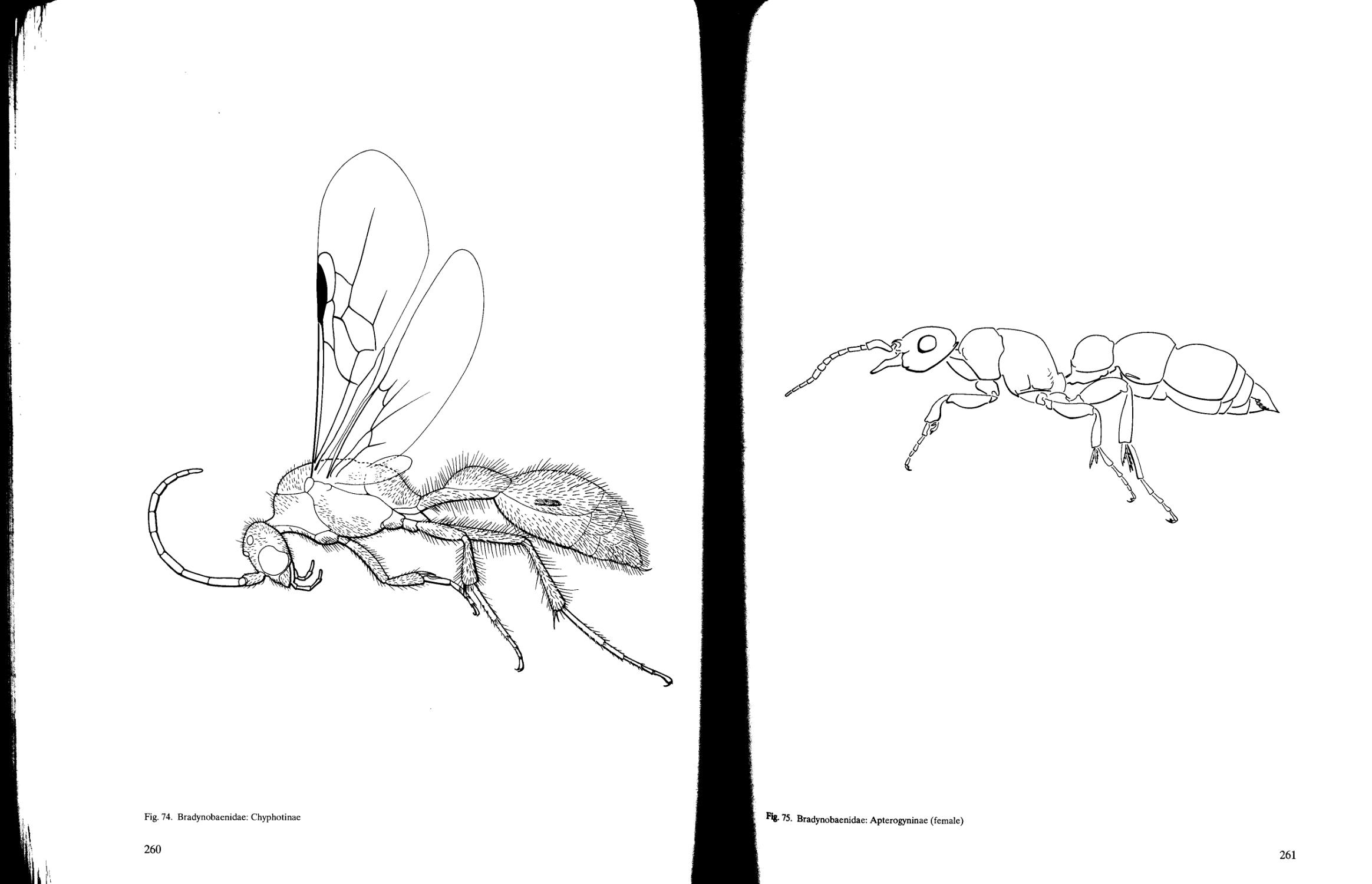
Key to subfamilies of Bradynobaenidae 207
Key to subfamilies of Scoliidae 211
Key to subfamilies of Vespidae 213
Key to subfamilies of Formicidae 218
References to Vespoidea 225
Habitus drawings of Vespoidea 232
IV
Chapter 9. Superfamily Apoidea Albert T. Finnamore and Charles D. Michener 279
Key to series of Apoidea 279
Key to families of Spheciformes 280
Key to subfamilies of Ampulicidae 291
Key to subfamilies of Sphecidae 292
Key to subfamilies of Pemphredonidae 294
Key to subfamilies of Astatidae 295
Key to subfamilies of Crabronidae 297
Key to subfamilies of Mellinidae 298
Key to subfamilies of Nyssonidae 300
Key to subfamilies of Philanthidae 304
Key to families of Apiformes 308
References to Apoidea 321
Habitus drawings of Apoidea 326
Chapter 10. Superfamily Ichneumonoidea David B. Wahl and Michael J. Sharkey 358
Key to families of Ichneumonoidea 359
Key to subfamilies of Braconidae 363
Key to subfamilies of Holarctic and Neotropical Ichneumonidae 396
References to Ichneumonoidea 442
Habitus drawings of Ichneumonoidea 449
Chapter 11. Superfamilies Evanioidea, Stephanoidea, Megalyroidea, and
Trigonalyoidea William R.M. Mason 510
Key to families of Evanioidea 510
References to Evanioidea, Stephanoidea, Megalyroidea, and Trigonalyoidea 514
Habitus drawings of Evanioidea, Stephanoidea, Megalyroidea, and Trigonalyoidea 515
Chapter 12. Superfamily Cynipoidea AlasdairJ. Ritchie 521
Key to families of Cynipoidea 522
References to Cynipoidea 529
Habitus drawings of Cynipoidea 531
Chapter 13. Superfamily Proctotrupoidea Lubomir Masner 537
Key to families of Proctotrupoidea 538
References to Proctotrupoidea 547
Habitus drawings of Proctotrupoidea 549
Chapter 14. Superfamily Platygastroidea Lubomir Masner 558
Key to families of Platygastroidea 559
References to Platygastroidea 562
Habitus drawings of Platygastroidea 564
Chapter 15. Superfamily Ceraphronoidea Lubomir Masner 566
Key to families of Ceraphronoidea 566
References to Ceraphronoidea 567
Habitus drawings of Ceraphronoidea 568
Chapter 16. Superfamilies Mymarommatoidea and Chalcidoidea Gary A.P. Gibson 570
Key to families of Chalcidoidea (including Mymarommatidae) 573
References to Mymarommatoidea and Chalcidoidea 627
Habitus drawings of Mymarommatoidea and Chalcidoidea 635
Appendix 1 List of habitus drawings, with generic names 656
Index 660
v
Contributors
D.J. Brothers, Ph.D.
Department of Zoology and Entomology
University of Natal
Pietermaritzburg, 3200
Republic of South Africa
A.T. Finnamore, Ph.D.
Provincial Museum of Alberta
12845 102nd Street
Edmonton, AB T5N 0M6
Canada
G.A.P. Gibson, Ph.D.
Agriculture Canada
Centre for Land and Biological Resources Research
K.W. Neatby Building
Central Experimental Farm
Ottawa, ON K1A 0C6
Canada
tW.R.M. Mason, Ph.D.
Agriculture Canada
Centre for Land and Biological Resources Research
K.W. Neatby Building
Central Experimental Farm
Ottawa, ON K1A 0C6
Canada
CD. Michener, Ph.D.
Snow Entomological Museum, Snow Hall
University of Kansas
Lawrence, KS 66045
USA
A.J. Ritchie, Ph.D.
Gordon & Breach Science Publishers
270 8th Avenue
New York, NY 10011
USA
H. Goulet, Ph.D.
Agriculture Canada
Centre for Land and Biological Resources Research
K.W. Neatby Building
Central Experimental Farm
Ottawa, ON K1A 0C6
Canada
M.J. Sharkey, Ph.D.
Agriculture Canada
Centre for Land and Biological Resources Research
K.W. Neatby Building
Central Experimental Farm
Ottawa, ON K1A 0C6
Canada
J.T. Huber, Ph.D.
Forestry Canada
% Centre for Land and Biological Resources
Research
K.W. Neatby Building
Central Experimental Farm
Ottawa, ON K1A 0C6
Canada
D.B. Wahl, Ph.D.
American Entomological Institute
3005 S.W. 56th Avenue
Gainesville, FL 32608
USA
L. Masner, Ph.D.
Agriculture Canada
Centre for Land and Biological Resources Research
K.W. Neatby Building
Central Experimental Farm
Ottawa, ON K1A 0C6
Canada
VI
Acknowledgments
This publication was produced with support
from the Centre for Land and Biological Resources
Research, Agriculture Canada, Ottawa. As editors
of the project, we sincerely thank the contributing
authors for their prompt attention to the questions
that arose during editing of the text and their
goodwill in agreeing to changes that were needed to
produce a relatively uniform set of keys and family
sketches.
Help in completing the project was efficiently and
cheerfully provided by both technical and professional
staff. S. Rigby drew many of the illustrations. L.
Dumouchel and H.E. Bisdee performed the exacting
and time-consuming task of arranging the numerous
illustrations for each key couplet and preparing and
numbering most of the plates. We extend special
thanks to them for the quality of their work, for their
thoughtful relationship with each collaborator, and for
their patience in dealing with what must have seemed
endless changes requested by the authors and editors.
J. Denis and two summer students, M. McKenzie and
S. Grant, provided capable assistance in preparing
many of the plates, and D. Tierney prepared a few of
the illustrations. J.D. Read carefully proofread the
entire text, checked many of the references for
accuracy of citation, prepared the computer-
generated flowchart, and organized the illustrations
for the glossary.
J. McWilliams typed much of the text and
patiently made the countless corrections requested
by authors and editors to several drafts of the
manuscript.The thousands of prints required for
publication were done by B. Wollenschlager under
the direction of B. Edwards and R. St. John of the
Informations Systems and Cartography Unit,
Centre for Land and Biological Resources
Research. A. Lutes and J. McCarthy of Research
Program Service are thanked for willingly
permitting access to the equipment for plate production in
their unit and freely giving instruction in its use.
In addition to contributing to the text of three
superfamilies, A.T. Finnamore prepared the
illustrations for the keys to Chrysidoidea,
Vespoidea, and Spheciformes, a large additional
task. CD. Michener critically reviewed Chapter 3.
The various sections were reviewed by several
specialists. We thank A. Austin, Waite Agricultural
Research Institute, Adelaide, Australia; Z. Boucek,
C.A.B. International Institute of Entomology,
London, England; N.D.M. Fergusson and J.S.
Noyes, The Natural History Museum, London,
England; L. Kimsey, University of California,
Davis, California; M. Sanborne, MacDonald
College, Sainte-Anne-de-Bellevue, Quebec; D.R.
Smith, United States National Museum,
Washington, D.C; H.K. Townes, American
Entomological Institute, Gainesville, Florida; J.
Whitfield, University of Missouri, St. Louis,
Missouri; F. Ronquist, University of Uppsala,
Sweden, as well as the contributors to this project
for their fast and accurate reviews of earlier drafts
of the manuscript. R.S. Anderson, Canadian
Museum of Nature, Ottawa, Ontario, reviewed
Chapters 1-5. As a coleopterist he provided a fresh
perspective on the publication and made several
useful suggestions for improving its clarity. We
especially thank R. Wharton, Texas A & M
University, College Station, for critically reviewing
the entire publication.
The manual was initially developed for a course
on identification of Hymenoptera given on three
occasions to a total of 45 students from across
Canada and several foreign countries. The students
tested early versions of the keys and pointed out
inaccuracies or lack of clarity in couplets and
illustrations that needed improvement. We thank all
of them for their suggestions. In this respect, we want
to thank M. Sarazin, who organized the courses.
Several figures were redrawn with permission
and are acknowledged below.
Chapters 12 and 16, Cynipoidea and
Chalcidoidea Figs. 192-196; from The Insects of
Australia, Figs. 37.24A,B,D,F,I; 37.25A; 37.261,
respectively, published in 1970 by the
Commonwealth Scientific and Industrial Research
Organization, Canberra, Australia.
Chapters 9, Apoidea Figs. 95,100,101, 102,
104,106,113; from R.M. Bohart and A.S. Menke
Specid Wasps of the World: A Generic Revision, Figs.
8, 42, 56, 65, 136,151, 190, respectively, published
in 1976 by the Regents of the University of
California, Berkeley, California.
Chapter 16, Chalcidoidea figures for couplet
numbers la {right), 30a {top), 4bb {left top), 4bb {left
bottom), 31a, 31b, 17d {left), 29a, 27bb {right), 4c
{top), 8aa, 8cc, 27bb, 24a {left), 24a {right), 24b, 7aa,
7bb {right); from Z. Boucek, Australasian
Chalcidoidea {Hymenoptera), Figs. 67 {in part), 161,
164, 215, 256, 275, 313, 372, 394, 497, 615, 919, 920,
923,1298, respectively, published in 1988 by the
C.A.B. International.
Chapter 12, Cynipoidea figures for couplet
numbers lc, Id, lcc, ldd left bottom, lec, ldd right
top, lcc, ldd right bottom, lcc, ldd right middle, 4c
top right, 4b, 7ee, 7e; from J. Quinlan,yl Revisionary
Classification of the Cynipoidea (Hymenoptera) of the
Ethiopian Zoogeographical Region. Aspicerinae
(Figitidae) and Oberthuerellinae (Liopteridae), Figs.
1, 2, 3, 6, 8, 33, 34, 38, 40, respectively), Bulletin of
the British Museum (Natural History) 39(2):
85-133, published in 1979 by the Trustees of the
British Museum (Natural History).
VI1
Chapter 1 Introduction (English)
John T. Huber
Since the early 1980s, application of biological
control methods against insect pests has had a
major resurgence. Parasite Hymenoptera are
among the most important agents for biological
control and are extremely abundant in most
terrestrial habitats. Despite their importance, their
biology and taxonomy remain poorly known and
their identification is difficult.
This publication is the result of a course on
identification of Hymenoptera given three times
since 1985 at the Centre for Land and Biological
Resources Research (then known as the
Biosystematics Research Centre). The initial
impetus for the course came from an annual course
sponsored since 1980 by the Systematic Entomology
Laboratory, United States Department of
Agriculture, Washington, D.C. A similar course
offered jointly since 1989 by The Natural History
Museum, London, and by Sheffield University has
also been very successful. The considerable interest
in these courses indicated the need for a
comprehensive identification guide to all extant
families of Hymenoptera.
Keys to families of Hymenoptera in standard
textbooks tend to repeat those published earlier,
usually using the same characters and format, and
often permitting only the more characteristic
members of a family to be identified. Rare groups
or groups difficult to key out because of unusual
attributes often were not included. The keys
presented here were written by specialists who tried
to include as many exceptional groups as possible to
make the keys comprehensive. Therefore, although
the guide was written primarily for nontaxonomists
and for taxonomy students identifying
Hymenoptera for the first time, it should also be
useful to more experienced Hymenoptera
taxonomists. Ninety-nine families and 20
superfamilies are treated (Table 1), although
further research on relationships will almost
certainly result in changes to these numbers. At
least one new family is known but is not included
here, pending its formal description.
Because this guide is intended for a wide group
of users, several features were incorporated into the
keys to make them easy to use while maintaining
accuracy and completeness: each distinct attribute
is listed alphabetically in the alternates of the
couplets; figures illustrating key attributes are
placed with the respective couplets; and terms are
standardized. One chapter on structure, including
an illustrated glossary of terms, and another on the
use of the keys are included to help the first-time
user of identification keys to Hymenoptera.
The main emphasis is on family identification
using the keys, which are complemented by family
sketches. The sketches include a taxonomic
diagnosis to supplement the keys, a summary of the
biology, the size and distribution, and important
(mostly taxonomic) literature references. For
several families we have provided sketches and keys
for the constituent subfamilies. Also included is at
least one habitus drawing of a member of each
family and many subfamilies. Literature to specific
genera and species is usually excluded. Literature of
a more general nature is cited in Chapters 2 and 3.
For convenience, in this text "North America"
refers to Canada and the United States only.
Although much effort has gone into key
construction, there is always room for
improvement. Largely because of differences in size
of the various families, the level to which such
groups are keyed is variable. Although all extant
families are keyed, keys to subfamilies may or may
not be included. For example, we have presented
keys to the subfamilies of Braconidae and
Ichneumonidae because of the large size of each of
these two families. When included, not all the
subfamily keys give complete world coverage, but
North America, at least, is covered.
Using the keys will likely lead to discovering
exceptions to them. It is hoped that suggestions for
improving the keys will be made to the editors or to
the authors of the appropriate section.
Eleven authors contributed to the publication.
Because each specialist had his own style of key
writing, some editorial changes had to be made to
provide a reasonably uniform work. This included
compromises and choices in use of morphological
terms where several equally meaningful or useful
terms could be used. The editors tried to maintain a
fairly simple and uniform set of terms, in the keys
at least, without losing accuracy or clarity.
If this guide results in greater interest in
taxonomy of Hymenoptera and encourages
biologists, teachers, students, biological control
officers, and other nontaxonomists to identify their
own specimens of Hymenoptera to family, then the
goal of the authors will have been fulfilled.
1
Introduction (frangais)
Depuis le debut de la derniere decennie,
l'application des techniques de lutte biologique
contre les insectes nuisibles connait un grand essor.
Les hymenopteres parasites comptent parmi les
agents les plus importants de cette lutte biologique.
En depit de leur importance, leur biologie et leur
taxonomie demeurent peu connues et leur
identification est difficile.
Suite a une demande accrue de cours
d'introduction aux hymenopteres et en l'absence de
publications traitant des families d'hymenopteres
au niveau mondial, plusieurs hymenopteristes
conjointement avec le "Systematic Entomology
Laboratory" a Washington ont cree un cours
portant sur les families d'hymenopteres parasites.
La popularite et le succes de ce cours encouragea
une orientation semblable au les Centre de
recherches sur les terres et les ressources
biologiques au Canada ou plusieurs
hymenopteristes ont coopere a la preparation d'un
cours annuel sur toutes les families d'hymenopteres.
Apres avoir presente ce cours a trois reprises, il
devint clair qu'un guide detaille d'identification des
families d'hymenopteres serait un outil
indispensable a tout etudiant. Apres plusieurs
annees d'efforts et avec la collaboration de
plusieurs scientifiques de ce Centre et d'ailleurs, ce
guide voit finalement le jour.
Les quelques guides sur les families
d'hymenopteres se ressemblent dans la construction
des clefs d'identification, et ne permettent la
reconnaissance que des families majeures. Certains
groupes demeurent difficiles a caracteriser a cause
d'attributs inhabituels, ou sont simplement ignores
car ils semblent trop peu communs
(particulierement les groupes bases sur de petites
especes qui sont representees par peu de specimens
dans les collections malgre leur grande abondance
en nature). Ce guide-ci se veut l'un des plus
complets car les experts qui l'ont redige y ont inclus
tous les groupes exceptionnels connus. Ainsi, 99
families et de 20 super-families y sont traitees
(Tableau 1), ces nombres etant appeles a changer a
l'avenir avec la poursuite de la recherche sur la
classification des hymenopteres. Ce guide s'adresse
a tout biologiste ayant un interet dans la lutte
biologique et a tout debutant desireux de se
familiariser avec les hymenopteres. De plus, il sera
particulierement utile a tous les etudiants de la
taxonomie, de meme qu'aux specialistes de la
systematique des hymenopteres.
Comme ce travail est destine aussi bien aux
debutants qu'aux experts, nous avons simplifie la
presentation et les illustrations des clefs
d'identification sans pour autant en reduire
l'integralite et la precision. Premierement, nous
avons separe en paragraphes et denote
alphabetiquement chaque attribut particulier pour
chacune des deux composantes d'un couplet.
Deuxiemement, les abondantes figures illustrant
divers attributs apparaissent simultanement avec les
couplets correspondants. Troisiemement, seul un
vocabulaire uniforme et clair est employe. Enfin,
pour aider les personnes peu familieres avec les
hymenopteres, nous avons prepare des chapitres
sur la morphologie et sur l'utilisation des clefs
d'identification ainsi qu'un glossaire illustre.
Bien que l'emphase de ce guide soit mise sur
les clefs d'identification, les families qui y sont
identifiees sont caracterisees davantage dans de
brefs exposes. On y presentent des diagnoses
morphologiques qui completent les clefs, un
sommaire sur la biologie, la diversite et la
distribution de chaque famille et dans bien des cas,
de leurs sous-families, des references pertinentes et
au moins une illustration complete d'une espece
typique a chaque famille. Les references de nature
specifique et generique y sont exclues, cependant
sous les chapitres 2 et 3 on mentionne plusieurs
travaux d'ordre general.
Par souci de commodite, « Amerique du Nord »
dans cet ouvrage correspond au Canada et aux
Etats-Unis seulement.
Malgre tous les efforts apportes a une
definition claire de toutes les families, certaines
demeurent neanmoins obscures. De telles
differences nous ont contraints de definir les
families a des niveaux differents. Pour certaines
grandes families (e.g., Ichneumonidae et
Braconidae), il est beaucoup plus utile et significatif
d'examiner egalement leurs sous-families. Ainsi,
bien que Ton presente des clefs pour toute les
families existantes, seulement quelques clefs aux
sous-families sont indues, celles-ci couvrant au
moins la faune nord-americaine.
Dans un travail de cette envergure, il se glisse
inevitablement des erreurs. II serait done fort
apprecie et souhaitable que tout commentaire apte
a ameliorer les clefs ou le texte dans une edition
ulterieure soit communique aux redacteurs ou aux
auteurs respectifs de chaque section.
Avec la contribution de 11 auteurs ayant
chacun une approche differente quant a la
construction des clefs, il a ete necessaire d'apporter
quelques modifications afin que le style et le
vocabulaire soient assez uniformes. Certains
compromis se sont averes necessaires dans le choix
de termes morphologiques particuliers afin d'eviter
l'ambigu'ite et la confusion.
Si ce guide suscite de l'interet pour la
taxonomie des hymenopteres et encourage les
biologistes, les professeurs, les etudiants, et les
agents de lutte biologique a identifier leurs propres
specimens d'hymenopteres au niveau familial, alors
le but des auteurs aura ete atteint.
2
Table 1. Checklist of superfamilies and families of Hymenoptera arranged alphabetically by superfamily and
Sn rsu^erTamUy "^ °f SUbfamiHeS * &*" * Parentheses ™* * they are"keyed separately
SYMPHYTA
CEPHOIDEA
Cephidae
MEGALOGONTOIDEA
Megalodontidae
Pamphiliidae
ORUSSOIDEA
Orussidae
SIRICOIDEA
Siricidae
TENTHREDINOIDEA
Argidae
Blasticotomidae
Cimbicidae
Diprionidae
Pergidae
Tenthredinidae
XYELOIDEA
Xyelidae
UNPLACED
Anaxyelidae
Xiphydriidae
APOCRITA (ACULEATA)
APOIDEA (APIFORMES)
Andrenidae
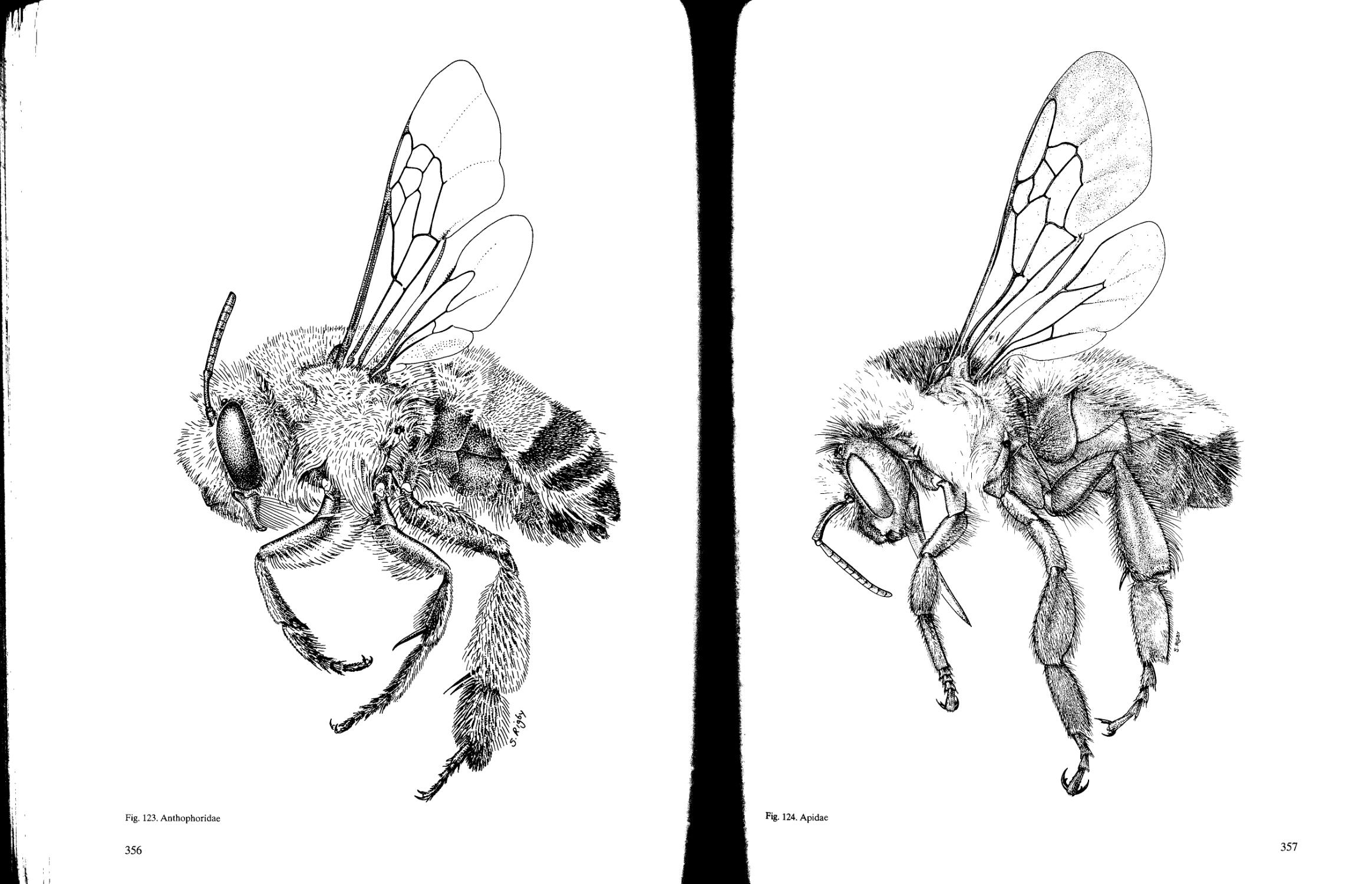
Anthophoridae
Apidae
Colletidae
Ctenoplectidae
Fideliidae
Halictidae
Megachilidae
Melittidae
Oxaeidae
Stenotritidae
APOIDEA (SPHECIFORMES)
Ampulicidae (2)
Astatidae (3)
Crabronidae (2)
Heterogynaidae
Mellinidae (2)
Nyssonidae (7)
Pemphredonidae (2)
Philanthidae (6)
Sphecidae (3)
CHRYSIDOIDEA
Bethylidae (4)
Chrysididae (4)
Dryinidae(ll)
Embolemidae
Plumariidae
Sclerogibbidae
Scolebythidae
VESPOIDEA
Bradynobaenidae (4)
Formicidae (10)
Mutillidae (7)
Pompilidae (3)
Rhopalosomatidae
Sapygidae (2)
Scoliidae (2)
Sierolomorphidae
Tiphiidae (6)
Vespidae (6)
APOCRITA (PARASITICA)
CERAPHRONOIDEA
Ceraphronidae
Megaspilidae
CHALCIDOIDEA
Agaonidae
Aphelinidae
Chalcididae
Elasmidae
Encyrtidae
Eucharitidae
Eulophidae
Eupelmidae
Eurytomidae
Leucospidae
Mymaridae
Ormyridae
Perilampidae
Pteromalidae
Rotoitidae
Signiphoridae
Tanaostigmatidae
Tetracampidae
Torymidae
Trichogrammatidae
CYNIPOIDEA
Charipidae
Cynipidae
Eucoilidae
Figitidae
Ibaliidae
Liopteridae
EVANIOIDEA
Aulacidae
Evaniidae
Gasteruptiidae
ICHNEUMONOIDEA
Braconidae (30)
Ichneumonidae (32)
MEGALYROIDEA
Megalyridae
MYMAROMMATOIDEA
Mymarommatidae
PLATYGASTROIDEA
Platygastridae
Scelionidae
PROCTOTRUPOIDEA
Austroniidae
Diapriidae
Heloridae
Monomachidae
Pelecinidae
Peradeniidae
Proctotrupidae
Roproniidae
Vanhorniidae
STEPHANOIDEA
Stephanidae
TRIGONALYOIDEA
Trigonalyidae
3
Chapter 2 Order Hymenoptera
William R.M. Mason and John T. Huber
General comments
Hymenoptera is one of-the four great orders of
insects, the other three being Coleoptera,
Lepidoptera, and Diptera. Each order includes over
100 000 described species around the world, with
Coleoptera having over 300 000. In Canada,
Hymenoptera includes about 7000 named species,
or about 25% of the total named insects. Experts
have estimated less than half of Hymenoptera in
Canada have been named, bringing the estimated
Canadian fauna to 14 000-16 000 species. At
For recognition the most useful features are
those unique to the group and those that are
present in all its members. Only a few
morphological attributes fulfill both these
conditions in Hymenoptera. In adults the following
attributes are the most easily seen.
The mouthparts are mandibulate and the basal
sections of the labium and maxilla lie closely
attached side by side and have a sharp transverse
fold near the basal third. Thus, in side view they
have the shape of a figure 7, and the maxillar-labial
complex can be folded up behind the head or
extended out beyond the mandibles at will by
muscular action that swings the basal section
(cardo) and simultaneously straightens (for
extension) or folds (for contraction) at the
transverse joint. The underarm jab punch of a boxer
is a similar action by a human arm. Although it is
difficult to observe, the labium is only attached by
its sides to the maxillae and not, as is usual in other
mandibulate insects, directly to the head. The
maxillae retain their direct attachment to the head.
The hind wing bears a few to many hamuli in a
row, about two-thirds of the way toward its apex
along the anterior margin. In a few Hymenoptera
some hamuli also occur at the base of the anterior
margin. The hamuli engage in a gutter-like
sclerotized strip (frenal fold) opposite them on the
posterior margin of the fore wing. In flight they
cause the fore and hind wings to act as a single
airfoil. The hamuli are, of course, absent when
wings are reduced or lost. The hamuli are the most
reliable and most easily seen diagnostic feature of
the Hymenoptera. This feature cannnot, of course,
be used in the numerous wingless Hynmenoptera.
The ovipositor (valvulae 1 and 2) is articulated
to the valvifer at the base and has two pairs of
muscles that enable it to be swung downn to drill
vertically. When not in use the ovipositor can be
present, over 18 000 species have been described in
North America; a conservative estimate of
undescribed species is about 30 000. The size of the
world terrestrial fauna in well-known groups is
often at least 10 times that of the Nearctic, so that
one could estimate 300 000 species of Hymenoptera
as a conservative world total. It is difficult to make
any general observations about so many species
without citing many exceptions, but nevertheless
some generalizations are useful.
folded up into the sheaths (valvulae 3) by the action
of the opposing muscle pair. Most Platygastroidea
and the genus Pelecinus (Pelecinidae) are
exceptional, having lost this flexibility.
One of the most important characters is a
genetic one. Sex determination is normally by a
haplo-diploid system. In this system unfertilized
(haploid) eggs have half the number of
chromosomes found in fertilized (diploid) eggs, and
both types of eggs develop. Males normally are
haploid, whereas females are always diploid and
usually result from fertilized eggs. By controlling
fertilization of eggs as they are laid, the female can
regulate the sex ratio, e.g., fertilized eggs laid in
large hosts and unfertilized ones in small hosts. In
addition, the haploid sex allows recessive lethal
mutations to be rapidly eliminated from the
population because they cannot be masked in males
and transmitted to the next generation.
Other, usually more practical characters for
identifying adult Hymenoptera in the field or
laboratory follow, but they are not nearly
completely reliable because they may be shared by
other insects or may not be present in all
Hymenoptera. The following characters should be
looked for in combination; no single one is proof
that the specimen is a hymenopteran:
• Four membranous wings; the hind wings shorter
than the fore wings; the membranes not obscured
by scales or dense hair.
• Wings with fewer veins than in many insects,
usually four longitudinal veins (rarely five or six),
but these are deflected and confused with
crossveins in the central part of the wing so that
the basal and apical veins do not appear
continuous; often there are far fewer veins in
species of very small body size.
Diagnosis
4
• Crossveins rarely more than seven and often
difficult to distinguish from longitudinal veins.
• Base of fore wing covered by a small roundish
sclerite, the tegula.
• Fore wing usually with a stigma (sclerotized
triangular or semicircular area) on the anterior
margin at about the midpoint. At its base the
stigma is separated from the costal vein by a
notch. These features are sometimes (30%) lost
in species of very small body size and reduced
wing venation.
• Abdominal tergum 1 fused to the metanotum but
a movable connection existing between the first
and second abdominal segments.
• Usually (90% or more) the apparent abdomen
(metasoma) is joined to the apparent thorax
(mesosoma) by a very narrow connection (the
"wasp waist"); this connection is actually the
greatly narrowed articulation of abdominal terga
1 and 2.
• A true, primitive ovipositor with three pairs of
valves occurs in the female and is sometimes
plainly visible ventrally or apically.
Although many of the above mentioned
attributes involve wing structures, it is possible to
identify wingless Hymenoptera by other means:
• The mouthparts are mandibulate and have the
same folding maxillar-labial complex as winged
Hymenoptera (see above).
• Almost all species that are wingless, including
those with a relatively wide base to the
metasoma, have a narrow connection between
the mesosoma and the metasoma. The main
exceptions are the minute males of fig wasps
(Agaonidae).
Hymenoptera is one of the most numerous
groups of insects, with many members familiar to
everyone. This familiarity is demonstrated by the
vernacular names given to Hymenoptera that serve,
bother, frighten, or hurt people in their daily lives.
Some names are bee, ant, wasp, and sawfly.
Farmers, foresters, gardeners, and naturalists
usually have a greater interest and a larger
vocabulary of names such as nursery pine sawfly,
wheatstem sawfly, wood wasp, gall wasp, velvet ant,
yellow jacket, alfalfa leaf-cutting bee, bumble bee,'
honey bee, and so on. This is natural because these
people see so many insects and may even have to
compete with such insects for a living. Even though
a vernacular name like bee or ant may apply to
• All wingless females have a typical hymenopteran
ovipositor, but because many of them are
Aculeata, Platygastroidea, or Ceraphronoidea,
the ovipositor rarely can be seen without
dissection.
• Many have a prognathous or partly prognathous
head, the underside of which is readily seen. The
sides of the head extend down between the oral
fossa and foramen magnum, where they meet to
form a median groove, or fuse completely. No
true gula (as in Coleoptera) is formed.
• The immature stages of Hymenoptera are usually
not as easy to recognize or characterize as adults;
but then, apart from the larvae of Symphyta,
most people do not see them and have no
particular interest in identifying them.
Hymenoptera contains two main larval types:
caterpillar-like and grub-like. Symphyta mostly have
larvae that are caterpillar-like and crawl about on
plants. They are most likely to be confused with
Lepidoptera larvae (caterpillars). Most
Lepidoptera and Symphyta larvae have three pairs
of thoracic legs and an apical pair of abdominal
prolegs, but true caterpillars have at most only four
pairs of prolegs, on abdominal segments 3-6.
Typically, Symphyta larvae have at least five pairs of
prolegs, on abdominal segments 2-6. The prolegs
of Symphyta do not have crochets, whereas those of
Lepidoptera larvae do. Unfortunately, the prolegs
usually disappear in burrowing larvae of both
orders, making it more difficult to differentiate
them. Symphyta larvae have only one simple eye,
when present; Lepidoptera larvae have more than
one. Apocrita have legless grub-like larvae that are
nearly featureless. They are most likely to be
confused with certain Coleoptera larvae, e.g.,
Curculionoidea, or with Cecidomyiidae (Diptera).
Kukalova-Peck (1991) listed other characters
that define the Hymenoptera.
hundreds of species, the majority of Hymenoptera
species go quietly about their business unnoticed
and unnamed.
Taxonomists divide Hymenoptera into two
main suborders: Symphyta, or sawflies, and
Apocrita, or wasp-waisted Hymenoptera.
Suborder Symphyta
The first group, Symphyta, is small (only 7% of
Canadian species) and geologically old. Its
members first appeared as fossils in the Triassic,
about 200 million years ago. The species have
preserved most of the ancestral attributes of the
order, especially the plant-feeding habits, numerous
Order sketch
5
wing veins, and the comparatively unmodified
abdomen in which the first two segments look very
much like the succeeding ones. Most adult sawflies
are stubby, soft-bodied Hymenoptera that fly only
weakly. The ovipositor of most species is used to
pierce plant tissue and is compressed laterally and
modified with marginal teeth so that it looks and
functions like a saw, hence the name sawfly. The
larvae of most species are caterpillar-like, equipped
with legs, eyes and antennae, and walk about eating
foliage like true caterpillars (Lepidoptera), but a
few groups have eyeless, largely legless larvae that
bore in various plant tissues, including wood.
Suborder Apocrita
Apocrita are specialized most conspicuously by
the greatly narrowed connection between
abdominal segments 1 and 2, which gives greater
flexibility at that joint. The apparent abdomen,
called the metasoma, is thus not the entire
abdomen, because tergum 1 is now fused into the
thorax, appearing and functioning as part of that
body region. The ovipositor is thin and cylindical,
and sometimes very long. The fluids that lubricate
the ovipositor of sawflies are modified for an
additional aggressive function of paralyzing or
killing the prey. The larvae are eyeless, legless, with
very small antennae or none, and otherwise are
nearly featureless; in the most reduced forms the
sclerotized head capsule is reduced to a few bands
supporting the mouthparts, or it is even reduced to
nothing but a pair of mandibles. Apocrita larvae are
found in concealed or parasitic situations.
Apocrita include the familiar ants, bees, and
wasps, as well as tiny wasp-like parasites seldom
noticed by people. Members of the Apocrita are
fundamentally carnivorous, feeding on other insects
A summary of present concepts of the
evolution and higher classification of Hymenoptera
is given in Gauld and Bolton (1988). The goal of a
natural classification is to communicate, in a
simplified manner, hypotheses of the evolutionary
history of a group. Organisms are classified by
recency of common descent. Within a classification,
monophyletic groups consist of an ancestor and all
its descendants; a paraphyletic group consists of a
common ancestor and some, but not all, of its
descendants; and a polyphyletic group is an
assemblage of descendants from more than one
common ancestor. In construction of classifications,
monophyly is strived for, though many taxonomists
accept paraphyletic taxa. Polyphyly is rejected.
The order Hymenoptera is traditionally divided
into Symphyta and Apocrita, as discussed above.
Symphyta are generally accepted to be a
and spiders. However, several large groups have
abandoned the carnivorous life and returned to a
plant diet, but in specialized ways. One group, the
bees (Apiformes), feed on pollen and nectar,
whereas another group, the gall wasps (Cynipidae),
make the familiar galls on oak (Quercus) and rose
(Rosa), inside which the larvae feed. Members of
some groups of Apocrita feed inside developing
seeds or grass stems, and a few species of ants are
plant feeders or omnivorous, gathering seeds or
leaves or almost any other foodstuffs.
Almost 75% of all species of Apocrita are,
during the larval stage, parasitoids of other insects
or spiders, although as adults they live as
independently as any insect. The female wasp lays
her egg or eggs inside (or outside but almost always
attached to) the egg, larva, pupa, nymph, or even
adult of the particular prey that her larvae need to
develop. The parasitic larva, on hatching, proceeds
to devour its host in various ways that are usually
fatal to the host. When fully grown, the parasitoid
larva pupates and finally emerges as another adult
parasitoid. Insects that parasitize other animals are
called parasitoids to distinguish them from true
parasites such as tapeworms (Cestoda).
Most stinging wasps (the females in some
groups have lost the ability to sting) and most ants
are predators, though some are ectoparasitoids.
The predatory social species (that is, all the ants,
plus the paper wasps and yellow jackets) hunt and
capture insects or spiders and bring them to the
nest, where they are shared among the larvae and
adults of the colony. The vegetarian social bees
similarly distribute nectar and pollen to larvae and
adults. The many solitary wasps are almost all
specialists, catching just a few kinds of insects or
spiders that are placed in small cells, there to
nourish the larvae.
paraphyletic assemblage, which in this guide are
classified into six superfamilies and two unassigned
families. It is treated here in one chapter for
convenience.
Apocrita is probably monophyletic and is
treated here in several chapters by superfamily.
Within Apocrita, two subdivisions are traditionally
accepted: Parasitica (sometimes called Terebrantia)
and Aculeata. Members of Parasitica have an
ovipositor that retains its primitive egg-laying
function and is adapted for piercing, and likely are
a paraphyletic assemblage. Members of Aculeata,
comprising those Hymenoptera whose ovipositor is
modified for stinging instead of egg-laying, are
demonstrably monophyletic. The biological and
morphological distinctions between Parasitica and
Aculeata are difficult to draw and are subject to
many exceptions, and so it is more useful to deal
Higher classification
6
with a series of superfamilies; in this guide 14
superfamilies are recognized. Although some of
them are clearly monophyletic, the monophyly of
most groups has yet to be demonstrated.
The correct phylogenetic relationships of
superfamilies within the order and suborders have
not yet been fully determined. Certain groups of
superfamilies clearly belong together and can be
arranged from most primitive to most advanced, for
example, Chrysidoidea (primitive); Vespoidea,
Apoidea (advanced). Other superfamilies are
grouped together for practicality because their
members are few and their relationships to other
superfamilies are ambiguous, for example,
Evanioidea, Megalyroidea, Stephanoidea, and
Trigonalyoidea. Within each superfamily the
included families may be arranged by each author
either alphabetically or according to their
hypothesized relationships in light of current
Relatively few general works specifically
devoted to the Hymenoptera exist. Those published
since 1950 and a few older references still of great
use or interest are listed here. They include any
paper or book dealing with various aspects of the
entire order for either a country or a major
geographical region. Works treating only a part of
the order are listed in the appropriate superfamily
chapter. In addition, several general textbooks of
entomology that include chapters on Hymenoptera
are listed. Works on Hymenoptera, including keys,
intended for amateurs are by Cooper (1945),
Berland (1958a, 19586), Zahradnik (1985), and
Betts (1986). These works vary considerably in
quality, and the keys should be used with care.
Cooper (1945) contains useful information on
collecting, rearing, and studying Hymenoptera.
Keys The only world keys to Hymenoptera are by
Handlirsch (1925) and Brues et al. (1954). Keys to
all the families for a particular country or region
are given by van Achterberg (1982) for
northwestern Europe, Borror et al. (1989) for
North America, Boucek (1957) for Czechoslovakia,
Ceballos (1941-43) for Spain, Costa Lima (1960)
for Brazil, Delvare and Aberlenc (1989) for Africa
and South America, Landin (1971) for Sweden,
Oehlke (1989) for Germany, Oehlke (1969) for
eastern Germany, Prinsloo and Eardley (1985) for
South Africa, Richards (1977) and Gauld and
Bolton (1988) for Great Britain, and Riek (1970)
and Naumann (1992) for Australia.
Biology Summaries of biology are given by Arnett
(1985), Berland and Bernard (1951a, 19516),
Bischoff (1929), Borror et al. (1989), Boucek
(1957), Brown (1982), Clausen (1940), Costa Lima
(1960,1962), Daly et al. (1978), Gauld and Bolton
knowledge. Their relationships will almost certainly
be changed as more information becomes available.
The terms microhymenoptera and
macrohymenoptera are used in this guide for
convenience, not as formal taxonomic units.
Members of microhymenoptera are mostly small
(usually under 3 mm long) species with reduced
venation. They have only one pair of spiracles on
the metasoma or none at all. Included are
Chalcidoidea, Cynipoidea, Proctotrupoidea,
Ceraphronoidea, Platygastroidea, and a few rare
groups.
Members of macrohymenoptera are usually
much larger, including the familiar bees, wasps, and
ants. They almost always have numerous veins and
cells, at least in the fore wing, and have four to
seven pairs of spiracles on the metasoma. Included
are stinging Hymenoptera (Apoidea, Vespoidea,
Chrysidoidea) and Ichneumonoidea.
(1988), Grandi (1966), Kryzhanovskii and Malyshev
(1963), Malyshev (1959,1968), Naumann (1992),
Prinsloo and Eardley (1985), Richards and Davies
(1977), Riek (1970), Riek and Cardale (1974), Ross
et al. (1982), and Schmiedeknecht (1930).
Phylogeny Konigsmann (1976,1977, 1978a,
19786) and Rasnitsyn (1980, 1988) analyzed
infraordinal relationships of the order from a
phylogenetic perspective. Rasnitsyn (1980,1988,
1990) and Rasnitsyn and Kulicha (1990) also
discussed the fossil Hymenoptera and the general
classification of the order.
Biological significance, Conservation Gauld et al.
(1990) provided an overview of the importance of
the order and conservation of Hymenoptera in
Europe.
Catalogs, checklists and regional surveys Few
checklists or catalogs for the entire order have been
published for any country or region since Dalla
Torre (1892-1902) published his world catalog. The
only such catalogs are by Wu (1941) for China,
Ceballos (1956) for Spain, and Krombein et al.
(1979) for North America. Fitton et al. (1978) and
Hirashima (1989) published checklists for Britain
and Japan, respectively. Pagliano and Scaramozzino
(1990) published a checklist of generic and
subgeneric names of Hymenoptera of the world.
Masner et al. (1978) surveyed the Canadian fauna.
Newsletters Several newsletters for special-
interest groups are available that are useful for
those who wish to make contact with specialists
around the world. The newsletters cover the entire
order except for Cynipoidea and a few small, rare
superfamilies. Their purpose is to provide a forum
for communication and informal discussion among
Literature on Hymenoptera
7
researchers working on the biology or taxonomy of
specific groups of Hymenoptera. A list of the
newsletters and their editors is given below. The
specialized, formal journals devoted to social
insects, such as the honey bee, Apis mellifera
Linnaeus, are not included.
Newsletters for Hymenopterists
Newsletter and
date started
Proctos
1975
Ichnews
1976
Groups covered
Proctotrupoidea
Scelionoidea
Ceraphronoidea
Ichneumonoidea
Editors
L. Masner
Agriculture Canada
Centre for Land and Biological Resources Research
K.W. Neatby Building
Central Experimental Farm
Ottawa, ON K1A 0C6
Canada
N.F. Johnson
Department of Entomology
1735 Neil Avenue
Ohio State University
Columbus, OH 43210
USA
P. Marsh
Systematic Entomology Laboratory
United States Department of Agriculture
% United States National Museum NHB 168
Washington, DC 20560
USA
M. J. Sharkey
Agriculture Canada
Centre for Land and Biological Resources Research
K.W. Neatby Building
Central Experimental Farm
Ottawa, ON K1A 0C6
Canada
D. Wahl
American Entomological Institute
3005 S.W. 56th Avenue
Gainesville, FL 32608
USA
Sphecos
1979
Chalcid Forum
1983
Aculeata
(except bees
and ants)
Chalcidoidea
A. Menke
Systematic Entomology Laboratory
United States Department of Agriculture
c/o United States National Museum NHB 168
Washington, DC 20560
USA
E.E. Grissell and M.E. Schauff
Systematic Entomology Laboratory
United States Department of Agriculture
c/o United States National Museum NHB 168
Washington, DC 20560
USA
G.A.P Gibson and J.T. Huber
Centre for Land and Biological Resources Research
K.W. Neatby Building
Central Experimental Farm
Ottawa, ON K1A 0C6
Canada
Trichogramma
News
1983
Symphytos
1984
Melissa
1986
Trichogramma
and other egg
parasitoids
Symphyta
Apoidea
(bees only)
S.A. Hassan
BBA
Institut fur biologische
Schadlingsbekampfung
Heinrichstrasse 243
D-6100, Darmstadt
Germany
H. Goulet
Centre for Land and Biological Resources Research
K.W. Neatby Building
Central Experimental Farm
Ottawa, ON K1A 0C6
Canada
D.R. Smith
Systematic Entomology Laboratory
United States Department of Agriculture
c/o United States National Museum NHB 168
Washington, DC 20560
USA
R.J. McGinley and B.B. Norden
Department of Entomology
Smithsonian Institution NHB 105
Washington, DC 20560
USA
C.D.Michener
Entomological Museum, Snow Hall
University of Kansas
Lawrence, KS 66045
USA
Notes from
Underground
1989
Formicidae
(ants)
M. Moffet and S. Cover
MCZ Laboratories
Harvard University
Cambridge, MA 02138
USA
References
Achterberg, C. van. 1982. Familietabel van de
Hymenoptera in Noordwest-Europa.
Wetenschappelijke Mededelingen van de
Koningklijke Nederlandse Natuurhistorische
Vereniging 152. 50 pp.
Arnett, R.H., Jr. 1985. Hymenoptera (wasps, ants,
and bees). Pages 402-471 in American insects:
a handbook of the insects of America north of
Mexico. Van Nostrand Reinold, New York,
USA. 850 pp.
Berland, L., and F. Bernard. 1951a.
Hymenopteroides (Symphytes et Terebrants).
Pages 771-975 in Grasse, P.-P Traite de
zoologie, anatomie, systematique, biologie.
Tome X. Insectes superieurs et hemipteroides
(Premier fascicule). Masson, Paris, France.
975 pp.
Berland, L., and F. Bernard. 19516.
Hymenopteroides (Aculeates). Pages
976-1276, in Grasse, P.-P. Traite de zoologie,
anatomie, systematique, biologie. Tome X.
Insectes superieurs et hemipteroides (Fascicule
II). Masson, Paris, France. 973 pp.
Berland, L. 1958a. Atlas des Hymenopteres de
France, Belgique, Suisse. I. Tenthredes,
Parasites, Porte-aiguillon (Bethylides). Nouvel
Atlas d'Entomologie. Editions N. Boubee,
Paris, France. 155 pp.
Berland, L. 19586. Atlas des Hymenopteres de
France, Belgique, Suisse. II. Porte-aiguillons:
Bethyloides (fin), Scolioides, Formicoides,
Pompiloides, Vespoides, Sphecoides, Apoides.
Nouvel Atlas d'Entomologie. Editions N.
Boubee, Paris, France. 184 pp.
Betts, C. 1986. The hymenopterist's handbook. The
amateur entomologist. Vol. VII. Second
edition. The Amateur Entomologist's Society,
Middlesex, England. 208 pp.
Bischoff, H. 1929. Biologie der Hymenopteren: eine
Naturgeschischte der Hautflugler. Julius
Springer, Berlin, Germany. 606 pp.
Borror, D.J., C.A. Triplehorn, and N.F. Johnson.
1989. Order Hymenoptera, sawflies, parasitic
wasps, ants, wasps, bees. Pages 665—744 in An
introduction to the study of insects. Sixth
edition. Saunders College Publishing,
Philadelphia, Pennsylvania, USA. 875 pp.
Boucek, Z. 1957. Blanokridli-Hymenoptera
Pages 35-406 in Kratochvil, J. ed. Klic Zvfreny
CSR. Nakladetelstvi Ceskoslovenske
Akademie Ved. Prague, Czechoslovakia.
746 pp.
Brown, W.L., Jr. 1982. Hymenoptera. Pages
652-680 in Parker, S.P., ed. Synopsis and
classification of living organisms. Vol. 2.
McGraw-Hill, New York, New York, USA.
1232 pp.
Brues, C.T., A.L. Melander, and EM. Carpenter.
1954. Order Hymenoptera. Pages 621-684 in
Classification of insects: keys to the living and
extinct families of insects, and to the living
families of other terrestrial arthropods. Bulletin
of the Museum of Comparative Zoology 108.
917 pp.
Ceballos, G. 1941-1943. Las tribus de los
himenopteros de Espana. Consejo Superior de
Investigaciones Cientificas. Trabajos del
Instituto Espanol de Entomologia, Madrid,
Spain. 420 pp.
Ceballos, G. 1956. Catalogo de los himenopteros de
Espana. Consejo Superior de Investigaciones
Cientificas. Trabajos del Instituto Espanol de
Entomologia, Madrid, Spain. 554 pp.
Clausen, C.P 1940. Entomophagous insects.
McGraw-Hill, New York, New York, USA.
688 pp.
Cooper, B.A. ed. 1945. Hymenopertist's handbook.
The Amateur Entomologist 7 (40). 160 pp.
Reprinted 1969, Department of Agriculture,
The University, Leeds, England.
Costa Lima, A. da. 1960. Insetos do Brasil. 11°
Tomo. Capitulo 30. Hymenopteros l.a Parte.
Escola Nacional de Agronomia, Rio de Janeiro,
Brazil. Serie Didactica No. 13. 368 pp.
Costa Lima, A. da. 1962. Insetos do Brasil. 12°
Tomo. Capitulo 30. Hymenopteros 2.a Parte.
Escola Nacional de Agronomia, Rio de Janeiro,
Brazil. Serie Didactica No. 13. 393 pp.
Dalla Torre, C.G. de. 1892-1902. Catalogus
hymenopterorum hucusque descriptorum
systematicus et synonymicus. Guilelmi
Englemann, Leipsig, Germany.
Daly, H.V, J.T. Doyen, and PR. Ehrlich. 1978.
Order Hymenoptera (bees, wasps, ants, etc.).
Pages 478-502 in Introduction to insect biology
and diversity. McGraw-Hill, New York, New
York, USA. 564 pp.
Delvare, G., and H.-P Aberlenc. 1989. Ordre
Hymenoptera. Pages 163-200 in Les insectes
d'Afrique et d'Amerique tropicale. Cles pour la
reconnaissance des families. PRIFAS,
CIRAD-GERDAT, Montpellier, France.
302 pp.
10
Fitton, M.G., M.W.R. de V. Graham, Z.R.J.
Boucek, N.D.M. Fergusson, T. Huddleston, J.
Quinlan, and O.W. Richards. 1978. A check list
of British insects. Second edition (completely
revised). Part 4: Hymenoptera. Handbooks for
the identification of British insects. Vol. XI,
Part 4. Royal Entomological Society of London,
London, England. 159 pp.
Gauld, I., and B. Bolton, eds. 1988. The
Hymenoptera. Oxford University Press,
Oxford, England. 332 pp.
Gauld, I.D., N. Mark Collins, and M.G. Fitton.
1990. The biological significance and
conservation of Hymenoptera in Europe.
Nature and Environment Series, No. 44.
Council of Europe, Strasbourg, France. 47 pp.
Grandi, G. 1966. Ordine Hymenoptera. Pages
611-638 in Institzioni di entomologia generale.
Edizioni Calderini, Bologna, Italy. 654 pp.
Handlirsch, A. 1925. Uberordnung und Ordnung:
Hymenoptera L. (Hautflugler). Pages 712-825
in Schroder, C, ed. Handbuch der
Entomologie. Band III. Gustav Fischer, Jena,
Germany. 1201 pp.
Hirashima, Y. 1989. Hymenoptera. Pages 541-692
in A checklist of Japanese insects.
Entomological Laboratory, Faculty of
Agriculture, Kyushu, University, Kyushu,
Japan. 1767 pp.
Konigsmann, E. 1976. Das phylogenetisches System
der Hymenoptera. Teil 1: Einfuhrung,
Grundplanmerkmale, Schwestergruppe und
Fossilfunde. Deutsche Entomologische
Zeitschrift, N.F. Band 23, Heft IV-V253-279.
Konigsmann, E. 1977. Das phylogenetisches System
der Hymenoptera. Teil 2: "Symphyta".
Deutsche Entomologische Zeitschrift, N.F.
Band 24, Heft 1/111:1-40.
Konigsmann, E. 1978a. Das phylogenetisches
System der Hymenoptera. Teil 3: "Terebrantes"
(Unterordnung Apocrita). Deutsche
Entomologische Zeitschrift, N.F. Band 25, Heft
1/111:1-55.
Konigsmann, E. 19786. Das phylogenetisches
System der Hymenoptera. Teil 4: Aculeata
(Unterordnung Apocrita). Deutsche
Entomologische Zeitschrift, N.F. Band 25, Heft
IV-V365-435.
Krombein, K.V., PD. Hurd Jr., D.R. Smith, and
B.D. Burks. 1979. Catalog of Hymenoptera in
America North of Mexico. Vols. 1-3.
Smithsonian Institution Press, Washington,
D.C, USA. 2735 pp.
Kryzhanovskii, O.L., and S.I. Malyshev. 1963. The
Hymenoptera, their origin and evolution.
Sovetskaya nauka, Moscow, USSR. 291 pp. [In
Russian.]
Kukalova-Peck, J. 1991. Fossil history and the
evolution of hexapod structures. Pages
141-179 in CSIRO, ed. The insects of
Australia. A textbook for students and research
workers. Second edition. Vol. I. Melbourne
University Press, Carlton, Australia, xviii +
542 pp.
Landin, B.O. 1971. Hypemoptera. Pages 510-1019
in Faltfauna, Insekter 2:2, pp. 381 -1053. Natur
och kultur, Storkholm, Sweden.
Malyshev, S.I. 1959. The Hymenoptera, their origin
and evolution. Sovetskaya nauka, Moscow,
USSR. 297 pp. [In Russian.]
Malyshev, S.I. 1968. Genesis of the Hymenoptera
and the phases of their evolution. Methuen,
London, England. 319 pp.
Masner, L., J.R. Barron, H.V. Danks, A.T
Finnamore, A. Francoeur, G.A.P. Gibson,
W.R.M. Mason, and CM. Yoshimoto. 1978.
Hymenoptera. Pages 485-508, in Danks, H.V,
ed. Canada and its insect fauna. Memoirs of
the Entomological Society of Canada 108.
573 pp.
Naumann, I.D. 1992. Hymenoptera (wasps, bees,
ants, sawflies). Pages 916-1000, in CSIRO, ed.
The insects of Australia: a textbook for
students and research workers. Second edition.
Vol. 2, pp. 543-1137. Melbourne University
Press, Carlton, Australia.
Oehlke, J. 1969. Beitrage zur Insektenfauna der
DDR: Hymenoptera-Bestimmungstabellen bis
zu den Unterfamilien. Beitrage Entomologie
19:753-801.
Oehlke, J. 1989. Hymenoptera - Hautflugler.
Pages 398-463 in Stresemann, E., H.-J.
Hannemann, B. Klausnitzer, and K. Senglaub,
eds. Exkursionsfauna fur die Gebiete der DDR
und der BDR. Band 2/1 Wirbellose Insekten -
Erster Teil. 8. Auflage. Volk und Wissen, Berlin,
Germany.
Pagliano, G., and P. Scaramozzino. 1990. Elenco dei
generi di Hymenoptera del mondo. Bollettino
della Societa Entomologica Italiana
(Supplemento). Vol. 122. 210 pp.
Prinsloo, G.L., and G.L. Eardley. 1985. Order
Hymenoptera (sawflies, wasps, bees, ants).
Pages 393-451 in Scholtz, C.H., and E. Holm,
eds. Insects of Southern Africa. Butterworths,
Durban, South Africa. 502 pp.
Rasnitsyn, A.P 1980. Origin and evolution of
hymenopterous insects. Trudy
Paleontologicheskogo instituta. Akademiya
naukSSSR 174:1-192. [In Russian.]
11
Rasnitsyn, A.P. 1988. An outline of evolution of the
hymenopterous insects (order Vespida).
Oriental Insects 22:115-145.
Rasnitsyn, A.P. 1990. Hymenoptera. Vespida. Pages
177-205 in Rasnitsyn, A.P, ed. Late Mesozoic
insects of eastern Transbaihalia. Nauka Press,
Moscow, USSR. 223 pp.
Rasnitsyn, A.P, and R. Kulicka. 1990.
Hymenopteran insects in Baltic amber with
respect to the overall history of the order. Prace
Museum Ziemi 41:53-64.
Richards, O.W. 1977. Hymenoptera. Introduction
and key to families. 2nd edition. Handbooks for
the identification of British insects. Vol. VI,
Part 1. Royal Entomological Society of London,
London, England. 100 pp.
Richards, O.W., and R.G. Davies. 1977.
Hymenoptera (ants, bees, wasps, ichneumon
flies, sawflies, etc.). Pages 1175-1279 in Imm's
general textbook of entomology. 10th edition.
Vol. 2. Classification and biology. Chapman
and Hall, London, England. 1354 pp.
Riek, E.E, 1970. Hymenoptera (wasps, bees, ants).
Pages 867-959 in The insects of Australia.
CSIRO, sponsor. Melbourne University Press,
Carlton, Australia, xiii + 1029 pp.
Riek, E.F., and J.C. Cardale. 1974. Hymenoptera
(wasps, bees, ants). Pages 107-111 in The
insects of Australia: a textbook for students and
research workers. Supplement 1974. CSIRO,
sponsor. Melbourne University Press, Carlton,
Australia. 146 pp.
Ross, H.H., C.A. Ross, and J.R.P Ross. 1982.
Hymenoptera. Pages 408-434 in A textbook of
entomology. 4th edition. John Wiley and Sons,
New York, New York, USA. 666 pp.
Schmiedeknecht, O. 1930. Die Hymenopteren
nord- und Mitteleuropas. Gustav Fischer, Jena,
Germany. 1062 pp.
Sedivy, J. 1989. Enumeratio Insectarum
Bohemoslovakiae. Checklist of Czechoslovak
Insects III (Hymenoptera). Acta Faunistica
Entomologica Musei Nationalis Pragae. XIX.
194 pp.
Valentine, E.W, and A.H. Walher. 1991. Annotated
catalogue of New Zealand. Hymenoptera.
DSIR Plant Protection Report No. 4. 84 pp.
Wu, C.F 1941. Catalogus insectorum sinensium
(catalogue of Chinese insects). Vol. 6. Yenching
University, Beijing, People's Republic of China.
333 pp.
Zahradnik, J. 1985. Bienen, Wespen, Ameisen. Die
Hautfliigler Mitteleuropas. W. Keller, Stuttgart,
Germany. 191 pp.
12
Chapter 3 Structure
John T. Huber and Michael J. Sharkey
General comments
This guide is intended to familiarize the reader
with the families of Hymenoptera, not
hymenopteran morphology. We therefore attempt to use
the simplest acceptable terms for structures. Terms
used in the keys are defined and illustrated in the
glossary. A brief overview of hymenopteran
morphology, accompanied by labeled drawings of
bodies and wings representing some major groups
of Hymenoptera, is given to orient the reader and
put into perspective many of the terms used.
When the reader goes beyond this guide to
identify specimens to the level of genus or species, a
plethora of other, often synonymous, morphological
terms will be encountered. In part, this occurs
because over many years various terms have been
replaced by more exact or more generalized terms
and different terms have been applied to
homologous structures in various groups.
Consequently, any given structure may have several
names applied to it, and one term may apply to
several distinct structures. This confusion is
nowhere more apparent than in names applied to
the wing veins, where several major systems are
used.
It is beyond the scope of this guide to define all
terms as they have been applied to structures within
Hymenoptera. The following annotated list of
references will help the interested reader.
Bohart, R.M., and A.S. Menke. 1976. Sphecid wasps
of the world: a generic revision. University of
California Press, Berkeley, California, USA. ix +
695 pp.
The morphology chapter, especially the
sections on wing venation and the lists of
synonyms for various terms, is useful to all who
work on aculeate Hymenoptera.
Brothers, D.J. 1975. Phylogeny and classification of
the aculeate Hymenoptera, with special
reference to Mutillidae. University of Kansas
Science Bulletin 50:483-648.
Includes the first cladistic analysis of Aculeata,
with a detailed discussion of characters.
Bou&k, Z. 1988. Australasian Chalcidoidea
(Hymenoptera): a biosystematic revision of
genera of fourteen families, with a
reclassification of species. C.A.B. International,
Wallingford, England. 832 pp.
Includes a detailed discussion of terms used in
chalcidoid taxonomy.
Gauld, I., and B. Bolton, eds. 1988. The
Hymenoptera. Oxford University Press,
Oxford, England, xi + 332 pp.
Includes a chapter on the morphology of
Hymenoptera.
Gibson, G.A.P 1985. Some pro- and mesothoracic
structures important for phylogenetic analysis
of Hymenoptera, with a review of terms for the
structures. Canadian Entomologist
117:1395-1443.
A review and reinterpretation of thoracic
structure and a discussion of synonymous terms
applied to them.
Holldobler, B., and E.O. Wilson. 1990. The ants.
The Belknap Press of Harvard University Press,
Cambridge, Massachusetts, USA. 732 pp.
Includes a detailed section on morphology of
ants.
Michener, CD. 1944. Comparative external
morphology, phylogeny, and a classification of
the bees (Hymenoptera). Bulletin of the
American Museum of Natural History
82:151-326.
Includes a detailed section on morphology of
bees.
Nichols, S.W, comp. 1989. The Torre-Bueno
glossary of entomology. The New York
Entomological Society, New York, New York,
USA. 840 pp.
The most comprehensive reference book in
English for entomological terms.
Richards, O.W. 1977. Hymenoptera: introduction
and key to families. Second edition. Handbooks
for the identification of British insects. Vol. 6,
Part 1. Royal Entomological Society of London,
London, England. 100 pp.
A detailed overview of hymenopteran
morphology and terminology.
Ronquist, E, and G. Nordlander. 1989. Skeletal
morphology of an archaic cynipoid, Ibalia
rufipes (Hymenoptera: Ibaliidae). Entomologica
scandinavica, Supplement No. 33. 60 pp.
13
A detailed account of one species, including a
discussion of its structure and terms used in
relation to other Hymenoptera.
Ross, H.H. 1937. A generic classification of the
Nearctic sawflies (Hymenoptera, Symphyta).
Illinois biological monographs 15(2). 173 pp.
A reference for sawfly morphology.
Snodgrass, R.E. 1935. Principles of insect
morphology. McGraw-Hill, New York, New
York, USA. ix + 667 pp.
A general work on insect morphology.
Townes, H.K. 1969. The genera of Ichneumonidae,
Part I. Memoirs of the American
Entomological Institute. Number 11. American
Entomological Institute, Gainesville, Florida,
USA. 300 pp.
Includes an illustrated glossary of terms used
for ichneumonids.
Body orientation and relationship of parts
Hymenoptera should be mounted in a uniform
manner for study. Small specimens are mounted on
triangular or rectangular cards, glued to the right
side of the thorax so that the dorsal, ventral, and
left sides of the body are clearly visible. Larger
specimens are pinned through the mesoscutum
slightly to the right of the midline or are glued by
the right side of the thorax to the side of a pin.
Whether pointed or pinned, the head of the
specimen should face to the left when it is
examined in lateral view. When examined in dorsal
view, positional terms used in the keys are based on
the assumption that the head of the specimen faces
away from the observer, and the wings are stretched
out either horizontally away from the body or
vertically above the body.
Accurate descriptions of structures and their
positions relative to one another are essential for
clear understanding and must be consistent in keys
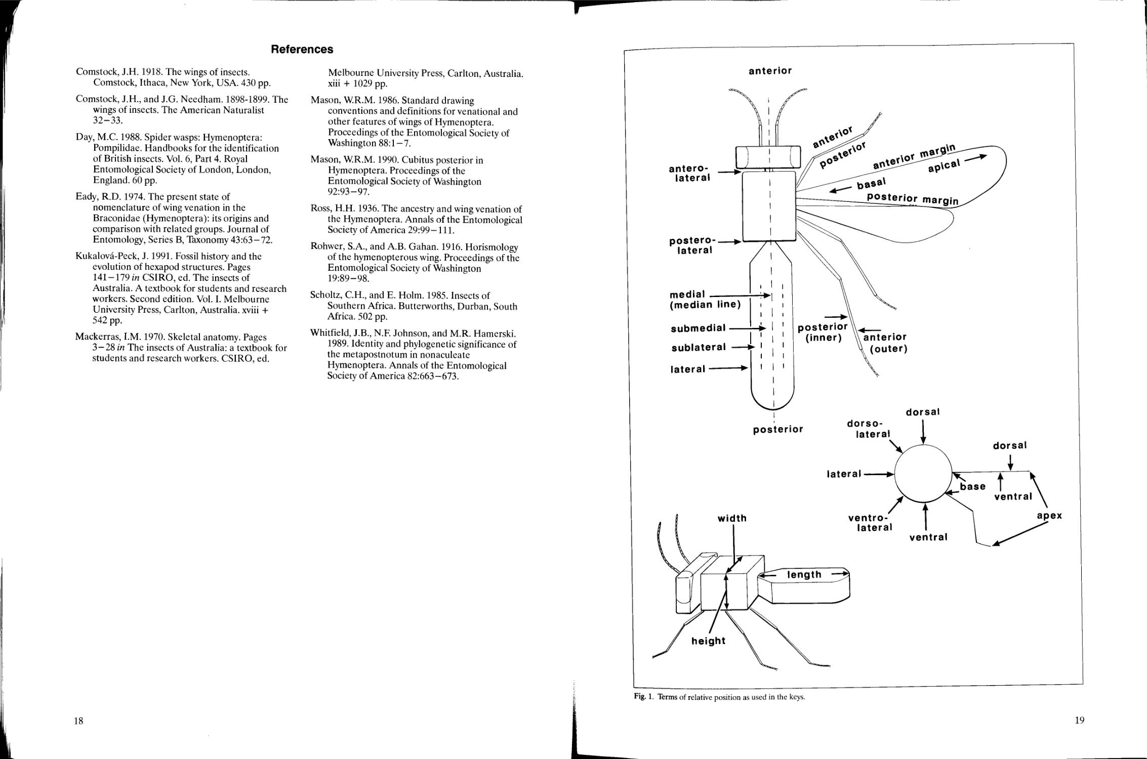
and descriptions. The terms of relative positions
used in this publication are illustrated (Fig. 1) and
defined below. Because leg positions vary,
depending on which leg is discussed and how it is
bent, it is convenient to describe features of each
part of any leg as though it were extended
horizontally and at right angles to the long axis of
the body, regardless of its natural orientation in the
living insect or its position in a mounted specimen.
There are only eight descriptors of position
used in the keys, though these are combined in
various ways when necessary, e.g., anterodorsal,
posterolateral.
anterior Toward or at the head end of the body or
structure (front, frontal).
posterior Toward or at the hind end of the body
or structure (rear, back).
dorsal Toward or at the top or upper surface of
the body or structure (above).
ventral Toward or at the bottom or lower surface
of the body or structure (below).
medial/median Toward or at the centre, or central
area or line, of the body or structure (middle,
mid).
lateral Toward or at the side of the body, or the
margin or edge of a structure.
For the appendages (mouthparts, antennae,
wings, legs, genitalia) additional descriptors are:
apical The end farthest away from the body
(apex); at or toward the tip (distal).
basal The end closest to the body (base); at or
toward the base (proximal).
The terms basal and apical as applied to the
thorax-mesosoma have been used in the literature
with the thorax-abdomen (mesosoma-metasoma)
junction as the central reference point. However, to
avoid confusion in this publication and to eliminate
the need for a reference point, these two terms are
not used for the body; instead, anterior and
posterior are used.
The prefix sub- may be added to describe an
attribute near the extremes, e.g., subapical,
sublaterally.
Length, width, and height (not depth) and their
corresponding adjectives (long, wide, high) are used
when giving measurements or proportions of a
structure. Depth is used only when describing how
deep a pit is or when describing certain types of
sculpture. Further information on orientation is
given in Mackerras (1970) and Scholtz and Holm
(1985).
Morphology
Head, antennae, and mouthparts
(Figs. 6-10, 13, 14)
The head is the anterior division of the insect
body. It is shaped like a rectangular six-sided box
with its longitudinal axis usually oriented vertically
and the mouthparts directed ventrally
(hypognathous). The position of various parts is
described for a head oriented in this manner. The
14
head is divided into six areas, which are sometimes
further subdivided (Figs. 6-9). The relative size of
these areas varies greatly and is often defined and
demarcated somewhat differently among the
various groups of Hymenoptera. Useful reference
points are the toruli anteriorly, compound eyes
laterally, three ocelli dorsally, foramen magnum
posteriorly, and oral cavity ventrally. The rim of the
foramen magnum is articulated with the propleura,
connecting the head to the rest of the body.
The anterior surface of the head from the oral
cavity to the anterior ocellus and between the
compound eyes is the face (Fig. 6). The face is
usually subdivided into at least three areas: clypeus,
face (in the narrow sense) and frons. The clypeus
(not to be confused with the labrum) is the ventral
area, immediately above the margin of the oral
cavity. The dorsolateral corners, or lateral margins,
of the clypeus include the anterior tentorial pits,
which are small, often inconspicuous pits located on
either side. A clearly visible to inconspicuous line
(epistomal groove) between and below the pits
indicates the dorsal and lateral margins of the
clypeus. The face (in the narrow sense) is the
medial area, above and sometimes beside the
clypeus. The dorsal margin of the face is defined by
an imaginary transverse line at the level of the
ventral margins of the toruli (antennal sockets).
The frons is the dorsal area, above the face. The
dorsal margin of the frons is defined by an
imaginary transverse line between the compound
eyes at the level of the anterior margin of the
median ocellus.
The dorsal surface of the head, between the
dorsal margins of the compound eyes and including
the ocelli, is the vertex (Figs. 7, 8). The vertex is
defined anteriorly by the dorsal margin of the frons
and posteriorly by the occipital carina or groove. If
there is no ridge or groove, the vertex is defined
posteriorly either by an imaginary line between the
compound eyes at the level of the posterior margins
of the lateral ocelli, or by the highest point of the
head behind the lateral ocelli if the ocelli are lower
than the area of the head posterior to them.
The posterior surface of the head is divided
into five areas which may not all be present or well
defined in a particular species: occiput, postocciput,
postgena, gena, and hypostoma (Figs. 8, 9). The
occiput is the dorsal part between the occipital
groove or carina, when present, and the
postoccipital groove. The ventral part between
these two grooves is the postgena. The narrow
ring-like postocciput is between the postoccipital
groove and the foramen magnum. The gena is the
ventral or lateral area below and behind the eye
and in front of the occipital groove, when present.
The gena is delimited anteriorly by an imaginary
line between the ventral apex of the compound eye
and the anterior articulation of the mandible. If the
occipital groove or carina is absent, the occiput
merges with the vertex and can be considered as the
entire area (excluding the postocciput) above the
foramen, and the gena as the entire area lateral to
and below the foramen. The hypostoma is a narrow
sclerite bordering the oral cavity posteriorly and
separated from the gena and postgena by the
hypostomal carina.
The antennae (Fig. 14) are a paired structure
composed, from the base, of three segments: scape,
pedicel, and flagellum (Fig. 13). The flagellum is
usually secondarily divided into two or more
flagellomeres. The scape is attached to the front of
the head by a socket (torulus). Between the socket
and base of the scape there is often a short, narrow
radicle that sometimes is distinctly differentiated
from the scape. The radicle is a part of the scape
and is not counted as a separate segment.
The mouthparts (Figs. 6, 9,10) surround the
oral cavity anpd are composed of four externally
visible components. From anterior to posterior
these components include the following: labrum
(usually hidden behind the clypeus), paired
mandibles, paired maxillae, and labium. Each
maxilla may be subdivided into cardo, stipes,
lacinia, and galea. The labium may be subdivided
into submentum, mentum, prementum, glossa, and
paraglossa. Both the maxillae and the labium bear
segmented palpi.
Thorax/mesosoma and legs
(Figs. 2-5, 11, 12)
The thorax is the middle division of the insect
body. It can be imagined as a six-sided rectangular
box with its long axis oriented horizontally. It is
composed of three segments: prothorax,
mesothorax and metathorax. When wings are
present, the first pair (fore wings) is on the
mesothorax, and the second pair (hind wings) is on
the metathorax. In members of the suborder
Symphyta the first segment of the abdomen is
similar to the remaining segments; there is no
distinct constriction between the first and second
segments (Figs. 2, 3). In members of the suborder
Apocrita the first segment of the abdomen is widely
and immovably connected to the metathorax and
usually narrowly and flexibly connected to the rest
of the abdomen (Figs. 4, 5). This first abdominal
segment is the propodeum, and the apparent thorax
(true thorax plus propodeum) is the mesosoma
(middle body).
Each segment of the thorax is divided into a
dorsal area (notum), lateral area (pleuron), and
ventral area (sternum). The notum and pleuron are
usually large and may be further subdivided, but the
sternum is undivided, reduced, and, on the
mesothorax and metathorax, considered to be
invaginated internally and represented only by a
median longitudinal line. From anterior to
posterior the notum may consist of up to three
15
separate areas: scutum, scutellum, and postnotum.
The pronotum is undivided (Figs. 2-5). The
mesonotum is usually divided into mesoscutum and
mesoscutellum (Fig. 4), and, in the suborder
Symphyta, mesopostnotum (Fig. 2). The
metanotum is not clearly divided or is undivided in
Apocrita (Fig. 4), but in Symphyta it is usually
divided into the metascutum and the
metascutellum (Fig. 2). A metapostnotum is
distinguishable in most families of Hymenoptera
though it is not clearly visible without dissection
(Whitfield et al. 1989).
The paired propleuron is undivided and
occupies the lateroventral area behind the head
(Figs. 3, 5). The propleuron is usually loosely and
flexibly connected to the pronotum and to the rest
of the thorax. The fore leg is connected to the
posteroventral apex of the propleuron. The
mesopleuron is the largest of the pleura and is
usually subdivided into an anterior mesepisternum
and posterior mesepimeron (Figs. 3, 5), though the
division may be very inconspicuous in some
Apocrita. The metapleuron is small and may be
divided in the same way as the mesopleuron
(Figs. 3, 5).
Any given sclerite of the thorax/mesosoma may
be subdivided into named areas by grooves or
ridges, which also have specific names applied to
them, but which may vary depending on the taxon.
Terms applied to parts of the thorax/mesosoma are
therefore numerous and often confusing.
A leg consists of six primary segments from
base to apex: coxa, trochanter, femur, tibia, tarsus,
and pretarsus (Fig. 11). The femur sometimes has a
partial secondary division basally, the trochantellus,
making it appear as though the trochanter is
two-segmented. Each tibia usually has one or two
spurs apically. In many Hymenoptera one of the
protibial spurs is modified into a cleaning
apparatus for the antenna.
The tarsus is secondarily subdivided into three
to five tarsomeres, each of which may bear a ventral
pad, the plantar lobe. The pretarsus (Fig. 12)
consists of two claws and associated structures.
Abdomen/metasoma and genitalia
(Figs. 2-5)
The abdomen is the posterior division of the
insect body. It can be imagined as a horizontal
cylinder. Primitively, the abdomen consists of 11
segments, but because of fusion and loss usually no
more than 10 segments are visible. As discussed
above, in Apocrita the first abdominal segment is
fused to the thorax as the propodeum and the
remaining (usually nine) segments are collectively
called the metasoma (Figs. 4, 5). Each segment
The reader may find this section rather difficult, but do not despair;
to understand the system of naming veins to key out a specimen.
consists of a dorsal plate or tergum and a ventral
plate or sternum (Figs. 3, 5). There are no special
pleural sclerites, and a spiracle opens at the side of
one or more terga, which often extend ventrally to
cover the lateral surfaces of the abdomen. If the
terga and sterna are subdivided into smaller
sclerites, they are called tergites and sternites,
respectively.
Female Hymenoptera have an egg-laying
apparatus or ovipositor, which may be concealed or
exposed, depending on the species. The ovipositor
is protected by a pair of ovipositor sheaths (Figs. 2,
3). In aculeate Hymenoptera the ovipositor has
become modified exclusively for stinging and no
longer functions for egg laying (Figs. 4, 5). The
genitalia of male Hymenoptera are concealed and
vary from simple, tubular structures with the parts
greatly reduced, to relatively complex and elaborate
structures.
Wings1
(Figs. 13-19)
The system used here for naming wing veins
and cells is the Comstock and Needham
(1898-1899) system updated by Comstock (1918),
Ross (1936) and others (see Mason 1990 for
additional references). Unfortunately, there are
other systems employed by hymenopterists such as
the Rohwer-Gahan (1916) system widely used for
the Ichneumonoidea. The authors of the chapters
on Apoidea, Chrysidoidea, and Vespoidea prefer a
different system of naming wing cells. One reason
for this, given by Michener (p. 310 in this
publication), is that it is sometimes impossible to
determine the true origins of some wing cells. For
example, the three submarginal cells of most
aculeate Hymenoptera are 1R1, IRs, and 2Rs.
When the number is reduced to two, one cannot
determine whether the missing vein is the second
abscissa of Rs or lr-m. Therefore, when naming
these cells, the use of the generic term submarginal
is more practical than the more specific terms 1R1,
IRs and 2Rs. For comparison, the equivalent (when
the complete complement of three cells is present)
names of veins and cells used in the Comstock-
Needham system are tabulated in Chapter 7
(p. 130). For a comparison of the updated
Comstock-Needham system and other systems see
Day (1988).
In many small parasitic Hymenoptera (e.g.,
Chalcidoidea, Proctotrupoidea, Ceraphronoidea,
Platygastroidea, and some Chrysidoidea) the wing
venation is very reduced, and a simplified
nomenclature is used (Fig. 14). From the base of
the wing the veins are submarginal, marginal, and
postmarginal; a stigmal vein branches posteriorly
all veins and cells are well illustrated in the keys, and so it is not necessary
16
from the junction of the marginal and
postmarginalveins. The stigmal vein may be
knobbed apically as the stigma (see glossary), which
sometimes sends off a little stub, the uncus, toward
the postmarginal vein. The names of these veins do
not connote any homology with similarly named
veins in other Hymenoptera but are simply a
convenient system of naming veins that has
developed for these groups. The probable
equivalents in the Comstock- Needham system are
given in parentheses in Fig. 14.
Another factor that complicates hymenopteran
wing nomenclature is that the homologies of the
wing veins with those of other insect orders have
not been finalized, so that some change in names
will undoubtedly occur. For example, Kukalova-
Peck (1991) presented a somewhat different
interpretation from the rather conservative
approach adopted here. Eady (1974) and Day
(1988) compared the various systems that have
been used by hymenopterists.
The Comstock-Needham system recognizes
eight major longitudinal veins, abbreviated by
capital letters. Starting from the anterior margin of
the wing they are as follows: costa (C), subcosta
(Sc), radius (R), media (M), cubitus (Cu) and three
anal (1A, 2A, 3A) veins. In addition, there may be a
short jugal (J) vein in some Symphyta and Apoidea.
Primitively, a longitudinal vein consists of two
main, basal branches, an anterior convex and a
posterior concave branch. These in turn may be
further branched. In Hymenoptera the posterior
main branch of all veins except the radius has been
lost. For the radius the anterior branch is
designated Rl (radius) and the posterior branch is
designated Rs (radial sector). (A better terminology
would be "radius anterior" and "radius posterior"
but the terms radius and radial sector are already
widely used in Hymenoptera so we continue to use
them.) Otherwise, if a vein branches, the most
anterior branch is given the subscript 1 and the
more posterior branches the subscripts 2, 3 ....
A vein may have several segments or abscissae.
They are delimited by the intersection of other
veins, usually crossveins. Thus a vein that is
intersected by two other veins has three abscissae,
numbered consecutively from the base to the apex
of the wing, e.g., when Cu has three abscissae, they
are 1/Cu (the basal portion of Cu), 2/Cu, and 3/Cu
(the apical portion of Cu). In this publication the
numerical value of the abscissae and the vein name
are separated by a slash; this convention facilitates
naming of the anal vein abscissae. For example, the
first abscissa of the first anal vein is 1/1 A. Vein
abscissae vary with the species so that 3/Cu of one
species is not necessarily homologous with 3/Cu of
another species. In contrast, we have attempted to
homologize vein branches and crossveins.
Crossveins, indicated by lowercase letters, take
the name of the veins they connect, with the
anterior vein given first. Thus, a crossvein that
connects R with M is r-m. If there are several r-m
crossveins, they take numerical values as well, e.g.,
lr-m, 2r-m, and so on. If a crossvein joins two
branches of the same vein, the crossvein takes the
name of the major longitudinal vein, e.g., a
crossvein between R and Rs is called r-rs.
Two veins may fuse for part or all of their
length, appearing as one. The resulting vein takes
the name of both component veins joined by a plus
(+) sign. For example, Rs and M are often fused
for portions of their lengths; the fused portion is
called Rs+M. Veins may fuse end to end, so that it
is impossible to know exactly where the first one
ends and the second begins. In this case the
composite veins are joined with an ampersand (&).
For example, in all Ichneumonidae and many
Braconidae the first abscissa of vein Rs+M is lost
and the junction Rs and M cannot be ascertained;
the vein is therefore termed Rs&M.
Wing cells, abbreviated by capital letters, take
the name of the vein lying anterior to them. If
several fused veins form the anterior boundary of a
cell, the cell takes the name of the vein that would
be most posterior. Thus, the cell posterior to
C+Sc+R is the radial cell (R). If more than one
cell is directly behind a vein, the cells are numbered
consecutively from the base, e.g., three medial cells
would be 1M, 2M, and 3M.
New wing veins may arise in certain lineages.
Such veins are either given a new name or are
named (misleadingly) after the vein to which they
are most similar in position. We follow the latter
approach for convenience but distinguish the new
vein from its namesake by an ('), as in the braconid
subfamily key, couplet no. 3.
Wing veins vary from heavily sclerotized and
tubular to barely visible and distinguishable only as
a slight concave line when light is reflected from the
wing surface. These vein types have been named by
Mason (1986) as tubular, nebulous, and spectral
veins and are defined in the glossary.
The surfaces of both the fore and hind wings
are subdivided into areas that are delimited by
folds, flexion lines, or furrows. Though each of
these terms has a specific functional connotation,
all are called folds in this publication, for simplicity.
A notch often indicates the point where a fold
meets the wing margin. The wing margin may bulge
between notches or between the wing base and a
notch; these bulges are called lobes.
17
References
Comstock, J.H. 1918. The wings of insects.
Comstock, Ithaca, New York, USA. 430 pp.
Comstock, J.H., and J.G. Needham. 1898-1899. The
wings of insects. The American Naturalist
32-33.
Day, M.C. 1988. Spider wasps: Hymenoptera:
Pompilidae. Handbooks for the identification
of British insects. Vol. 6, Part 4. Royal
Entomological Society of London, London,
England. 60 pp.
Eady, R.D. 1974. The present state of
nomenclature of wing venation in the
Braconidae (Hymenoptera): its origins and
comparison with related groups. Journal of
Entomology, Series B, Taxonomy 43:63-72.
Kukalova-Peck, J. 1991. Fossil history and the
evolution of hexapod structures. Pages
141-179 in CSIRO, ed. The insects of
Australia. A textbook for students and research
workers. Second edition. Vol. I. Melbourne
University Press, Carlton, Australia, xviii +
542 pp.
Mackerras, I.M. 1970. Skeletal anatomy. Pages
3-28 in The insects of Australia: a textbook for
students and research workers. CSIRO, ed.
Melbourne University Press, Carlton, Australia,
xiii + 1029 pp.
Mason, W.R.M. 1986. Standard drawing
conventions and definitions for venational and
other features of wings of Hymenoptera.
Proceedings of the Entomological Society of
Washington 88:1-7.
Mason, W.R.M. 1990. Cubitus posterior in
Hymenoptera. Proceedings of the
Entomological Society of Washington
92:93-97.
Ross, H.H. 1936. The ancestry and wing venation of
the Hymenoptera. Annals of the Entomological
Society of America 29:99-111.
Rohwer, S.A., and A.B. Gahan. 1916. Horismology
of the hymenopterous wing. Proceedings of the
Entomological Society of Washington
19:89-98.
Scholtz, C.H., and E. Holm. 1985. Insects of
Southern Africa. Butterworths, Durban, South
Africa. 502 pp.
Whitfield, J.B., N.F. Johnson, and M.R. Hamerski.
1989. Identity and phylogenetic significance of
the metapostnotum in nonaculeate
Hymenoptera. Annals of the Entomological
Society of America 82:663-673.
18
anterior
anterolateral
posterolateral
media
(median line)
submedial
sublateral
lateral
• dorso-
poster.or |a|era|
ventrolateral
apex
ventral
Fig. 1. Terms of relative position as used in the keys.
scape
mesoscutum
tegula
notaulus
mesopostnotum
cenchrus 711JfW*.
metascutum
metapostnotum
cercus
ovipositor sheath
Fig. 2. Main morphological divisions and structures of a species of Symphyta, Aglaostigma quattuordecimpunctatum (Norton)
(Tenthredinidae): dorsal view.
20
mouthparts
procoxa
mesople
scape
4
mese
mesepimeron
mesocoxa
metapleuron
"metepisternum
metepimeron
metacoxa
sterna
ovipositor sheath
cercus
Fig. 3. Main morphological divisions and structures of a species of Symphyta, Aglaostigma quattuordecimpunctatum (Norton)
(Tenthredinidae): lateral view.
21
scape
mesoscutum
tegula
axilla -
cercus
ovipositor (sting)
Fig. 4. Main morphological divisions and structures of a species of Apocrita, Nomada sp. (Anthophoridae): dorsal view. Note: tergum 6
retracted into 5 and not visible.
22
mesopleuron ^=r
metapleuron
mesocoxa
metacoxa
sternum
HEAD
pleuron
onotum
scutum
tegula MESOSOMA
tergum METASOMA
Fig. 5. Main morphological divisions and structures of a species of Apocrita, Nomada sp. (Anthophoridae): lateral view. Note: tergum 6
retracted into 5 and not visible.
23
lateral ocellus.
eye
labrum
frons
eye-
lateral ocellus
frons
f?p^
torulus
eye
8
median ocellus
torulus
clypeus
mandible
torulus
median ocellus
occiput
vertex
gena
mandible
Figs. 6-8. Head of Zele sp. (Apocrita: Braconidae): Fig. 6, anterior view; Fig. 7, dorsal view; Fig. 8, lateral view.
24
occipital carina
occiput
labial palpus
maxillary palpus
lateral ocellus
postoccipital carina
postgena
u——foramen magnum
/ ~~—-—gena
hypostoma
mandible
maxilla
cardo■
stipes
maxillary palpus
galea
10
labium
submentum
mentum
prementum
paraglossa
labial palpus
glossa
— flabellum
Fig- 9. HeadofZe/e sp. (Apocrita: Braconidae), posterior view.
F>g-10. Mouthparts of Apis sp. (Apocrita: Apidae), dorsal view.
25
femur
trochanter
coxa
tibial spurs
plantar lobe
tarsal claw
pretarsus
11
claw
arolium
12
tibia
trochantellus
tarsus
apical tarsomere
pretarsus
Fig. 11. Fore leg oiAglaostigma sp. (Symphyta: Tenthredinidae).
Fig. 12. Pretarsus of Pamphiliussp. (Symphyta: Pamphiliidae).
26
flagellum
pedicel
13
radicle
submarginal (R) marginal (C+R) postmarginal (R1)
uncus
'^*/j£ stigma
14
Fig. 13. Antenna of Macroneura sp. (Apocrita: Eupelmidae).
Fig. 14. Fore and hind wings of a pteromalid (Apocrita: Chalcidoidea).
27
Cell Names
c
Sc
Scl
R
Cu
lCu
2Cu
3Cu
1A
2A
1/3A
2/3A
1A'
2A'
3A'
1R1
2R1
3R1
Costal
Subcostal
Subcostal 1
Radial
Cubital
First cubital
Second cubital
Third cubital
First anal
Second anal
First, third anal
Second, third anal
First anal
Second anal
Third anal
First radial 1
First radial 2
First radial 3
Rs
IRS
2Rs
3Rs
1+2RS
Rsl
1RS2
2Rs2
M
IM'
IM
2M
3M
2+3M
Radial sector
First radial sector
Second radial sector
Third radial sector
First + second radial
sector
Radial sector 1
First radial sector 2
Second radial sector 2
Medial
First medial
First medial
Second medial
Third medial
Second plus third medial
(' indicates a dubious homology)
Fig. 16. Wing cells of Macroxyela sp. (Symphyta: Xyelidae).
Fig. 17. Wing veins of Helcon sp. (Apocrita: Braconidae).
L
30
Fig. 18. Wing cells oiHelcon sp. (Apocrita: Braconidae). Cell names given with Fig. 16.
31
Fig. 19. Wing veins of Dolichurus sp. (Apocrita: Ampulicidae).
32
Fig. 20. Wing cells of Dolichurus sp. (Apocrita: Ampulicidae). Cell names given with Fig. 16.
33
Glossary
Terms are defined as they apply to
Hymenoptera and include only those used in the
keys of this publication. Additional terms may be
used in the family sketches or in other publications
but are not included in the glossary; their
definitions can be found in the works cited in the
previous section. Names of wing veins and cells are
also defined and illustrated in the previous section.
Because some of the terms we use have been
applied in a different sense in other works, care
must be taken when comparing terms used in our
keys with those used in other keys. The illustrations
following each definition in the glossary should be
used as guides only because they are sometimes
schematic and show the structure as represented in
only one or two species. Depending on the species
being examined, the size, shape, and relative
position of a structure varies. This must be
considered when comparing the glossary
illustrations with the specimen being keyed.
Abbreviations: adj. adjective; pi. plural; cf.
compare.
abdomen (adj., abdominal) The principal
posterior division of the body, posterior to the
leg-bearing segments and composed of 10 or
fewer apparent segments; in most Symphyta
abdominal segment 1 is easily recognized by its
median split (cf. metasoma, propodeum).
abscissa (pi., abscissae) A segment of a wing vein
that is delimited by the intersection of other
veins.
acute Sharply angled, less than 90 °.
adventitious vein A wing vein that is not
homologized with a standard wing vein.
annulate Ringed; formed in ring-like segments or
color patterns.
antenna (pi., antennae; adj., antennal) A paired,
segmented sensory appendage of the head
between the compound eyes, which consists of
three segments having intrinsic muscles (see
also scape, pedicel, and flagellum).
apex (pi., apices; adj., apical, apico-) Part of a
structure farthest from its point of attachment
to the body.
areola (pi., areolae; adj., areolate) In
Ichneumonidae, the median area of the
propodeum that is enclosed by ridges.
axilla (pi., axillae; adj., axillar) In groups with a
transscutal articulation, posterolateral portion
of the mesoscutum separated from the
mesoscutum lateral to the scutellum; usually
triangular (see also scutum).
areolet In Ichneumonidae, the small cell in the
centre of the fore wing; the first radial sector
cell.
axillula (pi., axillulae; adj., axillular) In
Chalcidoidea, the lateral subdivision of the
scutellum delimited by a longitudinal line.
arolium (pi., arolia) A pad-like median lobe
between the tarsal claws (see also pretarsus).
base (adj., basal, basi-) Part of a structure nearest
to its point of attachment to the body.
basitibial plate In Apoidea, a small, flat, hairless,
raised region of the dorsal surface of the
metatibia at its base.
bulla (pi., bullae) In Ichneumonidae,
unpigmented area of a vein where it is crossed
by a wing fold or line of flexion.
cardo (pi., cardines) The basal part of the maxilla.
carina (pi., carinae; adj., carinate) A ridge or
raised line.
cell The area between the veins of a wing; it is
closed when completely surrounded by veins,
otherwise it is open (see morphology section
for naming of cells).
cenchrus (pi., cenchri) In Symphyta, a paired,
circular, or oval structure on the sublateral
portion of the metascutum.
cercus (pi., cerci; adj., cereal) A paired, posterior,
sensory appendage of the last (morphological
tenth) abdominal tergum, which usually bears
several setae. With a reduction of the apical
segments of the metasoma, the cerci often
appear to be on a more anterior segment.
chela (pi., chelae; adj., chelate) In Dryinidae, the
modified tarsus in which the apical tarsomere
forms a clasping structure with its opposable
tarsal claw; a pincer.
36
claval fold The furrow on the fore wing just
anterior to, and parallel with, vein 1A, and
extending to the claval notch on the wing
margin.
clypeus (adj., clypeal) The medial sclente of the
head immediately above the labrum; often
defined dorsally and laterally by the epistomal
groove.
/^~~\ ^
( °
Y/ §
VJb
—T~ 'fcffl™
~^~~T \
© \j
claval lobe The posterior portion of a wing behind
the claval fold and in front of the jugal fold,
when present (see also jugal lobe).
compressed Flattened from side to side (higher
than wide).
concave Pertaining to a linear structure, margin,
or surface that is curved inward (cf. convex).
convex Pertaining to a linear structure, margin, or
surface that is curved outward (cf. concave).
corbicula (pi., corbiculae) In Apiformes, the
concave, smooth region of the metatibia that is
margined by a fringe of setae arising from the
margins; it forms a pollen basket.
3#p
[^
%MMM^$$Mj$y}}}?3S&*
Sgis^^
^K;
^T
;r_---
X---)
costal notch The excision of the wing margin
between the apex of the costal vein and the
base of the stigma.
club The enlarged apical flagellomere or
flagellomeres of an antenna (see also funicle).
coxa (pi., coxae; adj., coxal) The first segment of a
leg, between the body and the trochanter.
depressed Flattened from top to bottom (wider
than high).
disc (adj., discal) The central surface of any
structure.
emarginate Notched; with an obtuse, rounded, or
quadrate section cut out of a margin.
endodont mandible A mandible with the teeth
facing inward so that when the mandibles are
closed their tips point toward each other's base
(cf. exodont).
epicnemial carina The ridge on the mesopleuron
that more or less parallels the anterior margin
of the mesepisternum and that delineates the
posterior margin of the epicnemium.
epicnemium (adj., epicnemial) The anterior
portion of the mesopleuron delimited
posteriorly by the epicnemial carina (see also
prepectus).
epimeron (pi., epimera; adj., epimeral) The
portion of a pleuron posterior to the pleural
groove (cf. episternum).
epipygium In Chalcidoidea, a small, more or less
sclerotized, fingernail-like flap attached to the
last metasomal tergum between the cerci.
episternal groove See mesepisternal groove.
episternum (pi., episterna; adj., episternal) The
portion of a pleuron anterior to the pleural
groove (cf. epimeron).
38
epistomal groove A groove defining the lateral
and dorsal margin of the clypeus.
face (adj., facial) In Parasitica, the anterior
surface of the head between the eyes from the
ventral margin of the toruli to the oral cavity,
excluding the clypeus; in Symphyta and
Aculeata, anterior surface of the head between
the eyes from the ocelli to the oral cavity,
including the clypeus.
epomia (pi., epomiae) In Ichneumonoidea, an
oblique ridge crossing the transverse furrow on
the side of the pronotum.
facial fovea In Apoidea, a depressed, often finely
and densely pubescent area along the inner
orbit of a compound eye.
exodont mandible A mandible with the teeth
facing outward so that when the mandibles are
closed their tips point anteriorly or away from
each other (cf. endodont).
felt line In Mutillidae and Bradynobaenidae, a
longitudinal line of flattened setae and
secretory pores laterally on metasomal
tergum 2.
39
femur (pi., femora; adj., femoral) The third
segment of a leg, between the trochanter and
tibia.
flabellum (pi., flabella) In Apiformes, a small thin
plate at the apex of the glossa.
flagellomere A segment-like subdivision of the
flagellum; it is numbered consecutively from
the base of the flagellum.
flagellum (pi., flagella; adj., flagellar) The third
primary division or segment of the antenna; it
articulates with the pedicel basally and almost
always is subdivided into flagellomeres.
foramen (pi., foramina) A hole in the body wall
through which vessels or nerves pass. The
foramen magnum is the central hole in the back
of the head; the propodeal foramen is the hole
in the posterior area of the propodeum.
frenum (pi., frena; adj., frenal) In Chalcidoidea,
the transverse line on the scutellum that
delineates a posterior portion of the scutellum,
the frenal area.
frons The area of the head between the ventral
margin of the toruli and the anterior margin of
the median ocellus. (Note: this is not equivalent
to the true frons of larvae.)
40
frontal carina A longitudinal ridge or pair of
ridges on the frons between (and sometimes
partly covering) the toruli.
funicle (adj., funicular) A group of flagellomeres
between the pedicel and the club.
galea (pi., galeae) The outer apical lobe of the
maxilla, articulated basally with the stipes.
gastrocoelus-thyridium (pi., gastrocoeli) In
Ichneumonidae, the usually transverse
impression anterolaterally on metasomal
tergum 2. The gastrocoelus includes the
thyridium (t), which is the surface area with
specialized sculpture, whereas the gastrocoelus
is the impression itself.
gena (pi., genae; adj., genal) The cheek; the
lateral part of the head between the compound
eye and, when present, the occipital carina;
otherwise, the lower (in hypognathous head) or
anterior (in prognathous head) part of the back
of the head between the compound eye and the
occiput.
glossa (pi., glossae) The paired, fused, median
lobe of the labium articulated basally with the
prementum.
glymma (pi., glymmae) In Ichneumonidae, the
paired groove or pit on the side of metasomal
segment 1 between its base and the spiracle; it
is nearly always present when sternum 1 is free
from tergum 1 but absent when tergum 1 and
sternum 1 are fused.
groove A linear impression on a sclerite (see also
suture).
hamulus (pi., hamuli) One of a series of small
bristle-like hooks on the anterior margin of the
hind wing; in some groups there may be a basal
and an apical series of hamuli.
head The principal anterior division of the body;
it bears the mouthparts and antennae.
humeral plate The anterior sclerite of the wing at
the base of vein C.
hypognathous With the head more or less at a
right angle to the plane of the body (vertical),
so the mouthparts are directed ventrally (cf.
prognathous).
42
hypopygium (adj., hypopygial) The last visible
sternum of the abdomen.
jugal lobe The posterior area of the wing behind
vein 1A and set off from more anterior areas by
a slight fold (jugal fold) on the wing and by a
notch on the wing margin (see also claval lobe).
hypostomal bridge The sclerotized posterior part
of the head that sometimes separates the
foramen magnum from the oral cavity (the
bridge may sometimes be formed by the
median fusion of the postgena or gena and is
then called the postgenal or genal bridge).
labium (adj., labial) The posterior, medial
appendage of the mouthparts between and
behind the maxillae, composed of the
submentum, mentum, prementum, glossa,
paraglossa, and labial palpi.
hypostomal carina The ridge on the back of the
head along the oral cavity that normally
delimits the hypostoma from the postgena and
occiput.
labrum (adj., labral) The anterior, medial
appendage of the mouthparts attached to the
underside of, and often concealed by, the
clypeus.
43
lacinia (pi., Iaciniae) The inner apical lobe of the
maxilla, articulated to the stipes.
mandible (adj., mandibular) The paired, heavily
sclerotized biting and chewing lateral
appendage of the mouthparts between the
labrum and maxilla.
lamina (pi., laminae; adj., laminate) A thin plate
or leaf-like process.
laterotergite The lateral part of an
abdominal/metasomal tergum that is marked by
a crease or groove, or is even completely
detached from the main dorsal part of the
tergum (see also tergite, tergum).
maxilla (pi., maxillae; adj., maxillary) The paired
appendage of the mouthparts between the
mandible and labium, consisting of the cardo,
stipes, lacinia, galea, and maxillary palpus.
malar space The shortest distance between the
base of the mandible and the margin of the
compound eye.
mentum A labial sclerite articulating basally with
the submentum and apically with the
prementum; often fused with the latter and
indistinguishable as a separate sclerite.
44
i i
mes-, meso- A Greek prefix meaning middle or
mid; used with Latin, latinized, or Greek words
to indicate the middle (often second) part of a
structure.
mesepimeron The posterior subdivision of the
mesopleuron, usually small relative to the
mesepisternum or almost absent.
mesonotum The dorsal part of the mesothorax.
mesepisternal groove A groove on the
mesopleuron, extending ventrally from a pit
under the base of the fore wing and, when
complete, reaching the anteroventral margin of
the mesothorax.
mesopleuron The lateral and ventral part of the
mesothorax (see also mesosternum).
mesepisternum The anterior subdivision of the
mesopleuron, usually comprising most of the
mesopleuron.
mesoscutal lobe See mesoscutum.
mesoscutellar appendage In Symphyta, the
posterior subdivision of the mesoscutellum,
usually crescent-shaped and overhanging the
postnotum of the mesothorax.
mesoscutellum See scutellum.
45
mesoscutum The
inesonotum excluding the
. „ . ^ «nesonotum excluding i
scutel urn; in gr transsCutal
articulation the £tion of the mesonotum
anterior to the anicuiation; the mesoscutum is
usually divided by the notauli into a medial part
and two lateral i4es (see aiso scutum).
mesothorax The second and largest of the three
primary subdivisions of the thorax, bearing the
middle pair of legs and, when present, the fore
wings.
mesosoma (pi., mesosomata; adj., mesosomal)
Apocrita, the thorax plus the propodeum
(cf. thorax).
In
mesotrochantinal plate The ventral part of the
mesothorax inflected towards the metasternum
beneath the base of the mesocoxae; visible only
by removing the mesocoxae.
mesosternum The sternum of the mesothorax,
usually invaginated and not visible, but
sometimes inappropriately used as a general
term for the ventral surface of the mesothorax,
such as when it is modified into a flattened
plate with posteriorly projecting lobes (see also
mesopleuron).
m
\ '>;;!# ^g
1 ■* \ '
:=jp7
met-, meta- A Greek prefix meaning hind or
posterior; used with Latin, latinized, or Greek
words to indicate the posterior (usually third)
part of a structure.
46
metanotum The dorsal part of the metathorax.
metapleuron The lateral and ventral part of the
metathorax.
metascutellum In Symphyta, the middle region of
the metanotum.
metasoma (pi., metasomata; adj., metasomal) In
Apocrita, the apparent abdomen, consisting of
the abdomen excluding the first segment or
propodeum. In this publication the first
metasomal segment which is often shortened,
and narrowed, and not clearly visible, is
numbered 1 (cf. abdomen).
metasternum The sternum of the metathorax,
usually invaginated and not visible but
sometimes used as a general term for the
ventral surface of the metathorax.
metathorax The third of the three primary
subdivisions of the thorax, bearing the hind pair
of legs and, when present, the hind wings.
47
metepimeron The posterior subdivision of the
metapleuron.
metepisternum The anterior subdivision of the
metapleuron.
mouthparts The appendages of the head used for
feeding, including the labrum, hypopharynx,
mandibles, maxillae, and labium.
nebulous vein A wing vein that is uniformly
pigmented (not darker on its margins), without
a tubular structure; it can be seen with both
reflected and transmitted light (cf. spectral,
tubular veins).
notaulus (pi., notauli) The usually oblique,
longitudinal groove on the mesoscutum, often
dividing the mesoscutum into medial and
lateral parts.
notopleural suture A groove separating the
mesonotum from the mesopleuron.
notum (pi., nota; adj., notal) A thoracic tergum,
usually subdivided into a scutum and a
scutellum.
oblique scutal carina In Spheciformes, the ridge
on the mesoscutum extending obliquely
posteriorly and medially from the lateral
margin near the posterior of the tegula.
ocellus (pi., ocelli; adj., ocellar) A simple eye,
consisting of a single, usually round or oval
facet. Hymenoptera usually have three ocelli:
one median (anterior) and two lateral
(posterior).
occipital carina A ridge on the posterior surface
of the head that separates the occiput from the
vertex and gena; the ventral part of the ridge is
sometimes called the genal carina.
48
occiput (adj., occipital) The posterior part of the
head behind the vertex dorsally and the genae
laterally. If an occipital carina is present, the
occiput is the area between it and the very
narrow postocciput surrounding the foramen
magnum (see also postgena).
oral cavity The opening of the head from which
the mouthparts are suspended.
orbit The narrow border around the eye. The
inner and outer orbits are those parts of the
face plus the frons and the gena, respectively,
immediately next to the eye.
ovipositor In females, a slender, paired and
interlocking, saw-like or tubular structure used
for laying the eggs or, in Aculeata, for stinging
or, in some Ichneumonoidea, for both; it may
be concealed or may extend beyond the apex of
the body and is protected by a pair of
ovipositor sheaths.
ovipositor sheath A paired, sclerotized structure
enclosing the external part of the ovipositor.
palpus (pi., palpi; adj., palpal) Paired sensory
appendages of the maxilla and labium,
consisting of one to six segments and one to
four segments, respectively.
paraglossa (pi., paraglossae) A paired, lateral
lobe of the labium articulated basally with the
prementum lateral to the base of the glossa.
49
pedicel The second primary division or segment of pleural groove A groove on the mesopleuron and
the antenna; it articulates apically with the metapleuron extending between the wing base
flagellum and basally with the scape. and the coxal articulation; it separates the
P 1 episternum from the epimeron.
pedunculate An intermediate, club-like condition '
between sessile and petiolate (see also pleuron (pi., pleura; adj., pleural) The lateral part
petiolate). of a body segment, commonly of a thoracic
petiolate Stalked. segment where the pleuron occupies the lateral
petiole Part of the metasoma, usually metasomal as well as the ventral areas of the mesothorax
plantar lobe A small membranous pad projecting
from the ventral apex of tarsomeres 1 —4.
50
postgena (adj., postgenal) The lower part of the
occiput; when the occipital carina is absent, the
gena and postgena are continuous, and the
entire lower area constitutes the gena (see also
occiput).
postnotum A posterior sclerite of the mesonotum
and metanotum behind the scutellum. The
postnotum of the metathorax is fused dorsally
with abdominal tergum 1 and laterally with the
metepimeron; in Apocrita the postnotum is
rarely visible, and then only as an impressed
line in front of the propodeum.
postocciput (adj., postoccipital) A narrow area
surrounding the foramen magnum; it is
separated from the occiput by the postoccipital
groove.
postpectal carina In Ichneumonoidea, the
posterior transverse carina ventrally on the
mesopleuron, just in front of the mesocoxa.
r r~^\f \
r a I V /^f
L ^—*)s^ 7\/~* •■-.
-^
""y
'i\
prementum A labial sclerite articulating basally
with the mentum and bearing the glossae,
paraglossae, and palpi apically.
prepectus A sclerite of the thorax between the
pronotum and the mesepisternum (see also
epicnemium).
51
prestigma The enlarged and sclerotized apex of
one or more veins on the anterior margin of the
wing basal to the costal notch and almost
continuous with the stigma.
pronotal collar The horizontal surface of the
pronotum posterior to the sloping, and often
narrowing, region immediately behind the
head.
pretarsus The apical segment of a leg, bearing the
claws and associated structures (see also
arolium).
pronotal flange The anterior projecting rim of the
pronotum; it is often hidden by the head.
pro- A Latin prefix meaning before or anterior;
used with Latin, latinized, or Greek words to
indicate the anterior (usually first) part of a
structure.
prognathous With the head more or less in the
same plane as the body (horizontal) so the
mouthparts are directed anteriorly (cf.
hypognathous).
pronotal lobe The rounded posterolateral
extension of the pronotum covering the
mesothoracic spiracle.
52
pronotal plate In Cynipoidea, the dorsal disc of
the pronotum.
pronotum (adj., pronotal) The dorsal sclerite of
the prothorax; in most Hymenoptera,
occupying also the dorsolateral or lateral part
of the prothorax.
propleuron (pi., propleura) The lateral part of the
prothorax. In Hymenoptera, it is displaced
somewhat by the pronotum so as to occupy an
oblique position ventrolateral and anterior to
the pronotum.
propodeal carina In Ichneumonoidea, one or
more named, usually distinct, transverse or
longitudinal ridges on the propodeum.
propodeum (adj., propodeal) In Apocrita, the first
tergum of the abdomen, widely and immovably
fused with the metanotum and with each
metapleuron of the thorax, and usually
narrowly and flexibly joined to the rest of the
abdomen (see also abdomen, metasoma,
thorax).
prosternum A ventral sclerite of the prothorax,
between the propleura.
o o
U
53
prothorax The first of the three primary
subdivisions of the thorax, composed of the
pronotum, the propleuron, and the prosternum.
pubescence The short, fine, often closely set setae
on the body.
punctate Bearing fine, impressed points or pits.
pygidial plate A specialized area of the tergum of
the last externally visible segment of the
metasoma; it is usually flattened and defined
laterally by ridges or grooves.
54
radicle The basal part of the scape often strongly
defined by a constriction; it articulates with the
torulus.
recumbent Referring to a seta lying parallel to the
body surface.
reticulate Covered with a network of lines;
meshed.
scape The first primary division or segment of the
antenna; it articulates apically with the pedicel
and basally with the torulus.
sclerite A hardened plate of the integument; it is
separated from other such plates by sutures or
membrane.
sclerotized Referring to the body integument;
relatively stiff and usually darkly pigmented
areas compared with usually colorless and
flexible membranous areas.
scopa (pi., scopae) In Apiformes, a brush-like
structure of short stiff hairs of equal length
used for collecting pollen.
scrobal groove A horizontal groove on the
mesopleuron that may be continuous with the
episternal groove anteriorly and ends at the
pleural groove posteriorly.
scrobe (adj., scrobal) A groove in the body
integument for the reception or concealment of
an appendage, e.g., a longitudinal depression of
the head above each torulus for reception of
the scape or a groove on the mesopleuron for
reception of the mesofemur; the scrobe of the
mesepisternum is a small pit.
sculpture Markings or a pattern of impressions or
elevations on the surface of a structure.
scutellum (pi., scutella; adj., scutellar) The middle
region of the mesonotum or metanotum,
behind the scutum. In Apocrita, only the
mesoscutellum is evident and is simply called
the scutellum, whereas in Symphyta both the
mesoscutellum and metascutellum are present.
scutum (pi., scuta; adj., scutal) The anterior
sclerite of a notum, in front of the scutellum. In
Apocrita, the mesoscutum is functionally only
the area in front of the transscutal articulation;
the axilla, although morphologically part of the
mesoscutum, is treated as separate (see also
axilla).
segment A ring or subdivision of the body or of an
appendage between areas of flexibility, and
bearing intrinsic muscles.
sensillum (pi., sensilla) A simple sense organ,
such as a seta, or one of the structural units of a
compound sense organ.
sessile One structure attached to another, without
a distinct constriction (cf. pedunculate,
petiolate).
seta (pi., setae; adj., setal) A slender, hair-like,
usually sensory extension of the cuticle,
connected to the body wall by a socket.
spectral vein A wing vein that is indicated only by
a ridge or furrow on the wing surface; it has no
trace of pigment and can only be seen with
reflected light (cf. tubular, nebulous veins).
55
spiracle (adj., spiracular) A small, round or oval
lateral opening on a body segment through
which air enters the tracheae.
sternaulus (pi., sternauli) The horizontal
lateroventral carina or groove near the lower
margin of the mesopleuron, extending from the
lower end of the epicnemial carina toward the
mesocoxa.
sternite The sclerotized subdivision of a sternum
bounded by grooves or by membranous lines or
areas (see also sternum).
sternum (pi., sterna; adj., sternal) The ventral
division of a body segment, which may be
subdivided into sternites; usually only refers to
the segments of the abdomen/metasoma and to
the prothorax. The sterna of the mesothorax
and metathorax are considered to be
invaginated within the thorax.
stigma A thickly sclerotized and usually darkly
pigmented area on the fore wing margin at the
apex of the costal vein. In Chalcidoidea, a
knob-like enlargement of the apex of the
stigmal vein.
stipes (pi., stipites; adj., stipital) A major sclerite
of the maxilla, articulating basally with the
cardo, apically with the galea and lacinia, and
laterally with the maxillary palpus.
sub- Latin prefix meaning under; also used
commonly to mean somewhat or almost (e.g.,
subequal is synonymous with almost equal).
subantennal groove In Apiformes, one or two fine
grooves between the torulus and the clypeus.
II
subgenal carina A ridge bordering the gena
ventrally; it extends from the hypostomal carina
to the anterior or facial articulation of the
mandible.
submentum The basal sclerite of the labium
articulating apically with the mentum; in
Apoidea, an often V-shaped sclerite (often
called the lorum) with an arm articulating with
each cardo and medially articulating with the
mentum.
submetapleural carina A ridge on the ventral
margin of the lower part of the metapleuron,
between the bases of the mesocoxae and
metacoxae.
subocular groove A groove extending from the
ventral margin of the eye to the anterior or
facial articulation of the mandible.
suture A line or membranous area on the cuticle
indicating the junction of two formerly separate
plates or sclerites (see also groove).
tarsomere A subdivision of the tarsus; each tarsus
has 3-5 tarsomeres.
tarsus (pi., tarsi; adj., tarsal) The fifth segment of
a leg, attached basally to the tibia and
subdivided into tarsomeres.
tegula (pi., tegulae) A small, scale-like sclerite
covering the base of the fore wing, basal to the
humeral plate.
tibia (pi., tibiae; adj., tibial) The fourth segment
of a leg, between the femur and the tarsus.
tergite A sclerotized subdivision of a tergum
bounded by grooves, or membranous lines or
areas (see also laterotergite, tergum).
tergum (pi., terga; adj., tergal) A dorsal sclerite of
a body segment, which may be subdivided into
tergites; specifically used for the
abdomen/metasoma (see also laterotergite).
tibial spur A spine-like, multicellular extension of
the cuticle connected to an appendage by a
socket; usually found apically on the tibiae.
torulus (pi., toruli) A paired socket on the front
of the head upon which the scape is articulated.
thorax (pi., thoraces; adj., thoracic) The principal
middle division of the body to which the legs
are attached, between the head and abdomen
(cf. mesosoma).
transscutal articulation A line of weakness across
the mesonotum between the fore wing bases,
which secondarily separates the posterolateral
angles of the mesoscutum beside the scutellum
(see also axilla, scutum).
trochantellus The basal end of the femur; it looks
like a second segment of the trochanter.
thyridium See gastrocoelus.
58
trochanter The second segment of a leg, between
the coxa and femur.
vertex The top of the head between the eyes, from
the anterior margin of the median ocellus to
the occiput.
truncate Cut off squarely at the tip.
tubular vein A pigmented wing vein that is hollow
and therefore appears darker laterally and
lighter medially; it can be seen with both
reflected and transmitted light (cf. nebulous,
spectral veins).
uncus In Chalcidoidea, a short, narrow stub near
the apex of the stigmal vein directed towards
the postmarginal vein.
volsella (pi., volsellae) A paired, median inner
appendage of the male genitalia.
vein (adj., veinal) Narrow, usually dark
thickenings of a wing arising at the wing base
and branching towards the apex (see also
nebulous, spectral, tubular veins, and
morphology section for naming of veins).
59
Chapter 4 Use of keys
Henri Goulet and William R.M. Mason
General comments
The specimen to be keyed It is important to have a
life-like specimen that is properly pinned or
pointed. The ideal specimen should be clean and
complete with the wings, legs, and antennae
outstretched and spread away from the body and
from each other. If the specimen is soft-bodied it
should not be shriveled or collapsed; if possible it
should be critical-point-dried or freeze-dried. With
experience, specimens that are less than perfectly
preserved or mounted can be identified correctly,
but first-time users of keys will find it easier to work
with intact, well-preserved, and well-mounted
specimens.
Magnification requirements Specimens should be
examined with a stereomicroscope using a
magnification range of 40-60 X. Although most
characters can easily be studied at lower
magnifications, higher magnifications (100-150 X)
are usually needed to study the characters of very
small Hymenoptera, e.g., Chalcidoidea.
Lighting Good light is essential for proper
examination of specimens. Most microscope lamps
provide sufficient light for studying Hymenoptera at
the lower magnification range given above.
However, the quality of lighting is often poor
because glare from smooth surfaces obscures the
clarity of surface structures. If you use a simple and
inexpensive diffuser it greatly increases clarity of
the structures seen. The light beam is diffused by
placing a 2 x 2 cm - 3 x 5 cm piece of thick (0.02
mm) tracing acetate (of the type used by
draftspersons) between the light source and
specimen. Sharply defined glareless views of surface
structures are obtained when the acetate is less
than 20 mm from the specimen, and the greatest
clarity is obtained when the acetate is as close as
1-3 mm from the specimen. When the acetate sheet
is very close to the specimen, be careful to avoid
breaking off appendages.
Key construction Each couplet is arranged in
contrasting pairs of statements. Within the first
statement of each couplet, separate characters or
groups of related characters are labeled by single
letters (a, b, and so on); within the second
statement of the couplet double letters (aa, bb, and
so on) are used to designate the contrasting
characters or groups of related characters. You
should compare, for example, 8a with 8aa, 8b with
8bb, and for clarification you can refer to the
illustrations. We have sometimes added statements
in parentheses for clarification or as additional
attributes to reinforce characterization of the group
(taxon), but they are usually not contrasted with the
alternative half of the couplet. A page number in
parentheses is given when you reach a group name;
it leads either to another key or to a discussion of
the group.
Illustrations Each illustration associated with a
couplet is not necessarily one of the taxon included
by the couplet but is a similar expression of the
character to be observed; other parts of the figure
should be ignored. For instance, when the character
to be observed concerns the stigma you should
ignore other venation in the figure. All illustrations
required for each couplet are printed on the page
facing the couplet or directly above the couplet for
easy comparison. Each illustration or group of
illustrations has the same letter(s) as the
corresponding statement letter within the couplet.
For example, if you are at couplet 16 the illustration
associated with statement bb is coded as bb. If a
range of possibilities exists, then there may be more
than one figure that has the same code; for
example, several figures may be coded b but only
one coded bb. Arrows, sometimes with
abbreviations or key words, are added to an
illustration to draw attention to the location of the
attribute in question. Easily interpreted characters
(such as number of flagellomeres or tarsomeres) or
attributes widely understood as being typical or
normal (a normal tarsus versus a chelate tarsus) are
not illustrated.
We have taken great care to make the keys as
complete as possible. Although we tried to illustrate
each couplet sufficiently, many couplets do not
include the complete range of variation because of
space limitations. Thus, a figure may only illustrate
part of the range of variation for that section of the
couplet. You should be careful not to use the
habitus drawings accompanying the family
discussions to identify specimens until you have
considerable familiarity with a group, because great
diversity in habitus can occur in a single family. A
list of habitus drawings with generic names for each
illustration is given in Appendix 1.
Terms used in the keys The main purpose of a key
is to be functional. Therefore we use common
English adjectives instead of technical terms
wherever feasible without a loss of accuracy. You
should consult Chapter 3, particularly the part
treating relative position, so as to become familiar
with the technical terms used in the keys. Several
keys contain couplets with conjunctions such as and,
but, if, or and so on. They are given in boldface to
60
draw attention to them. Note these terms carefully
to avoid mistakes when keying out a specimen.
We make a fairly rigorous attempt at
uniformity among the keys, but, we hope, without
reducing clarity and accuracy. In the keys and
descriptions only the left half of an insect is
described. Thus, paired (bilaterally symmetrical)
structures (such as appendages) are described or
discussed in the singular unless reference is to all of
a serially homologous set, e.g., "legs black" (all
three pairs are black), but "fore leg black" (only
one pair is black). Using the singular improves
clarity, e.g., "head in anterior view with torulus
touching clypeus." This approach may cause some
confusion initially when you observe a bilaterally
symmetrical or paired structure in dorsal view: two
The key contains 71 couplets (72 keyed units),
but only 20 superfamilies are recognized in the
conservative classification used here. Obviously,
some groups key out more than once. The most
diverse is Chrysidoidea, parts of which fall into 15
couplets, eight for winged and seven for wingless or
short-winged species. Members of this superfamily
are united chiefly by an internal feature of the
female genitalia, quite impractical for use as a key
character; in addition, almost every family of
Chrysidoidea includes both winged and flightless
members, which run to separate parts of the key.
Another diverse superfamily is Vespoidea, which
keys to nine couplets, six of which are for the
numerous flightless species. On the other hand, 10
smaller superfamilies key out at only one couplet
each and five others at only two couplets.
Among groups that key out in several couplets,
it is convenient for the user to know the couplets
most commonly reached. Chrysidoidea (except
Dryinidae and very small species) mostly key to 27,
but very small Bethylidae go to 36 and 46; most
Dryinidae key to 21, whereas the numerous
wingless dryinids run to 59. Most Vespoidea arrive
at 32 if winged, but the majority of wingless females
key to 65. Most Apoidea will run to 29, but very
hairy bees can optionally be keyed to 24. The most
numerous Hymenoptera, worker ants (Formicidae),
run very rapidly to couplets 49 and 50, but typical
winged ants key to 32 with other Vespoidea.
Ichneumonoidea usually run to 19 and most
Chalcidoidea to 43 and 44. Proctotrupoidea mostly
key to 40, with large species going to 22, but
Diapriidae key to 38 and 48. Almost all Evanioidea
key to 26. Most other groups key out at one
couplet.
Do not be daunted by the length of the key: the
median number of steps to arrive at any group is
between 9 and 10. Only three superfamilies have
some individuals that need more than 14 steps, and
parts of the structure are evident yet the structure is
described in the singular. In certain situations,
however, use of the plural is necessary, e.g., "toruli
closer to eyes than to each other."
A word of encouragement for the student At first
the keys may look esoteric and difficult to use.
Indeed, initially they will be, but with careful
reading of each couplet and with practice, the
characters will become much more understandable
and the keys more comprehensible. Familiarize
yourself with basic insect morphology by studying
Chapter 3. Be patient and persevere, and you will
discover that most specimens you have can be
keyed out correctly.
a maximum of 18 steps are needed for a few
Vespoidea.
The key has been made as user-friendly as
possible by several devices in addition to features
outlined in the previous section. First, the
confusion caused by conditional clauses (introduced
by words such as if, both-and, either-or, and but)
has been eliminated as much as possible. This effort
usually required extra couplets or extra
subheadings, but it avoids misinterpretations and
speeds the user's passage though the key. Second,
in cases of continuous variation (e.g., presence or
absence of a stigma, or length of the shortened fore
wing in some Ichneumonidae and Bethylidae) The
user's decision has been made noncritical, either by
constructing the key to allow specimens in an
intermediate range to run correctly on either side
of the couplet (e.g., 1,14,15, 16, 24) or by using a
loop. Third, loops are also used to try to prevent
misinterpretation of difficult characters leading the
user onto false paths. For instance, most chalcidoids
key easily from 33 to 42, but some of the smallest,
such as Mymaridae, have the membrane in front of
R (submarginal vein) in the fore wing so reduced as
to be almost invisible, so that a beginner may easily
interpret them as having a vein on the anterior
margin. But, nevertheless, they will run through 34,
37, and 41 to 42, where they are reunited with other
chalcidoids. The same route may be followed in
keying some minute Bethylidae and
Platygastroidea.
A flowchart (pp. 62 and 63) is added to help
serious users understand the location and function
of the feedback loops. Loop 3-43 accommodates
the rather numerous chalcidoids with broadly
attached metasoma; loop 16-33 redirects
Diapriidae that have, or may appear to have, three
cells in the fore wing, back to the
microhymenoptera part of the key (33-48); it also
sends a few very large Chalcidoidea and Cynipoidea
Preamble to superfamily key
61
Flowchart for superfamily key
(Direction of flow is always from the lower to the higher number. Names are abbreviated; superfamilies are set
in capital letters, to distinguish them from families and subfamilies.)
■ wings short or absent
see next
page
TRIGONALY
62
Flowchart for superfamily key (concluded)
see
previous
page
CHALCID
JL
52
51
wings fully
developed
50
□
49
m wings short
'or absent
.VESP
Formic
53
CHALCID
Agaon
.VESP
Rhopalosomat
VESP
Formic
Bradynobaen
54
66
CHRYSID
Amiseg
/
55
56
68
CHRYSID
Embolem
67
■PROCTOTRUP
Diapri
69
/
VESP
Pompil
70
TENTHREDIN ICHNEUMON
57
\
58
]/.
PLATYGASTR
CERAPHRON
LATYGASTR
71
^V
PROCTOTRUP
CYNIP
59
CHRYSID
Dryin
60
61
63
CHRYSID
Plumari
62
64
CHRYSID
Sclerogibb
CHRYSID
"Bethyl
AP
Heterogyna
*VESP
Mutill
65
.CHRYSID
Bethyl
^VESP
63
to the microhymenoptera section if their strong
nebulous veins are interpreted as tubular; loop
15-18 returns Aculeata and Braconidae without a
stigma to the mainstream of macrohymenoptera
(18-32) and renders uncritical the decision on
whether a stigma is present or not; loop 20-24
redirects a few Aculeata (Rhopalosomatidae and
some small Spheciformes) back to the Aculeata
after separating Ichneumonoidea; and loop 25-28
redirects a few male Mutillidae and Spheciformes
after separating most Evanioidea and Chrysidoidea.
The traditional separation for winged
Vespoidea and Apoidea (separation of tegula from
pronotum) is used but couplets 29-32 and 24 have
been added to take care of exceptions among
Megachilidae, winged Formicidae, and a few small
Pemphredonidae. The tegula character is
unworkable for Chrysidoidea, and they are keyed
elsewhere.
Venational characters are used in this key more
precisely than many entomologists are used to. It is
important to distinguish between tubular and
nebulous veins (see Glossary) when counting closed
cells. A cell that reaches the margin of the wing is
considered open if no tubular vein occurs along the
complete margin of that cell, and a cell that has one
or more of its enclosing veins nebulous is also
considered open for use of this key. Bullae (see
Glossary) are interpreted as not spoiling the closure
of a cell.
Some vaguely quantitative terms can be defined
to some degree. Rarely means less than 1%, often
much less; seldom, up to 10%; occasionally, up to
25%; sometimes, 25-50%; often, 50-75%;
frequently, 75-90%; usually, up to 95% or more;
and almost always, 99% or more. Parenthetical
remarks in key couplets contain information often
useful in identification, such as notes on minimum
magnification needed, geographical restrictions of
some groups, and useful features that may not be
Brothers, D.J. 1975. Phylogeny and classification of
the aculeate Hymenoptera, with special
reference to Mutillidae. University of Kansas
Science Bulletin 50:483-648.
Gibson, G.A.P 1985. Some pro- and mesothoracic
structures important for phylogenetic analyses
of Hymenoptera, with a review of the terms for
structures. Canadian Entomologist
117:1395-1443.
found in all specimens. The reciprocal condition is
not necessarily excluded from specimens in the
other half of the couplet.
Writers of keys to higher groups are standing,
so to speak, on the shoulders of previous
generations who discovered the characters we still
use to define units. All we can do is define the old
characters more accurately, make accommodation
for exceptions, and juggle the order in which they
are used. Really new characters are scarce. They
must be invented when recently discovered groups
are added (couplets 61 and 63 for female
Plumariidae and Heterogynaidae) or as a practical
matter for additions of non-Holarctic groups rarely
considered in most keys (couplets 13, 31, 35, 48, 62,
and 68). A few characters seem to be newly used as
key characters, though several have been
mentioned in phylogenetic studies (Gibson 1985,
Brothers 1975, Mason 1983, Rasnitsyn 1980). They
are found in couplets 4,12c, 22, 41c, 52b, 56c, 62c,
65a, 65c, 65d, 69a, and 70.
Some colleagues and contributors to this
publication offered useful suggestions during the
long process of key construction. Our thanks go
especially to Lubomir Masner, who gave his time
and knowledge generously in suggesting characters
and their arrangements, particularly for
microhymenoptera. Without his help, the treatment
of these minute and little-known parasites would
have been much less efficient. Gary Gibson also
gave advice, particularly concerning that almost
endlessly diverse group, Chalcidoidea: couplets 44,
51, and 52 are largely written by him. Henri Goulet
pointed out several characters useful for separating
groups of Symphyta. Finally, Denis Brothers
provided an extensive critique of the aculeate part
of the key and made many improvements. His
knowledge of rare Old World tropical species
resulted in numerous changes, especially for the
flightless forms in couplets 60-65.
Mason, W.R.M. 1983. Two new species of
Khoikhoiinae (Hym.: Braconidae). Oriental
Insects 18:285-288.
Rasnitsyn, A.P 1980. Origin and evolution of
hymenopterous insects. Trudy
Paleontologicheskogo instituta. Akademiya
naukSSSR 174:1-192. [In Russian.]
References
64
Chapter 5 Key to superfamilies1 of HYMENOPTERA
William R.M. Mason
1 a. Fore wing, when directed backwards, extending far beyond apex of thorax or mesosoma and
usually beyond apex of abdomen or metasoma 2
aa. Fore wing absent or small, at longest not extending much beyond apex of thorax or mesosoma2
49
2(1) a. Body in dorsal view with very little or no constriction near its middle between abdominal
segments 1 and 2 (occasionally some constriction visible in lateral view) 3
aa. Body with strong and usually conspicuous dorsal and lateral constriction between abdominal
segments 1 and 2, thus delimiting the mesosoma and metasoma (APOCRITA) 12
All recognized superfamilies and some families of disputed superfamily status.
Because a few species are variable, showing every stage of wing reduction, this division is arbitrary: doubtful cases key through either side of
the couplet.
65
3(2) a. Fore wing with no closed cells and only 1 or 2 veins.
b. Body very small (less than 3 mm long) (a few CHALCIDOIDEA) 43
aa. Fore wing with numerous closed cells and veins.
bb. Body larger (3-40 mm long) (SYMPHYTA) 4
4(3) a. Mesonotum divided by nearly straight, transverse groove between bases of fore wings, the
groove separating from the mesoscutum a triangular area (axilla) on either side of scutellum
aa. Mesoscutum without such groove; mesoscutum (msc) touching scutellum (sc) over a long
oblique groove on each side 6
66
bb.cc
a. Torulus below level of ventral margin of eye.
b. Head with crown of backward-pointing teeth around median ocellus.
c. Abdominal tergum 1 not divided medially.
d. Abdominal terga evenly rounded at sides ORUSSOIDEA (p. 103)
aa. Torulus above level of ventral margin of eye.
bb. Head without such crown of teeth.
cc. Abdominal tergum 1 divided medially.
dd. Abdominal terga angular at sides XIPHYDRIIDAE (p. Ill)
aa
a. Protibia with 2 apical spurs, the smaller spur at least half as long as larger spur (ovipositor
blade-like and usually short) 7
aa. Protibia with 1 apical spur (female often with long, thin ovipositor) 10
67
aa
7(6) a. Fore wing with longitudinal vein Sc between veins C and R (sometimes veins R and Sc touching)
8
aa. Fore wing without longitudinal vein Sc; at most a short vein (Scl) dividing costal cell (C)
9
cc
8(7) a. Flagellomere 1 much wider than, and 1-4 times as long as, combined length of following small
flagellomeres.
b. Clypeus (cp) about two-thirds as wide as face between eyes; labrum (lb) conspicuous below
clypeus.
c. Abdomen rounded at side (ovipositor usually large and conspicuous)
XYELOIDEA (p. Ill)
aa. Flagellomere 1 about as wide as, and no longer than, combined length of following 3 flagellomeres.
bb. Clypeus (cp) about as wide as face between eyes; labrum hidden behind clypeus.
cc. Abdomen strongly flattened dorsoventrally, all but last 2 or 3 segments sharply folded at side
(PAMPHILIIDAE) MEGALODONTOIDEA (p. 102)
68
a. Metatibia with 1 preapical spur or none.
b. Pronotum (nO[) in dorsal view with posterior margin strongly concave; excavation (measured
medially) much longer than median length of pronotum TENTHREDINOIDEA (p. 105)
aa. Metatibia with 2 or 3 preapical spurs.
bb. Pronotum (nOj) in dorsal view with posterior margin weakly concave; excavation (measured
medially) about as long as median length of pronotum
(MEGALODONTIDAE) MEGALODONTOIDEA (p. 102)
69
10(6)
a.
b.
aa.
bb.
Last tergum of female and last sternum of male each with apical, median, cylindrical projection
with concave tip.
Pronotum in lateral view with a horizontal dorsal surface and with a vertical, concave anterior
surface; pronotum in dorsal view with posterior margin strongly concave, with medial length
about one-third lateral length (SIRICIDAE) SIRICOIDEA (p. 104)
Last tergum and last sternum of both sexes apically thin and without cylindrical projection.
Pronotum in lateral view sloping forward at about 45°; pronotum in dorsal view with posterior
margin weakly concave, with medial length over one-half lateral length H
70
11(10) a. Fore wing with posterior-most vein (2A&3A) sinuate and in part far from posterior margin of wing.
b. Pedicel (pd) as long as flagellomere 1.
c. Metanotum with pair of cenchrus.
(Western North America) ANAXYELIDAE (p. Ill)
aa. Fore wing with posterior-most vein (2A) straight, parallel to, and mostly close to posterior
margin of wing,
bb. Pedicel (pd) much shorter than flagellomere 1.
cc. Metanotum without cenchrus CEPHOIDEA (p. 101)
12(2)
a.
b.
aa.
bb.
cc.
Head globular, with crown of teeth around median ocellus.
Body slender, usually 5-35 mm long; female with ovipositor as long as, or longer than, body
(see habitus, Fig. 189).
Metacoxa widest at base, the 2 basal articulations with metapleuron widely separated from each
other and arranged vertically; long axis of metacoxa in lateral view parallel to that of mesosoma.
(Rarely collected) STEPHANOIDEA (p. 512)
Head usually not globular and rarely with circle of teeth around median ocellus.
Body shape and size various, often stout or small; ovipositor often short or concealed.
Metacoxa usually strongly narrowed at base, the two basal articulations usually very close to one
another (difficult to see) and arranged obliquely; long axis of metacoxa usually angled away from
that of mesosoma 13
71
13(12) a. Malar space with deep depression for reception of scape (upper margin of depression usually
sharply carinate).
b. Mesoscutum with large, triangular axilla (ax)—usually about as large as scutellum (sc)—and
almost always with a median groove; body length over 3 mm.
c. Antenna with 12 flagellomeres.
(Rarely collected, mostly south-temperate regions and southeast Asia)
MEGALYROIDEA (p. 513)
aa. Malar space without such a depression (a similar depression sometimes visible, but it does not lie
between eye and mandible),
bb. Mesoscutum rarely with both large axilla and median groove, but if with both, then body length
usually under 3 mm and flagellomeres not 12 in number,
cc. Antenna rarely with 12 flagellomeres 14
72
a
aa aa
14(13) a. Fore wing with 3 or more cells enclosed by conspicuous tubular veins.3
b. Body usually over 5 mm long, but seldom (about 3% of species) shorter than 2 mm 15
aa. Fore wing with 2 or fewer cells enclosed by tubular veins; venation reduced or absent.
bb. Body seldom (about 2% of species) over 5 mm long 33
..-•■•'" a aa
15(14) a. Fore wing without stigma4 16
aa. Fore wing with stigma 18
16(15) a. Fore wing with at least 5 cells completely enclosed by tubular veins5 17
aa. Fore wing with only 3 or 4 cells enclosed by tubular veins 33
If distinguishing between tubular and nebulous veins is difficult, counting the number of closed cells may be confusing. Some latitude is built
uvto the key to allow difficult specimens to key through either side of this couplet.
The presence of a stigma may be difficult to determine; species transitional for this character should key correctly on either side of this
couplet.
See note under couplet 14.
73
17(16) a. Profemur strongly widened, wider than or as wide as metafemur.
b. Antenna with 18 or more flagellomeres.
c. Torulus under a transverse ledge at extreme front of head
(SCLEROGIBBIDAE) CHRYSIDOIDEA (p. 139)
aa. Profemur smaller than metafemur.
bb. Antenna with 10 or more flagellomeres (usually 10-11 flagellomeres but many more in a few
Ichneumonoidea).
cc. Torulus closer to middle of head and not under a transverse ledge 18
8 aa
18(15,17) a. Fore wing with veins C and R touching or fused, eliminating costal cell (fore wing apparently with
only 3 longitudinal veins basally) 19
aa. Fore wing with veins C and R separate; a long narrow costal cell present (fore wing with
4 separate longitudinal veins basally) 21
74
aa
19(18) a. Abdominal sterna less strongly sclerotized than terga.
b. Anterior sterna (usually metasomal sterna 1-3) each subdivided into several sclerites (usually
collapsed in air-dried specimens, giving abdomen a concave and folded underside; ovipositor
often long and conspicuous) (a few Chrysidinae, which may key here, are bright metallic green or
blue and have only 3 visible terga and elbowed antennae; these go to couplet 27)
most ICHNEUMONOIDEA (p. 359)
aa. Abdominal sterna as strongly sclerotized as terga.
bb. Anterior sterna (usually metasomal sterna 1-3) not subdivided and evenly convex ventrally
(sterna rarely flat and metallic green, blue, or red) 20
20(19) a. Antenna with 10 or 11 flagellomeres 24
aa. Antenna with 12 or more flagellomeres a few ICHNEUMONOIDEA (p. 359)
21(18) a. Antenna with 8 flagellomeres (most females with protarsus specialized into pincer like a crab's
claw) (most DRYINIDAE, some EMBOLEMIDAE) CHRYSIDOIDEA (p. 131)
Antenna with 10 or more flagellomeres 22
aa.
a,b,c
aa,bb,cc
22(21) a. Fore wing with vein Cu continuing nearly straight to wing margin (venation conspicuous in all
species keying here).
b. Fore wing with vein 2cu-a as long as, or longer than, vein lcu-a.
c. Fore wing without veins 2r-m and 2m-cu some PROCTOTRUPOIDEA (p. 538)
aa. Fore wing with vein Cu deflected abruptly (angle of 30-90° or more) posteriorly, in step-like
manner, at the base of lm-cu.
bb. Fore wing with vein 2cu-a usually less than half as long as vein lcu-a.
cc. Fore wing usually with veins 2r-m and 2m-cu (if veins mentioned in statements aa, bb, and cc are
difficult to see, incomplete, or missing, go to couplet 25) 23
75
*N
23(22) a. Antenna with 14 or more flagellomeres.
b. Tarsomeres 1-4 each with apicoventral, fleshy, finger-like projection about half as long as width
of tarsomere TRIGONALYOIDEA (p. 513)
aa. Antenna with 10 or 11 flagellomeres.
bb. Tarsomeres without such projections, or rarely with very small flat lobes 24
cc
24(20,23) a.
b.
aa.
bb.
cc.
Thoracic sutures around tegula so obscured by long dense hair that nearby sutures cannot be
seen.
Body hairs,6 especially on propodeum, not smooth, but with short to long lateral branches
(under magnification of 40 x or more).
Hind leg with tarsomere 1 flattened from side to side, about four times as long as tarsomere 2 and
twice as wide (metasomal tergum 2 without felt line) (some Apiformes) APOIDEA (p. 308)
Thoracic sutures visible around tegula (tg).
Body hairs6 variable in density and structure, usually sparse or short.
Hind leg with tarsomere 1 usually cylindrical, not over three times as long as tarsomere 2 (if, as in
a few male Mutillidae, with long, branched mesosomal hairs, then metasomal tergum 2 with felt
line) 25
6 All bees (Apiformes) have at least a few hairs with short to long (plumose) branches, but they are often difficult to find on species with short
or sparse hairs. Bees with sparse hair, or specimens in which the branched hairs cannot be seen, will key correctly below (couplets 29 and 30).
Occasional male Mutillidae (Vespoidea) have some branched hairs, but the general body hair of these species does not obscure the view of the
tegula and adjacent sutures; these individuals key to couplet 25.
76
a .>--3" aa
25(24) a. Hing wing without cells entirely enclosed by tubular veins .
aa. Hind wing with 1-3 cells entirely enclosed by tubular veins
26
28
26(25) a. Metasoma arising high on propodeum; distance between propodeal foramen and metacoxal
cavity greater than width of either opening (ovipositor often conspicuous and long)
most EVANIOIDEA (p. 510)
aa. Metasoma arising low on propodeum, closer to metacoxa than width of coxa or propodeal
foramen and metacoxal cavity forming a common opening (ovipositor (sting) normally
concealed and short) 27
27(26)
a.
b.
aa.
bb.
Pronotum (noi) (measured medially) more than half as long as mesoscutum (ms).
Metasomal terga 1 and 2 with very weak constriction at their junction dorsally and laterally or
with no constriction at all most CHRYSIDOIDEA (p. 130)
Pronotum (noi) (measured medially) less than half as long as mesoscutum (ms).
Metasomal terga 1 and 2 almost always with strong constriction at their junction dorsally and
laterally 28
77
28(25, 27) a. Tegula (tg) separated from pronotum (noi) so that mesoscutum (ms) touches mesopleuron (pl2)
29
aa. Tegula (tg) touching pronotum (noi) when wings are folded so that mesoscutum (ms) separated
from mesopleuron (pl2) by tegula 30
29(28) a. Pronotum in lateral view with posterior margin bearing a conspicuous, angularly protruding
rounded lobe; dorsal margin of lobe forming concave angle of 30-90° with pronotal margin
most APOIDEA (p. 279)
aa. Pronotum in lateral view with dorsal and posterior margins meeting in a rounded angle but
without a conspicuous protruding rounded lobe on posterior margin; dorsal margin of pronotum
forming a weakly concave to weakly convex curve (0-20°) above posterior angle of pronotum
(some FORMICIDAE) VESPOIDEA (p. 217)
78
aa
bb
30(28) a. Body hairs with short to long branches, especially on propodeum, near bases of wings, around
coxae, or on margins of terga (use at least 40° magnification),
b. Hind leg with tarsomere 1 flattened from side to side, about four times as long as tarsomere 2 and
twice as wide (metasomal tergum 2 without felt line)
(some MEGACHILIDAE) APOIDEA (p. 319)
Body hairs thin and smooth, very rarely with short or long branches.
Hind leg with tarsomere 1 cylindrical, not over three times as long as tarsomere 2 (if, rarely in
some male Mutillidae, long, branched mesosomal hairs occur, then metasomal tergum 2 with felt
line) 31
aa.
bb.
31(30)
a.
b.
aa.
bb.
Fore wing with 4-5 veins approaching or touching apical margin beyond stigma.
Metasomal sterna 1 and 2 with junction smoothly overlapping, neither constricted nor articulated
(South American and southern African deserts)
(PLUMARIIDAE) CHRYSIDOIDEA (p. 133)
Fore wing with 3 (very rarely 4) veins approaching or touching apical margin beyond stigma
(rarely, most venation crowded toward base of wing, but up to 3 transparent, convex, spectral
veins can be seen stretching toward wing apex).
Metasomal sterna 1 and 2 articulating with each other, their junction forming a straight groove
32
79
32(31) a. Pronotum in lateral view with transverse median part meeting mesoscutum at a sharp angle or in
a deep groove; posteromedial part of pronotal margin in dorsal view rather straight and meeting
upper margin of posterior lobe in a sharp, approximately right-angled curve.
b. Tegula (tg) touching protruding lobe (lo) on posterior margin of pronotum
a few APOIDEA (p. 279)
aa. Pronotum in lateral view with transverse median part blending smoothly to level of mesonotum,
not elevated in a transverse roll-like lobe; posterior margin of pronotum often without lobe, the
lobe defined by concave angles above and below; protruding lobe sometimes present but
occurring completely below tegula; posterior margin of pronotum in dorsal view forming a
comparatively uniform curve that continues back to the tegula or to a pointed lobe above it.
bb. Tegula (tg) touching pronotal margin, but point of contact above any protruding lobe (lo) that
may be present on the posterior margin most VESPOIDEA (p. 162)
80
aa aa
33(14,16) a. Fore wing with a tubular vein (C or C+R) on basal part of anterior margin (fore wing sometimes
having additional veins) 34
aa. Fore wing without a tubular vein (C absent) on basal part of anterior margin (a vein may occur on
apical half of anterior margin); sometimes fore wing with no venation at all 42
81
aa
bb
34(33) a. Metasoma with apparent tergum 1 very large (true tergum 1 very small and invisibly fused into
anterior of tergum 2), at least as wide as and as long as, or longer than, following terga combined.
b. Fore wing with only 1 vein on anterior margin CERAPHRONOIDEA (p. 566)
Metasoma with tergum 1 about the same length as, or shorter than, other metasomal terga, or
much narrower than rest of metasoma.
Fore wing usually with 2 veins close to anterior margin (C and R, and usually with costal cell
between them) or, if apparently only 1 vein on anterior margin, then metasoma not as in
statement 34a 35
aa.
bb.
aa
aa
35(34) a. Metasomal segment 1 node-like: wider at middle and narrowed at both ends; often metasomal
segment 2 also node-like (a few FORMICIDAE) VESPOIDEA (p. 217)
Metasomal segment 1 not node-like: either highest and widest at posterior margin, or cylindrical
(of uniform height and width), or short, ring-like, and difficult to see 36
aa.
82
36(35) a. Head prognathous.
b. Pronotum (no!) in dorsal view shaped like truncated pyramid; pronotum, measured along
median line, much longer than mesoscutum and touching tegula.
c. Clypeus almost always with median longitudinal carina
(some BETHYLIDAE) CHRYSIDOIDEA (p. 133)
aa. Head hypognathous.
bb. Pronotum in dorsal view usually more or less U-shaped, with concave margin next to
mesonotum; pronotum, measured along median line, much shorter than mesoscutum
(pronotum sometimes longer and not U-shaped, but then not touching tegula).
cc. Clypeus without median longitudinal carina 37
aa
37(36) a. Fore wing with veins C and R enclosing long narrow costal cell (C); costal cell extending from
near wing base to near stigma or costal notch 38
aa. Fore wing with only one apparent vein along anterior margin; costal cell absent or only a slit
occupying no more than half of distance from wing base to stigma or costal notch
41
83
84
kL
L--
bb
38(37)
a.
b.
aa.
bb.
Torulus facing upwards, on upper surface of ledge on frons (best seen in lateral view).
Fore wing without stigma (a small stigma-like sclerotization, i.e., part of marginal vein,
sometimes visible) (some DIAPRIIDAE) PROCTOTRUPOIDEA (p. 546)
Torulus facing forwards, flush with frons, but rarely at apex of a strongly protruding frons
(see habitus, Fig. 45).
Fore wing almost always with stigma 39
aa
39(38) a. Metasoma arising high on propodeum; distance between propodeal foramen and metacoxal
cavity greater than width of either opening (a few EVANIIDAE) EVANIOIDEA (p. 512)
aa. Metasoma arising low on propodeum; propodeal foramen closer to metacoxa than width of
metacoxa or metasoma and metacoxa with a common opening 40
40(39)
a.
aa.
Antenna with 8 flagellomeres
(some DRYINIDAE and EMBOLEMIDAE) CHRYSIDOIDEA (p. 131)
Antenna with 10 or more flagellomeres some PROCTOTRUPOIDEA (p. 538)
aa
41(37) a. Fore wing with stigma.
b. Scape not more than twice as long as wide.
c. Anterior metasomal sterna softer than terga, with each sternum subdivided into several
sternites; underside of metasoma concave with sterna folded in air-dried specimens
a few ICHNEUMONOIDEA (p. 359)
aa. Fore wing without stigma.
bb. Scape usually over three times as long as wide.
cc. All metasomal sterna as hard as terga and rarely subdivided; underside of metasoma convex and
sterna not folded in air-dried specimens (sometimes terga and sterna equally soft and collapsed
both dorsally and ventrally) 42
85
42(33,41) a. Fore wing spoon-shaped, with long marginal hairs and reticulate membrane; hind wing minute
and forked apically, without membrane (60 X magnification required),
b. Metasomal segments 1 and 2 cylindrical and much narrower than other segments.
(Body less than 0.8 mm long; rarely collected) MYMAROMMATOIDEA (p. 570)
aa. Fore wing usually differently shaped, with or without long marginal hairs, but if with long
marginal hairs then wing membrane not reticulate; hind wing longer and not forked apically,
almost always with membrane.
bb. Metasoma with only one narrow cylindrical segment or none visible 43
86
cc
43(42) a. Head with dark H-shaped mark between eyes, ocelli, and toruli.
b. Toruli usually closer to eyes than to one another (each torulus usually less than its own diameter
from eye).
c. Hind wing stalked basally (membrane of hind wing not extending to wing base) and fore wing
apparently with only 1 vein.
(Body usually under 1.5 mm long, most species much smaller)
(MYMARIDAE) CHALCIDOIDEA (p. 627)
aa. Head without dark H-shaped mark,
bb. Toruli usually closer to each other than to eyes.
cc. Hind wing not stalked basally (membrane of hind wing extending to wing base) and fore wing
often with more than one vein 44
87
44(43) a. Pronotum (no^ in lateral view usually separated from tegula (tg).
b. Mesothoracic spiracle (sp) (not always visible) located between pronotum and lateral margin of
mesoscutum (ms), far from anterior margin of mesopleuron (pl2).
c. Body often with metallic color, or antenna with at least 1 minute, ring-like flagellomere just
after pedicel, or both (body of smaller species often weakly sclerotized, so that air-dried
specimens frequently collapsed in parts of head, mesosoma and metasoma)
most CHALCIDOIDEA (p. 573)
aa. Pronotum (noi) in lateral view usually extending posteriorly to tegula (tg).
bb. Mesothoracic spiracle (sp) located between pronotum and anterior margin of mesopleuron
(pl2), separated from lateral margin of mesoscutum (ms) (or on pronotum in similar location).
cc. Body rarely with metallic color and antenna without minute, ring-like flagellomere just after
pedicel 45
45(44) a. Torulus usually immediately above oral cavity, but if higher, then immediately above apex of
triangular clypeus (cp) 46
aa. Torulus separated by more than its own diameter from dorsal margin of clypeus (cp) 47
88
46(45) a. Pronotum (noj) in dorsal view shaped like truncated pyramid; pronotal length measured along
midline, as long as or longer than mesoscutum (ms).
b. Metasomal segment 1 in dorsal view without angular anterolateral corners and with conical or bell-
shaped outline (some BETHYLTOAE and CHRYSIDIDAE) CHRYSIDOIDEA (p. 538)
Pronotum (nd) in dorsal view U-shaped, medially much shorter than mesoscutum (ms).
Metasomal segment 1 in dorsal view with distinct, angular anterolateral corners, subrectangular
in outline most PLATYGASTROIDEA (p. 558)
aa.
bb.
aa.bb
47(45)
a.
b.
aa.
bb.
Head in lateral view with torulus facing forward at about middle of more or less flat face.
Scape a little shorter than, to a little longer than, pedicel most CYNIPOIDEA (p. 522)
Head in lateral view with torulus facing mostly upward on protruding shelf-like central part of face.
Scape several times as long as pedicel 48
89
48(47) a. Femora and tibiae frequently with leaf-like ventral lamina.
b. Head in dorsal view with large rectangular projection (narrower than width of head and bearing
pallisade of fused hairs) extending from behind ocelli to pronotum.
(Indo-Australian region; rarely collected) (Loboscelidiinae) CHRYSIDOIDEA (p. 138)
aa. Femora and tibiae more or less cylindrical and without ventral lamina.
bb. Head in dorsal view with no such rectangular projection behind ocelli
(some DIAPRIIDAE) PROCTOTRUPOIDEA (p. 546)
aa.bb.cc
aa.bb.cc
49(1) a. Metasomal segment 2 node-like (anterior and posterior diameters about the same, and central
diameter greatest).
b. Metasomal segment 2 not longer than 1, usually distinctly shorter.
c. Metasomal segment 2 with greatest diameter distinctly less than that of segment 3
and also shorter than 3
(many FORMICIDAE and BRADYNOBAENIDAE) VESPOIDEA (p. 162)
aa. Metasomal segment 2 usually tapered with anterior diameter less than posterior diameter and
central diameter intermediate, or with posterior diameter about as large as maximum diameter
of metasoma.
bb. Metasomal segment 2 about as long as 1 or longer; segment 2 occasionally shorter than 1, but only
if 1 is long, tapered and not node-like,
cc. Metasomal segment 2 with greatest diameter about equal to that of segment 3, or often greater
and also longer than 3 50
90
50(49) a. Metasomal segment 1 node-like (see statement 49a), greatly enlarged laterally or dorsally, the
maximum diameter usually near middle of the segment
(many FORMICIDAE) VESPOIDEA (p. 217)
aa. Metasomal segment 1 usually tapered and with greatest diameter at apex; segment 1 sometimes
cylindrical or short, ring-like and hard to see but never node-like 51
a,b,c
51(50) a. Males; found exclusively inside figs (fruit of Ficus).
b. Antenna shorter than head.
c. Metasoma often long, weakly sclerotized, and pale
(AGAONIDAE) CHALCIDOIDEA (p. 610)
aa. Females; or males found elsewhere than inside figs.
bb. Antenna at least as long as head.
cc. Metasoma oval and usually shorter 52
91
52(51) a. Pronotum (noi) in lateral view not extending to tegula (tg).
b. Integument usually thin, especially dorsal part of metasoma; body almost always collapsed in
air-dried specimens CHALCIDOIDEA (p. 573)
aa. Pronotum (no^ in lateral view extending to tegula (tg) (propleuron (pli) sometimes large and
exposed, not to be confused with the pronotum), or thorax greatly modified and tegula absent.
bb. Integument thicker; body rarely collapsed in air-dried specimens (except for the ventral side of
metasoma of Ichneumonoidea, which have a short scape and usually over 13 flagellomeres)
53
53(52) a. Vertex separated from occiput by acute and carinate angle.
b. Eye large, nearly touching posterior carinate angle of head.
c. Fore wing extending to base of metasoma; tegula present.
d. Body over 3 mm long; profemur extremely large; antenna with 10 or 11 flagellomeres
(one genus of RHOPALOSOMATIDAE) VESPOIDEA (p. 205)
aa. Vertex and occiput rounded at their junction and usually without carina,
bb. Eye various, usually small and far from posterior curvature of head.
cc. Wing remnants and tegula usually absent.
dd. If agreeing with statement a or b above, then length 2 mm or less and antenna with 5
flagellomeres, or with no tegula and no wing remnants 54
92
54(53) a. Torulus closer to clypeus than its own diameter, usually touching dorsal margin of clypeus, and
often partly concealed by overhanging ridge above or between the toruli 55
aa. Torulus separated from dorsal margin of clypeus by about twice or more its own diameter, not
concealed in dorsal view by partly overhanging ridge 66
55(54) a. Metasoma with 4-5 visible terga (body stout; head usually elongate below eye and with
shallow depression above torulus) (some CHRYSIDIDAE) CHRYSIDOIDEA (p. 135)
aa. Metasoma with 6-8 visible terga (head without depression above torulus) 56
aa
56(55)
a.
b.
c.
aa.
bb.
cc.
Scape at least five times as long as maximum width, more or less straight, and with radicle more
or less parallel to long axis of scape.
Body under 3 mm long.
Metasoma without visible spiracles 57
Scape usually less than three times as long as maximum width, but if longer, then sinuate with
radicle extending at right angles to long axis of scape.
Body 2-30 mm long.
Metasoma with at least 2 pairs of spiracles visible (on segments 1 and 2) 59
93
57(56) a. Metasoma usually lens-shaped in cross section, wider than high, with sides usually sharply
angulate.
b. Metasomal tergum 2 or 3 largest some PLATYGASTROIDEA (p. 559)
aa. Metasoma not lens-shaped in cross section, about as wide as high, with sides rounded,
bb. Metasoma with apparent tergum 1 largest, at least three times as long as any other tergum
58
a,b,c
aa.bb.cc
58(57) a. Antenna with 5 flagellomeres.
b. Vertex separated from occiput by acute and carinate angle.
c. Eye large, nearly touching posterior carinate angle of head
some PLATYGASTROIDEA (p. 559)
aa. Antenna with more than 5 flagellomeres.
bb. Vertex and occiput rounded at their junction or, at most, vertex subacute posteriorly.
cc. Eye not touching posterior margin of vertex posterolaterally
some CERAPHRONOIDEA (p. 566)
^^YT^ a \3^^aa
59(56) a. Fore leg of female with pincer formed by apical tarsomere and 1 very large tarsal claw (fore leg of
male normal),
b. Antenna with 8 flagellomeres (some DRYINIDAE) CHRYSIDOIDEA (p. 139)
aa. Fore leg in either sex without pincer; apical tarsomere similar to others and both tarsal claws
about same size.
bb. Antenna with 10 or more flagellomeres, rarely with pedicel hidden inside scape and thus antenna
appearing to have 11 articles 60
~Lj—'9
/ \ a,b y^"^ \ aa.bb
60(59) a. Mesosoma with tegula (tg) and minute to moderate-sized wing remnants (wg) (wing remnant
sometimes completely concealed by tegula).
b. Mesosoma (especially mesonotum) normal, generally similar to that of winged Hymenoptera
61
aa. Mesosoma without tegula and wing remnants.
bb. Mesosoma (especially mesonotum) often profoundly modified by fusions, enlargements, and
reductions 63
95
aa,bb
61(60) a. Pronotum (noi) with widely angulate lateral lobe extending posterior to entire tegula (tg).
b. Metasomal tergum 6 with downwardly curved apical hook.
(African and Mediterranean regions; rarely collected)
(female HETEROGYNAIDAE) APOIDEA (p. 290)
aa. Pronotum (noi) touching tegula (tg) but not extending posterior to it.
bb. Metasomal tergum 6 simple or emarginate apically but not hook-like 62
bb,cc
62(61) a. Body sometimes coarsely punctate and with abundant long hair; body sometimes coriaceous
(leather-like).
b. Metasomal tergum 2 often with lateral felt line.
c. Metasomal tergum 1 with lateral spiracle that may be hard to see among coarse punctures
a few VESPOIDEA (p. 162)
aa. Body mostly coriaceous and comparatively smooth; with only sparse hair,
bb. Metasomal tergum 2 without felt line.
cc. Metasomal tergum 1 anteriorly with conspicuous dorsal spiracle
(a few BETHYLIDAE) CHRYSIDOIDEA (p. 133)
96
aa.bb
63(60) a. Propleura (ph) fused dorsally and ventrally into a tubular neck protruding in front of pronotum
(noi).
b. Junction between mesothorax and fused metathorax + propodeum very deeply constricted
ventrally and laterally.
(South American and southern African deserts; extremely rarely collected)
(female PLUMARIIDAE) (CHRYSIDOIDEA) (p. 133)
aa. Propleura (ph) not fused to each other, not protruding in front of pronotum (noi).
bb. Junction between mesothorax and fused metathorax + propodeum weakly or not constricted
ventrally (sometimes constricted laterally) or often lost because of fusions 64
aa,cc
a,b,c
64(63) a. Antenna with 15 or more flagellomeres.
b. Profemur strongly swollen, at least twice as wide as metafemur.
c. Toruli concealed in dorsal view under a transverse ledge that forms the anterior end of a
prognathous head (female SCLEROGIBBIDAE) CHRYSIDOIDEA (p. 139)
aa. Antenna with 10 or 11 flagellomeres (pedicel rarely hidden within scape),
bb. Profemur usually not wider than metafemur (rarely strongly swollen).
cc. Toruli visible in dorsal view or partly concealed by 2 dorsomedial hoods with a depression
between them 65
97
98
a,b,d
ee
aa,bb,cc
65(64) a. Mesopleuron separated from mesonotum by a deep groove easily seen in dorsal view; mesosoma
widest at mesopleura (pl2).
Head longer than wide and strongly prognathous; clypeus with median longitudinal carina.
Propodeal spiracles round or oval and closer to one another than to apex of propodeum.
Metasomal tergum 1 with spiracle dorsal.
Metasomal tergum 2 without felt line or groove
(some BETHYLIDAE) CHRYSIDOIDEA (p. 133)
Mesopleuron not separated from mesonotum or only weakly separated; mesosoma behind
pronotum with sclerites usually fused into box-like structure (including propodeum) that may be
widest at middle.
Head usually as wide as long and rarely prognathous; clypeus sometimes with median carina.
Propodeal spiracles usually slit-like and further from one another than from apex of propodeum.
Metasomal tergum 1 with spiracle lateral.
Metasomal segment 2 sometimes with felt line or longitudinal groove
most VESPOIDEA (p. 162)
b.
c.
d.
aa.
bb.
cc.
dd.
ee.
66(54) a. Scape at least four times as long as wide ..
aa. Scape less than three times as long as wide
67
68
L.
a,b,c X7 aa.bb
67(66) a. Pronotum more or less U-shaped in dorsal view; medial length usually less than one-quarter as
long as mesoscutum.
b. Metasomal tergum 2 several times as long as any other tergum and much wider than cylindrical
segment 1.
c. Antenna usually with 9—13 flagellomeres, sometimes 7 flagellomeres or fewer
(DIAPRIIDAE) PROCTOTRUPOIDEA (p. 546)
aa. Pronotum more or less rectangular; medial length about as long as mesoscutum.
bb. Metasomal terga 1 and 2 about equally long and wide; metasomal segment 1 conical.
cc. Antenna with 8 flagellomeres (EMBOLEMIDAE) CHRYSIDOIDEA (p. 146)
68(66) a. Body without constriction between thorax and abdomen TENTHREDINOIDEA (p. 105)
aa. Body with strong and conspicuous dorsal and lateral constrictions between mesosoma and
metasoma 69
99
69(68)
a.
b.
aa.
bb.
Metasoma with most sterna composed of several sclerites and much more weakly sclerotized
than tergites; sterna folded and collapsed in air-dried specimens.
Antenna usually with more than 13 flagellomeres (sometimes as few as 9)
a few ICHNEUMONOIDEA (p. 359)
Metasoma with sterna each composed of a single sclerite; sclerite as hard as corresponding
tergite and evenly convex; sterna not collapsed in air-dried specimens.
Antenna with 10-13 flagellomeres 70
aa
70(69) a. Metasomal segment 1 long, conical, and with spiracle.
b. Antenna with 10 flagellomeres (a few POMPILIDAE) VESPOIDEA (p. 202)
aa. Metasomal segment 1 short, cylindrical, and without spiracle.
bb. Antenna with 11 or more flagellomeres 71
aa.bb
i" cc
71(70) a. Metasoma spindle-shaped in lateral view.
b. Ovipositor sheath hook-like, directed ventrally.
c. Flagellomeres without longitudinal placoid sensilla
(a few female PROCTOTRUPIDAE) PROCTOTRUPOIDEA (p. 544)
aa. Metasoma disc-shaped in lateral view, more or less compressed,
bb. Ovipositor sheath straight, directed posteriorly or dorsally.
cc. Flagellomeres with numerous and closely spaced longitudinal placoid sensilla (at least
magnification of 40 x required) a few female CYNIPOIDEA (p. 522)
100
Chapter 6 Superfamilies Cephoidea, Megalodontoidea, Orussoidea,
Siricoidea, Tenthredinoidea, and Xyeloidea
(Figs. 21 -34)
Henri Goulet
Superfamily CEPHOIDEA
(Fig. 21)
Included family (1): Cephidae.
Members of this family are easily keyed out,
but to the untrained eye adults are very much like
Diagnosis Cenchri absent; slight dorsoventral
constriction between abdominal segments 1 and 2.
Comments Members of Cephidae are sometimes
called stem sawflies, because their larvae feed
inside grass stems or twigs of woody plants. Those
that bore within stems of cultivated cereals are
major pests, e.g., Cephas ductus Norton in wheat.
They are unusually slender for sawflies, at first
glance resembling ichneumonid wasps. The family
contains about 100 species, mostly from Eurasia.
One species is known from Madagascar. About 13
those of Ichneumonidae. However, they differ from
Ichneumonidae by abdominal segment 1, which is
clearly part of the abdomen rather than the thorax.
species occur in North America, including 10 in
Canada.
References Benson (1946a) characterized the
world genera, Muche (1981) keyed the world
species, and Middlekauff (1969) the North
American genera. Gussakovskij (1935) reviewed the
Palaearctic species, Zhelokhovtsev (1988) reviewed
the species of European USSR, Takeuchi (1938) the
Japanese species, Benson (1951) the British species,
Ries (1937) the North American species, and Smith
(1988) the Neotropical species.
Fig. 22). The species are restricted to temperate
regions of Eurasia and North America.
Superfamily MEGA
(Figs. 22,
Included families (2): Megalodontidae,
Pamphiliidae.
The very large, almost prognathous head,
widest near the clypeus, is distinctive (see habitus
Superfamily MEGALODONTOIDEA
(Figs. 22, 23)
Family CEPHIDAE
(Fig. 21)
101
Key to families of MEGALODONTOIDEA
aa
2A&3A
cc
a. Antenna thread-like.
b. Abdominal terga 2-8 sharply folded above spiracle.
c. Fore wing with vein 2A&3A markedly sinuate
aa. Antenna saw-like or comb-like.
bb. Abdominal terga 2-8 rounded above spiracle.
cc. Fore wing with vein 2A&3A straight
PAMPHILIIDAE (p. 102)
MEGALODONTIDAE (p. 102)
Family MEGALODONTIDAE
(Fig. 22)
Diagnosis Distinguished from Pamphiliidae by
antenna saw-like or comb-like and terga 2-5 not
folded above spiracles.
Comments Larvae feed on herbaceous plants.
Megalodontidae contains about 40 species and is
restricted to temperate regions of Eurasia.
References Gussakovskij (1935) characterized the
genera. Zhelokhovtsev (1988) reviewed the species
of European USSR, and Takeuchi (1938) reviewed
the Japanese species.
Family PAMPHILIIDAE
(Fig. 23)
Diagnosis Distinguished from Megalodontidae by
filiform or thread-like antennae and a longitudinal
fold on abdominal terga 2-5 above spiracles.
Comments Larvae of these sawflies have the
unusual habit of rolling leaves or spinning silk to
form webs in which they feed. Some species are
gregarious and live in large webs as do tent
caterpillars. A few species attack fruit trees and
conifers and are sometimes pests. The family is an
ancient one: fossils belonging to it have been found
in rocks of Jurassic age, about 150 million years old.
The family contains about 200 species in the
temperate and boreal regions of Eurasia and North
America. About 75 species occur in North America,
including 55 in Canada.
102
References Benson (1945a) and Achterberg and
Aartsen (1986) characterized the genera.
Gussakovskij (1935) reviewed the Palaearctic
species; Viitasaari (1982b) the Finnish species;
Benson (1951) the British species; Zhelokhovtsev
(1988) the species of European USSR; Achterberg
and Aartsen (1986) the European species; Takeuchi
Included family (1): Orussidae.
Though rarely seen, members of Orussoidea
play a pivotal role in deciphering major lineages of
Hymenoptera, as discovered in the study of their
Diagnosis Antenna inserted below the ventral
margin of eye, generally under a distinct transverse
ridge.
Comments Members of Orussidae are rare and
morphologically unique. Although true sawflies,
they have a very long thin ovipositor, as do some of
the large ichneumonid wasps, and are parasitoids of
wood-boring Coleoptera and Hymenoptera larvae.
The long ovipositor is seen only when the female is
actually ovipositing: at other times it is kept
doubled up inside the body. Internally, it extends
forward through the middle of the body, lying
between the flight muscles in the centre of the
thorax. Here it makes a loop up over the gut and
returns again to the tip of the abdomen.
Throughout it is enclosed in a pocket of tissue
apparently derived from abdominal sterna 8 and 9.
How the female unfolds and extrudes its ovipositor
(which is at least twice as long as the insect) and
gets it all back into the body and folded around the
gut inside the thorax is unknown. Another unusual
feature is the extremely long and slender eggs. They
are longer than the body itself but are folded up
Included family (1): Siricidae.
These very characteristic and large wood-boring
Hymenoptera are restricted to the northern
(1938) the Japanese species; Shinohara (1985a,
19856, 1986a, 19866,1987) the Japanese and East
Asian species of Onycholyda; and Middlekauff
(1958,1964) the North American genera and
species. Smith (1988) revised the Neotropical
species.
unusual structures (Rasnitsyn 1988). Adults are
cylindrical in cross section and have several spines
around the median ocellus.
inside the ovarioles and only reach full length when
passing down the ovipositor. A third unusual
feature is the sawfly's ability to jump. The
tergotrochanteral muscle is massive and replaces
the dorsoventral flight muscle. Absence of the latter
is unique within Hymenoptera. The family is
ancient and is well represented as fossils. It contains
about 70 usually rare species in all tropical and
temperate regions, with the greatest diversity in
Africa. Nine species occur in North America,
including four in Canada.
References Benson (1955) characterized the world
genera. Zhelokhovtsev (1988) reviewed the species
of European USSR; Takeuchi (1938) the Japanese
species; Benson (1951) the British species;
Middlekauff (1983) the North and Central
American species; Smith (1988) the Neotropical
species; and Benson (1935,19386) the African and
Australian species. Cooper (1953) described
oviposition and genital anatomy of Orussus. Burke
(1917) and Rawlings (1957) recorded hosts. Guiglia
(1965) cataloged the world species.
hemisphere, except for an accidentally introduced
pest species in Australia and New Zealand.
Superfamily ORUSSOIDEA
(Fig. 24)
Family ORUSSIDAE
(Fig. 24)
Superfamily SIRICOIDEA
(Fig. 25)
103
Family SIRICIDAE
(Fig. 25)
Diagnosis Last tergum in female or last sternum
in male with a horn-like projection; pronotum
transversely folded, collar-shaped.
Comments Members of Siricidae are brightly
colored insects commonly called wood wasps or
horntails. Adults are among the largest
Hymenoptera, measuring up to 50 mm long. They
use their drill-like ovipositor to insert their eggs and
spores of a symbiotic fungus into dead or dying
trees. The fungal spores are stored in special organs
on the larvae and adult females. All larvae are
wood borers, either in conifers or hardwood trees.
They pupate in the bark just below the outer
surface. Siricidae and related, now extinct, families
were well represented in early Tertiary and
Included families (6): Argidae,
Blasticotomidae, Cimbicidae, Diprionidae,
Pergidae, Tenthredinidae.
Members of Tenthredinoidea are distinguished
from other superfamilies and families of Symphyta
by the markedly narrow pronotum medially, by the
presence of two spurs at the apex of the protibia,
and by the lack of a transscutal groove on the
Mesozoic times. The family contains 93 species.
Nineteen species occur in North America, including
14 in Canada.
References Benson (1943) reviewed the world
fauna with keys to genera and to species of some
genera. Ross (1937) and Smith (1975) characterized
the North American genera and Benson (1951) the
European genera. Gussakovskij (1935) reviewed the
Palaearctic species, Zhelokhovtsev (1988) the
species of European USSR, Viitasaari (1984) the
Finnish species, Middlekauff (1960) the western
North American species, and Smith (1988) the
Neotropical species. Smith (1978) cataloged the
world species. Stillwell (1965,1966) described the
symbiotic association between Siricidae and Fungi.
mesonotum. Larvae are all phytophagous and feed
mainly on leaf tissue of ferns, horsetails,
gymnosperms and angiosperms, and rarely in the
pith of twigs or on catkins.
Most species of Symphyta belong to this
superfamily. Over 7000 species occur around the
world except for most oceanic islands.
Tenthredinidae is the largest family.
Superfamily TENTHREDINOIDEA
(Figs. 26-31)
104
Key to families of TENTHREDINOIDEA
1 a. Tergum 1 (ti) extending to metacoxa (cx3) and fused with metapleuron (pl3) (in pale
specimens, area of fusion darker and not to be confused as a division between sclerites).
b. Mesoscutellar appendage fused to mesoscutellum (msc).
c. Protibia with dorsal spur usually simple 2
aa. Tergum 1 (ti) not extending to metacoxa (cx3) and not fused with metapleuron (pl3).
bb. Mesoscutellar appendage (msca) clearly outlined at least laterally.
cc. Protibia with dorsal spur usually with lateral lobe 4
105
2(1) a. Abdominal terga 2-5 each usually folded above spiracle; flagellum club-like.
b. Fore wing with vein 2r.
c. Ovipositor sheath clearly divided into basal (valvula 2) and apical (valvula 3) sclerites
CIMBICIDAE (p. 109)
aa. Abdominal terga 2-5 each rounded above spiracle; flagellum various but usually not club-like
(only in some South American and Australian genera),
bb. Fore wing without vein 2r.
cc. Ovipositor sheath not divided (basal and apical sclerites fused) 3
3(2) a. Antenna with 4-23 flagellomeres.
b. Hind wing without closed anal and medial cells.
c. Fore wing in specimens of most genera without closed anal cell; base of vein 2A&3A absent
PERGIDAE (p. 109)
106
aa. Antenna with 1 flagellomere.
bb. Hind wing usually with closed anal (1A) and medial (2M) cells.
cc. Fore wing with closed anal cell (1A); base of vein 2A&3A almost always present
ARGIDAE (p. 108)
4(1) a. Antenna with 1 or 2 flagellomeres.
b. Abdominal terga 2-7 each clearly folded below spiracle.
c. Fore wing with vein M clearly joining Rs not R; veins M and Rs+M forming an evenly curved line
BLASTICOTOMIDAE (p. 108)
aa. Antenna with 5 or more flagellomeres (in a few species, apical flagellomeres counted even if
fused, as long as delineated by a line),
bb. Abdominal terga each rounded below spiracle,
cc. Fore wing with vein M joining R, or joining Rs+M at or almost at (within width of a vein) R; vein
Rs+M meeting R or M at an angle 5
107
aa
aa
5(4)
aa.
bb.
cc.
Antenna with 15 or more flagellomeres.
Anterior margin of lateral ocellus level with middle of median ocellus.
Mesepimeron with upper portion not excavated along centre DIPRIONIDAE (p. 109)
Antenna with 5-10 flagellomeres. .
Anterior margin of lateral ocellus level with posterior margin of median ocellus or postenor to it.
Mesepimeron with upper portion excavated along centre TENTHREDINIDAE (p. 110)
Family ARGIDAE
(Fig. 26)
Diagnosis Antenna with 1 flagellomere;
metapleuron fused with tergum 1.
Comments Many tropical species are brightly
colored and may have striped or pictured wings.
The larvae feed like caterpillars on foliage of many
plants but seldom become abundant enough to
cause much damage. The family is most diverse in
tropical regions where about 800 species are found.
About 70 species occur in North America, including
about 15 in Canada.
References Benson (1938a) and Malaise (1941)
characterized the genera. Gussakovskij (1935)
reviewed the Palaearctic species; Zhelokhovtsev
(1988) the species of European USSR; Benson
(1951) the British species; Takeuchi (1939) the
Japanese species; Smith (1969d, 1970,19716) the
North American species (except Arge); Pasteels
(1953) the African species; and Smith (1989,1992)
the New World species of Arge and the Neotropical
Argidae.
Family BLASTICOTOMIDAE
(Fig. 27)
Diagnosis Antenna with 1 or 2 flagellomeres;
mesoscutellar appendage narrow; terga 2-5 sharply
folded ventral to spiracles.
Comments Larvae are stem borers of ferns. The
family contains three genera and nine species
restricted to temperate regions of Eurasia.
108
References Gussakovskij (1935) characterized the
genera and species. Zhelokhovtsev (1988) reviewed
the species of European USSR, Togashi (1989) the
Diagnosis Antenna club-like; mesoscutellar
appendage absent; terga usually folded dorsal to
spiracles.
Comments Some members of Cimbicidae are
among the largest Hymenoptera. One of them,
Cimbex americana Leach, is a spectacular insect
(adult about 3 cm long, larva 5 cm long) that
attracts attention when found. The adult buzzes
when picked up; when touched, the spines on its
powerful legs generally cause pain that is intense
enough to cause it to be dropped. The larva is the
size of the little finger; it coils itself into a tight
Diagnosis Antenna with about 20 flagellomeres,
comb-like in males, saw-like in females; tergum 1
separated from metapleuron in both sexes.
Comments Because the larvae attack and
sometimes kill softwood trees they are considered
major pests. Groups of striped or spotted
caterpillar-like larvae are fairly common
(sometimes much too common) on pine (Pinus)
trees in early summer. Even though hundreds of
entomologists and foresters have studied diprionids
for over a century, many species still cannot be
Diagnosis Antenna with 4 or more flagellomeres;
fore wing without vein 2r and usually without anal
cell; hind wing without anal cell; metapleuron fused
with tergum 1.
Comments Larvae usually feed externally on
leaves though some Australian species are leaf
miners on Eucalyptus. Except for a small lineage in
North America, species of Pergidae are restricted to
Australia, and Central and South America, where
they reach their greatest diversity. The family
contains more than 400 species. At least four
Japanese species, and Shinohara (1983) the
Taiwanese species. Smith (1978) cataloged the
world species.
spiral and drops when alarmed. Larvae are solitary
and eat leaves of honeysuckle (Lonicera), willow
(Salix), elm (Ulmus), snowberry (Symphoricarpus),
and sometimes other plants. The family contains
130 species around the world. About 12 species
occur in North America, including eight in Canada.
References Benson (1938a) characterized most
genera and Ross (1937) those of North America.
Gussakovskij (1947) reviewed the Palaearctic
species, Zhelokhovtsev (1988) the species of
European USSR, Takeuchi (1939) the Japanese
species, and Smith (1988) the Neotropical species.
clearly defined. The family contains about 90
species restricted to the Northern Hemisphere.
About 45 species occur in North America, including
20 in Canada.
References Benson (1939a, 19456) characterized
the world genera and Smith (1974a) the Nearctic
genera. Gussakovskij (1947) reviewed the
Palaearctic species, Zhelokhovtsev (1988) the
species of European USSR, Viitasaari (1987) the
Finnish species, Takeuchi (1940) the Japanese
species, and Smith (1988) the Neotropical species.
s; species occur in North America, including three in
! Canada.
;d
References Benson (1938) characterized the world
subfamilies, Riek (1970) the Australian subfamilies,
and Smith (1990) the New World subfamilies.
Benson (1963) characterized the genera of
n Euryinae. Smith (1990) revised the species of most
to Neotropical genera. Benson (19396,1965) revised
the species of Perga. Smith (1978) cataloged the
world species.
Family CIMBICIDAE
(Fig. 28)
Family DIPRIONIDAE
(Fig. 29)
Family PERGIDAE
(Fig. 30)
109
Family TENTHREDINIDAE
(Fig. 31)
Diagnosis Antenna with 5-9 flagellomeres (in
some species the apical flagellomeres may be fused,
but they are counted if a division is still visible);
metapleuron clearly separated from tergum 1;
mesoscutellar appendage long and clearly outlined
(at least laterally).
Comments Adults are most common only for a
few weeks in spring and early summer, but some are
found throughout the summer and fall. Most
species in temperate regions produce only one
generation per year. In exceptional circumstances,
some species produce two or more generations.
Adults of Tenthredininae emerge comparatively late
and are found in early and mid summer. Adults are
usually black, most often with a color pattern of
green, brown, yellow, red, or white. When alive,
many species, especially among Tenthredininae and
Nematinae, are strikingly bright and beautiful, but
sadly for the collector most of the brilliant colors,
especially the greens, fade rapidly to shades of
yellow-brown after death. Most species feed singly
on leaves, but some are borers in twigs and fruit,
some (mainly Nematinae) are gall makers, and a
few are leaf miners. Tenthredinidae is the dominant
sawfly group in boreal and arctic regions as far
north as vegetation occurs. The family consists of
over 6000 species found mostly in temperate
regions of the Northern Hemisphere. About 800
species occur in North America, including about
600 in Canada.
References Because of the large diversity of
genera and species, the references are given by
subfamily. Ross (1937) characterized the North
American subfamilies. His concept is generally
accepted and was partly reorganized by Smith
(1969a, 19696,1969c, 1971a, 1974a, 19746,1979a,
19796) and by Goulet (1986,1992). Ross (1937) and
Goulet (1992) reviewed the North American
genera, Benson (1952) the British genera, and
Takeuchi (1952) the Japanese genera.
• Allantinae Malaise (1963) characterized the
world genera (his subfamily concept included the
genera of Selandriinae and Heterarthrinae), and
Smith (19796) the Nearctic genera. Benson
(1952) reviewed the British species,
Zhelokhovtsev (1988) the species of European
USSR, and Smith (19796), the North American
species.
• Blennocampinae Smith (1969c) characterized
the North American genera. Benson (1952)
reviewed the British species, Zhelokhovtsev
(1988) the species of European USSR, and Smith
(1969c) the North American species.
• Heterarthrinae Smith (1976c) characterized the
North American genera of the subfamily and the
world genera of Fenusini, and Takeuchi (1952)
characterized the Japanese genera. Benson
(1952) reviewed the British species,
Zhelokhovtsev (1988) the species of European
USSR, and Smith (1971a) the North American
species.
• Nematinae Benson (1958) characterized the
British genera, Zhelokhovtsev (1988) the species
of European USSR, Takeuchi (1952) the
Japanese genera, and Ross (1937) the North
American genera. Smith (1974c) treated the
genera of Cladiini and (19766) those of the
Pseudodineurini. Zhelokhovtsev (1988) reviewed
the species of European USSR, and Benson
(1958) the British species.
• Selandriinae (including Dolerinae) Malaise
(1963) characterized the world genera (his
subfamily concept included the genera of
Heterarthrinae and Allantinae), and Smith
(1969a) the Nearctic genera. Zhelokhovtsev
(1988) reviewed the species of European USSR,
Takeuchi (1941) the Japanese species, Smith
(1969a) the North American species, Goulet
(1986) the Nearctic Dolerini, Benson (1952) the
British species, and Malaise (1945) the southeast
Asian species.
• Susaninae The subfamily consists of one genus
restricted to North America. Smith (19696)
revised the species, and Wong and Milliron
(1972) added another species.
• Tenthredininae Malaise (1945) characterized
the world genera, Benson (19466) the European
genera, and Ross (1937) and Goulet (1992) the
North American genera. Benson (1952) reviewed
the British species, Zhelokhovtsev (1988) the
species of European USSR, and Malaise (1945)
the southeast Asian species.
110
Superfamily XYELOIDEA
(Fig. 32)
Included family (1): Xyelidae.
The superfamily consists of two subfamilies,
four tribes, five genera, and 46 species. The extinct
fauna consists of four subfamilies, eight tribes, 40
genera, and 106 species. Most of the classification is
based on extinct taxa.
Family XYELIDAE
(Fig. 32)
Diagnosis Antenna with an enormous
flagellomere 1, followed by several short and
narrow flagellomeres.
Comments Most species are 2-4 mm long and
belong to the genus Xyela. The larvae feed in the
springtime inside the pollen-producing cones of
pines (Pinus). A few other species feed within buds
and within developing shoots of fir (Abies) or on
leaves of deciduous trees. Members of the genus
Xyela are commonly found flying around
cone-bearing branch tips of mature pines (Pinus),
or visiting willow (Salix) blossoms or other
pollen-bearing plants nearby during warm days in
early spring; otherwise they are rarely found.
Xyelidae are the earliest known Hymenoptera in
the fossil record. They have been found in the
Triassic period (about 200 million years old). At
that time they were the dominant group of
Hymenoptera and must have occurred in far greater
variety and abundance than they do now. The
family contains 46 species. Twenty-four species
occur in North America, including 13 in Canada.
References Ross (1937) characterized the genera.
The species were reviewed by the following:
Gussakovskij (1935), Palaearctic; Zhelokhovtsev
(1988), European USSR; Viitasaari (19826),
Finland; Takeuchi (1938), Japan; Benson (1951),
UK; Burdick (1961), North America; and Smith
(1988), Neotropical. Smith (1978) cataloged the
species.
Family ANAXYELIDAE1
(Fig. 33)
Diagnosis Pedicel as long as flagellomere 1;
pronotum with longitudinal medial groove or
articulation.
Comments Adults oviposit in recently burnt cedar
(Juniperus, Libocedrus, Thuja) and juniper (Juniper)
trees in which their wood-boring larvae feed. Only
one extant species is known, Syntexis libocedrii
Rohwer, a living fossil found from California to
south-central British Columbia. It is all that is left
of a large Mesozoic fauna.
References Middlekauff (1974) described the
biology of the only species. Smith (1978) cataloged
the extant and fossil species.
Family XIPHYDRIIDAE'
(Fig. 34)
Diagnosis Propleuron unusually long; head thus
clearly separated from pronotum by a distinct
"neck".
Comments These wood-boring sawflies attack
small trunks and branches of deciduous trees. The
family contains about 80 species around the world.
Ten species occur in North America, including six in
Canada.
References Benson (1954) characterized the world
genera. The species were reviewed by the following:
Gussakovskij (1935), Palaearctic; Zhelokhovtsev
(1988), European USSR; Takeuchi (1938), Japan;
Smith (1976a, 1983), North America; and Smith
(1988), Neotropical. Deyrup (1984) described the
biology oiXiphydria maculata Say, with notes on
other species. Smith (1978) cataloged the world
species.
Family unassigned to superfamily of Symphyta.
Ill
References to Symphyta
Although most references are specifically
mentioned under each family, some treat one or
several aspects of Symphyta as a whole. Benson
(1950) and Viitasaari (1982a) described their
natural history. Yuasa (1922), and Lorenz and
Kraus (1957) characterized the larvae of several
Abe, M., and D.R. Smith. 1991. The genus-group
names of Symphyta (Hymenoptera) and their
type species. Esakia 31:1-115.
Achterberg, C. van, and B. van Aartsen. 1986. The
European Pamphiliidae (Hymenoptera:
Symphyta), with special reference to the
Netherlands. Zoologische Verhandelingen No.
234. 98 pp.
Benson, R.B. 1935. On the genera of Orussidae
with an account of African species
(Hymenoptera Symphyta). Occasional papers
of the Rhodesian Museum 4:1-17.
Benson, R.B. 1938a. On the classification of
sawflies (Hymenoptera Symphyta).
Transactions of the Royal Entomological
Society of London 87(15):353-384.
Benson, R.B. 19386. On the Australian Orussidae
with a key to the genera of the world
(Hymenoptera, Symphyta). Annals and
Magazine of Natural History (11)2:1 -15.
Benson, R.B. 1939a. On the genera of the
Diprionidae (Hymenoptera Symphyta).
Bulletin of Entomological Research
30:339-342.
Benson, R.B. 19396. A revision of the Australian
sawflies of the genus Perga Leach, sens. lat.
(Hymenoptera Symphyta). Australian Zoologist
9:324-357.
Benson, R.B. 1943. Studies in Siricidae, especially
of Europe and southern Asia (Hymenoptera,
Symphyta). Bulletin of Entomological Research
34:27-51.
Benson, R.B. 1945a. Classification of the
Pamphiliidae (Hymenoptera Symphyta).
Proceedings of the Royal Entomological
Society of London, Series B 14:25-33.
Benson, R.B. 19456. Further note on the
classification of the Diprionidae
(Hymenoptera, Symphyta). Bulletin of
Entomological Research 36:163-164.
Benson, R.B. 1946a. Classification of the Cephidae
(Hymenoptera Symphyta). Transactions of the
Royal Entomological Society of London
96(6):89-108.
genera for each family. Wright (1990) keyed the
British genera. Abe and Smith (1991) listed all
generic names with their type species, and
references of major works listed by geographical
region. Goulet (1992) keyed the Canadian genera.
Benson, R.B. 19466. The European genera of the
Tenthredininae (Hymenoptera
Tenthredinidae). Proceedings of the Royal
Entomological Society of London, Series B
15:33-40.
Benson, R.B. 1950. An introduction to the natural
history of British sawflies (Hymenoptera
Symphyta). Transactions of the Society for
British Entomology 10:45-142.
Benson, R.B. 1951. Hymenoptera: 2. Symphyta.
Section (a). Handbooks for the identification of
British insects. Vol. VI, Part 2(a): 1-50. Royal
Entomological Society of London, London,
England.
Benson, R.B. 1952. Hymenoptera: 2. Symphyta.
Section (b). Handbooks for the identification of
British insects. Vol. VI, Part 2(b):51-138.
Royal Entomological Society of London,
London, England.
Benson, R.B. 1954. Classification of the
Xiphydriidae (Hymenoptera). Transactions of
the Royal Entomological Society of London
105:151-162.
Benson, R.B. 1955. Classification of the Orussidae,
with some new genera and species
(Hymenoptera: Symphyta). Proceedings of the
Royal Entomological Society of London, Series
B 24:13-23.
Benson, R.B. 1958. Hymenoptera: 2. Symphyta.
Section (c). Handbooks for the identification of
British insects. Vol. VI, Part 2(c):139-252.
Royal Entomological Society of London,
London, England.
Benson, R.B. 1959. Tribes of the Tenthredininae
and a new European genus (Hymenoptera:
Tenthredinidae). Proceedings of the Royal
Entomological Society of London, Series B
28:121-127.
Benson, R.B. 1963. Some new western Australian
sawflies of the Euryinae and Phylacteophaginae
(Hymenoptera, Pergidae). Journal of the Royal
Society of Western Australia 46(3):81-84.
Benson, R.B. 1965. Some new pergid sawflies from
New Guinea (Hymenoptera Symphyta). Annals
and Magazine of Natural History (13)8:45-49.
112
Burdick, D.J. 1961. A taxonomic and biological
study of the genus Xyela Dalman in North
America. University of California Publications
in Entomology 17:285-356.
Burke, H.E. 1917. Oryssus is parasitic. Proceedings
of the Entomological Society of Washington
19:87-89.
Cooper, K.W. 1953. Egg gigantism, oviposition, and
genital anatomy: their bearing on the biology
and phylogenetic position of Orussus.
Proceedings of the Rochester Academy of
Sciences 10:38-68.
Deyrup, M.A. 1984. A maple wood wasp, Xiphydria
maculata, and its insect enemies
(Hymenoptera: Xiphydriidae). The Great
Lakes Entomologist 17(1): 17-28.
Goulet, H. 1986. The genera and species of the
Nearctic Dolerini (Symphyta: Tenthredinidae:
Selandriinae): classification and phylogeny.
Memoirs of the Entomological Society of
Canada No. 135. 208 pp.
Goulet, H. 1992. The genera and subgenera of the
sawflies of Canada and Alaska: Hymenoptera:
Symphyta. The insects and arachnids of
Canada. Part 20. Agriculture Canada
Publication 1876. 235 pp.
Guiglia, D. 1965. Orussidae. Pages 1-18 in
Ferriere, C, and J. van der Vecht, eds.
Hymenopterorum Catalogus, Pars 1. 18 pp.
Gussakovskij, V.V. 1935. Faune de l'URSS, Insectes
hymenopteres, Vol. II, No. 1. Chalastogastra
(partie 1). 452 pp.
Gussakovskij, V.V. 1947. Faune de l'URSS, Insectes
hymenopteres, Vol. II, No. 2. Chalastogastra
(partie 2). 234 pp.
Lorenz, H., and M. Kraus. 1957. Die
Larvalsystematik der Blattwespen.
(Tenthredinoidea und Megalodontoidea).
Abhandlungen zur Larvalsystematik der
Insecten. Nr. 1. Akademie-Verlag, Berlin,
Germany. 339 pp.
Malaise, R. 1941. Gattungstabelle der Blattwespen
(Hym. Tenth.) der Welt. Entomologisk Tidskrift
62:131-140.
Malaise, R. 1945. Tenthredinoidea of south-eastern
Asia. Opuscula Entomologica, Supplementum
4. 288 pp.
Malaise, R. 1963. Hymenoptera Tenthredinoidea,
subfamily Selandriinae, key to the genera of the
world. Entomologisk Tidskrift 84:159-215.
Middlekauff, WW 1958. The North American
sawflies of the genera^4can//!o/yda, Cephalcia
and Neurotoma (Hymenoptera, Pamphiliidae).
University of California Publications in
Entomology 14(2):51-174.
Middlekauff, WW 1960. The siricid wood wasps of
California (Hymenoptera: Symphyta). Bulletin
of the California Insect Survey 6(4):59-77.
Middlekauff, WW 1964. The North American
sawflies of the genus Pamphilius (Hymenoptera:
Pamphiliidae). University of California
Publications in Entomology 38:1-84.
Middlekauff, WW 1969. The cephid stem borers of
California (Hymenoptera: Cephidae). Bulletin
of the California Insect Survey 11:1-18.
Middlekauff, WW 1974. Larva of wood-boring
sawfly Syntexis libocedrii Rohwer
(Hymenoptera: Syntexidae). Pan-Pacific
Entomologist 50(3):288-290.
Middlekauff, WW 1983. A revision of the sawfly
family Orussidae for North and Central
America (Hymenoptera: Symphyta, Orussidae).
University of California Publication in
Entomology 101:1-46.
Muche, H. 1981. Die Cephidae der Erde (Hym.,
Cephidae). Deutsche Entomologische
Zeitschrift 28(4-5):234-295.
Pasteels, J. 1953. Prodromes d'une faune des
Tenthredinoidea (Hymenoptera) de l'Afrique
noire. I.- Argidae. Memoires de la Societe
entomologique de Belgique 26.128 pp.
Quinlan, J., and I.D. Gauld. 1981. Symphyta:
(except Tenthredinidae): Hymenoptera.
Handbooks for the identification of British
insects. Vol. VI, Part 2(a). New edition, Royal
Entomological Society of London, London,
England. 67 pp.
Rasnitsyn, A.P. 1988. An outline of evolution of the
hymenopterous insects (Order Vespida).
Oriental Insects 22:115-145.
Rawlings, G.B. 1957. Guiglia schauinslandi
(Ashmead), a parasite of Sirex noctilio
(Fabricius) in New Zealand. Entomologist
90:25-36.
Riek, E.F 1970. Hymenoptera (wasps, bees, ants).
Pages 867-959 in CSIRO, sponsor. The insects
of Australia: a textbook for students and
research workers. Melbourne University Press,
Carlton, Victoria, Australia, xiii + 1029 pp.
Ries, D.T 1937. A revision of the Nearctic
Cephidae (Hymenoptera: Tenthredinoidea).
Transactions of the American Entomological
Society 63:259-324.
Ross, H.H. 1937. A generic classification of the
Nearctic sawflies (Hymenoptera, Symphyta).
Illinois Biological Monograph 15(2):1 —173.
113
Shinohara, A. 1983. Discovery of the families
Xyelidae, Pamphiliidae, Blasticotomidae and
Orussidae from Taiwan, with descriptions of
four new species (Hymenoptera: Symphyta).
Proceedings of the Entomological Society of
Washington 85:309-320.
Shinohara, A. 1985a. The sawfly genus Onycholyda
(Hymenoptera, Pamphiliidae) of Japan I.
Kontyu53(2):346-359.
Shinohara, A. 19856. The sawfly genus Onycholyda
(Hymenoptera, Pamphiliidae) of Japan II.
Kontyu53(4):711-720.
Shinohara, A. 1986a. The sawfly genus Onycholyda
(Hymenoptera, Pamphiliidae) of Japan III.
Kontyu54(2):271-281.
Shinohara, A. 1986b. The sawfly genus Onycholyda
(Hymenoptera, Pamphiliidae) of Japan IV.
Kontyu54(4):611-617.
Shinohara, A. 1987. The sawfly genus Onycholyda
(Hymenoptera, Pamphiliidae) of Japan V.
Kontyu55(3):486-501.
Smith, D.R. 1969a. Nearctic sawflies. II.
Selandriinae: adults (Hymenoptera:
Tenthredinidae). Technical Bulletin. United
States Department of Agriculture, Agricultural
Research Service 1398.10 + 48 pp.
Smith, D.R. 19696. The genus Susana Rohwer and
Middleton (Hymenoptera: Tenthredinidae).
Proceedings of the Entomological Society of
Washington 71:13-23.
Smith, D.R. 1969c. Nearctic sawflies. I.
Blennocampinae: adults and larvae
(Hymenoptera: Tenthredinidae). Technical
Bulletin. United States Department of
Agriculture, Agricultural Research Service
1397. 179 pp.
Smith, D.R. 1969d. Key to genera of Nearctic
Argidae (Hymenoptera) with revisions of the
genera Atomacera Say and Sterictiphora
Billberg. Transactions of the American
Entomological Society 95:439-457.
Smith, D.R. 1970. Nearctic species of the genus
Ptenus Kirby (Hymenoptera: Argidae).
Transactions of the American Entomological
Society 96:79-100.
Smith, D.R. 1971a. Nearctic Sawflies. III.
Heterarthrinae: adults and larvae
(Hymenoptera: Tenthredinidae). Technical
Bulletin. United States Department of
Agriculture, Agricultural Research Service
1420.10 + 84 pp.
Smith, D.R. 19716. Nearctic sawflies of the genera
Neoptilia Ashmead, Schizocerella Forsius,
Aprosthema Konow, and Sphacophilus
Provancher (Hymenoptera: Argidae).
Transactions of the American Entomological
Society 97:537-594.
Smith, D.R. 1974a. Conifer sawflies, Diprionidae:
key to North American genera, checklist of
world species, and new species from Mexico
(Hymenoptera). Proceedings of the
Entomological Society of Washington
76:409-418.
Smith, D.R. 19746. Ischyroceraea Kiaer, a sawfly
genus new for North America (Hymenoptera:
Tenthredinidae). Cooperative Economic Insects
Report 24(35):707-710.
Smith, D.R. 1974c. Sawflies of the tribe Cladiini in
North America (Hymenoptera: Tenthredinidae:
Nematinae). Transactions of the American
Entomological Society 100:1-28.
Smith, D.R. 1975. Eriotremex formosanus
(Matsumura), an Asian horntail in North
America (Hymenoptera: Siricidae).
Cooperative Economic Insects Report
25(44):851-854.
Smith, D.R. 1976a. The xiphydriid woodwasps of
North America (Hymenoptera: Xiphydriidae).
Transactions of the American Entomological
Society 102:101-131.
Smith, D.R. 19766. Sawflies of the tribe
Pseudodineurini in North America
(Hymenoptera: Tenthredinidae). Proceedings
of the Entomological Society of Washington
78:67-79.
Smith, D.R. 1976c. World genera of the leaf-mining
sawfly tribe Fenusini (Hymenoptera:
Tenthredinidae). Entomologia Scandinavica
7:253-260.
Smith, D.R. 1978. Suborder Symphyta (Xyelidae,
Pararchexyelidae, Parapamphiliidae,
Xyelydidae, Karatavitidae, Gigasiricidae,
Sepulcidae, Pseudosiricidae, Anaxyelidae,
Siricidae, Xiphydriidae, Paroryssidae,
Xyelotomidae, Blasticotomidae, Pergidae).
Hymenopterorum Catalogus, Pars 14.193 pp.
Smith, D.R. 1979a. Suborder Symphyta. Pages
3-137 in Krombein, K.V., P.D. Hurd, Jr., D.R.
Smith, and B.D. Burks, eds. Catalog of
Hymenoptera in America north of Mexico.
Vol. 1. Smithsonian Institution Press,
Washington, D.C., USA. 1198 pp.
Smith, D.R. 19796. Nearctic sawflies. IV
Allantinae: adults and larvae (Hymenoptera:
Tenthredinidae). Technical Bulletin. United
States Department of Agriculture, Agricultural
Research Service 1595.169 pp.
Smith, D.R. 1983. Xiphydna prolongata (Geoffroy)
(Hymenoptera: Xiphydriidae) adventive in
114
North America. Proceedings of the
Entomological Society of Washington
85(4):860-861.
Smith, D.R. 1988. A synopsis of the sawflies
(Hymenoptera: Symphyta) of America south of
the United States: introduction, Xyelidae,
Pamphiliidae, Cimbicidae, Diprionidae,
Xiphydriidae, Siricidae, Orussidae, Cephidae.
Systematic Entomology 13:205-261.
Smith, D.R. 1989. The sawfly genus Arge
(Hymenoptera: Argidae) in the Western
Hemisphere. Transactions of the American
Entomological Society 115:83-205.
Smith, D.R. 1990. A synopsis of the sawflies
(Hymenoptera, Symphyta) of America south of
the United States: Pergidae. Revista Brasileira
de Entomologia 34:7-200.
Smith, D.R. 1992. A synopsis of the sawflies
(Hymenoptera: Symphyta) of America south of
the United States: Argidae. Memoirs of the
American Entomological Society 39:1-201.
Stilwell, M.A. 1965. Hypopleural organs of the
woodwasp larva Tremex columba (L.) containing
the fungus Daedalea unicolor Bull, ex Fries.
Canadian Entomologist 97:783-784.
Stilwell, M.A. 1966. Woodwasps (Siricidae) in
conifers and the associated fungus, Stereum
chailletii, in eastern Canada. Forest Science
72:121-127.
Takeuchi, K. 1938. A systematic study on the
suborder Symphyta (Hymenoptera) of the
Japanese Empire (I). Tenthredo 2(2):173-229.
Takeuchi, K. 1939. A systematic study on the
suborder Symphyta (Hym.) of the Japanese
Empire (II). Tenthredo 2(4):393-439.
Takeuchi, K. 1940. A systematic study on the
suborder Symphyta (Hym.) of the Japanese
Empire (III). Tenthredo 3(2):187-199.
Takeuchi, K. 1941. A systematic study on the
suborder Symphyta (Hym.) of the Japanese
Empire (IV). Tenthredo 3(3):230-274.
Takeuchi, K. 1952. Generic classification of the
Tenthredinidae (Hymenoptera; Symphyta).
Shinibi, Kyoto, Japan. 89 pp.
Togashi, I. 1989. Japanese sawflies of the family
Blasticotomidae (Hymenoptera: Symphyta).
Proceedings of the Entomological Society of
Washington 91: 406-413.
Viitasaari, M. 1982a. Sahapistiaiset 1. Yleinin osa.
Helsingin yliopisto. Maatalous-ja
metsaelaintieteen laitos. Jalkaisuja 3. 85 pp.
Viitasaari, M. 19826. Sahapistiaiset 2. Xyeloidea ja
Megalodontoidea. Maatalous-ja
metsaelaintieteen laitos. Jalkaisuja 5. 72 pp.
Viitasaari, M. 1984. Sahapistiaiset 3. Siricoides,
Orussoidea ja Cephoidea. Helsingin yliopisto.
Maatalous-ja metsaelaintieteen laitos.
Jalkaisuja 6. 66 pp.
Viitasaari, M. 1987. Sahapistiaiset 4. Havupistiaiset
(Diprionidae). Maatalous-ja metsaelaintieteen
laitos. Jalkaisuja 10. 79 pp.
Wong, H.R., and H.E. Milliron. 1972. A Canadian
species of Susana on western juniper. Canadian
Entomologist 104:1025-1028.
Wright, A. 1990. British sawflies (Hymenoptera:
Symphyta): a key to adults of the genera
occurring in Britain. Field Studies 7:531-593.
Yuasa, H. 1922. A classification of the larvae of the
Tenthredinoidea. Illinois Biological Monograph
7(4). 172 pp.
Zhelokhovtsev, A.H. 1988. Order Hymenoptera.
Suborder Symphyta (Chalastogastra). Pages
7-234 in Medvedev G.S., ed. Keys to the
insects of the European part of the USSR.
Vol. 3. Hymenoptera, Part 6. Academy of
Sciences of the USSR, Zoological Institute.
Nauka, Leningrad, USSR. 250 pp. [In Russian.]
115
Fig. 21. Cephidae
116
Fig. 22. Megalodontidae
117
L
Fig. 23. Pamphiliidae
118
Fig. 24. Orussidae
119
L
Fig. 25. Siricidae
120
% 26. Argidae
121
Fig. 27. Blasticotomidae
122
Fig. 28. Cimbicidae
123
r
Fig. 29. Diprionidae
124
% 30. Pergida
125
L
Fig. 31. Tenthredinidae
126
Fig. 32. Xyelidae
127
T
IL
Fig. 33. Anaxyelidae
128
Fig-34. Xiphydriidae
129
Chapter 7 Superfamily Chrysidoidea
(Figs. 35-45)
Albert T. Finnamore and Denis J. Brothers
Included families (7): Bethylidae, Chrysididae,
Dryinidae, Embolemidae, Plumariidae,
Sclerogibbidae, Scolebythidae.
Diagnosis Antenna with same number of
flagellomeres in both sexes, usually 8 or 11;
pronotum with posterolateral apex usually reaching
tegula but sometimes separated by distinct cuticular
gap, with posterodorsal margin usually fairly
shallowly concave, with posterolateral margin with
lobe covering spiracle scarcely developed, and with
lateroventral extremities broadly separated;
metapostnotum short, transverse and fused with
propodeum, sometimes exposed but not posteriorly
expanded in middle; wing venation reduced, usually
3 or fewer closed cells in fore wing (rarely up to 8)
and 1 or no closed cells in hind wing (rarely up to
3); hind wing without jugal lobe; metasomal sterna
1 and 2 not separated by constriction; female with
articulation within gonocoxite 2 near base;
ovipositor concealed at rest and usually modified as
a sting; plumose setae absent. Sexual dimorphism
slight to extreme: male macropterous but very
rarely brachypterous or apterous; female usually
macropterous but often apterous and rarely
brachypterous.
Comments Chrysidoidea contains three large and
common families (the first three listed above) and
four small rare ones. Most chrysidoids are among
the smallest of Aculeata; many are under 3 mm
long. These parasitoids are often found in unusual
situations for aculeates, e.g., in eggs of
Cheleutoptera (= Phasmatodea), externally on
Cicadellidae (Homoptera) and nymphal
Embioptera, or on Coleoptera or Lepidoptera
larvae beneath bark or in soil. Some are
cleptoparasites in nests of other Aculeata. In some
groups the females are wingless and often flattened,
which facilitates host location in concealed habitats.
Except for Dryinidae and Chrysididae,
chrysidoids form a poorly known pantropical group
with relatively few northern representatives. The
world fauna is estimated to be around 16 000
species, 70% of which are undescribed. About 550
described species occur in North America, including
about 150 in Canada, predominantly in southern
areas. The number of genera and species given
below for each family and subfamily includes only
those that are described, unless otherwise stated.
Carpenter (1986) analyzed the relationships of the
families. Brothers and Carpenter (1993)
re-examined and extended that analysis.
The names of wing cells and veins used in the
chapters on Chrysidoidea, Vespoidea, and Apoidea
are those of Gauld and Bolton (1988). The
following table gives the modified
Comstock-Needham equivalents (for a discussion of
wing venation see Chapter 3).
Gauld and Bolton
(1988)
Marginal cell (M)
1st submarginal cell (ISM)
2nd submarginal cell (2SM)
3rd submarginal cell (3SM)
1st discal cell (ID)
2nd discal cell (2D)
1st subdiscal cell (1SD)
subbasal cell (SB)
Modified
Comstock-Needham
2R1
IRl
IRs
2Rs
IM
2M
2Cu
lCu
130
Key to families of CHRYSIDOIDEA
cc
2(1)
a. Antenna with 13—37 flagellomeres.
b. Female wingless.
c. Female with profemur greatly enlarged and swollen; male with profemur usually enlarged and
swollen.
d. Female protibia wide with ventral lamina overlying outer face of femur
SCLEROGIBBIDAE (p. 139)
aa. Antenna with 8-11 flagellomeres.
bb. Female fully winged, short-winged, or wingless.
cc. Profemur usually not enlarged or swollen.
dd. Female with protibia usually slender; if enlarged then without ventral lamina 2
a. Antenna with 8 flagellomeres1 3
aa. Antenna with 10 or 11 flagellomeres 4
aa.bb
cc
3(2)
a.
b.
c.
aa.
bb.
cc.
Antenna inserted on low conical protuberance above middle of head, the torulus more than
twice its width from dorsal margin of clypeus.
Head in lateral view strongly convex anteriorly.
Female protarsus unmodified, with 2 small equal-sized claws EMBOLEMIDAE (p. 146)
Antenna inserted on flat or concave surface below middle of head, the torulus less than its width
from dorsal margin of clypeus.
Head in lateral view concave to slightly convex anteriorly.
Female protarsus usually pincer-like, with last segment laterally expanded and 1 claw enlarged;
female protarsus rarely unmodified DRYINIDAE (p. 140)
One species of Bethylidae also keys here (Strejcek 1990), but it can be separated from Embolemidae and Dryinidae by having the head much
longer than wide.
131
a
aa
4(2) a. Fully winged form (male): hind wing with at least 2 cells enclosed by tubular veins.
b. Wingless form (female): mesothorax separated from metathorax by strong ventral constriction.
c. Female pronotum without anterior flange, propleura thus exposed in dorsal view.
d. Male flagellum with numerous conspicuous erect setae at least half as long as width of
flagellomere PLUMARIIDAE (p. 133)
aa. Fully winged form (either sex): hind wing with 1 or no cells enclosed by tubular veins.
bb. Wingless or short-winged form (either sex): mesothorax not separated from metathorax by
strong ventral constriction,
cc. Female pronotum usually with anterior flang, propleura thus concealed (not visible) in dorsal
view; if pronotal flange not developed and propleura exposed in dorsal view then fully winged,
dd. Male flagellum with inconspicuous recumbent setae shorter than half the width of flagellomere
bb,cc
5(4) a. Pronotum (noi) with anterior flange not developed, the propleuron (pli) thus exposed in dorsal
view; head distinctly separated from pronotum.
b. Prosternum large and diamond-shaped, exposed in ventral view.
c. Protrochanter originating from lateral surface of procoxa (procoxa produced posterior to origin
of protrochanter) SCOLEBYTHIDAE (p. 133)
aa. Pronotum (noi) with anterior flange well developed, the propleuron thus (pli) concealed in
dorsal view; head scarcely separated from pronotum.
bb. Prosternum small and more or less transverse, often concealed in ventral view,
cc. Protrochanter originating from posterior apex of procoxa 6
132
(j(5) a. Metasoma with 6 or 7 exposed terga.
b. Pronotum usually touching tegula.
c. Head usually prognathous BETHYLIDAE (p. 134)
aa. Metasoma with 5 or fewer exposed terga, rarely with indications of a 6th.
bb. Pronotum usually separated from tegula by a distinct cuticular gap.
cc. Head usually hypognathous CHRYSIDIDAE (p. 137)
Family PLUMARIIDAE
(Fig. 35)
Diagnosis Antenna with 11 (rarely 10)
flagellomeres, each usually with long erect setae;
pronotum without anterior flange, propleuron thus
partly exposed dorsally. Sexual dimorphism
extreme: male macropterous, flagellomeres with
numerous setae, pronotum vertical, hind wing with
at least 2 cells enclosed by tubular veins; female
apterous, head prognathous, flagellomeres with a
few scattered setae, pronotum horizontal,
mesothorax separated from metathorax by deep
ventral (and lateral) constriction.
Comments Plumariidae contains four genera from
arid and semiarid regions: two in South America
(Ecuador to Chile and Argentina) and two in
southern Africa (South Africa to Namibia and
Zimbabwe). Fewer than 20 species have been
described. Adults are black to pale brown. The few
very rare females have been collected under rocks
or in pitfall traps, but the more commonly collected
males come to lights (nocturnal species) or to
Malaise traps (diurnal species). Nothing else is
known about their biology.
References Brothers (1974) keyed three genera;
Day (1984) subsequently recognized that
Heterogyna was a member of Apoidea and placed it
in Heterogynaidae. Evans (1966) described the first
known female. Nagy (1973) keyed several
Neotropical species, and Day (1977) described the
fourth genus and discussed relationships. Janvier
(1933) described the larva of a parasitoid of
Trachypus sp. (Philanthidae), which he had
identified as a Plumarius, but that identification is
almost certainly incorrect and the species is
probably a nocturnal mutillid.
Family SCOLEBYTHIDAE
(Fig. 36)
Diagnosis Head obliquely hypognathous; antenna
with 11 flagellomeres; pronotum substantially
horizontal without anterior flange, propleuron thus
partly exposed dorsally; prosternum large,
diamond-shaped; hind wing without closed cells;
protrochanter originating from lateral surface of
procoxa. Sexual dimorphism slight: both sexes
macropterous.
Comments Scolebythidae contains three
monotypic genera, in Brazil, South Africa and
Australia, and Madagascar. Adults are
predominantly black or dark brown. Little is known
about their biology, but the larvae are probably
gregarious external parasitoids of wood-boring
Cerambycidae (Coleoptera). Pupation occurs in the
host burrow.
References Evans (1963) described the family.
Nagy (1975) and Evans, Kugler and Brown (1980)
keyed the genera. Day (1977), Evans, Kugler and
Brown (1980), and Brothers (1981) dealt with the
biology.
Family BETHYLIDAE
(Figs. 37, 38)
Diagnosis Head usually prognathous; antenna
with 11 (rarely 10 or 8) flagellomeres; pronotum
with anterior flange, propleuron thus concealed in
dorsal view; prosternum small, more or less
transverse, often concealed in ventral view;
metasoma with 6 or 7 exposed terga. Sexual
dimorphism slight to extreme: male macropterous,
rarely brachypterous; female macropterous,
brachypterous, or apterous; brachypterous and
apterous forms without deep ventral constriction
between mesothorax and metathorax.
Comments Bethylidae is probably the largest
family of chrysidoids and includes about 2200
species in four subfamilies: Bethylinae, Epyrinae,
Mesitiinae, and Pristocerinae. The species are
133
particularly abundant in the tropics. Adults are
predominantly black or dark brown, although
several New World species are metallic green. The
female stings and paralyses the host, usually a
Lepidoptera or Coleoptera larva, lays several eggs
on it, and either leaves it where it was found or
sometimes drags it into a crevice. In some species
the female tends the eggs and developing larvae.
Pupation occurs next to the host remains. The three
North American subfamilies (199 species in 20
genera) extend to southern Canada, where 22
species (eight genera) occur.
Key to subfamilies of BETHYLIDAE
(modified from Evans 1978)
a. Fore wing with veins Rs and M obtusely angled or giving rise to a vein or vein stub (Rs+M); 1st
discal cell (ID) sometimes present.
b. Frons with longitudinal median carina or polished streak extending a short distance from
clypeus.
c. Fully winged or short-winged Bethylinae
aa. Fore wing with vein Rs&M simple and evenly arched, not angled or giving rise to any vein or stub,
sometimes without vein Rs&M; 1st discal cell (ID) absent,
bb. Frons usually without median longitudinal carina or polished streak extending from clypeus.
cc. Fully winged, short-winged, or wingless 2
134
2(1) a. Propodeum with dorsal posterolateral angle produced as a single large tooth Mesitiinae
aa. Propodeum with dorsal posterolateral angle smoothly rounded, carinate, or with a pair of small
teeth formed by expanded carinae 3
3(2) a. Female wingless, without tegula or ocelli; male fully winged.
b. Female with eye small or absent, the eye height up to one-quarter of head width.
c. Female with propodeum often constricted anteriorly.
d. Male with scutellum not in contact with propodeum; metanotum well developed with small
emargination or fovea opposite posterior apex of scutellum Pristocerinae
aa. Both sexes fully winged, short-winged, or wingless; if wingless then usually with tegula and ocelli,
bb. Female with eye height more than one-quarter of head width,
cc. Female with propodeum not constricted anteriorly.
dd. Both sexes with scutellum in contact with propodeum or nearly so; metanotum occasionally
narrowly transverse but without pit opposite posterior apex of scutellum Epyrinae
135
Subfamily Pristocerinae
(Fig. 37)
Females of this cosmopolitan subfamily are
apterous and quite unlike the males, are rarely
collected, and are very difficult to identify. The few
records available indicate that the larvae are
parasitoids of wood-boring and soil-dwelling
Coleoptera larvae; a few have been found in ant
colonies and are presumed to be parasitoids of
myrmecophilous Coleoptera larvae. Sixty-one
species (five genera) occur in North America,
including four species (one genus) in Canada.
Subfamily Epyrinae
Adults of this cosmopolitan subfamily are
mostly black, although some Neotropical adults are
metallic green; sexual dimorphism is slight. Adults
are parasitoids of Coleoptera larvae in concealed
situations. Several genera are cosmopolitan,
including parasitoids of pests in stored grain and
occasionally in insect collections. Ninety-eight
species (11 genera) occur in North America,
including 13 species (five genera) in Canada.
Subfamily Bethylinae
(Fig. 38)
Adults of this cosmopolitan subfamily are
mostly black; sexual dimorphism is slight. Larvae
are parasitoids of Lepidoptera larvae in concealed
Diagnosis Head hypognathous; antenna with 11
flagellomeres; pronotum with anterior flange, thus
propleuron concealed in dorsal view; pronotum
with posterolateral apex usually well separated from
tegula but sometimes touching; prosternum small;
metasoma with 5 or fewer exposed terga, rarely
with trace of a 6th. Sexual dimorphism usually very
slight: both sexes macropterous, rarely
brachypterous or apterous; brachypterous and
apterous forms without deep ventral constriction
between mesothorax and metathorax.
Comments Chrysididae includes about 3000
species in four subfamilies: Amiseginae,
situations (budborers, casebearers, leafrollers) or
parasitoids of exposed caterpillars, which are then
concealed by the female wasp. Forty species (four
genera) occur in North America, including five
species (two genera) in Canada.
Subfamily Mesitiinae
This subfamily includes fewer than 200 species,
all from the Old World. Many adults are black and
reddish with pale markings on the dark folded
wings and somewhat resemble Amiseginae
(Chrysididae) or Mutillidae. Sexual dimorphism is
marked. The only record indicates that the larvae
are parasitoids of immature Clytrinae (Coleoptera:
Chrysomelidae).
References Kieffer's (1914) difficult work is still
the only comprehensive treatment of the world
fauna. Evans (1964,1978) keyed the New World
genera, presented keys to species, and tabulated
host records for the North American fauna.
Richards (19396) and Perkins (1976) dealt with the
British species. Benoit (1963) dealt with some
African Pristocerinae. Nagy (1974) and Argaman
(1988a) proposed new subfamilies, which we
consider to fall within the range of those recognized
above. Moczar (1970,1984) keyed the genera and
some species of Mesitiinae. Clausen (1940), Doutt
(1973), and Nagy (1969) dealt with biology.
Hawkins and Gordh (1986) provided a bibliography
of world literature, and Gordh and Moczar (1990)
cataloged the world fauna.
Chrysidinae, Cleptinae, and Loboscelidiinae. Their
greatest diversity is in temperate deserts of both
hemispheres. Adults are predominantly metallic
green, violet and/or red, seldom brown or dull
black, if brown or black then sometimes partly dull
red. Larvae are parasitoids of the eggs or of the
mature larvae of other insects or they are
cleptoparasites in insect nests. Pupation occurs
within the host egg, cocoon, or nest. The three
North American subfamilies (about 231 species in
25 genera) extend to southern Canada where 59
species (11 genera) occur, mostly in the extreme
south, although several occur in the boreal forest.
Family CHRYSIDIDAE
(Figs. 39-42)
136
Key to subfamilies of CHRYSIDIDAE
(modified from Bohart and Kimsey 1982)
a. Metasoma concave ventrally, with sterna beyond sternum 1 not visible in lateral view.
b. Metasoma usually with up to 3 visible terga (male occasionally with 4 terga) Chrysidinae
aa. Metasoma convex ventrally, with sterna visible in lateral view.
bb. Metasoma with 4 or 5 visible terga, sometimes with indications of a 6th 2
2(1) a. Metasomal terga 1 + 2 subequal to or shorter than terga 3 + 4.
b. Clypeus with ventrolateral margin concave and with narrow medical projection .
aa. Metasomal terga 1 + 2 much longer than terga 3 + 4.
bb. . Clypeus with ventrolateral margin convex and without narrow medial projection
. Cleptinae
3
137
a,b,c,d
3(2)
a.
b.
c.
aa.
bb.
cc.
dd.
Frons with frontal shelf.
Vertex produced posteriorly.
Gena laminate posteriorly (with wide, flat, fused setae); scape, femora, and tibiae often laminate
ventrally.
Tegula very large, posteriorly produced and recurved Loboscelidiinae
Frons without frontal shelf.
Vertex not produced posteriorly.
Gena, scape, femora, and tibiae without laminae.
Tegula small or absent Amiseginae
Subfamily Cleptinae
(Fig. 39)
This is a widespread subfamily (absent in
Australia and subsaharan Africa). Adults are often
only partly metallic. The larvae are ectoparasitoids
on the prepupae of Tenthredinoidea within their
cocoons. Seven species (one genus) occur in North
America, including two species in Canada.
Subfamily Amiseginae
(Fig. 40)
This widespread, predominantly tropical
subfamily includes species with adults that are often
not metallic. The larvae are parasitoids in the eggs
of Cheleutoptera (= Phasmatodea). Four species
(three genera) occur in North America, including
one species in Canada.
Subfamily Loboscelidiinae
(Fig. 41)
This tropical subfamily from the Oriental and
Australian regions includes fewer than 20 species.
Adults are shiny brown and not metallic. The larvae
are apparently parasitoids in eggs of Cheleutoptera
(= Phasmatodea).
Subfamily Chrysidinae
(Fig. 42)
This cosmopolitan group, the cuckoo wasps, is
by far the largest subfamily. Adults are almost
invariably brilliantly metallic, and have a concave
metasomal venter, which permits them to roll into a
ball when threatened. Larvae are usually
cleptoparasites in the nests of solitary bees or
wasps, the egg being laid in the host cell, usually
before provisioning has been completed; the
cuckoo-wasp larva kills the egg or recently hatched
larva of the host and then feeds on the stored
provisions. Some species are more typical
parasitoids in that the larva feeds on the mature
larva of the host within a cell or cocoon. Hosts
include Vespidae, various Spheciformes,
Megachilidae, and a few Lepidoptera. Two hundred
and twenty species (21 genera) occur in North
America, including 56 species (nine genera) in
Canada, mostly in the south.
References Kimsey and Bohart (1990) revised the
world genera, and Kimsey and Bohart (1981) keyed
the Neotropical genera. Bohart and Kimsey (1982)
revised and keyed the species of America north of
Mexico. The Palaearctic species were treated in
part by Morgan (1984) for Britain; Nikolskaya
(1978) for European USSR; Balthasar (1951) for
Palestine; and Tsuneki (1970a, 19706,1970c) for
Japan. Edney (1940,1947,1952, 1953, 1954a, 19546,
1962) revised the South African species.
Zimmermann (1956) revised the species of
138
Madagascar. Kimsey (1986,19876) added to the
Oriental and Neotropical Amiseginae, respectively.
Krombein (1957,1983) keyed the genera of
Amiseginae, and revised Amiseginae and
Loboscelidiinae of Sri Lanka, respectively. Maa and
Yoshimoto (1961) revised and keyed the species of
Loboscelidiinae, Day (1979) discussed their
Diagnosis Head obliquely hypognathous; antenna
with 13-37 flagellomeres; pronotum with anterior
flange, thus propleuron concealed in dorsal view.
Sexual dimorphism extreme: male macropterous,
profemur usually enlarged; female apterous,
without deep ventral constriction between
mesothorax and metathorax, with profemur
strongly enlarged, and protibia wide, with ventral
lamina overlying outer face of femur.
Comments Sclerogibbidae includes seven genera
and 10 species distributed in the warmer areas of
the world. Adults are predominantly black or dark
Diagnosis Antenna with 8 flagellomeres and
inserted near clypeus on more-or-less flat frons;
pronotum with anterior flange, thus propleuron
concealed in dorsal view; pronotum with
posterolateral apex either well separated from
tegula or touching it. Sexual dimorphism moderate
to extreme: male macropterous or rarely
brachypterous, the protarsus simple and
unmodified; female macropterous, brachypterous,
or apterous, the protarsus usually chelate but
sometimes simple; brachypterous and apterous
forms without deep ventral constriction between
mesothorax and metathorax.
Comments Dryinidae is a cosmopolitan family
including 58 genera and almost 1100 species in 11
subfamilies: Aphelopinae, Anteoninae,
Apodryininae, Biaphelopinae, Bocchinae,
Conganteoninae, Dryininae, Gonatopodinae,
Plesiodryininae, Thaumatodryininae, and
Transdryininae. Adults are usually dull black or
placement, and Kimsey (1988) described new
species and a second genus. Kimsey (1987a)
reviewed the tribe Parnopini (Chrysidinae) and
Kimsey (1985) keyed genera and species of the
Neochrysis group of Chrysidini (Chrysidinae).
Clausen (1940), Krombein (1967), Carpenter
(1986), and Yamada (1987) discussed the biology.
brown, sometimes partly reddish. The larvae are
ectoparasitoids of Embioptera nymphs. Pupation
occurs in the host web. Two species (one genus)
occur in North America, neither in Canada.
References Argaman (19886) recognized three
subfamilies (the appropriateness of which awaits
evaluation of further material), revised the world
genera, and provided synonymies for most species.
Richards (1939a) keyed the world species and
Shetlar (1973) the American species. Krombein
(1979a) treated the species of Sri Lanka. Callan
(1939) dealt with the biology.
brown. Females are often apterous and ant-like in
appearance and behavior, and usually have a
chelate protarsus for clasping the host; the
protarsus is modified by enlargement of one claw
and lateral expansion of tarsomere 5. Dryinid
larvae are parasitoids of immature and adult
Homoptera-Auchenorrhyncha. They have been
recorded from about 12 families, of which
Cicadellidae, Delphacidae, and Flatidae are
probably the most important. The larvae initially
develop within the host body, but later instars form
a characteristic sac (thylacium) bulging from the
host abdomen and formed by the parasitoid's
exuviae. Pupation occurs in the soil or on the host
food plant. In rare cases, polyembryony occurs and
the larvae develop entirely within the body of the
host. Seven subfamilies containing 117 species in 27
genera occur in North America. Five subfamilies
extend to Canada, where about 38 species in 14
genera occur in the south and several reach the
Northwest Territories.
Family SCLEROGIBBIDAE
(Fig. 43)
Family DRYINIDAE
(Fig. 44)
139
Key to subfamilies of DRYINIDAE
(based on Olmi 1984, 1987a)
(Males of Transdryininae, Apodryininae, Biaphelopinae, and Plesiodryininae unknown.)
a
\4 \ (Jj|
a ^> aa
1 a. Protarsus chelate; female 2
aa. Protarsus simple, unmodified; male or female 10
o ^ (732v)
2(1) a. Ocelli absent 3
aa. Ocelli present 4
3(2) a. Mesosomal dorsum with all segments fused, without sutures except between apparent pronotum
and mesopleuron (Neotropical and Australian; three species known) female Apodryininae
aa. Mesosomal dorsum with pronotum free; distinct sutures between pronotum and mesothorax
(Neotropical; one species known) female Plesiodryininae
140
cc
4(2) a. Protarsal chela without rudimentary claw (sometimes difficult to be sure because rudimentary
claw often flattened against arolium and virtually indistinguishable even when present).
b. Enlarged claw of protarsus without preapical tooth or lamina and:
c. Head almost always with occipital carina complete and:
d. Fully winged or short-winged or with scale-like remnants of wings 5
aa. Protarsal chela with rudimentary claw, this often flattened against arolium and virtually
indistinguishable,
bb. Enlarged claw of protarsus with preapical tooth or lamina and/or:
cc. Head with occipital carina incomplete or absent and/or:
dd. Wingless (without scale-like remnants of wings) 6
KJ
U
aa
5(4) a. Mesotibia with 1 apical spur; metatibia with 2 apical spurs (tibial spur formula 1,1, 2).
b. Head with occipital carina complete.
(Widespread; many species) female Anteoninae
aa. Mesotibia without apical spur; metatibia with 1 apical spur (tibial spur formula 1, 0,1).
bb. Head without occipital carina.
(Australian; two species known) female Transdryininae
6(4) a. Antenna with tufts of long hairs on flagellomeres 3-8 female Thaumatodryininae
aa. Antenna without tufts of long hairs 7
141
Oftll Ofill Oftll
wl)(J. UiJ(J„ )i|J(iJ.
7(6) a. Mesotibia without apical spur (tibial spur formula 1, 0,1, or 1, 0, 2)
female Gonatopodinae
aa. Mesotibia with 1 apical spur (tibial spur formula 1, 1,1, or 1,1, 2) 8
V^- a aa ^X> J bb
8(7) a. Mandible with 4 apical teeth, these progressively larger from anterior to posterior.
b. Protarsal chela with enlarged claw having 1 preapical tooth or none and protarsomere 5 with 2 or
more preapical laminae female Dryininae
aa. Mandible with 1-4 apical teeth; if 4 then usually 3 large teeth and a rudimentary tooth between
posterior teeth.
bb. Protarsal chela variable; if mandible with 4 apical teeth and these progressively larger from
anterior to posterior, then enlarged claw (cw) of protarsus with row of preapical teeth and
protarsomere 5 with 1 preapical lamina (la) 9
^ ^/ b ^^ ^^ bb
9(8) a. Fully winged or, rarely, short-winged; if fully winged then:
b. Fore wing with 3 cells completely enclosed by pigmented veins.
(Widespread, except Neotropical; many species) female Bocchinae
aa. Fully winged.
bb. Fore wing with 2 cells completely enclosed by pigmented veins.
(Palaearctic and Oriental; few species) female Conganteoninae
142
10(1) a. Fully winged.
b. Fore wing with 1 or 2 cells enclosed by pigmented veins.
c. Head with occipital carina.
d. Male or female
aa. Fully winged or, rarely, short-winged.
bb. Fore wing with 3 cells enclosed by pigmented veins; if with 2 cells then:
cc. Head without occipital carina (otherwise with carina).
dd. Male
11
13
11(10) a. Fore wing with 1 cell enclosed by pigmented veins .
aa. Fore wing with 2 cells enclosed by pigmented veins
Aphelopinae
12
12(11) a. Fore wing pigmented stigmal vein (r-rs&Rs) with apical part (Rs) much shorter than basal part
(r-rs).
b. Female or male.
(Two species known.) Biaphelopinae
aa. Fore wing pigmented stigmal vein (r-rs&Rs) with apical part (Rs) about as long as or longer than
basal part (r-rs).
bb. Male.
(Eight species known) male Conganteoninae
143
aa
13(10) a. Mandible with 4 apical teeth, these progressively larger from anterior to posterior 14
aa. Mandible with 1-4 apical teeth; if 4 then 3 large teeth and a rudimentary tooth between
posterior teeth
15
14(13) a. Fore wing with vein Rl apical to stigma (st) shorter than stigma male Anteoninae
aa. Fore wing with vein Rl apical to stigma (st) as long as or longer than stigma
male Thaumatodryininae
aa
15(13) a. Mandible with 1-4 apical teeth; if 3 then these not progressively increasing in size
from anterior to posterior male Bocchinae
aa. Mandible with 3 apical teeth progressively increasing in size from anterior to posterior.
16
16(15) a. Outer lobe of genitalia without dorsal process.
b. Occipital carina present, complete or incomplete male Dryininae
aa. Outer lobe of genitalia with a dorsal process.
bb. Occipital carina usually absent, occasionally complete male Gonatopodinae
Subfamily Aphelopinae
This widespread subfamily includes about 55
species in three genera. Larvae are parasitoids of
Membracidae and some Cicadellidae; in one genus
they are polyembryonic persistent endoparasitoids.
Six known species (two genera) occur in North
America, including five species (two genera) in
Canada.
Subfamily Biaphelopinae
This Palaearctic and Neotropical subfamily
includes two species in one genus, known from
females only. Habits are unknown.
144
Subfamily Conganteoninae
Subfamily Dryininae
This Palaearctic, Oriental, and Afrotropical
ibfamily includes about eight species in three
;nera. Habits are unknown.
Subfamily Plesiodryininae
This monotypic subfamily is known from a
female collected in Florida, USA. Habits are
unknown.
Subfamily Anteoninae
This widespread subfamily includes about 250
species in five genera. Larvae are parasitoids of
Cicadellidae. Twenty-two species (three genera) are
known in North America, including 14 species
(three genera) in Canada.
Subfamily Bocchinae
This widespread subfamily includes about 60
species in seven genera. The larvae are parasitoids
of Issidae and some Cicadellidae. About 10 known
species (one genus) occur in North America,
including two species in Canada.
Subfamily Thaumatodryininae
This widespread subfamily includes 22 species
in one genus. The larvae are parasitoids of Flatidae.
The only North American species is not reported in
Canada.
This widespread subfamily includes about 190
species in eight genera. Larvae are parasitoids of a
variety of Fulgoroidea. Sixteen species (four
genera) occur in North America, including one
modern species and one fossil species in Canada.
Subfamily Transdryininae
This Australian subfamily includes two
monotypic genera known from females only.
Habits are unknown.
Subfamily Gonatopodinae
(Fig. 44)
This widespread subfamily includes about 390
species in 23 genera. Larvae are parasitoids of some
Cicadellidae and a variety of Fulgoroidea. About 60
species (15 genera) occur in North America,
including 16 species (seven genera) in Canada.
Subfamily Apodryininae
This Neotropical subfamily includes three
species in two genera, known from females only.
Habits are unknown.
References Olmi (1984,1986, 1987a, 19876,1987c,
1987d, 1991) revised the genera and species of the
world. Olmi (1990) examined the distribution of
species on the islands of the Pacific Ocean. Clausen
(1940), Olmi (1984,1989), and Waloff and Jervis
(1987) surveyed the biology.
145
Family EMBOLEMIDAE
Diagnosis Antenna with 8 flagellomeres and
inserted on low conical protuberance above middle
of head, the torulus more than twice its width from
dorsal margin of clypeus; pronotum with anterior
flange, and with posterolateral apex touching
tegula; propleuron concealed in dorsal view. Sexual
dimorphism slight to extreme: male macropterous;
female macropterous, brachypterous, or apterous,
with protarsus unmodified; brachypterous and
apterous forms without deep ventral constriction
between mesothorax and metathorax.
Comments Embolemidae is a widespread family,
including two genera and about 10 species. Several
undescribed species are known. Adults are usually
dull black or brown. Little is known of their biology,
References
Argaman, Q. 1988a. A new subfamily of Bethylidae
allied to Pristocerinae (Hymenoptera).
Bollettino della Societa Entomologica Italiana
120:139-152.
Argaman, Q. 19886. Generic synopsis of
Sclerogibbidae (Hymenoptera). Annales
Historico-Naturales Musei Nationalis
Hungarici 80:177-187.
Balthasar, V. 1951. Monographie des chrysidides de
Palestine et des pays limitrophes. Sbornik
Entomologickeho Oddeleni Narodniho Musea
vPraze 17:1-317.
Benoit, PL.G. 1963. Monographie des Bethylidae
d'Afrique Noire (Hymenoptera). I:
Sous-famille Pristocerinae: Tribu Dicrogeniini;
Tribu Pristocerini, Gen. Pristocera Klug.
Annales du Musee Royal de PAfrique Centrale.
Serie in Quarto. Zoologie 119:1-95.
Bohart, R.M., and L.S. Kimsey. 1982. A synopsis of
the Chrysididae in America north of Mexico.
Memoirs of the American Entomological
Institute 33:1-266.
Bridwell, J.C. 1958. Biological notes on
Ampulicomorpha confusa Ashmead and its
fulgorid host (Hymenoptera: Dryinidae and
Homoptera: Achilidae). Proceedings of the
Entomological Society of Washington
60:23-26.
Brothers, D.J. 1974. The genera of Plumariidae,
with description of a new genus and species
from Argentina (Hymenoptera: Bethyloidea).
Journal of the Entomological Society of
Southern Africa 37:351-356.
146
45)
but a Nearctic species has been reared from nymphs
of Achilidae (Homoptera) which feed on
subcortical fungi, the larva developing in a similar
manner to those of Dryinidae. In the Palaearctic
region, brachypterous females have been collected
in ant nests and in burrows of small mammals. Both
Nearctic species (two genera) probably occur in
Canada, although only one has so far been recorded
there.
References The family has not been revised.
Krombein (19796) cataloged the two North
American species. Donisthorpe (1927), Heim de
Balsac (1935), Consani (1948), Bridwell (1958),
Hirashima and Yamagishi (1975), Carpenter (1986),
and Wharton (1989) discussed the biology.
Chrysidoidea
Brothers, D.J. 1981. Note on the biology of
Ycaploca evansi (Hymenoptera: Scolebythidae).
Journal of the Entomological Society of
Southern Africa 44:107-108.
Brothers, D.J., and J.M. Carpenter. 1993.
Phylogeny of Aculeata: Chrysidoidea and
Vespoidea (Hymenoptera). Journal of
Hymenoptera Research (in press).
Callan, E. McC. 1939. A note on the breeding of
Probethylus callani Richards (Hymenopt.,
Bethylidae), an embiopteran parasite.
Proceedings of the Royal Entomological
Society of London, Series B 8:223-224.
Carpenter, J.M. 1986. Cladistics of the
Chrysidoidea (Hymenoptera). Journal of the
New York Entomological Society 94:303-330.
Clausen, C.P 1940. Entomophagous insects.
McGraw-Hill, New York, New York, USA.
788 pp.
Consani, M. 1948. Interessante reperto su
Embolemus ruddii Westwood (Hymenoptera,
Bethyloidea, Embolemidae). Redia
33:123-125.
Day, M.C. 1977. A new genus of Plumariidae from
southern Africa, with notes on Scolebythidae
(Hymenoptera, Chrysidoidea). Cimbebasia (A)
4:171-177.
Day, M.C. 1979. The affinities of Loboscelidia
Westwood (Hymenoptera: Chrysididae:
Loboscelidiinae). Systematic Entomology
4:21-30.
_ jyi Q 1984. The enigmatic genus Heterogyna
Nagy (Hymenoptera: Sphecidae;
Heterogyninae). Systematic Entomology
9:293-307.
nonisthorpe, H. St J.K. 1927. The guests of British
ants: their habits and life histories. Routledge
and Sons, London, England. 268 pp.
Doutt, R-L-1973- Maternal care of immature
progeny by parasitoids. Annals of the
Entomological Society of America 66:486-487.
Edney, E.B. 1940. The Heteronychinae (Family
Chrysididae) of South Africa. Occasional
Papers of the National Museum of Southern
Rhodesia 9:29-126.
Edney, E.B. 1947. The Holonychinae (Family
Chrysididae) of South Africa. Part I: The tribes
Pseudochrysidini Bischoff; Parnopini Aaron;
Allocoeliini Mocsary. Occasional Papers of the
National Museum of Southern Rhodesia
13:168-208.
Edney, E.B. 1952. The Holonychinae (Family
Chrysididae) of South Africa. Part II: Chrysidea
Bischoff, Gonochtysis Licht. and Holochrysis
Licht. Occasional Papers of the National
Museum of Southern Rhodesia 17:403-452 +
plates I-IV.
Edney, E.B. 1953. The Holonychinae (Family
Chrysididae) of South Africa. Part III: Trichrysis
Licht. Occasional Papers of the National
Museum of Southern Rhodesia 18:532-539 +
plates I, II.
Edney, E.B. 1954a. The Holonychinae (Family
Chrysididae) of South Africa. Part IV:
Tetrachrysis Licht. Occasional Papers of the
National Museum of Southern Rhodesia
19:543-623.
Edney, E.B. 19546. The Holonychinae (Family
Chrysididae) of South Africa. Part V:
Pentachrysis Licht. and Hexachrysis Licht.
Occasional Papers of the National Museum of
Southern Rhodesia 19:624-673.
Edney, E.B. 1962. New species and records of
Chrysididae from southern Africa. Occasional
Papers of the National Museum of Southern
Rhodesia 26B:856-870.
Evans, H.E. 1963. A new family of wasps. Psyche
70:7-16.
Evans, H.E. 1964. A synopsis of the American
Bethylidae (Hymenoptera, Aculeata). Bulletin
of the Museum of Comparative Zoology 132.
222 pp.
Evans, H.E. 1966. Discovery of the female
Plumarius (Hymenoptera, Plumariidae). Psyche
73:229-237.
Evans, H.E. 1978. The Bethylidae of America north
of Mexico. Memoirs of the American
Entomological Institute 27:1-332.
Evans, H.E., C. Kugler, and WL. Brown, Jr. 1980.
Rediscovery of Scolebythus madecassus, with a
description of the male and of the female sting
apparatus (Hymenoptera: Scolebythidae).
Psyche 86:45-51.
Gauld, I., and B. Bolton. 1988. The Hymenoptera.
British Museum (Natural History). Oxford
University Press, Oxford, England, xi + 332 pp.
Gordh, G., and L. Moczar. 1990. A catalog of the
world Bethylidae (Hymenoptera: Aculeata).
Memoirs of the American Entomological
Institute 46:1-364.
Hawkins, B.A., and G. Gordh. 1986. Bibliography
of the world literature of the Bethylidae
(Hymenoptera: Bethyloidea). Insecta Mundi
1:261-283.
Heim de Balsac, H. 1935. Ecologie de Pedinomma
rufescens Westwood; sa presence dans les nids
des micromammiferes (Hym. Embolemidae).
Revue Franchise d'Entomologie 2:109-112.
Hirashima, Y., and K. Yamagishi. 1975.
Embolemidae of Japan, with description of a
new species of Embolemus from Hachijo Island
(Hymenoptera, Bethyloidea). Esakia 9:25-30.
Janvier, H. 1933. Etude biologique de quelques
hymenopteres du Chile. Annales des Sciences
Naturelles, Zoologie (10)16:209-356.
Kieffer, J.J. 1914. Bethylidae. Das Tierreich
41:11-595.
Kimsey, L.S. 1985. Distinction of the 'Neochrysis'
genera and description of new species
(Chrysididae, Hymenoptera). Psyche
92:269-286.
Kimsey, L.S. 1986. New species and genera of
Amiseginae from Asia (Chrysididae,
Hymenoptera). Psyche 93:153-165.
Kimsey, L.S. 1987a. Review of the subfamily
Parnopinae (Hymenoptera, Chrysididae).
Journal of the Kansas Entomological Society
60:83-91.
Kimsey, L.S. 19876. New genera and species of
Neotropical Amiseginae (Hymenoptera,
Chrysididae). Psyche 94:57-76.
Kimsey, L.S. 1988. Loboscelidiinae, new species
and a new genus from Malaysia (Chrysididae,
Hymenoptera). Psyche 95:67-79.
Kimsey, L.S., and R.M. Bohart. 1981. A synopsis of
the chrysidid genera of Neotropical America
(Chrysidoidea, Hymenoptera). Psyche
87:75-91.
147
Kimsey, L.S., and R.M. Bohart. 1990. The chrysidid
wasps of the world. Oxford University Press,
Oxford, England, xii + 652 pp.
Krombein, K.V. 1957. A generic review of the
Amiseginae, a group of phasmatid egg
parasites, and notes on the Adelphinae
(Hymenoptera, Bethyloidea, Chrysididae).
Transactions of the American Entomological
Society 82:147-215.
Krombein, K.V. 1979a. Biosystematic studies of
Ceylonese wasps, VI. Notes on the
Sclerogibbidae with descriptions of two new
species (Hymenoptera: Chrysidoidea).
Proceedings of the Entomological Society of
Washington 81:465-474.
Krombein, K.V. 19796. Superfamily Bethyloidea.
Pages 1203-1251 in Krombein, K.V., P.D.
Hurd, Jr., D.R. Smith, and B.D. Burks, eds.
Catalog of Hymenoptera in America north of
Mexico. Vol. 2, Apocrita (Aculeata), pp.
1199-2209. Smithsonian Institution Press,
Washington, D.C., USA.
Krombein, K.V 1983. Biosystematic studies of
Ceylonese wasps, XI: a monograph of the
Amiseginae and Loboscelidiinae
(Hymenoptera: Chrysididae). Smithsonian
Contributions to Zoology 376. 79 pp.
Maa, T.C., and CM. Yoshimoto. 1961.
Loboscelidiidae, a new family of Hymenoptera.
Pacific Insects 3:523-548.
Moczar, L. 1970. Mesitinae [sic] of world with new
genera and species 1. (Hymenoptera:
Bethylidae). Acta Zoologica Academiae
Scientiarum Hungarica 16:175-203.
Moczar, L. 1984. Oriental Mesitiinae
(Hymenoptera: Bethylidae). Folia
Entomologica Hungarica 45(1):109-150.
Morgan, D. 1984. Cuckoo wasps. Hymenoptera,
Chrysididae. Handbooks for the identification
of British insects. Vol. VI, Part 5. Royal
Entomological Society of London, London,
England. 37 pp.
Nagy, C.G. 1969. Sur la sous-famille Mesitinae [sic]
Berland (Hym., Bethylidae). Lucrarile statiunii
de CercetSri Marine "Prof. loan Borcea"
Agigea 3:275-300 + plates I-XIX.
Nagy, C.G. 1973. Revisionary studies on the family
Plumariidae Bischoff (Hymenoptera,
Heterogynoidea). Folia Entomologica
Hungarica (Series Nova) 26(Suppl.):255-267.
Nagy, C.G. 1974. A new bethylid subfamily allied to
Protopristocerinae (Hymenoptera, Bethylidae).
Bollettino della Societa Entomologica Italiana
106(5-7):126-129.
Nagy, C.G. 1975. A new genus of Scolebythidae
(Hymenoptera) from South Africa and
Australia. Journal of the Entomological Society
of Southern Africa 38:75-78.
Nikolskaya, M.N. 1978. Superfamily Chrysidoidea.
Pages 58-71 in Tobias, V.I., ed. Hymenoptera,
Part 1. Keys to the insects of the European part
of the USSR. Vol. 3. Akademia Nauk SSSR,
Leningrad, USSR. 583 pp. [In Russian.]
Olmi, M. 1984. A revision of the Dryinidae
(Hymenoptera). Memoirs of the American
Entomological Institute 37:1-1913.
Olmi, M. 1986. New species and genera of
Dryinidae (Hymenoptera Chrysidoidea).
Frustula Entomologica (Nuova Serie)
7-8:63-105.
Olmi, M. 1987a. New species of Dryinidae, with
description of a new subfamily from Florida
and a new species from Dominica amber
(Hymenoptera, Chrysidoidea). Bollettino del
Museo Regionale di Scienze Naturali
5(l):211-238.
Olmi, M. 19876. New species of Dryinidae
(Hymenoptera, Chrysidoidea). Fragmenta
Entomologica 19:371-456.
Olmi, M. 1987c. Nuove specie Americane di
Dryinidae (Hymenoptera Chrysidoidea).
Bollettino della Societa Entomologica Italiana
119:99-116.
Olmi, M. 1987d. Descrizione di nuove specie di
Dryinidae (Hymenoptera, Chrysidoidea).
Bollettino di Zoologia Agraria e Bachicoltura
(Serie II) 19:31-70.
Olmi, M. 1989. Ricerche sui Dryinidae limitanti lo
suituppo di Auchenorrinchi in ecosistemi
mediterranei: ospiti, parassitoidi e percentuali
di parassitizzazione (Hymenoptera,
Chrysidoidea). Frustula Entomologica (Nuova
Serie) 9:223-232.
Olmi, M. 1990. Dryinidae (Hymenoptera) of
oceanic islands: biogeographical aspects. Atti
dei Convegni Lineei 85:787-798.
Olmi, M. 1991. Supplement to the revision of the
world Dryinidae (Hymenoptera Chrysidoidea).
Frustula Entomologica (Nuova Serie)
12:109-395.
Perkins, J.F. 1976. Hymenoptera, Bethyloidea
(excluding Chrysididae). Handbooks for the
identification of British insects. Vol. VI, Part
3(a). Royal Entomological Society of London,
LoLucrndon, England. 38 pp.
Richards, O.W 1939a. The Bethylidae of the
subfamily Sclerogibbinae (Hymenoptera).
148
Proceedings of the Royal Entomological
Society of London, Series B 8:211-223.
Richards, O.W. 19396. The British Bethylidae (s.l.)
(Hymenoptera). Transactions of the Royal
Entomological Society of London 89:185-344.
Shetlar, D.J. 1973. A redescription and biology of
Probethylus schwarzi Ashmead (Hymenoptera:
Sclerogibbidae) with notes on related species.
Entomological News 84:205-210.
Strejc'ek, J. 1990. Beschreibung einer neuen
Gattung und Art der Familie Bethylidae aus
der Tschechoslowakei: Acephalonomia
cicidophaga gen. et sp. n. (Insecta,
Hymenoptera, Bethyloidea). Reichenbachia
28(8): 47-50.
TSuneki, K. 1970a. Bemerkungen und
Beschreibungen iiber den japanischen
Heteronychinen (Hym., Chrysididae). Life
Study 14(2):27-34.
TSuneki, K. 19706. A guide to the study of Japanese
Hymenoptera (24), (II) Chrysididae, I. Life
Study 14(2):45-50.
Tsuneki, K. 1970c. A guide to the study of Japanese
Hymenoptera (26), (II) Chrysididae, 21. Life
Study 14(2):66-72.
Waloff, A., and M.A. Jervis. 1987. Communities of
parasitoids associated with leafhoppers and
planthoppers in Europe. Advances in
Ecological Research 17:281-402.
Wharton, R.A. 1989. Final instar larva of the
embolemid wasp, Ampulicomorpha confusa
(Hymenoptera). Proceedings of the
Entomological Society of Washington
91:509-512.
Yamada, Y. 1987. Factors determining the rate of
parasitism by a parasitoid with low fecundity,
Chrysis shanghaiensis (Hymenoptera:
Chrysididae). Journal of Animal Ecology
56:1029-1042.
Zimmermann, S. 1956. Contribution a l'etude des
Chrysidides de Madagascar (Hymenoptera).
Memoires de l'lnstitut Scientifique de
Madagascar, Serie E 7:141-165.
149
Fig. 35. Plumariidae
150
Fig. 36. Scolebythidae
151
Fig. 37. Bethylidae: Pristocerinae (male)
152
F,8- 38. Bethylidae: Bethylinae (female)
Fig. 39. Chrysididae: Cleptinae
154
V3
'•g- 40. Chrysididae: Amiseginae
155
Fig. 41. Chrysididae: Loboscelidiinae
156
Rg. 42. Chrysididae: Chrysidinae
157
Fig. 43. Sclerogibbidae
158
•8- 44. Dryinidae: Gonatopodinae
159
Chapter 8 Superfamily Vespoidea
(Figs. 46-92)
Denis J. Brothers and Albert T. Finnamore
Included families (10): Bradynobaenidae,
Formicidae, Mutillidae, Pompilidae,
Rhopalosomatidae, Sapygidae, Scoliidae,
Sierolomorphidae, Tiphiidae, Vespidae.
Diagnosis Antenna with 10 flagellomeres in
female and 11 in male; pronotum with
posterolateral apex reaching or exceeding tegula,
the posterodorsal margin shallowly to very deeply
concave but seldom broadly U-shaped, with lobe
covering spiracle scarcely developed or only weakly
convex and with lateroventral extremities broadly
separated; metapostnotum short, transverse, fused
with propodeum and sometimes exposed but not
posteriorly expanded in middle; wing venation well
developed, usually 10 or 9 closed cells in fore wing
and 2 closed cells in hind wing (sometimes fewer);
hind wing usually with jugal lobe; metasomal sterna
1 and 2 often separated by a constriction; female
without an articulation within gonocoxite 2;
ovipositor concealed at rest and modified as a sting;
plumose setae usually absent. Sexual dimorphism
slight to extreme: male macropterous but very
rarely brachypterous or apterous; female usually
macropterous but sometimes brachypterous or
apterous.
Comments Vespoidea includes two of the most
frequently encountered groups of Aculeata,
paper-nesting wasps and ants, both highly social. In
addition, several groups of solitary predators or
parasitoids comprise the vast majority of the wasp
component of the superfamily. Species of
Vespoidea exhibit a complete range of behavior,
not only from parasitoid to predator, scavenger or
specialized phytophage, but also from solitary to
highly social with formation of large colonies and
morphological and behavioral caste differentiation.
The parasitoid groups tend to parasitize the larvae
of soil-dwelling Coleoptera or soil-nesting wasps
and bees. Cleptoparasites generally parasitize
colonies or nests of related genera within their
respective families, although a few parasitize
unrelated Hymenoptera. The predators and
scavengers prey on a wide variety of other insects
and spiders (Araneae), a female usually
provisioning each cell with several prey as food for
the larva. The plant-feeding groups tend to
specialize on seeds, pollen or specially cultivated
fungi.
Vespoidea is mainly tropical, with perhaps
48 000 species around the world, about half
undescribed. About 1950 species occur in the
Nearctic region, including about 312 species in
Canada. The numbers of genera and species given
below for each family and subfamily include only
those that are described, unless otherwise
indicated. Acceptance of Vespoidea as comprising
the traditional Scolioidea, Pompiloidea, Vespoidea,
and Formicoidea, as well as the delimitations of the
various families, is based on the works of Brothers
(1975), Carpenter (1982) and Brothers and
Carpenter (1993), who reexamined and extended
earlier analyses. The key to families is based on the
world fauna. Because sexual or other dimorphism is
common, especially with respect to degree of wing
development and associated mesosomal
adaptations, apterous and brachypterous specimens
tend to be rather different from the macropterous
forms and are keyed separately. In this chapter the
wing vein and cell terminology of Gauld and Bolton
(1988) is used. A table of updated Comstock-
Needham equivalents is given in Chapter 7 (p. 130).
161
Key to families of VESPOIDEA
aa
a. Wings fully developed (fore wing at least as long as mesosoma); individual capable of sustained
flight 2
aa. Wings absent or short (fore wing shorter than length of mesosoma); individual incapable of
sustained flight 19
aa
(7X1)
BEi
cc
2(1) a. Metacoxae (cx3) widely separated (rarely less so) by wide flat metasternum at same level as
mesosternum; metacoxa partly concealed basally by wide lamina.
b. Wing membranes finely longitudinally wrinkled apically.
c. Propodeum divided into thirds by 2 longitudinal grooves traversing both disc and declivity
SCOLIIDAE (p. 211)
162
aa. Metacoxae (cx3) almost contiguous; metasternum not wide and usually not at same level as
mesosternum; metacoxa not concealed basally by lamina (i.e., metasternum not expanded
posterolaterally).
bb. Wing membranes without definite fine longitudinal wrinkles apically but with surface sometimes
irregular.
cc. Propodeum without 2 longitudinal grooves traversing both disc and declivity 3
aa bb
a. Mesosternum expanded posteromedially as a pair of small laminae overlying bases of mesocoxae
(cx2).
b. Hind wing always with jugal lobe 4
aa. Mesosternum not expanded posteromedially over bases of mesocoxae (cx2), although
sometimes acutely margined,
bb. Hind wing sometimes without jugal lobe, but usually with lobe 5
a. Antenna with apical spine-like setae on at least flagellomeres 5 and 6, at most on 1-6.
b. Eye with inner margin strongly emarginate.
c. Male with last visible metasomal sternum simple and exposed, not at all spine-like apically
some RHOPALOSOMATIDAE (p. 205)
aa. Antenna without apical spine-like setae on any flagellomere.
bb. Eye with inner margin convex or very shallowly sinuate, seldom broadly emarginate.
cc. Male with last visible metasomal sternum usually spine-like apically, often forming upcurved
hook; sternum sometimes simple or partly concealed most TIPHIIDAE (p. 178)
163
a'b a,b ^—^ a,b
aa.bb
5(3) a. Metasomal segment 1 node-like, in lateral view with tergum expanded in middle and more or less
abruptly constricted at both ends,
b. Metasomal segment 2 sometimes node-like or with distinct dorsal and ventral constrictions
between segments 2 and 3 and usually no constrictions between subsequent segments 6
aa. Metasoma without node-like segments; tergum 1 in lateral view not markedly or abruptly
constricted apically, although sometimes somewhat swollen in middle and/or tergum 2
sometimes narrowed anteriorly.
bb. Metasomal segments 2 and 3 without constriction between them, but if constriction present then
similar constrictions between subsequent segments 12
-*~^ a a aa
6(5) a. Metasomal tergum 2 and/or sternum 2 laterally with felt line of dense recumbent setae or with
deep lateral longitudinal groove 7
aa. Metasomal tergum 2 and sternum 2 without felt line and without deep lateral longitudinal
groove 8
164
c cc
7(6) a. Metasternum mostly concealed, not anteriorly produced but strongly depressed anteromedially
to accommodate contiguous mesocoxae (cx2).
b. Metasoma with last visible sternum simple, bilobed, or emarginate apically, not hook-like.
c. Hind wing usually without jugal lobe, but sometimes with lobe.
d. Metasomal sternum 2 sometimes with felt line, but often without felt line
some male MUTILLIDAE (p. 188)
aa. Metasternum conspicuous and slightly produced anteriorly between separated mesocoxae (cx2),
not at all depressed,
bb. Metasoma with last visible sternum usually forming a strong medial upcurved hook; sternum
sometimes simple,
cc. Hind wing usually with jugal lobe, but rarely without lobe,
dd. Metasomal sternum 2 without felt line some male BRADYNOBAENIDAE (p. 207)
8(6) a. Last visible metasomal sternum forming medial upcurved hook.
b. Metasoma predominantly cylindrical, at least twice as long as and narrower than mesosoma
a few male TIPHIIDAE (p. 178)
aa. Last visible metasomal sternum usually simple, never forming medial upcurved hook,
bb. Metasoma very rarely cylindrical, usually less than twice as long, and at least as wide, as
mesosoma 9
165
r^i
a,d
9(8) a. Pronotum with posterolateral apex distinctly angulate or rectangular, extending slightly above
and beyond anterior margin of tegula (tg).
b. Fore wing with first discal cell (ID) at least as long as subbasal cell (SB) but rarely shorter.
c. Fore wing usually folded longitudinally, but sometimes flat.
d. Eye with inner margin strongly emarginate, but rarely merely sinuate
a few VESPIDAE (p. 213)
aa. Pronotum with posterolateral apex usually rounded or truncate, not extending beyond anterior
margin of tegula (tg).
bb. Fore wing with first discal cell (ID) much shorter than subbasal cell (SB),
cc. Fore wing flat, not folded longitudinally,
dd. Eye with inner margin usually convex or shallowly sinuate, but rarely strongly emarginate
10
10(9)
a.
b.
aa.
bb.
Scape with radicle inserted at right angle to long axis of scape and usually concealed in basal
depression of scape.
Fore wing usually with 3 submarginal cells (SM) enclosed by tubular veins, but sometimes with
only 2 submarginal cells and traces of 3rd a few male MUTILLIDAE (p. 188)
Scape with radicle scarcely deviating from long axis of scape and not concealed in base of scape.
Fore wing with 2 or fewer submarginal cells (SM) enclosed by tubular veins (second cell very
rarely subdivided) 11
166
/
m^
^
aa.bb
B
</
/■
/
dd
b.
11(10) a. Metasomal segment 1 strongly node-like with tergum very strongly constricted posteriorly, but
very rarely weakly so.
Metasomal segment 2 sometimes node-like or with conspicuous dorsal and ventral constrictions
between segments 2 and 3.
Pronotum with posterodorsal margin usually deeply concave and length of dorsal surface
(measured medially excluding anterior flange) usually less than one-third the length of
mesoscutum, but pronotum sometimes with posterodorsal margin shallowly concave and dorsal
surface longer.
Female antenna elbowed between scape and pedicel; scape more than five times as long as wide
some FORMICIDAE (p. 218)
Metasomal segment 1 scarcely node-like, with tergum weakly constricted posteriorly.
Metasomal segments 2 and 3 without constriction between them.
Pronotum with posterodorsal margin shallowly concave, with length of dorsal surface (measured
medially excluding anterior flange) more than half the length of mesoscutum.
Female antenna not elbowed between scape and pedicel; scape less than three times as long as
wide a few SIEROLOMORPHIDAE (p. 202)
aa.
bb.
cc.
dd.
167
r-> \—J— aa '- ' aa
12(5) a. Metasomal tergum 2 and/or sternum 2 laterally with felt line of dense recumbent setae or with
deep lateral longitudinal groove (groove sometimes obscured by long hairs); if groove shallow
and inconspicuous then separated from anterior margin and shorter than sclerite.
b. Last visible metasomal sternum sometimes with 3 spines apically, but sternum usually simple.
c. Fore wing sometimes without marginal and submarginal cells, but usually with marginal and/or
submarginal cells enclosed by tubular veins 13
aa. Metasomal tergum 2 and sternum 2 without felt line and without lateral longitudinal groove; if
fine groove present then extending from anterior margin to almost full length of sclerite.
bb. Last visible metasomal sternum usually simple, never with 3 spines apically.
cc. Fore wing with marginal and/or submarginal cells enclosed by tubular veins 14
13(12) a. Metasternum mostly concealed, not anteriorly produced but strongly depressed anteromedially
to accommodate contiguous mesocoxae.
b. Metasoma with last visible sternum simple or emarginate apically, rarely with 2 weak spines but
never with medial hook or 3 spines.
c. Hind wing usually without jugal lobe, rarely with lobe.
d. Metasomal sternum 2 sometimes with, but usually without, felt line
most male MUTILLIDAE (p. 188)
168
aa. Metasternum conspicuous and slightly produced anteriorly between separated mesocoxae, not
at all depressed,
bb. Metasoma with last visible sternum usually with medial hook or with 3 strong spines apically, but
sternum sometimes simple,
cc. Hind wing usually with jugal lobe, but rarely without lobe,
dd. Metasomal sternum 2 without felt line some male BRADYNOBAENIDAE (p. 207)
14(12) a. Last visible metasomal sternum forming medial upcurved hook.
b. Metasoma predominantly cylindrical, at least twice as long as and narrower than mesosoma
a few male TIPHIIDAE (p. 178)
aa. Last visible metasomal sternum usually simple, never forming medial upcurved hook,
bb. Metasoma very rarely cylindrical, usually less than twice as long as and at least as wide as
mesosoma 15
15(14) a. Pronotum with posterolateral apex distinctly angulate or rectangular, extending slightly above
and beyond anterior margin of tegula.
b. Fore wing with first discal cell (ID) at least as long as subbasal cell (SB), but rarely shorter.
c. Fore wing usually folded longitudinally, but sometimes flat.
d. Eye with inner margin strongly emarginate, but rarely merely sinuate
most VESPIDAE (p. 213)
aa. Pronotum with posterolateral apex usually rounded or truncate, not extending beyond anterior
margin of tegula.
bb. Fore wing with first discal cell (ID) much shorter than subbasal cell (SB),
cc. Fore wing flat, but very rarely folded longitudinally.
dd. Eye with inner margin usually convex or shallowly sinuate, but rarely strongly emarginate
16
169
16(15) a. Mesepisternum with straight oblique groove traversing entire sclerite, but groove rarely absent.
b. Hind leg posterior (inner) tibial spur with tuft or row of fine brush-like setae basally, coadapted
with highly setose brush-like region at base of tarsomere 1.
c. Legs usually conspicuously long and slender, with apex of metatibia often exceeding apex of
metasoma.
d. Hind wing with jugal lobe most POMPILIDAE (p. 203)
aa. Mesepisternum without straight oblique groove traversing entire sclerite, but sometimes with
depression or pit.
bb. Hind leg posterior (inner) tibial spur without tuft or row of brush-like setae basally, although
spur often entirely or predominantly finely setose; tarsomere 1 more or less evenly setose,
without definite basal brush-like area,
cc. Legs not conspicuously long, with apex of metatibia not exceeding apex of metasoma.
dd. Hind wing sometimes without jugal lobe, but often with lobe 17
17(16) a. Fore wing with only 1 submarginal cell (SM) enclosed by tubular veins, the cell virtually
rectangular and as wide apically as basally.
b. Torulus on lower surface of slight medial swelling of frons and not partly concealed under well-
defined transverse carina or laminate tubercle.
c. Scape with radicle scarcely deviating from long axis of scape and not concealed in base of scape.
d. Body weakly sculptured, mostly shining most SIEROLOMORPHIDAE (p. 202)
aa. Fore wing with 2 or more submarginal cells (SM) enclosed by tubular veins; if (rarely) only 1 cell,
then cell irregularly triangular to pentagonal and much narrower apically than basally.
bb. Torulus partly concealed under well-defined transverse carina or laminate tubercle.
cc. Scape with radicle strongly deviating from long axis of scape and often concealed in basal
depression of scape.
dd. Body usually strongly sculptured, mostly dull 18
18(17) a. Metasomal sternum 1 evenly and strongly depressed posteriorly, not overlapping sternum 2; in
lateral view a strong constriction occurring between sterna 1 and 2.
b. Metasomal sternum 6 simple and transverse, not conical, ending well anterior to apex of
metasoma.
c. Metasomal tergum 1 usually distinctly narrower than anterior of tergum 2, their sides not
forming an even contour a few male MUTILLIDAE (p. 188)
aa. Metasomal sternum 1 not depressed (except rarely at apical margin), overlapping sternum 2
slightly; in lateral view no strong constriction occurring between sterna 1 and 2.
bb. Metasomal sternum 6 sometimes conical, surrounding sting and extending beyond last tergum,
but often simple and transverse and ending well anterior to apex of metasoma.
cc. Metasomal tergum 1 as wide as anterior of tergum 2, their sides forming an even contour ....
SAPYGIDAE (p. 186)
a aa
19(1) a. Mesosternum expanded posteromedially as a pair of small laminae overlying bases of mesocoxae
20
aa. Mesosternum not expanded posteromedially over bases of mesocoxae 21
171
b bb
20(19) a. Head with entire posterior surface concave; vertex and gena bordered posteriorly by a
continuous carina.
b. Profemur strongly swollen, much thicker than other femora.
c. Mesothorax narrower than prothorax some RHOPALOSOMATIDAE (p. 205)
aa. Head with posterior surface convex or flat; vertex and gena without continuous carina,
bb. Profemur not swollen, no thicker than other femora.
cc. Mesothorax usually wider than prothorax, but rarely narrower
some female TIPHIIDAE (p. 178)
172
21(19) a. Metasomal tergum 2 and/or sternum 2 laterally with deep longitudinal groove, or with felt line of
dense recumbent setae, or with felted pit, these structures sometimes indistinct; if groove
shallow and inconspicuous then separated from anterior margin and shorter than sclerite.
b. Mesocoxae sometimes distinctly separated by anterior extension of metasternum, but
mesocoxae often contiguous 22
aa. Metasomal tergum 2 and sternum 2 without longitudinal groove, without felt line, and without
felted pit; if fine lateral groove present then extending from anterior margin to almost full length
of sclerite.
bb. Mesocoxae contiguous, not distinctly separated by anterior extension of metasternum .... 23
173
<K>
0 Q
aa
V
J
bb.cc
b.
c.
22(21) a. Metasternum conspicuous, anteriorly extended between separated mesocoxae and not at all
depressed.
Pronotum movable, articulating with mesothorax by distinct suture, and with posterodorsal
margin straight or only slightly concave.
Pronotal length (measured medially excluding anterior flange), at least two-thirds dorsal length
of fused mesonotum-propodeum female BRADYNOBAENIDAE (p. 206)
aa. Metasternum mostly concealed, not anteriorly extended but strongly depressed anteromedially
to accommodate contiguous mesocoxae.
bb. Pronotum usually indistinguishably fused with mesothorax, and sometimes with suture
demarcating deeply concave posterodorsal margin,
cc. Pronotal length (measured medially excluding anterior flange), less than one-third dorsal length
of mesonotum-propodeum, but usually indeterminable because of fusion
some MUTILLIDAE (p. 188)
174
aa,bb
23(21)
a.
b.
aa.
bb.
Metasoma with segment 1 node-like, in lateral view tergum 1 expanded in middle and more or
less abruptly constricted at both ends.
Metasomal segment 2 sometimes node-like or with distinct dorsal and ventral constrictions
between segments 2 and 3 24
Metasoma without node-like segments; in lateral view tergum 1 not markedly or abruptly
constricted apically, although sometimes somewhat swollen in the middle and/or tergum 2
sometimes narrowed anteriorly.
Metasomal segments 2 and 3 without constriction between them 25
aa.bb
24(23) a. Scape with radicle not deviating much from long axis of scape and not concealed in basal
depression of scape.
b. Scape more than five times as long as wide.
c. Metasomal segment 2 sometimes node-like or with distinct dorsal and ventral constrictions
between segments 2 and 3.
d. Suture between mesonotum and metanotum sometimes distinct, but often indistinct or absent.
e. Hind leg with posterior (inner) tibial spur finely comb-like dorsally, coadapted with setose notch
at base of tarsus most FORMICIDAE (p. 218)
aa. Scape with radicle inserted at right angles to long axis of scape, usually concealed in basal
depression of scape,
bb. Scape less than four times as long as wide,
cc. Metasomal segments 2 and 3 without constriction between them,
dd. Suture between mesonotum and metanotum never distinct,
ee. Hind leg with posterior (inner) tibial spur not comb-like dorsally, but sometimes saw-like
laterally, and without notch at base of tarsus a few female MUTILLIDAE (p. 188)
175
aa
25(23) a. Suture between mesonotum and metanotum not distinct; dorsum of mesosoma comprising at
most 2 distinct regions.
b. Mesosoma squat, usually less than twice as long as wide.
c. Hind leg with posterior (inner) tibial spur not comb-like or brush-like dorsally, but often saw-like
laterally, and without notch or brush-like area at base of tarsus
a few MUTILLIDAE (p. 188)
aa. Suture between mesonotum and metanotum distinct, often movable; dorsum of mesosoma
comprising 3 or more distinct regions, very rarely only 2 regions,
bb. Mesosoma usually elongate, often at least three times as long as wide,
cc. Hind leg with posterior (inner) tibial spur often finely comb-like or brush-like dorsally and coadapted
with setose notch or brush-like area at base of tarsus, but spur sometimes simple 26
a,b,c
aa,bb,cc
26(25) a.
b.
c.
d.
aa.
bb.
cc.
Mesonotum with conspicuous and well-differentiated scutum and scutellum; mesothorax
slightly larger than propodeal region.
Metanotum separated from metapostnotum by distinct articulating suture; metapostnotum
forming distinct depressed area along anterior margin of propodeum.
Tegula well developed.
Metasoma distinctly oval in cross section and somewhat depressed • •
a few female SIEROLOMORPHIDAE (p. 202)
Mesonotum with scutum and scutellum usually poorly differentiated; mesothorax usually
distinctly smaller than propodeal region, but sometimes same size.
Metanotum indistinguishably fused to propodeum; metapostnotum not visible.
Tegula usually absent or minute, but rarely well-developed.
176
dd. Metasoma usually circular in cross section, but sometimes somewhat compressed, and very
rarely depressed 27
<w
aa
cc
27(26)
a.
b.
c.
aa.
bb.
cc.
Mesosoma divided into 3 distinct regions (prothorax, mesothorax, and metathorax-propodeum),
with strong constriction laterally between mesothorax and metathorax.
Mesepisternum without oblique transverse groove.
Metatibia often with 1 apical spur, sometimes with 2 a few female TIPHIIDAE (p. 178)
Mesosoma divided into 2 distinct regions (prothorax and mesothorax-metathorax-
propodeum), without constriction laterally between mesothorax and metathorax.
Mesepisternum often with straight oblique groove traversing entire sclerite, but groove
sometimes absent.
Metatibia with 2 apical spurs a few POMPILIDAE (p. 203)
Family TIPHIIDAE
(Figs. 46-54)
Diagnosis Dorsal rim of torulus usually simple but
often tuberculate or produced as a transverse ridge
or swelling of frons; eye with inner margin convex
or more or less straight, seldom emarginate.
Pronotum with posterodorsal margin usually weakly
concave but sometimes U-shaped, and with
posterolateral apex usually rounded but sometimes
truncate anterior to tegula; mesosternum with
laminate expansions on each side of midline
covering bases of contiguous mesocoxae, the
expansions rarely reduced to small teeth;
metacoxae contiguous; hind wing with distinct
claval and jugal lobes; female usually with mesotibia
and metatibia stout and heavily spined. Metasoma
usually sessile but sometimes briefly petiolate;
metasomal segment 1 without a true node, although
sometimes approaching it; metasomal sternum 1
usually separated from sternum 2 by a deep
constriction but sometimes without any
constriction; male metasomal sternum 8
(hypopygium) usually forming a single strong acute
upcurved hook, the hypopygium sometimes simple
or with 2-5 spines, and usually entirely exposed but
sometimes partly concealed. Sexual dimorphism
slight to extreme: male macropterous; female
usually macropterous or apterous, sometimes
brachypterous; brachypterous and apterous forms
with mesosoma different from that in macropterous
form, the pronotum freely articulating, the
mesonotum and propodeum (at least) also distinct.
Comments Tiphiidae is a varied, cosmopolitan,
but predominantly tropical family containing about
1500 species in seven subfamilies: Anthoboscinae,
Brachycistidinae, Diamminae, Methochinae,
Myzininae, Thynninae, and Tiphiinae. Adults
generally are predominantly black, sometimes
marked with yellow or red. Sex associations are
often very difficult, especially in groups with
seldom-collected apterous females. All species are
solitary. The larvae are usually ectoparasitoids of
the larvae of soil-dwelling Coleoptera, pupation
occurring within the substrate occupied by the host.
The five North American subfamilies (about 220
species in 21 genera) extend to southern Canada,
where about 26 species (about six genera) occur.
177
Key to subfamilies of TIPHIIDAE
Denis J. Brothers
(For characters of Diamminae see account of Thynninae, below.)
a. Fully winged or short-winged (fore wing usually at least as long as mesosoma).
b. Male or female
aa. Wingless (fore wing completely absent or no longer than tegula).
bb. Female only
2
10
2(1)
n
aa
fMO
0
aa
£ffife
aa
a. Male: metasoma with 7 exposed terga; antenna with 11 flagellomeres (fl) (pedicel sometimes
hidden in apex of scape so that antenna apparently has a total of 12 segments) 3
. Female: metasoma with 6 exposed terga; antenna with 10 flagellomeres (fl) (pedicel sometimes
hidden in apex of scape so that antenna apparently has a total of 11 segments) 8
3(2) a. Tarsal claws simple and smooth ventrally.
b. Mesotibia with 1 apical spur.
c. Body pale to dark brown, rarely black, without bright markings.
d. Ocelli usually conspicuously enlarged, seldom small
aa. Tarsal claws toothed or cleft ventrally.
bb. Mesotibia usually with 2 apical spurs, but rarely only 1.
cc. Body usually black, often with red or yellow markings.
dd. Ocelli small, but rarely conspicuously enlarged
male Brachycistidinae
4(3) a. Metasomal sternum 8 (hypopygium) with apex simple, or bluntly produced, or variously
spine-like; if hypopygium 8 with a single strong acute upcurved hook then the hook
dorsoventrally flattened and flanked by lateral teeth basally or issuing from ventral surface
before apex; sternum 6 not enlarged.
b. Tegula (tg) short and semicircular, exposing humeral and median plates and ending well anterior
to level of transscutal articulation (ta).
c. Fore wing with 3 submarginal cells (SM) enclosed by tubular veins.
d. Fore wing sometimes with sclerotized spur (remnant of vein lr-rs) projecting into apical region
of first submarginal cell, but often without spur 5
aa. Entire metasomal sternum 8 (hypopygium) modified as a single strong acute upcurved hook, the
hook often exposed but sometimes partly or entirely concealed by enlarged sternum 6.
bb. Tegula (tg) sometimes elongate or nearly circular, concealing humeral and median plates and
ending at or posterior to level of transscutal articulation (ta), but often short and semicircular,
exposing humeral and median plates and ending well anterior to level of transscutal articulation.
cc. Fore wing sometimes with 2 submarginal cells (SM) enclosed by tubular veins, but often with 3.
dd. Fore wing without sclerotized spur projecting into apical region of first submarginal cell, but
rarely with unsclerotized trace of vein lr-rs 6
179
a. Torulus partly or entirely concealed from above under well-defined transverse carina or laminat
tubercle; if torulus exposed from above then frons slightly obliquely swollen above toruli, and
with a strong vertical carina between toruli.
b. Metasomal sternum 8 (hypopygium) with apex often bluntly produced or variously spine-like,
but sometimes simple and rounded male Thynninae and Diamminae
aa. Torulus entirely exposed from above; frons flat or more or less evenly convex.
bb. Metasomal sternum 8 (hypopygium) with apex simple and rounded male Anthoboscinae
a. Tegula (tg) elongate or nearly circular, concealing humeral and median plates and ending at or
posterior to level of transscutal articulation (ta).
b. Metasomal sternum 6 much longer than 5 and with apex reaching tip of pygidium (py); sternum 6
concealing sternum 7 and most of hypopygial hook male Tiphiinae
aa. Tegula (tg) short and semicircular, exposing humeral and median plates and ending well anterior
to level of transscutal articulation (ta).
bb. Metasomal sternum 6 no longer than 5 and with apex ending far anterior to tip of pygidium (py),
thus exposing part of sternum 7 and most of hypopygial hook 7
cc
7(6)
b.
c.
aa.
bb.
cc.
Fore wing apparently with only 2 submarginal cells (SM) enclosed by tubular veins, vein Rs
between true first and second submarginal cells reduced or absent, at least anteroapically.
Eye covered with conspicuous long setae.
Metatibia with posterior (inner) apical spur distinctly S-shaped male Methochinae
Fore wing with 3 submarginal cells (SM) enclosed by tubular veins, vein Rs between first and
second cells complete.
Eye bare, without any setae.
Metatibia with posterior (inner) apical spur more or less straight male Myzininae
H
n
n
a
OTT3
bb
8(2)
a.
b.
aa.
bb.
Torulus partly or entirely concealed from above under well-defined transverse carina or laminate
tubercle formed by elaboration of frons above and/or between toruli.
Antenna usually apparently with 11 segments (pedicel reduced and concealed in apex of scape),
but rarely with 12 distinct segments most female Myzininae
Torulus entirely exposed from above; frons flat or more or less evenly convex.
Antenna with 12 distinct segments (pedicel not concealed in apex of scape) 9
181
9(8) a. Tegula (tg) elongate or nearly circular, concealing humeral and median plates and ending at or
posterior to level of transscutal articulation (ta).
b. Hind wing with jugal lobe longer than subbasal cell.
c. Fore wing apparently with only 2 submarginal cells (SM) enclosed by tubular veins, vein Rs
between true first and second submarginal cells reduced or absent posterobasally at least
female Tiphiinae
aa. Tegula (tg) short and semicircular, exposing humeral and median plates and ending well anterior
to level of transscutal articulation (ta).
bb. Hind wing with jugal lobe shorter than subbasal cell,
cc. Fore wing with 3 submarginal cells (SM) enclosed by tubular veins, vein Rs between first and
second cells complete female Anthoboscinae
182
c cc
a. Metatibia with 1 apical spur, the spur S-shaped and finely comb-like dorsally; mesotibia with
1 apical spur, the spur comb-like dorsally.
b. Mesocoxa (cx2) entirely exposed, with mesosternum having at most a minute transverse tooth
posteromedially.
c. Prothorax, mesothorax, and metathorax-propodeum forming three distinct, similar, and almost
equal regions separated by marked constrictions; metathorax-propodeum almost globose.
d. Legs long and slender, with femora and tibiae elongate and subcylindrical.
e. Ocelli present female Methochinae
aa. Metatibia usually with 2 apical spurs, rarely 1, the spurs fairly straight and smooth dorsally;
mesotibia usually with 2 but sometimes 1 apical spur, the spurs smooth dorsally.
bb. Mesocoxa (cx2) usually partly concealed basally by posteromedial laminate expansion of
mesosternum overlying coxa, but mesocoxa rarely partly exposed,
cc. Prothorax, mesothorax, and metathorax-propodeum neither similar nor almost equal, with
some regions not separated by marked constrictions; metathorax-propodeum not almost
globose, its margin(s) often angled,
dd. Legs usually short and stout with femora and tibiae squat and/or flattened, but rarely slender,
ee. Ocelli usually absent, but sometimes present 11
183
11(10) a. Torulus concealed under well-defined lobe or laminate tubercle 12
aa. Torulus exposed; frons sometimes slightly swollen above torulus but not forming distinct lobe or
laminate tubercle 13
12(11) a. Antenna apparently with 11 segments (pedicel not visible from above but reduced and concealed
in apex of scape).
b. Tegula distinctly present.
c. Ocelli distinctly present although reduced a few female Myzininae
aa. Antenna with 12 distinct segments (pedicel visible from above and not entirely concealed in apex
of scape),
bb. Tegula usually absent, but rarely present and indistinct,
cc. Ocelli absent, but rarely represented by nonfunctional traces most female Thynninae
13(11) a. Tarsal claws simple and smooth ventrally.
b. Mesonotum and mesopleuron entirely fused, the suture between them indistinct or absent.
c. Mesotibia with 1 and metatibia with 2 well-developed apical spurs
female Brachycistidinae
aa. Tarsal claws usually toothed or cleft ventrally, but rarely simple and smooth.
bb. Mesonotum and mesopleuron not entirely fused, the suture between them distinct.
184
cc. Mesotibia and metatibia each usually with 2 well-developed apical spurs, rarely each with 1
spur or mesotibia apparently without any spur; if mesotibia with 1 and metatibia with 2 spurs
then mesotibial spur reduced and claws distinctly toothed or cleft
a few female Thynninae and female Diamminae
Subfamily Anthoboscinae
(Fig. 46)
This widespread subfamily, which is absent
from the Oriental region, includes several species in
six genera. Sexual dimorphism is slight; both males
and females are usually predominantly black
without extensive bright markings. The larvae are
probably ectoparasitoids on the larvae of
Scarabaeoidea (Coleoptera). The only North
American species, found in the western United
States, may also occur in Canada in the dry valleys
of southern British Columbia.
Subfamilies Thynninae and Diamminae
(Figs. 47, 48)
This austral group, found in the Australian and
Neotropical regions, includes many species in 51
genera. Sexual dimorphism is extreme; males are
usually predominantly dark but often with extensive
bright markings, especially yellow or white, and
females are generally mostly dark without bright
markings. The larvae are ectoparasitoids on the
larvae of Scarabaeoidea (Coleoptera); one species
parasitizes Gryllotalpidae (Grylloptera). A few
species have been used for biological control.
Kimsey (1991) recognized a separate subfamily,
Diamminae, for the gryllotalpid parasitoid. Females
are recognized by their metallic bluish body color
and the reduction of the metapleuron to a strip
hidden by the mesopleuron, except at its dorsal
extremity; males have veins lm-cu and 2m-cu both
received by submarginal cell 2 (in Thynninae
received by submarginal cells 1 and 2 respectively or
with 2m-cu rarely aligned with 2 rs-m), and the
hypopygium is thickened and obtusely rounded
apically (rarely so in Thynninae where the
hypopygium is usually variously spined).
Subfamily Myzininae
(Fig. 49)
This widespread subfamily includes many
species in 12 genera. Sexual dimorphism is
moderate to extreme; males are usually
predominantly dark but often with extensive bright
markings, especially yellow, and females are
generally mostly dark with less extensive bright
markings; adults of some species are extensively red
°r even metallic green. The larvae are usually
ectoparasitoids on the larvae of soil-dwelling
Scarabaeoidea, but a few parasitize the larvae of
Cicindelinae or timber-dwelling Cerambycidae (all
Coleoptera). Fourteen species in two genera occur
in North America, including one species in Canada.
Subfamily Methochinae
(Figs. 50, 51)
This widespread subfamily, which is absent
from the Australian region, includes few species in
two genera. Sexual dimorphism is extreme; males
are usually black, and females are mostly black
and/or red. The larvae are ectoparasitoids on the
larvae of soil-dwelling Cicindelinae (Coleoptera).
Four species in one genus occur in North America,
including one species in Canada.
Subfamily Tiphiinae
(Fig. 52)
This cosmopolitan subfamily includes many
species in nine genera. Sexual dimorphism is slight
to moderate; both males and females are usually
black but sometimes with red appendages and
seldom with bright yellow markings. The larvae are
ectoparasitoids on the larvae of Scarabaeoidea
(Coleoptera). A few species have been used for
biological control. One hundred and forty-two
species in five genera occur in North America,
including 20 species in two genera in Canada.
Subfamily Brachycistidinae
(Figs. 53, 54)
This Nearctic subfamily, which is restricted to
arid regions of western North America, includes
several species in 13 genera. Sexual dimorphism is
extreme; both males and females are generally pale
to dark brown. The adults are usually nocturnal,
and the larvae are probably ectoparasitoids on the
larvae of Scarabaeoidea (Coleoptera). Sixty-one
species in 12 genera (six including only males, five
only females, and one both sexes) occur in North
America, including three species in one genus in
southwestern Canada.
References Pate (1947a) keyed the world
subfamilies, and Brothers (1975), Kimsey (1991)
and Brothers and Carpenter (1993) revised the
subfamily divisions. Tobias (1978ft) keyed the
species of European USSR, and Gorbatovski (1981)
those of far eastern USSR. Krombein (1982)
185
revised the species of Sri Lanka. Turner (1912)
reviewed the world Anthoboscinae, and Genise
(1984) treated the Neotropical species. Turner
(1910) treated the world genera of Thynninae, and
Given (1954) provided a catalog of the Thynninae
of the Australian region. Krombein (1937,1938)
keyed the world genera of Myzininae, and those of
North America and the Nearctic species of
Myzinum, respectively. Brown (1985) revised the
Myzininae of Australia. Krombein (1979b) keyed
the world genera and subgenera of Methochinae.
Allen (1966,1968,1971,1972) revised the North
American species of Tiphia and Paratiphia, and
keyed the New World genera of Tiphiinae. Arbouw
(1985) cataloged Tiphiinae. Mickel and Krombein
(1942) keyed the genera of Brachycistidinae for
females, and Pate (1947a) keyed them for both
sexes; Wasbauer (1968) presented an updated key
to genera of males. Bischoff (1927), Clausen (1940),
Pate (1947a), and Krombein (1968) gave
information on biology.
Family SAPYGIDAE
(Figs. 55, 56)
Diagnosis Dorsal rim of torulus elaborated as a
small tubercle or transverse ridge; eye with inner
margin more or less straight or strongly emarginate.
Pronotum with posterodorsal margin weakly
concave and with posterolateral apex rounded
anterior to tegula; mesocoxae and metacoxae
contiguous; hind wing without distinct claval lobe
but with distinct jugal lobe; metacoxa with dorsal
longitudinal carina. Metasoma sessile; metasomal
sternum 1 usually not separated from sternum 2 by
any constriction but weak constriction sometimes
present; female metasomal sternum 6 conical and
imbricate, with sides overlapping each other
dorsally and extending posteriorly beyond tergum 6;
male metasomal sternum 8 (hypopygium) simple,
entirely exposed. Sexual dimorphism slight: both
male and female macropterous.
Comments Sapygidae is a widespread family that
is absent from the Australian region and contains
about 80 species in two subfamilies:
Fedtschenkiinae and Sapyginae. Adults are
generally predominantly black, often marked with
yellow or white. All species are solitary. The larvae
are cleptoparasites or ectoparasitoids of the larvae
of Megachilidae, Anthophoridae, and Eumeninae,
pupation occurring within the cell constructed by
the host. Both subfamilies (17 species in three
genera) occur in North America, including about six
species in two genera in southern Canada.
Key to subfamilies of SAPYGIDAE
Denis J. Brothers
a. Eye with inner margin very weakly sinuate.
b. Toruli close together, separated by a space about as wide as a torulus.
c. Pronotum with dorsal anterolateral extremity (humeral angle) rounded.
d. Metasomal sternum 1 separated from sternum 2 by a shallow constriction ...
Fedtschenkiinae
186
aa. Eye with inner margin deeply emarginate.
bb. Toruli far apart, separated by a space at least twice as wide as a torulus.
cc. Pronotum with dorsal anterolateral extremity (humeral angle) acute and carinate.
dd. Metasomal sternum 1 not separated from sternum 2 by a constriction Sapyginae
Subfamily Fedtschenkiinae
(Fig. 55)
This Holarctic subfamily, which is restricted to
arid areas, includes very few species in one genus.
Adults are usually black. The larvae are
ectoparasitoids on the larvae of soil-nesting
Eumeninae. One species occurs in North America;
it may occur in southwestern Canada although it
has not yet been recorded there.
Subfamily Sapyginae
(Fig. 56)
This widespread subfamily, which is absent
from the Australian region, includes several species
in a few genera. Adults are usually black with yellow
or white markings. The larvae are cleptoparasites or
ectoparasitoids of the larvae of Megachilidae,
Anthophoridae, and Eumeninae. Sixteen species in
two genera occur in North America, including six
species in two genera in Canada.
References Tobias (1965) keyed the subfamilies,
and he and Bradley (1955) discussed
Fedtschenkiinae. Pate (1947b) provided keys to the
New World genera. Arnold (1929) revised the
African species, and Kurzenko (1986) revised those
of the USSR. Krombein (1979c) summarized
information on biology.
Family MUTILLIDAE
(Figs. 57-68)
Diagnosis Dorsal rim of torulus elaborated as a
strong convex tubercle; eye with inner margin
convex, more or less straight or emarginate in male,
convex or more or less straight in female. Pronotum
with posterodorsal margin usually V-shaped but
sometimes weakly concave, the posterolateral apex
usually truncate but sometimes rounded anterior to
tegula; mesocoxae and metacoxae contiguous; hind
wing without distinct claval lobe, usually without,
but rarely with, distinct jugal lobe. Metasoma
usually sessile but sometimes with a short petiole;
metasomal segment 1 without a true node although
sometimes approaching it; metasomal sternum 1
separated from sternum 2 by a deep constriction;
metasomal segment 2 usually with longitudinal felt
line or with felted pit on tergum and/or sternum but
sometimes without; male metasomal sternum 8
(hypopygium) usually simple or rarely with apex
emarginate, usually entirely exposed but rarely
concealed. Sexual dimorphism usually extreme but
rarely slight: male usually macropterous, sometimes
brachypterous or apterous; female apterous;
brachypterous and apterous forms with mesosoma
different from that in macropterous form, with
pronotum usually fused to mesothorax but rarely
freely articulating, and mesonotum and
nietanotum-propodeum usually indistinguishably
™sed but tne suture rareiy discernible.
Comments Mutillidae (velvet ants) is a
cosmopolitan but predominantly tropical family
containing about 5000 species in seven subfamilies:
Mutillinae, Myrmillinae, Myrmosinae,
Pseudophotopsidinae, Rhopalomutillinae,
Sphaeropthalminae, and Ticoplinae. We believe
that Kudakrumiinae, described by Krombein
(1979a) and redefined by Lelej (1981a), is better
regarded as a tribe within Myrmosinae. Adults are
generally densely pubescent and predominantly
black, brown, or reddish, often marked with bright
spots or bands that are usually white, yellow, or red;
some species from the Australian region are
brilliantly metallic in shades of bronze, green, blue,
or purple; the wings are usually dark. The
integument is usually extremely thick and hard, and
the females often have a very powerful sting. Sex
associations are very difficult and usually depend on
captures in copulo or rearings from a single host.
The male is very much larger than the female in
some species and may then carry the female before
mating, or true phoretic copulation may occur. All
species are solitary. The larvae are ectoparasitoids
of the enclosed immatures (usually larvae or pupae)
of other insects, especially Aculeata but also
Cyclorrhapha (Diptera), Lepidoptera, Coleoptera,
and Blattodea (Dictuoptera). Pupation occurs
within the cell, cocoon, or ootheca constructed by
the host. About 435 species (20 genera in three
subfamilies) occur in North America, including
about 25 species in southwestern Canada and three
in eastern Canada.
187
Key to subfamilies of MUTILLIDAE
Denis J. Brothers
cc&cc% ^ao5j^N
(7
^
aa.bb
a. Wings fully developed (fore wing at least as long as mesosoma); individual capable of sustained
flight.
b. Male only: metasoma with 7 visible terga, the antenna with 11 flagellomeres 2
aa. Wings absent or fore wing much shorter than mesosomal length; individual incapable of
sustained flight,
bb. Usually female: metasoma with 6 visible terga, the antenna usually with 10 flagellomeres, rarely 11;
sometimes male: metasoma with 7 visible terga, the antenna with 11 flagellomeres 8
188
2(1) a. Fore wing with tubular veins M and Cul extending to apical margin.
b. Hind wing usually with jugal lobe, but rarely without lobe.
c. Metasomal segment 2 without any trace of felt line.
d. Metacoxa with laminate basal process dorsally, but rarely a weakly reflexed carina there.
e. Pronotum with posterodorsal margin shallowly concave male Myrmosinae
aa. Fore wing with tubular veins M and Cul ending far from apical margin, the remnants sometimes
visible in apical membrane but not tubular,
bb. Hind wing usually without jugal lobe, but rarely with lobe.
cc. Metasomal segment 2 usually with, but sometimes without, felt line on tergum and/or sternum,
dd. Metacoxa smoothly rounded, or with a low basal carina dorsally.
ee. Pronotum with posterodorsal margin deeply concave, often almost angulate, sometimes
moderately concave 3
189
cc
3(2) a. Hind wing with jugal lobe.
b. Pronotum with felted pit ventrolaterally.
c. Tarsal claws with small acute tooth ventrally beyond mid length
male Pseudophotopsidinae
aa. Hind wing without jugal lobe.
bb. Pronotum without felted pit.
cc. Tarsal claws simple and smooth ventrally, but rarely with laminate basal tooth
tf£
^X^
bb
4(3)
aa.
bb.
Mesopleuron with posterior margin convex, especially over ventral half, and not fused with
metapleuron.
Fore wing with second submarginal cell (2SM) petiolate anteriorly—i.e., not sharing a common
vein with marginal cell (M) but connected to it by a crossvein; if (rarely) only 1 submarginal cell
enclosed by tubular veins then the cell much shortened apically most male Ticoplinae
Mesopleuron with posterior margin distinctly sinuate, concave at about mid height and usually
below, and fused with metapleuron at ventral bridge at least.
Fore wing with second submarginal cell (2SM) sessile anteriorly—i.e., sharing a common vein with
marginal cell (M); if (rarely) only 1 submarginal cell enclosed by tubular veins, then the cell not much
shorter apically than basally '
190
bb
cc,dd
C3
ee
a. Last visible sternum strongly emarginate apically, usually with a narrow ventrally projecting lobe
on each side.
b. Tarsal claws with basal laminate tooth ventrally.
c. Metasomal segment 2 without felt line.
d. Metasomal segment 1 petiolate and almost node-like.
e. Eye with inner margin deeply emarginate and tegula (tg) ending at level of transscutal
articulation (ta) male Rhopalomutillinae
aa. Last visible sternum straight or convex apically, without narrow ventrally projecting lobe on each
side, but rarely emarginate.
bb. Tarsal claws simple and smooth ventrally.
cc. Metasomal segment 2 usually with, but rarely without, felt line on tergum and/or sternum,
dd. Metasomal segment 1 sessile, or petiolate and/or almost node-like,
ee. Eye with inner margin convex, sinuate or shallowly emarginate; if eye deeply emarginate then
tegula (tg) usually extending well posterior to transscutal articulation (ta) 6
191
6(5) a. Eye with inner margin deeply and abruptly emarginate; if eye shallowly emarginate then notch
distinctly above mid height.
b. Tegula (tg) distinctly reflexed or flattened posteriorly or with distinct longitudinal angle
anteriorly, usually ending well posterior to level of transscutal articulation (ta) but sometimes
shorter.
c. Fore wing with stigma (st) unsclerotized and cell-like, with distinct bounding veins (especially in
ventral view), or stigma absent, rarely with stigma apparently well sclerotized through thickening
of vein R.
d. Metasomal segment 1 usually sessile or predominantly cylindrical, but sometimes petiolate.
most male Mutillinae
aa. Eye with inner margin usually convex, but sometimes sinuate or shallowly emarginate at about
mid height,
bb. Tegula usually entirely convex and ending at or anterior to level of transscutal articulation; if
tegula slightly recurved posteriorly and/or ending more posteriorly then inner margin of eye not
at all emarginate.
cc. Fore wing with stigma not entirely cell-like, usually well sclerotized and distinct, but sometimes
reduced or absent,
dd. Metasomal segment 1 petiolate or sessile, never predominantly cylindrical 7
192
a. Fore wing with stigma well developed and completely sclerotized; if stigma reduced and poorly
sclerotized then metasomal tergum 1 petiolate and/or almost node-like.
b. Eye round with inner margin usually strongly convex, but sometimes sinuate.
c. Eye often protruding markedly, almost hemispherical and smooth-surfaced, but sometimes
protruding little and/or surface distinctly faceted.
d. Tegula (tg) usually ending at level of transscutal articulation (ta) and entirely convex, but
sometimes extending further posteriorly and/or recurved posteriorly.
e. Plumose hairs sometimes present in addition to simple hairs.
f. Metasomal tergum 1 usually petiolate, or sessile and not forming an even contour with tergum 2,
but tergum 1 sometimes sessile and forming an even contour with tergum 2
most male Sphaeropthalminae
aa. Fore wing with stigma much reduced and poorly sclerotized or unsclerotized, not bounded
basally and metasomal tergum 1 sessile,
bb. Eye oval with inner margin weakly convex, sinuate, or shallowly emarginate.
cc. Eye protruding little, the surface distinctly faceted,
dd. Tegula (tg) ending at or anterior to transscutal articulation (ta), the tegula not recurved
posteriorly,
ee. Plumose hairs absent,
ff. Metasomal tergum 1 sessile, forming a fairly even contour with tergum 2; tergum 1 never
distinctly petiolate most male Myrmillinae
193
8(1) a. Pronotum with posterodorsal margin straight or scarcely concave, articulating with mesothorax;
dorsum of remainder of mesosoma without sutures.
b. Metacoxa with laminate basal process dorsally.
c. Metasomal segment 2 without any trace of felt line or felted pit female Myrmosinae
aa. Pronotum with posterodorsal margin moderately to deeply concave or indiscernible, usually
fused with mesothorax; if pronotum articulating then dorsum of remainder of mesosoma with
distinct sutures,
bb. Metacoxa smoothly rounded or with a low basal carina dorsally.
cc. Metasomal segment 2 usually with felt line or felted pit on tergum and/or on sternum, but rarely
without 9
9(8) a. Pronotum with felted pit ventrolaterally.
b. Tarsal claws with small acute tooth ventrally beyond mid length.
c. Female only: ocelli often present but sometimes absent female Pseudophotopsidinae
aa. Pronotum without felted pit.
bb. Tarsal claws simple and smooth ventrally.
cc. Usually female: ocelli absent; if ocelli present then male 10
a. Maxillary and labial palpi minute and usually concealed, each with 2 segments or fewer.
b. Mesosoma in dorsal view with sides abruptly and strongly concave from widest point (on
mesothorax) to propodeal spiracle; female only.
c. Metasomal tergum 1 with distinct dorsal and anterior surfaces; dorsal surface at least half as long
as wide, with sides only slightly diverging, parallel, or slightly converging posteriorly.
d. Metasomal segment 2 without felt line or felted pit female Rhopalomutillinae
aa. Maxillary and labial palpi long and exposed, with 6 and 4 segments respectively.
bb. Mesosoma in dorsal view with sides usually more or less straight or convex immediately anterior
to propodeal spiracle, at most gradually and weakly concave in female, but sometimes fairly
strongly concave in male and then mesosoma not widest across mesothorax.
cc. Metasomal tergum 1 usually without distinct dorsal and anterior surfaces; if dorsal surface
distinct then usually much less than half as long as wide with sides usually diverging posteriorly
but sometimes parallel.
dd. Metasomal segment 2 usually with, but rarely without, felt line or felted pit on tergum and/or on
sternum 11
195
V_7 %
bb
aa,cc,ee
aa,cc,ee
aa,cc,ee
11(10)
a.
b.
c.
d.
Mesosoma in dorsal view widest (excluding length of any spines) at level of acute spine or tooth
halfway down posterolateral margin of propodeum.
Metasomal tergum 2 without felt line or felted pit.
Metasomal tergum 1 almost as wide as tergum 2, sessile, and forming an even contour with it.
Pronotum with posterolateral margin indistinguishably fused with mesopleuron in female and in
male with nonarticulating pronotum; margin distinct in male with articulating pronotum.
Distance from dorsal anterolateral extremity (humeral angle) of pronotum to pronotal spiracle
at least as long as distance between pronotal and propodeal spiracles in female and in male with
nonarticulating pronotum; distance often different in male with articulating pronotum.
Eye sometimes densely setose, but often bare female and a few male Ticoplinae
Mesosoma in dorsal view rarely widest posteriorly (excluding length of any spines); if widest
posteriorly then usually widest at higher point and/or without strong tooth or spine halfway down
posterolateral margin (sometimes with tooth or spine on posterodorsal angle).
Metasomal tergum 2 usually with felt line or felted pit; if without then mesosoma definitely not
widest posteriorly.
Metasomal tergum 1 usually less than two-thirds width of tergum 2, petiolate or not forming an
even contour with it, but tergum 1 rarely almost as wide as tergum 2, sessile and forming an even
contour with it.
Pronotum with posterolateral margin usually distinct although fused with mesopleuron in
female and male with nonarticulating pronotum, but margin sometimes indistinguishable;
margin distinct in male with articulating pronotum.
Distance from dorsal anterolateral extremity (humeral angle) of pronotum to pronotal spiracle
much shorter than distance between pronotal and propodeal spiracles in female and in male with
nonarticulating pronotum; distance often different in male with articulating pronotum.
ff. Eye bare, not setose 12
aa.
bb.
cc.
dd.
ee
196
12(11) a. Mesosoma in dorsal view distinctly widest across mesothorax or pronotal spiracle, usually
narrowest posteriorly, with sides convex or sinuate; male rarely with mesosoma widest anterior
to pronotal spiracle.
b. Female with mesopleural carina ending dorsally some distance posterior to pronotal spiracle,
but sometimes without carina 13
aa. Mesosoma in dorsal view not widest across mesothorax or pronotal spiracle, widest posteriorly
or anterior to pronotal spiracle or parallel-sided, with sides more or less straight or concave; male
never with mesosoma widest anterior to pronotal spiracle.
bb. Female with mesopleural carina ending dorsally at or immediately posterior to pronotal spiracle,
the carina usually distinct but sometimes absent dorsally 14
197
a. Eye round or longer than high but rarely slightly higher than long, with inner margin usually
strongly and evenly convex but rarely weakly sinuate.
b. Eye often smooth (i.e., facets indistinct on surface) and strongly projecting, but sometimes
distinctly faceted and/or weakly projecting.
c. Plumose hairs sometimes present in addition to simple hairs.
d. Female with metasomal tergum 1 markedly narrower than 2, short and sessile or petiolate but not
cylindrical.
e. Female mesosoma with dorsolateral margin sometimes multiply dentate, but often rounded or
with a single tooth.
f. Tegula, if present, scarcely longer than wide and entirely convex
female and a few male Sphaeropthalminae
aa. Eye distinctly higher than long with inner margin weakly and/or unevenly convex, sinuate or
emarginate.
bb. Eye distinctly faceted and weakly projecting,
cc. Plumose hairs absent,
dd. Female with metasomal tergum 1 short and scarcely narrower than 2, or cylindrical and much
narrower than 2.
ee. Female mesosoma with dorsolateral margin rounded or at most with a single tooth behind
pronotal spiracle, dorsolateral margin not multiply dentate.
ff. Tegula, if present, distinctly longer than wide and slightly recurved posteriorly
some female and a few male Mutillinae
a. Male: metasoma with 7 visible terga; antenna with 11 flagellomeres 15
aa. Female: metasoma with 6 visible terga; antenna with 10 flagellomeres 16
a. Eye with inner margin distinctly emarginate and/or tegula distinctly longer than wide and
recurved posteriorly a few male Mutillinae
aa. Eye with inner margin weakly convex or sinuate; tegula (if present) scarcely longer than
wide and entirely convex a few male Myrmillinae
aa
16(14) a. Last metasomal tergum (mtg) with well-defined flat pygidial plate bordered laterally and usually
distinctly sculptured but sometimes smooth 17
aa. Last metasomal tergum (mtg) evenly convex, without well-defined flat bordered pygidial plate,
at most smooth and glabrous medially 18
<3b
zccv
b,c,d
bb,cc,dd
17(16) a. Mesopleural carina usually not raised ventrally anterior to mesocoxa, but sometimes with weak
carina present there.
b. Mesosoma in dorsal view usually more or less parallel-sided, but sometimes widest anteriorly or
posteriorly.
c. Head in dorsal view rounded or oval, with sides convex and converging behind eyes and with
posterolateral apex widely rounded.
d. Head area in dorsal view usually less (rarely greater) than mesosomal area.
e. Mesosoma with pleura usually more or less flat or weakly convex, but pleura rarely concave.
f. Pronotum with posterolateral margin usually distinct, but margin rarely not discernable
most female Mutillinae
aa. Mesopleural carina usually raised ventrally as a strong carina or tooth anterior to mesocoxa,
but sometimes without carina there,
bb. Mesosoma in dorsal view more or less parallel-sided or widest posteriorly, but very rarely widest
anteriorly at prominent dorsal anterolateral extemity (humeral angle),
cc. Head in dorsal view often almost square, with sides more or less straight and parallel behind eyes
and with posterolateral apex narrowly rounded, but the head sometimes rounded or oval,
dd. Head area in dorsal view usually greater (rarely less) than mesosomal area,
ee. Mesosoma with pleura usually concave, but pleura sometimes more or less flat,
ff. Pronotum with posterolateral margin usually not discernible, but margin rarely distinct
a few female Myrmillinae
199
a,f
<3h
b,c,d
bb,cc,dd
aa.ff
18(16)
a.
b.
c.
d.
e.
f.
g-
aa.
bb.
cc.
dd.
ee.
ff.
Mesopleural carina usually not raised ventrally anterior to mesocoxa, but sometimes with weak
carina present there.
Mesosoma in dorsal view usually more or less parallel-sided or widest anteriorly, but rarely
widest posteriorly.
Head in dorsal view rounded or oval, with sides convex and converging behind eyes and with
posterolateral apex widely rounded.
Head area in dorsal view usually less (rarely greater) than mesosomal area.
Mesosoma with pleura usually more or less flat or weakly convex, but pleura rarely concave.
Pronotum with posterolateral margin usually distinct, but margin rarely not discernable.
Body length usually greater than 6 mm, but sometimes less a few female Mutillinae
Mesopleural carina usually raised ventrally as a strong carina or tooth anterior to mesocoxa, but
the carina sometimes absent there.
Mesosoma in dorsal view widest posteriorly or more or less parallel-sided.
Head in dorsal view almost square, with sides more or less straight and parallel behind eyes and
with posterolateral apex angulate or narrowly rounded, but the head sometimes rounded or
oval.
Head area in dorsal view usually greater (rarely less) than mesosomal area.
Mesosoma with pleura usually concave, but pleura sometimes more or less flat.
Pronotum with posterolateral margin usually not discernible, but margin rarely distinct.
Body length usually less than 10 mm, but sometimes greater most female Myrmillinae
Subfamily Myrmosinae
(Figs. 57, 58)
This Holarctic and Oriental subfamily includes
fewer than 50 species in about eight genera. Males
are usually uniformly black and females are usually
brown or reddish; they are generally diurnal. Sexual
dimorphism is always extreme, and true phoretic
copulation occurs in some species, but the male
does not generally carry the female. Fifteen species
in three genera occur in North America, including
two species in two genera in southern Canada.
Subfamily Pseudophotopsidinae
(Fig. 59)
This Palaearctic and Afrotropical subfamily
includes fewer than 30 species in one genus. Males
and females are usually uniformly brown or reddish;
they are generally nocturnal or crepuscular. Sexual
200
dimorphism is always extreme, but the male
probably does not carry the female.
Subfamily Ticoplinae
(Fig. 60)
This Palaearctic, Afrotropical, and Oriental
subfamily includes fewer than 100 species in about
three genera. Males are usually uniformly black or
black and reddish brown, and females are usually
black and reddish brown; they are generally diurnal.
Sexual dimorphism varies from extreme to slight
(species with apterous males), and the male
probably does not carry the female.
Subfamily Rhopalomutillinae
(Figs. 61, 62)
This Afrotropical and Oriental subfamily
includes fewer than 50 species in about four genera.
Males are usually uniformly black, and females are
usually brown; they are diurnal. Sexual dimorphism
is always extreme and true phoretic copulation is
the rule.
Subfamily Sphaeropthalminae
(Figs. 63, 64)
This cosmopolitan subfamily includes about
2500 species in about 80 genera. Color varies
greatly, from uniformly black or brown to brilliantly
marked or metallic; adults are usually diurnal but
many desert species are nocturnal or crepuscular.
Sexual dimorphism is extreme to slight (species
with apterous males), the male usually does not
carry the female, and true phoretic copulation does
not occur. About 360 species in about 15 genera
occur in North America, including 20 species in
about five genera in Canada.
Subfamily Myrmillinae
(Figs. 65, 66)
This Palaearctic, Afrotropical, and Oriental
subfamily includes about 400 species in about 20
genera. Males are usually uniformly black or black
and red, and females are usually brown or black and
red without striking markings; they are diurnal.
Sexual dimorphism is extreme to slight (species
with apterous males), and the male probably does
not carry the female.
Subfamily Mutillinae
(Figs. 67, 68)
This cosmopolitan subfamily (richest in the
Old World but absent from most of Australia)
includes about 1800 species in about 40 genera.
Males are usually black, brown, or black and red
without striking markings, and females are usually
black and red with conspicuous pale spots; they are
usually diurnal, but a few desert species are
nocturnal or crepuscular. Sexual dimorphism is
extreme to marked (species with brachypterous
males), and the male sometimes carries the female,
but true phoretic copulation does not occur. About
60 species in two genera occur in North America,
including three species in two genera in Canada.
References Brothers (1975) revised the family and
subfamily classification, and Mickel (1970)
presented a bibliography. Bingham (1897) surveyed
the Indian species, Andre (1899-1903) revised the
European species, Bischoff (1920-21) revised the
Afrotropical species, and Nonveiller (1980a, 1980ft)
reviewed the African fauna. Olsoufieff (1938) and
Krombein (1972) revised the Madagascan species,
Giner Man' (1944) the Spanish species, Invrea
(1964) the Italian species, Lelej (1985) the fauna of
the USSR, and Chen (1957) that of China. Mickel
(1934) surveyed the species of the Philippines and
the Pacific islands, and Mickel (1935ft, 1952) keyed
the genera and reviewed the species of northern
South America, respectively. Cameron
(1894-1899) dealt with the Central American and
Mexican species, and Nonveiller (1990) cataloged
the Neotropical and Mexican species. Krombein
(1940) reviewed the world Myrmosinae, Suarez
(1960,1988) revised the Moroccan species and the
species from the Iberian Peninsula, respectively,
Wasbauer (1973) keyed the Nearctic species, and
Krombein (1979a) described the Kudakrumiinae
(here regarded as a tribe of the Myrmosinae).
Schuster (1958) and Mickel (1935a, 1936) treated
Nearctic Sphaeropthalminae. Mickel (1937) and
Schuster (1951,1956) treated Nearctic Mutillinae,
and Mickel (1938), Schuster (1949), and Casal
(1968,1969a, 1969ft, 1970) treated Neotropical
species. Bischoff (1927), Mickel (1928), Ferguson
(1962), Nonveiller (1963) and Brothers (1972, 1984,
1989) surveyed the information on biology.
201
Family SIEROLOMORPHIDAE
(Fig. 69)
Diagnosis Dorsal rim of torulus simple but frons
evenly swollen above it; eye with inner margin more
or less straight. Pronotum with posterodorsal
margin weakly concave and with posterolateral apex
rounded anterior to tegula; mesocoxae and
metacoxae contiguous; hind wing without distinct
claval or jugal lobes; metacoxa with weak dorsal
longitudinal carina. Metasoma sessile; metasomal
segment 1 without true node although sometimes
approaching it; metasomal sternum 1 separated
from sternum 2 by deep constriction; male
metasomal sternum 8 (hypopygium) simple but
reduced, mostly concealed. Sexual dimorphism
slight to marked: both sexes usually macropterous
but female sometimes brachypterous or apterous;
brachypterous and apterous forms with mesosoma
similar to that in macropterous form, with
pronotum freely articulating, and with mesonotum,
metanotum, and propodeum distinct.
Comments Sierolomorphidae is a Northern
Hemisphere family containing about 10 species in
one genus. Adults are black or dark brown. All
species are solitary. The larvae are probably
ectoparasitoids of other insects, but nothing is
known about the biology. The six New World
species are distributed from the Yukon in Canada
to Panama and Hawaii; two species occur across
Canada.
References Evans (1961) keyed the Nearctic
species and Nagy (1971) described the two
Palaearctic species.
Family POM PI LI DAE
(Figs. 70-72)
Diagnosis Dorsal rim of torulus simple; eye with
inner margin usually more or less straight, rarely
emarginate. Pronotum with posterodorsal margin
usually weakly but sometimes moderately concave,
with posterolateral apex rounded anterior to tegula;
mesepisternum with straight oblique groove, rarely
without; mesocoxae and metacoxae contiguous;
hind wing without distinct claval lobe but with
distinct jugal lobe; legs usually conspicuously
elongate; posterior (inner) spur of metatibia
modified as a calcar. Metasoma usually sessile but
rarely more or less petiolate; metasomal sternum 1
not separated from sternum 2 by any constriction;
male metasomal sternum 8 (hypopygium) simple,
entirely exposed. Sexual dimorphism slight: both
sexes usually macropterous, rarely brachypterous or
apterous; brachypterous and apterous forms with
mesosoma usually somewhat different from that in
macropterous form, with pronotum freely
articulating, and with at least mesonotum and
propodeum also distinct.
Comments Pompilidae (spider wasps) is a
cosmopolitan but predominantly tropical family
containing about 4200 species in three subfamilies:
Ceropalinae, Pepsinae, and Pompilinae. Adults are
predominantly black, often with red, white, or
yellow areas or markings. All species are solitary.
Usually each larva develops on a single paralyzed
spider (Araneae), very rarely another order of
Arachnida, in a cell constructed and provisioned by
the female, although a preexisting cavity may
sometimes be used; the larva is rarely an
ectoparasitoid of an active spider or a
cleptoparasite in a cell provisioned by another
pompilid. About 282 species in 40 genera occur in
North America, including about 75 species in 20
genera in Canada that are found mostly in the
southwest, although several reach the southern
fringes of the Arctic.
202
Key to subfamilies of POMPILIDAE
(modified from Townes 1957)
a. Metasomal sternum 2 with distinct sharp transverse groove, but male often without sharp groove.
b. Mesofemur and metafemur without subapical spine-like setae set in grooves or pits.
c. Metatibia often with a series of teeth along dorsal margin, but sometimes without such teeth.
d. Metatibia with apical spine-like setae of more or less uniform length, the setae not splayed.
e. Tarsomere 5 ventrally without distinct median row of setae, but sometimes with pair of submedian
rows of setae.
f. Fore wing with vein Cul simple at base, without any definite downward deflection (second discal
cell (2D) without a posterior "pocket") Pepsinae
aa. Metasomal sternum 2 without distinct sharp transverse groove, but sometimes with wide shallow
transverse depression,
bb. Mesofemur and metafemur usually with 1 or more subapical dorsal spine-like setae set in
grooves or pits, but sometimes without such setae,
cc. Metatibia usually without a series of teeth along dorsal margin, but rarely with such teeth,
dd. Metatibia with apical spine-like setae usually of different lengths and splayed, but the setae
sometimes of uniform length and not splayed,
ee. Tarsomere 5 often with a distinct median ventral row of setae, but sometimes without setae or
with setae otherwise arranged,
ff. Fore wing with vein Cul usually distinctly deflected downward at base (second discal cell (2D)
with a posterior "pocket"), but vein sometimes not deflected 2
203
2(1) a. Metatibia with apical spine-like setae long, of irregular lengths and spacing, the setae distinctly
splayed.
b. Fore wing with vein Cul usually distinctly deflected downward at base (second discal cell (2D)
with a posterior "pocket"), but vein rarely not deflected.
c. Mesofemur and metafemur usually with 1 or more distinct subapical dorsal spine-like setae set
in grooves or pits, but rarely without such setae.
d. Tarsomere 5 with ventral preapical setae often forming a distinct median row, but the setae
sometimes absent.
e. Female metasomal sternum 6 (subgenital plate) without median longitudinal carina or sharp
fold.
f. Labrum often concealed below clypeus, but labrum sometimes exposed Pompilinae
aa. Metatibia with apical spine-like setae short, of more or less uniform length and spacing, not
splayed,
bb. Fore wing with vein Cul simple at base, without any definite downward deflection (second discal
cell (2D) without a posterior "pocket"),
cc. Mesofemur and metafemur usually without subapical spine-like setae set in grooves or pits; if
femora with such setae then these setae small and inconspicuous,
dd. Tarsomere 5 with ventral preapical setae usually not forming a distinct median row, differently
arranged, with setae sometimes absent.
204
ee. Female metasomal sternum 6 (subgenital plate) with distinct median longitudinal carina or
sharp fold, at least apically.
ff. Labrum exposed, not concealed below clypeus Ceropalinae
Subfamily Pepsinae
(Fig. 70)
This cosmopolitan subfamily includes over
2000 species in about 100 genera. It includes some
of the largest pompilids; many are black with
orange-red wings. About 115 species in 13 genera
occur in North America, including 24 species in
seven genera in Canada.
Subfamily Pompilinae
(Fig. 71)
This cosmopolitan subfamily includes about
2000 species in about 100 genera. It includes some
of the smallest pompilids; many are black and gray.
A few species are ectoparasitoids of living spiders
(Araneae). About 142 species in 24 genera occur in
North America, including about 46 species in 11
genera in Canada.
Subfamily Ceropalinae
(Fig. 72)
This cosmopolitan subfamily includes about
200 species in a few genera. Adults are generally
black, usually with yellow or white markings. Most
species are cleptoparasites of other pompilids, but
some are ectoparasitoids of living spiders
Diagnosis Dorsal rim of torulus simple; eye with
inner margin more or less straight or deeply
emarginate; apices of 2 or more flagellomeres
usually with spines but spines rarely absent;
pronotum with posterodorsal margin weakly or
deeply concave, and with posterolateral apex
weakly truncate anterior to tegula; mesosternum
with small laminate expansions on each side of
midline covering bases of contiguous mesocoxae;
metacoxae contiguous; fore wing with costal cell
virtually obliterated through fusion of veins C and
Sc except near stigma; hind wing with distinct claval
and jugal lobes; posterior (inner) spur of metatibia
modified as a calcar. Metasoma sessile or petiolate;
metasomal segment 1 without a true node although
sometimes approaching it; metasomal sternum 1
separated from sternum 2 by a slight constriction;
male metasomal sternum 8 (hypopygium) simple,
entirely exposed. Sexual dimorphism slight: both
(Araneae). About 25 species in three genera occur
in North America, including five species in two
genera in Canada.
References Haupt (1927) surveyed the family for
central, northern, and eastern Europe, Day (1988)
for Britain, Tobias (1978c) for European USSR,
Tsuneki (1989) for Taiwan, Harris (1987) for New
Zealand, and Arnold (1932-1937) for the
Afro tropical region. Wahis (1986) cataloged the
western European species. Dreisbach (1952) keyed
the New World genera of Pepsini (Pepsinae), and
Townes (1957) presented keys to the Nearctic
genera and species of Pepsinae and Ceropalinae.
Evans (1973) treated the Neotropical genera of
Auplopodini (Pepsinae), Evans (1966) the Central
American and Mexican species of Pompilinae
(including keys to tribes and North and Central
American genera), and Evans (1950,1951a, 19516)
the Nearctic species of Pompilini (Pompilinae).
Bradley (1944) keyed the New World species of
Aporini (Pompilinae). Day (1981) restricted the
genus Pompilus to seven Old World species and
suggested that the American subgenera be
distributed among the taxa used in modern
Palaearctic treatments. Day (1988) analyzed
morphology and relationships. Bischoff (1927),
Evans (1953), Olberg (1959), Grandi (1961), Evans
and Yoshimoto (1962), Grout and Brothers (1982),
and Day (1988) surveyed the information on
biology.
sexes usually macropterous, sometimes both
brachypterous or apterous; brachypterous and
apterous forms with head carinate and concave
posteriorly, with mesosoma different from that in
macropterous form, with pronotum freely
articulating, and with at least mesonotum and
propodeum also distinct.
Comments Rhopalosomatidae is a widespread
family, predominantly tropical, absent from the
Palaearctic region, and containing about 34 species
in four genera. Adults are usually uniformly tawny
or brownish, seldom black and then often with
reddish or yellowish markings. Macropterous forms
are usually nocturnal, but the brachypterous and
apterous ones are usually diurnal. All species are
solitary. The larvae are ectoparasitoids of nymphal
Gryllidae (Grylloptera), forming sac-like structures
similar to those produced by Dryinidae and
Family RHOPALOSOMATIDAE
(Fig. 73)
205
pupating in a sheltered spot. Three species in three
genera occur in North America, including one
species in Canada, in extreme southern Ontario.
Diagnosis Dorsal rim of torulus usually
elaborated as a strong convex tubercle but
sometimes weakly tuberculate or simple; eye with
inner margin convex, more or less straight or
sinuate in male, convex or more or less straight in
female; pronotum with posterodorsal margin
usually U-shaped but sometimes weakly concave,
and with posterolateral apex usually rounded
anterior to tegula but sometimes rounded below
tegula; mesocoxae slightly separated by
metasternum; metacoxae contiguous; hind wing
usually without but rarely with distinct claval lobe,
usually with but rarely without distinct jugal lobe.
Metasoma usually petiolate but sometimes sessile;
metasomal segment 1 usually without a true node
but sometimes strongly node-like; metasomal
sternum 1 separated from sternum 2 by a deep
constriction; metasomal segment 2 usually with
longitudinal felt line on tergum but sometimes
merely with longitudinal cuticular groove; male
metasomal sternum 8 (hypopygium) entirely
exposed, usually with 3 spines, the middle spine
generally forming a single strong acute upcurved
hook, but hypopygium sometimes simple. Sexual
dimorphism extreme: male usually macropterous,
very rarely slightly brachypterous; female apterous;
References Townes (1977) revised and keyed the
genera and species of the world. Gurney (1953)
dealt with the biology.
apterous form with mesosoma different from that
in macropterous and brachypterous forms, with
pronotum freely articulating, and with mesonotum,
metanotum, and propodeum usually
indistinguishably fused but the sutures between
them sometimes distinct.
Comments Bradynobaenidae is a widespread
family, predominantly tropical, absent from the
Australian region, and containing about 155 species
in four subfamilies: Apterogyninae,
Bradynobaeninae, Chyphotinae, and Typhoctinae.
Adults are often fairly densely hairy and
predominantly black, brown, reddish, or bicolored
(black and reddish), sometimes marked with pale
bands. The integument is often extremely thick and
hard, and the females may have a very powerful
sting. Sex associations are very difficult and usually
depend on captures in copulo or on correlations of
distribution. All species are apparently solitary.
Information on a single somewhat atypical species
indicates that the larvae are ectoparasitoids of
Solifugae (Arachnida), pupation occurring within
the shelter of the host. About 48 species (two
genera in two subfamilies) occur in North America,
including one species in western Canada.
Family BRADYNOBAENIDAE
(Figs. 74-76)
206
Key to subfamilies of BRADYNOBAENIDAE
Denis J. Brothers
rv f\
u
aa.bb
a. Wings usually fully developed, rarely short-winged.
b. Mesoscutum, mesoscutellum, metanotum, and propodeum distinct and usually articulating
c. Male only
aa. Wings absent.
bb. Mesoscutum, mesoscutellum, metanotum, and propodeum indistinct and fused.
cc. Female only
2(1)
-M
a " aa bb
a. Metasoma with last visible sternum simple, apically convex, or very weakly emarginate.
b. Fore wing with marginal cell (M) well-developed, much larger than stigma
male Typhoctinae
aa. Metasoma with last visible sternum complex, apically with long medial upcurved hook or 3
almost straight subequal spines,
bb. Fore wing with marginal cell reduced, no larger than stigma, or absent 3
207
3(2)
a.
b.
c.
aa.
bb.
cc.
Metasoma with last visible sternum apically having 3 almost straight nearly equal spines.
Metasomal tergum 2 without felt line, but with only a shallow longitudinal groove laterally.
Fore wing without submarginal cell male Bradynobaeninae
Metasoma with last visible sternum apically having a long medial upcurved hook.
Metasomal tergum 2 with well-developed felt line of fine recumbent setae laterally.
Fore wing with at least 1 submarginal cell (SM) 4
aa.bb
4(3)
a.
b.
c.
aa.
bb.
cc.
Metasomal segment 1 with long anterior stalk formed by sternum only.
Metasoma without constriction between segments 2 and 3; tergum 3 much shorter than 2.
Fore wing with marginal cell (M) male Chyphotinae
Metasomal segment 1 with short anterior stalk formed by tergum and sternum.
Metasoma with constriction between segments 2 and 3; tergum 3 longer than 2.
Fore wing without marginal cell male Apterogyninae
208
aa
a. Metasomal tergum 2 with lateral felt line of short fine recumbent setae.
b. Pronotum in dorsal view narrowed posteriorly or parallel-sided; if parallel-sided then at least as
long as wide and/or forming a discontinuous profile with rest of mesosoma in dorsal view 6
aa. Metasomal tergum 2 without felt line, but with lateral longitudinal groove.
bb. Pronotum in dorsal view widened posteriorly or parallel-sided; if parallel-sided then much wider
than long and forming a more or less continuous profile with rest of mesosoma in dorsal view
7
a,b,c
aa,bb,cc
a. Mesosoma in dorsal view with pronotum (noi) about as long as remainder of mesosoma.
b. Pronotum in dorsal view at least as wide as subrectangular remainder of mesosoma.
c. Metasomal segment 1 without anterior stalk, the tergum extending to base
female Typhoctinae
aa. Mesosoma in dorsal view with pronotum (noi) no more than half as long as remainder of
mesosoma.
bb. Pronotum in dorsal view less than two-thirds width of more or less spherical remainder of
mesosoma.
cc. Metasomal segment 1 with cylindrical anterior stalk formed by sternum only
female Chyphotinae
209
^"
a,b,c
7(5) a. Metasomal segment 1 about or less than half width of 2, strongly node-like and constricted
posteriorly.
b. Metasomal segment 2 constricted posteriorly, usually node-like.
c. Head hypognathous, not depressed.
d. Body dull and heavily sculptured female Apterogyninae
aa. Metasomal segment 1 at least two-thirds width of 2, not at all node-like, and not constricted
posteriorly,
bb. Metasomal segment 2 not at all constricted posteriorly, not node-like.
cc. Head prognathous, depressed.
dd. Body shining and very sparsely sculptured, smooth female Bradynobaeninae
Subfamily Typhoctinae
This Nearctic and Neotropical subfamily
includes about 10 species in four genera. Males are
usually uniformly black, and females are usually
black or reddish, sometimes with pale markings;
they are diurnal. Larvae of Typhoctes have been
found on immature Solifugae (Arachnida). Three
species in one genus occur in North America,
possibly including one in southwestern Canada.
Subfamily Chyphotinae
(Fig. 74)
This Nearctic subfamily includes about 55
species in one genus. Males and females are usually
uniformly brownish; adults are nocturnal. Hosts are
unknown. Forty-five species occur in North
America, one in southwestern Canada.
Subfamily Apterogyninae
(Figs. 75, 76)
This Afrotropical, Palaearctic, and Oriental
subfamily includes about 80 species in two genera.
Males are usually uniformly black or brownish, and
females are usually brownish or black and reddish;
they are diurnal or nocturnal. Hosts are unknown.
Subfamily Bradynobaeninae
This Neotropical subfamily includes about 10
species in one genus. Males are usually uniformly
black, or black and reddish, and females are usually
brownish, or black and reddish; they are diurnal.
Hosts are unknown.
References Brothers (1975) established the family
and subfamily limits, and Nonveiller (1990)
cataloged the Neotropical species. Mickel (1967)
revised Chyphotinae and Genise (1986)
Bradynobaeninae. Krombein and Schuster (1957)
reviewed the North American species of
Typhoctinae, and Invrea (1951,1957) the northern
and southern African Apterogyninae, respectively.
The little information on biology is still
unpublished.
210
Family SCOLIIDAE
(Figs
Diagnosis Dorsal rim of torulus usually simple but
sometimes widely tuberculate; eye with inner
margin usually deeply emarginate but sometimes
weakly sinuate. Pronotum with posterodorsal
margin U-shaped and with posterolateral apex
truncate and weakly produced above anterior
margin of tegula; mesosternum with small laminate
expansions on each side of midline covering bases
of widely separated mesocoxae; metasternum with
laminate expansions covering bases of widely
separated metacoxae; wings with dense fine
longitudinal wrinkles near apices; hind wing without
distinct claval lobe but with distinct jugal lobe;
female usually with mesotibia and metatibia stout
and heavily spined. Metasoma sessile; metasomal
segment 1 without a true node, although sometimes
approaching it; metasomal sternum 1 separated
from sternum 2 by a deep constriction; male
metasomal sternum 8 (hypopygium) partly
Key to subfamilies of SCOLIIDAE
(modified from Day, Else, and Morgan 1981)
a. Eye with inner margin deeply emarginate.
b. Mesotibia with 1 apical spur.
c. Fore wing without break or articulation between stigma and prestigma Scoiiinae
aa. Eye with inner margin almost straight, weakly sinuate,
bb. Mesotibia with 2 apical spurs.
cc. Fore wing with distinct break or articulation (costal notch) between stigma and prestigma
Proscoliinae
77, 78)
concealed, usually forming 3 straight apical spines,
but hypopygium sometimes bluntly trilobed. Sexual
dimorphism slight to moderate: both sexes
macropterous.
Comments Scoliidae is a cosmopolitan but
predominantly tropical family, containing about 300
species in two subfamilies: Proscoliinae and
Scoiiinae. Adults are often predominantly black,
but commonly marked with yellow, white, or red.
Sex associations are sometimes difficult because
color patterns frequently differ between the sexes.
All species are solitary. The larvae are
ectoparasitoids of the larvae of Coleoptera, usually
Scarabaeoidea but rarely Curculionoidea, pupation
occurring within the substrate occupied by the host.
About 21 species in five genera occur in North
America, including two species in Canada.
211
Subfamily Proscoliinae
(Fig. 77)
This Palaearctic subfamily includes two species
in one genus. Males are uniformly black and
females uniformly reddish; they are diurnal. Hosts
are unknown.
Subfamily Scoliinae
(Fig. 78)
This cosmopolitan subfamily includes about
300 species in several genera. Males are usually
predominantly black but often marked with yellow,
white, or red, females are usually predominantly
black with fewer markings, and the wings are often
iridescent; they are diurnal. Larvae are
ectoparasitoids of larval Scarabaeoidea or
Curculionoidea (Coleoptera). About 21 species in
five genera occur in North America, including two
species in Canada.
References Rasnitsyn (1977) reviewed the
subfamily classification and Day, Else, and Morgan
(1981) keyed the subfamilies and revised the
species of Proscoliinae. Grissell (1977) keyed the
Nearctic genera, MacKay (1987) keyed the species
of southwestern United States, and Bradley (1957,
1964) keyed the New World subgenera of
Campsomeris. Betrem (1928,1935, 1941) revised
the species of Indo-Australia, of the western
Palaearctic region, and of China and Japan,
respectively. Tobias (1978a) keyed the species of
European USSR and Lelej (1981£>) those of far
eastern USSR. Krombein (1963,1978) revised the
species of the Papuan subregion and those of Sri
Lanka, respectively. Betrem and Bradley (1972)
revised the Campsomerini of Africa. Bradley (1959)
treated some African groups of Scoliini. Pagliano
(1987) revised the Italian Scoliidae. Bischoff (1927)
and Clausen (1940) surveyed the biology.
Family VESPI DAE
(Figs. 79-84)
Diagnosis Dorsal rim of torulus simple; eye with
inner margin deeply emarginate. Pronotum with
posterodorsal margin V-shaped, and with
posterolateral apex acute and strongly produced
above anterior margin of tegula; mesocoxae and
metacoxae contiguous; fore wing usually
longitudinally folded but sometimes not folded;
hind wing without distinct claval lobe, and usually
with distinct jugal lobe but sometimes without;
posterior (inner) spur of metatibia weakly modified
as a calcar. Metasoma sessile or petiolate;
metasomal segment 1 without a true node although
sometimes approaching it; metasomal sternum 1
separated from sternum 2 by a deep constriction;
male metasomal sternum 8 (hypopygium) simple,
not concealed. Usually no sterile worker caste.
Sexual dimorphism slight: both sexes macropterous.
Comments Vespidae is a cosmopolitan but
predominantly tropical family, containing about
4000 species in six subfamilies: Eumeninae,
Euparagiinae, Masarinae, Polistinae,
Stenogastrinae, and Vespinae. The group was often
divided into three separate families (Masaridae,
Eumenidae, and Vespidae) in the past. Adults are
generally predominantly black or brown but are
often extensively marked with yellow or white. Most
species are solitary, but many are social. In solitary
species the larva is usually predatory on other
insects, particularly caterpillars, in a cell
constructed and provisioned by the adult female;
the larva is rarely supplied with a mixture of pollen
and nectar instead. In social species the larva is
progressively fed by adult females on masticated
insects or rarely predominantly on glandular
secretions. A few are cleptoparasites in the nests of
social species. Pupation occurs within the cell.
About 315 species in about 31 genera (in five
subfamilies) occur in North America, including 62
species in 14 genera (and perhaps an additional 27
species) in Canada.
212
Key to subfamilies of VESPIDAE
(modified from Carpenter 1981)
a. Hind wing with jugal lobe (J) long (half the length of subbasal cell (SB)).
b. Fore wing with first subdiscal cell (ISD) strongly narrowed and projecting apically.
c. Fore wing with vein lcu-a distinctly sinuate and about as long as vein 2/1A.
d. Fore wing with veins lm-cu and 2m-cu received in different submarginal cells
Euparagiinae
aa. Hind wing with jugal lobe (J) short (at most one-third the length of subbasal cell (SB)) or absent,
bb. Fore wing with first subdiscal cell (ISD) neither narrowed nor projecting apically.
cc. Fore wing with vein lcu-a straight and much shorter than vein 2/1 A.
dd. Fore wing with veins lm-cu and 2m-cu usually received in the same submarginal cell, but
sometimes in different submarginal cells 2
213
2(1) a. Fore wing with apex of marginal cell (M) widely separated from anterior margin of wing, the
apical free section of vein R at least as long as width of stigma.
b. Mesoscutum without lateral longitudinal carina opposite tegula (tg), and without projecting lobe
at posterolateral apex.
c. Mesofemur without basal ring Masarinae
aa. Fore wing with apex of marginal cell (M) narrowly or not separated from anterior margin of wing,
the apical free section of vein R less than half as long as width of stigma or absent.
bb. Mesoscutum with a lateral longitudinal carina opposite tegula (tg) or with a projecting lobe at
posterolateral apex.
cc. Mesofemur usually with a basal ring, but sometimes without 3
3(2) a. Fore wing not longitudinally folded at rest, not reaching beyond metasomal segment 3.
b. Pronotal lobe (pi) separated from tegula (tg) by a distance greater than length of lobe.
c. Clypeus projecting and acute or narrowly rounded ventrally Stenogastrinae
aa. Fore wing longitudinally folded at rest, usually reaching at least to posterior margin of
metasomal segment 4.
bb. Pronotal lobe (pi) usually separated from tegula (tg) by a distance equal to or less than length of
lobe, but distance rarely greater.
214
cc. Clypeus usually not projecting, neither acute nor rounded ventrally, but sometimes projecting
and acute or narrowly rounded 4
^-^
/ , V> fV , 1
cc
4(3) a. Mesoscutum with parategula (lobe projecting backward from posterolateral apex).
b. Posterior lingual plate longer than wide.
c. Tarsal claws usually cleft, but rarely simple and smooth ventrally.
d. Solitary or subsocial Eumeninae
aa. Mesoscutum without parategula.
bb. Posterior lingual plate as long as wide.
cc. Tarsal claws simple and smooth ventrally.
dd. Eusocial 5
cc
5(4) a. Metacoxa with dorsal longitudinal carina.
b. Hind wing without jugal lobe.
c. Metasoma sessile; tergum 1 abruptly declivous anteriorly Vespinae
aa. Metacoxa smooth dorsally.
bb. Hind wing usually with jugal lobe, but sometimes without.
cc. Metasoma more or less sessile or petiolate; tergum 1 gradually declivous anteriorly
Polistinae
215
Subfamily Euparagiinae
(Fig. 79)
This Nearctic subfamily includes about 10
species in one genus. Adults are small and compact
with a sessile metasoma. All species are solitary.
Burrows in the soil are provisioned with Coleoptera
larvae. None occurs in Canada.
Subfamily Masarinae
(Fig. 80)
This cosmopolitan subfamily, most speciose in
the Southern Hemisphere, includes about 220 species
in 18 genera. Adults are small to moderate, with a
sessile metasoma. All species are solitary. Burrows in
the soil or exposed mud nests are provisioned with a
mixture of pollen and nectar. About 15 species in one
genus occur in North America, including two species
in southwestern Canada.
Subfamily Eumeninae
(Fig. 81)
This cosmopolitan subfamily includes about
3000 species in about 150 genera. Adults are small
to large and compact to elongate with a sessile to
strongly petiolate metasoma. All species are solitary
or sometimes subsocial. Burrows in the soil or
wood, or exposed mud nests, are provisioned with
the larvae of Lepidoptera, Coleoptera, or
Symphyta. About 250 species in 23 genera occur in
North America, including 42 species in nine genera
in Canada.
Subfamily Stenogastrinae
(Fig. 82)
This Oriental subfamily includes about 50
species in about six genera. Adults are fairly small
to moderate, and elongate with a strongly petiolate
metasoma. They are weakly social and construct
loose paper or mud nests in sheltered places. The
larvae are fed on glandular secretions and
masticated insects.
Subfamily Vespinae
(Fig. 83)
This Holarctic and Oriental subfamily
(yellow-jackets and hornets) includes about 80
species in four genera. Adults are moderate to very
large and compact with a sessile metasoma. They
are eusocial and construct annual or perennial
compact paper nests, which are usually
multicombed and covered with a paper envelope,
suspended in the open, in sheltered positions or
underground. The larvae are fed on masticated
insects or even flesh from dead vertebrates. A few
are cleptoparasites in the nests of other Vespinae.
Eighteen species in two genera occur in North
America, including 16 species in two genera in
Canada as far north as the treeline and even up to
70 km beyond.
Subfamily Polistinae
(Fig. 84)
This cosmopolitan subfamily (paper wasps),
most speciose in the Neotropical region, includes
about 700 species in about 25 genera. Adults are
small to large and compact to elongate with a
sessile to petiolate metasoma. They are eusocial
and construct annual or perennial compact paper
nests, which are sometimes multicombed and
covered with a paper or mud envelope but are often
single-combed and exposed, suspended in the open
or in sheltered positions. The larvae are fed on
masticated insects, especially caterpillars, but may
also be fed stored termite (Termitodea,
Dictuoptera) or ant reproductives, or honey.
Twenty-two species in four genera occur in North
America, including two species in two genera in
southern Canada.
References Carpenter (1981,1987, 1988,1989)
revised the higher classification and presented a key
to subfamilies, and analyzed the phytogeny of the
genera of Vespinae, Stenogastrinae, and Gayellini
(Masarinae). Tobias (1978c?) keyed the species of
European USSR, and Li (1985) reviewed the species
of China. Cardale (1985) cataloged the Australian
species. Richards (1962,1978a, 1978b) revised
Euparagiinae and Masarinae, the New World
Polistinae, and the Australian Polistinae and
Vespinae, respectively. Snelling (1981) keyed the
genera of Stenogastrinae, Vespinae, and Polistinae.
Carpenter (1986) presented a checklist of
genus-group names in Eumeninae. Carpenter and
dimming (1985) reviewed and keyed the Nearctic
genera of Eumeninae, and Menke and Stange (1986)
updated that key. Van der Vecht and Fischer (1972)
cataloged the Palaearctic Eumeninae, Giordani Soika
(1978) and Guichard (1980) keyed the genera of
Europe, and Kurzenko (1981) keyed the genera of
the USSR. Archer (1989) keyed the world species of
Vespinae, and Akre et al. (1981) and Miller (1961)
keyed the Nearctic Vespinae. Das and Gupta (1989)
revised the Vespinae, Polistinae, and Stenogastrinae
of the Indian subregion. The biology has been treated
by many authors, including Bischoff (1927), Michener
and Michener (1951), Richards (1953), Olberg (1959),
Grandi (1961), Krombein (1967), Evans and West
Eberhard (1970), Wilson (1971), Matsuura and
Yamane (1984), Gess and Gess (1989), Turillazzi
(1989), and Ross and Matthews (1991).
216
Family FORMICIDAE
(Figs. 85-92)
Diagnosis Dorsal rim of torulus often tuberculate
or concealed under vertical laminate carina of
frons, rim sometimes simple; eye with inner margin
more or less straight or convex. Pronotum with
posterodorsal margin weakly concave to U-shaped
and with posterolateral apex truncate anterior to
tegula; metapleural gland usually opening above
metacoxa, the opening and/or gland rarely absent;
mesocoxae and metacoxae contiguous; hind wing
usually without distinct claval lobe but claval lobe
rarely indicated, and usually without distinct jugal
lobe but jugal lobe rarely present; posterior (inner)
spur of metatibia modified as a calcar. Metasoma
petiolate; metasomal segment 1 usually strongly
constricted at each end, forming a true node, but
rarely unconstricted posterodorsally; metasomal
sternum 1 separated from sternum 2 by a deep
constriction; male metasomal sternum 8
(hypopygium) simple, not concealed. Sterile worker
caste present but rarely absent. Sexual dimorphism
slight to extreme: reproductives of both sexes
usually macropterous, but female rarely
brachypterous or sometimes apterous (often
dealate) and male rarely apterous; sterile female
apterous; apterous form with mesosoma somewhat
different from that in macropterous and
brachypterous forms, with pronotum usually freely
articulating but sometimes fused with mesothorax,
with mesonotum and metanotum-propodeum
rarely articulating but usually fused with the suture
between them often distinct, and with mesosomal
sclerites sometimes entirely fused with all sutures
indistinguishable.
Comments Formicidae (ants) is a cosmopolitan
but predominantly tropical family, containing about
8800 species in 10 subfamilies: Aneuretinae,
Dolichoderinae, Dorylinae (including Aenictini and
Ecitonini), Formicinae, Leptanillinae,
Myrmeciinae, Myrmicinae, Nothomyrmeciinae,
Ponerinae (including Cerapachyini), and
Pseudomyrmecinae; the recognition of more
subfamilies as suggested in recent publications
awaits more general acceptance. Many species are
widespread or cosmopolitan as a result of
introductions by humans. Adults are generally more
or less uniformly black, brown, reddish, or
yellowish. All are highly social, but some are slave
makers or social parasites in which the worker caste
has been lost, and a few have laying workers instead
of queens. The larvae are fed by the workers on a
wide variety of substances of animal or vegetable
origin, in chambers constructed by the workers.
Pupation occurs within the chamber, and adult
eclosion from the cocoon (when present) usually
cannot take place without assistance from workers.
About 590 species in about 73 genera (six in
subfamilies) occur in North America, including 111
species in about 28 genera in Canada.
217
Key to subfamilies of FORMICIDAE (workers)
{modified from Snelling 1981)
A/
KK^f
\f
a,d,f \
•C^o'
aa.dd.ff
iccy
^
^>
aa,bb,cc,ee ^—(y
aa,ff
218
a.
b.
c.
d.
e.
f.
aa.
bb.
cc.
dd.
ee.
ff.
Eye vestigial or absent.
Clypeus short, the antenna thus inserted near anterior margin of head.
Frontal carina short and vertical, not covering torulus.
Metasoma usually with 2 anterior nodes.
Antenna usually short.
Propodeum usually without spines
Eye fully developed and/or:
Clypeus long, the antenna thus inserted far from anterior margin of head and/or:
Frontal carina long and oblique, covering torulus and/or:
Metasoma with 1 anterior node and/or:
Antenna long and/or:
Propodeum with spines
■^3<?>o
aa.cc
bb
2(1) a. Body at most 2.5 mm long and markedly elongate and slender; groove between pronotum and
mesonotum distinct.
b. Maxillary palpus not segmented or with 2 segments; labial palpus not segmented.
c. Metasoma with 2 anterior nodes Leptanillinae
aa. Body more than 3 mm long, usually not markedly elongate and slender, and/or groove between
pronotum and mesonotum weak or not discernible,
bb. Maxillary and labial palpi each with 2 or 3 segments,
cc. Metasoma with 1 or 2 anterior nodes Dorylinae
'Qit/i>^
\—^—-"N
/ v
3(1) a. Metasoma with 1 anterior node 4
aa. Metasoma with 2 anterior nodes 9
219
a,b
aa,bb
4(3) a. Metasoma with distinct constriction between segments 2 and 3.
b. Metasomal segment 3 with tergum and sternum fused anteriorly some Ponerinae
aa. Metasoma without distinct constriction between segments 2 and 3.
bb. Metasomal segment 3 usually with tergum and sternum free but sometimes fused anteriorly
(/
0
<?
<?
:C=C
^^
%
^>.
^
aa.bb
5(4) a. In anterior view, mandibles originating close together near middle of anteroventral margin of
head.
b. Mandibles parallel to each other when closed, forming a straight line when fully open.
c. Metasomal segment 3 with tergum and sternum fused anteriorly some Ponerinae
aa. In anterior view, mandibles originating a distance apart, at lateral extremities of anteroventral
margin of head,
bb. Mandibles usually distinctly converging when closed, forming an acute or obtuse angle when fully
open,
cc. Metasomal segment 3 with tergum and sternum free 6
220
a. Sting absent or vestigial.
b. Metasomal segment 1 very much shorter than 2 and usually scale-like (anteroposteriorly
flattened and without narrow anterior stalk), but segment rarely less flattened 7
aa. Sting fully developed and protrusible.
bb. Metasomal segment 1 more than half length of 2, widely swollen posteriorly, and with narrow
anterior stalk 8
a. Opening at apex of metasoma formed by last sternum (s6); opening terminal, semicircular to
circular, usually nozzle-like and/or surrounded by a fringe of hairs.
b. If opening at apex of metasoma concealed by last tergum then antenna inserted well posterior to
posterior clypeal margin (otherwise antenna at posterior clypeal margin) Formicinae
aa. Opening at apex of metasoma formed by last tergum (mt6) and sternum (s6); opening terminal or
subterminal, slit-like and without a fringe of hairs,
bb. Antenna inserted at posterior clypeal margin Dolichoderinae
a. Clypeus with anteroventral margin emarginate in middle.
b. Eye small, positioned below mid height of lateral margin of head.
c. Propodeum angulate in lateral view, with strong spine at posterodorsal angle .... Aneuretinae
aa. Clypeus with anteroventral margin convex in middle.
bb. Eye large and convex, positioned slightly above mid height of lateral margin of head.
cc. Propodeum rounded in lateral view, without spines Nothomyrmeciinae
221
9(3) a. Mandible very long, more or less straight, narrow, apically acute and toothed along entire inner
margin.
b. Eye positioned slightly below mid height of lateral margin of head.
c. Propodeum without spines.
d. Metasomal segment 1 gradually swollen and often with narrow anterior stalk.
e. Metasomal segment 2 cup-shaped or bell-shaped and larger than 1 Myrmeciinae
aa. Mandible short and/or curved and/or wide and/or apically truncate and/or not toothed along
entire inner margin and/or:
bb. Eye positioned above mid height of lateral margin of head and/or:
cc. Propodeum with spines and/or:
dd. Metasomal segment 1 abruptly swollen and/or without narrow anterior stalk and/or:
222
ee. Metasomal segment 2 not cup-shaped or bell-shaped, similar in size to 1 or smaller than 1
10
a,c,d,e
10(9)
a.
b.
c.
d.
e.
aa.
bb.
cc.
dd.
ee.
Frontal carinae usually close together, narrow, and not covering toruli.
Body markedly elongate, often very slender.
Eye very large and elongate.
Clypeus with dorsal margin rounded, not produced dorsally between frontal carinae.
Antenna short Pseudomyrmecinae
Frontal carinae usually well-separated, large, and covering toruli.
Body compact and/or:
Eye small and rounded and/or:
Clypeus with dorsal margin narrow and produced dorsally between frontal carinae and/or:
Antenna long Myrmicinae
Subfamily Myrmeciinae
(Fig. 85)
This Australian subfamily (bulldog ants)
includes about 90 species in one genus. Adults are
moderate to large predators; female reproductives
are macropterous, and workers can sting. Nests are
permanent and in the soil.
Subfamily Pseudomyrmecinae
(Fig. 86)
This cosmopolitan subfamily (tree ants)
includes about 150 species in two genera; some
authorities consider the Cerapachyini to constitute
a separate subfamily. Adults are small to
moderate-sized predators; female reproductives are
macropterous, and workers can sting. Nests are
permanent and in hollow stems or thorns of trees or
other plants. Five species in one genus occur in
North America, none in Canada.
Subfamily Ponerinae
(Rg.87)
This cosmopolitan subfamily includes about
jw species in about 55 genera. Adults are small to
"Tge predators; female reproductives are
macropterous or apterous, and workers can sting.
Nests are permanent and are usually constructed in
the soil or rotting logs. Thirty-three species in 16
genera occur in North America, including four
species in about three genera in Canada.
Subfamily Dorylinae
(Fig. 88)
This cosmopolitan subfamily of army and
driver ants includes about 170 species in about
seven genera; some authorities consider the
Aenictini and the Ecitonini to comprise separate
subfamilies. Adults are small to large predators or
rarely phytophagous; female reproductives are
apterous, and workers can usually sting. Colonies
are nomadic, and the workers usually establish
bivouacs in the soil or in sheltered places above
ground. Twenty-five species in three genera occur in
North America, none in Canada.
Subfamily Leptanillinae
This Australian, Oriental, and southern
Palaearctic subfamily includes about 20 species in
four genera. Adults are minute to small and
probably predacious; female reproductives are
apterous, and workers can sting. Nests are
constructed in the soil.
223
Subfamily Myrmicinae
(Fig. 89)
This cosmopolitan subfamily includes about
2000 species in about 140 genera. Adults are small
to moderate-sized predators, granivores, or
fungivores; female reproductives are macropterous,
and workers can or cannot sting. Nests are
permanent and are usually constructed in the soil,
in rotting wood, under objects, or in trees
(including carton-making species); some species are
social parasites. About 310 species in 35 genera
occur in North America, including 37 species in
about 15 genera in Canada.
Subfamily Nothomyrmeciinae
This Australian subfamily includes one
monotypic genus. Adults are moderate-sized
predators; female reproductives are brachypterous,
and workers can sting. Nests are permanent and are
constructed in the soil.
Subfamily Aneuretinae
This Oriental subfamily includes one
monotypic genus. Adults are small predators;
female reproductive are macropterous, and workers
can sting. Nests are permanent and are constructed
in the soil.
Subfamily Dolichoderinae
(Fig. 90)
This cosmopolitan subfamily includes about 230
species in about 22 genera. Adults are small to
moderate-sized and polyphagous; female
reproductives are macropterous, and workers
cannot sting. Nests are permanent and are usually
constructed in the soil, in rotting wood, or under
objects. About 17 species in six genera occur in
North America, including three species in two
genera in Canada.
Subfamily Formicinae
(Figs. 91,92)
This cosmopolitan subfamily, which is
commonest in temperate regions, includes about
1400 species in about 50 genera. Adults are small to
moderate-sized and mostly phytophagous; female
reproductives are macropterous, and workers
cannot sting. Nests are permanent and are usually
constructed in the soil, in rotting wood, or under
objects; a few species are social parasites. About
200 species in 12 genera occur in North America,
including 67 species in about eight genera in
Canada.
References The best general work on ants is by
Holldobler and Wilson (1990), which includes
comprehensive regional keys to the world genera, a
discussion of ant morphology, and detailed
coverage of all aspects of ant biology. Wheeler
(1922), Taylor (1978), Snelling (1981), Wheeler and
Wheeler (1972,1985), and Bolton (1990c, 1990b,
1990c,) evaluated and/or keyed the subfamilies.
Wheeler (1922) keyed and Brown (1973) listed the
world genera and their distributions. The fauna was
revised as follows: Creighton (1950) for North
America, Smith (1936) for Puerto Rico, Snelling
and Hunt (1976) for Chile, Bernard (1968) for
Europe, Bolton and Collingwood (1975) for Britain,
Arnold (1915-1926) for South Africa, Wilson and
Taylor (1967) for Polynesia, and Brown (1958) for
New Zealand. Wheeler and Wheeler (1990) keyed
the Nearctic genera, Kusnezov (1978) keyed the
genera of Argentina, and Bolton (1973) those of
West Africa. Chapman and Capco (1951) provided
a checklist of the Asian species. Kempf (1972)
cataloged the Neotropical species, and Taylor and
Brown (1985) the Australian species. Ogata and
Taylor (1991) reviewed Myrmeciinae. Wheeler and
Wheeler (1976) surveyed the larvae. Ant biology
has been studied by numerous authors and was
surveyed by Wheeler (1910) and Wilson (1971).
Peeters and Crozier (1988) analyzed the concept of
caste, Baroni Urbani (1989) evaluated biological
features as related to phylogeny, and Smith (1979)
summarized biological and taxonomic information
for the Nearctic species. Schneirla (1971) treated
the biology of Dorylinae, and Weber (1972) that of
Attini (Myrmicinae).
224
References to Vespoidea
Akre, R.D., A. Green, J.F. MacDonald, P.J. Landolt
and H.G. Davis. 1981. The yellowjackets of
America, north of Mexico. United States
Department of Agriculture, Agricultural
Handbook 552.102 pp.
Allen, H.W 1966. A revision of the Tiphiinae
(Hymenoptera: Tiphiidae) of eastern North
America. Transactions of the American
Entomological Society 92:231-356.
Allen, H.W. 1968. A monographic study of the
genus Paratiphia. Transactions of the American
Entomological Society 94:25-109.
Allen, H.W. 1971. A monographic study of the
genus Tiphia (Hymenoptera: Tiphiidae) of
western North America. Transactions of the
American Entomological Society 97:201-359.
Allen, H.W. 1972. A monographic study of the
subfamily Tiphiinae (Hymenoptera: Tiphiidae)
of South America. Smithsonian Contributions
to Zoology 113:1-76.
Andre, E. 1899-1903. Les Mutillides. Species des
Hymenopteres d'Europe et d'Algerie. Vol. 8.
Librairie Scientifique A. Hermann, Paris,
France. 479 pp. + 15 plates.
Arbouw, G.J. 1985. Subfamily Tiphiinae.
Hymenopterorum Catalogus (Nova Editio),
Pars 17. Junk, Dordrecht, The Netherlands.
157 pp.
Archer, M.E. 1989. A key to the world species of
the Vespinae (Hymenoptera). Parts 1 and 2.
Research Monograph of the College of Ripon
and York St. John, No. 2. York, England. Part
1: 41 pp.; Part 2, figures: 34 pp.
Arnold, G. 1915-1926. A monograph of the
Formicidae of South Africa. Annals of the
South African Museum 14:1-766.
Arnold, G. 1929. African species of Sapygidae.
Annals of the Transvaal Museum 13:175-181.
Arnold, G. 1932-1937. The Psammocharidae (olim
Pompilidae) of the Ethiopian region. Annals of
the Transvaal Museum 14:284-396, plate IX;
15:41-122, 283-399, plates I-IV, 413-483;
18:73-123,415-460; 19:1-98.
Baroni Urbani, C. 1989. Phylogeny and behavioural
evolution in ants, with a discussion of the role
of behaviour in evolutionary processes.
•'Ethology Ecology Evolution 1:137-168.
Bernard, F. 1968. Les fourmis (Hymenoptera:
Formicidae) d'Europe occidentale et
septentrionale. Fauna de l'Europe et du Bassin
Mediterranean, no. 3. Masson, Paris, France.
411pp.
Betrem, J.G. 1928. Monographic der
Indo-Australischen Scoliiden (Hym., Acul.) mit
zoogeographischen Betrachtungen. Treubia 9
(Supplement)^-ii +1-388 + plates I-V.
Betrem, J.G. 1935. Beitrage zur Kenntnis der
Palaarktischen Arten des Genus Scolia.
Tijdschrift voor Entomologie 78:1-78.
Betrem, J.G. 1941. Etude systematique des
Scoliidae de Chine et leurs relations avec les
autres groupes de Scoliidae. Notes
d'Entomologie Chinoise 8(4):47-188.
Betrem, J.G., and J.C. Bradley. 1972. The African
Campsomerinae (Hymenoptera, Scoliidae).
Monographieen van de Nederlandse
Entomologische Vereniging 6:1-326 +
plates 1-6.
Bingham, C.T. 1897. The fauna of British India,
including Ceylon and Burma. Hymenoptera.
Vol. I. Wasps and bees. Taylor and Francis,
London, England. 609 pp. + 4 plates.
Bischoff, H. 1920-1921. Monographic der
Mutilliden Afrikas. Archiv fur Naturgeschichte
(A) 86:1-830 +plates 1-7.
Bischoff, H. 1927. Biologie der Hymenopteren: eine
Naturgeschichte der Hautfliigler. Julius
Springer, Berlin, Germany. 606 pp.
Bolton, B. 1973. The ant genera of West Africa: a
synonymic synopsis with keys (Hymenoptera:
Formicidae). Bulletin of the British Museum
(Natural History), Entomology 27:317-368.
Bolton, B. 1990c. Abdominal characters and status
of the cerapachyine ants. Journal of Natural
History 24:53-68.
Bolton, B. \990b. The higher classification of the
ant subfamily Leptanillinae (Hymenoptera:
Formicidae). Systematic Entomology
15:267-282.
Bolton, B. 1990c. Army ants reassessed: the
phylogeny and classification of the doryline
section (Hymenoptera, Formicidae). Journal of
Natural History 24:1339-1364.
Bolton, B., and C.A. Collingwood. 1975.
Hymenoptera: Formicidae. Handbooks for the
identification of British insects. Vol. VI, Part
3(c). Royal Entomological Society of London,
London, England. 34 pp.
Bradley, J.C. 1944. A preliminary revision of the
Pompilinae of the Americas, exclusive of the
tribe Pompilini (Hymenoptera: Pompilidae).
Transactions of the American Entomological
Society 70:23-157.
225
Bradley, J.C. 1955. Notes on the synonymy,
distribution and affinities of the subfamily
Fedtschenkiinae of the Sapygidae
(Hymenoptera). Entomological News
66:230-233.
Bradley, J.C. 1957. The taxa of Campsomeris
(Hymenoptera: Scoliidae) occurring in the New
World. Transactions of the American
Entomological Society 83:65-67.
Bradley, J.C. 1959. The Scoliidae of Africa, Parts I
and II. Annals of the Transvaal Museum
23:331-362.
Bradley, J.C. 1964. Further notes on the American
taxa of Campsomeris (Hymenoptera: Scoliidae).
Entomological News 75:101-108.
Brothers, D.J. 1972. Biology and immature stages
of Pseudomethoca f. frigida, with notes on other
species (Hymenoptera: Mutillidae). University
of Kansas Science Bulletin 50:1—38.
Brothers, D.J. 1975. Phylogeny and classification of
the aculeate Hymenoptera, with special
reference to Mutillidae. University of Kansas
Science Bulletin 50:483-648.
Brothers, D.J. 1984. Gregarious parasitoidism in
Australian Mutillidae (Hymenoptera).
Australian Entomological Magazine 11:8-10.
Brothers, D.J. 1989. Alternative life-history styles of
mutillid wasps (Insecta: Hymenoptera). Pages
279-291 in Bruton, M.N., ed. Alternative
life-history styles of animals. Kluwer,
Dordrecht, The Netherlands. 635 pp.
Brothers, D.J. and J.M. Carpenter. 1993. Phylogeny
of Aculeata: Chrysidoidea and Vespoidea
(Hymenoptera). Journal of Hymenoptera
Research 2 (in press).
Brown, G.R. 1985. The Australian Myzininae
(Hymenoptera: Tiphiidae). Journal of the
Australian Entomological Society 24:135-141.
Brown, W.L., Jr. 1958. A review of the ants of New
Zealand (Hymenoptera). Acta
Hymenopterologica 1:1-50.
Brown, W.L., Jr. 1973. A comparison of the Hylean
and Congo-West African rain forest ant faunas.
Pages 161-185 in Meggers, E.J., A.S. Ayensu,
and W.D. Duckworth, eds. Tropical forest
ecosystems in Africa and South America: a
comparative review. Smithsonian Institution
Press, Washington, D.C., USA.
Cameron, P. 1894-1899. Fam. Mutillidae. Pages
259-395, 404, plates 13,14, in Biologia
Centrali-Americana. Insecta. Hymenoptera
(Fossores). Vol. II. 413 pp. + 14 plates.
Cardale, J.C. 1985. Hymenoptera: Vespoidea and
Sphecoidea. Pages 150-303 in Walton, D.W,
exec. ed. Zoological Catalogue of Australia.
Vol. 2. Australian Government Publishing
Service, Canberra, Australia. 387 pp.
Carpenter, J.M. 1981. The phylogenetic
relationships and natural classification of the
Vespoidea (Hymenoptera). Systematic
Entomology 7:11-38.
Carpenter, J.M. 1986. A synonymic generic
checklist of the Eumeninae (Hymenoptera:
Vespidae). Psyche 93:61-90.
Carpenter, J.M. 1987. Phylogenetic relationships
and classification of the Vespinae
(Hymenoptera: Vespidae). Systematic
Entomology 12:413-431.
Carpenter, J.M. 1988. The phylogenetic system of
the Stenogastrinae (Hymenoptera: Vespidae).
Journal of the New York Entomological Society
96:140-175.
Carpenter, J.M. 1989. The phylogenetic system of
the Gayellini (Hymenoptera: Vespidae;
Masarinae). Psyche 95:211-241.
Carpenter, J.M., and J.M. Cumming. 1985. A
character analysis of the North American
potter wasps (Hymenoptera: Vespidae;
Eumeninae). Journal of Natural History
19:877-916.
Casal, O.H. 1968. Aportaciones para el
conocimiento de los Mutillidae de la Republica
Argentina. II Los machos de Ephutini
Ashmead (Hymenoptera). Physis 28:77-93.
Casal, O.H. 1969c. Las hembras de Ephutini de la
Republica Argentina
(Hymenoptera—Mutillidae). Revista de la
Sociedad Entomologica Argentina 31:33-41.
Casal, O.H. 1969&. Las hembras de Ephutini del
Estado de Santa Catarina, Brasil
(Hymenoptera—Mutillidae). Revista de la
Sociedad Entomologica Argentina 31:97-105.
Casal, O.H. 1970. Las hembras de Ephutini del
Estado de Rio de Janeiro, Brasil
(Hymenoptera, Mutillidae). Physis 29:297-310.
Chapman, J.W, and S.R. Capco. 1951. Checklist of
the ants (Hymenoptera: Formicidae) of Asia.
Monograph 1. Institute of Science and
Technology, Manila, The Philippines. 327 pp.
Chen, C-W. 1957. A revision of the velvety ants or
Mutillidae of China (Hymenoptera). Quarterly
Journal of the Taiwan Museum 10:135-224 +
plates 1-6.
Clausen, C.R 1940. Entomophagous insects.
McGraw-Hill, New York, New York, USA.
788 pp.
226
Creighton, W.S. 1950. The ants of North America.
Bulletin of the Museum of Comparative
Zoology 104:1-587 + 57 plates.
Das, B.P., and V.K. Gupta. 1989. The social wasps
of India and the adjacent countries. Oriental
Insects Monograph No. 11. 292 pp.
Day, M.C. 1981. A revision oiPompilus Fabricius
(Hymenoptera: Pompilidae), with further
nomenclatural and biological considerations.
Bulletin of the British Museum (Natural
History), Entomology 42:1-42.
Day, M.C. 1988. Spider wasps: Hymenoptera:
Pompilidae. Handbooks for the identification
of British insects. Vol. VI, Part 4. Royal
Entomological Society of London, London,
England. 60 pp.
Day, M.C, G.R. Else, and D. Morgan. 1981. The
most primitive Scoliidae (Hymenoptera).
Journal of Natural History 15:671-684.
Dreisbach, R.R. 1952. Key to the American genera
of the subfamily Cryptocheilinae
(Hymenoptera: Psammocharidae) males and
females. Journal of the New York
Entomological Society 60:119-125.
Evans, H.E. 1950. A taxonomic study of the
Nearctic spider wasps belonging to the tribe
Pompilini (Hymenoptera: Pompilidae). Part I.
Transactions of the American Entomological
Society 75:133-270.
Evans, H.E. 1951c. A taxonomic study of the
Nearctic spider wasps belonging to the tribe
Pompilini (Hymenoptera: Pompilidae). Part II:
genus Anoplius Dufour. Transactions of the
American Entomological Society 76:207-361.
Evans, H.E. 19516. A taxonomic study of the
Nearctic spider wasps belonging to the tribe
Pompilini (Hymenoptera: Pompilidae). Part III.
Transactions of the American Entomological
Society 77:203-330.
Evans, H.E. 1953. Comparative ethology and the
systematics of spider wasps. Systematic Zoology
2:155-172.
Evans, H.E. 1961. A preliminary review of the
Nearctic species of Sierolomorpha
(Hymenoptera). Breviora 140:1-12.
Evans, H.E. 1966. A revision of the Mexican and
Central American spider wasps of the subfamily
Pompilinae (Hymenoptera: Pompilidae).
Memoirs of the American Entomological
Society 20 i-ii + 1-442 + plates 1-11.
Evans, H.E. 1973. Studies on Neotropical
Pompilidae (Hymenoptera). IX. The genera of
Auplopodini. Psyche 80:212-226.
Evans, H.E., and M.J. West Eberhard. 1970. The
wasps. University of Michigan Press, Ann
Arbor, Michigan, USA. 271 pp.
Evans, H.E., and CM. Yoshimoto. 1962. The
ecology and nesting behavior of the Pompilidae
(Hymenoptera) of the northeastern United
States. Miscellaneous Publications of the
Entomological Society of America 3:65-119.
Ferguson, WE. 1962. Biological characteristics of
the mutillid subgenus Photopsis Blake and their
systematic values. University of California
Publications in Entomology 27:1-91.
Gauld, I., and B. Bolton, eds. 1988. The
Hymenoptera. Oxford University Press,
Oxford, England. 344 pp. + 10 plates.
Genise, J.F. 1984. Las Anthoboscinae neotropicales
y comentarios sobre la diversidad de la
subfamilia (Hymenoptera, Tiphiidae). Revista
de la Sociedad Entomologica Argentina
43:195-220.
Genise, J.F. 1986. Las Bradynobaenidae y algunas
modificaciones a la classification general de
Hymenoptera Aculeata. Physis (C) 44:39-53.
Gess, S., and FW. Gess. 1989. Flower visiting by
masarid wasps in southern Africa
(Hymenoptera: Vespoidea: Masaridae). Annals
of the Cape Provincial Museums (Natural
History) 18:95-134.
Giner Man', J. 1944. Himenopteros de Espana.
Fams. Apterogynidae y Mutillidae. Instituto
Espanol de Entomologia, Madrid, Spain.
124 pp.
Giordani Soika, A. 1978. Tabella par
l'identificazione dei generi Europei della
famiglia Eumenidae. Societa Veneziana di
Scienze Naturali, Lavori 3:30-41.
Given, B.B. 1954. A catalogue of the Thynninae
(Tiphiidae, Hymenoptera) of Australia and
adjacent areas. New Zealand Department of
Scientific and Industrial Research, Bulletin
109:1-89.
Gorbatovski, V.V. 1981. Wasps of the family
Tiphiidae (Hymenoptera) of the Far Eastern
USSR. Pages 72-80 in Kupyanskaya, A.N.,
A.S. Lelej, PA. Ler, and VC. Fedikova, eds.
Hymenoptera of the Far East. Akademiia Nauk
SSSR, Vladivostok, USSR. 136 pp. [In
Russian.]
Grandi, G. 1961. Studi di un entomologo sugli
imenotteri superiori. Bollettino dell'Istituto di
Entomologia dell'Universita di Bologna
25:i-xvi + 1-661.
Grissell, E.E. 1977. The scoliid wasps of Florida. I.
Introduction, biology, and key to Nearctic
227
genera (Hymenoptera: Scoliidae). Florida
Department of Agriculture and Consumer
Services, Division of Plant Industry,
Entomology Circular 179. 2 pp.
Grout, T.G., and D.J. Brothers. 1982. Behaviour of
a parasitic pompilid wasp (Hymenoptera).
Journal of the Entomological Society of
Southern Africa 45:217-220.
Guichard, K.M. 1980. Greek wasps of the family
Eumenidae (Hymenoptera) with a key to the
European genera. Entomologist's Gazette
31:39-59.
Gurney, A.B. 1953. Notes on the biology and
immature stages of a cricket parasite of the
genus Rhopalosoma. Proceedings of the United
States National Museum 103:19-34.
Harris, A.C. 1987. Pompilidae (Insecta:
Hymenoptera). Fauna of New Zealand.
Department of Scientific and Industrial
Research, Auckland, New Zealand. No. 12.
154 pp.
Haupt, H. 1927. Monographic der Psammocharidae
(Pompilidae) Mittel-, Nord- und Osteuropas.
Deutsche Entomologische Zeitschrift
1926-1927:1-367.
Holldobler, B., and E.O. Wilson. 1990. The ants.
Belknap Press of Harvard University Press,
Cambridge, Massachusetts, USA. 747 pp. +
24 plates.
Invrea, F. 1951. LcApterogyna del Nord Africa
(Hymenoptera—Apterogynidae). Annali del
Museo Civico di Storia Naturale Giacomo
Doria 65:150-172.
Invrea, F. 1957. LcApterogyna del Sud Africa
(Hymenoptera—Apterogynidae). Annali del
Museo Civico di Storia Naturale Giacomo
Doria 69:257-332.
Invrea, F. 1964. Mutillidae—Myrmosidae. Fauna
d'ltalia 5. Edizioni Calderini, Bologna, Italy.
315 pp.
Kempf, W.W. 1972. Catalogo abreviado das
formigas da regiao neotropical (Hymenoptera:
Formicidae). Studia Entomologica 15:3-344.
o
Kimsey, L.S. 1991. Relationships among the tiphiid
wasp subfamilies (Hymenoptera). Systematic
Entomology 16:427-438.
Krombein, K.V. 1937. Studies in the Tiphiidae
(Hymenoptera Aculeata). I. A review of the
genera of Myzininae. Annals of the
Entomological Society of America 30:26-30.
Krombein, K.V. 1938. Studies in the Tiphiidae
(Hymenoptera, Aculeata). II. A revision of the
Nearctic Myzininae. Transactions of the
American Entomological Society 64:227-292.
Krombein, K.V. 1940. Studies in the Tiphiidae
(Hymenoptera Aculeata). IV. A revision of the
Myrmosinae of the New World with a
discussion of the Old World species.
Transactions of the American Entomological
Society 65:415-465.
Krombein, K.V. 1963. The Scoliidae of New
Guinea, Bismarck Archipelago and Solomon
Islands (Hymenoptera, Aculeata). Nova
Guinea, Zoology 22: 543-651.
Krombein, K.V. 1967. Trap-nesting wasps and bees:
life histories, nests, and associates. Smithsonian
Institution Press, Washington, D.C., USA.
576 pp.
Krombein, K.V. 1968. Studies in the Tiphiidae, X.
Hylomesa, a new genus of myzinine wasp
parasitic on the larvae of longicorn beetles
(Hymenoptera). Proceedings of the United
States National Museum 124(3644):l-22 +
1 plate.
Krombein, K.V. 1972. Monograph of the
Madagascan Mutillidae (Hymenoptera). Part I:
Myrmillini, Mutillini and Smicromyrmini.
Musee Royal de PAfrique Centrale, Annales
(Serie in octavo, Sciences Zoologiques) 199:i-x
+ 1-61.
Krombein, K.V. 1978. Systematic studies of
Ceylonese wasps, II: a monograph of the
Scoliidae (Hymenoptera: Scoliidae).
Smithsonian Contributions to Zoology
283:1-55
Krombein, K.V. 1979a. Biosystematic studies of
Ceylonese wasps, IV. Kudakrumiinae, a new
subfamily of primitive wasps (Hymenoptera:
Mutillidae). Transactions of the American
Entomological Society 105:67-83.
Krombein, K.V. \919b. Studies in the Tiphiidae.
XII. A new genus of Methochinae with notes
on the subgenera of Methocha Latreille
(Hymenoptera Aculeata). Proceedings of the
Entomological Society of Washington
81:424-434.
Krombein, K.V. 1979c. Scolioidea. Pages
1253-1321 in Krombein, K.V., P.D. Hurd, Jr.,
D.R. Smith, and B.D. Burks, eds. Catalog of
Hymenoptera in America north of Mexico. Vol.
2, Apocrita (Aculeata), pp. 1199-2209.
Smithsonian Institution Press, Washington,
D.C., USA.
Krombein, K.V. 1982. Biosystematic studies of
Ceylonese wasps, IX: a monograph of the
Tiphiidae (Hymenoptera: Vespoidea).
Smithsonian Contributions to Zoology
374:1-121.
228
Krombein, K.V, and RM. Schuster. 1957. A review
of the Typhoctinae (Hymenoptera: Mutillidae).
Proceedings of the Entomological Society of
Washington 59:209-232.
Kurzenko, N.V 1981. Review of the genera of solitary
wing folding wasps of the family Eumenidae
(Hymenoptera, Vespoidea) in the fauna of the
USSR. Pages 81-112 in Kupyanskaya, AN., AS.
Lelej, PA. Ler, and VC. Fedikova, eds.
Hymenoptera of the Far East. Akademiia Nauk
SSSR, Vladivostok, USSR. 136 pp. [In Russian.]
Kurzenko, N.V 1986. The wasps of the family
Sapygidae (Hymenoptera, Aculeata) of the
USSR fauna. Pages 64-80 in Hymenoptera of
Eastern Siberia and the Far East. Akademiia
Nauk SSSR, Vladivostok, USSR. [In Russian.]
Kusnezov, N. 1978. Hormigas argentinas. Clave para
su identification. Fundacion Miguel Lillo,
Miscelanea 61:1-147.
Lelej, AS. 1981c. Velvet ants of the subfamilies
Kudakrumiinae and Myrmosinae (Hymenoptera,
Mutillidae) in the fauna of Palaearctic.
Zoologicheskii Zhurnal 60:371-379. [In Russian.]
Lelej, AS. 1981&. Wasps of the family Scoliidae
(Hymenoptera) of the far eastern USSR. Pages
48-50 in Kupyanskaya, AN., AS. Lelej, PA
Ler, and VC. Fedikova, eds. Hymenoptera of the
Far East. Akademiia Nauk SSSR, Vladivostok,
USSR. 136 pp. [In Russian.]
Lelej, AS. 1985. Velvet-ants (Hymenoptera,
Mutillidae) of the fauna of the USSR and
adjoining areas. Akademiia Nauk SSSR,
Leningrad, USSR. 268 pp. [In Russian.]
Li, T-S. 1985. Hymenoptera: Vespoidea. Economic
insect fauna of China. Part 30. Science Press,
Beijing, People's Republic of China. 159 pp.
[In Chinese.]
MacKay, WP 1987. The scoliid wasps of the
southwestern United States (Hymenoptera:
Scoliidae). Southwestern Naturalist 32:357-362.
Matsuura, M., and S. Yamane. 1984. Comparative
ethology of the vespine wasps. Hokkaido
University Press, Hokkaido, Japan. 444 pp. [In
Japanese; English summary.]
Menke, AS., and LA Stange. 1986. Delta
campaniforme rendelli (Bingham) and Zeta
argillaceum (Linnaeus) established in southern
Florida, and comments on generic discretion in
Eumenes sJ. (Hymenoptera: Vespidae:
Eumeninae). Florida Entomologist 69:697-702.
Michener, CD., and M.H. Michener. 1951. American
social insects. D. Van Nostrand, Toronto,
Ontario, Canada. 283 pp. + 64 plates.
Mickel, C.E. 1928. Biological and taxonomic
investigations on the mutillid wasps. Bulletin of
the United States National Museum 143: i-x +
1-351 + plates 1-5.
Mickel, C.E. 1934. Mutillidae of the Philippine
Islands. Philippine Journal of Science 54:91-219.
Mickel, C.E. 1935c. Descriptions and records of
Nearctic mutillid wasps of the genera
Myrmilloides and Pseudomethoca (Hymenoptera:
Mutillidae). Transactions of the American
Entomological Society 61:383-398.
Mickel, C.E. 19356. The mutillid wasps of the islands
of the Pacific Ocean (Hymenoptera: Mutillidae).
Transactions of the Royal Entomological Society
of London 83:177-312.
Mickel, C.E. 1936. New species and records of
Nearctic mutillid wasps of the genus Dasymutilla
(Hymenoptera). Annals of the Entomological
Society of America 29:29-60.
Mickel, C.E. 1937. The mutillid wasps of the genus
Timulla which occur in North America north of
Mexico. Entomologica Americana 17:1-119.
Mickel, C.E. 1938. The neotropical mutillid wasps of
the genus Timulla Ashmead (Hymenoptera:
Mutillidae). Transactions of the Royal
Entomological Society of London 87:529-679 +
plates 1-9.
Mickel, C.E. 1952. The Mutillidae (wasps) of British
Guiana. Zoologica 37:105-150.
Mickel, C.E. 1967. A review of the mutillid genus
Chyphotes Blake (Hymenoptera; Mutillidae;
Apterogyninae). Transactions of the American
Entomological Society 93:125-234.
Mickel, C.E. 1970. Two hundred years of Mutillidae
research (Hymenoptera), an annotated
bibliography. University of Minnesota
Agricultural Experiment Station Technical
Bulletin 271:1-77.
Mickel, C.E., and K.V Krombein. 1942. Glyptometopa
Ashmead and related genera in the
Brachycistidinae, with descriptions of new genera
and species (Hymenoptera, Tiphiidae). American
Midland Naturalist 28:648-679.
Miller, C.D.F. 1961. Taxonomy and distribution of
Nearctic Vespula. Canadian Entomologist,
Supplement 22. 52 pp.
Nagy, C.G. 1971. First record of the Old World
species of Sierolomorpha Ashm. (Hym.,
Heterogynoidea). Reichenbachia 13:247-249.
Nonveiller, G. 1963. Quelle est la cause de la rarete
des Mutillides? Resultats de l'etude de certains
de leurs caracteres biologiques et ecologjques.
Memorie della Societa Entomologica Italiana
42:24-57.
229
Nonveiller, G. 1980a. Studies on African mutillids
(Hymenoptera, Mutillidae). IX. The history and
the present status of our knowledge of the
Mutillidae fauna of Africa south of the Sahara
(excluding Madagascar). Acta Entomologica
Jugoslavica 16:29-41.
Nonveiller, G. 1980b. Recherches sur les Mutillides de
l'Afrique (Hymenoptera, Mutillidae). X. Bref
apercji des resultats des recherches sur la faune
des Mutillides du Cameroun effectuees au cours
de la periode 1962-1975. Memoires publies par
l'lnstitut pour la Protection des Plantes, Belgrade,
Yugoslavia 14:11-68.
Nonveiller, G. 1990. Catalogue of the Mutillidae,
Myrmosidae and Bradynobaenidae of the
Neotropical Region including Mexico.
Hymenopterorum Catalogus (Nova Editio), Pars
18. SPB, The Hague, The Netherlands. 150 pp.
Ogata, K, and R.W. Taylor. 1991. Ants of the genus
Myrmecia Fabricius: a preliminary review and key
to the named species. (Hymenoptera:
Formicidae: Myrmeciinae). Journal of Natural
History 25:1623-1673.
Olberg, G. 1959. Das Verhalten der solitaren Wespen
Mitteleuropas (Vespidae, Pompilidae,
Sphecidae). VEB Deutscher Verlag der
Wissenschaften, Berlin, Germany. 416 pp.
Olsoufieff, G. 1938. Revision systematique des
Mutilles de Madagascar. Bulletin de l'Academie
Malgache (Nouvelle Serie) 20:171-217 +
2 plates.
Pagliano, G. 1987. Methocidae e Scoliidae Italiani
(Hymenoptera). Bollettino del Museo Civico di
Storia Naturale di Venezia 37:157-181.
Pate, V.S.L. 1947a. A conspectus of the Tiphiidae,
with particular reference to the Nearctic forms
(Hymenoptera, Aculeata). Journal of the New
York Entomological Society 55:115-145.
Pate, VS.L. 19476. Neotropical Sapygjdae, with a
conspectus of the family (Hymenoptera:
Aculeata). Acta Zoologjca Lilloana 4:393-426.
Peeters, C. and R.H. Crozier. 1988. Caste and
reproduction in ants: not all mated egg-layers are
"queens". Psyche 95:283-288.
Rasnitsyn, AP. 1977. A new subfamily of scoliid wasps
(Hymenoptera, Scoliidae, Proscoliinae).
Zoologjcheskii Zhurnal 56:522-529. [In Russian.]
Richards, O.W. 1953. The social insects. McDonald,
London, England. 219 pp.
Richards, O.W. 1962. A revisional study of the
masarid wasps (Hymenoptera, Vespidae). British
Museum (Natural History), London, England.
302 pp.
Richards, O.W. 1978a. The social wasps of the
Americas excluding the Vespinae. British
Museum (Natural History), London, England.
580 pp. + 4 plates.
Richards, O.W. 19786. The Australian social wasps
(Hymenoptera: Vespidae). Australian Journal of
Zoology, Supplemental Series 61:1-132.
Ross, K. and R. Matthews, eds. 1991. The social
biology of wasps. Comstock, Ithaca, New York,
USA 695 pp.
Schneirla, T.C. 1971. Army ants: A study in social
organization. WH. Freeman, San Francisco,
California, USA 367 pp.
Schuster, R.M. 1949. Contributions toward a
monograph of the Mutillidae of the Neotropical
Region. III. A key to the subfamilies represented
and descriptions of several new genera
(Hymenoptera). Entomologica Americana (New
Series) 29:59-140.
Schuster, R.M. 1951. A revision of the genus Ephuta
(Mutillidae) in America north of Mexico. Journal
of the New York Entomological Society 59:1-43.
Schuster, RM. 1956. A revision of the genus Ephuta
(Mutillidae) in America north of Mexico. Part II.
Species group grisea. Journal of the New York
Entomological Society 64:7-84.
Schuster, R.M. 1958. A revision of the
sphaeropthalmine Mutillidae of America north of
Mexico. II. Entomologica Americana (New
Series) 37:1-130.
Smith, D.R. 1979. Formicoidea. Pages 1323-1467 in
Krombein, K.V, PD. Hurd, Jr., D.R. Smith, and
B.D. Burks, eds. Catalog of Hymenoptera in
America north of Mexico. Vol. 2, Apocrita
(Aculeata), pp. 1199-2209. Smithsonian
Institution Press, Washington, D.C., USA
Smith, M.R. 1936. The ants of Puerto Rico. Journal
of the Agricultural University of Puerto Rico
20:819-875.
Snelling, R.R. 1981. Systematics of social
Hymenoptera. Pages 369-453 in Hermann, H.R,
ed. Social insects. Vol. II. Academic Press, New
York, New York, USA 505 pp.
Snelling, R.R., and J.H. Hunt. 1976. The ants of
Chile. Revista Chilena de Entomologia 9:63-129.
Suarez, F.J. 1960. Las Myrmosa de Marruecos (Hym.,
Myrmosidae). Hesperis Tamuda 1:111-141.
Suarez, F.J. 1988. Mirmosidos de la Peninsula Iberica
(Hymenoptera, Myrmosidae) Graellsia
44:81-158.
Taylor, R.W. 1978. Nothomyrmecia macrops: a
living-fossil ant rediscovered. Science
201:979-985.
230
Taylor, RW, and D.R. Brown. 1985. Hymenoptera:
Formicoidea. Pages 1 -149 in Walton, D.W., exec,
ed. Zoological catalogue of Australia. Vol. 2.
Australian Government Publishing Service,
Canberra, Australia. 387 pp.
Tobias, VI. 1965. Contribution to the knowledge of
the family Fedtschenkiidae (Hymenoptera,
Sapygoidea). Zoologicheskii Zhurnal
44:706-715. [In Russian.]
Tobias, VI. 1978a. Family Scoliidae. Pages 47-54 in
Tobias VI., ed. Hymenoptera, Part I. Keys to
the insects of the European part of the USSR
Vol. 3. Akademiia Nauk SSSR, Leningrad,
USSR. 583 pp. [In Russian.]
Tobias, VI. 19786. Family Tiphiidae [and] Family
Metochidae [sic]. Pages 54-56 in Tobias, VI, ed.
Hymenoptera, Part I. Keys to the insects of the
European part of the USSR. Vol. 3. Akademiia
Nauk SSSR, Leningrad, USSR. 583 pp. [In
Russian.]
Tobias, VI. 1978c. Superfamily Pompiloidea. Pages
83-147 in Tobias, VI., ed. Hymenoptera, Part I.
Keys to the insects of the European part of the
USSR. Vol. 3. Akademiia Nauk SSSR,
Leningrad. 583 pp. [In Russian.]
Tobias, VI. 1978a1. Superfamily Vespoidea—
folded-winged wasps. Pages 147-173 in Tobias,
VI., ed. Hymenoptera, Part I. Keys to the
insects of the European part of the USSR Vol. 3.
Akademiia Nauk SSSR, Leningrad, USSR.
583 pp. [In Russian.]
Townes, H.K 1957. Nearctic wasps of the subfamilies
Pepsinae and Ceropalinae. Bulletin of the United
States National Museum 209:1-272.
Townes, H.K. 1977. A revision of the
Rhopalosomatidae (Hymenoptera).
Contributions of the American Entomological
Institute 15(1):1—34.
Isuneki, K 1989. A study on the Pompilidae of
Taiwan (Hymenoptera). Special Publications of
the Japan Hymenopterists Association 35:1-180.
Turillazzi, S. 1989. The origin and evolution of social
life in the Stenogastrinae (Hymenoptera,
Vespidae). Journal of Insect Behavior 2:649-661.
Turner, RE. 1910. Hymenoptera. Fam. Thynnidae.
Genera Insectorum. Fascicle 105. Verteneuil et
Desmet, Brussels, Belgium. 62 pp.
Turner, RE. 1912. Studies in the fossorial wasps of
the family Scoliidae, subfamilies Elidinae and
Anthoboscinae. Proceedings of the Zoological
Society of London 1912:696-754.
van der Vecht, J., and F.C.J. Fischer. 1972. Palaearctic
Eumenidae. Hymenopterorum Catalogus (Nova
Editio), Pars 8. Junk, The Hague, The
Netherlands. 199 pp.
Wahis, R 1986. Catalogue systematique et codage des
Hymenopteres Pompilides de la region
Ouest-Europeenne. Notes Fauniques de
Gembloux, No. 12. Gembloux, Belgium. 91 pp.
Wasbauer, M.S. 1968. New genera of male
Brachycistidinae with a redescription of
Brachycistellus Baker and a key to North
American genera (Hymenoptera: Tiphiidae).
Pan-Pacific Entomologist 44:184-197.
Wasbauer, M.S. 1973. Some new taxa in the
Myrmosinae with keys to the females in North
America (Hymenoptera: Tiphiidae). Pan-Pacific
Entomologist 49:325-337.
Weber, N.A 1972. Gardening ants, the attines.
Memoirs of the American Philosophical Society
92:i-xx + 1-146.
Wheeler, G.C, and J. Wheeler. 1972. The subfamilies
of Formicidae. Proceedings of the Entomological
Society of Washington 74:35-45.
Wheeler, G.C, and J. Wheeler. 1976. Ant larvae:
Review and synthesis. Memoirs of the
Entomological Society of Washington 7:1-108.
Wheeler, G.C, and J. Wheeler. 1985. A simplified
conspectus of the Formicidae. Transactions of the
American Entomological Society 111:255-264.
Wheeler, G.C, and J. Wheeler. 1990. Insecta:
Hymenoptera Formicidae. Pages 1277-1294 in
Dindal, D.L., ed. Soil biology guide.
Wiley-Interscience, New York, New York, USA
1349 pp.
Wheeler, W.M. 1910. Ants, their structure,
development and behavior. Columbia University
Press, New York, New York, USA 689 pp.
Wheeler, WM. 1922. Ants of the American Museum
Congo Expedition. A contribution to the
myrmecology of Africa. Bulletin of the American
Museum of Natural History 45:1-1139.
Wilson, E.0.1971. The insect societies. The Belknap
Press of Harvard University Press, Cambridge,
Massachussetts, USA 560 pp.
Wilson, E.O., and R.W. Taylor. 1967. The ants of
Polynesia (Hymenoptera: Formicidae). Pacific
Insects Monographs 14:1-109.
231
(
o
<2
ccw
Fig. 46. Tiphiidae: Anthoboscinae
o
232
^
Fig. 47. Tiphiidae: Thynninae (female)
233
t«*<^
Fig. 48. Tiphiidac: Thynninae (male)
234
^
V
^
^
V
kc^
X
^
r\
I
a
'8- 49. Tiphiidae: Myzininae
235
1.
yc$^a
Fig. 50. Tiphiidae: Methocinae (female)
236
Fig. 51. Tiphiidae: Methocinae (male)
237
Fig. 52. Tiphiidae: Tiphiinae
238
rig. 53. Tiphiidae: Brachycistidinae (female)
239
Sn±B^
Fig. 54. Tiphiidae: Brachycistidinae (male)
240
"g. 55. Sapygidae: Fedtschenkiinae
241
^c^
%57. Mutillidae: Myrmosinae (female)
243
<w^~
Fig. 58. Mutillidae: Myrmosinae (male)
244
Rg- 59. Mutillidae: Pseudophotopsidinae
245
<*r?^
Fig. 60. Mutillidae: Ticoplinae
246
Fig- 61. Mutillidae: Rhopalomutillinac (female)
247
I
Fig. 62. Mutillidae: Rhopalomutillinae (male)
248
F>g- 63. Mutillidae: Sphaeropthalminae (female)
249
Fig. 64. Mutillidae: Sphaeropthalminae (male)
250
% 65. Mutillidae: Myrmillinae (female)
251
^a^\^
<s*=<^>
<f
r
s
Fig. 66. Mutillidae: Myrmillinae (male)
252
% 67. Mutillidae: Mutillinae (female)
253
<p
&>
f>
*****&&
Q<^
Fig. 68. Mutillidae: Mutillinae (male)
254
"8- 69. Sierolomorphidae
255
Fig. 70. Pompilidae: Pepsinae
256
3KC=^=3=3
/
Fig. 72. Pompilidae: Ceropalinae
258
™g. 73. Rhopalosomatidae
259
Fig. 74. Bradynobaenidae: Chyphotinae
260
^
Xs
r*&2k
*%■ 75. Bradynobaenidae: Apterogyninae (female)
261
Fig. 76. Bradynobaenidae: Apterogyninae (male)
262
w
ty
^
>&
&
it
J
%77. Scoliidae: Proscoliinae
263
Fig. 78. Scoliidae: Scoliinae
264
* 79- Vespidae: Euparagiinae
265
Fig. 80. Vespidae: Masarinae
266
Flft 81. Vespidae: Eumeninae
267
Fig. 82. Vespidae: Stenogastrinae
268
Rg.83. Vespidae: Vespinae
269
o
<p^
i
o
Fig. 84. Vespidae: Polistinae
270
pig. 85. Formicidae: Myrmeciinae
p*
0
u
^^
Oa'v
Fig. 86. Formicidae: Pseudomyrmeciinae
272
^
V> ^^_ a
Rg. 87. Formicidae: Ponerinae
Fig. 88. Formicidae: Dorylinae
274
Fig. 89. Formicidae: Myrmicinae
275
Fig- 91. Formicidae: Formicinae (worker)
277
Fig. 123. Anthophoridae
356
Fig. 124. Apidae
357
Chapter 10 Superfamily ICHNEUMONOIDEA
(Figs. 125-185)
David B. Wahl and Michael J. Sharkey
Included families (2): Braconidae, Ichneumonidae.
Diagnosis Veins C and R of fore wing adjacent or
fused, so that cell C is absent or apically nearly so;
antenna not elbowed, almost always with more than
11 flagellomeres; ovipositor often exserted and
long; trochantelli present; metasomal sternum 1
divided in half and apical portion weakly
sclerotized; metasomal tergum 1 often with lateral
pit (glymma) on anterior half; mandible usually with
2 teeth.
Comments Ichneumonoidea contains the two
largest families in the Hymenoptera: Braconidae,
with approximately 40 000 species, and
Ichneumonidae, with approximately 60 000 species.
Both families are found around the world.
Only two families, Braconidae and
Ichneumonidae, are recognized here. Several other
families have been placed in the superfamily. The
family Stephanidae has often been included
(Townes 1969, Carlson 1979), but it possesses none
of the autapomorphies that define Ichneumonoidea
(Sharkey and Wahl 1992). The ichneumonid
subfamily Paxylommatinae has been variously
treated as a subfamily of braconids (van Achterberg
1976) or as a separate family (Mason 1981). The
paper by Mason showed that it could not be
regarded as belonging in the Braconidae; Rasnitsyn
(1980) and Gauld (1984a) treat the group as a
subfamily of ichneumonids. Some workers, such as
Mason (1971), regard the ichneumonid subfamily
Agriotypinae as having family status, placing
emphasis upon certain specialized attributes.
Ichneumonid specialists see no compelling reason
to regard them as anything other than a derived
group of ichneumonids. Aphidiine braconids are
often treated as a family. A major autapomorphy of
Braconidae, tergum 2 fused with 3, is present in the
aphidiines, although the fusion is weakened and
some bending occurs. The braconid subfamily
Apozyginae was originally described as a separate
family (Mason 1978) but is best considered as
belonging to the cyclostome braconids. See Sharkey
and Wahl (1992) for a detailed discussion of these
taxa as well as fossil ichneumonoids.
The following is only an introduction to the
major features of ichneumonoid biology. For a
synthesis of the literature, the student should refer
to Clausen (1940), Askew (1971), Gauld (1988),
Gauld and Bolton (1988), any of several textbooks
on biological control (DeBach 1964, Huffaker and
Messenger 1976), and Marsh and Carlson (1979).
Vinson (1976) and Vinson and Iwantsch (1980)
provided good summaries of the literature on host
selection and suitability.
Ichneumonoids parasitize mainly the larvae and
pupae of holometabolous insects, excluding the
Megaloptera and Siphonaptera. Whereas
ichneumonids are almost completely restricted to
the immature stages of the Holometabola (a few
groups use egg nests of Pseudoscorpionida, egg
cocoons of Araneae or adult Araneae), many
braconids parasitize nymphal Hemimetabola
(Homoptera—Aphididae, Heteroptera, Isoptera,
and Psocoptera). No braconids are known to
parasitize Araneae or their eggs. A few braconids
also parasitize adult Coleoptera and Hymenoptera.
Unlike microhymenoptera, ichneumonoids rarely
parasitize individual eggs, although many braconids
and a few ichneumonids are egg-larval parasitoids,
laying an egg in the host egg but consuming the host
in its larval stage. Symphyta parasitism is quite
common in Ichneumonidae, having arisen on
several separate occasions. In braconids, only
Ichneutinae and a few scattered species of other
groups are sawfly parasitoids.
Ectoparasitism, that is, living on the surface of
the host and feeding through an integumentary
wound, is the primitive condition for
ichneumonoids (and Apocrita). External
parasitoids generally parasitize hosts in concealed
locations, such as stem tunnels, pupal cells, leaf
rolls, or cocoons. Many species inject venom before
the eggs are laid; the resulting paralysis may be
temporary or permanent, or even fatal. The egg is
sometimes deposited next to the host, especially
when paralysis is permanent. If only temporary
paralysis is induced, the egg is often deposited on
the host but where the host cannot get to it.
Endoparasitism has evolved independently on
several occasions within the ichneumonoids, the
exact number of times within each family being
uncertain. Although certain advantages are gained
by developing inside the host, the ichneumonoid is
subject to attack by the host's immune system. A
variety of strategies are used to overcome this,
including the injection of viruses at the time of
oviposition. These serve to control the immune
reactions of the host (Edson et al. 1981).
In addition to ectoparasitic and endoparasitic
lifestyles, ichneumonoid biology can be viewed in
another way. Askew and Shaw (1986) distinguished
between idiobionts, which do not allow the host to
develop after oviposition, and koinobionts, which
allow host development after oviposition and do
358
not kill it until a later stage. Mature larvae,
prepupae, or pupae are the hosts of idiobionts,
which are often ectoparasitoids. A venom that
paralyzes or kills the host is usually injected at
oviposition. To quote Gauld (1987), the host is an
"immobile piece of meat." Idiobiont endoparasitic
taxa are known, some of which are quite speciose,
such as Pimplini and most Ichneumoninae in
Ichneumonidae, and most Euphorinae in
Braconidae. Koinobionts are usually
endoparasitoids, parasitizing the eggs or early larval
stages of the host. Parasitoid development is
delayed or protracted, allowing the host to reach
the later larval instars or pupal stage before it is
consumed. Gauld (1987) discussed the subject
further with respect to patterns of diversity in
tropical ichneumonid fauna.
Gregarious parasitism is more common in
braconids than ichneumonids. In contrast,
hyperparasitism is only infrequently encountered in
braconids, whereas many ichneumonids are
hyperparasitoids of other ichneumonoids or
Tachinidae (Diptera).
There are three to five larval instars; and the
mature larva is grub-like and apodous, resembling
the larvae of Aculeata. Several heavily sclerotized
rods and bands occur around the mouthparts and
are of great taxonomic value. The cast-off skin of
the mature larva is retained in the parasitoid's
cocoon, or in the host remains if no parasitoid
cocoon is spun, along with the larval meconium and
the cast-off pupal skin. The larval skins can be
mounted on slides for study (Wahl 1984,1989), and
head structures can then be examined. The cocoon
and its contents should always be preserved with
reared specimens, preferably in a gelatin capsule
pinned with the reared adult.
Key to families of ICHNEUMONOIDEA
a. Wings fully developed 2
aa. Wings reduced (at most reaching level of metasomal segment 2) or absent 7
aa
aa
2(1) a. Fore wing with vein 2m-cu tubular, sometimes nebulous or spectral
aa. Fore wing with vein 2m-cu absent, not even visible as spectral vein .
3
4
359
3(2) a. Fore wing with vein 1/Rs+M separating cells 1M and 1R1. .
b. Hind wing with vein lr-m basal to separation of veins Rl and Rs.
c. Metasomal tergum 2 fused with 3 and junction between them inflexible.
d. Metasomal sterna 2 and 3 strongly sclerotized and fused together.
(Found only in Chile) (Apozyx) BRACONIDAE (p. 363)
aa. Fore wing without vein 1/Rs+M, with compound cell 1M + 1R1 present.
bb. Hind wing with vein lr-m opposite or apical to separation of veins Rl and Rs.
cc. Metasomal tergum 2 usually separate from 3, with flexible junction between them.
dd. Metasomal sterna usually weakly sclerotized and collapsed when dried (sterna 2 and 3 strongly
sclerotized and fused together in Agriotypus, a Palaearctic genus)
most ICHNEUMONIDAE (p. 396)
aa
aa
4(2) a. Hind wing with venation well developed: veins R, base of Rs, lr-m, and M clearly visible; at least
1 cell closed by tubular veins 5
aa. Hind wing with venation greatly reduced: vein lr-m spectral or absent; no cells closed by tubular
veins 6
360
aa
aa
5(4) a. Hind wing with vein lr-m opposite or apical to separation of veins Rl and Rs
(a few genera) ICHNEUMONIDAE (p. 396)
aa. Hind wing with vein lr-m basal to separation of veins Rl and Rs
(most genera) BRACONIDAE (p. 363)
6(4) a. Metasomal tergum 2 fused with 3 (apparent tergum 2 with 2 spiracles on each side)
(a few genera of Aphidiinae and Doryctinae) BRACONIDAE (p. 363)
aa. Metasomal tergum 2 clearly separated from and articulated with 3
(Hybrizon and Neorhacodes) ICHNEUMONIDAE (p. 396)
aa
7(1) a. Mandibles widely separated when closed, with tips not touching and teeth pointing outward
BRACONIDAE (p. 363)
aa. Mandibles with tips touching when closed, or overlapping and teeth pointing inward 8
361
8(7) a. Clypeus (cp) with apical margin concave; anterior surface of labrum (lb) concave and exposed
BRACONIDAE (p. 363)
aa. Clypeus (cp) with apical margin straight or convex; labrum flat and usually concealed
ICHNEUMONIDAE (p. 396)
Family BRACONIDAE
(Figs. 125-153)
Michael J. Sharkey
Diagnosis Vein 2m-cu of fore wing absent, except
in specimens oiApozyxpenyai Mason from Chile
(present in 95% of Ichneumonidae); vein 1/Rs+M
of fore wing often (85%) present (absent in all
Ichneumonidae); vein lr-m of hind wing usually
(95%) basal to separation of Rl and Rs (opposite
or apical in Ichneumonidae); metasomal tergum 2
fused with 3, though secondarily flexible in
Aphidiinae (90% of Ichneumonidae with flexible
suture).
Comments Braconidae is the second largest
family of Hymenoptera, with at least 40 000 species.
The family occurs around the world and is diverse
in all areas, with no striking preference for tropical
or temperate regions or for wet or dry habitats. Van
Achterberg (1976) provided a summary of the
taxonomic history of the family; Shaw and
Huddleston (1991) discussed the family's
classification and biology.
Members of Braconidae have a variety of
biologies that are summarized under the
appropriate subfamilies. Hosts are usually the
larvae of Holometabola, although nymphs of
Hemimetabola and adults of both Holometabola
and Hemimetabola are also parasitized. Two major
lineages occur within the Braconidae, the
cyclostome and non-cyclostome braconids. Most
species of Braconidae are endoparasitic
koinobionts, although a large number are idiobiont
ectoparasitoids. Idiobionts generally paralyze their
hosts, lay an egg on or near the host, and begin
consuming the host immediately after the egg
hatches. Most idiobionts are ectoparasitoids.
Koinobionts usually do not paralyze their prey, and
typically an egg is laid inside the host; the egg
hatches immediately but undergoes a quiescent
period while the host grows to an appropriate size
and stage. Koinobionts usually exercise some
control over the development of their hosts (Vinson
and Iwantsch 1980), and because they are closely
associated with the life cycles of their hosts they
have limited host ranges. Conversely, idiobionts are
usually not closely synchronized with their hosts,
and host ranges are generally quite large (Askew
and Shaw 1986). Ectoparasitism and the idiobiont
way of life are ground-plan attributes of
Braconidae. However, both endoparasitism and
koinobiosis appear to have developed several times
within the family.
No consensus has yet been reached on the
number of subfamilies in Braconidae which, in part,
reflects the lack of knowledge concerning the
phylogeny of the group. This problem is rapidly
being rectified by several researchers, including the
author, who are investigating the phylogenetic
relationships of the family. No doubt this work will
result in further instability in subfamily
classification in the short term, but it should
produce stability in the end. The most recent
subfamily analyses of Braconidae were by van
Achterberg (1976,1984a) and Quicke and van
Achterberg (1990). I follow some of their
modifications here and reject others in an attempt
to produce a classification based, to a greater
extent, on monophyletic taxa. The greatest
difference from their classifications is that I treat
many of the smaller subfamilies as tribes within
larger subfamilies. Where I have presented new
groupings, I have discussed the rationale under the
362
appropriate subfamily. Several of the subfamilies
recognized in this treatment are probably
polyphyletic, whereas a few others are undoubtedly
paraphyletic. This is not a desirable situation, but I
have nothing better to present at the moment.
Twenty-nine subfamilies are recognized in this
work. The discussions of the various subfamilies are
divided into two sections, which represent natural
(monophyletic) groupings. I refer to these as the
cyclostome and non-cyclostome braconids. The 29
subfamilies recognized here are included in the
following key.
References Shenefelt (1965) published a
bibliography of articles on Braconidae. Shenefelt
(1969,1970a, 19706, 1972, 1973a, 19736,1974,1975
1978), Shenefelt and Marsh (1976), Fischer (1971),
Mackauer and Stary (1967) and Mackauer (1968)
cataloged the described species of Braconidae.
Shenefelt (1980) indexed his catalogs. Tobias,
List of subfamilies of Braconidae
Adeliinae
Agathidinae
Alysiinae1
Amicrocentrinae
Aphidiinae1
Apozyginae1
Braconinae1
Cardiochilinae
Cheloninae
Doryctinae1
Dirrhopinae
Euphorinae
Gnamptodontinae1
Helconinae
Homolobinae
Ichneutinae
Khoikhoiinae
Macrocentrinae
Meteoridiinae
Meteorinae
Microgastrinae
Miracinae
Neoneurinae
Opiinae1
Orgilinae
Rogadinae1
Sigalphinae
Trachypetinae
Xiphozelinae
Key to subfamilies of BRACONIDAE
1 a. Wingless or with wings reduced, not extending past tergum 1 ...
aa. Wings present and extending past tergum 1
Belokobylskij, and Kotenko (1986) and Tobias,
Yakimavichus, and Kirijak (1986), building on the
historic works of Telenga (1936,1941,1955) and
Tobias (1975), keyed all the described species of
Braconidae of European USSR. Marsh (1979)
cataloged the described species of Braconidae of
North America. Marsh et al. (1987) keyed the
genera of Braconidae occurring in North America.
Van Achterberg (1990) provided a key to the
Holarctic subfamilies and (1984a) discussed their
phylogenetic relationships. Capek (1970)
investigated larval braconids and discussed the
phylogeny of Braconidae in light of larval
morphology. Capek (1973) provided a key to the
larvae of braconid subfamilies. Huddleston (1988)
discussed the Braconidae of Britain at the
subfamily level. Shaw and Huddleston (1991)
provided a key to the subfamilies of Britain and
discussed in considerable detail the biological
attributes of each.
Member of the cyclostome group of Braconidae.
2(1) a. Mandible exodont, with more than 2 teeth; mandibles not touching each other when closed
most Alysiinae (p. 392)
aa. Mandible endodont, with 2 teeth or rarely 1 tooth; mandibles touching each other when closed
3
op — -~ -T-
c .'■■••■■■•'" d
dd
3(2) a. Labrum (lb) exposed and concave.
b. Clypeus (cp) with apical margin concave (95 %)2 or straight.
c. Metasomal tergum 2 with spiracle usually (95%) on median tergite.
d. Hind wing often (50%) with vein 2m-cu' 51
aa. Labrum completely concealed by clypeus or, if exposed, then labrum (lb) flat or convex and
usually sculptured,
bb. Clypeus (cp) with apical margin various but often (60%) convex,
cc. Metasomal tergum 2 with spiracle usually (95%) on laterotergite.
dd. Hind wing almost always (99.9%) without vein 2m-cu' 4
2 Percentage refers to proportion of fauna with attribute in question.
364
4(3)
a.
b.
c.
aa.
bb.
cc.
Metasomal tergum 1 with spiracle on laterotergite.
Fore wing with vein Rs not reaching wing margin as tubular vein.
Head without occipital carina
Metasomal tergum 1 with spiracle on median tergite.
Fore wing with vein Rs various but often reaching wing margin as tubular vein.
Head with or without occipital carina
48
aa
5(4) a. Mesopleuron without epicnemial carina
aa. Mesopleuron with epicnemial carina ...
6
11
If the spiracle is on the laterotergite then it is often hidden under the metasoma and obscured by folds in the laterotergite and by the
metacoxa. It is best to look for a spiracle on the median tergite, and if one is not visible there, it can be assumed to be on the laterotergite.
365
\n\ i 11i\v| y J
Vilify VZ7
^^ a X7 aa
6(5) a. Metasomal tergum 2 with anterior transverse area raised above level of remainder of tergum
most Gnamptodontinae (p. 393)
aa. Metasomal tergum 2 with anterior transverse area at same level as remainder of tergum 7
a aa
7(6) a. Fore wing with vein Rs reaching wing margin as tubular vein 8
aa. Fore wing without vein Rs reaching wing margin as tubular vein 10
366
8(7) a. Fore wing with anal crossvein.
b.4 Propleuron usually (95%) with dorsolateral margin smooth, usually (95%) without longitudinal
carina that couples with pronotum some Ichneutinae (p. 390)
aa. Fore wing without anal crossvein.
bb. Propleuron with dorsolateral margin bearing longitudinal carina that couples with pronotum
9
9(8) a. Fore wing with vein Rs ending near stigma (st) (Neoneurus) Neoneurinae (p. 391)
aa. Fore wing with vein Rs ending near apex of wing, far from stigma (st)
some Opiinae (p. 394)
4 TL-
This carina is often difficult to see if the propleuron is closely appressed to the pronotum, but it is usually visible on the extreme anterior and
posterior ends of the pronotum.
367
bb
10(7) a. Head with occipital carina.
b. Fore wing with venation as figured
aa. Head without occipital carina,
bb. Fore wing with venation as figured
Adeliinae (p. 388)
(Elasmosoma) Neoneurinae (p. 391)
bb
11(5) a. With the following combination of attributes:
b. Hind wing with vein 2m-cu'.
c. Metasomal tergum 2 usually (98%) with spiracle on median tergite
(a few Rogadini, including Betylobracon) Rogadinae (p. 394)
aa. Without the preceding combination of attributes:
bb. Hind wing usually (99%) without vein 2m-cu'.
cc. Metasomal tergum 2 usually (98%) with spiracle on laterotergite
12
368
aa
12(11) a. Fore wing with vein r-m, though vein not always tubular or complete 13
aa. Fore wing without vein r-m 36
13(12)
a.
b.
c.
aa.
bb.
cc.
With the following combination of attributes:
Metasomal tergum 2 with spiracle on median tergite.
Head with occipital carina almost meeting lateral ocellus (Ademori) Opiinae (p. 394)
Without the preceding combination of attributes:
Metasomal tergum 2 usually (95%) with spiracle on laterotergite.
Head with occipital carina, if present, usually (98%) not approaching lateral ocellus 14
aa
14(13) a. Fore wing with vein 2cu-a short and tubular 15
aa. Fore wing usually without vein 2cu-a or, if vein present, not tubular 32
369
aa
15(14) a.5 Hind wing with vein 2/Cu ..
aa. Hind wing without vein 2/Cu
16
19
aa
16(15) a. Fore wing without vein 2/Rs+M (Australian)
aa. Fore wing with vein 2/Rs+M
17
18
17(16) a. Fore wing with vein 1/Rs+M complete and tubular throughout its length
{Trachypetus) Trachypetinae (p. 392)
aa. Fore wing with vein 1/Rs+M incomplete, not tubular throughout its length
(other genera) Trachypetinae (p. 392)
a aa
18(16) a. Fore wing with last abscissa of vein Rs more than twice as long as penultimate abscissa
Meteoridiinae (p. 390)
aa. Fore wing with last abscissa of vein Rs less than twice as long as penultimate abscissa
Sigalphinae (p. 391)
5 This attribute is sometimes difficult to interpret because vein 2/Cu may be in a very posterior position, as in Fig. 15a, and therefore appears to
be vein 2/1 A, which is present, though weakly so, in Fig. 15aa. The best way to distinguish the two is to note the position of the bulla: if the bulla
is on vein 1 A, the vein is 2/Cu; if the bulla is on vein Cu and cu-a, the vein is 2/1 A.
370
aa aaa
19(15) a. Fore wing with cell IRs triangular 20
aa. Fore wing with cell IRs quadrate 21
aaa. Fore wing with cell IRs pentagonal 29
M+Cu
M+Cu
1/M
bb
20(19) a. Scutellum with transverse posterior depression.
b. Hind with with vein 1/M less than twice as long as vein M+Cu
(Microtypini) Homolobinae (p. 389)
aa. Scutellum without transverse posterior depression.
bb. Hind wing with vein 1/M more than twice as long as vein M+Cu some Orgilinae (p. 391)
aa
21(19) a. Metasomal tergum 1 with dorsal pit ...
aa. Metasomal tergum 1 without dorsal pit
22
23
371
/
aa
"t^t
bb
22(21)
a.
b.
aa.
bb.
Metatibia with medial (longest) spur shorter than half length of tarsomere 1.
Metasomal tergum 1 with median tergite less than five times as long as its posterior width
some Meteorinae (p. 390)
Metatibia with medial (longest) spur as long as or longer than half length of tarsomere 1.
Metasomal tergum 1 with median tergite more than five times as long as its posterior width
Xiphozelinae (p. 392)
23(21) a. Head without occipital carina
aa. Head with occipital carina ...
24
25
a bb
24(23) a. Metatrochantellus with spines.
b. Hind wing with vein Rs some Macrocentrinae (p. 390)
aa. Metatrochantellus without spines.
bb. Hind wing without vein Rs (Ichneutes) Ichneutinae (p. 390)
372
aa
25(23) a. Head with occipital carina ending ventrally on subgenal carina 26
aa. Head with occipital carina absent ventrally or ending ventrally on hypostomal carina 27
'Wffffrr
aa.bb
26(25)
a.
b.
Metasomal tergum 1 fused with tergum 2.
Metasomal terga 1-3 forming carapace covering remaining terga
aa. Metasomal tergum 1 articulating with tergum 2.
bb. Metasomal terga 1-3 not forming carapace
some Cheloninae (p. 388)
. some Helconinae (p. 389)
aa
27(25) a. Metatibia with medial spur shorter than half length of tarsomere 1
aa. Metatibia with medial spur as long as or longer than half length of tarsomere 1
28
(Homolobini) Homolobinae (p. 389)
373
bb
28 (27) a. Fore wing with vein M+Cu partly tubular in basal half,
b. Hind wing with vein cu-a
aa. Fore wing with M+Cu not tubular in basal half.
bb. Hind wing without vein cu-a
some Helconinae (p. 389)
some Aphidiinae (p. 392)
29(19) a. Mesopleuron with epicnemial carina.
b. Head with occipital carina
aa. Mesopleuron without epicnemial carina,
bb. Head without occipital carina.
(Afrotropical)
30
Amicrocentrinae (p. 388)
374
\ 1
aa,bb
30(29) a. Metasomal tergum 1 fused with tergum 2.
b. Metasomal terga 1-3 forming carapace covering remaining terga
some Cheloninae (p. 388)
aa. Metasomal tergum 1 articulating with tergum 2.
bb. Metasomal terga 1-3 not forming carapace 31
aa
31(30) a. Hind wing with vein cu-a some Helconinae (p. 389)
aa. Hind wing without vein cu-a some Aphidiinae (p. 392)
32(14) a. Head without occipital carina most Agathidinae (p. 388)
aa. Head with occipital carina 33
375
aa
33 (32) a. Metasomal terga 1 - 3 forming carapace covering remaining terga
some Cheloninae (p. 388)
aa. Metasomal terga 1-3 not forming carapace 34
34(33) a. Metasomal tergum 1 with median tergite more than four times as long as its posterior width
some Euphorinae (p. 389)
aa Metasomal tergum 1 with median tergite less than four times as long as its posterior width
35
376
aa
bb
35(34) a. Fore wing with cell IRs pentagonal.
b. Metasomal tergum 1 with 2 longitudinal carinae greater than half length of tergum
(Dyscoletes) Helconinae (p. 389)
aa. Fore wing with cell IRs usually (90%) quadrate or, if cell IRs pentagonal, then bb always applies.
bb. Metasomal tergum 1 usually (90%) without 2 longitudinal carinae greater than half length of
tergum some Meteorinae (p. 390)
aa
36(12) a. Hind wing with vein lr-m tubular 38
aa. Hind wing without vein lr-m or, if vein present, then not tubular 37
a,b
aa,bb
37(36) a. Fore wing with vein Rs curved sharply towards anterior margin of wing.
b. Fore wing with vein 1/Rs+M often (70%) indicated some Euphorinae (p. 389)
aa. Fore wing with vein Rs not curved towards anterior margin of wing.
bb. Fore wing without vein 1/Rs+M some Aphidiinae (p. 392)
377
a,b
aa.bb
38(36) a. Fore wing without vein 2/Rs.
b. Fore wing with vein M+Cu tubular (Praon) Aphidiinae (p. 392)
aa. Fore wing with vein 2/Rs.
bb. Fore wing with vein M+Cu various: tubular, nebulous, or spectral 39
aa
39(38) a. Fore wing with vein 2cu-a ...
aa. Fore wing without vein 2cu-a
40
42
40(39) a. Hind wing with vein 2a' (Charmon) Macrocentrinae (p. 390)
aa. Hind wing without vein 2a' 41
378
aa.bb
41(40) a. Hind leg with medial tibial spur more than one-third as long as tarsomere 1.
b. Metatibia with apical pegs or spines some Orgilinae (p. 391)
aa. Hind leg with medial tibial spur less than or equal to one-third as long as tarsomere 1.
bb. Metatibia without apical pegs or spines some Helconinae (p. 389)
aa
42(39) a. Metasomal tergum 1 articulating with tergum 2 43
aa. Metasomal tergum 1 fused with tergum 2 (some Brachistini) Helconinae (p. 389)
43(42) a. Head with occipital carina, at least laterally 44
aa. Head without occipital carina 46
379
aa
44(43) a. Fore wing with one of the following veins absent or not tubular: 1/Rs+M, 3/Cu, M+Cu
some Euphorinae (p. 389)
aa. Fore wing with all the following veins tubular: 1/Rs+M, 3/Cu, M+Cu 45
aa
45(44) a. Fore wing with vein 1/Rs ...
aa. Fore wing without vein 1/Rs
some Euphorinae (p. 389)
(some Brachistini) Helconinae (p. 389)
46(43) a. Metasomal tergum 1 with median tergite more than four times as along as its posterior width
some Euphorinae (p. 389)
aa. Metasomal tergum 1 with median tergite less than four times as long as its posterior width
. 47
380
a "*"—■-••• aa
47(46) a. Fore wing with vein 1/Rs+M incomplete or not tubular throughout its length
some Agathidinae (p. 388)
aa. Fore wing with vein 1/Rs+M complete and tubular throughout its length
Dirrhopinae (p. 389)
b bb
48(4) a. Antenna with 12 flagellomeres.
b. Fore wing with vein 1/Rs+M not tubular throughout its length Miracinae (p. 391)
aa. Antenna with 16 or more flagellomeres.
bb. Fore wing with vein 1/Rs+M tubular throughout its length 49
b bb
49(48) a. Antenna with 16 flagellomeres.
b. Fore wing usually (80%) without cell IRs, or if cell IRs present, then cell not distinctly wider
than long Microgastrinae (p. 391)
aa. Antenna with more than 16 flagellomeres.
bb. Fore wing with cell IRs present and distinctly wider than long 50
381
aa
50(49) a. Transscutal articulation groove-like (South Africa) ..
aa. Transscutal articulation absent or represented by line
.. Khoikhoiinae (p. 390)
Cardiochilinae (p. 388)
51(3) a. Mesopleuron without epicnemial carina
aa. Mesopleuron with epicnemial carina ...
52
61
52(51) a. Head with occipital carina (carina sometimes weak or incomplete) 53
aa. Head without occipital carina 56
aa
53(52) a. Head with occipital carina ending ventrally on subgenal carina, not meeting hypostomal carina,
or meeting at junction of hypostomal carina and base of mandible 54
aa. Head with occipital carina absent ventrally or ending ventrally on hypostomal carina distinctly
away from subgenal carina (end of hypostomal carina) 55
aa,bb
54(53) a. Fore wing usually (80%) without vein 2/Rs+M; if vein 2/Rs+M present, then b always applies,
b. Fore wing length from base of stigma to wing apex usually (80%) more than 1.4 times as long as
length from base of stigma to tegula some Opiinae (p. 394)
aa. Fore wing usually (90%) with vein 2/Rs+M; if vein 2/Rs+M absent, then:
bb. Fore wing length less than 1.4 times as long as length from base of stigma to tegula
(Exothecini) Rogadinae (p. 394)
aa
55(53) a. Fore wing with vein 1/Rs+M absent or, if present, then not tubular throughout its length
(Cosmophoms) Euphorinae (p. 389)
aa. Fore wing with vein 1/Rs+M, tubular throughout its length
(a few Hormiini) Rogadinae (p. 394)
383
a
aa
56(52) a. Protarsomere 5 as long as protarsomeres 2, 3, and 4 combined (New World; rare)
(Ypsistocerini) Rogadinae (p. 394)
aa. Protarsomere 5 not longer than protarsomeres 2, 3, and 4 combined 57
ism\
57(56) a. Metasomal tergum 2 with raised transverse anterior area having evenly rounded or straight
posterior margin some Gnamptodontinae (p. 393)
aa. Metasomal tergum 2 usually without raised transverse area anteriorly, but, if raised area present,
its posterior margin not evenly rounded or straight 58
M+Cu
M+Cu
58(57) a. Hind wing with vein M+Cu less than half as long as vein 1/M Braconinae (p. 393)
aa. Hind wing with vein M+Cu more than half as long as vein 1/M 59
384
2
[/.'■; . 3 . . A] 2
a,b ^^ aa.bb
59(58) a. Posterior portion of metasomal tergum 1 and most of terga 2 and 3 membranous.
b. Metasomal tergum 1 with median longitudinal carina {Leuriniori) Rogadinae (p. 394)
aa. Posterior portion of metasomal tergum 1 and most of terga 2 and 3 well sclerotized.
bb. Metasomal tergum 1 without median longitudinal carina 60
a aa
60(59) a. Venation similar to that figured (New World) some Opiinae (p. 394)
aa. Venation as figured (Palaearctic) (Telengaia) Gnamptodontinae (p. 393)
a aa
61(51) a. Fore wing with vein 2m-cu (Chile) Apozyginae (p. 393)
aa. Fore wing without vein 2m-cu 62
385
/R
aa.bb
62(61) a. Protarsomere 5 as long as protarsomeres 2, 3, and 4 combined.
b. Protarsomeres 2, 3, and 4 each as wide as or wider than long
(Yelicones) Rogadinae (p. 394)
aa. Protarsomere 5 not longer than protarsomeres 2, 3, and 4 combined.
bb. Protarsomeres 2, 3, and 4 not all as wide as or wider than long 63
\
aa
63(62) a. Protibia with pegs or spines on anterior surface ...
aa. Protibia without pegs or spines on anterior surface
some Doryctinae (p. 393)
(various tribes) Rogadinae (p. 394)
64(1) a. Mandible exodont, usually with more than 2 teeth; mandibles not touching each other when
closed some Alysiinae (p. 392)
aa. Mandible endodont, with 2 teeth; mandibles touching each other when closed 65
386
65(64) a. Protarsomere 5 as long as protarsomeres 2, 3, and 4 combined (New World; rare)
(Ypsistocerini) Rogadinae (p. 394)
aa. Protarsomere 5 not longer than protarsomeres 2, 3, and 4 combined 66
V
aa
66(65) a. Protibia with spines or pegs on anterior surface ...
aa. Protibia without spines or pegs on anterior surface
some Doryctinae (p. 393)
(some Hormiini) Rogadinae (p. 394)
Non-cyclostome Braconidae
Diagnosis Labrum not concave, usually
sculptured, and often concealed beneath mandibles;
vein m-cu of hind wing absent, except for a very
small stub in specimens of Dirrhope and Acampsis;
spiracle of metasomal tergum 2 usually (99%) on
laterotergite; metasoma not bent between segments
2 and 3; species not parasitic on Aphidae
(Homoptera).
Biology All are endoparasitoids, most are
koinobionts, though secondarily some have reverted
to the idiobiont mode of parasitism. Female venom
glands usually have the musculature reduced,
suggesting little venom is used. This seems probable
in light of the fact that most hosts are parasitized
when they are small compared with the parasitoid.
Interestingly, members of Ichneutinae have heavily
muscled venom glands, and they parasitize the egg
stage of their host. When this occurs, the muscles
may be necessary to counter the pressure in the egg.
The most common hosts are Lepidoptera
larvae, followed by Coleoptera larvae. Other hosts
include Hemiptera nymphs and adults; Orthoptera
and Psocoptera nymphs; adult Hymenoptera,
Coleoptera, and Chrysopidae (Neuroptera); and
Symphyta larvae. Typically, an early instar of the
host is parasitized, the parasitoid egg hatches, and
its development is arrested until the host is nearly
full grown, at which time the parasitoid rapidly
develops and consumes the host. The parasitoid
often oviposits into a host egg and delays
387
development as described above. The development
of the host is often regulated so that the parasitoid
emerges in synchrony with the next generation of
hosts. Members of Euphorinae and several other
smaller groups oviposit into adult or nymphal
insects, and some are idiobionts.
Subfamily Adeliinae
(Fig. 125)
Diagnosis Vein Rs of fore wing not tubular to
wing margin; fore wing without vein r; hind wing
with vein A not tubular; transscutal suture present;
metasomal terga 1-3 fused but not forming
carapace over remaining terga.
Biology Solitary endoparasitoids of leaf-mining
Lepidoptera larvae. Because of their close
phylogenetic relationship with Cheloninae,
members are possibly egg-larval koinobiont
parasitoids.
Distribution Worldwide, although not recorded in
the Neotropical or Oriental regions. However,
several species are in the Canadian National
Collection from South America (Bolivia, Ecuador),
and undoubtedly members will be found in the
Oriental region as well; two genera.
Reference Whitfield (1988) differentiated the two
included genera and commented on the biology of
the subfamily.
Subfamily Agathidinae (including Mesocoelus)
(Fig. 126)
Diagnosis Fore wing with cell IRs small or absent,
with last abscissa of vein Rs close to stigma such
that cell 2R1 narrow, and with wing fold between
prestigma and vein 1/Rs; gena and mouthparts
sometimes (25%) elongate; occipital carina absent.
Biology Solitary koinobiont endoparasitoids of
Lepidoptera larvae. Most are diurnal, but about
10% to 20% are nocturnal. The last instar
parasitoid larva leaves the body of the host and
consumes the remains externally, except for the
head capsule. Agathidines spin a cocoon, which is
often inside the cocoon spun by the final instar of
the host.
Distribution Worldwide; 54 genera.
Comments Sharkey (1986) discussed the rationale
for including Mesocoelus in Agathidinae.
References Shenefelt (19706) cataloged the world
species. Nixon (1986) revised the Western
European species. Sharkey (1992) discussed the
phylogeny of the subfamily and proposed a tribal
classification, with a key to tribes.
Subfamily Amicrocentrinae
(Fig. 127)
Diagnosis Body length more than 8 mm; occipital
carina absent; epicnemial carina absent; fore wing
with cell IRs with five bordering veins; ovipositor
about as long as fore wing.
Biology Parasitoids of large, stem-boring
Lepidoptera larvae. The pale yellowish brown color
of adults suggests that they are nocturnal.
Distribution Madagascar and continental Africa;
one genus {Amicrocentrum).
Reference Van Achterberg (1979a) first
recognized the subfamily and revised the species.
Subfamily Cardiochilinae
(Fig. 128)
Diagnosis Fore wing with last abscissa of vein Rs
weakly sclerotized and decurved, and with cell IRs
present and distinctly wider than long; antenna with
more than 16 flagellomeres; metasomal tergum 1
with spiracle on laterotergite; occipital carina
absent; transscutal suture inflexible, superficial.
Biology Solitary koinobiont endoparasitoids of
Lepidoptera larvae.
Distribution Worldwide; five genera.
References Huddleston and Walker (1988)
reviewed the biology of Cardiochiles. Mason (1983)
discussed the relationships of Cardiochilinae with
other subfamilies. Shenefelt (1973a) cataloged the
world species.
Subfamily Cheloninae
(Fig. 129)
Diagnosis Metasomal terga 1—3 fused, forming a
carapace covering remaining terga; fore wing with
vein r-s present though not always tubular;
postpectal carina present anterior to mesocoxa;
epicnemial carina absent.
Biology Solitary egg-larval koinobiont
endoparasitoids of Lepidoptera. The egg is laid in
the host egg, and development of the parasitoid is
arrested at the first-instar larval stage until the host
larva has prepared a pupation retreat (Shaw and
Huddleston 1991).
388
Distribution Worldwide; seven genera.
References Shenefelt (19736) cataloged the world
species. Vance (1932a) described the life history of
Chelonus annulipes Wesmael.
Subfamily Dirrhopinae
(Fig. 130)
Diagnosis Fore wing with vein Rs not tubular to
wing margin, and with vein r present;
metatarsomere 1 with longitudinal comb of closely
appressed setae; transscutal articulation
groove-like; metasoma not greatly constricted
anteriorly, not petiolate; metasomal terga 1-3 not
forming carapace.
Biology Solitary endoparasitoids of leaf-mining
Lepidoptera larvae. Because of their close
phylogenetic relationship with Cheloninae they may
be egg-larval koinobionts.
Distribution Described species are from the
Palaearctic and Nearctic regions; undescribed
specimens from South Africa and the Solomon
Islands (Australian region) are in the Canadian
National Collection; more collecting will likely
extend the distribution to other regions; one genus
{Dirrhope).
Reference Shenefelt (1973a) cataloged the world
species.
Subfamily Euphorinae (including Centistini, and
Ecnomios)
(Fig. 131)
Diagnosis This is a rather diverse and likely
polyphyletic assemblage that is difficult to diagnose;
metasoma usually (85%) petiolate; fore wing with
cell 2Cu open apically and with vein r-m usually
(85%) absent; maxillary palpus usually (85%)
5-segmented.
Biology Solitary, rarely gregarious, usually
koinobiont endoparasitoids of several different
orders of insects including the following, in order of
frequency: Coleoptera, Hemiptera, Neuroptera,
Psocoptera, Orthoptera, and Hymenoptera. Adult
and nymphal stages are usually parasitized,
although the larvae of Coleoptera are parasitized
by a few members.
Distribution Worldwide; 63 genera.
References Shaw (1985) revised the world genera
and presented a phylogenetic analysis. Shenefelt
(1969) cataloged the world species. Van Achterberg
(1985) revised the genera of Centistini. Shaw and
Huddleston (1991) detailed the biology of the
subfamily.
Subfamily Helconinae (comprising Blacini
(including Dyscoletes), Brachistini, Brulleiini,
Cenocoeliini, Diospilini, and Helconini)
(Fig. 132)
Diagnosis Brulleiini, Cenocoeliini, Diospilini, and
Helconini: occipital carina present; fore wing with
vein r-m present and with cell IRs quadrate or
pentagonal; metasomal tergum 1 usually (95%)
rugose and remaining terga smooth; metasomal
tergum 1 not greatly narrowed anteriorly.
Blacini: occipital carina present; fore wing with
vein r-m usually (99%) absent (except Dyscoletes),
and with vein 2cu-a usually (99%) absent (except
members oiBlacozona and Stegnocella); metasomal
tergum 1 with dorsolateral pits.
Brachistini: occipital carina present; fore wing
without vein r-m and with vein 2cu-a usually (95%)
present; metasomal tergum 1 with dorsolateral pits
very weak or absent.
Biology Mostly koinobiont endoparasitoids of
Coleoptera larvae. Male mating swarms have been
observed in several species of Blacus (Southwood
1957, Haeselbarth 1973). Members of Dyscoletes
(Blacini) are parasitic on Boreus larvae
(Mecoptera) (Mason 1976).
Distribution Worldwide; about 50 genera.
References Shenefelt (1969,1970a) cataloged the
world species. Van Achterberg (1983a) revised the
species of Brulleiini and discussed its phylogenetic
relationships within Helconinae. Mason (1974)
revised the generic concepts of Brachistini and
provided a key to genera. Van Achterberg (1975a,
1988) revised the world genera of Blacini, described
many new species, and commented on the
phylogeny, biology, and distribution of the tribe.
Haeselbarth (1973) revised the European species of
Blacus.
Subfamily Homolobinae (comprising Homolobini
and Microtypini)
(Fig. 133)
Diagnosis Homolobini: fore wing with vein r-m
present but not tubular; body usually (98%)
yellowish brown; metasomal tergum 1 long, almost
parallel-sided, and usually widest at the spiracles.
Microtypini: fore wing and cell IRs triangular,
vein A with an a' cross vein; occipital carinae and
subpronope present.
389
Biology Solitary koinobiont endoparasitoids of
Lepidoptera larvae. Most (90%) species are
nocturnal.
Distribution Worldwide; four genera.
Reference Van Achterberg (1979c) revised the
world genera of Homolobini (as Zethinae) and
discussed their distribution, biology, and phylogeny.
Subfamily Ichneutinae
(Fig. 134)
Diagnosis Dorsal longitudinal carina of
propleuron usually (95%) absent; occipital carina
absent; epicnemial carina often (50%) absent;
metasomal terga 1-3 not forming carapace.
Biology Members of the tribe Ichneutini and
Proteropini are solitary koinobiont larval
parasitoids of larval Symphyta; those of
Muesebeckiini parasitize leaf-mining Lepidoptera.
The egg or first-instar larva of the host is
parasitized, and development is delayed until the
host larva has spun its cocoon.
Distribution Worldwide; nine genera.
References Mason (1969) proposed the tribe
Muesebeckiini and revised the included genera.
Shenefelt (1973a) cataloged the world species.
Subfamily Khoikhoiinae
(Fig. 135)
Diagnosis Metasomal tergum 1 with spiracle on
laterotergite; transscutal articulation functional,
groove-like, not superficially impressed; fore wing
with apical abscissa of vein Rs spectral or nebulous;
epicnemial carina absent.
Biology Unknown, but based on their close
relationship to Cardiochilinae, members of
Khoikhoiinae are likely solitary koinobiont
endoparasitoids of Lepidoptera larvae.
Distribution South Africa; two genera.
Reference Mason (1983) first recognized the
subfamily and described two genera.
Subfamily Macrocentrinae (including Charmon)
(Fig. 136)
Diagnosis Charmonini: fore wing without vein
r-m; occipital carina reduced dorsally; hind wing
with vein 2a'.
Macrocentrini: metatrochantellus with spines;
occipital carina absent; body usually (95%)
yellowish brown; sclerotized bridge present between
metacoxal cavities and propodeal foramen.
Biology Solitary or gregarious endoparasitoids of
early-to late-instar Lepidoptera larvae. Some
gregarious species of Macrocentrus are known to be
polyembryonic; many species are nocturnal.
Distribution Worldwide; eight genera.
References Parker (1931) gave a detailed account
of the biology of Macrocentrus grandii Goidanich
(as M. gifuensis Ashmead). Daniel (1932) gave a
thorough description of the biology and
morphology of all life stages of Macrocentrus
ancylivorus Rowher. Quicke and van Achterberg
(1990) suggested the sister group relationship
between Charmon and Macrocentrini.
Subfamily Meteoridiinae
(Fig. 137)
Diagnosis Hind wing with vein 2/Cu; fore wing
with cell IRs quadrate and with vein 2cu-a present.
Biology Larval-pupal endoparasitoids of
Lepidoptera. Eggs are laid into the host larvae, but
adults emerge from the host pupae.
Distribution Worldwide; two genera.
Reference Shenefelt (1970a) cataloged the world
species.
Subfamily Meteorinae (including Zele)
(Fig. 138)
Diagnosis Metasomal tergum 1 usually (95%)
much narrower anteriorly than posteriorly; fore
wing with vein r-m present and with cell IRs
quadrate; metacoxal cavities not separated from
propodeal foramen by sclerotized bridge;
metasomal tergum 1 often (80%) with deep dorsal
pits.
Biology Solitary or gregarious koinobiont
endoparasitoids of Coleoptera or Lepidoptera
larvae. Some species oiMeteorus that parasitize
Lepidoptera larvae suspend their cocoons from a
line of silk resembling a meteor, hence the generic
name. Many species oiMeteorus and Zele are
nocturnal. Some species are idiobionts.
Distribution Worldwide; two genera.
Comments Some researchers consider the two
included genera to be members of Euphorinae and
others treat them as separate monotypic
subfamilies.
390
References Huddleston (1980) revised the western
Palaearctic species oiMeteorus and discussed
taxonomic history, biology, and host records.
Shenefelt (1969) cataloged the world species.
Madel (1963) reported in detail the biology of M.
colon Haliday (as M.fragilis Wesmael). Van
Achterberg (1979c) revised the world fauna oiZele.
Maeto (1990) discussed the phylogenetic
relationships and host associations of the subfamily.
Subfamily Microgastrinae
(Fig. 139)
Diagnosis Fore wing with last abscissa of vein Rs
not tubular; metasomal tergum 1 with spiracle on
laterotergite; occipital carina absent; antenna with
16 flagellomeres (because of a median constriction
in each flagellomere, the flagellum may appear to
have 32 articles); apical (ventral) margin of clypeus
concave.
Biology Solitary or gregarious koinobiont
endoparasitoids of Lepidoptera larvae (see Shaw
and Huddleston, 1990, for a detailed summary).
Usually koinobionts, sometimes egg-larval
parasitoids.
Distribution Worldwide; 52 genera (largest
braconid subfamily in terms of number of species).
References Mason revised the world genera and
discussed phylogenetic relationships. Shenefelt
(1972,1973a) cataloged the world species. Austin
and Dangerfield (1992) reviewed the Australian
fauna.
Subfamily Miracinae
(Fig. 140)
Diagnosis Fore wing with last abscissa of vein Rs
not sclerotized and without vein r-m; metasomal
tergum 1 with spiracle on membranous
laterotergite; antenna with 12 flagellomeres.
Biology Solitary endoparasitoids of leaf-mining
Lepidoptera larvae.
Distribution Worldwide; two genera.
Reference Shenefelt (1973a) cataloged the world
species.
Subfamily Neoneurinae
(Fig. 141)
Diagnosis Epicnemial carina absent; occipital
carina absent; maxillary palpus with 3 segments;
labial palpus with 2 segments; metasomal terga with
setae scattered over surface and not restricted to
posterior transverse row.
Biology Internal parasitoids of adult Formicidae.
The ovipositor is greatly curved and is thrust
directly into the gaster of the adult ant.
Distribution Holarctic, including northern Africa;
two genera.
Comments This subfamily may be a derived
lineage of Centistini (Euphorinae). Some attributes
that suggest this supposition are parasitization of
adult insects, laterally compressed ovipositor, and
specialized pit-like antennal sensilla.
Reference Huddleston (1976) revised the world
species oiElasmosoma and discussed the biology of
the subfamily.
Subfamily Orgilinae
(Fig. 142)
Diagnosis Fore wing with vein r-m usually absent
but, if present, then cell 1-Rs triangular, and vein A
lacking anal crossveins; occipital carina usually
(90%) present; no wing fold between vein 1/Rs and
stigma; dorsal pit absent; vein 2cu-a of fore wing
present. Antestrix, known only from two Chilean
species, is not included in this diagnosis.
Biology Solitary koinobiont endoparasitoids of
Lepidoptera larvae.
Distribution Worldwide; seven genera.
References Van Achterberg (1987) partly revised
the subfamily. Shenefelt (1970a) cataloged the
world species. Oatman et al. (1969) described the
biology of Orgilus lepidus Muesebeck.
Subfamily Sigalphinae (includingAcampsis,
Minanga, Neoacampsis, Pselaphanus, and Sigalphus)
(Fig. 143)
Diagnosis Occipital carina absent dorsally; fore
wing with free apical abscissa of vein Cu present
and with vein r-m present; metacoxal cavities open
to propodeal foramen and not separated by
sclerotized bridge; metasomal tergum 1 usually
(95%) with pair of percurrent longitudinal carinae.
Biology Solitary endoparasitoids of Lepidoptera
larvae.
Distribution Worldwide; five genera.
References Shenefelt (1970a, 19736) cataloged the
world species.
391
Subfamily Trachypetinae
(Fig-144)
Diagnosis Fore wing with cell IRs pentagonal;
ovipositor barely exerted; antennal flagellomere
with more than 50 articles.
Biology Unknown. I suspect, based on presumed
phylogenetic affinites, that members are
endoparasitoids of Lepidoptera larvae. More than
half of the known specimens have been collected at
light in desert habitats (A.D. Austin, personal
communication).
Distribution Australian region; three genera.
Reference Tobias (1979) proposed the subfamily
and revised the genera. Austin et al. (in press)
revised the genera and species.
Diagnosis Labrum usually (70%) concave,
smooth, and often (80%) mostly glabrous (many
Doryctinae have microsculpture and many setae on
the labrum, but in those forms the labrum is
distinctly concave); many members without these
attributes have exodont mandibles; hind wing with
vein m-cu often (50%) present; metasomal tergum
2 with spiracle usually (90%) on median tergite.
Members of Aphidiinae, which are here associated
with the cyclostomes for the first time, do not share
some of the more obvious diagnostic attributes of
the other cyclostomes. They are best diagnosed by
the following: joint between metasomal terga 2 and
3 flexible; metasoma often (50%) bent ventrally
between segments 2 and 3; hind wing with vein 1A
and vein cu-a absent or spectral, never sclerotized,
and with long sensory setae near junction of veins R
and lr-m; parasitoids of Aphidae (Homoptera).
Biology Most are idiobiont ectoparasitoids of
Lepidoptera larvae and Coleoptera, although many
members are endoparasitoids of Diptera, Aphidae
(Homoptera), and Lepidoptera, and many of these
are koinobionts. Isoptera and Embioptera are also
known hosts and one genus may be phytophagous
(Marsh 1991). Most females have well-muscled
venom glands and use venom to subdue their prey
and paralyze it, at least temporarily. Development
of the parasitoid usually begins immediately, with
little or no visible effect on the development of the
host, which is quickly consumed.
Subfamily Alysiinae
(Fig. 146)
Diagnosis Mandibles exodont, not touching when
closed; epicnemial carina absent; occipital carina
Subfamily Xiphozelinae
(Fig. 145)
Diagnosis Occipital carina absent; sclerotized
bridge between metacoxal cavities and propodeal
foramen; hind wing with cell lCu much longer on
posterior margin than on anterior margin (vein 1A
much longer than vein M+Cu); metasomal segment
1 with median tergite more than five times as long
as apical width.
Biology Solitary nocturnal endoparasitoids of
Noctuidae (Lepidoptera) larvae.
Distribution Palaearctic, Oriental, and Australian
regions; two genera.
Reference Van Achterberg (19796) revised the
subfamily.
absent; hind wing with vein 2m-cu often (50%)
present.
Biology Most (90%) are solitary koinobionts and
all are endoparasitic on Cyclorrhapha (Diptera)
larvae.
Distribution Worldwide, but much more speciose
in temperate regions; 65 genera.
Comments Alysiinae is a derived lineage of the
paraphyletic subfamily Opiinae (Buckingham and
Sharkey 1988).
References Wharton (1980) reviewed the
taxonomy of Nearctic Alysiini, and Wharton (1984)
summarized the biology of Alysiini. Griffiths (1964)
discussed the evolution, biology, and taxonomy of
Alysiinae, especially of Dacnusini.
Subfamily Aphidiinae
(Fig. 147)
Diagnosis Antenna usually (80%) curved ventrally
in dead specimens; flexible joint between
metasomal terga 2 and 3 (in dead specimens the
metasoma is often (50%) bent at this point); hind
wing with veins 1A and cu-a absent or not tubular;
parasitic on Aphidae (Homoptera).
Biology Solitary koinobiont endoparasitoids of
Aphidae (Homoptera) nymphs and adults.
Distribution Worldwide, but more speciose in
temperate regions; about 51 genera (P. Stary,
personal communication).
Cyclostome Braconidae
392
Comments Aphidiinae is placed with the
idiobiont (cyclostome) Braconidae because of the
presence of the following apomorphies: hind wing
with anterior margin excavated basally and with
long sensory setae present near junction of veins
R and r-m; metasomal terga weakly sclerotized,
labrum smooth, triangular, and mostly glabrous;
and host mummified (as in most Rogadinae).
References Stary (1970) summarized most
information on Aphidiinae, including morphology,
key to world genera, and phylogeny. Mackauer
(1968) cataloged the world species.
Subfamily Apozyginae
(Fig. 148)
Diagnosis Fore wing with vein 2m-cu; labrum
glabrous and concave; hind wing with vein 2/Cu.
Biology Unknown, but judging from a general
similarity to some Doryctinae members are possibly
idiobiont ectoparasitoids of xylophagous
Coleoptera larvae.
Distribution Chile; one genus with one species,
Apozyxpenai Mason.
Comments Apozyginae is treated here as a
subfamily of Braconidae rather than as a distinct
family as originally proposed by Mason (1978,
1987). Sharkey and Wahl (1992) justified this
placement.
References Mason (1978) described Apozyx and
proposed a new family for it. Mason (1987) also
described the previously unknown female and
discussed phylogenetic relationships.
Subfamily Braconinae (including Vapellina)
(Fig. 149)
Diagnosis Labrum concave; occipital carina
absent; epicnemial carina absent; hind wing with
vein 1/M at least twice as long as M+Cu.
Biology Most members are idiobiont
ectoparasitoids of concealed larvae of xylophagous
and stem-boring Coleoptera and Lepidoptera
larvae, and rarely of Diptera and Symphyta. Several
genera are gregarious endoparasitoids of
Lepidoptera pupae (van Achterberg 19846, Quicke
1987a).
Distribution Worldwide; 151 genera.
Comments Vapellini, proposed by Quicke (19876)
as a new subfamily, is included here as a tribe of
Braconinae.
References Quicke (1987a) revised the genera of
the Old World and presented notes on the
subfamily. Quicke (1988) also discussed the higher
classification, biology, and biogeography of the
group. Quicke and Sharkey (1989) revised the
genera of North America.
Subfamily Doryctinae (including Histeromems)
(Fig. 150)
Diagnosis Labrum concave; protarsus usually
(99%) with spines along anterior margin; occipital
carina present but usually (80%) absent ventrally;
epicnemial carina present.
Biology Mostly solitary idiobiont ectoparasitoids
of xylophagous and stem-boring Coleoptera larvae,
though one genus is known to parasitize
Embioptera (Shaw and Edgerly 1985) and another
maybe phytophagous (Marsh 1991).
Distribution Worldwide; 75 genera.
References Shenefelt and Marsh (1976) cataloged
the world species. Marsh (1965) reviewed the
genera of North America.
Subfamily Gnamptodontinae (including
Telengaia)
(Fig. 151)
Diagnosis Labrum concave to flat; fore wing with
cell 2Cu open (vein 1A incomplete); metasomal
tergum 2 with smooth, anterior, transverse
elevation; propodeum without sculpture.
Biology Parasitoids of leaf-mining larvae of
Nepticulidae (Lepidoptera). It is not known if they
are endoparasitoids or ectoparasitoids, but based
on their possible sister group relationship with
Opiinae plus Alysiinae (Buckingham and Sharkey
1988), I suspect that they are endoparasitoids.
Distribution Worldwide; four genera.
Comments Telengaia is similar to most
gnamptodontines. Members have a modified
metasoma, but wing venation and head and
mesosomal structures lead me to believe that
Gnamptodontinae, including Telengaia, is
monophyletic.
References Van Achterberg (19836) provided
revisionary notes on the subfamily. Shenefelt (1975)
cataloged the world species.
393
Subfamily Opiinae (including Mesostoa)
(Fig. 152)
Diagnosis Epicnemial carina absent (except
Ademon); occipital carina often (85%) absent
dorsally but usually (98%) present laterally;
occipital carina, when present, usually (98%)
meeting subgenal carina, not hypostomal carina;
hind wing with vein 2m-cu often (50%) present;
clypeus with ventral margin usually (90%) not
concave.
Biology Solitary endoparasitoids of Cyclorrhapha
(Diptera) larvae. As with members of Alysiinae,
they often parasitize a late larval instar, but they
are also known to parasitize eggs and early instar
larvae.
Distribution Worldwide; 17 genera; most species
are in the large genus Opius, which is divided into
about 50 subgenera.
Comments Van Achterberg (1975) proposed
Mesostoa as a separate monotypic subfamily. It is
included here in the Opiinae based on the shared
possession of the following: labrum not greatly
concave; clypeus straight ventrally; epicnemial
carina absent; ovipositor bent dorsally. Opiinae is
paraphyletic in that Alysiinae is a derived lineage of
this assemblage (Buckingham and Sharkey 1988).
References Fischer (1971) cataloged the world
species and Fischer (1972,1977, 1987) revised the
world fauna of Opiinae. Wharton (1987,1988)
made some changes in the generic classification.
Subfamily Rogadinae (including Exothecini,
Hormiini, Lysterimini, Pambolini, Rhyssalini,
Rhysipolini, Hydrangiacolini, Rogadini, and
Ypsistoceratini)
(Fig. 153)
Note: This subfamily is certainly not monophyletic.
Many of the constituent tribes have been placed in
other subfamilies (Doryctinae) or have been
treated as independent subfamilies. Because the
relationships of these taxa are poorly understood,
I have adopted a very broad definition of
Rogadinae. Most of the tribes comprising the
Rogadinae are discussed and diagnosed separately
below. I hope this approach will make this section
more useful as the subfamily concepts of
Braconidae evolve.
Tribe Exothecini
Biology Idiobiont ectoparasitoids of concealed
(usually leaf-mining) Lepidoptera, Diptera,
Coleoptera, and Symphyta larvae.
Distribution Worldwide (?); five genera.
References Shenefelt (1975) cataloged the world
species. Shaw (1983) described the biology of
several genera.
Tribes Hormiini, Lysterimini, and Pambolini
Diagnosis Labrum concave; metasomal tergum 2
with spiracle on median tergite or near margin of
median and lateral tergites; occipital carina absent
ventrally or meeting hypostomal carina; metasomal
tergum 1 without median longitudinal carina, often
(80%) with 2 percurrent longitudinal carinae;
protibia without pegs or spines; epicnemial carina
present. Hormiini: metasomal terga, except first,
membranous. Lysterimini: metasomal segments
1-3 with median tergites heavily sclerotized and
sculptured and usually covering following terga.
Pambolini: propodeum often with posterolateral
spine or bump; metasomal terga 2 and 3 not
membranous, usually smooth, but if sculptured then
not covering following terga.
Biology Members of Hormiini and Lysterimini are
usually gregarious ectoparasitoids of concealed
Lepidoptera larvae. Members of Pambolini are
solitary ectoparasitoids of Coleoptera and
Lepidoptera larvae.
Distribution Worldwide; about 15 genera.
References Shenefelt (1975) cataloged the world
species. Hedqvist (1963) revised the world genera
of Hormiinae s.l. Mason (1948) presented a
detailed account of the biology of Chremylus
rubiginosus (Nees) (Pambolini).
Tribe Rhyssalini
Diagnosis Labrum concave; metasomal tergum 2
with spiracle on laterotergite, well below margin of
median tergite; occipital carina ending ventrally on
hypostomal carina; metasomal tergum 1 without
median longitudinal carina, or metasoma not
coarsely sculptured beyond tergum 1, or both;
protibia without spines or pegs on anterior surface.
Biology Usually gregarious, sometimes solitary,
idiobiont ectoparasitoids of Coleoptera and
Lepidoptera larvae.
Distribution Worldwide (Australian?); five
genera.
References Shenefelt (1975) cataloged the world
species.
Diagnosis Labrum concave; occipital carina
ending ventrally on subgenal carina or absent
ventrally but present at least laterally; epicnemial
carina absent.
394
Tribe Rhysipolini (including Hydrangiacolini)
Diagnosis Labrum concave; occipital carina
ending ventrally on subgenal carina; metasomal
tergum 1 without median longitudinal carina, or
metasoma not coarsely sculptured beyond tergum 1,
0r both; anterior surface of protibia without pegs
and spines.
Biology Koinobiont ectoparasitoids of
Lepidoptera larvae.
Distribution Worldwide; seven genera.
References Shenefelt (1975) cataloged the world
species. Shaw (1983) described the biology of
several species of Rhysipolis.
Tribe Rogadini (including Betylobracon, Leurinion
and Ypsistoceratini)
Diagnosis Occipital carina present; labrum usually
(95%) concave; metasomal tergum 1 usually (95%)
with sharp median longitudinal carina; metasomal
terga 3 and 4 also often (75%) have median
longitudinal carina; protibia without pegs and
spines on anterior surface.
Biology Koinobiont endoparasitoids of
Lepidoptera larvae; pupation takes place inside the
mummified remains of the host, except for
members of Leurinion, which do not mummify their
hosts. The biology of members of Ypsistoceratini is
unknown, but they have been associated with
termite nests (Isoptera).
Distribution Worldwide; about 45 genera.
Comments Leurinion is usually placed in
Hormiinae because, as in members of Hormiinae,
the median tergites of some metasomal segments
are membranous. However, this attribute occurs in
several other cyclostome subfamilies including
members of Rogadinae, e.g.,Aeliodes excavatus
(Telenga). Furthermore, the membranous portions
of the metasomal terga of Leurinion species are
more widespread than the membranous proportions
Diagnosis Fore wing with vein 2m-cu present (lost
in a few Ichneumonidae, absent in all Braconidae
except Apozyginae) and with vein 1/Rs+M absent,
forming the compound cell 1M + 1R1 (vein present
in about 85% of the Braconidae); hind wing with
vein lr-m opposite or apical to the separation of
veins Rl and Rs (basal in Braconidae); metasomal
tergum 2 usually separated from 3 and their
of Hormiinae species and include the posterior part
of tergum 1. Members of Leurinion have a sharp
median longitudinal carina on the propodeum and
metasomal terga 2 and 3. This combination of
derived characters is unknown to me outside
Rogadinae s.s.
Ypsistoceratini, composed of Ypsistocerus,
Termitobracon, and an undescribed genus from the
southeastern USA, are sometimes placed in their
own subfamily. They are a derived group
morphologically and have lost many attributes that
allow for their easy placement in any cyclostome
subfamily. They share several derived characters
with the rogadine genus Yelicones: expanded apical
tarsomeres of all legs and the presence of spines on
the anterior surface of the protibia.
Tobias (1979) placed Betylobracon in its own
subfamily. However, based on the presence of a
strong m-cu' vein in the fore wing (a derived
character within the idiobiont Braconidae) and
metasomal spiracles located on the median tergites
(a primitive character that excludes it from the
koinobiont Braconidae) it appears to belong to the
idiobiont lineage. Of the subfamilies in this lineage
it appears to be closely related to Yelicones and
allies. This is shown by the following: femora
swollen, apical tarsomeres swollen and elongate,
and ovipositor short (shared by all Rogadini).
Betylobracon waterhousei Tobias, the only species
described to date, bears a striking resemblance to
Yelicones delicatus (Cresson). The principal
difference between the two species is that the
clypeus of the former is not concave ventrally, but
this character state reversal is not uncommon
within the idiobiont clade.
References Shenefelt (1975) cataloged the world
species. Shaw (1983) described the biology of
several species. Tobias (1979) discussed the
phylogenetic placement of Betylobracon. After this
publication was in proof, van Achterberg (1992)
revised the West Palaearctic and Afrotropical
genera and proposed two new tribes, Clinocentrini
for Clinocentrus, and allies and Yeliconini for
Yelicones, Betylobracon, and allies.
junction flexible (tergum 2 fused with 3 in the
Braconidae).
Comments Ichneumonidae is the largest family in
the Hymenoptera (and one of the largest in the
Insecta) with at least 60 000 species. The family
occurs around the world, with more species in cool
moist climates than in warm dry ones. The eastern
Family ICHNEUMONIDAE
(Fig. 154-185)
David B. Wahl
395
Palaearctic and eastern Nearctic regions are
particularly rich in species.
Townes (1969) gives an account of the
taxonomic history of the family. Briefly, five
subfamilies were used by most workers from 1855
until about 1940, when the trend toward splitting up
the subfamilies began. Perkins (in Beirne 1941)
recognized 14 subfamilies. Townes's classification is
the dominant system today. He began his research
in 1945, culminating in 1969-1971 with a series of
four monographs treating the genera of all
subfamilies except Ichneumoninae. He recognized
25 subfamilies. Since then, additional subfamilies
have been proposed for various taxa that are misfits
in Townes's classification. Much of this recent work
has been based on the morphology of the mature
larva. I recognize 35 subfamilies.
The novice trying to learn ichneumonid
systematics is confronted by a bewildering array of
family-group (subfamily, tribe, subtribe) names.
This problem was caused by Townes, who refused to
follow the International Code of Zoological
Nomenclature or to recognize the validity of certain
Opinions of the International Commission on
Zoological Nomenclature (see Townes (1969) for a
discussion of his position). Thus, many of Townes's
family-group names are invalid if one chooses to
follow the International Code of Zoological
Nomenclature. Fitton and Gauld (1976, 1978) have
discussed the subject of family-group names, and I
have followed their recommendations.
The major biological aspects (ectoparasitism
and endoparasitism, idiobioncy, and koinobioncy),
including the most striking differences in the
biology of ichneumonids and braconids, were
discussed in the introduction to Ichneumonoidea.
Host preferences for the various subfamilies are
given in the appropriate subfamily. Ichneumonids
are parasitoids of immature holometabolous insects
(Coleoptera, Diptera, Hymenoptera, Lepidoptera,
Raphidioptera, Trichoptera) or Chelicerata
(Araneae and Pseudoscorpionida eggs, adult
Araneae). Symphyta and Lepidoptera are the most
common hosts. Perhaps Gauld (1984a) provided the
best overview of the biology of the various groups.
References Keys and descriptions for all genera
were published in Townes (1969,1970a, 19706,
1971), except for Ichneumoninae (regional generic
keys are listed under this subfamily). Revisionary
work published since then is cited under each
subfamily. Short (1978) gave a comprehensive
treatment of ichneumonid larvae. Catalogs of
species for various biogeographical regions are as
follows: Nearctic, Carlson (1979); Indo-Australian,
Townes, Townes, and Gupta (1961), Gupta (1987);
eastern Palaearctic, Townes, Momoi, and Townes
(1965); Neotropical, Townes and Townes (1966);
Ethiopian, Townes and Townes (1973). Gauld
(1984a) provided updated generic keys for
Australia.
Key to subfamilies of Holarctic and Neotropical ICHNEUMONIDAE
a. Wings vestigial or absent
(wing reduction or absence occurs in members of several subfamilies (Ichneumoninae,
Orthocentrinae, Phygadeuontinae, Xoridinae), but the majority of such species belong to Gelis
(Phygadeuontini); they are not keyed further).
aa. Wings normal 2
396
a. Fore wing with vein 2m-cu spectral or absent.
b. Venation sometimes reduced or faint 3
aa. Fore wing with vein 2m-cu tubular, at least in part, and
bb. Venation complete 6
a. Mesopleuron with sternaulus complete and reaching mesocoxa (cx2)
(some Phygadeuontini) Phygadeuontinae (p. 439)
aa. Mesopleuron with sternaulus short (less than half as long as mesopleuron) or absent 4
4(3) a. Metasomal segment 1 about as long as its apical width in dorsal view.
b. Metasoma dorso-ventrally depressed; terga 3 and 4 wider than high.
c. Eyes with inner margins parallel ventrally Neorhacodinae (p. 437)
aa. Metasomal segment 1 at least 2.5 times as long as apical width in dorsal view.
bb. Metasoma laterally compressed.
cc. Eyes with inner margins parallel or convergent ventrally 5
398
M
Fore wing with vein M tubular and pigmented, appearing to originate from cell 2R1.
Metafemur not expanded apically.
Fore leg with tarsomere 1 longer than total length of remaining tarsomeres.
Eye without distinct setae.
Antenna with about 11 flagellomeres Paxylommatinae (p. 438)
Fore wing with vein M spectral, not originating from cell 2R1.
Metafemur expanded subapically.
Fore leg with tarsomere 1 about as long as total length of remaining tarsomeres.
Eye with dense setae.
Antenna with about 22 flagellomeres (Ophionellus) Anomaloninae (p. 433)
Apex of scutellum with median spine about as long as scutellum.
Metasomal sterna 2-4 completely sclerotized and convex.
Metatarsal claws about half as long as tarsomere 5 Agriotypinae (p. 432)
Apex of scutellum without median spine or spine shorter than scutellum.
Sterna 2-4 partly membranous or, if completely sclerotized, then flat.
Metatarsal claws less than half as long as tarsomere 5 7
5(4)
a.
b.
c.
d.
e.
aa.
bb.
cc.
dd.
ee.
6(2)
a.
b.
c.
aa.
bb.
cc.
399
7(6)
a.
b.
c.
d.
aa.
bb.
cc.
dd.
Ovipositor weakly curved downward, tapering from base to apex.
Ovipositor with sparse weak teeth on ventral margin.
Clypeal margin with small median tooth.
Propodeum usually without transverse carinae Collyriinae (p. 434)
Ovipositor often not as above.
Ovipositor with or without teeth; teeth, when present, occurring at apex.
Clypeus with apical margin medially with 1 or 2 teeth, or teeth absent.
Propodeum usually with transverse carinae 8
aa
8(7)
a.
b.
c.
d.
aa.
bb.
cc.
dd.
Pronotum with raised bifurcate flange or bilobate process mediodorsally.
Flagellum usually flattened and widened medially, especially in males.
Fore wing with areolet open.
Ovipositor sheath almost concealed Eucerotinae (p. 435)
Pronotum smooth mediodorsally, or with transverse groove, or with small ridge, without bifucate
or bilobate process.
Flagellum not flattened and widened medially.
Fore wing with areolet open or closed.
Ovipositor sheath usually visible 9
400
9(8) a. Antenna with 12 or 13 flagellomeres.
Labrum projecting conspicuously ventral to clypeal margin, with very shallow to moderately
deep median notch, and:
Metasomal segment 1 in dorsal view subpetiolate or petiolate, with spiracle usually occurring
barely behind middle but occasionally near posterior margin.
Ovipositor without dorsal subapical notch Adelognathinae (p. 432)
Antenna with more than 13 flagellomeres.
Labrum usually not conspicuously projecting ventral to clypeal margin (except Ichneumoninae)
and without median notch, and:
cc,dd. Not exactly as above 10
a.
b.
d.
aa.
bb.
401
c,d
cc.dd
10(9)
b.
c.
d.
e.
f.
aa.
bb.
cc.
dd.
ee.
ff.
Fore wing with areolet large and rhombic (diamond-shaped) (except in Cidaphus, which has eye
swollen and ocelli enlarged).
Clypeus not separated from face by distinct groove, clypeus plus face forming wide and weakly
convex surface.
Ovipositor long and needle-like, ovipositor sheath long and rigid.
Female hypopygium in lateral view large and triangular, not extending beyond metasomal apex.
Male genitalia with outer lobe (gonoforceps) produced into elongate process.
Metasomal segment 1 with spiracle near or barely posterior to middle; glymma large and deep
Mesochorinae (p. 436)
Fore wing with areolet various but usually not rhombic.
Clypeus usually separated from face by distinct groove; if groove absent then clypeus plus face
usually forming strongly convex surface.
Ovipositor almost always stouter, ovipositor sheath often curved.
Female hypopygium various; if large and triangular then usually extending beyond metasomal
apex.
Male genitalia with outer lobe (gonoforceps) very rarely produced into elongate processes.
Metasomal segment 1 various : 11
a,c
aa.cc
a,b
aa.bb
11(10) a. Metasomal segment 1 with spiracle posterior to middle.
b. Metasomal segment 1 in dorsal view with anterior part usually slender, often cylindrical and
widened apically.
c. Metasomal sternum 1 usually extending to or posterior to spiracle 12
aa. Metasomal segment 1 with spiracle at or anterior to middle,
bb. Metasomal segment 1 in dorsal view usually uniformly wide.
cc. Metasomal sternum 1 often short and not extending to spiracle, rarely as long as tergum 1
33
c
V b d "" •- ,.-••'" cc
12(11) a. Metasoma laterally compressed, segments 3 and 4 higher than wide, if indistinctly so either:
b. Tarsal claws conspicuously comb-like or:
c. Fore wing with areolet often petiolate when closed.
d. Ovipositor almost always with dorsal subapical notch 13
aa. Metasoma dorsoventrally depressed or cylindrical; segments 3 and 4 wider than high.
bb. Tarsal claws usually not comb-like.
cc. Fore wing with areolet, when closed, almost never petiolate.
dd. Ovipositor usually without dorsal subapical notch 22
\*^\ ^T—^2f,"cu~--kiA ~—^ .: ^v"^svsv^'^ //■
a a aa
13(12) a. Fore wing with areolet open, the remaining vein apical to vein 2m-cu by more than two-fifths
length of remaining vein 14
aa. Fore wing with areolet open or closed; if open, then remaining vein basal, opposite, or apical to
vein 2m-cu by less than two-fifths length of remaining vein 16
403
aa
14(13) a. Fore wing cell 3Cu with adventitious vein 3/1A' present and parallel to wing margin.
b. Ocelli usually large, the lateral ocellus close to or contiguous with eye.
c. Body usually pale brownish-orange, rarely black.
d. Ovipositor short, barely extending beyond metasomal apex (with rare exceptions).
e. Fore wing with cell 1M+1R1 often with hairless area and sclerotized inclusions.
f. Tarsal claws usually densely comb-like Ophioninae (p. 437)
aa. Fore wing cell 3Cu without adventitious vein 3/1A' or with only short one.
bb. Ocelli not enlarged, with lateral ocellus (oc) seperated from eye by at least half its diameter or
more,
cc-ff. Not as above IS
15(14)
a.
b.
c.
d.
e.
aa.
bb.
cc-ee.
Propodeum with transverse and longitudinal carinae; propodeal sculpture not coarse and
reticulate.
Apical margin of clypeus with fringe of long parallel setae.
Maxillary palpus with 4 segments; labial palpus with 3 segments.
Mesopleuron usually with long foveate groove.
Fore wing with stigma enlarged some Tersilochinae (p. 441)
Propodeum usually without carinae, at most with basal transverse carina;1 propodeal sculpture
usually coarse and reticulate.
Apical margin of clypeus without fringe of setae.
Not as above (Anomalonini and Ophioptents) Anomaloninae (p. 433)
Names of specific carinae on propodeum are according to Townes (1969: 46).
404
oc
16(13) a. Propodeum usually without carinae, at most with basal transverse carina;1 propodeal sculpture
usually coarse and reticulate.
b. Head in dorsal view with lateral ocellus separated from occipital carina by less than its largest
diameter and occipital carina (oc) approximately level with ocellus.
c. Metasoma laterally compressed and almost flattened in dorsal view
(Gravenhorstiini) Anomaloninae (p. 433)
aa. Propodeum with carinae usually complete and defining areola; propodeal sculpture usually fine,
bb. Head in dorsal view with lateral ocellus usually separated from occipital carina by more than its
largest diameter and/or occipital carina occurring below level of ocellus,
cc. Metasoma laterally compressed but rarely almost flattened 17
HW b \J\J bb
17(16) a. Propleuron with ventroposterior corner having strongly produced, more or less angulate lobe
touching or overlapping pronotum.
b. Mesothorax ventrally with postpectal carina complete or, if interrupted anterior to mesocoxa
(Meloboris), then:
c. Clypeus not separated from face by distinct groove 18
aa. Propleuron with ventroposterior corner not developed as distinct lobe, not angulate, at most
with weak groove delimiting it from main area of propleuron.
bb. Mesothorax ventrally without postpectal carina or, if present, carina interrupted anterior to
mesocoxa.
cc. Clypeus separated from face 19
See note under couplet 15.
405
dd
18(17) a. Mesotibial and metatibial spurs separated by sclerotized bridge from respective tarsomere 1, the
tibial apices thus with 2 membranous insertions.
b. Clypeus separated from face by groove.
c. Face usually more or less pale.
d. Fore wing with stigma often quite short and widely triangular Cremastinae (p. 434)
aa. Mesotibia and metatibial spurs not separated by bridge from respective tarsomere 1, the tibial
apices thus with 1 membranous insertion,
bb. Clypeus weakly or not separated from face (except some Chriodes).
cc. Face usually entirely black,
dd. Fore wing with stigma slender Campopleginae (p. 433)
M+Cu
cc
/
19(17) a. Apical margin of clypeus with fringe of long parallel setae.
b. Fore wing with areolet open.
c. Hind wing with basal three-fifths of vein M+Cu nebulous or absent; vein 2/Cu absent.
d. Maxillary palpus with 4 segments; labial palpus with 3 segments.
e. Ovipositor with dorsal subapical notch most Tersilochinae (p. 441)
aa. Apical margin of clypeus with or without fringe of long parallel setae.
bb. Fore wing usually with areolet closed.
cc. Hind wing with vein M+Cu complete; vein 2/Cu present, at least as spectral vein.
dd. Maxillary palpus with 5 segments; labial palpus with 4 segments.
ee. Ovipositor without dorsal subapical notch 20
406
20(19) a. Apical margin of clypeus with 2 blunt median teeth.
b. Fore wing cell 3Cu with adventitious vein 3/Al' present parallel to wing margin (vein is nebulous
and surrounded by brown pigmentation).
c. Propodeum with anterolateral corner overhanging propodeal spiracle.
d. Ovipositor with dorsal subapical notch.
e. Ovipositor sheath wide, flat, polished, and smooth (Tatogaster) Tatogastrinae (p. 441)
aa. Apical margin of clypeus simple.
bb. Fore wing cell 3Cu without adventitious vein.
cc. Propodeum with anterolateral corner not developed.
dd. Ovipositor without dorsal subapical notch.
ee. Ovipositor sheath narrow, the sheaths tightly covering ovipositor 21
21(20) a. Fore wing vein 2m-cu with 2 bullae.
b. Mesopleuron with sternaulus extending to at least middle, usually reaching mesocoxa.
c. Protibia without tooth apically on dorsal margin
(Atractodes and Tropistes) Phygadeuontinae (p. 439)
aa. Fore wing vein 2m-cu with 1 bulla.
bb. Mesopleuron without sternaulus.
cc. Protibia with tooth apically on dorsal margin (Seleucus) Ctenopelmatinae (p. 434)
22(12) a. Metatibia with 1 apical spur 23
aa. Metatibia with 2 apical spurs 24
407
b bb
23(22) a. Apical margin of clypeus with large median tooth.
b. Fore wing with vein r-rs originating from base of stigma (st)
(Sphinctus) Tryphoninae (p. 442)
aa. Apical margin of clypeus without tooth.
bb. Fore wing with vein r-rs originating from middle of stigma (st)
(Periope) Metopiinae (p. 437)
24(22) a. Clypeus not separated from face by distinct groove and/or:
b. Fore wing with areolet petiolate
aa. Clypeus separated from face by groove or:
bb. Fore wing with areolet closed or open, not petiolate
25
26
408
aa
25(24) a. Mesothorax with postpectal carina complete a few Campopleginae (p. 433)
aa. Mesothorax with postpectal carina incomplete
(Bremiella and Ischyrocnemis) Metopiinae (p. 437)
\\ a
26(24) a. Metasoma joined to propodeum distinctly dorsal to metacoxal cavity.
b. Labrum large and conspicuously exposed ventral to clypeal margin.
c. Flagellum slightly enlarged apically, circular in cross section
{Labium) Labeninae (p. 436)
aa. Metasoma joined to propodeum between or just above metacoxal cavities.
bb. Labrum small, mostly or entirely concealed by clypeus.
cc. Flagellum tapering apically, in female often flattened ventrally, oval in cross section 27
a,c
27(26)
a.
b.
c.
d.
e.
f.
Eyes with inner margins usually strongly convergent ventrally.
Fore wing with areolet sometimes petiolate.
Clypeus small and strongly convex.
Malar space usually long and with subocular groove.
Mandible small, thin, and blade-like.
Small delicate specimens
aa. Eyes with inner margins not convergent ventrally.
bb. Fore wing with areolet sessile or sometimes absent,
cc-ff. Not exactly as above
a few Orthocentrinae (p. 438)
28
409
28(27) a. Clypeus large; apical margin with fringe of long parallel setae (NOT to be confused with labral
margin of Ichneumoninae) and/or:
b. Ovipositor partly membranous and translucent.
c. Mesoscutum with notaulus long and sharply defined, usually reaching or surpassing centre of
mesoscutum some TVyphoninae (p. 442)
aa. Clypeus without fringe of setae and:
bb. Ovipositor without membranous areas.
cc. Mesoscutum with notaulus often short or absent, usually not sharply defined to centre of
mesoscutum 29
29(28) a. Female hypopygium in lateral view large and conspicuous.
b. Ovipositor with dorsal subapical notch; ventral margin without apical teeth.
c. Propodeum usually without carinae or with only apical transverse carina1
aa. Female hypopygium in lateral view inconspicuous.
bb. Ovipositor without dorsal subapical notch; ventral margin sometimes with apical teeth.
cc. Propodeal carinae various but at least with basal and apical transverse carinae1
30
31
See note under couplet 15.
30(29)
a.
b.
c.
d.
aa.
bb.
cc.
dd.
Propodeum without carinae or with only apical transverse carina.1
Metapleuron with submetapleural carina widened anteriorly into flange.
Ovipositor sheath narrow, tightly covering ovipositor.
Maxillary palpus short, not reaching middle of mesopleuron a few Banchinae (p. 433)
Propodeum with normal complement of carinae.
Metapleuron with submetapleural carina not widened anteriorly.
Ovipositor sheath much wider than ovipositor (similar to couplet Figs. 20d,e).
Maxillary palpus elongate, reaching middle of mesopleuron
(a few Oxytorus) Oxytorinae (p. 438)
See note under couplet 15.
411
31(29) a. Clypeus usually wide and flattened, the apical margin widely truncate or almost truncate and not
or only weakly impressed.
b. Mesopleuron with sternaulus almost always short, less than half as long as mesopleuron, or
absent.
c. Ovipositor almost never extending conspicuously beyond metasomal apex; sheath rigid.
d. Fore wing with areolet closed (except in Lusius and Epitomus).
e. Metasomal tergum 2 with thyridium usually well developed and usually with transverse
gastrocoelus.
f. Hind wing with vein M+Cu almost always straight Ichneumoninae (p. 435)
aa. Clypeus various, usually moderately to strongly convex, the apical margin usually convex and
impressed,
bb. Mesopleuron usually with sternaulus at least half as long as mesopleuron and usually extending
to mesocoxa.
cc. Ovipositor usually extending conspicuously posterior to metasomal apex; sheath flexible except
when very short,
dd. Fore wing with areolet closed or open.
ee. Metasomal tergum 2 without gastrocoelus, the thyridium usually small or absent,
ff. Hind wing with vein M+Cu often strongly arched 32
aa.bb
32(31) a. Mesopleuron with epicnemial carina far from anterior margin of mesopleuron; dorsal apex of
carina ventral to midpoint of posterior margin of pronotum and:
b. Mesopleuron with sternaulus 0.2-0.5 times as long as mesopleuron.
c. Hind wing with vein 2/Cu spectral and with vein 1/Rs often shorter than vein lr-m
(Brachycyrtini) Labeninae (p. 436)
aa. Mesopleuron with epicnemial carina at, or close to, anterior margin of mesopleuron; dorsal apex
of carina dorsal to midpoint of posterior margin of pronotum or:
bb. Mesopleuron with sternaulus at least half as long as mesopleuron (usually extending to
mesocoxa).
cc. Hind wing with vein 2/Cu usually pigmented (tubular or nebulous), rarely spectral or absent, and
with vein 1/Rs as long as, or longer than, vein lr-m
most Phygadeuontinae (p. 439)
33(11) a. Mesotibia with 1 apical spur 34
aa. Mesotibia with 2 apical spurs 35
34(33) a. Face with large, flat or concave shield-shaped area bordered by ridges.
b. Face dorsally with projection between toruli (Metopius) Metopiinae (p. 437)
aa,bb. Face not as above (Exenterini) Tryphoninae (p. 442)
35(33) a. Clypeus not separated from face by groove and in lateral view forming almost continuous
strongly to weakly convex bulge 36
aa. Clypeus separated from face by more or less distinct groove (groove may be weak medially but is
present laterally) or, if groove rarely absent, then face rather flat 39
413
I \o o I -r^\
\S=? a c
36(35) a. Eye with conspicuous setae.
b. Fore wing with areolet open.
c. Female tarsal claws with large basal lobe (Schizopyga) Pimplinae (p. 439)
aa. Eye usually without setae.
bb. Fore wing with areolet closed or open.
cc. Female tarsal claws simple or comb-like 37
lr a X^ b'c«d ^e*-^^ bb
C
37(36) a. Scape in dorsal view 1.8-2.4 times as long as wide.
b. Face with dorsal margin simple, not produced as triangular process above or between toruli.
c. Mandible very slender and twisted apically.
d. Malar space long with distinct subocular groove (small delicate species, fore wing 1.7-4.7 mm
long some Orthocentrinae (p. 438)
aa. Scape in dorsal view 1.2—1.7 times as long as wide.
bb. Face usually with dorsal margin produced into triangular process extending between toruli or
over ventral margins of toruli.
cc, dd. Not as above 38
38(37) a. Face with dorsal margin produced into triangular process, the process extending between toruli
or over ventral margins of toruli (except Apolophus and Macromalon in which tarsal claws are
simple).
b. Tarsal claws simple or comb-like.
c. Ovipositor uniform in diameter, without dorsal subapical notch but sometimes with weak notch
some distance from apex most Metopiinae (p. 437)
414
aa. Face with dorsal margin simple, the margin not produced as triangular process between toruli.
bb. Tarsal claws comb-like to apex.
cc. Ovipositor tapering to needle-like point and without dorsal notch
(Rhorus) Ctenopelmatinae (p. 434)
39(35) a. Mandible with dorsal tooth wide, more or less distinctly divided into upper and lower points by
weak notch or impression, and mandible thus apparently three-toothed.
b. Tergum 1 rectangular in dorsal view and not distinctly narrowed anteriorly.
c. Hind wing with vein 1/Cu about as long as vein cu-a.
d. Ovipositor not extending beyond metasomal apex.
e. Small species (fore wing about 3-8 mm long) with transverse head
Diplazontinae (p. 435)
aa. Mandible with dorsal tooth not divided, and thus with 1 or 2 teeth or, if dorsal tooth rarely more
or less divided, then:
bb. Tergum 1 in dorsal view narrowed anteriorly and:
cc. Hind wing with vein 1/Cu two-fifths as long as or less than vein cu-a (usually much less).
dd,ee. Not as above 40
aa
40(39) a. Metasoma joined to propodeum distinctly above metacoxal cavity.
b. Flagellum often slightly enlarged apically, circular in cross section
some Labeninae (p. 436)
aa. Metasoma joined to propodeum between or just dorsal to metacoxal cavities.
bb. Flagellum not enlarged apically, sometimes tapering apically 41
415
cc
41(40) a. Female hypopygium in lateral view very large and triangular, folded along midline, usually
elongated and projecting beyond metasomal apex.
b. Ovipositor long and without dorsal subapical notch.
c. Male protarsal and mesotarsal claws with small subapical tooth; apex of claws clef
most Acaenitinae (p. 432)
aa. Female hypopygium smaller and shorter, not conspicuously projecting beyond metasomal apex,
bb. Ovipositor various, sometimes with dorsal subapical notch.
cc. Male protarsal and mesotarsal claws simple or comb-like, without subapical tooth except in
species without epicnemial carina 42
^-3*
/
b,c
42(41) a. Metasomal terga 2-5 in dorsal view with sharp grooves delimiting median triangular area;
triangle apex pointing to anterior margin of tergum and:
b. Pronotum with epomia strong; dorsal apex of epomia projecting as tooth and:
c. Propodeum with anterolateral corner projecting anteriorly and engaged with small hook on
metanotum Lycorininae (p. 436)
aa. Metasomal terga 2-5 in dorsal view smooth or with various impressions, sometimes with median
pair of deep oblique grooves, or having a triangle with apex pointing to posterior margin of
tergum.
bb. Pronotum with or without epomia, rarely with dorsal apex of epomia projecting as tooth.
cc. Metanotum not as above 43
416
43(42) a. Labrum conspicuously exposed ventral to apical margin of clypeus.
b. Clypeus small, NOT with secondary division of clypeus as in some Pimplini
aa. Labrum not or only slightly exposed ventral to apical margin of clypeus.
bb. Clypeus variable in size, with secondary division as in some Pimplini
44
45
bb
44(43) a. Hind wing without vein 2/Cu.
b. Hind wing with only 1 basal hamulus.
c. Mesoscutum with notaulus as figured.
d. Metasomal segment 1 in lateral view as figured; tergum and sternum separated by suture, the
tergum with prominent longitudinal ridges.
e. Small species of normal habitus, fore wing 3-4 mm long Orthopelmatinae (p. 438)
aa. Hind wing with vein 2/Cu.
bb. Hind wing with 5-6 basal hamuli.
cc. Mesoscutum without notaulus.
dd. Metasomal segment 1 in lateral view as figured; tergum and sternum fused with no trace of
suture, the tergum without longitudinal ridges,
ee. Larger species of elongate habitus, fore wing 4-12 mm long .... (Grotea) Labeninae (p. 436)
417
45(43) a. Protibia with tooth apically on dorsal margin and:
b. Fore wing vein 2m-cu without strong zig-zag.
c. Ovipositor usually with dorsal subapical notch and not extending beyond metasomal apex.
d. Clypeus usually wide and short, rounded to blunt apically 46
aa. Protibia usually without tooth apically on dorsal margin.
bb. Fore wing vein 2m-cu occasionally with strong zig-zag.
cc. Ovipositor various, often without dorsal subapical notch.
dd. Clypeus various 47
46(45) a. Metasomal tergum 3 with laterotergite large and not separated by crease.
b. Ovipositor various, not needle-like, varying from longer than metasoma to barely extending
beyond metasomal apex, weakly upcurved, and without dorsal subapical notch
Phrudinae (p. 439)
aa. Metasomal tergum 3 with laterotergite not especially large, separated by crease,
bb. Ovipositor barely or not extending beyond metasomal apex and with dorsal subapical notch
(except species with needle-like ovipositor) most Ctenopelmatinae (p. 434)
aa
47(45) a. Metasomal tergum 3 (and sometimes 2) with laterotergite large and not separated by crease
and:
b. Fore wing vein 2m-cu with 1 bulla.
c. Ovipositor tapered to slender point and without dorsal subapical notch or ventral ridges.
d. Fore wing 1.4-3.4 mm long Phrudinae (p. 439)
418
aa. Metasomal tergum 3 with laterotergite large to vestigial, nearly always separated by crease.
bb. Fore wing vein 2m-cu with 1 or 2 bullae.
cc. Ovipositor often with dorsal subapical notch or ventral ridges.
dd. Fore wing length various 43
48(47) a. Female 49
aa. Male (correct placement of males to subfamily for the following groups is difficult for the
beginner; most males will key out to the correct subfamily, but using the subfamily diagnoses and
cross-checking with the female key is recommended) 62
—_-^Z
^"a r~zr^L\^>S^^^ aa
/
—-- a yy w/T^> aa
49(48) a. Ovipositor with dorsal subapical notch; ventral apical margin smooth or with very inconspicuous
ridges (NOTE: ovipositor sheaths may have to be separated from ovipositor to see notch)
50
aa. Ovipositor without dorsal subapical notch or with weak notch surmounting raised dorsal apical
node; ventral apical margin of ovipositor usually with conspicuous ridges or teeth 55
Is %I [s \
I—-__^J a ^ J aa
50(49) a. Metasomal terga 2-4 with submedian pair of deep oblique grooves
(Glyptini) Banchinae (p. 433)
aa. Metasomal terga 2-4 without grooves 51
419
\
A
a,b,c
/
51(50)
a.
b.
c.
aa.
bb.
cc.
dd.
Metasomal sterna 4-5 completely sclerotized, forming evenly convex and shining surface.
Metasomal apex as figured: hypopygium large, ovipositor sheath partly concealed and flattened;
general appearance of metasomal apex compact and pointed in dorsal view.
Metasomal segment 1 elongate; glymma absent; spiracle at or near middle of segment; sternum 1
long and extending beyond spiracle.
Propodeum with basal transverse carina1 complete Oxytorinae (p. 438)
Metasomal sterna 4-5 partly membranous or folded along midline, or both.
Metasomal apex not as above: ovipositor sheath not as enlarged or concealed and general
appearance less compact, with segments not as closely adhering.
Metasomal segment 1 not as above: glymma often present; spiracle usually near anterior of
segment; sternum 1 shorter.
Propodeum with basal transverse carina1 incomplete or absent 52
52(51) a. Female hypopygium enlarged and with median apical notch.
b. Metapleuron with submetapleural carina widened anteriorly into flange.
c. Fore wing vein 2m-cu usually with 1 bulla.
d. Tarsal claws usually comb-like.
e. Malar space without distinct subocular groove
53
See note under couplet 15.
420
aa. Female hypopygium not enlarged and/or without median apical notch.
bb. Metapleuron with submetapleural carina not widened anteriorly into flange.
cc. Fore wing vein 2m-cu usually with 2 bullae.
dd. Tarsal claws simple.
ee. Malar space usually with distinct subocular groove
54
aa
53(52) a. Propodeum with median longitudinal carina1 indistinct or absent
some Banchinae (p. 433)
aa. Propodeum with median longitudinal carinae1 sharp and distinct
(Notostilbops) Stilbopinae (p. 441)
a,b,d
54(52) a. Clypeus convex basally, with remainder weakly concave, and with apical margin truncate.
b. Face never strongly produced forward at level of toruli and:
c. Head with occipital carina complete.
d. Malar space length less than mandibular width at base and without distinct subocular groove.
e. Body robust; metasoma well sclerotized Cylloceriinae (p. 434)
aa. Clypeus usually small and strongly convex, with apical margin usually convex; if clypeus flat then:
bb. Face strongly produced forward at level of toruli and:
cc. Head with occipital carina incomplete medially.
dd. Malar space usually as long as or longer than mandibular width at base and usually with distinct
subocular groove,
ee. Body delicate; metasoma weakly sclerotized and often collapsed in air-dried specimens
some Orthocentrinae (p. 438)
See note under couplet 15.
421
55(49)
a.
b.
c.
d.
e.
f.
aa-ff.
Clypeus usually small and strongly convex, with apical margin usually convex.
Malar space usually as long as or longer than mandibular width at base and usually with distinct
subocular groove.
Mandible small, thin, and blade-like.
Head in anterior view strongly tapering ventrally; eye prominent.
Ovipositor not as in Fig. 55e.
Body delicate; metasoma weakly sclerotized and often collapsed in air-dried specimens
some Orthocentrinae (p. 438)
Not as above; if similiar, (polysphinctine Pimplinae) malar space usually very short and
ovipositor as in Fig. 55e 56
/
a,b
bb
56(55)
a.
b.
Female hypopygium large and conspicuous in lateral view.
Ovipositor without ventral apical teeth
aa. Female hypopygium smaller and inconspicuous.
bb. Ovipositor usually with at least a few ventral apical teeth .
57
58
aa.bb
dd
57(56) a. Clypeus and face forming wide and flat area, the face with prominent ridge ventral to toruli; head
quadrate in anterior view.
b. Clypeus with median tooth on apical margin.
c. Malar space shorter than mandibular width at base and with distinct subocular groove.
d. Ovipositor less than half as long as metatibia Microleptinae (p. 437)
aa. Clypeus and face not as above; head triangular in anterior view.
bb. Clypeus without median tooth on apical margin.
cc. Malar space shorter than mandibular width at base and without subocular groove.
dd. Ovipositor half to twice as long as metatibia
(Panteles and Stilbops) Stilbopinae (p. 441)
422
58(56) a. Metasomal segment 1 with glymma, the glymmae sometimes deep and almost meeting at
midline.
b. Tarsal claws usually comb-like (sometimes simple), without large basal tooth.
c. Clypeus often wide, the apical margin with fringe of long parallel setae and without median
notch.
d. Eggs often found attached to ovipositor by stalk most Iryphoninae (p. 442)
aa. Metasomal segment 1 often without glymma or, if glymmae present (many Pimplinae), then:
bb. Tarsal claws not comb-like but often with large basal tooth or lobe and:
cc. Clypeal margin without apical setae and usually with median notch.
dd. Eggs not found attached to ovipositor 59
bb
/
dd
59(58) a. Metasomal segment 1 with glymma and/or:
b. Propodeum without basal transverse carina,1 often with only trace of median longitudinal
carinae.
c. Clypeus with apical half often thin and apical margin with median notch (appearing bilobed).
d. Female tarsal claws often with basal lobe or tooth Pimplinae2 (p. 439)
aa. Metasomal segment 1 without glymma and:
bb. Propodeum with at least trace of basal transverse carina.1
cc. Clypeus not as above.
dd. Tarsal claws simple 60
See note under couplet 15.
See comments under Pimplinae (p. 440) concerning division of this group.
423
60(59) a. Ovipositor not extending beyond metasomal apex.
b. Mesopleuron without sternaulus.
c. Fore wing with areolet closed.
d. Clypeus wide and flat, apical margin truncate
(Alomya and Pseudalomya) Ichneumoninae (p. 435)
aa. Ovipositor extending considerably beyond metasomal apex,
bb. Mesopleuron with or without sternaulus.
cc. Fore wing with areolet open or closed.
dd. Clypeus narrower and convex 61
424
aa
61(60) a. Mesopleuron with sternaulus indistinct or less than half as long as mesopleuron, or absent.
b. Fore wing with areolet open.
c. Propodeum with median longitudinal carinae;1 transverse carinae also usually present.
d. Mandible with 1 or 2 teeth some Xoridinae (p. 442)
aa. Mesopleuron with sternaulus usually distinct and more than half as long as mesopleuron.
bb. Fore wing with areolet closed or open.
cc. Propodeal carinae various, often only with transverse carinae.
dd. Mandible with 2 teeth some Phygadeuontinae (p. 439)
aa
62(48) a. Fore wing with vein 2m-cu with 1 bulla 63
aa. Fore wing with vein 2m-cu with 2 bullae 70
63(62) a. Metasomal segment 1 with glymma 64
aa. Metasomal segment 1 without glymma 66
See note under couplet 15.
425
64(63) a. Clypeus with fringe of long parallel setae on apical margin.
b. Metapleuron with submetapleural carina not widened anteriorly into flange.
c. Propodeal carinae various.
d. Metasomal terga 2-4 without submedian pair of deep oblique grooves
a few Tryphoninae (p. 442)
aa. Clypeus without fringe of setae on apical margin.
bb. Metapleuron with submetapleural carina widened anteriorly into flange.
cc. Propodeal carinae various but often with only apical transverse carina.1
dd. Metasomal terga with or without submedian pair of deep grooves 65
65(64) a. Propodeum usually without median longitudinal carinae;1 if median longitudinal carinae
present then:
b. Metasomal terga 2-4 with submedian pair of deep oblique grooves
some Banchinae (p. 433)
aa. Propodeum with median longitudinal carinae1 and:
bb. Metasomal terga 2-4 without grooves (Notostilbops and Panteles) Stilbopinae (p. 441)
bb
66(63) a. Gena with dorsal half having weak to strong minute denticles.
b. Mesopleuron without epicnemial carina.
c. Clypeus usually small, quadrate and almost flat
(most Poemeniini) Pimplinae2 (p. 439)
1 See note under couplet 15.
2 See comments under Pimplinae (p. 440).
426
aa. Gena with dorsal half smooth.
bb. Mesopleuron with epicnemial carina.
cc. Clypeus various
67
a,b,c
67(66)
a.
b.
c.
d.
e.
aa,bb.
cc.
dd,ee.
Clypeus and face forming wide and flat area, the face with prominent carina ventral to toruli.
Clypeal margin with median tooth.
Malar space with subocular groove.
Fore wing with vein lcu-a apical to vein 1/M by seven-tenths the latter's length.
Fore wing with areolet open Microleptinae (p. 437)
Clypeus and face not as above.
Malar space usually without subocular groove.
Not as above 68
68(67) a. Mesopleuron with sternaulus extending to at least middle and usually reaching mesocoxa.
b. Fore wing with areolet pentagonal when closed.
c. Propodeum with only transverse carinae some Phygadeuontinae (p. 439)
aa. Mesopleuron without sternaulus or, if sternaulus present, not reaching middle.
bb. Fore wing with areolet three-sided and closed, or areolet open.
cc. Propodeum with longitudinal and transverse carinae 69
427
69(68)
a.
b.
aa.
bb.
cc.
Propodeum with basal transverse carinae.1
Propodeal spiracle elongate.
Maxillary palpus elongate, reaching middle of mesopleuron
Propodeum without basal transverse carina.1
Propodeal spiracle round.
Maxillary palpus shorter
Oxytorinae (p. 438)
some Orthocentrinae (p. 438)
aa
70(62) a. Tarsal claws comb-like, at least basally.
b. Clypeus with apical margin simple and without median notch (not bilobed) or tooth 71
aa. Tarsal claws simple.
bb. Clypeus with apical margin various, sometimes with median notch or tooth 72
71(70) a. Clypeus with fringe of long parallel setae on apical margin or:
b. Propodeum without carinae and surface transversely striate.
c. Metasomal segment 1 with glymmae sometimes deep and meeting almost at midline
most Tryphoninae (p. 442)
aa. Clypeus without fringe of setae on apical margin.
bb. Propodeum with carinae reduced and usually with only apical transverse carina,
cc. Metasomal segment 1 with glymmae but glymmae not deep and not meeting almost at midline
some Banchinae (p. 433)
See note under couplet 15.
428
72(70) a. Mesopleuron with sternaulus long, the sternaulus usually extending to mesocoxa.
b. Fore wing with areolet pentagonal if closed; vein 3r-m sometimes nebulous.
c. Metasomal segment 1 without glymma a few Phygadeuontinae (p. 439)
aa. Mesopleuron with sternaulus short, less than one-third as long as mesopleuron, or sternaulus
absent,
bb. Fore wing with areolet closed or open, when closed pentagonal only in Ichneumoninae.
cc. Metasomal segment 1 with or without glymma 73
73(72) a. Clypeus wide and flat, the apical margin truncate and without median tooth
(Alomya and Pseudalomya) Ichneumoninae (p. 435)
aa. Clypeus smaller, the apical margin convex or nearly so and occasionally with median tooth
74
74(73) a. Clypeus with apical margin usually thin and with median notch (appearing bilobed, without
median tooth) or:
b. Mesoscutum with sharp transverse wrinkles or:
c. Eye with inner margin sharply emarginate opposite torulus and:
d. Clypeus divided by distinct transverse groove.
e. Propodeal carinae more or less absent, usually with only traces of median longitudinal carinae.
f. Apical tarsomere of all tarsi sometimes enlarged Pimplinae2 (p. 439)
aa. Clypeal margin without notch and:
bb. Mesoscutum without sharp transverse wrinkles,
cc. Eye with inner margin not emarginate and:
dd. Clypeus not divided,
ee. Propodeal carinae various,
ff. Apical tarsomere of all tarsi not enlarged 75
2 See comments under Pimplinae (p. 440) concerning the division of this group.
429
75(74) a. Clypeus with median tooth on apical margin ..
aa. Clypeus without median tooth on apical margin
76
77
aa.bb
76(75)
a.
b.
aa.
bb.
cc.
Mesosoma strongly flattened.
Genal width opposite mid-height of eye about 1.5 times as long as eye width.
Fore wing with areolet open (Aplomerus) Xoridinae (p. 442)
Mesosoma not flattened.
Genal width opposite mid-height of eye less than eye width.
Fore wing with areolet usually closed (Coleocentrus) Acaenitinae (p. 432)
77(75) a. Malar space as long as mandibular width at apex.
b. Clypeus small and basally convex, with apex noticeably flattened.
c. Head with junction of hypostomal and occipital carinae separated from base of mandible by
about basal width of mandible; occipital carina obsolescent ventrally.
Fore wing with vein lcu-a apical to vein 1/M by about half length of vein lcu-a.
Propodeum with transverse carinae (Stilbops) Stilbopinae (p. 441)
aa-ee. Not with above combination 78
d.
e.
430
Clypeus usually small and strongly convex, with apical margin usually convex; if clypeus flat then:
Face strongly produced forward at level of toruli and:
Head with occipital carina medially incomplete.
Head in anterior view strongly tapering ventrally; eye prominent.
Mandible small, thin, and blade-like, with 2 teeth.
Usually small, delicate species (fore wing 2-5 mm long); metasoma weakly sclerotized and often
collapsed in air-dried specimens some Orthocentrinae (p. 438)
Clypeus flat or weakly convex, and:
Face not strongly produced at level of toruli.
Head with occipital carina complete.
Head in anterior view oblong or only weakly tapering medially.
Mandible robust, with 1 or 2 teeth.
Generally larger species (fore wing 5 mm or more in length), with well-sclerotized metasoma
79
Mandible with 1 tooth or:
Metafemur with tooth ventrally some Xoridinae (p. 442)
Mandible with 2 teeth and:
Metafemur without tooth ventrally 80
431
80(79)
a.
b.
aa.
bb.
cc.
Clypeus convex basally, with remainder weakly concave and with apical margin truncate.
Flagellomeres 3 and 4 usually with deep semicircular notches.
Hind wing with vein 1/Cu about as long as vein cu-a or longer Cylloceriinae (p. 434)
Clypeus uniformly flattened and apical margin convex.
Flagellomeres 3 and 4 without notches.
Hind wing with vein 1/Cu usually less than two-fifths as long as vein cu-a
(Palaearctic Coleocentrini) Acaenitinae (p. 432)
Subfamily Acaenitinae
(Fig. 154)
Diagnosis Medium to large (fore wing 5-20 mm
long). Clypeus separated from face by a groove or
not, with apex often appearing thick because of
preapical ridge; labrum usually conspicuous and
semicircular in appearance; sternaulus of
mesopleuron absent; propodeum with variable
number of carinae, with areola often present;
protarsal and mesotarsal claws usually with
accessory tooth near apex; metasomal segment 1
stout to slender, rather straight, usually without
glymma, and with spiracle at or before middle;
apical 0.3-0.5 of metasoma laterally compressed;
female hypopygium very large, triangular in lateral
view, the apex surpassing metasomal apex;
ovipositor extending beyond metasomal apex and
usually as long as metasoma, the dorsal subapical
notch absent.
Biology Relatively few species have been reared;
hosts are larvae in wood or woody tissues
(Coleoptera and probably dubious records of
Sesiidae (Lepidoptera) and Siricoidea). Speculation
that they are endoparasitoids (Gauld 1984b; Wahl,
1986) has been confirmed by rearing one species as
a koinobiont endoparasitoid of a weevil
(Coleoptera: Curculionidae) (Shaw and Wahl 1989).
Distribution Worldwide, except South America;
24 genera.
Subfamily Adelognathinae
(Fig. 155)
Diagnosis Small (fore wing 2-4 mm long).
Clypeus convex and separated from face by weak
groove, the apical margin truncate; labrum exposed
and conspicuous, with apical margin having median
notch that varies from weak to strong; antenna with
12-13 flagellomeres; sternaulus of mesopleuron
absent; propodeum varying from having at least
transverse carina absent to lacking all carinae; fore
wing with vein 3r-m weak or absent; metasomal
segment 1 without glymma, with spiracle usually
barely beyond middle but occasionally near apex;
metasoma dorsoventrally compressed; ovipositor
about as long as metasomal height at apex, the
dorsal subapical notch absent.
Biology Solitary and gregarious idiobiont
ectoparasitoids of Symphyta larvae.
Distribution Holarctic; one genus (Adelognathus).
Subfamily Agriotypinae
(Fig. 156)
Diagnosis Medium (fore wing about 5 mm long).
Clypeus small and produced apically as long median
tooth; mandible with upper tooth shorter than
lower tooth; scutellum with long apical spine; tarsal
claws long, weakly curved, and simple; sternaulus of
mesopleuron extending to mesocoxa, though
sometimes weak; propodeum without transverse
carinae and with strong longitudinal carinae;
metasomal segment 1 without glymma and with no
trace of separation between tergum and sternum;
tergum 2 of male partly fused with 3; tergum 2 and
sternum 2 of female fused with tergum 3 and
sternum 3, respectively; sterna 2-6 of both sexes
completely sclerotized.
Biology Idiobiont ectoparasitoids of Trichoptera
pupae and prepupae in streams.
Distribution Palaearctic; one genus (Agriotypus).
Comments Mason (1971) argued that Agriotypus
does not belong in Ichneumonidae but is instead
better placed in Proctotrupoidea; most
ichneumonid and proctotrupoid workers do not
432
agree with this. Bill Mason himself (personal
communication) later changed his views and agreed
that they should be in the Ichneumonoidea, albeit
as a separate family.
References Chao and Zhang (1981) keyed the six
species described up to that time; one has been
described since then.
Subfamily Anomaloninae (= Anomalinae of
Townes; includes Theriinae of Dasch)
(Fig. 157)
Diagnosis Small to large (fore wing 2-25 mm
long). Clypeus convex and often not separated from
face by groove, the apical margin often with median
point; lateral ocellus usually positioned close to
occipital ridge; ventroposterior corner of
propleuron with strongly produced lobe that
touches or overlaps pronotum; sternaulus of
mesopleuron absent; postpectal carina usually
complete; mesosoma usually coarsely punctate;
propodeum without regular carinae and usually
coarsely reticulate, with apex projecting between
metacoxae; fore wing with areolet open, with
remaining vein (2/Rs) usually apical to vein 2m-cu
but may sometimes be opposite or basal; metasomal
segment 1 long and slender, without glymma, with
no trace of tergal-sternal suture, and with spiracle
near apex; metasoma strongly compressed laterally;
ovipositor varying from about as long as height of
metasomal apex to as long as metatibia, the dorsal
subapical notch present.
Biology Koinobiont endoparasitoids of
Lepidoptera or Coleoptera; oviposition is into
larvae, with emergence always from the pupa;
adults often found in drier habitats than usual for
the family.
Distribution Worldwide; 38 genera.
Comments Townes (1971) subdivided the family
into four tribes. Gauld (1976) found one of these
was polyphyletic and recognized only two tribes,
Anomalonini and Therionini (now known as
Gravenhorstiini). These tribes, for no explicit
reason, were raised by Dasch (1984) to subfamilies
(Dasch's Theriinae should be Gravenhorstiinae).
Because the subfamily as recognized by Townes and
Gauld is a natural group defined by many
autapomorphies, Dasch s division is not sound.
Reference Gauld (1976) gave generic diagnoses,
with keys to world genera.
Subfamily Banchinae
(Fig. 158)
Diagnosis Small to large (fore wing 3-16 mm
long). Clypeus convex, nearly always separated from
face by groove, the apical margin varying from
rounded to sharp, thin, and evenly convex
(sometimes with median notch); upper tooth of
mandible sometimes subdivided; sternaulus of
mesopleuron absent or short; anterior part of
submetapleural carina usually produced as strong
lobe; propodeum often only with posterior
transverse carina present or carinae absent;
metasomal segment 1 usually wide with spiracle
before middle but sometimes slender, with spiracle
near apex; glymma present or absent; terga 2-4
sometimes with conspicuous median pair of deep
oblique grooves converging anteriorly and diverging
posteriorly; female hypopygium large and triangular
in lateral view, not extending beyond metasomal
apex, the apex with median notch; ovipositor short
to very long, with dorsal subapical notch.
Biology Koinobiont endoparasitoids of
Lepidoptera larvae; Glyptini and Atrophini
parasitize caterpillars in leaf rolls, tunnels, buds,
and other concealed situations, whereas Banchini
parasitize more exposed hosts (especially
Noctuidae).
Distribution Worldwide; 53 genera.
Comments Lissonotini of Townes (1971) is now
replaced by Atrophini (Gauld 1984a).
Subfamily Campopleginae (= Porizontinae of
Townes)
(Fig. 159)
Diagnosis Small to large (fore wing 2-14 mm
long). Clypeus usually not distinctly separated from
face, the apical margin thin or blunt, sometimes
with median tooth or angle; mandible often with
ventral flange; ventroposterior corner of propleuron
with strongly produced lobe touching or overlaping
pronotum; mesotibial and metatibial spurs not
separated from tarsomere 1 by sclerotized bridge;
sternaulus of mesopleuron almost always absent or
short, very rarely reaching mesocoxa; postpectal
carina usually complete; propodeum usually with
fairly complete set of carinae; fore wing with
areolet closed or open; hind wing with vein 1/Rs
varying from slightly longer to shorter than vein
lr-m; metasomal segment 1 usually long and
slender, widened apically, with or without glymma,
and with spiracle near apex; metasoma usually
weakly to strongly compressed laterally, at least
apically in females; ovipositor short to long, often
upcurved, dorsal subapical notch almost always
433
present. Predominant color black or black and red;
face rarely pale in Holarctic species.
Biology Koinobiont endoparasitoids mainly of
Lepidoptera or Symphyta larvae; some parasitize
Coleoptera larvae and a few parasitize Raphidiidae
(Raphidioptera).
Distribution Worldwide; 65 genera.
Comments This is one of the most commonly
encountered subfamilies, and its members are very
abundant. Many of the genera, however, are poorly
defined and therefore difficult to identify.
Subfamily Collyriinae
(Fig. 160)
Diagnosis Medium (fore wing 5-7 mm long).
Apex of clypeus subtruncate, with weak median
tooth; sternaulus of mesopleuron absent;
propodeum long, with transverse carinae usually
missing; protarsal and mesotarsal claws with median
tooth; fore wing with areolet open; metasomal
segment 1 elongate and narrow, straight, without
glymma, and with spiracle a little in front of middle;
metasoma subcylindrical, with apical half weakly
compressed laterally; ovipositor curved downward,
tapered to slender apex, the apical half of ventral
margin having row of small weak teeth, the dorsal
subapical notch absent.
Biology Koinobiont endoparasitoids of Cephus
(Cephidae); oviposition is into the host egg and
emergence is from the mature host larva.
Distribution Holarctic (Collyria coxator (Villers)
introduced to Nearctic); one genus.
Subfamily Cremastinae
(Fig. 161)
Diagnosis Small to medium (fore wing 3-14 mm
long). Clypeus usually convex, separated from face
by groove, the apical margin usually without
projections; ventroposterior corner of propleuron
with strongly produced lobe, the lobe touching or
overlaping pronotum; sternaulus of mesopleuron
short or absent; postpectal carina complete;
mesotibial and metatibial spurs separated from
tarsomere 1 by sclerotized bridge; propodeal
carinae complete or almost so; fore wing with
areolet often open, stigma often wide and
triangular; vein 1/Rs of hind wing often much
shorter than vein lr-m; metasomal tergum 1
elongate, with glymma (if present) forming an
elongate groove, and with spiracle near apex;
metasoma strongly compressed laterally; ovipositor
long, with apex sometimes slightly decurved or
sinuous, and with dorsal subapical notch; face
usually pale.
Biology Koinobiont endoparasitoids of
Lepidoptera and, less commonly, Coleoptera larvae
in tunnels, leaf rolls, buds, galls, and other
concealed situations.
Distribution Worldwide; 25 genera.
Subfamily Ctenopelmatinae (= Scolobatinae of
Townes)
(Fig. 162)
Diagnosis Small to large (fore wing 2.9-22 mm
long). Clypeus fairly flat, usually wide and short,
usually separated from face by groove, the apical
margin often blunt or rounded; mandible long and
weakly narrowed; apex of protibia with tooth on
dorsal margin; sternaulus of mesopleuron absent or
short; metasomal segment 1 slender to very stout,
with or without glymma, and with spiracle before or
at middle; metasoma usually cylindrical or
dorsoventrally depressed, sometimes laterally
compressed; ovipositor barely extending beyond
metasomal apex, the dorsal subapical notch present
except when ovipositor needle-like.
Biology Koinobiont endoparasitoids of Symphyta
and, rarely, Lepidoptera; oviposition is into the egg
or larva, with emergence after the host cocoon is
spun.
Distribution Worldwide, most species in Holarctic
region; 95 genera.
Subfamily Cylloceriinae
(Fig. 163)
Diagnosis Small to medium (fore wing 4-8 mm
long). Clypeus separated from face by groove and
convex basally, the remainder weakly concave and
the apical margin simple and almost truncate;
mandible stout and with 2 teeth; male flagellomeres
3-4 simple or with deep semicircular notches;
sternaulus of mesopleuron absent; postpectal carina
completely absent; fore wing with areolet open;
metasomal segment 1 with glymma, and with
spiracle before middle; ovipositor about twice as
long as metatibia and upcurved, and with dorsal
subapical notch.
Biology Cylloceria has been recorded as a
koinobiont endoparasitoid of Tipulidae (Diptera)
(Wahl 1986,1990).
Distribution Holarctic and Neotropical; two
genera.
434
Comments Townes (1945) placed the two genera
Allomacrus and Cylloceria in his Microleptinae.
They were later removed to a separate subfamily
(Wahl 1990).
Subfamily Diacritinae
Diagnosis Medium (fore wing 5.0-8.5 mm long).
Clypeus weakly convex or almost flat, with apical
margin narrowly impressed and subtruncate; dorsal
half of gena without denticles; ventral part of
epomia not sharp and not on raised ridge close to
and more or less in parallel with, anterior pronotal
margin; epicnemial carina of mesopleuron present;
sternaulus of mesopleuron short or absent;
mesoscutum smooth, with notauli long and strong;
propodeum with carinae absent except for apical
transverse carina; metasomal segment 1 elongate
and narrow (3-4 times as long as apical width),
without glymma; metasoma cylindrical or
dorsoventrally compressed; female with metasomal
tergum 8 not elongate; ovipositor varying from
about 0.7 times as long as metasoma to about twice
as long; dorsal subapical notch absent.
Biology Unknown
Distribution Holarctic; two genera.
Subfamily Diplazontinae
(Fig. 164)
Diagnosis Small to medium (fore wing 2.8-8 mm
long). Clypeus small and separated from face by
groove, the apical margin usually concave and
notched; upper tooth of mandible wide and notched
so that mandible appears 3-toothed; male antenna
often with tyloids; sternaulus of mesopleuron short
or absent; metasomal segment 1 short, wide at base
and only slightly to moderately wider at apex, the
glymma small and shallow, and the spiracle in front
of middle; metasoma dorsoventrally depressed or in
some females with apex laterally compressed;
ovipositor short, not or barely extending beyond
metasomal apex; dorsal notch present at about
middle.
Biology Koinobiont endoparasitoids of Syrphidae
(Diptera); oviposition is into the egg or larva and
emergence is from the puparium.
Distribution Worldwide; most species in Holarctic
region; 19 genera.
References Fitton and Rotheray (1982) keyed the
European genera and discussed problems with
generic definitions.
Subfamily Eucerotinae
(Fig. 165)
Diagnosis Small to medium (fore wing 4-11 mm
long). Clypeus usually without distinct groove
separating it from face, the apical margin blunt;
occipital carina reaching base of mandible without
joining hypostomal carina; antenna (especially in
males) widened and flattened medially; apex of
protibia rarely with tooth on dorsal margin;
pronotum mediodorsally with bifurcate raised
flange or process; sternaulus of mesopleuron
absent; fore wing with areolet open; metasomal
segment 1 wide and short, with glymma small and
with spiracle before middle; metasoma
dorsoventrally depressed; ovipositor short and
usually inconspicious, without dorsal subapical
notch.
Biology Hyperparasitoids of Ichneumonoidea;
eggs are laid on leaf surfaces, and the first instar
larva attaches itself to a passing Lepidoptera or
Symphyta larva and enters the body of an emerging
primary endoparasitoid, such as a campoplegine or
banchine or an attached ectoparasitoid.
Distribution Worldwide; most species in cool
temperate areas; one genus (Euceros).
Comments Townes has variously placed this genus
in Tryphoninae and Ctenopelmatinae. Studies of
adult and larval morphology, and the biology, have
led most workers to place it in its own subfamily.
Reference Barron (1976,1978) revised the world
fauna.
Subfamily Ichneumoninae
(Fig. 166)
Diagnosis Small to large (fore wing 2.2-21 mm
long). Clypeus usually wide and flat and separated
from face by weak groove, the apex widely truncate;
mandible usually long and slender, with lower tooth
(when present) usually much reduced;
ventroposterior corner of propleuron without
strongly produced lobe; sternaulus of mesopleuron
usually short or absent, very rarely reaching
mesocoxa; postpectal carina incomplete; propodeal
carinae usually complete; fore wing with areolet
pentagonal or subtriangular, almost always closed;
hind wing with vein M+Cu almost always straight;
metasomal segment 1 slender anteriorly, widened
posteriorly, without glymma, and with spiracle near
apex; tergum 2 usually with deep gastrocoeli;
metasoma dorsoventrally depressed; ovipositor
short, without dorsal subapical notch and with
sheath rigid.
435
Biology Endoparasitoids of Lepidoptera;
oviposition is into larvae (koinobionts) or pupae
(idiobionts); emergence is always from the pupa.
Females search on foot for hosts in shrubs and leaf
litter. Many species are sexually dichromatic.
Distribution Worldwide; 373 genera.
Comments This is the second largest subfamily
and one of the easiest to recognize, although it is
sometimes confused with Phygadeuontinae. The
distinctive clypeus, short sternaulus, straight M+Cu
of hind wing, and deep gastrocoeli are good
recognition attributes to separate it from
Phygadeuontinae. Perkins and some other
European authors have treated the Palaearctic
genus Alomya and its relatives as a separate
subfamily, Alomyinae. Both larval morphology and
consideration of closely related genera such as
Pseudalomya and Megalomya unequivocally show
Alomya to be related to Phaeogenes and its related
genera.
References Heinrich (1961-1962) keyed the
Nearctic genera (excluding Alomyini). Heinrich
(1977) keyed most of the Nearctic genera described
since then. Townes and his collaborators cataloged
and keyed the genera of other biogeographic
regions. Gauld (1984a) treated the Australian
genera. Heinrich (1967-1969) keyed the Ethiopian
genera. Perkins's (1959) treatment of western
Palaearctic Alomyini (= Phaeogenini) allows
identification of most Nearctic genera.
Subfamily Labeninae (= Labiinae of Townes)
(Fig. 167)
Diagnosis Small to large (fore wing 3-25 mm
long). Clypeus separated from face by groove, the
apical margin without teeth; labrum sometimes
prominently exposed; antenna often apically
enlarged; apex of protibia sometimes with tooth on
dorsal margin; sternaulus of mesopleuron short or
absent; propodeal spiracle usually elongate;
metasomal insertion on propodeum usually
distinctly above metacoxal insertions; metasomal
segment 1 short to elongate, sometimes very
slender, with spiracle varying in position from
before to far behind middle; metasoma usually
dorsoventrally depressed; female hypopygium not
enlarged; ovipositor short to very long, without
dorsal subapical notch.
Biology Many species are idiobiont
ectoparasitoids of Coleoptera larvae in plant tissue;
some may parasitize other hosts in similar
situations. Groteini parasitize solitary bees, eating
both the larva(e) and pollen stores; Brachycyrtini
parasitize cocoons of Chrysopidae (Neuroptera)
and Araneae egg sacs. Poecilocrypus species are
phytophagous, feeding on gall tissues.
Distribution Worldwide, with most diversity in the
Southern Hemisphere; 14 genera.
Reference Gauld (1983) discussed phylogenetic
relationships of the genera, tribal classification,
biogeography, and other topics.
Subfamily Lycorininae
(Fig. 168)
Diagnosis Small to medium (fore wing 3-7 mm
long). Clypeus small, separated from face by
groove, the apical margin sharp and without teeth;
malar space with subocular groove; sternaulus of
mesopleuron absent or short; dorsolateral corner of
propodeum projecting anteriorly and engaging
small hook on metanotum; fore wing with areolet
open and with vein 2/Rs longer than sections of
vein M between veins 2/Rs and 2m-cu; hind wing
with vein 1/Rs longer than vein lr-m; metasomal
segment 1 wide, with glymma, and with spiracle in
front of middle; terga 2-4 each with median
triangular area surrounded by strongly impressed
grooves and with apex of triangular area pointing to
tergal base; female hypopygium large and
triangular, centrally membranous but without
median apical notch; ovipositor long, without dorsal
subapical notch, and with apex having strong node.
Biology Parasitoids of small Lepidoptera larvae in
leaf rolls; probably endoparasitic.
Distribution Worldwide; about 30 species in one
genus (Lycorina—see Gauld 1984a).
Subfamily Mesochorinae
(Fig. 169)
Diagnosis Small to large (fore wing 2-14 mm
long). Clypeus usually not separated from face by
groove, the apical margin evenly convex and
without teeth; sternaulus of mesopleuron short or
absent; fore wing with areolet large and usually
rhombic (diamond-shaped); metasomal segment 1
slender, with glymma large and deep, and with
spiracle near or behind middle; female metasoma
usually somewhat laterally compressed; female
hypopygium large and triangular in lateral view, not
or barely extending beyond metasomal apex, and
folded on midline; ovipositor needle-like, without
dorsal subapical notch; gonoforceps of male
genitalia extended into long and narrow rod.
Biology Almost all are koinobiont
hyperparasitoids of ectoparasitic or endoparasitic
Ichneumonoidea, and, less frequently, of
Tachinidae (Diptera). One record exists of a
mesochorine reared as a primary endoparasitoid of
Lepidoptera.
436
Distribution Worldwide; 10 genera.
Reference Dasch (1974) described three new
genera in his revision of the Neotropical fauna.
Subfamily Metopiinae
(Fig. 170)
Diagnosis Small to large (fore wing 3-11 mm
long)- Clypeus not separated from face by groove,
both forming an evenly convex surface except in
Metopius, where face has a flat or concave
shield-shaped area bounded by ridges; dorsal
margin of face produced into triangular process
extending between or over toruli (except
Ischyrocnemis); sternaulus of mesopleuron absent
or short; division between trochantellus and femur
of front and middle legs often obsolete or absent;
metasomal segment 1 short and stout to long and
subpetiolate, usually with glymma, and with spiracle
before middle (except Bremiella, Ischyrocnemis, and
some Periope); ovipositor short, not extending
beyond metasomal apex, and sometimes with weak
dorsal notch some distance from apex.
Biology Koinobiont endoparasitoids of
Lepidoptera, usually those in leaf rolls or folds;
oviposition is into the larva; emergence is from the
pupa.
Distribution Worldwide; 26 genera.
Subfamily Microleptinae
(Fig. 171)
Diagnosis Small (fore wing 4.0-4.8 mm long).
Clypeus wide and short, almost flat; face fairly flat,
forming transverse ridge below toruli; mandible
long and stout, fairly wide at apex; malar space with
subocular groove; male antenna with tyloids;
sternaulus of mesopleuron short or absent;
transverse carina of propodeum medially
incomplete; fore wing with areolet open; metasomal
tergum 1 without glymma and with spiracle just
before middle; apex of metatibia with dense setal
fringe on posterior margin; metasoma
dorsoventrally depressed; ovipositor not extending
beyond metasomal apex, the dorsal subapical notch
absent.
Biology Endoparasitoids of Stratiomyidae
(Diptera) (Wahl 1986); probably koinobionts.
Distribution Holarctic; one genus {Microleptes).
Comments Townes (1945) placed Microleptes with
genera that are now in Orthocentrinae. On the
basis of adult and larval characters, Wahl (1986)
removed it to a subfamily of its own. Gauld (1991)
placed the subfamily in the Pimpliformes subfamily
group by mistake (I. Gauld, personal
communication); see Wahl (1986,1990) for larval
evidence that it does not belong there.
Subfamily Neorhacodinae
(Fig. 172)
Diagnosis Small (fore wing about 2 mm long).
Clypeus apically convex, margin truncate;
mesosoma stout; sternaulus of mesopleuron absent
or short; metapleuron without pit below pleural
carina; propodeum with transverse carina absent;
fore wing without areolet (veins 2/Rs and 3r-m
absent), with 2nd-4th abcissae of vein M appearing
to begin at cell 2R1, and with vein 2m-cu spectral;
metasomal tergum 1 stout but narrowed anteriorly,
with glymma present but weak; metasoma
dorsoventrally depressed; ovipositor 0.4-1.3 times
as long as metatibia, dorsal subapical notch absent.
Biology Probably endoparasitic; reared from nests
oiSpilomena (Pemphredonidae).
Distribution Ethiopian, Holarctic, and
Neotropical; two genera.
Comments Townes (1969) originally placed the
two genera as a tribe in the Banchinae but later
(1970b) put them in a separate subfamily on the
basis of adult, larval, and biological characters.
Subfamily Ophioninae
(Fig. 173)
Diagnosis Medium to large (fore wing 6-29 mm
long). Clypeus separated from face by distinct
groove, the apical margin never with teeth; ocelli
always large, with lateral ocelli separated from eyes
by less than their diameter; antenna long and
slender, often with more than 55 flagellomeres;
ventroposterior corner of propleuron without
strongly produced lobe; postpectal carina complete
or interrupted; fore wing with areolet open, with
vein 3r-m apical to vein 2m-cu, with cell 3Cu with
adventitious vein originating at apical end of vein
2/1A and paralleling wing margin, and with
compound cell 1M+1R1 often with hairless area
and sclerotized inclusions; metasomal segment 1
long, without glymma, without trace of
tergal-sternal suture, and with spiracle near apex;
metasoma strongly compressed laterally; ovipositor
short, equal to metasomal height at apex, the dorsal
subapical notch present. Body usually pale yellowish
or brownish.
Biology Koinobiont endoparasitoids of
Lepidoptera; one species parasitizes Scarabaeidae
(Coleoptera).
Distribution Worldwide; 32 genera.
437
Comments Ophionines are often confused with
other large, pale nocturnal ichneumonids (e.g.,
Netelia of the Tryphoninae). The latter usually have
a complete areolet in the fore wing; metasomal
tergum 1 has a prominent glymma and the spiracle
near or before the middle.
Reference Gauld (1985) revised the genera of the
world.
Subfamily Orthocentrinae (includes part of
Microleptinae of Townes and part of Oxytorinae of
Fitton and Gauld 1976)
(Fig. 174)
Diagnosis Small to medium (fore wing 2-9 mm
long). Clypeus usually small and strongly convex,
sometimes forming large and strongly convex area
with the face (groove between clypeus and face
absent in this case); mandible usually slender, thin
and blade-like; head in anterior view usually
strongly triangular; malar space often long and with
subocular groove; male antenna often with concave
tyloids; sternaulus of mesopleuron absent or short;
hind wing often without vein 2/Cu; metasomal
segment 1 stout to slender, with spiracle usually
near or in front of middle, and with or without
glymma; ovipositor very short to extending beyond
metasomal apex by up to 3.5 times length of
metatibia, the dorsal subapical notch present or
absent.
Biology Mycetophilidae and Sciaridae (Diptera)
have been recorded as hosts; all are presumably
koinobiont endoparasitoids.
Distribution Worldwide; 28 genera.
Comments Townes's original concept of
Microleptinae (Townes 1971) has been modified
considerably. Microleptes was placed in its own
subfamily on the basis of larval morphology (Wahl
1986). Further study led to the removal of
Tatogaster, Oxytorus, Allomacrus, and Cylloceria; the
remaining genera were combined with
Orthocentrinae (Wahl 1990). Explanation of these
changes are given in the aforementioned paper.
Subfamily Orthopelmatinae
(Fig. 175)
Diagnosis Small (fore wing 3-4 mm long).
Clypeus small and weakly convex, separated from
face by groove, the apical margin concave and
exposing semicircular labrum; male antenna
without tyloids; sternaulus of mesopleuron absent
or short; fore wing with areolet open; hind wing
without vein 2/Cu; metasomal segment 1 cylindrical
and decurved, with tergum 1 as long as sternum 1,
without glymma, and with spiracle near base;
metasoma dorsoventrally depressed; laterotergites
of terga 2-7 narrow; ovipositor 0.3-1.6 times as
long as metatibia, the dorsal subapical notch absent
Biology Endoparasitoids in galls of Cynipidae on
Rubus and Rosa.
Distribution Holarctic; one genus {Orthopelma).
Subfamily Oxytorinae
(Fig. 176)
Diagnosis Small to medium (fore wing 4-7 mm
long). Clypeus large, separated from face by groove,
apically with pronounced transverse depression;
mandible long and stout with 2 apical teeth;
maxillary palpus elongate, with apex reaching
middle of mesopleuron; sternaulus of mesopleuron
absent; fore wing with areolet open or closed;
metasomal segment 1 elongate and narrow, with
prominent longitudinal carinae, without glymma,
and with spiracle at or beyond middle; female
hypopygium large and folded on midline, not
projecting beyond metasomal apex and partly
concealing ovipositor sheaths; metasomal sterna
4-5 completely sclerotized, forming evenly convex
and shining surface; apical third of female
metasoma laterally compressed; ovipositor about as
long as metasomal height at apex, the dorsal
subapical notch present; ovipositor sheaths wide
and almost flat.
Biology Unknown.
Distribution Holarctic; one genus (Oxytorus).
Comments Townes (1971) placed this genus in his
Microleptinae. Wahl (1990) removed it to a
separate subfamily. Fitton and Gauld (1976)
applied the subfamily name to Microleptinae sensu
Townes, as Townes's usage was incorrect according
to the International Code of Zoological
Nomenclature.
Subfamily Paxylommatinae
(Fig. 177)
Diagnosis Small (fore wing 2-3 mm long).
Clypeus small, elongate, and strongly convex;
anterior tentorial pits large and prominent; head in
anterior view strongly tapered ventrally; mandible
small and usually obscured by prominent maxilla;
antenna with about 11 flagellomeres; mesosoma
short and high; sternaulus of mesopleuron absent;
tarsomere 1 of fore leg about twice as long as
tarsomeres 2-4; coxae long and slender;
propodeum usually with only median longitudinal
carina present; fore wing without areolet (veins
2/Rs and 3r-m absent), with second to fourth
438
abcissae of vein M appearing to originate from cell
2R1; hind wing with vein lr-m opposite separation
0f veins Rl and Rs; metasomal segment 1
cylindrical, with tergum 1 and sternum 1 of equal
length, without glymma, and with spiracle at
middle; ovipositor about as long as metasomal
height at apex, without dorsal subapical notch.
Biology Observations of flight activity and some
rearing records strongly suggest that
paxylommatines are endoparasitoids of Formicidae.
Donisthorpe and Wilkinson (1930) gave an
excellent summary of what is known of the group's
biology.
Distribution Holarctic; one genus (Hybrizon).
Subfamily Phrudinae
(Fig. 178)
Diagnosis Small to large (fore wing 2-26 mm
long). Clypeus large and transverse, weakly to
strongly separated from face by groove, the apical
margin thick and usually with fringe of long parallel
setae; sternaulus of mesopleuron absent or short;
apex of protibia sometimes with tooth on dorsal
margin; propodeum with carinae and areola usually
complete or sometimes almost absent; fore wing
with areolet open or closed; stigma large and
triangular; hind wing with vein 1/Rs varying from as
long as to shorter than vein lr-m, and vein 2/Cu
present (at least as spectral vein) or absent;
metasomal tergum 1 with or without glymma and
with spiracle usually at or before middle, the female
metasoma slightly compressed laterally;
laterotergites of terga 3—6 (and often tergum 2) not
separated from median tergites by crease;
ovipositor length short to as long as metatibia,
without dorsal subapical notch.
Biology Very little is known; two genera have been
reared as endoparasitoids of Coleoptera larvae.
Distribution Worldwide; 12 genera.
Comments This is probably not a natural group.
Although some are superficially similar to
Tersilochinae, phrudines (or the various elements
therein) are almost certainly not related to that
subfamily. The five-segmented maxillary palpi,
anterior position of the spiracle of metasomal
segment 1, and lack of a dorsal subapical notch on
the ovipositor help to differentiate phrudines from
tersilochines. They are rarely encountered.
Subfamily Phygadeuontinae (= Gelinae of
Townes, Cryptinae of authors)
(Fig. 179)
Diagnosis Small to large (fore wing 2-27 mm
long). Clypeus usually convex, separated from face
by groove, with apical margin usually evenly convex
and often with median lobe or teeth; male antenna
usually with tyloids; ventroposterior corner of
propleuron without strongly produced lobe;
sternaulus of mesopleuron usually long and
reaching mesocoxa; propodeum with carinae
variable, from complete to having only transverse
carinae present, and often with posterolateral
projections well developed; fore wing with areolet
pentagonal when closed; hind wing with vein
M+Cu often arched; metasomal segment 1 usually
long with some posterior widening, without
glymma, and with spiracle usually behind middle;
metasoma usually dorsoventrally depressed;
ovipositor short to long, without dorsal subapical
notch; ovipositor sheaths flexible.
Biology Most are idiobiont ectoparasitoids of
Holometabola pupae or prepupae; Hedycryptina,
Phygadeuontina, and Stilpnina have some
endoparasitic species, and a few may be
koinobionts. Some species parasitize the egg sacs of
Araneae and Pseudoscorpionida. Many can develop
as secondary parasitoids.
Distribution Worldwide; 379 genera.
Comments This is the largest subfamily. The
characters described in the key distinguish it from
Ichneumoninae or brachycyrtine Labeninae, the
only subfamilies with which it might normally be
mistaken. The traditional name until about 30 years
ago was Cryptinae. This name is not available.
Townes used Gelinae, based on the oldest generic
name, but this practice is not in keeping with the
International Code of Zoological Nomenclature.
Subfamily Pimplinae (= Ephialtinae of Townes)
(Fig. 180)
Diagnosis Small to large (fore wing 3-28 mm
long). Clypeus separated from face by groove,
usually with apical half thin and apical margin with
median notch (giving a bilobed appearance); dorsal
half of gena without denticles; ventral part of
epomia not sharp and on raised ridge close to and
or more or less parallel with anterior pronotal
margin; mesoscutum smooth, with notauli variable;
epicnemial carina of mesopleuron present;
sternaulus of mesopleuron short or absent;
propodeum often with carina reduced, with few or
no areas delimited; metasomal segment 1 usually
short and wide, usually with glymma, and with
spiracle before middle; metasoma cylindrical or
439
dorsoventrally flattened; metasomal terga 2-4
sometimes with surface impressions and swellings;
ovipositor short to very long, without dorsal
subapical notch, the apex of ventral valve often with
ridges or teeth.
Biology Most are idiobiont ectoparasitoids of
larvae and pupae of Holometabola. Hosts are
generally injected with venom at oviposition and
killed or paralyzed. Species of Pimplini are often
endoparasitoids of Lepidoptera prepupae and
pupae. Tromatobia and related genera parasitize
egg sacs and adults of Araneae, a trend that
culminates in the koinobiont Polysphinctini, which
parasitize Araneae exclusively.
Subfamily Poemeniinae
Diagnosis Small to large (fore wing 4-19 mm
long). Clypeus variable, from large and evenly
convex to small, quadrate, and flattened; gena with
dorsal half usually with weak to strong, minute
denticles; epomia with ventral part sharp and on
raised ridge close to and or more or less in parallel
with, anterior pronotal margin; mesoscutum varying
from being covered with sharp transverse wrinkles
to smooth, with notauli often prominent;
epicnemial carina usually absent; sternaulus of
mesopleuron short or absent; propodeum usually
without carinae; metasomal segment 1 elongate
Distribution Worldwide; 64 genera.
Comments Gauld (1991) divided the Pimplinae
sensu Townes into several subfamilies, based upon
Eggleton (1989). Rhyssini, Diacritini, and
Poemeniini were elevated to subfamily status. In
additon, Pseudorhyssa was transferred from
Delomeristini to Poemeniinae. I was unable to
incorporate Gauld's changes into the subfamily key
but a key to Pimplinae and related subfamilies that
come out at couplets 59, 66, and 74 is provided
here:
(about twice as long as apical width), without
glymma, and with spiracle at or before middle;
metasoma cylindrical or dorsoventrally flattened;
female with metasomal tergum 8 elongate but not
ending in polished rim or truncate horn; ovipositor
as long as metasoma or longer, without dorsal
subapical notch.
Biology All are believed to be ectoparasitoids
(likely idiobionts) of Holometabola in wood.
Although Coleoptera species probably represent
the majority of hosts, species oiPoemenia usually
seem to parasitize Aculeata nesting in wood, in
abandoned plant galls, and other concealed
1 a. Mesoscutum covered with sharp transverse wrinkles 2
aa. Mesoscutum without transverse wrinkles 3
2(1) a. Metasomal segment 1 with sharp lateral longitudinal carina extending from spiracle to posterior
margin,
b. Female with posterior apex of tergum 8 not ending in heavily polished rim or truncate
horn {Pseudorhyssa) Poemeniinae (p. 440)
aa. Metasomal segment 1 without distinct lateral longitudinal carina extending from spiracle to
posterior margin,
bb. Female with posterior apex of tergum 8 ending in heavily polished rim or truncate horn
Rhyssinae (p. 441)
3(1) a. Epicnemial carina absent and:
b. Gena with dorsal half having weak to strong, minute denticles (absent in a few species of
Poemenia) and/or:
c. Epomia with ventral part sharp and on raised ridge close to or more or less in parallel with
anterior pronotal margin Poemeniinae (p. 440)
aa. Epicnemial carina almost always present and:
bb. Gena with dorsal half not having minute denticles and:
cc. Epomia not on ridge as described in statement c 4
4(3) a. Metasomal segment 1 long and narrow, 3-4 times as long as wide, and without glymma.
b. Clypeus weakly convex or almost flat, with apical margin narrowly impressed and subtruncate.
c. Apical tarsomere of all legs not enlarged Diacritinae (p. 435)
aa. Metasomal segment 1 variable in shape, but usually short and rectangular in dorsal view, and
with glymma.
bb. Clypeus usually with apical half thin, with apical margin having median notch (appearing
bilobed).
cc. Apical tarsomere of all legs sometimes enlarged Pimplinae (p. 439)
440
locations. One species has also been reared from a
species of Tortricidae (Lepidoptera) in pine cones.
Distribution Worldwide, except Ethiopian; 10
genera.
Subfamily Rhyssinae
Diagnosis Medium to large (fore wing 6-30 mm
long). Clypeus small, subrectangular, its apical
margin with median tubercle and/or lateral
tubercles; gena without denticles on dorsal half;
epomia with ventral part not sharp and on raised
ridge close to, and more or less in parallel with,
anterior pronotal margin; mesoscutum with
irregular sharp transverse wrinkles; epicnemial
carina of mesopleuron present except in some
species of Epirhyssa; sternaulus of mesopleuron
short or absent; propodeum without carinae;
metasomal segment 1 usually short and wide, with
or without glymma, and with spiracle at or before
middle; metasoma cylindrical or dorsoventrally
flattened; female with tergum 8 elongate and
ending in polished rim or truncate horn; ovipositor
as long as metasoma or longer, without dorsal
subapical notch.
Biology Idiobiont ectoparasitoids of wood-boring
Symphyta and Coleoptera.
Distribution Worldwide; eight genera.
Subfamily Stilbopinae
(Fig. 181)
Diagnosis Small (fore wing 4-5 mm long).
Clypeus convex (in Stilbops with apical half
flattened) and separated from face by groove, the
apical margin without teeth; sternaulus of
mesopleuron absent or short; metapleuron without
pit below pleural ridge; propodeum with carinae
usually complete; fore wing with areolet closed or
open and with vein cu-a apical to vein 1/M by
0.3-0.5 times length of lcu-a; metasomal segment 1
short and wide, with glymma, and with spiracle at or
before middle; metasoma dorsoventrally flattened;
female hypopygium large and triangular in lateral
view, not extending beyond metasomal apex; apex
of hypopygium without median notch; ovipositor
varying from about as long as height of metasoma
at apex (sharply tapering and dorsal subapical notch
absent) to about as long as metasoma (not tapering
and dorsal subapical notch present).
Biology Species of Panteles and Stilbops are
endoparasitoids of Incurvariidae (Lepidoptera);
oviposition is into the host egg, and adult
emergence is from the host cocoon.
Distribution Holarctic and Chile; three genera.
Comments Townes (in Townes and Townes 1951)
originally placed Stilbops and Panteles as a tribe in
Tryphoninae but later (Townes 1970b) transferred
them to the Banchinae and described Notostilbops
from Chile. Notostilbops and Stilbops were later
placed in a separate subfamily, Stilbopinae (Townes
and Townes 1978) leaving Panteles in Banchinae.
Wahl (1988) transferred Panteles to the Stilbopinae.
Subfamily Tatogastrinae
(Fig. 182)
Diagnosis Medium (fore wing about 6 mm long).
Clypeus large, separated from face by groove, the
clypeal apex with median pair of small blunt teeth;
sternaulus of mesopleuron short; apex of protibia
with tooth on dorsal margin; propodeum long with
unbroken profile, with anterolateral corners of
propodeum elevated as low crests that overhang
spiracles; fore wing with areolet triangular and
sessile, and with cell 3Cu with weak adventitious
vein originating at apical end of vein 2/1A and
paralleling wing margin; metasomal segment 1 long,
without glymma, without trace of tergal-sternal
suture, and with spiracle at anterior 0.6 of segment;
metasoma strongly compressed laterally; ovipositor
about as long as metasomal height at apex, with
dorsal subapical notch; ovipositor sheath wide and
flat.
Biology Unknown.
Distribution Argentina and Chile; one species,
Tatogaster nigra (Townes).
Comments Townes (1971) placed the genus in his
Microleptinae. It was later removed to its own
subfamily (Wahl 1990).
Subfamily Tersilochinae
(Fig. 183)
Diagnosis Small to medium (fore wing 2-10 mm
long). Clypeus wide, separated from face by groove,
the apical margin with fringe of long parallel setae;
section of hypostomal carina between foramen and
intersection with occipital carina usually absent;
ventroposterior corner of propleuron without
strongly produced lobe; sternaulus of mesopleuron
absent but foveate groove superficially like
sternaulus usually present, extending from about
midheight of mesopleuron to metacoxa; postpectal
carina incomplete; fore wing with areolet open and
2/Rs very short; stigma large and triangular; hind
wing with 0.6 of vein M+Cu often spectral or
absent, with vein 1/Rs shorter than vein lr-m, and
with vein 2/Cu absent; metasomal segment 1
slender, with or without glymma, and with spiracle
near apex; metasoma laterally compressed;
441
laterotergites of terga 2-4 wide and not separated
from median tergites by crease; ovipositor slightly
to strongly upcurved, short to very long, with dorsal
subapical notch.
Biology Most are endoparasitoids of Coleoptera
larvae, although Symphyta larvae are recorded as
hosts of one genus. Because Curculionidae and
Chrysomelidae (Coleoptera) often serve as hosts,
the subfamily is of interest for biological control
purposes. All are koinobionts.
Distribution Worldwide; 18 genera.
Subfamily Tryphoninae
(Fig. 184)
Diagnosis Small to large (fore wing 3-23 mm
long). Clypeus convex and often large, separated
from face by groove, the apical margin with fringe
of long parallel setae and often blunt; sternaulus of
mesopleuron absent or short; tarsal claws usually
pectinate; propodeum sometimes with carinae
reduced or absent and with transverse striations;
fore wing with areolet usually closed; metasomal
segment 1 stout to slender, with glymma usually
present and large, and with spiracle usually at or
before middle; metasoma usually dorsoventrally
flattened (laterally compressed in Netelia);
ovipositor usually short, not longer than metasomal
height at apex, without dorsal subapical notch;
ovipositor often with attached eggs.
Biology Most species are ectoparasitoids of
Symphyta larvae, but members of some genera
References to
Achterberg, C. van. 1975. A revision of the tribus
Blacini (Hymenoptera, Braconidae,
Helconinae). Tijdschrift voor Entomologie
118:159-322.
Achterberg, C. van. 1975. A new genus, Mesostoa
gen. nov., from W. Australia, belonging to a
new subfamily (Hymenoptera, Braconidae).
Entomologische Berichten (Amsterdam)
35:158-160.
Achterberg, C. van. 1976. A preliminary key to the
subfamilies of the Braconidae (Hymenoptera).
Tijdschrift voor Entomologie 119:33-78.
Achterberg, C. van. 1979. A revision of the species
of Amicrocentrinae, a new subfamily
(Hymenoptera, Braconidae), with a description
of the final instar larval instar of
Amicrocentrum curvineris by J.R.T. Short.
Tijdschrift voor Entomologie 122:1-28.
(including the very speciose^Vefe/ia) are
ectoparasitoids of Lepidoptera larvae. The egg is
attached to the host's cuticle by means of a plug or
anchor. All are koinobionts.
Distribution Worldwide, but most species
Holarctic; 51 genera.
Subfamily Xoridinae
(Fig. 185)
Diagnosis Small to large (fore wing 3-25 mm
long). Clypeus separated from face by groove and
usually with strong transverse ridge and flattened
apical area; mandible short, with 1 or 2 teeth; frons
sometimes with crest between toruli; sternaulus of
mesopleuron absent or short; ventral margin of
metatibia sometimes with prominent tooth; fore
wing with areolet open and with vein 2/Rs shorter
than sections of vein M between veins 2/Rs and
2m-cu; metasomal segment 1 large and stout,
without glymma, and with spiracle at or before
middle; metasoma cylindrical or dorsoventrally
flattened; ovipositor at least as long as metatibia
and frequently longer, and without dorsal subapical
notch.
Biology Idiobiont ectoparasitoids of wood-boring
Coleoptera and Symphyta. Most parasitize larvae,
but pupae and pre-eclosion adults may be used.
Distribution Worldwide; four genera.
Ichneumonoidea
Achterberg, C. van. 1979a. A revision of the species
of Amicrocentrinae, a new subfamily
(Hymenoptera, Braconidae), with a description
of the final instar larva of Amicrocentrum
curvineris by J.R.T. Short. Tijdschrift voor
Entomologie 122:1-28.
Achterberg, C. van. 1919b. A revision of the new
subfamily Xiphozelinae (Hymenoptera,
Braconidae). Tijdschrift voor Entomologie
122:29-46.
Achterberg, C. van. 1979c. A revision of the
subfamily Zelinae auct. (Hymenoptera,
Braconidae). Tijdschrift voor Entomologie
122:241-479.
Achterberg, C. van. 1983a. A revision of the new
tribe Brulleiini. Contributions of the American
Entomological Institute 20:281-306.
442
Achterberg, C. van. 1983b. Revisionary notes on the
subfamily Gnaptodontinae, with a description
of eleven new species (Hymneoptera,
Braconidae). Tijdschrift voor Entomologie
126:25-57.
Achterberg, C. van. 1984a. Essay on the phylogeny
of the Braconidae (Hymenoptera:
Ichneumonoidea). Entomologisk Tidskrift
105:41-58.
Achterberg, C. van. 19846. Revision of the genera
of the Braconini with the first and second
metasomal tergites immovably joined
(Hymenoptera, Braconidae, Braconinae).
Tijdschrift voor Entomologie 127:137-164.
Achterberg, C. van. 1985. Notes on Braconidae
V-VI. Zoologische Mededelingen
59:341-362.
Achterberg, C. van. 1987. Revisionary notes on the
subfamily Orgilinae (Hymenoptera:
Braconidae). Zoologische Verhandelingen 242.
111pp.
Achterberg, C. van. 1990. Illustrated key to the
subfamilies of the Holarctic Braconidae
(Hymenoptera: Ichneumonoidea). Zoologische
Mededelingen 64:1-20.
Achterberg, C. van. 1992. Revision of the genera of
Afrotropical and West Palaeartic Rogadinae
Foerster (Hymenoptera: Braconidae).
Zoologische Verhandelingen 273.102 pp.
Askew, R.R. 1971. Parasitic insects. Heinemann
Educational Books, London, England. 316 pp.
Askew, R.R., and M.R. Shaw. 1986. Parasitoid
communities: their size, structure and
development. Pages 225-264 in Waage, J. and
D. Greathead, eds. Insect parasitoids.
Academic Press, London, England. 389 pp.
Austin, A.D., and PC. Dangerfield. 1992. Synopsis
of Australian Microgastrinae (Hymenoptera:
Braconidae), with a key to genera and a
description of new taxa. Invertebrate Taxonomy
6:1-76,
Austin, A.D., R.A. Wharton, and PC. Dangerfield
(in press). Revision of the endemic Australian
subfamily Trachypetinae Schulz s.l. (including
Cercobarconinae Tobias) (Hymenoptera:
Braconidae). Systematic Entomology.
Barron, J.R. 1976. Systematics of Nearctic Euceros
(Hymenoptera: Ichneumonidae: Eucerotinae).
Naturaliste Canadien 103:285-375.
Barron, J.R. 1978. Systematics of the world
Eucerotinae (Hymenoptera, Ichneumonidae),
Part 2. Non-Nearctic species. Naturaliste
Canadien 105:327-324.
Beirne, B.P 1941. A consideration of the cephalic
structures and spiracles of the final instar larvae
of the Ichneumonidae (Hym.). Transactions of
the Society for British Entomology
7(5):123-190.
Buckingham, G.R., and M.J. Sharkey. 1988.
Abdominal exocrine glands in the Braconidae
(Hymenoptera). Pages 199-242 in Gupta, V.K.,
ed. Advances in parasitic Hymenoptera
research. Brill, Leiden, The Netherlands.
546 pp.
Capek, M. 1970. A new classification of the
Braconidae based on the cephalic structures
of the final instar larva and biological evidence.
Canadian Entomologist 102:846-875.
Capek, M. 1973. Key to the final instar larvae of
the Braconidae (Hymenoptera). Acta Instituti
Forestalls Zvolenensis 4:259-268.
Carlson, R.W. 1979. Family Ichneumonidae. Pages
315-740 in Krombein, K.V, PD. Hurd, D.R.
Smith, and B.D. Burks, eds. Catalog of
Hymenoptera in America north of Mexico, Vol.
1. Smithsonian Institution Press, Washington,
D.C., USA. 1198 pp.
Chao, H.F., and Y. Zhang. 1981. Two new species
oiAgriotypus from Jilin Province
(Hymenoptera: Ichneumonoidea,
Agriotypidae). Entomotaxonomia 3:79-86.
Clausen, C.P 1940. Entomophagous insects.
McGraw Hill, New York, USA. 688 pp.
Dasch, C.E. 1974. Neotropic Mesochorinae
(Hymenoptera: Ichneumonidae). Memoirs of
the American Entomological Institute
22:1-509.
Dasch, C.E. 1984. Ichneumon-flies of America
North of Mexico: 9. Subfamilies Theriinae and
Anomaloninae. Memoirs of the American
Entomological Institute 36:1-610.
Daniel, D.M. 1932. Macrocentrus ancylivorus
Rohwer, a polyembryonic braconid parasite of
the oriental fruit moth. New York State
Agricultural Experiment Station, Technical
Bulletin No. 187.101 pp.
DeBach, P., ed. 1964. Biological control of insect
pests and weeds. Chapman and Hall, London,
England. 844 pp.
Donisthorpe, H.S.J.K., and D.S. Wilkinson. 1930.
Notes on the genus Paxylomma (Hym., Brae),
with the description of a new species taken in
Britain. Transactions of the Entomological
Society of London 78:87-93.
Edson, K.M., S.B. Vinson, D.B. Stoltz, and M.D.
Summers. 1981. Virus in a parasitoid wasp:
443
supression of the cellular immune response in
the parasitoid's host. Science 211:582-583.
Eggleton, P. 1989. The phylogeny and evolutionary
biology of the Pimplinae (Hymenoptera:
Ichneumonidae). Ph. D. thesis, University of
London, London, England. 295 pp.
Fischer, M. 1971. Index of entomophagous insects,
Hym., Braconidae, World Opiinae. Le
Francois, Paris, France. 189 pp.
Fischer, M. 1972. Hymenoptera: Braconidae
(Opiinae I). Das Tierreich 91, Walter de
Gruyter, Berlin, Germany, xii + 620 pp.
Fischer, M. 1977. Hymenoptera: Braconidae
(Opiinae II—Amerika). Das Tierreich 96,
Walter de Gruyter, Berlin, Germany, xxvii +
1001 pp.
Fischer, M. 1987. Hymenoptera: Braconidae
Opiinae III—athiopische, orientalische,
australische und oceanische Region. Das
Tierreich 104, Walter de Gruyter, Berlin,
Germany, xv + 734 pp.
Fitton, M.G., and I.D. Gauld. 1976. The
family-group names of the Ichneumonidae
(excluding the Ichneumoninae). Systematic
Zoology 1:247-258.
Fitton, M.G., and I.D. Gauld. 1978. Further notes
on family-group names of Ichneumonidae
(Hymenoptera). Systematic Zoology
3:245-247.
Fitton, M.G., and G.E. Rotheray. 1982. A key to
the European genera of diplazontine
ichneumon-flies (Ichneumonidae) with notes
on the British Fauna. Systematic Entomology
7:311-320.
Gauld, I.D. 1976. The classification of the
Anomaloninae (Hymenoptera:
Ichneumonidae). Bulletin of the British
Museum (Natural History) Entomology
33:1-135.
Gauld, I.D. 1983. The classification, evolution and
distribution of the Labeninae, an ancient
southern group of Ichneumonidae
(Hymenoptera). Systematic Entomology
8:167-178.
Gauld, I.D. 1984a. An introduction to the
Ichneumonidae of Australia. British Museum
(Natural History), London, England. 413 pp.
Gauld, I.D. 1984fc. The Pimplinae, Xoridinae,
Acaenitinae and Lycorininae (Hymenoptera:
Ichneumonidae) of Australia. Bulletin of the
British Museum (Natural History) Entomology
Series 49:1-339.
Gauld, I.D. 1985. The phylogeny, classification and
evolution of parasitic wasps of the subfamily
Ophioninae (Ichneumonidae). Bulletin of the
British Museum (Natural History) Entomology
Series 51:1-185.
Gauld, I.D. 1987. Some factors affecting the
composition of tropical ichneumonid faunas.
Biological Journal of the Linnean Society
30:299-312.
Gauld, I.D., and B. Bolton. 1988. The
Hymenoptera. Oxford University Press,
Oxford, England. 332 pp.
Gauld, I.D. 1988. Evolutionary patterns of host
utilization by ichneumonoid parasitoids
(Hymenoptera: Ichneumonidae and
Braconidae). Biological Journal of the Linnean
Society 35:351-377.
Gauld, I.D. 1991. The Ichneumonidae of Costa
Rica, 1. Memoirs of the American
Entomological Institute 47:1-589.
Griffiths, G.C.D. The Alysiinae (Hymenoptera:
Braconidae) parasites of the Agromyzidae
(Diptera). 1. General questions of taxonomy,
biology and evolution. Beitrage zur
Entomologie 14:823-914.
Gupta, V.K. 1987. The Ichneumonidae of the
Indo-Australian area (Hymenoptera). Memoirs
of the American Entomological Institute
41(1-2):1-1210.
Haeselbarth, E. 1962. Zur biologie,
entwicklungsgeschichte und Okologie von
Brachistes atricornis Ratz. (Hym. Braconidae)
als eines parasiten von Pissodes piceae (111.)
(Col., Cure). Zeitschrift fur angewandte
Entomologie 49:233-289.
Haeselbarth, E. 1973. DieBlacus-arten europasund
Zentral—Asiens (Hym., Braconidae).
Veroffentlichungen der zoologischen
Staats-sammlungen, Miinchen 16:69-170.
Hedqvist, K.-J. 1963. Notes on the Hormiinae with
descriptions of new genera and species
(Hymenoptera: Braconidae). Entomologisk
Tidskrift 84:30-61.
Heinrich, G.H. 1961-1962. Synopsis of Nearctic
Ichneumoninae Stenopneusticae with particular
reference to the northeastern region
(Hymenoptera). Parts 1-6. Canadian
Entomologist, Supplement 15:1-87,
18:89-205, 21:207-368, 23:369-505,
26:507-671, 27:675-802, 29:803-886.
Heinrich, G.H. 1967-1969. Synopsis and
reclassification of the Ichneumonidae
Stenopneusticae of Africa south of the Sahara
(Hymenoptera). Vols. 1-5. Farmington State
College Press, Farmington, Maine, U.S.A.
1250 pp.
444
Heinrich, G.H. 1977. Ichneumonidae of Florida
and neighboring states. Arthropods of Florida
9:1-350.
Huddleston, T. 1976. A revision oiElasmosoma
Ruthe (Hymenoptera, Braconidae) with two
new species from Mongolia. Annales
Historico-Naturales Musei Nationalis
Hungarici 68:215-225.
Huddleston, T. 1980. A revision of the western
Palearctic species of the genus Meteorus
(Hymenoptera: Braconidae). Bulletin of the
British Museum of Natural History
(Entomology) 41:1-58.
Huddleston, T. 1988. Braconidae. Pages 208-217 in
Gauld, I. and B. Bolton, eds. The
Hymenoptera. Oxford University Press.
Oxford, England. 332 pp.
Huddleston, T, and A.K. Walker. 1988. Cardiochiles
(Hymenoptera: Braconidae), a parasitoid of
lepidopterous larvae, in the Sahel of Africa,
with a review of the biology and host
relationships of the genus. Bulletin of
Entomological Research 78:435-461.
Huffaker, C.B., and PS. Messenger, eds. 1976.
Theory and practice of biological control.
Academic Press, New York, USA. 788 pp.
Mackauer, M. 1968. Aphidiidae. Hymenopterorum
Catalogus Pars 3:1-103.
Mackauer, M., and P. Stary. 1967. Index of the
world Aphidiidae. Index of Entomophagous
Insects. Le Francois, Paris, France. 195 pp.
Madel, G. 1963. Beitrage zur morphologie und
biologie von Meteorus fagilis Wesm. (Hym.,
Braconidae)—ein endoparasit des mondvogels
Phalera bucephala L. (Lep. Notodontidae).
Zeitschrift fur angewandte Entomologie
53:1-47.
Maeto, K. 1990. Phylogenetic relationships and host
associations of the subfamily Meteorinae
Cresson (Hymenoptera, Braconidae). Japanese
Journal of Entomology 58:383-396.
Marsh, P.M. 1963. A key to the Nearctic subfamilies
of the family Braconidae (Hymenoptera).
Annals of the Entomological Society of
America 56:522-527.
Marsh, P.M. 1965. The Nearctic Doryctinae, 1. A
review of the subfamily with a revision of the
tribe Hecabolini (Hymenoptera: Braconidae).
Annals of the Entomological Society of
America 58:668-699.
Marsh, P.M. 1971. Keys to the Nearctic genera of
the families Braconidae, Aphidiidae, and
Hybrizontidae (Hymenoptera). Annals of the
Entomological Society of America 64:841-850.
Marsh, P.M. 1979. Family Braconidae. Pages
144-295 in Krombein, K.V, PD. Hurd, Jr.,
D.R. Smith, and B.D. Burks, eds. Catalog of
Hymenoptera in America north of Mexico. Vol.
1. Smithsonian Institution Press, Washington,
D.C., USA. 1198 pp.
Marsh, P.M. 1991. Description of a phytophagous
doryctinae braconid from Brazil (Hymenoptera:
Braconidae). Proceedings of the Entomological
Society of Washington 93:92-95.
Marsh, P.M., and R.W Carlson. 1979. Superfamily
Ichneumonoidea. Pages 143-144 in Krombein,
K.V., PD. Hurd, D.R. Smith, and B.D. Burks,
eds. Catalog of Hymenoptera in America north
of Mexico, Vol. 1. Smithsonian Institution
Press, Washington, D.C., USA. 1198 pp.
Marsh, P.M., S.R. Shaw, and R.A. Wharton. 1987.
An identification manual for the North
American genera of the family Braconidae
(Hymenoptera). Memoirs of the Entomological
Society of Washington 13. 98 pp.
Mason, H.C. 1948. Chremylus rubiginosis (Nees), a
braconid parasite of the casemaking clothes
moth. Annals of the Entomological Society of
America 41:28-40.
Mason, W.R.M. 1969. Muesebeckiini, a new tribe of
Braconidae (Hymenoptera). Proceedings of the
Entomological Society of Washington
71:263-278.
Mason, W.R.M. 1971. An Indian Agriotypus
(Hymenoptera: Agriotypidae). Canadian
Entomologist 103:1521-1524.
Mason, W.R.M. 1974. A generic synopsis of
Brachistini (Hymenoptera: Braconidae) and
recognition of the name Charmon Haliday.
Proceedings of the Entomological Society of
Washington 76:235-246.
Mason, W.R.M. 1976. A revision oiDycoletes
Haliday (Hymenoptera: Braconidae). Canadian
Entomologist 108:855-858.
Mason, W.R.M. 1978. A new genus, species and
family of Hymenoptera (Ichneumonidae) from
Chile. Proceedings of the Entomological
Society of Washington 80:606-610.
Mason, W.R.M. 1979. A new genus and species of
Orgilini (Hymenoptera: Braconidae) from New
Guinea. Proceedings of the Entomological
Society of Washington 81:640-644.
Mason, W.R.M. 1981. Paxylommatidae: The correct
family-group name for Hybrizon Fallen
(Hymenoptera; Ichneumonoidea), with figures
of unusal antennal sensilla. Canadian
Entomologist 113:433-439.
445
Mason, W.R.M. 1981. The polyphletic nature of
Apanteles Foerster (Hymenoptera:
Braconidae): a phylogeny and reclassification
of Microgastrinae. Memoirs of the
Entomological Society of Canada No. 115.
147 pp.
Mason, W.R.M. 1983. A new South African
subfamily related to Cardiochilinae
(Hymenoptera: Braconidae). Contributions of
the American Entomological Institute
20:49-62.
Mason, W.R.M. 1985. The correct spelling for
Adelius Haliday (Hymenoptera: Braconidae).
Proceedings of the Entomological Society of
Washington 87:896.
Mason, W.R.M. 1986. Standard drawing
conventions and definitions for venational and
other features of wings of Hymenoptera.
Proceedings of the Entomological Society of
Washington 88:1-7.
Mason, W.R.M. 1987. Discovery of female Apozyx
(Hymenoptera: Apozygidae) and comments on
its taxonomic position. Proceedings of the
Entomological Society of Washington
89:226-229.
Matthews, R.W 1974. Biology of Braconidae.
Annual Review of Entomology 19:15-32.
Nixon, G.E.J. 1986. A revision of the European
Agathidinae (Hymenoptera: Braconidae).
Bulletin of the British Museum of Natural
History (Entomology) 52:183-242.
Oatman, E.R., G.R. Platner, and PD. Greany.
1969. The biology of Orgilus lepidus
(Hymenoptera: Braconidae), a primary parasite
of the potato tuberworm. Annals of the
Entomological Society of America
62:1407-1414.
Parker, H.L. 1931. Macrocentrus gifuensis Ashmead,
a polyembryonic parasite in the European Corn
Borer. Technical Bulletin. United States
Department of Agriculture No. 230. 62 pp.
Perkins, J.E 1959. Ichneumonidae, key to
subfamilies and Ichneumoninae. I. Handbooks
for the Identification of British Insects Vol. 7,
Part 2ai. Royal Entomological Society of
London, London, England. 116 pp.
Quicke, D.L.J. 1987a. The Old World genera of
braconine wasps. Journal of Natural History
21:43-157.
Quicke, D.L.J. 19876. A new subfamily of
Braconidae, the Vaepellinae, based on a new
genus and species from Ghana (Insecta,
Hymenoptera). Zoologica Scripta 16:73-77.
Quicke, D.L.J. 1988. Higher classification,
biogeography and biology of the Braconinae
(Hymenoptera: Braconidae). Pages 117-138i"«
Gupta, V.K., ed. Advances in parasitic
Hymenoptera research. Brill, Leiden, The
Netherlands. 546 pp.
Quicke, D.L.J., and C. van Achterberg. 1990.
Phylogeny of the subfamilies of the family
Braconidae (Hymenoptera: Ichneumonoidea)
Zoologische Verhandelingen 258:3-95.
Quicke, D.L.J., and M.J. Sharkey. 1989. A key to
and notes on the genera of Braconinae
(Hymenoptera: Braconidae) from America
north of Mexico with descriptions of two new
genera and three new species. Canadian
Entomologist 121:337-361.
Rasnitsyn, A.P 1980. The origin and evolution of
Hymenoptera. Trudy Paleontologicheskogo
instituta Akademiya Nauk SSSR 174:1-190.
[In Russian.]
Sharkey, M.J. 1986. The phylogenetic affinities of
Mesocoelus Schulz (Agathidinae: Braconidae:
Hymenoptera). Canadian Entomologist
118:283-286.
Sharkey, M.J. 1992. Cladistics and tribal
classification of the Agathidinae
(Hymenoptera: Braconidae). Journal of
Natural History 26:425-447.
Sharkey, M.J., and D.B. Wahl. 1992. Cladistics of
the Ichneumonoidea. Journal of Hymenoptera
Research 1:15-24.
Shaw, M.R. 1983. On(e) evolution of parasitism:
the biology of some genera of Rogadinae
(Braconidae). Contributions of the American
Entomological Institute 20:307-328.
Shaw, M.R., and T. Huddleston. 1991. Classification
and biology of braconid wasps (Hymenoptera:
Branconiae). Handbooks for the Identification
of British Insects, Vol. 7, Part 11. Royal
Entomological Society of London, London,
England. 126 pp.
Shaw M.R., and D.B. Wahl. 1989. The biology, egg
and larvae of Acaenitus dubitator (Panzer)
(Hymenoptera, Ichneumonidae: Acaenitinae).
Systematic Entomology 14:117-125.
Shaw, S.R. 1985. A phylogenetic study of the
subfamilies Meteorinae and Euphorinae
(Hymenoptera: Braconidae). Entomography
3:277-370.
Shaw, S.R., and J.S. Edgerly. 1985. A new braconid
genus (Hymenoptera) parasitizing webspinners
(Embiidina) in Trinidad. Psyche 92:505-511.
446
Shenefelt, R.D. 1965. A contribution towards
knowledge of the world literature regarding
Braconidae. Beitrage zur Entomologie, Band
15, Nr. 3/4:243-500.
Shenefelt, R.D. 1969. Braconidae 1. Hybrizoninae,
Euphorinae, Cosmophorinae, Neoneurinae,
Macrocentrinae. Hymenopterorum Catalogus,
Pars 4:1-176.
Shenefelt, R.D. 1970a. Braconidae 2. Helconinae,
Calyptinae, Mimagathidinae, Triaspinae.
Hymenopterorum Catalogus, Pars 5:177-305.
Shenefelt, R.D. 19706. Braconidae 3. Agathidinae.
Hymenopterorum Catalogus Pars 6:306-428.
Shenefelt, R.D. 1972. Braconidae 4. Microgastrinae
Apanteles. Hymenopterorum Catalogus, Pars
7:429-668.
Shenefelt, R.D. 1973a. Braconidae 5.
Microgasterinae and Ichneutinae.
Hymenopterorum Catalogus, Pars 9:669-812.
Shenefelt, R.D. 19736. Braconidae 6. Cheloninae.
Hymenopterorum Catalogus, Pars 10:813-936.
Shenefelt, R.D. 1974. Braconidae 7. Alysiinae.
Hymenopterorum Catalogus, Pars
11:937-1114.
Shenefelt, R.D. 1975. Braconidae 8. Exothecinae,
Rogadinae. Hymenopterorum Catalogus, Pars
12:1115-1262.
Shenefelt, R.D. 1978. Braconidae 10. Braconinae,
Gnathobraconinae, Mesostoinae,
Pseudodicrogeniinae, Telengainae,
Ypsistocerinae plus Braconidae in general,
major groups, unplaced genera and species.
Hymenopterorum Catalogus, Pars
15:1425-1872.
Shenefelt, R.D. 1980. Braconidae 11. Introduction,
guide to host names, index to braconid names.
Hymenopterorum Catalogus, Pars 16:1-384.
Shenefelt, R.D., and P.M. Marsh. 1976. Braconidae
9. Doryctinae. Hymenopterorum Catalogus,
Pars 13:1263-1424.
Short, J.R.T 1978. The final larval instars of
Ichneumonidae. Memoirs of the American
Entomological Institute 25:1-508.
Southwood, T 1957. Observations on swarming in
Braconidae (Hym.) and Coniopterygidae
(Neuroptera). Proceedings of the Royal
Entomological Society of London 32:60-82.
Stary, P. 1966. Aphid parasites of Czechoslovakia: a
review of the Czechoslovak Aphidiidae
(Hymenoptera). Academia, Publishing House
of the Czechoslovak Academy of Sciences,
Prague, Czechoslovakia. 242 pp.
Stary, P. 1970. Biology of aphid parasites
(Hymenoptera: Aphidiidae) with respect to
integrated control. Series Entomologica 6.
Junk, The Hague, The Netherlands, viii +
641 pp.
Telenga, N.A. 1936. Faune de l'URSS. Insectes
Hymenopteres. Fam. Braconidae (PL).
Leningrad, Instituta zoologii Akademiya Nauk
SSSR. Fauna Rossii 5(2). 402 pp.
Telenga, N.A. 1941. Faune de l'URSS. Insectes
Hymenopteres. Vol. V, No. 3. Fam. Braconidae:
Sous-fam. Braconinae (cont.) et Sigalphinae.
Moscow, Instituta zoologii Akademiya Nauk
SSSR. Fauna Rossii (n.s.) 24. 466 pp.
Telenga, N.A. 1955. Faune de l'URSS.
Hymenoptera. Vol. V, Pt. 4. Fam. Braconidae:
Subfam. Microgasterinae, Subfam. Agathinae.
Moscow, Instituta zoologii Akademiya Nauk
SSSR. Fauna Rossi (n.s.) 61. 312 pp.
Tobias, VI. 1975. A review of the Braconide
(Hymenoptera) of the USSR. Amerind
Publication Company, New Delhi, India. 164
pp. [Translated from Russian. Trudy
Vsesoyuznogo entomologicheskogo
obshchestva Akademiya Nauk SSSR
54:156-268(1971).]
Tobias, VI. 1979. Two new and little known
subfamilies of Braconidae (Hymenoptera) from
Australia. Entomological Review 58:70-79.
Tobias, V.I., S.A. Belokobylskij, and A.G. Kotenko.
1986. Key to insects of European part of the
USSR. Vol. III. Hymenoptera, Part 4.
Leningrad Nauka. 509 pp.
Tobias, VI., A.B. Yakimavichus, and I.G. Kirijak.
1986. Key to insects of European part of the
USSR. Vol. III. Hymenoptera, Part 5.
Leningrad Nauka. 309 pp.
Townes, H. 1945. A catalogue and reclassification of
the Nearctic Ichneumonidae. Memoirs of the
American Entomological Society 11:478-925.
Townes, H.K. 1969. The genera of Ichneumonidae.
Part 1. Memoirs of the American
Entomological Institute 11:1-300.
Townes, H.K. 1970a. The genera of Ichneumonidae.
Part 2. Memoirs of the American
Entomological Institute 12:1-537.
Townes, H.K. 19706. The genera of Ichneumonidae.
Part 3. Memoirs of the American
Entomological Institute 13:1-307.
Townes, H.K. 1971. The genera of Ichneumonidae.
Part 4. Memoirs of the American
Entomological Institute 17:1-372.
447
Townes, H.K. 1972. Ichneumonidae as biological
control agents. Proceedings of the Tall Timbers
Conference on Ecological Animal Control by
Habitat Management 1971:235-248.
Townes, H., S. Momoi, and M. Townes. 1965. A
catalogue and reclassification of eastern
Palearctic Ichneumonidae. Memoirs of the
American Entomological Institute 5:1-661.
Townes, H., and M. Townes. 1951. Family
Ichneumonidae. Pages 184-409 in Muesebeck,
C.F.W., K.V. Krombein, and H.K. Townes, eds.
Hymenoptera of America north of Mexico:
synoptic catalog. United States Department of
Agriculture, Agriculture Monograph No. 2.
1420 pp.
Townes, H., and M. Townes. 1966. A catalogue and
reclassification of Neotropic Ichneumonidae.
Memoirs of the American Entomological
Institute 8:1-367.
Townes, H., and M. Townes. 1973. A catalogue and
reclassification of Ethiopian Ichneumonidae.
Memoirs of the American Entomological
Institute 19:1-416.
Townes, H., and M. Townes. 1978. Ichneumon-flies
of America North of Mexico: 7. Subfamily
Banchinae, Tribes Lissonotini and Banchini.
Memoirs of the American Entomological
Institute 26:1-614.
Townes H., M. Townes, and V.K. Gupta. 1961. A
catalogue and reclassification of
Indo-Australian Ichneumonidae. Memoirs of
the American Entomological Institute 1:1-522.
Vance, A.M. 1932a. The biology and morphology of
the braconid Chelonus annulipes Wesm., a
parasite of the European corn borer. Technical
Bulletin. United States Department of
Agriculture No. 294. 48 pp.
Vance, A.M. 1932&. Microgaster tibialis Nees as a
hymenopterous parasite oiPyrausta nubialis
Hubn. in Europe. Annals of the Entomological
Society of America 25:121-134.
Vinson, S.B. 1976. Host selection by insect
parasitoids. Annual Review of Entomology
21:109-133.
Vinson, S.B., and G.F. Iwantsch. 1980. Host
suitability for insect parasitoids. Annual Review
of Entomology 25:397-419.
Vinson, S.B., and G.F. Iwantsch. 1980. Host
regulation by insect parasitoids. Quarterly
Review of Biology 55:143-165.
Wahl, D.B. 1984. An improved method for
preparing exuviae of parasitic Hymenoptera.
Entomological News 95:227-228.
Wahl, D.B. 1986. Larval structures of oxytorines
and their significance for the higher
classification of some Ichneumonidae
(Hymenoptera). Systematic Entomology
11:117-127.
Wahl, D.B. 1988. A review of the mature larvae of
the Banchini and their phylogenetic
significance, with comments on the Stilbopinae
(Hymenoptera: Ichneumonidae). Pages
147-161 in Gupta, V.K., ed. Advances in
parasitic Hymenoptera research. Brill, Leiden,
The Netherlands. 546 pp.
Wahl, D.B. 1989. Further notes on preparing
exuviae of parasitic Hymenoptera.
Entomological News 100:181-182.
Wahl, D.B. 1990. A review of the mature larvae of
Diplazontinae, with notes on larvae of
Acaenitinae and Orthocentrinae and proposal
of two new subfamilies (Insecta: Hymenoptera,
Ichneumonidae). Journal of Natural History
24:27-52.
Wharton, R.A. 1980. Review of the Nearctic
Alysiini (Hymenoptera: Braconidae) with
discussion of generic relationships within the
tribe. University of California Publications in
Entomology 88:1-112.
Wharton, R.A. 1984. Biology of the Alysiini
(Hymenoptera: Braconidae), parasitoids of
cyclorrhaphous Diptera. Texas Agricultural
Experimental Station Technical Monograph
11:1-39.
Wharton, R.A. 1987a. Changes in nomenclature
and classification of some opiine Braconiae
(Hymenoptera). Proceedings of the
Entomological Society of Washington
89:61-73.
Wharton, R.A. 1987fc. Classification of the braconid
subfamily Opiinae (Hymenoptera). Canadian
Entomologist 120:333-360.
Whitfield, J.B. 1988. Two new species of Paradelius
(Hymenoptera: Braconidae) from North
America with biological notes. Pan-Pacific
Entomologist 64:313-319.
448
Fig. 126. Braconidae: Agathidinae
450
Fig. 127. Braconidae: Amicrocentrinae
451
Fig. 128. Braconidae: Cardiochilinae
452
y-^x^^r"
¥4 *
:K^ xa-.t
:L \\^
%t
™g-129. Braconidae: Cheloninae
453
Fig. 130. Braconidae: Dirrhopinae
454
Fig. 131. Braconidae: Euphorinae
455
Fig. 132. Braconidae: Helconinae
456
Fig. 133. Braconidae: Homolobinae
Fig. 134. Braconidae: Ichneutinae
458
Fig. 135. Braconidae: Khoikhoiinae
459
Fig. 136. Braconidae: Macrocentrinae
460
Fig. 137. Braconidae: Meteoridiinae
461
Fig. 138. Braconidae: Meteorinae
462
Fig. 139. Braconidae: Microgastrinae
Fig. 140. Braconidae: Miracinae
464
Fig. 141. Braconidae: Neoneurinae
465
Fig. 142. Braconidae: Orgilinae
466
,i' ■ \
Fig. 143. Braconidae: Sigalphinae
467
Fig. 144. Braconidae: Trachypetinae
Fig. 145. Braconidae: Xiphozelinae
468
Fig. 146. Braconidae: Alysiinae
Fig. 147. Braconidae: Aphidiinae
470
471
U
Fig. 148. Braconidae: Apozyginae
472
% 149. Braconidae: Braconinae
473
Fig. 150. Braconidae: Doryctinae
Pig. 151. Braconidae: Gnamptodontinae
474
475
Fig. 152. Braconidae: Opiinae
476
Fig. 153. Braconidae: Rogadinae
477
Fig. 154. Ichneumonidae: Acaenitinae
478
Fig. 155. Ichneumonidae: Adelognathinae
479
Fig. 156. Ichneumonidae: Agriotypinae
480
Pig-157. Ichneumonidae: Anomaloninae
481
Fig. 158. Ichneumonidae: Banchinae
482
Fig. 159. Ichneumonidae: Campopleginae
483
Fig. 160. Ichneumonidae: Collyrinae
484
™g-161. Ichneumonidae: Cremastinae
485
Fig. 162. Ichneumonidae: Ctenopelmatinae
486
Fig. 163. Ichneumonidae: Cylloceriinae
Fig. 164. Ichneumonidae: Diplazontinae
Fig. 165. Ichneumonidae: Eucerotinae
488
489
Fig. 166. Ichneumonidae: Ichneumoninae
490
Fig. 167. Ichneumonidae: Labeninae
491
Fig. 168. Ichneumonidae: Lycorininae
492
Fig. 169. Ichneumonidae: Mesochorinae
493
Fig. 170. Ichneumonidae: Metopiinae
494
Fig. 171. Ichneumonidae: Microleptinae
495
Fig. 172. Ichneumonidae: Neorhacodinae
496
Fig. 173. Ichneumonidae: Ophioninae
497
Fig. 174. Ichneumonidae: Orthocentrinae
498
Fig. 175. Ichneumonidae: Orthopelmatinae
499
Fig. 176. Ichneumonidae: Oxytorinae
500
Fig. 177. Ichneumonidae: Paxylommatinae
501
Fig. 178. Ichneumonidae: Phrudinae
502
Fig. 179. Ichneumonidae: Phygadeuontinae
503
Fig. 180. Ichneumonidae: Pimplinae
504
Rg-181. Ichneumonidae: Stilbopinae
505
Fig. 182. Ichneumonidae: Tatogastrinae
Fig. 183. Ichneumonidae: Tersilochinae
506
507
J. ii
Fig. 184. Ichneumonidae: Tryphoninae
508
Fig. 185. Ichneumonidae: Xoridinae
Chapter 11 Superfamilies Evanioidea, Stephanoidea,
Megalyroidea, and Trigonalyoidea
(Figs. 186-191)
William R.M. Mason
These four superfamilies are grouped together
only for convenience and because their numbers
are comparatively small, totalling just over 1300
species.
Superfamily EVANIOIDEA
(Figs. 186-188)
Included families (3): Aulacidae, Evaniidae,
Gasteruptiidae
Members of the superfamily fall in two groups:
Aulacidae and Gasteruptiidae, which are so similar
that they are often placed in one family (Townes
1950); and Evaniidae, which is different enough to
be placed in a separate superfamily by some
workers (Townes 1949). The three families are
united mainly by the peculiar structure of the high
attachment of the metasoma on the propodeum,
but the rich fossil record suggests that intermediate
groups once existed. Aulacidae and Gasteruptiidae
share several attributes not found in Evaniidae:
metasomal terga 1 and 2 (but not the sterna) fused
into a long, gently tapering double segment;
propleura extending forward beyond pronotum and,
in this neck-like extention, curving upward and
inward to meet dorsally; male antenna with 11
flagellomeres, female with 12.
Key to families of EVANIOIDEA
a. Metasomal segment 1 petiolate; rest of metasoma disc-shaped.
b. Hind wing with jugal lobe.
c. Ovipositor sheath very short
EVANIIDAE (p. 512)
510
aa. Metasomal segment 1 usually conical; rest of metasoma elongate.
bb. Hind wing without jugal lobe.
cc. Ovipositor sheath moderately to very long (very short in Hyptiogaster) 2
aa,bb
2(1)
a.
b.
aa.
bb.
Metasomal segment 1 (mti) with anterior margin almost touching metanotum (no3).
Metatibia gradually enlarged or markedly swollen toward apex, appearing club-shaped
GASTERUPTIIDAE (p. 512)
Anterior margin of metasomal segment 1 (mti) distinctly separated from metanotum (no3)
(height of metasomal tergum 1 anteriorly equal to, or less than, medial length of propodeum).
Metatibia at most slightly widened toward apex AULACIDAE (p. 511)
Family AULACIDAE
(Fig. 186)
Diagnosis Body relatively slender; antenna with 12
flagellomeres in female, 11 in male; propleura long
and neck-like; mesoscutum often humped anteriorly;
metasoma attached high on propodeum but distinctly
separated from metanotum by a wide dorsal band of
the propodeum; medial (inner) side of female
metacoxa usually with a vertical or oblique groove;
metatibia not thickened; ovipositor long.
Comments This is a worldwide family of ancient
lineage containing about 13 genera and 150 known
species, as well as many that are not described.
Although there are relatively few extant species,
extinct species are abundant in the fossil record and
must have formed a major part of Hymenoptera in
the Mesozoic era. Adults parasitize larvae of
wood-boring Coleoptera; some parasitize
wood-boring sawfly larvae of the family Xiphydriidae.
They can sometimes be found in relative abundance
following lumbering operations or forest fires but are
otherwise scarce. Twenty-eight species occur in North
America, including about 10 in Canada, some of
which are common in the boreal forest.
511
References Kieffer (1912) revised and Hedicke
(1939£>) cataloged the world species. Townes (1950)
reviewed the North American genera and species.
Diagnosis Body slender; antenna with 12
flagellomeres in female, 11 in male; propleura long
and neck-like, clearly separating head from
pronotum; metasoma attached so high on
propodeum that it appears to touch metanotum;
metatibia strongly clavate in both sexes; ovipositor
moderately to very long (except in Hyptiogaster).
Comments Members of this family have a
characteristic hovering flight with the swollen
metatibiae hanging down so that the insect
resembles a helicopter carrying a large load on a
cable. The biology of Nearctic species is not known,
but some European species have been reared from
nests of solitary bees or wasps in holes in wood
Diagnosis Body short and stout, with a
characteristic appearance; antenna elbowed, with
11 (rarely 8) flagellomeres in both sexes; legs
relatively long; hind wing with jugal lobe separated
from claval lobe by deep incision; metasoma
relatively small and compressed, attached high on
propodeum by a curved, tubular petiole; ovipositor
short and mostly hidden.
Comments Evaniidae contains about 14 genera
and 400 species and is abundant in tropical regions.
In Europe one species occurs as far north as the
Lapland tundra. A few species are cosmopolitan,
living inside warehouses and other buildings with a
Diagnosis Body long and slender; head globular
with a few tooth-like spines around median ocellus;
antenna with a variable, high number of
flagellomeres, usually over 20; pronotum neck-like;
fore wing without vein C between tegula and
Crosskey (1951) revised the British species. Oehlke
(1984) revised the German species. Pagliano (1986)
revised the Italian species.
where they are predators, feeding upon one or
more of the eggs and larvae found in the nests. The
family contains about nine genera and 500 species
around the world, with more species in tropical
than in temperate areas. Fifteen species occur in
North America, including nine in Canada.
References Kieffer (1912) revised and Hedicke
(1939c) cataloged the world species. Crosskey
(1951,1962) revised the British species and
reviewed the world genera. Pasteels (1958 and
earlier papers) revised species of Gasteruption.
Townes (1950) reviewed the North American
genera and species. Oehlke (1984) revised the
German species.
cockroach population. Adults seek out and oviposit
h in the concealed egg-cases of cockroaches
(Dictuoptera: Blattodea); the larvae feed on the
ed cockroach eggs and pupate inside the ootheca. By
most definitions, evaniids are predators. Eleven
m species occur in North America, including four in
tor the extreme south of Canada.
References Kieffer (1912) revised and Hedicke
a (1939a) cataloged the world species. Townes (1949)
>ns. reviewed the genera and species of North America.
Crosskey (1951) revised the British species. Oehlke
(1984) revised the German species. Pagliano (1986)
ti a revised the Italian species.
stigma; metafemur enlarged and toothed ventrally;
ovipositor long.
Comments Most species of this family are
believed to be parasites of large wood-boring
Family GASTERUPTIIDAE
(Fig. 187)
Family EVANIIDAE
(Fig. 188)
Superfamily STEPHANOIDEA
(Fig. 189)
Included family (1): Stephanidae.
Family STEPHANIDAE
(Fig. 189)
512
Coleoptera. Schlettererius cinctipes (Cresson) from
western North America was introduced to
Tasmania, where it parasitizes an imported siricid in
pine plantations. The family contains about nine
genera and 100 species, mostly in the tropics. Six
species occur in North America, including two in
Canada.
Diagnosis Body stocky and cylindrical; gena with
very large, deep, oval pit in which the antennal
scape can be housed. Characteristically, the
mesoscutum is relatively flat, has large triangular
axillae, and in all but one rare genus has a strong
median groove bisecting the mesoscutum.
Comments Members of this family are parasitoids
mostly of Coleoptera larvae found under tree bark,
Diagnosis Antenna inserted on frons under small
lobe; antenna with more than 20 flagellomeres; fore
wing with 10 closed cells, the costal cell wide; hind
wing with 2 closed cells; tarsomeres 1-4 with
apicoventral plantar lobes; metasoma pedunculate;
ovipositor reduced.
Comments The life history is complex, and many
important details are unknown. Trigonalyids lay
thousands of minute (0.1 mm) but thick-shelled
eggs on the underside of leaves near the margin.
The eggs remain unhatched until they are eaten by
a caterpillar (either of Symphyta or of
Lepidoptera), when cracks made by the jaws of the
caterpillar and its digestive juices cause hatching.
The newly hatched trigonalyid larva makes its way
into the body cavity of the caterpillar. If the host
already carries a parasitoid larva, that parasitoid is
attacked; otherwise the trigonalyid larva apparently
References Eliot (1922) revised the world species.
Townes (1945) reviewed the North American
species. Oehlke (1984) revised the German species.
Benoit (1984) revised the Central and West African
species.
Pagliano (1986) redescribed the single Italian
species and a new one from Morocco.
but one is known to parasitize a species of
Pemphredonidae. The family contains 10 genera
and 45 usually rare species that occur in South
America, Africa, southeast Asia, and Australia.
References Shaw (1987,1988,1990a, 1990/?)
discussed the family and described several genera.
Naumann (1987) described aMegalyra parasitic on
Pemphredonidae.
waits until the caterpillar is parasitized when it
attacks only the parasitoid, not the primary host.
Some trigonalyids have been reared from larvae of
Vespula. They are believed to reach their host by
way of infested caterpillars that are fed to the wasp
larvae by the worker wasps. The family contains
about 75 usually rare species (22 genera) around
the world. Four rare species occur in North
America; all occur in Canada.
References Schulz (1907) revised the world
genera. Bischoff (1938) and Weinstein and Austin
(1991) cataloged the world species. Clausen (1940)
and Weinstein and Austin (1991) reviewed the
biology. Townes (1956) reviewed the North
American genera and species, Oehlke (1984)
revised the German species, and Tsuneki (1991)
revised the species from Japan and adjacent areas.
Superfamily MEGALYROIDEA
(Fig. 190)
Included family (1): Megalyridae.
Family MEGALYRIDAE
(Fig. 190)
Superfamily TRIGONALYOIDEA
(Fig. 191)
Included family (1): Trigonalyidae.
Family TRIGONALYIDAE
(Fig. 191)
513
References to Evanioidea, Stephanoidea, Megalyroidea, and Trigonalyoidea
Benoit, P.L.G. 1984. Stephanidae de l'Afrique
occidentale et centrale (Hymenoptera). Revue
de Zoologie Africaine 98(l):215-228.
Bischoff, H. 1938. Trigonaloidae. Hymenopterorum
Catalogus, Pars 5. Junk, Dordrecht, The
Netherlands. 18 pp.
Clausen, C. P. 1940. Entomophagous insects.
McGraw-Hill, New York, New York, USA.
688 pp.
Crosskey, R.W. 1951. The morphology, taxonomy
and biology of the British Evanioidea
(Hymenoptera). Transactions of the Royal
Entomological Society of London 102:247-301.
Crosskey, R.W. 1962. The classification of the
Gasteruptiidae (Hymenoptera). Transactions of
the Royal Entomological Society of London
114:377-402.
Eliot, J.N. 1922. Monograph of the hymenopterous
family Stephanidae. Proceedings of the
Zoological Society of London, 1922:705-831.
Hedicke, H. 1939a. Evaniidae. Hymenopterorum
Catalogus, Pars 9. Junk, Dordrecht, The
Netherlands. 50 pp.
Hedicke, H. 1939£>. Aulacidae. Hymenopterorum
Catalogus, Pars 10. Junk, Dordrecht, The
Netherlands. 27 pp.
Hedicke, H. 1939c. Gasteruptiidae.
Hymenopterorum Catalogus, Pars 11. Junk,
Dordrecht, The Netherlands. 54 pp.
Kieffer, J.J. 1908. Hymenoptera. Fam. Stephanidae.
Genera insectorum, Fascicle 77. Verteneuil et
Desmet, Brussels, Belgium. 10 pp.
Kieffer, J.J. 1912. Evaniidae. Das Tierreich 30. R.
Friedlander und Sohn, Berlin, Germany.
431 pp.
Naumann, I.D. 1987. A new megalyrid
(Hymenoptera, Megalyridae) parasitic on a
sphecid wasp in Australia. Journal of the
Australian Entomological Society 26:215-222.
Oehlke, J. 1984. Beitrage zur Insektenfauna der
DDR: Hymenoptera Evanioidea,
Stephanoidea, Trigonalyoidea (Insecta).
Faunistische Abhandlungen Staatliches
Museum fur Tierkunde in Dresden
11:161-189.
Pagliano, G. 1986. Aulacidae, Stephanidae ed
Evaniidae d'ltalia con descrizione di un nuovo
Stephanidae del Marocco. Atti del Museo
civico di Storia naturale di Grosseto 9/10:1-20.
Pasteels, J.-J. 1958. Revision du genre Gasteruption
(Hymenoptera, Evanioidea, Gasteruptionidae).
V. Especes indo-malaises. Bulletin Annuel de la
Societe Royale Entomologique de Belgique
94:169-213.
Schulz, W.A. 1907. Hymenoptera. Fam.
Trigonaloidea. Genera Insectorum, Fascicle 61.
Verteneuil et Desmet, Brussels, Belgium. 24 pp.
Shaw, R.S. 1987. Three new megalyrids from South
America (Hymenoptera: Megalyridae). Psyche
94:189-199
Shaw, R.S. 1988. Carminator, a new genus of
Megalyridae (Hymenoptera) from the Oriental
and Australian regions, with a commentary on
the definition of the family. Systematic
Entomology 13:101-113.
Shaw, R.S. 1990a. Phylogeny and biogeography of
the parasitoid wasp family Megalyridae
(Hymenoptera). Journal of Biogeography
17:569-581.
Shaw, R.S. 1990£>. A taxonomic revision of the
long-tailed wasps of the genus Megalyra
Westwood (Hymenoptera—Megalyidae).
Invertebrate Taxonomy 4:100-152.
Townes, H.K. 1945. The Nearctic species of the
family Stephanidae (Hymenoptera).
Proceedings of the United States National
Museum 99:361-370.
Townes, H.K. 1949. The Nearctic species of
Evaniidae (Hymenoptera). Proceedings of the
United States National Museum 99:525-539.
Townes, H.K. 1950. The Nearctic species of
Gasteruptiidae (Hymenoptera). Proceedings of
the United States National Museum
100:85-145.
Townes, H.K. 1956. The Nearctic species of
trigonalid wasps. Proceedings of the United
States National Museum 106:295-304.
Tsuneki, K. 1991. Revision of the Trigonalidae of
Japan and her adjacent territories
(Hymenoptera). Special Publication of the
Japan Hymenopterists Association 37:1-68.
Weinstein, P., and A.D. Austin. 1991. The host
relationships of trigonalyid wasps
(Hymenoptera: Trigonalyidae), with a review of
their biology and catalogue to world species.
Journal of Natural History 25:399-433.
514
Fig. 187. Gasteruptiidae
516
Fig. 188. Evaniidae
517
Fig. 189. Stephanidae
518
Fig. 190. Megalyridae
Chapter 12 Superfamily CYNIPOIDEA
(Figs. 192-197)
Alasdair J. Ritchie
Included families (6): Charipidae, Cynipidae,
Eucoilidae, Figitidae, Ibaliidae, Liopteridae.
Diagnosis Body mostly small to medium (rarely
up to 30 mm), never with metallic luster; antenna
not elbowed, that of female usually with 11
flagellomeres, that of male almost always with 12 or
13 flagellomeres; pronotum reaching tegula; fore
wing when present, with distinctive triangular radial
cell with the anterior margin either open or closed,
and without a true stigma; tarsi 5-segmented;
metasoma almost always laterally compressed.
Comments This small superfamily contains both
parasitic and phytophagous species. The
phytophagous species, including gall formers and
inquilines (Cynipidae), are the best known
members, but most species are parasitic. The higher
classification within Cynipoidea has not been
satisfactorily resolved. The current classification is
based primarily on biological characteristics and
secondarily on morphological evidence.
Approximately 13 natural groups appear to exist
but the relationships among them have not been
determined. Most authors treat each of the 13
groups as a subfamily; however, there is little
agreement on the number and composition of
families within the superfamily. Males are typically
more difficult to identify because most taxonomic
work has been done on females. The following key
is for practical identification and is artificial.
References Weld's (1952) work is the most recent
on the world fauna of Cynipoidea and provides keys
to genera for all subfamilies. The only other
treatment of the world fauna is the monograph by
Dalla Torre and Kieffer (1910). More recently, Riek
(1971) and Quinlan (1979) prepared new keys to
the families and subfamilies of the Cynipoidea.
Riek's key does not work for many males and will
not accurately place the inquiline gall formers
(Cynipinae). Quinlan's key attempts to place males,
but uses many characters that are not present for all
taxa. Thus, a new key is presented here. The
classification proposed by Quinlan (1979) is
followed, but with reservations. The number of
genera and species for the world was estimated
from a survey of the world literature; the number of
Nearctic species is taken from Burks (1979).
521
Key to families of CYNIPOIDEA
1 a. Fore wing with cell Rl nine times as long as wide.
b. Hind leg with tarsomere 1 longer than remaining tarsomeres combined.
c. Metasoma from segment 2 on knife-like, strongly compressed laterally.
d. Female: metasomal tergum 6 longest, measured dorsally.
e. Body 10-30 mm long IBALIIDAE (p. 526)
aa. Fore wing with cell Rl at most six times as long as wide.
bb. Hind leg with tarsomere 1 shorter than remaining tarsomeres combined.
cc. Metasoma from segment 2 on not strongly compressed.
dd. Female: metasomal tergum 2, 3, 4 or 5 longest, measured dorsally.
ee. Body usually 1—8 mm long (rarely up to 15 mm long in some Liopteridae)
522
\ \ / 3 \
a I / x llr
2(1) a. Apparent metasomal tergum 4 or 5 largest in lateral view (except adults of Xenocynips, which
have metasomal terga 2-4 fused and a distinct tooth on the underside of the metafemur)
LIOPTERIDAE (p. 527)
aa. Apparent metasomal tergum 2 or 3 largest in lateral view 3
^=t^ a a
3(2) a. Mesoscutellum with raised round or teardrop-shaped plate.
b. Head, mesoscutum, and mesopleuron without rough sculpture (mesoscutum rarely with distinct
notauli) EUCOILIDAE (p. 527)
aa. Mesoscutellum without round or teardrop-shaped plate.
bb. Head, mesoscutum, and mesopleuron often with rough sculpture 4
523
a. Metasomal tergum 2 tongue-like (especially in dorsal view).
b. Metatibia usually with longitudinal ridges on posterior (inner) surface.
c. Mesoscutellum often with either a spine projecting posteriorly or with posterior pits.
d. Fore wing with venation often weakly pigmented.
e. Genal carina strongly developed some FIGITIDAE (p. 527)
aa. Metasomal tergum 2 not tongue-like.
bb. Metatibia without longitudinal ridges.
cc. Mesoscutellar spine, if present, projecting posterodorsally.
dd. Fore wing with venation usually darkly pigmented.
ee. Genal carina present or absent 5
5(4) a. Propodeum without strong lateral ridges, often with transverse ridges and 3 weak longitudinal
ridges, the propodeum sometimes produced posteriorly.
b. Metasomal segment 1 at least 1.5 times as long as high in lateral view.
c. Head distinctly wider than mesosoma some FIGITIDAE (p. 527)
aa. Propodeum usually with strong sublateral ridges, without transverse ridges, and usually without
median longitudinal ridge,
bb. Metasomal segment 1 shorter than high or not apparent,
cc. Head as wide as or narrower than mesosoma 6
6(5) a. Pronotal plate well developed, with lateral margins.
b. Mesoscutum smooth (except for notauli), without transverse ridges, frequently longer than
wide, with only a few stout setae.
c. Mesoscutellum sometimes with posterior spine or ridge.
d. Female: hypopygium without spine.
e. Tarsal claws without preapical tooth.
f. Head with genal carina strongly developed most FIGITIDAE (p. 527)
aa. Pronotal plate weakly developed (moderately developed in adults of Euceroptres and Myrtopsen,
which have weakly toothed tarsal claws, females with a hypopygial spine, and at least a suggestion
of transverse sculpture on the mesoscutum).
bb. Mesoscutum smooth or sculptured, wider than long, with or without fine and dense setae.
cc. Mesoscutellum without posterior spine or ridge.
dd. Female: hypopygium sometimes with spine.
ee. Tarsal claws sometimes with preapical tooth.
ff. Head with genal carina weak or absent 7
525
ee
7(6) a. Head smooth and shining; posteroventral margin of gena without ridge.
b. Body usually without sculpture, but if with sculpture then with a distinct brush of strong seta on
the gena (adults of Lytoxysta).
c. Mesotibia and metatibia often with 1 spur at apex.
d. Female: hypopygium without spine.
e. Male: flagellomere 1 or 2 frequently bent.
f. Tarsal claw without preapical tooth.
g. Body 1-3 mm long CHARIPIDAE (p. 528)
aa. Head with rough sculpture; posteroventral margin of gena usually with ridge,
bb. Body with some sculpture and gena without brush of strong setae,
cc. Mesotibia and metatibia with 2 spurs at apex,
dd. Female: hypopygium usually with spine,
ee. Male: flagellomere 2 not bent, flagellomere 1 usually bent,
ff. Tarsal claw often with preapical tooth,
gg. Body 1-8 mm long CYNIPIDAE1 (p. 528)
Family I BALI I DAE
(Fig. 192)
Diagnosis Fore wing with extremely long thin
radial cell; tarsomere 1 of hind leg longer than the
remaining tarsomeres combined; metasoma strongly
compressed, knife-like.
Comments These are the largest Cynipoidea
known, with some members reaching 30 mm in
length. The family contains a single genus, Ibalia,
parasitic on Siricidae and Anaxyelidae. The larva
feeds internally in early stages, externally later. The
family contains about 15 species confined to the
Holarctic region but one species has been
introduced to New Zealand. Seven species occur in
North America, including four in Canada.
References Maa (1949) revised the eastern
Palaearctic species. Weld (1952) reviewed the
Nearctic species. Kerrich (1973) revised the species
associated with conifers. Fergusson (1986) revised
the British species. Ronquist and Nordlander
(1989) treated in detail the morphology of one
species.
The family includes Himalocynipinae and Australocynipinae.
526
Family LIOPTERIDAE
(Fig. 193)
Diagnosis Metasomal tergum 4, 5, or 6 in dorsal
view the largest tergum; metasoma petiolate. The
family is divided into three subfamilies:
Liopterinae, Oberthuerellinae, and Mesocynipinae.
Oberthuerellinae is distinguished by the presence of
a strong spine on the underside of the metafemur;
Liopterinae has the metasomal petiole distinctly
elongate; and Mesocynipinae has the petiole
shorter than wide.
Comments The biology is unknown, but
circumstantial evidence indicates that some are
Diagnosis Metasomal tergum 3 the largest
tergum; scutellum usually modified (e.g., with
spines, additional pits, or ridges); female
hypopygium without a spine; head and mesosoma
usually strongly sculptured.
Comments The family is traditionally divided into
three subfamilies: Anacharitinae, Aspiceratinae,
and Figitinae. The relationships between the
subfamilies are unclear, and the subfamily Figitinae
is artificial; several genera appear to be
intermediates between the subfamilies. The family
contains 30 genera and approximately 250
described species around the world. Eighteen
genera and 58 species occur in North America,
including 12 in Canada.
• Anacharitinae Metasomal petiole longer than
wide; head distinctly wider than mesosoma; gena
with strong posteroventral ridge; mesoscutellar
spine, when present, directed posterodorsally.
Members of this subfamily are parasitoids of
larvae of Hemerobiidae and Chrysopidae
(Neuroptera). Nordlander (19826) suggested that
Anacharitinae should be given family status.
Diagnosis Scutellum medially with characteristic
round or teardrop-shaped raised plate ("cup" of
the older literature).
Comments This is the largest family of
Cynipoidea. Eucoilids are internal parasitoids of
the calyptrate Diptera larvae, emerging from the
puparium. Many species are associated with dung
or rotting fruit, but the family is not restricted to
these habitats. The number of species around the
parasitic on wood-boring insects (Coleoptera:
Buprestidae; Hymenoptera: Siricidae). The family
contains 13 genera and 71 species around the world
but primarily in the tropics. Three species in two
genera of Mesocynipinae occur in North America,
none occur in Canada.
References Hedicke and Kerrich (1940) revised
Liopterinae; Weld (1952) keyed genera and species
of Mesocynipinae. Quinlan (1979) revised
Oberthuerellinae from the Ethiopian region. Maa
(1949) discussed the biology.
• Aspiceratinae Tergum 2 tongue-like; the
posterior (inner) surface of metatibia with ridges;
metasomal petiole shorter than wide; mesoscutal
spine, when present, directed posteriorly. Known
host records for Aspiceratinae indicate that
members of this subfamily are parasitoids of
Diptera puparia (Cyclorrhapha) and appear to be
associated with dung. Quinlan (1979) revised the
Aspiceratinae of the Ethiopian region.
• Figitinae Metasomal petiole shorter than wide;
tergum 2 not tongue-like; posterior surface of the
metatibia without ridges; gena usually with
posteroventral ridge; mesoscutal spine (when
present) directed posterodorsally. The subfamily
appears to be a heterogeneous assemblage and is
best defined as those Figitidae that do not belong
to either Aspiceratinae or Anacharitinae. It
contains mostly internal parasitoids of Diptera.
The early stages of the larva are parasitized, but
the parasitoid emerges from the puparium.
References Weld (1952) reviewed the world
genera. Fergusson (1986) revised the British
species.
world is unknown, but most occur in the tropics,
where they are very abundant. Fifteen genera and
78 species occur in North America, including 10
species in Canada. A minimum of research could
double or triple the Canadian list.
References Eucoilidae has not been revised since
Dalla Torre and Kieffer (1910) and Weld (1952).
Nordlander (1976,1978,1980,1981,1982a, 19826)
revised several genera. Quinlan (1986) keyed the
Family FIGITIDAE
(Fig. 194)
Family EUCOILIDAE
(Fig. 195)
527
Afrotropical genera. Quinlan (1967) keyed the
brachypterous genera and species. Vet and Bakker
Diagnosis This family is apparently defined by
host association and negative morphological
attributes (e.g., the lack of sculpture). Adults are
usually very small (1-3 mm) and so can be difficult
to identify.
Comments The family is divided into two
subfamilies: Alloxystinae and Charipinae. Members
of Alloxystinae are hyperparasitoids of Aphidae
(Homoptera) through Braconidae (Aphidiinae) and
Aphelinidae. The early stages of the
hyperparasitoid are spent inside its primary host,
which is inside an aphid, but after the braconid or
aphelinid pupates, the last larval instar of the
alloxystine feeds externally and pupates inside the
inflated aphid skin. Members of Charipinae are
Comments All phytophagous Cynipoidea are
placed in this family. Members of Cynipidae either
form galls on various plants or live as inquilines in
the galls of other gall-forming insects.
There is some disagreement about the limits of
Cynipidae. Most European authors (e.g., Quinlan
1979) have a broad concept of the family and
recognize four subfamilies: Himalocynipinae,
Austrocynipinae, Pycnostigmatinae, and Cynipinae.
North American authors usually have a narrower
concept of Cynipidae, limiting the family to gall
formers and inquilines. If this is accepted, then the
other subfamilies should be given family status.
However, until the higher classification is resolved,
to do so here would be strictly an arbitrary decision
that is unwarranted. Synerginae (inquilines),
sometimes treated as a subfamily, does not warrant
subfamily status.
• Himalocynipinae Yoshimoto (1970) proposed
the subfamily Himalocynipinae in Figitidae for
Himalocynips vigilis Yoshimoto from Nepal.
Although the biology ofH. vigilis is unknown, it
appears to be morphologically intermediate
between Figitinae and Cynipinae, and is easily
distinguished from them both by the
forward-projecting clypeus and by the antenna
with 18 flagellomeres. The presence of a distinct
hypopygial spine appears to show a closer
relationship between Himalocynips and other
Cynipidae than to Figitidae.
(1985) and Vet and Van Alphen (1985) studied
aspects of the behavior of some species.
hyperparasitoids of Psylloidea (Homoptera)
through Encyrtidae (Herard 1986).
The family contains four genera and about 200
species around the world. Four genera and 29
species occur in North America, including 18
species in Canada.
References Weld (1952) reviewed the world
genera. Hellen (1963) revised the Finnish species.
Andrews (1978) revised the Nearctic species of
Alloxystinae. Fergusson (1986) revised the British
species. Quinlan and Evenhuis (1980) discussed the
subfamily names. Menke and Evenhuis (1991)
keyed the North American genera and provided a
checklist of species.
• Austrocynipinae Riek (1971) proposed the
subfamily Austrocynipinae in Cynipidae for
Austrocynips mirabilis Riek. Austrocynips is
intermediate between Mesocynipinae and
Cynipinae and is therefore placed in its own
subfamily. It is easily distinguished by the stigma
in the radial cell of the fore wing; the presence of
a hypopygial spine relates Austrocynips to
Cynipidae. It has been reared from the seeds of
Araucaria in Australia.
• Pycnostigmatinae This subfamily contains two
species, each in its own monotypic genus
restricted to Africa. It is distinguished by the
distinctive radial cell which is reduced in size,
closed, with heavy veins, and resembles a stigma.
The biology of Pycnostigmatinae is unknown, but
it has been suggested that they are inquilines in
the galls of other insects.
• Cynipinae Many female Cynipinae have the
hypopygial spine extremely well developed;
however, males are difficult to identify, and it is
often impossible to place them to genus.
Gall-forming species on oaks (Quercus) and roses
(Rosa) can be recognized by the extremely narrow
pronotum (median dorsal length less than
one-seventh lateral height). Most species of
Cynipinae form galls on Quercus (Fagaceae)
species in warm-temperate and subtropical
regions. Sexual and agamic generations alternate
in many oak gall formers. A few genera form galls
Family CHARIPIDAE
(Fig. 196)
Family CYNIPIDAE
(Fig. 197)
528
on Compositae and Rosaceae, but rarely other
plants. Cynipinae is restricted to the Holarctic
region except for one species in South Africa
(Rhoophilus loewi Kieffer) and eight species in
South America (Myrtopsen spp., Plagiotwchus
suberi Weld). The subfamily contains about 93
genera and 1200 species around the world.
Forty-six genera and about 640 species occur in
North America, including about 110 species in
Canada.
Andrews, FG. 1978. Taxonomy and host specificity
of Nearctic Alloxystinae with a catalog of the
world species (Hymenoptera: Cynipidae).
Occasional Papers in Entomology No. 25. State
of California. Department of Food and
Agriculture, Division of Plant Industry.
Sacramento, California, USA. 128 pp.
Askew, R.R. 1984. The biology of gall wasps. Pages
223-271 in Ananthakrishnan, T.N., ed. Biology
of gall insects. Edward Arnold, London,
England. 362 pp.
Buhr, H. 1964. Bestimmungstabellen der Gallen
(Zoo- und Phytocecciden) an Pflanzen Mittel-
und Nordeuropas. Fischer, Jena, Germany.
1572 pp.
Burks, B.D. 1979. Cynipoidea. Pages 1045-1107 in
Krombein, K.V., P.D. Hurd, Jr., D.R. Smith,
and B.D. Burks, eds. Catalog of Hymenoptera
in America north of Mexico, Vol. 1.
Smithsonian Institution Press, Washington,
D.C., USA. 1198 pp.
Dalla Torre, K.W, and J.J. Kieffer. 1910.
Cynipidae. Das Tierreich 24:1-891, +
I-XXXV Berlin, Germany.
Eady, R.D., and J. Quinlan. 1963. Hymenoptera.
Cynipoidea. Keys to families and subfamilies
and Cynipinae (including galls). Handbooks for
the identification of British insects, Vol. VIII,
Part 1(a). Royal Entomological Society of
London, London, England. 81 pp.
Fergusson, N.D.M. 1986. Charipidae, Ibaliidae &
Figitidae. Hymenoptera: Cynipoidea.
Handbooks for the identification of British
insects, Vol. VIII, Part 1(c). Royal
Entomological Society of London, London,
England. 55 pp.
Hedicke, H., and G.J. Kerrich. 1940. A revision of
the Family Liopteridae (Hymenopt.,
Cynipoidea). Transactions of the Royal
Entomological Society of London 90:177-225.
References Weld (1952) keyed the genera of gall
formers and inquilines, and Weld (1957,1959,1960)
provided illustrated lists of the galls of the United
States. Kinsey (1930, 1936) described and keyed the
oak-gall formers of North America; however, these
keys are difficult to use. Eady and Quinlan (1963)
gave illustrated keys to the galls and gall formers of
Britain; Buhr (1964) keyed the European species.
Askew (1984) reviewed the biology of gall formers.
Hellen, W. 1963. Die Alloxystinen Finnlands
(Hymenoptera: Cynipidae). Fauna Fennica
15:1-23.
Herard, F. 1986. Annotated list of the
entomophagous complex associated with pear
psylla, Psyllapyri (L.) (Horn.: Psyllidae) in
France. Agronomie 6(1):1—34.
Kerrich, G.J. 1973. On the taxonomy of some forms
of Ibalia Latreille (Hymenoptera: Cynipoidea)
associated with conifers. Zoological Journal of
the Linnaean Society 53:65-79.
Kinsey, A.C. 1930. The gall wasp genus Cynips. A
study in the origin of species. Indiana
University Studies, Vol. XVI, Nos. 84, 85, 86.
557 pp.
Kinsey, A.C. 1936. The origin of higher categories
in Cynips. Indiana University Publications,
Science Series No. 4:1-334.
Maa, T. 1949. A revision of the Asiatic Ibaliidae.
Treubia 20:263-274.
Menke, A.S., and H.H. Evenhuis. 1991. North
American Charipidae: key to genera,
nomenclature, species checklists, and a new
species of Dilyta Forster (Hymenoptera:
Cynipoidea). Proceedings of the Entomological
Society of Washington. 93:136-158.
Nordlander, G. 1976. Studies on Eucoilidae (Hym.,
Cynipoidea). I. A revision of the north-western
European species of Cothonaspis Htg. with
description of a new species and notes on some
other genera. Entomologisk Tidskrift
97:65-77.
Nordlander, G. 1978. Revision of the genus
Rhoptromeris Forster, 1869 with reference to
north-western European species. Studies on
Eucoilidae (Hym.: Cynipoidea). II.
Entomologica Scandinavica 9:47-62.
Nordlander, G. 1980. Revision of the genus
Leptopilina Forster, 1869, with notes on the
status of some other genera (Hymenoptera,
Cynipoidea: Eucoilidae). Entomologica
Scandinavica 11:428-453.
References to Cynipoidea
529
Nordlander, G. 1981. A review of the genus
Trybliographa Forster, 1869 (Hymenoptera,
Cynipoidea: Eucoilidae). Entomologica
Scandinavica 12: 381—402.
Nordlander, G. 1982a. Identities and relationships
of the previously confused genera
Odonteucoila, Coneucoela, and Trichoplasta
(Hymenoptera, Cynipoidea: Eucoilidae).
Entomologica Scandinavica 13:269-292.
Nordlander, G. 19826. Systematics and phylogeny
of an interrelated group of genera within the
family Eucoilidae (Insecta: Hymenoptera,
Cynipoidea). Ph.D. Thesis, University of
Stockholm, Stockholm, Sweden. 34 pp.
Quinlan, J. 1967. The brachypterous genera and
species of Eucoilidae (Hymenoptera), with
descriptions and figures of some type species.
Proceedings of the Royal Entomological
Society of London, Series B, Taxonomy
36:1-10.
Quinlan, J. 1979. A revisionary classification of the
Cynipoidea (Hymenoptera) of the Ethiopian
zoogeographical region: Aspicerinae (Figitidae)
and Oberthuerellinae (Liopteridae). Bulletin of
the British Museum (Natural History)
Entomology 39:85 -133.
Quinlan, J. 1986. A key to the Afro-tropical genera
of Eucoilidae (Hymenoptera), with a revision
of certain genera. Bulletin of the British
Museum (Natural History) Entomology
52(4):243-366.
Quinlan, J., and H.H. Evenhuis. 1980. Status of the
subfamily names Charipinae and Alloxystinae
(Hymenoptera: Cynipidae). Systematic
Entomology 5:427-430.
Riek, E.F. 1971. A new subfamily of cynipoid wasps
(Hymenoptera: Cynipoidea) from Australia.
Pages 107-112 in Asahinas, S., J. Linsley
Gressitt, Z. Hidaka, T. Nishida, and K.
Nomura, eds. Entomological essays to
commemorate the retirement of Professor K.
Yasumatsu. Hokuryukan Publishing, Tokyo,
Japan. 389 pp.
Ritchie, A.J. 1984. A review of the higher
classification of the inquiline gall wasps
(Hymenoptera: Cynipidae) and a revision of
the Nearctic species of Periclistus Foerster.
Ph.D. thesis, Carleton University, Ottawa,
Ontario, Canada, xiv + 368 pp.
Ronquist, F, and G. Nordlander. 1989. Skeletal
morphology of an archaic cynipoid, Ibalia
rufipes (Hymenoptera: Ibaliidae). Entomologica
Scandinavica, Supplement 33. 60 pp.
Vet, L.E.M., and J.M. van Alphen. 1985. A
comparative Functional approach to the host
detection behaviour of parasitic wasps. I. A
quantitative study on Eucoilidae and Alysiinae.
Oikos 44:478-486.
Vet, L.E., and K. Bakker. 1985. A comparative
functional approach to the host detection
behaviour of parasitic wasps. II. A quantitative
study on eight eucoilid species. Oikos
44:487-498.
Weld, L.H. 1952. Cynipoidea (Hym.) 1905-1950.
Privately printed. Ann Arbor, Michigan. USA.
351pp.
Weld, L.H. 1957. Cynipid galls of the Pacific slope
(Hymenoptera, Cynipoidea). Privately printed.
Ann Arbor, Michigan, USA. 64 pp.
Weld, L.H. 1959. Cynipid galls of the eastern
United States. Privately printed. Ann Arbor,
Michigan, USA. 124 pp.
Weld, L.H. 1960. Cynipid galls of the Southwest
(Hymenoptera, Cynipoidea). Privately printed.
Ann Arbor, Michigan, USA. 35 pp.
Yoshimoto, CM. 1970. A new subfamily of
Cynipoidea (Hymenoptera) from Nepal.
Canadian Entomologist 102:1583-1585.
530
Fig. 193. Liopteridae
532
Fig. 194. Figitidae
533
Fig. 195. Eucoilidae
534
1
Fig. 196. Charipidae
535
Chapter 13 Superfamily PROCTOTRUPOIDEA
(Figs. 198-206)
Lubomir Masner
Included families (9): Austroniidae, Diapriidae,
Heloridae, Monomachidae, Pelecinidae,
Peradeniidae, Proctotrupidae, Roproniidae,
Vanhorniidae.
Only two of the included families, Diapriidae
and Proctotrupidae, are speciose. The rest are
small, relict families, with few species. The entire
superfamily is apparently very old, with some
intermediate groups missing, so that the extant
families are isolated from one another and
therefore easy to distinguish.
Because of its taxonomic diversity this
superfamily is difficult to define morphologically.
However, all members are strongly sclerotized and
nonmetallic; the antennae have a constant number
of flagellomeres (at the specific and generic level),
without longitudinal placoid sensilla; metasomal
segment 2 (true or apparent) is often the largest
segment; and the ovipositor is either internal, or it
is external and is housed in strongly sclerotized
sheaths (in Vanhomia it is in a ventral groove on the
metasoma), always issuing from the metasomal
apex.
The biology and hosts of the superfamily are as
diverse as its morphology. Members of most of the
families are predominantly parasitoids of various
Coleoptera, rarely Symphyta or Neuroptera.
Diapriidae and Monomachidae seem to be
confined mostly to Diptera, although a few species
of the former parasitize Coleoptera or Formicidae,
and Ismarus is hyperparasitic on Homoptera
through Dryinidae.
About 2500 species are described but the world
fauna is estimated at about 6000 species.
Within the superfamily two groups appear to
form more natural units: Pelecinidae, Vanhorniidae,
Proctotrupidae, Heloridae, Peradeniidae, and
Roproniidae in one group, and Monomachidae,
Austroniidae and Diapriidae in the other.
Naumann and Masner (1985) keyed the families;
Johnson (1992) cataloged the species.
537
Key to families of PROCTOTRUPOIDEA
a. Body 20 - 70 mm long.
b. Hind leg with tarsomere 1 distinctly shorter than 2.
c. Fore wing with vein Rs forked.
(Western Hemisphere; one widespread species) ...
aa. Body shorter than 20 mm.
bb. Hind leg with tarsomere 1 distinctly longer than 2.
cc. Fore wing with vein Rs not forked or Rs absent ...
PELECINIDAE (p. 543)
538
2(1) a. Mandibles exodont, with tips not touching when closed.
b. Metasomal tergum 1 (syntergum) distinctly the largest.
c. Ovipositor directed forward between legs and housed in ventral groove on metasoma.
(Northern Hemisphere; three described species) VANHORNIIDAE (p. 543)
aa. Mandibles endodont, with tips touching or crossing when closed, the mandibles rarely reduced
or absent,
bb. Metasomal tergum 1 not largest, but often long, cylindrical, in some Proctotrupidae covered
dorsally by syntergum and visible only at sides,
cc. Ovipositor directed backward or curved downward, or not extruded 3
"t
3—~2
3(2) a. Metasomal tergum 2 subequal to tergum 3 4
aa. Metasomal tergum 2 (true or apparent) several times as long as tergum 3 5
539
4(3) a. Metasoma very long and slender, tail-like in female; segment 1 in both sexes at least 3 times as
long as wide.
b. Tarsal claws simple.
(South and Central America, and Australia; a few rare species)
MONOMACHIDAE (p. 545)
aa. Metasoma short, almost scalpel-like; segment 1 in both sexes at most twice as long as wide,
bb. Tarsal claws each with basal rectangular lobe.
(Australia; three species) AUSTRONIIDAE (p. 545)
540
, \=T a'b ^^aa.bb
^^ ■■■-,, ___ „x
V "••"""" °
cc . cc
"'-■■'Sh
CC
5(3) a. Scape distinctly elongate, at least 2.5 times as long as wide.
b. Head in lateral view with antennal shelf usually distinct.
c. Fore wing with stigma at most linear or spot-like, or rarely wing veinless.
(Worldwide; numerous species) DIAPRIIDAE (p. 546)
aa. Scape short, at most 2.2 times as long as wide.
bb. Head without antennal shelf.
cc. Fore wing with stigma elongate or thick 6
541
^
b b
T~ ***** \^-— — ' iT^lu^^^^-^^^
6(5) a. Antenna with 11 flagellomeres, very rarely with 10.
b. Fore wing with medial cell (M) not defined, with basal intercept of vein Rs+M nebulous or
absent j
aa. Antenna with 14 (including ring-like article) or 12 flagellomeres.
bb. Fore wing with medial cell (M) defined, with basal intercept of vein Rs+M present as tubular
vein 8
^V a _^ ^^-"\
^vv ——r—t\Z ——t^ n aa
7(6) a. Metasomal segment 1 as long as rest of metasoma; metatibia club-like.
b. Eyes with inner margins strongly convergent ventrally.
c. Lateral ocellus separated from inner orbit by its own diameter.
(Australia; two species) PERADENIIDAE (p. 544)
aa. Metasomal segment 1 and metatibia not as above,
bb. Eyes with inner margins not convergent ventrally.
cc. Lateral ocellus separated from inner orbit by more than its own diameter.
(Worldwide; many species) PROCTOTRUPIDAE (p. 544)
542
bb
cc
8(6) a. Antenna with 12 flagellomeres, none ring-like.
b. Metasoma beyond segment 1 strongly compressed laterally, much higher than wide, in lateral
view with terga much higher than sterna.
c. Fore wing with medial cell (M) with more than three sides; vein 1/Rs present.
(Nearctic and Oriental regions; several rare species) ROPRONIIDAE (p. 545)
aa. Antenna with 14 flagellomeres, including 1 ring-like.
bb. Metasoma beyond segment 1 slightly wider than high, in lateral view with terga subequal in
height to sterna,
cc. Fore wing with medial cell (M) subtriangular; vein 1/Rs absent.
(Worldwide; several species) HELORIDAE (p. 544)
Family PELECINIDAE
(Fig. 198)
Diagnosis Body at least 20 mm long, up to 70 mm
long in some larger females, black. Fore wing with
vein Rs forked; female metasoma long and slender,
male metasoma shorter and pedunculate with
metasomal segment 1 forming petiole.
Comments The single known species, Pelecinus
pofyturator (Drury), parasitizes grubs of Phyllophaga
(Coleoptera: Scarabaeidae) by sticking its entire
flexible metasoma into the soil to reach the host.
Females are encountered fairly frequently in late
summer or early fall in eastern Canada, but males
are very rare. The family occurs only in the Western
Hemisphere (Canada to Argentina), but one extinct
genus is known from Oligocene Baltic amber.
References Lim et al. (1980) described the
prepupa and pupa of P. pofyturator. Mason (1984)
described the structure and function of the female
metasoma.
Family VANHORNIIDAE
(Fig. 199)
Diagnosis Body 6-7 mm long. Mandibles
exodont; antenna inserted right above clypeus;
metasoma with most segments fused to form large
carapace; ovipositor housed in ventral groove on
metasoma, with apex projecting forward.
Comments The species Vanhornia eucnemidarum
Crawford parasitizes larvae of Eucnemidae
(Coleoptera) in old, dying maple {Acer) trees. The
family contains one genus with five species: one in
Europe, two in North America (one undescribed),
one in China, and one (undescribed) in Japan.
References Mason (1983) described the structure
of the metasoma. He and Chu (1990) described a
species from China. Deyrup (1985) provided
information on the biology of V. eucnemidarum.
543
Family PROCTOTRUPIDAE
(Fig. 200)
Diagnosis Body 3-10 mm long, relatively robust,
usually black, with predominantly smooth sculpture
except on propodeum. Mandible usually
unidentate; antenna thread-like, with 11
flagellomeres in both sexes; fore wing with distinct
stigma, with relatively wide closed costal cell and
closed radial cell formed by tubular veins, and with
other veins nebulous; transscutal suture (between
tegulae) absent; metasoma in lateral view more or
less curved, with apex distinctly downcurved in
females; metasomal tergum 1 and sternum 1 fused
to form petiole, and petiole fused posteriorly with
sternum 2 (petiole sometimes overlapped by
anterior margin of syntergum); terga 2—4 fused into
syntergum; all terga in lateral view overlap the
sterna considerably to entirely. Female ovipositor
with strongly sclerotized sheaths.
Comments Most species are solitary
endoparasitoids of Coleoptera larvae living in soil
litter and rotten wood; some species parasitize
larvae of Mycetophilidae (Diptera). Gregarious
parasitism occurs and is considered a secondary
adaptation. In all species a peculiar emergence
pattern from the host occurs. The parasite larva
pupates outside the host larva but remains
connected by its posterior end to the ventral surface
of the host, as in Pelecinidae. A thin membrane
(but no cocoon) is formed on the parasitoid pupa.
Adults are common in wet, shaded habitats. Males
generally are caught in Malaise traps, females in
pan or pitfall traps.
Townes and Townes (1981) recognized three
subfamilies: Vanhorniinae, Austroserphinae
(= Acanthoserphinae), and Proctotrupinae. Here,
Vanhorniinae is treated as a family related to
Proctotrupidae.
Austroserphinae, with three genera and few
species in Australia, New Guinea, and South
America, represents an archaic group with the most
complete wing venation, though most of the veins
(M, Rs+M, Cu) are only nebulous; the scape has
one or two sharp spikes; and the mandibles are
almost vestigial. Austroserphus has the ovipositor
and sheaths enclosed in a long gutter-shaped
extension of the apical sternum that presumably is
used for digging or for piercing during oviposition.
Heloriserphus, with two species from Chile,
represents, in the author's opinion, a separate
subfamily characterized mainly by the unique
structure of the metasoma, which is not downcurved
at the apex, is campanulate rather than laterally
compressed with the sterna considerably exposed,
and has the ovipositor protruding straight back in
line with the longitudinal axis of the body.
Proctotrupinae is the largest subfamily, with 21
genera of very uniform habitus around the world.
The family contains 310 described species but
up to 1200 are estimated for the world fauna.
Seventy-five species occur in North America,
including about 35 in Canada.
References Townes and Townes (1981) revised the
world species. Nixon (1938) keyed the British
species. Pschorn-Walcher (1971) keyed the species
of Switzerland, but the key can be used for all of
central Europe. Kozlov (1987) keyed the species for
the former USSR.
Family HELORIDAE
(Fig. 201)
Diagnosis Body 6-8 mm long, robust, usually
black. Mandibles long, sickle-shaped, and crossing
over scissor-like; labrum long and narrow,
finger-like, usually not exposed; flagellum with
ring-like article between pedicel and flagellomere 1;
fore wing with 5 closed cells including subtriangular
first medial cell; tarsal claws pectinate; metasomal
segment 1 distinctly elongate (petiolate);
metasomal terga 2-4 fused into syntergite.
Comments Members are solitary endoparasitoids
in larvae of Chrysopidae (Neuroptera). Adults
emerge from the host cocoon. The family contains
seven uncommon species around the world but is
apparently absent from lowland tropics. Two species
occur in North America, including Canada.
References Townes (1977) reviewed the world
species.
Family PERADENIIDAE
(Fig. 202)
Diagnosis Body 6-10 mm long, robust, black,
similar in habitus to some twig-nesting
Pemphredonidae (Pemphredon). Eyes extremely
large, with inner orbits distinctly converging below;
544
malar space almost absent; pronotum in lateral view
strongly receding backwards; fore wing venation
reduced to only 2 closed cells; metatibia strongly
clavate; metasomal segment 1 extremely long and
narrow (petiolate), anterior part of tergum 2 and
sternum 2 constricted and neck-like, terga 2-4
fused into syntergite.
Diagnosis Body 5-10 mm long, robust, with some
coarse sculpture; fore wing with relatively complete
venation; metasoma beyond petiole rudder-shaped,
compressed, metasomal segment 1 forming long
petiole; metasomal tergum 2 clearly (only
moderately inXiphyropronia) the longest;
hypopygium plough-shaped in females.
Comments One species has been reared from
Symphyta cocoons. The family contains two genera
Diagnosis Body about 5 mm long, smooth,
relatively robust. Metasoma almost scalpel-like,
strongly compressed laterally, especially in females;
ovipositor relatively long but retracted into
metasoma and upcurved apically; pronotum with
sharp transverse carina medially, partly covering
anterior part of mesoscutum; metacoxa close to
propodeal orifice; metasomal segment 1 (petiole)
slightly elongate in female, more distinctly elongate
in male.
Diagnosis Body 10-18 mm long, smooth, slender,
not black; sexes very dimorphic. Female with
sickle-shaped attenuated metasoma; male with
pedunculate metasoma. Mandible massive;
pronotum almost neck-like with sharp transverse
ridge, capable of sliding over anterior part of
mesoscutum; propodeum distinctly cone-shaped,
without median keel; metacoxa inserted relatively
far from propodeal foramen; fore wing with at least
5 closed cells and relatively narrow stigma;
metasomal segment 1 (petiole) remarkably long and
slender; metasomal tergum 2 composed of 1 tergum
that is not the longest segment; ovipositor
extremely short, concealed inside metasomal
segment 8.
Comments Biology is unknown but I.D. Naumann
(personal communication) observed a distinct flight
period during winter. The family contains one
genus (Peradenia) with two rare species restricted to
Tasmania and Victoria, Australia.
References Naumann and Masner (1985)
described the family.
with 18 species in the Nearctic, Palaearctic, and
Oriental regions. Four species (one undescribed)
occur in North America, including two in Canada.
References Townes (1948) reviewed the world
species. Yasumatsu (1956), Heqvist (1959), Chao
(1962), He (1983), Lin, (1987), and Madl (1991)
described additional species from the Palaearctic
and Oriental regions.
Comments Biology and hosts are unknown. This
family contains one genus with three species and is
restricted to Australia.
References Riek (1955) described Austronia in the
Heloridae. Kozlov (1970) proposed the family
Austroniidae.
Comments Some Neotropical species are light
green and a few are multicolored. One Australian
species has been reared from Stratiomyidae
(Diptera). Adults of the Australian species are
active during the winter. In two undescribed
Neotropical species (from Peru and Chile) the
female is micropterous. The family contains two
genera and about 20 rare species, mostly in the New
World tropics (Guerrero, Mexico, to Argentina and
Chile), with only a few others in Australia and New
Guinea. The family presumably originated in the
Southern Hemisphere.
References Schulz (1911) revised the world
species. Naumann (1985) revised the Australian
species.
Family ROPRONIIDAE
(Fig. 203)
Family AUSTRONIIDAE
(Fig. 204)
Family MONOMACHIDAE
(Fig. 205)
545
Family DIAPRIIDAE
(Fig. 206)
Diagnosis Body mostly 2-4 mm long,
exceptionally as small as 1 mm or as large as 8 mm,
smooth and highly polished; antenna more or less
distinctly elbowed, with moderately to strongly
elongate scape inserted high above clypeus, usually
on a prominent transverse ledge; fore wing without
stigma but sometimes with slightly thickened
marginal vein; metasoma distinctly petiolate with
true or apparent tergum 2 the longest; ovipositor
almost entirely retracted.
Comments The biological groundplan is primary
endoparasitism of various Diptera (larval-pupal, or
pupal parasitoids), though species in other orders
are parasitized by some advanced groups. Adults
are generally found in damp, shaded habitats such
as in forests and marshes, near or in water, and in
the soil. Four subfamilies are recognized: Belytinae,
Ismarinae, Ambositrinae, and Diapriinae. These
seem to form natural units well characterized both
morphologically and biologically. The family
contains about 150 genera and about 2300
described species, but the fauna is estimated to be
4500 species around the world. About 300 species
occur in North America, including about 200 in
Canada. Most taxa outside Europe are
undescribed.
• Belytinae is perhaps the most primitive subfamily
on the basis of morphology and hosts. The fore
wing has relatively complete venation, usually
with three closed cells (costal, medial, and
radial), and metasomal tergum 2 is formed by 1
large tergum. Belytinae is best defined by a single
synapomorphy: two longitudinal grooves on
metasomal sternum 2 in which the lateral margins
of tergum 2 fit. This groove may continue on the
following sterna, sometimes supplemented by a
ridge. The antenna of Belytinae usually has 13
flagellomeres in females (rarely 12 or only 10), 12
in males, with flagellomere 1 sexually modified;
the scape is relatively long, inserted high on frons
on distinct ledge. Although there are few reliable
host records, Belytinae appears to be restricted to
Mycetophilidae and Sciaridae (Diptera)
(Chambers 1971). The subfamily occurs around
the world mostly in moist habitats, with the
greatest diversity and abundance in the cool
southern temperate forests of Chile, New
Zealand, Tasmania, and southeastern Australia.
• Ismarinae is perhaps the most aberrant
subfamily, both morphologically and biologically.
The female antenna has 13 flagellomeres, the
male has 12, and flagellomere 2 is sexually
modified; the scape is relatively short, about 2.5
times as long as wide, inserted fairly low on frons,
which is not expanded into a ledge; the
mesosoma is relatively short and highly convex
dorsally, with notauli reduced to small
anterolateral depressions; the metatibia is
strongly incrassate; metasomal segment 1 is a very
short petiole, the remainder of the metasoma is
campanulate, with the terga fused to various
degrees to form a carapace. In the most primitive
species all the sutures between terga are
superficially indicated, giving a large single
tergum 2, but in more advanced species the
sutures gradually become evanescent until only
one suture remains between tergum 7 and 8. The
unique position of Ismarinae, not only within
Diapriidae but Proctotrupoidea in general, is
underlined by its biology. Adults are
hyperparasitoids of Cicadellidae (Homoptera)
through larvae of Dryinidae (Chambers 1955,
Waloff 1975, Jervis 1979). With only a few rare or
infrequent species in one genus this relict
subfamily is the smallest in Diapriidae.
• Ambositrinae shares the ancestral number of
flagellomeres (females 13, males 12) with the
previous two subfamilies. Usually, male
flagellomere 2 is sexually modified, metasomal
sternum 2 is divided by a deep suture from large
sternum 3, and the metasomal tergites are sharply
margined laterally and acutely flexed under to
couple with the sterna. In most genera the large
tergum 2 (syntergum) is composed of fused terga
2, 3, and 4 so that the metasoma has only five
visible tergites in females and six in males (two
primitive Southern Hemisphere genera have one
extra free tergum visible). Wing venation is
relatively reduced, with the radial cell not closed,
the marginal vein usually very short, and the
costal vein sometimes absent (e.g.,Ambositra).
The sexes are occasionally strikingly dimorphic,
with females brachypterous or apterous, almost
ant-like, and males macropterous. Occasionally
both sexes are brachypterous. Biology is known
for only one species. Hosts are assumed to be
Mycetophilidae and related Nematocera
(Diptera). There are about 20 genera, mostly
with Gondwanan distribution. Most species occur
in moist forest habitats in south temperate zones.
• Diapriinae has the derived number of
flagellomeres either 11 in both sexes
(Spilomicrini) or 10 in females and 12 in males
(other tribes). Numerous variations occur caused
by a reduction in number. The male antenna is
thread-like, often with long hairs or bristles, and
flagellomere 2 is modified. The female flagellum
is more or less distinctly clavate or incrassate
towards the apex. The fore wing has reduced
venation; in some species the wing appears to be
or really is veinless; and the radial cell is not
546
-^
closed. Primitive Diapriinae (Psilini) have a true
large tergum 2, but in most derived members
(Spilomicrini, Diapriini) the apparent tergum 2 is
actually composed of terga 2 and 3 fused into a
syntergum. Hosts are primarily Diptera
Orthorrhapha (Tabanidae, Stratiomyidae,
Syrphidae) and Cyclorrhapha (e.g., Muscidae,
Anthomyiidae, Tachinidae, Calliphoridae,
Sarcophagidae, Chloropidae, Tephritidae).
Secondarily, some species changed hosts to
Coleoptera (Staphylinidae, Psephenidae), and
some appear to parasitize larvae of Formicidae.
Some species occur in extreme habitats, where
their hosts are found, e.g., the intertidal zone of
continents and subantarctic islands (Early 1978,
1980), deep in the soil, in mammal burrows, and
in bird nests. Species of several highly specialized
genera of Diapriini are highly integrated with
army ants in the New World and termites
(Dictuoptera: Termitodea) in the Old World
Chambers, VH. 1955. Some hosts of Anteon spp.
(Hymenoptera, Dryinidae) and a hyperparasite
Ismarus (Hymenoptera, Belytinae).
Entomologist's Monthly Magazine 91:114-115.
Chambers, V.H. 1971. Large populations of
Belytinae ((Hymenoptera, Diapriidae)
Entomologist's Monthly Magazine
106:149-154.
Chao, H.F 1962. Description of a new species of
Ropronia from Szechuan, China (Roproniidae,
Hymenoptera). Acta Entomologica Sinica
11:377-381.
Deyrup, M. 1985. Notes on the Vanhorniidae
(Hymenoptera). Great Lakes Entomologist
18(2):65-68.
Early, J.W. 1978. New Diapriinae (Hymenoptera:
Diapriidae) from the South Island and
Subantarctic Islands of New Zealand. Journal
of the Royal Society of New Zealand
8(2):207-228.
Early, J.W. 1980. The Diapriidae (Hymenoptera) of
the Southern Islands of New Zealand. Journal
of the Royal Society of New Zealand.
10(2):153-171.
He, J.-H. 1983. A new species of the genus
Ropronia (Hymenoptera: Roproniidae).
Entomotaxonomia 5(4):279-280.
He, J.-H., and J.-M. Chu. 1990. A new genus and
species of Vanhorniidae from China
(Hymenoptera: Serphidae). Acta Entomologica
Sinica 33:102-104.
tropics. Huggert and Masner (1983) assumed that
some of these species changed hosts gradually
from parasitizing scavenging Diptera living in ant
nests to the ants themselves. However, only two
records are available to support this (Huggert and
Masner 1983, Loiacono 1987).
References Kieffer's (1916) key to world genera is
largely of historical value only. Keys by Nixon
(1957,1980) for Britain (also good for northwestern
Europe), and Kozlov (1987) for the former USSR
are good starting points for interested students.
There are no modern keys to world genera. Masner
(1976) revised the New World Ismarinae. Masner
(1961) proposed the subfamily Ambositrinae.
Naumann (1982,1987,1988) revised the
Ambositrinae of Australia, New Zealand, New
Guinea, and Oceania. Hellen (1963) keyed the
Finnish Diapriinae.
Hellen, W 1963. Die Diapriinen Finnlands
(Hymenoptera: Proctotrupoidea). Fauna
Fennica 14:1-35.
Heqvist, K.J. 1959. A new species of Ropronia from
Burma (Proctotrupoidea, Heloridae).
Entomologisk Tidskrift 80:137-139.
Huggert, L., and L. Masner. 1983. A review of
myrmecophilic-symphilic diapriid wasps in the
Holarctic realm, with descriptions of new taxa
and a key to genera (Hymenoptera:
Proctotrupoidea: Diapriidae). Contributions of
the American Entomological Institute
20:63-89.
Jervis, M.A. 1979. Parasitism oiAphelopus species
(Hymenoptera: Dryinidae) by Ismarus dorsiger
(Curtis) (Hymenoptera: Diapriidae).
Entomologists' Gazette 30:127-129.
Johnson, N.F 1992. Catalog of world species of
Proctotrupoidea, exclusive of Platygastridae
(Hymenoptera). Memoirs of the American
Entomological Institute No. 51. 825 pp.
Kieffer, J.J. 1916. Diapriidae. Das Tierreich 44.
Berlin. 627 pp.
Kozlov, M.A. 1970. Supergeneric groupings of the
Proctotrupoidea (Hymenoptera). Review of
Entomology 39:203-226.
Kozlov, M.A. 1987. Superfamily Proctotrupoidea
(Proctotrupoids). Pages 983-1212 in
Medvedev, G.S., ed. Keys to the insects of the
European part of the USSR III, Part 2.
Amerind Publishing, New Delhi, India.
1341 pp.
References to Proctotrupoidea
547
Lim, K.P, W.N. Yule, and R.K. Stewart. 1980. A
note on Pelecinuspolyturator (Hymenoptera:
Pelecinidae), a parasite of Phyllophaga anxia
(Coleoptera: Scarabaeidae). Canadian
Entomologist 112(2):219-220.
Lin, K.S. 1987. On the genus Ropronia Provancher,
1886 (Hymenoptera: Roproniidae) of Taiwan
and Fukien, China. Taiwan Agricultural
Research Institute Special Publication No.
22:41-50.
Loiacono, M. 1987. Un nuevo diaprido
(Hymenoptera) parasitoide de larvas de
Acromyrmex ambiguus (Emery) (Hymenoptera:
Formicidae) en el Uruguay. Revista de la
Sociedad Entomologica Argentina
44(2):129-136.
Madl, M. 1991. Zwei neue Ropronia-Arten aus der
Turkei (Hymenoptera, Serphoidea,
Roproniidae). Linzer Biologische Beitrage
23:387-392.
Masner, L. 1976. A revision of the Ismarinae of the
New World (Hymenoptera, Proctotrupoidea,
Diapriidae). Canadian Entomologist
108:1243-1266.
Mason, WR.M. 1983. The abdomen of Vanhornia
eucnemidarum (Hymenoptera:
Proctotrupoidea). Canadian Entomologist
116:419-426.
Mason, WR.M. 1984. Structure and movement of
the abdomen of female Pelecinus polyturator
(Hymenoptera: Pelecinidae). Canadian
Entomologist 115:1483-1488.
Naumann, I.D. 1982. Systematics of the Australian
Ambositrinae (Hymenoptera: Diapriidae), with
a synopsis of non-Australian genera of the
subfamily. Australian Journal of Zoology
Supplemental Series 85:1-239.
Naumann, I.D. 1985. The Australian species of
Monomachidae (Hymenoptera:
Proctotrupoidea), with a revised diagnosis of
the family. Journal of the Australian
Entomological Society 24:261-274.
Naumann, I.D. 1987. The Ambositrinae
(Hymenoptera: Diapriidae) of Melanesia.
Invertebrate Taxonomy 1:439-471.
Naumann, I.D. 1988. Ambositrinae (Insecta:
Diapriidae). Fauna of New Zealand 15. 116 pp.
Naumann, I.D., and L. Masner. 1985. Parasitic
wasps of the proctotrupoid complex: a new
family from Australia and a key to world
families (Hymenoptera: Proctotrupoidea sensu
lato). Australian Journal of Zoology 33:761-83.
Nixon, G.E.J. 1938. A preliminary revision of the
British Proctotrupinae (Hym.,
Proctotrupoidea). Transactions of the Royal
Entomological Society of London 87:431-465.
Nixon, G.E.J. 1957. Hymenoptera.
Proctotrupoidea. Diapriidae subfamily
Belytinae. Handbooks for the identification of
British insects, Vol. 8, Part 3 (dii). Royal
Entomological Society of London, London,
England. 107 pp.
Nixon, G.E.J. 1980. Diapriidae (Diapriinae).
Hymenoptera, Proctotrupoidea. Handbooks for
the identification of British insects, Vol. 8, Part
3 (di). Royal Entomological Society of London,
London, England. 55 pp.
Pschorn-Walcher, H. 1971. Heloridae et
Proctotrupidae. Insecta Helvetica 4,
Hymenoptera. Fotorotar, Zurich, Switzerland.
64 pp.
Riek, E.F 1955. Australian Heloridae, including
Monomachidae. Australian Journal of Zoology
3:258-265.
Schulz, WA. 1911. Systematische Uebersicht der
Monomachiden. ler Congress International de
l'Entomologie 2:405-422.
Townes, H.K. 1948. The serphoid Hymenoptera of
the family Roproniidae. Proceedings of the
United States National Museum 98:85-89.
Townes, H.K. 1977. A revision of the Heloridae
(Hymenoptera). Contributions to the American
Entomological Institute 15(2): 1-12.
Townes, H.K., and M.C. Townes. 1981. A revision
of the Serphidae (Hymenoptera). Memoirs of
the American Entomological Institute 32.
541 pp.
Waloff, N. 1975. The parasitoids of the nymphal
and adult stages of leafhoppers
(Auchenorrhyncha: Homoptera) of acidic
grassland. Transactions of the Royal
Entomological Society of London 126:637-686.
Yasumatsu, K. 1956. Two new species of
Roproniidae (Hymenoptera). Insecta
Matsumurana 19(3-4):117-122.
548
Fig. 199. Vanhorniidae
Fig. 200. Proctotrupidae
550
551
w
Fig. 201. Heloridae
552
II
Fig. 202. Peradeniidae
553
Fig. 203. Roproniidae
554
Fig. 204. Austroniidae
555
Fig. 205. Monomachidae
556
Fig. 206. Diapriidae
557
Chapter 14 Superfamily PLATYGASTROIDEA
(Figs. 207, 208)
Lubomir Masner
Included families (2): Platygastridae,
Scelionidae.
The placement of the two families Scelionidae
and Platygastridae as an independent superfamily
outside the classical Proctotrupoidea may appear
rather avant-garde. However, an opinion about the
special position of these two families was expressed
some time ago (Masner 1956) and later was
formally recognized in at least two major
classifications (Richards and Davies 1977, Delvare
and Aberlenc 1989) as Scelionoidea. Nevertheless,
a thorough phylogenetic analysis supported by an
in-depth morphological study is necessary to justify
the superfamily and to recognize it properly. This
will be done elsewhere, but a brief discussion is
presented below.
The first and most important attribute of
Platygastroidea is found in the unique structure of
the metasoma and particularly in the operating
mechanism of the ovipositor. The ovipositor is
relatively weakly sclerotized, and when not in use is
entirely retracted inside the metasoma, where it is
housed in a tube of soft tissue (Austin 1983). The
tube itself seems to be homologous to
intersegmental membrane and may be folded; in
some groups its parts are telescoped to achieve
maximum length when extruded. Morphological
adaptations for increasing the length may be
internal or external. Internal adaptations are, for
example, a coil-like housing of the ovipositor within
the metasoma. External adaptations include a
specialized horn on tergite 1, a hump on sternite 1,
a sac-like extension of sternite 2, or a tail-like
extension of the entire metasoma. The tube with
ovipositor is extruded from the metasoma during
oviposition by joint action of muscles and turgor
pressure of body fluids. The turgor pressure is
presumed to be created by pumping movements of
the head and propleura into the mesosoma and by
the bellows-like action of the metasomal tergites
and sternites. To achieve maximum pumping
pressure and ovipositor extension, all metasomal
segments, including segment 1, have the tergites
and sternites connected at the sides with either
simple tissues, or more or less specialized locking
mechanisms consisting of laterotergites and
laterosternites. The loss of functional metasomal
spiracles in species of Platygastroidea is correlated
with the presumed bellows-like functioning of the
metasoma. Depending on the group (genus, tribe,
subfamily) the female metasoma in Platygastroidea
has only 7 (many Scelionidae) or 6 (some
Scelionidae and almost all Platygastridae) visible
terga; a few highly derived Platygastridae have only
3 or 2 visible terga. This anomalous situation is
explained primarily by a reduction or a loss of the
true abdominal tergum 8 in all groups. When
reduced, abdominal tergum 8 is attached to the
anterior margin of abdominal tergum 9
(= metasomal tergum 7) and is always fully
internal, not visible (e.g., Teleasinae). However, in
most groups it is depigmented and hyaline, or
completely lost. The primitive condition of
metasomal tergum 7 (= abdominal 9) is completely
external, fully sclerotized, bearing a pair of cerci or
sensory plates with long bristles and articulating
with metasomal tergum 6 without being extruded
with the ovipositor tube (Telenominae, Gryonini,
Baeini). In some derived groups of Scelioninae,
metasomal tergum 7 may be partly or completely
invaginated under metasomal tergum 6 and
extruded with the ovipositor tube at its tip during
oviposition; the cerci with long bristles most
probably serve as sensory organs to detect the host
egg in the surrounding substrate (soil, plant tissue).
In Platygastridae, abdominal tergum 8 is absent and
metasomal tergum 7 (= abdominal 9) is strongly
reduced, hyaline, without cerci or sensory plates,
always deeply invaginated under metasomal tergum
6, and not extruded with the ovipositor tube.
The second major attribute of Platygastroidea
is the presence and function of basiconic sensilla on
club articles (clavomeres) of the female antenna.
Each clavomere bears 1-2 sensilla on its extreme
ventral margin or surface. If two sensilla are present
they are parallel to each other (paired in Nixonia).
Preliminary experiments and observations indicate
that these structures may have a secretory rather
than receptory function (Bin and Vinson 1986).
About 4000 species have been described, but
the world fauna is estimated to be about 10 000
species.
558
2(1)
a.
b.
c.
aa.
bb.
cc.
Fore wing with stigmal vein and usually also postmarginal vein, the veins rarely indistinct or absent.
Antenna usually with 9-10 flagellomeres, very rarely with 8 or fewer flagellomeres.
Male flagellomere 3 modified some SCELIONIDAE (p. 560)
Fore wing without stigmal and postmarginal veins, usually veinless.
Antenna usually with 8 flagellomeres, rarely with fewer flagellomeres.
Male flagellomere 2 or rarely 1 modified PLATYGASTRIDAE (p. 561)
Family SCELIONIDAE
(Fig. 207)
Diagnosis Body 1-2.5 mm long, rarely as small as
0.5 mm or as large as 10 mm, predominantly black,
sometimes yellow or multicolored, often distinctly
sculptured, rarely with metallic colors. Antenna
usually with 9 or 10 flagellomeres, occasionally as
few as 4 or as many as 12. Male with flagellomere 3
modified; fore wing with submarginal vein usually
reaching anterior margin of wing to continue as
marginal vein (sometimes thickened into darker
spot), the stigmal and often postmarginal veins
present; hind wing in most genera with complete
submarginal vein reaching to hamuli; wings rarely
without veins; metasoma in most genera moderately
to strongly depressed dorsoventrally; primarily,
metasomal segments subequal in length, but
secondarily, one of the segments may be much
larger than others; if segment 2 is longest then
submarginal vein reaches anterior margin of wing to
continue as marginal, stigmal, and often also
postmarginal veins; in female, apparent metasomal
segment 7 either external or internal, with cerci or
sensory plates, and may be extruded with the
ovipositor during oviposition or attached to
apparent tergum 6 (see superfamily discussion for
explanation of segment numbering).
Comments Members of this large family are
remarkably diverse in habitus, depending on the
shape and size of the host egg from which they
emerged—cylindrical to depressed, elongate and
spindle-shaped to short, squat, and stocky.
The biological ground plan is solitary
endoparasitism in eggs of insects and spiders
(Araneae), with hyperparasitism and
superparasitism strongly avoided. Unsegmented
teleaform first instar larvae kill the host embryo; all
subsequent development of the parasite is
completed within the single host egg (idiobiont
development). Adults occur mostly in more open,
sunny habitats such as grasslands, but they are also
often encountered in deserts, forests, soil, marshes,
and water.
The family contains about 150 genera and 3000
described species around the world, but the total
fauna is estimated to be about 7000 species. In
North America, 275 species have been described,
including about 150 in Canada, but this number is
only a fraction of the total. Three subfamilies are
usually recognized: Scelioninae, Teleasinae, and
Telenominae. The first two are closely related and
should form one group, but the latter is very
different.
• Scelioninae is the largest and most polytypic
subfamily, containing more than 90% of scelionid
genera, classified in 16 tribes. The primitive state
of the metasoma with the segments subequal in
length occurs in Sparasionini, Mantibariini,
Scelionini, and most Calliscelionini. The
560
advanced state, with segment 2 or 3 distinctly the
longest, occurs in Gryonini, Baeini, and
Embidobiini. Laterosternites are well defined,
and together with the sharply flexed
laterotergites, they form an acute lateral margin
to the metasoma. Females of Scelioninae
parasitize eggs of various insects and spiders
(Araneae). The geological age of each group is
reflected by choice of appropriate host. The very
primitive Sparasionini parasitize archaic
Tettigonioidea (Orthoptera) or Grylloidea
(Grylloptera). Females of the more advanced
Scelioninae parasitize more advanced
Tettigonioidea or Grylloidea; members of
Scelionini are parasitoids of Acrididae
(Orthoptera). Members of some highly derived
tribes parasitize nonorthopteroid hosts such as
Heteroptera or Embioptera. The tribe Baeini
apparently coevolved with araneomorph spiders
(Araneae) (Austin 1985). Members of Thoronini
parasitize Heteroptera under water (Masner
1972), and other scelionines parasitize Grylloidea
in caves. Several highly specialized phoretic
species in the tribes Mantibariini, Scelionini, and
Gryonini are associated with Mantidae
(Dictuoptera: Mantodea), Acrididae
(Orthoptera), or Hemiptera, respectively.
Surprisingly, no Scelioninae evolved as associates
of Formicidae or Isoptera. Maximum diversity
and number of species in all groups of this
subfamily occur in the tropics, where only a small
fraction of the species have been described.
Members of Scelioninae also flourish in dry
habitats including deserts.
• Teleasinae should better be regarded as a tribe of
Scelioninae. This subfamily is very homogeneous
with relatively few genera, often difficult to
distinguish from one another. Adults of
Teleasinae are distinguished from those of
Scelioninae by the apomorphic wing venation
with long marginal vein, reduced palpi, and large
metasomal tergite 3. In females, apparent tergite
7 is not extruded with the ovipositor during
Diagnosis Body predominantly 1-2 mm long,
rarely up to 4 mm, slender, usually black, rarely
yellowish, with no metallic colors. Antenna strongly
elbowed, usually with 8 flagellomeres, rarely with
fewer (5-7); male flagellomere 2 (rarely 1)
modified; fore wing usually veinless or if
submarginal vein developed then only very rarely
reaching anterior margin of wing, the stigmal and
postmarginal veins absent; hind wing at most with
short stub of submarginal vein; metasomal segment
2 always the longest and widest; female almost
oviposition. Although little is known about their
biology, all members of the subfamily quite
probably parasitize eggs of Carabidae
(Coleoptera). A high degree of wing reduction
occurs in many species along with possible wing
polymorphy in at least some of them, including
the males. Most species occur in temperate
climates.
• Telenominae is distinguished by the absence of
laterosternites and hence the entire structure of
the metasoma, which is not held so rigidly
together as in the other subfamilies. The wide
laterotergites overlap the sterna relatively loosely,
and metasomal segment 2 is the largest. In
females, apparent tergum 7 is external, not
extruded with the ovipositor during oviposition,
and the cerci are transformed into sensory plates
studded with long hairs. Males usually have the
antenna with 10 flagellomeres and females with 9,
with only a few apomorphic exceptions. The
subfamily is very homogeneous, with few genera
but with a large number of described species and
many more undescribed. During the evolution of
the subfamily a host shift from Heteroptera
(more primitive genera) to Lepidoptera (most
Telenomus) occurred, with only a few species
parasitizing such diverse hosts as Neuroptera,
Diptera, and Homoptera. The largest genus,
Telenomus, is important in biological control. The
species are distributed equally in both temperate
and tropical climates.
References Masner (1976,1980) keyed the world
genera and the genera of the Holarctic region,
respectively. Galloway and Austin (1984) reviewed
the Australian species of Scelioninae. Kozlov (1987)
keyed the species of the USSR. Dodd (1930)
reviewed the Australian species of Teleasinae. Fouts
(1947) reviewed the Nearctic species of Trimorus.
Johnson (1984) proposed species groups for the
genus Telenomus. Kozlov and Kononova (1983)
revised Telenomus for the USSR. Johnson (1992)
cataloged the species.
always with only 6 apparent tergites, exceptionally
fewer; metasomal tergum 7 (apical tergum)
internal, considerably reduced and depigmented,
without cerci or sensory plates, hidden under
tergum 6, and not extruded with ovipositor.
Comments Adults occur in most habitats, often
high on vegetation, searching for hosts such as
Cecidomyiidae (Diptera). Some Platygastridae are
primarily solitary parasitoids in eggs of various
insects (Coleoptera, Homoptera), or they parasitize
egg-like hosts such as young larvae of Coccoidea or
Family PLATYGASTRIDAE
(Fig. 208)
561
Aleyrodidae (Homoptera); the entire development
is completed in one stage of the host (idiobionts).
However, most Platygastridae are koinobionts,
parasitizing the host egg (usually gall-forming
Cecidomyiidae) but developing only after the host
is nearly full grown (prepupa or pupa). Some of
these species are polyembryonic, with two or more
individuals developing from one fertilized egg.
The family contains about 1100 described
species around the world but several thousand
species are estimated. In North America, about 255
species are described including about 200 in
Canada, but this is only a fraction of the total.
Masner and Huggert (1989) recognized two
subfamilies: Sceliotrachelinae and Platygastrinae;
the former subfamily Inostemmatinae was shown to
be a heterogeneous assemblage. Its species were
reassigned to Sceliotrachelinae or Platygastrinae.
• Sceliotrachelinae comprises mostly squat to
plump species. The laterotergites are relatively
wide, and the general structure of the metasoma
is similar to that of Telenominae. In females the
club is usually abrupt, with three clavomeres, or
the clavomeres are partly to completely fused into
a single solid clavomere; in males the antenna is
often subclavate. The fore wing in most species
has a tubular submarginal vein, knobbed apically.
Most members whose biology is known are
idiobionts, parasitizing eggs of various insects
such as Curculionidae and Cerambycidae
(Coleoptera), and Flatidae (Homoptera), or
parasitizing early stages of Pseudococcidae or
Aleyrodidae (Homoptera). About 20 genera
occur around the world but the Southern
Austin, A.D. 1983. Morphology and mechanics of
the ovipositor system of Ceratobaeus Ashmead
(Hymenoptera: Scelionidae) and related
genera. International Journal of Insect
Morphology and Embryology 12(2/3):139—155.
Austin, A.D. 1985. The function of spider egg sacs
in relation to parasitoids and predators, with
special reference to the Australian fauna.
Journal of Natural History 19:359-379.
Bin, E, and S.B. Vinson. 1986. Morphology of the
antennal sex-glands in male Trissolcus basalis
(Woll.) (Hymenoptera: Scelionidae), and egg
parasitoid of the green stink bug, Nezara
viridula (Hemiptera: Pentatomidae).
International Journal of Morphology and
Embryology 15(3):359-376.
Delvare, G., and H.-P. Aberlenc. 1989. Les insectes
d'Afrique et d'Amerique tropicale. Cles pour la
reconnaissance des families PRIFAS,
Hemisphere, especially Chile, Australia, and
southern Africa, seems to be the centre of
diversity.
• Platygastrinae comprises mostly slender to very
elongate species. The laterotergites are usually
narrow and tightly appressed against the
sternites, making the metasoma more compact
than in Sceliotrachelinae. In females the
cylindrical club usually has four or five
clavomeres, with the clavomeres clearly
separated; in males the flagellum is usually
thread-like. The fore wing submarginal vein is
present in more primitive species but absent in
the more numerous, derived species.
Platygastrinae are biologically very cohesive,
associated with Cecidomyiidae (Diptera) as
koinobionts. The female parasitizes the egg or
early instar larva, and the adult wasp emerges
from the host prepupa or pupa. Different species
seem also to be closely associated with particular
host plants (or parts of the plant) where the host
gall is located. About 40 genera occur around the
world, with cool temperate zones apparently
about as speciose as the tropics. However, the
dominance of Platygastrinae in Chile and New
Zealand is truly remarkable.
References Masner and Huggert (1989) revised
and keyed the genera of the former subfamily
Inostemmatinae. Vlug (1985) keyed and
redescribed some European species described by
Walker and Haliday. Kozlov (1987) treated the
species in the former USSR.
CIRAD-GERDAT, Montpellier, France.
302 pp.
Dodd, A.P. 1930. A revision of the Australian
Teleasinae (Hymenoptera: Proctotrypoidea).
Proceedings of the Linnaean Society of New
South Wales 4(2):41-91.
Fouts, R.M. 1947. Parasitic wasps of the genus
Trimorus in North America. Proceedings of the
United States National Museum 98:91-148.
Galloway, I.D., and A.D. Austin. 1984. Revision of
the Scelioninae (Hymenoptera: Scelionidae) in
Australia. Australian Journal of Zoology,
Supplemental Series. 138 pp.
Johnson, N.F. 1984. Systematics of Nearctic
Telenomus: Classification and revision of the
podisi and phymatae species groups
(Hymenoptera: Scelionidae). Ohio State
University, Knull Series No. 2:1-113.
References to Platyqastroidea
562
Johnson, N.F. 1992. Catalog of world species of
Proctotrupoidea, exclusive of Platygastridae
(Hymenoptera). Memoirs of the American
Entomological Institute No. 51. 825 pp.
Kozlov, M.A., and S.V. Kononova. 1983.
Telenomines of the fauna of USSR. Nauka,
Leningrad. 335 pp. [In Russian.]
Kozlov, M.A. 1987. Superfamily Proctotrupoidea
(Proctotrupoids). Pages 983-1212 in
Medvedev, G.S., ed. Keys to the insects of the
European part of the USSR, Volume HI, Part
2. Amerind, New Delhi, India. 1341 pp.
Masner, L. 1956. First preliminary report on the
occurrence of genera of the group
Proctotrupoidea (Hym.) in CSR. (First part—
Family Scelionidae). Acta Faunica
Entomologica Musei Nationalis Pragae
1:99-126.
Masner, L. 1972. The classification and
interrelationships of Thoronini (Hymenoptera:
Proctotrupoidea, Scelionidae). Canadian
Entomologist 104:833-849.
Masner, L. 1976. Revisionary notes and keys to
world genera of Scelionidae (Hymenoptera;
Proctotrupoidea). Memoirs of the
Entomological Society of Canada 97. 87 pp.
Masner, L. 1980. Key to the Holarctic genera of
Scelionidae, with descriptions of new genera
and species (Hymenoptera: Proctotrupoidea).
Memoirs of the Entomological Society of
Canada 113. 54 pp.
Masner, L., and L. Huggert. 1989. World review
and keys to genera of the subfamily
Inostemmatinae with reassignment of the taxa
to the Platygastrinae and Sceliotrachelinae
(Hymenoptera: Platygastridae). Memoirs of the
Entomological Society of Canada No. 147.
214 pp.
Richards, O.W, and R.G. Davies. 1977. Imms'
general textbook of entomology. 10th ed., Vol.
2, Classification and biology. Chapman and
Hall, London, England. 1354 pp.
Vlug, H.J. 1985. The types of Platygastridae
(Hymenoptera, Scelionoidea) described by
Haliday and Walker and preserved in the
National Museum of Ireland and in the British
Museum (Natural History). 2. Keys to species,
redescriptions, synonymy. Tijdschrift voor
Entomologie 127:179-224.
563
Fig. 207. Scelionidae
564
Fig. 208. Platygastridae
565
Chapter 15 Superfamily Ceraphronoidea
(Figs. 209, 210)
Lubomir Masner
Included families (2): Ceraphronidae,
Megaspilidae.
The superfamily is defined by the following
combination of attributes: presence of 2 spurs on
apex of protibia; fore wing with vein C + R fused
into one solid bar; metasomal segment 2 very large,
seemingly connected directly to propodeum. Other
important attributes are: fore wing with upcurved
Rs; distinctly geniculate antenna with very long
scape inserted right above clypeus; mesoscutum
divided into three parts; and absence of metasomal
spiracle. The species are small and superficially
resemble Scelionidae, Chalcidoidea, or even
Dryinidae (Aphelopinae).
Over 800 species have been described around
the world, but about 2000 species are estimated.
Masner and Dessart (1967) established the
superfamily and gave data on interrelationships and
morphology of the major groups.
Key to families of CERAPHRONOIDEA
dd
a. Anterior margin of metasoma in dorsal view with neck-like constriction.
b. Male and female antenna each with 9 flagellomeres.
c. Mesotibia with 2 spurs.
d. Fore wing usually with large stigma.
e. Mesoscutum usually with 3 longitudinal furrows or rarely reduced to narrow sclerite
MEGASPILIDAE (p. 567)
aa. Anterior margin of metasoma in dorsal view without neck-like constriction but bordered with
strong rim.
bb. Female antenna with 8 flagellomeres; male antenna with 9 flagellomeres (rarely with fewer).
cc. Mesotibia with 1 spur.
dd. Fore wing with linear stigma.
ee. Mesoscutum at most with one median furrow, not reduced CERAPHRONIDAE (p. 567)
566
Family MEGASPILIDAE
(Fig. 209)
Diagnosis Body usually 2-3 mm long
(exceptionally up to 4 mm), black or yellow;
macropterous, brachypterous, or apterous; fore
wing with large stigma (except in males of
Lagynodinae); antenna with 9 flagellomeres in both
sexes; metasoma with neck-like, constricted,
anterior margin of the largest tergum (true tergum
2 = apparent tergum 1).
Comments Little is known about hosts and habits,
but some members are primary parasitoids of
Coccoidea (Homoptera), Neuroptera, and puparia
of various Diptera, or are hyperparasitoids of
Aphididae (Homoptera) through Aphidiinae
Diagnosis Body 1-3 mm long, usually black or
brown but sometimes yellow, orange, or reddish;
macropterous, brachypterous, or almost apterous; if
winged then fore wing with narrow linear stigma
and metasoma with wide base. Female antenna with
7 or 8 flagellomeres, male antenna with 8 or 9
flagellomeres.
Comments Little is known about hosts and habits,
but some species have been reared as
endoparasitoids of Cecidomyiidae (Diptera),
Thysanoptera, Lepidoptera, Neuroptera, puparia of
Alekseev, V.N. 1987. Superfamily Ceraphronoidea
(Ceraphronids). Pages 1213-1257 in
Medvedev, G.S., ed. Keys to the insects of the
European part of the USSR, Volume III, part
2. Amerind, New Delhi, India. 1341 pp.
Dessart, P., and P. Cancemi. 1986. Tableau
dichotomique des genres de Ceraphronoidea
(Braconidae). One species from California
parasitizes Boreidae (Mecoptera). Two subfamilies
are recognized: Megaspilinae and Lagynodinae.
The former, with worldwide distribution, comprises
12 genera; the latter comprises two genera, with
sexes usually extremely dimorphic. The family
contains about 450 described species around the
world; about 1000 species are estimated. Fifty-two
species occur in North America, including about 35
in Canada.
References Dessart and Cancemi (1986) keyed the
world genera. Alekseev (1987) keyed the genera
and species of European USSR.
higher Diptera, or as hyperparasitoids from
cocoons of Braconidae. Members are frequently
if encountered in soil; some are associated with
Formicidae but with no direct integration. The
th family contains about 360 species around the world;
about 1000 species are estimated. Fifty-two species
occur in North America, including about 35 in
Canada.
s,
References Dessart and Cancemi (1986) keyed the
world genera. Alekseev (1987) keyed the genera
af and species of European USSR.
(Hymenoptera) avec commentaires et nouvelles
especes. Frustula entomologica, Nouvelles
Series VII-VIII (XX-XXI):307-372.
Masner, L., and P. Dessart. 1967. La reclassification
des categories taxonomiques superieures des
Ceraphronoidea (Hymenoptera). Institut
Royale des Sciences Naturelles de Belgique
43/22:1-33
Family CERAPHRONIDAE
(Fig. 210)
References to Ceraphronoidea
567
1
Fig. 209. Megaspilidae
568
Fig. 210. Ceraphronidae
569
Chapter 16 Superfamilies Mymarommatoidea and Chalcidoidea
(Figs. 211-231)
Gary A. P. Gibson
Superfamily MYMAROMMATOIDEA
(Fig. 211)
by their two-segmented petiole, bizarre head
structure (bellows-like structure of unknown
function), and reticulate patterned fore wing.
/IAROMMATIDAE
(Fig. 211)
Family MYMAROMMATIDAE
Included families (1): Mymarommatidae.
Superfamily classification is discussed under
the comments for Chalcidoidea. Mymarommatids
are readily distinguished from other Hymenoptera
Diagnosis Body minute (less than 1 mm long),
yellow to brown, without metallic luster; antennal
toruli subcontiguous, high on head; antenna
geniculate; flagellum without longitudinal
multiporous plate sensilla, filiform in male but with
club of 1 or 2 flagellomeres in female; female with
7—9 flagellomeres and males with 11 flagellomeres;
mandible exodont, teeth wide and outcurved; head
capsule with strongly convex frontal surface and flat
posterior surface separated by pleated membrane
(dorsally, head capable of expanding and
contracting in bellows-like manner); pronotum
triangular in lateral view, strongly reduced medially
and not visible in dorsal view, but posterodorsal
angle extending to tegula; prepectus absent;
individuals fully winged to wingless; fore wing of
fully winged specimens spatulate (spoon-shaped),
with mesh-like or cell-like pattern on membrane
and long marginal setae arising from within
membrane; hind wing of fully winged specimens
reduced to short, stalk-like, apically bifurcate vein;
Included families (20): Agaonidae,
Aphelinidae, Chalcididae, Elasmidae, Encyrtidae,
Eucharitidae, Eulophidae, Eupelmidae,
Eurytomidae, Leucospidae, Mymaridae,
Ormyridae, Perilampidae, Pteromalidae,
Rotoitidae, Signiphoridae, Tanaostigmatidae,
Tetracampidae, Torymidae, Trichogrammatidae.
Diagnosis Fully winged chalcidoids are
distinguished from most other Hymenoptera by
their reduced fore wing venation. At most a single
vein complex occurs, composed of the submarginal,
marginal, stigmal, and postmarginal veins (Fig. 14,
protibial spur straight and simple or curved and
bifurcate; tarsi with 5 tarsomeres; metasoma
petiolate, with the two anterior segments tubular.
Comments Mymarommatidae occur around the
world, with a single genus, Palaeomymar, recognized
as valid at present. Nine described species are
extant, but the group is ancient and specimens have
been found in amber from the Cretaceous, Baltic,
and Dominican periods (about 25—100 million
years old).
Nothing is known about the hosts of
mymarommatids, but a specimen was reared from a
bracket fungus; most specimens have been collected
from tropical or temperate deciduous forests, often
from leaf litter or soil extractions.
References Debauche (1948) and Gibson (1986a)
discussed morphology and classification; Clouatre
et al. (1989) gave information on the known habitat
in Canada; Rasnitsyn and Kulicka (1990) reviewed
incidence in late Cretaceous and Cenozoic amber.
Chapter 3). Most chalcidoids also have a separate
sclerite, the prepectus, partly separating the
mesopleuron from a more or less saddle-like or
horseshoe-like pronotum. This is unlike most other
parasitic Hymenoptera, which lack an exposed
prepectus between the mesopleuron and pronotum,
and have the pronotum highly reduced medially so
as to be triangular in lateral view. Because a
prepectus is present between the pronotum and the
mesopleuron in most chalcidoids, the pronotum
typically does not extend to the tegula, but how
conspicuous this feature is depends on size of the
prepectus. The position of the mesothoracic
Superfamily CHALCIDOIDEA
(Figs. 212-231)
570
spiracle, if visible, also distinguishes chalcidoids
from other parasitic Hymenoptera. In chalcidoids
the mesothoracic spiracle is at the dorsal margin of
the pronotum, usually at the juncture of the
pronotum, prepectus, and mesoscutum, but at least
between the pronotum and mesoscutum. Other
parasitic Hymenoptera have the mesothoracic
spiracle situated below the dorsal margin of the
pronotum, either between the pronotum and the
mesopleuron (in some taxa concealed beneath a
conspicuous pronotal lobe) or on the pronotum
itself in this same relative position. Consequently,
the spiracle and mesoscutum are separated by the
posterodorsal angle of the pronotum. Virtually all
chalcidoids also have longitudinal, ridge-like
sensory structures (multiporous plate sensilla) on
one or more flagellomeres, with the apices of the
sensilla projecting above the surface, and often
beyond the apex, of the flagellomere. Many
chalcidoids also have a metallic luster, which
differentiates them from most other parasitic
microhymenoptera. Bouc'ek (1988a) gave a
comprehensive review of chalcidoid structure.
Comments Chalcidoids are found in all
zoogeographic regions and in all habitats from
equatorial forests to the northernmost tundra, from
deserts to ponds. Despite their omnipresence they
remain one of the poorest known groups of
parasitic Hymenoptera, partly because of their
small size (most are 3-5 mm or less in length),
morphological and biological diversity, and
numerical abundance. About 3300 nominal genera
and 22 500 nominal species have been described, of
which about 2000 genera and 18 500 species are
considered to be valid (Noyes 1990a). The number
of species certainly represents only a fraction of the
true diversity of Chalcidoidea, and estimates of
60 000-100 000 chalcidoid species do not seem to
be unreasonable (Noyes 1978, Gordh 1979). The
approximate number of genera and species given
for each family in this guide is based on Noyes
(1990a).
Biological diversity of chalcidoids was reviewed
in detail by Bou&k (1988a), Gauld and Bolton
(1988), and Bendel-Janssen (1977), and so it is only
briefly discussed for each family. Most chalcidoids
are parasitoids or, rarely, predators of the
immature stages or, very rarely, of adults of 12
orders of Insecta, two orders of Arachnida
(Araneae and Acari), and one family of Nematoda
(Anguinidae). This represents about the same
number of orders that are parasitized by the rest of
the parasitic Hymenoptera together. Few
chalcidoids are phytophagous, either as gall formers
or seed eaters, or as inquilines within the galls of
other species.
Members of Chalcidoidea, or Chalcidoidea plus
Mymarommatidae, are readily demonstrated as
monophyletic taxa. Gibson (1986a) defined
Chalcidoidea on the basis of three external,
putatively synapomorphic attributes. Because
members of Mymarommatidae lack these three
attributes, but share other putative synapomorphies
with chalcidoids, they were hypothesized as the
sister group of Chalcidoidea sensu stricto. Gauld
and Bolton (1988) and Naumann (1991) classified
mymarommatids as a family in Chalcidoidea,
whereas Delvare and Aberlenc (1989) and Noyes
and Valentine (19896) classified them as a
superfamily separate from Chalcidoidea. In this
guide, mymarommatids are classified as a
superfamily and are keyed out in the key to
superfamilies, but they are also keyed out in the key
to families of Chalcidoidea because of present
instability in their classification.
Bouc'ek (1988fo) reviewed extensively the
history of chalcidoid classification. Since 1952 from
nine to 24 families have been recognized in the
superfamily, and currently workers have not
reached a consensus as to family level classification
or relationships. Many of the families cannot be
defined by any unique attribute or even
combination of attributes if the world fauna is
treated. Furthermore, using combinations of
attributes to key out family level taxa results in
seemingly endless, albeit often rare, exceptions or
intermediate forms. These problems have long been
acknowledged by chalcidologists. As stated by
Gordh (1979), classifications are based on external
morphology, and chalcidoids are exceedingly plastic
morphologically. This has resulted in differences of
opinion over the limits of higher taxa because
various workers have interpreted the significance of
characters differently. Grissell (1980) also noted
that characters used to delimit higher taxa often
work well for only one sex and generally are not
disjunctive, i.e., they intergrade and crop up from
time to time where least expected. As a result,
many of the chalcidoid families likely represent taxa
of convenience, based on overall similarity, more
than they represent monophyletic evolutionary
lineages.
Family classification used here largely follows
that of Bouc'ek (1988a), except that delineation of
Perilampidae and Eucharitidae follows Heraty and
Darling (1984). Because many family diagnoses
cannot be made strictly differential, diagnostic
attributes of the most inclusive definable unit
within each family (usually subfamily, rarely tribe)
are given, except for Pteromalidae, which generally
is considered to be the paraphyletic dumping
ground of the Chalcidoidea. Family diagnoses are
arranged primarily by degree of structural similarity
between members, which may or may not reflect
phylogenetic relationships. No attempt has been
made to make the diagnoses strictly comparable
because this would tend to obscure attributes that
are important for recognition of any particular
taxon. However, families with similar looking
members have more comparable diagnoses.
571
Regional keys to families (publications with an
asterisk also include keys to genera). *Nikolskaya
(1952) and *Medvedev (1978), European USSR;
*Peck, Bouc'ek and Hoffer (1964), Europe; Graham
(1969), Europe—key to genera of Pteromalidae;
Riek (1970), Australia; *Alayo and Hernandez
(1978), Cuba; Prinsloo (1980), Ethiopian region;
Yoshimoto (1984), Canada; *Subba Rao and Hayat
(1985), Oriental region; *Boucek (1988a),
Australasian region—keys to genera of 14 of 21
families; Gauld and Bolton (1988), Britain; Delvare
and Aberlenc (1989), tropical Africa and America;
*Noyes and Valentine (1989ft), New Zealand—keys
to genera of 12 of 16 families; Grissell and Schauff
(1990), Nearctic region; Naumann (1991),
Australia.
Use of key The key attempts to permit
identification of chalcidoids, or at least female
chalcidoids, to family on a world basis. This is
perhaps a Sisyphean task because of the great
morphological diversity found in many families
even when treated on a regional basis, much less a
world basis, and the indefinite family placement of
some taxa. For these reasons members of some
subfamilies, such as those currently assigned to
Agaonidae, are distinguished in the key to families.
The problem of morphological diversity has also
resulted in a key replete with "usually" and
"often," and such conjunctions as "and," "or,"
"but." These qualifying words are important for
correct identification of specimens and are in
boldface type. Footnotes are used to denote
exceptions or attributes that are not applicable for
rare taxa, which should be keyed through the
respective couplet. Additional information is given
following the couplet to help recognize the problem
taxa. For example, some members of
Pleurotroppopsis (Eulophidae) have the metafemur
toothed ventrally, but they have four tarsomeres
and thus would be keyed through "aa" in couplet 1.
Because the key attempts to encompass world
diversity it does not ignore exceptions for easier
construction, and admittedly is complicated. Users
of the key should also note that because chalcidoids
generally are quite small and fragile, success in
keying specimens depends on how well preserved
and prepared the specimens are, and whether
adequate magnification and appropriate lighting
are used. (Noyes (1990ft) gave an excellent,
comprehensive review of techniques for killing,
preserving, and mounting chalcidoids for taxonomic
study.) Where possible, descriptors used in the key
have been simplified to avoid specialized jargon;
synonymous terms used in chalcidoid literature and
supplemental explanatory statements for couplets
are given in parentheses.
572
Key to families of CHALCIDOIDEA (including MYMAROMMATIDAE)
aa -&'
aa.bb
a. Metafemur toothed or serrate along at least apical third of ventral margin, usually also
conspicuously enlarged.
b. Tarsi with 5 tarsomeres.
c. Specimens fully winged, usually relatively large 2
aa.1 Metafemur at most with subapical tooth or lobe, usually not conspicuously enlarged.
bb. Tarsi with 3-5 tarsomeres.
cc. Specimens fully winged to wingless, sometimes very small 5
1 Very rare specimens with enlarged and ventrally toothed or serrate metafemur, bnt then tarsi with 4 tarsomeres and/or metatibia
conspicuously shorter than metafemur.
573
2(1)
a.
b.
c.
d.
e.
aa.
bb.
cc.
dd.
ee.
Female with one or more metasomal terga longitudinally grooved for reception of upturned
ovipositor sheaths (if long, sheath evenly recurved over metasoma).
Male with most metasomal terga fused into carapace.
Metasomal terga extensively, densely punctate.
Fore wing normally folded longitudinally.
Body usually black with transverse yellow or red bands on mesosoma and metasoma (at least
along posterior margin of large rectangular pronotum); metasomal segment 1 (petiole)
transverse and inconspicuous LEUCOSPIDAE (p. 606)
Female with metasomal terga not longitudinally grooved (if long, ovipositor sheath projecting
posteriorly or not evenly recurved over metasoma).
Male with metasomal terga separate.
Metasomal terga usually relatively smooth or with fine sculpture.
Fore wing flat.
Body color varied, sometimes with metallic luster but if extensively yellow-red, or black with
yellow-red marks, then metasomal segment 1 usually long and tubular 3
2 Females oiPolistomorpha with metasomal terga not grooved and short ovipositor sheaths not upturned, but then tergum anterior to tergum
with spiracle (sp) distinctly the largest and forming widest part of metasoma and posterior surface of metacoxa with submedial tooth (t).
574
3(2) a. Head and mesosoma only very rarely with metallic luster (if so, tegula very large and gena ridged).
b. Tegula (tg) usually conspicuously large (often at least twice as long as distance between
pronotum and base of tegula) and/or gena (ge) distinctly ridged.
c. Prepectus (pr) a small, slender, or curved sclerite along anterodorsal margin of mesopleuron and
usually distinctly separated from base of procoxa and often difficult to distinguish
CHALCIDIDAE (p. 606)
aa. Head and mesosoma usually with distinct metallic luster (at least entirely dark).
bb. Tegula (tg) not conspicuously large (only about as long as distance between pronotum and base
of tegula) and gena rounded,
cc. Prepectus (pr) a distinct triangular sclerite extending between mesoscutum and base of procoxa
4(3) a. Fore wing with marginal vein (mv) at most 1.5 times length of postmarginal vein (pm) and with
stigmal vein (sv) relatively long.
b. Head without occipital carina.
c. Eyes in frontal view with inner margins usually diverging at least over lower half
some PTEROMALIDAE (p. 608)
aa. Fore wing with marginal vein more than twice length of postmarginal vein and at least four times
length of very short stigmal vein,
bb. Head with occipital carina (carina often fine),
cc. Eyes in frontal view with inner margins symmetrically curved or subparallel
some TORYMIDAE (p. 614)
575
<a^te>/pCU>
pm
"v-'-.:--""-
5(1) a. Tarsi with 3 tarsomeres, all tarsomeres relatively long and about same length.
b. Body without metallic luster and usually about 1 mm long, with metasoma widely attached to
mesosoma.
c. Fore wing of fully winged specimen with postmarginal vein (pm) either absent or virtually so, and
with setae often partly arranged in 4 or more longitudinal or radiating lines
TRICHOGRAMMATIDAE (p. 626)
aa. Tarsi with 4 or 5 tarsomeres (if extremely rarely with fewer tarsomeres, then some very short),
bb. Body often with distinct metallic luster or distinctly longer or with distinct constriction between
mesosoma and metasoma.
cc. Fore wing of fully winged specimen with postmarginal vein often distinct and only rarely with
distinct setal lines 6
576
Metasomal segments 1 and 2 tubular.
Fore wing of fully winged specimen spoon-shaped (stalk-like basally), with very long marginal
setae and with reticulate pattern on membrane.
Toruli subcontiguous and high on head.
Body without metallic luster, yellowish to brown, less than 1 mm long
(MYMAROMMATIDAE) MYMAROMMATOIDEA (p. 570)
Metasoma either widely attached to mesosoma or with only segment 1 tubular (extremely rarely
similar to statement "a."
Fore wing of fully winged specimen usually different in structure, but if spoon-shaped with long
marginal setae then without mesh-like pattern on membrane and toruli conspicuously
separated.
Body often black or with metallic luster or much longer than 1 mm 7
577
II II.
7(6) a. Head with H-like pattern of marks (a straight mark across face above toruli and often less
conspicuous lateral marks along inner margin of eye on vertex and face).
b? Torulus usually only about its own diameter from eye, conspicuously closer to eye than to other
torulus.
c.3 Hind wing of fully winged specimen with membrane usually originating apically from stalk or
rarely with stalk only.
d. Body usually 1.5 mm or less in length MYMARIDAE (p. 627)
aa. Head without H-like pattern of marks (sometimes with membranous or light-colored transverse,
V-shaped, or X-shaped mark above toruli).
bb. Torulus usually closer to midline of head so distance between toruli less than or equal to distance
between torulus and eye.
cc. Hind wing of fully winged specimen with membrane extending at least narrowly to base of wing,
dd. Body often much longer 8
3 Rarely with hind wing membrane narrowly extending to base and then sometimes with head wedge-like so toruli about equally spaced from
each other and from each eye.
578
cc
8(7) a. Body usually 2 mm or less in length and metasoma very widely attached to mesosoma (dorsal
surface flat or sclerites only slightly convex and virtually in same plane).
b. Propodeum with large, posteriorly pointed triangular area delineated by fine grooves.
c. Scutellum plus axillae narrowly transverse-rectangular (belt-like), and axilla at most obscurely
delineated from scutellum.
d. Fore wing of fully winged specimen with membrane virtually bare, but usually with fringe of long
marginal setae SIGNIPHORIDAE (p. 623)
aa. Body often either much longer or with metasoma separated from mesosoma by distinct
constriction,
bb. Propodeum with areas, if any, delineated by carinae.
cc. Scutellum (sc) plus axillae (ax) usually at least as long as wide and axilla usually distinctly
delineated,
dd. Fore wing of fully winged specimen usually with setae on membrane, but if membrane
apparently bare then marginal cilia short 9
579
9(8)
a:
b.
c.
aa.5
bb.
cc.
Mesopleuron evenly convex and cushion-like for entire, or almost entire, region between
prepectus and propodeum (mesopleuron longer than high).
Mesotarsus usually with one or more rows of pegs and/or dense pad-like cushion of setae on
ventral surface of at least tarsomere 1.
Mesotibia often with row or patch of pegs along anterior apical margin 10
Mesopleuron not evenly convex, usually more or less subdivided by oblique depression (dp) or
groove (gr), except often for a small differentiated area ventral to tegula (mesopleuron usually
higher than long).
Mesotarsus without pegs or dense pad of setae ventrally.
Mesotibia rarely with row or patch of pegs along anterior apical margin 13
4 Rare specimens from New World with flat mesopleural area occupying about half to two-thirds of region between prepectus and propodeum,
and without tarsal or tibial pegs, but then in ventral view with small membranous area (mb) anterior to each mesocoxa.
5 Some Aphelinidae with mesopleuron intermediate in structure between that described in statements "a" and "aa," but these specimens will
key through either part of couplet.
580
10(9) a. Mesocoxa inserted at or anterior to midline of mesopleuron (mesosternum very short) and
prepectus (pr) flat.
b. Axillae (ax) usually transverse and with medial angles touching so that scutellum (sc) angled at
transscutal articulation.
c. Cercus usually conspicuously advanced anteriorly, with some terga V-like between, or M-like
between and around, the cerci.
d. Fore wing of fully winged specimen with marginal vein usually shorter than stigmal vein,
sometimes point-like ENCYRTIDAE (p. 621)
aa. Mesocoxa inserted distinctly posterior to midline of mesopleuron (mesosternum relatively long)
or if near midline then cercus not advanced, marginal vein conspicuous, and prepectus (pr)
usually bulbous and protruding lateral to pronotum (noi).
bb. Axillae (ax) usually equilateral to elongate-triangular, sometimes also with medial angles
separate so that scutellum (sc) partly transverse along transscutal articulation,
cc. Cercus not conspicuously advanced, terga more or less transverse.
dd. Fore wing of fully winged specimen with marginal vein relatively long, always longer than stigmal
vein 11
581
a,b,c
aa,cc
aa,cc
11(10) a. Notauli entire and widely separated at transscutal articulation.
b. Antenna with 7 or fewer flagellomeres.
c. Body with metasoma very widely attached to mesosoma, 2 mm or less in length
some APHELINIDAE (p. 622)
aa. Notauli absent or if entire then V-shaped and meeting at or anterior to transscutal articulation,
bb. Antenna with 8-11 flagellomeres.
cc. Body with metasoma separated from mesosoma by distinct constriction and usually
conspicuously longer 12
582
aa.bb
aa.bb
12(11) a. Prepectus flat.
b. Mesosoma elongate, in ventral view mesosternum at least slightly longer than wide and usually
conspicuously so most EUPELMIDAE (p. 618)
aa.6 Prepectus bulbous and protruding lateral to pronotum.
bb. Mesosoma compact, mesosternum conspicuously wider than long
TANAOSTIGMATIDAE (p. 620)
6 Monotypic genus from Japan with prepectus as described in statement "a" but mesosoma as described in statement "bb," such specimens
without membranous area anterior to each mesocoxa and axilla transverse triangular (similar to most Encyrtidae, see Fig. b of couplet 10).
583
Mv
A
V
%
13(9) a. Metacoxa (cx3) very large and compressed (disk-like), in lateral view flat with more or less
semicircular surface conspicuously larger than mesopleuron (pb).
b. Metatibia either with some very conspicuous long bristles along dorsal surface or with dense rows
of short bristles in more or less wavy or diamond-like patterns.
c. Metanotum protruding posterior to scutellum (sc) as flat, horizontal, often translucent or
light-colored triangular plate 14
aa. Metacoxa subtriangular to circular in cross section, in lateral view sometimes distinctly longer
than mesopleuron but not enlarged-semicircular,
bb. Metatibia rarely with conspicuous long bristles but not patterned with dense rows of short
bristles,
cc. Metanotum not protruding as horizontal triangular plate posterior to scutellum 15
mv ii •••
mv pit
/ /
..'in"""""!"1
/
' ' / '-
mv pm
w
\
/
bb
14(13) a. Tarsi with 4 tarsomeres.
b. Fore wing with very short stigmal (sv) and postmarginal (pm) veins and very long marginal vein
(mv) ELASMIDAE (p. 626)
aa. Tarsi with 5 tarsomeres.
bb. Fore wing with relatively long stigmal (st) and postmarginal (pm) veins (marginal vein (mv) at
most three times as long as postmarginal vein)
(some Eriaporinae) APHELINIDAE (p. 622)
584
15(13) a. Female (male unknown*) and:
b. Antenna with 12 flagellomeres, of which apical 6 form compact or less conspicuously
differentiated club (cl).
c. Tarsi with 4 tarsomeres ROTOITIDAE (p. 624)
aa. Male or, if female, then:
bb.7 Antenna usually with 11 or fewer flagellomeres and club formed by 3 or fewer flagellomeres.
cc. Tarsi with 4 or 5 tarsomeres 16
16(15) a. Fully winged or fore wing at least with well-developed membranous surface extending
conspicuously beyond posterior margin of mesosoma 17
aa. Wingless or fore wing extremely reduced, often filament-like or scale-like, but at most extending
slightly beyond posterior margin of mesosoma 43
17(16) a. Female and:
b. Protibia and usually metatibia conspicuously smaller than respective femur (usually half or less
length of femur and subtriangular, with stout spines or teeth (te) apically).
c. Body (including head) smooth and shiny, at most with scattered micropunctures or other very
obscure sculpture, and without or with only very slight metallic luster.
d. Head flat or wedge-like in lateral view, more or less quadrate to rectangular with subparallel
sides in anterior view, and often membranous (mb) above toruli.
e. Mandible usually with ventral transversely ridged appendage or with posteriorly directed teeth
(Agaoninae; Sycoecinae; rare Sycophaginae; Otitesellinae) AGAONIDAE (p. 610)
aa. Male or, if female, then:
bb. Protibia and metatibia subequal in length to respective femur.
cc. Body often with distinct sculpture, with distinct metallic luster, or with both.
dd. Head usually shaped differently, never membranous above toruli.
ee. Mandible with apical teeth only
18
* See note on page 624.
Rare specimens with 12 or more flagellomeres, but these with club formed by at most 4 flagellomeres and the tarsi with 5 tarsomeres.
585
18(17) a. Body 2 mm or less in length, without metallic luster and with metasoma widely attached to
mesosoma.
b. Fore wing usually with long marginal vein (mv), but at least postmarginal and stigmal (sv) veins
both very short (postmarginal vein absent or not extending beyond point in line with apex of
stigmal vein).
c. Antenna with at most 6 distinct flagellomeres and protibial spur relatively long and curved
some APHELINIDAE (p. 622)
aa. Body often longer, or with metallic luster, and/or with metasoma separated from mesosoma by
distinct constriction,
bb. Fore wing venation varied, the postmarginal and/or stigmal veins often relatively long (if stigmal
vein very short then postmarginal vein usually extending beyond point in line with its apex),
cc. Antenna usually either with more than 6 distinct flagellomeres or protibial spur short and
straight 19
586
19(18) a. Tarsi with 4 tarsomeres.
b. Protibial spur straight, needle-like, often short and inconspicuous 20
aa.8 At least protarsus and metatarsus with 5 tarsomeres.
bb.8 Protibial spur usually curved, apically cleft, and distinct 21
b,c,d
^ bb.cc.dd
20(19) a. Male (uncommon) and:
b. Antenna with 6 or extremely rarely 5 flagellomeres between pedicel (pd) and club (cl) (excluding
any obscure basal ring-like flagellomeres).
c. Propodeum often setose medially, with setae of each side directed toward midline.
d. Pronotum (noi) usually bell-shaped in dorsal view and subequal in length or longer than
mesoscutum (ms) some TETRACAMPIDAE (p. 623)
aa. Female or, if male, then:
bb. Antenna usually with 5 or fewer distinct flagellomeres between pedicel (pd) and club (cl).
cc. Propodeum with at least middle third bare.
dd. Pronotum (noi) often transverse in dorsal view and much shorter than mesoscutum
most EULOPHIDAE (p. 625)
Rare specimens from figs (Ficus) with long and conspicuous protibial spur but with 4 or fewer tarsomeres, but these specimens with stigmal
vein long and nearly at right angle to relatively short marginal vein.
587
aa.bb
aa.bb
aa.bb
aa.bb
21(19)
a. Mesosoma in lateral view with mesoscutum (ms) abruptly convex above pronotum (noi), with
pronotum vertical and in dorsal view not visible medially.
b.9 Axillae (ax) often distinctly joined medially, or mandible thin in lateral view (sickle-like if open
and widely crossed over, one on top of other, if closed), or prepectus (pr) convexly enlarged and
shoulder-like on either side of pronotum (noi) 22
588
aa. Mesosoma in lateral view with mesoscutum (ms) not abruptly convex above pronotum (nO[),
with pronotum usually visible medially in dorsal view (even if only a margin).
bb.10 Axillae (ax) separated or with anteromedial angles just touching, the mandible relatively wide in
lateral view, and the prepectus (pr) either fused to pronotum or present as flat sclerite posterior
to pronotum (noi) 23
aa.bb
bb,dd
/^"\
22(21) a. Metasomal segment 1 (mti) tubular and usually at least as long as wide.
b. Prepectus (pr) either fused with pronotum (noi) or flat sclerite posterior to pronotum.
c. Labrum with apical margin produced into finger-like processes, each process with long, often
flattened seta (labrum often concealed under clypeus (cp) but then setae usually visible as
radiating row from beneath apex of clypeus).
d. Left mandible with 2 teeth, right mandible with 3 teeth (unless mandibles reduced to small pegs)
EUCHARITIDAE (p. 617)
aa. Metasomal segment 1 (mti) transverse.
bb. Prepectus (pr) distinctly convex, shoulder-like on either side of pronotum (noi).
cc. Labrum flap-like ventral to clypeus (cp), without setae as described in statement "c."
dd. Both mandibles with 2 teeth, (rare, Old World)
(Philomidinae) PTEROMALIDAE (p. 608)
9 A few species with none of these attributes.
10 Rare specimens with axillae widely joined and with thin sickle-like mandibles, but with distinct transverse pronotal collar in dorsal view.
589
aa aa.dd
23(21) a. Prepectus either fused (fu) with pronotum or rigidly combined (re) with both pronotum and
mesopleuron (pronotum (noi), prepectus (pr), and mesopleuron anterior to femoral depression
(fd), all in flat plane).
b. Metasomal tergum 2 (mt2) fused dorsally to tergum 3 (mt3) (with or without visible groove), with
lateral margin of tergum 2 usually free and widely curved posteriorly but with composite tergum
covering at least half of metasoma.
c. Left mandible with 2 teeth, right mandible with 3 teeth.
d. Mesosoma with pronotum and mesonotum coarsely punctate and/or with transverse ridges and
usually very plump (as high or higher than long in lateral view) 24
aa. Prepectus (pr) not fused with pronotum (noi) and only rarely forming rigid composite structure
with both pronotum and mesopleuron (mesosoma usually distinctly depressed or angulate at
junction of pronotum and prepectus plus mesopleuron).
bb, cc. Metasoma usually with terga 2 and 3 independent but if as described in statement "b" then both
mandibles with 2 teeth,
dd. Mesosoma often with pronotum and mesonotum reticulate or with other fine sculpture but if
coarsely punctate then mesosoma usually distinctly longer than high in lateral view 25
590
24(23) a. Axillae (ax) widely joined medially and scutellum (sc) extending over metasoma as a long, wide,
bilobed projection,
b. Mandibles thin in lateral view (sickle-like when open, crossed one over other when closed)
(Akapalinae) PTEROMALIDAE (p. 608)
aa. Axillae (ax) separated or with anteromedial angles just touching, and scutellum at most
extending as posteriorly narrowed lobe,
bb. Mandibles wide in lateral view (crossed one in front of other when closed)
PERILAMPIDAE (p. 616)
25(23) a. Fore wing with stigma conspicuously enlarged (knob-like).
b. Head with occipital carina (oc) (carina often fine).
c. Pronotal collar (co) at least half length of mesoscutum (ms) and large, rectangular to subconical
(Megastigminae) TORYMIDAE (p. 614)
aa-cc. Fore wing only very rarely with stigma conspicuously enlarged, and then pronotal collar
transverse and/or head without occipital carina 26
591
aa aa.dd aa.dd aa.ee
26(25) a. Pronotum in dorsal view with large rectangular to quadrate collar (co) at least half as long as
mesoscutum (ms) [posterior margin of collar often incurved but sides subparallel and anterior
margin usually abruptly shoulder-like lateral to concavely narrowed neck (ne)].
b. Propodeum often widely depressed medially or with median longitudinal channel.
c.11 Body rarely with metallic luster, usually coarsely sculptured or with distinct mesh-like or
granular sculpture.
d. Head without occipital carina (though gena often ridged).
e. Scutellum without frenum.
f. Ovipositor sheath extending at most slightly beyond apex of last metasomal tergum
EURYTOMIDAE (p. 607)
aa. Pronotum various, often conspicuously transverse (tr), or with sides evenly or sinuately
convergent so as to be more or less bell-shaped (bs), but if similar to statement "a" then some to
all of statements "c-f" different.
bb. Propodeum relatively flat or evenly convex.
cc. Body often with metallic luster or smooth and shiny with only scattered punctures.
dd. Head sometimes with occipital carina.
ee. Scutellum sometimes with frenum (fr) delimited by straight or curved transverse line.
ff. Ovipositor sheath sometimes extending conspicuously beyond apex of last tergum 27
New World specimens with metallic luster have ridged gena and propodeal channel.
592
27(26) a. Female and:
b. Metasoma with ovipositor sheath, or tail-like structure,12 extending for distance greater than
length of metasoma (excluding sheaths or tail) 28
aa. Male or, if female, then:
bb. Metasoma with ovipositor sheath (ov) extending for distance less than length of metasoma and if
posterior terga greatly lengthened then metasoma (mt) more or less evenly narrowed and/or
heavily sclerotized 32
28(27) a. Metasoma with tail-like structure composed of ovipositor sheaths and posterior one or two terga
(Sycoryctini) AGAONIDAE (p. 610)
aa. Metasoma with only ovipositor sheaths conspicuously long 29
12 Formed by ovipositor sheaths (ov) and greatly lengthened posterior 1 or 2 metasomal terga (mt), which often look like the ovipositor
sheaths fused along their midline; structure filament-like or tube-like and abruptly narrowed from anterior part of metasoma, which is
relatively lightly sclerotized.
593
29(28)
a.
b.
aa.
bb.
Scutellum (sc) distinctly quadrate—widely truncate along transscutal articulation and with
subparallel or incurved axillular grooves (ag) and transverse frenal groove (fg).
Fore wing with marginal vein (mv) short, less than half length of costal cell (C) and usually
subequal in length or shorter than relatively long stigmal vein (sv)
(some Sycophaginae) AGAONIDAE (p. 610)
Scutellum (sc) shield-like or teardrop-shaped.
Fore wing with marginal vein at least half length of costal cell and at least twice as long as stigmal
•""'" 30
vein
30(29)
a.
b.
c.
aa.
bb.
cc.
Metasoma with apical tergum transverse dorsally and with posterior margin Q-like, the
emargination partly surrounding a small, more or less sclerotized, thumbnail-like median flap
(ep, epipygium).
Head usually with occipital carina (carina often fine, very rarely absent).
Metacoxa usually conspicuously large and subtriangular in cross section (typically widely
attached to metathorax-propodeum at about 45° angle) some TORYMIDAE (p. 614)
Metasoma with apical tergum usually about as long as preceding tergum and if posterior margin
conspicuously emarginate (Q-like) then tergum without epipygium.
Head without occipital carina.
Metacoxa usually relatively slender, subcircular 31
594
bb
31(30) a. Ovipositor sheath curved or coiled ventrally, thread-like.
b. Metapleuron of New World species rectangular, dorsal margin transverse below base of hind
wing (Apocryptini; some Otitesellinae) AGAONIDAE (p. 610)
aa. Ovipositor sheath straight (directed posteriorly), robust.
bb. Metapleuron triangular, angulate below base of hind wing
some PTEROMALIDAE (p. 608)
aa
aa
aa
32(27) a. Fore wing with stigmal vein (sv) longer than short to absent postmarginal vein (pm) and marginal
vein (mv) one-third or less length of costal cell (C), with stigmal vein usually virtually at right
angle to marginal vein.
Body without metallic luster, yellowish to black (pronotum large, subrectangular to subconical
but relatively flat, without differentiated collar and neck)
(Epichrysomallinae; some Sycophaginae) AGAONIDAE (p. 610)
Fore wing venation only very rarely as described in statement "a," usually with marginal vein
longer or postmarginal vein as long as or longer than stigmal vein,
bb. Body often with metallic luster 33
b.1;
aa.
13 Rare males with metallic luster but these with scutellum distinctly quadrate (widely truncate along transscutal articulation, with straight or
incurved axillular grooves and with transverse frenal groove).
14 If venation as described in statement "a" then metapleuron triangular (angulate below base of hind wing) and body at least partly with
metallic luster and/or pronotum with distinctly transverse collar.
595
aa
33(32) a. Protibial spur inconspicuous (only about one-third or less length of tarsomere 1), slender and
straight.
b. Antenna with at most 10 flagellomeres, usually with 7 distinct flagellomeres, or with 1 obscure
ring-like flagellomere (an, anellus) and 5 or 6 distinct flagellomeres between pedicel (pd) and
club (cl).
c. Propodeum often setose medially, with setae on each side directed toward midline.
d. Males sometimes with marginal vein (mv) enlarged as elongate, thickened, black area that is
conspicuously longer than submarginal vein (sm) some TETRACAMPIDAE (p. 623)
aa. Protibial spur robust, longer and more conspicuous than described in statement "a," often
distinctly curved,
bb. Antenna often with 11 flagellomeres or with 2 or more ring-like flagellomeres next to pedicel,
cc. Propodeum with medial one-third bare, except very rarely,
dd. Males rarely with marginal vein enlarged as described in statement "d," but if so then at most
only slightly longer than length of submarginal vein 34
596
--•>7^
34(33) a. Fore wing with 1-3 long bristles on parastigma (ps, curved apical part of submarginal vein), with
parastigma often extending spike-like behind submarginal vein.
b. Mesofemur with ventrally directed subapical bristle on both anterior and posterior surfaces.
c. Body without metallic luster.
d. Antenna composed of 5-7 distinct flagellomeres, with 4 or fewer distinct flagellomeres
(nonring-like) between pedicel and club (basal article of club may look like fifth flagellomere)
(some Eriaporinae) APHELINIDAE (p. 622)
aa. Fore wing only extremely rarely with differentiated long bristles on parastigma but if so then
some or all of statements "b-d" different,
bb. Mesofemur only rarely with subapical bristle on anterior and posterior surfaces,
cc. Body often with metallic luster,
dd. Antenna other than described in statement "d" 35
35(34)
a.
b.
Head with occipital carina (carina often very fine).
Metacoxa subtriangular in cross section, widely attached to metathorax-propodeum at about
45° angle and usually conspicuously large,
c. Fore wing usually with very long marginal vein (mv) and short (sessile) stigmal vein (sv), but if
stigmal vein relatively long then distance between apex of uncus (un) and postmarginal vein (pm)
at most equal to width of stigma (st) 36
aa, bb. Head without occipital carina or if with carina then metacoxa usually subcircular in cross section
and narrowly attached to metathorax, more or less dorsally, by sphincter-like base.
Fore wing venation usually different than described in statement "c"
cc.
37
597
cc.dd
36(35)
37(35)
a.15 Metasoma usually with transverse band of coarse punctures and/or row of crenulae (cr) on one
or more terga.
b. Prepectus small and inconspicuous, fused with mesopleuron over most of height (widely
separate from procoxa if pronotum appressed against mesopleuron).
c. Metasoma with button-like cercus (ce) near posterior margin of apical tergum.
d. Female with ovipositor sheath (ov) only slightly exserted and with posterior margin of apical
tergum not emarginate (tergum V-like in dorsal view) ORMYRIDAE (p. 615)
aa. Metasoma without differentiated sculpture.
bb. Prepectus large and triangular, extending ventrally to procoxa.
cc. Metasoma with peg-like cercus (ce) projecting from posterior margin of apical tergum.
dd. Female with ovipositor sheath conspicuously exserted and with posterior margin of apical
tergum Q-like and partly surrounding a small thumbnail-like median flap (ep, epipygium)
some TORYMIDAE (p. 614)
a. Female 38
aa. Male 40
15 Rarely with crenulae reduced to small, separate, longitudinal ridges bearing a relatively long white seta (se) or very rarely with only
transverse row(s) of long white setae on central terga, but then with two very robust, curved metatibial spurs (ts).
598
38(37) a. Metasoma widely attached to mesosoma and more or less compressed and tapered posteriorly,
with dorsum distinctly convex so that in lateral view apex usually curved down,
b. Metapleuron quadrate to rectangular, the dorsal margin transverse below base of hind wing
(except for some rare New World taxa) (some Otitesellinae) AGAONIDAE (p. 610)
aa. Metasoma usually petiolate, or ovate to lanceolate in dorsal view and depressed in lateral view,
but if compressed then apex not curved down.
bb. Metapleuron subtriangular, angulate below base of hind wing (except extremely rarely)
39
39(38) a. Ovipositor sheath (ov) distinctly exserted.
b. Apical tergum of metasoma transverse dorsally and with posterior margin Q-like, the
emargination partly surrounding a small, more or less sclerotized, thumbnail-like median flap
(ep, epipygium) some TORYMIDAE (p. 614)
aa, bb.16 Ovipositor sheath not distinctly exserted or if so then apical tergum of metasoma without
epipygium and usually at least as long dorsally as preceding tergum
most PTEROMALIDAE (p. 608)
16 Females of Chromeurytominae with states "a" and "b," but such females with only 6 flagellomeres between pedicel and club.
599
a,d ^ c cc
<^
d dd
40(37) a. Pronotum (noi) large, rectangular to subconical.
b. Body usually yellowish to brown, relatively lightly sclerotized with erect scattered setae.
c. Metapleuron quadrate to rectangular, dorsal margin transverse below base of hind wing.
d. Fore wing sometimes with stigmal vein relatively long and directed from marginal vein virtually
at right angle, or with scutellum (sc) distinctly quadrate some AGAONIDAE (p. 610)
aa. Pronotum often very short.
bb. Body often with metallic luster and/or heavily sclerotized with short setae.
cc. Metapleuron subtriangular, angulate below base of hind wing.
dd. Fore wing (except rarely) with stigmal vein either short or directed from marginal vein at obtuse
angle and scutellum not distinctly quadrate 41
600
aa
41(40) a. Mesotibia with distinctively long apical spur, about one-third length of mesotarsus and subequal
in length to tarsomere 1.
b. Protibia with 1 or more short, stout, curved spines on dorsoapical margin (requires high
magnification) 42
aa. Mesotibia with relatively short apical spur, about one-quarter length or less of mesotarsus and
usually distinctly shorter than tarsomere 1.
bb. Protibia only very rarely with dorsoapical spines most PTEROMALIDAE (p. 608)
very few TORYMIDAE and AGAONIDAE (pp. 610, 614)
bb
42(41) a. Mesotrochantinal plate (mp) separated from metasternum (st3) by distinct membranous area
(requires removal of mesocoxa to observe structure).
b. Mesopleuron (pl2) with femoral depression (fd) coriaceous or minutely reticulate
(Eupelminae) EUPELMIDAE (p. 618)
aa. Mesotrochantinal plate (mp) extending to metasternum (mt3).
bb. Mesopleuron (pl2) with femoral depression (fd) usually distinctly reticulate or punctate
some PTEROMALIDAE (p. 608)
601
bb.cc
43(16) a. Male (associated with figs—fruits of Ficus) and:
b. Head prognathous, heavily sclerotized, without ocelli, and often conspicuously long or large
relative to rest of body.
c. Mesosoma usually highly modified, with pronotum (no i) very large and other nota usually more
or less fused.
d. One or more tibiae usually partly covered with robust spines or denticles and/or with apical comb
of denticles 44
aa. Female or, if male, then:
bb. Head hypognathous, with ocelli (oc), and not unusually large relative to rest of body.
cc. Mesosoma usually not highly modified, with nota separate.
dd. Tibiae without spines or denticles 47
602
44(43) a. Metasoma with posterior 4 or 5 segments tubularly elongated and tapered, recurved under
mesosoma, and without conspicuous spiracle or posterolateral extension.
b. Protibia (ti) less than one-half as long as conspicuously larger profemur (fe)
(male Agaoninae) AGAONIDAE (p. 610)
aa. Metasoma relatively short or, if long, then with subparallel or diverging sides and subtruncate
posterior margin, and often with very large metasomal spiracle (sp) or long posterolateral
extension.
bb. Protibia usually more than half as long as profemur 45
603
45(44) a. Metasoma relatively long, with subparallel or diverging sides and with more or less truncate
posterior margin, and usually either with long posterolateral extension (ex) or with extremely
large and conspicuous spiracle (sp).
b. Antenna with 1-3 flagellomeres (most male Sycophaginae) AGAONIDAE (p. 610)
aa. Metasoma relatively short and tapered posteriorly.
bb. Antenna with 6-11 flagellomeres 46
aa
46(45)
47(43)
a. Toruli near oral cavity, usually subcontiguous but at least closer to each other than to eyes
(some male Sycoryctinae) AGAONIDAE (p. 610)
aa. Toruli near middle of head or higher but if only about one-third height of head from oral cavity
then far apart (male Otitesellinae) AGAONIDAE (p. 610)
a.
b.
aa.
bb.
Tarsi with 3 tarsomeres.
Antenna (usually very short) with at most 6 flagellomeres
some TRICHOGRAMMATIDAE (p. 626)
Tarsi with 4 or 5 tarsomeres.
Antenna often with more than 6 flagellomeres 48
604
aa,bb
48(47) a. Tarsi with 4 tarsomeres.
b. Protibial spur straight, short and inconspicuous some EULOPHIDAE (p. 625)
aa. Tarsi with 5 tarsomeres.
bb. Protibial spur curved, relatively long and conspicuous 49
f***^
aa.bb
aa.bb
49(48) a. Antenna with 2-4 flagellomeres.
b. Pronotum (noi) short, distinctly less than length of mesoscutum (ms)
some APHELINIDAE (p. 622)
aa, bb. Antenna with 9-11 flagellomeres or pronotum (noi) longer than mesoscutum (ms)
some PTEROMALIDAE (p. 608)
605
Family CHALCIDIDAE
(Fig. 212)
(C = Chalcidinae, D = Dirhininae, E =
Epitraninae, H = Haltichellinae, S =
Smicromorphinae)
Diagnosis Body without metallic luster (except
Notaspidium), mainly black or brownish to entirely
yellow or reddish; head and mesosoma heavily
sclerotized, usually coarsely punctate at least in
part; head dorsally sometimes with projection
(horn) between scrobal depression and each eye
(D); gena carinate or ridged; antenna inserted
below (E, H, some D) or at or above level of ventral
margin of eye (C, S, some D); flagellum shorter
than length of eye and with 5-7 flagellomeres (S)
or conspicuously longer than eye and with 9—11
flagellomeres, the first flagellomere ring-like or not;
pronotum with quadrate to transverse
subrectangular collar (posterior margin often
conspicuously incurved) or rarely (S) almost linear
in dorsal view, but with lateral margin straight and
not extending to tegula (cf. Leucospidae);
prepectus small, often difficult to distinguish;
mesopleuron with shallow femoral depression over
most of height; individuals fully winged; fore wing
not folded lengthwise, with venation sometimes
reduced to oblong spot at apex of submarginal vein
but usually with marginal and stigmal veins distinct,
with marginal vein usually longer than stigmal vein,
and with postmarginal vein absent to long; tarsi
with 5 tarsomeres; metacoxa long and in cross
section subcircular to very large and subtriangular;
metafemur large, compressed, and ventrally serrate
or dentate over at least apical third; metatibia
usually distinctly curved, either truncate with 2
apical spurs (H) or obliquely pointed with 1 spur or
none; metasoma with petiole transverse to long and
slender, rarely inserted high on propodeum
adjacent to metanotum (S) or longitudinally
carinate (E); postpetiolar terga separate; ovipositor
sheath exserted only very slightly.
Diagnosis Body usually black or brownish to
extensively yellow or reddish (slight metallic luster
in some species); mesosoma and metasoma heavily
sclerotized and conspicuously, densely punctate;
antenna inserted at or above mid height of head,
with 11 flagellomeres (suture between terminal 2
flagellomeres sometimes indistinct), the first
flagellomere not ring-like; pronotum with quadrate
to transverse subrectangular collar that often is
transversely carinate, and with more or less sinuate
lateral margin partly surrounding and extending
over spiracle to base of elongate tegula; prepectus
Comments Chalcidids are widely distributed, with
about 195 nominal genera and 1875 nominal
species. Members are readily distinguished, even
with inclusion of the bizarre subfamily
Smicromorphinae, but Chalcididae could well be
paraphyletic relative to Leucospidae. Subfamily
classification follows Boucek (1988a); a sixth
subfamily (Brachymerinae) often has been
distinguished from Chalcidinae based on a
transverse petiole. The subfamilies can be
recognized by using a combination of the attributes
given above.
Biology Chalcididae are primary parasitoids or
hyperparasitoids, mostly of Lepidoptera (primarily
of young pupae) and Diptera (primarily of mature
larvae), though some parasitize other Hymenoptera
or Coleoptera, and a few are known from a wide
variety of other insect hosts. The modified hind legs
apparently have several functions, at least in
females. Females of some species grab the host
between the toothed femur and curved tibia during
oviposition, whereas others stand upright on the
hind legs so that the front and middle legs are free
to manipulate the host; females are also known to
use the hind legs in back-to-back fighting, and
females of Lasiochalcidia igiliensis Masi use their
hind legs to hold apart the mandibles of ant-lion
larvae (Neuroptera: Myrmeliontidae) while
ovipositing into the membrane between the head
and the thorax.
References Boucek (1988a) keyed the subfamilies
known from the Australasian region and listed
published keys to genera from other regions.
Delvare and Bouc'ek (1992) revised the genera of
the family and the species of the Chalcidini from
the New World.
small and inconspicuous along anterodorsal margin
of mesopleuron; mesopleuron with deep,
well-delineated femoral depression ventrally;
individuals fully winged; fore wing normally folded
longitudinally, with submarginal and postmarginal
veins very long but with marginal vein shorter than
stigmal vein; tarsi with 5 tarsomeres; metacoxa very
large; metafemur large and compressed, ventrally
serrate or dentate over at least apical half;
metatibia distinctly curved or not, apically truncate
with 2 spurs or obliquely pointed with 1 spur;
petiole transverse; males with metasomal terga
Family LEUCOSPIDAE
(Fig. 213)
606
more or less fused into carapace except for anterior
1 or 2 postpetiolar terga and apical tergum; females
with metasomal terga separate, the second
postpetiolar tergum mostly or entirely concealed
under first tergum (at most visible only as small disc
laterally), the third postpetiolar tergum very short
in dorsal view, and the fifth postpetiolar tergum
(preceding spiracle-bearing tergum) usually largest
and forming widest part of metasoma; females with
one or more metasomal terga medially grooved for
reception of ovipositor sheaths (except
Polistomorpha); ovipositor sheaths, if long, evenly
recurved over metasoma.
Comments Leucospids are primarily tropical and
subtropical. Although 11 genera have been
described, only four are considered to be valid
(Leucospis, Micrapion, Neleucospis, and
Diagnosis Body without metallic luster (except
Chryseida and Nikanoria), usually black or partly to
entirely yellow or reddish; head and mesosoma
usually heavily sclerotized and coarsely sculptured,
but at least with distinct mesh-like or granular
sculpture, and metasoma usually relatively smooth
and shining; head without occipital carina, but with
gena often carinate or ridged; antenna almost
always inserted at or above level of ventral margin
of eye; flagellum sometimes with 11 flagellomeres
of which basal 1-3 flagellomeres usually ring-like,
but more often with first flagellomere ring-like and
with only 4-6 subsequent flagellomeres, these in
males often with whorls of long setae; pronotum
with quadrate to transverse subrectangular collar
(posterior margin often distinctly incurved) at least
half as long as mesoscutum; prepectus large to very
small; individuals fully winged (except rare wingless
forms in Tetramesa); fore wing with marginal vein
sometimes conspicuously thickened or enlarged
into a pseudostigma, and then often with brownish
area behind parastigma, but with stigmal vein
directed from marginal vein at distinctly obtuse
angle—cf. Agaonidae (Epichrysomallinae); tarsi
with 5 tarsomeres (except males of Boucekiana
tetracampoide De Santis); metacoxa usually
relatively small and subcircular in cross section;
metafemur relatively slender (metacoxa and
metafemur conspicuously enlarged in Masneroma,
but metafemur not ventrally dentate—cf.
Chalcididae); propodeum often more or less
depressed medially or longitudinally channelled;
metasoma with apical tergum sometimes
elongate-tapered, but ovipositor sheath not
conspicuously exserted (except Exeutytoma).
Polistomorpha). About 240 nominal species are
described. Leucospids are among the largest of
chalcidoids (2-17 mm) and are often quite striking
in appearance because they usually mimic various
wasps, though usually not the species they
parasitize. The family undoubtedly is monophyletic,
even with inclusion of Polistomorpha, which has
more plesiomorphic-looking females, but classifying
leucospids separately from chalcidids may render
Chalcididae paraphyletic.
Biology Leucospids are all ectoparasitoids of
solitary aculeate Hymenoptera, primarily of solitary
bees (Apiformes) but also of Eumeninae
(Vespidae) and Sphecidae.
Reference Bouc'ek (1974a) revised the world
fauna.
Comments Eurytomids are widely distributed,
with about 135 nominal genera and 1425 nominal
species. Subfamily classification currently is
unstable. Burks (1971a) divided the genera into
eight subfamilies, but without giving any characters
to recognize them. Stage and Snelling (1986)
recognized three subfamilies (Rileyinae,
Heimbrinae, Eurytominae), giving a character
matrix by which members could be distinguished.
Zerova (1988) reviewed previous classifications and
proposed a new classification of seven subfamilies
(Rileyinae, Burmesiinae, Heimbrinae,
Eurytominae, Harmolitinae, Aximinae,
Eudecatominae). Each subfamily was diagnosed,
and all previously described genera were assigned
to subfamily based on the classification.
Biology Members of Eurytomidae have extremely
diverse host associations. Some species are
phytophagous, others are entomophagous, and still
others are both, feeding on plant tissue before
and/or after consuming an insect host. The
phytophagous species are stem miners, gall formers,
or seed eaters, whereas almost all of the
entomophagous species are primary ectoparasitoids
or hyperparasitoids of larvae concealed within plant
tissue, such as stems, galls, and seeds.
References Burks (1971a) presented a synopsis of
the world genera, including a key to 49 genera but
listing an additional 15 taxa as unplaced. Stage and
Snelling (1986) characterized the three recognized
subfamilies and keyed the known genera and
species of Heimbrinae. Zerova (1988) did not
include keys.
Family EURYTOMIDAE
(Fig. 214)
607
Family PTEROMALIDAE
Diagnosis, Comments, Biology Pteromalidae is
not defined by any unique attribute or combination
of attributes. Membership is largely determined by
elimination; the family is composed of chalcidoids
that have tarsi with 5 tarsomeres but not the entire
suite of attributes that define any other family
whose members have tarsi with 5 tarsomeres.
Pteromalids are widely distributed, with about 845
nominal genera and 4115 nominal species.
Graham (1969) recognized 15 pteromalid
subfamilies, whereas Boucek (1988a) recognized 28
subfamilies. Readers are referred to those two
works for diagnostic attributes and keys to separate
the subfamilies. A list of subfamilies that are, or
have recently been, recognized as subfamilies in
Pteromalidae are given below. Unless otherwise
indicated, Graham refers to Graham (1969)
and Boucek to Boucek (1988a).
• Akapalinae Established as a subfamily of
Eucharitidae by Bouc'ek (see "Comments" for
Eucharitidae), but recognized here as a subfamily
of Pteromalidae because I accept the concept of
Eucharitidae presented by Heraty and Darling
(1984); one genus, Akapala, restricted to
Australia; hosts unknown.
• Asaphinae Cosmopolitan; primary parasitoids
or more often hyperparasitoids of Homoptera,
especially Aphidae and Coccoidea, but also of
Neuroptera (Chrysopidae, Hemerobiidae) and
Siphonaptera.
• Austrosystasinae Established by Boucek for
Austrosystasis atricorpus Girault; restricted to
Australia; found in galls on Eleocarpus.
• Austroterobiinae Established by Boucek for
Austrotewbia partibrunnea Girault from Australia
and for undescribed taxa from Africa and South
Asia; one African species recorded as a
parasitoid of Icerya (Homoptera: Margarodidae).
• Brachyscelidiphaginae Classified as the tribe
Melanosomellini in Ormocerinae by Bouc'ek; key
to genera by Gahan and Ferriere (1947) included
all pteromalid genera that were known to be gall
makers or that had been reared from galls and
thus also included genera currently assigned to
Coelocybinae and Epichrysomallinae.
• Ceinae Composed of Spalangiopelta (world
species revised by Darling 1991) and Cea; hosts
little known but records indicate leaf-mining
Diptera (Agromyzidae and Drosophilidae).
• Cerocephalinae Primarily tropical in
distribution, with keys to genera by Gahan (1946)
608
;• 215)
and Hedqvist (1969a); parasitoids of small
wood-boring Coleoptera.
• Chalcedectinae Recognized as a subfamily by
Graham, but as a tribe in Cleonyminae
by Boucek; mostly parasitoids of wood-boring
Coleoptera.
• Chromeurytominae Established by Boucek for
Chromeurytoma and Asaphoideus (both from
Australia) and for an undescribed genus from
Burma (see also "Comments" for Torymidae).
• Chrysolampinae Recognized as subfamily of
Pteromalidae by Graham but as subfamily of
Perilampidae by Bouc'ek; phylogenetic
relationships analyzed by Heraty and Darling
(1984) and Darling and Miller (1991); key to New
World genera by Darling (1986); parasitoids of
Coleoptera.
• Cleonyminae Cosmopolitan, but most diverse in
tropics; parasitoids, mostly of wood-boring
Coleoptera but some of stem-nesting or
mud-nesting aculeate Hymenoptera;
Bouc'ek (1958fo) keyed the world genera and
Hedqvist (1961) the tribes of Cleonymini.
• Coelocybinae Established by Boucek; southern
hemisphere in distribution; associated with galls
on various trees.
• Colotrechninae Including, as a synonym,
Dvaliniinae (Hedqvist 1978); cosmopolitan; often
associated with galls, also some parasitoids of
Coleoptera.
• Cratominae Single genus Cratomus, in Europe
and North America; hosts unknown, probably
wood-boring Coleoptera.
• Diparinae Cosmopolitan; hosts little known, but
one species reared as a parasitoid of
Curculionidae (hosts may include Coleoptera on
roots or parts of plants close to the ground); in
addition to the keys to genera by Graham and
by Bouc'ek, Yoshimoto (1977) keyed the Nearctic
genera and Hedqvist (1969b) the African genera.
• Ditropinotellinae Established by Bouc'ek for
Ditropinotella, restricted to Australia and New
Guinea; associated with galls on trees and shrubs.
• Dvaliniinae Established by Hedqvist (1978) for
four genera from South America, but
synonymized with Colotrechinae by Grissell
(1985).
• Elatoidinae Established by Boucek for
Elatoides; known from Japan and Far East
Province of USSR; species revised by Kamijo
(1983); parasitoids of Coccoidea (Homoptera).
• Epichrysomallinae See Agaonidae.
• Erotolepsiinae Established by Boucek for
Erotolepsia (Neotropical), and Eunotopsia and
Papuopsia (Australia); hosts unknown.
• Eunotinae Cosmopolitan; associated with
Homoptera, as predators of eggs of Coccidae, or
more rarely from Psyllidae and Aphidae,
sometimes as hyperparasitoids.
• Eutrichosomatinae Composed of Eutrichosoma
and Peckianus (New World), and Collessina
(Australia); genera revised by Boucek (1974b);
one species recorded as a parasitoid of
Curculionidae.
• Herbertiinae Established by Bouc'ek for
Herbertia, a cosmopolitan genus; species revised
by Burks (1959); parasitoids of leaf-mining
Diptera.
• Keiraninae Established by Boucek for his new
genus and species Keirana longicollis; restricted to
Australia; parasitoid of Callipappus rubiginosus
Maskell (Homoptera: Margarodidae).
• Leptofoeninae Often included as part of
Cleonyminae, but recognized as subfamily
by Boucek; two genera: Leptofoenus (New World;
species revised by LaSalle and Stage 1985) and
Doddifoenus (Australia); hosts unknown, but
probably parasitoids of wood-boring Coleoptera.
• Louriciinae Often included as part of
Cleonyminae, but recognized as subfamily
by Bouc'ek; single genus Callimomoides
(= Louricia) restricted to the Old World tropics;
parasitoids of eggs of Cerambycidae
(Coleoptera).
• Macromesinae Single genus Macromesus
(differentiated from other pteromalids by having
mesotarsus with 4 tarsomeres); known from all
continents except South America; parasitoids of
Scolytidae and Curculionidae in twigs.
• Miscogasterinae Graham included in this
subfamily the tribes Miscogasterini,
Sphegigasterini, Trigonoderini, Micradelini,
Termolampini, Ormocerini, and Pirenini;
Boucek treated the last two tribes as subfamilies,
transferred the Trigonoderini to Pteromalinae,
included Sphegigasterini within Pteromalini, and
retained only the Miscogasterini as
Miscogasterinae; under Bouc'ek's classification,
members are parasitoids of Diptera that mine or
burrow into the soft tissue of plants.
• Nefoeninae Established by Bouc'ek for his new
genus and species Nefoenus pilosus and for one
undescribed species; restricted to Australia; hosts
unknown.
• Neodiparinae Single genus Neodipara; Askew
(1970,1975) described the females and males;
hosts unknown.
• Ormocerinae Recognized as a tribe in
Miscogasterinae by Graham but as a subfamily
by Boucek (including Brachyscelidiphaginae as
the tribe Melanosomellini); gall makers or
parasitoids of gall makers.
• Otitesellinae See Agaonidae.
• Panstenoninae Single genus Panstenon; known
from all continents except South America;
associated with grasses (hosts apparently insect
eggs and larvae developing in internodes of grass
stems).
• Parasaphodinae Established by Boucek for
Parasaphodes; known from Africa, Australia, and
Oriental region; parasitoids of Stictococcidae and
Eriococcidae (Homoptera).
• Philomidinae Classified as a subfamily of
Eucharitidae by Bou&k (see "Comments" for
Eucharitidae); hosts unknown (suggested as
parasitoids of ground-nesting Apidae by Bouc'ek).
• Pireninae Cosmopolitan; parasitoids of
Cecidomyiidae (Diptera); recognized as a tribe of
Miscogasterinae by Graham.
• Pteromalinae Largest of the subfamilies,
comprising those pteromalids not assigned to
other subfamilies; including the Sphegigasterini
and Trigonoderini sensu Boucek, which were
included in Miscogasterinae by Graham.
• Spalangiinae Cosmopolitan; primary
parasitoids of cyclorrhaphous Diptera (some
species used in biological control programs
against synanthropic flies and sheep-maggot
flies); two genera: Spalangia (revised
by Bouc'ek 1963) and Playaspalangia (Yoshimoto
1976).
• Storeyinae Established by Bouc'ek for his new
genus and species, Storey a paradoxa, from
Australia, and an undescribed species from
Nepal; hosts unknown.
References Graham (1969) and Boucek (1988a)
reviewed subfamily classification for Europe and
the Australasian region, respectively; Bouc'ek and
Rasplus (1991) provided an illustrated key to the
West-Palaearctic genera; other regional keys to
genera are listed under "Regional keys to families."
Family AGAONIDAE
Diagnosis, Comments Membership and subfamily
classification of Agaonidae are extremely
unstable. Bouc'ek et al. (1981) classified Agaoninae
and Blastophaginae as the only two subfamilies of
Agaonidae; in the same work they classified
Sycophaginae (Sycophagini and Apocryptini) in
Torymidae and Otitesellinae and Epichrysomallinae
in Pteromalidae, but left unplaced to family
Sycoecinae and Sycoryctinae (Philotrypesini and
Sycoryctini). More recently, Bouc'ek (1988a)
classified all the above taxa in Agaonidae but
treated the Sycophagini as a subfamily and
transferred Apocryptini to Sycoryctinae. Bouc'ek's
(1988a) concept of Agaonidae included chalcidoids
that are associated with figs and that have a
postgenal bridge but that lack an occipital carina
and/or that have a short, nonexserted ovipositor.
This combination of attributes was used to delimit
agaonids from torymids. Torymidae and Agaonidae
share a postgenal bridge (along with some other
chalcidoids, including Ormyridae and some
Pteromalidae and Eurytomidae), but with rare
exceptions torymids have an occipital carina. The
only agaonids (sensu Bouc'ek 1988a) with an
occipital carina are some Epichrysomallinae, but
females of this subfamily have a nonexserted
ovipositor, whereas female torymids have an
exserted, usually long ovipositor. Partly for this
reason Epichrysomallinae was hypothesized as the
most primitive agaonid clade by Bouc'ek (1988a). In
partial support of his classification, Bouc'ek (1988a)
noted that studies of the female reproductive
structures of Torymidae and Agaonidae by Copland
and King (1972) and Copland et al. (1973),
respectively, indicated that the reproductive
systems of epichrysomallines and sycophagines were
similar in several attributes to that of agaonids
(sensu stricto). However, no classification of
Agaonidae/Torymidae will be valid phylogenetically
until critical studies are made to determine the
following: which shared attributes are
plesiomorphic and which are apomorphic, which
attributes are shared because of inheritance from a
common ancestor, and which shared attributes
result from convergence because of similar selection
pressures within the fig-gall niche. For example,
Wiebes (1981: 553) classified Sycoecinae in
Torymidae, considering that the resemblance
between sycoecines and agaonines was the result of
convergence because of similar habits, "particularly
the entering of the fig receptacle through the
narrow ostiole."
Boucek's (1988a) classification is followed here
for Agaonidae, simply as one that is as well
substantiated as any other. The subfamilies are
diagnosed below so that at least most members can
be differentiated from each other and from other
610
216)
chalcidoids. Some females of Agaoninae,
Sycoecinae, Sycophaginae, and Otitesellinae are
convergent in structure and easily confused, and
many female Otitesellinae, particularly those from
the New World tropics, are easily confused with
Pteromalidae. Apterous agaonid males are
relatively easy to distinguish to subfamily, but
winged males are easily confused with pteromalid
males. Agaonids are tropical and subtropical, with
about 120 nominal genera and 650 nominal species.
Biology Members of the subfamily Agaoninae are
the "true" fig wasps, all developing within the
syconia, or figs, of Ficus species. Figs and fig wasps
may have co-evolved because agaonines are the sole
agents for the pollination of the internal flowers,
because related figs have related pollinators, and
because each species of agaonid has its own species
of fig. A female agaonine finds a suitable fig of the
correct species and enters the syconium through the
ostiole, forcing apart the bracts that fill the ostiole
with the help of her modified legs, antennae, and
mandibles. Once in the syconium the female
searches among the florets for a gall flower, which
is adapted for oviposition by the wasp and
development of the agaonid larva. The ovipositor is
inserted into the stigma, through the style, and into
the ovary, into which the egg is deposited.
Parthenocarpic development of the ovule
endosperm is stimulated to produce a gall, which
becomes the larval food source. Males mature first
and escape from the gall by biting their way through
the thin wall of the ovary. A male then finds a gall
containing a female, chews a hole through the ovary
wall, inserts his recurved metasoma through the
hole, and copulates with the relatively immobile
female. The female escapes from the ovary by
enlarging the hole made by the male. The anthers
of male flowers in the syconium ripen by the time
the female agaonine emerges, and pollen collects in
pollen pockets on the mesosoma, in corbicula on
each foreleg, or in folds of intersegmental
membrane of the metasoma. The female then
completes the cycle by leaving the fig and flying to a
new fig of the proper species. After entering the fig
the sticky stigma of female flowers, which are
unsuitable for oviposition, are pollinated during the
female's search for a fig flower in which to oviposit.
Members of the other agaonid subfamilies
often were said to be parasitoids of agaonines, but
Boucek (1988a) indicated that this probably is
incorrect and that many, perhaps all, are
phytophagous, competing for food and space with
the pollinating agaonines.
• Agaoninae Male and female body without
metallic luster, shiny and smooth, or at most
sparsely micropunctate or alutaceous; head
prognathous; antenna inserted near oral cavity;
pronotum subequal in length or longer than
mesoscutum, or with posterior margin variously
incurved or emarginate; protibia and metatibia
much shorter and usually much less massive than
respective femur, most often subtriangular with 2
or more stout spines or denticles apically. Female
fully winged; fore wing venation sometimes
obsolescent except for submarginal vein, though
usually complete and then marginal vein
relatively short, with stigmal vein virtually at right
angle to marginal vein and usually relatively long
(stigmal vein often subequal in length to marginal
vein); head flattened or more or less wedge-like
in lateral view, usually partly membranous above
toruli (then head often medially collapsed in
air-dried specimens); mandible with ventrally
toothed or serrated appendage, or with
transversely ridged lamellar appendage fused to
base; scape robust and often compressed, and/or
with one or more surfaces angulate; first
flagellomere not ring-like, often apically
produced into spine or appendage; scutellar-
axillar complex with straight or incurved axillular
groove (groove sometimes very fine, rarely
obsolete), the scutellum therefore usually
quadrate to more or less hourglass in shape and
with large lateral axillula between scutellum and
axilla; metasoma very widely attached to
mesosoma; metasomal tergum 7 (tergum with
spiracle) with posterior margin usually distinctly
emarginate and membranous medially, with
spiracle sometimes conspicuously large and/or
more or less ovoid; tergum 8 entirely
membranous or reduced to small thumbnail-like
sclerotized flap between peg-like cerci (exact
structure usually concealed or difficult to discern
in air-dried specimens but cerci often appear to
originate from posterior margin of tergum 7, or
cerci and tergum 8 separated from tergum 7 by
membrane); ovipositor sheath exserted, usually
long and often partly coiled. Male wingless,
yellowish, with body highly modified; eyes very
small or vestigial; ocelli absent; antenna with 2-6
flagellomeres; tibiae stout, with spines; metasoma
long, with posterior 4 or 5 segments tubularly
lengthened and tapered apically (telescoping),
normally recurved U-like under mesosoma.
• Sycoecinae Female with body as described for
Agaoninae except metatibia normal (not
shortened like protibia) and antenna inserted
near mid height of head in some taxa; fully
winged; fore wing with marginal vein distinctly
longer than stigmal vein (also often conspicuously
thicker than other venation), with postmarginal
vein subequal in length or shorter than stigmal
vein and tapered to point, and with stigmal vein
at distinctly obtuse angle to marginal vein; head
in frontal view more or less parallel-sided
(subquadrate to elongate rectangular), in lateral
view flattened to more or less wedge-like,
completely sclerotized, though often with median
groove or channel above toruli; mandible usually
with one or more small, posteriorly pointing teeth
along ventral posterior margin; antenna with
scape unmodified (spindle-like) and first
flagellomere ring-like; scutellum without axillular
groove, thus not distinctly quadrate nor with
axillula; metasoma widely attached to mesosoma;
metasomal tergum 7 completely sclerotized, with
posterior margin transverse and with spiracle
unmodified; tergum 8 sclerotized and not
conspicuously modified, the posterior margin
neither emarginate nor with thumbnail-like flap
(epipygium); cerci peg-like; ovipositor sheath
distinctly exserted or not. Male fully winged or
very rarely wingless; fully winged males with
venation as described for females; antenna with 9
flagellomeres; wingless males with antenna
inserted below line drawn between ventral margin
of eyes, with metasoma neither elongate nor
recurved under mesosoma.
Comments Female sycoecines can be confused
with female agaonines because of their highly
modified protibia, smooth and shiny body, and
similar shape of head. Most females are also similar
to female agaonines because the mandibles usually
have posteriorly pointing teeth along the ventral
posterior margin. However, female agaonines are
distinguished from sycoecine females by described
differences in antennal and scutellar structure, wing
venation and conspicuously small metatibia. Female
agaonines also usually have the head partly
membranous above the toruli and they differ in the
structure of metasomal tergum 8, though this latter
difference usually is difficult to distinguish in
air-dried specimens. Female sycoecines can also be
confused with some female sycophagines and with
otitesellines (see "Comments" for these latter two
subfamilies).
• Otitesellinae Female fully winged; dark, usually
with at least obscure metallic luster and usually
with distinct, often reticulate sculpture, very
rarely smooth and shiny; head only very rarely
prognathous and flattened, usually hypognathous
and oval to lenticular in lateral view; antenna
inserted near ventral margin of eye or nearer mid
height of head; antenna with basal 1-3
flagellomeres ring-like, with 4 or 5 flagellomeres
not ring-like, and with club of 3 flagellomeres;
mandible without ventral teeth (except
Grasseiana); pronotum very short to as long as
mesoscutum; mesoscutum with notauli usually
entire, though often fine, or obsolete in some
taxa with smooth mesoscutum; scutellar-axillar
complex without axillular groove, with shield-like
scutellum, and with widely separated
subtriangular axillae; fore wing surface often
relatively sparsely setose to almost glabrous;
611
marginal vein conspicuously longer than stigmal
vein (not conspicuously thicker than other
venation), with postmarginal vein often longer
than stigmal vein, and with stigmal vein at obtuse
angle to marginal vein; protibia often distinctly
shorter than profemur but only very rarely
conspicuously modified as in Agaoninae or
Sycoecinae; metatibia not modified (except
Grasseiana); mesocoxae inserted virtually
between metacoxae, often forming almost
straight line; metacoxa only about as long as
height of mesothorax; metafemur not
conspicuously long, often compressed
spindle-like; metasoma sessile, usually more or
less compressed cylindrical and/or posteriorly
tapered, with dorsum convex and sternum
relatively flat so metasomal apex usually
downcurved; metasomal tergum 7 unmodified,
with spiracle unmodified; tergum 8 distinct, with
posterior margin not conspicuously emarginate
and without epipygium (rarely with depigmented
band from each cercus to posterior margin of
tergum delineating median epipygium-like area);
cercus button-like to peg-like but extending from
surface of tergum near posterior margin;
ovipositor sheath at least slightly exserted, usually
relatively short, thick and downcurved, but
sometimes about as long as body and then more
thread-like and coiled. Male usually wingless or
with filamentous fore wing and then: yellowish,
without metallic luster, smooth and shiny; head
and mandibles usually conspicuously large; ocelli
absent; antenna inserted near mid height of head
or higher, or if only about one-third height of
head from oral cavity then inserted widely apart;
tibiae stout, with spines; metasoma relatively
short and tapered to apex.
Comments Depending on the taxon, females of
Otitesellinae are easily confused with females of
Sycoecinae, Apocryptini, or Pteromalidae. Females
of two Old World genera (Eujacobsonia and
Grasseiana) have a flattened head, the head and
mesosoma relatively smooth and shiny with obscure
metallic luster, and a large pronotum, and therefore
are very similar to sycoecine females (males of the
two genera exhibit a structure that is typical for
apterous otitesellines). Most female sycoecines are
readily distinguished from Eujacobsonia and
Grasseiana females by wing venation and
conspicuously exserted ovipositor sheath, but
BouJek (1988a) should be consulted for accurate
identification. Female otitesellines from the New
World often have quite long ovipositors and are
very similar to females of Apocrypta. This latter
genus is not known from the New World, but
females are differentiated from otitesellines by
their ventrally projected plowshare-like anterior
metasomal sterna (not just the hypopygium), by
cerci that extend from within thickened, grooved,
posterolateral margins of tergum 8, and by very
long subcylindrical metacoxa and metafemur. Most
female otitesellines are much more similar to
various pteromalids and can only be differentiated
with difficulty by a combination of attributes,
including head with a postgenal bridge but without
occipital carina, body with metallic luster, structure
of the antenna, position of the mesocoxa, often
complete notauli, nonpetiolate metasoma, usually
exserted ovipositor sheath, and generally
downcurved, cylindrically tapering metasoma.
Except for males of two species of Walkerella
(see Boucek 1988a), Otiteselline males from the
Old World are wingless and are readily
distinguished from other wingless agaonid males by
position of the toruli (widely spaced and in dorsal
two-thirds of head). Males of two New World
genera (Aepocerus and Heterandrium) are fully
winged.
• Sycophaginae Female fully winged, with or
without metallic luster, usually with distinct
sculpture but rarely smooth and shiny; head
rarely prognathous and flattened, usually
hypognathous and oval to lenticular in lateral
view; antenna with basal 1 or 2 flagellomeres
ring-like, with 6 flagellomeres not ring-like, and
with club of 3 flagellomeres; scutellar-axillar
complex with straight or incurved axillular groove
and transverse frenal groove (grooves sometimes
very fine or obsolete when mesonotum smooth),
the scutellum therefore usually quadrate to more
or less hourglass-shaped, with oval to
subtriangular axillula between scutellum and
axilla; fore wing with relatively short marginal
vein (one-third or less length of costal cell) that
usually is subequal in length or shorter than
stigmal vein, and with postmarginal vein absent to
longer than stigmal vein; protibia and metatibia
rarely modified as described for Agaoninae;
metacoxa only about as long as height of
mesothorax; metasoma sessile to petiolate;
metasomal spiracle rarely conspicuously enlarged;
tergum 8 with posterior margin deeply, sinuately
A-like, with small sclerotized thumbnail-like
medial flap (epipygium) and with peg-like cercus
from within membrane on either side of
epipygium; ovipositor sheath long. Male of
Pseudidarnes fully winged, with venation and
scutellar-axillar complex as for female; males of
other known taxa wingless. Wingless males
yellowish to brown, smooth and shiny; antenna
inserted adjacent to oral cavity, often on
transversely subdivided anterior portion of head;
scape compressed and flagellum with 2 or 3
flagellomeres; head and body flattened
(ribbon-like); metasoma relatively long, with
subparallel sides and more or less truncate
posterior margin (except Idarnes); tibiae stout,
with spines; metasomal spiracle usually very large,
usually with long membranous extension.
612
Comments Some female sycophagines can be
confused with sycoecine or agaonine females
because of a smooth and shiny body, flat head,
similarly modified front and hind legs, and enlarged
metasomal spiracle, but they are distinguished by
structure of metasomal tergum 8, which is
characteristic for the subfamily. Most female
sycophagines are much more readily distinguishable
by the structure of their scutellar-axillar complex.
The wingless males are most like those of
Apocryptinae, but the latter have a subcylindrical
scape.
• Sycoryctinae—Sycoryctini Female fully winged,
yellowish to black, with or without metallic luster
or distinct sculpture; stigmal vein distinct to very
long, at obtuse angle to marginal vein;
postmarginal vein relatively long, it and often
marginal vein distinctly longer than stigmal vein;
metasomal tergum 8, or 7 and 8, conspicuously
elongated, tubularly (tail-like) protracted over
ovipositor sheaths (often appearing as ovipositor
sheaths fused along midline); tergum 8 without
epipygium; cercus absent or button-like;
metasomal sterna forming relatively flat venter or
many New World taxa with anterior sterna
ventrally projected plowshare-like (variedly deep)
and then metacoxa and metafemur more
conspicuously elongate-slender, subcylindrical.
Male fully winged to wingless; wingless or
subapterous males without metallic luster, but
some fully winged males with metallic luster;
antenna with 6-11 flagellomeres; pronotum
quadrate to rectangular; fully winged males with
stigmal vein relatively long and at distinctly
obtuse angle to marginal vein, with marginal and
postmarginal veins both relatively long, longer
than stigmal vein; subapterous to wingless males
with antenna inserted near oral cavity, the toruli
usually subcontiguous or at least closer to each
other than to eyes, the tibiae stout, with spines,
the metasoma relatively small, tapered
posteriorly to acute angle, and the metasomal
spiracle small and unmodified.
Apocryptini Female fully winged, at least in part
with metallic luster, and with distinct sculpture;
antenna with basal 2 flagellomeres ring-like, with
5 flagellomeres not ring-like, and with club 3
having flagellomeres; metacoxa and metafemur
conspicuously elongate-slender, subcylindrical;
metasoma compressed, with anterior sterna deep
and ventrally projected plowshare-like;
metasomal tergum 8 dorsally extensive (subequal
in length to tergum 7), with posterior margin
conspicuously n -like, but not emarginate and
without epipygium; cercus peg-like, originating
from within thickened, grooved, posterolateral
margin of tergum 8; ovipositor sheath very long.
Male wingless; yellowish, smooth and shiny;
antenna inserted adjacent to oral cavity; scape
subcylindrical; flagellum composed of single
flagellomere partly fused to pedicel; tibiae stout,
with spines; metasoma relatively long, with
subparallel sides and more or less truncate
posterior margin; metasomal spiracle very large
but without membranous extension.
Comments Members of this subfamily are
characterized only by the relatively typical
chalcidoid wing venation, the long ovipositor of
females, and the absence of attributes that are used
to characterize the other agaonid subfamilies.
Apocrypta Coquerel is the only genus assigned to
Apocryptini, and it is restricted to the Old World.
Many New World Sycoryctini have more or less
plowshare-like metasomal sterna and, correlated
with this, long hind legs, similar to females of
Apocrypta. Female Apocrypta are also very similar to
some New World otitesellines, as is discussed under
the latter subfamily. Males of Apocrypta are quite
similar to most males of the subfamily
Sycophaginae; fully winged, metallic-hued male
sycoryctines can also be confused easily with male
torymids, except for the different fore wing
venation.
• Epichrysomallinae Female fully winged, without
metallic luster, yellowish to black, rarely densely
punctured (Ascobia), usually relatively smooth,
with only fine striae or reticulation; occipital
carina present or absent, often fine; head
hypognathous to prognathous; toruli near mid
height when head hypognathous but at or near
oral margin when prognathous; antenna with
number of flagellomeres highly varied; pronotum
transverse-rectangular to elongate-subconical but
without distinctly delineated collar and neck; fore
wing with stigmal vein relatively long (often
subequal in length or longer than relatively short
marginal vein and almost always longer than
postmarginal vein) and at nearly right angle to
marginal vein; protibia and metatibia not reduced
or highly modified; tarsi with 4 or 5 tarsomeres;
metacoxa inserted conspicuously above
mesocoxa; metasoma sessile to petiolate;
metasomal tergum 8 not emarginate, without
epipygium; cercus button-like to slightly elongate;
ovipositor sheath not exserted. Male wingless to
fully winged; wingless males with tibiae normal,
not conspicuously widened apically, and without
stout spines; fully winged males with color,
sculpture, pronotum, and fore wing venation
similar to that described for females and with
antenna often inserted near oral margin.
Comments Members of this subfamily were
restricted to the Old World until recent
introductions. Some females with a prognathous
head could be confused with agaonine or sycoecine
females because of their smooth and shiny body,
but the protibia is unmodified. Most female
epichrysomallines are much more easily mistaken
for eurytomids because of their nonexserted
ovipositor sheath, usually transverse to quadrate
pronotum, and nonmetallic luster. The fore wing
venation and usually smooth head and mesosoma
differentiate epichrysomallines from eurytomids.
Though the ovipositor is not exserted, it is long and
coiled within the metasoma.
References Alternative classifications of
Chalcidoidea associated with figs were provided by
Boucek (1988a) and Bouc'ek et al. (1981); the
former included a key to the subfamilies, tribes, and
genera of Agaonidae of the Australasian region,
including important general references. Grandi
(1963) cataloged the world species of Agaonidae.
Hill (1967a) provided a synoptic catalog of the
genera of chalcidoids associated with figs, as well as
an extensive bibliography on figs and fig insects,
Diagnosis Head with postgenal bridge and, except
rarely, occipital carina; antenna with 11
flagellomeres, with basal 0-4 flagellomeres
ring-like, and the club with 3 flagellomeres;
mesoscutum with notauli entire, usually distinct;
tarsi with 5 tarsomeres; prepectus relatively large,
ventral angle extending at least to base of procoxa;
cercus peg-like, originating from posterolateral
margin of metasomal tergum 8; tergum 8 of female
very short dorsally, with posterior margin
conspicuously emarginate (Q-like) and partly
surrounding small, more or less sclerotized,
thumbnail-like setose flap (epipygium); ovipositor
sheath exserted, usually long.
Comments As discussed under Agaonidae,
concepts of the limits and membership of this
family are not stabilized. Except for inclusion of the
Echthrodapinae within Monodontomerinae (see
under Eucharitidae), Bouc'ek's (1988a)
classification is used here for Torymidae; the four
subfamilies that he recognized are discussed
separately below. Female torymids usually are quite
easy to recognize because of their distinctly
exserted ovipositor sheath, peg-like cerci, and
structure of tergum 8, though the last two attributes
often are difficult to observe in air-dried specimens.
As well, females of Chromeurytominae
(Pteromalidae) possess all the diagnostic attributes
given above for Torymidae, except that the antenna
has only 9 flagellomeres (6 funicular articles plus
club of 3 articles). Bouc'ek (1988a) should be
consulted for additional attributes to differentiate
chromeurytomines. Male torymids, particularly
those that lack an occipital carina, are even more
readily confused with male pteromalids. Fore wing
venation coupled with other attributes, such as the
peg-like cerci and relatively large metacoxae (of
and (1967/?) keyed some genera and subgenera of
Agaoninae. Wiebes (1966b) keyed some genera of
Sycoryctini, cataloged the genera and species of
Otitesellinae (1967, 1974), keyed the African
genera of Agaoninae (1974), analyzed the
phylogeny of Agaoninae relative to the phylogeny
of Ficus (19826), and provided excellent accounts of
the biology of fig insects and the history of fig wasp
research (1977, 1982a). Morphological studies have
been made of Blastophagapsenes (L.) (Agaoninae)
(Grandi 1929), Idarnes (Sycophaginae) (Gordh
1975), and Apocrypta (Sycoryctinae) (Ulenberg
1983); Wiebes (1966a) studied the structure of the
terminal metasomal terga and ovipositor of
Sycophaginae and Sycoryctinae, and Copland et al.
(1973) studied structure of the female reproductive
system of Agaoninae.
most subfamilies), will distinguish most males.
Torymids are widely distributed, with about 120
nominal genera and 1150 nominal species.
• Toryminae Body usually with metallic luster at
least in part; individuals fully winged (except for
some males of Physothorax); fore wing venation
usually distinctive, with long marginal vein
(almost always longer than half length of costal
cell), relatively short postmarginal vein (often
one-third or less length of marginal vein), and
usually very short stigmal vein (though stigma
itself sometimes quite enlarged); groove
separating mesopleuron and metapleuron
distinctly sinuate; metacoxa usually 2-3 times as
long as and distinctly more massive than procoxa,
more or less triangular in cross section, and
widely attached to metathorax-propodeum at
about 45° angle; propodeal spiracle not elevated,
near anterior margin of propodeum; metasoma
usually with 1 or more anterior terga emarginate
at midline; female with metasomal tergum 7 and
8 often separated medially by membranous area,
and with ovipositor sheath usually at least
one-third length of metatibia (usually much
longer); male antenna without whorls of setae.
Comments Members of this subfamily are
distinguished from those of Monodontomerinae
only by the sinuate groove separating the
mesopleuron and metapleuron.
Biology Most torymines are ectoparasitoids of
gall-forming insects (particularly Cynipidae and
Cecidomyiidae) or are phytophagous inquilines in
the galls, but a few are primary parasitoids or
hyperparasitoids of various Holometabola or
Coccoidea.
Family TORYMIDAE
(Fig. 217)
614
Reference Bouc'ek (1988a) listed regional keys to
genera.
• Monodontomerinae Females and males as
described for Toryminae except groove separating
mesopleuron and metapleuron straight, and more
commonly with marginal vein shorter and stigmal
and postmarginal veins longer. If the stigmal vein
is relatively long, i.e., more pteromalid-like, then
the distance between the apex of the uncus and
the postmarginal vein is equal to, or less than, the
width of the stigma. Members of some taxa also
exhibit one or more of the following attributes:
ovipositor not exserted or only slightly so;
metacoxa of comparable size to procoxa and/or
subcircular in cross section; metafemur massively
enlarged and ventrally serrate or dentate, and
metatibia curved and extending apically into
acute angle or spine (similar to many
Chalcididae).
Comments, Biology Taxa with the metafemur and
metatibia modified as described above were often
classified as the subfamily Podagrioninae in
Torymidae, or even as their own family. Members
are now classified in three tribes of
Monodontomerinae: Podagrionini—world-wide,
parasitoids of egg cases of Mantidae (Dictuoptera);
Chrysochalcissini—Old World, parasitoids of
Heteroptera eggs; and Chalcimerini—one known
species from Mediterranean region parasitic on
Aylax (Cynipidae) larvae. Other monodontomerines
(Monodontomerini, Erimerini, Palachiini) can also
have hind legs quite similar to those described
above, but the metatibia is truncate apically.
Members of Monodontomerini are parasitoids of
various Diptera, Hymenoptera, Lepidoptera, and
Coleoptera; those of Erimerini are parasitoids of
various insects in grass stems; and those of
Palachiini are parasitoids of eggs of Mantidae
(Dictuoptera).
References Szelenyi (1957) provided a key to the
then known world genera of Monodontomerini
(Monodontomerinae sensu Szelenyi). Bouc'ek
(1978b) gave a key to the world genera with greatly
enlarged and toothed hind femora (except
non-African Podagrionini) in a key to the African
genera of Torymidae. Grissell and Goodpasture
(1981) provided a key to the three
monodontomerine tribes known in the Nearctic
Diagnosis Body relatively heavily sclerotized, with
metallic luster at least in part; in lateral view
anterior and dorsal surface of head plus mesosoma
usually distinctly convex, with frontal surface of
region and revised the Nearctic species of
Podagrionini.
• Thaumatoryminae Females and males as
described for Toryminae except as follows:
marginal vein short, less than half length of costal
cell and only about twice length of postmarginal
vein; groove between mesopleuron and
metapleuron slightly sinuate; propodeal spiracle
on mound-like convexity distant from anterior
margin of propodeum; male antenna with whorls
of setae.
Comments, Biology This subfamily was
established in Peck et al. (1964) for the single
species, Thaumatorymus notanisoides Ferriere and
Novicky, from the Mediterranean region. The host
is unknown.
References Peck et al. (1964) and Boucek (1978b)
gave attributes to differentiate this subfamily from
other torymid subfamilies.
• Megastigminae Body often partly or entirely
without metallic luster, usually yellowish and/or
brownish to black, with scattered, bristle-like
setae; male antenna with or without whorls of
setae; pronotum and/or mesoscutum usually
transversely striate, aciculate, or punctate-rugose;
fore wing with stigma conspicuously knob-like
enlarged; postmarginal vein usually as long as, or
longer than, marginal vein; metacoxa of similar
size to procoxa, subcircular in cross section;
groove separating mesopleuron and metapleuron
straight; ovipositor sheath long and upcurved.
Comments Members of this subfamily are readily
identified by the enlarged stigma of the fore wing.
Biology Most megastigmines are phytophagous in
seeds of Rosaceae, Pinaceae, and Cupressaceae,
though some are gall formers, or are parasitoids of
gall-forming or seed-feeding Hymenoptera and
Diptera.
References Milliron (1949) keyed the known
genera of Megastigminae and cataloged the world
species; he also revised the Nearctic species of
Megastigmus and gave a host list and summary of
the known biology for the world species of this
genus. Kamijo (1962) revised the Megastigminae of
Japan, and Bouc'ek (1970) revised the species from
western Europe.
head often faced more or less ventrally; head with
postgenal bridge and occipital carina; antenna with
11 flagellomeres, basal 1-4 flagellomeres ring-like;
prepectus small, fused ventrally with mesopleuron,
Family ORMYRIDAE
(Fig. 218)
615
the external ventral angle of prepectus separated
from base of procoxa by posterolateral margin of
pronotum; mesoscutum with or without fine
notauli; fore wing with relatively long marginal vein,
short postmarginal vein, and sessile stigmal vein;
tarsi with 5 tarsomeres; metacoxa large, as long as
or almost as long as metafemur, subtriangular in
cross section and attached to metathorax-
propodeum at about 45° angle; metatibia usually
with spine-like setae along posterior surface and
with 2 stout sometimes very long apical spurs, one
or both spurs curved; metasoma usually moderately
compressed in female, moderately flattened in
male; petiole transverse; second postpetiolar
tergum usually very short, in some males and many
females concealed under first tergum or visible only
laterally, and then with only 6 visible postpetiolar
terga; one or more terga usually with large, deep
punctures in transverse row(s), and/or transverse
row(s) of crenulate sculpture, but at least with long,
often bristle-like setae arranged in transverse
row(s); metasomal tergum 8 not emarginate,
without epipygium, with button-like cercus near
posterolateral margin; ovipositor sheath not greatly
exserted.
Comments Ormyrids sometimes are classified in
Torymidae because of their similar fore wing
venation and metacoxal structure. Members of the
two families also share a postgenal bridge and
Diagnosis Body black or with metallic luster,
usually distinctly plump in habitus; head and/or
pronotum and mesonotum usually with coarse
thimble-like (umbilicate) punctures or transverse
arcuate wrinkles, but with metasoma polished and
shiny; antenna relatively short, with 11
flagellomeres, the first flagellomere ring-like;
mandible deep in lateral view, robust, the right
mandible with 3 teeth, the left mandible with 2
teeth, one mandible crossed in front of other when
closed; labrum often concealed under clypeus but if
visible then with apical margin produced into
finger-like processes, each process with long,
sometimes spatulate seta (setae usually visible in
radiating row from beneath apex of clypeus when
labrum concealed); pronotum with distinct,
transverse collar that is abruptly angled relative to
vertical or concave neck; prepectus usually fused to
pronotum but if visible as distinct sclerite then
posterolateral surface of pronotum, prepectus, and
anterior margin of mesopleuron rigidly combined in
flat plane; tarsi with 5 tarsomeres; metasoma
usually more or less triangular in lateral view, with
first and second postpetiolar terga fused dorsally
(with or without visible groove) but with lateral
occipital carina but are separated by structure of
the cerci, and in females by structure of the apical
metasomal tergum and ovipositor sheath. Exact
relationships are unknown, but recognition of
ormyrids as a family-level taxon most likely makes
Pteromalidae or Torymidae paraphyletic. Doganlar
(1991) recognized four genera, Avrasyamyrus,
Cyrtosoma, Ormyrus and Ormyrulus, based primarily
on the number of rows of punctures on the
metasomal terga. The only speciose genus, Ormyrus,
contains about 90 species distributed widely in the
Old World but primarily north temperate in the
New World. Records of the genus are extremely
rare south of Central America; Ormyrus brasiliensis
Ashmead is the only described species from South
America.
Biology Ormyrulus gibbus Boucek was reared from
galls of Cecidomyiidae (Boucek 1986). Species of
Ormyrus are primary parasitoids or hyperparasitoids
in galls of other insects, primarily Cynipidae
(Hymenoptera) and Tephritidae (Diptera), though
two species in North America are known to
parasitize a pteromalid that forms galls on
blueberries (Vaccinium). Some tropical species are
known to develop in figs (Ficus) as facultative or
obligatory parasitoids of fig wasps.
Reference Doganlar (1991) keyed the genera.
margin of the first postpetiolar tergum free and
widely curved posterior to dorsal line of fusion, and
with the composite sclerite covering half or more of
metasoma; ovipositor sheath only slightly exserted.
Comments Perilampids are widely distributed,
with about 260 nominal species. Six of 30 nominal
genera have generally been recognized as valid
(keyed by Boucek 1978a), but Argaman (1990,
1991) recently described 26 new genera. Though
most of Argaman's genera may well fall into
synonymy, a comprehensive study is needed to
determine if the paper constitutes anything more
than taxonomic terrorism.
Perilampids sometimes are classified as a
subfamily of Pteromalidae or if as a separate family
then with or without the Chrysolampinae included
as a subfamily. Chrysolampines are classified here
as a subfamily of Pteromalidae because members
lack a digitate labrum, the mandibles each have 2
teeth, and the prepectus is moveable and does not
form a rigid, composite structure with the
pronotum and mesopleuron. Other taxa that are
similar in appearance to perilampids, and the
hypothesized relationships of the family, are
discussed under Eucharitidae.
Family PERILAMPIDAE
(Fig. 219)
616
Biology Most perilampids are hyperparasitoids of
Lepidoptera through Tachinidae (Diptera) or
Ichneumonoidea, or are primary parasitoids of
wood-boring Coleoptera (primarily Anobiidae and
Platypodidae). A few species are known to be
primary parasitoids or hyperparasitoids of various
Hymenoptera, Orthoptera, and Neuroptera. An
unusual attribute of Perilampidae, and of
Eucharitidae, is that host-finding is done by the
larva rather than the adult female. Females deposit
their eggs away from the host in or on leaves or
buds, in cracks of bark, or under lichens on trees.
An egg hatches into a specialized planidium type of
larva that is mobile and that attempts to attach
itself by its mandibles to almost any moving object.
Thus, attachment to an appropriate host, or to an
intermediate carrier that will transport the
planidium into a gallery or nest where there are
appropriate larvae, is serendipitous. A planidium
that attaches itself to a secondary host burrows into
the body and searches for a primary tachinid
(Diptera) or ichneumonoid host, which if found will
also be entered. A planidium that attaches itself to
(E = Eucharitinae, O = Oraseminae)
Diagnosis Body with or without metallic luster;
head often appearing small relative to mesosoma
and/or thin in lateral view and more or less
transverse triangular to oval in anterior view;
antenna with 8-24 flagellomeres, these sometimes
modified (serrate, ramose, or otherwise), with first
flagellomere similar to others (E) or ring-like (O);
mandible usually large and thin in lateral view,
widely crossed over when closed (one on top of
other) and sickle-like when open, but rarely
mandible absent or reduced peg-like; right
mandible with 3 teeth and left mandible with 2
teeth when not reduced; labrum often concealed
under clypeus, but if visible then with apical margin
produced into digit-like processes, each process
with long, sometimes spatulate seta (setae usually
visible in radiating row from beneath apex of
clypeus if labrum concealed); pronotum vertical,
not visible medially in dorsal view; mesonotum
abruptly convex above pronotum; scutellum often
with posterior projection(s); prepectus fused to
pronotum (E) or separate sclerite posterior to
pronotum (O); axillae usually narrowly to widely
contiguous medially; tarsi with 5 tarsomeres;
metasoma with petiole usually at least as long as
wide, often much longer, very rarely transverse;
postpetiolar segments often more or less
compressed, with first tergum often covering
following terga; ovipositor sheath not exserted.
a primary host may remain external, but in either
instance the planidium then becomes dormant until
the primary host pupates. Once this occurs the
planidium, if internal, exits the host body and
initiates feeding and further development as an
ectoparasitoid. Development is hypermetamorphic,
with the second and subsequent larval instars being
hymenopteriform.
References Boucek (1978a) and Argaman (1990)
present keys to genera. Boucek (1978a) also revised
the species of Krombeinius and Euperilampus,
and Boucek (1980) revised the species of Monacon.
Smulyan (1936) revised the Nearctic species of
Perilampus, and Argaman (1991) gave an annotated
checklist of the world species of Perilampus that he
assigned to his new genera. Darling (1983,1988)
revised the New World species of Euperilampus and
described labral structure of members of
Perilampidae and Eucharitidae, respectively.
Heraty and Darling (1984) described and compared
the planidial type of larvae of Perilampidae and
Eucharitidae.
Comments Eucharitids are widely distributed but
are most diverse in the tropics and subtropics.
About 70 nominal genera and 380 nominal species
are described. Concepts of limits and relationships
of this family are unsatisfactory. Traditionally, two
subfamilies are included in Eucharitidae:
Eucharitinae and Oraseminae. On the basis of
larval morphology, Heraty and Darling (1984)
hypothesized that Eucharitidae is the sister group
of Perilampidae and that Chrysolampinae
(Pteromalidae) is the sister group of Eucharitidae
plus Perilampidae. Darling (1988) further
hypothesized Eucharitidae and Perilampidae as
sister taxa based on the digitate labrum, which was
hypothesized as derived from a chrysolampine-like
labrum. Boucek (1988a) recognized five subfamilies
in Eucharitidae: Eucharitinae, Oraseminae,
Akapalinae, Philomidinae, and Echthrodapinae.
Members of Akapalinae have a digitate labrum,
whereas members of Philomidinae and
Echthrodapinae have a flap-like labrum.
Akapalines are also like many eucharitids because
they have thin mandibles, widely joined axillae, and
a scutellum that extends posteriorly as a wide,
bilobed appendage. However, akapalines are also
similar to perilampids because they have a
transverse pronotal collar and the first 2
postpetiolar terga are fused. Members of
Philomidinae are superficially like eucharitids
because they share a similar pronotum and a highly
convex mesonotum, modified flagellomeres, and
more or less sickle-like mandibles, though each
Family EUCHARITIDAE
(Fig. 220)
mandible has only 2 teeth and one mandible is
crossed in front of the other when closed. The
labrum is also nondigitate and the axillae are
vertical and widely separate. Members of
Echthrodapinae are least like eucharitids, similar
only in having very narrowly contiguous axillae
(mandibles reduced and lobe-like). Except for the
nonexserted ovipositor sheaths of females,
echthrodapines are more like torymids because they
have an occipital carina as well as a setose
epipygium and peg-like cerci articulated with
metasomal tergum 8. In addition, the metafemur
has a subapical ventral lobe, and one species is
known to be an ectoparasite of bee pupae, as are
some Monodontomerinae (Torymidae). Because of
these attributes, in this guide ecthrodapines are
included in the subfamily Monodontomerinae of
Torymidae. The combination of attributes
possessed by members of the other taxa discussed
above also indicates convergence in one or more of
the attributes that are used to define or to relate
Eucharitidae and Perilampidae. Akapalinae and
Philomidinae are therefore classified as subfamilies
in Pteromalidae until relationships can be resolved
by an analysis of character-state homology and
distribution.
Diagnosis, Comments It is not possible to
diagnose this family by any single attribute or
combination of attributes without excluding some
taxa whose members possess one or more of the
attributes and without including some taxa whose
members lack one or more of the attributes. Many
of the attributes used to define Eupelmidae are
primitive and are shared with other chalcidoids:
tarsi with 5 tarsomeres; fore wing with relatively
long marginal vein; mesocoxa inserted posterior to
midline of mesopleuron so that procoxa and
mesocoxa separated by quadrate to long
mesosternum; mesoscutum and scutellar-axillar
complex articulated along entire width (except
Neanastatus); prepectus not bulbously protruding
exterior to pronotum; and cerci not conspicuously
advanced anteriorly. Other attributes shared by
members, including male eupelmines, are derived
but are shared with some other chalcidoids: one or
more small, curved spicules on dorsoapical margin
of protibia (inconspicuous and requiring high
magnification to see on small specimens); relatively
long and microsetose mesotibial spur; and eyes with
inner margins, or at least ventral inner margins,
diverging in anterior view. Protibial spicules are
characteristic of many chalcidoids that parasitize
wood-boring Coleoptera, and the last two attributes
are shared with members of Cleonyminae
Biology Adult eucharitid females deposit their
eggs into plant tissue, such as leaves and buds, away
from the ant nest. Once hatched the mobile
planidium type of larva attempts to attach itself by
its mandibles to almost any moving object. The
planidium is carried into an ant (Formicidae) nest if
it fortuitously attaches itself to a foraging worker
ant or perhaps to some ant-nest associate. In at
least some instances the planidium feeds on an
intermediate carrier, such as a Thysanoptera,
thereby extending the planidium's life until it can
transfer to a worker ant. Once in an ant nest the
planidium transfers to a larva and ceases further
development until the larva pupates, whereupon it
initiates feeding and subsequent hypermetamorphic
development as an ectoparasitoid.
References Heraty (1985) keyed the genera and
revised the species of Eucharitinae for North
America, and Heraty (1989) established the
monophyly of Eucharitinae on the basis of
skeletomusculature of the mesosoma. Clausen
(1940, 1941) gave detailed observations on
eucharitid behavior and life history. Heraty and
Darling (1984) described and compared the
planidial type of larvae of Eucharitidae and
Perilampidae.
(Pteromalidae). Other structures that traditionally
are used to characterize the family not only are
shared with some other chalcidoids but are not
possessed by all eupelmids: convex mesopleuron
(enlarged acropleuron), modified structure of
mesocoxal articulation, and presence of mesotibial
and mesotarsal pegs. Gibson (1989) concluded that
these features were derived convergently to similar
features in Encyrtidae and Tanaostigmatidae, and
that Eupelmidae likely represents a grade level
taxon. The three subfamilies currently assigned to
Eupelmidae are diagnosed below to characterize
distribution of attributes within the family. The
primitive and derived attributes that are listed
above are not included in the following diagnoses.
Eupelmids are widely distributed, but are most
diverse in the tropics and subtropics. About 90
nominal genera and 900 nominal species are
described.
References Kalina (1984) revised the subfamilies
and the genera of the Palaearctic Region, and
Boucek (1988a) keyed the subfamilies and genera
of the Australasian region. Gibson (1989) analyzed
monophyly and relationships of the family, and
gave keys to distinguish eupelmid subfamilies from
other chalcidoids.
Family EUPELMIDAE
(Fig. 221)
618
• Calosotinae Females and males not
conspicuously dimorphic in mesosomal attributes;
pronotum conspicuous in dorsal view, the sides
subparallel posteriorly and abruptly convergent to
head anteriorly; mesoscutum with notauli sulcate
or as obscure lines, either V-shaped or subparallel
in anterior half of mesoscutum; axillae either
subtriangular with anteromedial angles
contiguous or axilla variously reduced, sometimes
to a linear strip along each side of scutellum;
mesopleuron in lateral view with or without
distinct mesepisternum and/or mesepimeron;
acropleuron flat or convex, occupying at least half
length of pleuron and usually extending to
metapleuron at least dorsally; mesothorax in
ventral view with membranous area anterior to
each mesocoxa; mesocoxa able to rotate
anteriorly out of fossa; mesotibia almost always
without row of pegs along anteroapical margin;
mesotibial spur elongate-slender to robust;
mesotarsus without pegs, or with row of pegs
along posteroventral margin, or with rows along
both anteroventral and posteroventral margins;
individuals fully winged.
Comments Members of Calosotinae are highly
varied in structure. The most derived members are
most similar to female eupelmines, whereas the
most primitive members intergrade in structure
with various Cleonyminae (Pteromalidae).
Calosotinae is most diverse in the Old World, but
the most primitive members are in the New World.
Biology Members of Calosotinae are mostly
ectoparasitoids of wood-boring Coleoptera. Some
species of Calosota are known to be primary
parasitoids or hyperparasitoids of other chalcidoids
in grass stems, or of Diptera or Lepidoptera; some
have also been reared from the nests of solitary
bees and wasps.
Reference Gibson (1989) revised the world
genera.
• Neanastatinae Females and males not
conspicuously dimorphic in mesosomal attributes;
pronotum conspicuous in dorsal view, conical or
rhomboidal; mesoscutum without notauli; axillae
varied in shape, with anteromedial angles
contiguous to widely separate; mesopleuron with
acropleuron conspicuously convex and enlarged;
mesothorax in ventral view without membranous
area anterior to each mesocoxa; mesocoxa unable
to rotate anteriorly out of fossa; mesotibia almost
always with row of pegs along anteroapical
margin and with conspicuously elongate-
thickened spur; mesotarsus with row of pegs
extending along anteroventral and anteroapical
margins of each tarsomere so that rows appear
curved or oblique; individuals fully winged.
Comments Neanastatines are most notable among
eupelmids for the absence of a membranous area
anterior to each mesocoxa, so that the mesocoxae
are unable to rotate anteriorly. Neanastatinae is
most diverse in the Old World.
Biology Members of the Old World genus
Neanastatus are parasitoids of larvae of
Cecidomyiidae (Diptera) or are hyperparasitoids
through Platygastridae. Other members of the
subfamily whose biology is known are parasitoids of
the larvae of wood-boring Coleoptera, primarily
Cerambycidae and Buprestidae.
References Kalina (1984) established the
subfamily name Neanastatinae, which was not
realized by Boucek (1988a) when he proposed
Metapelmatinae or by Gibson (1989) when he
revised the world genera under that name.
• Eupelminae Females and males highly
dimorphic in most mesosomal attributes (males
are pteromalid-like in appearance). Female with
pronotum usually conspicuous in dorsal view,
often as described for Calosotinae but usually
medially divided or with longitudinal white line;
mesoscutum with notauli V-shaped but usually
furrow-like, very rarely sulcate; axillae equilateral
to elongate subtriangular, with anteromedial
angles usually subcontiguous; mesopleuron with
acropleuron convex and extending to
metapleuron; mesothorax in ventral view with
membranous area anterior to each mesocoxa;
mesocoxa able to rotate anteriorly out of fossa;
mesotibia often with row of pegs along
anteroapical margin, with elongate-thickened
spur; mesotarsus with one or more rows of pegs
along both anteroventral and posteroventral
margins or if pegs reduced in size and/or number
then ventral surface of tarsomere 1 conspicuously
pad-like setose; individuals fully winged or
brachypterous to various extents. Male with
pronotum distinct or almost vertical and not
visible medially in dorsal view; mesoscutum with
linear notauli usually present (often obscure),
and then complete and widely separate at
transscutal articulation; mesopleuron without
conspicuously enlarged acropleuron, with shallow
femoral groove between mesocoxa and prepectus;
mesothorax in ventral view without membranous
area anterior to each mesocoxa; mesocoxa unable
to rotate anteriorly out of fossa;
mesotrochantinal plate extending only about half
distance to metasternum, the intervening area
membranous (internal attribute visible only if
mesocoxae removed); metacoxa relatively small,
only about twice size of mesocoxa; mesotibia
without pegs along anteroapical margin, with
elongate-slender spur; mesotarsus slender, not
differentiated from protarsus and metatarsus,
without pegs ventrally; individuals fully winged.
Comments Males and females of Eupelminae are
highly dimorphic, the pteromalid-like males being
very primitive in structure and the females very
derived in structure. This either-or situation
contrasts with Calosotinae, in which males and
females are similarly structured but there are
transformation series from primitive to derived
structures within the subfamily.
Taxonomy of Eupelminae is based on females;
male eupelmines are so different from females that
most cannot currently be assigned confidently to
genus. Males are most similar to male Cleonyminae
(Cleonymini, Chalcedectini) because of the
elongate-slender mesotibial spur, divergent or
ventrally divergent inner margins of the eyes, and
dorsoapical spicules on the protibia. Male
chalcedectines are easily separated from male
eupelmines by an enlarged and dentate metafemur,
but male cleonymines are much more difficult to
distinguish. Male cleonymines often have
conspicuously large metacoxae, and the sculpture of
the femoral groove often is coarsely reticulate
relative to the finer sculpture of most male
eupelmines. However, the only absolute attribute
known to distinguish males of the two groups is an
internal structure. Male cleonymines have the
mesotrochantinal plate extending to the
metasternum, completely separating the mesocoxal
fossae from each other. In male eupelmines the
mesotrochantinal plate extends only partly to the
metasternum so that the mesocoxal fossae are
connected posteriorly by a transverse membranous
area (males of some other noncleonymine
pteromalids have a similar structure).
Female eupelmines often die in a contorted
U-shape, with the head and metasoma reflexed
dorsally relative to the mesosoma, and, if so, are
readily distinguished. This unusual shape results
from the way females jump. Most chalcidoids jump
Diagnosis (excluding Cynipencyrtus) Body with
pronotum vertical, only linear margin visible in
dorsal view; mesoscutum without notauli or notauli
sinuately V-shaped convergent, and if convergent
then notauli either meeting at transscutal
articulation or extending at least half length of
mesoscutum; mesoscutum with sinuate posterior
margin, articulated with scutellar-axillar complex
only laterally, with conspicuous membranous area
between sclerites if mesonotum roof-like arched;
axillae subtriangular with contiguous anteromedial
angles; prepectus bulbous and protruding anteriorly
beside pronotum; individuals fully winged; fore
wing with relatively long marginal vein; acropleuron
convexly enlarged to metapleuron; mesothorax in
ventral view with membranous area anterior to each
with their middle legs, powered by a large
dorsoventral tergal muscle inserted into each
mesotrochanter. Jumping by eupelmine females is
powered by a large longitudinal muscle that
occupies each acropleuron. Contraction of the
acropleural muscles stretches pads of resilin (a
rubber-like protein) inserted into the anterolateral
margins of the mesoscutum. The resilin pads are
thought to store the energy produced by muscle
contraction until some unknown lock mechanism is
triggered. The resilin pads then contract and pull
the mesoscutum back, resulting in the mesonotum
being arched along the transscutal articulation.
Arching the mesonotum shortens it, which results in
the pronotum being pulled back up over the
anterior margin of the mesosternum so that the
prothorax and head are rotated upward. Arching
also rotates the scutellar-axillar complex so that the
posterior margin pushes down on the base of the
metasoma, thereby reflexing the apex of the
metasoma upward. Arching the scutellar-axillar
complex also rotates the sides of the axillae, pulling
up on a tendon-like ligament that connects each
axilla and mesotrochanter and changing a
horizontal force of muscle contraction into a
vertical force for jumping.
Biology Eupelminae is most diverse in the New
World tropics; it is by far the most speciose as well
as morphologically and biologically the most diverse
subfamily. Members are primary parasitoids or
hyperparasitoids of the egg or larval stages of
various insect and spider (Araneae) hosts in a wide
variety of niches.
Reference Gibson (1986b) compared the
skeletomusculature of males and females,
explaining differences as the result of differences in
muscles used for flight and jumping.
mesocoxa; mesocoxa able to rotate anteriorly out of
fossa; mesocoxa inserted posterior to, or near
midline of, acropleuron; mesosternum
transverse-rectangular; protibia without dorsoapical
spicule, though cuticle sometimes dorsoapically
produced into a denticle; mesotibia without pegs
along anteroapical margin, with robust spur; tarsi
with 5 tarsomeres; mesotarsus either without pegs
ventrally or with row of pegs along posteroventral
margin; cercus not conspicuously advanced
anteriorly and with at least one cereal seta very long
and kinked.
Comments Tanaostigmatidae is primarily tropical
and subtropical, with about 15 nominal genera and
90 nominal species. The family diagnosis does not
Family TANAOSTIGMATIDAE
(Fig. 222)
620
include the genus Cynipencyrtus, which initially was
described in Encyrtidae but was transferred to
Tanaostigmatidae by LaSalle and Noyes (1985).
Placement of Cynipencyrtus in Tanaostigmatidae is
problematical because specimens differ from
Tanaostigmatidae (sensu stricto) in numerous
attributes, including the following: axillae
transverse-triangular; mesoscutum and
scutellar-axillar complex only inconspicuously
separated when flexed; pronotum transverse-
rectangular in dorsal view and covering anterior
portion of each prepectus; membranous area
anterior to each mesocoxa absent; mesotibial apical
pegs present; and pattern of mesotarsal pegs
different. In most of these attributes specimens of
Diagnosis Body with pronotum usually visible and
most often transverse in dorsal view; mesoscutum
usually without notauli but if present then linear
and more or less sinuately V-shaped; mesoscutum
articulated to scutellar-axillar complex only
laterally, with very slender membranous area and/or
depressed anterior margin of scutellar-axillar
complex visible between sclerites if mesonotum
arched; axillae almost always transverse-triangular,
usually with contiguous anteromedial angles
(sometimes appearing separate because of
overhanging posteromedial curvature of
mesoscutum); prepectus flat posterior to pronotum
but internally with prepectal strut between ventral
angle of prepectus and mesoscutum; individuals
fully winged to wingless; fully winged specimens
with marginal, stigmal, and postmarginal veins
relatively short, the marginal vein usually very short
to punctiform; acropleuron convexly enlarged to
metapleuron; mesothorax in ventral view with or
without membranous area anterior to each
mesocoxa but mesocoxa unable to rotate completely
out of fossa; mesocoxa inserted at or anterior to
midline of acropleuron; mesosternum transverse;
protibia without dorsoapical spicules, with relatively
long, curved tibial spur; mesotibia with row of pegs
along anteroapical margin, with robust, usually
elongate spur; tarsi usually with 5, less commonly
with 4, tarsomeres; mesotarsus usually with pegs in
various patterns along ventral or anteroventral
surfaces of tarsomeres; metasoma with cercus
advanced anteriorly, usually distinctly so, and then
apical tergum large, triangular or U-shaped, and/or
with one or more terga M-like between and around
cerci.
Comments This is one of the largest and
structurally most diverse chalcidoid families,
particularly in structure of the head and antennae.
Cynipencyrtus more closely resemble typical
encyrtids than tanaostigmatids, but they lack the
apomorphic mesocoxal position of encyrtids.
Tanaostigmatidae, Cynipencyrtus, and Encyrtidae
likely form a monophyletic assemblage, but exact
relationships among the taxa are unresolved.
Biology The only known species of Cynipencyrtus
(from Japan) is parasitic on Cynipidae on Quercus.
Other tanaostigmatids are phytophagous, forming
galls in stems, leaves, seeds, or flower ovules.
References LaSalle (1987) revised the New World
fauna. LaSalle and Noyes (1985) and Gibson (1989)
analyzed family placement and phylogenetic
relationships of Cynipencyrtus.
About 745 nominal genera and 3825 nominal
species are described. Members are easily
recognized by the completely enlarged acropleuron
in conjunction with the advanced position of the
mesocoxae. Most members are also characterized
by their very short marginal vein and conspicuously
advanced cerci. Two subfamilies often are
recognized, Encyrtinae and Tetracneminae, but
their members are differentiated by attributes that
are usually difficult to observe and that are not
possessed by all members of each subfamily
(dentition of mandibles, presence or absence of
metasomal paratergites, and presence or absence of
differentiated setae along linea calva of fore wing).
Biology Encyrtidae is one of the most important
chalcidoid families for biological control. Species
are endoparasitoids, mostly of Coccoidea
(Homoptera), but also of the eggs or larvae of
Coleoptera, Diptera, Lepidoptera, Hymenoptera
(as primary parasitoids or hyperparasitoids),
Neuroptera, Orthoptera, Hemiptera, and
Arachnida. One tribe, Copidosomatini, whose
members are primary parasitoids of Lepidoptera,
has an unusual process of multiplication of
specimens from one egg within the host larva. The
egg in this polyembryonic type of development
divides into an irregularly branched chain of cells,
each of which becomes a separate embryo. The
resulting endoparasitic larvae consume the host
larva and pupate within its swollen and distorted
skin, with the emerging adults being all of one sex,
unless more than one egg was deposited initially.
References Trjapitzin and Gordh (1978a, 1978b)
keyed the females and males, respectively, of the
genera of North America; Trjapitzin (1971,1989)
keyed the Palaearctic genera; Noyes (1980,1988)
keyed the Neotropical genera and the New Zealand
genera and species, respectively; Noyes and Hayat
Family ENCYRTIDAE
(Fig. 223)
621
(1984) keyed the genera from the Indo-Pacific
region; and Prinsloo and Annecke (1979) keyed the
genera from the Ethiopian region. Trjapitzin
(1973a, 1973/?) discussed subfamilial and tribal
classification, and Trjapitzin (1977) gave a
(A = Aphelininae, E = Eriaporinae)
Diagnosis Body 2 mm in length or less, relatively
lightly sclerotized, often dark but at most with
obscure metallic luster; antenna usually with at
most 6 distinct flagellomeres (rarely with 7-9
flagellomeres including obscure ring-like basal
flagellomeres); females with at most 4 distinct
flagellomeres between pedicel and club; males
without distinct club; pronotum usually distinctly
shorter than half length of mesoscutum and often
linear in dorsal view; mesoscutum with notauli more
or less straight, complete and widely separated at
transscutal articulation near anteromedial angles of
axillae; axillae relatively small, with anteromedial
angles widely separated, usually partly advanced
anterior to scutellum; prepectus flat posterior to
pronotum, with anterodorsal point of articulation
anterior to (E) or posterior to (some A) insertion
of pl2-t2C muscle, sometimes also with internal
prepectal strut between ventral angle of prepectus
and mesoscutum (some A); individuals usually fully
winged but some females subapterous; fore wing
with marginal vein relatively long; stigmal vein short
and postmarginal vein either absent or not
extending beyond point in line with apex of stigmal
vein (A), or stigmal and postmarginal veins both
long but then parastigma (curved apical part of
submarginal vein) usually extending spike-like onto
membrane behind submarginal vein and with 1 or 2
conspicuously long bristles (E); mesopleuron
usually with distinct mesepisternum and
mesepimeron separated by fine groove and then
with or without small subalar acropleuron, but
acropleuron sometimes enlarged and very rarely
comprising entire mesopleuron; mesothorax in
ventral view without membranous area anterior to
mesocoxa (except Coccobius); mesotrochantinal
plate and metasternum abutting (requires removal
of mesocoxae to observe) and either with
metasternum extending anteriorly and ventrally to
mesotrochantinal plate (then metasternal pit widely
separated from anterior margin of metasternum)
(A) or mesotrochantinal plate extending dorsally to
transverse margin of metasternum (then
metasternal pit very near anterior margin of
metasternum) (E); protibia with relatively long,
curved apical spur; mesotibia dorsoapically, and
mesotarsus ventrally, without pegs (except some
Eutrichosomella); mesotibial spur relatively long,
comprehensive analysis of known morphological
diversity within the family. Tachikawa (1963)
monographed the family for Japan, and Tachikawa
(1981) provided a list of the known hosts for
members of encyrtid genera.
often robust; metatibia sometimes with long and
conspicuous bristles (most E); tarsi usually with 5,
less commonly with 4, tarsomeres; propodeum
almost always conspicuously transverse, with
metasoma widely attached to mesosoma; cercus not
conspicuously advanced anteriorly.
Comments Aphelinids are widely distributed.
Though about 100 nominal genera have been
described, only about 40 are recognized as valid;
about 1120 nominal species are described. Concepts
of family membership and relationships are
unsatisfactory, and aphelinids have been classified
as a subfamily in both Encyrtidae and Eulophidae.
Woolley (1988) hypothesized that Aphelinidae is
paraphyletic relative to Signiphoridae, with the
latter family most closely related toAzotus and
Ablerus. Genera that sometimes are classified as the
subfamily Eriaporinae (these genera also
sometimes classified as a separate family,
Euryischidae) present a special problem. Specimens
differ from Aphelinidae (sensu stricto) in having
well-developed stigmal and postmarginal veins.
Also, one of the included genera (Euryischia) has
disc-like compressed metacoxae, the metanotum
extending posteromedially into a flat triangular
plate, the propodeum relatively long medially, and
two robust metatibial spurs, similar to members of
Elasmidae. Specimens of other eriaporine genera
are less elasmid-like and more typically aphelinid-
like, except for the wing venation. Gibson (1989)
hypothesized that Aphelinidae and Signiphoridae
are sister taxa, based on the structure of the
metasternum, and that this and the structure of the
prepectus indicate that eriaporines are incorrectly
classified in Aphelinidae. However, exact
relationships with Aphelinidae and Elasmidae are
uncertain, and eriaporines are here included as a
subfamily of Aphelinidae as a matter of
convenience. Boucek (1988a) also suggested that
the subfamily Mongolocampinae, currently assigned
to Tetracampidae, might be better classified in
Aphelinidae.
Biology Along with Encyrtidae, Aphelinidae is
one of the most important chalcidoid families for
biological control. Species are primary
endoparasitoids or ectoparasitoids, or
hyperparasitoids, mostly of Aleyrodoidea,
Aphidoidea, Auchenorrhyncha, Psylloidea, and
Family APHELINIDAE
(Fig. 224)
622
especially Coccoidea (Homoptera), but also of the
eggs of Lepidoptera and Orthoptera, the eggs,
larvae, and pupae of Diptera, and the larvae of
other chalcidoids and Dryinidae. The family is
renowned for its often complicated modes of
differential development and parasitism of the
sexes. In some species the females are
endoparasitoids and the males ectoparasitoids of
the same host species, whereas in others the males
are hyperparasitoids of females of their own species
(obligate adelphoparasitism or autoparasitism), of
other aphelinid species (facultative
adelphoparasitism), or of other primary parasitoids.
Diagnosis Body 2 mm in length or less, shiny with
very fine sculpture, rarely with very slight metallic
luster, usually black, brown, or with yellow to
orange markings; body compact and streamlined,
usually elliptical in dorsal view, mesosoma and
metasoma very widely attached to each other, with
sides continuous in dorsal view and with dorsal
surfaces more or less flattened and in one plane;
antenna with 2—5 flagellomeres, with 1—4 quadrate
to ring-like funicular articles, and with large, usually
elongate-slender unsegmented club in both sexes;
mesoscutum transverse, without notauli; scutellar-
axillar complex transverse-subrectangular, with
axilla not differentiated or only obscurely so, and
then small and widely separated; propodeum
relatively large, with posteriorly pointed medial
triangular area delineated by fine grooves;
individuals fully winged to wingless; wings usually
with marginal fringe of long setae, but surface of
membrane glabrous except possibly for isolated
setae; fore wing with marginal vein short to long,
with stigmal vein sessile, and without postmarginal
vein; protibial spur relatively long, curved,
sometimes pectinate; mesotibia usually with 2-4
long bristles along dorsal margin and with long,
usually spinose spur; tarsi with 5 tarsomeres;
metasoma with terga transverse and subequal in
length.
Comments This family is most diverse in the New
World tropics and subtropics. Four genera are
(M = Mongolocampinae, P = Platynocheilinae,
T = Tetracampinae)
Diagnosis Body usually with metallic luster; head
usually with occipital carina (P, T except Niticampe);
References Hayat (1983) and Jasnosh (1983) each
keyed the world genera (Jasnosh did not include
the eriaporine genera); Jasnosh (1976, 1979)
reviewed subfamily classification and summarized
the hosts for aphelinid genera, respectively;
Ferriere (1965) revised the aphelinid fauna of
Europe and the Mediterranean Basin, Nikolskaya
and Jasnosh (1966) of the European USSR and the
Caucasus, and De Santis (1948) of Argentina;
Rosen and DeBach (1979) monographed the world
Aphytis. Compere (1947), Shafee (1975), and Hayat
and Verma (1980) discussed classification of the
eriaporine genera. Flanders (1967) related
adelphoparasitism with ovipositional behavior.
recognized as valid, with about 80 nominal species.
Signiphorids have been included as a subfamily of
Encyrtidae or Aphelinidae in some classifications,
but their distinctive habitus makes them among the
easiest chalcidoids to recognize at family level.
Woolley (1988) hypothesized that signiphorids are
most closely related to two genera within
Aphelinidae, i.e., that Aphelinidae is paraphyletic
relative to Signiphoridae. However, Gibson (1989)
hypothesized that Signiphoridae and Aphelinidae
sensu stricto (excluding Eriaporinae) are sister taxa.
The family has sometimes been divided into two
subfamilies, Signiphorinae and Thysaninae, based
primarily on wing and antennal characters; both
Signiphoridae and Thysanidae have been used for
the family name.
Biology Most signiphorids are parasitoids of
Homoptera, primarily of Coccidae and Aleyrodidae
but also of Aphidae and Psyllidae, either as primary
parasitoids or, more commonly, as hyperparasitoids
through other Hymenoptera and Diptera.
References Woolley (1986) gave a detailed
historical account of family level classification;
Woolley (1988) analyzed monophyly and
relationships of the family, revised the genera, and
keyed the species groups of Signiphora; and Woolley
(1990) diagnosed the family and keyed the genera.
antenna with 9 or 10 flagellomeres (if club
composed of 3 flagellomeres); flagellum with 5-7
funicular articles and usually with club of 3
flagellomeres (rarely, articles of club fused and not
distinguished by sutures) (M: antenna with 9
Family SIGNIPHORIDAE
(Fig. 225)
Family TETRACAMPIDAE
(Fig. 226)
flagellomeres, but at least first flagellomere
ring-like and easily overlooked so funicle appears to
be composed of 5 flagellomeres; P: antenna with 10
flagellomeres, with first flagellomere distinct, so
funicle with 7 flagellomeres; T: antenna usually with
10 flagellomeres, but first flagellomere ring-like and
easily overlooked, so funicle appears to be
composed of 6, or extremely rarely 5, flagellomeres,
and rarely with 3 flagellomeres of club
indistinguishably fused); pronotum sometimes
transverse-linear in dorsal view (most M) but
usually transverse-conical to elongate-conical or
bell-shaped (P, T, M: Eremocampe); mesoscutum
usually with complete linear notauli; propodeum
with median third glabrous (M, P, some T) or setose
with setae directed toward midline from each side
(many T); individuals fully winged; fore wing usually
with very long, thin marginal vein, and with
postmarginal vein extending beyond apex of very
short stigmal vein (T), but sometimes marginal vein
greatly widened or cylindrically to foliately enlarged
and darkened (then much longer than short
submarginal vein), with postmarginal vein either
extending beyond point in line with apex of stigmal
vein (male P), or not (M); protibial spur short and
straight but apically furcate; tarsi with 5 (M, P,
female T) or 4 (male T) tarsomeres; metasoma
separated by distinct constriction from mesosoma;
petiole transverse to longer than wide.
Comments About 15 nominal genera and 50
nominal species are described, but monophyly and
relationships of Tetracampidae are uncertain. In
addition to the three extant subfamilies, three
extinct subfamilies have been described from
Cretaceous amber. Boucek (1988a) suggested that
Mongolocampinae more likely is the plesiomorphic
sister-group of all the other forms traditionally
classified in the Aphelinidae. Tetracampids and
rotoitids are similar in having the protibial spur
relatively short and straight (derived state, as in
eulophids) but apically furcate (primitive state, as
in pteromalids). Tetracampids are also intermediate
in structure between most pteromalids and
eulophids in number of flagellomeres; as well, most
tetracampids have tarsi with 5 tarsomeres but males
of Tetracampinae (and one extinct subfamily) have
tarsi with 4 tarsomeres. Most members of trie most
common subfamily, Tetracampinae, have a
distinctive mesosomal setation. The dorsum of the
mesosoma, usually including the propodeum and
axillae, is conspicuously setose, except most often
for the scutellum, which is virtually glabrous except
for one or two pairs of very long paralateral setae
(similar to some eulophids).
Biology Species of Mongolocampinae are
parasitoids of the larvae oiAphaniosoma (Diptera:
Chiromyidae), which form gall-like swellings on the
leaves of Nitraria species. Species of
Platynocheilinae and some species of
Tetracampinae are parasitoids of Diptera larvae
that mine leaves and twigs; other Tetracampinae
are endoparasitoids of the eggs of Chrysomelidae
(Coleoptera) and Diprionidae.
References Yoshimoto (1975,1978) described the
extinct subfamilies, and the only two endemic New
World tetracampids, respectively. Boucek (1958a)
established the subfamily Platynocheilinae and
revised the Palaearctic Tetracampidae, and
Boucek and Askew (1968a) cataloged the world
species. Sugonyaev (1971) established the subfamily
Mongolocampinae, discussed subfamily
classification, and keyed the three subfamilies.
Family ROTOITIDAE
(Fig. 227)
Diagnosis Body without metallic luster, lightly
sclerotized (usually shriveled if air-dried); antenna
with 12 flagellomeres, the first flagellomere not
ring-like and the apical 6 flagellomeres forming a
more or less compact club; prepectus linear, usually
concealed beneath posterolateral margin of
pronotum in air-dried specimens; mesoscutum with
notauli obscure or absent; individuals fully winged
or brachypterous; fore wing of fully winged
specimens with postmarginal vein very long, much
longer than relatively long stigmal and marginal
veins; protibial spur short and straight but apically
furcate; tarsi with 4 tarsomeres; metasomal spiracle
present or absent.
Comments, Biology This family was described for
Rotoita basalis Boucek and Noyes, from New
Zealand, but the family diagnosis includes a second
undescribed genus from Chile. The family is known
only from females*; based on the number of
tarsomeres and structure of the protibial spur,
members probably are related to Tetracampidae
and/or Eulophidae. The hosts of rotoitids are
unknown.
Reference Boucek and Noyes (1987) established
the family and discussed family relations.
*Note: After the key was completed, a male of the underscribed genus was collected in Chile. It is similar to females (Fig. 227) except that the
moniliform flagellum has only 11 articles and lacks a differentiated club.
624
Family EULOPHIDAE
(Fig. 228)
Diagnosis Body with or without metallic luster,
usually lightly sclerotized (often collapsed or
shriveled if air-dried); antenna with 5-10
flagellomeres; females usually with funicle of 2-4
nonring-like flagellomeres and with club of 3 or
fewer flagellomeres; males with antenna of 6 or
fewer distinct flagellomeres, often without distinct
club; prepectus conspicuous, usually subtriangular;
mesoscutum with notauli absent to complete;
scutellum sometimes with pair of submedian
longitudinal lines and/or with 1-3 pairs of long
paralateral setae; axillae often partly advanced
anterior to scutellum; individuals usually fully
winged; protibial spur short, straight, simple; tarsi
with 4 tarsomeres; mesosoma and metasoma
separated by distinct constriction; petiole transverse
to elongate.
Comments Eulophidae is one of the largest
chalcidoid families, with about 540 nominal genera
and 3900 nominal species. Infrafamily classification
is unstable, but four subfamilies (discussed below)
generally are recognized.
Biology This is one of the economically most
important chalcidoid families. Most species are
primary parasitoids of concealed larvae, particularly
leafmining Lepidoptera, Diptera, Hymenoptera,
and Coleoptera, but the host range is extremely
diverse. Some species are phytophagous.
References Graham (1959) provided keys to the
genera of Britain, and Graham (1987,1991) revised
the European genera and species of Tetrastichinae.
Miller (1970) keyed the North American genera of
Eulophinae, and Schauff (1991) revised the
Holarctic genera of Entodoninae. Domenichini
(1966) cataloged the Palaearctic Tetrastichinae,
and Boucek and Askew (1968b) cataloged the
remainder of the family for the Palaearctic region.
• Eulophinae Scutellum mostly without
submedian longitudinal lines, with 2 or more
pairs of sublateral setae; axillae not advanced
anterior to scutellum, or only slightly so;
mesoscutum with notauli absent to entire; fore
wing with venation not interrupted at base of
parastigma, with submarginal vein smoothly
joining marginal vein; submarginal vein usually
with 3 or more setae; postmarginal vein at least as
long as stigmal vein; male antenna often branched.
Biology Species are mostly solitary or gregarious
external parasitoids of Diptera, Lepidoptera,
Coleoptera, Hymenoptera (usually of the larvae but
rarely of the pupae of members of these orders),
Homoptera, and Heteroptera; leafminers and
free-living caterpillars are the most common hosts;
a few species are phytophagous. Species of
Euplectrus are unusual in two ways: first, the
gregarious external larvae feed dorsally on a
lepidopterous host that often is freely moving
about; and second, once the host contents are
consumed the larvae move below the emaciated
host to pupate, each larva spinning a silk cocoon to
hold the host down and to separate itself from
other larvae.
• Tetrastichinae Scutellum usually with pair of
submedian longitudinal lines and with 2 or more
pairs of long sublateral setae; axillae usually
distinctly advanced anterior to scutellum;
mesoscutum with notauli entire, linear; fore wing
venation more or less interrupted at base of
parastigma, the latter projected basally to tapered
apex of submarginal vein; submarginal vein
usually with 3 or more setae, rarely fewer;
postmarginal vein absent or distinctly shorter
than stigmal vein.
Biology Species are mostly internal primary
parasitoids of the eggs, larvae, or pupae of
Lepidoptera, Coleoptera, and Diptera, but they
have been reared from members of 10 insect orders;
a few species are known to be phytophagous,
predaceous on gall-mites (Acari) or Nematoda, or
parasitoids of thrips larvae (Thysanoptera).
Members oiMelittobia are parasitoids of aculeate
Hymenoptera; their biology has been relatively well
studied because of their strong sexual dimorphism,
the development of two dimorphic generations on a
single host, and the existence of fighting males.
• Euderinae Scutellum without submedian
longitudinal lines, extensively setose or with 2 or
3 pairs of sublateral setae; axillae usually
distinctly advanced anterior to scutellum;
mesoscutum with notauli entire; fore wing
venation indistinctly interrupted at base of
parastigma; submarginal vein usually with 3 or
more setae, rarely less; postmarginal vein at least
as long as stigmal vein.
Biology Species are internal parasitoids of the
eggs or larvae of Lepidoptera, Coleoptera,
Tephritidae (Diptera), and Cephidae.
• Entedoninae Scutellum mostly without
submedian longitudinal lines, usually with 1 pair
of sublateral setae; axillae usually not distinctly
advanced anterior to scutellum; mesoscutum with
notauli usually incomplete, rarely extending to
transscutal articulation, and if so then usually as
relatively shallow, wide impressions posteriorly;
fore wing venation distinctly interrupted at base
of parastigma; submarginal vein with 2 setae;
postmarginal vein at least as long as stigmal vein.
625
Biology Species are mostly internal parasitoids of
Lepidoptera or Coleoptera larvae that are hidden
in mines, stems, rolled leaves, or other places.
Diagnosis Body brown or black to extensively
yellow, usually with slight metallic luster; mesosoma
and metasoma wedge-like in cross section; head
more or less lenticular, with occiput sharply
margined; antenna with 7 flagellomeres in females
and 8 flagellomeres in males, the basal flagellomere
ring-like and the club with 3 flagellomeres; males
with long ramus from basal 3 flagellomeres of
funicle; mesoscutum conspicuously setose, without
notauli; axillae relatively small and widely
separated, more or less advanced anterior to
scutellum; scutellum usually glabrous except for 2
pairs of long sublateral setae, but rarely more or
less evenly setose with sublateral setae not
conspicuously differentiated; mesopleuron almost
flat, with only very fine mesopleural groove and
with posteroventral angle extending slightly over
base of metacoxa; metanotum extending medially
over propodeum as flat, triangular, usually
yellowish or translucent lamina; fore wing
distinctively elongate-narrow, the length about
three times or more greatest width; marginal vein
very long; postmarginal vein short; stigmal vein
sessile; protibial spur short and straight; mesofemur
and metafemur compressed, more or less
elongate-oval; metacoxa greatly enlarged and
compressed, disc-like; metatibia with conspicuous,
short black bristles, the bristles usually in more or
less undulatory rows or diamond-like pattern; tarsi
with 4 tarsomeres; mesotarsus and metatarsus
Diagnosis Body usually 1 mm or less in length,
lightly sclerotized (usually shriveled when air-
dried), without metallic luster; antenna distinctly
shorter than combined length of head and
mesosoma, with 3-7 flagellomeres (including at
most two ring-like and two nonring-like funicular
articles) and with club with 1-5 flagellomeres
(some flagellomeres forming club may be mistaken
for additional funicular articles in males with a
noncompact club); individuals usually fully winged;
fore wing varying from very wide (with apical
margin almost truncate) to very narrow (and then
with long marginal setae), the setae on membrane
often partly aligned in longitudinal or radiating
rows; fore wing with marginal vein, stigmal vein,
and postmarginal vein variable, but often lacking
Ectoparasitism, or hyperparasitism through Diptera
and Hymenoptera, or parasitism of eggs or pupae
occur but are not common.
conspicuously elongate-slender, slightly tapered to
apex; metasoma with transverse petiole.
Comments Elasmus is the only genus generally
recognized, though Riek (1967) established a
second genus based on differences in the setal
pattern of the mesotibia and metatibia and relative
length of the sublateral scutellar setae. Elasmus is
most diverse in the Old World tropics, but about
260 nominal species occur around the world. The
family sometimes is classified as a subfamily of
Eulophidae because of the number of tarsomeres
and because of the structure of the protibial spur,
but members are also similar in structure to some
Aphelinidae—see diagnosis and comments
concerning Aphelinidae (Eriaporinae).
Biology Elasmids are mostly either primary
external parasitoids of Lepidoptera larvae or
hyperparasitoids of their larvae through various
Hymenoptera, particularly Ichneumonidae and
Braconidae; a few species also have been reared
from the nests oiPolistes (Vespidae), as primary
parasitoids of their larvae.
References Burks (1965) revised the North
American species of Elasmus, and Burks (1971i>)
described one additional species; Riek (1967)
revised the Australian species; Ferriere (1929)
revised the Asiatic and African species, and
Ferriere (1947) the European species.
postmarginal vein, and with short marginal vein and
relatively long stigmal vein (then venation more or
less S-shaped)—and if postmarginal vein present
then very short and not extending past the point in
line with apex of stigmal vein; hind wing almost
always with membrane extending to base, rarely
stalked; protibial spur short and straight; tarsi with
3 tarsomeres; metasoma widely attached to
mesosoma.
Comments About 75 genera are recognized as
valid, with about 675 nominal species. The family
most likely is monophyletic based on the number of
tarsomeres. Viggiani (1971) recognized two
subfamilies within Trichogrammatidae
(Trichogrammatinae and Oligosetinae) based on
Family ELASMIDAE
(Fig. 229)
Family TRICHOGRAMMATIDAE
(Fig. 230)
626
male genitalia. Differences in male genitalia have
also been used to help delimit genera, and species
in various genera, thus necessitating good slide
mounts for taxonomic study.
Biology Trichogrammatids are all egg parasitoids
of the large holometabolous orders and of
Homoptera, Hemiptera, Orthoptera, and
Thysanoptera. Species appear to be more
habitat-specific than host-specific, and females of
some species attempt to oviposit into virtually
anything of an acceptable size and shape. Species of
Trichogramma have been used extensively in
biological control programs of various pests
(particularly Lepidoptera), resulting in an
enormous economic literature of questionable value
because of taxonomic confusion in species
identification.
References Doutt and Viggiani (1968) revised and
keyed the world genera, and Viggiani (1971)
presented a key based on male genitalia for 27 of
the 72 genera they recognized. Nagarkatti and
Nagaraja (1977) reviewed the biosystematic
literature on Trichogramma and
Trichogrammatoidea, and Yousuf and Shafee (1986)
gave a checklist and bibliography for the world
species of the family.
Family MYMARIDAE
(Fig. 231)
Diagnosis Body usually less than 1.5 mm, but
rarely up to 5 mm in length, at most with obscure
metallic luster; antennae inserted closer to each eye
than to each other (except if head wedge-like in
lateral view) and almost always distinctly longer
than combined length of head and mesosoma; first
flagellomere very rarely ring-like in male; antennal
club distinct in female, absent from male; head with
H-like (often dark) set of marks (transverse mark
across head above toruli and lateral mark along
inner margin of each eye on vertex and face);
individuals usually fully winged; hind wing usually
either with membrane arising apically from stalk
formed by submarginal vein or with wing composed
only of stalk, but rarely with membrane narrowly
extending to base of wing; protibial spur relatively
long and curved; tarsi with 4 or 5 tarsomeres;
metasoma widely attached to mesosoma (sessile),
or separated by distinct constriction (subsessile) or
tubular petiole (petiolate).
Comments Mymarids are widely distributed, with
about 100 genera recognized as valid and with
about 1400 nominal species. The family usually is
divided into two subfamilies in two different
classifications. In one classification the number of
tarsomeres is used to characterize the subfamilies
Mymarinae (with 4 tarsomeres) and Gonatocerinae
(with 5 tarsomeres). In the alternative classification,
the manner of attachment of the metasoma to the
mesosoma is used to distinguish Alaptinae (widely
attached) and Mymarinae (subsessile or petiolate).
A third subfamily, Eubroncinae, based primarily on
a wedge-like head structure, was established by
Yoshimoto et al. (1972). Though not all
eubroncines have the head conspicuously
wedgelike, members differ from other mymarids in having
the membrane of the hind wing narrowly extending
to the base of the wing so that it is not stalked.
Mymaridae is indicated to be a monophyletic group
based on the pattern of lines on the head plus other
features listed by Schauff (1984). Because mymarids
are so distinct structurally from other chalcidoids
they sometimes have been considered to be the
sister group of the rest of the superfamily.
However, Chalcidoidea excluding Mymaridae lack
known synapomorphic features, so that it is equally
likely that Mymaridae represents the sister group of
only some part of the superfamily.
Biology Mymarids are all parasitoids of insect
eggs, mostly of eggs laid in concealed situations,
such as in plant tissue, under scales, or in the soil;
hosts are primarily Homoptera and Hemiptera,
though Psocoptera, Coleoptera, Orthoptera, and
Diptera are also used.
References Annecke and Doutt (1961) revised the
world genera, Schauff (1984) the North American
genera, Noyes and Valentine (1989a) the New
Zealand genera, and Yoshimoto (1990) the New
World genera. Huber (1986) gave a historical
review of mymarid classification and recorded
hosts, and included an extensive bibliography.
References to Mymarommatoidea and Chalcidoidea
Annecke, D.P., and R.L. Doutt. 1961. The genera of
the Mymaridae (Hymenoptera: Chalcidoidea).
Republic of South Africa, Department of
Agricultural Technical Services, Entomology
Memoirs Vol. 5. 71 pp.
Alayo D., and L.R. Hernandez. 1978. Introduccion
al estudio de los himenopteros de Cuba:
Superfamilia Chalcidoidea. Academia de
Ciencias de Cuba, La Habana. 105 pp.
627
Argaman, Q. 1990. A synopsis of Perilampus
Latreille with descriptions of new genera and
species (Hymenoptera: Perilampidae), I. Acta
Zoologica Hungarica 36:189-263.
Argaman, Q. 1991. A synopsis of Perilampus
Latreille with descriptions of new genera and
species (Hymenoptera, Perilampidae), II. Acta
Zoologica Hungarica 37:1-19.
Askew, R.R. 1970. The female of Neodipara
masneri Boucek (Hymenoptera: Pteromalidae),
a genus and species new to Britain.
Entomologist's Gazette 21:277-281.
Askew, R.R. 1975. The male of Neodipara
masneri Boucek (Hymenoptera: Pteromalidae);
a correction. Entomologist's Gazette
26:208-210.
Bendel-Janssen, B. 1977. Zur Biologie, Okologie
und Ethologie der Chalcidoidea (Hym.).
Mitteilungen aus der Biologischen
Bundesanstalt fur Land- und Forstwirtshaft No.
176.163 pp.
Boucek, Z. 1958a. Revision der europaischen
Tetracampidae (Hym. Chalcidoidea) mit einem
Katalog der Arten der Welt. Acta
Entomologica Musei Nationalis Pragae
32(491):41-90.
Boucek, Z. 1958b. Eine Cleonyminen-Studie;
Bestimmungsstabelle der Gattungen mit
Beschreibungen und Notizen, eingeschlossen
einige Eupelmidae (Hym. Chalcidoidea). Acta
Entomologica Musei Nationalis Pragae
32(510):353-386.
Boucek, Z. 1963. A taxonomic study in Spalangia
Latr. (Hymenoptera, Chalcidoidea). Acta
Entomologica Musei Nationalis Pragae
35:429-512.
Boucek, Z. 1970. On some British Megastigmus
(Hym. Torymidae), with a revised key to the
west European species. Entomologist's Gazette
21:265-275.
Boucek, Z. 1974a. A revision of the Leucospidae
(Hymenoptera: Chalcidoidea) of the world.
Bulletin of the British Museum (Natural
History) (Entomology), Supplement 23. 241 pp.
Boucek, Z. 1974b. The pteromalid subfamily
Eutrichosomatinae (Hymenoptera:
Chalcidoidea). Journal of Entomology (B)
43(2):129-138.
Boucek, Z. 1978a. A generic key to Perilampinae
(Hymenoptera, Chalcidoidea), with a revision
of Krombeinius n. gen. and Euperilampus
Walker. Entomologica Scandinavica
9(4):299-307.
Boucek, Z. 1978b. A study of the non-podagrionine
Torymidae with enlarged hind femora, with a
key to the African genera (Hymenoptera).
Journal of the Entomological Society of
Southern Africa 41(1):91 —134.
Boucek, Z. 1980. A revision of the genus Monacon
Waterston (Hymenoptera: Chalcidoidea:
Perilampinae), parasites of ambrosia beetles
(Coleoptera: Platypodidae). Bulletin of
Entomological Research 70(l):73-96.
Boucek, Z. 1986. Taxonomic study of chalcidoid
wasps (Hymenoptera) associated with gall
midges (Diptera: Cecidomyiidae) on mango
trees. Bulletin of Entomological Research
76(3):393-407.
Boucek, Z. 1988a. Australasian Chalcidoidea
(Hymenoptera): a biosystematic revision of
genera of fourteen families, with a
reclassification of species. C.A.B. International,
Wallingford, England. 832 pp.
Boucek, Z. 1988b. An overview of the higher
classification of the Chalcidoidea (Parasitic
Hymenoptera). Pages 11-23 in Gupta, V.K.,
ed. Advances in parasitic Hymenoptera
research. E.J. Brill, Leiden, The Netherlands.
546 pp.
Boucek, Z., and R.R. Askew. 1968a. Index of world
Tetracampidae (Hym. Chalcidoidea). Index of
entomophagous insects, Part I. V. Delucchi and
G. Remaudiere, eds. Le Francois, Paris, France.
19 pp.
Boucek, Z., and R.R. Askew. 1968b. Index of
Palearctic Eulophidae (excl. Tetrastichinae)
(Hym. Chalcidoidea). Index of entomophagous
insects. V. Delucchi and G. Remaudiere, eds.
Le Francois, Paris, France. 254 pp.
Boucek, Z., and J.S. Noyes. 1987. Rotoitidae, a
curious new family of Chalcidoidea
(Hymenoptera) from New Zealand. Systematic
Entomology 12:407-412.
Boucek, Z., and J.-Y. Rasplus. 1991. Illustrated key
to west-Palearctic genera of Pteromalidae:
Hymenoptera-Chalcidoidea. INRA, Versailles,
France. 144 pp.
Boucek, Z., A. Watsham, and J.T Wiebes. 1981.
The fig wasp fauna of the receptacles of Ficus
thonningii (Hymenoptera, Chalcidoidea).
Tijdschrift voor Entomologie 124(5): 149-233.
Burks, B.D. 1959. The species of the genus
Herbertia Howard (Hymenoptera,
Pteromalidae). Proceedings of the
Entomological Society of Washington
61(6):249-255.
628
Burks, B.D. 1965. The North American species of
Elasmus Westwood (Hymenoptera,
Eulophidae). Proceedings of the Biological
Society of Washington 78(25):201-207.
Burks, B.D. 1971a. A synopsis of the genera of the
family Eurytomidae (Hymenoptera:
Chalcidoidea). Transactions of the American
Entomological Society 97(1): 1-89.
Burks, B.D. 1971b. A North American Elasmus
parasitic on Polistes (Hymenoptera:
Eulophidae). Journal of the Washington
Academy of Science 61(3):194-196.
Clausen, C.P. 1940. The oviposition habits of the
Eucharidae (Hymenoptera). Journal of the
Washington Academy of Sciences
30(12):504-516.
Clausen, C.P. 1941. The habits of the Eucharidae.
Psyche 48(2-3):57-69.
Clouatre, A., D. Coderre, and D. Gagnon. 1989.
Habitat of a new Mymarommatidae found in
southern Quebec, Canada (Hymenoptera:
Terebrantes). Canadian Entomologist
121:825-826.
Compere, H. 1947. A new genus and species,
Eurymyiocnema aphelinoidea (Hymenoptera,
Aphelinidae), and a history of the genera
Euryischia Riley and Myiocnema Ashmead.
Bulletin of Entomological Research
38(3):381-388.
Copland, M.J.W., and PE. King. 1972. The
structure of the female reproductive system in
the Torymidae (Hymenoptera: Chalcidoidea).
Transactions of the Royal Entomological
Society of London 124(2):191-212.
Copland, M.J.W, PE. King, and D.S. Hill. 1973.
The structure of the female reproductive
system in the Agaonidae (Chalcidoidea,
Hymenoptera). Journal of Entomology
48(l):25-35.
Darling, D.C. 1983. A review of the New World
species of Euperilampus (Hymenoptera;
Chalcidoidea), with notes about host
associations and phylogenetic relationships.
Quaestiones Entomologicae 19(l-2):l-40.
Darling, D.C. 1986. Revision of the New World
Chrysolampinae (Hymenoptera: Chalcidoidea).
Canadian Entomologist 118(9):913-940.
Darling, D.C. 1988. Comparative morphology of
the labrum in Hymenoptera: the digitate
labrum of Perilampidae and Eucharitidae
(Chalcidoidea). Canadian Journal of Zoology
66(12):2811-2835.
Darling, D.C. 1991. Revision of the world species of
Spalangiopelta (Hymenoptera: Chalcidoidea:
Pteromalidae: Ceinae). Royal Ontario Museum
Life Sciences Contribution 155.43 pp.
Darling, D.C, and T.D. Miller. 1991. Life history
and larval morphology of Chrysolampus
(Hymenoptera: Chalcidoidea: Chrysolampinae)
in western North America. Canadian Journal of
Zoology 69:2168-2177.
Debauche, H.R. 1948. Etude sur les
Mymarommidae et les Mymaridae de la
Belgique (Hymenoptera Chalcidoidea).
Memoires du Musee Royal d'Histoire Naturelle
de Belgique 108. 248 pp.
Delvare, G., and H.-P. Aberlenc. 1989. Les insectes
d'Afrique et d'Amerique tropicale. Cles pour la
reconnaissance des families. PRIFAS,
CIRAD-GERDAT, Montpellier, France.
302 pp.
Delvare, G., and Z. Boucek. 1992. On the New
World Chalcididae (Hymenoptera). Memoirs of
the American Entomological Institute No. 53.
466 pp.
De Santis, L. 1948. Estudio monografico de los
afelinidos de la Republica Argentina
(Hymenoptera, Chalcidoidea). Revista del
Museo de la Plata (nueva serie), Section
Zoologia5(32):23-280.
Doganlar, M. 1991. Systematic positions of some
taxa in Ormyridae and descriptions of a new
species in Ormyrus from Turkey and a new
genus in the family (Hymenoptera,
Chalcidoidea). Tiirkiye Entomologije Dergisi
15:1-13.
Domenichini, G. 1966. Index of Palearctic
Tetrastichinae (Hym. Eulophidae). Index of
Entomophagous Insects. V. Delucchi and G.
Remaudiere, eds. Le Francois, Paris, France.
101 pp.
Doutt, R.L., and G. Viggiani. 1968. The
classification of the Trichogrammatidae
(Hymenoptera: Chalcidoidea). Proceedings of
the California Academy of Sciences
35(20):477-586.
Ferriere, Ch. 1929. The Asiatic and African species
of the genus Elasmus, Westw. (Hym., Chalcid.).
Bulletin of Entomological Research
20(4):411-423.
Ferriere, Ch. 1947. Les especes europeennes du
genre Elasmus Westw. (Hym. Chalc).
Mitteilungen der Schweizerischen
Entomologischen Gesellschaft 20(6):565-580.
Ferriere, Ch. 1965. Hymenoptera: Aphelinidae
d'Europe et du Bassin Mediterranean. Masson,
Paris, France. 206 pp.
629
Flanders, S.E. 1967. Deviate-ontogenies in the
aphelinid male (Hymenoptera) associated with
the ovipositional behaviour of the parental
female. Entomophaga 12(5):415-427.
Gahan, A.B. 1946. Review of some chalcidoid
genera related to Cerocephala Westwood.
Proceedings of the United States National
Museum 96(3203):349-376.
Gahan, A.B., and Ch. Ferriere. 1947. Notes on
some gall-inhabiting Chalcidoidea
(Hymenoptera). Annals of the Entomological
Society of America 40(2):271 -302.
Gauld, I., and B. Bolton, eds. 1988. The
Hymenoptera. Oxford University Press,
Oxford, England, xi + 332 pp., 10 color plate.
Gibson, G.A.P 1986a. Evidence for monophyly and
relationships of Chalcidoidea, Mymaridae, and
Mymarommatidae (Hymenoptera:
Terebrantes). Canadian Entomologist
118(3):205-240.
Gibson, G.A.P. 19866. Mesothoracic
skeletomusculature and mechanics of flight and
jumping in Eupelminae (Hymenoptera,
Chalcidoidea: Eupelmidae). Canadian
Entomologist 118(7):691-728.
Gibson, G.A.P. 1989. Phylogeny and classification
of Eupelmidae, with a revision of the world
genera of Calosotinae and Metapelmatinae
(Hymenoptera: Chalcidoidea). Memoirs of the
Entomological Society of Canada No. 149.
121 pp.
Gordh, G. 1975. The comparative external
morphology and systematics of the neotropical
parasitic fig wasp genus Idarnes (Hymenoptera:
Torymidae). University of Kansas Science
Bulletin 50(9):389-455.
Gordh, G. 1979. Chalcidoidea. Pages 743-1043 in
Krombein, K.V., PD. Hurd, D.R. Smith, and
B.D. Burks, eds. Catalog of Hymenoptera in
America north of Mexico, Vol. 1. Smithsonian
Institution Press, Washington, D.C., USA.
1198 pp.
Graham, M.W.R. de V. 1959. Keys to the British
genera and species of Elachertinae,
Eulophinae, Entedontinae, and Euderinae
(Hym., Chalcidoidea). Transactions of the
Society for British Entomology
13(10):169-204.
Graham, M.W.R. de V. 1969. The Pteromalidae of
northwestern Europe (Hymenoptera:
Chalcidoidea). Bulletin of the British Museum
(Natural History) Entomology, Supplement No.
16. 908 pp.
Graham, M.W.R. de V. 1987. A reclassification of
the European Tetrastichinae (Hymenoptera:
Eulophidae), with a revision of certain genera.
Bulletin of the British Museum (Natural
History), Entomology Series 55(1). 392 pp.
Graham, M.W.R. de V. 1991. A reclassification of
the European Tetrastichinae (Hymenoptera:
Eulophidae): revision of the remaining genera.
Memoirs of the American Entomological
Institute 49. 322 pp.
Grandi, G. 1929. Studio morfologico e biologico
della Blastophaga psenes (L.) (2a edizione
riveduta). Bollettino del Laboratorio di
Entomologia del R. Istituto Superiore Agrario
di Bologna 2:1-147.
Grandi, G. 1963. Catalogo ragionato degli Agaonidi
del mondo descritti fino a oggi (6a edizione).
Bollettino dell'Istituto di Entomologia della
Universita degli studi di Bologna 26:319-373.
Grissell, E.E. 1980. Chalcidoidea. Pages 1-50 in
Syllabus for Parasitic Hymenoptera Training
Session 1, 23-28 June, 1980, College Park,
Maryland, USA. Unpublished.
Grissell, E.E. 1985. Some nomenclatural changes in
the Chalcidoidea (Hymenoptera). Proceedings
of the Entomological Society of Washington
87(2):350-355.
Grissell, E.E., and C.E. Goodpasture. 1981. A
review of Nearctic Podagrionini, with
description of sexual behaviour of Podagrion
mantis (Hymenoptera: Torymidae). Annals of
the Entomological Society of America
74(2):226-241.
Grissell, E.E., and M.E. Schauff. 1990. A handbook
of the families of Nearctic Chalcidoidea
(Hymenoptera). Entomological Society of
Washington, Handbook No. 1. 85 pp.
Hayat, M. 1983. The genera of Aphelinidae
(Hymenoptera) of the world. Systematic
Entomology 8(1):63-102.
Hayat, M., and M. Verma. 1980. The aphelinid
subfamily Eriaporinae (Hym.: Chalcidoidea).
Oriental Insects 14(l):29-40.
Hedqvist, K.-J. 1961. Notes on Cleonymidae (Hym.
Chalcidoidea). I. Entomologisk Tidskrift
82(1-2):91-110.
Hedqvist, K.-J. 1969a. Notes on Cerocephalini with
description of new genera and species
(Hymenoptera: Chalcidoidea: Pteromalidae).
Proceedings of the Entomological Society of
Washington 71(3):449-467.
Hedqvist, K.-J. 1969b. New genera and species of
Diparini with notes on the tribe (Hym.,
Chalcidoidea). Entomologisk Tidskrift
90(3-4):174-202.
630
Hedqvist, K.-J. 1978. A new subfamily and two new
genera and species from the New World
(Hymenoptera, Chalcidoidea: Pteromalidae).
Entomologica Scandinavica 9(2): 135-139.
Heraty, J.M. 1985. A revision of the Nearctic
Eucharitinae (Hymenoptera: Chalcidoidea:
Eucharitidae). Proceedings of the
Entomological Society of Ontario 116:61-103.
Heraty, J.M. 1989. Morphology of the mesosoma of
Kapala (Hymenoptera: Eucharitidae) with
emphasis on its phylogenetic implications.
Canadian Journal of Zoology 67(1):115 —125.
Heraty, J.M., and D.C. Darling. 1984. Comparative
morphology of the planidial larvae of
Eucharitidae and Perilampidae (Hymenoptera:
Chalcidoidea). Systematic Entomology
9(3):309-328.
Hill, D.S. 1967a. Figs {Ficus spp.) and fig-wasps
(Chalcidoidea). Journal of Natural History
l(3):413-434.
Hill, D.S. 1967b. Figs {Ficus spp.) of Hong Kong.
Hong Kong University Press, Hong Kong. 130
pp., 65 plate.
Huber, J.T 1986. Systematics, biology, and hosts of
the Mymaridae and Mymarommatidae
(Insecta: Hymenoptera): 1758-1984.
Entomography 4: 185-243.
Jasnosh, V.A. 1976. Classification of parasitic
Hymenoptera of the family Aphelinidae
(Chalcidoidea). Entomologicheskoe Obozrenie
55(1):159-168. [In Russian.] [English
translation: Entomological Review
55(1):114-120.]
Jasnosh, V.A. 1979. Host-parasite relations in the
family Aphelinidae (Hymenoptera,
Chalcidoidea). Entomologicheskoe Obozrenie
58(4):751-761. [In Russian.] [English
translation: Entomological Review (1980)
58(4):61-70.]
Jasnosh, V.A. 1983. A review of the aphelinid
genera (Hymenoptera, Aphelinidae) of the
world. I. A key to the genera.
Entomologicheskoe Obozrenie 62(1):157—171.
[In Russian.] [English translation:
Entomological Review 62(1): 145-159.]
Kalina, V. 1984. New genera and species of
Palearctic Eupelmidae (Hymenoptera,
Chalcidoidea). Silvaecultura Tropica et
Subtropica 10:1-29.
Kamijo, K. 1962. A revision of the species of the
Megastigminae occurring in Japan
(Hymenoptera: Chalcidoidea) [Taxonomic
studies on the Torymidae of Japan, I]. Insecta
Matsumurana 25(1): 18-40.
Kamijo, K. 1983. A revision of the genus Elatoides
Nikol'skaya (Hymenoptera, Pteromalidae),
with description of a new species. Kontyu
51(4):573-581.
LaSalle, J. 1987. New World Tanaostigmatidae
(Hymenoptera, Chalcidoidea). Contributions of
the American Entomological Institute 23(1).
181 pp.
LaSalle, J., and J.S. Noyes. 1985. New family
placement for the genus Cynipencyrtus
(Hymenoptera: Chalcidoidea:
Tanaostigmatidae). Journal of the New York
Entomological Society 93(4): 1261-1264.
LaSalle, J., and G.I. Stage. 1985. The chalcidoid
genus Leptofoenus (Hymenoptera:
Pteromalidae). Systematic Entomology
10:285-298.
Medvedev, G.S., editor-in-chief. 1978. Keys to the
insects of the European part of the USSR.
Volume III: Hymenoptera. Part II. Academy of
Sciences of the USSR, Institute of Zoology,
No. 120, Nauka, Leningrad, USSR. 758 pp. [In
Russian.] [English translation: Amerind, New
Delhi, India. 1341 pp.]
Miller, CD. 1970. The Nearctic species oiPnigalio
and Sympiesis (Hymenoptera: Eulophidae).
Memoirs of the Entomological Society of
Canada, No. 68. 121 pp.
Milliron, H.E. 1949. Taxonomic and biological
investigations in the genus Megastigmus with
particular reference to the taxonomy of the
Nearctic species (Hymenoptera: Chalcidoidea;
Callimomidae). American Midland Naturalist
41(2):257-420.
Nagarkatti, S., and H. Nagaraja. 1977.
Biosystematics of Trichogramma and
Trichogrammatoidea species. Annual Review of
Entomology 22:157-176.
Naumann, I.D. 1991. Hymenoptera (wasps, bees,
ants, sawflies). Pages 916-1000 in CSIRO, ed.
The insects of Australia: a textbook for
students and research workers, Vol. 2, pp.
543-1137. Melbourne University Press,
Carlton, Australia.
Nikolskaya, M.N. 1952. The chalcid fauna of the
USSR (Chalcidoidea). Keys to the Fauna of the
USSR, Zoological Institute of the Academy of
Sciences of the USSR, No. 44, Moscow, USSR.
574 pp. [In Russian.] [English translation: Israel
Program for Scientific Translations, Jerusalem,
Israel, 1963. 593 pp.]
Nikolskaya, M.N., and V.A. Jasnosh. 1966.
Aphelinidae of the European part of the
U.S.S.R. and the Caucasus (Chalcidoidea,
Aphelinidae). Akademiia Nauk SSSR,
631
Zoologicheskii Institutom, Izdatel'stvo 'Nauka',
Moscow, Russia, No. 91. 294 pp. [In Russian.]
Noyes, J.S. 1978. On the numbers of genera and
species of Chalcidoidea (Hymenoptera) in the
world. Entomologist's Gazette 29:163-164.
Noyes, J.S. 1980. A review of the genera of
Neotropical Encyrtidae (Hymenoptera:
Chalcidoidea). Bulletin of the British Museum
(Natural History) (Entomology)
41(3):107-253.
Noyes, J.S. 1988. Encyrtidae (Insecta:
Hymenoptera). Fauna of New Zealand No. 13.
188 pp.
Noyes, J.S. 1990a. The number of described
chalcidoid taxa in the world that are currently
regarded as valid. Pages 9-10 in Chalcid
Forum No. 13. 31 pp. Unpublished newsletter.
Noyes, J.S. 1990fo. Chalcidoid parasitoids. Pages
247—262 m D. Rosen, ed. World crop pests,
Volume 4B. Armored scale insects: their
biology, natural enemies and control. Elsevier,
Amsterdam, The Netherlands. 688 pp.
Noyes, J.S., and M. Hayat. 1984. A review of the
genera of Indo-Pacific Encyrtidae
(Hymenoptera: Chalcidoidea). Bulletin of the
British Museum (Natural History)
(Entomology) 48(3):131-395.
Noyes, J.S., and E.W. Valentine. 1989a. Mymaridae
(Insecta: Hymenoptera)—introduction, and
review of genera. Fauna of New Zealand No.
17. 95 pp.
Noyes, J.S., and E.W. Valentine, 19896.
Chalcidoidea (Insecta: Hymenoptera)—
introduction, and review of genera in smaller
families. Fauna of New Zealand No. 18. 91 pp.
Peck, O., Z. Boucek, and A. Hoffer. 1964. Keys to
the Chalcidoidea of Czechoslovakia (Insecta:
Hymenoptera). Memoirs of the Entomological
Society of Canada No. 34.125 pp.
Prinsloo, G.L. 1980. An illustrated guide to the
families of African Chalcidoidea (Insecta:
Hymenoptera). Republic of South Africa,
Department of Agriculture and Fisheries
Science Bulletin No. 395. 66 pp.
Prinsloo, G.L., and D.P. Annecke. 1979. A key to
the genera of Encyrtidae from the Ethiopian
region, with descriptions of three new genera
(Hymenoptera: Chalcidoidea). Journal of the
Entomological Society of Southern Africa
42(2):349-382.
Rasnitsyn, A.P., and R. Kulicka. 1990.
Hymenopteran insects in Baltic amber with
respect to the overall history of the order. Prace
Museum Ziemi 41:53-64.
Riek, E.F. 1967. Australian Hymenoptera
Chalcidoidea: Family Eulophidae, subfamily
Elasminae. Australian Journal of Zoology
15:145-199.
Riek, E.F. 1970. Hymenoptera. Pages 867-959 in
The insects of Australia. Melbourne University
Press, Carlton, Victoria, Australia. 1029 pp.
Rosen, D., and P. DeBach. 1979. Species of Aphytis
of the world (Hymenoptera: Aphelinidae).
Junk, The Hague, The Netherlands, ix +
801 pp.
Schauff, M.E. 1984. The Holarctic genera of
Mymaridae (Hymenoptera: Chalcidoidea).
Memoirs of the Entomological Society of
Washington No. 12. 67 pp.
Schauff, M.E. 1991. The Holarctic genera of
Entedoninae (Hymenoptera: Eulophidae).
Contributions of the American Entomological
Institute 26. 109 pp.
Shafee, S.A. 1975. A new family of Chalcidoidea
(Insecta: Hymenoptera). Records of the
Zoological Survey of India 68:21-31.
Smulyan, M.T 1936. A revision of the chalcid flies
of the genus Perilampus Latreille occurring in
America north of Mexico. Proceedings of the
United States National Museum
83(2990):369-412.
Stage, G.I., and R.R. Snelling. 1986. The
subfamilies of Eurytomidae and systematics of
the subfamily Heimbrinae (Hymenoptera:
Chalcidoidea). Contributions in Science,
Natural History Museum of Los Angeles
County No. 375.17 pp.
Subba Rao, B.R., and M. Hayat, eds. 1985. The
Chalcidoidea (Insecta: Hymenoptera) of India
and the adjacent countries. Part I: Reviews of
families and keys to families and genera.
Oriental Insects 19:163-310 (+310A-0).
Sugonyaev, E.S. 1971. A new subfamily of chalcids,
Mongolocampinae subfam. n. (Hymenoptera,
Chalcidoidea, Tetracampidae) from Mongolia
and Kazakhstan. Entomologicheskoe
Obozrenie 50(3):664-675. [In Russian.]
[English translation: Entomological Review
50(3):377-383.]
Szelenyi, G. 1957. The genera of the subfamily
Monodontomerinae (Hym. Chalcidoidea).
Annales Historico-Naturales Musei Nationalis
Hungarici, series nova 8:381-388.
Tachikawa, T. 1963. Revisional studies on the
Encyrtidae of Japan (Hymenoptera:
Chalcidoidea). Memoirs of the Ehime
University, Section VI (Agriculture)
9(l):l-264.
632
Tachikawa, T. 1981. Hosts of encyrtid genera in the
world (Hymenoptera: Chalcidoidea). Memoirs
of the College of Agriculture, Ehime University
25(2):85-110.
Trjapitzin, V.A. 1971. Review of genera of
Palaearctic encyrtids (Hymenoptera,
Encyrtidae). Akademiia Nauk SSSR,
Paraziticheskie Nasekomye-entomofagi, Trudy
Vsesoyuznogo entomologicheskogo
Obshchestva, Izdatel'stvo "Nauka",
Leningradskoe Otdelenie, Leningrad
54:68-155. [In Russian.]
Trjapitzin, V.A. 1973a. Classification of parasitic
Hymenoptera of the family Encyrtidae
(Chalcidoidea). Part I. Review of systems of
classifications, the subfamily Tetracneminae
Howard, 1892. Entomologicheskoe Obozrenie
52(1):163-175. [In Russian.] [English
translation: Entomological Review
52(1):118-125.]
Trjapitzin, V.A. 1913b. Classification of parasitic
Hymenoptera of the family Encyrtidae
(Chalcidoidea). Part II. The subfamily
Encyrtinae Walker, 1837. Entomologicheskoe
Obozrenie 52(2):416-429. [In Russian.]
[English translation: Entomological Review
52(2):287-295.]
Trjapitzin, V.A. 1977. The characteristic features of
the morphology of adult encyrtids
(Hymenoptera, Chalcidoidea, Encyrtidae) and
their systematic significance. Akademiia Nauk
SSSR, Paraziticheskie Nasekomye-entomofagi,
Trudy Vsesoyuznogo entomologicheskogo
Obshchestva, Izdatel'stvo "Nauka,"
Leningradskoe Otdelenie, Leningrad
58:145-199. [In Russian.]
Trjapitzin, V.A. 1989. Parasitic Hymenoptera of the
Family Encyrtidae of the Palaearctics.
Akademiia Nauk SSSR, Leningrad, USSR.
487 pp. [In Russian.]
Trjapitzin, V.A., and G. Gordh. 1978a. Review of
genera of Nearctic Encyrtidae (Hymenoptera,
Chalcidoidea). I. Entomologicheskoe
Obozrenie 57(2):364-385. [In Russian.]
[English translation: Entomological Review
57:257-270.]
Trjapitzin, V.A., and G. Gordh. 19786. Review of
genera of Nearctic Encyrtidae (Hymenoptera,
Chalcidoidea). II. Entomologicheskoe
Obozrenie 57(3):636-653. [In Russian.]
[English translation: Entomological Review
(1979) 57(3):437-448.]
Ulenberg, S.A. 1983. Morphological description of
Apocrypta perplexa Coquerel, the type-species
of the genus (fig wasp parasites; Hymenoptera,
Chalcidoidea, Torymidae). Proceedings of the
Koninklijke Nederlandse Akademie van
Wetenschappen, Series C 86(l):63-94.
Viggiani, G. 1971. Ricerche sugli Hymenoptera
Chalcidoidea. XXVIII. Studio morfologico
comparativo dell'armatura genitale esterna
maschile dei Trichogrammatidae. Bollettino del
Laboratorio di Entomologia Agraria "Filippo
Silvestri" di Portici 29:181-222.
Viggiani, G. 1988. A preliminary classification of
the Mymaridae (Hymenoptera: Chalcidoidea)
based on the external male genitalic characters.
Bollettino del Laboratorio di Entomologia
Agraria "Filippo Silvestri" di Portici
45:141-148.
Wiebes, J.T. 1966a. The structure of the ovipositing
organs as a tribal character in the
Indo-Australian sycophagine Torymidae
(Hymenoptera, Chalcidoidea). Zoologische
Mededelingen41(9):151-159.
Wiebes, J.T. 19666. Bornean fig wasps from Ficus
stupenda Miquel (Hymenoptera, Chalcidoidea).
Tijdschrift voor Entomologie 109(7):163-192.
Wiebes, J.T. 1967. Redescription of Sycophaginae
from Ceylon and India, with designation of
lectotypes, and a world catalogue of the
Otitesellini (Hymenoptera, Chalcidoidea,
Torymidae). Tijdschrift voor Entomologie
110(13):399-442.
Wiebes, J.T. 1974. Nigeriella, a new genus of West
African fig wasps allied to Elisabethiella Grandi
(Hymenoptera, Chalcidoidea, Agaonidae).
Zoologische Mededelingen 48(5):29-42.
Wiebes, J.T. 1977. A short history of fig wasp
research. Gardener's Bulletin, Singapore
29:207-232.
Wiebes, J.T. 1981. The fig insects of La Reunion
(Hymenoptera, Chalcidoidea). Annales de la
Societe Entomologique de France (Nouvelle
Serie) 17(4):543-570.
Wiebes, J.T. 1982a. Fig wasps (Hymenoptera).
Pages 735-755 in J.L. Gressitt, ed.
Biogeography and ecology of New Guinea.
Monographiae Biologicae 42. Junk, The
Hague, The Netherlands.
Wiebes, J.T. 19826. The phylogeny of the
Agaonidae (Hymenoptera, Chalcidoidea).
Netherlands Journal of Zoology
32(3):395-411.
Woolley, J.B. 1986. Signiphoridae or Thysanidae?
A review of a problem in family-level
nomenclature (Hymenoptera: Chalcidoidea).
Bulletin of the Entomological Society of
America 32(2):91-96.
633
Woolley, J.B. 1988. Phylogeny and classification of
the Signiphoridae (Hymenoptera:
Chalcidoidea). Systematic Entomology
13:465-501.
Woolley, J.B. 1990. Signiphoridae. Pages 167-176
in D. Rosen, ed. World crop pests, Volume 4B.
Armored scale insects: their biology, natural
enemies and control. Elsevier, Amsterdam, The
Netherlands. 688 pp.
Yoshimoto, CM. 1975. Cretaceous chalcidoid
fossils from Canadian amber. Canadian
Entomologist 107(5):499-528.
Yoshimoto, CM. 1976. Playaspalangia a new genus
of Spalangiinae (Hymenoptera, Chalcidoidea:
Pteromalidae) from Mexico. Canadian
Entomologist 108(5):475-478.
Yoshimoto, CM. 1977. Revision of the Diparinae
(Pteromalidae: Chalcidoidea) from America
north of Mexico. Canadian Entomologist
109(7):1035-1056.
Yoshimoto, CM. 1978. Two new species of
Epiclerus from the New World (Hymenoptera:
Chalcidoidea, Tetracampidae). Canadian
Entomologist 110(11):1207-1211.
Yoshimoto, CM. 1984. The families and
subfamilies of Canadian chalcidoid wasps
(Hymenoptera: Chalcidoidea). The Insects and
Arachnids of Canada, Part 12. Agriculture
Canada Publication 1760, Ottawa, Canada.
149 pp.
Yoshimoto, CM. 1990. A review of the genera of
New World Mymaridae. (Hymenoptera:
Chalcidoidea). Flora and Fauna Handbook No.
7. Sandhill Crane, Gainesville, Florida, USA.
166 pp.
Yoshimoto, CM., M.A. Kozlov, and V.A. Trjapitzin.
1972. A new subfamily of Mymaridae
(Hymenoptera, Chalcidoidea).
Entomologicheskoe Obozrenie 51:878-885. [In
Russian.] [English translation: Entomological
Review (1989) 51(4):52l-525.]
Yousuf, M., and S.A. Shafee. 1986. Checklist of
species and bibliography of the world
Trichogrammatidae (Hymenoptera). Indian
Journal of Systematic Entomology 3(2):29-82.
Zerova, M.D. 1988. The main trends of evolution
and the system of chalcids of the family
Eurytomidae (Hymenoptera, Chalcidoidea).
Entomologicheskoe Obozrenie 67(3):649-674.
[In Russian.] [English translation:
Entomological Review (1989) 68(2):102-128.]
634
Fig. 212. Chalcididae
636
Fig. 213. Leucospidae
637
Fig. 214. Eurytomidae
638
Fig. 215. Pteromalidae
639
Fig. 216. Agaonidae
640
Fig. 217. Torymidae
641
Fig. 218. Ormyridae
642
Fig. 219. Perilampidae
643
Fig. 220. Eucharitidae
644
Fig. 221. Eupelmidae
645
Fig. 222. Tanaostigmatidae
646
Fig. 223. Encyrtidae
647
Fig. 224. Aphelinidae
648
Fig. 225. Signiphoridae
649
Fig. 226. Tetracampidae
650
Fig. 227. Rotoitidae
651
1
Fig. 228. Eulophidae
652
Fig. 229. Elasmidae
653
Fig. 230. Trichogrammatidae
654
Fig. 231. Mymaridae
655
Appendix 1 List of habitus drawings, with generic names
Numerical list of habitus illustrations in order of appearance in text. Subfamilies given only when they are keyed
and discussed separately.
Fig.
21
22
23
24
25
26
27
28
29
30
31
32
33
34
35
36
37
38
39
40
41
42
43
44
45
46
47
48
49
50
51
52
53
54
55
56
57
58
59
60
61
62
63
64
65
66
67
68
69
70
71
72
73
74
Superfamily
Cephoidea
Megalodontoidea
Orussoidea
Siricoidea
Tenthredinoidea
Xyeloidea
Unplaced
Chrysidoidea
Vespoidea
Family
CEPHIDAE
MEGALODONTIDAE
PAMPHILIIDAE
ORUSSIDAE
SIRICIDAE
ARGIDAE
BLASTICOTOMIDAE
CIMBICIDAE
DIPRIONIDAE
PERGIDAE
TENTHREDINIDAE
XYELIDAE
ANAXYELIDAE
XIPHYDRIIDAE
PLUMARIIDAE
SCOLEBYTHIDAE
BETHYLIDAE
CHRYSIDIDAE
SCLEROGIBBIDAE
DRYINIDAE
EMBOLEMIDAE
TIPHIIDAE
SAPYGIDAE
MUTILLIDAE
SIEROLOMORPHIDAE
POMPILIDAE
RHOPALOSOMATIDAE
BRADYNOBAENIDAE
Subfamily
Pristocerinae male
Bethylinae female
Cleptinae
Amiseginae
Loboscelidiinae
Chrysidinae
Gonatopodinae
Anthoboscinae
Thynninae female
Thynninae male
Myzininae
Methocinae female
Methocinae male
Tiphiinae
Brachycistidinae female
Brachycistidinae male
Fedschenkiinae
Sapyginae
Myrmosinae female
Myrmosinae male
Pseudophotopsidinae
Ticoplinae
Rhopalomutillinae female
Rhopalomutillinae male
Sphaeropthalminae female
Sphaeropthalminae male
Myrmillinae female
Myrmillinae male
Mutillinae female
Mutillinae male
Pepsinae
Pompilinae
Ceropalinae
Chyphotinae
Genus
Janus
Megalodontes
Pamphilius
Orussus
Urocerus
Arge
Blasticotoma
Trichosoma
Neodiprion
Acordulecera
Dolerus
Xyela
Syntexis
Xiphydria
Plumarius
Clystopsenella
Pseudisobrachium
Bethylus
Cleptes
Adelphe
Loboscelidea
Chrysis
Probethylus
Gonatopus
Embolemus
-
-
-
Pterombrus
Methocha
Methocha
Tiphia
Eurycros
Brachycistis
Fedtschenkia
Sapyga
Myrmosa
Myrmosa
Pseudoph otopsis
Ticopla
Ropalomutilla
Ropalomutilla
Dasymutilla
Dasymutilla
Myrmilla
Myrmilla
Timutilla
Ephuta
Sierolomorpha
Priocnemella
Arachnospila
Ceropales
Rhopalosoma
Chyphotes
656
?ig. Superfam
75
76
77
78
79
80
81
82
83
84
85
86
87
88
89
90
91
92
93 Apoidea
94
95
96
97
98
99
100
101
102
103
104
105
106
107
108
109
110
111
112
113
114
115
116
117
118
119
120
121
122
123
124
ily Family
SCOLI1DAE
VESPIDAE
FORMICIDAE
HETEROGYNAIDAE
AMPULICIDAE
SPHECIDAE
PEMPHREDONIDAE
ASTATIDAE
CRABRONIDAE
MELLINIDAE
NYSSONIDAE
PHILANTHIDAE
COLLETIDAE
STENOTRITIDAE
ANDRENIDAE
OXEIDAE
HALICTIDAE
MELITTIDAE
CTENOPLECTIDAE
FIDELIIDAE
MEGACHILIDAE
ANTHOPHORIDAE
APIDAE
125 Ichneumonoidea BRACONIDAE
126
127
128
129
130
131
132
133
Subfamily
Apterogyninae female
Apterogyninae male
Proscoliinae
Scoliinae
Euparagiinae
Masarinae
Eumeninae
Stenogastrinae
Vespinae
Polistinae
Myrmeciinae
Pseudomyrmeciinae
Ponerinae
Dorylinae
Myrmicinae
Dolichoderinae
Formicinae female (worker'
Formicinae male
Dolichurinae
Ampulicinae
Sceliphrinae
Sphecinae
Ammophilinae
Pseninae
Pemphredoninae
Astatinae
Larrinae
Crabroninae
Mellininae
Alyssoninae
Nyssoninae
Gorytinae
Stizinae
Bembicinae
Eremiasphecinae
Philanthinae
Aphilanthopinae
Cercerinae
Adeliinae
Agathidinae
Amicrocentrinae
Cardiochilinae
Cheloninae
Dirrhopinae
Euphorinae
Helconinae
Homolobinae
Genus
Apterogyna
Apterogyna
Proscolia
Campsomeris
Euparagia
Pseudomasaris
Ancistrocerus
Parischnogaster
Provespa
Polybia
Myrmecia
Pseudomyrmex
Pachycondyla
Aenictus
Atta
Monads
Formica
-
Heterogyna
Dolichurus
Ampulex
Chalybion
Sphex
Ammophila
Pseneo
Pemphredon
Dryudella
Liris
Crossocerus
Mellinus
Alysson
Zanysson
Neoplisus
Bembicinus
Microbembex
Eremiasphecium
Philanthus
Aphilanthops
Eucerceris
Hylaeus
Ctenocolletes
Andrena
Protoxaea
Halictus
Macropis
Ctenoplectra
Neofidelia
Megachile
Anthophora
Bombus
Adelius
Bassus
Amicrocentrus
Cardiochiles
Chelonus
Dirrhopus
Syntretus
Wroughtonia
Homolobus
657
Fig.
134
135
136
137
138
139
140
141
142
143
144
145
146
147
148
149
150
151
152
153
154
155
156
157
158
159
160
161
162
163
164
165
166
167
168
169
170
171
172
173
174
175
176
177
178
179
180
181
182
183
184
185
186
187
188
189
190
191
192
Superfamily
Evanioidea
Stephanoidea
Megalyroidea
Trigonalyoidea
Cynipoidea
Family
ICHNEUMONIDAE
AULACIDAE
GASTERUPTIIDAE
EVANIIDAE
STEPHANIDAE
MEGALYRIDAE
TRIGONALYIDAE
IBALIIDAE
Subfamily
Ichneutinae
Khoikhoiinae
Macrocentrinae
Meteoridiinae
Meteorinae
Microgastrinae
Miracinae
Neoneurinae
Orgilinae
Sigalphinae
Trachypetinae
Xiphozelinae
Alysiinae
Aphidiinae
Apozyginae
Braconinae
Doryctinae
Gnamptodontinae
Opiinae
Rogadinae
Acaenitinae
Adelognathinae
Agriotypinae
Anomaloninae
Banchinae
Campopleginae
Collyrinae
Cremastinae
Ctenopelmatinae
Cylloceriinae
Diplazontinae
Eucerotinae
Ichneumoninae
Labeninae
Lycorinae
Mesochorinae
Metopiinae
Microleptinae
Neorhacodinae
Ophioninae
Orthocentrinae
Orthopelmatinae
Oxytorinae
Paxylommatinae
Phrudinae
Phygadeuontinae
Pimplinae
Stilbopinae
Tatogastrinae
Tersilochinae
Tryphoninae
Xoridinae
Genus
Ichneutes
Khoikhoia
Macrocentrus
Meteoridea
Meteorus
Promicrogaster
Mirax
Neoneurus
Orgilus
Sigalphus
Trachypetus
Xiphozele
Gnathopleura
Ephedras
Apozyx
Digonogastra
Spathius
Gnamptodon
Opius
Aleoides
Arotes
Adelognathus
Agriotypus
Barylpa
Lissonota
Diadegma
Collyria
Temelucha
Synoecetes
Cylloceria
Diplazon
Euceros
Messatoporus
Grotea
Toxophorides
Stichtopisthus
Exochus
Microleptes
Neorhacodea
Enicospilus
Stenomacrus
Orthopelma
Oxytorus
Hybrizon
Phrudus
Attractodes
Coccigomimus
Stilbops
Tatogaster
Heterocola
Polybastus
Xorides
Pristaulacus
Gasteruption
Prosevania
Megischus
Megalyridea
Poecilogonalos
Ibalia
658
Fig.
193
194
195
196
197
198
199
200
201
202
203
204
205
206
207
208
209
210
211
212
213
214
215
216
217
218
219
220
221
222
223
224
225
226
227
228
229
230
231
Superfamily
Proctotrupoidea
Platygastroidea
Ceraphronoidea
Mymarommatoidea
Chalcidoidea
Family Subfamily
LIOPTERIDAE
FIGITIDAE
EUCOILIDAE
CHARIPIDAE
CYNIPIDAE
PELECINIDAE
VANHORNIIDAE
PROCTOTRUPIDAE
HELORIDAE
PERADENIIDAE
ROPRONIIDAE
AUSTRONIIDAE
MONOMACHIDAE
DIAPRIIDAE
SCELIONIDAE
PLATYGASTRIDAE
MEGASPILIDAE
CERAPHRONIDAE
MYMAROMMATIDAE
CHALCIDIDAE
LEUCOSPIDAE
EURYTOMIDAE
PTEROMALIDAE
AGAONIDAE
TORYMIDAE
ORMYRIDAE
PERILAMPIDAE
EUCHARITIDAE
EUPELMIDAE
TANAOSTIGMATIDAE
ENCYRTIDAE
APHELINIDAE
SIGNIPHORIDAE
TETRACAMPIDAE
ROTOITIDAE
EULOPHIDAE
ELASMIDAE
TRICHOGRAMMATIDAE
MYMARIDAE
Genus
Mesocynips
Xyalophora
Heptamerocera
Charips
Diplolepis
Pelecinus
Vanhomia
Exallonyx
Heloridus
Peradenia
Ropronia
Austronia
Monomachus
Spilomicrus
Trimorus
Trichacoides
Megaspilus
Ceraphron
Palaeomymar
Chalcis
Leucospis
Eurytoma
Habrocytus
Pleistodontes
Torymus
Ormyrus
Perilampus
Pseudochalcura
Anastatus
Tanaostigmodes
—
Aphytis
Signiphora
Epiclerus
—
Aprostocetus
Elasmus
Ophioneurus
Gonatocerus
659
Index to scientific names
This index has been set in the following typefaces: Hymenoptera superfamilies and higher groupings in regular
weight capital letters; Hymenoptera families in boldface capitals; Hymenoptera subfamilies in boldface;
Hymenoptera tribes and subtribes, and non-Hymenoptera names, in regular weight; and all genera and species
in italics. Page numbers in italics refer to key couplets; page numbers in boldface refer to family or subfamily
sketches.
Abies 111
Ablerus 622
Acaenitinae 416, 430, 432, 432
Acampsis 387,391
Acanthoserphinae 544
Acari 571,625
Acer 543
Achilidae 146
Acrididae 293,561
ACULEATA 6, 7, 130, 187, 359, 440
Adeliinae 368,388
Adelognathinae 407,432
Adelognathus 432
Ademon 369,394
Aeliodes 395
Aenictini 223
Aepocerus 612
AGAONIDAE 5, 91,572,585, 593,594, 595,599,
600, 601, 603, 609, 610, 614
Agaoninae 585, 603, 604, 610, 610, 612
Agapostemon 318
Agathidinae 375,381, 388
Agriotypinae 358,399, 432
Agriotypus 432
Akapala 608
Akapalinae 591, 608, 617, 618
Alaptinae 627
Aleyrodidae 562,623
Aleyrodoidea 622
Allantinae 110
Allomacrus 435,438
Alloxystinae 528
Alomya 424, 428, 436
Alomyinae 436
Alysiinae 364, 386, 392, 393, 394
Alyssoninae 300,302
Ambositra 546
Ambositrinae 546
americana Leach 109
Amicrocentrinae 374,388
Amicrocentrum 388
Amiseginae 136,138,138
Ammophilinae 293,293
AMPULICIDAE 281,290
Ampulicinae 297,291
Anacharitinae 527
ANAXYELIDAE 71, 111, 526
Andrena 316, 317, 319, 320
ANDRENIDAE 373,317,319
Andreninae 317, 318, 319
Aneuretinae 227,224
Anguinidae 571
Anobiidae 617
Anomalinae 433
Anomaloninae 399, 404, 405, 433
Anomalonini 404,433
Anteoninae 141,144,145
Antestrix 391
Anthidiini 320
Anthidium 320
Anthoboscinae 180,182,185
Anthomyiidae 547
Anthophora 320
ANTHOPHORIDAE 186,187, 307,310, 320
Anthophorinae 320
Aphaniosoma 624
APHELINIDAE 528,580,582, 584,585,597, 605,
622, 623, 624, 626
Aphelininae 622
Aphelopinae 143,144, 566
Aphidae 294, 358, 392, 528, 567, 608, 609, 623
Aphidiinae 361, 362,374,375, 377,378, 392, 393,
528, 567
Aphidoidea 622
Aphilanthopinae 305,306
APIDAE 307,310, 318,321, 609
APIFORMES 6, 76, 279,279, 306, 307, 607
Apinae 307,321
Apis 8, 306, 321
Aplomerus 430
APOCRITA 5, 6, 15, 16, 65, 358
Apocrypta 612,613
Apocryptini 595, 610, 612, 613
Apodryininae 140,145
APOIDEA 7,17, 76, 78, 79, 80, 279
Apolophus 414
Apozyginae 358,385, 393, 395
Apozyx 360,362,393
Apterogyninae 208,210, 210
Arachnida 202,206,210,571,621
Araneae 161, 202, 205, 280, 292, 293, 296, 297,
302, 358, 396, 436, 439, 440, 561, 571, 620
Araucaria 528
ARGIDAE 707,108
Asaphinae 608
Asaphoideus 608
Ascobia 613
Aspiceratinae 527
ASTATIDAE 286,295
Astatinae 295,296
Atractodes 407
atricorpus Girault 608
Atrophini 433
Auchenorrhyncha 139,622
Augochlora 318
AULACIDAE 510,577,511
660
Austrocynipinae 528
Austrocynips 528
AUSTRONIIDAE 540,545
Austroserphinae 544
Austroserphus 544
Austrosyntasis 608
Austrosystasinae 608
Austroterobia 608
Austroterobiinae 608
Avrasymamerus 616
Aximinae 607
Ay lax 615
Azotus 622
Baeini 558,561
Banchinae 411, 419, 421, 426, 428, 433, 437, 441
Banchini 433
basalis Bouc'ek and Noyes 624
Belytinae 546
Belytobracon 368,395
Bembicinae 301,303
BETHYLIDAE 83,89,96,98,131,133,133
Bethylinae 134,136
Biaphelopinae 143,144
Blacini 389
Blacozona 389
Blacus 389
BLASTICOTOMIDAE 107,108
Blastophaginae 610
Blattodea 188,290,512
Bocchinae 142,144,145
Bombinae 321
Bombus 307,320
Bombyliidae 299
Boreidae 567
Boreus 389
Boucekiana 607
Brachistini 379,380, 389
Brachycistidinae 178,184,185
Brachycyrtini 413,436
Brachymerinae 606
Brachyscelidiphaginae 608,609
BRACONIDAE 358, 359,360,361, 362,362, 393,
394, 395, 528, 567, 626
Braconinae 384, 393
BRADYNOBAENIDAE 90,165,169,174, 206
Bradynobaeninae 208, 209, 210,210
brasiliensis Ashmead 616
Bremiella 409,437
Brulleiini 389
Buprestidae 527,619
Burmesiinae 607
Callimomoides 609
Callipappus 609
Calliphoridae 547
Calliscelionini 560
Calosotinae 619,620
Campopleginae 406, 409, 433
Carabidae 561
Cardiochiles 388
Cardiochilinae 382, 388, 390
Caupolicana 317
Cea 608
Cecidomyiidae 5, 561, 562, 567, 609, 614, 619
Ceinae 608
Cenocoeliini 389
Centistini 389,391
CEPHIDAE 101, 434, 625
CEPHOIDEA 71,101
Cephus 101,434
Cerambycidae 185, 562, 609, 619
Cerapachyini 223
CERAPHRONIDAE 566,567
CERAPHRONOIDEA 5, 7,16, 82, 94, 566
Ceratina 320
Ceratinini 320
Cercerinae 305,306
Cerceris 306
Cercopidae 280, 294, 302
Cerocephalinae 608
Ceropalinae 205,205
Chalcedectinae 608
Chalcedectini 620
CHALCIDIDAE 575, 606, 615
Chalcidinae 606
CHALCIDOIDEA 7,16, 66, 87, 88, 91, 92, 566,
570, 571
Chalcimerini 615
CHARIPIDAE 526,528
Charipinae 528
Charmon 378,390
Charmonini 390
Cheleutoptera 130,138
Chelicerata 396
Cheloninae 373, 375,376, 388, 388, 389
Chilicola 317
Chiromyidae 624
Chlorion 293
Chloropidae 547
Chriodes 406
Chromeurytoma 608
Chromeurytominae 608,614
Chryseida 607
CHRYSIDIDAE 89, 93,136
Chrysidinae 137,138
CHRYSIDOIDEA 7,16, 74, 75, 77, 79,83, 84,89,
90, 93, 95, 96, 97, 98, 99,130
Chrysochalcissini 615
Chrysolampinae 608, 616, 617
Chrysomelidae 136,442, 624
Chrysopidae 387, 436, 527, 544, 608
Chyphotinae 208, 209, 210
Cicadellidae 130, 139, 144,145, 280, 294, 302, 546
Cicadidae 303
Cicindellidae 185
Cidaphus 402
Cimbex 109
CIMBICIDAE 106,109
cinctipes (Cresson) 513
cinctus Norton 101
Cleonyminae 608, 608, 609, 618, 619, 620
661
Cleonymini 608,620
Cleptinae 737,137
Clytrinae 136
Coccidae 609,623
Coccobius 622
Coccoidea 561, 567, 608, 609, 614, 621, 622
Coelocybinae 608,608
Coleocentrini 432
Coleocentrus 430
Coleoptera 103,130, 134,136, 161,177,185,187,
211, 216, 303, 306, 358, 387, 389, 390, 392, 393,
394, 396, 432, 433, 434, 436, 437, 439, 440, 441,
442, 511, 513, 527, 537, 543, 544, 547, 562, 606,
608, 615, 617, 618, 619, 621, 624, 625, 626, 627
Collembola 294
Collessina 609
Colletes 316,320
COLLETIDAE 376,316,317,319
Colletinae 316,318
Collyria 434
Collyriinae 400,434
Colotrechninae 608
Compositae 528
Conganteoninae 142,143,145
Copidosomatini 621
Cosmophorus 383
coxator (Villers) 434
CRABRONIDAE 280,284,287, 288, 289,290, 296
Crabroninae 297,298
Cratominae 608
Cratomus 608
Cremastinae 406,434
Cryptinae 439
Ctenopelmatinae 407, 415,418, 434, 435
CTENOPLECTRIDAE 307,314, 319, 319
Cupressaceae 615
Curculionidae 432, 442, 562, 608, 609
Curculionoidea 5, 211, 212
Cyclorrhapha 187, 392, 527, 547
Cylloceria 434, 435, 438
Cylloceriinae 421, 432, 434
Cynipencyrtus 620
CYNIPIDAE 6, 438, 521,526, 528, 614, 615, 616,
620
Cynipinae 528,529
CYNIPOIDEA 7,89,100, 521, 526
Cyrtosoma 616
Dasypodinae 319
delicatus (Cresson) 395
Delomeristini 440
Delphacidae 139
Diacritinae 440
Diacritini 440
Diamminae 180,185,185
DIAPRIIDAE 84, 90, 99, 537,541, 546
Diapriinae 546,547
Diapriini 547
Dictuoptera 188, 216, 290, 303, 512, 547, 561, 615
Dieunomia 318
Dinetinae 295,296
Diospilini 389
Diparinae 608
Diphaglossinae 316,317
Diplazontinae 415,435
DIPRIONIDAE 108,109, 624
Diptera 187, 298, 299, 303, 359, 392, 393, 394, 396,
434, 435, 436, 437, 438, 527, 537, 544, 545, 546,
547, 561, 562, 567, 606, 609, 615, 616, 619, 621,
622, 624, 625, 626, 627
Dirhininae 606
Dirrhope 387,389
Dirrhopinae 381,389
Ditropinotella 608
Ditropinotellinae 608
Doddifoenus 609
Dolerinae 110
Dolichoderinae 227,224
Dolichurinae 297,291
Doryctinae 361,386,387, 392, 393, 393, 394
Dorylinae 219,223
DRYINIDAE 75, 84, 95,131,139, 205, 537, 566,
622
Dryininae 742,144,145
Dufourea 318
Dufoureinae 318
Dvaliniinae 608,608
Dyscoletes 311,389
Echthrodapinae 614,617
Ecitonini 223
Ecnomios 389
ELASMIDAE 584,622,626
Elasmosoma 368
Elasmus 626
Elatoides 608
Elatoidinae 608
Eleocarpus 608
Embidobiini 561
Embioptera 130, 139, 392, 393, 561
EMBOLEMIDAE 75, 84, 99,131,146
ENCYRTIDAE 528,581, 618, 620, 621, 623
Encyrtinae 621
Entedoninae 625
Entomosericinae 302,302
Epeolus 320
Ephialtinae 439
Epichrysomallinae 595, 608, 609, 610, 613
Epirhyssa 441
Epitraninae 606
Epyrinae 135,136
Eremiasphecinae 305,306
Eremocampe 624
Eriaporinae 584, 597, 622, 623, 626
Ericrocidini 320
Erimerini 615
Eriococcidae 609
Erotolepsia 609
Erotolepsiinae 609
Eubroncinae 627
Eucalyptus 109
Euceroptres 525
662
Euceros 435
Eucerotinae 400,435
EUCHARITIDAE 571, 589, 608, 609, 614, 617,
617, 618
Eucharitinae 617
eucmenidamm Crawford 543
EUCOILIDAE 523,527
Eudecatominae 607
Euderinae 625
Euglossinae 321
Eujacobsonia 612
Eulonchopria 316
EULOPHIDAE 572, 587, 605, 625
Eulophinae 625
Eumeninae 186, 187, 275, 607
Eunotinae 609
Eunotopsia 609
Euparagiinae 213,216
EUPELMIDAE 583, 601, 618
Eupelminae 601, 619, 620
Euphorinae 359, 376,377,380,383, 388, 389, 390,
391
Euplectrus 625
Euryglossinae 317
Euryischia 622
EURYISCHIDAE 622
EURYTOMIDAE 592, 607, 610
Eurytominae 599,607
Eutrichosomatinae 609
Eutrichosomella 622
EVANIIDAE 84,510,570,512
EVANIOIDEA 7,77,84,510
excavatus (Telenga) 395
Exenterini 413
Exeurytoma 607
Exothecini 383, 394
Fagaceae 528
Fedtschenkiinae 186,187
Ficus 91, 587, 602, 610, 613
FIDELIIDAE 308,310, 319
FIGITIDAE 524,525, 527
Figitinae 527,528
Flatidae 139,145, 562
Formica 306
FORMICIDAE 78,82, 90, 91,167,175, 217, 306,
391, 439, 537, 547, 561, 567, 618
Formicinae 227,224
FORMICOIDEA 161
Fulgoridae 280, 294, 302, 303
Fulgoroidea 145
GASTERUPTIIDAE 510,577,512
Gelinae 439
Gelis 396
gibbus Boucek 616
Glyptini 419,433
Gnamptodontinae 366, 384, 385, 393
Gonatocerinae 627
Gonatopodinae 742, 744,145
Gorytinae 301,302,302
Grasseiana 611,612
Gravenhorstiini 405,433
Grotea All
Groteini 436
Gryllidae 205
Grylloidea 561
Grylloptera 185, 205, 293, 561
Gryllotalpidae 185
Gryonini 558,561
HALICTIDAE 306,307,375,317,318,319
Halictinae 318
Halictus 316,318
Haltichellinae 606
Harmolitinae 607
Hedycryptina 439
Heimbrinae 607
Helconinae 373,374,375, 377,379,380, 389
Helconini 389
Heliocausinae 301,302
HELORIDAE 543,544
Heloriserphus 544
Hemerobiidae 527,608
Hemimetabola 280, 358, 362
Hemiptera 295, 296, 297, 302, 303, 387, 389, 561,
621, 627
Hemirhiza 316
Herbertia 609
Herbertiinae 609
Hesperapis 319
Heterandrium 612
Heterarthrinae 110
Heterogyna 290
HETEROGYNAIDAE 96,290
Heteroptera 358, 561, 615, 625, 625
Himalocynipinae 528
Himalocynips 528
Histeromerus 393
Holometabola 280, 358, 362, 439, 440, 614
Hotnolobinae 371,373,389
Homolobini 373,389
Homoptera 130,139,146, 280, 294, 302, 303, 358,
392, 528, 537, 546, 562, 567, 608, 609, 621, 622,
623, 625, 627
Hormiini 383,387,394, 395
Hybrizon 367,439
Hydrangiacolini 395
Hylaeinae 316,317
Hylaeus 307,316,317
Hymenoptera 4, 5, 6, 103,161, 303, 387, 389, 606,
608, 615, 617, 396, 621, 625, 626
Hyptiogaster 511,512
Ibalia 526
IBALIIDAE 522,526
Icerya 608
ICHNEUMONIDAE 358, 359,360, 362, 362,395,
626
Ichneumoninae 359, 396,396, 410,472, 424, 428,
435, 439
663
ICHNEUMONOIDEA 7, 75,85,100,101,
360, 433, 435, 436
Ichneutes 372
Ichneutinae 358,367,372, 387, 390
Idarnes 612
igiliensis Masi 606
Incurvariidae 441
Inostemtnatinae 562
Ischyrocnemis 409, A2>1
Ismarinae 546
Ismarus 537
Isoptera 358, 392, 395, 561
Issidae 145
Juniperus 111
Keirana 609
Keiraninae 609
Khoikhoiinae 382,390
Kudakrumiinae 187
Labeninae 409, 413,415, 417, 436, 439
Labiinae 436
Labium 409
Lagynodinae 567
Laphyragoginae 295,296
Larrinae 297,297
Lasiochalcidia 606
Lasioglossum 318
Lepidoptera 5, 6,130,134,136,138,187, 216, 292,
293, 303, 387, 388, 389, 390, 391, 392, 394, 395,
396, 432, 433, 434, 435, 436, 437, 440, 441, 513,
561, 567, 606, 615, 617, 619, 621, 622, 625, 626,
627
Leptanillinae 219,223
Leptofoeninae 609
Leptofoenus 609
LEUCOSPIDAE 574,606
Leucospis 607
Leurinion 385,395
libocedrii Rohwer 111
Libocedrus 111
LIOPTERIDAE 522,523, 527
Liopterinae 527
Lissonotini 433
Lithurge 320
Lithurginae 320
Loboscelidiinae 90,138,138
loewi Kieffer 529
longicollis Bou&k 609
Lonicera 109
Louricia 609
Louriciinae 609
Lycorina 436
Lycorininae 416,436
Lysterimini 394
Lytoxysta 526
Macrocentrinae 372,378,390
Macrocentrini 390
Macrocentrus 390
macrohymenoptera (informal group) 7
Macromalon 414
Macromesinae 609
Macromesus 609
Mantibariini 561
Mantidae 561,615
Mantodea 303,561
Margarodidae 608,609
Masarinae 214,216
Masneroma 607
Mecoptera 389,567
Megachile 320
MEGACHILIDAE 79, 138,186, 187, 307,310,
319,319
Megachilinae 320
Megachilini 320
MEGALODONTIDAE 69,102,102
MEGALODONTOIDEA 68,69,101
Megalomya 436
Megaloptera 358
MEGALYRIDAE 513
MEGALYROIDEA 7,72,513
Meganomiinae 319
MEGASPILIDAE 566,567
Megaspilinae 567
Megastigminae 591,615
melanderi Cockerell 318
Melanosomellini 608,609
Melectini 320
Melipona 321
Meliponinae 307,321
MELITTIDAE 307,314, 317, 318, 319
Melittinae 319
Melittobia 625
mellifera Linnaeus 8, 306, 321
MELLINIDAE 284,286,29$
Mellininae 298,299
Meloboris 405
Membracidae 144, 294, 303
Meroglossa 316
Mesitiinae 135,136
Mesochorinae 402,436
Mesocoelus 388
Mesocynipinae 527,528
Mesostoa 394
Meteoridiinae 370,390
Meteorinae 372,377, 390
Meteorus 390
Methocinae 181,183,185
Metopiinae 408, 409, 413, 414, 437
Metopius 413,437
Micradelini 609
Micrapion 607
Microbembex 280,303
Microgastrinae 381,391
microhymenoptera (informal group) 7
Microleptes 437,438
Microleptinae 422, 427, 435, 437, 438, 441
Microstigmus 294
Microtypini 371,389
Minanga 391
mirabilis Riek 528
664
Miracinae 381,391
Miscogasterinae 609
Miscogasterini 609
Mongolocampinae 622,623
Monodontotnerinae 614, 615, 618
Monodontomerini 615
MONOMACHIDAE 537,540, 545
Muesebeckiini 390
Muscidae 547
MUTILLIDAE 76, 136, 765, 766,168,171,174,
175,176,\%1
Mutillinae 192,198,199,200, 201
Mycetophilidae 438, 544, 546
MYMARIDAE 87,578, 627
Mymarinae 627
MYMAROMMATIDAE 570,571
MYMAROMMATOIDEA 86, 570,577
Myrmeciinae 222,223
Myrmeleontidae 606
Myrmicinae 223,224
Myrmillinae 193,198,199, 200, 201
Myrmosinae 189,194, 200
Myrtopsen 525,529
Myzininae 181, 184, 185
Neanastatinae 619
Neanastatus 618
Nefoeninae 609
Nefoenus 609
Neleucospis 607
Nematinae 110
Nematocera 546
Nematoda 571
Neoacampsis 391
Neodipara 609
Neodiparinae 609
Neolarra 320
Neoneurinae 367,391
Neorhacodes 361
Neorhacodinae 397,437
Nepticulidae 393
Netelia 438,442
Neuroptera 303, 387, 389, 436, 527, 537, 544, 561,
567, 606, 608, 617, 621
nigra (Townes) 441
Nikanoria 607
Niticampe 623
Nitraria 624
Nixonia 558
Noctuidae 392,433
Nomada 320
Nomadinae 320
Nomia 318
Nomiinae 318
notanisoides Ferriere and Novicky 615
Notaspidium 606
Nothomyrmeciinae 227, 224
Notostilbops 421, 422, 426, 430, 441
NYSSONIDAE 280, 282, 284, 285,287, 299
Nyssoninae 300, 302
Oberthuerellinae 527
Odonata 303
Odontosphecinae 306,306
Oligosetinae 626
Ophionellus 399
Ophioninae 404,437
Ophiopterus 404
Opiinae 367, 369, 383,385, 392, 393, 394
Opius 394
Oraseminae 617
Orgilinae 371, 379, 391
Ormocerinae 608,609
Ormocerini 609,609
ORMYRIDAE 598, 610, 615
Ormyrulus 616
Ormyrus 616
Orthocentrinae 396, 409, 414, 421,422,428, 431,
437, 438
Orthopelma 438
Orthopelmatinae 417, 438
Orthoptera 293, 302, 303, 387, 389, 561, 617, 621,
622, 627
Orthorrhapha 547
ORUSSIDAE 103
ORUSSOIDEA 67,103
Otitesellinae 585,595,599, 604, 610, 611, 612
Oxaea 318
OXAEIDAE 315,318
Oxytorinae 411, 420, 428, 438
Oxytorus 477,438
Palachiini 615
Palaeomymar 570
Palaeorhiza 316
Pambolini 394
PAMPHILIIDAE 68,102,102
Panstenon 609
Panstenoninae 609
Panteles 422, 426, 441
Panurginae 317
Papuopsia 609
paradoxa Bouc'ek 609
Parasaphodes 609
Parasaphodinae 609
PARASITICA 6,7
partibrunnea Girault 608
Paxylommatinae 358,399, 438
Peckianus 609
PELECINIDAE 538, 543, 544
Pelecinus 4,543
Pemphredon 544
PEMPHREDONIDAE 282,294,437,513,544
Pemphredoninae 294,294
penyai Mason 362, 393
Pepsinae 203,205
Peradenia 545
PERADENIIDAE 542,544
Perdita 317,320
PERGIDAE 106,109
PERILAMPIDAE 571,591, 608, 616, 617, 618
Periope 408,437
665
Phasmatodea 130,138
Pheogenes 436
PHILANTHIDAE 284, 287,288, 289, 290, 303
Philanthinae 304,306
Philomidinae 589, 609, 617, 618
Philotrypesini 610
Phrudinae 418, 439
Phygadeuontina 439
Phygadeuontinae 396, 397, 407, 413, 425, 427,
428, 436, 439
Phygadeuontini 396,397
Phyllophaga 543
Physothorax 614
pilosus Bou&k 609
Pimpliformes 437
Pimplinae 414, 423, 426, 439, 440
Pimplini 359, 417, 429, 440
Pinaceae 615
Pinus 109, 111
Pireninae 609
Pirenini 609
Plagiotrochus 529
PLATYGASTRIDAE 558,560, 561, 562, 619
Platygastrinae 562
PLATYGASTROIDEA 4, 5, 7, 16, 94, 558
Platynocheilinae 623
Platypodidae 617
Playaspalangia 609
Plesiodryininae 140,145
Pleurotroppopsis 572
PLUMARIIDAE 79, 97,132
Podagrioninae 615
Podagrionini 615
Poecilocryptus 436
Poemenia 440
Poemeniinae 440, 440, 440
Poemeniini 426,440
Polistes 626
Polistinae 215,216
Polistomorpha 607
Polysphinctini 440
polyturator (Drury) 543
POMPILIDAE 100,170,177, 202
Pompilinae 204, 205
POMPILOIDEA 161
Ponerinae 220,223
Porizontinae 433
Praon 378
Pristocerinae 135,136
PROCTOTRUPIDAE 100, 537,542, 544
Proctotrupinae 544
PROCTOTRUPOIDEA 7,16, 75,84, 90, 99,100,
432, 537, 546
Proscoliinae 277,212
Protoxaea 318
Pselaphanus 391
Pselaphidae 547
Pseninae 294,294
Pseudalomya 424, 428, 436
Pseudidames 612
Pseudococcidae 562
Pseudomyrmecinae 223,223
Pseudophotopsidinae 190,194, 200
Pseudorhyssa 440,440
Pseudoscoliinae 306,306
Pseudoscorpionida 358, 396,439
Psilini 547
Psithyrus 321
Psocoptera 387, 389, 627
Psyllidae 294, 303, 609, 623
Psylloidea 528,622
PTEROMALIDAE 571, 572,575,589,591,595,
599, 601, 605, 608, 610, 612, 614, 615, 616, 618,
619
Pteromalinae 609
Pteromalini 609
Ptiloglossa 317
Pycnostigmatinae 528
Quercus 6, 528, 622
Raphidiidae 434
Raphidioptera 396,434
Rhoophilus 529
Rhopalomutillinae 191,195, 201
RHOPALOSOMATIDAE 92,163,172, 205
Rhorus 415
Rhysipolini 395
Rhyssalini 394
Rhyssinae 440,441
Rhyssini 440
Rileyinae 607
Rogadinae 368,383,384,385,386,387, 393,394
Rogadini 368,395
Rophitinae 318
ROPRONHDAE 543,545
Rosa 6, 438, 528
Rosaceae 528,615
Rotoita 624
ROTOITIDAE 555,624
rubiginosus Maskell 609
Rubus 438
Salix 109, 111
SAPYGIDAE 171,186
Sapyginae 186,187
Scarabaeidae 437,543
Scarabaeoidea 185,211
SCELIONIDAE 558,559,560, 560, 566
Scelioninae 560,561
Scelionini 561
SCELIONOIDEA 558
Sceliotrachelinae 562
Sceliphrinae 292,293
Schizopyga 414
Sch lettererius 513
Sciaridae 438,546
SCLEROGIBBIDAE 74,97,131,139
SCOLEBYTHIDAE 132,133
SCOLIIDAE 162,211
Scoliinae 211,212
SCOLIOIDEA 161
666
Scolobatinae 434
Scolytidae 609
Selandriinae 110
Seleucus 407
Sesiidae 432
SIEROLOMORPHIDAE 767,171, 202
Sigalphinae 370,391
Sigalphus 391
SIGNIPHORIDAE 579, 620, 623
Signiphorinae 623
Siphonaptera 358,608
SIRICIDAE 70,104, 526, 527
SIRICOIDEA 70,103, 432
Smicrotnorphinae 606
Soliphugae 206,210
Spalangia 609
Spalangiinae 609
Spalangiopelta 608
Sparasionini 561
Sphaeropthalminae 193,198, 201
SPHECIDAE 281, 292, 607
SPHECIFORMES 138, 279,2 79, 280, 307, 316
Sphecinae 293,293
Sphecodes 318
SPHECOIDEA 279
Sphegigasterini 609
Sphinctus 408
Spilomena 437
Spilomicrini 546,547
Staphylinidae 547
Stegnocella 389
Stenogastrinae 214,216
STENOTRITIDAE 316,317
STEPHANIDAE 358,512
STEPHANOIDEA 7,77,512
Stictococcidae 609
Stilbopinae 421, 422, 426, 430, 441
Stilbops 441
Stilpnina 439
Stizinae 301,303
Storeya 609
Storeyinae 609
Stratiomyidae 437, 545, 547
Susaninae 110
Sycoecinae 585, 610, 611, 612
Sycophaginae 585,594, 595, 604, 610, 612, 613
Sycophagini 610
Sycoryctinae 610,613
Sycoryctini 593, 610, 613
Symphoricarpus 109
SYMPHYTA 5, 6,15, 16, 17, 66, 216, 293, 358,
387, 390, 393, 394, 396, 432, 434, 435, 441, 442,
513,537,545
Synerginae 528
Syntexis 111
Syrphidae 435,547
Tabanidae 547
Tachinidae 359, 436, 547, 617
TANAOSTIGMATIDAE 583, 618, 620, 621
Tatogaster 407, 438, 441
Tatogastrinae 407,441
Teleasinae 558,561
Telengaia 385,393
Telenominae 558,561
Telenomus 561
TENTHREDINIDAE 108,110
Tenthredininae 110
TENTHREDINOIDEA 69, 99,104,138
Tephritidae 547, 616, 625
TEREBRANTIA 6,7
terminalis Cresson 320
Termitobracon 395
Termitodea 216,547
Termolampini 609
Tersilochinae 404, 406, 439, 441
TETRACAMPIDAE 587, 596, 622, 623
Tetracampinae 623
Tetracneminae 621
Tetramesa 607
Tetrastichinae 625
Tettigoniidae 293
Tettigonioidea 561
Thaumatodryininae 141,144,145
Thaumatoryminae 615
Thaumatorymus 615
Theriinae 433
Therionini 433
Thoronini 561
Thuja 111
Thynninae 180,184,185,185
THYSANIDAE 623
Thysaninae 623
Thysanoptera 294, 567, 625, 618, 627
Ticoplinae 190,196, 201
TIPHIIDAE 163,165,169,172,177,111
Tiphiinae 180,182,185
Tipulidae 434
Tortricidae 440
TORYMIDAE 575,591,594, 598,599, 601, 608,
610,614,615,618
Toryminae 614,615
Trachypetinae 370,392
Trachypetus 370
Transdryininae 141,145
Trichogramma 627
TRICHOGRAMMATIDAE 576, 604, 626
Trichogrammatinae 626
Trichoptera 396,432
Triepeolus 320
Trigona 321
TRIGONALYIDAE 513
TRIGONALYOIDEA 7, 76, 513
Trigonoderini 609
Trigonopsis 293
Tromatobia 440
Tropistes 403
Tryphoninae 408,410, 413, 423, 426, 428, 435,
438, 441, 442
Trypoxylon 280
Trypoxylonini 297
Typhoctes 210
Jyphoctinae 207, 209, 210
Ulmus 109
Vaccinium 616
Vanhornia 537,543
VANHORNIIDAE 539,543
Vanhorniinae 544
Vapellina 393
Vapellini 393
VESPIDAE 138, 766, 769, 212, 607, 626
Vespinae 275,216
VESPOIDEA 7, 16, 78,80,82, 90, 91, 92, 96, 98,
100,161
Vespula 513
vigilis Yoshimoto 528
Walkerella 612
waterhousei Tobias 395
Xenocynips 523
Xenosphecinae 298,299
Xeromelissinae 317
Xiphiropronia 545
Xiphozelinae 372,392
XIPHYDRIIDAE 67,111,511
Xoridinae 396,425,430, 431, 442
Xyela 111
XYELIDAE 111
XYELOIDEA 68,111
Xylocopinae 320
Xylocopini 320
Yelicones 386,395
Ypsistocerini 384,387,395
Ypsistocerus 395
Zele 390
668