/


Author: Massimini M. Tononi G.
Tags: medicine psychology psychoanalysis
ISBN: 978-0-19-872844-3
Year: 2018


Text
SIZING UP CONSCIOUSNESS
SIZING UP
CONSCIOUSNESS
Towards an Objective Measure
of the Capacity for Experience
MARCELLO MASSIMINI & GIULIO TONONI
T R A N S L AT E D B Y F R A N C E S A N D E R S O N
1
1
Great Clarendon Street, Oxford, OX2 6DP,
United Kingdom
Oxford University Press is a department of the University of Oxford.
It furthers the University’s objective of excellence in research, scholarship,
and education by publishing worldwide. Oxford is a registered trade mark of
Oxford University Press in the UK and in certain other countries
© 2013 Baldini & Castoldi 2013
Originally published as Nulla di più grande di Marcello Massimini e Giulio Tononi,
Baldini e Castoldi 2013
© Oxford University Press 2018
The moral rights of the authorshave been asserted
First Edition published in 2018
Impression: 1
All rights reserved. No part of this publication may be reproduced, stored in
a retrieval system, or transmitted, in any form or by any means, without the
prior permission in writing of Oxford University Press, or as expressly permitted
by law, by licence or under terms agreed with the appropriate reprographics
rights organization. Enquiries concerning reproduction outside the scope of the
above should be sent to the Rights Department, Oxford University Press, at the
address above
You must not circulate this work in any other form
and you must impose this same condition on any acquirer
Published in the United States of America by Oxford University Press
198 Madison Avenue, New York, NY 10016, United States of America
British Library Cataloguing in Publication Data
Data available
Library of Congress Control Number: 2018933552
ISBN 978–0–19–872844–3
Printed and bound by
CPI Group (UK) Ltd, Croydon, CR0 4YY
Oxford University Press makes no representation, express or implied, that the
drug dosages in this book are correct. Readers must therefore always check
the product information and clinical procedures with the most up-to-date
published product information and data sheets provided by the manufacturers
and the most recent codes of conduct and safety regulations. The authors and
the publishers do not accept responsibility or legal liability for any errors in the
text or for the misuse or misapplication of material in this work. Except where
otherwise stated, drug dosages and recommendations are for the non-pregnant
adult who is not breast-feeding
Links to third party websites are provided by Oxford in good faith and
for information only. Oxford disclaims any responsibility for the materials
contained in any third party website referenced in this work.
The translation of this work has been funded by SEPS
SEGRETARIATO EUROPEO PER LE PUBBLICAZIONI SCIENTIFICHE
Via Val d’Aposa 7 – 40123 Bologna – Italy
seps@seps.it – www.seps.it
PREFACE
S
cience has developed reliable measures for almost everything.
Astronomers have learned to detect stars that are a million light-
years away, but we still struggle when it comes to see the light of
consciousness in the world around us. Yes, neuroscience has made enormous strides forward over the past few decades. We have a good idea
now of how complicated neural circuits recognize different colors, encode memories, produce the elaborate computations that are essential
for verbal communication, coordinate complicated movements, such
as grasping a moving object, and much more besides. Today, we understand some of these mechanisms so well that we can replicate them in
the artificial circuits of a computer. However, when we are called to answer fundamental questions about the relationships between matter and
consciousness in a principled way, we become less and less confident.
What is so special about a piece of flesh that can host a subject who sees
light or dark? Why is the brain associated with a capacity for experience,
but not the liver or the heart, as previous cultures believed? Why certain parts of the brain, but not others? Why does consciousness fade
during deep sleep, while the cerebral cortex remains active? Why does
it recover, vivid, and intense, during a dream? Can unresponsive patients
with a functional island of cortex surrounded by widespread damage
be conscious? Is a parrot that talks, or an octopus that learns and plays,
conscious? Can computers be conscious? Could a system behave like us
and yet be devoid of consciousness—a zombie? Here, we will not try to
explain the different facets and contents of human consciousness; rather we would like to consider these broad questions in the search for
a guiding principle, a general rule to size up the capacity for experience
in natural and artificial entities. Most important, we wish to share a passionate scientific journey with the reader in an attempt to appreciate
what is most precious within the human skull and beyond.
v
P reface
The journey starts in the mortuary, with a powerful experience.
A medical student, attending an autopsy, is given a human brain to hold.
There it lies—in the palm of his hand, and suddenly he realizes that
this small, fragile object, not many hours earlier, had housed an entire
universe—space, time, shapes, colors, animals, fears, hopes, the joy of
a sunset. This realization comes as a shock, he is thrown into a state of
confusion, and feels lost, much more so than when, as a boy, he tried
to imagine the earth as a grain of sand floating in an immense, icy universe. This is how it is. Consciousness depends on a handful of neurons
buzzing within the cranium; it just takes a blow to the head, a vial of general anesthetic, or just falling asleep, and the whole universe, ourselves
included, disappears. Our destiny is chained to that of a small greasy machine that can seize up at any time.
There are two reasons for such a raw incipit. The first is that, frequently, accounts regarding consciousness are swamped in definitions
and logical traps, and the concept becomes fuzzy, fragmented, and diluted. The student holding a brain in the palm of his hand helps to keep
us on track, not to forget the fundamental question to which we have
to find an answer—what makes this lump of matter, apparently not so
different from the flesh of the heart and the liver—so special that it can
house a subject who suffers, dreams, or simply exists? The second reason
is autobiographical; we have both held a brain in our hand, albeit at different times and in different places, and neither of us has forgotten how
it feels.
At the end of a journey along the boundaries of consciousness—from
the cerebral cortex to the cerebellum, from wakefulness to sleep, anesthesia, and coma, from photodiodes to digital cameras, supercomputers,
octopuses, dolphins, and much more besides—we will come back to the
crime scene: in the mortuary with the student reappraising the brain in
the palm of his hand.
Journeys benefit from a map, and our journey is no exception to the
rule. The overall chart is symmetrical, with the first four chapters being
dedicated to questions, the fifth offering a key to interpretation, and the
last four chapters proposing answers in reverse order. The first chapter
taps into existential issues that are then reconsidered in the final chapter,
the second chapter poses questions of an ethical and philosophical nature that are dealt with in the eighth chapter, the third chapter examines
the clinical dilemma of patients in a comatose or vegetative state, which
vi
P reface
is taken up again experimentally in the seventh chapter. The fourth
chapter sets out a number of fundamental anatomical–physiological
paradoxes, which are addressed in the sixth chapter. The fifth chapter
introduces a theoretical principle and is the keystone of the structure.
Thus, the periphery of the book (the initial and final chapters) is more
speculative and evocative, whereas the arguments become increasingly
stringent towards the center of the book, in line with classic scientific
methodology. This book is not a typical scientific essay and we should
forewarn the reader that many relevant contributions to the subject will
not be mentioned—far from being exhaustive and representative of the
whole field, it describes a particular scientific trajectory and a preliminary exploration of the potential practical, clinical, and ethical implications of a general principle about the nature of consciousness.
Having said this, the distinctive feature of the text is that it was written
on an impulse. It is the story of a journey, told as we would tell a group of
friends about a remarkable trip. After all, when it comes to conscious experience, we are all equally expert and equally naive. So, we apologize in
advance for riding our hobby horses, for the digressions, the bursts of exaggerated enthusiasm, all typical of late night discussions with friends.
vii
ACKNOWLEDGEMENTS
T
his book describes a preliminary attempt to translate a general principle about the nature of consciousness into practical measurements. As such, this essay is a slim reflection of a larger experimental
work carried out over the last 15 years by team of researchers spread
across the globe. We shared sleepless nights with Fabio Ferrarelli, Reto
Huber, Steven Esser, Brady Riedner and Sean Hill while performing the
first TMS-EEG measurements in sleeping brains at the University of
Wisconsin. Steven Laureys and the ComaScience Group at the University
of Liege made the difference; thanks to the relentless efforts of Melanie
Boly (now at University of Wisconsin) Olivia Gosseries, Marie-Aurelie
Bruno and Pierre Boveroux, TMS-EEG measurements could be performed for the first time at the bedside of brain-injured patients. Mario
Rosanova, Silvia Casarotto, Andrea Pigorini, Matteo Fecchio, Angela
Comanducci and Simone Sarasso at the University of Milan were essential: this core team developed, validated the methodology and then
performed measurements in hundreds of subjects in various institution,
including the Don Gnocchi Institute where, thanks to Guya Devalle, theoretical principles have meet day-to-day clinical practice. Special thanks
go to Adenauer Giradi Casali (now at Universidade Federal de São Paulo,
Brazil) who has been pivotal in many respects, including the design and
implementation of the Perturbational Complexity Index.
The story told in this book revolves around a theory that has been
developed over the years thanks to the invaluable contribution of
Larissa Albantakis, David Balduzzi, Melanie Boly, Chiara Cirelli, Graham
Findlay, Lice Ghilardi, Bill Marshall, Will Mayner, Masafumi Oizumi,
Shun Sasai, Olaf Sporns, Nao Tsuchiya, and Christof Koch, who jump-
started the science of consciousness more than two decades ago. We are
also grateful to many with whom we have discussed general ideas about
the nature of consciousness, providing constructive and critical comments, among them Chris Adami, Mike Alkire, Giorgio Ascoli, Bernie
ix
acknowledgements
Baars, Tim Bayne, Ned Block, Hal Blumenfeld, Emery Brown, Gyuri
Buszaki, Susan Blackmore, Ned Block, Jim Crutchfield, David Chalmers,
Patricia Churchland, the late Francis Crick, Antonio Damasio, Richie
Davidson, Stan Dehaene, Dan Dennett, Gerald Edelman, Karl Friston,
Stefano Fusi, Michael Gazzaniga, Mel Goodale, Allan Hobson, Tony
Hudetz, Giandomenico Iannetti, Sid Kouider, Hakwan Lau, Rodolfo
Llinas, Karlheinz Meier, Rafael Malach, Lucia Melloni, Thomas Metzinger,
Hedda Mørch, Yuval Nir, Lino Nobili, Umberto Olcese, Adrian Owen,
Brad Postle, Cyriel Pennartz, Marc Raichle, Anil Seth, Niko Schiff, Andy
Schwartz, Mavi Sanchez Vives, John Searle, Wolf Singer, Johan Fredrick
Storm, and Barry van Veen.
We are also grateful to Salvatore Vitellino at Baldini & Castoldi who
prompted us to write the first Italian version of this book, Martin Baum
at Oxford University Press who convinced us that it was a good thing
to publish it in English, Frances Anderson who translated it, Charlotte
Holloway and Sue Finlay for the excellent support in editing the final
version.
Finally, this book could be written only with the generous support
for the science that sustains it. For this, Marcello Massimini is grateful
to the McDonnell Fundation, the Brain and Mind Program of the
Canadian Institute for Advanced Research (CIFAR), the Italian Ministry
of Education, the University of Milan and the European Commission.
Special gratitude goes to the colleagues and friends involved in the
Luminous (H2020 FET) project and in the Human Brain Project through
which ideas and experiments have been developed, will further grow,
change and extend across disciplines, models and scales.
Giulio Tononi is grateful to the McDonnell Fundation, DARPA, the
NIH Director’s Pioneer Award, the NIMH and NINDS, the Templeton
Foundation and the Tiny Blue Dot Foundation. His heartfelt thanks go
to the support provided by his laboratory, his department, the School of
Medicine, the UW Medical Foundation, and the University of Wisconsin.
x
CONTENTS
1. A Brain in Your Hand
1
2. Zombies and Dolls
8
3. Brain Islands
22
4. Mysteries in the Cranium
51
5. A Theoretical Principle
62
6. Reappraising the stuff in our head
78
7. Assessing Consciousness in Other Humans: from
theory to practice
98
8. Inferring consciousness out there
143
9. A Universe in Your Palm
164
References
Index
177
193
xi
CHAPTER 1
A BRAIN IN YOUR HAND
Our Planet, Our Home
S
ometimes events of an extraordinary nature force us to face the actual
dimension of things and leave an indelible mark. Take, for example,
the astronauts of the Apollo mission, who lived the experience of setting a foot on the Moon; these men were neither poets nor philosophers,
they were engineers and military pilots, men of action. They reached the
Moon after years of physical training, saturated in technology, chained
to rigid sequences and endless checklists, prepared for any eventuality
except one . . . the mind-shattering experience of existential flight. At a
certain point in time these men saw the Earth, their home planet, rising
over the Moon’s horizon; in that instant they became vividly aware of
a new sense of the world that was difficult to share with other people.
There are not many ways of expressing the power of the experience in
seeing the Earth from the Moon; in fact the astronauts all use more or
less the same words:
You look back at the Earth from the Moon, and you can put your thumb
up to the window and hide the Earth behind your thumb. Everything you
have ever known is behind your thumb . . . (Jim Lowell)
I put up my thumb and shut one eye, and my thumb blotted out the planet
Earth. I didn’t feel like a giant. I felt very, very small. (Neil Armstrong)
The Earth reminded us of a Christmas tree ornament hanging in the
blackness of space. As we got further and further away it diminished in
size. Finally it shrank to the size of a marble, the most beautiful marble
you can imagine. That beautiful, warm, living object looked so fragile, so
delicate, that if you touched it with a finger it would crumble and fall apart.
Seeing this has to change a man. (James B. Irwin)
1
A brain I n your hand
These words may seem naive, but on reflection they are rather deep and
touching. There are no attempts here to expound complicated concepts;
they express simple, sincere, and genuine astonishment. There they were,
men who for years had dreamed of discovering the Moon, and all of a
sudden, totally unexpectedly, they discovered the Earth. They saw their
planet from a distance (approximately 300 thousand km) in a new perspective. All joys, sufferings, separations, dreams, everything distilled in
a colored marble that you can hide behind your thumb. Back home, the
astronauts mulled over their experiences on the Moon, and talked about
them on the press and on television. Even when the overwhelming impression of the first sight of the Earth from the Moon started to fade, they
still remembered the strange sensation of realizing that the bright sky
that spans the human tragedy and comedy is, in fact, just a thin curtain
and beyond it is darkness, immense and icy. The astronauts tell how their
initial feelings of anguish soon transformed into liberation, serenity, and
wonder. In a split second they were able to shrug off not only the fatigue
of their mission, but also the weight of ages of tormented history. Many
of them tried to express the profound and constructive affection they
felt for that fragile home and its inhabitants, others went so far as to suggest that this spectacular sight of the Earth from the Moon, if accessible
to all (including those in positions of power), would convince mankind to make a fresh start [1].1 Archibald McLeish, an American poet,
after having seen a photograph of the Earth observed from the Moon,
brought back by the Apollo mission, attempted to interpret the state of
mind of the astronauts with these lines published on the New York Times
on December 25, 1968:
To see the earth as it truly is, small and beautiful in that eternal silence
where it floats, is to see ourselves as riders on the earth together, brothers
on that bright loveliness in the eternal cold, brothers who know now they
are truly brothers.
Unfortunately, we cannot say that these words have had a major impact
on our attitude toward the planet. However, we are left with the impression that the sight of the Earth from the Moon succeeded in putting
1 This cognitive shift reported by a number of astronauts is known as the overview effect;
for a detailed description of this phenomenon see reference [1].
2
O ur B rain , “ O ur ” H ome
things in their correct perspective, even if for a very short time. It was
like the perfect incarnation of the Copernican revolution, only 400 years
later. Of course, it is difficult to describe the sensations that the astronauts felt in the exact moment when they touched the wonderful modesty of the Earth with a fingertip. Indeed, it is hardly worth the effort;
certain facts unleash their power only when they hit our senses directly.
In 1968 everyone knew that the Earth is round and that it lies on the periphery of an immense universe. However, knowing this is one thing,
feeling it is another.
Our Brain, “Our” Home
Today much is known about consciousness and its relationship with
the body. We know that it does not depend on the lungs, liver, or heart,
but on a handful of neurons in the skull. It only takes a small lesion in
the occipital cortex to lose the perception of color or of faces. For each
of us, a traumatic brain injury or a small dose of anesthetic may obliterate the universe as we know it, including ourselves. Even if you have
never experienced coma or general anesthesia, your nightly descent into
sleep is your own personal experience of how delicate the relationship
between consciousness and brain matter is. Every night, when we fall
asleep, something changes in the way our neurons function and suddenly we do not exist anymore—to all extents and purposes, the universe as we know it dissolves into nothing. In fact, we die every night.
This is extraordinary, but we don’t often think about it, perhaps because
we cannot experience unconsciousness. When we are unconscious, we
are not present. As Epicurus pointed out long ago, there is little point
in worrying about when we are not there, before birth or after death.
When death comes, we go. Maybe we are not overly concerned about
the annihilation of deep sleep because we know (or think we know) that
the universe will come back in the space of a few seconds upon waking
up. Maybe we take consciousness and our brain for granted, just as we
take for granted the planet upon which we live and other things that we
cannot appreciate in their true dimension because we are completely immersed in them.
If we decide to face the question of the brain-consciousness relationship straight on, the essence of the mystery eludes us. Even those who,
3
A brain I n your hand
armed with good intentions and a strong dose of curiosity, attack the
vast literature that has accumulated on the subject, soon discover that
it is far too easy to get lost. Start with the name itself. A disconcerting
plethora of notions and definitions lie behind the term “consciousness.”
In certain contexts the term is used to indicate moral conscience, in
others to signify awareness of self, and in others again it indicates the
ability of an individual to react to a stimulus. This is just the tip of the iceberg. If we investigate further, particularly if we venture into the realms
of the philosophy of mind, we soon get the impression that we must
have embarked on a long and tortuous path that most likely will lead
nowhere or may even turn out to be circular. It seems inevitable that we
must take some philosophical stance on the relationship between matter
and mind, but which? The possibilities are many, the choices seemingly
arbitrary, and reference points rare—one begins to suspect that medieval scholar mulling over the proofs of God’s existence must have felt
similarly. Book after book, page after page, the concept of consciousness begins to disintegrate into myriads of distinctions and categories
[2–4]. Who can blame us if we get discouraged? The British scholar
Stuart Sutherland expressed his frustration in the introduction to his
International Dictionary of Psychology:
consciousness is a fascinating but elusive phenomenon: it is impossible to
specify what it is, what it does, or why it evolved. Nothing worth reading
has been written on it.
A harsh judgment, and you might think that it is hardly an appropriate
quotation for the first chapter of a book on consciousness. It certainly
expresses the despair still shared by many students and scholars alike
after struggling with this subject in a nutshell. This is why we would like
to open this book with an experience, rather than a definition. We will
have time to sharpen the blades of logic later; for now, let us start the
journey by giving free rein to our instincts.
So how can we stimulate our senses to “recognize” consciousness? Is
there a way to feel the mystery of consciousness in all its power? Maybe
there is one, though it does not have the heroic quality of the astronauts’ experience; quite the contrary. In fact, to come to terms with consciousness, we must leave the aseptic and majestic atmosphere of outer
space and step into the shoes of a medical student getting to grips with
4
O ur B rain , “ O ur ” H ome
his first autopsy, trying to conquer waves of nausea in the malodorous
and narrow environment of the mortuary. A first autopsy is an experience that is difficult to forget. The moment the pathologist picks up
their knife, and opens the chest and the abdomen of the cadaver lying
on the steel table, nearly everything that we have learned with such effort during the years of medical study falls apart before our very eyes.
Those tidy organs, neatly distinct in the illustrations in the anatomy text
book, are not so tidy nor are they so neatly separate; the liver isn’t as
brown, nor are the lungs so blue and the heart is certainly not so red. In
a split second the pumps and filters, the levers and gears, all the crystalline mechanisms of physiology melt into a homogeneous mush, while
wafts of the odor of decomposition penetrate our masks that have been
(ineffectively) soused in aftershave. It is quite a shock to realize that we
are made of such crude matter, which decomposes so rapidly. Even the
most cynical medical student struggles against accepting the concept of
his body being material, but it just takes one autopsy to end the struggle
and this is just the beginning.
After examining the internal organs of the chest and the abdomen,
the pathologist slits open the scalp, folds it back, and uses a vibrating saw
to cut open the calvarium. Two or three sharp taps with a chisel and the
skull cap is removed and deposited on the dissecting table with a thud.
These sounds leave their mark, and echo in your ears for days just as the
crash of a road accident will, if you are caught up in it. After freeing the
convexity of the brain from the membranes that envelop it, the pathologist uses a spatula to lift out the frontal lobe and takes dissecting scissors to the optical nerves, the acoustic nerves, and all the other fibers
that tie the brain to the cranium. One final cut to the brain stem and
the cerebrum is free. The pathologist turns to the nearest student and
places the brain in their palm. It is then passed from hand to hand for
the students to examine it in turn. Now imagine you are next. You have
a choice; you can either observe this organ like you analyzed the spleen,
the liver, and the heart, then pass it on to the person standing next to you
or you can stop for a moment and ponder that this damp and jelly-like
mass, lying inert in your hands, was a universe as vast as your own, just
a few hours ago. Everything that you are, everything that you know, that
you remember, imagine, and dream is contained in an object that can
be handled like any other worldly object. A thing with mass and borders. Your mind begins to whirl as it did when, as a child, you tried to
5
A brain I n your hand
imagine the immensity of the universe, and the profusion of galaxies and
stars. The dizziness that you are experiencing may even be stronger than
what the astronauts felt when watching the small Earth setting behind
the Moon. Holding a brain in your hand is an overwhelming experience
that, in the blink of an eye, erases all the habits, philosophical positions,
definitions, and logical traps that stand between us and the mystery of
consciousness. It is almost an initiation, one that any scholar interested
in consciousness should go through. A simple question seems to spring
spontaneously from the nerves of the hand holding the brain, clamoring
for an answer. What makes this object so different from the rest? What
makes it so special?
There, standing in front of the dissection table, you don’t ask how it is
possible that brain matter can feed the contorted flames of self-reflection,
nor do you feel the need to understand how it manages to produce the
perception of a scene from one of Brueghel’s crowded paintings. You
have just examined the liver and heart, so you simply ask how it is possible that this tofu-like organ, which weighs 1.5 kg if that, can host a subject who can see light or just pure dark. Why the brain, but not the other
organs? Of course, you are a model student and you remember that the
brain generates electric signals; but just a moment! So, does the heart. Of
course! the brain is composed of tens of billions of neurons and trillions
of synapses . . . ah, but within the brain the cerebellum has even more
neurons and synapses than the cerebrum, and it has nothing to do with
consciousness. Then you remember that during the physiology course
they told you that, in certain phases of sleep, the brain isolates itself temporarily from the nerves that connect it to the outside world and starts
dreaming—a vivid, colorful, riotous universe, generated entirely from
within. You are struck by the thought that this fragile mass of tissue lying
here in your hand could dream, if it were drenched in the right solution
of sugar and oxygen, and while you are still standing there trying to collect your thoughts, your neighbor nudges you; it is time to pass the brain
on. This intense and tangible mystery unfolds in little over 1 min, but
what power it can have!
As students, we both had a similar experience in the mortuary. Both
of us are convinced that the effect of holding a brain, feeling its texture and weight, is not unlike hiding the Earth behind your thumb at
300 thousand km. It is a sublime experience, in the philosophical and
literary sense of the term; it is both a source of mental anguish and
6
O ur B rain , “ O ur ” H ome
liberation. In the first place, it is disturbing to have to attribute the perfection, beauty, and integrity of what we can conceive and perceive to
such a humble object; a small greasy machine whose working parts will,
sooner or later, break down and melt. This cannot be right; we are much
more than this! A glance at the cadaver on the dissection table, his open
eyes, however, is enough to convince us that not long before, this body
was a being who could see, hear, feel, and think, from the smallest to the
grandest though, just as we can. The astronauts saw the extraordinary
richness of the world in a tiny colored marble suspended in space. We
grasped the borders of the humble matter that contains anything that
can be experienced. During the autopsy, we have held the forbidden fruit
and there is no going back. Innocence is lost. We must swear that we
will not accept pseudo-solutions and will take nothing for granted. We
are aware that a valid scientific explanation of consciousness must stand
up to the test of facts and measures, but we also know that it must stand
up to the test of our senses and the judgment of our instinct. If one day,
holding a brain in our hand will be much more a revelation and much
less of a challenge, we will have somehow succeeded.
7
CHAPTER 2
ZOMBIES AND DOLLS
W
e have just stepped into the shoes of a young medical student
staring at a grey, soft mass in the palm of his hand. Just like
Hamlet musing on the skull of Yorick, the court jester, he is deeply disturbed by the juxtaposition of the richness of being and the poverty of
matter. Both our student and Hamlet are faced with a tangible and unavoidable question. What is so special about this small and seemingly
modest object? There must be something! For many, this question will
remain unanswered. Any attempt to understand the physical weight of
consciousness is doomed to failure, and the miracle of how the brain
produces consciousness will remain just that, a miracle that requires an
act of faith like the miracle of water being transformed into wine [5].1
The young student is obviously not in the mood to give up, but for the
time being, let skepticism take the upper hand. In fact, skepticism has
deep historical and philosophical roots, and an interesting rationale that
is well worth exploring.
A Philosopher’s Doubt
Dualism, a line of philosophical thought that still poses tough challenges
to a scientific approach to consciousness, has a long history that started,
at least officially, with Descartes. The French philosopher held that consciousness has nothing to do with the physical world [6]. In his view, the
fact that we can have a clear and distinct idea of ourselves as thinking
beings, completely different from the idea we have of our body as a material extension, is irrefutable proof that consciousness and the mind are
1 The effective analogy of the water of the physical brain turned into the wine of consciousness was introduced by the British Philosopher Colin McGinn [5].
8
P hilosophical Z ombies
two separate entities. The attribute that defines matter is its extension—
the fact that it occupies space, and so can be measured and explained in
scientific terms. The attribute which defines the mind, on the other hand,
is thought, which being immaterial in nature cannot be measured. These
two substances are ontologically different and the insurmountable barrier between material and mental substances, between the res extensa and
the res cogitans, lies at the heart of the dualist standpoint.
Centuries after Descartes, this position still enjoys support, because it
appears simple and easy to grasp, and also because it complies with the
natural human reluctance to be considered on a level with the other objects on the planet. On a closer look, however, the dualist approach also
has its share of contradictions and problems. For example, how is it possible that something that affects the body (a burn, for example), provokes
a response at mental level (the sensation of pain)? How is it possible for a
thought to result in the physical movement of an arm or leg? How can a
chemical anesthetic or a physical trauma pause our existence for hours or
even years? Even the most hard-boiled dualist has to admit that there is a
connection between matter and mind somewhere, but where? Descartes
solved the question by proposing the pineal gland (epiphysis), a small
structure situated in the center of the brain, as an exclusive place in which
mind and matter could interact. This solution did not go down well in the
16th century and obviously does not pass muster in today’s more sophisticated environment. However, even if we smile and discard Descartes’
attempt as ingenuous, the fact remains that there is still no agreement as
to where two ontologically separate substances might interact.
This said, today’s dualists appear to be less concerned with finding
a solution to the mystery of the mind–matter relationship than with
underlining the limits of the scientific approach to the problem. We are
not going to find the place where matter becomes experience, simply because it does not exist. Consciousness is one thing and matter is another,
and science has no way of making the two become one. This is an important point, which deserves serious pondering.
Philosophical Zombies
Believe it or not, contemporary philosophy often uses zombies to illustrate the dualist perspective. These philosophical zombies (also
9
Z ombies and dolls
known as p-zombies) came to notice thanks to the Australian philosopher David Chalmers [7]and, of course, have no relation to the
Creole undead evoked by Caribbean witch doctors or to the partially
decomposed corpses that chase people in B-movies. Philosophical
zombies are well-mannered, respectable, and undistinguishable from
the man in the street as far as their behavior is concerned. No medical
or psychological examination will reveal any dissimilarity between a
human being and a philosophical zombie. The only difference is that
the philosophical zombie is totally devoid of subjective experience. If
they touch a blistering hot surface they will snatch their hand back,
will scream and swear like a trooper, but in point of fact, they do not
feel pain. They feel nothing. According to many contemporary philosophers, the very fact that it is possible to think of a creature that from
the material point of view is like us, but does not have any subjective
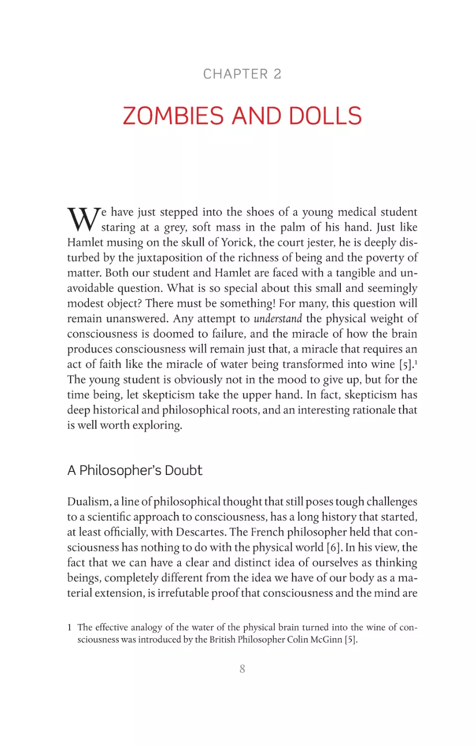
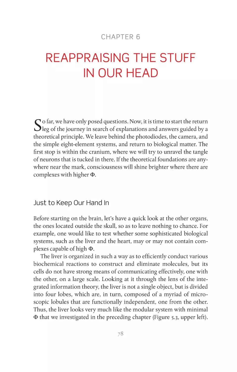
experience (Figure 2.1), indicates that consciousness cannot be inferred
from physical properties. These zombies don’t eat human flesh, but
they still manage to terrorize conferences dedicated to the science of
consciousness, where they are evoked by witch-doctors camouflaged
as cultivated scholars to embarrass and paralyze naïve speakers.
As often happens in philosophical debate, every argument or thought
experiment has a counter-argument or -experiment. Of course, zombies
are no exception to this. For example, it has been argued that the very
possibility of the existence of a philosophical zombie conflicts with our
certainty that we are sentient beings [8]. Indeed, if the zombies function
as we do, surely they, too, will have this same certainty. If they can entertain this false belief, who can say that we ourselves are not mistaken
when we maintain that we are thinking beings? As per this counter-
argument, assuming that it is absurd to throw doubt on our subjective
experience, philosophical zombies are neither possible nor conceivable.
Several books and dozens of articles have been written on this subject,
but who is right and who is wrong? This is an example of the circular
traps that we would like to avoid, so we will take this argument no further. As we stated in Chapter 1, “A Brain in Your Palm,” we prefer to keep
our feet on the ground, rather than experience the frisson of logical disorientation; hence, we will now attempt to reformulate the problem of
the zombies in a different way.
10
D igital Z ombies
A
B
C
Figure 2.1 Different kinds of zombies. (A) A graphic representation of a philosophical
zombie: the possibility of conceiving a being that is physically and behaviorally identical
to us, but does not possess conscious experience. (B) Actroid-F, the android developed
by the University of Osaka and a forerunner of the digital zombie. The sensors and the
activators are visible under the artificial silicon “skin.” (C) Two biological zombies, which
reside in the cranium—the cerebellum (above) and the basal ganglia (below).
Figure 2.1c Upper: Source: CLIPAREA l Custom media/Shutterstock.com.
Figure 2.1c Lower: Source: decade3d—anatomy online/Shutterstock.com.
Digital Zombies
Take one of the latest generation smartphones, connect it to Internet,
and download an app with a search engine, and software for voice recognition and synthesized speech; now you have a small object that can
respond to many questions, remind you of your appointments, and suggest a good restaurant close by that fits in with your past choices of menu
and price bracket. The voice of such devices is becoming so friendly and
helpful that you might soon catch yourself saying thank you to your
digital assistant! Of course, until now it has always been perfectly natural
to attribute consciousness to someone who listens to us and then replies,
but a smartphone certainly isn’t conscious. Sure, it may fool us for a few
seconds, and then only when we are distracted, but of course we can tell.
11
Z ombies and dolls
However, imagine what might happen if something rather more
powerful were to be placed between the microphone and the voice
synthesizer, such as Watson, the 3-million dollar IBM computer with
2880 processors and 16 terabytes RAM that can process 500 GB/s. In
2011, Watson was invited to participate in the popular American television quiz show, Jeopardy [9]. The game consisted of pressing a button
before the other participants, then answering a series of questions correctly. These questions were quite difficult to interpret as they were formulated like crossword clues, playing on words and double meanings.
For three episodes, which attracted huge audiences, Watson challenged
the two champions of the program. The computer played large sums of
money when it was sure to win, just a few dollars when it was not sure
of the answer, and didn’t press the button when it did not have the answer at all. Its monitor even blushed when it made a mistake. In the end
it won, much to the disappointment of the two human champions and
the delight of the public, who had become its ardent fan in just a few
hours. Poor Watson didn’t know it had won, however; in fact, Watson
never experienced anything about anything. When a modern computer
responds correctly to our input, when it produces an error message,
crashes, or reboots, it does not understand the meaning or significance
of what it is saying or doing; there is a consensus on this, even among the
audience who instinctively attributed to Watson plenty of human ambitions and emotions.
In fact, it is unlikely that the software of the supercomputer would
have passed the exam that computers should “dream” of passing—the
Turing Test, introduced in 1950 by Alan Turing [10], the British mathematician who influenced the outcome of World War II by deciphering
the German codes and considered to be the father of information technology. The Turing Test was constructed around the question “how can
we know if a computer understands what it says?” Turing devised a pragmatic solution, based on the following principle: a machine can think
and understand what is said to the extent that it can sustain a conversation that is indistinguishable from a conversation between two human
beings. In practical terms, the Turing Test consists of a conversation held
at a distance between two terminals, with an exchange of comments
such as we have today when opening a conversation using Skype or
Messenger. If, at a certain point, it is clear that the respondent on the
other end is a machine and not a human being, then the machine has
12
D igital Z ombies
failed. If the machine dupes us into believing that we are chatting with a
human being, the machine has passed the test.
There is no doubt that Watson, who could have beaten anybody at
“Jeopardy!” would have tripped up in the early phases of a simple chat.
The first exchanges, “How are you?,” “How are things?,” would not have
constituted a problem, and it might even have been able to respond to
specific questions, even difficult ones, but there would have been something “off.” After a while, we would have had the irritating impression
that the interlocutor on the other end of the line was not paying attention, or had not grasped the context of the conversation. It would not
have been long before an absurd misunderstanding or a totally irrelevant
answer would have revealed the non-human identity of the other party
to the conversation.
While it is likely that Watson would not pass the Turing Test, better
hardware running smarter software may soon do that, even in a face-to-
face encounter. Imagine a much more powerful supercomputer (even in
2011 Watson was only ranked 94th) and plug it inside a futuristic version
of Actroid-F, the gynoid robot developed by the University of Osaka.
This humanoid machine replicates the movements and facial expressions of a young Japanese girl, including tics and emotions (Figure 2.1B),
through a system of sensors and actuators [11]. It is perfectly conceivable
that a mix of microprocessors, optical fibers, sensors and compressed
air activators, microphones, and speech recognition software, covered
in soft silicone, may one day dupe an unwary interlocutor into thinking
it is human for a fairly long period of time. The futuristic android will
call on a powerful combination of calculation, speed, machine learning
algorithms, and access to immense databases to produce a fluid and appropriate conversation. In a face-to-face encounter, we may be a little perplexed by some inaccuracies, but it is likely that we will attribute them to
intriguing cultural differences and, enchanted by the gentle ways of this
exotic companion, we may even propose a romantic place for dinner.
Then, just as we are about to shyly suggest our favorite restaurant, our
enhanced version of Actroid-F may pull the mask off, leaving us with the
humiliating sensation of having been fooled by a fancy doll.
After all, on close reflection, it is quite easy to see how a black box,
which provides appropriate responses to any question, does not necessarily house someone who understands. This can best be illustrated by
the Chinese room thought experiment conceived by John Roger Searle
13
Z ombies and dolls
[12]. Pretend you are in a huge box, armed with pen and paper, and an
exhaustive catalogue in which all the questions that can be formulated in
Japanese kanji are each associated with the correct answers, also written
in Japanese kanji.2 There is a hole in the side of the box through which
someone on the outside passes you slips of paper on which are written
questions. You painstakingly consult the catalogue, find the question
which corresponds visually to the one written on the paper you have
received, and copy down the answer. You provide the correct answer
every time, even though you do not know Japanese and have understood
nothing, neither the question nor the answer. You have simply followed
the instructions to the letter. This will probably be the system the android will follow, just much faster, to converse with you in the future.
Millions of processors will consult an exhaustive catalogue of possible
questions and answers, and provide the most appropriate answer at the
speed of light. This is perfectly conceivable. Searle’s insight was to put
ourselves in the place of the computer in order to understand that, even
if we could manipulate the symbols perfectly, we would understand and
feel nothing.
This is when we are struck by a thought. What if our brain, too, is
just an expert manipulator of symbols? Just as the silicon microprocessors exchange digital symbols along optical fibers, so the neurons in our
brain exchange electrical impulses through microscopic cables composed of fat and proteins. Could they do this without understanding anything, without feeling anything, without “being” anyone? We hope that
this digression into the world of digital zombies has helped to clarify the
doubt expressed by the philosophical zombies. In all cases, the most remarkable zombies live elsewhere. Indeed, they are much closer.
Zombies Within Our Skulls
In 2001, Christof Koch and Francis Crick [13] published a note in the
journal Nature called “The zombie within.” In the note, they argued that
our brain contains a large number of “zombie systems,” whose hallmarks are efficient sensorimotor behavior and immediate, rapid action.
2 Here, to be consistent with the nationality of Actroid-F, we play with Japanese writing,
rather than with Chinese characters as in the original formulation.
14
Z ombies W ithin O ur S kulls
Every waking day, we live in close contact with faithful and silent servants of whose presence we are mostly unaware. We don’t notice them
because they do what they do in the dark, far from the flame of consciousness, but they are there. In fact, we often teach them to do what
they do. Imagine (or remember) the first lesson on the piano. You can
take nothing for granted, you must make a conscious effort in every
movement—how to sit on the stool, the correct posture, how to place
your hands on the keyboard, how to move your fingers. You touch the
keys—your performance is slow, staccato, labored. As time goes by,
you become more proficient and your performance improves. The
learning process that a human being undertakes is both tangible and
extraordinary, but what is truly amazing is that once you have learnt
the complicated motor sequences that in the initial phases absorbed
every iota of concentration, they cease to appear on your “radar.” You
no longer have to consciously think about these sequences. They become so automatic they cease to exist and your consciousness, liberated from the fetters of the motor sequences, can apply itself to how the
music should be interpreted. It is almost as if a mysterious entity in the
subconscious, which moves surely and swiftly, much more surely and
swiftly than we ourselves do, has taken over. Someone or something
is playing the pianoforte in your place, and is playing it well. Music
teachers understand this phenomenon perfectly, and at a certain point
tell their students “let your fingers play, don’t think about it,” and one of
those faithful zombies that live in the cerebrum will execute the notes
fluidly, without hesitation, and without making even the slightest mistake. If you try to take back control of the motor sequence, then inevitably your performance slows down or you hit the wrong notes. This
is because your inner zombie has become much better at playing the
piano than you, and does not appreciate interference, only indications
of a very general nature. The pianoforte is just one example, but there
are many others—when your hands and feet automatically work accelerator and brake, to maintain the correct distance from the vehicle
in front, while you chat to your companions; when you find the right
words at the right time, with the right inflection and meaning, while in
reality you only have a vague idea of what you want to say; when your
skis navigate bumps in the snow, while you admire the view of distant
peaks. In all these instances you have to thank an unconscious entity
that works for you.
15
Z ombies and dolls
Where do these scrupulous, devoted servants live? The answer comes
from clinical neurology. It is probable that there are zombies all over the
place in our brain, but certain types of cerebral lesion have shown that
the majority are to be found in two neural structures: the cerebellum,
in the posterior cranial fossa and the basal ganglia, a voluminous mass
of neurons drowned in the depths of the cerebral hemispheres (Figure
2.1C). If one of these two structures is destroyed or degenerates, life becomes very difficult. We become painfully conscious of every gesture,
particularly of those of which we were previously hardly aware. When
an inner zombie dies, we suddenly become aware of the importance of
the work it did for us. Even the most banal tasks, such as grasping a glass
of water, become arduous. Distances must be calculated, movements
measured, the hand has to open at the right time and be closed with the
correct amount of pressure—too strong a grip and we break the glass,
too weak and the glass slips through our fingers. With all these complications, errors are inevitable [14]. Many, too many, of the tasks that were
carried out by the zombie now have to come under our control [15]. The
luxury of that light unconscious management that we took for granted is
gone for good and the resulting overload is impossible to manage. If the
cerebellum is destroyed, even shaking a friend’s hand is a complicated
task, and driving a car or learning to play a musical instrument becomes
extremely hard.
We become aware of these zombies when they disappear, but also
when they malfunction and stop collaborating. Tourette syndrome,
commonly known as Tourette’s, is a disorder of the basal ganglia characterized by forms of behavior, some of which are quite complex, that the
person is unable to control [16]. Symptoms include tics, such as spontaneous exclamations, sometimes socially inacceptable (coprolalia), repeating what others have said (echolalia), and repetition of one’s own
words (palilalia), although blinking and throat clearing are the most
common symptoms. Persons with Tourette’s may suddenly execute
complicated dance steps or pirouettes. They often have a high IQ, but
face great challenges in life because they are not always able to control
what they say and do. It is as if a mischievous alien, residing in the depths
of their brain, takes over their body, and makes them move and speak
out of character.
These zombies are more than just a logical possibility. They are composed of neurons, synapses, and circuits; they live in our brain, they
16
A N euroscientist ’ s D oubt
speak, walk, and do other complicated things without sparking the
flame of consciousness. Why? What is the mysterious ingredient that is
missing in their make-up? As was argued by Koch and Crick, the existence of zombie systems raises two questions. First, why aren’t we just
big bundles of unconscious fast zombie agents? Why bother with consciousness, which takes almost half a second to set in? Second, what
is the difference between the neuronal circuits that make up zombie
agents and those that support conscious experience? Addressing these
kinds of questions is exactly what scientists can and should do. Indeed,
when Crick and Koch jump-started the whole field of the neuroscience
of consciousness and shattered the unwritten rule that forbade scientists
to even mention consciousness as a subject worthy of serious inquiry,
they did so precisely because they knew how to frame their questions
in concrete, experimental terms. The zombies in the brain seem to be
the incarnation of the abstract doubts of the philosopher. We can touch
them and study them, but until we have understood them, skepticism is
more than justified.
A Neuroscientist’s Doubt
So, what can neuroscientists say about consciousness? After all, they
are in close contact with damp biological matter every day. One thing is
certain; they will not have an inferiority complex with regard to philosophers. Physiology has been extremely successful in explaining how the
heart pumps blood, and how the liver and the kidneys filter and purify
it. Over the last 50 years, neurophysiology and the neurosciences in general have literally exploded and our knowledge of the brain has increased
exponentially. An untold quantity of data accumulates each day in thousands of neuroscience departments and continues to grow. Teams of
students, graduates, postgraduates, and researchers slave daily on experiments and data, using increasingly powerful instruments. Ten years
ago, it was difficult to record brain activity with more than 20 sensors;
now we can cover the brain with hundreds of electrodes. In the past, a
university purchasing a magnetic resonance imaging (MRI) scanner
made the national headlines, today you can find scanners in almost any
basement, where the anatomy and the metabolism of the human brain
can be recorded with millimeter precision in vivo. Not to mention the
17
Z ombies and dolls
electron microscopes, which can capture the detail of a synapsis with
Angstrom-level spatial resolution (0.0000007 mm), two-photon imaging techniques, optogenetics, and the like.
There are scholars in the field of neuroscience who have dedicated
their lives to understanding the workings of a single molecule, expressed in a particular class of neurons, located in a particular structure of the brain, anywhere between the periphery of the spinal cord
to the more noble areas of the association cortex. The competition between laboratories is fierce; no angle of the brain forest has been left
in its virgin state, researchers have marked every tree, colonized and
cultivated every millimeter. You are curious about the structure of the
cells in the eye of a fruit fly? You want to know about the neurons of
the human cerebral cortex that are specialized in recognizing Jennifer
Aniston [17]? The stands of the Annual Meeting of the Society for
Neuroscience, a bustling scientific fair, which attracts at least 30,000
scientists every year, have it all.
All this effort has been rewarded with results. Neurophysiologists are
revealing the neural mechanisms that control how we grasp an object—
the programing of motor sequences, anticipatory postural adjustments,
the perfect coordination of the limbs, fine control of the force needed
from the finger muscles. These are complex mechanisms that are not yet
completely understood, but it is just a question of time, there is nothing
mysterious or unfathomable. Similarly, they are demystifying much of
the visual system’s working—from the receptive field of the rods and
cones of the retina to the mechanisms of motion detection higher up in
the cortical hierarchy. These successes are replicated for hearing, smell,
touch, and much more.
In principle, it will soon be possible to draw a precise diagram of the
auditory and visual systems, of the sense of touch, and the circuits dedicated to motor planning and control. With time and patience, we will
be able to piece together a detailed diagram of the circuits in the brain
and reconstruct them one-by-one, just as a counterfeiter might replicated complicated objects—such as a digital camera, car, or a military
airplane—based on a stolen blueprint. The chances are that we will end
up with a tangled mass of artificial neurons that behave just like the
real ones, but will this bring us closer to the solution of the mystery of
consciousness? Not necessarily. Let us see why, again using a tangible
example.
18
A N euroscientist ’ s D oubt
The scientists of Lausanne Polytechnic have been working for years
on a scientific enterprise without precedent, the Blue Brain project.
A team of researchers from a number of fields—physicists, biologists,
physiologists and computer scientists—are reproducing everything that
is known about the brain into a supercomputer simulation: molecules,
synapses, the electrical properties of different neurons and their patterns of connectivity, with the objective of reproducing entire areas of
the cortex and, finally, the entire brain. By the end of 2006, the project
had achieved its first objective, the reconstruction of a simplified cortical
column. This is a cylindrical structure, about 2 mm high and 0.5 mm in
diameter, which is thought to represent the fundamental functional unit
for the cerebral cortex. Artificial cortical columns are much bigger than
biological columns; they are housed in a supercomputer that takes up
a large room. A few years ago, the project reached another landmark,
the detailed simulation of a portion of the rat’s somatosensory cortex
containing about 30,000 simulated neurons (of 200 different types) connected by 40 million synapses [18]. This is, without doubt, the most complete simulation to date of a piece of excitable brain matter. In the years
to come, petaflops of computer power and smart algorithms will grow
this chunk of digital cortex further. Indeed, the project, which represents
a fantastic scientific adventure, can count on generous financing and is
very ambitious. Its goal is to create a virtual mouse brain with hundreds
of millions of neurons and, ultimately, simulate the human brain.
Now, let us fast-forward a few decades and suppose that the empirical
data and mathematical models used to reconstruct virtual neurons, their
biophysical properties, and their connections are correct and complete,
that their activity can be simulated in real time on a powerful supercomputer that can fit inside an artificial head, and that two artificial eyes, two
ears, a nose, two nimble robotic hands, two legs and a smooth digital vocalizing system have been plugged in. Now, imagine that you have been
asked to sign a rather peculiar contract, which establishes that at a certain point in your existence, when your body starts to show the first irrefutable signs of deterioration or the signs of an untreatable disease, you
will hand over your biological brain to a group of scientists. You will be
allowed to say your last goodbyes to family and friends, then the scientists will anesthetize you, extract your brain and record its activity with
billions of sensors. They will cut it into sections, take photographs of
it with the electronic microscope and analyze it thoroughly. Finally, all
19
Z ombies and dolls
your neurons, their biophysical properties, and their connections will be
uploaded and simulated by virtual neurons implemented by the supercomputer inside the head of your new artificial body. Every last detail
will be simulated faithfully, the synapses of the hippocampus, where
your most vivid memories are stored, the circuits of the amygdala where
the events and conditioning of your own unique life have carved out
fears that you have never confessed to yourself, and so on. Trains of neuronal spikes will run incessantly across the simulation, millions per seconds, replicating the coding and the computations of your original brain
and all this will rely on silicon chips, which are impervious to viruses,
tumors, or heart attacks, and do not suffer the effects of the passing of
time. The “silicon you” will behave exactly as you always did, and everybody will think that inside the new body there is you—the same old
you—with all your idiosyncrasies, obsessions, and your few endearing
traits. The contract promises you the digital immortality of your brain’s
activity in exchange for renouncing a few months of biological life. It
lures you with the promise of watching your children grow up, enjoying
their successes, and offering a helping hand when needed, of continuing
to see your friends, and even the option of asking to switch off the whole
thing when you have had enough.
The stakes are high, very high, and the goal is to turn one of man’s
greatest dreams, immortality, into reality. There are doubts, however,
just as strong as the stakes are high. How many people would actually
sign a similar contract? It is quite possible that not even the most optimistic of scientists, the most hard-boiled materialist and anti-dualist
would put his signature to this agreement, when his time comes. Not in
the name of moral qualms, but because an inner voice whispers incessantly, “Who says that being a machine that simulates exactly the activity
of all your neurons is the same as being you? Who says that scientists
have understood the relevant properties of the brain that have to be reproduced? What if the spatial–temporal grain at which the simulation
runs is not the right one? What if the secret of consciousness is tucked
away in a sub-atomic detail of the functioning of neuronal membranes?
What if, instead, the relevant processes occur through unknown interactions that extend beyond my brain? Maybe, it is a particular chemical aspect of the biological matter, which cannot be reproduced on a
silicon chip? Am I trading my last conscious days for a reincarnation as
20
A N euroscientist ’ s D oubt
an eternal zombie? Do not sign! Put that pen down and go and hug your
family one more time!”
The bottom line is that we totally trust science when it is a question
of substituting vital organs, such as hearts and kidneys, with artificial
devices, but when it comes to the brain, that is a different ball game altogether. Why are we so reluctant? Because we have a niggling suspicion that however much we know about those billions of neurons in our
head and how they function, we are no closer to a scientific explanation
of how the brain generates subjective experience. This is the doubt that
troubles physiologists, who feel rather like the early astronomers with
their detailed descriptions and charts of the movements of the celestial
bodies, but who had no idea as to whether those movements were dictated by a general law. When all is said and done, our doubts, and the
doubts of the physiologist and the philosopher, may share the same rational root. When it comes to the relationships between consciousness
and the brain, we have described a lot, but we lack principles.
21
CHAPTER 3
BRAIN ISLANDS
N
ow we leave the morgue, the digital dolls, and the zombies, and
move to the intensive care unit (ICU), where living human beings
fluctuate between consciousness and unconsciousness, and where
sometimes the flame of experience burns unseen on brain islands lost in
a sea of neural dissolution. Here, among the tubes and drip-feeds, artificial lungs, and monitors, the way we pose the question changes, but the
question itself remains the same.
Bad Awakening
How do we know a fellow human being is conscious? In the normal run
of things, the question doesn’t even arise. Unless we are really into philosophical zombies, we just assume that others are conscious beings—we
are similar physically, so we will be similar in having subjective experiences. If your friend has nodded off on the settee in front of the television,
or seems to be lost in a world of his own, you can ask something along
the lines of “are you with us?” and a gesture of the hand or a grunt is all
that is needed to reassure you that he has not “lost his senses.” A doctor
uses much the same method to evaluate the level of consciousness of
a patient who has just been given a dose of anesthetic in the operating
theater, or of a man arriving in the Emergency Ward with his eyes closed
and his face covered in blood. In cases such as these, the doctor asks the
patient to open his eyes, say his name, say where he is, and clench his
right or left fist. If the patient responds, he is conscious. If there is no response, the doctor will try exerting pressure on the palm of the patient’s
hand, or on the nail bed to elicit pain. If there is still no response, the patient is considered to be unconscious. So, the fundamental criterion for
establishing the conscious existence of another human being is whether
22
B ad A wakening
he is able to respond to stimuli and commands. This method normally
works well, but not always.
[ . . . ] that grey and silent abyss suddenly takes on a green hue, hazy at first,
but which gradually comes into focus; at the same time, I can hear faint
noises in the distance, now they are coming nearer and I can hear them
more clearly. Where am I? Ah, of course, the operation! I am having difficulty in getting my thoughts together, but yes, this is it, the operation and
now I can see the curtain through half-closed eyelids. The first impression
is that it is happening to someone else, indifference mixed with stupor;
but then I am seized with an impelling desire to move. I make a superhuman effort, but fail: not because I feel tied down, but because I don’t
have a body to move. I am just a brain floating in a void and somewhere
I have a mouth, blocked by something that stops me using it, that prevents
me from calling out. Everything else has just disappeared, dissolved! As
soon as I realize that they are operating on me, and I am awake, damnably
awake even if I can’t feel anything, panic explodes. But this is a strange sort
of panic, logical, ascetic, without emotion, without heartbeats because
I haven’t a heart, without pain because I haven’t a body, without anxiety
because what is happening has been decided by someone else. It is mental
panic, empty panic; an anguished delirium from which there is no escape.
I have no idea how long it lasts, but to me it is an eternity. I pray that it will
end, I want to do something to make it end, but I don’t know what. Then,
finally, everything fades out . . .
This first-person account by a patient who accidently recovered consciousness during an operation was published in a specialized scientific journal [19]. As chance would have it, the patient was himself an
anesthesiologist, a doctor well-accustomed to playing with powerful
drugs and with the consciousness of other people. The unfortunate anesthesiologist suddenly awakens with the crisp realization that he’s still
undergoing a surgical intervention. He feels a desperate urge to move
and shout to his colleagues to let them know that he is conscious, but
he can’t. He is fully paralyzed and artificially ventilated though a tube
stuck in his throat. The agony lasts for an undefined period (presumably,
a few minutes) until an additional dose of anesthetic knocks him back
into unconsciousness.
Regaining consciousness during an operation, technically called “anesthesia awareness,” is a fairly rare occurrence. Approximately one patient
in 1000 reports having been conscious during an operation that required
23
B rain islands
general anesthetic [20, 21]. Fortunately, it is extremely rare (though not
impossible) for the patient to feel pain, as the patient is also given analgesic
compounds before and during the operation. Even so, it is a traumatic experience that may have serious psychological consequences [22, 23]. The
anguish and the trauma are to be attributed mainly to the total paralysis
that the subject experiences upon regaining awareness [24]. Immediately
after administering the general anesthetic and before the surgeon starts
to operate, the patient receives an injection of neuromuscular blocking
drugs (NBDs), which completely block the transmission of electric impulses from the nerve endings to the muscles. This block prevents the
muscles from generating violent reflex contractions during the various
phases of the operation, although it also impedes the patient from expanding his chest autonomously, so air is pumped into the lungs through
a tube to support ventilation. Normally, patients are not aware of this as
they lose consciousness just a few seconds after receiving the anesthetic
and regain it only when they are back on the ward. When everything goes
as planned, between the lights in the operating theater, which seem to
flicker and collapse before slipping into darkness, and the anxious faces
of your loved ones in the ward, you just cease to exist.
It is still not clear why some patients regain consciousness on the
operating table, in spite of having received an adequate dose of anesthetic
[25]. The problem is that it is difficult for the anesthesiologist to detect the
recovery of consciousness during surgery [26]. The total paralysis makes
it impossible for the patient to communicate with the medical team; he
is forced to witness as the operation proceeds. He is present, but cannot
make anyone aware of the fact. He can see, but cannot act. He can hear,
but cannot communicate. He is a conscious prisoner in his own skull.
Probably, the closest thing to being buried alive. In Victorian times, the
fear of such a terrifying possibility led to a plethora of “safety coffin” designs: coffins equipped with complicated devices, whereby a cord was
tied round the corpse’s wrist and chest was linked to lamps, bells, and
flags on the surface of the tomb; any movement would set off a system
of visual and acoustic alarms (Figure 3.1). For a small fee, these curious
contraptions could be rented and installed in the coffin for a few days or
even a few weeks. As far as we know, none of the scrupulous customers
of these safety coffins ever recovered from death, and there are no records
on hand to tell us how many men and women were, in fact, erroneously
buried alive. We can only hope none or, at the worst, very few.
24
B ad A wakening
Returning to the operating theater, however, there are reasons to suspect that the number of people who regain consciousness during an
operation is more than 1 in 1000. This suspicion is raised by the application of a simple technique that is very reminiscent of the safety coffins, the isolated forearm technique (IFT) [27]. This method leaves an
open channel between the patient’s brain and the anesthesiologist in the
operating theater. In the IFT, a padded cuff/tourniquet is applied to the
forearm before the NBD is injected, so that the muscles in the forearm are
not paralyzed and can be used to communicate with the medical team.
Even though the rest of his body is blocked and he is unable to speak, the
patient is still able to clench and unclench his non-paralyzed fist. The IFT
is rather laborious and is not always suitable for general surgery, but has
provided some interesting data. The patients who clench their fists on
the operating table when the anesthetist asks “are you still with us?” may
be much more numerous than those who later recall that they were conscious during the operation, maybe 1 in 10 as opposed to 1 in 1000 [26].
Whether the IFT actually reflect the persistence of consciousness or, rather, an automatic reaction is still a matter of debate [28], yet anesthesia-
induced amnesia [29] may certainly contribute to the underestimation
of intraoperative awareness. In all cases, consciousness does not need to
be remembered in order to exist; for example, we are acutely conscious
of what we are doing during a drinking spree, even if the next day we
have absolutely no recall of what happened. Who would accept to endure or inflict a minute of unsupportable pain, even if given a promise
that afterwards there would be no memory of it?
Fortunately, those patients who do suffer intraoperative awakening
can count on the fact that, however distressing it may be at the time, the
nightmare will end: muscular paralysis is reversible and once they leave
the operating theater, they will either have blissfully forgotten their ordeal or will be able to forcefully explain to the surgeons what they have
been through. Intraoperative awakening remains a problem for anesthesiologists, but above all it is a general admonition that we cannot afford
to ignore. There are cases in which consciousness is present, desperately
and intensely present, but cannot be detected from the outside. This
throws doubt on our ability to promptly recognize consciousness in
our fellow human beings, a doubt that becomes anguish when we are
faced with patients who have emerged from coma, but lie immobile and
glassy-eyed, for months or even years.
25
B rain islands
26
S ur v i v ing C oma
Surviving Coma
Coma comes from the Greek, and its original meaning is “deep sleep;”
indeed, a coma closely resembles sleep. The patient lies motionless
with his eyes closed, but whereas, in true sleep, all that is needed to
wake a person is a gentle shake, no external stimuli will wake the
person in a coma [30, 31]. In some cases, painful stimuli may provoke a
brief reflexive reaction, but nothing more. The ancient Greeks used the
word coma both for the heavy “comatose” sleep that follows a night
on the town, and the sleep from which there is no awakening, the
sleep that Penelope begged for, distraught for the absence of Ulysses.
Until about 50 years ago, that just about described it. If a person who
fell into a coma did not recover contact with the world shortly after,
he slid into that deadly sleep from which there is no return. The state
of coma is typically associated with traumatic, vascular, toxic, or anoxic distress of the brain, including a phylogenetically ancient structure that is situated deep in the skull and connects the cerebrum to
the spinal cord—the brain stem [30]. The brainstem contains various
groups of neurons that are necessary to maintain arousal or wakefulness. In addition, the brainstem controls life-supporting functions,
such as breathing and the regulation of blood pressure. In the acute
phase, any brain insult can cause a swelling of intracranial tissues,
leading to increased pressure, which in turn results in a compression
of the brainstem against other structures in the lower portion of the
skull (Figure 3.2). In the past, this compression and the temporary
impairment of brainstem function had a catastrophic effect—coma,
followed by cessation of spontaneous breathing, rapidly leading to
cardiac arrest and death.
Figure 3.1 An illustration of the mechanisms of a safety coffin. The drawing is taken
from the patent filed on July 4, 1882, by Albert Fearnaught. His particular version
was based on the installation of a complicated mechanism, whereby a rope tied
around the deceased’s wrist, if tugged, would release a spring that, in turn, would
open a large colored fan inserted at the upper end of a tube at ground level. The same
movement would cause a flap to open and allow fresh air to circulate in the coffin.
The patent explains that following interment, the coffin should be frequently checked
by the cemetery custodian. It also specifies that the mechanism can be recycled
for use in other coffins if the deceased should not return to the world of the living.
27
B rain islands
A
B
Figure 3.2 A schematic drawing to illustrate the different herniation syndromes
seen following increases in intracranial pressure and mass effects. When the volume
increase is symmetric in the two hemispheres (A), there may be central herniation,
as well as herniation of either or both medial temporal lobes, through the tentorial
opening, resulting in brainstem compression. Asymmetric compression (B) may
cause herniation of the ipsilateral cingulate gyrus under the falx (falcine herniation).
This type of compression may cause distortion of the diencephalon and brainstem
by either downward herniation or midline shift.
Reproduced from Plum and Posner’s Diagnosis of Stupor and Coma, 4th edn, Jerome B. Posner,
Clifford B. Saper, Nicholas Schiff, and Fred Plum, p. 96, Figure 3.2 © 2007, Oxford University
Press. Reproduced by permission of Oxford University Press https://global.oup.com/academic/
product/plum-and-posners-diagnosis-of-stupor-and-coma-9780195321319?q=Plum%20and%20
Posner%E2%80%99s%20Diagnosis%20of%20Stupor%20and%20Coma&lang=en&cc=gb.
Today, coma is not necessarily a passage to death. The game changer
was the invention of mechanical ventilation. This technique was introduced in clinical practice in 1952 by the Danish doctor Bjorn Ibsen, during
the poliomyelitis epidemic of 1952, which in the span of a few weeks,
temporarily (but completely) paralyzed hundreds of people, including
children [32, 33]. Ibsen inserted tubes into his patients’ trachea, which
were then connected to a pump, each worked by one of the 200 medical students he had enlisted for the purpose. He organized special wards
for this life-supporting activity, so he can also be said to be the inventor
of the ICU. His heroic efforts were crowned with success and the mortality rate from poliomyelitis in his area dropped from 90% to just 25%.
Mechanical ventilation is, of course, no longer manually operated, the
28
S ur v i v ing C oma
pumps are motorized, and controlled by sensors and computers, which
maintain the optimal mix of oxygen and carbon dioxide for months or
years if necessary. Today, breathing can be controlled indefinitely and a
new chapter has been written in the natural history of coma. In fact, rather unexpectedly, a relatively simple device, such as a pump that inflates
the lungs, changed the course of medicine, and moved boundaries that,
for ages, where thought to be insuperable and fixed.
First, death has moved from the heart to the brain with important consequences from the clinical, juridical, and anthropological stand points
[34]. In the past, death coincided with the cessation of cardiorespiratory
functions. A relative was considered dead if a mirror held in front of his
lips did not mist over. He had “expired,” “drawn his last breath.” Then,
the doctors could confirm by feeling his pulse and auscultating the heart.
The cessation of cardiorespiratory function and the death of the brain
were inseparable, the two events were reciprocally linked. Today, mechanical ventilation has broken this vicious circle and cardiorespiratory
function can be maintained artificially, with or without a functioning
brain. The main result is that many patients can survive the most critical phases of coma, during which brainstem function is temporarily impaired. The other side of the token is that we can encounter the paradox
of a body that is still functioning, even if its brain is completely destroyed, liquefied—a moving chest and a beating heart, without a brain.
In 1968, a group of 10 doctors, a theologian, a lawyer, and a historian
met in Harvard to review the issue. The Harvard Committee, as it was
known, had the responsibility of establishing a new criterion with which
to define death, which could no longer be cardiorespiratory in nature,
but needed to be neurological [35]. The discussion was long and arduous,
with doctors becoming lawyers, and the lawyer waxing philosophical,
but a decision was finally reached. Death coincides with the irreversible cessation of the functions of the whole brain, brainstem included.
From the operational point of view, the process for establishing brain
death is complex and requires a number of steps, including verification
of complete unresponsiveness; the cessation of all functions and reflexes
of the brainstem, spontaneous breathing included; and (depending on
the specific legislation of the country) the instrumental documentation
of the absence of electrical and metabolic activity in the brain. In point
of fact, brain death identifies a particular, artificial situation where, to all
extents and purposes, the body is decapitated (the nervous system above
29
B rain islands
the spinal cord is completely destroyed), but continues to receive oxygen
through external machines. Death decreed, based on neurological criteria, may be even harder for the patient’s nearest to accept, as his outward appearance is often healthy, the chest rising, the skin warm and
rosy. Today, this delicate task lies with the doctor. On the other hand,
a diagnosis of brain death is necessarily more accurate than that established by the cessation of the respiratory and circulatory functions, even
if it is less intuitive. The new frontier of death is very clear, and does not
admit any possibility of error or recovery.
While the introduction of the mechanical ventilator has helped to
establish a new clear divide between life and death, it has played havoc
with the borders of consciousness [36, 37]. Millions of patients with
devastating cerebral lesions, which only 50 years ago would have been
lethal, now survive the comatose state and may slip into medical conditions that were previously unknown. In fact, following a coma there
is now a range of possible states—between brain death and conscious
awakening—which at present can only be classified in provisional terms.
A patient who survives coma will typically start opening his eyes
after 2 weeks or so. This is the sign that the coma is coming to an end.
The patient’s eyes open because the brainstem starts to function again,
so guaranteeing spontaneous respiration and the recovery of what the
neurologists call vigilance, but is commonly known as wakefulness.
The part of the brainstem that is closest to the spinal cord (the so-
called “medulla oblongata”) is primarily involved in the maintenance
of the functioning of the cardiorespiratory systems, while the upper
parts, the pons and the midbrain, house extremely important neurons,
called the activating system, which control eyes opening and establish
whether the brain is generally switched off or on, in a state of sleep or
wakefulness [31]. When these neurons fire-up in a healthy brain (which
happens every morning upon awakening), the muscular tone increases,
the eyes open and the person regains awareness of the self and the surrounding world. In a brain that has suffered severe damage, this does
not necessarily happen. Brainstem neurons may activate, spontaneous
breathing recovers, and the eyes open, but this does not mean that consciousness returns [30, 38]. It is like flicking a switch when the bulb
has burnt out. A click, with no light. In fact, the recovery of brainstem
functions, although necessary, is not, in itself, sufficient for awakening
to be accompanied by the recovery of subjective experience.
30
S ur v i v ing C oma
Some patients open their eyes, breathe spontaneously, and appear
to be awake, but do not show voluntary behavior, do not respond to
simple orders, and do not react purposely to painful stimuli. This condition of unresponsive wakefulness was originally defined by Bryan
Jannet and Fred Plum in 1972 as the vegetative state (VS) [39], a diagnostic term that is now been replaced by the more descriptive and
neutral label unresponsive wakefulness syndrome (UWS) [40]. This is
a strange situation as we are used to associating eye-opening with the
recovery of consciousness; after all, this is what happens every morning
when we arise out of our nightly oblivion. Hence, we naturally tend to
interpret the open position of the eyelid as a master power-on indicator,
as the sign that consciousness is turned on. In this man-made condition, however, brought about as it is by medical technology, this intuition is wrong—eyelids and orbs are just things moved by the primitive,
blind force of the brainstem. In these patients, there is often an increase
in muscle tone and a range of automatic gestures, but these are unintentional and without meaning—the eyes move without pursuing a target,
there are reflex movements of the limbs and neck, grimaces, chewing
movements, utterances, and sometimes uncontrolled outbursts of
laughter and tears. The vegetative state/unresponsive wakefulness syndrome (VS/UWS) obviously differs from the internal decapitation of
brain death and it differs from coma in that the patient is awake. The
only thing missing, or at least, all that appears to be missing judging by
behavior, is that spark of consciousness.
However, the VS/UWS is just one of the new offspring of coma. Some
patients may come out of a coma, open their eyes, recover self-awareness,
and recognize the world around them, but remain totally paralyzed.
Unfortunately, this is not the temporary paralysis of anesthesia awareness, but a permanent state caused by neuronal damage. Such paralyses
are often caused by a very small lesion (maybe only a few centimeters
in extension), but they are, nonetheless, devastating because it affects a
key spot in the brainstem where all the fibers (and there are hundreds
of thousands), which carry motor commands from the cerebral cortex
to the spinal cord, meet and proceed in a single “bundle.” The resulting
condition is known as “locked-in syndrome” (LIS) [30, 41], which is a
poignant rendering of the sense of isolation that characterizes this state.
It is not uncommon for LIS patients to be diagnosed as VS/UWS, particularly during the first few months of disease [42].
31
B rain islands
Fortunately, in time, most of these patients recover the control of some
extrinsic eye muscles, and become able to shift their gaze up and down,
or blink. These movements are governed by motor fibers that leave the
brainstem before the area of the lesion. In some cases, it is the patients’
relatives who realize that these eye movements are not random automatic
patterns, but represent a desperate and laborious attempt to communicate consciousness. Once they have been identified as LIS, these patients learn to use eye movements to establish a binary code and initiate
a communication process, which at the beginning is very basic (yes/no),
but can become progressively more efficient as time passes. Using this
fragile output channel, literally reduced to a single thread that connects
the cerebral cortex to the muscles of the eye, LIS patients can finally write
and communicate their conscious universe, as well as hunger, thirst,
pain, desperation, happiness [43–45]. Many LIS patients who do manage
to communicate with the external world, report that their quality of life
is not very much different to that reported by healthy controls [46]. This
rather surprising finding is very revealing. It suggests, on the one hand,
that healthy subjects tend to complain too much about their own existence. On the other, it shows that a locked-in brain can access an internal universe that is large and interesting enough to replace the one that
cannot be actively explored. Of course, this all depends on the patients’
conscious existence being acknowledged and on an adequate level of
support. Unfortunately, there are patients in which not even eye movements are preserved. This rare condition, where all outputs are blocked,
is known as “complete locked-in syndrome” [47]. In this case, the anguish and the sense of impotence described by the anesthesiologist who
regained awareness in a fully paralyzed body may become a permanent
state. The possibility of even a single human being in such a condition of
incommunicable existence is a fact that is hard to accept.
There are no limits to variety in the outcomes of intensive care medicine; in fact, there are a plethora of conditions between LIS and VS/UWS,
which have been classified collectively as a “minimally conscious state”
(MCS). MCS was only officially recognized as distinct from the VS/UWS
in 2002 [36], which testifies to our still provisional understanding of
what may happen after a coma and provides an inkling of the changes
that we can expect over the coming decades [48]. Patients who are unable communicate their thoughts and feelings, but occasionally show
inconsistent, but discernible signs of consciousness, are considered as
32
S ur v i v ing C oma
being in a MCS. A classic example is the brain-injured subject who, in a
context of general unresponsiveness, occasionally appears to track, with
purposeful eye movements, doctors and relatives as they move about
the room.
These minimal signs of consciousness change from one day to another, from one minute to the next, and are difficult to extrapolate from
a background of intense automatic activity. For example, it is very difficult to decide whether a finger twitch or the bats of an eyelid are involuntary reflexes or voluntary attempts to interact with the environment.
It is equally difficult to judge whether an eruption of laughter or suddenly bursting into tears reflects a conscious emotional content or just
the mechanical consequence of the random discharge of a small group
of neurons surviving in the depth of the brain. Therefore, the diagnosis
of MCS can only be reached through a careful, detailed, and repeated
evaluation of the patient’s behavior. Accurate detection of MCS patients
is extremely important, as these patients retain some form of experience,
even though we do not know to what extent or of what. Probably, this
state covers a multitude of conditions that we will never know or understand because they cannot be communicated. It is possible that some
patients may only have a fragmented glimmer of consciousness, rather
similar to what healthy individuals experience as they sink into slumber,
others may fluctuate in a sort of dream world or delirium, while others
still may be completely lucid and conscious, but without any reliable
means of communicating this. There is another reason why an accurate
diagnosis is important: unlike patients in a VS/UWS, MCS patients have
a better chance of recovery even after years of illness [49, 50], particularly if they undergo intensive rehabilitation programs.
Unfortunately, detecting consciousness in patients who are unable
to communicate remains difficult, and MCS patients are a paradigmatic
example of this problem. Recent data show that nearly half of the patient labelled as being in an unconscious VS/UWS are actually in a MCS
[51–53]; this low sensitivity in detecting conscious existence represents
one of the most serious clinical and ethical issues of contemporary neurology. According to a rough estimate, in the United States alone, there
are 280,000 MCS patients, the equivalent of the population of a major
city [54]. How many more are not recognized and end up dying in places
with lower standards of care? How can consciousness escape our radar
so easily? To better understand the reasons behind this diagnostic error,
33
B rain islands
let us step into the shoes of a detective in search of signs of conscious
experience.
Level 1: Sensory Inputs and Motor Outputs
Classically, the decision as to whether a patient is conscious or not is
made based on a stimulus–response paradigm. This is the first, fundamental level of investigation. Since the physical mechanisms of
subjective experience are unknown, we treat the brain as a black
box and assess consciousness based on input–output relationships
(Figure 3.3A).
A series of standardized tests using verbal and sensory stimuli (visual,
acoustic, tactile, and pain) have been established for this purpose; scores
are attributed based on the responses given and the spontaneous behavior observed. In very general terms, this clinical approach is not so
different from the Turing test. The underlying principle is that the more
specific, reproducible, and articulated the responses are, the higher the
level of consciousness is. A patient who is able to open his mouth, clench
his right fist, left fist, open his eyes, and smile when requested to do so,
will be given a higher score than a patient who in response to all these
commands only opens his mouth. The most effective of these scales is
the coma recovery scale—revised (CRS-R), specifically designed to recognize the faint signs that characterize the passage from the VS/UWS to
the MCS [55]. This scale represents the gold standard and its repeated,
careful administration is the cornerstone of an accurate assessment of
behavioral consciousness. If a patient shows purposeful motor activity,
and responds to stimuli in a way that is specific and reproducible, there
is no doubt that he is conscious.
On the other hand, even the CRS-R is not watertight [56]. As was
mentioned earlier, a dissociation between experience and motor outputs
can happen during anesthesia awareness due to NBDs. A fortiori, motor
responses may be blocked at multiple levels in neurological syndromes.
For example, in amyotrophic lateral sclerosis [57] or in some disorders
of the peripheral nerves, such as the Guillain–Barré syndrome [58], the
generation and conduction of electrical impulses from the motor cortex
to the muscles may be severely impeded. Often, ischemic, hemorrhagic
or traumatic lesions may interrupt or tear off the bundles of fibers that
34
L e v el 1 : S ensory I nputs and M otor O utputs
A
B
C
Figure 3.3 The different levels at which consciousness can be assessed. (A) Level
1: when sensory processing (IN, input) and motor behavior (OUT, output) are preserved,
the behavioral evaluation can be effective in detecting consciousness, even in the
extreme case of locked-in patients. In these patients, a pontine lesion (such as the one
indicated by the red arrow) spares the motor nerve controlling vertical eye movements
and blinking, through which an effective communication system can be implemented.
(B) Level 2: when motor outputs are completely blocked (red cross), but sensory
processing is preserved, active fMRI paradigms, in which neuronal responses to verbal
instructions are recorded directly from the cerebral cortex can be used. In this example
an otherwise unresponsive patient produces specific neuronal activations in response
to verbal instructions, such as “imagine you are playing tennis” (top) or “imagine you
are walking through your home” (bottom). (C) Level 3: when both sensory processing
and motor behavior are impaired, such as in the case of disconnected cortical islands
that show preserved activity levels (as indicated here by high PET activity in the right
hemisphere), assessing consciousness requires opening the black-box of the brain and
identifying the full neural correlates of consciousness.
Figure 3.3b: Reproduced from Adrian M. Owen, Martin R. Coleman, Melanie Boly, Matthew
H. Davis, Steven Laureys, and John D. Pickard, Detecting awareness in the Vegetative State,
Science, 343(5792), p. 1402, doi: 10.1126/science.1130197. © 2006, American Association for
the Advancement of Science. Reprinted with permission from AAAS.
Figure 3.3c: Reprinted from Brain Stimulation, 8(1), 142–9. Olivia Gosseries, Simone Sarasso,
Silvia Casarotto, Mélanie Boly, Caroline Schnakers, Martino Napolitani, Marie-Aurélie Bruno, Didier
Ledoux, Jean-Flory Tshibanda, Marcello Massimini, Steven Laureys, and Mario Rosanova, On the
cerebral origin of EEG responses to TMS: insights from severe cortical lesions, pp. 142–9, https://
doi.org/10.1016/j.brs.2014.10.008. Copyright © 2015, Elsevier Inc., with permission from Elsevier.
35
B rain islands
run from the cerebral cortex to the spinal cord [47]. At a higher level,
the basal ganglia and/or the frontal areas of the cerebral cortex may be
destroyed or disabled, leading to a loss of motor initiative [59–61]. In all
these cases, consciousness can be present, but voluntary motor activity
is impeded. So, while a specific, purposeful response can be taken as the
definite evidence of the presence of consciousness, the absence of such
movements in a brain-damaged patient cannot exclude it. Put simply, the
absence of the proof (no motor behavior) cannot be said to be proof of
the absence (no conscious experience), and further investigation is required to solve the case.
Level 2: Sensory Inputs and Neural Outputs
At this point, the consciousness detective needs to dig deeper in order
to identify those patients who are willing to respond to commands, but
cannot because of motor impairment. Ideally, one should be able to detect the neural reflections of a patient’s intentions directly, before the
electrical impulses that carry the actual motor command are blocked on
their way down to the muscles (Figure 3.3B). Incredible as it sounds, this
is now possible, as demonstrated by an emblematic case that attracted
much attention in scientific circles and beyond. The story began like the
story of many other patients; a young girl in her twenties was involved in
a serious road accident and suffered a devastating brain trauma. When
she was admitted to the emergency room, a computed tomography (CT)
scan showed a cerebral hemorrhage and extensive lesions to the frontal
cerebral cortex. As often happens after a trauma, the brain swelled, thus
increasing pressure inside the cranium. For this reason, the brain stem
was squeezed downwards against the rigid structures at the base of the
cranium and the patient entered a state of coma. To prevent irreversible
brainstem damage (potentially leading to brain death), the neurosurgeons removed a part of the skull to alleviate the internal pressure and,
after a few days, the patient opened her eyes spontaneously and emerged
from coma. Bedside behavioral tests revealed only reflex activity; the
eyes were open, but did not fixate nor track, the head did not turn toward stimuli, arms and legs showed no purposeful movements. The tests
were repeated after a few months with the same outcome—no voluntary
movement, no response to stimuli, no sign of awareness of the self or the
36
L e v el 2 : S ensory I nputs and N eural O utputs
surrounding environment. The patient was awake, but appeared to be
unconscious and the criteria for the diagnosis of a VS/UWS were met.
That is, until a team of neuroscientists from the University of Cambridge
and the University of Liege decided to run a very important test.
The team had just completed a study on a group of healthy individuals demonstrating that it is possible to “read” a person’s intentions directly from brain activity [62]. The readings were very coarse, but reliable.
The participants were given a functional magnetic resonance imaging
(fMRI) scan, which estimated the variations in the amount of oxygen
consumed by neurons. The greater the number of impulses generated
by the neurons, the more oxygen they consumed, so when the variations were processed by a computer, they provide the input for detailed
maps of brain activity. The scientists discovered that, when a healthy
volunteer was asked to imagine that they were playing tennis, specific
parts of the frontal cerebral cortex (the supplementary motor area),
which are involved in planning movement, are clearly activated; however, when the same person was asked to imagine navigating through
his own apartment (say, from the bedroom to the kitchen), a very different constellation of brain areas (including the posterior parietal lobe,
the parahippocampal gyrus deep in the core of the brain, and the lateral
premotor cortex), involved in the processing of spatial coordinates and
the memory of places, lit up. Looking at the neural activation maps on
the monitor, the Cambridge–Liege research team were able to say, with
a high degree of accuracy, whether the person (who was lying motionless in the scanner) was actually thinking of playing tennis or of walking
through her apartment. Hence, by employing this protocol, called active
paradigm, an external observer can judge whether a subject is willfully
responding to specific instructions completely independent of motor
behavior.
Encouraged by their success with healthy individuals, the team, led
by Adrian Owen and Steven Laureys, decided to assess the efficacy of
the technique on the young patient who had been diagnosed as being
in VS/UWS. Undaunted by her lack of response and immobility, the scientists painstakingly explained the experiment; she was told that, at a
certain point, in time she would be asked to imagine doing certain things
and that she should then concentrate on mentally engaging in these activities. They then pushed her bed into the narrow tunnel of the fMRI
scanner. After a few minutes the scanner started to detect the metabolic
37
B rain islands
reflections of neural activity, and the researcher in the control room issued his instructions using a microphone connected to headphones.
When the girl was asked to imagine playing tennis, her muscles remained
still, but neurons in the supplementary motor cortex started to become
active; the anterior region of the brain lit up bright red, showing a significant increase in oxygen consumption. Just a coincidence? Shortly
after, the researcher told her to imagine navigating through her home,
and neural activity split and shifted to a different set of brain regions,
just as in the healthy subjects. The test was repeated several times, and
the results were consistent. The patient was present, understood the
instructions, wanted to respond and did so with her neurons. Shortly
after, in September 2006, the results of the experiment were published
in the prestigious scientific magazine Science [63], confirming what until
then had just been an unsettling doubt. There are conscious brains that
can actually escape the behavioral radar. This finding marked the beginning of an astonishing scientific adventure, with profound implications
that can be fully appreciated through the words of Adrian Owen himself [64].1 How many patients are lying in hospital wards and nursing
homes, in a state on non-behavioral consciousness? What do they think?
What do they want? After this first breakthrough, the Cambridge–Liege
team extended the experiment to a number of other cases, and were able
to demonstrate that the first was not an isolated incident (see also [65]).
Then, they showed even more—a few VS/UWS patients can even establish a slow, rudimentary form of communication, through which they
can answer simple questions—imagine to play tennis to say “yes,” spatial navigation to say “no” [66]. The finding represents a milestone with
profound ethical and legal implication (see, for example, [67], [37] and
[68]) that we will not explore in this context. Here, let us go back to the
problem of reliably detecting consciousness.
Can we use brain responses to verbal commands to solve the
problem once and for all? Unfortunately, not. As the Cambridge–Liege
researchers point out, there are too many brain-injured patients (over
80%) who are conscious, as judged by the usual behavioral standards,
but are unable to voluntarily activate specific neurons in a brain scan
1 In this engaging and moving book the author tells the story of his journey in search
for communication with patients lost in the grey zone at the outer boundary of
consciousness.
38
L e v el 2 : S ensory I nputs and N eural O utputs
[66]. That is, there are too many “false negatives,” meaning that the
“imagine playing tennis” fMRI test has a low sensitivity. The problem
is that, although following the instructions to imagine playing tennis
is fairly simple for healthy individuals, the same task can be far too
challenging for a person who has suffered brain damage [69]. There
are, in fact, numerous reasons for this: in the first place, many brain-
damaged patients are aphasic, they are no longer able to understand
verbal language and, therefore, the instructions mean nothing to them.
Others do not have the necessary reserves of concentration and attention. Other patients may have suffered lesions to the very regions of
the brain that are necessary to imagine playing tennis or navigating
through the environment. Then, there are those who are simply too depressed, demotivated, confused, to want to participate in what might
appear to be a senseless game, requiring an extra active effort they are
reluctant to make. To make things more complicated, recording neural
activity with fMRI in non-collaborating patients is extremely challenging, as even slight uncontrolled head movements may confound data
interpretation. At this point, the consciousness detective has to come
to terms with the fact that, even in the case of the fMRI active paradigm, the absence of proof is not proof of absence.
Of course, researchers and clinicians have tried other methods that
require less effort on the patient’s part and present less technical challenges. One example is the P3, an electrical response to sensory stimuli
that can be registered by sticking a few electroencephalogram (EEG)
electrodes on the patient’s scalp. This EEG response was first described
50 years ago [70] and was called P3 because it had a positive polarity and
because it occurs roughly 300 ms after the onset of a sensory stimulus
A good way to elicit this response is by presenting a less frequent “target”
or “oddball” stimulus together with more frequent “standard” stimuli,
such as when a high-pitch tone (“beep”) is embedded in a sequence of
lower-pitch stimuli (“boop”). A P3-like wave can also be elicited by administering “oddball” sequences of stimuli (“boop, boop, boop, boop,
boop”) amongst a series of more frequent standard sequences (“boop,
boop, boop, boop, beep”) [71]. In all cases, the necessary condition for
the P3 wave to occur is that the “oddball” sensory event needs to be related to the task at hand in some way; for example, the subject must be
instructed to pay attention, and count the infrequent events or to react
to them overtly with a button press. Thus, in healthy volunteers the P3
39
B rain islands
component is present only when the individual is attentive and aware
of the rule. If a subject forgets the rules, becomes distracted by another
task, or starts mind-wandering, the P3 vanishes [72]. In other words, the
presence of this late EEG response suggests that the subject has received,
attended, detected, and actively manipulated in her mind a specific sensory event. Importantly, a P3 can be found in some unresponsive patients
[73], confirming the possibility that conscious sensory processing can be
preserved, even in the absence of motor behavior. Thus, the P3 has recently been proposed as a signature of consciousness and as a test to be
applied at the bedside of brain-injured patients [74, 75]. Is the P3 sensitive
enough to serve as a dependable consciousness screening? When one
examines the performance of this test in the case of MCS patients, a P3b
is present in only 14% of the cases [73, 76]. So, similarly to the fMRI active paradigm, the P3 can reveal the presence of covert consciousness in
particular patients, but generally suffers from low sensitivity. Practically,
if a detective was to search for signs of awareness in the ICU or in a rehabilitation center using the P3 as a test, he would miss eight conscious
patients out of 10. The reason for this does not rest in a technical detail (the method is extremely simple), but in a general confound; it is becoming now clear that this late brain response does not reflect conscious
perception, as such, but rather cognitive processes that follow conscious
perception such as working memory, executive functions, task planning
and reporting [77–79]. Recent paradigms, called no-report paradigms,
during which participants become aware of a particular sensory event,
but are not requested to actively report their percept, show that conscious experience can occur without a P3 [80]. Likewise, the P3 can
be absent in a heathy subject who is blissfully aware of the light shimmering on the waves at sunset, as well as in a brain-injured patient who
is immersed in his own memories, in a vivid hallucination, or worse, in
excruciating pain.
Finally, there is a more fundamental problem for approaches based
on the administration of sensory stimuli or commands, however: consciousness may be present with or without sensory processing. We
all know this because it happens almost every night, when we dream.
During dreaming complex, temporally unfolding hallucinatory episodes
can be as intense and vivid as waking experiences—yet sensory stimuli
are ignored to the point that they are rarely incorporated into experience [81, 82]. Consciousness may also completely disconnect from the
40
L e v el 2 : S ensory I nputs and N eural O utputs
external environment during some forms of anesthesia. Some dissociative esthetic agents, such as ketamine at high doses, are known to induce
a dream-like hallucinatory state associated with sensory disconnection
and complete unresponsiveness [83]. Similar disconnections and dissociations may occur in pathological conditions, whereby many patients
may be conscious, but fail to produce the right neuronal responses to
external stimuli just because their sensory pathways and cortices are
disconnected or functionally disabled. Other patients may just be somewhere else, in states that are not easily accessible. It is difficult to understand how it feels to be in such a condition, just as it is hard to imagine
how it feels to perceive the world through the echolocation of a bat.
[ . . . ] There was something and the something was not nothing. The nearest
label for the something might possibly be “awareness”, but that could be
misleading, since any awareness I’d ever had before the accident was my
awareness, my awareness of one thing or another. In contrast, this something, if it be called awareness, had no I as its subject and no content as its
object. It just was.
This is the first-person account of the aftermath of a mild traumatic brain injury from which the subject recovered promptly [84].
Notably, even subtler alterations of neuronal functioning may lead
to unusual states. Consider, for example, how it feels to recover from
syncope [85]:
[ . . . ] at the beginning of coming to, one has a certain moment of vague,
limitless infinite feeling—a sense of existence in general without the least
trace of distinction between the me and not-me
These descriptions of disconnected, unbound experience are relevant
for two reasons. First, because they underscore, once more, a fundamental notion—consciousness can exist—in and of itself—as a state
of pure presence devoid of any perceptual object, self-monitoring or
acting, during which input and output are not necessary and no function is performed. Second, these accidental reports give a rough idea
of the alien experiences that patients stricken by more severe brain
injuries may have, without being able to tell. The fact of the matter is
that intensive care medicine is artificially producing, as a by-product of
saving many lives, new kinds of brains that may remain isolated, split,
41
B rain islands
or fragmented; in the extreme case, large cortical islands, or an archipelago of islands, may survive that are totally disconnected from the
world outside [86]. Can these islands maintain a capacity for some form
of experience? That is, does it feel like anything to be a big chunk of
isolated human cortex? For now, we cannot answer the question. What
we know is that, in such cases, assessing consciousness based on inputs
and outputs would be ineffective.
Level 3: Into the Black Box
If we do not want to miss anyone out, we clearly need to find ways to
assess consciousness independent of sensory processing and motor behavior; this is the last resort, which requires opening the black box of the
brain (Figure 3.3C). The problem basically boils down to addressing the
instinctive question bugging the student struck by the cerebrum on his
palm. What is special about a conscious piece of matter? There must be
a difference that makes a difference in the inner workings of the brain!
Something that is always there when consciousness is present, something
measurable that goes away when the brain ceases to host a subject and
becomes a dull object. Only that now, having considered the possibility
of conscious cortical islands, the existential and philosophical flavor of
the problem leaves space to the bitter taste of the urgent ethical question.
In 1907 Douncan McDougall, put six men on a precision commercial scale and found a difference of 21 g at the moment of their dying.
Before Dr McDougall could publish his results, the New York Times
broke the story with an article entitled “Soul as weight, physician,
thinks” (Figure 3.4). The finding was finally published in Journal of the
American Society for Psychical Research, only to be disproved soon after
because of fundamental methodological flaws and lack of experimental control.
Today, we have far better scales and shared methodological standards, yet, weighing consciousness directly in the brain remains a challenging task. Like astronomers, we must be ready to capture the faintest
of signals, movements of waves and particles, far reflections of a star that
cannot be observed with the naked eye. Yet it is still quite easy to blunder.
Scientifically, the challenge boils down to identifying the neural
mechanisms that are necessary and sufficient for being conscious in a
42
L e v el 3 : I nto the B lack B o x
Figure 3.4 The New York Times article reporting Douncan McDougall’s findings
in 1907.
broad sense. We think it is important to reaffirm once again that what
we are looking for here is very different from the neuronal correlates of
a specific conscious content (say a face, a sound, or an imaginary task).
Here, we are dealing with a more fundamental quest, the one for the
neural correlates of a general capacity for any form of conscious experience, otherwise called the full neural correlates of consciousness [87].
Empirically, the task involves screening different features of neural activity to identify the brain-based index that yields the best accuracy;
that is, a measure that ideally has maximum specificity (it is always low
when consciousness is absent) and maximum sensitivity (it is always
43
B rain islands
high when any form of consciousness is present). Can such an index be
found? To address this question, it is helpful to go through a quick overview of the different aspects of brain activity that have been considered
so far as possible full neuronal correlates of consciousness.
Global Activity Levels
Let’s start with the obvious candidate. The first idea that springs to mind
is that plainly measuring overall levels of brain activity may represent a
viable strategy. Maybe consciousness is most intense when the brain is
most active and fades when it shuts down—as simple as that. We know
that the cranium is crammed full of neurons and that each of these can
emit a variable number of electrical impulses (from 0 to 400) every
second. One could thus start from the basic hypothesis that consciousness is related to the quantity of electrical impulses; if this were true, it
would greatly simplify matters—a recording made with any one of the
available techniques to gauge global levels of neuronal activity [fMRI,
positron-emission tomography (PET)] would do, even in the case of a
totally unresponsive patient. A subject whose neurons engage in frenetic neural activity would be more conscious that one with just some
groups of neurons buzzing here and there, and a patient with no active
neurons would be unconscious. There is, in fact, a coarse relationship
between overall activity levels and the brain’s capacity for consciousness, if only because preserved levels of brain activity indicate that
neurons and their basic metabolic capabilities are still in functioning
order. However, this relationship is far from direct and there are many
exceptions. For example, intracerebral recordings show that, during
the deepest stages of non-rapid eye movement (NREM) sleep, when
consciousness fades almost completely, average firing rates of cortical
neurons can be as high, if not higher, than during wakefulness [88, 89].
Epileptic seizures provide another striking example; during many generalized seizures, consciousness is lost, yet the brain is super-active,
with neurons firing as intensely as they can (Figure 3.5A) [90]! The brain
activity = consciousness equation is also challenged by the findings of
studies on brain-damaged patients. While it is true that patients who are
comatose or VS/UWS usually show a decrease in cerebral metabolism
and that preserved metabolism is a good prognostic marker of future
recovery [91], it is also true that global activity rates do not necessarily
increase when patients recover consciousness [92]. The truth of the
44
L e v el 3 : I nto the B lack B o x
matter is that consciousness can disappear even when the level of neural
activity is normal (or even higher than normal), and can reappear when
brain metabolism is much reduced. In short, a simple count of overall
brain activity levels does not provide a valid consciousness meter.
Activity in Specific Regions of the Brain.
An alternative possibility is that activity in some privileged brain regions may be crucial for consciousness. For example, the frontal lobe
has been traditionally thought to be in a special position because of its
evolutionary profile, and its role in decision-making and self-reflection.
After all, many think that the frontal lobe is what makes us human and
we know that we are a conscious species. Over the past two decades,
several experiments have been performed comparing brain responses
to sensory stimuli that are or are not consciously perceived, as indicated by the subject’s report [74]. The results of these experiments suggest that, when a subject reports that he/she has seen a stimulus, there
is a sudden “ignition” of a wave of neuronal activity, which reverberates
between the back of the brain to the frontal cortex. This frontoparietal
activation has been proposed as a general signature of consciousness. Is
this frontal ignition the difference that makes the difference between unconsciousness and consciousness? If this were the case, one could aim
an activity meter directly to the frontal cortex, in order to reliably read
out a subject’s state of consciousness. This idea is appealing in its intuitive simplicity, but seems less plausible if one ponders its rationale and
the data carefully. First, the design of the experiments highlighting the
role of the frontal lobe essentially rely on the subjects’ capacity to decide
whether they saw or heard something, and their willingness to communicate their decision. In fact, the explosive activation of the frontal lobe,
just like the P3 described earlier, tends to fade when the subjects still consciously perceive the stimulus, but are not asked to report on it [93, 94].
Second, the view that the anterior part of the brain is necessary for the
generation of subjective experience must confront the evidence obtained
from lesion studies that consciousness can be present, even after massive frontal lesions. For example, complete bilateral frontal lobectomy
and large bilateral prefrontal resections, once performed in some psychiatric patients, do not impair consciousness [95]. Likewise, prefrontal lobotomy and leucotomy, in which the prefrontal cortex is cut off from its
thalamic inputs, do not impair consciousness [96]. Notably, neurological
45
B rain islands
Figure 3.5 The empirical search for full neural correlates of consciousness:
problems and exceptions. (A) Voxel-by-voxel maps of the mean percentage change
in fMRI signals during absence seizures: large-scale frontoparietal activity increases
during the loss of consciousness (warm colors) and decreases during recovery of
consciousness (cool colors). (B) A young man who had fallen on an iron spike that
completely penetrated both of his frontal lobes causing an extensive bilateral lesion
affecting orbital, dorsolateral, and mesial regions of the prefrontal cortex, who
nevertheless, went on to live a stable family life—marrying and raising two children—
in an appropriate professional and social setting. Although displaying many of the
typical frontal lobe behavioral disturbances, he never complained of loss of sensory
perception nor did he show visual or other deficits. (C) Spontaneous EEG activity
showing the emergence of hypersynchronous alpha (10–12 Hz) rhythms during loss
of consciousness induced by propofol anesthesia. (D) Low-amplitude alpha rhythms
can be recorded in unconscious patients in severe post-anoxic coma. (E) Conversely,
high-amplitude bilateral slow waves may be observed in awake, conscious patients,
during some form of non-convulsive status epilepticus.
Figure 3.5 (Panel A) Reprinted from Consciousness and Cognition, 35 N. Noy, S. Bickel, E. Zion-
Golumbic, M. Harel, T. Golan, I. Davidesco, C. A. Schevon, G. M. McKhann, R. R. Goodman, C. E.
Schroeder, A. D. Mehta, and R. Malach, Ignition’s glow: ultra-fast spread of global cortical activity
accompanying local “ignitions” in visual cortex during conscious visual perception, pp. 206–
24, https://doi.org/10.1016/j.concog.2015.03.006. Copyright © 2015, with permission from
Elsevier Inc. (Panel B) Adapted with permission from JAMA Neurology, 58 (7) Mataró, M., Jurado,
M.A., García-Sánchez, C. et al. Long-term effects of bilateral frontal brain lesion: 60 years after
injury with an iron bar, pp. 1139–42, Figure 3c, doi:10.1001/archneur.58.7.1139. Copyright ©
2001, American Medical Association, with permission. All rights reserved. (Panel C) Reprinted
from Current Biology, 23(6), Gernot G. Supp, Markus Siegel, Joerg F. Hipp, and Andreas
K. Engel, Cortical hypersynchrony predicts breakdown of sensory processing during loss of
consciousness, pp. 1988–93, https://doi.org/10.1016/j.cub.2011.10.017. © 2011 Elsevier Ltd.,
with permission from Elsevier Ltd. (Panel D) Reproduced with permission from Journal of the
46
L e v el 3 : I nto the B lack B o x
patients with extensive bilateral prefrontal damage (Figure 3.5B) may have
grossly deficient scores in tests of frontal lobe function, but intact perceptual abilities, and are obviously conscious and, indeed, conduct a nearly
normal life [97–99]. Taken together, the data suggest that frontal portions
of the cortical network may play a prominent role in reflection, working
memory, reporting and in the control and execution of many cognitive
tasks, but are not necessary for consciousness [87, 100]. In essence, the
anterior part of the brain seems to be more concerned with doing, rather
than with being. In fact, the strongest correlation between activity and absence/presence of experience in both physiological [101] and pathological
conditions [102] is found in the posteromedial cortex. This correlation is
statistically significant at the group-level and points to the existence of a
posterior hot zone for consciousness, but it is still far from providing a
reliable index to assess consciousness objectively in individual subjects.
Levels of Neuronal Synchrony
Maybe measuring global or regional levels of neuronal activity is not
enough; maybe what really matters is not so much the quantity of neuronal activity, but its quality. In recent years, much attention has been dedicated to the phenomenon of synchronization, the ability of neural groups
distributed across distant brain regions to fire their impulses in a coordinated fashion. Synchronization can be measured by simultaneously
recording the activations produced by various areas of the brain and by
computing their relative timing. The EEG is the ideal instrument for this
type of assessment because it can detect neuronal activity with great precision, down to milliseconds; thus, when distant groups of neurons fire
together (say, within the same 20–30 ms window), the EEG oscillations
recorded by the various electrodes show a high temporal correlation and
are defined as “synchronized.” The idea that there is a connection between
the synchronization of neural impulses and consciousness derives from
American Medical Association: Neurology, 32(11), Barbara F. Westmoreland, Donald W. Klass,
Frank W. Sharbrough, and Thomas J. Reagan, Alpha-
coma: electroencephalographic, clinical,
pathologic, and etiologic correlations, pp. 713–18, doi:10.1001/archneur.1975.00490530035001.
© 1975, American Medical Association, with permission. All rights reserved. (Panel E) Reprinted from
Epilepsia, 36(2), Aysen Gökyiğit and Ahmet Calişkan, Diffuse spike-wave status of 9-year duration
without behavioral change or intellectual decline, pp. 210–13,. doi: 10.1111/j.1528-1157.1995.
tb00982.x. Copyright © 1995, John Wiley and Sons., with permission.
47
B rain islands
the observation that, in many experimental conditions, the conscious perception of a complex object, such as a face, is typically accompanied by
rapid bursts (30–70 Hz) of electrical activity that is correlated across distant regions of the brain [103–105]. This experimental evidence ties in well
with the reasonable assumption that consciousness requires “binding” together a multitude of attributes within a single experience [106]. Hence,
transient synchronization may be the mechanisms by which distant
groups of neurons that process different sensory features (vertical and
horizontal lines, colors, and movements) come together when a subject
recognizes a complex object, such as a face. This empirical evidence led
to the hypothesis that synchronization, above and beyond levels of activations, may be the essential mechanism of consciousness. Hence, over
the last two decades, a large number of experiments have been performed
where the degree of temporal correlation between EEG oscillations was
studied in various conditions, in which consciousness comes and go,
including wakefulness, sleep, epilepsy, and disorders of consciousness.
Is there a straightforward relationship between the degree of large-scale
neuronal synchronization and the capacity for consciousness? Although
measures of temporal correlation provide interesting results at the group
level [107, 108], there are too many exceptions to the rule. Indeed, synchronization does not necessarily decrease when consciousness fades and
there are various conditions in which neuronal activity can even become
hyper-synchronous when consciousness is lost. For example, neuronal
synchrony can be preserved during NREM sleep [88], and even enhanced
following the administration of many anesthetic agents that induce loss of
consciousness [109] (Figure 3.5C). Crucially, during generalized seizures,
billions of neurons across the whole cortical mantle start firing in tight
synchrony, which should result in maximal binding, but consciousness
vanishes, rather than becoming more vivid [110, 111]. Overall, there seems
to be some kind of balance—there must be some degree of synchrony for
conscious experience to be there, but not too much of it, otherwise consciousness fades. Yet, we do not have a clear theoretical rationale for this,
neither we have a useful decisional cut-off based on the optimal synchrony
level that we could use to detect consciousness in individual patients.
Slow Waves and Fast Rhythms
In spite of the recent attempts to squeeze out quantitative indices of consciousness using sophisticated algorithms, the simple observation of
48
L e v el 3 : I nto the B lack B o x
the EEG trace may still represent one of the most sensitive and useful
marker available. The notion of a basic relationship between the shape
of brain waves and the level of consciousness is a legacy of Hans Berger,
the pioneer of human EEG. Almost 100 years ago, Berger noticed that
subjects who were fully alert and responsive showed low-voltage, fast
waves (more than 8/s), which became progressively larger and slower
as they became drowsy and fell asleep [112]. This general correlation
has been duly confirmed over the following decades [113, 114]. Now, we
know well that high-voltage slow waves tend to be prominent during
NREM sleep early in the night when subjects, if awakened, often claim
they were not conscious at all [115]. Likewise, a sudden increase in slow
waves coincides with the behavioral loss of consciousness upon induction of general anesthesia [116, 117]. In a clinical context, the changing
pattern of waves from large, slow waves to low-frequency faster EEG
oscillations correlates at the group level with the progression from the
VS/UWS, to the MCS, to behavioral consciousness [118]. Nevertheless,
distinguishing conscious from unconscious individuals simply based
on the amplitude and the frequency of scalp EEG oscillations is not always reliable, as a significant overlap exists between VS/UWS and MCS
patients [76, 119]. Again, when it comes to single cases, there are many
exceptions. For example, some unconscious patients who are in a severe post-anoxic coma show steady, widespread fast (10 Hz) rhythm,
rather than slow waves [120] (Figure 3.5D) Conversely, in some cases
consciousness is present but there are large slow waves, as after the administration of atropine [121, 122] or in some forms of status epilepticus
[123, 124] (Figure 3.5E). An interesting possibility is that, in such patients,
slow wave activity may only involve some cortical areas, but not others.
Indeed, intracranial electrical recordings show that, during sleep, some
cortical areas may become activated, while the scalp EEG is still dominated by slow waves [125] and source modeling reveals that such locally
activated EEG over a parieto-occipital region just before awakening from
slow-wave sleep occurs when participants report visual dreams, even
though other cortical areas display low-frequency activity at this time
[101]. Thus, it may be that local, rather than global, cortical activation is
important for consciousness, but these aspects are difficult to assess reliably based on the spontaneous scalp EEG alone.
So, our investigation ends here for the time being. To detect consciousness reliably in another human being, under all circumstances,
49
B rain islands
is much more difficult than it seems, and the methods and instruments
that are currently available are not yet equal to the task. Observing behavioral outputs to sensory stimuli is not enough because patients may
be unable to move. Directly detecting neuronal activations in response to
commands or sensory inputs may overcome motor blockage, but lacks
sensitivity because patients may be conscious, but disconnected from
the external environment, distracted, or not interested in performing
the requested task. Even when we try to go beyond input–output relationships by directly peeking into the black box of the brain using advanced tools, we cannot unequivocally solve this issue. We may obtain
significant average effects at the population level in some conditions,
but lower accuracy when it comes to individual subjects across different
conditions. This is particularly frustrating considering the triumphs, real
or apparent, that technical developments have supported in other fields
of brain research. Every year scientific articles and commentaries in the
general press report spectacular breakthroughs, leading us to believe
that the relation between mind and brain no longer holds any secrets.
The cerebral areas of empathy have been tracked down and identified, as
have those areas that seem to make the difference between being truly
in love and faking it, and those which nudge us to choose one soft drink,
rather than another. We can find such remarkable neural correlates of
psychological features and much more, yet we are unable to safely distinguish between a brain that hosts a universe and one that does not,
between a fellow human who consciously exists and one who does not.
The difference between being conscious and unconscious, after all, is
the most important difference of all. Why it is so difficult to find it in
the brain?
50
CHAPTER 4
MYSTERIES IN THE CRANIUM
A
s we have just seen, existential and philosophical issues aside,
there are hard questions about consciousness that we are urgently
called to address. The fact that we still feel uncertain when facing these
questions may not be so surprising; perhaps, we haven’t done all our
homework. Why does the cortex support consciousness, but not the
cerebellum, which has four times more neurons? Why does consciousness vanish during deep sleep, even though neurons remain active? How
can the vivid experience of a dream be generated without interactions
with the environment? Why does consciousness split if the corpus callosum is cut? These mysteries lie right in our own cranium; they involve
concrete facts that are clearly discernible and have been for so decades.
Maybe, such paradoxes, surprisingly ignored, in spite of the fact that they
are so evident, represent challenges that should be faced in the first place.
The Cerebellum and the Thalamocortical System
Our cranium houses roughly 86 billion neurons, distributed across
distinct structures, including the thalamocortical system and the cerebellum. The former, which is composed of the cerebral cortex and the
thalamus, takes up most of the available space, while the latter is tucked
away in the posterior cranial fossa, which is more or less at the level of
the nape of the neck.
Indeed, this relatively small, extremely elegant, and compact structure
contains the majority of our neurons: the cerebellum is home to roughly
70 billion neatly packed neurons, while the thalamocortical system has
about only 16 billion [126]. As is to be expected, the cerebellum is rich in
resources. Compared with the rest of the brain, its wiring is just as vast
and intricate, it contains a cocktail of neurotransmitters that is just as rich,
51
M ysteries in the cranium
it is capable of exquisite plasticity and copes with an intense exchange of
signals with the external world on both the input and output side [127].
It receives visual, acoustic, tactile, and various other signals, and emits
motor commands that regulate many aspects of our behavior [128]. Even
more, the cerebellum contains maps of the body and the outside space,
and often shows selective activation during cognitive tasks and in relation
to emotion. As if this were not enough, it is also strongly connected in
both directions with the thalamus and indirectly with the cerebral cortex,
especially the frontal cortex [129]. No doubt, the cerebellum is a marvel of
biological intricacy just like the rest of the brain, with the difference that
its phylogenetic history has even deeper, more ancient, roots.
The paradox is that the cerebellum, the largest neural structure in
terms of the number of neurons, has very little to do with consciousness. There are certain tumors, which can invade it rapidly, threatening
to spread to the rest of the brain; in these cases, the only option is to
perform a very radical operation, and remove the cerebellum with its
70 billion neurons from the cranium and dump the whole lot in the surgical waste disposal unit. What are the consequences of such a drastic
operation? An individual without a cerebellum is easily identifiable by
the way he walks, gingerly and clumsily, with legs wide apart. He has
difficulty in coordinating rapid movements, he shakes, and tends to articulate words syllable-by-syllable, sometimes with an explosive effect.
Although such individuals may have obvious difficulty in coordinating their movements, their consciousness is surprisingly unaffected
[130, 131]. In fact, the intensity of subjective experience in patients with
cerebellar lesions does not seem to change and they continue to perceive
the external world as before, with the same vividness and wealth of detail. Through forms, colors, sounds, smells, tastes, pain, emotions, and
thoughts, the extraordinary variety of consciousness survives intact.
A paradigmatic example is the remarkable story of a 24-year-old female
patient from China [132]. The woman was admitted to the hospital after
complaining of dizziness, nausea, and vomiting for about 1 month. Her
mother reported to the caregivers that the woman’s speech was unintelligible until the age of 6, and that she learned to walk only at the age of 7.
For the rest of the time, she had led a seemingly normal life. At the time
of admission, she was married with a daughter. The neurological examination revealed that she could cooperate and fully orient herself. Her
word comprehension and expression were intact and she had no sign
52
T he C erebellum and the T halamocortical S ystem
of aphasia, only a mild voice tremor, with slurred pronunciation and a
harsh voice quality. The doctors gave her a MR scan and found that there
was no cerebellum at all in her skull (complete cerebellar agenesis; Figure
4.1). She lacked 80% of her neurons, but she was definitely conscious.
The truth of the matter is that the cerebellum receives and processes
visual and acoustic signals, and much more besides, it performs remarkable computations, but it is blind and deaf. The cerebellum is very good
at playing piano, driving cars, and skiing, but has very little feeling to call
its own. Thus, when it is surgically removed, consciousness remains. It is
a 70 billion neurons zombie! We have not, however, yet discovered why it
is a zombie. We know the cerebellum inside out, synapse after synapse,
neuron after neuron; we can draw its basic circuitry on a blackboard, yet
Figure 4.1 A conscious life, without a cerebellum. A T1-weighted sagittal MRI
reveals that there is no recognizable cerebellar structure and the posterior fossa
(indicated by the white arrow) is filled by cerebrospinal fluid. The patient was
diagnosed with complete primary cerebellar agenesis, but had a normal life except
for dizziness and nausea at the time of admission.
Reproduced from Carla Martins and Helena Hůlková, Neuroinflammation, mitochondrial defects
and neurodegeneration in mucopolysaccharidosis III type C mouse model, Brain, 138 (2),
pp. 336–355, https://doi.org/10.1093/brain/awu355 © 2015, Oxford University Press.
53
M ysteries in the cranium
we cannot answer this basic question. This fact alone should be enough
to justify the blackest pessimism.
If the cerebellum has so little relevance to consciousness, it stands
to reason that the thalamocortical system, or at least parts of it, must
instead be quite important and, indeed, all those unfortunate patients
described in Chapter 3, “Brain Islands,” are mute witnesses to this basic
fact. A large lesion to the thalamocortical system can leave the patient
an empty shell [30]. The relevance of this system is demonstrated not
only by the irreversible eclipse of consciousness when its architecture
is completely destroyed, but also by the consequences that result from
focal lesions to specific regions of the cortex. For example, lesions in
certain regions of the temporal lobes of the cerebral cortex will eliminate our acoustic experience, while damage to the occipital lobes may
obliterate visual experiences. Smaller lesions may create lacunae in
very specific aspects of our consciousness, while leaving the rest intact. If, for example, a tumor, heart attack, or surgical operation causes
damage to visual area V5 in the visual cortex, the perception of motion will disappear, while other aspects of vision, such as the capacity
to perceive form and color, will continue to function as before. Vice
versa, if an area adjacent to visual area V5 is damaged, the perception
of color, for example, will disappear, but the perception of motion will
be unimpaired.
So why do the neurons of the thalamocortical system play a crucial role in determining subjective experience, while those of the cerebellum do not? It cannot be a question of sheer numbers, of how many
neurons there are, or how many synapses; nor how many neurotransmitters or other molecules; nor is it a question of receiving impulses
from the senses or the ability to influence motor activities. In all these
aspects, the cerebellum can hold its own and more against the cerebral
cortex. There is nothing here that would justify the dramatic difference in the effect on the capacity for experience following removal of
the cerebellum (none) and of the thalamocortical system (consciousness is extinguished). Why does the cerebellum, with all its biological
marvels, have no impact on consciousness, while the thalamocortical
system does? Put the question to leading neuroanatomists and neurophysiologists; search for the answer on the stands of the Neuroscience
Meeting, in the pages of the numerous texts on neurobiology and philosophy of mind. Nothing. Not even the shadow of an explanation. It is
54
A sleep , A wake
almost as if no one had ever asked the question; as if the question itself
were so trivial as not to merit an answer. The reality, however, is very
different. The simple fact that the thalamocortical system contains the
physical substrate of consciousness and the cerebellum does not is of
vital importance and should throw light on the biological basis of consciousness. These two structures represent a unique control experiment: they share many characteristics, but they are worlds apart when
it comes to subjective experience. A very good vantage point from
which to examine the fundamental differences between conscious and
unconscious matter.
Asleep, Awake
An ancient theological tradition held that is not possible to list God’s
attributes as He is not subject to the limitations of our imagination.
Therefore, it is preferable to specify what God is not, rather than what
He is (in Latin, via negationis). This could be applied equally effectively
to consciousness, as it is easier to specify what it is not, rather than
what it is, at least until such time that a scientific explanation of the
phenomenon is found. Today, a generally acceptable pre-scientific definition of consciousness could be that it is all that disappears when
we fall into a dreamless sleep. In fact, when we sink into sleep early
in the night the entire universe disappears, at least as far as we are
concerned.
At first glance, it may look relatively simple to understand what is
responsible for this nightly interruption of our conscious existence.
Indeed, at the beginning of the last century the question did not seem
to present any major difficulties; it was generally accepted that the brain
shut down during the hours of sleep. The nerve cells needed a break to restore their energy after the stress of processing stimuli from the external
world, programing movements, listening and speaking, and worrying
about survival during the waking hours. Therefore, it was not surprising
that consciousness, that trusty companion of wakefulness, should take
a break, too.
Research studies put paid to this notion, showing that the brain is not
silenced at all during sleep. At least, not in the way it was thought it did.
We now know, too, that the majority of neurons in the cerebral cortex
55
M ysteries in the cranium
are just as active during sleep as they are during our waking hours. It
may seem strange, but this is a relatively recent discovery. Until 15 years
ago, with some rare exceptions, no-one had carefully compared the
number of impulses spontaneously fired during sleep with those fired
during wakefulness. For many reasons, historical, technical, or maybe
just a question of scientific “vogues,” the brain’s spontaneous activity
was considered to be less interesting than its reactions to sensory stimuli.
Whatever the reasons, the fact is that, at the turn of the millennium, science knew a lot about how the neurons in the visual cortex produce
electrical impulses when the retina is stimulated by horizontal lines,
vertical lines, oblique lines, moving lines, intermittent flashing chessboard patterns, faces, landscapes, and much more besides, but much less
about what these same neurons do when they are left to their own devices. A few scientists, however, notably among them Mircea Steriade,
an old-school physiologist who observed the activity of cortical neurons
far into his old age (80 years!) in a small laboratory at the University of
Quebec, were curious to know. Thanks to their efforts we now know
that if a cortical neuron fires on average 10–20 times/s when a person
is awake, it will fire more or less the same number of times during deep
sleep [89, 113] (Figure 4.2).
The neurons of the sleeping brain are constantly on the move. So, the
question is why does consciousness fade during NREM sleep, even when
neural activity does not? Why is it that when we wake someone from
deep NREM sleep, especially early in the night, and ask them to report
“anything that was going through your mind just before waking up” they
will often say that they were not there, they were “coming out of nowhere, out of nothingness, as if I didn’t exist” [115, 133].
As we said before, this fading of consciousness, consciousness that
dwindles into oblivion is not accompanied by a commensurate decline in neural activity. There is a difference in how the neurons fire,
certainly. The most significant difference seems to be that, during
NREM sleep, the sequences of impulses emitted by the neurons become intermittent [88]. To a certain extent, scientists are now familiar with the mechanisms that are responsible for this phenomenon;
what happens during NREM sleep is that the longer neurons remain
depolarized and active, the more likely they are to precipitate into a
hyperpolarized down-state—a complete cessation of synaptic activity
that can last for a tenth of a second or more—after which they revert
56
A sleep , A wake
NREM sleep
Wakefulness
EEG - left
area 21
Intra-cell
left area 21
–57 mV
10 s
Figure 4.2 Cortical neurons are active during NREM sleep. The top trace represents
depth-
EEG activity recorded from area 21 in the cat’s cerebral cortex during
a spontaneous transition from wakefulness to sleep. The bottom trace is the
intracellular activity recorded from a pyramidal neuron in the same area. During
sleep the neuronal membrane may transiently become hyperpolarized (down-
states), but average firing rates are comparable to wakefulness.
Adapted from M. Steriade, I. Timofeev, and F. Grenier, Natural Waking and Sleep States: A View
From Inside Neocortical Neurons, Journal of Neurophysiology, 85 (5), pp. 1969–1985, Figure 8,
© 2001, The American Physiological Society (APS). All rights reserved.
to another activated up-state [134, 135]. As we shall see later, this tendency of cortical neurons to fall into a silent hyperpolarized period
after an initial activation is called bistability and appears to be due to
depolarization-dependent potassium currents, which increase with
the amount of prior activation. This phenomenon is evidently relevant, but why a person’s universe should disappear due to the opening
of potassium channels and a few hundred milliseconds of neuronal
hyperpolarization remains a mystery.
Once again, there is no point in searching for the definitive answer
to this enigma in the classic literature on neurobiology. It isn’t there.
Once again, we have come up against a situation of distinct opposites (a
state of full consciousness, and being unconscious or almost so). Once
again, we have an excellent control experiment to hand; the same physical architecture (nobody is destroying neurons or cutting connections
in the cerebral cortex upon falling asleep) can switch from a conscious
universe to dull matter and this depends on small changes in how the
57
M ysteries in the cranium
membranes of neurons are functioning. There has to be an explanation
for this and it is definitely worth looking for.
Dreaming
Consciousness is not an exclusive privilege of wakefulness. The flame of
consciousness can flare up in the witching hours and illuminate a universe that is completely disconnected from the external reality. This is
probably the most interesting neurophysiological experiment of all. If
we wake a person later in the night or in the early hours of the morning,
what he will have to say about what is going on in his mind is probably
not so very different to what he would say in his waking hours. These
reports are especially frequent when subjects are awoken during a phase
of sleep called rapid eye movement (REM; because it is characterized
by rapid eye movements) [136]. Awakening from REM sleep yields reports of conscious experience 80–90% of the time and, especially in the
early hours of the morning; the percentage is close to 100%, which is, of
course, the report rate of wakefulness [137]. Dreams are long and complicated experiences, rich in vivid perceptions and emotions, which may
take the form of a story:
[ . . . ] I wake up and it is very early, maybe dawn has just broken because
the light is milky and everything is silent . . . probably everyone in the
house is still asleep. I get up and go into the kitchen; I see a light coming
from the veranda. I go out onto the balcony and look outside. The usual
landscape is transformed, everything is crystal . . . trees, houses, even the
air. [ . . . ] Now the light is clearer and the facets of the crystal landscape
reflect the light. I stand there, wide-eyed, looking at it. Everything is so
beautiful. I feel both perturbed and happy at the same time. It was as if
I had been sucked into the landscape, I felt I was a part of those crystals
that I could see outside.
These sentences are taken from a dream narrated by a 9-year old girl.
Sometimes the experiences we live in the dream world are just as vivid as
those of wakefulness, sometimes even more so. Psychologists dedicate
entire tomes to illustrating the differences between the mental activity
that occurs during dreams and while awake, rather like cinema critics
who write essays on the differences between the neo-realist and fantastic
58
O ther F acts to be E x plained
phases of Fellini’s works. For our purposes, however, the similarities in
these states are more important than the differences, because in both
cases the director is the same—the conscious brain. When we dream we
see forms, colors and movements, exactly as we do when we are awake.
It is true, our critical sense is rather blunted and we tend to accept strange
happenings as being completely normal, but we can count on our five
senses (with the possible exception of the sense of smell) just as in our
waking hours. The fundamental difference, which is truly astounding,
is that when we dream our brain functions in complete autonomy.
Especially during REM sleep, inputs from the environment are blocked
somewhere in the sensory cortex, whereas the output from motor cortex
are blocked on their way to the muscles, leading to a temporary paralysis
[138]. The brain sees without the retina, walks without legs, disconnected
from nerves and muscles. So, we are left with the fact that dreaming,
traditionally belittled by science as being unpredictable and ephemeral,
on the contrary, has a crucial scientific value. Consciousness is not produced through interaction with the external world here and now, it exists
in the brain! If that brain were to be disconnected from its nerves, extracted from the cranium and kept alive in a bath of oxygen and sugar,
the dream would continue, rich and bizarre as before, unpredictable as
always. Just as if nothing untoward had happened.
Other Facts to be Explained
The paradoxes regarding the relationship between the thalamocortical
system and the cerebellum, wakefulness, deep sleep, and dreaming
throw an unrelenting light on basic facts that we cannot explain still
and indicate where we should be directing our efforts in the first place.
However, there are more enigmas to be investigated and solved.
Take the retina, for example. Just as the other sense organs, it determines what we perceive visually at any given moment, but its neural
activity does not appear to contribute directly to conscious experience.
While the cells of the retina can distinguish between light and dark, and
transmit this information to the visual cortex, their continuously changing discharge profile does not correspond at all to the stable visual field
that we perceive [139]. In other words, what the retina sees is not what we
see. In addition to that, it is well known that retinal blindness in adults
59
M ysteries in the cranium
does not prevent recall of colors, or imagining or dreaming in vivid
color. How can this be explained?
The discharge activity of the long neural chains that connect the
cerebral cortex to the muscles is necessary to obtain the behavioral responses normally associated with conscious experience, but does not
make any contribution to consciousness. We have already seen that patients with LIS caused by lesions to the ventral portion of the pons, completely paralyzed with the exception of the muscles of the eyes, which
can sometimes perform vertical movements, are nonetheless fully conscious. We have also seen that, when we dream, we are conscious, but
paralyzed. Last, but not least, we have seen that even vast lesions to the
central motor system do not affect consciousness. Again, the question
is . . . why?
The neural activations which take place in the basal ganglia, a voluminous subcortical structure tightly connected to the thalamocortical
system, are important for the production of language, through motor
sequences [140]. Their activity provide a crucial drive for motor outputs
[141], but the neural processes that occur inside these structures do not
appear to be necessary for consciousness [142, 143]. So the question once
more is . . . why?
The activating systems of the brain stem, which we have seen to be
fundamental for staying awake, act in many ways as an on/off switch
that can influence the global state of the thalamocortical system [144]
and, consequently, of the state of consciousness. This explains why lesions of dysfunctions to these structures can cause a person to fall into
a coma. On the other hand, we have seen that the VS/UWS patients with
an intact brain stem, but a severely damaged thalamocortical system
open their eyes and look awake, but remain unconscious. Why are the
neurons of the brainstem activating system critical for maintaining a
state of wakefulness, but at the same time do not appear to contribute
directly to conscious experience?
If the thalamocortical system is split, consciousness splits. Some epileptic patients used to be subjected to a callosotomy, in which the corpus
callosum, a wide, flat bundle of 200 million fibers that connects the two
hemispheres of the brain, is severed. The rationale for this quite radical
intervention was to preserve at least one hemisphere from the spread of
the frequent seizures originating in the confraternal one. After surgery,
patients reported a better quality of life, in spite of a really radical change.
60
O ther F acts to be E x plained
As shown by extensive research, the two split hemispheres each house a
different conscious individual [145]. How is this possible?
Psychophysiological studies show that 200–500 ms elapse before a
sensory stimulus is perceived consciously [146]. Why is this? Why does
it appear that neural activity only contributes to consciousness at certain
temporal intervals and not at others?
These facts are in front of our eyes; they are tangible both clinically
and experimentally, and offer plenty of paradoxes to think about. Will
we simply need to collect more data to come up with an answer to these
paradoxes, develop new technical marvels to dig deeper in the brain?
Should we look for a yet unidentified gene, an unknown molecule, a special neuron, or a rare diamond still hiding inside the cranium? Maybe not.
Perhaps, what is really missing at this stage is a theory—a fundamental,
principled approach to consciousness—what it is and what it may take
to have it. After all, there are precedents in the history of biology. Take
evolution by natural selection—there were many, well-known observations in support of the theory before Darwin published his Origins of the
Species by Means of Natural Section. The individual variations within each
species were clearly visible, as was the continuity between the species.
Breeders had been selecting hereditary characteristics through cross-
breeding with almost scientific precision for centuries. A large amount
of suggestive information was there. What was missing was a theoretical
principle, a key to combine the available data.
One of the fundamental principles of reason, maybe its very root, is
the Principle of Sufficient Reason. In Latin this is stated as nihil est sine
ratione cur potius sit quam non sit. Nothing happens without a reason. There
has to be an explanation as to why something is the way it is and not
otherwise. Why some physical mechanisms, but not others, should be
associated with consciousness? There has to be a reason behind the
paradoxes identified in this chapter, unless consciousness has nothing to
do with the brain, or we decide to waive all reason.
61
CHAPTER 5
A THEORETICAL PRINCIPLE
S
tarting with Galileo Galilei, scientists have done their utmost to
eliminate all traces of subjectivity from the study of Nature. All scientific theories start from the observation of the objective properties
of natural phenomena, those which presumably do not depend on the
perspective of the observer. The study of these objective properties provides the founding principles in physics, chemistry, biology, and so on.
Consciousness, however, is different. What makes it different is its relation with the scientific observer. The physical process we are studying
here, unlike all the other processes and objects that can be described in
objective terms, involves ourselves as subjective conscious observers;
hence, we cannot exclude ourselves from the analysis. This requires
a radical change in perspective. In his Discourse on the Method of Rightly
Conducting the Reason, and Seeking Truth in the Sciences [6]Descartes took
his famous cogito ergo sum as a basis. This consisted in acknowledging
the primacy of consciousness. He famously argued that, while it is always possible that we might be mistaken about the nature of objects in
the external world, of one thing we can be certain—that our subjective
experience exists. This is the ground truth and to formulate a theoretical principle about consciousness one can only start from this “zeroth”
axiom—that subjective experience exists.
The theory that will guide us through the rest of this book addresses
the problem of consciousness in a new way. The integrated information
theory (IIT) does not start from the brain and ask how it could give rise
to experience. Indeed, as long as one starts from the brain —in effect
trying to squeeze consciousness out of matter, the problem of consciousness may be not only hard, but almost impossible to solve [7]. We
have seen several examples of this difficulty in the first part of this book.
Instead, IIT starts from the essential phenomenal properties of experience, and infers postulates about the characteristics that are required of
62
F rom P henomenology to the P hysical S ubstrate of C onsciousness
its physical substrate [147, 148]. In essence, the theory is grounded on
the best available data on consciousness: the fundamental structure of
experience, as we—healthy, adult individuals—know it. Based on this, it
makes an inference to the best explanation: a general hypothesis about
what kind mechanisms, in the physical world, may account for this phenomenological structure and what may not. Then, it proposes a measure
to test hypothesis, make predictions and novel inferences.
In this chapter, we will introduce IIT in a rather light-weighted manner,
thus we will try to avoid equations, and focus on the minimal set of principles1 that are necessary to address the practical problems we have encountered so far. Why is the thalamocortical system crucial for consciousness,
but not the cerebellum with all its neurons? Why does consciousness fade,
while neurons remain active during sleep? What should we try to measure
at the bedside of patients? Where do we start when we are called to find
out whether someone feels pain, even though it may be unresponsive, or
instead whether something is merely a zombie that acts as if it felt pain
without actually feeling anything? Finally, what makes this small object
that I can hold on the palm of my hand so different and special?
From Phenomenology to the Physical Substrate
of Consciousness
A simple way to introduce IIT is to summarize it as follows:
A physical system has subjective experience to the extent that it is capable
of integrating information.
This statement about the physical substrates of consciousness, which
may seem at a first reading to be cryptic and abstract, is actually rather
immediate. It derives from two fundamental phenomenological properties of experience:
• conscious experience is informative;
• conscious experience is integrated.
1 The full set of axioms and postulates of IIT can be found in [149].
63
A theoretical principle
These two properties are so essential and embedded in the second-
by-second flow of our daily existence that we typically take them for
granted. To see them it may be useful to engage in a couple of thought
experiments.
Information
Imagine facing a large blank screen that extends beyond your visual field.
So does a small photodiode—a small electrical circuit with a variable resistance that changes with exposure to light. The photodiode gives an
“on” signal when the current exceeds a certain value, and an “off” signal
when it is below that value. All you have to do is to say “light” when the
screen is white and “dark” when it turns black, which it does every few
seconds or so. Clearly, both you and the photodiode can perform this
simple task equally well. Clearly, you were alternately aware of light and
dark. The question is: was the photodiode as aware of light and dark as
you were? The photodiode performed perfectly, perhaps better than you
did (it does not get distracted), but was it aware of light and dark? The
question does not even sound relevant. You can buy the photodiode for
a few dollars at the electronics department and you can crush it under
your foot with no qualms. Why should you have the benefit of experience, whereas the photodiode, who behaved just like you during the test,
should be devoid of it?
Forget the idea that it is being made of biological matter that makes
the difference. Remember that the cells of the retina and the cerebral
cortex, which fire when you see light, are also photodiodes, only composed of carbon, oxygen, hydrogen, and nitrogen. Forget the idea that it
is just a difference in the size and the intricacy of the circuits they form
because you know that there are structures inside your brain, such as
the cerebellum, which are composed of myriads of elements arranged
in complicated ways, but are still irrelevant for subjective experience.
Then, why me, but not the photodiode? Recall the principle of sufficient
reason: nihil est sine ratione cur potius sit quam non sit. There must be a reason.
Let us continue with the thought experiment. As you are absorbed in
these thought, the screen is flooded with light again, but this time it is a
uniform red light. You are faced with a dilemma. You were told to distinguish between light and dark, no-one said anything about color. Should
you shout “Red!” or say there has been a mistake? At the same time, the
change in the intensity of light has already altered the resistance of the
64
F rom P henomenology to the P hysical S ubstrate of C onsciousness
photodiode, which now signals “On.” After a few seconds, the screen turns
blue, then green, yellow, and brown. Each time you have a different experience. Then the screen suddenly shows a beautiful painting, then another
one, first post-modern, then impressionist, then a splendid example of
early renaissance art, then an incomprehensible representation of cows in
the cubist style. The number of possible paintings is countless. Then you
see a human figure, then another, changing dress, face, and expression;
and then the screen begins displaying some movie frames, and then, you
begin to think, every possible frame from every possible movie . . . Clearly,
for each one of these images, you would have a specific experience, different from any other, but what about the photodiode? Flooded with
countless images on the screen, the photodiode would have just entered
one of two states: “on” or “off,” depending on the amount of light. Nothing
more, nothing less. So, this is the fundamental difference! During the first
experiment, each time you called out “dark!” you were not only singling
out dark from “light!,” but from millions of billions of other possible states
of affairs; when the photodiode called “off,” it was just singling out “off,”
from “on,” the only other state available to it (Figure 5.1).
Claude Shannon, the “father of the information theory,” defined information as a reduction of uncertainty [150]. This reduction depends
on the repertoire of possible states that are ruled out once a system enters one particular state. For example, if a coin falls with the heads side
upward, just one alternative is excluded, but when a dice falls with the
three dot face upwards more uncertainty is eliminated, that is more information is generated; for example, we know that the dice did not fall
with the one, two, four, five and six dot faces uppermost. Information is
measured in bits and is equal to the logarithm of the possible alternatives.
In the case of the coin, which has only two alternatives, the information
can be expressed as 1 bit [log2(2) = 1], while in the case of the dice with its
six potential states, the formula is 2.58 bit [(log2(6) = 2.58].
Returning to the experiment once again, even when we have the simplest of experiences, such as when we see pure dark, the repertoire of
possibilities is immense, as is the amount of information (expressed in
bit) that is specified by the brain in fraction of a second (Figure 5.1).
The darkness that we perceive is dark because it differs in a particular
way from other innumerable conscious states: pure light, a starry sky
pure red, pure blue, nuances of color without end, half red, half blue,
an infinite combination of objects, paintings, landscapes, human beings,
65
A theoretical principle
PHOTODIODE
BRAIN
dark
dark
Figure 5.1 Information. When the photodiode signals “off” it only specifies “dark,”
rather than “light”, corresponding to one “bit” of information. On the other hand,
when we report “dark” our brain specifies a particular state of affair, one out of a
countless number of alternatives, corresponding to a large amount of information.
Photodiode: © OSRAM GmbH. and Jesada Sabai/Shutterstock.com
Brain: © Jesada Sabai/Shutterstock.com
words, phrases, tastes, smells, emotions, thoughts, dreams, fantasies,
memories, hopes, desires, and any combination of these, and yet more
again. Information, the ability to differentiate between an almost inexhaustible set of alternatives, is a fundamental aspect of our conscious
experience. There must be an intrinsic mechanism for this. Thus, we can
write down the first principle and ensuing postulate:
Consciousness experience is rich in information
Thus,
the physical substrate of consciousness must be highly differentiated—
that is, it must be able to generate a vast repertoire of states
In other words, the physical substrate of consciousness has to be much
more than a photodiode, a coin, or a dice. A diamond cut to show billions of billions of facets would be a better comparison.
66
F rom P henomenology to the P hysical S ubstrate of C onsciousness
Integration
However, information alone is worthless if it is not integrated. To see
this, let us go back in the room and replace our opponent with a digital
camera with a resolution of one megapixel, instead of the photodiode.
The sensor of the camera is composed of a million photodiodes arranged on a regular grating behind the lens, which as in the previous
experiment respond to variations in the intensity of light with a corresponding variation in the resistance of the current. Each element of the
sensor signals “on” or “off” according to the presence or absence of light
in the corresponding area by the other side of the lens. Now the situation
is very different: the sensor of the camera is able to provide differentiated
responses to an immense number of scenes. When you aim the camera
to a new scene, to a blank wall, a red wall, a white wall, a blue wall, a wall
painted half in red and half in blue, a face, a different face, a landscape, a
different landscape and so on, the combination of “on” and “off” photodiodes will change. Assuming, for the sake of simplicity, that we are
dealing with a black and white camera, the sensor can provide different
responses to 21.000.000 different states, which is perhaps comparable to
the repertoire of potential conscious states.
So, does this mean that the digital camera is a conscious entity? Surely
not! Then, how can the ability to distinguish among billions of states that
set us apart from the photodiode in the first experiment, be crucial for
consciousness if it does not distinguish us from a commercial camera?
Again, let’s appeal to the principle of sufficient reason and take a closer
look at the camera: we can hold it in our hands and it appears to be a
single entity, but in reality its unity is just an illusion—an error of perspective. The unity of the camera is only in the eye of the beholder, but
there is no actual reason to consider a collection of photodiodes as single
entity. In fact, this is an ontological error. We are attributing existence
to something that does not exist for itself. If we consider the camera by
its intrinsic perspective, it immediately disintegrates in millions of parts.
Each photodiode of the sensor array is perfectly independent from the
others. There are no causal interactions between the elements of the
sensors, by design. If we were to cut the sensor of the camera with a thin
blade in two, with 500,000 photodiodes in the right half and 500,000 in
the left half, what would change? The answer is “nothing!” The camera
would continue to function as before, capturing images and transmitting
67
A theoretical principle
them correctly. Even if you cut the video-camera into four, 16, or a million parts, the result would still be the same, because the photodiodes
function independently one from the other. Every one of the million
photodiodes will go on reporting its own separate dot (Figure 5.2, left
panel). There has never been, nor will there ever be, any interaction between them, because they are separate entities. The truth of the matter is
that the casing of the camera does not contain one single entity capable
of accessing billions and billions of states, but just a collection of 1 million single entities, each capable of accessing one of two states, corresponding to 1 bit of information. Indeed, the camera does not have any
more information than the sum of its parts. Therefore, at least with respect to information, there is no need to invoke the camera above its
parts. We might as well drop it from the list of existing entities and stick
with a million photodiodes.
What if, instead, we take the same thin blade and cut the brain in two?
Would nothing change, as with the camera? For example, would the
visual field continue to be perceived as a single image? Not at all. As was
mentioned earlier, until recently neurosurgeons used to cut the corpus
callosum to treat severe cases of drug-resistant epilepsy, separating the
hemispheres to prevent an interhemispheric spread of the epileptic activity. This neurosurgical procedure produced a special condition, the
so-called split-brain. Split-brain patients were able to return to a normal
life fairly soon after the operation without too many problems, and the
frequency and spread of seizures were much reduced. The side effect of
this treatment was that the patient was no longer one patient, but two.
One would see the right side of the world, and the other the left side, but
neither would see the whole image.
It is now clear that complete callosotomy results in the paradox of
two separate conscious experiences sharing the same skull. The resident of the left hemisphere may have much better language and reasoning skills, whereas the one of the right hemisphere may be stronger
at drawing, but hardly speaks at all. Crucially, the talkative left hemisphere is largely unaware of what its right twin sees, touches, or draws.
Similarly, the right hemisphere has no explicit idea of what the left one
hears, reads, or says. Although some cross-talk is still possible through
thin bundles of subcortical fibers passing from one hemisphere to the
other, the right hand literally does not know what the left hand is doing.
This interesting condition was thoroughly investigated by Roger Sperry
68
F rom P henomenology to the P hysical S ubstrate of C onsciousness
A
B
DIGITAL CAMERA
BRAIN
Figure 5.2 Integration. Although to an external observer the sensor array of a digital
camera appears to be a single entity, it is a collection of independent entities, the
individual photodiodes, when it is examined by an intrinsic perspective. In fact, the
sensor can be cut into two, four, or all its parts without impacting on its function.
Indeed, by an intrinsic perspective what exists are only the individual photodiodes
and their output is unchanged. The brain functions differently; when it enters one
of the countless states available it does so as a single, integrated system. This
also means that we can only be in one conscious state at a time. In the vase-face
picture, we either see two faces or the vase (a). When the brain is divided into
two, consciousness is divided into two also, as shown by split-brain patients. The
conscious subject in the right hemisphere will see the face on the left, and vice
versa. When the brain is cut into many parts, such as when connections are severed
by diffuse axonal injury, consciousness is lost (b).
Figure 5.2a: EUSKALANATO/Flickr (CC BY-SA 2.0)
69
A theoretical principle
and Michael Gazzaniga in laboratory conditions [145, 151]. They found
that both hemispheres have their own private experiences, their own
very private thoughts, personality, and motivation to act, all of which is
inaccessible for the other hemisphere. Usually, split-brain patients continued their existence without any major difficulties and people around
them would hardly notice a change. Indeed, the two hemispheres had
grown up together, playing and working together, sharing the same
world, millisecond-by-millisecond, for decades. Thus, they get along
pretty well, much better than any couple of identical twins and they
seem like one single individual to the external observer. However, from
the intrinsic point of view there is nothing it is like to be “one” split-brain
patient.
Now what would happen if we were to cut the brain into four, eight,
16, or a million parts, to the point of severing each of the fibers that connect the 100 billion neurons housed inside the casing of the skull? In
theory, the cranium would be left with a huge collection of biological
photodiodes, each having one bit (or slightly more) of information.
A mass of binary elements for which dark would mean nothing more
than one of two states.
What happens to consciousness in practice? A person involved in a
car accident may experience deceleration from 100 km/h to 0 in the fraction of a second. The effect of such a change in speed to the brain can be
totally devastating—ripping apart and disrupting the axons, those delicate fibers that connect the neurons to each other. Diffuse axonal injury,
as this form of trauma is called, causes tissues of differing densities to
slide over each other and stretch the axons that cross the junctions between the white and grey matter. Probably not all neurons will be separated from each other, but the result is shattering and in most cases result
in loss of consciousness, that is, the result is a persistent VS/UWS [30].
We have seen that cutting the sensor of a camera into a million parts
makes no difference as the camera was never a single entity in the first
place, but cutting the brain into parts a makes huge difference. This
points to a crucial distinction that is often neglected—the camera does
not exist as such, while the brain is a single entity with billions and billions of states at its disposal. Since the brain generates each one of the
particular shapes of experience as a single, integrated piece, it can only
enter one state at a time. Here comes the second fundamental principle—
each conscious experience is integrated, that is, indivisible, irreducible to
70
F rom P henomenology to the P hysical S ubstrate of C onsciousness
its parts. As illustrated in Figure 5.2 (right panel), at any given moment
we will perceive either the faces or the vase, it is impossible to see both
at once. Nor will we ever see our left visual field separated from the right
visual field unless our brain is divided into two, as in split-brain patients.
Every second of our conscious life, every time we see a face or a vase, or
a blue wall or red wall, and every time we close our eyes to the dark, our
brain rifles through billions of possibilities and does so as a single entity.
In the intact brain, information is for the system as a whole, above and
beyond its parts.
Thus, we can write down the second principle and ensuing postulate:
Conscious experience is integrated
thus
the physical substrate of consciousness must constitute a single, integrated entity.
Combining the information postulate with the integration postulate, we
derive the keystone of the theory:
a physical system has subjective experience to the extent that it is capable
of integrating information
In essence, the central assumption of IIT is that these two fundamental
properties of conscious experience—information and integration—rest
on the coexistence of differentiation and integration, that is, on a unique
balance between diversity and unity in the physical brain. This is a non-
trivial assumption as, at first sight, it would seem that these two properties are extremely difficult to reconcile. They act as two forces working
against each other. The more specialized the elements of a system are, the
more difficult it is to make them interact and, consequently, integration
will be extremely arduous. On the other hand, the stronger and more
efficient the interaction between the elements, the more homogeneous
their behavior will be and the overall level of differentiation within the
system will be reduced. Loosely speaking, the coexistence of differentiation and integration is no easy matter in any walk of life, from the personal to the political, from the biological to the organization of human
society, let alone in physical systems. Somehow, these two opposing
71
A theoretical principle
forces have reached an improbable, optimal equilibrium somewhere in
the matter of the brain. Now, this keystone may be falsified or validated
by empirical evidence. To do this, the coexistence of information and integration has to be expressed in quantitative terms: a measure has to be
found to quantify a system’s capacity to integrate information.
Φ: Measuring Integrated Information
Methods have been developed for measuring the quantity of information
that can be transmitted from a source to a receiver through a channel (such
as the optical fibers used for Internet) and for measuring the quantity of
information that can be stored on a physical support (such as the hard disk
on a computer, for example), but there is still no way to measure a system’s
capacity for integrating information. What is needed is a method to quantify the amount of information that a system can generate as a whole, above
and beyond its parts.
This is certainly no easy task, not least because science has developed
measures for almost all physical phenomena, but still lacks a serious
way to establish one key property of the objects of the universe, that is,
whether they constitute an entity or not. When is an entity one entity?
How can multiple elements be a single thing? A very simple question that
has not yet been answered or, perhaps, it had not yet been asked, at least
in scientific terms. To do so, IIT introduces a novel quantity, called Phi
and denoted by the Greek letter Φ, which can be in principle applied to
any physical system. The vertical bar of the Greek letter stands for information, while the circle indicates integration. The value of Φ is expressed
in bits, the unit measure of information but, as we shall remark later, the
term information is used very differently in IIT and in Shannon’s classic
theory of communication. While the idea behind Φ is simple enough,
the actual calculation can be rather complex. As for the principles of IIT,
we will outline the fundamental procedure underlying the quantification of integrated information in a simplified manner.2
The first fundamental tenet is that observation, in itself, is not sufficient to measure integrated information. As shown by the thought
2 A detailed description of the principles and of the mathematical framework for
measuring Φ can be found in [148] and [149].
72
Φ: MEASURING INTEGRATED INFORMATION
experiment about distinguishing light and dark, an external observer,
say an alien researcher, would not have been able to detect the difference
between a conscious human and a photodiode. By considering behavioral responses, he would have no means of knowing that, for the human
brain, the experience of darkness excluded billions upon billions of other
possible states, that darkness is not simply the absence of light, but also
the absence of red, blue, a starry sky, a sound, of everything that humans
can imagine and dream, and all the possible combinations of these innumerable states. These states could potentially have happened within the
brain, but did not. This intrinsic repertoire is, in fact, what counts, what
has to be measured. What we need to quantify here is very different from
information as classically defined. For Shannon and for most neuroscientists, information is extrinsic and observational; it is assessed from
the extrinsic perspective of an observer and it quantifies how accurately
input signals can be decoded from the output signals transmitted across
a noisy channel. Here, information is intrinsic and causal; it is assessed
from the intrinsic perspective of a system based on the cause–effect repertoire generated by its internal mechanisms. In the light–dark experiment, in order to know that the brain was different from the photodiode,
the alien researcher should have opened the black-box and perturbed
the elements of each system in all possible ways. Then, he would have
learned that, unlike the photodiode, the brain had an extremely differentiated set of internal mechanisms by which many different causes
(perturbations) would have produced many different effects. So, the first
general principle is that, above and beyond what the system is actually
doing, one needs to explore its intrinsic potential, the size of the cause–
effect repertoire that can be generated by its internal mechanisms. If
many different perturbations reliably produce many specific effects—if
differences make the difference within the system itself—we will know
that any state generated within the system is highly informative to itself, even when all that the subject is doing is just signaling light or dark.
Indeed, checking every possible counter factual by applying perturbations to the photodiode would have promptly revealed a trivially small
intrinsic cause–effect repertoire, whereby all causes would have inevitably converged into a stereotypical response.
Now comes another challenge. Are we actually dealing with one entity or with a collection of separate mechanisms? How is it possible to
establish whether a large number of specific responses to a large number
73
A theoretical principle
of different perturbations are produced by one integrated system or by
an aggregate of independent elements? What counts, according to IIT, is
only the information that is irreducible to that specified by independent
sub-systems. The basic idea is very simple—if partitioning a system
makes no difference to the information it generates, there is no system
to begin with. In practice, the amount of irreducible information is the
one identified by the cruelest cut among all possible cuts. This is the cut
that passes through the system’s weakest link, the one that divides the
system into its strongest parts—those that generate as much information as possible by themselves, leaving as little as possible for the whole.
Take the camera. Cutting the sensor chip in two, or in any other way,
would make no difference to the information generated by its million
photodiodes. Indeed, every cut would reveal a weakest link between the
photodiodes, corresponding to 0 bit. It is thus immediately evident that
the camera is not an entity from its own intrinsic perspective and the
only irreducible information that one can measure is that generated by
the individual photodiodes (Φ = 1 bit). Now consider the brain. In this
case, all cuts would lead to a loss of information because neurons tend
to be linked by effective connections. The brain as whole, then, can integrate information. However, parts of the brain may integrate more information than the whole brain itself. Indeed, one must consider any set
of elements within a system—in this case, say, neurons—and measure
how much information they integrate by finding their “minimum cut”—
their weakest link—the one that determines to what extent they can be
a single entity. In the end, within the brain, one will sooner or later find a
particular set of neurons, a core for which the value of Φ is maximal. That
complex of neurons—the one that is maximally integrated—will correspond to the neural substrate of consciousness, or so says IIT.
As a simple illustration, we will now apply these principles to a
number of simple models composed of eight elements. Of course, these
models cannot compare with the staggering numbers of the brain, but
they do provide a concrete and hopefully accessible illustration of how
the measure of Φ works.
Figure 5.3 shows, on the far left of the top row, a system composed
of eight elements, representing neurons. Here, the elements interact in
pairs, forming four modules that cannot interact, however. This gives the
system a certain degree of differentiation as each element has very specific connections. Indeed, if the system is fully challenged, for example,
74
Φ: MEASURING INTEGRATED INFORMATION
Φ = 20
Φ = 74
Φ = 10
Φ = 20
Φ = 74
Figure 5.3 Four simplified models end the corresponding values of integrated
information. The blue dots represent the elements (neurons) and the arrows
the connections between them. For each model, the grey dark area represents
the partition of the system with maximum Φ. In the lower panel, the light-grey
area indicates the larger system with lower Φ and the dotted lines highlight the
weak links.
Reproduced from Giulio Tononi, An information integration theory of consciousness, BMC
Neuroscience, 5, p. 42, https://doi.org/10.1186/1471-2202-5-42. © Tononi; licensee BioMed
Central Ltd. 2004. This work is licensed under the Creative Commons Attribution License (CC BY
2.0). It is attributed to the author, Giulio Tononi.
by perturbing all its components in various combinations, a different response will be obtained each time. If element 1 is stimulated, element 2
will fire, if element 3 fires it will trigger element 4, and so on, resulting in
a fairly elevated repertoire of states. However, it becomes soon obvious
that this system is not integrated as there are cuts that reveal the absence
of any interaction between the various modules (the presence of partitions yielding 0 bit); the system can be decomposed (actually, it does not
exist) and its Φ value is low, because it can be reduced to the one of its
individual modules.
On the far right of the top row there is another system, which clearly
lays at the other end of the spectrum. This is a homogeneous system in
which each element interacts with all the others, with no “weak links:”
the system is integrated because all its elements interact with all the
75
A theoretical principle
others. However, for this very reason the amount of information generated by the system will be low, since all elements will behave in the
same way. This becomes obvious when the cause–effect repertoire is
measured through an exhaustive set of perturbations. When element 1 is
stimulated, all the other elements will be activated and the same global,
stereotypical responses will be obtained for elements 2, 3, 4, 5, 6, 7, and
8, revealing that the system is, in fact, a single entity, but with a low repertoire of potential states. This minimal repertoire of states leads to a
low Φ, even though the system is integrated. This shows that integration
alone is not sufficient to obtain high Φ values, if the elements that compose the system are not able to engage in specific causal interactions.
The eight-element system in the middle of the top line balances the
differentiation of the model to the left with the integration of the model
to the right. In this case, different perturbations produce many specific
effects within the system (high information). Yet there is no cut that can
split the system into two parts without drastically decreasing the information generated by the system (high integration). Indeed, this system
was created by a calculator that arranged the connections between the
eight elements in all possible ways, until it found the highest value of Φ.
Such an optimization process required several hours of computations, a
time that rises exponentially if one searches for the best balance in larger
systems. These three simple models show, in a rather intuitive way, how
Φ can quantify the capacity for integrated information, which depends
on the joint presence of differentiation and integration in the functional
architecture of a system. They also provide a practical example of how
the information captured by Φ is intrinsic, causal, and irreducible.
Now, in order to highlight another important property of this
measure, let’s examine one last system. The model depicted in the lower
panel of Figure 5.3 is a mini-brain composed of 26 elements, which has
been constructed by taking the system above (Φ = 74) and adding six
chains of three elements each. This is, indeed, a larger system whose
elements are all engaged in some sort of interactions. In this case, no cut
will result in zero bit of integrated information, because there will always be some effect, albeit minimal, among any of the partitions. Now,
the question is whether a system of interacting elements that is simply
larger will automatically integrate more information. What will Φ reveal
in this case? Perturbation after perturbation and cut after cut, the analysis would mercilessly reveal a weak link. Indeed, there is a partition of
76
Φ: MEASURING INTEGRATED INFORMATION
the entire system, the one indicated by the dotted line, across which Φ is
not zero (it is 10 bits), but is much lower than the information integrated
by the smaller subset of system elements that form the system’s core (74
bits). Therefore, from the point of view of integrated information, the
extra elements in the chains add nothing whatsoever; there is no reason
to take the larger system into consideration if it integrates less information than one of its subsets. After all, this is just a generalization of the
extreme case represented by the camera. There is no point in considering the sensor of the camera as it integrates less information than the
individual photodiodes. What really counts is the subset of elements that
integrate more information than its subsystems (including its elements
taken singly) and more than the any larger system of which it is a part.
In metaphorical terms, one could say that the measure Φ is like
Occam’s razor—whatever is not necessary and does not add anything
is automatically eliminated. It cuts into the heart of the system, peeling
and dropping, until it reaches the core where integrated information is
maximal. The maximum density of diversity in a unity or, in more technical terms, a maximum of irreducible intrinsic cause–effect power. This
improbable balance is what a physical system should have to account for
the richness and unity of subjective experience. That which, at least in
theory, can make light luminous and darkness dark.
77
CHAPTER 6
REAPPRAISING THE STUFF
IN OUR HEAD
S
o far, we have only posed questions. Now, it is time to start the return
leg of the journey in search of explanations and answers guided by a
theoretical principle. We leave behind the photodiodes, the camera, and
the simple eight-element systems, and return to biological matter. The
first stop is within the cranium, where we will try to unravel the tangle
of neurons that is tucked in there. If the theoretical foundations are anywhere near the mark, consciousness will shine brighter where there are
complexes with higher Φ.
Just to Keep Our Hand In
Before starting on the brain, let’s have a quick look at the other organs,
the ones located outside the skull, so as to leave nothing to chance. For
example, one would like to test whether some sophisticated biological
systems, such as the liver and the heart, may or may not contain complexes capable of high Φ.
The liver is organized in such a way as to efficiently conduct various
biochemical reactions to construct and eliminate molecules, but its
cells do not have strong means of communicating effectively, one with
the other, on a large scale. Looking at it through the lens of the integrated information theory, the liver is not a single object, but is divided
into four lobes, which are, in turn, composed of a myriad of microscopic lobules that are functionally independent, one from the other.
Thus, the liver looks very much like the modular system with minimal
Φ that we investigated in the preceding chapter (Figure 5.3, upper left).
78
T he C erebellum and the T halamocortical S ystem
If we were to cut the liver into two or more parts (with microsurgical
precision, obviously!) it would continue to function effectively, like the
split sensor of the video-camera.
The heart, on the other hand, is a single thing. The cells of the
myocardium not only produce electrical impulses like neurons, they also
communicate reciprocally through connections (electrical synapses) that
are stronger, faster, and more direct than those that connect brain cells.
These connections permit a synchronous contraction, so that at each
heart beat a powerful wave of electrical impulses floods through the entire organ, activating hundreds of millions of cells in an orderly manner
and in a fraction of a second. Looking at the heart from this point of view
it certainly seems to be an integrated system and if we were to cut it into
two parts, there is no doubt that its functionality would change drastically. However, although the heart is definitely a unitary object, by and
large it lacks differentiation. With the exception of a handful of conduction fibers with specific arrangement, the cells of the heart are interconnected in a highly homogeneous and stereotypical way. Every cell of the
myocardium is directly connected to its neighbor; the wave of electrical
activity floods through the heart like the ripples a stone causes when
thrown into water. It makes no difference where the first stimulus takes
place, the final result is always the same; all the cells in the heart will activate and contract. This is good because we can use simple pace-makers
and defibrillators to save lives. On the other hand, as we saw in Chapter 5,
“A Theoretical Principle,” a system that reacts to different perturbations
by generating stereotypical, all-or-nothing responses cannot integrate information and its Φ is minimal (Figure 5.3, upper right). Indeed, in spite of
its unity, clinical, and symbolic relevance, the heart—just like the liver—
can be replaced by a machine or transplanted without any fear of moving
consciousness from one body to another.
The Cerebellum and the Thalamocortical System
Ok, this was just a digression, an exercise to become familiar with the
tools we have at hand. The liver and the heart were relatively easy to analyze, but now comes the brain and the going gets tougher. We are faced
with 100 billion nerve cells, connected by an estimated one quadrillion
synapses (numbers that can dwarf the Milky Way). The cramped estate
79
R eappraising the stuff in our head
in the cranium offers multiple opportunities for communication and it
houses structures with an architectural intricacy that, as far as we know,
is unrivalled in the universe, all in the space of two fists. This is where
we encounter the first major paradox, however; the vast majority of the
neurons and synapses that make up the encephalon have little to do with
consciousness.
You will remember that the cerebellum, which has many more
neurons (70 billion) and synapses than the thalamocortical system,
can be removed in its entirety from the cranium without impacting the
content and vividness of consciousness [131, 152]. Thus, subjectively,
the contribution of the cerebellum to the capacity for consciousness
is irrelevant. Instead, many kinds of lesions of the remaining 16 billion
neurons, the ones in the thalamocortical system can lead to the irreversible annihilation of consciousness [30]. This is one of the most relevant
pieces of evidence in our possession and is of fundamental importance
for the integrated information theory.
Let’s take a moment to recap. IIT states that conscious experience as
we know it is associated with the presence of complexes with high Φ.
Thus, if it was to be discovered that, in terms of their capacity for integrated information, there is no significant difference between the
thalamocortical system and cerebellum, then the theory would have
to be discarded and there would be no point in continuing our quest.
Therefore, before proceeding it is necessary to examine the differences
between the thalamocortical system and the cerebellum through the
lens of the theory.
The cerebellum receives a plethora of impulses that transmit visual
and acoustic signals from the eyes and ears, vestibular signals from the
organs of balance, somatosensory signals from the receptors in the skin,
muscles, and tendons, and a variety of signals from other parts of the
brain regarding movements that are being executed or will be executed
in the near future. Each signal is transmitted to a module of the cerebellar cortex, which takes the form of a cloak of neurons that covers the
surface of the cerebellum, not unlike how the cerebral cortex covers the
surface of the cerebrum. Inside the neural circuits of each module, the incoming electrical impulses are added, subtracted, multiplied, compared,
and processed, until finally a signal is emitted from the cerebellum.
At a first glance, this is not very different from what happens in
the thalamocortical system. The same types of signal regarding sight,
80
T he C erebellum and the T halamocortical S ystem
hearing, touch, and movement are sent to specialized centers in the cerebral cortex. Here, as in the cerebellum, they are added, subtracted, multiplied, compared, and processed. Each module of the cerebral cortex then
emits a response through output nerve fibers.
Simply on the basis of the vast variety of signals that both the cerebellum and the thalamocortical system receive and process, it can safely
be said that both structures are highly differentiated. In fact, if we return for a moment to the room of the photodiode experiment, we can
see that the neurons of the cerebellar cortex and those of the cerebral
cortex produce specific responses to different stimuli, acoustic, visual,
tactile, movement, acceleration, and so on [127]. However, in spite of
their similarity in this exquisite degree of functional differentiation,
there is a radical difference between the cerebellum and the thalamocortical system—a difference that could easily be missed if we did not have
a principle at hand.
Now, we remember the sharp blade that cut the sensor of the camera
into pieces and what it revealed. Back in the morgue, with the cerebellum
in one hand and the cerebrum in the other, we pull the two hemispheres
of the cerebral cortex and those of the cerebellum apart to see if they are
connected by a bundle of fibers. It feels rather like butchery, but in this
case, the means are justified by the result—the two hemispheres of the
cerebellum are not connected, while those of the thalamocortical system
are. A very simple observation in itself, but it is anything but trivial if one
is equipped with a theory.
The cerebellum does not have a corpus callosum (Figure 6.1). The 35
billion neurons that inhabit the right hemisphere are completely separate from the 35 billion neurons in the left. A huge split-brain! This is
not a good sign for integration and it immediately rings a warning bell.
But, wait! Forty billion neurons are not to be sniffed at, after all they still
are double the number of those present in the entire thalamocortical
system. Why can’t the two cerebellar hemispheres be superconscious
twins? At this point, more butchering with our bare hands would not
help; we need to borrow the neuro-anatomist’s paraphernalia. To proceed with our investigations, we have to sit down and patiently examine
how the neurons in each cerebellar hemisphere are interconnected.
If we were to do just that, we would conclude that the cerebellum is not
only devoid of a corpus callosum to connect the two hemispheres, but
that there are no long-range reciprocal connections within the different
81
R eappraising the stuff in our head
CEREBELLUM
THALAMOCORTICAL SYSTEM
callosum
Figure 6.1 A comparison between the architecture of the cerebellum (top) and
the one of the thalamocortical system (bottom). (Top left) Opening between the
two hemispheres of the cerebellum, reveals the complete absence of connections
between the right and the left side of the structure. (Bottom left) Instead, the
two hemispheres of the cerebral cortex are connected by the 200 million fibers
of the corpus callosum. (Top center) a microscopic examination shows that the
cerebellar cortex is composed of myriads of segregated feed-forward circuits that
do not form an integrated entity. (Bottom center) conversely, the cerebral cortex
is characterized by a balanced architecture that is, at once, highly integrated
and highly differentiated. This is exemplified here by the Van Essen Matrix, which
summarizes the connectivity patterns of the visual system. (Top and bottom
right) simplified models of the general architectures of the cerebellum and of the
thalamocortical system.
Intact cerebellum: © 2005 John A Beal. This work is licensed under the Creative Commons
Attribution 2.5 Generic License (CC BY 2.5). It is attributed to the author John A Beal. Open
Cerebellum: Thanakrit Sathavornmanee/
Shutterstock.com. Cerebellar modules: Adapted
from Peyman Yeganeh-Doost, Oliver Gruber, Peter Falkai, and Andrea Schmitt, The role of the
cerebellum in schizophrenia: from cognition to molecular pathways, Clinics, 66(1), http://dx.doi.
org/10.1590/S1807-59322011001300009. © 2011, The Authors. This work is licensed under
the Creative Commons Attribution License (CC BY 2.0). It is attributed to the authors. The Van
Essen Matrix: Reproduced from Daniel J. Felleman and David C. van Essen, Distributed hierarchical
processing in the primate cerebral cortex, Cerebral Cortex, 1(1), 30, Figure 4, https://doi.org/
10.1093/cercor/1.1.1-a. © 1991, Oxford University Press.
82
T he C erebellum and the T halamocortical S ystem
zones of each cerebellar hemisphere. In essence, the cerebellum turns
out to be composed of myriads of independent modules [153]. That is,
it appears to be structured in such a way that once the signals reach the
appropriate module, they remain confined there and do not influence
other modules in any way. The electrical impulses enter each module of
the cerebellum, are processed, and are quickly (within 10–20 ms) spat
out by the other end of the circuit. As can be seen in Figure 6.1 (upper
panel), the 70 billion neurons of the cerebellum are arranged in a series
of segregated structures juxtaposed together, each of which receives and
transmits signals that it does not share with the other modules.
This characteristic architecture of the cerebellum has been known
for some time. It accounts for the extraordinary speed and precision
with which this organ regulates movement and other functions. The
set-up of the cerebellum resembles a series of small processors arranged in parallel lines, each of which carries out its specific task with
remarkable precision and rapidity. Take movement, for example. Each
module receives two forms of input. On the one hand, motor signals
that provide instructions as to how a certain aspect of the movement is
to be carried out; on the other hand, sensory information regarding the
current state of affairs in the outside word. Each module compares the
two signals, makes the appropriate calculations and emits instructions
for any necessary adjustments to prevent errors. In this process, the
cerebellar modules, which are extremely plastic, adapt to new conditions and learn to predict so that the adjustment signals become faster
and increasingly more precise [154, 155]. This is how the cerebellum
progressively assumes ownership of all motor patterns that initially
have to be learned slowly, making a conscious effort. As a result, we are
able to reach out a hand and grasp a glass of water without having to
make conscious calculations of the weights and distances involved, or
run our fingers over the keyboard of a piano to produce a harmonious
sequence of notes without thinking. Seen from this point of view, this
whole process is astounding. These modules deserve gratitude for
their crystalline efficiency, flexibility, and the incredible speed with
which they perform their tasks. For all this, the cerebellum pays a high
price—it lacks integration. Even more, each cerebellar circuit seems to
be organized in a series of feed-forward steps, with no recurrent excitation [156], an arrangement that is, as we shall see in Chapter 8, at least
in principle, compatible with Φ = 0.
83
R eappraising the stuff in our head
We can hold the cerebellum in our hands as one object, it has one
name and it owns one chapter in each textbook of physiology. Like the
camera, it is considered to be a single entity. However, its unity is just
an illusion, which disintegrates as soon as it is investigated correctly,
with the appropriate rationale. Regardless of the immense quantity of
neurons, synapses, and neurotransmitters; the richness of incoming
and outgoing signals; its interconnections with the rest of the brain,
including the cerebral cortex; notwithstanding all this, the cerebellum
as such just does not exist. It is another example of ontological mistake,
which is promptly revealed by Occam’s razor in the form of Φ. What
does exist is a vast number of individual modules, separate and free-
standing, each with an information integration capacity that, if anything,
is only slightly higher than that of a photodiode. Elegant and intricate as
it may be, the cerebellum is not a unitary system with countless states at
its disposal, but a collection of countless elements, each with just a few
states to hand. For quite some time now, scientists and clinicians have
known that what the cerebellum learns to do suddenly falls below the
radar of consciousness and, when it is physically removed from the cranium, consciousness continues as before. Now we are perhaps closer to
understanding why this is so.
It is time to turn our attention to the thalamocortical system. There
can be no doubt that the presence of the corpus callosum bodes well
for integration, but it is not an essential condition. Even if the neurosurgeon cuts with his knife the 200 million fibers of the callosum, each
hemisphere is still able to generate its own consciousness, albeit perhaps
with a somewhat diminished repertoire. So, once again, we will have to
arm ourselves with the appropriate finer methodology to examine the
intricate forest of neurons and connections, in the cerebral cortex this
time. In addition to the corpus callosum, the thalamocortical system
has bundles of short-and long-distance fibers, some of which connect
directly neurons that are far apart (Figure 6.1, lower panel). These fibers
criss-cross each hemisphere from top to bottom, from side to side and
diagonally, tracing straight lines, wide arcs, and narrow curves. In appearance, they resemble a road map, with “speedways” connecting the
neural groups of various cortical lobes, with millions of minor roads
branching off from them, heading in all directions. The electrical impulses fly along these speedways, frequently in both directions, and then
veer off onto motorways, then highways, local roads, lanes, even onto
84
T he C erebellum and the T halamocortical S ystem
tiny alleys. This network is by no means homogeneous; the cerebral
cortex hosts various neural “ethnic” groups, each with its own language,
specificity, its particular set of connections, and preferences.
A “Holy Grail” of neuroscience is to figure out exactly how this neural
melting pot is organized. The Human Genome Project that sequenced
and mapped the genes of Homo sapiens was completed in 2003, after which
the even more ambitious Human Connectome Project was launched in
July 2009 [157]; top research centers and universities joined forces with
the final goal of drawing a wiring diagram of the brain. A daunting proposition! To give an idea of the complexity of the project, the reconstruction of the brain of Caenorhabditis elegans (which despite the somewhat
pretentious name is a free-living transparent roundworm about 1 mm
in length), took about 12 years and was considered to be an exceptional
feat [158]. However, the worm’s brain is composed of only 302 neurons
connected by 8000 synapses, whereas the numbers of the human brain
connectome are simply breath-taking; billions of neurons, millions of
miles of nerve fibers, and many more connections than the genome has
letters. A square millimeter of cerebral cortex, which would be comparable to the volume of the entire C. elegans, contains approximately 150,000
neurons, each of which receives something like 10,000 synapses. This
gives an idea of the immensity of the task that the Connectome Project
is undertaking.
Despite of this level of complexity, some general principles of the
architecture of the human thalamocortical system have been understood well enough to enable us to draw two important conclusions. The
first is that the structure of this system is characterized by an exquisite
degree of functional specialization [159]. Cortical neurons are connected
to each other in such a way that each neuron tends to send and receive
inputs and outputs to a distinct set of neighbors. As a consequence, each
neuron is functionally specialized to respond to different inputs. In sensory regions in the back of the brain, this is reflected in the functional segregation between different areas and within each area. Different neurons
in lower visual cortices respond to activity at different locations in the
visual field, within each location they selectively respond to specific
input features (such as orientation, spatial frequency, or wavelength). In
higher areas, neurons respond to specific conjunctions of features (such
as faces). More generally, neurons in different cortical areas respond to
different dimensions (such as color and motion) or sensory modalities
85
R eappraising the stuff in our head
(such as vision or touch). The second conclusion is that these highly
specialized neurons are, nevertheless, highly integrated. Especially in
the posterior cortex, connections within each area are organized in a
grid-like manner1 and across areas in a pyramid-like, convergent/divergent manner. This kind of architecture leads to a high level of systematic
overlap in the connections among neurons, giving rise to a core network
that “hangs together” tightly—in other words, it is functionally highly
integrated. In contrast, networks in which neurons are organized into
segregated modules, such as the cerebellum, or in which the connections
are organized more randomly, with less overlap, as may be the case in
some parts of prefrontal cortex, are much less integrated. Of course, we
should keep in mind that we are just starting to characterize the precise
architecture of the human cortex and its multiscale complexity (from
microscopic synaptic contacts to macroscopic fiber bundles), which will
require combining atlases [160–162] and large-scale simulations [163–
166]. Nevertheless, it seems fair to say that the thalamocortical system
contains a structural core characterized by an effective combination of
specialization and integration [167, 168]. Such a balanced anatomy is a
necessary prerequisite to integrate information and suits well the empirical data highlighting the importance of the thalamocortical system for
consciousness.
Going back to the theory, what are the implications of this brief analysis? The structural balance described above implies that, in the thalamocortical system, the activity of each neural group depends, and
depends very specifically, on the activity of all the other groups. This
means that the system as a whole can count on virtually infinite activity
patterns. Going back to the light–dark experiment, it is evident that, in
the thalamocortical system, darkness is darkness because the underlying
activity pattern of cortical neurons is one out of an extraordinarily large
number of alternative patterns, which would have been triggered by
light, color, objects, sounds, feeling, and thoughts of all kinds. Every conscious experience corresponds to a unique shape that differs in its own
specific way from all the other possible shapes, which as we have seen,
are countless.
1 For an interesting discussion on whether and how grid-like architectures, such as the
maps in visual cortex, may contribute to high level of information integration and to
consciousness see https://www.scottaaronson.com/blog/?p=1823.
86
T he C erebellum and the T halamocortical S ystem
In this enormous framework, silent neurons are just as important as
those that fire, just as the pauses in the music executed by great philharmonic orchestras count just as much, if not even more, than the actual
notes played. How would we know that darkness is darkness and not
blue, for example, without the possibility of having silent “blue” neurons?
If the millions of modules that constitute our cerebral cortex were to be
removed, one by one, leaving just the module that responds to “darkness,” what would we see? Initially, darkness would not be distinguishable from blue, from an object, a face, or a sound, and at the end it would
not be distinguishable from anything at all. In fact, darkness would be
nothing. We would see nothing and be nothing. Even if the darkness
module, in total isolation, were still able to command the muscles of the
mouth to say “darkness,” the word would have no meaning; it would be
a word without consciousness.
Figure 6.2 illustrates a notable clinical correlate of this principle.
Nikolas Schiff of the University of New York studied a group of VS/UWS
patients, all of whom had retained only very small islands of cortical
matter following severe brain injuries [169]. One patient in particular
presented an interesting case; in her devastated brain, a few modules belonging to the language area had remained alive—they could still receive
auditory inputs and control some muscles to utter words in Spanish and
English. These words, however, were produced at random and made no
sense, being spoken out of context. Indeed, the patient had been in a
VS/UWS for over 20 years following a lesion that destroyed most of the
cortex. Those pronounced by a group of survivor neurons isolated from
the loom of the thalamocortical system were certainly words, but words
without mind.
This clinical example underscores a crucial point that is often forgotten. It is commonly assumed loosely that the firing of specific cortical
neurons, e.g., those for black, conveys some specific information, e.g.,
that there is something black, and that such information becomes conscious either as such or perhaps if it is disseminated widely. However, if
one thinks carefully, a given cortical element has no information about
whether what made it fire was a particular color, rather than a face, a
visual stimulus rather than a sound, a sensory stimulus, rather than a
thought. All it knows is whether it fired or not, just as each receiving
element only knows whether it received an input or not. Thus, the information specifying “black” cannot possibly be in the message conveyed
87
R eappraising the stuff in our head
RH
LH
10 fT
–10 0
10 fT
130 msec
–10 0
130 msec
Figure 6.2 Words without mind. A woman, unconscious for 20 years, spontaneously
produces infrequent, isolated words unrelated to any environmental context.
The yellow areas depicted on the brain indicate small cortical territories with
preserved metabolism (as measured with PET) in the left hemisphere. Below,
magnetoencephalographic (MEG) recordings are shown, identifying patterns of
evoked brain activity in response to auditory stimulation only in the left hemisphere
(LH). The observation demonstrates that isolated neuronal groups may express
well-defined fragments of activity in a severely damaged, unconscious brain.
Reproduced from Nicholas Schiff, Urs Ribary, Fred Plum and Rodolfo Llinás, Words without
mind, Journal of Cognitive Neuroscience, 6(11), pp. 650–6, doi: 10.1162/089892999563715.
© 1999, Massachusetts Institute of Technology. Reprinted by permission of MIT Press Journals.
by the firing of any neural element, whether it is located in a high-order
cortical area or it is broadcasted globally. Crucially, according to IIT,
that information is not in an extrinsic message, but resides instead in a
form specified intrinsically by the system as a whole. This is an essential
difference. For example, according to functionalistic, information-processing approaches, such as Global Workspace Theory, consciousness
requires the active “broadcasting” of information. By contrast, IIT
makes the highly counterintuitive neurophysiological prediction that
consciousness should be present even when most neurons comprising
the substrate of consciousness are inactive. What would it feel like?
Perhaps, like the “pure consciousness” state or “core experience” documented over many centuries, and within many different cultural contexts and spiritual traditions—literally, an empty unity from which all
88
O ther M ysteries I nside the B rain
multiplicities of sensuous, conceptual, or other empirical contents has
been excluded [170].
Mystical experiences aside, this is where the thalamocortical system
plays its ace, leaving astonished and confused philosophers, scientists,
and laymen alike. When we see darkness, we think we are seeing darkness, that darkness exists out there, but this is far from the truth. The
darkness that we see, that we imagine, that we dream of, exists only in
relation to what could have been, but was not, to an immense repertoire
of potential states. The form of darkness, with its uniqueness and richness, is woven on a perfectly balanced enchanted loom, in a split second
and in one sole place where, at any given time, everything counts and
where so much is possible. In this respect, the thalamocortical system is
very different from the cerebellum and probably from any other object
that we know of.
Other Mysteries Inside the Brain
Under the lens of IIT, the cerebellum disintegrated into a heap of dust,
whereas from the thalamocortical system arose a majestic cathedral.
Can something be said about other structures and paradoxes that lurk
in the cranium?
The Retina and Sensory Systems
What we see normally depends on the pattern of electrical impulses that
run across a neural pathway, which leads from the retina to the visual
modules of the cerebral cortex. That said, the retina and the optic nerve
do not contribute directly to conscious visual experience [133]. For example, we know that sudden changes in the activity of the retinal cells
that occur during blinking and saccadic movements do not interfere
with the stability of the image we perceive. We also know that there is a
blind spot on the retina that is devoid of receptors, as well a peripheral
portion of the retina that has a low spatial resolution and is not very sensitive to color. Yet, none of this affects our conscious visual experience—
we are not aware of the blind spot, and we do not see a pixelated, black
and white peripheral visual field. Not to mention that acquired retinal
blindness does not prevent us from imagining colorful, vivid scenes,
and that whatever we dream visually has nothing to do with the neural
89
R eappraising the stuff in our head
impulses running in the optic nerve. To sum up, it is clear that while the
retina and the optic nerve help us to see what is in the world around us,
they are neither necessary nor are they sufficient in themselves to generate a conscious visual experience in any given moment. All this is well
known, but deserves an explanation.
Let’s see whether a theoretical principle can help. Take a small model
of the thalamocortical system, which includes a tiny retina, that is a
number of parallel incoming neural chains. If we measure Φ for this
group of elements, we will immediately identify the weak link. There is
a partition of this system across which little information is integrated,
much less than that integrated by one of its parts (Figure 6.3A). This particular cut excludes the incoming retinal chain and shows that the system
as a whole integrates less information than the eight-element complex
that replicates the general architecture of the thalamocortical system.
Going back to the real brain, it is useful to recall that light and dark
only exist in relation to the immense number of possibilities available
to a system that is at once integrated and differentiated. The retina and
the optic fibers are characterized by a segregated architecture that is
very similar to that which characterizes the sensor of the camera and
the modules of the cerebellum, and so do not take part to the core with
maximum Φ. This way visual afferent neurons, like all the other sensory
pathways that run parallel towards the cerebral cortex, can influence the
firing of the thalamocortical neurons without in any way influencing its
capacity for integrating information. The hard core of the thalamocortical system, with its vast repertoire of causal relations, can be influenced
in what it sees and hears via the sense organs, but it can also see and
hear without them. For this reason, we can imagine and dream with our
eyes shut.
Motor Systems
What we have just said about sensory pathways also applies to motor
outputs. We tend to associate the presence of consciousness with the observation of a repertoire of gestures and purposeful movements, both
in everyday life and in clinical neurological practice. If we ask a person
a number of questions and receive specific responses to each, then we
assume that the person is conscious. This criterion is quite reasonable
from the point of view of integrated information, given that a wide range
90
O ther M ysteries I nside the B rain
A
Sensory Pathways
Basal Ganglia
Motor Pathways
Φ = 74
Φ = 74
Φ = 10
C
B
D
Φ = 74
Φ = 10
Activating Systems
Φ = 74
Φ = 10
Φ = 10
Figure 6.3 Four simplified models illustrate the general architecture of (A) the
sensory pathways, (B) the motor pathways, (C) the basal ganglia, (D) the activating
systems, and the corresponding values of integrated information. The blue dots
represent the elements (neurons) and the arrows the connections between them.
For each model, the grey dark area represents the partition of the system with
maximum Φ. The light grey area indicates the larger system with lower Φ and the
dotted lines highlight the weak links.
Reproduced from Giulio Tononi, An information integration theory of consciousness, BMC
Neuroscience, 5, p. 42, https://doi.org/10.1186/1471-2202-5-42. © Tononi; licensee BioMed
Central Ltd. 2004. This work is licensed under the Creative Commons Attribution License (CC BY
2.0). It is attributed to the author, Giulio Tononi.
of behaviors indicates that the integrated system has a vast repertoire
of available states at its disposal. This notwithstanding, the neural activity of the motor system does not appear to be a necessary condition
for being conscious. As we saw earlier in this book, patients suffering
from LIS are fully conscious, even though they are almost completely
paralyzed [47]. This is also true for whoever dreams during REM sleep,
when motor outputs are actively inhibited by physiological mechanisms
[80]. Why do motor outputs not contribute directly to conscious experience, even though they are essential for behaviors that express the
rich conscious life going on inside the skull? As with sensory input from
the retina, and as illustrated in Figure 6.3B, efferent motor pathways,
91
R eappraising the stuff in our head
running in parallel, like those of the senses, are not included in the complex with high Φ.
Basal Ganglia
As mentioned in Chapter 2, besides the cerebellum, there is another
major zombie in the brain, the basal ganglia. These are large structures,
located just below the cortex, capable of producing highly complicated
behaviors. However, once again, the neural processes that take place
in these subcortical nuclei do not seem necessary for consciousness.
Although the basal ganglia are crucial in maintaining the excitability of
frontal executive and motor circuits [141], it is known that large, hemorrhagic lesions can occur without significant alteration in the level of
consciousness [171]. For example, children affected by familial bilateral
basal ganglia necrosis may not be able to speak, but are able to respond to
sensory stimuli and communicate with their parents in other ways [142].
Similarly, adults with late onset familial dystonia and lesions throughout
the basal ganglia may develop cognitive problems, which can include a
frontal syndrome, but are nonetheless clearly conscious [143].The basal
ganglia are inserted into various circuits, some of which involved in the
production of language, others in controlling movement, others deal
with the cognitive functions, others with social behaviors, motivation,
and the regulation of emotion. Each circuit originates from a specific
module in the cerebral cortex, passes through the basal ganglia via the
thalamus, then returns to the cortex. From an anatomical point of view,
the basal ganglia appear as a group of parallel, feed-forward neural chains
[172, 173]. How is it possible that such complex structures, so closely associated to thalamocortical circuits and capable of influencing its functioning, do not contribute to consciousness? As illustrated in Figure 6.3C,
parallel loops can be informationally excluded by the main complex, even
though they are connected to it by both input and output sides. This is
interesting as it suggests that some subcortical cycles or loops may implement specialized subroutines that are capable of influencing the states
of the main corticothalamic complex without joining it. Such informationally insulated cortico-subcortical-cortical loops could constitute the
neural substrates for many unconscious processes that can affect and
be affected by conscious experience. Metaphorically speaking, the cortical modules are located in the control room, where all the important
92
O ther M ysteries I nside the B rain
decisions are taken in a consensus meeting, whereas subcortical loops
represent a sort of private line for technical data processing, for the use
of individual cortical elements. Together with the cerebellum, the basal
ganglia circuits supply uninterrupted unconscious support to our conscious life. When we speak aloud, or talk to ourselves in our head, when
we type on a keyboard or write with a pen, automatic routines and sub-
routines are continuously at work to find the right word, to check our
grammar, and transmit the results to our consciousness for the next iteration. The support of unconscious routines to our conscious existence is
always there: not only when we speak, write, play a musical instrument
or engage in physical activities—it is also present when we are involved
in purely cognitive tasks, such as when we calculate, when we think, and
when we make plans. Many thought sequences and associations are automatic and take place without our being aware of the processes behind
them, for better or for worse. They are, to all extents and purposes, beyond our control. Just imagine millions of feed-forward rings placed in
parallel rows, receiving and re-transmitting sequences of impulses to the
conscious parts of the thalamocortical complex, sequences that will be
modified, adjusted, and sometimes even distorted depending on experience and habit.
At this point, the question arises as to what is the share of total brain
activity that does not directly contribute to experience. If we consider
the amount of activity of the segregated modules of the cerebellum and
that carried out in the millions of loops that constitute the basal ganglia,
then it is reasonable to suppose that a substantial part of what the
neurons do remains in the realm of the unconscious. In these terms, IIT
also accounts for the vastness of the unconscious and the unpredictable
conditioning that it exerts on our everyday life.
Activating Systems
Lesions of the activating systems of the brainstem may cause loss of
consciousness and coma (see Chapter 3). Conversely, as the physiologists Moruzzi and Magoun demonstrated in 1949, electrical stimulation
of these neural systems can provoke widespread activation of the thalamocortical system and bring an animal out of a comatose state [144].
The neural groups that compose the activating systems release a family
of neuromodulators such as acetylcholine, histamine, noradrenaline,
93
R eappraising the stuff in our head
serotonin, dopamine, and glutamate, which can regulate the excitability
of the entire thalamocortical system [174]. Basically, they function as a
switch, impacting the general state of the system. However, activating
systems do not appear to make a specific contribution to experience and,
most important, they are not sufficient for consciousness, as we have
seen in VS/UWS patients. Why is this so?
Figure 6.3D provides a schematic representation of how the
activating systems are arranged anatomically, showing a single elem
ent that project diverging inputs to all thalamocortical elements.
Although the activating element is connected to each and every elem
ent of a system with high Φ, it remains excluded from it. From the
standpoint of information integration, the activating system neuron
and the eight thalamocortical neurons do, indeed, form a system, but
one with a lower Φ. Again, IIT’s Occam’s razor eliminates everything
that is not strictly necessary and suggests that if we were to find a direct
way of effectively activating thalamocortical neurons, whether with
direct electric neuromodulation or chemically, it might be possible to
restore consciousness to a patient whose brainstem switch has been
irreparably broken.
The Split-Brain
Consciousness divides when the corpus callosum is severed. Surprisingly,
neither the level nor the content of the consciousness of the dominating
hemisphere are significantly modified by this operation and even the
non-dominant hemisphere, while losing some important abilities, such
as speech, continues to host a conscious individual. What can IIT say
about this phenomenon? Clearly, the 200 million fibers that connect the
two hemispheres bidirectionally are sufficient to ensure that the thalamocortical system taken as a whole integrates more information than
the hemispheres would on their own, which is why each of us is a single
conscious being, not two. By the same token, when the corpus callosum
is severed, the two hemispheres cannot form a single integrated complex, and consciousness splits, exactly as one would expect if consciousness requires information integration [145, 151]. On the other hand,
because the two hemispheres are organized quite similarly, except for a
few functions, such as language, there is a fair amount of redundancy in
their information capacity, because much of the states that are intrinsically available to the left are duplicated in the right. So, the prediction of
94
T ime and C onsciousness
IIT is that the value of Φ of each hemisphere in the split-brain condition
should not be dramatically less or dramatically different to when the two
hemispheres are united. Moreover, IIT makes an intriguing prediction,
because what matters is always the maximum of integrated information,
if we were to cut the callosum progressively, axon-by-axon, there would
be a singularity; a precise moment where the two hemisphere would become two maxima with Φ values higher than that value for the whole
brain. At that precise moment, one consciousness would split in two.
Time and Consciousness
Conscious experience requires a special architecture, but time as well
[175, 176]. For example, in order to elicit a conscious sensation by means
of direct cortical stimulation in humans the train of electrical pulses
must last for about 500 ms and evoked activations of shorter duration
generally fail to yield a subjective report [177]. Along the same lines,
psychophysiological studies have demonstrated that it can take up to
200–300 ms to attain the full perception of a sensory stimulus [178].
Thus, entering a given conscious state seems to require a minimal duration of neural activity that amounts to few hundred milliseconds. Of
course, the brain can detect relevant stimuli and supply useful responses
quicker than this, but these are not conscious reactions. When we touch
a burning hot surface and our hand snaps away in a reflex action, or
when an athlete springs off blocks 120 ms after the “bang” of the starter’s
gun, there is no doubt that the stimulus has been somehow processed by
neural circuits, but the experience of the pain of the burn, or the sound
of the starter’s gun, comes after. The emergence of a visual percept is rather like the development of a film and unfolds on the time scale of a few
hundred milliseconds: first, we are aware something has happened, then
we become aware of the “gist” of a scene, telling us that we are outdoors,
rather than indoors, then elementary characteristics, such as movement,
appear, the spatial orientation, the approximate dimensions, and then
forms and colors, followed by the formation of various objects and our
recognition of them as such. Why is this so?
According to IIT, even the simple experience of darkness corresponds
to a unique shape that is specified by distributed causal interactions
within the thalamocortical system. If darkness is to be darkness and not
95
R eappraising the stuff in our head
light, or blue, or a face, or a sound or a taste, if it is to have meaning for us,
neurons located far apart need to affect each other to build up a specific
pattern of activity. Inevitably, in the wet matter of the brain this process
takes time.
Neurons have certain biophysical characteristics, including finite
conduction velocities and relatively slow impulse response properties.
In practice, neural impulses travel along the axons at a rate that depends
on the caliber of the axon and its degree of insulation. The most rapid
travels at the equivalent of 50 m/s, around 180 km/h, but some fibers do
not support such speed, and the impulse will not reach more than 0.3
m/s, just a little over 1 km/h. Fortunately, the distances in the brain are
measured in centimeters and not in kilometers, so the time it takes to
travel from point A to point B is just a fraction of a second, say 10–100
ms depending on the destination and on how well the axon is insulated.
Once the impulse has arrived at destination, it has to be transmitted
from neuron A to neuron B, which takes more time, partly due to synaptic delays and partly (the lion’s share) to the time required for neuron B
to build up a supra-threshold response and fire an impulse. This last step
is the most critical because neuron B may emit an impulse only if it integrates many inputs coming from neuron A and other neurons, which
can take from just a few milliseconds to tens of milliseconds. Scaling up
the example, it is clear that building-up a chain of reliable and specific
causal interactions among distributed groups of neurons, takes some
time. After a few milliseconds, causal interactions would be zero and the
system would be totally disintegrated, after a few tens of milliseconds,
causal interactions would start to become effective, but they would still
be unreliable and poorly differentiated, only later they would stabilize in
a specific form, thanks to both feed-forward and feed-back connection,
to finally dissipate back into the sea of spontaneous brain activity. In essence, there is an optimal time interval at which the brain makes more
difference to itself. It is important to remark that, according to IIT, the
delay of experience is not due to the time it takes for an extrinsic piece of
information to travel and be broadcast in the brain, but to the time the
physical brain takes to create information, from its intrinsic perspective.
Crucially, the optimal timescale at which Φ reaches a maximum should
be consistent with estimates of the time scale of experience. Based on
what we know from phenomenology, the instant of consciousness is
definite, ranging from a several tens of milliseconds to a few hundred
96
T ime and C onsciousness
milliseconds, rather than lasting a few milliseconds or a few seconds
[146]. This is a well-known, non-trivial fact for which IIT tries to provide
a principled explanation.
Now, it is also somewhat easier to understand why the cerebellum and
the basal ganglia are so much quicker than we consciously are. These
zombies, so useful and swift, do not integrate information and all the
neural processes remain segregated in local feed-forward circuits with
a much faster time constant. We, as conscious beings, are much slower
and much clumsier, but this is the price we have to pay for building up an
intrinsic perspective. All-in-all, it is a price we pay willingly, because we
are all in the same boat and because we have the suspicion that time itself
would not exist if experience was not there to mark it.
97
CHAPTER 7
ASSESSING CONSCIOUSNESS
IN OTHER HUMANS: FROM
THEORY TO PRACTICE
L
ooking at neuroanatomy through the lens of a theory has provided
useful insights and plausible explanations for fundamental observations, as well as puzzling paradoxes. We tried to find our way through a
tangle of billions of neurons and emerged with some criteria that allowed
us to recognize certain complicated brain structures that are, nevertheless, unsuited to integrating information—such as the cerebellum and
the basal ganglia—and thus cannot support consciousness, but only
“zombie” systems. Now it’s time to move on and ask what changes when
consciousness fades during sleep and anesthesia, or recovers during a
dream, even though the structure of the brain remains the same. Most
important, in doing so, we would like to improve our ability to detect
consciousness at the bedside of patients, irrespective of sensory processing and responsiveness, based on the guidelines provided by IIT. If
consciousness depends on the brain’s capacity to integrate information,
we must find practical ways to measure it. Galileo said: “Measure what
can be measured, and make measurable what cannot be measured.” We
need to build a new kind of telescope, one that is perhaps very primitive,
but that is designed according to first principles.
Difficult to Achieve, Difficult to Measure
It is now obvious that measuring levels of neural activity will not be sufficient to obtain even the roughest estimate of Φ. In fact, just as integrated
information is not determined by the number of elements in a system (as
98
D ifficult to A chie v e , D ifficult to M easure
illustrated by the model in the bottom panel of Figure 5.3 and by the paradigmatic case of the cerebellum), it is not necessarily affected by the overall
amount of neuronal firing. To the extreme, if all cortical neurons fired at
their maximum capacity, but their synaptic connections were disabled, Φ
would be zero because the brain would lack intrinsic cause–effect power.
Empirical measures designed to capture the synergy among distant
groups of neurons are certainly more interesting in this context. As briefly
outlined in Chapter 3, these indices rely on the principle of temporal correlation, that is on the assumption that groups of neurons, which show
activity patterns that are coordinated it time, are also linked by reciprocal
causal interactions. However, this is not necessarily true. In fact, there can
be cases in which high levels of synchrony are achieved among elements
that are actually segregated. All it takes is a common driver, a hidden neuronal source that imposes its own activity pattern through feed-forward
connections to all the other elements. In this master-and-slave arrangement, such as the one illustrated in Figure 6.3D, all receiving neurons can
display high levels of temporal correlation even in the complete absence of
reciprocal causal interactions. In such cases, one would be inclined to infer
integration where there is none, which is to commit the fatal ontological
error of taking an aggregate of independent elements for a unity. There is
plenty of room for such a misunderstanding in the realm of neurophysiology, because there are many common drivers, from correlated sensory
inputs to neuronal pace-makers hidden in the depth of the brain.
An additional problem with measures of temporal correlation is that
high synchrony among distant group neurons implies that different
elements are engaged in similar patterns of activity, which corresponds
to a decrease of overall differentiation within the system. In practice,
the more we try to be sensitive to neural integration by classic indices
of temporal correlation, the more we miss the information part of the
story. Now, it may not come as a surprise that, in some conditions, such
as during a seizure, indices of synchrony peak as consciousness fades.
When integration is maximal, information can be minimal, which may
explain why measures of synchrony do not discriminate reliably at the
single-subject level between consciousness and unconsciousness.
Clearly, information must also be seriously taken into account and
measured explicitly. The simplest way to quantify information from
brain signals is measuring their variability or entropy. This principle is
widely applied to neural activity in more or less formal ways. For example,
99
A ssessing consciousness in humans : F R O M T H E O R Y T O P R A C T I C E
neurologists typically look to the differentiation of EEG rhythms, their
reactivity, and tendency to change in time and space, whereas anesthesiologists have been building simple machines that record brain activity in
the operating room and output entropy-related empirical indices of the
level of consciousness, such as the bi-spectral index [179]. The general assumption is that a conscious brain that receives and processes different
stimuli shows more variable patterns with higher entropy. One problem
with this approach is that entropy-related measures reach maximal
values for random signals, which is for patterns that can be produced by
noisy systems that are everything, but complex and interesting. However,
the key issue is that entropy per se is not sensitive to whether the information it quantifies is generated by one single system or by a collection
of many. Taking this argument to the extreme, the entropy of a disconnected cortex, where isolated elements run freely according to their own
pace, would be maximal, just like the entropy generated by the 1 million
independent photodiodes seizing with static noise. This lack of sensitivity
for causality and integration is probably one of the reasons why current
indices of information are not up to the task when it comes to reliably assessing consciousness in the brain of individual subjects [180].
In essence, besides the technical difficulties inherent to detecting
genuine integration and information per se, there is a more general
issue—empirical indices that are sensitive to integration have low sensitivity to information, whereas measures that are sensitive to information
have low sensitivity to integration. The coexistence of unity and diversity is not only difficult to achieve in physical systems, but also difficult
to measure. This is a non-trivial task [181, 182], which requires thinking,
effort, and a precise theoretical motivation.
Assessing Integrated Information: From Theory
to Practice
The principle outlined in Chapter 5 provides the rationale to guide the design of such a novel measurement tool. According to IIT, what we need to
gauge is the amount of information that is generated through causal interactions within the brain. With this in mind, we can list a few practical rules:
Rule 1: Observing is not enough, one needs to perturb and detect cause–effect relationships. As we have already discussed, neurons that are segregated may
100
GUIDELINES FOR ASSESSING INTEGRATED INFORMATION
show synchronous activity simply because they receive common inputs.
To unmask this false integration one can resort to perturbations, that is,
stimulate directly a subset of neurons and measure the effects of their activation on the rest of the brain. In the presence of genuine integration,
the initial activation will trigger a chain of widespread cause–effect interactions across the system. Otherwise, the effects of the perturbation will
remain local, irrespective of the observable level of synchrony.
Rule 2: Detecting widespread responses is not enough; one needs to assess their differentiation. Making sure that the system is one is a key step, but it is not sufficient.
A large integrated system that is only capable of global, all-or-nothing responses, such as a huge photodiode or the beating heart, is not a suitable
physical substrate of consciousness. Clearly, one needs to check to what
extent the cause–effect mechanisms within the system are differentiated.
This can be done by assessing the complexity of the effects of the perturbation. Only in a brain that is, at once, integrated and differentiated, will the
perturbation engage many distributed elements in specific ways, resulting
in a rich spatiotemporal dynamics that is complex and difficult to describe.
Rule 3: Responses must be recorded on an adequate time scale. As we know from phenomenology, consciousness has a definite time scale, ranging from tens
of milliseconds to a few hundred milliseconds. Thus, the relevant cause–
effect interactions must occur with a similar temporal grain. This requires
recording the brain’s response to the perturbation with a sub-second time
resolution.
Rule 4: The measurement must bypass sensory inputs and motor outputs. In Chapter 5,
“A Theoretical Principle,” we have seen that parallel incoming and outgoing neural chains are not part of the main complex as these channels
represent weak links. Likewise, we have considered practical cases in
which consciousness may be present, but not accessible from the outside
world just because afferent and efferent pathways are blocked or disabled.
Thus, it is fundamental to directly probe the core of the thalamocortical
system independently of sensory processing and motor behavior.
A summary of these four guidelines can be formulated as:
Evaluating a brain’s capacity to integrate information requires direct perturbations of cortical neurons (bypass input and output chains) to assess the spatial
extent of the evoked response (integration) and its differentiation (information content) on a sub-second time scale (time constant of consciousness).
Putting this in layman’s terms, we need to find ways to directly perturb
the thalamocortical complex and listen to the echo it produces [183], a
101
A ssessing consciousness in humans : F R O M T H E O R Y T O P R A C T I C E
way of proceeding very similar to what we would do with any unknown
object; we knock on it with our knuckles and deduce what it might contain based on the sound it makes. If the theoretical postulates are correct,
a conscious brain should sound very different from an unconscious one,
as schematically illustrated in Figure 7.1. When consciousness is lost, the
echo will either be local, because only the subset of elements that are directly perturbed and not more distant elements, will respond to the stimulation (low integration), or it will be global, but stereotypical, because all
elements will respond in the same way (low information). The echo will
be both global and differentiated, that is complex, only if many elements
interact through specific mechanisms (integrated information).
Of course, characterizing this echo is still a far cry from calculating Φ,
which would require perturbing the brain in all possible ways across all
possible bipartitions [147, 149]. Yet, this approach is directly inspired by
phenomenological principles and it offers, for the first time, a viable way
to jointly gauge unity and difference, causality and information directly
in the black box of the brain.
A Magnetic Probe to Appreciate the Echo
of Consciousness
Before putting principles into practice, there are technical issues to be solved.
The central challenge is to find a way of directly activating a subset of cortical
neurons, and recording the response of the rest of the brain with good spatial and temporal resolution. Of course, this needs to be done non-invasively
(without sticking electrodes inside the brain) and possibly at the bedside
of the patient. Fortunately, it turns out that all this can be done without
inventing radically new machines; the innovation is to be found in the concept, rather than in the means. A radically new strategy of measurement
can be obtained using two old instruments, magnetic brain stimulation and
EEG, both of which have almost a century of honored service to their merit.
The history of magnetic stimulation dates back to the dawn of the 1900s,
from the stories of the strange experiences recounted by workers in the
first power stations. They told of bizarre and inexplicable sensations, hallucinations, intermittent glows, and sudden shafts of light. It was not long
before it was realized that these strange phenomena were not casual, but
occurred when the men were working in the neighborhood of the great
102
A M agnetic P robe to A ppreciate the E cho of C onsciousness
copper bobbins with the high tension current. Silvanus Phillips Thompson,
a Fellow of the Royal Society, investigated the phenomenon in 1910,
developing the first rudimentary transcranial magnetic stimulator (TMS;
Figure 7.2A). The instrument was made of two large coils of copper wire,
and the patient was seated with his head between the two coils. A strong
electrical current (approximately 180 Amps) was then passed through the
coils. Those who were brave enough to undertake the treatment reported
hallucinations, unexpected bursts of light in various areas of their visual
field, and a strange after-taste on the tongue. These sensory effects were
caused by magnetic induction as described by Michael Faraday more than
half a century earlier; the electric current that ran through the copper wire
coiled into the bobbins generated a magnetic field that penetrated skin and
bone, reaching and stimulating the neurons to generate electrical impulses,
which the brain interpreted as light and taste. At the time, the question as to
whether the strange effects reported by subjects were due to a stimulation
of the peripheral nerves or to a direct effect on cortical neurons was to remain unanswered. The world had to wait until 1985, when Anthony Barker
of the University of Sheffield tried out the first modern TMS on the head of
one of his collaborators [184]. He positioned his instrument correspondingly with the area of the cerebral cortex that commands hand movements
and threw the switch, releasing a tension of approximately 10,000 V into
the bobbins. After about 10 ms the collaborator’s hand jerked, just like the
hand of a puppet manipulated by his master. TMS generated a strong (1–2 T)
and brief (0.3 ms) magnetic field, which induced an electric field sufficient
to elicit a burst of action potentials in a subset of cortical neurons. The electrical impulses ran down the axons towards the spinal cord and the peripheral nerves, eliciting a muscle contraction. From that day on, TMS became
the primary instrument for studying the state of motor cortex and of the
neural fibers running down to muscles.
The possibility of directly stimulating a selected subset of cortical
neurons through the scalp is an excellent starting point for approximating
the theoretical measurement we have in mind. However, we are not interested in how neural activity moves along motor fibers, which has nothing
to do with consciousness; we want to record the echo that the magnetic
perturbation produces in the whole thalamocortical system. In fact, rather
more than that; we want to know if and how the echo changes when consciousness disappears and when it recovers. This is where another veteran
of the neurophysiological arsenal comes to our aid: the EEG (Figure 7.2B).
103
A ssessing consciousness in humans : F R O M T H E O R Y T O P R A C T I C E
Low Integration
1
8
2
7
3
6
Low Information
1
2
3
4
5
6
7
8
Time (milliseconds)
Time (milliseconds)
5
Time (milliseconds)
4
Integration &
Information
1
2
3
4
5
6
7
8
Time
Time
Time
Figure 7.1 An idealized illustration of the principles underlying the empirical
approximation of Φ. The same simplified models of eight elements described in
Figure 5.3 are considered. The jagged red arrow indicates the perturbation; active
elements and connections are depicted in red. The traces at the bottom represent
the time-course of activity recorded from each element within the systems. Left
column: low integration; perturbing the first element results in a short-leaved,
local activation involving only the element connected to the perturbed one. Right
104
A M agnetic P robe to A ppreciate the E cho of C onsciousness
The idea is very simple, at least on paper: first knock on the cerebral
cortex with TMS and then listen to the brain’s echo with the EEG. TMS
turns on a group of cortical neurons, which react by generating electrical
impulses impacting upon other cortical neurons and so on, finally triggering a chain reaction that resonates within the entire system. By applying a large number of electrodes to the patient’s scalp and connecting
them to a special TMS-compatible EEG amplifier, it is possible to pick up
the echo of the rush of electrical activation throughout the brain, with
millisecond precision [185]. The more the brain is integrated and differentiated, the more global and complex will be the causal chain of electrical
events triggered by TMS. At last, we have the necessary equipment for our
investigations: a rudimentary electromagnetic telescope to glimpse at the
physical substrate of consciousness, which we will henceforth call TMS/
EEG (Figure 7.2C). Now theory can be translated into practice:
TMS can be used to stimulate a subset of cortical neurons (direct access
to the main complex) and the EEG to record the extension (integration)
and the complexity (information) of the electrical response produced by
the thalamocortical system on a sub-second time scale (time constant of
consciousness).
Having described the basic approach and the experimental set-up, we
now ask the reader to join us in the laboratory to get a practical feel of
the data we can acquire with TMS/EEG. In the following, there will be a
slight change of tone; we will try to summarize almost 15 years of trial
and error, sleepless nights, technical mishaps, and above all, of passionate team work distributed between the University of Wisconsin,
the ComaScience Group at the University of Liege, and the University of
Milan. Here, nostalgic shades and occasional bursts of enthusiasm will
be unavoidable, but let’s state clearly the hypothesis to be tested—the
column: low information; perturbing the first element results in a widespread, all-or-
none activation. Central column: integrated information; only in this case, perturbing
the first element results in a widespread, complex response involving different
elements at different time intervals.
Reproduced from Simone Sarasso, Mario Rosanova, Adenauer G. Casali, Silvia Casarotto, Matteo
Fecchio, Melanie Boly, Olivia Gosseries, Giulio Tononi, Steven Laureys, and Marcello Massimini,
Clinical EEG and Neuroscience, 45(1), pp. 40–9, doi: 10.1177/1550059413513723, © 2014
SAGE Publications. Reprinted by permission of SAGE Publications, Inc.
105
A ssessing consciousness in humans : F R O M T H E O R Y T O P R A C T I C E
A
C
B
TMS/EEG
Brain Navigation
Figure 7.2 The basic set-up for non-invasive cortical perturbations and recordings.
(A) The first transcranial magnetic stimulator, developed by Silvanus Phillips
Thompson in the early 1900s. (B) One of the first systems for EEG recordings in
humans, employed by Hans Berger in Germany in 1924. (C) The basic components of
the TMS/EEG set-up used in the experiments described in this chapter. The subject
wears a 60-channel EEG cap (blue) connected to a TMS-compatible EEG amplifier.
The TMS coil is positioned at the desired location on the subject’s head. Passive
sensors (white spheres) mounted on the TMS coil and on the subject’s head are
tracked by an infrared camera and their relative position is co-registered with the
individual subject’s structural MRI. The monitor of the navigation system displays in
106
D reamless S leep
EEG response to TMS will be widespread and complex in a conscious
brain and will be local (loss of integration), and/or less complex (loss of
information) whenever consciousness fades.
Dreamless Sleep
Watching in real time the encephalogram of a person slipping into sleep
is an absorbing and fascinating experience. It is like contemplating a fireplace or the ocean. The traces of electrical activity picked up by dozens
of EEG sensors appear on the monitor as bursts of flames, licking the
darkness with tongues of light, or caressing it like the waves on the
shore. You could watch it for hours, mesmerized. When the person enters deep sleep, the sea whips up into a tempest; the myriad rivulets of
the waking hours swirl into huge waves of electric activity, sometimes
slow and majestic, at other times capped with white horses, small, sharp
fluctuations that seem to be created by sudden gusts of wind. The brain
seems to be more agitated in the sleeping state than when it is awake, but
the level of consciousness dwindles until it disappears altogether. Why
is this? It is now clear that addressing this question requires more than
just an observation of the surface. We have to toss a stone into the water.
The first TMS/EEG measurement at the boundary of consciousness
came after 1 year of testing and fine tuning of equipment and procedures.
Finally, we enlisted our first subject, a young student from the Department
of Philosophy at the University of Wisconsin. We explained to him once
more what the experiment consisted of, while we applied 60 electrodes to
his head. Then we showed him the probe, which is to all effects and purposes a 10-cm wide magnet, and explained that we would use this device
to briefly activate a 2-cm2 patch of neurons in his cerebral cortex. We told
him that he wouldn’t feel a thing during the stimulation and that all he had
real-time a 3D reconstruction of the patient’s brain, the position of the TMS and an
estimation of the intensity of the electric field generated on the cortical surface. In
this way, TMS can be targeted to specific cortical areas at the desired intensity of
stimulation.
Figure 7.2a: Reproduced by permission of Archives Imperial College London. Figure 7.2b: Photo
by Apic/Getty Images.
107
A ssessing consciousness in humans : F R O M T H E O R Y T O P R A C T I C E
to do was relax in the comfortable reclining armchair. We told him that we
were about to look into his brain as if we were technicians testing the circuits of a TV. We were just going to inject an electrical current at a specific
point to see how it flowed into the elements of the system and what kind
of effects it had. No task, no particular engagement, or behavior was requested on his side; he just had to try and fall asleep, although under these
conditions this was not going to be easy, with dozens of cables attached to
the head, unfamiliar machines studded with flickering LEDs, an infrared
camera scrutinized him from its perch high in the ceiling, an image of his
brain for him to see on the monitor, laid open without its skull, enveloped
by a strange hood clustered with sensors, and the stimulator looming over
him, held by a mechanical arm. No big deal, like many university students,
he was naturally sleep-deprived and happy to rest on a comfortable chair
with people getting busy around him. He actually reported the relaxing
feel of sitting in a barbershop, ready to be shaved.
When everything was ready we asked the student to stay awake and,
using neuro-navigation, we targeted the TMS probe and set it to the right
intensity to activate cortical neurons. We turned on the stimulator, which
started pinging the cortex regularly—once every 2 s or so—and we took
our place in front of the monitor to appreciate the brain’s electrical echo
(Figure 7.3). The first measurement met our expectations. The initial activity, triggered by the magnetic perturbation in the site immediately
below the stimulator, shifted from one cortical area to another, reverberating for approximately 300 ms. The end result was a complex chain of
causal interactions, in which many areas, distributed across the cortex,
lit up and shut down in different ways, and at different times (Figure 7.3,
upper panel). We declared ourselves satisfied—we had knocked on the
waking brain and had recorded the distant echo of diversity in unity.
Then we turned off the lights and told the student to try to catch some
sleep. After about 20 min we were ready to probe, for the first time, a
brain caught in the large waves of deep NREM sleep. We switched on the
TMS again and aimed it at the coordinates of the same group of neurons
that we had stimulated before, releasing the exact same magnetic field.
We stared at the computer monitor glooming in the dark and after a few
pulses of TMS the response of the sleeping brain was already clearly visible. The area just below the stimulator bounced with a positive–negative
wave of electrical activity, which was obviously larger than the response
recorded in wakefulness. However, the electrical symphony had gone!
108
D reamless S leep
It was almost as if the coarse physical composition of the brain had
changed: the multiform echo that resounded through the entire cranium had given way to a dull thud [186]. The initial response was large,
but did not propagate beyond the area that was being directly stimulated
and the cerebral cortex seemed fragmented (Figure 7.3, lower panel). We
shook the student awake and asked him what he was feeling, what he
remembered of what was going on in his mind. The response? A laconic
“Nothing,” as if he had not been there.
Thus, below the agitated waves of spontaneous activity and their
apparent synchrony, TMS/EEG revealed a core difference between conscious wakefulness and dreamless sleep, a difference that, theoretically
speaking, made perfect sense: no integration, no consciousness. But
hold on, maybe neurons are a just little lazier during sleep. Maybe it will
be enough to knock a little harder to overcome the blockage, and elicit
a widespread and complex echo. In this case, the difference would only
be relative and therefore of little interest. To dispel this doubt we decided to repeat the experiment, but this time we turned the stimulator’s
dial up almost to its maximum intensity, corresponding to an electric
field on the cerebral cortex of 160 V/m, virtually a magnetic hummer.
The instant we turned on the TMS, an enormous, uniform magnetic
wave invaded the student’s brain, propagating from the site of stimulation like an oil slick [187]. Every single one of the 60 electrodes recorded
the same response, a positive peak in voltage, followed by a negative
deflection, a trough as deep as the peak was high. The symphony of
wakefulness gave way to a loud thump, and the entire thalamocortical
system shuddered like a uniform mass, just like the beating heart or a
liquid aggregate of homogeneous elements. We woke our student again
and asked the same question. What was going on in your head? The
poor fellow opened his eyes, looked at us as if we were aliens from outer
space, tormenting him with robot tweezers and probes, shook his head
and said “Nothing at all, guys.” It was 3 o’clock in the morning and we
let him go home.
We repeated the same sequence on a number of subjects and the results were always the same, when we knocked on the awake brain, TMS/
EEG invariably revealed a large core of complex causal interactions that
disappeared in dreamless sleep. The unconscious brain either reacted
with a local slow wave indicating a loss of integration or, when we tried
to force a way through using potent stimuli, a global uniform wave
109
A ssessing consciousness in humans : F R O M T H E O R Y T O P R A C T I C E
TMS
EEG
Wake
TMS
EEG
0
TMS
0
NREM
milliseconds
300 ms
300
TMS
EEG
0
300 ms
TMS
0
milliseconds
300
Figure 7.3 TMS/EEG measurements in wakefulness and in NREM sleep. The jagged
red arrow indicates the site of stimulation, which is kept constant during the whole
measurement by the navigation system (upper panel). The blue circles on the
subject’s scalp indicate the position of the EEG electrodes. The traces represent the
averaged (150 trials) electrical response of the brain recorded by different electrodes
(the red trace highlights the activity picked up by the electrode located directly
under the stimulator). The seven images depicted below show the spatial–temporal
110
S leep D reaming
indicating a loss of information (Figure 7.4). If the shapes and meanings
of experience are really woven into the complexity of cause–effect interactions within the brain, it is hardly surprising that the universe disappears, or at least loses its brilliance, when we descend into deep sleep.
Sleep Dreaming
Sleep is the only physiological condition in which a human may cease
to exist subjectively, but there are moments in the night when subjective
experience returns. Dreams interrupt the void of deep sleep and tend
to occur with greater frequency in the later part of the sleep cycle [137].
Oneiric experiences occur very often (but not only) during the REM phase,
which is characterized by an acceleration in the EEG trace that shows
smaller and more frequent waves, a significant reduction in the peripheral
muscle tone (a sort of temporary paralysis), and the appearance of REMs.
As we mentioned earlier, REM sleep is also associated with a profound sensory disconnection. Inputs coming from the external environment are not
incorporated into the dream experience and are blocked somewhere at the
early stages of cortical processing [82, 188], as witnessed by the absence
of late event-related responses to sensory stimuli, such as the P3 [189]. Yet
subjects are vividly conscious! So, a crucial question is whether it is possible to use TMS/EEG to bypass sensory inputs and motor outputs, and
demonstrate the presence of a complex core of causal interactions during
dynamics of cortical activation (after source reconstruction) triggered by TMS. During
wakefulness, TMS triggers a complex EEG response where many electrodes are
involved (high integration) and where each electrode reacts with a different waveform
(high differentiation). The corresponding cortical activation is characterized by a
long-range, long-lasting pattern of causal interactions where different cortical areas
become engaged at different times. During NREM sleep, TMS triggers a large EEG
positive–negative wave that is predominant in the electrode placed directly under the
stimulator (red trace). The corresponding pattern of cortical activation is a strong,
stable current that remains confined to the stimulated cortical module, indicating a
break-down of complex causal interactions.
Data from Marcello Massimini, Fabio Ferrarelli, Reto Huber, Steve K. Esser, Harpreet Singh, and
Giulio Tononi, Breakdown of cortical effective connectivity during sleep, Science, 309(5744),
pp. 2228–32, Figure 3. DOI: 10.1126/science.1117256, September 2005.
111
A ssessing consciousness in humans : F R O M T H E O R Y T O P R A C T I C E
Awake
TMS 90 V/m
0
ms
300
NREM
TMS 90 V/m
TMS 160 V/m
Figure 7.4 During NREM sleep, the brain fails to produce responses that are jointly
integrated and differentiated. The jagged red arrow indicates the site of stimulation
and the traces show the time-
course of TMS-
evoked activation recorded at
different cortical sites (circles). The red trace highlights the response of the cortical
area placed directly under the TMS coil. First row: during wakefulness TMS triggers
a pattern of causal interactions that is, at once, integrated and differentiated as
it would be expected in the case of the balanced architecture depicted on right.
Second row: during NREM sleep, TMS delivered at the same cortical site at the
same intensity (90V/m) only elicit a local activation, as it would be expected in the
case of the modular system depicted on the right. Third row: increasing stimulation
intensity (160V/m) elicits global wave of stereotypical activation that spreads over
the cortex, as would be expected in the case of the homogeneous system depicted
on the right.
112
S leep D reaming
dreaming. With this question in mind, we returned to the sleep laboratory with a strong motivation to catch a dream with our magnetic probe.
It turned out to be a daunting proposition because steady episodes of REM
sleep only come late in the night, at a time when the experimenter is normally exhausted, the contacts between the EEG electrodes and the scalp
are often bad and the TMS coil is overheated. After dozens of frustrating
attempts, one night we positioned once more the TMS/EEG probe and
settled down to wait, with not much hope. After approximately 20 min
the volunteer fell asleep and, once again, we saw the multiform echo of
the conscious brain collapsing into a simple wave. Another 4 h passed as
we waited for the right moment, just like fishermen waiting for the ripple
that indicates the time is right to pull on the line. Then it came! The trace
that recorded electrical activity in the muscles flattened out, while two
major bleeps appeared on the trace recording eye movement. We glanced
at our subject, another university student, who was lying in the reclining
chair, motionless, with his eyes shut. He could neither hear nor speak;
his brain was like a black box, isolated and temporarily inaccessible. We
waited another 10 s and furtively turned on our magnetic dream catcher.
TMS-evoked activity spread and rebounded for a few hundred milliseconds (Figure 7.5), drawing a complex echo on the computer screen [190].
As soon as we were sure we had enough data, we shook the student awake
and asked him the usual question: “What happened? Tell us what was
going on in your head!” This time we got a story, the report was lengthy,
a little confused maybe, but quite articulate; almost 350 words telling of
places, some known to the student others not, a mixed crowd at a party, familiar faces that suddenly transformed into strangers, an ex-girlfriend, and
an intense emotion that we did not investigate further . . . that was enough
for us, we were happy knowing that a universe had returned, entirely generated within the brain—and that we had been able to see its reflection.
In the following year, we spent many nights only to catch another
dozen dreams, but enough to confirm the main result—a conscious report upon awakening can be predicted by the complexity of the brain
echo observed during sleep. Recently, the challenge was taken a step
Data from Marcello Massimini, Fabio Ferrarelli, Steve K. Esser, Brady A. Riedner, Reto Huber,
Michael Murphy, Michael J. Peterson, and Giulio Tononi, Triggering sleep slow waves by
transcranial magnetic stimulation, Proceedings of the National Academy of Sciences of the
United States of America, 104(20), pp. 8496–501, doi: 10.1073/pnas.0702495104, May 2007.
113
A ssessing consciousness in humans : F R O M T H E O R Y T O P R A C T I C E
Awake
I’m here
0
NREM
I was not
there
0
300 ms
I was
there
REM
0
0
300 ms
300 ms
300 ms
Figure 7.5 Recovery of complex responses during REM sleep. Overnight TMS/EEG
measurements are performed in a subject transitioning from wakefulness, to NREM
sleep to REM sleep. The brain maps on the left side show the spatial–temporal
pattern of cortical activation triggered by TMS in the three states; the colors
indicate the latency of activation, from 0 (blue) to 300 ms (red). The traces on the
right represent the corresponding EEG scalp responses (the red trace highlights the
responses recorded from the sensor located under the stimulator). The presence/
absence of consciousness is assessed through a retrospective reports upon
awakening (indicated by the balloon “I was there” or “I was not there”). A complex
spatial-temporal dynamics recovers upon transitioning from NREM to REM sleep
upon awakening from which the subject reports having had a long dream (“I was
there”).
114
L oss of C onsciousness in G eneral A nesthesia
further by researchers at the University of Wisconsin, who were able
to demonstrate a difference in TMS-evoked responses within the same
state of NREM sleep, depending on whether subjects reported a dream or
not, thus confirming the ability of TMS/EEG to detect a difference in the
capacity for experience, everything else being equal [191].
Loss of Consciousness in General Anesthesia
For the past 150 years doctors have been fundamentally agnostic about
physical mechanisms of consciousness, but this has in no way hampered
the routine use of certain chemical concoctions to annihilate consciousness in patient undergoing surgical procedures. We know empirically that
certain molecules extinguish consciousness even if we don’t know exactly
why. Given the practical success of anesthesiology, the relatively isolated
cases of patients who recover consciousness on the operating table seem
acceptable. Yet, even these could be avoided if it were possible to find an accurate index to detect recovery of consciousness independently of behavior.
Leaving this question aside for the moment, we can consider general anesthetics from a purely scientific point of view. We have just seen
that, in line with theoretical predictions, brain complexity collapses and
resurges when consciousness fades and recovers across the sleep–wake
cycle, but we do not know yet whether these results can be generalized
to other conditions. If a theoretical prediction is general, it must hold for
all conditions, including the many ways in which consciousness can be
manipulated pharmacologically.
Hence, we designed protocols to obtain TMS/EEG measurements,
while subjects were made unconscious by different chemical agents. We
started with three commonly used consciousness-annihilators with diverse mechanisms of action—midazolam, propofol, and xenon. There
is no need to describe these experiments in detail here, as the main outcome was straightforward; in all such cases, whether the agent was administered by intravenous injection or as a gas through a mask, the
Adapted from, Cortical reactivity and effective connectivity during REM sleep in humans,
M. Massimini, F. Ferrarelli, M. J. Murphy, R. Huber, B. A. Riedner, S. Casarotto & G. Tononi,
Cognitive Neuroscience, 1(3), pp. 176–83, doi.org/10.1080/17588921003731578. © 2010,
Routledge. Reprinted with permission of Taylor & Francis Ltd, http://www.tandfonline.com.
115
A ssessing consciousness in humans : F R O M T H E O R Y T O P R A C T I C E
multiform echo observed in wakefulness collapsed into the usual slow
wave and subjects reported the usual “I was not there” when prompted
upon awakening (Figure 7.6A).
Intriguingly, midazolam, propofol, and xenon, which act by very different pharmacological mechanisms, all resulted in a slow wave that either remained local (pointing to a loss of integration) or propagated like
an oil spot (pointing to a loss of information) [192, 193]. If, indeed, consciousness depends on an improbable equilibrium between unity and
diversity, it may be easier to understand why, over the centuries, man has
found so many compounds that annihilate experience.
Disconnected Consciousness: The Interesting Case
of Ketamine
A key goal of anesthesia is to prevent the unpleasant experience of surgery and the most secure way of doing so is to suppress consciousness
altogether with deep enough anesthesia. However, an alternate strategy
is to enforce a state of disconnection, whereby subjects may retain consciousness, but are completely isolated from the external environment.
This condition, called dissociative anesthesia, can be typically achieved
through the administration of ketamine [194]. This drug is remarkable
in that, at the right dosage, it induces profound disconnection and unresponsiveness, but subjects often report “ketamine dreams” upon
emergence from anesthesia [83, 195]. At times, these dreams can turn
into really bad trips, thus ketamine is now rarely used, in spite of being
a very effective and safe agent. Subjects under ketamine anesthesia are
totally unresponsive and, unlike a sleeping subject, cannot be awakened,
even with painful stimuli; in fact, they can undergo major invasive interventions. In this respect, ketamine anesthesia takes the sensory disconnection of REM sleep to the extreme, but there is something even
more interesting. Behaviorally, there is a striking similarity between
this pharmacologically induced condition and the VS/UWS; like brain-
injured, unresponsive patents, ketamine-anesthetized subjects show increased muscle tone and lie in their bed open-eyed, with a blank stare,
totally unreactive to external stimuli. Practically, if these subjects were
to be assessed through a typical bedside testing with the coma recovery
scale (CRS), their score would indicate complete unconsciousness. Their
116
DISCONNECTED CONSCIOUSNESS: KETAMINE
spontaneous brain activity, as recorded by the EEG, is also very different
from wakefulness; slower rhythms and large amplitude slow waves are
prominent, a pattern that is not too different from the one observed
during dreamless sleep and other forms of anesthesia [196]. In addition, late brain responses to sensory stimuli, such as the P3, are absent,
confirming a strong gating of sensory processing [197–199]. Yet, these
subjects are conscious. How do we know that? We know it because, unlike VS/UWS patients, ketamine-anesthetized subjects can tell us where
they have been. When the effect of the drug has vanished, subjects can
provide retrospective reports that are just like full-fledged dreams, having
a highly structured and explicitly narrative, rich emotional components,
and being extended in time. These experiences, completely unrelated to
the external environment, are sometimes very distressing, at other times
pleasant, such as this one, reported by one of our experimental subjects:
[ . . . ] I had the feeling of falling backwards into space. [ . . . ] I then found
myself in a futuristic spatial vessel-like environment, discussing with two
people I know about some abstract theoretical problem I am interested in,
and we concluded that there was no solution, and that we disagreed on
some fundamental points. The conversation was very intense, but no anger
was associated with it. At the end of this brief discussion I came up with
the strong conclusion that everything I thought before was wrong, that
nothing I believed was true anymore [ . . . ]. At this stage, the space of my
dream became slippery, I fell again with a feeling of shrinking space where
everything became oblique, and I could see through the window a very
large, white sphere in the intersideral space. [ . . . ] I continued to fall and arrived in another room, still thinking that I had to find a solution to our theoretical problem using some kind of technology or way of calculation that
did not exist yet; I had, at that point, a vision of a futuristic environment
with a lot of machines, and some intense discussions with some unknown
people about this mathematical problem. Then, reality started to fade and
shrink further, the space of my dream flattened and virtually disappeared,
and instead there was the apparition of something like a white and bright
shape invading progressively the whole space. I would describe this scene
as a beautiful end-of the-world vision, with a real sensation of beauty, very
impressive and strange, but no anxiety associated with it [ . . . ].
All this, and much more, was experienced during our ketamine anesthesia experiments, while the subjects’ consciousness was completely
inaccessible from the external perspective of an observer. This time
around, we had to hold our breath before switching on the stimulator,
117
A ssessing consciousness in humans : F R O M T H E O R Y T O P R A C T I C E
A
Awake
I’m here
Midazolam
I was not
there
Propofol
I was not
there
Xenon
I was not
there
B
Ketamine
I was
there
Figure 7.6 Relationships between consciousness and complexity during
unresponsiveness induced by anesthesia. (A) The four rows show, from top to
118
QUANTIFYING TMS/EEG DATA: THE PCI
because we knew that this was a critical phase of our endeavor—the final
test to validate a measure of consciousness that is independent from
sensory processing and motor outputs, or a failure requiring a radical
revision of our approach. It turned out that, under the probe of TMS,
unresponsiveness induced by ketamine anesthesia was dramatically different from the same behavioral unresponsiveness under midazolam,
propofol, and xenon, upon awakening from which subjects reported no
experience. Instead of the stereotypical slow wave, under ketamine, the
brain returned on the computer screen the multiform echo typical of
conscious wakefulness, in spite of a complete disconnection and a sleep-
like background EEG [193] (Figure 7.6B).
Finally, we had a way to detect a capacity for consciousness, above
and beyond motor behavior, sensory processing, task engagement, and
spontaneous brain activity, at least in healthy controls. It was almost
time to move our bulky equipment to the bedside of patients emerging
from coma, to probe brain islands, and listen to their echo.
Quantifying TMS/EEG Data: The Perturbation
Complexity Index
Before moving TMS/EEG to the hospital, however, we had to solve a couple
of problems. The first challenge was to transform eyeball descriptions
bottom, show the cortical responses evoked by TMS during wakefulness, midazolam,
propofol, and xenon anesthesia. The jagged red arrow indicates the site of stimulation
and the traces show the time-course of TMS-evoked activation recorded at different
cortical sites (circles). In all these cases, subjects lost consciousness as assessed
through retrospective reports collected upon awakening (“I was not there”) and
the complex response of wakefulness collapsed into a stereotypical slow wave.
(B) Upon awakening from ketamine anesthesia, subjects reported vivid and intense
dreaming; only in this case TMS triggered a complex wakefulness-like response
despite complete behavioral unresponsiveness at the time of measurement.
Data from Simone Sarasso, Melanie Boly, Martino Napolitani, Olivia Gosseries, Vanessa
Charland-Verville, Silvia Casarotto, Mario Rosanova, Adenauer Girardi Casali, Jean-Francois Brichant,
Pierre Boveroux, Steffen Rex, GiulioTononi, Steven Laureys, and Marcello Massimini, Consciousness
and complexity during unresponsiveness induced by propofol, xenon, and ketamine, Current
Biology, 25(23), pp. 3099–105, Figure 2, doi.org/10.1016/j.cub.2015.10.014, November 2015.
119
A ssessing consciousness in humans : F R O M T H E O R Y T O P R A C T I C E
and intuitions into quantitative terms, that is, to capture in one number
the theoretically relevant aspects of our results. It took some time and
thinking until we came up with a method to quantify, albeit coarsely, integrated information starting from TMS/EEG data. Operationally, the procedure involved two fundamental steps. First, extracting the causal effects
of TMS in the brain by applying a conservative statistic to TMS-evoked
responses in order to obtain a spatiotemporal matrix of significant deterministic cortical activations. Second, compressing this spatiotemporal
matrix to calculate its complexity by means of the same algorithms that
we use to zip files and images on a computer. Schematically, the procedure involved “zapping” the cortex and “zipping” its responses, thus
we called the resulting metrics the perturbational complexity index (PCI)
[200]. The underlying idea is that PCI should be low if causal interactions
among cortical areas are reduced (loss of integration), because the matrix of activation engaged by TMS is spatially restricted; PCI is also low
if many interacting areas react to the perturbation, but they do so in a
stereotypical way (loss of differentiation) because in this case the resulting
matrix is large, but redundant and can be effectively compressed. In fact,
PCI should reach high values only if the initial perturbation is transmitted
to a large set of integrated areas that react in a differentiated way, giving
rise to a spatiotemporal pattern of deterministic activation that cannot be
easily reduced or zipped (Figure 7.7).
It is worth noting that this novel index differs substantially from other
metrics that have been previously employed to assess consciousness.
Unlike measures of the differentiation of spontaneous activity (entropy,
algorithmic complexity), PCI only evaluates the deterministic responses
of the cortex to perturbations and is, therefore, largely insensitive to
random processes or to patterns that are not generated through causal
interactions. Unlike measures of integration that rely on widespread
neural synchronization (coherence, phase locking, mutual information), PCI is low when neural activations are spatially extended, but not
differentiated. Finally, unlike sensory event-evoked potentials (ERPs)
PCI bypasses both sensory inputs and motor outputs, and directly access the core that is relevant for consciousness. In essence, PCI quantifies
the amount of irreducible information generated by causal interactions
within the thalamocortical system, thus providing an empirically measurable scalar that, albeit crudely, approximates the theoretical measure
of Φ as defined in Chapter 5.
120
Validation and C alibration of P C I
(iii) Binarize
(iv) Compress
Low
Integration
ZIP
Low
Information
ZIP
ZIP
Perturbational Complexity Index (PCI)
(ii) Record
Integrated
Information
(i) Perturb
Figure 7.7 PCI simultaneously quantifies integration and differentiation. Operation
ally, calculating PCI involves: (i) perturbing the brain; (ii) recording the cortical
response (about 200 trials): (iii) performing statistics to extract a binary matrix
describing the spatial-
temporal pattern of deterministic, causal interactions;
and (iv) compressing (zipping) this matrix. Local (low integration) or stereotypical
(low differentiation) responses can be effectively compressed, yielding low PCI
values. By contrast, responses that are both integrated and differentiated are less
compressible, resulting in high PCI values.
Data from Adenauer G. Casali, Olivia Gosseries, Mario Rosanova, Mélanie Boly, Simone Sarasso,
Karina R. Casali, Silvia Casarotto, Marie-Aurélie Bruno, Steven Laureys, Giulio Tononi, and
Marcello Massimini, A theoretically based index of consciousness independent of sensory
processing and behavior, Science Translational Medicine, 5(198), pp. 198ra105, Figure 2,
doi: 10.1126/scitranslmed.3006294, August 2013.
Validation and Calibration of PCI
With a theory-based numeric index at hand, we were ready to take the
last methodological step. Our goal, and that of much of clinical neurophysiology, was to find an objective cut-off to detect those patients who
retain a capacity for consciousness even in the absence of any evidence
based on sensory processing and motor behavior. This task, however,
presents an intrinsic challenge, due to a fundamental problem having
to do with circular logic. How can we validate an index of consciousness in patients who are fully inaccessible and do not provide any reliable
121
A ssessing consciousness in humans : F R O M T H E O R Y T O P R A C T I C E
evidence about their state of consciousness? Most research studies about
brain-based biomarkers of consciousness in coma patients become,
sooner or later, stuck in this circular trap [201, 202]. They attempt to validate a marker with an unknown performance on a population, typically
including VS/UWS patients, in which the ground truth is also unknown
(due to the fallacies of behavioral assessment). One equation with two
unknowns, which cannot be solved.
Overcoming this logical short-circuit required a change in perspective and taking a slightly longer way. The change in perspective
is that unresponsive patients should not be considered as the population on which the marker has to be validated, but as the individuals
to which the marker needs to be applied, possibly to reveal something
new about them. The way is longer in that the marker needs to be first
validated based on a ground truth, that is, on a control population
in which we have strong evidence about the presence or absence of
consciousness. With this in mind, we spent time and effort in calibrating the newly developed PCI on a large benchmark population of
150 subjects who could confirm the presence or absence of conscious
experience through immediate or delayed reports. This population
included:
• healthy subjects of different ages (range 18–80 years) and
conscious brain-injured patients who were awake and able to
communicate;
• unresponsive subjects who did not report any conscious
experience upon awakening from NREM sleep or midazolam,
xenon, and propofol anesthesia;
• subjects who were disconnected and unresponsive during REM
sleep and ketamine anesthesia, but retrospectively reported
having had vivid conscious experiences upon awakening.
Considering subjects’ reports as a practical standard for assessing consciousness (no report = unconscious condition; immediate or delayed
report = conscious condition), we then performed receiver operating
characteristic (ROC) curve analysis. This kind of analysis was developed during World War II, when radar receiver operators were being
assessed on their ability to discriminate the presence of incoming
enemy aircrafts from harmless sources of signal (e.g., flocks of birds).
122
TESTING PCI AT THE BOUNDARY OF CONSCIOUSNESS: MCS
Not only did individual radar operators differ in their skills, but changes
in the radar receiver gain levels could influence detection; lowering the
gain may lead to missing many enemy aircrafts (false negative results),
whereas increasing it may lead to many false alarms (false positive results). Thus, the goal of ROC is to assess a test’s performance, as well
as the optimal threshold that jointly reduced false negative and false
positive results.
By applying ROC analysis to assess the ability of PCI to detect consciousness versus unconsciousness, we found an empirical cut-
off
(PCI = 0.31), which we called PCI*, yielding 100% sensitivity (no false
negative results) and 100% specificity (no false positive results) across all
the conditions included in the benchmark population (Figure 7.8).
We were particularly satisfied because the PCI radar performed with
maximum accuracy even in stress-test conditions, where other empirical measures had not even been assessed before. Indeed, the benchmark
population included not only a large group of conscious patients with a
heterogeneous set of brain injuries (subcortical and cortical stroke, LIS,
and multifocal traumatic injuries), but also the challenging case of conscious subjects disconnected from the environment. Most important,
we had an objective threshold. Although we knew that this was not a
magic number, nor an absolute boundary between consciousness and
unconsciousness, we finally had an operational criterion to guide us in
conditions in which no other reference is available. Thus, we pushed our
testing further.
Testing PCI at the Boundary of Consciousness:
Minimally Conscious Patients
As described in Chapter 3, MCS patients incarnate the boundary of
consciousness, the last line of behavioral evidence before the unknown
land of the VS/UWS. Even though MCS patients are unable to provide
a verbal report about their subjective experience, they show some
overt signs of consciousness [203]. The presence of such signs, albeit
faint and fluctuating, can be considered as a minimal standard against
which the sensitivity of any candidate index of consciousness should be
tested. This check is relevant because the reliable detection of MCS patients represents a general challenge for bedside, brain-based indices of
123
124
Perturbational Complexity Index
0
0.8
REM sleep
Midazolam
Xenon
sleep
NREM
I WAS THERE
Ketamine
I WAS NOT THERE
Propofol
Healthy subjects
0
false positive rate
AUC = 100%
sensitivity = 100%
specificity = 100%
PCI* = 0.31
Wakefulness
100
I AM THERE
BEHAVIORAL UNRESPONSIVENESS
100
true positive rate
BEHAVIORAL UNRESPONSIVENESS
stroke
Cortical
Subcortical
stroke
Brain-injured patients
PCI*
A ssessing consciousness in humans : F R O M T H E O R Y T O P R A C T I C E
EMCS
LIS
TESTING PCI AT THE BOUNDARY OF CONSCIOUSNESS: MCS
Figure 7.8 Calibration of PCI in the benchmark population. Each circle represents
the PCI value computed from the cortical responses to TMS in one subject (for each
subject only the session yielding the maximum PCI value is considered). Individuals
are grouped by condition and within each condition are sorted by increasing age.
During NREM sleep and anesthesia with midazolam, xenon, and propofol, subjects
were behaviorally unresponsive and did not provide any report upon awakening
(“I was not there”). During dreaming and ketamine anesthesia, subjects were
behaviorally unresponsive, but provided delayed subjective reports upon awakening
(“I was there”). During wakefulness, both healthy subjects and conscious brain-
injured patients could immediately report their subjective experience (“I am here”).
The inset shows the result of ROC curve analysis applied to PCI values for computing
the optimal cut-off (PCI* = 0.31) that discriminates between unconsciousness (as
assessed through the absence of any subjective report) and consciousness (as
assessed through the presence of either an immediate or a delayed subjective
report). The cut-off PCI* is indicated by the dashed line. Area under the curve (AUC)
is 100%; using PCI* as a cut-off, sensitivity and specificity both result in 100%.
EMCS = emergence from minimally conscious state; LIS = locked-in syndrome; REM = rapid eye
movement.
Adapted from Stratification of unresponsive patients by an independently validated index of
brain complexity, Silvia Casarotto, Angela Comanducci, Mario Rosanova, Simone Sarasso, Matteo
Fecchio, Martino Napolitani, Andrea Pigorini, Adenauer G. Casali, Pietro D. Trimarchi, Melanie Boly,
Olivia Gosseries, Olivier Bodart, Francesco Curto, Cristina Landi, Maurizio Mariotti, Guya Devalle,
Steven Laureys, Giulio Tononi, and Marcello Massimini, Annals of Neurology, 80(5) pp. 718–29,
Figure 1, doi: 10.1002/ana.24779. © 2016, The Authors. Annals of Neurology published by Wiley
Periodicals, Inc., on behalf of American Neurological Association.
125
A ssessing consciousness in humans : F R O M T H E O R Y T O P R A C T I C E
consciousness. Besides late ERPs, which can be found only in up to 31%
of MCS patients [72], synchrony and entropy-related measures based
on spontaneous EEG show the best sensitivity, reaching a maximum
of 72% [76]. Having in mind the final goal of detecting consciousness
in patients who are totally unresponsive, it is crucial to first make sure
that the measurement under consideration is highly sensitive in those
patients who actually show some behavioral signs of consciousness.
Testing sensitivity in patients who are unable to report is important also
for another reason. Although subjective reports about the presence/absence of consciousness in a benchmark population are currently the
best available evidence for calibrating a metric of consciousness to be
applied to subjects that are unable to report, they are not necessarily
always reliable. For example, in certain situations people may be conscious, say during a dream, but subsequently forget their experiences.
This intrinsic limitation of introspection may affect ROC analysis and
the raise optimal cut-off (PCI*), potentially leading to lower sensitivity
in brain injured patients.
As shown in (Figure 7.9A) when tested in MCS patients PCI yielded a
sensitivity of 95%; 36 out of 38 patients fell above the threshold of complexity (PCI*) independently derived from the benchmark population.
The tangible improvement in sensitivity was a definite confirmation that
PCI and its calibration were in a position to reliably detect consciousness,
even in the absence of reports [204]. Notably, TMS/EEG revealed complex cortical responses also in MCS patients who were characterized by
a severely abnormal spontaneous EEG pattern (Figure 7.9B). Thus, perturbations could detect a core of complexity even below the surface of
ongoing EEG waves. Finally, we felt it was time to move beyond the behavioral boundaries of consciousness and use our consciousness radar
on the neurological battlefield.
Applying PCI to the Unknown: VS/UWS Patients
In VS/UWS patients, the absence of behavioral signs of consciousness
per se cannot be considered as proof of the absence of consciousness.
On we saw was quite interesting: by knocking on the brain with TMS we
found three distinct modes of brain reactivity, all hiding behind the same
clinical label [204].
126
A pplying P ci to the U nknown : Vs / U ws P atients
The first kind of mode was a condition of complete cortical unresponsiveness, whereby TMS failed to elicit any significant neuronal reaction resulting in a flat trace, even when the stimulator was aimed at
high intensity and at multiple brain sites (Figure 7.10B, left panel). This
complete lack of causal effects necessarily resulted in PCI = 0. In our cohort, this no-response TMS/EEG pattern was only found in post-anoxic
patients, that is, in patients who suffered diffuse neuronal death in the
cerebral cortex following cardiac arrest. In this case, our radar could only
corroborate what was already known, based on structural imaging: the
cerebral cortex was largely and irreversibly damaged, and could not return any signal. Accordingly, brain metabolism, as measured by PET,
was widely suppressed in the cerebral cortex and in the thalamus, but
preserved in the brainstem and cerebellum [205]. Essentially, this no-
response TMS/EEG pattern reflected a condition in which the physical
substrate of consciousness was structurally destroyed, described by
early neurologists as the “apallic syndrome”. A note of caution is, however, mandatory. Limitations inherent to the TMS technique prevent
mapping of the whole brain, thus small or deep responsive areas may
escape the radar.
The second mode was more frequent and much more interesting. In
this case, TMS elicited a low-complexity cortical response (PCI < PCI*)
that was, in all respects, similar to the one observed during dreamless
NREM sleep (Figure 7.10B, central panel). In fact, in most VS/UWS patients stimulating portions of the cortex that were anatomically intact
(as assessed by structural imaging) invariably resulted in a large, sleep-
like slow wave. This result was intriguing for at least two reasons. First,
because it represented a clear-cut dissociation between the mechanisms
of arousal and the mechanisms of information integration; indeed, these
patients were open-eyed and behaviorally awake, whereas their cortical
circuits were unable to engage in complex interactions. Second, because
the sleep-like response could be found also in VS/UWS patients who
retained large portions of an intact brain (in some cases larger than in
MCS patients) raising the question as to why thalamocortical circuits
may become stuck in a functional state of low complexity. Why was the
physical substrate of consciousness structurally there, but disabled? Was
it just functionally unbalanced or blocked? Could we do anything to revert to this pathological sleep-like state and restore complexity? We will
come back to these important issues at the end of this chapter.
127
A ssessing consciousness in humans : F R O M T H E O R Y T O P R A C T I C E
A
8
7
10
10
7
14
13
11
13
11
14
9
Perturbational Complexity Index
0.8
18
17
16
15
CRS-R
BEHAVIORAL RESPONSIVENESS
30
35
0
18
0
patient #
PCI*
1
5
10
15
20
25
Benchmark population
B
PCI = 0.52
25 μV
–50
PCI = 0.44
20 μV
300 ms –50
–25
300 ms –50
–20
Severely abnormal
PCI = 0.35
10 μV
300 ms
–10
Moderately abnormal
Mildly abnormal
F3-C3
P3-O1
F4-C4
P4-O2
20 μV 1 s
Figure 7.9 Sensitivity of PCI in detecting minimally conscious (MCS) patients. A: The
histogram (left) summarizes the distribution of maximum PCI values obtained in the
benchmark population in the absence of subjective report (blue) and in the presence
of subjective report (delayed, green; immediate, red). The dashed horizontal line
128
A pplying P ci to the U nknown : Vs / U ws P atients
The third mode was most intriguing: in about 20% of VS/UWS patients we found high-complexity responses (PCI > PCI*), that is, the same
internal neuronal core that we had learned was associated with the presence of experience, including disconnected dreams (Figure 7.10B, right
panel). These instances begged for a thorough reflection. Across hundreds of measurements performed in the benchmark population, PCI
was always higher than PCI* when consciousness was present and always
below when consciousness was absent, as assessed through subjective
report. Moreover, the PCI values found the high-complexity VS/UWS
patients were well within the range found in conscious controls. Based
on the independent validation of the method, we had to parsimoniously
assume that these high-complexity unresponsive patients may have retained a capacity for consciousness that was not expressed in behavior.
highlights the optimal cut-off (PCI*) computed from ROC analysis on the benchmark
population. The scatter plot (right) shows all the maximum PCI value obtained in
individual minimally conscious state (n = 38) patients, sorted by the Coma Recovery
Scale-Revised (CRS-R) total score in decreasing order. For each patient, the PCI is
represented by a color-filled circle. (B) The first row shows the average TMS-evoked
potentials (all channels superimposed, with three illustrative channels highlighted in
bold) together with the corresponding PCI values in three representative MCS patients
with PCI higher than PCI* (from left to right: Patients 19, 10, and 25). The second row
shows 10 s of spontaneous EEG recorded from four bipolar channels (F3-C3, P3-O1, F4-
C4, and P4-O2) in the same patients. Despite having all PCI > PCI*, MCS patients displayed
patterns of spontaneous EEG activity that were very different: a severely abnormal
(left), a moderately abnormal (center), and a mildly abnormal (right) background.
Figure 7.9a: Adapted from Stratification of unresponsive patients by an independently validated
index of brain complexity, Silvia Casarotto, Angela Comanducci, Mario Rosanova, Simone
Sarasso, Matteo Fecchio, Martino Napolitani, Andrea Pigorini, Adenauer G. Casali, Pietro
D. Trimarchi, Melanie Boly, Olivia Gosseries, Olivier Bodart, Francesco Curto, Cristina Landi,
Maurizio Mariotti, Guya Devalle, Steven Laureys, Giulio Tononi, and Marcello Massimini, Annals
of Neurology, 80(5), pp. 718–29, Figure 2, doi: 10.1002/ana.24779. © 2016, The Authors.
Annals of Neurology published by Wiley Periodicals, Inc. on behalf of American Neurological
Association. Figure 7.9b: Adapted from Stratification of unresponsive patients by an
independently validated index of brain complexity, Silvia Casarotto, Angela Comanducci, Mario
Rosanova, Simone Sarasso, Matteo Fecchio, Martino Napolitani, Andrea Pigorini, Adenauer
G. Casali, Pietro D. Trimarchi, Melanie Boly, Olivia Gosseries, Olivier Bodart, Francesco Curto,
Cristina Landi, Maurizio Mariotti, Guya Devalle, Steven Laureys, Giulio Tononi, and Marcello
Massimini, Annals of Neurology, 80(5), pp. 718–29, Figure 3c, doi: 10.1002/ana.24779. © 2016,
The Authors. Annals of Neurology published by Wiley Periodicals, Inc. on behalf of American
Neurological Association.
129
A ssessing consciousness in humans : F R O M T H E O R Y T O P R A C T I C E
A
CRS-R
Perturbational Complexity Index
0.8
6
patient #
BEHAVIORAL UNRESPONSIVENESS
7
1
5
5
4
3
PCI*
B
L
4 μV
–50
0
I
Benchmark population
18
No Response
R
350 ms
–4
Apallic Syndrome
15
20
Low Complexity
L
PCI = 0
10
35 μV
R
PCI = 0.23
–50
350 ms
25
PCI*
0
30
35
40
High Complexity
L
11 μV
–50
R
PCI = 0.44
350 ms
–11
–35
Restore Complexity
Restore Communication
Figure 7.10 PCI-based stratification of unresponsive patients. (A) As in the previous
figure, the histogram (left) summarizes the distribution of PCI values obtained in
the benchmark population in the absence of subjective report (blue) and in the
presence of subjective report (delayed, green; immediate, red) conditions. The
dashed horizontal line highlights the optimal cut-off (PCI*) computed from receiver
operating characteristic curve analysis on the benchmark population. The scatter
130
A pplying P ci to the U nknown : Vs / U ws P atients
After all, we had just found a similar dissociation between consciousness and behavior in ketamine-anesthetized subjects who lay
with eyes open, completely unresponsive at the time of measurement,
but had provided a delayed report upon awakening. Unlike these high-
complexity anesthetized subjects, high-complexity VS/UWS patients
might just be unable to report due to a persistent pathological sensory,
motor, or executive disconnection. Perhaps, they all had a capacity for
consciousness; whether this was a lucid locked-in state, a dream-like experience, the faint glimpse of falling anesthetized or a full-blown ketamine-like hallucination, we could not know. For some of these patients
showing behavioral signs of internal experience was probably just a
matter of time, or luck. In fact, it turned out that high-complexity VS/
UWS patients had a higher chance (60%) of recovering responsiveness
in the months following the measurement compared with ones in the
low-complexity mode (20%) and the ones in the no-response mode (0%).
Predicting behavioral recovery, however, was not the main point. The
point was that we might have detected a capacity for consciousness, an
plot (right) shows all the maximum PCI values obtained in individual VS/UWS (n = 43)
patients, sorted by the CRS-R total score in decreasing order. For each patient, the
PCI is represented by a color-filled circle. VS patients could be stratified into three
subgroups according to PCI values: high-complexity patients with PCI > PCI* (n = 9,
red), low-complexity patients with PCI < PCI* (n = 21, blue), and no-response patients
with PCI = 0 (n = 13, black). (B) The structural MRI, the typical average TMS-evoked
potential and the corresponding PCI value is reported for a representative subject
of each subgroup.
Figure 7.10a: Adapted from Stratification of unresponsive patients by an independently validated
index of brain complexity, Silvia Casarotto, Angela Comanducci, Mario Rosanova, Simone
Sarasso, Matteo Fecchio, Martino Napolitani, Andrea Pigorini, Adenauer G. Casali, Pietro
D. Trimarchi, Melanie Boly, Olivia Gosseries, Olivier Bodart, Francesco Curto, Cristina Landi,
Maurizio Mariotti, Guya Devalle, Steven Laureys, Giulio Tononi, and Marcello Massimini, Annals of
Neurology, 80(5), pp. 718–29, Figure 2, doi: 10.1002/ana.24779. © 2016, The Authors. Annals of
Neurology published by Wiley Periodicals, Inc. on behalf of American Neurological Association.
Figure 7.10b: Adapted from Stratification of unresponsive patients by an independently validated
index of brain complexity, Silvia Casarotto, Angela Comanducci, Mario Rosanova, Simone
Sarasso, Matteo Fecchio, Martino Napolitani, Andrea Pigorini, Adenauer G. Casali, Pietro D.
Trimarchi, Melanie Boly, Olivia Gosseries, Olivier Bodart, Francesco Curto, Cristina Landi, Maurizio
Mariotti, Guya Devalle, Steven Laureys, Giulio Tononi, and Marcello Massimini, Annals of Neurology,
80(5), pp. 718–29, Figure 4a, doi: 10.1002/ana.24779. © 2016, The Authors. Annals of Neurology
published by Wiley Periodicals, Inc. on behalf of American Neurological Association.
131
A ssessing consciousness in humans : F R O M T H E O R Y T O P R A C T I C E
Day 35
CRS-R = 4
VS/UWS
PCI = 0.21
Day 46
CRS-R = 5
VS/UWS
Day 56
CRS-R = 16
EMCS
PCI = 0.37
PCI = 0.39
100 ms
TMS
TMS
TMS
Figure 7.11 Recovery of complexity, before behavioral recovery of consciousness. The
figure illustrates the evolution of TMS-evoked responses and PCI in a patient who was
followed longitudinally upon awakening from a coma. For each session, the day since
injury, CRS-R total scores, and the corresponding clinical diagnosis are indicated. The
extent of activation triggered by TMS is plotted on the cortical surface and color-coded,
according to their location in six anatomical macro-areas as indicated in the legend;
the time-series (colored traces) represent TMS-evoked cortical currents recorded from
an array of six sources (black circles on the cortical map in the legend) in each macro-
area. The white crosses mark the sites of stimulation in the left parietal cortex.
VS/UWS = unresponsive wakefulness syndrome/vegetative state; EMCS = emergence from the
minimally conscious state.
Data from Mario Rosanova, Olivia Gosseries, Silvia Casarotto, Mélanie Boly, Adenauer G. Casali,
Marie-Aurélie Bruno, Maurizio Mariotti, Pierre Boveroux, Giulio Tononi, Steven Laureys, and Marcello
Massimini, Recovery of cortical effective connectivity and recovery of consciousness in vegetative
patients, Brain, 135(4), pp. 1308–20, Figure 2, doi: doi.org/10.1093/brain/awr340, January 2012.
internal core of complexity (or a complex with high Φ), which could not
be expressed in behavior. A paradigmatic example is the trajectory of the
patient shown in Figure 7.11.
This patient had suffered traumatic brain injury, after which she remained in a coma for 4 weeks. Then, she opened her eyes without giving
any behavioral sign of consciousness at the clinical evaluation—the
classic VS/UWS. Probing her brain with TMS/EEG at this stage revealed
a simple sleep-like response associated with low PCI. A few weeks later
132
P ci > P ci * : F ind a C hannel
the patient seemed to follow for a moment the nurse on duty with her
eyes, but this remained an isolated instance that the CRS-R could not
objectify; thus, a clinical diagnosis of VS/UWS was confirmed. However,
TMS/EEG measurements performed the very same day demonstrated
a complex response with PCI > PCI*. This change in the internal state
of thalamocortical circuits remained the only evidence that something
relevant was happening for another few weeks, until one morning the
patient moved her harm towards the nurse and tried to utter some
words. Upon request, she raised her hand again and again, nodded to answer questions and made obvious efforts to communicate verbally. Only
at this point did the CRS-R jump to scores compatible with a diagnosis
of emergence from the MCS. The patient was suddenly able to engage in
functional communication. Had she been conscious for weeks? Difficult
to say, because she was extremely confused and had serious memory
problems. What was clear was that intrinsic brain complexity had been
steadily there for weeks, whereas establishing a behavioral channel of
communication took much more time and effort on the patient’s side.
Sometimes, instead, the burden falls upon the shoulder of the examiner, such as in the case a young boy in his twenties who, after a motorbike accident, had remained behaviorally vegetative for 2 months, upon
repeated examinations. During this period, the spontaneous EEG was
mixed, but the response to TMS showed the complexity typical of conscious wakefulness in repeated measurements. The case was solved only
when his uncle came to visit from north Africa and spoke to him in
Arabic, a language the boy had been exposed to only as a young child
before moving to Italy at 5 years old—a fact we did not know. The patient
smiled, answered questions with gestures, followed his uncle’s commands reliably, but kept disregarding instructions in Italian, let alone the
boring routine of the CRS-R, which was duly re-administered. The natural conclusion was that he had been conscious throughout; he had just
not been interested in showing this to us.
PCI > PCI*: Find a Channel
These two cases we have just described offer an example of how a sensitive objective measure must prompt deeper behavioral investigations.
However, other high-complexity patients simply remain in a totally
133
A ssessing consciousness in humans : F R O M T H E O R Y T O P R A C T I C E
unresponsive state, even after intensive behavioral testing. How do we
deal with such cases? Theoretical principles, exhausting benchmark validations, and common sense suggest that we should behave as if these
patients are actually conscious, even if they remain unresponsive, even
if they have an abnormal EEG, and do not show late ERPs. Since these
tests lack sensitivity and can be negative in conscious subjects (for example, during ketamine anesthesia and in MCS patients), we know that
their absence in not evidence of the absence of consciousness. It seems
more prudent to trust instead the high sensitivity and the high specificity
of PCI—an objective measure that has been calibrated against a ground
truth—certainly not the perfect truth, but the best available.
In practice, high-complexity VS/UWS patients, in whom the core of
internal thalamocortical interactions seem to be in good order, should be
selected for intensive interventions aimed at restoring connectedness to
the external environment. There are different options here, and cutting-
edge approaches are in continuous development. For example, a blockade
of motor outputs can be bypassed by active paradigms [66] or brain–machine interfaces based on EEG [206], near-infrared spectroscopy [207],
or even intra-cortical recording devices [208]. In these ways, a fully paralyzed patient can learn to modulate his brain activity to answer questions,
move computer cursors, or robotic devices. However, applying these
techniques is extremely challenging, as it requires extensive training, personalized approaches, and dedicated personnel. Distinguishing among
different classes of unresponsive patients through PCI could serve as a
principled way to concentrate these resources on those cases in which
the physical substrate of consciousness is in working order. Given its sensitivity and specificity, PCI may represent a fundamental screening for a
capacity for consciousness, before more complex techniques can be applied to facilitate communication. Beside active paradigms and brain–machine interfaces, more radical interventions are becoming available. An
interesting possibility is to increase behavioral output through thalamic
stimulations. This invasive approach, which requires implanting electrode in the depth of the brain, boosts the activity of basal ganglia, thereby
enhancing the overall excitability of frontal executive and motor circuits
[209]. This method has been successfully tested only in MCS patients, but
may make a crucial difference in high-complexity VS/UWS patients who
are just below the threshold for behavioral manifestations of consciousness. In principle, implantable neuroprosthetic devices may one day help
134
P ci < P ci * : R estore C omple x ity
overcome blockages on the input side, also. Preliminary, but encouraging,
results have been obtained by direct implants in the occipital cortex to restore rudimentary vision [210] and substantial investments are under way
to design implantable devices opening direct, large-band width communication channels between the cortex and external devices [211]. Overall,
it seems that the future will be ripe with novel techniques to provide the
brain with prosthetic inputs and outputs, and a principled approach to
patient selection seems to be a necessary prerequisite. Interventions to
restore communication take time, resources, extraordinary efforts, and
yet may fail. Once you have confidence, however, that a hidden capacity
for consciousness is there, when you have seen its unequivocal, complex
echo on the computer screen, it is all worthwhile.
PCI < PCI*: Restore Complexity
As discussed already, unresponsive patients showing high-complexity
responses to TMS/EEG call for strenuous efforts to restore inputs and
outputs, and thereby communication. What about the large number of
low-complexity patients, some of whom may retain large portions of the
cortex intact, up to almost one entire hemisphere? Here, the objective to
strive for would be to attempt to restore sufficient levels of complexity,
under the assumption that the residual neural structure is still capable of
doing so. In these brain-injured subjects, the brain’s electrical reaction to
the TMS perturbation is, at least superficially, indistinguishable from the
one observed during dreamless NREM sleep—the usual stereotypical
slow wave. Thus, understanding how complexity collapses in the brain
of naturally sleeping subjects is the necessary first step for investigating
possible ways of helping brain-injured patients.
Why does a majestic piece of architecture, like the waking thalamocortical system, collapse into an amorphous and fragmented mass upon
falling asleep? How can the brain’s capacity for information integration
drop in the space of a few minutes, structural connections and average
activity levels being roughly the same? Interestingly, a small change in internal neuronal dynamics may make the fatal difference. As already mentioned in Chapter 3, it is well known that, at the level of cortical neurons,
the key difference between wakefulness and NREM sleep is an increase
in the opening of potassium channels [135]. The resulting currents
135
A ssessing consciousness in humans : F R O M T H E O R Y T O P R A C T I C E
tend to strongly hyperpolarize neuronal membranes because potassium ions, which carry a positive charge, exit the cells in large amounts.
Importantly, these potassium currents are activity-dependent, in that
they become stronger with the amount of prior activation. The end result
is that, during sleep, cortical neurons develop the tendency to fall into a
short period of deep hyperpolarization and complete silence, lasting a
few hundred milliseconds, after an initial activation. This simple dynamic
is called bistability, because neurons naturally tend to alternate between
two extreme states: an active period—otherwise called up-state—and a
silent period—otherwise called down-state. This alternation is known to
underlie the occurrence of spontaneous slow waves during NREM sleep,
but the intriguing possibility is that neuronal bistability may be the key
reason why the brain’s capacity for integrated information collapses [212].
In a recent experiment we have moved one step closer to neurons
to test this hypothesis. We did so by replicating the logic of TMS/EEG
measurements, this time inside the cranium. The experiments involved
single-pulse intracortical electrical stimulation (SPES in lieu of TMS) and
local field potential (LFP) recordings (in lieu of EEG) in humans undergoing presurgical evaluation for the removal of an epileptic focus. As in
the case of TMS/EEG experiments, cortical stimulations and recordings
were performed both during wakefulness and NREM sleep [213]. The difference was that LFP recordings, unlike EEG traces, could provide more
precise information about the behavior of neurons.
As hoped, intracranial measurements provided valuable insights.
First, they confirmed TMS/EEG results by showing that during wakefulness electrical stimulation triggers a chain of recurrent deterministic
activations in distant cortical targets, whereas during NREM the same
input induces a low-complexity slow wave. Second, they revealed the
neuronal underpinning of this positive–negative slow wave: in response
to SPES, cortical neurons engage in a brief response, then quickly plunge
into a silent down-state. This was clear evidence pointing to a bistable
behavior, but there was something even more interesting—after the
down-state, neuronal activity resumed stochastically, in a way that was
totally unrelated to the initial input, whereas during wakefulness large-
scale causal effects were prolonged for up to 500 ms. Overall, the picture
was clear and consistent: during sleep, due to their tendency to fall into
a down-state after an initial activation, cortical neurons become unable
to engage in reciprocal complex pattern of causal interactions; upon
136
P ci < P ci * : R estore C omple x ity
receiving an input, they tend to respond briefly, then hush, and forget
(Figure 7.12). The shift to this bistable behavior is a small step for individual neurons but makes a giant difference under the lens of Φ: first,
the occurrence of a stereotypical down-state reduces information capacity (all neurons react in the same way), then the causal chain is interrupted (all neurons resume activity stochastically) leading to a loss of
integration. Basically, when the system becomes bistable any input or
local activation within the brain can only induce a feed-forward sweep
of activation that quickly dissipates into nothing.
Perhaps this is how the marvelous complexity of the entire thalamocortical system can melt upon falling asleep, anatomical structure and
average activity levels being fundamentally unchanged! It is a sobering
thought; all it takes may be a simple change in potassium conductance,
and what took millions of years of natural selection, 9 months of gestation, and a lifetime of experience to create, is suddenly gone.
Of course, in physiological conditions, complexity can be easily
restored: a few seconds before we wake up, the neurons of brainstem
activating systems increase their firing rates watering the whole thalamocortical system with a cocktail of neurotransmitters, such as acetylcholine, noradrenaline, glutamate, and histamine, that all converge on
one single mechanism—closing potassium channels on neuronal membranes [214]. In this way, bistability is gone and complexity recovers in
the brain. Sometimes, such as during REM sleep, a whiff of acetylcholine
is enough to do the magic, and the universe appears in a dream. This is
what seems to happen when consciousness fades and returns across the
physiological wake–sleep cycle.
In some pathological cases, instead, switching on the brainstem will
not do, even though large portions of the thalamocortical system are
spared by the lesion. Why in many VS/UWS patients, cortical areas that
are anatomically intact, active, and reactive remain stuck in a low-complexity state? The answer may be that some lesions may precipitate whatever is left of the cortex in a state of pathological sleep-like bistability.
Interestingly, this may happen in many different ways, thus pointing to
a final common pathway to loss of consciousness. For example, injuries
in the depth of the brain may simply interrupt the fibers that convey the
activating cocktail from the brainstem to the thalamus and cortex. In
such extreme cases, the physical substrate of consciousness may be fully
intact, but totally disabled and stuck in a pathological bistable state due to
137
A ssessing consciousness in humans : F R O M T H E O R Y T O P R A C T I C E
Wakefulness
SPES
NREM Sleep
LFP
ERSP
slow wave
Hz
100
20
600 ms
PLF
down-state
1
0
Long-lasting causal effects
Break-down of causality
Figure 7.12 Neuronal bistability breaks off causality and information integration
during NREM sleep. The figure illustrates the results of experiments involving
intracortical single pulse electrical stimulation (SPES) and local field potential
(LFP) recordings in humans during wakefulness and NREM sleep. For each condition
(wakefulness, NREM sleep), the average SPES-
evoked LFP response is shown,
together with the corresponding event-related spectral perturbation (ERSP) and
the time course of the phase-locking factor (PLF). During wakefulness, electrical
stimulation triggers a complex LFP response associated with a long-lasting chain
of deterministic effects, as reflected by sustained (~500 ms) PLF in distant cortical
targets. During NREM the same input induces a slow wave associated with a cortical
down-state (suppression of power >20 Hz in the ERSP, reflecting a period of neuronal
silence). After the down-state, cortical activity resumes to wakefulness-like levels,
but the phase-locking (PLF) to the stimulus is lost, indicative of a break in the cause-
effect chain. Thus, cortical circuits, upon receiving an input, tend to respond briefly,
then hush and forget. Overall, these findings suggest that bistability—the tendency
of cortical neurons to fall into a down-state after an initial activation—plays a key
role in disrupting the cause–effect repertoire of the brain and thus its capacity for
integrating information.
Data from Andrea Pigorini, Simone Sarasso, Paola Proserpio, Caroline Szymanski, Gabriele
Arnulfo, Silvia Casarotto, Matteo Fecchio, Mario Rosanova, Maurizio Mariotti, Giorgio Lo
Russo, J. Matias Palva, Lino Nobili, and Marcello Massimini, Bistability breaks-off deterministic
responses to intracortical stimulation during non-REM sleep, NeuroImage, 112, pp. 105–13, doi.
org/10.1016/j.neuroimage.2015.02.056, May 2015.
138
P ci < P ci * : R estore C omple x ity
lack of critical levels of activating neuromodulation [215]. In other cases,
lesions may alter the excitation/inhibition balance in the rest of the cortex
in favor of inhibition [216]. This loss of balance is known to occur locally
after a stroke, but may involve the whole remaining cortex after a severe,
multifocal injury. Crucially, shifting the equilibrium toward excessive inhibition also results in a bistable, low-complexity condition; after all, this
is what most anesthetics, such as midazolam and propofol, do (xenon instead, does so by increasing potassium currents). Another experimental
way to induce full-blown bistable dynamics is severing a critical amount
of white matter fibers below the cortical surface. Intracellular recordings
in animal models have shown that, following a surgical white matter
undercut (cortical slab), pyramidal neurons develop a stronger tendency
to fire briefly and then enter into a silent down-state [217]. In humans,
variable degrees of white matter disconnection, known as diffuse axonal
injury, often occurs following the accelerations/decelerations associated
with traumatic events. Once engendered, bistable dynamics in cortical
neurons would result in a global state of functional disconnection and
low-complexity, which goes well beyond the share of structural disconnection directly caused by the actual white matter lesion.
To the extent that pathological sleep-
like bistability represents a
common functional endpoint, disrupting large-scale interaction across
structurally intact portions of the cortex, its reduction may be relevant for
recovery. The course of events illustrated in Figure 7.13 is compatible with
this hypothesis. This figure illustrates the results of longitudinal TMS/EEG
measurements performed in one patient evolving from VS/UWS to recovery of consciousness. In this patient, the process of behavioral recovery
was associated with a progressive decrease of bistability and by a concurrent recovery of causality and complexity, brain structure being equal [218].
Can we optimize neuromodulation or pharmacological therapeutic
strategies to push neurons beyond the threshold for bistable dynamics,
thus recovering complexity? In the years to come, it will be crucial to
further elucidate the relationships between neuronal bistability, overall
network complexity and consciousness through extensive experiments
across species and models, including in vitro preparations and large-scale
detailed computer simulations.1 As a preliminary proof of principle, a
1 This thorough exploration is one of the several challenges taken up by the Human Brain
Project, a 10-year research project launched in 2013 by the European Commission.
139
A ssessing consciousness in humans : F R O M T H E O R Y T O P R A C T I C E
A
B
VS/UWS
CRS-R = 5
Recovery
CRS-R = 20
EEG
TMS
ERSP
down state
20 Hz
PLF
short causal
effects
structural
lesion
low complexity
0.40
PCI
PCI*
PCI*
0.14
0
300
Figure 7.13 Reduction of bistability is associated with recovery of complexity and
consciousness in VS/UWS patients. The figures show the results of two subsequent
(30 days apart) TMS/EEG measurements in one patient who recovered consciousness
from the VS/UWS. (A) the position of the TMS coil is shown (top) together with a
coronal section of the MR image showing the location and the extent of the structural
lesion: a right frontotemporo-
parietal hemorrhage with ischemic sequelae and
thalamic hematoma. (B) The evolution of the evoked response (EEG), bi-stability (ERSP),
causality (LFP), and complexity (PCI) is shown during the spontaneous process of
recovery. Similarly to what shown in Figure 7.12 during sleep, the VS/UWS condition is
characterized by an EEG slow wave associated with a cortical down-state (suppression
of power >20 Hz in the ERSP), which curtails the causal effects of the initial activation
(short-
lasting PLF). Crucially, the timing of the down-
state (red, dashed arrow)
corresponds to the time at which complex neuronal interactions stop building up (as
indicated by the plateau of the PCI time course). Upon recovery of consciousness, the
down-state disappears, causal interactions are uninterrupted and PCI can grow above
the threshold (PCI*) identified in the benchmark population.
140
P ci < P ci * : R estore C omple x ity
recent study employing electrical perturbations and recordings in isolated cortical slices (an extreme case of cortical island) showed that complex causal interactions, as assessed by an adapted version of PCI, can
be manipulated restored by pharmacological manipulations that reduce
neuronal bistability [219].
The stakes are high because while bistability can disable the entire
thalamocortical system, it can be reversed, at least in principle. This may
work in only a selected minority of patients, but it is a possibility that it is
worth considering. In fact, in some cases, cortical circuits may just need
a little kick. After all, the biological brain and its neurons are plenty of
non-linear dynamics, such as voltage-gated channels, spiking thresholds,
dendritic amplification, and the like. You close just one more potassium
channel, reduce inhibition below a critical level or depolarize a notch the
membrane, and the complexity of the whole system may change. This
non-linearity may be the reason why complexity, which in theory can go
across many subtle gradations, in practice is either high or low: mostly,
we all fall asleep, become anesthetized and wake up. Perhaps it is also
the reason why some patients spontaneously flip from a low-complexity
condition to a conscious high-complexity state with no apparent reason.
Whether we will be able to understand and promote this process is an
open question, one that we shall try address, having solid principles and
the right measuring tools at hand.
This was a rather heavy chapter in which we overloaded the reader
with an electrophysiological tour-de-force. Clearly, the story is far for
been closed: recording brain responses to TMS in patients still requires
a complex equipment, substantial time and man-power. A simplification of the technique and a standardization of the procedures is warranted before PCI can walk out of the research lab towards the routine
clinical environment. Even then, the measurement would remain quite
crude, a blurred echo from a distant planet. Hopefully, better techniques will become available to stimulate and record from the brain on
a finer spatial scale, leading to a better approximation of Φ. Although
Data from Mario Rosanova, Matteo Fecchio, Silvia Casarotto, Simone Sarasso, Adenauer G
Casali, Andrea Pigorini, Angela Comanducci, Francesca Seregni, Guya Devalle, Olivier Bodart,
Melanie Boly, Olivia Gosseries, Steven Laureys, and Marcello Massimini Sleep-like bistability,
loss of causality and complexity in the brain of Unresponsive Wakefulness Syndrome patients.
bioRxiv doi: http:// dx.doi.org/ 10.1101/ 242644, January 2018.
141
A ssessing consciousness in humans : F R O M T H E O R Y T O P R A C T I C E
here we have focused on a perturbational approach that was directly
driven by IIT’s postulates, other methods are being developed that
gauge functional differentiation and functional integration in the brain.
For example, a recent study has demonstrated that the repertoire (differentiation) of fMRI functional connectivity (integration) configurations is greater during wakefulness than during propofol anesthesia in
monkeys [220]. Another work in monkeys showed that electrocortical
dynamics become more stable upon loss of consciousness, regardless of
anesthetic-specific effects on activity [221]. Likewise, network analysis
and computational modeling applied to human fMRI data have suggested that large-scale dynamics and connectivity during loss of consciousness depart from a critical state toward more stable fluctuations
[222]. Hopefully novel algorithms [223–227] and modeling approaches
[165, 228] based on the analysis of spontaneous brain activity may afford an accuracy similar as the one of PCI, albeit with a simpler set-up.
For now, we can conclude that translating basic theoretical principles
into empirical measurements offered mechanistic insight and a clear
practical advantage. Extensive testing showing high sensitivity and
high specificity against a ground truth validate theoretical predictions
and indicates that we may be one step closer to the physical substrate
of consciousness, allowing for better inference in unknown conditions.
A radar that works better than another against a clear blue sky, can also
be trusted more when it is pointed at the clouds.
142
CHAPTER 8
INFERRING CONSCIOUSNESS
OUT THERE
A
s is often the case in science, a theory is first tested and validated
in situations that are close to ideal, and then extrapolated to more
remote cases. Thus, whether consciousness varies with a system’s capacity for integrated information would first be validated on my own
consciousness: for example, does Φ, or a surrogate measure, systematically collapse when I undergo general anesthesia or when I fall into
a dreamless sleep, and return to high values when we dream or hallucinate during ketamine? Then, one can test other healthy subjects who
are able to report and finally extrapolate to more difficult cases, such as
to unresponsive patients with islands of functioning brain tissue, as we
have done in Chapter 7. In this chapter, we would like to apply the same
principle to explore its implications for the physical world outside the
human skull. In essence, we shall ask whether and where in nature there
are physical mechanisms that are well suited to account for experience,
as we know it. We understand this is a reckless proposition, since available empirical measures are obviously not up to the task, but we believe
it is a good exercise. Often, the most we can do is to draw our best inferences about unknown instances based on a principle that works well in
many known instances, and that it is much better than to make arbitrary
claims or to draw no inference whatsoever.
Criteria and Boundaries
In 1970, the biologist Jacques Monod wrote a lucid and visionary essay
“Chance and necessity: an essay on the natural philosophy of modern
biology,” which became a classic of scientific literature. Monod opened
143
INFERRING CONSCIOUSNESS OUT THERE
with a provocative question, “If we were to land on Mars (or if Martians
were to land on Earth), with the objective of searching for traces of some
intelligent life form, what should we (they) look for?” His suggestion
was that, for a start, the scientists of the space agency who had been entrusted with the mission should search the surface of the planet for some
form of organized activity, for the production of artifacts (the product of
an activity, of an art form). The first lines of the essay read:
The difference between artificial and natural objects seems immediately
and unambiguously apparent to all of us. A rock, a mountain, a river, or a
cloud—these are natural objects; a knife, a handkerchief, a car—so many
artificial objects, artifacts. Analyze these judgements, however, and it will
be seen that they are neither immediate nor strictly objective.
In the pages that follow, Monod showed just how challenging it is to
write a program for a computer that would allow it to distinguish,
based on purely objective criteria, which objects are natural in origin
and which are artifacts. He points out that the machine should be able
to recognize objects of a regular shape automatically, because regularity (right angles, straight lines, perfect symmetry, etc.) is rarely seen
in objects carved by natural forces. Then the program should look for
repetition, because if the researcher (whether human or alien) were to
find symmetrical objects in great numbers (as, for example, Neolithic
arrow heads) this would indicate the passage of an organized form of
life, with a systematic project, capacity for abstraction, objectives, and
goals. Perfect, we couldn’t agree more. Then he brings an alien team to
Earth to search for an organized life form. They encounter a swarm of
bees. Bees are incredibly sophisticated, perfectly symmetrical, and are
found in vast numbers; in addition, at least to the human (and probably
also to the alien) eye, they seem to be perfect replicas one of the other. At
this point their computer tells the aliens that they are in the presence of
a highly organized industrial civilization that makes their own attempts
back home look like child’s play, which produces miniature artifacts of
extraordinary complexity in vast numbers. So, are bees artificial or natural? Monod has used this expedient of the tale of the alien expedition
to point out a rather startling fact—distinguishing the natural from the
artificial, the animated from the unanimated is not as easy as we might
think. If a computer has to make this distinction, it has to be programed
144
P lotting a C hart
to consider not only the symmetry and the replication of the objects, but
also their origin. While artificial objects (such as arrow heads and digital
cameras) are produced by external forces, natural objects like bees and
flowers are self-producing, through a process that Monod defined as “autonomous morphogenesis.” Once the program has been upgraded to include this variable in its calculations, the problem seems to be taken care
of and the alien mission proceeds on its way, but then it comes across
a colony of quartz crystals, perfectly symmetrical, faithfully replicated,
which multiply by autonomous morphogenesis. The alien team at this
point are really perplexed. What is the criterion that distinguishes the
crystal from a living being? According to Monod, it is a question of quantity, not quality. The difference lies in the quantity of information that
organizes the growth of the object, not the quality. Only minimal information is required to repeat the elementary cell of the crystal, a bit more
to code for the structure of a virus and a myriad to form a biological cell.
The essay continues with a fascinating journey through the molecular
structure of the DNA to alight at the organization of human society,
but here we will leave Monod and the problem of characterizing living
forms. The point is simply that, even basic distinctions that we take for
granted, such as that between living and not living, can be extremely difficult to pin down on the basis of objective criteria.
Plotting a Chart
Now, keeping in mind that the divide between the inanimate and the animate is not as clear as it may seem at a first glance, let us move on to the
problem of the presence of consciousness outside the human head. In
this case, we ourselves are the visiting aliens, born and bred on a planet
of which we know very little. The issue is now in the following terms—
How do we recognize the presence of “someone who feels something”
in a world populated by an incredible variety of beings that are not in a
position to communicate their experiences to us?
Let’s try to transform this question into chart form. We will put all
biological and non-biological objects that populate the planet on the x-
axis. We could put them in random order, but just for the sake of narration we will sort them roughly into their reverse phylogenetic order. First
come humans, then apes and other terrestrial mammals, then marine
145
INFERRING CONSCIOUSNESS OUT THERE
mammals, birds, reptiles, amphibians, invertebrates, mushrooms, plants,
bacteria, and so on. We don’t want to leave anything out, so we will add
inanimate objects, such as crystals, stones; human artifacts, such as GPS
navigators, smartphones, and computers. Primary consciousness goes
onto the y-axis. What do we mean by primary consciousness? The sensation we experience when watching the landscape passing by through
the window of the train, when we are completely absorbed by the plot of
a film, or remain captivated by the sound of the waves rippling onto the
shore and the play of light on their crests; that is, anything that doesn’t
require a special effort of reflection, attention, memory, imagination,
or choice [229]. Perhaps only those who have practiced meditation as a
rigid discipline for years can assess properly whether it is possible for a
human being to shake of the weight of all reflections, self-awareness, and
effortful thinking [230], but what we can all say with a satisfactory degree of certainty is that that weight may be much reduced when experiencing pure dark, a color, a flower, appreciating the purity of a sound, a
perfume or enduring pain.
Once we have roughly defined the variables of the axes, we can entertain a few interesting questions. Let’s start by asking how the capacity to
feel pain (say, a pinprick) is distributed along our x-axis. Of course, we
could have considered the experience of pleasure, or the experience of
light or dark. We chose pain as it is more effective in stressing the sense
of ethical urgency related to the problem of assessing the capacity for
consciousness in non-human beings. What would this chart look like?
Will it have a peak around humans and then fall precipitously to zero
in all the other categories? Or will it be a straight horizontal line, a constant across all categories? A trend that plunges suddenly at the divide
between the animate and the inanimate worlds? A curve with a more
gradual profile? Or maybe none of these, but a bizarre landscape with
multiple valleys and peaks? Well, at present, we just don’t know. What
we can say is that how we answer these questions should condition the
way we interact with the other inhabitants and objects that populate our
planet.
In 1974, the American philosopher Thomas Nagel asked the question
“What is it like to be a bat?” [231] with the aim of throwing light on the
issue of privacy and inviolability of subjective experience. It has become
a classic in the philosophy of mind. We certainly do not know what
it feels like to explore the intricate depths of a wood winging through
146
P lotting a C hart
branches with only sonar to guide us, but the real problem is that we
are not even sure about whether it feels anything at all to be a bat. In
fact, we do not have a clear and shared idea of how the capacity for consciousness is distributed throughout the objects in the world, including
animals. The basic issue is, of course, that they don’t speak, or more precisely, that they do not use a language to which we are privy. Since we are
accustomed to attribute consciousness to beings that speak and communicate about their internal states, we are immediately at a disadvantage
when faced with the absence of a language that we can decode. Thus,
opinions vary in the extreme depending on whom one asks, and this uncertainty has remained unchanged over the centuries. See, for example,
the radical divergence between the ideas expressed by two great French
philosophers, Descartes and Montaigne.
Descartes drew a sharp line between humans and the rest of the natural world. According to him, verbal and non-verbal language, and logical reasoning are the distinguishing factors that separate man from
all the other species. Relying on the principle of parsimony (the famous
Occam’s razor), the French philosopher maintained that the behavior
of all non-human animals can be explained without calling consciousness into play. According to Descartes, the actions of animals, however
surprising and complex they may seem, are to be interpreted in purely
mechanical terms. Just like the machines produced by mankind, animals
are machines produced by Nature. In his Discourse on Method [232],
Descartes concluded that animals are just mechanisms that are incapable of any feeling whatsoever:
[ . . . ] it is Nature which acts in them, according to the disposition of their
organs. Similarly, people recognize that a clock, which is composed only
of wheels and springs, can count the hours and measure time more accurately than we can with all our practical wisdom.
Descartes was the first to perfect the system of orthogonal axes (x, y),
which still carries his name today. Then, how would he have plotted
the chart of primary consciousness (y-axis) versus the objects of the
world (x-axis)? Presumably, as a huge peak corresponding to humans
that falls back to zero everywhere else. Very neat and tidy, but how
would Descartes have classified those millions of patients who lie motionless in the neurological wards and rehabilitation centers, human
147
INFERRING CONSCIOUSNESS OUT THERE
beings endowed with consciousness, but who have lost the power of
speech and, sometimes, of reason? Where would he have positioned
those who have not yet acquired the capacity to use speech to communicate? On which side of the barrier would he have placed a child
who has not yet learned how to communicate his/her thoughts? Does
this same child, after acquiring the faculty of language, suddenly gain
an intrinsic perspective, her world becoming populated with colors,
forms, sounds, joys, and sorrows? These are just a few of the problems
that arise if, like Descartes, we decide to draw a sharp line between the
conscious and the unconscious world based on the ability to think and
communicate.
Just a few years earlier and not far away, Michel de Montaigne
formulated a position diametrically opposed to Descartes [233].
A strong advocate of doubt and cultural relativism, Montaigne
loathed clear-cut distinctions and directly opposing positions, by far
preferring skepticism. When he dealt with the question of the distribution of consciousness in nature, he characteristically left all the
possibilities open:
[ . . . ] when I am playing with my cat, how do I know that she is not passing
time with me rather than I with her? [ . . . ] Why do we assume it is a defect
in the animals and not in us that we cannot communicate with them? We
do not understand them any more than they understand us. They may
think of us as brute beasts for the same reasons as we think of them to be
so [ . . . ].
Montaigne could be just as radical as Descartes, however, albeit in a different direction:
[ . . . ] there is a greater difference between a man and another man than
between an animal and a man.
Montaigne, with his ecumenical approach that embraced the concept of
consciousness for all living organisms enjoys a certain following today.
Take Peter Singer, for example, a contemporary Australian philosopher
and animal activist pioneer, who draws the line between shrimps and
oysters (and mussels), for the delight of the palates of the better-off,
and claims, like Montaigne, that some animals may have a greater capacity for consciousness than some humans [234]. Other contemporary
148
B eha v iors
thinkers have gone a step further, sustaining the view that consciousness
as we experience it is a property that is shared by single cells.
The objective of this brief historical digression, which is obviously incomplete, is just to give an idea of the range of possible positions regarding
how consciousness is distributed in nature. It is certainly worthy of note
that the range is just as wide today as it was in the times of Montaigne and
Descartes. If you ask for an informal view, most people will say that for
them inanimate objects are not conscious, and only some living beings
are endowed with consciousness. Very few people think that bacteria,
mushrooms or plants have any form of consciousness, and worms and
leaches don’t fare much better. The debate becomes more heated when
the subject matter moves up the scale to fish, amphibians, and reptiles,
and can become ferocious when birds and mammals are discussed. How
many owners of cats or horses would question the capacity of these animals to perceive pain? How many dog owners would question the capacity of their pets to feel not only pain, but happiness and empathy? On
the other hand, there are those who maintain that there is no evidence
of subjective experience in the animal kingdom, with the exception of
man and (maybe) the great primates. To summarize, if we were to conduct a survey among representatives of the human race from different
cultures, social extraction, and religion, and asked them to draw the line
on our chart of the distribution of primary consciousness, we would end
up with many different versions, often radically different. Like Monod’s
explorers, we need to converge on some objective criteria. Which ones?
The current approach to the problem typically relies on two observable
properties—either the complexity of behavior or the size of the brain
[235]. It is worth trying to draw a chart based on these criteria.
Behaviors
It is perfectly reasonable to hypothesize a connection between the capacity for complex behavior and the capacity for consciousness. The
simple fact that our capacity to interact significantly with the world
around us decreases when the level of consciousness diminishes during
sleep or when we are under the effect of an anesthetic, seem to support
this view [236]. Indeed, up to a point the observation of behavior may
certainly provide a rough indicator of the presence of consciousness, not
149
INFERRING CONSCIOUSNESS OUT THERE
only in humans, but also within the animal kingdom [237]. It is a fact that
the more carefully we observe animals, the greater the variety and flexibility of their behavior appear, and this is not only true for primates, but
also for animals that are phylogenetically far removed from the human
species, such as crows, parrots, octopuses, and bees. There is little doubt
that these animals, aliens who live in bodies and eco-systems so different
from ours, constitute the most interesting challenge of all; so, we will
start by examining their behavior patterns, moving from left to right on
the x-axis until we reach the great divide between animate and inanimate
entities.
Let’s start with dolphins. There is a wealth of scientific and popular literature on the behavior of these animals. Whoever has visited a dolphin
house will have been able to form an opinion on the capacity of these
marine mammals to understand complex verbal instructions, to imitate, learn elaborate motor sequences and work out creative solutions.
What is more surprising and has greatly impressed scientists working
with dolphins is the complexity of their social interactions when they
are in their natural habitat [238]. These mammals seem to be particularly
attentive to social dynamics; indeed, they seem to be almost obsessed
with them. They live at an exhausting rhythm, weaving relationships,
creating alliances, shifting alliances, and betraying their allies. Dolphins
see very little with their eyes, they use echo-location instead, in much the
same way as bats do. Not only do they monitor the area in which they
are swimming, they also eavesdrop the return of the impulses emitted
by other dolphins, to get an idea of what they are seeing. Marine biologists are coming to the conclusion that, behind what appears to us to be
a playful and happy-go-lucky existence, dolphins are actually involved
in a social whirl, which is perhaps more intricate and, at times, paranoid
than many human associations. It seems that dolphins, like humans and
the great apes, can recognize themselves in a mirror, which is a behavior
that for many psychologists suggests the presence of self-consciousness.
That said, it is extremely difficult to interpret the behavior in front of a
mirror of a dolphin, a being without hands or facial mimics, much more
difficult than, say, interpreting that of a monkey. This is one of the reasons
why the question of self-awareness in dolphins is still a subject of debate [239]. On the other hand, the most articulate behavior is not necessarily the most indicative: sometimes dolphins blow bubbles, then they
move off a little distance and turn to watch them dancing up through the
150
B eha v iors
water. Why do they do this? A well-deserved break from an exhausting
social life? A fresh breath of primary consciousness? Unfortunately, dolphins cannot talk to us and so we just don’t know.
There are other animals that do talk to us. Parrots, for example. Take
the case of Alex, an African Grey who was trained over a period of approximately 30 years by the American researcher Irene Pepperberg [240].
Towards the end of his life, Alex had acquired a vocabulary of 150 words
(similar to the vocabulary of a 2-year old human), he could count up to
six, distinguish seven colors, and six geometric forms, and had even a
grasp of some general concepts, such as “bigger,” “smaller,” “same,” “different,” “above,” “below,” and “zero.” He was able to categorize objects,
like keys, independently of their color, material, or form, and when he
was tired, he would say, “wanna go back,” which certainly gave the lie to
the common saying, “learn like a parrot.” Alex died unexpectedly in 2007
and his successors have not revealed the same level of ability. Now, what
would Descartes have said if he had been able to meet Alex? Other birds
don’t speak, but show behavior that we consider to be typically human,
such as the ability to make and use utensils. The crows of New Caledonia,
for example, have been known to bend a hair pin to make a hook that
they use to extract food from a cylindrical container [241], a type of activity that reveals a noteworthy capacity for discrimination and planning.
Another interesting research study in 1995 assessed the ability of certain
pigeons to recognize and distinguish the works of Picasso and Monet
[242]. After training they were able to distinguish paintings by these artists, even if they had never seen the paintings before. Quite amazing!
Going further right on our scale, we come to the octopus, a marine
mollusc with eight limbs and three hearts that would not look out
of place in a far-fetched science fiction film. Is it really worthwhile
investigating the consciousness of this cephalopod that lives a solitary
existence among the rocks on the sea bed? Consider that Octopus vulgaris
has a range of extraordinarily flexible behaviors, supported by a significant working memory and highly developed discrimination abilities
[243]. It is able to distinguish artifacts of different weights, forms, and
dimensions, it uses objects such as stones and the shell of the coconut
to procure food, to hide, to play, and is able to learn the best strategy
(among many) to find a way out of a maze. Although octopuses are not
social beings, they have an extraordinary capacity to learn from their
fellows: an octopus that observes its neighbor in a nearby in aquarium
151
INFERRING CONSCIOUSNESS OUT THERE
opening a jar to extract food, can immediately imitate the same movements as soon as it is given a similar jar [244]. In addition, they seem
to be mischievous and sneaky—they can systematically squirt their
ink on selected people who they do not like and do overnight raids of
neighboring tanks for food [245]. Given these abilities, and much more
besides, entire books and essays have been dedicated to the wondrous
nature of octopuses [245–247]. Even more, cephalopods have been the
subject of UK and EU protective legislation “as there is scientific evidence
of their ability to experience pain, suffering distress and lasting harm.”
Moving further right on our axis we come across a swarm of bees,
like the one encountered by Monod’s alien explorers. The social behavior
of these insects (division of labor, construction, and maintenance of the
beehive, castes, and task differentiation depending on age) is notoriously
complex. Not unlike dolphins, bees live in highly stratified, yet flexible social organizations with group decision-making skills that rival academic
or corporate committees in efficiency. Although many of the aspects of
the bees’ social behavior are somewhat rigid as they are genetically predetermined, bumble bees can show a surprising degree of cognitive flexibility in, for example, manipulating objects [248]. However, bees have
other well-known remarkable abilities, such as that of communicating
distances and coordinates of food sources to other bees with extreme precision. This communication is done by a fi
gure 8 “waggle dance,” during
which the bee performs waggling movements [249]. Flowers located in
line with the sun are indicated by a waggling run in an upward direction,
and any angle to the right or the left of the sun is coded with a corresponding angle to the right or left of the upward direction. The distance
between the hive and the recruitment target is encoded in the duration
of the waggle runs. The further away is the food source, the longer the
waggle, with an increase coefficient of approximately 75 ms for every
100 m. Apparently more expert bees, the ones who have been in the
hive longest, are even capable of adjusting the angulation of their waggle
dance to take into consideration the movement of the sun, so that the
additional help is able to find the food source immediately. This is a really
extraordinary achievement in a tiny object that weighs less than 0.1 g.
This quick overview from dolphins to bees seems to suggest that, based
on the complexity of the behavior, some form of primary consciousness
could be present in many species beyond humans. Let us now move further to the right along the x-axis, and cross the divide between the animate
152
B rains
and the inanimate. First of all, let us consider a GPS. Listen to that persuasive voice telling you which is the best route to a secluded resort with
greater precision than your partner does. What about your laptop, which
never fails to beat you at chess? Not to mention those supercomputers
such as Watson that can answer any question better than any human.
There can be no doubt that these gadgets, which can produce language
and calculations, would have given Descartes food for thought. Just imagine if he had met Alex the African Grey parrot and then encountered
the supercomputer Deep Blue playing chess with Kasparov. At the very
least, he would have been in a quandary, as his philosophical system basically assumed that the power to speak and calculate was an indication
of the presence of consciousness. In spite of this, we are convinced that
our GPS, our laptop, and Deep Blue are less endowed with consciousness
than a parrot, an octopus, or a bee—indeed, most of us are convinced that
they are completely unconscious. We would rate them at zero consciousness in our chart, just as we would rate headphones, calculators, and lawnmowers. But hold on a minute! Why do we think so? It often boils down
to this: that we built them ourselves, we know exactly how they work, and
we know that their impressive performance is due to speed of calculation
and the respect of a series of basic rules (including learning rules) that we
ourselves have imposed. Who is there to say that this makes them radically
inferior to many animal species that show complex behaviors built in by
evolution and natural selection? This is the problem with the consciousness/behavior relation: however appealing it may be, the mere fact that a
behavior repertoire is complex and “cognitively sophisticated” is not sufficient to clinch the case for the presence of consciousness. Indeed, it isn’t
even a necessary condition. As we have already underscored in several occasions, it would be a serious error to deny consciousness to an unresponsive patient lying in pain in a hospital ward, or to a man happily dreaming
in his bed. It is probably just as serious an error to deny consciousness to a
dolphin that has opted to pass its existence watching bubbles of air rising
through the water and is not going to tell us about what it feels like.
Brains
What about other objective criteria, the sheer mass of neurons? A natural expectation is that there should be some relation between the
153
INFERRING CONSCIOUSNESS OUT THERE
amount of brain tissue and the capacity for consciousness. Actually, we
are so sure of the general value of this rule, that we identify the death of
an individual with the death of all (or almost all) of his neurons, so it is
worthwhile taking a moment to reflect on the relationship between the
quantity of neurons and the capacity for consciousness in the inhabitants of the biological universe, particularly as the physical dimension of
the brain can be measured quite accurately [250]. The size of the various
brains in the mammalian world vary enormously; the sperm whale
takes first place, weighing in at 7 kg, while at the end of the scale we find
the hamster with a tiny brain that weighs just 1 g. In between there is
the elephant with a respectable 5-kg brain, the dolphin with 1.5 kg, humans just below the dolphins at 1400 g, monkeys at 400 g, dogs at 80 g
and parrots at 6 g [251]. These values cannot be said to be indicative of
the extent of the animal’s cognitive faculties, however. The dimension
and weight of a brain primarily depend on the dimension and weight
of the animal; larger animals need more and larger neurons, to control
the basic functions of the host. An elephant will need many more receptors, sensory and motor neurons to control its immense surface (an
elephant’s skin can reach a dimension of 20 m2) and muscle mass (the
trunk alone contains up to 100,000 muscles) than a hamster. Therefore,
before thinking about the brain’s dimensions in relation to the cognitive
functions, the variable has to be somehow corrected for the size of the
animal.
Comparative neuroanatomy has addressed this issue by using an
index, known as the encephalization quotient (EQ), which is an approximate measure of relative brain size as defined by the ratio between the
actual brain mass and the predicted brain mass of a given animal [252].
On average, in mammals the dimension of the neural system increases
in relation to the size of the animal, following an exponential curve (with
a power of approximately 0.66). Some species are collocated below this
average curve and others above; the EQ quantifies the deviation from
the average. The concept behind this exercise is that the excess neurons,
the ones that are not strictly necessary for regulating the basic functions
of the body, can be dedicated to the higher cognitive functions. In fact,
when the EQ as opposed to the simple weight of the brain is adopted
as the criterion, the ranking changes quite significantly. Humans shoot
to the top with an EQ ratio of 6.5, followed by dolphins (5.5), chimpanzees (2.6), elephants (1.8), dogs (1.2), parrots (1), hamsters (0.6) and last the
154
A P rincipled A pproach
sperm whale with 0.26. This system of measurement has its limits—it
is only applicable to mammals, it tends to penalize larger animals (like
the sperm whale) and it does not take into consideration the number of
neurons (which, of course, is an important factor in the weight of the
brain). However, in spite of these shortcomings, it does provide a scale
that is, at least intuitively, much more acceptable than that of the mere
weight of the cerebral mass. So, could it be used as a reliable indicator
of the capacity for consciousness in the animal kingdom? Probably not.
Remember that the 20 billion neurons of our thalamocortical system are
worth much, much more than the 70 billion neurons in the cerebellum.
As we have seen, within the human brain, it is not the mass of neurons
or their density that counts, but how they are organized. So, what is the
effective weight for consciousness of the 250 billion neurons that constitute the elephant’s brain? Just the 7 billion neurons in its cerebral cortex?
Which are more important, the 15 billion neurons in the cortex of a fin
whale or the 10 billion of the gorilla’s cortex? The 150 million neurons
in the cerebral cortex of a dog or the 500 million of the octopus’ brain?
Apart from some organizational aspects that are common to most beings, the neural cells, and their connectors often form different architectures in different species. The human nervous system is different to that
of a dolphin, which is very different to that of a parrot, which in turn has
little in common with the nervous system of a fish or a bee [253]. The
octopus is a case apart; at least two-thirds of its neurons are decentralized in its tentacles. The bee contains about 800,000 nerve cells whose
morphological and electrical heterogeneity rivals that of any neocortical
neuron. These cells are assembled in highly non-linear feedback circuits
whose density is up to 10 times higher than that of neocortex [254]. Is the
bee’s brain central complex, with its remarkable intricacy, more like the
cerebellum or more like the cerebral cortex with respect to experience?
When a bee navigates a maze, does it do so like when we consciously deliberate whether to turn right or left, or rather like when we play a well-
learned piano piece in a seemingly non-conscious manner?
A Principled Approach
It would seem that both the functionalistic approach (which adopts behavior as a criterion) and the brain power-based approach (which relies
155
INFERRING CONSCIOUSNESS OUT THERE
on the number of neurons) leave much room for arbitrary choices. The
alien consciousness-explorer visiting the planet, from the sky to the
abyss, would get confused at some point. After all, a similar disorientation occurs to the neurologist visiting brain-injured patients armed with
behavioral scales and brain scans. In the case of human injured brains, a
principled approach turned out to be quite useful. Can a theory help in
orienting ourselves, in guiding our practical decisions about the distribution of consciousness in the world?
To begin with, IIT can explain why both the functionalistic approach
and the neuron-count may not work so well [255]. This is well illustrated
by the two systems depicted in Figure 8.1. To the eye of an external observer, the two systems display the exact same behavior; that is, for the
same inputs, the two networks will yield exactly the same outputs, they
are functionally equivalent. If the external observer counts the number
of elements (or neurons) that compose the two systems, he also realizes
that the example on the right contains four times more neurons (38 vs
6) than the system on the left. Hence, by a purely external perspective,
relying on observable behavior and brain size, he would be probably inclined to say that, if anything, the system on the right has a slight edge.
In the perspective of IIT, instead, the two physical systems could not
be more different. The system on the left contains, between inputs and
outputs, a core of positive Φ because there is a set of elements that are reciprocally connected through recurrent connections. The system on the
right, instead, is a purely feed-forward network in which one layer feeds
the next one without any recurrent causal interaction. In a feed-forward
network, the input layer is always determined entirely by external inputs and the output layer does not affect the rest of the system, and the
same is true recursively for the next layers downstream and upstream.
This kind of system cannot make a difference to itself, it has no intrinsic
cause–effect power and has Φ = 0 [149]. According to IIT, then, such a
feed-forward network completely lacks integration and does not exist
intrinsically, for itself, but is a zombie, even if it performs quickly a range
of tasks and even if it is larger.
The fact that any complex neural network with feedback circuits can
be mapped (using many more neurons and connections) onto a purely
feed-forward network in such a manner that the latter approximates
or fully reproduces its input–output relationships is of capital importance. In the extreme case, a huge, purely feed-forward system able to
156
A P rincipled A pproach
A
Integrated system
Output:
B
Feed-forward system
Output:
Φ=0
Φ = 0.79
Figure 8.1 complicated, intelligent systems can be unconscious. (A) A strongly
integrated system made of six elements (ABDHIJ) forms a complex with Φ = 0.76.
(B) Given many more elements (n = 38) and connections, it is possible to construct a
feed-forward network implementing the same input–output function as the strongly
integrated system in (A). However, in the feed-forward network, the input layer is
always determined entirely by external inputs and the output layer does not affect
the rest of the system; hence, neither layer can be part of a complex, and the same is
true recursively for the next layers downstream and upstream. According to IIT, then,
a feed-forward network does not exist intrinsically—for itself—but is a zombie—
carrying out tasks unconsciously. Thus, despite the functional equivalence, the feed-
forward system is unconscious, a “zombie” without phenomenological experience.
Reproduced from Masafumi Oizumi, Larissa Albantakis, and Giulio Tononi, From the phenomenology
to the mechanisms of consciousness: integrated information theory 3.0, PLoS Computational
Biology, 10(5), e1003588, Figure 12, https://doi.org/10.1371/journal.pcbi.1003588. © 2014, The
Authors. This work is licensed under the Creative Commons Attribution License (CC BY 2.0). It is
attributed to Masafumi Oizumi, Larissa Albantakis, and Giulio Tononi.
replicate the input-output behavior of the human brain would be behaviorally indistinguishable from us, yet it would have zero Φ and would
thus be a “perfect” zombie [255]. Something along this direction may
be already happening with present-day digital computers that, thanks
157
INFERRING CONSCIOUSNESS OUT THERE
to the exponential increase of computational power and memory capacity, can execute the appropriate algorithms to outperform us in many
tasks. Feed-forward networks like those used in deep learning can label
images, recognize animals, emotions through facial expressions, and
much more. Last year, AlphaGo a computer program based on advanced deep-learning methods running on chips optimized for neural
networks, has challenged the world Go top-ranked players [256]. Go is
a popular board game, invented in China more than 2500 years ago, is
much more complex than chess and was regarded by artificial intelligence experts as a hard problem. Not any more: AlphaGo has beaten all
opponents including current world No.1 player, leaving astonished not
only the Go community, but society at large.1 However, this was only the
beginning. AlphaGoZero, an updated algorithm using a method called
reinforcement learning figured out how to play Go by itself, without the
need for any human example, dataset, or intervention [257]. Learning
from scratch, in only 40 days it became the best world player suggesting
that the same approach could be transplanted from the game of Go to
any other domain. Indeed, while this book is going to press, a generalization of AlphaGoZero, simply called AlphaZero, has been presented;
AlphaZero learned on its own how to excel in three different board
games, Go, chess, and shogi. Time will tell if the holy grail of artificial
general intelligence is finally within reach. However, even if one day an
updated version of these digital champions was to learn how to pass the
Turing test it would not be conscious according to IIT, as long as it is organized like the system depicted on the right side of Figure 8.1.
What about a software that simulates in detail not just our behavior,
but even the biophysics of neurons, synapses, and so on of the relevant
portion of the human brain, such as the one imagined in Chapter 3?
Functionalism would hold that it would be absolutely conscious, as in
this case all the relevant functional roles within our brain, not just input–
output relationships would have been replicated faithfully. According to
IIT, however, this would be not justified, for the simple reason that the
1 An article published on January 5, 2017 in the Wall Street Journal, entitled “Humans
Mourn Loss After Google Is Unmasked as China’s Go Master”, reported an interview
with the No.1 ranking Go player Ke Jie stated: “After humanity spent thousands of years
improving our tactics, computers tell us that humans are completely wrong . . . I would
go as far as to say not a single human has touched the edge of the truth of Go”.
158
A P rincipled A pproach
brain is real, but a simulation of the brain is virtual. Simulating a black
hole, will not bend time and space. For the theory, consciousness is a
fundamental property of physical systems, one that requires having real
cause–effect power intrinsically. This goes along with our intuition that
we would not feel comfortable to trade, even for a minute, our perishable brain for its eternal simulation. All these performances and computations would score 0 on the y-axis of our graph. Of course, for the
computer that is running the simulation, which is as real as the brain,
it is a different story. Here, everything depends on how elements, say
transistors, are arranged. Current architectures would probably break
down into mini-complexes with very low Φ, whereas future evolutions
of current neuromorphic circuits [258], where the cause–effect power of
the biological brain is emulated on a different substrate, may score much
higher. The bottom line is that, according to IIT, it is not so much what a
system does, but how it does it, that determines whether it is conscious.
In this perspective, things become much more interesting and unpredictable as we move back to the left on our graph to analyze the biological world. Here, evolutionary pressure may have naturally selected
systems that carry on the same behavioral feats with less elements and
more recurrent connections, due to space and energy constraint. Yet,
neuronal properties and architectures may vary extremely, and together
with Φ. Take again the cerebellum with its myriad of feed-forward circuits or the parallel unidirectional loops of the basal ganglia. Or consider
our own thalamocortical system during NREM sleep, which turns into a
largely feed-forward machine due to the breakdown of recurrent interactions brought about by bistable dynamics. We know how it feels to be
such strange neuronal animals: as far as we can tell, it feels like nothing
or close to nothing. What kind of neuronal animals are out there, beyond
the walls of our skull?
It is true that neuro-anatomical and neurophysiological data for the
incredibly variegated animal world are not as detailed as those available
for the human species, but a genuinely theoretical approach can still
offer a valid starting point to venture some predictions. For example,
if a detailed analysis of the cerebral network formed by the 500 million
neurons of the octopus were to demonstrate that they do not form an
integrated system, but break down instead into a collection of small
modules within each tentacle, we would have to conclude that, notwithstanding its agility, coordination, and predisposition for learning, the
159
INFERRING CONSCIOUSNESS OUT THERE
capacity for consciousness of the octopus would not be much greater
than that of our cerebellum. The cephalopod can be tossed into the
cooking pot with the same lack of qualms that the neurosurgeon throws
the diseased cerebellum into the surgical waste bin. For now, however,
we still do not know.
A similar line of reasoning could be applied to the brain of certain
fish, which appear to have separate visual, tactile, gustative, and olfactory channels, each of which is able to regulate the fish’s behavior autonomously. What would be the verdict in this case? It looks as if these
brains are fragmented into a collection of parallel channels, similar to
those of our basal ganglia, in which cases we should not worry much
about their internal feelings.
What about the brains of our feathered friends? It is true that we
sometimes use the term “bird brain” to indicate a person of little cognitive agility, but in point of fact the tiny cranium of the bird contains
an extraordinarily well-developed cerebellum, much more sophisticated
than the computer of a brand new airliner. However, it has not yet been
resolved whether the other neurons are organized in a pattern similar
to our basal ganglia or to that of the thalamocortical system [259], and
as we have seen, this is an essential piece of the puzzle. If they are organized in a parallel feed-forward pattern similar to that of our basal
ganglia loops, the words that Alex tweeted will be no more conscious
than those spoken by SIRI or the GPS navigator. On the other hand, if
uttered by something with an architecture more akin to the system depicted in Figure 8.1, those same words might be expressing the experiences and wishes of a tiny being endowed with a modicum of subjective
experience.
Above and beyond architectures, differences in the biophysical properties of neurons may also play a very important role. For example, a
recent study has shown that pyramidal neurons of the human neocortex have a membrane capacitance that is half the capacitance of rodent pyramidal neurons [260]. According to simulations, this difference
has a major impact on signal transfer, by enhancing synaptic and action
potential transmission, neuronal excitability, and possibly recurrent
interactions. As we have seen in the previous chapter, where we have
considered the effects of increased potassium conductance during sleep,
small changes in intrinsic membrane properties may dramatically affect
the strength and the nature of causal interactions among neurons. The
160
A n I nteresting L andscape
field of comparative neurophysiology is in its infancy and many surprises lay ahead of us. In all cases, one should consider the possibility
that even small deviations of the biophysical properties of neurons
across species may affect the intrinsic cause–effect power of a system,
and thus its Φ value, in substantial and surprising ways [253]. To evaluate
this, observing neuroanatomy is not enough and in vivo measures must
be adopted.
An Interesting Landscape
According to IIT, Φ can be graded. In us it may become higher as we
grow from a baby to an adult whose brain has fully matured and becomes more functionally specialized. It can also increase and collapse
when we are highly alert or drowsy, intoxicated by drugs or alcohol, or
become demented in old age. Similarly, its values can vary largely within
the animal kingdom, and even beyond, depending on how biological and
physical systems are organized. A corollary of IIT that violates common
intuitions is that even simple circuits can have a little bit of Φ and thus
a minimal capacity for consciousness, provided that they are not purely
feed-forward. By the same token, even when we fall in the deepest of
NREM sleep, Φ is not going to be zero: when the thalamocortical system
“disintegrates” due to bistability, it is likely to break down into many
small complexes constituted of local groups of neurons linked by some
recurrent causal interactions. What is it like to be an intrinsic entity with
a low Φ value? How much should the absolute value of Φ matter for ethical concerns?
To try and address such questions properly, we need to wrap up our
rudimentary TMS/EEG equipment, stow it in the basement and fast-
forward to the future, trusting on the steady development of measurement techniques in neuroscience. It is conceivable that one day we
will be able to systematically stimulate and record the brain at the appropriate spatial and temporal scale, moving closer to the exhaustive
measure of Φ, as it was sketched in Chapter 5. For example, we could
imagine the ability to stimulate neurons optogenetically, while simultaneously recording the firing activity of a population of neurons via
two-photon calcium imaging to exhaustively assess its information
capacity. We could, again, use optogenetics to temporarily deactivate
161
INFERRING CONSCIOUSNESS OUT THERE
specific connections in order to experimentally “partition” the brain and
test for the irreducibility of its cause–effect repertoires. With a giant leap
of imagination, we could envisage a universal probe that can read out the
instantaneous values of Φ when aimed at any system, from the brain of
a worm to that of a human. At this point, we could systematically compare how conscious we feel subjectively with the objective read-out of
Φ and gradually become experts in matching our subjective state to the
state of its physical substrate, like excellent wine tasters. We would appreciate this relationship while we are engaged in deep self-reflection, totally absorbed by a sunset, extremely tired, fighting to stay awake in front
of the TV, drunk after a big night with friends, under sedation, waking
up from deep sleep, from a strange dream, and so forth. Of course, we
would not be able to experience and read out all possible levels of Φ.
As we have already discussed, due to physical constraints in our brain
(thresholds, non-linear amplifications, voltage dependent channels and
so on), it is likely that during physiological adult life, Φ will jump in big
steps from high to low values, and back. We may even learn that our
own subjective judgement, which typically relies on introspection and
memory, may not always be fully reliable when it comes to subtle gradations in the level of our own consciousness, especially when the level
is low. In due time, however, it should be possible to establish rough
calibration and a practical, conservative threshold of Φ or some related
index such that nobody (including ourselves) ever reported that “there
was something it was like to be us” when the read-out was below that
value and another threshold above which some sort of experience was
invariably present. For example, we could establish the typical value of
Φ corresponding to when we are in deep sleep or anesthesia. Whatever
that value may turn out to be, we will know that, from our own intrinsic
perspective, that level of existence is nearly insignificant, meaning that
it does not feel like much at all—very close to not existing at all. At his
point, we could point the probe to the alien world outside of our brains
and look at its landscape from a different perspective.
For all practical purposes, then, if we are able to establish that an
octopus could never exceed the lower threshold of Φ, we could conclude that, whatever little amount of consciousness is in there, it does
not matter either intrinsically or extrinsically—it will feel like so little
that, compared with consciousness as we know it, it might as well feel
like nothing. As a loose metaphor, consider temperature. While for us
162
A n I nteresting L andscape
the difference between 0°C and 10°C matters, that between –260°C and
–270°C matters much less—both are inconceivably cold. However, we
might find instead that octopuses and parrots systematically score well
above the lower threshold, or within the range of a 1-year-old human, or
even within the range of our dreams. Whatever the answer, we will have,
for the first time, a principled way of drawing a comparison between us,
an octopus, a parrot, or the future version of AlphaZero. At the least, we
could avoid gross mistakes—that is, to overlook a capacity for experience (experience, as we know it)—when we are forced to make choices
in a world that is becoming more and more complicated.
We do not know its profile, but we suspect that the graph with some
empirical measure of Φ on the y-axis may turn out to have an interesting
and surprising shape—almost certainly neither the “all or nothing”
distribution that Descartes proposed, nor the straight line endorsed
by Montaigne. Indeed, the theory itself both enlarges and restricts the
scope of what is conscious. It is likely that highly intelligent artifacts,
such as supercomputers that may emulate and surpass our intelligence,
may turn out to be unconscious, just like the cerebellum or the basal
ganglia. Yet it is just as likely that a capacity for experience will be found
in physical subtrates well beyond the human thalamocortical system, in
entities that may not strike us as worth much in terms of behavior or
intelligence, in places we did not expect—in the depths of the sea, in the
sky, in an isolated bed in a long-term ward, and maybe one day, into the
heart of a neuromorphic circuit. We may not yet know what it feels like
to be in there, but we may infer that it feels like something.
163
CHAPTER 9
A UNIVERSE IN YOUR PALM
W
e look other people square in the eye, we shake their hand, touch
their skin, but their brain is always well hidden away. Inaccessible.
It is closed in a rigid box that protects it from mechanical injury, and
also prevents us from coming to terms each day with the fact that this
organ, too, is composed of matter (flesh, to put it bluntly); it has its own
weight, smell, and a particular texture. If the central nervous system
were exposed somewhere on the surface of our body, or if it were visible
through a glass skull, we would be constantly faced with its paradoxes.
As it is, only neurosurgeons have this privilege; each day they touch
brains, manipulate them, stimulate them, cut away pieces. The brain has
no nerves: it does not feel pain and so can be handled, while subjects are
awake. Imagine what it must be like to stimulate the wet surface of the
cortex with a clumsy bipolar electrode and hear the voice of the patient,
from the other side of the green curtain, saying, “I see a light now.” What
it is like to suck up that same clammy matter with a sort of hoover, and
hear it gurgle like crème caramel in a straw, when the owner of the brain
is there, waiting for her epileptic focus to be removed? At the end of the
first chapter, we left our student in the autopsy room, holding the brain
of another human in his hand; unlike most neurosurgeons, he has had
a lot of free time to ponder. At this point of the book, we step back into
his shoes to share, in random order, some considerations with an existential flavor.
Extrinsic and Intrinsic
When the student was comparing the weight of the brain with the
weight of the liver and of other organs, perhaps he felt a bit like Jacques
Monod’s alien researchers in their attempts to define the difference
164
E x trinsic and I ntrinsic
between animate and inanimate. This greyish mass that can see, suffer,
and dream, at first glance does not seem to have anything special about
it. The substance of experience is not wider, nor heavier. How can this
object gain an intrinsic, subjective perspective? In that fateful moment,
he did not search for an explanation of all the facets and possible contents of human experience; he just wanted to find a way to size-up a general capacity for consciousness in the physical world; perhaps not the
best way, but a better one. Above all, he was driven by the instinct to
touch, weigh, and measure.
After the shock in the morgue, the student returned to his studies,
and devoured books and scientific articles. He understood that chains
of neurons organized in multiple layers can carry out extremely complicated computations, such as detecting light waves of differing length,
discriminating faces, grasping objects, and so on. He also came to the
conclusion, that all these functions, however complicated they are, can
be replicated by fast machines running sophisticated algorithms, without
consciousness being there. He then left the library and the lecture rooms
to join rounds in the ICU and neurological wards, where consciousness may fade, return, and hide disconnected from the world outside.
Navigating through a jungle of drip-feeds, tubes, and respirators, he realized that even when the full technological arsenal of neuroscience is deployed, consciousness can escape, like the sea swirling through a fishing
net. Surely, if subjective experience is not an illusion and has to do with
the brain, the difference between consciousness and unconsciousness
cannot hide in a detail. At the very least, it must be comparable with
the difference between steam and ice, a change is the state of matter, as
a physicist would say [261]. If we are unable to reliably detect this difference it must be because we are missing something fundamental. Perhaps
the problem should be examined from a different perspective.
At this point, our student realized that the only way forward is to
take a step back; forget the brain and start from the only thing we can
know for certain, from the fundamental properties of our experience—
consciousness exists, is highly informative, and is integrated. A minimal
set of first principles, probably not the complete list, but enough to start
a new kind of exploration through the physical world. A frantic expedition inside and outside the cranium, through the cortex, cerebellum,
and basal ganglia, between sleep, dreams, anesthesia, and back again to
the neurological ward, armed with a magnetic probe; an expedition that
165
A uni v erse I N your palm
dared stretching to the realm of octopuses, parrots, crystals, and fast
supercomputers. From all this, the student emerged dazed and confused.
It had all been too quick, many important details were glossed over, the
examples were not always a perfect fit, the measurements rough and certainly not as accurate as he would have wished. Despite this, he felt that
he had a glimpse of what he was looking for.
In the first place, his initial intuition that something was missing was
more than justified. A key challenge in appreciating what makes the
brain so special when compared with other physical bodies was due
to a fundamental misunderstanding. Now, it is clear that, while science
has discovered how to measure many properties of natural objects—
extension, mass, luminosity, temperature, and so on—one essential attribute, unity, has never been seriously considered, let alone measured.
No attempts have been made at establishing an objective criterion by
which to decide whether a given object is one integrated entity or an
aggregate of smaller units. An object is usually defined as one entity
when it has clear boundaries that distinguish it from its neighbors, when
its pieces are well glued together or move in synchrony, when it has a
volume, a weight that can be expressed as a number, or when it can be
identified with a name. Thus, the property of “being one” has been attributed somewhat generously to objects in general, to galaxies, mountains,
cities, biological structures, video-cameras, and many more. This is the
origin of the misunderstanding—the gift of unity is given after a superficial examination from the extrinsic perspective, rather than from the
intrinsic perspective of the system under consideration. The experiment
with the camera is a perfect example of this. Looking at it from the outside, the camera appears as a single object, but the camera does not exist
in and of itself, because within its sensor array differences do not make
any difference. In fact, the function of the camera did not change when
the sensor array was cut into millions of pieces; it was simply reduced
to the single mechanisms of the individual photodiodes and nothing
more. This experiment taught the student that establishing whether an
object is a single entity is not easy, but it can be done. Only maxima of
integrated information exist and the razor of Φ is merciless. Within the
plastic case of the camera, individual photodiodes were all that existed.
When the student applies this new perspective to the organs that he had
handled in the autopsy room, he sees them in a new light. The liver and the
heart melt and shrink; the cerebellum and the basal ganglia disintegrate,
166
E x trinsic and I ntrinsic
just like the camera. Weak links and chains made of myriads of neurons
break easily, splitting systems and peeling them down to their bones.
Everything crumbles, except for an irreducible core within the thalamocortical system. Emerging from the morgue late at night, the student sees
two fellows talking across the street. Within each brain there is a major
thalamocortical complex—a maximally irreducible cause–effect structure with a high value of Φ. Because they are both talking and listening to
each other, they also form a larger system with some positive level of Φ,
but it is not maximally irreducible; the larger system has a value of Φ that
is much less than that of each major complex it contains. Now, it is clear
to the student that, by the intrinsic perspective, there is nothing it is like
to be two people, let alone the 10 billion citizens of the world. He mentally probes other complicated objects and sees them dissolve into much
lesser beings; mountains into heaps of sand; the supercomputer Watson
into transistors; cities and buildings into aggregates of thermostats. Then,
he turns toward the sky and, for a split second, sees the stars and the Milky
Way dissolving as a heap of sand. He remembers a poem, one that is dear
to many neuroscientists, one that he has found quoted in many books
about the brain, and one that he finally understands it in all its power:
The brain is wider than the sky,
For, put them side by side,
The one the other will include
With ease, and you beside.
The brain is deeper than the sea,
For, hold them, blue to blue,
The one the other will absorb,
As sponges, buckets do.
The brain is just the weight of God,
For, lift them, pound for pound,
And they will differ, if they do,
As syllable from sound.
Perhaps, in a century or so, a scientist will fully demonstrate with formulas and measurements what a poet revealed with a delicate flick of
the wrist, well over 100 years ago. Our student is not that scientist, nor a
poet, but he feels inspired. If integrated information were like heat, the
awake thalamocortical system would be as hot as the center of the sun;
167
A uni v erse I N your palm
if it were light, it would be brighter than any of the known stars; and if it
were an extension, it would simply be the grandest. He sneaks back into
the morgue and takes the brain in the palm of his hand. He is now moved
to see the largest entity existing in the known universe, the most dense
and intense, dressed in such modest clothing.
Actual and Potential
Now the student has another thought. In theory, measuring Φ requires
assessing the repertoire of states that is available to a physical system if it
were perturbed in all possible ways. Thus, consciousness, depends on the
potential of the brain. However, the student reasons, I am conscious here
and now, consciousness is the most actual thing there is. To be conscious,
I don’t need to explore the potential of my brain to establish the dimension of its repertoire of alternative states; this is bizarre! Then the student recalls that the definition of mass, which is the most actual property
that a physical object possesses, is also given in potential, dispositional
terms; the more mass a body has, the less its speed would change when
it receives a force. Yet, it does not take a force to have a mass, the student
thinks—a body does not need to calculate how its speed would change
subject to a force, to have mass; nor does a brain need to compute its
responses to perturbations to be conscious. Only if you want to measure
mass, do you need to perturb the body with a force. The same is true for
the brain. Practically, the capacity for consciousness could be assessed
reliably by the perturbational complexity index: during the experiments,
it was not enough to observe what the brain was doing, but one had to
apply a magnetic perturbation to learn what it could do. Essentially, one
had to shake the brain with a simple magnetic zap to reveal its capacity
to unfold complex shapes from within. That was the only way to gauge
intrinsic information as suggested by theoretical principles, and turned
out to be quite effective. However, force and perturbations are to be applied only to measure the system’s intrinsic properties from the exterior,
but a system has its fundamental properties, whether they are measured
or not. At each moment in time, what could have been is intrinsic to
what is, just like mass is intrinsic to a body. So, the student ponders, a
brain’s evanescent and elusive consciousness may be as much or as little
real as a body’s mass, its most material aspect.
168
O utside and I nside
What if integrated information were, like mass, a fundamental property? In 1990, John Wheeler, a collaborator of Albert Einstein, Niels Bohr,
and Enrico Fermi, coined the phrase “it from bit” [262]. For Wheeler,
everything, every it—particles, magnetic fields, space, time—derives its
function, its significance, and its very existence from a series of binary
choices (bit), in other words, from information. This is certainly in line
with IIT, but IIT is much more specific. First it emphasizes the essential
relationship between information and causation. Causality is often interpreted as a reliable correlation between successively observed events. If we
see that event A is regularly followed by event B, we deduce that A causes
B. However, a complete definition of causality requires perturbations and
a counter-check [263]. For instance, would effect B still have happened
if, instead of imposing A, one had imposed perturbations C, D, E, and F?
If it turned out that all perturbations result in effect B, we would begin
to think that B was not so much caused by the preceding states, but rather that B was inevitable. In other words, the less a cause is specific (informative), the less it is a cause. Where information is diluted, causality is
weak, and where there is the maximum possible information, causality is
strongest. Crucially, according to IIT, the information that matters must
also be integrated, that is irreducible—the physical substrate of consciousness is a maximum of irreducible cause–effect power. Seized by a
burst of enthusiasm, the student imagines that if integrated information
could be measured and expressed precisely like mass, the conscious brain
would look like a giant black hole, bending space and time.
Outside and Inside
That image of a powerful black hole swirling in an empty universe
throws a shadow over the whole subject. If the intrinsic prospective is the
only one that counts, it follows that every consciousness is profoundly
isolated. The notion that experience can be generated at any moment
completely inside the walls of the skull seems to be correct, but implies
hopeless solitude. The signals that come from the world outside are just
meaningless electrical impulses and all the shapes of the world are woven
within the unique loom of everyone’s thalamocortical system. Thus, experience cannot be shared and each of us lives locked-in, a prisoner of a
private garden generated within.
169
A uni v erse I N your palm
The student imagines a human brain that is forced to develop and
grow in a vat. All the necessary nutrients and chemicals are added at
the right time and in the right place, so that the genes can best express
all that they know about the architecture of the nervous system. Over a
period of weeks and months, the brain unfolds—it starts as a long tube
that bends, swells, adds layers, eye buds, while neurons multiply at the
astonishing pace of 250,000 elements/min [264], migrate, and stretch
their axons far and wide. The axons wrap themselves in a thin layer of
fat for insulation, and their ability to transmit becomes increasingly fast
and efficient. This is all written down in the genetic instruction manual
and is constructed faithfully to plan, up to the structural backbone of
the thalamocortical system. However, since its first days, when it was
a lump of cells, this brain received no input from the outside world, it
never heard the blood rushing through the arteries of the uterus, far less
its mother’s heartbeat or her voice. It did not experience the suffocating
grip of the birth channel, it did not yell as the first touch of air reached its
lungs, it did not feel the warmth of its parent’s embrace. It never went to
school. It grew in total isolation, following its internal laws, those laws
that have been determined by millions of years of slow and painful evolution. It becomes adult, but only because of the passage of time. Now
what could this brain, whose sensory nerves have never transmitted any
impulse, do? Maybe, it could dream. But of what? Perhaps, it would have
only faded primordial dreams, maybe not even that.
According to IIT, consciousness develops with the repertoire of states
that the brain can access, and the repertoire grows with its experience of
the world. The thalamocortical system leaves the genetic factory in the
form of a block of hyper-connected elements [265]. It is a very ductile
block, ready to be carved in; in fact, brain circuits develop principally
by removing existing connections, rather than by adding new ones. In
the beginning, neurons are connected according to a basic plan, without
much specificity and electrical activity looks as stereotypical as in deep
NREM sleep or in the beating heart [266, 267]. Then, the core of innate
connections is fine-tuned. Those that receive few impulses are gradually
pruned away, allowing those that capture the events and coincidences of
the external world, from the uterus to the great outside, to grow stronger
[268]. From a very early age, babies are fascinated by cause–effect relationships [269]. They can spend hours pressing a switch to see if it causes
an event (say, a given sound) in a reproducible manner, then they move
170
O utside and I nside
to explore other switches and buttons to check all counterfactuals, to see
whether they trigger different events. As these associations are incorporated into the neural circuits as synaptic connections, the intrinsic cause–
effect repertoire grows. In time, the events captured by the brain become
increasingly complex, the circuits become more specific, the number of
possible internal states increases, and so does Φ.
The cause–effect repertoire is likely to grow at a very fast pace in
these early exploratory stages of life [270], but may continue to grow
throughout the person’s lifespan, albeit at a significantly lower rate. In
fact, it is not too hard to increase the number of possible states, even
in an adult brain. For example, an adult can easily refine his knowledge of wines; with some training, a person who was initially unable
to discriminate between red and white, gradually learns to appreciate
different types of grapes, distinguish vineyards, and even the vintages.
The neural circuits differentiate further and what before was identical becomes two, or many more, different states. This happens when
we learn a new language, develop our taste in art, and every time we
interact with other human beings. The number of possible conscious
states available to an adult is so vast that adding another million or
so through study, experience, and dedication is no great deal against
the absolute value of Φ. However, from the early fetus to the grown-
up human the difference might be substantial, maybe comparable
with the one between sleep and wakefulness—nobody knows yet.
What can be safely assumed is that, by selecting and pruning, the brain
constructs an internal model of the external world; a model that may
soon become so solid, general, and independent that the world can be
dreamed from within. So, through dreaming, imagining, and creating,
the internal repertoire continues to expand, even beyond the possibilities offered by the environment.
This string of reasoning, leads the student to two soothing conclusions. The first is that consciousness needs the world and other people to
develop, but then it can grow and exist on its own; once external relations
become internal, the universe exists from within. The second is that we
might be not so lonely, after all. Our brain changes so much during our
lifespan that the continuity of our identity may just rely on a collection
of memories and beliefs. As long as these are similar enough from one
moment to the next, the person can be said to stay the same. However,
strip away your memories, beliefs, and self-reflections, abstract from
171
A uni v erse I N your palm
your body and from the contingencies of senses, and you may share
more with others than with our past or future selves. The medieval philosopher and mystic Meister Eckhart said:
[ . . .] if a man turns away from self and from all created things, then—to
the extent that you do this—you will attain to oneness and blessedness in
your soul’s spark, which time and place never touched [ . . .] [271]
Maybe, the student thinks, this is still our best shot toward immortality, surely better than simulating our brain’s activity on a supercomputer. Coming relatively more down to earth, he remembers an old man
he had seen coming day after day, for years, to the bedside of his wife. She
had been severely stricken and was constantly unresponsive, but that
did not seem to bother him at all. It was as if he felt that she was always
there—in which brain, his or hers, probably did not matter.
Function and Experience
Like most of his peers, our student is deeply fascinated by computers and
robots, ever more so by their amazing feats and speed. The same speed
that when you Google something gives you the illusion of accessing
the whole world at once. Only 20 years back it was perfectly normal to
search for scientific articles on a library index, until it became possible
to perform the first queries on a computer terminal; they seemed so fast
at the time, but would feel unbearably slow today. Now, when our optic
fiber connection slows down a bit, that extra second before a new page
pops up seems an eternity, we feel the urge to click, double click, reload,
or move the cursor to another link. We are conditioned by speed and
action because we interact every day with machines that are becoming
faster and faster.
The amount of information that is produced, transmitted, and stored
in the world around us is growing at an unthinkable pace [272]. It is estimated that every year a few zettabyte (1 zettabyte = 1021 bytes) of new
data is accumulated, which corresponds, more or less, to a pile of CD-
ROMs five times higher than the distance from the earth to the moon.
To put it in more human terms, this is the equivalent of giving each
person living on earth today, 320 times the amount of information that
172
F unction and E x perience
was stored in the Library of Alexandria, which contained a copy of all
books written before the 3rd century bc. It is difficult to convey the sheer
magnitudes involved, but it is probably enough to consider that, in the
year 2013 alone, an amount of information roughly equal to that previously accumulated by mankind was produced and stored, and that these
figures are growing exponentially. In principle, and it may happen quite
soon, everything under the sun will be quantified, digitized, and stored.
Analyzing this ocean of digital information, also known as big data,
can be very useful in a wide range of domains, from matching the right
people on dating sites to predicting epidemics, catastrophic meteorological events, diagnosing diseases, and much more. Since, for the human
brain, crunching big data is inherently impossible, artificial intelligence
is taking care of this. Every second, computers are applying algorithms
to an unimaginable wealth of disparate data to infer probabilities—from
the likelihood that an incoming email is spam to the probability that a
given pattern of phone calls reflects an imminent terrorist attack. The
secret of these machines is speed—extreme speed—so that they can
be fed with lots of data on which to quickly discover patterns of correlations, make new predictions, and learn from their errors. The student is
aware that much of mankind’s future, from health care to the economy,
will depend on these machines and on their algorithms, for better or
for worse. He is confident that society will find ways to strike the right
balance between a big data-driven prediction and a doctor’s gut feeling
about the prognosis of an individual patient, but he is disturbed by another vison—a giant grey cerebellum lurking over the planet like an alien
vessel.
During his journey, he has had several occasions to note how often
and how easily experience can dissociate from speed and performance.
He saw that happening within his own head, where 70 billion neurons
organized in feed-forward circuits predicted outcomes much better and
much faster than he consciously did. His cerebellum could catch, without
seeing, things that were absolutely out of reach for his thalamocortical
system, such as a tennis ball flying at around 200 km/h. He understood
that building an intrinsic perspective through integration takes time in
the biological brain and that the price to pay for extreme speed is unconsciousness. He saw other people experiencing the most intense of
dreams, while being disconnected and paralyzed, and others swiftly
moving their fingers on the piano keyboard without even noticing. He
173
A uni v erse I N your palm
met fully conscious LIS patients making superhuman efforts to slowly
put together a word, one letter every few seconds, and compared them
with Watson, which could access the equivalent of one million books
(500 GB) every second and win Jeopardy, without being or feeling anything. Thus, he had learned that being and doing are two very different
things, and that a functionalistic approach to consciousness can lead to
fundamental misunderstandings.
Now, he sees how this issue may become paramount in the future.
The whole world will be sampled, digitized, stored, and fed in yottabytes
to supercomputers running machine-learning algorithms. However,
this whole flood of information will remain extrinsic, quickly sweeping
through the layers of a huge digital cerebellum running much of the
world’s business. We live together very well with our biological servants
in the skull (we have evolved together), but how will it go with external,
digital ones? Will they play the keyboard for us, and offer us more time
and freedom to consciously enjoy the symphony of life? Or will the
pressure for speed and performance prevail, thus making integrated, intrinsic information an even rarer specimen? In all cases, we shall be well
prepared to appreciate experience, and its physical substrate, above and
beyond performance, computations, predictive abilities, goal-directed
behavior, and intelligence, so that we can take the time to enjoy and
preserve it.
Periphery and Center
Scientific explanations of the mysteries of nature have given us the capacity to foresee, control, and manipulate the course of events, but they
have also relentlessly stripped us of any illusions. First, the Copernican
revolution ousted us from the center of the universe and relegated us
to a tiny rock orbiting around a star, somewhere on the periphery of a
galaxy in the middle of billions of other galaxies. Then came the theory
of evolution, which shattered our dream of nobility—humanity was
no longer the elect race, but just one of the multifarious outcomes
of a game of blind chance and necessity. The essence of our lineage
is reduced to a chain of molecules that is very similar to the chain of
molecules that code for a mouse. Where will a scientific theory of consciousness land us?
174
P eriphery and C enter
Up to now, we have observed the universe with instruments that are
only sensitive to mass, electrical charges, and energy. We have gazed on
an immense empty space that contained enormous agglomerates—the
stars and the planets—in comparison with which we are only a tiny
mote of dust, a ridiculously small portion of all that exists. However, if
the theory is correct and if one day we will be able to measure Φ with the
same ease and precision with which, today, we can measure other physical quantities, the whole panorama may change radically. If we were
able to measure integrated information—instead of mass, electrical
charges, and energy—the planets and the galaxies would turn to grey
dust and the universe would become even emptier and colder. Except for
one place where we would see billions of new stars, glowing immense
and bright. In this corner, the universe exists the most. Here, following
millions of years of immense sacrifice, suffering, and courageous exploration, external relationships have become internal, ever richer and
inseparable. Only here can light exist intrinsically, without the need for
an external observer. Time will tell if another scientific revolution will
return us to the center of our universe, naturally and on our merits. For
now, like the astronaut who saw the earth lost in an icy space, the student is caught by a sense of wonder and deep affection. He would like
to shield the weakest flames, the ones that struggle to awake and those
that are fading, and decides that the best thing he can do is to explore,
integrate and share, to understand the world, and let it exist a bit more.
175
REFERENCES
1. White, F. The Overview Effect: Space Exploration and Human Evolution, Second
Edition, 2nd edn. Reston, VA: AIAA, 1998.
2. da Miller, G.A. Psychology. The Science of Mental Life. New York: Penguin Books.
3. Velmans, M. How to define consciousness: and how not to define
consciousness. J Conscious Stud 2009; 16: 139–56.
4. Jonkisz, J. Consciousness: a four-fold taxonomy. J Conscious Stud 2012;
19: 55–82.
5. McGinn, C. Can we solve the mind–body problem? Mind 1989; 98: 349–66.
6. Descartes, R., Cottingham, J., Stoothoff, R., and Murdoch, D. The Philosophical
Writings of Descartes, vol. 1. Cambridge: Press Syndicate of Cambridge
University Press, 1985.
7. Chalmers, D.J. The Conscious Mind: In Search of a Fundamental Theory. Revised
edn. New York: Oxford University Press, 1997.
8. Thomas, N.J.T. Zombie killer. In: Hameroff, S.R., Kaszniak, A.W., and Scott,
A.C. (eds). Toward a Science of Consciousness. Cambridge, MA: MIT Press, 1998.
9. Baker, S. Final Jeopardy: Man vs. Machine and the Quest to Know Everything.
Boston: Houghton Mifflin Harcourt, 2011.
10. Turing, A.M. I.—
Computing machinery and intelligence. Mind 1950;
LIX: 433–60.
11. Ishiguro, H. Android science. In: Thrun, D.S., Brooks, D.R., Durrant-Whyte,
D.H. (eds). Robotics Research. Berlin: Springer , 2007, pp. 118–27.
12. Searle, J.R. Minds, brains, and programs. Behav Brain Sci 1980; 3: 417–24.
13. Koch, C. and Crick, F. The zombie within. Nature 2001; 411: 893.
14. Townsend, J., Courchesne, E., Covington, J., et al. Spatial attention deficits in
patients with acquired or developmental cerebellar abnormality. J Neurosci
Off J Soc Neurosci 1999; 19: 5632–43.
15. Schmahmann, J.D. Disorders of the cerebellum: ataxia, dysmetria of
thought, and the cerebellar cognitive affective syndrome. J Neuropsychiat Clin
Neurosci 2004; 16: 367–78.
16. Singer, H.S. Tourette’s syndrome: from behaviour to biology. Lancet Neurol
2005; 4: 149–59.
17. Quiroga, R.Q., Reddy, L., Kreiman, G., et al. Invariant visual representation
by single neurons in the human brain. Nature 2005; 435: 1102–7.
18. Markram, H., Muller, E., Ramaswamy, S., et al. Reconstruction and simulation
of neocortical microcircuitry. Cell 2015; 163: 456–92.
177
R eferences
19. Peduto, V.A., Silvetti, L., and Piga, M. [An anesthetized anesthesiologist tells
his experience of waking up accidentally during the operation]. Minerva
Anestesiol 1994; 60: 1–5.
20. Sandin, R.H., Enlund, G., Samuelsson, P., et al. Awareness during
anaesthesia: a prospective case study. Lancet Lond 2000; 355: 707–11.
21. Mashour, G.A. and Avidan, M.S. Intraoperative awareness: controversies
and non-controversies. BJA Br J Anaesth 2015; 115: i20–6.
22. Lennmarken, C., Bildfors, K., Enlund, G., et al. Victims of awareness. Acta
Anaesthesiol Scand 2002; 46: 229–31.
23. Leslie, K., Chan, M.T.V., Myles, P.S., et al. Posttraumatic stress disorder in
aware patients from the B-aware trial. Anesth Analg 2010; 110: 823–8.
24. Cook, T.M., Andrade, J., Bogod, D.G., et al. 5th National Audit Project (NAP5) on
accidental awareness during general anaesthesia: patient experiences, human
factors, sedation, consent, and medicolegal issues. Br J Anaesth 2014; 113: 560–74.
25. Aranake, A., Gradwohl, S., Ben-
Abdallah, A., et al. Increased risk of
intraoperative awareness in patients with a history of awareness.
Anesthesiology 2013; 119: 1275–83.
26. Sanders, R.D., Tononi, G., Laureys, S., et al. Unresponsiveness ≠
unconsciousness. Anesthesiology 2012; 116: 946–59.
27. Russell, I.F. Comparison of wakefulness with two anaesthetic regimens.
Total i.v. balanced anaesthesia. Br J Anaesth 1986; 58: 965–8.
28. Pandit, J.J., Russell, I.F., and Wang, M. Interpretations of responses using
the isolated forearm technique in general anaesthesia: a debate. Br J Anaesth
2015; 115(Suppl. 1): i32–45.
29. Perouansky, M. and Pearce, R.A. How we recall (or don’t): the hippocampal
memory machine and anesthetic amnesia. Can J Anaesth 2011; 58: 157–66.
30. Posner, J.B., Saper, C.B., Schiff, N., et al. Plum and Posner’s Diagnosis of Stupor and
Coma, 4th edn. Oxford: Oxford University Press, 2007.
31. Brown, E.N., Lydic, R., and Schiff, N.D. General anesthesia, sleep, and coma.
N Engl J Med 2010; 363: 2638–50.
32. Ibsen, B. Treatment of respiratory complications in poliomyelitis; the
anesthetist’s viewpoint. Dan Med Bull 1954; 1: 9–12.
33. Berthelsen, P.G. Manual positive pressure ventilation and the Copenhagen
Poliomyelitis Epidemic 1952: an attempt at setting the record straight. Acta
Anaesthesiol Scand 2014; 58(5): 503–7.
34. Laureys, S. Science and society: death, unconsciousness and the brain. Nat
Rev Neurosci 2005; 6: 899–909.
35. A definition of irreversible coma. Report of the Ad Hoc Committee of the
Harvard Medical School to Examine the Definition of Brain Death. JAMA
1968; 205: 337–40.
36. Giacino, J.T., Fins, J.J., Laureys, S., et al. Disorders of consciousness after
acquired brain injury: the state of the science. Nat Rev Neurol 2014;
10: 99–114.
178
R eferences
37. Fins, J.J. Rights Come to Mind: Brain Injury, Ethics, and the Struggle for Consciousness.
New York, NY: Cambridge University Press, 2015.
38. Laureys, S. The neural correlate of (un)awareness: lessons from the vegetative
state. Trends Cogn Sci 2005; 9: 556–9.
39. Jennett, B. and Plum, F. Persistent vegetative state after brain damage.
A syndrome in search of a name. Lancet Lond Engl 1972; 1: 734–7.
40. Laureys, S., Celesia, G.G., Cohadon, F., et al. Unresponsive wakefulness
syndrome: a new name for the vegetative state or apallic syndrome. BMC
Med 2010; 8: 68.
41. León-Carrión, J., van Eeckhout, P., and Domínguez-Morales, M.D.R. The locked-
in syndrome: a syndrome looking for a therapy. Brain Inj 2002; 16: 555–69.
42. Laureys, S., Pellas, F., Van Eeckhout, P., et al. The locked-in syndrome: what
is it like to be conscious but paralyzed and voiceless? Prog Brain Res 2005;
150: 495–511.
43. Bauby, J.-D. The Diving Bell and the Butterfly: A Memoir of Life in Death. Princeton,
NJ: Vintage, 1998.
44. Coghlan, P. In the Blink of an Eye. CreateSpace Independent Publishing
Platform, 2013.
45. Tavalaro, J. and Tayson, R. Look Up for Yes. New York: Penguin USA, 1998.
46. Bruno, M.-A., Bernheim, J.L., Ledoux, D., et al. A survey on self-assessed well-
being in a cohort of chronic locked-in syndrome patients: happy majority,
miserable minority. BMJ Open 2011; 1: e000039.
47. Bauer, G., Gerstenbrand, F., and Rumpl, E. Varieties of the locked-
in
syndrome. J Neurol 1979; 221: 77–91.
48. Giacino, J. and Whyte, J. The vegetative and minimally conscious
states: current knowledge and remaining questions. J Head Trauma Rehabil
2005; 20: 30–50.
49. Giacino, J.T. and Kalmar, K. The vegetative and minimally conscious states: a
comparison . . . J Head Trauma Rehabil 1997; 12(4): 36–51.
50. Fins, J.J., Schiff, N.D., and Foley, K.M. Late recovery from the minimally
conscious state: ethical and policy implications. Neurology 2007; 68: 304–7.
51. Andrews, K., Murphy, L., Munday, R., et al. Misdiagnosis of the vegetative
state: retrospective study in a rehabilitation unit. Br Med J 1996; 313: 13–6.
52. Majerus, S., Gill-Thwaites, H., Andrews, K., et al. Behavioral evaluation of
consciousness in severe brain damage. Prog Brain Res 2005; 150: 397–413.
53. Schnakers, C., Vanhaudenhuyse, A., Giacino, J., et al. Diagnostic accuracy
of the vegetative and minimally conscious state: clinical consensus versus
standardized neurobehavioral assessment. BMC Neurol 2009; 9: 35.
54. Strauss, D.J., Ashwal, S., Day, S.M., et al. Life expectancy of children in
vegetative and minimally conscious states. Pediatr Neurol 2000; 23: 312–19.
55. Giacino, J.T., Kalmar, K., and Whyte, J. The JFK Coma Recovery Scale-
Revised: measurement characteristics and diagnostic utility. Arch Phys Med
Rehabil 2004; 85: 2020–9.
179
R eferences
56. Bodien, Y.G., Carlowicz, C.A., Chatelle, C., et al. Sensitivity and specificity
of the Coma Recovery Scale--Revised total score in detection of conscious
awareness. Arch Phys Med Rehabil 2016; 97: 490–2.e1.
57. Hayashi, H. and Kato, S. Total manifestations of amyotrophic lateral
sclerosis. ALS in the totally locked-in state. J Neurol Sci 1989; 93: 19–35.
58. Ragazzoni, A., Grippo, A., Tozzi, F., et al. Event-related potentials in patients
with total locked-in state due to fulminant Guillain–Barré syndrome. Int J
Psychophysiol Off J Int Organ Psychophysiol 2000; 37: 99–109.
59. Cairns, H., Oldfield, R.C., Pennybacker, J.B., et al. Akinetic mutism with an
epidermoid cyst of the 3rd ventricle. Brain 1941; 64: 273–90.
60. Németh, G., Hegedüs, K., and Molnár, L. Akinetic mutism associated with
bicingular lesions: clinicopathological and functional anatomical correlates.
Eur Arch Psychiatry Neurol Sci 1988; 237: 218–22.
61. Jang, S.H. and Kwon, H.G. Akinetic mutism in a patient with mild traumatic
brain injury: a diffusion tensor tractography study. Brain Inj 2017: 1–5.
62. Boly, M., Coleman, M.R., Davis, M.H., et al. When thoughts become action: an
fMRI paradigm to study volitional brain activity in non-communicative
brain injured patients. NeuroImage 2007; 36: 979–92.
63. Owen, A.M., Coleman, M.R., Boly, M., et al. Detecting awareness in the
vegetative state. Science 2006; 313: 1402.
64. Owen, A. Into the Gray Zone: A Neuroscientist Explores the Border Between Life and
Death. New York, NY: Scribner, 2017.
65. Naci, L., Cusack, R., Anello, M., et al. A common neural code for similar
conscious experiences in different individuals. Proc Natl Acad Sci 2014;
111: 14277–82.
66. Monti, M.M., Vanhaudenhuyse, A., Coleman, M.R., et al. Willful modulation
of brain activity in disorders of consciousness. N Engl J Med 2010;
362: 579–89.
67. Fernández-Espejo, D. and Owen, A.M. Detecting awareness after severe
brain injury. Nat Rev Neurosci 2013; 14: 801–9.
68. Bayne, T., Hohwy, J., and Owen, A.M. Reforming the taxonomy in disorders
of consciousness. Ann Neurol 2017; 82(6): 866–72.
69. Bardin, J.C., Fins, J.J., Katz, D.I., et al. Dissociations between behavioural
and functional magnetic resonance imaging-based evaluations of cognitive
function after brain injury. Brain J Neurol 2011; 134: 769–82.
70. Sutton, S., Braren, M., Zubin, J., et al. Evoked-potential correlates of stimulus
uncertainty. Science 1965; 150: 1187–8.
71. Bekinschtein, T.A., Dehaene, S., Rohaut, B., et al. Neural signature of the
conscious processing of auditory regularities. Proc Natl Acad Sci U S A 2009;
106: 1672–7.
72. King, J.R., Faugeras, F., Gramfort, A., et al. Single-trial decoding of auditory
novelty responses facilitates the detection of residual consciousness.
NeuroImage 2013; 83: 726–38.
180
R eferences
73. Faugeras, F., Rohaut, B., Weiss, N., et al. Probing consciousness with event-
related potentials in the vegetative state. Neurology 2011; 77: 264–8.
74. Dehaene, S. and Changeux, J.-P. Experimental and theoretical approaches to
conscious processing. Neuron 2011; 70: 200–27.
75. Dehaene, S. Consciousness and the Brain: Deciphering How the Brain Codes Our
Thoughts. New York, NY: Viking, 2014.
76. Sitt, J.D., King, J.-R., El Karoui, I., et al. Large scale screening of neural
signatures of consciousness in patients in a vegetative or minimally
conscious state. Brain J Neurol 2014; 137: 2258–70.
77. Melloni, L., Schwiedrzik, C.M., Müller, N., et al. Expectations change the
signatures and timing of electrophysiological correlates of perceptual
awareness. J Neurosci Off J Soc Neurosci 2011; 31: 1386–96.
78. Aru, J., Bachmann, T., Singer, W., et al. Distilling the neural correlates of
consciousness. Neurosci Biobehav Rev 2012; 36: 737–46.
79. Pitts, M.A., Metzler, S., and Hillyard, S.A. Isolating neural correlates of
conscious perception from neural correlates of reporting one’s perception.
Front Psychol 2014; 8(5): 1078.
80. Pitts, M.A., Padwal, J., Fennelly, D., et al. Gamma band activity and the P3
reflect post-perceptual processes, not visual awareness. NeuroImage 2014;
101: 337–50.
81. Koulack, D. Effects of somatosensory stimulation on dream content. Arch
Gen Psychiatry 1969; 20: 718–25.
82. Nir, Y. and Tononi, G. Dreaming and the brain: from phenomenology to
neurophysiology. Trends Cogn Sci 2010; 14: 88–100.
83. Hejja, P. and Galloon, S. A consideration of ketamine dreams. Can Anaesth
Soc J 1975; 22: 100–5.
84. Sullivan, P.R. Contentless consciousness and information-
processing
theories of mind. Philos Psychiat Psychol 1995; 2: 51–9.
85. James, W. The Principles of Psychology, vol. 1. Mineola, NY: Dover Publications,
1950, 273.
86. Gosseries, O., Di, H., Laureys, S., et al. Measuring consciousness in severely
damaged brains. Annu Rev Neurosci 2014; 37: 457–78.
87. Koch, C., Massimini, M., Boly, M., et al. Neural correlates of consciousness:
progress and problems. Nat Rev Neurosci 2016; 17: 307–21.
88. Destexhe, A., Contreras, D., and Steriade, M. Spatiotemporal analysis of
local field potentials and unit discharges in cat cerebral cortex during natural
wake and sleep states. J Neurosci 1999; 19: 4595–608.
89. Steriade, M., Timofeev, I., and Grenier, F. Natural waking and sleep
states: a view from inside neocortical neurons. J Neurophysiol 2001;
85: 1969–85.
90. DeSalvo, M.N., Schridde, U., Mishra, A.M., et al. Focal BOLD fMRI changes
in bicuculline-induced tonic-clonic seizures in the rat. NeuroImage 2010;
50: 902–9.
181
R eferences
91. Stender, J., Gosseries, O., Bruno, M.-A., et al. Diagnostic precision of PET
imaging and functional MRI in disorders of consciousness: a clinical
validation study. Lancet Lond Engl 2014; 384: 514–22.
92. Laureys, S., Antoine, S., Boly, M., et al. Brain function in the vegetative state.
Acta Neurol Belg 2002; 102: 177–85.
93. Frässle, S., Sommer, J., Jansen, A., et al. Binocular rivalry: frontal activity
relates to introspection and action but not to perception. J Neurosci Off J Soc
Neurosci 2014; 34: 1738–47.
94. Noy, N., Bickel, S., Zion-Golumbic, E., et al. Ignition’s glow: ultra-fast spread
of global cortical activity accompanying local “ignitions” in visual cortex
during conscious visual perception. Conscious Cogn 2015; 35: 206–24.
95. Hebb, D.O. and Penfield, W. Human behavior after extensive bilateral
removal from the frontal lobes. Arch Neurol Psychiatry 1940; 44: 421–38.
96. Fulton, J.F. Functional Localization in Relation to Frontal Lobotomy.
Oxford: Oxford University Press, 1949.
97. Brickner, R.M. Brain of patient A after bilateral frontal lobectomy; status of
frontal-lobe problem. AMA Arch Neurol Psychiatry 1952; 68: 293–313.
98. Mataró, M., Jurado, M.A., García-Sánchez, C., et al. Long-term effects of
bilateral frontal brain lesion: 60 years after injury with an iron bar. Arch
Neurol 2001; 58: 1139–42.
99. Markowitsch, H.J. and Kessler, J. Massive impairment in executive functions
with partial preservation of other cognitive functions: the case of a young
patient with severe degeneration of the prefrontal cortex. Exp Brain Res
2000; 133: 94–102.
100. Tsuchiya, N., Wilke, M., Frässle, S., et al. No-report paradigms: extracting the
true neural correlates of consciousness. Trends Cogn Sci 2015; 19: 757–70.
101. Siclari, F., Baird, B., Perogamvros, L., et al. The neural correlates of dreaming.
Nat Neurosci 2017, 20(6): 872–8.
102. Vanhaudenhuyse, A., Noirhomme, Q., Tshibanda, L.J.-F., et al. Default
network connectivity reflects the level of consciousness in non-
communicative brain-damaged patients. Brain 2010; 133: 161–71.
103. Crick, F. and Koch, C. Some reflections on visual awareness. Cold Spring
Harb Symp Quant Biol 1990; 55: 953–62.
104. Fries, P., Roelfsema, P.R., Engel, A.K., et al. Synchronization of oscillatory
responses in visual cortex correlates with perception in interocular rivalry.
Proc Natl Acad Sci U S A 1997; 94: 12699–704.
105. Rodriguez, E., George, N., Lachaux, J.P., et al. Perception’s shadow: long-
distance synchronization of human brain activity. Nature 1999; 397: 430–3.
106. Singer W. Neuronal synchrony: a versatile code for the definition of
relations? Neuron 1999; 24: 49–65.
107. King, J.-R., Sitt, J.D., Faugeras, F., et al. Information sharing in the brain
indexes consciousness in noncommunicative patients. Curr Biol CB 2013;
23: 1914–19.
182
R eferences
108. Chennu, S., Finoia, P., Kamau, E., et al. Spectral signatures of reorganised
brain networks in disorders of consciousness. PLoS Comput Biol 2014;
10: e1003887.
109. Supp, G.G., Siegel, M., Hipp, J.F., et al. Cortical hypersynchrony predicts
breakdown of sensory processing during loss of consciousness. Curr Biol
CB 2011; 21: 1988–93.
110. Pockett, S. and Holmes, M.D. Intracranial EEG power spectra and phase
synchrony during consciousness and unconsciousness. Conscious Cogn
2009; 18: 1049–55.
111. Arthuis, M., Valton, L., Régis, J., et al. Impaired consciousness during
temporal lobe seizures is related to increased long-
distance cortical-
subcortical synchronization. Brain J Neurol 2009; 132: 2091–101.
112. Berger, H. Über das Elektrenkephalogramm des Menschen. Arch Für
Psychiatr Nervenkrankh 1933; 99: 555–74.
113. Steriade, M. Corticothalamic resonance, states of vigilance and mentation.
Neuroscience 2000; 101: 243–76.
114. Kales, A., Rechtschaffen, A., University of California, Los Angeles et al. (eds)
A Manual of Standardized Terminology, Techniques and Scoring System for Sleep
Stages of Human Subjects. Rechtschaffen, A. and Kales, A. (eds). Bethesda,
MD: U. S. National Institute of Neurological Diseases and Blindness,
Neurological Information Network, 1968.
115. McNamara, P., Johnson, P., McLaren, D., et al. REM and NREM sleep
mentation. Int Rev Neurobiol 2010; 92: 69–86.
116. Lewis, L.D., Weiner, V.S., Mukamel, E.A., et al. Rapid fragmentation of
neuronal networks at the onset of propofol-induced unconsciousness. Proc
Natl Acad Sci U S A 2012; 109: E3377–86.
117. Ní Mhuircheartaigh, R., Warnaby, C., Rogers, R., et al. Slow-wave activity
saturation and thalamocortical isolation during propofol anesthesia in
humans. Sci Transl Med 2013; 5: 208ra148.
118. Schiff, N.D., Nauvel, T., and Victor, J.D. Large-scale brain dynamics in
disorders of consciousness. Curr Opin Neurobiol 2014; 25: 7–14.
119. Forgacs, P.B., Conte, M.M., Fridman, E.A., et al. Preservation of
electroencephalographic organization in patients with impaired
consciousness and imaging-based evidence of command-following. Ann
Neurol 2014; 76: 869–79.
120. Westmoreland, B.F., Klass, D.W., Sharbrough, F.W., et al. Alpha-coma.
Electroencephalographic, clinical, pathologic, and etiologic correlations.
Arch Neurol 1975; 32: 713–18.
121. Wikler, A. Pharmacologic dissociation of behavior and EEG “sleep patterns”
in dogs; morphine, n-allylnormorphine, and atropine. Proc Soc Exp Biol Med
Soc Exp Biol Med N Y N 1952; 79: 261–5.
122. Bradley, P.B. The effect of atropine and related drugs on the EEG and
behaviour. Prog Brain Res 1968; 28: 3–13.
183
R eferences
123. Gökyiğit, A. and Calişkan, A. Diffuse spike-
wave status of 9-
year
duration without behavioral change or intellectual decline. Epilepsia 1995;
36: 210–13.
124. Vuilleumier, P., Assal, F., Blanke, O., et al. Distinct behavioral and EEG
topographic correlates of loss of consciousness in absences. Epilepsia 2000;
41: 687–93.
125. Nobili, L., De Gennaro, L., Proserpio, P., et al. Local aspects of
sleep: observations from intracerebral recordings in humans. Prog Brain Res
2012; 199: 219–32.
126. Herculano-Houzel, S. The remarkable, yet not extraordinary, human brain
as a scaled-up primate brain and its associated cost. Proc Natl Acad Sci U S A
2012; 109(Suppl. 1): 10661–8.
127. Glickstein, M. and Doron, K. Cerebellum: connections and functions.
Cerebellum Lond Engl 2008; 7: 589–94.
128. Glickstein, M. What does the cerebellum really do? Curr Biol CB 2007;
17: R824–7.
129. Palesi, F., Tournier, J.-
D., Calamante, F., et al. Contralateral cerebello-
thalamo-cortical pathways with prominent involvement of associative
areas in humans in vivo. Brain Struct Funct 2015; 220: 3369–84.
130. Boyd, C.A. Cerebellar agenesis revisited. Brain 2010; 133: 941–4.
131. Lemon, R.N. and Edgley, S.A. Life without a cerebellum. Brain 2010;
133: 652–4.
132. Yu, F., Jiang, Q., Sun, X., et al. A new case of complete primary cerebellar
agenesis: clinical and imaging findings in a living patient. Brain J Neurol
2015; 138: e353.
133. Siclari, F., LaRocque, J.J., Postle, B.R., et al. Assessing sleep consciousness
within subjects using a serial awakening paradigm. Front Psychol 2013;
4: 542.
134. Timofeev, I., Grenier, F., and Steriade, M. Disfacilitation and active inhibition
in the neocortex during the natural sleep-wake cycle: an intracellular study.
Proc Natl Acad Sci U S A 2001; 98: 1924–9.
135. Compte, A., Sanchez-
Vives, M.V., McCormick, D.A., et al. Cellular
and network mechanisms of slow oscillatory activity (<1 Hz) and
wave propagations in a cortical network model. J Neurophysiol 2003;
89: 2707–25.
136. Aserinsky, E. and Kleitman, N. Regularly occurring periods of eye motility,
and concomitant phenomena, during sleep. Science 1953; 118: 273–4.
137. Stickgold, R., Malia, A., Fosse, R., et al. Brain-mind states: I. Longitudinal
field study of sleep/wake factors influencing mentation report length. Sleep
2001; 24: 171–9.
138. Hobson, J.A., Pace-
Schott, E.F., Stickgold, R. Dreaming and the
brain: toward a cognitive neuroscience of conscious states. Behav Brain Sci
2000; 23: 793–842; discussion 904–1121.
184
R eferences
139. Thiele, A., Henning, P., Kubischik, M., et al. Neural mechanisms of saccadic
suppression. Science 2002; 295: 2460–2.
140. Floyer-Lea, A. and Matthews, P.M. Changing brain networks for visuomotor
control with increased movement automaticity. J Neurophysiol 2004;
92: 2405–12.
141. Schiff, N.D. Recovery of consciousness after brain injury: a mesocircuit
hypothesis. Trends Neurosci 2010; 33: 1–9.
142. Straussberg, R., Shorer, Z., Weitz, R., et al. Familial infantile bilateral striatal
necrosis: clinical features and response to biotin treatment. Neurology 2002;
59: 983–9.
143. Caparros-
Lefebvre, D., Destée, A. and Petit, H. Late onset familial
dystonia: could mitochondrial deficits induce a diffuse lesioning process
of the whole basal ganglia system? J Neurol Neurosurg Psychiatry 1997;
63: 196–203.
144. Moruzzi, G. and Magoun, H.W. Brain stem reticular formation and
activation of the EEG. Electroencephalogr Clin Neurophysiol 1949; 1: 455–73.
145. Gazzaniga, M.S. The split-brain: rooting consciousness in biology. Proc Natl
Acad Sci U S A 2014; 111: 18093–4.
146. Bachmann, T. Microgenetic Approach to the Conscious Mind. Amsterdam: John
Benjamins Publishing Company, 2000.
147. Tononi, G. An information integration theory of consciousness. BMC
Neurosci 2004; 5: 42.
148. Tononi, G., Boly, M., Massimini, M., et al. Integrated information
theory: from consciousness to its physical substrate. Nat Rev Neurosci 2016;
17: 450–61.
149. Oizumi, M., Albantakis, L., and Tononi, G. From the phenomenology to
the mechanisms of consciousness: Integrated Information Theory 3.0.
PLoS Comput Biol 2014; 10: e1003588.
150. BSTJ : A Mathematical Theory of Communication (Shannon, C.E.)., 1948.
151. Sperry, R.W. Cerebral organization and behavior: the split brain behaves
in many respects like two separate brains, providing new research
possibilities. Science 1961; 133: 1749–57.
152. Levisohn, L., Cronin-
Golomb, A., and Schmahmann, J.D.
Neuropsychological consequences of cerebellar tumour resection in
children: cerebellar cognitive affective syndrome in a paediatric population.
Brain J Neurol 2000; 123(Pt 5): 1041–50.
153. Oscarsson, O. Functional units of the cerebellum—sagittal zones and
microzones. Trends Neurosci 1979; 2: 143–5.
154. Apps, R. and Garwicz, M. Anatomical and physiological foundations of
cerebellar information processing. Nat Rev Neurosci 2005; 6(4): 297–311.
155. Martin, T.A., Keating, J.G., Goodkin, H.P., et al. Throwing while looking
through prisms. I. Focal olivocerebellar lesions impair adaptation. Brain J
Neurol 1996; 119(Pt 4): 1183–98.
185
R eferences
156. Brunel, N., Hakim, V., Isope, P., et al. Optimal information storage and the
distribution of synaptic weights: perceptron versus Purkinje cell. Neuron
2004; 43: 745–57.
157. NIH Launches the Human Connectome Project to Unravel the Brain’s
Connections. Natl Inst Health NIH 2015. https://www.nih.gov/news-events/
news-releases/nih-launches-human-connectome-project-unravel-brains-
connections [accessed 18 January 2018].
158. Jarrell, T.A., Wang, Y., Bloniarz, A.E., et al. The connectome of a decision-
making neural network. Science 2012; 337: 437–44.
159. Bartels, A. and Zeki, S. The chronoarchitecture of the cerebral cortex. Philos
Trans R Soc Lond B Biol Sci 2005; 360: 733–50.
160. Van Essen, D.C. Cartography and connectomes. Neuron 2013; 80: 775–90.
161. Caspers, S., Eickhoff, S.B., Zilles, K., et al. Microstructural grey matter
parcellation and its relevance for connectome analyses. NeuroImage 2013;
80: 18–26.
162. Ding, S.-L ., Royall, J.J., Sunkin, S.M., et al. Comprehensive cellular-resolution
atlas of the adult human brain. J Comp Neurol 2016; 524: 3127–481.
163. Hill, S. and Tononi, G. Modeling sleep and wakefulness in the
thalamocortical system. J Neurophysiol 2005; 93: 1671–98.
164. Jirsa, V.K., Sporns, O., Breakspear, M., et al. Towards the virtual
brain: network modeling of the intact and the damaged brain. Arch Ital Biol
2010; 148: 189–205.
165. Deco, G., Tononi, G., Boly, M., et al. Rethinking segregation and
integration: contributions of whole-brain modelling. Nat Rev Neurosci 2015;
16: 430–9.
166. Amunts, K., Ebell, C., Muller, J., et al. The Human Brain Project: creating a
European research infrastructure to decode the human brain. Neuron 2016;
92: 574–81.
167. Friston, K.J. Modalities, modes, and models in functional neuroimaging.
Science 2009; 326: 399–403.
168. Bullmore, E. and Sporns, O. Complex brain networks: graph theoretical
analysis of structural and functional systems. Nat Rev Neurosci 2009;
10: 186–98.
169. Schiff, N., Ribary, U., Plum, F., et al. Words without mind. J Cogn Neurosci
1999; 11: 650–6.
170. Stace, W.T. Mysticism and Philosophy. Los Angeles: Jeremy Tercher, 1987.
171. Jain, S.K., Sundar, I.V., Sharma, V., et al. Bilateral large traumatic basal
ganglia haemorrhage in a conscious adult: a rare case report. Brain Inj 2013;
27: 500–3.
172. Alexander, G.E., DeLong, M.R., and Strick, P.L. Parallel organization of
functionally segregated circuits linking basal ganglia and cortex. Annu Rev
Neurosci 1986; 9: 357–81.
186
R eferences
173. McHaffie, J.G., Stanford, T.R., Stein, B.E., et al. Subcortical loops through
the basal ganglia. Trends Neurosci 2005; 28: 401–7.
174. Parvizi, J. and Damasio, A. Consciousness and the brainstem. Cognition
2001; 79: 135–60.
175. Libet, B. Mind Time: The Temporal Factor in Consciousness. Cambridge,
MA: Harvard University Press, 1634.
176. Eagleman, D.M. and Sejnowski, T.J. Motion integration and postdiction in
visual awareness. Science 2000; 287: 2036–8.
177. Libet, B. Brain stimulation in the study of neuronal functions for conscious
sensory experiences. Hum Neurobiol 1982; 1: 235–42.
178. Sergent, C., Baillet, S., and Dehaene, S. Timing of the brain events underlying
access to consciousness during the attentional blink. Nat Neurosci 2005;
8: 1391–400.
179. Kertai, M.D., Whitlock, E.L., and Avidan, M.S. Brain monitoring with
electroencephalography and the electroencephalogram-derived bispectral
index during cardiac surgery. Anesth Analg 2012;114:533–46.
180. Kaskinoro, K., Maksimow, A., Långsjö, J., et al. Wide inter-
individual
variability of bispectral index and spectral entropy at loss of consciousness
during increasing concentrations of dexmedetomidine, propofol, and
sevoflurane. Br J Anaesth 2011; 107: 573–80.
181. Tononi, G. and Edelman, G.M. Consciousness and complexity. Science 1998;
282: 1846–51.
182. Barrett, A.B. and Seth, A.K. Practical measures of integrated information
for time-series data. PLoS Comput Biol 2011; 7: e1001052.
183. Massimini, M., Boly, M., Casali, A., et al. A perturbational approach for
evaluating the brain’s capacity for consciousness. Prog Brain Res 2009;
177: 201–14.
184. Barker, A.T., Jalinous, R., and Freeston, I.L. Non-
invasive magnetic
stimulation of human motor cortex. Lancet Lond Engl 1985; 1: 1106–7.
185. Ilmoniemi, R.J., Virtanen, J., Ruohonen, J., et al. Neuronal responses to
magnetic stimulation reveal cortical reactivity and connectivity. Neuroreport
1997; 8: 3537–40.
186. Massimini, M., Ferrarelli, F., Huber, R., et al. Breakdown of cortical effective
connectivity during sleep. Science 2005; 309: 2228–32.
187. Massimini, M., Ferrarelli, F., Esser, S.K., et al. Triggering sleep slow waves
by transcranial magnetic stimulation. Proc Natl Acad Sci U S A 2007;
104: 8496–501.
188. Funk, C.M., Honjoh, S., Rodriguez, A.V., et al. Local slow waves in
superficial layers of primary cortical areas during REM sleep. Curr Biol CB
2016; 26: 396–403.
189. Strauss, M., Sitt, J.D., King, J.-R., et al. Disruption of hierarchical predictive
coding during sleep. Proc Natl Acad Sci U S A 2015; 112: E1353–62.
187
R eferences
190. Massimini, M., Ferrarelli, F., Murphy, M., et al. Cortical reactivity and
effective connectivity during REM sleep in humans. Cogn Neurosci 2010;
1: 176–83.
191. Nieminen, J.O., Gosseries, O., Massimini, M., et al. Consciousness and
cortical responsiveness: a within-
state study during non-
rapid eye
movement sleep. Sci Rep 2016; 6: 30932.
192. Ferrarelli, F., Massimini, M., Sarasso, S., et al. Breakdown in cortical effective
connectivity during midazolam-induced loss of consciousness. Proc Natl
Acad Sci U S A 2010; 107: 2681–6.
193. Sarasso, S., Boly, M., Napolitani, M., et al. Consciousness and complexity
during unresponsiveness induced by propofol, xenon, and ketamine. Curr
Biol CB 2015; 25: 3099–105.
194. Domino, E.F. Taming the ketamine tiger. Anesthesiol J Am Soc Anesthesiol
2010; 113: 678–84.
195. Collier, B.B. Ketamine and the conscious mind. Anaesthesia 1972;
27: 120–34.
196. Hirota, K. Special cases: ketamine, nitrous oxide and xenon. Best Pract Res
Clin Anaesthesiol 2006; 20: 69–79.
197. Oranje, B., van Berckel, B., Kemner, C., et al. The effects of a sub-anaesthetic
dose of ketamine on human selective attention. Neuropsychopharmacology
2000; 22: 293–302.
198. Watson, T.D., Petrakis, I.L., Edgecombe, J., et al. Modulation of the cortical
processing of novel and target stimuli by drugs affecting glutamate and
GABA neurotransmission. Int J Neuropsychopharmacol 2009; 12: 357–70.
199. Uhrig, L., Janssen, D., Dehaene, S., et al. Cerebral responses to local and
global auditory novelty under general anesthesia. NeuroImage 2016;
141: 326–40.
200. Casali, A.G., Gosseries, O., Rosanova, M., et al. A theoretically based index
of consciousness independent of sensory processing and behavior. Sci
Transl Med 2013; 5: 198ra105.
201. Harrison, A.H. and Connolly, J.F. Finding a way in: a review and practical
evaluation of fMRI and EEG for detection and assessment in disorders of
consciousness. Neurosci Biobehav Rev 2013; 37: 1403–19.
202. Peterson, A., Cruse, D., Naci, L., et al. Risk, diagnostic error, and the clinical
science of consciousness. NeuroImage Clin 2015; 7: 588–97.
203. Giacino, J.T., Ashwal, S., Childs, N., et al. The minimally conscious state
definition and diagnostic criteria. Neurology 2002; 58: 349–53.
204. Casarotto, S., Comanducci, A., Rosanova, M., et al. Stratification of
unresponsive patients by an independently validated index of brain
complexity. Ann Neurol 2016; 80: 718–29.
205. Gosseries, O., Sarasso, S., Casarotto, S., et al. On the cerebral origin of EEG
responses to TMS: insights from severe cortical lesions. Brain Stimulat 2015;
8: 142–9.
188
R eferences
206. Kübler, A. and Neumann, N. Brain-
computer interfaces—
the key for
the conscious brain locked into a paralyzed body. Prog Brain Res 2005;
150: 513–25.
207. Chaudhary, U., Birbaumer, N., and Ramos-
Murguialday, A. Brain-
computer interfaces for communication and rehabilitation. Nat Rev Neurol
2016; 12: 513–25.
208. Jarosiewicz, B., Sarma, A.A., Bacher, D., et al. Virtual typing by people with
tetraplegia using a self-calibrating intracortical brain-computer interface.
Sci Transl Med 2015; 7: 313ra179.
209. Schiff, N.D., Giacino, J.T., Kalmar, K., et al. Behavioural improvements
with thalamic stimulation after severe traumatic brain injury. Nature 2007;
448: 600–3.
210. Dobelle, W.H., Mladejovsky, M.G., and Girvin, J.P. Artifical vision for the
blind: electrical stimulation of visual cortex offers hope for a functional
prosthesis. Science 1974; 183: 440–4.
211. Winkler, R. Elon musk launches Neuralink to connect brains with
computers. Wall Street Journal. Available at: https://www.wsj.com/articles/
elon- musk- l aunches- n euralink- to- c onnect- brains- w ith- c omputers-
1490642652 (accessed 4 May, 2017).
212. Massimini, M., Ferrarelli, F., Sarasso, S., et al. Cortical mechanisms of
loss of consciousness: insight from TMS/EEG studies. Arch Ital Biol 2012;
150: 44–55.
213. Pigorini, A., Sarasso, S., Proserpio, P., et al. Bistability breaks-
off
deterministic responses to intracortical stimulation during non-
REM
sleep. NeuroImage 2015; 112: 105–13.
214. Steriade, M. Arousal: revisiting the reticular activating system. Science 1996;
272: 225–6.
215. Meythaler, J.M., Peduzzi, J.D., Eleftheriou, E., et al. Current concepts: diffuse
axonal injury-associated traumatic brain injury. Arch Phys Med Rehabil 2001;
82: 1461–71.
216. Murase, N., Duque, J., Mazzocchio, R., et al. Influence of interhemispheric
interactions on motor function in chronic stroke. Ann Neurol 2004;
55: 400–9.
217. Timofeev, I., Grenier, F., Bazhenov, M., et al. Origin of slow cortical
oscillations in deafferented cortical slabs. Cereb Cortex 2000; 10(12): 1185–99.
218. Rosanova, M., Fecchio, M., Casarotto, S., et al. Sleep-like bistability, loss
of causality and complexity in the brain of Unresponsive Wakefulness
Syndrome patients. bioRxiv Jan. 4, 2018; doi: http://dx.doi.org/10.1101/
242644.
219. D’Andola, M., Rebollo, B., Casali, A.G., et al. Bistability, causality, and
complexity in cortical networks: an in vitro perturbational study. Cereb
Cortex 2017; May 19: 1–10. doi: 10.1093/cercor/bhx122. [Epub ahead of
print].
189
R eferences
220. Barttfeld, P., Uhrig, L., Sitt, J.D., et al. Signature of consciousness in the
dynamics of resting-state brain activity. Proc Natl Acad Sci U S A 2015;
112: 887–92.
221. Solovey, G., Alonso, L.M., Yanagawa, T., et al. Loss of consciousness is
associated with stabilization of cortical activity. J Neurosci Off J Soc Neurosci
2015; 35: 10866–77.
222. Tagliazucchi, E., Chialvo, D.R., Siniatchkin, M., et al. Large-scale signatures
of unconsciousness are consistent with a departure from critical dynamics.
J R Soc Interface 2016; 13: 20151027.
223. Seth, A.K., Barrett, A.B., and Barnett, L. Causal density and integrated
information as measures of conscious level. Philos Trans R Soc Lond Math
Phys Eng Sci 2011; 369: 3748–67.
224. Marshall, W., Gomez-Ramirez, J., and Tononi, G. Integrated information
and state differentiation. Front Psychol 2016; 7: 926.
225. Braun, U., Schäfer, A., Walter, H., et al. Dynamic reconfiguration of frontal
brain networks during executive cognition in humans. Proc Natl Acad Sci U
S A 2015; 112: 11678–83.
226. Oizumi, M., Tsuchiya, N., and Amari, S.-
I. Unified framework for
information integration based on information geometry. Proc Natl Acad Sci
U S A 2016; 113: 14817–22.
227. Tegmark, M. Improved measures of integrated information. PLoS Comput
Biol 2016; 12(11): e1005123.
228. Zamora-
López, G., Chen, Y., Deco, G., et al. Functional complexity
emerging from anatomical constraints in the brain: the significance of
network modularity and rich-clubs. Sci Rep 2016; 6: 38424.
229. Edelman, G. Bright Air, Brilliant Fire: On The Matter Of The Mind. Reprint
edizione. New York, NY: Basic Books, 1993.
230. Ricard, M. and Singer, W. Beyond the Self: Conversations between Buddhism and
Neuroscience. Cambridge, MA: MIT Press, 2017.
231. Nagel T. What is it like to be a bat? Philos Rev 1974; 83: 435–50.
232. Descartes, R. Discourse on Method and Meditations on First Philosophy, 4th edn.
Indianapolis IN: Hackett Publishing Co, Inc, 1998.
233. Montaigne, M., de. An Apology for Raymond Sebond. Screech, M.A. (ed.).
London : New York, NY: Penguin Classics, 1988.
234. Singer, P. Animal Liberation: The Definitive Classic of the Animal Movement.
Reissue edition. New York: Harper Perennial Modern Classics, 2009.
235. Edelman, D.B. and Seth, A.K. Animal consciousness: a synthetic approach.
Trends Neurosci 2009; 32: 476–84.
236. Mashour, G.A. and Alkire, M.T. Evolution of consciousness: phylogeny,
ontogeny, and emergence from general anesthesia. Proc Natl Acad Sci U S A
2013; 110(Suppl. 2): 10357–64.
237. Edelman, D.B., Baars, B.J., and Seth, A.K. Identifying hallmarks of
consciousness in non-mammalian species. Conscious Cogn 2005; 14: 169–87.
190
R eferences
238. Reiss, D., McCowan, B., and Marino, L. Communicative and other cognitive
characteristics of bottlenose dolphins. Trends Cogn Sci 1997; 1: 140–5.
239. Reiss, D. and Marino, L. Mirror self-recognition in the bottlenose dolphin: a
case of cognitive convergence. Proc Natl Acad Sci U S A 2001; 98: 5937–42.
240. Pepperberg, I.M. In search of king Solomon’s ring: cognitive and
communicative studies of Grey parrots (Psittacus erithacus). Brain Behav Evol
2002; 59: 54–67.
241. Emery, N.J. and Clayton, N.S. The mentality of crows: convergent evolution
of intelligence in corvids and apes. Science 2004; 306: 1903–7.
242. Watanabe, S., Sakamoto, J., Wakita, M. Pigeons’ discrimination of paintings
by Monet and Picasso. J Exp Anal Behav 1995; 63: 165–74.
243. Mather, J.A. Cephalopod consciousness: behavioural evidence. Conscious
Cogn 2008; 17: 37–48.
244. Fiorito, G., Biederman, G.B., Davey, V.A., et al. The role of stimulus
preexposure in problem solving by Octopus vulgaris. Anim Cogn 1998;
1: 107–12.
245. Montgomery, S. The Soul of an Octopus: A Surprising Exploration into the Wonder
of Consciousness. Reprint edition. New York, NY: Atria Books, 2016.
246. Anderson, R.C., Mather, J.A., and Wood, J.B. Octopus: The Ocean’s Intelligent
Invertebrate. Portland, OR: Timber Press, 2010.
247. Godfrey-Smith, P. Other Minds: The Octopus, the Sea, and the Deep Origins of
Consciousness. New York, NY: Farrar, Straus and Giroux, 2016.
248. Loukola, O.J., Perry, C.J., Coscos, L., et al. Bumblebees show cognitive
flexibility by improving on an observed complex behavior. Science 2017;
355: 833–6.
249. Dyer, F.C. The biology of the dance language. Annu Rev Entomol 2002;
47: 917–49.
250. Cairό, O. External measures of cognition. Front Hum Neurosci 2011; 5: 108.
251. Roth, G. and Dicke, U. Evolution of the brain and intelligence. Trends Cogn
Sci 2005; 9: 250–7.
252. Jerison, H.J. The theory of encephalization. Ann N Y Acad Sci 1977;
299: 146–60.
253. Koch, C. and Laurent, G. Complexity and the nervous system. Science 1999;
284: 96–8.
254. Mobbs, P.G. The brain of the honeybee Apis mellifera. I. The connections and
spatial organization of the mushroom bodies. Philos Trans R Soc B Biol Sci
1982; 298: 309–54.
255. Tononi, G. and Koch, C. Consciousness: here, there and everywhere? Philos
Trans R Soc B Biol Sci 2015; 370(1668). pii: 20140167.
256. Silver, D., Huang, A., Maddison, C.J., et al. Mastering the game of Go with
deep neural networks and tree search. Nature 2016; 529(7587): 484–9.
257. Silver, D., Schrittwieser, J., Simonyan, K., et al. Mastering the game of Go
without human knowledge. Nature 2017; 550(7676): 354–9.
191
R eferences
258. Pfeil, T., Grübl, A., Jeltsch, S., et al. Six networks on a universal neuromorphic
computing substrate. Front Neurosci 2013; 7: 11.
259. Jarvis, E.D., Güntürkün, O., Bruce, L., et al. Avian brains and a new
understanding of vertebrate brain evolution. Nat Rev Neurosci 2005;
6: 151–9.
260. Eyal, G., Verhoog, M.B., Testa-Silva, G., et al. Unique membrane properties
and enhanced signal processing in human neocortical neurons. eLife 2016;
5: pii: e16553.
261. Tegmark, M. Conciousness as a state of matter. Chaos, Solitons & Fractals
2015; 76: 238–70.
262. Wheeler, J.A. Information, Physics, Quantum: The Search for Links. Austin,
TX: Physics Dept., University of Texas, 1990.
263. Pearl, J. Causality: Models, Reasoning and Inference, 2nd edn. Cambridge:
Cambridge University Press, 2009.
264. Nelson, C. (2010). Neural Development and Lifelong Plasticity. In D. Keating
(Ed.), Nature and Nurture in Early Child Development (pp. 45-69). Cambridge:
Cambridge University Press. doi:10.1017/CBO9780511975394.003.
265. Innocenti, G.M. and Price, D.J. Exuberance in the development of cortical
networks. Nat Rev Neurosci 2005; 6: 955–65.
266. Moore, A.R., Zhou, W.-L ., Jakovcevski, I., et al. Spontaneous electrical
activity in the human fetal cortex in vitro. J Neurosci Off J Soc Neurosci 2011;
31: 2391–8.
267. Shen, J. and Colonnese, M.T. Development of activity in the mouse visual
cortex. J Neurosci Off J Soc Neurosci 2016; 36: 12259–75.
268. Penn, A.A. and Shatz, C.J. Brain waves and brain wiring: the role of
endogenous and sensory-driven neural activity in development. Pediatr Res
1999; 45: 447–58.
269. Kouider, S., Long, B., Le Stanc, L., et al. Neural dynamics of prediction and
surprise in infants. Nat Commun 2015; 6: 8537.
270. Gopnik, A., O’Grady, S., Lucas, C.G., et al. Changes in cognitive flexibility
and hypothesis search across human life history from childhood to
adolescence to adulthood. Proc Natl Acad Sci U S A 2017; 114(30).
271. The complete mystical works of Meiser Eckart. Transl. M. O’C Walshe.
A Herder & Herder Book. New York, NY: Crossroad Publishing
Company, 2010.
272. Mayer-Schonberger, V. and Cukier, K. Big Data: A Revolution That Will
Transform How We Live, Work, and Think. Boston: Houghton Mifflin, 2013.
192
INDEX
Notes
Figures are indicated by an italic f following the page number.
vs. indicates a comparison
Abbreviations used
EEG—electroencephalography
fMRI—functional magnetic resonance imaging
IIT—integrated information theory
LIS—locked-in syndrome
NREM sleep—non-rapid eye movement sleep
PCI—perturbational complexity index (PCI)
REM sleep—rapid eye movement sleep
SPES/LFP—single-pulse intracortical electrical stimulation/local field potential
recording combination
TMS/EEG—transcranial magnetic stimulation–electroencephalography
combination
acetylcholine, 93, 137
regional activity
activating systems, 93–94
models of, 91f
Actroid-F, 13
actual, potential vs., 168–9
AlphaGo, 158
amyotrophic lateral sclerosis (ALS), 34
anesthesia
functional connectivity, wakefulness
vs., 142
general see general anesthesia
neuronal synchrony levels, 46f, 49
see also ketamine-anesthetised
patients
anesthesia awareness, 23–24, 25
Apollo mission, 1–2
Armstrong, Neil, 1
Barker, Anthony, 103
basal ganglia, 92–93
hemorrhagic lesions, 92
language, 60
models of, 91f
motor sequences, 16
bees, consciousness, 152
behaviors, consciousness vs., 149–53
benchmark population, PCI validation/
calibration, 122
Berger, Hans, 48, 106f
big data (digital information), 173
binary choices (bit), 169
biological objects, non-biological objects
vs., charts, 145–9
birds, consciousness, 151, 160
bi-spectral index, 100
bistability, 135
pathological sleep-like, 137, 139, 140f
black box, brain as, 34, 42–50
black holes, 169–70
blindness, 59–60
Blue Brain Project, 19
body, consciousness, relations with, 3–4
Bohr, Niels, 169
brain
black box as, 34, 35f, 42–50
mass and consciousness, 153–5
metabolism, PET, 127
regional activity, 45–47
supercomputer as, 19–20
brain damage
fMRI, 44, 46f
P3 in consciousness recognition, 39
PCI, 126–7, 128f, 129f
193
I ndex
brain development thought
experiment, 170
brain islands, 22–50
brainstem
anatomy, 27
sleep/wake cycle, 60
burial alive, 24, 26f
Caenorhabditis elegans, 85
callosotomy, 60–61, 68, 70
cause–effect relationships, 170–1
integrated information
assessment, 100–1
center, periphery and, 174–5
cerebellum, 51–55, 79–84
actions of, 52, 53–54
function regulation, 83
inputs, 80
module composition, 83, 84
motor sequences, 16
neuronal properties/
architecture, 159–60
neuron number, 51, 80
Φ, 84
removal/absence, 52–53, 53f
structure differentiation, 81
thalamocortical system vs., 59, 80–81,
82f, 83
tumors of, 52
unconnected hemispheres, 81–82
cerebral cortex, sleep, 55–56, 57f
cerebral hemispheres, 16
Chalmers, David, 10
charts, biological vs. non-biological
objects, 145–9
Chinese room thought experiment, 13–14
coma, 27–34
awakening from, 30–34
see also locked-in syndrome (LIS);
minimally conscious state (MCS);
vegetative state/unresponsive
wakefulness syndrome (VS/UWS)
eye opening, 31
physiology, 27
coma recovery scale (CRS), 116
coma recovery scale–revised (CRS–R), 34
communication
conscious detection, 33
consciousness, 152
LIS, 32
locked-in syndrome, 32
computed tomography (CT), 36
computers
natural objects vs. artifacts, 144–5
software simulations of
consciousness, 158–9
consciousness
animals, 150–2, 160
behaviors vs., 149–53
body, relations with, 3–4
brain mass as measure, 153–5
cerebellum removal/absence effects,
52–53, 53f
communication, 152
definitions, 4
development in IIT, 170–1
diffuse axonal injury, 70
functionalist approach, 155–6
motor systems, 90–91
physical substrate of, 63–72
regaining during operation, 22–24, 25
sensory processing and, 40–41
software simulations of, 158–9
splitting of, 60–61, 68, 70, 94–95
time and, 95–97
consciousness identification, 4–5,
22–23, 98–142
communication, 33
computed tomography, 36
EEG, 39–40
global activity levels, 44–45
neural mechanisms, 42–43, 46f
sensory inputs and motor outputs,
34–36, 35f
sensory inputs and neural outputs,
35f, 36–42
standardized tests, 34
stimulus–response paradigm,
34–36, 35f
TMS/EEG
see also Φ
conscious wakefulness, dreamless sleep
vs., 109, 110f
Copernican Revolution, 174
coprolalia, 16
corpus callosum, 84
surgical division of, 60–61, 94
cortex
focal lesions, effects of, 54
neuron connections, 85–86
cortico-subcortical-cortical loops, basal
ganglia, 92
194
I ndex
Crick, Francis, 14–15
CRS (coma recovery scale), 116
CRS–R (coma recovery
scale–revised), 34
death, definition changes, 29–30
Deep Blue, consciousness
implications, 153
Descartes, 8–9, 62
consciousness in humans vs. other
animals, 147–8
diffuse axonal injury, 70
digital age, 172–4
digital information (big data), 173
digital zombies, 11–14
Discourse on the Method of Rightly Conducting
the Reason and Seeking Truth in the
Sciences (Descartes), 62
dolphins, consciousness in, 150–1
dopamine, 94
dreaming, 58–59, 111–15, 114f
REM sleep, 58
see also rapid eye movement
(REM) sleep
dreamless sleep
conscious wakefulness vs., 109, 110f
see also non-rapid eye movement
(NREM) sleep
dualism, 8–9
Earth, view from the moon, 1–2
echolalia, 16
Eckhart, Meister, 172
Einstein, Albert, 169
electroencephalography (EEG)
consciousness recognition, 39–40
fast rhythms, 48–50
history of, 103, 105, 106f
information quantification, 100
neuronal activity, 44, 46f
neuronal synchrony levels, 47–48
sleep, 107, 109, 110f
slow waves, 46f, 48–50
transcranial magnetic stimulation
combination see TMS/EEG
encephalization quotient (EQ), 154–5
Epicurus, 3
epileptic seizures, neuronal activity, 44
event-related potentials (ERPs), PCI vs., 120
experience, function and, 172–4
extrinsic, intrinsic vs., 164–8
eye movements
coma recovery, 32–33
LIS, 60
minimally conscious state (MIS), 32
eye opening, coma, 31
fast rhythms, EEG, 48–50
feed-back connections, 96–97
feed-forward neural networks,
96–97, 156–8
Fermi, Enrico, 169
frontal lobe, 44, 45–47
function, experience and, 172–4
functional connectivity, wakefulness vs.
anesthesia, 142
functionalist approach,
consciousness, 155–6
functional magnetic resonance
imaging (fMRI)
brain damage, 44, 46f
computed tomography, 36
functional connectivity, wakefulness vs.
anesthesia, 142
neuronal activity, 44, 46f
Galileo, 98
Gazzagniga, Michael, 70
general anesthesia, 24
TMS/EEG, 115–16, 118–19f
Global Workspace Theory, 88–89
glutamate, 94
GPS, consciousness implications, 153
Guillain–Barré syndrome, 34
Hamlet, 8
heart, 79
hemorrhagic lesions, basal ganglia, 92
herniation syndromes, 27, 28f
high-complexity VS/UWS patients, 134
histamine, 93
Human Connectome Project, 85
Human Genome Project, 85
Ibsen, Bjorn, 28
IFT (isolated forearm techniques), 25
immortality, 19–21
information
growth in, 172–3
IIT, 64–66
quantification, 99–100
inside, outside vs., 169–72
195
I ndex
integrated information theory (IIT), 62–77
assessment, 100–2
central assumption, 71–72
consciousness development, 170–1
definition, 62–63
functionalist/brain mass-based
approach to consciousness, 156
information, 64–66
integration, 67–72, 69f
Φ see Φ
summary of, 63–64
integration, IIT, 67–72, 69f
International Dictionary of Psychology
(Sutherland), 4
intracranial tissue swelling,
coma, 27, 28f
intrinsic, extrinsic vs., 164–8
Irwin, James B, 1
isolated forearm techniques
(IFT), 25
Jannet, Bryan, 31
ketamine-anesthetised patients
PCI, 131, 134
TMS/EEG, 116–19, 118–19f
VS/UWS vs., 116–17
Koch, Christof, 14–15
language
basal ganglia, 60
unresponsive patients, 133
Laureys, Steven, 37–38
lesions
activating systems, 93
basal ganglia, 92
cortex, 54
thalamocortical system, 54
leucotomy, 45
liver, 78–79
local field potential (LFP) recording,
single-pulse intracortical
electrical stimulation combination
see SPES/LFP
locked-in syndrome (LIS), 31–32
communication, 32
eye movements, 60
long-distance fibers, thalamocortical
system, 84–85
low-complexity patients, 135
Lowell, Jim, 1
magnetic resonance imaging (MRI), 17–18
matter
definitions of, 9
mind interaction, 9
McDougall, Douncan, 42
McLeish, Archibald, 2
mechanical ventilation, 28–29
medulla oblongata, 30
mind–matter interaction, 9
minimally conscious state (MCS), 32–33
PCI, 123, 126, 128–9f
slow waves in EEG, 49
module composition, cerebellum, 84
Monod, Jacques, 143–5
de Montaigne, Michel, 148
moon landings, 1–2
motor outputs
integrated information assessment, 101
sensory inputs and, 34–36, 35f
motor systems, 90–92
consciousness, 90–91
models of, 91f
sequence learning, 15
Nagel, Thomas, 146–7
neural circuits/networks
consciousness recognition, 42–43, 46f
differentiation, 171
feed-forward networks, 156–8
neural outputs, sensory inputs and,
35f, 36–42
neurology, death definition, 29–30
neuromodulators, 93–94
neuromuscular blocking drugs (NBDs), 24
neurons
activity and fMRI, 44, 46f
biophysics of, 96, 160–1
impulses in NREM sleep, 56–57
Φ, 98–99
synchrony levels, 47–48
synergy, 99
temporal correlation, 99
neurophysiology, 17–21
neuroscience, work in, 17–18
non-biological objects, biological objects
vs., charts, 145–9
non-rapid eye movement (NREM) sleep
consciousness, effects on, 56–57
neuronal activity, 44
neuronal impulses, 56–57
neuronal synchrony levels, 48
196
I ndex
PCI, 127, 130–1f
Φ, 161
slow waves in EEG, 46f, 48–50
TMS/EEG, 108–9, 112f
wakefulness vs., 135–6
noradrenaline, 93
no-report paradigms, 40
objects, definition of, 166
observation, integrated information
measure, 72–73
Occam's razor, 147
occipital lobe damage, 54
octopus, consciousness, 151–2
outside, inside vs., 169–72
Owen, Adrian, 37–38
P3, 39–40
pain, biological vs. non-biological object
charts, 146
palilalia, 16
parrots, consciousness, 151
pathological sleep-like bistability, 137,
139, 140f
periphery, center and, 174–5
perturbational complexity index (PCI),
119–20, 121f
brain-injured unresponsive patients,
126–7, 128f, 129f
complexity restoration, 135–42
event-related potentials vs., 120
ketamine-anesthetised patients,
131, 134
minimally conscious state, 123, 126
NREM sleep, 127, 130–1f
post-anoxic patients, 124
validation/calibration, 121–3
VS/UWS, 126–33, 130–1f, 132f
see also TMS/EEG
phenomenology, 63–72
Φ, 72–77, 168
cerebellum, 84
definition, 72
empirical approximation, 101–2, 104f
example of, 74–75, 75f
functionalist/brain mass-based
approach to consciousness,
156, 157f
grading of, 161
heart, 79
liver, 78–79
mechanisms, 73–74
neural activity, 98–99
NREM sleep, 161
philosophical zombies (p-zombies),
9–10, 11f
photodiode/camera thought experiment,
64–66, 66f, 67–68, 70–71, 74
physical substrate of
consciousness, 63–72
piano playing, 15
Plum, Fred, 31
positive–negative slow wave, SPES/
LFP, 136
positron emission tomography (PET)
brain metabolism, 127
neuronal activity, 44
post-anoxic patients, PCI, 127
potassium channels, wake vs. NREM
sleep, 135–6
potential, actual vs., 168–9
brain mass-based approach,
consciousness, 155–6
prefrontal lobotomy, 45
primary consciousness, biological vs.
non-biological object charts, 146
Principle of Sufficient Reason, 61
p-zombies (philosophical zombies),
9–10, 11f
rapid eye movement (REM) sleep, 111, 113
dreaming, 58
TMS/EEG, 111, 113, 114f
see also dreaming
receiver operating characteristic (ROC)
curve analysis, PCI validation/
calibration, 122–3, 124–5f
redundancy, corpus callosum, 94–95
response recording, IIT assessment, 101
retina, 59–60, 89–90
blindness, 59–60
robots/androids, 13–14
safety coffins, 24, 25, 26f
Schiff, Nikolas, 87
Searle, John Roger, 13–14
sensory inputs
integrated information
assessment, 101
motor outputs and, 34–36, 35f
neural outputs and, 35f, 36–42
197
I ndex
sensory processing, consciousness
and, 40–41
sensory stimulus
time for perception, 95
timing of, 61
sensory system, 89–90
serotonin, 94
Shannon, Claude, 65
Shannon's formulation, 72–73
short-distance fibers, thalamocortical
system, 84–85
silent neurons, thalamocortical system,
87, 88f
Singer, Peter, 148–9
single-pulse intracortical electrical
stimulation (SPES), local field
potential recording combination see
SPES/LFP
sleep, 55–58, 107–11
neurons, effects on, 55–56, 57f
reasons for, 55–58
thalamocortical system, 135
TMS/EEG, 107–9, 110–11f
see also non-rapid eye movement
(NREM) sleep; rapid eye movement
(REM) sleep
slow waves, EEG, 46f, 48–50
smartphones, 11–12
soul, weight of, 42, 43f
speech synthesizers, 11–12
Sperry, Roger, 68, 70
SPES/LFP, 136, 137, 138f
physiological conditions, 136–7
positive–negative slow wave, 136
split-brain patients, 68, 94–95
thalamocortical system effects, 60–61
standardized tests, consciousness
recognition, 34
Steriade, Mircea, 56
stimulus–response paradigm,
consciousness recognition,
33–36, 35f
stroke, balance loss, 139
student doctor thought experiment, 2–4,
5–7, 164–8
subjective experience, 146–7
philosophical zombies, 10
supercomputers, 12–13
brain simulation, 19–20
surgical operations, regaining
consciousness during, 22–24, 25
Sutherland, Stuart, 4
synchronization, 47–48
syncope, recovery from, 41
temporal correlation, neurons, 99
thalamocortical system, 51–55,
84–89, 167
actions of, 52
architecture of, 85–86
brain stem connectivity, 60
cerebellum vs., 59, 80–81, 82f, 83
corpus callosum, 84
lesions, effects of, 54
long-distance fibers, 84–85
models of, 90, 91f
neuronal properties/architecture
variation, 159–60
neuron number, 51
short-distance fibers, 84–85
silent neurons, 87, 88f
sleep, 135
splitting effects, 60–61
Thompson, Silvanus Phillips, 103, 106f
time, consciousness and, 95–97
TMS/EEG, 102, 103, 105
general anesthesia, 115–16, 118–19f
ketamine-anesthetised patients,
116–19, 118–19f
NREM sleep, 108–9, 112f
PCI see perturbational complexity
index (PCI)
principle, 105
REM sleep, 111, 113, 114f
sleep, 107–9, 110–11f
Tourette syndrome, 16
transcranial magnetic stimulation (TMS)
EEG combination see TMS/EEG
history of, 102, 103, 106f
Turing, Alan, 12–13
Turing Test, 12–13
unconscious support, basal ganglia, 93
unresponsive patients, language, 133
vegetative state/unresponsive wakefulness
syndrome (VS/UWS), 31
activating systems, 94
198
I ndex
computed tomography, 37
high-complexity patients, 134
ketamine-anesthetised patients
vs., 117–18
PCI, 126–33, 130–1f, 132f
slow waves in EEG, 49
ventilation, mechanical, 28–29
verbal commands, brain responses
to, 38–39
vision, 89–90
visual area V5 damage, 54
voice recognition software, 11–12
voluntary behavior, coma, awakening
from, 31
wakefulness
functional connectivity, anesthesia
vs., 142
NREM sleep vs., 135–6
Watson supercomputer, 12, 167
weight of the soul, 42, 43f
Wheeler, John, 169
widespread response detection, IIT
assessment, 101
zombies, 14–17
199